plasma membranes from insect midgut cells
TRANSCRIPT
Anais da Academia Brasileira de Ciências (2006) 78(2): 255-269(Annals of the Brazilian Academy of Sciences)ISSN 0001-3765www.scielo.br/aabc
Plasma membranes from insect midgut cells
WALTER R. TERRA, RITA H. COSTA∗ and CLÉLIA FERREIRA
Departamento de Bioquímica, Instituto de Química, Universidade de São Paulo,C.P. 26077, 05513-970 São Paulo, SP, Brasil
Manuscript received on April 20, 2005; accepted for publication on May 29, 2005;contributed by WALTER R. TERRA**
ABSTRACT
Plasma membranes from insect midgut cells are separated into apical and basolateral domains. The apical
domain is usually modified into microvilli with a molecular structure similar to other animals. Nevertheless,
the microvillar structure should differ in some insects to permit the traffic inside them of secretory vesicles that
may budd laterally or pinch-off from the tips of microvilli. Other microvillar modifications are associated with
proton-pumping or with the interplay with an ensheathing lipid membrane (the perimicrovilllar membrane)
observed in the midgut cells of hemipterans (aphids and bugs). The perimicrovillar membranes are thought
to be involved in amino acid absorption from diluted diets. The microvillar and perimicrovillar membranes
have densities (and protein content) that depend on the insect taxon. The role played by the microvillar and
perimicrovillar proteins in insect midgut physiology is reviewed here trying to provide a coherent picture of
data and highlighting further research areas.
Key words: microvillar membranes, perimicrovillar membranes, nutrient absorption, ion transport, water
transport, midgut molecular physiology.
INTRODUCTION
Midgut cells are associated one another by junctions
that separate the plasma cell membranes into an api-
cal and a basolateral domain. The apical domain
is usually modified into finger-like projections, the
microvilli, whose shape are ensured by a core of
actin that are held in place by various ancillary pro-
teins (see below). The insect microvilli may be mod-
ified by the presence of inner mitochondria or an
ensheathing membrane (the perimicrovillar mem-
Dedicated to the memory of Prof. Francisco Lara (1925– 2004).*Present address: Curso de Farmácia, Universidade MetodistaRua do Sacramento 230, 09640-000 São Bernardo do Campo,SP, Brasil.**Member Academia Brasileira de CiênciasCorrespondence to: Walter R. TerraE-mail: [email protected]
brane) or to make possible unique secretory mech-
anisms. The basolateral domain has peculiar inter-
cellular junctions in its lateral part (Lane et al.
1996), whereas the basal part may be modified into
varied infoldings.
It is known since a long time that insect
midgut cell apexes are involved in the transport of
water (Wigglesworth 1933) and organic compounds
(Treherne 1959). Nevertheless, only after 1980 it
was recognized that insect midgut apical cell mem-
branes play a role in digestive events. Before 1980,
all insect digestive enzymes were considered to be
secreted like among mammals till 1961. In mam-
mals, Miller and Crane (1961) provided cell frac-
tionation data showing that disaccharidases are firm-
ly bound to the cell membrane covering the entero-
An Acad Bras Cienc (2006) 78 (2)
256 WALTER R. TERRA, RITA H. COSTA and CLÉLIA FERREIRA
cyte microvilli.
Ferreira and Terra (1980) succeeded in isolat-
ing microvilli from an insect midgut having a single
cell type (midgut caeca from a lower Diptera, that is
a sciarid fly) by using a differential calcium precipi-
tation technique (Schmitz et al. 1973) developed for
mammals. The technique consists in homogeniz-
ing the tissue in a small Waring blendor, followed
by the addition of a divalent cation (usually Ca++).
Calcium ions causes agglutination of cell membra-
nes, except microvillar ones, because of the elec-
tronic charge associated with their glycocalyx. The
supernatant of low speed centrifugation is enriched
with microvilli (electron microscope controls) that
are collected by medium speed centrifugation. Sim-
ilar results were obtained by time-consuming differ-
ential centrifugation. A few months later, Hanozet
et al. (1980) attempted to isolate microvilli from
the columnar (principal) cells of a tissue (composed
of columnar and goblet cell) of a lepidopteran
(moth) larva. Although the final microvilli prepa-
ration was enriched with some putative enzyme
markers, contamination by cell membranes of the
goblet cell could not be ruled out by the lack of mi-
croscopic data. Goblet cells are characterized by
having modified microvilli with mitochondria in-
side them.
This paper reviews data on plasma membranes
from midgut cells taking into account cell types,
midgut regions and the phylogenetic position of the
insect. Throughout, the focus is on providing a co-
herent picture of data and highlighting further re-
search areas.
MICROVILLAR AND BASOLATERAL MEMBRANES
ISOLATION OF MEMBRANES FROM THE MIDGUTS OF
DIFFERENT INSECTS
The insect migut cell microvillus is homologous to
that described in vertebrates and reviewed by Be-
ment and Mooseker (1996). Thus, a bundle of paral-
lel actin filaments cross-linked by the actin-bundling
proteins villin and fimbrin form the core of a mi-
crovillus. Lateral side arms (composed of myosin I
and the Ca2+-binding protein, calmodulin) connect
the sides of the actin bundle to the overlying plasma
membrane. The actin bundles from the microvillus
extend down into the cell and are rooted in the ter-
minal web, where they are linked together by a set
of proteins including spectrin and myosin II (Bautz
1989, Bonfanti et al. 1992, Morgan et al. 1995, Dal-
lai et al. 1998).
After the development of a method to isolate
microvilli from midgut caeca cells from a lower
Diptera (see above), Cioffi and Wolfersberger
(1983) devised an ingenious (although tedious)
method to isolate plasma membranes from the mid-
gut columnar and goblet cells of lepidopteran larvae,
based on the stepwise disruption of the midgut by
ultrasound. This method was used to recognize en-
zyme markers (most of them digestive enzymes) for
columnar cell microvilli (Wolfersberger 1984) and
to show that H+/K+-ATPase is located exclusively
in the modified microvilli of the midgut goblet cells
(Wieczorek et al. 1984). Cioffiand Wolfersberger
(1983) were also able to isolate lateral and basal
membranes from lepidopteran midgut cells.
Santos et al. (1986) compared several proce-
dures to prepare microvilli from lepidopteran mid-
gut columnar cells with electron microscope mon-
itoring. They showed that, although preparations
obtained by the ultrasound technique are almost
free from contaminants, the yield of microvillar
membranes is very low compared with the diva-
lent cation differential precipitation methods. Af-
ter this paper and complementary data from Eisen
et al. (1989), differential precipitation methods be-
came the method of choice to prepare microvillar
membranes from columnar cells of lepidopteran
midguts. Those preparations are used to study mem-
brane-bound digestive enzymes, transport phenom-
ena and the binding of toxins (see below).
In addition to lower Diptera and Lepidoptera,
cation differential precipitation has been used to iso-
late microvilli from midgut cells from other insect
orders such as Dictyoptera (cockroaches) (Parenti et
al. 1986), Coleoptera (beetles) (Ferreira et al. 1990),
and higher Diptera (flies) (Lemos and Terra 1992).
An Acad Bras Cienc (2006) 78 (2)
INSECT MIDGUT CELL MEMBRANES 257
Electron microscopy of insect midgut micro-
villi preparations (Houk et al. 1986, Santos et al.
1986) demonstrates, as previously observed for ver-
tebrate enterocytes (Schmitz et al. 1973), that they
are substantially free from other cell structures, al-
though the microvilli still contain some cytoskeleton
elements. The amount of contaminating membranes
is evaluated with marker enzymes. It should be
noted, however, that many enzyme markers of sub-
cellular structures, with the exception of succinate
dehydrogenase (mitochondria) and lactate dehydro-
genase (cytosol), are not always suitable. Thus,
γ -glutamyl transferase, which is a useful microvil-
lar membrane marker for Diptera (Bodnaryk et al.
1974, Espinoza-Fuentes et al. 1987), Lepidoptera
(Eisen et al. 1989) and Dictyoptera (Parenti et al.
1986), occurs only in trace amounts in Coleoptera
(Ferreira et al. 1990) and only in soluble form in
Hymenoptera (bees, wasps, and ants) (Schumaker
et al. 1993). Alkaline phosphatase, a plasma mem-
brane (microvillar or microvillar plus basolateral
membranes) marker in most insects (Terra and Fer-
reira 1994), is a soluble enzyme in larvae of Coleop-
tera (Ferreira et al. 1990, Reuveni et al. 1993) and
in adults of Diptera (Houk et al. 1986) and Lepidop-
tera (Terra et al. 1990). Acid phosphatase, which
is a marker of lysosomes in some tissue (e.g. mam-
malian liver, Evans 1978), is found mainly in the
cytosol of larval midgut cells of Diptera (Ferreira
and Terra 1980), Lepidoptera (Santos and Terra
1984) and Coleoptera (Ferreira et al. 1990).
The enrichment of microvillar membranes in a
preparation depends on the ratio of total cellular
protein to microvillar protein. The lower the micro-
villar protein concentration relative to total protein,
the more enrichment of microvillar membranes can
occur. Thus, higher enrichments may result from
small microvilli relative to cell size, of microvillar
membranes poor in protein components or of the
fact that only parts of the cells of the tissue have
true microvilli. As a consequence, enrichments vary
widely according to midgut region and to the phy-
logenetic group the insect pertains (see review in
Terra and Ferreira 1994).
BIOCHEMISTRY OF MICROVILLAR MEMBRANES
Early attempts to study the biochemistry of micro-
villar membranes consisted on the determination of
the ratio of phosphorus to protein content in mi-
crovilli preparations from Lepidoptera larvae and
SDS-PAGE of these proteins (Wolfersberger et al.
1987) and in similar preparations from Diptera
adults (Houk et al. 1986). Nevertheless, as dis-
cussed above, microvillar membranes are contam-
inated by cytoskeleton elements and other minor
components. Jordão et al. (1995) prepared micro-
villi from midguts of Rhynchosciara americana and
Musca domestica (a lower and higher Diptera, re-
spectively) and Tenebrio molitor (Coleoptera) using
the calcium differential precipitation method. The
microvilli were then treated with hyperosmotic Tris
buffer that disrupts microvilli into microvillar mem-
branes and core material. On dilution, the core mate-
rial dissociates, permitting the pelleting of microvil-
lar membranes, while leaving cytoskeleton elements
in the supernatant. The microvillar membranes were
shown to be free from contaminating membranes
and cytoskeleton elements by chromatography in
Sepharose 4B, SDS-PAGE and electron microscopy.
A similar approach was used to prepare microvillar
membranes free from cytoskeleton elements from
Spodoptera frugiperda (Lepidoptera) (Capella et al.
1997). Specific activities of marker enzymes are
1.5- to 2.5– fold higher than in the original microvilli
preparations, which is not different from the best
preparations from mammals.
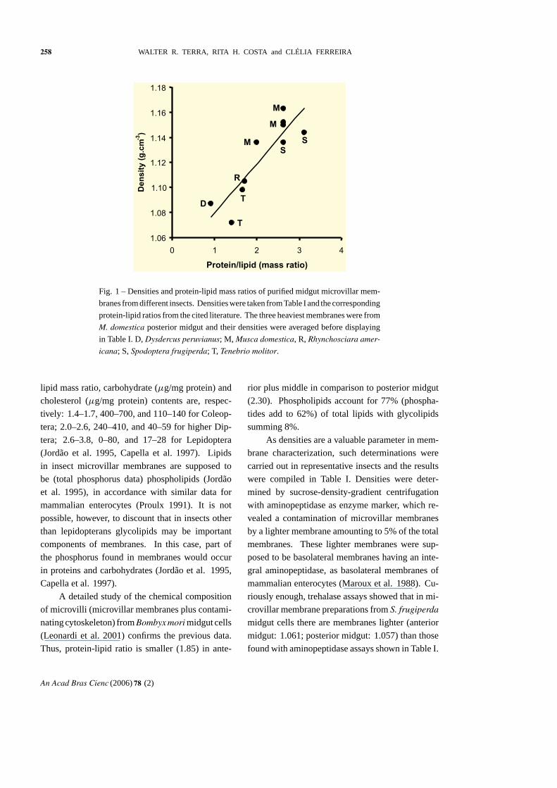
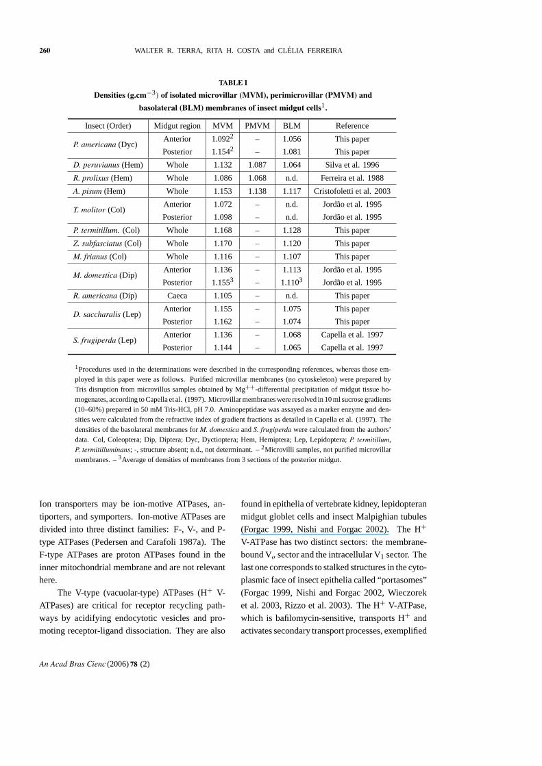
The density of the purified insect midgut
microvillar membranes linearly increase with the
protein-lipid mass ratio (Fig. 1). Nevertheless, T.
molitor datum is remarkably low and should be re-
investigated. The observed range of protein-lipid
mass of insect microvillar membranes is 1.41– 3.13,
which is wider than that found among mammalian
enterocytes (1.54– 2.44, Proulx 1991).
Apparently there is an inverse relationship
between protein-lipid mass ratio (or membrane
density) and cholesterol and carbohydrate content
in insect microvillar membranes. Thus, protein-
An Acad Bras Cienc (2006) 78 (2)
258 WALTER R. TERRA, RITA H. COSTA and CLÉLIA FERREIRA
1.06
1.08
1.10
1.12
1.14
1.16
1.18
0 1 2 3 4
Protein/lipid (mass ratio)
Den
sit
y(g
.cm
-3)
M
M
MS
S
R
T
T
D
Fig. 1 – Densities and protein-lipid mass ratios of purified midgut microvillar mem-
branes from different insects. Densities were taken from Table I and the corresponding
protein-lipid ratios from the cited literature. The three heaviest membranes were from
M. domestica posterior midgut and their densities were averaged before displaying
in Table I. D, Dysdercus peruvianus; M, Musca domestica, R, Rhynchosciara amer-
icana; S, Spodoptera frugiperda; T, Tenebrio molitor.
lipid mass ratio, carbohydrate (µg/mg protein) and
cholesterol (µg/mg protein) contents are, respec-
tively: 1.4– 1.7, 400– 700, and 110– 140 for Coleop-
tera; 2.0– 2.6, 240– 410, and 40– 59 for higher Dip-
tera; 2.6– 3.8, 0– 80, and 17– 28 for Lepidoptera
(Jordão et al. 1995, Capella et al. 1997). Lipids
in insect microvillar membranes are supposed to
be (total phosphorus data) phospholipids (Jordão
et al. 1995), in accordance with similar data for
mammalian enterocytes (Proulx 1991). It is not
possible, however, to discount that in insects other
than lepidopterans glycolipids may be important
components of membranes. In this case, part of
the phosphorus found in membranes would occur
in proteins and carbohydrates (Jordão et al. 1995,
Capella et al. 1997).
A detailed study of the chemical composition
of microvilli (microvillar membranes plus contami-
nating cytoskeleton) from Bombyx mori midgut cells
(Leonardi et al. 2001) confirms the previous data.
Thus, protein-lipid ratio is smaller (1.85) in ante-
rior plus middle in comparison to posterior midgut
(2.30). Phospholipids account for 77% (phospha-
tides add to 62%) of total lipids with glycolipids
summing 8%.
As densities are a valuable parameter in mem-
brane characterization, such determinations were
carried out in representative insects and the results
were compiled in Table I. Densities were deter-
mined by sucrose-density-gradient centrifugation
with aminopeptidase as enzyme marker, which re-
vealed a contamination of microvillar membranes
by a lighter membrane amounting to 5% of the total
membranes. These lighter membranes were sup-
posed to be basolateral membranes having an inte-
gral aminopeptidase, as basolateral membranes of
mammalian enterocytes (Maroux et al. 1988). Cu-
riously enough, trehalase assays showed that in mi-
crovillar membrane preparations from S. frugiperda
midgut cells there are membranes lighter (anterior
midgut: 1.061; posterior midgut: 1.057) than those
found with aminopeptidase assays shown in Table I.
An Acad Bras Cienc (2006) 78 (2)
INSECT MIDGUT CELL MEMBRANES 259
This suggests that the sucrose gradients are resolving
more than one domain of basolateral membranes.
Microvillar densities vary widely among insects,
with more evolved ones (Lepidoptera and Diptera)
having membranes with densities higher than 1.135.
This suggests that the midgut cell surface plays more
sophisticated roles (associated with a higher protein
content) than in lower insects. The same is true
for microvillar in comparison to basolateral mem-
branes. What kind of roles these membranes play
will be discussed in the next section.
PHYSIOLOGICAL ROLE OF MICROVILLAR AND
BASOLATERAL MEMBRANES
Initial considerations and surface digestion
The physiological role of midgut microvillar mem-
branes may change along the midgut and among
insect taxa and should include: surface (terminal)
digestion, absorption, ion homeostasis, signaling,
and unique digestive enzyme secretion mechanisms.
Some of these functions depend on the concurrent
participation of basolateral membranes, like those
associated with the transepithelial transport of
water, ions and nutrients. Most of the membrane
roles are played by integral membrane (occasion-
ally cytoskeleton) proteins that will be considered
in turn.
The densities of isolated plasma membranes
of insect cells depend essentially on their protein
contents (Fig. 1). If we set appart data on P. ame-
ricana (not purified microvillar membranes) and
on hemipterans (to be discussed on section 4),
the amount of protein in membranes may largely re-
flect digestive enzyme content, in accordance with
the presumed role of these membranes in digestion.
Thus, in Coleoptera most digestion occurs inside
the peritrophic membrane (cylindrical anatomical
structure separating the midgut contents from the
midgut cells) with little or no digestion being car-
ried out by enzymes associated with the microvillar
membranes. In contrast, in Diptera the initial and
intermediate stages of digestion occur in midgut lu-
men and most terminal digestion is carried out by
microvillar enzymes. Furthermore, there is a differ-
entiation along the Coleoptera and higher Diptera
midguts so that most terminal digestion takes place
at the posterior midgut, which in higher Diptera
functionally corresponds to the whole midgut of
other insect species (Terra and Ferreira 1994, 2005).
Pyrearinus termitilluminans larvae regurgitate
onto their prey their midgut contents that accom-
plishes initial digestion. Pre-liquefied material is
then ingested by larvae and the intermediate and final
digestion take place on the surface of midgut cells by
microvillar enzymes (Colepicolo-Neto et al. 1986),
thus explaining the high density of microvillar mem-
branes. It is not clear why the microvillar mem-
branes in Z. subfasciatus midgut cells are heavy,
because most digestion in these insects occurs in
luminal contents (Silva et al. 1999). One possibil-
ity is contamination of the microvillar membranes
by peritrophic gel, a not well-known substance that
replaces the peritrophic membrane in these insects
(Terra 2001).
Lepidopteran data (Table I) clearly do not fol-
low the rule according to which the amount of pro-
tein in membranes reflects digestive enzyme con-
tent. In these insects, enzymes involved in terminal
digestion are immobilized at the surface of midgut
cells because they are entrapped in the cell glyco-
calyx, instead of being integral membrane proteins
(Terra and Ferreira 1994, 2005). The high density of
lepidopteran midgut microvillar membranes proba-
bly results from a large amount of different trans-
porters, although it is not clear why lepidopteran
larvae need more transporters than dipteran larvae.
Microvillar integral digestive enzymes vary
among different taxa. Most frequently they are:
aminopeptidase, alkaline phosphatase, carboxypep-
tidase, dipeptidase, and α-glucosidase (Terra and
Ferreira 1994). For a recent review of these enzymes
see specific entries in Terra and Ferreira (2005).
Ion and water transport
Absorption of nutrients and ions is carried out by
membrane integral proteins known as transporters.
An Acad Bras Cienc (2006) 78 (2)
260 WALTER R. TERRA, RITA H. COSTA and CLÉLIA FERREIRA
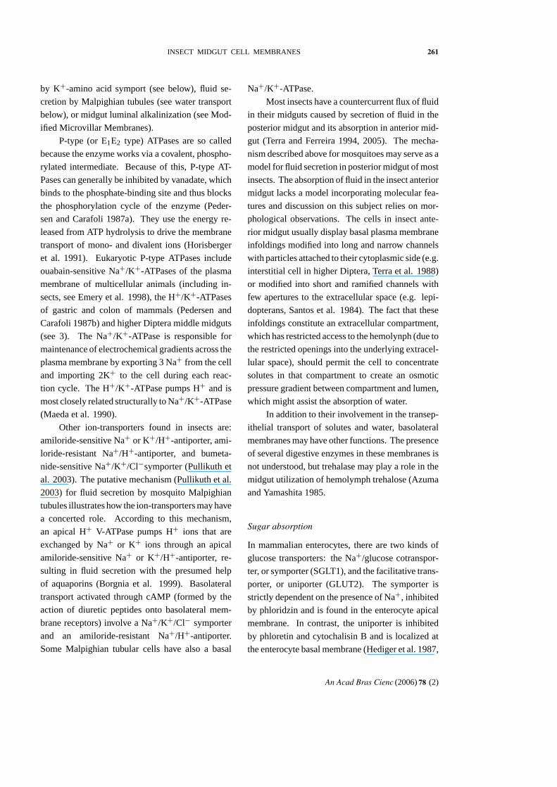
TABLE I
Densities (g.cm−3) of isolated microvillar (MVM), perimicrovillar (PMVM) and
basolateral (BLM) membranes of insect midgut cells1.
Insect (Order) Midgut region MVM PMVM BLM Reference
P. americana (Dyc)Anterior 1.0922 – 1.056 This paper
Posterior 1.1542 – 1.081 This paper
D. peruvianus (Hem) Whole 1.132 1.087 1.064 Silva et al. 1996
R. prolixus (Hem) Whole 1.086 1.068 n.d. Ferreira et al. 1988
A. pisum (Hem) Whole 1.153 1.138 1.117 Cristofoletti et al. 2003
T. molitor (Col)Anterior 1.072 – n.d. Jordão et al. 1995
Posterior 1.098 – n.d. Jordão et al. 1995
P. termitillum. (Col) Whole 1.168 – 1.128 This paper
Z. subfasciatus (Col) Whole 1.170 – 1.120 This paper
M. frianus (Col) Whole 1.116 – 1.107 This paper
M. domestica (Dip)Anterior 1.136 – 1.113 Jordão et al. 1995
Posterior 1.1553 – 1.1103 Jordão et al. 1995
R. americana (Dip) Caeca 1.105 – n.d. This paper
D. saccharalis (Lep)Anterior 1.155 – 1.075 This paper
Posterior 1.162 – 1.074 This paper
S. frugiperda (Lep)Anterior 1.136 – 1.068 Capella et al. 1997
Posterior 1.144 – 1.065 Capella et al. 1997
1Procedures used in the determinations were described in the corresponding references, whereas those em-ployed in this paper were as follows. Purified microvillar membranes (no cytoskeleton) were prepared byTris disruption from microvillus samples obtained by Mg++-differential precipitation of midgut tissue ho-mogenates, according to Capella et al. (1997). Microvillar membranes were resolved in 10 ml sucrose gradients(10– 60%) prepared in 50 mM Tris-HCl, pH 7.0. Aminopeptidase was assayed as a marker enzyme and den-sities were calculated from the refractive index of gradient fractions as detailed in Capella et al. (1997). Thedensities of the basolateral membranes for M. domestica and S. frugiperda were calculated from the authors’data. Col, Coleoptera; Dip, Diptera; Dyc, Dyctioptera; Hem, Hemiptera; Lep, Lepidoptera; P. termitillum,P. termitilluminans; -, structure absent; n.d., not determinant. – 2Microvilli samples, not purified microvillarmembranes. – 3Average of densities of membranes from 3 sections of the posterior midgut.
Ion transporters may be ion-motive ATPases, an-
tiporters, and symporters. Ion-motive ATPases are
divided into three distinct families: F-, V-, and P-
type ATPases (Pedersen and Carafoli 1987a). The
F-type ATPases are proton ATPases found in the
inner mitochondrial membrane and are not relevant
here.
The V-type (vacuolar-type) ATPases (H+ V-
ATPases) are critical for receptor recycling path-
ways by acidifying endocytotic vesicles and pro-
moting receptor-ligand dissociation. They are also
found in epithelia of vertebrate kidney, lepidopteran
midgut globlet cells and insect Malpighian tubules
(Forgac 1999, Nishi and Forgac 2002). The H+
V-ATPase has two distinct sectors: the membrane-
bound Vo sector and the intracellular V1 sector. The
last one corresponds to stalked structures in the cyto-
plasmic face of insect epithelia called “ portasomes”
(Forgac 1999, Nishi and Forgac 2002, Wieczorek
et al. 2003, Rizzo et al. 2003). The H+ V-ATPase,
which is bafilomycin-sensitive, transports H+ and
activates secondary transport processes, exemplified
An Acad Bras Cienc (2006) 78 (2)
INSECT MIDGUT CELL MEMBRANES 261
by K+-amino acid symport (see below), fluid se-
cretion by Malpighian tubules (see water transport
below), or midgut luminal alkalinization (see Mod-
ified Microvillar Membranes).
P-type (or E1E2 type) ATPases are so called
because the enzyme works via a covalent, phospho-
rylated intermediate. Because of this, P-type AT-
Pases can generally be inhibited by vanadate, which
binds to the phosphate-binding site and thus blocks
the phosphorylation cycle of the enzyme (Peder-
sen and Carafoli 1987a). They use the energy re-
leased from ATP hydrolysis to drive the membrane
transport of mono- and divalent ions (Horisberger
et al. 1991). Eukaryotic P-type ATPases include
ouabain-sensitive Na+/K+-ATPases of the plasma
membrane of multicellular animals (including in-
sects, see Emery et al. 1998), the H+/K+-ATPases
of gastric and colon of mammals (Pedersen and
Carafoli 1987b) and higher Diptera middle midguts
(see 3). The Na+/K+-ATPase is responsible for
maintenance of electrochemical gradients across the
plasma membrane by exporting 3 Na+ from the cell
and importing 2K+ to the cell during each reac-
tion cycle. The H+/K+-ATPase pumps H+ and is
most closely related structurally to Na+/K+-ATPase
(Maeda et al. 1990).
Other ion-transporters found in insects are:
amiloride-sensitive Na+ or K+/H+-antiporter, ami-
loride-resistant Na+/H+-antiporter, and bumeta-
nide-sensitive Na+/K+/Cl−symporter (Pullikuth et
al. 2003). The putative mechanism (Pullikuth et al.
2003) for fluid secretion by mosquito Malpighian
tubules illustrates how the ion-transporters may have
a concerted role. According to this mechanism,
an apical H+ V-ATPase pumps H+ ions that are
exchanged by Na+ or K+ ions through an apical
amiloride-sensitive Na+ or K+/H+-antiporter, re-
sulting in fluid secretion with the presumed help
of aquaporins (Borgnia et al. 1999). Basolateral
transport activated through cAMP (formed by the
action of diuretic peptides onto basolateral mem-
brane receptors) involve a Na+/K+/Cl− symporter
and an amiloride-resistant Na+/H+-antiporter.
Some Malpighian tubular cells have also a basal
Na+/K+-ATPase.
Most insects have a countercurrent flux of fluid
in their midguts caused by secretion of fluid in the
posterior midgut and its absorption in anterior mid-
gut (Terra and Ferreira 1994, 2005). The mecha-
nism described above for mosquitoes may serve as a
model for fluid secretion in posterior midgut of most
insects. The absorption of fluid in the insect anterior
midgut lacks a model incorporating molecular fea-
tures and discussion on this subject relies on mor-
phological observations. The cells in insect ante-
rior midgut usually display basal plasma membrane
infoldings modified into long and narrow channels
with particles attached to their cytoplasmic side (e.g.
interstitial cell in higher Diptera, Terra et al. 1988)
or modified into short and ramified channels with
few apertures to the extracellular space (e.g. lepi-
dopterans, Santos et al. 1984). The fact that these
infoldings constitute an extracellular compartment,
which has restricted access to the hemolynph (due to
the restricted openings into the underlying extracel-
lular space), should permit the cell to concentrate
solutes in that compartment to create an osmotic
pressure gradient between compartment and lumen,
which might assist the absorption of water.
In addition to their involvement in the transep-
ithelial transport of solutes and water, basolateral
membranes may have other functions. The presence
of several digestive enzymes in these membranes is
not understood, but trehalase may play a role in the
midgut utilization of hemolymph trehalose (Azuma
and Yamashita 1985.
Sugar absorption
In mammalian enterocytes, there are two kinds of
glucose transporters: the Na+/glucose cotranspor-
ter, or symporter (SGLT1), and the facilitative trans-
porter, or uniporter (GLUT2). The symporter is
strictly dependent on the presence of Na+, inhibited
by phloridzin and is found in the enterocyte apical
membrane. In contrast, the uniporter is inhibited
by phloretin and cytochalisin B and is localized at
the enterocyte basal membrane (Hediger et al. 1987,
An Acad Bras Cienc (2006) 78 (2)
262 WALTER R. TERRA, RITA H. COSTA and CLÉLIA FERREIRA
Takata 1996, Kellett 2001, Giordana et al. 2003).
Although it has not been functionally demon-
strated the presence of sugar transporters in insect
midgut cells, a glucose uniporter is very likely to oc-
cur. Drosophila melanogaster has a glucose trans-
porter gene that is homologous with the mammalian
glucose uniporter genes (Escher and Rasmuson-
Lestander 1999). Furthermore, the absorption of
glucose by the epidermis of the endoparasitoid
Aphidius ervi seem to involve a uniporter (Gior-
dana et al. 2003).
Amino acid transport
Amino acid transporters have been studied in the
midguts of adult stage of the dictyopteran (cock-
roach) Blabera gigantea and in the larval stage of the
coleopteran Leptinotarsa decemlineata and the lepi-
dopterans Philosamia cynthia, and Bombyx mori. In
coleopterans, only uniporters were found, whereas
in the other insects amino acid-cation symporters
occur in addition to the uniporter. Dictyopterans
have a Na+-coupled amino acid transport with the
electrochemical potential maintained by a Na+/K+-
ATPase such in mammalian cells. Lepidopterans
possess transport features different from those of
mammals. K+ is secreted in these insects through
the concerted action of an apical H+V-ATPase and
apical K+/H+-antiporter, thereby providing the
drive force for absorption of amino acids by an
amino acid-K+ symporter (Castagna et al. 1997).
The successful preparation of vesicles from
lepidopteran midgut microvillar membranes (brush-
border membrane vesicles) (see Isolation of Mem-
branes from Midgut of Different Insects) prompted
the study of amino acid transport, beginning with
the paper of Hanozet et al. (1980). The studies
led to the finding of several distinct amino acid-K+
symporters with overlapping specifies (Castagna et
al. 1997, Wolfersberger 2000). A cDNA encod-
ing a lepidopteran midgut amino acid-K+ symporter
was cloned. The encoded protein showed weak but
significant sequence identity with amino acid trans-
porters belonging to the sodium-dependent and
chloride-dependent γ -aminobutyric acid (GABA)
superfamily (Castagna et al. 1998). Since then, the
sequence of several new insect cation-amino acid
symporters were deposited in the GenBank and most
of the present research looks for specific inhibitors
of amino acid transport like bestatin, (previously
widely used as an aminopeptidase inhibitor) and
fenoxycarb (Wolfersberger 2000).
Secretory mechanisms
The microvillar molecular organization is proba-
bly modified in insects displaying unique secretory
mechanisms not seen in other animals. Lepidopte-
ran anterior midgut cells secrete digestive enzymes
by two kinds of microapocrine secretion. In the mi-
croapocrine secretion with budding vesicles, small
vesicles migrate into the microvilli, from which
they bud laterally as double membrane vesicles.
Microapocrine secretion with pinched-off vesicles
is characterized by vesicles migrating to the tips of
microvilli, where they fuse one another and with
the microvillar membrane. Finally, vesicles pinch
off from the enlarged microvilli tips. In both cases,
the secretory contents are released by membrane
fusion and/or by membrane solubilization caused
by high pH contents or by luminal detergents (Terra
and Ferreira 1994, 2005).
In accordance with the putative microvillar
cytoskeleton differences associated with unique
secretory mechanisms, lepidopteran anterior mid-
gut microvilli preparations are free from cytos-
keleton before the Tris-disruption step (see Iso-
lation of Membranes from Midgut of Different In-
sects and Biochemistry of Microvillar Membranes)
(Capella et al. 1997). A S. frugiperda midgut cDNA
expression library is being screened with
antibodies raised with purified microvillar mem-
branes as antigens, in an attempt to identify the
molecules involved in these secretory mechanisms
(A.H.P. Ferreira, L.O. Guerra, P.B. Paiva, B. Schn-
abel, M.R.S. Briones, W.R. Terra and C. Ferreira,
unpublished data).
An Acad Bras Cienc (2006) 78 (2)
INSECT MIDGUT CELL MEMBRANES 263
MODIFIED MICROVILLAR MEMBRANES
Modified microvilli are typical of the lepidopteran
goblet cells and higher dipteran oxyntic cells. Gob-
let cells have a cavity, which is formed by invagi-
nation of the apical membrane and which occupies
most of the cell (long-neck goblet cell) or only its
upper part (stalked goblet cell). The infolded apical
membrane shows modified microvilli containing
mitochondria and their cytoplasmic side are studded
with small particles (Cioffi1979, Santos et al. 1984)
that corresponds to a H+V-ATPase (Wieczorek et
al. 2003). Goblet cells generate a high gut pH in
lepidopterans according to the following model
(Wieczorek et al. 2003). Carbonic anhydrase pro-
duces carbonic acid that dissociates into bicar-
bonate and a proton. The proton is pumped by
an H+V-ATPase into the goblet cell cavity, from
where it is removed in exchange with K+ that even-
tually diffuses into lumen. Bicarbonate is secreted in
exchange with chloride and loses a proton due to the
intense field near the membrane, forming carbonate
and raising the gut pH.
Oxyntic cells have an apical membrane invagi-
nated into ramified crypts which are coated with
microvilli and display numerous mitochondria in
their cytoplasm. The cytoplasmic side of oxyntic
microvillar membranes are studded with small
particles (Terra et al. 1988). These are believed to
be proton pumps (P-type ATPase, see Ion and water
transport) that acidify to pH 3.2 the contents of mid-
dle midgut in higher Diptera. Chloride ions seem to
follow the movement of protons. This hypothesis is
supported by the observed effect of different com-
pounds in luminal pH and in the luminal chloride
content (Terra and Regel 1995). It is remarkable
how similar is the mechanism of insect midgut
luminal acidification and that one found in mam-
malian stomachs (Forte et al. 1980).
PERIMICROVILLAR MEMBRANES
Perimicrovillar membranes (PMVM) are mem-
branes that cover the midgut cells microvilli extend-
ing into the gut lumen with dead ends (Lane and
Harrison 1979, Andries and Torpier 1982, Silva et
al. 1995) and were described in Hemiptera (bugs,
aphids, and cicadas). The domain of PMVM en-
sheathing the microvilli are set in position by
columns obliquely disposed between them and the
microvillar membrane (Lane and Harrison 1979).
Freeze-fracture replicas showed that PMVM
are almost free from intramembranous particles,
thus resembling myelin sheets (Lane and Harrison
1979, Andries and Torpier 1982). Therefore,
PMVM must display a lower buoyant density than
the microvillar membranes. Based on this, the
two membranes were isolated by density-gradient
centrifugation and enzyme markers identified: α-
glucosidase for PMVM and α-mannosidase or β-
glucosidase for microvillar membranes (Ferreira et
al. 1988, Silva et al. 1996). PMVM densities of R.
prolixus and D. peruvianus are alike that of myelin
sheets, as expected, although A. pisum PMVM are
surprisingly heavy (Table I). It should be noted, how-
ever, that the last mentioned PMVM are actually
modified PMVM and seem to have a high enzyme
content (see below). Microvillar membrane densi-
ties of R. prolixus are small in contrast to that of
D. peruvianus and A. pisum (Table I). This probably
reflects the putative pumps seen in D. peruvianus mi-
crovillar membranes and the contamination of mi-
crovillar membranes by lamellar links in A. pisum
(see below). Immunolocalization of the PMVM
enzyme marker, α-glucosidase, suggests that these
membranes are formed when double membrane
vesicles fuse their outer membranes with the mi-
crovillar membranes and their inner membranes
with PMVM. A double membrane Golgi cisterna
(on budding) forms the double membrane vesicles
(Silva et al. 1995).
PMVM and a PMVM-bound α-glucosidase
occur in the major hemipteran infra-orders and
in the sister order Thysanoptera (thrips), but lack in
the orders Psocoptera (plant lice) and Phthiraptera
(lice). This suggests that PMVM may have orig-
inated in the condylognatha (Paraneopteran taxon
including Hemiptera and Thysanoptera) ancestral
stock (Silva et al. 2004). The Condylognathan an-
An Acad Bras Cienc (2006) 78 (2)
264 WALTER R. TERRA, RITA H. COSTA and CLÉLIA FERREIRA
cestors should feed as present-day thrips on phloem
by a punch and suck mechanism. Phloem has very
low contents of protein (with few exceptions) and
carbohydrate polymers and is rich in sucrose and
relatively poor in free amino acids (Terra 1990).
Upon adapting to this food, Condylognathan an-
cestors would lose most digestive enzymes and the
peritrophic membrane that are associated with lu-
minal digestion. Essential amino acids present in
low concentrations in sap may be absorbed by a hy-
pothesized mechanism (Terra 1988, Terra and Fer-
reira 1994) as follows: microvillar membranes ac-
tively transport K+ from the perimicrovillar space
into the midgut cells, generating a concentration gra-
dient between the gut luminal sap rich in K+ and
the perimicrovillar space. This concentration gra-
dient may be the driving force for the active ab-
sorption of amino acids by appropriate symporters
in PMVM. Amino acids, once in the perimicrovil-
lar space may diffuse up to specific transporters on
the microvillar surface. Although amino acid sym-
porters have been found in the microvillar mem-
branes of several insects (see Amino acid transport),
no attempts have been made to study the other pos-
tulated proteins (e.g., amino acid-K+-symporters in
PMVM and potassium pumps in microvillar mem-
branes). Thus, in spite of the model provided an
explanation for the occurrence of these peculiar cell
structures in condylognatha, it is supported only by:
(1) evidence that amino acids are absorbed with
potassium ions in Dysdercus peruvianus (Silva and
Terra 1994); (2) occurrence of particles studying
the cytoplasmic face of the midgut microvillar
membrane of D. peruvianus. These might be ion
pumps responsible for the putative K+-transport
(Silva et al. 1995).
Organic compounds in xylem sap (much more
diluted than phloem sap) need to be concentrated
before they can be absorbed by the perimicrovillar
membrane. This occurs in the filter chamber that
consists of a thin-walled, dilated anterior midgut in
close contact with the posterior midgut and the prox-
imal ends of the Malpighian tubules (anatomical
structure analogous to the mammalian nephron).
This arrangement enables water to pass directly from
the anterior midgut to the Malpighian tubules, con-
centrating food in midgut. The high permeability of
the filter chamber membrane to water results from
the occurrence of specific proteins named aquapor-
ins (Borgnia et al. 1999). These were immunolo-
calized in the microvillar border (PMVM and mi-
crovillar membranes) of the filter chamber cells of
several hemipteran xylem sap feeders (Le Cahérec
et al. 1997).
Hemipterans like aphids may suck high-
sucrose phloem saps with osmolarity up to three
times that of the insect hemolymph. This results
in a considerable hydrostatic pressure caused by
the tendency of water to move from the hemolymph
into midgut lumen. To withstand these high hydro-
static pressures there are links between apical lamel-
lae (replacing usual midgut cell microvilli). As a
consequence of these links, PMVM could no longer
exist and were replaced by membranes seen asso-
ciated with the tips of the lamellae, the modified
PMVM, which contain unexpected enzymes like a
cysteine proteinase (Ponsen 1991, Cristofoletti et
al. 2003).
Haematophagous, seed-sucking, and predator
hemipterans evolved from the sap-feeders regain-
ing the ability to digest polymers. Compartmentali-
zation of digestion was maintained by PMVM as a
substitute for the absent peritrophic membrane (Fer-
reira et al. 1988, Silva et al. 1995).
CONCLUDING REMARKS
The study of the plasma membranes of insect midgut
cells has progressed enough to reveal many of their
characteristics. Research emphasis on the unique
aspects of insect midgut cells may led to seminal
findings, in disparate fields as cell biology, molec-
ular physiology, and molecular evolution as may
provide new targets for insect control. Thus, the
study of microvilli engaged in microaprocrine se-
cretion may disclose novel mechanisms of vesicle
trafficking and membrane fusion. The description
in molecular detail of the plasma membrane role in
An Acad Bras Cienc (2006) 78 (2)
INSECT MIDGUT CELL MEMBRANES 265
the functioning of oxyntic and interstitial cells from
the midgut of higher Diptera should illustrate a mar-
velous case of convergence with mammalian gastric
cells. A molecular physiological approach to the
interplay of hemipteran midgut plasma membranes
will certainly be revealing, as these insects are the
only animals that live exclusively sucking the usu-
ally nutrient-poor plant saps. Finally, the implica-
tions of the knowledge on the plasma membrane
signaling system of midgut-function coordination is
beyond speculation, due to scarcity of data. Progress
in the fields reviewed is being supported by the asso-
ciation of biochemical and molecular biology pro-
cedures. A proteomic approach to those fields is
still hampered by the lack of sufficient biological
material from specific insect tissues.
ACKNOWLEDGMENTS
This work was supported by Brazilian Research
Agencies: Fundação de Amparo à Pesquisa do Es-
tado de São Paulo (FAPESP) and Conselho Nacio-
nal de Desenvolvimento Científico e Tecnológico,
Programa de Apoio a Núcleos de Excelência
(CNPq/PRONEX). R.H. Costa had a post-doctoral
position on leave from UNIFESP-EPM, São Paulo;
W.R. Terra and C. Ferreira are staff members of the
Biochemistry Department and research fellows of
CNPq.
RESUMO
As membranas plasmáticas das células intestinais dos
insetos apresentam um domínio apical e outro basal. O
domínio apical é geralmente modificado em microvilosi-
dades com organização molecular similar a de outros
animais, embora possam diferir naqueles insetos que
apresentam vesículas secretoras em trânsito que brotam
lateralmente ou destacam-se das extremidades das mi-
crovilosidades. Outras modificações microvilares estão
associadas a bombeamento de prótons ou a interrelações
com uma membrana lipídica (a membrana perimicrovi-
lar) que reveste as microvilosidades de células intesti-
nais de hemípteros (pulgões e percevejos). Admite-se
que as membranas perimicrovilares estejam envolvidas
na absorção de aminoácidos a partir de dietas diluídas.
As membranas microvilares e perimicrovilares tem den-
sidades distintas (e conteúdo protéico) que dependem do
táxon do inseto. O papel desempenhado pelas proteínas
microvilares e perimicrovilares na fisiologia intestinal dos
insetos é revisto, procurando fornecer uma visão coerente
dos dados e chamando a atenção para novos objetivos de
pesquisa.
Palavras-chave: membranas microvilares, membranas
perimicrovilares, absorção de nutrientes, transporte de
íon, transporte de água, fisiologia molecular do intestino
médio.
REFERENCES
ANDRIES JC AND TORPIER G. 1982. An extracellular
brush border coat of lipid membranes in the midgut of
Nepa cinerea (Insecta: Heteroptera): ultrastructure
and genesis. Biol Cell 46: 195– 202.
AZUMA M AND YAMASHITA O. 1985. Cellular local-
ization and proposed function of midgut trehalase in
the silkworm larva, Bombyx mori. Tissue Cell 17:
539– 551.
BAUTZ AM. 1989. A villin-like protein in the intesti-
nal brush border of Calliphora vicina R.D. larvae
(Diptera: Calliphoridae). Int J Insect Morphol Em-
bryol 18: 281– 288.
BEMENT WM AND MOOSEKER MS. 1996. The cy-
toskeleton of the intestinal epithelium: components,
assembly, and dynamic rearrangements. In: HES-
KETH JE AND PRYME JF (Eds), The cytoskeleton:
a multi-volume treatise. Greenwich: JAI Press 3:
359– 404.
BODNARYK RP, BRONSKILL JF AND FETTERLEY JR.
1974. Membrane-bound γ -glutamyl transpeptidase
and its role in phenylalanine absorption-reabsorption
in the larva of M. domestica. J Insect Physiol 20:
167– 181.
BONFANTI P, COLOMBO A, HEINTZELMAN MB,
MOOSEKER MS AND CAMATINI M. 1992. The
molecular architecture of an insect midgut brush-
border cytoskeleton. Eur J Cell Biol 57: 298– 307.
BORGNIA M, NIELSEN S, ENGEL A AND AGREE P.
1999. Cellular and molecular biology of the agua-
porin water channels. Annu Rev Biochem 68: 425–
458.
An Acad Bras Cienc (2006) 78 (2)
266 WALTER R. TERRA, RITA H. COSTA and CLÉLIA FERREIRA
CAPELLA AN, TERRA WR, RIBEIRO AF AND FER-
REIRA C. 1997. Cytoskeletal removal and charac-
terization of the microvillar membranes isolated
from two midgut regions of Spodoptera frugiperda
(Lepidoptera). Insect Biochem Molec Biol 27:
793– 801.
CASTAGNA M, SHAYAKUL C, TROTTI D, SACCHI VF,
HARVEY WR AND HEDIGER MA. 1997. Molec-
ular characteristics of mammalian and insect amino
acid transporters: implications for amino acid home-
ostasis. J Exp Biol 200: 269– 286.
CASTAGNA M, SHAYAKUL C, TROTTI D, SACCHI
VF, HARVEY WR AND HEDIGER MA. 1998. Clo-
ning and characterization of a potassium-coupled
amino acid transporter. Proc Natl Acad Sci USA 95:
5395– 5400.
CIOFFI M. 1979. The morphology and fine structure of
the larval midgut of a moth (Manduca sexta)
in relation to active ion transport. Tissue Cell 11:
467– 479.
CIOFFI M AND WOLFERSBERGER MG. 1983. Isolation
of separate apical, lateral and basal plasma membrane
from cells of an insect epithelium. A procedure based
on tissue organization and ultrastructure. Tissue Cell
15: 781– 803.
COLEPICOLO-NETO P, BECHARA EJH, FERREIRA C
AND TERRA WR. 1986. Evolutionary considera-
tions of the spatial organization of digestion in the
luminescent predaceous larvae of Pyrearinus termi-
tilluminans (Coleoptera: Elateridae). Insect Bio-
chem 16: 811– 817.
CRISTOFOLETTI PT, RIBEIRO AF, DERAISON C,
RAHBÉ Y AND TERRA WR. 2003. Midgut adapta-
tion and digestive enzyme distribution in a phloem
feeding insect, the pea aphid Acyrtosiphom pisum. J
Insect Physiol 49: 11– 24.
DALLAI R, LUPETTI P AND LANE NJ. 1998. The or-
ganization of actin in the apical region of insect
midgut cells after deep etching. J Struct Biol 122:
283– 292.
EISEN NS, FERNANDES VF, HARVEY WR, SPAETH
DD AND WOLFERSBERGER MG. 1989. Compari-
son of brush border membrane vesicles prepared by
three methods from larval Manduca sexta midgut.
Insect Biochem 19: 337– 342.
EMERY AM, BILLINGSLEY PF, READY PD AND
DJAMGOZ MBA. 1998. Insect Na+/K+-ATPase. J
Insect Physiol 44: 197– 209.
ESCHER SA AND RASMUSON-LESTANDER A. 1999.
The Drosophila glucose transporter gene: cDNA se-
quence, phylogenetic comparisons analysis of func-
tional sites and secondary structures. Hereditas 130:
95– 103.
ESPINOZA-FUENTES FP, RIBEIRO AF AND TERRA
WR. 1987. Microvillar and secreted digestive en-
zymes from Musca domestica larvae. Subcellular
fractionation of midgut cells with electron micros-
copy monitoring. Insect Biochem 17: 819– 827.
EVANS WH. 1978. Preparation and characterization
of mammalian plasma membrane. In: WORK TS
AND WORK E (Eds), Techniques in Biochemistry
and Molecular Biology Part 1, Amsterdam: North-
Holland, p. 103– 121.
FERREIRA C AND TERRA WR. 1980. Intracellular dis-
tribution of hydrolases in midgut caeca cells from
an insect with emphasis on plasma membrane-bound
enzymes. Comp Biochem Physiol 66B: 467– 473.
FERREIRA C, RIBEIRO AF, GARCIA ES AND TERRA
WR. 1988. Digestive enzymes trapped between
and associated with the double plasma membranes
of Rhodnius prolixus posterior midgut cells. Insect
Biochem 18: 521– 530.
FERREIRA C, BELLINELLO GL, RIBEIRO AF AND
TERRA WR. 1990. Digestive enzymes associated
with the glycocalyx microvillar membranes and se-
cretory vesicles from midgut cells of Tenebrio moli-
tor larvae. Insect Biochem 20: 839– 847.
FORGAC M. 1999. Structure and properties of the vacuo-
lar (H+)-ATPases. J Biol Chem 274: 12951– 12954.
FORTE JG, MACHEN TE AND OBRINK KJ. 1980.
Mechanisms of gastric H+ and Cl- transport. Annu
Rev Physiol 42: 111– 126.
GIORDANA B, MILANI A, GRIMALDI A, FARNETI R,
CASARTELLI M, AMBROSECCHIO MR, DIGILIO
MC, LEONARDI MG, DE EGUILEAR M AND PEN-
NACCHIO F. 2003. Absorption of sugars and amino
acids by the epidermis of Aphidius ervi larvae. J In-
sect Physiol 49: 1115– 1124.
HANOZET GM, GIORDANA B AND SACCHI JF. 1980.
K+-dependent phenylalanine uptake in membrane
vesicles isolated from the midgut of Philosamia cyn-
thia larvae. Biochim Biophys Acta 596: 481– 486.
An Acad Bras Cienc (2006) 78 (2)
INSECT MIDGUT CELL MEMBRANES 267
HEDIGER MA, COADY MJ, IKEDA TD AND WRIGHT
EM. 1987. Expression, cloning and cDNA sequenc-
ing of the Na+/glucose cotransporter. Nature 330:
379– 381.
HORISBERGER JD, LEMAS V, KRAEHENBUHL JP AND
ROSSIER BC. 1991. Structure-function relationship
of Na+, K+-ATPase. Annu Rev Physiol 53: 565–
584.
HOUK EJ, ARCUS YM AND HARDY JL. 1986. Isola-
tion and characterization of brush border fragments
from mosquito mesenterons. Archs Insect Biochem
Physiol 3: 135– 146.
JORDÃ O BP, TERRA WR AND FERREIRA C. 1995.
Chemical determinations in microvillar membranes
purified from brush-borders isolated from the larval
midgut of one Coleoptera and two Diptera species.
Insect Biochem Molec Biol 25: 417– 426.
KELLETT GL. 2001. The facilitate component of intes-
tinal glucose absorption. J Physiol 531: 585– 595.
LANE NJ AND HARRISON JB. 1979. An unusual cell
surface modification: a double plasma membrane. J
Cell Sci 39: 355– 372.
LANE NJ, DALLAI R AND ASHHURTS DE. 1996.
Structural macromolecules of the cell membranes
and the extracellular matrices of the insect midgut.
In: LEHANE MJ AND BILLINGSLEY PF (Eds), The
Biology of Insect Midgut, London: Chapman & Hall,
p. 115– 150.
LE CAHEREC F, GUILLAM MT, BEURON F, CAVALIER
A, THOMAS D AND GOURATON J. 1997. Aqua-
porin-related proteins. Cell Tissue Res 290: 143–
151.
LEMOS FJA AND TERRA WR. 1992. A high yield
preparation of Musca domestica larval midgut mi-
crovilli and the subcellular distribution of amylase
and trypsin. Insect Biochem Molec Biol 22: 433–
438.
LEONARDI MG, MARCIANI P, MONTORFANO PG,
CAPPELLOZZA S AND GIORDANA B. 2001. Ef-
fect of fenoxycarb on leucine uptake and lipid com-
position of midgut brush border membrane in the
silkworm, Bombyx mori (Lepidoptera, Bombycidae).
Pest Biochem Physiol 70: 42– 51.
MAEDA M, OSHIMAN K, TAMURA S AND FUTAI M.
1990. Human gastric (H+ + K+)-ATPase gene. Sim-
ilarity to (Na+ + K+)-ATPase genes in exon/intron
organization but difference in control region. J Biol
Chem 265: 9027– 9032.
MAROUX S, COUDRIER E, FERACCI H, GORVEL JP
AND LOUVARD D. 1988. Molecular organization
of the intestinal brush border. Biochimie 70: 1297–
1306.
MILLER D AND CRANE RK. 1961. The digestive func-
tion of the epithelium of the small intestine. II.
Localization of disaccharide hydrolysis in the iso-
lated brush border portion of intestinal epithelial
cells. Biochim Biophys Acta 52: 293– 298.
MORGAN NS, HEINTZELMAN MB AND MOOSEKER
MS. 1995. Characterization of myosin-IA and myo-
sin-IB, two unconventional myosins associated with
the Drosophila brush border cytoskeleton. Devel
Biol 172: 51– 71.
NISHI T AND FORGAC M. 2002. The vacuolar (H+)-
ATPases-Nature’s most versatile proton pumps. Nat
Rev Mol Cell Biol 3: 94– 103.
PARENTI P, SACCHI, FV, HANOZET GM AND GIOR-
DANA B. 1986. Na-dependent uptake of phenyla-
lanine in the midgut of a cockroach (Blabera gigan-
tea). J Comp Physiol 156B: 549– 556.
PEDERSEN PL AND CARAFOLI E. 1987a. Ion motive
ATPases. I. Ubiquity, properties and significance to
cell function. Trends Biochem Sci 12: 146– 150.
PEDERSEN PL AND CARAFOLI E. 1987b. Ion mo-
tive ATPases. II. Energy coupling and work out put.
Trends Biochem Sci 12: 186– 189.
PONSEN MB. 1991. Structure of the digestive system
of aphids, in particular Hyalopterus and Coloradoa,
and its bearing on the evolution of filterchambers in
the Aphidoidea. Wageningen Agricultural Univer-
sity Papers 91– 5, 3– 61.
PROULX P. 1991. Structure-function relationships in
intestinal brush border membranes. Biochim Bio-
phys Acta 1071: 255– 271.
PULLIKUTH AK, FILIPPOV V AND GILL SS. 2003.
Phylogeny and cloning of ion transporters in mos-
quitoes. J Exp Biol 206: 3857– 3868.
REUVENI M, HONG YS, DUNN PE AND NEAL JJ.
1993. Leucine transport into brush border mem-
brane vesicles from guts of Leptinotarsa decemli-
neata and Manduca sexta. Comp Biochem Physiol
104A: 267– 272.
An Acad Bras Cienc (2006) 78 (2)
268 WALTER R. TERRA, RITA H. COSTA and CLÉLIA FERREIRA
RIZZO VF, COSKUN U, RADERMACHER M, RUIZ T,
ARMBRUSTER A AND GRUBER G. 2003. Resolu-
tion of the V1 ATPase from Manduca sexta into sub-
complexes and visualization of an ATPase-active A3
B3EG complex by electron microscopy. J Biol Chem
278: 270– 275.
SANTOS CD AND TERRA WR. 1984. Plasma mem-
brane-associated amylase and trypsin: intracellular
distribution of digestive enzymes in the midgut of
the cassava hornworm, Erinnyis ello. Insect Biochem
14: 587– 595.
SANTOS CD, RIBEIRO AF, FERREIRA C AND TERRA
WR. 1984. The larval midgut of the cassava horn-
worm (Erinnyis ello). Ultrastructure, fluid fluxes and
the secretory activity in relation to the organization
of digestion. Cell Tissue Res 237: 565– 574.
SANTOS CD, RIBEIRO AF AND TERRA WR. 1986.
Differential centrifugation, calcium precipitation and
ultrasonic disruption of midgut cells of Erinnyis ello
caterpillars. Purification of cell microvilli and infer-
ences concerning secretory mechanisms. Can J Zool
64: 490– 500.
SCHMITZ J, PREISER H, MAESTRACCI D, GHOSH BK,
CERDA J AND CRANE RK. 1973. Purification of the
human intestinal brush border membrane. Biochim
Biophys Acta 323: 98– 112.
SCHUMACKER TTS, CRISTOFOLETTI PT AND TERRA
WR. 1993. Properties and compartmentalization of
digestive carbohydrases and proteases in Scaptotri-
gona bipunctata (Apidae: Meliponinae) larvae. Api-
dologie 24: 3– 17.
SILVA CP AND TERRA WR. 1994. Digestive and ab-
sorptive sites along the midgut of the cotton seed
sucker bug Dysdercus peruvianus (Hemiptera: Pyr-
rhocoridae). Insect Biochem Molec Biol 24: 493–
505.
SILVA CP, RIBEIRO AF, GULBENKIAN S AND TERRA
WR. 1995. Organization, origin and function of
the outer microvillar (perimicrovillar) membranes of
Dysdercus peruvianus (Hemiptera) midgut cells. J
Insect Physiol 41: 1093– 1103.
SILVA CP, RIBEIRO AF AND TERRA WR. 1996. En-
zyme markers and isolation of the microvillar and
perimicrovillar membranes of Dysdercus peruvia-
nus (Hemiptera: Pyrrhocoridae) midgut cells. Insect
Biochem Molec Biol 26: 1011– 1018.
SILVA CP, TERRA WR, XAVIER-FILHO J, GROSSI-DE-
SÁ MF, LOPES AR AND PONTES EG. 1999. Di-
gestion in larvae of Callosobrucchus maculatus and
Zabrotes subfasciatus (Coleoptera: Bruchidae) with
emphasis on α-amylases and oligosaccharidases. In-
sect Biochem Molec Biol 29: 355– 366.
SILVA CP, SILVA JR, VASCONCELOS FF, PETRETSKI
DA, DAMATTA RA, RIBEIRO AF AND TERRA
WR. 2004. Occurrence of perimicrovillar membra-
nes in paraneopteran insect orders with comments on
their function and evolutionary significance. Arthr
Struct Devel 33: 139– 148.
TAKATA K. 1996. Glucose transporters in the transep-
ithelial transport of glucose. J Electron Microsc 45:
275– 284.
TERRA WR. 1988. Physiology and biochemistry of in-
sect digestion: an evolutionary perspective. Braz J
Med Biol Res 21: 675– 734.
TERRA WR. 1990. Evolution of digestive systems of
insects. Annu Rev Entomol 35: 181– 200.
TERRA WR. 2001. The origin and functions of the per-
itrophic membrane and peritrophic gel. Arch Insect
Biochem Physiol 47: 47– 61.
TERRA WR AND FERREIRA C. 1994. Insect diges-
tive enzymes: properties, compartmentalization and
function. Com Biochem Physiol 109B: 1– 62.
TERRA WR AND FERREIRA C. 2005. Biochemistry of
digestion. In: GILBERT LI, IATROU K AND GILL
SS (Eds), Comprehensive Molecular Insect Science,
Oxford: Elsevier 4: 171– 224.
TERRA WR AND REGEL R. 1995. pH buffering in
Musca domestica midguts. Comp Biochem Physiol
112A: 559– 564.
TERRA WR, ESPINOZA-FUENTES FP, RIBEIRO AF
AND FERREIRA C. 1988. The larval midgut of
the housefly (Musca domestica): ultrastructure, fluid
fluxes and ion secretion in relation to the organization
of digestion. J Insect Physiol 34: 463– 472.
TERRA WR, SANTOS CD AND RIBEIRO AF. 1990. Ul-
trastructural and biochemical basis of the digestion
of nectar and other nutrients by the moth Erynnyis
ello. Ent Exp Appl 56: 277– 286.
TREHERNE JE. 1959. Amino acid absorption in the
locust (schistocerca gregaria Forsk). J Exp Biol 36:
533– 545.
An Acad Bras Cienc (2006) 78 (2)
INSECT MIDGUT CELL MEMBRANES 269
WIECZOREK H, HUSS M, MERZENDORFER H, REI-
NEKE S, VITAUSKA O AND ZEISKE W. 2003. The
insect plasma membrane H+/V-ATPase: intra-, inter-
and supramolecular aspects. J Bioenerg Biomem 35:
359– 366.
WIECZOREK HC, CIOFFI M, HARVEY WR, KULBLER
G AND WOLFERSBERGER MG. 1984. KCI- stim-
ulated ATPase activity in purified goblet cell apical
membrane from Manduca Sexta larval midgut. Proc
First Intern Congress Comp Physiol Biochem, Liege,
Belgium, B– 101.
WIGGLESWORTH VB. 1933. The function of the anal
gills of the mosquito larva. J Exp Biol 10: 16– 26.
WOLFERSBERGER MG. 1984. Enzymology of plasma
membranes of insect intestinal cells. Am Zool 24:
187– 197.
WOLFERSBERGER MG. 2000. Amino acid transport in
insects. Annu Rev Entomol 45: 111– 120.
WOLFERSBERGER M, LUETHY P, MAURER A, PA-
RENTI P, SACCHI FV, GIORDANA B AND HANO-
ZET GM. 1987. Preparation and partial characteri-
zation of amino acid transporting brush border mem-
brane vesicles from the larval midgut of the cabbage
butterfly (Pieris brassicae). Comp Biochem Physiol
86A: 301– 308.
An Acad Bras Cienc (2006) 78 (2)