plant lectins are potent inhibitors of coronaviruses by interfering with two targets in the viral...
TRANSCRIPT
A
cs3EaNEbTv©
K
1
atnRtc
Lf
m
0d
Antiviral Research 75 (2007) 179–187
Plant lectins are potent inhibitors of coronaviruses by interferingwith two targets in the viral replication cycle
Els Keyaerts a, Leen Vijgen a, Christophe Pannecouque b, Els Van Damme c,Willy Peumans c, Herman Egberink d, Jan Balzarini b,∗, Marc Van Ranst a,∗∗
a Laboratory of Clinical & Epidemiological Virology, Department of Microbiology & Immunology, Rega Institute for Medical Research,University of Leuven, Minderbroedersstraat 10, 3000 Leuven, Belgium
b Laboratory of Virology and Chemotherapy, Rega Institute for Medical Research, University of Leuven, Belgiumc Department of Molecular Biotechnology, University of Gent, Belgium
d Department of Infectious Diseases & Immunology, Veterinary Faculty, Utrecht, The Netherlands
Received 19 December 2006; accepted 5 March 2007
bstract
We describe the antiviral activity of plant lectins with specificity for different glycan structures against the severe acute respiratory syndromeoronavirus (SARS-CoV) and the feline infectious peritonitis virus (FIPV) in vitro. The SARS-CoV emerged in 2002 as an important cause ofevere lower respiratory tract infection in humans, and FIPV infection causes a chronic and often fatal peritonitis in cats. A unique collection of3 plant lectins with different specificities were evaluated. The plant lectins possessed marked antiviral properties against both coronaviruses withC50 values in the lower microgram/ml range (middle nanomolar range), being non-toxic (CC50) at 50–100 �g/ml. The strongest anti-coronavirusctivity was found predominantly among the mannose-binding lectins. In addition, a number of galactose-, N-acetylgalactosamine-, glucose-, and-acetylglucosamine-specific plant agglutinines exhibited anti-coronaviral activity. A significant correlation (with an r-value of 0.70) between theC50 values of the 10 mannose-specific plant lectins effective against the two coronaviruses was found. In contrast, little correlation was seen
etween the activity of other types of lectins. Two targets of possible antiviral intervention were identified in the replication cycle of SARS-CoV.he first target is located early in the replication cycle, most probably viral attachment, and the second target is located at the end of the infectiousirus cycle.2007 Elsevier B.V. All rights reserved.
rtnt
r
eywords: Coronavirus; Plant lectins; Antiviral; SARS-CoV; Mannose
. Introduction
The identification of a novel coronavirus as the causativegent of the severe acute respiratory syndrome (SARS) has ledo a renewed interest in coronaviruses (Rota et al., 2003). Coro-aviruses are large, enveloped single-stranded positive sense
NA viruses with a genome of approximately 30 kb in length,he largest found in any of the RNA viruses. Coronaviruses arelassified into three groups based on genetic and serological
∗ Corresponding author. Tel.: +32 16 337352; fax: +32 16 337340.∗∗ Corresponding author at: Rega Institute for Medical Research, University ofeuven, Minderbroedersstraat 10, 3000 Leuven, Belgium. Tel.: +32 16 347908;
ax: +32 16 347900.E-mail addresses: [email protected] (J. Balzarini),
[email protected] (M. Van Ranst).
ro1nHpcted
166-3542/$ – see front matter © 2007 Elsevier B.V. All rights reserved.oi:10.1016/j.antiviral.2007.03.003
elationships (Gonzalez et al., 2003). The feline infectious peri-onitis virus (FIPV) belongs to group 1 and the SARS-CoV isot assigned to any of these groups but is most closely relatedo group 2 coronaviruses.
Until now five human coronaviruses are known to causeespiratory tract illnesses. HCoV-OC43 and HCoV-229E areesponsible for 10–30% of all common colds, and infectionsccur mainly during the winter and early spring (Larson et al.,980). During the 2002–2003 winter season, a new human coro-avirus, HCoV-NL63, was isolated in The Netherlands (van deroek et al., 2004). The recurrent detection of HCoV-NL63 inatient samples worldwide indicates that HCoV-NL63 can be
onsidered as a new important etiologic agent in respiratoryract infections (Arden et al., 2005; Bastien et al., 2005; Moest al., 2005). The fifth human coronavirus, HCoV-HKU1 wasiscovered in 2004 by a Chinese group (Woo et al., 2005). Since
1 l Res
taV
wdstcwtcstic
aht1tielripltaawc(pgbtlg
bgltabt
2
2
sb
pCHmi
2
Psig((bis1(cpLstF
2
lcibsbwVpvtb(de
2
omdc
80 E. Keyaerts et al. / Antivira
hen, this new coronavirus was additionally detected in Australiand France, suggesting a worldwide spread (Sloots et al., 2006;abret et al., 2006).
Before the emergence of SARS-CoV, coronavirus researchas focused on the veterinary field, where coronaviruses causeevastating epizootics of respiratory or enteric diseases in live-tock and poultry. In cats, two coronaviruses are described,he feline infectious peritonitis virus (FIPV) and feline entericoronavirus (FECV). These feline coronaviruses are spreadorldwide and infect domestic cats and other members of
he Felidae family. An infection with FECV is usually sub-linical, except in young kittens where it may cause mild toevere diarrhea (Pedersen et al., 1981). In contrast, FIPV infec-ion causes a chronic and very often fatal peritonitis whichs the most important cause of death of infectious origin inats.
Lectins are natural proteins that target the sugar moieties ofwide variety of glycoproteins. They are widespread among
igher plants and are subdivided into seven families of struc-urally and evolutionarily related proteins (Van Damme et al.,998). More than a decade ago, plant lectins were reportedo inhibit HIV replication in lymphocyte cell cultures throughnhibition of virus-cell fusion (Balzarini et al., 1991; Matsuit al., 1990; Hammar et al., 1989; Hansen et al., 1989). Plantectins are carbohydrate-binding proteins capable of specificecognition and reversible binding to carbohydrates. Initially,t was reported that plant lectins inhibit virus replication byreventing virus adsorption (Muller et al., 1988), but it wasater shown that they prevent fusion of HIV particles with theirarget cells (Balzarini et al., 1991, 1992). In addition to thentiviral effect of mannose- and N-acetylglucosamine-specificgglutinins on HIV, an inhibitory effect of these plant lectinsas reported on cytomegalovirus infection, respiratory syn-
ytial virus infection and influenza A virus infection in vitroBalzarini et al., 1991, 1992, 2004). The SARS-CoV spikerotein is heavily glycosylated and contains 23 putative N-lycosylation sites, among which 12 have been described toe effectively glycosylated (Krokhin et al., 2003). One mayherefore expect coronavirus infectivity to be inhibited by thoseectins that are specific for the glycans present in the spikelycoprotein.
In the present study, we evaluated a panel of 33 carbohydrate-inding proteins, containing mannose, N-acetyl glucosamine,lucose, galactose, and N-acetyl galactosamine specific plantectins, for their activity against SARS-CoV and FIPV infec-ion in vitro. We demonstrate that the most pronouncednti-coronavirus activity was found mainly among the mannose-inding lectins. Two targets of possible antiviral intervention inhe replication cycle of SARS-CoV have been identified.
. Materials and methods
.1. Test compounds
The origin and sugar-specificity of the plant lectins used in ourtudy are listed in Table 1. All plant lectins were kindly providedy Prof. Van Damme (University of Gent, Belgium) and were
tTtb
earch 75 (2007) 179–187
urified as described before (Van Damme et al., 1998, 2002;hen et al., 2002; Wang et al., 2003). The mannose specific lectinippeastrum hybrid agglutinin (HHA) was used in the furtherechanism of action studies, because of the its availability and
ts relative good anti-coronavirus activity.
.2. Cells and virus
The SARS-CoV Frankfurt 1 strain was kindly provided byrof. Dr. Rabenau from the Johann Wolfgang Goethe Univer-ity, Frankfurt, Germany. Vero E6 cells ATCC were propagatedn minimal essential medium (MEM; Gibco, Life Technolo-ies, Rockville, MD) supplemented with 10% fetal calf serumFCS; Integro, Zaandam, The Netherlands), 1% l-glutamineGibco, Life Technologies, Rockville, MD), and 0.3% sodiumicarbonate (Gibco, Life Technologies, Rockville, MD). Virus-nfected cells were maintained at 37 ◦C in 5% CO2 in MEMupplemented with 2% FCS. The isolation of FIPV strain 79-146 was previously described by McKeirnan and co-workersMcKeirnan et al., 2005). Crandell-Reese feline kidney (CrFK)ells were maintained in RPMI-1640 medium (Gibco) sup-lemented with 10% fetal calf serum (Harlan Sera-Lab Ltd.,oughborough, UK), 2 mM l-glutamine (Gibco), and 0.075%odium bicarbonate (Gibco). Virus-infected cells were main-ained at 37 ◦C in RPMI-1640 medium supplemented with 2%CS.
.3. Virus handling and titration
SARS-CoV culture and assays were carried out in a biosafetyevel-3 laboratory at the University of Leuven, according to theonditions set out in ‘Biosafety in Microbiological and Biomed-cal Laboratories’ (Herman et al., 2004). FIPV was handled in aiosafety level-2 laboratory. The virus titer in the frozen cultureupernatant was determined by using a cytopathicity (CPE)-ased assay. Briefly, 100 �l of virus in 10-fold serial dilutionas added, in quadruplicate, to a subconfluent monolayer ofero E6 (for SARS-CoV) or CrFK (for FIPV) cells in a 96-welllate. After 3 days of incubation for SARS-CoV, the cells wereisually scored for virus-induced CPE. After 4 days of incuba-ion for FIPV, the viability of the virus-infected cells was scoredy the tetrazolin-based colorimetric method as described beforeBalzarini et al., Antiviral Res., in press, 2006). The limitingilution end point (CCID50/ml) was determined by the Karberquation (Karber, 1931).
.4. Antiviral assay
Antiviral activity and cytotoxicity measurements were basedn the viability of Vero E6 cells that had been infected (orock-infected) with 100 CCID50 (50% cell culture infective
oses) of the SARS-CoV (Keyaerts et al., 2004) and CrFKells infected (or mock-infected) with 100 CCID50 of FIPV in
he presence of various concentrations of the test compounds.hree days (SARS-CoV) or 4 days (FIPV) after infection,he number of viable cells was quantified by a tetrazolium-ased colorimetric method, in which the reduction of
E. Keyaerts et al. / Antiviral Research 75 (2007) 179–187 181
Table 1Plant lectins tested for their antiviral activity against SARS-CoV and FIPV
Lectin Plant species Common name Taxonomy Reference
Man-specific agglutininsHHA Hippeastrum hybrid Amaryllis Monocot, Amaryllidaceae Van Damme et al. (1998)GNA Galanthus nivalis Snowdrop Monocot, Amaryllidaceae Van Damme et al. (1998)NPA Narcissus pseudonarcissus daffodil Monocot, Amaryllidaceae Van Damme et al. (1998)LRA Lycoris radiata Red spider lily Monocot, Amaryllidaceae Van Damme et al. (1998)APA Allium porrum Leek Monocot, Alliaceae Van Damme et al. (1998)AUA Allium ursinum Ramsons Monocot, Alliaceae Van Damme et al. (1998)ASA Allium sativum Garlic Monocot, Alliaceae Van Damme et al. (1998)ASA I Allium sativum Garlic Monocot, Alliaceae Van Damme et al. (1998)Col O Colocasia esculenta Taro Monocot, Araceae Van Damme et al. (1998)CA Cymbidium hybrid Cymbidium orchid Monocot, Orchidaceae Van Damme et al. (1998)LOA Listera ovata Twayblade Monocot, Orchidaceae Van Damme et al. (1998)EHA Epipactis helleborine Broad-leaved helleborine Monocot, Orchidaceae Van Damme et al. (1998)TL M I Tulipa hybrid Tulip Monocot, Liliaceae Van Damme et al. (1998)Morniga M II Morus Nigra Black mulberry tree Dicotyl, Moraceae Van Damme et al. (2002)
GlcNAc-specific agglutininsPallGlcNac Phragmites australis Common reed Monocot, Gramineae Van Damme et al. (1998)Nictaba Nicotiana tabacum Tabacco plant Dicot, Solanaceae Chen et al. (2002)
(GlcNAc)n-specific agglutininsUDA Urtica dioica Stinging nettle Dicot, Urticaceae c Van Damme et al. (1998)Heveine Hevea brasiliensis Rubber tree Dicot, Euphorbiaceae Van Damme et al. (1998)
GalNAc-specific agglutininsPMRIP t Polygonatum multiflorum tetramer Solomon’s seal Monocot, Liliaceae Van Damme et al. (1998)BDA Bryonia dioica White bryony Dicot, Curcurbitaceae Van Damme et al. (1998)Glechoma Glechoma hederacea Ground ivy Dicot, Lamiaceae Wang et al. (2003)
Gal-specific agglutininsMorniga G II Morus Nigra Black mulberry tree Dicot, Moraceae Van Damme et al. (2002)Jacalin Artocarpus integrifolia Jackfruit Dicot, Moraceae Van Damme et al. (1998)
Neu5Ac�(2.6)Gal/GalNAc-specific agglutininsSNA I Sambucus nigra Elderberry Dicot, Sambucaceae Van Damme et al. (1998)
Man/Glc-specific agglutininsCladistris Cladastris lutea Yellow wood Dicot, Fabaceae Van Damme et al. (1998)
Gal/GalNAc specific agglutininsPMRIP m Polygonatum multiflorum monomer Solomon’s Seal Monocot, Liliaceae Van Damme et al. (1998)ML II Viscum album Mistletoe Dicotyledoneae, Viscaceae Van Damme et al. (1998)
GalNAc (>Gal) specific agglutininsML III Viscum album Mistletoe Dicotyl, Viscaceae Van Damme et al. (1998)
GalNAc�(1,3)Gal > GalNAc > Gal-specific agglutininsIRA Iris hybrid Iris Monocot, Iridaceae Van Damme et al. (1998)IRA b Iris hybrid Iris Monocot, Iridaceae Van Damme et al. (1998)IRA r Iris hybrid Iris Monocot, Iridaceae Van Damme et al. (1998)
Man/GalNAc-specific agglutininsTL C II Tulipa hybrid Tulip Monocot, Liliaceae Van Damme et al. (1998)
M N-acN
t2AmwLtrt
dv(
2
onocot: Monocotyledoneae, dicot: Dicotyledoneae, Man: mannose, GlcNac:-acetylneuraminic acid.
he 3-(4,5-dimethylthiazol-2-yl)-5-(3-carboxymethoxyphenyl)--(4-sulfophenyl)-2H-tetrazolium (MTS) dye (CellTiter 96Queous One Solution kit, Promega, The Netherlands) byitochondrial dehydrogenases to a soluble colored formazanas measured in a spectrophotometer (Multiskan EX, Thermo
absystems, Belgium) at 492 nm. The cytotoxic concentra-ion was defined as the concentration of the compound thateduced cell viability by 50% [50% cytotoxic concentra-ion (CC50)], and the antiviral effective concentration was
ba
etyl glucosamine, GalNAc: N-acetyl galactosamine, Gal: galactose, Neu5Ac:
efined as the compound concentration that suppressed theiral cytopathic effect by 50% [50% effective concentrationEC50)].
.5. Real-time RT-PCR
The methodology of the real-time RT-PCR assay haseen described previously (Keyaerts et al., 2006). Briefly,
real-time quantitative RT-PCR was designed to measure
1 l Res
cPw33ToosaA7trvatwt3
2
woPaaibwvdia
2p
Cacefcia
2t
fi2p
wra
2
i(Ctidsmtdtqaatqd
3
3
wtC5icfipawaStCwlmiwatare also markedly active against the SARS-CoV with a selectiv-
82 E. Keyaerts et al. / Antivira
opies of the replicase 1B gene of the SARS-CoV genome.rimers spanning a target region of 68 bp were selected: for-ard primer SARS-FP (5′-CACCCGCGAAG-AAGCTATTC-′), MGB probe SARS-TP (FAM 5′-TGCGTGGATTGGCTT-′NFQ-MGB), and reverse primer SARS-RP (5′-TTGCA-GACAGCCCTCTACATC-3′). A 25 �l RT-PCR was carriedut using 5 �l of extracted RNA or standard cRNA, 12.5 �lf one step RT qPCR Mastermix containing ROX as a pas-ive reference (Eurogentec, Seraing, Belgium), 900 nM forwardnd reverse primer, and 150 nM minor groove binding probe.mplification and detection were performed in a ABI PRISM700 Sequence Detection System (Applied Biosystems, Fos-er City, CA, USA) under the following conditions: an initialeverse transcription at 48 ◦C for 30 min, followed by PCR acti-ation at 95 ◦C for 10 min and 45 cycles of amplification (15 st 95 ◦C and 1 min at 60 ◦C). The threshold cycle representedhe refraction cycle number at which a positive amplificationas measured, and was set at 10 times the standard devia-
ion of the mean baseline emission calculated for PCR cycles–15.
.6. Time-of-addition assay
Subconfluent monolayers of Vero E6 cells in 96-well platesere infected with 1000 CCID50 SARS-CoV. After 20 minf adsorption, cell monolayers were washed five times withBS. HHA was added in triplicate at a concentration 10-foldbove the EC50 (i.e. 32 �g/ml), at the time of infection or atvariety of different time points thereafter. Eight hours after
nfection, a time at which the first viral cycle has alreadyeen completed (Keyaerts et al., 2005), supernatants and cellsere collected. Viral RNA was extracted using the QIAampiral RNA kit and Rneasy minikit (Qiagen), respectively toetermine the viral RNA-load in the cell supernatant andntracellularly by using the quantitative RT-PCR describedbove.
.7. Pre-exposure of cell-free SARS-CoV to the plant lectinsrior to infection
In a first set of experiments, a concentrated cell-free SARS-oV preparation (50 �l) was exposed to 50 �l of 1280, 512, 205,nd 82 �g of HHA per ml for 2 h at room temperature (final lectinoncentrations of 640, 256, 120, and 41 �g/ml). Then, the drug-xposed virus suspension was diluted such that a final 1000-old dilution of virus and lectin were used to inoculate Vero E6ell cultures (1 × 10⁴ cells/well). At day 3 post-infection, virus-nduced destruction of the Vero E6 cell cultures was quantifieds described above.
.8. Pre-exposure of Vero E6 cells to the plant lectins prioro infection
Vero E6 cells seeded in 96-well plates were pre-exposed tonal HHA concentrations of 640, 256, 64, and 26 �g/ml forh. Then, cell monolayers were washed five times with 200 �lhosphate-buffered saline (PBS). Next, the cells were infected
il((
earch 75 (2007) 179–187
ith 100 CCID50 SARS-CoV. At day 3 post-infection, the antivi-al activity of the plant lectins was determined as describedbove.
.9. Virus entry assay
In this assay the inhibitory effects of HHA on virus entrynto Vero E6 cells were measured. Therefore, Vero E6 cells1 × 105 cells/well in a 24 well plate) were incubated with 100CID50 SARS-CoV in the absence or presence of serial dilu-
ions of the test compound. Two experiments were performedn parallel. In the first setting, the compound was present onlyuring virus attachment (incubation for 60 min at 4 ◦C). In theecond setting, the compound was present during virus attach-ent and penetration (incubation for 60 min at 37 ◦C). After
his initial incubation, both settings underwent the same proce-ure. Cells were washed five times with PBS in order to removehe unadsorbed virus particles and the test compound. Subse-uently, the 24 well plates were incubated at 37 ◦C. Eight hoursfter infection the cells were subjected to a freeze-thaw cyclend cell lysates were collected. Viral RNA was extracted usinghe QIAamp viral RNA kit and subsequently viral RNA-loaduantification was elaborated by using the quantitative RT-PCRescribed above.
. Results
.1. Antiviral activity of plant lectins
A broad range of plant lectins with different specificitiesere evaluated in a colorimetric cell culture-based assay for
heir antiviral activity against coronaviruses, namely the SARS-oV and FIPV. The antiviral activities, represented by their0% effective concentration or EC50, and the cytotoxic activ-ty in Vero and CrFK cells, represented by their 50% cytotoxiconcentration or CC50 are shown together with the sugar speci-city of the different plant lectins in Table 2. Out of the 33lant lectins tested against SARS-CoV and FIPV, 15 lectins hadntiviral properties against both Coronaviruses; 5 plant lectinsere active only against SARS-CoV and 2 lectins showed solely
ctivity against FIPV. Eight lectins were inactive against bothARS-CoV and FIPV. All mannose-binding lectins, except for
he lectins isolated from garlic, had anti-coronavirus properties.ytotoxicity in Vero and CrFK cells was measured in parallelith the determination of the antiviral activity. In general, plant
ectins were more toxic to CrFK cells than to Vero E6 cells. Theost potent lectin against the SARS-CoV-induced cytopathic-
ty is the mannose-specific plant lectin isolated from leek (APA)ith an EC50 of 0.45 �g/ml and a selectivity index of >222. In
ddition, the N-acetyl glucosamine-specific lectins isolated fromhe stinging nettle (UDA) and from the tobacco plant (Nictaba)
ty index of >77 and >59, respectively. For FIPV, the most activeectin is the mannose-specific lectin isolated from the twaybladeLOA) with an EC50 of 0.7 �g/ml and a selectivity index of >143Table 1).
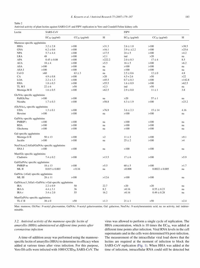
E. Keyaerts et al. / Antiviral Research 75 (2007) 179–187 183
Table 2Antiviral activity of plant lectins against SARS-CoV and FIPV replication in Vero and Crandell Feline kidney cells
Lectin SARS-CoV FIPV
EC50 (�g/ml) CC50 (�g/ml) SI EC50 (�g/ml) CC50 (�g/ml) SI
Mannose-specific agglutininsHHA 3.2 ± 2.8 >100 >31.3 2.6 ± 1.0 >100 >38.5GNA 6.2 ± 0.6 >100 >16.1 3.9 ± ±2.2 >100 >25.6NPA 5.7 ± 4.4 >100 >17.5 24 ± 5 >100 >4.2LRA 48 >100 >2.1 ind >20 naAPA 0.45 ± 0.08 >100 >222.2 2.0 ± 0.3 17 ± 4 8.5AUA 18 ± 4 >100 >5.5 16 ± 5 >100 >6.2ASA >100 >100 na >100 >100 naASA I >100 >100 na >100 >100 naCol O >60 63 ± 3 na 2.5 ± 0.6 12 ± 0 4.8CA 4.9 ± 0.8 >100 >20 4.5 + 2.6 >50 >22LOA 2.2 ± 1.3 >100 >45.5 0.7 ± 0.3 >100 >142.8EHA 1.8 ± 0.3 >100 >55.5 1.6 ± 0.9 >100 >62.5TL M I 22 ± 6 >50 >2.3 ind >50 naMorniga M II 1.6 ± 0.5 >100 >62.5 2.9 ± 0.0 11 ± 1 3.8
GlcNAc-specific agglutininsPallGlcNac >100 >100 na >20 57 ± 1 naNictaba 1.7 ± 0.3 >100 >58.8 4.3 ± 1.9 >100 >23.2
(GlcNAc)n-specific agglutininsUDA 1.3 ± 0.1 >100 >76.9 3.6 ± 2.3 15 ± 14 4.16Heveine >100 >100 na >100 >100 na
GalNAc-specific agglutininsPMRIP t >100 >100 na >100 >100 naBDA >100 >100 na >100 >100 naGlechoma >100 >100 na >100 >100 na
Gal-specific agglutininsMorniga G II 50 ± 13 >100 >2 11 ± 2 >100 >9.1Jacalin >100 >100 na 25 ± 2 >100 >4
Neu5Ac�(2.6)Gal/GalNAc-specific agglutininsSNA I >100 >100 na >100 >100 na
Man/Glc-specific agglutininsCladistris 7.4 ± 0.2 >100 >13.5 17 ± 6 >100 >5.9
Gal/GalNAc specific agglutininsPMRIP m 18 ± 13 >100 >5.5 60 ± 5 >100 >1.7ML II 0.015 ± 0.003 < 0.16 na >0.008 0.0022 ± 0.005 na
GalNAc (>Gal) specific agglutininsML III 28 ± 11 >100 >12.6 >100 >100 na
GalNAc�(1,3)Gal > GalNAc > Gal-specific agglutininsIRA 2.2 ± 0.9 50 22.7 >20 >20 naIRA b 4.4 ± 3.1 36 8.2 >0.16 0.55 ± 0.23 naIRA r 3.4 ± 2.0 55 16.2 >0.16 0.49 ± 0.28 na
Man/GalNAc-specific agglutininsTL C II 38 ± 0 >50 >1.3 21 ± 1 >50 >2.4
M ine, Gm
3ac
saV
vHds
an: mannose, GlcNac: N-acetyl glucosamine, GalNAc: N-acetyl galactosaminable.
.2. Antiviral activity of the mannose-specific lectin ofmaryllis (HHA) administered at different time points afteroronavirus infection
A time-of-addition assay was performed using the mannose-pecific lectin of amaryllis (HHA) to determine its efficacy whendded at various times after virus infection. For this purpose,ero E6 cells were infected with 1000 CCID50 SARS-CoV. The
TlSt
al: galactose, Neu5Ac: N-acetylneuraminic acid, na: no activity, ind: indeter-
irus was allowed to perform a single cycle of replication. TheHA concentration, which is 10 times the EC50, was added atifferent time points after infection. Viral RNA levels in the cellupernatants and in the cells were determined 8 h post-infection.
he measurement of the intracellular viral load shows that theectins are required at the moment of infection to block theARS-CoV replication (Fig. 1). When HHA was added at the
ime of infection, intracellular RNA could still be detected but
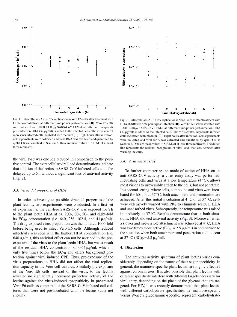
184 E. Keyaerts et al. / Antiviral Research 75 (2007) 179–187
Fig. 1. Intracellular SARS-CoV replication in Vero E6 cells after treatment withHHA concentrations at different time points post-infection (�). Vero E6 cellswere infected with 1000 CCID50 SARS-CoV FFM-1 at different time-pointspost-infection HHA (32 �g/ml) is added to the infected cells. The virus controlrepresents infected cells incubated with medium (�). Eight hours after infection,cqt
tttd(
3
potiTbi6eootvtorlVts
Fig. 2. Extracellular SARS-CoV replication in Vero E6 cells after treatment withHHA at different time points post-infection (�). Vero E6 cells were infected with1000 CCID50 SARS-CoV FFM-1 at different time-points post-infection HHA(32 �g/ml) is added to the infected cells. The virus control represents infectedcells incubated with medium (�). Eight hours after infection, cell supernatantswere collected and viral RNA was extracted and quantified by qRT-PCR asSlw
3
aImIbawaitawta
4
sga
ell supernatants were collected and viral RNA was extracted and quantified byRT-PCR as described in Section 2. Data are mean values ± S.E.M. of at leasthree replicates.
he viral load was one log reduced in comparison to the posi-ive control. The extracellular viral load determinations indicatehat addition of the lectins to SARS-CoV-infected cells could beelayed up to 5 h without a significant loss of antiviral activityFig. 2).
.3. Virucidal properties of HHA
In order to investigate possible virucidal properties of thelant lectins, two experiments were conducted. In a first setf experiments, the cell-free SARS-CoV was exposed for 2 ho the plant lectin HHA at ca. 200-, 80-, 20-, and eight-foldts EC50 concentration (i.e. 640, 256, 102.4, and 41 �g/ml).he drug-exposed virus preparation was then diluted 1000-foldefore being used to infect Vero E6 cells. Although reducednfectivity was seen with the highest HHA concentration (i.e.40 �g/ml), this antiviral effect can not be ascribed to the pre-xposure of the virus to the plant lectin HHA, but was a resultf the residual HHA concentration of 0.64 �g/ml, which isnly five times below the EC50 and offers background pro-ection against viral induced CPE. Thus, pre-exposure of theirus preparations to HHA did not affect the viral replica-ion capacity in the Vero cell cultures. Similarly pre-exposuref the Vero E6 cells, instead of the virus, to the lectins
evealed no significantly increased protective activity of theectins against the virus-induced cytopathicity in pre-treatedero E6 cells as compared to the SARS-CoV-infected cell cul-ures that were not pre-incubated with the lectins (data nothown).
dvgwv
ection 2. Data are mean values ± S.E.M. of at least three replicates. The dottedine represents the residual background of viral load, that was detected afterashing the cells.
.4. Virus entry assay
To further characterize the mode of action of HHA on itsnti-SARS-CoV activity, a virus entry assay was performed.ncubating cells and virus at a low temperature (4 ◦C), allowsost virions to irreversibly attach to the cells, but not penetrate.
n a second setting, where cells, compound and virus were incu-ated for 60 min at 37 ◦C, both attachment and penetration arechieved. After this initial incubation at 4 ◦C or at 37 ◦C, cellsere extensively washed with PBS to eliminate residual HHA
nd unadsorbed virus. Subsequently, the temperature was raisedmmediately to 37 ◦C. Results demonstrate that in both situa-ions, HHA showed antiviral activity (Fig. 3). Moreover, whenslower and irreversible attachment was achieved at 4 ◦C, HHAas two times more active (EC50 = 2.5 �g/ml) in comparison to
he situation when both attachment and penetration could occurt 37 ◦C (EC50 = 5.2 �g/ml).
. Discussion
The antiviral activity spectrum of plant lectins varies con-iderably, depending on the nature of their sugar specificity. Ineneral, the mannose-specific plant lectins are highly effectivegainst coronaviruses. It is also possible that plant lectins withifferent specificity interfere with different targets necessary for
iral entry, depending on the place of the glycans that are tar-eted. For HIV, it was recently demonstrated that plant lectinsith different carbohydrate specificities, i.e. mannose-specificersus N-acetylglucosamine-specific, represent carbohydrate-
E. Keyaerts et al. / Antiviral Research 75 (2007) 179–187 185
Fig. 3. The inhibitory effects of HHA on virus adsorption to Vero E6 cells arepresented as percent protection. Two experiments were performed in parallel. Intma
bd
escNacfpFapNgTgttoitpgHtc
vaoahmba
Fig. 4. Calculation of the correlation coefficient between the EC50 values of theplant lectins against SARS-CoV on the one hand, and the EC50 values of theco0
ir
Hemwialc
ptwpwdiwtcortts
he first setting (white bars), the compound was present only during virus attach-ent. In the second setting (black bars), the compound was present during virus
ttachment and penetration. Data are mean values ± S.E.M. of four replicates.
inding agents with a similar mode of antiviral action, but withifferent genetic barriers (Balzarini et al., 2005).
Out of 33 plant lectins evaluated, 15 showed antiviral prop-rties against both SARS-CoV and FIPV, and 8 plant lectinshowed no anti-coronavirus activity. The lectins with anti-oronaviral activity included mannose-, glucose-, galactose-,-acetyl glucosamine- and N-acetyl galactosamine-specificgglutinins In general plant lectins were more toxic to CrFKells than to Vero E6 cells. This could partly explain some dif-erences between the SARS-CoV activity and FIPV activity oflant lectins. Most of the lectins that were not active againstIPV were too toxic for the CrFK cells. The most prominentnti-coronavirus activity was found among the mannose-specificlant lectins. In the SARS-CoV spike protein there are 12-glycosylation sites. The sugars attached to four of these N-lycosylation sites have been identified (Krokhin et al., 2003).wo of the four sugars were found to be high-mannose typelycans, whereas the other two showed complex glycan struc-ures (Krokhin et al., 2003). The presence of high-mannoseype glycans can explain the potent anti-SARS-CoV activityf mannose-specific plant lectins. When we compare the activ-ty spectrum of the plant lectins against coronaviruses withheir activity spectrum against HIV, only the mannose-specificlant lectins, with the exception of UDA, which is a N-acetyllucosamine-specific lectin, possess antiviral activity againstIV (Balzarini et al., 1991, 1992). Thus, the carbohydrate spec-
rum of plant lectins that inhibit virus infection is broader fororonaviruses than for HIV.
Calculation of the correlation coefficient between the EC50alues of the plant lectins against SARS-CoV on the one hand,nd the EC50 values of the compounds against FIPV on thether hand, resulted in an r-value of 0.38. When only thective mannose-specific lectins were taken into account, a much
igher r-value of 0.70 was calculated (Fig. 4). Furthermore, aarked correlation was found when the correlation coefficientetween the anti-HIV activity of the plant lectins (Balzarini etl., 1991, 1992), and their anti-FIPV or anti-SARS-CoV activ-
nist
ompounds against FIPV on the other hand, resulted in an r-value of 0.38. Whennly the active mannose-specific lectins were taken into account, an r-value of.70 was found.
ty was calculated (r-values were found to be 0.55 and 0.52,espectively).
Plant lectins are known to possess virucidal properties againstIV, and may therefore qualify as HIV microbicides (Balzarini
t al., 2004). In contrast to HIV, the plant lectins did not showarked virucidal properties against the SARS-CoV, neitherhen they were pre-incubated with the cells, nor when pre-
ncubated with the virus. This can be due to a lower bindingffinity (on-rate) of the plant lectins to the coronavirus enve-ope glycans and/or a faster off-rate of the plant lectins from theoronavirus envelope glycans.
To elucidate the mechanism of antiviral intervention of thelant lectins, a time-of-addition assay was elaborated usinghe mannose-specific lectin of the amaryllis (HHA). The lectinas added to the infected cell cultures at different time-pointsost-infection. At 8 h post-infection, the SARS-CoV viral loadas quantified intracellularly and extracellularly. A markedifference was noted between these two measurements. Thentracellular viral RNA-load could only be efficiently reducedhen the lectin was added at the time of infection, indicating that
he plant lectins interact with an early step in the viral replicationycle. In general, the SARS-CoV entry starts with the attachmentf the virus to its specific receptor ACE2 on the cell surface. Theeceptor binding subsequently induces the viral envelope proteino undergo conformational changes that mediate fusion betweenhe viral and cellular membranes (Doms, 2004). The coronaviruspike glycoprotein is responsible for these two steps in the coro-
avirus entry process. Since each step of the viral entry pathways a potential target for antiviral agents, entry inhibitors fall intoeveral categories. A first class consists of inhibitors that bind tohe ACE2 receptor, a second category comprises entry inhibitors
1 l Res
trffcdibnvmpcaowwHtpm
trlpctacc2ttdao
rmdScaftadt
A
obUc
aStdpEKf
R
A
B
B
B
B
B
C
DG
H
H
H
H
86 E. Keyaerts et al. / Antivira
hat bind to the virus and prevent it from interacting with itseceptors and a third group of inhibitors can hamper the con-ormational changes, resulting in the inhibition of SARS-CoVusion with the target cell. In order to distinguish between theseategories of entry inhibitors more in-depth studies were con-ucted. The virus entry assay showed that HHA was active whenncubating the cells and virus at 4 ◦C. This low temperature incu-ation allows most virions to irreversibly attach to the cells, butot to penetrate the cells. Moreover, when this slower and irre-ersible attachment was achieved at 4 ◦C, HHA was two timesore active (EC50 = 2.5 �g/ml) in comparison to attachment and
enetration at 37 ◦C (EC50 = 5.2 �g/ml). This observation indi-ates that this compound most probably interferes with virusttachment. To elucidate if HHA is an ACE2 receptor inhibitorr an entry inhibitor which binds the virus, and prevent it in thisay from interacting with its receptor, we pre-incubated HHAith either the SARS-CoV or with the cells. Pre-incubation ofHA with the virus did not provide inhibition of virus replica-
ion, nor did the pre-incubation of HHA with the cells. Thesere-incubation experiments did not help us to understand theechanism of action of the plant lectins.Interestingly, the results of the extracellular viral RNA quan-
ification also points to an intervention at a late step in theeplication cycle around 5 h post-infection. Huang and col-eagues observed that the presence of the spike protein in aseudoparticle is necessary for budding and formation of aorona-like structure (Huang et al., 2004). This indicates thathe spike protein is likely to be important not only for viralttachment and fusion, but also for maturation and egress fromells. Earlier we demonstrated that one SARS-CoV replicationycle takes 6 h to complete in Vero E6 cells (Keyaerts et al.,005). Therefore, inhibition of extracellular viral RNA-load upo 5 h post-infection suggests interference at the level of exocy-osis or egress from the cell. Taken together, we could clearlyemonstrate that the plant lectins interact both at virus entry andt virus release, a phenomenon that has never been observed forther viruses, including HIV.
In conclusion, we identified a variety of plant lectins as antivi-al compounds against the SARS-CoV and the FIPV. The lectinsost probably interfere with the glycans on the spike protein
uring virus entry and virus release. In view of the fact that theARS-CoV discovery led to the identification of two new humanoronaviruses, HCoV-NL63 and HCoV-HKU1, both of whichre associated with serious lower respiratory diseases, the searchor efficient anti-coronavirus compounds becomes more impor-ant. Our findings on the selective and potent anti-coronavirusctivity of plant lectins should trigger further research on theiscovery of other carbohydrate-binding agents, including syn-hetic low-molecular-weight compounds.
cknowledgments
We would like to thank all the colleagues of the laboratory
f Clinical & Epidemiological Virology, Department of Micro-iology and Immunology, Rega Institute for Medical Research,niversity of Leuven, Belgium, for helpful comments and dis-ussion. We thank Lizette van Berckelaer for excellent technical
K
K
earch 75 (2007) 179–187
ssistance. This work is part of the activities of the Euro-AsianARS-DTV Network (SP22-CT-2004-511064) supported by
he European Commission specific research and technologicalevelopment program “Integrating and Strengthening the Euro-ean Research Area” and the R. Descartes Prize-2001 of theuropean Commission and the Centers of Excellence of the.U. Leuven (to JB.). This work was supported by a postdoctoral
ellowship of the Research Fund K.U. Leuven to Leen Vijgen.
eferences
rden, K.E., Nissen, M.D., Sloots, T.P., Mackay, I.M., 2005. New human coro-navirus, HCoV-NL63, associated with severe lower respiratory tract diseasein Australia. J. Med. Virol. 75, 455–462.
alzarini, J., Schols, D., Neyts, J., Van Damme, E., Peumans, W., De Clercq,E., 1991. Alpha-(1-3)- and alpha-(1-6)-d-mannose-specific plant lectins aremarkedly inhibitory to human immunodeficiency virus and cytomegalovirusinfections in vitro. Antimicrob. Agents Chemother. 35, 410–416.
alzarini, J., Neyts, J., Schols, D., Hosoya, M., Van Damme, E., Peumans, W.,De Clercq, E., 1992. The mannose-specific plant lectins from cymbidiumhybrid and epipactis helleborine and the (N-acetylglucosamine)n-specificplant lectin from Urtica dioica are potent and selective inhibitors of humanimmunodeficiency virus and cytomegalovirus replication in vitro. AntiviralRes. 18, 191–207.
alzarini, J., Hatse, S., Vermeire, K., Princen, K., Aquaro, S., Perno, C.F., DeClercq, E., Egberink, H., Vanden Mooter, G., Peumans, W., Van Damme, E.,Schols, D., 2004. Mannose-specific plant lectins from the Amaryllidaceaefamily qualify as efficient microbicides for prevention of human immun-odeficiency virus infection. Antimicrob. Agents Chemother. 48, 3858–3870.
alzarini, J., Van Laethem, K., Hatse, S., Froeyen, M., Peumans, W., VanDamme, E., Schols, D., 2005. Carbohydrate-binding agents cause deletionsof highly conserved glycosylation sites in HIV GP120: a new therapeuticconcept to hit the achilles heel of HIV. J. Biol. Chem. 280, 41005–41014.
astien, N., Anderson, K., Hart, L., Van Caeseele, P., Brandt, K., Milley, D.,Hatchette, T., Weiss, E.C., Li, Y., 2005. Human coronavirus NL63 infectionin Canada. J. Infect. Dis. 191, 503–506.
hen, Y., Peumans, W.J., Hause, B., Bras, J., Kumar, M., Proost, P., Barre, A.,Rouge, P., Van Damme, E.J., 2002. Jasmonic acid methyl ester induces thesynthesis of a cytoplasmic/nuclear chito-oligosaccharide binding lectin intobacco leaves. FASEB J. 16, 905–907.
oms, R.W., 2004. Viral entry denied. N. Engl. J. Med. 351, 743–744.onzalez, J.M., Gomez-Puertas, P., Cavanagh, D., Gorbalenya, A.E., Enjuanes,
L., 2003. A comparative sequence analysis to revise the current taxonomyof the family Coronaviridae. Arch. Virol. 148, 2207–2235.
ammar, L., Eriksson, S., Morein, B., 1989. Human immunodeficiency virusglycoproteins: lectin binding properties. AIDS Res. Hum. Retroviruses 5,495–506.
ansen, J.E., Nielsen, C.M., Nielsen, C., Heegaard, P., Mathiesen, L.R., Nielsen,J.O., 1989. Correlation between carbohydrate structures on the envelope gly-coprotein gp120 of HIV-1 and HIV-2 and syncytium inhibition with lectins.AIDS 3, 635–641.
erman, P., Verlinden, Y., Breyer, D., Van Cleemput, E., Brochier, B., Sneyers,M., Snacken, R., Hermans, P., Kerkhofs, P., Liesnard, C., Rombaut, B., VanRanst, M., Van Der Groen, G., Goubau, P., Moens, W., 2004. Biosafety riskassessment of the severe acute respiratory syndrome (SARS) Coronavirusand containment measures for the diagnostic and research laboratories. Appl.Biosafety 9, 128–142.
uang, Y., Yang, Z.Y., Kong, W.P., Nabel, G.J., 2004. Generation of syntheticsevere acute respiratory syndrome coronavirus pseudoparticles: implicationsfor assembly and vaccine production. J. Virol. 78, 12557–12565.
arber, G., 1931. Beitrag zur kollektiven behandlungpharmakologischer Rei-henversuche. Arch. Exp. Pathol. Pharmakol. 162, 480–483.
eyaerts, E., Vijgen, L., Maes, P., Neyts, J., Van Ranst, M., 2004. In vitro inhi-bition of severe acute respiratory syndrome coronavirus by chloroquine.Biochem. Biophys. Res. Commun. 323, 264–268.
l Res
K
K
K
L
M
M
M
M
P
R
S
V
V
V
v
W
Woo, P.C., Lau, S.K., Chu, C.M., Chan, K.H., Tsoi, H.W., Huang, Y., Wong,
E. Keyaerts et al. / Antivira
eyaerts, E., Vijgen, L., Maes, P., Neyts, J., Van Ranst, M., 2005. Growth kineticsof SARS-coronavirus in Vero E6 cells. Biochem. Biophys. Res. Commun.329, 1147–1151.
eyaerts, E., Vijgen, L., Maes, P., Duson, G., Neyts, J., Van Ranst, M., 2006.Viral load quantitation of SARS-coronavirus RNA using a one-step real-timeRT-PCR. Int. J. Infect. Dis. 10, 32–37.
rokhin, O., Li, Y., Andonov, A., Feldmann, H., Flick, R., Jones, S., Stroeher,U., Bastien, N., Dasuri, K.V., Cheng, K., Simonsen, J.N., Perreault, H.,Wilkins, J., Ens, W., Plummer, F., Standing, K.G., 2003. Mass spectrometriccharacterization of proteins from the SARS virus: a preliminary report. Mol.Cell Proteomics. 2, 346–356.
arson, H.E., Reed, S.E., Tyrrell, D.A., 1980. Isolation of rhinoviruses andcoronaviruses from 38 colds in adults. J. Med. Virol. 5, 221–229.
atsui, T., Kobayashi, S., Yoshida, O., Ishii, S., Abe, Y., Yamamoto, N., 1990.Effects of succinylated concanavalin A on infectivity and syncytial formationof human immunodeficiency virus. Med. Microbiol. Immunol. (Berl) 179,225–235.
cKeirnan, A.J., Evermann, J.F., Hargis, A., Ott, R.L., 2005. Isolation of felinecoronaviruses from two cats with diverse disease manifestations. FelinePract. 11, 17–20.
oes, E., Vijgen, L., Keyaerts, E., Zlateva, K., Li, S., Maes, P., Pyrc, K.,Berkhout, B., van der, H.L., Van Ranst, M., 2005. A novel pancoronavirusRT-PCR assay: frequent detection of human coronavirus NL63 in childrenhospitalized with respiratory tract infections in Belgium. BMC. Infect. Dis.5, 6.
uller, W.E., Renneisen, K., Kreuter, M.H., Schroder, H.C., Winkler, I., 1988.The d-mannose-specific lectin from Gerardia savaglia blocks binding ofhuman immunodeficiency virus type I to H9 cells and human lymphocytesin vitro. J. Acquir. Immune. Defic. Syndr. 1, 453–458.
edersen, N.C., Boyle, J.F., Floyd, K., Fudge, A., Barker, J., 1981. An entericcoronavirus infection of cats and its relationship to feline infectious peri-tonitis. Am. J. Vet. Res. 42, 368–377.
ota, P.A., Oberste, M.S., Monroe, S.S., Nix, W.A., Campagnoli, R., Icenogle,J.P., Penaranda, S., Bankamp, B., Maher, K., Chen, M.H., Tong, S., Tamin,
earch 75 (2007) 179–187 187
A., Lowe, L., Frace, M., DeRisi, J.L., Chen, Q., Wang, D., Erdman, D.D.,Peret, T.C., Burns, C., Ksiazek, T.G., Rollin, P.E., Sanchez, A., Liffick, S.,Holloway, B., Limor, J., McCaustland, K., Olsen-Rasmussen, M., Fouchier,R., Gunther, S., Osterhaus, A.D., Drosten, C., Pallansch, M.A., Ander-son, L.J., Bellini, W.J., 2003. Characterization of a novel coronavirusassociated with severe acute respiratory syndrome. Science 300, 1394–1399.
loots, T.P., McErlean, P., Speicher, D.J., Arden, K.E., Nissen, M.D., Mackay,I.M., 2006. Evidence of human coronavirus HKU1 and human bocavirus inAustralian children. J. Clin. Virol. 35, 99–102.
abret, A., Dina, J., Gouarin, S., Petitjean, J., Corbet, S., Freymuth, F., 2006.Detection of the new human coronavirus HKU1: a report of 6 cases. Clin.Infect. Dis. 42, 634–639.
an Damme, E., Peumans, W., Pusztai, A., Bardocz, S., 1998. Handbook ofPlant Lectins: Properties and Biomedical Applications. John Wiley & Sons,Chichester, West Sussex, England.
an Damme, E.J., Hause, B., Hu, J., Barre, A., Rouge, P., Proost, P., Peumans,W.J., 2002. Two distinct jacalin-related lectins with a different specificityand subcellular location are major vegetative storage proteins in the bark ofthe black mulberry tree. Plant Physiol. 130, 757–769.
an der Hoek, L., Pyrc, K., Jebbink, M.F., Vermeulen-Oost, W., Berkhout, R.J.,Wolthers, K.C., Wertheim-van Dillen, P.M., Kaandorp, J., Spaargaren, J.,Berkhout, B., 2004. Identification of a new human coronavirus. Nat. Med.10, 368–373.
ang, W., Peumans, W.J., Rouge, P., Rossi, C., Proost, P., Chen, J., Van Damme,E.J., 2003. Leaves of the Lamiaceae species Glechoma hederacea (groundivy) contain a lectin that is structurally and evolutionary related to the legumelectins. Plant J. 33, 293–304.
B.H., Poon, R.W., Cai, J.J., Luk, W.K., Poon, L.L., Wong, S.S., Guan,Y., Peiris, J.S., Yuen, K.Y., 2005. Characterization and complete genomesequence of a novel coronavirus, coronavirus HKU1, from patients withpneumonia. J. Virol. 79, 884–895.