piper sarmentosum inhibits icam-1 and nox4 gene expression in oxidative stress-induced human...
TRANSCRIPT

RESEARCH ARTICLE Open Access
Piper sarmentosum inhibits ICAM-1 and Nox4gene expression in oxidative stress-inducedhuman umbilical vein endothelial cellsAzizah Ugusman, Zaiton Zakaria*, Chua Kien Hui and Nor Anita Megat Mohd Nordin
Abstract
Background: Aqueous extract of Piper sarmentosum (AEPS) is known to possess antioxidant and anti-atherosclerotic activities but the mechanism responsible for it remains unclear. In early part of atherosclerosis,nuclear factor-kappa B (NF-�B) induces the expression of cellular adhesion molecules such as vascular cell adhesionmolecule-1 (VCAM-1), intracellular adhesion molecule-1 (ICAM-1) and E-selectin. NADPH oxidase 4 (Nox4) is thepredominant source of superoxide in the endothelial cells whereas superoxide dismutase 1 (SOD1), catalase (CAT)and glutathione peroxidase (GPx) are the antioxidant enzymes responsible for inactivating reactive oxygen species.The present study aimed to investigate the effects of AEPS on the gene expression of NF-�B, VCAM-1, ICAM-1, E-selectin, Nox4, SOD1, CAT and GPx in cultured human umbilical vein endothelial cells (HUVECs).
Methods: HUVECs were divided into four groups:- control; treatment with 180 μM hydrogen peroxide (H2O2);treatment with 150 μg/mL AEPS and concomitant treatment with AEPS and H2O2 for 24 hours. Total RNA wasextracted from all the groups of HUVEC using TRI reagent. Subsequently, qPCR was carried out to determine themRNA expression of NF-�B, VCAM-1, ICAM-1, E-selectin, Nox4, SOD1, CAT and GPx. The specificity of the reactionswas verified using melting curve analysis and agarose gel electrophoresis.
Results: When stimulated with H2O2, HUVECs expressed higher level of ICAM-1 (1.3-fold) and Nox4 (1.2-fold) mRNAexpression. However, AEPS treatment led to a reduction in the mRNA expression of ICAM-1 (p < 0.01) and Nox4 (p< 0.05) in the H2O2-induced HUVECs. AEPS also upregulated the mRNA expression of SOD1 (p < 0.05), CAT (p <0.01) and GPx (p < 0.05) in oxidative stress-induced HUVECs. There was no significant change in the mRNAexpression of VCAM-1 and E-selectin.
Conclusion: The expressional suppression of ICAM-1 and Nox4 and induction of antioxidant enzymes might be animportant component of the vascular protective effect of AEPS.
BackgroundAtherosclerosis has been recognized as a chronic inflam-matory disease and oxidative stress plays a pivotal rolein its initiation and progression [1]. Endothelial dysfunc-tion is considered to be an early marker for athero-sclerosis [2]. Evidence suggests that increasedproduction of reactive oxygen species (ROS) and vascu-lar inflammation play important roles in endothelialdysfunction.
Vascular disorders, through over expression of adhe-sion molecules and cytokines are involved in the devel-opment of atherosclerosis. Endothelial cells in humanatherosclerotic lesions have increased cell adhesionmolecules expression such as intercellular adhesionmolecule-1 (ICAM-1), vascular cell adhesion molecule-1(VCAM-1) and endothelial selectin (E-selectin) [3,4].The adhesion of monocytes to the arterial wall and theirsubsequent infiltration and differentiation into macro-phages are the key events in the development of athero-sclerosis. Nuclear factor-kappa B (NF-�B) is known toplay a critical role in the development of inflammatoryresponse by upregulating the expression of VCAM-1,ICAM-1 and E-selectin [5]. It has been suggested that
* Correspondence: [email protected] of Physiology, Faculty of Medicine, Universiti KebangsaanMalaysia Medical Centre, Jalan Raja Muda Abdul Aziz, 50300 Kuala Lumpur,Malaysia
Ugusman et al. BMC Complementary and Alternative Medicine 2011, 11:31http://www.biomedcentral.com/1472-6882/11/31
© 2011 Ugusman et al; licensee BioMed Central Ltd. This is an Open Access article distributed under the terms of the CreativeCommons Attribution License (http://creativecommons.org/licenses/by/2.0), which permits unrestricted use, distribution, andreproduction in any medium, provided the original work is properly cited.

NF-�B is an oxidative stress-responsive transcriptionfactor. Antioxidants and free radical scavengers such asvitamin E derivatives, N-acetyl-cysteine and thiolreagents inhibit the activation of NF-�B, strongly sup-porting the idea that reactive oxygen species (ROS) areinvolved in the activation process [6].Under oxidative stress, macrophages generate ROS
such as superoxides, leading to LDL oxidation [7]. Inthe vascular wall, ROS can be produced by severalenzyme systems including NADPH oxidases, xanthineoxidase, uncoupled endothelial nitric oxide synthase,lipoxygenases and myeloperoxidase [8]. Although allthese enzymes can contribute to oxidative stress,NADPH oxidases (Nox) are the predominant source ofROS in the vasculature. In diseased human coronaryarteries, about 60% of total vascular superoxide isderived from Nox [9]. Vascular tissues express the Noxisoforms Nox1, Nox2, Nox4 and Nox5. In human umbi-lical vein endothelial cells (HUVECs), the expressionlevel of Nox4 is 100-fold higher than that of Nox1,Nox2 or Nox5, suggesting that Nox4 is the major sourceof ROS in these cells [10]. Increased Nox4 expression isassociated with early progression of atherosclerotic pla-que [11].To protect cells from the damage caused by ROS,
organisms have evolved several defense mechanisms torapidly and efficiently remove ROS. This includes theantioxidant enzymes such as superoxide dismutase(SOD), catalase (CAT) and glutathione peroxidase(GPx). SOD catalyzes the dismutation of superoxide tohydrogen peroxide (H2O2) while CAT and GPx convertH2O2 to water [12].Piper sarmentosum (Figure 1) is a herbaceous plant
that is commonly found in the tropical regions such asthe Southeast Asia. The plant extracts have beenreported to possess pharmacological properties like anti-tuberculous [13], anti cancer [14], hypoglycaemic [15],anti-malarial [16], anti-nociceptive and anti-inflamma-tory [17]. As per recent research reports, the aqueousextracts of Piper sarmentosum (AEPS) leaves have beenreported to improve endothelial function by promotingnitric oxide production in HUVECs [18]. Variousextracts prepared from Piper sarmentosum leaves haveexhibited potent effects as antioxidant in modulatingoxidative stress in H2O2-induced HUVECs [19]. More-over, administration of AEPS reduced atheroscleroticlesion in hypercholesterolemic rabbits [20]. Based on thecardiovascular protective effects of AEPS mentionedabove, the present study was designed to investigate theeffects of AEPS on the gene expression of NF-�B, cellu-lar adhesion molecules (VCAM-1, ICAM-1 and E-selec-tin), Nox4 and antioxidant enzymes (SOD1, CAT, GPx)in HUVECs.
MethodsPreparation of aqueous extract of Piper sarmentosumLeaves of Piper sarmentosum were collected in SungaiBuloh, Malaysia and identified by a plant taxonomistfrom Forest Research Institute of Malaysia (voucher spe-cimen: FRI 45870). The leaves were washed with tapwater, cut into small pieces, sun-dried and grounded topowder form. Ten percent of AEPS was prepared bysoaking 100 g of the powdered leaves in 900 ml of puri-fied water and incubated in a high speed mixer at 80°Cfor 3 hours. After being left to cool, the extract was fil-tered using a mesh and further concentrated. The aqu-eous extract was then freeze-dried to powdered form ofthe extract and kept at 4°C until the experiments.
Cell culture and treatment protocolsHuman umbilical cords were obtained under sterile con-dition. Informed consent was obtained from each sub-ject and the present study was approved by the EthicalResearch Committee of Universiti Kebangsaan MalaysiaMedical Center (Project Code: FF-148-2009). HUVECswere obtained from umbilical cord veins by 0.1% Col-lagenase Type I (Gibco-Invitrogen Corp., Grand Island,N.Y.) digestion. Cells were grown in medium 200 (Cas-cade Biologics, USA) supplemented with LSGS (lowserum growth supplement; Cascade Biologics, USA) at37°C in a humidified atmosphere of 5% CO2 and 95%air. HUVECs were confirmed by the typical endothelialcell cobblestone morphology and the positive
Figure 1 Piper sarmentosum leaves.
Ugusman et al. BMC Complementary and Alternative Medicine 2011, 11:31http://www.biomedcentral.com/1472-6882/11/31
Page 2 of 8
expressions of vonWillebrand factor and CD31 inimmunocytochemistry. The culture medium was chan-ged every other day until the cells reached confluence.HUVECs from passage 3 at 80% confluency were usedfor this experiment. The cells were divided into fourgroups:- control; treatment with 180 μM H2O2; treat-ment with 150 μg/mL AEPS and concomitant treatmentwith 150 μg/mL AEPS and 180 μM H2O2. All treat-ments were given for 24 hours. The dose of H2O2 usedwas based on the IC50 of H2O2 while the dose of AEPSwas based on the EC50 of AEPS adopted from a previousstudy [19]. The rationale to use H2O2 in this study wasto induce oxidative stress. Oxidative stress has beenassociated with endothelial dysfunction which later pro-gresses into the development of atherosclerosis.
Quantitative reverse transcription polymerase chainreaction (qPCR)After treatment for 24 hours, total RNA from HUVECswas extracted using TRI Reagent (Molecular ResearchCenter, Cincinnati, USA) as previous research protocol[21]. Polyacryl carrier (Molecular Research Center,Cincinnati, USA) was added to precipitate the totalRNA. Extracted RNA pellet was then washed with 75%ethanol and dried prior to dissolving it in RNase andDNase free water (Invitrogen, Carlsbad, USA).Extracted total RNA was assessed for its purity andquantity using Nanodrop ND-100 spectrophotometer(Wilmington DE, USA) and stored at -80°C before use.Complimentary DNA (cDNA) was synthesized usingSuperScript III First-Strand Synthesis SuperMix (Invi-trogen, Carlsbad, USA). A total of 20 μl of volumereaction which consisted of 10 μl of 2X RT ReactionMix, 2 μl of RT Enzyme, 5 μl of total RNA and 3 μl of
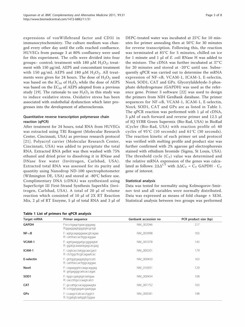
DEPC-treated water was incubated at 25°C for 10 min-utes for primer annealing then at 50°C for 30 minutesfor reverse transcription. Following this, the reactionwas terminated at 85°C for 5 minutes, chilled on icefor 1 minute and 1 μl of E. coli RNase H was added tothe mixture. The cDNA was further incubated at 37°Cfor 20 minutes and stored at -20°C until use. Subse-quently qPCR was carried out to determine the mRNAexpression of NF-�B, VCAM-1, ICAM-1, E-selectin,Nox4, SOD1, CAT and GPx. Glycerylaldehyde-3-phos-phate dehydrogenase (GAPDH) was used as the refer-ence gene. Primer 3 software [22] was used to designthe primers from NIH GenBank database. The primersequences for NF-�B, VCAM-1, ICAM-1, E-selectin,Nox4, SOD1, CAT and GPx are as listed in Table 1.The qPCR reaction was performed with 1 μl of cDNA,5 μM of each forward and reverse primer and 12.5 μlof IQ SYBR Green Supermix (Bio-Rad, USA) in BioRadiCycler (Bio-Rad, USA) with reaction profile of: 40cycles of 95°C (10 seconds) and 61°C (30 seconds).The reaction kinetic of each primer set and protocolwas verified with melting profile and product size wasfurther confirmed with 2% agarose gel electrophoresisstained with ethidium bromide (Sigma, St Louis, USA).The threshold cycle (CT) value was determined andthe relative mRNA expression of the genes was calcu-lated as follows: 2ΔΔCT with ΔΔCT = CT GAPDH - CT
gene of interest.
Statistical analysisData was tested for normality using Kolmogorov-Smir-nov test and all variables were normally distributed.Data was expressed as means of fold change ± SEM.Statistical analysis between two groups was performed
Table 1 List of primers for qPCR analysis
Target mRNA Primer sequence Genbank accession no PCR product size (bp)
GAPDH F:tccctgagctgaacgggaagR:ggaggagtgggtgtcgctgt
NM_002046 217
NF-�B F: agtgcagaggaaacgtcagaaR: cattttaccacttggcaggaa
NM_003998 163
VCAM-1 F: agttgaaggatgcgggagtatR: ggatgcaaaatagagcacgag
NM_001078 143
ICAM-1 F: cagtcacctatggcaacgactR: ctctggcttcgtcagaatcac
NM_000201 179
E-selectin F: gtttggtgaggtgtgctcattR: cattttaccacttggcaggaa
NM_000450 163
Nox4 F: cagaaggttccaagcaggagR: gttgagggcattcaccagat
NM_016931 129
SOD1 F: tggccgatgtgtctattgaaR: cacctttgcccaagtcatct
NM_000454 108
CAT F: gccattgccacaggaaagtaR: ccttggtgagatcgaatgga
NM_001752 103
GPx F: ccaagctcatcacctggtctR: tcgatgtcaatggtctggaa
NM_000581 198
Ugusman et al. BMC Complementary and Alternative Medicine 2011, 11:31http://www.biomedcentral.com/1472-6882/11/31
Page 3 of 8

using paired t-test in SPSS version 16.0 software. Valuesof p < 0.05 were considered statistically significant.
ResultsEffects of AEPS on NF-�B mRNA expression in HUVECsBoth AEPS and H2O2 did not show any significantchanges in the mRNA expression of NF-�B (Figure 2).
Effects of AEPS on VCAM-1, ICAM-1 and E-selectin mRNAexpression in HUVECsTreatment with AEPS alone did not show a significantchange in ICAM-1 expression (Figure 3B). HUVECstreated with H2O2 showed a significantly higher (1.3-fold) level of ICAM-1 mRNA expression compared tothe control group. Concomitant treatment of HUVECswith both AEPS and H2O2 resulted in a down regulationof ICAM-1 mRNA expression than the control andH2O2 groups. However, there was no significant changein the mRNA expressions of VCAM-1 and E-selectin inresponse to AEPS and H2O2 (Figure 3A, 3C).
Effects of AEPS on Nox4 mRNA expression in HUVECsThe aqueous extract of PS significantly reduced Nox4mRNA expression in HUVECs compared to the controlgroup (Figure 4). When stimulated with H2O2, HUVECsexpressed higher (1.2-fold) level of Nox4 mRNA expres-sion. However, the H2O2-induced Nox4 mRNA expres-sion was significantly down regulated by AEPS.
Effects of AEPS on SOD1, CAT and GPx mRNA expressionin HUVECsHUVECs treated with AEPS had significantly higherlevel of SOD1, CAT and GPx mRNA expressions com-pared to the control group (Figure 5A, 5B, 5C). The
highest increase was observed in the CAT expression(2.2-fold) followed by GPx (1.3 fold) and SOD1 (1.2-fold). In the oxidative stress-induced group, HUVECstreated with H2O2 also showed significantly higher levelof SOD1, CAT and GPx mRNA expressions comparedwith the control group. HUVECs treated with bothAEPS and H2O2 had significantly higher level of SOD1,CAT and GPx mRNA expressions than the controlgroup but no significant difference from the singleH2O2-treated and AEPS-treated groups.
DiscussionIt is well known that the transcription factor NF-�B isessential in regulation of the gene expression of celladhesion molecules such as VCAM-1, ICAM-1 and E-selectin [5]. Therefore, we hypothesized that AEPS hasthe ability to modulate the expression of NF-�B and celladhesion molecules in H2O2-induced HUVECs. In thepresent study, both AEPS and H2O2 did not have anysignificant effect on the gene expression of NF-�B inHUVECs (Figure 2). In another study, H2O2 inducesNF-�B activation in porcine aortic endothelial cells butnot in human aortic endothelial cells, suggesting thatporcine endothelial cells might be more sensitive toH2O2 compared to human endothelial cells [23].However, treatment with H2O2 caused an upregula-
tion of ICAM-1 mRNA expression (Figure 3B). TheH2O2-induced ICAM-1 mRNA expression was signifi-cantly down regulated by AEPS. Both AEPS and H2O2
did not have any significant effect on the gene expres-sion of VCAM-1 and E-selectin (Figure 3A, 3C).In this study, the effects of H2O2 on the cellular adhe-
sion molecules expression are in accordance with earlierresearch [24]. In the study, treatment of HUVECs with50 μmol/L H2O2 for 24 hours increased the level ofICAM-1 mRNA expression but it did not induce theexpression of VCAM-1 and E-selectin. Hydrogen perox-ide also did not seem to activate NF-�B. This could becaused by a difference in sensitivity of endothelial cellsamong different species. Human endothelial cells arerelatively resistant to oxidative damage compared toendothelial cells cultured from other species [24]. Treat-ment with 400 μM H2O2 upregulates VCAM-1 expres-sion in porcine aortic endothelial cells but not onHUVECs and human aortic endothelial cells, suggestingthat porcine endothelial cells might be more sensitive toH2O2 compared with human endothelial cells [23].However, at a higher dose (> 1000 μM) of H2O2,HUVECs can significantly upregulate VCAM-1 expres-sion [23]. The dose used in this study was 180 μMwhich was much lower than that. The lower dose ofH2O2 and the relative resistance of HUVECs to oxida-tive damage may contribute to the non-significantchanges in VCAM-1 and E-selectin.
Figure 2 NF-�B mRNA expression in HUVECs. Figure 2 representsthe bar chart showing NF-�B mRNA expression in control, AEPS,H2O2 and AEPS + H2O2 groups. Both AEPS and H2O2 did not causesignificant changes in NF-�B mRNA expression in HUVEC. Values aremeans ± SEM of n = 6.
Ugusman et al. BMC Complementary and Alternative Medicine 2011, 11:31http://www.biomedcentral.com/1472-6882/11/31
Page 4 of 8

The present study demonstrated that AEPS downregulated the mRNA expression of the ROS-producingenzyme Nox4 (Figure 4), and at the same time, upregu-lated the expression of ROS-inactivating enzymes;SOD1, CAT and GPx (Figure 5A, 5B, 5C) in HUVECs.Although several ROS-generating systems have beendescribed in endothelial and other vascular cells,NADPH oxidases (Nox) have now been recognized tobe the major source of ROS in the vasculature [9]. TheNox enzyme complex consists of two essential mem-brane-bound subunits, gp91phox and p22phox, whichcomposed of cytochrome b558, and several cytosolicregulatory components. The enzyme is dormant in rest-ing cells, but on stimulation, the cytosolic subunitstranslocate to the cytochrome b558 at the membraneleading to activation of the enzyme and the release oflarge amounts of superoxides [25].NADPH oxidase-mediated ROS production is regu-
lated at two levels: gene expression of the Nox subunitsand the enzyme activity [26]. To the best of our
Figure 3 VCAM-1, ICAM-1 and E-selectin mRNA expression in HUVECs. Figure 3 represents the bar chart showing VCAM-1 (A), ICAM-1 (B)and E-selectin (C) mRNA expression in control, AEPS, H2O2 and AEPS + H2O2 groups. HUVECs treated with H2O2 showed a significantly higherlevel of ICAM-1 mRNA expression. The H2O2-induced ICAM-1 mRNA expression was significantly down regulated by AEPS. Data are denoted asmean ± SEM of n = 6. (**) p < 0.01 vs. control; (##) p < 0.01 vs. H2O2.
Figure 4 Nox4 mRNA expression in HUVECs. Figure 4 representsthe bar chart showing Nox4 mRNA expression in control, AEPS,H2O2 and AEPS + H2O2 groups. HUVECs treated with AEPS hadlower Nox4 mRNA expression while H2O2 caused a higher Nox4mRNA expression. Data are denoted as mean ± SEM of n = 6. (*) p< 0.05 vs. control; (#) p < 0.05 vs. H2O2.
Ugusman et al. BMC Complementary and Alternative Medicine 2011, 11:31http://www.biomedcentral.com/1472-6882/11/31
Page 5 of 8

knowledge, the present study is the first of its kind toreport that AEPS decreased the gene expression ofNox4, which is the predominant Nox isoform found inendothelial cells. The present study also showed thattreatment of HUVECs with H2O2 upregulated Nox4mRNA expression. In another study, H2O2 was capableof upregulating the Nox subunit p22phox mRNA andprotein expression in endothelial cells [25]. The H2O2-induced Nox4 mRNA expression was significantly downregulated by AEPS (Figure 4). This could be one of themechanisms by which AEPS reduced endothelial oxida-tive stress.The antioxidant enzymes represent a first line of
defense against ROS by metabolizing them to innocuousbyproducts. The first enzymatic reaction in the reduc-tion pathway of oxygen occurs during the dismutationof two molecules of superoxides when they are con-verted to H2O2 and oxygen. The enzyme involved at
this step is one of two isoforms of superoxide dismutase(SOD); CuZnSOD or SOD1 which is present in thecytosol while MnSOD or SOD2 is located in the mito-chondrial matrix. Although H2O2 is not a free radicalitself, it is reactive and it is rapidly converted into thehighly reactive hydroxyl anion in the presence of ferrousion via the Fenton reaction unless it is efficientlyremoved. Two enzymes participate in the removal ofH2O2 from the cellular environment; glutathione peroxi-dase (GPx) and catalase (CAT). Glutathione peroxidaseis present in both the cytosol and mitochondria whileCAT is present mainly in the peroxisomes. Catalase andGPx detoxify H2O2 into water and oxygen [12].Antioxidant enzymes; SOD, CAT and GPx are
thought to be effective for augmentation of antioxidantdefenses in endothelial cells [27]. As shown in Figure 5,treatment with AEPS upregulated the expression ofSOD1, CAT and GPx in HUVECs. These results
Figure 5 SOD1, CAT and GPx mRNA expression in HUVECs. Figure 5 represents the bar chart showing SOD1 (A), CAT (B) and GPx (C) mRNAexpression in control, AEPS, H2O2 and AEPS + H2O2 groups. Single treatment of HUVEC with AEPS or H2O2 significantly increased SOD1, CATand GPx mRNA expression. The highest level of SOD1, CAT and GPx mRNA expression was observed in HUVEC treated with both AEPS andH2O2. Data are expressed as mean ± SEM of n = 10. (*) p < 0.05 vs. control; (**) p < 0.01 vs. control.
Ugusman et al. BMC Complementary and Alternative Medicine 2011, 11:31http://www.biomedcentral.com/1472-6882/11/31
Page 6 of 8
suggested that the protective effects of AEPS againstoxidative stress may be related to the increased abilityto upregulate the antioxidant enzymes expression. Thegreatest inductions were in the levels of CAT mRNA;indicating that CAT plays an important role in scaven-ging H2O2. In other reports, upregulation of antioxidantenzymes by resveratrol protects aortic smooth musclecells [28] and HUVECs [29] against oxidative stress.When HUVECs were exposed to H2O2, there was anincrease in the mRNA expression of SOD1, CAT andGPx. This could be part of the defense mechanism ofthe cells to protect themselves better from the damagethat was induced by H2O2 [30].This study showed that AEPS significantly reduced
Nox4 expression and increased SOD1, CAT and GPxexpression. This indicates an increase in cellular defensemechanism against oxidative stress and implies the vas-culature-protective effect of AEPS. Previous studyshowed that AEPS has direct ROS scavenging ability[31]. Therefore, this study further improves the previousknowledge on the vasculature-protective effects of AEPSas it shows that AEPS can effectively increase celldefense mechanism against oxidative stress.The leaves of Piper sarmentosum contained biologi-
cally active flavonoid compounds such as myricetin, api-genin, quercetin and rutin which are potent antioxidants[13,32]. Since the aqueous extract of Piper sarmentosumleaves used in the present study was not a purified com-ponent, the limitation of this study was the inability todetermine the specific components of the plant thatmediated the observed effects. However, it is suggestedthat the effects were due to the flavonoid compoundsmentioned above.
ConclusionsThe present study describes some novel effects of AEPS.By decreasing the expression of ICAM-1 and Nox4 andenhancing the expression of SOD1, CAT and GPx,AEPS represents a unique approach in reducingendothelial oxidative stress. The findings indicate a newinsight into the mechanism involved in the vascular pro-tective effect of AEPS.
AcknowledgementsThe authors are grateful to Universiti Kebangsaan Malaysia Medical Centre(FF-148-2009), Ministry of Science, Technology and Innovation (02-01-02-SF-0447) as well as Ministry of Higher Education (UKM-FF-03-FRGS0005-2007),Malaysia for funding this project. The authors also would like to thankAssociate Prof. Dr. Srijit Das for his contribution in editing this paper.
Authors’ contributionsAU: Performing the study, analyzing the data and preparing the manuscript.ZZ: Supervising the work, providing the grants for the study, evaluating thedata, correcting the manuscript and coordinating the study. CKH:Supervising the work, evaluating the data, correcting the manuscript andcoordinating the study. NAMMN: Providing the grants for the study,
evaluating the data and correcting the manuscript. All authors read andapproved the final manuscript.
Competing interestsThe authors declare that they have no competing interests.
Received: 31 December 2010 Accepted: 16 April 2011Published: 16 April 2011
References1. Libby P, Theroux P: Pathophysiology of coronary artery disease.
Circulation 2005, 111(25):3481-3488.2. Kawashima S: Malfunction of Vascular Control in Lifestyle-Related
Diseases: Endothelial Nitric Oxide (NO) Synthase/NO System inAtherosclerosis. J Pharmacol Sci 2004, 96(4):411-419.
3. Lopes-Virella M, Virella G, Orchard T, Koskinen S, Evans R, Becker D,Forrest K: Antibodies to Oxidized LDL and LDL-Containing ImmuneComplexes as Risk Factors for Coronary Artery Disease in DiabetesMellitus 1. Clin Immunol 1999, 90(2):165-172.
4. Quagliaro L, Piconi L, Assaloni R, Da Ros R, Maier A, Zuodar G, Ceriello A:Intermittent high glucose enhances ICAM-1, VCAM-1 and E-selectinexpression in human umbilical vein endothelial cells in culture: thedistinct role of protein kinase C and mitochondrial superoxideproduction. Atherosclerosis 2005, 183(2):259-267.
5. Kang J, Park S, Yang K, Kim H: Silymarin inhibits TNF-alpha-inducedexpression of adhesion molecules in human umbilical vein endothelialcells. FEBS letters 2003, 550(1-3):89-93.
6. Wei Z, Peng Q, Lau B, Shah V: Ginkgo biloba inhibits hydrogen peroxide-induced activation of nuclear factor kappa B in vascular endothelialcells. Gen Pharmacol 1999, 33(5):369-375.
7. Steinberg D: Low density lipoprotein oxidation and its pathobiologicalsignificance. J Biol Chem 1997, 272(34):20963-20966.
8. Droge W: Free radicals in the physiological control of cell function.Physiol Rev 2002, 82(1):47-95.
9. Guzik T, Sadowski J, Guzik B, Jopek A, Kapelak B, Przybylowski P,Wierzbicki K, Korbut R, Harrison D, Channon K: Coronary artery superoxideproduction and nox isoform expression in human coronary arterydisease. Arterioscler Thromb Vasc Biol 2006, 26(2):333-339.
10. Xu H, Goettsch C, Xia N, Horke S, Morawietz H, Förstermann U, Li H:Differential roles of PKC alpha and PKC in controlling the geneexpression of Nox4 in human endothelial cells. Free Radic Biol Med 2008,44(8):1656-1667.
11. Sorescu D, Weiss D, Lassegue B, Clempus R, Szocs K, Sorescu G, Valppu L,Quinn M, Lambeth J, Vega J: Superoxide production and expression ofNox family proteins in human atherosclerosis. Circulation 2002,105(12):1429-1435.
12. Mates J: Effects of antioxidant enzymes in the molecular control ofreactive oxygen species toxicology. Biogenic amines 2000, 16(1):53-62.
13. Hussain K, Ismail Z, Sadikun A, Ibrahim P: Antioxidant, anti-TB activities,phenolic and amide contents of standardised extracts of Pipersarmentosum Roxb. Nat Prod Res 2009, 23(3):238-249.
14. Hisham Z, Haryani W, Zaidah Z, Fauzi S, Sahidan S, Rohaya M: Intrinsicanticarcinogenic effects of Piper sarmentosum ethanolic extract on ahuman hepatoma cell line. Cancer Cell Int 2009, 9.
15. Peungvicha P, Thirawarapan S, Temsiririrkkul R, Watanabe H, KumarPrasain J, Kadota S: Hypoglycemic effect of the water extract of Pipersarmentosum in rats. J Ethnopharmacol 1998, 60(1):27-32.
16. Najib Nik A, Rahman N, Furuta T, Kojima S, Takane K, Ali Mohd M:Antimalarial activity of extracts of Malaysian medicinal plants. JEthnopharmacol Journal 1999, 64(3):249-254.
17. Zakaria Z, Patahuddin H, Mohamad A, Israf D, Sulaiman M: In vivo anti-nociceptive and anti-inflammatory activities of the aqueous extractof the leaves of Piper sarmentosum. J Ethnopharmacol 2009,128:42-48.
18. Ugusman A, Zakaria Z, Hui C, Nordin N: Piper sarmentosum increasesnitric oxide production in oxidative stress: a study on human umbilicalvein endothelial cells. Clinics 2010, 65:709-714.
19. Hafizah A, Zaiton Z, Zulkhairi A, Mohd Ilham A, Nor Anita M, Zaleha A:Piper sarmentosum as an antioxidant on oxidative stress in humanumbilical vein endothelial cells induced by hydrogen peroxide. JZhejiang Univ-Sc B 2010, 11(5):357-365.
Ugusman et al. BMC Complementary and Alternative Medicine 2011, 11:31http://www.biomedcentral.com/1472-6882/11/31
Page 7 of 8
20. Adel A, Zaiton Z, Faizah O, Srijit D, Santhana R, Nor-Anita N: Aqueousextract of Piper sarmentosum decreases atherosclerotic lesions in highcholesterolemic experimental rabbits. Lipids Health Dis 9:44.
21. Chua K, Aminuddin B, Fuzina N, Ruszymah B: Insulin-transferrin-seleniumprevent human chondrocyte dedifferentiation and promote theformation of high quality tissue engineered human hyaline cartilage. EurCell Mater 2005, 9(9):58-67.
22. Primer 3 software. [http://frodo.wi.mit.edu/primer3/].23. Lee S, Chung J, Ha I, Yi K, Lee J, Kang H, Choi I, Oh K, Kim J, Surh C:
Hydrogen peroxide increases human leukocyte adhesion to porcineaortic endothelial cells via NF kappa B-dependent up-regulation ofVCAM-1. Int Immunol 2007, 19:1349-1359.
24. Bradley J, Johnson D, Pober J: Endothelial activation by hydrogenperoxide. Selective increases of intercellular adhesion molecule-1 andmajor histocompatibility complex class I. Am J Pathol 1993,142(5):1598-1609.
25. Djordjevic T, Pogrebniak A, BelAiba R, Bonello S, Wotzlaw C, Acker H, Hess J,Görlach A: The expression of the NADPH oxidase subunit p22phox isregulated by a redox-sensitive pathway in endothelial cells. Free RadicBiol Med 2005, 38(5):616-630.
26. Bedard K, Krause K: The Nox family of ROS-generating NADPH oxidases:physiology and pathophysiology. Physiol Rev 2007, 87(1):245-313.
27. Muzykantov V: Targeting of superoxide dismutase and catalase tovascular endothelium. J Control Release 2001, 71(1):1-21.
28. Li Y, Cao Z, Zhu H: Upregulation of endogenous antioxidants and phase2 enzymes by the red wine polyphenol, resveratrol in cultured aorticsmooth muscle cells leads to cytoprotection against oxidative andelectrophilic stress. Pharmacol Res 2006, 53(1):6-15.
29. Spanier G, Xu H, Xia N, Tobias S, Deng S, Wojnowski L, Forstermann U, Li H:Resveratrol reduces endothelial oxidative stress by modulating the geneexpression of superoxide dismutase 1 (SOD1), glutathione peroxidase 1(GPx1) and NADPH oxidase subunit (Nox4). J Physiol Pharmacol 2009,60:111-116.
30. Rodriguez C, Mayo J, Sainz R, Antolín I, Herrera F, Martín V, Reiter R:Regulation of antioxidant enzymes: a significant role for melatonin. JPineal Res 2004, 36(1):1-9.
31. Amran Adel A, Zakaria Zaiton, Othman Faizah, Das Srijit, Al-Mekhlafi Hesham M, Nordin Nor-Anita MM: Changes in the vascular celladhesion molecule-1, intercellular adhesion molecule-1 and c-reactiveprotein following administration of aqueous extract of Pipersarmentosum on experimental rabbits fed with cholesterol diet. Lipids inHealth and Disease 2011, 10:2.
32. Miean K, Mohamed S: Flavonoid (myricetin, quercetin, kaempferol,luteolin, and apigenin) content of edible tropical plants. J Agric FoodChem 2001, 49(6):3106-3112.
Pre-publication historyThe pre-publication history for this paper can be accessed here:http://www.biomedcentral.com/1472-6882/11/31/prepub
doi:10.1186/1472-6882-11-31Cite this article as: Ugusman et al.: Piper sarmentosum inhibits ICAM-1and Nox4 gene expression in oxidative stress-induced human umbilicalvein endothelial cells. BMC Complementary and Alternative Medicine 201111:31.
Submit your next manuscript to BioMed Centraland take full advantage of:
• Convenient online submission
• Thorough peer review
• No space constraints or color figure charges
• Immediate publication on acceptance
• Inclusion in PubMed, CAS, Scopus and Google Scholar
• Research which is freely available for redistribution
Submit your manuscript at www.biomedcentral.com/submit
Ugusman et al. BMC Complementary and Alternative Medicine 2011, 11:31http://www.biomedcentral.com/1472-6882/11/31
Page 8 of 8