phytochemical investigation and standardization of some
TRANSCRIPT
European Journal of Molecular & Clinical Medicine (EJMCM)
ISSN: 2515-8260 Volume 07, Issue 11, 2020
8444
Phytochemical Investigation And Standardization Of Some Medicinal
Plants Ms. Madhurima Yadav*, Dr. Raju Choukse, Dr. Sourabh Jain
Faculty of Pharmacy, Dr. A. P. J. Abdul Kalam University, Indore
*Communicating Author
n [email protected] edit
Abstract
To evaluate pharmacognostic features including macroscopic, microscopic and physicochemical parameters of the
five plants Argyreia speciosa, Balanitesa egyptiaca, Gloriosa superb, Tagetes erecta & Cyathocline purpurea.
Micro and Macroscopic features of fresh and dried root samples were explored. Physicochemical variables was
done by implementing WHO urged variables, preliminary phytochemical and fluorescent evaluation of root sample
had been performed intended for correct recognition and standardization of All five plants Argyreia speciosa,
Balanitesa egyptiaca, Gloriosa superb, Tagetes erecta & Cyathocline purpurea. The colour, shape, size, odor, and
surface features had been observed from the root and then powder root material of all five plants. Light electron
microscope i.e. Olympus CX-21i trinocular Microscope images of cross section of root and powdered root revealed
that the existence of cork cells, Xylem fibers with tapered ends, lignified xylem vessels, phloem fibers, medullary
rays, sclerides and parenchymatous cells. Phytochemical testing revealed the existence of flavonoids, alkaloids,
tannins, phenols, steroids, acid compounds, glycosides, amino acids, and proteins. Physicochemical parameters
including moisture content, ash value, extractive value and fluorescent behavior of root powder had been identified.
These variables are useful which will distinguish the powdered drug material. The present studies useful in an
attempt to augment the data with regards to its standardization, identity and performing additional exploration on
Ayurveda approach to medicine.
Key words: Standardization; Phytochemicals: Microscopy; Morphology; Extractive value
Introduction
India being a botanical garden of the world and a gold mine of well recorded and traditionally well practiced
knowledge of herbal medicine. WHO encourages the traditional drugs because of its fewer side effects and most of
the European countries expanding towards Ayurvedic medicines? Since ancient times, people have been exploring
the nature particularly plants in search of new drugs [1]. Plant materials have been used for the treatment of various
diseases throughout the world before the advent of modern clinical drugs. The use of medicinal plants still play an
important role to cover the basic health needs in the developing countries and the industrialized societies has been
traced to the extraction and development of several drugs from these plants as well as from traditionally used folk
medicine [2]. Various medicinal properties have been attributed to natural herbs; medicinal plants constitute the
main source of new pharmaceuticals and health care products. Phytochemical screening is very important in
identifying new sources of therapeutically and industrially important compounds like alkaloids, flavonoids,
phenolic compounds, saponins, steroids, tannins, terpenoids etc; [3]. Previously the crude drugs were identified by
comparison only with the standard descriptions available, but recently due to advancement in the field of
pharmacognosy various techniques have been following for the standardization of crude drugs [4]. Medicinal herbs
have been used in one form or another under indigenous systems of medicine. Dubey et al. [5] mentioned that the
complete phytochemical investigations of medicinal plants of India should be carried out, because these secondary
metabolites are responsible for medicinal activity of the plant. Number of plants were screened for secondary
European Journal of Molecular & Clinical Medicine (EJMCM)
ISSN: 2515-8260 Volume 07, Issue 11, 2020
8445
metabolites for their medicinal values [6] Artemisia annua, [7] Svensonia hyderobadensis, [8] Thymus vulgaris, [9]
Allium giganteum, [10] Cephalotaxus koreana, [4] Boswellia ovalifoliolata, [11] Memecylan umbellatum and [12]
Dalbergia sisso. Plant products have been part of phytomedicines since time immemorial. These can be derived
from any part of the plant like bark, leaves, flowers, seeds, etc [13] i.e., any part of the plant may contain active
components. Knowledge of the chemical constituents of plants is desirable because such information will be of
value for the synthesis of complex chemical substances. Such phytochemical screening of various plants is reported
by many workers [14, 1, 15]. In the present work, qualitative phytochemical analysis was carried out in 5 medicinal
plants. Phytochemicals are natural and nonnutritive plant bioactive chemical compounds that have protective or
disease preventive properties against external stress and pathogenic attack [4]. Nowadays, traditional medicinal
practices form an integral part of complementary or alternative medicine. The plant-derived phytochemicals with
therapeutic properties could be used as a single therapeutic agent or as combined formulations in drug development
[5]. The choice of technique depends largely on the solubility properties and volatilities of the compounds to be
separated. The phytochemical investigation of a plant may involve extraction of plant materials, phytochemical
screening, separation and isolation of the constituents, characterization of the isolated compounds [6]. Liver has a
pivotal role in the maintenance of normal physiological process through its multiple and diverse functions, such as
metabolism, secretion, storage and detoxification of a variety of drugs. It is exposed to a wide variety of xenobiotic,
hepatotoxins and chemotherapeutic agents that lead to damage and subsequent impairment of its function. Therefore
herbal and other indigenous sources have been adequately explored for the safe and effective hepatoprotective
action. In the absence of reliable liver protective drugs in modern medicine, in India, a number of medicinal plants
and their formulations are used to cure hepatic disorders in traditional systems of medicine [7]. There are numerous
plants and traditional formulations available for the treatment of liver diseases. Treating liver diseases with botanical
drugs has a long tradition, but evidence for efficacy is sparse. Moreover, synthetic drugs available in the market
may cause serious side effects. To pursue scientific proof, the present work is designed and screened with the seven
medicinal plants, which were used traditionally for treating liver disorders.
Material & Methods Selection and collection of Plant material
Plants were searched in Madhya Pradesh particularly areas near to Indore. All plants were collected from area
near Indore. Plants were collected as whole and herbarium was also Prepared and submitted. Plant was
authenticated by Dr. Zia Ul Hasan, Botanist, Department of Botany, Safia College of Science, Bhopa (M.P.)
[Voucher No. Argyreia speciosa, Balanitesa egyptiaca, Gloriosa superb, Tagetes erecta & Cyathocline purpurea
318/BOT/SAFIA/18, 319/BOT/SAFIA/18, 320/BOT/SAFIA/18, 321/BOT/SAFIA/18,
322/BOT/SAFIA/18]. Plant materials was washed and dried under shade. Dried plant material was grinded using
electric grinder at Department of Pharmacy, Dr. A.P.J. Abdul Kalam University, Indore, M.P. Dried plant material
was kept in closed air tight container till any further use.
Morphological studies Organoleptic evaluation
Organoleptic evaluation comprises the color, odor and texture
Microscopic examination
Microscopic examination comprises the Epidermal layer, Trichomes, Vascular bundle and Stomata Physiochemical
examination
Foreign matter
The sample shall be free from visible signs of mold growth, sliminess, stones, rodent excreta, insects or any other
noxious foreign matter when examined as given below. Took a representative portion from a large container, or
remove the entire contents of the packing if 100 g or less, and spread in a thin layer in a suitable dish or tray.
European Journal of Molecular & Clinical Medicine (EJMCM)
ISSN: 2515-8260 Volume 07, Issue 11, 2020
8446
Examine in daylight with unaided eye. Transferred suspected particles, if any, to a petri dish, and examine with 10x
lens in daylight.
Ash value Total ash
Place about 2-4g of the ground air-dried material, accurately weighed, in a previously ignited and tarred crucible
(usually of platinum or silica). Spread the material in an even layer and ignite it by gradually increasing the heat to
500-600°C until it is white, indicating the absence of carbon. Cool in a desiccators and weigh. If carbon-free ash
cannot be obtained in this manner, cool the crucible and moisten the residue with about 2 ml of water or a saturated
solution of ammonium nitrate. Dry on a water-bath, then on a hot-plate and ignite to constant weight. Allow the
residue to cool in suitable desiccators for 30 minutes, then weigh without delay. Calculate the content of total ash
in mg per g of air-dried material.
Acid insoluble ash
To the crucible containing the total ash, add 25 ml of hydrochloric acid (~70g/l) TS, cover with a watch-glass and
boil gently for 5 minutes. Rinse the watch-glass with 5 ml of hot water and add this liquid to the crucible. Collect
the insoluble matter on an ash less filter-paper and wash with hot water until the filtrate is neutral. Transferred the
filter-paper containing the insoluble matter to the original crucible, dry on a hot-plate and ignite to constant weight.
Allow the residue to cool in a suitable desiccators for 30 minutes, then weigh without delay. Calculate the content
of acid-insoluble ash in mg per g of air-dried material.
Water soluble ash
To the crucible containing the total ash, add 25 ml of water and boil for 5 minutes. Collect the insoluble matter in a
sintered-glass crucible or on an ash less filter-paper. Wash with hot water and ignite in a crucible for 15 minutes at
a temperature not exceeding 450°C. Subtract the weight of this residue in mg from the weight of total ash. Calculate
the content of water-soluble ash in mg per g of air-dried material
Sulphated ash
Heat a silica or platinum crucible to redness for 10 minutes; allow cooling in a desiccators and weighing. Put 1 to 2
g of the substance, accurately weighed, into the crucible; ignite gently at first, until the substance is thoroughly
charred. Cool, moisten the residue with 1 ml of sulphuric acid, heat gently until white fumes are no longer evolved
and ignite at 8000 ± 250 until all black particles have disappeared. Conduct the ignition in a place protected from
air currents. Allow the crucible to cool, add a few drops of sulphuric acid and heat. Ignite as before, allow cooling
and weighing. Repeat the operation until two successive weighing do not differ by more than 0.5 mg.
Loss on drying
Place about 10 g of drug (without preliminary drying) after accurately weighing (accurately weighed to within 0.01
g) it in a tarred evaporating dish. For example, for ungrounded or unpowered drug, prepared about 10 g of the
sample by cutting shredding so that the parts are about 3 mm in thickness. Seeds and fruits, smaller than 3 mm
should be cracked. Avoid the use of high speed mills in preparing the samples, and exercise care that no appreciable
amount of moisture is lost during preparation and that the portion token is representative of the official sample.
After placing the above said amount of the drug in the tarred evaporating dish, dry at 105° for 5 hours, and weigh.
Continue the drying and weighing at one hour interval until difference between two successive weighing
corresponds to not more than 0.25 per cent. Constant weight is reached when two consecutive weighing after drying
for 30 minutes and cooling for 30 minutes in a desiccators, show not more than 0.01 g difference.
European Journal of Molecular & Clinical Medicine (EJMCM)
ISSN: 2515-8260 Volume 07, Issue 11, 2020
8447
Extractive value Ethanol soluble extractive value
Macerate 5 g of the air dried drug, coarsely powdered, with 100 ml of alcohol the specified strength in a closed
flask for twenty-four hours, shaking frequently during six hours and allowing standing for eighteen hours. Filter
rapidly, taking precautions against loss of solvent, evaporate 25 ml of the filtrate to dryness in a tarred flat bottomed
shallow dish, and dry at 1050, to constant weight and weigh. Calculate the percentage of alcoholsoluble extractive
with reference to the air-dried drug. For determination of methanol soluble extractive use methanol on place of
alcohol.
Water soluble extractive value
Proceed as directed for the determination of alcohol-soluble extractive, using chloroform-water instead of ethanol.
Crude extract preparation
In present study, plant materials was extracted by continuous hot percolation method using Soxhlet apparatus
(Kokate, 1994). Powdered material of Argyreia speciosa, Balanitesa egyptiaca, Gloriosa superb, Tagetes erecta &
Cyathocline purpurea was placed in thimble of soxhlet apparatus. Soxlation was performed at 60°C using petroleum
ether as non polar solvent. Exhausted plant material (marc) was dried and afterward re-extracted with ethyl acetate
and methanol solvent. For each solvent, soxhlation was continued till no visual colour change was observed in
siphon tube and completion of extraction was confirmed by absence of any residual solvent, when evaporated.
Obtained extracts was evaporated using rotary vacuum evaporator (Buchi type) at 40°C. Dried extract was weighed
and percentage yield for each extract was determined using formula:
Weight of extract
% Yield= 100
Weight of PlantMaterialused
Prepared extracts were observed for organoleptic characters (percentage yield, colour and odour) and were packed
in air tight container and labeled till further use (The Ayurvedic Pharmacopoeia of India).
Screening of phytochemical constituents
Qualitative phytochemical testing of extracts was done to study the presence or absence of various phytochemical
constituents using standard tests (Kokate CK et al., 1993).
Tests for Carbohydrates
Molish Test: 2 ml of aqueous extract was treated with 2 drops of alcoholic α-naphthol solution in a test tube
and then 1 ml of conc.H2SO4 was added carefully along the sides of the test tube. Formation of violet ring
at the junction indicated the presence of carbohydrates.
Fehling’s Test: To 1 ml of aqueous extract, 1 ml of Fehling’s A and 1 ml of Fehling’s B solutions were
added in a test tube and heated in the water bath for 10 minutes. Formation of red precipitate indicated the
presence of reducing sugar.
Benedict’s test: Equal volume of Benedict’s reagent and extract were mixed in a test tube and heated in the
water bath for 5-10 minutes. Solution appears green, yellow or red depending on the amount of reducing
sugar present in the test solution which indicated the presence of reducing sugar.
European Journal of Molecular & Clinical Medicine (EJMCM)
ISSN: 2515-8260 Volume 07, Issue 11, 2020
8448
Tests for Protein and Amino acids
Biuret’s Test: The extract was treated with 1 ml of 10% sodium hydroxide solution in a test tube and heated.
A drop of 0.7% copper sulphate solution was added to the above mixture. The formation of violet or pink
color indicated the presence of proteins.
Million’s Test: 3 ml of extract was mixed with 5 ml of Million’s reagent. White precipitate formed which
on heating turned to brick red, indicated the presence of proteins.
Ninhydrin Test: 3 ml of the test solution was heated with 3 drops of 5% Ninhydrin solution in a water bath
for 10 minutes. Formation of blue color indicated the presence of amino acids.
Tests for Glycosides:
Borntrager’s Test: To 3 ml of test solution, dilute sulphuric acid was added, boiled for 5 minutes and filtered.
To the cold filtrate, equal volume of benzene or chloroform was added and mixed well. The organic solvent
layer was separated and ammonia was added to it. Formation of pink to red color in ammonical layer
indicated presence of anthraquinone glycosides.
Legal’s Test: 1 ml of test solution was dissolved in pyridine. 1 ml of sodium nitropruside solution was added
and made alkaline using 10% sodium hydroxide solution. Formation of pink to blood red color indicated
the presence of cardiac glycosides.
Keller-Killiani Test: To 2 ml of test solution, 3 ml of glacial acetic acid and 1 drop of 5% ferric chloride
were added in a test tube. Add carefully 0.5 ml of concentrated sulphuric acid by the side of the test tube.
Formation of blue color in the acetic acid layer indicated the presence of cardiac glycosides.
Tests for Alkaloids
To the extract, dilute hydrochloric acid was added; further the solution was mixed well and filtered. With the filtrate,
the following tests were performed:
Mayer’s Test: To 2-3 ml of filtrate, few drops of Mayer’s reagent were added along sides of tube. Formation
of white or creamy precipitate indicated the presence of alkaloids.
Dragendroff’s Test: To 1-2 ml of filtrate, few drops of Dragendroff’s reagent were added in a test tube.
Formation of red precipitate indicated the presence of alkaloids.
Hager’s Test: To 1-2 ml of filtrate, few drops of Hager’s reagent were added in a test tube. Formation of
yellow color precipitate indicated the presence of alkaloids.
Wagner’s Test: To 1-2 ml of filtrate, few drops of Wagner’s reagent were added in a test tube. Formation
of reddish brown precipitate indicated the presence of alkaloids.
Tests for Saponins
Froth Test: The extract was diluted with distilled water and shaken in graduated cylinder for 15 minutes. The
formation of layer of foam indicated the presence of saponins.
Tests for Flavonoids
Lead Acetate Test: The extract was treated with few drops of lead acetate solution. Formation of yellow
precipitate may indicate the presence of flavonoids.
Alkaline Reagent Test: The extract was treated with few drops of sodium hydroxide separately in a test
tube. Formation of intense yellow color, which becomes color less on addition of few drops of dilute acid,
indicated presence of flavonoids.
European Journal of Molecular & Clinical Medicine (EJMCM)
ISSN: 2515-8260 Volume 07, Issue 11, 2020
8449
Shinoda test: To the extract, 5 ml (95%) of ethanol was added. The mixture was treated with few fragments
of magnesium turning, followed by drop wise addition of concentrated hydrochloric acid. Formation of pink
color indicated presence of flavonoids.
Tests for Triterpenoids and Steroids
Salkowski’s Test: The extract was treated with chloroform and filtered. The filtrate was added with few
drops of concentrated sulphuric acid, shaken and allowed to stand. If the lower layers turns red, sterol are
present. Presence of golden yellow layer at bottom indicated the presence of triterpenes.
Libermann-Burchard’s Test: The extract was treated with chloroform. To this solution few drops of acetic
anhydride were added, boiled and cooled. Concentrated sulphuric acid was added through the side of the
test tube. Formation of brown ring at the junction of two layers, if upper layer turned green, indicated the
presence of steroids and formation of deep red color indicated the presence of triterpenoids.
Tests for Tannin and Phenolic compounds
Ferric Chloride Test: Some amount of extract was dissolved in distilled water. To this solution 2 ml of 5%
ferric chloride solution was added. Formation of blue, green or violet color indicated the presence of
phenolic compounds.
Lead Acetate Test: Some amount of extract was dissolved in distilled water. To this solution few drops of
lead acetate solution was added. Formation of white precipitate indicated the presence of phenolic
compounds.
Dilute Iodine Solution test: To 2-3 ml of extract, few drops of dilute iodine solution were added. Formation
of transient red color indicated the presence of phenolic compounds.
Gelatin Test: Some quantity of extract dissolved in distilled water. To this solution 2 ml of 1% gelatin
solution containing 10% sodium chloride was added. Development of white precipitate indicated the
presence of phenolic compounds. Tests for Fats and Oils
Solubility test: To 2-3 ml of the alcoholic solution of extract, few ml of chloroform was added and solubility
was observed, or to 2-3 ml of the alcoholic solution of extract, few ml of 90% ethanol was added and
solubility was observed.
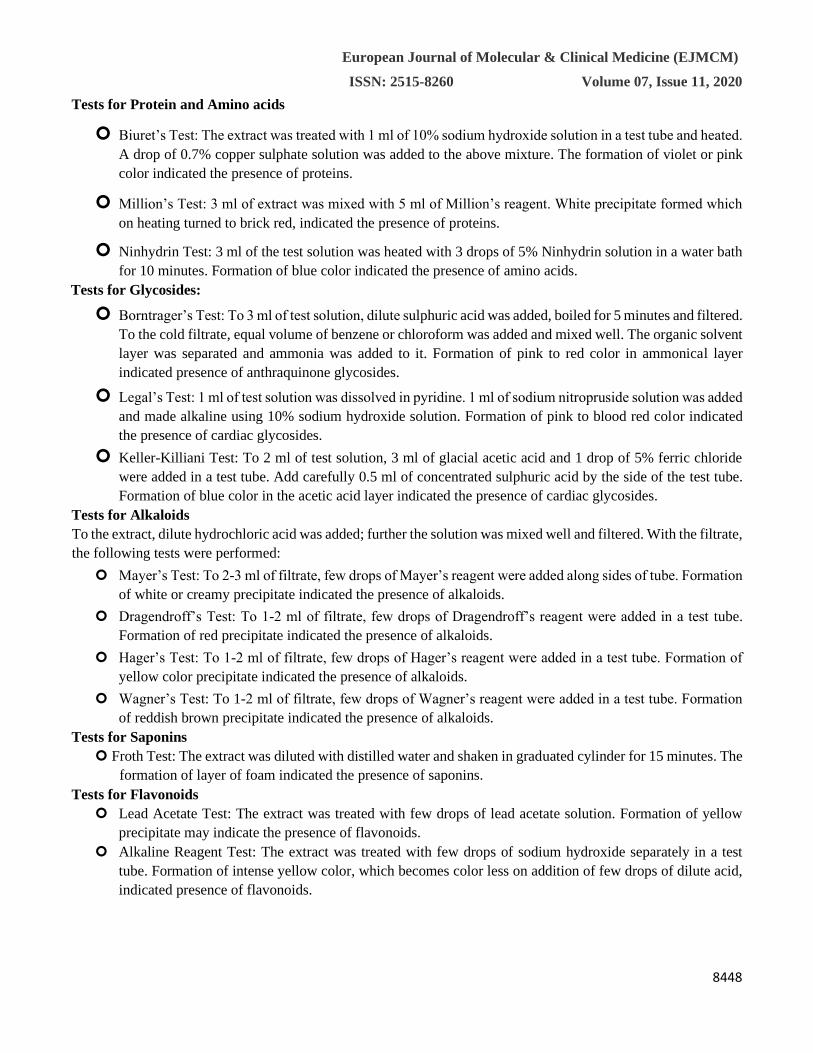
Result & Discussion Morphological studies Organoleptic evaluation of powder of sample
S. No. Analysis
Parameters
Test Sample
Argyreia
speciosa
Balanitesa
egyptiaca
Gloriosa
superb
Tagetes
erecta
Cyathocline
purpurea
1. Colour Light brown
Brown Greenish Yellowish
Brown
Brown
2. Odour Aromatic Aromatic Not specific Aromatic Not specific
3. Taste Bitter Sour Astringent Bitter No taste
4. Texture Fibrous Fibrous Coarse powder Coarse
powder
Coarse
powder
European Journal of Molecular & Clinical Medicine (EJMCM)
ISSN: 2515-8260 Volume 07, Issue 11, 2020
8450
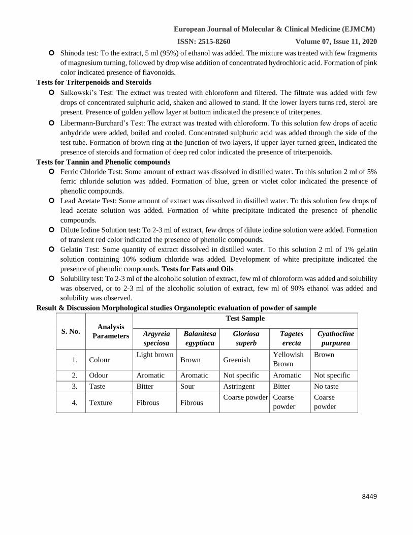
Fig. 1: T. S of Stem of Argyreia pilosa; a: morphology of stem; b: TS of stem entire view; c: unicellular
covering trichome; d: TS of the stem at a magnification of 40X; e: TS of stem showing trichome, epidermis,
and collenchyma; f: TS of stem showed uniseriate medullary rays. Abbreviations: Tri: trichome, Ep:
epidermis, Col: collenchyma, Sc: scleroidal cells, Xy: xylem, Ph: phloem, MR: medullary rays
Fig. 2: T.S. of leaf showing cristal distribution (Under polarized light.)
European Journal of Molecular & Clinical Medicine (EJMCM)
ISSN: 2515-8260 Volume 07, Issue 11, 2020
8451
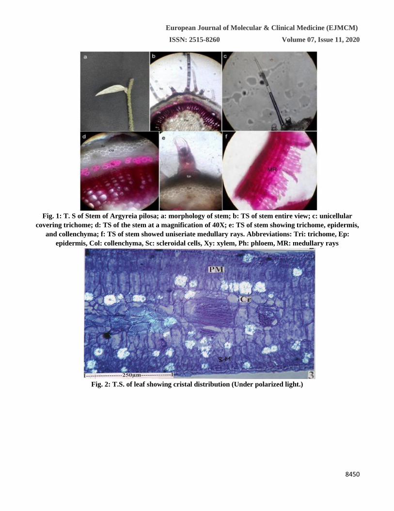
(AdE- Adaxial Epidermis, BS- Bundle Sheath, Cr- Crystals, Cu- Cuticle, Ep- Epidermis, Gc- Guard cells,
LM- Leaf margin, Ph- Phloem, PM- Palisade Mesophyll, Sc- Subsidiary cell, St- Stomata, X- Xylem.)
Fig. 3: Microcopical Examination of Balanitesa egyptiaca
European Journal of Molecular & Clinical Medicine (EJMCM)
ISSN: 2515-8260 Volume 07, Issue 11, 2020
8452
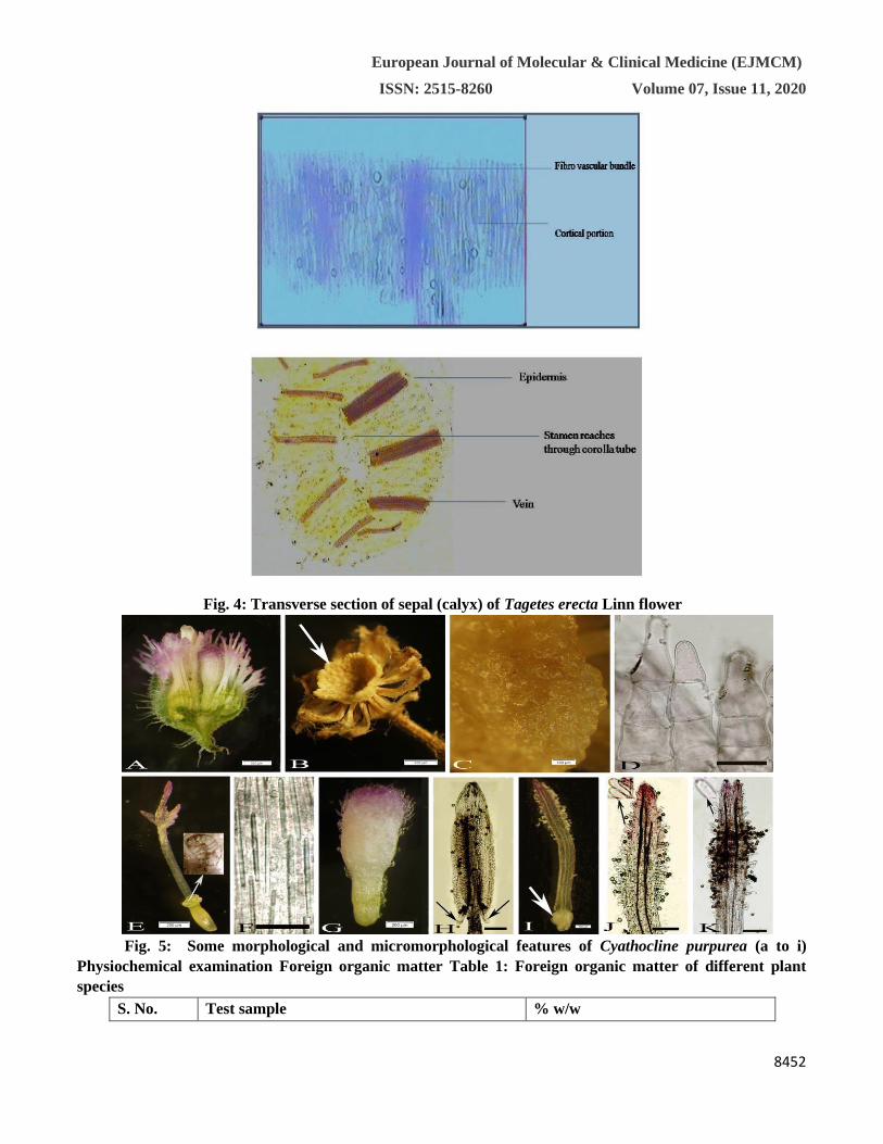
Fig. 4: Transverse section of sepal (calyx) of Tagetes erecta Linn flower
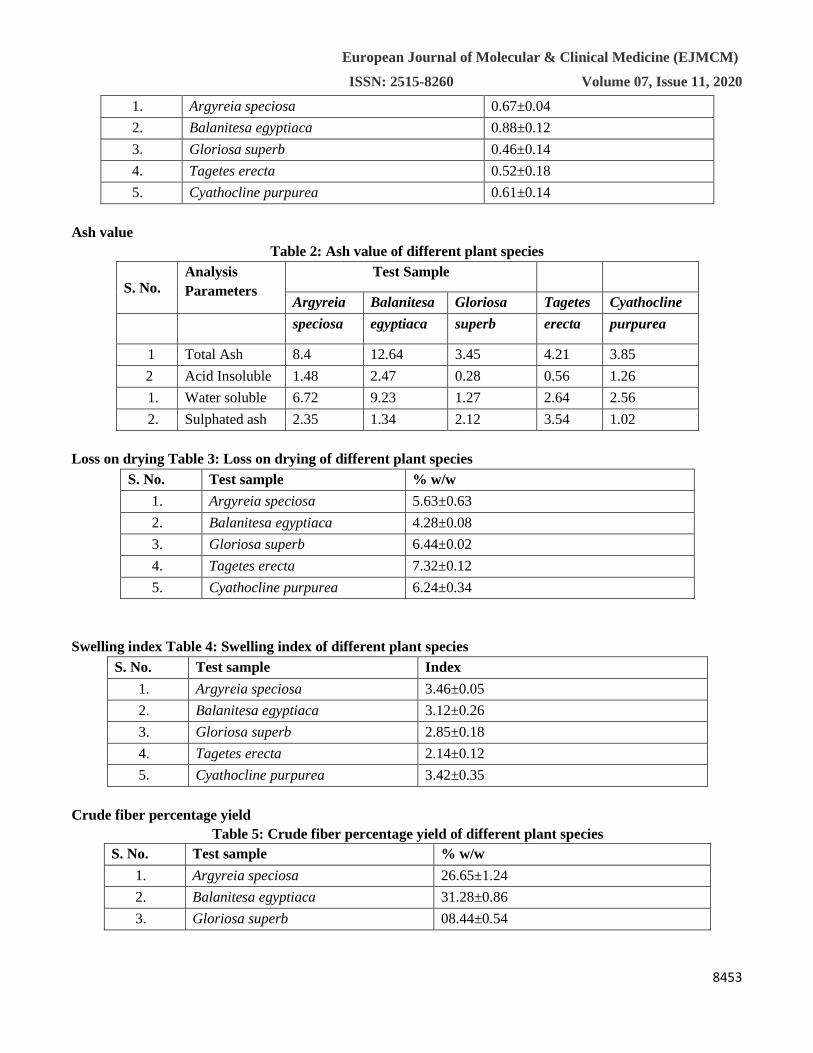
Fig. 5: Some morphological and micromorphological features of Cyathocline purpurea (a to i)
Physiochemical examination Foreign organic matter Table 1: Foreign organic matter of different plant
species
S. No. Test sample % w/w
European Journal of Molecular & Clinical Medicine (EJMCM)
ISSN: 2515-8260 Volume 07, Issue 11, 2020
8453
1. Argyreia speciosa 0.67±0.04
2. Balanitesa egyptiaca 0.88±0.12
3. Gloriosa superb 0.46±0.14
4. Tagetes erecta 0.52±0.18
5. Cyathocline purpurea 0.61±0.14
Ash value
Table 2: Ash value of different plant species
S. No. Analysis
Parameters
Test Sample
Argyreia Balanitesa Gloriosa Tagetes Cyathocline
speciosa egyptiaca superb erecta purpurea
1 Total Ash 8.4 12.64 3.45 4.21 3.85
2 Acid Insoluble 1.48 2.47 0.28 0.56 1.26
1. Water soluble 6.72 9.23 1.27 2.64 2.56
2. Sulphated ash 2.35 1.34 2.12 3.54 1.02
Loss on drying Table 3: Loss on drying of different plant species
S. No. Test sample % w/w
1. Argyreia speciosa 5.63±0.63
2. Balanitesa egyptiaca 4.28±0.08
3. Gloriosa superb 6.44±0.02
4. Tagetes erecta 7.32±0.12
5. Cyathocline purpurea 6.24±0.34
Swelling index Table 4: Swelling index of different plant species
S. No. Test sample Index
1. Argyreia speciosa 3.46±0.05
2. Balanitesa egyptiaca 3.12±0.26
3. Gloriosa superb 2.85±0.18
4. Tagetes erecta 2.14±0.12
5. Cyathocline purpurea 3.42±0.35
Crude fiber percentage yield
Table 5: Crude fiber percentage yield of different plant species
S. No. Test sample % w/w
1. Argyreia speciosa 26.65±1.24
2. Balanitesa egyptiaca 31.28±0.86
3. Gloriosa superb 08.44±0.54
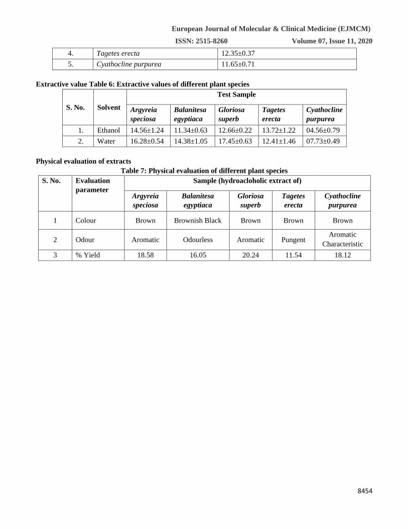
European Journal of Molecular & Clinical Medicine (EJMCM)
ISSN: 2515-8260 Volume 07, Issue 11, 2020
8454
4. Tagetes erecta 12.35±0.37
5. Cyathocline purpurea 11.65±0.71
Extractive value Table 6: Extractive values of different plant species
S. No. Solvent
Test Sample
Argyreia
speciosa
Balanitesa
egyptiaca
Gloriosa
superb
Tagetes
erecta
Cyathocline
purpurea
1. Ethanol 14.56±1.24 11.34±0.63 12.66±0.22 13.72±1.22 04.56±0.79
2. Water 16.28±0.54 14.38±1.05 17.45±0.63 12.41±1.46 07.73±0.49
Physical evaluation of extracts
Table 7: Physical evaluation of different plant species
S. No. Evaluation
parameter
Sample (hydroacloholic extract of)
Argyreia
speciosa
Balanitesa
egyptiaca
Gloriosa
superb
Tagetes
erecta
Cyathocline
purpurea
1 Colour Brown Brownish Black Brown Brown Brown
2 Odour Aromatic Odourless Aromatic Pungent Aromatic
Characteristic
3 % Yield 18.58 16.05 20.24 11.54 18.12
European Journal of Molecular & Clinical Medicine (EJMCM)
ISSN: 2515-8260 Volume 07, Issue 11, 2020
8455
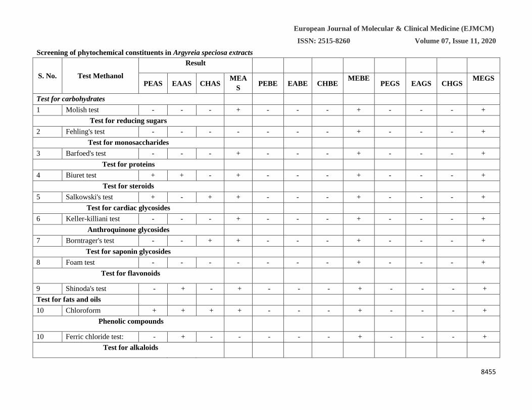
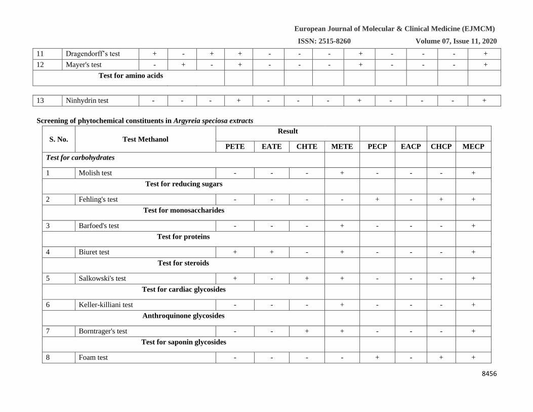
Screening of phytochemical constituents in Argyreia speciosa extracts
S. No. Test Methanol
Result
PEAS EAAS CHAS MEA
S PEBE EABE CHBE
MEBE PEGS EAGS CHGS
MEGS
Test for carbohydrates
1 Molish test - - - + - - - + - - - +
Test for reducing sugars
2 Fehling's test - - - - - - - + - - - +
Test for monosaccharides
3 Barfoed's test - - - + - - - + - - - +
Test for proteins
4 Biuret test + + - + - - - + - - - +
Test for steroids
5 Salkowski's test + - + + - - - + - - - +
Test for cardiac glycosides
6 Keller-killiani test - - - + - - - + - - - +
Anthroquinone glycosides
7 Borntrager's test - - + + - - - + - - - +
Test for saponin glycosides
8 Foam test - - - - - - - + - - - +
Test for flavonoids
9 Shinoda's test - + - + - - - + - - - +
Test for fats and oils
10 Chloroform + + + + - - - + - - - +
Phenolic compounds
10 Ferric chloride test: - + - - - - - + - - - +
Test for alkaloids
European Journal of Molecular & Clinical Medicine (EJMCM)
ISSN: 2515-8260 Volume 07, Issue 11, 2020
8456
11 Dragendorff’s test + - + + - - - + - - - +
12 Mayer's test - + - + - - - + - - - +
Test for amino acids
13 Ninhydrin test - - - + - - - + - - - +
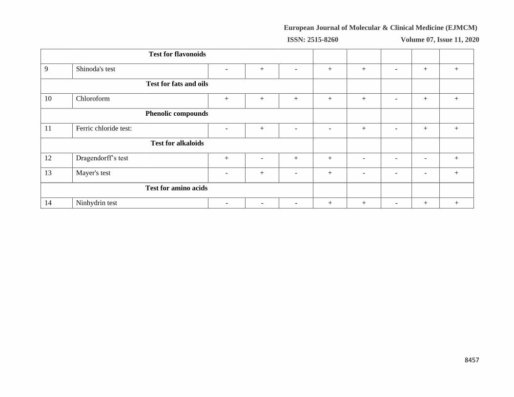
Screening of phytochemical constituents in Argyreia speciosa extracts
S. No. Test Methanol Result
PETE EATE CHTE METE PECP EACP CHCP MECP
Test for carbohydrates
1 Molish test - - - + - - - +
Test for reducing sugars
2 Fehling's test - - - - + - + +
Test for monosaccharides
3 Barfoed's test - - - + - - - +
Test for proteins
4 Biuret test + + - + - - - +
Test for steroids
5 Salkowski's test + - + + - - - +
Test for cardiac glycosides
6 Keller-killiani test - - - + - - - +
Anthroquinone glycosides
7 Borntrager's test - - + + - - - +
Test for saponin glycosides
8 Foam test - - - - + - + +
European Journal of Molecular & Clinical Medicine (EJMCM)
ISSN: 2515-8260 Volume 07, Issue 11, 2020
8457
Test for amino acids
14 Ninhydrin test - - - + + - + +
Test for flavonoids
9 Shinoda's test - + - + + - + +
Test for fats and oils
10 Chloroform + + + + + - + +
Phenolic compounds
11 Ferric chloride test: - + - - + - + +
Test for alkaloids
12 Dragendorff’s test + - + + - - - +
13 Mayer's test - + - + - - - +
European Journal of Molecular & Clinical Medicine (EJMCM)
ISSN: 2515-8260 Volume 07, Issue 11, 2020
8458
In transactional view, the leaf exhibits dorsiventaral midrib and smooth, even and thick lamina. The midrib is more
or less flat on the adaxial side and slightly conical on the abaxial side. The midrib is 500 μm thick. It consists of
thin epidermal layers of small thick walled cells. The cuticles were prominent and uneven. The vascular bundle of
the midrib is single, more or less circular, prominent and collateral. The vascular bundle consists of wide cluster of
narrow thick walled xylem elements and thin is of phloem located on the lower end of the xylem strand. The vascular
strand is surrounded by a thin layer of sclerenchyma sheath. There are also one or two layers of large hyaline
parenchyma cells around the vascular straind. The vascular bundle is 200 μm in transverse plane. The vascular
bundles of the lateral veins are circular and they do not project beyond the level of the surface of the lamina.
Lamina
The lamina is 350 μm thick. It is isolateral, ie; The adaxial and abaxial sides are not well differentiated. The
epidermal cells on both sides are large and thick walled with thick cuticle. The mesophyll tissue is consists of adaxial
zone of two to three layers of vertically elongated compact palisade cells. Similar type of palisade cells are also
seen on the abaxial part, the abaxial palisade cells are in two layers. The median part of the lamina consists of two
or three layers of spherical, less compact spongy parenchyma cells. The stomata occur at the level of epidermis, the
guard cells have short cubicular ledges. The epidermis including cutical is 20 μm thick.
Powder Microscopy
Fragments of lamina were observed under the microscope.
Epidermal trichomes
Druse non glandular epidermal trichomes are seen the abaxial surface of the lamina. The trichomes are unicellular,
unbranched and thick walled. The trichomesare pointed at the cell inclusions. The trichomes are up to
250 μm long and 15 μm thick.
Veins and terminal sclereids
Veins with vein islets and Veins terminations are also seen on the leaf fragments. The vein-islets are many sided,
and have thick, straight vein boundaries. The vein terminations have characteristic cluster of squarish sclereids with
wide lumen and fairly thick walls.
Crystal bodies are wide spread in the powder. The crystals are exclusively druses, which are spherical bodies
comprising many pointed triangular small crystal units.
Langali consists of dried tuberous roots of Gloriosa superba Linn., classified as one among upavisha dravya. Garbha
patani, garbhantaka, and garbhaghna are some special synonyms suggestive of its abortifacient activity. Colchicine
an alkaloid and its glycoside colchicoside are the two compounds which of its roots and having increased demand
by the pharmaceutical companies within and outside India. As Langali forms an ingredient of many
Popular Ayurvedic formulations it has become an endangered species. Rhizomes of Costus speciosus are said to be
sold in the market under the name of Langali. Authentication should be the primary criteria of any research using
plants, which will help to ensure the quality of any medicinal product. A characteristic macro-microscopic
fingerprint of a drug is diagnostic in identifying crude drugs of plant origin.
This study is an attempt to establish, the diagnostic characteristics of Argyreia speciosa, Balanitesa egyptiaca,
Gloriosa superb, Tagetes erecta & Cyathocline purpurea. These results can be employed as suitable quality control
measures to ensure the quality, safety and efficacy of this herbal drug material. The parameters studied here are
useful to identify and authenticate the traditionally important medicinal plants Argyreia speciosa, Balanitesa
egyptiaca, Gloriosa superb, Tagetes erecta & Cyathocline purpurea and this will prove helpful in the preparation
of herbal monographs and pharmacopoeial standards as emphasized by WHO.
Because the safety and efficacy are the ultimate goals, to ensure the reproducible quality of the herbal drugs, the
exact identification and quality assurance of the raw material is essential [15]. Mostly the herbal materials are
European Journal of Molecular & Clinical Medicine (EJMCM)
ISSN: 2515-8260 Volume 07, Issue 11, 2020
8459
supplied to the market is shrunken, twisted, rolled and deformed and without trade name and proper identification.
So, such drugs can easily adulterated or substituted. The application of pharmacognostic protocols such as
macromorphology, micromorphology, organoleptic tests, ash value, extractive values, foreign matters, swelling
index and histochemical studies will help in identifying genuine drugs because these tests result in specific results
for a particular drug.
Microscopy also plays an important role in drug identification. The importance of epidermal characters, in general,
are widely recognized in taxonomic considerations and in many cases these are successfully used in the
identification of taxa at genus as well as species levels [16]. Similarly, studies in stomata have a great taxonomic as
well as pharmacognostic value in proper identification of medicinal plants [17].
The physical parameters are almost constant for a plant therefore these are helpful in setting standards for a crude
drug. Various physicochemical parameters were evaluated for the leaf, stem bark and fruit parts as mentioned in
WHO guidelines. These parameters are important for detection of drug adulteration or improper handling of raw
materials [1]. One such parameter is ash value, which gives an idea of inorganic composition and other impurities
in a plant drug. The total ash value is also important for detection of metal, salts, and silica [18].
There are always chances of microbial growth when the crude drug is stored for a longer period of time and the
moisture content of crude drug is directly related to its stability and consequently with the shelf life of crude drug.
The lower the moisture content, the higher will be the stability of that drug and chance of microbial growth will be
less and vice versa [19]. Identification of the different classes of phytochemical constituents of the plant is an
important parameter, which gives an indication of the pharmacological active metabolites present in the plant [20].
Conclusion
Phytochemical studies on Argyreia speciosa, Balanitesa egyptiaca, Gloriosa superb, Tagetes erecta & Cyathocline
purpurea plants indicate it to be a useful plant to investigate for phytochemical and biological assays. HPLC analysis
may serve as a useful data for the standardization of the drug. The data generated from this study would help in the
authentication of various parts of Argyreia speciosa, Balanitesa egyptiaca, Gloriosa superb, Tagetes erecta &
Cyathocline purpurea, a very important constituent of various herbal drug formulations. This may lead to easier
authentication of herbal drugs procured from markets for the correct identification of the medicinal plant
ingredients. References
1. Akbar S, Hanif U, Ali J, et al. Pharmacognostic studies of stem, roots and leaves of Malva parviflora L.
Asian Pac J Trop Biomed. 2014;4(5):410– 415.
2. Amponsah IK, Mensah AY, Otoo A, et al. Pharmacognostic standardisation of Hilleria latifolia (Lam.) H.
Walt.(Phytolaccaceae). Asian Pac J Trop Biomed. 2014;4(12):941–946.
3. Traiperm P, Staples GW. A new endemic Thai species of Argyreia (Convolvulaceae). Phytotaxa.
2014;164(4):281–285.
4. Chetty KM, Sivaji K, Rao KT. Flowering Plants of Chittoor District. Tirupati, India: Student Offset Printers;
2008.
5. Ambasta S. The useful plants of India. New Delhi: CSIR; 1986:918. 6. Pullaiah T. Flora of Guntur District,
Andhra Pradesh, India. New Delhi: Daya Books; 2000:417.
6. Matthew KM. An excursion flora of central Tamilnadu, India. USA: CRC Press; 1995.
7. Bharathajothi P, Jegatheesan M. Ethno dermatological plants used by the Paliyar tribals of western Ghats,
Puliangudi, Tirunelveli district, Tamil Nadu, India. International Journal of Current Res. 2017;9(7):53436–
53438.
European Journal of Molecular & Clinical Medicine (EJMCM)
ISSN: 2515-8260 Volume 07, Issue 11, 2020
8460
8. Packialakshmi N, Beevi HF. Antibacterial screening on leaves of Argyreia cymosa roxb. against pathogenic
bacteria isolated from infected pateints samples wound, sputum and stool. International Journal of Applied
Sciences and Biotechnology. 2014;2(3):279–282.
9. Bokhad MN, Rothe S. An overview of medicinally important lianas from dry deciduous forest of west
Vidarbha region (MS) India. Bioscience discovery. 2015;6(2):117–120. 11. Ganesan S, Chandhirasekaran
M, Selvaraj A. Ethnoveterinary healthcare practices in southern districts of Tamil Nadu. CSIR. 2008:347–
354.
10. Badami S, Vaijanathappa J, Bhojraj S. In vitro antioxidant activity of Argyreia cymosa bark extracts.
Fitoterapia. 2008;79(4):287–289.
11. Jhade D, Ahirwar D, Jain R, et al. Pharmacognostic standardization, physico-and phytochemical evaluation
of Amaranthus spinosus linn. Root. J Young Pharm. 2011;3(3):221–225.
12. Ghorpade P, Siddiqui A, Patil MJ, et al. Pharmacognostic and phytochemical evaluation of Celosia argentea.
Pharmacognosy Journal. 2012;4(33):7–15.
13. Kataria S, Rao SK, Bhandari A, et al. Pharmacognostical standardization of Corchorus depressus (L.) Stocks
(Tiliaceae)-A promising ethnomedicinal plant. Indian Journal of Traditional Knowledge. 2013;12(3):489–
497.
14. Choudhary N, Siddiqui M, Khatoon S. Pharmacognostic evaluation of Tinospora cordifolia (Willd.) Miers
and identification of biomarkers. Indian Journal of Traditional Knowledge. 2014;13(3):543–550.
15. World Health O. Quality control methods for herbal materials Geneva: World Health Organization; 2011.
16. Khandelwal KR. Practical pharmacognosy: techniques and experiments. Maharashtra: Niral Prakashan;
2008.
17. Harborne JB. Phytochemical methods; a guide to modern techniques of plant analysis. London: Chapman
& Hall; 1973:302.
18. Bladt S. Plant drug analysis: a thin layer chromatography atlas. Germany: Springer-Verlag Berlin
Heidelberg; 1996:384.
19. Alam F, Najum us Saqib Q. Pharmacognostic standardization and preliminary phytochemical studies of
Gaultheria trichophylla. Pharm Biol. 2015;53(12):1711–1718.
20. Singh P, Khosa RL, Mishra G, et al. Pharmacognostical evaluation of aerial parts of Graptophyllum pictum
(L.) Griff. (Syn: Justicia picta Linn.): A well-known folklore medicinal plant. Anc Sci Life.
2015;34(4):223–229.