photosynthetic eukaryotes unite: endosymbiosis connects the dots
TRANSCRIPT
Photosynthetic eukaryotes unite:endosymbiosis connects the dotsDebashish Bhattacharya,* Hwan Su Yoon, and Jeremiah D. Hackett
SummaryThe photosynthetic organelle of algae and plants (theplastid) traces its origin to a primary endosymbioticevent in which a previously non-photosynthetic protistengulfed and enslaved a cyanobacterium. This eukaryotethen gave rise to the red, green and glaucophyte algae.However, many algal lineages, such as the chlorophyllc-containing chromists, have a more complicated evolu-tionary history involving a secondary endosymbioticevent, in which a protist engulfed an existing eukaryoticalga (in this case, a red alga). Chromists such as diatomsand kelps then rose to great importance in aquatichabitats. Another algal group, the dinoflagellates, hasundergone tertiary (engulfment of a secondary plastid)and even quaternary endosymbioses. In this review, weexamine algal diversity and show endosymbiosis to be amajor force in algal evolution. This area of research hasadvanced rapidly and long-standing issues such as thechromalveolate hypothesis and the extent of endosym-biotic gene transfer have recently been clarified.BioEssays 26:50–60, 2004.� 2003 Wiley Periodicals, Inc.
Introduction
Accounting for the remarkable diversity of photosynthetic
protists (algae) remains an important challenge in evolutionary
biology. The photosynthetic organelle, termed the plastid, is
found in forms as diverse as microscopic diatoms, giant kelps,
toxic dinoflagellates, edible red algae, and land plants (see
Table 1). A critical question is whether all photosynthetic forms
are united on a single branch of the tree of life or whether
plastids have been spread throughout the tree through
multiple independent lateral transfers. If the latter, then how
many lateral transfers are needed to explain extant algal
lineages? Answers to these questions have been rapidly
accumulating recently and they provide surprising insights into
the course of eukaryotic evolution.
It has now been clearly documented that plastids originate
through endosymbiosis,(1–6) whereby a single-celled protist
engulfs and retains a foreign photosynthetic cell (see Fig. 1).
Over time, the foreign cell is reduced to a plastid and is
vertically transmitted to subsequent generations. Endosym-
biosis comes in three major types, primary, secondary and
tertiary endosymbiosis. The first results from the engulfment of
a photosynthetic prokaryote (cyanobacterium, Fig. 1A) and
gives rise to a plastid bound by two membranes (the inner,
1st, and outer, 2nd, membranes of the cyanobacterium, see
Ref. 7). The outer, phagosomal membrane of this primary
‘‘host’’ cell is lost. Primary endosymbiosis is believed to have
occurred once in evolution, giving rise to the proto-alga that is
the ultimate root of all plastids.(4) The plastid became fully
established in the proto-algal population and many genes of
both photosynthetic and non-photosynthetic function were
transferred to the nucleus of the primary host. The remarkable
achievement of these cells was to engineer a way of reintro-
ducing all the plastid proteins, whose genes were transferred
to the nucleus and translated in the cytoplasm, back into
the plastid compartment to express their function. The esta-
blishment of this protein import system after primary endo-
symbiosis required the evolution of a ‘‘transit’’ sequence
(about 24–100 amino acids in length)(8) at their N termini. This
extra sequence probably originated through mutations that
extended the original open reading frame at the 50 terminus,
and the encoded proteins were selected for because of their
targeting capacity. Once the sequences encoding these
transit peptides were established in a few genes, then they
may have spread through exon shuffling into other photosyn-
thetic genes.(9,10) It appears that, once these unlikely events
had occurred and the first algal populations were established,
the path was paved for the rise of photosynthetic eukaryotes.
50 BioEssays 26.1 BioEssays 26:50–60, � 2003 Wiley Periodicals, Inc.
Department of Biological Sciences and Center for Comparative
Genomics, University of Iowa, Iowa City
Funding agency: This work was supported by grants by the National
Science Foundation. (NSF) awarded to D. B. (DEB 01-07754, MCB
01-10252, MCB 02-36631). J. D. H. was supported by an Institutional
NRSA (T 32 GM98629) from the National Institutes of Health and
H. S. Y. was partially supported by the Post Doctoral Fellows Program
of the Korean Science and Engineering Foundation.
*Correspondence to: Debashish Bhattacharya, Department of Biolo-
gical Sciences and Center for Comparative Genomics, University of
Iowa, 210 Biology Building, Iowa City, Iowa 52242-1324.
E-mail: [email protected]
DOI 10.1002/bies.10376
Published online in Wiley InterScience (www.interscience.wiley.com).
Abbreviations: COXII, mitochondrial cytochrome c oxidase subunit II;
gnd, 6-phosphogluconate dehydrogenase gene; GAPDH, glyceralde-
hyde-3-phosphate dehydrogenase; Ma, millions of years ago; rRNA,
ribosomal RNA.
Review articles
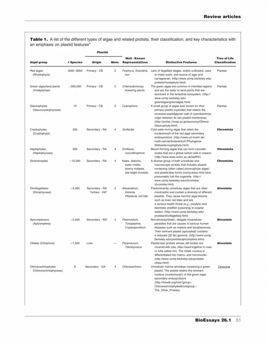
Table 1. A list of the different types of algae and related protists, their classification, and key characteristics with
an emphasis on plastid features*
Algal group # Species
Plastid
Well - Known
Representatives Distinctive Features
Tree of Life
ClassificationOrigin Mem.
Red algae
(Rhodophyta)
5000–6000 Primary - CB 2 Porphyra, Gracilaria,
nori
Lack of flagellate stages, widely cultivated, used
to make sushi, and source of agar and
carrageenan. (http://www.ucmp.berkeley.edu/
protista/rhodophyta.html)
Plantae
Green algae/land plants
(Viridiplantae)
>500,000 Primary - CB 2 Chlamydomonas,
flowering plants
The green algae are common in intertidal regions
and are the sister to land plants that are
dominant in the terrestrial ecosystem. (http://
www.ucmp.berkeley.edu/
greenalgae/greenalgae.html)
Plantae
Glaucophytes
(Glaucocystophyceae)
13 Primary - CB 2 Cyanophora A small group of algae best known for their
primary plastid (cyanelle) that retains the
ancestral peptidiglycan wall of cyanobacterial
origin between its two plastid membranes.
(http://protist.i.hosei.ac.jp/taxonomy/Others/
Glaucophyta.html)
Plantae
Cryptophytes
(Cryptophyta)
200 Secondary - RA 4 Guillardia Cold water-loving algae that retain the
nucleomorph of the red algal secondary
endosymbiont. (http://www.uni-koeln.de/
math-nat-fak/botanik/bot1/Phylogenie-
Webseite/cryptophyta.html)
Chromista
Haptophytes
(Haptophyceae)
500 Secondary - RA 4 Emiliania,
coccolithophore
Bloom-forming algae that can form coccolith
scales that are a global carbon sink in oceans.
(http://www.soes.soton.ac.uk/staff/tt/)
Chromista
Stramenopiles >10,000 Secondary - RA 4 Kelps, diatoms,
water molds,
downy mildews,
late blight of potato
A diverse group of both unicellular and
macroscopic protists that includes plastid-
containing (often called chromophyte algae)
and plastid-less forms (oomycetes) that have
presumably lost the organelle. (http://
www.ucmp.berkeley.edu/chromista/
chromista.html)
Chromista
Dinoflagellates
(Dinophyceae)
>4,000 Secondary - RA
Tertiary - HA*
3 Alexandrium,
Karenia,
Pfiesteria, red tide
Predominantly unicellular algae that are often
mixotrophic and contain a diversity of different
plastids. They cause harmful algal blooms
such as toxic red tides and are
a serious health threat (e.g., paralytic and
diarrhetic shellfish poisoning) in coastal
waters. (http://www.ucmp.berkeley.edu/
protista/dinoflagellata.html)
Alveolata
Apicomplexans
(Apicomplexa)
>2,400 Secondary - RA* 4 Plasmodium,
Toxoplasma,
Cryptosporidium
Non-photosynthetic, obligate intracellular
parasites that are causes of serious human
diseases such as malaria and toxoplasmosis.
Their remnant plastid (apicoplast) contains
a reduced (35 kb) genome. (http://www.ucmp.
berkeley.edu/protista/apicomplexa.html)
Alveolata
Ciliates (Ciliophora) >7,500 Loss — Paramecium,
Tetrahymena
Plastid-less protists whose cell bodies are
covered with cilia, often fused together in rows
or tufts called cirri. The ciliate nucleus is
differentiated into macro- and micronuclei.
(http://www.ucmp.berkeley.edu/protista/
ciliata.html)
Alveolata
Chlorarachniophytes
(Chlorarachniophyceae)
6 Secondary - GA 4 Chlorarachnion Unicellular marine amoebae containing a green
plastid. The plastid retains the remnant
nucleus (nucleomorph) of the green algal
secondary endosymbiont.
(http://tolweb.org/tree?group¼Chlorarachniophytes&contgroup¼The_Other_Protists)
Cercozoa
Review articles
BioEssays 26.1 51
Recent phylogenetic analyses using nuclear and mitochon-
drial loci(11–13) suggest that the proto-alga split into two
lineages. The first contains the glaucophyte algae, which failed
to rise to any great taxonomic importance,(14,15) whereas the
second gave rise to the highly successful red algae(16) and
their sister group the green algae(17) plus land plants.(18)
These three lineages are classified as the Plantae (see
Table 1). Endosymbiosis left a sizeable mark on the Plantae
that goes well beyond the lateral transfer of photosynthetic
capacity. A recent analysis(19) of the complete Arabidopsis
nuclear genome suggests that up to 18% of this plant’s genes,
many of non-photosynthetic function (e.g., disease resistance,
intracellular protein routing), originated from the cyanobacter-
ium through endosymbiotic gene transfer. Endosymbiosis
had, therefore, a considerable influence in the early evolution
of algae by significantly enriching their nuclear genomes with
cyanobacterial, often duplicated genes. Selection could act
on these divergent sequences to explore new functions(13,19)
or to replace existing host genes with those from the
endosymbiont.(20,21)
Secondary endosymbiosis and
the rise of algae
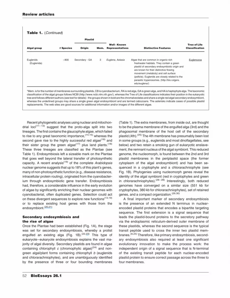
Once the Plantae had been established (Fig. 1A), the stage
was set for secondary endosymbiosis, whereby a protist
engulfed an existing alga (Fig. 1B).(22,23) This type of
eukaryote–eukaryote endosymbiosis explains the vast ma-
jority of algal diversity. Secondary plastids are found in algae
containing chlorophyll c (chromophytic algae)(24) and non-
green algal/plant forms containing chlorophyll b (euglenids
and chlorarachniophytes), and are unambiguously identified
by the presence of three or four bounding membranes
(Table 1). The extra membranes, from inside out, are thought
to be the plasma membrane of the engulfed alga (3rd) and the
phagosomal membrane of the host cell of the secondary
plastid (4th).(25) The 4th membrane has presumably been lost
in some groups (e.g., euglenids and most dinoflagellates, see
below) and two retain a smoking gun of eukaryotic enslave-
ment, the remnant nucleus of the algal symbiont. This reduced
genome, the nucleomorph, is found between the 2nd and 3rd
plastid membranes in the periplastid space (the former
cytoplasm of the algal endosymbiont) and has been se-
quenced in a cryptophyte and a chlorarachniophyte (see
Fig. 1B). Phylogenies using nucleomorph genes reveal the
identity of the algal symbiont (red in cryptophytes and green
in chlorarachniophytes).(26–28) Interestingly, both reduced
genomes have converged on a similar size (551 kb for
cryptophytes, 380 kb for chlorarachniophytes), set of retained
genes, and a compact organization.(29,30)
A final important marker of secondary endosymbiosis
is the presence of an extended N terminus in nuclear-
encoded plastid proteins that encodes a bipartite targeting
sequence. The first extension is a signal sequence that
leads the plastid-bound proteins to the secretory pathway
via the endoplasmic reticulum-derived outer membrane of
these plastids, whereas the second sequence is the typical
transit peptide used to cross the inner two plastid mem-
branes.(8,25) Therefore, like primary endosymbiosis, second-
ary endosymbiosis also required at least one significant
evolutionary innovation to make the process work: the
independent origin of a signal sequence that is N-terminal
of the existing transit peptide for each nuclear-encoded
plastid protein to ensure correct passage across the three to
four membranes.
Table 1. (Continued)
Algal group # Species
Plastid
Well - Known
Representatives Distinctive Features
Tree of Life
ClassificationOrigin Mem.
Euglenids
(Euglenida)
>800 Secondary - GA 3 Euglena, Astasia Algae that are common in organic-rich
freshwater habitats. They contain a green
plastid of secondary endosymbiotic origin and
are known for their distinctive flowing
movement (metaboly) and cell surface
(pellicle). Euglenids are closely related to the
parasitic trypanosomes. (http://bio.rutgers.
edu/euglena/)
Euglenozoa
*Mem. is for the number of membranes surrounding plastids, CB is cyanobacterium, RA is red alga, GA is green alga, and HA is haptophyte alga. The taxonomic
classification of the algal groups follows NCBI (http://www.ncbi.nlm.nih.gov/), whereas the Tree of Life classifications indicates their position in the eukaryotic
tree and follows different authors (see text for details) - the groups shown in bold are the chromalveolates and share a single red algal secondary endosymbiont,
whereas the underlined groups may share a single green algal endosymbiont and are termed cabozoans. The asterisks indicate cases of possible plastid
replacements. The web sites are good sources for additional information and/or images of the different algae.
Review articles
52 BioEssays 26.1

The secondary plastids in brief
The chromalveolate hypothesisThe chromalveolates group was postulated primarily on the
basis of molecular phylogenetic analyses that unite particular
members of these morphologically disparate lineages,(32)
and the hypothesis that all taxa containing chlorophyll c
(i.e., a chromophytic plastid)(33,34) share a common origin.
The alveolates share two characters that unambiguously
unite them as a lineage, namely tubular mitochondrial cristae
and sacs or alveoli under the plasma membrane. The
ecologically and economically important chromalveolates
(see Table 1)(35) defines a broadly diverse group that includes
the Chromista, comprising the cryptophyte,(36) haptophyte,(37)
and stramenopile(16) algae, and the Alveolata, comprising
the parasitic apicomplexans,(38) the plastid-less ciliates,(39)
and the dinoflagellate algae.(40) The chromalveolate plastid
is believed to have originated from a red algal secondary
endosymbiosis with the ensuing evolution of chlorophyll c2.
The plastid was putatively lost in ciliates and parasitic/
saprobic stramenopiles like oomycetes (e.g., the water mold
Achlya) and its genome reduced to a 35 kb DNA circle in the
apicomplexan plastid (the apicoplast).(41,42) Evidence in
support of these ideas has been slow in coming but recent
data now appear to have established the chromalveolates as a
monophyletic entity.(42,43)
Phylogenies of nuclear genes have until now, however,
only provided marginal support for the chromalveolate
hypothesis. Analysis of small subunit rRNA(44) and a combined
data set of EF-1a, actin, a-tubulin, and b-tubulin amino acid
sequences (that did not include haptophytes)(45) are consis-
tent with a sister group relationship between two chromalveo-
late groups, the stramenopiles and alveolates. The limited
taxon sampling and phylogenetic power of these data sets are
a weakness that needs to be addressed in future studies. In
contrast, strong support for chromalveolate monophyly has
come from analyses of plastid genes(43) and from the finding
of a unique gene duplication shared by members of this
assemblage.(42)
We have recently analyzed a concatenated data set of five
plastid-encoded genes (5827 nt in total) from 36 taxonomically
diverse members of the red and chromist algae. In this study,
we addressed one cornerstone of the chromalveolate hypoth-
esis that had not yet been adequately tested using multiple
nuclear loci, the monophyly of the Chromista (see Table 1).(33)
This group previously had little justification on morphologi-
cal(24) or phylogenetic grounds (nuclear and mitochondrial loci
do not resolve their positions)(4,11,13) for being united in a single
lineage. They primarily share a four-membrane-bound chlor-
ophyll c-containing plastid that is located within the lumen of
the rough endoplasmic reticulum. The haptophytes and
stramenopiles, however, share characters such as tubular
mitochondrial cristae, similar storage products, and fucox-
anthin that suggest a specific relationship between these
taxa.(24) The combined plastid gene tree (Fig. 2) appears to
have settled the issue of chromist monophyly by providing
Figure 1. Origins of plastids via A: primary, B:secondary, and C: tertiary endosymbiosis. Gene
transfer from the endosymbiont to the host nucleus
is shown with the arrows and the colored chro-
mosomes. The mitochondrion has been omitted
from these figures. The remnant algal nucleus
(nucleomorph) in secondary and tertiary endosym-
bioses is shown. This genome has been lost in all
algae but the cryptophytes (member of the
chromalveolates) and chlorarachniophytes
(marked with asterisks).
Review articles
BioEssays 26.1 53

strong support for three important ideas: (1) as previously
suggested,(46) the chromist plastids are all of red algal origin,
(2) chromist plastids share a monophyletic origin and, by
extension, so do the host cells containing these plastids, and
(3) the basal position of the cryptophytes in the Chromista
suggests that retention of the red algal nucleomorph in the
periplastid space, the presence of phycobilin pigments, and
the storage of photosynthates as starch (all are absent in
haptophytes and stramenopiles) are ancestral endosymbiont
characters that were likely lost after the divergence of the
cryptophytes (see character evolution model in Fig. 3).
Furthermore, in contrast to the haptophytes and strame-
nopiles that have tubular mitochondrial cristae, the crypto-
phytes have flattened cristae, suggesting that this was the
ancestral condition in the Chromista. The alveolates, however,
contain tubular cristae and are presumably sister to the chro-
mists (Fig. 3). If flattened cristae was ancestral in the
chromalveolates, then this character was lost twice, once in
the common ancestor of alveolates and once after the
divergence of the cryptophytes in the Chromista (see Fig. 3).
A more parsimonious explanation for the cristae data would
emerge if the cryptophytes were to branch at the base of the
chromalveolates and the chromists would become paraphy-
letic. Analysis of an endosymbiotic replacement involving the
enolase gene in cryptophytes is consistent with this idea.(20)
Verification of the early divergence of the cryptophytes will
have to wait, however, until alveolate plastid sequences are
included in the analysis shown in Fig. 2. This analysis could
have two likely outcomes: (1) alveolate photosynthetic genes
of red algal origin diverge from the branch uniting all chromists
(as expected¼ 2 losses of flattened cristae, or (2) they diverge
on the branch uniting the haptophytes and stramenopiles (¼ 1
loss). Such target genes are most likely to be found in the
nucleus of dinoflagellates because the non-photosynthetic
apicomplexans and ciliates may not have maintained these
coding regions.
Another important result from the Yoon et al.(43) study was
the dating of the chromist secondary endosymbiotic event.
Usage of two red algal fossil constraint dates and a molecular
clock method that does not assume a uniform mutation rate in
different lineages suggests that the earliest date (asterisk in
Fig. 2) for the origin of the chromist plastid is 1260� 30 million
years ago (Ma).(43) The endosymbiosis marks, therefore, the
birth of the chromist algae. This date is substantially earlier
than some estimates (e.g., 850 Ma)(31) but agrees well with the
fossil record, which shows the appearance of a diversity of
algae and protists near the Mesoproterozoic/Neoproterozoic
boundary about 1000 Ma.(47,48) A recent analysis of the
Figure 2. Phylogenetic relationships of red algal and
chromist plastids inferred from a minimum evolution (ME)
analysis of the combined DNA sequences of 16S rRNA,
psaA, psbA, rbcL, and tufA (5827 nt). The general time
reversible model(79) with estimations of nucleotide fre-
quencies, the proportion of invariant sites that are unable
to accept substitutions, and the shape parameter of
the gamma distribution to accommodate rate variations
across sites (GTRþ IþG [gtr] model) was used in this
analysis. Results of a ME-gtr bootstrap analysis (2000
replicates) are shown above the branches. The thick
branches are of significant evolutionary importance and
all have>95% posterior probability in a Bayesian analysis
of the DNA data (for details, see Yoon et al.).(43) This
tree is rooted on the branch leading to the glauco-
phyte sequence. The branch lengths are proportional to
the number of substitutions per site (see scale in figure).
The asterisk marks the branch where the chromist plastid
originated through a red algal secondary endosymbiosis
(ca. 1260 Ma).(43)
Review articles
54 BioEssays 26.1

Earth’s geochemical record, in particular with respect to
the levels of the oxygen and nitrogen that are critical to algal
growth, is consistent with the known eukaryotic fossil
record(49) and our molecular dating results. Furthermore, a
subsequent analysis (H.S.Y., J.D.H., and D.B. unpublished
data) using a six plastid gene data set that includes green
algae and land plants to incorporate additional robust fossil
constraints in the gene phylogeny (e.g., origin of land plants
and the monocot–dicot split) substantiates the findings of
Yoon et al.(43)
Now that Chromista monophyly appears to be established,
what evidence is there to include them in the chromalveolates?
The strongest evidence comes from phylogenetic analyses
that show a specific phylogenetic relationship between the
nuclear-encoded plastid-targeted glyceraldehyde-3-phos-
phate dehydrogenase (GAPDH) gene in the cryptophytes
and stramenopiles (chromists) on one hand and the apicom-
plexans and dinoflagellates (alveolates) on the other.(42)
Importantly, this gene appears to have originated through a
unique duplication of the existing cytosolic gene in these taxa,
and the subsequent replacement of the original plastid-
targeted sequence (of cyanobacterial origin) by the gene dup-
licate. This unusual shared derived character unites the
studied chromalveolates and, as corroboration, the phyloge-
nies show the plastid-targeted genes to be phylogenetically
distinct from the homolog of cyanobacterial origin in non-
chromalveolates such as green algae/land plants and red
algae. These latter taxa have not undergone the gene
duplication–replacement event.(42) These findings all point
to a critical event in evolution, a single red algal secondary
endosymbiosis that gave birth to common ancestor of a super-
assemblage, thechromalveolates. Theendosymbiotic event is
ancient (ca. 1200 Ma) and the host cell of the secondary plastid
has diverged extensively during this long period of evolution.
It is a testimony to the power of modern molecular evolu-
tionary methods and the slow evolutionary rate of many plastid
genes that we are able to look back so far in evolutionary time
and still detect the signal of shared ancestry (see Fig. 2).
The major insights into eukaryotic evolution that come from
the finding of chromalveolate monophyly are:
* secondary plastid loss is common (e.g., in plastid-less
stramenopiles such as oomycetes, in ciliates, and in some
dinoflagellates).(42,50) In support of this idea, Andersson
and Roger(51) have recently found a 6-phosphogluconate
dehydrogenase (gnd) gene of cyanobacterial (i.e., plastid)
origin in the parasitic stramenopile, Phytophthora infes-
tans. This suggests that Phytophthora was likely once
photosynthetic because its gnd gene is closely related to
the homologue in photosynthetic members of this lineage.
* Chromalveolates share a homologous plastid protein
import system that evolved once in their common ancestor.
Figure 3. Proposed evolutionary tree of the chromalveolates showing the origin and loss of important (mostly plastid) characters. The
evidence for chromist plastid monophyly is described in Yoon et al.(43) The timing of the tertiary endosymbiosis that gave rise to the
haptophyte-type plastid in either the pre-dinoflagellate(66,80) or only in fucoxanthin-containing taxa(69) remains an open question (large filled
circle with a cross). The possible green algal endosymbiosis in the common ancestor of apicomplexans and dinoflagellates (large open
circle with a cross) is based on Funes et al.,(21,76) Kohler et al.,(75) and our preliminary results (see text for details). Gain or loss of character
states is shown with the slashesacross the branches. The brancheswith broken lines at the origin represent uncertainty about the position of
these taxa in the host tree, whereas the small filled circles with dashes or the wavy line inside represent plastid loss and plastid degeneration,
respectively. pRER is the plastid rough endoplasmic reticulum.
Review articles
BioEssays 26.1 55
* Chlorophyll c2 appears to have evolved only once in the
common ancestor of the chromalveolates. Phylogenetic
analysis of chlorophyll a/b and chlorophyll a/c light harvest-
ing complex proteins supports this idea showing that the
chlorophyll a/c-binding proteins in chromists (and dino-
flagellates) form a monophyletic group that traces its origin
to a red algal-like ancestor.(46)
Given these impressive data, one may be led to believe that
the story of chromalveolate evolution has been convincingly
solved. This is not, however, entirely the case as the section
on dinoflagellates below will explain. But first, we will review
current knowledge about the secondary plastids of green algal
origin.
The green secondary plastidsSecondary endosymbiosis also gave rise to the chlorophyll
b-containing plant-like plastid in distinctly non-plant-like taxa,
the euglenids(52) and the chlorarachniophytes. The Euglenida
are sister to the Kinetoplastida (which includes the parasitic
trypanosomatids), and together form the Euglenozoa. The
presence of a three-membrane green plastid in photosynthetic
euglenids and the absence of a plastid in the trypanosomes
had previously suggested that the algal secondary endosym-
biosis occurred at the base of the euglenid lineage and that
trypanosomatids ancestrally lacked a plastid. The remarkable
finding of plant-like genes in trypanosomatids now suggest,
however, that all Euglenozoa may have at one time been
photosynthetic and that kinetoplastids lost their photosynthe-
tic organelle secondarily.(53) The footprint of the secondary
endosymbiosis in the trypanosomatids is metabolic enzymes
localized in specialized peroxisomes (glycosomes [not derived
from plastids]) that trace their origin to the green alga through
endosymbiotic gene transfer. This story underlines the
surprising ancestral distribution of photosynthesis in eukar-
yotes and suggests that plastids may have been more
widespread than we imagine. The distribution of these
organelles has been decimated by secondary losses due
often to the evolution of a parasitic or saprobic life-style (i.e.,
trypanosomatids were presumably once free-living algae),(54)
a theme that was repeated in the chromalveolates.
The second group of non-green algal protists that contain
chlorophyll b is the chlorarachniophytes. These are amoebo-
flagellate members of the protist assemblage Cercozoa(55)
that includes euglyphids, formanifera, and plasmodiophorid
plant pathogens.(56–58) This broad array of protists contains
two photosynthetic groups, the chlorarachniophytes with
their chlorophyll b-containing secondary plastid and the filose
amoeba, Paulinella chromatophora, with its cyanelle that
superficially resembles the plastids of glaucophytes.(59,60)
Whereas the origin of the Paulinella cyanelle still needs to be
resolved, the phylogenetic evidence based on plastid and
nucleomorph sequences has established the green algal
origin of the secondary plastid in chlorarachniophytes.(27,28)
A recent analysis of cDNAs from the chloroarachniophyte
Bigelowiella natans indicates that a large number of plastid
genes in this species were transferred from the green algal
endosymbiont to the host nucleus. More remarkably, about
21% of the photosynthetic genes were derived from lateral
transfers involving non-green sources such as red or strame-
nopile algae.(61) These results underline the importance
of endosymbiosis to facilitating intergenome gene transfer
and suggest that mixotrophic species such as Bigelowiella
(and dinoflagellates, see below) may be particularly adept at
scavenging genes from different prey. In contrast, taxa such as
the green alga Chlamydomonas reinhardtii, which is auto-
trophic and for which the complete nuclear genome is known,
do not show evidence of lateral transfer of photosynthetic
genes from different sources (i.e., all are derived from the
original cyanobacterial primary endosymbiont).(61)
What still remains to be determined with regard to green
algal secondary plastids is whether chlorarachniophytes and
euglenids share the same green algal endosymbiont in a
common ancestor. The available nuclear and plastid gene
data do not support this hypothesis,(62–64) although the high
divergence rate and base composition bias of chlorarachnio-
phyte and euglenid sequences leaves open the possibility of
Cercozoa–Euglenozoa monophyly (together the Cabozoa
[Table 1]).(35) If true, this latter scenario would reduce the
number of secondary endosymbioses involving a chlorophyll
b-containing endosymbiont to one, render secondary plastid
loss a widespread force in eukaryotic evolution, and place
algae near the root of much of the tree of life.
The dinoflagellates and
their tertiary endosymbioses
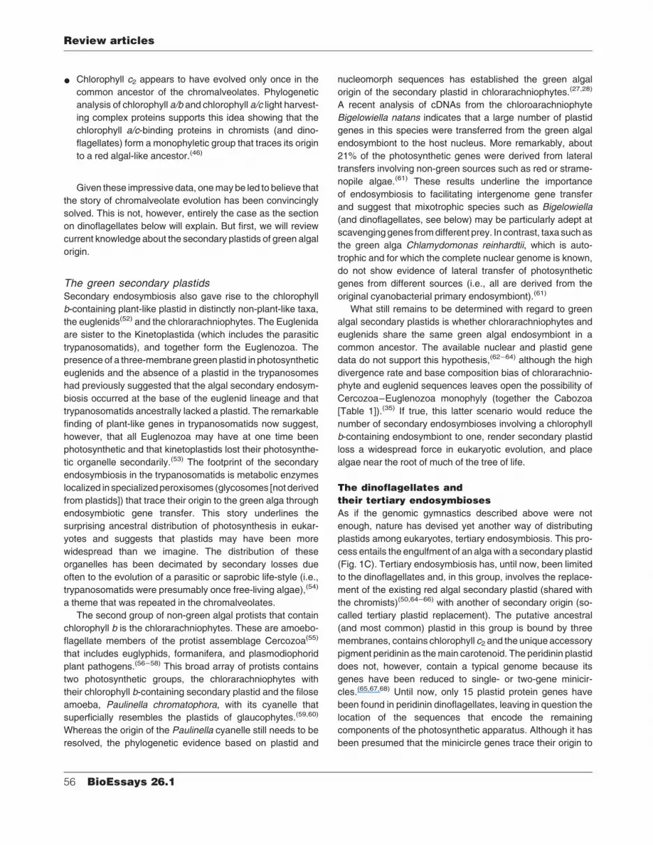
As if the genomic gymnastics described above were not
enough, nature has devised yet another way of distributing
plastids among eukaryotes, tertiary endosymbiosis. This pro-
cess entails the engulfment of an alga with a secondary plastid
(Fig. 1C). Tertiary endosymbiosis has, until now, been limited
to the dinoflagellates and, in this group, involves the replace-
ment of the existing red algal secondary plastid (shared with
the chromists)(50,64–66) with another of secondary origin (so-
called tertiary plastid replacement). The putative ancestral
(and most common) plastid in this group is bound by three
membranes, contains chlorophyll c2 and the unique accessory
pigment peridinin as the main carotenoid. The peridinin plastid
does not, however, contain a typical genome because its
genes have been reduced to single- or two-gene minicir-
cles.(65,67,68) Until now, only 15 plastid protein genes have
been found in peridinin dinoflagellates, leaving in question the
location of the sequences that encode the remaining
components of the photosynthetic apparatus. Although it has
been presumed that the minicircle genes trace their origin to
Review articles
56 BioEssays 26.1

the red algal secondary endosymbiosis in the chromalveol-
ate ancestor(65), it is also possible that these sequences may
have multiple origins from different endosymbiotic events
(see below).
Given the ancestral origin of the red algal secondary
plastid in dinoflagellates, tertiary replacement has been used
to explain the plastid in taxa such as Karenia spp. and
Karlodinium micrum that contain chlorophylls c1þ c2 and 190-
hexanoyloxy-fucoxanthin and/or 190-butanoyloxy-fucoxanthin
but lack peridinin, similar to the haptophyte algae.(66,69)
Tertiary endosymbiosis also would account for plastid origin
in other dinoflagellates such as Dinophysis spp. (cryptophyte
origin),(70) Peridinium foliaceum (stramenopile origin),(71) and
Lepidodinium viride (green algal origin).(72) Give these
complex series of events, understanding the impact of
endosymbiosis on dinoflagellate evolution will require a
genomics approach. In this regard, we are currently generat-
ing a data set of up to 10,000 unique 30 reads from normalized
and subtracted cDNA libraries of the toxic dinoflagellate
Alexandrium tamarense. These data will help us quantify the
genomic contribution of multiple endosymbioses to dinofla-
gellate evolution and, as we and others(73) predict, likely
identify the missing dinoflagellate plastid genes as compo-
nents of the Alexandrium nuclear genome. Preliminary data
from theAlexandrium cDNA data set have in fact confirmed the
presence of many plastid genes in the nucleus of this dino-
flagellate (J.D.H., H.S.Y., and D.B. unpublished results).
Conclusions
Endosymbiosis has created a plethora of photosynthetic
eukaryotes that have significantly shaped the Earth’s history.
The known fossil record and our molecular dating analysis
suggest that photosynthetic eukaryotes have been around for
well over a billion years. The super-assemblage, chromalveo-
lates, appears to be a monophyletic group and the ancient red
algal secondary endosymbiosis that we have recently de-
scribed(43) and the GAPDH duplication found by Fast et al.(42)
unite them as a cohesive lineage. The analysis of multi-gene
nuclear and mitochondrial data sets that accommodate a
broad taxonomic sampling should verify this hypothesis in the
coming years.(74) The present state of knowledge regarding
plastid and host relationships among algae is summarized in
Fig. 4. The plastid sequence data have significantly added to
our understanding of the algal tree of life both within lineages
Figure 4. Present understanding of the phylogenetic relationships of algae. The host relationships that have resulted from analyses of
nuclear or mitochondrial loci are shown with the black line. The branches with broken lines represent uncertainty about the position of these
taxa in the host tree. Phylogenetic relationships inferred from plastid gene comparisons are shown with the colored lines: red for the red algal
secondary endosymbiosis in chromalveolates, green for the putative green algal secondary endosymbiosis in cabozoans, and blue for the
haptophyte tertiary replacement either in the dinoflagellate common ancestor or only in fucoxanthin-containing taxa. Cabozoan monophyly
has not been substantiated with molecular phylogenetic data therefore the line leading from the Euglenozoa to the chlorarachniophytes is
dashed. Cases of potential gene transfer from green algae in the common ancestor of apicomplexans and dinoflagellates are not shown in
this figure. Plastid loss in the ciliates is shown with the open red box and plastid degeneration in the apicomplexans is shown with the dashed
red line. CB denotes the primary endosymbiosis that gave rise to the proto-algal ancestor of the red, green, and glaucophyte algae.
Review articles
BioEssays 26.1 57

and through the union of groups such as the Chromista that
had yet to be proven using other loci.
Perhaps most exciting is the mounting evidence(21,75) for
a green algal influence in the alveolates. Kohler et al.(75)
originally suggested a green algal origin of the apicoplast
based on phylogenetic analyses of the highly divergent tufA
gene. This relatively weakly supported result was recently
buttressed by the findings of Funes et al.(21) who identified a
unique nuclear-encoded cox2 (mitochondrial cytochrome c
oxidase subunit II gene) in apicomplexans and in some
green algae that is split into two coding regions (cox2a, cox2b).
Phylogenetic analysis supports the monophyly of the COX-
IIAþCOXIIB sequence in greens and apicomplexans, imply-
ing a single origin of these split genes in a green alga, and its
lateral transfer into the apicomplexans presumably after
endosymbiosis.(21,64) Funes et al.(76) and Waller et al.(77) have
recently provided differing interpretations of the COXII data
and its support for a green algal endosymbiosis in the
apicomplexans. Interestingly, analysis of our initial Alexan-
drium cDNA set shows the presence of multiple green algal
genes in this taxon that is sister to apicomplexans (J.D.H.,
H.S.Y., and D.B. unpublished results), supporting the findings
of Funes et al.(21,76) In our estimation, these exciting results
are either due to multiple gene transfers from different green
algae or a ‘‘hidden’’ green algal endosymbiosis that occurred
at least in the common ancestor of apicomplexans and
dinoflagellates. Addition of other completed genome se-
quences (e.g., Plasmodium falciparum)(78) to the analyses
should provide the data to test this idea. We also predict that
genomics projects will, in the near future, turn up other
examples of gene transfer in chromalveolates and in other
protists that have had a photosynthetic ancestry. Groups that
contain, or at one time contained, a secondary or tertiary
plastid will likely encode nuclear genes that have arisen
through lateral gene transfer from the eukaryotic endosym-
biont(s), much like the case found for the genes of cyanobac-
terial origin in Arabidopsis.(19)
These events may best be interpreted under the ‘‘Russian
Doll Paradigm’’ in which each round of endosymbiosis leads to
large-scale gene transfer that significantly reshapes the
nuclear genome of the host. This process, schematically
portrayed in the nuclear chromosomes shown in Fig. 1, is akin
to nested Russian dolls and is predicted to be a driving force in
generating eukaryotic biodiversity by providing a major influx
of novel genes into protistan genomes.
Acknowledgments
Figure 1 is based on an image created by NSF graphic artist
Kirk D. Woellert.
References1. Mereschkowsky C. Uber Natur und Ursprung der Chromatophoren im
Pflanzenreiche. Biol Zentralbl 1905;25:593–604.
2. Margulis L. Origin of Eucaryotic Cells. New Haven: Yale University Press;
1970. 349 pp.
3. Gray MW. The endosymbiont hypothesis revisited. Int Rev Cytol 1992;
141:233–357.
4. Bhattacharya D, Medlin L. The phylogeny of plastids: A review based on
comparisons of small subunit ribosomal RNA coding regions. J Phycol
1995;31:489–498.
5. Delwiche CF. Tracing the thread of plastid diversity through the tapestry
of life. Am Nat 1999;154(S4):S164–S177.
6. McFadden GI. Primary and secondary endosymbiosis and the origin of
plastids. J Phycol 2001;37:951–959.
7. Reumann S, Davila-Aponte J, Keegstra K. The evolutionary origin of the
protein-translocating channel of chloroplastic envelope membranes:
identification of a cyanobacterial homolog. Proc Natl Acad Sci USA 1999;
96:784–789.
8. Foth BJ, Ralph SA, Tonkin CJ, Struck NS, Fraunholz M, Roos DS,
Cowman AF, McFadden GI. Dissecting apicoplast targeting in the
malaria parasite Plasmodium falciparum. Science 2003;299:705–708.
9. Martin W, Herrmann RG. Gene transfer from organelles to the nucleus:
how much, what happens, and why? Plant Physiol 1998;118:9–17.
10. Blanchard JL, Lynch M. Organellar genes: why do they end up in the
nucleus? Trends Genet 2000;16:315–320.
11. Gray MW, et al. Genome structure and gene content in protist mito-
chondrial DNAs. Nucleic Acids Res 1998;26:865–878.
12. Moreira D, Le Guyader H, Philippe H. The origin of red algae and the
evolution of chloroplasts. Nature 2000;405:69–72.
13. Stibitz T, Keeling P, Bhattacharya D. Symbiotic origin of a novel actin
gene in the cryptophyte Pyrenomonas helgolandii. Mol Biol Evol 2000;
17:1731–1738.
14. Kies L. Zur systematischen Einordnung von Cyanophora paradoxa,
Gloeochaete wittrockiana und Glaucocystis nostochinearum. Ber Deu-
tsch Bot Ges Bd 1979;92:445–454.
15. Kies L, Kremer BP. Typification of the Glaucocystophyta. Taxon 1986;
35:128–133.
16. Graham LD, Wilcox LW. Algae. New Jersey: Prentice-Hall Incorporated;
2000. 700 pp.
17. Melkonian M. Phylum Chlorophyta: introduction to the Chlorophyta. In:
Margulis L, Corliss JO, Melkonian M, Chapman DJ, editors. Handbook of
Protoctista. Boston: Jones and Bartlett Publishers Incorporated; 1990.
p 597–599.
18. Raven P, Evert RF, Eichhorn SE. Biology of Plants. New York: Bedford,
Freeman, and Worth Publishing Group; 1998. 875 pp.
19. Martin W, Rujan T, Richly E, Hansen A, Cornelsen S, Lins T, Leister D,
Stoebe B, Hasegawa M, Penny D. Evolutionary analysis of Arabidopsis,
cyanobacterial, and chloroplast genomes reveals plastid phylogeny and
thousands of cyanobacterial genes in the nucleus. Proc Natl Acad Sci
USA 2002;99:12246–12251.
20. Keeling PJ, Palmer JD. Lateral transfer at the gene and subgenic levels
in the evolution of eukaryotic enolase. Proc Natl Acad Sci USA 2001;
98:10745–10750.
21. Funes S, Davidson E, Reyes-Prieto A, Magallon S, Herion P, King MP.
Gonzalez-Halphen D. A green algal apicoplast ancestor. Science 2002;
298:2155.
22. Ludwig M, Gibbs SP. Are the nucleomorphs of cryptomonads and
Chlorarachnion the vestigial nuclei of eukaryotic endosymbionts? Ann
Acad Sci NY 1987;503:198–211.
23. Gibbs S. The evolution of algal chloroplasts. In: Lewin RA, editor. Origins
of Plastids. New York: Chapman and Hall Company; 1993. p 107–121.
24. Bhattacharya D, Medlin L, Wainright PO, Ariztia EV, Bibeau C, Stickel SK,
Sogin ML. Algae containing chlorophylls aþc are polyphyletic: Molecular
evolutionary analysis of the Chromophyta. Evolution 1992;46:1801–
1817.
25. McFadden GI. Plastids and protein targeting. J Euk Microbiol 1999;
46:339–346.
26. Douglas SE, Murphy CA, Spencer DF, Gray MW. Molecular evidence
that cryptomonad algae are chimaeras of two phylogenetically distinct
unicellular eukaryotes. Nature 1991;350:148–151.
27. McFadden GI, Gilson PR, Waller RF. Molecular phylogeny of chlorar-
achniophytes based on plastid rRNA and rbcL sequences. Arch
Protistenkd 1995;145:231–239.
Review articles
58 BioEssays 26.1

28. Van de Peer Y, Rensing SA, Maier U-G, De Wachter R. Substitution rate
calibration of small subunit rRNA identifies chlorarachniophyte endo-
symbionts as remnants of green algae. Proc Natl Acad Sci USA 1996;
93:7732–7736.
29. Gilson PR, McFadden GI. Good things in small packages: the tiny
genomes of chlorarachniophyte endosymbionts. BioEssays 1997;19:
167–173.
30. Douglas S, Zauner S, Fraunholz M, Beaton M, Penny S, Deng LT, Wu X,
Reith M, Cavalier-Smith T, Maier UG. The highly reduced genome of an
enslaved algal nucleus. Nature 2001;410:1091–1096.
31. Cavalier-Smith T. Chloroplast evolution: secondary symbiogenesis and
multiple losses. Curr Biol 2002;12:R62–R64.
32. Gajadhar AA, Marquardt WC, Hall R, Gunderson J, Ariztia-Carmona EV,
Sogin ML. Ribosomal RNA sequences of Sarcocystis muris, Theileria
annulata and Crypthecodinium cohnii reveal evolutionary relation-
ships among apicomplexans, dinoflagellates, and ciliates. Mol Biochem
Parasitol 1991;45:147–154.
33. Cavalier-Smith T. The kingdom Chromista: origin and systematics. In:
Round FE, Chapman DJ, editors. Progress in Phycological Research
(Vol. 4). Bristol: Biopress Limited; 1986. p 309–347.
34. Cavalier-Smith T. Membrane heredity and early chloroplast evolution.
Trends Plant Sci 2000;5:174–182.
35. Cavalier-Smith T. Principles of protein and lipid targeting in secondary
symbiogenesis: euglenoid, dinoflagellate, and sporozoan plastid origins
and the eukaryote family tree. J Eukaryot Microbiol 1999;46:347–366.
36. Gillott M. Phylum Cryptophyta (Cryptomonads). In: Margulis L, Corliss
JO, Melkonian M, Chapman DJ, editors. Handbook of Protoctista.
Boston: Jones and Bartlett Publishers Incorporated; 1990. p 139–151.
37. Green JC, Perch-Nielsen K, Westbroek P. Phylum Prymnesiophyta. In:
Margulis L, Corliss JO, Melkonian M, Chapman DJ, editors. Handbook of
Protoctista. Boston: Jones and Bartlett Publishers Incorporated; 1990.
p 293–317.
38. Vivier E, Desportes I. Phylum Apicomplexa. In: Margulis L, Corliss JO,
Melkonian M, Chapman DJ, editors. Handbook of Protoctista. Boston:
Jones and Bartlett Publishers Incorporated; 1990. p 549–573.
39. Lynn DH, Small EB. Phylum Ciliophora. In: Margulis L, Corliss JO,
Melkonian M, Chapman DJ, editors. Handbook of Protoctista. Boston:
Jones and Bartlett Publishers Incorporated; 1990. p 498–523.
40. Taylor FJR. Phylum Dinoflagellata. In: Margulis L, Corliss JO, Melkonian
M, Chapman DJ, editors. Handbook of Protoctista. Boston: Jones and
Bartlett Publishers Incorporated; 1990. p 419–437.
41. Wilson RJ, et al. Complete gene map of the plastid-like DNA of the
malaria parasite Plasmodium falciparum. J Mol Biol 1996;261:155–172.
42. Fast NM, Kissinger JC, Roos DS, Keeling PJ. Nuclear-encoded, plastid-
targeted genes suggest a single common origin for apicomplexan and
dinoflagellate plastids. Mol Biol Evol 2001;18:418–426.
43. Yoon HS, Hackett J, Pinto G, Bhattacharya D. The single, ancient origin
of chromist plastids. Proc Natl Acad Sci USA 2002;99:15507–15512.
44. Van de Peer Y, De Wachter R. Evolutionary relationships among the
eukaryotic crown taxa taking into account site-to-site rate variation in 18S
rRNA. J Mol Evol 1997;45:619–630.
45. Baldauf SL, Roger AJ, Wenk-Siefert I, Doolittle WF. A kingdom-level
phylogeny of eukaryotes based on combined protein data. Science
2000;290:972–977.
46. Durnford DG, Deane JA, Tan S, McFadden GI, Gantt E, Green BR. A
phylogenetic assessment of the eukaryotic light-harvesting antenna pro-
teins, with implications for plastid evolution. J Mol Evol 1999;48:59–68.
47. Knoll AH. The early evolution of eukaryotes: a geological perspective.
Science 1992;256:622–627.
48. Xiao S, Zhang Y, Knoll AH. Three-dimensional preservation of algae and
animal embryos in a Neoproterozoic phosphorite. Nature 1998;391:553–
558.
49. Anbar AD, Knoll AH. Proterozoic ocean chemistry and evolution: a
bioinorganic bridge. Science 2002;297:1137–1142.
50. Saldarriaga JF, Taylor FJ, Keeling PJ, Cavalier-Smith T. Dinoflagellate
nuclear SSU rRNA phylogeny suggests multiple plastid losses and
replacements. J Mol Evol 2001;53:204–213.
51. Andersson JO, Roger AJ. A cyanobacterial gene in nonphotosynthetic
protists-an early chloroplast acquisition in eukaryotes? Curr Biol 2002;
12:115–119.
52. Walne PL, Kivic PA. Phylum Euglenida. In: Margulis L, Corliss JO,
Melkonian M, Chapman DJ, editors. Handbook of Protoctista. Boston:
Jones and Bartlett Publishers Incorporated; 1990. p 270–287.
53. Hannaert V, Saavedra E, Duffieux F, Szikora J-P, Rigden DJ, Michels
PAM, Opperdoes FR. Plant-like traits associated with metabolism of
Trypanosoma parasites. Proc Natl Acad Sci USA 2003;100:1067–
1071.
54. Martin W, Borst P. Secondary loss of chloroplasts in trypanosomes. Proc
Natl Acad Sci USA 2003;100:765–767.
55. Cavalier-Smith T. A revised six-kingdom system of life. Biol Rev Camb
Philos Soc 1998;73:203–266.
56. Bhattacharya D, Helmchen T, Melkonian M. Molecular evolutionary
analyses of nuclear-encoded small subunit ribosomal RNA identify an
independent rhizopod lineage containing the Euglyphidae and the
Chlorarachniophyta. J Euk Microbiol 1995;42:65–69.
57. Cavalier-Smith T, Chao EE. Molecular phylogeny of the free-living
archezoan Trepomonas agilis and the nature of the first eukaryote. J
Mol Evol 1996;43:551–562.
58. Keeling PJ. Foraminifera and Cercozoa are related in actin phylogeny:
two orphans find a home? Mol Biol Evol 2001;18:1551–1557.
59. Bhattacharya D, Helmchen T, Bibeau C, Melkonian M. Comparisons of
small subunit ribosomal RNAs reveal the evolutionary position of the
Glaucocystophyta. Mol Biol Evol 1995;12:415–420.
60. Helmchen T, Bhattacharya D, Melkonian M. Analyses of ribosomal
RNA sequences from glaucocystophyte cyanelles provide new insights
into the evolutionary relationships of plastids. J Mol Evol 1995;41:203–
210.
61. Archibald JM, Rogers MB, Toop M, Ishida K-I, Keeling PJ. Lateral gene
transfer and the evolution of plastid-targeted proteins in the secondary
plastid-containing alga Bigelowiella natans. Proc Natl Acad Sci USA
2003;100:7678–7683.
62. Oliveira M, Bhattacharya D. Phylogeny of the Bangiophycidae (Rhodo-
phyta) and the secondary endosymbiotic origin of algal plastids. Am J
Bot 2000;87:482–492.
63. Archibald JM, Keeling PJ. Recycled plastids: a ‘green movement’ in
eukaryotic evolution. Trends Genet 2002;18:577–584.
64. Palmer JD. The symbiotic birth and spread of plastids: how many times
and whodunit? J Phycol 2003;39:4–12.
65. Zhang Z, Green BR, Cavalier-Smith T. Single gene circles in dino-
flagellate chloroplast genomes. Nature 1999;400:155–159.
66. Yoon HS, Hackett J, Bhattacharya D. A single origin of the peridinin-, and
fucoxanthin-containing plastids in dinoflagellates through tertiary endo-
symbiosis. Proc Natl Acad Sci USA 2002b;99:11724–11729.
67. Barbrook AC, Howe CJ. Minicircular plastid DNA in the dinoflagellate
Amphidinium operculatum. Mol Gen Genet 2000;263:152–158.
68. Barbrook AC, Symington H, Nisbet RE, Larkum A, Howe CJ. Organiza-
tion and expression of the plastid genome of the dinoflagellate
Amphidinium operculatum. Mol Genet Genomics 2001;266:632–638.
69. Hansen G, Daugbjerg N, Henriksen P. Comparative stidy of Gymnodi-
nium mikimotoi and Gymnodinium aureolum, comb. nov. (¼Gyrodinium
aureolum) based on morphology, pigment composition, and molecular
data. J Phycol 2000;36:394–410.
70. Hackett JD, Maranda L, Yoon HS, Bhattacharya D. Phylogenetic
evidence for the cryptophyte origin of the plastid of Dinophysis
(Dinophysiales, Dinophyceae). J Phycol 2003;39:440–448.
71. Chesnick JM, Morden CW, Schmieg AM. Identity of the endosymbiont of
Peridinium foliaceum (Pyrrophyta): analysis of the rbcLS operon. J Phycol
1996;32:850–857.
72. Watanabe MM, Takeda Y, Sasa T, Inouye I, Suda S, Sawaguchi T,
Chihara M. A green dinoflagellate with chlorophylls a and b morphology
fine structure of the chloroplast and chlorophyll composition. J Phycol
1987;23:382–389.
73. Zhang Z, Cavalier-Smith T, Green BR. Evolution of dinoflagellate
unigenic minicircles and the partially concerted divergence of their
putative replicon origins. Mol Biol Evol 2002;19:489–500.
74. Nozaki H, Matsuzaki M, Takahara M, Misumi O, Kuroiwa H, Hasegawa
M, Shin-I T, Kohara Y, Ogasawara N, Kuroiwa T. The phylogenetic
position of red algae revealed by multiple nuclear genes from
mitochondria-containing eukaryotes and an alternative hypothesis on
the origin of plastids. J Mol Evol 2003;56:485–497.
Review articles
BioEssays 26.1 59

75. Kohler S, Delwiche CF, Denny PW, Tilney LG, Webster P, Wilson RJ,
Palmer JD, Roos DS. A plastid of probable green algal origin in Api-
complexan parasites. Science 1997;275:1485–1489.
76. Funes S, Davidson E, Reyes-Prieto A, Magallon S, Herion P, King MP,
Gonzalez-Halphen D. Response to Comment on ‘‘A Green Algal Apico-
plast Ancestor’’. Science 2003;301:49.
77. Waller RF, Keeling PJ, Van Dooren GG, McFadden GI. Comment on
‘‘A Green Algal Apicoplast Ancestor’’. Science 2003;301:49.
78. Gardner MJ, et al. Genome sequence of the human malaria parasite
Plasmodium falciparum. Nature 2002;419:498–511.
79. Rodrıguez F, Oliver JL, Marin A, Medina JR. The general
stochastic model of nucleotide substitution. J Theor Biol 1990;142:
485–501.
80. Morden CW, Sherwood AR. Continued evolutionary surprises among
dinoflagellates. Proc Natl Acad Sci USA 2002;99:11558–11560.
Review articles
60 BioEssays 26.1