phosducin-like proteins in dictyostelium discoideum: implications for the phosducin family of...
TRANSCRIPT
Mieke Blaauw, Jaco C.Knol, Arjan Kortholt,Jeroen Roelofs1, Ruchira2, Marten Postma,Antonie J.W.G.Visser2 andPeter J.M.Van Haastert3
Department of Biochemistry, University of Groningen, Nijenborgh 4,9747 AG Groningen, The Netherlands
1Present address: Department of Cell Biology, Harvard MedicalSchool, 240 Longwood Avenue, Boston, MA 02115-5730, USA2Present address: MicroSpectroscopy Centre, Laboratory ofBiochemistry, Wageningen University, The Netherlands
3Corresponding authore-mail: [email protected]
Retinal phosducin is known to sequester transducinGbg, thereby modulating transducin activity. Phos-ducin is a member of a family of phosducin-likeproteins (PhLP) found in eukaryotes. Phylogeny of33 phosducin-like proteins from metazoa, plants andlower eukaryotes identi®ed three distinct groupsnamed phosducin-I±III. We discovered three phlpgenes in Dictyostelium, each encoding a phosducin-likeprotein of a different group. Disruption of thephlp1 gene strongly impaired G-protein signalling,apparently due to mislocalization of Gbg in phlp1-nullcells. GFP-Gb and GFP-Gg are membrane associatedin wild-type cells, but cytosolic in phlp1-null cells.Phlp2 disruption is lethal due to a synchronouscollapse of the cells after 16±17 cell divisions. Phlp3disruptants show no abnormal phenotype. Theseresults establish a role for phosducin-like proteinsin facilitating folding, localization or function ofproteins, in addition to modulating G-proteinsignalling.Keywords: chaperone/Dictyostelium/G protein/signaltransduction/phosducin
Introduction
Heterotrimeric GTP-binding proteins (G proteins) aresignal transducers that couple heptahelical transmembranereceptors to their intracellular effectors. G proteins arecomposed of Ga, Gb and Gg subunits. Upon activation bytheir receptors, bound GDP is exchanged for GTP. Thisresults in a conformational change that induces dis-sociation of GTP-bound Ga from the Gbg complex. TheGaGTP and free Gbg subunits both interact with down-stream effectors. The intrinsic GTPase activity of Ga leadsto the formation of inactive GaGDP, which reassociateswith Gbg resulting in the reformation of the trimeric Gprotein (Clapham and Neer, 1997; Hamm, 1998).Signalling via G proteins is modulated by several proteins,such as regulators of G-protein signalling (RGS proteins),which activate the GTPase activity of Ga (Ross and
Wilkie, 2000), and by phosducin, which sequesters Gbg(Bauer et al., 1992).
Phosducin, a cytosolic 28 kDa protein, is composed oftwo domains: the N-terminal 13 kDa is mostly helical,while the C-terminal 15 kDa folds like thioredoxin. Gbg-binding studies and the X-ray structure of the phosducinretinal±Gbg complex show that both domains contribute tothe interaction with Gbg. The N-terminal helical domain ofphosducin binds extensively to the loops of Gb that alsoprovide the interaction with Ga, while the C-terminaldomain of phosducin binds to the region of Gbg that canassociate with the membrane surface (Gaudet et al., 1996;Loew et al., 1998; Savage et al., 2000). Thereforephosducin is thought to modulate Gbg activity by bindingto free Gbg and blocking Gbg association with Gasubunits, effectors or membranes (Bauer et al., 1992;Bluml et al., 1997).
Phosducin was ®rst discovered at high concentrations inthe retina (Lee et al., 1990) and the developmentallyrelated pineal gland (Reig et al., 1990). Additional studiesrevealed three splice variants of phosducin in human retina(Craft et al., 1998). Besides expression in the retina,phosducin is also detected at lower levels in many othermammalian tissues (Bauer et al., 1992; Danner and Lohse,1996). Many phosducin-related proteins have been dis-covered in vertebrates and in lower eukaryotes, suggestingthat retinal phosducin is a member of a phosducin familyof proteins. The phosducin-like protein PhLP shows 41%amino acid identity with phosducin (Miles et al., 1993). Incontrast with phosducin, PhLP has a similar expressionlevel in a wide variety of tissue (Schroder and Lohse,2000). All phosducin and PhLP variants have beenreported to bind to Gbg in vitro, with the exception ofsplice variants leading to N-terminal truncations ofphosducin (Schroder and Lohse, 1996, 2000; Craft et al.,1998). In addition to these two genes, we recognized threeadditional phosducin-like proteins in the sequence data-bases of the Human Genome Project; this large number ofphosducin isoforms complicates the functional analysis ofphosducin-like proteins in vertebrates.
In unicellular eukaryotes G-protein signal-transductionpathways mediate processes as diverse as mating in yeast(Dohlman, 2002) and morphogenesis and chemotaxis inDictyostelium (Parent and Devreotes, 1999; Firtel andChung, 2000). Proteins of the phosducin family appear tobe involved in the regulation of Gb activity in lowereukaryotes. In the fungus Cryphonectria parasitica, thebdm-1 gene encodes a phosducin-like protein (Kasaharaet al., 2000). Disruption of this gene demonstrates a role ofthe BDM-1 protein in Gbg function and Ga accumulation.The Saccharomyces cerevisiae genome encodes twophosducin-like proteins, Plp1 and Plp2, that are able tobind Gbg and regulate Gbg-dependent signalling (Flanaryet al., 2000). Furthermore, disruption of the plp2 gene is
Phosducin-like proteins in Dictyosteliumdiscoideum: implications for the phosducinfamily of proteins
The EMBO Journal Vol. 22 No. 19 pp. 5047±5057, 2003
ã European Molecular Biology Organization 5047

lethal, indicating that Plp2 must have an essential functionin the cell. We discovered three genes in Dictyosteliumencoding phosducin-like proteins. Dictyostelium is a soilamoeba which undergoes a developmental program uponstarvation. Individual amoebae chemotax towards eachother and aggregate to form multicellular structures com-posed of stalk cells and spores. In this organism the functionof G-protein signalling, mediating chemotaxis and multi-cellular development, has been well established (Wu et al.,1995; Parent and Devreotes, 1999; Firtel and Chung, 2000;Janetopoulos et al., 2001; Zhang et al., 2001). We exploitedthe genetics of Dictyostelium to investigate the function ofthe three phosducin-like proteins.
Based on phylogenetic analysis of 33 protein sequencesfrom mammals, invertebrates, plants and unicellulareukaryotes, we show that the phosducin family consistsof three subgroups, which we named phosducin-I,phosducin-II and phosducin-III. The phosducin-I subgroupcontains retinal phosducin and several phosducin-likeproteins. The Gbg-binding motif of retinal phosducin in
the N-terminal domain is highly conserved within thissubfamily. In proteins of the phosducin-II subgroup thismotif is replaced by another highly conserved motif,while this part of the N-terminal domain is absent inthe phosducin-III subgroup. Some organisms, such asS.cerevisiae and Arabidopsis, contain only two phosducin-like proteins, while vertebrates have many members ineach subgroup. The three Dictyostelium phosducin-likeproteins each belong to a different group, giving theunique opportunity of investigating the function of thephosducin subgroups. Dictyostelium PhLP1, retinal phos-ducin and the C.parasitica BDM-1 all belong to thephosducin-I group. Disruption of phlp1 in Dictyosteliumresults in a phenotype resembling that of Gb-null cells,similar to disruption of bdm-1 in C.parasitica (Kasaharaet al., 2000). Interestingly, we observed that GFP-Gb andGFP-Gg are both cytosolic in the Dictyostelium phlp1disruptant, while associated with the plasma membrane inwild-type cells. Disruption of phlp2 is lethal, as for yeastPlp2 that belongs to the same phosducin-II group. The
M.Blaauw et al.
5048

phlp3 disruptant displayed no abnormal phenotype, as wasthe case after inactivation of the yeast phosducin-IIIhomologue Plp1 (Flanary et al., 2000).
Results
Identi®cation of phosducin-like proteins inDictyosteliumTo identify phosducin family proteins, the Dictyosteliumdiscoideum genomic and cDNA databases were screenedwith a collection of phosducin-like protein sequences fromdifferent organisms (see Materials and methods). Thisrevealed three genes encoding putative proteins sharing asigni®cant degree of identity with phosducin. Therefore wedenoted the genes as phlp1, phlp2 and phlp3, respectively.Sequence analysis of genomic DNA and cDNA revealedthat each phlp gene consists of two exons, separated by asingle intron of 172, 94 and 118 bases in phlp1, phlp2 andphlp3, respectively. The introns are short and AT rich,which is common for Dictyostelium. The position of the
intron is not conserved within the Dictyostelium phlpgenes. The exons upstream of the introns in phlp1, phlp2and phlp3 encode 206, 121 and 34 amino acids, and thedownstream exons encode 110, 118 and150 amino acids,giving a total predicted size of 316, 239 and 184 aminoacids for PhLP1, PhLP2 and PhLP3, respectively. Likemammalian retinal phosducins, the Dictyostelium PhLP1,PhLP2 and PhLP3 are acidic proteins with predicted pIvalues of 4.68, 4.94 and 5.62, respectively.
Phylogeny of phosducin family proteinsGenomic databases of many organisms were screened toidentify members of the phosducin superfamily: theorganisms include human, mouse, Caenorhabditiselegans, Drosophila, Arabidopsis and other plants,S.cerevisiae and all other unicellular eukaryotes as far asdatabases were available. The assembly consists of 33putative phosducin genes; the protein coding sequence inthe database was incomplete for only one sequence (seeSupplementary data available at The EMBO Journal
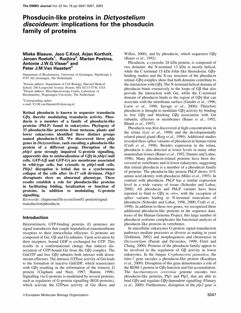
Fig. 1. The phosducin family consists of three de®ned subgroups. The protein sequences of 33 phosducin homologues were obtained from differentorganisms. These sequences were aligned (see Supplementary data for protein sequences, DDBJ/EMBL/GenBank entries and complete alignment).(A) Phylogenetic tree. The alignment was used as input for the Phylip program to construct the tree. The numbers indicate bootstrap values.(B) Alignment of three or four members of each subgroup. Residues shaded in black are conserved in 80±100% of all sequences; residues shaded ingrey are conserved in 60±70% of all sequences; bold characters are conserved in 75±100% of the sequences of a subgroup only. Substitutions withinthe following groups were considered as conservative: DE, RK, NQ, ST, FWY and MAILV. The structural elements of HsPhd are indicated asfollows: ~~~, ¯exible loop; ===, a-helix; ®, b-strand. The Gb-contacting residues of HsPhd are denoted by + above the aligned sequences. The startof the thioredoxin-like C-terminal domain is marked by a ®lled triangle.
Phosducin-like proteins in D.discoideum
5049

Online for DDBJ/EMBL/GenBank entries and sequencealignment). A phylogenetic tree was constructed using thededuced amino acid sequences of these 33 phosducinisoforms (Figure 1A). Two interesting observations can bemade. First, the analysis reveals that the phosducin familyconsists of three monophyletic groups, designatedphosducin-I, phosducin-II and phosducin-III, respectively.Secondly, the predicted gene products of Dictyosteliumphlp1, phlp2 and phlp3 each belong to a differentphosducin group. Apparently, proteins belonging to eachof the three subgroups are present only in mammals,Drosophila and Dictyostelium. Human retinal phosducin(HsPhd) and the phosducin-like protein HsPhLP belong tothe phosducin-I group. We detected three additionalphosducin homologues in the Human Genome Sequencedatabases. Two phosducin-like proteins (namedHsPhLP2A and HsPhLP2B) are incorporated in thephosducin-II group. One phosducin-like protein belongedto the phosducin-III group and is annotated as a putativeATP-binding protein (many sequences of thephosducin-III group have this annotation, which is derivedfrom a distant homologue that has an additional ATP-binding domain).
Sequence analysis of phosducin family proteinsAn alignment of the derived amino acid sequences ofphosducin and phosducin-like proteins of Dictyostelium,S.cerevisiae, C.parasitica and Homo sapiens is provided inFigure 1B, along with secondary structural characteristicsand Gbg-binding residues of mammalian retinal phos-ducin. The archetypal retinal phosducin is composed of anN-terminal domain, containing a-helices 1 to 3, and aC-terminal thioredoxin-like domain, consisting of a ®ve-stranded b-sheet and four a-helixes. The N-terminaldomain binds extensively to the loops of Gbg that providethe interaction with Ga, while the C-domain binds to themembrane association surface of Gbg (Gaudet et al., 1996;Loew et al., 1998; Savage et al., 2000).
As can be seen in the alignments, all proteins of the threesubgroups have extensive homology at the C-terminalthioredoxin domains (Figure 1B and the complete align-ment in the Supplementary data). In contrast, eachsubfamily has distinctive features in its N-terminaldomains. The characteristic Gbg-binding motif TGPK-GVINDWR in helix 1 (Gaudet et al., 1996) is highlyconserved in all proteins of the phosducin-I group. Withinthe phosducin-II subgroup, only the residues GVI of thisGb binding motif are conserved as GIL. However, twomotifs adjoining the GIL residues are unique and highlyconserved, yielding the phosducin-II-speci®c signaturesequence TEWNDILRxxGILPPK. Phosducin-III in factlacks helix1.
The amino acids of Gb that interact with retinalphosducin have been identi®ed in the crystal structure ofthe complex (Gaudet et al., 1996). Of the 27 amino acidsof transducin Gb that interact with phosducin, 25 areconserved in Dictyostelium Gb (data not shown). Theamino acids of retinal phosducin that interact with Gb areindicated in Figure 1B. In Dictyostelium PhLP1, 16 of the32 Gb-Interacting amino acids are conserved. In PhLP2and PhLP3, only ten and eight, respectively, of theGb-Interacting amino acids are conserved.
Inactivation of phlp genes in DictyosteliumTo establish the functions of the three Dictyosteliumphosducin-like proteins, and of the phosducin subgroups ingeneral, each of the Dictyostelium phlp genes wasinactivated by homologous recombination. Linear phlpDNA fragments, in which part of the open reading frame(ORF) was replaced by the bsr selection cassette (Sutoh,1993), were electroporated into the Dictyostelium wild-type strain AX3. Disruption of phlp1 and phlp3 wascon®rmed by Southern blotting using genomic DNA ofblasticidin-resistant cell lines (data not shown).
Isolation of phlp2 disruptants appeared to be problem-atic. We repeatedly obtained two types of blasticidin-resistant clones: normal growers and clones that initiallygrew well but died ~3 weeks after transformation. DNAwas isolated from several clones ~5 days before weexpected death in the non-viable clones. PCR wasperformed with primers recognizing the bsr cassette andpart of the 3¢ untranslated region of the phlp2 gene notpresent in the knockout construct (Figure 2A). Weobserved that dying clones gave the expected 0.5 kbPCR product, while none of the normally growing cellsgave a PCR product (Figure 2B). Using primersrecognizing the 5¢ and 3¢ ends of the phlp2 gene, thenormal growers yielded a wild-type size band, indicatingthat they are random integrants. This demonstrates that thephlp2 disruption obtained is lethal.
Phenotypes of phlp null cellsThe mutants obtained by targeted disruption of phlp genesbehaved quite differently from each other. For phlp3±
mutants we did not ®nd any abnormal phenotype. Growthrates were normal and the disruptants aggregated anddeveloped normally into fruiting bodies on non-nutrientagar plates (Figure 3A). Also chemotaxis assays did notreveal a difference from wild-type AX3 cells (Figure 3B).
Disruption of phlp2 resulted in a pronounced phenotypeshowing a loss of cell viability. The phlp2± cells initiallyproliferated well (Figure 2C). However, ~5 days aftertransformation the growth rate declined, and cells stoppedproliferating after ~18 days when a maximum of ~105 cellswas obtained. Between days 20 and 22 the cells diedsynchronously (Figure 2C). Cells showed the normalamoeboid appearance and movement until 2 days beforedeath, when they became round and small, and ®nallylysed. When some of the cells were transferred to mediumwithout blasticidin selection or to plates with bacteria7 days before their expected death, they died on approxim-ately the same day as the cells in the original medium. Thedata strongly suggest that PhLP2 is essential inDictyostelium.
Targeted disruption of phlp1 also resulted in a verystrong phenotype. First, the ability of phlp1± cells to growon bacterial lawns was severely affected. While wild-typeAX3 cells formed plaques, aggregated and producedfruiting bodies within a few days, the phlp1± disruptantscleared the lawns much more slowly, forming 2- to 3-foldsmaller plaques than AX3. Secondly, the phlp1± cells didnot aggregate on non-nutrient agar plates or bacteriallawns, even after several weeks (Figure 3A). SomeDictyostelium mutants that are aggregation de®cientbecause they cannot produce or secrete cAMP, doaggregate if they are supplied with exogenous cAMP
M.Blaauw et al.
5050

pulses or mixed with cAMP-secreting wild-type cells.However, the phlp1± cells failed to coaggregate with wild-type cells in mixtures with AX3 cells, and also remainedaggregation de®cient after pulsing with cAMP. Thirdly,the mutant cells did not display chemotaxis towards a widerange of cAMP and folic acid concentrations (Figure 3B),
and did not respond to bacterial extracts which are a richsource of chemoattractants binding to different surfacereceptors (data not shown). Finally, the doubling time inaxenic medium, both on plates and in shaking cultures,was increased (18±20 h) compared with the doubling timeof AX3 cells (10±12 h). Except for the reduced growthrate, these phenotypic properties of phlp1-null cells arevery similar to those of gb± cells (Lilly et al., 1993; Wuet al., 1995). A gb-null mimicking phenotype has alsobeen reported for disruption of the bdm1 gene fromC.parasitica which, like Dictyostelium phlp1, encodes aphosducin-I family member (Kasahara et al., 2000).
To con®rm that the phenotype of phlp1± cells was due todisruption of the phlp1 gene, an extrachromosomalplasmid containing the ORF of phlp1 was transformedinto the phlp1± cells. Expression of phlp1 in these phlp1±/phlp1OE cells rescued all phenotypic defects; develop-mental morphology, growth rate and chemotactic respon-ses returned to those of wild-type AX3 (Figure 3). Thepresence of phlp1 mRNA in phlp1±/phlp1OE was con-®rmed in a northern blot (data not shown). Although theamount of phlp1 mRNA was much elevated comparedwith the level in wild-type cells, no adverse effects wereobserved; it is not known if PhLP1 protein levels areelevated in the phlp1±/phlp1OE cells.
Biochemical assays in phlp1± cellsTo investigate why phlp1-null cells have a phenotype thatis similar to that of gb-null cells, we analysed receptor±G-protein effector interactions in phlp1-null cells. First,we studied the interaction between cAMP surfacereceptors and G proteins. Membranes of wild-type AX3cells contain both high- and low-af®nity cAMP bindingsites; high-af®nity sites represent cAMP receptors that arecoupled to functional G proteins. Addition of GTPgScauses the release of G proteins from the receptors andhence a conversion of cAMP receptors from a high-af®nityto a low-af®nity form (Van Haastert, 1984; Van Haastertet al., 1986). We measured the effect of GTPgS on thebinding of 10 nM cAMP to membranes from wild-type,phlp1-null and gb-null cells. GTPgS induces a stronginhibition of cAMP binding to AX3 membranes(Figure 4A). In phlp1-null cells, the basal level of cAMPbinding was substantially diminished and no effect ofGTPgS was observed. The gb-null cells showed essentiallythe same cAMP binding properties as phlp1-null cells,while the rescue phlp1±/phlp1OE cells displayed the highcAMP binding and strong GTPgS-mediated inhibition ofwild-type cells (Figure 4A).
Next, we investigated whether the diminished basallevel of cAMP binding in phlp1± cells was due to areduction of the total number of receptor sites, or whetherthe af®nity of the cAMP receptors was affected. Scatchardanalysis was performed on membranes from phlp1± andphlp1±/phlp1OE cells. As shown in Figure 4B, cAMPbinding to phlp1±/phlp1OE membranes displayed curvi-linear Scatchard plots showing ~16 000 high-af®nitybinding sites per cell with a Kd of 4 nM and ~84 000 low-af®nity binding sites per cell with a Kd of 550 nM. Thehigh-af®nity binding sites disappear upon addition ofGTPgS, while the total number of sites is unaffected.These cAMP-binding properties of phlp1±/phlp1OE mem-branes are essentially identical with cAMP binding to
Fig. 2. Lethal phenotype of phlp2± cells. (A) Schematic of phlp2 genedisruption. The arrowheads denote the three primers used for PCRanalysis. PCR primer c recognizes part of the 3¢ untranslated regionthat is not present in the knockout construct. (B) Identi®cation of phlp2disruptants by PCR analysis. DNA was isolated from several clones~15 days after transformation; some of these clones died around day 22(clones 1±5), while others remained viable (clone 6). PCR analysisusing primer set a + c is predicted to yield a 503 bp product for a dis-rupted phlp2 gene and no product for an intact gene, while PCR analy-sis with primers b + c will yield an 893 bp product for the intact phlp2gene. (C) Cell growth curve. The amount of cells was estimated atdifferent days after transformation to calculate the number of popula-tion doublings. Data shown are the means and standard deviations ofsix phlp2± strains; the clones were identi®ed as phlp2 gene disruptantby PCR on day 18±20 as shown in (B). The population-doublingkinetics for random integrants are identical with those of wild-typeAX3 cells and are shown for comparison. The results reveal thatinitially the doubling time of phlp2± cells (closed circles) is about thesame as that of random integrants (open circles) but gradually increasesuntil cells stop dividing and die after 3 weeks (arrow).
Phosducin-like proteins in D.discoideum
5051

wild-type membranes (Van Haastert et al., 1986).Membranes from phlp1-null cells displayed only low-af®nity binding sites and GTPgS had no effect; the numberof binding sites was ~60 000 per cell which is ~70% of thatfor wild type or phlp1±/phlp1OE. The disappearance ofhigh-af®nity cAMP binding and the absence of GTPgS-mediated inhibition of cAMP binding in phlp1-null cellswere also described for gb± cells (Lilly et al., 1993; Wuet al., 1995), suggesting that a functional couplingbetween G proteins and cAMP receptors is abolished inphlp1 disruptants.
Agonist binding to cAMP receptors induces the accu-mulation of several second messengers, including cAMP
and cGMP (Van Haastert and Kuwayama, 1997). Figure 5shows that phlp1± cells starved for 5 h did not display asigni®cant cAMP or cGMP accumulation in response tothe agonist. Pulsing of the cells with cAMP at 5 minintervals during the starvation period has been shown torescue some mutants, but did not have any affect on theresponses of phlp1± cells (data not shown). The phlp1±/phlp1OE cells displayed wild-type patterns of cAMP andcGMP accumulation (Figure 5). In summary, the datademonstrate that the interactions between cAMP receptorsand G proteins, and between G proteins and effectorenzymes are absent in phlp1± cells. The absence offunctional heterotrimeric G proteins explains the defects
Fig. 3. Phenotype of Dictyostelium phlp1± and phlp3± disruptants. The genes were inactivated by homologous recombination. (A) Phenotype of wild-type AX3, phlp1±, phlp1±/phlp1OE and phlp3± cells on non-nutrient agar plates. Photographs were taken after 30 h of starvation. (B) Dose±responsecurves of chemotaxis towards cAMP and folic acid. The response to a range of cAMP concentrations was measured in droplets using the small-population assay. An agar cutting assay was used to score for chemotaxis to different concentrations of folic acid: AX3 (open circles), phlp1± (closedcircles), phlp1±/phlp1OE (squares) and phlp3± (triangles).
M.Blaauw et al.
5052

of chemotaxis and cell aggregation. These combinedproperties of phlp1± cells are very similar to the phenotypedescribed for gb± cells (Lilly et al., 1993; Wu et al., 1995).
Localization of GFP-Gb and GFP-Gg in phlp1-nullcellsBecause the phenotype of phlp1± is very similar to that ofgb± cells, the effect of phlp1 disruption on subcellularlocalization of GFP-Gb and GFP-Gg was examined. Inwild-type cells both GFP-Gb and GFP-Gg are enriched atthe plasma membrane (Figure 6). Expression of GFP-Gbin gb± cells rescued the aggregation-defective phenotype(data not shown), and resulted in membrane-enrichedlocalization as in wild-type cells. In contrast, overexpres-sion of either GFP-Gb or GFP-Gg did not restore
aggregation competence in phlp1± cells (data notshown). Moreover, disruption of phlp1 caused GFP-Gband GFP-Gg to become localized in the cytosol (Figure 6).In gb± cells, localization of GFP-Gg was also restricted tothe cytosol (Figure 6). The ¯uorescence of cytosolicGFP-Gb and GFP-Gg in phlp1± and gb± cells displayed arather granular pattern. Western blots using a rabbitpolyclonal anti-GFP antiserum con®rmed that the com-plete fusion proteins were expressed in all cell lines (datanot shown). In summary, expression of GFP-tagged Gband Gg showed that Gbg is mislocalized to the cytosol inphlp1± cells, which explains their gb± -like phenotype.
Discussion
The phosducin family appears to comprise many membersfound in different eukaryotes. The prototype is retinalphosducin which consists of an N-terminal helical domainand a C-terminal thioredoxin-like domain (Gaudet et al.,1996; Loew et al., 1998; Savage et al., 2000). Phosducinand phosducin-like proteins have been reported to bind toGbg in vitro (Schroder and Lohse, 1996, 2000; Craft et al.,1998). G-protein signalling has been studied in great detailin Dictyostelium (Wu et al., 1995; Parent and Devreotes,1999; Firtel and Chung, 2000; Janetopoulos et al., 2001;Zhang et al., 2001). In order to investigate a possible roleof phosducin-like proteins in regulation of G-proteinactivity in Dictyostelium, we screened the D.discoideumcDNA and genomic databases for phosducin isoforms. We
Fig. 5. Defective G-protein-mediated responses in phlp1± cells. cAMPand cGMP response of AX3, phlp1±, phlp1±/phlp1OE and gb± cells.(A) Cells were starved and pulsed with cAMP for 5 h and stimulatedwith 10 mM 2¢-deoxy-cAMP and 10 mM dithiothreitol for the detectionof the cAMP response. Prior to stimulation (open bars) and 90 s afterstimulation (black bars), cells were lysed and assayed for cAMP.(B) For measurement of the cGMP response, starved cells were stimu-lated with 1 mM cAMP. Prior to stimulation (open bars) and 15 s afterstimulation (black bars), cells were lysed and assayed for cGMP.
Fig. 4. Defective receptor±G protein interaction in phlp1± cells.(A) GTPgS inhibition of 3H-cAMP binding to membranes of AX3,phlp1±, phlp1±/phlp1OE and gb± cells. Membranes were prepared fromcells that were starved for 5 h. Binding assays were performed in theabsence (open bars) or presence (black bars) of 30 mM GTPgS. (B) Todetermine the number and af®nity of the cAMP-binding sites,Scatchard analysis was carried out by including different concentrationsof cAMP in the binding assays. The results for membranes of phlp1±/phlp1OE (squares) or phlp1± cells (circles) in the absence (open sym-bols) or presence (closed symbols) of 30 mM GTPgS are shown. Onenanomole of bound cAMP is equivalent to 6000 binding sites per cell.Data were ®tted using the program FigP. The data for phlp1±/phlp1OE
in the absence of GTPgS were ®tted with a two-receptor model; thedata for the other conditions were ®tted statistically better with a one-receptor model. The kinetic data are as follows: for phlp1±/phlp1OE
without GTPgS, Kd1 = 4.07 6 3.68 nM, B1d = 15 600 6 2600 sites/cell, Kd2 = 557 6 107 nM and B2 = 84 000 6 17 000 sites/cell; forphlp1±/phlp1OE with GTPgS, Kd = 507 6 92 nM and B = 80 500 66000 sites/cell; for phlp1± without GTPgS, Kd = 480 6 35 nM and B =60 000 6 2500 sites/cell; for phlp1± with GTPgS, Kd = 491 6 31 nMand B = 65 000 6 2000 sites/cell.
Phosducin-like proteins in D.discoideum
5053

discovered three novel genes belonging to the phosducinfamily of proteins. In order to investigate the diversity ofthe phosducin family, we compared the deduced proteinsequences of 33 phosducin family proteins from a varietyof eukaryotes, including Dictyostelium. Phylogeneticanalysis revealed that they can be classi®ed into threedistinct groups denoted phosducin-I, phosducin-II andphosducin-III. These three distinct phylogenetic groupswere also obtained when only the more conservedC-terminal thioredoxin-like domains of the proteins wereused in the phylogenetic analysis. The Dictyosteliumproteins, called PhLP1, PhLP2 and PhLP3, each belong toone of the three groups.
The phosducin-I group harbours both retinal phos-ducins and the mammalian phosducin-like protein PhLP.No phosducins of subgroup I were detected in thecompleted genomes of the plants Arabidopsis andOryza sativa (rice), and the yeasts S.cerevisiae andSchizosaccharomyces pombe. PhLP and the phosducin-Iproteins of lower eukaryotes contain an N-terminalextension compared with retinal phosducin. In all mem-bers of group I, the Gbg binding motif in the N-terminaldomain (TGPKGVINDWRK) is highly conserved. Thissuggests that all members of the phosducin-I group may
interact with Gbg (Bauer et al., 1992; Danner and Lohse,1996; Bluml et al., 1997; Flanary et al., 2000; Kasaharaet al., 2000; Savage et al., 2000).
To test the function of PhLP1 in Dictyostelium, we dis-rupted the phlp1 gene. This resulted in a strong phenotype,nearly indistinguishable from the D.discoideum gb-nullphenotype (Lilly et al., 1993; Wu et al., 1995). The phlp1±
cells were non-chemotactic and aggregation de®cient.Furthermore, cAMP binding in phlp1± cells was insensitiveto GTPgS, and did not induce cAMP or cGMP responses inthese cells. These results strongly indicate the absence ofG-protein signalling in phlp1± cells owing to loss of Gbgfunction. A gb-null mimicking phenotype has also beenreported for disruption of bdm1, a homologue of phlp1 inthe fungus C.parasitica (Kasahara et al., 2000). In order toexamine whether Gbg was properly localized, we over-expressed GFP-Gb and GFP-Gg in wild-type, phlp1± andgb± cells. In wild-type cells Gbg was mainly found on themembrane. Expression of GFP-Gb in the phlp1 disruptantdid not restore the phenotype. The ¯uorescent images ofphlp1± cells clearly show localization of GFP-Gb andGFP-Gg in the cytosol. Therefore we conclude that thedefects in G-protein signalling in phlp1± cells are due tomislocalization of Gbg. Membrane localization of Gbg is
Fig. 6. Expression of GFP-Gb and GFP-Gg. Confocal images of GFP-Gb and GFP-Gg in AX3, phlp1± and gb± cells. Cells were incubated in phosphatebuffer. GFP was excited with a 488 nm laser and the GFP ¯uorescence was ®ltered through a BP 505±550 nm ®lter. Cell strains and expressedGFP-fusion proteins are marked at the left and the top of the ®gure, respectively. Scale bar, 20 mm.
M.Blaauw et al.
5054

mediated by isoprenylation at the C-terminus of Gg (Muntzet al., 1992; Zhang et al., 2001). Our observation thatGFP-Gg in gb± cells is cytosolic indicates that membranelocalization of Gg requires functional Gb, which has alsobeen described for mammalian Gg (Pronin and Gautam,1993). This suggests that the primary defect of G-proteinsignalling in the phlp1 disruptant is due to either non-functional Gb or Gg, or the inability to make a functionalGbg complex. Several studies indicate that chaperonesmay be important for correct folding of Gb and otherproteins of the family of WD repeat proteins (Clapham andNeer, 1997; Garcia-Higuera et al., 1998). In addition,mammalian PhLP1 has been reported to have the capacityto interact with the proteasomal protein SUG1 (Barhiteet al., 1998) and the cytosolic chaperonin complex (CCT)(McLaughlin et al., 2002). We propose that DictyosteliumPhLP1 facilitates proper folding of Gb or assembly of Gbinto a Gbg complex.
Members of the phosducin-III group lack most of theN-terminal domain, but their C-terminal domain sharesextensive homology with phosducin homologues from theother two groups. The phlp3± mutants in Dictyostelium didnot show an abnormal phenotype under the conditionstested. A similar result was described for disruption of theyeast homologue plp1 (Flanary et al., 2000). Apparently,PhLP3 is not required for growth, G-protein signalling ordevelopment. Further investigation is needed to providemore information about the function of PhLP3.
Phosducin-II proteins lack part of the N-terminaldomain compared with members of the phosducin-Igroup. Their N-terminal domain contains a differentmotif: (TEWNDILRxxGILPPK). Members of thephosducin-II group are present in lower eukaryotes andplants; mammals possess two different isoforms namedPhLP2A and PhLP2B. Unexpectedly, no phosducin-IIhomologue could be found in C.elegans. Like plp2 inS.cerevisiae (Flanary et al., 2000), phlp2 appears to be anessential gene in Dictyostelium. Disruption of phlp2resulted in a gradually decreasing growth rate, leading toa synchronous collapse after 16±17 population doublings.Cell death might be caused by dilution of an essentialfactor due to subsequent cell divisions until the concen-tration declines below a threshold level. This essentialfactor may be either PhLP2 itself or another factorrequiring PhLP2 for functioning. It is unlikely thatPhLP2 is expressed at a high level; the number of plp2transcripts in S.cerevisiae has been estimated to be lessthan one per cell (Flanary et al., 2000). Therefore wesuggest that PhLP2 functions in a cascade and ful®ls acatalytic role, for example as a chaperone for one or moreessential proteins of the family of WD repeat proteins. Thehomologous mouse germ-cell-speci®c phosducin-likeprotein (MgcPhLP or MmPhLP2B) is proposed to beinvolved in germ-cell maturation (Lopez et al., 2003).When expressed in yeast plp2± cells, MgcPhLP comple-mented the defect caused by plp2 disruption, suggesting anevolutionarily conserved function of PhLP2.
In summary, phylogenetic analysis reveals that mem-bers of the phosducin family of proteins can be classi®edinto three distinct groups: phosducin-I, phosducin-II andphosducin±III. Having three distinct phosducin-likeproteins, Dictyostelium may serve as a model system toinvestigate the speci®c functions of phosducin isoforms.
While phosducin in the visual system is thought todownregulate G-protein signalling, this may not be theonly function of members of the phosducin family ofproteins. Indeed, phenotypic analyses of phosducin-likeprotein disruption mutants in Dictyostelium, C.parasiticaand S.cerevisiae strongly suggest that phosducinhomologues from different groups have distinct cellularfunctions.
Materials and methods
Cell growth and developmentAX3 (wild-type) and phlp± cells (see below) were grown in HG5 medium,which was supplemented with 10 mg/ml blasticidinn S (ICN) for phlp±
cells. Rescued phlp1±/phlp1OE cells and cells expressing GFP-Gb andGFP-Gg were supplemented with 30 mg/ml G418 (GibcoBRL). Cellswere starved by shaking for up to 5 h in 10 mM phosphate buffer pH 6.5(PB) at a density of 107 cells/ml. To observe developmental phenotypes,cells were deposited on non-nutrient (NN) agar (1.5% agar in PB) andincubated at 22°C. To study growth on bacteria, cells were plated withKlebsiella aerogenes on SM medium and incubated at 22°C for severaldays.
BioinformaticsBLAST searches in databases representing organisms from differentphyla (see below) yielded in total ~30 phosducin homologues (seeSupplementary data for abbreviations, DDBJ/EMBL/GenBank accessionnumbers, sequences and alignments of the protein sequences). Usingthese phosducin sequences, we identi®ed sequences in the DictyosteliumcDNA and genomic databases representing three phosducin homologues,named phlp1, phlp2 and phlp3. The complete ORFs were assembled fromraw sequence data of the Dictyostelium genome project (http://genome.imb-jena.de/dictyostelium/). The sequences of phlp1, phlp2and phlp3 have been deposited at DDBJ/EMBL/GenBank underaccession numbers AF540058, AF540059 and AF540060, respectively,and the encoded amino acid sequences are presented in Figure 1B.
Blast searches were carried out in the general DDBJ/EMBL/GenBank(http://www.ncbi.nlm.nih.gov), and several speci®c databases forDrosophila (http://www.fruit¯y.org), C.elegans (http://www.wormbase.org), human (http://www.ncbi.nlm.nih.gov/genome/seq/HsBlast.html andhttp://publication.celera.com), yeast (http://www.ncbi.nlm.nih.gov),Plasmodium (http://www.ncbi.nlm.nih.gov/Malaria/plasmodiumbl.html),Dictyostelium (http://www.sdsc.edu/mpr/dicty/) and Arabidopsis (http://www.arabidopsis.org).
Multiple sequence alignments were constructed using the CLUSTALW program (Thompson et al., 1994), followed by manual optimization.Distance matrices were constructed from the alignments with thePROTDIST program of the PHYLIP package, which uses the DayhoffPAM 100 matrix for the calculation of evolutionary distances (Phylip 3.5)(Felsenstein, 1996). Phylogenetic trees were generated using the FITCHprogram of the PHYLIP package, with 100 bootstrap replications toassess the reliability of the nodes. Programs from the ExPASy MolecularBiology server (http://www.expasy.ch/) were used for the analysis ofprotein sequences.
Plasmid constructionThe coding sequences for phlp1, phlp2 and phlp3 were ampli®ed by PCR(Expand Long Template System, Roche) from Dictyostelium genomicDNA or by RT-PCR (M-MLV Reverse Transcriptase, Promega) frommRNA. The complete phlp1 coding sequence was ampli®ed with theprimers 5¢-TCTCAGATCTAAAGAATGGAACAAAACATTTTAAA-TAG-3¢ and 5¢-AGAGGGATCCTTAATCGTCATTATCATCATCG-GAC-3¢). The primers 5¢-GAGGATCCAAAAATGGGTTTAGGTAA-AACAGAATG-3¢ and 5¢-GAGGATCCTCATCAGAATCAGAATTAT-CAG-3¢ yielded the complete coding sequence of phlp2. For ampli®cationof phlp3, primers 5¢-GGAAGATCTAAAAATGTCAGAAAATAATAC-CAATAATG-3¢ and 5¢-GCTAGATCTATCATCTTCTTTAAATTTAT-TATTCTTAACATC-3¢ were used. The PCR products were cloned intothe TA cloning vector pGEM T-easy (Promega). All constructs weresequenced to con®rm the nucleotide sequences of the phosducin insertsand the position of the introns, and subcloned using the restriction sitesunderlined in the primers above in Dictyostelium extrachromosomalexpression vector AH2, an MB12neo derivative (Linskens et al., 1999).
Phosducin-like proteins in D.discoideum
5055

For the creation of expression plasmids encoding GFP-Gb and GFP-Ggfusion proteins, the coding sequences of Gb and Gg genes were PCR-ampli®ed from plasmids carrying the ORFs of Gb or Gg (Janetopouloset al., 2001) with Pfu polymerase (Promega). For Gb, the primers 5¢-TTTAGATCTAAAATGTCATCAGATATTTCAGAAAAAATTCAAC-AAGCAAG-3¢ and 5¢-TTTGGATCCTTAAGCCCAAATCTTGAGGA-GAGAATCC-3¢ were used. For Gg, we employed the primers 5¢-ATATAGATCTATAATGTCCGAATCACAATTAAAAAAAGTTTT-AAAAG-3¢ and 5¢-ATTACTAGTTTATAACACAGAACATCCATT-TCCTTTGAGTGGTT-3¢. The resultant PCR products were cloned inpGEM-T-easy, sequenced and subcloned in the Dictyostelium expressionvector LB5Neo, an MB12neo derivative with an N-terminal GFP cloningcassette (a gift from L.Bosgraaf).
Gene inactivationThe phlp genes have small coding regions (~550±950 bp), eachinterrupted by an AT-rich intron. We PCR ampli®ed fragments upstream(5¢) and downstream (3¢) of the intron of each phlp gene. The 5¢ and 3¢fragments of each gene were cloned sequentially around the bsr selectioncassette (Sutoh, 1993), such that the bsr cassette is in the reverseorientation with respect to the phlp fragments.
The 5¢ phlp1 fragment was ampli®ed using primers 5¢-GTACGCG-TAGCTCGTATGGAACAATG-3¢ and 5¢-GTCTGCAGCGAAAAC-ATTTGGTGGTTC-3¢; this fragment was cloned in the bsr-containingplasmid pUC21/bsr using the restriction sites PstI and MluI. The 3¢ phlp1fragment was ampli®ed using primers 5¢-ATAGAATTCTATATACCA-GAATGTG-3¢ and 5¢-GTATCGATCCTAGTTCTTCAGTGAG-3¢; thisfragment was cloned in the TA cloning vector pGEM T-easy, andsubsequently cloned into the 5¢ phlp1/bsr-containing plasmid using theEcoRI sites.
To obtain the phlp2 disruption construct, the EcoRI fragment frompGEMT-easy/phlp2 (see above) was used as the 5¢ fragment. The 3¢fragment was obtained by PCR using the primers 5¢-GATCTAGA-TTCCTCAATGTCAATTAGTAAATC-3¢ and 5¢-GAGGATCCTCAT-CAGAATCAGAATTATCAG-3¢. These fragments were cloned into thebsr containing plasmid using BamHI and XbaI.
For the phlp3 disruption plasmid, the 5¢ fragment was ampli®ed usingthe primers 5¢-GGAAGATCTAAAAATGTCAGAAAATAATACCAA-TAATG-3¢ and 5¢-GAGGATCCCACCATGTGTTGATAAAAAG-3¢.The primer pair 5¢-GTTCTAGAGCAAAAACACATTTAGGTAC-3¢and 5¢-GCTAGATCTATCATCTTCTTTAAATTTATTATTCTTAAC-ATC-3¢ was used to amplify the 3¢ fragment of phlp3.
A linear fragment with the bsr cassette and phlp ¯anking sequenceswas obtained from each disruption construct by PCR, using the 5¢-senseand 3¢-antisense outer primers. Dictyostelium AX3 cells were transformedby electroporation using 5 mg of linear DNA fragment. Blasticidin-resistant clones were screened for homologous recombination using PCRand Southern blot analysis.
For rescue experiments of phlp1± cells, the ORF of phlp1 was clonedinto the BglII site of the extrachromosomal expression plasmid AH2.
AssaysAll biochemical assays were carried out with cells that were starved for 5 hby shaking in PB at a density of 107 cells/ml. When indicated, cells werepulsed with 10 nM cAMP at 5 min intervals during the starvation period.To assay for 3H-cAMP binding to membranes, starved cells wereharvested and resuspended to 108/ml in PB. Preparation of membranesand the binding assays were performed as described (Snaar-Jagalska andVan Haastert, 1994). Each binding assay was performed in triplicate inthe presence or absence of 30 mM guanosine 5¢-O-(3-thiotriphosphate)(GTPgS). To determine the number and af®nity of the cAMP-bindingsites, Scatchard analysis was carried out by including differentconcentrations of cAMP in the binding assays.
The receptor-mediated responses of the cells were measured as theamount of cGMP produced upon stimulation by 1 mM cAMP, and theamount of cAMP produced upon stimulation by 10 mM 2¢-deoxy-cAMPand 10 mM dithiotreitol; cGMP and cAMP levels were determined byisotope dilution assays as described (Snaar-Jagalska and Van Haastert,1994).
Chemotaxis towards cAMP was measured in droplets using the small-population assay (Konijn, 1970). An agar cutting assay was used to scorefor chemotaxis to folic acid (Kuwayama et al., 1993).
Fluorescence microscopyFor microscopy, cells were washed twice with phosphate buffer (17 mM,pH 6.5) and starved for 1 h. Images were obtained from cells incubated inbuffer using a confocal laser scanning microscope (ConfoCor 2, LSM 510
combination setup; Carl-Zeiss, Germany). GFP was excited with the488 nm argon ion laser controlled by an acousto-optical tunable ®lter(AOTF). A dichroic beam splitter (HFT 488) separated the excitationfrom the emission. The GFP ¯uorescence was ®ltered through a BP 505±550 nm ®lter. The objective used was a 403 oil±immersion Plan-Neo¯uarwith a numerical aperture of 1.3. The pinhole was set at 73 mm. Imageswere analysed with the Zeiss LSM Image Browser software package.
Supplementary dataSupplementary data are available at The EMBO Journal Online.
Acknowledgements
We thank Chris Janetopoulos and Leonard Bosgraaf for providingplasmids and Annelies Heidekamp for constructing plasmid AH2. We areindebted to the Japanese Dictyostelium cDNA consortium (HokkaidoUniversity, University of Tsukuba and Kinki University), and to theDictyostelium genomic DNA consortium (University of Cologne, theInstitute of Molecular Biotechnology in Jena, the Baylor College ofMedicine in Houston and the Sanger Centre in Hinxton). This researchwas supported by the Netherlands Organization for Scienti®c Research.
References
Barhite,S., Thibault,C. and Miles,M.F. (1998) Phosducin-like protein(PhLP), a regulator of G beta gamma function, interacts with theproteasomal protein SUG1. Biochim. Biophys. Acta, 1402, 95±101.
Bauer,P.H., Muller,S., Puzicha,M., Pippig,S., Obermaier,B., Helmreich,E.J. and Lohse,M.J. (1992) Phosducin is a protein kinase A-regulatedG-protein regulator. Nature, 358, 73±76.
Bluml,K., Schnepp,W., Schroder,S., Beyermann,M., Macias,M.,Oschkinat,H. and Lohse,M.J. (1997) A small region in phosducininhibits G-protein bg-subunit function. EMBO J., 16, 4908±4915.
Clapham,D.E. and Neer,E.J. (1997) G protein bg subunits. Annu. Rev.Pharmacol. Toxicol., 37, 167±203.
Craft,C.M., Xu,J., Slepak,V.Z., Zhan-Poe,X., Zhu,X., Brown,B. andLolley,R.N. (1998) PhLPs and PhLOPs in the phosducin family of Gbg binding proteins. Biochemistry, 37, 15758±15772.
Danner,S. and Lohse,M.J. (1996) Phosducin is a ubiquitous G-proteinregulator. Proc. Natl Acad. Sci. USA, 93, 10145±10150.
Dohlman,H.G. (2002) G proteins and pheromone signaling. Annu. Rev.Physiol., 64, 129±152.
Felsenstein,J. (1996) Inferring phylogenies from protein sequences byparsimony, distance and likelihood methods. Methods Enzymol., 266,418±427.
Firtel,R.A. and Chung,C.Y. (2000) The molecular genetics ofchemotaxis: sensing and responding to chemoattractant gradients.Bioessays, 22, 603±615.
Flanary,P.L., DiBello,P.R., Estrada,P. and Dohlman,H.G. (2000)Functional analysis of Plp1 and Plp2, two homologues of phosducinin yeast. J. Biol. Chem., 275, 18462±18469.
Garcia-Higuera,I., Gaitatzes,C., Smith,T.F. and Neer,E.J. (1998) Foldinga WD repeat propeller. Role of highly conserved aspartic acid residuesin the G protein b subunit and Sec13. J. Biol. Chem., 273, 9041±9049.
Gaudet,R., Bohm,A. and Sigler,P.B. (1996) Crystal structure at 2.4angstroms resolution of the complex of transducin betagamma and itsregulator, phosducin. Cell, 87, 577±588.
Hamm,H.E. (1998) The many faces of G protein signaling. J. Biol.Chem., 273, 669±672.
Janetopoulos,C., Jin,T. and Devreotes,P. (2001) Receptor-mediatedactivation of heterotrimeric G-proteins in living cells. Science, 291,2408±2411.
Kasahara,S., Wang,P. and Nuss,D.L. (2000) Identi®cation of bdm-1, agene involved in G protein b-subunit function and a-subunitaccumulation. Proc. Natl Acad. Sci. USA, 97, 412±417.
Konijn,T.M. (1970) Microbiological assay of cyclic 3¢,5¢-AMP.Experientia, 26, 367±369.
Kuwayama,H., Ishida,S. and Van Haastert,P.J.M. (1993) Non-chemotactic Dictyostelium discoideum mutants with altered cGMPsignal transduction. J. Cell Biol., 123, 1453±1462.
Lee,R.H., Fowler,A., McGinnis,J.F., Lolley,R.N. and Craft,C.M. (1990)Amino acid and cDNA sequence of bovine phosducin, a solublephosphoprotein from photoreceptor cells. J. Biol. Chem., 265, 15867±15873.
Lilly,P., Wu,L., Welker,D.L. and Devreotes,P.N. (1993) A G-protein
M.Blaauw et al.
5056

b-subunit is essential for Dictyostelium development. Genes Dev., 7,986±995.
Linskens,M.H., Grootenhuis,P.D., Blaauw,M., Huisman-de Winkel,B.,Van Ravestein,A., Van Haastert,P.J. and Heikoop,J.C. (1999) Randommutagenesis and screening of complex glycoproteins: expression ofhuman gonadotropins in Dictyostelium discoideum. FASEB J., 13,639±645.
Loew,A., Ho,Y.K., Blundell,T. and Bax,B. (1998) Phosducin induces astructural change in transducin bg. Structure, 6, 1007±1019.
Lopez,P., Yaman,R., Lopez-Fernandez,L.A., Vidal,F., Puel,D.,Clertant,P., Cuzin,F. and Rassoulzadegan,M. (2003) A novel germline-speci®c gene of the phosducin-like protein (PhLP) family. Ameiotic function conserved from yeast to mice. J. Biol. Chem., 278,1751±1757.
McLaughlin,J.N., Thulin,C.D., Hart,S.J., Resing,K.A., Ahn,N.G. andWillardson,B.M. (2002) Regulatory interaction of phosducin-likeprotein with the cytosolic chaperonin complex. Proc. Natl Acad.Sci. USA, 99, 7962±7967.
Miles,M.F., Barhite,S., Sganga,M. and Elliott,M. (1993) Phosducin-likeprotein: an ethanol-responsive potential modulator of guaninenucleotide-binding protein function. Proc. Natl Acad. Sci. USA, 90,10831±10835.
Muntz,K.H., Sternweis,P.C., Gilman,A.G. and Mumby,S.M. (1992)In¯uence of gamma subunit prenylation on association of guaninenucleotide-binding regulatory proteins with membranes. Mol. Biol.Cell, 3, 49±61.
Parent,C.A. and Devreotes,P.N. (1999) A cell's sense of direction.Science, 284, 765±770.
Pronin,A.N. and Gautam,N. (1993) Proper processing of a G proteingamma subunit depends on complex formation with a b subunit. FEBSLett., 328, 89±93.
Reig,J.A., Yu,L. and Klein,D.C. (1990) Pineal transduction. Adrenergic±cyclic AMP-dependent phosphorylation of cytoplasmic 33-kDaprotein (MEKA) which binds bg-complex of transducin. J. Biol.Chem., 265, 5816±5824.
Ross,E.M. and Wilkie,T.M. (2000) GTPase-activating proteins forheterotrimeric G proteins: regulators of G protein signaling (RGS)and RGS-like proteins. Annu. Rev. Biochem., 69, 795±827.
Savage,J.R., McLaughlin,J.N., Skiba,N.P., Hamm,H.E. andWillardson,B.M. (2000) Functional roles of the two domains ofphosducin and phosducin-like protein. J. Biol. Chem., 275, 30399±30407.
Schroder,S. and Lohse,M.J. (1996) Inhibition of G-protein bg-subunitfunctions by phosducin-like protein. Proc. Natl Acad. Sci. USA, 93,2100±2104.
Schroder,S. and Lohse,M.J. (2000) Quanti®cation of the tissue levels andfunction of the G-protein regulator phosducin-like protein (PhlP).Naunyn Schmiedebergs Arch. Pharmacol., 362, 435±439.
Snaar-Jagalska,B.E. and Van Haastert,P.J. (1994) G-protein assays inDictyostelium. Methods Enzymol., 237, 387±408.
Sutoh,K. (1993) A transformation vector for Dictyostelium discoideumwith a new selectable marker bsr. Plasmid, 30, 150±154.
Thompson,J.D., Higgins,D.G. and Gibson,T.J. (1994) CLUSTAL W:improving the sensitivity of progressive multiple sequence alignmentthrough sequence weighting, position-speci®c gap penalties andweight matrix choice. Nucleic Acids Res., 22, 4673±4680.
Van Haastert,P.J.M. (1984) Guanine nucleotides modulate cell surfacecAMP-binding sites in membranes from Dictyostelium discoideum.Biochem. Biophys. Res. Commun., 124, 597±604.
Van Haastert,P.J.M. and Kuwayama,H. (1997) cGMP as secondmessenger during Dictyostelium chemotaxis. FEBS Lett., 410, 25±28.
Van Haastert,P.J.M., De Wit,R.J., Janssens,P.M., Kesbeke,F. andDeGoede,J. (1986) G-protein-mediated interconversions of cell-surface cAMP receptors and their involvement in excitation anddesensitization of guanylate cyclase in Dictyostelium discoideum.J. Biol. Chem., 261, 6904±6911.
Wu,L., Valkema,R., Van Haastert,P.J. and Devreotes,P.N. (1995) The Gprotein beta subunit is essential for multiple responses tochemoattractants in Dictyostelium. J. Cell Biol., 129, 1667±1675.
Zhang,N., Long,Y. and Devreotes,P.N. (2001) Ggamma inDictyostelium: its role in localisation of gbetagamma to themembrane is required for chemotaxis in shallow gradients. Mol.Biol. Cell, 12, 3204±3213.
Received August 8, 2002; revised July 2, 2003;accepted August 13, 2003
Phosducin-like proteins in D.discoideum
5057