ph.d. thesis zoltán czigány md doctoral school of clinical
TRANSCRIPT
Investigating the hepatoprotective effects of limb remote ischemic perconditioning
Ph.D. Thesis
Zoltán Czigány M.D.
Doctoral School of Clinical Medicine
SemmelweisUniversity
Supervisor: Attila Szijártó, M.D., Ph.D. Official reviewers: Norbert Németh, M.D., Ph.D. Levente Kiss, M.D., Ph.D. Head of the Final Examination Committee: Zoltán Máthé, M.D., Ph.D. Members of the Final Examination Committee: Andrea Ferencz, M.D., Ph.D. Kristóf Dede, M.D., Ph.D.
Budapest 2016
Zoltán Czigány M.D. - Ph.D. Thesis Doctoral School of Clinical Medicine – Semmelweis University
2
TABLE OF CONTENTS TABLE OF CONTENTS ............................................................................................... 2 LIST OF ABBREVIATIONS ........................................................................................ 4
1. INTRODUCTION ...................................................................................................... 6 1.1. Ischemic-reperfusion injury of the liver ............................................................ 7 1.2. Vascular exclusion techniques in liver surgery ............................................... 12
1.2.1. Pringle-maneuver .......................................................................................... 12 1.2.2. Hemihepatic and segmental vascular exclusion ........................................... 13 1.2.3. Total hepatic vascular occlusion (THVO) .................................................... 14
1.3. Small animal experimental models for liver IR injury ................................... 15 1.3.1. Partial liver IR injury models ........................................................................ 16 1.3.2. Models of global hepatic ischemia ............................................................... 17 1.3.3. Duration of liver exclusion in rodent models of liver IR injury ................... 18 1.3.4. Further concerns regarding liver IR injury models in rodents ...................... 19
1.4. Ischemic conditioning approaches ................................................................... 21 1.4.1. Terminology .................................................................................................. 23 1.4.2. Remote ischemic perconditioning: underlying mechanisms ........................ 23
1.4.2.1. Connective mechanisms ......................................................................... 25 1.4.2.2. Signal transduction and effector mechanisms ........................................ 27
2. OBJECTIVES ........................................................................................................... 30 3. MATERIALS and METHODS ............................................................................... 31
3.1. Experimental settings, surgical procedures ..................................................... 31 3.1.1. Ethical background and animals ................................................................... 31 3.1.2. Circumstances during surgery and anesthesia protocol ................................ 31 3.1.3. Studies and experimental groups .................................................................. 32
............................................................................................................................. 32 3.1.4. Surgical procedure ........................................................................................ 33
3.1.4.1. General surgical approaches in Study I. and Study II. ........................... 33 3.1.4.2. Specificities of surgical procedures in Study I. ..................................... 34 3.1.4.3. Specificities of surgical procedures in Study II. .................................... 34
3.2. Assessment of systemic hemodynamics and microcirculation ....................... 35 3.2.1. Hemodynamics ............................................................................................. 35 3.2.2. Microcirculation ............................................................................................ 36
3.3. Light microscopy and automated image analysis ........................................... 36 3.3.1. Histological analysis – Study I. .................................................................... 36 3.3.2. Histological analysis – Study II. ................................................................... 37
3.4. Biochemical examination ................................................................................... 38 3.5. Redox-state measurements ................................................................................ 38
3.5.1. Measurement of tissue free radicals and antioxidant capacity ...................... 38 3.6. Serum TNF-a levels ........................................................................................... 39 3.7. Statistical analysis .............................................................................................. 39
4. RESULTS .................................................................................................................. 40 4.1. Study I. – Effects of RIPER on IR injury of the liver ..................................... 40
4.1.1. Assessment of systemic hemodynamics and microcirculation ..................... 40
Zoltán Czigány M.D. - Ph.D. Thesis Doctoral School of Clinical Medicine – Semmelweis University
3
4.1.1.1. Hemodynamics ...................................................................................... 40 4.1.1.2. Microcirculation ..................................................................................... 41
4.1.2. Histological analysis ..................................................................................... 43 4.1.3. Biochemical examination .............................................................................. 45 4.1.4. Redox-state measurements ............................................................................ 46 4.1.5. Serum TNF-alpha levels ............................................................................... 47
4.2. Study II. - Neural elements behind RIPER hepatoprotection ....................... 48 4.2.1. Assessment of systemic hemodynamics and microcirculation ..................... 48
4.2.1.1. Hemodynamics ...................................................................................... 48 4.2.1.2. Microcirculation ..................................................................................... 49
4.2.2. Histological analysis ..................................................................................... 50 4.2.3. Biochemical examination .............................................................................. 51 4.2.4. Redox-state measurements ............................................................................ 51
5. DISCUSSION ............................................................................................................ 53 6. CONCLUSIONS ....................................................................................................... 66
7. SUMMARY ............................................................................................................... 68 8. ÖSSZEFOGLALÁS .................................................................................................. 69
9. BIBLIOGRAPHY ..................................................................................................... 70 10. Bibliography of the candidate´s publications ....................................................... 88
11. ACKNOWLEDGEMENTS ................................................................................... 89
Zoltán Czigány M.D. - Ph.D. Thesis Doctoral School of Clinical Medicine – Semmelweis University
4
LIST OF ABBREVIATIONS Abbreviations used in text in alphabetic order. Each figure has a separate list of abbreviations in the figure legends. ALT Alanine aminotransferase
ANOVA Analysis of variances
AST Aspartate aminotransferase
ATP Adenosine triphosphate
bwkg Body weight Kilogram
cGMP Cyclic guanosine monophosphate
CGRP Calcitonin gene related peptide
eNOS Endothelial nitric-oxide synthase
ERK Extracellular signal-regulated protein kinase
ET Endothelin
FN Femoral nerve
HSP Heat shock protein
ICAM Intercellular adhesion molecule
IFN Interferon
IL Interleukin
IPC Ischemic preconditioning
IPOST Ischemic postconditioning
IR Ischemia-reperfusion
JNK c-Jun N-terminal kinase
K+ATP Channel ATP-sensitive potassium channel
LDF Laser Doppler flowmeter
LLL Left lateral lobe
MAP Mean arterial pressure
MDA Malondialdehyde
miRNAs Microribonucleic acid
ML Median lobe
MODS Multiple organ dysfunction syndrome
MP Maximal plateau
mPTP Mitochondrial permeability transition pore
Zoltán Czigány M.D. - Ph.D. Thesis Doctoral School of Clinical Medicine – Semmelweis University
5
mRNAs Messenger ribonucleic acids
NO Nitric-oxide
NR Nerve resection
PI3K Phosphoinositide 3-kinase
PKA Protein kinase A
PKC Protein kinase C
PKG Protein kinase G
RA Reperfusion area
RIPC Remote ischemic preconditioning
RIPER Remote ischemic perconditioning
RIPOST Remote ischemic postconditioning
RISK Reperfusion injury salvage kinase
ROS Reactive oxygen species
rpm Revolutions per minute
SAFE Survivor activating factor enhancement
SECs Sinusoidal endothelial cells
SH-group Thiol groups
SN Sciatic nerve
SOD Superoxide dismutase
STAT Signal transducer and activator of transcription
STDF Stromal derived factor
tBi Total bilirubin
TGF Transforming growth factor
THVO Total hepatic vascular occlusion
TK Tyrosine kinase
TNF Tumor necrosis factor
TXA2 Thromboxane A2
Zoltán Czigány M.D. - Ph.D. Thesis Doctoral School of Clinical Medicine – Semmelweis University
6
1. INTRODUCTION
Over the past few decades there has been a progressive decrease in both
morbidity and mortality following major liver resections, liver transplantations, and
other liver surgical procedures involving vessel occlusions. Mortality rate associated
with major resection has decreased by half compared with the 1990s and is currently
between 3-7%, depending on the study conducted [1,2].
It should be noted, however, that major liver resections performed in patients
with chronic liver diseases, cirrhosis or severe co-morbidities are still carrying a
significant mortality risk (up to 16%) [3]. Duration of liver exclusion is one of the most
significant determining factors, which affects mortality within a homogenous patient
population requiring major liver surgeries. Therefore, numerous surgical and
pharmacological methods have been developed in order to reduce liver ischemia-
reperfusion (IR) injury [4,5].
Classic local surgical conditioning techniques, such as ischemic preconditioning
and postconditioning, have proved useful in reducing the degree of IR injury in many
organs, including the liver [5-7]. A new theory regarding surgical conditioning
techniques emerged in 1993, the concept of remote organ conditioning. The essence of
this technique lays in the observation that target organ protection can be achieved by
brief IR cycles applied to a distant organ [8].
Remote ischemic perconditioning (RIPER) refers to the application of brief,
remote ischemic and reperfusion cycles instituted after the induction of sustained target
organ ischemia but before reperfusion.
This novel technique is capable of reducing myocardial or cerebral IR damage
according to various cutting-edge experimental and clinical studies [9]. Although,
perconditioning has proved to be effective in various cases, the underlying mechanisms
behind the protective effect have not been sufficiently explored yet.
This introduction chapter of the present doctoral thesis aims to give the reader an
overview on different aspects and mechanisms of liver IR injury. Various small animal
models and clinical scenarios with IR injury of the liver, as well as specific features and
underlying mechanisms of different conditioning techniques, are discussed.
Zoltán Czigány M.D. - Ph.D. Thesis Doctoral School of Clinical Medicine – Semmelweis University
7
1.1. Ischemic-reperfusion injury of the liver
Ischemic injury is a phenomenon induced by interruption of blood flow to the
organs, tissues. Tissue damage is paradoxically accentuated following revascularization
causing the pathophysiological responses termed as reperfusion injury. Ischemic-
reperfusion injury is a frequent and, in several cases, inevitable sequel of major liver
resections and transplantations. Studies of Toledo-Pereyra et al. shed light for the first
time on the possibility of paradox damage occurring during reperfusion of transplanted
canine livers in 1975 [10,11]. Afterwards, countless new studies have revealed several
important details and molecular mechanisms of liver IR injury [12-14].
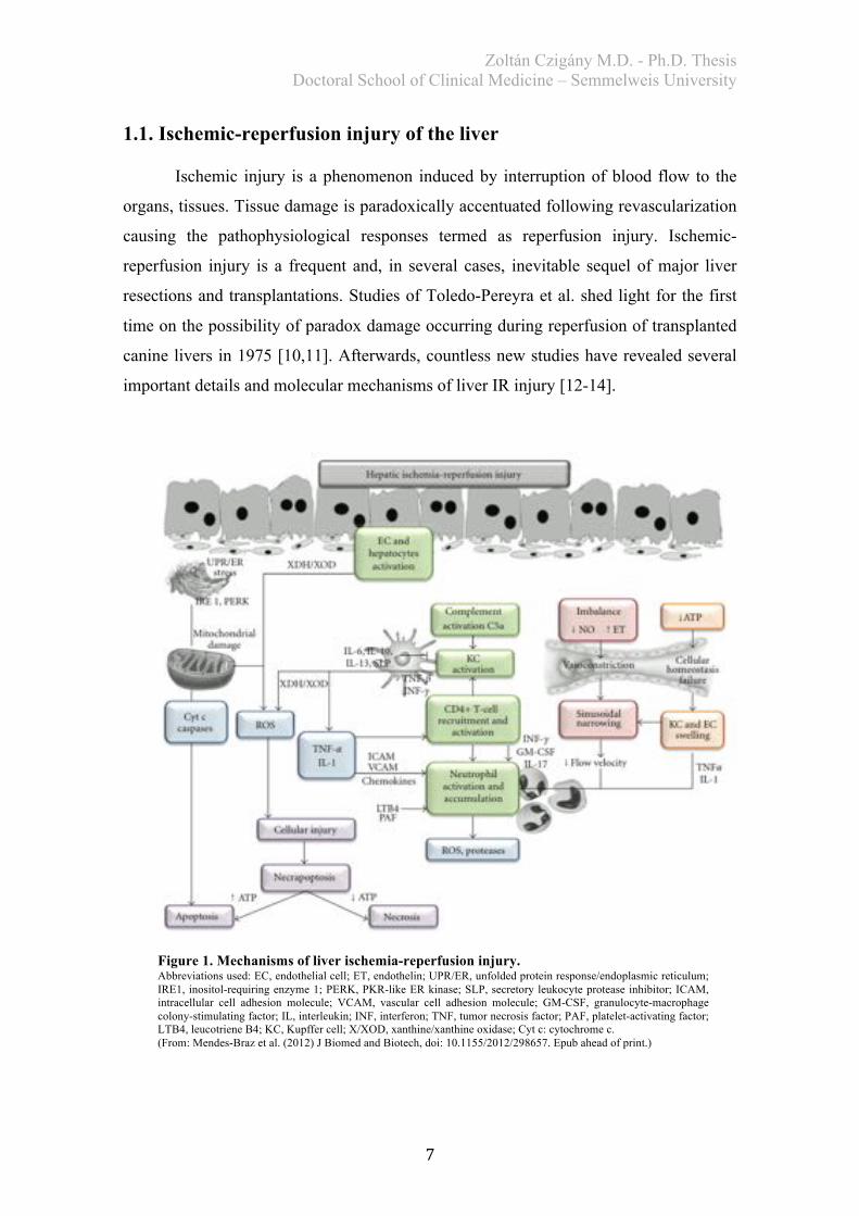
Figure 1. Mechanisms of liver ischemia-reperfusion injury. Abbreviations used: EC, endothelial cell; ET, endothelin; UPR/ER, unfolded protein response/endoplasmic reticulum; IRE1, inositol-requiring enzyme 1; PERK, PKR-like ER kinase; SLP, secretory leukocyte protease inhibitor; ICAM, intracellular cell adhesion molecule; VCAM, vascular cell adhesion molecule; GM-CSF, granulocyte-macrophage colony-stimulating factor; IL, interleukin; INF, interferon; TNF, tumor necrosis factor; PAF, platelet-activating factor; LTB4, leucotriene B4; KC, Kupffer cell; X/XOD, xanthine/xanthine oxidase; Cyt c: cytochrome c. (From: Mendes-Braz et al. (2012) J Biomed and Biotech, doi: 10.1155/2012/298657. Epub ahead of print.)
Zoltán Czigány M.D. - Ph.D. Thesis Doctoral School of Clinical Medicine – Semmelweis University
8
During ischemia oxygen supply to hepatocytes becomes insufficient as a result
of the absent blood flow. Consequently, adenosine triphosphate (ATP) depletion
induces deficiencies in active ion transport mechanisms and initiates the activation of
anabolic glycolysis [15]. In parallel with the unfolding ATP deficit, reduced activity of
Na+/K+ ATPase results in increased cellular influx of Na+ [16]. Prominent intracellular
acidosis during ischemia, caused by metabolites of anaerobic glycolysis, facilitates
Na+/H+Antiporter-1 activity contributing to the Na+ influx. As a result of intracellular
Na+ accumulation, physiological function of Na+/Ca2+ Exchanger is disturbed presented
as Ca2+ influx and mitochondrial Ca2+ accumulation [17]. Cellular ion homeostasis
disturbances will end up in cellular swelling and cell-death (Figure 1.).
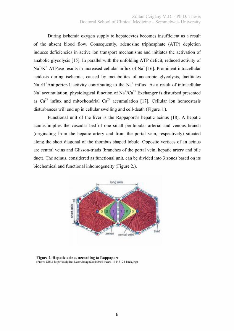
Functional unit of the liver is the Rappaport’s hepatic acinus [18]. A hepatic
acinus implies the vascular bed of one small perilobular arterial and venous branch
(originating from the hepatic artery and from the portal vein, respectively) situated
along the short diagonal of the rhombus shaped lobule. Opposite vertices of an acinus
are central veins and Glisson-triads (branches of the portal vein, hepatic artery and bile
duct). The acinus, considered as functional unit, can be divided into 3 zones based on its
biochemical and functional inhomogeneity (Figure 2.).
Figure 2. Hepatic acinus according to Rappaport (From: URL:http://studydroid.com/imageCards/0a/k1/card-11143124-back.jpg)
Zoltán Czigány M.D. - Ph.D. Thesis Doctoral School of Clinical Medicine – Semmelweis University
9
While nutritive blood supply of hepatocytes in zone 3 is poor, the closest region
to the portal vessels (zone 1) is well-oxygenated. Accordingly, activity of oxidative
metabolism is much higher in hepatocytes of zone 1; therefore, they are more resistant
against the detrimental effects of IR injury and have better regenerative potential when
compared to zone 3 [18]. Periportal necrosis is a characteristic sign of liver IR injury.
The mitochondrion is an essential component in injury during ischemia-
reperfusion and the main source of extensive reactive oxygen species (ROS) production.
During reperfusion, an important initiator of cell-death is the opening of mitochondrial
giant-channel, known as mitochondrial permeability transition pore (mPTP) [19]. Fast
restoration of normal pH during reperfusion, ROS, high concentrations of Ca2+, and
oxidant compounds induce mPTP opening, whereas low pH, cyclosporin A, and Mg2+
block it [19].
Besides hepatocytes, sinusoidal endothelial cells (SECs) are also particularly
sensitive to IR injury [20]. Membrane potential changes of SECs, cell swelling,
disturbances in cytoskeleton organization are characteristic signs of IR injury. These
aforementioned changes, combined with the imbalance between decreased nitric-oxide
(NO) levels and increased endothelin (ET) and thromboxane A2 (TXA2) production,
are the main factors in endothelial cell dysfunction. Further steps, such as leukocyte
infiltration, platelet aggregation as well as hepatic stellate cell contraction induced
sinusoidal narrowing, are all contributing to the observed reperfusion microcirculatory
failure [20].
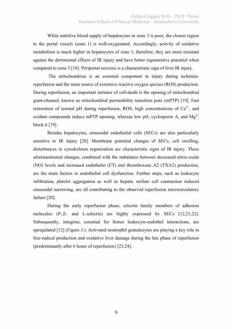
During the early reperfusion phase, selectin family members of adhesion
molecules (P-,E- and L-selectin) are highly expressed by SECs [12,21,22].
Subsequently, integrins, essential for firmer leukocyte-endothel interactions, are
upregulated [12] (Figure 3.). Activated neutrophil granulocytes are playing a key role in
free-radical production and oxidative liver damage during the late phase of reperfusion
(predominantly after 6 hours of reperfusion) [23,24].
Zoltán Czigány M.D. - Ph.D. Thesis Doctoral School of Clinical Medicine – Semmelweis University
10
As discussed above, cellular ATP depletion during ischemia results in
accumulation of adenosine, hypoxanthine and xanthine [12]. During the initial phase of
reperfusion, cellular metabolism is still shifted towards anaerobic pathways; thus the
immediately increased oxygen delivery generates free-radicals (superoxide, hydrogen-
peroxide, peroxynitrite) [12]. In this very early phase of injury, tissue-specific
macrophages, the Kupffer-cells, are believed to be the main source of ROS production.
Infiltrating neutrophils take a more dominant role later on [25,26].
A complex cross-talk between different cytokines is also essential in the
mechanism of liver IR. Extensively studied cytokine and chemokine mediators are
TNF-a, IL-6, IFNs, IL-10, CXCL-10, etc. [12].
Figure 3. Leukocyte rolling, adhesion and extravasation during IR injury. Abbreviations used: LFA-1, leukocyte function associated antigen-1; VLA-4, very late antigen-4; ICAM-1, intercellular adhesion molecule-1; VCAM-1, vascular cell adhesion molecule-1; PSGL-1, P-selectin glycoprotein ligand-1; JAM-1, junctional adhesion molecule-1. (From: Iwasaki, Czigány et al. Adhesion Molecules: Therapeutic Targets for Allograft Rejection and Ischemia-Reperfusion Injury. In: Chen (editor), Current Immunosupressive Therapy in Organ Transplantation. Nova Science Publishers, New York. 2015: 369.)
Zoltán Czigány M.D. - Ph.D. Thesis Doctoral School of Clinical Medicine – Semmelweis University
11
TNF-a, predominantly originated from activated macrophages during ischemia-
reperfusion, has a crucial role in the mechanism of injury [14]. It has pleiotropic effects
in post-ischemic injury and inflammation, albeit it thought to be an initiator molecule of
liver regeneration as well, together with IL-6 [14]. Very soon after reperfusion IL-1 and
TNF-a levels are markedly increased [27,28]. IL-1 is induced by TNF-a [14]. IL-1 can
intensify neutrophil ROS production, while TNF-a has a positive feedback loop
resulting in more TNF-a release [27,28]. Delayed elevation of IL-6 levels can also be
observed.
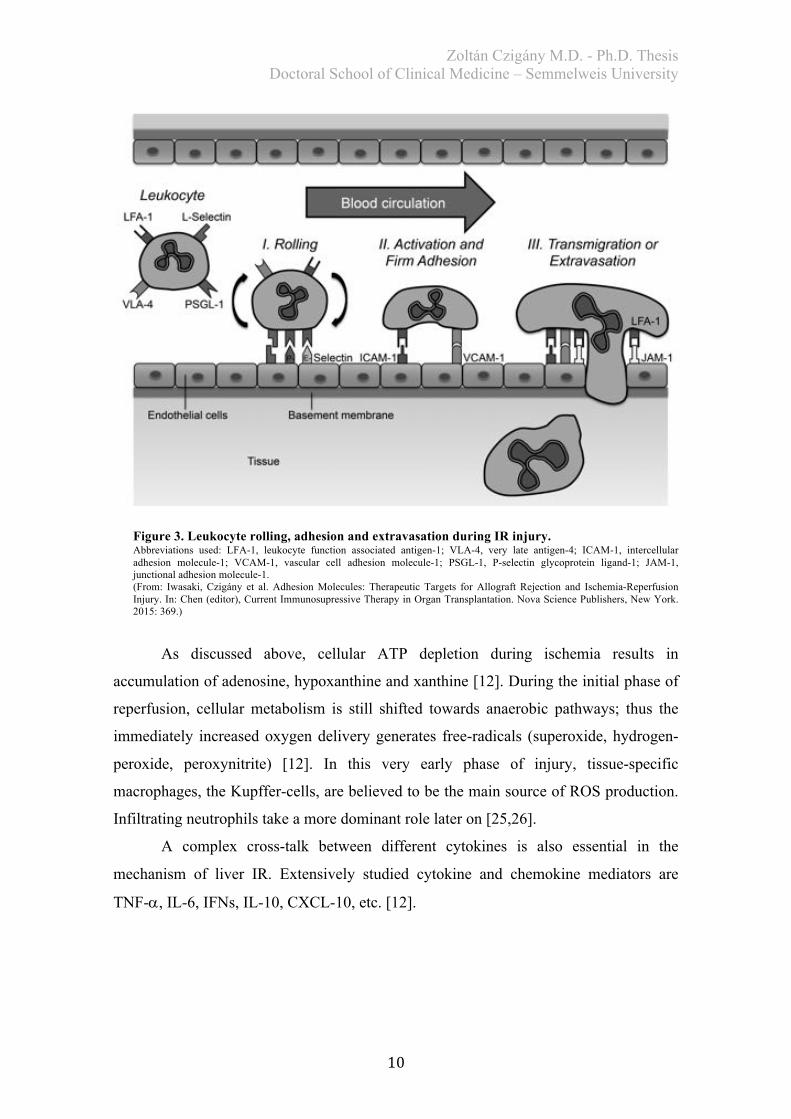
Up to this point, the
author has discussed several
crucial steps in the mechanisms
of local injury observed in liver
ischemia-reperfusion. Although,
liver IR injury following
sustained hepatic exclusions is a
well-recognized phenomenon,
injury of other remote organs,
affected by the systemic effects
of liver IR, has not been widely
explored yet (Figure 4.). It has
been reported that IR injury of the
liver might be responsible for
several remote organ injuries in
various surgical scenarios, including major resections and transplantations [29]. Acute
kidney injury [30], lung damage and acute respiratory distress syndrome [31], gut
barrier failure and consequential bacterial translocation [32], pancreatic, [33] and
adrenal injury [34] as well as myocardial damage [35] might occur in experimental
settings or in clinical liver surgery (Figure 4.). Among several mechanisms, free-
radicals (ROS) and inflammatory pathways (cytokines and leukocytes) are thought to
play an essential role in the unfolding multi-organ dysfunction syndrome (MODS)
following major liver surgeries [29].
Figure 4. Remote organs affected by the systemic consequences of liver IR injury. (From: Nastos et al. (2014) Oxid Med Cell Longev, doi: 10.1155/2014/906965. Epub ahead of print.)
Zoltán Czigány M.D. - Ph.D. Thesis Doctoral School of Clinical Medicine – Semmelweis University
12
1.2. Vascular exclusion techniques in liver surgery
Taking the fact into account that IR injury of the liver occurs mainly when we
are forced to apply sustained exclusions of vascular pedicles during surgical
interventions, in the following part of the doctoral thesis we attempted to compile the
most important vascular control techniques used in clinic.
1.2.1. Pringle-maneuver
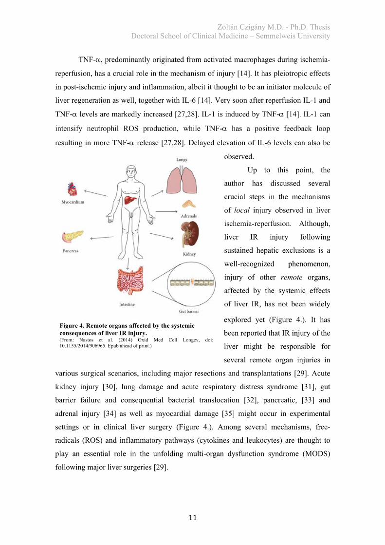
The best-known inflow occlusion technique to control blood loss during surgical
interventions is linked inseparably with the name of Dr. James Hogarth Pringle [36]
(Figure 5.). Pringle published his approach in 1908 to control bleeding from the liver in
patients with severe abdominal trauma via clamping the hepatoduodenal ligament
(Figure 6.). However, all of his 8 patients described in his seminal study had died
postoperatively, the principles of vascular control during liver resections became
accepted predominantly after Ichio Honjo and Jean-Louis Lortat-Jacob (Figure 5.)
reported the first successful major anatomical liver resections [37,38].
Figure 5. Jean-Louis Lortat-Jacob (1908-1992) and James Hogarth Pringle (1863-1941)
Zoltán Czigány M.D. - Ph.D. Thesis Doctoral School of Clinical Medicine – Semmelweis University
13
However, the aforementioned vascular control approach is attributed to Pringle
and known as Pringle-maneuver in international literature, the Hungarian surgeon,
Jónás Báron suggested and reported the use of temporary occlusion of the
hepatoduodenal ligament to minimize blood loss, earlier, in 1876 [39].
Although, hepatic pedicle clamping and reperfusion have significant
hemodynamic effects, the exclusion is well-tolerated thanks to the uninterrupted caval
flow [40,41].
The literature is controversial, whether inflow occlusion should be applied
continuously or intermittently [40,41]. Intermittent clamping is usually applied as 15-30
min ischemia interrupted by 5-15 min reperfusion [42]. In case of a good functioning
liver, continuous clamping of the hepatoduodenal ligament can safely be implemented
for up to 60 minutes [43].
Belghiti et al. performed a randomized study to compare continuous portal
clamping with intermittent portal exclusion [44]. They found a significant increase in
blood loss, although they detected a markedly reduced parenchymal damage in the
intermittent clamping group compared with continuous clamping. They have concluded
that intermittent clamping is beneficial, especially in patients with abnormal liver
parenchyma. This approach allows us to extend net ischemic time safely to 120 min.
1.2.2. Hemihepatic and segmental vascular exclusion
These techniques can minimize IR injury of remnant liver as well as limit
hemodynamic effects and splanchnic congestion. Furthermore, they are particularly
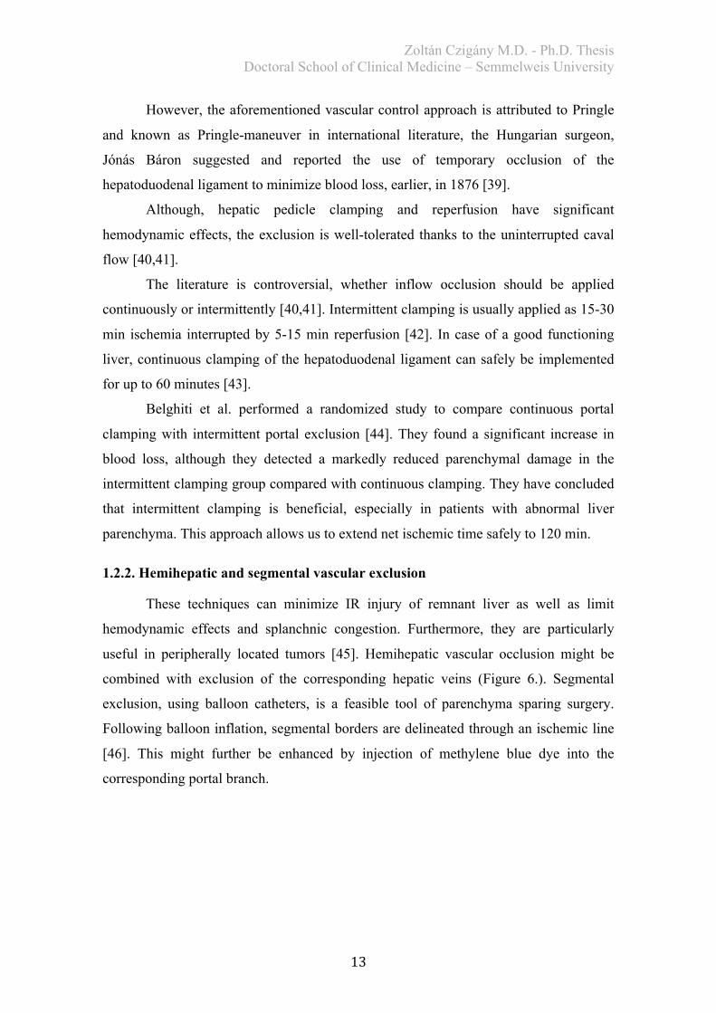
useful in peripherally located tumors [45]. Hemihepatic vascular occlusion might be
combined with exclusion of the corresponding hepatic veins (Figure 6.). Segmental
exclusion, using balloon catheters, is a feasible tool of parenchyma sparing surgery.
Following balloon inflation, segmental borders are delineated through an ischemic line
[46]. This might further be enhanced by injection of methylene blue dye into the
corresponding portal branch.
Zoltán Czigány M.D. - Ph.D. Thesis Doctoral School of Clinical Medicine – Semmelweis University
14
1.2.3. Total hepatic vascular occlusion (THVO)
This exclusion (Figure 6.) is necessary usually in case of an extensive central
tumor mass, which might also involve the inferior vena cava [40,41]. It can also be
considered in cases where persistent backflow from the hepatic veins causes an
intensive blood loss during resection; however, in such cases, other interventions e.g.
lowering the central venous pressure (<5 cm H2O) should be attempted first [41].
THVO has major hemodynamic consequences; therefore, a highly experienced
anesthesiologist and careful monitoring are the prerequisites of this procedure [41,47].
THVO is not tolerated by 10-20% of the patients [40,41,47]. Despite the enormous
Figure 6. Different vascular occlusion techniques in clinical settings. A. Exclusion of the hepatoduodenal ligament according to Pringle B. Hemihepatic vascular exclusion C. Total hepatic vascular occlusion D. Total hepatic vascular exclusion with preserved caval flow (From: Abdalla et al. (2004) Sur Clin N Am, 84: 563-585.)
Zoltán Czigány M.D. - Ph.D. Thesis Doctoral School of Clinical Medicine – Semmelweis University
15
hemodynamic burden caused by application of THVO, it has several advantages as
well. In THVO various complex surgical interventions can be performed. It makes the
in situ hypothermic perfusion [48], ex situ resection and auto-transplantation as well as
the ante-situm resections possible [49,50]. Combination of inflow occlusion and extraparenchymal control of the hepatic
veins (Figure 6.) eliminates severe hemodynamic consequences of THVO [51].
Prevention of air embolism or backflow bleeding without interruption of vena cava
inferior flow is the main advantage of this procedure [41]. Nevertheless, it cannot be
applied in cases when a tumor mass is involving the vena cava and reconstruction is
required, or when the involvement of the cavo-hepatic junction makes the exposure of
the hepatic veins complicated [41].
1.3. Small animal experimental models for liver IR injury
Planning and implementing of clinical trials have very strict ethical criteria.
After obtaining required ethical permissions the enrollment and follow-up phase as well
as data evaluation can take several years. Our access to human tissues and the amount
of samples taken are usually limited which can easily lead to underpowered studies.
Laboratory research on cell- and tissue-cultures has contributed to scientific
advancements regarding subcellular and molecular mechanisms and pathways. In most
cases, however, we can only attempt to extrapolate these findings to the in vivo clinical
situation with poor certainty.
Pre-clinical/translational research, using animals, has also several drawbacks
regarding the translatability of findings to human clinical situations; however, it is still
an inevitable approach for the in vivo investigation of pathophisiology responses
following complex surgical interventions.
During the last decades several feasible and cost-effective rodent models have
been developed to investigate characteristics, mechanisms, and further aspects of liver
IR injury. In the present subchapter we have attempted to summarize the advantages
and disadvantages of the experimentally and clinically most relevant models. Specific
features of global and partial liver IR injury in rodents are described in detail.
Zoltán Czigány M.D. - Ph.D. Thesis Doctoral School of Clinical Medicine – Semmelweis University
16
1.3.1. Partial liver IR injury models
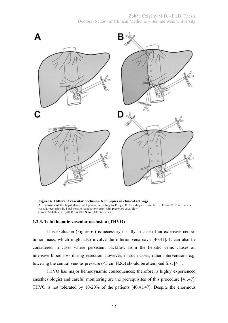
The reason behind using rodent partial IR injury models is the need for isolated
investigation of in vivo effects of liver ischemia-reperfusion. In the literature we can
find various models from 10% exclusion models, achieved via exclusion of the caudate
lobes, until 70% partial liver ischemia. Figure 7. depicts the most frequently used
models based on percentage of the excluded liver volume and shows the anatomical
aspects of partial liver ischemia in rats. Essence of the surgical intervention in these
models is the selective clamping of the corresponding biliovascular pedicle, using
atraumatic clamps.
In this model we can
induce ischemia of respective
liver lobes, meanwhile leaving
uninterrupted blood supply to
others. This method can
prevent severe congestion of
the gastrointestinal tract, which
is poorly tolerated by rodents.
However, in the
literature there is no general
agreement concerning the need
for a partial liver resection in
this model, most of the authors
are removing the non-
ischemized lobes right before
liver reperfusion. Through this
aforementioned partial
hepatectomy we can namely prevent the blood-stealing effect of the healthy lobes
during reperfusion (due to increased vascular resistance of the post-ischemic lobes),
ensuring reperfusion of the ischemic damaged liver lobes. In Budapest the reproducible
rat model of partial liver ischemia was developed by Kupcsulik et al. in the 70s. This
pioneering work provided a basis for the present thesis as well [52].
Figure7.Different models for liver IR injury in rats.Models for partial and total liver ischemia. Grey: ischemized lobes. Abbreviations used: IRL, inferior right lobe; SRL, surperior right lobe; RML, right mediate lobe; LML, left mediate lobe; LLL, left lateral lobe; ACL, anterior caudate lobe; PCL, posterior caudate lobe. (Adapted from: Czigány et al. (2015) Eur Surg Res, 55(1-2): 119-138.)
Zoltán Czigány M.D. - Ph.D. Thesis Doctoral School of Clinical Medicine – Semmelweis University
17
Advantages of this model are the opportunity to investigate more selectively the
in vivo effects of liver IR injury without extreme hemodynamic impairment, severe
gastrointestinal tract congestion and consequential bacterial translocation as well as
bowel-origin cytokine storm. At the same time, in cases of sustained liver exclusions,
the presence of systemic damages and remote organ injuries is probable.
Partial liver IR injury models might have inferior clinical relevance when
compared to 100% hepatic exclusion. In clinical practice the most frequently used liver
exclusion approach is the so-called Pringle-(Báron)-maneuver, which means the cross-
clamping of the hepatoduodenal ligament [36]. Nonetheless, selective balloon catheter
occlusion of liver lobes and segments, and hemihepatic vascular occlusion for liver
resection can be achieved in clinical settings as well (see Figure 6.) [53,54].
1.3.2. Models of global hepatic ischemia
Global hepatic ischemia in rats and mice can be performed easily via clamping
of the hepatoduodenal ligament [13]. This model is optimal to mimic the clinical
situation of normothermic liver ischemia with Pringle-maneuver for liver resections.
The drawback of this model is the induced very complex multi-organ injury with all of
its consequences. Global hepatic ischemia results in major hemodynamic alterations and
congestion in the portal vascular bed [19]. Injuries of different remote organs
(myocardium, kidney, lungs, gut barrier, adrenals, pancreas etc.) are frequent
complications [29].
There are different approaches developed for small animals to decompress the
portal circulation in the anhepatic phase [13]. Besides various surgically established
porto-systemic shunts [55-57], an interesting technique is the so-called subcutaneous
spleen transposition. According to the widely accepted approach, the spleen can be
pulled through and fixed in a subcutaneous position via a small incision in the left
hypochondrium building a strong collateral network [13,58].
Since the use of a certain portal decompression technique for liver IR injury
studies has not been generally accepted within the scientific community, the
comparability of findings with numerous different models is questionable (total or
partial ischemia, with or without shunt, etc.).
Zoltán Czigány M.D. - Ph.D. Thesis Doctoral School of Clinical Medicine – Semmelweis University
18
1.3.3. Duration of liver exclusion in rodent models of liver IR injury
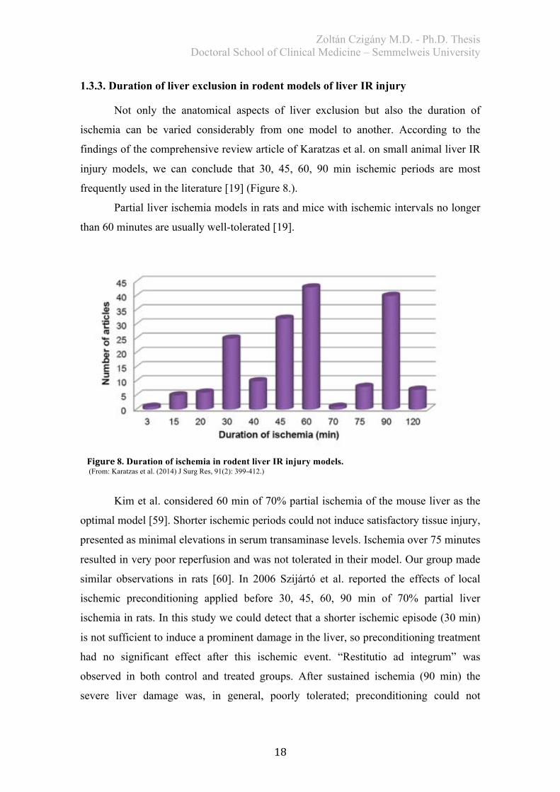
Not only the anatomical aspects of liver exclusion but also the duration of
ischemia can be varied considerably from one model to another. According to the
findings of the comprehensive review article of Karatzas et al. on small animal liver IR
injury models, we can conclude that 30, 45, 60, 90 min ischemic periods are most
frequently used in the literature [19] (Figure 8.).
Partial liver ischemia models in rats and mice with ischemic intervals no longer
than 60 minutes are usually well-tolerated [19].
Kim et al. considered 60 min of 70% partial ischemia of the mouse liver as the
optimal model [59]. Shorter ischemic periods could not induce satisfactory tissue injury,
presented as minimal elevations in serum transaminase levels. Ischemia over 75 minutes
resulted in very poor reperfusion and was not tolerated in their model. Our group made
similar observations in rats [60]. In 2006 Szijártó et al. reported the effects of local
ischemic preconditioning applied before 30, 45, 60, 90 min of 70% partial liver
ischemia in rats. In this study we could detect that a shorter ischemic episode (30 min)
is not sufficient to induce a prominent damage in the liver, so preconditioning treatment
had no significant effect after this ischemic event. “Restitutio ad integrum” was
observed in both control and treated groups. After sustained ischemia (90 min) the
severe liver damage was, in general, poorly tolerated; preconditioning could not
Figure8. Duration of ischemia in rodent liver IR injury models. (From: Karatzas et al. (2014) J Surg Res, 91(2): 399-412.)
Zoltán Czigány M.D. - Ph.D. Thesis Doctoral School of Clinical Medicine – Semmelweis University
19
considerably improve liver microcirculation and tissue injury. Most remarkable
differences we could observe between control and preconditioning groups after 60
minutes of 70% ischemia. This period led to moderate tissue injury, which was
significantly reduced by preconditioning treatment. According to the above-described
findings, we can say that the optimal model for mimicking sublethal partial liver
ischemia in rodents is the 45-60 min liver exclusion, while the “point of no return” can
be found between 60-90 minutes.
In the partial ischemia model, animals can be sacrificed after various time
points. Based on the biphasic characteristics of liver IR injury (early phase 1-6 h, late
phase 9-24 h) 1,3,6 and 24 h intervals after reperfusion are frequently recommended for
analysis [19,59].
Regarding global hepatic ischemia (without porto-systemic shunting) the
literature is more controversial. For acute experiments, investigating short-term
consequences of severe ischemic-reperfusion injury, longer exclusion times are also
accepted [19]. However, 60-90 min total hepatic ischemia results in a very poor long-
term survival [61]. Higher survival rates can be achieved with 30-(45) min of global
ischemia [62-64].
1.3.4. Further concerns regarding liver IR injury models in rodents
During the planning phase of an experimental study using laboratory rodents,
several aspects have to be considered, which might have a decisive role to play
regarding the reliability of our model and results.
Anesthesia practice for laboratory rodents has markedly changed during the last
decades. Modern volatile anesthetics (e.g. isoflurane) and injectable anesthetic
combinations (e.g. ketamine/xylazine) are getting more and more popular in rodent
surgery [65,66]; nevertheless, the use of obsolete approaches (ether, barbiturates) still
can be identified in numerous newer reports.
The assumed conditioning, anti-ischemic effects of different anesthetic drugs,
however, should also be considered when our aim is to investigate liver IR injury in a
rodent model. Various authors have proved pharmacological conditioning effects of
ketamine in different models [67,68]. Guzman et al. reported that ketamine could reduce
Zoltán Czigány M.D. - Ph.D. Thesis Doctoral School of Clinical Medicine – Semmelweis University
20
mucosal injury in a rat model of intestinal IR injury [67]. Similar effects were found in
an in vitro study on human myocardial tissue [68].
According to the findings of Kim et al. isoflurane protects against small
intestinal injury, hepatic and renal dysfunction following severe intestinal IR injury via
induction of intestinal epithelial TGF-β1 expression [69].
These effects can cause only “internal errors” within our experimental setting,
not considerably influencing the between group differences, but we have to be aware of
these aforementioned “side-effects” of the used anesthetic agents.
Injection anesthetics must be administered carefully, particularly in studies
where sustained liver exclusion is applied (liver transplantation with total hepatic
vascular occlusion or IR injury studies with total hepatic ischemia). Significant
increment in plasma concentration of several anesthetic drugs, during the anhepatic
phase, might result in unexpected mortality [70-72].
A number of distinct age-related differences have been identified in IR injury
and inflammatory responses of laboratory rodents [13,73]. In general, older rats show
more severe tissue injury following the same surgical intervention [74,75]. Laboratory
rats between 200-350 grams are ideal for modeling liver IR injury. Smaller animals are
technically more difficult to operate, at the same time, older animals (over 400 g) can
also present problems, due to the higher postoperative complication rates and increased
intra-abdominal fat [13].
Gender differences might also have an effect on our results, especially on the
comparability of the findings [13]. Studies have shown that males and females present
differences in susceptibility to reperfusion injury [76-78]. In female animals ischemic
tolerance is probably dependent on the estrous cycle, though its role has not been
properly clarified yet [76,77].
Small animals are extremely sensitive to changes in body core temperature;
therefore, it is very important to monitor and maintain body temperature
perioperatively. Heijnen et al. could show significant increment in serum transaminase
levels and in histopathological damage in a rat model of 60 minutes partial liver
ischemia when the animals´ body temperature increased from 36°C to 38°C [79].
Zoltán Czigány M.D. - Ph.D. Thesis Doctoral School of Clinical Medicine – Semmelweis University
21
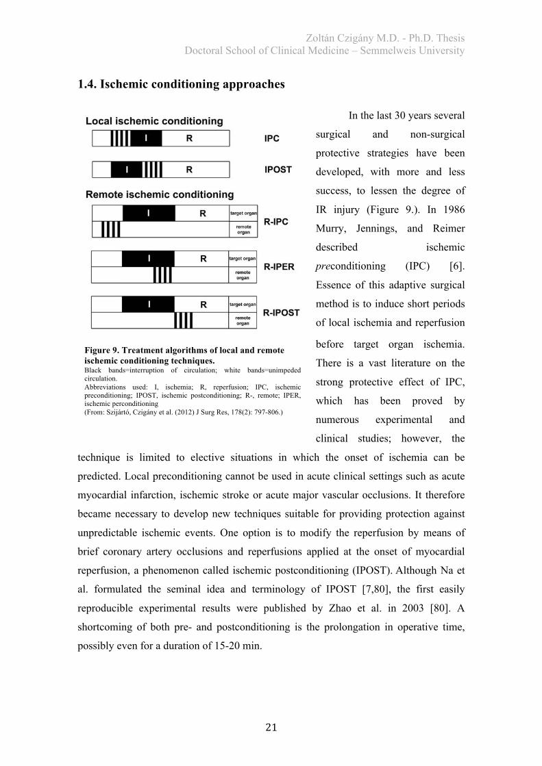
1.4. Ischemic conditioning approaches
In the last 30 years several
surgical and non-surgical
protective strategies have been
developed, with more and less
success, to lessen the degree of
IR injury (Figure 9.). In 1986
Murry, Jennings, and Reimer
described ischemic
preconditioning (IPC) [6].
Essence of this adaptive surgical
method is to induce short periods
of local ischemia and reperfusion
before target organ ischemia.
There is a vast literature on the
strong protective effect of IPC,
which has been proved by
numerous experimental and
clinical studies; however, the
technique is limited to elective situations in which the onset of ischemia can be
predicted. Local preconditioning cannot be used in acute clinical settings such as acute
myocardial infarction, ischemic stroke or acute major vascular occlusions. It therefore
became necessary to develop new techniques suitable for providing protection against
unpredictable ischemic events. One option is to modify the reperfusion by means of
brief coronary artery occlusions and reperfusions applied at the onset of myocardial
reperfusion, a phenomenon called ischemic postconditioning (IPOST).Although Na et
al. formulated the seminal idea and terminology of IPOST [7,80], the first easily
reproducible experimental results were published by Zhao et al. in 2003 [80]. A
shortcoming of both pre- and postconditioning is the prolongation in operative time,
possibly even for a duration of 15-20 min.
Figure 9. Treatment algorithms of local and remote ischemic conditioning techniques. Black bands=interruption of circulation; white bands=unimpeded circulation. Abbreviations used: I, ischemia; R, reperfusion; IPC, ischemic preconditioning; IPOST, ischemic postconditioning; R-, remote; IPER, ischemic perconditioning (From: Szijártó, Czigány et al. (2012) J Surg Res, 178(2): 797-806.)
Zoltán Czigány M.D. - Ph.D. Thesis Doctoral School of Clinical Medicine – Semmelweis University
22
Reducing the extent of IR injury via
short episodes of ischemia and reperfusion
instituted at a remote site is a novel idea. As
for local conditioning techniques (IPC and
IPOST), remote ischemic conditioning can
be applied before target organ ischemia
(remote ischemic preconditioning [RIPC]) or
at the onset of reperfusion (remote ischemic
postconditioning [RIPOST]). When the site
of conditioning is remotely located,
conditioning cycles can be applied during
target organ ischemia; a novel phenomenon
known as remote ischemic perconditioning
(RIPER) (Figure 9 and 10.).
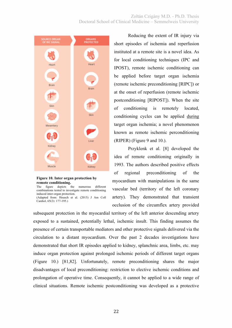
Przyklenk et al. [8] developed the
idea of remote conditioning originally in
1993. The authors described positive effects
of regional preconditioning of the
myocardium with manipulations in the same
vascular bed (territory of the left coronary
artery). They demonstrated that transient
occlusion of the circumflex artery provided
subsequent protection in the myocardial territory of the left anterior descending artery
exposed to a sustained, potentially lethal, ischemic insult. This finding assumes the
presence of certain transportable mediators and other protective signals delivered via the
circulation to a distant myocardium. Over the past 2 decades investigations have
demonstrated that short IR episodes applied to kidney, splanchnic area, limbs, etc. may
induce organ protection against prolonged ischemic periods of different target organs
(Figure 10.) [81,82]. Unfortunately, remote preconditioning shares the major
disadvantages of local preconditioning: restriction to elective ischemic conditions and
prolongation of operative time. Consequently, it cannot be applied to a wide range of
clinical situations. Remote ischemic postconditioning was developed as a protective
Figure 10. Inter organ protection by remote conditioning. The figure depicts the numerous different combinations tested to investigate remote conditioning induced inter-organ protection. (Adapted from: Heusch et al. (2015) J Am Coll Cardiol, 65(2): 177-195.)
Zoltán Czigány M.D. - Ph.D. Thesis Doctoral School of Clinical Medicine – Semmelweis University
23
strategy bearing similarity to local postconditioning (IPOST); therefore, it extends the
range of application of remote conditioning techniques [83-85].
Remote ischemic perconditioning, proposed for the first time by Schmidt et al.
in 2007 [86], has plenty beneficial features from a practical point of view. Essence of
this strategy is that short remote ischemic attacks are applied after induction of target
organ ischemia but before the onset of reperfusion.
1.4.1. Terminology
There is much confusion in literature regarding the concept called “ischemic
perconditioning”. Three different terms exist for this strategy, meaning “the induction of
brief and repetitive interruptions of blood flow at a distance, applied concurrently with
sustained target organ ischemia”: (1) remote perconditioning [86-88] (2) remote
periconditioning [89,90] and (3) remote postconditioning [91-93].
Term “RIPOST” can be excluded first as proper terminus, because it implicates
that conditioning cycles take place after (“post”) target organ ischemia (Figure 9.).
Distinction between periconditioning and perconditioning is more of a semantic
question rather than methodical. Greek prefix peri- means “around something in time”
and is found in the expression “perinatal mortality.” In contrast, the meaning of the
prefix “per” is “over, through, during the course of something” such as “perennial
rhinitis.” Thus, from a linguistic point of view, the term “perconditioning” is suitable
for describing this method. Furthermore, Schmidt et al. [86], pioneers of this procedure,
proposed the term “remote perconditioning” as well. Accordingly, “perconditioning”
seems to be a suitable term for all cases in which brief ischemic and reperfusion cycles
are instituted after the onset of target organ ischemia and completed before reperfusion.
1.4.2. Remote ischemic perconditioning: underlying mechanisms
There is scarce evidence to explain how brief cycles of IR on a distant organ,
during prolonged target organ vascular occlusion, are able to provide organ protection.
Here, we discuss the literature findings on remote ischemic conditioning in order to
understand the underlying mechanisms.
Hausenloy and Lim et al. [81,94] have recently given a detailed review on the
underlying mechanisms of RIPC. According to their theory, different humoral, neural
Zoltán Czigány M.D. - Ph.D. Thesis Doctoral School of Clinical Medicine – Semmelweis University
24
and systemic components hypothetically interact with each other to provide target organ
protection from a remote site.
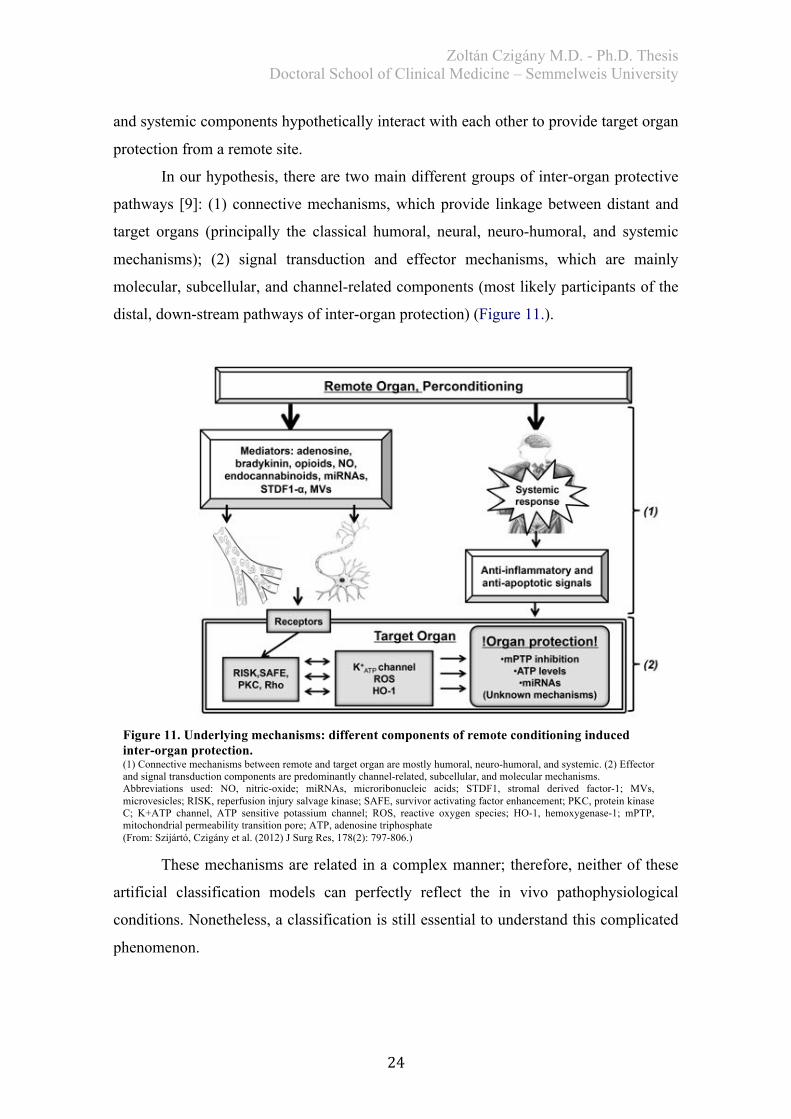
In our hypothesis, there are two main different groups of inter-organ protective
pathways [9]: (1) connective mechanisms, which provide linkage between distant and
target organs (principally the classical humoral, neural, neuro-humoral, and systemic
mechanisms); (2) signal transduction and effector mechanisms, which are mainly
molecular, subcellular, and channel-related components (most likely participants of the
distal, down-stream pathways of inter-organ protection) (Figure 11.).
These mechanisms are related in a complex manner; therefore, neither of these
artificial classification models can perfectly reflect the in vivo pathophysiological
conditions. Nonetheless, a classification is still essential to understand this complicated
phenomenon.
Figure 11. Underlying mechanisms: different components of remote conditioning induced inter-organ protection. (1) Connective mechanisms between remote and target organ are mostly humoral, neuro-humoral, and systemic. (2) Effector and signal transduction components are predominantly channel-related, subcellular, and molecular mechanisms. Abbreviations used: NO, nitric-oxide; miRNAs, microribonucleic acids; STDF1, stromal derived factor-1; MVs, microvesicles; RISK, reperfusion injury salvage kinase; SAFE, survivor activating factor enhancement; PKC, protein kinase C; K+ATP channel, ATP sensitive potassium channel; ROS, reactive oxygen species; HO-1, hemoxygenase-1; mPTP, mitochondrial permeability transition pore; ATP, adenosine triphosphate (From: Szijártó, Czigány et al. (2012) J Surg Res, 178(2): 797-806.)
Zoltán Czigány M.D. - Ph.D. Thesis Doctoral School of Clinical Medicine – Semmelweis University
25
As pointed out in previous studies, concentration or activation status of
molecular, humoral, and subcellular participants may be varied significantly among the
different remote ischemic conditioning subtypes (RIPC, RIPER, and RIPOST); namely,
the timing of these procedures is radically different (before, during or after target organ
ischemia) [95]. It is assumed, however, that the main biochemical and signal
transduction pathways might be similar among the conditioning techniques, accordingly
some comparison between the remote conditioning strategies is inevitable [96].
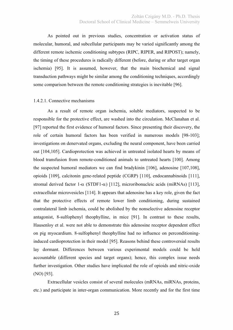
1.4.2.1. Connective mechanisms
As a result of remote organ ischemia, soluble mediators, suspected to be
responsible for the protective effect, are washed into the circulation. McClanahan et al.
[97] reported the first evidence of humoral factors. Since presenting their discovery, the
role of certain humoral factors has been verified in numerous models [98-103];
investigations on denervated organs, excluding the neural component, have been carried
out [104,105]. Cardioprotection was achieved in untreated isolated hearts by means of
blood transfusion from remote-conditioned animals to untreated hearts [100]. Among
the suspected humoral mediators we can find bradykinin [106], adenosine [107,108],
opioids [109], calcitonin gene-related peptide (CGRP) [110], endocannabinoids [111],
stromal derived factor 1-a (STDF1-a) [112], microribonucleic acids (miRNAs) [113],
extracellular microvesicles [114]. It appears that adenosine has a key role, given the fact
that the protective effects of remote lower limb conditioning, during sustained
contralateral limb ischemia, could be abolished by the nonselective adenosine receptor
antagonist, 8-sulfophenyl theophylline, in mice [91]. In contrast to these results,
Hausenloy et al. were not able to demonstrate this adenosine receptor dependent effect
on pig myocardium. 8-sulfophenyl theophylline had no influence on perconditioning-
induced cardioprotection in their model [95]. Reasons behind these controversial results
lay dormant. Differences between various experimental models could be held
accountable (different species and target organs); hence, this complex issue needs
further investigation. Other studies have implicated the role of opioids and nitric-oxide
(NO) [93].
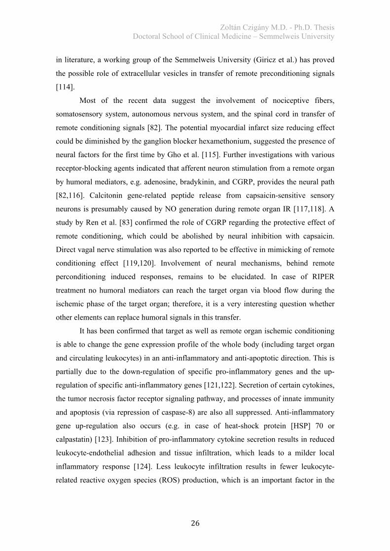
Extracellular vesicles consist of several molecules (mRNAs, miRNAs, proteins,
etc.) and participate in inter-organ communication. More recently and for the first time
Zoltán Czigány M.D. - Ph.D. Thesis Doctoral School of Clinical Medicine – Semmelweis University
26
in literature, a working group of the Semmelweis University (Giricz et al.) has proved
the possible role of extracellular vesicles in transfer of remote preconditioning signals
[114].
Most of the recent data suggest the involvement of nociceptive fibers,
somatosensory system, autonomous nervous system, and the spinal cord in transfer of
remote conditioning signals [82]. The potential myocardial infarct size reducing effect
could be diminished by the ganglion blocker hexamethonium, suggested the presence of
neural factors for the first time by Gho et al. [115]. Further investigations with various
receptor-blocking agents indicated that afferent neuron stimulation from a remote organ
by humoral mediators, e.g. adenosine, bradykinin, and CGRP, provides the neural path
[82,116]. Calcitonin gene-related peptide release from capsaicin-sensitive sensory
neurons is presumably caused by NO generation during remote organ IR [117,118]. A
study by Ren et al. [83] confirmed the role of CGRP regarding the protective effect of
remote conditioning, which could be abolished by neural inhibition with capsaicin.
Direct vagal nerve stimulation was also reported to be effective in mimicking of remote
conditioning effect [119,120]. Involvement of neural mechanisms, behind remote
perconditioning induced responses, remains to be elucidated. In case of RIPER
treatment no humoral mediators can reach the target organ via blood flow during the
ischemic phase of the target organ; therefore, it is a very interesting question whether
other elements can replace humoral signals in this transfer.
It has been confirmed that target as well as remote organ ischemic conditioning
is able to change the gene expression profile of the whole body (including target organ
and circulating leukocytes) in an anti-inflammatory and anti-apoptotic direction. This is
partially due to the down-regulation of specific pro-inflammatory genes and the up-
regulation of specific anti-inflammatory genes [121,122]. Secretion of certain cytokines,
the tumor necrosis factor receptor signaling pathway, and processes of innate immunity
and apoptosis (via repression of caspase-8) are also all suppressed. Anti-inflammatory
gene up-regulation also occurs (e.g. in case of heat-shock protein [HSP] 70 or
calpastatin) [123]. Inhibition of pro-inflammatory cytokine secretion results in reduced
leukocyte-endothelial adhesion and tissue infiltration, which leads to a milder local
inflammatory response [124]. Less leukocyte infiltration results in fewer leukocyte-
related reactive oxygen species (ROS) production, which is an important factor in the
Zoltán Czigány M.D. - Ph.D. Thesis Doctoral School of Clinical Medicine – Semmelweis University
27
late phase of reperfusion. This phenomenon suggests a partial explanation for both the
observation of Harkin et al. [125] and our findings [126], according to which lower limb
ischemic preconditioning and postconditioning can reduce remote lung injury and
leukocyte sequestration after local limb IR injury. In these cases, however, the
dominating factor is presumably the local IR injury reducing effect of the conditioning
strategy, leading to mitigated remote organ damage. Nonetheless, Wei et al. [127]
reported a remission in inflammatory responses caused by RIPER. The authors
observed significantly diminished macrophage and neutrophil infiltration in rat
myocardium after perconditioning.
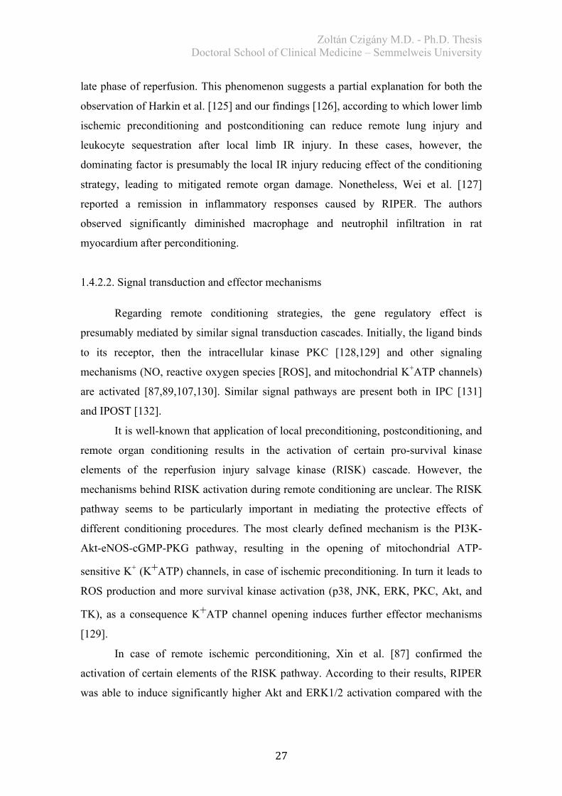
1.4.2.2. Signal transduction and effector mechanisms
Regarding remote conditioning strategies, the gene regulatory effect is
presumably mediated by similar signal transduction cascades. Initially, the ligand binds
to its receptor, then the intracellular kinase PKC [128,129] and other signaling
mechanisms (NO, reactive oxygen species [ROS], and mitochondrial K+ATP channels)
are activated [87,89,107,130]. Similar signal pathways are present both in IPC [131]
and IPOST [132].
It is well-known that application of local preconditioning, postconditioning, and
remote organ conditioning results in the activation of certain pro-survival kinase
elements of the reperfusion injury salvage kinase (RISK) cascade. However, the
mechanisms behind RISK activation during remote conditioning are unclear. The RISK
pathway seems to be particularly important in mediating the protective effects of
different conditioning procedures. The most clearly defined mechanism is the PI3K-
Akt-eNOS-cGMP-PKG pathway, resulting in the opening of mitochondrial ATP-
sensitive K+ (K+ATP) channels, in case of ischemic preconditioning. In turn it leads to
ROS production and more survival kinase activation (p38, JNK, ERK, PKC, Akt, and
TK), as a consequence K+ATP channel opening induces further effector mechanisms
[129].
In case of remote ischemic perconditioning, Xin et al. [87] confirmed the
activation of certain elements of the RISK pathway. According to their results, RIPER
was able to induce significantly higher Akt and ERK1/2 activation compared with the
Zoltán Czigány M.D. - Ph.D. Thesis Doctoral School of Clinical Medicine – Semmelweis University
28
IR control group. Despite the impressive strides made, the literature is controversial. In
their recent study, Hausenloy et al. [95] reported that RIPER could reduce myocardial
IR injury in an unknown manner but independent of RISK activation.
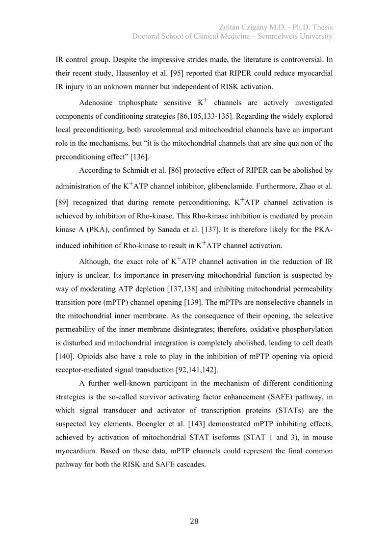
Adenosine triphosphate sensitive K+ channels are actively investigated
components of conditioning strategies [86,105,133-135]. Regarding the widely explored
local preconditioning, both sarcolemmal and mitochondrial channels have an important
role in the mechanisms, but “it is the mitochondrial channels that are sine qua non of the
preconditioning effect” [136].
According to Schmidt et al. [86] protective effect of RIPER can be abolished by
administration of the K+ATP channel inhibitor, glibenclamide. Furthermore, Zhao et al.
[89] recognized that during remote perconditioning, K+ATP channel activation is
achieved by inhibition of Rho-kinase. This Rho-kinase inhibition is mediated by protein
kinase A (PKA), confirmed by Sanada et al. [137]. It is therefore likely for the PKA-
induced inhibition of Rho-kinase to result in K+ATP channel activation.
Although, the exact role of K+ATP channel activation in the reduction of IR
injury is unclear. Its importance in preserving mitochondrial function is suspected by
way of moderating ATP depletion [137,138] and inhibiting mitochondrial permeability
transition pore (mPTP) channel opening [139]. The mPTPs are nonselective channels in
the mitochondrial inner membrane. As the consequence of their opening, the selective
permeability of the inner membrane disintegrates; therefore, oxidative phosphorylation
is disturbed and mitochondrial integration is completely abolished, leading to cell death
[140]. Opioids also have a role to play in the inhibition of mPTP opening via opioid
receptor-mediated signal transduction [92,141,142].
A further well-known participant in the mechanism of different conditioning
strategies is the so-called survivor activating factor enhancement (SAFE) pathway, in
which signal transducer and activator of transcription proteins (STATs) are the
suspected key elements. Boengler et al. [143] demonstrated mPTP inhibiting effects,
achieved by activation of mitochondrial STAT isoforms (STAT 1 and 3), in mouse
myocardium. Based on these data, mPTP channels could represent the final common
pathway for both the RISK and SAFE cascades.
Zoltán Czigány M.D. - Ph.D. Thesis Doctoral School of Clinical Medicine – Semmelweis University
29
Tamareille et al. demonstrated an improvement in cardioprotection with use of
RIPER in combination with IPOST, as opposed to IPOST alone [144]. The authors
associated RIPER induced protection with the recruitment of the SAFE pathway. Their
study indicated the existence of a complex crosstalk between RISK and SAFE
pathways. Inhibition of both cascades could abolish the cardioprotective effects of
RIPER+IPOST combination. They observed decreased activation of RISK pathway,
achieved by administration of SAFE inhibitors, and vice versa, when RISK inhibitors
were given. Data mentioned above suggest the presence of many common elements
among the various conditioning methods; yet, the specific role of these common
mediators in different procedures still needs to be investigated.
According to several studies ROS production plays a key role as a second
messenger in the mechanism of conditioning procedures during the early reperfusion
phase [145-150]. The protective effects most likely prevail in local preconditioning
[151] and postconditioning [152] via activation of the RISK pathway and inhibition of
mPTP channels. However, positive effects of ROS production are to be sharply
separated from extensive ROS generation observed during reperfusion, which correlates
with tissue injury and subsequent cell death [153]. These results are consistent with the
findings of Xin et al. [87], who demonstrated reduction of malondialdehyde and
superoxide levels related to RIPER. Furthermore, they observed increased survival
kinase activation, which suggests an inverse correlation with malondialdehyde and
superoxide levels. There is scare evidence on the mediator role of ROS in remote
ischemic conditioning of the liver.
Zoltán Czigány M.D. - Ph.D. Thesis Doctoral School of Clinical Medicine – Semmelweis University
30
2. OBJECTIVES
Ischemic-reperfusion injury of the liver represents a major problem in numerous
clinical settings. In extended liver resections performed with Pringle-maneuver the liver
undergoes a sustained exclusion and a consequential normothermic liver IR injury.
During liver transplantations cold ischemia and warm reperfusion of the liver graft can
result in primary graft dysfunction or non-function, especially when we are forced to
deal with extended criteria donors (steatotic grafts, elderly donors, etc.).
The main objective of our studies was to investigate the effects of a novel
approach on the ischemic-reperfusion injury of the rat liver. Following initial studies on
protective effects of remote ischemic perconditioning, we aimed to reveal whether or
not neural elements are participating in the RIPER induced protection.
In the present Doctoral Thesis we were looking for answers for the following questions:
1. Is our rat model of hepatic ischemia-reperfusion injury and remote ischemic
perconditioning suitable and feasible to test the effects of remote ischemic
perconditioning?
2. Can the applied remote ischemic perconditioning protocol exert any effects
a, on liver tissue injury?
b, on systemic hemodynamics and liver as well as lower limb
microcirculation?
c, on redox-homeostasis and systemic inflammation?
3. Is left femoral artery perconditioning also able to exert hepatoprotection in our
model, and if yes, does remote organ denervation has any effect on the
perconditioning induced hepatoprotection?
4. Is the “tile-based” automated histological image analysis feasible within a real
experimental setting?
Zoltán Czigány M.D. - Ph.D. Thesis Doctoral School of Clinical Medicine – Semmelweis University
31
3. MATERIALS and METHODS
3.1. Experimental settings, surgical procedures
3.1.1. Ethical background and animals
Experimental protocols were reviewed and approved by the Institutional Animal
Care and Use Committee of the Semmelweis University and were in accordance with
Government Decree 40/2013. (II. 14.).
Male Wistar rats weighing 200-250 g were used (Semmelweis University
Central Animal Facility, Budapest, Hungary) during the studies (Sn=114). The animals
were held under standard animal care conditions at 22-24°C, with 12-h day-night
cycles. Standard rodent pellets (Toxi-coop Ltd, Dunakeszi, Hungary) and water were
granted ad libitum.
3.1.2. Circumstances during surgery and anesthesia protocol
All experiments were implemented at the same time of day to avoid disturbing
effects of circadian rhythm.
Animals were anesthetized with intraperitoneal injections of ketamine
(Calypsol®) (75 mg/bwkg) and xylazine (Xylasin®) (7.5 mg/bwkg), then they were
placed on a heating pad connected with a rectal thermometer to maintain body
temperature between 36.5 and 37.5°C (Homeothermic Blanket Control Unit; Harvard
Apparatus Ltd, Holliston, MA, USA). 22-gauge polyethylene catheter was placed into
the right jugular vein for maintenance of anesthesia (25 mg/bwkg/h ketamine and 2.5
mg/bwkg/h xylazine) and administration of saline infusion (3 mL/bwkg/h) as
compensation for intraoperative fluid loss.
Postoperative analgesia was achieved by the administration of buprenorphine
(0.01 mg/bwkg/24h).
Zoltán Czigány M.D. - Ph.D. Thesis Doctoral School of Clinical Medicine – Semmelweis University
32
3.1.3. Studies and experimental groups
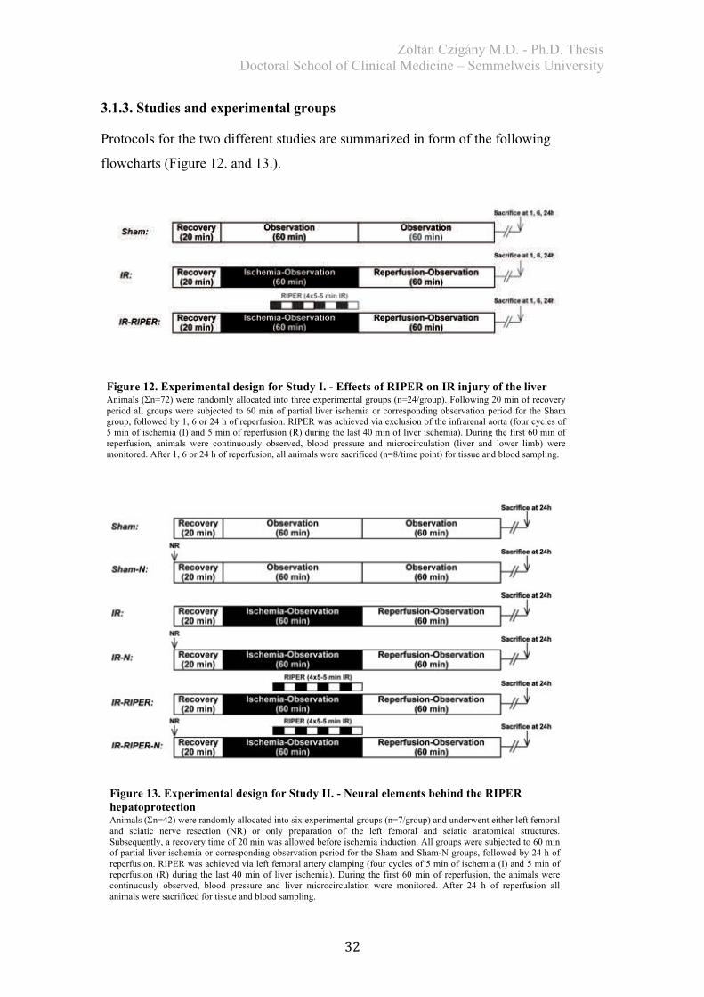
Protocols for the two different studies are summarized in form of the following
flowcharts (Figure 12. and 13.).
Figure 13. Experimental design for Study II. - Neural elements behind the RIPER hepatoprotection Animals (Sn=42) were randomly allocated into six experimental groups (n=7/group) and underwent either left femoral and sciatic nerve resection (NR) or only preparation of the left femoral and sciatic anatomical structures. Subsequently, a recovery time of 20 min was allowed before ischemia induction. All groups were subjected to 60 min of partial liver ischemia or corresponding observation period for the Sham and Sham-N groups, followed by 24 h of reperfusion. RIPER was achieved via left femoral artery clamping (four cycles of 5 min of ischemia (I) and 5 min of reperfusion (R) during the last 40 min of liver ischemia). During the first 60 min of reperfusion, the animals were continuously observed, blood pressure and liver microcirculation were monitored. After 24 h of reperfusion all animals were sacrificed for tissue and blood sampling.
Figure 12. Experimental design for Study I. - Effects of RIPER on IR injury of the liver Animals (Sn=72) were randomly allocated into three experimental groups (n=24/group). Following 20 min of recovery period all groups were subjected to 60 min of partial liver ischemia or corresponding observation period for the Sham group, followed by 1, 6 or 24 h of reperfusion. RIPER was achieved via exclusion of the infrarenal aorta (four cycles of 5 min of ischemia (I) and 5 min of reperfusion (R) during the last 40 min of liver ischemia). During the first 60 min of reperfusion, animals were continuously observed, blood pressure and microcirculation (liver and lower limb) were monitored. After 1, 6 or 24 h of reperfusion, all animals were sacrificed (n=8/time point) for tissue and blood sampling.
Zoltán Czigány M.D. - Ph.D. Thesis Doctoral School of Clinical Medicine – Semmelweis University
33
3.1.4. Surgical procedure
3.1.4.1. General surgical approaches in Study I. and Study II.
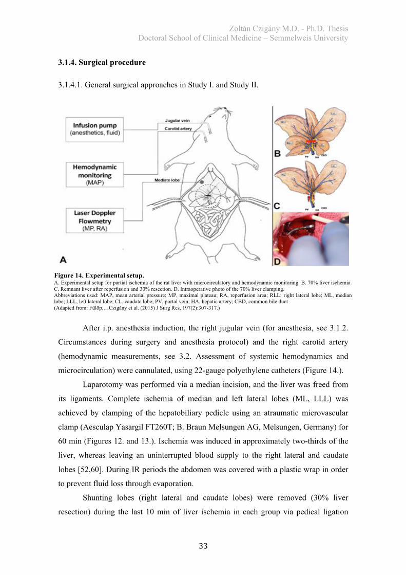
After i.p. anesthesia induction, the right jugular vein (for anesthesia, see 3.1.2.
Circumstances during surgery and anesthesia protocol) and the right carotid artery
(hemodynamic measurements, see 3.2. Assessment of systemic hemodynamics and
microcirculation) were cannulated, using 22-gauge polyethylene catheters (Figure 14.).
Laparotomy was performed via a median incision, and the liver was freed from
its ligaments. Complete ischemia of median and left lateral lobes (ML, LLL) was
achieved by clamping of the hepatobiliary pedicle using an atraumatic microvascular
clamp (Aesculap Yasargil FT260T; B. Braun Melsungen AG, Melsungen, Germany) for
60 min (Figures 12. and 13.). Ischemia was induced in approximately two-thirds of the
liver, whereas leaving an uninterrupted blood supply to the right lateral and caudate
lobes [52,60]. During IR periods the abdomen was covered with a plastic wrap in order
to prevent fluid loss through evaporation.
Shunting lobes (right lateral and caudate lobes) were removed (30% liver
resection) during the last 10 min of liver ischemia in each group via pedical ligation
Figure 14. Experimental setup. A. Experimental setup for partial ischemia of the rat liver with microcirculatory and hemodynamic monitoring. B. 70% liver ischemia. C. Remnant liver after reperfusion and 30% resection. D. Intraoperative photo of the 70% liver clamping. Abbreviations used: MAP, mean arterial pressure; MP, maximal plateau; RA, reperfusion area; RLL; right lateral lobe; ML, median lobe; LLL, left lateral lobe; CL, caudate lobe; PV, portal vein; HA, hepatic artery; CBD, common bile duct (Adapted from: Fülöp,…Czigány et al. (2015) J Surg Res, 197(2):307-317.)
Zoltán Czigány M.D. - Ph.D. Thesis Doctoral School of Clinical Medicine – Semmelweis University
34
method, using a 4-0 silk thread (Resorba Medical Inc, Nuremberg, Germany). At the
end of ischemia, the vascular clip was removed and liver reperfusion was allowed.
3.1.4.2. Specificities of surgical procedures in Study I.
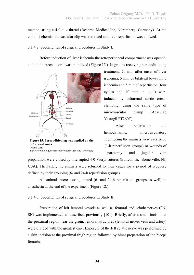
Before induction of liver ischemia the retroperitoneal compartment was opened,
and the infrarenal aorta was mobilized (Figure 15.). In groups receiving perconditioning
treatment, 20 min after onset of liver
ischemia, 5 min of bilateral lower limb
ischemia and 5 min of reperfusion (four
cycles and 40 min in total) were
induced by infrarenal aortic cross-
clamping, using the same type of
microvascular clamp (Aesculap
Yasargil FT260T).
After reperfusion and
hemodynamic, microcirculatory
monitoring the animals were sacrificed
(1-h reperfusion groups) or wounds of
laparotomy and jugular vein
preparation were closedby interrupted 4-0 Vicryl sutures (Ethicon Inc, Somerville, NJ,
USA). Thereafter, the animals were returned to their cages for a period of recovery
defined by their grouping (6- and 24-h reperfusion groups).
All animals were exsanguinated (6- and 24-h reperfusion groups as well) in
anesthesia at the end of the experiment (Figure 12.).
3.1.4.3. Specificities of surgical procedures in Study II.
Preparation of left femoral vessels as well as femoral and sciatic nerves (FN,
SN) was implemented as described previously [101]. Briefly, after a small incisionat
the proximal region near the groin, femoral structures (femoral nerve, vein and artery)
were divided with the greatest care. Exposure of the left sciatic nerve was performed by
a skin incision at the proximal thigh region followed by blunt preparation of the biceps
femoris.
Figure 15. Perconditioning was applied on the infrarenal aorta. (From: URL: http://www.biologycorner.com/resources/rat_circ_artery.gif)
Zoltán Czigány M.D. - Ph.D. Thesis Doctoral School of Clinical Medicine – Semmelweis University
35
Approximately 0.5 cm segments of the FN and SN were resected in groups
subjected to nerve resection (NR) followed by a recovery period of 20 min.
Four cycles, each of 5 min of ischemia and 5 min of reperfusion, were applied as
RIPER protocol during the last 40 min of hepatic ischemia achieved by clamping of the
left femoral artery with a microvessel clamp (Aesculap BIEMER FD561 R; B. Braun)
(Figure 13.).
Following the first post-ischemic hour, wounds (laparotomy as well as vena
jugularis and nerve preparations) were closed by interrupted 4-0 Vicryl sutures. Before
returning to their cages, animals were treated with subcutaneous injection of normal
saline solution (5 mL/bwkg).
After 24 h of liver reperfusion, animals were sacrificed and exsanguinated via
right ventricular puncture in anesthesia (Figure 13.).
3.2. Assessment of systemic hemodynamics and microcirculation
In order to carry out hemodynamic and microcirculation analyses, a 125-min
period was measured after laparotomy and 20 min of recovery (5 min of pre-ischemic
baseline, 60 min of ischemia, and 60 min of reperfusion).
3.2.1. Hemodynamics
Blood pressure was measured by an invasive blood pressure monitoring system
(Kent Scientific Corporation, Torrington, CT, USA) and recorded with DasyLab
software, version 9.00.02 (National Instruments Corporation, Austin, TX, USA) via
cannulated right carotid artery.
Study I: For comparison between groups, the average of the reperfusion mean arterial
pressure (MAP) was calculated for the last 20 min reperfusion plateau of the graph of
each animal.
Study II: After revealing the characteristics of MAP in Study I., only the most critical
time points of reperfusion were evaluated and compared in Study II. (Reperfusion 0
min, 30 min, 60 min).
Zoltán Czigány M.D. - Ph.D. Thesis Doctoral School of Clinical Medicine – Semmelweis University
36
3.2.2. Microcirculation
Microcirculation was measured using a laser Doppler monitor and a surface
probe (DRT4 device with DP1T surface probe; Moor Instruments Ltd, London, UK).
The device measures reflection of emitted laser light based on the Doppler effect.
Laser Doppler flowmeter (LDF) probe was placed on a fixed location on the
liver’s left lateral lobe and held in place. In Study I. a second surface probe was placed
on the left femoral biceps muscle to assess alterations in lower limb microcirculation
during liver IR and perconditioning.
The LDF monitor registered tissue microcirculation in Arbitrary Units. Due to
considerable differences between individuals, certain correction was necessary for the
sake of comparability between groups. Details of mathematical transformation, required
for correct interpretation of circulation data, were described previously by our team
[60].
Integral of the reperfusion graph segments (reperfusion area: RA) and maximal
plateau (MP) of the reperfusion section were introduced to characterise
microcirculation.
3.3. Light microscopy and automated image analysis
3.3.1. Histological analysis – Study I.
Histological samples were harvested from the left lateral lobe of the liver from
identical anatomical sites. The excised liver was fixed in 4% neutral buffered formalin
and embedded in paraffin. Slides, 3 µm thick, were stained with hematoxylin and eosin.
The examining pathologist was not informed of the applied treatment or grouping.
Slides were subjected to semiquantitative histological analysis as described by Suzuki et
al. [154]. Sinusoidal congestion, hepatocyte necrosis, and ballooning degeneration were
individually graded from 0-4. To simplify this complex scoring, a total score, the sum
of the aforementioned individual parameters with a maximum of 12 points/animal, was
introduced by other authors [155]. This total score was used when presenting our
results. Ten random fields were evaluated per section.
Zoltán Czigány M.D. - Ph.D. Thesis Doctoral School of Clinical Medicine – Semmelweis University
37
3.3.2. Histological analysis – Study II.
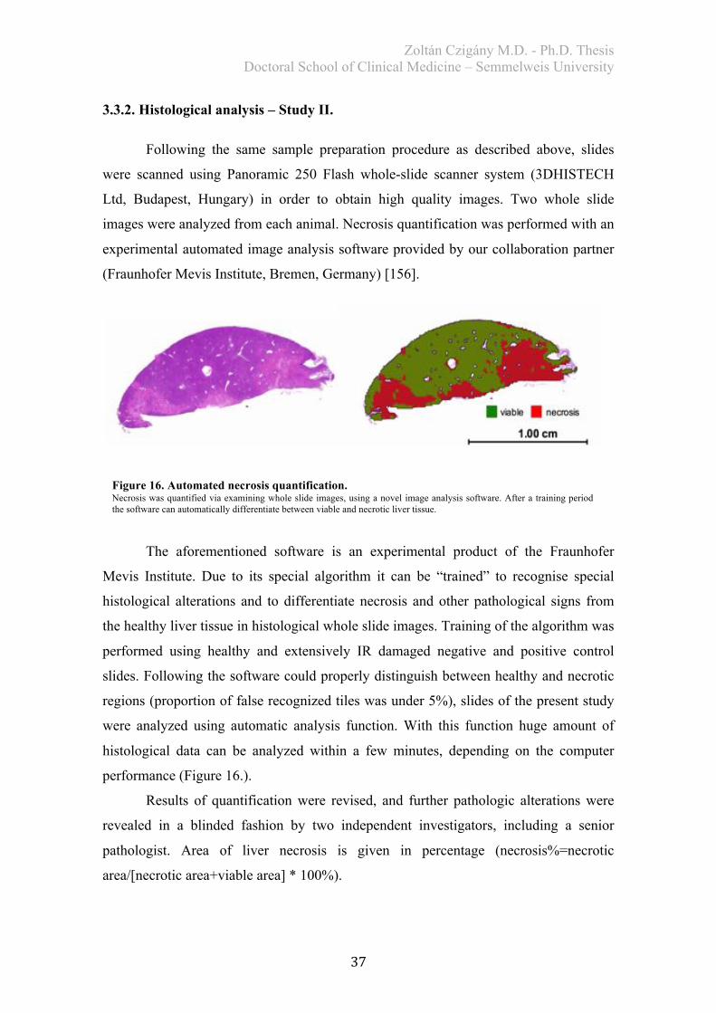
Following the same sample preparation procedure as described above, slides
were scanned using Panoramic 250 Flash whole-slide scanner system (3DHISTECH
Ltd, Budapest, Hungary) in order to obtain high quality images. Two whole slide
images were analyzed from each animal. Necrosis quantification was performed with an
experimental automated image analysis software provided by our collaboration partner
(Fraunhofer Mevis Institute, Bremen, Germany) [156].
The aforementioned software is an experimental product of the Fraunhofer
Mevis Institute. Due to its special algorithm it can be “trained” to recognise special
histological alterations and to differentiate necrosis and other pathological signs from
the healthy liver tissue in histological whole slide images. Training of the algorithm was
performed using healthy and extensively IR damaged negative and positive control
slides. Following the software could properly distinguish between healthy and necrotic
regions (proportion of false recognized tiles was under 5%), slides of the present study
were analyzed using automatic analysis function. With this function huge amount of
histological data can be analyzed within a few minutes, depending on the computer
performance (Figure 16.).
Results of quantification were revised, and further pathologic alterations were
revealed in a blinded fashion by two independent investigators, including a senior
pathologist. Area of liver necrosis is given in percentage (necrosis%=necrotic
area/[necrotic area+viable area] * 100%).
Figure 16. Automated necrosis quantification. Necrosis was quantified via examining whole slide images, using a novel image analysis software. After a training period the software can automatically differentiate between viable and necrotic liver tissue.
Zoltán Czigány M.D. - Ph.D. Thesis Doctoral School of Clinical Medicine – Semmelweis University
38
3.4. Biochemical examination
At the end of experiments, blood samples were collected via right ventricular
puncture and then centrifuged (3000 rpm for 2x10 min, at room temperature). Samples
were snap frozen in liquid nitrogen and stored at -80°C until analysis. Plasma aspartate
aminotransferase (AST) alanine aminotransferase (ALT) and total bilirubin (tBi) levels
were analyzed. Measurements were performed using automated clinical chemistry
analyzer (Beckman Coulter AU480/2011; Beckman Coulter Inc, Brea, CA, USA).
3.5. Redox-state measurements
Residual liver tissue of left lateral and median lobes were cut into pieces and
washed five times with 4°C saline solution to reduce blood content, then homogenized
at 4°C using Potter-Elvehjem homogenizer. After the procedure, samples were
immediately snap-frozen in liquid nitrogen and then stored at -80°C for assessment.
Protein content of each sample was standardized (10 mg/mL) according to the method
of Lowry et al. [157]. All reagents used for redox-state measurements were purchased
from Sigma-Aldrich Inc (St. Louis, MO, USA) and Reanal Chemical Corporation
(Budapest, Hungary).
3.5.1. Measurement of tissue free radicals and antioxidant capacity
A sensitive chemiluminescence assay was adopted to measure tissue reactive
oxygen species content. The analysis was performed according to the method of
Blázovics et al. [158]. Measurements were carried out using Lumat LB 9051
luminometer (Berthold Technologies GmbH, Bad Wildbad, Germany). Light intensity,
which is directly proportional to the concentration of free-radical agents in the sample,
was given in relative light unit percentage compared to the background.
For analysis of antioxidant capacity three further spectrophotometric
measurement protocols were implemented using a Hitachi U-2000 instrument (Hitachi
High-Technologies Corporation, Tokyo, Japan). The method of Oyaizu was used to
determine global reducing power of samples [159] expressed in ascorbic acid equivalent
(mgAA/mL). Hydrogen (H-) donating ability reflects the non-protein-related
antioxidant capacity of samples, which was measured on 517 nm in the presence of 1,1-
Zoltán Czigány M.D. - Ph.D. Thesis Doctoral School of Clinical Medicine – Semmelweis University
39
diphenyl-2-picryl-hydrasyl radical, according to the method of Blois [160] modified by
Blázovics et al. [158]. Results are given in inhibition percentage. Thiol (SH-) groups
were measured via the approach of Sedlak and Lindsay [161]. Results show the protein-
bound reducing power of the samples in micromoles per liter.
3.6. Serum TNF-a levels
Serum tumor necrosis factor alpha (TNF-a) levels were measured using
commercial enzyme-linked immunosorbent assay (ELISA) kits according to
manufacturer’s guidelines (Quantikine Rat TNF-a Immunoassay kit; R&D Systems Inc,
Mineapolis, MN, USA). All samples were tested in duplicate. Concentrations were
calculated from a standard curve and expressed in picogram per milliliter (pg/mL).
Threshold for detection was 5 pg/mL.
3.7. Statistical analysis
Values were expressed as mean ± standard deviation, with “n” representing the
numbers of animals or samples studied. Statistical analysis was performed with one-
way analysis of variance (ANOVA) and Scheffe’s post-hoc correction. Normality and
homoscedasticity were tested. For analysis of histological scores Kruskal-Wallis H test
was applied. Differences were considered significant when p < 0.05. Calculations were
performed using IBM SPSS Statistics 20 Software (IBM Corporation, Armonk, NY,
USA). Results were plotted using GraphPad Prism 6 Software (GraphPad Software Inc.,
San Diego, CA, USA).
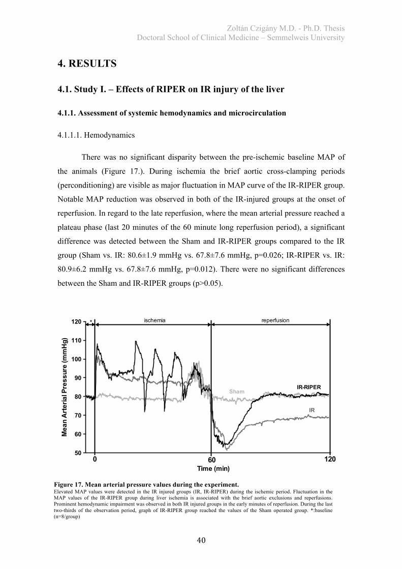
Zoltán Czigány M.D. - Ph.D. Thesis Doctoral School of Clinical Medicine – Semmelweis University
40
4. RESULTS
4.1. Study I. – Effects of RIPER on IR injury of the liver
4.1.1. Assessment of systemic hemodynamics and microcirculation
4.1.1.1. Hemodynamics
There was no significant disparity between the pre-ischemic baseline MAP of
the animals (Figure 17.). During ischemia the brief aortic cross-clamping periods
(perconditioning) are visible as major fluctuation in MAP curve of the IR-RIPER group.
Notable MAP reduction was observed in both of the IR-injured groups at the onset of
reperfusion. In regard to the late reperfusion, where the mean arterial pressure reached a
plateau phase (last 20 minutes of the 60 minute long reperfusion period), a significant
difference was detected between the Sham and IR-RIPER groups compared to the IR
group (Sham vs. IR: 80.6±1.9 mmHg vs. 67.8±7.6 mmHg, p=0.026; IR-RIPER vs. IR:
80.9±6.2 mmHg vs. 67.8±7.6 mmHg, p=0.012). There were no significant differences
between the Sham and IR-RIPER groups (p>0.05).
50
60
70
80
90
100
110
120
Time (min)
Mea
n A
rter
ial P
ress
ure
(mm
Hg)
* ischemia reperfusion
0 60 120
ShamIR-RIPER
IR
Figure 17. Mean arterial pressure values during the experiment. Elevated MAP values were detected in the IR injured groups (IR, IR-RIPER) during the ischemic period. Fluctuation in the MAP values of the IR-RIPER group during liver ischemia is associated with the brief aortic exclusions and reperfusions. Prominent hemodynamic impairment was observed in both IR injured groups in the early minutes of reperfusion. During the last two-thirds of the observation period, graph of IR-RIPER group reached the values of the Sham operated group. *:baseline (n=8/group)
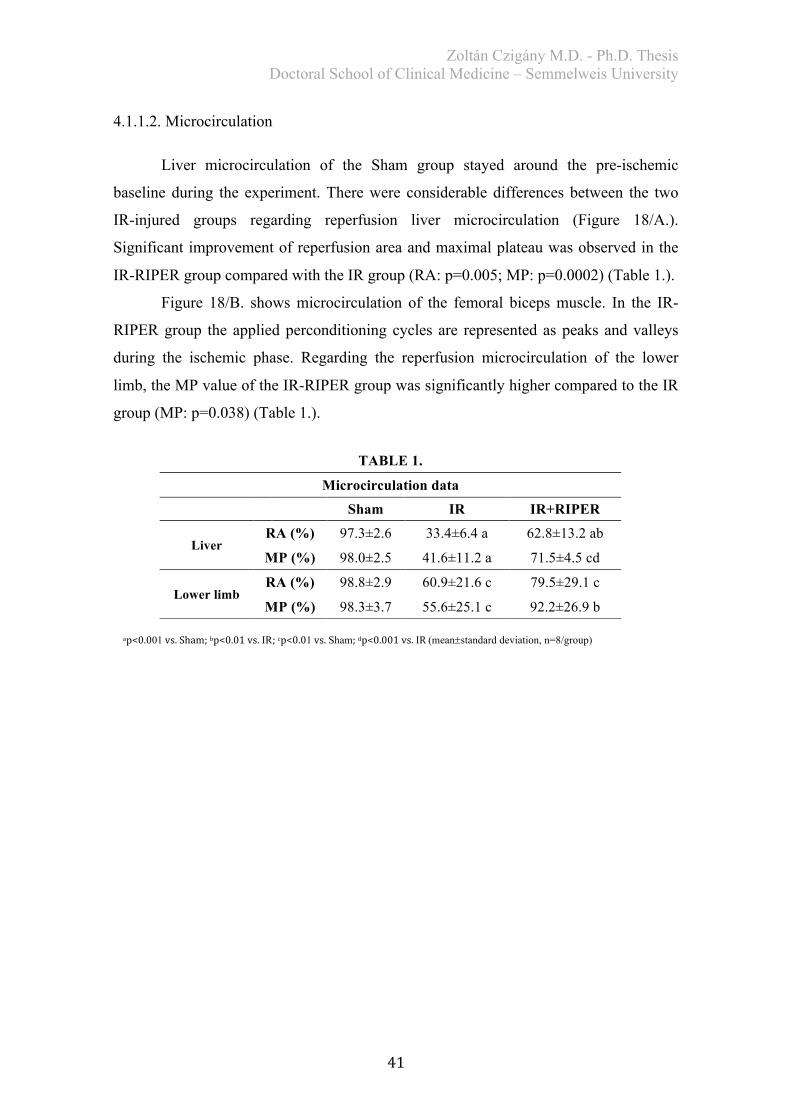
Zoltán Czigány M.D. - Ph.D. Thesis Doctoral School of Clinical Medicine – Semmelweis University
41
4.1.1.2. Microcirculation
Liver microcirculation of the Sham group stayed around the pre-ischemic
baseline during the experiment. There were considerable differences between the two
IR-injured groups regarding reperfusion liver microcirculation (Figure 18/A.).
Significant improvement of reperfusion area and maximal plateau was observed in the
IR-RIPER group compared with the IR group (RA: p=0.005; MP: p=0.0002) (Table 1.).
Figure 18/B. shows microcirculation of the femoral biceps muscle. In the IR-
RIPER group the applied perconditioning cycles are represented as peaks and valleys
during the ischemic phase. Regarding the reperfusion microcirculation of the lower
limb, the MP value of the IR-RIPER group was significantly higher compared to the IR
group (MP: p=0.038) (Table 1.).
TABLE 1.
Microcirculation data
Sham IR IR+RIPER
Liver RA (%) 97.3±2.6 33.4±6.4 a 62.8±13.2 ab
MP (%) 98.0±2.5 41.6±11.2 a 71.5±4.5 cd
Lower limb RA (%) 98.8±2.9 60.9±21.6 c 79.5±29.1 c
MP (%) 98.3±3.7 55.6±25.1 c 92.2±26.9 b
ap<0.001vs.Sham;bp<0.01vs.IR;cp<0.01vs.Sham;dp<0.001vs.IR(mean±standard deviation, n=8/group)
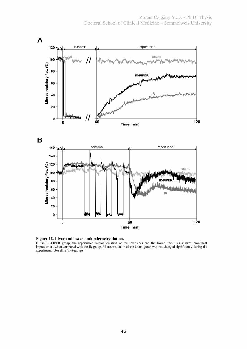
Zoltán Czigány M.D. - Ph.D. Thesis Doctoral School of Clinical Medicine – Semmelweis University
42
0
20
40
60
80
100
120
//
//
* ischemia reperfusion
0 60 120Time (min)
Mic
roci
rcul
ator
y flo
w (%
)
Sham
IR-RIPER
IR
0
20
40
60
80
100
120
140
160
Time (min)
Mic
roci
rcul
ator
y flo
w (%
)
* ischemia reperfusion
0 60 120
Sham
IR-RIPER
IR
A
B
Figure 18. Liver and lower limb microcirculation. In the IR-RIPER group, the reperfusion microcirculation of the liver (A.) and the lower limb (B.) showed prominent improvement when compared with the IR group. Microcirculation of the Sham group was not changed significantly during the experiment. *:baseline (n=8/group)
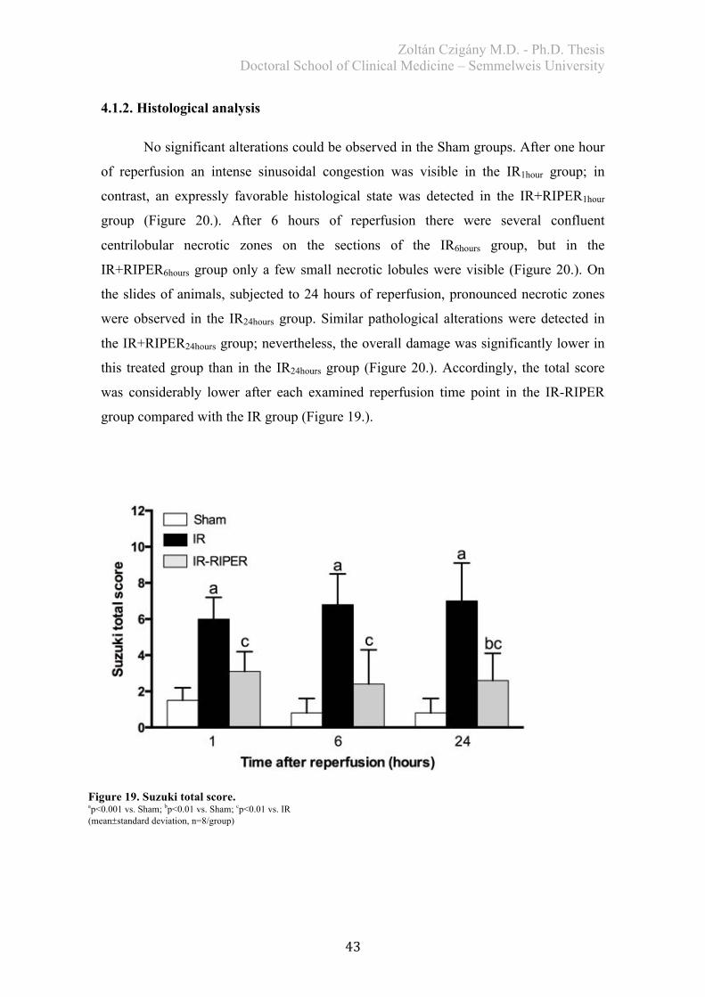
Zoltán Czigány M.D. - Ph.D. Thesis Doctoral School of Clinical Medicine – Semmelweis University
43
4.1.2. Histological analysis
No significant alterations could be observed in the Sham groups. After one hour
of reperfusion an intense sinusoidal congestion was visible in the IR1hour group; in
contrast, an expressly favorable histological state was detected in the IR+RIPER1hour
group (Figure 20.). After 6 hours of reperfusion there were several confluent
centrilobular necrotic zones on the sections of the IR6hours group, but in the
IR+RIPER6hours group only a few small necrotic lobules were visible (Figure 20.). On
the slides of animals, subjected to 24 hours of reperfusion, pronounced necrotic zones
were observed in the IR24hours group. Similar pathological alterations were detected in
the IR+RIPER24hours group; nevertheless, the overall damage was significantly lower in
this treated group than in the IR24hours group (Figure 20.). Accordingly, the total score
was considerably lower after each examined reperfusion time point in the IR-RIPER
group compared with the IR group (Figure 19.).
Figure 19. Suzuki total score. ap<0.001 vs. Sham; bp<0.01 vs. Sham; cp<0.01 vs. IR (mean±standard deviation, n=8/group)
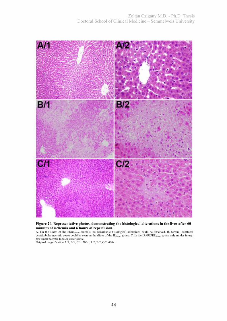
Zoltán Czigány M.D. - Ph.D. Thesis Doctoral School of Clinical Medicine – Semmelweis University
44
Figure 20. Representative photos, demonstrating the histological alterations in the liver after 60 minutes of ischemia and 6 hours of reperfusion. A. On the slides of the Sham6hours animals, no remarkable histological alterations could be observed. B. Several confluent centrilobular necrotic zones could be seen on the slides of the IR6hours group. C. In the IR+RIPER6hours group only milder injury, few small necrotic lobules were visible. Original magnification A/1, B/1, C/1: 200x; A/2, B/2, C/2: 400x.
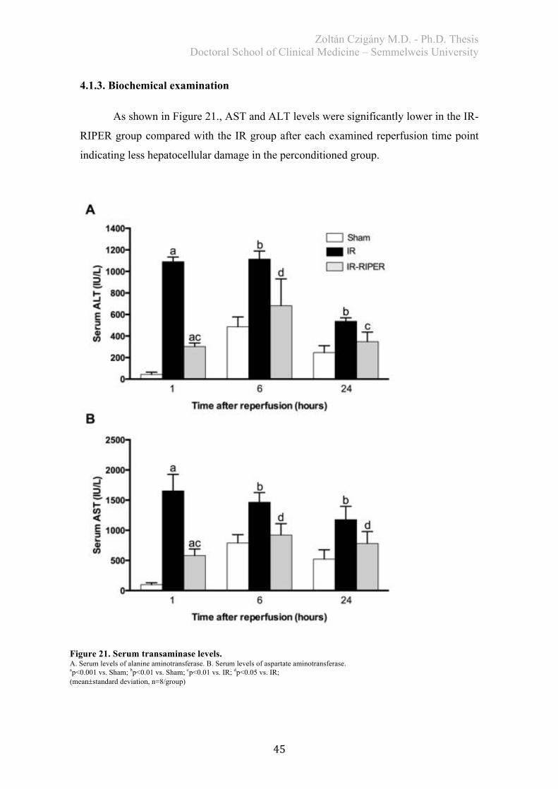
Zoltán Czigány M.D. - Ph.D. Thesis Doctoral School of Clinical Medicine – Semmelweis University
45
4.1.3. Biochemical examination
As shown in Figure 21., AST and ALT levels were significantly lower in the IR-
RIPER group compared with the IR group after each examined reperfusion time point
indicating less hepatocellular damage in the perconditioned group.
Figure 21. Serum transaminase levels. A. Serum levels of alanine aminotransferase. B. Serum levels of aspartate aminotransferase. ap<0.001 vs. Sham; bp<0.01 vs. Sham; cp<0.01 vs. IR; dp<0.05 vs. IR; (mean±standard deviation, n=8/group)
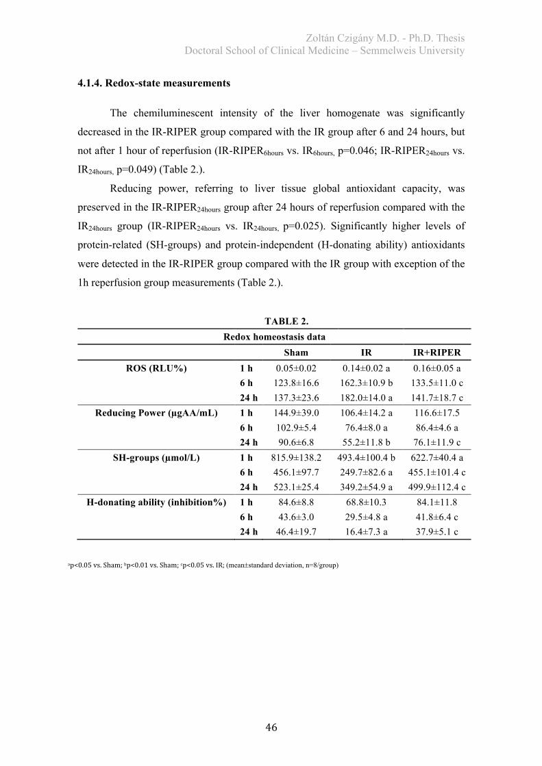
Zoltán Czigány M.D. - Ph.D. Thesis Doctoral School of Clinical Medicine – Semmelweis University
46
4.1.4. Redox-state measurements
The chemiluminescent intensity of the liver homogenate was significantly
decreased in the IR-RIPER group compared with the IR group after 6 and 24 hours, but
not after 1 hour of reperfusion (IR-RIPER6hours vs. IR6hours, p=0.046; IR-RIPER24hours vs.
IR24hours, p=0.049) (Table 2.).
Reducing power, referring to liver tissue global antioxidant capacity, was
preserved in the IR-RIPER24hours group after 24 hours of reperfusion compared with the
IR24hours group (IR-RIPER24hours vs. IR24hours, p=0.025). Significantly higher levels of
protein-related (SH-groups) and protein-independent (H-donating ability) antioxidants
were detected in the IR-RIPER group compared with the IR group with exception of the
1h reperfusion group measurements (Table 2.).
TABLE 2.
Redox homeostasis data Sham IR IR+RIPER
ROS (RLU%) 1 h 0.05±0.02 0.14±0.02 a 0.16±0.05 a 6 h 123.8±16.6 162.3±10.9 b 133.5±11.0 c 24 h 137.3±23.6 182.0±14.0 a 141.7±18.7 c
Reducing Power (µgAA/mL) 1 h 144.9±39.0 106.4±14.2 a 116.6±17.5 6 h 102.9±5.4 76.4±8.0 a 86.4±4.6 a 24 h 90.6±6.8 55.2±11.8 b 76.1±11.9 c
SH-groups (µmol/L) 1 h 815.9±138.2 493.4±100.4 b 622.7±40.4 a 6 h 456.1±97.7 249.7±82.6 a 455.1±101.4 c 24 h 523.1±25.4 349.2±54.9 a 499.9±112.4 c
H-donating ability (inhibition%) 1 h 84.6±8.8 68.8±10.3 84.1±11.8 6 h 43.6±3.0 29.5±4.8 a 41.8±6.4 c 24 h 46.4±19.7 16.4±7.3 a 37.9±5.1 c
ap<0.05vs.Sham;bp<0.01vs.Sham;cp<0.05vs.IR;(mean±standard deviation, n=8/group)
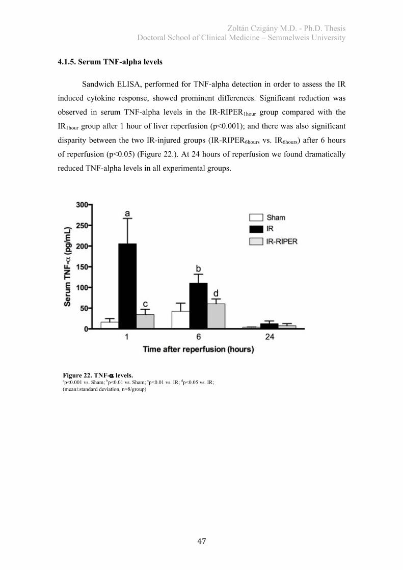
Zoltán Czigány M.D. - Ph.D. Thesis Doctoral School of Clinical Medicine – Semmelweis University
47
4.1.5. Serum TNF-alpha levels
Sandwich ELISA, performed for TNF-alpha detection in order to assess the IR
induced cytokine response, showed prominent differences. Significant reduction was
observed in serum TNF-alpha levels in the IR-RIPER1hour group compared with the
IR1hour group after 1 hour of liver reperfusion (p<0.001); and there was also significant
disparity between the two IR-injured groups (IR-RIPER6hours vs. IR6hours) after 6 hours
of reperfusion (p<0.05) (Figure 22.). At 24 hours of reperfusion we found dramatically
reduced TNF-alpha levels in all experimental groups.
Figure 22. TNF-a levels. ap<0.001 vs. Sham; bp<0.01 vs. Sham; cp<0.01 vs. IR; dp<0.05 vs. IR; (mean±standard deviation, n=8/group)
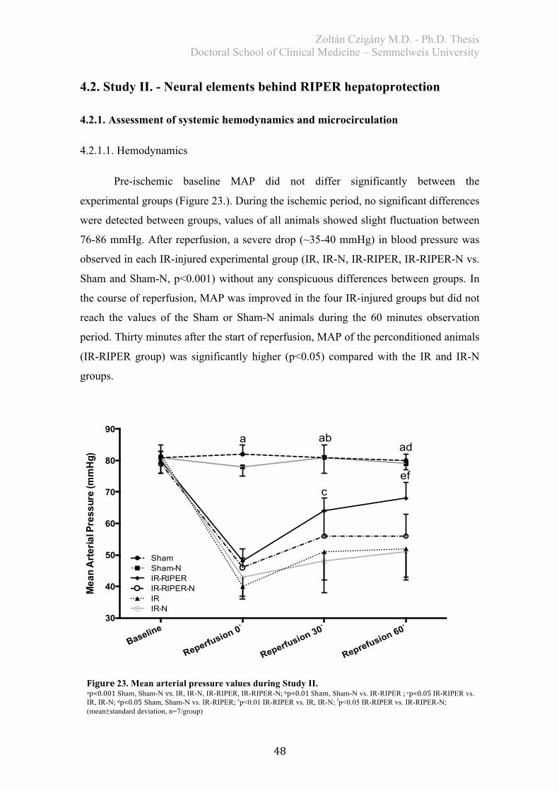
Zoltán Czigány M.D. - Ph.D. Thesis Doctoral School of Clinical Medicine – Semmelweis University
48
4.2. Study II. - Neural elements behind RIPER hepatoprotection
4.2.1. Assessment of systemic hemodynamics and microcirculation
4.2.1.1. Hemodynamics
Pre-ischemic baseline MAP did not differ significantly between the
experimental groups (Figure 23.). During the ischemic period, no significant differences
were detected between groups, values of all animals showed slight fluctuation between
76-86 mmHg. After reperfusion, a severe drop (~35-40 mmHg) in blood pressure was
observed in each IR-injured experimental group (IR, IR-N, IR-RIPER, IR-RIPER-N vs.
Sham and Sham-N, p<0.001) without any conspicuous differences between groups. In
the course of reperfusion, MAP was improved in the four IR-injured groups but did not
reach the values of the Sham or Sham-N animals during the 60 minutes observation
period. Thirty minutes after the start of reperfusion, MAP of the perconditioned animals
(IR-RIPER group) was significantly higher (p<0.05) compared with the IR and IR-N
groups.
Baseline
Reperfusion 0`
Reperfusion 30`
Reprefusion 60`30
40
50
60
70
80
90
Mea
n A
rter
ial P
ress
ure
(mm
Hg)
ShamSham-N
IRIR-N
IR-RIPERIR-RIPER-N
a abad
cef
Figure23. Mean arterial pressure values during Study II. ap<0.001Sham, Sham-N vs. IR, IR-N, IR-RIPER, IR-RIPER-N;bp<0.01Sham, Sham-N vs. IR-RIPER ;cp<0.05IR-RIPER vs. IR, IR-N;dp<0.05 Sham, Sham-N vs. IR-RIPER; ep<0.01 IR-RIPER vs. IR, IR-N; fp<0.05 IR-RIPER vs. IR-RIPER-N; (mean±standard deviation, n=7/group)
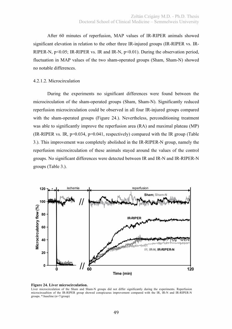
Zoltán Czigány M.D. - Ph.D. Thesis Doctoral School of Clinical Medicine – Semmelweis University
49
After 60 minutes of reperfusion, MAP values of IR-RIPER animals showed
significant elevation in relation to the other three IR-injured groups (IR-RIPER vs. IR-
RIPER-N, p<0.05; IR-RIPER vs. IR and IR-N, p<0.01). During the observation period,
fluctuation in MAP values of the two sham-operated groups (Sham, Sham-N) showed
no notable differences.
4.2.1.2. Microcirculation
During the experiments no significant differences were found between the
microcirculation of the sham-operated groups (Sham, Sham-N). Significantly reduced
reperfusion microcirculation could be observed in all four IR-injured groups compared
with the sham-operated groups (Figure 24.). Nevertheless, perconditioning treatment
was able to significantly improve the reperfusion area (RA) and maximal plateau (MP)
(IR-RIPER vs. IR, p=0.034, p=0.041, respectively) compared with the IR group (Table
3.). This improvement was completely abolished in the IR-RIPER-N group, namely the
reperfusion microcirculation of these animals stayed around the values of the control
groups. No significant differences were detected between IR and IR-N and IR-RIPER-N
groups (Table 3.).
0
20
40
60
80
100
120
Time (min)
Mic
roci
rcul
ator
y flo
w (%
)
//
//* ischemia reperfusion
0 60 120
Sham; Sham-N
IR-RIPER
IR; IR-N; IR-RIPER-N
Figure 24. Liver microcirculation. Liver microcirculation of the Sham and Sham-N groups did not differ significantly during the experiments. Reperfusion microcircualtion of the IR-RIPER group showed conspicuous improvement compared with the IR, IR-N and IR-RIPER-N groups. *:baseline (n=7/group)
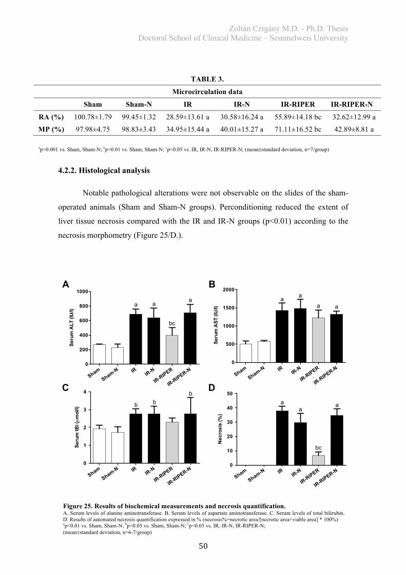
Zoltán Czigány M.D. - Ph.D. Thesis Doctoral School of Clinical Medicine – Semmelweis University
50
4.2.2. Histological analysis
Notable pathological alterations were not observable on the slides of the sham-
operated animals (Sham and Sham-N groups). Perconditioning reduced the extent of
liver tissue necrosis compared with the IR and IR-N groups (p<0.01) according to the
necrosis morphometry (Figure 25/D.).
Sham
Sham-N IRIR-N
IR-RIPER
IR-RIPER-N
0
200
400
600
800
1000
a a
bc
a
Ser
um A
LT (I
U/l)
Sham
Sham-N IRIR-N
IR-RIPER
IR-RIPER-N
0
1
2
3
4
b bb
Ser
um tB
i (µ
mol
/l)
Sham
Sham-N IRIR-N
IR-RIPER
IR-RIPER-N
0
500
1000
1500
2000
a a
a a
Ser
um A
ST
(IU/l)
Sham
Sham-N IRIR-N
IR-RIPER
IR-RIPER-N
0
10
20
30
40
50
aa
a
Nec
rosi
s (%
)
bc
A B
C D
TABLE 3.
Microcirculation data
Sham Sham-N IR IR-N IR-RIPER IR-RIPER-N
RA (%) 100.78±1.79 99.45±1.32 28.59±13.61 a 30.58±16.24 a 55.89±14.18 bc 32.62±12.99 a
MP (%) 97.98±4.75 98.83±3.43 34.95±15.44 a 40.01±15.27 a 71.11±16.52 bc 42.89±8.81 a
Figure 25. Results of biochemical measurements and necrosis quantification. A. Serum levels of alanine aminotransferase. B. Serum levels of aspartate aminotransferase. C. Serum levels of total bilirubin. D. Results of automated necrosis quantification expressed in % (necrosis%=necrotic area/[necrotic area+viable area] * 100%) ap<0.01 vs. Sham, Sham-N; bp<0.05 vs. Sham, Sham-N; cp<0.05 vs. IR, IR-N, IR-RIPER-N; (mean±standard deviation, n=6-7/group)
ap<0.001 vs. Sham, Sham-N; bp<0.01 vs. Sham, Sham-N; cp<0.05 vs. IR, IR-N, IR-RIPER-N; (mean±standard deviation, n=7/group)
Zoltán Czigány M.D. - Ph.D. Thesis Doctoral School of Clinical Medicine – Semmelweis University
51
Nerve resection before perconditioning treatment (IR-RIPER-N group) resulted
in a significantly higher damage than in the IR-RIPER group (IR-RIPER vs. IR-RIPER-
N, p<0.01). No significant differences were observed between this group (IR-RIPER-N
group) and the IR or IR-N groups. Next to the extended necrotic areas, considerable
ballooning degeneration, sinusoidal congestion, and neutrophil cell infiltration were
detected in the slides of the IR, IR-N, IR-RIPER-N animals. These alterations were
manifested in the IR-RIPER group as well; however, no convincing differences were
found concerning the presence of inflammatory cells between groups.
4.2.3. Biochemical examination
Serum transaminase levels were prominently elevated after 24 hours of
reperfusion in the IR-injured groups. Regarding ALT, significantly increased levels
(p<0.01) were detected in the IR, IR-N, and IR-RIPER-N groups compared with the
Sham and Sham-N groups. In the RIPER group serum ALT elevation was significantly
milder than in the other three IR-injured groups (IR-RIPER vs. IR, IR-N, IR-RIPER-N,
p<0.05) (Figure 25/A.).
Characteristics of AST release were different. In case of aspartate
aminotransferase, a significant elevation (p<0.01) was revealed in all four IR-injured
groups compared with the Sham and Sham-N groups; however, the perconditioning
treatment had no significant effect on AST levels (Figure 25/B.).
No severe alterations were observed concerning the tBi levels in any of the
experimental groups following 60 min ischemia and 24 hours of reperfusion (Figure
25/C.). Significant elevation of tBi can be seen in the IR, IR-N, and IR-RIPER-N
groups but not in the IR-RIPER group when compared with the sham-operated groups.
4.2.4. Redox-state measurements
Chemiluminescent intensity – thus tissue free-radical content – was significantly
elevated after 24 hours observation period in all IR-injured groups when compared with
the sham groups (IR, IR-N, IR-RIPER, IR-RIPER-N vs. Sham and Sham-N, p<0.001).
Perconditioning treatment potently reduced the level of free-radicals in liver
homogenate in relation to the three other IR-injured groups (IR-RIPER vs. IR, IR-N,
IR-RIPER-N, p<0.01) (Table 4.).
Zoltán Czigány M.D. - Ph.D. Thesis Doctoral School of Clinical Medicine – Semmelweis University
52
A more complex pattern was observed regarding the different parameters of
tissue antioxidant capacity. Global reducing power of the IR, IR-N, and IR-RIPER-N
groups was significantly reduced in comparison with the values assessed in the samples
of the sham-operated animals (IR, IR-N, IR-RIPER-N vs. Sham and Sham-N, p<0.01)
(Table 4.). In the IR-RIPER group, significantly preserved global reducing power was
observed when compared with the other three IR-injured groups (IR-RIPER vs. IR and
IR-RIPER-N, p<0.01; IR-RIPER vs. IR-N, p<0.05). It should be noted, furthermore,
that no significant differences were observed between the IR-RIPER-N as well as IR
and IR-N groups in regards to the global redox-state parameters (luminometry, reducing
power assessment) (Table 4.).
Considering protein-unbound antioxidants, a significant reduction (p<0.01) was
detected in H-donating ability of the IR and IR-N groups in relation to the Sham and
Sham-N values (Table 4.). The values of H-donating ability in the IR-RIPER group
were significantly higher (p<0.05) than in the IR-N group, but not when compared with
the IR group (IR-RIPER vs. IR, p=0.062). H-donating ability of IR-RIPER and IR-
RIPER-N groups did not differ significantly, either from each other or from the values
of the sham-operated groups. To some extent, a similar pattern was observed in the
thiol-group measurements, namely nerve resection was not able to induce significant
reduction in the values of thiol-groups (IR-RIPER vs. IR-RIPER-N, p=0.079); albeit, no
significantly higher protein-related antioxidant capacity was detected in the IR-RIPER-
N group either when compared with the IR and IR-N groups (Table 4.).
Significant differences were not detected in data of the four different redox-state
parameters between the Sham and Sham-N, or the IR and IR-N groups.
TABLE 4.
Redox homeostasis data
Sham Sham-N IR IR-N IR-RIPER IR-RIPER-N
ROS (RLU%) 6.4±10.5 6.9±11.7 229.2±16.1 a
202.3±20.3 a
131.8±20.8 ab
194.9±16.7 ac
Reducing power (µgAA/mL) 181.3±20.3 171.6±9.2 128.8±11.9
d 134.6±9.0
d 163.4±12.8
ef 131.3±13.6 d
SH-groups (µmol/L) 964.6±108.3 927.9±89.7 480.9±80.9 a
486.8±138.5 a
768.8±92.8 gh 594.3±74.9 a
H-donating ability (inhibiton%) 65.7±5.2 63.9±9.4 47.4±6.5 d 45.7±9.6 d 59.8±7.5 f 56.4±6.7
ap<0.001 vs. Sham, Sham-N; bp<0.01 vs. IR, IR-N, IR-RIPER-N; cp<0.05 vs. IR, dp<0.01 vs. Sham, Sham-N; ep<0.01 vs. IR, IR-RIPER-N; fp<0.05 vs. IR-N; gp<0.05 vs. Sham; hp<0.01 vs. IR, IR-N; (mean±standard deviation, n=6-7/group)
Zoltán Czigány M.D. - Ph.D. Thesis Doctoral School of Clinical Medicine – Semmelweis University
53
5. DISCUSSION
Ischemic-reperfusion injury remains one of the major concerns in extended liver
resections and liver transplantations. Sustained liver exclusion and consequential
ischemia is characterized by tissue hypoxia and reduced tissue adenosine triphosphate
levels, which cannot maintain active cellular functions resulting in cell death [12,19].
The “healing” reperfusion, the restoration of blood flow, is inevitable to keep organ
function, nevertheless, it can initiate paradoxically a cascade, which ultimately can
cause further cellular damage [12]. IR injury can lead to severe damage and dysfunction
of the remnant liver after liver resections [162]. Following liver transplantation IR
induced hepatocellular injury is a frequent reason for primary non-function or late
dysfunction of the implanted liver graft and might lead to a higher incidence of acute
and chronic rejection as well [163]. These issues are even more emphasized when we
have to deal with a critically low future remnant liver volume following resection or
when using liver grafts from extended criteria donors (e.g. steatotic grafts, elderly
donors, etc.) for liver transplantation [164]. Therefore, studying and understanding
mechanisms and characteristics of liver ischemia and developing novel strategies are
essential in order to increase ischemic tolerance of the liver. Basic and translational
research might help us to reduce complication rates in liver surgery and to extend donor
pool for liver transplantation.
Since Jennings et al. introduced the concept of reperfusion injury in 1960,
several surgical and non-surgical protective strategies have been developed to limit the
degree of injury, some more successful than others [165]. After the pioneering work of
Murry and Vinten-Johansen, surgical conditioning approaches (local ischemic pre- and
postconditioning) became widely used in several settings [6,132]. The concept of
remote ischemic conditioning was developed later on by Karin Przyklenk [8]. She
postulated that ischemic conditioning might be effective if brief ischemic-reperfusion
attacks are applied in a remote vascular bed, distantly from the target organ.
Remote ischemic perconditioning, first proposed by Schmidt et al. in 2007, has
several beneficial features from a practical point of view [86]. The essence of this
strategy is that brief, remote ischemic attacks are applied after induction of target organ
ischemia but before onset of reperfusion.
Zoltán Czigány M.D. - Ph.D. Thesis Doctoral School of Clinical Medicine – Semmelweis University
54
Considering that RIPER was already tried in different IR injury scenarios of the
cardiovascular system, we aimed to use this approach for the first time in a rat model of
liver IR injury. Based on our previous experience, the widely used rat model of 60 min
normothermic, 70% partial liver ischemia was adopted as basic experimental model
[19,60].
Subsequently we had to develop a feasible and effective RIPER protocol for our
experimental setting. At the planning phase of our first study 16 articles were available
with the use of RIPER in different experimental models and clinical settings [9].
Considering that no studies were published before which would have demonstrated the
effects of RIPER in parenchymal organs we had to rely on previous findings of
cardiovascular and neurological studies [9].
However, remote conditioning can be applied on various organs to achieve
target organ protection [81], among the 16 studies published in the topic of RIPER (at
the time point of planning Study I.) 94% of the authors (15/16) used skeletal muscle
ischemia-reperfusion attacks (limbs) as conditioning treatment [9]. Conditioning on the
lower limbs can be achieved non-invasively (tourniquet, inflatable cuffs, etc.) and
invasively (aortic cross clamping, femoral artery clamping, etc.) [9]. In our model we
had to perform a median laparotomy to induce ischemic-reperfusion injury of the liver;
therefore, we concluded that the easiest and the most feasible way to induce
perconditioning of the skeletal muscle in this setting would be the use of an infrarenal
aortic cross-clamping.
For determining the optimal conditioning protocol (number of cycles and
duration of ischemia-reperfusion) we adopted the previous findings of other authors. An
elegant study of Xin et al. investigated the infarct size limiting effects of different
RIPER protocols [87]. They reported that fewer (1-2x5 min IR) repetitions or too short
cycles (4x1-3 min IR) are not able to exert a significant protection in a rat model of
myocardial infarction. The most effective RIPER protocol was the application of 5
minute long IR episodes in 3-4 cycles according to their findings. Potent IR injury
limiting effects of this protocol (4x5 min limb IR) were confirmed by others
[9,86,89,95,127,166-169]. Further concerns, regarding the used model and the remote
conditioning treatment, are described in detail in the Introduction of the present doctoral
thesis.
Zoltán Czigány M.D. - Ph.D. Thesis Doctoral School of Clinical Medicine – Semmelweis University
55
Microcirculatory damage is a pivotal mechanism in liver IR injury [170,171].
Microvascular injury plays an emphasized role in prolongation of the ischemic period
and thus in aggravation of cellular injury, due to the no-reflow phenomenon. Main
components of microcirculation dysfunction are the deterioration of active ion transport
mechanisms, secondary to ischemia-induced ATP deficiency, and the consequential
endothel swelling, cellular edema [171]. Results of nitrogen oxide/endothelin imbalance
and sinusoidal narrowing, during reperfusion, are worsened by the accumulation of
activated neutrophil granulocytes and by reduced red blood cell velocity [170]. In our
study we observed a significant improvement in liver reperfusion microcirculation in
the IR+RIPER group compared with the IR group. These results are in accordance with
previous findings of others who investigated different aspects of microcirculation in
order to demonstrate the positive effects of remote perconditioning on target organ
circulation [88,89] and on endothelial function [133].
Loukogeorgakis et al. examined flow-mediated dilation in a human study, which
is a recognized parameter of endothelial function [133]. These authors proved that
remote conditioning treatment, applied during sustained limb ischemia, was able to
decrease the extent of endothelial dysfunction. Zhao et al. demonstrated the efficient
infarct-size reducing effect of remote perconditioning in a porcine model of myocardial
infarction [89]. They also evinced the favorable effect of the mentioned technique on IR
induced myocardial no-reflow phenomenon. Noteworthy is the common point between
the two aforementioned different studies, namely, both authors have reported the crucial
role of K+ATP channels in the mechanism of remote conditioning induced protection.
Besides target organ (liver) microcirculation, we attempted to follow capillary
circulation of the “conditioned organ” in Study I. During the early phase of liver
reperfusion, a prominent drop was observed in microcirculation of the left femoral
biceps muscle in both IR-injured groups. Nonetheless, skeletal muscle circulation of the
RIPER group improved with a greater slope, and the mean microcirculation curve of
this group reached higher values compared with the IR group during the observation
period.
As previously discussed (see Introduction), liver ischemia has not only local but
also global consequences with definitive dysfunction and injury of several organs [29].
The detected drop in lower limb microcirculation in our study, during the early
Zoltán Czigány M.D. - Ph.D. Thesis Doctoral School of Clinical Medicine – Semmelweis University
56
reperfusion in both IR-injured groups, can probably be attributed to a circulation
redistribution, induced by reperfusion of the liver and resulting in reduced peripherial
blood volume. There are multiple explanations or hypotheses for the conspicuous
characteristic differences between the RIPER and IR groups regarding lower limb
microcirculation during the latter phase of the observation period. First, the application
of remote conditioning mitigated liver injury in our model, which might result in a
reduced liver origin metabolite content in the systemic circulation (cytokines, free-
radicals, etc.); therefore, the improved skeletal muscle microcirculation might be, at one
hand, interpreted as a result of a reduced systemic response. On the other hand, taking
the known effects of different, previously discussed (see Introduction) conditioning
techniques into account, we have to consider the possibility of a local conditioning
effect behind the aforementioned microcirculatory differences. More exactly, we can
assume that remote ischemic perconditioning, applied on the infrarenal aorta, might
exert a local preconditioning effect on the skeletal muscle, protecting against the
detrimental systemic effects of liver IR injury.
In Study I. we also attempted to find an answer for the question of whether or
not this 40 minute long lower limb ischemia-reperfusion “burden” is able to affect the
systemic hemodynamics of the animals. Interestingly, we detected a significantly
improved reperfusion mean arterial pressure (MAP) in the perconditioned animals as
compared with the IR group. A plausible explanation for this phenomenon could be that
in the treated group a milder liver injury resulted in decreased mediator release into the
systemic circulation, which might even have cardiodepression or endothelial-
dysfunction inducing activity [29]. Such deteriorated systemic hemodynamics [172],
myocardial function impairment, and elevation of myocardial enzyme markers
following liver IR injury were reported by others [29,173,174].
In 2011, Wei et al. proposed that perconditioning has significant effect on
inflammatory reactions, demonstrated by reduction of polymorphonuclear (PMN)
leukocyte infiltration, and monocyte chemoattractant protein-1 (MCP-1) expression in
their rodent model of myocardial infarction [127]. Nevertheless, the mentioned team
could not prove the favorable effects of perconditioning on expression of TNF-a.
Sedaghat et al. investigated the effects of RIPER on remote hepatic injury as a
Zoltán Czigány M.D. - Ph.D. Thesis Doctoral School of Clinical Medicine – Semmelweis University
57
consequence of 45 min renal ischemia in rats [175]. They could not show any
significant differences regarding serum TNF-a expression between groups.
In our study we evaluated the changes in serum levels of TNF-a after 1 hour of
liver ischemia and 1, 6, 24 hours of reperfusion. In the present model perconditioning
was able to significantly reduce serum TNF-a levels, compared with the IR1hour group,
after one hour of liver reperfusion. After this observation period there was no significant
disparity between the Sham1hour and IR+RIPER1hour groups. Likewise, significantly
attenuated TNF-a levels were observed after 6 hours of reperfusion; nevertheless, in the
24 hours reperfusion group serum cytokine levels were remarkably reduced without any
noticeable differences between the experimental groups. Our results are correlating
closely with the very recent findings of Wang et al. They demonstrated that remote
ischemic perconditioning exerts a positive effect on the serum profile of pro- and anti-
inflammatory cytokines (TNF-a, IL-6, IL-1, IL-10, ICAM-1, etc.) in a rat model of
myocardial ischemia-reperfusion injury [120].
Besides pro-inflammatory cytokine expression, reperfusion associated
sinusoidal endothelial injury, imbalance between vasoconstrictive/vasodilatative effects
as well as excessive oxidative stress and consequential free-radical production are also
represented as key factors in development of microcirculatory impairment and no-
reflow phenomenon. Taking the aforementioned facts into account, in our study we
aimed to identify the subtleties of the alteration in redox-homeostasis, following hepatic
IR injury and perconditioning. Based on chemiluminescency and spectrophotometry,
different approaches have been applied for the assessment of tissue free-radical content
and antioxidant-capacity [158].
As described in the Results, significantly favorable changes were observed
regarding liver tissue free-radical levels as well as antioxidant-capacity parameters in
the IR+RIPER group compared with the IR group in our study.
Yet it is noteworthy that the prominent differences in serum TNF-a levels were
observed mostly during the early phase of reperfusion, contrary to the more severe
disturbances in redox-homeostasis parameters, which found to prevail after 6 and 24
hours of reperfusion. TNF-a-related signaling is activated early after the onset of
reperfusion, acting as a trigger mechanism during the early phase of injury [14], thus
Zoltán Czigány M.D. - Ph.D. Thesis Doctoral School of Clinical Medicine – Semmelweis University
58
elevated serum TNF-a levels can determine the later phase inflammatory responses and
redox-state disturbances.
The described redox-state characteristics might be associated with certain liver
specific features [170]. Reperfusion injury and free-radical release in the liver can be
divided into two distinct phases: the early, acute phase (first 1-6 hours of reperfusion) is
dominated by oxidative stress and Kupffer-cell activation, and ensuing free radical
production, during the early phase of reperfusion [29,176]. The later phase is
characterized by expression of adhesion molecules and consequential
polymorphonuclear leukocyte infiltration.
In contrast to the alterations observed in our study after the first hour of
reperfusion, representative results were detected after 6 hours concerning all redox-
homeostasis parameters with the exception of reducing power. These aforementioned
findings would justify further investigations and detailed monitoring of the first six
hours of reperfusion to obtain an in-depth understanding of characteristics of redox-state
alterations as well as Kupffer-cell and neutrophil activations in this model. In
accordance with our findings, Iwamoto et al. and also Kawamoto et al. published
similar data obtained from rat models of liver ischemia and reperfusion [177,178]. In
their studies, these authors determined free-radical production using different
luminometry measurements and reported that post-ischemic free-radical production was
drastically increased with liver reperfusion time. They found significantly elevated
blood ROS content after 90-120 minutes of liver reperfusion. Tang et al. reported
increased activity of the protective superoxide dismutase (SOD) enzyme and parallel
decreased lipid peroxydation, measured via determining blood malondialdehyde (MDA)
levels, with the use of remote conditioning in a rabbit model of myocardial ischemia-
reperfusion injury [93]. More recently, Costa et al. have investigated the effects of a
similar RIPER protocol in a rat model of 70% partial liver IR injury. Their results have
confirmed the findings of our study, demonstrating that RIPER can reduce liver tissue
MDA levels and can preserve tissue antioxidant capacity, which they evaluated via
trolox equivalent antioxidant capacity measurement [179].
Since the seminal study of Konstantinov et al., we also know that remote
conditioning is able to suppress several leukocyte functions (leukocyte chemotaxis,
adhesion, and migration), which are essential in IR induced inflammatory responses
Zoltán Czigány M.D. - Ph.D. Thesis Doctoral School of Clinical Medicine – Semmelweis University
59
[123]. These favorable gene expression profile alterations might also explain the
preserved antioxidant defense line and reduced free-radical production by
perconditioning confirmed in our present study and also in the latter work of Costa et al.
[179]. Summarizing the probable mechanistic steps, perconditioning might be able to
hinder intense free-radical production via reducing microvascular injury [89],
inflammatory responses [120,127], and leukocyte functions [123]; furthermore, it could
initiate activation of free-radical elimination processes [93], resulting in the above
detailed favorable redox-state changes.
Various experimental and clinical studies have corroborated the potent necrosis-
size reducing effect of remote ischemic perconditioning in myocardium, brain, or
skeletal muscle [88,91,180]. In 2012, Basalay et al. published encouraging results
regarding bilateral lower limb remote ischemic perconditioning in a rat model of
myocardial infarction [181].These authors reported significant necrosis reduction with
application of perconditioning as compared with non-treated animals (a reduction of
infarct size by 56%). Based on these data we attempted to evaluate the so far unrevealed
hepatoprotective effects of remote ischemic perconditioning. In our study,
histopathological alterations were assessed using a semiquantitative scoring system, and
serum transaminase levels were also measured. We found that total-score values were
significantly lower in the IR-RIPER group, compared with the corresponding IR group,
following each examined reperfusion time point. A similar tendency was observed
regarding serum transaminase levels measured for estimation of hepatocyte injury
following reperfusion. According to our experimental data, significant reduction was
detected in serum AST and ALT levels in the perconditioned group compared with the
corresponding IR group following each reperfusion period. In addition, a marked
disparity was observed between values of the IR-RIPER1hour group in comparison with
the Sham1hour group exclusively after this reperfusion period. Considering the results
detailed above, our study revealed remarkably mitigated target organ injury upon the
application of remote ischemic perconditioning. Our findings were confirmed by
subsequent studies of other research groups. Costa et al. and Sedaghat et al. have
demonstrated the hepatoprotective effects of remote ischemic perconditioning in their
articles published in 2014 [175,179].
Zoltán Czigány M.D. - Ph.D. Thesis Doctoral School of Clinical Medicine – Semmelweis University
60
In Study I. we obtained complex short- and medium-term experiences with
lower limb RIPER in hepatic IR injury. Subsequently, our attention turned to the
investigation of underlying mechanisms of RIPER induced hepatoprotection.
As discussed above, several studies have demonstrated the potent protective
effects of remote ischemic perconditioning on different target organs such as heart
[180], brain [182], skeletal muscle [91], and for the first time by our team on liver;
although, our knowledge on the complex mechanisms behind this promising technique
is full of obscure details. According to the widely accepted theory, the connection
between remote and target organs is provided by certain blood-borne factors and/or
neural, systemic elements. These “connective mechanisms” appear to convey the
protective signal to the target organ, initiating a complex subcellular protective response
[9].
Up to now, only a handful of data have been published on the mechanisms of
remote perconditioning. It should be noted that, however, there are ample similarities
and overlap between the mechanisms of different ischemic conditioning procedures, we
should also assume the probable existence of subtle differences, namely thanks to the
timing of different procedures, in relation to target organ ischemia (pre-, per-, and
postconditioning), the concentration and activation status of the participants cannot be
similar, which might alter the observed protective responses. Slight differences between
protective pathways, triggered by remote pre- and perconditioning, have been proposed
by Hausenloy et al. [95]. These investigators reported that the Wortmannin PI3-kinase
inhibitor could abolish the protective effect of RIPC but not of RIPER. Therefore, we
have to be careful when interpreting our study results and adopting evidence concerning
the mechanisms, obtained from other studies using differently timed conditioning
procedures (e.g. RIPC).
Gho et al. firstly applied a ganglion blocking agent, hexamethonium, which
completely abrogated the myocardial infarct-size reduction achieved by application of
brief mesenteric IR attacks as remote preconditioning treatment [115]. Afterwards,
further studies confirmed the role of autonomic nervous system behind the RIPC
phenomenon. As a subsequent step, other authors have proved that direct vagal nerve
stimulation can also reduce myocardial IR injury [183,184]. Furthermore, the potent
protective effect of RIPC is absent in the presence of bilateral cervical vagotomy [181].
Zoltán Czigány M.D. - Ph.D. Thesis Doctoral School of Clinical Medicine – Semmelweis University
61
Recently, Matitskaya et al. have reported their interesting observations. In their study
the selective inhibition of the vagal cholinergic pre-ganglionic neurons could likewise
effectively extinguish RIPC protection, whilst selective activation of the mentioned
neural elements had a strong RIPC mimicking effect. [119]. Presumably, different
mediators (adenosine, bradykinin, etc.), released from the remote organ during the short
ischemic-reperfusion episodes and stimulating local neural elements, are responsible for
triggering these complex neural mechanisms. It has been proposed, as suspected trigger
mechanism, that these mediators could activate capsaicin sensitive sensory neurons
which can release calcitoning gene related peptide (CGRP) and thus induce a
subcellular protection with the participation of complex kinase cascades [117].
Most of the previous findings on the mechanisms of RIPER are obtained from
cardiovascular studies. Significantly less data are on hand in regards of the ischemic
events of parenchymal organs such as the liver [185]. According to our best knowledge,
no data have been reported concerning the liver before, which would investigate the role
of neural elements behind the hepatoprotective effects of RIPER. Lim et al. published
that myocardial protection, evoked by hind limb remote ischemic preconditioning, is
completely abolished after transection of the ipsilateral femoral and sciatic nerves [101].
Based on these data, in Study II., we aimed to investigate the effects of hind limb nerve
resection on the efficacy of remote perconditioning against hepatic warm IR injury.
To design a well-elaborated study we had to implement minor modifications in
our previous model (see Study I.). We modified the remote conditioning protocol, due
to the need to completely denervate the remote (conditioning) organ. In Study II. remote
perconditioning was applied on the left femoral artery, after preparation of the left
femoral and sciatic nerves. However, in this new model we applied remote conditioning
on a significantly reduced vascular bed, but in turn we could easily achieve selective
denervation of the conditioned area.
In the present study, we similarly followed liver microcirculatory changes using
laser Doppler flowmetry and systemic hemodynamics during the early phase of
reperfusion. As a result of IR injury, liver reperfusion microcirculation as well as
systemic mean arterial pressure values were strongly reduced in IR and IR-N groups. In
contrast, results of the IR-RIPER group were significantly improved during the
observation period. It could also be observed that resection of femoral and sciatic nerves
Zoltán Czigány M.D. - Ph.D. Thesis Doctoral School of Clinical Medicine – Semmelweis University
62
blunted the protection of RIPER, accordingly the circulation of the IR-RIPER-N group
stayed around the values of the IR and IR-N groups during observation.
Automated image analysis was used to quantify necrosis caused by IR induced
tissue injury. We utilized a novel software developed by our collaboration partner
(Fraunhofer Mevis Research Institute). The used experimental-phase software is the
first product, which enables the quick and automated quantification of necrosis in entire
tissue sections. Details on this “tile-based” automatic analysis software and the exact
methods were described by Homeyer et al. [156]. Briefly, necrotic zones can be
distinguished from viable areas based on their histological appearance. The
mathematical algorithm of the program can recognize the necrosis specific features
(dissolution of cellular architecture, “moth-eaten” appearance, shrunken or completely
absent nuclei, etc.) after a certain teaching period. This teaching session has to be
performed by an experienced investigator using reference slides (with prominent
necrotic and viable areas). Subsequently, if the algorithm is able to designate precisely
viable and necrotic areas and can distinguish them from background and border areas,
we can start to evaluate our study samples. We determined this border as less than 5%
of false recognized tiles. Depending on analysis settings and the performance of our
computer, enormous amount of histological data can be quantitatively analyzed within a
short period of time (minutes). The software is not only able to perform high-throughput
quantitative analysis of liver necrosis, but it was also successfully tested using other
tissues and staining methods [186].
Extent of histological tissue injury was pronounced in the ischemic-reperfusion
injured groups (IR, IR-N), after 1 hour of ischemia and 24 hours of reperfusion in Study
II. Femoral artery perconditioning strongly reduced the size of necrotic areas, although
such protection could not be observed in the IR-RIPER-N group; nerve resection prior
to the conditioning treatment aggravated tissue injury.
In accordance with our results of histological evaluation and microcirculatory
measurements, left femoral artery perconditioning was also able to significantly reduce
cellular injury indicated by ALT serum activity, which effect was also completely
abrogated by nerve resection. In contrast, AST levels were elevated in all four IR-
injured groups without characteristic differences among the experimental groups. These
confounding results might be ascribed to differing specificity of the two parameters,
Zoltán Czigány M.D. - Ph.D. Thesis Doctoral School of Clinical Medicine – Semmelweis University
63
whereby ALT is mostly considered as a liver injury specific marker, while AST is also
released after injuries of several other tissues such as myocardium and skeletal muscle
[187].
Serum total bilirubin levels were measured to roughly estimate liver function
(uptake and excretion). Slightly but significantly elevated tBi levels in the IR, IR-N, IR-
RIPER-N groups, except in the IR-RIPER group, coincide with the rest of our data.
Concerning liver tissue free-radical levels (investigated via luminometry) and
the indirectly proportional reducing power, it can be concluded that RIPER applied on
left femoral artery was able to positively influence tissue free-radical burden during
reperfusion, thereby preserving antioxidant defense line of the tissue. This protective
effect could not be observed following nerve resection. This phenomenon is less
obvious in relation to protein-related antioxidants (SH-groups) and H-donating ability.
Regarding these parameters, which are only partially representative for tissue
antioxidant capacity, nerve resection was not able to completely abolish the effects of
perconditioning; consequently, the values of the IR-RIPER and IR-RIPER-N groups did
not differ significantly.
Our results of Study II. are in line with the findings of Lim et al. They
demonstrated that denervation of the left limb can completely abolish the positive
effects of left femoral artery preconditioning on infarct-size, in a murine model of
myocardial ischemia-reperfusion [101]. In a more recent study, Wang et al. have tested
the protective effects of vagal stimulation perconditioning during experimentally
induced coronary occlusion. The authors postulated that electronic vagal stimulation
might exert its effects through activation of the cholinergic anti-inflammatory pathway
[120]. The observed positive alterations in infarct-size and inflammatory response with
the use of this method were comparable with the results obtained with the application of
standard ischemic perconditioning in their model. These findings underpin the possible
role of certain neural elements behind RIPER induced organ protection. These results
do not necessarily mean, however, that neural mechanisms are dominating in the
protective response induced by RIPER. We hypothesize that the innate responses
behind RIPER might form a redundant, however, also sensitive network with
multilateral communication. If there is a certain inhibiting factor (like denervation),
disturbing the transfer of protective signals, at one or multiple points, it might be able to
Zoltán Czigány M.D. - Ph.D. Thesis Doctoral School of Clinical Medicine – Semmelweis University
64
completely suspend these favorable effects. This might be the explanation for the
phenomenon that ischemic conditioning is in general less effective in the presence of
co-morbidities such as hyperlipidemia, diabetes, which conditions can well-known
interfere with the innate protective singaling pathways [188].
An important issue in the field of remote ischemic conditioning research is the
minimum amount of tissue necessary to temporarily be excluded from the circulation to
achieve a satisfactory protective effect in the target organ [9]. A previous study of our
group showed that the infrarenal aortic clamping (used as perconditioning protocol in
Study I.) can cause ischemia in the lumbosacral region and in the two lower limbs,
representing 42.93 ± 0.58 % of total body muscle weight of the rat, while weight of the
musculature of one leg is only 12.16 ± 0.35 % of the total [189].
According to the findings of previous studies, not really the amount of (skeletal
muscle) ischemized tissue is determinative, but the optimal repetition and length of the
cycles [9,87,190,191]. It was previously reported that even the selective ischemia and
reperfusion of canine gracilis muscle can protect against a subsequent IR injury in the
contralateral gracilis muscle [191]. In the present doctoral thesis we did not aim to
statistically compare the results obtained with application of the two different RIPER
protocols (Study I. – infrarenal aortic cross-clamping; Study II. – left femoral artery
clamping), because of the fact that relatively long time have passed between the two
studies and measurements. An interesting further direction for the present research
would be a comparative investigation of the effects of different skeletal muscle
exclusions (aorta cross-clamping, femoral clamping, limb tourniquet, inflatable cuffs
etc.) on RIPER induced hepatoprotection. Reliability and reproducibility of animal
studies represents an actual hot topic in literature [192-194]. Accordingly, it is
questionable whether the results of two separate pre-clinical studies with relatively low
numbers of individuals enrolled (thus with low power) are comparable. Nevertheless, in
general we can say that no conspicuous differences were observed and recorded,
regarding the effects of the two different RIPER protocols during our studies. Effects of
left femoral artery clamping were similar with those of aortic cross-clamping, regarding
liver microcirculation, tissue injury as well as liver tissue redox-homeostasis alterations.
In summary, the present thesis provides the first complex short- and medium-
term experiences with lower limb RIPER in a rat model of normothermic liver IR
Zoltán Czigány M.D. - Ph.D. Thesis Doctoral School of Clinical Medicine – Semmelweis University
65
injury. RIPER is a feasible, available, low-risk method, which could mean a valuable
supplementary approach for the clinical practice in several acute and elective situations,
when the possibility of severe IR injury should be concerned.
Investigating different connective and effector mechanisms behind RIPER
induced organ protection might help us to identify several exact molecular targets of the
protective pathway. The end result would be an efficient and practical pharmacologic
conditioning technique. In the present thesis we showed that hepatoprotective effect of
remote ischemic perconditioning could be almost completely abolished by denervation
of the remote (“conditioning”) organ; meanwhile, neural transection alone had no
significant effect on liver injury. Our results, therefore, imply that protective signals
evoked by perconditioning are conveyed to the target organ to a significant extent via
participation of certain neural elements, as reported in case of other kinds of remote
conditioning treatments as well [9,101,195]. In this study no attempt was made to
investigate more fine, cellular, molecular details of the neural pathway behind RIPER,
which is a major limitation of our current work.
Accordingly, further studies should be conducted to clarify the still very obscure
exact mechanisms and neural components behind the effect of remote conditioning
treatment and also the further possible differences or similarities between remote pre-,
per- and postconditioning. Furthermore, future multi-centric, randomized clinical trials
are necessary to evaluate efficacy and safety of this method in different clinical settings
of liver surgery.
Zoltán Czigány M.D. - Ph.D. Thesis Doctoral School of Clinical Medicine – Semmelweis University
66
6. CONCLUSIONS
As answers to the aims of the Doctoral Thesis formulated under Chapter 2. Objectives, we can conclude that:
1. Sixty minutes of 70% partial liver ischemia in rats is a feasible experimental
model to investigate the consequences and characteristics of liver IR injury and
the effects of remote ischemic perconditioning. In this model of moderate liver
ischemic-reperfusion injury, application of perconditioning resulted in
significant alterations in several parameters.
2. a, After 60 minutes of partial liver ischemia, remote ischemic perconditioning,
applied in 4 cycles of 5 minutes of ischemia and 5 minutes of reperfusion, could
potently mitigate liver injury, according to the post-reperfusion levels of serum
transaminases and based on the semiquantitative histological scoring.
b, Perconditioning positively influenced systemic hemodynamics (mean arterial
pressure) and microcirculation (liver and lower limb) of the animals, manifested
in significant differences between reperfusion macro- and microcirculation
values of treated and non-treated animals.
c, Significantly lower tissue free-radical levels and preserved antioxidant
capacity were observed with the use of perconditioning after 6 and 24 hours of
reperfusion. In parallel, conspicuous reduction was detected in serum values of
the pro-inflammatory cytokine, TNF-a, during the early phase of reperfusion.
3. In Study II., left femoral artery perconditioning could also exert potent
protection against the deleterious effects of liver exclusion and reperfusion. We
could observe significant alteration between treated and non-treated groups in
tissue damage (ALT, necrosis%), liver microcirculation and systemic
hemodynamics as well as in liver tissue redox-homeostasis. Perconditioning
induced hepatoprotection was abolished by denervation of the vascular bed,
where conditioning was applied.
Zoltán Czigány M.D. - Ph.D. Thesis Doctoral School of Clinical Medicine – Semmelweis University
67
4. Automated histological image analysis is an accurate and useful tool for the
evaluation of histological samples of a real experiment. Teaching of the
algorithm can be achieved easily and analysis of the slides is fast and reliable.
Zoltán Czigány M.D. - Ph.D. Thesis Doctoral School of Clinical Medicine – Semmelweis University
68
7. SUMMARY
Background: Ischemic-reperfusion (IR) injury of the liver is still a crucial factor in postoperative morbidity and mortality following major liver resections and transplantations. During the last decades, several surgical and pharmacological approaches have been developed to mitigate IR injury of the liver and to improve outcome for patients. The so-called remote ischemic perconditioning (RIPER) imply the application of brief, ischemic-reperfusion attacks on a remote organ after induction of target organ ischemia but before the onset of reperfusion. However, this method might have a potent protective effect, it has not been investigated before in a model of liver IR injury. Objectives: Our aim was to investigate the effects of remote ischemic perconditioning in a rat model of hepatic IR injury; furthermore, we aimed to demonstrate whether or not certain neural elements are participating in transfer of protective signals between remote and target organs. Materials and Methods: Two studies were planned to analyze the effects of RIPER in a rat model of liver IR injury. In both studies 60 min of partial liver ischemia (70% of liver volume) was used as basic model. In Study I. animals were randomly divided into three groups: Sham, IR, IR-RIPER (n=24/group). Ischemic perconditioning was applied during the last 40 min of liver ischemia via infrarenal aortic cross-clamping (4x5 min I, 5 min R). Hepatic and lower limb microcirculation as well as systemic hemodynamics, were monitored during the first hour of reperfusion. After 1,6,24 hours of reperfusion (n=8/time point) liver samples were taken for histological and redox-state assessment. Suzuki score was used for semi-quantitative histological analysis. Serum transaminase (AST, ALT) and TNF-a levels were measured. In Study II. rats were randomly allocated into six groups: Sham, IR, IR-RIPER±denervation (n=7/group). Half of the animals underwent left femoral and sciatic nerve resection. The same perconditioning protocol was applied (4x5 min I, 5 min R), but the left femoral artery was clamped. After 24 h of reperfusion all animals were sacrificed. Similar endpoints were used as in Study I. (liver microcirculation, systemic hemodynamics, necrosis quantification, serum transaminases and total bilirubin, redox-state changes). Results: RIPER could exert a strong protective effect in our model. It improved microcirculation and systemic hemodynamics, reduced free-radical stress, preserved antioxidant defense line, mitigated tissue injury, and reduced systemic TNF-a levels. We could observe a comparable protective effect with the use of remote perconditioning on the left femoral artery in Study II. This effect was abolished when denervation of the remote organ was performed before conditioning. Conclusion: Perconditioning was able to reduce IR injury of the liver in our model. Presence of a certain inter-organ neural pathway is assumed behind the observed protective response.
Zoltán Czigány M.D. - Ph.D. Thesis Doctoral School of Clinical Medicine – Semmelweis University
69
8. ÖSSZEFOGLALÁS
A dolgozat magyar nyelvű címe: „A végtagi távoli szervi iszkémiás perkondicionálás hepatoprotektív hatásainak vizsgálata”
Háttér: A máj iszkémiás-reperfúziós (IR) károsodása még napjainkban is meghatározó szerepet játszik a major májreszekciókat és transzplantációkat követő morbiditási és mortalitási mutatók alakításában. Az utóbbi évtizedek során a máj IR károsodásának csökkentésére és a műtéti kimenetel javítására számos sebészeti és farmakológiai módszer került kipróbálásra. Az úgynevezett távoli szervi iszkémiás perconditioniálás (RIPER) egy eltérő szerven, a célszervi iszkémia alatt létrehozott rövid iszkémiás-reperfúziós epizódok alkalmazását jelenti. Habár e technika potens védőhatással bírhat, ezt korábban még nem vizsgálták a máj iszkémiás-reperfúziós állapotai kapcsán. Célkitűzés: Célunk volt a távoli szervi perkondicionálás máj iszkémia-reperfúzióra gyakorolt hatásainak patkánymodellben történő vizsgálta, továbbá a hatás hátterében álló bizonyos neurális elemek közvetítő szerepének bizonyítása vagy cáfolása. Anyagok és Módszerek: A RIPER patkánymáj IR károsodásra kifejtett hatásainak vizsgálatára két kísérletet terveztünk. Mindegyik vizsgálatban 60 perc parciális (a máj volumen 70%-a) máj iszkémiát hoztunk létre. Az első vizsgálatban az állatokat 3 csoportba osztottuk: Áloperált, IR, IR-RIPER (n=24/csoport). A máj iszkémia utolsó 40 perce alatt az infrarenális aorta kirekesztésével perkondicionálást végeztünk (4x5 min I, 5 min R). Az első poszt-iszkémiás óra során monitorizáltuk az állatok szisztémás vérnyomását, továbbá a máj és az alsó végtag mikrocirkulációs változásait. Meghatározott idejű repefúziót követően (1,6,24 óra, n=8/időpont) a májakból mintát vettünk szövetteni analízis és redox-státusz vizsgálatok céljából. A szemikvantitatív szövettani analízishez a Suzuki-pontrendszert hívtuk segítségül. Szérum transzamináz és TNF-a szinteket határoztunk meg. A második vizsgálatban az állatokat 6 csoportba randomizáltuk: Áloperált, IR, IR-RIPER±denerváció (n=7/csoport). Az állatok fele bal oldali n. femoralis és ischiadicus reszekción esett át. A korábbiakkal azonos perkondicionálási protokoll ez esetben a bal femorális artérián került alkalmazásra (4x5 min I, 5 min R). 24 óra repefúziót követően az állatokat termináltuk. A második vizsgálatban az első kísérlethez hasonló végpontokat vizsgáltunk (máj mikrocirkuláció, szisztémás hemodinamika, nekrózis kvantifikálás, szérum transzaminázok és totál bilirubin, redox-státusz változások). Eredmények: A távoli szervi iszkémiás perkondiciánálás modellünkben erős hepatoprotektív hatást volt képes kifejteni. Alkalmazásával javult a mikrocirkuláció és a szisztémás hemodinamika, csökkent szabagyök stressz és megőrzött antioxidáns védelmi vonal volt megfigyelhető. A kondicionálás használata csökkentette továbbá a szöveti károsodást és a szérum TNF-a szinteket. Mindezekkel összevethető védőhatás volt megfigyelhető a második vizsgálatban, a bal femorális artérián alkalmazott kondicionálás esetén is. E kedvező hatások azonban a kondiciánálást megelőző idegátmetszés esetén nem voltak megfigyelhetőek. Következtetés: Modellünkben a perkondicionálás képes volt csökkenteni a máj IR károsodás mértékét. A protektív hatás hátterében egyes közvetítő idegelemek szerepe feltételezhető.
Zoltán Czigány M.D. - Ph.D. Thesis Doctoral School of Clinical Medicine – Semmelweis University
70
9. BIBLIOGRAPHY
1 Nathan H, Segev DL, Mayo SC, Choti MA, Cameron AM, Wolfgang CL, Hirose K, Edil BH, Schulick RD, Pawlik TM. (2012) National trends in surgical procedures for hepatocellular carcinoma: 1998-2008. Cancer, 118:1838-1844.
2 Dimick JB, Wainess RM, Cowan JA, Upchurch GR, Jr., Knol JA, Colletti LM. (2004) National trends in the use and outcomes of hepatic resection. J Am Coll Surg, 199:31-38.
3 McCormack L, Capitanich P, Quinonez E. (2008) Liver surgery in the presence of cirrhosis or steatosis: Is morbidity increased? Patient Saf Surg, 2:8.
4 Jaeschke H, Woolbright BL. (2012) Current strategies to minimize hepatic ischemia-reperfusion injury by targeting reactive oxygen species. Transplant Rev (Orlando), 26:103-114.
5 Gurusamy KS, Gonzalez HD, Davidson BR. (2010) Current protective strategies in liver surgery. World J Gastroenterol, 16:6098-6103.
6 Murry CE, Jennings RB, Reimer KA. (1986) Preconditioning with ischemia: A delay of lethal cell injury in ischemic myocardium. Circulation, 74:1124-1136.
7 Na HS, Kim YI, Yoon YW, Han HC, Nahm SH, Hong SK. (1996) Ventricular premature beat-driven intermittent restoration of coronary blood flow reduces the incidence of reperfusion-induced ventricular fibrillation in a cat model of regional ischemia. American Heart Journal, 132:78-83.
8 Przyklenk K, Bauer B, Ovize M, Kloner RA, Whittaker P. (1993) Regional ischemic preconditioning protects remote virgin myocardium from subsequent sustained coronary occlusion. Circulation, 87:893-899.
9 Szijarto A, Czigany Z, Turoczi Z, Harsanyi L. (2012) Remote ischemic perconditioning--a simple, low-risk method to decrease ischemic reperfusion injury: Models, protocols and mechanistic background. A review. J Surg Res, 178:797-806.
10 Toledo-Pereyra LH, Simmons RL, Najarian JS. (1975) Factors determining successful liver preservation for transplantation. Ann Surg, 181:289-298.
11 Toledo-Pereyra LH, Simmons RL, Najarian JS. (1975) Protection of the ischemic liver by donor pretreatment before transplantation. Am J Surg, 129:513-517.
Zoltán Czigány M.D. - Ph.D. Thesis Doctoral School of Clinical Medicine – Semmelweis University
71
12 Datta G, Fuller BJ, Davidson BR. (2013) Molecular mechanisms of liver ischemia reperfusion injury: Insights from transgenic knockout models. World J Gastroenterol, 19:1683-1698.
13 Mendes-Braz M, Elias-Miro M, Jimenez-Castro MB, Casillas-Ramirez A, Ramalho FS, Peralta C. (2012) The current state of knowledge of hepatic ischemia-reperfusion injury based on its study in experimental models. J Biomed Biotechnol, 2012:298657.
14 Teoh NC, Farrell GC. (2003) Hepatic ischemia reperfusion injury: Pathogenic mechanisms and basis for hepatoprotection. J Gastroenterol Hepatol, 18:891-902.
15 Xia ZF, Horton JW, Zhao PY, Babcock EE, Sherry AD, Malloy CR. (1996) Effects of ischemia on intracellular sodium and phosphates in the in vivo rat liver. J Appl Physiol (1985), 81:1395-1403.
16 Massip-Salcedo M, Rosello-Catafau J, Prieto J, Avila MA, Peralta C. (2007) The response of the hepatocyte to ischemia. Liver Int, 27:6-16.
17 Morin D, Assaly R, Paradis S, Berdeaux A. (2009) Inhibition of mitochondrial membrane permeability as a putative pharmacological target for cardioprotection. Curr Med Chem, 16:4382-4398.
18 Rappaport AM. (1958) The structural and functional unit in the human liver (liver acinus). Anat Rec, 130:673-689.
19 Karatzas T, Neri AA, Baibaki ME, Dontas IA. (2014) Rodent models of hepatic ischemia-reperfusion injury: Time and percentage-related pathophysiological mechanisms. J Surg Res, 191:399-412.
20 Peralta C, Jimenez-Castro MB, Gracia-Sancho J. (2013) Hepatic ischemia and reperfusion injury: Effects on the liver sinusoidal milieu. J Hepatol, 59:1094-1106.
21 Sawaya DE, Jr., Zibari GB, Minardi A, Bilton B, Burney D, Granger DN, McDonald JC, Brown M. (1999) P-selectin contributes to the initial recruitment of rolling and adherent leukocytes in hepatic venules after ischemia/reperfusion. Shock, 12:227-232.
22 Young CS, Palma JM, Mosher BD, Harkema J, Naylor DF, Dean RE, Crockett E. (2001) Hepatic ischemia/reperfusion injury in p-selectin and intercellular adhesion molecule-1 double-mutant mice. Am Surg, 67:737-744.
23 Hines IN, Kawachi S, Harada H, Pavlick KP, Hoffman JM, Bharwani S, Wolf RE, Grisham MB. (2002) Role of nitric oxide in liver ischemia and reperfusion injury. Mol Cell Biochem, 234-235:229-237.
Zoltán Czigány M.D. - Ph.D. Thesis Doctoral School of Clinical Medicine – Semmelweis University
72
24 Kawachi S, Hines IN, Laroux FS, Hoffman J, Bharwani S, Gray L, Leffer D, Grisham MB. (2000) Nitric oxide synthase and postischemic liver injury. Biochem Biophys Res Commun, 276:851-854.
25 Andrukhiv A, Costa AD, West IC, Garlid KD. (2006) Opening mitokatp increases superoxide generation from complex i of the electron transport chain. Am J Physiol Heart Circ Physiol, 291:H2067-2074.
26 Jaeschke H, Farhood A, Bautista AP, Spolarics Z, Spitzer JJ. (1993) Complement activates kupffer cells and neutrophils during reperfusion after hepatic ischemia. Am J Physiol, 264:G801-809.
27 Wanner GA, Ertel W, Muller P, Hofer Y, Leiderer R, Menger MD, Messmer K. (1996) Liver ischemia and reperfusion induces a systemic inflammatory response through kupffer cell activation. Shock, 5:34-40.
28 Shirasugi N, Wakabayashi G, Shimazu M, Oshima A, Shito M, Kawachi S, Karahashi T, Kumamoto Y, Yoshida M, Kitajima M. (1997) Up-regulation of oxygen-derived free radicals by interleukin-1 in hepatic ischemia/reperfusion injury. Transplantation, 64:1398-1403.
29 Nastos C, Kalimeris K, Papoutsidakis N. (2014) Global consequences of liver ischemia/reperfusion injury. Oxid Med Cell Longev, 2014:906965.
30 Barri YM, Sanchez EQ, Jennings LW, Melton LB, Hays S, Levy MF, Klintmalm GB. (2009) Acute kidney injury following liver transplantation: Definition and outcome. Liver Transpl, 15:475-483.
31 Hong SK, Hwang S, Lee SG, Lee LS, Ahn CS, Kim KH, Moon DB, Ha TY. (2006) Pulmonary complications following adult liver transplantation. Transplant Proc, 38:2979-2981.
32 Abdala E, Baia CE, Mies S, Massarollo PC, de Paula Cavalheiro N, Baia VR, Inacio CA, Sef HC, Barone AA. (2007) Bacterial translocation during liver transplantation: A randomized trial comparing conventional with venovenous bypass vs. Piggyback methods. Liver Transpl, 13:488-496.
33 Tsuzuki T, Shimizu S, Takahashi S, Iio H. (1993) Hyperamylasemia after hepatic resection. Am J Gastroenterol, 88:734-736.
34 Iwasaki T, Tominaga M, Fukumoto T, Kusunoki N, Sugimoto T, Kido M, Ogata S, Takebe A, Tanaka M, Ku Y. (2006) Relative adrenal insufficiency manifested with multiple organ dysfunction in a liver transplant patient. Liver Transpl, 12:1896-1899.
Zoltán Czigány M.D. - Ph.D. Thesis Doctoral School of Clinical Medicine – Semmelweis University
73
35 Papoutsidakis N, Arkadopoulos N, Smyrniotis V, Tzanatos H, Kalimeris K, Nastos K, Defterevos G, Pafiti A, Kostopanagiotou G. (2011) Early myocardial injury is an integral component of experimental acute liver failure - a study in two porcine models. Arch Med Sci, 7:217-223.
36 Pringle JH. (1908) V. Notes on the arrest of hepatic hemorrhage due to trauma. Ann Surg, 48:541-549.
37 Tsuchiya R. (2002) [surgery of the liver and pancreas in japan--message to the special symposium commemorating dr. Ichio honjo]. Nihon Geka Gakkai Zasshi, 103:318-321.
38 Lortat-Jacob JL, Robert HG, Henry C. (1952) [excision of the right lobe of the liver for a malignant secondary tumor]. Arch Mal Appar Dig Mal Nutr, 41:662-667.
39 Gaál Cs, Oláh A. Hibák és szövödmények a hasi sebészetben. Medicina, Budapest, 2006: 206.
40 van Gulik TM, de Graaf W, Dinant S, Busch OR, Gouma DJ. (2007) Vascular occlusion techniques during liver resection. Dig Surg, 24:274-281.
41 Abdalla EK, Noun R, Belghiti J. (2004) Hepatic vascular occlusion: Which technique? Surg Clin North Am, 84:563-585.
42 Esaki M, Sano T, Shimada K, Sakamoto Y, Takahashi Y, Wakai K, Kosuge T. (2006) Randomized clinical trial of hepatectomy using intermittent pedicle occlusion with ischaemic intervals of 15 versus 30 minutes. Br J Surg, 93:944-951.
43 Huguet C, Gavelli A, Chieco PA, Bona S, Harb J, Joseph JM, Jobard J, Gramaglia M, Lasserre M. (1992) Liver ischemia for hepatic resection: Where is the limit? Surgery, 111:251-259.
44 Belghiti J, Noun R, Malafosse R, Jagot P, Sauvanet A, Pierangeli F, Marty J, Farges O. (1999) Continuous versus intermittent portal triad clamping for liver resection: A controlled study. Ann Surg, 229:369-375.
45 Horgan PG, Leen E. (2001) A simple technique for vascular control during hepatectomy: The half-pringle. Am J Surg, 182:265-267.
46 Castaing D, Garden OJ, Bismuth H. (1989) Segmental liver resection using ultrasound-guided selective portal venous occlusion. Ann Surg, 210:20-23.
47 Delva E, Barberousse JP, Nordlinger B, Ollivier JM, Vacher B, Guilmet C, Huguet C. (1984) Hemodynamic and biochemical monitoring during major liver resection with use of hepatic vascular exclusion. Surgery, 95:309-318.
Zoltán Czigány M.D. - Ph.D. Thesis Doctoral School of Clinical Medicine – Semmelweis University
74
48 Fortner JG, Shiu MH, Kinne DW, Kim DK, Castro EB, Watson RC, Howland WS, Beattie EJ, Jr. (1974) Major hepatic resection using vascular isolation and hypothermic perfusion. Ann Surg, 180:644-652.
49 Lodge JP, Ammori BJ, Prasad KR, Bellamy MC. (2000) Ex vivo and in situ resection of inferior vena cava with hepatectomy for colorectal metastases. Ann Surg, 231:471-479.
50 Oldhafer KJ, Lang H, Malago M, Testa G, Broelsch CE. (2001) [ex situ resection and resection of the in situ perfused liver: Are there still indications?]. Chirurg, 72:131-137.
51 Smyrniotis VE, Kostopanagiotou GG, Gamaletsos EL, Vassiliou JG, Voros DC, Fotopoulos AC, Contis JC. (2002) Total versus selective hepatic vascular exclusion in major liver resections. Am J Surg, 183:173-178.
52 Kupcsulik P, Kokas P. (1979) Ischemic damage of the liver. Part ii: In vivo investigation of the prevention of the ischemic lesion of the liver. Acta Hepatogastroenterol (Stuttg), 26:284-289.
53 Sarely Israelashvili M, Zippel DB, Koller M, Valeanu A, Scott D, Ayalon S, Ben Ari GY, Papa MZ. (2005) Use of transportal balloon catheter occlusion of the portal triad in prevention of bleeding during liver resection. J Surg Oncol, 89:39-42.
54 Shimamura Y, Gunven P, Takenaka Y, Shimizu H, Akimoto H, Shima Y, Arima K, Takahashi A, Kitaya T, Matsuyama T, et al. (1986) Selective portal branch occlusion by balloon catheter during liver resection. Surgery, 100:938-941.
55 Blakemore AH, Lord JW. (1945) The technic of using vitallium tubes in establishing portacaval shunts for portal hypertension. Ann Surg, 122:476-489.
56 Burnett WE, Rosemond GP, Weston JK, Tyson RR. (1951) Studies of hepatic response to changes in blood supply. Surg Forum:147-153.
57 Bernstein DE, Cheiker S. (1959) Simple technique for portal-caval shunt in the rat. J Appl Physiol, 14:469-470.
58 Holzen JP, Palmes D, Langer M, Spiegel HU. (2005) Microsurgical training curriculum for learning kidney and liver transplantation in the rat. Microsurgery, 25:614-623.
59 Kim MS, Lee KH, Lee WM, Jun JH, Kim DH. (2011) Cd44 disruption attenuates murine hepatic ischemia/reperfusion injury. J Korean Med Sci, 26:919-926.
Zoltán Czigány M.D. - Ph.D. Thesis Doctoral School of Clinical Medicine – Semmelweis University
75
60 Szijarto A, Hahn O, Lotz G, Schaff Z, Madarasz E, Kupcsulik PK. (2006) Effect of ischemic preconditioning on rat liver microcirculation monitored with laser doppler flowmetry. J Surg Res, 131:150-157.
61 Hossain MA, Izuishi K, Maeta H. (2003) Protective effects of d-allose against ischemia reperfusion injury of the rat liver. J Hepatobiliary Pancreat Surg, 10:218-225.
62 Sener G, Sehirli O, Ercan F, Sirvanci S, Gedik N, Kacmaz A. (2005) Protective effect of mesna (2-mercaptoethane sulfonate) against hepatic ischemia/reperfusion injury in rats. Surg Today, 35:575-580.
63 Araujo Junior RJ, Silva Junior RG, Vasconcelos MP, Guimaraes SB, Vasconcelos PR, Garcia JH. (2011) Preconditioning with l-alanyl-glutamine reduces hepatic ischemia-reperfusion injury in rats. Acta Cir Bras, 26 Suppl 1:8-13.
64 Ypsilantis P, Lambropoulou M, Anagnostopoulos C, Tsigalou C, Vasiliadis C, Kortsaris A, Papadopoulos N, Simopoulos C. (2011) Pringle maneuver exacerbates systemic inflammatory response and multiple-organ injury induced by extended liver radiofrequency ablation. Hum Exp Toxicol, 30:1855-1864.
65 Stokes EL, Flecknell PA, Richardson CA. (2009) Reported analgesic and anaesthetic administration to rodents undergoing experimental surgical procedures. Lab Anim, 43:149-154.
66 Richardson CA, Flecknell PA. (2005) Anaesthesia and post-operative analgesia following experimental surgery in laboratory rodents: Are we making progress? Altern Lab Anim, 33:119-127.
67 Guzman-de la Garza FJ, Camara-Lemarroy CR, Ballesteros-Elizondo RG, Alarcon-Galvan G, Cordero-Perez P, Fernandez-Garza NE. (2010) Ketamine and the myenteric plexus in intestinal ischemia/reperfusion injury. Dig Dis Sci, 55:1878-1885.
68 Hanouz JL, Zhu L, Persehaye E, Massetti M, Babatasi G, Khayat A, Ducouret P, Plaud B, Gerard JL. (2005) Ketamine preconditions isolated human right atrial myocardium: Roles of adenosine triphosphate-sensitive potassium channels and adrenoceptors. Anesthesiology, 102:1190-1196.
69 Kim M, Park SW, Kim M, D'Agati VD, Lee HT. (2012) Isoflurane post-conditioning protects against intestinal ischemia-reperfusion injury and multiorgan dysfunction via transforming growth factor-beta1 generation. Ann Surg, 255:492-503.
70 Roughan JV, Ojeda OB, Flecknell PA. (1999) The influence of pre-anaesthetic administration of buprenorphine on the anaesthetic effects of ketamine/medetomidine and pentobarbitone in rats and the consequences of repeated anaesthesia. Lab Anim, 33:234-242.
Zoltán Czigány M.D. - Ph.D. Thesis Doctoral School of Clinical Medicine – Semmelweis University
76
71 Weng XC, Zhou L, Fu YY, Zhu SM, He HL, Wu J. (2005) Dose requirements of continuous infusion of rocuronium and atracurium throughout orthotopic liver transplantation in humans. J Zhejiang Univ Sci B, 6:869-872.
72 Wu J, Zhu SM, He HL, Weng XC, Huang SQ, Chen YZ. (2005) Plasma propofol concentrations during orthotopic liver transplantation. Acta Anaesthesiol Scand, 49:804-810.
73 Okaya T, Blanchard J, Schuster R, Kuboki S, Husted T, Caldwell CC, Zingarelli B, Wong H, Solomkin JS, Lentsch AB. (2005) Age-dependent responses to hepatic ischemia/reperfusion injury. Shock, 24:421-427.
74 Park Y, Hirose R, Coatney JL, Ferrell L, Behrends M, Roberts JP, Serkova NJ, Niemann CU. (2007) Ischemia-reperfusion injury is more severe in older versus young rat livers. J Surg Res, 137:96-102.
75 Yahanda AM, Paidas CN, Clemens MG. (1990) Susceptibility of hepatic microcirculation to reperfusion injury: A comparison of adult and suckling rats. J Pediatr Surg, 25:208-213.
76 Gasbarrini A, Addolorato G, Di Campli C, Simoncini M, Montemagno S, Castagneto M, Padalino C, Pola P, Gasbarrini G. (2001) Gender affects reperfusion injury in rat liver. Dig Dis Sci, 46:1305-1312.
77 Burkhardt M, Slotta JE, Garcia P, Seekamp A, Menger MD, Pohlemann T. (2008) The effect of estrogen on hepatic microcirculation after ischemia/reperfusion. Int J Colorectal Dis, 23:113-119.
78 Shen SQ, Zhang Y, Xiong CL. (2007) The protective effects of 17beta-estradiol on hepatic ischemia-reperfusion injury in rat model, associated with regulation of heat-shock protein expression. J Surg Res, 140:67-76.
79 Heijnen BH, van Veen SQ, Straatsburg IH, van Gulik TM. (2001) Pronounced effect of minor changes in body temperature on ischemia and reperfusion injury in rat liver. J Appl Physiol (1985), 91:265-268.
80 Zhao ZQ, Corvera JS, Halkos ME, Kerendi F, Wang NP, Guyton RA, Vinter-Johansen J. (2003) Inhibition of myocardial injury by ischemic postconditioning during reperfusion: Comparison with ischemic preconditioning. American Journal of Physiology-Heart and Circulatory Physiology, 285:H579-H588.
81 Hausenloy DJ, Yellon DM. (2008) Remote ischaemic preconditioning: Underlying mechanisms and clinical application. Cardiovascular Research, 79:377-386.
Zoltán Czigány M.D. - Ph.D. Thesis Doctoral School of Clinical Medicine – Semmelweis University
77
82 Heusch G, Botker HE, Przyklenk K, Redington A, Yellon D. (2015) Remote ischemic conditioning. J Am Coll Cardiol, 65:177-195.
83 Ren CC, Yan ZM, Wei DT, Gao XW, Chen XY, Zhao H. (2009) Limb remote ischemic postconditioning protects against focal ischemia in rats. Brain Research, 1288:88-94.
84 Andreka G, Vertesaljai M, Szantho G, Font G, Piroth Z, Fontos G, Juhasz ED, Szekely L, Szelid Z, Turner MS, Ashrafian H, Frenneaux MP, Andreka P. (2007) Remote ischaemic postconditioning protects the heart during acute myocardial infarction in pigs. Heart, 93:749-752.
85 Jimenez-Navarro MF, Carrasco-Chinchilla F, Munoz-Garcia AJ, Dominguez-Franco A, Caballero-Borrego J, Alonso-Briales JH, Hernandez-Garcia JM, de Teresa-Galvan E. (2011) Remote ischemic postconditioning: Does it protect against ischemic damage in percutaneous coronary revascularization? Justification and design of a randomized placebo-controlled clinical trial. Cardiology, 119:164-169.
86 Schmidt MR, Smerup M, Konstantinov IE, Shimizu M, Li J, Cheung M, White PA, Kristiansen SB, Sorensen K, Dzavik V, Redington AN, Kharbanda RK. (2007) Intermittent peripheral tissue ischemia during coronary ischemia reduces myocardial infarction through a k-atp-dependent mechanism: First demonstration of remote ischemic perconditioning. American Journal of Physiology-Heart and Circulatory Physiology, 292:H1883-H1890.
87 Xin P, Zhu W, Li J, Ma SX, Wang LX, Liu MY, Li JB, Wei M, Redington AN. (2010) Combined local ischemic postconditioning and remote perconditioning recapitulate cardioprotective effects of local ischemic preconditioning. American Journal of Physiology-Heart and Circulatory Physiology, 298:H1819-H1831.
88 Hoda MN, Bhatia K, Hafez SS, Johnson MH, Siddiqui S, Ergul A, Zaidi SK, Fagan SC, Hess DC. (2014) Remote ischemic perconditioning is effective after embolic stroke in ovariectomized female mice. Transl Stroke Res, 5(4):484-490.
89 Zhao JL, Yang YJ, Pei WD, Sun YH, You SJ, Gao RL. (2009) Remote periconditioning reduces myocardial no-reflow by the activation of k-atp channel via inhibition of rho-kinase. International Journal of Cardiology, 133:179-184.
90 Zitta K, Meybohm P, Bein B, Ohnesorge H, Steinfath M, Scholz J, Albrecht M. (2010) Cytoprotective effects of the volatile anesthetic sevoflurane are highly dependent on timing and duration of sevoflurane conditioning: Findings from a human, in-vitro hypoxia model. European Journal of Pharmacology, 645:39-46.
91 Tsubota H, Marui A, Esaki J, Bir SC, Ikeda T, Sakata R. (2010) Remote postconditioning may attenuate ischaemia-reperfusion injury in the murine hind limb
Zoltán Czigány M.D. - Ph.D. Thesis Doctoral School of Clinical Medicine – Semmelweis University
78
through adenosine receptor activation. European Journal of Vascular and Endovascular Surgery, 40:804-809.
92 Xu YC, Xue FS, Liao X, Xiong J, Yang QY, Wang WL, Zhang YM. (2009) Combined morphine and limb remote ischaemia postconditioning may produce an enhanced cardioprotection. Medical Hypotheses, 73:302-305.
93 Tang YH, Xu JJ, Li JX, Cheng XS. (2011) Remote postconditioning induced by brief pulmonary ischemia and reperfusion attenuates myocardial reperfusion injury in rabbits. Chinese Medical Journal, 124:1683-1688.
94 Lim SY, Hausenloy DJ. (2012) Remote ischemic conditioning: From bench to bedside. Front Physiol, 3:27.
95 Hausenloy DJ, Iliodromitis EK, Andreadou I, Papalois A, Gritsopoulos G, Anastasiou-Nana M, Kremastinos DT, Yellon DM. (2012) Investigating the signal transduction pathways underlying remote ischemic conditioning in the porcine heart. Cardiovasc Drugs Ther, 26:87-93.
96 Shi W, Vinten-Johansen J. (2012) Endogenous cardioprotection by ischaemic postconditioning and remote conditioning. Cardiovasc Res, 94:206-216.
97 McClanahan TB, Nao BS, Wolke LJ, Martin BJ, Mertz TE, Gallagher KP. (1993) Brief renal occlusion and reperfusion reduces myocardial infarct size in rabbits. Faseb Journal, 7:A118-A118.
98 Shimizu M, Tropak M, Diaz RJ, Suto F, Surendra H, Kuzmin E, Li J, Gross G, Wilson GJ, Callahan J, Redington AN. (2009) Transient limb ischaemia remotely preconditions through a humoral mechanism acting directly on the myocardium: Evidence suggesting cross-species protection. Clinical Science, 117:191-200.
99 Dickson EW, Tubbs RJ, Porcaro WA, Lee WJ, Blehar DJ, Carraway RE, Darling CE, Przyklenk K. (2002) Myocardial preconditioning factors evoke mesenteric ischemic tolerance via opioid receptors and k-atp channels. American Journal of Physiology-Heart and Circulatory Physiology, 283:H22-H28.
100 Dickson EW, Porcaro WA, Fenton RA, Heard SO, Reindhardt CP, Renzi FP, Przyklenk K. (2000) Preconditioning at a distance in the isolated rabbit heart. Academic Emergency Medicine, 7:311-317.
101 Lim SY, Yellon DM, Hausenloy DJ. (2010) The neural and humoral pathways in remote limb ischemic preconditioning. Basic Research in Cardiology, 105:651-655.
102 Huffman LC, Koch SE, Butler KL. (2008) Coronary effluent from a preconditioned heart activates the jak-stat pathway and induces cardioprotection in a
Zoltán Czigány M.D. - Ph.D. Thesis Doctoral School of Clinical Medicine – Semmelweis University
79
donor heart. American Journal of Physiology-Heart and Circulatory Physiology, 294:H257-H262.
103 Redington KL, Disenhouse T, Strantzas SC, Gladstone R, Wei C, Tropak MB, Dai X, Manlhiot C, Li J, Redington AN. (2012) Remote cardioprotection by direct peripheral nerve stimulation and topical capsaicin is mediated by circulating humoral factors. Basic Res Cardiol, 107:1-10.
104 Konstantinov IE, Arab S, Li J, Coles JG, Boscarino C, Mori A, Cukerman E, Dawood F, Cheung MMH, Shimizu M, Liu PP, Redington AN. (2005) The remote ischemic preconditioning stimulus modifies gene expression in mouse myocardium. Journal of Thoracic and Cardiovascular Surgery, 130:1326-1332.
105 Kristiansen SB, Henning O, Kharbanda RK, Nielsen-Kudsk JE, Schmidt MR, Redington AN, Nielsen TT, Botker HE. (2005) Remote preconditioning reduces ischemic injury in the explanted heart by a k-atp channel-dependent mechanism. American Journal of Physiology-Heart and Circulatory Physiology, 288:H1252-H1256.
106 Schoemaker RG, van Heijningen CL. (2000) Bradykinin mediates cardiac preconditioning at a distance. American Journal of Physiology-Heart and Circulatory Physiology, 278:H1571-H1576.
107 Pell TJ, Baxter GF, Yellon DM, Drew GM. (1998) Renal ischemia preconditions myocardium: Role of adenosine receptors and atp-sensitive potassium channels. American Journal of Physiology-Heart and Circulatory Physiology, 275:H1542-H1547.
108 Schulte G, Sommerschild H, Yang J, Tokuno S, Goiny M, Lovdahl C, Johansson B, Fredholm BB, Valen G. (2004) Adenosine a(1) receptors are necessary for protection of the murine heart by remote, delayed adaptation to ischaemia. Acta Physiologica Scandinavica, 182:133-143.
109 Patel HH, Moore J, Hsu AK, Gross GJ. (2002) Cardioprotection at a distance: Mesenteric artery occlusion protects the myocardium via an opioid sensitive mechanism. Journal of Molecular and Cellular Cardiology, 34:1317-1323.
110 Tang ZL, Dai W, Li YJ, Deng HW. (1999) Involvement of capsaicin-sensitive sensory nerves in early and delayed cardioprotection induced by a brief ischaemia of the small intestine. Naunyn-Schmiedebergs Archives of Pharmacology, 359:243-247.
111 Hajrasouliha AR, Tavakoli S, Ghasemi M, Jabehdar-Maralani P, Sadeghipour H, Ebrahimi F, Dehpour AR. (2008) Endogenous cannabinoids contribute to remote ischemic preconditioning via cannabinoid cb2 receptors in the rat heart. European Journal of Pharmacology, 579:246-252.
Zoltán Czigány M.D. - Ph.D. Thesis Doctoral School of Clinical Medicine – Semmelweis University
80
112 Davidson SM, Selvaraj P, He D, Boi-Doku C, Yellon RL, Vicencio JM, Yellon DM. (2013) Remote ischaemic preconditioning involves signalling through the sdf-1alpha/cxcr4 signalling axis. Basic Res Cardiol, 108:377.
113 Li J, Rohailla S, Gelber N, Rutka J, Sabah N, Gladstone RA, Wei C, Hu P, Kharbanda RK, Redington AN. (2014) Microrna-144 is a circulating effector of remote ischemic preconditioning. Basic Res Cardiol, 109:423.
114 Giricz Z, Varga ZV, Baranyai T, Sipos P, Paloczi K, Kittel A, Buzas EI, Ferdinandy P. (2014) Cardioprotection by remote ischemic preconditioning of the rat heart is mediated by extracellular vesicles. J Mol Cell Cardiol, 68:75-78.
115 Gho BCG, Schoemaker RG, vandenDoel MA, Duncker DJ, Verdouw PD. (1996) Myocardial protection by brief ischemia in noncardiac tissue. Circulation, 94:2193-2200.
116 Hartmann P, Varga R, Zobolyak Z, Heger J, Csosz B, Nemeth I, Razga Z, Vizler C, Garab D, Santha P, Jancso G, Boros M, Szabo A. (2011) Anti-inflammatory effects of limb ischaemic preconditioning are mediated by sensory nerve activation in rats. Naunyn Schmiedebergs Arch Pharmacol, 383:179-189.
117 Wolfrum S, Nienstedt J, Heidbreder M, Schneider K, Dominiak P, Dendorfer A. (2005) Calcitonin gene related peptide mediates cardioprotection by remote preconditioning. Regulatory Peptides, 127:217-224.
118 Li YJ, Xiao ZS, Peng CF, Deng HW. (1996) Calcitonin gene-related peptide-induced preconditioning protects against ischemia-reperfusion injury in isolated rat hearts. European Journal of Pharmacology, 311:163-167.
119 Mastitskaya S, Marina N, Gourine A, Gilbey MP, Spyer KM, Teschemacher AG, Kasparov S, Trapp S, Ackland GL, Gourine AV. (2012) Cardioprotection evoked by remote ischaemic preconditioning is critically dependent on the activity of vagal pre-ganglionic neurones. Cardiovasc Res, 95:487-494.
120 Wang Q, Liu GP, Xue FS, Wang SY, Cui XL, Li RP, Yang GZ, Sun C, Liao X. (2015) Combined vagal stimulation and limb remote ischemic perconditioning enhances cardioprotection via an anti-inflammatory pathway. Inflammation, 38(5):1748-1760.
121 Peralta C, Fernandez L, Panes J, Prats N, Sans M, Pique JM, Gelpi E, Rosello-Catafau J. (2001) Preconditioning protects against systemic disorders associated with hepatic ischemia-reperfusion through blockade of tumor necrosis factor-induced p-selectin up-regulation in the rat. Hepatology, 33:100-113.
122 Souza MVP, Loiola RT, Rocha EL, Simao AFL, Gomes AS, Souza M, Ribeiro RA. (2009) Hind limb ischemic preconditioning induces an anti-inflammatory response
Zoltán Czigány M.D. - Ph.D. Thesis Doctoral School of Clinical Medicine – Semmelweis University
81
by remote organs in rats. Brazilian Journal of Medical and Biological Research, 42:921-929.
123 Konstantinov IE, Arab S, Kharbanda RK, Li J, Cheung MMH, Cherepanov V, Downey GP, Liu PP, Cukerman E, Coles JG, Redington AN. (2004) The remote ischemic preconditioning stimulus modifies inflammatory gene expression in humans. Physiological Genomics, 19:143-150.
124 Shimizu M, Saxena P, Konstantinov IE, Cherepanov V, Cheung MMH, Wearden P, Hua ZD, Schmidt M, Downey GP, Redington AN. (2010) Remote ischemic preconditioning decreases adhesion and selectively modifies functional responses of human neutrophils. Journal of Surgical Research, 158:155-161.
125 Harkin DW, D'Sa A, McCallion K, Hoper M, Campbell FC. (2002) Ischemic preconditioning before lower limb ischemia-reperfusion protects against acute lung injury. Journal of Vascular Surgery, 35:1264-1273.
126 Gyurkovics E, Aranyi P, Turoczi Z, Garbaisz D, Varga M, Hegedus VZ, Lotz G, Kupcsulik PK, Szijarto A. (2010) Postconditioning attenuates remote organ injury after lower limb arterial occlusion. Interventional Medicine and Applied Science, 2:169-177.
127 Wei M, Xin P, Li SA, Tao JP, Li YP, Li J, Liu MY, Li JB, Zhu W, Redington AN. (2011) Repeated remote ischemic postconditioning protects against adverse left ventricular remodeling and improves survival in a rat model of myocardial infarction. Circulation Research, 108:1220-U1199.
128 Wolfrum S, Schneider K, Heidbreder M, Nienstedt J, Dominiak P, Dendorfer A. (2002) Remote preconditioning protects the heart by activating myocardial pkc epsilon-isoform. Cardiovascular Research, 55:583-589.
129 Hausenloy DJ, Yellon DM. (2006) Survival kinases in ischemic preconditioning and postconditioning. Cardiovascular Research, 70:240-253.
130 Weinbrenner C, Nelles M, Herzog N, Sarvary L, Strasser RH. (2002) Remote preconditioning by infrarenal occlusion of the aorta protects the heart from infarction: A newly identified non-neuronal but pkc-dependent pathway. Cardiovascular Research, 55:590-601.
131 Yellon DM, Downey JM. (2003) Preconditioning the myocardium: From cellular physiology to clinical cardiology. Physiological Reviews, 83:1113-1151.
132 Vinten-Johansen J. (2007) Postconditioning: A mechanical maneuver that triggers biological and molecular cardioprotective responses to reperfusion. Heart Failure Reviews, 12:235-244.
Zoltán Czigány M.D. - Ph.D. Thesis Doctoral School of Clinical Medicine – Semmelweis University
82
133 Loukogeorgakis SP, Williams R, Panagiotidou AT, Kolvekar SK, Donald A, Cole TJ, Yellon DM, Deanfield JE, MacAllister RJ. (2007) Transient limb ischemia induces remote preconditioning and remote postconditioning in humans by a k-atp channel-dependent mechanism. Circulation, 116:1386-1395.
134 Yang XM, Proctor JB, Cui L, Krieg T, Downey JM, Cohen MV. (2004) Multiple, brief coronary occlusions during early reperfusion protect rabbit hearts by targeting cell signaling pathways. Journal of the American College of Cardiology, 44:1103-1110.
135 Konstantinov IE, Li J, Cheung MM, Shimizu M, Stokoe J, Kharbanda RK, Redington AN. (2005) Remote ischemic preconditioning of the recipient reduces myocardial ischemia-reperfusion injury of the denervated donor heart via a katp channel-dependent mechanism. Transplantation, 79:1691-1695.
136 Saxena P, Newman MAJ, Shehatha JS, Redington AN, Konstantinov IE. (2010) Remote ischemic conditioning: Evolution of the concept, mechanisms, and clinical application. Journal of Cardiac Surgery, 25:127-134.
137 Sanada S, Kitakaze M, Papst PJ, Asanuma H, Node K, Takashima S, Asakura M, Ogita H, Liao YL, Sakata Y, Ogai A, Fukushima T, Yamada J, Shinozaki Y, Kuzuya T, Mori H. (2001) Cardioprotective effect afforded by transient exposure to phosphodiesterase iii inhibitors - the role of protein kinase a and p38 mitogen-activated protein kinase. Circulation, 104:705-710.
138 Dos Santos P, Kowaltowski AJ, Laclau MN, Seetharaman S, Paucek P, Boudina S, Thambo JB, Tariosse L, Garlid KD. (2002) Mechanisms by which opening the mitochondrial atp-sensitive k+ channel protects the ischemic heart. American Journal of Physiology-Heart and Circulatory Physiology, 283:H284-H295.
139 Costa ADT, Jakob R, Costa CL, Andrukhiv K, West IC, Garlid KD. (2006) The mechanism by which the mitochondrial atp-sensitive k+ channel opening and h2o2 inhibit the mitochondrial permeability transition. Journal of Biological Chemistry, 281:20801-20808.
140 Gateau-Roesch O, Argaud L, Ovize M. (2006) Mitochondrial permeability transition pore and postconditioning. Cardiovascular Research, 70:264-273.
141 Zhang SZ, Wang NF, Xu J, Gao Q, Lin GH, Bruce IC, Xia Q. (2006) Kappa-opioid receptors mediate cardioprotection by remote preconditioning. Anesthesiology, 105:550-556.
142 Weihrauch D, Krolikowski JG, Bienengraeber M, Kersten JR, Warltier DC, Pagel PS. (2005) Morphine enhances isoflurane-induced postconditioning against
Zoltán Czigány M.D. - Ph.D. Thesis Doctoral School of Clinical Medicine – Semmelweis University
83
myocardial infarction: The role of phosphatidylinositol-3-kinase and opioid receptors in rabbits. Anesthesia and Analgesia, 101:942-949.
143 Boengler K, Hilfiker-Kleiner D, Heusch G, Schulz R. (2010) Inhibition of permeability transition pore opening by mitochondrial stat3 and its role in myocardial ischemia/reperfusion. Basic Research in Cardiology, 105:771-785.
144 Tamareille S, Mateus V, Ghaboura N, Jeanneteau J, Croue A, Henrion D, Furber A, Prunier F. (2011) Risk and safe signaling pathway interactions in remote limb ischemic perconditioning in combination with local ischemic postconditioning. Basic Research in Cardiology, 106:1329-1339.
145 Penna C, Rastaldo R, Mancardi D, Raimondo S, Cappello S, Gattullo D, Losano G, Pagliaro P. (2006) Post-conditioning induced cardioprotection requires signaling through a redox-sensitive mechanism, mitochondrial atp-sensitive k+ channel and protein kinase c activation. Basic Research in Cardiology, 101:180-189.
146 Downey JM, Cohen MV. (2006) A really radical observation - a comment on penna et al. In basic res cardiol (2006) 101 : 180-189. Basic Research in Cardiology, 101:190-191.
147 Penna C, Mancardi D, Rastaldo R, Pagliaro P. (2009) Cardioprotection: A radical view free radicals in pre and postconditioning. Biochimica Et Biophysica Acta-Bioenergetics, 1787:781-793.
148 Liu Y, Yang XM, Iliodromitis EK, Kremastinos DT, Dost T, Cohen MV, Downey JM. (2008) Redox signaling at reperfusion is required for protection from ischemic preconditioning but not from a direct pkc activator. Basic Research in Cardiology, 103:54-59.
149 Hausenloy DJ, Wynne AM, Yellon DM. (2007) Ischemic preconditioning targets the reperfusion phase. Basic Research in Cardiology, 102:445-452.
150 Penna C, Mancardi D, Rastaldo R, Losano G, Pagliaro P. (2007) Intermittent activation of bradykinin b-2 receptors and mitochondrial k-atp channels trigger cardiac postconditioning through redox signaling. Cardiovascular Research, 75:168-177.
151 Hausenloy DJ, Duchen MR, Yellon DM. (2003) Inhibiting mitochondrial permeability transition pore opening at reperfusion protects against ischaemia-reperfusion injury. Cardiovascular Research, 60:617-625.
152 Argaud L, Gateau-Roesch O, Raisky O, Loufouat J, Robert D, Ovize M. (2005) Postconditioning inhibits mitochondrial permeability transition. Circulation, 111:194-197.
Zoltán Czigány M.D. - Ph.D. Thesis Doctoral School of Clinical Medicine – Semmelweis University
84
153 Becker LB. (2004) New concepts in reactive oxygen species and cardiovascular reperfusion physiology. Cardiovascular Research, 61:461-470.
154 Suzuki S, Toledo-Pereyra LH, Rodriguez FJ, Cejalvo D. (1993) Neutrophil infiltration as an important factor in liver ischemia and reperfusion injury. Modulating effects of fk506 and cyclosporine. Transplantation, 55:1265-1272.
155 Si ZZ, Li JQ, Qi HZ, He ZJ, Hu W, Li YN. (2010) Recombinant adenovirus vector ad-hil-10 protects grafts from cold ischemia-reperfusion injury following orthotopic liver transplantation in rats. Hepatobiliary Pancreat Dis Int, 9:144-148.
156 Homeyer A, Schenk A, Arlt J, Dahmen U, Dirsch O, Hahn HK. (2013) Practical quantification of necrosis in histological whole-slide images. Comput Med Imaging Graph, 37:313-322.
157 Lowry OH, Rosebrough NJ, Farr AL, Randall RJ. (1951) Protein measurement with the folin phenol reagent. J Biol Chem, 193:265-275.
158 Blazovics A, Kovacs A, Lugasi A, Hagymasi K, Biro L, Feher J. (1999) Antioxidant defense in erythrocytes and plasma of patients with active and quiescent crohn disease and ulcerative colitis: A chemiluminescent study. Clin Chem, 45:895-896.
159 Oyaizu M. (1986) Studies on products of browning reaction prepared from glucosamine. Japanese Journal of Nutrition, 44:307-315.
160 Blois MS. (1958.) Antioxidant determination by the use of stable free radicals. Nature, 4617:1999-2000.
161 Sedlak J, Lindsay RH. (1968) Estimation of total, protein-bound, and nonprotein sulfhydryl groups in tissue with ellman's reagent. Anal Biochem, 25:192-205.
162 Tang B, Wang Z, Qi G, Yuan S, Yu S, Li B, Wei Y, Huang Q, Zhai R, He S. (2015) Microrna-155 deficiency attenuates ischemia-reperfusion injury after liver transplantation in mice. Transpl Int, 28:751-760.
163 Zhai Y, Busuttil RW, Kupiec-Weglinski JW. (2011) Liver ischemia and reperfusion injury: New insights into mechanisms of innate-adaptive immune-mediated tissue inflammation. Am J Transplant, 11:1563-1569.
164 Maggi U, De Feo TM, Andorno E, Cillo U, De Carlis L, Colledan M, Burra P, De Fazio N, Rossi G. (2015) Fifteen years and 382 extended right grafts from in situ split livers in a multicenter study: Are these still extended criteria liver grafts? Liver Transpl, 21:500-511.
Zoltán Czigány M.D. - Ph.D. Thesis Doctoral School of Clinical Medicine – Semmelweis University
85
165 Jennings RB, Sommers HM, Smyth GA, Flack HA, Linn H. (1960) Myocardial necrosis induced by temporary occlusion of a coronary artery in the dog. Arch Pathol, 70:68-78.
166 Hahn CD, Manlhiot C, Schmidt MR, Nielsen TT, Redington AN. (2011) Remote ischemic per-conditioning: A novel therapy for acute stroke? Stroke, 42:2960-2962.
167 Hougaard KD, Hjort N, Zeidler D, Sorensen L, Norgaard A, Hansen TM, von Weitzel-Mudersbach P, Simonsen CZ, Damgaard D, Gottrup H, Svendsen K, Rasmussen PV, Ribe LR, Mikkelsen IK, Nagenthiraja K, Cho TH, Redington AN, Botker HE, Ostergaard L, Mouridsen K, Andersen G. (2014) Remote ischemic perconditioning as an adjunct therapy to thrombolysis in patients with acute ischemic stroke: A randomized trial. Stroke, 45:159-167.
168 Botker HE, Kharbanda R, Schmidt MR, Bottcher M, Kaltoft AK, Terkelsen CJ, Munk K, Andersen NH, Hansen TM, Trautner S, Lassen JF, Christiansen EH, Krusell LR, Kristensen SD, Thuesen L, Nielsen SS, Rehling M, Sorensen HT, Redington AN, Nielsen TT. (2010) Remote ischaemic conditioning before hospital admission, as a complement to angioplasty, and effect on myocardial salvage in patients with acute myocardial infarction: A randomised trial. Lancet, 375:727-734.
169 Munk K, Andersen NH, Schmidt MR, Nielsen SS, Terkelsen CJ, Sloth E, Botker HE, Nielsen TT, Poulsen SH. (2010) Remote ischemic conditioning in patients with myocardial infarction treated with primary angioplasty impact on left ventricular function assessed by comprehensive echocardiography and gated single-photon emission ct. Circulation-Cardiovascular Imaging, 3:656-662.
170 Vollmar B, Menger MD. (2009) The hepatic microcirculation: Mechanistic contributions and therapeutic targets in liver injury and repair. Physiol Rev, 89:1269-1339.
171 Ramalho FS, Fernandez-Monteiro I, Rosello-Catafau J, Peralta C. (2006) Hepatic microcirculatory failure. Acta Cir Bras, 21 Suppl 1:48-53.
172 Glanemann M, Vollmar B, Nussler AK, Schaefer T, Neuhaus P, Menger MD. (2003) Ischemic preconditioning protects from hepatic ischemia/reperfusion-injury by preservation of microcirculation and mitochondrial redox-state. J Hepatol, 38:59-66.
173 Hsu CC, Wang JJ. (2012) L-ascorbic acid and alpha-tocopherol attenuates liver ischemia-reperfusion induced of cardiac function impairment. Transplant Proc, 44:933-936.
Zoltán Czigány M.D. - Ph.D. Thesis Doctoral School of Clinical Medicine – Semmelweis University
86
174 Chen TH, Chen KH, Wang JJ. (2012) Preischemic treatment with melatonin attenuates liver reperfusion-induced impairment of cardiac function. Transplant Proc, 44:970-973.
175 Sedaghat Z, Kadkhodaee M, Seifi B, Ahghari P. (2014) Hepatoprotective effects of remote perconditioning during renal ischemia. Bratisl Lek Listy, 115:675-679.
176 Jaeschke H, Farhood A. (1991) Neutrophil and kupffer cell-induced oxidant stress and ischemia-reperfusion injury in rat liver. Am J Physiol, 260:G355-362.
177 Iwamoto A, Egashira T, Takayama F, Yamanaka Y, Noguchi T. (2002) Change in free radical-related substances in plasma following ischemia-reperfusion in rat liver. Pathophysiology, 8:167-174.
178 Kawamoto S, Inoue M, Tashiro S, Morino Y, Miyauchi Y. (1990) Inhibition of ischemia and reflow-induced liver injury by an sod derivative that circulates bound to albumin. Arch Biochem Biophys, 277:160-165.
179 Costa FL, Yamaki VN, Goncalves TB, Coelho JV, Percario S, Brito MV. (2014) Combined remote ischemic perconditioning and local postconditioning on liver ischemia-reperfusion injury. J Surg Res, 192:98-102.
180 Tamareille S, Mateus V, Ghaboura N, Jeanneteau J, Croue A, Henrion D, Furber A, Prunier F. (2011) Risk and safe signaling pathway interactions in remote limb ischemic perconditioning in combination with local ischemic postconditioning. Basic Res Cardiol, 106:1329-1339.
181 Basalay M, Barsukevich V, Mastitskaya S, Mrochek A, Pernow J, Sjoquist PO, Ackland GL, Gourine AV, Gourine A. (2012) Remote ischaemic pre- and delayed postconditioning - similar degree of cardioprotection but distinct mechanisms. Exp Physiol, 97:908-917.
182 Hoda MN, Siddiqui S, Herberg S, Periyasamy-Thandavan S, Bhatia K, Hafez SS, Johnson MH, Hill WD, Ergul A, Fagan SC, Hess DC. (2012) Remote ischemic perconditioning is effective alone and in combination with intravenous tissue-type plasminogen activator in murine model of embolic stroke. Stroke, 43:2794-2799.
183 Katare RG, Ando M, Kakinuma Y, Arikawa M, Handa T, Yamasaki F, Sato T. (2009) Vagal nerve stimulation prevents reperfusion injury through inhibition of opening of mitochondrial permeability transition pore independent of the bradycardiac effect. J Thorac Cardiovasc Surg, 137:223-231.
184 Calvillo L, Vanoli E, Andreoli E, Besana A, Omodeo E, Gnecchi M, Zerbi P, Vago G, Busca G, Schwartz PJ. (2011) Vagal stimulation, through its nicotinic action,
Zoltán Czigány M.D. - Ph.D. Thesis Doctoral School of Clinical Medicine – Semmelweis University
87
limits infarct size and the inflammatory response to myocardial ischemia and reperfusion. J Cardiovasc Pharmacol, 58:500-507.
185 Selzner N, Boehnert M, Selzner M. (2012) Preconditioning, postconditioning, and remote conditioning in solid organ transplantation: Basic mechanisms and translational applications. Transplant Rev (Orlando), 26:115-124.
186 Arlt J, Homeyer A, Sanger C, Dahmen U, Dirsch O. (2016) One size fits all: Evaluation of the transferability of a new "learning" histologic image analysis application. Appl Immunohistochem Mol Morphol, 24:1-10.
187 Giannini EG, Testa R, Savarino V. (2005) Liver enzyme alteration: A guide for clinicians. CMAJ, 172:367-379.
188 Varga ZV, Giricz Z, Bencsik P, Madonna R, Gyongyosi M, Schulz R, Mayr M, Thum T, Puskas LG, Ferdinandy P. (2015) Functional genomics of cardioprotection by ischemic conditioning and the influence of comorbid conditions: Implications in target identification. Curr Drug Targets, 16:904-911.
189 Rosero O, Nemeth K, Turoczi Z, Fulop A, Garbaisz D, Gyorffy A, Szuak A, Dorogi B, Kiss M, Nemeskeri A, Harsanyi L, Szijarto A. (2014) Collateral circulation of the rat lower limb and its significance in ischemia-reperfusion studies. Surg Today, 44:2345-2353.
190 Birnbaum Y, Hale SL, Kloner RA. (1997) Ischemic preconditioning at a distance - reduction of myocardial infarct size by partial reduction of blood supply combined with rapid stimulation of the gastrocnemius muscle in the rabbit. Circulation, 96:1641-1646.
191 Liauw SK, Rubin BB, Lindsay TF, Romaschin AD, Walker PM. (1996) Sequential ischemia/reperfusion results in contralateral skeletal muscle salvage. Am J Physiol, 270:H1407-1413.
192 Kilkenny C, Browne WJ, Cuthill IC, Emerson M, Altman DG. (2010) Improving bioscience research reporting: The arrive guidelines for reporting animal research. PLoS Biol, 8:e1000412.
193 Macleod M. (2011) Why animal research needs to improve. Nature, 477:511.
194 Tolba RH, Riederer BM, Weiskirchen R. (2015) Standard operating procedures in experimental liver research: Time to achieve uniformity. Lab Anim, 49:1-3.
195 Bell R, Yellon D. (2013) Surgery: Remote ischaemic conditioning--approaching prime time? Nat Rev Cardiol, 10:619-621.
Zoltán Czigány M.D. - Ph.D. Thesis Doctoral School of Clinical Medicine – Semmelweis University
88
10. Bibliography of the candidate´s publications Articles providing the basis of the doctoral thesis:
1. Czigány Z, Turóczi Zs, Bulhardt O, Hegedüs V, Lotz G, Rakonczay Z, Balla Z, Harsányi L, Szijártó A. (2012) Távoli szervi kondicionálás: rövid távú hepatoprotektív hatások patkánymodellben. [Remote ischemic perconditioning: short term effects on rat liver ischemic-reperfusion injury.] Orvosi Hetilap [Hungarian Medical Journal], 153 (40): 1579-1587. 2. Czigány Z, Turóczi Zs, Onódy P, Harsányi L, Hegedüs V, Szijártó A. (2013) Remote ischemic perconditioning protects the liver from ischemia-reperfusion injury. Journal of Surgical Research, 185: 605-613. 3. Czigány Z, Turóczi Zs, Kleiner D, Lotz G, Homeyer A, Harsányi L, Szijártó A. (2015) Neural elements behind the hepatoprotection of remote perconditioning. Journal of Surgical Research, 193: 642-651.
Further articles:
1. Szijártó A, Czigány Z, Turóczi Zs, Harsányi L. (2012) Remote ischemic perconditioning – a simple, low risk method to decrease ischemic-reperfusion injury: Models, protocols, and the mechanistic background. Journal of Surgical Research, 178 (2): 797-806. 2. Turóczi Zs, Fülöp A, Czigány Z, Varga G, Rosero O, Tőkés T, Kaszaki J, Lotz G, Harsányi L, Szijártó A. (2015) Improvements of small intestinal microcirculation by postconditioning after lower limb ischemia. Microvascular Research, 98: 119-125. 3. Fülöp A, Budai A, Czigány Z, Lotz G, Dezső K, Paku S, Harsányi L, Szijártó A. (2015) Alterations in hepatic lobar function in regenerating rat liver. Journal of Surgical Research, 197 (2): 307-317.
4. Czigány Z, Iwasaki J, Yagi S, Nagai K, Szijártó A, Uemoto S, Tolba RH. (2015) Improving research practice in rat orthotopic and partial orthotopic liver transplantation: a review, recommendation and publication guide. Invited Review. European Surgical Research, 55: 119-138.
5. Lauber DT, Tihanyi DK, Czigány Z, Fülöp A, Budai A, Drozgyik D, Lotz G, Harsányi
L, Szijártó A. (2016) Liver regeneration after different degrees of portal vein ligation. Journal of Surgical Research, 203 (2): 451-458.

Zoltán Czigány M.D. - Ph.D. Thesis Doctoral School of Clinical Medicine – Semmelweis University
89
11. ACKNOWLEDGEMENTS
I would like to express my gratitude to Professor Dr. Péter Kupcsulik and to
Professor Dr. László Harsányi for providing the background for a quality research work
at the 1st Department of Surgery, Semmelweis University.
I am especially grateful to my supervisor, Dr. Attila Szijártó, for his valuable
personal guidance and support during the vicissitudinous years.
I wish to thank my colleague, Dr. Zsolt Turóczi, for his help and patience as
well as valuable suggestions and contributions, which substantially increased the
scientific quality and value of the present doctoral thesis.
Furthermore, I would like to thank all colleagues of the Experimental Surgery
and Training Center of 1st Department of Surgery and the collaboration partners from
other institutes: Dr. Viktor Hegedüs, Dr. Dénes Kleiner, Dr. Gábor Lotz, Prof. Dr. Anna
Blázovics, Dr. Péter Lestár, Dr. Attila Fintha, André Homeyer M.Sc., Dr. Andras Fülöp,
Dr. Olivér Rosero, Dr. Dávid Garbaisz, Dr. Ónody Péter, Dr. Rita Stangl, Dr. Tibor
Kovács, Nikoletta Ölvedi, Flóra Puskás, Orsolya Bulhardt.
Special thanks go to Viktória Bőke M.A., state certified English translator, for
her kind help with the linguistic revision of this doctoral thesis.
At last, but not least I am grateful to my family for their help, trust and support.