pharmacological evaluation of humanized anti-epidermal growth factor receptor, monoclonal antibody...
TRANSCRIPT
Pharmacological Evaluation of Humanized Anti-EpidermalGrowth Factor Receptor, Monoclonal Antibody h-R3, in Patients
With Advanced Epithelial-Derived Cancer
*Tania Crombet, †Leonel Torres, ‡Elia Neninger, §Mauricio Catala, †Marıa E. Solano,†Alejandro Perera, *Olga Torres, *Normando Iznaga, *Franz Torres, *Rolando Perez, and
*Agustın Lage
*Center of Molecular Immunology; †Center for Clinical Research; ‡Hermanos Ameijeiras Hospital; §Center for Medical andSurgical Research, Havana, Cuba.
Summary: Epidermal growth factor receptor (EGFR) overexpression has been de-tected in many tumors of epithelial origin, and it is often associated with tumor growthadvantages and poor prognosis. h-R3 is a genetically engineered humanized antibody(mAb) that recognizes an epitope located in the extracellular domain of human EGFR.The antibody exhibited potent in vitro and in vivo antitumor effect on EGFR overex-pressing cell lines. To study safety, pharmacokinetics, and biodistribution, 12 patientswith advanced epithelial-derived tumors received single intravenous infusion of h-R3at four dose levels. Safety evaluation was made according to World Health Organi-zation toxicity criteria. For biodistribution, 3 mg of the total dose were labeled with99m-Technetium and then pooled with the rest of the dose. Anterior and posteriorwhole-body images were acquired using a gamma camera. Blood samples were takenfor pharmacokinetics, antiidiotypic response, and for soluble EGFR detection. AfterhR3 administration, no evidence of severe toxicity was observed. Secondary reactionswere mild and moderate and mainly consisted of tremors, fever, and vomiting. Noanaphylactic or skin reactions were detected. Qualitative analysis of whole-body im-ages showed that the liver had the highest mAb uptake. Pharmacokinetic analysisrevealed that elimination half-lives and the AUC increased linearly with dose, whiletotal body clearance decreased when increasing doses of h-R3. No relation betweenshed EGFR and mAb clearance was found. No antiidiotypic response against h-R3 wasdetected. Several phase II trials are now underway to evaluate the efficacy of h-R3 inthe treatment of advanced cancer patients. Key Words: Epidermal growth factorreceptor—Monoclonal antibody—Phase I clinical trial.
The epidermal growth factor receptor (EGFR) is atransmembrane glycoprotein mediating mitogenic effectson epithelial cells. It is overexpressed in a wide variety ofhuman tumors, including nonsmall cell lung cancer (1,2),astrocytic tumors (3,4), head and neck (5,6), ovarian (7),
cervical (8,9), bladder (10), and esophageal cancers(11,12). EGFR has been associated with high malignantdegree and poor outcome of cancer patients. More re-cently, EGFR expression has been correlated with alter-ations in cell cycle progression (13), increased invasivecapacity (14), enhanced angiogenesis (15,16), and de-creased apoptosis of tumor cells (17).
Several strategies have been developed to disrupt theEGFR-associated signal transduction cascade. The maintherapeutic approaches include monoclonal antibodies(mAb) (18,19) directed against the extracellular binding
Received June 28, 2002; accepted November 7, 2002.Address correspondence and reprint requests to Tania Crombet-
Ramos, MD, Center of Molecular Immunology, Division of ClinicalImmunology, P.O. Box 16040, Havana 11600, Cuba; e-mail:[email protected]
Journal of Immunotherapy26(2):139–148 © 2003 Lippincott Williams & Wilkins, Inc., Philadelphia
139
domain of the receptor and small molecule tyrosine ki-nase inhibitors (20), which act by interfering with ATPbinding to the receptor.
Ior efg/r3 is a murine mAb obtained at the Center ofMolecular Immunology (CIM) using the standard hy-bridoma technology, after immunization of BALB/cmice with a purified fraction of human placenta enrichedin EGFR. The antibody recognizes EGFR with high af-finity, inhibiting in vitro and in vivo proliferation ofEGFR overexpressing cell lines (21,22).
Fifty-eight advanced cancer patients were treatedwith the referred murine mAb at doses from 160 to3,200 mg, cumulative dose (23,24). After repeated expo-sure to ior egf/r3, anaphylactic reactions were detectedin four patients, probably related with the develop-ment of human anti-mouse antibodies (HAMA)response. Pharmacokinetics demonstrated that the elimi-nation half-life and the area under the time-concentrationcurve of the murine mAb increased linearly with thedose. Elimination half-life ranged from 27.3 hours for 50mg to 65.89 hours for 500 mg (24). In parallel, 148patients were included in immunoscintigraphic studieswith the same antibody, at a low dose, labeled99-mTechnetium (99mTc) (25). Overall sensitivity ofthe immunoscintigraphic imaging was 84.2% for in vivodetection of primary tumors, metastases, and recur-rences of many different epithelial neoplasms (25).No other similar study has been reported for any anti-EGFR mAb.
Ior egf/r3 was humanized by complementarity de-termining regions grafting into a human framework,to avoid immune response against the murine anti-body and to improve its immunologic effector func-tions (26).
h-R3, the humanized mAb derived from ior egfr/r3, isa genetically engineered IgG1, with high affinity (KD �10−9 mol/L) and specificity to the EGFR that blocksgrowth-factor binding receptor activation and subsequentsignal-transduction events (27).
The antiproliferative and antiangiogenic effects ofh-R3 were demonstrated in vitro as well as in vivo on thevulvar carcinoma cell line A431. In addition, a signifi-cant proapoptotic activity was shown in vivo, after theinduction of complete regression of well-established hu-man cancer xenografts. Flow cytometric analysis re-vealed that h-R3 arrest cell cycle progression at the G1/Sboundaries (28).
To evaluate the tolerability, biodistribution, pharma-cokinetics, and immunogenicity of h-R3, an open-label,dose-escalation phase I clinical trial was carried out inpatients with advanced epithelial-derived tumors.
MATERIALS AND METHODS
Trial Design and Treatment Procedure
Twelve patients were included in four treatment co-horts (three patients each), receiving a single administra-tion of the antibody. A modified Fibonacci scheme wasused for dose escalation. The starting dose was selectedtaking into account the mAb effective dose at the experi-ments with human tumors xenografts (27,28) and con-sidering that doses above 40 mg of another anti-EGFRmAb induced significant saturation of the target receptorwithin the tumor (29). mAb doses in the four definedgroups were 50, 100, 200, and 400 mg. The antibody wasadministered by intravenous (IV) infusions, in 250 mL ofsodium chloride, over 30 minutes. The Institutional Re-view Boards of the Center for Surgical and Medical Re-search, the “Hermanos Ameijeiras Hospital,” and theCenter for Clinical Research, hospitals in which the trialwas carried out, approved the protocol. The State’s Cen-ter for Drug Quality Control (CECMED), the NationalRegulatory Agency, also approved the protocol.
Patient Selection
Patients with histologically confirmed advanced-stageepithelial tumors that were not amenable to receive anyfurther therapy and who had finished their last treatmentat least 4 weeks before were included in the trial. Otherselection criteria were a good performance status, normalhematological conditions, as well as conserved hepaticand renal functions. The most important exclusion crite-ria consisted of previous treatments with murine anti-EGFR antibodies, pregnancy or lactation, serious chronicdiseases, and active infections. All patients signed a writ-ten consent form before their inclusion in the clinicaltrial.
Safety and Tolerability
Adverse reactions were detected by physical examina-tion every 4 hours during the infusion day and then daily,up to 72 hours after h-R3 administration. In addition,patients underwent complete blood count including dif-ferential leukocyte count, platelet count, and hemoglo-bin. Other biochemical parameters such as total proteins,albumin, bilirubin, glucose, creatinine, and enzymes in-cluding glutamic oxalacetic transaminase, glutamic-pyruvic transaminase, and alkaline phosphatase werealso measured. All laboratory tests were done before in-clusion in the trial, then weekly for the first month andthere after, monthly up to 6 months. Adverse events were
T. CROMBET ET AL.140
J Immunother, Vol. 26, No. 2, 2003
classified according to the World Health Organization(WHO) Toxicity Scale (30).
Biodistribution Study
Antibody Labeling and Quality Control
h-R3 was directly labeled according to the Schwartzand Steinstrasser’s method (31) as described previously(32,33). Labeled product was subjected to ascending pa-per chromatography on Whatman 3 MM paper as sta-tionary phase and 0.9% saline and acetone as mobilephases for the radiochemical purity control. Radiochemi-cal purity higher than 90% was considered satisfactoryfor patient administration.
Three milligrams of the total antibody dose were la-beled with 99mTc and were pooled with the rest of theunlabeled mAb, for all dosage groups to enable the as-sessment of biodistribution in vivo. The labeled antibodywas used as a tracer for biodistribution purposes.
Antibody Immunoreactivity
A homogeneous radio receptor analysis (34) was usedto measure the ability of the reduced mAb to interactwith the EGFR expressed in a human placenta micro-somal fraction. Reduced mAb was compared with nonre-duced (native) antibody for its ability to compete withradioiodinated human EGF (125I-EGF). Data from threeindependent experiments were averaged and plotted,showing that the reduction process had no effect on mAbimmunoreactivity. Previously, similar results were ob-tained for 99mTc-labeled h-R3 compared with nonre-duced, control antibody after using the same radiolabel-ing procedure (32,33).
Image Acquisition
Anterior and posterior whole-body images were ac-quired at 10 minutes, 1, 3, 5, and 24 hours after mAbinfusion, using a gamma camera (SOPHYCAMMERADS7, France). The gamma camera head was fitted with adiverging parallel-hole collimator to increase the lateralviewing aspect. A 20% window centered on the 140 keVemission peak of the 99mTc was used. All views wereacquired at a speed of 20 cm/min using a 512 × 1024matrix, zoom 1.
Images were processed in a SOPHY-20P system usingthe software BioDose v1.0, developed at the Center forClinical Research. Geometric mean images were com-puted from the anterior and posterior whole-body scansand regions of interest were drawn over organs with
higher mAb uptake. Total counts were computed for theselected organs at different time intervals. Then, organcount values were converted to activity, corrected fordecay and expressed as percentage of the total adminis-tered activity. Whole-body activity was initially 100%,followed by an exponential clearance due to biologicremoval and physical decay of activity. No planar orsingle photon emission computed tomography imageswere acquired over the known disease sites.
Pharmacokinetics Analysis
Following antibody infusion, 3 mL of blood sampleswere collected in patients from an antecubital vein, op-posite to the injection site, using an indwelling catheter atthe following time intervals: 5, 10, 20, 30 minutes, 1, 3,5, 8, 18, 24, 36, 48, 72, 168, and 720 hours after injec-tion.
Counting radioactivity in serum to estimate antibodyconcentration was not useful, due to the short physicalhalf-life of the isotope selected for the biodistributionassessment. The physical half-life of technetium (t1/2 �6.02 hours) was too short for optimal pairing with h-R3,resulting in an underestimation of the antibody clearance.To overcome this technical problem, a cellular ELISAwas developed to estimate the serum antibody concen-tration. Briefly, A431 (104 cells/well), an establishedoverexpressing EGFR cell line, was plated into 96 wellsculture plates. The cells were maintained at 37°C untilconfluence and formaldehyde 1% (50 �L/well) wasadded to fix cells to the plate solid phase. Serial dilutionsof h-R3 together with serum samples were added in trip-licates. Plates were incubated for 1 hour at 37°C and,after washing, were incubated with an alkaline phospha-tase-labeled anti-human IgG conjugate (Sigma ChemicalCo, U.S.A.) at 37°C for 1 hour. Color was developed byadding 1 mg/mL of para-nitro-phenyl-phosphate dilutedin diethanolamine buffer. Absorbance was read at 405nm.
Pharmacokinetics was evaluated using model-depen-dent analyses performed according to the Akaike’s In-formation Criteria (AIC) (35) as a statistical test by asoftware package (WIN NONLINE, MD, U.S.A.), whichperforms a nonlinear least squares regression analysis.All statistical analyses were performed using the SPSSsoftware, version 8.0 (SPSS Inc, Chicago, IL, U.S.A.).
Determination of Serum EGFR Level
To evaluate the impact of shed EGFR in mAb clear-ance, the level of EGFR extracellular domain (ECD) inthe pretreatment serum samples was measured, using a
h-R3 IN THE TREATMENT OF ADVANCED EPITHELIAL-DERIVED CANCER 141
J Immunother, Vol. 26, No. 2, 2003

commercial quantitative kit (Oncogene Science, Union-dale, NY, U.S.A.). The sandwich ELISA uses a capturemAb and an alkaline phosphatase-labeled mouse anti-body as detector. Capture and detector antibodies recog-nize the ECD of EGFR. The ELISA was performed ac-cording to the manufacturer’s recommendation. A posi-tive elevation of serum EGFR level was defined as anyvalue above the upper limit for healthy subjects.
Antiidiotypic Response
Blood samples were collected at 0 and 7 days and thenmonthly up to 6 months, to detect antibodies that reactwith the murine ior egf/r3 idiotype. A qualitative directELISA was performed by first coating high bindingELISA plates (Maxisorb; Costar, Cambridge, MA,U.S.A.) with ior egf/r3 (10 �g/mL) overnight at 4°C.Plates were washed and 1:100 dilutions of sera fromh-R3-treated patients were added. Plates were incubatedfor 1 hour at 37°C and washed after an Alkaline Phos-phatase coupled anti-human IgG antibody (SigmaChemical Co) was added. Plates were incubated andwashed and substrate (para-nitro-phenyl-phosphate, 1mg/mL) was added. Absorbance was measured at 405nm. A pretreatment serum sample of each patient wasused as control. The assay was considered positive, whenposttreatment/pretreatment ratio was higher than 2.
The sera working dilution was selected by making adilution-absorbance calibration curve of a positivesample (from a treated patient) in parallel with the pre-
treatment sample. The working dilution was defined asthe one in which the ratio between the positive and pre-treatment sample was the highest. Absorbance of thepositive sample was measured in the section of the stan-dard curve that exhibited a linear relationship betweenabsorbance and dilution.
RESULTS
Patient Characteristics
Twelve patients, 1 man and 11 women, mean age 59years (35 to 74 years), were enrolled in the trial; all hadhistologically confirmed advanced-stage epithelial tu-mors.
Primary tumors corresponded to ovary (four cases),breast (four cases), lung (two cases), stomach (one case),and kidney (one case). Patients were not eligible for anyfurther treatment: five had previous surgery in combina-tion with radiation and/or chemotherapy, two patientswere treated with radio and chemotherapy, and four pa-tients received radiotherapy or surgery alone, while onepatient received chemotherapy exclusively. Patient char-acteristics are detailed in Table 1.
Safety and Tolerability
No evidences of serious adverse events were observed.Seven of 12 patients (58.3%) developed mild or moder-ate adverse reactions according to the WHO toxicity
TABLE 1. Patients’ baseline characteristics and adverse events after h-R3 treatment
Patientno.
Doselevel, mg
Age,y/sex Entry diagnoses Stage
Previoustreatment Adverse events
Intensity(grade)
1 50 35/F Ovarian adenocarcinoma IV Chemotherapy Nausea and vomiting 12 50 50/F Ovarian adenocarcinoma IV Surgery
ChemotherapyFever, tremors, dryness of mouth 1
3 50 53/M Lung epidermoid carcinoma III RadiotherapyChemotherapy
Fever, flushing 1
4 100 68/F Stomach adenocarcinoma IV Surgery Tremors 25 100 58/F Ductal infiltrating carcinoma of the breast IV Radiotherapy
ChemotherapyTremors, asthenia 1
6 100 66/F Ductal infiltrating carcinoma of the breast IV Surgery No7 200 65/F Ovarian adenocarcinoma IV Surgery
RadiotherapyChemotherapy
No
8 200 68/F Ductal infiltrating carcinoma of the breast IV Radiotherapy No9 200 55/F Lung epidermoid carcinoma IV Radiotherapy No
10 400 74/F Ovarian adenocarcinoma III SurgeryRadiotherapy
Tremors 2
11 400 58/F Renal cell carcinoma IV SurgeryChemotherapy
Tremors, hypertension 2
12 400 62/F Ductal infiltrating carcinoma of the breast IV SurgeryChemotherapyRadiotherapy
No No
T. CROMBET ET AL.142
J Immunother, Vol. 26, No. 2, 2003
scale. All adverse events appeared between 30 minutesand 1 hour after infusion completion and were controlledwith standard medication. Adverse reactions mainly con-sisted of tremors, fever, vomiting, nausea, dryness ofmouth, asthenia, hypertension, and flushing. No acne-iform rash or other dermatological toxicity was detected.Adverse events are described in Table 1.
Biodistribution
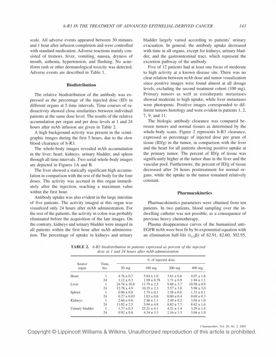
The relative biodistribution of the antibody was ex-pressed as the percentage of the injected dose (ID) indifferent organs at 5 time intervals. Time courses of ra-dioactivity showed close similarities between individualpatients at the same dose level. The results of the relativeaccumulation per organ and per dose levels at 1 and 24hours after mAb infusion are given in Table 2.
A high background activity was present in the scinti-graphic images during the first 5 hours, due to the slowblood clearance of h-R3.
The whole-body images revealed mAb accumulationin the liver, heart, kidneys, urinary bladder, and spleenthrough all time intervals. Two serial whole-body imagesare depicted in Figures 1A and B.
The liver showed a statically significant high accumu-lation in comparison with the rest of the body for the fourdoses. The activity was accrued in this organ immedi-ately after the injection, reaching a maximum valuewithin the first hour.
Antibody uptake was also evident in the large intestineof five patients. The activity imaged at this organ wasvisualized only 24 hours after mAb administration. Forthe rest of the patients, the activity in colon was probablyeliminated before the acquisition of the late images. Onthe contrary, kidneys and urinary bladder were imaged inall patients within the first hour after mAb administra-tion. The percentage of uptake in kidneys and urinary
bladder largely varied according to patients’ urinaryevacuation. In general, the antibody uptake decreasedwith time in all organs, except for kidneys, urinary blad-der, and the gastrointestinal tract, which represent theexcretion pathway of the antibody.
Five of 12 patients had at least one focus of moderateto high activity at a known disease site. There was noclear relation between mAb dose and tumor visualizationsince positive images were found almost at all dosagelevels, excluding the second treatment cohort (100 mg).Primary tumors as well as extrahepatic metastasesshowed moderate to high uptake, while liver metastaseswere photopenic. Positive images corresponded to dif-ferent tumors histology and were evident in patients 1, 2,7, 9, and 11.
The biologic antibody clearance was compared be-tween tumors and normal tissues as determined by thewhole-body scans. Figure 2 represents h-R3 clearance,expressed as percentage of injected dose per gram oftissue (ID/g) in the tumor, in comparison with the liverand the heart for all patients showing positive uptake atthe primary tumor. The percent of ID/g of tissue wassignificantly higher at the tumor than in the liver and thevascular pool. Furthermore, the percent of ID/g of tissuedecreased after 24 hours posttreatment for normal or-gans, while the uptake in the tumor remained relativelyconstant.
Pharmacokinetics
Pharmacokinetics parameters were obtained from tenpatients. In two patients, blood sampling over the in-dwelling catheter was not possible, as a consequence ofprevious heavy chemotherapy.
Plasma disappearance curves of the humanized anti-EGFR mAb were best fit by bi-exponential equation withan elimination half-life (t1/2�) of 62.91, 82.60, 302.95,
TABLE 2. h-R3 biodistribution in patients expressed as percent of the injecteddose at 1 and 24 hours after mAb administration
Sourceorgan
Time,hrs.
% of injected dose
50 mg 100 mg 200 mg 400 mg
Heart 1 4.76 ± 0.7 5.84 ± 1.0 3.61 ± 0.6 4.07 ± 1.624 1.12 ± 0.3 1.98 ± 0.78 1.71 ± 0.9 1.94 ± 1.1
Liver 1 24.76 ± 10.8 11.79 ± 2.5 9.88 ± 3.7 10.58 ± 0.924 12.78 ± 4.9 10.35 ± 2.3 5.57 ± 3.8 5.98 ± 3.0
Spleen 1 0.96 ± 0.8 1.74 ± 0.1 1.58 ± 0.6 1.33 ± 0.124 0.27 ± 0.03 1.03 ± 0.6 0.80 ± 0.4 0.69 ± 0.3
Kidneys 1 2.66 ± 0.6 2.46 ± 1.1 2.49 ± 0.2 3.04 ± 1.024 11.02 ± 2.5 5.94 ± 4.8 8.82 ± 7.7 8.62 ± 1.6
Urinary bladder 1 1.37 ± 0.3 25.21 ± 4.1 4.21 ± 1.4 3.29 ± 1.424 0.92 ± 0.8 4.34 ± 3.3 2.16 ± 1.3 3.04 ± 1.0
h-R3 IN THE TREATMENT OF ADVANCED EPITHELIAL-DERIVED CANCER 143
J Immunother, Vol. 26, No. 2, 2003

and 304.51 hours for the dose of 50, 100, 200, and 400mg, respectively. The AUC was 45/458, 145/931,573/612, and 635/275 ng/mL/min, and the maximal ac-tivity concentration (Cmax) was 27/790, 36/612, 52/713,
57/117 ng/mL for the dose of 50, 100, 200, and 400 mg,respectively. The apparent volume of the central com-partment (Vc) was 2321.96, 2823.67, 4279.71, and7173.99 mL, and the total elimination clearance (Cl) was1.08, 0.67, 0.34, and 0.76 (mL/hr/kg) for the dose of 50,100, 200, and 400 mg, respectively. No statistically sig-nificant differences in clearance for the four dose levelswere found (P � 0.095, Kruskal-Wallis Test).
Determination of Serum EGFR Level
EGFR level was quantified before inclusion in theclinical trial, using a human EGFR quantitative kit, tocorrelate pharmacokinetics with the soluble blocking an-tigen.
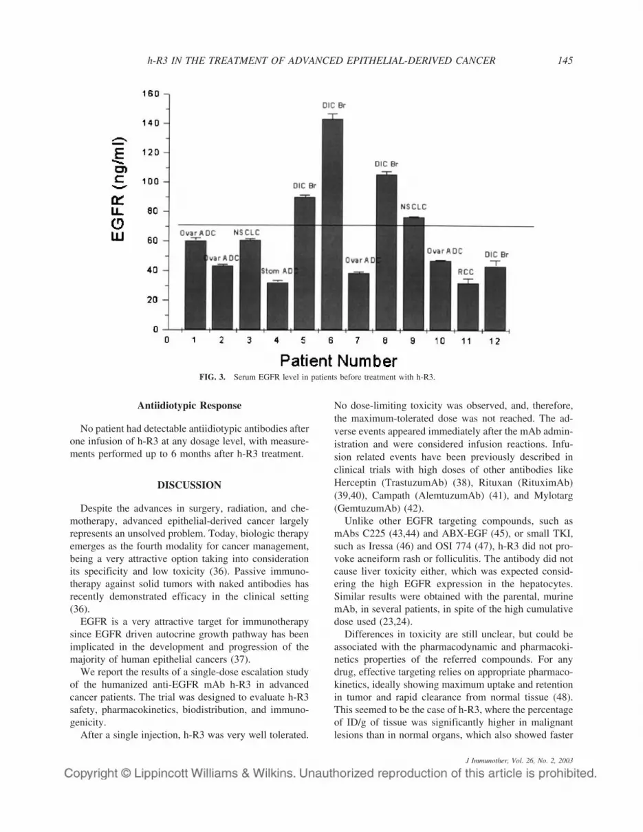
Since according to the kit information, the mean serumlevel of EGFR in the healthy population is 61 ng/mL(range 48–72 ng/mL), the cutoff value for EGFR posi-tivity was 72 ng/mL. Positive serum elevation of EGFRwas observed in 4 of 12 patients included in the study.Unexpectedly, the peak amounts of the soluble EGFRwere detected in three patients bearing ductal infiltratingcarcinomas of the breast (Fig. 3). No correlation wasfound between clearance and shed receptor.
FIG. 2. Antibody clearance in tumor, liver, and heart, as determinedby whole-body scans in a gamma camera expressed as Log % ID/g oftissue (tumor � 2 g, liver � 1,800 g, and heart � 330 g). Thepercentage of ID/g of tumor showed a greater variability than thepercent of ID/g of normal tissues due to the diversity of the neoplasmsshowing mAb uptake. � � Heart; � � Liver; � � Tumor.
FIG. 1. (A and B) Serial anterior and posterior whole-body images of patients 1 and 2. Images were acquired at 1, 3, 5, and 24 hours after 99Tch-R3 infusion and showed mAb accumulation in the liver, heart, spleen, kidneys, and urinary bladder. Patient 1 had one focus of high activity in theovarian primary tumor (left ovary) and multiple liver metastases that were photopenic. Patient 2 had one focus of high activity in the ovarian primarytumor (left ovary).
T. CROMBET ET AL.144
J Immunother, Vol. 26, No. 2, 2003
Antiidiotypic Response
No patient had detectable antiidiotypic antibodies afterone infusion of h-R3 at any dosage level, with measure-ments performed up to 6 months after h-R3 treatment.
DISCUSSION
Despite the advances in surgery, radiation, and che-motherapy, advanced epithelial-derived cancer largelyrepresents an unsolved problem. Today, biologic therapyemerges as the fourth modality for cancer management,being a very attractive option taking into considerationits specificity and low toxicity (36). Passive immuno-therapy against solid tumors with naked antibodies hasrecently demonstrated efficacy in the clinical setting(36).
EGFR is a very attractive target for immunotherapysince EGFR driven autocrine growth pathway has beenimplicated in the development and progression of themajority of human epithelial cancers (37).
We report the results of a single-dose escalation studyof the humanized anti-EGFR mAb h-R3 in advancedcancer patients. The trial was designed to evaluate h-R3safety, pharmacokinetics, biodistribution, and immuno-genicity.
After a single injection, h-R3 was very well tolerated.
No dose-limiting toxicity was observed, and, therefore,the maximum-tolerated dose was not reached. The ad-verse events appeared immediately after the mAb admin-istration and were considered infusion reactions. Infu-sion related events have been previously described inclinical trials with high doses of other antibodies likeHerceptin (TrastuzumAb) (38), Rituxan (RituximAb)(39,40), Campath (AlemtuzumAb) (41), and Mylotarg(GemtuzumAb) (42).
Unlike other EGFR targeting compounds, such asmAbs C225 (43,44) and ABX-EGF (45), or small TKI,such as Iressa (46) and OSI 774 (47), h-R3 did not pro-voke acneiform rash or folliculitis. The antibody did notcause liver toxicity either, which was expected consid-ering the high EGFR expression in the hepatocytes.Similar results were obtained with the parental, murinemAb, in several patients, in spite of the high cumulativedose used (23,24).
Differences in toxicity are still unclear, but could beassociated with the pharmacodynamic and pharmacoki-netics properties of the referred compounds. For anydrug, effective targeting relies on appropriate pharmaco-kinetics, ideally showing maximum uptake and retentionin tumor and rapid clearance from normal tissue (48).This seemed to be the case of h-R3, where the percentageof ID/g of tissue was significantly higher in malignantlesions than in normal organs, which also showed faster
FIG. 3. Serum EGFR level in patients before treatment with h-R3.
h-R3 IN THE TREATMENT OF ADVANCED EPITHELIAL-DERIVED CANCER 145
J Immunother, Vol. 26, No. 2, 2003
clearance according to the serial whole-body images.Similar data for other anti-EGFR drugs are not availablein the literature, and, in our view, such comparison iswarranted. We cannot preclude that the different safetyprofile could also be explained at cellular or molecularlevel.
Biodistribution showed the main binding sites of theh-R3, the blood activity profile, and the mAb eliminationroutes. h-R3 accumulation was seen in liver, heart, kid-neys, urinary bladder, spleen, and large intestine. Thebiodistribution profile agreed with previous reports ofour group for the murine antibody (49). Similarities inbiodistribution confirmed that after the humanizationprocess, h-R3 maintained the original in vivo recognitionpattern of normal organs and tissues. The high liver up-take was related with the high EGFR expression in he-patocytes, while the accumulation of the humanizedmAb in heart and spleen could be considered as theactivity of the vascular compartment. Immune complexformation between the antibody and shed EGFR couldalso explain the specific accumulation in the spleen.Blood pool activity appeared considerably reduced in thelate images. The activity found in kidneys, urinary blad-der, and colon illustrated the main excretion routes of theantibody and its metabolites.
Despite the fact that planar or single photon emissioncomputed tomography images were not acquired, posi-tive tumor uptake was observed in five patients (six le-sions). The protocol used during the scintigraphic acqui-sitions was appropriated for biodistribution assessments,but not for lesion imaging. Therefore, the defined matrixformat and the scan times provided scintigraphic studieswith low statistical counts. As a consequence, low con-trast images with minimal usefulness for diagnosis pur-poses were collected. In addition, since 99Tc has a shorthalf-life (t1/2 6.02 hours), the latest images were acquired24 hours after mAb administration, before optimaltumor/nontumor ratios were achieved, taking into con-sideration that some tumors have a slow antibody uptakeindex. Positive tumor visualization was achieved at threeof the dosage levels (50, 200, and 400 mg); while at thesecond lowest dose, tumors were not identified. Malig-nant lesions in this particular treatment cohort corre-sponded to two breast carcinomas and a stomach adeno-carcinoma, and, therefore, tumor imaging could havebeen interfered by positive uptake in the vicinity by theliver and the blood pool. Other factors like EGFR ex-pression at the primary tumor as well as the tumor vas-cularity may also influence mAb uptake.
The use of ior egf/r3, the original murine mAb, forradioimmunodetection had already proved to be success-ful, with 84% of sensitivity for in vivo detection of ep-
ithelial tumors from all localizations (25). A diagnosticclinical trial to evaluate the sensitivity, specificity as wellas predictive values of h-R3 is ongoing.
99mTechnetium was not appropriate for the pharmaco-kinetics study, because its physical half-life did notmatch with the antibody clearance. Consequently, anELISA was developed to quantify h-R3 in serum. How-ever, pharmacokinetics should be considered as prelimi-nary in view of the small number of patients per group.The parameters determined using bicompartmentalanalysis suggested a nonlinear behavior of h-R3. TheAUC and elimination half-lives increased linearly withdose, while increasing concentrations of the antibodylead to a decrease in plasma clearance between the doses50 and 200 mg. The peak serum concentrations of h-R3after infusion were also dose-dependent and its elimina-tion half-life (62.91 to 304.51 hours) was significantlyhigher than that obtained with the murine mAb (27.3 to65.89 hours). Similar elimination half-lives had been re-ported for other chimeric or humanized anti-EGFR anti-bodies (50,51).
Clearance in cancer patients could be modified by sev-eral factors including soluble antigen in circulation. Forinstance, phase I studies revealed that an increased clear-ance of Herceptin, was associated with high levels ofshed antigen. In our small sample, EGFR shedding didnot correlate with h-R3 clearance.
Increased serum ECD levels have been reported inpatients with cancers known to overexpress Her 1 or Her2 (52–54). In our set, soluble EGFR was very high inthree patients bearing ductal infiltrating breast carcino-mas, while according to the literature, EGFR is onlyoverexpressed in 25–30% of breast carcinomas using im-munohistochemistry or Fluorescence in Situ Hybridiza-tion (FISH) (55,56). Previously, no significant associa-tion had been found between the serum EGFR level andTNM stage, tumor burden or the differentiation degree.Moreover, in some studies, no differences in this onco-protein serum levels, had been observed in patients withimmunohistochemically overexpressing and nonoverex-pressing tumors (52–54). The origin and biologic role ofsoluble EGFR remains to be determined.
The lack of antiidiotypic response emphasize the rela-tively nonimmunogenicity of the humanized mAb, al-though it is necessary to confirm this finding in a largerseries of patients treated with repeated doses of h-R3.
In conclusion, h-R3 is a new, promising anti-EGFRantibody. Major advantages of the antibody rely on itsreduced toxicity and immunogenicity and on its pro-longed half-life. It can be used as a single agent or incombination with radiation or chemotherapy. As radio orchemotherapy may increase EGFR-mediated signal
T. CROMBET ET AL.146
J Immunother, Vol. 26, No. 2, 2003
transduction or expression by tumor cells, the combina-tion seems to be the ideal scenario for the antibody.Completion of the phase II trial using repeated mAbdoses in conjunction with radiotherapy is underway todefine the response rate in advanced head and neck can-cer patients. Further work is in progress to optimize theeffects of h-R3 in other tumor localizations.
REFERENCES
1. Brabender J, Danenberg KD, Metzger R, et al. Epidermal growthfactor receptor and HER2-neu mRNA expression in non-small celllung cancer is correlated with survival. Clin Cancer Res. 2001;7:1850–1855.
2. Nicholson RI, Gee JM, Harper ME. EGFR and cancer prognosis.Eur J Cancer. 2001;37(Suppl 4):9–15.
3. Libermann TA, Razon N, Bartal AD, et al. Expression of epider-mal growth factor receptors in human brain tumors. Cancer Res.1984;4:753–760.
4. FeldKamp MM, Lau N, Guha A. Signal transduction pathways andtheir relevance in human astrocytomas. Cancer Immunol Immuno-ther. 1997;44:157–164.
5. Kearsley JH, Leonard JH, Walsh MD, et al. A comparison ofepidermal growth factor receptor (EGFR) and c-erbB-2 oncogeneexpression in head and neck squamous cell carcinomas. Pathology.1991;23:189–194.
6. Rikimaru K, Tadokoro K, Yamamoto T, et al. Gene amplificationand overexpression of epidermal growth factor receptor in squa-mous cell carcinoma of the head and neck. Head Neck. 1992;14:8–13.
7. Alper O, Bergmann-Leitner ES, Bennett TA, et al. Epidermalgrowth factor receptor signaling and the invasive phenotype ofovarian carcinoma cells. J Natl Cancer Ins. 2001;93:1375–1384.
8. Bianco C, Bianco R, Tortora G, et al. Antitumor activity of com-bined treatment of human cancer cells with ionizing radiation andanti-epidermal growth factor receptor monoclonal antibody C225plus type I protein kinase A antisense oligonucleotide. Clin CancerRes. 2000;6:4343–4350.
9. Ngan HY, Cheung AN, Liu SS, et al. Abnormal expression ofepidermal growth factor receptor and c-erbB2 in squamous cellcarcinoma of the cervix: correlation with human papillomavirusand prognosis. Tumor Biol. 2001;22:176–183.
10. Chow NH, Chan SH, Tzai TS, et al. Expression profiles of ErbBfamily receptors and prognosis in primary transitional cell carci-noma of the urinary bladder. Clin Cancer Res. 2001;7:1957–1962.
11. Inada S, Koto T, Futami K, et al. Evaluation of malignancy and theprognosis of esophageal cancer based on an immunohistochemicalstudy (p53, E-cadherin, epidermal growth factor receptor). SurgToday. 1999;29:493–503.
12. Wang LS, Chow KC, Chi KH. Prognosis of esophageal squamouscell carcinoma: analysis of clinicopathologic and biologic factors.Am J Gastroenterol. 1999;94:1933–1940.
13. Giordano A, Rustum YM, Wenner CE. Cell cycle: molecular tar-gets for diagnosis and therapy: tumor suppressor genes and cellcycle progression in cancer. J Cell Biochem. 1998;70:1–7.
14. Damstrup L, Rude Voldborg B, Spang-Thomsen M, et al. In vitroinvasion of small-cell lung cancer cell lines correlates with expres-sion of epidermal growth factor receptor. Br J Cancer. 1998;78:631–640.
15. Petit AM, Rak J, Hung MC, et al. Neutralizing antibodies againstepidermal growth factor and ErbB-2/neu receptor tyrosine kinasesdownregulate vascular endothelial growth factor production by tu-mor cells in vitro and in vivo: angiogenic implications for signaltransduction therapy of solid tumors. Am J Pathol. 1997;151:1523–1530.
16. Kerbel RS, Viloria-Petit A, Okada F, et al. Establishing a link
between oncogenes and tumor angiogenesis. Mol Med. 1998;4:286–295.
17. Gibson S, Tu S, Oyer R, et al. Epidermal growth factor protectsepithelial cells against Fas-induced apoptosis. Requirement for Aktactivation. J Biol Chem. 1999;274:17612–17618.
18. Mendelsohn J, Baselga J. The EGF receptor family as targets forcancer therapy. Oncogene. 2000;19:6550–6565.
19. Kim ES, Khuri FR, Herbst RS. Epidermal growth factor receptorbiology (IMC-C225). Curr Opin Oncol. 2001;13:506–513.
20. Ciardiello F, Tortora G. A novel approach in the treatment ofcancer: targeting the epidermal growth factor receptor. Clin Can-cer Res. 2001;7:2958–70.
21. Fernandez A, Perez R, Macıas A, et al. Generation and character-ization of anti-EGFR antibodies. Interferon y Biotecnologıa. 1989;6:289–298.
22. Fernandez A, Spitzer E, Perez R, et al. A new monoclonal antibodyfor detection of the EGF-R in Western Blot and paraffin embeddedtissue sections. J Cell Biochem. 1992;49:157–165.
23. Crombet T, Torres O, Neninger E, et al. Phase I clinical evaluationof a neutralizing monoclonal antibody against epidermal growthfactor receptor. Cancer Biother Radiopharm. 2001;16:93–102.
24. Crombet T, Torres O, Rodriguez V, et al. Phase I clinical evalu-ation of a neutralizing monoclonal antibody against epidermalgrowth factor receptor in advanced brain tumor patients: prelimi-nary study. Hybridoma. 2001;20:131–136.
25. Ramos-Suzarte M, Rodriguez N, Oliva JP, et al. 99mTc-labeledantihuman epidermal growth factor receptor antibody in patientswith tumors of epithelial origin: Part III. Clinical trials safety anddiagnostic efficacy. J Nucl Med. 1999;40:768–775.
26. Mateo C, Moreno E, Amour K, et al. Humanization of a mousemonoclonal antibody that blocks the epidermal growth factor re-ceptor: recovery of antagonistic activity. Immunotechnology. 1997;3:71–81.
27. Viloria-Petit A, Crombet T, Jothy S, et al. Acquired resistance tothe antitumor effect of epidermal growth factor receptor-blockingantibodies in vivo: a role for altered tumor angiogenesis. CancerRes. 2001;61:5090–5101.
28. Crombet-Ramos T, Rak J, Perez R, et al. Antiproliferative, anti-angiogenic and proapoptotic activity of h-R3: A humanized anti-EGFR antibody. Int J Cancer. 2002;101:567–575.
29. Stragliotto G, Vega F, Stasiecki P et al. Multiple infusions ofanti-epidermal growth factor receptor (EGFR) monoclonal anti-body (EMD 55,900) in patients with recurrent malignant gliomas.Eur J Cancer. 1996;32A:636–640.
30. Edwards IR. Harmonisation in pharmacovigilance. Drug Saf.1994;10:93.
31. Schwarz A, Steinstraber A. A novel approach to Tc-99m-labeledmonoclonal antibodies. J Nucl Med. 1987;28:721–727.
32. Iznaga Escobar N, Morales AM, Duconge J, et al. Pharmacokinet-ics, biodistribution and dosimetry of 99mTc-labeled anti-humanepidermal growth factor receptor humanized monoclonal antibodyR3 in rats. Nucl Med Biol. 1998;25:17–23.
33. Morales AA, Duconge J, Alvarez-Ruiz D, et al. Humanized versusmurine anti-human epidermal growth factor receptor monoclonalantibodies for immunoscintigraphic studies. Nucl Med Biol. 2000;27:199–206.
34. Macias A, Perez R, Lage A. Estudios sobre el factor de crecimientoepidermico (EGF) II: Desarrollo de un radioreceptor analisis parala determinación de cantidades picomolares. Interferon y Biotec-nologıa. 1985;2:115–127.
35. Akaike A. Posterior probabilities for choosing regression models.Ann Inst Math Stat. 1978;30:A9.
36. Rosenberg S. Principles of Cancer Management: BiologicTherapy. In: De Vita V, Hellman S, Rosemberg S, eds. Cancer:Principles and Practice of Oncology 2001. 6th ed. Philadelphia,PA: Lippincott Williams & Wilkins; 2001:307–334.
37. Ciardiello F. Epidermal growth factor receptor tyrosine kinase in-hibitors as anticancer agents. Drugs. 2000;60(suppl 1):25:41–42.
38. Slamon D, Leyland B, Shak S, et al. Use of chemotherapy plus a
h-R3 IN THE TREATMENT OF ADVANCED EPITHELIAL-DERIVED CANCER 147
J Immunother, Vol. 26, No. 2, 2003

monoclonal antibody against Her 2 for metastatic breast cancer thatoverexpresses Her 2. N Engl J Med. 2000;344:783–792.
39. Davis TA, Grillo-López AJ, White CA, et al. RituximAb anti-CD20 monoclonal antibody therapy in non-Hodgkin’s lymphoma:safety and efficacy of re-treatment. J Clin Oncol. 2000;18:3135–3143.
40. Piro LD, White CA, Grillo-Lopez AJ, et al. Extended RituximAb(anti-CD20 monoclonal antibody) therapy for relapsed or refrac-tory low-grade or follicular non-Hodgkin’s lymphoma. Ann Oncol.2000;10:655–661.
41. Dearden CE, Matutes E, Cazin B, et al. High remission rate inT-cell prolymphocytic leukemia with CAMPATH-1H. Blood.2001;98:1721–1726.
42. Bross PF, Beitz J, Chen G, et al. Approval summary: gemtuzumAbozogamicin in relapsed acute myeloid leukemia. Clin Cancer Res.2001;7:1490–1496.
43. Busam KJ, Capodieci P, Motzer R, et al. Cutaneous side-effects incancer patients treated with the antiepidermal growth factor recep-tor antibody C225. Br J Dermatol. 2001;144:1169–1176.
44. Shin DM, Donato NJ, Perez-Soler R, et al. Epidermal growthfactor receptor-targeted therapy with C225 and cisplatin in patientswith head and neck cancer. Clin Cancer Res. 2001;7:1204–1212.
45. Figlin R, Belldegrun A, Lohner M, et al. ABX-EGF: a fully humananti-EGF receptor antibody in patients with advanced cancer [ab-stract]. Proc Am Soc Clin Oncol. 2001;20:276a.
46. Meric JB, Faivre S, Monnerat C, et al. Zd 1839 “Iressa” BullCancer. 2000;87:873–876.
47. Hidalgo M, Siu LL, Nemunaitis J, et al. Phase I and pharmacologicstudy of OSI-774, an epidermal growth factor receptor tyrosinekinase inhibitor, in patients with advanced solid malignancies.J Clin Oncol. 2001;19:3267–3279.
48. Stroomer JW, Roos JC, Sproll M, et al. Safety and biodistributionof 99mTechnetium-labeled anti-CD44v6 monoclonal antibody
BIWA 1 in head and neck cancer patients. Clin Cancer Res. 2000;6:3046–3055.
49. Iznaga-Escobar N, Torres LA, Morales A, et al. Tm-99m-labeledanti-EGF-receptor antibody in patients with tumor of epithelialorigin: I. Biodistribution and dosimetry for radioimmunotherapy.J Nucl Med. 1998;15:18–27.
50. Baselga J, Pfister D, Cooper MR, et al. Phase I studies of anti-epidermal growth factor receptor chimeric antibody C225 aloneand in combination with cisplatin. J Clin Oncol. 2000;18:904–914.
51. Bier H, Hoffmann T, Hauser U, et al. Clinical trial with escalatingdoses of the antiepidermal growth factor receptor humanizedmonoclonal antibody EMD 72 000 in patients with advanced squa-mous cell carcinoma of the larynx and hypopharynx. Cancer Che-mother Pharmac. 2001;47:519–524.
52. Partanen R, Hemminki K, Koskinen H, et al. The detection ofincreased amounts of the extracellular domain of the epidermalgrowth factor receptor in serum during carcinogenesis in asbestosispatients. J Occup Med. 1994;36:1324–1328.
53. Choi JH, Oh JY, Ryu SK, et al. Detection of epidermal growthfactor receptor in the serum of gastric carcinoma patients. Cancer.1997;79:1879–1883.
54. Oh MJ, Choi JH, Kim IH, et al. Detection of epidermal growthfactor receptor in the serum of patients with cervical carcinoma.Clin Cancer Res. 2000;6:4760–4763.
55. Sharma BK, Ray A, Kaur S, et al. Immunohistochemical co-expression of c-erbb-2/Neu oncoprotein, altered tumor suppressor(p53) protein, EGF-R and EMA in histologic subtypes of infil-trating duct carcinoma of the breast. Indian J Exp Biol. 1999;37:223–227.
56. Umekita Y, Ohi Y, Sagara Y, et al. Co-expression of epidermalgrowth factor receptor and transforming growth factor-alpha pre-dicts worse prognosis in breast-cancer patients. Int J Cancer. 2000;89:484–487.
T. CROMBET ET AL.148
J Immunother, Vol. 26, No. 2, 2003