persistent muscle fiber regeneration in long term denervation. past, present, future
TRANSCRIPT
Muscle fiber regeneration in long term denervation
Eur J Transl Myol - Basic Appl Myol 2015; 25 (2): 77-92
- 77 -
Persistent muscle fiber regeneration in long term denervation. Past, present, future
Ugo Carraro (1), Simona Boncompagni (2), Valerio Gobbo (3), Katia Rossini (4,5), Sandra
Zampieri (4,5), Simone Mosole (4,5), Barbara Ravara (4,5), Alessandra Nori (4), Roberto
Stramare (6), Francesco Ambrosio (7), Francesco Piccione (1), Stefano Masiero (8), Vincenzo
Vindigni (9), Paolo Gargiulo (10), Feliciano Protasi (2), Helmut Kern (5,11), Amber Pond
(12), Andrea Marcante (1)
(1) Department of Neurorehabilitation, Foundation San Camillo Hospital, I.R.C.C.S., Venice,
Italy; (2) CeSI, Center for Research on Aging, Department of Neuroscience, Imaging and
Clinical Sciences, University G. d’Annunzio of Chieti, Italy; (3) C.N.R. Institute of
Neuroscience, Department of Biomedical Science, University of Padova, Italy; (4)
Translational Myology, Interdepartmental Research Center of Myology of the University of
Padova (CIR-Myo), Department of Biomedical Science, Padova, Italy; (5) Ludwig Boltzmann
Institute of Electrical Stimulation and Physical Rehabilitation, Vienna, Austria; (6) CIR-Myo,
Department of Medicine, Radiology Unit, University of Padova, Italy; (7) Antalgic
Laboratory, Department of Medicine, University of Padova, Italy; (8) CIR-Myo, Department
of Neuroscience, Rehabilitation Unit, University of Padova, Italy; (9) CIR-Myo, Department of
Neuroscience, Plastic Surgery Unit, University of Padova, Italy; (10) Department of Science,
Education, Innovation, Landspitali University Hospital, Reykjavik, Iceland; (11) Department
of Physical Medicine, Wilhelminenspital, Vienna, Austria; (12) Anatomy Department,
Southern Illinois University, School of Medicine, Carbondale, Illinois, USA
Abstract
Despite the ravages of long term denervation there is structural and ultrastructural evidence for
survival of muscle fibers in mammals, with some fibers surviving at least ten months in
rodents and 3-6 years in humans. Further, in rodents there is evidence that muscle fibers may
regenerate even after repeated damage in the absence of the nerve, and that this potential is
maintained for several months after denervation. While in animal models permanently
denervated muscle sooner or later loses the ability to contract, the muscles may maintain their
size and ability to function if electrically stimulated soon after denervation. Whether in
mammals, humans included, this is a result of persistent de novo formation of muscle fibers is
an open issue we would like to explore in this review. During the past decade, we have studied
muscle biopsies from the quadriceps muscle of Spinal Cord Injury (SCI) patients suffering
with Conus and Cauda Equina syndrome, a condition that fully and irreversibly disconnects
skeletal muscle fibers from their damaged innervating motor neurons. We have demonstrated
that human denervated muscle fibers survive years of denervation and can be rescued from
severe atrophy by home-based Functional Electrical Stimulation (h-bFES). Using
immunohistochemistry with both non-stimulated and the h-bFES stimulated human muscle
biopsies, we have observed the persistent presence of muscle fibers which are positive to
labeling by an antibody which specifically recognizes the embryonic myosin heavy chain
(MHCemb). Relative to the total number of fibers present, only a small percentage of these
MHCemb positive fibers are detected, suggesting that they are regenerating muscle fibers and
not pre-existing myofibers re-expressing embryonic isoforms. Although embryonic isoforms
of acetylcholine receptors are known to be re-expressed and to spread from the end-plate to the
sarcolemma of muscle fibers in early phases of muscle denervation, we suggest that the
MHCemb positive muscle fibers we observe result from the activation, proliferation and fusion
of satellite cells, the myogenic precursors present under the basal lamina of the muscle fibers.
Using morphological features and molecular biomarkers, we show that severely atrophic
muscle fibers, with a peculiar cluster reorganization of myonuclei, are present in rodent muscle
Muscle fiber regeneration in long term denervation
Eur J Transl Myol - Basic Appl Myol 2015; 25 (2): 77-92
- 78 -
seven-months after neurectomy and in human muscles 30-months after complete Conus-Cauda
Equina Syndrome and that these are structurally distinct from early myotubes. Beyond
reviewing evidence from rodent and human studies, we add some ultrastructural evidence of
muscle fiber regeneration in long-term denervated human muscles and discuss the options to
substantially increase the regenerative potential of severely denervated human muscles not
having been treated with h-bFES. Some of the mandatory procedures, are ready to be
translated from animal experiments to clinical studies to meet the needs of persons with long-
term irreversible muscle denervation. An European Project, the trial Rise4EU (Rise for You, a
personalized treatment for recovery of function of denervated muscle in long-term stable SCI)
will hopefully follow
Key Words: Human muscle, Conus-Cauda Equina syndrome, Spinal cord injury, Permanent
muscle denervation, Severe atrophy and nuclear clumpings, Muscle fiber regeneration, Home-
based functional electrical stimulation (h-b FES), Recovery of tetanic contractility, Myogenic
stem cells Eur J Transl Myol - Basic Appl Myol 2015; 25 (2): 77-92
Skeletal muscle undergoes a rapid loss of both mass
and contractile force in response to loss of neural input
such as occurs in cases of sciatectomy in rats and with
spinal cord injury (SCI) in humans. The atrophy
subsequent to SCI is especially severe when the lesion
involves lower motor neurons (LMN) because, if
denervation is irreverisble, the muscle tissue ultimately
undergoes both fibrosis and fat substitution, thus
producing denervated degenerated muscle (DDM).
Unfortunately, long-term permanent denervation of
muscle tissue is an under-studied pathologic condition.
This situation may be attributable to the general belief
that muscle fibers will eventually disappear after weeks
or months of disconnection from the nervous system
and its provision of trophic factors (e.g., those related
to acetylcholine, agrin, BDNF and other as yet
unknown chemical factors) released from axonal
endings.1-3
Because both the response of rat muscle to
permanent denervation (Figure 1) and the response of
human muscle to SCI is an extreme loss of muscle
mass, there has been a good deal of skepticism aimed
at the efficacy of our studies of home-based Functional
Electrical Stimulation (h-bFES) as a potential therapy
to improve structure, appearance and tetanic
contractility of permanently denervated human
muscles.4 Based upon the fact that at late stages of
denervation severely atrophic skeletal muscle does not
respond to electrical stimulation, many neurologists
believe that muscle degeneration is irreversible and
thus therapy is not merited. Here, we respond to such
skepticism by discussing evidence to support the value
of our technique, namely the facts that: 1) myofibers
are indeed present in rat muscle one year after
denervation;5 2) atrophied denervated human muscle
maintains surviving and regenerating myofibers over
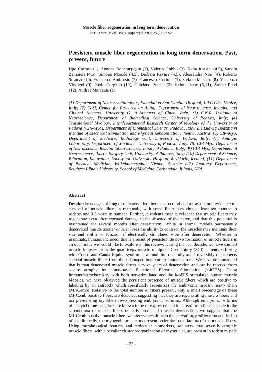
Fig 1. Macroscopic aspects of rat posterior legs. In comparison to adult normal rat (A), the 9-month denervated
legs (B) appear to have completely lost their muscle mass.
Muscle fiber regeneration in long term denervation
Eur J Transl Myol - Basic Appl Myol 2015; 25 (2): 77-92
- 79 -
time;4 3) immunochemical evidence of embryonic
myosin and evidence of biomarkers of myogenic
processes in rodents suggest that myogenesis may
occur in denervated muscles; and 4) h-bFES improves
ultrastructure, macro-structure, mass and contractility
of permanently denervated human muscle. Indeed,
denervated human muscle tissue responds to h-bFES
treatment by recovering surviving myofibers and,
possibly, also those recently regenerated muscle fibers
which are more responsive to electrical stimulation.3,4
Long term survival of permanently denervated
muscle fibers in mammals
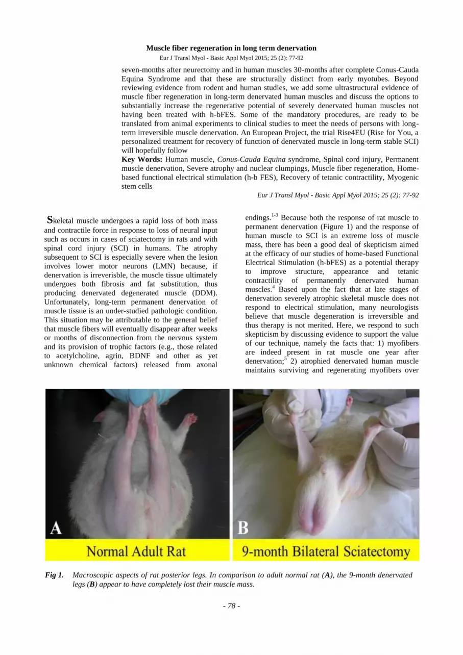
In support of the possibility of muscle recovery, first,
we argue that muscle fibers are recognizable, even at
the light microscope level, in the Tibialis Anterior
(TA) muscle of sciatectomized rats for up to 1-year
post denervation (Fig. 2).5
Addtionally, we point out
that electron microscopic analyses of the tissue
remaining in long term denervated rat leg muscles6,7
show that the deterioration which occurs in response to
denervation is ameliorated by electrical stimulation,
Fig 2. Long term denervation atrophy of the TA muscle in bilaterally sciatectomized rats. H-E stain. A, Normal
muscle; B, 1-month, C, 2-month, D, 4-month; E, 8-month; F, 12-month post denervation. Innervated TA
muscles are characterized by large, well packed myofiber profiles and minimal intermyofiber spaces. The
first stage of TA denervation is characterized by progressive reduction in muscle fiber diameter. In the mid-
denervation stage there is a further reduction up to 4-months denervation, in which muscle fiber size is about
10% of normal. Then, the fiber size does not change significantly at least up to 1-year after denervation by
bilateral sciatectomy.
Muscle fiber regeneration in long term denervation
Eur J Transl Myol - Basic Appl Myol 2015; 25 (2): 77-92
- 80 -
supporting our belief that long-term permanently
denervated muscle may be restored structurally (and
eventually to functional behavior) in particular if this
anti-atrophy/recovery process (i.e., h-bFES) is started
before muscular atrophy becomes severe.3,4
Rodent
models of muscle denervation, in particular after free
muscle transplantation,2 have demonstrated that lasting
periods (1 to 6 months post denervation) of myofiber
“atrophy” elapse before the detection of muscle
degeneration and also report the formation of very
small myofibers that may constitute up to 25% of the
total muscle fibers in denervated leg muscles of rats.6,7
Further, Borisov et al.,6 thoroughly characterized
severely atrophic rat muscle fibers at the ultrastructural
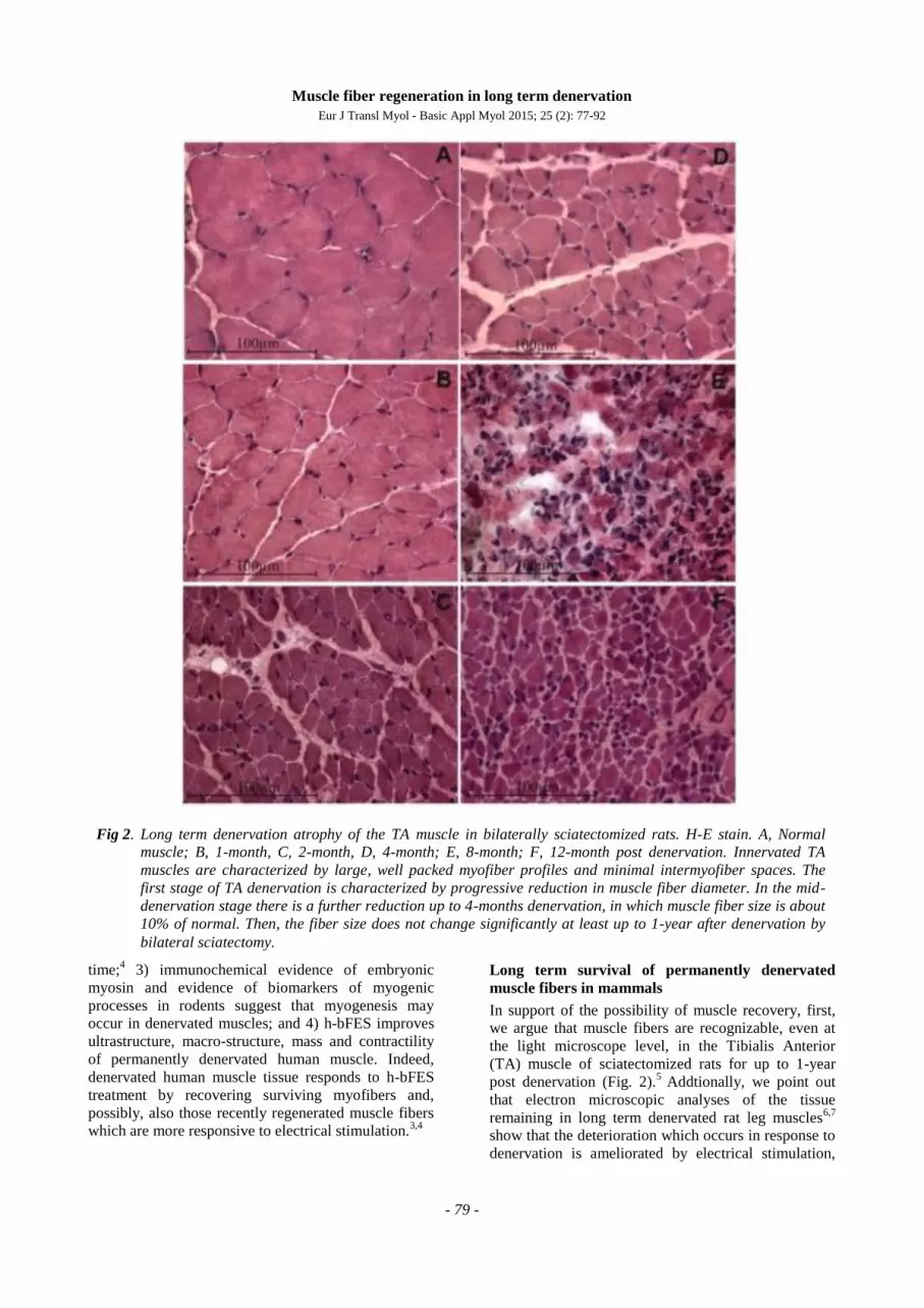
Fig 3. Semithin sections, toluidine blue stain. Note
in A the gradient of size of muscle fibers and
the presence of central nuclei in myofibers of
denervated and regenerated rat soleus
muscle 30 days after bilateral sciatectomy
and unilateral marcaine-induced muscle
damage. Spontaneous regeneration is also
present in 30 days denervated contralateral
muscle as suggested by the central nucleus in
one myofiber (arrow in B). 40x.
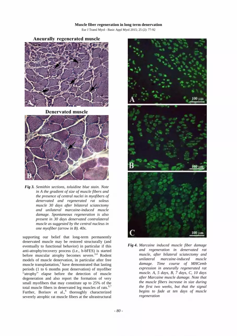
Fig 4. Marcaine induced muscle fiber damage
and regeneration in denervated rat
muscle, after bilateral sciatectomy and
unilateral marcaine-induced muscle
damage. Time course of MHCemb
expression in aneurally regenerated rat
muscle. A, 5 days, B, 7 days, C, 10 days
after Marcaine muscle damage. Note that
the muscle fibers increase in size during
the first two weeks, but that the signal
begins to fade at ten days of muscle
regeneration
Muscle fiber regeneration in long term denervation
Eur J Transl Myol - Basic Appl Myol 2015; 25 (2): 77-92
- 81 -
level and revealed that they contain a contractile
system, although poorly developed and with many
degenerated elements. The authors suggested that these
“incompletely differentiated” muscle fibers arise from
satellite cells that have detached from their parent
myofibers and may represent an “abortive” attempt at
myogenesis.6 Schmalbruch and colleagues have
described similar myofibers in rat muscle denervated
for 6–10 months.7 The majority of these muscle cells
were so small, down to 1 μm in diameter, that they
could not be identified by routine light microscopy.
Schmalbruch and colleagues further noted that some of
the smaller fibers were completely devoid of
myofilaments. Accordingly, they concluded that these
fibers are the result of repeated cycles of necrosis and
regeneration occurring in the chronically denervated
skeletal muscle, based on their reporting of
unequivocally necrotic muscle fibers.7 Carraro and
colleagues found similar featurers in rat hemi-
diaphragm denervated for 12–16 months.8
Taken together, the above examples provide
morphological evidence that very small myofibers may
not only represent extreme examples of severely
atrophied muscle fibers surviving long-term
denervation (see below), but may also occur as the
result of myofiber regeneration followed by atrophy in
chronic denervation.6 Indeed, Mussini et al.
9 have
shown that marcaine damage of permanently
denervated rat muscles produces unequivocal
morphological features of muscle damage and
regeneration and that repeated marcaine infusion of the
aneural regenerated rat muscle was followed by a
second round of muscle damage and regeneration,
demonstrating that the regenerative process is
accompained by survival and/or proliferation of
myogenic cells with structural and functional
characteristics of satellite cells (or, in other words,
“stem cells”).9 Conclusive evidence of aneural satellite
cell activation, proliferation, fusion and progression to
small muscle fibers was obtained by gel
electrophoresis which revealed the transient presence
of the embryonic isoforms of the myosin light and
heavy chains (MLCemb, MHCemb, respectively) in
regenerating rat muscles,10
and by immunolabeling
aneurally regenerating muscle cryosections (Fig. 3)
with an antibody that recognizes epitopes of the
MHCemb (Figs. 4 and 5).11,12
Note that in Fig. 4 the
size of the regenerated muscle fibers increases up to 10
days (Fig. 4, A-C), at least, but that the immunostain
starts to fade in several muscle fibers at this time of
regeneration (Fig. 4, C). At 30 days of regeneration in
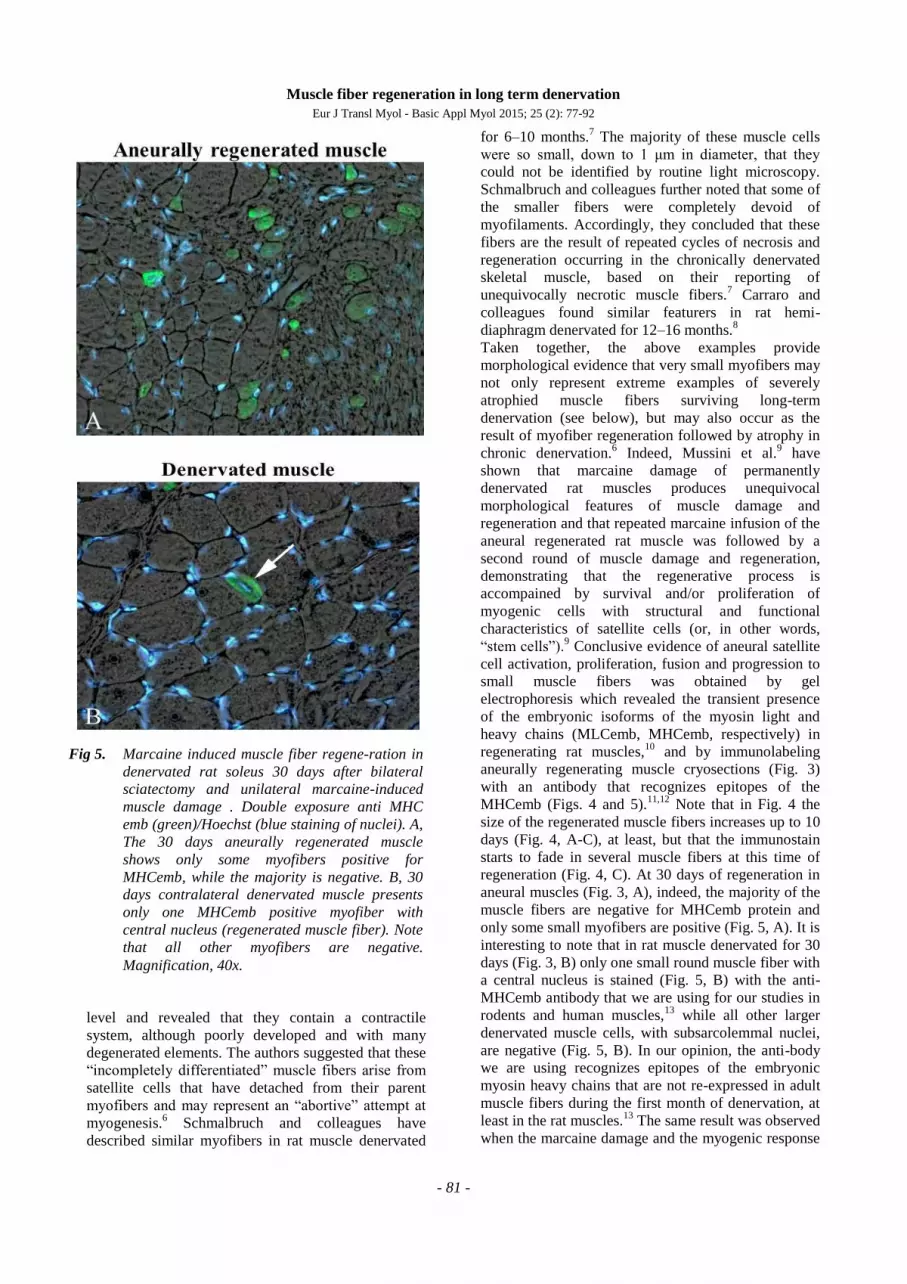
aneural muscles (Fig. 3, A), indeed, the majority of the
muscle fibers are negative for MHCemb protein and
only some small myofibers are positive (Fig. 5, A). It is
interesting to note that in rat muscle denervated for 30
days (Fig. 3, B) only one small round muscle fiber with
a central nucleus is stained (Fig. 5, B) with the anti-
MHCemb antibody that we are using for our studies in
rodents and human muscles,13
while all other larger
denervated muscle cells, with subsarcolemmal nuclei,
are negative (Fig. 5, B). In our opinion, the anti-body
we are using recognizes epitopes of the embryonic
myosin heavy chains that are not re-expressed in adult
muscle fibers during the first month of denervation, at
least in the rat muscles.13
The same result was observed
when the marcaine damage and the myogenic response
Fig 5. Marcaine induced muscle fiber regene-ration in
denervated rat soleus 30 days after bilateral
sciatectomy and unilateral marcaine-induced
muscle damage . Double exposure anti MHC
emb (green)/Hoechst (blue staining of nuclei). A,
The 30 days aneurally regenerated muscle
shows only some myofibers positive for
MHCemb, while the majority is negative. B, 30
days contralateral denervated muscle presents
only one MHCemb positive myofiber with
central nucleus (regenerated muscle fiber). Note
that all other myofibers are negative.
Magnification, 40x.
Muscle fiber regeneration in long term denervation
Eur J Transl Myol - Basic Appl Myol 2015; 25 (2): 77-92
- 82 -
occurred in rat muscles permanently denervated for 4
to 6 months.13
Further evidence of muscle fiber
regeneration is the detection of myogenin and MRF4
gene expression in response to long-term muscle
denervation,14,15
in spite of the down-regulation of
activity-dependent genes in long-term denervated rat
muscle.15
In conclusion, we are confident that
expression of the MHCemb is the result of a cellular
process of muscle regeneration.
It is also worth noting that the long term denervated
muscle fibers of rats are able to retain a functional
excitation-contraction coupling (ECC) apparatus that
responds in vitro to electrical stimulation for several
months, despite lost contractility.16
This provides a
potential mechanism for the muscle recovery that
occurs in response to rehabilitation strategies
developed on the basis of empirical clinical
observations.16
To define the time course and potential
effects of electrical stimulation on permanently
denervated muscle, we evaluated excitation-contraction
coupling of rat leg muscles during their progressive
response to long-term denervation16
by determining:
1) ultrastructural analysis; 2) specific binding to
dihydropyridine receptors, ryanodine receptors, Ca++
channels and extrusion Ca++
pumps; 3) gene
transcription and translation of Ca++
-handling proteins;
and 4) in vitro mechanical properties and
electrophysiological analyses of sarcolemmal passive
properties and L-type Ca++
current parameters. We
found that in response to long-term denervation: 1)
isolated muscle that is unable to twitch in vitro by
electrical stimulation has very small myofibers but may
show a slow caffeine contracture; 2) only roughly half
of the muscle fibers with "voltage-dependent Ca++
channel activity" are able to contract; 3) the ECC
mechanisms are still present and, in part, functional; 4)
ECC-related gene expression is upregulated; and 5) at
any time point, there are muscle fibers that are more
resistant than others to denervation atrophy and
disorganization of the ECC apparatus.16
These results
support the hypothesis that prolonged "resting" [Ca++
]i
may drive progression of muscle atrophy to
degeneration and that electrical stimulation-induced
[Ca++
]i modulation may mimic the lost nerve influence,
playing a key role in modifying the gene expression of
denervated muscle.16
Hence, these data provide a
potential molecular explanation for the muscle
recovery that occurs in response to rehabilitation
strategies developed on the basis of empirical clinical
observations.4
Severe atrophy and nuclear clumps mark human
muscle fibers surviving long-term denervation (2
to 6 years after SCI)
In rodents, three to seven months after denervation,
muscle fibers undergo a stage of slow progressive
atrophy that results in a consistent reduction (up to
90%) of the muscle volume. At this late stage, the
denervated muscle still contains numerous severely
atrophic muscle fibers, some of which have lost all the
contractile proteins and the spiral distribution of
myonuclei that have aggregated in the center of the
muscle fiber (nuclear regroupings or clumps). At the
same time, adipocytes and collagen sheets fill some of
the empty spaces of the muscle tissue and finally
fibrosis prevails.1,5,13,17
The permanent injury of LMNs in humans causes rapid
denervation atrophy of skeletal muscle fibers that
mainly occurs during the first few weeks post trauma.
In biopsies of quadriceps muscle harvested from
humans who have experienced complete LMN lesion
(complete Conus-Cauda Equina Syndrome), we have
observed events similar to those occurring in rats, but
over a more extended period of time (1 to 6 years).
Mild atrophy (that corresponds to an approximate 50%
decrease in muscle fiber size) appears within a few
weeks.18
It progresses during the first two years of
denervation, while severe atrophy (a stage in which
muscle fiber size decreases to 10% of normal values)
appears after two to three years, accompanied by a
progressive degeneration of the muscle tissue.18,19
This
muscle time-course is not unique to humans, but is also
a common feature in mammals larger than rodents
(rabbits, guinea pigs, sheep).20-22
After the second year
of persistent LMN denervation, human muscle tissue is
progressively enriched with severely atrophic fibers
which are depleted of myofibrillar apparati and, in
longitudinal section, present with aggregations of tens
of nuclei (nuclear clumps) separated by stretches of
empty myoplasm. In transverse sections, these severely
atrophic muscle fibers show three or more centrally
located nuclei and typically no contractile structures.1
We analyzed the time-course of events in severely
atrophic human muscle fibers presenting with nuclear
clumps in complete LMN denervated quadriceps
muscle biopsies harvested from one to ten years after
lumbar-ischiatic SCI.23
We noted that nuclear clumps
are common in the severely atrophic muscle fibers of
LMN denervated human muscle from three to six years
post injury (Fig. 6 A). Specifically, in the muscle
sections from biopsies harvested from one to two years
after complete denervation, we observed myonuclear
clumps in 2 ± 5% of fibres (mean ± SD). In biopsies
harvested three to five years after SCI, the percentage
of fibres with myonuclear clumps abruptly and
significantly increased to 27 ± 9% (p < 0.001);
however, the value significantly decreased in biopsies
harvested more than six years from SCI to 4 ± 4% (p <
0.001 vs three-six years of denervation).
Fig. 6, B and Fig. 7 show cryosections from 4.0-year
and 5.4-year LMN denervated human muscle biopsies,
respectively, after staining (in green) with an anti-
MHCslow antibody. In Fig. 7 the white arrows point to
slow-type (green) atrophic muscle fibers with
centralized nuclei labeled in blue by Hoechst 33258.
The black arrowheads indicate two less atrophic fast
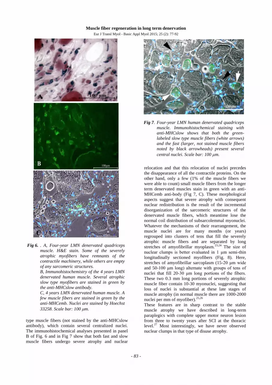
Muscle fiber regeneration in long term denervation
Eur J Transl Myol - Basic Appl Myol 2015; 25 (2): 77-92
- 83 -
type muscle fibers (not stained by the anti-MHCslow
antibody), which contain several centralized nuclei.
The immunohistochemical analyses presented in panel
B of Fig. 6 and in Fig 7 show that both fast and slow
muscle fibers undergo severe atrophy and nuclear
relocation and that this relocation of nuclei precedes
the disappearance of all the contractile proteins. On the
other hand, only a few (1% of the muscle fibers we
were able to count) small muscle fibers from the longer
term denervated muscles stain in green with an anti-
MHCemb anti-body (Fig 7, C). These morphological
aspects suggest that severe atrophy with consequent
nuclear redistribution is the result of the incremental
disorganization of the sarcomeric structures of the
denervated muscle fibers, which meantime lose the
normal coil distribution of subsarcolemmal myonuclei.
Whatever the mechanisms of their rearrangement, the
muscle nuclei are for many months (or years)
regrouped into clusters of tens that fill the severely
atrophic muscle fibers and are separated by long
stretches of amyofibrillar myoplasm.23,24
The size of
nuclear clumps is better evaluated in 1 μm semi-thin
longitudinally sectioned myofibers (Fig. 8). Here,
stretches of amyofibrillar sarcoplasm (15-20 μm wide
and 50-100 μm long) alternate with groups of tens of
nuclei that fill 20-30 μm long portions of the fibers.
These two 0.3 mm long portions of severely atrophic
muscle fiber contain 10-30 myonuclei, suggesting that
loss of nuclei is substantial at these late stages of
muscle atrophy (in normal muscle there are 1000-2000
nuclei per mm of myofiber).25,26
These features are in sharp contrast to the stable
muscle atrophy we have described in long-term
paraplegics with complete upper motor neuron lesion
from three to twenty years after SCI at the thoracic
level.27
Most interestingly, we have never observed
nuclear clumps in that type of disuse atrophy.
Fig 6. . A, Four-year LMN denervated quadriceps
muscle. H&E stain. Some of the severely
atrophic myofibers have remnants of the
contractile machinery, while others are empty
of any sarcomeric structures.
B, Immunohistochemistry of the 4 years LMN
denervated human muscle. Several atrophic
slow type myofibers are stained in green by
the anti-MHCslow antibody.
C, 4 years LMN denervated human muscle. A
few muscle fibers are stained in green by the
anti-MHCemb. Nuclei are stained by Hoechst
33258. Scale bar: 100 m.
Fig 7. Four-year LMN human denervated quadriceps
muscle. Immunohistochemical staining with
anti-MHCslow shows that both the green-
labeled slow type muscle fibers (white arrows)
and the fast (larger, not stained muscle fibers
noted by black arrowheads) present several
central nuclei. Scale bar: 100 m.
Muscle fiber regeneration in long term denervation
Eur J Transl Myol - Basic Appl Myol 2015; 25 (2): 77-92
- 84 -
The only practical way to perform a quantitative
analysis of nuclear clumps was to count them on
muscle cross sections and then to extrapolate the data
to the whole muscle. Taking into account that, after the
third year of denervation, approximately 30% of
atrophic myofibers show nuclear clumps, and that the
clumps of nuclei are separated by longer stretches of
amyofibrillar cytoplasm (Fig. 8), we calculate that
almost all the severely atrophic myofibers seen in
muscle biopsies from three to five years after complete,
permanent LMN injury present with relocation of their
nuclei from the spiral subsarcolemmal pattern of
normal muscle fibers to nuclear clumps.
Up to now, nuclear groupings have been seldom
described in muscle biopsies from patients affected
with neurodegenerative disorders, like amyotrophic
lateral sclerosis and spinal muscular atrophy.28
That is
not surprising because in neurodegenerative disorders
the incompleteness of denervation allows for
reinnervation and thus for reorganization of larger
muscle units.29
All together, our observations confirm that human
muscle fibers survive complete and permanent
denervation much longer than generally accepted.
Nuclear clumps should be considered markers of the
long-lasting ability of human muscle fibers to survive
in the absence of innervation (see Fig. 6 A and the
following chapter). These results, together with those
presented in the next two chapters, provide the
rationale to plan research aimed at recovery of these
severely atrophic myofibers. Specifically, we contend
that further research should include the combination of
molecular and cellular approaches with the functional
electrical stimulation that our previous studies have
shown to restore muscle structure and mass in human
long-term denervated and degenerated muscle.4,18,30-40
Sustained myogenesis in human long-term
denervated muscles
Immunohistochemical evidence
Muscle tissue harvested 9 months to 19 years post SCI
from patients of the EU Project RISE (Contract no.
QLG5-CT-2001-02191) were shown to contain the
above described severely atrophic, nuclear-clumped
muscle fibers (i.e., fibers surviving years of
denervation) and also immature thin fibers, which are
interpreted to be recently regenerating in nature. These
fibers (Fig. 9A) have a central nucleus, a thin
cytoplasmic rim containing a few myofilaments and
also express MHCemb.40
They represent about 1% of
the total myofiber population in the muscle biopsies.31
Indeed, morphologic characteristics of the long-term
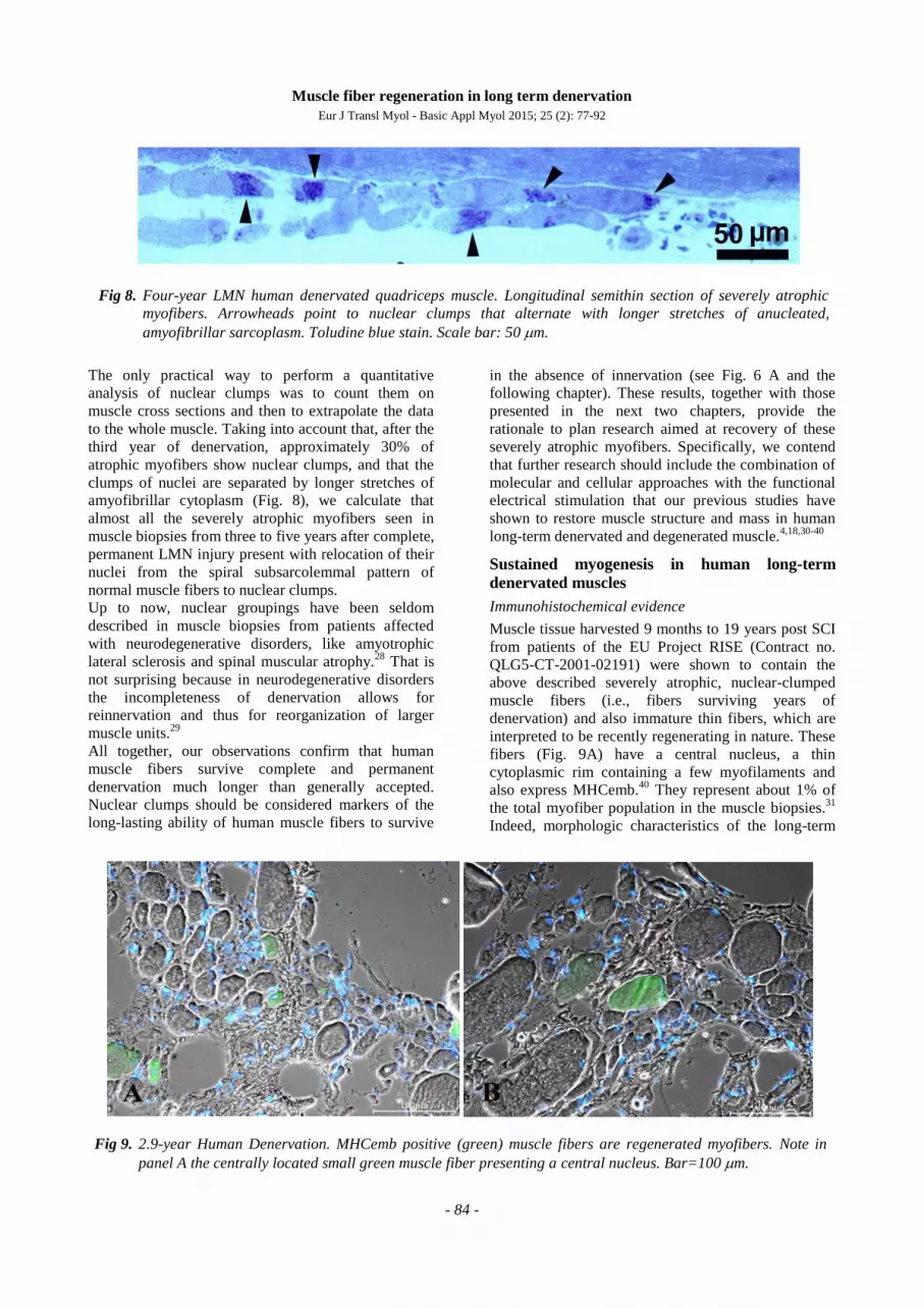
Fig 8. Four-year LMN human denervated quadriceps muscle. Longitudinal semithin section of severely atrophic
myofibers. Arrowheads point to nuclear clumps that alternate with longer stretches of anucleated,
amyofibrillar sarcoplasm. Toludine blue stain. Scale bar: 50 m.
Fig 9. 2.9-year Human Denervation. MHCemb positive (green) muscle fibers are regenerated myofibers. Note in
panel A the centrally located small green muscle fiber presenting a central nucleus. Bar=100 m.
Muscle fiber regeneration in long term denervation
Eur J Transl Myol - Basic Appl Myol 2015; 25 (2): 77-92
- 85 -
denervated muscle in animals and humans suggest that
some original fibers are lost and some of those
remaining are the result of repeated cycles of muscle
fiber death and regeneration. Cryosections of these
tissues show atrophic or severely atrophic myofibers
dispersed among adipocytes and connective tissue.
Monoclonal antibody for MHCemb stains 1 to 3 % of
the atrophic muscle fibers from 1- to 37-years post-
SCI. In contrast to this, after 2- to 10-years of h-bFES-
training, the muscle biopsies present with mainly large
round myofibers. In the h-bFES-trained muscles the
regenerative events are present, but at a lower rate than
in the long-term denervated muscles not receiving h-
bFES (MHCemb positive myofiber per mm2 of
cryosection area: 0.8+/-1.3 with h-bFES vs. 2.3+/-2.3
vs without h-bFES biopsies, mean+/-SEM, p = 0.011).
In our opinion, this is sound additional evidence for the
positive effectiveness of the electrical stimulation
protocol for h-bFES of DDM. In any case, the overall
results demonstrate that h-bFES-training is safe; that is,
it does not induce more myofiber necrosis followed by
regeneration than denervation alone.31
In our opinion the myofibers stained by the antibody
recognizing MHCemb had regenerated during the last
few weeks before biopsy, whatever the time from SCI.
This interpretation is based upon the fact that
MHCemb: 1) is expressed in myotubes and young
myofibers and 2) represents the soundest molecular
marker of early myogenic events in both developing
and adult muscles because, in the absence of
innervation, the embryonic isoforms are completely
replaced by a default programm of fast-type MHC
expression after two-three weeks of aneural
development.10,41-43
The round, green areas seen in Fig.
9 represent cross sections of myofibers staining
positively in response to anti-MHCemb antibody. They
are, in our opinion, newly regenerated myofibers,
which developed during the last few weeks before
biopsy in the long term denervated muscles of patients
suffering with complete Conus-Cauda Equina
Syndrome. They are not likely to be simply
reprogrammed atrophic cells. Their paucity speaks
against the interpretation that they are the product of a
molecular switch in myosin expression occurring in the
long term denervated muscle fibers, in spite of eventual
short term re-expression of embryonic protein isoforms
soon after denervation.10
Doppler et al.44
found similar features in randomly
chosen neurogenic muscle biopsies that did not fulfill
the criterion for long-term denervation (i.e., inability to
respond to electrical stimulation), because they were
from patients with amyotrophic lateral sclerosis or
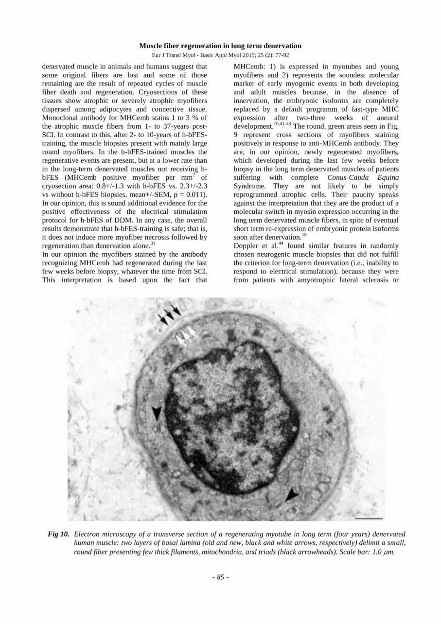
Fig 10. Electron microscopy of a transverse section of a regenerating myotube in long term (four years) denervated
human muscle: two layers of basal lamina (old and new, black and white arrows, respectively) delimit a small,
round fiber presenting few thick filaments, mitochondria, and triads (black arrowheads). Scale bar: 1.0 m.
Muscle fiber regeneration in long term denervation
Eur J Transl Myol - Basic Appl Myol 2015; 25 (2): 77-92
- 86 -
polyneuropathy. In both diseases, denervated
myofibers tend to be severely atrophic and angular,
resulting in an end stage of “nuclear clumps”, with
little cytoplasm left.44
In a cohort of 66 patients, bioptic
muscle sections were immunolabeled with monoclonal
antibodies against NCAM and neonatal myosin heavy
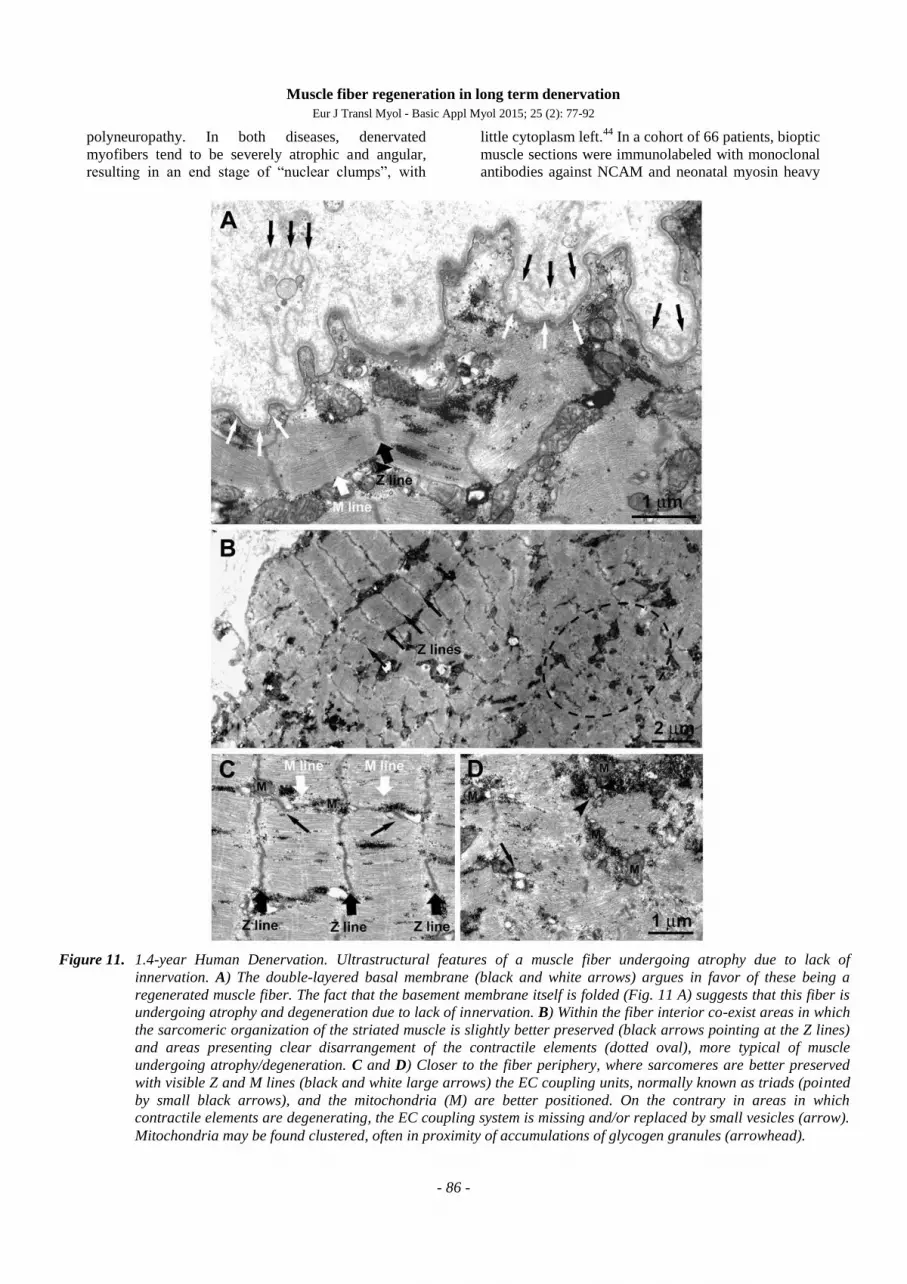
Figure 11. 1.4-year Human Denervation. Ultrastructural features of a muscle fiber undergoing atrophy due to lack of
innervation. A) The double-layered basal membrane (black and white arrows) argues in favor of these being a
regenerated muscle fiber. The fact that the basement membrane itself is folded (Fig. 11 A) suggests that this fiber is
undergoing atrophy and degeneration due to lack of innervation. B) Within the fiber interior co-exist areas in which
the sarcomeric organization of the striated muscle is slightly better preserved (black arrows pointing at the Z lines)
and areas presenting clear disarrangement of the contractile elements (dotted oval), more typical of muscle
undergoing atrophy/degeneration. C and D) Closer to the fiber periphery, where sarcomeres are better preserved
with visible Z and M lines (black and white large arrows) the EC coupling units, normally known as triads (pointed
by small black arrows), and the mitochondria (M) are better positioned. On the contrary in areas in which
contractile elements are degenerating, the EC coupling system is missing and/or replaced by small vesicles (arrow).
Mitochondria may be found clustered, often in proximity of accumulations of glycogen granules (arrowhead).

Muscle fiber regeneration in long term denervation
Eur J Transl Myol - Basic Appl Myol 2015; 25 (2): 77-92
- 87 -
chain (MHCneo), two proteins that are neurally and
developmentally regulated. Of the biopsy specimens,
75% contained small myofibers that showed a thin
perinuclear cytoplasmic rim. Small fibers expressing
MHCneo were found in all of these biopsies (100%)
and NCAM-positive fibers were detected in 98% of the
biopsies. The percentage of NCAM small fibers was
significantly lower than that of MHCneo fibers. The
authors conclude that myogenesis appears to be a
frequent finding in human neurogenic muscle
atrophy.44
Similar observations of muscle fiber
regeneration were obtained in human muscle biopsies
harvested from free transplanted muscle flaps that were
not reinnervated.4
Our observations, thus, suggest that the MHCemb
positive regenerated muscle fibers are not previous
myofibers re-expressing embryonic isoforms (as is the
case for acetylcholine receptors spreading from the
end-plate to all the sarcolemma of recently denervated
muscle fibers)3, but are the result of activation,
proliferation and fusion of satellite cells, the myogenic
precursors located under the basal lamina of the muscle
fibers.49
The transient expression of laminin isoforms peculiar
to early myogenesis during muscle regeneration is also
well established.45,46
Further, we and others have
shown that the regenerating muscle fibers,
differentiating in an aneural muscle, grow up to one
fourth the size of a normal adult muscle fiber, and then
undergo atrophy, degeneration, and death (typically by
apoptotic processes).1,47,48
Ultrastructural evidence
Electron microscopic analyses of muscle biopsies from
the EU RISE study contribute some evidence for
myofiber regeneration, i.e. the presence of double-
layered basement membranes that delimit myotubes
(Fig. 10) and more developed muscle fibers (Fig. 11).
We and others,8,45,46
interpreted these structures to
indicate that the external membrane layer belongs to a
muscle fibre that may have previously degenerated,
while the internal membrane represents a new basal
lamina that is being built on the surface of a
regenerating myotube within the old boundary.
Contrary to general expectation, but supported by
rodent experiments1,2,6-10,13-15
and analyses of human
muscle harvested from peripheral neuromuscular
disorders,44
myofiber regeneration has been
persistently observed (even if at a low rate) in long-
term denervated human muscles, where presumably
differentiating cells either positive to anti-MHCemb
antibody or with some contractile filaments and a
double layer of basal lamina are visible (Fig. 10). A
few thick myofilaments with Z lines are present and,
importantly, some triads (arrowheads in Fig. 10), that
is, the Ca++
-release units responsible for excitation-
contraction coupling.
The thousands of muscle cultures that begin
spontaneous contraction every day in myology labs
worldwide are the strongest evidence that early
myogenesis occurs in the absence of the nerve and its
secreted factors, though they never attain full
differentiation unless coinnervated. In truth, muscle
fibers with a double-layered basal lamina are sparse
observations in our series of denervated muscles, in
agreement with the fact that anti-MHCemb antibodies
stain only 1% of the myofibers in the same series of
long-term denervated human muscle biopsies.
The presence of a double-layered basal membrane
(Fig, 11,A, black and white arrows) argues in favor of
a fiber being likely regenerated during the month
before biopsy. The fiber is, however, undergoing
atrophy and degeneration, presumably due to the lack
of innervation as shown by extensive folding of the
basement membrane. Interestingly, within the fiber
interior areas with a better sarcomeric organization
(Fig. 11, Panel B, black arrows pointing at the Z lines)
coexist with others showing disarrangement of the
contractile elements (Fig. 11, Panel B, dotted oval),
which is more typical of muscle undergoing atrophy
and degeneration. The disorganized areas (such as the
one marked by the dashed oval in Fig. 11B) are usually
found in the center of the fibers contrary to what
happens in the long-term denervated fibers where the
degeneration usually begins at the periphery.18, 30-40
In
addition, even in better preserved areas, myofibrils are
often wavy and the sarcomeres are quite shortened.
This indicates local variability in activation resulting in
lack of proper alignment of contractile elements. The
variability in the structural integrity of the contractile
apparatus also affects the excitation-contraction (EC)
coupling elements and the positioning of mitochondria,
which are already quite structurally altered. Closer to
the fiber periphery, where sarcomeres are better
preserved with visible Z and M lines (Fig. 11 C, black
and white large arrows), the EC coupling units
(normally known as triads; noted by small black
arrows) and the mitochondria (M) are better
positioned. On the contrary, in areas where contractile
elements are disarranged, the EC coupling system is
missing and/or replaced by small vesicles, representing
remains of sarcoplasmic reticulum (SR) membranes
(Fig. 11 D, arrow). Mitochondria may be clustered,
often in proximity to accumulations of glycogen
granules (Fig. 11 Panel D, arrowhead).
Human long term denervated muscle recovers
mass and sustained contractility during the first
year of h-bFES
To counteract the progressive changes that transform
muscle into an unexcitable tissue, over the years we
have developed a novel therapeutic concept for
paraplegic patients with bilateral and complete LMN
denervation of the lower extremity due to complete
lesion of the conus and cauda equina.18,33,35,37
The new
Muscle fiber regeneration in long term denervation
Eur J Transl Myol - Basic Appl Myol 2015; 25 (2): 77-92
- 88 -
training strategy became possible because of the
development of a new generation of stimulation
equipment specifically designed for h-bFES. These
new stimulators and the large surface electrodes
necessary to cover the denervated muscles were
developed by the Center of Biomedical Engineering
and Physics at the Medical University of Vienna and
by the Wilhelminenspital, Vienna (Austria).18,37,50-53
The equipment was designed to reverse long-standing
and severe atrophy by delivering high-intensity and
long-duration impulses that can directly elicit
contraction of denervated skeletal muscle fibers, even
after they have lost the ability to respond to other
commercial electrical stimulators.18,37,50-53
Twenty out
of 25 patients enrolled in the EU RISE trial completed
the 2-year h-bFES program, which resulted in: 1) a
35% cross-sectional increase in area of the quadriceps
muscle; 2) a 75% increase in mean diameter of muscle
fibers; 3) improvements in the ultrastructural
organization of contractile material; and 4) a 1187%
increase in force output during electrical stimulation.
The recovery of quadriceps force was sufficient to
allow 25% of the subjects to perform FES-assisted
stand-up exercises and step-training in place.33-37
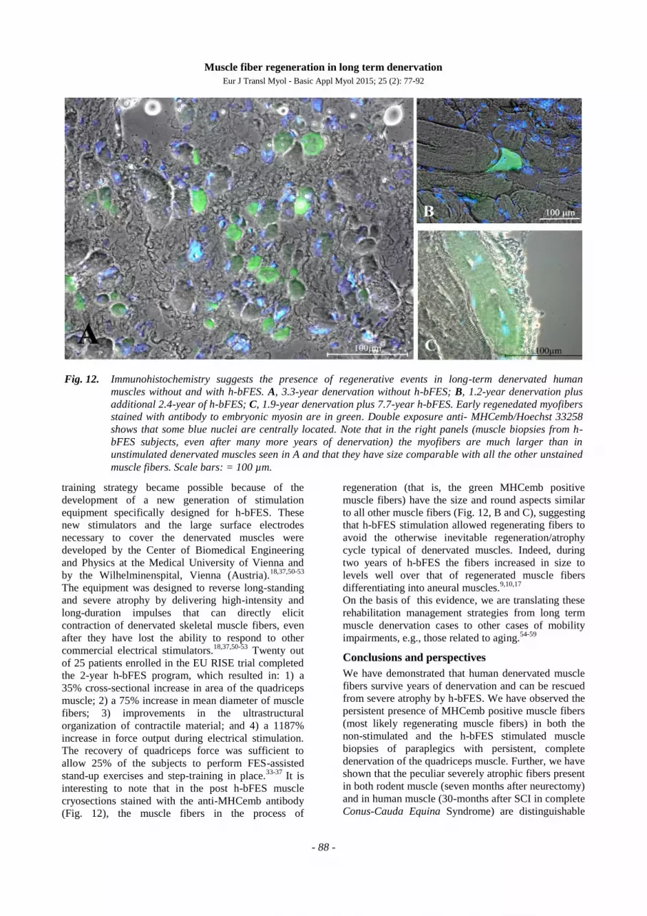
It is
interesting to note that in the post h-bFES muscle
cryosections stained with the anti-MHCemb antibody
(Fig. 12), the muscle fibers in the process of
regeneration (that is, the green MHCemb positive
muscle fibers) have the size and round aspects similar
to all other muscle fibers (Fig. 12, B and C), suggesting
that h-bFES stimulation allowed regenerating fibers to
avoid the otherwise inevitable regeneration/atrophy
cycle typical of denervated muscles. Indeed, during
two years of h-bFES the fibers increased in size to
levels well over that of regenerated muscle fibers
differentiating into aneural muscles.9,10,17
On the basis of this evidence, we are translating these
rehabilitation management strategies from long term
muscle denervation cases to other cases of mobility
impairments, e.g., those related to aging.54-59
Conclusions and perspectives
We have demonstrated that human denervated muscle
fibers survive years of denervation and can be rescued
from severe atrophy by h-bFES. We have observed the
persistent presence of MHCemb positive muscle fibers
(most likely regenerating muscle fibers) in both the
non-stimulated and the h-bFES stimulated muscle
biopsies of paraplegics with persistent, complete
denervation of the quadriceps muscle. Further, we have
shown that the peculiar severely atrophic fibers present
in both rodent muscle (seven months after neurectomy)
and in human muscle (30-months after SCI in complete
Conus-Cauda Equina Syndrome) are distinguishable
Fig. 12. Immunohistochemistry suggests the presence of regenerative events in long-term denervated human
muscles without and with h-bFES. A, 3.3-year denervation without h-bFES; B, 1.2-year denervation plus
additional 2.4-year of h-bFES; C, 1.9-year denervation plus 7.7-year h-bFES. Early regenedated myofibers
stained with antibody to embryonic myosin are in green. Double exposure anti- MHCemb/Hoechst 33258
shows that some blue nuclei are centrally located. Note that in the right panels (muscle biopsies from h-
bFES subjects, even after many more years of denervation) the myofibers are much larger than in
unstimulated denervated muscles seen in A and that they have size comparable with all the other unstained
muscle fibers. Scale bars: = 100 µm.
Muscle fiber regeneration in long term denervation
Eur J Transl Myol - Basic Appl Myol 2015; 25 (2): 77-92
- 89 -
from early myotubes using morphological features and
molecular biomarkers. Beyond reviewing past
evidence from rodent and human studies, here we list
the options to substantially increase the regenerative
potential of skeletal muscle in those patients who have
experienced denervation of muscles “too-late-to-be
recovered by h-bFES”, that is, they present with very
severely denervated muscles. Indeed, the needed
mandatory procedures are ready to be translated from
animal experiments to the needs of SCI persons with
long-term irreversible muscle denervation, that is: 1)
induction and separation of autologous myogenic cells,
either i) by in vivo marcaine infiltration of an
expendable muscle tissue (Latissimus Dorsi) and
grown in vitro;60
or ii) derived from autologous
adipose tissue by in vitro induction and cell-sorting
selection of myogenic stem cells able to replicate in
vivo;61-65
2) multiple injections of the autologous
myogenic stem cells, followed by their proliferation,
fusion and differentiation into adult-like muscle fibers;
and, finally, 3) their tetanic contractions induced by
surface electrodes and an external neuromodulator.18,30-
38,50-53
Why should we not try to apply h-bFES to encourage
the growth of regenerated muscle fibers, adding (to
those accumulating by spontaneous activation of
satellite cells) muscle fibers derived from myogenic
stem cells harvested from autologous adipose tissue?
We are confident that the evidence presented here will
convince ethical committees to allow animal
experiments to develop the safest possible procedures
for translation to those many patients who, having
missed an early commencement of h-bFES, have lost
muscle fibers as a consequence of the inevitable
progression of denervation-induced muscle tissue
degeneration that occurs in long-term Conus and
Cauda Equina Syndrome and other traumatic
peripheral neuropathies. Based on the evidence and
concepts presented here, an European Project, the trial
Rise4EU (Rise for You, a personalized treatment
protocol for functional recovery of denervated muscle
post long-term stable SCI), will hopefully be
implemented soon.
Acknowledgement
This work was supported by European Regional
Development Fund - Cross Border Cooperation
Programme Slovakia – Austria 2007–2013 (Interreg-
IVa), project Mobilität im Alter, MOBIL, N_00033
(partners: Ludwig Boltzmann Institute of Electrical
Stimulation and Physical Rehabilitation, Austria,
Center for Medical Physics and Biomedical
Engineering, Medical University of Vienna, Austria,
and Faculty of Physical Education and Sports,
Comenius University in Bratislava, Slovakia); Austrian
national co-financing of the Austrian Federal Ministry
of Science and Research; Ludwig Boltzmann Society
(Vienna, Austria); Telethon, (GGP13213) to F.P.
Corresponding Author
Andrea Marcante, Department of Neurorehabilitation,
San Camillo Hospital, I.R.C.C.S., Venezia-Lido, Italy;
and Department of Neuroscience, Rehabilitation Unit,
Padua University Hospital, Padova, Italy.
E-mail: "Andrea Marcante" [email protected]
E-mail of coauthors
Ugo Carraro: [email protected]
Simona Boncompagni: [email protected]
Valerio Gobbo: [email protected]
Sandra Zampieri: [email protected]
Simone Mosole: [email protected]
Barbara Ravara: [email protected]
Alessandra Nori: [email protected]
Roberto Stramare: [email protected]
Francesco Ambrosio: [email protected]
Francesco Piccione:
Stefano Masiero: [email protected]
Vincenzo Vindigni: [email protected]
Paolo Gargiulo: [email protected]
Feliciano Protasi: [email protected]
Helmut Kern: [email protected]
Amber Pond: [email protected]
References
1. Borisov AB, Dedkov EI, Carlson BM.
Interrelations of myogenic response, progressive
atrophy of muscle fibres, and cell death in
denervated skeletal muscle. Anat Rec
2001;264:203-18.
2. Carlson BM. The biology of long-term
denervated skeletal muscle. Eur J Transl Myol -
Basic Appl Myol 2014;24:5-12.
3. Lomo T. The response of denervated muscle to
long-term stimulation (1985, revisited here in
2014). Eur J Transl Myol - Basic Appl Myol
2014;24:13-9.
4. Kern H, Carraro U. Home-based Functional
Electrical Stimulation (h-b FES) for long-term
denervated human muscle: History, basics, results
and perspectives of the Vienna Rehabilitation
Strategy. Eur J Transl Myol/Basic Appl Myol
2014:24:27-40.
5. Adami N, Kern H, Mayr W, et al. Permanent
denervation of rat Tibialis Anterior after bilateral
sciatectomy: Determination of chronaxie by
surface electrode stimulation during progression
of atrophy up to one year. Basic Appl Myol
2007;17:237-434
6. Borisov AB, Dedkov EI, Carlson BM. Abortive
myogenesis in denervated skeletal muscle:
differentiative properties of satellite cells, their
migration, and block of terminal differentiation.
Anat Embryol 2005;209:269–279.
7. Schmalbruch H, al-Amood WS, Lewis DM.
Morphology of long-term denervated rat soleus
Muscle fiber regeneration in long term denervation
Eur J Transl Myol - Basic Appl Myol 2015; 25 (2): 77-92
- 90 -
muscle and the effect of chronic electrical
stimulation. J Physiol (Lond) 1991;441:233–241.
8. Carraro U, Morale D, Mussini I, et al. Chronic
denervation of rat diaphragm: maintenance of
fiber heterogeneity with associated increasing
uniformity of myosin isoforms. J Cell Biol
1985;100:161–74.
9. Mussini I, Favaro G, Carraro U. Maturation,
dystrophic changes and the continuous production
of fibers in skeletal muscle regenerating in the
absence of nerve. J Neurophatol Exp Neurol
1987;46:315-31.
10. Carraro U, Dalla Libera L, Catani C. Myosin light
and heavy chains in muscle regenerating in
absence of the nerve: transient appearance of the
embryonic light chain. Exp Neurol 1983;79:106-
17.
11. Sartore S, Gorza L, Schiaffino S. Fetal myosin
heavy chains in regenerating muscle. Nature
1982;298: 294–6.
12. Schiaffino S, Gorza L, Piton G, et al. Embryonic
and neonatal myosin heavy chain in denervated
and paralyzed rat skeletal muscle. Dev Biol
1988;127:1–11.
13. Carraro U, Rossini K, Zanin M, et al. Induced
myogenesis in long-term permanent denervation:
perspective role in Functional Electrical
Stimulation of denervated legs in humans. Basic
Appl Myol 2002;12:53-64.
14. Adams L, Carlson BM, Henderson L, Goldman
D. Adaptation of nicotinic acetylcholine receptor,
myogenin, and MRF4 gene expression to long
term muscle denervation. J Cell Biol
1995;131:1341-9.
15. Lapalombella R, Kern H, Adami N, et al.
Persistence of regenerative myogenesis in spite of
down-regulation of activity-dependent genes in
long-term denervated rat muscle. Neurol Res
2008;30:197-
16. Squecco R1, Carraro U, Kern H, Pond A, et al. A
subpopulation of rat muscle fibers maintains an
assessable excitation-contraction coupling
mechanism after long-standing denervation
despite lost contractility. J Neuropathol Exp
Neurol. 2009 Dec;68(12):1256-68. doi:
10.1097/NEN.0b013e3181c18416.
17. Carlson BM. “The Denervated Muscle” - 45 years
later. Basic Appl Myol 2007;17:113-7.
18. Kern H, Boncompagni S, Rossini K, et al. Long-
term denervation in humans causes degeneration
of both contractile and excitation contraction
coupling apparatus, wich is reversible by
functional electrical stimulation (FES). A role for
myofiber regeneration? J Neuropathol Exp
Neurol 2004;63:919-31.
19. Biral D, Kern H, Adami N, et al. Atrophy-
resistant fibers in permanent peripheral
denervation of human skeletal muscle. Neurol
Res 2008;30:137-44.
20. Ashley Z, Sutherland H, Lanmuller H, et al.
Atrophy, but not necrosis, in rabbit skeletal
muscle denervated for periods up to one year. Am
J Cell Physiol 2007;292:440-51.
21. Lewis DM, Al-Amood WS, Schmalbruch H.
Effects of long-term phasic electrical stimulation
of denervated soleus muscle: guinea pig
contrasted with rat. J Muscle Res Cell Motil
1997;18:573-86.
22. Carraro U, Catani C, Saggin L, et al. Isomyosin
changes after functional electrostimulation of
denervated sheep muscle. Muscle Nerve.
1988;11:1016-28.
23. Kern H, Carraro U, Biral D, et al. Severely
atrophic muscle fibers with nuclear clumps
survive many years in permanently denervated
human muscle. The Open Pathology Journal
2009;3:106-10.
24. Adami N, Biral D, Carraro U et al. The fiber
types of severely atrophic muscle fibers with
nuclear clumps of human 5-year LMN denervated
muscle. Basic Appl Myol 2009;19:225-8.
25. Aravamudan B, Mantilla CB, Zhan WZ, Sieck
GC. Denervation effects on myonuclear domain
size of rat diaphragm fibers. J Appl Physiol
2006;100:1618-22.
26. Gundersen K, Bruusgaard JC. Nuclear domains
during muscle atrophy: nuclei lost or lost
paradigm. J Physiol 2008;586:2675-81.
27. Kern H, Hofer C, Modlin M, et al. Stable muscle
atrophy in long term paraplegics with complete
upper motor neuron lesion from 3- to 20-year
SCI. Spinal Cord 2008;46:293-304.
28. Dubowitz V, Sewry CA. Muscle biopsy. A
practical approach. Sauders Elsevier Edition
2007. Neurol 2004;63:919-31
29. Wokke JH. Genes, trials, and care: The dynamics
of neuromuscular disease. Lancet Neurol
2004;3:1-16.
30. Kern H, Hofer C, Mayr W, Carraro U. European
Project RISE: Partners, protocols, demography.
Basic Appl Myol/Eur J Transl Myol 2009;19:211-
6.
31. Carraro U, Rossini K, Mayr W, Kern H. Muscle
fiber regeneration in human permanent lower
motoneuron denervation: relevance to safety and
effectiveness of FES-training, which induces
muscle recovery in SCI subjects. Artif Organs
2005;19:187–191.
32. Kern H, Salmons S, Mayr W, et al. Recovery of
long-term denervated human muscles induced by
electrical stimulation. Muscle Nerve 2005;31:98-
101.
33. Boncompagni S, Kern H, Rossini K, et al.
Structural differentiation of skeletal muscle fibers
Muscle fiber regeneration in long term denervation
Eur J Transl Myol - Basic Appl Myol 2015; 25 (2): 77-92
- 91 -
in the absence of innervation in humans. Proc
Natl Acad Sci USA 2007;104:19339-44.
34. Kern H, Carraro U. Translational myology focus
on clinical Challenges of functional electrical
stimulation of denervated muscle. Basic Appl
Myol/Eur J Transl Myol 2008;18:37-100.
35. Kern H, Carraro U, Adami N, et al. One year of
home-based Functional Electrical Stimulation
(FES) in complete lower motor neuron
paraplegia: Recovery of tetanic contractility
drives the structural improvements of denervated
muscle. Neurol Res 2010;32:5-12, doi:
10.1189/184313209 X385644.
36. Kern H, Hofer C, Mayr W. Protocols for clinical
work package of the European project RISE.
Basic Appl Myol/Eur J Transl Myol 2008;18:39-
44.
37. Kern H, Carraro U, Adami N, et al. Home-based
functional electrical stimulation rescues
permanently denervated muscles in paraplegic
patients with complete lower motor neuron
lesion. Neurorehabil Neural Repair. 2010;24:709-
21. Epub 2010 May 11.
38. Gargiulo P, Helgason T, Reynisson PJ, et al.
Monitoring of muscle and bone recovery in spinal
cord injury patients treated with electrical
stimulation using three-dimensional imaging and
segmentation techniques: methodological
assessment. Artif Organs 2011;35:275-81. doi:
10.1111/j.1525-1594.2011.01214.x.
39. Mancinelli R, Kern H, Fulle S, et al.
Transcriptional profile of denervated vastus
lateralis muscle derived from a patient 8 months
after spinal cord injury: a case-report. Int J
Immunopathol Pharmacol 2011;24:749-59.
40. Rossini K, Zanin ME, Carraro U. To stage and
quantify regenerative myogenesis in human long-
term permanent denervated muscle. Basic Appl
Myol 2002;12;277-87.
41. Whalen RG, Sell SM, Butler-Browne GS, et al
Three myosin heavy-chain isozymes appear
sequentially in rat muscle development. Nature
1981;292:805-9.
42. Lewis DM, Schmalbruch H. Contractile
properties of aneurally regenerated compared
with denervated muscles of rat. J Muscle Res Cell
Motil 1994;15:267-77.
43. Carraro U, Catani C. A sensitive SDS PAGE
method separating heavy chain isoforms of rat
skeletal muscles reveals the heterogeneous nature
of the embryonic myosin. Biochem Biophys Res
Commun 1983;116:793-802.
44. Doppler K, Mittelbronn M, Bornemann A.
Myogenesis in human denervated muscle
biopsies. Muscle Nerve 2008;37:79–83.
45. Gulati AK. Basement membrane component
changes in skeletal muscle transplants undergoing
regeneration or rejection. J Cell Biochem
1985;27:337-46.
46. Grounds MD, McGeachie JK, Davies MJ, et al.
The expression of extracellular matrix during
adult skeletal muscle regeneration: how the
basement membrane, interstitium and myogenic
cells collaborate. Basic Appl Myol 1998;8:129-
141.
47. Sandri M, Carraro U, Podhorska-Okolov et al.
Apoptosis, DNA damage and ubiquitin
expression in normal and mdx muscle fibers after
exercise. FEBS Lett 1995;373:291-5.
48. Carraro U, Franceschi C. Apoptosis of skeletal
and cardiac muscles and physical exercise. Aging
(Milano). 1997;9:19-34. Review.
49. Yablonka-Reuveni Z. The skeletal muscle
satellite cell: still young and fascinating at 50. J
Histochem Cytochem 2011;59:1041-59. doi:
10.1369/0022155411426780.
50. Hofer C, Mayr W, Stöhr H, et al. A stimulator for
functional activation of denervated muscles. Artif
Organs 2002;26:276-9.
51. Mayr W, Bijak M, Rafolt D,et al. Basic design
and construction of the Vienna FES implants:
existing solutions and prospects for new
generations of implants. Med Eng Phys
2001;23:53-60.
52. Mödlin M, Forstner C, Hofer C, et al. Electrical
stimulation of denervated muscles: first results of
a clinical study. Artif Organs 2005;29:203-6.
53. Gargiulo P, Reynisson PJ, Helgason B, et al.
Muscle, tendons, and bone: structural changes
during denervation and FES treatment. Neurol
Res 2011;33:750-8. doi: 10.1179/1743132811
Y.0000000007.
54. Kern H, Barberi L, Löfler S, et al. Electrical
stimulation counteracts muscle decline in seniors.
Front Aging Neurosci. 2014 Jul 24;6:189. doi:
10.3389/fnagi.2014.00189. eCollection 2014.
55. Mosole S, Carraro U, Kern H, et al. Long-term
high-level exercise promotes muscle
reinnervation with age. J Neuropathol Exp Neurol
2014;73:284-94. doi: 10.1097/NEN.00000000000
00032.
56. Zampieri S, Pietrangelo L, Loefler S, et al.
Lifelong physical exercise delays age-associated
skeletal muscle decline. J Gerontol A Biol Sci
Med Sci 2014 Feb 18. [Epub ahead of print]
57. Marcante A, Zanato R, Ferrero M, et al. Recovery
of tetanic contractility of denervated muscle: A
step toward a walking aid for foot drop. Biomed
Tech (Berl) 2013 Sep 7. pii:
/j/bmte.2013.58.issue-s1-A/bmt-2013-4016/bmt-
2013-4016.xml. doi: 10.1515/bmt-2013-4016.
[Epub ahead of print]
58. Zanato R, Stramare R, Boato N, et al. Dynamic
echomyography shows that FES in Peripheral
Denervation does not hamper muscle
Muscle fiber regeneration in long term denervation
Eur J Transl Myol - Basic Appl Myol 2015; 25 (2): 77-92
- 92 -
reinnervation. Biomed Tech (Berl) 2013 Sep 7.
pii:/j/bmte.2013.58.issue-s1-A/bmt-2013-
4034/bmt-2013-4034.xml. doi: 10.1515/bmt-
2013-4034. [Epub ahead of print].
59. Kern H, Pelosi L, Coletto L, et al. Atrophy/
hypertrophy cell signaling in muscles of young
athletes trained with vibrational-proprioceptive
stimulation. Neurol Res 2011;33:998-1009. doi:
10.1179/016164110X127 67786356633.
60. Péault B, Rudnicki M, Torrente Y, et al. Stem and
Progenitor Cells in Skeletal Muscle Development,
Maintenance, and Therapy. Mol Ther
2007;15:867-77. Epub 2007 Mar 27. doi: 10.1038
/mt.sj.6300145.
61. Lindroos B, Suuronen R, Miettinen S. The
potential of adipose stem cells in regenerative
medicine. Stem Cell Rev 2011;7:269-91. doi:
10.1007/s12015-010-9193-7.
62. Zuk PA, Zhu M, Ashjian P, et al. Human adipose
tissue is a source of multipotent stem cells. Mol
Biol Cell 2002;13:4279-95.
63. Russo V, Yu C, Belliveau P, et al. Comparison of
human adipose-derived stem cells isolated from
subcutaneous, omental, and intrathoracic adipose
tissue depots for regenerative applications. Stem
Cells Transl Med 2014;3:206-17. doi:
10.5966/sctm.2013-0125. Epub 2013 Dec 20.
64. Kern H, Carraro U. Home-based Functional
Electrical Stimulation for long-term denervated
human muscle: History, basics, results and
perspectives of the Vienna Rehabilitation
Strategy. Eur J Transl Myol - Basic Appl Myol
2014;24:27-40. doi: 10.4081/bam.2014.1.27
65. Huang H, Sun T, Chen L, et al. Consensus of
clinical neurorestorative progresses in patients
with complete chronic spinal cord injury. Cell
Transplant 2014;23 Suppl 1:5-17. doi:
10.3727/096368914X684952. Epub 2014 Oct 9.
66. Adami N, Biral D, Corbianco S, et al. The fiber
types of severely atrophic muscle fibers with
nuclear clumps of human 5-year LMN denervated
muscle. Basic Appl Myol 2009;19:225-8.