periprocedural and late consequences of overlapping cypher sirolimus-eluting stents
TRANSCRIPT
POPDRBCB
Tsiroosia(
RIHH¶setusm
2
Journal of the American College of Cardiology Vol. 48, No. 1, 2006© 2006 by the American College of Cardiology Foundation ISSN 0735-1097/06/$32.00P
Interventional Cardiology
eriprocedural and Late Consequences ofverlapping Cypher Sirolimus-Eluting Stents
ooled Analysis of Five Clinical Trialsean J. Kereiakes, MD, FACC,* Hong Wang, MD, MPH,† Jeffrey J. Popma, MD,‡ichard E. Kuntz, MD,‡ Dennis J. Donohoe, MD,† Joachim Schofer, MD,§ Erick Schampaert, MD,�ernhard Meier, MD, FACC, FESC,¶ Martin B. Leon, MD, FACC,# Jeffrey W. Moses, MD, FACC#incinnati, Ohio; Warren, New Jersey; Boston, Massachusetts; Hamburg, Germany; Montreal, Canada;ern, Switzerland; and New York, New York
OBJECTIVES The purpose of this research was to determine the relative safety and efficacy of multiple (�2)overlapping Cypher sirolimus-eluting stents (SES) (Johnson & Johnson, New Brunswick,New Jersey).
BACKGROUND Overlapping coronary stents are common. The periprocedural and late clinical and angio-graphic consequences of overlapped coronary stents are not clearly defined, particularly fordrug-eluting stents.
METHODS All patients enrolled into five clinical trials of the SES were analyzed. Three of these trialswere prospective randomized comparisons of the SES to the bare-metal stent (BMS),and two were prospective non-randomized trials of SES-treated patients with histori-cal controls. All clinical and angiographic outcomes in overlap-stent–treated patientswere compared by stent type and with single-stent–treated patients for the same stentdevice.
RESULTS In all, 575 patients with stent overlap (337 SES, 238 BMS) and 1,162 patients with singlestents (697 SES, 465 BMS) were analyzed. Stent overlap was associated with a greater latelumen loss in stent and more frequent angiographic restenosis regardless of stent type. Amongoverlap-stent–treated patients, the SES provided similar magnitude of restenosis benefit asobserved for single-stent–treated patients. Overlapped SES was not associated with anincrease in myocardial infarction.
CONCLUSIONS The strategy of SES overlap, when required, is both safe and efficacious in reducing restenosiswith no increase in the incidence of myocardial infarction or major adverse cardiovascularevents, when compared with a bare metal coronary stent prosthesis. (J Am Coll Cardiol
ublished by Elsevier Inc. doi:10.1016/j.jacc.2006.02.058
2006;48:21–31) © 2006 by the American College of Cardiology Foundation
lotvntrtdeMeftssBpmmf
he implantation of multiple (�2) overlapping coronarytents may be prompted by excessive target lesion length,ncomplete lesion coverage, and/or endoluminal injuryequiring additional stent scaffolding beyond the marginsf the initial stent deployed. Furthermore, multipleverlapping stents may be required to repair extensive orpiral coronary dissection. Stent strut overlap has beenncriminated both as a stimulus to neointimal hyperplasiand as a correlate to late angiographic restenosis in man1– 4). However, the relative contribution(s) of target
From the *Heart Center of Greater Cincinnati and the Lindner Center foresearch and Education at The Christ Hospital, Ohio Heart and Vascular Center
nc., Cincinnati, Ohio; †Cordis Corp., Warren, New Jersey; ‡Brigham and Women’sospital, Boston, Massachusetts; §Hamburg University Cardiovascular Center,amburg, Germany; �Hospital du Sacre’-Coeur de Montreal, Montreal, Canada;
Swiss Cardiovascular Center Bern, Bern, Switzerland; and the #Columbia Univer-ity Medical Center, New York, New York. Drs. Wang and Donohoe are Cordismployees. Cordis research grants were used to pay for the angiographic analysis forhe study. Dr. Meier’s institution, the Swiss Cardiovascular Center Bern, receivesnconditional grants from Cordis, J&J, and Boston Scientific. Dr. Leon has commontock in Cordis, is a consultant, and receives research grants. Since the writing of thisanuscript, Dr. Kuntz has now become an employee of Medtronic Inc.
wManuscript received September 16, 2005; revised manuscript received January 25,
006, accepted February 17, 2006.
esion length, total stented segment length, and numberf stents deployed per target lesion (stent overlap) towardhe occurrence of periprocedural major adverse cardio-ascular events (MACE) and/or late angiographic reste-osis have been the subject of debate (5–10). Specifically,he role of stent overlap as an independent determinant ofestenosis is unclear. Recently, concern has been raised byhe observation of an increased incidence of periproce-ural myocardial infarction after overlap of the paclitaxel-luting TAXUS stent (Boston Scientific, Marlborough,
assachusetts) (11–13). As the currently available drug-luting stent platforms differ considerably in metal plat-orm design, polymer and active pharmacologic agent,he extent to which the observations made after TAXUStent overlap may be extrapolated to the Cypherirolimus-eluting stent (SES) (Johnson & Johnson, Newrunswick, New Jersey) is unknown. The purpose of theresent study was to determine if the deployment ofultiple, overlapping SES is associated with an excess ofajor adverse clinical events (especially myocardial in-
arction) and/or angiographic restenosis when compared
ith bare-metal stent (BMS) treatment.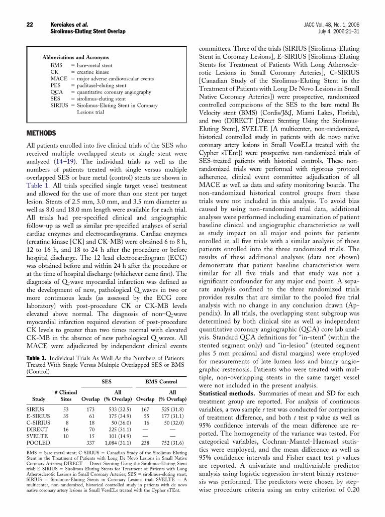
M
AranoTalwAfc(1hwadtmlemCCM
cSSr[TNcVaEhcCSraMntcabaeprdssrpapdqyspfgtwStvo9pct9aas
TT(
SECDSP
BSCtASmn
22 Kereiakes et al. JACC Vol. 48, No. 1, 2006Sirolimus-Eluting Stent Overlap July 4, 2006:21–31
ETHODS
ll patients enrolled into five clinical trials of the SES whoeceived multiple overlapped stents or single stent werenalyzed (14–19). The individual trials as well as theumbers of patients treated with single versus multipleverlapped SES or bare metal (control) stents are shown inable 1. All trials specified single target vessel treatment
nd allowed for the use of more than one stent per targetesion. Stents of 2.5 mm, 3.0 mm, and 3.5 mm diameter asell as 8.0 and 18.0 mm length were available for each trial.ll trials had pre-specified clinical and angiographic
ollow-up as well as similar pre-specified analyses of serialardiac enzymes and electrocardiograms. Cardiac enzymescreatine kinase [CK] and CK-MB) were obtained 6 to 8 h,2 to 16 h, and 18 to 24 h after the procedure or beforeospital discharge. The 12-lead electrocardiogram (ECG)as obtained before and within 24 h after the procedure or
t the time of hospital discharge (whichever came first). Theiagnosis of Q-wave myocardial infarction was defined ashe development of new, pathological Q waves in two orore continuous leads (as assessed by the ECG core
aboratory) with post-procedure CK or CK-MB levelslevated above normal. The diagnosis of non–Q-waveyocardial infarction required elevation of post-procedureK levels to greater than two times normal with elevatedK-MB in the absence of new pathological Q waves. AllACE were adjudicated by independent clinical events
Abbreviations and AcronymsBMS � bare-metal stentCK � creatine kinaseMACE � major adverse cardiovascular eventsPES � paclitaxel-eluting stentQCA � quantitative coronary angiographySES � sirolimus-eluting stentSIRIUS � Sirolimus-Eluting Stent in Coronary
Lesions trial
able 1. Individual Trials As Well As the Numbers of Patientsreated With Single Versus Multiple Overlapped SES or BMS
Control)
Study# Clinical
Sites
SES BMS Control
OverlapAll
(% Overlap) OverlapAll
(% Overlap)
IRIUS 53 173 533 (32.5) 167 525 (31.8)-SIRIUS 35 61 175 (34.9) 55 177 (31.1)-SIRIUS 8 18 50 (36.0) 16 50 (32.0)IRECT 16 70 225 (31.1) — —
VELTE 10 15 101 (14.9) — —OOLED 337 1,084 (31.1) 238 752 (31.6)
MS � bare-metal stent; C-SIRIUS � Canadian Study of the Sirolimus-Elutingtent in the Treatment of Patients with Long De Novo Lesions in Small Nativeoronary Arteries; DIRECT � Direct Stenting Using the Sirolimus-Eluting Stent
rial; E-SIRIUS � Sirolimus-Eluting Stents for Treatment of Patients with Longtherosclerotic Lesions in Small Coronary Arteries; SES � sirolimus-eluting stent;IRIUS � Sirolimus-Eluting Stents in Coronary Lesions trial; SVELTE � A
wulticenter, non-randomized, historical controlled study in patients with de novo
ative coronary artery lesions in Small VessELs treated with the Cypher sTEnt.
ommittees. Three of the trials (SIRIUS [Sirolimus-Elutingtent in Coronary Lesions], E-SIRIUS [Sirolimus-Elutingtents for Treatment of Patients With Long Atheroscle-otic Lesions in Small Coronary Arteries], C-SIRIUSCanadian Study of the Sirolimus-Eluting Stent in thereatment of Patients with Long De Novo Lesions in Smallative Coronary Arteries]) were prospective, randomized
ontrolled comparisons of the SES to the bare metal Bxelocity stent (BMS) (Cordis/J&J, Miami Lakes, Florida),
nd two (DIRECT [Direct Stenting Using the Sirolimus-luting Stent], SVELTE [A multicenter, non-randomized,istorical controlled study in patients with de novo nativeoronary artery lesions in Small VessELs treated with theypher sTEnt]) were prospective non-randomized trials ofES-treated patients with historical controls. These non-andomized trials were performed with rigorous protocoldherence, clinical event committee adjudication of all
ACE as well as data and safety monitoring boards. Theon-randomized historical control groups from theserials were not included in this analysis. To avoid biasaused by using non-randomized trial data, additionalnalyses were performed including examination of patientaseline clinical and angiographic characteristics as wells study impact on all major end points for patientsnrolled in all five trials with a similar analysis of thoseatients enrolled into the three randomized trials. Theesults of these additional analyses (data not shown)emonstrate that patient baseline characteristics wereimilar for all five trials and that study was not aignificant confounder for any major end point. A sepa-ate analysis confined to the three randomized trialsrovides results that are similar to the pooled five trialnalysis with no change in any conclusion drawn (Ap-endix). In all trials, the overlapping stent subgroup wasetermined by both clinical site as well as independentuantitative coronary angiographic (QCA) core lab anal-sis. Standard QCA definitions for “in-stent” (within thetented segment only) and “in-lesion” (stented segmentlus 5 mm proximal and distal margins) were employedor measurements of late lumen loss and binary angio-raphic restenosis. Patients who were treated with mul-iple, non-overlapping stents in the same target vesselere not included in the present analysis.tatistical methods. Summaries of mean and SD for each
reatment group are reported. For analysis of continuousariables, a two sample t test was conducted for comparisonf treatment difference, and both t test p value as well as5% confidence intervals of the mean difference are re-orted. The homogeneity of the variance was tested. Forategorical variables, Cochran-Mantel-Haenszel statis-ics were employed, and the mean difference as well as5% confidence intervals and Fisher exact test p valuesre reported. A univariate and multivariable predictornalysis using logistic regression in-stent binary resteno-is was performed. The predictors were chosen by step-
ise procedure criteria using an entry criterion of 0.20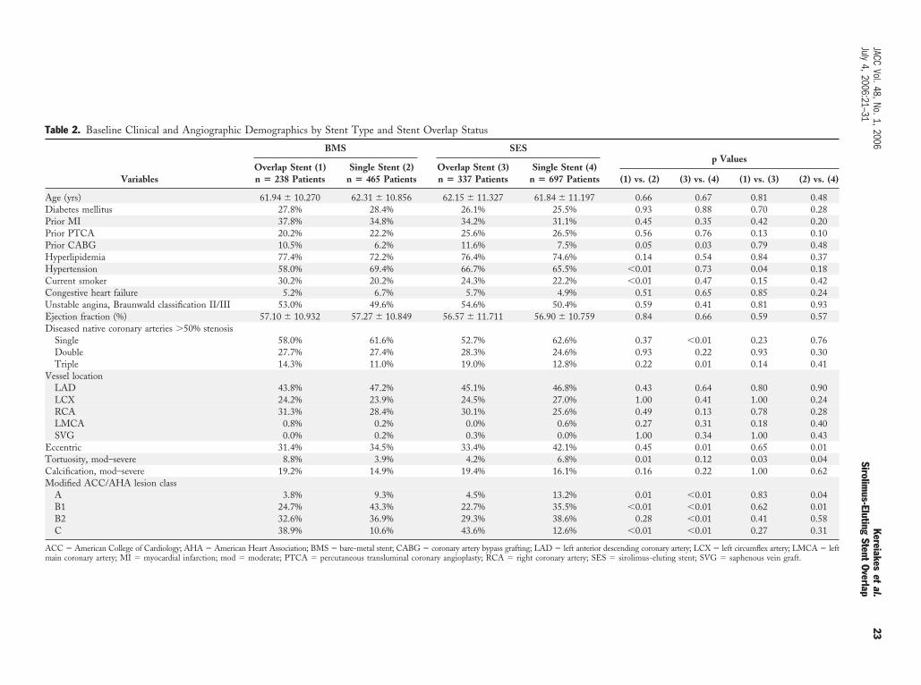
Table 2. Baseline Clinical and Angiographic Demographics by Stent Type and Stent Overlap Status
Variables
BMS SESp Values
Overlap Stent (1)n � 238 Patients
Single Stent (2)n � 465 Patients
Overlap Stent (3)n � 337 Patients
Single Stent (4)n � 697 Patients (1) vs. (2) (3) vs. (4) (1) vs. (3) (2) vs. (4)
Age (yrs) 61.94 � 10.270 62.31 � 10.856 62.15 � 11.327 61.84 � 11.197 0.66 0.67 0.81 0.48Diabetes mellitus 27.8% 28.4% 26.1% 25.5% 0.93 0.88 0.70 0.28Prior MI 37.8% 34.8% 34.2% 31.1% 0.45 0.35 0.42 0.20Prior PTCA 20.2% 22.2% 25.6% 26.5% 0.56 0.76 0.13 0.10Prior CABG 10.5% 6.2% 11.6% 7.5% 0.05 0.03 0.79 0.48Hyperlipidemia 77.4% 72.2% 76.4% 74.6% 0.14 0.54 0.84 0.37Hypertension 58.0% 69.4% 66.7% 65.5% �0.01 0.73 0.04 0.18Current smoker 30.2% 20.2% 24.3% 22.2% �0.01 0.47 0.15 0.42Congestive heart failure 5.2% 6.7% 5.7% 4.9% 0.51 0.65 0.85 0.24Unstable angina, Braunwald classification II/III 53.0% 49.6% 54.6% 50.4% 0.59 0.41 0.81 0.93Ejection fraction (%) 57.10 � 10.932 57.27 � 10.849 56.57 � 11.711 56.90 � 10.759 0.84 0.66 0.59 0.57Diseased native coronary arteries �50% stenosis
Single 58.0% 61.6% 52.7% 62.6% 0.37 �0.01 0.23 0.76Double 27.7% 27.4% 28.3% 24.6% 0.93 0.22 0.93 0.30Triple 14.3% 11.0% 19.0% 12.8% 0.22 0.01 0.14 0.41
Vessel locationLAD 43.8% 47.2% 45.1% 46.8% 0.43 0.64 0.80 0.90LCX 24.2% 23.9% 24.5% 27.0% 1.00 0.41 1.00 0.24RCA 31.3% 28.4% 30.1% 25.6% 0.49 0.13 0.78 0.28LMCA 0.8% 0.2% 0.0% 0.6% 0.27 0.31 0.18 0.40SVG 0.0% 0.2% 0.3% 0.0% 1.00 0.34 1.00 0.43
Eccentric 31.4% 34.5% 33.4% 42.1% 0.45 0.01 0.65 0.01Tortuosity, mod–severe 8.8% 3.9% 4.2% 6.8% 0.01 0.12 0.03 0.04Calcification, mod–severe 19.2% 14.9% 19.4% 16.1% 0.16 0.22 1.00 0.62Modified ACC/AHA lesion class
A 3.8% 9.3% 4.5% 13.2% 0.01 �0.01 0.83 0.04B1 24.7% 43.3% 22.7% 35.5% �0.01 �0.01 0.62 0.01B2 32.6% 36.9% 29.3% 38.6% 0.28 �0.01 0.41 0.58C 38.9% 10.6% 43.6% 12.6% �0.01 �0.01 0.27 0.31
ACC � American College of Cardiology; AHA � American Heart Association; BMS � bare-metal stent; CABG � coronary artery bypass grafting; LAD � left anterior descending coronary artery; LCX � left circumflex artery; LMCA � leftmain coronary artery; MI � myocardial infarction; mod � moderate; PTCA � percutaneous transluminal coronary angioplasty; RCA � right coronary artery; SES � sirolimus-eluting stent; SVG � saphenous vein graft.
23JACC
Vol.48,No.1,2006Kereiakes
etal.
July4,2006:21–31
Sirolimus-Eluting
StentOverlap
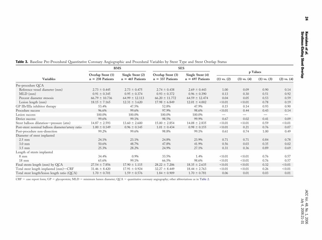
Table 3. Baseline Pre-Procedural Quantitative Coronary Angiographic and Procedural Variables by Stent Type and Stent Overlap Status
Variables
BMS SESp Values
Overlap Stent (1)n � 238 Patients
Single Stent (2)n � 465 Patients
Overlap Stent (3)n � 337 Patients
Single Stent (4)n � 697 Patients (1) vs. (2) (3) vs. (4) (1) vs. (3) (2) vs. (4)
Pre-procedure QCAReference vessel diameter (mm) 2.73 � 0.445 2.73 � 0.475 2.74 � 0.438 2.69 � 0.443 1.00 0.09 0.90 0.14MLD (mm) 0.91 � 0.345 0.95 � 0.374 0.93 � 0.372 0.96 � 0.390 0.13 0.30 0.51 0.92Percent diameter stenosis 66.79 � 10.736 64.99 � 12.113 66.20 � 11.772 64.59 � 12.474 0.04 0.05 0.53 0.59Lesion length (mm) 18.15 � 7.165 12.31 � 3.620 17.98 � 6.849 12.01 � 4.002 �0.01 �0.01 0.78 0.19
GP IIb/IIIa inhibitor therapy 53.4% 47.5% 52.8% 47.9% 0.15 0.14 0.93 0.90Procedure success 96.6% 99.6% 97.9% 98.6% �0.01 0.44 0.43 0.14Lesion success 100.0% 100.0% 100.0% 100.0% — — — —Device success 99.6% 99.1% 98.5% 99.9% 0.67 0.02 0.41 0.09Stent balloon dilatation—pressure (atm) 14.87 � 2.593 13.60 � 2.600 15.00 � 2.854 14.08 � 2.835 �0.01 �0.01 0.59 �0.01Post-stent nominal balloon diameter/artery ratio 1.00 � 0.149 0.96 � 0.145 1.01 � 0.434 0.98 � 0.155 �0.01 0.21 0.76 0.07Post-procedure non-dissection 99.2% 99.6% 98.8% 99.1% 0.61 0.74 1.00 0.49Diameter of stent implanted
2.5 mm 24.1% 23.1% 24.8% 23.9% 0.71 0.71 0.84 0.783.0 mm 50.6% 48.7% 47.8% 41.9% 0.56 0.03 0.35 0.023.5 mm 25.3% 28.2% 24.9% 27.1% 0.31 0.36 0.89 0.69
Length of stents implanted8 mm 34.4% 0.9% 33.5% 1.4% �0.01 �0.01 0.76 0.5718 mm 65.6% 99.1% 66.5% 98.6% �0.01 �0.01 0.76 0.57
Final stents length (mm) by QCA 27.54 � 7.856 17.90 � 1.115 28.22 � 7.206 18.35 � 2.635 �0.01 �0.01 0.32 �0.01Total stent length implanted (mm)—CRF 31.46 � 8.420 17.91 � 0.924 32.27 � 8.449 18.44 � 2.763 �0.01 �0.01 0.26 �0.01Total stent length/lesion length ratio (QCA) 1.70 � 0.701 1.59 � 0.576 1.84 � 0.909 1.70 � 0.781 0.06 0.01 0.03 0.01
CRF � case report form; GP � glycoprotein; MLD � minimum lumen diameter; QCA � quantitative coronary angiography; other abbreviations as in Table 2.
24Kereiakes
etal.JACC
Vol.48,No.1,2006Sirolim
us-ElutingStentOverlap
July4,2006:21–31
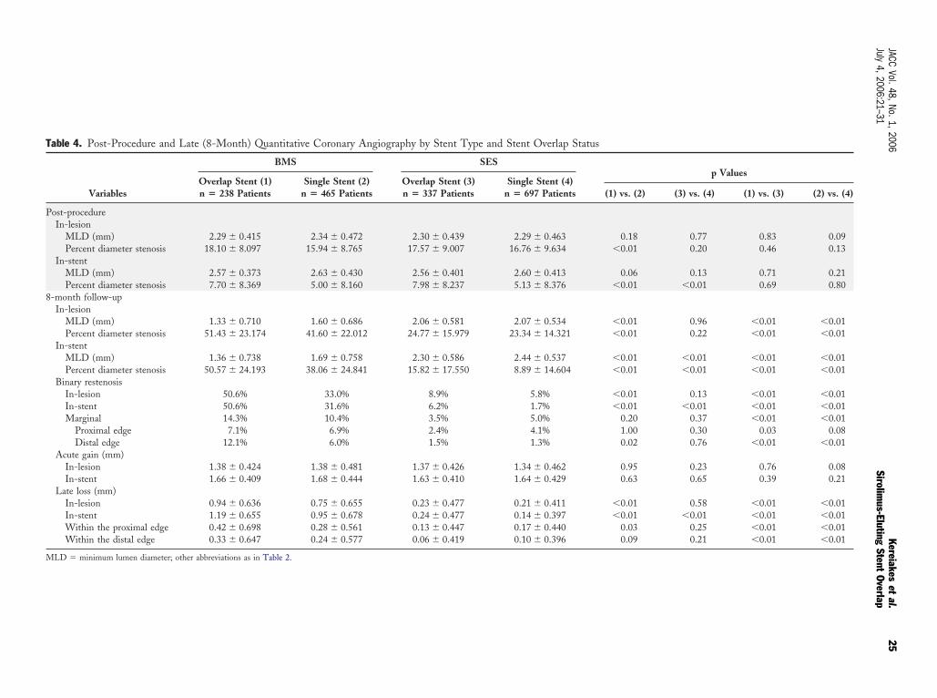
Table 4. Post-Procedure and Late (8-Month) Quantitative Coronary Angiography by Stent Type and Stent Overlap Status
Variables
BMS SESp Values
Overlap Stent (1)n � 238 Patients
Single Stent (2)n � 465 Patients
Overlap Stent (3)n � 337 Patients
Single Stent (4)n � 697 Patients (1) vs. (2) (3) vs. (4) (1) vs. (3) (2) vs. (4)
Post-procedureIn-lesion
MLD (mm) 2.29 � 0.415 2.34 � 0.472 2.30 � 0.439 2.29 � 0.463 0.18 0.77 0.83 0.09Percent diameter stenosis 18.10 � 8.097 15.94 � 8.765 17.57 � 9.007 16.76 � 9.634 �0.01 0.20 0.46 0.13
In-stentMLD (mm) 2.57 � 0.373 2.63 � 0.430 2.56 � 0.401 2.60 � 0.413 0.06 0.13 0.71 0.21Percent diameter stenosis 7.70 � 8.369 5.00 � 8.160 7.98 � 8.237 5.13 � 8.376 �0.01 �0.01 0.69 0.80
8-month follow-upIn-lesion
MLD (mm) 1.33 � 0.710 1.60 � 0.686 2.06 � 0.581 2.07 � 0.534 �0.01 0.96 �0.01 �0.01Percent diameter stenosis 51.43 � 23.174 41.60 � 22.012 24.77 � 15.979 23.34 � 14.321 �0.01 0.22 �0.01 �0.01
In-stentMLD (mm) 1.36 � 0.738 1.69 � 0.758 2.30 � 0.586 2.44 � 0.537 �0.01 �0.01 �0.01 �0.01Percent diameter stenosis 50.57 � 24.193 38.06 � 24.841 15.82 � 17.550 8.89 � 14.604 �0.01 �0.01 �0.01 �0.01
Binary restenosisIn-lesion 50.6% 33.0% 8.9% 5.8% �0.01 0.13 �0.01 �0.01In-stent 50.6% 31.6% 6.2% 1.7% �0.01 �0.01 �0.01 �0.01Marginal 14.3% 10.4% 3.5% 5.0% 0.20 0.37 �0.01 �0.01
Proximal edge 7.1% 6.9% 2.4% 4.1% 1.00 0.30 0.03 0.08Distal edge 12.1% 6.0% 1.5% 1.3% 0.02 0.76 �0.01 �0.01
Acute gain (mm)In-lesion 1.38 � 0.424 1.38 � 0.481 1.37 � 0.426 1.34 � 0.462 0.95 0.23 0.76 0.08In-stent 1.66 � 0.409 1.68 � 0.444 1.63 � 0.410 1.64 � 0.429 0.63 0.65 0.39 0.21
Late loss (mm)In-lesion 0.94 � 0.636 0.75 � 0.655 0.23 � 0.477 0.21 � 0.411 �0.01 0.58 �0.01 �0.01In-stent 1.19 � 0.655 0.95 � 0.678 0.24 � 0.477 0.14 � 0.397 �0.01 �0.01 �0.01 �0.01Within the proximal edge 0.42 � 0.698 0.28 � 0.561 0.13 � 0.447 0.17 � 0.440 0.03 0.25 �0.01 �0.01Within the distal edge 0.33 � 0.647 0.24 � 0.577 0.06 � 0.419 0.10 � 0.396 0.09 0.21 �0.01 �0.01
MLD � minimum lumen diameter; other abbreviations as in Table 2.
25JACC
Vol.48,No.1,2006Kereiakes
etal.
July4,2006:21–31
Sirolimus-Eluting
StentOverlap
wr
8csBws
R
BpaccSomAvBvsfptltb4t
8(cg0asipwocLdoalob
asMomtwoAvra
Fs
26 Kereiakes et al. JACC Vol. 48, No. 1, 2006Sirolimus-Eluting Stent Overlap July 4, 2006:21–31
ith a stay criterion of 0.10 in the multiple logisticegression model.
All statistical analyses were performed using SAS Version.2 (SAS Institute, Cary, North Carolina). In the multipleomparisons of QCA and clinical end points among theubgroups by treatment and overlap stent or single stent use,onferroni adjusted significance level of 0.0125 (0.05/4)ere applied. For any other comparisons, a statistical
ignificance level of 0.05 was used.
ESULTS
aseline clinical and angiographic demographic variables foratients treated with either single or overlapped SES as wells BMS controls are shown in Table 2. The only significantlinical difference observed between stent overlap patientohorts was a higher incidence of systemic hypertension inES treated patients. The only significant clinical differencebserved between single-stent–treated patient cohorts was aore frequent history of smoking in SES-treated patients.ll other clinical variables were similar between single
ersus overlap or SES versus BMS-treated patient groups.aseline quantitative coronary angiographic and proceduralariables are shown in Table 3. Baseline angiography wasimilar in overlap-stent–treated patient cohorts except for morerequent moderate or severe vessel tortuosity in BMS-treatedatients. Procedural variables were similar for overlap stent-reated patients except for a greater total stent-length-to-esion-length ratio determined by QCA in SES-treated pa-ients. The length of overlapped stent segment (mean � SD)y QCA was assessed only in the SIRIUS trial and was.6 � 9.1 mm for SES, 4.0 � 4.9 mm for BMS. Quanti-ative coronary angiographic variables post-procedure and at
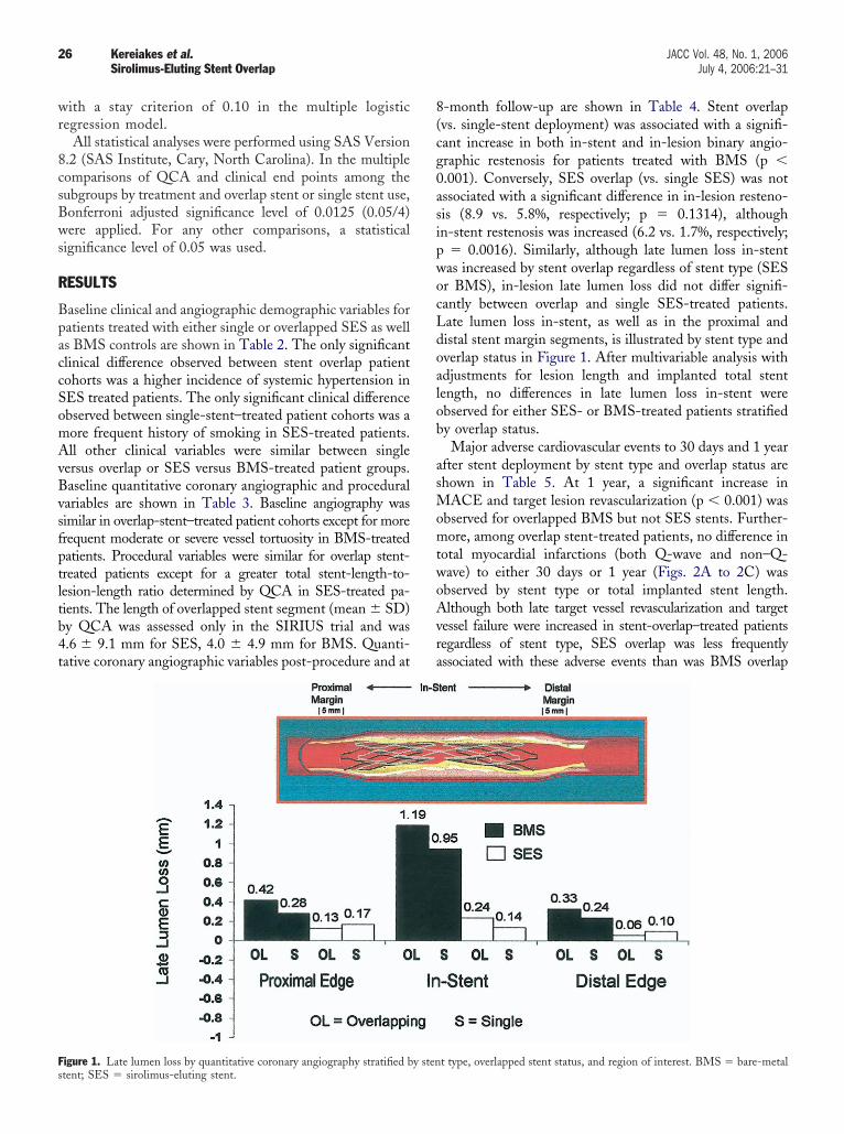
igure 1. Late lumen loss by quantitative coronary angiography stratified by stentent; SES � sirolimus-eluting stent.
-month follow-up are shown in Table 4. Stent overlapvs. single-stent deployment) was associated with a signifi-ant increase in both in-stent and in-lesion binary angio-raphic restenosis for patients treated with BMS (p �.001). Conversely, SES overlap (vs. single SES) was notssociated with a significant difference in in-lesion resteno-is (8.9 vs. 5.8%, respectively; p � 0.1314), althoughn-stent restenosis was increased (6.2 vs. 1.7%, respectively;
� 0.0016). Similarly, although late lumen loss in-stentas increased by stent overlap regardless of stent type (SESr BMS), in-lesion late lumen loss did not differ signifi-antly between overlap and single SES-treated patients.ate lumen loss in-stent, as well as in the proximal andistal stent margin segments, is illustrated by stent type andverlap status in Figure 1. After multivariable analysis withdjustments for lesion length and implanted total stentength, no differences in late lumen loss in-stent werebserved for either SES- or BMS-treated patients stratifiedy overlap status.Major adverse cardiovascular events to 30 days and 1 year
fter stent deployment by stent type and overlap status arehown in Table 5. At 1 year, a significant increase in
ACE and target lesion revascularization (p � 0.001) wasbserved for overlapped BMS but not SES stents. Further-ore, among overlap stent-treated patients, no difference in
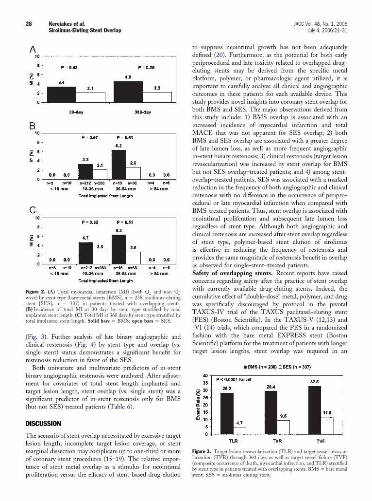
otal myocardial infarctions (both Q-wave and non–Q-ave) to either 30 days or 1 year (Figs. 2A to 2C) wasbserved by stent type or total implanted stent length.lthough both late target vessel revascularization and target
essel failure were increased in stent-overlap–treated patientsegardless of stent type, SES overlap was less frequentlyssociated with these adverse events than was BMS overlap
t type, overlapped stent status, and region of interest. BMS � bare-metal
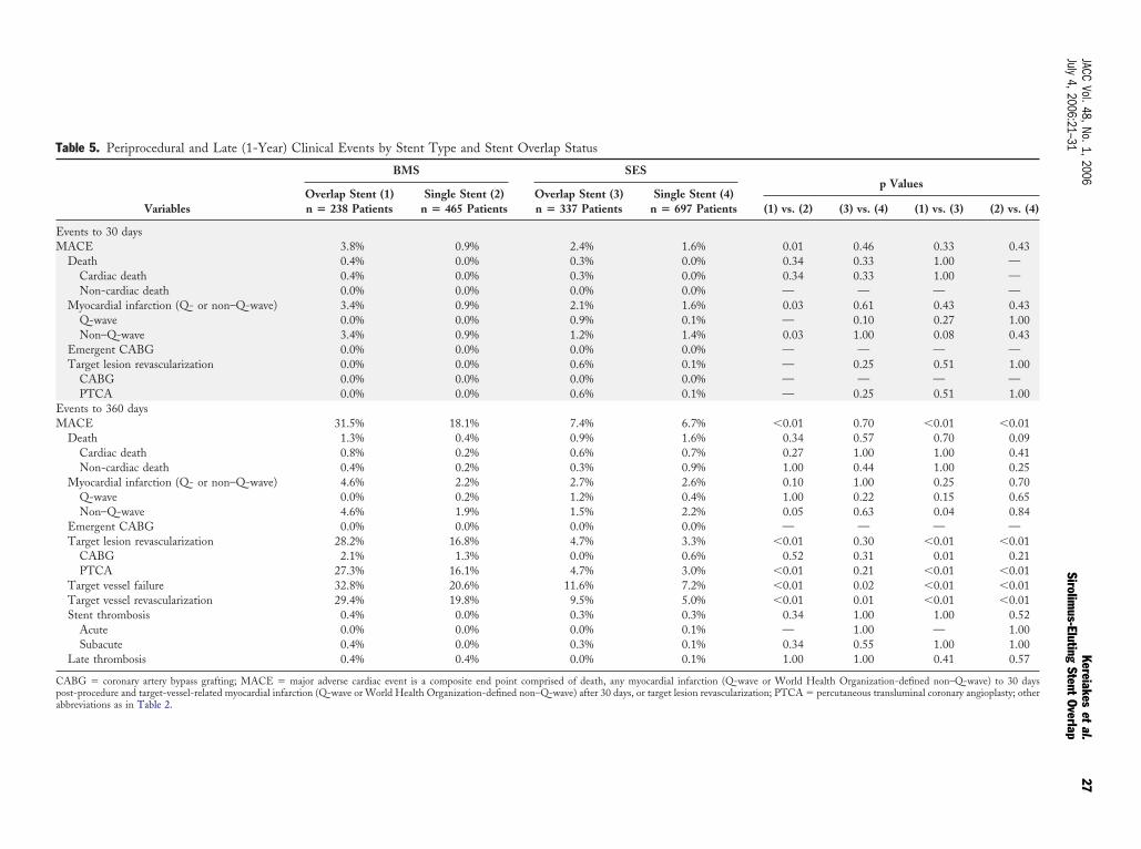
Table 5. Periprocedural and Late (1-Year) Clinical Events by Stent Type and Stent Overlap Status
Variables
BMS SESp Values
Overlap Stent (1)n � 238 Patients
Single Stent (2)n � 465 Patients
Overlap Stent (3)n � 337 Patients
Single Stent (4)n � 697 Patients (1) vs. (2) (3) vs. (4) (1) vs. (3) (2) vs. (4)
Events to 30 daysMACE 3.8% 0.9% 2.4% 1.6% 0.01 0.46 0.33 0.43
Death 0.4% 0.0% 0.3% 0.0% 0.34 0.33 1.00 —Cardiac death 0.4% 0.0% 0.3% 0.0% 0.34 0.33 1.00 —Non-cardiac death 0.0% 0.0% 0.0% 0.0% — — — —
Myocardial infarction (Q- or non–Q-wave) 3.4% 0.9% 2.1% 1.6% 0.03 0.61 0.43 0.43Q-wave 0.0% 0.0% 0.9% 0.1% — 0.10 0.27 1.00Non–Q-wave 3.4% 0.9% 1.2% 1.4% 0.03 1.00 0.08 0.43
Emergent CABG 0.0% 0.0% 0.0% 0.0% — — — —Target lesion revascularization 0.0% 0.0% 0.6% 0.1% — 0.25 0.51 1.00
CABG 0.0% 0.0% 0.0% 0.0% — — — —PTCA 0.0% 0.0% 0.6% 0.1% — 0.25 0.51 1.00
Events to 360 daysMACE 31.5% 18.1% 7.4% 6.7% �0.01 0.70 �0.01 �0.01
Death 1.3% 0.4% 0.9% 1.6% 0.34 0.57 0.70 0.09Cardiac death 0.8% 0.2% 0.6% 0.7% 0.27 1.00 1.00 0.41Non-cardiac death 0.4% 0.2% 0.3% 0.9% 1.00 0.44 1.00 0.25
Myocardial infarction (Q- or non–Q-wave) 4.6% 2.2% 2.7% 2.6% 0.10 1.00 0.25 0.70Q-wave 0.0% 0.2% 1.2% 0.4% 1.00 0.22 0.15 0.65Non–Q-wave 4.6% 1.9% 1.5% 2.2% 0.05 0.63 0.04 0.84
Emergent CABG 0.0% 0.0% 0.0% 0.0% — — — —Target lesion revascularization 28.2% 16.8% 4.7% 3.3% �0.01 0.30 �0.01 �0.01
CABG 2.1% 1.3% 0.0% 0.6% 0.52 0.31 0.01 0.21PTCA 27.3% 16.1% 4.7% 3.0% �0.01 0.21 �0.01 �0.01
Target vessel failure 32.8% 20.6% 11.6% 7.2% �0.01 0.02 �0.01 �0.01Target vessel revascularization 29.4% 19.8% 9.5% 5.0% �0.01 0.01 �0.01 �0.01Stent thrombosis 0.4% 0.0% 0.3% 0.3% 0.34 1.00 1.00 0.52
Acute 0.0% 0.0% 0.0% 0.1% — 1.00 — 1.00Subacute 0.4% 0.0% 0.3% 0.1% 0.34 0.55 1.00 1.00
Late thrombosis 0.4% 0.4% 0.0% 0.1% 1.00 1.00 0.41 0.57
CABG � coronary artery bypass grafting; MACE � major adverse cardiac event is a composite end point comprised of death, any myocardial infarction (Q-wave or World Health Organization-defined non–Q-wave) to 30 dayspost-procedure and target-vessel-related myocardial infarction (Q-wave or World Health Organization-defined non–Q-wave) after 30 days, or target lesion revascularization; PTCA � percutaneous transluminal coronary angioplasty; otherabbreviations as in Table 2.
27JACC
Vol.48,No.1,2006Kereiakes
etal.
July4,2006:21–31
Sirolimus-Eluting
StentOverlap
(csr
bmts(
D
Tlmotp
tdpepiosbtiMBoirborrcBnrcoipaScwcwT(-fSt
Fws(it
Fl(
28 Kereiakes et al. JACC Vol. 48, No. 1, 2006Sirolimus-Eluting Stent Overlap July 4, 2006:21–31
Fig. 3). Further analysis of late binary angiographic andlinical restenosis (Fig. 4) by stent type and overlap (vs.ingle stent) status demonstrates a significant benefit forestenosis reduction in favor of the SES.
Both univariate and multivariate predictors of in-stentinary angiographic restenosis were analyzed. After adjust-ent for covariates of total stent length implanted and
arget lesion length, stent overlap (vs. single stent) was aignificant predictor of in-stent restenosis only for BMSbut not SES) treated patients (Table 6).
ISCUSSION
he scenario of stent overlap necessitated by excessive targetesion length, incomplete target lesion coverage, or stent
arginal dissection may complicate up to one-third or moref coronary stent procedures (15–19). The relative impor-ance of stent metal overlap as a stimulus for neointimal
igure 2. (A) Total myocardial infarction (MI) (both Q- and non–Q-ave) by stent type (bare-metal stents [BMS], n � 238; sirolimus-eluting
tent [SES], n � 337) in patients treated with overlapping stents.B) Incidence of total MI at 30 days by stent type stratified by totalmplanted stent length. (C) Total MI at 360 days by stent type stratified byotal implanted stent length. Solid bars � BMS; open bars � SES.
roliferation versus the efficacy of stent-based drug elutionbs
o suppress neointimal growth has not been adequatelyefined (20). Furthermore, as the potential for both earlyeriprocedural and late toxicity related to overlapped drug-luting stents may be derived from the specific metallatform, polymer, or pharmacologic agent utilized, it ismportant to carefully analyze all clinical and angiographicutcomes in these patients for each available device. Thistudy provides novel insights into coronary stent overlap foroth BMS and SES. The major observations derived fromhis study include: 1) BMS overlap is associated with anncreased incidence of myocardial infarction and total
ACE that was not apparent for SES overlap; 2) bothMS and SES overlap are associated with a greater degreef late lumen loss, as well as more frequent angiographicn-stent binary restenosis; 3) clinical restenosis (target lesionevascularization) was increased by stent overlap for BMSut not SES-overlap–treated patients; and 4) among stent-verlap–treated patients, SES was associated with a markededuction in the frequency of both angiographic and clinicalestenosis with no difference in the occurrence of peripro-edural or late myocardial infarction when compared withMS-treated patients. Thus, stent overlap is associated witheointimal proliferation and subsequent late lumen lossegardless of stent type. Although both angiographic andlinical restenosis are increased after stent overlap regardlessf stent type, polymer-based stent elution of sirolimuss effective in reducing the frequency of restenosis androvides the same magnitude of restenosis benefit in overlaps observed for single-stent–treated patients.afety of overlapping stents. Recent reports have raisedoncerns regarding safety after the practice of stent overlapith currently available drug-eluting stents. Indeed, the
umulative effect of “double-dose” metal, polymer, and drugas specifically discouraged by protocol in the pivotalAXUS-IV trial of the TAXUS paclitaxel-eluting stent
PES) (Boston Scientific). In the TAXUS-V (12,13) andVI (14) trials, which compared the PES in a randomizedashion with the bare metal EXPRESS stent (Bostoncientific) platform for the treatment of patients with longerarget lesion lengths, stent overlap was required in an
igure 3. Target lesion revascularization (TLR) and target vessel revascu-arization (TVR) through 360 days as well as target vessel failure (TVF)composite occurrence of death, myocardial infarction, and TLR) stratified
y stent type in patients treated with overlapping stents. BMS � bare metaltent; SES � sirolimus-eluting stent.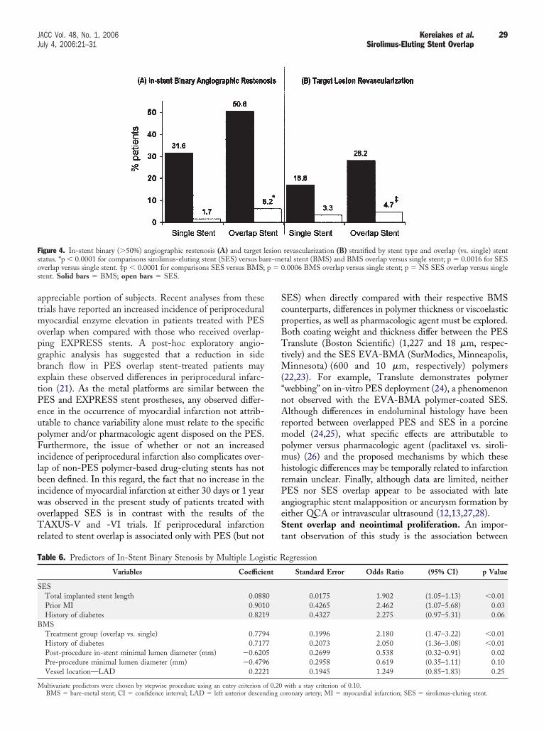
atmopgbetPeupFilbiwoTr
ScpBTtM(“nArmpmhrPaeSt
Fso p � 0s
T
S
B
M
29JACC Vol. 48, No. 1, 2006 Kereiakes et al.July 4, 2006:21–31 Sirolimus-Eluting Stent Overlap
ppreciable portion of subjects. Recent analyses from theserials have reported an increased incidence of periproceduralyocardial enzyme elevation in patients treated with PES
verlap when compared with those who received overlap-ing EXPRESS stents. A post-hoc exploratory angio-raphic analysis has suggested that a reduction in sideranch flow in PES overlap stent-treated patients mayxplain these observed differences in periprocedural infarc-ion (21). As the metal platforms are similar between theES and EXPRESS stent prostheses, any observed differ-nce in the occurrence of myocardial infarction not attrib-table to chance variability alone must relate to the specificolymer and/or pharmacologic agent disposed on the PES.urthermore, the issue of whether or not an increased
ncidence of periprocedural infarction also complicates over-ap of non-PES polymer-based drug-eluting stents has noteen defined. In this regard, the fact that no increase in thencidence of myocardial infarction at either 30 days or 1 yearas observed in the present study of patients treated withverlapped SES is in contrast with the results of theAXUS-V and -VI trials. If periprocedural infarction
elated to stent overlap is associated only with PES (but not
igure 4. In-stent binary (�50%) angiographic restenosis (A) and target ltatus. *p � 0.0001 for comparisons sirolimus-eluting stent (SES) versus baverlap versus single stent. ‡p � 0.0001 for comparisons SES versus BMS;tent. Solid bars � BMS; open bars � SES.
able 6. Predictors of In-Stent Binary Stenosis by Multiple Logi
Variables Coefficie
ESTotal implanted stent length 0.088Prior MI 0.901History of diabetes 0.821
MSTreatment group (overlap vs. single) 0.779History of diabetes 0.717Post-procedure in-stent minimal lumen diameter (mm) �0.620Pre-procedure minimal lumen diameter (mm) �0.479Vessel location—LAD 0.222
ultivariate predictors were chosen by stepwise procedure using an entry criterion of 0.20BMS � bare-metal stent; CI � confidence interval; LAD � left anterior descending c
ES) when directly compared with their respective BMSounterparts, differences in polymer thickness or viscoelasticroperties, as well as pharmacologic agent must be explored.oth coating weight and thickness differ between the PESranslute (Boston Scientific) (1,227 and 18 �m, respec-
ively) and the SES EVA-BMA (SurModics, Minneapolis,innesota) (600 and 10 �m, respectively) polymers
22,23). For example, Translute demonstrates polymerwebbing” on in-vitro PES deployment (24), a phenomenonot observed with the EVA-BMA polymer-coated SES.lthough differences in endoluminal histology have been
eported between overlapped PES and SES in a porcineodel (24,25), what specific effects are attributable to
olymer versus pharmacologic agent (paclitaxel vs. siroli-us) (26) and the proposed mechanisms by which these
istologic differences may be temporally related to infarctionemain unclear. Finally, although data are limited, neitherES nor SES overlap appear to be associated with latengiographic stent malapposition or aneurysm formation byither QCA or intravascular ultrasound (12,13,27,28).tent overlap and neointimal proliferation. An impor-
ant observation of this study is the association between
revascularization (B) stratified by stent type and overlap (vs. single) stenttal stent (BMS) and BMS overlap versus single stent; p � 0.0016 for SES.0006 BMS overlap versus single stent; p � NS SES overlap versus single
egression
Standard Error Odds Ratio (95% CI) p Value
0.0175 1.902 (1.05–1.13) �0.010.4265 2.462 (1.07–5.68) 0.030.4327 2.275 (0.97–5.31) 0.06
0.1996 2.180 (1.47–3.22) �0.010.2073 2.050 (1.36–3.08) �0.010.2699 0.538 (0.32–0.91) 0.020.2958 0.619 (0.35–1.11) 0.100.1945 1.249 (0.85–1.83) 0.25
esionre-me
stic R
nt
009
47561
with a stay criterion of 0.10.oronary artery; MI � myocardial infarction; SES � sirolimus-eluting stent.
sBaidrsnvssdordpSsatcsattltfpfciscrMwdMSootSSasfpartcsla
bfctncCSswbnnasmcpsepoi
RTfAf
R
1
1
30 Kereiakes et al. JACC Vol. 48, No. 1, 2006Sirolimus-Eluting Stent Overlap July 4, 2006:21–31
tent overlap and neointimal proliferation for both SES andMS. For both stent platforms, late quantitative coronaryngiographic measurements in-stent demonstrate that min-mum lumen diameter was significantly less, while percentiameter stenosis, late lumen loss, and binary angiographicestenosis were significantly greater in overlapped versusingle stents. Nevertheless, the relative magnitude of reste-osis reduction observed in patients treated with single SESersus BMS was maintained in the comparison of overlaptent-treated patients. Thus, polymer-based elution ofirolimus appears to be effective in reducing the enhancedegree of neointimal proliferation and late lumen lossbserved in overlapped stents. Of note, after multivariableegression analysis with adjustments for total stent lengtheployed and target lesion length, stent overlap remained aredictor of in-stent restenosis for BMS but not SES.tent thrombosis. Although both stent length (29) andtent overlap (30) have been incriminated as being associ-ted with an increased incidence of drug-eluting stenthrombosis, no such relationship was identified in theurrent study despite complete follow-up to one year in allubjects. Combination oral antiplatelet therapy with aspirinnd clopidogrel was mandated by study protocol for onlyhree months for the trials included in this analysis. Al-hough concerns that stent overlap might increase theikelihood of subsequent stent strut malapposition, and thushe propensity for thrombosis, late follow-up (to 1,080 days)rom the cumulative experience in four randomized com-arative trials of SES versus BMS demonstrates no evidenceor an increase in stent thrombosis (M. Leon, personalommunication, May 2005). Similarly, no evidence for anncrease in localized aneurysm formation at the overlapegment was observed by quantitative angiography in theurrent study or by intravascular ultrasound in previouseports (31,32).
ACE related to stent overlap. Although stent overlapas associated with an increase in total MACE to both 30ays and 1 year for BMS-treated patients, no increase inACE was observed in patients who received overlapping
ES. The major differences between patients treated withverlapping stents by stent type were in the frequencyccurrence of target lesion and target vessel revasculariza-ion, as well as target vessel failure (increased in BMS- vs.ES-treated cohorts).tudy limitations. The major limitation of the presentnalysis, which compares patients treated with overlappingtents, is that the decision to deploy stents in an overlappingashion was not randomly assigned. However, the factorsrompting overlapping stent deployment were not predict-ble in the majority of cases, and the portion of patients whoequired stent overlap was similar between SES- and BMS-reated patients among those enrolled into randomizedomparative trials. Furthermore, the present study repre-ents the largest experience to date for patients with over-apping coronary stents that includes complete angiographic
nd late clinical follow-up. In addition, although data fromoth randomized and non-randomized trials were pooledor this report, separate detailed analyses demonstrated alllinical and angiographic end points to be similar betweenhe three randomized trials and all five trials (Appendix). Ofote, patients enrolled into all five trials had similar baselinelinical and angiographic characteristics.onclusions. This analysis of patients with overlappingES or BMS platforms demonstrates that stent overlap is atimulus for neointimal proliferation and late lumen lossith subsequent restenosis regardless of stent type. Stent-ased polymer elution of sirolimus is effective in suppressingeointimal hyperplasia and provides a similar degree of reste-osis benefit when compared with BMS in both overlappednd single-stent–treated patient cohorts. Sirolimus-elutingtent overlap is not associated with an increased incidence ofyocardial infarction or MACE in late follow-up when
ompared with either single SES-treated patients or withatients treated with single or overlapped BMS. Thus, thetrategy of SES overlap, when required, is both safe andfficacious in reducing restenosis in comparison with a BMSrosthesis. Careful attention should be paid to achieveptimal stent deployment in those cases where stent overlaps required.
eprint requests and correspondence: Dr. Dean J. Kereiakes,he Heart Center of Greater Cincinnati and The Lindner Center
or Research and Education at the Christ Hospital, 2123 Auburnvenue, Suite 424, Cincinnati, Ohio 45219. E-mail: lindner@
use.net.
EFERENCES
1. Ellis SG, Savage M, Fischman D, et al. Restenosis after placement ofPalmaz-Schatz stents in native coronary arteries. Initial results of amulticenter experience. Circulation 1992;86:1836–44.
2. Kastrati A, Elezi S, Dirschinger J, Hadamitzky M, Neumann FJ,Schomig A. Influence of lesion length on restenosis after coronarystent placement. Am J Cardiol 1999;83:1617–22.
3. Kastrati A, Schomig A, Elezi S, et al. Predictive factors of restenosisafter coronary stent placement. J Am Coll Cardiol 1997;30:1428–36.
4. Haude M, Erbel R, Straub U, Dietz U, Meyer J. Short and long termresults after intracoronary stenting in human coronary arteries: mono-centre experience with balloon-expandable Palmaz-Schatz stent. BrHeart J 1991;66:337–45.
5. Lee SH, Jang Y, Oh SJ, et al. Overlapping vs. one long stenting in longcoronary lesions. Catheter Cardiovasc Interv 2004;62:298–302.
6. Hoffmann R, Herrmann G, Silber S, et al. Randomized comparison ofsuccess and adverse event rates and cost effectiveness of one long versustwo short stents for treatment of long coronary narrowings. Am JCardiol 2002;90:460–4.
7. Kornowski R, Mehran R, Hong MK, et al. Procedural results and lateclinical outcomes after placement of three or more stents in singlecoronary lesions. Circulation 1998;97:1355–61.
8. Bauters C, Hubert E, Prat A, et al. Predictors of restenosis aftercoronary stent implantation. J Am Coll Cardiol 1998;31:1291–8.
9. Pan M, deLezo JS, Medina A, et al. Influence of stent treatmentstrategies in the long-term outcome of patients with long diffusecoronary lesions. Cathet Cardiovasc Intervent 2003;58:293–300.
0. Kastrati A, Schomig A, Elezi S, et al. Predictive factors of restenosisafter coronary stent placement. J Am Coll Cardiol 1997;36:1428–36.
1. Stone GW, Ellis SG, Cannon L, et al. Comparison of a polymer-based paclitaxel-eluting stent with a bare metal stent in patients with
complex coronary artery disease—a randomized controlled trial.JAMA 2005:294:1215–23.
1
1
1
1
1
1
1
1
2
2
2
2
2
2
2
2
2
2
3
3
3
A
Fi
31JACC Vol. 48, No. 1, 2006 Kereiakes et al.July 4, 2006:21–31 Sirolimus-Eluting Stent Overlap
2. Dawkins KD. TAXUS VI 9 month results—in-depth analysis of longlesions. Presented at: EuroPCR; Paris, France: May 25, 2004.
3. Grube E. Taxus VI. Randomized trial of moderate-rate releasepolymer-based paclitaxel-eluting Taxus stent for the treatment oflonger lesions. Presented at: Late Breaking Trials, EuroPCR; Paris,France: May 24, 2005.
4. Moses JW, Leon MB, Popma J, et al. Sirolimus-eluting stents versusstandard stents in patients with stenosis in a native coronary artery.N Engl J Med 2003;349:1315–23.
5. Schofer J, Shluter M, Gershlick AH, et al. Sirolimus-eluting stents fortreatment of patients with long atherosclerotic lesions in small coro-nary arteries: double-blind, randomised controlled trial (E-SIRIUS).Lancet 2003;362:1093–9.
6. Schampaert E, Cohen EA, Schlüter M, et al. The Canadian study ofthe sirolimus-eluting stent in the treatment of patients with long denovo lesions in small native coronary arteries (C-SIRIUS). J Am CollCardiol 2004;43:1110–5.
7. Schlüter M, Schofer J, Gershlick AH, Schampaert E, Wijns W,Breithardt G, for the E- and C-SIRIUS Investigators. Direct stentingof native de novo coronary artery lesions with the sirolimus-elutingstent—a post hoc subanalysis of the pooled E- and C-SIRIUS trials.J Am Coll Cardiol 2005;45:10–3.
8. Moses JW, Leon MB, Popma JJ, Cohen SA, Kuntz RE. DIRECT:Direct stenting using the sirolimus-eluting Bx-Velocity stent: proce-dural, clinical and angiographic outcomes compared to a predilitationstrategy. Presented at: Annual American College of CardiologyMeeting, Late Breaking Trials; New Orleans, LA: March 7, 2004.
9. Meier B, Sousa JE, Guagliumi G, et al. Sirolimus-eluting coronarystents in small vessels. Am Heart J 2006;151:1019.e1–1019.e7.
0. Holmes DR Jr., Leon MB, Moses JW, et al. Analysis of 1-year clinicaloutcomes in the SIRIUS trial. A randomized trial of a sirolimus-eluting stent versus a standard stent in patients at high risk for coronaryrestenosis. Circulation 2004;109:634–42.
1. Popma JJ. Angiographic analysis of stent overlap/multiple stents fromTAXUS V; Annual American College of Cardiology. Presented at:Late Breaking Trials; Orlando, FL: March 6, 2005.
2. Boston Scientific Corporation Premarket Application Panel Document—Summary of Safety and Effectiveness. Available at: http://www.fda.gov/ohrms/dockets/ac/03/briefing/4005b1_02_SSE.pdf. Accessed September
14, 2005. t3. Colombo A, Cosgrave J. Paclitaxel-eluting stents in complex lesions.JAMA 2005;294:1268–70.
4. Finn AV, Kolodgie FD, Virmani R, et al. Differential response ofdelayed healing and persistent inflammation at sites of overlappingsirolimus- or paclitaxel-eluting stents. Circulation 2005;112:270–8.
5. Perin EC, Silva GV, Faloticco R, et al. Differential inflammatory effectof sirolimus eluting stent and paclitaxel eluting stent in porcine chronicischemic model. Am J Cardiol 2005;96:168H.
6. Farb A, Heller PF, Virmani R, et al. Pathological analysis of localdelivery of paclitaxel via a polymer-coated stent. Circulation 2001;104:473–9.
7. Stone GW, Ellis SG, Cox DA, et al. A polymer-based, paclitaxel-eluting stent in patients with coronary artery disease. New Engl J Med2004;350:221–31.
8. Stone GW, Ellis SG, Cox DA, et al. One-year clinical results with theslow-release, polymer-based, paclitaxel-eluting TAXUS stent. TheTAXUS-IV trial. Circulation 2004;109:1942–7.
9. Moreno R, Fernandez C, Hernandez R, et al. Drug-eluting stentthrombosis: results from a pooled analysis including 10 randomizedstudies. J Am Coll Cardiol 2005;45:954–9.
0. Meucci F, Falchetti E, Vittori G, et al. Increased risk of sub-acutethrombosis after overlapping with drug-eluting stents: an analysis fromthe real-world eluting stent comparative Italian retrospective evalua-tion (RECIPE) study (abstr). J Am Coll Cardiol 2005;45 SupplA:34A.
1. Munoz JS, Abizaid A, Mintz GS, et al. Intravascular ultrasound studyof effects of overlapping sirolimus-eluting stents. Am J Cardiol2004;93:470–3.
2. Ruiz-Nodar JM, Carrillo P, Frutos A, et al. Intravascular ultrasoundstudy of patients who underwent overlapping of paclitaxel and siroli-mus stents: comparison with the results obtained after overlappingwith same drug-eluting stent (abstr). J Am Coll Cardiol 2005;49 SupplA:58A.
PPENDIX
or the analysis of key independent and response variablesn SIRIUS, C- SIRIUS, and E- SIRIUS trials, please see
he online version of this article.