covered stents in patients with complex aortic coarctations
TRANSCRIPT
Covered stents in patients with complexaortic coarctationsGianfranco Butera, MD, PhD,a Luciane Piazza, MD,a Massimo Chessa, MD, PhD,a Diana Gabriella Negura, MD,a
Luca Rosti, MD,a Raul Abella, MD,a Angelica Delogu, MD,b Claudia Condoluci, MD,c Andrea Magherini, MD,d andMario Carminati, MDa San Donato Milanese, Firenze, and Roma, Italy
Background There are limited data in the literature about the use of covered stent in patients with aortic coarctation.
Methods Between January 2004 and September 2006, we implanted covered Cheatham-Platinum stents in 33patients with complex aortic coarctation (23 men, median age 13 years, range 6-66 years). Twenty subjects had nativeaortic coarctation, whereas 13 had recoarctation. All procedures were performed under general anesthesia andorotracheal intubation.
Results The stents used ranged from 22 to 45 mm in length. The mean fluoroscopy and procedure times were 14 ± 6 and74 ± 15 minutes, respectively. After implantation, the gradient across the stenosis decreased significantly (pre stent: medianvalue 39 mm Hg [range 20-75 mm Hg] vs post stent: median value 0 mm Hg [range 0-12 mm Hg] [P b .0001]). Vesseldiameter increased from a median value of 5 mm (range 0-11) to a median value of 15 mm (range 10-25) (P b .0001).The stents were placed in the correct position in all subjects. No complications occurred, and on angiographic control, thestenoses had been relieved and the aneurysms completely excluded. During a median follow-up of 12 months (1-40 months),the results were stable without complications. One patient developed intrastent restenosis due to a significant endothelialproliferation that was successfully treated by high-pressure balloon angioplasty.
Conclusions Covered Cheatham-Platinum stents are promising tools for the treatment of complex aortic coarctation.(Am Heart J 2007;154:795-800.)
Surgery or standard transcatheter approaches(balloon angioplasty or bare stent implantation) can beassociated with significant morbidity and mortality inpatients with complex aortic coarctations (ie, subatreticaortic coarctation or associated with aneurysm). Barestents have been used in all sites in patients withcongenital heart diseases, and large series and follow-upstudies are reported in literature.1-3 However, evenwith these stents, aneurysms may form, or aortic rupturemay occur.3
Covered stents are currently widely used in thetreatment of abdominal and thoracic atheroscleroticaneurysm in adults.4 However, there is limited experi-ence on the use of covered stents in congenital heartdiseases,5-9 a setting in which their role remains to beclearly defined.
From the aPediatric Cardiology, Policlinico San Donato I.R.C.C.S., San Donato Milanese,Italy, bPediatric Cardiology, Università Cattolica Sacro Cuore, Roma, Italy, cPediatricCardiology, I.R.C.C.S. San Raffaele, Pisana, Roma, Italy, and dDepartment of Pediatrics,Università di Firenze, Firenze, Italy.Submitted November 28, 2006; accepted June 17, 2007.Reprint requests: Gianfranco Butera, MD, PhD, Pediatric Cardiology, Via Morandi, 30,20098 San Donato Milanese (MI), Italy.E-mail: [email protected]/$ - see front matter© 2007, Mosby, Inc. All rights reserved.doi:10.1016/j.ahj.2007.06.018
In the present study, we report our experience of33 patients in whom covered Cheatham-Platinum(CP) stents were used for the treatment of complexaortic coarctation.
MethodsThe data of 33 patients (23 males) who underwent implanta-
tion of covered CP stents (NuMED, Hopkinton, NY) forcomplex aortic coarctation were collected prospectivelybetween January 2004 and September 2006.We defined as complex an aortic coarctation or
recoarctation when it was subatretic, associated with ananeurysm, associated with an irregular wall, associated withdilation of the ascending aorta, associated with patent ductusarteriosus, or occurring in patients previously treated with theuse of surgical patches.The median age and weight at the time of the stent
implantation were 13 years (range 6-66 years) and 60 kg(range 22-110 kg), respectively. Informed consent wasobtained from all the patients or their parents.Twenty subjects had native aortic coarctation: 7 had
subatretic coarctation (Figure 1), 10 had severe aortic coarcta-tion with irregular wall, 2 had an associated aneurysm, and 1 hadan associated patent ductus arteriosus (Figure 2).Thirteen subjects had aortic recoarctation: in 8 subjects, aortic
recoarctation occurred after surgery. Two patients had anassociated aneurysm; 3 had severe aortic recoarctation after

Figure 1
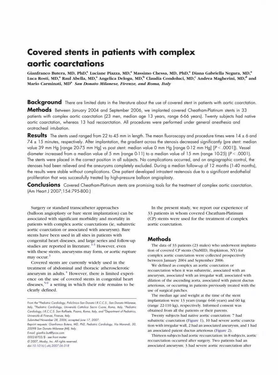
Subatretic native aortic coarctation. A, thoracic aortography in the anteroposterior view with the multipurpose catheter near the coarctation. Bthoracic aortography in the anteroposterior view with pigtail across the coarctation. C, Predilation by using balloon angioplasty. D, The stent ideployed and no residual stenosis is present.
796 Butera et alAmerican Heart Journal
October 2007
having been treated surgically by the use of e-PTFE patch; finally,3 of these patients had severe aortic recoarctation and an intimalirregularity of the aortic wall.In another 5 subjects, recoarctation occurred after percuta-
neous treatment, and it was associated in all subjects withaneurysm formation. Of 5 subjects, 3 had a previous balloonangioplasty, whereas 2 had a stent implantation for subatreticaortic coarctation.
Implantation techniqueThe patients underwent general anesthesia and orotracheal
intubation. Intravenous heparin (100 IU/kg, maximum of5000 IU) was given immediately after femoral artery cannula-tion, which was achieved by using an 8F introducer. Thestenotic segment was crossed by a 6F multipurpose catheter anda floppy guide wire (0.035-in. Terumo guide wire). The catheterwas exchanged with a standard 0.035-in., 260-cm exchangeguide wire for a pigtail catheter. The pressure gradient was
,s
measured between the 8F femoral sheath and the pigtailcatheter located in the ascending aorta. Anteroposterior, 40° leftanterior oblique and lateral angiograms were obtained withthe holes of the pigtail catheter close to the stenotic area.The following measurements were obtained from the
angiography: (1) diameter and length of the stenotic area,(2) diameter of the descending aorta at the level of thediaphragm, (3) diameter of the aorta at the level of thesubclavian artery, and (4) diameter of the transverse arch.The diameter of the balloon was chosen to be equal to that
of the distal arch at the level of the origin of the subclavianartery. If hypoplasia of the distal arch was present, thediameter of the transverse arch was used to choose theballoon. When a near atretic aortic coarctation was found, weperformed predilation of the aortic segment by usingcoronary balloons. Furthermore, in these cases, we choseto dilate up to a maximum of 6 to 8 times the diameter of thestenotic area.
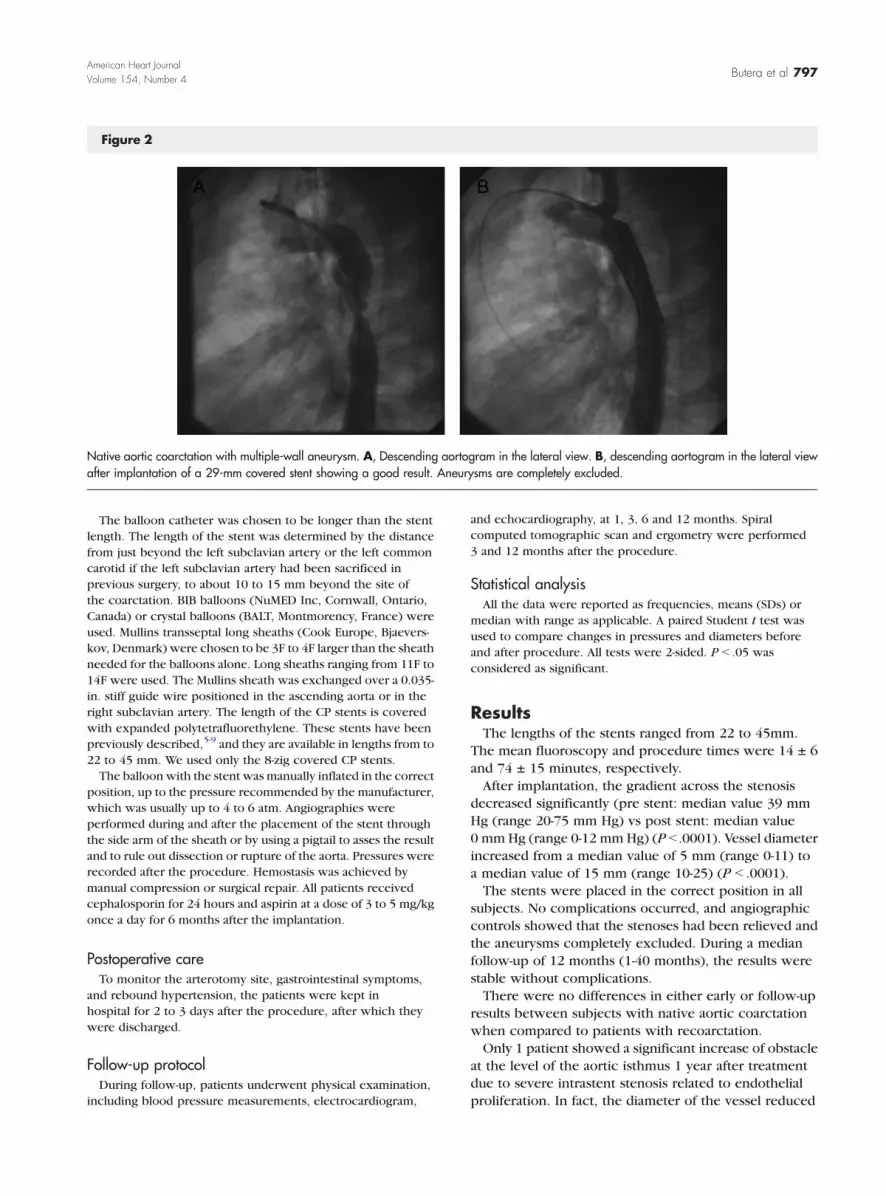
Figure 2
Native aortic coarctation with multiple-wall aneurysm. A, Descending aortogram in the lateral view. B, descending aortogram in the lateral viewafter implantation of a 29-mm covered stent showing a good result. Aneurysms are completely excluded.
Butera et al 797American Heart JournalVolume 154, Number 4
The balloon catheter was chosen to be longer than the stentlength. The length of the stent was determined by the distancefrom just beyond the left subclavian artery or the left commoncarotid if the left subclavian artery had been sacrificed inprevious surgery, to about 10 to 15 mm beyond the site ofthe coarctation. BIB balloons (NuMED Inc, Cornwall, Ontario,Canada) or crystal balloons (BALT, Montmorency, France) wereused. Mullins transseptal long sheaths (Cook Europe, Bjaevers-kov, Denmark) were chosen to be 3F to 4F larger than the sheathneeded for the balloons alone. Long sheaths ranging from 11F to14F were used. The Mullins sheath was exchanged over a 0.035-in. stiff guide wire positioned in the ascending aorta or in theright subclavian artery. The length of the CP stents is coveredwith expanded polytetrafluorethylene. These stents have beenpreviously described,5-9 and they are available in lengths from to22 to 45 mm. We used only the 8-zig covered CP stents.The balloon with the stent was manually inflated in the correct
position, up to the pressure recommended by the manufacturer,which was usually up to 4 to 6 atm. Angiographies wereperformed during and after the placement of the stent throughthe side arm of the sheath or by using a pigtail to asses the resultand to rule out dissection or rupture of the aorta. Pressures wererecorded after the procedure. Hemostasis was achieved bymanual compression or surgical repair. All patients receivedcephalosporin for 24 hours and aspirin at a dose of 3 to 5 mg/kgonce a day for 6 months after the implantation.
Postoperative careTo monitor the arterotomy site, gastrointestinal symptoms,
and rebound hypertension, the patients were kept inhospital for 2 to 3 days after the procedure, after which theywere discharged.
Follow-up protocolDuring follow-up, patients underwent physical examination,
including blood pressure measurements, electrocardiogram,
and echocardiography, at 1, 3, 6 and 12 months. Spiralcomputed tomographic scan and ergometry were performed3 and 12 months after the procedure.
Statistical analysisAll the data were reported as frequencies, means (SDs) or
median with range as applicable. A paired Student t test wasused to compare changes in pressures and diameters beforeand after procedure. All tests were 2-sided. P b .05 wasconsidered as significant.
ResultsThe lengths of the stents ranged from 22 to 45mm.
The mean fluoroscopy and procedure times were 14 ± 6and 74 ± 15 minutes, respectively.After implantation, the gradient across the stenosis
decreased significantly (pre stent: median value 39 mmHg (range 20-75 mm Hg) vs post stent: median value0 mmHg (range 0-12 mmHg) (P b .0001). Vessel diameterincreased from a median value of 5 mm (range 0-11) toa median value of 15 mm (range 10-25) (P b .0001).The stents were placed in the correct position in all
subjects. No complications occurred, and angiographiccontrols showed that the stenoses had been relieved andthe aneurysms completely excluded. During a medianfollow-up of 12 months (1-40 months), the results werestable without complications.There were no differences in either early or follow-up
results between subjects with native aortic coarctationwhen compared to patients with recoarctation.Only 1 patient showed a significant increase of obstacle
at the level of the aortic isthmus 1 year after treatmentdue to severe intrastent stenosis related to endothelialproliferation. In fact, the diameter of the vessel reduced
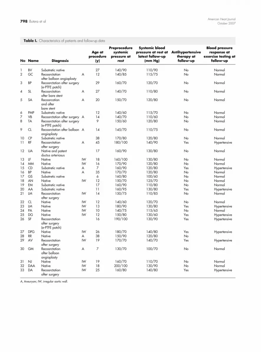
Table I. Characteristics of patients and follow-up data
No Name Diagnosis
Age atprocedure
(y)
Preproceduresystemic
pressure atrest
Systemic bloodpressure at rest atlatest follow-up
(mm Hg)
Antihypertensivetherapy atfollow-up
Blood pressureresponse at
exercise testing atfollow-up
1 BV Subatretic native 27 140/90 110/90 No Normal2 GC Recoarctation
after balloon angioplastyA 12 140/85 115/75 No Normal
3 BP Recoarctation after surgery(e-PTFE patch)
29 160/70 120/70 No Normal
4 SL Recoarctationafter bare stent
A 27 140/70 110/80 No Normal
5 SA Recoarcationand afterbare stent
A 20 150/70 120/80 No Normal
6 PMP Subatretic native 12 140/60 115/70 No Normal7 VB Recoarctation after surgery A 14 140/70 110/60 No Normal8 TA Recoarctation after surgery
(e-PTFE patch)9 150/60 120/80 No Normal
9 CL Recoarctation after balloonangioplasty
A 14 145/70 110/75 No Normal
10 CP Subatretic native 38 170/80 120/80 No Normal11 RF Recoarctation
after surgeryA 45 180/100 140/90 Yes Hypertensive
12 UA Native and patentductus arteriosus
17 160/90 130/80 No Normal
13 LF Native IW 18 160/100 130/80 No Normal14 MM Native IW 16 170/90 120/80 No Normal15 CD Subatretic native A 7 160/90 120/80 Yes Hypertensive16 BP Native A 35 170/70 120/80 No Normal17 GS Subatretic native 6 160/80 100/60 No Normal18 AN Native IW 12 150/70 125/70 No Normal19 EM Subatretic native 17 160/90 110/80 No Normal20 AA Subatretic native 11 160/95 130/80 Yes Hypertensive21 LM Recoarctation
after surgeryIW 14 150/75 110/85 No Normal
22 CL Native IW 12 140/60 120/70 No Normal23 LM Native IW 13 180/90 130/80 Yes Hypertensive24 PA Native IW 10 140/75 115/65 No Normal25 DG Native IW 12 150/80 130/60 Yes Hypertensive26 SF Recoarctation
after surgery(e-PTFE patch)
16 190/100 130/90 Yes Hypertensive
27 DPG Native IW 26 180/70 140/80 Yes Hypertensive28 RR Native A 38 150/90 120/80 No29 AV Recoarctation
after surgeryIW 19 170/70 140/70 Yes Hypertensive
30 GM Recoarctationafter balloonangioplasty
A 7 130/70 100/70 No Normal
31 NJ Native IW 19 160/70 110/70 No Normal32 DAA Native IW 18 200/100 130/90 No Normal33 DA Recoarctation
after surgeryIW 25 160/80 140/80 Yes Hypertensive
A, Aneurysm; IW, irregular aortic wall.
798 Butera et alAmerican Heart Journal
October 2007

Butera et al 799American Heart JournalVolume 154, Number 4
from 12 mm at stent implantation to 8 mm. He wassuccessfully treated by high-pressure balloon angioplastyusing a 12 × 40 ultrathin balloon (Boston Sci Ltd, Cork,Ireland) dilated up to 12 atm. Nine subjects requiredantihypertensive drugs at follow-up due to the occur-rence of systemic hypertension during exercise testing(Table I). In all patients needing drugs, β-blockerswere 2used.
DiscussionAortic coarctation in adulthood in patients with an
associated aneurysm, in subjects with near-atretic aortichistmus, or in subjects with recurrent pathology, is a verychallenging clinical problem.In fact, in these cases, surgical procedures can be
hazardous in regard to hemostatic control of largeintercostals arteries; furthermore, incidence of postrepairparaplegia is greater than in simple aortic coarctation,10
in particular when an aneurysm is present because of theneed to sacrifice intercostal arteries.Standard percutaneous techniques appear promising,
but they can be associated with major limitations. In fact,limiting factors of angioplasty alone are recoil of thevessel with recurrence of stenosis and vascular injurywith consequent vessel dissection or aneurysm forma-tion.11,13 The use of bare stents has solved the firstproblem, but aneurysm formation still remains an issue,the reported incidence being up to 5%.2,3,12-15 Further-more, Palmaz stents have sharp edges and are associatedwith the risk of vessel dissection. In our earlierexperience,3 1 subject died due to vessel dissection,and 2 subjects treated with bare stents for near-atreticaortic coarctation developed an aneurysm of the aorticwall at the level of the stent.These drawbacks may be overcome with the use of
covered CP stents. These stents have rounded edgesthat are less traumatic to the native vessels. Furthermore,the ePTFE protects the stenotic and diseased segment,particularly when a subatretic segment is being treated.To obtain a good result, it is of paramount importanceto use a covered stent of sufficient length to straddlethe diseased tissue completely.There are not a large amount of data about the use
of covered CP stents in the management of aorticcoarctation.5-9,12,16-19 Ewert et al16 reported theirexperience with the use of 60 CP stents. Eleven patients(median age 40 years, range 8-67 years) were treatedwith a covered CP stent for subatretic aorticcoarctation in 6 cases, native coarctation in 2 cases,aortic recoarctation in 2 cases, and aortic recoarctationand aneurysm in 1 case. Qureshi et al7 implantedcovered stents into 4 subjects with aortic coarctation.Two of these subjects had had a previous balloondilation with subsequent recoarctation and aneurysmformation. They were both successfully treated with
covered CP stents, 1 with a 34-mm and the other witha 39-mm stent.Pedra et al17 used covered CP stents and self-expand-
able stent graft in 9 patients. The procedure wassuccessful in all subjects. However, these authorsreported the development of wall aneurysm in 2 patientsdespite the use of covered stents.Tzifa et al 9 reported a multicenter experience of
30 patients (mean age 28 years, range 8-65 years)treated by using covered CP stents. During a medianfollow-up of 12 months, all stents were patent and ingood position. Minor problems were encountered in2 subjects, whereas 4 more patients needed subsequentballoon dilation after the initial procedure. Finally, in43% of the subjects, the antihypertensive medicationwas either decreased or stopped.In addition, there are single case reports on the use
of covered stents.5,8,18 Furthermore, there are rarereports of self-expanding stents being used to treataneurysms in patients who were previously surgicallytreated for aortic coarctation.19 However, in these cases,no recoarctation was present. Finally, Ovaert et al11
published the successful implantation of graft Jomedstents in 5 patients, 2 of whom had postangioplastyaneurysm formation.In our series, which to our knowledge is the largest
single-center experience reported in literature, we usedcovered CP stents to treat patients with complex aorticcoarctation. The procedure was successful in all cases,and results were stable during a median follow-up of12 months. Only 1 subject showed intrastent restenosisdue to significant endothelial growth.Finally, no data has been reported on subjects in
whom a covered stent was placed within a previousbare stent. We treated 2 subjects with this situationwithout problems.However, there are some concerns about their use.
The main one is the possibility of occluding sidebranches, especially in the case of the spinal artery.However, the spinal artery usually originates below thelevel of the ninth thoracic vertebra and, in particular,in the aorta below the diaphragm.20 Therefore, spinalartery occlusion is unlikely to occur.A potential concern could be related to the occurrence
of embolization of a covered stent. If this situationoccurs with a bare stent, it can be dilated in the abdominalor thoracic aorta without major problems becausebranching arteries remain patent. If this is the case whileusing a covered stent, many problems may occur ifimportant arteries are closed by the ePTFE coverage.Therefore, implantation has to be performed very carefully.Another concern is related to the use of CP stents in
growing children. In fact, these stents are covered withePTFE, which potentially limits the extent to whichthey can be redilated. However, we used only 8-zig stents,and the ePTFE covering it is quite an elastic material and
800 Butera et alAmerican Heart Journal
October 2007
is usually quite abundant. Furthermore, it gives noinflammatory reaction. For all these reasons, we believethat these stents are capable of being redilated up to anadult size.Finally, there is the issue of intrastent restenosis that
occurred in one of our patients, and that has to beinvestigated in the follow-up.The main limitations of our report are related to the
relatively small number of patients treated and the lack ofdata about very long-term follow-up. Nevertheless,considering our experience and published data, webelieve that covered CP stents are very useful tools forcomplex aortic coarctation.Finally, probably even in “simple” coarctation, use of
covered CP stents may increase the safety of procedureover either short- or long-term periods. However,randomized studies of bare versus covered stents shouldbe performed in order to address this issue.
References1. Mullins CE, O'Laughlin MP, Vick III GW, et al. Implantation of
balloon-expandable intravascular grafts by catheterization in pul-monary arteries and systemic veins. Circulation 1988;77:188-9.
2. O'Laughlin MP, Perry SB, Lock JE, et al. Use of endovascular stents incongenital heart disease. Circulation 1991;83:1923-39.
3. Chessa M, Carrozza M, Butera G, et al. Results and mid–long-termfollow-up of stent implantation for native and recurrent coarctation ofthe aorta. Eur Heart J 2005;26:2728-32.
4. Taylor PR, Gaines PA, McGuinness CL, et al. Thoracic aortic stentgrafts—early experiences from two centres, using commerciallyavailable devices. Eur J Vasc Endovasc Surg 2001;22:70-6.
5. Forbes T, Mastisoff D, Dysart J, et al. Treatment of coexistentcoarctation and aneurysm of the aorta with covered stent in apediatric patient. Pediatr Cardiol 2003;24:289-91.
6. Ewert P, Abdul-Khaliq H, Peters B, et al. Transcatheter therapy of longextreme subatretic aortic coarctation with covered stents. CatheterCardiovasc Interv 2004;63:236-9.
7. Qureshi SA, Zubrzycka M, Brzezinska-Rajszys G, et al. Use ofcovered Cheatham-Platinum stents in aortic coarctation andrecoarctation. Cardiol Young 2004;14:50-4.
8. Gunn J, Cleveland T, Gaines P. Covered stent to treat co-existentcoarctation and aneurysm of the aorta in a young man. Heart1999;82:351.
9. Tzifa A, Ewert P, Brzezinska-Rajszys G, et al. Covered Cheatham-Platinum stents for aortic coarctation. Early and intermediate results.J Am Coll Cardiol 2006;47:1457-63.
10. Kirlklin/Barrat-Boyes. Cardiac surgery. 3rd ed. Philadelphia: ChurcillLivingstone; 2003. p. 1315-77.
11. Ovaert C, Benson LN, Nykanen D, et al. Transcatheter treatment ofcoarctation of the aorta: a review. Pediatr Cardiol 1998;19:27-44.
12. De Giovanni JV. Covered stents in the treatment of aortic coarctation.J Interv Cardiol 2001;14:187-90.
13. Fawzy ME, Awad M, Hassan W, et al. Long-term outcome (up to 15years) of balloon angioplasty of discrete native coarctation of theaorta in adolescents and adult. J Am Coll Cardiol 2004;43:1062-70.
14. Suarez de Lezo J, Pan M, Romero M, et al. Immediate and followingfindings after stent treatment for severe coarctation of aorta. Am JCardiol 1999;83:400-6.
15. Cheatham JP. Stenting of coarctation of the aorta. CatheterCardiovasc Interv 2001;54:112-25.
16. Ewert P, Schubert S, Peters B, et al. The CP stent—short, long,covered—for the treatment of aortic coarctation, stenosis ofpulmonary arteries and caval veins, and Fontan anastomosis inchildren and adults: an evaluation of 60 stents in 53 patients. Heart2005;81:948-53.
17. Pedra CA, Fontes VF, Esteves CA, et al. Use of covered stents in themanagement of coarctation of the aorta. Pediatr Cardiol 2004;26:431-9.
18. Khan MS, Moore JW. Treatment of abdominal aortic pseudo-aneurysm with covered stents in a pediatric patient. CatheterCardiovasc Interv 2000;14:445-8.
19. Ince H, Petzsch M, Rehders T, et al. Percutaneous endovascular repairof aneurysm after previous coarctation surgery. Circulation2003;108:2967-70.
20. Connolly JE. Prevention of spinal cord complications in aortic surgery.Am J Surg 1998;176:92-101.