pelviperineology december 2018
TRANSCRIPT

2005Perhimpu
na n
Disfungsi Dasar Panggul W
anit
a In
done
sia
Rivista Italiana di Colon-ProctologiaFounded in 1982
‘Taxe Perçue’ ‘Tassa Riscossa’ - Padova C.M.P.
Poste Italiane s.p.a.Spedizione in Abb. Post. - 70% - DCB Padova
ISSN 1973-4905 Vol. 37 - N. 4December 2018
Contents
98 Chronic pelvic pain and uterosacral ligaments: a systematic reviewD. BaDiu, T. EnaChE, E. BraTila
101 influence of foot stool on defecation: a prospective studyS. Takano, M. nakaShiMa, M. TSuChino, Y. nakao, a. WaTanaBE
105 Quality of life and pelvic organ prolapse-related symptoms afterpelvic floor reconstruction with a titanized polypropylene meshfor cystocele: long-term results in a 36 month follow-upC. Fun̈FgElD, M. STEhlE, B. hEnnE, M. grEBE, J.o. kauFholD, D.WaTErMann, M. MEngEl
110 Comparison of two methods of tension of the urethral mediumtape in patient female with stress urinary incontinencesubmitted to T.o.T.W.M. ChávEz
112 how the midurethral sling worksB. aBEnDSTEin, P. PETroS
114 vaginal sacropexy achieved by eight tension-free fixing armsmesh- preliminary resultsC.Goepel, M. SzakaCS, n. FarkaS, B.Farkas
118 Comparison to sacrospinous fixation versus infracoccygealsacropexy in vaginal vault prolapse at 2-year follow-upE. CaSTillo-Pino, C. CanESSa, W. laurí
122 The key role of the transverse pre-cervical arc of gil-vernet inurethral closureJ.M. gil-vErnET
123 Chronic pelvic pain associated with pelvic vein incompetencel. PlES, a.T. CorBu, D.o. Balalau, r.M. SiMa, o.g. olaru, a.D.STanESCu
127 letter to the EditorsF. PuCCiani
INSTRUCTIONS FOR AUTHORS The manuscripts including tables and illustrations must be submitted to Pel-viperineology only via the Isubmit system www.isubmit.it. This enables arapid and effective peer review. Full upload instructions and support are avai-lable online from the submission site.
In http://www.pelviperineology.org/pelviperineology authors in -struc tions. html please find the updated guidelines for the Authors.
CopertinaVol.37-n.4-ingl.qxp_Layout 1 08/01/19 08:54 Pagina 2

C
CopertinaVol.37-n.4-ingl.qxp_Layout 1 08/01/19 08:54 Pagina 3

Editorial Office: BENITO FERRARO, LUISA MARCATOe-mail: [email protected] - [email protected]
Quarterly journal of scientific information registered at the Tribunale di Padova, Italy n. 741 dated 23-10-1982 and 26-05-2004Editorial Director: GIUSEPPE DODI (Direttore Responsabile)
Printer “Tipografia Veneta” Via E. Dalla Costa, 6 - 35129 Padova - e-mail: [email protected]
BURGHARD ABENDSTEIN, Gynaecologist, AustriaANTONELLA BIROLI, Physiatrist, ItalyCORNEL PETRE BRATILA, Gynaecologist, RomaniaSHUKING DING, Colorectal Surgeon, P. R. ChinaTRAIAN ENACHE, Urogynaecologist, RomaniaENRICO FINAZZI-AGRÒ, Urologist, ItalyKLAUS GOESCHEN, Urogynaecologist, GermanyDARREN M. GOLD, Colorectal Surgeon, AustraliaWOLFRAM JAEGER, Gynaecologist, GermanyDIRK G. KIEBACK, Gynaecologist, GermanyFILIPPO LA TORRE, Colorectal Surgeon, ItalyNUCELIO LEMOS, Gynaecologist, BrazilMICHAEL D. LEVIN, Pediatric radiologist, IsraelBERNHARD LIEDL, Urologist, GermanyNAAMA MARCUS BRAUN, Urogynecologist, IsraelANDRI MULLER-FUNOGEA, Gynaecologist, GermanyMENAHEM NEUMAN, Urogynaecologist, Israel
OSCAR CONTRERAS ORTIZ, Gynaecologist, ArgentinaPAULO PALMA, Urologist, BrazilMARC POSSOVER, Gynaecologist, SwitzerlandFILIPPO PUCCIANI, Colorectal Surgeon, ItalyRICHARD REID, Gynaecologist, AustraliaGIULIO SANTORO, Colorectal Surgeon, ItalyYUKI SEKIGUCHI, Urologist, JapanMAURIZIO SERATI, Urogynaecologist, ItalySALVATORE SIRACUSANO, Urologist, ItalyMARCO SOLIGO, Gynaecologist, ItalyJEAN PIERRE SPINOSA, Gynaecologist, SwitzerlandMICHAEL SWASH, Neurologist, UKVINCENT TSE, Urologist, AustraliaPETER VON THEOBALD, Gynaecologist, Reunion Island, FrancePAWEL WIECZOREK, Radiologist, PolandQINGKAI WU, Urogynaecologist, P. R. ChinaCARL ZIMMERMAN, Gynaecologist, USA
PELVIPERINEOLOGYA multidisciplinary pelvic floor journal
www.pelviperineology.org
Rivista Italiana di Colon-ProctologiaFounded in 1982
Vol. 37N. 4
December 2018
Editorial Board
Managing Editor GIUSEPPE DODI, Colorectal Surgeon, ItalyAssistant Managing Editor ADI Y. WEINTRAUB, Urogynecologist, Israel
EditorsANDRI NIEUWOUDT, Gynaecologist, Netherlands - PETER PETROS, Gynaecologist, AustraliaAKIN SIVASLIOGLU, Urogynecologist, Turkey - FLORIAN WAGENLEHNER, Urologist, Germany
Senior editor ELVIRA BARBULEA BRATILA, Romania - Editor emeritus BRUCE FARNSWORTH, Australia
Aesthetic gynecology - RED ALINSOD (USA)Andrology - ANDREA GAROLLA (Italy)Challenging cases- VITO LEANZA (Italy)Chronic pelvic pain - MAREK JANTOS (Australia)Imaging - VITTORIO PILONI (Italy)Medical Informatics - MAURIZIO SPELLA (Italy)NeuroGastroenterology and Intestinal Rehabilitation -GABRIELE BAZZOCCHI (Italy)
Pediatric Surgery - PAOLA MIDRIO (Italy)Pelvic floor Rehabilitation - DONATELLA GIRAUDO (Italy),GIANFRANCO LAMBERTI (Italy)
Psychology - SIBYLLA VERDI HUGHES (Italy)Sacral Neurostimulation - MARIA ANGELA CERRUTO (Italy)Sexology - OSCAR HORKY (Australia)Statistics - CARLO SCHIEVANO (Italy)Systematic Reviews - STERGIOS K. DOUMOUCHTSIS (UK)
Sections
Official Journal of the: International Society for Pelviperineology (www.pelviperineology.com)Associaciòn LatinoAmericana de Piso Pelvico
Perhimpunan Disfungsi Dasar Panggul Wanita Indonesia The Israeli Society of Urogynecology and Pelvic Floor
Romanian Uro-Gyn Society Pelvic Reconstructive Surgery and Incontinence Association (Turkey)
97-Frontespizio n. 4-18.qxp_front 22/12/18 11:04 Pagina 65
98 Pelviperineology 2018; 37: 98-100 http://www.pelviperineology.org
INTRODUCTIONChronic pelvic pain (CPP) is a disabling disease which
occurs in almost 20% of the female population1, decreasingthe patient’s quality of life2. Although the pathogenesis ofthe disease is still said to be unknown, the main treatmentsinclude psychotherapy, drugs, laparoscopic nerve ablation,hysterectomy which implies the removal of the ovaryand/or neuromodulation with better results3,4.
CPP was described as being part of a specific symptomcomplex, according to the Integral Theory published in19935,6 also known as the ‘posterior fornix syndrome’. Thiscomprises chronic pelvic pain, urge, nocturia, abnormal blad-der emptying and is mainly due to laxity in apical support7.
The USLs laxities were showed to be caused by age re-lated collagen reduction from the ligaments or weakeningfrom depolymerisation of hormones, at menstruation andespecially during pregnancy6. Uterus represents the organwhich sustains the main structure of the pelvic floor.Although it has such an importance, many physicians stillrecommend the removal of the uterus together with the lig-aments in different pathologies.
Interestingly, at menopause, ovaries lose the productionof estrogen and if another complication occurs like hys-terectomy, the ligaments will not be able to maintain thepelvic floor structures. Moreover, by applying hysterecto-my, the blood supply can be reduced to the vaginal apex,including USLs and cardinal ligaments. Taken together, allthese negative factors start to develop the posterior zonesymptoms6. Initially, in 1996, treatment of these symptomswas mainly achieved by uterosacral ligaments (USLs) pli-cation8. However, because of these pathogenic factorswhich damage the main structural components of USL, col-lagen, tapes were added to create new collagen to struc-turally reinforce damaged USLs for cure of apical prolapseand posterior fornix symptoms9.
Our systematic review aims to provide an emerging up-date about the relationship of USLs to causation of CPPand cure thereof by USL reconstruction.
METHODSLiterature Search
This systematic review was conducted by screening andgathering results of research papers from literature search
in PubMed database. External sources were not used.Relevant studies were searched by using keywords algo-rithm: “chronic pelvic pain” [All Fields] AND “uterosacralligaments” [All Fields].Inclusion and Exclusion Criteria
The articles from the database with the keywords inputwere screened and analyzed (n = 15) with the PRISMAguidelines10 using the following criteria: (i) original arti-cles; (ii) published in English language; (iii) publishedwithin year 1986-2018; (iv) CPP origin, USL involvement,surgical and non-surgical treatment; (v) qualitative and/orquantitative studies; and (vi) studies assessing the involve-ment of USL in the CPP origin (Figure 1). We excludedconference abstracts, letters, and review articles.Data Extraction
We gathered all of the full-text articles that met the inclu-sion criteria. The results from 5 research articles that arerelevant to this review were extracted and analyzed. Theoutcomes of the studies were comprehensively analyzedand discussed.
Review
Chronic pelvic pain and uterosacral ligaments: a systematicreview DIANA BADIU1, TRAIAN ENACHE2, ELVIRA BRATILA3
1 Faculty of Medicine, “Ovidius” University of Constanta, Romania2 “Prof.Dr. Panait Sarbu” Obstetrics&Gynecology Hospital, Bucharest, Romania3 Department of Obstetrics&Gynecology, “Carol Davila” University of Medicine and Pharmacy, Bucharest, Romania
Abstract: Introduction. According to the Integral System, chronic pelvic pain (CPP) in the female co-occurs with abnormal bladder emptying,urge and nocturia symptoms with a common causation, lax uterosacral ligaments (USLs). Methods. In our systematic review, the PubMeddatabase was used for the literature search using the keywords algorithm which included “chronic pelvic pain” and “uterosacral ligaments”.Relevant studies regarding CPP origin, USLs involvement in causation and symptom cure after surgical or non-surgical treatment wereanalysed in this review. Results. We found evidence that when USLs become lax, the gravity forces act on the uterus and vagina, develop con-gestion. In this respect, posterior compartment repair could be used in order to reinforce the USLs. Conclusions. CPP and other symptoms re-lated to posterior zone of the pelvic floor were mainly due to USL laxity and repairing these ligaments restored its clinical manifestations, aspredicted by the Integral Theory.
Keywords: Chronic pelvic pain; Uterosacral ligaments; Laxity; Pelvic floor; Posterior zone.
!"#$%!&!'(%!)$(*+,-./012.3+4.20
56*78960:8;<.20=3>?@A0
B'C##$!$D0(*+,-./00
/,*..313908/1390+5-.0E320E;/5*E,50
=3>FA0
#G!D!H!G!%I0&8--J5.K50E*+,-./0E//.//.20L7*0
.-191;1-15M0=3>FA0
(*+,-./0.K,-82.20=3>?A0
JN0L8--J5.K50E*+,-.0130BOE31/6P0
[email protected]*+,-./0Q1560'::0L*7<0.327<.5*17/1/0
!$'GR"#"0&8--J5.K50E*+,-./013,-82.20=3>SA0
Figure 1. – Methodology for articles selectionaccording to PRISMA review guidelines10.
98-100-Badiu - chronic pelvic.qxp_treatment 22/12/18 12:14 Pagina 98

References CPP origin USLs involvement After surgical treatment Non-surgical treatment
7 2nd degree or greater Loose of USLs USL repair using TFS by applying –uterine/apical prolapse Integral Theory System
8 Deep retraction pockets USLs elasticity Pockets excision (defined –in the posterior cul de sac as estimated to be greater than 0.5 cm)
9 Hypothesized areas: Traction applied – Using pain pressure threshold - abdominal wall; to USLs algometry and standard numeric - vulva; scale- pelvic floor;- vaginal vault
10 Nerve trunks Operative of nerve – Visual analogue scale questionnaire interruption trunks interruption at 3 and 6 months and 1,2,3 and in the USLs in the USL by 5 years at women with CPP with laparoscopic uterosacral or without laparoscopic uterosacral nerve ablation nerve ablationtreatment
11 The Lee-Frankenhauser Nerve trunks Laparoscopic –sensory nerve plexes interruption by uterosacral nerveand parasympathetic laparoscopic uterosacral ablationganglia in the USLs nerve ablation
TABLE 1. Comparison of the included full-text articles.
99
Chronic pelvic pain and uterosacral ligaments: a systematic review
RESULTSStudy characteristics
We found 48 articles from which 5 were reviews (fromwhich one had the full-text in French language). From 43articles, we screened 9 full-text articles (34 were only ab-stracts from which 4 with only the title and 1 abstract withthe text in Portuguese). From 9 full-text articles, we exclud-ed 4 full-text articles: one with full-text in Spanish and theother three in which the CPP was referred to endometriosiscause. Finally, we included only 5 articles for analysis.Furthermore, we did not find any conference abstracts.
Studies that were used in this review where focusingmore on CPP origin, USLs involvement, taken into consid-eration the surgical or non-surgical treatment (Table 1). Thestudies used in this review have a low to moderate level ofcertainty.
In the study of Liedl and contributors11, the origin of CPPwas the symptoms from apical prolapsed of 2nd or greaterdegree (POPQ, stages 2-4) including overactive bladder.After applying surgical treatment by using Integral TheorySystem with TFS, in order to repair the loose of cardinaland US ligaments, the patients presented improved symp-toms and better clinical quality of life.
In another study12, the authors made the surgical excisionof the deep retractive pockets, improving in this way thesymptoms such as CPP. It was also shown that women whohad endometriosis in their deep retractive pockets, had sig-nificant improvement in deep dyspareunia and quality oflife.
Another study made a combination of quantitative andsemiquantitative techniques for the pain track13. The exam-ination of the pain had different regions like pelvic abdom-inal wall, vulvar vestibule, pelvic floor and the vaginalvault. The study approach involved pain pressure thresholdalgometry and standardized numeric scale. By using thesemethods, it was showed to better quantify the pelvic paincomplexity and that the pain in the abdominal walls, pelvicfloor and USLs should be separately evaluated.
Another study14 involved 487 women with chronic painlasting more than 6 months with or without minimal en-dometriosis. The patients were randomized according to la-paroscopic uterosacral nerve ablation (LUNA) group or noLUNA group. The main symptoms were achieved by usingVisual Analogue Scale (VAS), Euro-QoL (EQ-5D) or EQ-VAS. The results of the study showed that LUNA did not re-sult in improvements in pain, dysmenorrheal, dyspareunia,
pelvic pain or QoL compared with no LUNA group (i.e.without pelvic denervation). In the protocol of The LUNATrial Collaboration15 it was tested the hypothesis if in womenwith CPP LUNA can alleviate pain at 12 month follow-up.After the surgery, questionnaires like VAS, an index of sexu-al satisfaction and the EuroQoL SD-EQ instrument was ad-ministered at 3, 6 and 12 months. The study showed that theLUNA effectiveness may be higher for central compared tonon-central pain without any other associated diseases.
DISCUSSIONThe role of uterosacral ligaments laxity in chronicpelvic pain
Although CPP represents a continuous important issue indaily medical practice, on many occasions it is underdiag-nosed and undertreated by most of the physicians. A simpleinitially effective treatment was reinforcement of the liga-ments laxity which supports the uterus and vagina8.
Though vaginal USL plication showed an initial 85% im-provement from the pain relief, there was further deteriorat-ed in time8. Following this, insertion of a posterior vaginalsling showed a greater improvement of the symptoms16.
One characteristic of this pain related to mechanical fac-tors. The pain was exacerbated when standing and relievedon lying down. This is explained by the inadequately sup-ported nerves being stimulated by gravitational forces. It isunfortunate that such pain is often attributed to psycholog-ical issues17 which in fact may be secondary, not primary. Itis therefore important that laxity in the posterior ligamentsbe first checked before considering other differentiating di-agnosis like psychiatric case8.
Knowing that the nerve fibers from USLs are visceralfibers, visceral innervations including those from T12-L1and S2-4 would explain the pain distribution, although thestretching of the ligaments by gravity will also stimulatethe nerve ending causing in this way pain.
The first step in reducing the pain is to ask the patient tolie down, decreasing the pressure8. Then, the second stepcould be represented by pessary application which can pro-vide the normal mechanical support of the ligaments8. Thecongestion characteristics could be explained by the fol-lowing: uterus is supported by both cardinal and USLs, as-sisted together with the pelvic floor muscles. When the lig-aments become lax, the gravity forces acting on the uteruscould develop congestion by “kinking” of the pelvic veins,preventing in this way the outflow18.
98-100-Badiu - chronic pelvic.qxp_treatment 22/12/18 12:14 Pagina 99

The posterior compartment repair of the pelvic floorThe looseness or laxity of the vagina and its ligaments
supports can cause CPP, organ prolapse, urge and stress in-continence, nocturia, voiding dysfunction, faecal inconti-nence and constipation19,20.
The pain usually appears from the inability of USLs lax-ity to support the nerves near the ligaments. These nervesare stretched by gravity or during intercourse and causeCPP. This pain is almost invariably associated with othersymptoms like nocturia, faecal incontinence and obstructeddefecation20.
It is important to understand that tissue structure is oftendisplaced laterally (e. g. cardinal ligaments, USLs, recto-vaginal fascia, pubocervical fascia, hammock, and perinealbody). Therefore, effective posterior compartment surgicaltechniques are required to bring the tissues together in theanatomically normal position21.
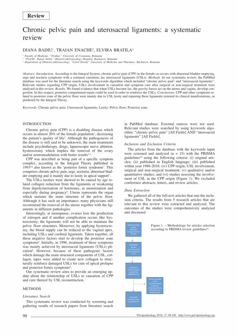
Physiologically, the uterine ligaments provide the poste-rior cervical support component of the uterine cervix, andthe cardinal ligaments provide the lateral component.Transmitted in biomechanical language we speak of a “ten-sion in the thread” that opposes the displacement of thecervix, one rearward oriented and the other the side. The re-sult of these forces is a vector with posterolateral orienta-tion (Figure 2).
Any reconstruction technique should target the ligamentreconstruction whose vector result is similar to the physio-logical result. The disappearance of postoperative painshould confirm the cause-effect relationship between USLsand CPP.Limitations and Future Research Suggestions
It is known that CPP is specific for the posterior zone.The diagnosis of posterior zone is detected when the patienthas urgency nocturia, and especially CPP22. The limitationof our study consisted in that we followed only the USLsinvolvement without assessing the state of the perinealbody which constitutes part of the back ligaments.
CONCLUSIONSA key symptom related to posterior zone is the CPP.
Knowing that the main underlying anatomical defect is de-ficiency and/or laxity of USL, the repair of these ligamentscan lead to restoration of the structure and function, theclinical symptoms and manifestations.
DISCLOSURE STATEMENTSThe authors declare no conflict of interest.
REFERENCES1. Breivik H, Collett B, Ventafridda V, Cohen R, Gallacher D.
Survey of chronic pain in Europe: prevalence, impact on dailylife, and treatment. Eur J Pain 2006; 10 (4): 287-333.
2. Yosef A, Ahmed AG, Al-Hussaini T, Abdellah MS, Cua G,Bedaiwy MA. Chronic pelvic pain: pathogenesis and validated
assessment. Middle East Fertility Society Journal 2016; 21: 205-21.
3. Fall M, Baranowski AP, Elneil S, Engeler D, Hughes J, MesselinkEJ et al. EAU guidelines on chronic pelvic pain. Eur Urol 2010;57 (1): 35-48.
4. Zabihi N, Mourtzinos A, Maher MG, Raz S, Rodriguez LV.Short-term results of bilateral S2-S4 sacral neuromodulation forthe treatment of refractory interstitial cystitis, painful bladder syn-drome, and chronic pelvic pain. Int Urogynecol J Pelvic FloorDysfunct 2008; 19 (4): 553-7.
5. Petros PE. Pelvic pain in pregnancy may be caused by uterosacralligament laxity and may be associated with nocturia, urgency andabnormal bladder emptying. Acta Obstet Gynecol Scand 2011; 90(9): 1050.
6. Petros P. The Integral System. Central European Journal ofUrology 2011; 64 (3): 110-9.
7. Petros P, Ulmsten U. The posterior fornix syndrome: a multiplesymptom complex of pelvic pain and abnormal urinary symp-toms deriving from laxity in the posterior fornix. ScandinavianJournal of Urology and Nephrology 1993; 27 (Supplement 153 -PART IV): 89-93.
8. Petros PP. Severe chronic pelvic pain in women may be causedby ligamentous laxity in the posterior fornix of the vagina. Aust NZ J Obstet Gynaecol 1996; 36 (3): 351-4.
9. Petros PE. New ambulatory surgical methods using an anatomicalclassification of urinary dysfunction improve stress. Urge andAbnormal Emptying Int Urogynecoi J 1997; 8: 270-8.
10. Moher D, Liberati A, Tetzlaff J, Altman DG, PRISMA GROUP.Preferred reporting items for systematic reviews and meta-analy-ses: the PRISMA statement. PLoS Med 2009; 6 (7): e1000097.
11. Liedl B, Inoue H, Sekiguchi Y, Haverfield M, Richardson P,Yassourides A, Wagenlehner F. Is overactive bladder in the fe-male surgically curable by ligament repair? Cent European J Urol2017; 70: 53-9.
12. Yeung PP Jr, Ian Logan, Gavard JA. Deep retraction pockets, en-dometriosis, and quality of life. Frontiers in Public Health 2016;4: 85.
13. Fenton BW, Grey SF, Reichenbach M, McCarroll M, VonGruenigen V. Phenotyping chronic pelvic pain based on latentclass modeling of physical examination. Hindawi PublishingCorporation, Pain Research and Treatment 2013; Vol 2013,Article ID 891301, http://dx.doi.org/10.1155/2013/891301.
14. Daniels J, Gray R, Hills RK et al. Laparoscopic uterosacral nerveablation for alleviating chronic pelvic pain. A RandomizedControlled Trial. JAMA 2009; 302 (9): 955-61.
15. The LUNA Trial Collaboration. A randomised controlled trial toassess the efficacy of Laparoscopic Uterosacral Nerve Ablation(LUNA) in the treatment of chronic pelvic pain: The trial proto-col [ISRCTN41196151]. BMC Women’s Health 2003; 3: 6.
16. Petros P, Richardson P. TFS posterior sling improves overactivebladder, pelvic pain and abnormal emptying, even with minorprolapse. A prospective urodynamic study. Pelviperineology2010; 29: 52-5.
17. Beard R, Reginald P, Pearce S. Psychological and somatic factorsin women with pain due to pelvic congestion. Br J ObstetGynaecol 1988; 245: 413-21.
18. Petros PP. Severe chronic pelvic pain in women may be causedby ligamentous laxity in the posterior fornix of the vagina. Aust.NZ J Obst. Gynaecol 1996; 36 (3): 351-4.
19. Petros PE, Swash M. The Musculoelastic Theory of anorectalfunction and dysfunction. J Pelviperineology 2008; 27: 89-93.
20. Liedl B. Male and female urinary incontinence from the view-point of the pelvic floor surgeon. Urologe 2010; A 49: 289-303.
21. Wagenlehner F, Bschleipfer T, Liedl B, Gunnemann A, Petros P,Weidner W. Surgical reconstruction of pelvic floor descent: Anatomicand functional aspects. Urologia Internationalis 2010; 84: 1-9.
22. Petros PE, Bornstein J. Vulvar vestibulitis may be a referred painarising from laxity in the uterosacral ligaments – a hypothesisbased on 3 prospective case reports. ANZJOG 2004; 44: 483-6.
Correspondence to: Diana Badiu - 1st University Street - Constanta 900470 -Romania E-mail:[email protected]
100
Diana Badiu, Traian Enache, Elvira Bratila
Figure 2. – The pos-terior vector resultantof the ligaments sup-porting the cervix.
98-100-Badiu - chronic pelvic.qxp_treatment 22/12/18 12:14 Pagina 100

Figure 1. – a, Upright sitting position without a foot stool; b, Upperbody bent forward position with a foot stool; c, Upper body back-ward with a foot stool
101Pelviperineology 2018; 37: 101-103 http://www.pelviperineology.org
INTRODUCTIONFecal outlet obstruction lowers the quality of life (QOL)
of patients with functional constipation. Outlet obstructionmay be attributed to the following causes: non relaxation ofthe puborectalis muscle, anismus, rectal prolapse, rectoceleand rectal hyposensitivity.
Tsuchino et al assessed rectal and anal pressure duringdefecation with the patient in a bending position rather thanin a normal sitting position1. Moreover, Takano et al report-ed the efficacy of bending the upper part of the body for-ward on defecation2 and they named this posture the“Thinker Position”. However, some patients have also ex-perienced evacuation difficulties with this position.
Findings in the literature indicate that the squatting posi-tion is superior to the traditional upright sitting position fordefecation3,4. However, sudden changes in defecation habitssuch as altering the position from sitting to squatting or intro-ducing a special commode may add psychological stress andcause incomplete evacuation. The closest position to squat-ting on a western commode is using a foot stool (foot step).We hypothesized that adding a foot stool in conjunction withstructure would help facilitate defecation. Therefore, the aimof this prospective non-randomized single group study wasto assess the efficacy of a foot stool on defecation.
PATIENTS AND METHODSThe risks of added x-ray exposure were disclosed to all
the patients and informed consent to participate in the studywas obtained. The inclusion criteria were patients experi-encing constipation and who were scheduled to undergocinedefecography. The indications for defecography weresymptoms of evacuation difficulty and a feeling of incom-plete evacuation. A diagnosis of constipation and outlet ob-struction were made using the criteria for the functionaldefecation disorders of ROME IV. The exclusion criteriawere patients who were under 18 years of age, pregnantand/or had prior rectal surgery. This study was approved bythe institutional review board (IRB).Cinedefecography technique
Patients were administered a phosphate enema 30 min-utes prior to the procedure. They were then placed in the
left lateral decubitus position and approximately 100 mL ofbarium paste was injected into the rectum. The bariumpaste was mixed with oatmeal until it reached a Bristol type4, stool consistency. The patient was then asked to sit on acommode and lateral films of the pelvis were taken duringthe pushing phase in a sitting position with and without afoot stool (Figure 1a). Manometry technique
Pushing rectal pressure examinations were performedwith and without a foot stool in the upright sitting positionand the upper body bent forward position. Rectal pressurewas assessed using an anorectal function testing kit(GMMS Gastrointestinal Manometry System: GMMS-200,Star Medical, Tokyo, Japan).Interpretation of data
Patient characteristics (i.e. gender, age, comorbidity, andprior perianal surgery) were retrospectively obtained fromthe medical records. Anorectal angle (ARA), perineal planedistance (PPD), and puborectalis length (PRL) in rest andduring straining were measured from the radiographs. ARAwas defined as the angle between the axis of the anal canaland the distal half of the posterior rectal wall5-7. PPD waspredetermined to be the vertical distance between the ARAposition and an imaginary line drawn between the pubicsymphysis to the tip of the coccyx. PRL was measured asthe distance between the ARA and the pubic symphysis7,8.
Original article
Influence of foot stool on defecation: a prospective studySHOTA TAKANO, MIDORI NAKASHIMA, MASAHIRO TSUCHINO, YUYA NAKAO, ATSUSHI WATANABEDepartment of Functional Anorectal Disorder, Coloproctology Center Takano Hospital, Kumamoto, Japan
Abstract: Objective: The aim of this study was to determine the efficacy of adding a foot stool to help facilitate defecation in patients with fecaloutlet obstruction. Methods: Patients (n=53) who experienced evacuation difficulties between June and October 2016 were enrolled in thisprospective non-randomized single group study. Cinedefecography was performed with and without a foot stool. Anorectal angle (ARA), per-ineal plane distance (PPD), and puborectalis length (PRL) during rest and straining in both positions were measured from the radiographs. Rectalpressure was measured with the lateral position and sitting with and without a foot stool. Results: There was no significant difference betweenwith and without a foot stool in ARA, PPD and PRL. In the upper body bent forward group, the time to evacuation was significantly shorter witha foot stool compared to without a foot stool (123 vs 91 sec, p=0.04). The difference of rectal pressure between the lateral position and sittingposition significantly increased with a foot stool compared to without a foot stool (22.1 vs 16.7 mmH2O, p<0.01). The difference of rectal pres-sure between with and without a foot stool increased in the upper body bent forward position compared to the upright sitting position (5.4 vs 1.9mmH2O, p<0.01). Conclusion: The findings suggest that using a foot stool with structure is a more efficient method for defecation. However, theupper body bent forward position is also important. This technique may be useful for retraining patients with constipation.
Keywords: Constipation; Defecation posture; Defecography; Fecal outlet obstruction; Foot stool.
101-104-takano influence.qxp_treatment 07/01/19 09:39 Pagina 101

102
Shota Takano, Midori Nakashima, Masahiro Tsuchino, Yuya Nakao, Atsushi Watanabe
Sacral Slope (SS) was measured from the radiographyand defined as the angle between the superior line of thesacrum and the horizontal line. The difference of SS be-tween with and without a foot stool was calculated. Whenthe patient bent the upper body forward with a foot stoolthe SS became wider, and when the patient bent the upperbody backwards the SS became narrower.Statistical analysis
Previous studies have determined that the mean pelvicfloor location increased from 1.3 cm compared to the re-cumbent and sitting positions8. Therefore, the effect size todetermine a clinically relevant difference for this study waspreset at 1.3 cm for PPD. With an alpha of 0.05 and a betaof 0.9, approximately 20 patients were needed for thisstudy. The paired t-test was used to compare the sets ofmeasurements for both positions and P values less than0.05 were considered statistically significant.
RESULTSOut of the 53 patients enrolled in the study, 25 of them
were female with an average age of 70.2 (range: 21-90)years. Twenty-three of the patients used the upper bodybent forward position with a foot stool and 30 of them usedthe upper body bent backwards position. The mean valuesof ARA, PPD and PRL during straining with or without afoot stool are shown in Table 1. There was no significantdifference between with and without a foot stool in ARA,PPD or PRL.
Pushing rectal pressure showed no significant differencebetween with and without a foot stool in both the uprightsitting position and the upper body bent forward position(Table 2).
In the upper body bent forward group, the time to evacu-ation was significantly shorter with a foot stool comparedto without a foot stool (123 vs 91 sec, p=0.04). The differ-ence of rectal pressure between the lateral position and theupright sitting position significantly increased with a footstool compared to without a foot stool (22.1 vs 16.7mmH2O, p<0.01). The difference of rectal pressure be-tween with and without a foot stool increased in the upperbody bent forward position compared to the upright sittingposition (5.4 vs 1.9 mmH2O, p<0.01). The results for timeto evacuation, rectal pressure comparing the lateral positionand the upright sitting position, and rectal pressure compar-
ing with and without a foot stool are shown in Figures 2, 3and 4, respectively.
DISCUSSIONDefecation is a very important part of human life. Fecal
outlet obstruction is defined as “difficulty in evacuation oremptying of the rectum which may occur even with fre-quent visits to the washroom”. Moreover, body positionduring defecation is an important element of defecation.Historically, humans have squatted in order to defecate3,9
and this practice still continues today in underdevelopedcountries10. While squatting for defecation continues to bethe principal position in Asia and Africa, Western popula-tions have become accustomed to sitting on a commode3.The widespread use of a sitting toilet began during the 19th
century when sewage systems were developed to improvesanitation as cities and populations grew11. Compared withthe sitting position, squatting was associated with signifi-cantly less time to achieve a sensation of satisfactory bowelemptying and a lower degree of subjectively assessedstraining3. Rad found that ARA and PPD were greater insubjects who squatted versus those who sat (ARA 132 vs92; PPD 8.4 vs 6.6 cm, respectively)12. The rectoanal angleof squatting (126°) for defecation was larger than the nor-mal sitting position (100°) (P < 0.05), and was also largerthan the hip-flex sitting position (99°) (P < 0.01)4. Tagartfound that the ARA straightens with fully flexed hips—cor-responding to the squatting position assumed for defeca-
Figure 2. – Time to evacuation using valium paste without/with afoot stool. Time to evacuation is shorter with a foot stool thanwithout a foot stool.
Figure 3. – The difference of rectal pressure between the lateralposition and the sitting position without/with a foot stool. The dif-ference is larger using a foot stool than without one.
Without foot stool With foot stool p value
ARA (o) 140.1 143.5 0.41PPD (cm) 98.9 98.3 0.89PRL (cm) 128.3 130.1 0.62Length to evacuation (min) 100.7 95.8 0.32Evacuation rate (%) 60.1 67.4 0.25Evacuation volume (g) 144.9 167.7 0.12
TABLE 1. Comparison of cinedefecography measurements betweenwith and without foot stool.ARA anorectal angle, PPD perineal plane distance, PRL puborec-talis length.
Without foot stool With foot stool p value
Sitting strait 85.1 86.9 0.71Bending forward 84.5 89.9 0.18
TABLE 2. Comparison of pushing rectal pressure between the verti-cal position and upper body bent forward without/with foot stool.
101-104-takano influence.qxp_treatment 07/01/19 09:39 Pagina 102

103
Influence of foot stool on defecation: a prospective study
tion—and converts the rectoanal outlet into a straight canal,thereby facilitating rectal emptying13. Takano found that 22patients were unable to evacuate the barium paste andtherefore underwent cinedefecography in the upper bodybent forward (Thinker) position. “The Thinker” positionhad a significantly wider ARA than the sitting position(113° vs. 134°, respectively; p = 0.03), larger PPD (7.1 vs.9.3 cm, respectively; p = 0.02), and longer PRL (12.9 vs.15.2 cm, respectively; p = 0.005) during straining. Elevenpatients experienced complete evacuation in “The Thinker”position2.
The present study demonstrated a wider ARA, largerPPD and longer PRL but no significant difference wasfound between with a foot stool and without a foot stool.The difference between our study and the previous study isthe selection of the patients. In the previous study conduct-ed by Takano et al, all 21 patients were unable to defecatein the upright sitting position2. However, in this study thepatients who experienced evacuation difficulties were allenrolled whether or not the patient was able to evacuate thebarium paste. Therefore, ARA, PPD and PRL did not reveala significant difference. Now we have data for 22 patientswho were unable to evacuate the barium paste without afoot stool and who underwent cinedefecography with a footstool. Cinedefecography revealed a wider ARA with a footstool than without a foot stool.
Other studies have found differences among the variouspositions. Altomare et al noted that when the patient sits ona commode, the ARA opens wider than it does in the stand-ing position14. Rao et al reported on the influence of bodyposition on defecation using a water-filled balloon andmanometory. In the prone position, one third of the subjectshad dyssynergia and half of them could not expel the paste(artificial stool). When sitting with a distended rectum,most subjects displayed normal defecation patterns and theability to expel stool. The authors reported that the sittingposition appears to be more conducive to defecation thanthe lying position. In addition, the manometric recordingsduring attempted defecation showed that the intrarectalpressure was lower in the left lateral position than in the sit-ting position15.
The findings revealed in this study suggest that the rectalpressure is higher in patients who use the upper body bentforward position with a foot stool than without a foot stool.The average age of the patients in our present study is high-er than in our previous study (Takano et al, 2014). Thisseems to indicate that for older patients, rectal pressure ismore important for defecation than the relaxation of thepelvic muscles.
Tsuchino et al found that there was a higher rectal pres-sure and lower anal pressure in the upper body bent for-ward position. Furthermore, they stated that this positioncreates a higher intraabdominal pressure that seems to helpfacilitate evacuation1.
CONCLUSIONSThe findings suggest that a foot stool in conjunction with
structure is a more efficient method for defecation.However, the upper body bent forward position is also veryimportant. This technique may be useful for retraining pa-tients with constipation. However, this study has somemethodological limitations. Further studies are needed toverify these findings.
DISCLOSURE STATEMENTSWe declare no conflict of interest.
REFERENCES1. Tsuchino M, Yamashita K, Bouda T, Kai Y, Takano S, Takano
M. A Study of the Relationship Between Continence Functionand Defecatory Posture. Journal of Japanese Society of Stomaand Continence Reha 2008; 24: 34-38.
2. Takano S, Sands DR. Influence of body posture on defecation:a prospective study of “The Thinker” position. TechColoproctol 2016; 20: 117-121.
3. Sikirov D. Comparison of straining during defecation in threepositions: results and implications for human health. Dig DisSci 2003; 48: 1201-1205.
4. Sakakibara R, Tsuoyama K, Hosoi H et al. Influence of BodyPosition on Defecation in Humans. Low Urin Tract Symptoms2010; 2: 16-21.
5. Jorge JM, Wexner SD, Marchetti F et al. How reliable are cur-rently available methods of measuring the anorectal angle? DisColon Rectum 1992; 35: 332-338.
6. Felt-Bersma RJ, Luth WJ, Janssen JJ, Meuwissen SG.Defecography in patients with anorectal disorders. Which find-ings are clinically relevant? Dis Colon Rectum 1990; 33: 277-284.
7. Jorge JM, Wexner SD, Ehrenpreis ED, Nogueras JJ, JagelmanDG. Does perineal descent correlate with pudendal neuropa-thy? Dis Colon Rectum 1993; 36: 475-483.
8. Habib FI, Corazziari E, Viscardi A, Badiali D, Torsoli A. Roleof body position, gender, and age on pelvic floor location andmobility. Dig Dis Sci 1992; 37: 500-505.
9. Haubruch W, Constipation. In: Berk J, ed. BockusGastroenterology; 4th ed.; 1985, 111.
10. Boles R. Constipation. JAMA 1927; 89: 1766-1770.11. Singer C. History of Technology. The Industrial Revolution
1958; 507-508.12. Rad S. Impact of Ethnic Habit on Defecographic
Measurements. Arch Iranian Med 2002; 5: 115-117.13. Tagart RE. The anal canal and rectum: their varying relation-
ship and its effect on anal continence. Dis Colon Rectum 1966;9: 449-452.
14. Altomare DF, Rinaldi M, Veglia A, Guglielmi A, Sallustio PL,Tripoli G. Contribution of posture to the maintenance of analcontinence. Int J Colorectal Dis 2001; 16: 51-54.
15. Rao SS, Kavlock R, Rao S. Influence of body position andstool characteristics on defecation in humans. Am JGastroenterol 2006; 101: 2790-2796.
Correspondence to: Shota Takano - 3-2-55 Oe, Chuo-ku - Kumamoto 862-0971 - JapanE-mail: [email protected]
Figure 4. – The difference of rectal pressure between the upperbody bent forward position and the backward position without andwith a foot stool.
101-104-takano influence.qxp_treatment 07/01/19 09:39 Pagina 103

104
Shota Takano, Midori Nakashima, Masahiro Tsuchino, Yuya Nakao, Atsushi Watanabe
Gyneco... Takano’s article is quite interesting if considered from an obstetrical point of view. The postural postpartum at-titude is in fact an effective procedure to solve many obstructed labor situations, as demonstrated by the Authors in case of fe-cal outlet obstruction.
The degree of inclination of the delivery channel is a fundamental point in the dynamics and mechanics of labor, such asthe alignment of the anal canal for stool ejection. This alignment creates the ideal conditions for the pelvic entrance to be inline with the uterine and fetal body, making the uterine contraction more ergonomic and the progression of the fetus withoutdifficulty.
The type of dorsal curvature, the anti or retroversion of the pelvis, the type of contracture of the muscles, the position andorientation of the lower limbs condition the movements of each individual element of the pelvis which constitutes a functionalunit.
It is known that, in the presence of a slowing down or stopping of the progression of the part presented in the second stageof labor, it is useful to make the woman taking up the crouched position on the heels, which increases the bispynous and bitu-beral diameter by 2 cm and 1 cm respectively.
Another useful position to align the fetus with the pelvis and to increase the diameters of the pelvic inlet is the sitting posi-tion reclined forward.
These postures accelerate the fetal descent into the delivery channel in obstetrics and reduce the expulsive effort with respectto the horizontal position, as well as Takano shows a significant decrease of time to fecal evacuation and increased rectal pres-sure in the upper body bent forward.
Finally, as in obstetrics the different postures have different indications depending on the cause of obstructed labor and ofthe proper stage, it would be interesting to evaluate the effect of different postures depending on the cause of fecal outlet ob-struction due to relaxatio (rectocele or rectal prolapse) compared to those from hypertone (anismus or contracture of puborec-talis muscle).
REFERENCERegalia-Fumagalli A. Promoting delivery. Carrocci Faber Ed., 2013
Correspondence to: PDr Chiara AlessiObstetrics and Gynecologic Unit. AO Padovae-mail: [email protected]
Uro... The authors suggest that using a foot stool with structure is a more efficient method for defecation. We know thatmicturition in the woman takes place in a sitting position and that normally the urine emission is facilitated by a straighteningof the urethra without a “thinker position”. In the male this does not happen because a rectilinealization of the urethra is notpossible for the length of the male urethra and for the presence of surrounding anatomical structures.
In this context it is clear that the “thinker position” is not necessary for urination in both sexes and that the assumption of apossible thinker position becomes necessary exclusively to increase the intravesical pressure during urination in patients whoare affected by detrusor acontractility or in those who have an orthotopic neobladder.
Correspondence to: Salvatore SiracusanoAssociate Professor of UrologyUniversity of VeronaEmail: [email protected]
Multidisciplinary UroGyneProcto Editorial CommentTo improve the integration among the three segments of the pelvic floor, some of the articles published in Pelviperineology are
commented on by Urologists, Gynecologists, Proctologists/Colo Rectal Surgeons or other Specialists, with their critical opinionand a teaching purpose. Differences, similarities and possible relationships between the data presented and what is known in the threefields of competence are stressed, or the absence of any analogy is indicated. The discussion is not a peer review, it concerns con-cepts, ideas, theories, not the methodology of the presentation.
101-104-takano influence.qxp_treatment 07/01/19 09:39 Pagina 104

105Pelviperineology 2018; 37: 105-109 http://www.pelviperineology.org
INTRODUCTIONPelvic organ prolapse (POP) is a common disease preva-
lent in 50% of parous women and can significantly reducepatients’ quality of life (QoL)1. Pelvic organs, such as theuterus, bladder and/or bowel can descend because of failingof the pelvic soft tissue support (ligaments, connective tis-sue, etc.) and weakness of the vaginal wall. Affected wom-en show various urinary, bowel and sexual symptoms re-sulting in a profoundly impaired QoL1-4. Treatment alterna-tives range from non-surgical therapies, which are mainlyfocused on minimization of risk factors, to a great varietyof surgical options including abdominal (open or laparo-scopic) and minimal-invasive transvaginal techniques withor without the use of surgical meshes1. Nowadays, nativetissue repair with sacrospinal (Amreich-Richter) oruterosacral ligament fixation is the preferred surgicalmethod for the treatment of a cystocele and apical prolapsevia the transvaginal approach.
The reconstruction of the anatomical location of organsof the true pelvis is the aim of every surgical intervention.However, the functional result is more important for affect-ed patients than anatomically correct reconstruction. QoL ishighly dependent on the function of the bladder and bowel,sexuality and pelvic pain. Furthermore, long term stabilityis of great interest. Due to the high rate of recurrent POPwith conservative native tissue treatment options1,5 alloplas-tic meshes were established. Current literature indicates alower recurrence rate after POP reconstruction with surgi-cal meshes1,5,6. Nevertheless, the high rate of mesh-associat-ed adverse events of first generation meshes discreditedthese materials and therefore, discussions are still contro-versial6-9. The aim of this observational study was to inves-tigate the expected anatomic stability and furthermore, thenumber of adverse events, the effect on QoL and POP-re-lated symptoms after cystocele correction with a moderntype 1a polypropylene mesh with titanium containing coat-ing in a long term follow up.
METHODS
Patient and study designThis prospective observational study was carried out at
nine German hospitals (clinicaltrials.gov, NCT01084889).Two hundred ninety-two patients with cystocele or POP≥ grade II (International Continence Society [ICS] classi-fication using the Pelvic Organ Prolapse Quantification[POP-Q] system10) or patients with grade I prolapse withsymptoms requiring surgical intervention were includedin the study. Primary procedures as well as surgery for re-currence were permitted. Exclusion criteria were definedas status post mesh implantation in the anterior compart-ment, status post pelvic radiation, and previous systemicsteroid therapy. All patients were able to understand thenature, goals, benefits, results and risks of the study andwere briefed in detail about the study. The participantshad the right to revoke their consent at any time. Primaryendpoints were defined as the erosion rate during the firsttwelve months of observation and patients’ QoL sixmonths postoperatively11,12. Secondary endpoints includeddocumentation of all adverse events during the studycourse, and feasibility of mesh implantation. Additionally,QoL after twelve and 36 months was assessed13. The datawere anonymized in accordance with the German DataProtection Act, making it impossible for third persons toidentify patients. The protocol of the clinical trial was as-sessed positively by ethic committees as required by theprofessional code. The study was supervised through ex-ternal auditing and 100% monitoring. Patients were exam-ined at six, twelve and 36 months postoperatively.Patients’ QoL was recorded using the German version ofthe validated Prolapse Quality-of-Life (P-QoL) question-naire14,15. The anatomical results were assessed using thePOP-Q system.
Original article
Quality of life and pelvic organ prolapse-related symptomsafter pelvic floor reconstruction with a titanized polypropylenemesh for cystocele: long-term results in a 36 month follow-upCHRISTIAN FÜNFGELD1, MARGIT STEHLE1, BIRGIT HENNE2, MARKUS GREBE3, JAN OLIVER KAUFHOLD4, DIRK WATERMANN5, MATHIAS MENGEL6
1 Klinik Tettnang GmbH, Tettnang, 2St. Elisabeth Krankenhaus, Leipzig, 3Städtisches Krankenhaus, Dresden-Friedrichstadt, 4Klinikum Ludwigsburg, 5EvangelischesDiakoniekrankenhaus, Freiburg
Abstract: Pelvic organ prolapse (POP) significantly impairs the function of bladder, bowel and sexuality and reduces quality of life (QoL).The aim of POP surgery is the reconstruction of the pelvic organ anatomy and improvement of QoL. Conventional native tissue repair has ahigher recurrence rate compared to the implantation of an alloplastic mesh. An increased risk of adverse events with first generation-meshesand no significant improvement of QoL is still a matter of debate. The purpose of this study was to investigate anatomical stability, compli-cations, improvement of QoL, and the influence on POP-related symptoms after 36 months. 289 women with a symptomatic cystocele> grade I were treated with a titanium coated polypropylene mesh (TiLOOP® Total 6, pfm medical ag). POP-related QoL and symptoms wereevaluated pre- and postoperatively. Mean age of patients was 67 ± 8 years. Preoperative POP-Q grades were diagnosed as following: 47.1%with grade II; 49.8% with grade III, and 3.1% with grade IV. Postoperatively, 21.8% of patients were cured (grade 0), 62.7% were diagnosedwith grade I, 15.1% with grade II, and 0.4% with grade IV. The recurrence rate in the treated anterior compartment was very low (2.4% aftertwelve and 1.9% after 36 months, respectively). Concerning POP-related symptoms patients’ condition improved. Furthermore, QoL im-proved significantly in all nine investigated domains (p < 0.001, Wilcoxon test). Therefore, implantation of a second generation-mesh can beoffered to patients with a recurrent or a high-grade prolapse after extensive patient information on the risks and benefits of mesh-supportedPOP repair.
Keywords: Pelvic organ prolapse; Quality of life; Surgical mesh.
105-109-Fuenfgeld - Quality of life.qxp_treatment 22/12/18 11:18 Pagina 105

Figure 1. – Distal, lateral and apical fixation of the 6-arm surgicalmesh TiLOOP® Total 6 (pfm medical ag).
106
Christian Fünfgeld, Margit Stehle, Birgit Henne, Markus Grebe, Jan Oliver Kaufhold, Dirk Watermann, Mathias Mengel
Surgical method and mesh implantA titanized polypropylene mesh (TiLOOP® Total 6, pfm
medical ag) with a pore size of > 1mm was implanted viathe transvaginal route for cystocele correction. Subsequentto a longitudinal full thickness incision of the anterior vagi-nal wall the cystocele was dissected. Implantation of the al-loplastic mesh was achieved using a tunneler for transobtu-rator and ischiorectal placement. The mesh was then fixeddistally, laterally and apically with the apical fixation at thesacrospinal ligament (Figure 1). Additional surgical proce-dures such as reconstruction of the posterior compartment,hysterectomy or placement of a suburethral sling were al-lowed. Complete information on the surgical procedure(s)was documented. Patients received vaginal estrogen and asingle-dose antibiotic agent.Anatomical outcome
Anatomical results were determined using the validatedstandard international classification for prolapse surgerypublished by the ICS in 1996: the POP-Q system10. The lo-cation of the defective structures is assessed and the sever-ity of the prolapse is measured. All defined points in thethree compartments of the pelvic floor (anterior: Aa, Ba;apical: C, D - cervix or vaginal apex; posterior: Ap, Bp) arequantified regarding their distance to the hymenal ring.Thus, the classification of the degree of the prolapse isstandardized, quantifiable and reproducible. POP-Q mea-sures were assessed preoperatively and six, twelve and 36months after the implantation of the surgical mesh.Prolapse-related quality of life
Impairment of patients’ QoL caused by prolapse inducedsymptoms and particularly bladder or bowel dysfunctionsare of superficial interest. However, prolapse sensation,dyspareunia and pelvic pain reduce QoL, too. During thisclinical investigation patients’ QoL was assessed using thevalidated German version of the P-QoL questionnaire14.Data was collected prior to implantation and six, twelveand 36 months postoperatively. The P-QoL questionnaireconsists of 40 questions considering patients’ perception oftheir general state of health, the impact of the prolapse, rolelimitations and physical limitations, questions about pa-tients’ personal relationships including sexuality, emotions,sleep and other personal limitations. The higher the scorethe higher the impairment of QoL (0 = no limitations, 100= lowest QoL). Patients were free not to answer individualor all questions on their QoL.
Statistical analysisStatistical analysis was done using IBM SPSS, version
22. Wilcoxon test was used for the statistical analysis of pa-tients’ pre- and postoperative QoL. For subgroup analysisconcerning recurrence Chi-squared test was used.Concerning analyses on erosions, POP-Q and QoL Mann-Whitney U-test was used.Clinical Event Committee
All adverse events reported during the study course wereevaluated by an independent committee of experts (ClinicalEvent Committee, CEC) using the Common TerminologyCriteria for Adverse Events (CTCAE, version 4.0)16. Theexperts were selected based on their clinical and scientificexperience. To confirm their independence all membersdisclosed their (financial) interests.
RESULTS
DemographyDuring the recruitment phase 292 patients were included
whereby 289 were treated with the medical device underinvestigation. Two patients withdrew their consent and forone patient mesh implantation appeared not to be suitableintraoperatively. Six months after implantation 280 patientswere available for follow up, at twelve months data on 286patients were collected and after 36 months 269 patientswere followed up. During the study course two patientsdied for reasons irrespective of the study treatment.
On average patients were aged 67 ± 8 years (43-87 years)and BMI amounted to 27 ± 4 kg/m² (17-37 kg/m²). Birthrate accounted for 2.3 ± 1.2 children. Concerning patients’history of gynecological treatments 31.8% (92/289) of pa-tients had a hysterectomy and 14.9% (43/289) were previ-ously operated on for prolapse. 34.9% (101/289) of patientsunderwent a conventional posterior colporrhaphy for recto-cele repair in addition to the study treatment (mesh implan-tation in the anterior compartment). Simultaneous implan-tation of a posterior surgical mesh was conducted in 25.6%(74/289) of patients and in 36.3% (105/289) of cases theuterus was removed.Anatomical results
The validated international POP-Q system was used todetermine the severity of prolapse prior implantation andat every follow up during the clinical study10. Preoperativegrade II prolapse was reported for 47.1% (136/289) of pa-tients; 49.8% (144/289) were diagnosed with grade III, and3.1% (9/289) of patients had a grade IV prolapse accordingto the ICS definition.
Concerning the anterior compartment 2.4% (7/286) ofpatients presented with a recurrence twelve months postop-eratively and a further 1.9% (5/269) after 36 months.However, in addition to the anterior recurrent descensus1.0% (3/286) was as well diagnosed with a concomitantapical/posterior descensus twelve months postoperativelyand another 1.5% (4/269) after 36 months, respectively.
Regarding anatomical stability in general 14.0% (40/286)were diagnosed with a recurrent descensus during the firsttwelve months of observation and a further 5.2% (14/269)showed up with recurrent prolapse after 36 months. Out ofthe patients suffering from recurrent descensus only 22.2%(12/54) showed recurrent descensus in the anterior com-partment either solely or in addition to recurrent descensusin the apical/posterior compartment. Thus, the majority ofpatients presented with de novo or recurrent descensus inthe counter compartment during the observation period of
105-109-Fuenfgeld - Quality of life.qxp_treatment 22/12/18 11:18 Pagina 106

N %
Foreign-body sensation 225 77.9Pulling pain in womb area 140 48.4Prolapse sensation 233 80.6At least one of the aforementioned symptoms 278 96.2Dyspareunia 45 15.6Micturition problems 136 47.1Urge urinary incontinence (UUI) 104 36.0Stress urinary incontinence (SUI) 115 39.8
Grade I 91 31.5Grade II 24 8.3
Mixed urinary incontinence 61 21.1Defecation disorder 35 12.1Rectal incontinence 14 4.8
TABLE 2. History of symptoms (prior to implantation)
107
Quality of life and pelvic organ prolapse-related symptoms after pelvic floor reconstruction with a titanized polypropylene etc.
36 months (42 of 54 patients). However, in the late followup after 36 months 21.8% (55/252) were cured with no pro-lapse (grade 0); for 62.7% (158/252) of patients a grade Iprolapse was reported; 15.1% (38/252) had a grade II pro-lapse, and 0.4% (1/252) were diagnosed with a grade IVprolapse.Prolapse-related quality of life
Data on patients’ QoL was collected by the P-QoL ques-tionnaire considering nine different domains including per-sonal relationships14. During the course of the study a con-tinuously significant improvement of QoL was reported (p< 0.001, Wilcoxon test, Table 1 and Figure 2). As early assix months after implantation of the surgical mesh patients’QoL was improved even for the domain “personal relation-ships” (p < 0.001, Wilcoxon test). During the twelve and 36months follow up further improvement of QoL was statedby the patients. However, improvement of QoL betweenthe twelve and 36 months follow up was stagnating but stillsignificantly improved compared to the preoperative condi-tions (p < 0.001, Wilcoxon test).Prolapse-related symptoms
Prior to implantation, the major factors impairing pa-tients’ QoL were prolapse sensation, pulling pain in thewomb area, urinary disorders (voiding dysfunction, stressurinary incontinence (SUI), urge urinary incontinence(UUI)), dyspareunia, and anorectal disorders (Table 2).
Concerning the prolapse symptoms described preopera-tively the following results were obtained in the postopera-tive observation period (see also Table 3 and Figure 3):
Foreign-body sensation was reduced from 77.9%(225/289) before implantation to 3.7% (10/269) after 36months. Pulling pain in the womb area was described by48.4% (140/289) of patients prior to implantation of thesurgical mesh compared to 2.6% (7/269) 36 months postop-eratively. Differences concerning SUI were inferior (39.8%(115/289) preoperatively vs. 33.8% (91/269) 36 monthspostoperatively). The amount of patients suffering fromUUI was reduced from 36% (104/289) preoperatively to8.9% (24/269) after 36 months; concerning rectal inconti-nence 4.8 % (14/289) of patients were affected prior to im-plantation of TiLOOP® Total 6 and 1.9% (5/269) after 36months. Dyspareunia was reduced from 15.6% (45/289) to6.3% (17/269). Defecation disorder differed only slightly
Figure 2. – Development of patients’ QoL during the observationperiod of 36 months: Significant improvement six, twelve and 36months postoperatively in all investigated domains (general healthperceptions, prolapse impact, role, physical and social limitations,personal relationships, emotions, sleep/energy and severity mea-sures.
pre-OP 6-M-FU 12-M-FU 36-M-FU
Mean SD Mean SD Mean SD Mean SD
General stateof health 39.3 21.0 27.2 17.0 25.5 16.3 26.5 18.2Negative impactof prolapse 73.5 26.7 19.4 27.6 16.2 24.8 14.7 25.1Role limitations 58.5 29.2 15.8 24.9 11.3 20.1 11.6 21.4Physical limitations 55.0 30.2 16.6 26.0 10.9 19.9 11.1 21.0Social limitations 20.6 26.5 6.0 16.7 3.9 12.8 4.2 12.7Personalrelationships 43.8 37.5 16.5 26.8 11.0 22.2 10.4 19.5Emotions 29.6 27.7 9.3 18.6 6.5 15.9 5.4 13.7Sleep/Energy 32.4 22.4 18.5 18.5 16.7 16.3 17.7 17.9Severityof symptoms 40.8 19.8 17.1 16.4 14.4 16.0 13.5 15.5p-value na < 0.001 < 0.001 < 0.001
TABLE 1. Quality of life prior and after implantation of TiLOOP®Toal 6 (P‑QoL questionnaire) - The higher the score, the lower thequality of life. Mean values and standard deviation (SD) were cal-culated for the anamnestic data, and after 6, 12 and 36 months.
(12.1% (35/289) preoperatively vs. 10.4% (28/269) 36months postoperatively).Adverse events
Adverse events were documented during the course ofthe study. An independent CEC assessed all events usingthe CTCAE code. After discounting erroneous or duplicatereports 176 adverse events were evaluable. Out of these109 were classified as serious and 67 as non-serious ad-verse events. No adverse event was reported to be probablyor definitely related to the study device. The majority of ad-verse events were classified as “renal and urinary disor-ders” or “reproductive system and breast disorders” accord-ing to the CTCAE code. “Urinary incontinence” was mostcommon accounting for about one fourth of all adverseevents.
As described previously, intra- and perioperative compli-cations were rare11,12. Bladder lesions were diagnosed in1.7% (5/289) of patients; ureteral injury, bleeding requiringblood transfusion, urinary tract infection or infectedhematoma just after discharge from hospital was reportedfor 0.3% (1/289) in each case. Both infections were treatedconservatively. Positional pain was described by 0.3%(1/289) of treated patients.
Furthermore, concerning the first primary endpoint pre-viously published data reported on the amount of erosionsoccurring during the first twelve months of the study11,13.
105-109-Fuenfgeld - Quality of life.qxp_treatment 22/12/18 11:18 Pagina 107

108
Christian Fünfgeld, Margit Stehle, Birgit Henne, Markus Grebe, Jan Oliver Kaufhold, Dirk Watermann, Mathias Mengel
follow up. De novo UUI was described merely for 5.75%which is in line with current literature reporting on a reduc-tion of urge symptoms by apical fixation20. Concerning SUIthe results are different. In some cases SUI continues afterimplantation of an alloplastic mesh, partially patients arecured completely or patients develop a de novo SUI as ob-served in 20.3% (58/286) of patients after six months in thepresent study. This corresponds to current data describing ahigher rate of de novo SUI after mesh-assisted cystocelecorrection compared to conventional colporrhaphy1. Dataon pelvic pain is rare as this symptom is usually not takeninto account in clinical studies on POP repair. Here, weconsidered pelvic pain explicitly. The results show that theamount of patients affected by pelvic pain was significantlyreduced from 48.4% prior implantation to 2.6% after 36months. This supports the hypothesis by Petros that pelvicpain can be caused by POP19,21.
Implantation of surgical meshes is not recommended insexually active women, since an increased risk of dyspare-unia after implantation of alloplastic meshes has been de-scribed in the scientific literature22. However, the rate ofdyspareunia was reduced from up to 24%22 to as low as3%1. The rate of de nove dyspareunia was very low. Oneshould bear in mind that a large percentage of affected pa-tients is not sexually active due to comorbidities, no or im-potent partner or no interest.
60.2% of the cystocele correction was accompanied by areconstruction of the posterior compartment. In 34.9% ofcases native tissue repair was conducted while a posteriormesh was implanted in 25.3% of posterior surgeries. Due tothis additional treatment, no assessment of the reduction offecal incontinence (4.8% preoperatively vs. 1.9% postoper-atively) and defecation disorders (12.1% preoperatively vs.10.4% postoperatively) is possible. Thus, it is not possibleto evaluate if the anterior or posterior reconstructionsurgery causes the improvement of anorectal function.
The aforementioned results of this prospective multicen-ter study are an important contribution to the still ongoingdiscussion of alloplastic meshes in POP repair. However,the rate of erosions is still a matter of controversial debateand should be addressed in future studies, too. The sponsorof this current clinical study finished another observationalstudy on a polypropylene mesh with titanium containingcoating with an extended pore size of 3 mm and reducedweight (TiLOOP® PRO A, pfm medical ag;NCT02690220). Results of this trial with 52 patients arepromising and will be published soon.
There are some limitations of this clinical study whichshould be taken into account. First of all, there was no con-trol group and thus, it is not assessable if the improvementof patients’ QoL after the implantation of a surgical mesh issuperior compared to native tissue repair. Furthermore,concomitant or later surgeries in the pelvic area were per-formed and thus, it is not possible to evaluate if the anterioror posterior reconstruction surgeries cause the significantimprovement of patients’ symptoms and QoL. Additionally,the study was sponsored by pfm medical ag. However,study data was monitored objectively and supervised by anindependent clinical event committee which ensured theobjectivity of the presented data.
CONCLUSIONThis prospective multicenter clinical trial to investigate
the long-term effects of the use of titanized meshes with adistal, lateral and apical fixation revealed anatomical stabil-ity. Concerning the anterior compartment recurrence ratewas low and far lower than reported for native tissue repair.
Baseline 6-M-FU 12-M-FU 36-M-FU
N % of 289 N % of 280 N % of 286 N % of 269
Foreign-bodysensation 225 77.9 10 3.6 17 5.9 10 3.7Pulling painin womb area 140 48.4 25 8.9 17 5.9 7 2.6SUI 115 39.8 105 37.5 94 32.9 91 33.8UUI 104 36.0 31 11.1 29 10.1 24 8.9Rectal incontinence 14 4.8 5 1.8 9 3.1 5 1.9Dyspareunia* 45 15.6 17 6.1 19 6.6 17 6.3Defecation disorder 35 12.1 19 6.8 28 9.8 28 10.4
* At anamnesis the question was binary and had to be answered by “yes” or “no”. At la-ter visits, an additional answer option “not assessable” was provided.
TABLE 3. Development of symptoms during observation period of36 months.
Briefly, 10.5% (30/286) of patients were diagnosed witherosion during the first twelve months. Out of these, 56.7%(17/30) were either conservatively treated or an outpatientprocedure under local anesthesia was sufficient. Surgicalintervention under general anesthesia was necessary in43.3% (13/30) of the cases. However, no mesh explantationwas required. Altogether, patients described no symptomsin 46.7% (14/30) of the reported erosions.
No unknown or unexpected events occurred and none ofthe reported events was probably or definitely related to theproduct under investigation.
DISCUSSIONRecurrence of prolapse is one of the major topics of
many clinical studies concerning mesh-assisted prolapse re-pair. Current literature indicates that the rate of recurrencescan be significantly reduced using alloplastic meshes com-pared to native tissue repair1,6,17. The results of the presentstudy with 289 patients are in line with this revealing a lowrecurrence rate in the anterior compartment 36 monthspostoperatively. Furthermore, the risk for recurrent descen-sus in the posterior compartment is reduced to one third byuterus preservation (21.3% vs. 7.6%). To the best of ourknowledge, this has not been described in the scientific lit-erature so far.
QoL can be improved independent of the procedure forprolapse correction6,8,18. Differences between native tissuerepair and mesh-assisted surgery with respect to the im-provement of QoL are minor and significance levels differdepending on the clinical study8,18. The results of the pre-sent study show a statistically significant improvement ofpatients’ QoL after implantation when compared to QoLprior implantation in all investigated domains (p < 0.001).Furthermore, a gradual improvement of patients’ QoL fromsix to 36 months occurred. Analyses of changing or im-proving main prolapse symptoms are merely addressed inmost studies. However, the Integral Theory by Peter Petroselucidates the relation of prolapse symptoms, functionalityof the pelvic floor and anatomical defects19. Therefore, thedevelopment of prolapse symptoms starting from preopera-tive conditions over an observation period of 36 monthspostoperatively was investigated during this clinical study.The main focus was on prolapse sensation, micturition dis-orders, SUI, UUI, fecal incontinence, defecation disorders,pain in the womb area, and impairment of sexuality. As ex-pected, almost all patients reported on an improved pro-lapse sensation after anatomical reconstruction and simulta-neously a low rate of recurrences was described.Concerning UUI symptoms 84.8% of patients with preop-erative symptoms were free of symptoms at the 36 months
105-109-Fuenfgeld - Quality of life.qxp_treatment 22/12/18 11:18 Pagina 108

109
Quality of life and pelvic organ prolapse-related symptoms after pelvic floor reconstruction with a titanized polypropylene etc.
Improvement of QoL was highly significant in all consid-ered areas. Typical symptoms of POP such as prolapse sen-sation, micturition problems, UUI, pelvic pain and sexualdisorder were significantly reduced. Mesh-assisted POP re-construction has its specific risks as every surgical proce-dure1,6,9. However, the results of this study demonstratethese risks to be acceptable since patients benefit from asignificant improvement of QoL. Therefore, the reconstruc-tion of POP using alloplastic meshes can be offered to pa-tients after a thorough patient information and discussion ofall risks, benefits and alternative treatment options. Patientswith recurrent POP or a higher grade prolapse in a primarysetting may benefit from the good results on long-term sta-bility to avoid reoperation for recurrences.
ACKNOWLEDGEMENTSWe would like to thank Dr. Lutz Sternfeld for his support
during the study and Arim Shukri for his analysis of the sta-tistical data. Our thanks go also to Dr. Angelika Greser forthe critical review of the manuscript.
CLINICAL TRIALS REGISTER AND FUNDINGUS National Library of Medicine, clinicaltrials.gov:
NCT01084889. The study was sponsored by pfm medicalag, Cologne, Germany.
DISCLOSURESThe authors state that they have no conflicts of interest
beyond the activities listed below. The activities listed herehad and have no effect on the results of the study or thestudy’s publication. Christian Fünfgeld: speaker’s feesfrom pfm medical, Serag Wiessner, BARD, AMS, AMI,Astellas, Recordati, Promedon; Mathias Mengel: speaker’sfees from pfm medical, AMI; Markus Grebe: speaker’s feesfrom pfm medical; Dirk Watermann: speaker’s fees for re-search from Serag Wiessner, fees from AMS and Johnson& Johnson; Margit Stehle, Brigit Henne, Jan Kaufhold:none.
REFERENCES1. Deutschen Gesellschaft für Gynäkologie und Geburtshilfe
(DGGG) and Arbeitsgemeinschaft der WissenschaftlichenMedizinischen Fachgesellschaften (AWMF), Diagnostik undTherapie des weiblichen Descensus genitalis. 2016.
2. Slieker-ten Hove MC, Pool-Goudzwaard AL, Eijkemans MJ,Steegers-Theunissen RP, Burger CW, Vierhout ME et al. Theprevalence of pelvic organ prolapse symptoms and signs andtheir relation with bladder and bowel disorders in a general fe-male population. International urogynecology journal andpelvic floor dysfunction 2009; 20 (9): 1037-45.
3. Mengel M, Stehle M, Henne B, Grebe M, Kaufhold J,Watermann D et al. Entwicklung des Miktionsverhaltens unterBelastungsbedingungen und Lebensqualität nach net-zgestützter Zystozelenkorrektur Development of voiding be-haviour under stress conditions and quality of life after mesh-based cystocele repair. Gynäkologische Praxis 2015; 39 (3):10.
4. Weber AM, Walters MD and Piedmonte MR. Sexual functionand vaginal anatomy in women before and after surgery forpelvic organ prolapse and urinary incontinence. Am J ObstetGynecol 2000; 182 (6): 1610-5.
5. Denman MA, Gregory WT, Boyles SH, Smith V, Edwards SRand Clark AL, Reoperation 10 years after surgically managedpelvic organ prolapse and urinary incontinence. Am J ObstetGynecol 2008; 198 (5): 555 e1-5.
6. Maher C, Feiner B, Baessler K, Christmann-Schmid C, HayaN and Marjoribanks J, Transvaginal mesh or grafts comparedwith native tissue repair for vaginal prolapse. CochraneDatabase Syst Rev 2016; 2: Cd012079.
7. US Department of Health and Human Services, Food andDrug Administration, Center for Devices and RadiologicalHealth and Center for Biologics Evaluation and Research,UPDATE on Serious Complications Associated withTransvaginal Placement of Surgical Mesh for Pelvic OrganProlapse: FDA Safety Communication. 2011.
8. Glazener CM, Breeman S, Elders A, Hemming C, Cooper KG,Freeman RM et al. Mesh, graft, or standard repair for womenhaving primary transvaginal anterior or posterior compartmentprolapse surgery: two parallel-group, multicentre, randomised,controlled trials (PROSPECT). Lancet 2017; 389 (10067):381-392.
9. Chapple CR, Cruz F, Deffieux X, Milani AL, Arlandis S,Artibani W et al. Consensus Statement of the EuropeanUrology Association and the European UrogynaecologicalAssociation on the Use of Implanted Materials for TreatingPelvic Organ Prolapse and Stress Urinary Incontinence. EurUrol, 2017.
10. Bump RC, Mattiasson A, Bo K, Brubaker LP, DeLancey JO,Klarskov P et al. The standardization of terminology of femalepelvic organ prolapse and pelvic floor dysfunction. Am JObstet Gynecol 1996; 175 (1): 10-17.
11. Fünfgeld CM et al. Zystozelenkorrektur mit alloplastischenNetzen. Frauenarzt 2015; 56 (12).
12. Farthmann J, Mengel M, Henne B, Grebe M, Watermann D,Kaufhold J et al. Improvement of pelvic floor-related qualityof life and sexual function after vaginal mesh implantation forcystocele: primary endpoint of a prospective multicentre trial.Arch Gynecol Obstet 2016; 294 (1): 115-121.
13. Funfgeld C, Stehle M, Henne B, Kaufhold J, Watermann D,Grebe M et al. Quality of Life, Sexuality, Anatomical Resultsand Side-effects of Implantation of an Alloplastic Mesh forCystocele Correction at Follow-up after 36 Months.Geburtshilfe Frauenheilkd 2017; 77 (9): 993-1001.
14. Lenz F, Stammer H, Brocker K, Rak M, Scherg H, Sohn C.Validation of a German version of the P-QOL Questionnaire.Int Urogynecol J Pelvic Floor Dysfunct 2009; 20 (6): 641-649.
15. Digesu GA, Khullar V, Cardozo L, Robinson D, Salvatore S.P-QOL: a validated questionnaire to assess the symptoms andquality of life of women with urogenital prolapse. IntUrogynecol J Pelvic Floor Dysfunct 2005; 16 (3): 176-181.
16. National Cancer Institute, Common Terminology Criteria forAdverse Events (CTCAE) 4.03, N. C. Institute, Editor. 2010,National Cancer Institute: National Cancer Institute.
17. Scientific Committee on Emerging and Newly-IdentifiedHealth Risks SCENIHR, Opinion on the safety of surgicalmeshes used in urogynecological surgery 2015.
18. Maher C, Feiner B, Baessler K, Schmid C. Surgical manage-ment of pelvic organ prolapse in women. Cochrane DatabaseSyst Rev 2013; 4: CD004014.
19. Petros PE and Woodman PJ. The Integral Theory of conti-nence. Int Urogynecol J Pelvic Floor Dysfunct 2008; 19 (1):35-40.
20. Jager W, Mirenska O, Brugge S. Surgical treatment of mixedand urge urinary incontinence in women. Gynecol ObstetInvest 2012; 74 (2): 157-64.
21. Petros PE, Goeschen K. Der weibliche Beckenboden, ed. S.Verlag. 2009. 228.
22. Deutschen Gesellschaft für Gynäkologie und Geburtshilfe(DGGG) and Arbeitsgemeinschaft der WissenschaftlichenMedizinischen Fachgesellschaften (AWMF), Descensus geni-talis der Frau - Diagnostik und Therapie. 2006.
Correspondence to: Christian Fuenfgeld - Emil-Muench-Straße 16 - Tettnang 88069 -GermanyE-mail: [email protected]
105-109-Fuenfgeld - Quality of life.qxp_treatment 22/12/18 11:18 Pagina 109

110 Pelviperineology 2018; 37: 110-111 http://www.pelviperineology.org
INTRODUCTION The International Continence Society (ICS) defines stress
urinary incontinence (I.U.E) as the loss of urine that occursbefore exertion or exercise or secondary to sneezing orcoughing1. This situation is related to two physiopathologi-cal factors: urethral hypermobility and intrinsic sphincterdeficit.
UI is not a disease that endangers the life of the patient,but significantly deteriorates the quality of life of the suf-ferer, since it reduces their self-esteem and diminishes theirautonomy2.
Surgical treatment is aimed at increasing the support ofthe urethra and thereby increasing the urethral resistanceduring the efforts. In cases with clear urethral hypermobili-ty, and even in non-severe intrinsic sphincter insufficiencyor associated with a fixed urethra, tension-free urethral sus-pension techniques (minimally invasive TVT or TOT tech-niques) have become the reference tests and they have dis-placed colposuspension techniques like Burch’s, which foryears was the most effective technique. The tension-freeurethral suspension techniques are based on the Petros andUlmsten studies, which propose a new conception of pelvicdynamics (Petros integral theory)3, and consists of placinga synthetic material tape (made of monofilament braidedpolypropylene) below the urethra, towards the posterior pu-bic surface in the case of TVT or towards the obturatorholes in the TOT, as a reinforcement of the pubourethralligament. It is assumed that these tapes are placed “withouttension”.
The original description had an accurate guide on how toestablish the desired distance between the tape and urethra.Ulmsten et al. described the distance to achieve as:“Placing a 16-Charrière Foley catheter in the urethra and aMetzenbaum scissor between the urethra and the tape”4.Delorme defined the distance between the tape and the ure-thra as “a visible distance of a few millimeters”5.
Unfortunately, there is no standardized method to givetension to the tape, which can cause possible obstruction inthe bladder outlet.
For its standardization, several alternatives have beenproposed, such as those by Ludwig S et al. that proposesthe TOT 8/4 where it is observed in 83% of the patients thetape is between 3 to 5 mm below the urethra6.
OBJECTIVES The objective of the study was to evaluate the urinary
flow and post voiding residual, comparing two tensionmethods of the suburethral tape during the surgery of T.O.Tfor I.U.E.
MATERIAL AND METHODS A case-control study was carried out in 75 women
diagnosed with Stress Urinary Incontinence, usingUrogynecological and Urodynamic exams and who were el-igible for this study. They were subjected to tape placementT.O.T according to the Delorme technique; using for calibra-tion two methods: one with Foley urethral catheter No 16F(5.3mm) with interposition of scissors May (3mm) called“controls” and another using Hegar dilators 8mm urethraland 4mm between the urethra and tape called “cases”.
The patients were evaluated in pre and postoperative (3months) the: Q (max), and postvoid residual
Exclusion criteria: It was decided to exclude patientswho presented:
Urinary urgency or urge urinary incontinence; Previousincontinence surgery; Prolapse of pelvic organs ≥ stage II;Pelvic radiotherapy; Use of uroselective drugs, and neuro-logical diseases.
RESULTSOf the total number of patients, and in a randomized
manner, 36 patients underwent surgery with tape tension ona 16F Foley Catheter with May Scissors interposition (con-trols) and 39 patients with Hegar dilators urethral 8mm and4mm between the urethra and the tape (table 1).
Original article
Comparison of two methods of tension of the urethral mediumtape in patient female with stress urinary incontinencesubmitted to T.O.T.WALTER MONTAÑO CHÁVEZUrologist Surgeon, Urology Service of Hospital Obrero No 1, La Paz Bolivia
Abstract: Introduction: The placement of middle urethral tape is the most common approach for women with I.U.E (stress urinary inconti-nence). Unfortunately, there is no standardized method to give tension to the tape, which can cause possible obstruction in the bladder out-let. Objectives: To evaluate the urinary flow and post voiding residual, comparing two tension methods. Material and Method: A case-controlstudy was conducted in 75 women diagnosed with I.U.E, who underwent placement of a T.O.T tape; using two methods for calibration: onewith Foley urethral catheter No 16F (5.3mm) with interposition of scissors May (3mm) and another using Hegar dilators 8mm inside the ure-thra and 4mm between the urethra and the tape. The patients were evaluated in pre- and postoperative (3 months) the: Q (max), and post-voidresidue. Results: 36 patients underwent tension of the tape on Foley urethral catheter 16F + scissors (controls) and 39 with dilators Hegar 8/ 4mm (cases). The Q (max) Pre-operative of the control group was 40.2 +/- 2.6 and in the Post-operative 27.6 +/- 4.1 (P: 0.044). In the casegroup, the preoperative Q (max) was 39.1 +/- 2.1 and the postoperative Q (max) of 31.1 +/- 2 (P: 0.000). The post-micturition residue of thecontrol group was 9.2 +/- 6 ml and in the postoperative period it increased to 19.1 +/- 22 ml, in the case group the preoperative residue was7.9 +/- 6.5 ml and the postoperative period decreased to 3, 6 +/- 4.7 (p: 0.0000). The variation of the Post Voiding residual in the control groupof -4.3 +/- 4.4 and in the case control group of 9.9 +/- 22 (p: 0.0002). Conclusion: The case group (method 8/4) showed a statistically signif-icant improvement in Q (max) and postoperative post voiding residual in relation to the control group (catheter + scissors), who presented11% of overcorrections (p: 0.048).
Keywords: Middle urethral; Stress urinary incontinence; T.O.T. tape.
110-11-CHAVEZ comparison two.qxp_treatment 22/12/18 11:41 Pagina 110

111
Comparison of two methods of tension of the urethral medium tape in patient female with stress urinary incontinence etc.
sling operation, excessive tension on the sling will constrictthe urethra and cause urinary retention. On the other hand,excessive looseness of the sling will cause ongoing SUI. Inour comparative study, the methodology using the Hegardilators inside the urethra and between the urethra and tapeobviously more closely approximated the natural physiolo-gy with fewer retention states.
CONCLUSION Patients undergoing TOT surgery with adjustment
method 8/4 (cases) presented statistically significant im-provement of Q (max) and postoperative post voidingresidual in relation to the group submitted to calibrationwith Foley Catheter + scissors (controls), who presented in11%, overcorrection with increased post voiding residue (p:0.048).
DISCLOSURE STATEMENTSThere was no conflict of interest, informed patient con-
sent was obtained, and the study was approved by the localethical committee.
REFERENCES1. Abrams P, Cardozo L, Fall M, y cols. The standardiza-
tion of terminology of lower urinary tract function.Report from the standardization subcommittee of theInternational Continence. Society. Neurourol Urodyn2002; 21 (2): 167-78.
2. Espuña Pons, M. Incontinencia de orina en la mujer.Med Clin (Barc) 2003; 120: 464-472.
3. Petros, P.E., Ulmsten, U.I. An integral theory of femaleurinary incontinence. Experimental and clinical consid-erations. Acta Obstet Gynecol Scand Suppl 1990; 153: 7-31.
4. Ulmsten U, Henriksson L, Johnson P, Varhos G. Anambulatory surgical procedure under local anesthesia fortreatment of female urinary incontinence. Int Urogyn JPelvic Floor Dysf 1996; 7: 81-86.
5. Delorme E. Transobturator urethral suspension: miniin-vasive procedure in the treatment of stress urinary incon-tinence in women. Progres en Urologie. 2001; 11: 1306-1313
6. Ludwing S, Sturmm M, Mallmann P, Jager W, TOT 8/4:A Way to standardize the surgical procedure of a tran-sobturator tape. Biomed Res. Epub 2016 Feb 15.
Correspondence to: Walter Gustavo Montaño Chavez - Calle Brazil s/n - La Paz 591La Paz - BoliviaE-mail: [email protected]
Variable Cases Controls Statistical test Value of p
N 39 36Age (years) 52.3 +/- 7.2 53.7 +/- 7.1 Student’ t 0,39SLUT time (years) 4.1+/- 0.9 4.1 +/- 1.3 Student’ t 0,73No cloths / day 3,6 +/- 0.8 2,9 +/- 0.9 Student’ t 0,0004 (*)Body Mass Index 25 +/- 1,8 25.5 +>/- 2.1 Student’ t 0.097Parity 2.7 +/- 1.1 2.8 +/- 1.1 Student’ t 0.64DM2 20.5 % 16.7 % Chi squared 0.34Obesity 38.5% 55.6% Chi squared 0,074Arterial hypertension 10.3% 2.8% Fisher’s exact test 0.204Caesarean section 12.8% 11,1% Fisher’s exact test 0.55Q (max) Pre-Operative 39.1 +/- 2.1 40.2 +/- 2.6 Student’ t 0,044 (*)VLPP 88.5 +/- 17.3 85.4 +/- 19.6 Student’ t 0,48Post Voiding Residue (ml) 7.9 +/- 6.5 9.2 +/- 6 Student’ t 0.35
PostoperativeVariable Cases Controls Statistical test Value of p
N 39 36Q (max) Post-Operative 31.1+/-2 27.6 +/- 4.1 Student’ t 0,0000 (*)Loss of urine PO 10.3 % (4) 11.1% (4) Fisher’s exact test 0.598Post Voiding Residue (ml) 3,6 +/- 4,7 19,1 +/- 22 Student’ t 0,0000 (*)Re operation 0 11,1% (4) Fisher’s exact test 0,048 (*)Post-Voiding residual variation -4,3 +/- 4.4 9,9 +/- 22 Student’ t 0.0002 (*)
TABLE 1. Preoperative.
The average Q (max) in the Pre-operative of the controlgroup was 40.2 +/- 2.6 and in the Post operative 27.6 +/-4.1 with statistically significant improvement (P: 0.044).
In group case, the average preoperative Q (max) was39.1 +/- 2.1 and the post-operative one of 31.1 +/- 2 alsobeing statistically significant (P: 0.000).
The average post-void urine residue of the control groupin the preoperative period was 9.2 +/- 6 ml and in the post-operative group it increased to 19.1 +/- 22 ml, while in thecase group the preoperative post-micron residue was 7.9 +/ - 6.5ml and the postoperative period decreased to 3.6 +/-4.7 (p: 0.0000).
The variation of the Post Voiding residual in the controlgroup of -4.3 +/- 4.4 and in the case control group of 9.9 +/-22 shows a statistically significant reduction (p: 0.0002).
Two patients in the control group had increased post-voidvolumes due to overcorrection, which had to be submittedto removal of the tape at 3 months.
DISCUSSIONThe musculoelastic mechanism proposed for continence2,
stretches and narrows the proximal urethra against a com-petent pubourerthral ligament (PUL). Narrowing a tube in-creases its resistance to flow, inversely by the 4th power ofthe radius (Poiseuille’s Law). An adequately tightened PULis required to restore this closure mechanism2. However,this exponential effect works both ways. It means that thetightening of the sling has to be very precise. With any
110-11-CHAVEZ comparison two.qxp_treatment 22/12/18 11:41 Pagina 111

112 Pelviperineology 2018; 37: 112-113 http://www.pelviperineology.org
INTRODUCTION
1986-1990The discovery of the MUS began with two unrelated ob-
servations in the mid 1980s. A hemostat applied atmidurethra, fig. 1, (so it could not obstruct) controlled urineloss on coughing without bladder neck elevation. The secondobservation was an implanted Teflon tape created a densecollagenous tissue reaction around it. It was hypothesizedthat the cause of urinary stress incontinence (USI) was colla-gen deficiency in the pubourethral ligament (PUL) and that atape implanted in the exact position of PUL would createnew collagen to reinforce it. Experimental implantation ofthe tape in 13 canines validated this hypothesis and created anew surgical principle, creation of an artificial collagenousneoligament3. Between 1988 and 1989, prototype MUS wereperformed in 30 women. The position of the bladder neckrelative to the symphysis was determined preoperativelywith radio-opaque dye in the balloon of a Foley catheter atrest and straining. The midurethrally placed Mersilene tapewas configured as an inverted ‘U’ on rectus sheath with itsarms descending into the vagina, so the tape could be low-ered if the patient could not pass urine. At a certain point, all30 patients could pass urine freely and were continent oncoughing. A 2nd standing xray was taken1.Xray findings All 30 patients were initially cured with no
bladder neck elevation, even those with pre-op bladder neckbelow the lower border of symphysis. These findings invali-dated the concept of a ‘pressure equalization zone’. Otherxray findings were: a forward force pulling distal urethra for-wards against the tape: a backward force pulling the Foleyballoon backwards against the tape; a 3rd downward forcepulling the Foley balloon downward. Only muscle forcescould explain these observations; forwards: anterior part ofpubococcygeus muscle (PCM); backwards: levator plate(LP); downwards: conjoint longitudinal muscle of the anus.Conclusions from the 1990 experiments A muscle-based closure mechanism was acting against a
PUL of optimum length to close both distal urethra andbladder neck.
QuestionWhy a PUL of optimum length? A striated mus-cle requires a firm anchoring point to contract efficiently. Ifthe insertion point, in this case PUL is loose, the closuremuscles effectively lengthen, their contractile forces weak-en and urethra cannot be closed. This explanation can beeasily tested by gently supporting the PUL with a haemo-stat placed on one side immediately behind the symphysis.This controls urine loss on coughing.A further clinical test (late 1980s). A small group of pa-
tients with USI were asked to arrive with a full bladder.These were given extra fluid until they felt an urge to emp-ty. Application of the one-sided hemostat test controlledUSI. With the haemostat in place, they were asked to relaxand pass urine. Those who succeeded were asked to straindown. This accelerated the stream. This test demonstrated1. The mechanism of the MUS is not obstructive. 2. If thepressure transmission theory were valid, the urine streamshould have diminished or stopped.
1995 Local anesthesia methodology for MUS allowed an exper-
iment which tested both pressure transmission theory and theproposed musculoelastic mechanism1 for truth or falsity.Pressure transducers were placed in equivalent positions in-side and outside the bladder and urethra4. Pressure readingswere made during coughing. As expected, the pressure insidethe bladder was lower, between 2 and 5 cm H2O less thanoutside the bladder. However, the pressure inside the urethrawas in every case between 5 and 21 cm H2O greater than thepressure outside. These findings confirmed the action of a re-flex muscular mechanism and at the same time invalidatedthe pressure transmission theory. The pressure readings werecarried out with vaginal incision open and repeated when thevagina was sutured. With open flaps, all patients lost largestreams of urine on coughing, even when there were majorrises in intraurethral pressure during coughing: in the 4 pa-tients tested, midurethral pressure rise was 78, 94, 112, and170% greater than resting. On tightening the flaps (withoutelevation) full continence was restored in all patients.1999 Under video ultrasound control5, a haemostat placed ex-
actly in the position of PUL, on one side of the urethra,
Original article
How the midurethral sling worksBURGHARD ABENDSTEIN1, PETER PETROS21 Department of Obstetrics and Gynecology, Bezirkskrankenhaus, Hall in Tirol, Austria2 University of NSW School of Medicine Dept of Surgery, St Vincent’s Hospital, Sydney, Australia
Abstract: Though the midurethral sling (MUS), the gold standard operation for cure of urinary stress incontinence has been in use for almost30 years, few surgeons understand the anatomical basis for how it works. The original explanation given in 1990 was that the pubourethralligament (PUL) weakened due to birth related collagen damage. The MUS tape shortened and reinforced the PUL by creation of a collage-nous neoligament. This neoligament restored the distal and proximal urethral closure mechanisms. The closure mechanisms were further val-idated by simultaneous measurement of intra and extra urethral and bladder pressures, video ultrasound imaging after placing a haemostat onone side of the vagina to support the damaged PUL and in 2014 by a mathematical model which showed that any explanation which reliedon the pressure equalisation theory would need a pressure The midurethral sling (MUS) is the most studied operation in the history of Urog-ynecology, validated by more than 2000 studies. Few know its history, fewer how it works. Even today, what some consider as the peak bodyfor incontinence science, the International Consultation for Incontinence, attributes urethral closure to pressure transmission theories, none ofwhich can explain why a tape placed at midurethra can restore continence. Absent from ICI is any mention of the Integral Theory (1) onwhich the first commercial MUS, the TVT was based (2). The aim of this comment is to present the original scientific work which led to theMUS and further experiments which sought to validate the original mechanisms proposed.
Keywords: Midurethral sling; Integral theory; Pressure transmission; Pubourethral ligament.
* a rise in intraabdominal pressure closes the urethra
112-113 Abendstein HOW.qxp_treatment 22/12/18 11:39 Pagina 112

113
How the midurethral sling works
controlled most of the urine loss on coughing, fig. 1. Inthose patients where there was some residual leakage oncoughing, plicating the suburethral vagina with a 2nd curvedhaemostat restored continence. Both closure mechanismsas hypothesized1 were validated . Note closure at bladderneck and distal urethra in fig1. It is evident on examinationof fig. 1, that a PUL of specific length is required to restorecontinence and urethrovesical geometry. Pressure transmis-sion theories cannot explain fig. 1. Only oppositely actingvector forces acting against a competent PUL can explaindistal urethral and bladder neck closure.2014 A mathematical model based on known biomechanical
tissue characteristics estimated that a bladder pressure 2 or-ders of magnitude (100 times) was needed to open out theurethra from the inside for normal micturition6. The samepressure requirement would apply for closure from the out-side in, the pressure transmission theory.
CONCLUSION The MUS works by restoring the anchoring point for the
opposite closure muscles PCM and LP.
CONFLICTS OF INTEREST Burghard Abendstein has no conflicts of interest. Peter
Petros is the co-inventor of the midurethral sling.
REFERENCES1. Petros PE & Ulmsten U. An Integral Theory of female urinaryincontinence. Acta Obstet. et Gynecol. Scand. 1990; Supp. 153;69; 1-79
2. Ulmsten U, Henriksson L, Johnson P, and Varhos G. An ambu-latory surgical procedure under local anesthesia for treatment offemale urinary incontinence, Int. Urogynecol. J. 1996; 7: 81-86.
3. Petros PE & Ulmsten U, Papadimitriou J. The Autogenic Ne-oligament procedure: A technique for planned formation of anartificial neo-ligament. Acta Obstet. et Gynecol. Scand. 1990;Supp. 153; 69; 43-51.
4. Petros PE & Ulmsten U. Urethral pressure increase on effortoriginates from within the urethra, and continence from muscu-lo-vaginal closure. Neurourology and Urodynamics 1995; 14:337-350.
5. Petros PE, Von Konsky B. Anchoring the midurethra restoresbladder neck anatomy and continence. Lancet 1999; 354: 9193:997-998.
6. Bush M, Wagenlehner F, Liedl B, Petros P. “A finite elementmodel validates an external mechanism for opening the urethraltube prior to micturition in the female”, World Journal of Urol-ogy 2015; 33: 1151-1157.
Correspondence to: Peter P. PetrosUniversity of NSW, Professorial Unit, St Vincent’s Hospital Sydney, AustraliaEmail: [email protected]
Figure 1. – A firm pubourethral ligament ‘PUL’ is required for urethral and bladder neck closure. Left figure, ‘REST’ S=symphysis; U=urethra; B=bladder; a=anterior vaginal wall; b=posterior vaginal wall. Middle figure ‘STRAIN’ A lax PUL cannot support the posterior urethral wall. This allows the posterior pelvic muscles to stretch the vaginalwalls ‘a’ and ‘b’ backwards; this pulls open the posterior urethral wall. The urethra opens out (funnels) both proximally and distally.Right figure ‘MID/UR ANCHOR’ Pressing upwards with a hemostat (arrow) restores PUL length and the strength of the muscle forces whichcontract against PUL; ‘a’&’b’ visibly tension; distal urethral and bladder neck closure.
112-113 Abendstein HOW.qxp_treatment 22/12/18 11:39 Pagina 113

114 Pelviperineology 2018; 37: 114-117 http://www.pelviperineology.org
INTRODUCTIONPelvic organ prolapse (POP) is a relatively common dis-
ease, which is described as a loss of anatomical support ofthe pelvic organs leading to impairment of normal functionand diminished quality of life1. The apical suspension ofthe pelvic organs is achieved by the attachment of the vagi-nal apex to the pelvic walls and the sacrum through the up-per paracolpium, which has been reported to be the key-stone of the female pelvic organ support as described in alandmark publication from Petros2. The loss of integrity inDeLancey Level I support is arise from several reasonsranging from obstetric related injuries, menopause, geneticfactors, chronically increased intraabdominal pressure,pelvic floor trauma to spina bifida, and will eventually leadto apical vaginal prolapse3.
The treatment of symptomatic apical defects can be conser-vative (pelvic floor exercise, vaginal pessary, electrostimula-tion and biofeedback therapy), or surgical, through pelvic re-constructive surgery. Currently sacral fixation of the uterus,or the vaginal vault after hysterectomy, via abdominal, la-paroscopic and robotic approaches utilizing polypropylenexenograft , or abdominal fascia , or fascia lata autografts areconsidered to be the mainstream of interventions , despitevaginal sacrospinal fixation and vaginal mesh surgical tech-niques also provide good anatomical cure of prolapse 4,5.However vaginal surgical approaches utilizing syntheticmeshes for apical prolapse repair has been well established ,they are also intensively debated in the last few years6,7.Although we are well aware of the recent findings about themesh related complications reported in the literature, and theraising concerns about graft use in prolapse surgery , based on our results and experiences our study group is continuously devoted toward the use of vaginal grafts in POP surgery . Recently Guyomard and Delorme introduced a vaginal mesh with six transfixing pelvic straps providing anteroposterior and lateral suspension as a feasible and effective way of the anterior and central compartment reconstruction8. Despite therespected results of the vaginal surgical technique, it notseemed to be optimal, because the apex is only suspended ina single vector direction toward the sacrospinous ligament.
Therefore our aim was to mimic normal anatomy, and to es-tablish an optimal surgical technique to enforce apical sup-port by multivectoral (sacrospinous and sacrotuberal ligamentanchored) suspension with the use of a partially absorbablepolypropylene vaginal mesh with eight fixing arms.
MATERIALS AND METHODS
Study population and data collectionThis study was approved by the Institutional Ethical
Review Board. In a prospective cohort study, 29 women suf-fering from symptomatic POP-Q stage II-IV anterior and cen-tral compartment prolapse, who were intended to be treatedwith vaginal surgery, were included. Patients underwent diag-nosis, therapy and follow up at the Vivantes HumboldtClinic’s Pelvic Floor and Incontinence Centre (Berlin,Germany) between January 1st 2017 and January 31st 2018.All patients provided their written informed consent to partic-ipate. All patients reported vagina bulging with correlated uri-nary symptoms (urgency, hesitency, frequency, prolonged uri-nary stream and feeling of incomplete emptying) or sexualdysfunction (dyspareunia, decreased lubrication and de-creased sensation, arousal or orgasm). Patients with active in-fections of the pelvis or vagina, such as vaginitis, urinary tractinfection or pelvic inflammatory disease, and patients whowere noncompliant or unlikely to participate in the follow up(they did not attend their check ups) were excluded. Followup examinations were carried out 3 month after surgery.Baseline demographic data, age, parity, medical history,menopausal state, sexual activity and BMI were recorded.Evaluation of POP
All women were examined according to the InternationalUrogynecological Association (IUGA) guidelines, and all ter-minology currently used refers to the recommendations of theInternational Continence Society (ICS). The level of alteredpelvic anatomy was assessed by using the pelvic organ quan-tification system (POP-Q)9. All examinations were carried outin an outpatient setting, where patients were positioned instandard lithotomy position, physicians were utilizing anteri-
Original article
Vaginal sacropexy achieved by eight tension-free fixing armsmesh- preliminary resultsCHRISTIAN GOEPEL2, MIKLOS SZAKACS1, NELLI FARKAS3, BALINT FARKAS1,*1 Department of Obstetrics and Gynaecology University of Pecs Clinical Centre, Pecs, Hungary2 Vivantes Humbold t Clinic, Pelvic Floor and Incontinence Centre, Berlin, Germany3 University of Pecs, Institute of Bioanalysis, Pecs, Hungary* Member of the MTA-PTE Human Reprod uction Scientific Research Group, Hungarian Acad emy of Sciences (MTA), Bud apest, Hungary.
Abstract: Objective: Surgical techniques utilizing six arm synthetic meshes for apical prolapse repair has been established, and debated in the last few years. Such vaginal hysteropexy (HPX) has several advantages over abdominal or laparoscopic HPX, although even through trans-sacrospinous fixation the apical reinforcement is not perfect. Our aim was to achieve optimal apical support by multivectoral apical suspen-sion with the use of eight arm polypropylene vaginal mesh. Methods: In a prospective preliminary study design, 29 patients with pelvic organ prolapse quantification (POP-Q) system stage II-IV anterior and central compartment prolapse were included. They underwent the implanta-tion of an eight arm polypropylene mesh, through a single anterior vaginal incision. The fixation has been achieved through dual transobtu-rator and dual apical (sacrospinous and sacrotuberal) sutureless anchoring. Results: we experienced significant improvement in the prolapse after surgery in all patients. The mean Aa point was ascended from 1.1 cm to –2.9 cm, consequently the mean Ba point changed from 2.5 cm to –2.9 cm, and the mean C point climbed from 0.6 cm to –7.5 cm after the surgery. All patients were subjectively satisfied after the inter-vention. During the 12 week follow up period no mesh extrusion and no dyspareunia were noted. Conclusions: the vaginal implantation of the eight arm mesh comes with high patient satisfaction rates, and achieves vertical vector stabilization of the vaginal apex through sacrotu-beral fixation, although further studies with more participants are required to assess the effectiveness of the approach.
Keywords: Pelvic organ prolapse; Sacropexy; Surgical treatment; Vaginal mesh; Pelvic reconstructive surgery.
114-117-Goepel - Vaginal.qxp_treatment 07/01/19 17:00 Pagina 114

or and posterior vaginal retractors, while patients performedValsalva manoeuvres, in order to reveal the predominantcompartment of prolapse. The POP-Q stage, the level of pro-lapse in each compartment, the genital hiatus, the vaginallength, and the vaginal introitus size (measured by finger-breadths) were examined as described earlier by Farkas et al(2016). Involuntary loss of urin was assessed by stress test inlithothomy and upstand positions, and with the demonstrationof urethral funnelling with ultrasound (US). Pelvic floor USalso revealed the urethral length, level of prolapse and thepelvic anatomy. Post voiding residual volume was objectivelyassessed through catheterisation pre and postoperatively ,moreover urin culture was carried out preoperatively.Description of the surgical methods
The development of the method was based on the descrip-tion of a six fixation straps nonanchored vaginal mesh byGoyumard and Delorme8. Patient underwent the antero-pos-terior placement of a partially absorbable polypropylenemesh with eight fixing straps, by an experienced surgeon (C.G.), who carried out all the operations in exactly the samemanner. The position of the mesh is shown in Figure 1, andwas as following : after infiltrating the anterior vaginal wallwith epinephrine containing physiological saline (1 ampullaepinephrine in 500 ml in isotonic NaCl solution) a single in-cision midline colpotomy was carried out. The vaginal ep-ithelium was then dissected bilaterally from the underlyingpre-vesical tissue in a manner of intensive hydro dissection,fine scissor preparation (push- spread technique), and digitalseparation until reaching the obturator membranes , thesacrospinous , and sacrotuberal ligaments bilaterally . In thecreated space a 6 cm wide and 10 cm long cross -shapedpiece of partially absorbable polypropylene mesh with a pore size of 2 x 4 mm (SERATOM® E+ PA MR, ref# SN218MR,Serag-Wiessner, Germany) was placed without fixing suturesunder the pre-vesical tissue. The stabilizing tape arms were
Figure 1. – Schematic picture of the mesh position and the anchor-ing points of the fixing arms. Abbreviations: S – sacral bone; Sy –symphysis bone; U – uterus; UB – urinary bladder; R – rectum;SSL – sacrospinous ligament.
Figure 2. – Demonstration of the mesh introduction. A 6 x 10 cmlong partially absorbable MR visible mesh with eight fixation strapswas introduced (A) after midline colpotomy . The transobturator and the transgluteal trans-sacrospinous straps are inserted bilaterally as described by Guyomard and Delorme. The sutureless placement the final two tapes in direction sacrum are (B - arrow) carried out under manual guidance overlaying the sacrotuberal ligament (C). The final sub-vesical position of the partially absorbable tension -free mesh can be seen (D). As a final step the mesh is covered with vaginal ep- ithelium, arrows representing the visible transobturator and trans - sacrospinous fixing straps (E). No vaginal tamponade was used
Characteristics Preoperative Postoperative p valueProlapse POP-Q Stage
Anterior compartment (cystocele) n (%)I 0 0II 3 (10) 0III 16 (56) 0 < 0.05IV 10 (34) 0 < 0.05Apical compartment (uterus or vaginal vault) n (%)I 7 (24) 0 < 0.05II 7 (24) 0 < 0.05III 5 (17) 0IV 3 (10) 0Posterior compartment (rectocele) n (%)I 14 (48) 14 (48)II 4 (14) 4 (14)III 1 (2) 1 (2)IV 1 (2) 1 (2)
TABLE 1. Data related to the midurethral sling surgery.
115
Multivectoral apical stabilisation to overcome anterior and central defects
placed in four positions: anterior (1-2.) and posterior (3-4.)transobturator, trans- sacrospinous (5-6.) and sacral-sacrotu-beral ligament (7-8.) by out-in method, except for the lasttwo straps, which were just placed under digital guidanceover the anatomic structures retroperitoneal. As a final stepthe vaginal epithelium was closed with a nonlocking contin-uous everting mattress sutures (Figure 2).Statistical analysis
Statistical analyses were performed by using IBM SPSSStatistic 20 (IBM Corporation, Armonk, NY, USA) at theUniversity of Pecs, Institute of Bioanalysis. The samplesize (n) was 29. Continuous measurements are summarizedand presented as averages and standard deviations (SD),categorical data is presented as observed or as percentages.For the independence analysis between the categorical vari-ables Mann-Whitney and Independent Student’s t-test per-formed. To determine whether there is a significant differ-ence between the expected frequencies and the observedfrequencies in one or more categories Chi-square test wasused. Statistical significance was set at p < 0.05.Results Demographic data
The average age of the study population was 68 years ±9 SD (min: 49, max: 77), and the mean parity was 2.0 ±1.19 SD per patient (min: 0, max: 4). The study populationhad average weight of 72.41 kg and height of 1.63 m, re-sulting in a mean BMI of 27.14 ± 4.47 kg/m2 (min: 21.77,max: 41.02). All patients were in menopause. A total of 13patients (44 ) were sexually active, from which all of them
114-117-Goepel - Vaginal.qxp_treatment 07/01/19 17:00 Pagina 115

Figure 3. – Evolution of the POP-Q variables before (black dia-grams) and after (grey diagrams) the surgery. The x axis representsthe distances of the landmark points from the hymen in centime-ters. Asterisks represent significant statistical difference betweenthe values calculated by Mann-Whitney test (** p < 0.01).
complained about sexual dysfunction. The majority of theparticipants (90%) had no prior gynecological surgeries,and all together 2 women (7%) underwent vaginal hys-terectomy, with simultaneous anterior and posterior colpor-raphy due prolapse indication in their history, and one pa-tient had a laparoscopic sacrohysteropexy (3%).Evolution of POP-Q scores after tape implantation surgeries
We experienced major improvement in the anterior and inthe central prolapse after surgery in all patients, but nochange in the posterior compartment (Figure 3). We man-aged to observe significant shift in the Aa points (from –0.93cm ±0.90 SD to –2.86 cm ±0.59 SD) and in the Ap points(from –1.71 cm ±1.38 SD to –2.54 cm ±0.74 SD). Moreovera major significant development was also observed in the Cpoint (from –1.86 cm ±3.54 SD to –7.07 cm ±1.05 SD) andin the D point (from –3.17 cm ±3.72 SD to –7.68 cm ±2.70SD) positions. Interestingly significant improvement was ob-served in the posterior compartment (Ba point from 2.36 cm±0.95 SD to –2.93 cm ±0.26 SD; Bp point from –1.39 cm±1.31 SD to –2.32 cm V 0.72 SD). In the further POP-Qvariables (GH, PB and TVL) no significant differences werecalculated before and after the surgery (Table 1).Subjective and objective outcomes
All patients were subjectively satisfied after the interven-tion. The pre and postoperative functional urinary symp-toms are listed in Table 2.
Those patients (n=11) who had positive stress cough test prior surgery were found to be stress incontinent aftersurgery. During the follow up period de novo SUI occurredin two patients (6%). We observed a significant fall in theamount of the rest urin after surgery , obtained bycathetherisation , and in parallel the disappearance of urgesymptoms (Table 2). Perioperatively no visceral injury andno haemorrhage was observed (preoperative mean Hgb lev- el 132 g/L ± 14 .4 SD , Htc 38 .93 L/L ± 10 .58 SD ; postoper - ative mean Hgb level 118.5 g/L ± 14.8 SD, Htc 34.6 L/L ± 11.42 SD ). The mean close -to-cut operation time was 60.52 min ± 27.56 SD. The mean hospital stay was 3 days / 2 nights. During the 3 month follow up period no mesh extru- sion and no prolapse recurrence were noted.
DISCUSSIONTo our knowledge, this is the first study which demon-
strates a vaginal surgical technique, utilizing a partially ab-
116
Christian Goepel, Balint Farkas, Miklos Szakacs, Nelli Farkas
Urinary before after symptoms surgery (n) surgery (n)
p value
No SUI (%) 62.1 (18) 58.6 (17)SUI 37.9 (11) 44.8 (13)†No urgency 55.2 (16) 0 <0.01Urgency with incontinence 44.8 (13) 0 <0.01Non-voiding dysfunction 0.06 ( 2) 0Voiding dysfunction with no residual 0 0Voiding dysfunction with residual 44.82 (13) 0 <0.01Residual urin (mean ± SD ml) 64.44±60.97 15.41±19.33 < 0.05
TABLE 2. Functional symptoms of the study participants prior andafter surgery.
sorbable polypropylene vaginal mesh, in order to providemultivectoral apical suspension and successfully treat cen-tral and anterior zone prolapse in order to mimic normalanatomy . In the current study , 29 women with symptomatic stage II-IV apical prolapse and traction or pulsion cystoce - les underwent single incision vaginal mesh implantation with eight fixing straps , and after 3 month all of them were referred themselves to be symptomless.
The development of the currently used prosthesis incor-porated the transobturator lateral suspension technique (torepair the pubecervical fascia deficiency) described byEglin et al.9, based on the theories of Petros2, and the trans-gluteal sacrospinous ligament fixation technique10, further-more aimed to achieve sacral fixation. Since apical and par-avaginal defects are manifestations of the same phenome-non, in this regard, the simultaneous correction of both de-fects is rational11. Moreover the proper apical support canonly be achieved if the surgical repair is aimed to mock thenormal anatomic force vectors, originate from theuterosacral ligament (USL) and cardinal ligament (CL),which ensures stability and flexibility of the uterus as wellas provide apical support.
Concerns about the safety and morbidity of mesh andgraft use in POP surgery ultimately questioned the justifica-tion of synthetic mesh materials in vaginal prolapse surger-ies. Despite vaginal mesh placement provides better cure ofprolapse then other techniques6,12 the international debate isbased on the mounting evidence reported in the literatureabout mesh related complications13, and the US Food andDrug Administration (FDA) safety warning regardingtransvaginal mesh complications14. The increased numberof litigations led to a ban on the use of vaginal mesh in sev-eral countries (Scotland, UK), however the findings are notconcordant. The PROSPECT study concluded that patientsundergo primary transvaginal mesh placement has nosymptomatic or anatomic benefit in short term15, althoughother study groups found contradictory results and reportedthat vaginal mesh is feasible and effective way of treatmentin medium term16-18. Despite the dilemma of the use oftransvaginal mesh in POP surgeries, based on our experi-ence our study group is not intending to discard the vaginaloperative route from the surgical inventory.
A common reason against mesh is the high number of denovo SUI after mesh repair of the middle zone and apicaldefects, with an incidence ranging between 8.6-23%19-21.We found a persistency of stress symptoms in all preopera-tively SUI patients and we experienced de novo stress ap-pearance in 6 % (n=2) in the study group. Another contra-dictory view against synthetic allograft implantation is thehigh risk of mesh exposure19. Although in recent studiesthis complication is reported to occur between 7.7-10.1%20-
21, in our current study we found no tape exposure during
114-117-Goepel - Vaginal.qxp_treatment 07/01/19 17:00 Pagina 116

117
Multivectoral apical stabilisation to overcome anterior and central defects
the relatively short follow up period, however long termfollow up examination is necessary to evaluate the clinicaloutcome of the intervention from that perspective.
On the other hand there are several undoubted advantagesof synthetic allograft use in the operative management of an-terior and central compartment defects compared to other ap-proaches. The above described method results high patientsatisfaction, short hospitalization, and excellent stabilizationof the vaginal apex and the anterior wall. The method is con-sidered to be real minimal invasive techniques, with a pre-ferred cosmetic result. While a recent study failed to reportsignificant differences in cost between laparoscopic sacro-hysteropexy and vaginal mesh correction for the treatment ofPOP (mean 5985.7 €, CI 95 %: 5613.14 versus mean6534.31€, CI 95%: 6290.36), we found an average cost of3840€ / mean 3 days/2 nights hospital stay per eight armmesh implantation, therefore we concluded that single inci-sion vaginal sacropexy is a cost effective surgical approachto treat anterior and central defects.
Self-critical considerations are highlighting the limitationof our preliminary study, which is the low number of par-ticipants, the short duration of follow up, the lack of com-parative studies, and the long learning curve of the inter-vention. Therefore further long term studies with more par-ticipants, and in addition postoperative magnetic resolutionscan verifications of the mesh position are required to as-sess the effectiveness of the approach. Disadvantage of themethod is, that it has no impact on coexisting stress urinaryincontinence (SUI), therefore a second step operation is re-quired to overcome all the urinary symptoms.
The strongpoint of our study is the demonstration of newand innovative operative technique in order to overcome an-terior and central compartment defects, through a multivec-toral apical suspension, which allows vertical apex stabiliza-tion. In conclusion the vaginal implantation of the eight armmesh comes with high patient satisfaction rates, results pain-less elevation, and achieves vertical vector stabilization ofthe vaginal apex through sacral fixation therefore it is a rea-sonable alternative of laparoscopic sacrohysteropexy.
DISCLOSURE STATEMENTSThe authors declare no personal o financial conflict of in-
terest to disclose.
REFERENCES1. Mouritsen L. Classification and evaluation of prolapse. Best
Pract Res Clin Obstet Gynaecol. 2005; 19: 895-911.2. Petros P. The integral system. Cent European J Urol. 2011; 64
(3): 110-9.3. DeLancey JO. The anatomy of the pelvic floor. Curr Opin
Obstet Gynecol. 1994; 4: 313-6.4. Maher C. Surgery for women with apical vaginal prolapse.
Cochrane Database Syst Rev 2016; 1: 10.5. Coolen AWM, Bui BN, Dietz V, Wang R, van Montfoort APA,
Mol BWJ, Roovers JWR, Bongers MY. The treatment of post-hysterectomy vaginal vault prolapse: a systematic review andmeta-analysis. Int Urogynecol J. 2017; 12: 1767-1783.
6. Maher C, Feiner B, Baessler K, Schmid C. Surgical manage-ment of pelvic organ prolapse in women. Cochrane DatabaseSyst Rev. 2013 Apr 30; (4): CD004014.
7. Glazener CM, Breeman S, Elders A, Hemming C, Cooper KG,Freeman RM et al. Mesh, graft, or standard repair for womenhaving primary transvaginal anterior or posterior compartmentprolapse surgery: two parallel-group, multicentre, randomised,controlled trials (PROSPECT). Lancet 2017; 389: 381-392.
8. Guyomard A, Delorme E. Transvaginal treatment of anterior orcentral urogenital prolapse using six tension-free straps andlight mesh. Int J Gynaecol Obstet 2016; 133: 365-9.
9. Eglin G, Ska JM, Serres X. Transobturator subvesical mesh.Tolerance and short-term results of a 103 case continuous se-ries. Gynecol Obstet Fertil 2003; 31: 14-9.
10. Bump RC, Mattiasson A, Bo K, Brubaker LP, DeLancey JOL,Klarsov P et al. The standardization of terminology of femalepelvic organ prolapsed and pelvic floor dysfunction. Am JObstet Gynecol 1996; 175: 291-20.
11. Ulmsten U, Petros P. Intravaginal slingplasty (IVS): an ambu-latory surgical procedure for treatment of female urinary in-continence. Scand J Urol Nephrol 1995; 29: 75-82.
12. Larson KA, Luo J, Guire KE, Chen L, Ashton-Miller JA,DeLancey JO. 3D analysis of cystoceles using magnetic reso-nance imaging assessing midline, paravaginal, and apical de-fects. Int Urogynecol J 2012; 23: 285-93.
13. Jia X, Glazener C, Mowatt G, MacLennan G, Fraser C, Burr J.Systematic review of the efficacy and safety of using mesh orgrafts in surgery for anterior and / or posterior vaginal wallprolapse 2008. http://www.nice.org.uk/guidance/index.jsp?ac-tion=byID&o=11363 (accessed June 30, 2008).
14. Maher C. Anterior vaginal compartment surgery. IntUrogynecol J Pelvic Floor Dysfunct 2013; 24 (11): 1791-1802.
15. Food and Drug Administration. Safety communications - UP-DATE on serious complications associated with transvaginalplacement of surgical mesh for pelvic organ prolapse: FDAsafety communication. 2011.http://www.fda.gov/MedicalDevices/Safety/AlertsandNotices/ucm262435.htm (accessed 23 May 2016).
16. Glazener CM, Breeman S, Elders A, Hemming C, Cooper KG,Freeman RM, Smith AR, Reid F, Hagen S, Montgomery I,Kilonzo M, Boyers D, McDonald A, McPherson G,MacLennan G, Norrie J. Mesh, graft, or standard repair forwomen having primary transvaginal anterior or posterior com-partment prolapse surgery: two parallel-group, multicentre,randomised, controlled trials (PROSPECT). Lancet 2017; 389:381-392.
17. Murphy M, Holzberg A, van Raalte H, Kohli N, Goldman HB,Lucente V. Time to rethink: an evidence-based response frompelvic surgeons to the FDA Safety Communication: “UPDATEon Serious Complications Associated with TransvaginalPlacement of Surgical Mesh for Pelvic Organ Prolapse”. IntUrogynecol J 2012; 23: 5-9.
18. Lo TS, Al-Kharabsheh AM, Tan YL, Pue LB, Hsieh WC, Uy-Patrimonio MC. Single incision anterior apical mesh andsacrospinous ligament fixation in pelvic prolapse surgery at 36months follow-up. Taiwan J Obstet Gynecol 2017; 56: 793-800.
19. Notten KJ, Essers BA, Weemhoff M, Rutten AG, Donners JJ,van Gestel I, Kruitwagen RF, Roovers JP, Dirksen CD. Do pa-tients prefer mesh or anterior colporrhaphy for primary correc-tion of anterior vaginal wall prolapse: a labelled discretechoice experiment. BJOG 2015; 122: 873-80.
20. Kontogiannis S, Goulimi E, Giannitsas K. Reasons for andAgainst Use of Non-absorbable, Synthetic Mesh During PelvicOrgan Prolapse Repair, According to the ProlapsedCompartment. Adv Ther 2017; 2139-2149.
21. Maher C, Feiner B, Baessler K, Christmann-Schmid C, HayaN, Marjoribanks J. Transvaginal mesh or grafts compared withnative tissue repair for vaginal prolapse. Cochrane DatabaseSyst Rev 2016; 2: CD012079.
22. Misraï V, Rouprêt M, Cour F, Chartier-Kastler E, Richard F.De novo urinary stress incontinence after laparoscopic sacralcolpopexy. BJU Int 2008; 101: 594-7.
Correspondence to: Balint Farkas, MD, Med. Habil. Department of Obstetrics andGynaecology University of Pecs Clinical Centre, 17. EdesanyakStr., Pecs, Hungary, H-7624. Phone: +36-72-536-360/ ext.0827,Fax: 0036-72-536-000 E-mail: [email protected]
114-117-Goepel - Vaginal.qxp_treatment 07/01/19 17:00 Pagina 117

118 Pelviperineology 2018; 37: 118-121 http://www.pelviperineology.org
INTRODUCTION Pelvic organ prolapse may occur in up to 50% of parous
women1. The lifetime risk of undergoing stress urinary in-continence (SUI) or pelvic organ prolapse (POP) surgeryby the age of 80 is 20%2.
An increased prevalence of pelvic organ prolapse is esti-mated in the coming years. The result is more correctivesurgeries and higher costs related to women’s health care3.The precise incidence of vaginal vault prolapse after hys-terectomy is difficult to define and was estimated to rangefrom 0.2-43%4,5.
The International Continence Society (ICS) defines api-cal vaginal prolapse as a descent of the vaginal cuff scar be-low a point that is 2cm less than the total vaginal lengthabove the plane of the hymen6.
Vaginal vault prolapse treatment is surgical and supportsseveral approaches (abdominal, vaginal or laparoscopic)and different repair strategies, with synthetic mesh or withthe patient’s own tissues.
Sacrocolpopexy has proven an effective surgical treat-ment for apical vaginal prolapse, with 90% long-term suc-cess rates7,8,9.
The safety and effectiveness of synthetic mid-urethralslings influenced the introduction of transvaginal mesh forpelvic organ prolapse. Transvaginal mesh placement kitsusing needles for bilateral fixation to ligamentssacrospinous were introduced in 2004 to create a hammocksupporting the apex and anterior or posterior vaginal walls(depending on placement) and avoid abdominal surgery10.
In July 2011, the U.S. Food and Drug Administration11 is-sued a security statement noting that “serious complica-tions associated with surgical mesh for vaginal repair ofpelvic organ prolapse are not rare”. They also add “it is notclear that transvaginal POP repair with mesh is more effec-tive than traditional non-mesh repair”12. This has promptedresponses and publications by many experts that transvagi-nal mesh can have some value because of the risk of recur-rent prolapse after repairswith each patient’s own local tis-sue12,13,14.
Therefore, our study sought to compare, at a 2-year fol-low-up, the efficacy, outcomes and complications of two
techniques for the treatment of vaginal vault prolapse:sacrospinous ligament fixation (SSLF) colpopexy with na-tive tissue repair and infracoccygeal sacropexy withtransvaginal mesh (TVM).
MATERIALS AND METHODSThis trial included women clinically diagnosed with
vaginal vault prolapse stage 3 or greater -according topelvic organ prolapse quantification (POP-Q)- who werecandidates for reconstructive surgery for vaginal apical pro-lapse. The study was approved by the ethics committee andconducted at the Medical School of the University of theRepublic, Montevideo, Uruguay, from October 2012 toOctober 2015.
The inclusion criteria were women with asymptomatic orsymptomatic POP-Q stage ≥3 vault prolapse who had giventheir informed consent. Exclusion criteria included recur-rent prolapse, vaginal or urinary infection in progress withdiabetes, coagulopathies, anticoagulant therapy or vaginallength less than 6cm or more than 12cm, and patients whodo not give their informed consent.
The allocation process was conducted by computer usinga sample randomization the day before surgery. The partic-ipants were assigned to an SSLF or a TVM group and in-formed of their allocation before the procedure. Both pro-cedures were performed by two surgeons from the surgicalteam with a previous learning curve.
All the patients were examined with the POP-Q systemand underwent urodynamics with prolapse reduction.Participants with a positive stress test were considered ashaving occult stress urinary incontinence (OSUI).
We prefer to use a preoperative vaginal oestrogen creamfor 4 weeks. Prophylactic antibiotics were given periopera-tively (3g of intravenous ampicillin/sulbactam).
Anaesthesia was epidural, except in patients with con-traindications or technique failure, in which case generalanaesthesia was administered. Operating time was fromcolpotomy to cessation of colporrhaphy.
Unilateral sacrospinous colpopexy (SSLF) is suitable forrestoration of a functional vagina15,16. A longitudinal inci-
Original article
Comparison to sacrospinous fixation versus infracoccygealsacropexy in vaginal vault prolapse at 2-year follow-upEDGARDO CASTILLO-PINO1, CÉSAR CANESSA2, WASHINGTON LAURÍA3
1 Urogynecology and Pelvic Floor Unit. Department of Obstetrics and Gynecology. University of the Republic. School of Medicine. Clinical Hospital Montevideo. Uruguay2 Department of Surgery. University of the Republic. School of Medicine. Clinical Hospital Montevideo. Uruguay.3 Department of Obstetrics and Gynecology. University of the Republic. School of Medicine. Clinical Hospital Montevideo. Uruguay.
Abstract: Objective: The aim of this study was to compare the efficacy, outcomes and complications of sacrospinous ligament fixation(SSLF) with native tissue repair and infracoccygeal sacropexy with transvaginal mesh (TVM) in the management of vaginal vault prolapse.Material and Methods: We recruited 63 women with stage 3, or greater prolapse, according to the pelvic organ prolapse quantification (POP-Q) system, requiring surgical correction. They were randomized in two groups: SSLF (n = 31) and TVM (n = 32). The primary outcome wasthe absence of POP-Q stage ≥3 prolapse at 24 months, and secondary outcomes were perioperative events, complications and reoperations.The participants were followed for the next 2 years, with scheduled evaluation. Results: Clinical and demographic data did not differ signif-icantly between the two treatment groups. Success in the TVM group was 84.3% (27/32) compared to 61% (19/31) in the SSLF group.Although there was not statistically significant differences between the groups, the prolapse recurrence trend was lower in the TVM group(RR 0.73). Recurrent prolapse occurred most frequently in the vaginal anterior wall (14.3%). The mesh exposure rate was 9.3%. Neither se-rious adverse events nor deaths occurred in either group. Conclusion: Our results showed that vaginal repair with mesh surgery was moresuccessful in terms of reducing recurrent prolapse than the traditional sacrospinous colpopexy, 24 months after surgery.
Keywords: Native repair tissue; Pelvic organ prolapse; Sacrospinous fixation ligament; Surgical mesh; Vaginal vault suspension.
118-121-CASTILLO comparison sacrospinous.qxp_treatment 22/12/18 11:49 Pagina 118

Comparison to sacrospinous fixation versus infracoccygeal sacropexy in vaginal vault prolapse at 2-year follow-up.
sion is made in the posterior vaginal vault. Either the rightor left rectal pillar, which separates the rectovaginal spacefrom the pararectal space, is penetrated by blunt or sharpdissection. The opening in the rectal pillar is widened, ex-posing the superior surface of the pelvic diaphragm, includ-ing the coccygeus muscle, which contains the sacrospinousligament.
A Breisky-Navratil retractor can be placed medially tomobilize and protect the rectum and expose the deeply lo-cated sacrospinous ligament. Any anchoring suture to theligament should be performed 1-2cm medial to the ischialspine toavoid trauma to pudendal nerve and vessels.Weusually perform vaginal apical suspension unilaterallyto the right sacrospinous ligament. Using a long needleholder with non-absorbable monofilaments sutures(Prolene ®) two threads are passed through the coccygeusmuscle and the sacrospinous ligament at the predeterminedpoint. The two sutures are placed in the vaginal wall avoid-ing catching the mucosa, and more than 1cm of the sutureline.
After enterocele and rectocele repair and vaginal wallclosure, the non-absorbable sutures fix the vaginal wall tothe sacrospinous ligament. The previously placed sutures inthe sacrospinous ligament are tied individually with thevaginal apex directed under finger guidance to the upper-most position.
Infracoccygeal sacropexy with mesh(TVM) was initiallydescribed as a minimally invasive surgical option to restorevaginal vault support17,18. A longitudinal incision is made inthe posterior wall of the vagina. Make the pararectal dissec-tion towards the ischiatic spine and palpate it with the indexfinger. Then, make a punctiform cutaneous incision in thegluteus, 3cm lateral and 3cm down from the anus, on bothsides. Introduce the trocar through two small incisions intothe ischiorectal fossa and through the levator ani musclestowards the 1-cm medial ischial spine.
Introduce a mesh tape onto the trocar tip and pull it backthrough the trocar’s path. Perform a similar passage on theopposite side. Suture the intravaginal polypropylene meshto the apex of the vagina with non-absorbable and fixedwithout tension. Pull the arms until the vagina is in anatom-ic position. Cut the mesh excess at perineal body level andcut the excess from the arms. The vaginal epithelium isclosed. We used kit-Nazca R ® synthetic mesh (Promedon,Córdoba, Argentina). The kit includes one synthetic mesh(permanent implant involving polypropylene monofilamentcentral mesh between two arms of the same material), andtwo posterior needles designed for use with the mesh im-plantation.
Colpopexy techniques were associated with anterior col-porrhaphy in 33% and midurethral sling in 11% of the pa-tients. All patients underwent perineorrhaphy. All partici-pants had the indwelling urethral catheter removed 48hours after surgery and the vagina is packed with moistgauze for 24 hours.
The participants were monitored for the next 2 years,with scheduled evaluations six weeks, then 3, 6, 12 and 24months after surgery. At each evaluation, the women wereinterviewed regarding spontaneous micturition and lowerurinary tract symptoms, defecation and bowel dysfunctionpelvic pain, buttock pain and postoperative dyspareunia.
The outcomes measured were objective success rates atPOP-Q point C defined as - 1cm and 0 postoperatively, fre-quency of complications and recurrent prolapse, and reop-erations. Recurrent prolapse was defined as any POP-Qpoint C beyond the hymen 12 months after surgery, or anyPOP reoperation within 12 months after surgery.
We initially estimated 58 patients were required for an
80% chance -a significant 5% level- of detecting an in-crease in the primary outcome measure from 60% in theSSLF group5,21 to 90% in the TVM group.
The statistical analysis was performed with SPSS 18.0version (SPSS Chicago, IL USA) using Fisher’s exact test,X2 test, relative risks (RRs) with 95% confidence intervals(CIs) and test for independent samples P<0.05 consideringwhat was statistically significant.
RESULTSSeven of 72 patients screened did not meet the inclusion
criteria, 2 women refused to take part in the study and 63patients consented and were randomized. Thirty-one under-went SSLF and 32 TVM and were followed up for 24months. There was no loss to follow-up.
Both groups were similar in demographics and preopera-tive variables, as seen in Table 1.
The mean age was 58 years (range 47-72) in the SSLFgroup and 56 years (range 45-70) in the TVM. 74% and68.75% of the SSLF and TVM groups respectively were intheir menopause. Vaginal parity > 2 was 80% and 75% ofthe SSLF and TVM groups respectively.
All the patients had had a previous hysterectomy, 35%through vaginal and 65% through abdominal access.Posterior compartment defects were more frequent than an-terior compartment (cystocele 33%, rectocele 46% and en-terocele 54%).
There was no difference in age, parity, body mass index(BMI), menopause status, previous hysterectomy surgery,apical defects and follow-ups, or in preoperative scores forany urodynamic parameters between the groups.
Epidural anaesthesia was used in 93. 5% of the SSLFprocedure and 90.6% of the TVM procedure, and generalanaesthesia was used in 6.5% of SSLF cases and 9.4% ofTVM cases.
Mean operating time was significantly shorter in theTVM group than in the SSLF group (23 minutes versus 45minutes; p< 0.01). Hospital discharge occurred within 72hours.
Perioperative complications for procedures are listed inTable 2.
Perioperative complications in the SSLF group includedone bladder injury repaired intraoperatively without seque-lae. Two patients in the SSLF group and one patient in theTVM group were transfused perioperatively, and 6 women
Variable SSLF group TVM group(n= 31) (n=32) P value
Age (years) 58 (SD 7.54) 56 (SD 7.67) 0.571Status menopause 23 (74) 22 (68,75) 0.836BMI (kg/m2) 27,79 (SD 2.67) 26,92 (SD 2.90) 0.219Vaginal parity > 2 25/31 (80) 24/32 (75) 0.590Prior hysterectomyvaginal 10/31(32.3) 12/32 (37.5) 0.663Prior hysterectomyabdominal 21/31 (67.7) 20/32 (62.5) 0.663Cystocele 11/31(35.4) 10/32 (31.3) 0.530Rectocele 16/31 (51.6) 13/32 (40.6) 0.707Enterocele 18/31 (58) 16/32 (50) 0.348Occult SUI 4/31 (13) 3/32 (9,4) 0.656
Abbreviations: SUI, stress urinary incontinence; POP, pelvic organ prolapse;BMI, body mass index; SSLF, sacrospinous ligament fixation; TVM, transvagi-nal mesh.
TABLE 1. Participant characteristics.
119
118-121-CASTILLO comparison sacrospinous.qxp_treatment 22/12/18 11:49 Pagina 119

Outcomes SSLF group TVM group RR(n= 31) (n=32) (95% IC)
Objective success 19 (61) 27 (84,3) 0.73(POP-Q stage ≤l 1) (0.53-1.00)Anatomic recurrence of 7 (22.58) 3 (9.37) 2.41anterior vaginal wall (0.68-8.48)Anatomic recurrence 6 (19.35) 1 (3.12) 6.16of posterior vaginal wall (0.79-48.53)Anatomic recurrence 3 (9.67) 1 (3.12) 3.10of apical vaginal (0.34-28.19)(Point C ≥ - 1)
Abbreviations: SSLF, sacrospinous ligament fixation; TVM, transvaginal mesh;POP-Q, pelvic organ prolapse quantification.
TABLE 3. Participant characteristics.
with urinary tract infection were recorded, 3 in each group(Table 2). Subsequently demonstrated by a positive urocul-ture study. Escherichia Coli was the most commonly isolat-ed germ (4 cases). All patients improved with antibioticsaccording to the sensitivity of the antibiogram.
Eight out of 63 patients presented urinary incontinence.Six of them were cases of “de novo” overactive bladder and2 “de novo” stress urinary incontinence (SUI), understoodas previously continent patients developing symptoms ofstress incontinence after reparative prolapse surgery.
Only one patient in the SSLF group (1/31) presented but-tock pain (3.2%) and three patients (3/32) in the TVMgroup (9.37%). Buttock pain was self-limited and neverlasted over 4 weeks after intervention. All patients im-proved with oral analgesics.
Forty-six of the 63 cases were sexually active. “De novo”dyspareunia and hispareunia were reported in 2 of theSSLF group and 4 of the TVM group. Of 6 cases of dys-pareunia, 3 required surgical correction. We observed vagi-nal stenosis in 2 out of 31 women in the SSLF group and 1out of 32 in the TVM group. Two of the three vaginalstenosis cases required surgical correction.
The presence of vaginal stenosis and dyspareunia in ourpatients is attributed to excessive anterior or posterior vagi-nal wall tissue cutting. There were no vascular, bowel orureteral injuries, fistulas or lesions to the sciatic nerve.There were three cases of mesh exposure in the TVM group(3/32).
All patients received local oestrogen administration andreoperation for treatment.
Follow-ups averaging two years demonstrated there wasa significant reduction in the extent of prolapse accordingto the POP-Q including point C in both groups comparedwith preoperative assessment.
The objective success rate (with POP-Q stage 0 or 1 pro-lapse at all vaginal sites) was 27 of 32 in the TVM group,compared with 19 of 31 in the SSLF group. Prolapse recur-rence was reduced in the TVM group (RR: 0.73).
In both groups, recurrent prolapse occurred most com-monly in the anterior (10/63 or 15.8%), posterior (7/63 or11.1%) and apical (4/63 or 6.35%) compartments. Of ninewomen with recurrences in the posterior compartment(posterior wall and apex), two had undergone open sacralcolpopexy and another seven had undergone vaginal repairwith mesh.
DISCUSSIONOur results showed that vaginal repair with mesh surgery
was more successful in terms of reducing recurrent pro-lapse than traditional sacrospinous colpopexy 24 monthsafter surgery.
A percentage of recurrence was found in our study with a24-month follow-up. Distribution by segments: anterior15.8%, posterior 11.1% and apical 6.35%. In apex vaginalprolapse surgery, reoccurrence is most frequent in the ante-rior wall. Additionally, 13 of the 31 patients treated by uni-lateral sacrospinous colpopexy presented recurrent prolapse(42%). One patient was recorded with recurrence in threecompartments and another with recurrence in the anteriorand posterior compartments. The transvaginal mesh groupshowed 5 of 32 patients with recurrent prolapse (15.6%).
Complications arising from mesh insertion are well de-scribed1,5. In our study, the prevalence of vaginal mesh ex-posure (9.37%) is similar to rates reported for vaginalsurgery1,5. These cases were resolved by releasing the meshand vaginal closure. Mesh exposure through vaginal epithe-lium has been one of the most serious complications report-ed, with a global frequency of 10%.
The 2013 Cochrane review1 observed that abdominalsacrocolpopexy was superior to a variety of vaginal proce-dures with a decreased rate of recurrent vault prolapse. Thevaginal approach facilitates pelvic floor relaxation repair,especially colporrhaphy and perineorrhaphy. Nevertheless,sacrospinous colpopexy was shorter to perform and less ex-pensive, with the advantage of early return to daily activi-ties. Early cohort studies of sacrospinous colpopexy showthe operations are effective for vaginal apex support1. In243 patients who underwent sacrospinous colpopexy andvaginal repairs, with a 73-month follow-up, showed pro-lapse recurrence of anterior, posterior, and apical segmentswas 37.4%, 13.6% and 8.2% respectively19.
Another matched case-control study comparing iliococ-cygeus suspension and sacrospinous colpopexy for vaginalvault prolapse reported results equally effective with simi-lar complication rates20. The same author found, at 2-yearfollow-up, in a randomized trial comparing abdominalsacrocolpopexy and vaginal sacrospinous fixation that therewas no difference in objective cure rates between thegroups7.
There have been many studies on SSLF, which have re-vealed a recurrence rate of 2.4% to 19%5,21. The advantageof bilateral SSFL is the symmetry of the procedure, whichrestores anatomy closer to normality with low morbidityand good anatomic and functional subjective results22.
Withagen et al23 compared mesh and conventional repairin patients with recurrent prolapse. The failure rate at 1-year follow-up was 45% in the conventional repair groupand 10% in the mesh group.
120
Edgardo Castillo-Pino, César Canessa, Washington Lauría
Complication SSLF group TVM group(n= 31) (n=32) P value*
SignificantHemorrhage 2 (6.45) 1 (3.12) 0.613Urinary infection 3 (9.67) 3 (9.37) 1.000Bladder injury 1 (3.22) 0 0.492OAB “de novo” 3 (9,7) 3 (9,4) 1.000SUI “de novo” 1 (3,2) 1 (3,1) 1.000Postoperative ileus 2 (6.45) 1 (3.12) 0.613Pelvic pain 2 (6.45) 1 (3.12) 0.613Buttock pain 1 (3.22) 3 (9.37) 0.613Dyspareunia“de novo” 2 (6.45) 4 (12.5) 0.672Stenosis vaginal 2 (6.45) 1 (3.12) 0.613Mesh exposure 0 3 (9.37) 0.238
Abbreviations: SSLF, sacrospinous ligament fixation; TVM, transvaginal mesh;OAB overactive bladder, SUI, stress urinary incontinence. * Test de Fisher.
TABLE 2. Complications at 24 months following surgery.
118-121-CASTILLO comparison sacrospinous.qxp_treatment 22/12/18 11:49 Pagina 120

121
Comparison to sacrospinous fixation versus infracoccygeal sacropexy in vaginal vault prolapse at 2-year follow-up.
In a meta-analysis of clinical trials and observationalstudies evaluating apical prolapse repair, the authors re-viewed 3425 patients from 24 studies employing vaginalmesh kits and reported a lower operation rate for recurrentPOP (1,3% at 17 months)24. The 2016 Cochrane reviewfound limited evidence in the use of transvaginal meshcompared to native repair tissue for apical vaginal pro-lapse25.
The strength of our study is that the surgical interven-tions were performed by only two skilled surgeons.Although our study was implemented as a randomized trial,it may have some limitations. One limitation is that thesample size was not enough to detect the difference that weinitially expected. Nevertheless, our data may be consid-ered in future systematic reviews.
In conclusion, our study showed improvements in objec-tive outcomes at 2 years, following transvaginal surgerywith mesh augmentation in the apical vault compared tosacrospinous fixation with native tissue repair. Thetransvaginal mesh had an objective success rate with lowercomplications and reoperation rate. However, the risk ofmesh exposure remains an issue for consideration and eval-uation in future. More randomized controlled studies areneeded to assess the effectiveness and safety of surgery forvaginal repair with mesh and long-term outcomes.
DISCLOSURE STATEMENTSThe authors have no conflict of interest, informed patient
consent was obtained, and the study was approved by thelocal ethical committee. The authors declared that thisstudy has received no financial support.
REFERENCES1. Maher C, Feiner B, Baessler K, Schmid C. Surgical manage-
ment of pelvic organ prolapse in women. Cochrane DatabaseSyst Rev (4) 2013; doi: 10.1002/14651858.CD004014.pub5
2. Wu JM, Matteus CA, Conover MM et al. Lifetime risk ofstress urinary incontinence or pelvic organ prolapse surgery.Obstet Gynecol 2014; 123: 1201-1206.
3. Wu JM, Hundley AF, Fulton RG, Myers ER. Forecasting theprevalence of pelvic floor disorders in US women: 2010 to2050. Obstet Gynecol 2009; 114: 1278-1283.
4. Dallenbach P, Kaelin-Gambirasio I, Jacob S et al. Incidencerate and risk factors for vaginal vault prolapse repair after hys-terectomy. Int Urogynecol J 2008; 19: 1623-1629.
5. Barber MD, Maher CF. Apical prolapse. Int Urogynecol J2013; 24: 1815-1833.
6. Abrams P, Cardozo L. Falt M et al. The standardization of ter-minology of lower urinary tract function: report from the stan-dardization Sub Committee of the International ContinenceSociety. Neurourol Urodyn 2002; 21 (2): 167-178.
7. Maher C, Baessler K, Glazener CM et al. Surgical manage-ment of pelvic organ prolapse in women. Cochrane DatabaseSystematic Review (3) 2007; Cd 004014. CochraneCollaboration and the National Institute for ClinicalExcellence (NICE UK)
8. Tate SB, Blackwell L, Lorenz DJ et al. Randomized trial offascia lata and polypropylene mesh for abdominal sacro-colpopexy: 5-year follow-up. Int Urogynecol J 2011; 22: 137-143.
9. Betschart C, Cervigni M, Contreras Ortiz O et al. Managementof apical compartment prolapse (uterine and vault prolapse): AFIGO Working Group report. Neurourol Urodyn. 2015; Oct20. doi: 10.1002/nau.22916. [Epub ahead of print]
10. Walters MD, Ridgeway BM. Surgical treatment of vaginalapex prolapse. Obstet Gynecol 2013; 121: 354-374.
11. FDA. Update on Serious Complications Associated withTransvaginal Placement of Surgical Mesh for Pelvic OrganProlapse: FDA Safety Communication. July 13, 2011. Retrievedfrom: http://www.fda.gov/MedicalDevices/Safety/AlertsandNotices/ucm262435.htm
12. Murphy M, Holzberg A, van Raalte H et al. Time to rethink:an evidence-based response from pelvic surgeons to the FDAsafety communication: “UPDATE on serious complicationsassociated with transvaginal placement of surgical mesh forpelvic organ prolapse”. Int Urogynecol J 2012; 23: 5-9.
13. Dwyer PL, Riss P. The mesh debate. Int Urogynecol J 2012;23: 1-2.
14. Brubaker L, Shull B. A perfect storm. Int Urogynecol J 2012;23: 3-4.
15. Richter K [The surgical treatment of the prolapsed vaginal fun-dus after uterine extirpation. A contribution on Amreich’s thesacrotuberal vaginal fixation] Die operative Behandlung desprolabiert ensche in den grundesnach Uterus extirpationBeitrag zur Vaginae fixatio Sacrotuberalis nach AmreichGeburtshilfe Frauenheilkd 1967; 27: 941-954.
16. Richter K. Massive eversion of the vagina: pathogenesis, di-agnosis and therapy of the “true” prolapse of the vaginalstump. Clin Obst Gynecol 1982; 25: 897-912.
17. Petros PE. New ambulatory surgical methods using ananatomical classification of urinary dysfunction improvestress, urge, and abnormal emptying. Int Urogynecol J 1997; 8:270-277.
18. Petros PE. Vaginal prolapse II. Restoration of dynamic vaginalsupports by infracoccigeal sacropexy. An axial day-case vagi-nal procedure. Int Urogynecol J 2001; 12: 296-303.
19. Paraiso MF, Ballard LA, Walters MD et al. Pelvic support de-fects and visceral and sexual function in women treated withsacrospinous ligament suspension and pelvic reconstruction.Obstet Gynecol 1996; 175: 1423-1430.
20. Maher CF, Murray CJ, Carey MP et al. Iliococcygeus orsacrospinous fixation for vaginal vault prolapse. ObstetGynecol 2001; 98: 40-44.
21. Nyyssonen V, Talvensaari-Mattila A, Santala M. Posterior in-travaginal slingplasty versus unilateral sacrospinous ligamentfixation in treatment of vaginal vault prolapse. ISRN ObstetGynecol, 2013; ID 958670, 5 pages.
22. Mothes AR, Wanzke L, Radosa MP, Runnebaum IB. Bilateralminimal tension sacrospinous fixation in pelvic organ pro-lapse: an observational study. Eur J Obstet Gynecol ReprodBiol 2015; 188: 1-5.
23. Withagen MI, Milani AL, den Boon J et al. Trocar-guidedmesh compared with conventional vaginal repair in recurrentprolapse: a randomized controlled trial. Obstet Gynecol 2011;117: 242-250.
24. Diwadkar GB, Barber MD, Feiner B et al. Complication andreoperation rates after apical vaginal prolapse surgical repair: asystematic review. Obstet Gynecol 2009; 113: 367-373.
25. Maher C, Feiner B, Baessler K et al. Surgery for women withapical vaginal prolapse. Cochrane Database of SystematicReviews 2016, Issue 10. Art. No.: CD012376. DOI:10.1002/14651858.CD012376.
Correspondence to: Edgardo Castillo Pino - Avenida Italia s/n - Montevideo 11600Montevideo - UruguayE-mail: [email protected]
118-121-CASTILLO comparison sacrospinous.qxp_treatment 22/12/18 11:49 Pagina 121

Figure 2. – Original diagram ACTA Obstet. et Gynecol. Scand.19901. Urethral closure I-I is the resting position of bladder neck. Theheavy black line represents the vagina. During effort PCM (A) theanterior portion of m.pubococcygeus pulls the distal vagina A-Eforwards against the pubourethral ligament ‘P-A’ to close distalurethra from behind and firmly anchor the distal urethra. Levatorplate (LP) pulls backwards against P-A extending the proximalvagina from X to X1 and bladder neck to 0-0. X is the vesico-vagi-nal ligamentous attachment of bladder base to the anterior vaginalwall immediately below the cervix. P-S is the pubovesical liga-ment which inserts into ‘S’, a fibro-muscular thickening in thelower anterior wall of bladder ‘B’ known as the ‘pre-cervical arcof Gil Vernet’. LMA (conjoint longitudinal muscle of the anus)contracts downwards against the cardinal/uterosacral ligaments topull down on the anterior border of LP. This ultimately pulls downX1 to effect closure at bladder neck much like kinking a hose. Urinary stress incontinence (USI) causation. In the original de-scription for urinary stress incontinence (USI) causation, (1) thepubourethral ligament P-A loosens (elongates) and the bladderbase/urethra complex “opens out’ (funnels), exactly as what hap-pens during micturition when PCM(A) relaxes. LP/LMA vectors(arrows) open out the vagina (heavy black line, fig1) and bladderbase/urethra between X1 and A and urine is lost. In the prototypeoperation, the length P-A was restored by the midurethral slingtape, which also restored the distal and bladder neck closure mech-anisms1.
122 Pelviperineology 2018; 37: 122 http://www.pelviperineology.org
The precervical arc of Gil-Vernet (Figure 1), was descri-bed circa 1951 by Salvador Gil-Vernet, famousUrologist/Anatomist and grandfather of the author1. Thisstructure was key to the conceptualization of the proximaland distal urethral closure mechanisms by Petros &Ulmsten and in the Integral Theory itself2. In the original1990 description for urethral closure1, three opposite direc-tional muscle forces stretched the urethra in opposite direc-tions to tension it against the pubourethral ligament (PUL),figure 2. The downward vector force (downward arrow, fi-gure 2), then rotated the proximal vagina and bladder basearound the transverse precervical arc of Gilvernet (figure 1)[‘S’, figure 2] to close (“kink”) the bladder neck. This wasthe first time in the literature where the function of this ana-tomical discovery by Professor Salvador Gil Vernet (1892-1987) was described.
REFERENCES 1. Gil-Vernet JM. Morphology and fuction of vesico-prostate-ure-thral musculature. Ed. Canova, Treviso 1968.
2. Petros PE & Ulmsten U. An Integral Theory of female urinaryincontinence. Acta Obstetrica et Gynecologica Scandinavica1990; 69 Supp. 153: 1-79.
Correspondence to: Dr. JM Gil-Vernet SedóCentro Médico TeknonVilana 12 - Barcelona 08022 - Spainemail: [email protected]
History
The key role of the transverse pre-cervical arc of Gil-Vernet inurethral closureJOSÉ M. GIL-VERNETCentro de Urología Gil-Vernet, Teknon Medical Center, Barcelona, Spain
Figure 1. – Transverse precervical arc. Original drawing fromSalvador Gil-Vernet’s collected works (circa 1951). A UH:urachus; ALB: detrusor anterior longitudinal bundle; vesicopro-static sulcus with vasculo-nervous bundle (arrowheads); EF: en-dopelvic fascia; S: symphysis pubis; transverse precervical arc(stars). B NVB prostate neurovascular bundle; transverse precervi-cal arc (arrowheads); pubovesical ligament (filled circles).
122-Gillvernet - the key.qxp_treatment 07/01/19 09:26 Pagina 122

123Pelviperineology 2018; 37: 123-126 http://www.pelviperineology.org
INTRODUCTIONThe underlining causes of chronic pelvic pain in women
suffer great variety, with most of the pathology being easilydiagnosed and treatable (e.g.: painful bladder syndrome, irri-table bowel syndrome, adhesions, adenomyosis, endometrio-sis, pelvic inflammatory disease etc.). The concern for gyne-cologists is the idiopathic chronic pelvic pain in women, be-cause of the lack of diagnosis criteria and accessibility toimaging techniques worldwide. The definition of the symp-tom, represented by chronic pelvic pain, refers to lower ab-dominal or pelvic pain, for more than 6 months, perceived bywomen in a continuously or intermittent manner with no re-lation to menstrual cycles, intercourse or pregnancy1. Thereare no guidelines referring to the definition of chronic pelvicpain2, but the constant characteristic that appears in everygiven definition worldwide is the duration over 6 months ofpelvic pain. The incidence of chronic pelvic pain is estimatedaround 24%3,4,5, but with the lack of accepted definition ordiagnosis criteria, incidence may vary among countries.Beside dull persisting pelvic pain, dysuria, dyspareunia, dys-menorrhea, dyskinesia may be associated6, implying the needfor a differential diagnosis, and treatment. Because of its im-pact not only upon women’s functionality but psychologicaleffect, many patients experiencing anxiety or depression7,therapeutic objectives have a tendency towards symptomaticrelief, for a long period of time.The pathophysiology behind the appearance of incompe-
tent pelvic veins is similar to that of peripheral vein insuf-ficiency8, where valvular leaflet’s function declines, mak-ing retrograde blood flow a cause for the extension in ve-nous diameter. Furthermore, the gravid uterus and the cir-culatory overdistention that appears in pregnancy con-tribute to the vicious circle responsible for pelvic vein in-competence2.Patient examination should include a pain history (pre-
cipitating and alienating factors, response to prior treat-ment, sexual activity, urination, defecation), representedthrough a variety of scoring systems, with the most com-monly used in literature being the Visual Analog Scale.Physical pelvic examination is based on external examina-tion of the genitals (looking for inflammatory or dermato-logical conditions, vulvar malignancies, varicose veins ofthe perineum or lower limbs), speculum examination and
unidigital or bimanual examination, in search for pelvictenderness in the right and left pelvic floor, cul-de-sac,bladder base, uterus and adnexa9.Laboratory testing has limited value and should be used
as a differential diagnosis. Pregnancy should be excluded.This study offers our clinic’s perspective upon the associ-
ation between chronic pelvic pain and pelvic vein incompe-tence, the objective being: finding statistically significantcorrelations between pelvic congestion syndrome and vari-ables like: parity, uterus position, associated pathology etc.
MATERIAL AND METHODS
Study design We conducted an observational retrospective study,
which took place in our clinic: Saint John Hospital-“Bucur” Maternity, in Bucharest, in a period of 2 yearsfrom Jan 2016 to Jan 2018. The objectives were to findamong women searching for medical care, those complain-ing of pelvic pain, and to identify the association of chronicpain and incompetent pelvic veins, as well as finding statis-tically significant correlations with several variables, suchas: age, associated pathology (ascites, abdominal surgery),uterus position, parity.Selection of patientsWe review the files of 2437 patients who underwent gen-
ital examination, in our clinic, during two years period oftime. 326 of them searched for medical care, complainingof pelvic pain. The criterions used for defining chronicpelvic pain were: pain in the lower abdomen, which lastedfor more than 6 month, with no identifiable etiology untilthe examination. We did not take under consideration, forthe inclusion criteria, variables like: age, parity, associatedpathology or surgery. Instead, based on these particularitems, statistically significant association with chronicpelvic pain and pelvic vein diameters were searched andmade. 128 patients met the criteria for chronic pelvic pain,all of which underwent ultrasonography for identificationand diagnosis of gynecologic pathology.Ultrasonography examination was conducted in each
case. With the patient supine, in a gynecological position, a
Original article
Chronic pelvic pain associated with pelvic vein incompetenceLIANA PLES1,2, ADINA TEODORA CORBU1,2, DENISA OANA BALALAU1,2, ROMINA MARINASIMA1,2, OCTAVIAN GABRIEL OLARU1,2, ANCA DANIELA STANESCU1,2
1 Carol Davila University of Medicine and Pharmacy, Bucharest, Romania2 Department of Obstetrics and Gynecology, St. John Hospital - Bucur Maternity, Bucharest
Abstract: Introduction: Chronic pelvic pain (CPP) is a spectrum of symptoms including pain, tension in the lower abdomen, dyspareunia,dyskinesia, dysuria or dysmenorrhea. The conditions responsible for CPP are various but in more than 60% cases the diagnosis cannot be es-tablished. Ultrasound is a useful tool in the evaluation of those patients. Material and method: We retrospectively evaluated the ultrasoundscans and physical examination of the patients who presented in our clinic for pelvic pain between Jan 2016-Jan 2018. We retrieved only thepatients who reported at least one of the symptoms compatible with CPP and reviewed the files. A chart with relevant characteristics -age,parity, main complaints, transvaginal ultrasound was completed. We searched for abnormal pelvic vascular patterns suggestive for incontinentpelvic veins. Results: In a two years period 2437 women asked for genital examination in “Bucur” Obstetrics and Gynecology Clinic. For326 women abdominal pain was the main complaint but only 128 met the criteria for CPP. In 31 of them ultrasound transvaginal scan re-vealed abnormal vascular patterns of the pelvic veins (enlarged, tortuous vessels) suggestive for congestion and incontinent veins.Conclusions: Congestion of the pelvic veins can be responsible for about 30% of the CPP especially in multiparous women. Ultrasound usingDoppler is useful in order to establish incontinence of the pelvic veins.
Keywords: Chronic pelvic pain; Doppler ultrasound; Pelvic congestion syndrome; Pelvic veins diameter; Pelvic veins incompetence.
123-126-PLES chronic pelvic.qxp_treatment 07/01/19 09:30 Pagina 123

Figure 4. – Association between uterine position and ascites.Figure 2. – Correlation between parity and maximum pelvic veindiameter.
Figure 1. – Parity prevalence chart.
124
Liana Ples, Adina Teodora Corbu, Denisa Oana Balalau, Romina Marina Sima, Octavian Gabriel Olaru, Anca Daniela Stanescu
transvaginal transducer in Mode B was used to identifypelvic masses, uterine fibroids or other abnormalities.Color Doppler mode with low velocity scales was used toassess the pelvic veins and its diameters, and also the pres-ence or absence of retrograde venous flow.Statistical analysis A data base was obtained, consisting of the patients who
had abnormal patterns of the pelvic veins on ultrasound ex-amination, and variables like: age, smoking character, pari-ty, associated pathologies, history of surgery, presence ofascites, uterine position, and pelvic vein diameters. Thepresence or absence of venous reflux when the Valsalvamaneuver was performed was noted while assessing thevein diameter.The statistical analysis was performed with specific us-
ing SPSS 20.0.Crosstabulation, T student test, Chi squaretest were applied for numeric variable. We analysed thecorrelation among parameters using bivariate tests such asPearson or Spearman correlations. P value was consideredstatistically significant for p<0.05 or 0.01 in selected situa-tions.
RESULTSThirty one women were found to have abnormal patterns
of the pelvic vein on ultrasonography examination, whenpresented with chronic pelvic pain in the emergency room
Figure 3. – Correlation between uterine position and ascites.
or as outpatients, from January 2016 to January 2018. Weconsidered being abnormal, a diameter of the pelvic veins(ovarian vein, internal iliac veins) that exceeded 6 mm. In71% of the cases, the diameter even exceeded 10 mm. Thebiggest diameter found was 26 mm, and was the case of a51 years old, tertiparous woman.Mean age was 39 years old, the frequency of the smokers
was almost equal to that of the non-smokers (46,5% vs49,6%).The majority did not have an associated gynecological
pathology (54,8%), but where identifiable, the majority ofthe patients had uterine fibroids (25,8%), followed by:ovarian cyst, uterine polyp, istmocel (6,5%). Of thosefound to have fibroids, 87,5% also had a maximum pelvicvein diameter over 10 mm.In terms of parity, most of the women examined for
chronic pelvic pain were multiparous, fourteen of thirty onebeing tertiparous with ages between 37 and 52 years old(Figure 1).Increased parity was correlated with greater pelvic vein
diameters. All of the secundiparous patients had diametersof the pelvic veins greater than 8 mm, while all of the terti-parous patients had pelvic vein diameters greater than 10mm (Figure 2).Ascites was found in 41,9% patients, 69,2% of which
had a pelvic vein diameter greater than 10 mm. Also wenoted that the prevalence of ascites was bigger in womenwith retroverted uterus than in those with anteverted posi-tion (Figure 3, Figure 4).Valsalva maneuver was used in all cases while perform-
ing Doppler ultrasonography, to evaluate the presence orabsence of a retrograde venous flow. In ten of all womenincluded in this study, a retrograde venous flow was ob-
123-126-PLES chronic pelvic.qxp_treatment 07/01/19 09:30 Pagina 124

Figure 6. – Uterus position and parity chart.
125
Chronic pelvic pain associated with pelvic vein incompetence
retrograde venous blood flow (pelvic vein incompe-tence)10, when associated with chronic pelvic pain, definethe term “pelvic congestion syndrome”. Recent literaturereviews consider pelvic vein incompetence as theetiopathogeni for chronic pelvic pain, but robust case-con-trol and blinded studies are yet to be conducted, in order tounify definitions and diagnosis techniques. 11-28% of theinvestigations for women with chronic pelvic pain, de-scribe also incompetent pelvic veins3. There are manyimaging techniques used to differentiate primary pelvicvein incompetence (known as idiopathic) from secondaryPVI. Ultrasonography, laparoscopy, magnetic resonance,venography, may diagnose pathologies such as: deep veinthrombosis, Nutcracker syndrome11, May-Thurner syn-drome, which are all responsible for chronic pelvic painand pelvic vein incompetence.This study highlighted a positive relationship between
chronic pelvic pain and pelvic vein incompetence in multi-parous women, cases with retroverted uterus, or presenceof ascites. Higher pelvic vein diameters were directly pro-portional with parity, and frequently seen in women withretroverted uterus. The incidence of retroverted uterus, inwomen with chronic pelvic pain, is higher than in the gen-eral population (19%)12,13 41,9% of the women experienc-ing chronic pelvic pain, examined in our clinic, had previ-ous pelvic or abdominal surgery, implying a possible riskfactor for this symptom, maybe due to intraperitoneal adhe-sions12, nerve or muscle damage.Concerning the vein dilatations, the majority of patients
(76,9%) had pelvic vein diameters greater than 10 mm,supporting the hypothesis of pelvic pain generated by ve-nous stasis and pelvic congestion which is aggravated byprolonged standing and sitting.Smoking has an intimate relation with chronic pain,
many theories being involved. Some authors believe thatsmoking affects indirectly the musculoskeletal function14,leading to chronic pain, other that nicotine plays a role inthe signaling pathways15,7,14. In our study there was no indi-cation for smoking-venous dilation association.Study limitationsDue to the retrospective character of this study, a ques-
tionnaire regarding subjective perception of the pain or apain-log could not be obtained. Also a psychological profilewas not made for all patients, thus we could not objectifyhow psychological symptoms and pathologies, like anxietyor depression, affects or cause higher perception of pain.
CONCLUSIONSThis study offers validation of some variables being con-
tributors to chronic pelvic pain and higher pelvic vein di-ameters. With a variety of causes and multiple factors in-volved, the diagnosis of incompetent pelvic vein is chal-lenging. Doppler ultrasonography is an important tool usedfor identification of the possible causes for chronic pelvicpain and also for assessing the characteristics of the pelvicveins.
DISCLOSURE STATEMENTSThe authors declare no personal o financial conflict of in-
terest to disclose.
REFERENCES1. Champaneria R, Shah L, Moss J et al. Health technology as-sessment 2016; 20 (5).
Figure 5. – Correlation between uterus position, parity and pelvicvein diameter.
served. 80% of these had diameters of the pelvic veinsgreater than 10 mm.Uterus position was assessed also by ultrasonography, re-
sulting 48,4% patients having retroverted uterus and 51,6%anteverted uterus. We calculated the mean pelvic vein di-ameter for each group and concluded that retroverted uteruswas associated with greater pelvic vein diameter (13,46mm vs 12,12 mm in anteverted uterus).Parity and retroverted uterus was the association of fac-
tors that correlated with greater pelvic vein diameters(Figure 5, Figure 6).
DISCUSSIONChronic pelvic pain has a profound mark on women’s
life, affecting functionality, economical status, psychologi-cal status, overall the quality of life. Therapeutic manage-ment is based on symptom relief, and improvement offunctionality.Ultrasonography, either transabdominal or transvaginal
approach, represents the first imagistic step in evaluatingwomen with chronic pelvic pain, having 96% sensitivity,100% specificity, with positive and negative predictivevalue of 100% and 94%, respectively3. It can diagnosepelvic masses, uterine fibroids, endometriosis etc. Themost common findings: enlarged pelvic vein diameters,
123-126-PLES chronic pelvic.qxp_treatment 07/01/19 09:30 Pagina 125

126
Liana Ples, Adina Teodora Corbu, Denisa Oana Balalau, Romina Marina Sima, Octavian Gabriel Olaru, Anca Daniela Stanescu
2. Williams RE, Hartmann KE, Steege JF. Documenting theCurrent Definitions of Chronic Pelvic Pain: Implications forResearch. Obstet Gynecol 2004; 103 (4): 686-691.
3. Health V. Pelvic vein incompetence: clinical perspectives.2017: 439-447.
4. Latthe P, Latthe M, Say L, Gülmezoglu M, Khan KS. WHOsystematic review of prevalence of chronic pelvic pain: a ne-glected reproductive health morbidity. BMC Public Health2006; 6 (1): 177.
5. Ahangari A. Systematic Review Prevalence of Chronic PelvicPain Among Women: An Updated Review.
6. Yosef A, Allaire C, Williams C et al. Multifactorial contribu-tors to the severity of chronic pelvic pain in women. 2016.
7. Barton SB, Kofoed BA, Doleys DM. Smoking and NarcoticsUse among Chronic Pain Patients. Psychol Rep 1989; 64(3_suppl): 1253-1254.
8. Bora A, Avcu S, Arslan H, Adali E, Bulut MD. The relation be-tween pelvic varicose veins and lower extremity venous insuf-ficiency in women with chronic pelvic pain. JBR-BTR 95 (4):215-221.
9. Chronic Pelvic Pain in Women.10. Park SJ, Lim JW, Ko YT et al. Diagnosis of Pelvic Congestion
Syndrome Using Transabdominal and TransvaginalSonography. Am J Roentgenol 2004; 182 (3): 683-688.
11. Gulleroglu K, Gulleroglu B, Baskin E. Nutcracker syndrome.World J Nephrol 2014; 3 (4): 277.
12. Neis KJ, Neis F. Chronic pelvic pain: Cause, diagnosis andtherapy from a gynaecologist’s and an endoscopist’s point ofview. Gynecol Endocrinol 2009; 25 (11): 757-761.
13. Juhan V. Chronic pelvic pain: An imaging approach. DiagnInterv Imaging 2015; 96: 9 97-1007.
14. Andersson H, Ejlertsson G, Leden I. Widespread muscu-loskeletal chronic pain associated with smoking. An epidemio-logical study in a general rural population. Scand J RehabilMed 1998; 30 (3): 185-191.
15. Mitchell MD, Mannino DM, Steinke DT, Kryscio RJ, BushHM, Crofford LJ. Association of Smoking and Chronic PainSyndromes in Kentucky Women. J Pain 2011; 12 (8): 892-899.
Correspondence to: Adina Teodora CorbuCarol Davila University of Medicine and PharmacyBucharest, RomaniaE-mail: [email protected]
123-126-PLES chronic pelvic.qxp_treatment 07/01/19 09:30 Pagina 126

127Pelviperineology 2018; 37: 127 http://www.pelviperineology.org
Letter to the Editors
I thank Dr. Gold for his comments on my paper (Descending perineum syndrome: pathophysiology of fecal incontinence,Pelviperineology 2018; 37 (2): 57-62) and I write to offer further comments. The premise is that aim of paper was “to understand the types and pathophysiology of fecal incontinence in patients affected
by DPS”. The conclusions “Urge incontinence, related to external anal sphincter atrophy, is the predominant pattern of fecalincontinence” are supported by instrumental data and are not hypotheses or theories. The participation of the pelvic floor at the fecal incontinence in the descending perineum syndrome is abundantly addressed
in the discussion. “When rectoanal intussusception is combined with levator hiatal widening and levator plate descent, it canbecome the morphological pathology underlying DPS fecal incontinence22. It is difficult to provide a single pathophysiologicalframework for DPS fecal incontinence. Although a multifactorial etiology seems have an impact on a weak pelvic floor, it isvery difficult to understand how much a single factor may destabilize the descending perineum. Surely a descending perineumpossesses per se a pathological structure of pelviperineal muscles, perineal body and supportive elements of the endopelvic fa-scia that can lead to fecal incontinence. For example, lax suspensory ligaments that inactivate striated pelvic muscle forces23,increased collagen breakdown such as a pathological etiology of urinary incontinence and pelvic organ prolapse24, the obser-vation that 45% of patients with joint hypermotility, stool evacuatory disorders and abnormal connective tissue also have fecalincontinence not due to sphincter dysfunction25, are all evidence that an impaired pelvic floor may be associated with fecal in-continence”. How much the individual muscles, puborectalis, pubococcygeus and ileococcygeus, are singularly involved in fe-cal incontinence, it is impossible, in the present state of instrumental diagnostics, to demonstrate such a thing objectively, assuggested by Dr. Gold. Returning to the aims of our paper, they were 1) to describe the clinical profile of fecal incontinenceand 2) to identify the main pathophysiological mechanisms of fecal incontinence. As regards 1), we described an extensivedata base, 1261 patients. As regards 2) we drew conclusions about pathophysiology based on our data. One view was thatLevator ani muscle is part of the pelvic floor and its involvement in the descending perineum syndrome is supported by “le-vator hiatal widening and levator plate descent”. These may well be, as Dr Gold states, hypotheses. As hypotheses, they arethere to be questioned, to be proven or disproven, as Dr Gold has done. That is how medical science progresses. We appre-ciate Dr Gold’s comment that the Integral Theory has not as yet been invalidated. Some, even many of the correlations madein our results may well be supportive of the Integral Theory. Some will not be. These would need a critical analysis involvingfar more than the short comment made here by Dr Gold. We invite Dr Gold and his colleagues to make such an analysis. Wewould, of course, be happy to assist any such endeavour.
FILIPPO PUCCIANIDepartment of Surgery and Translational Medicine, University of Florence, Italy
Largo Brambilla 3 - Florence 50134 - Italy E-mail: [email protected]
127-128-Editorial.qxp_treatment 22/12/18 11:57 Pagina 127

127-128-Editorial.qxp_treatment 22/12/18 11:57 Pagina 128

A MINIMALLY INVASIVE
AND EFFECTIVE BRIDGE BETWEEN
CONSERVATIVE THERAPY AND SURGERY
FOR BOWEL INCONTINENCE
EFFICIENTPerformed in an outpatient setting
SAFELess than 1% complication rate*
EFFECTIVEUp to 84% of patients experienced
rovement*
QUICK RECOVERYReturn to normal activities in a few days
84%experienced
cant improvement
SECCA PATIENTS
Less than complications
1%GreaterQuality of Life
CopertinaVol.37-n.4-ingl.qxp_Layout 1 08/01/19 08:54 Pagina 4

‘Taxe Perçue’ ‘Tassa Riscossa’ - Padova C.M.P.
P Spedizione in Abb. Post. - 70% - DCB Padova
CopertinaVol.37-n.4-ingl.qxp_Layout 1 08/01/19 08:54 Pagina 1