pediatric decision-making strategies
TRANSCRIPT

Activation CodeFor technical assistance:
email [email protected]
call 800-401-9962 (inside the US)
call +1-314-995-3200 (outside the US)
ACCESS it on any Internet-ready device
SEARCH all Expert Consult titles you own
LINK to PubMed abstracts
Mobile. Searchable. Expandable.
ALREADY REGISTERED?
1. Log in at expertconsult.com
2. Scratch off your Activation Code below
3. Enter it into the “Add a Title” box
4. Click “Activate Now”
5. Click the title under “My Titles”
FIRST-TIME USER?
1. REGISTER
• Click “Register Now” at expertconsult.com
• Fill in your user information and click “Continue”
2. ACTIVATE YOUR BOOK
• Scratch off your Activation Code below
• Enter it into the “Enter Activation Code” box
• Click “Activate Now”
• Click the title under “My Titles”
Don’t Forget Your Online Access to
Pediatric Decision-Making
Strategies

This page intentionally left blank
Albert J. Pomeranz, MDProfessorMedical College of WisconsinChildren’s Hospital of WisconsinMilwaukee, Wisconsin
Svapna Sabnis, MDAssociate ProfessorMedical College of WisconsinChildren’s Hospital of WisconsinMilwaukee, Wisconsin
Sharon L. Busey, MDAssociate ProfessorMedical College of WisconsinChildren’s Hospital of WisconsinMilwaukee, Wisconsin
Robert M. Kliegman, MDProfessorMedical College of WisconsinChildren’s Hospital of WisconsinMilwaukee, Wisconsin
Pediatric Decision-Making Strategies
Second Edition

Notices
Knowledge and best practice in this field are constantly changing. As new research and experience broaden our understanding, changes in research methods, professional practices, or medical treatment may become necessary.
Practitioners and researchers must always rely on their own experience and knowledge in evaluating and using any information, methods, compounds, or experiments described herein. In using such informa-tion or methods they should be mindful of their own safety and the safety of others, including parties for whom they have a professional responsibility.
With respect to any drug or pharmaceutical products identified, readers are advised to check the most current information provided (i) on procedures featured or (ii) by the manufacturer of each product to be administered, to verify the recommended dose or formula, the method and duration of administration, and contraindications. It is the responsibility of practitioners, relying on their own experience and knowledge of their patients, to make diagnoses, to determine dosages and the best treatment for each individual patient, and to take all appropriate safety precautions.
To the fullest extent of the law, neither the Publisher nor the authors, contributors, or editors assume any liability for any injury and/or damage to persons or property as a matter of products liability, negligence or otherwise, or from any use or operation of any methods, products, instructions, or ideas contained in the material herein.
PEDIATRIC DECISION-MAKING STRATEGIES, SECOND EDITION ISBN: 978-0-323-29854-4
Copyright © 2016 by Saunders, an imprint of Elsevier Inc.
All rights reserved. No part of this publication may be reproduced or transmitted in any form or by any means, electronic or mechanical, including photocopying, recording, or any information storage and retrieval system, without permission in writing from the Publisher. Details on how to seek permission, further information about the Publisher’s permissions policies and our arrangements with organizations such as the Copyright Clearance Center and the Copyright Licensing Agency, can be found at our website: www.elsevier.com/permissions.
This book and the individual contributions contained in it are protected under copyright by the Publisher (other than as may be noted herein).
Library of Congress Cataloging-in-Publication Data
Pomeranz, Albert J., author. Pediatric decision-making strategies / Albert J. Pomeranz, Svapna Sabnis, Sharon L. Busey, Robert M. Kliegman. -- 2nd edition. p. ; cm. Preceded by Pediatric decision-making strategies to accompany Nelson Textbook of Pediatrics, 16th ed. / Albert J. Pomeranz ... [et al.]. c2002. Includes bibliographical references and index. ISBN 978-0-323-29854-4 (pbk. : alk. paper) I. Sabnis, Svapna, author. II. Busey, Sharon L., author. III. Kliegman, Robert, author. IV. Title. [DNLM: 1. Pediatrics. 2. Diagnosis. WS 200] RJ50.5 618.92’0075--dc23 2014038049
1600 John F. Kennedy Blvd.Ste 1800Philadelphia, PA 19103-2899
Senior Content Strategist: James MerrittContent Development Specialist: Lisa Barnes Publishing Services Manager: Anne Altepeter Senior Project Manager: Cindy ThomsBook Designer: Steve Stave
Printed in United States of America
Last digit is the print number: 9 8 7 6 5 4 3 2 1
To Kate and Emily, the greatest daughters a father could hope forAP
To my loving and supportive family — my husband, Samir, and my sons, Rahul and Nishant; my mother, Malavika Kapur, who always inspires me, and in memory
of my father, Ravinder Lal KapurSS
To Craig — for his endless patience and loveSLB

This page intentionally left blank
vii
The information in the book is the most up to date available. The literature has been extensively reviewed, and many of the algorithms have been discussed with the appropriate specialists. We believe that we have created algorithms that are accurate and easy to follow. There is rarely a single acceptable approach to any given problem, and not all diagnoses can fit neatly into an algorithm. Even though the algorithms cannot be considered all-inclusive, the goal is to facilitate a logical and efficient step-wise approach to reasonable differential diagnoses for the com-mon clinical problems discussed. This task could not have been completed without the generous help of many of the faculty members of the Medical College of Wisconsin and Children’s Hospital of Wisconsin.
We are very pleased to be given the opportunity to produce a second edition of Pediatric Decision-Making Strategies 12 years after the original publication. The purpose and basic algorith-mic format of the text has not changed, but each chapter has been updated to reflect the latest medical information avail-able. As with the original text, the purpose is to assist the stu-dent, house officer, and clinician in the evaluation of common pediatric signs and symptoms and abnormal laboratory findings. The algorithmic format provides a rapid and concise stepwise approach to a diagnosis. The text accompanying each algorithm helps to clarify certain approaches to diagnoses and supplies additional useful information regarding various medical conditions.
Preface

This page intentionally left blank
ix
Fluids and Electrolytes; Omar Ali and Patricia Donohoue for Endocrine System; Alisha Mavis for Gastrointestinal System; Lynn D’Andrea for Respiratory System; and Larry Greenbaum of Emory School of Medicine for Fluids and Electrolytes.
We also wish to thank Lisa Barnes and James Merritt at Elsevier for their support and encouragement.
Special thanks to Kelsie Birschbach for her invaluable assis-tance in the manuscript preparation.
We wish to thank the many physicians and staff at the Medical College of Wisconsin and Children’s Hospital of Wisconsin who were asked a multitude of questions to ensure the accuracy and completeness of this text. They have all been extremely helpful and patient. We would like to extend spe-cial thanks to the following faculty members for their help: Jay Nocton and James Verbsky for Musculoskeletal System; Amanda Brandow for Hematology; Anoop Singh and Shanelle Clark for Cardiology; Scott Van Why and Cynthia Pan for
Acknowledgments

This page intentionally left blank

xi
HIV human immunodeficiency virusI and D incision and drainageICP intracranial pressureIV intravenousJRA juvenile rheumatoid arthritisKUB kidney, ureter, bladder (x-ray study)LFT liver function testLH luteinizing hormoneLP lumbar punctureMRI magnetic resonance imagingO&P ova and parasitesOM otitis mediaPCR polymerase chain reactionPPD purified protein derivative (of tuberculin)PT prothrombin timePTT partial thromboplastin timeRBC red blood cellRF rheumatoid factorRSV respiratory syncytial virusRTA renal tubular acidosisSCIWORA spinal cord injury in the absence of
radiographic abnormalitiesSI sacroiliacSp gr specific gravitys/p status postT4 thyroxineTd tetanus-diphtheria toxoidTSH thyroid-stimulating hormoneUA urinalysisUGI upper gastrointestinal seriesURI upper respiratory infectionUS ultrasoundUTI urinary tract infectionWBC white blood cell
ABG arterial blood gasesALT alanine aminotransferaseALTE apparent life-threatening eventANA antinuclear antibodyAP anteroposteriorARF acute rheumatic feverAST aspartate aminotransferaseAVN avascular necrosisBP blood pressureBUN blood urea nitrogenCBC complete blood countCMV cytomegalovirusCNS central nervous systemCr creatinineCRP C-reactive proteinCSF cerebrospinal fluidCT computed tomographyCXR chest x-rayDTP diphtheria-tetanus-pertussisEBV Epstein-Barr virusECF extracellular fluidECMO extracorporeal membrane oxygenationEEG electroencephalogramEKG electrocardiogramEMG electromyogramENT ear, nose, and throatESR erythrocyte sedimentation rateFSH follicle-stimulating hormoneGER gastroesophageal refluxGGT -glutamyl transferaseGI gastrointestinalGU genitourinaryH and P history and physicalHEENT head, eyes, ears, nose, and throatHgb hemoglobin
Abbreviations

This page intentionally left blank
xiii
47. Stiff or Painful Neck 17648. In-Toeing, Out-Toeing, and
Toe-Walking 17849. Bowlegs and Knock-Knees 180
Part VII NEUROLOGY 18350. Headaches 18451. Seizures and Other Paroxysmal
Disorders 18852. Involuntary Movements 19453. Hypotonia and Weakness 19854. Ataxia 20455. Altered Mental Status 20856. Hearing Loss 212
Part VIII DERMATOLOGY 21557. Alopecia 21658. Vesicles and Bullae 21859. Fever and Rash 222
Part IX HEMATOLOGY 22760. Lymphadenopathy 22861. Anemia 23262. Bleeding 23663. Petechiae/Purpura 24064. Neutropenia 24465. Pancytopenia 24866. Eosinophilia 252
Part X ENDOCRINE SYSTEM 25567. Short Stature 25668. Pubertal Delay 26069. Precocious Puberty in the Male 26470. Precocious Puberty in the Female 26671. Atypical or Ambiguous Genitalia 27072. Hirsutism 27473. Gynecomastia 27874. Obesity 28275. Polyuria 286
Part XI GENERAL 28976. Fever without a Source 29077. Fever of Unknown Origin 29478. Recurrent Infections 30079. Irritable Infant (Fussy or Excessively
Crying Infant) 30680. Failure to Thrive 31081. Sleep Disturbances 314
Part XII FLUIDS AND ELECTROLYTES 31782. Acidemia 31883. Alkalemia 32284. Hypernatremia 32485. Hyponatremia 32686. Hypokalemia 33087. Hyperkalemia 33288. Hypocalcemia 33489. Hypercalcemia 336
Part I HEAD, NECK, AND EYES 1 1. Ear Pain 2 2. Rhinorrhea 4 3. Sore Throat 6 4. Neck Masses 8 5. Abnormal Head Size, Shape, and
Fontanels 12 6. Red Eye 16 7. Strabismus 20 8. Visual Impairment and Leukocoria 22 9. Abnormal Eye Movements 26
Part II RESPIRATORY SYSTEM 2910. Cough 3011. Hoarseness 3412. Stridor 3613. Wheezing 3814. Cyanosis 4215. Hemoptysis 4416. Apnea 48
Part III CARDIOLOGY 5117. Chest Pain 5218. Syncope 5819. Palpitations 6220. Heart Murmurs 66
Part IV GASTROINTESTINAL SYSTEM 7121. Abdominal Pain 7222. Vomiting 7823. Diarrhea 8424. Constipation 9025. Gastrointestinal Bleeding 9426. Jaundice 9827. Hepatomegaly 10228. Splenomegaly 10629. Abdominal Masses 110
Part V GENITOURINARY SYSTEM 11330. Dysuria 11431. Enuresis 11632. Red Urine and Hematuria 12033. Proteinuria 12434. Edema 12635. Hypertension 13036. Scrotal Pain 13437. Scrotal Swelling (Painless) 13638. Dysmenorrhea 13839. Amenorrhea 14040. Abnormal Vaginal Bleeding 14441. Vaginal Discharge 148
Part VI MUSCULOSKELETAL SYSTEM 15142. Limp 15243. Arthritis 15644. Knee Pain 16245. Extremity Pain 16646. Back Pain 172
Contents

This page intentionally left blank

Head, Neck, and Eyes PART
I

2
Chapter 1
EAR PAIN
AOM. In general, OME should not be treated with antibiotics. Mild discomfort or a feeling of “fullness” is not unusual. Diagnosis can be aided by the use of tympanometry and acoustic reflectom-etry. These diagnostic tools determine the presence or absence of effusion but not infection.
5 With periostitis, infection within the mastoid air cells has spread to the periosteum that covers the mastoid process.
Further spread of infection results in osteitis, which involves destruction of mastoid air cells and abscess formation. Resul-tant swelling is often severe enough to cause outward displace-ment of the pinna.
6 A cholesteatoma is a collection of squamous cells in the middle ear and should be suspected if retraction or
perforation of the TM with white caseous debris is noted. The increasing size of the tumor results in destruction of the middle ear and temporal bone, in addition to intracranial spread.
7 The main clue to the diagnosis of a furuncle in the canal, although uncommon, is the severe pain elicited when the
otoscope tip is placed in the canal. The canal appears generally normal, except for the erythematous papule or pustule.
8 The ear canal is protected by cerumen, a waxy, water-repellent coating. Excessive wetness or trauma or various
skin dermatoses (e.g., eczema) can disrupt this cerumen. Frequent water exposure (e.g., swimming), hearing aids, eczematous skin lesions, and aggressive use of cotton-tipped swabs or other devices in the canal are risks for development of otitis externa. Edema, erythema, and discharge are common. Occasionally the disease is due to drainage from a perforated tympanic membrane or to infection in the presence of tympa-nostomy tubes. The moist, irritant nature of the purulent drainage results in superinfection from bacterial colonization. Pathogens include Pseudomonas aeruginosa, Staphylococcus aureus, other gram-negative organisms, and occasionally fungi.
Ear pain is common, particularly in the first few years of life. Acute otitis media (AOM) accounts for most cases. Over 80% of children have at least one episode of AOM by the age of 3 years.
1 Signs of AOM may be nonspecific in the child younger than age 2 (e.g., fever, irritability, vomiting). Ear tugging
is not a specific sign. AOM usually occurs with preceding or concomitant upper respiratory symptoms. The presence of a middle ear effusion is most accurately predicted by determining altered mobility of the tympanic membrane (TM) with an insufflator.
2 A swollen red auricle may be due to a contusion from blunt trauma (e.g., wrestling or boxing). It is important to
recognize development of a hematoma with subperichondrial collection of blood in order to correctly treat and prevent the formation of a “cauliflower ear.” Perichondritis of the ear carti-lage may also lead to deformity if untreated. Swelling of the ear may be due to sunburn, frostbite, or an allergic reaction to insect bites or contact irritants.
3 The diagnosis of AOM is usually made based on the pres-ence of middle ear inflammation (i.e., redness, opacity,
and bulging of TM), middle ear effusion, and recent acute illness. About two thirds of AOM episodes are a result of bacterial infection. The major pathogens are nontypable Haemophilus influenzae, Streptococcus pneumoniae, and Moraxella catarrhalis. Inappropriate diagnosis of AOM contributes to the overuse of antibiotics and the serious problem of antimicrobial resistance.
4 Otitis media with effusion (OME) is the presence of fluid in the middle ear space without signs of inflammation or infec-
tion. It is commonly associated with URI or a successfully treated
BibliographyAmerican Academy of Pediatrics: Diagnosis and management of acute otitis
media, Pediatrics 113:1451–1465, 2004.

Chapter 1 u EarPain 3
Contusion 2 Cellulitis/ Perichondritis 2 Dermatitis 2 Angioedema 2 Sunburn/ frostbite 2
Contusion 2 Cellulitis/ Perichondritis 2 Dermatitis 2 Angioedema 2 Sunburn/ frostbite 2
Acute otitis media(AOM) 3
Acute otitis media(AOM) 3
Otitis Media witheffusion (OME) 4 Otitis Media witheffusion (OME) 4
Acute mastoiditis with periostitis 5 Acute mastoid osteitis 5
Acute mastoiditis with periostitis 5 Acute mastoid osteitis 5
Acquiredcholesteatoma 6 Acquiredcholesteatoma 6
Furuncle 7 Furuncle 7 Otitis externa 8Foreign body (bead, insect)
Otitis externa 8Foreign body (bead, insect)
Temporomandibular joint disorder Tonsillitis/PharyngitisHerpes zosterDental condition Abscess Decay Tooth eruption
Temporomandibular joint disorder Tonsillitis/PharyngitisHerpes zosterDental condition Abscess Decay Tooth eruption
Sac-like structurebehind or adjacentto TM±discharge
Sac-like structurebehind or adjacentto TM±discharge
Localizedswelling/tendernesson outerthird of canal
Localizedswelling/tendernesson outerthird of canal
Pain onmovement ofpinna or tragus,swollen and erythematous canal±discharge
Pain onmovement ofpinna or tragus,swollen and erythematous canal±discharge
Post-auriculartendernessand swelling
Post-auriculartendernessand swelling
+Middle ear findings+Middle ear findings+Swelling/redness of the pinna
+Swelling/redness of the pinna
Referred painReferred pain+External canal findings+External canal findings
Middle ear effusionDecreased TM mobilityMiddle ear effusionDecreased TM mobility
+Middle earinflammation+Middle earinflammation
Ear exam normalEar exam normal
Perform H and P 1 Perform H and P 1
EAR PAINEAR PAIN
YES NO
YESNO
Nelson Textbook of Pediatrics, 19e. Chapters 631, 632, 634Nelsons Essentials, 6e. Chapter 105
4
Chapter 2
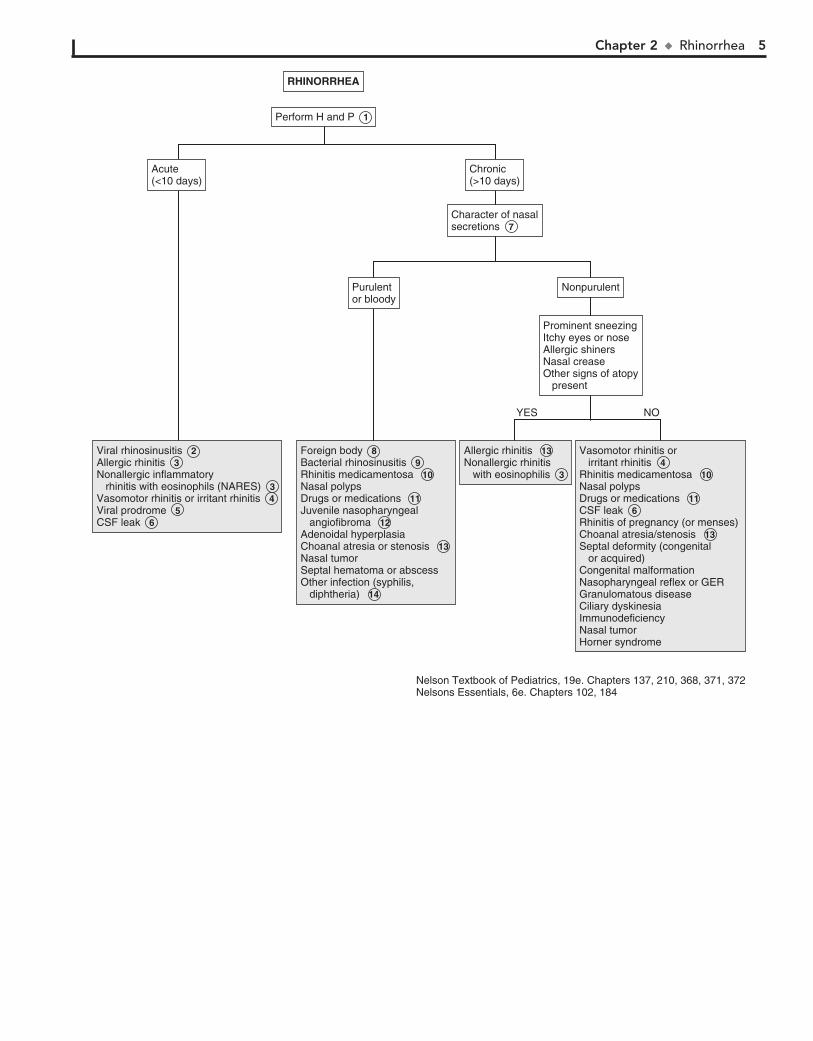
RHINORRHEA 7 When the clinical course and examination are not specific
for a diagnosis, especially when considering sinusitis versus allergic rhinitis, a microscopic examination of the nasal secretions may be helpful. An eosin–methylene blue stain of these secretions can help to identify eosinophils, WBCs, and bacteria. A predominance of WBCs and bacteria suggests sinusitis, and at least 5% eosinophils suggests allergic rhinitis. The two diseases may occur together.
8 Foreign bodies usually have a unilateral foul-smelling, purulent, or bloody discharge.
9 Clinical diagnosis of bacterial rhinosinusitis is made by findings of prolonged symptoms of rhinorrhea without
improvement for more than 10 to 14 days. Other suggestive symptoms include halitosis, fever, nocturnal cough, and post-nasal drip. Older children may have headache, facial pain, tooth pain, and periorbital swelling. Radiologic studies such as CT do not help differentiate bacterial from viral causes.
10 Rhinitis medicamentosa results from overuse of vasocon-strictor nose drops or sprays. A rapid toxic reaction of the
nasal mucosa causes rebound swelling and obstruction.
11 Cocaine, marijuana, and inhaled solvents may result in mucoid or purulent rhinorrhea. Medications causing
rhinorrhea include oral contraceptives, aspirin, nonsteroidal antiinflammatory and antihypertensive drugs. In the uncom-mon ASA triad (Samter’s triad), nasal polyps are associated with aspirin sensitivity and asthma.
12 Symptoms of nasal obstruction with increasing frequency of episodes of epistaxis, particularly unilateral, in boys are
suggestive of juvenile nasopharyngeal angiofibroma.
13 Bilateral choanal atresia presents early in the newborn period with respiratory distress. Unilateral choanal atresia
presents later with chronic unilateral rhinorrhea that can be clear or purulent. Feeding difficulties are also common, since most newborns are nose breathers. Inability to pass a nasal catheter suggests this diagnosis. An ENT consultation should be obtained whenever choanal atresia is suspected.
14 Infants with congenital syphilis may present between the second week and third month of life with a watery nasal
discharge that progresses to a mucopurulent or bloody dis-charge. Significant obstruction results in noisy breathing (“snuf-fles”). Chronic mucopurulent rhinorrhea, septal perforation, and saddle nose deformity are late complications. Serologic tests for treponemal antibodies and specimens for dark field microscopy examination should be obtained whenever this diagnosis is suspected.
Rhinorrhea is a common complaint in childhood. It is most frequently due to a viral URI but must be distinguished from allergies and other less common etiologies.
1 A careful HEENT examination is essential. Stigmata suggestive of genetic syndromes should be noted because
congenital nasal anomalies (e.g., atresia, stenosis, hypoplasia) are frequently associated with other anomalies. Examination of the nose should include the appearance of the mucosa (e.g., swelling, pallor, erythema, degree of patency), character of secretions, and presence of any obvious obstructing lesions (e.g., polyps, foreign bodies).
2 Acute rhinosinusitis or the “common cold” is the most com-mon cause of rhinorrhea. It is a relatively common illness in
children and adolescents. A viral etiology is far by the most com-mon; bacterial etiology is less common. (See footnote 9.)
3 Allergic rhinitis is an immunoglobulin (Ig)E-mediated con-dition that may be seasonal (e.g., hay fever) or perennial. The
nasal mucosa is typically boggy and pale or bluish. The rhinorrhea is clear and watery. Other allergic signs and symptoms, such as upward rubbing of the nose (i.e., allergic salute), allergic shiners, sneezing, and eye symptoms are common. Atopic disorders may be present (e.g., asthma, eczema). Fever suggests an alternative (infectious) diagnosis. Nonallergic inflammatory rhinitis with eosinophils (NARES) has a similar presentation to allergic rhinitis but without elevated IgE antibodies.
4 The vasomotor responses of increased secretion and mu-cosal swelling are the normal responses of the nasal mu-
cosa to a variety of stimuli. These responses are exaggerated in those with vasomotor rhinitis. External stimuli (e.g., cold tem-perature, change in humidity, cigarette smoke, spicy food) are the most common. The autonomic system response, hormones, and stress are other triggers.
5 Bronchiolitis, roseola infantum, measles, mononucleosis, hepatitis, pertussis, and erythema infectiosum may appear
with a prodromal acute watery rhinorrhea.
6 Rhinorrhea due to leakage of CSF is clear and usually unilateral, and it may vary noticeably with a change in
head position, Valsalva maneuver, or jugular compression. Detection of glucose (50 mg/100 mL or higher) in the fluid is highly suggestive. The condition may occur acutely with head trauma or chronically with congenital conditions (e.g., fistulas, encephaloceles) or tumors.
BibliographyDeMuri G, Wald ER: Acute bacterial sinusitis in children, N Engl J Med
367:1128–1134, 2012.
Chapter 2 u Rhinorrhea 5
RHINORRHEARHINORRHEA
Perform Perform H and P 1
Chronic(>10 days)Chronic(>10 days)
Character of nasalsecretionsCharacter of nasalsecretions 7
Prominent sneezingItchy eyes or noseAllergic shinersNasal creaseOther signs of atopy present
Prominent sneezingItchy eyes or noseAllergic shinersNasal creaseOther signs of atopy present
Acute(<10 days)Acute(<10 days)
Viral rhinosinusitis Allergic rhinitis Nonallergic inflammatory rhinitis with eosinophils (NARES) Vasomotor rhinitis or irritant rhinitis Viral prodrome CSF leak
Viral rhinosinusitis 2Allergic rhinitis 3Nonallergic inflammatory rhinitis with eosinophils (NARES) 3 Vasomotor rhinitis or irritant rhinitis 4Viral prodrome 5 CSF leak 6
NonpurulentNonpurulentPurulentor bloodyPurulentor bloody
Foreign body Bacterial rhinosinusitis Rhinitis medicamentosa Nasal polypsDrugs or medications Juvenile nasopharyngeal angiofibroma Adenoidal hyperplasiaChoanal atresia or stenosis Nasal tumorSeptal hematoma or abscessOther infection (syphilis, diphtheria)
Foreign body 8Bacterial rhinosinusitis 9Rhinitis medicamentosa 10Nasal polypsDrugs or medications 11Juvenile nasopharyngeal angiofibroma 12Adenoidal hyperplasiaChoanal atresia or stenosis 13 Nasal tumorSeptal hematoma or abscessOther infection (syphilis, diphtheria) 14
Allergic rhinitis Nonallergic rhinitis with eosinophilis
Allergic rhinitis 13 Nonallergic rhinitis with eosinophilis 3
Vasomotor rhinitis or irritant rhinitis Rhinitis medicamentosa Nasal polypsDrugs or medications CSF leak Rhinitis of pregnancy (or menses) Choanal atresia/stenosis Septal deformity (congenital or acquired) Congenital malformationNasopharyngeal reflex or GER Granulomatous diseaseCiliary dyskinesia Immunodeficiency Nasal tumorHorner syndrome
Vasomotor rhinitis or irritant rhinitis 4Rhinitis medicamentosa 10Nasal polypsDrugs or medications 11CSF leak 6Rhinitis of pregnancy (or menses) Choanal atresia/stenosis 13Septal deformity (congenital or acquired) Congenital malformationNasopharyngeal reflex or GER Granulomatous diseaseCiliary dyskinesia Immunodeficiency Nasal tumorHorner syndrome
NOYES
Nelson Textbook of Pediatrics, 19e. Chapters 137, 210, 368, 371, 372Nelsons Essentials, 6e. Chapters 102, 184

6
Chapter 3
SORE THROAT
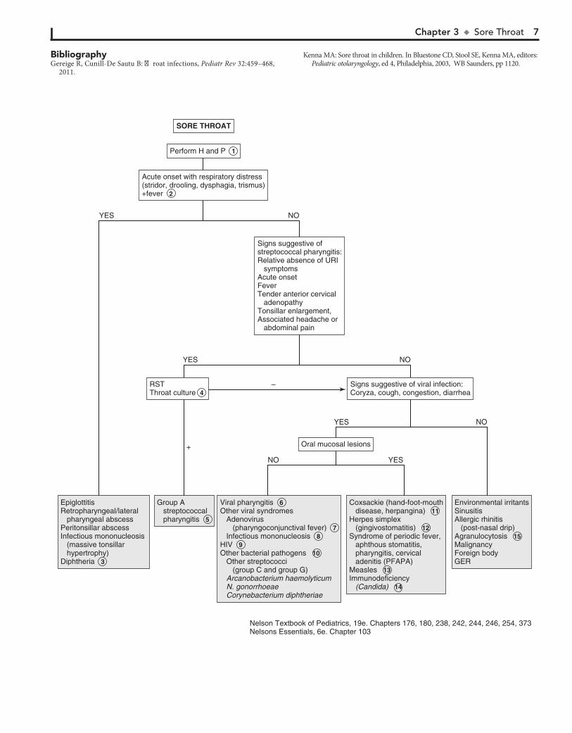
The most common etiologies are rhinovirus, coronavirus, ade-novirus, enterovirus, RSV, and metapneumovirus. Viral phar-yngitis is usually gradual in onset with early signs of fever, malaise, and anorexia generally preceding the sore throat.
7 Adenovirus may cause an exudative pharyngitis. Diarrhea and conjunctivitis are also common.
8 Exudative pharyngitis is often a manifestation of infectious mononucleosis. Patients can experience an abrupt onset of
fatigue, malaise, fever, and headache preceding the pharyngitis. Hepatosplenomegaly and generalized lymphadenopathy are common. Preadolescents tend to have milder symptoms than adolescents and young adults. Atypical lymphocytosis is sugges-tive of the disorder, and a positive “Monospot” (heterophile an-tibody) test finding confirms EBV mononucleosis. The test is not considered reliable in children younger than age 5 because of a low titer of heterophile antibody. EBV serology should be used in young patients or in patients with heterophile-negative cases. CMV serology should also be considered because CMV causes approximately 5% to 10% of cases.
9 Primary infection with HIV can also manifest with phar-yngitis and a mononucleosis-like syndrome.
10 Arcanobacterium haemolyticum may cause a scarlet fever–like illness but requires special culture methods. It is not
routinely sought in the evaluation of pharyngitis. Although non–group A streptococci have been implicated in pharyngitis, they cause a self-limiting illness, are not associated with complica-tions, and require no treatment. Gonococcal pharyngeal infec-tions are usually asymptomatic but can cause acute pharyngitis with fever and cervical lymphadenitis.
11 Coxsackie A16 is responsible for hand-foot-mouth disease, a characteristic outbreak of vesicles on the palms and soles,
with accompanying ulcerating vesicles throughout the orophar-ynx. Herpangina is a disorder characterized by fever and discrete painful, vesicular lesions of the posterior pharynx. A variety of enteroviruses cause herpangina, including enterovirus 71, al-though coxsackie A viruses are implicated most often.
12 Primary herpes simplex virus infection can cause gingivo-stomatitis characterized by painful ulcerating vesicles in
the anterior portion of the oral cavity, including the lips. An exudative tonsillitis may occur. Fevers and impaired fluid intake are common. Herpetic gingivostomatitis may last up to 2 weeks.
13 Pharyngitis characterized by intense erythema but absent tonsillar enlargement or exudate is an early finding in
measles. Fever, cough, coryza, conjunctivitis, and Koplik spots (i.e., blue-white enanthema on buccal mucosa) suggest the di-agnosis. These are followed by development of a maculopapular rash that begins on the forehead then spreads downward. Labo-ratory criteria for diagnosis include positive serologic test for measles immunoglobulin (Ig)M, seroconversion (a significant rise in measles IgG), isolation of measles virus, or identification by PCR of measles virus RNA from a clinical specimen (blood, urine, or respiratory secretions).
14 Immunocompromised patients are at risk for fungal oro-pharyngeal infections. Candida is the most common
pathogen. Diagnosis is made by examination of a specimen treated with potassium hydroxide or by culture.
15 Agranulocytosis may manifest as pharyngitis with a white or yellow exudate with underlying necrosis and ulceration.
Most sore throats are benign, self-limiting viral illnesses. The practitioner should always consider the likelihood of group A b-hemolytic streptococcus (Streptococcus pyogenes), which is important to identify and treat because of its potentially serious complications. Other less common causes should be considered when symptoms are worrisome or prolonged.
1 A history of exposure to a family member or classmate with a cold or documented group A streptococcal infec-
tion is helpful. A history of sexual activity or abuse should raise the suspicion for pharyngeal gonococcal infection. The degree of pharyngeal inflammation is not always consistent with the severity of the complaint. Tonsillar exudates are suggestive of streptococcus but also of mononucleosis and adenovirus. Many patients with streptococcal pharyngitis have only mild ery-thema without tonsillar enlargement or exudates. Small ulcers or vesicles on the soft palate suggest a viral etiology.
2 Acute onset of illness with associated symptoms of stridor, drooling, and air hunger or an unwillingness to recline sug-
gests impending airway obstruction. The patient warrants emer-gent management for airway stabilization and treatment for potentially life-threatening conditions such as epiglottitis and retropharyngeal abscess. (See Chapter 12.) A lateral neck film may be helpful but should be done only if the airway is stable.
3 Corynebacterium diphtheriae is a rare but serious cause of pharyngitis. The disease is suggested by a systemic illness
and grayish membrane over the tonsils and pharyngeal walls. It should be suspected in unimmunized persons or in persons from underdeveloped countries. Culture of the organism and confirmation of its toxin are necessary to confirm the diagnosis. Soft tissue swelling and enlarged lymph nodes can cause a bull-neck appearance.
4 Even when the clinical picture is highly suggestive of streptococcal pharyngitis, laboratory confirmation is
strongly recommended. Rapid antigen detection tests (RST) are highly specific, with sensitivities that are more variable. Throat cultures are the standard for diagnosis whenever the RST results are negative. The RST and the most commonly used culture methods do not identify organisms other than group A streptococcus. In cases in which another family member has a positive culture finding, or in which a typical scarlatina rash is present, group A streptococcus should still be considered despite negative test results.
5 Group A streptococcal pharyngitis is most common be-tween 5 and 11 years of age and unlikely under 3 years of
age. The occurrence of conjunctivitis, rhinitis, cough, and hoarseness is more indicative of a virus than group A strepto-coccus. Significant diarrhea also makes streptococcal disease unlikely. Some patients demonstrate the features of scarlet fever, including circumoral pallor, strawberry tongue, and a red, sandpaper-like scarlatina rash.
6 Viral pharyngitis is most commonly accompanied by “common cold” symptoms such as rhinitis and cough.
Chapter 3 u SoreThroat 7
SORE THROATSORE THROAT
Perform Perform H and P 1
Oral mucosal lesionsOral mucosal lesions
Signs suggestive of viral infection:Coryza, cough, congestion, diarrheaSigns suggestive of viral infection:Coryza, cough, congestion, diarrhea
RSTThroat culture 4 RSTThroat culture 4
Signs suggestive ofstreptococcal pharyngitis:Relative absence of URI symptoms Acute onset Fever Tender anterior cervical adenopathy Tonsillar enlargement, Associated headache or abdominal pain
Signs suggestive ofstreptococcal pharyngitis:Relative absence of URI symptoms Acute onset Fever Tender anterior cervical adenopathy Tonsillar enlargement, Associated headache or abdominal pain
EpiglottitisRetropharyngeal/lateral pharyngeal abscessPeritonsillar abscessInfectious mononucleosis (massive tonsillar hypertrophy) Diphtheria
EpiglottitisRetropharyngeal/lateral pharyngeal abscessPeritonsillar abscessInfectious mononucleosis (massive tonsillar hypertrophy) Diphtheria 3
Group A streptococcal pharyngitis
Group A streptococcal pharyngitis 5
Viral pharyngitisOther viral syndromes Adenovirus (pharyngoconjunctival fever) Infectious mononucleosisHIVOther bacterial pathogens Other streptococci (group C and group G) Arcanobacterium haemolyticum N. gonorrhoeae Corynebacterium diphtheriae
Viral pharyngitis 6Other viral syndromes Adenovirus (pharyngoconjunctival fever) 7 Infectious mononucleosis 8HIV 9Other bacterial pathogens 10 Other streptococci (group C and group G) Arcanobacterium haemolyticum N. gonorrhoeae Corynebacterium diphtheriae
Coxsackie (hand-foot-mouth disease, herpangina)Herpes simplex (gingivostomatitis)Syndrome of periodic fever, aphthous stomatitis, pharyngitis, cervical adenitis (PFAPA) MeaslesImmunodeficiency (Candida)
Coxsackie (hand-foot-mouth disease, herpangina) 11Herpes simplex (gingivostomatitis) 12Syndrome of periodic fever, aphthous stomatitis, pharyngitis, cervical adenitis (PFAPA) Measles 13Immunodeficiency (Candida) 14
Environmental irritantsSinusitisAllergic rhinitis (post-nasal drip) AgranulocytosisMalignancyForeign bodyGER
Environmental irritantsSinusitisAllergic rhinitis (post-nasal drip) Agranulocytosis 15MalignancyForeign bodyGER
Acute onset with respiratory distress(stridor, drooling, dysphagia, trismus)+fever
Acute onset with respiratory distress(stridor, drooling, dysphagia, trismus)+fever 2
NO YES
NOYES
–
+
NOYES
NOYES
Nelson Textbook of Pediatrics, 19e. Chapters 176, 180, 238, 242, 244, 246, 254, 373Nelsons Essentials, 6e. Chapter 103
BibliographyGereige R, Cunill-De Sautu B: Throat infections, Pediatr Rev 32:459–468,
2011.
Kenna MA: Sore throat in children. In Bluestone CD, Stool SE, Kenna MA, editors: Pediatric otolaryngology, ed 4, Philadelphia, 2003, WB Saunders, pp 1120.
8
Chapter 4
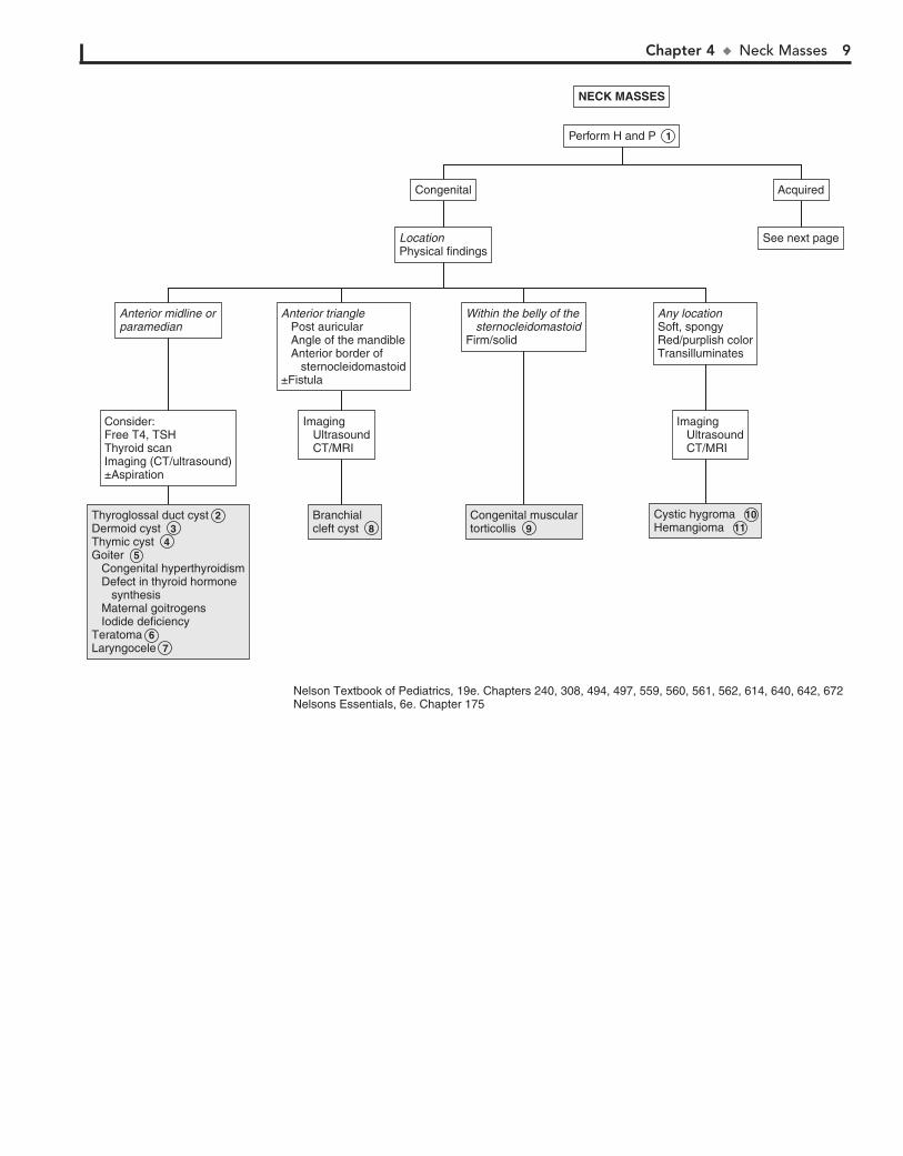
NECK MASSES 5 In newborn infants, a goiter may be associated with hypo-
thyroidism. This may occur with defects in thyroid hor-mone synthesis, administration of goitrogenic substances to the mother (e.g., antithyroid drug, iodides, amiodarone, radioio-dine), or iodine deficiency, causing endemic goiter, which is rare in the United States. Congenital hyperthyroidism in infants born to mothers with Graves disease may cause a goiter that usually resolves in 6 to 12 weeks.
6 Teratomas are usually midline but may be paramedian. They are firm and irregular and do not transilluminate.
Teratomas have classic radiologic findings of calcifications.
7 Laryngoceles are cystic dilations of the laryngeal ventricle located between the true and false vocal cords. They
appear as soft, compressible masses just lateral to the midline. Laryngoceles may enlarge with Valsalva maneuver. They may cause hoarseness or stridor. Air-fluid levels may be seen radiographically.
8 Branchial cleft anomalies include cysts, sinuses, and fistu-las. They are located in the lateral aspect of the anterior
triangle. Most anomalies arise from the second branchial arch along the anterior border of the sternocleidomastoid. Some may arise from the first branchial arch at the angle of the man-dible or in the postauricular region. These may not be present at birth but may manifest when older as drainage or a mass, if infected. Ultrasound, CT, or MRI may confirm the diagnosis.
9 Congenital torticollis is usually noted within the first few weeks of life. There is a firm, nontender, fibrous mass
within the body of the sternocleidomastoid. It results in tilting of the head toward the mass, with the chin in the opposite di-rection. It is believed to be caused by trauma or abnormal posi-tioning in utero. Prolonged, severe, untreated torticollis may result in a deformed face and skull.
10 Cystic hygromas (lymphangiomas) are cystic masses formed by dilated anomalous lymphatic channels. These are most
common in the posterior triangle but may occur in the subman-dibular or submental region. They are soft, nontender, diffuse, and compressible masses that may increase in size with straining or crying. Most transilluminate. Diagnosis may be confirmed by US. A CXR may be considered to look for mediastinal extension in patients with stridor or respiratory compromise. Chromo-somal aberrations are found in a significant percentage of infants who have cystic hygromas, and these lesions are associated fre-quently with Turner, Noonan, and Down syndromes. Cystic hy-gromas may be diagnosed as early as the second trimester of pregnancy by US.
11 Hemangiomas are vascular anomalies that appear at birth, often enlarging in the first year of life, followed by involution.
They are soft, compressible, red or purple-colored masses. They may increase in size with crying or Valsalva maneuver. They do not transilluminate. Bruits may be heard, particularly over large hemangiomas. The diagnosis can usually be made on physical findings, but US is a good initial test to confirm the diagnosis.
Most neck masses are benign, but it is important not to miss rare malignant masses. A directed H and P examination allows for successful diagnosis and, if necessary, referral for further evaluation and treatment.
1 Neck masses may be distinguished broadly into two cate-gories: congenital and acquired. Masses present since
birth, or with chronic drainage or recurrent episodes of swell-ing, are usually congenital. History of fever may indicate inflammation or infection. Constitutional symptoms such as fever, night sweats, and weight loss may indicate a malignancy or a granulomatous process. Rapidly enlarging, painless masses may be malignant. Those due to infection are often painful. Symptoms indicating compression of the trachea, esophagus, or recurrent laryngeal nerve should be elicited because rapid progression of the mass may be life threatening. A history of recurrent infections such as thrush, sinopulmonary infections, or cellulitis may indicate an immunodeficiency syndrome.
The location of the mass is helpful in making the diagnosis. The neck is divided into two triangles: the anterior triangle, which is bounded by the mandible, the sternocleidomastoid, and the anterior midline; and the the posterior triangle, which is bounded by the sternocleidomastoid, the distal two thirds of the clavicle, and the posterior midline. It is also important to determine the consistency of the lesion. Cystic lesions may show fluctuance and transilluminate. A bruit may be heard with vascular lesions.
2 Thyroglossal duct cysts are the most common congenital neck masses. However, they rarely manifest in the new-
born period and occur more commonly in children aged 2 to 10 years. Approximately one third are not diagnosed until after the age of 20. Thyroglossal duct cysts are usually painless and often move with tongue protrusion. They may occur with recurrent inflammation associated with a URI. Their location can be any-where from the base of the tongue to behind the sternum but are usually near or below the hyoid bone. US may be done to confirm the diagnosis. A thyroid scan is important to identify ectopic gland tissue in the cyst (found in one third of cases), because excision may lead to hypothyroidism.
3 Dermoid cysts are benign congenital neoplasms located in the midline. They are nontender, smooth, and doughy or
rubbery in consistency. They may be difficult to distinguish from thyroglossal duct cysts. In cases where the diagnosis is difficult to make, imaging studies and aspiration of the cyst may be considered.
4 Thymic cysts result from implantation of thymic tissue during its embryologic descent and are usually in a midline position.
Chapter 4 u NeckMasses 9
NECK MASSESNECK MASSES
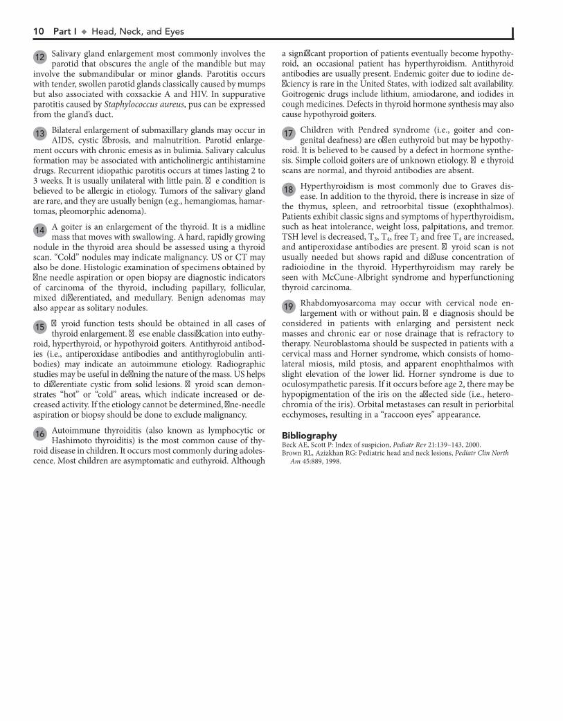
Perform H and P 1
AcquiredAcquiredCongenitalCongenital
See next pageSee next pageLocationPhysical findingsLocationPhysical findings
Thyroglossal duct cystDermoid cystThymic cystGoiter Congenital hyperthyroidism Defect in thyroid hormone synthesis Maternal goitrogens Iodide deficiencyTeratomaLaryngocele
Thyroglossal duct cyst 2 Dermoid cyst 3 Thymic cyst 4 Goiter 5 Congenital hyperthyroidism Defect in thyroid hormone synthesis Maternal goitrogens Iodide deficiency Teratoma 6 Laryngocele 7
Cystic hygromaCystic hygroma 10Hemangioma 11
Branchialcleft cyst Branchialcleft cyst 8
Consider:Free T4, TSH Thyroid scanImaging (CT/ultrasound)±Aspiration
Consider:Free T4, TSH Thyroid scanImaging (CT/ultrasound)±Aspiration
Imaging Ultrasound CT/MRI
Imaging Ultrasound CT/MRI
Imaging Ultrasound CT/MRI
Imaging Ultrasound CT/MRI
Anterior midline orparamedianAnterior midline orparamedian
Any locationSoft, spongyRed/purplish colorTransilluminates
Any locationSoft, spongyRed/purplish colorTransilluminates
Within the belly of the sternocleidomastoidFirm/solid
Within the belly of the sternocleidomastoidFirm/solid
Anterior triangle Post auricular Angle of the mandible Anterior border of sternocleidomastoid±Fistula
Anterior triangle Post auricular Angle of the mandible Anterior border of sternocleidomastoid±Fistula
Congenital musculartorticollis Congenital musculartorticollis 9
Nelson Textbook of Pediatrics, 19e. Chapters 240, 308, 494, 497, 559, 560, 561, 562, 614, 640, 642, 672Nelsons Essentials, 6e. Chapter 175
10 Part I u Head,Neck,andEyes
12 Salivary gland enlargement most commonly involves the parotid that obscures the angle of the mandible but may
involve the submandibular or minor glands. Parotitis occurs with tender, swollen parotid glands classically caused by mumps but also associated with coxsackie A and HIV. In suppurative parotitis caused by Staphylococcus aureus, pus can be expressed from the gland’s duct.
13 Bilateral enlargement of submaxillary glands may occur in AIDS, cystic fibrosis, and malnutrition. Parotid enlarge-
ment occurs with chronic emesis as in bulimia. Salivary calculus formation may be associated with anticholinergic antihistamine drugs. Recurrent idiopathic parotitis occurs at times lasting 2 to 3 weeks. It is usually unilateral with little pain. The condition is believed to be allergic in etiology. Tumors of the salivary gland are rare, and they are usually benign (e.g., hemangiomas, hamar-tomas, pleomorphic adenoma).
14 A goiter is an enlargement of the thyroid. It is a midline mass that moves with swallowing. A hard, rapidly growing
nodule in the thyroid area should be assessed using a thyroid scan. “Cold” nodules may indicate malignancy. US or CT may also be done. Histologic examination of specimens obtained by fine needle aspiration or open biopsy are diagnostic indicators of carcinoma of the thyroid, including papillary, follicular, mixed differentiated, and medullary. Benign adenomas may also appear as solitary nodules.
15 Thyroid function tests should be obtained in all cases of thyroid enlargement. These enable classification into euthy-
roid, hyperthyroid, or hypothyroid goiters. Antithyroid antibod-ies (i.e., antiperoxidase antibodies and antithyroglobulin anti-bodies) may indicate an autoimmune etiology. Radiographic studies may be useful in defining the nature of the mass. US helps to differentiate cystic from solid lesions. Thyroid scan demon-strates “hot” or “cold” areas, which indicate increased or de-creased activity. If the etiology cannot be determined, fine-needle aspiration or biopsy should be done to exclude malignancy.
16 Autoimmune thyroiditis (also known as lymphocytic or Hashimoto thyroiditis) is the most common cause of thy-
roid disease in children. It occurs most commonly during adoles-cence. Most children are asymptomatic and euthyroid. Although
a significant proportion of patients eventually become hypothy-roid, an occasional patient has hyperthyroidism. Antithyroid antibodies are usually present. Endemic goiter due to iodine de-ficiency is rare in the United States, with iodized salt availability. Goitrogenic drugs include lithium, amiodarone, and iodides in cough medicines. Defects in thyroid hormone synthesis may also cause hypothyroid goiters.
17 Children with Pendred syndrome (i.e., goiter and con-genital deafness) are often euthyroid but may be hypothy-
roid. It is believed to be caused by a defect in hormone synthe-sis. Simple colloid goiters are of unknown etiology. The thyroid scans are normal, and thyroid antibodies are absent.
18 Hyperthyroidism is most commonly due to Graves dis-ease. In addition to the thyroid, there is increase in size of
the thymus, spleen, and retroorbital tissue (exophthalmos). Patients exhibit classic signs and symptoms of hyperthyroidism, such as heat intolerance, weight loss, palpitations, and tremor. TSH level is decreased, T3, T4, free T3 and free T4 are increased, and antiperoxidase antibodies are present. Thyroid scan is not usually needed but shows rapid and diffuse concentration of radioiodine in the thyroid. Hyperthyroidism may rarely be seen with McCune-Albright syndrome and hyperfunctioning thyroid carcinoma.
19 Rhabdomyosarcoma may occur with cervical node en-largement with or without pain. The diagnosis should be
considered in patients with enlarging and persistent neck masses and chronic ear or nose drainage that is refractory to therapy. Neuroblastoma should be suspected in patients with a cervical mass and Horner syndrome, which consists of homo-lateral miosis, mild ptosis, and apparent enophthalmos with slight elevation of the lower lid. Horner syndrome is due to oculosympathetic paresis. If it occurs before age 2, there may be hypopigmentation of the iris on the affected side (i.e., hetero-chromia of the iris). Orbital metastases can result in periorbital ecchymoses, resulting in a “raccoon eyes” appearance.
BibliographyBeck AE, Scott P: Index of suspicion, Pediatr Rev 21:139–143, 2000. Brown RL, Azizkhan RG: Pediatric head and neck lesions, Pediatr Clin North
Am 45:889, 1998.
Chapter 4 u NeckMasses 11
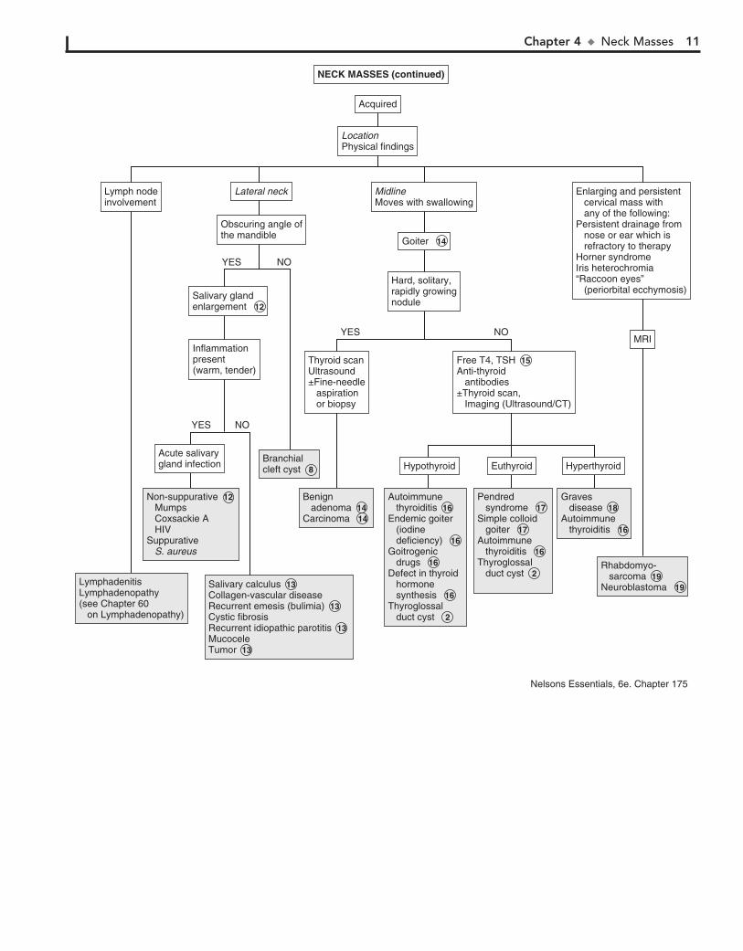
Rhabdomyo- sarcoma Neuroblastoma
Rhabdomyo- sarcoma 19 Neuroblastoma 19
LocationPhysical findingsLocationPhysical findings
AcquiredAcquired
Enlarging and persistent cervical mass with any of the following:Persistent drainage from nose or ear which is refractory to therapyHorner syndromeIris heterochromia“Raccoon eyes” (periorbital ecchymosis)
Enlarging and persistent cervical mass with any of the following:Persistent drainage from nose or ear which is refractory to therapyHorner syndromeIris heterochromia“Raccoon eyes” (periorbital ecchymosis)
MRIMRI
Lateral neckLateral neck
Obscuring angle ofthe mandibleObscuring angle ofthe mandible
MidlineMoves with swallowingMidlineMoves with swallowing
Goiter Goiter 14
Hard, solitary,rapidly growingnodule
Hard, solitary,rapidly growingnodule
Non-suppurative Mumps Coxsackie A HIVSuppurative S. aureus
Non-suppurative 12 Mumps Coxsackie A HIVSuppurative S. aureus
LymphadenitisLymphadenopathy(see Chapter 60 on Lymphadenopathy)
LymphadenitisLymphadenopathy(see Chapter 60 on Lymphadenopathy)
Graves diseaseAutoimmune thyroiditis
Graves disease 18 Autoimmune thyroiditis 16
Autoimmune thyroiditisEndemic goiter (iodine deficiency)Goitrogenic drugsDefect in thyroid hormone synthesisThyroglossal duct cyst
Autoimmune thyroiditis 16 Endemic goiter (iodine deficiency) 16 Goitrogenic drugs 16 Defect in thyroid hormone synthesis 16Thyroglossal duct cyst 2
Pendred syndromeSimple colloid goiterAutoimmune thyroiditisThyroglossal duct cyst
Pendred syndrome 17 Simple colloid goiter 17 Autoimmune thyroiditis 16 Thyroglossal duct cyst 2
Benign adenomaCarcinoma
Benign adenoma 14 Carcinoma 14
Branchial cleft cyst Branchial cleft cyst 8
Salivary calculusCollagen-vascular diseaseRecurrent emesis (bulimia)Cystic fibrosisRecurrent idiopathic parotitisMucoceleTumor
Salivary calculus 13 Collagen-vascular diseaseRecurrent emesis (bulimia) 13 Cystic fibrosisRecurrent idiopathic parotitis 13 MucoceleTumor 13
HypothyroidHypothyroid HyperthyroidHyperthyroid
Free T4, TSH Anti-thyroid antibodies ±Thyroid scan, Imaging (Ultrasound/CT)
Free T4, TSH 15 Anti-thyroid antibodies ±Thyroid scan, Imaging (Ultrasound/CT)
Thyroid scanUltrasound ±Fine-needle aspiration or biopsy
Thyroid scanUltrasound ±Fine-needle aspiration or biopsy
Salivary gland enlargement Salivary gland enlargement 12
Inflammationpresent(warm, tender)
Inflammationpresent(warm, tender)
Lymph nodeinvolvementLymph nodeinvolvement
EuthyroidEuthyroid
NOYES
Acute salivarygland infectionAcute salivarygland infection
NOYES
NOYES
NECK MASSES (continued)NECK MASSES (continued)
Nelsons Essentials, 6e. Chapter 175
12
Chapter 5
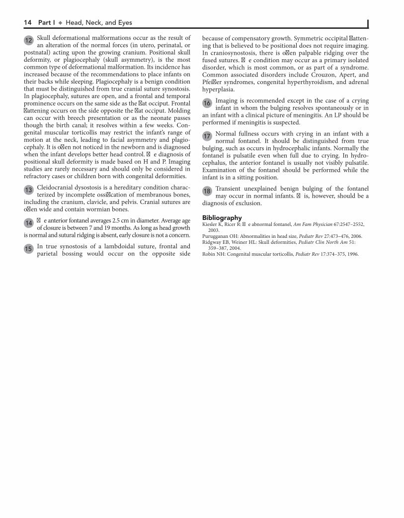
ABNORMAL HEAD SIZE, SHAPE, AND FONTANELS
increased production of CSF. Causes of obstructive (noncom-municating) hydrocephalus include aqueductal stenosis, neona-tal meningitis, subarachnoid hemorrhage in a premature infant, intrauterine viral infections, vein of Galen malformation, and posterior fossa lesions or malformations (tumors, Chiari malfor-mation, Dandy-Walker syndrome). Subarachnoid hemorrhage in a premature infant can also cause nonobstructive (communi-cating) hydrocephalus. A rare cause is overproduction of CSF with choroid plexus papilloma.
5 In hydranencephaly the cerebral hemispheres are absent or represented by membranous sacs. The cause of this
condition is unknown.
6 Occasionally, benign fluid collections (e.g., subarachnoid, subdural) cause macrocephaly without other clinical
significance. A pediatric neurosurgeon should be consulted for recommendations.
7 Various metabolic and degenerative disorders may cause megalencephaly. These include lysosomal diseases
(Tay-Sachs disease, gangliosidosis, mucopolysaccharidoses), maple syrup urine disease, and leukodystrophies.
8 Many syndromes are associated with microcephaly. If a chromosomal syndrome is suspected (child has abnormal
facies, short stature, congenital anomalies) karyotype and/or array-comparative genomic hybridization (microarray) study and MRI may be considered.
9 MRI can evaluate structural abnormalities of the brain (lissencephaly, pachygyria, and polymicrogyria) and both
MRI and CT scanning may detect intracerebral calcification, which suggests congenital infection. Also consider TORCH titers (toxoplasmosis, rubella, CMV, and herpes simplex) and HIV testing of the mother and child, as well as a urine culture for CMV. Consider testing for maternal serum phenylalanine level (PKU), because high maternal levels can affect a nonphe-nylketonuric infant.
10 Familial microcephaly is often associated with some degree of mental retardation.
11 Secondary microcephaly results from exposure to noxious agents during periods of rapid brain growth in utero or
during the first 2 years of life.
Macrocephaly is defined as an occipitofrontal circumference (OFC) greater than 2 standard deviations above the mean. Megalencephaly is a disorder of brain growth, usually accompa-nied by macrocephaly. An increase in growth rate with crossing of percentiles is of more concern than the case of a child with a large head growing at a normal rate. In microcephaly the OFC is 2 standard deviations below the mean.
1 A birth history, developmental history and history of irri-tability, headaches, and visual problems are important
components of the initial evaluation. For macrocephaly, inquire about familial head sizes (e.g., ask about hat sizes). It is impor-tant to note any features suggestive of specific syndromes.
2 US can be done if the anterior fontanel (AF) is open. Otherwise, an MRI should be considered. A CT is pre-
ferred if there is suspicion of trauma (nonaccidental or acciden-tal). Radiologic evaluation may not be necessary if development is normal, a parent is macrocephalic, and the child’s head is growing at a normal rate. Further evaluation is directed by the history and physical examination. Plain long bone radiographs may be indicated for evaluation of skeletal dysplasia or trauma. Consider chromosome testing (fragile X) or metabolic tests (urine organic acids).
3 Benign familial megalencephaly is the most common cause of anatomic megalencephaly. It is inherited as an
autosomal dominant trait. These children may have mild neu-rodevelopmental dysfunction. It is diagnosed by careful family history and measurement of the parents’ head circumferences.
4 Hydrocephalus is caused by multiple conditions associated with impaired circulation and absorption of CSF or
Chapter 5 u AbnormalHeadSize,Shape,andFontanels 13
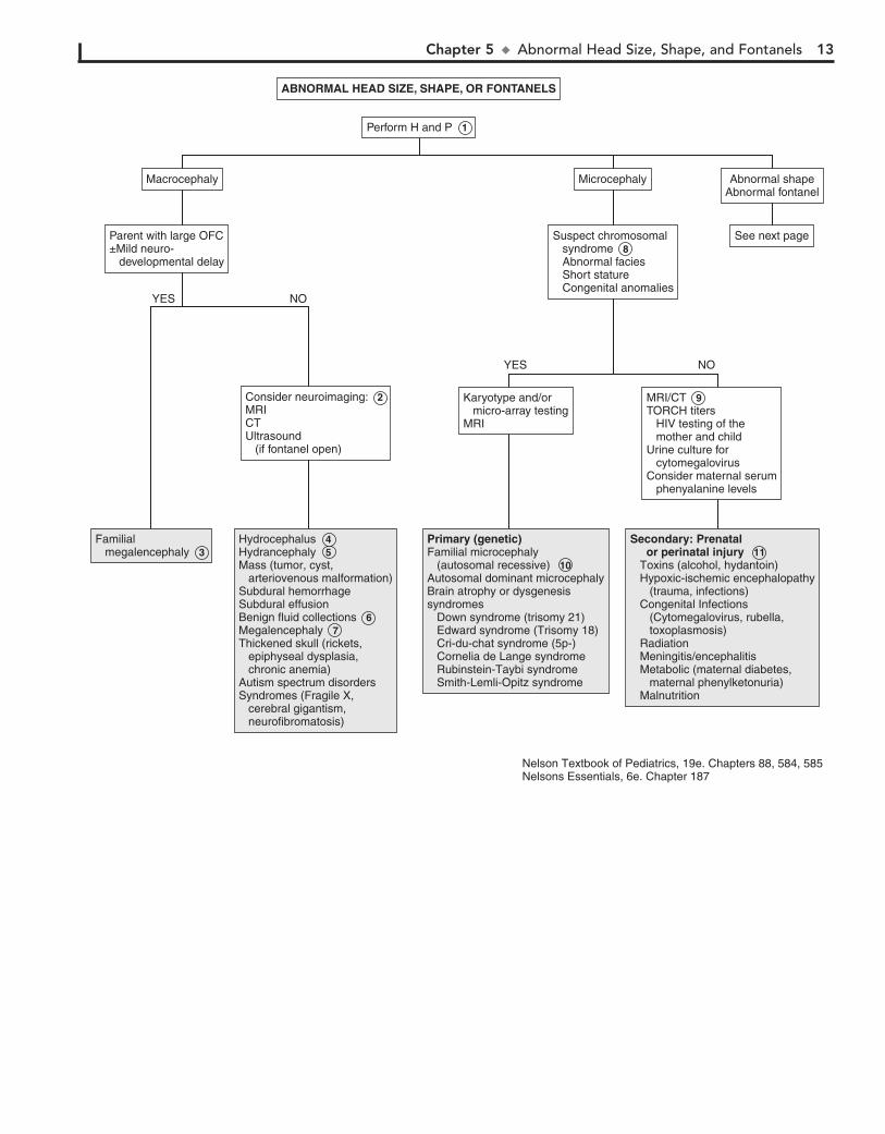
Familial megalencephalyFamilial megalencephaly 3
Secondary: Prenatal or perinatal injury Toxins (alcohol, hydantoin) Hypoxic-ischemic encephalopathy (trauma, infections) Congenital Infections (Cytomegalovirus, rubella, toxoplasmosis) Radiation Meningitis/encephalitis Metabolic (maternal diabetes, maternal phenylketonuria) Malnutrition
Secondary: Prenatal or perinatal injury 11 Toxins (alcohol, hydantoin) Hypoxic-ischemic encephalopathy (trauma, infections) Congenital Infections (Cytomegalovirus, rubella, toxoplasmosis) Radiation Meningitis/encephalitis Metabolic (maternal diabetes, maternal phenylketonuria) Malnutrition
Primary (genetic)Familial microcephaly (autosomal recessive)Autosomal dominant microcephalyBrain atrophy or dysgenesissyndromes Down syndrome (trisomy 21) Edward syndrome (Trisomy 18) Cri-du-chat syndrome (5p-) Cornelia de Lange syndrome Rubinstein-Taybi syndrome Smith-Lemli-Opitz syndrome
Primary (genetic)Familial microcephaly (autosomal recessive) 10 Autosomal dominant microcephalyBrain atrophy or dysgenesissyndromes Down syndrome (trisomy 21) Edward syndrome (Trisomy 18) Cri-du-chat syndrome (5p-) Cornelia de Lange syndrome Rubinstein-Taybi syndrome Smith-Lemli-Opitz syndrome
HydrocephalusHydrancephalyMass (tumor, cyst, arteriovenous malformation)Subdural hemorrhageSubdural effusionBenign fluid collectionsMegalencephalyThickened skull (rickets, epiphyseal dysplasia, chronic anemia)Autism spectrum disordersSyndromes (Fragile X, cerebral gigantism, neurofibromatosis)
Hydrocephalus 4 Hydrancephaly 5 Mass (tumor, cyst, arteriovenous malformation)Subdural hemorrhageSubdural effusionBenign fluid collections 6 Megalencephaly 7 Thickened skull (rickets, epiphyseal dysplasia, chronic anemia)Autism spectrum disordersSyndromes (Fragile X, cerebral gigantism, neurofibromatosis)
ABNORMAL HEAD SIZE, SHAPE, OR FONTANELS
Perform H and P 1
See next page
Macrocephaly Microcephaly Abnormal shapeAbnormal fontanel
Parent with large OFC±Mild neuro- developmental delay
Suspect chromosomal syndrome 8 Abnormal facies Short stature Congenital anomalies
Consider neuroimaging: 2 MRICT Ultrasound (if fontanel open)
MRI/CT 9 TORCH titers HIV testing of the mother and child Urine culture for cytomegalovirusConsider maternal serum phenyalanine levels
Karyotype and/or micro-array testingMRI
YES
YES
NO
NO
Nelson Textbook of Pediatrics, 19e. Chapters 88, 584, 585Nelsons Essentials, 6e. Chapter 187
14 Part I u Head,Neck,andEyes
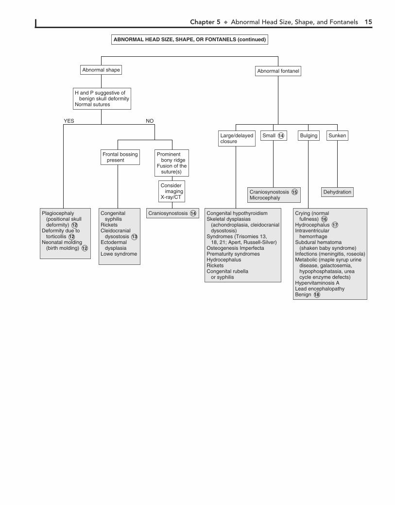
12 Skull deformational malformations occur as the result of an alteration of the normal forces (in utero, perinatal, or
postnatal) acting upon the growing cranium. Positional skull deformity, or plagiocephaly (skull asymmetry), is the most common type of deformational malformation. Its incidence has increased because of the recommendations to place infants on their backs while sleeping. Plagiocephaly is a benign condition that must be distinguished from true cranial suture synostosis. In plagiocephaly, sutures are open, and a frontal and temporal prominence occurs on the same side as the flat occiput. Frontal flattening occurs on the side opposite the flat occiput. Molding can occur with breech presentation or as the neonate passes though the birth canal; it resolves within a few weeks. Con-genital muscular torticollis may restrict the infant’s range of motion at the neck, leading to facial asymmetry and plagio-cephaly. It is often not noticed in the newborn and is diagnosed when the infant develops better head control. The diagnosis of positional skull deformity is made based on H and P. Imaging studies are rarely necessary and should only be considered in refractory cases or children born with congenital deformities.
13 Cleidocranial dysostosis is a hereditary condition charac-terized by incomplete ossification of membranous bones,
including the cranium, clavicle, and pelvis. Cranial sutures are often wide and contain wormian bones.
14 The anterior fontanel averages 2.5 cm in diameter. Average age of closure is between 7 and 19 months. As long as head growth
is normal and sutural ridging is absent, early closure is not a concern.
15 In true synostosis of a lambdoidal suture, frontal and parietal bossing would occur on the opposite side
because of compensatory growth. Symmetric occipital flatten-ing that is believed to be positional does not require imaging. In craniosynostosis, there is often palpable ridging over the fused sutures. The condition may occur as a primary isolated disorder, which is most common, or as part of a syndrome. Common associated disorders include Crouzon, Apert, and Pfeiffer syndromes, congenital hyperthyroidism, and adrenal hyperplasia.
16 Imaging is recommended except in the case of a crying infant in whom the bulging resolves spontaneously or in
an infant with a clinical picture of meningitis. An LP should be performed if meningitis is suspected.
17 Normal fullness occurs with crying in an infant with a normal fontanel. It should be distinguished from true
bulging, such as occurs in hydrocephalic infants. Normally the fontanel is pulsatile even when full due to crying. In hydro-cephalus, the anterior fontanel is usually not visibly pulsatile. Examination of the fontanel should be performed while the infant is in a sitting position.
18 Transient unexplained benign bulging of the fontanel may occur in normal infants. This, however, should be a
diagnosis of exclusion.
BibliographyKiesler K, Ricer R: The abnormal fontanel, Am Fam Physician 67:2547–2552,
2003. Purugganan OH: Abnormalities in head size, Pediatr Rev 27:473–476, 2006.Ridgway EB, Weiner HL: Skull deformities, Pediatr Clin North Am 51:
359–387, 2004.Robin NH: Congenital muscular torticollis, Pediatr Rev 17:374–375, 1996.
Chapter 5 u AbnormalHeadSize,Shape,andFontanels 15
DehydrationDehydration
SunkenSunken
Crying (normal fullness)HydrocephalusIntraventricular hemorrhageSubdural hematoma (shaken baby syndrome)Infections (meningitis, roseola)Metabolic (maple syrup urine disease, galactosemia, hypophosphatasia, urea cycle enzyme defects)Hypervitaminosis ALead encephalopathyBenign
Crying (normal fullness) 16 Hydrocephalus 17 Intraventricular hemorrhageSubdural hematoma (shaken baby syndrome)Infections (meningitis, roseola)Metabolic (maple syrup urine disease, galactosemia, hypophosphatasia, urea cycle enzyme defects)Hypervitaminosis ALead encephalopathyBenign 18
BulgingBulging
CraniosynostosisMicrocephaly Craniosynostosis 15 Microcephaly
SmallSmall 14
Congenital hypothyroidismSkeletal dysplasias (achondroplasia, cleidocranial dysostosis)Syndromes (Trisomies 13, 18, 21; Apert, Russell-Silver)Osteogenesis ImperfectaPrematurity syndromesHydrocephalusRicketsCongenital rubella or syphilis
Congenital hypothyroidismSkeletal dysplasias (achondroplasia, cleidocranial dysostosis)Syndromes (Trisomies 13, 18, 21; Apert, Russell-Silver)Osteogenesis ImperfectaPrematurity syndromesHydrocephalusRicketsCongenital rubella or syphilis
Large/delayedclosureLarge/delayedclosure
Abnormal shapeAbnormal shape Abnormal fontanelAbnormal fontanel
(continued)ABNORMAL HEAD SIZE, SHAPE, OR FONTANELS (continued)
CraniosynostosisCraniosynostosis 14 Plagiocephaly (positional skull deformity)Deformity due to torticollisNeonatal molding (birth molding)
Plagiocephaly (positional skull deformity) 12 Deformity due to torticollis 12 Neonatal molding (birth molding) 12
Congenital syphilisRicketsCleidocranial dysostosisEctodermal dysplasiaLowe syndrome
Congenital syphilisRicketsCleidocranial dysostosis 13 Ectodermal dysplasiaLowe syndrome
H and P suggestive of benign skull deformityNormal sutures
Frontal bossing present
Prominent bony ridgeFusion of the suture(s)
Consider imagingX-ray/CT
YES NO
16
Chapter 6
RED EYE
risk for inflammation and infection (i.e., dacryocystitis) of the obstructed nasolacrimal sac.
6 Tearing, photophobia, and blepharospasm make up the classic triad of presenting symptoms of infantile glaucoma.
Conjunctival injection, corneal enlargement (.12 mm), and corneal clouding (edema) are the other findings.
7 Conjunctivitis in the first 24 hours of life is probably a chem-ical conjunctivitis unless membranes were ruptured prema-
turely. Silver nitrate is more likely to produce this condition than other agents used for prophylaxis (e.g., erythromycin, tetracycline) and is no longer used in the United States. In older children, chemical irritants may include cosmetics or eye medications.
8 Corneal abrasion presents with pain, tearing, photopho-bia, and eye redness. It is an important consideration in
the diagnosis of an irritable infant. Diagnosis is by fluorescein staining and observation under blue light.
9 Subconjunctival hemorrhage may occur with vomiting, coughing, or weight lifting. It may also occur in newborns
after vaginal delivery.
10 Allergic conjunctivitis is characterized by itching, chemo-sis, papillae of the tarsal conjunctivae, and white stringy
discharge. In limbal vernal conjunctivitis, a ring of swollen con-junctiva surrounds the limbus of the cornea.
11 Bacterial conjunctivitis may be unilateral or bilateral, but viral conjunctivitis is more commonly bilateral. Bacterial
conjunctivitis is more likely to have purulent discharge than viral conjunctivitis, although significant overlap in the clinical presentation of the two etiologies does occur. Nontypable Hae-mophilus influenzae, pneumococci, staphylococci, and strepto-cocci are common agents.
12 Redness may be due to irritation from eye rubbing. Exces-sive television or computer use may cause decreased rate
of blinking, with drying and irritation.
13 Iritis and iridocyclitis may occur secondary to localized infection or trauma, or they may be manifestations of
a rheumatic disorder (e.g., JRA, Reiter syndrome, Behçet’s disease). Inflammatory bowel disease and Kawasaki disease are other associated conditions. Photophobia is typically a significant finding with iritis and iridocyclitis.
14 Scleritis may accompany certain autoimmune disorders including systemic lupus erythematosus and Henoch-
Schönlein purpura. Pain is present, eye discharge is absent, and dilated blood vessels are larger than in conjunctivitis.
15 Parinaud’s oculoglandular syndrome is a form of cat scratch disease caused by Bartonella henselae. Symptoms include a
granulomatous conjunctivitis and preauricular lymphadenopathy.
Red eye is a common pediatric complaint. It can occur second-ary to a wide range of etiologies.
1 The age of onset of the red eye, the nature of any discharge, and the associated signs and symptoms are the most
important components of the history. History of exposure to irritants (e.g., allergens, particulate matter, chemicals) and of trauma or infectious contacts (e.g., “pink-eye” in school or day-care settings) may also be helpful. For infants, inquire about the possibility of any maternal infections.
2 Conjunctivitis within the neonatal period (4 weeks of birth) is also known as ophthalmia neonatorum. The most
common causes in the United States are Staphylococcus aureus, Staphylococcus epidermidis, Streptococcus pneumoniae, and Moraxella catarrhalis.
3 Ophthalmia neonatorum also can be caused by Chlamydia trachomatis, Neisseria gonorrhoeae, and herpes simplex virus
(HSV). Gonococcal conjunctivitis typically appears as a fulminant purulent conjunctivitis in the first 2 to 6 days of life. Chlamydial conjunctivitis is more likely beyond the first 6 days of life and is often associated with a pneumonitis. It can develop in 30% to 40% of infants whose mothers had untreated chlamydia. Conjunctivitis caused by HSV characteristically occurs as a unilateral bright red eye with thin watery discharge. Vesicles or erosions are present on the lid or surrounding skin. These clinical findings are not spe-cific, however, and prompt evaluation and treatment are always indicated to avoid serious sequelae. A Gram stain and culture will aid in the diagnosis of gonorrhea. Rapid antigen tests are available for chlamydial infections. HSV is usually cultured, but PCR may be helpful. Ophthalmologic consultation is indicated when herpes is suspected.
4 Viral conjunctivitis may vary in presentation from mild redness and irritation with minimal watery drainage to
severe conjunctival injection with purulent discharge. Adenovi-rus is the most common cause and may present with preauricu-lar lymphadenopathy. Coxsackie and echoviruses may cause a hemorrhagic conjunctivitis.
5 Dacryostenosis (i.e., congenital lacrimal duct stenosis) is a common disorder that occurs within 2 to 4 months of age
but sometimes is not noticed until tear production with crying becomes evident. An excessive tear lake and overflow with crusting are seen on examination. Children so affected are at
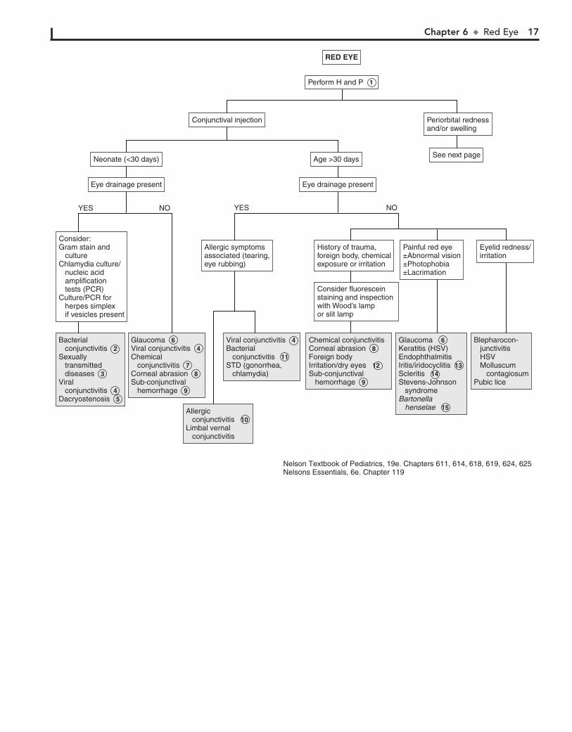
Chapter 6 u RedEye 17
Bacterial conjunctivitisSexually transmitted diseasesViral conjunctivitisDacryostenosis
Bacterial conjunctivitis 2 Sexually transmitted diseases 3 Viral conjunctivitis 4 Dacryostenosis 5
GlaucomaViral conjunctivitisChemical conjunctivitisCorneal abrasionSub-conjunctival hemorrhage
Glaucoma 6 Viral conjunctivitis 4 Chemical conjunctivitis 7 Corneal abrasion 8 Sub-conjunctival hemorrhage 9
Allergic conjunctivitisLimbal vernal conjunctivitis
Allergic conjunctivitis 10 Limbal vernal conjunctivitis
Viral conjunctivitisBacterial conjunctivitisSTD (gonorrhea, chlamydia)
Viral conjunctivitis 4 Bacterial conjunctivitis 11 STD (gonorrhea, chlamydia)
Chemical conjunctivitis Corneal abrasionForeign body Irritation/dry eyesSub-conjunctival hemorrhage
Chemical conjunctivitis Corneal abrasion 8 Foreign body Irritation/dry eyes 12 Sub-conjunctival hemorrhage 9
Glaucoma Keratitis (HSV)EndophthalmitisIritis/iridocyclitisScleritisStevens-Johnson syndrome Bartonella henselae
Glaucoma 6 Keratitis (HSV)EndophthalmitisIritis/iridocyclitis 13 Scleritis 14 Stevens-Johnson syndrome Bartonella henselae 15
Blepharocon- junctivitis HSV Molluscum contagiosumPubic lice
Blepharocon- junctivitis HSV Molluscum contagiosumPubic lice
RED EYE
Perform H and P 1
Consider fluorescein staining and inspection with Wood’s lamp or slit lamp
History of trauma, foreign body, chemicalexposure or irritation
Allergic symptoms associated (tearing,eye rubbing)
Painful red eye±Abnormal vision±Photophobia±Lacrimation
Eyelid redness/irritation
Consider:Gram stain and cultureChlamydia culture/ nucleic acid amplification tests (PCR)Culture/PCR for herpes simplex if vesicles present
Eye drainage present
Neonate (<30 days)
Conjunctival injection Periorbital rednessand/or swelling
See next page
Eye drainage present
Age >30 days
YES NOYES NO
Nelson Textbook of Pediatrics, 19e. Chapters 611, 614, 618, 619, 624, 625Nelsons Essentials, 6e. Chapter 119

18 Part I u Head,Neck,andEyes
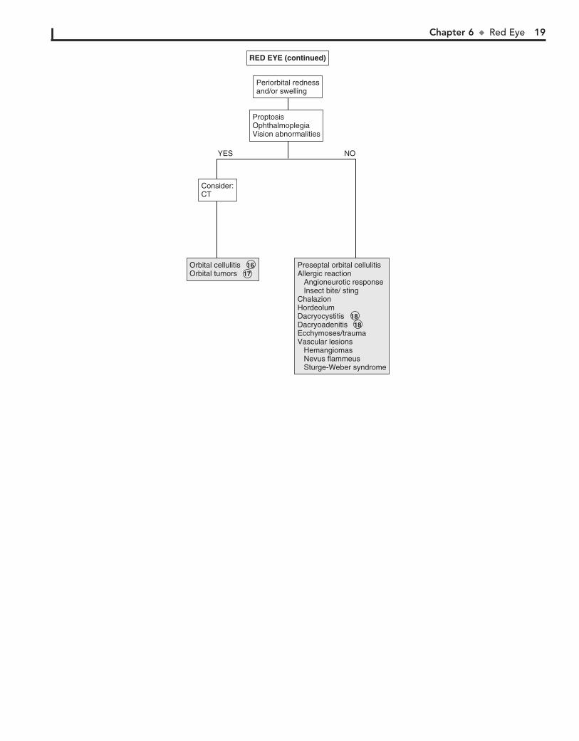
16 Pain with extraocular eye movements may accompany orbital cellulitis. Proptosis and impaired extraocular
movement and vision are other signs. Orbital cellulitis must be distinguished from preseptal (periorbital) cellulitis. Minimal conjunctival redness usually occurs in orbital cellulitis, and extraocular muscle movements are intact in preseptal cellulitis.
17 Orbital tumors, including rhabdomyosarcomas, neuroblas-tomas, and lymphangiomas, may have a similar presentation
to orbital cellulitis.
18 Some systemic disorders, such as sarcoid, tuberculosis, and syphilis, may cause chronic dacryocystitis.
BibliographyGreenberg MF, Pollard ZF: The red eye in childhood, Pediatr Clin North Am
50:105–124, 2003. Richards A, Guzman-Cottrill JA: Conjunctivitis, Pediatr Rev 31:196–208, 2010.
The lacrimal gland (i.e., the site of tear production) is located in the lateral aspect of the upper eyelid. Rarely, inflammation of the lacrimal gland (i.e., dacryoadenitis) can occur as a result of infections (e.g., S. aureus, infectious mononucleosis, mumps).
Chapter 6 u RedEye 19
Orbital cellulitisOrbital tumorsOrbital cellulitis 16 Orbital tumors 17
Preseptal orbital cellulitisAllergic reaction Angioneurotic response Insect bite/ stingChalazionHordeolumDacryocystitisDacryoadenitisEcchymoses/trauma Vascular lesions Hemangiomas Nevus flammeus Sturge-Weber syndrome
Preseptal orbital cellulitisAllergic reaction Angioneurotic response Insect bite/ stingChalazionHordeolumDacryocystitis 18 Dacryoadenitis 18 Ecchymoses/trauma Vascular lesions Hemangiomas Nevus flammeus Sturge-Weber syndrome
ProptosisOphthalmoplegiaVision abnormalities
ProptosisOphthalmoplegiaVision abnormalities
Periorbital rednessand/or swellingPeriorbital rednessand/or swelling
Consider:CTConsider:CT
YES NO
(continued)RED EYE (continued)
20
Chapter 7
STRABISMUS
of gaze. There is usually no underlying neurologic, mechanical, sensory, or other deficit. Noncomitant strabismus is suggested by an eye misalignment that varies according to the direction of the gaze. The condition is produced by an underlying nerve palsy, muscle weakness, or mechanical restriction of eye movement. Compensa-tory head tilting often occurs.
8 Infantile (congenital) esotropia appears before 6 months of age. There is often a family history of strabismus.
9 Acquired esotropia is often accommodative; the eyes turn inward with attempt to focus. Onset is typically between
2 and 3 years of age. Acquired esotropia may follow a period of occlusion of one eye.
10 Infantile exotropia is less common than infantile esotropia and is more frequent in children with neurologic abnor-
malities.
11 Careful assessment of ocular motility and associated lid and pupillary functions should help identify cranial nerve
palsies. Acquired cranial nerve palsies warrant careful evalua-tion to rule out CNS lesions. Compensatory head tilting often occurs.
In children, third nerve palsies are usually congenital and may be associated with a developmental anomaly or birth trauma. Acquired third nerve palsies in children are concerning and may indicate a neurologic abnormality (intracranial neo-plasm or aneurysm). A third nerve palsy causes exotropia, downward deviation (hypotropia) of the affected eye, and ptosis of the upper lid due to the normal but unopposed action of the lateral rectus muscle and the superior oblique muscle. There may be dilation of the pupil if the internal branch of the third nerve is involved.
Fourth nerve palsies can be congenital or acquired; they re-sult in weakness of the superior oblique muscle, resulting in upward deviation of the eye (hypertropia). The inferior oblique is relatively unopposed, and the affected eye demonstrates an upshoot when attempting to look toward the nose.
Sixth nerve palsies cause severely crossed eyes with limited ability to move the afflicted eye laterally.
12 In Duane syndrome there is a congenital absence of the sixth nerve nucleus and anomalous innervation of the
lateral rectus muscle. Lateral movement of the affected eye is limited. Medial movement produces sharp upshoots or down-shoots of the affected eye. These motions are also accompanied by globe retraction. They can have exotropia or esotropia. A defect of the sixth and seventh cranial nerve nuclei results in congenital facial diplegia and defective abduction in Möbius syndrome. Parinaud’s syndrome is a palsy of vertical gaze, iso-lated or associated with pupillary or nuclear oculomotor (third cranial nerve) paresis. In Gradenigo syndrome, inflammation results in a sixth nerve palsy due to nerve entrapment along the petrosphenoidal ligament. Etiologies include otitis media, mas-toiditis, and tumor. Monocular elevation deficiency is an in-ability to elevate the eye in both adduction and abduction. It may be due to paresis of the superior rectus and inferior oblique muscles, which are elevator muscles, or a restriction to eleva-tion from a fibrotic inferior rectus muscle.
13 Myasthenia gravis is uncommon in children but should be considered when there is intermittent strabismus and ptosis.
14 Palsies of the third cranial nerves with resultant pupillary di-lation and ptosis are characteristic of most ophthalmoplegic
Strabismus (“squint,” “crossed eyes,” “straying eyes”) is a term used to describe any misalignment of the eyes. It affects 4% of children younger than age 6. It is usually an isolated problem in children but can occasionally indicate an underlying pathology. Early diagnosis, appropriate referral, and treatment are essential to prevent the development of amblyopia (i.e., visual loss), which occurs in 30% to 50% of children with strabismus.
1 The history should include age of onset, circumstances eliciting the deviation, and associated visual complaints.
Prematurity, prenatal drug exposure (fetal alcohol syndrome), cerebral palsy, developmental delay, and chromosomal and ge-netic anomalies are risk factors for early-onset strabismus. A family history and evaluation of family photographs (for cor-neal light reflex and red reflex) may also be helpful.
Binocular alignment and ocular motility can be assessed us-ing the corneal light reflex test, cover/uncover, and alternate cover tests. Corneal light reflex tests are useful in younger chil-dren; the examiner projects a light source onto the cornea of both eyes simultaneously. In straight eyes, the light reflection appears symmetric and slightly nasal to the center of each pupil. If strabismus is present, the reflected light is asymmetric. Cover tests for strabismus require a child’s attention and cooperation. The alternate cover test differentiates tropias, or manifest devia-tions, from latent deviations, or phorias. Careful examination should result in being able to classify the problem as a hetero-phoria (latent, deviating under certain circumstances) or het-erotropia (constant), paralytic or nonparalytic, and inward turning (eso-) or outward turning (exo-). Based on the nature of the defect and the child’s age at the time the problem devel-ops, many cases can be identified as a specific clinical entity.
2 Intermittent transient eye crossing is normal in infants in the first 3 months of life. It is also known as ocular instabil-
ity of infancy and frequently occurs when infants are tired.
3 A wide, flat nasal bridge or prominent epicanthal folds may create an optical illusion of in- turning eyes (i.e.,
pseudostrabismus). Careful assessment of the corneal light re-flexes confirms that the alignment is normal.
4 Heterophoria is a latent tendency to deviate; it often oc-curs with fatigue, illness, stress, or covering one eye and is
often asymptomatic. If the heterophoria is significant, it may cause transient diplopia (double vision), headaches, or eye-strain and might require treatment.
5 Sensory strabismus occurs when there is severe vision loss (unilateral or bilateral) and there is subsequent loss of ocu-
lar alignment. It may be accompanied by sensory nystagmus in children with severe and early vision loss.
6 Strabismus may be a presenting symptom in children with retinoblastoma along with leukocoria.
7 The term comitant strabismus is used when the extraocular muscles and the nerves innervating them are normal. The
degree of deviation is constant or relatively constant in all directions
Chapter 7 u Strabismus 21
migraines. The eye muscle paralysis may last for a few weeks fol-lowing a headache.
15 Restrictive strabismus is due to mechanical forces such as inflammation, edema, trauma, or congenital disorders re-
sulting in fibrosis.
16 Blunt trauma to the eye leading to a blowout fracture of the orbit may cause strabismus due to muscle entrapment
as well as edema and hematoma of the muscle(s).
17 In Brown syndrome, an abnormality of the superior oblique tendon results in an inability to elevate the eye in the me-
dial position.
18 Excessive fibrosis and anomalous insertion of extraocular muscles results in ptosis and external ophthalmoplegia. Con-
vergence on attempted upward gaze, divergence on attempted downward gaze, and compensatory chin-up posturing are also characteristic of congenital fibrosis syndrome.
STRABISMUSSTRABISMUS
H and PScreening Tests Red reflex test Corneal light reflex Cover/uncover test Alternate cover test ±Visual acuity test (>3 years)
H and P 1 Screening Tests Red reflex test Corneal light reflex Cover/uncover test Alternate cover test ±Visual acuity test (>3 years)
RestrictiveDue to mechanical forces
Restrictive 15 Due to mechanical forces
ParalyticCranial nerve palsy (3rd, 4th, 6th)
Paralytic 11 Cranial nerve palsy (3rd, 4th, 6th)
Deviation constant in various directions of gazeAbsence of identifiable neurologic, mechanical, sensory deficits
Deviation constant in various directions of gazeAbsence of identifiable neurologic, mechanical, sensory deficits 7
Intermittently abnormal testsIntermittently abnormal tests
Abnormal testsAbnormal testsNormal testsNormal tests
Abnormal red reflex and/or Visual loss
Abnormal red reflex and/or Visual loss
Intermittent eye-crossing in infantsPseudo- strabismus
Intermittent eye-crossing in infants 2 Pseudo- strabismus 3
HeterophoriasHeterophorias 4 RetinoblastomaSensory strabismus(See Chapter 8 on visual impairment and leukocoria)
Retinoblastoma 5 Sensory strabismus 6 (See Chapter 8 on visual impairment and leukocoria)
Infantile esotropiaAcquired esotropiaInfantile exotropia
Infantile esotropia 8 Acquired esotropia 9 Infantile exotropia 10
TraumaBrown syndromeCongenital fibrosis syndromesGraves’ ophthalmopathy
Trauma 16 Brown syndrome 17 Congenital fibrosis syndromes 18 Graves’ ophthalmopathy
Syndromes (Duane syndrome, Möbius syndrome, Parinaud syndrome, Gradenigo syndrome, Monocular elevation defect) Congenital nerve palsyAcquired nerve palsy Trauma Infection CNS tumor or other lesion Hydrocephalus Post-viral Myasthenia gravis Ophthalmoplegic migraine
Syndromes (Duane syndrome, Möbius syndrome, Parinaud syndrome, Gradenigo syndrome, Monocular elevation defect) 12 Congenital nerve palsyAcquired nerve palsy Trauma Infection CNS tumor or other lesion Hydrocephalus Post-viral Myasthenia gravis 13 Ophthalmoplegic migraine 14
YES NO
Nelson Textbook of Pediatrics, 19e. Chapters 584, 614
BibliographyMagramm I: Amblyopia: Etiology, detection, and treatment, Pediatr Rev 13:
7–14, 1992. Ticho BH: Strabismus, Pediatr Clin North Am 50:173–188, 2003. Tingley DH: Vision screening essentials: Screening today for eye disorders in
the pediatric patient, Pediatr Rev 28:54–61, 2007.
22
Chapter 8
VISUAL IMPAIRMENT AND LEUKOCORIA
6 Glaucoma is a progressive optic neuropathy associated with elevated pressure within the eye. Congenital glaucoma is
present at birth, infantile glaucoma develops during the first 3 years of life, and juvenile glaucoma begins between 3 and 30 years of age. Glaucoma may be primary and due to an isolated anomaly of the drainage apparatus of the eye (trabecular mesh-work) or secondary with other associated ocular or systemic abnormalities. In infantile glaucoma, signs and symptoms are different from adult glaucoma, because the infant eye is pliable, leading to enlargement of the cornea and globe. This is known as buphthalmos or “ox-eye.” Only 30% of children with infantile glaucoma demonstrate the classic triad of glaucoma: tearing, photophobia, and blepharospasm (i.e., eyelid squeezing). Other signs include corneal haziness, conjunctival injection, and visual impairment.
7 Anterior uveitis involves inflammation of the iris and/or ciliary body. It may be associated with JRA, sarcoidosis,
Kawasaki disease, Stevens-Johnson syndrome, viral infections (e.g., herpes simplex, herpes zoster), syphilis, tuberculosis, and many other conditions. Posterior uveitis involves the choroid and often the retina. Causes include parasites (e.g., toxoplasmo-sis, toxocariasis), viruses (e.g., rubella, herpes simplex, HIV, CMV), tuberculosis, and others.
8 Optic neuritis is an inflammation or demyelination of the optic nerve, leading to acute vision loss. It may be due to
inflammatory diseases (e.g., systemic lupus erythematosus), infections (e.g., HIV, syphilis, Lyme disease, meningitis), toxin exposure (e.g., methanol, lead), or nutrient deficiency (e.g., B12). Multiple sclerosis is also a possible etiology but is more common in adults.
9 Psychogenic vision loss (malingering, conversion reac-tions) is more likely to occur in older school-aged children.
Complaints of visual loss or blurring may be accompanied by numerous other complaints (e.g., abnormal visual sensations, diplopia, polyopia, painful eyes, headaches). Diagnosis is sug-gested by normal examination findings and behavioral “red flags” such as inconsistent complaints in different situations, positive responses to examiner’s suggestions of maneuvers to improve vision, and inappropriate affect (e.g., indifferent, bel-ligerent, overdramatic) during the examination.
10 Amblyopia is a decrease in visual acuity despite correction of any refractive errors. It occurs in visually immature chil-
dren and is due to the lack of a clear image projecting onto the retina. When this occurs during the critical period of develop-ment in the first decade of life, amblyopia may occur. The un-formed image can occur secondary to a strabismus, a difference in refractive error between the eyes (anisometropic amblyopia), a high refractive error in both eyes (ametropic amblyopia), and image deprivation due to cataracts, corneal opacities, or occlu-sion (e.g., eye patch).
11 Optic gliomas are most commonly located in the optic chiasm but may occur anywhere along the optic pathway.
They can occur with a variety of symptoms, including unilateral vision loss, proptosis, bitemporal hemianopia, and eye devia-tion. Craniopharyngiomas may occur with visual loss, pituitary dysfunction (e.g., diabetes insipidus, short stature, hypothy-roidism), and increased ICP. Neuroimaging is indicated when-ever a tumor is suspected.
When treating a child in whom there are visual concerns, con-sultation with a pediatric ophthalmologist or optometrist is frequently required, especially for a very young infant.
1 A detailed description of the visual complaint is helpful, but in infants and young children the history tends to be based
primarily on the observations of family and caretakers. For older children, inquire about focal versus general blurring, double im-ages, night vision, and abnormal sensations (e.g., spots, lines). The birth history is an important component of the medical history because asphyxia or trauma (i.e., forceps injury) may be contribu-tory. Recent illness may aid in the diagnosis of sudden visual loss caused by acute optic neuritis. A family history of neurocutaneous disorders, metabolic disorders, cataracts, or other visual problems may also be helpful. The examination should include an assess-ment for visual acuity using a Snellen chart or one designed for preliterate children (e.g., tumbling E and picture tests). For infants and toddlers, referral for visual assessment using behavioral re-sponses may be necessary. Visual fields should also be assessed. Examination of the pupils, ocular motility, and eye alignment is necessary. A thorough ophthalmologic examination should be done. Common findings that may be helpful in diagnosis include strabismus, corneal opacity, leukocoria, and nystagmus.
2 Leukocoria (i.e., white pupillary reflex) may be due to ab-normalities in the retina, lens, or vitreous. Leukocoria is
most common in the young infant, although it may occur with problems that develop at a later age. It is the most common presentation of retinoblastoma. Referral to an ophthalmologist for a thorough diagnostic evaluation is always indicated.
3 Cataracts are opacity of the lens and are a common cause of leukocoria. Common etiologies of cataracts include infec-
tions (e.g., rubella, toxoplasmosis, CMV), hereditary or metabolic disorders, chromosomal disorders (e.g., trisomy 18, trisomy 13, Turner syndrome), toxins, and trauma. They may also develop as a result of an intraocular processes such as retinopathy of prema-turity. Metabolic and endocrine diseases associated with cataracts include galactosemia, galactokinase deficiency, hypoparathyroid-ism, Wilson disease, and juvenile-onset diabetes mellitus. Cata-racts may also be seen in children of diabetic and prediabetic mothers.
4 Retinoblastoma is a malignancy of the retina and is the most common intraocular tumor in children.
5 Retinopathy of prematurity is a disease of developing reti-nal vasculature and occurs in premature infants. According
to the American Academy of Pediatrics screening guidelines, infants with a birth weight of less than 1500 g or gestational age of 30 weeks or less, and selected high-risk infants with birth weight between 1500 and 2000 g or gestational age of over 30 weeks, should have retinal screening examinations.
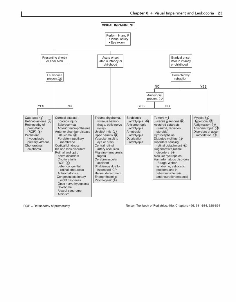
Chapter 8 u VisualImpairmentandLeukocoria 23
VISUAL IMPAIRMENTVISUAL IMPAIRMENT
Perform H and P • Visual acuity • Eye exam
Perform H and P • Visual acuity • Eye exam
AmblyopiapresentAmblyopiapresent 10
Corrected byrefraction
Corrected byrefraction
Gradual onsetlater in infancyor childhood
Gradual onsetlater in infancyor childhood
Acute onsetlater in infancy or
childhood
Acute onsetlater in infancy or
childhood
Presenting shortlyor after birth
Presenting shortlyor after birth
Strabismic amblyopiaAnisometropic amblyopiaAmetropic amblyopia Deprivation amblyopia
Strabismic amblyopia 10 Anisometropic amblyopiaAmetropic amblyopia Deprivation amblyopia
MyopiaHyperopiaAstigmatismAnisometropiaDisorders of acco- mmodation
Myopia 15 Hyperopia 16Astigmatism 17 Anisometropia 18 Disorders of acco- mmodation 19
TumorsJuvenile glaucoma Acquired cataracts (trauma, radiation, steroids)HydrocephalusDiabetes mellitusDisorders causing retinal detachmentDegenerative retinal disordersMacular dystrophiesHamartomatous disorders (Sturge-Weber syndrome, astrocytic proliferations in tuberous sclerosis and neurofibromatosis)
Tumors 11 Juvenile glaucoma 6 Acquired cataracts (trauma, radiation, steroids)HydrocephalusDiabetes mellitus 12 Disorders causing retinal detachment 13 Degenerative retinal disorders 14 Macular dystrophiesHamartomatous disorders (Sturge-Weber syndrome, astrocytic proliferations in tuberous sclerosis and neurofibromatosis)
Trauma (hyphema, vitreous hemor- rhage, optic nerve injury)Uveitis/ Iritis Optic neuritisVascular insult to eye or brainCentral retinal artery occlusionMigraine (amaurosis fugax)Cerebrovascular accidentStrabismus due to increased ICPRetinal detachmentEndophthalmitisPsychogenic
Trauma (hyphema, vitreous hemor- rhage, optic nerve injury)Uveitis/ Iritis 7 Optic neuritis 8 Vascular insult to eye or brainCentral retinal artery occlusionMigraine (amaurosis fugax)Cerebrovascular accidentStrabismus due to increased ICPRetinal detachmentEndophthalmitisPsychogenic 9
Corneal disease Forceps injury Sclerocornea Anterior microphthalmiaAnterior chamber disease Glaucoma Persistent pupillary membraneCortical blindnessIris and lens disorders Retinal and optic nerve disorders Chorioretinitis ROP Leber congenital retinal amaurosis Achromatopsia Congenital stationary night blindness Optic nerve hypoplasia Coloboma Aicardi syndrome Albinism
Corneal disease Forceps injury Sclerocornea Anterior microphthalmiaAnterior chamber disease Glaucoma 6 Persistent pupillary membraneCortical blindnessIris and lens disorders Retinal and optic nerve disorders Chorioretinitis ROP 5 Leber congenital retinal amaurosis Achromatopsia Congenital stationary night blindness Optic nerve hypoplasia Coloboma Aicardi syndrome Albinism
CataractsRetinoblastomaRetinopathy of prematurity (ROP)Persistent hyperplastic primary vitreous Chorioretinal coloboma
Cataracts 3 Retinoblastoma 4 Retinopathy of prematurity (ROP) 5 Persistent hyperplastic primary vitreous Chorioretinal coloboma
NOYES
NO YES
NOYES
LeukocoriapresentLeukocoriapresent 2
ROP = Retinopathy of prematurity Nelson Textbook of Pediatrics, 19e. Chapters 496, 611-614, 620-624
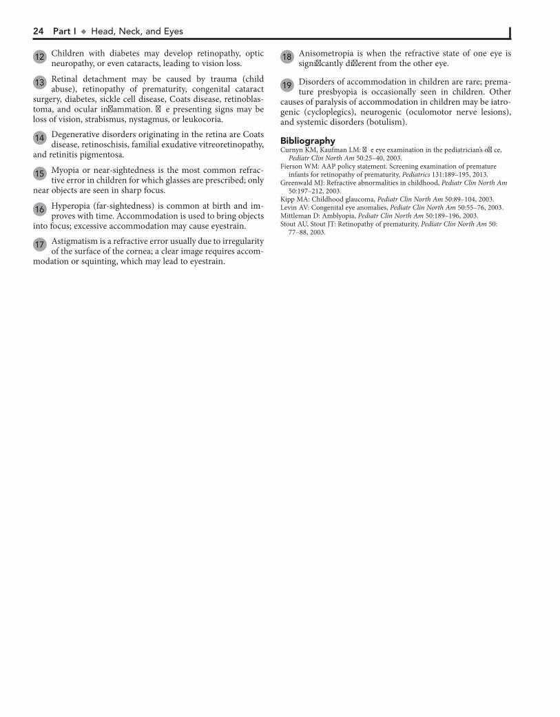
24 Part I u Head,Neck,andEyes
12 Children with diabetes may develop retinopathy, optic neuropathy, or even cataracts, leading to vision loss.
13 Retinal detachment may be caused by trauma (child abuse), retinopathy of prematurity, congenital cataract
surgery, diabetes, sickle cell disease, Coats disease, retinoblas-toma, and ocular inflammation. The presenting signs may be loss of vision, strabismus, nystagmus, or leukocoria.
14 Degenerative disorders originating in the retina are Coats disease, retinoschisis, familial exudative vitreoretinopathy,
and retinitis pigmentosa.
15 Myopia or near-sightedness is the most common refrac-tive error in children for which glasses are prescribed; only
near objects are seen in sharp focus.
16 Hyperopia (far-sightedness) is common at birth and im-proves with time. Accommodation is used to bring objects
into focus; excessive accommodation may cause eyestrain.
17 Astigmatism is a refractive error usually due to irregularity of the surface of the cornea; a clear image requires accom-
modation or squinting, which may lead to eyestrain.
18 Anisometropia is when the refractive state of one eye is significantly different from the other eye.
19 Disorders of accommodation in children are rare; prema-ture presbyopia is occasionally seen in children. Other
causes of paralysis of accommodation in children may be iatro-genic (cycloplegics), neurogenic (oculomotor nerve lesions), and systemic disorders (botulism).
BibliographyCurnyn KM, Kaufman LM: The eye examination in the pediatrician’s office,
Pediatr Clin North Am 50:25–40, 2003. Fierson WM: AAP policy statement. Screening examination of premature
infants for retinopathy of prematurity, Pediatrics 131:189–195, 2013. Greenwald MJ: Refractive abnormalities in childhood, Pediatr Clin North Am
50:197–212, 2003. Kipp MA: Childhood glaucoma, Pediatr Clin North Am 50:89–104, 2003. Levin AV: Congenital eye anomalies, Pediatr Clin North Am 50:55–76, 2003. Mittleman D: Amblyopia, Pediatr Clin North Am 50:189–196, 2003. Stout AU, Stout JT: Retinopathy of prematurity, Pediatr Clin North Am 50:
77–88, 2003.

Chapter 8 u VisualImpairmentandLeukocoria 25
VISUAL IMPAIRMENTVISUAL IMPAIRMENT
Perform H and P • Visual acuity • Eye exam
Perform H and P • Visual acuity • Eye exam
AmblyopiapresentAmblyopiapresent 10
Corrected byrefraction
Corrected byrefraction
Gradual onsetlater in infancyor childhood
Gradual onsetlater in infancyor childhood
Acute onsetlater in infancy or
childhood
Acute onsetlater in infancy or
childhood
Presenting shortlyor after birth
Presenting shortlyor after birth
Strabismic amblyopiaAnisometropic amblyopiaAmetropic amblyopia Deprivation amblyopia
Strabismic amblyopia 10 Anisometropic amblyopiaAmetropic amblyopia Deprivation amblyopia
MyopiaHyperopiaAstigmatismAnisometropiaDisorders of acco- mmodation
Myopia 15 Hyperopia 16Astigmatism 17 Anisometropia 18 Disorders of acco- mmodation 19
TumorsJuvenile glaucoma Acquired cataracts (trauma, radiation, steroids)HydrocephalusDiabetes mellitusDisorders causing retinal detachmentDegenerative retinal disordersMacular dystrophiesHamartomatous disorders (Sturge-Weber syndrome, astrocytic proliferations in tuberous sclerosis and neurofibromatosis)
Tumors 11 Juvenile glaucoma 6 Acquired cataracts (trauma, radiation, steroids)HydrocephalusDiabetes mellitus 12 Disorders causing retinal detachment 13 Degenerative retinal disorders 14 Macular dystrophiesHamartomatous disorders (Sturge-Weber syndrome, astrocytic proliferations in tuberous sclerosis and neurofibromatosis)
Trauma (hyphema, vitreous hemor- rhage, optic nerve injury)Uveitis/ Iritis Optic neuritisVascular insult to eye or brainCentral retinal artery occlusionMigraine (amaurosis fugax)Cerebrovascular accidentStrabismus due to increased ICPRetinal detachmentEndophthalmitisPsychogenic
Trauma (hyphema, vitreous hemor- rhage, optic nerve injury)Uveitis/ Iritis 7 Optic neuritis 8 Vascular insult to eye or brainCentral retinal artery occlusionMigraine (amaurosis fugax)Cerebrovascular accidentStrabismus due to increased ICPRetinal detachmentEndophthalmitisPsychogenic 9
Corneal disease Forceps injury Sclerocornea Anterior microphthalmiaAnterior chamber disease Glaucoma Persistent pupillary membraneCortical blindnessIris and lens disorders Retinal and optic nerve disorders Chorioretinitis ROP Leber congenital retinal amaurosis Achromatopsia Congenital stationary night blindness Optic nerve hypoplasia Coloboma Aicardi syndrome Albinism
Corneal disease Forceps injury Sclerocornea Anterior microphthalmiaAnterior chamber disease Glaucoma 6 Persistent pupillary membraneCortical blindnessIris and lens disorders Retinal and optic nerve disorders Chorioretinitis ROP 5 Leber congenital retinal amaurosis Achromatopsia Congenital stationary night blindness Optic nerve hypoplasia Coloboma Aicardi syndrome Albinism
CataractsRetinoblastomaRetinopathy of prematurity (ROP)Persistent hyperplastic primary vitreous Chorioretinal coloboma
Cataracts 3 Retinoblastoma 4 Retinopathy of prematurity (ROP) 5 Persistent hyperplastic primary vitreous Chorioretinal coloboma
NOYES
NO YES
NOYES
LeukocoriapresentLeukocoriapresent 2
ROP = Retinopathy of prematurity Nelson Textbook of Pediatrics, 19e. Chapters 496, 611-614, 620-624

26
Chapter 9
ABNORMAL EYE MOVEMENTS
with congenital or perinatal vision defects. It may also be ob-served in children who develop blindness in the first few years of life. The cause is frequently evident on general or ophthal-moscopic examination. Electrophysiologic imaging may be necessary to rule out certain causes (e.g., achromatopsia, con-genital stationary night blindness, Leber’s congenital retinal amaurosis) that are not evident on funduscopic examination. (See Chapter 8.) Neuroimaging to rule out tumors is always recommended when the cause is not evident.
4 Congenital idiopathic nystagmus typically appears in the first 3 months of life and is associated with compensatory
head tilting. A thorough examination, neuroimaging, and elec-trophysiologic studies are usually necessary to rule out underly-ing ocular or neurologic disorders.
5 Neurologic disorders must always be excluded as a cause of acquired nystagmus. Imaging should be considered
to exclude intracranial neoplasms. Other etiologies include CNS infections, trauma, encephalopathy, and demyelination disorders.
6 Labyrinthitis is often caused by viral illness and may be an infrequent complication of otitis media. Manifestations
can include vertigo, ear pain, nausea, vomiting, hearing loss, and nystagmus.
7 Spasmus nutans is usually a benign condition that occurs as a combination of bilateral asymmetric nystagmus,
head nodding, and torticollis in the first years of life. The etiol-ogy is unknown. It generally resolves by age 3 years without sequelae. Despite the benign nature of the condition, neuro-imaging is recommended to rule out the possibility of a CNS neoplasm.
8 Both children and adults can exhibit an occasional one to two beats of lateral nystagmus on side gaze that is not
considered significant.
Abnormal eye movements may occur as a benign finding or in association with other visual or ocular problems. They may, however, indicate an acquired, more severe underlying neuro-logic problem.
1 The history should include an accurate description of the eye movements, age of onset, and associated signs and
symptoms. Specifically inquire about visual acuity, color vision, night vision, photophobia, abnormal head movements, tinni-tus, and oscillopsia. Oscillopsia is a sensation of movement or swinging of the visual field.
Careful examination should yield an accurate description of the eye movements and other associated signs and symptoms. Eye movements should be initially classified as rhythmic (swing-ing, pendulum-like) or nonrhythmic. The association of any nonocular muscle movements should be discerned. Nystagmus is defined as repetitive rhythmic oscillations of one or both eyes. The waveform, direction, amplitude, frequency, and velocity of oscillations further help to classify the pattern of nystagmus. It should be noted whether the movements are symmetric or asymmetric between the two eyes. Some patterns have diagnos-tic implications. For example, vertical nystagmus is associated with posterior fossa lesions. Some drug-induced nystagmus may occasionally be vertical.
Frequently, neuroimaging and sometimes more specialized studies (e.g., electroretinogram, visual evoked potential test) are necessary to rule out underlying etiologies.
2 Ocular myoclonus describes rhythmic oscillating eye movements accompanied by nonocular movements of the
soft palate, tongue, face, pharynx, larynx, and diaphragm.
3 Congenital sensory nystagmus is commonly associated with ocular abnormalities that lead to visual impairment.
It is the most common type of nystagmus in infants. It generally occurs in children in the first 6 months of life and in children
BibliographyCurnyn KM, Kaufman LM: The eye examination in the pediatrician’s office,
Pediatr Clin North Am 50:25–40, 2003. Ruttum MS: Eye disorders. In Kliegman RM, Lye PS, Greenbaum LA, editors:
Practical strategies in pediatric diagnosis and therapy, ed 2, Philadelphia, 2004, Elsevier Saunders. Chapter 43.
Thompson L, Kaufman LM: The visually impaired child, Pediatr Clin North Am 50:225–239, 2003.
Chapter 9 u AbnormalEyeMovements 27
ABNORMAL EYE MOVEMENTSABNORMAL EYE MOVEMENTS
Rhythmic eyemovementspresent
Rhythmic eyemovementspresent
Perform H and PVisual acuityEye exam
Perform H and PVisual acuityEye exam 1
EncephalitisHydrocephalus Brainstem or cerebellar disordersNeuroblastoma
EncephalitisHydrocephalus Brainstem or cerebellar disordersNeuroblastoma
CerebellardysfunctionCerebellardysfunction
Cerebellar diseaseHydrocephalusCentral nervous system neoplasm
Cerebellar diseaseHydrocephalusCentral nervous system neoplasm
Pontine diseasePontine tumorsPontine diseasePontine tumors
Cerebellar damageDamage to midbrain, pons or medulla
Cerebellar damageDamage to midbrain, pons or medulla
Congenital sensory nystagmus Albinism Aniridia Achromatopsia Congenital cataracts Corneal opacities/anomalies Congenital macular lesion Congenital optic atrophy Glaucoma Leber’s congenital amaurosis Congenital stationary night blindness
Congenital sensory nystagmus 3 Albinism Aniridia Achromatopsia Congenital cataracts Corneal opacities/anomalies Congenital macular lesion Congenital optic atrophy Glaucoma Leber’s congenital amaurosis Congenital stationary night blindness
Congenital idiopathic motor nystagmus
Congenital idiopathic motor nystagmus 4
Intracranial neoplasmCNS infectionCNS traumaEncephalopathyMultiple sclerosisLabrynthitisSpasmus nutansDrugs/Medication (phenytoin, alcohol, sedatives)Vitamin B12 deficiencyNormal nystagmus
Intracranial neoplasm 5 CNS infection 5 CNS trauma 5 Encephalopathy 5 Multiple sclerosis 5 Labrynthitis 6 Spasmus nutans 7 Drugs/Medication (phenytoin, alcohol, sedatives)Vitamin B12 deficiencyNormal nystagmus 8
Opsoclonus (Multidirectional,spontaneousmovements)
Opsoclonus (Multidirectional,spontaneousmovements)
Ocular dysmetria(overshoot/undershootof eye on rapid fixation)
Ocular dysmetria(overshoot/undershootof eye on rapid fixation)
Ocular flutter(Horizontaloscillations withforward gaze/blinking)
Ocular flutter(Horizontaloscillations withforward gaze/blinking)
Ocular bobbing(downward jerkfrom primary gaze,remain for a fewseconds, and thendrift back)
Ocular bobbing(downward jerkfrom primary gaze,remain for a fewseconds, and thendrift back)
Visual impairmentor ocular defectVisual impairmentor ocular defect
Congenital nystagmus (presents at <6 months)
Congenital nystagmus (presents at <6 months)
Non-ocular musclegroups involvedNon-ocular musclegroups involved
Nystagmus Nystagmus
Ocular myoclonus(to-and-fro pendularoscillations of theeyes, with synchronousnonocular musclemovements)
Ocular myoclonus(to-and-fro pendularoscillations of theeyes, with synchronousnonocular musclemovements) 2
Acquired nystagmus(presents at >6 months)
Acquired nystagmus(presents at >6 months)
NOYES
NOYES
NO YES
Nelson Textbook of Pediatrics, 19e. Chapter 615

This page intentionally left blank

PART
IIRespiratory System
30
Chapter 10
COUGH
same spectrum as infectious croup because both have been as-sociated with viral infections.
5 According to the Centers for Disease Control and Preven-tion (CDC) and the World Health Organization (WHO), a
clinical diagnosis of pertussis can be made based on a cough history of 2 weeks’ duration if one of the following is present: paroxysms of coughing (especially at night), inspiratory whoop, or posttussive vomiting. Children with pertussis generally appear well between paroxysmal coughing spells. The “whoop” may not occur in infants younger than 3 months of age or in partially immunized children. Conjunctival hemorrhages, upper body petechiae, and exhaustion are additional supportive symp-toms; the absence of fever, myalgia, pharyngitis, and abnormal lung findings are also supportive. During epidemics or, in the case of close contact with a known case, a cough history 2 weeks is sufficient for diagnosis. Contact history is important; most cases of pertussis in infants and children can be traced to contact with a mildly symptomatic adolescent or adult whose only symptom may be a nonspecific prolonged cough. The tim-ing of exposure to sick contacts can be helpful; the incubation period for pertussis is 7 to 10 days, as opposed to 1 to 3 days for most viral URIs. Nasopharyngeal cultures are still considered the gold standard for diagnosis; however, the sensitivity can be diminished by the fastidious nature of the organism and inap-propriate handling of the specimen. PCR testing is more sensi-tive and is most useful during the first 3 weeks of illness; the risk of false-negative results increases after 4 weeks of illness. Testing after antibiotic treatment and early in the course of an illness when symptoms are still fairly nonspecific is not recommended. An elevated white blood cell count (15,000 to 100,000/ml) due to lymphocytosis supports the diagnosis, although it may not occur in very young infants or immunized children.
6 Aspiration of food or secretions in neurologically abnor-mal children may cause cough of varying frequency or
severity. Cough due to aspiration may be produced by airway inflammation, bronchospasm, or pneumonia. Aspiration pneu-monia may or may not be superinfected. If infection does occur, it is usually due to anaerobes or gram-negative organisms when chronic or nosocomial. Radionuclide scans or barium contrast studies may help to diagnose swallowing abnormalities. The intermittent nature of aspiration, however, frequently makes diagnosis difficult.
7 Rigid bronchoscopy is increasingly becoming the diagnos-tic and therapeutic procedure of choice when a choking
episode was witnessed, or when the history and physical are strongly suggestive of an aspirated foreign body. In the absence of a witnessed choking episode, chest films will generally be obtained to rule out other etiologies; however, only 10% to 25% of foreign bodies are radiopaque. Expiratory or lateral decubi-tus views may be helpful in identifying air trapping acutely but can be difficult to obtain.
8 Foreign body aspiration is usually obvious immediately, but occasionally a delay of weeks to months may occur
before symptoms develop. Aspiration of foreign bodies occurs most commonly in children under 4 years of age, with food (especially nuts) and small toys being the most commonly aspi-rated items. In cases where a delayed diagnosis is suspected based on clinical findings (e.g., chronic cough, wheezing, local-ized lung findings) the risk/benefit ratio of CT versus fluoros-copy versus performing a diagnostic flexible bronchoscopy must be considered.
1 The medical history should include a neonatal history and an assessment for immunodeficiency. An environmental
history should include inquiries about potential irritants (e.g., wood burning stove, smoke, perfume, scented candles, in-cense). The review of systems should include respiratory and nonrespiratory symptoms (e.g, poor growth and malodorous stools could be associated with cystic fibrosis; halitosis and headache may be associated with sinusitis). Inquire specifically about any recent choking episodes as well as any seasonal varia-tion of symptoms, and relationship of symptoms to feeding. A family history for asthma (and other atopic conditions) and cystic fibrosis may be helpful.
2 Bronchitis is a frequently used but nonspecific term in pedi-atrics. Inflammation of the large airways (tracheobronchi-
tis) commonly occurs and is due to multiple infectious agents. A viral URI complicated by a prolonged, possibly productive cough and malaise are common. These cases are typically self-limited (2 to 3 weeks) and unresponsive to antibiotics. Bacterial pneumonia may occur primarily or complicate a preceding URI. Fever and physical examination findings suggest this diagnosis. There is an emerging recognition of a persistent or protracted bacterial bron-chitis, which will respond to antibiotics and may be associated with more significant pulmonary disease (i.e., chronic suppura-tive lung disease). However, this diagnosis should be made with caution, particularly in children with a relatively acute history of cough. Careful consideration of other underlying pulmonary or systemic disorders should be made in children with chronic or recurring cough.
3 Clinical diagnosis of acute bacterial rhinosinusitis is made by prolonged symptoms of rhinorrhea for 10 to 14 days.
Halitosis, fever, nocturnal cough, and postnasal drip are other suggestive symptoms. Older children may experience headache, facial pain, tooth pain, and periorbital swelling.
4 Croup (i.e., laryngotracheobronchitis or laryngotracheitis) most commonly affects children age 3 months to 3 years.
It classically presents with the abrupt onset of an acute barky cough following 1 to 2 days of nonspecific viral URI symptoms. The cough is described as seal-like or brassy; inspiratory stridor, hoarseness, and respiratory distress may be associated. Worsen-ing of symptoms at night is typical. High fever may occur. Di-agnosis should be clinical; imaging (anteroposterior and lateral neck films) should only be obtained when another diagnosis is suspected (e.g., epiglottitis), symptoms are severe, or there is lack of improvement with therapy.
Spasmodic croup refers to a clinically similar condition, but without evidence of airway inflammation. The clinical course is typically more abrupt and short-lived than cases that are obvi-ously associated with URIs, plus the symptoms occur exclu-sively at night. Episodes can resolve and recur multiple times in one night and for a few nights in a row, with the child appearing completely well in between coughing episodes. A lack of con-sensus exists regarding whether a spasmodic croup presenta-tion is a distinct entity (with an allergic component) or on the
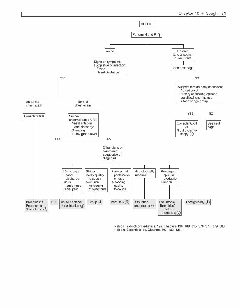
Chapter 10 u Cough 31
COUGHCOUGH
Suspect foreign body aspiration: Abrupt onset History of choking episode Localized lung findings ± toddler age group
Suspect foreign body aspiration: Abrupt onset History of choking episode Localized lung findings ± toddler age group
BronchiolitisPneumonia“Bronchitis”
BronchiolitisPneumonia“Bronchitis” 2
URIURI Acute bacterial rhinosinusitisAcute bacterial rhinosinusitis 3
CroupCroup 4 PertussisPertussis 5 Pneumonia“Bronchitis” (tracheo- bronchitis)
Pneumonia“Bronchitis” (tracheo- bronchitis) 2
AspirationpneumoniaAspirationpneumonia 6
Foreign bodyForeign body 8
NOYES
NOYES
Consider CXRvs.
Rigid broncho-scopy
Consider CXRvs.
Rigid broncho-scopy 7
See nextpageSee nextpage
AcuteAcute Chronic(2 to 3 weeks)or recurrent
Chronic(2 to 3 weeks)or recurrent
Prolonged sputum productionRhonchi
Prolonged sputum productionRhonchi
NeurologicallyimpairedNeurologicallyimpaired
Paroxysmal posttussive emesisWhooping quality to cough
Paroxysmal posttussive emesisWhooping quality to cough
StridorBarky quality to coughNocturnal worsening of symptoms
StridorBarky quality to coughNocturnal worsening of symptoms
10–14 days nasal dischargeSinus tendernessFacial pain
10–14 days nasal dischargeSinus tendernessFacial pain
Signs or symptomssuggestive of infection: Fever Nasal discharge
Signs or symptomssuggestive of infection: Fever Nasal discharge
See next pageSee next page
Abnormalchest examAbnormalchest exam
Normalchest exam
Normalchest exam
Consider CXRConsider CXR Suspect uncomplicated URI: Nasal irritation and discharge Sneezing ± Low-grade fever
Suspect uncomplicated URI: Nasal irritation and discharge Sneezing ± Low-grade fever
Other signs orsymptomssuggestive ofdiagnosis
Other signs orsymptomssuggestive ofdiagnosis
Nelson Texbook of Pediatrics, 19e. Chapters 138, 189, 315, 376, 377, 379, 383Nelsons Essentials, 6e. Chapters 107, 133, 136
NOYES
Perform Perform H and P 1

32 Part II u RespiratorySystem
9 Pulmonary contusions can occur as a result of chest trauma. The onset of symptoms may be acute or delayed.
Contusions may be visible on a chest x-ray; however, initial film findings are often negative.
10 Historically, gastroesophageal reflux disease (both symp-tomatic and asymptomatic) has been implicated as an etiol-
ogy of chronic cough. Actual data supporting the role of reflux in chronic cough remains conflicting, except in children with neurologic impairment and a risk of aspiration.
11 Asthma is the most common cause of chronic and recur-rent cough in children. Wheezing may not be evident. A
suggestive history, such as significant cough with colds, exer-cise, hard laughter, crying, or exposure to cold air, smoke, or other environmental irritants or a frequent nocturnal cough, and an improvement of symptoms to therapy (e.g., bronchodi-lators, short courses of oral corticosteroids) are highly sugges-tive of the asthma diagnosis. Ideally, spirometry would be used to confirm the diagnosis, although young children are typically unable to perform the testing, and normal results do not always rule out the diagnosis. Chest x-rays are not necessary in the absence of respiratory distress; however, if obtained, hyperinfla-tion and atelectasis are common findings.
12 The term postinfectious cough syndrome describes a pro-longed cough (up to 8 weeks), which may follow certain
otherwise uncomplicated respiratory infections, presumably due to extensive inflammation of airway epithelium or hypersensitivity of cough receptors. The term upper airway cough syndrome (pre-viously “postnasal drip”) describes cough symptoms made worse by lying down; sinusitis and allergic rhinitis may be contributors.
13 Habit cough (also called psychogenic cough or cough tic) is an infrequent cause of a prolonged cough in school-aged
children that is often refractory to treatment. The harsh, barky cough occurs only during waking hours. It will disappear dur-ing sleep and with distraction. The child appears well and is typically not bothered by the coughing, even though it can fre-quently be significant enough to disrupt a classroom.
14 Mediastinal disorders include tumor, lymphadenopathy due to sarcoidosis, tuberculosis, histoplasmosis and coc-
cidioidomycosis, pericardial cyst, and diaphragmatic hernia.
15 A large number of rare conditions can occur with cough, including interstitial lung disease, graft-versus-host disease,
a1-antitrypsin deficiency, pulmonary hemosiderosis, alveolar proteinosis, heart failure, pulmonary edema, sarcoidosis, oblit-erative bronchiolitis, and follicular bronchiolitis.
16 Angiotensin-converting enzyme (ACE) inhibitors are recognized as a cause of chronic cough. A history of
radiation to the chest or cytotoxic medications should raise suspicions for lung disease. Use of aspirin and other nonste-roidal pain relievers can exacerbate asthma symptoms in certain people who are sensitive to these drugs. Be conscious of illicit drug use in teens (including inhalants) as a cause of chronic cough.
BibliographyAmerican Academy of Pediatrics. Pertussis. In Red Book: 2012 Report of the
Committee on Infectious Diseases. Pickering LK, ed. 29th ed. Elk Grove Village, IL: American Academy of Pediatrics; 2012: 553-566.
Bjornson C, Johnson DW: Croup, Lancet 371:329–339, 2008.

Chapter 10 u Cough 33
Chronic(2 to 3 weeks)
orRecurrent
Chronic(2 to 3 weeks)
orRecurrent
Acute cough (continued)Acute cough (continued)
Obtain CXRConsider: Sweat chloride PPD UGI HIV testing Immuno- deficiency work-up (See also Chs. 60 and 78)
Obtain CXRConsider: Sweat chloride PPD UGI HIV testing Immuno- deficiency work-up (See also Chs. 60 and 78)
Signs or symptomssuggestive of diagnosis
Signs or symptomssuggestive of diagnosis
Pulmonary contusionPulmonary embolism
Pulmonary contusion 9 Pulmonary embolism
NO YES
Historyof traumaHistoryof trauma
Aspiration GERAspiration GER 10
Associated with feedingReflux symptoms
Associated with feedingReflux symptoms
Inhaled irritantsHypersensitivity pneumonitis
Inhaled irritantsHypersensitivity pneumonitis
EnvironmentalexposuresEnvironmentalexposures
AllergicrhinitisAllergicrhinitis
SneezingItchy eyesWatery rhinorrhea
SneezingItchy eyesWatery rhinorrhea
AsthmaAsthma 11
Cough exacerbated by exerciseFrequent nocturnal coughPositive family history of asthma
Cough exacerbated by exerciseFrequent nocturnal coughPositive family history of asthma
Acute bacterialrhinosinusitisAcute bacterialrhinosinusitis 3
Foreign bodyPostinfectious cough syndromeAsthmaVascular ring or slingHabit coughExternal ear canal irritationAtypical pneumonia (Mycoplasma pneumoniae, Chlamydophila pneumoniae)Mediastinal disordersCongestive heart failureMiscellaneous lung diseaseMedicationsTuberculosis
Foreign body 8Postinfectious cough syndrome 12AsthmaVascular ring or slingHabit cough 13 External ear canal irritationAtypical pneumonia (Mycoplasma pneumoniae, Chlamydophila pneumoniae)Mediastinal disorders 14 Congestive heart failureMiscellaneous lung disease 15 Medications 16Tuberculosis
Atypical pneumonia Cystic fibrosisHIVTuberculosisGERImmune deficiencyMediastinal disordersDysmotile ciliaPulmonary sequestrationBronchiectasis
Atypical pneumonia Cystic fibrosisHIVTuberculosisGER 10 Immune deficiencyMediastinal disorders 14 Dysmotile ciliaPulmonary sequestrationBronchiectasis
Prolonged nasal dischargeSinus tenderness
Prolonged nasal dischargeSinus tenderness
Consider: CXR PPD ± chest CT
Consider: CXR PPD ± chest CT
NOYES
Any of the following present: Failure to thrive Frequent infections Systemic symptoms Diffuse lymphadenopathy
Any of the following present: Failure to thrive Frequent infections Systemic symptoms Diffuse lymphadenopathy
Signs or symptomssuggestive of diagnosisSigns or symptomssuggestive of diagnosis
COUGH (continued)COUGH (continued)

34
Chapter 11
HOARSENESS
etiology of hoarseness is unclear. There is no clear consensus regarding when children (beyond infancy) should undergo visualization of their vocal cords for symptoms of a hoarse voice; specialists will make the decision for laryngoscopy based on the severity of hoarseness or associated symptoms, suspected diagnosis, and procedural risk. Any element of re-spiratory distress, tachypnea, or decreased air entry warrants visualization.
9 Acquired vocal cord paralysis may occur as a result of polyneuropathy (e.g., Guillain-Barré syndrome), Arnold-
Chiari malformation, compression from a mass or tumor, brainstem encephalitis, or neck or thoracic surgery.
10 Rare infectious causes of hoarseness include granuloma-tous lesions in the larynx due to syphilis, tuberculosis, and
histoplasmosis. Immunologic disorders, including HIV infec-tion, may predispose patients to fungal infections of the airways. Prolonged use of inhaled corticosteroids has been associated with Candida overgrowth. Diphtheria, rabies, and tetanus are other etiologies.
11 Respiratory papillomatosis develops as a progressive hoarseness anytime between the first several months and
up to 5 years of age. The etiology is human papillomavirus (most commonly types 6 and 11) transmitted at birth from the mother’s genital tract.
12 Cutaneous hemangiomas, especially of the head and neck, may be a marker for underlying airway hemangiomas that
can present as progressive hoarseness or stridor over the first few months of life. Lymphangiomas, rhabdomyosarcomas, and leukemic infiltrations are other etiologies.
13 Congenital hypothyroidism is often not apparent until after the newborn period. If undetected by newborn screening,
presenting signs include hoarseness, constipation, prolonged hyperbilirubinemia, hypotonia, and hypothermia. A lethargic, hoarse cry is not likely to be evident until after a few months of life. Hypothyroidism that develops later in childhood may also cause a hoarse voice.
14 Rarer causes of hoarseness include hypocalcemic tetany and thiamine deficiency, which can occur occasionally in
breast-fed infants of thiamine-deficient mothers. Connective tissue disorders including JRA are rare causes of hoarseness in children. Metabolic disorders cause hoarseness because of the abnormal deposition of metabolites in the airways or vocal cords. Other causes include vincristine toxicity, laryngeal edema related to congestive heart failure, and dryness due to ectodermal dysplasia, cystic fibrosis, antihistamine therapy, and psychogenic causes.
Hoarseness is a change in the quality of the voice caused by alteration of the size, shape, or tension of the vocal cords. It is associated with vocalization, as opposed to stridor, which is associated with respiration; however, the two can occur together. Hoarseness is usually benign, but evaluation is indi-cated for hoarseness if the onset is congenital, associated with trauma, or if it persists longer than 1 to 2 weeks.
1 Congenital anomalies of the larynx causing hoarseness include subglottic stenosis, laryngoceles, and webs. Laryn-
geal fissures and clefts are rare conditions, but 20% of posterior fissures or clefts are associated with tracheoesophageal fistulas.
2 Dislocation of the cricothyroid or cricoarytenoid articula-tions may occur during a traumatic birth. Trauma related
to neonatal intubation may also cause stenosis or dislocation of the laryngeal cartilages.
3 Congenital vocal cord paralysis is more commonly uni-lateral than bilateral; most cases are idiopathic. Unilateral
vocal cord paralysis is more common than bilateral. When the disorder is unilateral, it is associated with a weak, breathy cry and may lead to feeding difficulties and aspiration. In bilateral cases, stridor is more predominant and the cry may not be obviously affected. Birth trauma may also produce vocal cord paralysis because of damage to the recurrent laryngeal nerve. Vocal cord paralysis may also be a manifestation of central neurologic disorders, including hydrocephalus, subdural he-matomas, and other causes of brainstem compression (e.g., Dandy-Walker cysts, Arnold-Chiari malformations), as well as peripheral neurologic problems (due to recurrent laryngeal nerve trauma, cardiovascular abnormalities, mediastinal masses, myasthenia gravis, and other neuropathies). Neurologic prob-lems are more likely to be associated with bilateral vocal cord paralysis.
4 Hoarseness commonly occurs as a result of overuse of the voice. Inquire about recent attendance at athletic events or
rock concerts.
5 Benign vocal cord nodules (“screamer’s nodules”) may also be a complication of vocal overuse. Further evaluation
is indicated if rest does not result in resolution. Reflux to the level of the larynx can exacerbate symptoms.
6 The most common cause of transient hoarseness is croup (laryngotracheobronchitis) in older infants and toddlers,
and viral URIs causing laryngitis in older children. Sinus dis-ease is another cause, though it is less common.
7 Allergic angioneurotic edema of the larynx may occur acutely with hoarseness and respiratory distress. Urgent
evaluation and treatment are indicated.
8 One to two weeks is a reasonable interval to observe children prior to referring for an ENT evaluation if the
BibliographyAhmad SM, Soliman AM: Congenital anomalies of the larynx, Otolaryngol
Clin North Am 40:177–191, 2007. Derkay CS, Wiatrak B: Recurrent respiratory papillomatosis: a review,
Laryngoscope 118:1236–1247, 2008. Friedberg J: Hoarseness. In Bluestone CD, Stool SE, Alper CM, et al, editors:
Pediatric otolaryngology, ed 4, Philadelphia, 2003, WB Saunders, p 1413. Hastriter EV, Olsson JM: In brief: hoarseness, Pediatr Rev 27:e47–48, 2006. Schwartz SR, Dailey SH, Deutsch ES, et al: Clinical practice guideline:
hoarseness (dysphonia), Otolaryngol Head Neck Surg 141:S1–S31, 2009.

Chapter 11 u Hoarseness 35
HOARSENESSHOARSENESS
Consider ENT referralfor laryngoscopyConsider ENT referralfor laryngoscopy 8
CroupLaryngitisOveruseSmokingAngioneurotic edemaAllergy-related postnasal drip Post intubationLaryngeal abscess
Croup 6Laryngitis 6Overuse 4SmokingAngioneurotic edema 7Allergy-related postnasal drip Post intubationLaryngeal abscess
Pubertal voice changesTrauma (including surgical)Acquired vocal cord paralysisInfectionPapillomatosisTumors (including hemangiomas)HypothyroidismGERSmoking Other miscellaneous causes (hypocalcemic tetany, neurologic) ,Genetic syndromes (Williams, De Lange, Farber)
Pubertal voice changesTrauma (including surgical)Acquired vocal cord paralysis 9Infection 10Papillomatosis 11Tumors (including hemangiomas) 12Hypothyroidism 13GERSmoking Other miscellaneous causes (hypocalcemic tetany, neurologic) 3 , 14Genetic syndromes (Williams, De Lange, Farber)
OveruseVocal cord nodules
Overuse 4Vocal cord nodules 5
Congenital anomalies of larynx Birth trauma to larynxVocal cord paralysis Arnold-Chiari malformation Congenital Traumatic Other neurologic etiologyCri du chat syndrome
Congenital anomalies of larynx 1 Birth trauma to larynx 2 Vocal cord paralysis 3 Arnold-Chiari malformation Congenital Traumatic Other neurologic etiologyCri du chat syndrome
NOYES
History suggestiveof overuse
History suggestiveof overuse
NOYES
NOYES
Acute onset,duration <1-2 weeksAcute onset,duration <1-2 weeks
Perform Perform H and P
Occurring in immediatenewborn period
Nelson Textbook of Pediatrics, 19e. Chapters 311, 376, 377, 382Nelson Essentials, 6e. Chapters 107, 135
36
Chapter 12
STRIDOR 7 Epiglottitis is rare in developed countries because of wide-
spread immunization against Haemophilus influenzae. Classic features include rapid progression from a sore throat and fever to severe respiratory distress, drooling, and dysphagia. Patients tend to assume a tripod position, with their neck hyper-extended.Immediate intubation in an operating room is recom-mended because of the high risk of complete airway obstruction.Bacterial tracheitis should be suspected when an acute clinical worsening occurs (e.g., respiratory distress, toxicity, lack of im-provement with therapy) following a few days of milder illness. (Drooling and the tripod position preference do not occur with bacterial tracheitis.)
8 Abscesses (e.g., peritonsillar, retropharyngeal) usually de-velop as a complication of a pharyngitis or URI. Retropha-
ryngeal abscesses are more common in ages greater than 5 years and are more likely to present with neck pain, torticollis, hyperex-tension of the neck, and cervical adenopathy. Peritonsillar ab-scesses are more common in older children and adolescents and are more likely to present with trismus (pain and difficulty with opening the mouth), a muffled voice, and adenopathy; asymmet-rical peritonsillar bulging may be evident on exam. Nonspecific signs and symptoms may include sore throat, fever, adenopathy, drooling, and refusal to move the neck.
9 Spasmodic croup is clinically similar to infectious croup but without evidence of airway inflammation. The clinical
course is typically more abrupt and short-lived than cases that are obviously associated with URIs, plus the symptoms occur exclusively at night. Episodes can resolve and recur multiple times in 1 night and for up to 4 nights in a row, with the child appearing completely well in between coughing episodes. A lack of consensus exists regarding whether a spasmodic croup presentation is a distinct entity (with anallergic component) or on the same spectrum as infectious croup, because both have been associated with viral infections.
10 Stridor due to aspiration of a foreign body is generally an acute symptom of an object lodged in the larynx or trachea.
Even if a choking or gagging episode was not observed, rigid bronchoscopy is increasingly becoming the diagnostic (and ther-apeutic) procedure of choice when the H and P are strongly sug-gestive (e.g., acute respiratory distress, cough, stridor, inability to speak). Posterior-anterior chest x-rays are frequently obtained, but only 10% to 25% of foreign bodies are radiopaque. Aspiration of objects small enough to reach the lower airways is more likely to present with cough and wheezing. (See Chapter 10.)
11 Airway lesions (e.g., hemangioma, cyst, tumor) may cause airway obstruction due to rapid enlargement (e.g., hemor-
rhage, infection). Congenital hemangiomas in a subglottic location are rare but potentially life-threatening. Symptoms usually develop between 1 and 2 months of age as gradual en-largement causes progressive airway obstruction. Cutaneous hemangiomas, particularly on the head and neck, should raise suspicion for this condition. Rarely, mediastinal lesions, thyroid enlargement, or esophageal foreign bodies may cause stridor by impinging on the larynx.
12 Vascular malformations, that compress the trachea or esophagus usually present in infancy with stridor, cough,
or wheezing (or simply “noisy breathing” from a parent’s per-spective) from birth. Symptoms are usually worsened by crying and neck flexion; complete vascular rings may cause swallowing difficulties. Chest x-rays may suggest the diagnosis; barium
Stridor is a predominantly inspiratory harsh, medium-pitched sound caused by obstruction in the upper (extrathoracic) air-way. The most common cause is infectious croup.
1 Inquire about risk factors for respiratory problems, such as prematurity, intubation, chronic medical problems, and
hospitalizations. The review of systems should include signs of infection and any obvious aggravating factors (e.g., position, agitation). Particularly important is a history of any recent choking episodes, which would raise the suspicion of a foreign body. Assess the child’s level of illness, apparent anxiety, and respiratory distress (e.g., nasal flaring, grunting, accessory muscle use, retractions). A careful skin exam for cutaneous findings such as hemangiomas should be performed, particu-larly in infants in whom the stridor is gradually worsening over time.
2 The most common cause of congenital stridor is laryngo-malacia. If present early in the newborn period or
associated with significant dyspnea, ENT evaluation is neces-sary to rule out severe congenital laryngeal malformations that may require acute intervention. Milder cases of laryngomalacia do not require laryngoscopic diagnosis.
3 Laryngomalacia may be evident at birth but most com-monly becomes notable at 2 to 4 weeks of age. Symptoms
are characteristically worse when the infant is supine or agitated and typically resolve within the first year. Diagnosis is often clinical but may be confirmed by laryngoscopy. Laryngomalacia is often accompanied by tracheomalacia that may cause expira-tory wheezing and cough as well as stridor.
4 Subglottic tracheal stenosis may be congenital or acquired. The latter may result from prolonged endotracheal intuba-
tion and manifest gradually in the first year of life. Symptoms are exaggerated in an acute respiratory illness. Recurrent croup may occur at young ages in these children.
5 The indication for x-rays will be determined by clinical judg-ment based on the degree of respiratory distress and the
specific clinical presentation. A child with a clinical picture con-sistent with croup who is stable and responding well to therapy may be managed without imaging. For a child with severe respira-tory distress and a clinical picture suggesting bacterial tracheitis or epiglottitis (e.g., high fever, anxiety, drooling, cyanosis, tripod position), imaging should be delayed and airway protection pri-oritized. If epiglottitis is suspected, arrangements should be made for immediate intubation in an operating room.
6 Infectious croup (laryngotracheobronchitis or laryngotra-cheitis) most commonly affects children age 3 months to
3 years. It classically presents with the abrupt onset of an acute barky cough following 1 to 2 days of nonspecific viral URI symp-toms. The cough is described as seal-like or brassy; inspiratory stridor, hoarseness, and respiratory distress may be associated. Worsening of symptoms at night is typical. High fever may occur.

Chapter 12 u Stridor 37
14 Vocal cord paralysis in the newborn is most commonly due to birth trauma resulting in recurrent laryngeal nerve
trauma. Other causes include neurologic syndromes (Arnold-Chiari malformation) and neck or chest surgery.
BibliographyBjornson C, Johnson DW: Croup, Lancet 337:329–339, 2008. Sobol SE, Zapa1 S: Epiglottitis and croup, Otolaryngol Clin North Am 41:
551–566, 2008.
esophagrams can be very useful. However, echocardiograms plus CT or MR angiography are increasingly preferred because of the enhanced anatomic detail provided.
13 Human papillomaviruses can cause recurrent respiratory tract papillomas. Papillomas develop primarily in the
larynx; occasional spread to other sites in the aerodigestive tract occurs in severe cases. They can develop at any time from shortly after birth to several years of age, initially presenting as hoarseness (which may go unnoticed initially) and progressing to stridor.
STRIDORSTRIDOR
LaryngomalaciaVascular ring or slingMass lesionPapillomatosisVocal cord paralysisChiari crisisHypocalcemia
Laryngomalacia 3 Vascular ring or sling 12 Mass lesion 11 Papillomatosis 13 Vocal cord paralysis 14 Chiari crisisHypocalcemia
Croup (spasmodic)Foreign bodyAngioedema (allergic reaction) Mass lesionSubglottic stenosisTrauma (including surgical)
Croup (spasmodic) 9 Foreign body 10 Angioedema (allergic reaction) Mass lesion 11 Subglottic stenosis 4 Trauma (including surgical)
Croup (infectious)Abscess (peritonsillar, retropharyngeal)Subglottic stenosis (with respiratory infection)
Croup (infectious) 6 Abscess (peritonsillar, retropharyngeal) 8 Subglottic stenosis (with respiratory infection) 4
Croup (infectious)EpiglottitisBacterial tracheitisAbscess (peritonsillar, retropharyngeal)
Croup (infectious) 6 Epiglottitis 7 Bacterial tracheitis 7 Abscess (peritonsillar, retropharyngeal) 8
LaryngomalaciaTracheomalaciaSubglottic tracheal stenosisTracheal websCraniofacial abnormalities (Pierre Robin, cri du chat)
3 3 l
4
NOYES
NOYES
NOYES
AcuteAcute Chronic orrecurrentChronic orrecurrent
Consider ENTConsider ENT 2
Occurring in the immediatenewborn periodOccurring in the immediatenewborn period
Signs or symptomsof infection presentSigns or symptomsof infection present
Consider: AP and lateral neck x-rays CXR Barium esophagram ENT referral for laryngoscopy Echo with CT or MR angiography
Consider: AP and lateral neck x-rays CXR Barium esophagram ENT referral for laryngoscopy Echo with CT or MR angiography
Perform Perform H and P 1
Very ill appearingVery ill appearing
Consider lateraland AP neckx-rays
Consider lateraland AP neckx-rays 5
Nelson Textbook of Pediatrics, 19e. Chapters 258, 374, 376, 377, 378, 380Nelsons Essentials, 6e. Chapters 107, 135, 136
38
Chapter 13
WHEEZING
Chest x-rays will not aid in the diagnosis of asthma and should only be considered when the diagnosis is not clear or to rule out complications of an asthma attack (e.g., pneumothorax, atelec-tasis, pneumomediastinum).
4 Bronchiolitis is a common lower respiratory tract infection in infants. The classic presentation of acute bronchiolitis
begins with nonspecific cold symptoms and progresses fairly rapidly to profuse rhinorrhea, harsh cough, wheezing, and tachypnea. Respiratory distress may occur, especially in younger infants. RSV is the most common cause. Parainfluenza, influ-enza, rhinovirus, human metapneumovirus, adenovirus, and human bocavirus are other etiologies.
5 Mycoplasma pneumoniae and Chlamydophila pneumoniae are exceptions to the “bacteria do not cause wheezing”
generalization. These pathogens cause more clinically signifi-cant illness in school-aged children than in younger children. Cough that progresses over the first week of illness, fine crackles and wheezes, and nonspecific x-ray findings are characteristic.
6 Isolated episodes of wheezing or bronchospasm may occur with viral respiratory illnesses of milder clinical severity
than bronchiolitis. Wheezing with certain viral infections early in life is predictive of the later development of asthma; less clear is whether these infections cause asthma or whether they are identifying children who have a predilection toward the later development of asthma. A positive response to a trial of bronchodilator therapy confirms reversible airway obstruction. Until the wheezing manifests itself as a recurrent disorder of airway obstruction, the term “asthma” should not be used. Most children who wheeze in infancy or early childhood will not wheeze later in childhood.
7 Diagnosis of foreign bodies can be difficult. Episodes of choking or gagging associated with aspiration are often
unobserved. Posterior-anterior chest x-rays are frequently obtained, but only 10% to 25% of foreign bodies are radi-opaque. Rigid bronchoscopy is increasingly becoming the di-agnostic (and therapeutic) procedure of choice when the H and P examination are strongly suggestive. Localized lung findings or a history of recurrent or persistent pneumonia in a single lobe should arouse the clinician’s suspicion for a re-tained foreign body. (See Chapter 10.) Foreign bodies in the esophagus can be mistaken as tracheal foreign bodies.
Wheezing is a high-pitched musical sound caused by obstruc-tion of the lower (intrathoracic) airways. The etiologies for wheezing are numerous. The clinical significance of the under-lying problem can range from mild to severe.
1 Inquire about risk factors for respiratory problems, such as prematurity, intubation, chronic medical problems, and
hospitalizations. The review of systems should include signs and symptoms such as fever, weight loss, night sweats, and dys-phagia. Inquire specifically about any recent choking episodes and about medications (specifically inquire if the child was ever prescribed an inhaler in their past), as well as a family history of asthma and allergies. Signs of respiratory distress (e.g., nasal flaring, grunting, accessory muscle use, retractions) should be noted, as should chest wall asymmetry and chest excursion.
2 In patients with an uncomplicated illness consistent with bronchiolitis or a URI, a chest x-ray is not indicated.
Depending on the level of illness, a chest x-ray could be consid-ered to rule out a treatable pneumonia.
3 The most likely cause of recurrent wheezing in children is asthma. By definition, the diagnosis of asthma requires a
history of recurrent or chronic symptoms of wheezing or air-flow obstruction. An acute episode of wheezing can be the first manifestation of asthma, and a positive response to a trial of bronchodilator therapy confirms reversible airway obstruction. However, the diagnosis should not be made until the wheezing manifests itself as a recurrent disorder of airway obstruction supported by an appropriate history, physical examination, and diagnostic testing such as spirometry (if possible). A diag-nosis of asthma is supported by: (1) a history of a prolonged cough with colds, (2) nocturnal cough (unassociated with illness), (3) symptoms of cough or shortness of breath, and (4) chest tightness induced by exercise, hard laughter, crying, or exposure to cold air, smoke, or other environmental irritants.
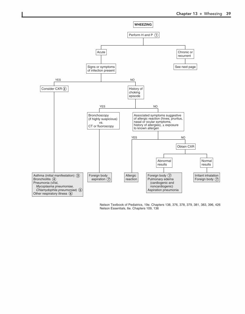
Chapter 13 u Wheezing 39
WHEEZINGWHEEZING
NormalresultsNormalresults
Obtain CXRObtain CXR
AbnormalresultsAbnormalresults
History ofchokingepisode
History ofchokingepisode
Consider CXR Consider CXR 2
Foreign body aspiration Foreign body aspiration 7
AllergicreactionAllergicreaction
Foreign body Pulmonary edema (cardiogenic and noncardiogenic)Aspiration pneumonia
Foreign body 7 Pulmonary edema (cardiogenic and noncardiogenic)Aspiration pneumonia
Irritant inhalationForeign body Irritant inhalationForeign body 7
Asthma (initial manifestation) Bronchiolitis Pneumonia (viral, Mycoplasma pneumoniae, Chlamydophila pneumoniae) Other respiratory illness
Asthma (initial manifestation) 3 Bronchiolitis 4 Pneumonia (viral, Mycoplasma pneumoniae, Chlamydophila pneumoniae) 5 Other respiratory illness 6
NOYES
Associated symptoms suggestiveof allergic reaction (hives, pruritus,nasal or ocular symptoms, history of allergies), ± exposureto known allergen
Associated symptoms suggestiveof allergic reaction (hives, pruritus,nasal or ocular symptoms, history of allergies), ± exposureto known allergen
Bronchoscopy (if highly suspicious) vs.CT or fluoroscopy
Bronchoscopy (if highly suspicious) vs.CT or fluoroscopy
NOYES
NOYES
AcuteAcute Chronic orrecurrentChronic orrecurrent
Signs or symptomsof infection presentSigns or symptomsof infection present
See next pageSee next page
Perform Perform H and P 1
Nelson Textbook of Pediatrics, 19e. Chapters 138, 376, 378, 379, 381, 383, 396, 426Nelson Essentials, 6e. Chapters 109, 136

40 Part II u RespiratorySystem
to inhaled bronchodilators. During acute exacerbations, pa-tients can experience dyspnea and stridor, but the pulmonary gas exchange rate is normal. The high-pitched respiratory sound created by vocal cord dysfunction is more correctly classified as stridor, although it is transmitted throughout the lung fields and is difficult to distinguish from peripheral wheezing (although it is primarily inspiratory). Pulmonary function tests during an episode demonstrate some degree of extrathoracic obstruction. Pulmonary function tests and sup-portive clinical history are adequate to make the diagnosis. Laryngoscopy (during an episode) may help to make the diag-nosis in less clear cases. The disorder is most common in teen-age girls, and underlying psychosocial stressors can often be identified.
13 Several environmental contaminants including inorganic dusts (e.g., talcum, asbestos, silica) and chemical fumes
can cause interstitial lung disease and wheezing. Disease due to inhalation of organic dusts (particularly on farms or with expo-sure to birds) has been reported in children, although it is sig-nificantly more common in adults.
14 Chest x-ray findings may be suggestive in cases of vascular malformations that compress the airway. The barium
esophagram (or UGI series) has been the traditional diagnostic test and is diagnostic in most cases, although it will not define the specific anatomy of a lesion. Barium studies are still fre-quently performed, although a CT or MR angiogram (where available) will provide more precise anatomic details. Echocar-diograms may visualize some vascular rings but will miss atretic segments and do not reliably define anatomy; their value is to rule out any accompanying cardiac defects. Other congenital malformations that may present with recurrent wheezing or stridor in young infants include tracheoesophageal fistulas, la-ryngeal or tracheal webs, stenoses, clefts, cysts, or atresias; bronchoscopy would effectively diagnose all of these. Suspected diagnosis, availability, radiation risk, and need for sedation should all factor into the selection of a diagnostic test.
8 Recurrent aspiration as a cause of chronic respiratory symptoms is most likely to occur in children with swallow-
ing dysfunction, frequently associated with an underlying neu-rologic condition. Chronic aspiration may result in a dimin-ished cough reflex, resulting in “silent aspiration.” Although GER has been historically associated with chronic cough and wheezing, evidence supporting this association is inconclusive.
9 Primary tracheomalacia and bronchomalacia are character-ized by wheezing that is located more centrally then periph-
erally, and is not responsive to bronchodilators. The wheezing is usually low pitched (not “musical”), expiratory, monophonic, and typically becomes evident after 2 to 3 months of life, although parents may describe the child as a “noisy breather” from birth. Cough may or may not be significant. The defect is insufficient cartilage to maintain airway patency throughout the breathing cycle. The problem can affect the trachea or bronchi in isolation, or it can affect the entire airway, including the larynx (laryngo-malacia). (See Chapter 12.) Bronchomalacia can be bilateral or unilateral (usually on the left). Diagnosis is often clinically based, but it may be confirmed using bronchoscopy.
10 Primary ciliary dyskinesia (PCD) is a rare cause of wheez-ing. Abnormal structure of the cilia leads to impaired
clearance of endobronchial secretions, resulting in chronic res-piratory infections (upper and lower tract). PCD may present in the neonatal period with respiratory distress; the clinical mani-festations in older children include chronic cough, recurrent wheezing and recurrent otitis, rhinosinusitis, and lower respira-tory tract infections. Poor feeding and failure to thrive may also occur. Approximately 50% of patients with PCD have situs in-versus totalis; when a patient with situs inverus totalis develops chronic sinusitis and bronchiectasis, they are described as hav-ing Kartagener triad (or syndrome).
11 Clinical diagnosis of cystic fibrosis occurs less frequently since newborn screening for cystic fibrosis has been imple-
mented for all U.S. states. Although the screening is 95% sensitive, it is only a screen—additional evaluation is always appropriate when clinical suspicions are present.
BibliographyWeinberger M, Abu-Hasan M: Pseudo-asthma: when cough, wheezing, and
dyspnea are not asthma, Pediatrics 120:855–864, 2007. 12 Vocal cord dysfunction can mimic asthma by manifesting
as periodic “wheezing” and dyspnea that is unresponsive

Chapter 13 u Wheezing 41
Congestive heart failureOther causes of pulmonary edemaHilar adenopathy (infection, tumor) Other mass lesions (tumor, hemangioma)Foreign body Recurrent aspiration Bronchiolitis obliteransOther interstitial disorders
Congestive heart failureOther causes of pulmonary edemaHilar adenopathy (infection, tumor) Other mass lesions (tumor, hemangioma)Foreign body 7 Recurrent aspiration 8 Bronchiolitis obliteransOther interstitial disorders
Asthma Asthma 3
Present since birthPresent since birth
Tracheobron-chomalacia Tracheobron-chomalacia 9
Primary ciliary dyskinesia Cystic fibrosis Bronchiectasis
Primary ciliary dyskinesia 10 Cystic fibrosis 11 Bronchiectasis
Recurrent sinopulmonary infectionsFailure to thrive
Recurrent sinopulmonary infectionsFailure to thrive
Sweat chloride± referral for evaluation of ciliary function
Sweat chloride± referral for evaluation of ciliary function
Asthma symptomsunresponsiveto therapy
Asthma symptomsunresponsiveto therapy
Vocal cord dysfunction Allergic broncho- pulmonary aspergillosis
Vocal cord dysfunction 12 Allergic broncho- pulmonary aspergillosis
Exposure to inhalantsExposure to inhalants
Inhalant reaction/ hypersensitivity Inhalant reaction/ hypersensitivity 13
Tachypnea,history ofprematurity
Tachypnea,history ofprematurity
Neonatal chroniclung disease (bronchopulmonarydysplasia)
Neonatal chroniclung disease (bronchopulmonarydysplasia)
Consider bariumesophagram, CT orMR angiography,echo, broncho-scopy
Consider bariumesophagram, CT orMR angiography,echo, broncho-scopy 14
Vascular ring or sling Other congenital malformations Foreign body
Vascular ring or sling Other congenital malformations Foreign body 7
NOYES
NOYES
AbnormalresultsAbnormalresults
Obtain CXRObtain CXR
Normal orinconclusiveresults
Normal orinconclusiveresults
Signs or symptomssuggestive of a specific disorderpresent
Signs or symptomssuggestive of a specific disorderpresent
H ± PE suggestive of asthma: Recurrent episodes Worse with exercise Frequent nocturnal cough Family history of asthma
H ± PE suggestive of asthma: Recurrent episodes Worse with exercise Frequent nocturnal cough Family history of asthma
Chronic or recurrentChronic or recurrent
WHEEZING (continued)WHEEZING (continued)

42
Chapter 14
CYANOSIS
CHD is suspected, the hyperoxia test (obtaining preductal and postductal pulse oximetry or ABG values before and after the administration of 100% FIO2) can help distinguish respiratory causes of cyanosis from pulmonary causes. Clinical status and oxygen saturation or arterial PO2 (PaO2) improve in respiratory disease; minimal changes occur with cyanotic heart lesions. A pediatric cardiologist should be consulted whenever the diag-nosis of CHD is suspected. An echocardiogram will quickly help to establish most diagnoses.
6 Pulmonary hypertension may cause peripheral cyanosis if the patient has a patent foramen ovale allowing right-to-left
shunting. Right-sided heart failure (cor pulmonale) occurring in advanced stages of the disease leads to cyanosis due to low cardiac output. Pulmonary hypertension has a number of causes (e.g., pulmonary, cardiac, idiopathic). Physical examina-tion reveals a loud single or narrowly split S2.
7 Dyspnea, cough, chest pain, wheezing, snoring, hemopty-sis, and abnormal physical findings on chest examination
suggest a pulmonary etiology of cyanosis.
8 Methemoglobin is Hgb containing iron in an oxidized (or ferric [Fe13]) state compared to the normal reduced (or
ferrous [Fe12]) state; it is normally present at a level of less than 1%. Its presence impairs oxygen delivery to tissue. Methemo-globinemia can be due to the presence of abnormal Hgb (the most common inherited variant is Hgb M) or a deficiency of enzymes involved in the normal reduction of heme; severe cases can be fatal. Certain drugs or toxins (oxidizing agents in drugs or anesthesia, nitrates in well water, and even nitrite-forming microorganisms causing diarrhea in infants) can also be responsible for the disorder, especially in young infants who have low levels of methemoglobin reductase activity and in-creased susceptibility to oxidation of Hgb F. Mild forms of congenital methemoglobinemia may appear later in infancy or childhood owing to exposure to precipitating agents. In methe-moglobinemia, cyanosis will not improve with administration of 100% inhaled oxygen, and the (deoxygenated) blood has a brown or purple color (in contrast to the bright red color of oxygenated blood). ABG pO2 values will be normal (because that is a measurement of O2 dissolved in plasma). Pulse oxim-etry values will be low but rarely below 85%; O2 saturation val-ues tend to be overestimated, although newer devices may overcome this limitation. ABGs reporting calculated O2 satura-tion values also yield misleadingly high values; measured O2 saturations are more accurate. A methemoglobin level must be obtained to confirm the diagnosis.
9 Cyanosis commonly accompanies apnea in children of any age. Causes include infection (e.g., RSV, pertussis, sepsis,
meningitis), seizures, ALTE, and neuromuscular disorders causing hypoventilation.
10 Breath-holding spells may cause cyanosis or pallor. Cya-notic or “blue” breath-holding spells are described as
prolonged expiratory apnea or a sudden lack of inspiratory effort, often during crying. They are usually triggered by injury, anger, or frustration. Apnea, brief loss of consciousness, tonic posturing, and occasionally anoxic seizures can also occur. Breath-holding spells typically occur between ages 6 and 18 months, although they may be seen in children up to age 5 or 6 years. Children recover quickly from these events, and no diagnostic evaluation is indicated, although affected children should be assessed for iron deficiency and treated if it is present.
Cyanosis refers to a bluish discoloration of the skin or mucous membranes. Central cyanosis affects the skin, mucous mem-branes, lips, and conjunctiva. It occurs secondary to significant arterial oxygen desaturation. A minimum of 5 g/dL of desatu-rated hemoglobin must be present in order for cyanosis to be clinically evident. Cyanosis can be due to pulmonary, cardiac, CNS (hypoventilation), neuromuscular (hypoventilation), or hematologic etiologies, or be a result of decreased oxygen in the environment (e.g., airtight space, suffocation). Peripheral cya-nosis may be due to more benign etiologies; however, careful assessment is necessary to rule out central cyanosis. (The dif-ferential diagnosis of cyanosis in the neonate is not included in this algorithm.)
1 Pertinent elements of the history of the cyanotic patient vary according to age. For infants, the birth history and age
of onset of cyanosis are important. For older children, a history of trauma, possible ingestion, or choking may be helpful. Any physical stigmata that may be suggestive of a genetic syndrome (e.g., trisomies, Turner syndrome, VATER) should be noted. Older children should be assessed for signs of chronic or pro-gressive illness, including growth parameters, clubbing, vascular skin markings, and stigmata of neuromuscular disease (e.g., hypotonia, weakness, bell-shaped chest). Also inquire about potential exposure to any medications, drugs, or potential toxins (including food poisoning). Obtaining an oxygen saturation value early in the assessment of a cyanotic patient is recom-mended. ABGs may help to distinguish pulmonary from cardiac etiologies; pulmonary problems are likely to cause hypercapnea.
2 Acrocyanosis is a benign bluish discoloration of the hands and feet that occurs when peripheral O2 extraction is in-
creased because of sluggish blood flow in the peripheral vascu-lar system. It is most common in young infants who have vaso-motor instability. Systemic arterial saturation is normal; pulse oximetry is normal.
3 For young infants, maternal problems (e.g., diabetes melli-tus, systemic lupus erythematous, substance abuse, terato-
genic medication use), and feeding difficulties (especially easy tiring with feeds) may suggest an increased risk of congenital heart disease (CHD). For older children, a history of poor growth, exercise intolerance, and findings of hypertension, hep-atosplenomegaly, peripheral edema, or asynchronous upper and lower extremity pulses are suggestive of a cardiac problem.
4 Cyanotic CHD is almost always diagnosed early in the newborn period, although many lesions may not present
until after hospital discharge. Preductal and postductal pulse oximetry performed after 24 hours of age has emerged as a very specific and acceptably sensitive (approximately 76%) method for detecting severe congenital heart disease in newborns. It is recommended to be performed routinely with other newborn screening tests, although implementation may vary by state.
5 Some cardiac diseases, most commonly tetralogy of Fallot, can present with cyanosis weeks to months after birth. If

Chapter 14 u Cyanosis 43
11 Cyanosis can occur with ingestion of agents that cause respiratory depression (e.g., narcotics, sedatives) or airway
edema and obstruction (e.g., acid or alkali products).
BibliographyMahle WT, Martin GR, Beekman RH 3rd, et al: Endorsement of Health and
Human Services recommendation for pulse oximetry screening for critical congenital heart disease, Pediatrics 129:190–192, 2012.
CYANOSISCYANOSIS
Acrocyanosis (vasomotor instability)Cold exposurePolycythemiaRaynaud phenomenon
Acrocyanosis (vasomotor instability) 2 Cold exposurePolycythemiaRaynaud phenomenon
CroupEpiglottitisForeign body Other causes of airway obstruction (congenital malformations, trauma)Laryngeal spasm (secondary to hypocalcemia, anesthesia induction, s/p extubation, irritant inhalants, aspiration)
CroupEpiglottitisForeign body Other causes of airway obstruction (congenital malformations, trauma)Laryngeal spasm (secondary to hypocalcemia, anesthesia induction, s/p extubation, irritant inhalants, aspiration)
Congenital heart disease Late-presentation tetralogy of Fallot Eisenmenger’s syndrome Ebstein’s anomalyCardiac tumors (rhabdomyomas, metastatic renal tumors)Pulmonary hypertension / cor pulmonale
Congenital heart disease 4 Late-presentation tetralogy of Fallot 5 Eisenmenger’s syndrome Ebstein’s anomalyCardiac tumors (rhabdomyomas, metastatic renal tumors)Pulmonary hypertension / cor pulmonale 6
BronchiolitisSevere pneumoniaPertussisChest trauma (pneumothorax, hemothorax)Arteriovenous malformationCystic fibrosisAsthmaOther chronic lung diseases
BronchiolitisSevere pneumoniaPertussisChest trauma (pneumothorax, hemothorax)Arteriovenous malformationCystic fibrosisAsthmaOther chronic lung diseases
MethemoglobinemiaApnea etiologiesBreath-holding spellsHypoglycemiaNeurologic causes of hypoventilation Seizures Drugs / Toxins Neuromuscular diseases (botulism, Guillain-Barré syndrome, spinal muscle atrophy)
Methemoglobinemia 8 Apnea etiologies 9 Breath-holding spells 10 HypoglycemiaNeurologic causes of hypoventilation Seizures Drugs / Toxins 11 Neuromuscular diseases (botulism, Guillain-Barré syndrome, spinal muscle atrophy)
H and P consistent withperipheral cyanosis(well-appearing, onlyextremities ± perioralregion affected)
H and P consistent withperipheral cyanosis(well-appearing, onlyextremities ± perioralregion affected)
Normal pulse oximetryNormal pulse oximetry
Symptoms of upper airwayobstruction presentSymptoms of upper airwayobstruction present
H and P suggestive ofcardiac diseaseH and P suggestive ofcardiac disease 3
Perform echoPerform echo H and P suggestive ofpulmonary diseaseH and P suggestive ofpulmonary disease 7
Obtain ABG (includemeasured O2 sat),methemoglobinemia level± CBC, calcium, glucose,EEG, drug screen
Obtain ABG (includemeasured O2 sat),methemoglobinemia level± CBC, calcium, glucose,EEG, drug screen
YES
YES
YES
Nelson Textbook of Pediatrics, 19e. Chapters 416, 427, 587Nelsons Essentials, 6e. Chapters 58, 139
YES NO
NO
NO
NO
Perform H and P,O2 saturation levelPerform H and P,O2 saturation level 1
Sasidharan P: An approach to diagnosis and management of cyanosis and tachypnea in term infants, Pediatr Clin North Am 51:999, 2004.
Stack AM: Cyanosis. In Fleisher G, Ludwig S, editors: Textbook of pediatric emergency medicine, ed 6, Philadelphia, 2010, Lippincott Williams & Wilkins, pp 198–202.

44
Chapter 15
HEMOPTYSIS
until the infected area is removed surgically. Sharp foreign bodies may cause acute hemoptysis because of airway laceration.
6 Cystic fibrosis is the most common chronic condition in children who experience hemoptysis. Bronchiectasis
(dilation and weakening of the airway wall) occurs secondary to chronic inflammation and infection. Acute or chronic hemoptysis, which is usually mild, occurs due to leakage of these bronchial wall vessels. Anastomoses between pulmo-nary and bronchial arteries can occasionally result in signifi-cant bleeding. Coagulopathy due to vitamin K malabsorption may also be present.
7 The most common vascular anomalies leading to hemop-tysis are arteriovenous malformations (AVM). Many chil-
dren with pulmonary AVM have hereditary hemorrhagic telangiectasia (Osler-Weber-Rendu syndrome), an autosomal dominant condition; 80% of these will experience only epistaxis (not hemoptysis). Presentation is rare in childhood; a history of recurrent epistaxis, a positive family history, and development of mucocutaneous telangiectasias at puberty support this diag-nosis. Large fistulas may be evident on chest x-ray; CT is the gold standard for diagnosis. Airway hemangiomas, unilateral pulmonary artery agenesis, and bronchial artery aneurysms are less common vascular anomalies.
8 Autoimmune disorders, which characteristically involve the lungs and the kidneys, are sometimes described as the
“pulmonary-renal syndromes.” Goodpasture syndrome (or anti–glomerular basement membrane antibody disease) occurs mostly in young adult males (rarely in children). Patients present abruptly with pulmonary hemorrhage and nephritis, both of which can be rapidly progressive and severe. Pathologically it is characterized by immunoglobulin deposition on alveolar and glomerular basement membranes, although diagnosis can be made by the finding of anti–glomerular basement membrane antibodies in the peripheral blood stream.
9 The term Wegener granulomatosis has been replaced by granulomatosis with polyangiitis. Bleeding from cavitation
of pulmonary granulomas results in hemoptysis; specific diag-nosis is based on the presence or absence of specific antinuclear cytoplasmic antibodies. Microscopic polyangiitis and Churg-Strauss syndrome are other ANCA (antineutrophil cytoplasmic antibodies) associated vasculitides that can result in pulmonary hemorrhage.
10 The term pulmonary hemosiderosis describes the accumu-lation of hemosiderin in the lungs that occurs when alveo-
lar macrophages convert hemoglobin to hemosiderin when alveolar hemorrhage occurs. Previous classification systems for causes of pulmonary hemorrhage utilized primary and sec-ondary causes of pulmonary hemosiderosis; newer systems divide the causes of diffuse alveolar hemorrhage based on the presence or absence of pulmonary capillaritis. There are a num-ber of causes of diffuse alveolar hemorrhage (e.g., pulmonary-renal syndromes, cardiac disease). The clinical manifestations can be variable; patients may be asymptomatic, or they may exhibit cough, wheezing, or dyspnea. They may or may not de-velop hemoptysis, or they may present with shock and respira-tory failure from massive hemoptysis. Iron deficiency anemia and poor growth are also common. Large numbers of hemosid-erin-laden macrophages in gastric fluid, sputum, bronchial washings, or lung biopsy specimens are diagnostic indicators.
Hemoptysis, the expectoration of blood from the lower respira-tory tract, is typically foamy bright red, mixed with sputum, and has an alkaline pH. Respiratory infections, foreign body aspiration, and bronchiectasis are among the most common causes. It may be associated with coughing and in some cases chest pain or a sensation of gurgling or warmth. Pulmonary hemorrhage, particularly of a slow nature, may occur without hemoptysis. Hemoptysis must be distinguished from blood from the nasopharynx and the GI tract (hematemesis), which is often dark red or brown with an acidic pH. Hematemesis is also more likely to be associated with nausea or abdominal pain than with coughing. Bleeding from epistaxis may result in blood that is swallowed and coughed out.
1 The history should contain inquiries about associated respiratory symptoms, epistaxis, foreign body aspiration,
recent procedures (e.g., tonsillectomy, laryngoscopy), and the possibility of a bleeding disorder. Children with certain under-lying conditions are predisposed to pulmonary hemorrhage that, in some cases, can be severe. Children with cystic fibrosis have the greatest risk of hemoptysis. Other at-risk disorders include cardiac disease, hemoglobinopathies, connective tissue disorders, coagulation abnormalities, and immunodeficiency states. A chest x-ray (at minimum) and specialty consultation should be urgently obtained when children with these condi-tions present with hemoptysis.
2 Lung contusions may occur without evidence of external trauma. The chest x-ray may reveal a poorly defined den-
sity. Initial films, however, may appear normal.
3 If the child has spit up a minimal amount of blood, and if the clinical picture is consistent with a nonthreatening,
self-limiting upper respiratory illness, it may not be necessary to obtain a chest x-ray or perform any further evaluation. Severe coughing may cause a small amount of hemoptysis in even mild cases of viral respiratory infections (tracheobronchi-tis) because the mucosa is inflamed and friable.
4 Infection is a common cause of hemoptysis. Children who have traveled internationally may be at risk for unusual
parasitic infections. Uncomplicated pneumonias are unlikely to cause hemoptysis, but severe pneumonias, particularly in im-munodeficient children, may result in hemoptysis due to erosion of bronchial wall vessels. Severe viral pneumonias (e.g., advenovirus, measles) and tuberculosis are other risk factors for hemoptysis.
5 Bleeding from a foreign body may appear weeks to months after aspiration. Often there is no recall of a choking episode.
Organic foreign bodies are problematic because they cause an inflammatory reaction that can result in significant bleeding. They are radiolucent and typically yield only subtle x-ray findings, such as air trapping or atelectasis. In some cases, bronchoscopy may not be diagnostic if the object has worked its way into smaller airways. CT may reveal the diagnosis, or diagnosis may be delayed

Chapter 15 u Hemoptysis 45
11 Other immune disorders include systemic lupus erythema-tous, Henoch-Schönlein purpura, and polyarteritis nodosa.
Immunocompromised children (especially following a transplant of solid organs or bone marrow) are also at risk for pulmonary hemorrhage.
12 Secondary pulmonary hypertension due to congenital or acquired heart lesions can lead to hemoptysis due to dila-
tion and angiomatoid changes of small pulmonary arteries. These changes develop slowly and present with hemoptysis in adolescence or young adulthood.
13 Heiner syndrome describes a condition of pulmonary hemosiderosis presumed to be due to cow’s milk hyper-
sensitivity. The condition has historically been reported to in-clude pulmonary infiltrates, GI bleeding, anemia, and failure to thrive; it is characterized by high serum titers of precipitating antibodies to cow’s milk proteins. There is, however, some con-troversy regarding the diagnosis because the role of the milk precipitins is unclear.
14 When no additional organ systems are involved and inves-tigation excludes other causes of pulmonary hemorrhage,
HEMOPTYSISHEMOPTYSIS
Perform CT or bronchoscopy (15)± drug screen, COlevel, ammonia
Perform CT or bronchoscopy (15)± drug screen, COlevel, ammonia
NormalCXR resultNormalCXR result
AbnormalCXR resultAbnormalCXR result
Viral URI (tracheobronchitis) Viral URI (tracheobronchitis) 3 Pneumonia
Penetrating chest wall traumaExternal chest wall trauma with pulmonary contusionEndotracheal intubationVigorous suctioning of endotracheal tube or tracheostomy tubePost-op complication (bronchoscopy, lung biopsy)
Penetrating chest wall traumaExternal chest wall trauma with pulmonary contusionEndotracheal intubationVigorous suctioning of endotracheal tube or tracheostomy tubePost-op complication (bronchoscopy, lung biopsy)
Obtain CXR, CBC ± coagulation
studies, PFTs, UA
Obtain CXR, CBC ± coagulation
studies, PFTs, UA
NOYES
Coughing illnessMinimal hemoptysisClinically stable
Coughing illnessMinimal hemoptysisClinically stable
Recent traumaor procedureRecent traumaor procedure
NOYES
Perform Perform H and P 1
Infection (pneumonia, lung abscess, tuberculosis) Foreign body Congenital malformation (pulmonary sequestration, bronchogenic cyst)Bronchiectasis (cystic fibrosis) Vascular anomalies Pulmonary renal syndromes Goodpasture syndrome Granulomatosis polyangiitis (Wegener granulomatosis) Other causes of diffuse alveolar hemorrhage Other immune disorders Pulmonary hypertension Other cardiac disease Heiner syndrome Idiopathic pulmonary hemosiderosis Pulmonary infarctionTumor
Infection (pneumonia, lung abscess, tuberculosis) 4 Foreign body 5 Congenital malformation (pulmonary sequestration, bronchogenic cyst)Bronchiectasis (cystic fibrosis) 6 Vascular anomalies 7 Pulmonary renal syndromes Goodpasture syndrome 8 Granulomatosis polyangiitis (Wegener granulomatosis) 9 Other causes of diffuse alveolar hemorrhage 10 Other immune disorders 11 Pulmonary hypertension Other cardiac disease 12 Heiner syndrome 13 Idiopathic pulmonary hemosiderosis 14 Pulmonary infarctionTumor
Foreign body Congenital malformation (pulmonary sequestration, bronchogenic cyst)Vascular anomalies Bronchitis (tracheobronchitis)Other causes of diffuse alveolar hemorrhage Pulmonary embolusCoagulopathyPulmonary hypertension (primary)Cardiac disease Prematurity Toxic inhalation (CO, NO2, crack cocaine)HyperammonemiaIntracranial hemorrhageTuberous sclerosisFactitiousIdiopathic
Foreign body 5 Congenital malformation (pulmonary sequestration, bronchogenic cyst)Vascular anomalies 7 Bronchitis (tracheobronchitis)Other causes of diffuse alveolar hemorrhage 10 Pulmonary embolusCoagulopathyPulmonary hypertension (primary)Cardiac disease 12 Prematurity Toxic inhalation (CO, NO2, crack cocaine)HyperammonemiaIntracranial hemorrhageTuberous sclerosisFactitiousIdiopathic
Nelson Textbook of Pediatrics, 19e. Chapters 161, 400, 401, 511, 642Nelsons Essentials, 6e. Chapter 136
2
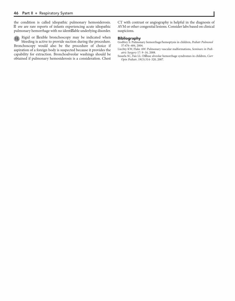
46 Part II u RespiratorySystem
the condition is called idiopathic pulmonary hemosiderosis. There are rare reports of infants experiencing acute idiopathic pulmonary hemorrhage with no identifiable underlying disorder.
15 Rigid or flexible bronchoscopy may be indicated when bleeding is active to provide suction during the procedure.
Bronchoscopy would also be the procedure of choice if aspiration of a foreign body is suspected because it provides the capability for extraction. Bronchoalveolar washings should be obtained if pulmonary hemosiderosis is a consideration. Chest
BibliographyGodfrey S: Pulmonary hemorrhage/hemoptysis in children, Pediatr Pulmonol
37:476–484, 2004.Liechty KW, Flake AW: Pulmonary vascular malformations, Seminars in Pedi-
atric Surgery 17: 9–16, 2008.Susarla SC, Fan LL: Diffuse alveolar hemorrhage syndromes in children, Curr
Opin Pediatr, 19(3):314–320, 2007.
CT with contrast or angiography is helpful in the diagnosis of AVM or other congenital lesions. Consider labs based on clinical suspicions.
Chapter 15 u Hemoptysis 47
HEMOPTYSISHEMOPTYSIS
Perform CT or bronchoscopy (15)± drug screen, COlevel, ammonia
Perform CT or bronchoscopy (15)± drug screen, COlevel, ammonia
NormalCXR resultNormalCXR result
AbnormalCXR resultAbnormalCXR result
Viral URI (tracheobronchitis) Viral URI (tracheobronchitis) 3 Pneumonia
Penetrating chest wall traumaExternal chest wall trauma with pulmonary contusionEndotracheal intubationVigorous suctioning of endotracheal tube or tracheostomy tubePost-op complication (bronchoscopy, lung biopsy)
Penetrating chest wall traumaExternal chest wall trauma with pulmonary contusionEndotracheal intubationVigorous suctioning of endotracheal tube or tracheostomy tubePost-op complication (bronchoscopy, lung biopsy)
Obtain CXR, CBC ± coagulation
studies, PFTs, UA
Obtain CXR, CBC ± coagulation
studies, PFTs, UA
NOYES
Coughing illnessMinimal hemoptysisClinically stable
Coughing illnessMinimal hemoptysisClinically stable
Recent traumaor procedureRecent traumaor procedure
NOYES
Perform Perform H and P 1
Infection (pneumonia, lung abscess, tuberculosis) Foreign body Congenital malformation (pulmonary sequestration, bronchogenic cyst)Bronchiectasis (cystic fibrosis) Vascular anomalies Pulmonary renal syndromes Goodpasture syndrome Granulomatosis polyangiitis (Wegener granulomatosis) Other causes of diffuse alveolar hemorrhage Other immune disorders Pulmonary hypertension Other cardiac disease Heiner syndrome Idiopathic pulmonary hemosiderosis Pulmonary infarctionTumor
Infection (pneumonia, lung abscess, tuberculosis) 4 Foreign body 5 Congenital malformation (pulmonary sequestration, bronchogenic cyst)Bronchiectasis (cystic fibrosis) 6 Vascular anomalies 7 Pulmonary renal syndromes Goodpasture syndrome 8 Granulomatosis polyangiitis (Wegener granulomatosis) 9 Other causes of diffuse alveolar hemorrhage 10 Other immune disorders 11 Pulmonary hypertension Other cardiac disease 12 Heiner syndrome 13 Idiopathic pulmonary hemosiderosis 14 Pulmonary infarctionTumor
Foreign body Congenital malformation (pulmonary sequestration, bronchogenic cyst)Vascular anomalies Bronchitis (tracheobronchitis)Other causes of diffuse alveolar hemorrhage Pulmonary embolusCoagulopathyPulmonary hypertension (primary)Cardiac disease Prematurity Toxic inhalation (CO, NO2, crack cocaine)HyperammonemiaIntracranial hemorrhageTuberous sclerosisFactitiousIdiopathic
Foreign body 5 Congenital malformation (pulmonary sequestration, bronchogenic cyst)Vascular anomalies 7 Bronchitis (tracheobronchitis)Other causes of diffuse alveolar hemorrhage 10 Pulmonary embolusCoagulopathyPulmonary hypertension (primary)Cardiac disease 12 Prematurity Toxic inhalation (CO, NO2, crack cocaine)HyperammonemiaIntracranial hemorrhageTuberous sclerosisFactitiousIdiopathic
Nelson Textbook of Pediatrics, 19e. Chapters 161, 400, 401, 511, 642Nelsons Essentials, 6e. Chapter 136
2

48
Chapter 16
APNEA
likely to be the primary cause of apnea only in neonates, but it may accompany serious disorders leading to apnea in older children.
3 The possibility of child abuse should always be considered when evaluating an apneic event. Inflicted neurotrauma,
deliberate suffocation, and poisoning attempts may be etiolo-gies; a history of sudden infant death syndrome (SIDS) or recurrent ALTEs (especially at a late age) in a family or a history of recurrent events while in the care of a single caregiver could be suspicious for caregiver-fabricated illness (previously called Munchausen syndrome by proxy).
4 Congenital metabolic disorders (e.g., fatty acid oxidation disorders, urea cycle defects) are being increasingly recog-
nized. A history of symptom onset occurring in association with fasting, altered mental status, recurrent episodes, a family history of infant deaths, and an occurrence beyond one year of age should raise suspicion for metabolic disorders. Serum glu-cose, ammonia, and pH should be obtained if suspicious of a metabolic disorder; if possible, samples of blood and urine should be obtained during the period of acute symptoms and frozen for future testing, if indicated.
5 Respiratory infections frequently associated with apnea include RSV (respiratory syncytial virus), bronchiolitis,
and pertussis. Both central and obstructive apnea can occur with RSV bronchiolitis. In young infants, apnea can be the first sign of RSV infection; there may be minimal pulmonary find-ings. Localized upper airway infections (e.g., tonsillitis, periton-sillar or retropharyngeal abscesses, croup, epiglottitis) can result in obstructive apnea.
6 The term apparent life-threatening event (ALTE) describes an acute event that appears frightening to the caregiver
and includes some combination of apnea, bradycardia, color change (usually cyanosis or pallor, occasionally plethora), and choking or gagging. The term replaces the previously used “near-miss SIDS”; scientific studies about the two conditions are difficult to compare because of the heterogeneity between them, but no clear association between SIDS and ALTEs has been demonstrated. ALTEs can be associated with a sleeping or awake state, may or may not be associated with feeding, and they are commonly reported to resolve quickly with interven-tion. Children frequently appear completely normal when they present for evaluation, which is why the evaluation decisions are so challenging.
7 Infants may appear to be choking or struggling to breathe during an event, suggesting an element of airway obstruc-
tion (obstructive apnea); they may exhibit absence of any respi-ratory effort (central apnea), or a mixture of both may occur.
8 Gastroesophageal reflux (GER) has been implicated in ALTEs, although its causal role is controversial since GER
is so common in infancy, plus no clear temporal association between reflux events and apneic events has been demon-strated. Other etiologies should always be carefully considered, even when GER appears to be temporally associated with an event. Esophageal pH monitoring is a more sensitive diagnostic test for GER than an upper gastrointestinal series (UGI) but is still only useful in determining causality if an event is captured during the study.
9 Airway problems that may lead to apneic events include nasal obstruction (e.g., severe allergies, polyps, choanal
Apnea is the cessation of breathing for a period of time (age dependent) or for any period if accompanied by pallor, cyano-sis, bradycardia, and/or hypotonia. Apnea may be due to a central origin, an element of airway obstruction or a combina-tion of both elements. (The differential diagnosis of apnea in neonates is extensive and includes many conditions unique to the neonate. Neither neonatal apnea nor apnea of prematurity is discussed here.)
When parents report that their child stopped breathing, benign conditions such as periodic breathing or breath-holding spells must be differentiated from more worrisome etiologies. Apnea can be a manifestation of a variety of seri-ous conditions, and in children it is frequently part of a constellation of symptoms (including choking, gagging, or change in color or tone) described as an apparent life-threat-ening event (ALTE).
1 Because of the broad differential diagnosis of apneic events, there is no concensus regarding a standardized recommended
evaluation. A careful history and physical remain the critical diag-nostic elements; multiple experts agree that subsequent testing should be guided by the history and physical because at least 50% of ALTE cases will have an identifiable etiology. The history for an apneic event should include any associated illness, the relationship of the event to sleeping and eating, and the presence or absence of associated symptoms, such as cyanosis, bradycardia, altered level of consciousness, and posturing or abnormal tonic-clonic movements. A birth and developmental history, a history of previous similar events, and a family history inquiring about apnea, ALTE, genetic disorders or infant deaths should be obtained. A social history should ask about potentially toxic expo-sures, including drugs or medications in the home, tobacco smoke exposure, and potential carbon monoxide exposure. Careful ques-tioning should be done regarding whether any intervention was needed and how quickly the child recovered from the event. For infants who were sleeping, inquire about sleep position, bedding, and coverings. The review of systems should include information about symptoms of airway obstruction, including chronic mouth breathing, noisy daytime respirations, snoring, and restlessness during sleep. The physical examination should be complete with careful attention to vital signs, head circumference (in infants), signs of airway obstruction, skin findings for bruising or signs of trauma, and facial dysmorphism. A careful ENT examination for fresh blood in the nose or mouth is important (it may suggest abuse). Dysmorphic features may be associated with craniofacial syndromes predisposing to airway obstruction (e.g., Pierre-Robin syndrome) or genetic or metabolic disorders. Airway hemangio-mas are often associated with hemangiomas on the face, neck, or upper trunk.
2 Urine toxicology screens are simple to perform and may reveal a causative etiology for apnea. Barbiturates, salicy-
lates, ipecac, boric acid, and cocaine are examples. Carbon monoxide poisoning can also be an etiology. Neuroimaging should be considered because child abuse is always part of the differential diagnosis of apnea in children. Hypoglycemia is
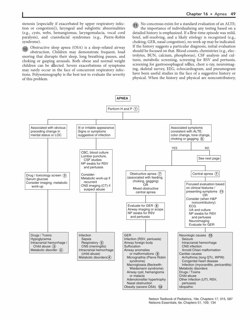
Chapter 16 u Apnea 49
stenosis [especially if exacerbated by upper respiratory infec-tion or congestion]), laryngeal and subglottic abnormalities (e.g., cysts, webs, hemangiomas, laryngomalacia, vocal cord paralysis), and craniofacial syndromes (e.g., Pierre-Robin syndrome).
10 Obstructive sleep apnea (OSA) is a sleep-related airway obstruction. Children may demonstrate frequent, loud
snoring that disrupts their sleep, long breathing pauses, and choking or gasping arousals. Both obese and normal weight children can be affected. Severe exacerbations of symptoms may rarely occur in the face of concurrent respiratory infec-tions. Polysomnography is the best test to evaluate the severity of this problem.
11 No concensus exists for a standard evaluation of an ALTE; the importance of individualizing any testing based on a
detailed history is emphasized. If a first-time episode was mild, brief, self-resolving, and a likely etiology is recognized (e.g., choking, GER, nasal congestion), no work up may be indicated. If the history suggests a particular diagnosis, initial evaluation should be focused on that. Blood counts, chemistries (e.g., elec-trolytes, BUN, calcium, phosphorus), CSF analysis and cul-tures, metabolic screening, screening for RSV and pertussis, screening for gastroesophageal reflux, chest x-ray, neuroimag-ing, skeletal survey, EEG, echocardiogram, and pneumogram have been useful studies in the face of a suggestive history or physical. When the history and physical are noncontributory,
Nelson Textbook of Pediatrics, 19e. Chapters 17, 315, 587Nelsons Essentials, 6e. Chapters 51, 109, 134
APNEA APNEA
Drugs / ToxinsHypoglycemiaIntracranial hemorrhage / Child abuseMetabolic disorder
Drugs / ToxinsHypoglycemiaIntracranial hemorrhage / Child abuse 3 Metabolic disorder 4
Infection Sepsis Respiratory CNS (meningitis)Intracranial hemorrhage (child abuse)Metabolic disorders
Infection Sepsis Respiratory 5 CNS (meningitis)Intracranial hemorrhage (child abuse)Metabolic disorders
GERInfection (RSV, pertussis) Airway foreign bodySuffocationAirway anomalies or malformations Micrognathia (Pierre Robin syndrome) Macroglossia (Beckwith- Wiedemann syndrome) Airway cyst, hemangioma or malacia Adenotonsillar hypertrophy Nasal obstructionObesity (severe OSA)
GERInfection (RSV, pertussis) Airway foreign bodySuffocationAirway anomalies or malformations 9 Micrognathia (Pierre Robin syndrome) Macroglossia (Beckwith- Wiedemann syndrome) Airway cyst, hemangioma or malacia Adenotonsillar hypertrophy Nasal obstructionObesity (severe OSA) 10
Neurologic causes Seizure Intracranial hemorrhage CNS infection Arnold Chiari malformationCardiac causes Arrhythmia (long QTc, WPW) Congenital heart disease Infection (myocarditis, pericarditis)Metabolic disordersDrugs / ToxinsChild abuseOther infection (UTI, RSV, pertussis) Idiopathic
Neurologic causes 12 Seizure Intracranial hemorrhage CNS infection Arnold Chiari malformationCardiac causes Arrhythmia (long QTc, WPW) Congenital heart disease Infection (myocarditis, pericarditis)Metabolic disordersDrugs / ToxinsChild abuseOther infection (UTI, RSV, pertussis) Idiopathic
NOYES
Perform Perform H and P 1
Associated with obviouspreceding change inmental status or LOC
Associated with obviouspreceding change inmental status or LOC
Ill or irritable appearanceSigns or symptomssuggestive of infection
Ill or irritable appearanceSigns or symptomssuggestive of infection
CBC, blood cultureLumbar puncture, CSF studiesNP swabs for RSV and pertussis Consider:Metabolic work-up if recurrent CNS imaging (CT) if suspect abuse
CBC, blood cultureLumbar puncture, CSF studiesNP swabs for RSV and pertussis Consider:Metabolic work-up if recurrent CNS imaging (CT) if suspect abuse
See next pageSee next page
Associated symptomsconsistent with ALTE:color change, tone change,choking or gagging
Associated symptomsconsistent with ALTE:color change, tone change,choking or gagging 6
Evaluate for GERAirway imaging or scopeNP swabs for RSV and pertussis
Evaluate for GER 8 Airway imaging or scopeNP swabs for RSV and pertussis
Drug / toxicology screen Serum glucoseConsider imaging, metabolic work-up
Drug / toxicology screen 2 Serum glucoseConsider imaging, metabolic work-up
Obstructive apnea(associated with feeding,
choking, gagging)OR
Mixed obstructivecentral apnea
Obstructive apnea 7 (associated with feeding,
choking, gagging)OR
Mixed obstructivecentral apnea
Central apneaCentral apnea 7
Focused evaluation basedon clinical features /presenting symptoms ORConsider (when H&P noncontributory): ECG UA and culture NP swabs for RSV and pertussis Neuroimaging Evaluate for GER
Focused evaluation basedon clinical features /presenting symptoms 11 ORConsider (when H&P noncontributory): ECG UA and culture NP swabs for RSV and pertussis Neuroimaging Evaluate for GER
4
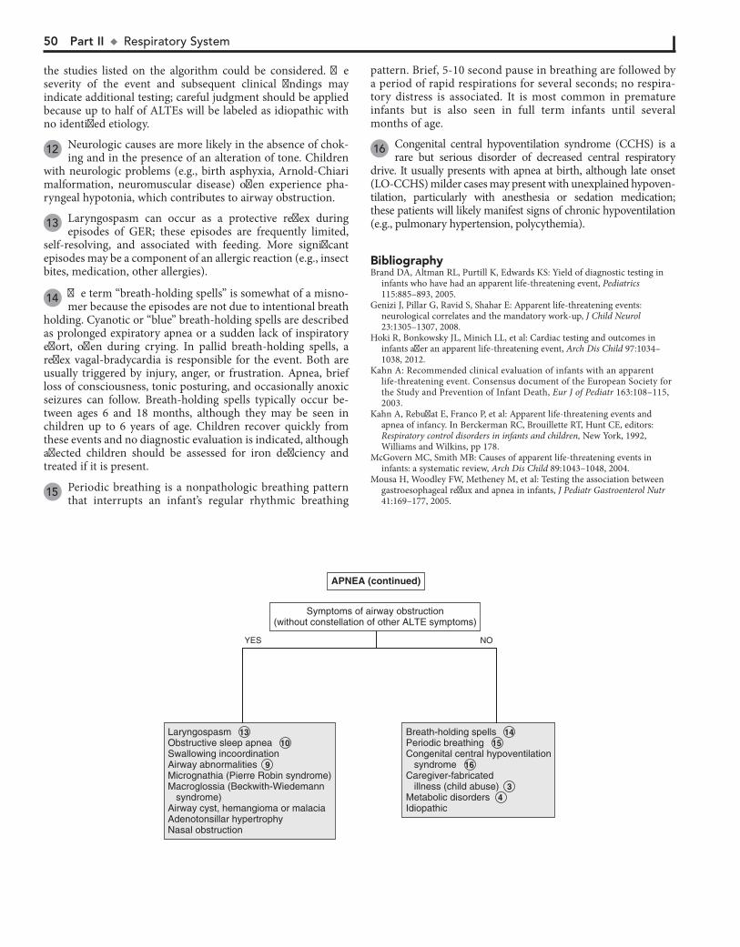
50 Part II u RespiratorySystem
the studies listed on the algorithm could be considered. The severity of the event and subsequent clinical findings may indicate additional testing; careful judgment should be applied because up to half of ALTEs will be labeled as idiopathic with no identified etiology.
12 Neurologic causes are more likely in the absence of chok-ing and in the presence of an alteration of tone. Children
with neurologic problems (e.g., birth asphyxia, Arnold-Chiari malformation, neuromuscular disease) often experience pha-ryngeal hypotonia, which contributes to airway obstruction.
13 Laryngospasm can occur as a protective reflex during episodes of GER; these episodes are frequently limited,
self-resolving, and associated with feeding. More significant episodes may be a component of an allergic reaction (e.g., insect bites, medication, other allergies).
14 The term “breath-holding spells” is somewhat of a misno-mer because the episodes are not due to intentional breath
holding. Cyanotic or “blue” breath-holding spells are described as prolonged expiratory apnea or a sudden lack of inspiratory effort, often during crying. In pallid breath-holding spells, a reflex vagal-bradycardia is responsible for the event. Both are usually triggered by injury, anger, or frustration. Apnea, brief loss of consciousness, tonic posturing, and occasionally anoxic seizures can follow. Breath-holding spells typically occur be-tween ages 6 and 18 months, although they may be seen in children up to 6 years of age. Children recover quickly from these events and no diagnostic evaluation is indicated, although affected children should be assessed for iron deficiency and treated if it is present.
15 Periodic breathing is a nonpathologic breathing pattern that interrupts an infant’s regular rhythmic breathing
pattern. Brief, 5-10 second pause in breathing are followed by a period of rapid respirations for several seconds; no respira-tory distress is associated. It is most common in premature infants but is also seen in full term infants until several months of age.
16 Congenital central hypoventilation syndrome (CCHS) is a rare but serious disorder of decreased central respiratory
drive. It usually presents with apnea at birth, although late onset (LO-CCHS) milder cases may present with unexplained hypoven-tilation, particularly with anesthesia or sedation medication; these patients will likely manifest signs of chronic hypoventilation (e.g., pulmonary hypertension, polycythemia).
BibliographyBrand DA, Altman RL, Purtill K, Edwards KS: Yield of diagnostic testing in
infants who have had an apparent life-threatening event, Pediatrics 115:885–893, 2005.
Genizi J, Pillar G, Ravid S, Shahar E: Apparent life-threatening events: neurological correlates and the mandatory work-up, J Child Neurol 23:1305–1307, 2008.
Hoki R, Bonkowsky JL, Minich LL, et al: Cardiac testing and outcomes in infants after an apparent life-threatening event, Arch Dis Child 97:1034–1038, 2012.
Kahn A: Recommended clinical evaluation of infants with an apparent life-threatening event. Consensus document of the European Society for the Study and Prevention of Infant Death, Eur J of Pediatr 163:108–115, 2003.
Kahn A, Rebuffat E, Franco P, et al: Apparent life-threatening events and apnea of infancy. In Berckerman RC, Brouillette RT, Hunt CE, editors: Respiratory control disorders in infants and children, New York, 1992, Williams and Wilkins, pp 178.
McGovern MC, Smith MB: Causes of apparent life-threatening events in infants: a systematic review, Arch Dis Child 89:1043–1048, 2004.
Mousa H, Woodley FW, Metheney M, et al: Testing the association between gastroesophageal reflux and apnea in infants, J Pediatr Gastroenterol Nutr 41:169–177, 2005.
APNEA (continued)APNEA (continued)
LaryngospasmObstructive sleep apneaSwallowing incoordinationAirway abnormalitiesMicrognathia (Pierre Robin syndrome)Macroglossia (Beckwith-Wiedemann syndrome)Airway cyst, hemangioma or malaciaAdenotonsillar hypertrophy Nasal obstruction
Laryngospasm 13 Obstructive sleep apnea 10 Swallowing incoordinationAirway abnormalities 9 Micrognathia (Pierre Robin syndrome)Macroglossia (Beckwith-Wiedemann syndrome)Airway cyst, hemangioma or malaciaAdenotonsillar hypertrophy Nasal obstruction
Breath-holding spellsPeriodic breathingCongenital central hypoventilation syndromeCaregiver-fabricated illness (child abuse)Metabolic disordersIdiopathic
Breath-holding spells 14 Periodic breathing 15 Congenital central hypoventilation syndrome 16 Caregiver-fabricated illness (child abuse) 3 Metabolic disorders 4 Idiopathic
NOYES
Symptoms of airway obstruction(without constellation of other ALTE symptoms)
Symptoms of airway obstruction(without constellation of other ALTE symptoms)

PART
IIICardiology

52
Chapter 17
CHEST PAIN
considered in athletes with repetitive upper extremity motions, especially in the absence of a recognized acute traumatic event; a bone scan should be considered if suspicion is high and x-rays are negative. Chest wall deformities (pectus carinatum, pectus excavatum) are rare causes of pediatric chest pain.
4 Muscular chest wall pain is common in weight-lifters, but carrying heavy back packs, severe coughing, and sports
involving rotation or twisting can also be causative. Sharp pain in the intercostal muscles can occur with infection due to cox-sackie and other enteroviruses. This pain (historically called pleurodynia or Bornholm disease) is sudden in onset, paroxys-mal, and accompanied by fever and other systemic signs of en-teroviral infection (e.g., vomiting, headache, sore throat). Sometimes the illness exhibits a biphasic pattern with a recurrence of the chest pain and fever several days after the initial presentation.
5 Early puberty may cause chest pain related to breast nodule development in males and females. Other breast disorders
including infections, cystic disorders, pregnancy, and menstrual swelling may cause chest pain in females.
6 Pain related to shingles (herpes zoster) may precede the appearance of the rash. Children affected by hypersensitiv-
ity pain syndromes may complain of pain with light touch to their chest wall, or even with wearing certain clothing; other somatic complaints are typically present as well.
7 Chest pain is occasionally the initial presentation of asthma. A history of nocturnal cough, atopy or a remote
history of bronchospasm may support the diagnosis. Broncho-constriction is often reported by children as chest pain. Pro-longed cough (due to acute exacerbations or poor control of asthma) can lead to soreness of chest wall muscles. Asthma also sometimes presents with a complaint of chest pain with run-ning or exertion (with or without coughing). Chest x-ray find-ings are often normal, but may reveal hyperinflation, atelectasis, or peribronchial thickening.
8 It is difficult to clearly define the role of psychological dis-orders in cases of pediatric chest pain because of inconsis-
tent use of valid psychological assessment tools and differences in research terminology in this area. Stress, anxiety, mood dis-orders, somatoform disorders, depression, and psychotic disor-ders have all been associated with chest pain; the validity of these diagnoses is impacted by the use (or misuse or nonuse) of appropriate psychological assessments. The psychological im-pact of organic causes of chest pain on patients is also poorly defined, even though it is likely very relevant to patients and families dealing with a serious medical diagnosis. Providers must be cognizant of the importance of using valid assessments to diagnose psychological disorders; psychogenic chest pain should never be a diagnosis of exclusion.
9 Hyperventilation typically presents with rapid breathing, dyspnea, anxiety, and sometimes with palpitations, chest
pain, paresthesias, lightheadedness, and confusion. Careful evaluation often reveals anxiety or underlying psychological concerns.
10 Precordial catch syndrome (Texidor twinge) is classically described as a benign condition characterized by brief par-
oxysms of sharp, well-localized pains in the midsternal or precor-dial region. Episodes are brief (30 seconds to 3 minutes), self- resolving, and exacerbated by deep breathing. Expert opinions
Chest pain is common in children and adolescents. Despite the degree of concern that it generates, the symptom is rarely as-sociated with a serious cardiac problem. The epidemiology of chest pain in youth is not well understood, although available data suggest more cases are classified as idiopathic than are at-tributed to a more specific etiology (e.g., cardiac, respiratory). Studies approximate between 1% and 10% of pediatric chest pain cases are due to cardiac etiologies.
1 A properly done history and physical are often the only tools required in the evaluation of pediatric chest pain.
Screening tests are not considered helpful unless specifically indicated. Eliciting the patient’s or family’s concerns about their complaint may be useful.
A medical history of asthma, sickle cell disease, collagen vascular disease, or a recent coughing illness may be helpful. Long-standing diabetes mellitus and chronic anemias are risk factors for ischemic chest pain. Inquire about a history of Kawa-saki disease, including the possibility of an undiagnosed case. The review of systems should include inquiries about associated acute and chronic symptoms and any precipitating factors. In-quire about choking episodes, recent trauma, and exercise or activities that could cause pain from muscle strain or overuse. It is critical to distinguish a history of exercise (that could cause muscular chest wall pain) from exercise as a precipitating factor (which may be consistent with ischemic pain and mandates an urgent cardiac evaluation). Associated syncope is very worri-some and also mandates a cardiac evaluation. A medication history could provide clues to a potentially causative etiology (oral contraceptives) or the possibility of mucosal injury (e.g., tetracycline, NSAIDs); also investigate the possibility of sub-stance abuse, especially cocaine and methamphetamine. Evalu-ation of psychosocial factors in the child’s life is very important. Ask about school attendance and performance, relationships with friends and family, and any current stresses or conflicts.
The family history should inquire about hypercholesterol-emia, Marfan syndrome, and cardiomyopathy. A family history of recurrent syncope or unexplained sudden death may suggest hypertrophic cardiomyopathy or long QT syndrome. Heart disease in an adult family member may provoke anxiety-related chest pain in a younger person.
A complete thorough physical exam is necessary; focusing on the chest exam may miss findings pertinent to a noncardiac underlying cause of chest pain.
2 Costochondritis is pain due to inflammation of the costo-chondral joints (where the bony rib meets the costal carti-
lage). It is a common cause of chest pain in children and is usually unilateral, sharp, transient in nature, and can be repro-duced by palpation on examination. Tietze syndrome, a rare form of costochondritis, affects a single chostochondral, cos-tosternal or sternoclavicular joint and causes notable swelling, tenderness, and warmth localized to the affected joint.
3 Other skeletal causes of chest pain include traumatic injury, spondyloarthritis, and stress fractures. The latter should be
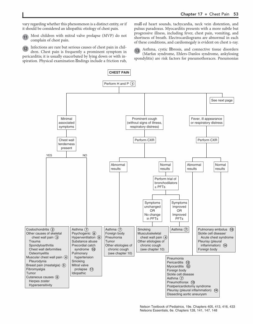
Chapter 17 u ChestPain 53
vary regarding whether this phenomenon is a distinct entity, or if it should be considered an idiopathic etiology of chest pain.
11 Most children with mitral valve prolapse (MVP) do not complain of chest pain.
12 Infections are rare but serious causes of chest pain in chil-dren. Chest pain is frequently a prominent symptom in
pericarditis; it is usually exacerbated by lying down or with in-spiration. Physical examination findings include a friction rub,
muffled heart sounds, tachycardia, neck vein distention, and pulsus paradoxus. Myocarditis presents with a more subtle but progressive illness, including fever, chest pain, vomiting, and shortness of breath. Electrocardiograms are abnormal in each of these conditions, and cardiomegaly is evident on chest x-ray.
13 Asthma, cystic fibrosis, and connective tissue disorders (Marfan syndrome, Ehlers-Danlos syndrome, ankylosing
spondylitis) are risk factors for pneumothoraces. Pneumonias
Costochondritis 2 Other causes of skeletal chest wall pain 3 Trauma Spondyloarthritis Chest wall deformities OsteomyelitisMuscular chest wall pain 4 PleurodyniaBreast pain (mastalgia) 5 Fibromyalgia TumorCutaneous causes 6 Herpes zoster Hypersensitivity
Costochondritis 2 Other causes of skeletal chest wall pain 3 Trauma Spondyloarthritis Chest wall deformities OsteomyelitisMuscular chest wall pain 4 PleurodyniaBreast pain (mastalgia) 5 Fibromyalgia TumorCutaneous causes 6 Herpes zoster Hypersensitivity
Asthma 7 Psychogenic 8 Hyperventilation 9 Substance abusePrecordial catch syndrome 10 Pulmonary hypertensionSmokingMitral valve prolapse 11 Idiopathic
Asthma 7 Psychogenic 8 Hyperventilation 9 Substance abusePrecordial catch syndrome 10 Pulmonary hypertensionSmokingMitral valve prolapse 11 Idiopathic
Asthma 7 Foreign bodyPneumoniaTumorOther etiologies of chronic cough (see chapter 10)
Asthma 7 Foreign bodyPneumoniaTumorOther etiologies of chronic cough (see chapter 10)
SmokingMusculoskeletal chest wall pain 4 Other etiologies of chronic cough (see chapter 10)
SmokingMusculoskeletal chest wall pain 4 Other etiologies of chronic cough (see chapter 10)
Asthma 7 Asthma 7
PneumoniaPericarditis 12 Myocarditis 12 Foreign bodySickle cell diseaseAsthma 7 Pneumothorax 13 Postpericardiotomy syndromePleurisy (pleural inflammation) 14 Dissecting aortic aneurysm
PneumoniaPericarditis 12 Myocarditis 12 Foreign bodySickle cell diseaseAsthma 7 Pneumothorax 13 Postpericardiotomy syndromePleurisy (pleural inflammation) 14 Dissecting aortic aneurysm
Pulmonary embolus 15 Sickle cell disease/ Acute chest syndromePleurisy (pleural inflammation) 14 Foreign body
Pulmonary embolus 15 Sickle cell disease/ Acute chest syndromePleurisy (pleural inflammation) 14 Foreign body
Chest walltenderness
present
Chest walltenderness
present
Symptomsunchanged
ORNo change
in PFTs
Symptomsunchanged
ORNo change
in PFTs
Symptomsimproved
ORImproved
PFTs
Symptomsimproved
ORImproved
PFTs
Perform trial ofbronchodilators± PFTs
Perform trial ofbronchodilators± PFTs
Normal resultsNormal results
Abnormal resultsAbnormal results
Normal resultsNormal results
Abnormal resultsAbnormal results
Minimalassociatedsymptoms
Minimalassociatedsymptoms
Perform CXRPerform CXR
Prominent cough(without signs of illness,
respiratory distress)
Prominent cough(without signs of illness,
respiratory distress)
Perform CXRPerform CXR
Fever, ill appearanceor respiratory distressFever, ill appearanceor respiratory distress
See next pageSee next page
Perform H and P 1 Perform H and P 1
CHEST PAINCHEST PAIN
YES NO
Nelson Textbook of Pediatrics, 19e. Chapters 405, 413, 416, 433Nelsons Essentials, 6e. Chapters 128, 141, 147, 148
54 Part III u Cardiology
intercostal nerve. Reproduction of the pain by hooking the fin-gers under the anterior costal margins and pulling the ribs for-ward is characteristic.
17 Symptoms of GERD (gastroesophageal reflux disease) vary by age; common symptoms in older children and
adolescents are abdominal or substernal pain, vomiting or re-gurgitation, increased pain after meals or when recumbent, and relief with antacids. A trial of empiric therapy is appropriate in children with typical symptoms, although a positive response is not confirmatory of GERD since spontaneous resolution of symptoms (due to any cause) can occur.
18 The value of an UGI contrast study is to rule out other GI disorders (e.g., esophageal abnormalities, intestinal ob-
struction); it will not diagnose GERD.
19 Peptic ulcer disease (PUD) frequently presents with a chronic intermittent history of dull or aching pain and
often includes nighttime complaints. The pain may be epigas-tric or poorly localized abdominal pain; it may or may not be relieved by antacids.
20 Eosinophilic esophagitis (EoE) is diagnosed by endoscopic biopsies showing localized eosinophilic infiltrates of the
esophagus. The condition is being increasingly recognized in all age groups; abdominal pain and vomiting are more common in younger children and dysphagia, chest pain, and food impac-tion are more likely in adolescents. Other atopic diseases and food allergies are commonly associated.
due to certain pathogens (e.g., Pneumocystis jirovecii in immu-nodeficient conditions or staphylococcal, anaerobic gram nega-tive pathogens) also predispose to the development of pneumo-thoraces. Healthy children may also develop spontaneous pneumothorax; cocaine use is a risk factor. Forceful vomiting is a rare cause of esophageal rupture causing pneumomediasti-num (Boerhaave syndrome). Traumatic or iatrogenic causes should also be considered.
14 Movement and deep breathing often aggravate the pain associated with pleurisy (pleuritis) or pleural effusions.
Bacterial pneumonias are the most common cause of pleuritis in children; collagen vascular disorders can also be causative.
15 Risk factors for venous thrombosis (e.g., oral contracep-tives, recent abortion or surgery [especially cardiac], the
presence of a central venous line, immobilization, sepsis, hyper-coagulable states, vascular malformations) should raise suspicion for pulmonary emboli. Associated symptoms include dyspnea, cough, hypoxia, and occasionally, hemoptysis. If em-boli are suspected, appropriate labs and imaging (spiral CT or pulmonary angiography) should be performed.
16 Slipping rib syndrome is characterized by pain along the lower rib margin of the upper abdomen, sometimes asso-
ciated with a slipping sensation and a popping or clicking sound. Although a clear consensus on the cause of the pain is lacking, a commonly presumed etiology is that trauma to the eighth, ninth, or tenth rib causes a sprain-like injury, which increases the mobility of the rib and allows impingement on an

Chapter 17 u ChestPain 55
Fracture Accidental Nonaccidental (child abuse)
Fracture Accidental Nonaccidental (child abuse)
Musculoskeletal chest wall pain 3 , 4 Slipping rib syndrome 16 Idiopathic
Musculoskeletal chest wall pain 3 , 4 Slipping rib syndrome 16 Idiopathic
Gastroesophageal reflux disease (esophagitis) 17
Gastroesophageal reflux disease (esophagitis) 17
Gastroesophageal reflux disease (esophagitis) 17 Peptic ulcer disease 19 Eosinophilic esophagitis 20 “Pill” esophagitisOther esophageal/ intestinal disorders
Gastroesophageal reflux disease (esophagitis) 17 Peptic ulcer disease 19 Eosinophilic esophagitis 20 “Pill” esophagitisOther esophageal/ intestinal disorders
Arrhythmia Isolated rhythm disturbance Drug/toxin induced Associated with systemic disease (anemia, hyperthyroidism) Associated cardiac defects (congenital or acquired)
Arrhythmia Isolated rhythm disturbance Drug/toxin induced Associated with systemic disease (anemia, hyperthyroidism) Associated cardiac defects (congenital or acquired)
ConsiderCXRConsiderCXR
Consider UGI 18 VSReferral to GI for endoscopy
Consider UGI 18 VSReferral to GI for endoscopy
No response or minimalresponse
No response or minimalresponse
PositiveresponsePositiveresponse
Normal resultsNormal results
Abnormal resultsAbnormal results
History of trauma or vigorous exercise
History of trauma or vigorous exercise
Trial of antacidsor H2 blockersTrial of antacidsor H2 blockers
Symptoms relatedto meals, position,
lower sternal/epigastric location
Symptoms relatedto meals, position,
lower sternal/epigastric location
Obtain ECG(See Chapter 19)Obtain ECG(See Chapter 19)
History ofpalpitationsHistory of
palpitations
See next pageSee next page
CHEST PAIN(continued)
CHEST PAIN(continued)
56 Part III u Cardiology
21 Consultation with a cardiologist is recommended because of the potentially serious (ischemic) nature of chest pain
caused by severe obstructive lesions; it is also considered a more cost-effective alternative to obtaining additional studies without consultation.
22 Hypertrophic cardiomyopathy is a genetic disorder trans-mitted in an autosomal dominant pattern, although a large
proportion of cases are considered de novo mutations. Classic physical examination findings include a left ventricular lift and a harsh systolic ejection murmur that is increased with any maneuver that decreases venous return (Valsalva maneuver, ris-ing from squatting to standing). As the development of hyper-trophy is gradual over years, examination findings in children may be limited to nonspecific murmurs; cardiac evaluation is indicated whenever there is a known family history.
23 Unless suspected to be asthma, chest pain that is precipi-tated by exercise or running or is associated with syncope
or palpitations warrants urgent cardiac consultation. Myocar-dial ischemia is rare in children overall, although an increasing number of children are at risk due to advances in care and treat-ment of congenital and acquired (Kawasaki) heart disease. Un-like adults with atherosclerotic heart disease, children do not experience classic angina-type pain (i.e., chest pressure or
squeezing sensation with radiation to neck, jaws or arms); rather, the symptoms of myocardial ischemia in younger pa-tients are nonspecific and include irritability, nausea, vomiting, abdominal pain, failure to thrive, shock, syncope, seizures, or sudden cardiac arrest. The presentation of congenital coronary artery abnormalities may be subtle or abrupt with few identifi-able risk factors. However, children with a history of heart sur-gery (e.g., repair of tetralogy of Fallot or transposition of the great arteries), congenital heart conditions, or a history of Ka-wasaki disease warrant a higher threshold of awareness for risk of ischemic chest pain.
24 Coronary artery anomalies are rare but can be associated with severe ischemia. The physical examination may be
normal or may include tachypnea, tachycardia, pallor, diapho-resis, distant heart tones, a murmur consistent with mitral regurgitation, or a gallop rhythm suggesting myocardial dys-function. Echocardiogram and angiography are used in diagnosis.
BibliographyEslick GD, Selbst SM: Pediatric chest pain. Preface. Pediatr Clin North Am
57(6):xiii-xiv, 2010.Reddy S, Singh H: Chest pain in children and adolescents, Pediatr Rev 31:
e1-e9, 2010.
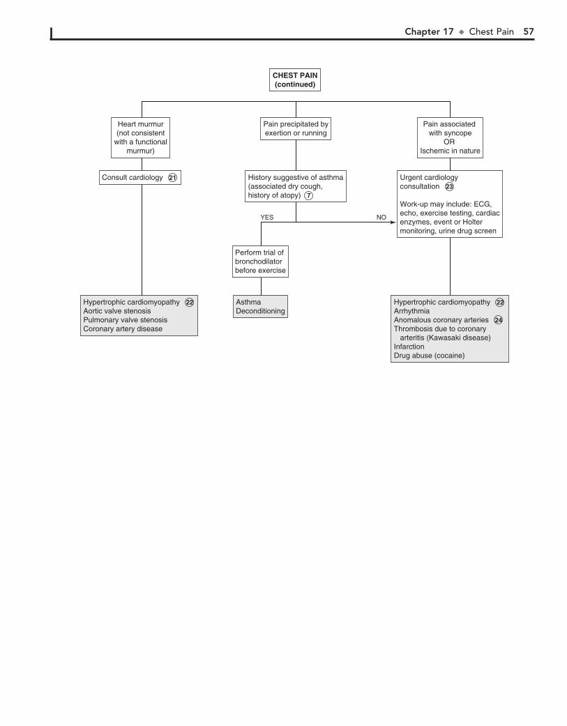
Chapter 17 u ChestPain 57
Hypertrophic cardiomyopathy 22 Aortic valve stenosisPulmonary valve stenosisCoronary artery disease
Hypertrophic cardiomyopathy 22 Aortic valve stenosisPulmonary valve stenosisCoronary artery disease
AsthmaDeconditioningAsthmaDeconditioning
Hypertrophic cardiomyopathy 22 ArrhythmiaAnomalous coronary arteries 24 Thrombosis due to coronary arteritis (Kawasaki disease) InfarctionDrug abuse (cocaine)
Hypertrophic cardiomyopathy 22 ArrhythmiaAnomalous coronary arteries 24 Thrombosis due to coronary arteritis (Kawasaki disease) InfarctionDrug abuse (cocaine)
Consult cardiology 21 Consult cardiology 21
Heart murmur(not consistent
with a functionalmurmur)
Heart murmur(not consistent
with a functionalmurmur)
History suggestive of asthma(associated dry cough,history of atopy) 7
History suggestive of asthma(associated dry cough,history of atopy) 7
Perform trial ofbronchodilatorbefore exercise
Perform trial ofbronchodilatorbefore exercise
Pain precipitated byexertion or runningPain precipitated byexertion or running
Urgent cardiologyconsultation 23 Work-up may include: ECG,echo, exercise testing, cardiacenzymes, event or Holtermonitoring, urine drug screen
Urgent cardiologyconsultation 23 Work-up may include: ECG,echo, exercise testing, cardiacenzymes, event or Holtermonitoring, urine drug screen
Pain associated with syncope
OR Ischemic in nature
Pain associated with syncope
OR Ischemic in nature
CHEST PAIN(continued)
CHEST PAIN(continued)
YES NO
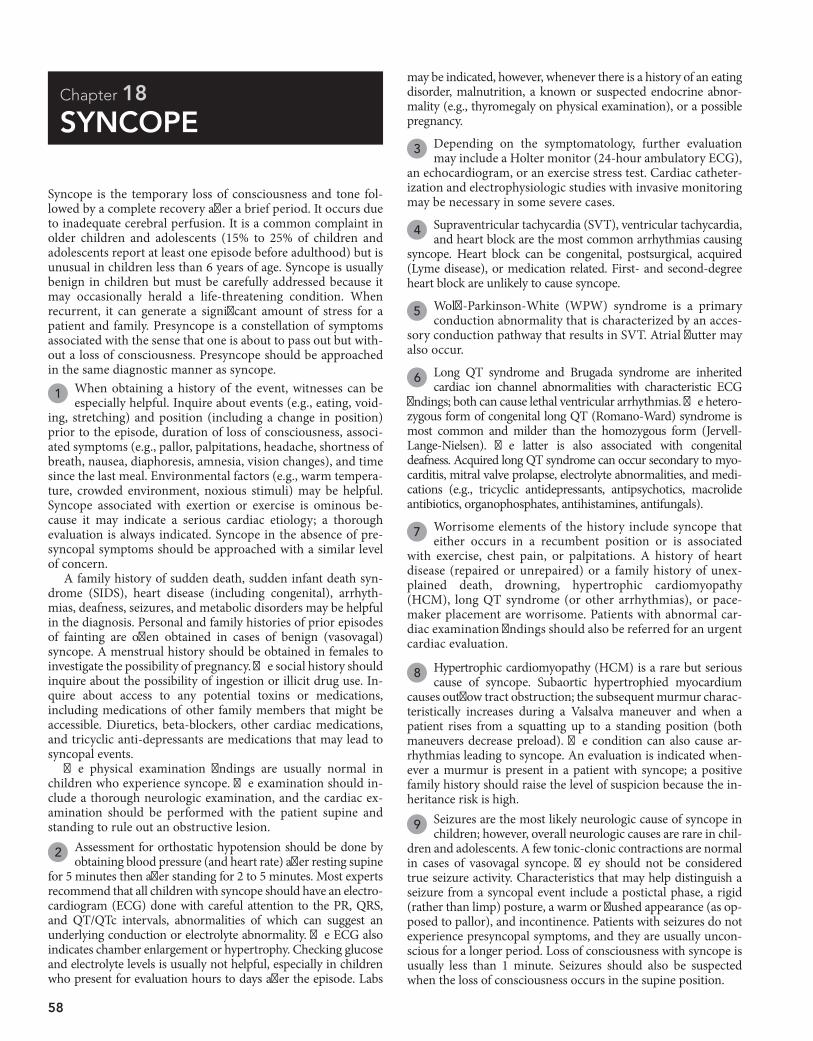
58
Chapter 18
SYNCOPE
may be indicated, however, whenever there is a history of an eating disorder, malnutrition, a known or suspected endocrine abnor-mality (e.g., thyromegaly on physical examination), or a possible pregnancy.
3 Depending on the symptomatology, further evaluation may include a Holter monitor (24-hour ambulatory ECG),
an echocardiogram, or an exercise stress test. Cardiac catheter-ization and electrophysiologic studies with invasive monitoring may be necessary in some severe cases.
4 Supraventricular tachycardia (SVT), ventricular tachycardia, and heart block are the most common arrhythmias causing
syncope. Heart block can be congenital, postsurgical, acquired (Lyme disease), or medication related. First- and second-degree heart block are unlikely to cause syncope.
5 Wolff-Parkinson-White (WPW) syndrome is a primary conduction abnormality that is characterized by an acces-
sory conduction pathway that results in SVT. Atrial flutter may also occur.
6 Long QT syndrome and Brugada syndrome are inherited cardiac ion channel abnormalities with characteristic ECG
findings; both can cause lethal ventricular arrhythmias. The hetero-zygous form of congenital long QT (Romano-Ward) syndrome is most common and milder than the homozygous form (Jervell-Lange-Nielsen). The latter is also associated with congenital deafness. Acquired long QT syndrome can occur secondary to myo-carditis, mitral valve prolapse, electrolyte abnormalities, and medi-cations (e.g., tricyclic antidepressants, antipsychotics, macrolide antibiotics, organophosphates, antihistamines, antifungals).
7 Worrisome elements of the history include syncope that either occurs in a recumbent position or is associated
with exercise, chest pain, or palpitations. A history of heart disease (repaired or unrepaired) or a family history of unex-plained death, drowning, hypertrophic cardiomyopathy (HCM), long QT syndrome (or other arrhythmias), or pace-maker placement are worrisome. Patients with abnormal car-diac examination findings should also be referred for an urgent cardiac evaluation.
8 Hypertrophic cardiomyopathy (HCM) is a rare but serious cause of syncope. Subaortic hypertrophied myocardium
causes outflow tract obstruction; the subsequent murmur charac-teristically increases during a Valsalva maneuver and when a patient rises from a squatting up to a standing position (both maneuvers decrease preload). The condition can also cause ar-rhythmias leading to syncope. An evaluation is indicated when-ever a murmur is present in a patient with syncope; a positive family history should raise the level of suspicion because the in-heritance risk is high.
9 Seizures are the most likely neurologic cause of syncope in children; however, overall neurologic causes are rare in chil-
dren and adolescents. A few tonic-clonic contractions are normal in cases of vasovagal syncope. They should not be considered true seizure activity. Characteristics that may help distinguish a seizure from a syncopal event include a postictal phase, a rigid (rather than limp) posture, a warm or flushed appearance (as op-posed to pallor), and incontinence. Patients with seizures do not experience presyncopal symptoms, and they are usually uncon-scious for a longer period. Loss of consciousness with syncope is usually less than 1 minute. Seizures should also be suspected when the loss of consciousness occurs in the supine position.
Syncope is the temporary loss of consciousness and tone fol-lowed by a complete recovery after a brief period. It occurs due to inadequate cerebral perfusion. It is a common complaint in older children and adolescents (15% to 25% of children and adolescents report at least one episode before adulthood) but is unusual in children less than 6 years of age. Syncope is usually benign in children but must be carefully addressed because it may occasionally herald a life-threatening condition. When recurrent, it can generate a significant amount of stress for a patient and family. Presyncope is a constellation of symptoms associated with the sense that one is about to pass out but with-out a loss of consciousness. Presyncope should be approached in the same diagnostic manner as syncope.
1 When obtaining a history of the event, witnesses can be especially helpful. Inquire about events (e.g., eating, void-
ing, stretching) and position (including a change in position) prior to the episode, duration of loss of consciousness, associ-ated symptoms (e.g., pallor, palpitations, headache, shortness of breath, nausea, diaphoresis, amnesia, vision changes), and time since the last meal. Environmental factors (e.g., warm tempera-ture, crowded environment, noxious stimuli) may be helpful. Syncope associated with exertion or exercise is ominous be-cause it may indicate a serious cardiac etiology; a thorough evaluation is always indicated. Syncope in the absence of pre-syncopal symptoms should be approached with a similar level of concern.
A family history of sudden death, sudden infant death syn-drome (SIDS), heart disease (including congenital), arrhyth-mias, deafness, seizures, and metabolic disorders may be helpful in the diagnosis. Personal and family histories of prior episodes of fainting are often obtained in cases of benign (vasovagal) syncope. A menstrual history should be obtained in females to investigate the possibility of pregnancy. The social history should inquire about the possibility of ingestion or illicit drug use. In-quire about access to any potential toxins or medications, including medications of other family members that might be accessible. Diuretics, beta-blockers, other cardiac medications, and tricyclic anti-depressants are medications that may lead to syncopal events.
The physical examination findings are usually normal in children who experience syncope. The examination should in-clude a thorough neurologic examination, and the cardiac ex-amination should be performed with the patient supine and standing to rule out an obstructive lesion.
2 Assessment for orthostatic hypotension should be done by obtaining blood pressure (and heart rate) after resting supine
for 5 minutes then after standing for 2 to 5 minutes. Most experts recommend that all children with syncope should have an electro-cardiogram (ECG) done with careful attention to the PR, QRS, and QT/QTc intervals, abnormalities of which can suggest an underlying conduction or electrolyte abnormality. The ECG also indicates chamber enlargement or hypertrophy. Checking glucose and electrolyte levels is usually not helpful, especially in children who present for evaluation hours to days after the episode. Labs
Chapter 18 u Syncope 59
PregnancyPregnancy Arrhythmias 4 Wolff-Parkinson-White 5 syndrome Long QT syndromes 6 Brugada syndrome 6 Obstructive lesions Aortic stenosis Pulmonic stenosisHypertrophic cardiomyopathy 8 Coronary artery anomalies (congenital, Kawasaki)Other causes of myocardial ischemia/infarctionMyocarditis/other myocardial dysfunctionPulmonary hypertensionIntracardiac mass or tumorSubclavian steal syndrome
Arrhythmias 4 Wolff-Parkinson-White 5 syndrome Long QT syndromes 6 Brugada syndrome 6 Obstructive lesions Aortic stenosis Pulmonic stenosisHypertrophic cardiomyopathy 8 Coronary artery anomalies (congenital, Kawasaki)Other causes of myocardial ischemia/infarctionMyocarditis/other myocardial dysfunctionPulmonary hypertensionIntracardiac mass or tumorSubclavian steal syndrome
Seizure disorder 9 Brain tumorMigraine 10
Seizure disorder 9 Brain tumorMigraine 10
Orthostatic hypotensionOrthostatic hypotension
Vasovagal (postural, reflex, neurally mediated) 12 Situational syncope 13 Cough Micturition Defecation Hair grooming StretchingOrthostatic hypotension 14 Hypovolemia Related to medicationsPostural orthostatic tachycardia syndrome 15
Vasovagal (postural, reflex, neurally mediated) 12 Situational syncope 13 Cough Micturition Defecation Hair grooming StretchingOrthostatic hypotension 14 Hypovolemia Related to medicationsPostural orthostatic tachycardia syndrome 15
Vasovagal (postural, reflex, neurally mediated) 12 Situational syncope 13 Cough Micturition Defecation Hair grooming StretchingOrthostatic hypotension 14 Hypovolemia Related to medicationsPostural orthostatic tachycardia syndrome 15 Breath-holding spells 16 Hysteria/conversion 17Metabolic (hypoglycemia, electrolyte disorders) 18 AnemiaHyperventilation 19 PregnancyAnorexia nervosaCarbon monoxide poisoning
Vasovagal (postural, reflex, neurally mediated) 12 Situational syncope 13 Cough Micturition Defecation Hair grooming StretchingOrthostatic hypotension 14 Hypovolemia Related to medicationsPostural orthostatic tachycardia syndrome 15 Breath-holding spells 16 Hysteria/conversion 17Metabolic (hypoglycemia, electrolyte disorders) 18 AnemiaHyperventilation 19 PregnancyAnorexia nervosaCarbon monoxide poisoning
Refer to cardiologyfor consideration of tilt test, otherevaluation 11
Refer to cardiologyfor consideration of tilt test, otherevaluation 11
Recurrent episodes(>2 to 3)Recurrent episodes(>2 to 3)
Obtain EEG,neurology consultObtain EEG,neurology consult
Refer to cardiology for further evaluation 3 Refer to cardiology for further evaluation 3
Suspect neurologicdisorder 9 Suspect neurologicdisorder 9
Signs or symptomssuggestive ofcardiac etiology 7
Signs or symptomssuggestive ofcardiac etiology 7
NormalresultsNormalresults
Abnormalorthostatic
blood pressures
Abnormalorthostatic
blood pressures
AbnormalECG
AbnormalECG
PositiveHCG
PositiveHCG
Perform H and P 1 Perform H and P 1
Perform ECG,orthostatic BPs± urine HCG in
adolescent girls 2
Perform ECG,orthostatic BPs± urine HCG in
adolescent girls 2
SYNCOPESYNCOPE
YES NO
YES NO
YES NO
Nelson Textbook of Pediatrics, 19e. Chapter 587Nelsons Essentials, 6e. Chapter 140
60 Part III u Cardiology
10 A rare migraine variant (basilar-type) can cause syncope. A severe occipital headache and unilateral visual changes are
commonly associated; ataxia, vertigo, and vomiting may also occur.
11 Further evaluation may be indicated because frequent epi-sodes of syncope are very distressing to a patient, even
when the etiology is benign. A tilt table evaluation may aid in the diagnosis of syncope due to orthostatic intolerance (e.g., vasovagal, orthostatic hypotension, postural orthostatic tachy-cardia syndrome) and in the selection of therapy; there are, however, limitations to its sensitivity, specificity, diagnostic yield, and reproducibility.
12 Vasovagal syncope is also called postural, reflex, neurocar-diogenic or neurally-mediated syncope, or simply “faint-
ing”; some overlap in terminology is occurring with increasing understanding of the pathophysiology of orthostasis (the body’s adjustment to upright posture). It is the most common type of syncope in normal children and adolescents; it occurs most fre-quently in the 15- to 19-year-old age group. A neurally-mediated decline in blood pressure (the exact mechanism of which is poorly understood) and heart rate are responsible for the tran-sient decrease in cerebral blood flow leading to the syncopal episode. Recognizable precipitating factors (rising to stand, a prolonged period of standing, certain stressors like venipunc-ture, noxious stimuli, fasting, or a crowded location) and pro-dromal (presyncopal) autonomic symptoms (e.g., nausea, pallor, diaphoresis) are characteristic of this type of noncardiac syn-cope. The absence of a prodromal or presyncopal sensation is not consistent with a vasovagal etiology and should prompt consideration of more serious etiologies.
Also, vasovagal syncope can occur after vigorous, usually pro-longed exertion (such as at the end of a long competitive run) due to a warm ambient temperature, venous pooling, and dehydra-tion; it is distinct from “mid-stride” syncope, which should prompt an immediate cardiac evaluation. Most of these cases have a vasovagal (not cardiac) etiology, but sports participation should be curtailed until a worrisome cardiac etiology has been ruled out.
13 Autonomic responses triggered by certain bodily func-tions or movements (e.g., cough, micturition, defecation,
hair grooming, stretching) can occasionally result in syncope; the mechanism is presumed to be vasovagal in nature.
14 Orthostatic hypotension is defined as a persistent fall of 20 mmHg systolic and/or 10 mmHg diastolic blood pressure
within 3 minutes of assuming an upright position without mov-ing the arms or legs. (Heart rates usually increase with standing but are not part of the diagnostic criteria.) In contrast to vasova-gal syncope, true orthostatic hypotension is due to an inadequate vasoconstriction response to standing and will lead to syncope without the typical presyncopal symptoms seen with vasovagal
syncope. Most cases in young people are nonneurogenic and caused by medications or hypovolemia (e.g., hemorrhage, dehy-dration). Neurogenic orthostatic hypotension is a significant disorder of the autonomic system and more likely to occur in older patients or in association with serious medical conditions (e.g., diabetes, amyloidosis, Parkinson’s disease).
15 Postural orthostatic tachycardia syndrome (POTS) de-scribes posturally-induced symptoms (e.g., lightheaded-
ness, palpitations, nausea) accompanied by an increased heart rate of 30 bpm documented while obtaining blood pressures for orthostatic hypotension. (Some decrease in blood pressure may occur but is not part of the diagnostic criteria.) Recurrent near syncope is most common, but frank syncope can occur. Chronic fatigue and exercise intolerance are commonly pres-ent in this syndrome, which is most common in young women.
16 Breath-holding spells are the most common mechanism of syncope in children younger than 6 years of age. Chil-
dren who are startled or upset hold their breath in expiration, collapse, and become cyanotic for a brief period.
17 Hysterical or conversion syncope is a diagnosis of exclusion. It is most common in adolescents; they typically have an
audience for this event. Hemodynamic changes, sweating, pallor, and subsequent psychological distress regarding the episode are absent.
18 Hunger, weakness, sweating, agitation, and confusion may accompany hypoglycemia or electrolyte disorders. Supine
position does not provide relief.
19 A history of preceding psychological distress, sensations of shortness of breath, chest pain, visual changes, and numb-
ness or tingling of the extremities may be reported in children with syncope due to hyperventilation. The patient may be able to reproduce the episode when requested to hyperventilate.
BibliographyFischer JW, Cho CS: Pediatric syncope: Cases form the emergency depart-
ment, Emerg Med Clin N Am 28:501–516, 2010.Lewis DA, Chala A: Syncope in the pediatric patient: The cardiologist’s perspective,
Pediatr Clin North Am 46:205–219, 1999.Park MK: Pediatric cardiology for practitioners [electronic resource]. Philadelphia,
2008, Mosby/Elsevier.Stewart JM: Common syndromes of orthostatic intolerance, Pediatrics 131:
968–980, 2013.Strickberger SA, Benson DW, Biaggioni I, et al: AHA/ACCF scientific state-
ment on the evaluation of syncope: from the American Heart Association Councils on Clinical Cardiology, Cardiovascular Nursing, Cardiovascular Disease in the Young, and Stroke, and the Quality of Care and Outcomes Research Interdisciplinary Working Group; and the American College of Cardiology Foundation in collaboration with the Heart Rhythm Society, J Am Coll Cardiol 47(2):473–484, 2006.

Chapter 18 u Syncope 61
PregnancyPregnancy Arrhythmias 4 Wolff-Parkinson-White 5 syndrome Long QT syndromes 6 Brugada syndrome 6 Obstructive lesions Aortic stenosis Pulmonic stenosisHypertrophic cardiomyopathy 8 Coronary artery anomalies (congenital, Kawasaki)Other causes of myocardial ischemia/infarctionMyocarditis/other myocardial dysfunctionPulmonary hypertensionIntracardiac mass or tumorSubclavian steal syndrome
Arrhythmias 4 Wolff-Parkinson-White 5 syndrome Long QT syndromes 6 Brugada syndrome 6 Obstructive lesions Aortic stenosis Pulmonic stenosisHypertrophic cardiomyopathy 8 Coronary artery anomalies (congenital, Kawasaki)Other causes of myocardial ischemia/infarctionMyocarditis/other myocardial dysfunctionPulmonary hypertensionIntracardiac mass or tumorSubclavian steal syndrome
Seizure disorder 9 Brain tumorMigraine 10
Seizure disorder 9 Brain tumorMigraine 10
Orthostatic hypotensionOrthostatic hypotension
Vasovagal (postural, reflex, neurally mediated) 12 Situational syncope 13 Cough Micturition Defecation Hair grooming StretchingOrthostatic hypotension 14 Hypovolemia Related to medicationsPostural orthostatic tachycardia syndrome 15
Vasovagal (postural, reflex, neurally mediated) 12 Situational syncope 13 Cough Micturition Defecation Hair grooming StretchingOrthostatic hypotension 14 Hypovolemia Related to medicationsPostural orthostatic tachycardia syndrome 15
Vasovagal (postural, reflex, neurally mediated) 12 Situational syncope 13 Cough Micturition Defecation Hair grooming StretchingOrthostatic hypotension 14 Hypovolemia Related to medicationsPostural orthostatic tachycardia syndrome 15 Breath-holding spells 16 Hysteria/conversion 17Metabolic (hypoglycemia, electrolyte disorders) 18 AnemiaHyperventilation 19 PregnancyAnorexia nervosaCarbon monoxide poisoning
Vasovagal (postural, reflex, neurally mediated) 12 Situational syncope 13 Cough Micturition Defecation Hair grooming StretchingOrthostatic hypotension 14 Hypovolemia Related to medicationsPostural orthostatic tachycardia syndrome 15 Breath-holding spells 16 Hysteria/conversion 17Metabolic (hypoglycemia, electrolyte disorders) 18 AnemiaHyperventilation 19 PregnancyAnorexia nervosaCarbon monoxide poisoning
Refer to cardiologyfor consideration of tilt test, otherevaluation 11
Refer to cardiologyfor consideration of tilt test, otherevaluation 11
Recurrent episodes(>2 to 3)Recurrent episodes(>2 to 3)
Obtain EEG,neurology consultObtain EEG,neurology consult
Refer to cardiology for further evaluation 3 Refer to cardiology for further evaluation 3
Suspect neurologicdisorder 9 Suspect neurologicdisorder 9
Signs or symptomssuggestive ofcardiac etiology 7
Signs or symptomssuggestive ofcardiac etiology 7
NormalresultsNormalresults
Abnormalorthostatic
blood pressures
Abnormalorthostatic
blood pressures
AbnormalECG
AbnormalECG
PositiveHCG
PositiveHCG
Perform H and P 1 Perform H and P 1
Perform ECG,orthostatic BPs± urine HCG in
adolescent girls 2
Perform ECG,orthostatic BPs± urine HCG in
adolescent girls 2
SYNCOPESYNCOPE
YES NO
YES NO
YES NO
Nelson Textbook of Pediatrics, 19e. Chapter 587Nelsons Essentials, 6e. Chapter 140
62
Chapter 19
PALPITATIONS
an ECG may not be necessary acutely; clinical judgment should be applied.
4 Sinus tachycardia needs to be distinguished from supra-ventricular tachycardia (SVT). Both are narrow complex
tachycardias. Sinus tachycardia is characterized by a normal P-wave axis, a gradual onset and termination, and a rate higher than the age-specific upper limit of normal (usually less than 230 to 240 beats per minute [bpm]); variability in the heart rate is a characteristic distinguishing it from SVT. Fever, pain, anemia, and dehydration are common causes of sinus tachycardia.
5 When drugs are responsible for palpitations, the most common mechanism is a transient increased heart rate,
but they may occasionally cause more serious rhythm distur-bances. Medications associated with palpitations include tricy-clic antidepressants, aminophylline, stimulants (for ADHD treatment), and thyroid replacement medications. Drugs of abuse (e.g., cocaine, amphetamines) can also cause cardiac side effects ranging from benign palpitations to serious cardiac arrhythmias.
6 Clinical characteristics of hyperthyroidism include goiter, accelerated linear growth, failure to gain weight (or weight
loss), abnormal eyelid retraction, exophthalmos, tremor, and weakness. Pallor on examination, a history of lethargy or easy fatigability, excessive blood loss, or a diet history suggestive of iron deficiency may be clues to anemia.
7 Holter monitoring is an extended rhythm recording (24 or 48 hours) recommended to attempt to capture an abnormal
rhythm when a patient experiences frequent symptoms. If events are more intermittent, an incident or event recorder is preferable; these require activation by the patient when symptoms develop.
8 Sinus arrhythmia is a normal variation of the heart rate with a slowing during expiration and acceleration during
inspiration. Occasionally it can be prominent; careful ausculta-tion will identify the relationship to the respiratory phases and distinguish it from premature atrial contractions.
9 Premature ventricular complexes (PVCs) are common, occurring in approximately 25% of the general population.
PVCs usually are followed by a compensatory pause preceding the next beat. They are often asymptomatic, although patients may describe a “skipped” beat followed by a strong beat or a sensation of the “heart turning over.” Anxiety, fever, and certain drugs, especially stimulants, may increase the occurrence of PVCs. Although usually benign, a history of syncope, heart disease, chest pain or other symptoms, a family history of sud-den death, aggravation by exercise, frequent or prolonged runs, or a nonuniform appearance of the QRS complex (suggesting a multifocal origin) are worrisome characteristics and warrant additional evaluation.
Palpitations are sensations of the heart’s actions. They may be described as rapid or slow, skipping or stopping, and regular or irregular. Most cases are not due to serious cardiac etiologies. The goal of the evaluation is to identify the small proportion of patients who are at risk for serious cardiac disease.
1 An accurate description of the sensation may aid in the diag-nosis. For children old enough to articulate the sensation,
racing, heart stopping or pausing, and skipping beats are common descriptors. Inquire about the duration of symptoms, whether the onset and termination of symptoms are subtle or abrupt, and the factors associated with onset (e.g., such as exercise) or termination (e.g., Valsalva maneuver). Infants may manifest nonspecific symp-toms of irritability and poor feeding; some cases may progress to congestive heart failure prior to identification of an abnormal rhythm. When appropriate, consider instructing parents on how to take the child’s pulse during future episodes.
Special attention should be paid to a history of structural cardiac abnormalities or cardiac surgery because those factors increase the risk of both arrhythmias and adverse outcomes as-sociated with them. Certain medications can be responsible for arrhythmias. Symptoms suggestive of endocrine disorders may also indicate etiologies. A social history should investigate stress levels, caffeine intake, and tobacco use. Familial disorders that may be a cause of palpitations include Wolff-Parkinson-White syndrome, long QT syndrome (deafness is associated with one of these inherited syndromes), and Kearns-Sayre syndrome (e.g., retinal degeneration, ophthalmoplegia, muscle weakness).
2 An association of palpitations with syncope or exertion is ominous and warrants urgent cardiac consultation to di-
rect an evaluation for an underlying cardiac etiology. Other worrisome history components include severe chest pain, a family history of long QT syndrome, and unexplained or aborted sudden death. If the history reveals any of these risk factors, an urgent cardiac evaluation is recommended.
3 A resting electrocardiogram (ECG) may identify an ar-rhythmia or suggest a cardiac abnormality that could be
associated with an arrhythmia (e.g., cardiomyopathy, long QT syndrome). It can also reveal abnormalities that may cause symptoms other than palpitations (e.g., AV block, bradycardia). When the complaint of palpitations is a minor one and is as-sociated with a likely causative condition (e.g., fever, anxiety),

Chapter 19 u Palpitations 63
AbnormalresultsAbnormalresults
Perform ECG 3 Perform ECG 3
AbnormalresultsAbnormalresults
AbnormalresultsAbnormalresults
NormalresultsNormalresults
NormalresultsNormalresults
Consider cardiology consult VSHolter or event monitor 7
Consider cardiology consult VSHolter or event monitor 7
Obtain CBCOR
Thyroid studies
Obtain CBCOR
Thyroid studies
Signs or symptomssuggestive of anemia or thyroid disease 6
Signs or symptomssuggestive of anemia or thyroid disease 6
History of drugor medication use
History of drugor medication use
Normal results OR 4
Sinus tachycardia
Normal results OR 4
Sinus tachycardia
ConsultcardiologyConsultcardiology
Perform H and P 1 Perform H and P 1
Symptoms associated with syncope, exertion, or severe chest pain 2
Symptoms associated with syncope, exertion, or severe chest pain 2
PALPITATIONSPALPITATIONS
YES NO
YES NO
YES NO
ArrhythmiaCardiomyopathyAnomalous coronary arteryOther cardiac disorder
ArrhythmiaCardiomyopathyAnomalous coronary arteryOther cardiac disorder
CaffeineNicotineMedications Decongestants AlbuterolOther drugs 5
CaffeineNicotineMedications Decongestants AlbuterolOther drugs 5
AnemiaHyperthyroidismAnemiaHyperthyroidism
Resolved arrhythmia or premature beatsDrugs (including tobacco, caffeine)Arteriovenous malformationPheochromocytomaHypoglycemiaAnxietyHyperventilationFever
Resolved arrhythmia or premature beatsDrugs (including tobacco, caffeine)Arteriovenous malformationPheochromocytomaHypoglycemiaAnxietyHyperventilationFever
Sinus arrhythmia 8 Premature ventricular contractions 9 Premature atrial contractions 10 Supraventricular tachycardia 11 Wolff-Parkinson-White syndrome 12 Other reentrant tachycardias Atrial flutter 13 Atrial fibrillation 14 Ectopic tachycardias (atrial, multifocal, junctional) Long QT syndrome 15 Brugada syndrome 15 Ventricular tachycardia 16 Structural or myocardial abnormality (myocarditis, hypertrophic cardiomyopathy, others)
Sinus arrhythmia 8 Premature ventricular contractions 9 Premature atrial contractions 10 Supraventricular tachycardia 11 Wolff-Parkinson-White syndrome 12 Other reentrant tachycardias Atrial flutter 13 Atrial fibrillation 14 Ectopic tachycardias (atrial, multifocal, junctional) Long QT syndrome 15 Brugada syndrome 15 Ventricular tachycardia 16 Structural or myocardial abnormality (myocarditis, hypertrophic cardiomyopathy, others)
Nelson Textbook of Pediatrics, 19e. Chapter 429Nelsons Essentials, 6e. Chapter 142

64 Part III u Cardiology
10 Premature atrial contractions (PACs) are also benign rhythm variations. They are usually asymptomatic; patients may oc-
casionally complain of skipped beats or pauses in their heart rate.
irregular ventricular rhythm. It usually occurs in the presence of cardiac abnormalities. Otherwise healthy children experi-encing atrial fibrillation should be evaluated for thyrotoxicosis, pulmonary emboli, and pericarditis.
15 Long QT syndrome and Brugada syndrome are examples of inherited cardiac ion channel abnormalities with character-
istic ECG findings. Genetic defects have been identified for sev-eral variations of long QT syndrome; these conditions can cause lethal ventricular arrhythmias. They may present with palpita-tions, syncope, drowning, or cardiac arrest. Calculation of the corrected QT interval (QTc) corrects the interval for the patient’s heart rate. Jervell-Lange-Nielsen is a congenital long QT syn-drome associated with congenital deafness. Acquired long QT syndrome can occur secondary to myocarditis, mitral valve pro-lapse, electrolyte abnormalities, and medications (e.g., tricyclic antidepressants, antipsychotics, macrolide antibiotics, antihista-mines, antifungals, cisapride, multiple cardiac medications).
16 Ventricular (wide QRS complex) tachycardia necessitates prompt treatment. It may be asymptomatic in children
with normal hearts; children with structural heart disease are more likely to be symptomatic.
BibliographyAnderson BR, Vetter VL: Arrhythmogenic causes of chest pain in children,
Pediatr Clin North Am 57:1305–1330, 2010. Biondi EA: Cardiac arrhythmias in children, Pedatri Rev 31:375–379, 2010. Park MK: Cardiac arrhythmias. In Park MK, editor: Pediatric cardiology for
practitioners, 5e, St. Louis, 2008, Mosby.
11 Supraventricular tachycardias (SVTs) originate in the atria, atrioventricular node, or one or more accessory
pathways between the atria and ventricles or within the AV node (reentrant tachycardias). Reentrant tachycardias are char-acterized by an abrupt onset and cessation and tend to occur when the patient is at rest. They demonstrate a narrow QRS complex, an abnormal P-wave axis, and an unvarying rate that usually exceeds 180 bpm. It may be as high as 300 bpm. Con-genital heart disease and use of over-the-counter decongestants are common causes of SVTs.
12 Wolff-Parkinson-White syndrome is characterized by an SVT due to an accessory conduction pathway between the
atria and the ventricles, which manifests (between episodes of SVT) with delta waves (a slow upstroke of the QRS) on the ECG.
13 Atrial rates of 250 to 300 bpm occur in atrial flutter (higher rates can occur in infants); ventricular conduction can be
1:1 but some degree of heart block (2:1, 3:1) is more common, so QRS complexes may appear normal. It usually occurs in children with congenital heart disease, especially postoperatively, but may occur in neonates with normal hearts. Onset and termination are abrupt; the ECG shows atrial saw-toothed flutter waves.
14 In atrial fibrillation, chaotic atrial excitation at a rapid rate (typically 350 to 400 bpm, sometimes higher) produces an
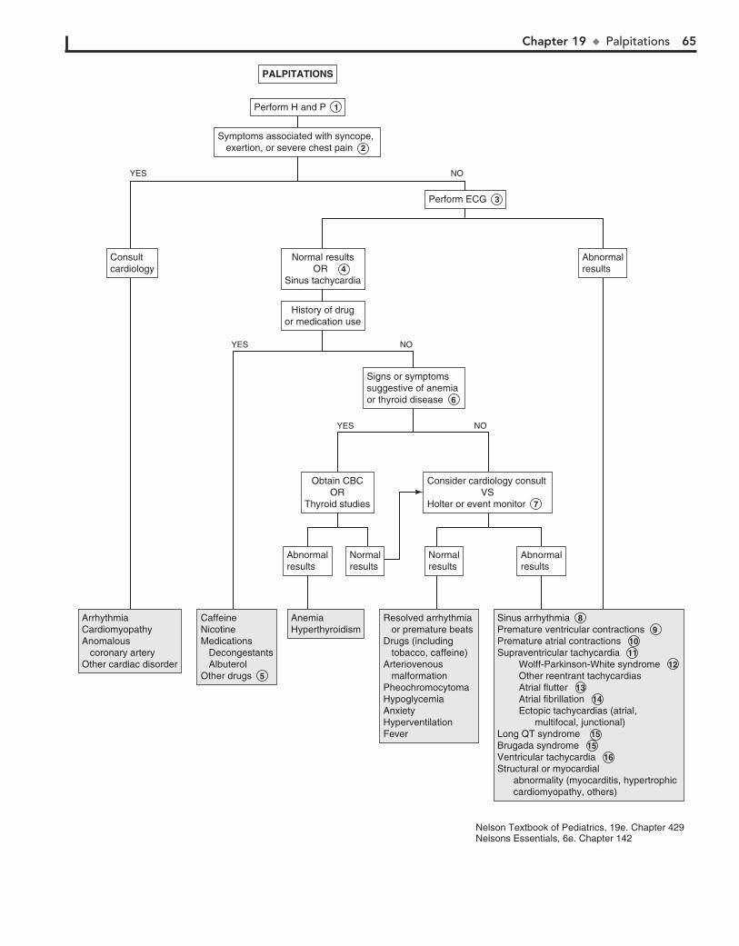
Chapter 19 u Palpitations 65
AbnormalresultsAbnormalresults
Perform ECG 3 Perform ECG 3
AbnormalresultsAbnormalresults
AbnormalresultsAbnormalresults
NormalresultsNormalresults
NormalresultsNormalresults
Consider cardiology consult VSHolter or event monitor 7
Consider cardiology consult VSHolter or event monitor 7
Obtain CBCOR
Thyroid studies
Obtain CBCOR
Thyroid studies
Signs or symptomssuggestive of anemia or thyroid disease 6
Signs or symptomssuggestive of anemia or thyroid disease 6
History of drugor medication use
History of drugor medication use
Normal results OR 4
Sinus tachycardia
Normal results OR 4
Sinus tachycardia
ConsultcardiologyConsultcardiology
Perform H and P 1 Perform H and P 1
Symptoms associated with syncope, exertion, or severe chest pain 2
Symptoms associated with syncope, exertion, or severe chest pain 2
PALPITATIONSPALPITATIONS
YES NO
YES NO
YES NO
ArrhythmiaCardiomyopathyAnomalous coronary arteryOther cardiac disorder
ArrhythmiaCardiomyopathyAnomalous coronary arteryOther cardiac disorder
CaffeineNicotineMedications Decongestants AlbuterolOther drugs 5
CaffeineNicotineMedications Decongestants AlbuterolOther drugs 5
AnemiaHyperthyroidismAnemiaHyperthyroidism
Resolved arrhythmia or premature beatsDrugs (including tobacco, caffeine)Arteriovenous malformationPheochromocytomaHypoglycemiaAnxietyHyperventilationFever
Resolved arrhythmia or premature beatsDrugs (including tobacco, caffeine)Arteriovenous malformationPheochromocytomaHypoglycemiaAnxietyHyperventilationFever
Sinus arrhythmia 8 Premature ventricular contractions 9 Premature atrial contractions 10 Supraventricular tachycardia 11 Wolff-Parkinson-White syndrome 12 Other reentrant tachycardias Atrial flutter 13 Atrial fibrillation 14 Ectopic tachycardias (atrial, multifocal, junctional) Long QT syndrome 15 Brugada syndrome 15 Ventricular tachycardia 16 Structural or myocardial abnormality (myocarditis, hypertrophic cardiomyopathy, others)
Sinus arrhythmia 8 Premature ventricular contractions 9 Premature atrial contractions 10 Supraventricular tachycardia 11 Wolff-Parkinson-White syndrome 12 Other reentrant tachycardias Atrial flutter 13 Atrial fibrillation 14 Ectopic tachycardias (atrial, multifocal, junctional) Long QT syndrome 15 Brugada syndrome 15 Ventricular tachycardia 16 Structural or myocardial abnormality (myocarditis, hypertrophic cardiomyopathy, others)
Nelson Textbook of Pediatrics, 19e. Chapter 429Nelsons Essentials, 6e. Chapter 142
66
Chapter 20
HEART MURMURS 2 Noncardiac disorders that can cause a systolic outflow
murmur include anemia, hyperthyroidism, and arterio-venous malformations (AVM). Hyperthyroidism is suggested by thyromegaly, tachycardia, a hyperdynamic precordium, slightly bounding pulses, and mild hypertension. Except for the thyromegaly, an AVM can cause similar findings; the ex-amination may reveal localized continuous bruits over the head, neck, or liver. Pulmonary AVMs are generally not as-sociated with bruits or murmurs. Anemia may manifest as pallor, tachycardia, exercise intolerance, or weakness.
3 A family history of sudden death or known hypertrophic cardiomyopathy is also significant and mandates further
evaluation. Other physical examination findings that are worri-some include an abnormal rhythm, suprasternal thrill, promi-nent apical thrust, digital clubbing, wide or bounding pulses, and absent or weak femoral pulses. Signs of systemic disease (e.g., arthritis, fever, night sweats, embolic phenomena) should prompt consideration of acquired conditions, such as rheu-matic heart disease or endocarditis.
4 When the diagnosis of a murmur is unclear, referral to a pediatric cardiologist is recommended; the severity of the
clinical picture should determine the urgency of the referral. Discussion with a cardiologist may guide a clinician in obtain-ing studies (ECG, chest x-ray, echocardiogram) prior to a con-sultation visit, although postponing studies until a scheduled consultation will generally be more cost-effective, especially in an asymptomatic patient.
5 Rheumatic fever is an immunologically mediated inflamma-tory disorder following infection with group A streptococcus
(GAS). 50% to 60% of rheumatic fever patients will experience carditis; in those cases, endocarditis (valvulitis) is universal and pericarditis or myocarditis may also develop. Patients typically present with murmurs of aortic or mitral regurgitation. The modified Jones criteria are used for diagnosis. Evidence of recent GAS disease (serologic or microbiologic) and either one major and two minor criteria or two major criteria are necessary to make the diagnosis. A history of fevers in the presence of a new or changing heart murmur should raise the suspicion for both rheu-matic fever and endocarditis.
6 Ventricular septal defects (VSDs) are the most common congenital heart lesions. Symptoms depend on the size
of the defect and pulmonary vascular resistance. Most VSDs are small and asymptomatic and close spontaneously in the first year or two of life. The murmur is classically described as a loud, usually holosystolic murmur with a harsh or blow-ing quality and is best heard at the left sternal border; a thrill or lift may be palpable with moderate lesions. In neonates the murmur may be heard best at the apex. Small defects may have soft murmurs that become softer over time as the lesion closes. In large defects, the left-to-right shunting in-creases over the first few weeks of life as pulmonary vascular resistance falls. Clinical symptoms of congestive heart failure develop gradually over this period. Referral for definitive diagnosis is recommended.
Most normal children (50% to 90%) have an audible heart mur-mur at some point prior to school age. The challenge to the prac-titioner is to ascertain which of these murmurs warrants addi-tional evaluation. The clinical diagnosis of a normal or innocent murmur should be made only in the presence of a normal history and physical examination and characteristics consistent with a normal murmur. Despite the easy availability of echocardiogra-phy, the history and physical examination remain the accepted means of diagnosing normal murmurs. When the diagnosis of a murmur is unclear, it is generally more cost-effective to refer to a pediatric cardiologist than to order an echocardiogram.
1 The addition of preductal and postductal pulse oximetry performed in newborn nurseries is recognized as a fairly
sensitive means of early identification of critical congenital heart disease. If not recognized in the newborn nursery, serious car-diac disorders may present with heart murmurs and congestive heart failure (CHF), the symptoms of which vary with age. In young infants with CHF, the feeding history may reveal limited intake, prolonged feeding times (but brief intervals between feeds because the infant is still hungry), diaphoresis, tachypnea or dyspnea, and possibly cyanosis with feeds. In older children, exercise or exertion can be assessed by inquiring about level of activity and tolerance to extended periods of play or activity (e.g., biking, gym class, walking, climbing stairs). Inquiring about how well older children keep up with their peers or sib-lings regarding exercise tolerance may also be helpful.
A history of fevers, lethargy, and recent dental work suggests possible endocarditis. Chronic mouth breathing, snoring, or ob-structive sleep apnea may be clues to pulmonary hypertension.
A maternal history of gestational diabetes, infection, and use of certain drugs or medications may be risk factors for congenital heart disease. A family history of sudden death, rheumatic fever, sudden infant death syndrome, and structural cardiac defects in a first-degree relative may be significant. A family history of hyper-trophic cardiomyopathy is sufficient to mandate an echocardio-gram because of the autosomal dominant pattern of inheritance.
When assessing the child with a murmur, a complete physical examination is essential; growth parameters, peripheral edema, and hepatomegaly can be signs of CHF or chronic disease. The cardiac examination should include assessment of pulses, palpa-tion of the precordium, auscultation, and blood pressures in both arms (involvement of a subclavian artery [most commonly the left] in a coarctation would cause a lower blood pressure in the ipsilateral arm) and a leg. Lower extremity blood pressure is usually 10-20 mmHg higher than upper extremity pressure. Di-minished femoral pulses or a delay between the radial and femoral pulses suggest coarctation of the aorta (the simple presence of a femoral pulse does not rule out coarctation).

Chapter 20 u HeartMurmurs 67
AnemiaHyperthyroidismArteriovenous malformation
AnemiaHyperthyroidismArteriovenous malformation
Rheumatic heart diseaseRheumatic heart disease Ventricular septal defect 6 Atrial septal defect 7 Coarctation of the aorta 8 Patent ductus arteriosus 9 Arteriovenous malformationPulmonary thromboembolismBacterial endocarditis 10 Acquired lesions Tricuspid insufficiency Mitral insufficiency Mitral valve prolapse Cardiomyopathy
Ventricular septal defect 6 Atrial septal defect 7 Coarctation of the aorta 8 Patent ductus arteriosus 9 Arteriovenous malformationPulmonary thromboembolismBacterial endocarditis 10 Acquired lesions Tricuspid insufficiency Mitral insufficiency Mitral valve prolapse Cardiomyopathy
Pulmonary flow murmur 11 Common innocent murmur (Still murmur) 12 Peripheral pulmonic stenosis (PPS) 13 Venous hum 14 Mammary soufflé (in pregnant or lactating women)Supraclavicular systolic murmur 15
Pulmonary flow murmur 11 Common innocent murmur (Still murmur) 12 Peripheral pulmonic stenosis (PPS) 13 Venous hum 14 Mammary soufflé (in pregnant or lactating women)Supraclavicular systolic murmur 15
Clinical picture suggestive of rheumatic fever: 5
Major clinical criteria: Minor criteria:Carditis FeverErythema marginatum ArthralgiaSubcutaneous nodules LeukocytosisChorea Elevated ESR or CRPArthritis Prolonged PR interval
Clinical picture suggestive of rheumatic fever: 5
Major clinical criteria: Minor criteria:Carditis FeverErythema marginatum ArthralgiaSubcutaneous nodules LeukocytosisChorea Elevated ESR or CRPArthritis Prolonged PR interval
Signs or symptoms suggestive of an innocent (nonpathologic) murmur:
Normal S2Early systoleQuiet precordiumShort durationLow intensity Increased intensity with fever, anxietyDecreased intensity with Valsalva maneuverNo family history of sudden death or hypertrophic cardiomyopathyNo associated symptoms
Refer tocardiology 4 Refer tocardiology 4
Signs or symptoms suggestive of a pathologic murmur 3
Exercise intoleranceSyncope with exertionCyanosisS1 and S2 abnormal or obscuredLoud or harsh (nonvibratory) murmurDiastolic, continuous, pansystolic or late systolic murmurEjection clickAbnormal pulsesOther signs/symptoms of CHF
Signs or symptoms suggestive of a pathologic murmur 3
Exercise intoleranceSyncope with exertionCyanosisS1 and S2 abnormal or obscuredLoud or harsh (nonvibratory) murmurDiastolic, continuous, pansystolic or late systolic murmurEjection clickAbnormal pulsesOther signs/symptoms of CHF
Abnormal resultsAbnormal results Normal resultsNormal results
Consider CBC, free T4,TSH ±imagingConsider CBC, free T4,TSH ±imaging
Perform H and P 1 Perform H and P 1
Signs or symptoms suggestive of noncardiac abnormality (anemia,
hyperthyroidism) 2
Signs or symptoms suggestive of noncardiac abnormality (anemia,
hyperthyroidism) 2
HEART MURMURSBEYOND THE
NEONATAL PERIOD
HEART MURMURSBEYOND THE
NEONATAL PERIOD
YES YESNO
YES NO
YES NO
NO
Nelson Textbook of Pediatrics, 19e. Chapters 176, 416, 419, 420, 421, 431Nelsons Essentials, 6e. Chapters 139, 143
(See next page forHeart Murmurs inthe Neonate)
68 Part III u Cardiology
7 Atrial septal defects (ASDs) are usually asymptomatic in infants and tend to be detected on routine physical exami-
nations in the toddler or preschooler. They are characterized by a hyperdynamic right ventricular impulse and a characteristic fixed and widely split second heart sound. Murmurs are not always audible, but large defects may manifest a mid-systolic pulmonary flow murmur at the left upper sternal border or a mid-diastolic rumble at the left mid or lower sternal border due to increased flow over the tricuspid valve. When an ASD is suspected, a cardiology evaluation is warranted to identify the type of defect and the need for monitoring versus repair.
8 Coarctation of the aorta (COA) may present in infancy with congestive heart failure and lower extremity hypoper-
fusion. Children not diagnosed in infancy can remain asymp-tomatic (even with severe coarctation) and often present with hypertension later in childhood. The classic physical findings are diminished or delayed arterial pulses in the lower extremi-ties compared to the upper extremities, with corresponding lower blood pressures in the lower extremities. A short systolic murmur at the third or fourth left intercostal spaces may be detected with transmission to the left infrascapular area or neck. A systolic ejection click or suprasternal thrill is consistent with a bicuspid aortic valve which occurs in 50% to 70% of patients with COA.
9 A small patent ductus arteriosus (PDA) in infants may be asymptomatic. Large ones tend to be symptomatic, causing
congestive heart failure and failure to thrive. Classic findings of large PDA shunts are a wide peripheral pulse pressure (bound-ing pulses), a prominent apical impulse, a systolic thrill at the left upper sternal border, and a continuous harsh machinery-like murmur heard in the left infraclavicular region and at the left upper sternal border. The murmur characteristically becomes loudest during systole and softens during diastole. Large lesions may also produce a mid-diastolic apical rumble. Referral to car-diology is indicated for definitive diagnosis and treatment. The lesion is one of the cardiac anomalies frequently associated with prenatal maternal rubella infection; it is also common in con-genital hypothyroidism.
10 Infective endocarditis can occur in children with healthy hearts but is most common in children with congenital heart
disease or rheumatic heart disease; other risk factors include any valve malformation, patients who have had cardiac surgery, im-munuosuppressed patients, patients with long-term intravascular catheters and IV drug users. Cases may present acutely or insidi-ously with intermittent fevers (classically occurring in the after-noons) and vague symptoms of fatigue, myalgias, joint pain, headache, and nausea or vomiting. New or changing murmurs, splenomegaly, and petechiae are common. Echocardiography is helpful in identifying vegetations, although results may be normal early in the disease. Adding transesophageal echocardiography to the transthoracic approach improves the diagnostic yield. Blood cultures are necessary to identify the pathogen. Three to five cul-tures from separate sites are recommended. Laboratories should be notified when endocarditis is suspected so that enriched media and prolonged incubation times are used.
11 The normal turbulence of ejection into the pulmonic root frequently generates an audible murmur of no hemody-
namic significance. These murmurs are usually grade 1 to 3, short systolic murmurs with a slightly grating (rather than vibratory) quality. They are heard best over the left upper sternal border and may or may not transmit to the neck. Clicks and thrills associated with pulmonary valve stenosis are absent, and a normal S2 distin-guishes it from an ASD. The murmur can be accentuated by full exhalation and the supine position, and diminished by the up-right posture and breath holding. It can occur in any age group (including neonates) but is most likely in older children and ado-lescents.
12 Some sources are replacing the classic “Still murmur” term with terms such as common innocent murmur, vibratory
innocent murmur, or classic vibratory murmur. These normal murmurs are common in children (most commonly 3-7 years of age). The non-radiating murmur is usually a low-grade short systolic murmur heard best at the mid to lower left sternal border and also towards the apex. It has a characteristic vibratory or musical quality; commonly used descriptions include buzzing, a vibrating tuning fork, or a twanging cello string. Rarely, it can be surprisingly loud and ominous-sounding with transmission throughout the precordium. It may be heard best with the bell of a stethoscope, is usually loudest when the patient is supine and tends to diminish with an upright or a sitting position. This mur-mur is also exacerbated by fever, anxiety, excitement, or exercise.
13 Peripheral pulmonary artery stenosis (PPS) presents with a soft mid-systolic ejection murmur caused by flow through
narrow pulmonary artery branches, which angle sharply off the main pulmonary artery. The murmur is loudest at the left upper sternal border with good transmission to the axillae and back. It generally disappears between 3 and 6 months of life as the pulmo-nary branches increase in size; persistence warrants cardiology evaluation to rule out true stenosis or constriction of the pulmonary arteries.
14 Venous hums are another common, normal murmur of childhood. These are medium frequency continuous mur-
murs heard in the infraclavicular region (right more common than left) and neck; their intensity increases slightly during di-astole. The murmur typically is heard when the patient is sitting or standing, and it diminishes or disappears in the supine posi-tion, when the patient turns his or her head far to one side, and when gentle pressure is applied to the jugular veins in the neck. They are presumed to be due to the turbulence created as the internal jugular and subclavian veins enter the superior vena cava. The main differential diagnoses are PDA and arteriove-nous malformations.
15 A supraclavicular systolic murmur (supraclavicular bruit) is a short systolic murmur heard best above the clavicles
with minimal radiation to the neck. It is distinguished from aortic or pulmonary valve stenosis by the absence of an associ-ated click and significant radiation to the neck. It is due to tur-bulence in the carotid arteries and may rarely be associated with a palpable thrill.

Chapter 20 u HeartMurmurs 69
16 Congenital heart disease usually manifests in the first few days or weeks of life, depending on the lesion. Preductal and
postductal pulse oximetry performed in newborn nurseries is identifying many cases of critical cardiac disease prior to symptom development. The development of a significant murmur in the neonatal period accompanied by cyanosis or congestive heart fail-ure warrants an urgent evaluation. Congestive heart failure may manifest as poor feeding, disinterest in feeding, excessive fatigue, diaphoresis, and tachypnea or dyspnea. In cyanotic heart defects,
BibliographyMenashe V: Heart murmurs. Pediatr Rev 28(4):e19–22, 2007. Park MK: Physical examination. In Pediatric cardiology for practitioners,
St. Louis, 2008, Mosby.
a sudden deterioration in the first few days of life occurs coincident with the closing of the ductus. (See Chapter 14.) Many syndromes (e.g., Down, Turner, Williams) have characteristic cardiac anomalies.
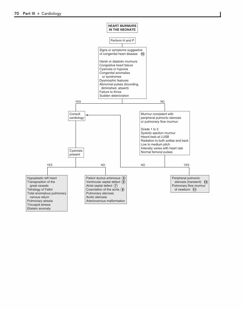
70 Part III u Cardiology
Hypoplastic left heartTransposition of the great vesselsTetralogy of FallotTotal anomalous pulmonary venous returnPulmonary atresiaTricuspid atresiaEbstein anomaly
Hypoplastic left heartTransposition of the great vesselsTetralogy of FallotTotal anomalous pulmonary venous returnPulmonary atresiaTricuspid atresiaEbstein anomaly
Patent ductus arteriosus 9 Ventricular septal defect 6 Atrial septal defect 7 Coarctation of the aorta 8 Pulmonary stenosisAortic stenosisArteriovenous malformation
Patent ductus arteriosus 9 Ventricular septal defect 6 Atrial septal defect 7 Coarctation of the aorta 8 Pulmonary stenosisAortic stenosisArteriovenous malformation
Peripheral pulmonic stenosis (transient) 13 Pulmonary flow murmur of newborn 11
Peripheral pulmonic stenosis (transient) 13 Pulmonary flow murmur of newborn 11
CyanosispresentCyanosispresent
ConsultcardiologyConsultcardiology
Murmur consistent withperipheral pulmonic stenosis or pulmonary flow murmur:
Grade 1 to 2Systolic ejection murmurHeard best at LUSB Radiation to both axillae and backLow to medium pitchIntensity varies with heart rateNormal femoral pulses
Murmur consistent withperipheral pulmonic stenosis or pulmonary flow murmur:
Grade 1 to 2Systolic ejection murmurHeard best at LUSB Radiation to both axillae and backLow to medium pitchIntensity varies with heart rateNormal femoral pulses
Perform H and PPerform H and P
Signs or symptoms suggestive of congenital heart disease: 16
Harsh or diastolic murmursCongestive heart failureCyanosis or hypoxiaCongenital anomalies or syndromesDysmorphic featuresAbnormal pulses (bounding, diminished, absent)Failure to thriveSudden deterioration
Signs or symptoms suggestive of congenital heart disease: 16
Harsh or diastolic murmursCongestive heart failureCyanosis or hypoxiaCongenital anomalies or syndromesDysmorphic featuresAbnormal pulses (bounding, diminished, absent)Failure to thriveSudden deterioration
HEART MURMURSIN THE NEONATEHEART MURMURSIN THE NEONATE
YES NO
YES NO
YESNO

Gastrointestinal System PART
IV
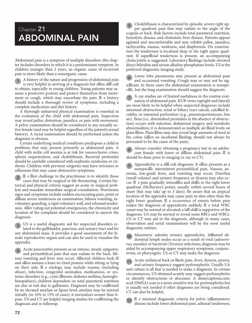
72
Chapter 21
ABDOMINAL PAIN
5 Cholelithiasis is characterized by episodic severe right up-per quadrant pain that may radiate to the angle of the
scapula or back. Risk factors include total parenteral nutrition, hemolytic disease, and cholestatic liver disease. Patients appear agitated and uncomfortable and may exhibit pallor, jaundice, tachycardia, nausea, weakness, and diaphoresis. On examina-tion the tenderness is localized deep in the right upper quad-rant. If superficial tenderness is present, an accompanying cholecystitis is suggested. Laboratory findings include elevated direct bilirubin and serum alkaline phosphatase levels. US is the preferred diagnostic imaging study.
6 Lower lobe pneumonia may present as abdominal pain and occasional vomiting. Cough may or may not be sig-
nificant. In these cases the abdominal examination is nonspe-cific, but the lung examination should suggest the diagnosis.
7 X-ray studies are of limited usefulness in the routine eval-uation of abdominal pain. KUB views (upright and lateral)
are most likely to be helpful when suspected diagnoses include intestinal obstruction, renal or biliary tract calculi, calcified fe-caliths, or intestinal perforation (e.g., pneumoperitoneum, free air). Ileus (i.e., diminished peristalsis in the absence of obstruc-tion) occurs with infection, abdominal surgery, and metabolic abnormalities; it is demonstrated as multiple air-fluid levels on plain films. Plain films may also reveal large amounts of stool in the colon (often an incidental finding; should not always be presumed to be the cause of the pain).
8 Always consider obtaining a pregnancy test in an adoles-cent female with lower or diffuse abdominal pain. This
should be done prior to imaging (x-ray or CT).
9 Appendicitis is a difficult diagnosis. It often presents as a nonspecific intermittent periumbilical pain. Nausea, an-
orexia, low-grade fever, and vomiting may occur. Diarrhea (small volume) and urinary frequency or dysuria may also oc-cur. The pain gradually intensifies and shifts to the right lower quadrant (McBurney’s point), usually within several hours of onset (but may take up to 3 days). Be aware that an atypical location of the appendix may cause pain in sites other than the right lower quadrant. The occurrence of emesis before pain makes the diagnosis of appendicitis unlikely. The total WBC count may be normal or elevated; a left shift is supportive of the diagnosis. UA may be normal or reveal some RBCs and WBCs. US or CT may aid in the diagnosis, although in many cases, observation and serial examinations will be the most useful diagnostic entities.
10 Mesenteric adenitis mimics appendicitis. Inflamed ab-dominal lymph nodes occur as a result of viral (adenovi-
rus, measles) or bacterial (Yersinia) infections; diagnosis may be aided by accompanying upper respiratory symptoms, conjunc-tivitis, or pharyngitis. US or CT may make the diagnosis.
11 Acute unilateral back or flank pain, fever, dysuria, pyuria, and urinary frequency suggest pyelonephritis. Usually UA
and culture is all that is needed to make a diagnosis. In certain circumstances, US obtained acutely may suggest pyelonephritis or identify obstructions or abscesses. A dimercaptosuccinic acid (DMSA) scan is a more sensitive test for pyelonephritis but is usually not needed if other diagnoses are being considered. CT can also be helpful.
12 The minimal diagnostic criteria for pelvic inflammatory disease include lower abdominal pain, adnexal tenderness,
Abdominal pain is a symptom of multiple disorders; this chap-ter includes disorders in which it is a predominant symptom. In children younger than 2 years, an organic cause of recurrent pain is more likely than a nonorganic cause.
1 A history of the nature and progression of abdominal pain is very helpful in arriving at a diagnosis but often difficult
to obtain, especially in young children. Young patients may as-sume a protective posture and protect themselves from move-ment or cough, which may exacerbate the pain. The history should include a thorough review of symptoms, including a complete medication and diet history.
A thorough unhurried physical examination is essential in the evaluation of the child with abdominal pain. Inspection may reveal pallor, distention, jaundice, or pain with movement. A pelvic examination should be considered in any sexually ac-tive female (and may be helpful regardless of the patient’s sexual history). A rectal examination should be performed unless the diagnosis is obvious.
Certain underlying medical conditions predispose a child to problems that may present primarily as abdominal pain. A child with sickle cell anemia is at risk for vasoocclusive crises, splenic sequestration, and cholelithiasis. Bacterial peritonitis should be carefully considered with nephrotic syndrome or cir-rhosis. Children with previous surgeries may have strictures or adhesions that may cause obstructive symptoms.
2 The first challenge to the practitioner is to identify those cases that may be surgical or life threatening. Certain his-
torical and physical criteria suggest an acute or surgical prob-lem and mandate immediate surgical consultation. Worrisome signs and symptoms include sudden excruciating pain, point or diffuse severe tenderness on examination, bilious vomiting, in-voluntary guarding, a rigid voluntary wall, and rebound tender-ness. After ruling out potential emergencies, the chronicity and location of the complaint should be considered to narrow the diagnosis.
3 US is a useful diagnostic aid for suspected disorders re-lated to the gallbladder, pancreas, and urinary tract and for
any abdominal mass. It provides a good assessment of the fe-male reproductive organs and can also be used to visualize the appendix.
4 Acute pancreatitis presents as an intense, steady, epigastric and periumbilical pain that may radiate to the back. Bil-
ious vomiting and fever may occur. Affected children look ill and often assume a knee-to-chest posture while sitting or lying on their side. The etiology may include trauma (including abuse), infection, congenital anomalies, medications, or sys-temic disorders (e.g., cystic fibrosis, diabetes mellitus, hemoglo-binopathies); children dependent on total parenteral nutrition are also at risk due to gallstones. Diagnosis may be confirmed by an elevated amylase or lipase level; amylase may be normal initially (in 10% to 15% of cases); it normalizes sooner than li-pase. US and CT are helpful imaging studies for confirming the diagnosis and in followup.
Chapter 21 u AbdominalPain 73
Referred pain(back, groin orshoulder)
Referred pain(back, groin orshoulder)
Consider: Ultrasound Amylase Lipase LFTs Chest x-ray
Consider: Ultrasound 3 Amylase Lipase LFTs Chest x-ray
Upper abdomenUpper abdomen
Location of painLocation of painConsult surgeryConsult surgery
Consider: KUB Ultrasound CT Chest x-ray
Consider: KUB Ultrasound 3 CT Chest x-ray
Diffuse,periumbilicalDiffuse,periumbilical
Consider: Urinalysis CBC KUB Ultrasound CT Chest x-ray ±Urine pregnancy test ±Pelvic exam
Consider: Urinalysis CBC KUB 7 Ultrasound 3 CT Chest x-ray ±Urine pregnancy test 8 ±Pelvic exam
Lower abdomenLower abdomen
ACUTE ABDOMINAL PAINACUTE ABDOMINAL PAIN
Perform Perform H and P 1
Signs or symptomssuggestive of an acuteabdomen present
Signs or symptomssuggestive of an acuteabdomen present 2
HepatitisAcute pancreatitis Cholecystitis/ cholelithiasis Fitz-Hugh-Curtis syndromeSub-diaphragmatic abscessPneumonia Splenic hemorrhage or trauma
HepatitisAcute pancreatitis 4 Cholecystitis/ cholelithiasis 5Fitz-Hugh-Curtis syndromeSub-diaphragmatic abscessPneumonia 6Splenic hemorrhage or trauma
AppendicitisConstipationMesenteric adenitisUTI/pyelonephritisPelvic inflammatory diseasePneumoniaPregnancy (ectopic, intrauterine)Intestinal obstructionIncarcerated herniaUrolithiasisPsoas abscess
Appendicitis 9 ConstipationMesenteric adenitis 10 UTI/pyelonephritis 11 Pelvic inflammatory disease 12 Pneumonia 6 Pregnancy (ectopic, intrauterine)Intestinal obstruction 13 Incarcerated herniaUrolithiasis 14 Psoas abscess
Intestinal perforation (ruptured appendicitis, peptic ulcer)PeritonitisIntussusceptionVolvulusRuptured spleenEctopic pregnancyOvarian or testicular torsionStrangulated herniaRuptured aortic aneurysm
Intestinal perforation (ruptured appendicitis, peptic ulcer)PeritonitisIntussusceptionVolvulusRuptured spleenEctopic pregnancyOvarian or testicular torsionStrangulated herniaRuptured aortic aneurysm
NOYES
ConstipationGastroenteritisEnterocolitisMesenteric adenitisTrauma (abuse)PneumoniaAppendicitisIntestinal obstructionAerophagiaBacterial peritonitisFood poisoningHenoch-Schönlein purpura
ConstipationGastroenteritis 15 EnterocolitisMesenteric adenitis 10 Trauma (abuse)Pneumonia Appendicitis 9 Intestinal obstruction 13 AerophagiaBacterial peritonitisFood poisoningHenoch-Schönlein purpura
PyelonephritisUrolithiasisCholelithiasis/cholecystitisPancreatitisSplenic hemorrhage or trauma
Pyelonephritis 11 Urolithiasis 14 Cholelithiasis/cholecystitis 5 Pancreatitis 4 Splenic hemorrhage or trauma
Consider: Urinalysis Ultrasound CT Amylase Lipase LFTs Chest x-ray
Consider: Urinalysis Ultrasound 3 CT Amylase Lipase LFTs Chest x-ray
Nelson Textbook of Pediatrics, 19e. Chapters 298, 334Nelsons Essentials, 6e. Chapter 126
74 Part IV u GastrointestinalSystem
or cervical motion tenderness. Additional diagnostic criteria include fever (.38.3°C [101°F]), abnormal cervical or vaginal discharge, elevated CRP or ESR, and positive cervical cultures for Neisseria gonorrhoeae or Chlamydia trachomatis. The role of US is not so much for definitive diagnosis as to identify tubo-ovarian abscesses, a common complication of pelvic inflamma-tory disease.
occasionally vomiting. One needs to rule out other diseases such as gastritis, ulcer, and eosinophilic gastroenteritis. Diagno-sis is based on the presence of these symptoms of peptic ulcer disease in the presence of normal findings on upper endoscopy.
21 Inflammatory bowel disease is an important diagnosis to exclude when faced with the child with chronic abdominal
pain. A thorough evaluation, including H and P and screening laboratory studies, will often suggest the disorder. Additional signs and symptoms that suggest inflammatory bowel disease are anorexia, growth failure, perianal disease, hematochezia, and diarrhea. The pain and diarrhea may awaken the child at night. Supportive laboratory results include anemia, increased ESR or CRP, thrombocytosis, hypoalbuminemia, heme-positive stools, and elevated stool calprotectin levels. Contrast medium studies (UGI series with small bowel follow-through) and bar-ium enema may be helpful, although MRI enterography is often used because of concerns about radiation. CT with oral contrast can be helpful if an abscess is suspected. Endoscopy should be done for definitive diagnosis.
22 Celiac disease, or gluten-sensitive enteropathy, is becom-ing increasingly recognized as a cause of chronic abdomi-
nal pain. There is inflammation of the small intestine due to exposure to dietary gluten. It classically presents with diarrhea, steatorrhea, anemia, abdominal distention, failure to thrive, and often with nonspecific abdominal complaints. The tissue trans-glutaminase (tTG) antibody enzyme-linked immunoassay is a good screening test for celiac disease.
23 GER esophagitis in older children presents as substernal pain, increased pain after meals or when recumbent, and
relief from pain with use of antacids.
24 Peptic ulcer disease includes gastric and duodenal ulcers, gastritis, and duodenitis. Unlike the classic adult presenta-
tion of epigastric pain, exacerbation with meals, and early morning occurrence, in children the pain may be more diffuse (epigastric or periumbilical) and unrelated to meals or time of day. If clinical presentation is consistent with the diagnosis, improvement with a trial of therapy is often diagnostic. If symp-toms do not respond to treatment, a search for Helicobacter pylori with a urea breath test, serum antibodies, or stool anti-gens may be indicated. However, endoscopy with biopsy is the most reliable method.
25 Chronic pancreatitis is a rare cause of recurrent abdominal pain in children. Children experience intermittent epigas-
tric abdominal pain, often with associated nausea and vomit-ing; symptoms are frequently precipitated by a large meal or stress. Serum lipase and amylase levels are not as likely to be elevated as with acute cases. US and CT may aid in diagnosis; x-ray studies may reveal calcifications consistent with chronic pancreatitis.
13 In intestinal obstruction, vomiting is usually a predomi-nant symptom. Pain is a later finding and is crampy or
colicky, and rushes of high-pitched “tinkling” bowel sounds may be noted. Plain x-ray films (KUB view) may reveal air-fluid levels or distended bowel loops above the obstruction; free air indicates an intestinal perforation. Contrast medium studies may aid in definitive diagnosis if malrotation or volvulus, distal small bowel obstruction, or intussusception is suspected.
14 Urolithiasis (i.e., kidney stones) presents as hematuria and acute colicky abdominal, flank, or back pain. The pain may
radiate to the upper leg and groin. US and helical CT will detect both radiopaque and radiolucent stones.
15 When acute gastroenteritis is suggested based on a clinical presentation of vomiting and diarrhea preceding the com-
plaint of diffuse abdominal pain and in the absence of any signs or symptoms of an acute abdomen, no additional workup is indicated. Parents should be counseled about worrisome signs and symptoms and supportive measures.
16 Chronic or recurrent abdominal pain is defined by recur-rent or persistent bouts of pain that occur over a minimum
of 3 months and may or may not interfere with daily activities. Chronic pain may be organic, nonorganic, or psychogenic.
17 If the diagnosis is in doubt, an abdominal film may help. Constipation is the most common cause of chronic and
recurrent abdominal pain in children. It causes colonic disten-sion and painful defecation. It may be functional or organic. However, presence of stool does not always mean the pain is from constipation.
18 Elements of the H and P examination that suggest an or-ganic cause of abdominal pain include fever, weight loss or
growth deceleration, joint symptoms, emesis (especially if blood or bile-stained), abnormal findings on physical examina-tion (e.g., abdominal mass, perianal disease), and blood in the stool or other abnormal results of laboratory studies. An or-ganic cause should be considered for pain or diarrhea that awakens a child from sleep, pain that is well localized away from the umbilicus, and pain that is referred to the back, flank, or shoulders.
19 Functional abdominal pain is the most common diagnosis after organic causes have been ruled out. It describes a
pain that does not have a clear structural or biochemical basis but is recognized as genuine pain. Characteristics include onset at age older than 5 years, intermittent or episodic nature, peri-umbilical location, and a lack of association with activity, meals, or bowel pattern. The physical examination is always normal (although patients may appear tired or pale during episodes), and results of laboratory studies are normal. The entity remains a common yet poorly explained affliction of childhood.
20 Functional dyspepsia describes pain and discomfort in the upper abdomen. It is characterized by epigastric pain ac-
companied by early satiety, bloating, belching, and nausea or
26 Irritable bowel syndrome is characterized by abdominal pain or discomfort and a variable defecation pattern.
Bloating, flatulence, and mucus may be present. Either diarrhea or constipation may predominate in the disorder; the abdomi-nal pain is usually relieved by defecation.
27 Lactose malabsorption causes symptoms of abdominal pain and cramping, bloating, diarrhea, and excess flatulence. In
primary adult-type hypolactasia, symptoms may not develop until 3 to 5 years of age when lactase levels begin to decline. Breath hy-drogen testing after an oral lactose load will make the diagnosis,

Chapter 21 u AbdominalPain 75
Consider: CBC ESR/CRP Amylase Lipase LFTs KUB US/CT/MRI UGI/SBFT Endoscopy
Consider: CBC ESR/CRP Amylase Lipase LFTs KUB US/CT/MRI UGI/SBFT Endoscopy
Non-specific signsand symptomsNon-specific signsand symptoms
Consider: CBC ESR/CRP Albumin Tissue transglutaminase Stool for occult blood Calprotectin KUB UGI/SBFT Endoscopy
Consider: CBC ESR/CRP Albumin Tissue transglutaminase Stool for occult blood Calprotectin KUB UGI/SBFT Endoscopy
Consider: CBC ESR/CRP Tissue transglutaminase Stool studies: -O and P -Giardia and Crypto antigen -Occult blood -Calprotectin -Stool culture Ultrasound/CT UGI/SBFT Colonoscopy MRI enterography Endoscopy
Consider: CBC ESR/CRP Tissue transglutaminase Stool studies: -O and P -Giardia and Crypto antigen -Occult blood -Calprotectin -Stool culture Ultrasound/CT UGI/SBFT Colonoscopy MRI enterography Endoscopy
Consider: Amylase Lipase Ultrasound/ CT UGI/SBFT Endoscopy
Consider: Amylase Lipase Ultrasound/ CT UGI/SBFT Endoscopy
Weight loss±DiarrheaWeight loss±Diarrhea
Go to Chapter 24 on ConstipationGo to Chapter 24 on ConstipationConsider: Urinalysis CBC ESR ALT, amylase, lipase
Consider: Urinalysis CBC ESR ALT, amylase, lipase
Signs and symptoms ofan organic etiology presentSigns and symptoms ofan organic etiology present 18
Distension,loose stools,diarrhea
Distension,loose stools,diarrhea
Aggravated orrelieved byeating
Aggravated orrelieved byeating
ABDOMINAL PAIN(CHRONIC OR RECURRENT) 16
ABDOMINAL PAIN(CHRONIC OR RECURRENT) 16
Perform Perform H and P
Signs and symptoms suggestive of constipationSigns and symptoms suggestive of constipation 17
Inflammatory bowel diseaseNeoplasmEosinophilic gastroenteritisCeliac disease
Inflammatory bowel disease 21 NeoplasmEosinophilic gastroenteritisCeliac disease 22
GER esophagitisPeptic ulcer diseaseFunctional dyspepsiaChronic pancreatitis
GER esophagitis 23 Peptic ulcer disease 24 Functional dyspepsia 20 Chronic pancreatitis 25
NOYES
NO YES
Inflammatory bowel diseaseIrritable bowel syndromeLactose intoleranceCeliac diseaseParasites (Giarda, Cryptosporidium)Yersenia and other bacteriaAmoebiasis C. difficile
Inflammatory bowel disease 21Irritable bowel syndrome 26 Lactose intolerance 27 Celiac disease 22Parasites (Giarda, Cryptosporidium)Yersenia and other bacteriaAmoebiasis C. difficile
DysmenorrheaMittelschmerzHematocolposEndometriosis
DysmenorrheaMittelschmerzHematocolposEndometriosis
Associatedwith menstrualcycle
Associatedwith menstrualcycle
ConstipationIrritable bowel syndromeEosinophilic esophagitisPeptic ulcer diseaseOvarian massesMalrotation with intermittent volvulusHydronephrosis/ UPJ obstructionChronic pancreatitisMeckel’s diverticulumAbdominal migraineAbdominal epilepsyLead poisoningParasites (Giardia)Chronic hepatitisHepatobiliary or pancreatic disordersFamilial Mediterranean feverNephrolithiasisPorphyriasHereditary angioedemaNeoplasmTrauma, tumor, infection of vertebrae
Constipation 17 Irritable bowel syndrome 26 Eosinophilic esophagitisPeptic ulcer disease 24 Ovarian massesMalrotation with intermittent volvulus 28 Hydronephrosis/ UPJ obstruction 29 Chronic pancreatitis 25Meckel’s diverticulumAbdominal migraineAbdominal epilepsyLead poisoningParasites (Giardia)Chronic hepatitisHepatobiliary or pancreatic disordersFamilial Mediterranean feverNephrolithiasisPorphyrias 30 Hereditary angioedemaNeoplasmTrauma, tumor, infection of vertebrae
Functional abdominal painFunctional dyspepsia
Functional abdominal pain 19 Functional dyspepsia 20
76 Part IV u GastrointestinalSystem
the groin or flank. In older children the disorder most commonly occurs in males and is on the left side. Symptoms tend to pre-dominate during periods of fluid loading and diuresis. US is recommended if obstruction is suspected.
30 Acute intermittent porphyria is the most common of the porphyrias. It generally presents as abdominal pain; periph-
eral neuropathies are also common. In severe cases the urine may turn a port wine color. The diagnosis is made by demonstrating decreased porphobilinogen deaminase in erythrocytes and in-creased urinary levels of aminolevulinic acid and porphobilinogen.
although resolution of symptoms after dietary restriction of lactose is strongly suggestive. Malabsorption of fructose and sorbitol may also cause symptoms of GI distress; a history of high fruit juice ingestion suggests the former, and frequent use of “sugar-free” products (sorbitol-containing) should raise suspicion of the latter.
28 A UGI series with small bowel follow-through is required to diagnose most cases of malrotation.
29 Ureteropelvic junction obstruction is an uncommon disor-der, often presenting as abdominal pain in children and
adolescents. Diagnosis in infants is often assisted by the presence of an abdominal mass or occurrence of a UTI. In older children, the physical examination and UA may be normal or may reveal a unilateral abdominal mass or hematuria. The chief complaint in over 70% of children older than 6 years with ureteropelvic junc-tion obstruction is abdominal pain that is frequently referred to
BibliographyMahajan LA, Kaplan B: Chronic abdominal pain of childhood and adolescence.
In Wyllie R, Hyams J, editors: Pediatric gastrointestinal and liver disease, ed 4, Philadelphia, 2010, WB Saunders, pp 64.
Ross A, LeLeiko NS: Acute abdominal pain, Pediatr Rev 31:135–144, 2010.
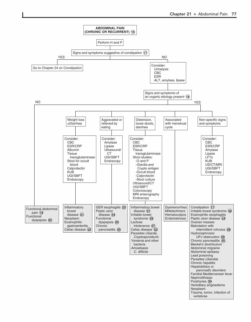
Chapter 21 u AbdominalPain 77
Consider: CBC ESR/CRP Amylase Lipase LFTs KUB US/CT/MRI UGI/SBFT Endoscopy
Consider: CBC ESR/CRP Amylase Lipase LFTs KUB US/CT/MRI UGI/SBFT Endoscopy
Non-specific signsand symptomsNon-specific signsand symptoms
Consider: CBC ESR/CRP Albumin Tissue transglutaminase Stool for occult blood Calprotectin KUB UGI/SBFT Endoscopy
Consider: CBC ESR/CRP Albumin Tissue transglutaminase Stool for occult blood Calprotectin KUB UGI/SBFT Endoscopy
Consider: CBC ESR/CRP Tissue transglutaminase Stool studies: -O and P -Giardia and Crypto antigen -Occult blood -Calprotectin -Stool culture Ultrasound/CT UGI/SBFT Colonoscopy MRI enterography Endoscopy
Consider: CBC ESR/CRP Tissue transglutaminase Stool studies: -O and P -Giardia and Crypto antigen -Occult blood -Calprotectin -Stool culture Ultrasound/CT UGI/SBFT Colonoscopy MRI enterography Endoscopy
Consider: Amylase Lipase Ultrasound/ CT UGI/SBFT Endoscopy
Consider: Amylase Lipase Ultrasound/ CT UGI/SBFT Endoscopy
Weight loss±DiarrheaWeight loss±Diarrhea
Go to Chapter 24 on ConstipationGo to Chapter 24 on ConstipationConsider: Urinalysis CBC ESR ALT, amylase, lipase
Consider: Urinalysis CBC ESR ALT, amylase, lipase
Signs and symptoms ofan organic etiology presentSigns and symptoms ofan organic etiology present 18
Distension,loose stools,diarrhea
Distension,loose stools,diarrhea
Aggravated orrelieved byeating
Aggravated orrelieved byeating
ABDOMINAL PAIN(CHRONIC OR RECURRENT) 16
ABDOMINAL PAIN(CHRONIC OR RECURRENT) 16
Perform Perform H and P
Signs and symptoms suggestive of constipationSigns and symptoms suggestive of constipation 17
Inflammatory bowel diseaseNeoplasmEosinophilic gastroenteritisCeliac disease
Inflammatory bowel disease 21 NeoplasmEosinophilic gastroenteritisCeliac disease 22
GER esophagitisPeptic ulcer diseaseFunctional dyspepsiaChronic pancreatitis
GER esophagitis 23 Peptic ulcer disease 24 Functional dyspepsia 20 Chronic pancreatitis 25
NOYES
NO YES
Inflammatory bowel diseaseIrritable bowel syndromeLactose intoleranceCeliac diseaseParasites (Giarda, Cryptosporidium)Yersenia and other bacteriaAmoebiasis C. difficile
Inflammatory bowel disease 21Irritable bowel syndrome 26 Lactose intolerance 27 Celiac disease 22Parasites (Giarda, Cryptosporidium)Yersenia and other bacteriaAmoebiasis C. difficile
DysmenorrheaMittelschmerzHematocolposEndometriosis
DysmenorrheaMittelschmerzHematocolposEndometriosis
Associatedwith menstrualcycle
Associatedwith menstrualcycle
ConstipationIrritable bowel syndromeEosinophilic esophagitisPeptic ulcer diseaseOvarian massesMalrotation with intermittent volvulusHydronephrosis/ UPJ obstructionChronic pancreatitisMeckel’s diverticulumAbdominal migraineAbdominal epilepsyLead poisoningParasites (Giardia)Chronic hepatitisHepatobiliary or pancreatic disordersFamilial Mediterranean feverNephrolithiasisPorphyriasHereditary angioedemaNeoplasmTrauma, tumor, infection of vertebrae
Constipation 17 Irritable bowel syndrome 26 Eosinophilic esophagitisPeptic ulcer disease 24 Ovarian massesMalrotation with intermittent volvulus 28 Hydronephrosis/ UPJ obstruction 29 Chronic pancreatitis 25Meckel’s diverticulumAbdominal migraineAbdominal epilepsyLead poisoningParasites (Giardia)Chronic hepatitisHepatobiliary or pancreatic disordersFamilial Mediterranean feverNephrolithiasisPorphyrias 30 Hereditary angioedemaNeoplasmTrauma, tumor, infection of vertebrae
Functional abdominal painFunctional dyspepsia
Functional abdominal pain 19 Functional dyspepsia 20
78
Chapter 22
VOMITING
7 Superior mesenteric artery syndrome describes a condi-tion of transient duodenal obstruction due to a trapping or
compression in the duodenum by the superior mesenteric ar-tery anteriorly and the aorta posteriorly. Clinically, bilious vomiting and epigastric pain occur and are relieved by a prone or knee-chest position. The condition is most commonly seen in cases of recent weight loss, lordosis, prolonged bed rest, or body casting.
8 In a child with acute-onset, large-volume emesis, fever, and/or diarrhea—a picture consistent with acute gastroen-
teritis—additional workup may not be necessary. With rotavi-rus in particular, vomiting often precedes diarrhea by 1 to 2 days. In other cases of acute severe vomiting, laboratory tests and a UA may aid in the assessment of dehydration and possible diagnosis. Further studies should be ordered based on sus-pected diagnoses.
9 Pyloric stenosis presents as nonbilious vomiting in the first few weeks of life and progresses in frequency and intensity.
Clinical characteristics include projectile vomiting, later onset of “coffee ground” emesis (hematemesis), and poor weight gain. Patients are often dehydrated, with a metabolic alkalosis and hypochloremia by the time they present. Diagnosis is by physi-cal examination and by US or a UGI study that shows the “string sign” of contrast medium through the narrowed pylorus.
10 Sinusitis may cause an acute, chronic, or cyclic pattern of vomiting. Associated nausea, congestion, postnasal drip, and
early morning occurrence often preceded by coughing suggest the diagnosis.
11 Mental status changes and abnormal respirations may indicate ingestion of a toxic drug or poison.
12 Ureteropelvic junction obstruction results in hydronephrosis during fluid loading and diuresis. Congenital cases appear as
an abdominal mass or UTI. Older children tend to present with intermittent abdominal or flank pain and often with vomiting. The physical examination and urinalysis may be normal or may reveal a unilateral abdominal mass or hematuria. A history of spontaneous resolution after several hours because of relief of the renal pelvic distention as dehydration develops is suggestive. US during an acute episode or after furosemide or an IV pyelogram should help provide the diagnosis.
13 Most inborn errors of metabolism appear early in the new-born period with vomiting and failure to thrive. Some
disorders occur as chronic or cyclic vomiting at later ages after the addition of certain foods to the diet or in the context of acute stresses or illnesses. These children may experience acute intermittent episodes of vomiting accompanied by acidosis, mental deterioration, and coma. There may be a family history of the disorder or of unexplained mental retardation, failure to thrive, or neonatal deaths.
Consider a metabol ic workup whenever neurologic symp-toms (e.g., altered mental status, hypotonia, seizures, unex-plained mental retardation), hepatosplenomegaly, or unusual odors (e.g., from breath, urine, ear wax) are present. For a meta-bolic workup, blood and urine should be obtained during epi-sodes of suggestive symptoms. Blood tests should include a CBC, electrolytes, pH, glucose, ammonia, lactate, carnitine, acyl carnitine, and serum amino acids. Urine should be analyzed for ketones, reducing substances, organic acids, and amino acids.
True vomiting is a forceful ejection of stomach or esophageal contents from the mouth. It is often accompanied by nausea and retching. Regurgitation is an effortless or near effortless ejection and is not true emesis. Rumination is regurgitation with rechewing of food.
1 Vomiting should be approached by first identifying the pattern of vomiting. The review of systems should
include other abdominal, respiratory, and neurologic com-plaints. Inquire about diet and medication use. In cases of chronic recurrent vomiting, the frequency is generally greater than two episodes; children are generally not acutely ill and vomit with a low intensity. In cyclic recurrent vomiting, epi-sodes are infrequent (#2/week) but are characterized by acute severity and illness and forceful vomiting occurring at a high frequency (i.e., .4 to 6 times/h). Autonomic signs and symp-toms such as pallor, lethargy, nausea, and abdominal pain are frequently associated. Initially, chronic and cyclic vomiting may appear to be acute problems until the pattern becomes evident. It is important to exclude pregnancy in a sexually active adolescent female.
2 When acute vomiting occurs in the context of an acute abdomen, immediate surgical consultation should be ob-
tained. Signs and symptoms of an acute abdomen include sud-den severe pain, bilious vomiting, point or diffuse tenderness on examination, diarrhea with abdominal distention, absent bowel sounds, involuntary guarding, rebound tenderness, a rigid abdomen, and pain with movement or cough.
3 Signs or symptoms suggestive of increased ICP include early morning occurrence, progressive headaches, absence
of nausea, abnormal funduscopic examination, or a bulging fontanel in infants. A few causes are listed in the algorithm.
4 Volvulus, the twisting of bowel on the mesentery, generally occurs in the context of congenital intestinal malrotation.
Many cases of malrotation present as volvulus (e.g., bilious vomiting, severe clinical toxicity) in the newborn period. Other cases present many years later as intermittent vomiting.
5 Obstruction can occur at any level of the GI tract and can appear as a surgical emergency or a chronic complaint of
abdominal pain or vomiting. Congenital lesions (e.g., esopha-geal stenosis, volvulus, duodenal webs, annular pancreas) usu-ally occur acutely in the newborn period but may occur later if the obstruction is partial. Many disorders (e.g., inflammatory bowel disease, mucosal disease, postoperative adhesions) may result in acquired obstructive lesions. Plain abdominal x-rays are recommended initially. US, CT, or fluoroscopy may provide a more definitive diagnosis but may not be necessary or recom-mended if a need for surgery has already been established.
6 Abdominal trauma occasionally results in an obstructive duodenal hematoma. Child abuse and seat belt injuries are
recognized causes.

Chapter 22 u Vomiting 79
AcuteAcute Cyclic recurrentCyclic recurrent
VOMITINGVOMITING
Perform Perform H and P 1
See next pageSee next page
ChronicChronic
See next pageSee next pageSigns or symptomssuggestive of an acuteabdomen
Signs or symptomssuggestive of an acuteabdomen 2
Malrotation with volvulusAppendicitisDuodenal hematomaTesticular or ovarian torsionOther causes of intestinal obstruction Congenital structural abnormalities Postsurgical adhesions Foreign body/bezoars Meckel’s diverticulum Incarcerated inguinal hernia Meconium ileus Intussusception Hirschsprung’s disease Superior mesenteric artery syndrome
Malrotation with volvulus 4 AppendicitisDuodenal hematoma 5 Testicular or ovarian torsionOther causes of intestinal obstruction 6 Congenital structural abnormalities Postsurgical adhesions Foreign body/bezoars Meckel’s diverticulum Incarcerated inguinal hernia Meconium ileus Intussusception Hirschsprung’s disease Superior mesenteric artery syndrome 7
Subdural hematomaConcussionMeningitis
Subdural hematomaConcussionMeningitis
HeadCT or MRIHeadCT or MRI
Consider: Electrolytes UA LFTs BUN/creatinine Amylase/lipase ± Stool studies ± Ammonia ± US/KUB ± Urine pregnancy test
Consider: Electrolytes UA LFTs BUN/creatinine Amylase/lipase ± Stool studies ± Ammonia ± US/KUB ± Urine pregnancy test
NOYES
Surgery consultation± KUB± Ultrasound/CT
Surgery consultation± KUB± Ultrasound/CT
Signs suggestive ofincreased intracranialpressure
Signs suggestive ofincreased intracranialpressure 3
NOYES
GastroenteritisPyloric stenosisFood poisoningUTI/pyelonephritisAcute sinusitisToxic ingestionMeningitisUreteropelvic junction obstructionAcute hydronephrosis due to ureteral stenosisRenal stonesDiabetic ketoacidosisHepatitisCholecystitisPancreatitisPregnancyAdrenal hemorrhageMetabolic disorders Urea cycle disorder Organic acidemia Fatty acid oxidation disorder Congenital adrenal hyperplasia
Gastroenteritis 8 Pyloric stenosis 9 Food poisoningUTI/pyelonephritisAcute sinusitis 10 Toxic ingestion 11 MeningitisUreteropelvic junction obstruction 12 Acute hydronephrosis due to ureteral stenosisRenal stonesDiabetic ketoacidosisHepatitisCholecystitisPancreatitisPregnancyAdrenal hemorrhageMetabolic disorders 13 Urea cycle disorder Organic acidemia Fatty acid oxidation disorder Congenital adrenal hyperplasia
Nelson Textbook of Pediatrics, 19e. Chapters 92, 96, 316, 321, 343, 491, 588Nelsons Essentials, 6e. Chapter 126
80 Part IV u GastrointestinalSystem
14 In infants, GER appears as a near effortless regurgitation. Infants may be irritable and demonstrate poor weight gain,
apnea, or Sandifer syndrome (arching). Older children may complain of effortless vomiting, substernal pain, dysphagia, exacerbation with certain foods, and relief with liquid antacids. When clinical suspicion is present without life-threatening symptoms (i.e., apnea), a trial of an H2 blocker or proton pump inhibitor may be diagnostic. The occurrence of apnea often prompts additional study.
15 The presentation of peptic ulcer disease (gastritis, duo-denitis, and gastric and duodenal ulcers) may be classic,
including epigastric pain, nocturnal awakening, evidence of GI bleeding, relief or exacerbation with meals, or nonspecific, especially in young children. A history of peptic ulcer disease or similar symptoms in family members should prompt a urea breath test or stool antigen assay to look for Helicobacter pylori. H. pylori antibodies can be tested for but are less useful than the urea breath test for diagnosis. Endoscopy should be considered if symptoms are atypical or there is no response to therapy.
16 Eosinophilic esophagitis presents with vomiting, feeding problems, pain, and dysphagia. Many patients have other
associated atopic diseases.
17 Vomiting may be a sign of food hypersensitivity (e.g., food protein enterocolitis syndrome, food protein enteropathy).
It is commonly due to cow’s milk or soy protein. Immuno-globulin (Ig)E-mediated food allergies may also present with vomiting. It may also occur as a sign of a specific disorder (e.g., hereditary fructose intolerance, celiac disease) that becomes evident after the introduction of certain foods.
18 Gastric stasis and paralytic ileus may be postsurgical or due to a neuropathy or drugs, electrolyte disturbances,
endocrinopathies, or injuries. Pseudo-obstruction is a rare chronic disorder of intermittent episodes of ileus. Causes are primarily neuropathic or myopathic. There is often a family his-tory. Manometer and biopsy may be necessary for definitive diagnosis.
19 Psychogenic vomiting should only be diagnosed after or-ganic causes have been ruled out. Diagnostic advances
(e.g., endoscopy with biopsy, motility studies) are responsible for a declining number of diagnoses of psychogenic causes. Patients are often anxious, affected by familial conflict, and not bothered by the vomiting.
20 Nausea, dizziness, vertigo, and nystagmus characterize vestibular disorders, including motion sickness.

Chapter 22 u Vomiting 81
Signs suggestive ofincreased intracranialpressure
Signs suggestive ofincreased intracranialpressure 3
History suggesting pepticulcer diseaseHistory suggesting pepticulcer disease
Head MRIHead MRI
TumorSubdural hematomaIdiopathic intracranial hypertension (pseudotumor cerebri)Chiari malformationHydrocephalus
TumorSubdural hematomaIdiopathic intracranial hypertension (pseudotumor cerebri)Chiari malformationHydrocephalus
NoNo
YesYes
Consider trial of therapyH2 blocker or proton pumpinhibitor
Consider trial of therapyH2 blocker or proton pumpinhibitor
Symptomsimproved
Symptomsimproved
Consider: UA Chemistry profile Electrolytes H. pylori testing (stool antigen, urea breath test, serology) Urine pregnancy test in adolescent females Endoscopy Upper GI series Abdominal ultrasound/CT
Consider: UA Chemistry profile Electrolytes H. pylori testing (stool antigen, urea breath test, serology) Urine pregnancy test in adolescent females Endoscopy Upper GI series Abdominal ultrasound/CT
YES NO
NOYES
Gastroesophageal reflux 14Peptic ulcer disease 15EsophagitisEosinophilic esophagitis 16DyspepsiaPyloric stenosis 9AchalasiaMalrotation with volvulus 4Intestinal obstruction 6HydronephrosisPregnancyFood hypersensitivity/allergy 17OverfeedingMedications side effects (NSAIDS, chemotherapy)Toxins (lead)Chronic sinusitisMotility disorders 18Metabolic disorders 13Congenital adrenal hyperplasiaSecretory tumorsRuminationPsychogenic 19Bulimia/eating disordersVestibular disorders 20
Gastroesophageal reflux 14Peptic ulcer disease 15EsophagitisEosinophilic esophagitis 16DyspepsiaPyloric stenosis 9AchalasiaMalrotation with volvulus 4Intestinal obstruction 6HydronephrosisPregnancyFood hypersensitivity/allergy 17OverfeedingMedications side effects (NSAIDS, chemotherapy)Toxins (lead)Chronic sinusitisMotility disorders 18Metabolic disorders 13Congenital adrenal hyperplasiaSecretory tumorsRuminationPsychogenic 19Bulimia/eating disordersVestibular disorders 20
Gastroesophageal reflux 14EsophagitisPeptic ucler disease 15
Gastroesophageal reflux 14EsophagitisPeptic ucler disease 15
ChronicChronic
VOMITING (continued)VOMITING (continued)
Nelson Textbook of Pediatrics, 19e. Chapters 92, 96, 316, 321, 343, 491, 588Nelsons Essentials, 6e. Chapter 126
82 Part IV u GastrointestinalSystem
21 In abdominal migraines, pain is the predominant symp-tom, and in cyclic vomiting syndrome, the vomiting is
predominant. In reality, abdominal migraine and cyclic vomit-ing syndrome often have overlapping symptoms. Characteristics of abdominal migraine include recurrent stereotypical episodes of midline abdominal pain lasting more than 6 hours, associated pallor, lethargy, anorexia, nausea, and normal laboratory values, as well as radiographic and endoscopic studies. The typical mi-graine symptoms of headache and photophobia only occur in 30% to 40% of children with the abdominal symptoms. The vomiting pattern is replaced by the more typical headaches as the child gets older.
22 Cyclic vomiting syndrome is characterized by recurrent stereotypical episodes of prolonged vomiting accompanied
by pallor, lethargy, anorexia, nausea, retching, and abdominal pain. These children present with a recurrent history of these episodes, with normal health in between. A family history of
migraine may be absent, and results of laboratory, radiographic, and endoscopic studies are normal.
Criteria for cyclic vomiting syndrome include: (1) at least 5 attacks in any interval, or a minimum of 3 attacks during a 6-month period, (2) episodic attacks of intense nausea and vomiting lasting 1 hour to 10 days and occurring at least 1 week apart, (2) stereotypical pattern and symptoms in the individual patient, (3) vomiting during attacks occurs at least 4 times per hour for at least 1 hour, (4) return to baseline health between episodes, (5) not attributed to another disorder.
BibliographyLi BUK, Sunku BK: Vomiting and nausea. In Wyllie R, Hyams J, editors:
Pediatric gastrointestinal and liver disease, ed 4, Philadelphia, 2010, WB Saunders, 2010, pp 64.
Li BUK, Lefevre F, Chelimsky GG, et al: North American Society for Pediatric Gastroenterology, Hepatology, and Nutrition consensus statement on the diagnosis and management of cyclic vomiting syndrome, J Pediatr Gastroen-terol Nutr 43:379–393, 2008.
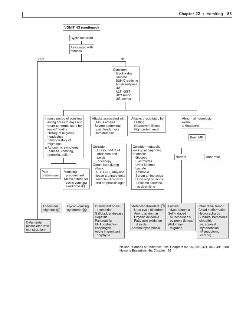
Chapter 22 u Vomiting 83
Associated withmensesAssociated withmenses
Cyclic recurrentCyclic recurrent
Abnormal neurologicexam± Headache
Abnormal neurologicexam± Headache
Intense period of vomiting lasting hours to days and return to normal state for weeks/months± History of migraine headaches± Family history of migraines± Autonomic symptoms (nausea, vomiting, anorexia, pallor)
Intense period of vomiting lasting hours to days and return to normal state for weeks/months± History of migraine headaches± Family history of migraines± Autonomic symptoms (nausea, vomiting, anorexia, pallor)
Attacks associated with: Bilious emesis Severe abdominal pain/tenderness Hematemesis
Attacks associated with: Bilious emesis Severe abdominal pain/tenderness Hematemesis
Consider: Ultrasound/CT of abdomen and pelvis EndoscopyObtain labs during attack: ALT, GGT, Amylase, lipase ± urinary delta aminolevulinic acid and porphobiliniogen
Consider: Ultrasound/CT of abdomen and pelvis EndoscopyObtain labs during attack: ALT, GGT, Amylase, lipase ± urinary delta aminolevulinic acid and porphobiliniogen
Catamenial(associated withmenstruation)
Catamenial(associated withmenstruation)
AbdominalmigraineAbdominalmigraine 21
AbnormalAbnormalNormal Normal
PainpredominantPainpredominant
Vomiting predominantMeets criteria for cyclic vomiting syndrome 22
Vomiting predominantMeets criteria for cyclic vomiting syndrome 22
Brain MRIBrain MRI
Consider: Electrolytes Glucose BUN/Creatinine Amylase/lipase UA ALT, GGT Ultrasound UGI series
Consider: Electrolytes Glucose BUN/Creatinine Amylase/lipase UA ALT, GGT Ultrasound UGI series
NOYES
Cyclic vomitingCyclic vomitingsyndrome 22
Intermittent bowel obstructionGallbladder diseaseHepatitisPancreatitisUPJ obstructionEsophagitisAcute intermittent porphyria
Intermittent bowel obstructionGallbladder diseaseHepatitisPancreatitisUPJ obstructionEsophagitisAcute intermittent porphyria
Attacks precipitated by: Fasting Intercurrent illness High protein meal
Attacks precipitated by: Fasting Intercurrent illness High protein meal
Consider metabolicworkup at beginningof attack: Glucose Electrolytes Urine ketones Lactate Ammonia Serum amino acids Urine organic acids ± Plasma carnitine, acylcarnitine
Consider metabolicworkup at beginningof attack: Glucose Electrolytes Urine ketones Lactate Ammonia Serum amino acids Urine organic acids ± Plasma carnitine, acylcarnitine
Metabolic disorders 13 Urea cycle disorders Amino acidemias Organic acidemia Fatty acid oxidation disorderAdrenal hyperplasia
Metabolic disorders 13 Urea cycle disorders Amino acidemias Organic acidemia Fatty acid oxidation disorderAdrenal hyperplasia
Familial dysautonomiaSelf-induced Munchausen’s by proxy (ipecac)Abdominal migraine
Familial dysautonomiaSelf-induced Munchausen’s by proxy (ipecac)Abdominal migraine
Intracranial tumorChiari malformationHydrocephalusSubdural hematomaIdiopathic intracranial hypertension (Pseudotumor cerebri)
Intracranial tumorChiari malformationHydrocephalusSubdural hematomaIdiopathic intracranial hypertension (Pseudotumor cerebri)
VOMITING (continued)VOMITING (continued)
Nelson Textbook of Pediatrics, 19e. Chapters 92, 96, 316, 321, 343, 491, 588Nelsons Essentials, 6e. Chapter 126
84
Chapter 23
DIARRHEA
most commonly identified source of outbreaks. Specific testing must be requested when the E. coli 0157:H7 strain is suspected. Yersinia and Campylobacter may be associated with a prolonged course of diarrhea. Bloody diarrhea is often seen with bacteria listed here, but diarrhea may also occur without blood.
4 Infection with Clostridium difficile (C. difficile) should be considered whenever diarrhea develops within several
weeks of antibiotic treatment. It is uncommon in children less than one year. It may manifest as mild or severe illness with or without grossly bloody diarrhea, abdominal pain, fever, or sys-temic toxicity. Definitive diagnosis is detection of the C. difficile toxin in the stool. Caution is advised in interpretation of the test in infants less than 1 year old because the test may be positive in asymptomatic infants.
5 Food protein-induced enterocolitis syndrome due to cow and/or soy milk allergy or intolerance may present with
bloody diarrhea in the first few months of life. Clinical improve-ment with a trial of a casein or whey hydrolysate formula or elimination of cow’s milk from a breast-feeding mother’s diet may be therapeutic and eliminate the need for any further evalu-ation. Early in infancy, food protein-induced enteropathy may present with diarrhea, occult blood loss, and hypoproteinemia.
6 Rotavirus is primarily a wintertime virus that affects infants and small children most often. Vomiting and diarrhea occur
abruptly after a 2- to 3-day incubation period; the vomiting gener-ally lasts 1 to 2 days, and loose watery stools last from 2 to 8 days. Fever commonly occurs; gross or occult blood is uncommon.
7 Giardiasis (from infection with Giardia lamblia) often results from contaminated food or water, but person-to-
person spread is common, especially in daycare centers and other crowded institutions. It should be considered as an etiol-ogy in acute cases of diarrhea, although it is most frequently considered a pathogen in chronic diarrhea (i.e., watery, with-out blood or mucus) with associated weight loss and abdomi-nal pain. Symptoms may also be intermittent and may even include constipation. Diagnosis may be difficult. Sensitivity of stool examination for ova and parasites increases with a greater number of samples. Examination of a single specimen is ap-proximately 70% sensitive. Antigen tests are more sensitive but will not aid in identifying other protozoans. Small intestinal biopsy or aspiration of duodenal or jejunal contents for exami-nation are other more sensitive means of diagnosis. Cryptospo-ridium may cause an acute diarrheal illness, including large watery stools, flatulence, malaise, and abdominal pain, lasting 3 to 30 days in a normal host. It causes a severe, chronic diar-rheal illness in immunocompromised patients. It is associated with contact with farm animals or contaminated water but is also spread through person-to-person contact. The organism may be identified in the stool, by microscopy, or enzyme immunoassay.
8 Parenteral diarrhea refers to diarrhea accompanying an infection outside the GI tract. Diarrhea is frequently
associated with upper respiratory infections, otitis media, and urinary tract infections. The mechanism is not clear.
Diarrhea is defined as stools of increased frequency, fluidity, and volume. In young children most acute diarrhea is of infec-tious etiology and is self-limited. Diarrhea lasting longer than 2 weeks may be considered chronic.
1 The history should include associated symptoms as well as the child’s growth pattern. A description of the diarrheal
stool may help indicate the diagnosis. Watery diarrhea may be osmotic due to carbohydrate malabsorption or secretory due to toxins, gastrointestinal peptides, bile acids, or laxatives. Steator-rhea (greasy stools) indicates fat malabsorption (e.g., pancreatic insufficiency). Mucus and blood in stools indicate intestinal inflammation (e.g., infection, inflammatory bowel disease). In toddlers the presence of undigested food may indicate a normal variation or chronic nonspecific diarrhea (“toddler’s diarrhea”). Overflow incontinence secondary to constipation and rectal impaction may be mistaken for diarrhea. Hematuria and ab-normal renal function suggest an enterohemorrhagic strain of Escherichia coli (E. coli); an associated severe dermatitis (e.g., perioral, acral, perineal) should suggest acrodermatitis entero-pathica. The social history should inquire about recent travel, exposure to unsanitary conditions, daycare attendance, risk fac-tors for HIV, and sick contacts.
A diet history that includes seafood, unwashed vegetables, unpasteurized milk, contaminated water, or uncooked meats may suggest a foodborne or waterborne agent in acute cases of diarrhea. In chronic cases, assessing type and quantity of oral intake, especially fluid selection, is helpful because certain se-lections may exacerbate diarrhea symptoms by an osmotic load. Inquire whether the onset of symptoms coincided with a change in diet (e.g., discontinuation of breast milk or formula, addition of jar foods or cereals, addition of sugar-free or other sorbitol containing compounds).
2 Sudden vomiting and explosive diarrhea within several hours of ingestion of a contaminated food suggests food
poisoning from pre-formed toxins produced by Staphylococcus aureus or Bacillus cereus. Other causes of foodborne illness in-clude other bacteria (Salmonella, Campylobacter, E. coli, Shigella) and viruses. Heavy metals, fish or shellfish poisoning, and mushrooms may cause paresthesias, paralysis, or mental status changes in addition to diarrhea.
3 High fevers and seizures have been associated with Shigella. E. coli 0157:H7 (an enterohemorrhagic strain) causes a hem-
orrhagic colitis that is followed by hemolytic-uremic syndrome in approximately 10% of cases. Enteroinvasive E. coli may also cause bloody diarrhea. In hemolytic-uremic syndrome, watery diarrhea precedes the grossly bloody stools; abdominal cramping with minimal or absent fevers is characteristic. Undercooked beef is the

Chapter 23 u Diarrhea 85
DIARRHEADIARRHEA
Symptoms acute (<2 weeks)Symptoms acute (<2 weeks)
Perform H and P 1 Perform H and P 1
Food poisoning 2 Food poisoning 2 Antibiotics LaxativesProkinetic agentsDrug side effects
Antibiotics LaxativesProkinetic agentsDrug side effects
Bacterial gastroenteritis 3 Shigella Salmonella Campylobacter Yersenia Enterohemorrhagic E. coli Enteroinvasive E. coli Aeromonas C. difficile 4
Bacterial gastroenteritis 3 Shigella Salmonella Campylobacter Yersenia Enterohemorrhagic E. coli Enteroinvasive E. coli Aeromonas C. difficile 4
Viral gastroenteritis Rotavirus 6 Enterovirus Norovirus agent CalicivirusProtozoa 7 Giardia CryptosporidiumBacterial E. coli (Traveler’s diarrhea) Salmonella C. difficile 4 Vibrio choleraParenteral diarrhea 8 Overfeeding
Viral gastroenteritis Rotavirus 6 Enterovirus Norovirus agent CalicivirusProtozoa 7 Giardia CryptosporidiumBacterial E. coli (Traveler’s diarrhea) Salmonella C. difficile 4 Vibrio choleraParenteral diarrhea 8 Overfeeding
Temporally associated with food intake Others with similar exposures and symptomsTemporally associated with food intake Others with similar exposures and symptoms
AcutediarrheaAcutediarrhea
ChronicdiarrheaChronicdiarrhea
Age <6 monthsAge <6 months Age >6 monthsAge >6 months
See Chronicdiarrhea in infantsSee Chronicdiarrhea in infants
See Chronicdiarrhea in olderchildren
See Chronicdiarrhea in olderchildren
Blood or mucus in stoolsBlood or mucus in stools
Recent medication useRecent medication use
Stool culture± C. difficile toxinStool culture± C. difficile toxin
Consider:Stool for O and PStool cultureGiardia antigenCryptosporidium antigen± C. difficile toxin
Consider:Stool for O and PStool cultureGiardia antigenCryptosporidium antigen± C. difficile toxin
NO
NO
NO
NO
YES
YES
YES
YES
Nelson Textbook of Pediatrics, 19e. Chapters 332, 333Nelsons Essentials, 6e. Chapter 126

86 Part IV u GastrointestinalSystem
9 Commercial reagents (Clinitest) are available to test for re-ducing substances; pH testing with Nitrazine paper should
be performed on a fresh stool specimen. Positive reducing sub-stances or a pH less than 5.5 suggests carbohydrate malabsorp-tion. Be aware of two caveats: (1) the test for reducing substances is only reliable when the child is being fed adequate amounts of carbohydrates, and (2) sucrose is not a reducing sugar and must be digested or split by bacteria to produce a positive test. Adding hydrochloric acid before the analysis should have the same result.
10 Postinfectious enteritis after acute enteritis is a common cause of prolonged diarrhea. Low-grade mucosal injury is
responsible for the malabsorption. In young infants, a second-ary lactase deficiency may be a contributing factor; in older infants and children a hypocaloric, high-carbohydrate diet is often responsible for the persistent malabsorption.
11 Approximately 10% of children with Hirschsprung disease develop enterocolitis. Historical ”red flags” include a history
of delayed passage of meconium, preceding constipation, Down syndrome, and a positive family history. Absent stool on rectal examination and immediate passage of stool after the rectal ex-amination are suggestive. A rectal suction biopsy demonstrating absent ganglion cells is necessary for diagnosis.
12 In chronic diarrhea a finding of leukocytes or occult blood is more suggestive of inflammatory bowel disease than bac-
terial infection. A Sudan stain for fat will confirm steatorrhea but does not specify whether the abnormality is of bowel, pancreatic, or biliary origin. Stool elastase-1 can help evaluate for pancreatic
exocrine function. Many causes of chronic diarrhea demonstrate an acidic pH or positive reducing substances consistent with some element of carbohydrate malabsorption.
13 Cystic fibrosis is characterized by frequent large, foul-smelling fatty stools not classic loose, watery diarrheal
stools. When accompanied by a history of recurrent respiratory infections and failure to thrive, a sweat chloride test should be done. Infants younger than 6 months tend to present with fail-ure to thrive; diarrhea (steatorrhea) is more common in older infants and toddlers.
14 Pancreatic insufficiency, chronic neutropenia, and short stature characterize Shwachman-Diamond syndrome.
15 Congenital chloride-losing diarrhea is a rare congenital dis-order diagnosed by abnormally high levels of stool chloride.
16 Immune-mediated damage of the small intestine in re-sponse to gluten occurs in celiac disease (gluten-sensitive
enteropathy). Symptoms of malabsorption and failure to thrive classically develop between 6 months and 3 years; symptom onset follows the addition of cereal grains (e.g., wheat, oat, barley, rye) to the diet. Diarrhea is often a late manifestation of the disorder, which may also include failure to thrive, anorexia, digital clubbing, anemia, abdominal distention, apathy, fre-quent fatty stools, and symptoms suggestive of specific nutrient deficiencies. IgA anti-tissue transglutaminase immunoassay (TTG) is a sensitive screening test. Biopsy is considered neces-sary for definitive diagnosis. Variable presentation has resulted in cases being diagnosed in older children.
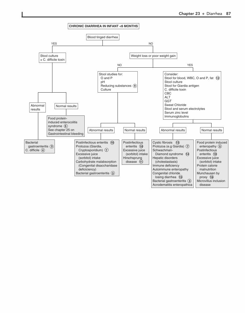
Chapter 23 u Diarrhea 87
CHRONIC DIARRHEA IN INFANT <6 MONTHSCHRONIC DIARRHEA IN INFANT <6 MONTHS
Blood tinged diarrheaBlood tinged diarrhea
Food protein- induced enterocolitis syndrome 5 See chapter 25 on Gastrointestinal bleeding
Food protein- induced enterocolitis syndrome 5 See chapter 25 on Gastrointestinal bleeding
Postinfectious enteritis 10 Protozoa (Giardia, Cryptosporidium) 7 Excessive juice (sorbitol) intakeCarbohydrate malabsorption (Congenital disaccharidase deficiciency)Bacterial gastroenteritis 3
Postinfectious enteritis 10 Protozoa (Giardia, Cryptosporidium) 7 Excessive juice (sorbitol) intakeCarbohydrate malabsorption (Congenital disaccharidase deficiciency)Bacterial gastroenteritis 3
Postinfectious enteritis 10Excessive juice (sorbitol) intakeHirschsprung disease 11
Postinfectious enteritis 10Excessive juice (sorbitol) intakeHirschsprung disease 11
Cystic fibrosis 13 Protozoa (e.g Giardia) 7 Schwachman- Diamond syndrome 14 Hepatic disorders (cholestastasis)Immune deficiencyAutoimmune enteropathyCongenital chloride losing diarrhea 15 Bacterial gastroenteritis 3 Acrodematitis enteropathica
Cystic fibrosis 13 Protozoa (e.g Giardia) 7 Schwachman- Diamond syndrome 14 Hepatic disorders (cholestastasis)Immune deficiencyAutoimmune enteropathyCongenital chloride losing diarrhea 15 Bacterial gastroenteritis 3 Acrodematitis enteropathica
Food protein induced enteropathy 5Postinfectious enteritis 10Excessive juice (sorbitol) intakeProtein calorie malnutritionMunchausen by proxy 16 Microvillus inclusion disease
Food protein induced enteropathy 5Postinfectious enteritis 10Excessive juice (sorbitol) intakeProtein calorie malnutritionMunchausen by proxy 16 Microvillus inclusion disease
Bacterial gastroenteritis 3 C. difficile 4
Bacterial gastroenteritis 3 C. difficile 4
Abnormal resultsAbnormal results Normal resultsNormal resultsNormal resultsNormal results
Stool studies for: O and P pH Reducing substances 9 Culture
Stool studies for: O and P pH Reducing substances 9 Culture
Weight loss or poor weight gainWeight loss or poor weight gainStool culture± C. difficile toxinStool culture± C. difficile toxin
Consider:Stool for blood, WBC, O and P, fat 12 Stool cultureStool for Giardia antigenC. difficile toxinCBCALTGGTSweat Chloride Stool and serum electrolytesSerum zinc levelImmunoglobulins
Consider:Stool for blood, WBC, O and P, fat 12 Stool cultureStool for Giardia antigenC. difficile toxinCBCALTGGTSweat Chloride Stool and serum electrolytesSerum zinc levelImmunoglobulins
Abnormal resultsAbnormal results
AbnormalresultsAbnormalresults
Normal resultsNormal results
NO
NO
YES
YES
88 Part IV u GastrointestinalSystem
17 Inflammatory bowel disease (e.g., Crohn disease, ulcerative colitis) is very important in the differential diagnosis of
chronic diarrhea in the school-aged child. Although rare, inflam-matory bowel disease may present in younger children. Ulcerative colitis is more common than Crohn disease in the toddler age group. Weight loss and growth retardation are the cardinal symp-toms. Gastrointestinal manifestations include diarrhea, abdominal pain, bloody stools, perianal disease, and malabsorption. Multiple extra intestinal manifestations (e.g., fever, oral ulcers, uveitis, rash, arthralgias) may also occur. Laboratory evaluation usually reveals anemia, an elevated sedimentation rate, leukocytosis, and some degree of hypoalbuminemia, thrombocytosis, and elevated acute phase-reactive proteins depending on the severity of the disease. Stool calprotectin levels may also be helpful in evaluation for IBD. Radiologic studies (e.g., barium enema, upper gastrointestinal series) aid in determining the extent of the disease. Colonoscopy and biopsy are necessary for definitive diagnosis.
18 Chronic nonspecific diarrhea or ”toddler’s diarrhea” is a frustrating but benign disorder most often affecting nor-
mally nourished children between 1 and 5 years of age. Children have up to 6 to 10 loose, watery, foul-smelling stools per day, often with food particles present. The pattern and consistency of the stools may vary considerably from day to day. In some cases they may be well formed in the morning and become looser throughout the day. In others the diarrhea may alternate with periods of constipation. Normal growth and an absence of stool passage at night suggest the diagnosis; stool examination is negative for blood, mucus, and excessive fat.
Previously, most of these cases were probably due to excessive fruit juice intake. As the role of fruit juices has been increasingly recognized, the disorder is diagnosed less frequently today.
19 Lactase levels gradually decrease between 3 and 5 years of age in children with primary adult-type hypolactasia.
Subtle increases in malabsorptive symptoms (e.g., flatulence, abdominal pain, loose stools) occur after milk ingestion. Breath hydrogen testing after an oral lactose load may make the diag-nosis, although resolution of symptoms after dietary restriction of lactose is strongly suggestive. Postinfectious secondary lactase deficiency can occur in older children but is not as common or as severe as in younger children.
20 Irritable bowel syndrome is a functional GI disorder that is being increasingly recognized in older children and adolescents.
Patients report a variable pattern of diarrhea and constipation plus abdominal pain, bloating, urgency, a sense of incomplete stool evacuation, or passage of mucus.
21 Soiling associated with chronic constipation (i.e., encopre-sis) may be described as diarrhea by parents. The history
and rectal examination usually make a rapid diagnosis.
BibliographyBranski D, Lerner A, Lebenthal E: Chronic diarrhea and malabsorption,
Pediatr Clin North Am 43:307–331, 1996. Veereman-Wauters G, Taminiau J: Diarrhea. In Wyllie R, Hyams J, editors:
Pediatric gastrointestinal and liver disease, ed 4, Philadelphia, 2010, WB Saunders, pp 64.
Steffen R, Wyllie R: Constipation. In Kliegman, Greenbaum L, Lye P, editors: Practical strategies in pediatric diagnosis and therapy, ed 2, Philadelphia, 2004, WB Saunders.
Constipation Guideline Committee of the North American Society of Pediatric Gastroenterology, Hepatology and Nutrition: Evaluation and treatment of constipation in infants and children: Recommendations of the North American Society for Pediatric Gastroenterology, Hepatology and Nutrition. J Pediatr Gastroenterol Nutr, 43(3) 43:e1-e13, 2006.

Chapter 23 u Diarrhea 89
CHRONIC DIARRHEA IN INFANT >6 MONTHS(>2 WEEKS DURATION)
CHRONIC DIARRHEA IN INFANT >6 MONTHS(>2 WEEKS DURATION)
Consider:Stool for blood, pH, reducing substances, O and PGiardia antigenCryptosporidium antigenStool cultureC. difficile toxin
Consider:Stool for blood, pH, reducing substances, O and PGiardia antigenCryptosporidium antigenStool cultureC. difficile toxin
Bacterial gastroenteritis 3 Protozoa (Giardia) 7C. difficile 4Postinfectious enteritis 10 Disaccharidase deficienciesInfammatory bowel disease 17
Bacterial gastroenteritis 3 Protozoa (Giardia) 7C. difficile 4Postinfectious enteritis 10 Disaccharidase deficienciesInfammatory bowel disease 17
Post infectious enteritis 10Congenital disaccharidase deficienciesHIVCystic fibrosisSchwachman-Diamond syndrome 14Celiac disease 16Immune deficiencyFood protein enteropathy 5Hepatic disorders (cholestastasis)Inflammatory bowel disease 17Acrodematitis enteropathicaProtein calorie malnutritionIntestinal telangectasias
Post infectious enteritis 10Congenital disaccharidase deficienciesHIVCystic fibrosisSchwachman-Diamond syndrome 14Celiac disease 16Immune deficiencyFood protein enteropathy 5Hepatic disorders (cholestastasis)Inflammatory bowel disease 17Acrodematitis enteropathicaProtein calorie malnutritionIntestinal telangectasias
Excessive juice (sorbitol) intakeChronic nonspecific diarrhea (toddler’s diarrhea) 18 Postinfectious enteritis 10 Lactose intolerance 19 Irritable bowel syndrome 20 Constipation with overflow incontinence 21 Laxative abuse Munchausen syndrome (or by proxy) 16
Excessive juice (sorbitol) intakeChronic nonspecific diarrhea (toddler’s diarrhea) 18 Postinfectious enteritis 10 Lactose intolerance 19 Irritable bowel syndrome 20 Constipation with overflow incontinence 21 Laxative abuse Munchausen syndrome (or by proxy) 16
Consider:Stool studies for WBC, fat, Alpha-1 antitrypsin, elastase-1 12 CBC with differentialALT/ GGTESR/ CRP HIV testingSweat Chloride IgA anti-tissue transglutaminase immunoassay (TTG)Serum immunoglobinsSerum zinc level (if rash present)
Consider:Stool studies for WBC, fat, Alpha-1 antitrypsin, elastase-1 12 CBC with differentialALT/ GGTESR/ CRP HIV testingSweat Chloride IgA anti-tissue transglutaminase immunoassay (TTG)Serum immunoglobinsSerum zinc level (if rash present)
Weight loss or poor weight gainWeight loss or poor weight gain
Normal resultsNormal resultsAbnormal resultsAbnormal results
NO
NO
YES
YES

90
Chapter 24
CONSTIPATION
a decrease in the child’s normal activity level (e.g., illness, injury, surgery) is also a risk factor for constipation. A change in diet, especially starting cow’s milk, is constipating for some children. Psychological factors or constitutional factors such as intrinsi-cally slow motility can also exacerbate any cause of constipation. In chronic cases, stool retention results in a vicious cycle of re-tained stool, painful defecation, resisting the urge to defecate, further retaining of stool, and so on. These children are at risk for encopresis (fecal incontinence).
4 Irritable bowel syndrome can be associated with constipa-tion and diarrhea. Pain is often associated with change in
appearance or frequency of stooling. A response usually occurs with improved dietary fiber.
5 Constipation can occur because of multiple classes of drugs, including anticonvulsants, anticholinergics, antacids, iron-
containing medications, calcium channel blockers, psycho-therapeutic drugs, and narcotic-containing medications. Toxins may include metal intoxication (e.g., lead, arsenic, mercury) and botulinum.
6 Laboratory studies are not normally contributory on a routine basis unless there is some suggestion in the history
or physical examin ation of a metabolic disturbance. Con-genital hypothyroidism is generally diagnosed through new-born screening programs, but the acquired form can occur at any age.
7 Abdominal x-rays are not usually useful in the initial evaluation of constipation. They may be considered in
determining if constipation is present at all. Rectal motility studies (manometry) will demonstrate physiologic abnormali-ties related to defecation that may be primary (Hirschsprung disease) or secondary (chronic constipation). The findings in Hirschsprung disease are so characteristic that many centers are now using manometry to establish the diagnosis. Manometry is useful in children with constipation starting very early in life. It may also be helpful in cases in which constipation has failed to respond to a treatment regimen. In very young infants, a biopsy may be preferred over manometry because the latter is techni-cally difficult at very young ages. Further evaluation by a spe-cialist is necessary to arrive at the remaining possible diagnoses.
8 Chronic intestinal pseudo-obstruction (or neuronal intes-tinal dysplasia) is a rare disorder of GI motility. It may
be congenital or acquired and may be due to a neuropathy or myopathy or be idiopathic.
9 Hirschsprung disease (i.e., congenital aganglionic megaco-lon) typically occurs as delayed passage of meconium in
40% of affected infants, followed by lower intestinal obstruction in young infants. Milder presentations include severe constipa-tion since birth, narrow-caliber stools, abdominal distention, and failure to thrive. Fecal soiling is almost unheard of in Hirschsprung cases. Patients with short segment disease may not present until older childhood, adolescence, or even adult-hood. A rectal mucosal suction biopsy revealing an absence of ganglion cells is often necessary for diagnosis of Hirschsprung disease.
Constipation is a very common pediatric problem. It can be defined as delay or difficulty in passage of stools for more than 2 weeks, resulting in distress to the patient. Stools are often painful or hard to pass. Functional constipation that is not due to organic or anatomic causes is encountered most commonly. Encopresis, also known as fecal incontinence, is fecal soiling that occurs in the presence of chronic functional constipation.
1 When the constipation is severe and if it has been a long-standing problem since early infancy, it is necessary to rule
out an underlying organic disorder. Inquire specifically about intermittent large stools, because some children with constipa-tion will have a daily bowel movement but with incomplete emptying and retention of a large stool mass. Parents may describe stool withholding maneuvers of gluteal tightening and posturing, which are sometimes interpreted as attempts to strain or defecate. Occasionally, a parent will misinterpret the signs of encopresis as diarrhea.
A diet history for fluid and fiber intake may be helpful. In some infants, ingestion of large amounts of cow’s milk is associ-ated with constipation. Concerns about possible abuse should be addressed in the social history. Recent psychosocial changes and stressors should also be explored. The family history may reveal risk factors for Hirschsprung disease, such as the disor-der itself or certain syndromes (e.g., trisomy 21, Waardenburg, Williams).
A careful spine and neurologic examination should be done to rule out spinal disorders that could be contributing to the constipation. An anal wink elicited by stroking the perianal skin with a sharp edge ensures normal sacral innervation. A digital rectal examination may be helpful. Children with chronic con-stipation have a dilated rectal ampulla and a large, hard stool mass unless they had a recent large bowel movement. Most children with Hirschsprung disease will not have any palpable stool in the first few centimeters of the anal canal.
2 If functional constipation is suspected, it is reasonable to presumptively treat the patient with education, dietary
changes, and medication. Treatment of constipation consists of a “clean out” of retained stool and a maintenance regimen of stool softening and toileting practices to sustain evacuation and restore normal rectal and colon tone. Attention to family dynamics and the response of both the parents and child to the problem should be addressed. If there is inadequate response or there is concern for organic etiology, further investigation is suggested.
3 Situational constipation is usually short-lived and situa-tional in response to a recent change or stress, such as start-
ing daycare, travel, or the birth of a sibling. For some children, the transition to all-day school and the associated loss of privacy will contribute to withholding behaviors. Any condition causing

Chapter 24 u Constipation 91
NormalNormalAbnormalAbnormal
HypercalcemiaLead toxicityHypothyroidismHypokalemiaCeliac disease
HypercalcemiaLead toxicityHypothyroidismHypokalemiaCeliac disease
Severe functional constipationChronic intestinal pseudo-obstructionHirschsprung diseaseMuscular dystrophyAcquired stricture
Severe functional constipation 3 Chronic intestinal pseudo-obstruction 8 Hirschsprung disease 9 Muscular dystrophyAcquired stricture
TreatmentEducationDiet± Medication
TreatmentEducationDiet± Medication
Abnormalphysical exam 10 Abnormalphysical exam 10
Responds totreatmentResponds totreatment
Consider: Free T4/ TSH Calcium Lead Electrolytes Celiac panel (TTG)
Consider: 6 Free T4/ TSH Calcium Lead Electrolytes Celiac panel (TTG)
Consider Abdominal x-ray 7 Barium enema 7 Referral for rectal manometry/ rectal biopsy 7
Consider: Abdominal x-ray 7 Barium enema 7 Referral for rectal manometry/ rectal biopsy 7
Any of the following present: Excessive cow’s milk intake Poor diet Toilet training problems Stress/ illness/ changes in routine Retentive behaviors Medications/ drugs/ toxins
Any of the following present: Excessive cow’s milk intake Poor diet Toilet training problems Stress/ illness/ changes in routine Retentive behaviors Medications/ drugs/ toxins
See next pageSee next page
CONSTIPATIONCONSTIPATION
Perform Perform H and P 1
Severeconstipation,present sinceinfancy
Severeconstipation,present sinceinfancy
YESNO
Nelson Textbook of Pediatrics, 19e. Chapters 96, 298, 324, 536Nelsons Essentials, 6e. Chapter 126
NOYES
NOYES
Anorectal lesions (fissures, hemorrhoids,
Hirschsprung diseaseConnective tissue disorders (SLE, scleroderma)
Anorectal lesions (fissures, hemorrhoids, abscess, trauma)Anorectal malformations (stenosis, anterior analdisplacement, ectopic anus, imperforate anus)Abnormal abdominal musculature (prune belly, gastroschisis)Spinal cord lesions (tethered cord, spina bifida)Neurologic (Botulism, cerebral palsy, myotonic dystrophy)Hirschsprung disease 9 Connective tissue disorders (SLE, scleroderma)
Functional constipationBehavioral/ situational constipation (phobias, abuse, coercive toilet training) 3Milk protein intoleranceIrritable bowel syndrome 4 Medications/ drugs/ toxins 5
Functional constipation 2 Behavioral/ situational constipation (phobias, abuse, coercive toilet training) 3Milk protein intoleranceIrritable bowel syndrome 4 Medications/ drugs/ toxins 5

92 Part IV u GastrointestinalSystem
13 Meconium ileus (i.e., small bowel obstruction) appears in the newborn period in approximately 10% of infants with
cystic fibrosis.
14 In contrast to meconium ileus, meconium plug syndrome is an obstruction of the colon with inspissated plugs. The
condition may occur in the infant of a diabetic mother and in cystic fibrosis, rectal aganglionosis, maternal drug abuse, and after maternal magnesium sulfate therapy for preeclampsia. A Gastrografin enema is usually diagnostic and therapeutic. Many cases are benign, but a sweat chloride test is recommended.
10 Physical examination can help identify anatomic abnormalities as well as underlying conditions associated with constipation.
Simple anterior displacement of the anus may contribute to constipa-tion because of the anterior angle of the canal that stool must be ex-pelled through, although this concept is not universally accepted. An anteriorly located anus must be distinguished from an ectopic anus, in which the anal canal and internal anal sphincter are displaced an-teriorly; the external anal sphincter remains in its normal posterior position. An ectopic anus should be suspected if an anal wink can be elicited posterior to the opening of the anal canal. Children with neu-rologic impairment of any cause (e.g., cerebral palsy, polyneuritis, spina bifida, muscular dystrophy) are at risk for constipation from poor intestinal motility,inadequate dietary fiber, and impaired sensa-tion. Children with a primary myopathy, collagen vascular disease, or amyloidosis develop progressively more severe constipation.
11 Newborn screening programs in the United States test for hypothyroidism. Repeat testing should be done as indi-
cated by the state laboratory.
12 In very young infants, an unprepared barium enema may be helpful in the evaluation of severe constipation. The test
will help identify Hirschsprung disease as well as congenital anomalies. If not done diagnostically, the barium enema should be performed after manometry or biopsy to assist in surgical planning, because it demarcates the transition zone.
BibliographyAbi-Hanna A, Lake AM: Constipation and encopresis in childhood, Pediatr
Rev 19:23, 1998. Constipation Guideline Committee of the North American Society for
Pediatric Gastroenterology, Hepatology and Nutrition: Evaluation and treatment of constipation in infants and children: Recommendations of the North American Society for Pediatric Gastroenterology, Hepa-tology and Nutrition, J Pediatr Gastroenterol Nutr 43:e1–e13, 2006.
Daher S, Tahan S, Solé D, et al: Cow’s milk protein intolerance and chronic constipation in children, Pediatr Allergy Immunol 12:339–342, 2001.
Steffen R, Wyllie R: Constipation. In Kliegman RM, Lye PS, Greenbaum LA, editors: Practical strategies in pediatric diagnosis and therapy, ed 2, Philadelphia, 2004, Elsevier Saunders.
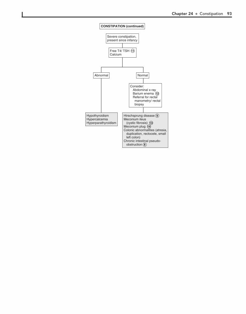
Chapter 24 u Constipation 93
Hirschsprung diseaseMeconium ileus (cystic fibrosis)Meconium plugColonic abnormalities (atresia, duplication, rectocele, small left colon)Chronic intestinal pseudo- obstruction
Hirschsprung disease 9 Meconium ileus (cystic fibrosis) 13 Meconium plug 14 Colonic abnormalities (atresia, duplication, rectocele, small left colon)Chronic intestinal pseudo- obstruction 8
HypothyroidismHypercalcemiaHyperparathyroidism
HypothyroidismHypercalcemiaHyperparathyroidism
NormalNormalAbnormalAbnormal
Consider:Consider: Abdominal x-ray Barium enema 12 Referral for rectal manometry/ rectal biopsy
CONSTIPATION (continued)CONSTIPATION (continued)
Severe constipation,present since infancySevere constipation,present since infancy
Free T4/ TSH 11 CalciumFree T4/ TSH 11 Calcium
94
Chapter 25
GASTROINTESTINAL BLEEDING
3 Hematemesis (i.e., vomiting of bright red blood or “coffee grounds”) is generally associated with UGI hemorrhage
proximal to the ligament of Treitz. Consider passing a nasogas-tric tube to help in evaluation and management in case of per-sistent or severe UGI bleeding. Hematochezia (rectal passage of bright red or maroon-colored blood mixed in with the stool) is usually associated with lower GI or colonic bleeding, although brisk UGI hemorrhages may also present in this way. Melena (i.e., dark, tarry-appearing stool) represents bleeding from any site above the ileocecal valve. Occult blood may originate from upper or lower GI sources. Blood originating in the rectum generally maintains a bright red color.
4 Significant GI bleeding warrants a CBC with platelet count, differential, and reticulocyte count to assess for anemia. Mi-
crocytosis suggests chronic bleeding, and serial CBCs may help assess severity of bleeding. If bleeding is significant or the history is suggestive of a bleeding disorder, obtain at minimum a PT and activated PTT. LFTs, electrolytes, Cr, and BUN may identify he-patic, renal, and metabolic problems. Hypoalbuminemia is consis-tent with long-standing liver dysfunction with portal hypertension, IBD, or protein-losing enteropathies. In cases in which there was a limited amount of bleeding and a clinical picture consistent with a benign, self-limited condition (swallowed blood, Mallory-Weiss tear), laboratory tests may not be indicated.
Upper endoscopy is successful in identifying UGI bleeding sites in 80% to 90% of cases. Radiographic contrast studies are less sensitive but may be used if endoscopy is not available. They are contraindicated in cases of active bleeding. Small bowel follow-through examinations may be helpful in identify-ing atretic lesions, strictures, and rotation abnormalities. Newer endoscopic methods to image the small bowel include balloon enteroscopy and capsule endoscopy. Bleeding scans can help locate bleeding sites not accessible with endoscopy. Angiogra-phy is most useful when bleeding is moderately brisk, but it is of limited value for lower GI hemorrhages.
5 Mallory-Weiss tears are common. Forceful recurrent vom-iting causes a tear in the distal esophageal mucosa that re-
sults in a limited amount of bright red hematemesis. In light of a stable clinical picture consistent with a self-limited vomiting illness and no evidence of obstruction, a diagnostic workup is not indicated.
6 Esophagitis as a complication of GER is a common cause of GI bleeding in children. GER maybe present even in the
absence of regurgitation.
7 Gastritis may be due to caustic ingestions, viral infections, Helicobacter pylori infection, radiation exposure, or bile
reflux. Frequent NSAID, cocaine, or alcohol use may cause gas-tritis in adolescents.
8 Peptic ulcer disease includes gastric and duodenal ulcers, gastritis, and duodenitis. Gastritis and peptic ulcers are
common causes of UGI bleeding in sick or stressed neonates or infants. In this young age group, they are more likely to present acutely as perforation or hemorrhage. Less severe cases may present as irritability, vomiting, and regurgitation. Ulcers in older children and adolescents are more likely to have a history of long-standing intermittent abdominal pain. Epigastric pain, especially if nocturnal or relieved with meals, is suggestive of peptic ulcer disease, although for children this “classic” constel-lation of symptoms is less reliable than for adults.
1 The history should include any previous GI problems (e.g., jaundice, liver disease, ulcers, GER, other GI hemorrhages),
blood transfusions, coagulopathies, and iron deficiency. A neona-tal history of total parenteral nutrition, omphalitis, or umbilical vein catheterization is a risk factor for portal vein thrombosis. For infants, inquire about maternal idiopathic thrombocytopenic pur-pura. Aspirin, nonsteroidal antiinflammatory drugs (NSAIDs), and probably corticosteroids may play a contributing role in the development of gastritis.
A family history may be helpful in cases of suspected peptic ulcer disease, inflammatory bowel disease (IBD), liver disease, Meckel diverticulum, polyps, or milk allergy. Inquire about a family history of coagulopathies and Hirschsprung disease. A history of nosebleeds raises the possibility of swallowed blood presenting as hematemesis. Hemoptysis may need to be ruled out in cases in which severe coughing is present. Ask about ingestion of undercooked meat, recent medications, and the possibility of other ingestions (e.g., toxins, foreign bodies). Kool-Aid, gelatin, food coloring, antibiotics, bismuth, beets, licorice, cranberries, spinach, and blueberries can all mimic GI bleeding. Large iron ingestions can cause hematemesis due to mucosal injury. Thera-peutic doses of iron will cause black stools, but they will remain negative for occult blood.
Examination of vital signs (heart rate, BP, orthostatics) is important to ensure that bleeding does not require emergent treatment for circulatory compromise. The growth curve may reveal failure to thrive, suggestive of IBD. A thorough physical exam is important to identify possible causes of bleeding. A careful abdominal exam may note tenderness (seen with ab-dominal inflammatory processes), splenomegaly or ascites (seen in portal hypertension), and a right lower quadrant mass may suggest intussusception. A careful skin exam may reveal a diagnosis; for example, hyperpigmented oral lesions (Peutz-Jeghers), petechiae (bleeding abnormality or disseminated in-travascular coagulation), purpura (Henoch-Schönlein purpura, hemolytic uremic syndrome), and jaundice or spider nevi (liver disease). A rectal examination may reveal tags, fissures and fis-tulae (Crohn disease), or erythema with tenderness (group A b-hemolytic streptococcal infection).
2 Confirming the presence of blood is important to avoid an unnecessary evaluation. A stool test for occult blood must
be part of the initial evaluation. False-positive test results can oc-cur in young women around the time of their menses and after recent ingestion of rare red meat or fresh peroxidase-containing foodstuffs such as broccoli, radishes, cauliflower, cantaloupe, or turnips. Munchausen by proxy should be considered if a history of significant bleeding is not supported by any documentation of actual blood loss. With a breast-fed infant, if swallowed maternal blood is suspected, definitive diagnosis can be made by an Apt-Downey test. Fecal leukocytes are consistent with an invasive infectious organism or an inflammatory condition.
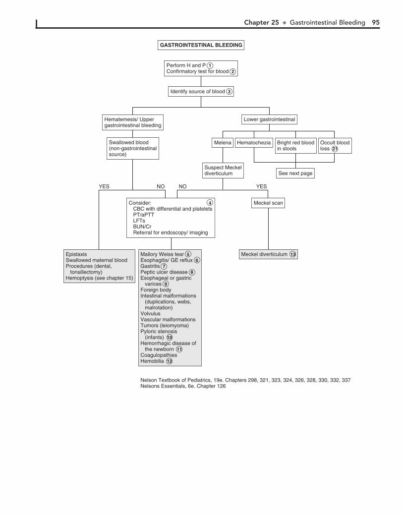
Chapter 25 u GastrointestinalBleeding 95
Suspect MeckeldiverticulumSuspect Meckeldiverticulum
GASTROINTESTINAL BLEEDINGGASTROINTESTINAL BLEEDING
Hematemesis/ Uppergastrointestinal bleedingHematemesis/ Uppergastrointestinal bleeding
Swallowed blood(non-gastrointestinalsource)
Swallowed blood(non-gastrointestinalsource)
Lower gastrointestinalLower gastrointestinal
MelenaMelena HematocheziaHematochezia Bright red bloodin stoolsBright red bloodin stools
Occult bloodloss 21Occult bloodloss 21
See next pageSee next page
Consider: 4 CBC with differential and platelets PT/aPTT LFTs BUN/Cr Referral for endoscopy/ imaging
Consider: 4 CBC with differential and platelets PT/aPTT LFTs BUN/Cr Referral for endoscopy/ imaging
Meckel diverticulum 13Meckel diverticulum 13Mallory Weiss tear 5 Esophagitis/ GE reflux 6 Gastritis 7Peptic ulcer disease 8Esophageal or gastric varices 9 Foreign bodyIntestinal malformations (duplications, webs, malrotation)VolvulusVascular malformationsTumors (leiomyoma)Pyloric stenosis (infants) 10Hemorrhagic disease of the newborn 11CoagulopathiesHemobilia 12
Mallory Weiss tear 5 Esophagitis/ GE reflux 6 Gastritis 7Peptic ulcer disease 8Esophageal or gastric varices 9 Foreign bodyIntestinal malformations (duplications, webs, malrotation)VolvulusVascular malformationsTumors (leiomyoma)Pyloric stenosis (infants) 10Hemorrhagic disease of the newborn 11CoagulopathiesHemobilia 12
EpistaxisSwallowed maternal bloodProcedures (dental, tonsillectomy)Hemoptysis (see chapter 15)
EpistaxisSwallowed maternal bloodProcedures (dental, tonsillectomy)Hemoptysis (see chapter 15)
Identify source of blood 3 Identify source of blood 3
Perform H and P 1Confirmatory test for blood 2 Perform H and P 1Confirmatory test for blood 2
Meckel scanMeckel scan
NO YESYES NO
Nelson Textbook of Pediatrics, 19e. Chapters 298, 321, 323, 324, 326, 328, 330, 332, 337Nelsons Essentials, 6e. Chapter 126
96 Part IV u GastrointestinalSystem
9 Varices are commonly due to portal vein hypertension secondary to intrinsic liver disease. Portal vein thrombosis
is the next most likely etiology. Thrombus formation can occur as the result of sepsis, pancreatitis, omphalitis, or umbilical vein catheterization. A series of tortuous collateral veins develop to bypass the obstructing thrombus and may appear years later as bleeding esophageal varices. US or endoscopy may be used to assess for varices when the patient is not actively bleeding.
10 Pyloric stenosis classically presents between 3 and 6 weeks of age as nonbilious vomiting that rapidly progresses to
frequent projectile vomiting, often complicated by dehydration, weight loss, and metabolic alkalosis. The emesis may develop a coffee-ground appearance and may test positive for blood. US is the preferred diagnostic test.
11 An unsuspected coagulopathy is more likely to occur in the newborn period than at older ages. Be aware of in-
creased risk in breast-fed infants, especially those who did not receive vitamin K at birth.
12 Hemobilia is hemorrhage into the biliary tract.
13 Approximately two thirds of Meckel diverticula occur as a painless but significant lower GI bleeding before 2 years of
age. The bleeding is due to ulceration from gastric mucosa con-tained in the diverticulum. Radioisotope technetium scanning (Meckel scan) can localize ectopic gastric mucosa.
14 Juvenile polyps are a common cause of painless rectal bleeding; most are in the distal colon. Inherited syn-
dromes associated with polyps and increased risk of develop-ing malignancy are familial adenomatous polyposis, Gardner syndrome, Peutz-Jeghers syndrome, and juvenile polyposis. Peutz-Jeghers syndrome is characterized by diffuse intestinal hamartomas and hyperpigmented macules of the oral mucosa.
15 Intussusception is the “telescoping” of one segment of the intestine into a distal segment. It can occur between 3
months and 6 years of age, but most cases occur before age 36 months. Severe episodes of abdominal pain (due to obstruction or ischemia) and crying occur, accompanied initially by periods of normal behavior between paroxysms. Progression to vomiting (sometimes bilious or bloody), lethargy, and shock may occur. “Currant jelly” stools are observed in approximately 60% of cases.
The barium enema, the traditional diagnostic and therapeutic modality, has been replaced in many centers by US for diagnosis and fluoroscopy-guided reduction with hydrostatic or pneumatic pressure (“air enema”). Any method of reduction should always be performed with surgical backup. Nonsurgical reduction should not be attempted if there is any evidence of shock or intestinal perforation (e.g., peritonitis). Barium enemas with air contrast can reveal mucosal abnormalities but overall are not very specific.
16 Although neonates are susceptible to most of the same problems as older age groups, some conditions tend to
present almost exclusively in the neonatal period. Necrotizing enterocolitis should always be considered in the presence of prematurity and occasionally can have a late presentation.
17 Bloody diarrhea is a common manifestation of bacterial pathogens (Yersinia, Salmonella, Shigella, and Campylobacter)
and parasites (ameba). Colitis accompanied by anemia, thrombo-cytopenia, or renal insufficiency (consistent with hemolytic- uremic syndrome) should prompt a specific investigation for Escherichia coli serotype 0157:H7. Recent antibiotic use should raise suspicions for Clostridium difficile. Infectious colitis is a rare cause of bleeding in neonates. Be aware of positive tests for C. difficile toxin; C. difficile may not be a pathogen in the new-born. Occult blood may occur with acute diarrhea due to any cause as a result of minor anal or perineal irritation.
18 Hirschsprung disease typically occurs as an obstruction, but it may appear as an enterocolitis with bleeding.
19 Food protein–induced enterocolitis syndrome and food protein–induced proctitis (milk protein sensitivity) typically present as a
history of bloody mucous stools and increasing stool frequency. The family history is often positive. Some infants fed soy-based formula can develop an identical clinical picture. Breast-fed infants can develop al-lergic symptoms from cow’s milk ingested by the mother. In infants with a clinical presentation for milk allergy or intolerance, a trial of a casein or whey hydrolysate formula or elimination of cow’s milk from a breastfeeding mother’s diet may be therapeutic and eliminate the need for any further evaluation.
20 Fissures are a common cause of bright red streaks coating the stool. They can occur even in the absence of constipation.
If the perianal region is erythematous, obtain a culture for group A Streptococcus to rule out a perianal cellulitis, which can predis-pose patients to fissures and bleeding. Hemorrhoids involving veins above the anorectal line may not be visible on examination.
21 Most causes of upper and lower GI bleeding can manifest as occult bleeding. Careful history-taking may narrow the
differential diagnosis and guide the appropriate workup. The diagnoses listed are among the most common that may present with occult blood loss.
BibliographyBoyle JT: Gastrointestinal bleeding in infants and children, Pediatr Rev 29:
39–51, 2008.Friedlander J, Mamula P: Gastrointestinal hemorrhage. In Wyllie R, Hyams J,
editors: Pediatric gastrointestinal and liver disease, ed 4, Philadelphia, 2010, WB Saunders, pp 64.
Sylvester FA, Hyams JS: Gastrointestinal bleeding. In Kliegman RM, Lye PS, Greenbaum LA, editors: Practical strategies in pediatric diagnosis and therapy, ed 2, Philadelphia, 2004, Elsevier Saunders.
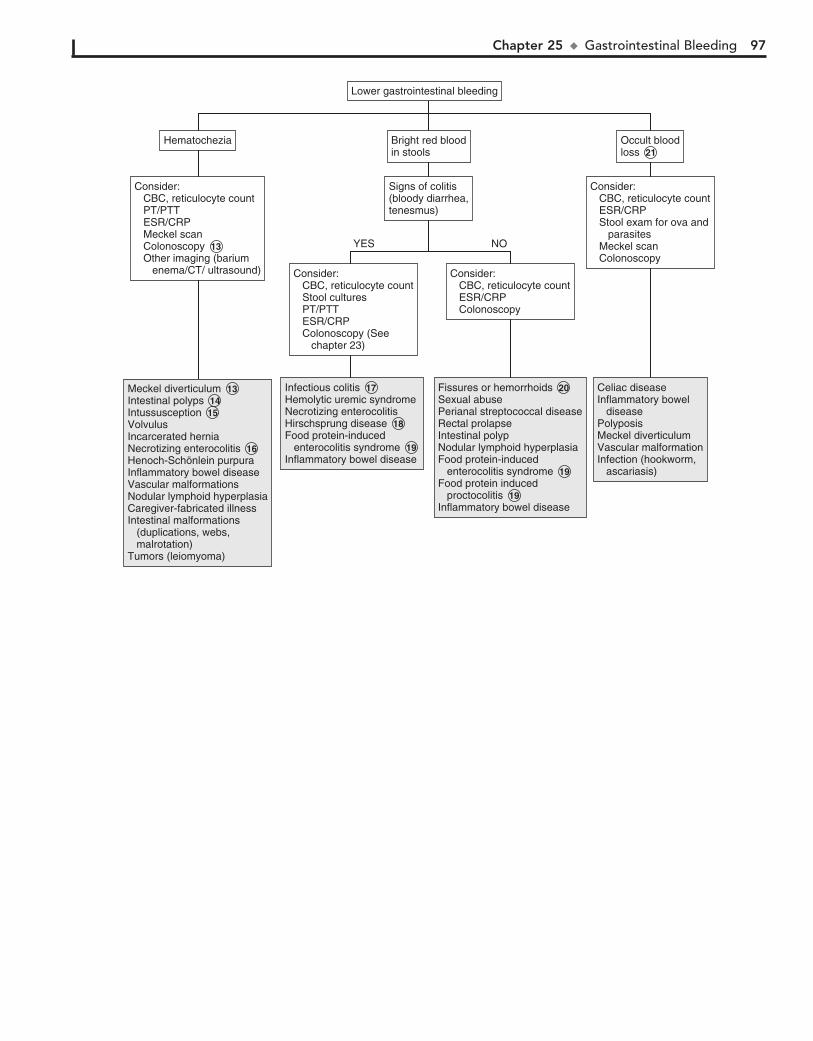
Chapter 25 u GastrointestinalBleeding 97
Consider: CBC, reticulocyte count Stool cultures PT/PTT ESR/CRP Colonoscopy (See chapter 23)
Consider: CBC, reticulocyte count Stool cultures PT/PTT ESR/CRP Colonoscopy (See chapter 23)
Consider: CBC, reticulocyte count ESR/CRP Colonoscopy
Consider: CBC, reticulocyte count ESR/CRP Colonoscopy
Signs of colitis(bloody diarrhea,tenesmus)
Signs of colitis(bloody diarrhea,tenesmus)
Consider: CBC, reticulocyte count ESR/CRP Stool exam for ova and parasites Meckel scan Colonoscopy
Consider: CBC, reticulocyte count ESR/CRP Stool exam for ova and parasites Meckel scan Colonoscopy
Bright red bloodin stoolsBright red bloodin stools
Lower gastrointestinal bleedingLower gastrointestinal bleeding
HematocheziaHematochezia Occult bloodloss 21 Occult bloodloss 21
NOYES
Consider: CBC, reticulocyte count PT/PTT ESR/CRP Meckel scan Colonoscopy 13 Other imaging (barium enema/CT/ ultrasound)
Consider: CBC, reticulocyte count PT/PTT ESR/CRP Meckel scan Colonoscopy 13 Other imaging (barium enema/CT/ ultrasound)
Meckel diverticulum 13 Intestinal polyps 14Intussusception 15VolvulusIncarcerated herniaNecrotizing enterocolitis 16 Henoch-Schönlein purpuraInflammatory bowel diseaseVascular malformationsNodular lymphoid hyperplasiaCaregiver-fabricated illnessIntestinal malformations (duplications, webs, malrotation)Tumors (leiomyoma)
Meckel diverticulum 13 Intestinal polyps 14Intussusception 15VolvulusIncarcerated herniaNecrotizing enterocolitis 16 Henoch-Schönlein purpuraInflammatory bowel diseaseVascular malformationsNodular lymphoid hyperplasiaCaregiver-fabricated illnessIntestinal malformations (duplications, webs, malrotation)Tumors (leiomyoma)
Infectious colitis 17 Hemolytic uremic syndromeNecrotizing enterocolitisHirschsprung disease 18 Food protein-induced enterocolitis syndrome 19 Inflammatory bowel disease
Infectious colitis 17 Hemolytic uremic syndromeNecrotizing enterocolitisHirschsprung disease 18 Food protein-induced enterocolitis syndrome 19 Inflammatory bowel disease
Celiac diseaseInflammatory bowel diseasePolyposisMeckel diverticulumVascular malformationInfection (hookworm, ascariasis)
Celiac diseaseInflammatory bowel diseasePolyposisMeckel diverticulumVascular malformationInfection (hookworm, ascariasis)
Fissures or hemorrhoids 20Sexual abusePerianal streptococcal diseaseRectal prolapseIntestinal polypNodular lymphoid hyperplasiaFood protein-induced enterocolitis syndrome 19Food protein induced proctocolitis 19Inflammatory bowel disease
Fissures or hemorrhoids 20Sexual abusePerianal streptococcal diseaseRectal prolapseIntestinal polypNodular lymphoid hyperplasiaFood protein-induced enterocolitis syndrome 19Food protein induced proctocolitis 19Inflammatory bowel disease
98
Chapter 26
JAUNDICE
5 Breast-fed infants have an exaggerated physiologic jaundice in the first week of life, with significantly higher bilirubin
levels than formula-fed infants in the first 5 days of life. This is due to decreased caloric intake until adequate breast milk supply is established with resulting increase in enterohepatic circulation.
6 ”Breast milk” jaundice is jaundice occurring after the first week of life. The mechanism is not clearly understood. To
confirm the diagnosis, breastfeeding may be discontinued for 24-48 hours to observe whether a decrease in bilirubin level occurs.
7 Isoimmune hemolytic disease occurs when maternal anti-bodies to the erythrocytes of the fetus cross the placenta
and cause destruction of the fetal red blood cells. Incompatibil-ity of the Rh factor causes the most severe disease in progressive pregnancies. ABO incompatibility causes less severe hemolysis. On occasion, infants in the latter group will demonstrate a negative or weakly positive direct Coombs test but the indirect Coombs will be positive.
8 Red blood cell membrane defects include hereditary sphe-rocytosis, hereditary elliptocytosis, infantile pyknocytosis,
hereditary stomatocytosis, and hereditary pyropoikilocytosis.
9 Extravascular blood results in increased bilirubin pro-duction. Examples include cephalohematoma, ecchymo-
ses, occult hemorrhage as well as swallowed maternal blood.
10 Any condition causing obstruction or delayed passage of meconium (e.g., Hirschsprung disease, meconium plug
syndrome) will increase enterohepatic circulation of bilirubin, contributing to indirect hyperbilirubinemia and jaundice.
11 Prolonged indirect hyperbilirubinemia may be the earli-est clinical manifestation of congenital hypothyroidism
as well as hypopituitarism. These may also cause conjugated hyperbilirubinemia.
12 Oxytocin, excess vitamin K in premature infants, some antibiotics (e.g., sulfonamides, ceftriaxone), and phenol
disinfectants are examples of drugs and toxins that may con-tribute to unconjugated hyperbilirubinemia.
13 Normal liver enzymes indicate that hepatic injury or biliary tract disease is less likely. A significant elevation in elevated
gammaglutamyl transpeptidase (GGT) suggests biliary obstruc-tion or intrahepatic cholestasis. Serum alkaline phosphatase is also increased in relation to aminotransferases (i.e., AST and ALT). In obstructive jaundice there is often prolonged prothrom-bin time that corrects with vitamin K administration due to decreased absorption of fat-soluble vitamins. History of acholic (clay colored) stools supports an obstructive cause. Elevation of serum transaminases is caused by intrinsic hepatocellular disease. With severe disease there may be impaired synthetic function causing hypoalbuminemia and a prolonged prothrombin time that does not correct with vitamin K. Hypoglycemia reflects hepa-tocellular damage; it indicates more severe disease and mandates an urgent workup.
14 A congenital infection is suggested by intrauterine growth retardation, microcephaly, and ophthalmologic abnormali-
ties (e.g., cataracts, chorioretinitis, posterior embryotoxon). Characteristic facies may suggest syndromes associated with hyperbilirubinemia. Any of the “TORCH infections” (e.g., toxo-plasmosis, rubella, cytomegalovirus, herpes virus, syphilis) may cause growth retardation and cholestasis.
Jaundice is the yellow discoloration of skin, sclerae, and other tis-sues caused by the deposition of bilirubin. The degree of jaundice is related to the serum level of bilirubin and the degree of its de-position into the extravascular tissues. The most common source of bilirubin is the increased breakdown of hemoglobin. Jaundice may lead to kernicterus, which is a neurologic syndrome resulting from the deposition of unconjugated (indirect) bilirubin in the basal ganglia and brainstem nuclei. The toxic blood level for an individual infant is unpredictable, but in general, kernicterus typically occurrs only in infants with a bilirubin .20 mg/dL.
In older infants jaundice should be distinguished from caro-tenemia, a diffuse yellowish-orange skin discoloration caused by ingestion of large amounts of carotene-containing foods (e.g., carrots, squash).
1 Although most cases of neonatal jaundice are physiologic, a careful history and physical examination are necessary to
rule out more serious disorders. The prenatal and birth history should inquire about delivery complications, maternal infec-tion, diabetes mellitus, and drug use. Oxytocin during labor is associated with an increased risk of jaundice. A history of poly-hydramnios suggests an intestinal obstruction. Other condi-tions resulting in a delayed passage of meconium (Hirschsprung disease, cystic fibrosis) will also contribute to hyperbilirubine-mia. Prematurity is a risk factor for hyperbilirubinemia that is often compounded by delayed enteral feeds, parenteral nutri-tion, and perinatal insults due to hypoxia and acidosis. Vomit-ing, lethargy, poor feeding, and failure to thrive may suggest an inborn error of metabolism. Breast-fed infants tend to have higher and more prolonged unconjugated bilirubin levels. A family history of jaundice, anemia, splenectomy, or cholecys-tectomy suggests a hereditary hemolytic disorder.
2 Detailed investigation is warranted in children with con-jugated hyperbilirubinemia, defined as a conjugated bili-
rubin level .2.0 mg/dl or a conjugated fraction .20% of the total bilirubin level. The evaluation should be directed at ruling out infection, metabolic disorders, anatomic abnormalities, and familial cholestatic syndromes.
3 Some risk factors for development of severe hyperbilirubi-nemia include prematurity, jaundice observed in first 24
hours of life, known blood group incompatibility or hemolytic disease, sibling who required phototherapy, cephalohematoma, bruising, poor breastfeeding, and East Asian race.
4 Nearly all newborns experience some rise in serum biliru-bin levels owing to the relative immaturity of their hepatic
excretory function. The frequent occurrence of hyperbilirubine-mia and jaundice—approximately one third of all newborns de-velop jaundice—has resulted in the term physiologic jaundice. Levels higher than those used in the definition may occur (i.e., exaggerated physiologic jaundice), but some evaluation must be done to rule out more serious disorders before making the diagnosis. A well-accepted AAP practice parameter exists for the management of hyperbilirubinemia in the healthy term new-born. (See Bibliography.)
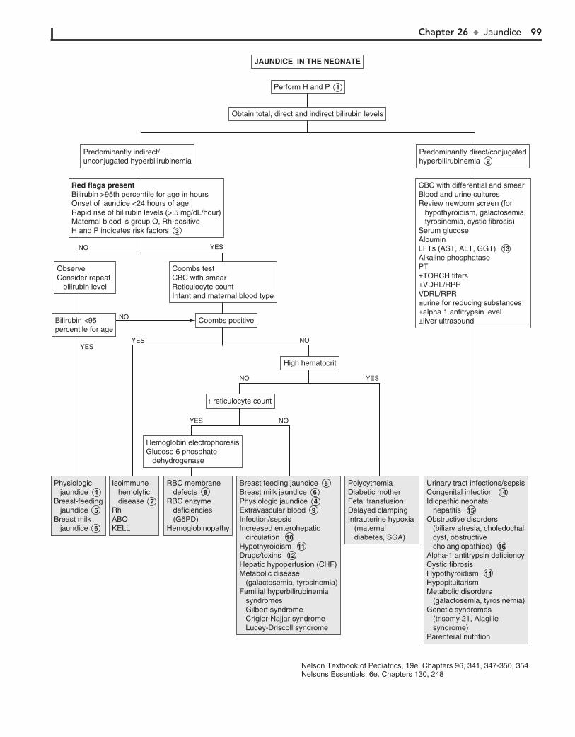
Chapter 26 u Jaundice 99
Red flags presentBilirubin >95th percentile for age in hoursOnset of jaundice <24 hours of ageRapid rise of bilirubin levels (>.5 mg/dL/hour)Maternal blood is group O, Rh-positiveH and P indicates risk factors 3
Red flags presentBilirubin >95th percentile for age in hoursOnset of jaundice <24 hours of ageRapid rise of bilirubin levels (>.5 mg/dL/hour)Maternal blood is group O, Rh-positiveH and P indicates risk factors 3
Physiologic jaundice 4 Breast-feeding jaundice 5 Breast milk jaundice 6
Physiologic jaundice 4 Breast-feeding jaundice 5 Breast milk jaundice 6
Isoimmune hemolytic disease 7 RhABOKELL
Isoimmune hemolytic disease 7 RhABOKELL
RBC membrane defects 8 RBC enzyme deficiencies (G6PD)Hemoglobinopathy
RBC membrane defects 8 RBC enzyme deficiencies (G6PD)Hemoglobinopathy
Breast feeding jaundice 5 Breast milk jaundice 6 Physiologic jaundice 4 Extravascular blood 9 Infection/sepsisIncreased enterohepatic circulation 10 Hypothyroidism 11 Drugs/toxins 12 Hepatic hypoperfusion (CHF)Metabolic disease (galactosemia, tyrosinemia)Familial hyperbilirubinemia syndromes Gilbert syndrome Crigler-Najjar syndrome Lucey-Driscoll syndrome
Breast feeding jaundice 5 Breast milk jaundice 6 Physiologic jaundice 4 Extravascular blood 9 Infection/sepsisIncreased enterohepatic circulation 10 Hypothyroidism 11 Drugs/toxins 12 Hepatic hypoperfusion (CHF)Metabolic disease (galactosemia, tyrosinemia)Familial hyperbilirubinemia syndromes Gilbert syndrome Crigler-Najjar syndrome Lucey-Driscoll syndrome
Polycythemia Diabetic motherFetal transfusionDelayed clampingIntrauterine hypoxia (maternal diabetes, SGA)
Polycythemia Diabetic motherFetal transfusionDelayed clampingIntrauterine hypoxia (maternal diabetes, SGA)
Urinary tract infections/sepsisCongenital infection 14 Idiopathic neonatal hepatitis 15 Obstructive disorders (biliary atresia, choledochal cyst, obstructive cholangiopathies) 16 Alpha-1 antitrypsin deficiencyCystic fibrosisHypothyroidism 11 HypopituitarismMetabolic disorders (galactosemia, tyrosinemia)Genetic syndromes (trisomy 21, Alagille syndrome)Parenteral nutrition
Urinary tract infections/sepsisCongenital infection 14 Idiopathic neonatal hepatitis 15 Obstructive disorders (biliary atresia, choledochal cyst, obstructive cholangiopathies) 16 Alpha-1 antitrypsin deficiencyCystic fibrosisHypothyroidism 11 HypopituitarismMetabolic disorders (galactosemia, tyrosinemia)Genetic syndromes (trisomy 21, Alagille syndrome)Parenteral nutrition
Hemoglobin electrophoresisGlucose 6 phosphate dehydrogenase
Hemoglobin electrophoresisGlucose 6 phosphate dehydrogenase
Obtain total, direct and indirect bilirubin levelsObtain total, direct and indirect bilirubin levels
Perform H and P 1 Perform H and P 1
JAUNDICE IN THE NEONATEJAUNDICE IN THE NEONATE
YES NO
YESNO
YES
YES
YESNO
NO
NO
reticulocyte count reticulocyte count
High hematocritHigh hematocrit
Coombs positiveCoombs positive
Coombs test CBC with smearReticulocyte countInfant and maternal blood type
Coombs test CBC with smearReticulocyte countInfant and maternal blood type
Predominantly indirect/unconjugated hyperbilirubinemiaPredominantly indirect/unconjugated hyperbilirubinemia
CBC with differential and smear Blood and urine culturesReview newborn screen (for hypothyroidism, galactosemia, tyrosinemia, cystic fibrosis)Serum glucoseAlbuminLFTs (AST, ALT, GGT) 13 Alkaline phosphatasePT±TORCH titers±VDRL/RPRVDRL/RPR±urine for reducing substances±alpha 1 antitrypsin level±liver ultrasound
CBC with differential and smear Blood and urine culturesReview newborn screen (for hypothyroidism, galactosemia, tyrosinemia, cystic fibrosis)Serum glucoseAlbuminLFTs (AST, ALT, GGT) 13 Alkaline phosphatasePT±TORCH titers±VDRL/RPRVDRL/RPR±urine for reducing substances±alpha 1 antitrypsin level±liver ultrasound
Predominantly direct/conjugatedhyperbilirubinemia 2 Predominantly direct/conjugatedhyperbilirubinemia 2
ObserveConsider repeat bilirubin level
ObserveConsider repeat bilirubin level
Bilirubin <95 percentile for ageBilirubin <95 percentile for age
Nelson Textbook of Pediatrics, 19e. Chapters 96, 341, 347-350, 354Nelsons Essentials, 6e. Chapters 130, 248
100 Part IV u GastrointestinalSystem
15 Idiopathic neonatal hepatitis is defined as prolonged con-jugated hyperbilirubinemia without an obvious etiology
after known infectious and metabolic and genetic causes have been excluded.
16 Biliary atresia is the most common cause of neonatal cho-lestasis and should be considered in children with conju-
gated hyperbilirubinemia and elevated GGT. Children are ini-tially asymptomatic at birth and become jaundiced after a few weeks. There may be acholic stools. Evaluation should include an ultrasound to exclude other anatomic abnormalities includ-ing choledochal cyst.
17 Review the medical history in the older child who pre-sents with jaundice because certain illnesses are associ-
ated with specific liver complications. Examples include AIDS, cystic fibrosis, hemolytic disorders, hemoglobinopathies, and inflammatory bowel disease. Include a travel history, sexual activity, tattoos, drug and alcohol use, and potential exposure to a hepatitis outbreak. A family history of jaundice, anemia, liver disease, splenectomy, or cholecystectomy suggests a hereditary disorder. A small liver on examination is consistent with a chronic liver disorder (hepatitis or cirrhosis). A large tender liver suggests acute hepatitis or congestive heart failure. Splenomegaly occurs in hemolytic disorders and in some oncologic disorders. Neurologic findings such as tremor, fine motor incoordination, clumsy gait, and chore form move-ments suggest Wilson disease. Eye examination may reveal Kayser-Fleischer rings (Wilson disease) or posterior embryo-toxon (Alagille syndrome). A workup specifically for jaundice may not be necessary when an underlying diagnosis such as congestive heart failure or sepsis is evident.
18 Autoimmune hemolytic anemias often demonstrate a direct or indirect positive Coombs test or rouleaux for-
mation on the smear. Mycoplasma pneumonia, Epstein-Barr virus, and lymphoproliferative disorders are associated with cold antibodies. Most cases of hemolytic anemia associated with warm antibodies are idiopathic. Other causes include lymphoproliferative disorders, systemic lupus erythematosus, malignancy, infection, immunodeficiency, and medications (e.g., penicillins, cephalosporins, tetracycline, erythromycin, ibuprofen, and tylenol).
19 Mechanical damage causing fragmentation hemolysis may occur in systemic disorders such as disseminated intravas-
cular coagulation, thrombotic thrombocytopenic purport, or
hemolytic-uremic syndrome. Extracorporeal membrane oxy-genation, prosthetic heart valves, and burns may cause hemoly-sis by a mechanical mechanism.
20 Wilson disease, an autosomal recessive disorder of copper metabolism, presents in the preadolescent or adolescent age
group. It may present as acute liver disease, neurologic symptoms (e.g., dysarthria, clumsiness, tremor), or both. Kayser-Fleischer rings in the cornea reflect deposited copper and are pathogno-monic. Diagnosis is by low serum ceruloplasmin level, high urinary copper excretion, and increased hepatic copper level on liver biopsy.
21 Some drugs that may cause hyperbilirubinemia in older children include antibiotics (e.g., erythromycin, tetracy-
cline), anticonvulsants (e.g., valproate, phenytoin), acetamino-phen, aspirin, alcohol, chlorpromazine, hormones (e.g., estro-gens, androgens), isoniazid, pemoline, and antineoplastics. Children on total parenteral nutrition are also at risk.
22 Autoimmune hepatitis may occur acutely (with symptoms such as malaise, anorexia, nausea, vomiting, jaundice) or
with chronic liver disease. Other autoimmune problems (e.g., hemolytic anemia, immune thrombocytopenia [ITP], arthritis, thyroiditis, vasculitides, nephritis, diabetes mellitus, or inflam-matory bowel disease) may be present. Laboratory evaluation reveals elevated transaminase levels, mild hyperbilirubinemia, hypergammaglobulinemia, and auto-antibodies (antinuclear antibodies, antinuclear anti-smooth muscle and liver-kidney microsomal [LKM] antibodies). Antimitochodrial antibodies may also be elevated, although it is usually associated with pri-mary biliary cirrhosis.
BibliographyAAP Practice Parameter: Management of hyperbilirubinemia in the newborn
infant 35 or more weeks of gestation, Pediatrics 114(1):297–316, 2004. Bhutani VK, Johnson L, Sivieri EM: Predictive ability of a predischarge hour-
specific serum bilirubin for subsequent significant hyperbilirubinemia in healthy term and near-term newborns, Pediatrics 103:6–14, 1999.
Brumbaugh D, Mack C: Conjugated hyperbilirubinemia in children, Pediatr Rev 33:291–302, 2012.
Lauer BJ, Spector ND: Hyperbilirubinemia in the newborn, Pediatr Rev 32:341–349, 2011.
Sullivan KM, Gourley GR: Jaundice. In Wyllie R, Hyams J, editors: Pediatric gastrointestinal and liver disease, ed 4, Philadelphia, 2010, WB Saunders.
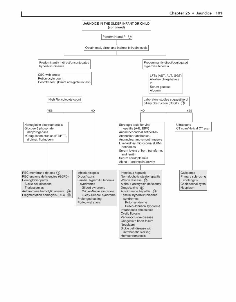
Chapter 26 u Jaundice 101
Serologic tests for viral hepatitis (A-E, EBV)Antimitochondrial antibodiesAntinuclear antibodies Antinuclear anti-smooth muscle Liver-kidney microsomal (LKM) antibodies Serum levels of iron, transferrin, and ferritin Serum ceruloplasmin Alpha-1 antitrypsin activity
Serologic tests for viral hepatitis (A-E, EBV)Antimitochondrial antibodiesAntinuclear antibodies Antinuclear anti-smooth muscle Liver-kidney microsomal (LKM) antibodies Serum levels of iron, transferrin, and ferritin Serum ceruloplasmin Alpha-1 antitrypsin activity
UltrasoundCT scan/Helical CT scanUltrasoundCT scan/Helical CT scan
LFTs (AST, ALT, GGT)Alkaline phosphatasePTSerum glucoseAlbumin
LFTs (AST, ALT, GGT)Alkaline phosphatasePTSerum glucoseAlbumin
Predominantly direct/conjugatedhyperbilirubinemiaPredominantly direct/conjugatedhyperbilirubinemia
Obtain total, direct and indirect bilirubin levelsObtain total, direct and indirect bilirubin levels
Perform H and P 17Perform H and P 17
JAUNDICE IN THE OLDER INFANT OR CHILD(continued)
JAUNDICE IN THE OLDER INFANT OR CHILD(continued)
YESNO
Laboratory studies suggestive ofbiliary obstruction ( GGT) 13 Laboratory studies suggestive ofbiliary obstruction ( GGT) 13
Hemoglobin electrophoresisGlucose 6 phosphate dehydrogenase±Coagulation studies (PT/PTT, d dimer, fibrinogen)
Hemoglobin electrophoresisGlucose 6 phosphate dehydrogenase±Coagulation studies (PT/PTT, d dimer, fibrinogen)
High Reticulocyte countHigh Reticulocyte count
CBC with smearReticulocyte countCoombs test (Direct anti-globulin test)
CBC with smearReticulocyte countCoombs test (Direct anti-globulin test)
Predominantly indirect/unconjugatedhyperbilirubinemiaPredominantly indirect/unconjugatedhyperbilirubinemia
YES NO
RBC membrane defects 7RBC enzyme deficiencies (G6PD)Hemoglobinopathy Sickle cell diseases ThalassemiasAutoimmune hemolytic anemia 18Fragmentation hemolysis (DIC) 19
RBC membrane defects 7RBC enzyme deficiencies (G6PD)Hemoglobinopathy Sickle cell diseases ThalassemiasAutoimmune hemolytic anemia 18Fragmentation hemolysis (DIC) 19
Infection/sepsisDrugs/toxinsFamilial hyperbilirubinemia syndromes Gilbert syndrome Crigler-Najjar syndrome Lucey-Driscoll syndromeProlonged fastingPortocaval shunt
Infection/sepsisDrugs/toxinsFamilial hyperbilirubinemia syndromes Gilbert syndrome Crigler-Najjar syndrome Lucey-Driscoll syndromeProlonged fastingPortocaval shunt
GallstonesPrimary sclerosing cholangitisCholedochal cystsNeoplasm
GallstonesPrimary sclerosing cholangitisCholedochal cystsNeoplasm
Infectious hepatitisNon-alcoholic steatohepatitisWilson disease 20Alpha-1 antitrypsin deficiencyDrugs/toxins 21Autoimmune hepatitis 22Familial hyperbilirubinemia syndromes Rotor syndrome Dubin-Johnson syndromeIntrahepatic cholestasisCystic fibrosisVeno-occlusive diseaseCongestive heart failureNeoplasmSickle cell disease with intrahepatic sicklingHemochromatosis
Infectious hepatitisNon-alcoholic steatohepatitisWilson disease 20Alpha-1 antitrypsin deficiencyDrugs/toxins 21Autoimmune hepatitis 22Familial hyperbilirubinemia syndromes Rotor syndrome Dubin-Johnson syndromeIntrahepatic cholestasisCystic fibrosisVeno-occlusive diseaseCongestive heart failureNeoplasmSickle cell disease with intrahepatic sicklingHemochromatosis
102
Chapter 27
HEPATOMEGALY
2 Elevated transaminase levels provide nonspecific informa-tion about hepatocellular injury. Liver function is better as-
sessed by serum albumin and prothrombin time because they rely on the synthetic function of the liver. Increased levels of alkaline phosphatase and GGT usually indicate cholestasis. Abdominal US will aid in determining whether hepatomegaly is present.
3 Inflammatory bowel disease can be associated with hepa-tobiliary disease through a variety of mechanisms. These
include autoimmune (autoimmune hepatitis), inflammatory (sclerosing cholangitis), medication toxicity (e.g., methotrex-ate), and related infections (hepatic abscess).
4 Nonalcoholic fatty liver disease is associated with obesity. It may cause fatty liver and ultimately inflammation and
fibrosis (nonalcoholic steatohepatitis). It used to be an adult disease but is increasingly described in children.
5 Patients with HIV/AIDS are prone to a variety of hepatobili-ary disorders. Other infections that cause hepatosplenomeg-
aly and anicteric hepatitis include cat-scratch disease, typhoid, brucellosis, tularemia, syphilis, Lyme disease, leptospirosis, Rocky Mountain spotted fever, Q fever, tuberculosis, and actinomycosis. Fitz-Hugh–Curtis syndrome is a perihepatitis associated with pel-vic inflammatory disease due to Neisseria gonorrhoeae or Chla-mydia trachomatis.
6 Metabolic storage disorders and peroxisomal disorders should be suspected in infants with hepatomegaly, hypotonia,
and loss of developmental milestones. These include amino acid defects (e.g., tyrosinemia), glycogen storage disorders, galactose-mia, mucopolysaccharidoses, and disorders of lipid metabolism. In addition to the screening tests listed, consider referral for further testing, liver biopsy, specific lymphocyte or urine assay, or bone marrow examination, which may be necessary to diagnose certain metabolic disorders. (See Chapter 22.)
7 Fulminant hepatic failure is the development of advanced liver disease in the absence of previous liver disease. The
etiology includes infection, drug reactions, Reye syndrome, and Wilson disease. Fulminant hepatic failure is unlikely in the ab-sence of jaundice.
The presence of a palpable liver does not always indicate hepatomegaly. Liver size varies with age. It may be measured by the extension of the liver edge below the costal margin, the span of dullness to percussion, or by imaging. In children, palpation of the liver edge more than 2 cm (.3.5 cm in new-borns) below the right costal margin suggests liver enlarge-ment. Liver span may be measured by percussing the upper margin of dullness and by palpating the lower edge in the right midclavicular line.
1 Inquire about a history of prolonged hyperbilirubinemia, as well as underlying conditions that may contribute to
liver disease (e.g., blood transfusions). Ask about episodic vomiting, associated neurologic changes, travel, and drug or toxin ingestion. Metabolic disease is suggested by symptoms of failure to thrive, vomiting, loss of developmental mile-stones, new seizures, or hypotonia. The social history should include sexual activity, medication and drug use, and expo-sure to possible hepatitis outbreaks or jaundiced persons. A family history of hepatic, neurologic, and psychiatric symp-toms should be elicited, as well as a history of neonatal deaths. Birth history may identify perinatally transmitted infections (hepatitis B, toxoplasmosis, syphilis, CMV, rubella, herpes simplex, HIV).
Cutaneous findings suggestive of liver disease include a prominent abdominal venous pattern, palmar erythema, and spider angiomas. Hepatomegaly is often associated with jaun-dice. (See Chapter 26 for additional diagnoses to consider.) When splenomegaly is present as well, other diagnoses may be considered. (See Chapter 28.) Specific signs may indicate syn-dromes. Coarse facial features may be seen with mucopolysac-charoidosis. Kayser-Fleischer rings are seen in Wilson disease.
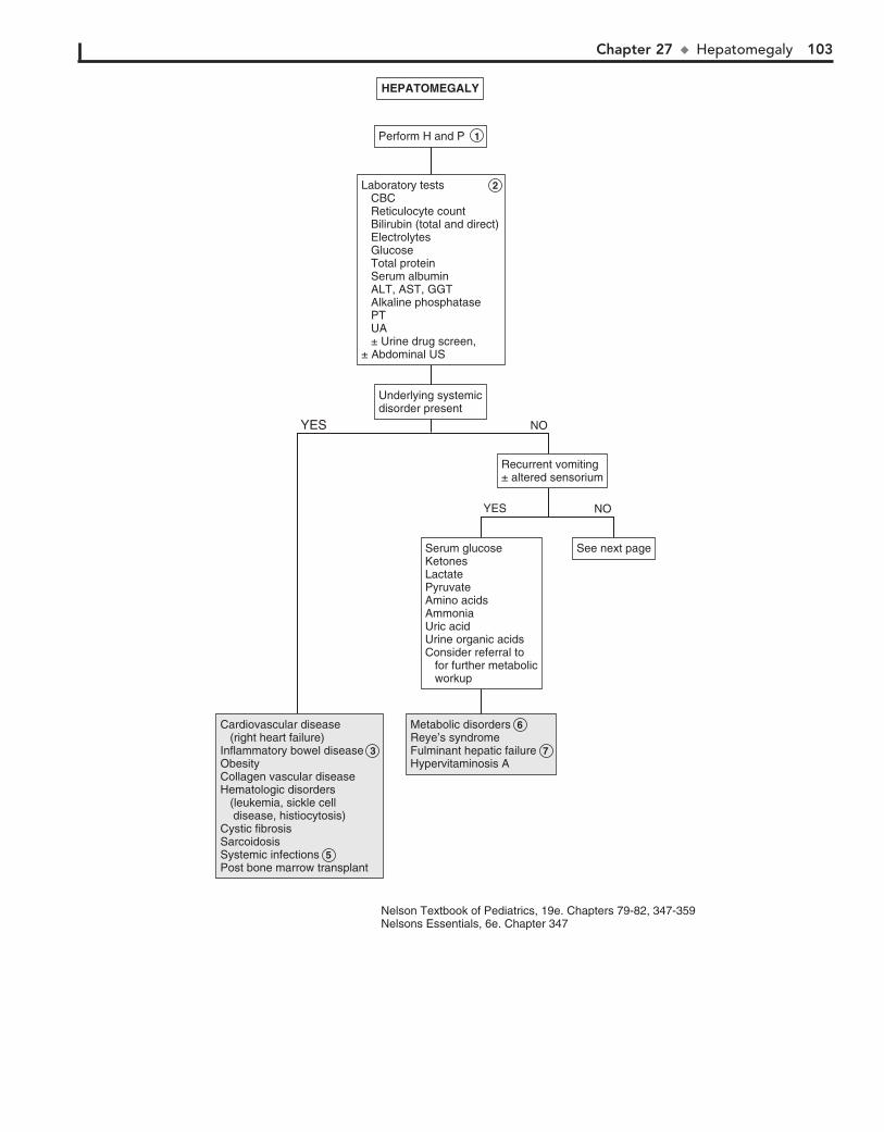
Chapter 27 u Hepatomegaly 103
Nelson Textbook of Pediatrics, 19e. Chapters 79-82, 347-359Nelsons Essentials, 6e. Chapter 347
Serum glucoseKetonesLactatePyruvateAmino acidsAmmoniaUric acidUrine organic acidsConsider referral to for further metabolic workup
Serum glucoseKetonesLactatePyruvateAmino acidsAmmoniaUric acidUrine organic acidsConsider referral to for further metabolic workup
Underlying systemicdisorder presentUnderlying systemicdisorder present
HEPATOMEGALYHEPATOMEGALY
Perform H and P 1 Perform H and P 1
Laboratory tests 2 CBC Reticulocyte count Bilirubin (total and direct) Electrolytes Glucose Total protein Serum albumin ALT, AST, GGT Alkaline phosphatase PT UA ± Urine drug screen,± Abdominal US
Laboratory tests 2 CBC Reticulocyte count Bilirubin (total and direct) Electrolytes Glucose Total protein Serum albumin ALT, AST, GGT Alkaline phosphatase PT UA ± Urine drug screen,± Abdominal US
Cardiovascular disease (right heart failure)Inflammatory bowel disease 3 ObesityCollagen vascular diseaseHematologic disorders (leukemia, sickle cell disease, histiocytosis)Cystic fibrosisSarcoidosisSystemic infections 5Post bone marrow transplant
Cardiovascular disease (right heart failure)Inflammatory bowel disease 3 ObesityCollagen vascular diseaseHematologic disorders (leukemia, sickle cell disease, histiocytosis)Cystic fibrosisSarcoidosisSystemic infections 5Post bone marrow transplant
Metabolic disorders 6Reye’s syndromeFulminant hepatic failure 7 Hypervitaminosis A
Metabolic disorders 6Reye’s syndromeFulminant hepatic failure 7 Hypervitaminosis A
Recurrent vomiting± altered sensoriumRecurrent vomiting± altered sensorium
See next pageSee next page
NO
NO
YES
YES

104 Part IV u GastrointestinalSystem
8 Zellweger syndrome is the only disorder of peroxisomal metabolism associated with hepatomegaly. Diagnosis is
typically made early in life owing to a characteristic phenotype, severe hypotonia, and hepatomegaly.
9 Wilson disease is in the differential diagnosis of acute hepatitis in all children older than 5 years. Kayser-Fleischer
rings and neurologic symptoms (e.g., tremor, choreiform move-ments) are suggestive. Diagnosis is by low serum ceruloplasmin levels, high urinary copper excretion, and increased hepatic copper levels on liver biopsy.
10 Acute hepatitis may present as a mild or severe illness. Most cases resemble a mild flulike nonicteric illness with
only tender hepatomegaly on examination. Mild splenomegaly may also occur. Consider serology for CMV and EBV when a high fever and adenopathy are present. Liver biopsy may occa-sionally be necessary to diagnose certain forms of chronic hepatitis.
11 A nonspecific fatty infiltration of the liver occurs in re-sponse to a variety of disorders (e.g., diabetes, cystic fibrosis,
inflammatory bowel disease, metabolic disorders). Chemother-apy, other medication and toxins, malnutrition, and obesity are also risk factors.
12 Ascites and tender hepatomegaly are common presenting symptoms of hepatic venous outflow obstruction. Serum
transaminases and bilirubin levels are minimally affected acutely. Budd-Chiari syndrome is a noncardiogenic hepatic venous outflow obstruction occurring in conditions predispos-ing to thrombosis.
BibliographyKliegman RM, Stanton B, St. Geme J, Schor N, Behrman RE, editors: Nelson
textbook of pediatrics, ed 19, Philadelphia, 2011, Elsevier/Saunders, Chapters 79-82, 347-359.
Ross H, Hight DW, Weiss RG: Abdominal masses in pediatric patients. In Wyllie R, Hyams J, editors: Pediatric gastrointestinal disease: Pathophysiol-ogy, diagnosis, management, ed 2, Philadelphia, 1999, WB Saunders, p 126.
Suchy FJ: Hepatomegaly. In Kliegman RM, Greenbaum, LA, Lye PS, editors: Practical strategies in pediatric diagnosis and therapy, ed 2, Philadelphia, 2004, Saunders, pp 333–344.
Wolf AD, Lavine JE: Hepatomegaly in neonates and children, Pediatr Rev 21:303–310, 2000.
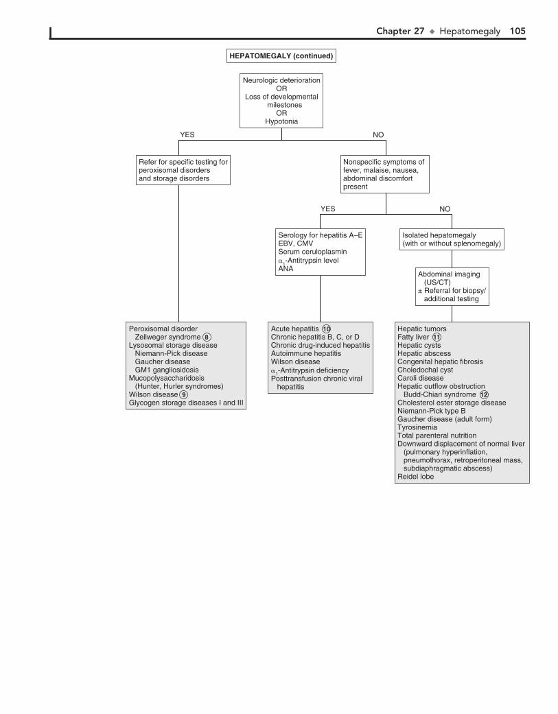
Chapter 27 u Hepatomegaly 105
Abdominal imaging (US/CT)± Referral for biopsy/ additional testing
Abdominal imaging (US/CT)± Referral for biopsy/ additional testing
Serology for hepatitis A–EEBV, CMVSerum ceruloplasmin�1-Antitrypsin levelANA
Serology for hepatitis A–EEBV, CMVSerum ceruloplasmin�1-Antitrypsin levelANA
HEPATOMEGALY (continued)HEPATOMEGALY (continued)
Neurologic deteriorationOR
Loss of developmental milestones
ORHypotonia
Neurologic deteriorationOR
Loss of developmental milestones
ORHypotonia
Acute hepatitis 10Chronic hepatitis B, C, or DChronic drug-induced hepatitisAutoimmune hepatitisWilson disease�1-Antitrypsin deficiencyPosttransfusion chronic viral hepatitis
Acute hepatitis 10Chronic hepatitis B, C, or DChronic drug-induced hepatitisAutoimmune hepatitisWilson disease�1-Antitrypsin deficiencyPosttransfusion chronic viral hepatitis
Peroxisomal disorder Zellweger syndrome 8Lysosomal storage disease Niemann-Pick disease Gaucher disease GM1 gangliosidosisMucopolysaccharidosis (Hunter, Hurler syndromes)Wilson disease 9Glycogen storage diseases I and III
Peroxisomal disorder Zellweger syndrome 8Lysosomal storage disease Niemann-Pick disease Gaucher disease GM1 gangliosidosisMucopolysaccharidosis (Hunter, Hurler syndromes)Wilson disease 9Glycogen storage diseases I and III
Hepatic tumorsFatty liver 11Hepatic cystsHepatic abscessCongenital hepatic fibrosisCholedochal cystCaroli diseaseHepatic outflow obstruction Budd-Chiari syndrome 12Cholesterol ester storage diseaseNiemann-Pick type BGaucher disease (adult form)TyrosinemiaTotal parenteral nutritionDownward displacement of normal liver (pulmonary hyperinflation, pneumothorax, retroperitoneal mass, subdiaphragmatic abscess)Reidel lobe
Hepatic tumorsFatty liver 11Hepatic cystsHepatic abscessCongenital hepatic fibrosisCholedochal cystCaroli diseaseHepatic outflow obstruction Budd-Chiari syndrome 12Cholesterol ester storage diseaseNiemann-Pick type BGaucher disease (adult form)TyrosinemiaTotal parenteral nutritionDownward displacement of normal liver (pulmonary hyperinflation, pneumothorax, retroperitoneal mass, subdiaphragmatic abscess)Reidel lobe
Nonspecific symptoms of fever, malaise, nausea, abdominal discomfortpresent
Nonspecific symptoms of fever, malaise, nausea, abdominal discomfortpresent
Refer for specific testing forperoxisomal disordersand storage disorders
Refer for specific testing forperoxisomal disordersand storage disorders
Isolated hepatomegaly(with or without splenomegaly)Isolated hepatomegaly(with or without splenomegaly)
NO
NO
YES
YES
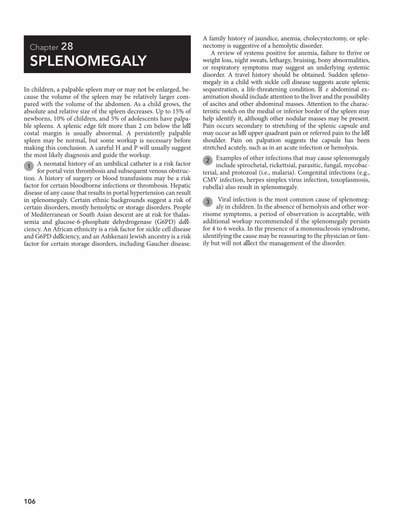
106
Chapter 28
SPLENOMEGALY
A family history of jaundice, anemia, cholecystectomy, or sple-nectomy is suggestive of a hemolytic disorder.
A review of systems positive for anemia, failure to thrive or weight loss, night sweats, lethargy, bruising, bony abnormalities, or respiratory symptoms may suggest an underlying systemic disorder. A travel history should be obtained. Sudden spleno-megaly in a child with sickle cell disease suggests acute splenic sequestration, a life-threatening condition. The abdominal ex-amination should include attention to the liver and the possibility of ascites and other abdominal masses. Attention to the charac-teristic notch on the medial or inferior border of the spleen may help identify it, although other nodular masses may be present. Pain occurs secondary to stretching of the splenic capsule and may occur as left upper quadrant pain or referred pain to the left shoulder. Pain on palpation suggests the capsule has been stretched acutely, such as in an acute infection or hemolysis.
2 Examples of other infections that may cause splenomegaly include spirochetal, rickettsial, parasitic, fungal, mycobac-
terial, and protozoal (i.e., malaria). Congenital infections (e.g., CMV infection, herpes simplex virus infection, toxoplasmosis, rubella) also result in splenomegaly.
3 Viral infection is the most common cause of splenomeg-aly in children. In the absence of hemolysis and other wor-
risome symptoms, a period of observation is acceptable, with additional workup recommended if the splenomegaly persists for 4 to 6 weeks. In the presence of a mononucleosis syndrome, identifying the cause may be reassuring to the physician or fam-ily but will not affect the management of the disorder.
In children, a palpable spleen may or may not be enlarged, be-cause the volume of the spleen may be relatively larger com-pared with the volume of the abdomen. As a child grows, the absolute and relative size of the spleen decreases. Up to 15% of newborns, 10% of children, and 5% of adolescents have palpa-ble spleens. A splenic edge felt more than 2 cm below the left costal margin is usually abnormal. A persistently palpable spleen may be normal, but some workup is necessary before making this conclusion. A careful H and P will usually suggest the most likely diagnosis and guide the workup.
1 A neonatal history of an umbilical catheter is a risk factor for portal vein thrombosis and subsequent venous obstruc-
tion. A history of surgery or blood transfusions may be a risk factor for certain bloodborne infections or thrombosis. Hepatic disease of any cause that results in portal hypertension can result in splenomegaly. Certain ethnic backgrounds suggest a risk of certain disorders, mostly hemolytic or storage disorders. People of Mediterranean or South Asian descent are at risk for thalas-semia and glucose-6-phosphate dehydrogenase (G6PD) defi-ciency. An African ethnicity is a risk factor for sickle cell disease and G6PD deficiency, and an Ashkenazi Jewish ancestry is a risk factor for certain storage disorders, including Gaucher disease.
Chapter 28 u Splenomegaly 107
Perform H and P 1 Perform H and P 1
US/abdominal CT/MRIUS/abdominal CT/MRI
SplenomegalyresolvedSplenomegalyresolved
SPLENOMEGALYSPLENOMEGALY
CBC with differentialSerology for EBV± Testing for CMV, toxoplasmosis± HIV testing
CBC with differentialSerology for EBV± Testing for CMV, toxoplasmosis± HIV testing
Obtain CBC with differential 4 Peripheral smearReticulocyte count
Obtain CBC with differential 4 Peripheral smearReticulocyte count
Acute viral infection 3 Acute viral infection 3 EBVCMVToxoplasmosisHIVOther infection 2 Bacterial (Salmonella typhi, infective endocarditis, splenic abscess) Spirochetal (syphilis, leptospirosis) Rickettsial (Rocky Mountain spotted fever, Q fever, typhus) Fungal (blastomycosis, systemic candidiasis) Mycobacterial (tuberculosis) Parasitic (malaria, toxoplasmosis, Toxocara canis, leishmaniasis)
EBVCMVToxoplasmosisHIVOther infection 2 Bacterial (Salmonella typhi, infective endocarditis, splenic abscess) Spirochetal (syphilis, leptospirosis) Rickettsial (Rocky Mountain spotted fever, Q fever, typhus) Fungal (blastomycosis, systemic candidiasis) Mycobacterial (tuberculosis) Parasitic (malaria, toxoplasmosis, Toxocara canis, leishmaniasis)
Infection 2HistiocytosisMalignancy (leukemia, lymphoma, Hodgkin disease, metastases)Collagen vascular disease (systemic lupus erythematosus, rheumatoid arthritis, autoimmune lymphoproliferative syndrome)
Infection 2HistiocytosisMalignancy (leukemia, lymphoma, Hodgkin disease, metastases)Collagen vascular disease (systemic lupus erythematosus, rheumatoid arthritis, autoimmune lymphoproliferative syndrome)
Observe 4-6weeks (unlessclinically ill)
Observe 4-6weeks (unlessclinically ill)
See next pageSee next page
Features suggestiveof infectious or viraletiology present: Pharyngitis Fever Lymphadenopathy
Features suggestiveof infectious or viraletiology present: Pharyngitis Fever Lymphadenopathy
AbnormalresultsAbnormalresults
NormalresultsNormalresults
Splenomegaly persistent(or patient ill)Splenomegaly persistent(or patient ill)
YES NO
Nelson Textbook of Pediatrics, 19e. Chapter 480Nelsons Essentials, 6e. Chapter 150
108 Part IV u GastrointestinalSystem
4 A CBC, blood smear, and reticulocyte count may indicate hemolytic disease, chronic anemia, or leukemia. In a child
up to 3 years of age with a normal CBC, a spleen that is palpable 2 cm below the costal margin may be normal.
5 Laboratory findings suggesting hemolysis include abnor-mal cell morphology; increased reticulocyte count, RBC
distribution width (RDW), indirect bilirubin, urine urobilino-gen, and lactate dehydrogenase; decreased serum haptoglobin; and hemoglobinuria.
6 When blasts are seen on peripheral smear, it indicates leu-kemia requiring referral for bone marrow examination.
7 Sequestration of RBCs may cause rapid splenic enlargement. It may be seen in congenital spherocytosis or other congeni-
tal or acquired hemolytic anemias (e.g., sickle cell disease).
8 Mechanical damage causing fragmentation hemolysis may occur in systemic disorders such as disseminated intravas-
cular coagulation, thrombotic thrombocytopenic purpura, and hemolytic-uremic syndrome. Extracorporeal membrane oxy-genation, prosthetic heart valves, and burns may cause hemoly-sis by a similar mechanism.
9 Obstruction of venous drainage at a prehepatic or intrahepatic level or in the portal vein can cause a congestive splenomegaly.
Congestive heart failure, Budd-Chiari syndrome, and hepatic disor-ders leading to cirrhosis are causes. Pancytopenia frequently results from excessive pooling (i.e., hypersplenism).
10 Storage disorders (e.g., Gaucher disease, glycogen storage disease, mucopolysaccharidosis) are usually diagnosed
early in life with abnormal growth, skeletal abnormalities, and developmental delay.
11 Osteopetrosis refers to disorders of increased skeletal density. Hepatosplenomegaly occurs secondary to extramedullary
hematopoiesis in severe forms. Milder variants of the disease may have only a mild anemia and no significant hepatosplenomegaly.
12 Pulmonary hyperinflation due to asthma, bronchiolitis, or a pneumothorax may cause splenic displacement.
BibliographyKliegman RM, Stanton B, St. Geme J, Schor N, Behrman RE, editors: Nelson
textbook of pediatrics, ed 19, Philadelphia, 2011, Elsevier/Saunders, Chapter 480.
Maloney KW: Splenomegaly. In Kliegman RM, Greenbaum LA, Lye PS, editors: Practical strategies in pediatric diagnosis and therapy, ed 2, Philadelphia, 2004, Elsevier, pp 345–352.
Reznik M, Ozuah PO: Splenomegaly. In McInerny TK, Adam HM, Campbell DE, et al, editors: American Academy of Pediatrics textbook of pediatric care, ed 1, Elk Grove Village, Ill, 2008, AAP, pp 1730.
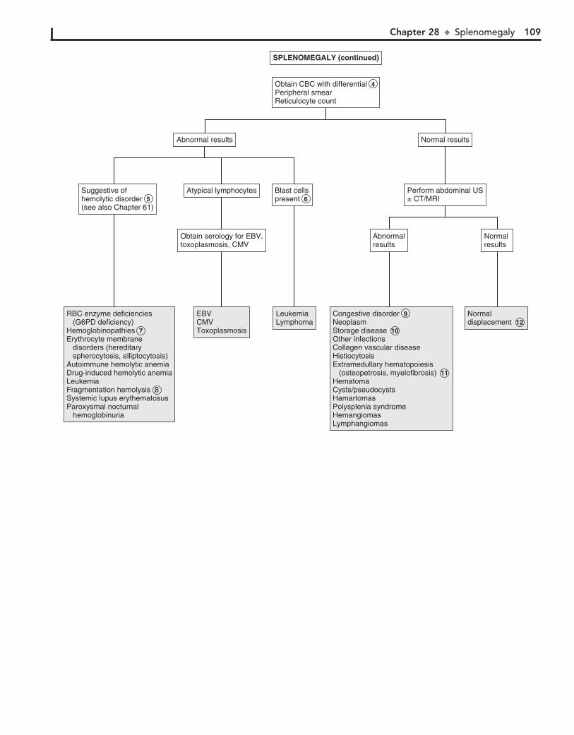
Chapter 28 u Splenomegaly 109
Normal resultsNormal results
SPLENOMEGALY (continued)SPLENOMEGALY (continued)
Obtain CBC with differential 4 Peripheral smearReticulocyte count
Obtain CBC with differential 4 Peripheral smearReticulocyte count
EBVCMVToxoplasmosis
EBVCMVToxoplasmosis
LeukemiaLymphomaLeukemiaLymphoma
Normaldisplacement 12 Normaldisplacement 12
RBC enzyme deficiencies (G6PD deficiency)Hemoglobinopathies 7Erythrocyte membrane disorders (hereditary spherocytosis, elliptocytosis)Autoimmune hemolytic anemiaDrug-induced hemolytic anemiaLeukemiaFragmentation hemolysis 8Systemic lupus erythematosusParoxysmal nocturnal hemoglobinuria
RBC enzyme deficiencies (G6PD deficiency)Hemoglobinopathies 7Erythrocyte membrane disorders (hereditary spherocytosis, elliptocytosis)Autoimmune hemolytic anemiaDrug-induced hemolytic anemiaLeukemiaFragmentation hemolysis 8Systemic lupus erythematosusParoxysmal nocturnal hemoglobinuria
Congestive disorder 9 NeoplasmStorage disease 10 Other infectionsCollagen vascular diseaseHistiocytosisExtramedullary hematopoiesis (osteopetrosis, myelofibrosis) 11 HematomaCysts/pseudocystsHamartomasPolysplenia syndromeHemangiomasLymphangiomas
Congestive disorder 9 NeoplasmStorage disease 10 Other infectionsCollagen vascular diseaseHistiocytosisExtramedullary hematopoiesis (osteopetrosis, myelofibrosis) 11 HematomaCysts/pseudocystsHamartomasPolysplenia syndromeHemangiomasLymphangiomas
Perform abdominal US± CT/MRIPerform abdominal US± CT/MRI
Blast cellspresent 6 Blast cellspresent 6
Suggestive ofhemolytic disorder 5 (see also Chapter 61)
Suggestive ofhemolytic disorder 5 (see also Chapter 61)
Atypical lymphocytesAtypical lymphocytes
Normal resultsNormal resultsAbnormal resultsAbnormal results
AbnormalresultsAbnormalresults
Obtain serology for EBV,toxoplasmosis, CMVObtain serology for EBV,toxoplasmosis, CMV
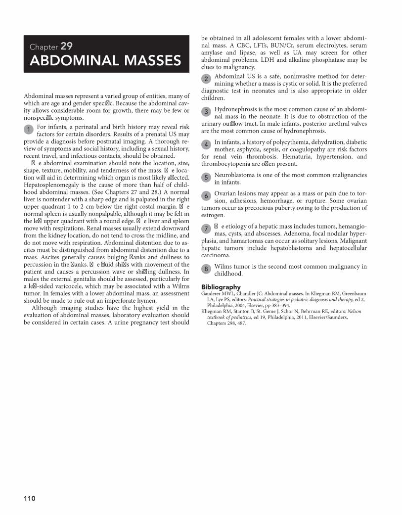
110
Chapter 29
ABDOMINAL MASSES
be obtained in all adolescent females with a lower abdomi-nal mass. A CBC, LFTs, BUN/Cr, serum electrolytes, serum amylase and lipase, as well as UA may screen for other abdominal problems. LDH and alkaline phosphatase may be clues to malignancy.
2 Abdominal US is a safe, noninvasive method for deter-mining whether a mass is cystic or solid. It is the preferred
diagnostic test in neonates and is also appropriate in older children.
3 Hydronephrosis is the most common cause of an abdomi-nal mass in the neonate. It is due to obstruction of the
urinary outflow tract. In male infants, posterior urethral valves are the most common cause of hydronephrosis.
4 In infants, a history of polycythemia, dehydration, diabetic mother, asphyxia, sepsis, or coagulopathy are risk factors
for renal vein thrombosis. Hematuria, hypertension, and thrombocytopenia are often present.
5 Neuroblastoma is one of the most common malignancies in infants.
6 Ovarian lesions may appear as a mass or pain due to tor-sion, adhesions, hemorrhage, or rupture. Some ovarian
tumors occur as precocious puberty owing to the production of estrogen.
7 The etiology of a hepatic mass includes tumors, hemangio-mas, cysts, and abscesses. Adenoma, focal nodular hyper-
plasia, and hamartomas can occur as solitary lesions. Malignant hepatic tumors include hepatoblastoma and hepatocellular carcinoma.
8 Wilms tumor is the second most common malignancy in childhood.
Abdominal masses represent a varied group of entities, many of which are age and gender specific. Because the abdominal cav-ity allows considerable room for growth, there may be few or nonspecific symptoms.
1 For infants, a perinatal and birth history may reveal risk factors for certain disorders. Results of a prenatal US may
provide a diagnosis before postnatal imaging. A thorough re-view of symptoms and social history, including a sexual history, recent travel, and infectious contacts, should be obtained.
The abdominal examination should note the location, size, shape, texture, mobility, and tenderness of the mass. The loca-tion will aid in determining which organ is most likely affected. Hepatosplenomegaly is the cause of more than half of child-hood abdominal masses. (See Chapters 27 and 28.) A normal liver is nontender with a sharp edge and is palpated in the right upper quadrant 1 to 2 cm below the right costal margin. The normal spleen is usually nonpalpable, although it may be felt in the left upper quadrant with a round edge. The liver and spleen move with respirations. Renal masses usually extend downward from the kidney location, do not tend to cross the midline, and do not move with respiration. Abdominal distention due to as-cites must be distinguished from abdominal distention due to a mass. Ascites generally causes bulging flanks and dullness to percussion in the flanks. The fluid shifts with movement of the patient and causes a percussion wave or shifting dullness. In males the external genitalia should be assessed, particularly for a left-sided varicocele, which may be associated with a Wilms tumor. In females with a lower abdominal mass, an assessment should be made to rule out an imperforate hymen.
Although imaging studies have the highest yield in the evaluation of abdominal masses, laboratory evaluation should be considered in certain cases. A urine pregnancy test should
BibliographyGauderer MWL, Chandler JC: Abdominal masses. In Kliegman RM, Greenbaum
LA, Lye PS, editors: Practical strategies in pediatric diagnosis and therapy, ed 2, Philadelphia, 2004, Elsevier, pp 383–394.
Kliegman RM, Stanton B, St. Geme J, Schor N, Behrman RE, editors: Nelson textbook of pediatrics, ed 19, Philadelphia, 2011, Elsevier/Saunders, Chapters 298, 487.
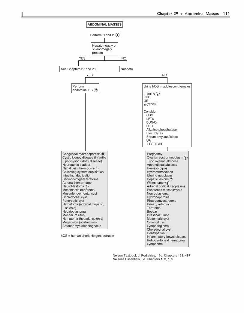
Chapter 29 u AbdominalMasses 111
ABDOMINAL MASSESABDOMINAL MASSES
NeonateNeonate
Hepatomegaly orsplenomegalypresent
Hepatomegaly orsplenomegalypresent
Perform H and P 1 Perform H and P 1
See Chapters 27 and 28See Chapters 27 and 28
Urine hCG in adolescent females
Imaging 2KUBUS± CT/MRI
Consider: CBC LFTs BUN/Cr LDH Alkaline phosphatase Electrolytes Serum amylase/lipase UA ± ESR/CRP
Urine hCG in adolescent females
Imaging 2KUBUS± CT/MRI
Consider: CBC LFTs BUN/Cr LDH Alkaline phosphatase Electrolytes Serum amylase/lipase UA ± ESR/CRP
Performabdominal US 2 Performabdominal US 2
PregnancyOvarian cyst or neoplasm 6 Tubo ovarian abscessAppendiceal abscessHematocolposHydrometrocolposUterine neoplasmHepatic lesions 7Wilms tumor 8Adrenal cortical neoplasmsPancreatic masses/cystsNeuroblastomaHydronephrosisRhabdomyosarcomaUrinary retentionTeratomaBezoarIntestinal tumorMesenteric cystOmental cystLymphangiomaCholedochal cystConstipationInflammatory bowel diseaseRetroperitoneal hematomaLymphoma
PregnancyOvarian cyst or neoplasm 6 Tubo ovarian abscessAppendiceal abscessHematocolposHydrometrocolposUterine neoplasmHepatic lesions 7Wilms tumor 8Adrenal cortical neoplasmsPancreatic masses/cystsNeuroblastomaHydronephrosisRhabdomyosarcomaUrinary retentionTeratomaBezoarIntestinal tumorMesenteric cystOmental cystLymphangiomaCholedochal cystConstipationInflammatory bowel diseaseRetroperitoneal hematomaLymphoma
Congenital hydronephrosis 3 Cystic kidney disease (infantile polycystic kidney disease)Neurogenic bladderRenal vein thrombosis 4Collecting system duplicationIntestinal duplicationSacrococcygeal teratomaAdrenal hemorrhageNeuroblastoma 5Mesoblastic nephromaMesenteric/omental cystCholedochal cystPancreatic cystHematoma (adrenal, hepatic, splenic)HepatoblastomaMeconium ileusHematoma (hepatic, splenic)Megacolon (obstruction)Anterior myelomeningocele
Congenital hydronephrosis 3 Cystic kidney disease (infantile polycystic kidney disease)Neurogenic bladderRenal vein thrombosis 4Collecting system duplicationIntestinal duplicationSacrococcygeal teratomaAdrenal hemorrhageNeuroblastoma 5Mesoblastic nephromaMesenteric/omental cystCholedochal cystPancreatic cystHematoma (adrenal, hepatic, splenic)HepatoblastomaMeconium ileusHematoma (hepatic, splenic)Megacolon (obstruction)Anterior myelomeningocele
NO
NO
YES
YES
Nelson Textbook of Pediatrics, 19e. Chapters 198, 487Nelsons Essentials, 6e. Chapters 153, 159
hCG = human chorionic gonadotropin

This page intentionally left blank

Genitourinary System PART
V
114
Chapter 30
DYSURIA
should also be performed if there is a recurrence of a febrile UTI. For males, a UTI at any age requires further evaluation.
4 Nucleic acid amplification tests (NAAT) for Neisseria gon-orrhoeae and Chlamydia trachomatis can be performed on
urine as well as genital specimens (urethral or cervical). Tests based on nucleic acid hybridization are also available. Penile, vaginal, or cervical cultures may be used as well. Wet saline mount of vaginal secretions with microscopy, including use of potassium hydroxide, and Gram stain can be used to detect Trichomonas, as well as bacterial vaginosis and vaginal candi-diasis. Tests to detect Trichomonas antigen are also used.
5 The presence of leukocytes on UA may indicate vaginitis due to sexually transmitted infections (N. gonorrhoeae, Chla-
mydia, herpes simplex, Trichomonas vaginalis). They may also indicate Candida albicans, enteric pathogens, or group A strepto-cocci. Urethritis is caused by N. gonorrhoeae, Chlamydia, Myco-plasma genitalium, and Ureaplasma urealyticum. (See Chapter 41.)
6 Nonspecific urethritis is often seen in premenarchal girls and is associated with poor hygiene, tight “nonbreathing”
clothes, and chemical irritants (bubble bath, harsh soaps). There may be erythema in the periurethral area. Anal pruritus may indicate pinworms, which can cause urethral irritation and can be confirmed by examination with a tape slide test.
7 Labial adhesions are common in prepubertal girls and are due to recurrent irritation, trauma, or infection of the hy-
poestrogenized epithelium of the labia minora. Urethral prolapse is an eversion of the urethral mucosa through the meatus. Bleed-ing and dysuria are common. Lichen sclerosus causes an hour-glass-shaped area of atrophy and scarring with depigmentation. Herpes simplex causes ulcers and vesicles. It may be detected by a scraping for direct fluorescent antibody (DFA) testing, PCR, or viral culture. Sexual abuse is often associated with rectal or vaginal bleeding and vaginitis. Appropriate studies should be obtained.
8 Microscopic hematuria may be seen with UTI. Gross hema-turia is seen with hemorrhagic cystitis (adenovirus, cyclo-
phosphamide), renal calculi, hypercalcuria, and trauma and is rarely tumor related (clots from Wilms tumor). (See Chapter 32.)
9 Meatal stenosis may occur for a number of reasons. In circumcised boys, it may result from recurrent meatal in-
flammation from moist diapers. Trauma, hypospadias repair, catheterization, and balanitis xerotica obliterans are other causes. It most often appears as an abnormal urine stream, intermittent dysuria, and occasional bleeding. Phimosis is when the foreskin cannot be retracted because of scarring or narrowing of the pre-putial opening. It must be distinguished from physiologic phimo-sis when the foreskin has not completed the normal separation from the glans, usually by 3 to 5 years of age. Paraphimosis is the incarceration of the prepuce behind the glans, often after forcible retraction of the foreskin. Balanitis is an inflammation of the pre-puce, which is usually due to urine, but infection may be involved.
Dysuria is pain or burning occurring with urination. It is often associated with urinary symptoms such as frequency, urgency, incontinence, and refusal to void. Dysuria is not specific for UTIs and often occurs in young children.
1 Toddlers may present with secondary enuresis, dribbling, and frequent squatting. Constipation, not being circumcised,
female gender, contraceptive diaphragms, and sexual intercourse may predispose to a UTI. Chemical irritants (e.g., detergents, bubble bath) and mechanical irritation (e.g., masturbation, for-eign body) may cause dysuria. Dark or tea-colored urine may in-dicate hematuria. A history of penile or vaginal discharge as well as sexual abuse should be elicited.
Physical examination should include BP, genitalia, abdomi-nal palpation of the kidneys, pelvic exam when indicated, and a careful neurologic exam in children with voiding dysfunction to exclude spinal cord pathology.
2 Urine for UA and culture must be properly obtained. UA, which includes microscopic examination and dipstick
method, correlates with infection, particularly in an older child. Dipstick methods test for leukocyte esterase (an enzyme present in WBCs) and nitrites. Microscopic analysis of unspun urine for WBCs (10/mm3) or bacteria on Gram stain is more predictive than spun urine (.5 WBCs/high-power field), which is com-monly used. RBCs are common with a bacterial UTI. WBC casts, when present, are associated with upper tract infections.
Urine culture remains the standard for diagnosis. Any growth in urine collected by suprapubic tap, more than 50,000 colonies by catheterization, or more than 105 colonies by clean-catch mid-stream urine indicate infection. “Bagged” specimens may have skin or fecal contaminants and are not recommended. Esche-richia coli is the most common pathogen; others include Proteus, Klebsiella, Staphylococcus saprophyticus, and enterococcus.
3 In older children, pyelonephritis may be clinically differ-entiated from cystitis by the presence of systemic features
(fever, vomiting) and signs (flank pain, costovertebral angle tenderness). In infants and young children, the clinical picture may be nonspecific, with fever and other symptoms present in upper or lower tract disease. If necessary, the diagnosis of py-elonephritis may be confirmed by renal scan, the most sensitive being 99mTc-dimercaptosuccinic acid (DMSA). Renal US may also show pyelonephritis but is not as sensitive; however, it is adequate to detect obstructive uropathy or high-grade reflux that may be associated with pyelonephritis.
The occurrence of the first episode of febrile UTIs in a child aged 2 to 24 months may be a marker for congenital anatomic abnormalities—in particular, vesicourethral reflux (VUR). Ac-cording to recent guidelines, US of the kidneys and bladder should be performed to detect anatomic abnormalities. A voiding cystourethrogram (VCUG) is indicated if renal and bladder US reveals hydronephrosis, scarring, or other findings that would suggest either high-grade VUR or obstructive uropathy. VCUG
BibliographySubcommittee on Urinary Tract Infection, Steering Committee on Quality Im-
provement and Management, Roberts KB: Urinary tract infection: Clinical practice guideline for the diagnosis and management of the initial UTI in febrile infants and children 2 to 24 months, Pediatrics 128:595–610, 2011.
Kliegman RM, Stanton B, St. Geme J, Schor N, Behrman RE, editors: Nelson textbook of pediatrics, ed 19, Philadelphia, 2011, Elsevier/Saunders, Chapters 114, 185, 254, 276, 513, 532, 538.
Chapter 30 u Dysuria 115
STI = sexually transmitted infection
Nelson Textbook of Pediatrics, 19e. Chapters 114, 185, 254, 276, 513, 532, 538Nelsons Essentials, 6e. Chapters 114, 116
Voidingcysto-urethrogram
Voidingcysto-urethrogramSTI testing 4 STI testing 4 Sterile
pyuriaSterile pyuria
AdolescentSexually active
AdolescentSexually active
AbnormalAbnormal AbnormalAbnormal
Vaginal examVaginal exam
FemaleFemale
Penis examPenis exam
MaleMale
NormalNormal NormalNormal
Abnormal voidingpatterns
Abnormal voidingpatterns
MechanicaltraumaMechanicaltrauma
ConsiderUS if systemic signs including fever are present 3
ConsiderUS if systemic signs including fever are present 3
UTIUTI Vaginal or penile dischargeVaginal or penile discharge
BacteruriaWBC >10/mm3
or >5/hpf+ Nitrite + Urine culture
BacteruriaWBC >10/mm3
or >5/hpf+ Nitrite + Urine culture
– Urine culture – Urine culture
HematuriaHematuriaProteinuriaProteinuria
See Chapter 33See Chapter 33
Normal resultsNormal resultsPyuriaPyuria
Viral cystitisUrethritis 5Renal abscessStevens- Johnson syndromeReiter syndrome
Viral cystitisUrethritis 5Renal abscessStevens- Johnson syndromeReiter syndrome
Hemorrhagic cystitis 8Renal calculi 8Hypercalciuria 8 Trauma (including abuse) 8Tumor 8(See Chapter 32)
Hemorrhagic cystitis 8Renal calculi 8Hypercalciuria 8 Trauma (including abuse) 8Tumor 8(See Chapter 32)
Vaginitis 5 Urethritis 5 CervicitisPID
Vaginitis 5 Urethritis 5 CervicitisPID
UA and culture 2 UA and culture 2
Perform H and P 1 Perform H and P 1
NO NOYES YES
Meatal stenosis 9 Balanitis 9 Phimosis 9 Paraphimosis 9 Penile ulcers (herpes simplex) 7
Meatal stenosis 9 Balanitis 9 Phimosis 9 Paraphimosis 9 Penile ulcers (herpes simplex) 7
Foreign body Water injectionMasturbation
Foreign body Water injectionMasturbation
Posterior urethral valvesUrethral stricture
Posterior urethral valvesUrethral stricture
Labial adhesions 7 Urethral prolapse 7 Lichen sclerosus 7 Vaginal ulcers (herpes simplex) 7 Sexual abuse 7 Trauma (non-abuse)
Labial adhesions 7 Urethral prolapse 7 Lichen sclerosus 7 Vaginal ulcers (herpes simplex) 7 Sexual abuse 7 Trauma (non-abuse)
Nonspecific urethritis 6 Pinworms
Nonspecific urethritis 6 Pinworms
DYSURIADYSURIA
CystitisPyelonephritis 3 CystitisPyelonephritis 3
116
Chapter 31
ENURESIS
4 Neurogenic bladder may develop secondary to a lesion of the central or peripheral nervous system. A careful neuro-
logic examination should be included, assessing strength, tone, sensation and reflexes of the lower extremities, and anal wink. The lumbosacral spine should be examined for hair tufts, dimples, masses, or other skin findings that might reveal spinal dysraphism. The voiding cystourethrogram demonstrates a trabeculated bladder with a “Christmas tree” or “pine cone” appearance. MRI should be done to look for spinal cord abnor-malities when the cause of the neurogenic bladder has not been determined.
5 In children who have leakage of urine after voiding, a care-ful examination may indicate labial fusion in which there
is retention of urine behind the fused labia. In some girls, espe-cially obese or preschool-aged girls who do not open the labia when voiding, there may be “reflux” of the urine into the va-gina, which later leaks out. Some girls who have postvoid dribble syndrome may feel a sense of wetness after voiding last-ing for a few minutes, although there is no evidence of urine. This is believed to be due to detrusor “after contractions.”
6 Urethral obstruction may appear as abnormal urinary symp-toms such as dribbling, poor stream, needing to push, or
weak thin stream. It may be congenital (e.g., posterior urethral valves, stricture, urethral diverticula) or acquired, owing to devel-opment of a stricture due to infection (Neisseria gonorrhoeae urethritis) or trauma (traumatic catheterization, urethral foreign body.
7 In dysfunctional voiding, there is inability of the urethral sphincter and pelvic floor musculature to relax during
voiding. Hinman syndrome (detrusor-sphincter dyssynergia) is an extreme form of this in a child without neurologic abnor-malities. It is also known as nonneurogenic neurogenic bladder. Children with this syndrome have a staccato stream and a de-creased urinary flow rate, day and night wetting, recurrent UTIs, constipation, and encopresis. Imaging shows a trabecu-lated bladder, a significant amount of residual urine after void-ing, and may show vesicourethral reflux, upper urinary tract dilation, and renal scarring.
8 Overactive bladder (urge syndrome, pediatric unstable bladder) is a common cause of daytime wetting. Affected children
may have daytime and nighttime wetting, frequency, and urgency, as well as squatting behavior, which is a characteristic symptom. The squatting is an attempt to suppress detrusor contractions, which can last more than a minute.
9 Giggle incontinence is associated with laughing and is common in girls. The entire bladder empties, in contrast to
stress incontinence, in which a small amount of urine leaks ow-ing to increased intraabdominal pressure. Common causes of stress incontinence are coughing, straining, or physical activity. Another common cause of wetting is micturition deferral. It is common in preschool-aged children who are engrossed in ac-tivities. This may increase the risk for UTI as well.
10 Ectopic ureter is a rare congenital anomaly. Incontinence occurs when the ureter is inserted distal to the external
sphincter. It is more common in girls, and there is constant dripping of urine.
Enuresis is urinary incontinence at an age when most children are continent. Nocturnal enuresis, the most common form, is the involuntary passage of urine during sleep. Diurnal enuresis is the unintended leakage of urine when awake in a child old enough to maintain bladder control. Primary nocturnal enure-sis refers to a child who has never been continent at night and is older than age 5 years. Secondary enuresis refers to a child who was successfully toilet trained for at least 3 to 6 months and becomes incontinent once again. It is often related to stress (new sibling, school trauma, physical or sexual abuse).
Monosymptomatic nocturnal enuresis with no associated daytime symptoms of urgency, frequency, or daytime enuresis is usually physiologic and occurs at least monthly in approxi-mately 20% of 5-year-olds and in 10% of 6-year-olds.
1 It is most important to distinguish between monosymp-tomatic nocturnal enuresis (which is usually benign) and
diurnal enuresis (which may have an organic cause). UTI is often associated with enuresis, as well as constipation and en-copresis. The timing of the wetting should be determined. Chil-dren with overactive bladder (pediatric unstable bladder) may have “squatting,” urinary frequency, and urgency and may also have symptoms of attention deficit hyperactivity disorder (ADHD). A history of holding urine until the last minute or enuresis associated with giggling, laughing, coughing, strain-ing, or physical activity may indicate the cause. Polyuria and polydipsia may indicate diabetes mellitus or diabetes insipidus. Neurologic symptoms or signs, as well as midline abnormali-ties, may indicate an underlying neurologic disorder associated with a neurogenic bladder. In children with nocturnal enuresis, a history of snoring and mouth breathing may indicate sleep apnea. Hypertension or growth failure may indicate chronic renal disease. A careful GU examination should be done to look for meatal stenosis, labial fusion, or other abnormalities. In patients with urethral obstruction, the bladder and kidneys may be enlarged.
2 All children with enuresis should also have a complete UA with microscopic examination and culture when indi-
cated. (See Chapter 30.) Glucosuria may indicate diabetes mel-litus. A first morning urine sample with specific gravity above 1.015 to 1.020 excludes diabetes insipidus. In children with in-continence associated with dysuria, frequency, urgency, and foul-smelling urine, the UA and culture may indicate a UTI. (See Chapter 30.) RBCs may be seen with urethral obstruction or hydronephrosis, and glucosuria may be due to diabetes mel-litus. Hematuria may be noted in children with hypercalcuria or sickle cell disease or trait.
3 Constipation is often associated with bladder dysfunction, because anorectal and lower urinary tract function are in-
terrelated. This relationship between abnormal bowel and blad-der function is known as dysfunctional elimination syndrome.

Chapter 31 u Enuresis 117
Nelson Textbook of Pediatrics, 19e. Chapters 21, 537Nelsons Essentials, 6e. Chapter 14
IVP = Intravenous pyelogramVCUG = Voiding cystourethrogram
UAabnormalUAabnormal
Giggle incontinence 9 Stress incontinenceVoiding postponement
Giggle incontinence 9 Stress incontinenceVoiding postponement
Urethral obstruction 6 Posterior urethral valvesMeatal stenosisStricturesUrinary diverticuliDysfunctional voiding 7 (including Hinman syndrome)
Urethral obstruction 6 Posterior urethral valvesMeatal stenosisStricturesUrinary diverticuliDysfunctional voiding 7 (including Hinman syndrome)
Neurogenic bladder 4Cerebral palsyMyelomeningoceleTethered cordSpinal cord tumorsCaudal regression syndromeSacral agenesis
Neurogenic bladder 4Cerebral palsyMyelomeningoceleTethered cordSpinal cord tumorsCaudal regression syndromeSacral agenesis
Diabetes insipidusDiabetes mellitus
Diabetes insipidusDiabetes mellitus
Overactivebladder 8 Overactivebladder 8
Ectopicureter 10 Ectopicureter 10
UTIDiabetes mellitiusDiabetes insipidusHydronephrosisHypercalcuriaSickle cell disease
UTIDiabetes mellitiusDiabetes insipidusHydronephrosisHypercalcuriaSickle cell disease
UAnormalUAnormal
Daytime urinary incontinenceDaytime urinary incontinence
Hard stool/fecalincontinence
Hard stool/fecalincontinence
Abnormal neurologic examMidline abnormalitiesAbnormal neurologic examMidline abnormalities
Abnormal voidingpatternsAbnormal voidingpatterns
Wets aftervoidingWets aftervoiding
VCUGMRI spineVCUGMRI spine
Abnormalurinarystream
Abnormalurinarystream
SquattingFrequencyUrgency
SquattingFrequencyUrgency
Associated activitiesGigglingPlayingExercise/ coughing/ strainingWaiting too long to urinate
Associated activitiesGigglingPlayingExercise/ coughing/ strainingWaiting too long to urinate
Consider:US of GUsystem
Consider:US of GUsystem
Consider:US of GUsystem
Consider:US of GUsystem
Consider:VCUGUS of GU system (before & after voiding)
Consider:VCUGUS of GU system (before & after voiding)
PolyuriaPolydipsiaPolyuriaPolydipsia
AlwayswetAlwayswet
USVCUGRenal scan
USVCUGRenal scan
Nocturnal enuresisNocturnal enuresis
See next pageSee next page
UA 2 UA 2
ENURESISENURESIS
Constipation 3 Constipation 3
Labial fusion 5 Postvoid dribble syndromeVaginal reflux of urine
Labial fusion 5 Postvoid dribble syndromeVaginal reflux of urine
Perform H and P 1 Perform H and P 1
118 Part V u GenitourinarySystem
11 A history of dysuria, frequency, urgency, daytime enuresis, polydipsia, polyuria, CNS injury, constipation or encopre-
sis, constant wetness, neurologic signs or symptoms, or abnor-mal urine stream may indicate an organic cause and prompt further evaluation, as in the case of diurnal enuresis. Nocturnal enuresis is rarely due to an organic etiology.
12 In children with sleep apnea, suggested by severe snoring and obligatory mouth breathing, wetting may occur during
sleep apnea. A lateral neck x-ray may be helpful to document large adenoids, and a sleep study to evaluate for obstructive sleep apnea. Referral to an ENT specialist is recommended.
BibliographyGraham KM, Levy JB: Enuresis, Pediatr Rev 30:165–173, 2009.Kliegman RM, Stanton B, St. Geme J, Schor N, Behrman RE, editors: Nelson
textbook of pediatrics, ed 19, Philadelphia, 2011, Saunders, Chapters 21, 537.Neveus T, Eggert P, Evans J, et al: Evaluation of and treatment for monosymp-
tomatic enuresis: A standardization document from the International Children’s Continence Society, J Urol 183:441–447, 2010.
13 Monosymptomatic primary nocturnal enuresis is a com-mon problem. It is more common in boys and often shows
a familial pattern. These children have a normal examination (including careful neurologic exam), no associated daytime symptoms, and have normal findings on UA.
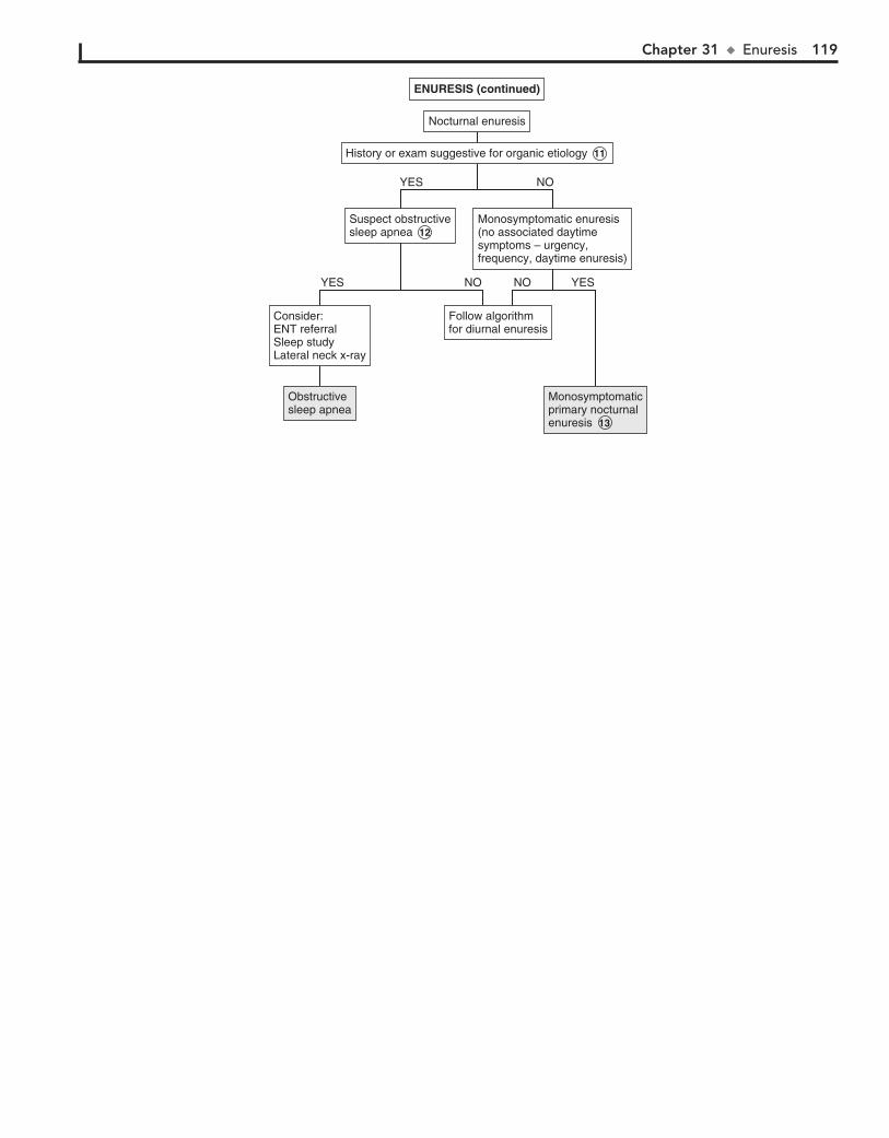
Chapter 31 u Enuresis 119
Monosymptomaticprimary nocturnalenuresis 13
Monosymptomaticprimary nocturnalenuresis 13
Suspect obstructivesleep apnea 12Suspect obstructivesleep apnea 12
Monosymptomatic enuresis(no associated daytimesymptoms – urgency,frequency, daytime enuresis)
Monosymptomatic enuresis(no associated daytimesymptoms – urgency,frequency, daytime enuresis)
Consider:ENT referralSleep studyLateral neck x-ray
Consider:ENT referralSleep studyLateral neck x-ray
Follow algorithmfor diurnal enuresisFollow algorithmfor diurnal enuresis
ENURESIS (continued)ENURESIS (continued)
Obstructivesleep apneaObstructivesleep apnea
History or exam suggestive for organic etiology 11 History or exam suggestive for organic etiology 11
Nocturnal enuresisNocturnal enuresis
NOYES
NO YESNOYES
120
Chapter 32
RED URINE AND HEMATURIA
have clots, rarely contains significant amounts of protein, and shows isomorphic RBC morphology.
5 Symptomatic gross hematuria may be due to renal disease. UTIs commonly cause gross hematuria and can be diag-
nosed by a positive urine culture when bacterial in origin. Hemorrhagic cystitis is often caused by adenovirus. Nephroli-thiasis is associated with renal colic, positive family history, and UA that may show crystals as well as hematuria. Premature in-fants who received furosemide may have nephrocalcinosis. Stones can be diagnosed by using IV pyelography, spiral CT, or US. X-rays may not detect radiolucent stones. Hypercalciuria, even without the presence of a stone, may cause abdominal or flank pain, dysuria, and hematuria. Meatal stenosis with ulcer-ation, trauma due to catheterization, and sexual abuse may cause hematuria. Abdominal and renal traumas are also causes and require abdominal CT scan with IV contrast medium en-hancement. Injury to the bladder and posterior urethra may be associated with pelvic fractures and may be diagnosed by retro-grade urethrography.
6 Idiopathic hypercalciuria most often occurs as persistent microscopic hematuria or as recurrent gross hematuria or
dysuria. A calcium-to-Cr ratio above 0.2 is suggestive. If this is present, a 24-hour urine collection for calcium should be ob-tained. Autosomal dominant polycystic kidney disease often appears as gross hematuria. Symptoms may begin in childhood but more often occur in adulthood. Renal and bladder tumors may rarely occur as hematuria. Arteriovenous malformations of the kidney may present as gross hematuria because of rapid transit of blood down the ureter; localization of bleeding may require cystoscopy or angiography. Stress hematuria occurs af-ter exercise. It is painless and of short duration; there is no proteinuria. Patients with benign familial hematuria (thin base-ment membrane nephropathy) have an excellent prognosis but must be followed. It has an autosomal dominant inheritance. Renal biopsy shows thinning of the glomerular basement mem-brane on electron microscopy. Nutcracker syndrome is due to the compression of the distal segment of the left renal vein be-tween the superior mesenteric artery and the aorta. Papillary necrosis may result in hematuria in patients with sickle cell disease or trait.
7 Acute postinfectious glomerulonephritis occurs 4 days to 3 weeks after a febrile illness just with hematuria, but also
with oliguria, edema, and hypertension. Group A streptococcal infection causing either pharyngitis or impetigo is the most common cause. Laboratory findings include a decrease in C3 and C4 levels and laboratory evidence of a preceding group A streptococcal infection (Streptozyme, antistreptolysin, antihyal-uronidase, anti–DNase B titers). Hematuria from immuno-globulin (Ig)A nephropathy appears within 48 hours of a URI. Microscopic hematuria may be present between episodes. Al-port syndrome is associated with a family history of renal dis-ease, deafness, and hematuria.
Henoch-Schönlein purpura (HSP) may appear as abdominal pain, joint pain, and lower extremity rash (palpable purpura). In addition to hematuria, proteinuria may also be present. There is no specific laboratory test for HSP; however, serum IgA levels may be elevated. In tuberous sclerosis, hematuria may be due to associated renal cysts and angiomyolipomas. Systemic infections such as bacterial endocarditis and shunt infections may be associated with hematuria and proteinuria. Nephrotic syndrome may be associated with hematuria,
Red or brown urine may indicate hematuria and possible renal disease. Hematuria is a common finding on UA. Microscopic hematuria is defined as more than 5 RBCs per high-power field in the sediment of freshly voided urine. Gross hematuria is vis-ible to the naked eye. The presence of RBCs in the urine must be confirmed because there are many conditions other than hematuria causing red or brown discoloration of the urine.
1 History should include urinary symptoms such as dysuria, frequency, and urgency, as well as flank or abdominal pain.
A history of exercise or trauma, including a foreign body, cath-eterization, or sexual/physical abuse, may indicate the cause of the hematuria. A medication, drug, and dietary history should be obtained. The history should include oliguria and hyperten-sion, as well as systemic illnesses often associated with renal disease (e.g., arthritis, respiratory illness). Family history should include renal abnormalities, hematuria, deafness, renal failure, hypertension, nephrolithiasis, sickle cell disease or trait, dialysis, or renal transplant. BP must be obtained. Physical examination should focus on the GU system and joints and on identifying any abdominal mass or a rash.
2 A positive reagent strip (dipstick) in the absence of RBCs indicates the presence of Hgb or myoglobin. He-
moglobinuria occurs with hemolysis. It may occur in hemo-lytic anemias, hemolytic-uremic syndrome, mismatched transfusions, freshwater drowning, septicemia, and paroxys-mal nocturnal hemoglobinuria. It is also associated with carbon monoxide, fava beans, venoms, mushrooms, naphtha-lene, quinine, and many other substances. A CBC with smear will often show fragmented cells, and the reticulocyte count may be elevated.
Myoglobinuria occurs with rhabdomyolysis after viral myo-sitis and in children with inborn errors of energy metabolism, often after exercise. The clinical picture as well as elevated muscle enzyme levels may aid in distinguishing myoglobinuria from hematuria. If needed, Hgb and myoglobin may be mea-sured in the urine.
3 Microscopic hematuria is often found on routine screen-ing. If the child is asymptomatic, with normal BP and no
proteinuria, the UA should be repeated at least 2 to 3 times over 2 to 3 months. If followup UA is normal, a diagnosis of isolated asymptomatic hematuria is made. If proteinuria is present, the evaluation is the same as for gross hematuria (see algorithm). If the child is symptomatic (hypertension, edema) and has pro-teinuria and a family history of deafness or kidney disease, a nephrologist should be consulted.
4 Gross hematuria can be localized to the upper or lower urinary tract. Upper tract bleeding causes brown, smoky,
or tea-colored urine. Proteinuria suggests glomerular involve-ment. (See Chapter 33.) Dysmorphic RBCs due to passage through the glomerular basement membrane also indicate up-per tract involvement. Lower tract bleeding is bright red, may
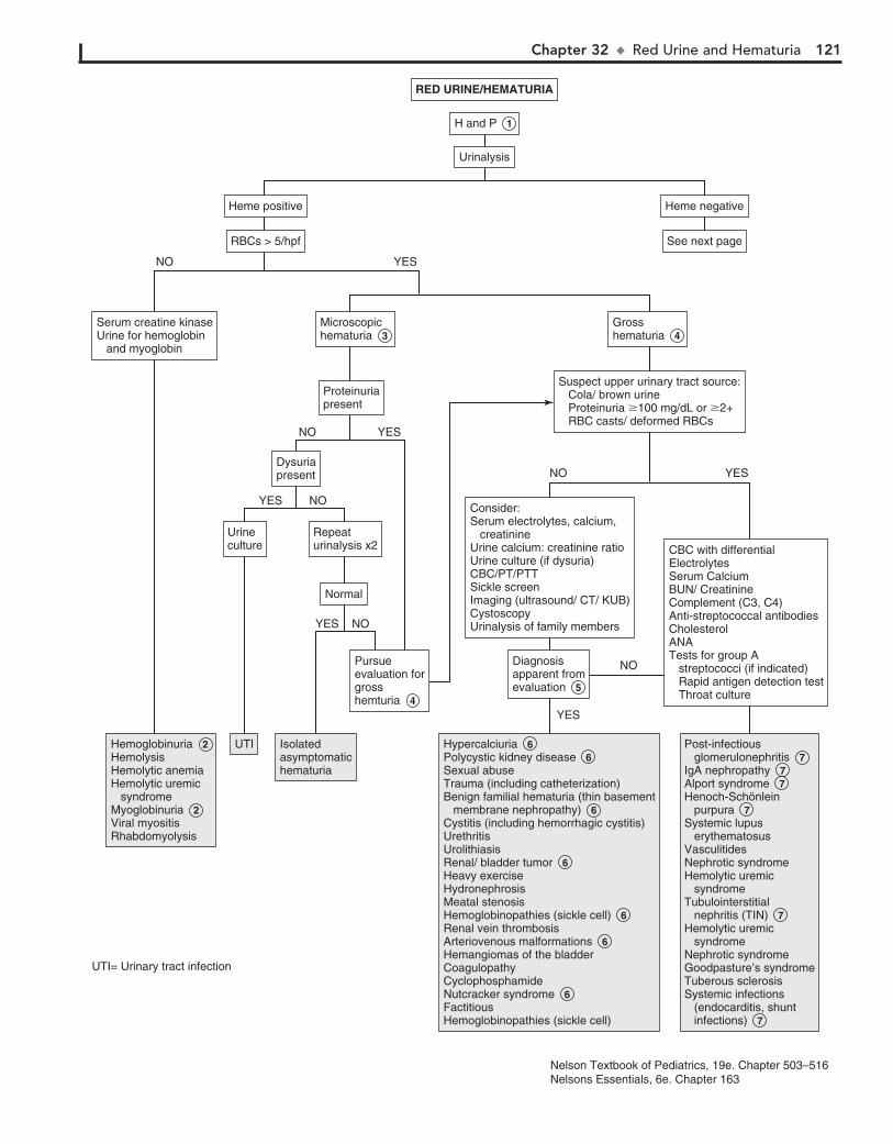
Chapter 32 u RedUrineandHematuria 121
Microscopichematuria 3 Microscopichematuria 3
Grosshematuria 4 Grosshematuria 4
Suspect upper urinary tract source: Cola/ brown urine Proteinuria �100 mg/dL or �2+ RBC casts/ deformed RBCs
Suspect upper urinary tract source: Cola/ brown urine Proteinuria �100 mg/dL or �2+ RBC casts/ deformed RBCs
RED URINE/HEMATURIARED URINE/HEMATURIA
UrinalysisUrinalysis
H and P 1 H and P 1
Hemoglobinuria 2 Hemolysis Hemolytic anemiaHemolytic uremic syndromeMyoglobinuria 2Viral myositisRhabdomyolysis
Hemoglobinuria 2 Hemolysis Hemolytic anemiaHemolytic uremic syndromeMyoglobinuria 2Viral myositisRhabdomyolysis
Consider:Serum electrolytes, calcium, creatinineUrine calcium: creatinine ratioUrine culture (if dysuria)CBC/PT/PTTSickle screenImaging (ultrasound/ CT/ KUB)CystoscopyUrinalysis of family members
Consider:Serum electrolytes, calcium, creatinineUrine calcium: creatinine ratioUrine culture (if dysuria)CBC/PT/PTTSickle screenImaging (ultrasound/ CT/ KUB)CystoscopyUrinalysis of family members
Diagnosisapparent fromevaluation 5
Diagnosisapparent fromevaluation 5
ProteinuriapresentProteinuriapresent
RBCs > 5/hpfRBCs > 5/hpf See next pageSee next page
Heme positiveHeme positive Heme negativeHeme negative
Serum creatine kinaseUrine for hemoglobin and myoglobin
Serum creatine kinaseUrine for hemoglobin and myoglobin
CBC with differentialElectrolytesSerum CalciumBUN/ CreatinineComplement (C3, C4)Anti-streptococcal antibodiesCholesterolANATests for group A streptococci (if indicated) Rapid antigen detection test Throat culture
CBC with differentialElectrolytesSerum CalciumBUN/ CreatinineComplement (C3, C4)Anti-streptococcal antibodiesCholesterolANATests for group A streptococci (if indicated) Rapid antigen detection test Throat culture
UrinecultureUrineculture
Repeaturinalysis x2Repeaturinalysis x2
NormalNormal
Pursueevaluation forgrosshemturia 4
Pursueevaluation forgrosshemturia 4
DysuriapresentDysuriapresent
UTIUTI Isolatedasymptomatichematuria
Isolatedasymptomatichematuria
Hypercalciuria 6 Polycystic kidney disease 6 Sexual abuseTrauma (including catheterization)Benign familial hematuria (thin basement membrane nephropathy) 6 Cystitis (including hemorrhagic cystitis)UrethritisUrolithiasisRenal/ bladder tumor 6 Heavy exerciseHydronephrosisMeatal stenosisHemoglobinopathies (sickle cell) 6 Renal vein thrombosisArteriovenous malformations 6 Hemangiomas of the bladderCoagulopathyCyclophosphamideNutcracker syndrome 6 FactitiousHemoglobinopathies (sickle cell)
Hypercalciuria 6 Polycystic kidney disease 6 Sexual abuseTrauma (including catheterization)Benign familial hematuria (thin basement membrane nephropathy) 6 Cystitis (including hemorrhagic cystitis)UrethritisUrolithiasisRenal/ bladder tumor 6 Heavy exerciseHydronephrosisMeatal stenosisHemoglobinopathies (sickle cell) 6 Renal vein thrombosisArteriovenous malformations 6 Hemangiomas of the bladderCoagulopathyCyclophosphamideNutcracker syndrome 6 FactitiousHemoglobinopathies (sickle cell)
Post-infectious glomerulonephritis 7 IgA nephropathy 7 Alport syndrome 7 Henoch-Schönlein purpura 7 Systemic lupus erythematosusVasculitidesNephrotic syndromeHemolytic uremic syndromeTubulointerstitial nephritis (TIN) 7 Hemolytic uremic syndromeNephrotic syndromeGoodpasture’s syndromeTuberous sclerosisSystemic infections (endocarditis, shunt infections) 7
Post-infectious glomerulonephritis 7 IgA nephropathy 7 Alport syndrome 7 Henoch-Schönlein purpura 7 Systemic lupus erythematosusVasculitidesNephrotic syndromeHemolytic uremic syndromeTubulointerstitial nephritis (TIN) 7 Hemolytic uremic syndromeNephrotic syndromeGoodpasture’s syndromeTuberous sclerosisSystemic infections (endocarditis, shunt infections) 7
YES
NO
YES
YES
NO
NO
YES NO
NO
YESNO
YES
UTI= Urinary tract infection
Nelson Textbook of Pediatrics, 19e. Chapter 503–516Nelsons Essentials, 6e. Chapter 163

122 Part V u GenitourinarySystem
particularly in focal segmental sclerosis and membranopro-liferative glomerulonephritis. Tubulointerstitial nephritis is often associated with penicillins, cephalosporins, sulfonamides, rifampin, tetracyclines, nonsteroidal antiinflammatory drugs, furosemide, thiazides, heavy metals, and others. It may also be associated with other diseases, such as systemic lupus erythematosus.
BibliographyKliegman RM, Stanton BF, St. Geme J, Schor N, Behrman RE, editors:
Nelson textbook of pediatrics, ed 19, Philadelphia, 2011, Elsevier Saunders. Chapters 503–516.
McInerny TK, Adam HM, Campbell DE, et al, editors: American Academy of Pediatrics textbook of pediatric care, Elk Grove Village, Ill, 2009, American Academy of Pediatrics, pp 1566–1569.
Pan CG: Evaluation of gross hematuria, Pediatr Clin North Am 53:401–412, 2006
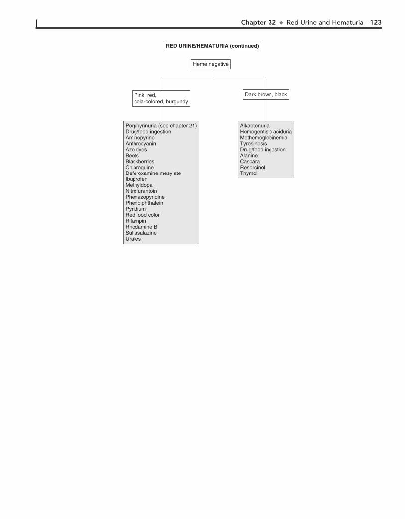
Chapter 32 u RedUrineandHematuria 123
Pink, red, cola-colored, burgundyPink, red, cola-colored, burgundy
Heme negativeHeme negative
RED URINE/HEMATURIA (continued)RED URINE/HEMATURIA (continued)
AlkaptonuriaHomogentisic aciduriaMethemoglobinemiaTyrosinosisDrug/food ingestionAlanineCascaraResorcinolThymol
AlkaptonuriaHomogentisic aciduriaMethemoglobinemiaTyrosinosisDrug/food ingestionAlanineCascaraResorcinolThymol
Porphyrinuria (see chapter 21)Drug/food ingestionAminopyrineAnthrocyaninAzo dyesBeetsBlackberriesChloroquineDeferoxamine mesylateIbuprofenMethyldopaNitrofurantoinPhenazopyridinePhenolphthaleinPyridiumRed food colorRifampinRhodamine BSulfasalazineUrates
Porphyrinuria (see chapter 21)Drug/food ingestionAminopyrineAnthrocyaninAzo dyesBeetsBlackberriesChloroquineDeferoxamine mesylateIbuprofenMethyldopaNitrofurantoinPhenazopyridinePhenolphthaleinPyridiumRed food colorRifampinRhodamine BSulfasalazineUrates
Dark brown, blackDark brown, black
124
Chapter 33
PROTEINURIA
ambulatory and recumbent (first void) urine samples for protein/Cr ratio. It may also be done by quantitative assess-ment of proteinuria in ambulatory and recumbent urine specimens. The recumbent specimen should have less than 4 mg/m2/h of protein. The amount in the ambulatory specimen may vary but is usually 2 to 4 times that of the recumbent specimen.
4 In patients who have persistent asymptomatic proteinuria, further evaluation may proceed as in symptomatic patients.
It is reasonable to refer even the patient with normal test results to a nephrologist, because there are different opinions regarding the need for biopsy.
5 Patients with proteinuria who are symptomatic (edema, hypertension) or have hematuria, associated systemic
complaints (rash, fever, arthralgia), or a significant family his-tory of glomerulonephritis or renal failure should have further evaluation. In most cases, referral to the nephrologist may be necessary. This evaluation includes assessment of renal function with BUN, Cr, and electrolyte determinations. Nephrotic syn-drome consists of proteinuria, hypoalbuminemia, edema, and hyperlipidemia. Total serum protein, albumin, as well as choles-terol and triglycerides are checked. Tests for antistreptococcal antibodies (Streptozyme) as well as complement levels (C3, C4) are done to exclude poststreptococcal glomerulonephritis. The diagnosis of poststreptococcal glomerulonephritis is made in a child with acute nephritic syndrome, evidence of recent strep-tococcal infection, and a low C3 level. In SLE, C3 and C4 are low. Antinuclear antibody testing may be considered, especially with hypertension or hematuria, to exclude SLE. Hepatitis B and C and HIV may also be associated with glomerulonephri-tis. A renal US should be considered to examine anatomy.
6 Minimal change nephrotic syndrome is more common in boys and usually appears between ages 2 and 6 years. UA
shows 31 to 41 proteinuria, and there may be microscopic he-maturia. Renal function may be reduced, cholesterol and triglyc-erides are elevated, and serum albumin is decreased. The C3 level is normal.
7 Nephrotic syndrome may develop with any type of glo-merulonephritis, especially membranous, membranopro-
liferative, postinfectious, SLE, chronic infection, and HSP glomerulonephritis. With poststreptococcal glomerulonephritis, antistreptococcal antibody levels are elevated and complement (C3) is decreased. (See Chapter 32 for conditions causing hematuria with proteinuria.)
8 Nephrotic syndromes may occur with extrarenal neoplasms such as carcinomas and lymphomas (Hodgkin disease).
9 Drugs or chemicals, such as penicillamine, gold, mercury compounds, probenecid, ethosuximide, methimazole, lith-
ium, phenytoin, and many others, may be associated with ne-phrotic syndrome.
10 Children younger than age 1 with nephrotic syndrome have a poor prognosis. Congenital nephrotic syndrome
(Finnish type) is the most common cause. It is an autosomal recessive condition and may occur as failure to thrive due to massive proteinuria. Nephrotic syndrome in infants may be secondary to infections such as syphilis, toxoplasmosis, CMV, rubella, hepatitis B, HIV, or malaria. Drugs, toxins
Proteinuria is a common laboratory finding that is often a symp-tom of renal disease. It may also be found in normal, healthy children. It is therefore important to distinguish between patho-logic and nonpathologic causes of proteinuria. As many as 10% of children will have 11 proteinuria at some time in their lives. Because this is a transient finding in a majority of children, it is important to retest the urine before making a diagnosis.
Proteinuria may be defined qualitatively using a urine dip-stick examination. Trace proteinuria is usually not significant; 11 proteinuria (30 mg/dl) may be significant. This should be repeated and viewed in the context of the urine Sp gr. False-negative results may occur with urine that is too dilute (,1.005), and false-positive results may occur with overlong dipstick im-mersion, alkaline urine, pyuria, bacteriuria, mucoprotein and quaternary ammonium compounds, and detergents.
Quantitative testing for proteinuria is done by a timed 12- to 24-hour urine collection for protein: less than 4 mg/m2/h is nor-mal, 4-40 mg/m2/h is abnormal, and over 40 mg/m2/h is in the nephrotic range. An early morning spot testing of urine protein/Cr ratio (in mg/dL) correlates well with 24-hour urine protein ex-cretion. A value above 0.2 is abnormal in children older than 2 years; for children 6 months to 2 years, over 0.5 is abnormal, and over 2 suggests nephrotic-range proteinuria. This test is not valid in children with decreased muscle mass (i.e., those with nutritional problems).
1 History should include questions about recent exercise, red or tea-colored urine, and respiratory or other febrile illness.
History indicative of edema, such as puffiness around the eyes on awakening, increased abdominal girth, and difficulty putting on shoes, may indicate nephrotic syndrome and should be investi-gated. Family history related to renal disease, hematuria, or hyper-tension should be pursued. Systemic complaints (e.g., arthralgia, rash, fever) may be symptoms of diseases such as systemic lupus erythematosus (SLE) or Henoch-Schönlein purpura (HSP). On physical examination, BP must be evaluated, as well as evidence of edema. Characteristic rashes may indicate the cause (e.g., a malar rash in SLE or purpuric rash with HSP).
2 Transient proteinuria may occur with fever (usually re-solves within 10 to 14 days of the illness), with strenuous
exercise (abates within 48 hours), and with stress, cold exposure, dehydration, heart failure, and seizures.
3 Children who have proteinuria on repeat UA, especially those older than 4 years, should be tested for orthostatic
proteinuria. This is an increased protein excretion in the upright position only and is less common in younger children. It may account for as much as 60% of all proteinuria in children and has a benign clinical course.
Any of the following tests may be performed to test for orthostatic proteinuria. Spot urine protein/Cr ratio of less than 0.2 in the first morning urine sample for 3 consecutive days confirms the diagnosis. Testing may be done by comparing
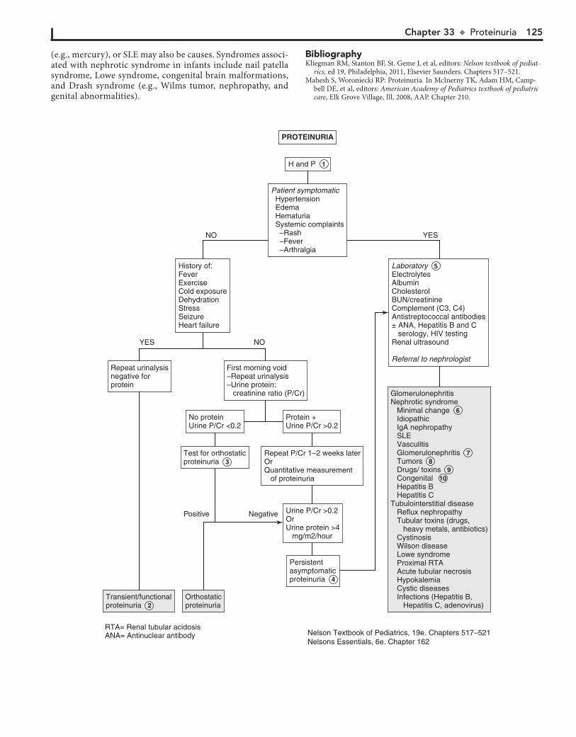
Chapter 33 u Proteinuria 125
(e.g., mercury), or SLE may also be causes. Syndromes associ-ated with nephrotic syndrome in infants include nail patella syndrome, Lowe syndrome, congenital brain malformations, and Drash syndrome (e.g., Wilms tumor, nephropathy, and genital abnormalities).
BibliographyKliegman RM, Stanton BF, St. Geme J, et al, editors: Nelson textbook of pediat-
rics, ed 19, Philadelphia, 2011, Elsevier Saunders. Chapters 517–521.Mahesh S, Woroniecki RP: Proteinuria. In McInerny TK, Adam HM, Camp-
bell DE, et al, editors: American Academy of Pediatrics textbook of pediatric care, Elk Grove Village, Ill, 2008, AAP. Chapter 210.
Test for orthostaticproteinuria 3Test for orthostaticproteinuria 3
Urine P/Cr >0.2OrUrine protein >4 mg/m2/hour
Urine P/Cr >0.2OrUrine protein >4 mg/m2/hour
PROTEINURIAPROTEINURIA
History of:FeverExerciseCold exposureDehydrationStressSeizureHeart failure
History of:FeverExerciseCold exposureDehydrationStressSeizureHeart failure
OrthostaticproteinuriaOrthostaticproteinuria
Persistentasymptomaticproteinuria 4
Persistentasymptomaticproteinuria 4
Transient/functionalproteinuria 2 Transient/functionalproteinuria 2
GlomerulonephritisNephrotic syndrome Minimal change 6 Idiopathic IgA nephropathy SLE Vasculitis Glomerulonephritis 7 Tumors 8 Drugs/ toxins 9 Congenital 10 Hepatitis B Hepatitis CTubulointerstitial disease Reflux nephropathy Tubular toxins (drugs, heavy metals, antibiotics) Cystinosis Wilson disease Lowe syndrome Proximal RTA Acute tubular necrosis Hypokalemia Cystic diseases Infections (Hepatitis B, Hepatitis C, adenovirus)
GlomerulonephritisNephrotic syndrome Minimal change 6 Idiopathic IgA nephropathy SLE Vasculitis Glomerulonephritis 7 Tumors 8 Drugs/ toxins 9 Congenital 10 Hepatitis B Hepatitis CTubulointerstitial disease Reflux nephropathy Tubular toxins (drugs, heavy metals, antibiotics) Cystinosis Wilson disease Lowe syndrome Proximal RTA Acute tubular necrosis Hypokalemia Cystic diseases Infections (Hepatitis B, Hepatitis C, adenovirus)
Protein +Urine P/Cr >0.2Protein +Urine P/Cr >0.2
Patient symptomatic Hypertension Edema Hematuria Systemic complaints –Rash –Fever –Arthralgia
Patient symptomatic Hypertension Edema Hematuria Systemic complaints –Rash –Fever –Arthralgia
No proteinUrine P/Cr <0.2No proteinUrine P/Cr <0.2
First morning void–Repeat urinalysis–Urine protein: creatinine ratio (P/Cr)
First morning void–Repeat urinalysis–Urine protein: creatinine ratio (P/Cr)
H and P 1 H and P 1
Repeat urinalysisnegative forprotein
Repeat urinalysisnegative forprotein
Laboratory 5 Electrolytes AlbuminCholesterolBUN/creatinineComplement (C3, C4)Antistreptococcal antibodies± ANA, Hepatitis B and C serology, HIV testing Renal ultrasound
Referral to nephrologist
Laboratory 5 Electrolytes AlbuminCholesterolBUN/creatinineComplement (C3, C4)Antistreptococcal antibodies± ANA, Hepatitis B and C serology, HIV testing Renal ultrasound
Referral to nephrologist
Repeat P/Cr 1–2 weeks laterOrQuantitative measurement of proteinuria
Repeat P/Cr 1–2 weeks laterOrQuantitative measurement of proteinuria
Positive Negative
NO YES
NOYES
RTA= Renal tubular acidosisANA= Antinuclear antibody Nelson Textbook of Pediatrics, 19e. Chapters 517–521
Nelsons Essentials, 6e. Chapter 162
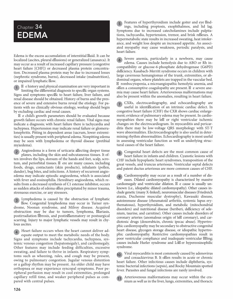
126
Chapter 34
EDEMA
5 Features of hyperthyroidism include goiter and eye find-ings, including proptosis, exophthalmos, and lid lag.
Symptoms due to increased catecholamines include palpita-tions, tachycardia, hypertension, tremor, and brisk reflexes. A hypermetabolic state results in increased sweating, heat intoler-ance, and weight loss despite an increased appetite. An associ-ated myopathy may cause weakness, periodic paralysis, and heart failure.
6 Severe anemia, particularly in a newborn, may cause edema. Causes include hemolysis due to ABO or Rh in-
compatibility or glucose-6-phosphate dehydrogenase (G6PD) deficiency. Kasabach-Merritt syndrome occurs in children with large cavernous hemangiomas of the trunk, extremities, or ab-dominal organs, where platelets are trapped in the vascular bed. Thrombocytopenia, a microangiopathic hemolytic anemia, and often a consumptive coagulopathy are present. The severe ane-mia may cause heart failure. Arteriovenous malformations may also be present within the anomalies, resulting in heart failure.
7 CXRs, electrocardiography, and echocardiography are useful in identification of an intrinsic cardiac defect. In
congestive heart failure (CHF) the CXR shows cardiac enlarge-ment; evidence of pulmonary edema may be present. In cardio-myopathies there may be left or right ventricular ischemic changes on the electrocardiogram. In myocarditis and pericar-ditis there may be low-voltage QRS morphology with ST-T wave abnormalities. Electrocardiography is also useful in deter-mining rhythm abnormalities. Echocardiography is most useful in assessing ventricular function as well as underlying struc-tural causes of the heart failure.
8 Congenital heart defects are the most common cause of heart failure in infants and children. Cyanotic lesions with
CHF include hypoplastic heart syndromes, transposition of the great vessels, and truncus arteriosus. Ventricular septal defects and patent ductus arteriosus are more common causes of CHF.
9 Cardiomyopathy may occur as a result of a number of dis-eases. Dilated cardiomyopathy is characterized by massive
cardiomegaly and ventricular dilation. The cause is usually un-known (i.e., idiopathic dilated cardiomyopathy). Other causes in-clude genetic (many X-linked), neuromuscular diseases (Friedreich ataxia, Duchenne muscular dystrophy), Kawasaki disease, autoimmune disease (rheumatoid arthritis, systemic lupus ery-thematosus), hyperthyroidism, and metabolic (mitochondrial disorders) and nutritional disease (beriberi, deficiency of sele-nium, taurine, and carnitine). Other causes include disorders of coronary arteries (anomalous origin of left coronary), and car-diotoxic drugs (doxorubicin, chronic ipecac abuse). Hypertro-phic cardiomyopathy may be secondary to obstructive congenital heart disease, glycogen storage disease, or idiopathic hypertro-phic cardiomyopathy. Restrictive cardiomyopathies result in poor ventricular compliance and inadequate ventricular filling; causes include Hurler syndrome and Löffler hypereosinophilic syndrome.
10 Viral myocarditis is most commonly caused by adenovirus and coxsackievirus B. It often results in acute or chronic
heart failure. Other infectious causes include diphtheria, sys-temic bacterial infections (sepsis), and Rocky Mountain spotted fever. Parasites and fungal infections are rarely involved.
11 Arteriovenous malformations may occur within the cra-nium as well as in the liver, lungs, extremities, and thoracic
Edema is the excess accumulation of interstitial fluid. It can be localized (ascites, pleural effusions) or generalized (anasarca). It may occur as a result of increased capillary pressure (congestive heart failure [CHF]) or decreased plasma protein concentra-tion. Decreased plasma protein may be due to increased losses (nephrotic syndrome, burns), decreased intake (malnutrition), or impaired lymphatic flow.
1 The history and physical examination are very important in limiting the differential diagnosis to specific organ systems.
Signs and symptoms specific to heart failure, liver failure, and renal disease should be obtained. History of burns and the pres-ence of severe and extensive burns reveal the etiology. For pa-tients with no clinically obvious etiology, workup should begin by excluding cardiac and renal causes.
The child’s growth parameters should be evaluated because growth failure occurs with chronic renal failure. Vital signs may indicate a diagnosis; with heart failure there is tachycardia and tachypnea. Hypertension may indicate renal failure or glomeru-lonephritis. Pitting in dependent areas (sacrum, lower extremi-ties) is usually present with peripheral edema. Nonpitting edema may be seen with lymphedema or thyroid disease (pretibial myxedema).
2 Angioedema is a form of urticaria affecting deeper tissue planes, including the skin and subcutaneous tissues. It of-
ten involves the lips, dorsum of the hands and feet, scalp, scro-tum, and periorbital tissues. There are many causes, including foods, drugs, contactants (skin products), inhalants (pollen, dander), bug bites, and infections. A history of recurrent angio-edema may indicate episodic angioedema, which is associated with fever and eosinophilia. Hereditary angioedema, which re-sults from a decreased synthesis of C1 esterase inhibitor, occurs as sudden attacks of edema often precipitated by minor trauma, strenuous exercise, or any stressors.
3 Lymphedema is caused by the obstruction of lymphatic flow. Congenital lymphedema may occur in Turner syn-
drome, Noonan syndrome, and Milroy disease. Acquired obstruction may be due to tumors, lymphoma, filariasis, postirradiation fibrosis, and postinflammatory or postsurgical scarring. Injury to major lymphatic vessels may result in chy-lous ascites.
4 Heart failure occurs when the heart cannot deliver ad-equate output to meet the metabolic needs of the body.
Signs and symptoms include tachycardia, tachypnea, sys-temic venous congestion (hepatomegaly), and cardiomegaly. Other features may include feeding difficulties, excessive sweating, and failure to thrive in infants. Respiratory symp-toms such as wheezing, rales, and cough may be present, owing to pulmonary congestion. Jugular venous distention or a gallop rhythm may be present. An older child may have orthopnea or may experience syncopal symptoms. Poor pe-ripheral perfusion may result in cool extremities, prolonged capillary refill time, and weaker peripheral pulses as com-pared with central pulses.
Chapter 34 u Edema 127
YES
YES
NO
NO
EDEMAEDEMA
Venous thrombosis/ obstructionAngioedema 2 Lymphedema 3 Milroy’s disease Turner syndrome Filariasis Lymphoma Iatrogenic (surgery, radiation)
Venous thrombosis/ obstructionAngioedema 2 Lymphedema 3 Milroy’s disease Turner syndrome Filariasis Lymphoma Iatrogenic (surgery, radiation)
Severe anemia 6 Kasabach-Merritt syndrome 6
Severe anemia 6 Kasabach-Merritt syndrome 6
Arteriovenousmalformations 11 Arteriovenousmalformations 11
Nephrotic syndromeTubulointerstitial diseaseGlomerulonephritisRenal failure
Nephrotic syndromeTubulointerstitial diseaseGlomerulonephritisRenal failure
Liver failure Infection (viral hepatitis, HIV) 14 Hepatotoxic drugs and chemicals (acetaminophen) 15 Metabolic disease 16 Ischemia (shock, CHF)
Liver failure Infection (viral hepatitis, HIV) 14 Hepatotoxic drugs and chemicals (acetaminophen) 15 Metabolic disease 16 Ischemia (shock, CHF)
Kwashiorkor 17 (protein calorie malnutrition)
Kwashiorkor 17 (protein calorie malnutrition)
BurnsSepsisBurnsSepsis
Protein losing enteropathy 18 Food protein induced enteropathy Postinfection Eosinophilic gastroenteritis
Protein losing enteropathy 18 Food protein induced enteropathy Postinfection Eosinophilic gastroenteritis
Congenital heart disease 8Cardiomyopathy 9 Myocarditis 10ArrhythmiasCoronary artery lesions (Kawasaki disease)Rheumatic heart diseaseHypertensionCor pulmonale (cystic fibrosis)
Congenital heart disease 8Cardiomyopathy 9 Myocarditis 10ArrhythmiasCoronary artery lesions (Kawasaki disease)Rheumatic heart diseaseHypertensionCor pulmonale (cystic fibrosis)
Hypothyroidism (Myxedema)Severe thyrotoxicosis
Hypothyroidism (Myxedema)Severe thyrotoxicosis
Imaging for AVMImaging for AVM
Evidence ofintrinsic cardiac defect on Chest x-ray Echocardiogram EKG 7
Evidence ofintrinsic cardiac defect on Chest x-ray Echocardiogram EKG 7
CBCUrinalysisAlbumin/ total protein BUN/ creatinineElectrolytes
CBCUrinalysisAlbumin/ total protein BUN/ creatinineElectrolytes
Anemia± ThrombocytopeniaAnemia± Thrombocytopenia
Signs and symptomsof CHF 4 –Tachypnea–Tachycardia–Gallop rhythm–Hepatomegaly
Signs and symptomsof CHF 4 –Tachypnea–Tachycardia–Gallop rhythm–Hepatomegaly
Abnormal U/A BUN/ creatinine
Abnormal U/A BUN/ creatinine
UrinalysisElectrolytesBUNCreatinineAlbuminCholesterol
UrinalysisElectrolytesBUNCreatinineAlbuminCholesterol
Signs and symptoms of renal disease–Hematuria–Hypertension
Signs and symptoms of renal disease–Hematuria–Hypertension
AbnormalAbnormal
NormalNormal Free T4TSHFree T4TSH
Signs and symptomsof thyroiddisease 5
Signs and symptomsof thyroiddisease 5
See chapters32, 33, and 35See chapters32, 33, and 35
Liver function tests 13AlbuminPT, PTTGlucose Electrolytes± Serum ammonia± Hepatitis serology± HIV
Liver function tests 13AlbuminPT, PTTGlucose Electrolytes± Serum ammonia± Hepatitis serology± HIV
Signs and symptomsof liver failure 12
Signs and symptomsof liver failure 12
Generalized 12 Generalized 12
H and P 1 H and P 1
LocalizedLocalized
CBCSerum albuminElectrolytesMagnesiumZinc
CBCSerum albuminElectrolytesMagnesiumZinc
Signs andsymptoms of severemalnutrition
Signs andsymptoms of severemalnutrition
CBCAlbumin/total proteinElectrolytesStool for α-1 antitrypsinCeliac panel
CBCAlbumin/total proteinElectrolytesStool for α-1 antitrypsinCeliac panel
DiarrheaDiarrhea Severeburns/sepsis
Severeburns/sepsis
CHF= Congestive heart failureAVM= Arteriovenous malformations Nelson Textbook of Pediatrics, 19e. Chapters 517-521
Nelsons Essentials, 6e. Chapter 162
128 Part V u GenitourinarySystem
12 Liver failure may be a complication of known liver disease or may be the presenting feature. Features include progres-
sive jaundice, fetor hepaticus, fever, anorexia, vomiting, and abdominal pain. There may be a rapid decrease in liver size without clinical improvement, hemorrhagic diathesis, and asci-tes. Infants may present with irritability, lethargy, poor feeding, and sleep disturbances. Mental status changes are noted with progression of symptoms. Older children may demonstrate asterixis.
13 In liver failure, hypoalbuminemia results in edema, and bilirubin levels (direct and indirect) are elevated. Serum
aminotransferase levels are elevated early but may decrease as the patient’s condition deteriorates. PT is prolonged and often does not correct with vitamin K administration. The serum am-monia level is usually elevated, and there may be hypoglycemia, hypokalemia, hyponatremia, metabolic acidosis, or respiratory alkalosis.
14 Viral hepatitis is a common cause of liver failure. It is more likely in children with a combined infection with hepatitis
B and D. Other viruses that may result in liver failure include EBV, herpes simplex virus, adenovirus, enterovirus, parvovirus B19, and varicella-zoster virus.
15 Known hepatotoxins include acetaminophen overdose, carbon tetrachloride, and Amanita phalloides mushrooms.
Idiosyncratic damage may occur with halothane, phenytoin, carbamazepine, or sodium valproate.
16 Metabolic disorders associated with liver failure include Wilson disease, galactosemia, hereditary tyrosinemia,
hereditary fructose intolerance, and urea cycle defects.
17 Kwashiorkor is a clinical syndrome resulting from severe protein deficiency and inadequate caloric intake (protein-
calorie malnutrition). Early in the disease, symptoms include anorexia, lethargy, apathy, and irritability. Later there is de-creased growth, decreased stamina, muscle loss, increased sus-ceptibility to infections, and edema. The edema may mask poor weight gain. Skin changes may be present, and the hair becomes coarse and discolored, resulting in streaky red or gray hair. Laboratory findings include decreased serum albumin, hypo-glycemia, hypophosphatemia, and deficiency of potassium and magnesium. Anemia may be normocytic, microcytic, or mac-rocytic. Signs of vitamin (especially vitamin A) and mineral (zinc) deficiencies may be present.
18 Protein-losing enteropathy may result in edema secondary to hypoalbuminemia. a1-Antitrypsin, unlike albumin,
is resistant to digestion. Measurement of levels in the stool is helpful in the diagnosis of protein-losing enteropathy. Causes include food protein–induced enteropathy and postinfectious enteropathy. Eosinophilic gastroenteritis is another cause; it may be associated with dietary protein hypersensitivity as well as other food allergies. There is often eosinophilia and an ele-vated serum immunoglobulin (Ig)E. Celiac disease producing severe malnutrition may also present with edema.
BibliographyLevy PA: Edema. In McInerny TK, Adam HM, Campbell DE, et al, editors:
American Academy of Pediatrics textbook of pediatric care, Elk Grove Village, Ill, 2008, AAP. Chapter 175.
Paller AS, Mancini AJ: Hurwitz clinical pediatric dermatology, ed 4, Philadelphia, 2011, WB Saunders.
wall. Heart failure is more likely with large intracranial arteriove-nous fistulas (e.g., vein of Galen malformation) and hepatic mal-formations. They are less likely with peripheral arteriovenous fistulas. Physical examination may reveal a bruit, but definitive diagnosis is made by imaging, usually by MRI.
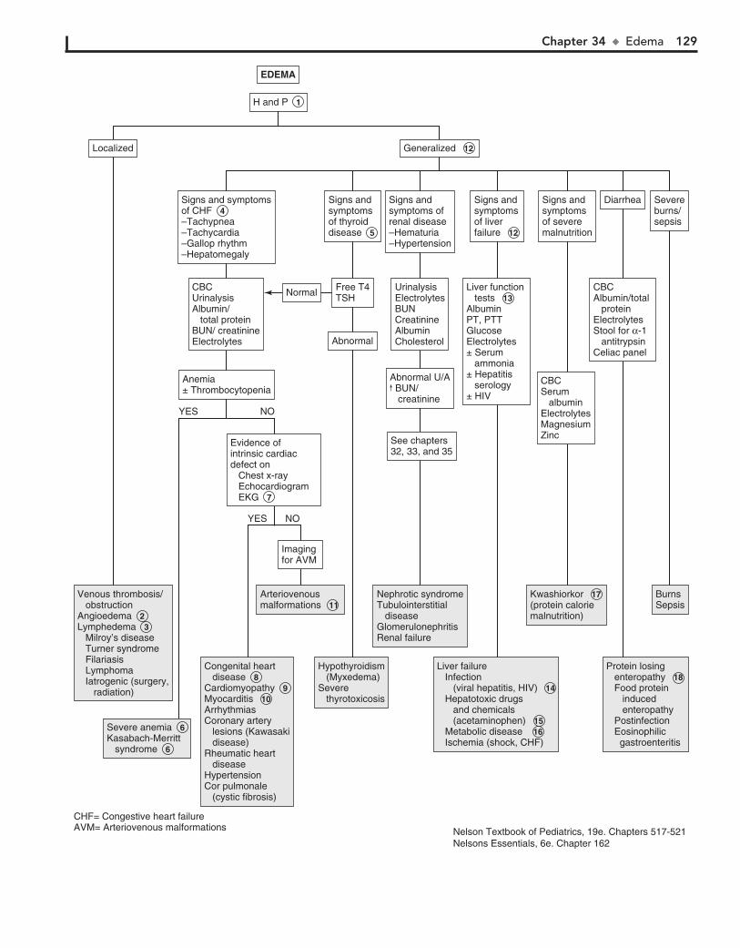
Chapter 34 u Edema 129
YES
YES
NO
NO
EDEMAEDEMA
Venous thrombosis/ obstructionAngioedema 2 Lymphedema 3 Milroy’s disease Turner syndrome Filariasis Lymphoma Iatrogenic (surgery, radiation)
Venous thrombosis/ obstructionAngioedema 2 Lymphedema 3 Milroy’s disease Turner syndrome Filariasis Lymphoma Iatrogenic (surgery, radiation)
Severe anemia 6 Kasabach-Merritt syndrome 6
Severe anemia 6 Kasabach-Merritt syndrome 6
Arteriovenousmalformations 11 Arteriovenousmalformations 11
Nephrotic syndromeTubulointerstitial diseaseGlomerulonephritisRenal failure
Nephrotic syndromeTubulointerstitial diseaseGlomerulonephritisRenal failure
Liver failure Infection (viral hepatitis, HIV) 14 Hepatotoxic drugs and chemicals (acetaminophen) 15 Metabolic disease 16 Ischemia (shock, CHF)
Liver failure Infection (viral hepatitis, HIV) 14 Hepatotoxic drugs and chemicals (acetaminophen) 15 Metabolic disease 16 Ischemia (shock, CHF)
Kwashiorkor 17 (protein calorie malnutrition)
Kwashiorkor 17 (protein calorie malnutrition)
BurnsSepsisBurnsSepsis
Protein losing enteropathy 18 Food protein induced enteropathy Postinfection Eosinophilic gastroenteritis
Protein losing enteropathy 18 Food protein induced enteropathy Postinfection Eosinophilic gastroenteritis
Congenital heart disease 8Cardiomyopathy 9 Myocarditis 10ArrhythmiasCoronary artery lesions (Kawasaki disease)Rheumatic heart diseaseHypertensionCor pulmonale (cystic fibrosis)
Congenital heart disease 8Cardiomyopathy 9 Myocarditis 10ArrhythmiasCoronary artery lesions (Kawasaki disease)Rheumatic heart diseaseHypertensionCor pulmonale (cystic fibrosis)
Hypothyroidism (Myxedema)Severe thyrotoxicosis
Hypothyroidism (Myxedema)Severe thyrotoxicosis
Imaging for AVMImaging for AVM
Evidence ofintrinsic cardiac defect on Chest x-ray Echocardiogram EKG 7
Evidence ofintrinsic cardiac defect on Chest x-ray Echocardiogram EKG 7
CBCUrinalysisAlbumin/ total protein BUN/ creatinineElectrolytes
CBCUrinalysisAlbumin/ total protein BUN/ creatinineElectrolytes
Anemia± ThrombocytopeniaAnemia± Thrombocytopenia
Signs and symptomsof CHF 4 –Tachypnea–Tachycardia–Gallop rhythm–Hepatomegaly
Signs and symptomsof CHF 4 –Tachypnea–Tachycardia–Gallop rhythm–Hepatomegaly
Abnormal U/A BUN/ creatinine
Abnormal U/A BUN/ creatinine
UrinalysisElectrolytesBUNCreatinineAlbuminCholesterol
UrinalysisElectrolytesBUNCreatinineAlbuminCholesterol
Signs and symptoms of renal disease–Hematuria–Hypertension
Signs and symptoms of renal disease–Hematuria–Hypertension
AbnormalAbnormal
NormalNormal Free T4TSHFree T4TSH
Signs and symptomsof thyroiddisease 5
Signs and symptomsof thyroiddisease 5
See chapters32, 33, and 35See chapters32, 33, and 35
Liver function tests 13AlbuminPT, PTTGlucose Electrolytes± Serum ammonia± Hepatitis serology± HIV
Liver function tests 13AlbuminPT, PTTGlucose Electrolytes± Serum ammonia± Hepatitis serology± HIV
Signs and symptomsof liver failure 12
Signs and symptomsof liver failure 12
Generalized 12 Generalized 12
H and P 1 H and P 1
LocalizedLocalized
CBCSerum albuminElectrolytesMagnesiumZinc
CBCSerum albuminElectrolytesMagnesiumZinc
Signs andsymptoms of severemalnutrition
Signs andsymptoms of severemalnutrition
CBCAlbumin/total proteinElectrolytesStool for α-1 antitrypsinCeliac panel
CBCAlbumin/total proteinElectrolytesStool for α-1 antitrypsinCeliac panel
DiarrheaDiarrhea Severeburns/sepsis
Severeburns/sepsis
CHF= Congestive heart failureAVM= Arteriovenous malformations Nelson Textbook of Pediatrics, 19e. Chapters 517-521
Nelsons Essentials, 6e. Chapter 162
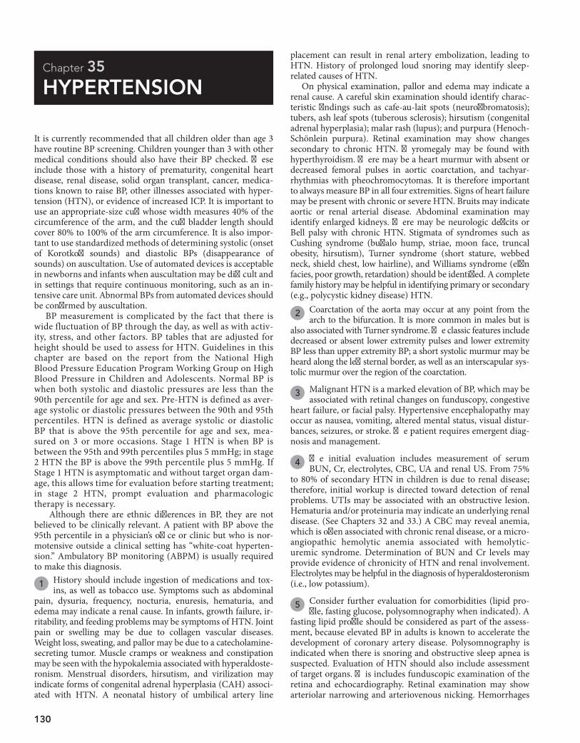
130
Chapter 35
HYPERTENSION
placement can result in renal artery embolization, leading to HTN. History of prolonged loud snoring may identify sleep-related causes of HTN.
On physical examination, pallor and edema may indicate a renal cause. A careful skin examination should identify charac-teristic findings such as cafe-au-lait spots (neurofibromatosis); tubers, ash leaf spots (tuberous sclerosis); hirsutism (congenital adrenal hyperplasia); malar rash (lupus); and purpura (Henoch-Schönlein purpura). Retinal examination may show changes secondary to chronic HTN. Thyromegaly may be found with hyperthyroidism. There may be a heart murmur with absent or decreased femoral pulses in aortic coarctation, and tachyar-rhythmias with pheochromocytomas. It is therefore important to always measure BP in all four extremities. Signs of heart failure may be present with chronic or severe HTN. Bruits may indicate aortic or renal arterial disease. Abdominal examination may identify enlarged kidneys. There may be neurologic deficits or Bell palsy with chronic HTN. Stigmata of syndromes such as Cushing syndrome (buffalo hump, striae, moon face, truncal obesity, hirsutism), Turner syndrome (short stature, webbed neck, shield chest, low hairline), and Williams syndrome (elfin facies, poor growth, retardation) should be identified. A complete family history may be helpful in identifying primary or secondary (e.g., polycystic kidney disease) HTN.
2 Coarctation of the aorta may occur at any point from the arch to the bifurcation. It is more common in males but is
also associated with Turner syndrome. The classic features include decreased or absent lower extremity pulses and lower extremity BP less than upper extremity BP; a short systolic murmur may be heard along the left sternal border, as well as an interscapular sys-tolic murmur over the region of the coarctation.
3 Malignant HTN is a marked elevation of BP, which may be associated with retinal changes on funduscopy, congestive
heart failure, or facial palsy. Hypertensive encephalopathy may occur as nausea, vomiting, altered mental status, visual distur-bances, seizures, or stroke. The patient requires emergent diag-nosis and management.
4 The initial evaluation includes measurement of serum BUN, Cr, electrolytes, CBC, UA and renal US. From 75%
to 80% of secondary HTN in children is due to renal disease; therefore, initial workup is directed toward detection of renal problems. UTIs may be associated with an obstructive lesion. Hematuria and/or proteinuria may indicate an underlying renal disease. (See Chapters 32 and 33.) A CBC may reveal anemia, which is often associated with chronic renal disease, or a micro-angiopathic hemolytic anemia associated with hemolytic-uremic syndrome. Determination of BUN and Cr levels may provide evidence of chronicity of HTN and renal involvement. Electrolytes may be helpful in the diagnosis of hyperaldosteronism (i.e., low potassium).
5 Consider further evaluation for comorbidities (lipid pro-file, fasting glucose, polysomnography when indicated). A
fasting lipid profile should be considered as part of the assess-ment, because elevated BP in adults is known to accelerate the development of coronary artery disease. Polysomnography is indicated when there is snoring and obstructive sleep apnea is suspected. Evaluation of HTN should also include assessment of target organs. This includes funduscopic examination of the retina and echocardiography. Retinal examination may show arteriolar narrowing and arteriovenous nicking. Hemorrhages
It is currently recommended that all children older than age 3 have routine BP screening. Children younger than 3 with other medical conditions should also have their BP checked. These include those with a history of prematurity, congenital heart disease, renal disease, solid organ transplant, cancer, medica-tions known to raise BP, other illnesses associated with hyper-tension (HTN), or evidence of increased ICP. It is important to use an appropriate-size cuff whose width measures 40% of the circumference of the arm, and the cuff bladder length should cover 80% to 100% of the arm circumference. It is also impor-tant to use standardized methods of determining systolic (onset of Korotkoff sounds) and diastolic BPs (disappearance of sounds) on auscultation. Use of automated devices is acceptable in newborns and infants when auscultation may be difficult and in settings that require continuous monitoring, such as an in-tensive care unit. Abnormal BPs from automated devices should be confirmed by auscultation.
BP measurement is complicated by the fact that there is wide fluctuation of BP through the day, as well as with activ-ity, stress, and other factors. BP tables that are adjusted for height should be used to assess for HTN. Guidelines in this chapter are based on the report from the National High Blood Pressure Education Program Working Group on High Blood Pressure in Children and Adolescents. Normal BP is when both systolic and diastolic pressures are less than the 90th percentile for age and sex. Pre-HTN is defined as aver-age systolic or diastolic pressures between the 90th and 95th percentiles. HTN is defined as average systolic or diastolic BP that is above the 95th percentile for age and sex, mea-sured on 3 or more occasions. Stage 1 HTN is when BP is between the 95th and 99th percentiles plus 5 mmHg; in stage 2 HTN the BP is above the 99th percentile plus 5 mmHg. If Stage 1 HTN is asymptomatic and without target organ dam-age, this allows time for evaluation before starting treatment; in stage 2 HTN, prompt evaluation and pharmacologic therapy is necessary.
Although there are ethnic differences in BP, they are not believed to be clinically relevant. A patient with BP above the 95th percentile in a physician’s office or clinic but who is nor-motensive outside a clinical setting has “white-coat hyperten-sion.” Ambulatory BP monitoring (ABPM) is usually required to make this diagnosis.
1 History should include ingestion of medications and tox-ins, as well as tobacco use. Symptoms such as abdominal
pain, dysuria, frequency, nocturia, enuresis, hematuria, and edema may indicate a renal cause. In infants, growth failure, ir-ritability, and feeding problems may be symptoms of HTN. Joint pain or swelling may be due to collagen vascular diseases. Weight loss, sweating, and pallor may be due to a catecholamine-secreting tumor. Muscle cramps or weakness and constipation may be seen with the hypokalemia associated with hyperaldoste-ronism. Menstrual disorders, hirsutism, and virilization may indicate forms of congenital adrenal hyperplasia (CAH) associ-ated with HTN. A neonatal history of umbilical artery line
Chapter 35 u Hypertension 131
CBC 4BUN/ creatinineElectrolytes, calciumUrinalysisRenal ultrasoundConsiderEvaluation for co-morbidity 5 –Fasting lipid panel –Fasting glucose –PolysomnographyEvaluation for target-organ damage 5 –Echocardiogram –Retinal examFurther evaluation as indicated (stage 2, prepubertal age, findings specific to underlying condition) 6 –Free T4, TSH –Ambulatory BP monitoring –Plasma renin –Renovascular imaging –Plasma and urine catecholamines –Plasma and urinary steroids –Urine pregnancy test (if suspected)
CBC 4BUN/ creatinineElectrolytes, calciumUrinalysisRenal ultrasoundConsiderEvaluation for co-morbidity 5 –Fasting lipid panel –Fasting glucose –PolysomnographyEvaluation for target-organ damage 5 –Echocardiogram –Retinal examFurther evaluation as indicated (stage 2, prepubertal age, findings specific to underlying condition) 6 –Free T4, TSH –Ambulatory BP monitoring –Plasma renin –Renovascular imaging –Plasma and urine catecholamines –Plasma and urinary steroids –Urine pregnancy test (if suspected)
HYPERTENSIONHYPERTENSION
Acute, severe onsetAcute, severe onset
Confirm BP (Repeat over 3 visits)Confirm BP (Repeat over 3 visits)
Stage I hypertensionStage I hypertension
“Malignant hypertension”Hypertensive encephalopathy 3
“Malignant hypertension”Hypertensive encephalopathy 3
Emergenttreatment andcomprehensiveinvestigation
Emergenttreatment andcomprehensiveinvestigation
NormalNormal AbnormalAbnormal
SecondaryhypertensionSecondaryhypertension
See next pageSee next page
CocaineBath saltsOral contraceptivesSympathomimetic agents (appetite suppressants, nasal decongestants)AmphetaminesADHD medicationsPhencyclidineCorticosteroids and ACTHLicorice (glycyrrhizic acid)Lead, mercury, cadmium, thalliumVitamin D intoxication adrenergic agonistsTheophylline
CocaineBath saltsOral contraceptivesSympathomimetic agents (appetite suppressants, nasal decongestants)AmphetaminesADHD medicationsPhencyclidineCorticosteroids and ACTHLicorice (glycyrrhizic acid)Lead, mercury, cadmium, thalliumVitamin D intoxication adrenergic agonistsTheophylline
Coarctation of the aorta 2Coarctation of the aorta 2
Primaryhypertension 7 Primaryhypertension 7
H and P 1 H and P 1
PrehypertensionPrehypertension
LifestylechangesLifestylechanges
Repeat BPin 6 monthsRepeat BPin 6 months
Considerwork-up ifstill elevated
Considerwork-up ifstill elevated
4 extremity manualBP measurement4 extremity manualBP measurement
Upper extremity BP >lower extremity BPUpper extremity BP >lower extremity BP
Ingestion ofdrugs/ medicationsIngestion ofdrugs/ medications
Drug testingas indicatedDrug testingas indicated
EchocardiogramEchocardiogram
Stage 2 hypertensionStage 2 hypertension
NOYES
NOYES
NOYES
UAC= Umbilical artery catheterACTH= Adrenocorticotropic hormone
Nelson Textbook of Pediatrics, 19e. Chapter 439Nelsons Essentials, 6e. Chapter 166
132 Part V u GenitourinarySystem
and exudates are more likely in adults. Echocardiography is used to detect left ventricular hypertrophy.
6 Other studies are performed if secondary HTN is sus-pected. A more extensive evaluation is suggested for
prepubertal children (usually ,10 years of age), those with stage 2 HTN, and those with H and P suggesting a specific un-derlying cause. Plasma renin is low in mineralocorticoid- related disease (with hypokalemia); renin levels may be elevated in renal artery stenosis. Plasma and urine catecholamines should be obtained in patients with symptoms of catecholamine excess (headache, sweating, tachycardia) or those who are at risk of pheochromocytoma (patients with neurofibromatosis). Screening for renovascular disease is performed in patients with stage 2 HTN if no other cause is identified or if there are predis-posing risk factors (history of umbilical artery catheterization, neurofibromatosis, or abdominal bruit). A renal scan may be helpful in determining renal scars (from recurrent UTIs) and ischemic areas secondary to renovascular disease. In young children with HTN, renovascular disease is a common cause and further testing may be needed. Other imaging methods include MR angiography, Doppler flow studies, 3-dimensional CT, or renal angiography. Appropriate referral of children with renal or renovascular disease should be considered for further evaluation. Voiding cystourethrography is used in diagnosis of reflux nephropathy.
7 In older children and adolescents with mild HTN, normal screening laboratory tests, and no evidence of end-organ
damage, essential or primary HTN may be considered. This is especially the case in children with a positive family history.
8 In children with features of Cushing syndrome and a negative history of exogenous corticosteroids, further hor-
monal and imaging studies may be considered. In children with symptoms of hyperthyroidism and systolic HTN, thyroxine and thyroid-stimulating hormone may be measured. Hypercalce-mia, which may be associated with hyperparathyroidism, may cause HTN.
CAH due to 11-hydroxylase deficiency or 17-hydroxlase deficiency may both result in HTN. 11-Hydroxylase deficiency
is characterized by virilization in females and early puberty in males. In 17-hydroxylase deficiency, there is hypokalemia and suppression of renin and aldosterone. Affected males are unvir-ilized and may appear phenotypically female with female exter-nal genitalia and a blind vagina; affected females have failure of pubertal sexual development and primary amenorrhea.
Low plasma renin activity and elevated aldosterone levels may indicate mineralocorticoid excess.
It is important to note that random plasma renin activity that is not profiled against urine sodium (“spot” or 24-hour urinary excretion) may not be specific or sensitive. Primary hyperaldo-steronism is characterized by HTN, hypokalemia, and sup-pressed plasma renin activity. Elevated plasma renin activity may indicate renovascular disease and should prompt renal referral for further evaluation. Children with catecholamine- secreting tumors (pheochromocytoma) usually have sustained HTN. If plasma or urine catecholamine levels are increased, imaging of the adrenal glands should be considered in the diag-nosis of pheochromocytoma. Neuroblastomas and ganglioneu-romas may also produce catecholamines.
9 Intermittent HTN may be present in patients with auto-nomic instability (e.g., Guillain-Barré syndrome, burns,
poliomyelitis, Stevens-Johnson syndrome, porphyria). There maybe episodic increases in urinary catecholamine excretion.
10 In children with H and P indicating raised ICP (e.g., head-ache, vomiting, papilledema, neurologic changes), cranial
imaging should be considered to rule out an intracranial mass. Other conditions associated with raised ICP are intracranial hemorrhage and brain injury.
11 Liddle syndrome is an autosomal dominant condition characterized by HTN and hypokalemia. Renin is sup-
pressed and aldosterone levels are low.
BibliographyBrady TM: Hypertension, Pediatr Rev 33:541–552, 2012.National High Blood Pressure Education Program Working Group on High
Blood Pressure in Children and Adolescents: The fourth report on the diag-nosis, evaluation, and treatment of high blood pressure in children and ad-olescents, Pediatrics 114:555–576, 2004.

Chapter 35 u Hypertension 133
SecondaryhypertensionSecondaryhypertension
HYPERTENSION (continued)HYPERTENSION (continued)
RenalPyelonephritisGlomerulonephritisHenoch-Schönlein purpuraHemolytic uremic syndromeHydronephrosisRenal anomaliesRenal tumorsRenal traumaSystemic lupus erythematosusReflux nephropathyUreteral obstructionRenovascularRenal artery stenosisRenal artery thrombosis (UAC)Renal vein thrombosisVasculitisEndocrine causes 8DiabetesHyperthyroidismCushing syndromeHyperparathyroidismCongenital adrenal hyperplasia1° hyperaldosteronismCatecholamine secreting tumorsOtherWhite coat hypertensionPregnancy (preeclamsia)Autonomic instability 9Intracranial mass 10Arteriovenous shuntLiddle syndrome 11Hypercalcemia –Hyperparathyroidism –William syndrome
RenalPyelonephritisGlomerulonephritisHenoch-Schönlein purpuraHemolytic uremic syndromeHydronephrosisRenal anomaliesRenal tumorsRenal traumaSystemic lupus erythematosusReflux nephropathyUreteral obstructionRenovascularRenal artery stenosisRenal artery thrombosis (UAC)Renal vein thrombosisVasculitisEndocrine causes 8DiabetesHyperthyroidismCushing syndromeHyperparathyroidismCongenital adrenal hyperplasia1° hyperaldosteronismCatecholamine secreting tumorsOtherWhite coat hypertensionPregnancy (preeclamsia)Autonomic instability 9Intracranial mass 10Arteriovenous shuntLiddle syndrome 11Hypercalcemia –Hyperparathyroidism –William syndrome
134
Chapter 36
SCROTAL PAIN
most often due to Chlamydia, Mycoplasma, and Neisseria gonor-rhoeae. In prepubertal boys, it is usually associated with a UTI secondary to a structural abnormality of the lower GU tract and involves Escherichia coli and other gram-negative organisms. In addition to gradual onset of testicular pain, there may be symp-toms of a UTI and urethral discharge. On examination there is scrotal edema, erythema, warmth, and tenderness. The cremas-teric reflex is usually preserved, and there may be a reactive hydrocele. When the inflammation involves the testis it is known as epididymoorchitis. The Prehn sign, which is the relief of pain with elevation of the testis, may be suggestive of epi-didymitis or orchitis. UA often shows pyuria or bacteriuria. A urine culture should be obtained and Gram stain and culture of a urethral discharge. If diagnosis is not definitive, imaging may be used to show increased blood flow to the testis and rule out torsion.
5 Torsion of the testicular appendix is most common be-tween ages 7 and 12 years. There is gradual onset of testicu-
lar pain and swelling with a 3- to 5-mm, tender, indurated mass on the upper pole of the testis. If the mass is visible through the scrotal skin, this is known as the “blue dot” sign. Imaging may show increased flow due to hyperemia of the testis.
6 Orchitis is rarely an isolated infection. It is usually associ-ated with viral infection: mumps, coxsackievirus, varicella,
or dengue.
7 Incarcerated hernias occur as painful irreducible swellings in the groin or scrotum. Strangulation results from com-
promised vascular supply.
8 Henoch-Schönlein purpura is a systemic vasculitis that may present as a purpuric rash or tense edema over the
scrotum. There may be swelling and tenderness of the testis.
9 Idiopathic fat necrosis involves acute painful swelling of the scrotum due to necrosis of intrascrotal fat, the etiology
of which is unknown.
10 Fournier gangrene of the scrotum is a form of necrotizing fasciitis, occurs rarely in children, and when present is
usually associated with severe diaper rash, insect bites, circum-cision, or perianal skin abscess. Organisms involved are Staphy-lococcus aureus, Streptococcus, Bacteroides fragilis, E. coli, and Clostridium welchii. There is acute scrotal swelling with redness and tenderness, as well as systemic symptoms of fever, chills, and septicemia.
Painful scrotal swelling requires urgent evaluation to rule out conditions such as testicular torsion or incarcerated inguinal hernias, which require immediate surgical management.
1 To determine the cause of the painful scrotum, the history should include the duration of pain (acute, chronic, or in-
termittent), radiation of pain to other areas, any other associated pain symptoms, and whether the pain is associated with exercise or trauma (suggesting testicular torsion). GU (dysuria, fre-quency, hematuria, penile discharge), abdominal, and systemic symptoms may be helpful in diagnosis. Inquire about sexual activity, UTIs, sexually transmitted diseases, and renal stones.
On examination, pubertal development should be assessed. Careful examination of the GU area should include the ingui-nal canals and spermatic cord, testis and epididymis, and posi-tion of testis within the scrotum. Changes in the scrotal skin and presence or absence of the cremasteric reflex should be noted.
2 Imaging studies are helpful in the diagnosis of painful scrotal swelling. Color Doppler US allows differentiation
of torsion from inflammation and can differentiate testicular blood flow from scrotal wall flow. In torsion of the testicular appendix, as opposed to testicular torsion, testicular blood flow will be normal or increased. In prepubertal boys, Doppler sig-nal may not be demonstrated, owing to small testicular size. The 99mcTc-pertechnetate testicular flow scan is also used to differ-entiate ischemia (as in testicular torsion), which appears as a “cold spot,” from inflammation of the testis, which causes nor-mal or increased uptake of the radionuclide dye. Neither test is 100% accurate. Both require clinical correlation and depend on the skill of the radiologist.
3 Testicular torsion is the cause in one third of cases of painful scrotum. It is a surgical emergency because of risk of gonadal
loss. It usually occurs between the ages of 10 and 18 years and is most often associated with a predisposing anatomic abnormality (“bell-clapper” deformity). It also may be associated with trauma. It occurs rarely in the neonatal period. Testicular torsion usually presents with sudden onset of pain, swelling, and tender enlarge-ment of the testis, which may be high-riding with an abnormal transverse lie. The cremasteric reflex is usually absent. There may be referred pain to the groin or abdomen and associated nausea or vomiting. In most cases, H and P can help to make the diag-nosis. Imaging studies show reduced blood flow.
4 Epididymitis is most common in sexually active adoles-cents and is due to retrograde spread of a urethral infection,
BibliographyKass EJ, Lundak B: The acute scrotum, Pediatr Clin North Am 44:1251, 1997. Palmer LS: Scrotal pain and swelling. In McInerny TK, Adam HM, Campbell
DE, et al, editors: American Academy of Pediatrics textbook of pediatric care, Elk Grove Village, Ill, 2008, AAP. Chapter 216.
Kliegman RM, Stanton BF, St. Geme J, et al, editors: Nelson textbook of pediatrics, ed 19, Philadelphia, 2011, Elsevier Saunders. Chapter 539.
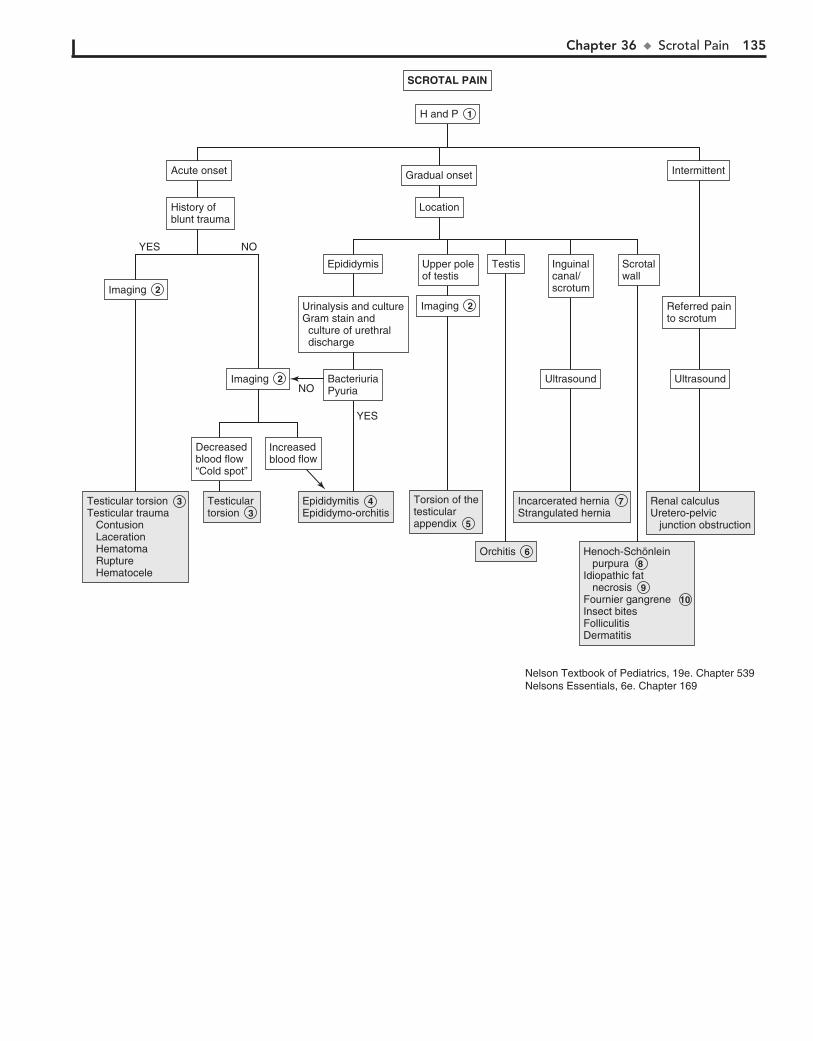
Chapter 36 u ScrotalPain 135
SCROTAL PAINSCROTAL PAIN
Testicular torsion 3 Testicular trauma Contusion Laceration Hematoma Rupture Hematocele
Testicular torsion 3 Testicular trauma Contusion Laceration Hematoma Rupture Hematocele
Testicular torsion 3 Testicular torsion 3
Epididymitis 4Epididymo-orchitisEpididymitis 4Epididymo-orchitis
Torsion of thetesticular appendix 5
Torsion of thetesticular appendix 5
Orchitis 6 Orchitis 6
Incarcerated hernia 7 Strangulated herniaIncarcerated hernia 7 Strangulated hernia
Henoch-Schönlein purpura 8 Idiopathic fat necrosis 9 Fournier gangrene 10Insect bitesFolliculitisDermatitis
Henoch-Schönlein purpura 8 Idiopathic fat necrosis 9 Fournier gangrene 10Insect bitesFolliculitisDermatitis
Renal calculusUretero-pelvic junction obstruction
Renal calculusUretero-pelvic junction obstruction
H and P 1 H and P 1
Decreasedblood flow“Cold spot”
Decreasedblood flow“Cold spot”
Increasedblood flowIncreasedblood flow
BacteriuriaPyuriaBacteriuriaPyuria
EpididymisEpididymis
History ofblunt traumaHistory ofblunt trauma
Acute onsetAcute onset
LocationLocation
Gradual onsetGradual onset IntermittentIntermittent
Urinalysis and cultureGram stain and culture of urethral discharge
Urinalysis and cultureGram stain and culture of urethral discharge
Upper poleof testisUpper poleof testis
UltrasoundUltrasound UltrasoundUltrasound
Inguinalcanal/scrotum
Inguinalcanal/scrotum
TestisTestis ScrotalwallScrotalwall
Referred painto scrotumReferred painto scrotum
Imaging 2 Imaging 2
YES
YES
NO
NO
Imaging 2 Imaging 2
Imaging 2 Imaging 2
Nelson Textbook of Pediatrics, 19e. Chapter 539Nelsons Essentials, 6e. Chapter 169
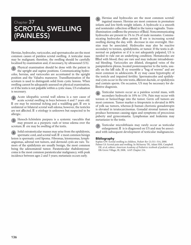
136
Chapter 37
SCROTAL SWELLING (PAINLESS)
5 Hernias and hydroceles are the most common scrotal/inguinal masses. Hernias are most common in premature
infants and low-birth-weight infants. A hydrocele is a smooth and nontender collection of fluid in the tunica vaginalis. Trans-illumination confirms the presence of fluid. Noncommunicating hydroceles are present in 1% to 2% of male neonates. Commu-nicating hydroceles often persist. There is increasing scrotal swelling during the day, with decrease in size overnight. Her-nias may be associated. Hydroceles may also be reactive secondary to torsion, epididymitis, or tumor. If the testis is ab-normal on palpation or if it is not adequately palpated, US is needed to rule out an underlying condition. Hematoceles are filled with blood; they are rare and may indicate intraabdomi-nal bleeding. Varicoceles are dilated, elongated veins of the pampiniform plexus, located posterosuperior to the testis, usu-ally on the left side. They resemble a “bag of worms” and are most common in adolescents. They may cause hypotrophy of the testicle and impaired fertility. Spermatoceles and epididy-mal cysts occur in the rete testis, efferent ductule, or epididymis and contain sperm. On occasion, US may be necessary for de-finitive diagnosis.
6 Testicular tumors occur as a painless scrotal mass, with secondary hydrocele in 10% to 15%. Pain may occur with
torsion or hemorrhage into the tumor. Germ cell tumors are most common. Tumor marker a fetoprotein is elevated in 80% of yolk sac tumors, whereas b-human chorionic gonadotropin is elevated in teratocarcinomas. Gonadal stromal tumors may produce hormones causing signs and symptoms of precocious puberty and gynecomastia. Lymphomas and leukemia may metastasize to the testis.
7 Testicular microlithiasis may rarely occur as testicular enlargement. This is diagnosed on US and may be associ-
ated with subsequent development of testicular malignancies.
Hernias, hydroceles, varicoceles, and spermatoceles are the most common causes of painless scrotal swelling. A testicular mass may be malignant; therefore, the swelling should be carefully localized by examination and, if necessary, by ultrasound (US).
1 Physical examination should be done with the patient in the upright and supine positions. Communicating hydro-
celes, hernias, and varicoceles are accentuated in the upright position and the Valsalva maneuver. Transillumination of the scrotum is used to distinguish solid from cystic lesions. When swelling cannot be adequately assessed on physical examination, or if the testis is not palpable within a cystic mass, US evaluation is necessary.
2 Acute idiopathic scrotal wall edema is a rare cause of acute scrotal swelling in boys between 4 and 7 years old.
There may be minimal itching and a waddling gait. There is unilateral or bilateral scrotal wall edema; however, the testicles are not affected. The etiology is unknown but suspected to be allergic.
3 Henoch-Schönlein purpura is a systemic vasculitis that may present as a purpuric rash or tense edema over the
scrotum. There may be swelling of the testis.
4 Solid extratesticular masses may arise from the epididymis, spermatic cord, and scrotal wall. The most common benign
lesion is spermatic cord lipoma. Fibromas, leiomyomas, lymph-angiomas, adrenal rest tumors, and dermoid cysts are rare. Tu-mors of the epididymis are usually benign, the most common being the adenomatoid tumor. Paratesticular rhabdomyosar-coma is the most common paratesticular malignancy, with peak incidence between ages 2 and 5 years; metastasis occurs early.
BibliographyKaplan GW: Scrotal swelling in children, Pediatr Rev 21:311–314, 2000. Palmer LS: Scrotal pain and swelling. In McInerny TK, Adam HM, Campbell
DE, et al, editors: American Academy of Pediatrics textbook of pediatric care, Elk Grove Village, Ill, 2008, AAP. Chapter 216.
Chapter 37 u ScrotalSwelling(Painless) 137
UltrasoundUltrasound
Solid massSolid mass
Extra-testicularExtra-testicular
H and P 1 H and P 1
Scrotal wall edema/swellingScrotal wall edema/swelling
DependentedemaDependentedema
Ultrasound± Serum tumor markers (� HCG, � fetoprotein)
Ultrasound± Serum tumor markers (� HCG, � fetoprotein)
TesticularTesticular
PAINLESS SCROTAL SWELLINGPAINLESS SCROTAL SWELLING
Idiopathic scrotal edema 2 Insect bitesHenoch-Schönleinpurpura 3
Idiopathic scrotal edema 2 Insect bitesHenoch-Schönleinpurpura 3
Soft tissue tumors 4Adenomatoid tumor 4Paratesticular rhabdomyosarcoma 4
Soft tissue tumors 4Adenomatoid tumor 4Paratesticular rhabdomyosarcoma 4
Nephrotic syndromeProtein losing enteropathyLiver failure (See chapter 34 on Edema)
Nephrotic syndromeProtein losing enteropathyLiver failure (See chapter 34 on Edema)
Testicular tumor 6Testicular microlithiasis 7 Testicular tumor 6Testicular microlithiasis 7
Cystic massCystic mass
± Ultrasound± Referral to urology
± Ultrasound± Referral to urology
Inguinal hernia 5 HydroceleHematoceleVaricoceleSpermatocele and epididymal cysts
Inguinal hernia 5 HydroceleHematoceleVaricoceleSpermatocele and epididymal cysts
Nelson Textbook of Pediatrics, 19e. Chapter 539
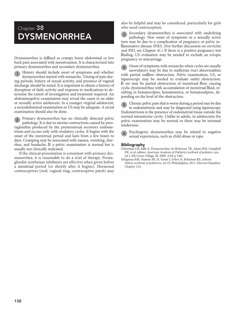
138
Chapter 38
DYSMENORRHEA
also be helpful and may be considered, particularly for girls who need contraception.
3 Secondary dysmenorrhea is associated with underlying pathology. New onset of symptoms in a sexually active
teen may be due to a complication of pregnancy or pelvic in-flammatory disease (PID). (For further discussion on cervicitis and PID, see Chapter 41.) If there is a positive pregnancy test finding, US evaluation may be needed to exclude an ectopic pregnancy or miscarriage.
4 Onset of symptoms with menarche when cycles are usually anovulatory may be due to müllerian tract abnormalities
with partial outflow obstruction. Pelvic examination, US, or laparoscopy may be needed to evaluate outlet obstruction. There may be partial obstruction of menstrual flow, causing cyclic dysmenorrhea with accumulation of menstrual fluid, re-sulting in hematocolpos, hematometra, or hematosalpinx, de-pending on the level of the obstruction.
5 Chronic pelvic pain that is worse during a period may be due to endometriosis and may be diagnosed using laparoscopy.
Endometriosis is the presence of endometrial tissue outside the normal intrauterine cavity. Unlike in adults, in adolescents the pelvic examination may be normal or there may be minimal tenderness.
6 Psychogenic dysmenorrhea may be related to negative sexual experiences, such as child abuse or rape.
Dysmenorrhea is defined as crampy lower abdominal or low back pain associated with menstruation. It is characterized into primary dysmenorrhea and secondary dysmenorrhea.
1 History should include onset of symptoms and whether dysmenorrhea started with menarche. Timing of pain dur-
ing periods, history of sexual activity, and presence of vaginal discharge should be noted. It is important to obtain a history of disruption of daily activity and response to medications to de-termine the extent of investigation and treatment required. An abdominopelvic examination may reveal the cause in an older or sexually active adolescent. In a younger virginal adolescent, a rectoabdominal examination or US may be adequate. A rectal examination should also be done.
2 Primary dysmenorrhea has no clinically detected pelvic pathology. It is due to uterine contractions caused by pros-
taglandins produced by the premenstrual secretory endome-trium and occurs only with ovulatory cycles. It begins with the onset of the menstrual period and lasts from a few hours to days. Cramping may be associated with nausea, vomiting, diar-rhea, and headache. The pelvic examination is normal but is usually not clinically indicated.
If the clinical presentation is consistent with primary dys-menorrhea, it is reasonable to do a trial of therapy. Prosta-glandin synthetase inhibitors are effective when given before a menstrual period (or shortly after it begins). Hormonal contraceptives (oral, vaginal ring, contraceptive patch) may
BibliographyDinerman LM, Joffe A: Dysmenorrhea. In McInerny TK, Adam HM, Campbell
DE, et al, editors: American Academy of Pediatrics textbook of pediatric care, ed 1, Elk Grove Village, Ill, 2009, AAP, p 1461.
Kliegman RM, Stanton BF, St. Geme J, Schor N, Behrman RE, editors: Nelson textbook of pediatrics, ed 19, Philadelphia, 2011, Elsevier/Saunders. Chapter 110.
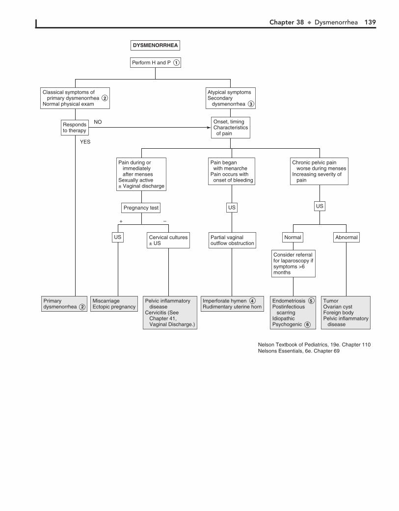
Chapter 38 u Dysmenorrhea 139
Pain during or immediately after mensesSexually active± Vaginal discharge
Pain during or immediately after mensesSexually active± Vaginal discharge
DYSMENORRHEADYSMENORRHEA
Perform H and P 1 Perform H and P 1
Primary dysmenorrhea 2 Primary dysmenorrhea 2
Miscarriage Ectopic pregnancyMiscarriage Ectopic pregnancy
Pelvic inflammatory diseaseCervicitis (See Chapter 41, Vaginal Discharge.)
Pelvic inflammatory diseaseCervicitis (See Chapter 41, Vaginal Discharge.)
Imperforate hymen 4 Rudimentary uterine hornImperforate hymen 4 Rudimentary uterine horn
TumorOvarian cystForeign bodyPelvic inflammatory disease
TumorOvarian cystForeign bodyPelvic inflammatory disease
Endometriosis 5 Postinfectious scarringIdiopathicPsychogenic 6
Endometriosis 5 Postinfectious scarringIdiopathicPsychogenic 6
NormalNormal AbnormalAbnormal
Consider referralfor laparoscopy ifsymptoms >6months
Consider referralfor laparoscopy ifsymptoms >6months
Cervical cultures± USCervical cultures± US
USUS
USUSUSUS
Partial vaginal outflow obstructionPartial vaginal outflow obstruction
Respondsto therapyRespondsto therapy
Onset, timingCharacteristics of pain
Onset, timingCharacteristics of pain
Classical symptoms of primary dysmenorrhea 2 Normal physical exam
Classical symptoms of primary dysmenorrhea 2 Normal physical exam
Atypical symptomsSecondary dysmenorrhea 3
Atypical symptomsSecondary dysmenorrhea 3
Pregnancy testPregnancy test
Chronic pelvic pain worse during mensesIncreasing severity of pain
Chronic pelvic pain worse during mensesIncreasing severity of pain
Pain began with menarchePain occurs with onset of bleeding
Pain began with menarchePain occurs with onset of bleeding
NO
+ –
YES
Nelson Textbook of Pediatrics, 19e. Chapter 110Nelsons Essentials, 6e. Chapter 69
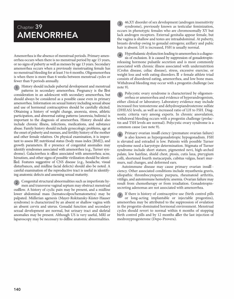
140
Chapter 39
AMENORRHEA
3 46,XY disorder of sex development (androgen insensitivity syndrome), previously known as testicular feminization,
occurs in phenotypic females who are chromosomally XY but lack androgen receptors. External genitalia appear female, but the vagina is shallow and testes are intraabdominal. At puberty, breasts develop owing to gonadal estrogens; axillary and pubic hair is absent. LH is increased, FSH is usually normal.
4 Hypothalamic dysfunction leading to amenorrhea is a diagno-sis of exclusion. It is caused by suppression of gonadotropin-
releasing hormone pulsatile secretion and is most commonly associated with chronic illness associated with undernutrition (Crohn disease, celiac disease), stress, excessive exercise, or weight loss and with eating disorders. The female athlete triad consists of disordered eating, amenorrhea, and low bone mass. Withdrawal bleeding may occur with a progestin challenge (see note 9).
5 Polycystic ovary syndrome is characterized by oligomen-orrhea or amenorrhea and evidence of hyperandrogenism,
either clinical or laboratory. Laboratory evidence may include increased free testosterone and dehydroepiandrosterone sulfate (DHEAS) levels, as well as increased ratio of LH to FSH. Diag-nostic criteria vary among experts. In chronic anovulation, withdrawal bleeding occurs with a progestin challenge (prolac-tin and TSH levels are normal). Polycystic ovary syndrome is a common cause (see note 9).
6 Primary ovarian insufficiency (premature ovarian failure) is also known as hypergonadotropic hypogonadism. FSH
is elevated and estradiol is low. Patients with possible Turner syndrome need a karyotype determination. Stigmata of Turner syndrome include short stature, pigmented nevi, high-arched palate, low hairline, shield chest, ptosis, cutis laxa, pterygium colli, shortened fourth metacarpals, cubitus valgus, heart mur-murs, nail changes, and deformed ears.
Autoimmune disease may cause primary ovarian insuffi-ciency. Other associated conditions include myasthenia gravis, idiopathic thrombocytopenic purpura, rheumatoid arthritis, vitiligo, and autoimmune hemolytic anemia. Ovarian failure may result from chemotherapy or from irradiation. Gonadotropin-secreting adenomas are not associated with amenorrhea.
7 If there is history of contraceptive use (birth control pills or long-acting implantable or injectable progestins),
amenorrhea may be attributed to the suppression of ovulation in the progestin-dominated hormonal environment. Menstrual cycles should revert to normal within 6 months of stopping birth control pills and by 12 months after the last injection of medroxyprogesterone (Depo-Provera).
Amenorrhea is the absence of menstrual periods. Primary amen-orrhea occurs when there is no menstrual period by age 15 years, or no signs of puberty as well as menses by age 13 years. Secondary amenorrhea occurs when a previously menstruating female has no menstrual bleeding for at least 3 to 6 months. Oligomenorrhea is when there is more than 6 weeks between menstrual cycles or fewer than 9 periods annually.
1 History should include pubertal development and menstrual patterns in secondary amenorrhea. Pregnancy is the first
consideration in an adolescent with secondary amenorrhea, but should always be considered as a possible cause even in primary amenorrhea. Information on sexual history including sexual abuse and use of hormonal contraceptives should be carefully elicited. Obtaining a history of weight change, anorexia, stress, athletic participation, and abnormal eating patterns (anorexia, bulimia) is important to the diagnosis of amenorrhea. History should also include chronic illness, infections, medications, and substance abuse. Family history should include gynecologic problems, age at the onset of puberty and menses, and fertility history of the mother and other female relatives. On physical examination, it is impor-tant to assess BP, nutritional status (body mass index [BMI]), and growth parameters. The presence of congenital anomalies may identify syndromes associated with amenorrhea (e.g., Turner syn-drome). Galactorrhea is often associated with amenorrhea; acne, hirsutism, and other signs of possible virilization should be identi-fied. Features suggestive of CNS disease (e.g., headache, visual disturbances, and midline facial defects) should also be noted. A careful examination of the reproductive tract is useful in identify-ing anatomic defects and assessing sexual maturity.
2 Congenital structural abnormalities such as imperforate hy-men and transverse vaginal septum may obstruct menstrual
outflow. A history of cyclic pain may be present, and a midline lower abdominal mass (hematocolpos/hematometra) may be palpated. Müllerian agenesis (Mayer-Rokitansky-Küster-Hauser syndrome) is characterized by an absent or shallow vagina with an absent cervix and uterus. Gonadal function and secondary sexual development are normal, but urinary tract and skeletal anomalies may be present. Although US is very useful, MRI or laparoscopy may be necessary to define anatomic abnormalities.

Chapter 39 u Amenorrhea 141
KaryotypeKaryotype
See next pageSee next page
Pelvic US± Karyotype± Free testosterone± DHEAS
Pelvic US± Karyotype± Free testosterone± DHEAS
Low/normal FSHLow/normal FSH FSH (repeat) LHLow estradiol
FSH (repeat) LHLow estradiol
FSH/LH± EstradiolFSH/LH± Estradiol
Secondary sexualcharacteristics presentSecondary sexualcharacteristics present
Primary amenorrheaPrimary amenorrhea
Urine hCGpositiveUrine hCGpositive
Urine hCGnegativeUrine hCGnegative
Hirsutism present?Signs of virilization?Hirsutism present?Signs of virilization?
See Chapters Atypical or Ambiguous Genitalia [71], Hirsutism [72]See Chapters Atypical or Ambiguous Genitalia [71], Hirsutism [72]
Secondary amenorrheaSecondary amenorrhea
History of contraceptive useHistory of contraceptive use
AMENORRHEAAMENORRHEA
See Chapter 68,Pubertal DelaySee Chapter 68,Pubertal Delay
PregnancyPregnancy Structural abnormalities 2 Imperforate hymen Transverse vaginal septum Müllerian agenesis46,XY Disorder of sex development (androgen insensitivity) 3Hypothalamic amenorrhea 4Polycystic ovary syndrome (PCOS) 5
Structural abnormalities 2 Imperforate hymen Transverse vaginal septum Müllerian agenesis46,XY Disorder of sex development (androgen insensitivity) 3Hypothalamic amenorrhea 4Polycystic ovary syndrome (PCOS) 5
Primary ovarian insufficiency 6 Turner syndrome Autoimmune oophoritis Chemotherapy or radiation Galactosemia Trisomy 21 Fragile X carriers Idiopathic
Primary ovarian insufficiency 6 Turner syndrome Autoimmune oophoritis Chemotherapy or radiation Galactosemia Trisomy 21 Fragile X carriers Idiopathic
Contraceptiveamenorrhea 7 Contraceptiveamenorrhea 7
YESNO YES
YES
NO
NO
Perform H and P 1 Perform H and P 1
Urine hCGUrine hCG
Nelson Textbook of Pediatrics, 19e. Chapters 110, 546, 580Nelsons Essentials, 6e. Chapter 69
DHEAS = dehydroepiandrosterone sulfate
142 Part V u GenitourinarySystem
8 Adolescent girls with hyperprolactinemia may present with amenorrhea or delayed puberty and often with
galactorrhea. Cranial imaging (MRI) is recommended to evaluate for pituitary or hypothalamic tumors or disease (cra-niopharyngioma, prolactinoma, sarcoidosis). Galactorrhea with normal or mildly elevated prolactin levels may be second-ary to nipple stimulation and chest wall irritation or trauma. Hyperprolactinemia may be due to drugs including antipsy-chotics, methyldopa, amitriptyline, benzodiazepines, cocaine, and metoclopramide.
9 Estrogen status may be evaluated by progestin challenge, because estrogen levels are not always reliable. Estrogen
status may also be confirmed by vaginal smear or the presence of abundant watery cervical mucus. For a progestin challenge, a 5- to 10-day course of oral medroxyprogesterone acetate or a single dose of intramuscular progesterone is given. If bleeding
BibliographyEmans SJH, Laufer MR: Amenorrhea in the adolescent. In Emans SJ, Laufer
MR, editors: Emans, Laufer, Goldstein’s pediatric & adolescent gynecology, ed 6, Philadelphia, 2012, Lippincott Williams & Wilkins, pp 138–158.
occurs within 2 weeks of treatment, it implies a functional uterus and outflow tract and an endometrium that has been exposed to estrogen. The amount of bleeding is roughly propor-tional to the amount and duration of prior estrogen exposure.
10 If there is no bleeding, it usually implies a low-estrogen state or hypoestrogenic amenorrhea. Rarely, it may be that
the uterus cannot bleed secondary to uterine scarring caused by prior dilatation and curettage or severe uterine infections (Ash-erman syndrome). This is rare in teenagers. It is important to rule out pregnancy. If there is any question, the pregnancy test must be repeated.
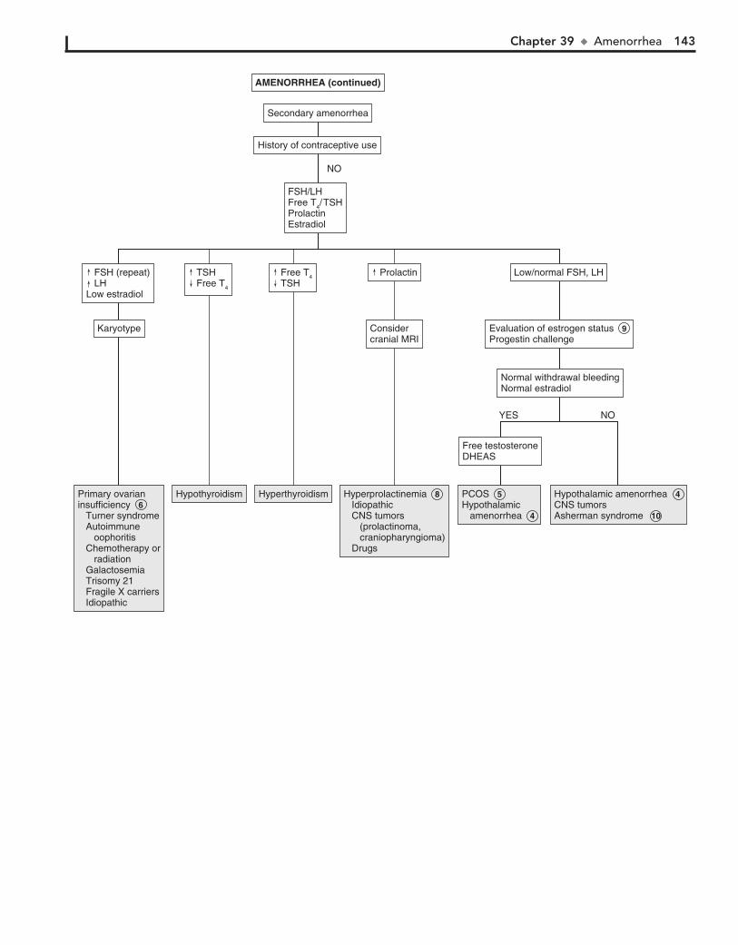
Chapter 39 u Amenorrhea 143
Evaluation of estrogen status 9 Progestin challengeEvaluation of estrogen status 9 Progestin challenge
Considercranial MRIConsidercranial MRI
AMENORRHEA (continued)AMENORRHEA (continued)
Secondary amenorrheaSecondary amenorrhea
History of contraceptive useHistory of contraceptive use
FSH/LHFree T4/ TSHProlactinEstradiol
FSH/LHFree T4/ TSHProlactinEstradiol
Primary ovarianinsufficiency 6 Turner syndrome Autoimmune oophoritis Chemotherapy or radiation Galactosemia Trisomy 21 Fragile X carriers Idiopathic
Primary ovarianinsufficiency 6 Turner syndrome Autoimmune oophoritis Chemotherapy or radiation Galactosemia Trisomy 21 Fragile X carriers Idiopathic
HypothyroidismHypothyroidism HyperthyroidismHyperthyroidism Hyperprolactinemia 8 Idiopathic CNS tumors (prolactinoma, craniopharyngioma) Drugs
Hyperprolactinemia 8 Idiopathic CNS tumors (prolactinoma, craniopharyngioma) Drugs
PCOS 5Hypothalamic amenorrhea 4
PCOS 5Hypothalamic amenorrhea 4
Hypothalamic amenorrhea 4 CNS tumorsAsherman syndrome 10
Hypothalamic amenorrhea 4 CNS tumorsAsherman syndrome 10
Low/normal FSH, LHLow/normal FSH, LH
KaryotypeKaryotype
Free testosteroneDHEASFree testosteroneDHEAS
Normal withdrawal bleedingNormal estradiolNormal withdrawal bleedingNormal estradiol
NO
NO
YES
FSH (repeat) LHLow estradiol
FSH (repeat) LHLow estradiol
Prolactin Prolactin Free T4 TSH Free T4 TSH
TSH Free T4
TSH Free T4

144
Chapter 40
ABNORMAL VAGINAL BLEEDING
3 In prepubertal-age girls without cyclic bleeding and with no signs of puberty, a vulvovaginal source is most com-
mon. Vaginal bleeding is more predictive of a foreign body than a vaginal discharge. The possibility of sexual abuse must be considered. The foreign object may be visualized in the knee-chest position, but if not, examination using anesthesia may be required.
4 Infectious vulvovaginitis usually appears as a discharge, but bleeding may be present. The most common organ-
isms obtained on culture are group A streptococci, Shigella, and mixed organisms. The presence of gonococci, Chlamydia, or Trichomonas should prompt evaluation for sexual abuse.
5 Vulvovaginal trauma is usually caused by straddle injuries and less commonly by vaginal penetration and tearing
from forced leg abduction; always consider the possibility of sexual abuse.
6 If a mass is visualized, consider urethral prolapse, which appears as red, friable, often necrotic tissue at the urethra.
7 In lichen sclerosus, the vulvar skin becomes thin and parchment-like (classically in an hourglass pattern around
the introitus and anus) and therefore susceptible to bleeding from minor trauma. The diagnosis may be confirmed by biopsy.
8 Neoplasms include hemangiomas, polyps, and sarcoma botryoides (a grapelike mass protruding from the va-
gina). Malignancies are uncommon (adenocarcinoma and rhabdomyosarcoma).
9 Exogenous exposures to estrogens may occur from inges-tion of birth control pills, foods, and beauty products. It
has been hypothesized that plastics may contain estrogen-like components.
10 Precocious menarche is a rare form of incomplete preco-cious puberty with cyclic menstruation but no other sec-
ondary sexual characteristics. There may be a slight increase in serum estrogen levels; gonadotropin levels are prepubertal.
Normal menstrual cycles range from 3 to 7 days. Periodic men-strual bleeding occurring more frequently than every 21 days, greater than every 45 days, or lasting longer than 7 days requires evaluation. With excessive blood loss, iron deficiency anemia may develop. Variations in menstrual cycles may include menorrhagia (normal intervals, excessive flow and duration of bleeding), metror-rhagia (irregular intervals), polymenorrhea (intervals # 21 days), oligomenorrhea (,6 menses/yr), and intermenstrual bleeding.
1 The age of the patient is important, as well as any history of abuse or trauma, including sexual abuse. A history of any for-
eign body, including intrauterine devices (IUDs) and tampons in older girls, should be elicited. In girls who have reached menarche, a detailed menstrual history including date of menarche and men-strual pattern should be obtained. Increased clots may signify an abnormality. A sexual history (sexually transmitted disease, sexual partners) is important, as well as any use of hormonal contraception. Exposure to medications, including exogenous estrogens, anticoagu-lants, and platelet inhibitors, may be a cause of bleeding. Abdominal pain or vaginal discharge may indicate infections. Review of systems should include stress, weight change, and chronic diseases. On ex-amination, the site of bleeding should be carefully assessed. An ex-amination of external genitalia must be done (vaginal digital exam if possible) to identify anatomic abnormalities, and a pelvic examina-tion performed when indicated for sexually active patients. Ortho-static BPs may be helpful in cases of heavy bleeding.
2 In the newborn, a small amount of endometrial bleeding may occur secondary to withdrawal from relatively high
fetal estrogen levels.
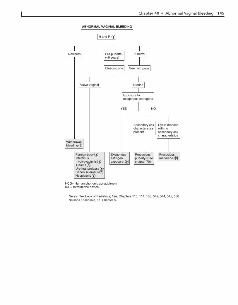
Chapter 40 u AbnormalVaginalBleeding 145
WithdrawalbleedingWithdrawalbleeding 2
Foreign bodyInfectious vulvovaginitisTrauma Urethral prolapseLichen sclerosusNeoplasms
Foreign body 3 Infectious vulvovaginitis 4 Trauma 5 Urethral prolapse 6 Lichen sclerosus 7 Neoplasms 8
Exogenousestrogenexposure 9
Exogenousestrogenexposure 9
Precociouspuberty (Seechapter 70)
Precociouspuberty (Seechapter 70)
PrecociousmenarchePrecociousmenarche 10
Cyclic menseswith no secondary sex characteristics
Cyclic menseswith no secondary sex characteristics
Secondary sexcharacteristicspresent
Secondary sexcharacteristicspresent
PubertalPubertalNewbornNewborn Pre-pubertal (<9 years)Pre-pubertal (<9 years)
See next pageSee next pageBleeding siteBleeding site
Exposure to exogenous estrogensExposure to exogenous estrogens
Vulvo-vaginal Vulvo-vaginal UterineUterine
ABNORMAL VAGINAL BLEEDINGABNORMAL VAGINAL BLEEDING
H and P 1
NOYES
HCG= Human chorionic gonadotropinIUD= Intrauterine device
Nelson Textbook of Pediatrics, 19e. Chapters 110, 114, 185, 542, 544, 545, 556Nelsons Essentials, 6e. Chapter 69
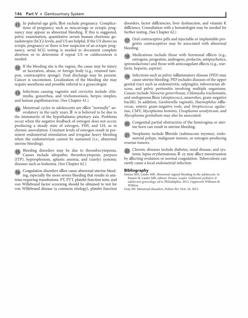
146 Part V u GenitourinarySystem
disorders, factor deficiencies, liver dysfunction, and vitamin K deficiency. Consultation with a hematologist may be needed for further testing. (See Chapter 62.)
17 Oral contraceptive pills and injectable or implantable pro-gestin contraceptives may be associated with abnormal
bleeding.
18 Medications include those with hormonal effects (e.g., estrogens, progestins, androgens, prolactin, antipsychotics,
spironolactone) and those with anticoagulant effects (e.g., war-farin, heparin, aspirin).
19 Infections such as pelvic inflammatory disease (PID) may cause uterine bleeding. PID includes diseases of the upper
genital tract such as endometritis, salpingitis, tuboovarian ab-scess, and pelvic peritonitis involving multiple organisms. Causes include Neisseria gonorrhoeae, Chlamydia trachomatis, and endogenous flora (streptococci, anaerobes, gram-negative bacilli). In addition, Gardnerella vaginalis, Haemophilus influ-enzae, enteric gram-negative rods, and Streptococcus agalac-tiae, CMV, Mycoplasma hominis, Ureaplasma urealyticum, and Mycoplasma genitalium may also be associated.
20 Congenital partial obstruction of the hemivagina or uter-ine horn can result in uterine bleeding.
21 Neoplasms include fibroids (submucous myoma), endo-metrial polyps, malignant tumors, or estrogen-producing
ovarian tumors.
22 Chronic diseases include diabetes, renal disease, and sys-temic lupus erythematosus. They may affect menstruation
by affecting ovulation or normal coagulation. Tuberculosis can rarely cause a local endometrial infection.
11 In pubertal-age girls, first exclude pregnancy. Complica-tions of pregnancy such as miscarriage or ectopic preg-
nancy may appear as abnormal bleeding. If this is suggested, pelvic examination, quantitative serum human chorionic go-nadotropin (hCG) levels, and US are helpful. If the US shows no ectopic pregnancy or there is low suspicion of an ectopic preg-nancy, serial hCG testing is needed to document complete abortion or to determine if repeat US or culdocentesis is needed.
12 If the bleeding site is the vagina, the cause may be injury or laceration, abuse, or foreign body (e.g., retained tam-
pon, contraceptive sponge). Foul discharge may be present. Cancer is uncommon. Localization of the bleeding site may require anesthesia and possible referral to a gynecologist.
13 Infections causing vaginitis and cervicitis include chla-mydia, gonorrhea, and trichomoniasis, herpes simplex,
and human papillomavirus. (See Chapter 41.)
14 Menstrual cycles in adolescents are often “normally” an-ovulatory in the early years. This is believed to be due to
the immaturity of the hypothalamic-pituitary axis. Problems occur when the negative feedback of estrogen does not occur, producing a steady state of estrogen, FSH, and LH, as in chronic anovulation. Constant levels of estrogen result in per-sistent endometrial stimulation and irregular heavy bleeding when the endometrium cannot be sustained (i.e., abnormal uterine bleeding).
15 Bleeding disorders may be due to thrombocytopenia. Causes include idiopathic thrombocytopenic purpura
(ITP), hypersplenism, aplastic anemia, and (rarely) systemic diseases such as leukemia. (See Chapter 62.)
16 Coagulation disorders often cause abnormal uterine bleed-ing, especially the more severe bleeding that results in ane-
mias requiring transfusions. PT, PTT, platelet function tests, and von Willebrand factor screening should be obtained to test for von Willebrand disease (a common etiology), platelet function
BibliographyEmans SJH, Laufer MR: Abnormal vaginal bleeding in the adolescent. In
Emans SJ, Laufer MR, editors: Emans, Laufer, Goldstein’s pediatric & adolescent gynecology, ed 6, Philadelphia, 2012, Lippincott Williams & Wilkins.
Gray SH: Menstrual disorders, Pediatr Rev 34:6–18, 2013.
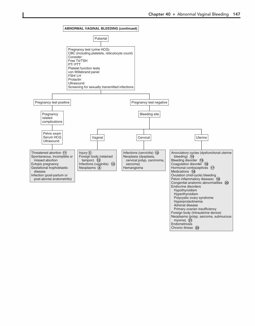
Chapter 40 u AbnormalVaginalBleeding 147
PubertalPubertal
Pregnancy test (urine HCG)CBC (including platelets, reticulocyte count)Consider:Free T4/TSHPT/ PTTPlatelet function testsvon Willebrand panelFSH/ LHProlactinUltrasoundScreening for sexually transmitted infections
Pregnancy test (urine HCG)CBC (including platelets, reticulocyte count)Consider:Free T4/TSHPT/ PTTPlatelet function testsvon Willebrand panelFSH/ LHProlactinUltrasoundScreening for sexually transmitted infections
ABNORMAL VAGINAL BLEEDING (continued)ABNORMAL VAGINAL BLEEDING (continued)
Threatened abortion Spontaneous, incomplete or missed abortion Ectopic pregnancyGestational trophoblastic diseaseInfection (post-partum or post-abortal endometritis)
Threatened abortion 11 Spontaneous, incomplete or missed abortion Ectopic pregnancyGestational trophoblastic diseaseInfection (post-partum or post-abortal endometritis)
Injury Foreign body (retained tampon) 12Infections (vaginitis) 13 Neoplasms 8
Injury 5 Foreign body (retained tampon) 12Infections (vaginitis) 13 Neoplasms 8
Infections (cervicitis)Neoplasia (dysplasia, cervical polyp, carcinoma, sarcoma)Hemangioma
Infections (cervicitis) 13 Neoplasia (dysplasia, cervical polyp, carcinoma, sarcoma)Hemangioma
Anovulatory cycles (dysfunctional uterine bleeding) 14 Bleeding disorder 15 Coagulation disorder 16 Hormonal contraceptives 17 Medications 18 Ovulation (mid-cycle) bleedingPelvic inflammatory disease) 19 Congenital anatomic abnormalities 20 Endocrine disorders Hypothyroidism Hyperthyroidism Polycystic ovary syndrome Hyperprolactinemia Adrenal disease Primary ovarian insufficiencyForeign body (Intrauterine device)Neoplasms (polyp, sarcoma, submucous myoma) 21 EndometriosisChronic illness 22
Anovulatory cycles (dysfunctional uterine bleeding) 14 Bleeding disorder 15 Coagulation disorder 16 Hormonal contraceptives 17 Medications 18 Ovulation (mid-cycle) bleedingPelvic inflammatory disease) 19 Congenital anatomic abnormalities 20 Endocrine disorders Hypothyroidism Hyperthyroidism Polycystic ovary syndrome Hyperprolactinemia Adrenal disease Primary ovarian insufficiencyForeign body (Intrauterine device)Neoplasms (polyp, sarcoma, submucous myoma) 21 EndometriosisChronic illness 22
UterineUterineVaginalVaginal CervicalCervicalPelvic examSerum HCGUltrasound
Pelvic examSerum HCGUltrasound
Bleeding siteBleeding sitePregnancyrelatedcomplications
Pregnancyrelatedcomplications
Pregnancy test positivePregnancy test positive Pregnancy test negativePregnancy test negative
148
Chapter 41
VAGINAL DISCHARGE
If there is possible sexual abuse, vaginal secretions must be cultured to exclude an STI.
6 Candidal vulvovaginitis in a prepubertal girl is uncommon but may occur after use of oral antibiotics.
7 Foreign bodies (often retained toilet tissue) in the vagina cause a foul-smelling brown or bloody discharge. If the
foreign body cannot be visualized, examination using anesthe-sia may be needed.
8 Anal pruritus may indicate pinworms and can be diag-nosed using a tape test. Pinworms are more common in
younger children.
9 Neoplasms are a rare cause of discharge. Examples include rhabdomyosarcoma, as well as adenosis and adenocarcinoma
in girls whose mothers were exposed to diethylstilbestrol during pregnancy.
10 In girls, an ectopic ureter may drain into the vagina (25%) and rarely into the cervix or uterus.
11 Müllerian anomalies may also present with vaginal dis-charge—for example, in a patient with a transverse vaginal
septum with a small opening.
12 The appearance of secretions may help in diagnosis; Can-dida causes a thick, whitish, curdy discharge, and Tricho-
monas has a yellow frothy discharge. A mucopurulent discharge with evidence of cervicitis may be seen with Chlamydia tracho-matis, N. gonorrhoeae, and herpes.
Microscopic examination of secretions may help provide the diagnosis. Trichomonads are motile flagellated organisms. Clue cells (i.e., epithelial cells coated with refractile bacteria) are seen with bacterial vaginosis. Potassium hydroxide (KOH) reveals pseudohyphae in Candida. A positive “whiff” test, due to release of amine odor, is present with bacterial vaginosis and sometimes with Trichomonas. A nucleic acid amplification test (NAAT) is the preferred test for detection of N. gonorrhoeae and C. trachomatis and can be performed on urethral or cervical specimens as well as urine. Other methods for detection of C. trachomatis include en-zyme immunoassay, ligase chain reaction, PCR, and culture. HIV and syphilis testing should be considered in all sexually active girls. If herpes simplex virus (HSV) is suspected, cell culture and PCR testing are preferred for a patient presenting with active le-sions. HSV serologic tests can be used to diagnose a patient with a history of vesicular genital lesions, and to diagnose a past HSV infection or current infection in a patient with an atypical lesions.
13 Gonorrheal infections may also involve the pharynx, rec-tum, and joints; disseminated infection may occur.
14 Chlamydial infections usually cause vaginal discharge, dysuria, or frequency. Endometritis with menorrhagia or
metrorrhagia may be present. Perihepatitis (Fitz-Hugh–Curtis syndrome) may occur with gonorrhea or chlamydial infection; these are also responsible for most pelvic inflammatory disease (PID).
15 Bacterial vaginosis occurs secondary to replacement of the normal hydrogen peroxide–producing vaginal flora (Lac-
tobacillus) by overgrowth of anaerobic microorganisms, as well as Gardnerella vaginalis, Ureaplasma, and Mycoplasma. Al-though bacterial vaginosis is not categorized as an STI, sexual activity is associated with increased frequency of vaginosis.
Vaginal discharge is a common but nonspecific sign in female adolescents. It should prompt consideration of sexually trans-mitted disease in sexually active girls.
1 Menstrual history, including changes in cycle or new onset of dysmenorrhea, may be helpful. A history of symptoms
including odor, color, amount of discharge, and pruritus should be obtained. Associated illnesses (HIV, diabetes) and medica-tions such as antibiotics and oral contraceptives may be con-tributing factors. Sexual history should include recent sexual encounters, number of partners, contraceptive and condom use, dyspareunia, history of sexually transmitted infection (STI), and pregnancies if any. Sexual abuse should be consid-ered, particularly in prepubertal girls with vaginal discharge. Most discharge in this age group is not sexually acquired.
Examination includes careful inspection of the vulva, vagina, and introitus for any abnormalities, including bruises, lacerations, excoriations, and vesicles, as well as character of the vaginal dis-charge. Pelvic examination (if necessary) in prepubertal girls may be done using a Huffman speculum. The Pederson speculum is slightly larger and may be used in an adolescent. Rashes may be seen with STIs, as well as joint and other constitutional symptoms.
2 Physiologic leukorrhea is a whitish mucoid discharge that occurs in newborns and adolescents. In pubertal adoles-
cents it starts before menarche and may decrease with onset of menses or may continue for a few years. During the middle of the menstrual cycle, it is copious and clear; it is scant and sticky during the second half of the cycle. Wet preparation shows epi-thelial cells and no abnormal findings. If the patient is sexually active, an STI must be ruled out.
3 All cases with suspected sexual abuse require direct cul-tures for Neisseria gonorrhoeae and Chlamydia, because
results of antigen detection tests may not be admissible in court. Referral to persons with expertise in evaluation of sexually abused children (sexual assault team) is indicated. It is prefera-ble that the physical exam be performed by a sexual abuse team if possible. Culture for gonorrhea and chlamydia should be obtained because it is considered the gold standard. Nucleic acid antigen tests should also be obtained because of increased yield; also, some state laws will accept them as evidence. Wet mount needs to be done, looking for Trichomonas and bacterial vaginosis. Any lesion suspicious for herpes should be cultured. Also test for HIV and syphilis.
4 In the prepubertal girl, the vulvar mucosa is thin and more susceptible to irritation and inflammation. Poor hygiene, use
of irritant soaps, bubble baths, and “nonbreathing” underwear may be associated with nonspecific or irritant vulvovaginitis.
5 Infectious vulvovaginitis may be due to fecal or respiratory pathogens, including Escherichia coli, group A Streptococ-
cus, Staphylococcus aureus, Haemophilus influenzae, Shigella, and Yersinia enterocolitica. A bloody vaginal discharge may be seen with Shigella or group A streptococcal infections.
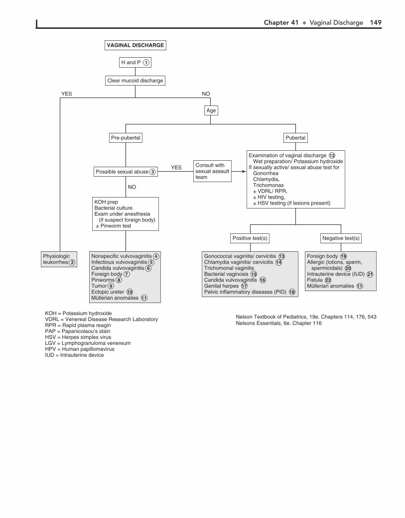
Chapter 41 u VaginalDischarge 149
PhysiologicleukorrheaPhysiologicleukorrhea 2
Nonspecific vulvovaginitisInfectious vulvovaginitisCandida vulvovaginitisForeign bodyPinwormsTumorEctopic ureterMüllerian anomalies
Nonspecific vulvovaginitis 4 Infectious vulvovaginitis 5 Candida vulvovaginitis 6 Foreign body 7 Pinworms 8 Tumor 9 Ectopic ureter 10 Müllerian anomalies 11
Gonococcal vaginitis/ cervicitisChlamydia vaginitis/ cervicitisTrichomonal vaginitisBacterial vaginosisCandida vulvovaginitisGenital herpesPelvic inflammatory diseases (PID)
Gonococcal vaginitis/ cervicitis 13 Chlamydia vaginitis/ cervicitis 14 Trichomonal vaginitisBacterial vaginosis 15 Candida vulvovaginitis 16 Genital herpes 17 Pelvic inflammatory diseases (PID) 18
Foreign bodyAllergic (lotions, sperm, spermicidals)Intrauterine device (IUD)FistulaMüllerian anomalies
Foreign body 19 Allergic (lotions, sperm, spermicidals) 20 Intrauterine device (IUD) 21 Fistula 22 Müllerian anomalies 11
Negative test(s)Negative test(s)Positive test(s)Positive test(s)
Examination of vaginal discharge Wet preparation/ Potassium hydroxideIf sexually active/ sexual abuse test for Gonorrhea Chlamydia, Trichomonas ± VDRL/ RPR, ± HIV testing, ± HSV testing (if lesions present)
Examination of vaginal discharge 12 Wet preparation/ Potassium hydroxideIf sexually active/ sexual abuse test for Gonorrhea Chlamydia, Trichomonas ± VDRL/ RPR, ± HIV testing, ± HSV testing (if lesions present)
Pre-pubertalPre-pubertal
Consult withsexual assaultteam
Consult withsexual assaultteam
Possible sexual abusePossible sexual abuse 3
KOH prepBacterial cultureExam under anesthesia (if suspect foreign body) ± Pinworm test
KOH prepBacterial cultureExam under anesthesia (if suspect foreign body) ± Pinworm test
PubertalPubertal
AgeAge
VAGINAL DISCHARGEVAGINAL DISCHARGE
H and P 1
Clear mucoid dischargeClear mucoid discharge
NOYES
NO
YES
KOH = Potassium hydroxideVDRL = Venereal Disease Research LaboratoryRPR = Rapid plasma reaginPAP = Papanicolaou’s stainHSV = Herpes simplex virusLGV = Lymphogranuloma venereumHPV = Human papillomavirusIUD = Intrauterine device
Nelson Textbook of Pediatrics, 19e. Chapters 114, 176, 543Nelsons Essentials, 6e. Chapter 116
150 Part V u GenitourinarySystem
19 Foreign bodies may also cause discharge in pubertal-age girls. Often it is a retained tampon.
20 Other causes of vaginitis include allergic vulvovaginitis caused by soaps, douches, contraceptive gels or creams,
and (rarely) sperm.
21 Chronic discharge may be due to an intrauterine device string.
22 Rectovaginal and anovaginal fistulae may be seen with Crohn disease and rarely with obstetric trauma (more of-
ten seen in developing countries with inadequate obstetric care). Vesicovaginal fistula may also be due to obstetric trauma.
16 Candida albicans is the most common fungal infection of the vagina; other Candida species may be involved. Diabetes mel-
litus, pregnancy, and use of antibiotics are predisposing factors.
17 Herpes appears initially as vesicles and pustules associated with vaginal discharge and dysuria, followed by ulceration
of the lesions, with associated constitutional symptoms. Tender inguinal lymphadenopathy may be present.
18 PID includes inflammatory disorders of the female upper genital tract. These may include endometritis, salpingitis,
tuboovarian abscess, and pelvic peritonitis, often in combina-tion. C. trachomatis and N. gonorrhoeae are most commonly involved. However, other pathogens may also be associated, such as anaerobes, G. vaginalis, H. influenzae, enteric gram-negative rods, Streptococcus agalactiae, CMV, Mycoplasma hom-inis, Ureaplasma urealyticum, and Mycoplasma genitalium.
PID is difficult to diagnose because of the wide variation in the symptoms and signs. Many females with PID have subtle or mild symptoms, resulting in many unrecognized cases. Health-care providers should consider the possibility of PID in young sexually active females presenting with vaginal discharge and/or abdominal pain.
BibliographyEmans SJH, Laufer MR: Vulvovaginal complaints in the adolescent. In Emans SJ,
Laufer MR, editors: Emans, Laufer, Goldstein’s pediatric & adolescent gynecology, ed 6, Philadelphia, 2012, Lippincott Williams & Wilkins, pp 305–324.
Emans SJH, Laufer MR: Sexually transmitted infections: Chlamydia, gonor-rhea, pelvic inflammatory disease, and syphilis. In Emans SJ, Laufer MR, editors: Emans, Laufer, Goldstein’s pediatric & adolescent gynecology, ed 6, Philadelphia, 2012, Lippincott Williams & Wilkins, pp 325–348.
Sugar NF, Graham EA: Common gynecologic problems in prepubertal girls, Pediatr Rev 27:213–222, 2006.

PART
VIMusculoskeletal System

152
Chapter 42
LIMP
biopsy or abscess aspiration. If the x-ray is negative and clinical suspicion is high, US may aid in detecting joint effusions, espe-cially of the hip. If an effusion is present, joint aspiration is recommended to rule out a septic arthritis. MRI is the preferred diagnostic test when osteomyelitis is suspected.
5 Septic arthritis is an infection confined to the capsule of a joint. Infection within the bone is osteomyelitis; secondary
spread to involve the joint may accompany it. Both disorders may present with localized pain and tenderness in an acutely ill child. Osteomyelitis may also occur subacutely with prolonged pain and limp but without fevers or systemic complaints. Bone changes on x-ray may not become evident for 7 to 10 days. Soft tissue changes may be evident earlier, plus films are usually obtained to rule out trauma and tumor initially. MRI is the preferred choice for diagnostic imaging if available. Bone scans are also sensitive and specific early in the clinical course if MRI is not available.
6 Septic arthritis is a medical emergency requiring prompt diagnosis and treatment. Patients typically present with
fever, malaise, refusal to walk, and localized joint pain, most commonly knee or hip. Examination reveals erythema, warmth, swelling, and pain with passive motion. US evaluation may sug-gest the diagnosis by demonstrating an effusion, but joint aspi-ration is mandated to confirm (or rule out) the diagnosis. (See Chapter 43.)
7 Acute transient synovitis (previously called “toxic synovi-tis”) is one of the most common causes of hip pain and
limping in children, usually between 3 and 8 years of age. The etiology is not well defined; it is described as a nonspecific in-flammatory condition, although viral, allergic, and traumatic mechanisms have all been suggested. Children present with unilateral hip pain, a painful limp, and slightly restricted abduc-tion and internal rotation. Fevers may be present, but rarely do patients present with the acute toxicity suggestive of septic ar-thritis. The diagnosis is one of exclusion; the most important diagnosis to exclude is a septic arthritis. Several recent studies have focused on identifying factors that help distinguish a sep-tic joint (which requires emergent treatment) from a transient synovitis (which is managed conservatively). The more of these factors that are present, the greater the likelihood of a septic joint: fever above 38.5 C°, an elevated CRP, an elevated ESR, refusal to bear weight, and an elevated WBC. With transient synovitis, laboratory results are usually normal or suggest a mild inflammatory process. X-rays are usually normal or may show a slightly widened medial joint space or accentuated peri-capsular shadow. Rarely, US and aspiration to rule out septic arthritis may be necessary when a child presents acutely with pain and fever; children presenting after 1 or 2 days of symp-toms may usually be managed more conservatively.
8 AVN (or Legg-Calvé-Perthes disease) is an ischemic necro-sis of the femoral head resulting in a bony deformity of the
femoral head. Children between 2 and 12 years of age are af-fected, with a peak incidence between 4 and 8 years of age; boys are affected more than girls. Children commonly demonstrate a limp before complaining of pain. Pain complaints are usually as-sociated with activity; the pain may be located in the groin or referred to the anteromedial thigh or knee. Common physical findings include slightly restricted abduction and internal rota-tion; over time, hip flexion contractures, atrophy of the leg mus-cles, and a leg-length discrepancy may be evident due to disuse.
An abnormal gait in a child may be due to pain, weakness, tor-sional deformity, or a musculoskeletal disorder.
1 A birth history and developmental history are particularly important for problems noticed early or around the time a
child begins walking. Prematurity and birth complications are risk factors for hypoxic brain damage (e.g., cerebral palsy). For older children, inquire about a history of trauma and systemic signs and symptoms (fever, rash, generalized weakness, weight loss) that may suggest infections or rheumatic disorders. Al-ways be conscious of the possibility of child abuse (nonacciden-tal trauma).
The musculoskeletal examination should include careful at-tention to all joints and the spine. The hip exam is particularly important because hip problems are a common cause of limp-ing, plus hip pathology frequently causes referred pain to the knee. Careful observation of the gait, ideally over a distance such as a long hallway, is critical. Having the child adequately undressed is also necessary for a good evaluation. In a painful gait, the child’s stance phase on the affected limb is shortened; trauma, infection, and rheumatic disorders are the most likely etiologies in these cases. Congenital and neuromuscular disor-ders are more likely to present with a painless (Trendelenburg) gait, which indicates proximal muscle weakness or hip instabil-ity. The stance phase is equal from side to side in these cases, but the child tends to shift their weight over the involved side for balance. Bilateral involvement produces a waddling gait.
2 Children are more at risk for epiphyseal fractures than liga-mentous sprains because their ligaments are generally stronger
than the adjacent growth plates. Because x-rays may be normal or show only physeal widening, the diagnosis is often clinical. Consul-tation should always be considered when in doubt.
3 Slipped capital femoral epiphysis (SCFE) is the most com-mon hip disorder in adolescents presenting with pain or
an abnormal gait. In this condition, a failure of the physis (growth plate) leads to posterior displacement of the metaphy-sis (femoral neck) relative to the epiphysis (femoral head). In many cases, an undiagnosed chronic slip is diagnosed after it is acutely worsened by trauma. A careful history often elicits chronic complaints of pain, subtle limp, or self-imposed activity restrictions. Examination reveals limited internal rotation of the affected hip and an out-toed, painful gait. AP and frog-leg lateral radiographs are routinely recommended to make the diagnosis; the latter should be performed with caution because of the potential risk for further displacement of the slip. Bilat-eral views are recommended because the condition is bilateral in 25% of cases at initial presentation. Conditions like hypothy-roidism, pituitary disorders, and renal osteodystrophy can im-pact bone ossification and increase the risk of developing SCFE.
4 When infection or inflammation is suspected, laboratory tests (CBC, ESR, CRP) may be helpful but are nonspecific.
Blood cultures yield a positive result in about 50% of cases of osteomyelitis; rates may be higher with specimens from a bone
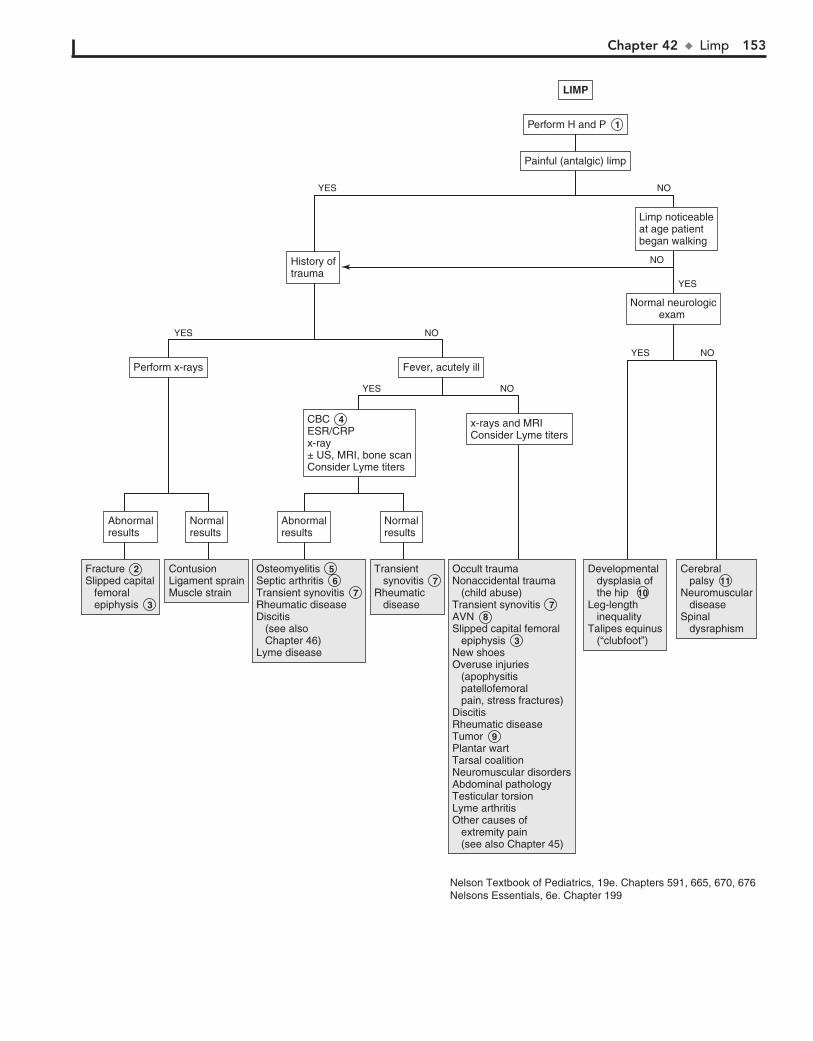
Chapter 42 u Limp 153
Fracture Slipped capital femoral epiphysis
Fracture 2 Slipped capital femoral epiphysis 3
ContusionLigament sprainMuscle strain
ContusionLigament sprainMuscle strain
Osteomyelitis Septic arthritis Transient synovitis Rheumatic diseaseDiscitis (see also Chapter 46)Lyme disease
Osteomyelitis 5 Septic arthritis 6 Transient synovitis 7 Rheumatic diseaseDiscitis (see also Chapter 46)Lyme disease
Transient synovitis Rheumatic disease
Transient synovitis 7 Rheumatic disease
Cerebral palsy Neuromuscular diseaseSpinal dysraphism
Cerebral palsy 11 Neuromuscular diseaseSpinal dysraphism
Developmental dysplasia of the hip Leg-length inequalityTalipes equinus (“clubfoot”)
Developmental dysplasia of the hip 10Leg-length inequalityTalipes equinus (“clubfoot”)
Occult traumaNonaccidental trauma (child abuse)Transient synovitis AVN Slipped capital femoral epiphysis New shoesOveruse injuries (apophysitis patellofemoral pain, stress fractures)DiscitisRheumatic diseaseTumor Plantar wartTarsal coalitionNeuromuscular disordersAbdominal pathologyTesticular torsionLyme arthritisOther causes of extremity pain (see also Chapter 45)
Occult traumaNonaccidental trauma (child abuse)Transient synovitis 7 AVN 8 Slipped capital femoral epiphysis 3 New shoesOveruse injuries (apophysitis patellofemoral pain, stress fractures)DiscitisRheumatic diseaseTumor 9 Plantar wartTarsal coalitionNeuromuscular disordersAbdominal pathologyTesticular torsionLyme arthritisOther causes of extremity pain (see also Chapter 45)
NormalresultsNormalresults
AbnormalresultsAbnormalresults
NormalresultsNormalresults
AbnormalresultsAbnormalresults
x-rays and MRI Consider Lyme titersx-rays and MRI Consider Lyme titers
CBC ESR/CRPx-ray± US, MRI, bone scanConsider Lyme titers
CBC 4 ESR/CRPx-ray± US, MRI, bone scanConsider Lyme titers
Fever, acutely illFever, acutely illPerform x-raysPerform x-rays
Normal neurologicexam
Normal neurologicexam
Limp noticeableat age patientbegan walking
Limp noticeableat age patientbegan walking
History oftraumaHistory oftrauma
LIMPLIMP
Perform H and P 1
Painful (antalgic) limpPainful (antalgic) limp
NOYES
NO
NO
YES
YES
YES
NO
NO
YES
Nelson Textbook of Pediatrics, 19e. Chapters 591, 665, 670, 676Nelsons Essentials, 6e. Chapter 199
154 Part VI u MusculoskeletalSystem
9 Night pain is characteristic of both benign and malignant primary or metastatic tumors. Characteristic x-ray findings
usually suggest the diagnosis of benign bone tumors; intervention (biopsy, removal, monitoring) will be required for many of them.
10 If not diagnosed in infancy, developmental dysplasia of the hip (DDH) will usually present as a limp, waddling
gait, or leg-length discrepancy after the child starts walking. On physical exam, hip abduction will be limited, a positive Galeazzi sign (knees at different levels with hips flexed when child lying supine) may be evident, and lumbar lordosis may be present (due to altered hip mechanics).
BibliographyCaird MS, Flynn JM, Leung YL, et al: Factors distinguishing septic arthritis
from transient synovitis of the hip in children. A prospective study, J Bone Joint Surg Am 88:1251–1257, 2006.
Perry DC, Bruce C: Evaluating the child who presents with an acute limp, BMJ 341:c4250, 2010.
11 Spastic diplegia is the most common type of cerebral palsy. Affected children are delayed in crawling and walk-
ing. The condition is characterized by toe-walking and a pain-less, waddling (Trendelenburg) gait. Examination reveals in-creased muscle tone, spasticity, hyperactive deep tendon reflexes, tight heel cords, and persistent pathologic reflexes.

Chapter 42 u Limp 155
Fracture Slipped capital femoral epiphysis
Fracture 2 Slipped capital femoral epiphysis 3
ContusionLigament sprainMuscle strain
ContusionLigament sprainMuscle strain
Osteomyelitis Septic arthritis Transient synovitis Rheumatic diseaseDiscitis (see also Chapter 46)Lyme disease
Osteomyelitis 5 Septic arthritis 6 Transient synovitis 7 Rheumatic diseaseDiscitis (see also Chapter 46)Lyme disease
Transient synovitis Rheumatic disease
Transient synovitis 7 Rheumatic disease
Cerebral palsy Neuromuscular diseaseSpinal dysraphism
Cerebral palsy 11 Neuromuscular diseaseSpinal dysraphism
Developmental dysplasia of the hip Leg-length inequalityTalipes equinus (“clubfoot”)
Developmental dysplasia of the hip 10Leg-length inequalityTalipes equinus (“clubfoot”)
Occult traumaNonaccidental trauma (child abuse)Transient synovitis AVN Slipped capital femoral epiphysis New shoesOveruse injuries (apophysitis patellofemoral pain, stress fractures)DiscitisRheumatic diseaseTumor Plantar wartTarsal coalitionNeuromuscular disordersAbdominal pathologyTesticular torsionLyme arthritisOther causes of extremity pain (see also Chapter 45)
Occult traumaNonaccidental trauma (child abuse)Transient synovitis 7 AVN 8 Slipped capital femoral epiphysis 3 New shoesOveruse injuries (apophysitis patellofemoral pain, stress fractures)DiscitisRheumatic diseaseTumor 9 Plantar wartTarsal coalitionNeuromuscular disordersAbdominal pathologyTesticular torsionLyme arthritisOther causes of extremity pain (see also Chapter 45)
NormalresultsNormalresults
AbnormalresultsAbnormalresults
NormalresultsNormalresults
AbnormalresultsAbnormalresults
x-rays and MRI Consider Lyme titersx-rays and MRI Consider Lyme titers
CBC ESR/CRPx-ray± US, MRI, bone scanConsider Lyme titers
CBC 4 ESR/CRPx-ray± US, MRI, bone scanConsider Lyme titers
Fever, acutely illFever, acutely illPerform x-raysPerform x-rays
Normal neurologicexam
Normal neurologicexam
Limp noticeableat age patientbegan walking
Limp noticeableat age patientbegan walking
History oftraumaHistory oftrauma
LIMPLIMP
Perform H and P 1
Painful (antalgic) limpPainful (antalgic) limp
NOYES
NO
NO
YES
YES
YES
NO
NO
YES
Nelson Textbook of Pediatrics, 19e. Chapters 591, 665, 670, 676Nelsons Essentials, 6e. Chapter 199
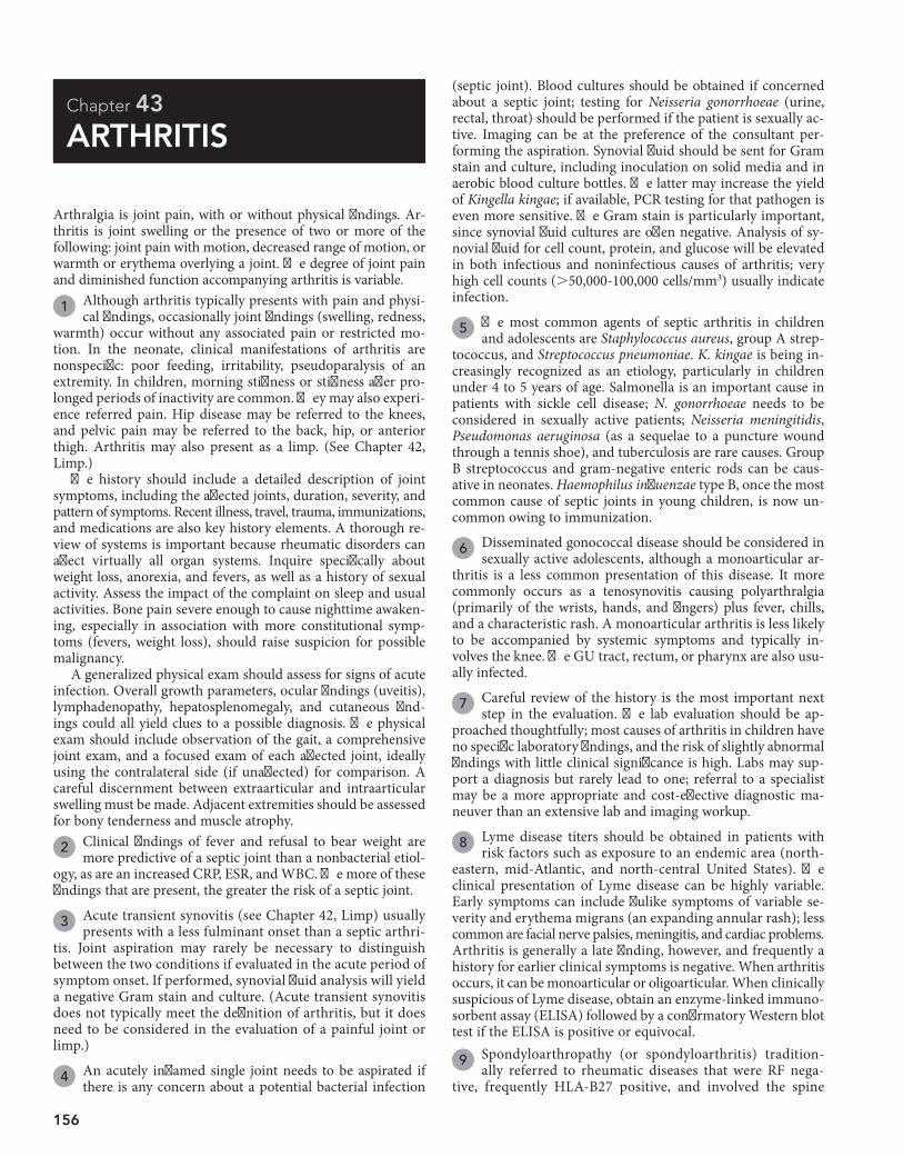
156
Chapter 43
ARTHRITIS
(septic joint). Blood cultures should be obtained if concerned about a septic joint; testing for Neisseria gonorrhoeae (urine, rectal, throat) should be performed if the patient is sexually ac-tive. Imaging can be at the preference of the consultant per-forming the aspiration. Synovial fluid should be sent for Gram stain and culture, including inoculation on solid media and in aerobic blood culture bottles. The latter may increase the yield of Kingella kingae; if available, PCR testing for that pathogen is even more sensitive. The Gram stain is particularly important, since synovial fluid cultures are often negative. Analysis of sy-novial fluid for cell count, protein, and glucose will be elevated in both infectious and noninfectious causes of arthritis; very high cell counts (.50,000-100,000 cells/mm3) usually indicate infection.
5 The most common agents of septic arthritis in children and adolescents are Staphylococcus aureus, group A strep-
tococcus, and Streptococcus pneumoniae. K. kingae is being in-creasingly recognized as an etiology, particularly in children under 4 to 5 years of age. Salmonella is an important cause in patients with sickle cell disease; N. gonorrhoeae needs to be considered in sexually active patients; Neisseria meningitidis, Pseudomonas aeruginosa (as a sequelae to a puncture wound through a tennis shoe), and tuberculosis are rare causes. Group B streptococcus and gram-negative enteric rods can be caus-ative in neonates. Haemophilus influenzae type B, once the most common cause of septic joints in young children, is now un-common owing to immunization.
6 Disseminated gonococcal disease should be considered in sexually active adolescents, although a monoarticular ar-
thritis is a less common presentation of this disease. It more commonly occurs as a tenosynovitis causing polyarthralgia (primarily of the wrists, hands, and fingers) plus fever, chills, and a characteristic rash. A monoarticular arthritis is less likely to be accompanied by systemic symptoms and typically in-volves the knee. The GU tract, rectum, or pharynx are also usu-ally infected.
7 Careful review of the history is the most important next step in the evaluation. The lab evaluation should be ap-
proached thoughtfully; most causes of arthritis in children have no specific laboratory findings, and the risk of slightly abnormal findings with little clinical significance is high. Labs may sup-port a diagnosis but rarely lead to one; referral to a specialist may be a more appropriate and cost-effective diagnostic ma-neuver than an extensive lab and imaging workup.
8 Lyme disease titers should be obtained in patients with risk factors such as exposure to an endemic area (north-
eastern, mid-Atlantic, and north-central United States). The clinical presentation of Lyme disease can be highly variable. Early symptoms can include flulike symptoms of variable se-verity and erythema migrans (an expanding annular rash); less common are facial nerve palsies, meningitis, and cardiac problems. Arthritis is generally a late finding, however, and frequently a history for earlier clinical symptoms is negative. When arthritis occurs, it can be monoarticular or oligoarticular. When clinically suspicious of Lyme disease, obtain an enzyme-linked immuno-sorbent assay (ELISA) followed by a confirmatory Western blot test if the ELISA is positive or equivocal.
Arthralgia is joint pain, with or without physical findings. Ar-thritis is joint swelling or the presence of two or more of the following: joint pain with motion, decreased range of motion, or warmth or erythema overlying a joint. The degree of joint pain and diminished function accompanying arthritis is variable.
1 Although arthritis typically presents with pain and physi-cal findings, occasionally joint findings (swelling, redness,
warmth) occur without any associated pain or restricted mo-tion. In the neonate, clinical manifestations of arthritis are nonspecific: poor feeding, irritability, pseudoparalysis of an extremity. In children, morning stiffness or stiffness after pro-longed periods of inactivity are common. They may also experi-ence referred pain. Hip disease may be referred to the knees, and pelvic pain may be referred to the back, hip, or anterior thigh. Arthritis may also present as a limp. (See Chapter 42, Limp.)
The history should include a detailed description of joint symptoms, including the affected joints, duration, severity, and pattern of symptoms. Recent illness, travel, trauma, immunizations, and medications are also key history elements. A thorough re-view of systems is important because rheumatic disorders can affect virtually all organ systems. Inquire specifically about weight loss, anorexia, and fevers, as well as a history of sexual activity. Assess the impact of the complaint on sleep and usual activities. Bone pain severe enough to cause nighttime awaken-ing, especially in association with more constitutional symp-toms (fevers, weight loss), should raise suspicion for possible malignancy.
A generalized physical exam should assess for signs of acute infection. Overall growth parameters, ocular findings (uveitis), lymphadenopathy, hepatosplenomegaly, and cutaneous find-ings could all yield clues to a possible diagnosis. The physical exam should include observation of the gait, a comprehensive joint exam, and a focused exam of each affected joint, ideally using the contralateral side (if unaffected) for comparison. A careful discernment between extraarticular and intraarticular swelling must be made. Adjacent extremities should be assessed for bony tenderness and muscle atrophy.
2 Clinical findings of fever and refusal to bear weight are more predictive of a septic joint than a nonbacterial etiol-
ogy, as are an increased CRP, ESR, and WBC. The more of these findings that are present, the greater the risk of a septic joint.
3 Acute transient synovitis (see Chapter 42, Limp) usually presents with a less fulminant onset than a septic arthri-
tis. Joint aspiration may rarely be necessary to distinguish between the two conditions if evaluated in the acute period of symptom onset. If performed, synovial fluid analysis will yield a negative Gram stain and culture. (Acute transient synovitis does not typically meet the definition of arthritis, but it does need to be considered in the evaluation of a painful joint or limp.)
4 An acutely inflamed single joint needs to be aspirated if there is any concern about a potential bacterial infection
9 Spondyloarthropathy (or spondyloarthritis) tradition-ally referred to rheumatic diseases that were RF nega-
tive, frequently HLA-B27 positive, and involved the spine
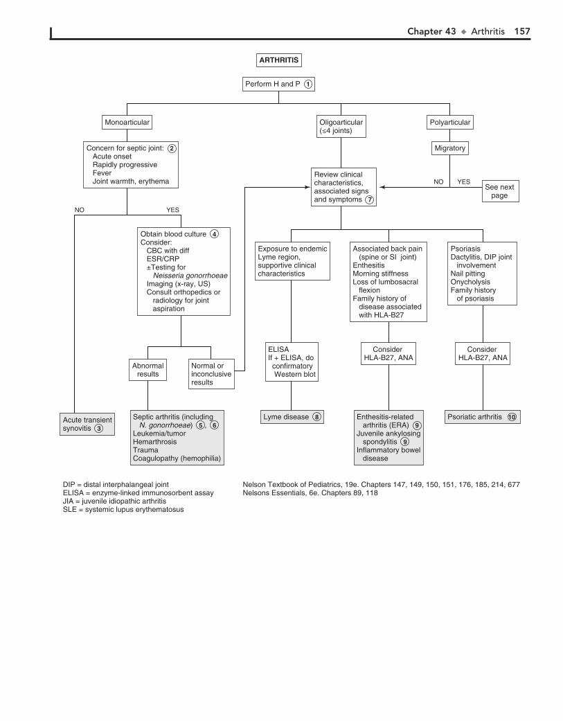
Chapter 43 u Arthritis 157
Septic arthritis (including N. gonorrhoeae) ,Leukemia/tumor Hemarthrosis Trauma Coagulopathy (hemophilia)
Septic arthritis (including N. gonorrhoeae) 5 , 6 Leukemia/tumor Hemarthrosis Trauma Coagulopathy (hemophilia)
Acute transientsynovitisAcute transientsynovitis 3
Lyme diseaseLyme disease 8 Enthesitis-related arthritis (ERA)Juvenile ankylosing spondylitisInflammatory bowel disease
Enthesitis-related arthritis (ERA) 9 Juvenile ankylosing spondylitis 9 Inflammatory bowel disease
Psoriatic arthritisPsoriatic arthritis 10
Normal orinconclusiveresults
Normal orinconclusiveresults
Abnormalresults
Abnormalresults
Obtain blood cultureConsider: CBC with diff ESR/CRP ±Testing for Neisseria gonorrhoeae Imaging (x-ray, US) Consult orthopedics or radiology for joint aspiration
Obtain blood culture 4 Consider: CBC with diff ESR/CRP ±Testing for Neisseria gonorrhoeae Imaging (x-ray, US) Consult orthopedics or radiology for joint aspiration
PsoriasisDactylitis, DIP joint involvementNail pittingOnycholysisFamily history of psoriasis
PsoriasisDactylitis, DIP joint involvementNail pittingOnycholysisFamily history of psoriasis
Exposure to endemicLyme region,supportive clinicalcharacteristics
Exposure to endemicLyme region,supportive clinicalcharacteristics
ELISAIf + ELISA, do confirmatory Western blot
ELISAIf + ELISA, do confirmatory Western blot
Consider HLA-B27, ANA
Consider HLA-B27, ANA
Consider HLA-B27, ANA
Consider HLA-B27, ANA
Associated back pain (spine or SI joint) EnthesitisMorning stiffnessLoss of lumbosacral flexionFamily history of disease associated with HLA-B27
Associated back pain (spine or SI joint) EnthesitisMorning stiffnessLoss of lumbosacral flexionFamily history of disease associated with HLA-B27
PolyarticularPolyarticularMonoarticularMonoarticular
MigratoryMigratory
See next page
See next page
Oligoarticular(≤4 joints)Oligoarticular(≤4 joints)
Review clinicalcharacteristics,associated signsand symptoms
Review clinicalcharacteristics,associated signsand symptoms 7
Concern for septic joint: Acute onset Rapidly progressive Fever Joint warmth, erythema
Concern for septic joint: 2 Acute onset Rapidly progressive Fever Joint warmth, erythema
ARTHRITISARTHRITIS
Perform Perform H and P 1
YES
YES
NO
NO
Nelson Textbook of Pediatrics, 19e. Chapters 147, 149, 150, 151, 176, 185, 214, 677Nelsons Essentials, 6e. Chapters 89, 118
DIP = distal interphalangeal jointELISA = enzyme-linked immunosorbent assayJIA = juvenile idiopathic arthritisSLE = systemic lupus erythematosus
158 Part VI u MusculoskeletalSystem
(especially the SI joints), large joints of the lower extremities, and entheses (where ligaments, tendons and joint capsules insert into bone). The current preferred classification system (per the International Leagues for the Associations for Rheu-matology [ILAR]) uses the term enthesitis-related arthritis (ERA) to include patients with both arthritis and enthesitis (the Achilles tendon and plantar fascia are common sites) or with enthesitis and two of the following additional character-istics: SI joint tenderness or lumbosacral pain, the presence of HLA-B27, onset of arthritis in males older than age 6 years, uveitis, or a first-degree relative with an HLA-B27–associated disease. Children meeting ILAR criteria for ERA may ulti-mately progress to meet criteria for juvenile ankylosing spon-dylitis or inflammatory bowel disease, although this progres-sion is not predictable. In juvenile ankylosing spondylitis, lower extremity arthritis (commonly including the hip) and severe enthesitis (especially at the foot, ankle, and knee) typically precede axial and SI joint involvement. X-rays can confirm sacroiliitis, but it may take years to get to this point. The arthritis associated with inflammatory bowel disease is polyarticular, affecting both small and large joints; it can also be migratory. Its severity usually reflects the degree of intes-tinal inflammation.
10 Psoriatic arthritis is included in the ILAR classification. Arthritis may precede the development of psoriasis. It is
most commonly an asymmetric oligoarthritis; both large and small joints can be affected, including the distal interphalangeal joints. Dactylitis (swollen “sausage-shaped” digits), nail pitting, and onycholysis are other characteristics. A positive ANA will occur more commonly than a positive HLA-B27 result. RF is negative.
11 Reactive arthritis refers to a monoarthritis or oligoarthri-tis, usually affecting the lower extremities, following (by
weeks or months) a GI or GU infection (Salmonella, Shigella, Yersinia enterocolitica, Campylobacter jejuni, Cryptosporidium parvum, Giardia intestinalis, Chlamydia trachomatis, and Urea-plasma have all been implicated). Enthesitis, dactylitis, cutaneous manifestations (balanitis, vulvitis, oral lesions, keratoderma blennorrhagica), and systemic symptoms of fever, malaise, and fatigue may occur; the arthritis is usually short-lived. Patients who are HLA-B27 positive have a higher risk of developing a chronic arthritis, especially spondyloarthritis. The arthritis
occurring with Campylobacter is more likely to be migratory. Reactive arthritis in not included in the ILAR classification system.
12 Arthritis is a frequent component of vasculitis syndromes, including Henoch-Schönlein purpura, polyarteritis no-
dosa, Wegener’s granulomatosis, and Takayasu arteritis. Char-acteristic skin findings occur with many of the disorders.
13 Arthritis and arthralgias have been associated with many viral infections, including rubella, parvovirus, EBV, CMV,
varicella, and influenza. The joint symptoms may occur during the course of the infection or as a postinfectious reaction. Symptoms usually resolve within 6 weeks. The arthritis occur-ring with mumps is more likely to be migratory.
14 The term juvenile idiopathic arthritis (JIA) encompasses all forms of chronic juvenile arthritis. The current pre-
ferred classification system (per ILAR) includes the disorders previously classified as JRA as well as the previously mentioned ERAs and psoriatic arthritis. For all of these disorders, the onset of arthritis must occur before 16 years of age and last a mini-mum of 6 weeks to meet the diagnostic criteria. (The term “rheumatoid” was removed to eliminate the association with adult seropositive rheumatoid arthritis, which is typically very distinct from childhood arthritis.) If clinical suspicion is high for JIA, labs may support the diagnosis. Hematologic abnor-malities reflect the degree of inflammation. RF is present in 5% to 10% of polyarthritis; ANA may be positive in oligoarthritis or polyarthritis. The presence of ANA indicates an increased risk of uveitis. Both RF and ANA may be transiently elevated in the presence of acute infections, so results may be misleading if the clinical diagnosis is not suggestive.
15 Oligoarthritis (#4 joints) is classified as persistent JIA if no additional joint involvement occurs over time; if addi-
tional joint involvement does occur, it is extended oligoarticu-lar JIA. Affected joints are mostly lower large joints (knees and ankles); hip involvement is very rare and should suggest an-other diagnosis (spondyloarthropathy or nonrheumatologic cause).
16 Polyarthritis includes involvement of 5 or more joints in up-per and lower extremities in the first 6 months of symptoms.
It is further classified based on the presence or absence of RF.
Chapter 43 u Arthritis 159
Reactive arthritis Disseminated gonococcal disease Inflammatory bowel disease
Reactive arthritis 11Disseminated gonococcal disease 6Inflammatory bowel disease
Vasculitis syndromes Serum sickness Lyme disease DermatomyositisBehçet syndromeViral-associated arthritis (parvovirus, rubella, enterovirus)
Vasculitis syndromes 12Serum sickness Lyme disease 8DermatomyositisBehçet syndromeViral-associated arthritis 13 (parvovirus, rubella, enterovirus)
Oligoarticular JIA Polyarticular JIA Oligoarticular JIA 15Polyarticular JIA 16
Lower-extremity arthritis (knee, ankle)Morning stiffness, easy fatigability, poor sleepAbsence of psoriasis and spondyloarthritisOnset at <16 years of ageDuration >6 weeks
Lower-extremity arthritis (knee, ankle)Morning stiffness, easy fatigability, poor sleepAbsence of psoriasis and spondyloarthritisOnset at <16 years of ageDuration >6 weeks
GI/GU symptomsGI/GU symptoms Skin findingsSkin findings
See next pageSee next page
Consider CBC withdiff, RF, ANA 14Consider CBC withdiff, RF, ANA 14
Consider culture forGonorrhea/chlamydia, stoolculture, HLA testing
Consider culture forGonorrhea/chlamydia, stoolculture, HLA testing
ARTHRITIS (continued)ARTHRITIS (continued)
Nonmigratory oligoarticularor polyarticular arthritis
(continued)
Nonmigratory oligoarticularor polyarticular arthritis
(continued)

160 Part VI u MusculoskeletalSystem
17 Children with systemic-onset JIA (SoJIA) experience short-lived high fevers once to twice daily for a mini-
mum of 2 weeks, often accompanied by a characteristic salmon-colored macular rash. Visceral involvement (hepa-tosplenomegaly, lymphadenopathy, serositis) is very com-mon and may precede the arthritis, although arthritis (which is usually polyarticular and can involve the hip, cer-vical spine, and temporomandibular joint) is necessary for the diagnosis. Labs typically reflect an anemia of chronic disease and generally very high inflammatory markers (in-cluding ferritin), WBCs, and platelet counts. RF and ANA are typically absent.
18 Arthralgias and arthritis in knees and hands may develop 10 to 28 days after immunization with rubella vaccine.
This reaction occurs most commonly in postpubertal females. An associated rash may occur.
19 Poststreptococcal arthritis is an oligoarticular nonmigra-tory arthritis that affects mostly lower joints after infection
with group A streptococcus. These cases do not fulfill the Jones criteria for acute rheumatic fever. Controversy exists over whether poststreptococcal arthritis is a distinct entity or an in-complete form of acute rheumatic fever.
20 In children, systemic lupus erythematosus (SLE) most commonly presents with nonspecific symptoms (malaise,
fatigue); small-joint arthralgias and arthritis are frequently present but overlooked in the early phases of the disease. Adults classically experience a migratory arthritis.
21 One of the shortcomings of the ILAR system is that chil-dren do experience arthritis that does not fit into one of
the classifications (or fits into more than one). The course or progression of these children is still unable to be predicted.
22 The polyarthritis that constitutes one of the Jones criteria for the diagnosis of rheumatic fever is migratory and char-
acterized by extreme tenderness, redness, and swelling of af-fected joints. It typically affects larger joints (knees, ankles, wrists, elbows) and rarely affects the spine, hands, or hips. This arthritis classically responds very quickly to treatment with aspirin. Arthralgias (in the absence of arthritis) constitute a minor criterion for the diagnosis of rheumatic fever.
BibliographyBerard R: Approach to the child with joint inflammation, Pediatr Clin North Am
59:245–262, 2012.Caird MS, Flynn JM, Leung YL, et al: Factors distinguishing septic arthritis
from transient synovitis of the hip in children: A prospective study, J Bone Joint Surg Am 88:1251–1257, 2006.
23 Hepatitis B arthritis-dermatitis syndrome clinically mim-ics serum sickness, with an urticarial rash and symmetric
migratory polyarthritis.
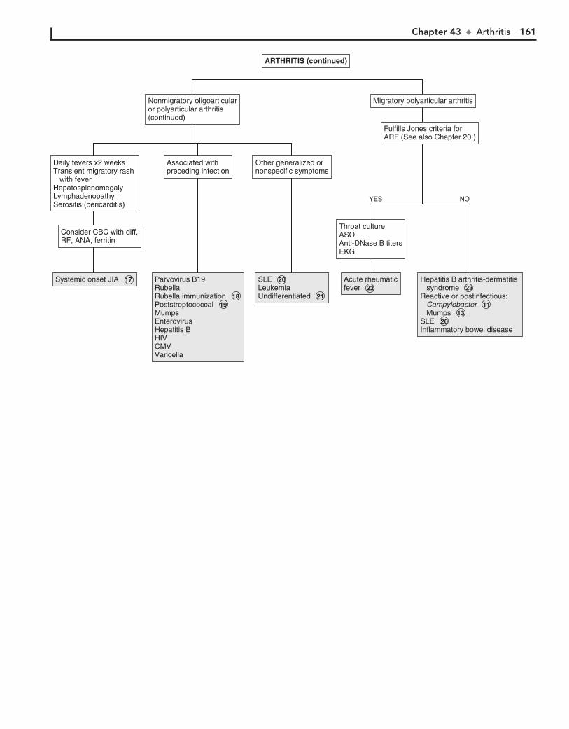
Chapter 43 u Arthritis 161
Systemic onset JIA Systemic onset JIA 17 Parvovirus B19RubellaRubella immunization Poststreptococcal MumpsEnterovirusHepatitis B HIVCMVVaricella
Parvovirus B19RubellaRubella immunization 18 Poststreptococcal 19 MumpsEnterovirusHepatitis B HIVCMVVaricella
SLE LeukemiaUndifferentiated
SLE 20 LeukemiaUndifferentiated 21
Acute rheumaticfever Acute rheumaticfever 22
Hepatitis B arthritis-dermatitis syndrome Reactive or postinfectious: Campylobacter Mumps SLE Inflammatory bowel disease
Hepatitis B arthritis-dermatitis syndrome 23 Reactive or postinfectious: Campylobacter 11 Mumps 13 SLE 20 Inflammatory bowel disease
Nonmigratory oligoarticularor polyarticular arthritis(continued)
Nonmigratory oligoarticularor polyarticular arthritis(continued)
Migratory polyarticular arthritisMigratory polyarticular arthritis
Fulfills Jones criteria forARF (See also Chapter 20.)Fulfills Jones criteria forARF (See also Chapter 20.)
Throat cultureASOAnti-DNase B titersEKG
Throat cultureASOAnti-DNase B titersEKG
Consider CBC with diff,RF, ANA, ferritinConsider CBC with diff,RF, ANA, ferritin
Daily fevers x2 weeksTransient migratory rash with feverHepatosplenomegalyLymphadenopathySerositis (pericarditis)
Daily fevers x2 weeksTransient migratory rash with feverHepatosplenomegalyLymphadenopathySerositis (pericarditis)
Associated withpreceding infectionAssociated withpreceding infection
Other generalized ornonspecific symptomsOther generalized ornonspecific symptoms
ARTHRITIS (continued)ARTHRITIS (continued)
NOYES

162
Chapter 44
KNEE PAIN
after a change in activity level. Pain is vaguely described; it may be around the knee or behind the patella. It is exacerbated by climbing stairs, squatting, running, and after sitting with the knee flexed for an extended period. The pain is frequently bilat-eral, and examination may be normal or reveal medial patellar tenderness or pain with patellofemoral compression. Diagnosis is usually clinical. Specialty consultation or x-rays may be indi-cated in atypical or prolonged symptoms that do not respond to therapy. Significant pain or guarding with attempted lateral or medial displacement of the patella (positive apprehension test) suggests a more serious disorder of chronic or recurrent patellar subluxation or dislocation.
6 Certain congenital conditions (high-riding patella, shal-low intercondylar notch, genu valgum deformity) may
predispose children to patellar misalignment significant enough to produce recurrent subluxation or dislocation. Significant pain or guarding with attempted lateral or medial displacement (positive apprehension sign) suggests subluxation. In patellar dislocation, the knee is usually locked in approximately 45 de-grees of flexion.
7 Sinding-Larsen-Johansson syndrome is an apophysitis at the site of insertion of the patellar tendon into the inferior
pole of the patella. It is an overuse syndrome that most fre-quently affects volleyball and basketball players. Pain is local-ized to the inferior pole of the patella and is aggravated by activ-ity. Diagnosis is usually clinical. Patients with similar complaints and tenderness over the patellar ligament near but not involving the bony attachments are most likely to have patellar tendinitis (“jumper’s knee”).
8 Osgood-Schlatter disease is an apophysitis at the site of in-sertion of the patellar tendon into the tibial tuberosity. It is
an overuse syndrome common in adolescent athletes who are undergoing a growth spurt. Examination reveals tenderness and swelling at the tibial tubercle and exacerbation of pain with re-sisted knee extension. Symptoms are commonly bilateral, al-though one side may be more symptomatic than the other. Patients complain of worsening pain with flexion activities (run-ning, jumping, kneeling, climbing stairs). Typical x-ray findings are soft tissue swelling and occasionally avulsed bony spicules over the tibial tuberosity, although x-rays are not usually indi-cated when the condition is bilateral. X-rays should be obtained when the pain is unilateral, not located directly over the tibial tuberosity, and when cases are unresponsive to treatment.
9 Meniscal injuries are more common in adolescents than in younger age groups. They are most commonly sports inju-
ries due to a twisting motion that occurs when the knee is flexed and the foot is firmly planted on the ground. Medial meniscal tears are more common than lateral ones. The injury may or may not be evident acutely; sometimes they present later with complaints of vague pain, recurrent effusions, stiffness, “giving out,” clicking, and sometimes locking. Examination reveals joint-line tenderness and occasionally a small effusion. X-rays are not helpful; MRIs are the preferred diagnostic entity.
10 Osteochondritis dissecans (OCD) develops after a small area of bone (most commonly in the medial femoral con-
dyle) becomes avascular. The necrotic fragment (with the ar-ticular cartilage overlying it) partially or completely separates from the long bone. Patients complain of nonspecific pain, usu-ally located around the patella and associated with activity. Ef-fusion is sometimes present. Locking or popping can occur
Knee pain can be an indicator of problems related to the dis-tal femur, proximal tibia, hip, or the knee itself. If arthritis is suspected (warmth or swelling of joint, limited motion, mul-tiple joint involvement, or systemic symptoms), refer to Chapter 43.
1 Knee pain in children can be due to acute problems or chronic processes. A thorough history and examination
should include the specific location of the pain, duration of the complaint, and (if relevant) relationship to and mechanism of trauma. It is important to clarify descriptions like “locking,” “catching,” or “giving out”; have the patient specify whether the knee was truly “stuck” in a position (requiring some type of manipulation by the patient to release it), if the joint was unable to be flexed or extended beyond a certain point, or if they sensed the joint was simply unstable versus if it truly buckled on them.
Range of motion may be affected simply by pain or by in-traarticular damage. Forced range of motion should never be attempted on exam. Bilateral knee evaluation allows compari-son for symmetry and better detection of subtle findings. Asymmetry of the thigh musculature suggests a chronic prob-lem with resultant muscular atrophy. A careful examination of the hips is critical in the evaluation of knee pain, especially in the absence of trauma; abnormalities could indicate a primary hip disorder.
2 A careful knee exam should include maneuvers (anterior drawer, Lachman, lateral pivot shift, Apley compression,
McMurray) that can aid in identifying ligamentous and menis-cal problems. Proficiency in performing these maneuvers is es-sential to doing a reliable exam. Mild injuries are likely to demonstrate pain on ligamentous testing in the absence of lax-ity or instability. Younger children are at greater risk for frac-tures than ligament sprains, because the physes (growth plates) are weaker than ligaments.
3 A history of a twisting injury, a “popping” sensation, and rapid development of an effusion (swelling) suggest an
anterior cruciate ligament (ACL) injury. Children presenting later may complain of their knee frequently “giving out.” Evalu-ation of a suspected ACL injury should include x-rays to rule out associated avulsion fractures. Because ACL tears are often associated with meniscal injury in children, referral for further evaluation (arthroscopy or MRI) is recommended.
4 A bipartite patella is the result of secondary ossification centers in the patella failing to fuse to the primary ossifica-
tion center. Most are asymptomatic, but occasionally pain may occur with sports, especially with jumping, or climbing stairs. Symptoms are usually unilateral, and the examination typically reveals pain at the superolateral pole of the patella. X-rays are diagnostic.
5 Patellofemoral stress syndrome (also known as patello-femoral pain syndrome or patellofemoral dysfunction)
comprises a spectrum of disorders characterized by anterior knee pain with no clearly definable cause. The term is typically applied to a pain syndrome experienced by adolescents, often
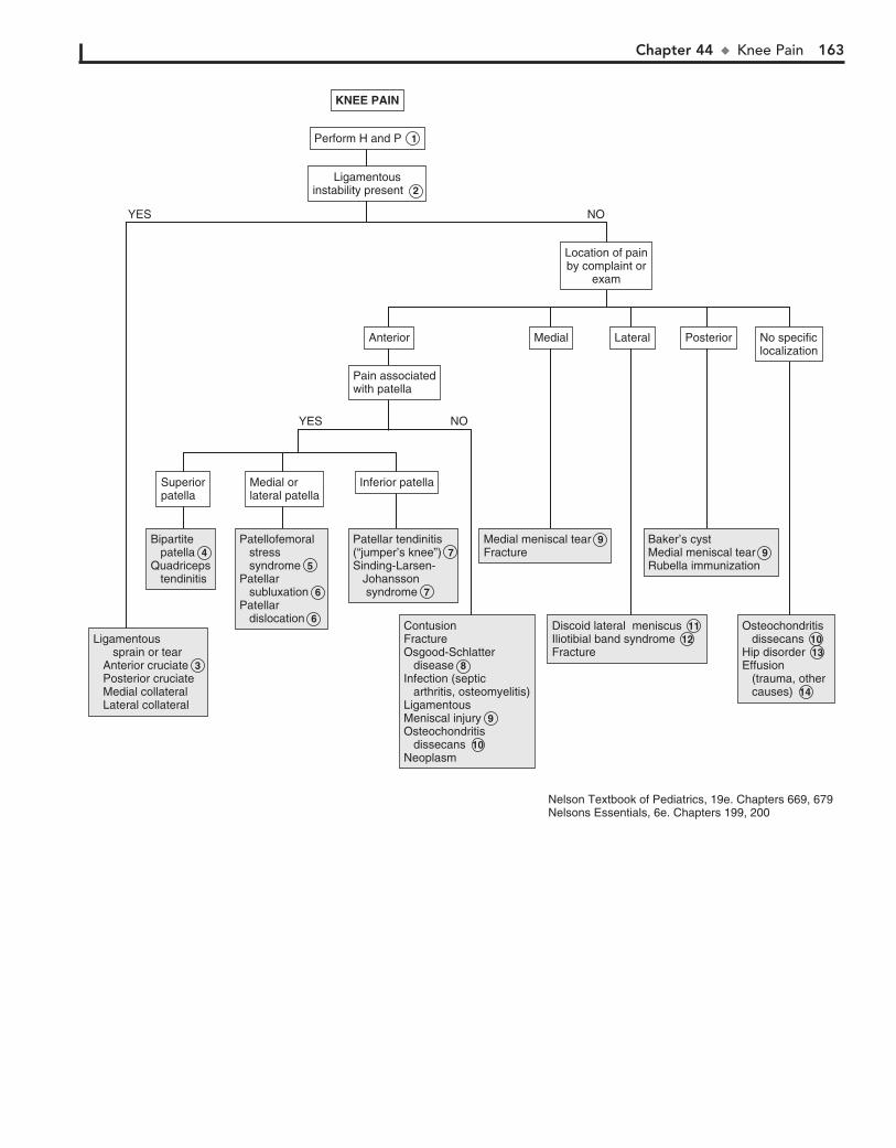
Chapter 44 u KneePain 163
Ligamentousinstability present
Ligamentousinstability present 2
Ligamentous sprain or tear Anterior cruciate Posterior cruciate Medial collateral Lateral collateral
Ligamentous sprain or tear Anterior cruciate 3 Posterior cruciate Medial collateral Lateral collateral
Bipartite patellaQuadriceps tendinitis
Bipartite patella 4 Quadriceps tendinitis
Patellofemoral stress syndromePatellar subluxationPatellar dislocation
Patellofemoral stress syndrome 5 Patellar subluxation 6 Patellar dislocation 6
Patellar tendinitis(“jumper’s knee”)Patellar tendinitis(“jumper’s knee”) 7Sinding-Larsen- Johansson syndrome 7
ContusionFractureOsgood-Schlatter diseaseInfection (septic arthritis, osteomyelitis)LigamentousMeniscal injuryOsteochondritis dissecansNeoplasm
ContusionFracture Osgood-Schlatter disease 8 Infection (septic arthritis, osteomyelitis)LigamentousMeniscal injury 9 Osteochondritis dissecans 10 Neoplasm
Medial meniscal tearFractureMedial meniscal tear 9 Fracture
Discoid lateral meniscusIliotibial band syndromeFracture
Discoid lateral meniscus 11 Iliotibial band syndrome 12 Fracture
Baker’s cystMedial meniscal tearRubella immunization
Baker’s cystMedial meniscal tear 9 Rubella immunization
Osteochondritis dissecansHip disorderEffusion (trauma, other causes)
Osteochondritis dissecans 10 Hip disorder 13Effusion (trauma, other causes) 14
Inferior patellaInferior patellaMedial or lateral patellaMedial or lateral patella
SuperiorpatellaSuperiorpatella
No specificlocalizationNo specificlocalization
MedialMedial LateralLateral PosteriorPosteriorAnteriorAnterior
Pain associatedwith patellaPain associatedwith patella
Location of painby complaint or
exam
Location of painby complaint or
exam
KNEE PAINKNEE PAIN
Perform H and P 1
NOYES
NOYES
Nelson Textbook of Pediatrics, 19e. Chapters 669, 679Nelsons Essentials, 6e. Chapters 199, 200

164 Part VI u MusculoskeletalSystem
when the bony fragment completely separates into the joint space. X-rays with a notch view are helpful in diagnosis; MRI will better define the cartilaginous involvement.
11 Discoid lateral meniscus is a congenital variant of the lateral meniscus. Patients typically present in late childhood or
adolescence with vague complaints of pain and an audible pop or snap with flexion. Examination reveals a palpable bulge at the lateral joint line when the knee is flexed. Standing x-rays may show a widened lateral joint space, flattening of the lateral femo-ral condyle, or cupping of the lateral aspect of the tibial plateau. MRI or arthroscopy may be necessary for definitive diagnosis.
12 Iliotibial band syndrome is an overuse syndrome that causes lateral knee pain in runners.
13 Poorly localized knee pain accompanied by an abnormal hip examination (restricted or painful internal rotation,
abduction or flexion) suggests a primary hip disorder such as AVN or a slipped capital femoral epiphysis (SCFE).
14 Effusions are most likely to occur in association with a trau-matic injury. Immediate development of an effusion after an
injury usually indicates hemarthrosis. Effusions may develop slowly (2 to 3 days) after an injury or may present intermittently owing to an intracapsular injury (meniscal tear), overuse, or a rheumatoid process. Septic arthritis should be considered when a knee is acutely painful, warm, and swollen, especially if the patient is febrile or toxic; immediate aspiration is essential when septic arthritis is suspected. Aspiration may also be necessary for the diagnosis of chronic or recurrent knee effusions.
BibliographyBaxter WR: Sports medicine in the growing child. In Morrissy R, Weinstein S,
editors: Lovell & Winter’s pediatric orthopaedics, ed 6, Philadelphia, 2006, Lippincott Williams & Wilkins, pp 1384–1429.
Koh J, section editor: Knee and lower leg. In Sarwark JF, editor: Essentials of musculoskeletal care, ed 3, Rosemont, Ill, 2010, American Academy of Orthopaedic Surgeons, pp 976.
Walsh WM, McCarty EC, Madden CC: Knee injuries. In Madden CC, Putukian M, Young CC, editors: Netter’s sports medicine, Philadelphia, 2010, Saunders Elsevier, pp 417–428.

Chapter 44 u KneePain 165
Ligamentousinstability present
Ligamentousinstability present 2
Ligamentous sprain or tear Anterior cruciate Posterior cruciate Medial collateral Lateral collateral
Ligamentous sprain or tear Anterior cruciate 3 Posterior cruciate Medial collateral Lateral collateral
Bipartite patellaQuadriceps tendinitis
Bipartite patella 4 Quadriceps tendinitis
Patellofemoral stress syndromePatellar subluxationPatellar dislocation
Patellofemoral stress syndrome 5 Patellar subluxation 6 Patellar dislocation 6
Patellar tendinitis(“jumper’s knee”)Patellar tendinitis(“jumper’s knee”) 7Sinding-Larsen- Johansson syndrome 7
ContusionFractureOsgood-Schlatter diseaseInfection (septic arthritis, osteomyelitis)LigamentousMeniscal injuryOsteochondritis dissecansNeoplasm
ContusionFracture Osgood-Schlatter disease 8 Infection (septic arthritis, osteomyelitis)LigamentousMeniscal injury 9 Osteochondritis dissecans 10 Neoplasm
Medial meniscal tearFractureMedial meniscal tear 9 Fracture
Discoid lateral meniscusIliotibial band syndromeFracture
Discoid lateral meniscus 11 Iliotibial band syndrome 12 Fracture
Baker’s cystMedial meniscal tearRubella immunization
Baker’s cystMedial meniscal tear 9 Rubella immunization
Osteochondritis dissecansHip disorderEffusion (trauma, other causes)
Osteochondritis dissecans 10 Hip disorder 13Effusion (trauma, other causes) 14
Inferior patellaInferior patellaMedial or lateral patellaMedial or lateral patella
SuperiorpatellaSuperiorpatella
No specificlocalizationNo specificlocalization
MedialMedial LateralLateral PosteriorPosteriorAnteriorAnterior
Pain associatedwith patellaPain associatedwith patella
Location of painby complaint or
exam
Location of painby complaint or
exam
KNEE PAINKNEE PAIN
Perform H and P 1
NOYES
NOYES
Nelson Textbook of Pediatrics, 19e. Chapters 669, 679Nelsons Essentials, 6e. Chapters 199, 200
166
Chapter 45
EXTREMITY PAIN
than the surrounding ligaments, tendons, and joint capsule. These injuries may result from excessive force or repetitive mi-crotrauma. Be aware of the possibility of child abuse when un-suspected fractures are detected.
5 Mild to moderate muscle aches (myalgias) are common with many viral infections; lab values (if obtained) are
generally normal or consistent with a viral illness. A more severe viral-related myositis presents typically with severe calf pain and difficulty or refusal to walk 5 to 7 days after the acute onset of viral (most commonly influenza type B) symp-toms. Labs reveal elevated muscle enzymes and possibly myoglobinuria.
6 Adolescents with juvenile fibromyalgia often experience headaches, fatigue, sleep problems, anxiety, and a subjec-
tive sensation of joint swelling in addition to localized areas of muscle tenderness; most experts consider a 3-month duration necessary for diagnosis.
7 Infection within the bone is osteomyelitis; it may present acutely with localized pain and tenderness in an ill-
appearing child or subacutely with prolonged pain and limp without fevers or systemic complaints. Bone changes on x-ray may not become evident for 7 to 10 days. MRI is the preferred choice for diagnostic imaging if available. Bone scans are also sensitive and specific early in the clinical course if MRI is not available.
8 Night pain is especially characteristic of both benign and malignant primary bone and metastatic tumors. Pain in
the absence of local tenderness is another clue.
9 Shin splints (medial tibial stress syndrome) are the most common overuse injury of the lower leg. They present with
diffuse tenderness along the lower third or half of the medial tibia; the pain onset is initially toward the end of a period of exercise but with progression will be present throughout activity. Imaging is not necessary for diagnosis. Consider x-rays only if a stress frac-ture is suspected; pain due to stress fractures will be localized, more severe, and present throughout activity. Swelling may or may not be associated. Radiographs will be normal in shin splints; they may also be normal until after 3 to 4 weeks of symptoms due to a stress fracture. MRI has replaced bone scan as the preferred imaging for suspected stress fractures in most centers.
This chapter is devoted to extremity pain other than joint prob-lems. (See Chapters 42, 43, 44 for complaints better character-ized as limp, arthritis, or knee pain.) Muscle pain (myalgia) is a nonspecific finding in many conditions; evaluation of other as-pects of the history and physical (joint pain, weakness, toxin exposure, systemic complaints, or other physical findings) may be more useful in narrowing a differential diagnosis.
1 Be aware of the possibility of unobserved trauma as well as intentional trauma (child abuse), particularly in the
preverbal child. Inquire specifically about the lifting of a child by the child’s extended arm, which can cause a radial head subluxation.
2 Radial head subluxation (“nursemaid’s elbow”) is most commonly the result of a preschool-aged child being lifted
upward by an extended arm. This upward traction causes the annular ligament to partially slip off the radial head. (Techni-cally, it is a subluxation of the ligament rather than the radius.) The injury is only mildly painful, but the child is reluctant to use the arm. They may prefer holding the arm splinted close to the body, often giving the impression of wrist pain. The history is generally the key to diagnosis; x-ray findings are nonspecific but films should be considered to rule out other injuries when the history is unclear.
3 “Burners” or “stingers” are transient neurologic injuries of the brachial plexus due to compression or traction. Tran-
sient severe unilateral pain extends from the shoulder to the fingertips. Accompanying weakness and numbness may occur. These injuries occur most commonly in football players and wrestlers. If bilateral or affecting the lower extremities, spinal cord injury must be considered.
4 Children and adolescents are more susceptible to physeal (growth plate) injury than ligament sprains or diaphyseal
(shaft) fractures because physes in growing children are weaker

Chapter 45 u ExtremityPain 167
ContusionSprain or strainRadial head subluxation Burners or stingers
ContusionSprain or strainRadial head subluxation 2 Burners or stingers 3
Fracture DislocationFracture 4 Dislocation
Myalgia MyositisFibromyalgia
Myalgia 5 MyositisFibromyalgia 6
Myositis (±rhabdomyolysis)Sickle cell crisisOsteomyelitis Bone tumor LeukemiaNeuroblastoma
Myositis 5 (±rhabdomyolysis)Sickle cell crisisOsteomyelitis 7 Bone tumor 8 LeukemiaNeuroblastoma
Osteomyelitis Bone tumor Fracture (intentional vs unintentional) Shin splits Stress fracture Leukemia
Osteomyelitis 7 Bone tumor 8 Fracture (intentional vs unintentional) 4 Shin splits 9 Stress fracture 9 Leukemia
ContusionContusion ContusionContusion Fracture (intentional vs unintentional) DislocationOsteomyelitis Bone tumor Myositis ossificansLeukemiaNeuroblastoma
Fracture (intentional vs unintentional) 4 DislocationOsteomyelitis 7 Bone tumor 8 Myositis ossificansLeukemiaNeuroblastoma
AbnormalresultsAbnormalresults
NormalresultsNormalresults
AbnormalresultsAbnormalresults
Consider MRIvs bone scanConsider MRIvs bone scan
NormalresultsNormalresults
Observe for1 weekObserve for1 week
NormalresultsNormalresults
Obtain CBC with diff and ESR or CRP
Obtain CBC with diff and ESR or CRP
AbnormalresultsAbnormalresults
AbnormalresultsAbnormalresults
NormalresultsNormalresults
AbnormalresultsAbnormalresults
NormalresultsNormalresults
Symptomsresolved after1 week
Symptomsresolved after1 week
Go to next pageGo to next pageHistory oftraumaHistory oftrauma
Systemic symptoms present(fever, irritability, fatigue,headache, sleep disturbances) ± swelling
Systemic symptoms present(fever, irritability, fatigue,headache, sleep disturbances) ± swelling
Swelling presentOR
Pain persistent >5-7 days(in the absence of obvious
systemic symptoms)
Swelling presentOR
Pain persistent >5-7 days(in the absence of obvious
systemic symptoms)
Consider x-rayConsider x-ray Consider: CBC with diff Blood culture ESR or CRP UA Muscle enzymes X-ray MRI ± Bone scan
Consider: CBC with diff Blood culture ESR or CRP UA Muscle enzymes X-ray MRI ± Bone scan
Obtain x-rayof affectedarea
Obtain x-rayof affectedarea
EXTREMITY PAINEXTREMITY PAIN
Perform H and P 1
NO
YES
Nelson Textbook of Pediatrics, 19e. Chapters 162, 250, 495, 666, 673, 679Nelsons Essentials, 6e. Chapters 92, 293
168 Part VI u MusculoskeletalSystem
10 A contusion of the quadriceps muscle may be complicated by ossification of the hematoma (myositis ossificans),
causing pain and stiffness for several months after the injury.
11 Complex regional pain syndrome (previously called reflex sympathetic dystrophy) is a rare condition presenting
acutely with intense extremity pain. There is either no history of trauma or one of a very minor injury followed by acute pain, swelling, and color and temperature change of the affected area days to weeks later. Erythema, warmth, and swelling occur ini-tially; chronically, disuse atrophy and cool, clammy skin develop.
12 Growing pains (benign nocturnal pains of childhood) are common. Children complain of bilateral diffuse extremity
pain, usually in the legs (thigh or calf). It typically occurs late in the day or at night and does not affect daytime activity. Massaging characteristically produces relief in these children; in contrast,
massage would aggravate pain in most serious conditions. The physical examination is normal. Pain that is unilateral, localized to a joint, or persists during the day should never be assumed to be growing pains.
13 Nerve compression manifests with tingling, numbness, and paresthesias (“pins and needles”) in addition to pain. Car-
pal tunnel syndrome classically presents with numbness on the radial (thumb) side of the hand, and ulnar nerve entrapment presents with numbness on the ulnar side of the hand (fourth and fifth fingers). Although rare in children, cervical nerve com-pression should be considered, especially if there is a history of neck trauma or symptoms are worse with the arm in an over-head position (thoracic outlet syndrome). When arm or shoul-der pain accompanies numbness and tingling, MRI or EMG should be considered to rule out cervical nerve compression.

Chapter 45 u ExtremityPain 169
Insect bitePuncture wound (superinfected)Foreign body (± cellulitis)
Insect bitePuncture wound (superinfected)Foreign body (± cellulitis)
CellulitisContact dermatitisComplex regional pain syndrome Insect bite
CellulitisContact dermatitisComplex regional pain syndrome 11 Insect bite
Growing pains Growing pains 12 Nerve compression Carpal tunnel syndrome Ulnar nerve entrapment Cervical disc herniation Thoracic outlet syndrome
Nerve compression 13 Carpal tunnel syndrome Ulnar nerve entrapment Cervical disc herniation Thoracic outlet syndrome
Erythema or swelling present± Fever
Erythema or swelling present± Fever
Historysuggestive ofgrowing pains
Historysuggestive ofgrowing pains 12
Numbness ortingling presentNumbness ortingling present
Go to next pageGo to next page
Central papuleor punctum visibleCentral papuleor punctum visible
Consider x-ray,specialty consultation(neurology, orthopedics)for EMG ± MRI
Consider x-ray,specialty consultation(neurology, orthopedics)for EMG ± MRI
EXTREMITY PAIN (continued)EXTREMITY PAIN (continued)
NOYES
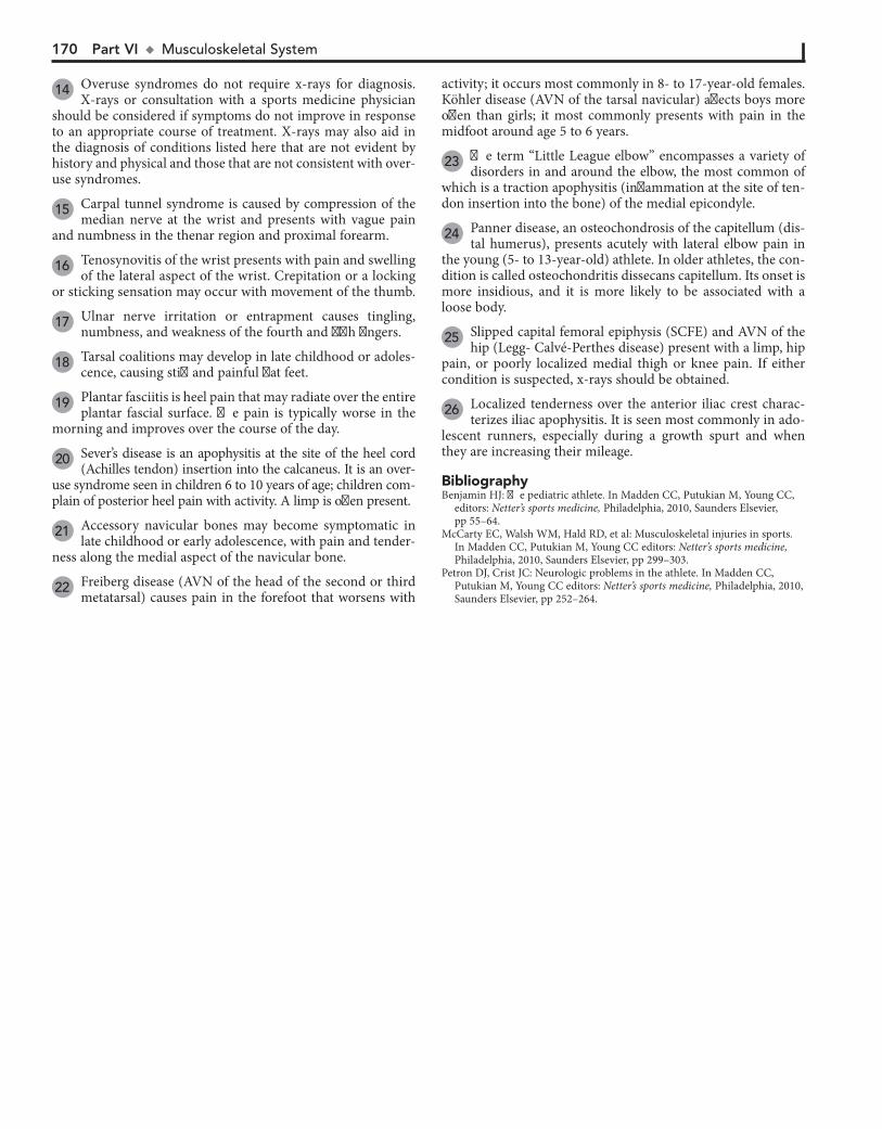
170 Part VI u MusculoskeletalSystem
14 Overuse syndromes do not require x-rays for diagnosis. X-rays or consultation with a sports medicine physician
should be considered if symptoms do not improve in response to an appropriate course of treatment. X-rays may also aid in the diagnosis of conditions listed here that are not evident by history and physical and those that are not consistent with over-use syndromes.
15 Carpal tunnel syndrome is caused by compression of the median nerve at the wrist and presents with vague pain
and numbness in the thenar region and proximal forearm.
16 Tenosynovitis of the wrist presents with pain and swelling of the lateral aspect of the wrist. Crepitation or a locking
or sticking sensation may occur with movement of the thumb.
17 Ulnar nerve irritation or entrapment causes tingling, numbness, and weakness of the fourth and fifth fingers.
18 Tarsal coalitions may develop in late childhood or adoles-cence, causing stiff and painful flat feet.
19 Plantar fasciitis is heel pain that may radiate over the entire plantar fascial surface. The pain is typically worse in the
morning and improves over the course of the day.
20 Sever’s disease is an apophysitis at the site of the heel cord (Achilles tendon) insertion into the calcaneus. It is an over-
use syndrome seen in children 6 to 10 years of age; children com-plain of posterior heel pain with activity. A limp is often present.
21 Accessory navicular bones may become symptomatic in late childhood or early adolescence, with pain and tender-
ness along the medial aspect of the navicular bone.
22 Freiberg disease (AVN of the head of the second or third metatarsal) causes pain in the forefoot that worsens with
activity; it occurs most commonly in 8- to 17-year-old females. Köhler disease (AVN of the tarsal navicular) affects boys more often than girls; it most commonly presents with pain in the midfoot around age 5 to 6 years.
23 The term “Little League elbow” encompasses a variety of disorders in and around the elbow, the most common of
which is a traction apophysitis (inflammation at the site of ten-don insertion into the bone) of the medial epicondyle.
24 Panner disease, an osteochondrosis of the capitellum (dis-tal humerus), presents acutely with lateral elbow pain in
the young (5- to 13-year-old) athlete. In older athletes, the con-dition is called osteochondritis dissecans capitellum. Its onset is more insidious, and it is more likely to be associated with a loose body.
25 Slipped capital femoral epiphysis (SCFE) and AVN of the hip (Legg- Calvé-Perthes disease) present with a limp, hip
pain, or poorly localized medial thigh or knee pain. If either condition is suspected, x-rays should be obtained.
26 Localized tenderness over the anterior iliac crest charac-terizes iliac apophysitis. It is seen most commonly in ado-
lescent runners, especially during a growth spurt and when they are increasing their mileage.
BibliographyBenjamin HJ: The pediatric athlete. In Madden CC, Putukian M, Young CC,
editors: Netter’s sports medicine, Philadelphia, 2010, Saunders Elsevier, pp 55–64.
McCarty EC, Walsh WM, Hald RD, et al: Musculoskeletal injuries in sports. In Madden CC, Putukian M, Young CC editors: Netter’s sports medicine, Philadelphia, 2010, Saunders Elsevier, pp 299–303.
Petron DJ, Crist JC: Neurologic problems in the athlete. In Madden CC, Putukian M, Young CC editors: Netter’s sports medicine, Philadelphia, 2010, Saunders Elsevier, pp 252–264.
Chapter 45 u ExtremityPain 171
ParonychiaCarpal tunnel syndromeTenosynovitisUlnar neuritisGanglion cystKienböck’s disease (osteonecrosis of carpal lunate)Congenital trigger thumb or fingerRaynaud phenomenonRadial head subluxationErythromelalgia
ParonychiaCarpal tunnel syndrome 15 Tenosynovitis 16 Ulnar neuritis 17 Ganglion cystKienböck’s disease (osteonecrosis of carpal lunate)Congenital trigger thumb or fingerRaynaud phenomenonRadial head subluxationErythromelalgia
Shin splintsGrowing painsStress fractureTibia vara (Blount’s disease)
Shin splints 9Growing pains 13 Stress fracture 9 Tibia vara (Blount’s disease)
Plantar wartsIngrown toenailBunionTarsal coalitionPlantar fasciitisSever’s disease (calcaneal apophysitis)Achilles tendonitisAccessory navicular bonePuncture wound ± foreign bodyPoor-fitting shoesOsteonecrosis Freiberg’s infraction Köhler’s diseaseErythromelalgia
Plantar wartsIngrown toenailBunionTarsal coalition 18 Plantar fasciitis 19 Sever’s disease 20 (calcaneal apophysitis)Achilles tendonitisAccessory navicular bone 21 Puncture wound ± foreign bodyPoor-fitting shoesOsteonecrosis Freiberg’s infraction 22 Köhler’s diseaseErythromelalgia
Overuse injury Instability Tendonitis Subluxation Rotator cuff injury
Overuse injury Instability Tendonitis Subluxation Rotator cuff injury
Overuse syndromes Apophysitis of medial epicondyle (“Little Leaguer’s elbow”) Panner disease Ostechondritis dissecans capitellum Lateral epicondylitis (“tennis elbow”) Medial epicondylitis (“golfer’s elbow”) Radial head subluxation
Overuse syndromes Apophysitis of medial epicondyle (“Little Leaguer’s elbow”) 23 Panner disease 24 Ostechondritis dissecans capitellum 24 Lateral epicondylitis (“tennis elbow”) Medial epicondylitis (“golfer’s elbow”) Radial head subluxation 2
Slipped capital femoral epiphysisAVN (Legg-Calvé- Perthes disease)Iliac crest apophysitis
Slipped capital femoral epiphysis 25 AVN (Legg-Calvé- Perthes disease) 25 Iliac crest apophysitis 26
Wrist and handWrist and hand LegLeg FootFoot ShoulderShoulder ElbowElbow HipHip
EXTREMITY PAIN (continued)EXTREMITY PAIN (continued)
Consider x-raysConsider x-rays 14
History of overuse ORPain well localized onarm, hand/wrist, leg, or foot
History of overuse ORPain well localized onarm, hand/wrist, leg, or foot
172
Chapter 46
BACK PAIN
pancreatitis, nephrolithiasis, and abscesses (perinephric , pelvic, paraspinal muscle, psoas muscle). A paraspinal abscess will gen-erally present with some neurologic signs; an MRI is recom-mended if suspected.
6 Primary vertebral osteomyelitis is rare in young children. It typically occurs in children older than 8 years, and an
insidious onset is more likely than an acute toxic presentation. Presentation commonly includes a vague or dull backache that may be worse at night; fever may or may not be present. Pott’s disease is tuberculosis spondylitis, a rare complication of un-treated tuberculosis that is more likely to occur in children than adults.
7 If pain persists, consider repeating x-rays versus obtaining other imaging in 2 weeks. Callus formation may indicate
fractures not obvious on initial films. In cases of suspected child abuse, a bone scan should be considered early in the evaluation when initial x-rays are negative.
8 Contusions and abrasions are the most common back in-juries sustained in routine play and sports in young chil-
dren. No additional workup is needed if no other injuries and an otherwise normal physical exam are present.
9 Back pain associated with bowel or bladder deficits, gait abnormalities, lower extremity pain, weakness, or reflex or
sensation deficits is suggestive of space-occupying lesions (spinal cord tumors, metastatic lesions) and warrants urgent evaluation.
10 Spondylolysis is a defect (stress fracture) of the bony con-nection between a vertebral body and its arch (pars inter-
articularis). Spondylolisthesis is the forward slippage or displacement of one vertebra in relation to another. Classic spondylolisthesis in children and adolescents involves the fifth lumbar and first sacral vertebral bodies. In these cases, fatigue or stress fractures of the pars interarticularis bilaterally allow forward slippage of the superior vertebra on the inferior verte-bra. Symptoms vary based on the degree of subsequent nerve compression.
Children participating in sports involving repetitive flexion and hyperextension (gymnasts, football players, weight lifters) are at increased risk for spondylolysis. Symptoms (poorly local-ized lumbar and lumbosacral pain, hamstring tightness) often do not occur until the adolescent growth spurt; spinal hyperex-tension typically exacerbates the pain. Defects often remain asymptomatic indefinitely. When symptoms do occur, they are not always consistent with the severity of the spondyolytic de-fect or the degree of slippage.
11 Consider a urine pregnancy test in adolescent females prior to obtaining x-rays.
12 Kyphosis is an exaggeration of the normal thoracic or tho-racolumbar curve in the sagittal plane. In postural or flexi-
ble cases, it is fully correctable with voluntary effort, and any as-sociated pain is typically mild. Spine films should be obtained in severe cases and when significant back pain is present to rule out Scheuermann’s disease. This is a structural hyperkyphosis; x-rays will show vertebral end plate irregularities and vertebral body wedging. Most cases of scoliosis, regardless of the age at onset, are idiopathic. Idiopathic scoliosis is not painful during childhood and adolescence. If pain is noted with a scoliotic curve, carefully consider infectious, inflammatory, and neoplastic causes.
Back pain is uncommon in children, especially chronic or se-vere back pain. Most children who complain of back pain have mild, self-resolving symptoms of limited duration. Symptoms of severe or persistent back pain, as well as any abnormal find-ings on physical examination, mandate a thorough evaluation. As with adults, however, clear etiologies are not always identi-fied, particularly in older children and adolescents without other associated symptoms.
1 The history should inquire about trauma, associated leg pain, gait abnormalities, weakness, extremity pain or tin-
gling, bowel or bladder incontinence, and any systemic signs or symptoms (fever, malaise, rashes, GI symptoms). Also ask about aggravating or relieving factors (particularly whether it is relieved with rest) and whether it wakes the patient from sleep. The physical should include careful neurologic and abdominal examinations, as well as a complete musculoskeletal exam. The neurologic exam should include assessment of anal tone and (in males) the cremasteric reflex; a pelvic exam may be indicated in older adolescent females.
2 The presence of associated abdominal pain, vomiting, dys-uria, hematuria, or vaginal complaints should prompt a
specific evaluation for intraabdominal or pelvic problems. Be vigilant for subtle neurologic signs and symptoms that may be overlooked in the presence of an acute illness (especially if bed-ridden); an MRI would be indicated in the case of any neuro-logic problems.
3 Discitis is an infection or inflammation of an intervertebral disc space. The most common pathogen is Staphylococcus
aureus. Children younger than age 7 years are most commonly affected; the peak incidence is at age 3. Common clinical findings are back pain, limping, and a stiff, straight posture due to loss of normal lumbar lordosis. These children often refuse to bend over to pick something up because of the pain related to spinal flexion. The degree of systemic illness is variable. Younger children (,3 years of age) are more likely to present with a toxic picture of fe-ver, refusal to walk, irritability, and decreased appetite. Older children and adolescents may complain of back pain and pain with walking and may or may not be febrile. Spine films will not reveal characteristic disc space narrowing or vertebral end plate irregularities until a few weeks after symptoms begin. MRI or bone scan may be necessary for early diagnosis; MRI is preferred because it will better differentiate conditions with similar presen-tations (vertebral osteomyelitis, abscesses).
4 Spinal x-rays or a bone scan may suggest the extent of metastatic tumor involvement in the spine, but MRI will
be necessary for more specific definition of the disease process. CT scans may be preferred when pathologic fractures are sus-pected, because of better bony detail.
5 Intraabdominal, retroperitoneal, or pelvic processes can cause referred back pain. More specific signs and symptoms
should aid in the diagnosis of problems such as pyelonephritis,
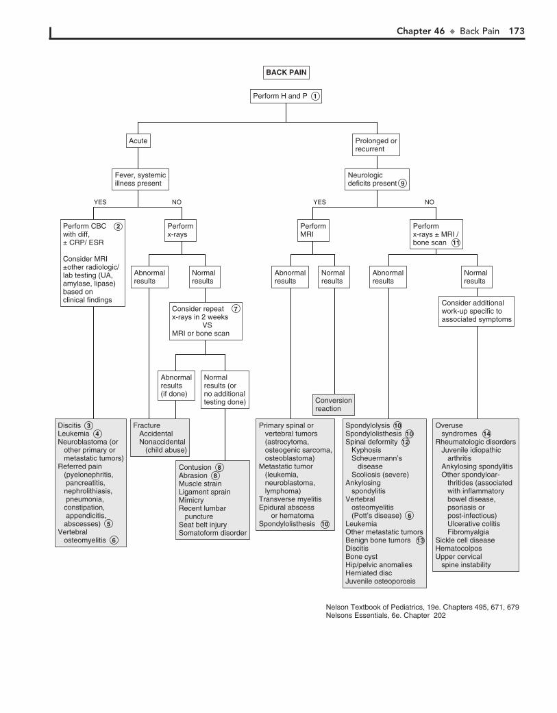
Chapter 46 u BackPain 173
DiscitisLeukemia Neuroblastoma (or other primary or metastatic tumors)Referred pain (pyelonephritis, pancreatitis, nephrolithiasis, pneumonia, constipation, appendicitis, abscesses) 5Vertebral osteomyelitis 6
Discitis 3 Leukemia 4 Neuroblastoma (or other primary or metastatic tumors)Referred pain (pyelonephritis, pancreatitis, nephrolithiasis, pneumonia, constipation, appendicitis, abscesses) 5Vertebral osteomyelitis 6
Fracture Accidental Nonaccidental (child abuse)
Fracture Accidental Nonaccidental (child abuse)
ContusionAbrasionMuscle strainLigament sprainMimicryRecent lumbar punctureSeat belt injurySomatoform disorder
Contusion 8 Abrasion 8 Muscle strainLigament sprainMimicryRecent lumbar punctureSeat belt injurySomatoform disorder
Primary spinal or vertebral tumors (astrocytoma, osteogenic sarcoma, osteoblastoma)Metastatic tumor (leukemia, neuroblastoma, lymphoma)Transverse myelitisEpidural abscess or hematomaSpondylolisthesis 10
Primary spinal or vertebral tumors (astrocytoma, osteogenic sarcoma, osteoblastoma)Metastatic tumor (leukemia, neuroblastoma, lymphoma)Transverse myelitisEpidural abscess or hematomaSpondylolisthesis 10
ConversionreactionConversionreaction
SpondylolysisSpondylolisthesisSpinal deformity Kyphosis Scheuermann’s disease Scoliosis (severe)Ankylosing spondylitisVertebral osteomyelitis (Pott’s disease) 6LeukemiaOther metastatic tumorsBenign bone tumors 13DiscitisBone cystHip/pelvic anomaliesHerniated discJuvenile osteoporosis
Spondylolysis 10 Spondylolisthesis 10 Spinal deformity 12 Kyphosis Scheuermann’s disease Scoliosis (severe)Ankylosing spondylitisVertebral osteomyelitis (Pott’s disease) 6Leukemia Other metastatic tumorsBenign bone tumors 13DiscitisBone cystHip/pelvic anomaliesHerniated discJuvenile osteoporosis
Overuse syndromes Rheumatologic disorders Juvenile idiopathic arthritis Ankylosing spondylitis Other spondyloar- thritides (associated with inflammatory bowel disease, psoriasis or post-infectious) Ulcerative colitis FibromyalgiaSickle cell diseaseHematocolposUpper cervical spine instability
Overuse syndromes 14 Rheumatologic disorders Juvenile idiopathic arthritis Ankylosing spondylitis Other spondyloar- thritides (associated with inflammatory bowel disease, psoriasis or post-infectious) Ulcerative colitis FibromyalgiaSickle cell diseaseHematocolposUpper cervical spine instability
Abnormalresults(if done)
Abnormalresults(if done)
Normal results (or no additionaltesting done)
Normal results (or no additionaltesting done)
Consider repeat 7 x-rays in 2 weeks VS MRI or bone scan
Consider repeat 7 x-rays in 2 weeks VS MRI or bone scan
Prolonged orrecurrentProlonged orrecurrent
AcuteAcute
Fever, systemicillness presentFever, systemicillness present
Neurologicdeficits presentNeurologicdeficits present 9
BACK PAINBACK PAIN
Perform Perform H and P 1
Performx-raysPerformx-rays
Perform CBC 2with diff, ± CRP/ ESR Consider MRI ±other radiologic/lab testing (UA, amylase, lipase)based on clinical findings
Perform CBC 2with diff, ± CRP/ ESR Consider MRI ±other radiologic/lab testing (UA, amylase, lipase)based on clinical findings
NOYES
AbnormalresultsAbnormalresults
Normal resultsNormal results
AbnormalresultsAbnormalresults
Normal resultsNormal results
AbnormalresultsAbnormalresults
Normal resultsNormal results
Consider additionalwork-up specific toassociated symptoms
Consider additionalwork-up specific toassociated symptoms
Performx-rays ± MRI /bone scan 11
Performx-rays ± MRI /bone scan 11
PerformMRIPerformMRI
NOYES
Nelson Textbook of Pediatrics, 19e. Chapters 495, 671, 679Nelsons Essentials, 6e. Chapter 202
174 Part VI u MusculoskeletalSystem
13 Benign tumors of the spine are rare. The most common ones are osteoid osteoma, osteoblastoma, and eosinophilic
granuloma. Presentation is typically a prolonged period of back pain (especially at night) that eventually evolves to stiffness and (rarely) a painful scoliosis or mild neurologic defects.
14 Overuse syndromes are more common in athletic adoles-cents than younger children. The stress of normal physi-
ologic activity results in microtrauma, which typically resolves. Overuse injuries result when repetitive activity without ade-quate conditioning or rest prohibits this resolution. Both bone
BibliographyGurd DP: Back pain in the young athlete, Sports Med Arthrosc 19:7–16, 2011.Corneli HN: Pain—back. In Fleisher G, Ludwig S, editors: Textbook of pediat-
ric emergency medicine, ed 6, Philadelphia, 2010, Lippincott Williams & Wilkins, pp 429–433.
and soft tissues (muscles, tendons, bursae, cartilage) are sus-ceptible to overuse injuries. Gymnasts, dancers, weight lifters, and football players are particularly prone to overuse injuries of the back. Carrying a heavy book bag or backpack may cause back pain.
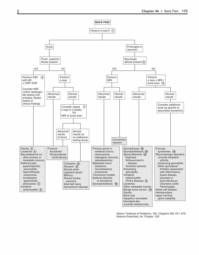
Chapter 46 u BackPain 175
DiscitisLeukemia Neuroblastoma (or other primary or metastatic tumors)Referred pain (pyelonephritis, pancreatitis, nephrolithiasis, pneumonia, constipation, appendicitis, abscesses) 5Vertebral osteomyelitis 6
Discitis 3 Leukemia 4 Neuroblastoma (or other primary or metastatic tumors)Referred pain (pyelonephritis, pancreatitis, nephrolithiasis, pneumonia, constipation, appendicitis, abscesses) 5Vertebral osteomyelitis 6
Fracture Accidental Nonaccidental (child abuse)
Fracture Accidental Nonaccidental (child abuse)
ContusionAbrasionMuscle strainLigament sprainMimicryRecent lumbar punctureSeat belt injurySomatoform disorder
Contusion 8 Abrasion 8 Muscle strainLigament sprainMimicryRecent lumbar punctureSeat belt injurySomatoform disorder
Primary spinal or vertebral tumors (astrocytoma, osteogenic sarcoma, osteoblastoma)Metastatic tumor (leukemia, neuroblastoma, lymphoma)Transverse myelitisEpidural abscess or hematomaSpondylolisthesis 10
Primary spinal or vertebral tumors (astrocytoma, osteogenic sarcoma, osteoblastoma)Metastatic tumor (leukemia, neuroblastoma, lymphoma)Transverse myelitisEpidural abscess or hematomaSpondylolisthesis 10
ConversionreactionConversionreaction
SpondylolysisSpondylolisthesisSpinal deformity Kyphosis Scheuermann’s disease Scoliosis (severe)Ankylosing spondylitisVertebral osteomyelitis (Pott’s disease) 6LeukemiaOther metastatic tumorsBenign bone tumors 13DiscitisBone cystHip/pelvic anomaliesHerniated discJuvenile osteoporosis
Spondylolysis 10 Spondylolisthesis 10 Spinal deformity 12 Kyphosis Scheuermann’s disease Scoliosis (severe)Ankylosing spondylitisVertebral osteomyelitis (Pott’s disease) 6Leukemia Other metastatic tumorsBenign bone tumors 13DiscitisBone cystHip/pelvic anomaliesHerniated discJuvenile osteoporosis
Overuse syndromes Rheumatologic disorders Juvenile idiopathic arthritis Ankylosing spondylitis Other spondyloar- thritides (associated with inflammatory bowel disease, psoriasis or post-infectious) Ulcerative colitis FibromyalgiaSickle cell diseaseHematocolposUpper cervical spine instability
Overuse syndromes 14 Rheumatologic disorders Juvenile idiopathic arthritis Ankylosing spondylitis Other spondyloar- thritides (associated with inflammatory bowel disease, psoriasis or post-infectious) Ulcerative colitis FibromyalgiaSickle cell diseaseHematocolposUpper cervical spine instability
Abnormalresults(if done)
Abnormalresults(if done)
Normal results (or no additionaltesting done)
Normal results (or no additionaltesting done)
Consider repeat 7 x-rays in 2 weeks VS MRI or bone scan
Consider repeat 7 x-rays in 2 weeks VS MRI or bone scan
Prolonged orrecurrentProlonged orrecurrent
AcuteAcute
Fever, systemicillness presentFever, systemicillness present
Neurologicdeficits presentNeurologicdeficits present 9
BACK PAINBACK PAIN
Perform Perform H and P 1
Performx-raysPerformx-rays
Perform CBC 2with diff, ± CRP/ ESR Consider MRI ±other radiologic/lab testing (UA, amylase, lipase)based on clinical findings
Perform CBC 2with diff, ± CRP/ ESR Consider MRI ±other radiologic/lab testing (UA, amylase, lipase)based on clinical findings
NOYES
AbnormalresultsAbnormalresults
Normal resultsNormal results
AbnormalresultsAbnormalresults
Normal resultsNormal results
AbnormalresultsAbnormalresults
Normal resultsNormal results
Consider additionalwork-up specific toassociated symptoms
Consider additionalwork-up specific toassociated symptoms
Performx-rays ± MRI /bone scan 11
Performx-rays ± MRI /bone scan 11
PerformMRIPerformMRI
NOYES
Nelson Textbook of Pediatrics, 19e. Chapters 495, 671, 679Nelsons Essentials, 6e. Chapter 202
176
Chapter 47
STIFF OR PAINFUL NECK
Most causes of neck pain and stiffness in children are benign; however, potentially life-threatening conditions (meningitis, cer-vical spine fracture) must always be considered in the evaluation.
1 The history should clarify the onset, duration, and nature of the complaint and include a comprehensive review of sys-
tems. The physical examination should note stiffness and range of motion of the neck (lateral movement and flexion-extension) and the specific nature and location of the pain (muscle spasm, muscle or bone tenderness). The differential diagnosis for a child who exhibits full mobility of their neck, even if it is painful, dif-fers from that of a child whose range of motion is limited.
Torticollis or “wry neck” describes a condition in which the infant’s or child’s neck is tilted to one side and rotated to the opposite side. It is the primary manifestation of congenital torticollis, but it can also be a symptom of several other prob-lems (infection, CNS neoplasm, structural abnormalities, neu-rologic disorders). A thorough neurologic examination includ-ing mental status, cranial nerve involvement, upper extremity pain or weakness, and cerebellar function is important.
2 Congenital muscular torticollis appears in the first several weeks of life. The head is tilted toward a shortened sterno-
cleidomastoid muscle; a fibrotic mass is frequently palpable in the muscle belly. Other signs of intrauterine mechanical deformation (plagiocephaly, facial asymmetry, foot deformities, developmental hip dysplasia) are frequently present. Plain x-rays of the cervical spine to rule out congenital vertebral abnormalities should be obtained in the absence of any of these associated clinical find-ings or if there is no response to a stretching program.
3 A suspected cervical spine injury should be treated in the acute setting with cervical spine immobilization until ap-
propriate x-rays can be taken.
4 The most common cervical spine subluxation involves the atlantoaxial (C1-C2) joint. It may result from mild as well
as severe trauma. Symptoms can include neck pain, sterno-cleidomastoid muscle tenderness, and torticollis. Symptoms of spinal cord compression are rare in children.
5 Although clavicular fractures are usually indicated by lo-calized pain and tenderness, the predominant presenting
symptom can occasionally be torticollis due to spasm of the sternocleidomastoid muscle.
6 The ligamentous laxity and hypermobility of the cervical spine and a potentially narrow spinal canal in young chil-
dren place them at risk for spinal cord injury without radio-graphic abnormalities (SCIWORA). These injuries are due to stretching or distortion of the spinal cord or nerve roots. Care-ful history-taking is essential because initial neurologic deficits (weakness, paresthesias) may be transient, resolving shortly af-ter the injury then recurring minutes to days later. MRI is indi-cated when suspected.
7 In meningitis, meningeal signs (nuchal rigidity, Kernig and Brudzinski signs) are usually accompanied by sys-
temic signs of illness, including fever, altered consciousness, poor feeding, headache, and possibly seizures. Classic menin-geal signs are not always present in meningitis, particularly in children younger than 18 to 24 months. With bacterial menin-gitis, CSF analysis usually reveals a low glucose level of usually less than 40 mg/dL, a high protein level, a neutrophilic leukocy-tosis, and a positive Gram stain.
8 Tumor of any origin (nerve, muscle, bone) may present acutely with a stiff neck, owing to swelling or nerve com-
pression. Nocturnal pain may indicate an osteoid osteoma. Diagnosis is by x-ray or MRI.
9 Hypermobility or instability of the occipitoatlantal or the atlantoaxial joints due to ligamentous hyperlaxity occurs
in up to 60% of children with Down syndrome. These children are at an increased risk for spinal cord injury. Patients may be asymptomatic or have slowly progressive neurologic symptoms including neck pain, clumsiness or increased falling, change in tone, weakness, sensory deficits, or changes in bowel or bladder control. The Special Olympics currently require cervical spine imaging for patients with Down syndrome prior to participa-tion in certain activities that may carry an increased risk of spinal injury. Otherwise, routine cervical spine imaging for as-ymptomatic patients with Down syndrome is currently not recommended as a routine part of health maintenance by the American Academy of Pediatrics. Families, however, need to be continually educated regarding worrisome signs and symptoms (as above) that are suggestive of spinal cord impingement and would warrant urgent evaluation.
Grisel syndrome is a rare mild atlantoaxial subluxation that occurs in children without other risk factors for subluxation (Down syndrome, connective tissue disorders, rheumatoid ar-thritis) and in the absence of trauma. It is presumed to be due to inflammation (related to pharyngitis or URI), causing in-creased laxity of the cervical spinal ligaments.
Other etiologies include rare congenital conditions (Marfan syndrome, Klippel-Feil syndrome, os odontoideum, Morquio syndrome).
10 Eye problems (nystagmus, superior oblique muscle weak-ness, strabismus) may result in compensatory neck stiff-
ness and torticollis in infants.
11 Neuroimaging may not be indicated if a drug effect is sus-pected. Several antipsychotic and antiemetic medications
(most commonly haloperidol, prochlorperazine, and metoclo-pramide) can produce acute dystonic reactions within days of exposure. Oculogyric crisis is a dystonic reaction characterized by torticollis and involuntary deviation and fixation of the gaze, usually upward.
12 Benign paroxysmal torticollis is characterized by torticol-lis of variable duration that may or may not be accompa-
nied by symptoms of pallor, agitation, ataxia, or vomiting. The onset is in the first year of life, and consciousness is not im-paired. The family history is usually positive for migraine.
BibliographyAmerican Academy of Pediatrics: Health supervision for children with Down
syndrome, Pediatrics 128:393–406, 2011.Tzimenatos L, Vance C, Kuppermann N: Neck stiffness. In Fleisher G, Ludwig S,
editors: Textbook of pediatric emergency medicine, ed 6, Philadelphia, 2010, Lippincott Williams & Wilkins, pp 392–401.

Chapter 47 u StifforPainfulNeck 177
Congenital muscular torticollis Congenital hemivertebraeKlippel-Feil syndromeSprengel deformityOther congenital syndromes
Congenital muscular torticollis 2 Congenital hemivertebraeKlippel-Feil syndromeSprengel deformityOther congenital syndromes
Cervical fractureCervical dislocation Cervical subluxation
Cervical fractureCervical dislocation 4Cervical subluxation 5
Soft tissue injury (contusion, sprain, strain, spasm)Sternocleidomastoid muscle hematomaSCIWORA
Soft tissue injury (contusion, sprain, strain, spasm)Sternocleidomastoid muscle hematomaSCIWORA 6
MeningitisEncephalitisSubarachnoid or epidural hemorrhageEpidural abscess
Meningitis 7 EncephalitisSubarachnoid or epidural hemorrhageEpidural abscess
TumorAtlantoaxial instability or subluxation
SCIWORAOcular torticollis CNS hemorrhage (or other space occupying lesion)Dystonic reaction Skeletal anomalies (occiput, atlas, axis)Vestibular disturbanceCervical radiculopathy
Tumor 8 Atlantoaxial instability or subluxation 9 Down syndrome Grisel syndrome Other etiologies SCIWORA 6 Ocular torticollis 10CNS hemorrhage (or other space occupying lesion)Dystonic reaction 11Skeletal anomalies (occiput, atlas, axis)Vestibular disturbanceCervical radiculopathy
Benign paroxysmal torticollisNontraumatic atlantoaxial instability or subluxationOsteoid osteoma of the cervical spineJuvenile rheumatoid arthritis (and other collagen vascular diseases)Sandifer’s syndromeThyroiditisIntervertebral disc calcificationSpontaneous pneumomediastinumPsychogenic
Benign paroxysmal torticollis 12 Nontraumatic atlantoaxial instability or subluxation 9 Osteoid osteoma of the cervical spine 8 Juvenile rheumatoid arthritis (and other collagen vascular diseases)Sandifer’s syndromeThyroiditisIntervertebral disc calcificationSpontaneous pneumomediastinumPsychogenic
Lymphadenitis PharyngitisRetropharyngeal abscessViral myositisVertebral osteomyelitisDiscitisMastoiditisJuvenile rheumatoid arthritisNeck muscle abscessUpper lobe pneumoniaThyroiditisJugular thrombophlebitisIntervertebral disc calcification
Lymphadenitis PharyngitisRetropharyngeal abscessViral myositisVertebral osteomyelitisDiscitisMastoiditisJuvenile rheumatoid arthritisNeck muscle abscessUpper lobe pneumoniaThyroiditisJugular thrombophlebitisIntervertebral disc calcification
AbnormalresultsAbnormalresults
Normal resultsNormal results
History of recent trauma with any of the following: Midline cervical pain Pain with neck movement Focal neurologic signs Loss of consciousness
History of recent trauma with any of the following: Midline cervical pain Pain with neck movement Focal neurologic signs Loss of consciousness
STIFF OR PAINFUL NECKSTIFF OR PAINFUL NECK
Perform Perform H and P 1
CongenitalCongenital
NOYES
AbnormalresultsAbnormalresults
Performlumbar puncturePerformlumbar puncture
Normal resultsNormal results
PerformneuroimagingPerformneuroimaging
ConsiderneuroimagingConsiderneuroimaging
Neurologic signsor symptoms presentNeurologic signsor symptoms present
Meningealsigns presentMeningealsigns present
NOYES
Fever,acutely illFever,acutely ill
Stabilize cervical spine,perform x-raysStabilize cervical spine,perform x-rays 3
NOYES
NOYES
YES NO
Nelson Textbook of Pediatrics, 19e. Chapters 66, 590, 672, 680Nelsons Essentials, 6e. Chapters 42, 202

178
Chapter 48
IN-TOEING, OUT-TOEING, AND TOE-WALKING
more often than males. In addition to “W” sitting, affected chil-dren frequently exhibit generalized ligamentous laxity.
4 An atavistic great toe (or dynamic hallucis abductus) “searches” or “wanders” medially when walking and gives
the impression of a dynamic in-toeing process. The abnormality is not evident at rest.
5 Metatarsus adductus is commonly presumed to be an effect of in utero molding (compression) on the developing foot;
subtle orthopedic abnormalities (muscle imbalance, subluxation) may also be factors. The forefoot is adducted and occasionally supinated relative to the normal midfoot and hindfoot, making the foot appear C-shaped (with the medial border concave and the lateral border convex). Assessment of the mobility of the de-formity is very important; feet that can be manually corrected to neutral (or overcorrected) are mild cases and may be treated with stretching and observed. If the deformity is fixed and does not actively or passively correct, referral for orthopedic consultation is indicated.
6 The exam abnormalities of clubfoot or talipes equinovarus include plantar flexion (cavus), metatarsus adductus of the
forefoot and midfoot, and equinus and varus of the hindfoot.
7 External (lateral) femoral torsion is an occasional cause of bilateral out-toeing. Unilateral cases in older children re-
quire assessment for a slipped capital femoral epiphysis.
8 External (lateral) tibial torsion is less common than medial tibial torsion. It usually occurs in conjunction with a calca-
neovalgus foot, a positional deformity due to intrauterine mal-position, characterized by dorsiflexion and mild eversion of the ankle. A more serious underlying problem (posteromedial bow of the tibia, vertical talus) may rarely be associated.
9 Flat feet (“pes planus”) may give the impression of out-toeing when the patient is standing. It is important to distinguish flex-
ible (in which the arch reappears when on tiptoes) from rigid flat feet; the latter is more likely to be problematic and require treatment.
10 Habitual (or idiopathic) toe walking may be a normal find-ing until 3 years of age. Neurologic examination and range
of motion (passive dorsiflexion beyond 15 degrees) are normal in these cases. Assessment to rule out other causes is indicated beyond age 3 or if acquired after the patient has been walking normally for a period of time.
11 A small amount of length-leg discrepancy is normal. Most children easily compensate for a discrepancy between
1 and 2 cm.
12 Torsion dystonia (dystonia musculorum deformans) is a movement disorder that begins in childhood. Toe walking
is observed owing to intermittent unilateral posturing (exten-sion and rotation) of a lower extremity, followed by progression to generalized involvement.
In-toeing and out-toeing are common and generally benign rotational deformities in children. Some positional deformi-ties will be noted before walking age. The rotational variation can occur anywhere between the hip and the foot. Toe walk-ing is frequently a benign condition but warrants careful assessment.
Version is the normal degree of rotation or twisting of a limb segment. Femoral anteversion is normal. More specifi-cally, the distal aspect of the femur (femoral condyles) is nor-mally rotated medially (twisted anteriorly) relative to the proximal aspect (the femoral head and neck). Femoral antever-sion is greatest at birth and decreases (via normal bony remod-eling) gradually until age 8 or 9 years to 15 to 20 degrees of anteversion (which is normal). The term torsion is defined as version that is in excess of 2 standard deviations of the norm. The distal aspect of the tibia is normally rotated medially rela-tive to the proximal aspect (tibial torsion), resulting in a bowed or in-toed appearance of the lower extremity. This appearance is usually exaggerated in the infant owing to in utero position-ing. (The use of this term torsion in the literature is somewhat inconsistent. The term is applied to excess femoral anteversion [internal femoral torsion], yet internal tibial “torsion” is con-sidered normal at birth.)
1 The history for a child presenting with a gait problem should include the age at onset, a birth and developmen-
tal history, progression of the problem and any exacerbating factors (fatigue, running). Also inquire about a family history of rotational disorders and skeletal disorders (including vita-min D–resistant rickets). In addition to a careful spine, lower extremity, and neurologic examination, a rotational profile (next page) is very helpful in the evaluation of torsional defor-mities. The graphs show the mean and 2 standard deviations of the expected degrees of the foot progression angle, thigh foot angle, femoral medial rotation (for boys and girls) and femoral lateral rotation according to age; it can frequently be reassuring to families to see that their child’s gait is within normal limits.
2 Internal (medial) tibial torsion is the most common cause of in-toeing in children younger than 3 years. It is usually
bilateral; if unilateral and associated with a bowing (varus) de-formity, pathologic skeletal abnormalities need to be consid-ered. Normal bony remodeling typically decreases the degree of tibial torsion to the normal expected range by early school age.
3 Internal (medial) femoral torsion (also referred to as ex-cessive femoral anteversion) is the most common cause of
in-toeing in children older than 2 years. Females are affected
BibliographyLincoln TL, Suen PW: Common rotational variations in children, J Am Acad
Orthop Surg 11:312–320, 2003.Scherl SA: Common lower extremity problems in children, Pediatr Rev 25:52,
2004.Smith BG: Lower extremity disorders in children and adolescents, Pediatr Rev
30:287, 2009.
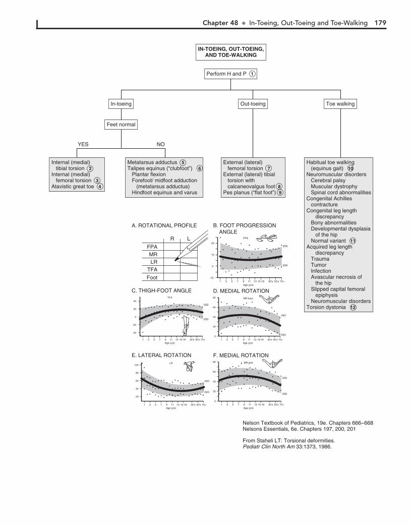
Chapter 48 u In-Toeing,Out-ToeingandToe-Walking 179
A. ROTATIONAL PROFILE
FPAMRLR
TFA
Foot
R L
10
20
0
-101 3 5 7 9 11 13 15-19 30’s 50’s 70+
2SD
2SD
B. FOOT PROGRESSION ANGLE
FPA
Age (yrs)
60
80
40
20
01 3 5 7 9 11 13 15-19 30’s 50’s 70+
2SD
2SD
F. MEDIAL ROTATIONMR girls
Age (yrs)
60
80
40
20
01 3 5 7 9 11
Age (yrs)13 15-19 30’s 50’s 70+
2SD
2SD
D. MEDIAL ROTATIONMR boys
60
100
80
40
20
1 3 5 7 9 11Age (yrs)
13 15-19 30’s 50’s 70+
2SD
2SD
E. LATERAL ROTATIONLR
0
40
20
-20
-40
1 3 5 7 9 11Age (yrs)
13 15-19 30’s 50’s 70+
2SD
2SD
C. THIGH-FOOT ANGLETFA
Internal (medial) tibial torsion Internal (medial) femoral torsion Atavistic great toe
Internal (medial) tibial torsion 2 Internal (medial) femoral torsion 3 Atavistic great toe 4
Metatarsus adductusTalipes equinus (“clubfoot”) Plantar flexion Forefoot/ midfoot adduction (metatarsus adductus) Hindfoot equinus and varus
Metatarsus adductus 5 Talipes equinus (“clubfoot”) 6 Plantar flexion Forefoot/ midfoot adduction (metatarsus adductus) Hindfoot equinus and varus
External (lateral) femoral torsionExternal (lateral) tibial torsion with calcaneovalgus footPes planus (“flat foot”)
External (lateral) femoral torsion 7External (lateral) tibial torsion with calcaneovalgus foot 8 Pes planus (“flat foot”) 9
Habitual toe walking (equinus gait) Neuromuscular disorders Cerebral palsy Muscular dystrophy Spinal cord abnormalitiesCongenital Achilles contractureCongenital leg length discrepancy Bony abnormalities Developmental dysplasia of the hip Normal variantAcquired leg length discrepancy Trauma Tumor Infection Avascular necrosis of the hip Slipped capital femoral epiphysis Neuromuscular disordersTorsion dystonia
Habitual toe walking (equinus gait) 10 Neuromuscular disorders Cerebral palsy Muscular dystrophy Spinal cord abnormalitiesCongenital Achilles contractureCongenital leg length discrepancy Bony abnormalities Developmental dysplasia of the hip Normal variant 11 Acquired leg length discrepancy Trauma Tumor Infection Avascular necrosis of the hip Slipped capital femoral epiphysis Neuromuscular disordersTorsion dystonia 12
Toe walkingToe walkingOut-toeingOut-toeingIn-toeingIn-toeing
Feet normalFeet normal
IN-TOEING, OUT-TOEING,AND TOE-WALKING
IN-TOEING, OUT-TOEING,AND TOE-WALKING
Perform Perform H and P 1
NOYES
From Staheli LT: Torsional deformities.Pediatr Clin North Am 33:1373, 1986.
Nelson Textbook of Pediatrics, 19e. Chapters 666–668Nelsons Essentials, 6e. Chapters 197, 200, 201
180
Chapter 49
BOWLEGS AND KNOCK-KNEES
lower extremities should be done to localize the deformity and identify any obvious bony abnormalities. Failure to follow a normal developmental sequence, rapid progression of the de-formity, family history of a pathologic condition, and physical findings suggestive of an underlying abnormality (neurofibro-matosis, arthritis, infection) all warrant further evaluation.
3 Genu varum (physiologic bowlegs) is usually a combina-tion of a normal varus angulation of the knee and internal
tibial torsion that is normal in the infant and toddler. Neutral-ization by 2 to 3 years of age is the norm. Persistence beyond age 3 or a severe case (.4 to 5 inches intercondylar distance, 16 degrees or greater tibial femoral angle) warrants investiga-tion to rule out a pathologic cause.
4 Physiologic genu valgum (knock-knees) develops as the natural overcorrection of physiologic genu varus and oc-
curs between the third and fourth years of life. Resolution gen-erally occurs between 5 and 8 years of age.
5 Asymmetric growth of the tibial growth plate (due to ab-normal ossification) results in tibia vara (Blount’s disease),
characterized by severe progressive bowing. Infantile onset (age 1 to 3 years) is most common in overweight toddlers, African-Americans, early walkers, and children with a family history of Blount’s disease; most (80%) have bilateral involvement. A later juvenile onset (age 4 to 10 years) is more likely to affect over-weight African-American males. Bilateral involvement is less frequent in this age group (50%), and pain (rather than defor-mity) is more likely to be the primary complaint. The condition can also develop in adolescents. X-rays show medial metaphy-seal irregularity, “beaking” of the medial tibial epiphysis, and wedging of the proximal epiphysis; specific radiographic crite-ria exist to characterize the stage of disease.
6 Congenital pseudoarthrosis of the tibia appears early in infancy as an anterolateral bowing of the tibia; 50% of af-
fected children have neurofibromatosis. (The condition affects 10% of all patients with neurofibromatosis.)
7 Rickets may be nutritional (vitamin D or calcium defi-ciency), genetic (vitamin D resistance) or due to hypophos-
phatasia (renal disorders, nutritional). A history of prolonged breastfeeding without adequate vitamin D supplementation is common in children with rickets. A widened physis and “frayed” appearance of the metaphysis are common radiologic findings in rickets.
8 Bony dysplasias may be characterized by metaphyseal, di-aphyseal, epiphyseal, or physeal plate abnormalities. Chil-
dren with these conditions are more likely to be small for their age and have a later age of onset of walking.
Angular deformities such as bowlegs and knock-knees are common complaints. Spontaneous resolution is the rule for most cases. It is important to know when to be concerned about a potentially pathologic diagnosis when treating an angular deformity.
Angular alignment of the lower extremities at birth is typi-cally varus (bowlegged) at the knee, although parents often do not comment on this alignment until a child starts standing. Normal bony remodeling occurs over the first 2 years of life, with typical resolution of this physiologic genu varus (bowlegs) to a near-neutral position. Subsequent development between the third and fourth year of life normally results in a valgus alignment at the knees (genu valgus or knock-knees), which gradually neutralizes to the normal adult alignment of mild valgus by age 7 or 8 years.
1 The history should clearly elucidate the nature of the complaint, age at onset, progression, and any previous
treatment. A complete medical (including a review of growth parameters), developmental (particularly the age when the child started walking), and family history should be part of the evaluation. Include a birth history and feeding history (breastfeeding, unusual diets) and inquire about medications (some anticonvulsants can impact vitamin D and phosphate). It may also be helpful to ask parents to clarify their exact concerns and fears about the child’s appearance and future prognosis, because education and reassurance about the ex-pected course of angular deformities is often all that is needed.
The physical assessment should include careful measure-ments and observation of the gait. A rotational profile should be done because rotational deformities can contribute to the ap-pearance of genu varum or valgum. (See Chapter 48.) Joints should be assessed for laxity and swelling suggesting arthritis. Extremities should be assessed for asymmetry suggesting bony abnormalities (congenital absence of bones, hypoplasia). Café-au-lait spots or neurofibromas may indicate neurofibromatosis. Any pain or tenderness on exam should be noted.
2 Gentle reassurance of parental concerns and education about the expected course of these conditions are often all
that is necessary. Certain indicators, such as short stature and significant asymmetry, do indicate further workup for a cause other than physiologic. If any worrisome prognosticators are present, preliminary x-ray (standing AP) evaluation of the
BibliographySchoenecker PL, Rich MM: The lower extremity. In Morrissy R, Weinstein S,
editors: Lovell & Winter’s pediatric orthopaedics, ed 6, Philadelphia, 2006, Lippincott Williams & Wilkins, pp 1158–1211.
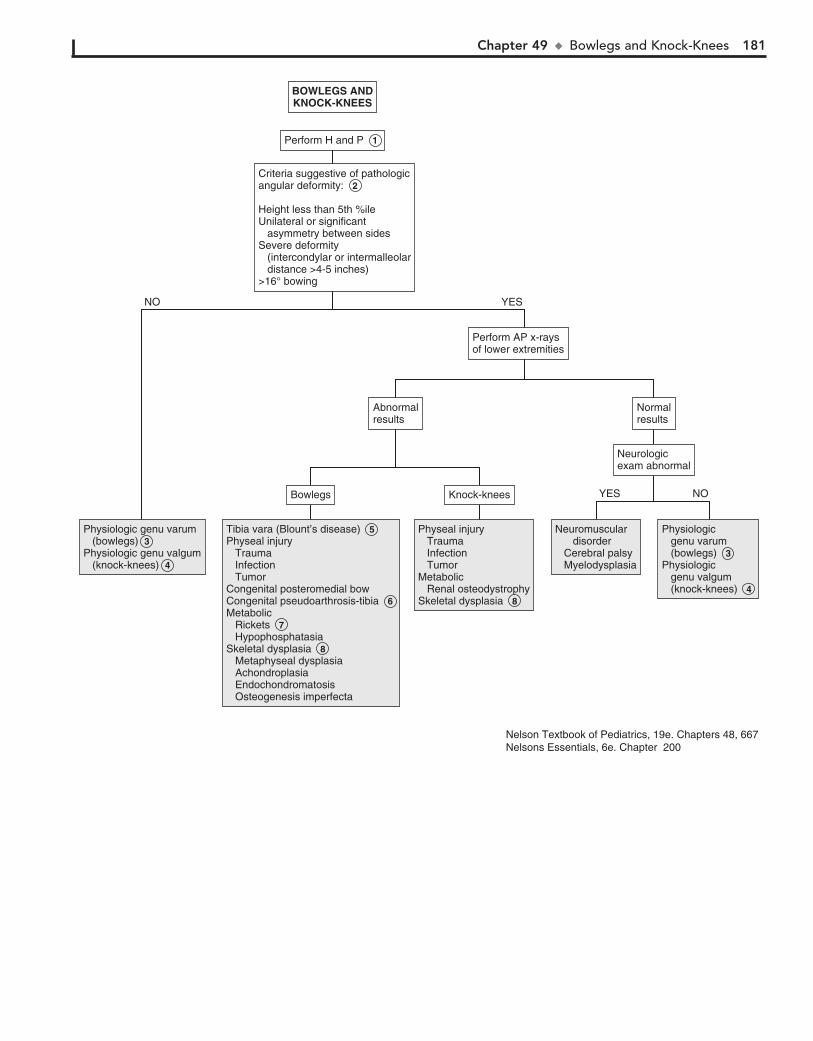
Chapter 49 u BowlegsandKnock-Knees 181
Criteria suggestive of pathologicangular deformity:
Height less than 5th %ileUnilateral or significant asymmetry between sidesSevere deformity (intercondylar or intermalleolar distance >4-5 inches)>16° bowing
Criteria suggestive of pathologicangular deformity: 2
Height less than 5th %ileUnilateral or significant asymmetry between sidesSevere deformity (intercondylar or intermalleolar distance >4-5 inches)>16° bowing
Physiologic genu varum (bowlegs)Physiologic genu valgum (knock-knees)
Physiologic genu varum (bowlegs) 3 Physiologic genu valgum (knock-knees) 4
Tibia vara (Blount’s disease) Physeal injury Trauma Infection TumorCongenital posteromedial bowCongenital pseudoarthrosis-tibiaMetabolic Rickets HypophosphatasiaSkeletal dysplasia Metaphyseal dysplasia Achondroplasia Endochondromatosis Osteogenesis imperfecta
Tibia vara (Blount’s disease) 5 Physeal injury Trauma Infection TumorCongenital posteromedial bowCongenital pseudoarthrosis-tibia 6 Metabolic Rickets 7 HypophosphatasiaSkeletal dysplasia 8 Metaphyseal dysplasia Achondroplasia Endochondromatosis Osteogenesis imperfecta
Physeal injury Trauma Infection TumorMetabolic Renal osteodystrophySkeletal dysplasia
Physeal injury Trauma Infection TumorMetabolic Renal osteodystrophySkeletal dysplasia 8
Neuromuscular disorder Cerebral palsy Myelodysplasia
Neuromuscular disorder Cerebral palsy Myelodysplasia
Physiologic genu varum (bowlegs) Physiologic genu valgum (knock-knees)
Physiologic genu varum (bowlegs) 3 Physiologic genu valgum (knock-knees) 4
Perform AP x-raysof lower extremitiesPerform AP x-raysof lower extremities
Knock-kneesKnock-kneesBowlegsBowlegs
NormalresultsNormalresults
AbnormalresultsAbnormalresults
Neurologicexam abnormalNeurologicexam abnormal
BOWLEGS ANDKNOCK-KNEESBOWLEGS ANDKNOCK-KNEES
Perform Perform H and P 1
NOYES
NO YES
Nelson Textbook of Pediatrics, 19e. Chapters 48, 667Nelsons Essentials, 6e. Chapter 200

This page intentionally left blank

PART
VIINeurology
184
Chapter 50
HEADACHES
pain, sensitivity to light and sound, an aura (scotomata, blurriness, visual distortions, sensory changes) or other transient neurologic disturbances. Relief with sleep is common. Triggers (e.g., stress, fa-tigue, illness) can often be identified. Migraines can be unilateral or bilateral in children, and the pain is typically characterized by a pounding or pulsing quality. (Auras, a longer headache duration, and a unilateral location become more common with increasing age.) Neurologic deficits (e.g., hemiplegia, vertigo, visual changes) may occur with some of the less common subtypes.
6 Many experts consider the absence of a family history for migraines as a risk factor for a space-occupying intracra-
nial lesion in children with acute headaches. Be conscious that migraine presentations can vary between family members, and headaches are often unrecognized as “migraines” by families; detailed questioning may be required. Most experts recom-mend neuroimaging for the child who has migrainous head-aches with an absolutely negative family history of migraine or its equivalent (e.g., motion sickness, cyclic vomiting).
7 The second edition of the International Classification of Headache Disorders provides detailed diagnostic criteria
for several subtypes of migraines. Diagnosis of a migraine dis-order requires at least two episodes meeting the diagnostic cri-teria. The classifications include migraines with and without aura (including hemiplegia and visual disturbances) as well as childhood periodic syndromes.
8 Neuroimaging (MRI is preferable if available) is warranted when the neurologic examination is abnormal or unusual
neurologic features occur during the migraine, when a child has headaches that awaken him or her from sleep or that are present on first awakening (then remit with upright posture), or are very brief in duration occurring only with cough or bending over.
9 Brain tumors present with symptoms due to increased in-tracranial pressure (ICP) or focal destruction or invasion
of brain tissue; presentations vary according to tumor type, location and patient age. The increased intracranial pressure typically causes headaches that may wake a child at night, are typically worse in the morning, are aggravated by coughing or bending over, and are frequently accompanied by vomiting; papilledema, sixth nerve palsy, altered level of consciousness, and gait instability may also be noted on examination.
10 An increased risk of brain abscesses is present in children with immunodeficiency disorders, right-to-left cardiac
shunts (especially tetralogy of Fallot), chronic ENT infections (ear, sinus, mastoid, dental), penetrating head injuries, soft tissue infections of the face or scalp, and infected VP shunts.
11 Hydrocephalus is most likely to cause headaches in older children (whose cranial sutures are closing). Headaches
are generalized; they may be acute or gradual in onset and mild or severe in quality, depending on how rapidly the hydrocepha-lus is progressing.
12 Third ventricle cysts may function as a ball-valve mechanism causing intermittent severe (“thunderclap”) headaches due
to transient increases in ICP. Other cysts (e.g., arachnoid, epi-dermoid, dermoid) may present with progressive headaches or seizures.
13 Neurologic abnormalities, altered mental status, or menin-geal signs (e.g., nuchal rigidity, Kernig, and Brudzinski
signs) warrant an evaluation to rule out meningitis.
Headaches are common and usually benign. Parents often seek medical attention for a child because they are concerned about the potential of a serious or life-threatening cause of the head-aches. Most headache complaints will be primary headache disorders; evaluation is essential to rule out more worrisome secondary causes of headache.
1 A thorough history should inquire about the quality, pat-tern, and progression of the complaint as well as inciting
factors, recent trauma, response to medications, and associated visual or sensory (or other neurologic) disturbances. Social and emotional issues should be reviewed; teens should typically be interviewed in private. The impact of the headaches on a child’s visual activity should be elicited. A thorough family history should inquire about headaches (particularly migraines), other neurologic problems, and general medical conditions that could cause secondary headaches. A headache diary may be helpful in characterizing headache patterns and identifying associated symptoms and triggers.
Older children are often able to describe their headache; nonverbal young children may become irritable, rock, rub their eyes or head, vomit, or seek a darkened environment.
When intracranial pressure (ICP) is increased (due to hy-drocephalus, mass lesions, edema, or hemorrhage), headaches are frequently characterized by ”red flags” such as pain that is present in the morning or awakens the patient from sleep and then remits with being upright; increased pain with cough, straining, or position change; altered sensorium or personality, or a patient’s description of ”the worst headache of my life.”
In addition to a thorough neurologic examination, the phys-ical examination should include a blood pressure evaluation, a thorough ENT examination (including dentition), and an as-sessment of visual acuity. Worrisome physical findings include abnormal neurologic signs, meningeal signs, papilledema, and altered sensorium.
2 Migraines are the most likely recurrent headache disorder to present with a first acute severe headache. Other chronic
or recurrent headache disorders will more commonly present for evaluation after repeated episodes (even if they have not quite reached the diagnostic 3 month duration criteria) but may need to be considered in the evaluation of any first severe acute headache.
3 Magnetic resonance imaging is generally preferred for iden-tifying structural lesions, infection, inflammation, and isch-
emia. Computed tomography (CT) is preferred in acute cases of suspected trauma or fracture. Imaging choice should consider that head CTs carry a higher radiation risk whereas MRIs are more costly, time-consuming, and they require sedation.
4 Headaches developing within 7 days of head trauma with signs or symptoms of concussion are defined as acute post-
traumatic headache.
5 Migraines are classically characterized by a positive family his-tory for migraines, associated nausea, vomiting or abdominal
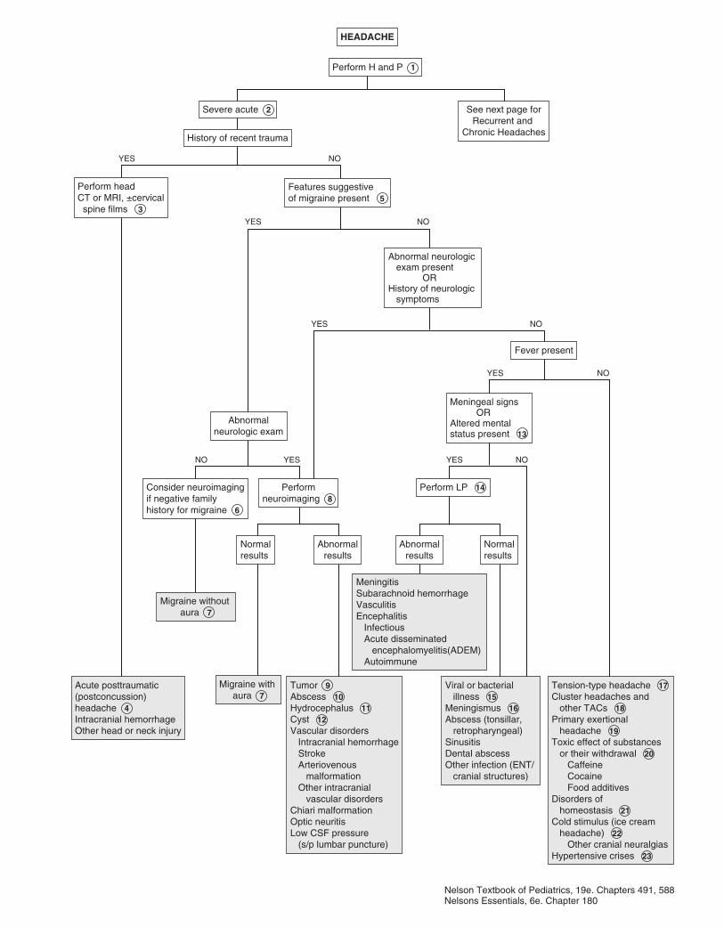
HEADACHEHEADACHE
Perform H and P 1 Perform H and P 1
Acute posttraumatic(postconcussion)headache 4Intracranial hemorrhageOther head or neck injury
Acute posttraumatic(postconcussion)headache 4Intracranial hemorrhageOther head or neck injury
Migraine without aura 7
Migraine without aura 7
Migraine with aura 7
Migraine with aura 7
Tumor 9Abscess 10Hydrocephalus 11Cyst 12Vascular disorders Intracranial hemorrhage Stroke Arteriovenous malformation Other intracranial vascular disordersChiari malformationOptic neuritis Low CSF pressure (s/p lumbar puncture)
Tumor 9Abscess 10Hydrocephalus 11Cyst 12Vascular disorders Intracranial hemorrhage Stroke Arteriovenous malformation Other intracranial vascular disordersChiari malformationOptic neuritis Low CSF pressure (s/p lumbar puncture)
MeningitisSubarachnoid hemorrhageVasculitisEncephalitis Infectious Acute disseminated encephalomyelitis(ADEM) Autoimmune
MeningitisSubarachnoid hemorrhageVasculitisEncephalitis Infectious Acute disseminated encephalomyelitis(ADEM) Autoimmune
Viral or bacterial illness 15Meningismus 16Abscess (tonsillar, retropharyngeal)SinusitisDental abscessOther infection (ENT/ cranial structures)
Viral or bacterial illness 15Meningismus 16Abscess (tonsillar, retropharyngeal)SinusitisDental abscessOther infection (ENT/ cranial structures)
Tension-type headache 17 Cluster headaches and other TACs 18Primary exertional headache 19Toxic effect of substances or their withdrawal 20 Caffeine Cocaine Food additivesDisorders of homeostasis 21Cold stimulus (ice cream headache) 22 Other cranial neuralgiasHypertensive crises 23
Tension-type headache 17 Cluster headaches and other TACs 18Primary exertional headache 19Toxic effect of substances or their withdrawal 20 Caffeine Cocaine Food additivesDisorders of homeostasis 21Cold stimulus (ice cream headache) 22 Other cranial neuralgiasHypertensive crises 23
Abnormalresults
Abnormalresults
NormalresultsNormalresults
Consider neuroimagingif negative familyhistory for migraine 6
Consider neuroimagingif negative familyhistory for migraine 6
Performneuroimaging 8
Performneuroimaging 8
Abnormalneurologic exam
Abnormalneurologic exam
Perform headCT or MRI, ±cervical spine films 3
Perform headCT or MRI, ±cervical spine films 3
Perform LP 14 Perform LP 14
Fever presentFever present
Features suggestiveof migraine present 5 Features suggestiveof migraine present 5
History of recent traumaHistory of recent trauma
Severe acute 2 Severe acute 2 See next page forRecurrent and
Chronic Headaches
See next page forRecurrent and
Chronic Headaches
Abnormalresults
Abnormalresults
NormalresultsNormalresults
NO NO
NO
NO
NO
YES YES
YES
YES
YES
NOYES
Abnormal neurologic exam present OR History of neurologic symptoms
Abnormal neurologic exam present OR History of neurologic symptoms
Meningeal signs ORAltered mentalstatus present 13
Meningeal signs ORAltered mentalstatus present 13
Nelson Textbook of Pediatrics, 19e. Chapters 491, 588Nelsons Essentials, 6e. Chapter 180
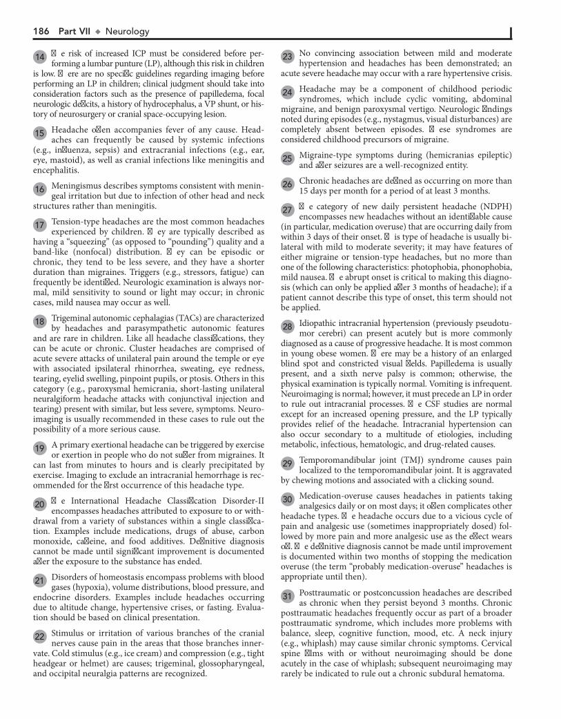
186 Part VII u Neurology
14 The risk of increased ICP must be considered before per-forming a lumbar punture (LP), although this risk in children
is low. There are no specific guidelines regarding imaging before performing an LP in children; clinical judgment should take into consideration factors such as the presence of papilledema, focal neurologic deficits, a history of hydrocephalus, a VP shunt, or his-tory of neurosurgery or cranial space-occupying lesion.
15 Headache often accompanies fever of any cause. Head-aches can frequently be caused by systemic infections
(e.g., influenza, sepsis) and extracranial infections (e.g., ear, eye, mastoid), as well as cranial infections like meningitis and encephalitis.
16 Meningismus describes symptoms consistent with menin-geal irritation but due to infection of other head and neck
structures rather than meningitis.
17 Tension-type headaches are the most common headaches experienced by children. They are typically described as
having a “squeezing” (as opposed to “pounding”) quality and a band-like (nonfocal) distribution. They can be episodic or chronic, they tend to be less severe, and they have a shorter duration than migraines. Triggers (e.g., stressors, fatigue) can frequently be identified. Neurologic examination is always nor-mal, mild sensitivity to sound or light may occur; in chronic cases, mild nausea may occur as well.
18 Trigeminal autonomic cephalagias (TACs) are characterized by headaches and parasympathetic autonomic features
and are rare in children. Like all headache classifications, they can be acute or chronic. Cluster headaches are comprised of acute severe attacks of unilateral pain around the temple or eye with associated ipsilateral rhinorrhea, sweating, eye redness, tearing, eyelid swelling, pinpoint pupils, or ptosis. Others in this category (e.g., paroxysmal hemicrania, short-lasting unilateral neuralgiform headache attacks with conjunctival injection and tearing) present with similar, but less severe, symptoms. Neuro-imaging is usually recommended in these cases to rule out the possibility of a more serious cause.
19 A primary exertional headache can be triggered by exercise or exertion in people who do not suffer from migraines. It
can last from minutes to hours and is clearly precipitated by exercise. Imaging to exclude an intracranial hemorrhage is rec-ommended for the first occurrence of this headache type.
20 The International Headache Classification Disorder-II encompasses headaches attributed to exposure to or with-
drawal from a variety of substances within a single classifica-tion. Examples include medications, drugs of abuse, carbon monoxide, caffeine, and food additives. Definitive diagnosis cannot be made until significant improvement is documented after the exposure to the substance has ended.
21 Disorders of homeostasis encompass problems with blood gases (hypoxia), volume distributions, blood pressure, and
endocrine disorders. Examples include headaches occurring due to altitude change, hypertensive crises, or fasting. Evalua-tion should be based on clinical presentation.
22 Stimulus or irritation of various branches of the cranial nerves cause pain in the areas that those branches inner-
vate. Cold stimulus (e.g., ice cream) and compression (e.g., tight headgear or helmet) are causes; trigeminal, glossopharyngeal, and occipital neuralgia patterns are recognized.
23 No convincing association between mild and moderate hypertension and headaches has been demonstrated; an
acute severe headache may occur with a rare hypertensive crisis.
24 Headache may be a component of childhood periodic syndromes, which include cyclic vomiting, abdominal
migraine, and benign paroxysmal vertigo. Neurologic findings noted during episodes (e.g., nystagmus, visual disturbances) are completely absent between episodes. These syndromes are considered childhood precursors of migraine.
25 Migraine-type symptoms during (hemicranias epileptic) and after seizures are a well-recognized entity.
26 Chronic headaches are defined as occurring on more than 15 days per month for a period of at least 3 months.
27 The category of new daily persistent headache (NDPH) encompasses new headaches without an identifiable cause
(in particular, medication overuse) that are occurring daily from within 3 days of their onset. This type of headache is usually bi-lateral with mild to moderate severity; it may have features of either migraine or tension-type headaches, but no more than one of the following characteristics: photophobia, phonophobia, mild nausea. The abrupt onset is critical to making this diagno-sis (which can only be applied after 3 months of headache); if a patient cannot describe this type of onset, this term should not be applied.
28 Idiopathic intracranial hypertension (previously pseudotu-mor cerebri) can present acutely but is more commonly
diagnosed as a cause of progressive headache. It is most common in young obese women. There may be a history of an enlarged blind spot and constricted visual fields. Papilledema is usually present, and a sixth nerve palsy is common; otherwise, the physical examination is typically normal. Vomiting is infrequent. Neuroimaging is normal; however, it must precede an LP in order to rule out intracranial processes. The CSF studies are normal except for an increased opening pressure, and the LP typically provides relief of the headache. Intracranial hypertension can also occur secondary to a multitude of etiologies, including metabolic, infectious, hematologic, and drug-related causes.
29 Temporomandibular joint (TMJ) syndrome causes pain localized to the temporomandibular joint. It is aggravated
by chewing motions and associated with a clicking sound.
30 Medication-overuse causes headaches in patients taking analgesics daily or on most days; it often complicates other
headache types. The headache occurs due to a vicious cycle of pain and analgesic use (sometimes inappropriately dosed) fol-lowed by more pain and more analgesic use as the effect wears off. The definitive diagnosis cannot be made until improvement is documented within two months of stopping the medication overuse (the term “probably medication-overuse” headaches is appropriate until then).
31 Posttraumatic or postconcussion headaches are described as chronic when they persist beyond 3 months. Chronic
posttraumatic headaches frequently occur as part of a broader posttraumatic syndrome, which includes more problems with balance, sleep, cognitive function, mood, etc. A neck injury (e.g., whiplash) may cause similar chronic symptoms. Cervical spine films with or without neuroimaging should be done acutely in the case of whiplash; subsequent neuroimaging may rarely be indicated to rule out a chronic subdural hematoma.
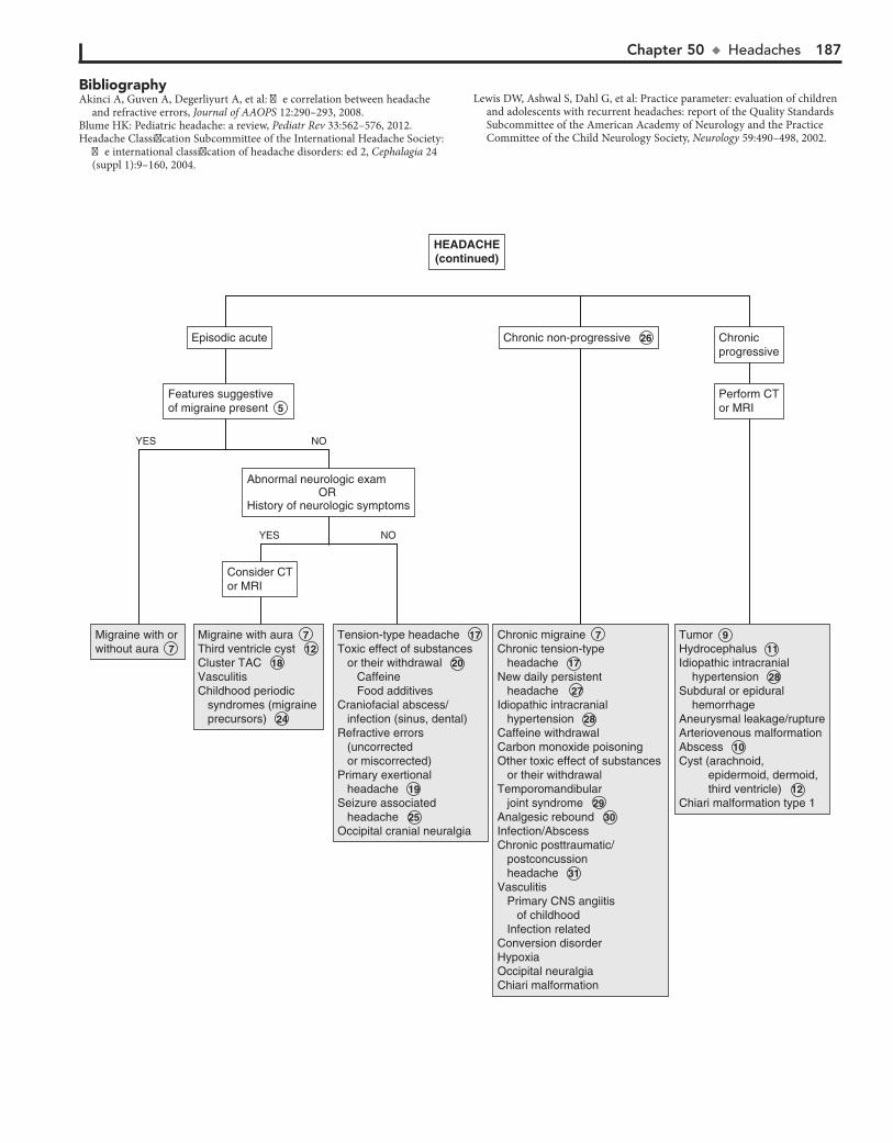
Chapter 50 u Headaches 187
Migraine with or without aura 7 Migraine with or without aura 7
Migraine with aura 7 Third ventricle cyst 12 Cluster TAC 18 VasculitisChildhood periodic syndromes (migraine precursors) 24
Migraine with aura 7 Third ventricle cyst 12 Cluster TAC 18 VasculitisChildhood periodic syndromes (migraine precursors) 24
Tension-type headache 17 Toxic effect of substances or their withdrawal 20 Caffeine Food additivesCraniofacial abscess/ infection (sinus, dental)Refractive errors (uncorrected or miscorrected)Primary exertional headache 19 Seizure associated headache 25 Occipital cranial neuralgia
Tension-type headache 17 Toxic effect of substances or their withdrawal 20 Caffeine Food additivesCraniofacial abscess/ infection (sinus, dental)Refractive errors (uncorrected or miscorrected)Primary exertional headache 19 Seizure associated headache 25 Occipital cranial neuralgia
Chronic migraine 7 Chronic tension-type headache 17 New daily persistent headache 27 Idiopathic intracranial hypertension 28 Caffeine withdrawalCarbon monoxide poisoningOther toxic effect of substances or their withdrawalTemporomandibular joint syndrome 29 Analgesic rebound 30Infection/AbscessChronic posttraumatic/ postconcussion headache 31 Vasculitis Primary CNS angiitis of childhood Infection relatedConversion disorderHypoxiaOccipital neuralgia Chiari malformation
Chronic migraine 7 Chronic tension-type headache 17 New daily persistent headache 27 Idiopathic intracranial hypertension 28 Caffeine withdrawalCarbon monoxide poisoningOther toxic effect of substances or their withdrawalTemporomandibular joint syndrome 29 Analgesic rebound 30Infection/AbscessChronic posttraumatic/ postconcussion headache 31 Vasculitis Primary CNS angiitis of childhood Infection relatedConversion disorderHypoxiaOccipital neuralgia Chiari malformation
Tumor 9 Hydrocephalus 11 Idiopathic intracranial hypertension 28 Subdural or epidural hemorrhageAneurysmal leakage/ruptureArteriovenous malformationAbscess 10 Cyst (arachnoid, epidermoid, dermoid, third ventricle) 12 Chiari malformation type 1
Tumor 9 Hydrocephalus 11 Idiopathic intracranial hypertension 28 Subdural or epidural hemorrhageAneurysmal leakage/ruptureArteriovenous malformationAbscess 10 Cyst (arachnoid, epidermoid, dermoid, third ventricle) 12 Chiari malformation type 1
Consider CTor MRIConsider CTor MRI
Features suggestiveof migraine present 5 Features suggestiveof migraine present 5
Episodic acuteEpisodic acute
Perform CTor MRIPerform CTor MRI
ChronicprogressiveChronicprogressive
Chronic non-progressive 26 Chronic non-progressive 26
NOYES
NOYES
HEADACHE(continued)HEADACHE(continued)
Abnormal neurologic exam OR History of neurologic symptoms
Abnormal neurologic exam OR History of neurologic symptoms
BibliographyAkinci A, Guven A, Degerliyurt A, et al: The correlation between headache
and refractive errors, Journal of AAOPS 12:290–293, 2008. Blume HK: Pediatric headache: a review, Pediatr Rev 33:562–576, 2012. Headache Classification Subcommittee of the International Headache Society:
The international classification of headache disorders: ed 2, Cephalagia 24 (suppl 1):9–160, 2004.
Lewis DW, Ashwal S, Dahl G, et al: Practice parameter: evaluation of children and adolescents with recurrent headaches: report of the Quality Standards Subcommittee of the American Academy of Neurology and the Practice Committee of the Child Neurology Society, Neurology 59:490–498, 2002.
188
6 Any labs obtained, particularly CBCs, should be directed towards evaluating the source of the fever, not to evaluate
the seizure. (Of note, when considering bacteremia as the source of the fever, the incidence of bacteremia is the same in febrile children under 24 months of age, regardless of whether they have a febrile seizure or not.)
Chapter 51
SEIZURES AND OTHER PAROXYSMAL DISORDERS
have a recurrence. The risk of subsequent epilepsy following a simple febrile seizure is low and affected by various predictors such as age of occurrence, duration of fever, height of fever, fam-ily history, etc. Complex febrile seizures are defined as focal or prolonged (longer than 15 minutes) or occurring in a flurry of repetitive episodes during the febrile illness. Genetic factors are being increasingly recognized as contributing factors in complex febrile seizures. (Remember, children younger than 6 months of age do not meet the criteria for a simple febrile seizure and should not be evaluated along this pathway.)
3 The evaluation of complex febrile seizures should be indi-vidualized based on the presentation. A very prolonged
seizure, a focal seizure (characteristic of herpes simplex virus [HSV] encephalitis), or an abnormal neurologic examination should prompt a thorough evaluation for a CNS infection. The diagnostic test of choice for herpes simplex virus encephalitis is a PCR for HSV performed on a spinal fluid specimen; tests should also be obtained for bacterial and viral etiologies of meningitis. MRI should be obtained for all children with a focal seizure. An EEG should be obtained acutely if concerned about febrile status epilepticus.
4 By definition, the term complex febrile seizure applies to children between 6 months and 60 months of age. Outside
of that age group, seizures with fever can be due to multiple other causes (e.g., HHV6, Shigella, immunizations).
5 A lumbar puncture is not routinely recommended for a child age 6-12 months (who is well appearing and fully im-
munized) who has experienced a simple febrile seizure because their risk of having bacterial meningitis is extremely low. Less straightforward is the recommendation for performing an LP on a child who has had antibiotics (by any route) in the days pre-ceding the seizure or whose immunization status is unknown or deficient (particularly for Hib and pneumococcus, which are known pathogens for bacterial meningitis). Most of the studies regarding outcomes of febrile seizures have been done on chil-dren with high immunization rates. Because the outcomes of febrile seizures for children with incomplete or unknown im-munization status or any pretreatment with antibiotics is un-known, an LP is recommended as an option for these children.
A seizure is a paroxysmal disturbance in brain function that manifests as an alteration in motor activity, level of conscious-ness, or autonomic function. Seizures and seizure disorders were traditionally classified according to whether the manifestations were generalized or focal and whether consciousness was pre-served or impaired. Advances in neuroscience that allow spe-cific, objective diagnostic criteria for seizures and seizure disorders have led to revision of the terminology and classification system of these entities; the current system emphasizes diagnosis based on objective, measurable criteria. The traditional definition of status epilepticus is the occurrence of prolonged or recurrent seizures (without a return to consciousness) for 30 minutes or longer. A number of nonepileptic paroxysmal disorders occur in childhood and must be distinguished from epileptic seizures.
1 A description of the event is the most valuable part of the evaluation because physical findings are rare and diagnos-
tic studies may not be conclusive. Indeed, the description of the event is generally going to be the key to discerning whether the event was likely a seizure versus a nonepileptic event; a video recording of the event can be extremely helpful in this regard. Inquire about the occurrence of an aura, preceding mood or behavioral changes, a detailed description of the event includ-ing motor, verbal, and autonomic changes (e.g., pupillary dila-tation, drooling, incontinence, pallor, vomiting), as well as a history of any inciting events (e.g., trauma, fever, crying). Ob-tain the medical history, including a birth and developmental history. Inquire about medication use and potential ingestions of toxic substances. In children with a known seizure disorder, specifically ask about medication compliance. Neurocutaneous lesions (e.g., café-au-lait spots, ash leaf macules) and vascular lesions should be noted on the physical examination.
2 Seizures meeting the criteria for simple febrile seizures are generally benign events with excellent long-term prognoses.
Two to five percent of the general population will experience a (usually simple) febrile seizure; of those, approximately 30% will
Chapter 51 u SeizuresandOtherParoxysmalDisorders 189
SEIZURES AND OTHERPAROXYSMAL DISORDERS
SEIZURES AND OTHERPAROXYSMAL DISORDERS
Fever present( 100.4°)Fever present( 100.4°)
See next pageSee next page
Perform H and P 1 Perform H and P 1
Complex febrile seizureCNS infectionSeizure with fever due to other causes 4
Complex febrile seizureCNS infectionSeizure with fever due to other causes 4
CNS infection CNS infection Simple febrileseizureSimple febrileseizure
Simple febrile seizureCNS infection Simple febrile seizureCNS infection
Simple febrile seizureSimple febrile seizure Simple febrile seizureCNS infection Simple febrile seizureCNS infection
Consider LPConsider LP
Age 6 to 12 monthsAge 6 to 12 monthsPerform LPPerform LP Age 12 monthsAge 12 months
Pretreatment with antibiotics?Pretreatment with antibiotics?
Abnormal resultsAbnormal results
Consider LP, neuroimaging, EEG 3
Consider LP, neuroimaging, EEG 3
Criteria for simple febrile seizure present: 2 Age 6 to 60 monthsNeurologically healthy childGeneralized (nonfocal) seizure<15” durationNo febrile seizure in previous 24 hours
Criteria for simple febrile seizure present: 2 Age 6 to 60 monthsNeurologically healthy childGeneralized (nonfocal) seizure<15” durationNo febrile seizure in previous 24 hours
Signs or symptoms suggestive of meningitispresent (vomiting, headache, neck stiffness,Kernig sign, Brudzinski sign)
Signs or symptoms suggestive of meningitispresent (vomiting, headache, neck stiffness,Kernig sign, Brudzinski sign)
NormalresultsNormalresults
Assess forsource of fever 6 Assess forsource of fever 6
Consider LPConsider LP
NO
NO
NO
NO
NOYES
YES
YES
YES
YES
5
Nelson Textbook of Pediatrics, 19e. Chapters 17, 586, 587, 594Nelsons Essentials, 6e. Chapter 181
Deficient or unknown immunization status OR Pretreatment with antibiotics 5
Deficient or unknown immunization status OR Pretreatment with antibiotics 5
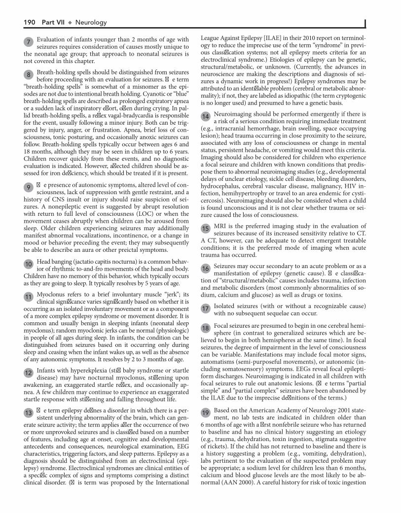
190 Part VII u Neurology
7 Evaluation of infants younger than 2 months of age with seizures requires consideration of causes mostly unique to
the neonatal age group; that approach to neonatal seizures is not covered in this chapter.
8 Breath-holding spells should be distinguished from seizures before proceeding with an evaluation for seizures. The term
“breath-holding spells” is somewhat of a misnomer as the epi-sodes are not due to intentional breath holding. Cyanotic or “blue” breath-holding spells are described as prolonged expiratory apnea or a sudden lack of inspiratory effort, often during crying. In pal-lid breath-holding spells, a reflex vagal-bradycardia is responsible for the event, usually following a minor injury. Both can be trig-gered by injury, anger, or frustration. Apnea, brief loss of con-sciousness, tonic posturing, and occasionally anoxic seizures can follow. Breath-holding spells typically occur between ages 6 and 18 months, although they may be seen in children up to 6 years. Children recover quickly from these events, and no diagnostic evaluation is indicated. However, affected children should be as-sessed for iron deficiency, which should be treated if it is present.
9 The presence of autonomic symptoms, altered level of con-sciousness, lack of suppression with gentle restraint, and a
history of CNS insult or injury should raise suspicion of sei-zures. A nonepileptic event is suggested by abrupt resolution with return to full level of consciousness (LOC) or when the movement ceases abruptly when children can be aroused from sleep. Older children experiencing seizures may additionally manifest abnormal vocalizations, incontinence, or a change in mood or behavior preceding the event; they may subsequently be able to describe an aura or other preictal symptoms.
10 Head banging (jactatio capitis nocturna) is a common behav-ior of rhythmic to-and-fro movements of the head and body.
Children have no memory of this behavior, which typically occurs as they are going to sleep. It typically resolves by 5 years of age.
11 Myoclonus refers to a brief involuntary muscle “jerk”; its clinical significance varies significantly based on whether it is
occurring as an isolated involuntary movement or as a component of a more complex epilepsy syndrome or movement disorder. It is common and usually benign in sleeping infants (neonatal sleep myoclonus); random myoclonic jerks can be normal (physiologic) in people of all ages during sleep. In infants, the condition can be distinguished from seizures based on it occurring only during sleep and ceasing when the infant wakes up, as well as the absence of any autonomic symptoms. It resolves by 2 to 3 months of age.
12 Infants with hyperekplexia (stiff baby syndrome or startle disease) may have nocturnal myoclonus, stiffening upon
awakening, an exaggerated startle reflex, and occasionally ap-nea. A few children may continue to experience an exaggerated startle response with stiffening and falling throughout life.
13 The term epilepsy defines a disorder in which there is a per-sistent underlying abnormality of the brain, which can gen-
erate seizure activity; the term applies after the occurrence of two or more unprovoked seizures and is classified based on a number of features, including age at onset, cognitive and developmental antecedents and consequences, neurological examination, EEG characteristics, triggering factors, and sleep patterns. Epilepsy as a diagnosis should be distinguished from an electroclinical (epi-lepsy) syndrome. Electroclinical syndromes are clinical entities of a specific complex of signs and symptoms comprising a distinct clinical disorder. (This term was proposed by the International
League Against Epilepsy [ILAE] in their 2010 report on terminol-ogy to reduce the imprecise use of the term “syndrome” in previ-ous classification systems; not all epilepsy meets criteria for an electroclinical syndrome.) Etiologies of epilepsy can be genetic, structural/metabolic, or unknown. (Currently, the advances in neuroscience are making the descriptions and diagnosis of sei-zures a dynamic work in progress!) Epilepsy syndromes may be attributed to an identifiable problem (cerebral or metabolic abnor-mality); if not, they are labeled as idiopathic (the term cryptogenic is no longer used) and presumed to have a genetic basis.
14 Neuroimaging should be performed emergently if there is a risk of a serious condition requiring immediate treatment
(e.g., intracranial hemorrhage, brain swelling, space occupying lesion); head trauma occurring in close proximity to the seizure, associated with any loss of consciousness or change in mental status, persistent headache, or vomiting would meet this criteria. Imaging should also be considered for children who experience a focal seizure and children with known conditions that predis-pose them to abnormal neuroimaging studies (e.g., developmental delays of unclear etiology, sickle cell disease, bleeding disorders, hydrocephalus, cerebral vascular disease, malignancy, HIV in-fection, hemihypertrophy or travel to an area endemic for cysti-cercosis). Neuroimaging should also be considered when a child is found unconscious and it is not clear whether trauma or sei-zure caused the loss of consciousness.
15 MRI is the preferred imaging study in the evaluation of seizures because of its increased sensitivity relative to CT.
A CT, however, can be adequate to detect emergent treatable conditions; it is the preferred mode of imaging when acute trauma has occurred.
16 Seizures may occur secondary to an acute problem or as a manifestation of epilepsy (genetic cause). The classifica-
tion of “structural/metabolic” causes includes trauma, infection and metabolic disorders (most commonly abnormalities of so-dium, calcium and glucose) as well as drugs or toxins.
17 Isolated seizures (with or without a recognizable cause) with no subsequent sequelae can occur.
18 Focal seizures are presumed to begin in one cerebral hemi-sphere (in contrast to generalized seizures which are be-
lieved to begin in both hemispheres at the same time). In focal seizures, the degree of impairment in the level of consciousness can be variable. Manifestations may include focal motor signs, automatisms (semi-purposeful movements), or autonomic (in-cluding somatosensory) symptoms. EEGs reveal focal epilepti-form discharges. Neuroimaging is indicated in all children with focal seizures to rule out anatomic lesions. (The terms “partial simple” and “partial complex” seizures have been abandoned by the ILAE due to the imprecise definitions of the terms.)
19 Based on the American Academy of Neurology 2001 state-ment, no lab tests are indicated in children older than
6 months of age with a first nonfebrile seizure who has returned to baseline and has no clinical history suggesting an etiology (e.g., trauma, dehydration, toxin ingestion, stigmata suggestive of rickets). If the child has not returned to baseline and there is a history suggesting a problem (e.g., vomiting, dehydration), labs pertinent to the evaluation of the suspected problem may be appropriate; a sodium level for children less than 6 months, calcium and blood glucose levels are the most likely to be ab-normal (AAN 2000). A careful history for risk of toxic ingestion
Chapter 51 u SeizuresandOtherParoxysmalDisorders 191
2 years 7
See next pageSee next page
Breath-holding spells 8 Breath-holding spells 8
Head banging 10 Neonatal sleep myoclonus 11 Myoclonic jerks (physiologic) 11 Hyperkplexia (stiff baby syndrome or startle disease) 12 Epileptic seizure/Electroclinical syndrome 13
Head banging 10 Neonatal sleep myoclonus 11 Myoclonic jerks (physiologic) 11 Hyperekplexia (stiff baby syndrome or startle disease) 12 Epileptic seizure/Electroclinical syndrome 13
Seizure due to structural/ metabolic causes 16 Generalized seizure (epileptic or nonepileptic) 13 17 Focal seizure (epileptic or nonepileptic) 18
Seizure due to structural/ metabolic causes 16 Generalized seizure (epileptic or nonepileptic) 13 17 Focal seizure (epileptic or nonepileptic) 18
Generalized seizures Tonic-clonic Absence 20 Myoclonic 21 Clonic Tonic Atonic Unknown (epileptic spasms) 22 Seizure due to structural/ metabolic causes 16 Paroxysmal torticollisAlternating hemiplegia of childhood 23 Jittery infantBenign paroxysmal vertigo 24 Spasmus nutans 25 Stereotypical movements 26 Other involuntary movements 27 Caregiver fabricated illness (previously Munchhausen by proxy)
Generalized seizures Tonic-clonic Absence 20 Myoclonic 21 Clonic Tonic Atonic Unknown (epileptic spasms) 22 Seizure due to structural/ metabolic causes 16 Paroxysmal torticollisAlternating hemiplegia of childhood 23 Jittery infantBenign paroxysmal vertigo 24 Spasmus nutans 25 Stereotypical movements 26 Other involuntary movements 27 Caregiver fabricated illness (previously Munchhausen by proxy)
History consistent with breath-holding spells 8 History consistent with breath-holding spells 8
Etiology suggested by H and P
Consider:EEG, lytes, calcium, glucose, drug screen 19
Etiology suggested by H and P
Consider:EEG, lytes, calcium, glucose, drug screen 19
Obtain CT if recent trauma, otherwise MRI 15
Obtain EEG
Consider:Drug screen, LP, glucose, lytes, calcium, magnesium, phosphorus
Obtain CT if recent trauma, otherwise MRI 15
Obtain EEG
Consider:Drug screen, LP, glucose, lytes, calcium, magnesium, phosphorus
Any of the following features present: 14 Recent traumaFocal componentPostictal focal defectNo return to baseline LOC after several hoursOther condition increasing risk of abnormal neuroimaging results Age <1 yr
Any of the following features present: 14 Recent traumaFocal componentPostictal focal defectNo return to baseline LOC after several hoursOther condition increasing risk of abnormal neuroimaging results Age <1 yr
Obtain EEG if suspicious of seizures 9 Obtain EEG if suspicious of seizures 9
Sleep relatedSleep related
NO
YES
YES
YES
NO
NO
NO
YES
Age 2 months to Age 2 months to 2 years (afebrile) 7
Nelson Textbook of Pediatrics, 19e. Chapters 17, 586, 587, 594Nelsons Essentials, 6e. Chapter 181
SEIZURES AND OTHERPAROXYSMAL DISORDERS
(continued)
SEIZURES AND OTHERPAROXYSMAL DISORDERS
(continued)
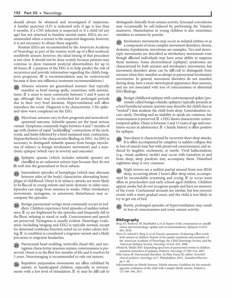
192 Part VII u Neurology
distinguish clinically from seizure activity. Syncopal convulsions may occasionally be self-induced by performing the Valsalva maneuver. Masturbation in young children is also sometimes mistaken as seizures by parents.
27 Involuntary movements may occur as isolated entities or as a component of more complex movement disorders; chorea,
dystonia, hypokinesia, myoclonus are examples. Tics and stereo-typic movements are described as involuntary movements even though affected individuals may have some ability to suppress those motions. Some electroclinical (epilepsy) syndromes are characterized by both seizures and involuntary movements, but movement disorders alone can be difficult to distinguish from seizures when they manifest as abrupt or paroxysmal involuntary movements. In general, movement disorders do not manifest during sleep, have a more stereotypical appearance than seizures, and are not associated with loss of consciousness or abnormal EEG findings.
should always be obtained and investigated if suspicious. A lumbar puncture (LP) is indicated only if age is less than 6 months, if a CNS infection is suspected or if a child (of any age) has not returned to baseline mental status. EEGs are rec-ommended when a seizure is the suspected diagnosis; however, it is not necessary to obtain them urgently.
Routine EEGs are recommended by the American Academy of Neurology as part of the routine work-up of a first nonfocal, nonfebrile seizure; however, the ideal timing of that procedure is not clear. It should not be done acutely because patients may continue to show transient postictal abnormalities for up to 48 hours. The purpose of the EEG is to help predict the risk of recurrence and provide information regarding the child’s long-term prognosis. (This recommendation may be controversial because it does not influence treatment recommendations.)
20 Absence seizures are generalized seizures that typically manifest as brief staring spells, sometimes with automa-
tisms. The onset is most commonly between 5 and 8 years of age, although they may be overlooked for prolonged periods due to their very brief duration. Hyperventilation will often reproduce the event. Diagnosis is by characteristic 3 Hz spike-and-slow-wave complexes on the EEG.
21 Myoclonic seizures vary in their prognosis and neurodevel-opmental outcome. Infantile spasms are the most serious
variant. Symptoms commonly begin between 4 and 7 months of age with clusters of rapid ”jackknifing” contractions of the neck, trunk, and limbs followed by a brief sustained tonic contraction. Hypsarrhythmia is the characteristic finding on EEG. An EEG is necessary to distinguish infantile spasms from benign myoclo-nus of infancy (a benign involuntary movement) and a myo-clonic epilepsy (which vary in severity and outcome).
22 Epileptic spasms (which includes infantile spasms) are classified as an unknown seizure type because they do not
fit well into the generalized or focal subsets.
23 Intermittent episodes of hemiplegia (which may alternate between sides of the body) characterize alternating hemi-
plegia of childhood. Onset is in infancy; attacks are more likely to be flaccid in young infants and more dystonic in older ones. Episodes can range from minutes to weeks. Other involuntary movements, nystagmus, or autonomic disturbances may ac-company the episodes.
24 Benign paroxysmal vertigo most commonly occurs in tod-dlers. Children experience brief episodes of sudden imbal-
ance. They are frightened by the episodes and frequently fall to the floor, refusing to stand or walk. Consciousness and speech are preserved. Nystagmus is usually evident. Neurologic evalu-ation (including imaging and EEG) is typically normal, except for abnormal vestibular function noted on ice water caloric test-ing. This condition is considered a migraine variant and a likely precursor to migraine headaches.
25 Paroxysmal head-nodding, torticollis (head tilt), and nys-tagmus characterize spasmus nutans; consciousness is pre-
served. Onset is in the first few months of life and it resolves by 5 years. Neuroimaging is recommended to rule out tumors.
26 Repetitive purposeless movements are often exhibited by autistic or handicapped children, especially in environ-
ments with a low level of stimulation. They may be difficult to
BibliographyBerg AT, Berkovic SF, Buchhalter J, et al: Report of the commission on classifi-
cation and terminology: update and recommendations, Epilepsia 51:676–685, 2010.
Hirtz D, Ashwal S, Berg A, et al: Practice parameter: Evaluating a first nonfe-brile seizure in children: Report of the quality standards subcommittee of the American Academy of Neurology, the Child Neurology Society, and the American Epilepsy Society, Neurology 55:616–623, 2000.
Obeid M, Mikati MD: Expanding spectrum of paroxysmal events in children: potential mimickers of epilepsy, Pediatric Neurology 37:309–316, 2007.
Piña-Garza JE: Paroxysmal disorders. In Piña-Garza JE, editor: Fenichel’s clinical pediatric neurology, ed 7, Philadelphia, 2013, Saunders/Elsevier, pp 1–46.
Subcommittee on Febrile Seizures. Febrile seizures: Guideline for the neurodi-agnostic evaluation of the child with a simple febrile seizure, Pediatrics 127:389–394, 2011.
28 Benign childhood epilepsy with centrotemporal spikes (pre-viously called benign rolandic epilepsy) typically presents as
a brief hemifacial seizure (parents may describe the child’s face as “twisted”) that awakens the child from sleep. Generalization oc-curs rarely. Drooling and an inability to speak are common, but consciousness is preserved. The EEG shows characteristic centro-temporal spikes. Onset is between 3 and 13 years of age and reso-lution occurs in adolescence. The family history is often positive for epilepsy.
29 Narcolepsy is characterized by recurrent short sleep attacks. It is often accompanied by cataplexy (a sudden collapse due
to loss of muscle tone but with preserved consciousness) and in-duced by laughter, excitement, or startle. Vivid hallucinations (e.g., visual, auditory, tactile) may occur with transition to and from sleep; sleep paralysis may accompany them. Disturbed nighttime sleep is very common.
30 Night terrors are a sudden partial arousal from non-REM sleep, occurring about 2 hours after sleep onset, accompa-
nied by inconsolable screaming and crying. They occur most often in preschoolers and early school-aged children. Children appear awake but do not recognize people and have no memory of the event. Confusional arousals are similar, but less extreme events with a more gradual onset, and the child is less likely to try to get out of bed.
31 Rarely, prolonged episodes of hyperventilation may result in loss of consciousness and some seizure activity.
Chapter 51 u SeizuresandOtherParoxysmalDisorders 193
Age >2 years(afebrile)Age >2 years(afebrile)
Benign childhood epilepsy with centrotemporal spikes 28 Narcolepsy/cataplexy 29 Night terrors 30 Myoclonic jerks 11 Head banging SleepwalkingConfusional arousals 30
Benign childhood epilepsy with centrotemporal spikes 28 Narcolepsy/cataplexy 29 Night terrors 30 Myoclonic jerks 11 Head banging SleepwalkingConfusional arousals 30
Seizure due to structural/ metabolic causes 16 Generalized seizure (epileptic or nonepileptic) 13 17 Focal seizure (epileptic or nonepileptic) 18
Seizure due to structural/ metabolic causes 16 Generalized seizure (epileptic or nonepileptic) 13 17 Focal seizure (epileptic or nonepileptic) 18
Generalized seizures Tonic-clonic Absence 20 Myoclonic 21 Clonic Tonic AtonicSeizure due to structural/metabolic causes 16 SyncopeBreath-holding spells 8 Cataplexy 29 Torticollis (paroxysmal or spasmodic)Hyperekplexia (startle disease) 12 Alternating hemiplegia of childhood 23 Myoclonus 11 Hyperventilation 31 Drug reactions (dystonia)Tic disordersBenign paroxysmal vertigo 24 Migraine variantSpasmus nutans 25 Stereotypic movements 26 Other involuntary movements 27 Psychogenic seizuresCaregiver-fabricated illness (previously Munchhausen by proxy)
Generalized seizures Tonic-clonic Absence 20 Myoclonic 21 Clonic Tonic AtonicSeizure due to structural/metabolic causes 16 SyncopeBreath-holding spells 8 Cataplexy 29 Torticollis (paroxysmal or spasmodic)Hyperekplexia (startle disease) 12 Alternating hemiplegia of childhood 23 Myoclonus 11 Hyperventilation 31 Drug reactions (dystonia)Tic disordersBenign paroxysmal vertigo 24 Migraine variantSpasmus nutans 25 Stereotypic movements 26 Other involuntary movements 27 Psychogenic seizuresCaregiver-fabricated illness (previously Munchhausen by proxy)
Obtain CT if recent trauma, otherwise MRI 15
Obtain EEG
Consider:Drug screen, glucose, lytes, calcium, magnesium, phosphorus
Obtain CT if recent trauma, otherwise MRI 15
Obtain EEG
Consider:Drug screen, glucose, lytes, calcium, magnesium, phosphorus
Any of the following features present: 14 Recent traumaFocal componentPostictal focal defectNo return to baseline LOC after several hoursOther condition increasing risk of abnormal neuroimaging results
Any of the following features present: 14 Recent traumaFocal componentPostictal focal defectNo return to baseline LOC after several hoursOther condition increasing risk of abnormal neuroimaging results
Obtain EEG if suspicious of seizures 9 Obtain EEG if suspicious of seizures 9
Sleep related 8 Sleep related 8
YES
YES
NO
NO
Nelson Textbook of Pediatrics, 19e. Chapters 17, 586, 587, 594Nelsons Essentials, 6e. Chapter 181
SEIZURES AND OTHERPAROXYSMAL DISORDERS
(continued)
SEIZURES AND OTHERPAROXYSMAL DISORDERS
(continued)
Etiology suggested by H&P
Consider:EEG, lytes, calcium, glucose, drug screen 19
Etiology suggested by H and P
Consider:EEG, lytes, calcium, glucose, drug screen 19

194
Chapter 52
INVOLUNTARY MOVEMENTS
an intentional movement (but intentional movement can worsen them), and children often try to disguise the movements by incor-porating them into a more purposeful movement (“parakine-sias”). They are not predictable or stereotypical of any particular body region and cannot be voluntarily suppressed with relaxation; children are frequently described as fidgety or hyperactive. Cho-rea affecting the shoulder or hip joints is called “ballism” and frequently manifests as large-amplitude flailing motions of the limbs. Affected children frequently are usually unable to maintain a voluntary posture (e.g., tongue protrusion, hand grip).
The movement of athetosis is a slow, smooth, continuous writhing motion that prevents a child from maintaining a stable posture. It tends to affect a particular body region; distal (as opposed to proximal) extremities are more likely to be involved, plus the face, neck, and trunk can be affected. It can be wors-ened by intentional movement but also appears at rest. In chil-dren, athetosis rarely occurs in isolation; it frequently coexists with chorea (choreoathetosis), most commonly in a specific form of cerebral palsy (dyskinetic) in which dystonia is typically a predominant finding as well.
6 A variety of drugs can induce hyperkinetic movements. An acute dystonic reaction typically occurs very early in the
course of medication use; later reactions can also occur. The cor-rect classification of tardive dyskinesia is unclear; it may be a subtype of chorea. Other authors classify it as a dystonia or a mim-icker of motor tics. It refers to a drug-induced syndrome of orofa-cial movements (e.g., tongue protrusion, lip smacking, puckering, grimacing). Neuroleptics and antiemetics (dopamine antagonists) are the most common causes. Chorea is more likely to occur with the abrupt discontinuation of a dopamine antagonist.
7 Sydenham chorea is an infrequent neurologic component of acute rheumatic fever. The onset is usually insidious, occur-
ring several weeks to months after an acute group A b-hemolytic streptococcal infection and may be accompanied by emotional lability and hypotonia. The chorea is usually asymmetric, although involvement of bilateral metacarpophalangeal joints producing a “piano-playing” effect is also commonly reported. Acute and convalescent antistreptolysin O titers may confirm a recent strep infection, but the diagnosis of Sydenham chorea is clinical (negative titers do not exclude it). If suspected, a cardiac evaluation is essential to rule out rheumatic carditis.
8 Systemic disorders that may occasionally cause chorea in-clude hyperthyroidism, hypoparathyroidism, systemic lupus
erythematosus, and pregnancy. Encephalitis and Lyme disease are other rare causes.
9 Severe choreoathetosis is seen in a small percentage of children after bypass surgery (“post pump”) for congenital
heart disease. Diagnosis is clinical.
10 Choreoathetosis and ataxia are late findings of alternating hemiplegia of childhood (attacks of flaccid hemiplegia
with nystagmus, dystonia, and tonic spells).
11 Benign hereditary (familial) chorea is an autosomal dominant disorder appearing early in childhood with
mild but continuous (not episodic or paroxysmal) chorea. Intention tremor, dysarthria, hypotonia, and athetosis may also be present. Development may be delayed, but intelligence is normal. Diagnosis is clinical. The family history may be overlooked if incomplete expression of the disorder occurs in parents.
Involuntary movements can be the primary or secondary man-ifestation of numerous neurologic disorders; they can also be benign. Hyperkinetic movements are much more common than hypokinetic movements (parkinsonism) in children. Clas-sification has historically been difficult because of ambiguous or overlapping terminology, plus affected children commonly demonstrate more than one type of disordered movement. The Task Force on Childhood Movement Disorders published a consensus statement in 2010 proposing definitions for hyperki-netic movements recognized in children based on the best available evidence. Hyperkinetic movements are defined as unwanted or excess movements.
1 Distinguishing movement disorders from seizures is often the first diagnostic challenge because many movement
disorders are also paroxysmal. Characteristics suggestive of seizures include: (1) symptoms that persist or worsen during sleep, (2) brief, nonstereotypical movements, (3) altered level of consciousness, and (4) accompanying epileptiform activity on EEG. An EEG should be performed whenever a seizure disor-der is suspected. If seizures are deemed unlikely, identifying or classifying the type of abnormal movement is the next step in narrowing the differential diagnosis. Videotaping the abnormal movements can be an extremely helpful diagnostic aid. Once the movement has been classified, evaluation is based on the suspected diagnosis for these disorders: imaging, medication trials, electromyography, or genetic testing may be indicated.
2 Hypokinesia or parkinsonism (e.g., bradykinesia, rigidity, tremor, abnormal posture) is rare in childhood. It may occur
in children affected by rare genetic or neurodegenerative disorders.
3 Huntington disease in children is more likely to manifest bradykinesia or dystonia rather than chorea (more likely
in adults). A juvenile onset occurs often in the Westphal variant of Huntington disease.
4 Wilson disease (hepatolenticular degeneration) is an auto-somal recessive disorder characterized by liver failure and
neurologic symptoms; children are more likely to present with the former. Neurologic symptoms (e.g., dystonia, dysarthria, chorea, rigidity, postural abnormalities, tremors, drooling) de-velop with disease progression. Diagnosis is by elevated serum ceruloplasmin and increased urinary copper excretion; liver biopsy determines the extent of the disease. Kayser-Fleischer rings (yellow-brown rings around the cornea due to copper deposition in the Descemet membrane) develop when the disease has progressed to include neurologic symptoms. When present, they are pathognomonic for the disease.
5 Chorea is a sequence of discrete random involuntary move-ments or fragments of involuntary movements. These move-
ments tend to occur in a jerky flow of rapid ongoing motions that can make distinguishing the distinct start and end point of indi-vidual movements difficult. They are not reliably associated with

Chapter 52 u InvoluntaryMovements 195
Perform H and PPerform H and P
Post head traumaPost encephalitisDystonia-parkinsonism (various genetic disorders)Huntington disease (Westphal variant) 3 Wilson disease 4 Parkinson disease (juvenile)GM1 gangliosidosis type 3Other neurodegenerative disorders
Post head traumaPost encephalitisDystonia-parkinsonism (various genetic disorders)Huntington disease (Westphal variant) 3 Wilson disease 4 Parkinson disease (juvenile)GM1 gangliosidosis type 3Other neurodegenerative disorders
Drugs/Toxins (tardive dyskinesia) 6 Sydenham chorea 7 Systemic conditions 8 Post cardiopulmonary bypass surgery 9 Tumors (cerebral hemisphere)Vascular insultAlternating hemiplegia of childhood 10 Kernicterus (and other basal ganglia disorders)Benign familial chorea 11 Familial paroxysmal choreoathetosis 12 Wilson disease 4 Huntington disease 13 Other genetic and metabolic disorders 14
Drugs/Toxins (tardive dyskinesia) 6 Sydenham chorea 7 Systemic conditions 8 Post cardiopulmonary bypass surgery 9 Tumors (cerebral hemisphere)Vascular insultAlternating hemiplegia of childhood 10 Kernicterus (and other basal ganglia disorders)Benign familial chorea 11 Familial paroxysmal choreoathetosis 12 Wilson disease 4 Huntington disease 13 Other genetic and metabolic disorders 14
Generalized symptomatic dystonias Drugs/Toxins (tardive dyskinesia) 6 Cerebral palsy (dyskinetic) Post-infectious (encephalitis) Sandifer’s syndrome (gastroesophageal reflux) Vascular insult Trauma Tumor Autoimmune MetabolicCNS malformationsInherited primary dystonia 16 Wilson disease 9 Focal dystonias 17 Torticollis 18 Other neurodegenerative disorders
Generalized symptomatic dystonias Drugs/Toxins (tardive dyskinesia) 6 Cerebral palsy (dyskinetic) Post-infectious (encephalitis) Sandifer’s syndrome (gastroesophageal reflux) Vascular insult Trauma Tumor Autoimmune MetabolicCNS malformationsInherited primary dystonia 16 Wilson disease 9 Focal dystonias 17 Torticollis 18 Other neurodegenerative disorders
See page 52-5See page 52-5Dystonia 15 Dystonia 15 Chorea orchoreoathetosis 5 Chorea orchoreoathetosis 5
Classify movementClassify movementSee Ch. 51See Ch. 51
Hypokinesia 2 (parkinsonism)Hypokinesia 2 (parkinsonism)
Characteristics ofseizures present 1 Characteristics ofseizures present 1
YES NO
Nelson Textbook of Pediatrics, 19e. Chapters 587, 590Nelsons Essentials, 6e. Chapter 183
INVOLUNTARYMOVEMENTS
INVOLUNTARYMOVEMENTS

196 Part VII u Neurology
12 Familial paroxysmal choreoathetosis is characterized by par-oxysmal attacks of choreoathetosis, dystonia, or ballismus
(flailing extremities). Episodes are short lived and may be trig-gered by startle or sudden movement or change in position.
13 Huntington disease is an autosomal dominant neurode-generative disorder of the basal ganglia, which rarely pre-
sents in childhood. Rigidity and dystonia are the most common pediatric manifestations, although chorea, mental deteriora-tion, behavioral problems, and seizures may also occur.
14 Other disorders that may include choreoathetosis include ataxia-telangiectasia (which may manifest with chorea
without ataxia), Fahr disease, pantothenate kinase-associated neurodegeneration (previously Hallervorden-Spatz disease), neuroacanthocytosis disorders, and Lesch-Nyhan syndrome.
15 The definition of dystonia is “a movement disorder in which involuntary sustained or intermittent muscle con-
tractions cause twisting and repetitive movements, abnormal postures, or both”; the term “torsion spasm” has also been used to describe this movement disorder. The movements can be brief or prolonged, they can be triggered by attempted movements (often only specific ones), they tend to occur in a particular pat-tern (resulting in identifiable postures) for a given child, and (with the exception of those associated with seizures) they are absent during sleep. Previously cocontraction (simultaneous contracture of agonist and antagonist muscles) was identified as a component of dystonia. Currently cocontraction is not be-lieved to be common in dystonia; when it does occur, it is sus-pected to be an element (possibly voluntary) of compensation.
16 Several primary inherited dystonias are identified. Pri-mary torsion dystonia (previously dystonia musculorum
deformans) is an autosomal dominant disorder in children that typically begins with a focal dystonia (e.g., arm, leg, or larynx) and progresses to generalized involvement; one particular mu-tation is more common in the Ashkenazi Jewish population. Dopa-responsive dystonia (Segawa syndrome) is an autosomal dominant disorder characterized by dopamine deficiency; symptoms demonstrate a diurnal variation, with worsening over the course of the day and a dramatic response to levodopa.
17 Focal dystonias involve a specific body region; examples include writer’s cramp, blepharospasm, torticollis, and opis-
thotonus. Blepharospasm (spasmodic eye closing) in children is often drug induced, although it occasionally occurs due to other causes of dystonia; it needs to be distinguished from tics.
18 When torticollis is associated with dystonia in the face or limbs, an MRI is indicated to rule out intracranial and
cervical spine disorders.
19 Myoclonus describes a series of involuntary, repeated shock-like unidirectional muscle jerks. The sudden muscle
contraction (positive myoclonus) may be followed by a period of muscle relaxation (negative myoclonus). The movements may occur in a random (multifocal) or generalized pattern; they may be rhythmic (myoclonus tremor) with fast and slow phases (cor-relating with contraction and relaxation); they can diminish but do not necessarily disappear during sleep, and movement may induce or worsen them. Myoclonus can be distinguished from tics because there is no preceding urge or suppressibility. It is more likely to signify a more ominous disorder in children than adults, although it can also be benign in children.
20 Physiologic myoclonus can be associated with sleep (at onset, during sleep, and when waking up); this is termed
sleep or nocturnal myoclonus. It can also be associated with anxiety.
21 Benign myoclonus of infancy is characterized by clusters of jerks of the head, neck, and arms. These nonepileptic
events are distinguished from the more ominous infantile myo-clonic spasms by cessation at about 3 months of age, a normal EEG, and subsequent normal development.
22 Essential myoclonus is a chronic condition of jerking (focal, segmental, generalized) that may be sporadic or
familial. Facial, trunk, and proximal muscles are typically af-fected, and no other neurologic problems are associated. Diag-nosis is clinical. EEGs and imaging studies are normal.
23 Juvenile myoclonic epilepsy (previously Janz syndrome) is one of the most common epilepsies of childhood. The
onset is typically in adolescence, and it is characterized by myo-clonic movements, generalized tonic-clonic seizures, and absence seizures. The myoclonic jerks are frequently the first manifestation; they are most prominent in the morning (caus-ing the patient to drop things) but are often ignored and diagnosis is delayed until a generalized seizure occurs. Charac-teristic EEG findings are bilateral, symmetrical spike and polyspike-and-wave discharges of 3.5 Hz to 6 Hz.
24 Myoclonus and opsoclonus may occur together as a syn-drome with or without ataxia. The condition is charac-
terized by opsoclonus (flurries of conjugate eye movements) and severe myoclonic jerking of the trunk and head. It may occur as an idiopathic disorder, due to encephalitis, or as a paraneoplastic disorder, most commonly associated with neuroblastoma.
25 Tremor is an involuntary, rhythmic continuous oscillatory (to-and-fro) movement about a joint axis. The speed and
the direction of the movement is the same in both directions. Depending on when it is most noticeable, a tremor may be des-ignated as “rest”, “postural”, or “action”. “Intention tremor” implies a worsening tremor as an intentional movement ap-proaches its target; it is associated with cerebellar dysfunction.
26 A low level physiologic tremor is normal in all people. It may be exacerbated by stress, anxiety, and certain medications.
27 Jitteriness occurs in response to a stimulus and is common in normal full-term infants, lasting for a few weeks. Etiolo-
gies that should be considered include hypoxic-ischemic en-cephalopathy, drug withdrawal, hypoglycemia, hypomagnese-mia, hypocalcemia, and intracranial hemorrhage. In contrast to seizures, eye deviations and altered respiratory patterns do not occur with jitteriness. Normal jitteriness can be stopped by gently touching and flexing the moving limb.
28 Essential (familial) tremor is an inherited condition that affects only the limb being used. It may manifest simply as
incoordination in young children; with time it is more clearly associated with action. The rhythmicity and the absence of worsening toward the end of an intentional movement distin-guish it from cerebellar dysfunction (dysmetria).
29 Motor tics are distinctly recognizable purposeless move-ments or movement fragments that are characteristically
associated with a preceding urge to perform the movement and
Chapter 52 u InvoluntaryMovements 197
are suppressible (at least briefly). They may occur as simple brief movements or as part of a more complex series of move-ments. Phonic tics involve simple vocalizations. Tics tend to vary over time, manifesting with different movements and fre-quency and typically disappearing after a certain period. They are often triggered by an identifiable stressor (e.g., fatigue, anxiety, excitement). Stereotypical movements (stereotypies) are repeated benign purposeless movements that appear inten-tional and can be suppressed without tension (examples are foot tapping or hair twisting).
30 Transient tics, typically eye blinking or facial movements, may last several weeks or up to one year and are often
associated with a positive family history.
31 Tourette syndrome is a life-long condition of vocal and motor tics that begins between 2 and 15 years of age.
Attention deficit disorder and obsessive-compulsive behaviors occur in many cases. Symptoms are typically exacerbated by stress. Diagnosis is clinical.
BibliographyDelgado MR, Albright AL: Movement disorders in children: definitions,
classifications, and grading systems, J Child Neurol 18: S1–S8, 2003. Piña-Garza JE: Movement disorders. In Piña-Garza JE, editor: Fenichel’s
clinical pediatric neurology, ed 7, Philadelphia, 2013, Saunders/Elsevier, pp 277–294.
Sanger TD, Chen D, Fehlings DL, et al: Definition and classification of hyperkinetic movements in childhood, Mov Disord 25:1538–1549, 2010.
32 Childhood acute neuropsychiatric symptoms (CANS) is a proposed classification encompassing an acute dramatic
onset of neuropsychiatric symptoms in children, including tics and obsessive-compulsive disorder (OCD). It is proposed to replace pediatric autoimmune neuropsychiatric disorder asso-ciated with streptococci (PANDAS) because several controver-sies have developed around that syndrome.
33 Mirror movements are involuntary movements of one side of the body that mirror intentional movements of the
opposite side of the body. They are normal starting in infancy and may persist up to 10 years of age.
Physiologic 20 Benign myoclonus of infancy 21 Essential myoclonus 22 Juvenile myoclonic epilepsy 23 Myoclonus and opsoclonus 24 Post CNS injuryDrugs/Toxins 6 Metabolic disordersTumor (CNS, spinal cord)Spinocerebellar degenerative disordersToxic encephalopathiesViral encephalitisBasal ganglia disordersOther neurodegenerative disorders
Physiologic 20 Benign myoclonus of infancy 21 Essential myoclonus 22 Juvenile myoclonic epilepsy 23 Myoclonus and opsoclonus 24 Post CNS injuryDrugs/Toxins 6 Metabolic disordersTumor (CNS, spinal cord)Spinocerebellar degenerative disordersToxic encephalopathiesViral encephalitisBasal ganglia disordersOther neurodegenerative disorders
Physiologic 26 Jitteriness 27 Essential (intentional) 28 Shuddering attacksHyperthyroidismOther metabolic disordersNeuropathies/ Neurodegenerative disordersDrugs/Toxins
Physiologic 26 Jitteriness 27 Essential (intentional) 28 Shuddering attacksHyperthyroidismOther metabolic disordersNeuropathies/ Neurodegenerative disordersDrugs/Toxins
Transient tic disorders 30 Chronic motor tic disorderTourette syndrome 31 Childhood acute neuropsychiatric symptoms (CANS) 32 Stimulant medicationBasal ganglia disordersStereotypy
Transient tic disorders 30 Chronic motor tic disorderTourette syndrome 31 Childhood acute neuropsychiatric symptoms (CANS) 32 Stimulant medicationBasal ganglia disordersStereotypy
Myoclonus 19 Myoclonus 19 Tremor 25 Tremor 25 Tics and stereotypical movements 29 Tics and stereotypical movements 29
Mirrormovements 33 Mirrormovements 33
INVOLUNTARYMOVEMENTS(continued)
INVOLUNTARYMOVEMENTS(continued)
198
Chapter 53
HYPOTONIA AND WEAKNESS
3 Myasthenia gravis is a spectrum of disorders characterized by easy fatigability of muscles due to either an immune-
mediated neuromuscular blockade or a disorder of the motor endplate. Transient neonatal myasthenia gravis occurs due to transfer of maternal antibodies from an affected mother to her fetus; affected infants can demonstrate hypotonia, poor feeding, and even respiratory insufficiency, which will resolve without further sequelae as the abnormal antibodies disappear. The ju-venile form of myasthenia gravis is an acquired automimmune disorder that occurs due to the presence of anti-acetylcholine receptor antibodies; it may begin in late infancy or childhood. Congenital myasthenia gravis syndromes are rare hereditary deficiencies of motor endplate acetylcholinesterase production or function (including defects of its receptors). Ptosis and extra-ocular muscle weakness are the most common symptoms. Rapid fatigue of muscles with worsening symptoms as the day progresses is characteristic. Diagnosis is confirmed by charac-teristic electromyography (EMG) findings. Anti-acetylcholine antibodies are only present in the immune-mediated versions (not in the congenital forms), and their presence is inconsistent in neonates born to mothers affected with myasthenia gravis. Rapid symptomatic improvement with a short-acting cholines-terase inhibitor (edrophonium or prostigmine methylsulfate) is a clinical diagnostic test; it should be performed only in infants and children meeting specific criteria and only in a setting with critical care support available.
4 In congenital myotonic dystrophy, severe hypotonia, weakness, swallowing and sucking difficulties, congenital
joint contractures, and sometimes respiratory insufficiency are evident at birth; these are usually infants born to mothers with symptomatic myotonic dystrophy. In childhood-onset myo-tonic dystrophy, myotonia (a disturbance of muscle relaxation) may be the first symptom. It may precede the distal weakness by several years. Facial weakness is also characteristic. EMG find-ings are characteristic later in childhood, and genetic testing is available for definitive diagnosis; diagnosis of the neonatal form, however, is usually based on clinical findings (often sup-ported by a family history).
5 Creatine kinase (CK) is released by damaged or degener-ating muscle fibers. Electromyography measures the elec-
tric potentials during various states of muscle contractions and may identify certain types of muscle diseases. Muscle biopsy can distinguish between neurogenic and myopathic processes, and histochemical studies will identify specific metabolic my-opathies. More specialized molecular and biochemical testing may be necessary when metabolic disorders or progressive encephalopathies are suggested. The evaluation for a metabolic disorder should include blood for a CBC, electrolytes, pH, glucose, ammonia, lactate, acylcarnitine profile, and amino acids and urine for ketones, reducing substances, organic acids, and carnitine. Neurologic and/or genetic consultation should be considered for specific recommendations for testing based on clinical suspicions.
6 Systemic disorders may cause hypotonia due to a distur-bance of cerebral function. The onset may be acute or in-
sidious. Depending on the clinical picture, laboratory studies to assess serum electrolytes, renal and thyroid function, and to rule out infection should be considered. A lumbar puncture (LP) and cerebral spinal fluid (CSF) evaluation may be necessary to rule out certain suspected infectious causes. If symptoms have been
Weakness is generally a complaint of the older child. Young infants are more likely to present with hypotonia than weak-ness. Although the two frequently occur together, they are not synonymous. Active tone is physiologic resistance to move-ment; passive tone is range of motion around joints. Hypotonia is a reduction in joint resistance to passive range of motion. Weakness relates to strength of (or power generated by) muscle. Any component of the nervous system may be responsible. Brain disorders are more common in hypotonic infants, whereas neuromuscular disorders are more likely in older children.
1 When considering a floppy infant, signs or symptoms sug-gestive of a cerebral disorder include seizures, impaired
level of consciousness, poor feeding, jitteriness, dysmorphic features, organ malformations, fisting of hands, brisk deep tendon reflexes, clonus, and autonomic dysfunction. Pertinent history includes perinatal events (including a history of drug or teratogen exposure), intrauterine movements, birth weight, and a family history of infant deaths or neuromuscular disorders. A history of traumatic or precipitous birth may be a risk factor for intracranial hemorrhage. The degree and distribution of the hypotonia and weakness are significant to the diagnosis. Hypo-tonic infants manifest significant joint hyperextensibility (scarf sign) and abnormal postural reflexes (e.g., traction response, axillary suspension, ventral suspension). They usually have markedly diminished spontaneous movements. The persistence of primitive reflexes (e.g., Moro, tonic neck reflex) in the ab-sence of spontaneous movements is suggestive of a cerebral disorder; the absence of deep tendon reflexes is more suggestive of a peripheral (motor neuron unit) disorder. Occasionally, characteristic facies or physical stigmata will suggest the diag-nosis (e.g., Down syndrome, Prader-Willi syndrome).
A feeding and developmental history is relevant in the as-sessment of older infants and toddlers. For the older child pre-senting with weakness, inquire about fatigability, falling, school (cognitive) performance, and the possibility of ingestions, as well as a family history. In toddlers and older children, strength can be assessed by observation of various tasks (e.g., standing on one foot, running, climbing stairs, rising to stand from a sitting or lying position [Gower sign]).
2 Hypotonia and weakness are almost universal findings in infants with Down syndrome. The strength improves, but
the hypotonia persists as they grow older. Infants with Prader-Willi syndrome present with marked hypotonia, poor suck and feeding difficulties in early infancy. Characteristic phenotypic features (e.g., narrow bifrontal diameter, almond-shaped palpe-brae, short stature, small hands and feet), hypogonadism, pathologic food seeking behaviors and obesity become evident later in childhood. Weakness improves in these children, although the hypotonia persists. MECP2 duplication is an x-linked condition causing infantile hypotonia, progressive spasticity, recurrent respiratory infections, and seizures.
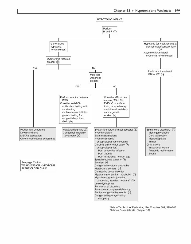
Chapter 53 u HypotoniaandWeakness 199
Prader-Willi syndromeDown syndromeMECP2 duplicationOther chromosomal syndromes
Prader-Willi syndromeDown syndromeMECP2 duplicationOther chromosomal syndromes
Myasthenia gravis 3 Congenital myotonic dystrophy 4
Myasthenia gravis 3 Congenital myotonic dystrophy 4
Systemic disorders/illness (sepsis) 6 HypothyroidismBrain malformationsHypoxic-ischemic encephalopathy/myelopathyCerebral palsy (other static 7 encephalopathies) Post congenital infection Post trauma Post intracranial hemorrhageSpinal muscular atrophy 8 Botulism 9 Congenital myotonic dystrophyMetabolic disorders 10 Connective tissue disorderMyopathy (congenital, metabolic) 11 Myasthenia gravis (juvenile, congenital, transient neonatal) 3LeukodystrophiesPerioxisomal disordersPyruvate carboxylase deficiencyBenign congenital hypotonia 12 Congenital hypomyelinating neuropathy
Systemic disorders/illness (sepsis) 6 HypothyroidismBrain malformationsHypoxic-ischemic encephalopathy/myelopathyCerebral palsy (other static 7 encephalopathies) Post congenital infection Post trauma Post intracranial hemorrhageSpinal muscular atrophy 8 Botulism 9 Congenital myotonic dystrophyMetabolic disorders 10 Connective tissue disorderMyopathy (congenital, metabolic) 11 Myasthenia gravis (juvenile, congenital, transient neonatal) 3LeukodystrophiesPerioxisomal disordersPyruvate carboxylase deficiencyBenign congenital hypotonia 12 Congenital hypomyelinating neuropathy
Spinal cord disorders 14 Meningomyelocele Cord transection Myelodysplasia TumorCNS lesions Intracranial lesions Anatomic malformation Stroke
Spinal cord disorders 14 Meningomyelocele Cord transection Myelodysplasia TumorCNS lesions Intracranial lesions Anatomic malformation Stroke
Maternalweaknesspresent
Maternalweaknesspresent
See page 53-5 forWEAKNESS OR HYPOTONIAIN THE OLDER CHILD
See page 53-5 forWEAKNESS OR HYPOTONIAIN THE OLDER CHILD
Perform spine ± headMRI or CT 13 Perform spine ± headMRI or CT 13
Hypotonia (or weakness) at a distinct motor/sensory level ORAsymmetric/unilateral hypotonia (or weakness)
Hypotonia (or weakness) at a distinct motor/sensory level ORAsymmetric/unilateral hypotonia (or weakness)
Generalizedhypotonia(or weakness)
Generalizedhypotonia(or weakness)
Perform infant ± maternal EMGConsider anti-ACh antibodies, testing with short-acting cholinesterase inhibitor, genetic testing for congenital myotonic dystrophy
Perform infant ± maternal EMGConsider anti-ACh antibodies, testing with short-acting cholinesterase inhibitor, genetic testing for congenital myotonic dystrophy
Consider MRI of head± spine, TSH, CK, EMG, C. botulinum toxin, muscle biopsy ± additional metabolicand/or genetic workup 5
Consider MRI of head± spine, TSH, CK, EMG, C. botulinum toxin, muscle biopsy ± additional metabolicand/or genetic workup 5
Perform H and P 1 Perform H and P 1
YES
YES
NO
NO
Dysmorphic featurespresent 2 Dysmorphic featurespresent 2
Nelson Textbook of Pediatrics, 19e. Chapters 584, 599–608Nelsons Essentials, 6e. Chapter 182
HYPOTONIC INFANTHYPOTONIC INFANT
200 Part VII u Neurology
7 Cerebral palsy (CP) is a static encephalopathy, a nonpro-gressive clinical disorder characterized by varying degrees
of mental and motor impairment. Rarely, hypotonia is promi-nent or persistent. In most cases hypotonia will progress to spasticity and dyskinetic movements. Commonly identified causes of CP include perinatal hypoxia or asphyxia, congenital infection and intracranial hemorrhage or trauma; many cases are likely due to undetected prenatal events.
8 In spinal muscular atrophies, degenerative loss of anterior horn cells in the spinal cord and brainstem motor neurons
occurs. Progressive weakness develops in variants presenting in infants; juvenile variants of this disorder present beyond in-fancy. Diagnosis is by EMG, DNA probes, and muscle biopsy.
9 Infantile botulism most commonly occurs in infants be-tween 2 weeks and 6 months of age. Source of the spores
carrying the toxin of Clostridium botulinum may be honey, corn syrup, soil or dust. Infants present with a descending flaccid paralysis. Cranial nerve symptoms are typically noted first (manifesting as poor suck, feeble cry, drooling); fever is charac-teristically absent. A recent history including poor feeding, constipation, weak cry and smile, hypotonia, ptosis, and my-driasis is common. Clinical diagnosis is key to early interven-tion; diagnosis is confirmed by recovery of the organism or toxin from stool, blood, or food sources. Older children can present with food-borne botulism due to ingestion of pre-formed toxin in poorly canned foods.
10 Metabolic disorders (particularly inborn errors of metabo-lism) usually appear in the neonatal period, although par-
tial or incomplete errors may not appear until later. A complaint of hypotonia combined with a history of recurrent bouts of leth-argy, vomiting, acidosis, and other neurologic findings should prompt appropriate metabolic screening laboratory studies. Some of the conditions to be considered include amino acid disorders, organic acidemia, urea cycle defects, fatty acid oxida-tion defects, adrenal insufficiency, and mitochondrial disorders.
11 Developmental disorders of muscle are referred to as con-genital myopathies; they are characterized by infantile hy-
potonia and weakness of variable severity and are generally nonprogressive. Serum CK values are typically normal; diagno-sis is usually by muscle biopsy. Metabolic myopathies include errors of glycogen and lipid metabolism as well as disorders of mitochondrial function.
12 Benign congenital hypotonia is a diagnosis of exclusion; it describes a nonprogressive hypotonia affecting infants and
children with no clear etiology, little or no developmental delay, and a generally good prognosis.
13 If acute intracranial hemorrhage is suspected (e.g., trau-matic birth, depressed skull fracture, other head trama), a
head CT is the most appropriate urgent imaging study.
14 Spinal cord disorders often present with hyperreflexia, clo-nus, a positive Babinski sign, and defined sensory loss in
the extremities. Hypotonia may be the prominent acute sign in infants. The diagnosis should be considered in infants with hy-potonia after a difficult delivery (particularly breech deliveries).
In older children disorders include traumatic transection, spinal cord tumor, transverse myelitis, and epidural spinal abscesses. Hypotonia is subsequently replaced by hypertonia.
15 Proximal leg weakness, wasting, and cramps are typically the initial manifestations of GM2-gangliosidosis, an auto-
somal recessive disorder of hexosaminidase A deficiency with several phenotypic variants (including Tay-Sachs disease).
16 Duchenne muscular dystrophy (DMD), an x-linked reces-sive disorder, is the most common type of muscular dys-
trophy. Diagnosis is usually in late infancy or early childhood when a child presents with the hyperlordotic posture and Gower sign, typically by age 3 years. A Trendelenburg gait, muscle atrophy, and pseudohypertrophy of the calves subse-quently develop. Toe walking and frequent falling are common; a history of delayed motor milestones may be noted retrospec-tively. Duchenne and Becker muscular dystrophies are both caused by a defect in the muscle protein dystrophin; Becker muscular dystrophy (BMD) has a later onset and less severe course. Genetic testing for the dystrophin gene defect is diag-nostic if the CK is elevated and the clinical picture is consistent with DMD. Muscle biopsy may be needed if the genetic testing is normal; it may also be useful in distinguishing between DMD and BMD.
17 Guillain-Barré syndrome is an acute demyelinating poly-neuropathy characterized by an ascending motor weak-
ness and areflexia; sensory and autonomic nerves may be af-fected. Respiratory compromise can occur if respiratory muscles are involved. The syndrome frequently follows an upper respi-ratory tract infection or Campylobacter diarrhea. Elevated CSF protein without pleocytosis is characteristic; EMG results are consistent with acute denervation of muscle.
18 The most common causes of progressive distal weakness are neuropathies, most of which are familial. Dysesthesias
(painful tingling and burning sensations) often accompany the weakness. Autonomic symptoms (e.g., orthostatic hypotension, gastrointestinal dysmotility, abnormal sweating) may be pres-ent, and deep tendon reflexes are usually markedly diminished relative to the degree of weakness. Diagnosis of these disorders is by nerve conduction velocity studies and EMG. A lumbar puncture and an evaluation of the CSF may be necessary to rule out certain suspected infectious causes. For example, nonpolio enteroviruses may cause poliomyelitis-like disease.
19 Several species of North American ticks carry a toxin that can cause a paralysis clinically similar to Guillain-Barré
syndrome. Tendon reflexes are usually diminished. Sensation is preserved, but burning or tingling may occur.
20 Scapulohumeral or scapuloperoneal syndromes have characteristics of both nerve and muscle disease; patients
present with proximal arm and distal leg weakness.
21 Transverse myelitis presents acutely with hypotonia and weakness. There is an identifiable motor-sensory level,
impaired bowel and bladder function, hyperreflexia, and an abnormal Babinski sign. In children younger than 3 years old, spinal cord dysfunction symptoms develop rapidly (hours to days); in older children the symptoms develop more gradually (days to weeks). Spinal MRI shows abnormal signal intensity of the involved level, and CSF examination shows a mild pleocy-tosis and elevated protein level (specifically myelin basic pro-tein and immunoglobulins).
chronic or other neurologic abnormalities exist, a metabolic investigation should also be done. If a genetic disorder is suspected, chromosome studies including microarray and con-sultation with a geneticist may be helpful.
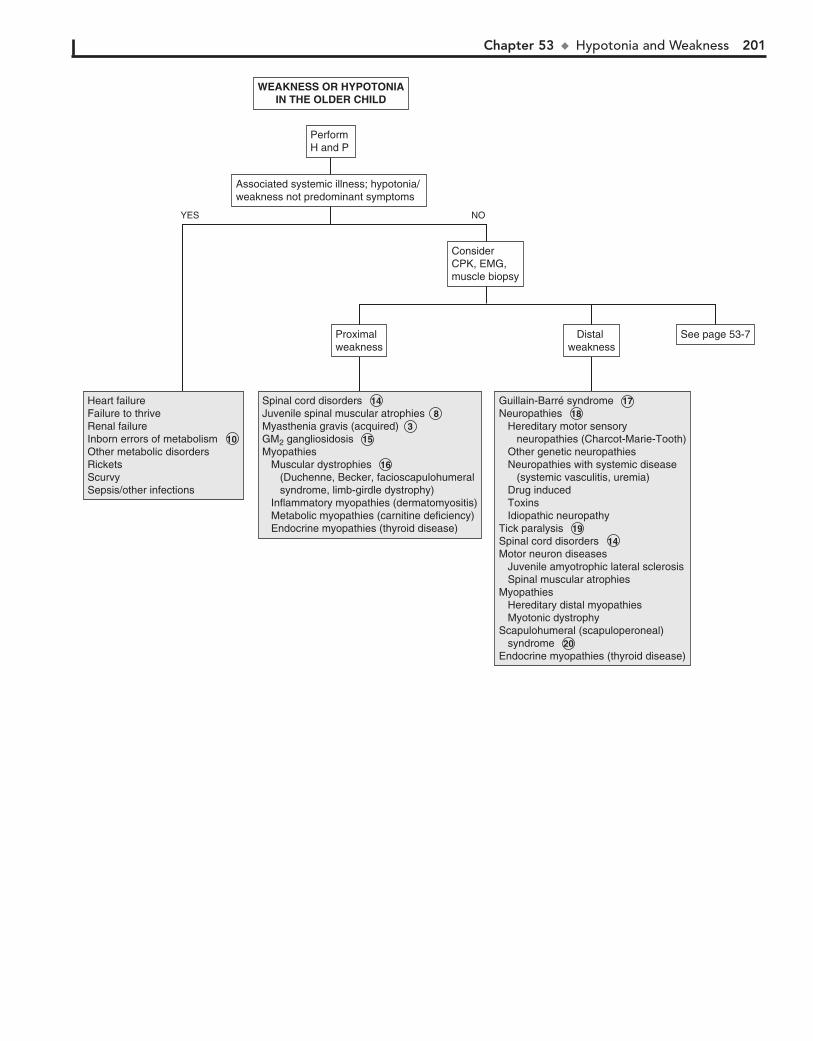
Chapter 53 u HypotoniaandWeakness 201
Heart failureFailure to thriveRenal failureInborn errors of metabolism 10 Other metabolic disordersRicketsScurvySepsis/other infections
Heart failureFailure to thriveRenal failureInborn errors of metabolism 10 Other metabolic disordersRicketsScurvySepsis/other infections
Spinal cord disorders 14 Juvenile spinal muscular atrophies 8 Myasthenia gravis (acquired) 3 GM2 gangliosidosis 15 Myopathies Muscular dystrophies 16 (Duchenne, Becker, facioscapulohumeral syndrome, limb-girdle dystrophy) Inflammatory myopathies (dermatomyositis) Metabolic myopathies (carnitine deficiency) Endocrine myopathies (thyroid disease)
Spinal cord disorders 14 Juvenile spinal muscular atrophies 8 Myasthenia gravis (acquired) 3 GM2 gangliosidosis 15 Myopathies Muscular dystrophies 16 (Duchenne, Becker, facioscapulohumeral syndrome, limb-girdle dystrophy) Inflammatory myopathies (dermatomyositis) Metabolic myopathies (carnitine deficiency) Endocrine myopathies (thyroid disease)
Guillain-Barré syndrome 17 Neuropathies 18 Hereditary motor sensory neuropathies (Charcot-Marie-Tooth) Other genetic neuropathies Neuropathies with systemic disease (systemic vasculitis, uremia) Drug induced Toxins Idiopathic neuropathyTick paralysis 19 Spinal cord disorders 14 Motor neuron diseases Juvenile amyotrophic lateral sclerosis Spinal muscular atrophiesMyopathies Hereditary distal myopathies Myotonic dystrophyScapulohumeral (scapuloperoneal) syndrome 20 Endocrine myopathies (thyroid disease)
Guillain-Barré syndrome 17 Neuropathies 18 Hereditary motor sensory neuropathies (Charcot-Marie-Tooth) Other genetic neuropathies Neuropathies with systemic disease (systemic vasculitis, uremia) Drug induced Toxins Idiopathic neuropathyTick paralysis 19 Spinal cord disorders 14 Motor neuron diseases Juvenile amyotrophic lateral sclerosis Spinal muscular atrophiesMyopathies Hereditary distal myopathies Myotonic dystrophyScapulohumeral (scapuloperoneal) syndrome 20 Endocrine myopathies (thyroid disease)
Proximal weaknessProximal weakness
ConsiderCPK, EMG,muscle biopsy
ConsiderCPK, EMG,muscle biopsy
Distal weakness
Distal weakness
See page 53-7See page 53-7
Associated systemic illness; hypotonia/ weakness not predominant symptomsAssociated systemic illness; hypotonia/ weakness not predominant symptoms
Perform H and PPerform H and P
YES NO
WEAKNESS OR HYPOTONIAIN THE OLDER CHILD
WEAKNESS OR HYPOTONIAIN THE OLDER CHILD

202 Part VII u Neurology
22 In periodic paralysis, patients experience episodes of severe weakness, often followed by partial or complete
paralysis. Episodes are usually related to hypokalemia or hyper-kalemia and may be primary (genetically transmitted) or secondary due to endocrine, renal, or gastrointestinal causes of hypokalemia or hyperkalemia.
BibliographyPeredo DE, Hannibal MC: The floppy infant: Evaluation of hypotonia, Pediatr
Rev 30:e66–76, 2009.Piña-Garza JE: The hypotonic infant. In Piña-Garza JE, editor: Fenichel’s
clinical pediatric neurology, ed 7, Philadelphia, 2013, Saunders/Elsevier, pp 147–169.
Piña-Garza JE: Flaccid limb weakness in childhood. In Piña-Garza JE, editor: Fenichel’s clinical pediatric neurology, ed 7, Philadelphia, 2013, Saunders/Elsevier, pp 170–194.
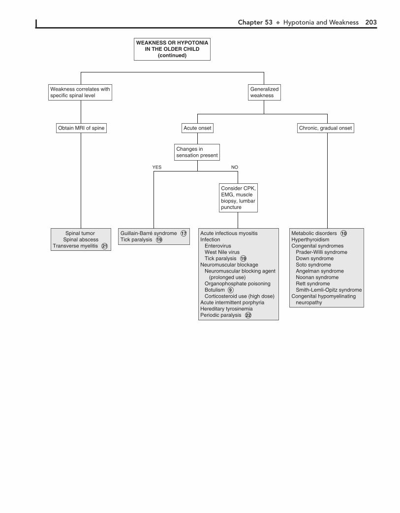
Chapter 53 u HypotoniaandWeakness 203
Spinal tumor Spinal abscess
Transverse myelitis 21
Spinal tumor Spinal abscess
Transverse myelitis 21
Guillain-Barré syndrome 17 Tick paralysis 19 Guillain-Barré syndrome 17 Tick paralysis 19
Acute infectious myositisInfection Enterovirus West Nile virus Tick paralysis 19 Neuromuscular blockage Neuromuscular blocking agent (prolonged use) Organophosphate poisoning Botulism 9 Corticosteroid use (high dose)Acute intermittent porphyriaHereditary tyrosinemiaPeriodic paralysis 22
Acute infectious myositisInfection Enterovirus West Nile virus Tick paralysis 19 Neuromuscular blockage Neuromuscular blocking agent (prolonged use) Organophosphate poisoning Botulism 9 Corticosteroid use (high dose)Acute intermittent porphyriaHereditary tyrosinemiaPeriodic paralysis 22
Metabolic disorders 10 HyperthyroidismCongenital syndromes Prader-Willi syndrome Down syndrome Soto syndrome Angelman syndrome Noonan syndrome Rett syndrome Smith-Lemli-Opitz syndromeCongenital hypomyelinating neuropathy
Metabolic disorders 10 HyperthyroidismCongenital syndromes Prader-Willi syndrome Down syndrome Soto syndrome Angelman syndrome Noonan syndrome Rett syndrome Smith-Lemli-Opitz syndromeCongenital hypomyelinating neuropathy
Consider CPK,EMG, musclebiopsy, lumbarpuncture
Consider CPK,EMG, musclebiopsy, lumbarpuncture
Changes insensation presentChanges insensation present
Acute onsetAcute onsetObtain MRI of spineObtain MRI of spine
Weakness correlates withspecific spinal levelWeakness correlates withspecific spinal level
GeneralizedweaknessGeneralizedweakness
Chronic, gradual onsetChronic, gradual onset
YES NO
WEAKNESS OR HYPOTONIAIN THE OLDER CHILD
(continued)
WEAKNESS OR HYPOTONIAIN THE OLDER CHILD
(continued)
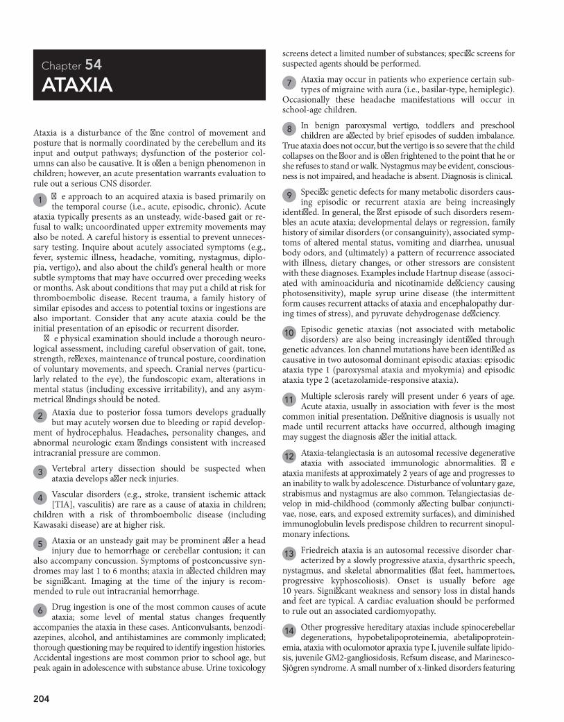
204
Chapter 54
ATAXIA
screens detect a limited number of substances; specific screens for suspected agents should be performed.
7 Ataxia may occur in patients who experience certain sub-types of migraine with aura (i.e., basilar-type, hemiplegic).
Occasionally these headache manifestations will occur in school-age children.
8 In benign paroxysmal vertigo, toddlers and preschool children are affected by brief episodes of sudden imbalance.
True ataxia does not occur, but the vertigo is so severe that the child collapses on the floor and is often frightened to the point that he or she refuses to stand or walk. Nystagmus may be evident, conscious-ness is not impaired, and headache is absent. Diagnosis is clinical.
9 Specific genetic defects for many metabolic disorders caus-ing episodic or recurrent ataxia are being increasingly
identified. In general, the first episode of such disorders resem-bles an acute ataxia; developmental delays or regression, family history of similar disorders (or consanguinity), associated symp-toms of altered mental status, vomiting and diarrhea, unusual body odors, and (ultimately) a pattern of recurrence associated with illness, dietary changes, or other stressors are consistent with these diagnoses. Examples include Hartnup disease (associ-ated with aminoaciduria and nicotinamide deficiency causing photosensitivity), maple syrup urine disease (the intermittent form causes recurrent attacks of ataxia and encephalopathy dur-ing times of stress), and pyruvate dehydrogenase deficiency.
10 Episodic genetic ataxias (not associated with metabolic disorders) are also being increasingly identified through
genetic advances. Ion channel mutations have been identified as causative in two autosomal dominant episodic ataxias: episodic ataxia type 1 (paroxysmal ataxia and myokymia) and episodic ataxia type 2 (acetazolamide-responsive ataxia).
11 Multiple sclerosis rarely will present under 6 years of age. Acute ataxia, usually in association with fever is the most
common initial presentation. Definitive diagnosis is usually not made until recurrent attacks have occurred, although imaging may suggest the diagnosis after the initial attack.
12 Ataxia-telangiectasia is an autosomal recessive degenerative ataxia with associated immunologic abnormalities. The
ataxia manifests at approximately 2 years of age and progresses to an inability to walk by adolescence. Disturbance of voluntary gaze, strabismus and nystagmus are also common. Telangiectasias de-velop in mid-childhood (commonly affecting bulbar conjuncti-vae, nose, ears, and exposed extremity surfaces), and diminished immunoglobulin levels predispose children to recurrent sinopul-monary infections.
13 Friedreich ataxia is an autosomal recessive disorder char-acterized by a slowly progressive ataxia, dysarthric speech,
nystagmus, and skeletal abnormalities (flat feet, hammertoes, progressive kyphoscoliosis). Onset is usually before age 10 years. Significant weakness and sensory loss in distal hands and feet are typical. A cardiac evaluation should be performed to rule out an associated cardiomyopathy.
14 Other progressive hereditary ataxias include spinocerebellar degenerations, hypobetalipoproteinemia, abetalipoprotein-
emia, ataxia with oculomotor apraxia type I, juvenile sulfate lipido-sis, juvenile GM2-gangliosidosis, Refsum disease, and Marinesco-Sjögren syndrome. A small number of x-linked disorders featuring
Ataxia is a disturbance of the fine control of movement and posture that is normally coordinated by the cerebellum and its input and output pathways; dysfunction of the posterior col-umns can also be causative. It is often a benign phenomenon in children; however, an acute presentation warrants evaluation to rule out a serious CNS disorder.
1 The approach to an acquired ataxia is based primarily on the temporal course (i.e., acute, episodic, chronic). Acute
ataxia typically presents as an unsteady, wide-based gait or re-fusal to walk; uncoordinated upper extremity movements may also be noted. A careful history is essential to prevent unneces-sary testing. Inquire about acutely associated symptoms (e.g., fever, systemic illness, headache, vomiting, nystagmus, diplo-pia, vertigo), and also about the child’s general health or more subtle symptoms that may have occurred over preceding weeks or months. Ask about conditions that may put a child at risk for thromboembolic disease. Recent trauma, a family history of similar episodes and access to potential toxins or ingestions are also important. Consider that any acute ataxia could be the initial presentation of an episodic or recurrent disorder.
The physical examination should include a thorough neuro-logical assessment, including careful observation of gait, tone, strength, reflexes, maintenance of truncal posture, coordination of voluntary movements, and speech. Cranial nerves (particu-larly related to the eye), the fundoscopic exam, alterations in mental status (including excessive irritability), and any asym-metrical findings should be noted.
2 Ataxia due to posterior fossa tumors develops gradually but may acutely worsen due to bleeding or rapid develop-
ment of hydrocephalus. Headaches, personality changes, and abnormal neurologic exam findings consistent with increased intracranial pressure are common.
3 Vertebral artery dissection should be suspected when ataxia develops after neck injuries.
4 Vascular disorders (e.g., stroke, transient ischemic attack [TIA], vasculitis) are rare as a cause of ataxia in children;
children with a risk of thromboembolic disease (including Kawasaki disease) are at higher risk.
5 Ataxia or an unsteady gait may be prominent after a head injury due to hemorrhage or cerebellar contusion; it can
also accompany concussion. Symptoms of postconcussive syn-dromes may last 1 to 6 months; ataxia in affected children may be significant. Imaging at the time of the injury is recom-mended to rule out intracranial hemorrhage.
6 Drug ingestion is one of the most common causes of acute ataxia; some level of mental status changes frequently
accompanies the ataxia in these cases. Anticonvulsants, benzodi-azepines, alcohol, and antihistamines are commonly implicated; thorough questioning may be required to identify ingestion histories. Accidental ingestions are most common prior to school age, but peak again in adolescence with substance abuse. Urine toxicology

Chapter 54 u Ataxia 205
206 Part VII u Neurology
18 The Miller-Fisher syndrome is considered a variant of Guillain-Barré syndrome, although it is also suspected to be a variant
of brainstem encephalitis. The cranial nerves are affected by the immune-mediated demyelination in this condition. Ataxia, oph-thalmoplegia, and areflexia occur after an infectious illness (par-ticularly Campylobacter gastroenteritis). Vertical gaze is charac-teristically affected while horizontal gaze is typically preserved. The areflexia aids in distinguishing this from acute postinfectious ataxia. CSF examination results vary based on the timing relative to symptom onset; an elevated protein without an increased cell count is characteristic later in the course.
19 Ataxia and cranial nerve dysfunction characterize brain-stem encephalitis; mental status changes, hemiparesis and
respiratory irregularities can also occur. Epstein-Barr virus, Listeria, and enterovirus (type 71) have been identified as eti-ologies. MRI will show increased signal intensity in the brain-stem. Evaluation of the CSF shows pleocytosis, normal glucose levels, and normal or slightly elevated protein levels. Brainstem auditory evoked response testing indicates abnormalities within the brainstem; EEGs will be normal. Neuroimaging will yield nonspecific findings but will rule out other etiologies.
20 Opsoclonus-myoclonus syndrome is characterized by opsoclonus, myoclonus, ataxia, and encephalopathy. It may
occur as a postinfectious entity, but is clearly recognized as a paraneoplastic cerebellar syndrome and should prompt an inves-tigation for neuroblastoma or (rarely) other neoplasms.
21 Acute episodic ataxia (”pseudoataxia”) may rarely be the only clinical manifestation of nonconvulsive seizure activity.
EEG is diagnostic.
ataxia are also recognized. The majority of these conditions will be diagnosed clinically or via specific blood or genetic tests; neuroim-aging is usually not contributory. Any of the acute recurrent ataxias (e.g., Hartnup disease, maple syrup urine disease) may eventually become progressive without return to baseline between attacks.
15 Labryinthitis can complicate acute otitis media, sinusitis, mastoiditis or bacterial meningitis. Severe vertigo, vomiting,
nystagmus and hearing loss may also be present.
16 Acute cerebellar ataxia (acute postinfectious cerebellitis) is the most common acute ataxia in children (approximately
40% of all cases). The etiology is generally a postinfectious cer-ebellar demyelination, which is presumed to be the result of an autoimmune reaction following infection. (Immunization has been suspected of inciting this condition, but evidence does not support this theory.) A preceding illness (5 to 21 days) is identi-fied in most cases; varicella is no longer the most commonly identified infectious trigger due to universal varicella immuniza-tion. The onset is acute ataxia and truncal instability with maxi-mal severity at the onset; occasionally a child is unable to walk at all at the onset. Head titubation (bobbing), tremor, dysmetria, and ocular abnormalities may also occur, but mental status is always normal and reflexes are preserved. The diagnosis is one of exclusion. A drug screen is probably the most appropriate test to be done acutely to rule out unsuspected ingestions; other test-ing serves primarily to rule out other etiologies. As improve-ment should begin within a few days in acute cerebellar ataxia, imaging should be performed if rapid improvement of the ataxia does not occur. If a lumbar puncture is performed, findings are usually normal or may show mild pleocytosis with an increased CSF protein level. Most children will fully recover.
17 Acute disseminated encephalomyelitis (acute postinfectious demyelinating encephalomyelitis) is another postinfectious,
immune-mediated condition with a more severe presentation than acute cerebellar ataxia, including altered mental status, seizures (which may progress to status epilepticus), and multifocal neuro-logic defects. MRI will show foci of demyelination and an LP, if performed, will reveal normal findings or may show mild pleocyto-sis with an increased CSF protein level. Repeated episodes may suggest multiple sclerosis.
BibliographyFriday JH: Ataxia. In Fleisher G, Ludwig S, editors: Textbook of pediatric
emergency medicine, ed 6, Philadelphia, 2010, Lippincott Williams & Wilkins, pp 164–167.
Piña-Garza JE, Ataxia: In Piña-Garza JE, editor: Fenichel’s clinical pediatric neurology, ed 7, Philadelphia, 2013, Saunders/Elsevier, pp 215–235.
Ryan MM, Engle EC: Topical review: Acute ataxia in childhood, J Child Neurol 18:309–316, 2003.
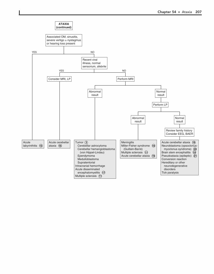
Chapter 54 u Ataxia 207
Acutelabyrinthitis 15 Acutelabyrinthitis 15
Acute cerebellarataxia 16 Acute cerebellarataxia 16
Tumor 2 Cerebellar astrocytoma Cerebellar hemangioblastoma (von Hippel-Lindau) Ependymoma Medulloblastoma SupratentorialIntracranial hemorrhageAcute disseminated encephalomyelitis 17 Multiple sclerosis 11
Tumor 2 Cerebellar astrocytoma Cerebellar hemangioblastoma (von Hippel-Lindau) Ependymoma Medulloblastoma SupratentorialIntracranial hemorrhageAcute disseminated encephalomyelitis 17 Multiple sclerosis 11
MeningitisMiller-Fisher syndrome 18 (Guillain-Barré)Multiple sclerosis 11 Acute cerebellar ataxia 16
MeningitisMiller-Fisher syndrome 18 (Guillain-Barré)Multiple sclerosis 11 Acute cerebellar ataxia 16
Acute cerebellar ataxia 16 Neuroblastoma (opsoclonus- myoclonus syndrome) 20 Brain stem encephalitis 19 Pseudoataxia (epileptic) 21 Conversion reactionHereditary or other neurodegenerative disordersTick paralysis
Acute cerebellar ataxia 16 Neuroblastoma (opsoclonus- myoclonus syndrome) 20 Brain stem encephalitis 19 Pseudoataxia (epileptic) 21 Conversion reactionHereditary or other neurodegenerative disordersTick paralysis
Review family historyConsider EEG, BAERReview family historyConsider EEG, BAER
Abnormalresult
Abnormalresult
Abnormalresult
Abnormalresult
Perform MRIPerform MRIConsider MRI, LPConsider MRI, LP
Recent viralillness, normalsensorium, afebrile
Recent viralillness, normalsensorium, afebrile
Perform LPPerform LP
Normalresult
Normalresult
Normalresult
Normalresult
YES
YES
NO
NO
ATAXIA(continued)
ATAXIA(continued)
Associated OM, sinusitis,severe vertigo ± nystagmusor hearing loss present
Associated OM, sinusitis,severe vertigo ± nystagmusor hearing loss present

208
3 MRI is helpful in the diagnosis of acute disseminated en-cephalomyelitis (ADEM), a condition in which symptoms
related to the encephalopathy predominate (e.g., confusion, ir-ritability, occasionally coma). MRI typically shows white matter lesions. Autoimmune encephalitis is a much more common cause of encephalitis than previously recognized; its presenta-tion is variable and can include behavioral changes, seizures, and sleep disturbances. The presence of specific antibodies and MRI findings are helpful in the diagnosis.
4 Rare infectious causes of encephalopathy include cat scratch disease, Rocky Mountain spotted fever, Lyme
disease, toxic shock syndrome, and measles.
5 A history of head trauma should prompt an evaluation for an intracranial injury. Other worrisome signs and symp-
toms include a bulging fontanel, retinal hemorrhages, focal neurologic findings, and signs of brain stem dysfunction (e.g., abnormal respiratory pattern and abnormal corneal, oculoce-phalic, or oculovestibular reflexes). Signs of increased intracra-nial pressure include a unilateral fixed or dilated pupil, ptosis, Cushing triad (i.e., hypertension, bradycardia, periodic breath-ing or apnea), cranial nerve VI palsy, papilledema, and a history of vomiting, headache, or ataxia.
6 A head CT is a good initial study and is usually readily available; it will reveal hemorrhages, most masses, and
hydrocephalus. Subsequent MRI may be needed to identify infratentorial masses, cerebral edema, infarction, or more subtle CNS findings.
7 Abusive head trauma (shaken baby syndrome) usually occurs in children younger than 12 months. Children
often have no external signs of trauma, although retinal hemorrhages and a bulging fontanel may be evident on examination. Neuroimaging reveals subdural hematomas. A complete evaluation for other injuries is indicated when abuse is suspected.
8 An intravenous dose of naloxone is recommended to potentially treat this rapidly reversible cause of altered
mental status.
9 Overdoses and poisonings are common in children. A sud-den onset of altered mental status, seizures, and vomiting,
especially with a preceding period of confusion or delirium, should raise suspicion for a possible ingestion. Toxicology screens may be helpful, although they may be of limited value because they are not standardized. If certain agents are suspected, tests for them should be specifically requested. Immunosuppressive agents (including steroids) may cause altered mental status. Prescription drugs commonly reported in overdose cases are benzodiazepines, salicylates, acetaminophen, barbiturates, and tricyclic antidepres-sants. Poisons (from household products), alcohol, and illegal substance abuse should also always be considered. Many toxins cause a characteristic toxidrome of signs and symptoms (includ-ing EKG abnormalities) that may aid in their identification. Level of consciousness, pupillary examination, and vital signs are the most helpful components in identifying a toxidrome.
10 Inborn errors of metabolism usually occur in the neonate with vomiting, lethargy, or seizures, although partial or
incomplete errors may not occur until later childhood or adoles-cence. Initial labs may suggest a metabolic disorder (e.g., acido-sis, hypoglycemia), but additional evaluation should be per-formed if clinically suspicious of an inborn error of metabolism.
An alteration in consciousness (awareness of self and environ-ment) may range from delirium (e.g., irritability, confusion, agitation) to coma. A delirious state is often characterized by alternating periods of lucidity and frequently progresses to lethargy or coma.
1 A child presenting with any alteration in consciousness must be emergently stabilized before a search is started for
the cause. The patient should then be evaluated to rule out a potentially life-threatening intracranial process that requires urgent treatment. The Glasgow Coma Scale (GCS) is the most widely used tool for assessing and monitoring changes in men-tal status. The GCS utilizes eye opening, motor responses, and verbal responses and is a more objective means of describing altered levels of consciousness when compared with less spe-cific terms such as stuporous, obtunded, or lethargic. Any focal-ity or asymmetry of the neurologic examination could suggest a problem localized to one cerebral hemisphere.
The history should include a recent review of systems and a history of trauma, medications, and possible toxic ingestions.
Certain components of the physical examination may suggest an underlying systemic disorder. For example, certain skin find-ings may suggest neurocutaneous disorders, Addison disease, anemia, carbon monoxide poisoning, or infectious conditions; needle track marks and signs of trauma should also be noted. Hepatomegaly can suggest hepatic failure (Reye syndrome) or heart failure. Fractured extremities raise the possibility of a fat embolism. Toxidromes (such as apnea and pinpoint pupils asso-ciated with opiates) help identify a possible ingestion.
A patient’s breathing pattern may aid in the diagnosis of altered mental status. Hyperventilation occurs with toxic-metabolic encephalopathies, increased intracranial pressure, and meta-bolic acidosis. Hypoventilation occurs with many drug inges-tions. Other breathing patterns (e.g., Cheyne-Stokes, apneustic breathing) may indicate specific sites of CNS dysfunction. The odor of the breath may be helpful. Many disorders and certain ingestions are accompanied by a characteristic odor.
Serum glucose and urine toxicology screens are recom-mended as first line lab evaluations; subsequent lab work can be obtained based on a suspected cause. Tests to be considered are a CBC, electrolytes, blood urea nitrogen, creatinine, calcium, magnesium, phosphorus, liver function tests, and an arterial blood gas analysis. Blood cultures, thyroid studies, serum am-monia levels, and serum osmolality may be helpful; an osmolal gap and anion gap should be calculated, especially if an inges-tion is suspected. An EKG may reveal arrhythmias or character-istics of certain toxidromes.
2 A lumbar puncture is contraindicated in children if they have any of the following: (1) cardiorespiratory compro-
mise, (2) focal neurologic findings or other suspicion of mass lesion, (3) signs of increased intracranial pressure other than a bulging fontanel, (4) skin or soft tissue infection overlying the site of the lumbar puncture or (5) thrombocytopenia.
Chapter 55
ALTERED MENTAL STATUS
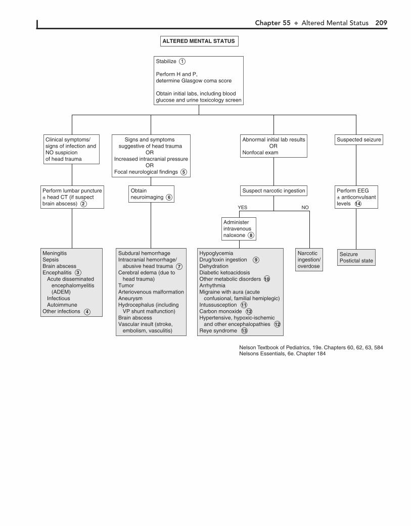
Chapter 55 u AlteredMentalStatus 209
MeningitisSepsisBrain abscessEncephalitis Acute disseminated encephalomyelitis (ADEM) Infectious AutoimmuneOther infections
Subdural hemorrhageIntracranial hemorrhage/ Abusive head traumaCerebral edema (due to head trauma)TumorArteriovenous malformationAneurysmHydrocephalus (including VP shunt malfunction)Brain abscessVascular insult (stroke, embolism, vasculitis)
Subdural hemorrhageIntracranial hemorrhage/ abusive head traumaCerebral edema (due to head trauma)TumorArteriovenous malformationAneurysmHydrocephalus (including VP shunt malfunction)Brain abscessVascular insult (stroke, embolism, vasculitis)
Narcoticingestion/overdose
Narcoticingestion/overdose
SeizurePostictal stateSeizurePostictal state
Administerintravenousnaloxone 7
Administerintravenousnaloxone 8
Perform EEG±anticonvulsantlevels 13
Perform EEG± anticonvulsantlevels 14
Suspect narcotic ingestionSuspect narcotic ingestion
Abnormal initial lab results ORNonfocal exam
Abnormal initial lab results ORNonfocal exam
Stabilize 1
Perform H and P,determine Glasgow coma score
Obtain initial labs, including blood glucose and urine toxicology screen
Stabilize 1
Perform H and P,determine Glasgow coma score
Obtain initial labs, including blood glucose and urine toxicology screen
Obtainneuroimaging 6 Obtainneuroimaging 6
Perform lumbar puncture±head CT (if suspect brain abscess) 2
Perform lumbar puncture± head CT (if suspect brain abscess) 2
Suspected seizureSuspected seizure Signs and symptoms suggestive of head trauma ORIncreased intracranial pressure ORFocal neurological findings 5
Signs and symptoms suggestive of head trauma ORIncreased intracranial pressure ORFocal neurological findings 5
Clinical symptoms/ signs of infection and NO suspicion of head trauma
Clinical symptoms/ signs of infection and NO suspicion of head trauma
4
3
HypoglycemiaDrug/ Toxin ingestion 8 DehydrationDiabetic ketoacidosisOther metabolic disorders 9 ArrhythmiaMigraine with aura (acute confusional, familial hemiplegic)Intussusception 10 Carbon monoxide 11 Hypertensive, hypoxic-ischemic and other encephalopathies 11 Reye syndrome 12
HypoglycemiaDrug/toxin ingestion 9 DehydrationDiabetic ketoacidosisOther metabolic disorders 10ArrhythmiaMigraine with aura (acute confusional, familial hemiplegic)Intussusception 11 Carbon monoxide 12 Hypertensive, hypoxic-ischemic and other encephalopathies 12 Reye syndrome 13
ALTERED MENTAL STATUS ALTERED MENTAL STATUS
NOYES
Nelson Textbook of Pediatrics, 19e. Chapters 60, 62, 63, 584Nelsons Essentials, 6e. Chapter 184
7
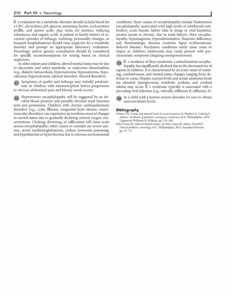
210 Part VII u Neurology
The evaluation for a metabolic disorder should include blood for a CBC, electrolytes, pH, glucose, ammonia, lactate, acylcarnitine profile, and amino acids, plus urine for ketones, reducing substances, and organic acids. A patient or family history of re-current episodes of lethargy, vomiting, personality changes, or frequent hospitalizations should raise suspicion for a metabolic disorder and prompt an appropriate laboratory evaluation. Neurologic and/or genetic consultation should be considered for specific recommendations for testing based on clinical suspicions.
conditions. Rarer causes of encephalopathy include Hashimoto’s encephalopathy (associated with high levels of antithyroid anti-bodies), acute hepatic failure (due to drugs or viral hepatitis), uremia (acute or chronic, due to renal failure), burn encepha-lopathy, hypomagnesia, hyperalimentation, thiamine deficiency, and rheumatologic diseases (systemic lupus erythematosus, Behcet’s disease). Psychiatric conditions rarely cause coma or stupor in children; adolescents may rarely present with psy-chosomatic symptoms (feigning unresponsiveness).
13 The incidence of Reye syndrome, a mitochondrial encepha-lopathy, has significantly declined due to the decreased use of
aspirin in children. It is characterized by an acute onset of vomit-ing, combativeness, and mental status changes ranging from de-lirium to coma. Hepatic enzyme levels and serum ammonia levels are elevated; hypoglycemia, metabolic acidosis, and cerebral edema may occur. The syndrome typically is associated with a preceding viral infection (e.g., varicella, influenza B, influenza A).
14 In a child with a known seizure disorder, be sure to obtain anticonvulsant levels.
BibliographyNelson DS: Coma and altered level of consciousness. In Fleisher G, Ludwig S,
editors: Textbook of pediatric emergency medicine, ed 6, Philadelphia, 2010, Lippincott Williams & Wilkins, pp 176–186.
Piña-Garza JE: Altered mental status. In Piña-Garza JE, editor: Fenichel’s clinical pediatric neurology, ed 7, Philadelphia, 2013, Saunders/Elsevier, pp. 47–75.
In older infants and children, altered mental status may be due to electrolyte and other metabolic or endocrine abnormalities (e.g., diabetic ketoacidosis, hypernatremia, hyponatremia, hypo-calcemia, hypercalcemia, adrenal disorders, thyroid disorders).
11 Symptoms of apathy and lethargy may initially predomi-nate in children with intussusception before progression
to obvious abdominal pain and bloody stools occurs.
12 Hypertensive encephalopathy will be suggested by an ele-vated blood pressure and possibly elevated renal function
tests and proteinuria. Children with chronic cardiopulmonary disorders (e.g., cystic fibrosis, congenital heart disease, neuro-muscular disorders) can experience an insidious onset of changes in mental status due to gradually declining arterial oxygen con-centrations. Choking, drowning, or suffocation will cause acute anoxia encephalopathy; other causes to consider are severe ane-mia, severe methemoglobinemia, carbon monoxide poisoning, and hypothermia or hyperthermia due to extreme environmental

Chapter 55 u AlteredMentalStatus 211
MeningitisSepsisBrain abscessEncephalitis Acute disseminated encephalomyelitis (ADEM) Infectious AutoimmuneOther infections
Subdural hemorrhageIntracranial hemorrhage/ Abusive head traumaCerebral edema (due to head trauma)TumorArteriovenous malformationAneurysmHydrocephalus (including VP shunt malfunction)Brain abscessVascular insult (stroke, embolism, vasculitis)
Subdural hemorrhageIntracranial hemorrhage/ abusive head traumaCerebral edema (due to head trauma)TumorArteriovenous malformationAneurysmHydrocephalus (including VP shunt malfunction)Brain abscessVascular insult (stroke, embolism, vasculitis)
Narcoticingestion/overdose
Narcoticingestion/overdose
SeizurePostictal stateSeizurePostictal state
Administerintravenousnaloxone 7
Administerintravenousnaloxone 8
Perform EEG±anticonvulsantlevels 13
Perform EEG± anticonvulsantlevels 14
Suspect narcotic ingestionSuspect narcotic ingestion
Abnormal initial lab results ORNonfocal exam
Abnormal initial lab results ORNonfocal exam
Stabilize 1
Perform H and P,determine Glasgow coma score
Obtain initial labs, including blood glucose and urine toxicology screen
Stabilize 1
Perform H and P,determine Glasgow coma score
Obtain initial labs, including blood glucose and urine toxicology screen
Obtainneuroimaging 6 Obtainneuroimaging 6
Perform lumbar puncture±head CT (if suspect brain abscess) 2
Perform lumbar puncture± head CT (if suspect brain abscess) 2
Suspected seizureSuspected seizure Signs and symptoms suggestive of head trauma ORIncreased intracranial pressure ORFocal neurological findings 5
Signs and symptoms suggestive of head trauma ORIncreased intracranial pressure ORFocal neurological findings 5
Clinical symptoms/ signs of infection and NO suspicion of head trauma
Clinical symptoms/ signs of infection and NO suspicion of head trauma
4
3
HypoglycemiaDrug/ Toxin ingestion 8 DehydrationDiabetic ketoacidosisOther metabolic disorders 9 ArrhythmiaMigraine with aura (acute confusional, familial hemiplegic)Intussusception 10 Carbon monoxide 11 Hypertensive, hypoxic-ischemic and other encephalopathies 11 Reye syndrome 12
HypoglycemiaDrug/toxin ingestion 9 DehydrationDiabetic ketoacidosisOther metabolic disorders 10ArrhythmiaMigraine with aura (acute confusional, familial hemiplegic)Intussusception 11 Carbon monoxide 12 Hypertensive, hypoxic-ischemic and other encephalopathies 12 Reye syndrome 13
ALTERED MENTAL STATUS ALTERED MENTAL STATUS
NOYES
Nelson Textbook of Pediatrics, 19e. Chapters 60, 62, 63, 584Nelsons Essentials, 6e. Chapter 184
7
212
Chapter 56
HEARING LOSS
24 months and (7) not following a one-step command by 13 to 15 months.
2 Mild conductive hearing loss is common with otitis media and normally improves with the resolution of the effusion.
Small tympanic membrane perforations have little effect on hearing, but large perforations may.
3 Tympanometry provides information about tympanic membrane compliance and middle ear pressure. It is most
helpful in identifying middle ear effusions and perforations of the tympanic membrane (TM). Before age 4 months, the exces-sive compliance of the ear canal limits the usefulness of the test.
Newborn screening programs rely on otoacoustic emissions (OAE) testing and auditory brainstem evoked response (ABR) tests to evaluate infant hearing. The advantage of ABR testing over OAE is its potential to identify an auditory neuropathy. Once a child can cooperate, pure tone audiometry with bone and air conduction results is recommended; most children can be reliably tested by this method by age 4 years. Referral for testing via other methods (depending on the age of the child) may be indicated if a child is too young to complete pure tone audiometry or if they are unable to cooperate with the testing.
4 A temporary shift in hearing threshold after exposure to potentially injurious sounds can precede permanent noise-
induced hearing loss (NIHL). NIHL has been attributed to high levels of continuous noise (e.g., music, recreational vehicles, power tools) and high intensity sounds of short duration (e.g., gunfire, firecrackers).
5 Referral to a multidisciplinary center is ideal to provide evaluation and treatment by audiology, otolaryngology,
and speech pathology. Genetics consultation may be helpful since approximately half of sensorineural hearing loss cases are associated with a genetic etiology. Genetics can aid in providing a specific diagnosis, prognostic factors, and counseling on as-sociated risks and conditions.
6 Genetics are estimated to be a factor in approximately 50% of cases of SNHL; two thirds of the cases are not associated
with any syndrome. Genetic counseling is becoming an increas-ingly important part of the evaluation of hearing loss as ad-vances in genetics are identifying mitochondrial disorders and mutations associated with an increased susceptibility to deaf-ness. Approximately 80% of cases of SNHL are inherited as au-tosomal recessive traits. Hearing impairment associated with some genetic disorders may not develop until later in childhood.
7 Effects of ototoxic drugs may not appear for up to 6 months after exposure to the drug. Aminoglycosides,
loop diuretics, and chemotherapy agents (especially cisplatin) are the most common offenders. Quinine, lead ,and arsenic have also been identified as causes of hearing loss.
8 Both conductive and sensorineural hearing loss have been reported in children who experience head trauma due to
temporal bone fractures or inner ear concussion; spontaneous resolution usually occurs.
9 Hearing loss due to congenital cytomegalovirus (CMV), toxoplasmosis, and syphilis may not appear until months to
Hearing loss can be conductive or sensorineural (or mixed) due to problems along the peripheral auditory pathway (i.e., external ear, middle ear, inner ear, and the auditory nerve). Conductive hearing loss is usually due to problems in the external and middle ear; sensorineural hearing loss (SNHL) is usually due to inner ear and auditory nerve problems. Screening of all new-born infants has been recommended by the U.S. Preventive Task Force since 2001 because early detection of and intervention for hearing loss are clearly beneficial to language development, and there has been minimal risk associated with the screening and treatment for the condition. Many cases of hearing loss, how-ever, are progressive and may not be detected at birth. Children with acquired or late-onset hearing loss may present with a more subtle problem, such as poor school performance. Sometimes children with effusion or eustachian tube dysfunction present with a complaint of hearing loss. The role of the primary care practitioner is to identify the hearing loss and make appropriate referrals for comprehensive evaluation and treatment. Identify-ing hearing loss as early as possible is critical to minimizing the adverse effects on speech and language and school performance.
1 Perinatal risk factors for hearing loss include congenital infections, craniofacial abnormalities, birthweight ,1500
grams, hyperbilirubinemia requiring exchange transfusion, low Apgar scores (,4 at 5 minutes and ,6 at 10 minutes), ototoxic medications (gentamicin), and mechanical ventilation (espe-cially for more than 5 days) or ECMO. Prenatal exposure to retinoids, cisplatin, and certain toxins (e.g., alcohol, mercury, quinine) have also been associated with hearing loss. For older children, inquire about noise exposure, trauma, and toxic inges-tions or exposures. A family history of hearing loss, a history of bacterial meningitis, and characteristics of syndromes associ-ated with hearing loss are risk factors at any age.
A family history positive for hearing loss developing under age 30 years, kidney abnormalities, different colored eyes, a white fore-lock of hair, night blindness, cardiac arrhythmias, or sudden cardiac death should raise suspicion for hereditary causes of hearing loss.
On the physical examination, carefully assess the head and neck for head shape, abnormal hair findings (white forelock), branchial cleft remnants, and abnormal ear findings. Eye place-ment and color should be noted; microphthalmia or retinitis could suggest a congenital infection. Skin and neurologic ex-aminations are also important; any characteristics suggestive of genetic syndromes should be noted.
A history of absent or delayed language milestones is significant in the evaluation of hearing loss. Some general ”red flags” suggest-ing language delays include (1) not startling to loud sounds by 3 months, (2) not vocalizing by 6 months, (3) not localizing speech or other sounds by 9 months, (4) not babbling multiple sounds or syllables by 12 months, (5) not saying ”mama” or ”dada” specifically by 13 months, (6) less than 50% of speech understandable by
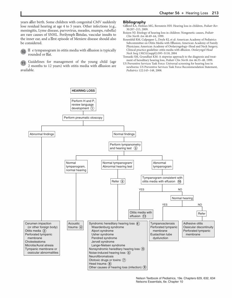
Chapter 56 u HearingLoss 213
Cerumen impaction (or other foreign body)Otitis media 2Perforated tympanic membraneCholesteatomaMicrotia/ Aural atresiaTympanic membrane or ossicular abnormalities
Cerumen impaction (or other foreign body)Otitis media 2Perforated tympanic membraneCholesteatomaMicrotia/Aural atresiaTympanic membrane or ossicular abnormalities
Acoustictrauma 4 Acoustictrauma 4
Otitis media witheffusion 11 Otitis media witheffusion 11
TympanosclerosisPerforated tympanic membraneEustachian tube dysfunction
TympanosclerosisPerforated tympanic membraneEustachian tube dysfunction
Adhesive otitisOssicular discontinuityPerforated tympanic membrane
Adhesive otitisOssicular discontinuityPerforated tympanic membrane
ReferRefer
Normal hearing
Tympanogram consistent withotitis media with effusion 10 Tympanogram consistent withotitis media with effusion 10
AbnormaltympanogramAbnormaltympanogram
Normal tympanogram/Abnormal hearing testNormal tympanogram/Abnormal hearing test
Perform tympanometryand hearing test 3 Perform tympanometryand hearing test 3
Normal findingsNormal findingsAbnormal findingsAbnormal findings
Normal tympanogram,normal hearing
Normal tympanogram,normal hearing
Perform H and P,review languagedevelopment 1
Perform H and P,review languagedevelopment 1
Refer 5 Refer 5
HEARING LOSSHEARING LOSS
YES NO
YES NO
Nelson Textbook of Pediatrics, 19e. Chapters 629, 632, 634Nelsons Essentials, 6e. Chapter 10
Perform pneumatic otoscopy
Waardenburg syndrome Alport syndrome Usher syndrome Pendred syndrome Jervell syndrome Lange-Nielsen syndromeNonsyndromic hereditary hearing loss 6
Noise-induced hearing loss 4 NeurofibromatosisOtotoxic drugs or toxins 7 Head trauma 8 Other causes of hearing loss (infection) 9
Syndromic hereditary hearing loss 6
years after birth. Some children with congenital CMV suddenly lose residual hearing at age 4 to 5 years. Other infections (e.g., meningitis, Lyme disease, parvovirus, measles, mumps, rubella) are rare causes of SNHL. Perilymph fistulas, vascular insults to the inner ear, and a first episode of Meniere disease should also be considered.
10 The tympanogram in otitis media with effusion is typically rounded or flat.
11 Guidelines for management of the young child (age 2 months to 12 years) with otitis media with effusion are
available.
BibliographyGifford KA, Holmes MG, Bernstein HH: Hearing loss in children, Pediatr Rev
30:207–215, 2009. Roizen NJ: Etiology of hearing loss in children: Nongenetic causes, Pediatr
Clin North Am 46:49–64, 1999. Rosenfeld RM, Culpepper L, Doyle KJ, et al: American Academy of Pediatrics
Subcommittee on Otitis Media with Effusion; American Academy of Family Physicians; American Academy of Otolaryngology–Head and Neck Surgery, Clinical practice guideline: otitis media with effusion. Otolaryngol Head Neck Surg 130(5)((suppl)):S95–S118, 2004
Tomaski SM, Grundfast KM: A stepwise approach to the diagnosis and treat-ment of hereditary hearing loss, Pediatr Clin North Am 46:35–48, 1999.
US Preventive Services Task Force: Universal screening for hearing loss in newborns: US Preventive Services Task Force Recommendation Statement, Pediatrics 122:143–148, 2008.

This page intentionally left blank

PART
VIIIDermatology
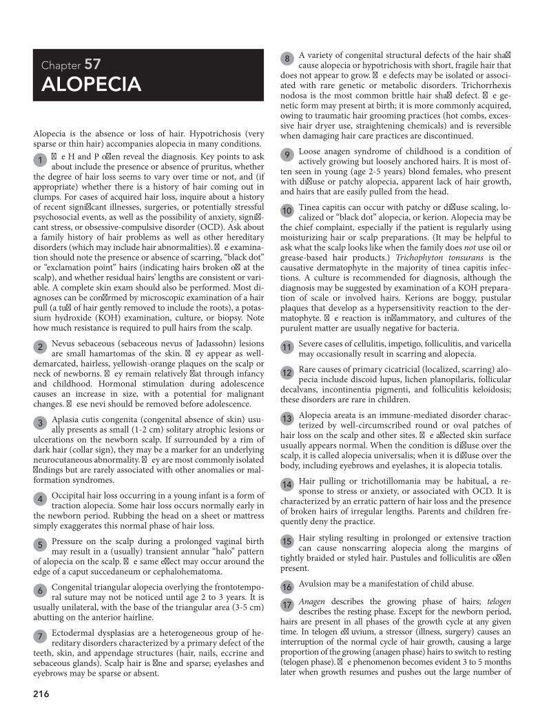
216
Chapter 57
ALOPECIA 8 A variety of congenital structural defects of the hair shaft
cause alopecia or hypotrichosis with short, fragile hair that does not appear to grow. The defects may be isolated or associ-ated with rare genetic or metabolic disorders. Trichorrhexis nodosa is the most common brittle hair shaft defect. The ge-netic form may present at birth; it is more commonly acquired, owing to traumatic hair grooming practices (hot combs, exces-sive hair dryer use, straightening chemicals) and is reversible when damaging hair care practices are discontinued.
9 Loose anagen syndrome of childhood is a condition of actively growing but loosely anchored hairs. It is most of-
ten seen in young (age 2-5 years) blond females, who present with diffuse or patchy alopecia, apparent lack of hair growth, and hairs that are easily pulled from the head.
10 Tinea capitis can occur with patchy or diffuse scaling, lo-calized or “black dot” alopecia, or kerion. Alopecia may be
the chief complaint, especially if the patient is regularly using moisturizing hair or scalp preparations. (It may be helpful to ask what the scalp looks like when the family does not use oil or grease-based hair products.) Trichophyton tonsurans is the causative dermatophyte in the majority of tinea capitis infec-tions. A culture is recommended for diagnosis, although the diagnosis may be suggested by examination of a KOH prepara-tion of scale or involved hairs. Kerions are boggy, pustular plaques that develop as a hypersensitivity reaction to the der-matophyte. The reaction is inflammatory, and cultures of the purulent matter are usually negative for bacteria.
11 Severe cases of cellulitis, impetigo, folliculitis, and varicella may occasionally result in scarring and alopecia.
12 Rare causes of primary cicatricial (localized, scarring) alo-pecia include discoid lupus, lichen planopilaris, follicular
decalvans, incontinentia pigmenti, and folliculitis keloidosis; these disorders are rare in children.
13 Alopecia areata is an immune-mediated disorder charac-terized by well-circumscribed round or oval patches of
hair loss on the scalp and other sites. The affected skin surface usually appears normal. When the condition is diffuse over the scalp, it is called alopecia universalis; when it is diffuse over the body, including eyebrows and eyelashes, it is alopecia totalis.
14 Hair pulling or trichotillomania may be habitual, a re-sponse to stress or anxiety, or associated with OCD. It is
characterized by an erratic pattern of hair loss and the presence of broken hairs of irregular lengths. Parents and children fre-quently deny the practice.
15 Hair styling resulting in prolonged or extensive traction can cause nonscarring alopecia along the margins of
tightly braided or styled hair. Pustules and folliculitis are often present.
16 Avulsion may be a manifestation of child abuse.
17 Anagen describes the growing phase of hairs; telogen describes the resting phase. Except for the newborn period,
hairs are present in all phases of the growth cycle at any given time. In telogen effluvium, a stressor (illness, surgery) causes an interruption of the normal cycle of hair growth, causing a large proportion of the growing (anagen phase) hairs to switch to resting (telogen phase). The phenomenon becomes evident 3 to 5 months later when growth resumes and pushes out the large number of
Alopecia is the absence or loss of hair. Hypotrichosis (very sparse or thin hair) accompanies alopecia in many conditions.
1 The H and P often reveal the diagnosis. Key points to ask about include the presence or absence of pruritus, whether
the degree of hair loss seems to vary over time or not, and (if appropriate) whether there is a history of hair coming out in clumps. For cases of acquired hair loss, inquire about a history of recent significant illnesses, surgeries, or potentially stressful psychosocial events, as well as the possibility of anxiety, signifi-cant stress, or obsessive-compulsive disorder (OCD). Ask about a family history of hair problems as well as other hereditary disorders (which may include hair abnormalities). The examina-tion should note the presence or absence of scarring, “black dot” or “exclamation point” hairs (indicating hairs broken off at the scalp), and whether residual hairs’ lengths are consistent or vari-able. A complete skin exam should also be performed. Most di-agnoses can be confirmed by microscopic examination of a hair pull (a tuft of hair gently removed to include the roots), a potas-sium hydroxide (KOH) examination, culture, or biopsy. Note how much resistance is required to pull hairs from the scalp.
2 Nevus sebaceous (sebaceous nevus of Jadassohn) lesions are small hamartomas of the skin. They appear as well-
demarcated, hairless, yellowish-orange plaques on the scalp or neck of newborns. They remain relatively flat through infancy and childhood. Hormonal stimulation during adolescence causes an increase in size, with a potential for malignant changes. These nevi should be removed before adolescence.
3 Aplasia cutis congenita (congenital absence of skin) usu-ally presents as small (1-2 cm) solitary atrophic lesions or
ulcerations on the newborn scalp. If surrounded by a rim of dark hair (collar sign), they may be a marker for an underlying neurocutaneous abnormality. They are most commonly isolated findings but are rarely associated with other anomalies or mal-formation syndromes.
4 Occipital hair loss occurring in a young infant is a form of traction alopecia. Some hair loss occurs normally early in
the newborn period. Rubbing the head on a sheet or mattress simply exaggerates this normal phase of hair loss.
5 Pressure on the scalp during a prolonged vaginal birth may result in a (usually) transient annular “halo” pattern
of alopecia on the scalp. The same effect may occur around the edge of a caput succedaneum or cephalohematoma.
6 Congenital triangular alopecia overlying the frontotempo-ral suture may not be noticed until age 2 to 3 years. It is
usually unilateral, with the base of the triangular area (3-5 cm) abutting on the anterior hairline.
7 Ectodermal dysplasias are a heterogeneous group of he-reditary disorders characterized by a primary defect of the
teeth, skin, and appendage structures (hair, nails, eccrine and sebaceous glands). Scalp hair is fine and sparse; eyelashes and eyebrows may be sparse or absent.
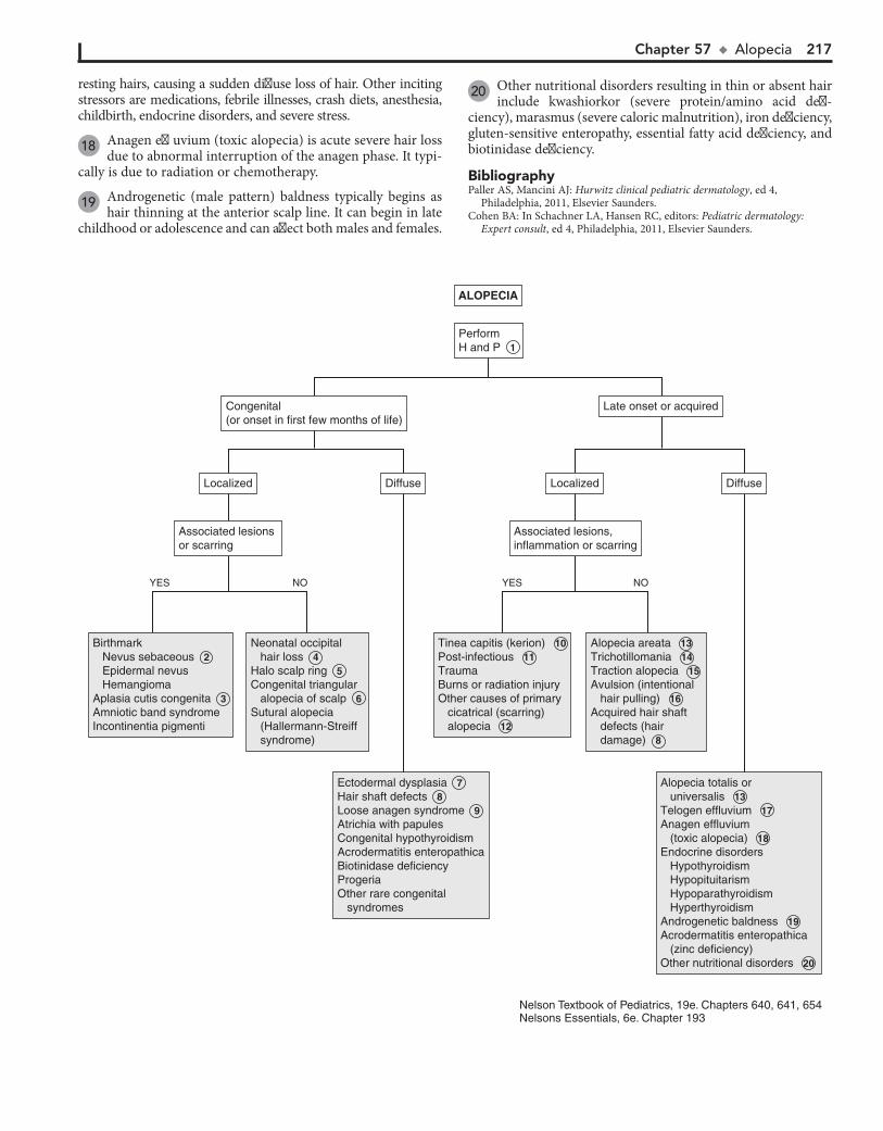
Chapter 57 u Alopecia 217
resting hairs, causing a sudden diffuse loss of hair. Other inciting stressors are medications, febrile illnesses, crash diets, anesthesia, childbirth, endocrine disorders, and severe stress.
18 Anagen effluvium (toxic alopecia) is acute severe hair loss due to abnormal interruption of the anagen phase. It typi-
cally is due to radiation or chemotherapy.
19 Androgenetic (male pattern) baldness typically begins as hair thinning at the anterior scalp line. It can begin in late
childhood or adolescence and can affect both males and females.
20 Other nutritional disorders resulting in thin or absent hair include kwashiorkor (severe protein/amino acid defi-
ciency), marasmus (severe caloric malnutrition), iron deficiency, gluten-sensitive enteropathy, essential fatty acid deficiency, and biotinidase deficiency.
BibliographyPaller AS, Mancini AJ: Hurwitz clinical pediatric dermatology, ed 4,
Philadelphia, 2011, Elsevier Saunders.Cohen BA: In Schachner LA, Hansen RC, editors: Pediatric dermatology:
Expert consult, ed 4, Philadelphia, 2011, Elsevier Saunders.
ALOPECIAALOPECIA
PerformH and P 1 PerformH and P 1
Birthmark Nevus sebaceous 2 Epidermal nevus HemangiomaAplasia cutis congenita 3 Amniotic band syndromeIncontinentia pigmenti
Birthmark Nevus sebaceous 2 Epidermal nevus HemangiomaAplasia cutis congenita 3 Amniotic band syndromeIncontinentia pigmenti
Neonatal occipital hair loss 4Halo scalp ring 5Congenital triangular alopecia of scalp 6 Sutural alopecia (Hallermann-Streiff syndrome)
Neonatal occipital hair loss 4Halo scalp ring 5Congenital triangular alopecia of scalp 6 Sutural alopecia (Hallermann-Streiff syndrome)
Tinea capitis (kerion) 10Post-infectious 11TraumaBurns or radiation injuryOther causes of primary cicatrical (scarring) alopecia 12
Tinea capitis (kerion) 10Post-infectious 11TraumaBurns or radiation injuryOther causes of primary cicatrical (scarring) alopecia 12
Alopecia areata 13Trichotillomania 14Traction alopecia 15 Avulsion (intentional hair pulling) 16Acquired hair shaft defects (hair damage) 8
Alopecia areata 13Trichotillomania 14Traction alopecia 15 Avulsion (intentional hair pulling) 16Acquired hair shaft defects (hair damage) 8
Ectodermal dysplasia 7Hair shaft defects 8Loose anagen syndrome 9Atrichia with papulesCongenital hypothyroidismAcrodermatitis enteropathicaBiotinidase deficiencyProgeriaOther rare congenital syndromes
Ectodermal dysplasia 7Hair shaft defects 8Loose anagen syndrome 9Atrichia with papulesCongenital hypothyroidismAcrodermatitis enteropathicaBiotinidase deficiencyProgeriaOther rare congenital syndromes
Alopecia totalis or universalis 13Telogen effluvium 17Anagen effluvium (toxic alopecia) 18Endocrine disorders Hypothyroidism Hypopituitarism Hypoparathyroidism HyperthyroidismAndrogenetic baldness 19Acrodermatitis enteropathica (zinc deficiency)Other nutritional disorders 20
Alopecia totalis or universalis 13Telogen effluvium 17Anagen effluvium (toxic alopecia) 18Endocrine disorders Hypothyroidism Hypopituitarism Hypoparathyroidism HyperthyroidismAndrogenetic baldness 19Acrodermatitis enteropathica (zinc deficiency)Other nutritional disorders 20
Congenital(or onset in first few months of life)Congenital(or onset in first few months of life)
Late onset or acquiredLate onset or acquired
Associated lesions or scarringAssociated lesions or scarring
LocalizedLocalized DiffuseDiffuse
NOYES
Associated lesions, inflammation or scarringAssociated lesions, inflammation or scarring
LocalizedLocalized DiffuseDiffuse
NOYES
Nelson Textbook of Pediatrics, 19e. Chapters 640, 641, 654Nelsons Essentials, 6e. Chapter 193
218
Chapter 58
VESICLES AND BULLAE
blisters developing on the hands and feet after significant trauma or friction, especially in hot weather conditions.
7 Incontinentia pigmenti is a hereditary disorder with mul-tisystem involvement; it mainly affects females. Cutaneous
manifestations develop in the first 2 weeks of life, with red streaks and crops of vesicles or bullae developing on the trunk or in a characteristic linear distribution on the extremities. Evo-lution to verrucous lesions followed by characteristic pigmenta-tion changes subsequently occurs, usually by 4 months of age. Blisters may recur later with febrile illnesses. The hair, eyes, CNS, and teeth are also affected.
8 Miliaria or heat rash may be characterized by tiny (1-2 mm) clear, thin-walled vesicles without any associated redness
(miliaria crystallina) or with larger (2-4 mm) vesicles or papules with associated erythema (miliaria rubra). Areas occluded by heavy clothing or affected by sunburn are most commonly affected.
9 Sucking blisters are presumably due to in utero sucking on the affected area. They may be located on the dorsal
surfaces of the forearm, hands, or fingers.
10 The lesions of bullous impetigo are flaccid bullae that rupture easily, leaving superficial erosions. This form of impetigo
is much less common than the nonbullous form (crusted lesions). In the newborn, the diaper area is most commonly affected.
11 The skin lesions of acrodermatitis enteropathica can be vesiculobullous or psoriasiform in nature. They are likely
to be found in acral, oral, and perineal locations.
12 Vesicles are a more common manifestation of scabies in infants and young children than in older children. They
are most likely to be found on the palms and soles and in the axillae and groin. Severe pruritus is characteristic.
13 Erythema multiforme (EM) is a blistering hypersensitivity reaction classically characterized by target lesions. Target
lesions are well-demarcated round or oval macules with distinct “rings”—outermost erythema surrounding a whitish ring and then a central dusky blue/gray or blistered center; the typical size ranges from 1 to 3 cm. A self-limited outbreak of target lesions occurs in EM; the outbreak is usually symmetric and involving primarily the upper extremities. Limited involvement of one mucosal surface may occur. Many types of infectious agents have been associated with EM; HSV is the most common.
14 Linear immunoglobulin (Ig)A disease (chronic bullous der-matosis of children) usually occurs in the preschool years.
The disorder is characterized by a widespread eruption of large sausage-shaped bullae with a variable degree of pruritus. The in-guinal region, lower trunk, buttocks, legs, and tops of the feet are most commonly affected, but the bullae may develop anywhere. Sometimes the bullae develop in an annular or rosette-like configuration surrounding a central crust (“cluster of jewels”).
15 In dermatitis herpetiformis, outbreaks of clusters of small papules and vesicles occur in a symmetric distribution on
the extensor surfaces, lower trunk, and buttocks. The outbreaks are extremely pruritic and tender. Hemorrhagic bullae on the palms and soles occasionally occur. The disorder is associated with a gluten-sensitive enteropathy.
16 The term pemphigus encompasses a group of chronic blistering disorders with variable severity and prognoses.
Vesicles are small (,0.5 cm) fluid-filled lesions; bullae are larger vesicles (.0.5-1 cm). Fluid may be clear or hemorrhagic. Vesiculobullous lesions (blisters) may be infectious or nonin-fectious. Noninfectious lesions may be spontaneous or induced by trauma or friction. Most vesiculobullous disorders are rare in children but need to be considered in chronic blistering con-ditions. Many are clinically indistinguishable from each other; skin biopsy, immunofluorescence, and electron microscopy are often necessary to make the definitive diagnosis.
The usefulness of this algorithm is limited (without photo-graphs) to categorizing potential diagnoses based on some broad clinical criteria. A more specific dermatology reference will often be necessary to confirm suspected diagnoses.
1 Erythema toxicum (ET) is a common, benign, self-limited neonatal rash. Tiny papules, vesicles, or pustules are pres-
ent on a blotchy erythematous macular base; the rash can be present on any surface except for the palms and soles. The onset generally is in the first few days of life (occasionally later), and remission is usually by 2 weeks. If microscopically examined, the lesions show an accumulation of eosinophils within the pilosebaceous apparatus.
2 Transient neonatal pustular melanosis is another benign, self-limited neonatal rash. Small, fragile vesicles or pus-
tules are noted at birth and can be present on any surface. The lesions rupture quickly, leaving a characteristic rim of scale and hyperpigmented macule that gradually fades. Histologic exam shows primarily neutrophils.
3 A generalized vesicular exanthem can occur in dissemi-nated herpes simplex virus (HSV) infection of the new-
born. Lesions associated with localized infections (involving only skin, eyes, and mouth) may be subtle, but diagnosis is critical because localized disease in these infants can progress to encephalitis or disseminated disease. PCR tests and cultures are diagnostic options for HSV; Tzanck smears are of limited use-fulness because they are not very sensitive, plus will not distin-guish between HSV and varicella.
4 Congenital candidiasis typically manifests as diffuse pap-ules and pustules, rarely bullae.
5 Staphylococcal scalded skin syndrome is an exfoliative dermatitis characterized by diffuse tender erythema, flac-
cid bullae, sheets of desquamating skin, and a positive Nikolsky sign (blistering elicited by light touch). The face, neck, groin, and axillae are most commonly affected.
6 Epidermolysis bullosa (EB) constitutes a group of inherited blistering disorders. The clinical severity, age of presenta-
tion, and level/site of skin cleavage vary. Blistering may be spon-taneous or in response to pressure or trauma. Most variants be-come evident in the neonatal period. The most common variant (EB simplex of the hands and feet [Weber-Cockayne syndrome]) may not manifest until adolescence or young adulthood, with

Chapter 58 u VesiclesandBullae 219
VESICLES AND BULLAEVESICLES AND BULLAE
Presenting within1 to 2 months of age
Presenting within1 to 2 months of age
Perform H and PPerform H and P
Erythema toxicum 1Transient neonatal pustular melanosis 2 Herpes simplex (disseminated) 3 Congenital candidiasis 4 Congenital varicellaStaphylococcal scalded skin syndrome 5 Epidermolysis bullosa 6 Incontinentia pigmenti 7 Other rare blistering congenital dermatoses
Erythema toxicum 1Transient neonatal pustular melanosis 2 Herpes simplex (disseminated) 3 Congenital candidiasis 4 Congenital varicellaStaphylococcal scalded skin syndrome 5 Epidermolysis bullosa 6 Incontinentia pigmenti 7 Other rare blistering congenital dermatoses
Miliaria 8 Sucking blisters 9 Bullous impetigo 10 Herpes simplex (localized)LymphangiomaAplasia cutis congenitaAcrodermatitis enteropathica 11
Miliaria 8 Sucking blisters 9 Bullous impetigo 10 Herpes simplex (localized)LymphangiomaAplasia cutis congenitaAcrodermatitis enteropathica 11
Scabies 12 Bullous impetigo 10 Erythema multiforme 13 Linear IgA dermatosis 14 Epidermolysis bullosa 6 Dermatitis herpetiformis 15 Pemphigus 16 Fixed drug reactionMastocytosis (bullous urticaria pigmentosa)Pityriaisis lichenoides et varioliformis acuta (PLEVA)
Scabies 12 Bullous impetigo 10 Erythema multiforme 13 Linear IgA dermatosis 14 Epidermolysis bullosa 6 Dermatitis herpetiformis 15 Pemphigus 16 Fixed drug reactionMastocytosis (bullous urticaria pigmentosa)Pityriaisis lichenoides et varioliformis acuta (PLEVA)
Bullous impetigo 10 Contact dermatitisInsect bitesBurnsFrictionHerpes simplex 3 Herpes zosterDyshidrotic eczema 17 Tinea pedis 18 Scabies 12Pemphigus 16 Epidermolysis bullosa 6 Blistering distal dactylitisFixed drug reactionLymphangiomaAcrodermatitis enteropathica 11 Photosensitivity reactions Sunburn Drug reactions Genetic disorders with photosensitivity Inborn errors of metabolism (porphyrias) Skin disease exacerbated or precipitated by lightLupus erythematosus (drug-induced)Diabetic bullae
Bullous impetigo 10 Contact dermatitisInsect bitesBurnsFrictionHerpes simplex 3 Herpes zosterDyshidrotic eczema 17 Tinea pedis 18 Scabies 12Pemphigus 16 Epidermolysis bullosa 6 Blistering distal dactylitisFixed drug reactionLymphangiomaAcrodermatitis enteropathica 11 Photosensitivity reactions Sunburn Drug reactions Genetic disorders with photosensitivity Inborn errors of metabolism (porphyrias) Skin disease exacerbated or precipitated by lightLupus erythematosus (drug-induced)Diabetic bullae
VaricellaHand-foot-mouth disease 19 Staphylococcal scalded skin syndrome 5 Stevens-Johnson syndrome 20 Toxic epidermal necrolysis 20 Acrodermatitis enteropathica 11
VaricellaHand-foot-mouth disease 19 Staphylococcal scalded skin syndrome 5 Stevens-Johnson syndrome 20 Toxic epidermal necrolysis 20 Acrodermatitis enteropathica 11
GeneralizeddistributionGeneralizeddistribution
LocalizeddistributionLocalizeddistribution
GeneralizeddistributionGeneralizeddistribution
LocalizeddistributionLocalizeddistribution
Systemic illnesspresent
Systemic illnesspresent
NO
NO
YES
YES
Nelson Textbook of Pediatrics, 19e. Chapters 589, 646, 647, 658Nelsons Essentials, 6e. Chapters 195, 196
220 Part VIII u Dermatology
The characteristic lesions are flaccid bullae with subsequent shallow erosions. Onset in childhood is rare; there is an infantile subtype with a predilection for acral regions, and a childhood vulvar subtype. In the pemphigus vulgaris variant, painful oral lesions may precede cutaneous involvement by weeks or months.
17 Recurrent outbreaks of small pruritic vesicles on the hands and feet occur in dyshidrotic eczema; larger vesicles and
bullae sometimes occur. The palms, soles, and lateral aspects of the fingers and toes are most commonly affected.
18 An inflammatory vesicular reaction may occasionally occur with tinea pedis.
19 Hand-foot-mouth syndrome is a viral (usually due to coxsackie or enteroviruses) illness characterized by a
prodrome of fever, anorexia, and sore throat followed by an enanthem of small ulcerating oral vesicles. The characteristic exanthem of oval vesicles (when it occurs) affects primarily the hands and feet (occasionally includes elbows, knees, or but-tocks) and develops after the oral lesions. Vesicles can also be seen with other enteroviral infections.
20 Stevens-Johnson syndrome (SJS) is a more severe, poten-tially life-threatening blistering hypersensitivity reaction.
It is often characterized by target lesions (in a more extensive distribution than in EM) and requires involvement of two or more mucosal surfaces (mouth, eye, urogenital, esophageal) for diagnosis. Mycoplasma pneumoniae is the most common patho-gen associated with SJS; several medications are also recognized precipitants. Drugs are more likely to be associated with SJS than EM. Toxic epidermal necrolysis is identified as a severe form of SJS, with a prodrome of fever and malaise and a subsequent se-vere dermatitis (including some bullae) with exfoliation of large sheets of full-thickness epidermis. It is most commonly precipi-tated by drugs.
BibliographyCohen BA: In Schachner LA, Hansen RC, editors: Pediatric dermatology, ed 4,
Philadelphia, 2011, Elsevier Saunders.Paller AS, Mancini AJ: Hurwitz clinical pediatric dermatology, ed 4, Philadelphia,
2011, Elsevier Saunders.
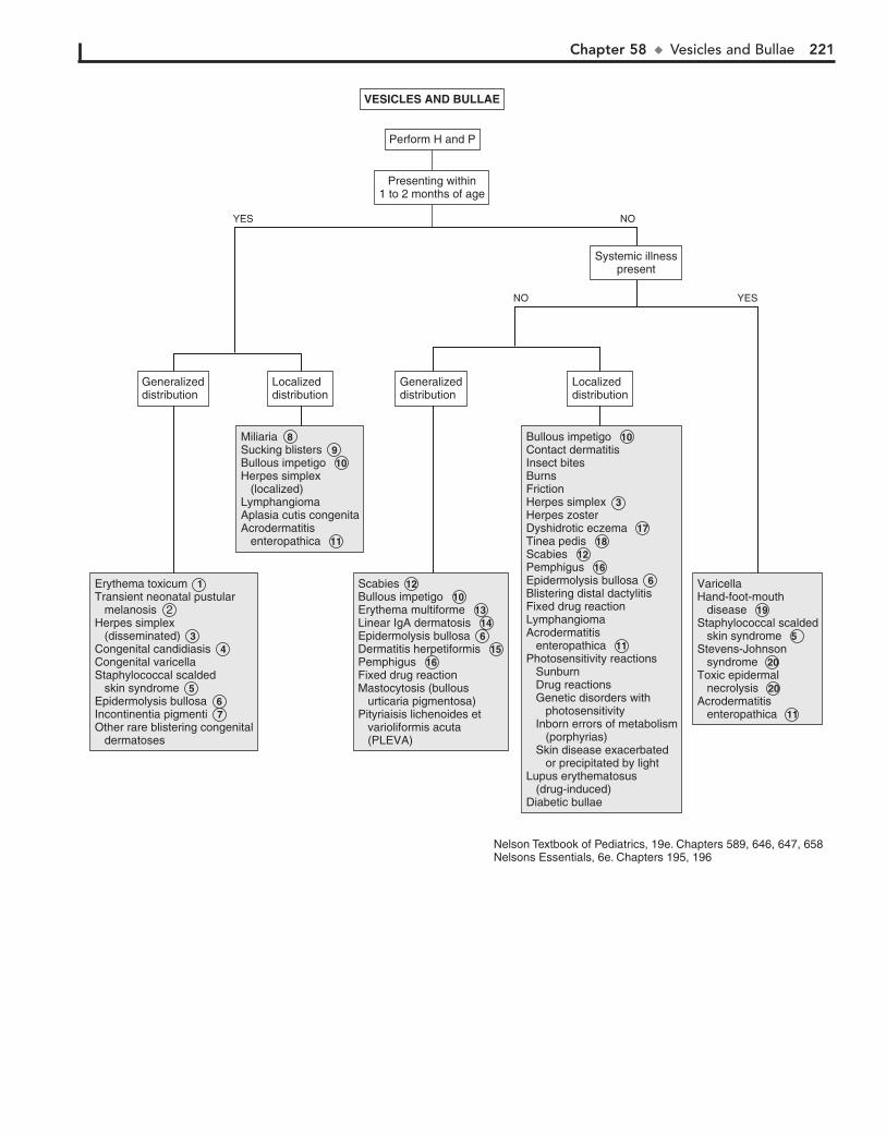
Chapter 58 u VesiclesandBullae 221
VESICLES AND BULLAEVESICLES AND BULLAE
Presenting within1 to 2 months of age
Presenting within1 to 2 months of age
Perform H and PPerform H and P
Erythema toxicum 1Transient neonatal pustular melanosis 2 Herpes simplex (disseminated) 3 Congenital candidiasis 4 Congenital varicellaStaphylococcal scalded skin syndrome 5 Epidermolysis bullosa 6 Incontinentia pigmenti 7 Other rare blistering congenital dermatoses
Erythema toxicum 1Transient neonatal pustular melanosis 2 Herpes simplex (disseminated) 3 Congenital candidiasis 4 Congenital varicellaStaphylococcal scalded skin syndrome 5 Epidermolysis bullosa 6 Incontinentia pigmenti 7 Other rare blistering congenital dermatoses
Miliaria 8 Sucking blisters 9 Bullous impetigo 10 Herpes simplex (localized)LymphangiomaAplasia cutis congenitaAcrodermatitis enteropathica 11
Miliaria 8 Sucking blisters 9 Bullous impetigo 10 Herpes simplex (localized)LymphangiomaAplasia cutis congenitaAcrodermatitis enteropathica 11
Scabies 12 Bullous impetigo 10 Erythema multiforme 13 Linear IgA dermatosis 14 Epidermolysis bullosa 6 Dermatitis herpetiformis 15 Pemphigus 16 Fixed drug reactionMastocytosis (bullous urticaria pigmentosa)Pityriaisis lichenoides et varioliformis acuta (PLEVA)
Scabies 12 Bullous impetigo 10 Erythema multiforme 13 Linear IgA dermatosis 14 Epidermolysis bullosa 6 Dermatitis herpetiformis 15 Pemphigus 16 Fixed drug reactionMastocytosis (bullous urticaria pigmentosa)Pityriaisis lichenoides et varioliformis acuta (PLEVA)
Bullous impetigo 10 Contact dermatitisInsect bitesBurnsFrictionHerpes simplex 3 Herpes zosterDyshidrotic eczema 17 Tinea pedis 18 Scabies 12Pemphigus 16 Epidermolysis bullosa 6 Blistering distal dactylitisFixed drug reactionLymphangiomaAcrodermatitis enteropathica 11 Photosensitivity reactions Sunburn Drug reactions Genetic disorders with photosensitivity Inborn errors of metabolism (porphyrias) Skin disease exacerbated or precipitated by lightLupus erythematosus (drug-induced)Diabetic bullae
Bullous impetigo 10 Contact dermatitisInsect bitesBurnsFrictionHerpes simplex 3 Herpes zosterDyshidrotic eczema 17 Tinea pedis 18 Scabies 12Pemphigus 16 Epidermolysis bullosa 6 Blistering distal dactylitisFixed drug reactionLymphangiomaAcrodermatitis enteropathica 11 Photosensitivity reactions Sunburn Drug reactions Genetic disorders with photosensitivity Inborn errors of metabolism (porphyrias) Skin disease exacerbated or precipitated by lightLupus erythematosus (drug-induced)Diabetic bullae
VaricellaHand-foot-mouth disease 19 Staphylococcal scalded skin syndrome 5 Stevens-Johnson syndrome 20 Toxic epidermal necrolysis 20 Acrodermatitis enteropathica 11
VaricellaHand-foot-mouth disease 19 Staphylococcal scalded skin syndrome 5 Stevens-Johnson syndrome 20 Toxic epidermal necrolysis 20 Acrodermatitis enteropathica 11
GeneralizeddistributionGeneralizeddistribution
LocalizeddistributionLocalizeddistribution
GeneralizeddistributionGeneralizeddistribution
LocalizeddistributionLocalizeddistribution
Systemic illnesspresent
Systemic illnesspresent
NO
NO
YES
YES
Nelson Textbook of Pediatrics, 19e. Chapters 589, 646, 647, 658Nelsons Essentials, 6e. Chapters 195, 196
222
Chapter 59
FEVER AND RASH
high fever lasting for 3 to 5 days, followed by a short-lived (hours to 1 to 3 days), nonspecific, morbilliform, rose-colored rash that appears as defervescence occurs. The etiology can be human herpesviruses 6 or 7.
6 Enteroviruses are a common cause of petechial rashes. The patients usually do not appear ill and may not require ex-
tensive testing. Close followup, however, is essential.
7 Although rarely seen today in countries with good immuni-zation practices, several distinctive clinical findings can aid
in the diagnosis of measles. Initial prodromal symptoms are the three “C’s”—cough, conjunctivitis, and coryza—which last for a few days before the development of high fever and the character-istic morbilliform exanthem that erupts (and subsequently fades) in a head-to-toe pattern. It affects the trunk more than the ex-tremities and may sometimes be petechial or hemorrhagic. Atypical or modified measles are milder cases that may develop in a child with partial protection (transplacental antibody in young infants, vaccination before 1 year of age, or recipients of immunoglobulin). Koplik spots (a white or bluish-white dot-like enanthem found on the buccal mucosa near the lower molars) are pathognomonic for measles when noted; they develop during the prodromal phase but are present for only a brief period of time (12 to 24 hours).
8 The classic rubella (German measles) rash consists of dis-crete pink macular lesions that appear initially on the face
and spread in a head-to-toe progression. They tend to coalesce in the truncal region and remain as discrete macules on the extremities. The facial rash is typically faded by the time the rash reaches the extremities.
9 Papular acrodermatitis (also called Gianotti-Crosti syn-drome) is a characteristic outbreak of discrete, flat-topped,
dark or dusky papules, usually 1 to 10 mm in size. Crops of le-sions erupt symmetrically on the face, buttocks, and extensor surfaces of limbs; palms and soles can be affected. Low-grade fever may or may not occur. It is a recognized reaction to im-munizations and viral infections. EBV is the most commonly associated virus; hepatitis B, coxsackie A16, CMV, and parain-fluenza are others.
10 Group A streptococcus (GAS) is associated with several characteristic skin findings. Scarlatina (scarlatiniform rash
or scarlet fever) describes a diffuse, fine papular, “ashy” or “sandpaper” rash that tends to develop initially on the neck and upper chest. The rash may be concentrated in creases (axillae, antecubital fossae, inguinal), where it takes on a linear petechial appearance (Pastia’s lines). Subsequent desquamation of the rash, particularly on the hands and feet, is characteristic in both treated and untreated GAS infections.
11 A typical erythema migrans rash is pathognomonic for Lyme disease. It begins as a red macule or papule at the site
of the tick bite and expands to an average diameter of 15 cm. It may be a uniform erythematous macule or demonstrate central clearing (target lesion).
12 The rash of rickettsial infections is often a late develop-ment in the illness course. The typical rash of Rocky
Mountain spotted fever (RMSF) due to Rickettsia rickettsii be-gins as a blanchable macular rash that initially develops on the wrists and ankles then spreads centrally and includes the palms, soles, trunk, and face. The macules frequently evolve into pete-chial (and sometimes purpuric) lesions. Rash may be entirely
Fever and rash are components of many disease processes, most of which are benign self-limited conditions. Rarely, this combi-nation of symptoms may herald a life-threatening illness, so it is essential to narrow the differential diagnosis with a careful his-tory and physical. Causes of fever and rash include infections, vasculitides, and hypersensitivity disorders. Laboratory tests should be ordered according to the presumptive diagnosis based on the history and physical. Many rashes are pathognomonic for certain diseases (varicella), and testing may not be indicated.
1 The history should include the characteristics of the rash, presence of pruritus or pain or tenderness, appearance in
relationship to the fever, and the evolution and progression of the rash. A history of ill contacts, recent travel, exposures (pets, wild-life, insects), medications (including the possibility of IV drug use), and sexual activity should be obtained. Past medical history should be reviewed, and a history of any prodromal or associated symptoms (abdominal pain, rash, headaches) obtained. Exami-nation should include a general assessment of the patient to de-termine the severity of the illness, including vital signs and height of fever. Tachycardia and tachypnea in a patient with fever and rash may indicate sepsis, particularly if there is altered mental status. The development of hypotension may indicate septic shock.
2 Note the distribution of the rash and lesion morphology and color, as well as the presence and characteristics of any
enanthems (eruptions on mucosal surfaces). The term macular describes flat lesions, papular describes raised or palpable le-sions, and the term morbilliform (classically used to describe the rash of measles) describes coalescence of maculopapular lesions into a diffuse or sheet-like distribution. Another impor-tant characteristic is whether a rash blanches with pressure or not. (Reddish lesions that blanch are due to dilated capillaries; nonblanchable lesions imply RBCs have leaked out of the blood vessels [extravasation]). Distribution may be generalized or lo-calized, symmetric or asymmetric, centripetal (more lesions on the trunk, less on the extremities and face), or centrifugal (more lesions on the distal extremities and face, less on the trunk). The characteristics of the rash and associated symptoms frequently suggest a diagnosis.
3 Target lesions are well-demarcated round or oval macules with distinct “rings”—a rim of outer erythema surround-
ing a whitish ring and then a central dusky blue/gray or bullous center; the typical size ranges from 1 to 3 cm.
4 Fifth disease (also called erythema infectiosum) is caused by human parvovirus B19. The rash starts with a “slapped
cheek” appearance with circumoral pallor, followed 1 to 4 days later by a more diffuse maculopapular rash that becomes lacy or reticular in appearance as it fades. A less common manifesta-tion of parvovirus B19 is a petechial rash in a papular-purpuric “gloves and socks” acral distribution.
5 The classic presentation of roseola (also called roseola in-fantum, exanthema subitum, or sixth disease) is an acute
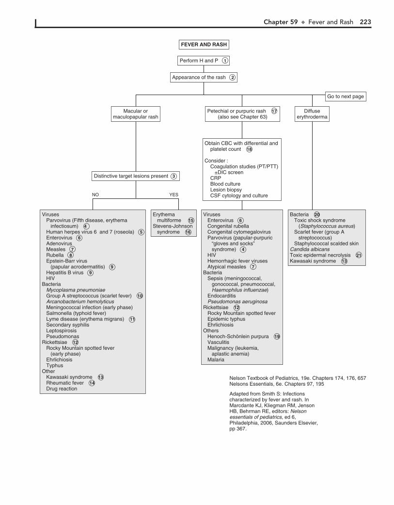
Chapter 59 u FeverandRash 223
FEVER AND RASHFEVER AND RASH
Appearance of the rash 2 Appearance of the rash 2
Perform H and P 1 Perform H and P 1
Erythema multiforme 15 Stevens-Johnson syndrome 16
Erythema multiforme 15 Stevens-Johnson syndrome 16
Bacteria 20 Toxic shock syndrome (Staphylococcus aureus) Scarlet fever (group A streptococcus) Staphylococcal scalded skinCandida albicansToxic epidermal necrolysis 21 Kawasaki syndrome 13
Bacteria 20 Toxic shock syndrome (Staphylococcus aureus) Scarlet fever (group A streptococcus) Staphylococcal scalded skinCandida albicansToxic epidermal necrolysis 21 Kawasaki syndrome 13
Viruses Parvovirus (Fifth disease, erythema infectiosum) 4 Human herpes virus 6 and 7 (roseola) 5 Enterovirus 6 Adenovirus Measles 7 Rubella 8 Epstein-Barr virus (papular acrodermatitis) 9 Hepatitis B virus 9 HIVBacteria Mycoplasma pneumoniae Group A streptococcus (scarlet fever) 10 Arcanobacterium hemolyticus Meningococcal infection (early phase) Salmonella (typhoid fever) Lyme disease (erythema migrans) 11 Secondary syphilis Leptospirosis PseudomonasRickettsiae 12 Rocky Mountain spotted fever (early phase) Ehrlichiosis Typhus Other Kawasaki syndrome 13 Rheumatic fever 14 Drug reaction
Viruses Parvovirus (Fifth disease, erythema infectiosum) 4 Human herpes virus 6 and 7 (roseola) 5 Enterovirus 6 Adenovirus Measles 7 Rubella 8 Epstein-Barr virus (papular acrodermatitis) 9 Hepatitis B virus 9 HIVBacteria Mycoplasma pneumoniae Group A streptococcus (scarlet fever) 10 Arcanobacterium hemolyticus Meningococcal infection (early phase) Salmonella (typhoid fever) Lyme disease (erythema migrans) 11 Secondary syphilis Leptospirosis PseudomonasRickettsiae 12 Rocky Mountain spotted fever (early phase) Ehrlichiosis Typhus Other Kawasaki syndrome 13 Rheumatic fever 14 Drug reaction
Viruses Enterovirus 6 Congenital rubella Congenital cytomegalovirus Parvovirus (papular-purpuric “gloves and socks” syndrome) 4 HIV Hemorrhagic fever viruses Atypical measles 7 Bacteria Sepsis (meningococcal, gonococcal, pneumococcal, Haemophilus influenzae) Endocarditis Pseudomonas aeruginosaRickettsiae 12 Rocky Mountain spotted fever Epidemic typhus EhrlichiosisOthers Henoch-Schönlein purpura 19 Vasculitis Malignancy (leukemia, aplastic anemia) Malaria
Viruses Enterovirus 6 Congenital rubella Congenital cytomegalovirus Parvovirus (papular-purpuric “gloves and socks” syndrome) 4 HIV Hemorrhagic fever viruses Atypical measles 7 Bacteria Sepsis (meningococcal, gonococcal, pneumococcal, Haemophilus influenzae) Endocarditis Pseudomonas aeruginosaRickettsiae 12 Rocky Mountain spotted fever Epidemic typhus EhrlichiosisOthers Henoch-Schönlein purpura 19 Vasculitis Malignancy (leukemia, aplastic anemia) Malaria
Distinctive target lesions present 3 Distinctive target lesions present 3
Obtain CBC with differential and platelet count 18
Consider : Coagulation studies (PT/PTT) ±DIC screen CRP Blood culture Lesion biopsy CSF cytology and culture
Obtain CBC with differential and platelet count 18
Consider : Coagulation studies (PT/PTT) ±DIC screen CRP Blood culture Lesion biopsy CSF cytology and culture
Macular ormaculopapular rash
Macular ormaculopapular rash
Petechial or purpuric rash 17 (also see Chapter 63)
Petechial or purpuric rash 17 (also see Chapter 63)
Diffuseerythroderma
Diffuseerythroderma
Go to next pageGo to next page
NO YES
Nelson Textbook of Pediatrics, 19e. Chapters 174, 176, 657Nelsons Essentials, 6e. Chapters 97, 195
Adapted from Smith S: Infections characterized by fever and rash. In Marcdante KJ, Kliegman RM, Jenson HB, Behrman RE, editors: Nelson essentials of pediatrics, ed 6, Philadelphia, 2006, Saunders Elsevier, pp 367.
224 Part VIII u Dermatology
absent in 20% of cases. The ehrilichioses (Ehrlichia chaffeensis, Anaplasma phagocytophilum, Ehrlichia ewingii) are other zoo-noses that are clinically similar to rickettsial infections, although rash development is even more variable with these infections. A history of tick exposure is often, but not always, obtained in these cases.
13 The onset of the rash of Kawasaki syndrome typically oc-curs 3 to 5 days after fever onset. It typically has a general-
ized distribution and a morbilliform or scarlatiniform nature. It may, however, be limited to the diaper region in young infants.
14 The Jones criteria must be fulfilled to make a diagnosis of rheumatic fever. (See Chapter 20.) Erythema marginatum
is rare but constitutes one of the major criteria; it is classically described as serpiginous, evanescent, macular lesions that are pale in the center. It appears on the trunk and extremities (but not the face), is not pruritic, and will become more evident upon warming of the skin.
15 Erythema multiforme (EM) is a blistering hypersensitivity reaction classically characterized by a limited outbreak of
target lesions, usually in a symmetric distribution involving the upper extremities. Limited involvement of one mucosal surface may occur. Herpes simplex virus (HSV) is the infectious agent most commonly associated with EM.
16 Stevens-Johnson syndrome (SJS) is a more severe, poten-tially life-threatening blistering hypersensitivity reaction.
It may also demonstrate target lesions (in a more extensive dis-tribution than in EM) and requires involvement of two or more mucosal surfaces (mouth, eye, urogenital, esophageal) for diag-nosis. Fevers are more likely with SJS. Mycoplasma pneumoniae is the infectious agent most commonly associated with SJS; several medications are also recognized precipitants.
17 Petechiae are tiny dark (red or purple) pinpoint lesions that do not blanch with pressure. Purpura are larger dark (pur-
ple or brown) nonblanchable lesions that may or may not be raised (palpable). These rashes in a febrile child mandate an im-mediate and careful evaluation because they may indicate poten-tially life-threatening infections, especially in a child younger than 2 years. Sepsis due to Neisseria meningitides (as well as other organisms) is of particular concern. Purpura may be as-sociated with disseminated intravascular coagulation (DIC), se-vere thrombocytopenia, or vasculitides. Mechanical factors (forceful cough, vomiting, compression due to tourniquet or BP cuff) occasionally lead to petechiae; in these cases the distribu-tion is likely to be localized (to an extremity [owing to compres-sion] or to the face or neck following coughing or vomiting) and the children afebrile and not severely ill-appearing.
18 In a patient with a petechial or purpuric rash, a CBC should be obtained. Other tests may be considered, depending on
clinical presentation; remember, fever and petechiae in a child may quickly evolve into a critical illness. Thrombocytopenia may be associated with many infections, including viral ones. Even with a normal platelet count and coagulation studies, a WBC count below 5000/mm3 or above 15,000/ mm3, an absolute band count greater than 500 cells/mm3, or abnormal CSF find-ings suggest an increased risk for bacterial or rickettsial infec-tion. The presence of purpura definitely warrants an urgent evaluation. Of note, petechial rash with fever is most commonly
a benign phenomenon (not associated with thrombocytopenia), usually due to enteroviral infection.
19 In children with Henoch-Schönlein purpura, the rash usu-ally begins as macules or urticarial lesions, later becoming
raised and petechial or purpuric. Lesions are usually concen-trated on the buttocks and lower extremities (gravity-dependent areas). Involvement of other organ systems (GI, renal, neuro-logic) suggest the diagnosis.
20 Manifestations of staphylococcal infections range from bullous impetigo (localized skin infections) to staphylo-
coccal scalded skin syndrome (diffuse erythema, exquisite ten-derness, extensive blisters, superficial erosions with subsequent desquamation) to toxic shock syndrome (TSS). TSS is a severe multisystem disease with a variable degree of cutaneous mani-festations ranging from sunburn-like erythroderma (diffuse, involving non–sun-exposed areas), intraepidermal blistering (Nikolsky sign), and desquamation. Streptococcal infections can cause similar erythroderma and TSS conditions.
21 Toxic epidermal necrolysis is presumed to be a severe form of SJS, with a prodrome of fever and malaise and a subse-
quent severe dermatitis (including severe erythroderma, some vesicles, and exfoliation of large sheets of full-thickness epider-mis). It is most commonly precipitated by drugs.
22 The classic rash of HSV is small vesicles and/or superficial ulcerations on an erythematous base.
23 Primary infections with varicella-zoster virus manifest as chickenpox. Pruritic, clear fluid–filled vesicles develop
initially on the scalp and face and spread to the trunk, with minimal involvement of the extremities. The lesions become cloudy then crust over within 24 to 48 hours; lesions in multiple stages are present simultaneously. Recurrent infections (zoster) follow a dermatomal pattern.
24 Erythema nodosum is a hypersensitivity reaction that manifests as discrete, tender, nodular lesions on the ex-
tensor surfaces of the extremities. Fever may precede or be coincident with the development of the lesions. The etiology is often unknown; recognized causes include infection, in-flammatory bowel disease, connective tissue disease, and medications.
25 Erysipelas is a rare manifestation of group A streptococcus. Infections of the deeper layers of the skin present as well-
demarcated, bright red, painful lesions. The skin appears infil-trated, and the borders are raised and firm.
26 Eschars are a rare manifestation of infections due to Pseu-domonas aeruginosa and certain fungal and rickettsial
infections.
27 Fever only rarely precedes pityriasis rosea, but the rash distribution is quite distinctive. A solitary oval herald
patch (an annular lesion with raised edges and fine scale) frequently (but not always) precedes the development of a more diffuse eruption of smaller pink or brownish oval le-sions with fine scale, sometimes with central clearing evident. These lesions tend to follow the cutaneous cleavage lines, frequently described as a “Christmas tree” pattern on the back.
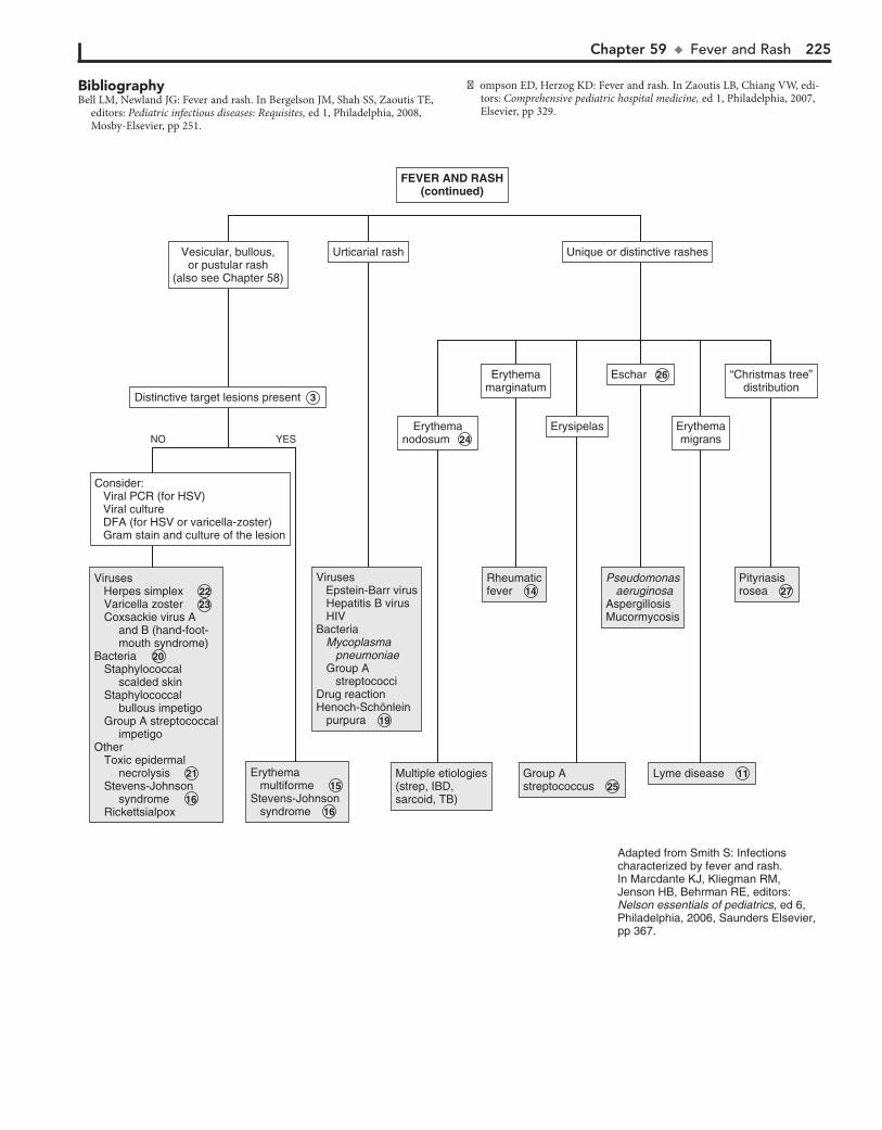
Chapter 59 u FeverandRash 225
BibliographyBell LM, Newland JG: Fever and rash. In Bergelson JM, Shah SS, Zaoutis TE,
editors: Pediatric infectious diseases: Requisites, ed 1, Philadelphia, 2008, Mosby-Elsevier, pp 251.
Erythema multiforme 15 Stevens-Johnson syndrome 16
Erythema multiforme 15 Stevens-Johnson syndrome 16
Distinctive target lesions present 3 Distinctive target lesions present 3
Consider: Viral PCR (for HSV) Viral culture DFA (for HSV or varicella-zoster) Gram stain and culture of the lesion
Consider: Viral PCR (for HSV) Viral culture DFA (for HSV or varicella-zoster) Gram stain and culture of the lesion
Vesicular, bullous,or pustular rash
(also see Chapter 58)
Vesicular, bullous,or pustular rash
(also see Chapter 58)
Urticarial rashUrticarial rash Unique or distinctive rashesUnique or distinctive rashes
ErythemamarginatumErythema
marginatum
Erythemamigrans
Erythemamigrans
“Christmas tree”distribution
“Christmas tree”distribution
ErysipelasErysipelasNO YES
Erythemanodosum 24
Erythemanodosum 24
Eschar 26 Eschar 26
Viruses Herpes simplex 22 Varicella zoster 23 Coxsackie virus A and B (hand-foot- mouth syndrome)Bacteria 20 Staphylococcal scalded skin Staphylococcal bullous impetigo Group A streptococcal impetigoOther Toxic epidermal necrolysis 21 Stevens-Johnson syndrome 16 Rickettsialpox
Viruses Herpes simplex 22 Varicella zoster 23 Coxsackie virus A and B (hand-foot- mouth syndrome)Bacteria 20 Staphylococcal scalded skin Staphylococcal bullous impetigo Group A streptococcal impetigoOther Toxic epidermal necrolysis 21 Stevens-Johnson syndrome 16 Rickettsialpox
Viruses Epstein-Barr virus Hepatitis B virus HIVBacteria Mycoplasma pneumoniae Group A streptococciDrug reactionHenoch-Schönlein purpura 19
Viruses Epstein-Barr virus Hepatitis B virus HIVBacteria Mycoplasma pneumoniae Group A streptococciDrug reactionHenoch-Schönlein purpura 19
Multiple etiologies(strep, IBD,sarcoid, TB)
Multiple etiologies(strep, IBD,sarcoid, TB)
Rheumaticfever 14 Rheumaticfever 14
Group Astreptococcus 25 Group Astreptococcus 25
Lyme disease 11 Lyme disease 11
Pseudomonas aeruginosaAspergillosisMucormycosis
Pseudomonas aeruginosaAspergillosisMucormycosis
Pityriasisrosea 27 Pityriasisrosea 27
Adapted from Smith S: Infections characterized by fever and rash.In Marcdante KJ, Kliegman RM, Jenson HB, Behrman RE, editors: Nelson essentials of pediatrics, ed 6, Philadelphia, 2006, Saunders Elsevier, pp 367.
FEVER AND RASH(continued)
FEVER AND RASH(continued)
Thompson ED, Herzog KD: Fever and rash. In Zaoutis LB, Chiang VW, edi-tors: Comprehensive pediatric hospital medicine, ed 1, Philadelphia, 2007, Elsevier, pp 329.

This page intentionally left blank

PART
IXHematology
228
Chapter 60
LYMPHADENOPATHY
often with hepatosplenomegaly. Bacterial causes include group A b-hemolytic streptococci or oral anaerobes such as Fusobacte-rium. Scalp infections such as tinea capitis may cause occipital lymphadenopathy.
3 If there is pharyngitis associated with cervical adenitis, a throat culture or rapid streptococcal antigen detection test
may be done to help diagnose group A b-hemolytic streptococci. Viral causes include EBV, herpes simplex, and enteroviruses.
4 Acute onset, unilateral adenitis is usually bacterial in ori-gin and may form an abscess. Bacterial infections of skin
and soft tissue (e.g., abscess, cellulitis, erysipelas, and fasciitis) are primarily caused by group A b-hemolytic streptococci or Staphylococcus aureus.
5 Subacute or chronic adenitis may be due to mycobacterial infections. Tuberculosis is increasing in incidence in chil-
dren and is usually associated with hilar adenopathy, with the lungs as the primary source of the infection. A positive tuberculin skin test and a chest x-ray may confirm tuberculosis. Induration of .15 mm is considered positive in a child older than 4 years with no risk factors. In children with known contacts with tuber-culosis, or who are clinically suspected to have tuberculosis, on immunosuppressive therapy, or who have immunosuppressive conditions, including HIV infection, induration .5 mm is con-sidered positive. Children at increased risk of disseminated dis-ease or increased environmental exposure to tuberculosis are considered positive at .10 mm induration. Interferon-gamma release assays may also be used in children 5 years.
6 Cervical adenitis is usually due to atypical mycobacteria, primarily Mycobacterium avium-intracellulare, M. kansasii,
M. scrofulaceum, and M. marinum. Diagnosis is determined using acid-fast staining and culture of the excised node or by fine-needle aspiration. An indeterminate tuberculin skin test with 5 to 9 mm of induration suggests infection with atypical mycobacteria. With modern methods of milk pasteurization, M. bovis, a previously common cause of cervical adenitis, is rarely seen.
7 Cat-scratch disease, caused by a gram-negative bacillus, Bartonella henselae, occurs after exposure to a scratch or
bite of a cat, with development of a papule at the site of trauma, followed in 7 to 14 days by regional lymphadenitis, usually axil-lary. Other symptoms include low-grade fever and malaise. The lymph nodes usually regress spontaneously within several weeks. Some 10% may have a purulent drainage that is culture negative. Serologic tests, such as indirect fluorescent antibody (IFA), enzyme-linked immunosorbent assay (ELISA), IgM tests, or polymerase chain reaction (PCR) testing are useful in making the diagnosis. If needed, diagnosis can be confirmed by biopsy of the node showing granulomas, central necrosis, and organisms seen on Warthin-Starry silver stain.
8 Kawasaki disease is determined clinically in children by noting 5 consecutive days of high fever accompanied by at
least four of the following five conditions: cervical lymphade-nopathy, oral mucosal erythema, conjunctivitis without exudates, rash, and extremity changes, such as edema and desquamation in the absence of known causes of these signs. The lymph node is typically single and large but is not consistently present.
9 If any of the ”red flags” are present, malignancy is suspected and the necessary evaluation should be done. This may in-
clude examination of the CBC for anemia, thrombocytopenia,
Lymphadenopathy is the presence of one or more enlarged lymph nodes measuring .1 cm in diameter for axillary and cer-vical nodes, .1.5 cm for inguinal nodes and .0.5 cm for epi-trochlear lymph nodes. It may be due to (1) reactive lymphade-nopathy, a common and normal function of lymph nodes characterized by hyperplasia in response to antigenic stimuli; (2) lymphadenitis, an inflammatory response to bacteria or their products, accompanied by erythema, warmth, and tenderness; (3) malignancy by primary origin in the node or secondary to metastases; and (4) rare lipid storage disorders.
1 A good history is essential. The age of the child may indicate the cause. Adenopathy in neonates may be due to infections
in utero (e.g., cytomegalovirus, syphilis, toxoplasmosis, human immunodeficiency virus [HIV]). In toddlers, adenopathy is usu-ally due to either focal infections that drain to the affected node or systemic viral infections. Malignancy is more likely to be a cause of lymphadenopathy in older children. Immunodeficiency may predispose children to opportunistic infections or malignancies. Certain medications (e.g., procainamide, sulfasalazine, phenytoin, or tetracycline) may cause adenopathy. Family or exposure history may suggest infections such as HIV, syphilis, tuberculosis, group A b-hemolytic streptococci, or mononucleosis. Birth and travel history may indicate exposure to endemic infections (e.g., tuber-culosis, histoplasmosis), as well as consumption of infected foods (e.g., Brucella, Mycobacterium from unpasteurized milk prod-ucts). Diagnostic clues may be revealed by social history (e.g., socioeconomic status or ethnicity), family diet (e.g., consumption of raw meats), and presence of family pets (e.g., cat-scratch dis-ease, toxoplasmosis from kitty litter). Adolescents must be asked about sexual activity, risk factors for HIV, and exposure to sexually transmitted diseases (e.g., syphilis). Lymphogranuloma venereum may cause inguinal lymphadenopathy. An acute onset may sug-gest infection, whereas an insidious onset accompanied by sys-temic symptoms (e.g., anorexia, weight loss, fevers, night sweats) suggests Hodgkin disease.
On physical examination, all areas that may be involved must be palpated, including cervical, preauricular and postauricular, axil-lary, epitrochlear, inguinal, and supraclavicular. Location of the node may be helpful in diagnosis, whether lymphadenopathy is localized or generalized. Localized lymphadenopathy often indi-cates involvement in the area of lymphatic drainage. Supraclavicu-lar lymphadenopathy is usually a red flag for mediastinal tumors or infections or for metastatic abdominal tumors. Palpation of the nodes is helpful, with erythema, warmth, and tenderness indicat-ing adenitis. Tender, nonerythematous, soft nodes may indicate a viral or a systemic infection. Firm or hard, rubbery, nontender nodes may indicate infiltrating tumors. Hard, matted, fixed, non-tender nodes indicate tumor or fibrosis after acute infection.
2 Reactive adenopathy is usually a transient response to infec-tions of the upper respiratory tract or skin. Pharyngeal infec-
tions commonly cause cervical lymphadenopathy. Common viral agents include adenovirus, parainfluenza, influenza, rhinovirus, and enterovirus. Cytomegalovirus (CMV) and Epstein-Barr virus (EBV) may cause a localized or generalized lymphadenopathy,

Chapter 60 u Lymphadenopathy 229
ReactiveAdenopathy 2 ReactiveAdenopathy 2
Group A �-hemolytic streptococciAdenovirusEBVCMVHerpes simplex I, VIEnterovirus
Group A �-hemolytic streptococciAdenovirusEBVCMVHerpes simplex I, VIEnterovirus
Staphylococcus aureusGroup A �-hemolyticAnaerobesPasturella multicodaYersenia pestisLGVSyphilisKawasaki disease 8
Staphylococcus aureusGroup A �-hemolyticAnaerobesPasturella multicodaYersenia pestisLGVSyphilisKawasaki disease 8
EBVCMVToxoplasmosisM. tuberculosis 5 Atypical mycobacterium 6 S. aureusGroup A �-hemolytic streptococciCat scratch disease 7 Herpes simplexEnterovirusesKawasaki disease 8
EBVCMVToxoplasmosisM. tuberculosis 5 Atypical mycobacterium 6 S. aureusGroup A �-hemolytic streptococciCat scratch disease 7 Herpes simplexEnterovirusesKawasaki disease 8
Hodgkin disease 10 Non-Hodgkin lymphomaLeukemiaNeuroblastomaRhabdomyosarcomaThyroid cancer
Hodgkin disease 10 Non-Hodgkin lymphomaLeukemiaNeuroblastomaRhabdomyosarcomaThyroid cancer
Tests for group A streptococci Rapid antigen detection test Throat culture± CBC± “Monospot”± EBV titers± Viral polymerase chain reaction
Tests for group A streptococci Rapid antigen detection test Throat culture± CBC± “Monospot”± EBV titers± Viral polymerase chain reaction
ConsiderCBCESRCRPPPD/ QuantiferonChest x-rayTest for group A streptococciPCR/serology for EBV, CMV, toxoplasmosis, B. henselae, HIVEchocardiogram
ConsiderCBCESRCRPPPD/ QuantiferonChest x-rayTest for group A streptococciPCR/serology for EBV, CMV, toxoplasmosis, B. henselae, HIVEchocardiogram
Signs and symptoms indicating possible malignancy 9 Supraclavicular node(s) Fixed/matted Progressive, painless enlargement Signs of airway obstruction Prolonged constitutional symptoms (fever, night sweats, weight loss)
Signs and symptoms indicating possible malignancy 9 Supraclavicular node(s) Fixed/matted Progressive, painless enlargement Signs of airway obstruction Prolonged constitutional symptoms (fever, night sweats, weight loss)
CBCCRPESRLDHAlkaline phosphataseUric acidImaging (chest x-ray/ ultrasound/CT)
Consider referral for:BiopsyBone marrow studies
CBCCRPESRLDHAlkaline phosphataseUric acidImaging (chest x-ray/ ultrasound/CT)
Consider referral for:BiopsyBone marrow studies
Consider:CBCBlood cultureIncision and drainageAspiration
Consider:CBCBlood cultureIncision and drainageAspiration
Suppurative or abscessUnilateral 4
Suppurative or abscessUnilateral 4
Pharyngitis 3 Pharyngitis 3
Nontender, small nodesAssociated URI, skin or dental infection
Nontender, small nodesAssociated URI, skin or dental infection
Warm, tender nodes± Fever± Erythema
Warm, tender nodes± Fever± Erythema
Chronic >4 weeksChronic >4 weeks
(See page 60-5)(See page 60-5)
GeneralizedGeneralizedLocalizedLocalized
LocationLocation
AdenitisAdenitis
Acute onset(<7 days)Acute onset(<7 days)
Sub-acute/ chronic onset(>1-3 weeks)
Sub-acute/ chronic onset(>1-3 weeks)
URI = Upper respiratory infectionEBV = Epstein-Barr virusCMV = CytomegalovirusLDH = Lactate dehydrogenaseVDRL = Venereal Disease Research LaboratoryRPR = Rapid plasma reaginPPD = Purified protein derivative (tuberculosis skin test)PCR = Polymerase chain reaction
H and P 1 H and P 1
LYMPHADENOPATHYLYMPHADENOPATHY
YESNO
Nelson Textbook of Pediatrics, 19e. Chapters 159, 160, 195, 201, 207, 210, 244, 246, 268, 282, 490, 501, 658Nelsons Essentials, 6e. Chapter 99
230 Part IX u Hematology
leukopenia, or leukocytosis, and for blast cells. Because of a tumor burden or bone involvement, there may be an elevation of lactate dehydrogenase, alkaline phosphatase, and uric acid levels. A chest x-ray may show mediastinal lymphadenopathy, which is sugges-tive of lymphoma. A CT scan may also be considered. Close clinical follow up should be done to watch for progression of symptoms. If cancer is suspected, a referral should be made to the appropriate specialist for biopsy or for bone marrow studies.
10 Hodgkin disease usually occurs as painless cervical or supra-clavicular lymphadenopathy in older children and adoles-
cents. Approximately 30% have systemic symptoms (e.g., fatigue, weight loss, fevers, night sweats). Pruritus, hemolytic anemia, and chest pain after alcohol ingestion are clues. Diagnosis is confirmed by lymph node biopsy and/or bone marrow aspiration. Non-Hodgkin lymphoma usually occurs as supraclavicular, cervical, or axillary adenopathy. In children in the United States, B-cell lym-phomas originate in the abdomen with inguinal or iliac lymph-adenopathy. About half of children with acute lymphoblastic leukemia present with adenopathy at the time of diagnosis. Sys-temic signs and symptoms (e.g., fever, malaise, weight loss, pallor, bone pain, petechiae, hepatosplenomegaly) may be present. The CBC may show anemia, thrombocytopenia, leukocytosis or leu-kopenia, and circulating blast cells. Acute myelogenous leukemia is less common in children but appears similarly. Several other malignancies, including disseminated neuroblastoma, rhabdo-myosarcoma, and thyroid cancer, may occur as localized or gen-eralized lymphadenopathy.
11 Generalized lymphadenopathy may be due to infections. HIV-infected children may have systemic symptoms, fail-
ure to thrive, and evidence of opportunistic infections. Many
viruses including EBV and CMV may cause generalized lymph-adenopathy. Syphilis caused by the spirochete Treponema palli-dum results in both localized and generalized lymphadenopathy. In primary syphilis there is usually localized inguinal adenopa-thy; in secondary syphilis there is usually generalized lymph node involvement. Epitrochlear involvement is suggestive. Diag-nostic tests include rapid plasma reagin (RPR), Venereal Disease Research Laboratory (VDRL), and fluorescent treponemal anti-body absorption (FTA-ABS). (See Chapter 41.) Toxoplasma gondii is a parasite of cats and can be acquired by humans from contact with cat feces or by eating raw meat. Although the infec-tion may be transmitted to the fetus, lymphadenopathy is un-common in the newborn and is more common in older children and young adults. Disseminated tuberculosis may present as generalized lymphadenopathy, pulmonary infiltrates, and systemic symptoms. Atypical mycobacteria may cause generalized lymph-adenopathy in HIV-infected children.
12 Sinus histiocytosis is a rare disorder with massive cervical lymphadenopathy, fever, elevated erythrocyte sedimenta-
tion rate, leukocytosis, and hypergammaglobulinemia. It may be diagnosed using biopsy and tends to resolve spontaneously. Rosai-Dorfman disease is a form of sinus histiocytosis with massive lymphadenopathy.
13 Drug reactions occasionally cause adenopathy. Examples include serum sickness from drugs such as cephalosporins
or the drug itself.
BibliographySahai S: Lymphadenopathy, Pediatr Rev 34(5):216–27, 2013.
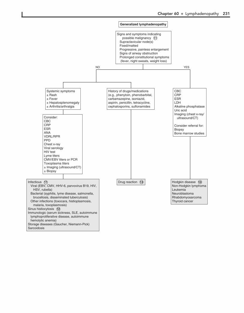
Chapter 60 u Lymphadenopathy 231
Infectious 11 Viral (EBV, CMV, HHV-6, parvovirus B19, HIV, HSV, rubella) Bacterial (syphilis, lyme disease, salmonella, brucellosis, disseminated tuberculosis) Other infections (toxocara, histioplasmosis, malaria, toxoplasmosis)Sinus histiocytosis 12 Immunologic (serum sickness, SLE, autoimmune lymphoproliferative disease, autoimmune hemolytic anemia)Storage diseases (Gaucher, Niemann-Pick)Sarcoidosis
Infectious 11 Viral (EBV, CMV, HHV-6, parvovirus B19, HIV, HSV, rubella) Bacterial (syphilis, lyme disease, salmonella, brucellosis, disseminated tuberculosis) Other infections (toxocara, histioplasmosis, malaria, toxoplasmosis)Sinus histiocytosis 12 Immunologic (serum sickness, SLE, autoimmune lymphoproliferative disease, autoimmune hemolytic anemia)Storage diseases (Gaucher, Niemann-Pick)Sarcoidosis
Drug reaction 13 Drug reaction 13 Hodgkin disease 10 Non-Hodgkin lymphomaLeukemiaNeuroblastomaRhabdomyosarcomaThyroid cancer
Hodgkin disease 10 Non-Hodgkin lymphomaLeukemiaNeuroblastomaRhabdomyosarcomaThyroid cancer
Signs and symptoms indicating possible malignancy 11 Supraclavicular node(s) Fixed/matted Progressive, painless enlargement Signs of airway obstruction Prolonged constitutional symptoms (fever, night sweats, weight loss)
Signs and symptoms indicating possible malignancy 11 Supraclavicular node(s) Fixed/matted Progressive, painless enlargement Signs of airway obstruction Prolonged constitutional symptoms (fever, night sweats, weight loss)
Consider:CBCCRPESRANAVDRL/RPRPPDChest x-rayViral serologyHIV testLyme titersCMV/EBV titers or PCRToxoplasma titers ± Imaging (ultrasound/CT)± Biopsy
Consider:CBCCRPESRANAVDRL/RPRPPDChest x-rayViral serologyHIV testLyme titersCMV/EBV titers or PCRToxoplasma titers ± Imaging (ultrasound/CT)± Biopsy
Systemic symptoms ± Rash± Fever± Hepatosplenomegaly± Arthritis/arthralgia
Systemic symptoms ± Rash± Fever± Hepatosplenomegaly± Arthritis/arthralgia
History of drugs/medications(e.g., phenytoin, phenobarbital, carbamazepine, isoniazid, aspirin, penicillin, tetracycline,cephalosporins, sulfonamides
History of drugs/medications(e.g., phenytoin, phenobarbital, carbamazepine, isoniazid, aspirin, penicillin, tetracycline,cephalosporins, sulfonamides
CBCCRPESRLDHAlkaline phosphataseUric acidImaging (chest x-ray/ ultrasound/CT)
Consider referral for:BiopsyBone marrow studies
CBCCRPESRLDHAlkaline phosphataseUric acidImaging (chest x-ray/ ultrasound/CT)
Consider referral for:BiopsyBone marrow studies
Generalized lymphadenopathyGeneralized lymphadenopathy
YESNO
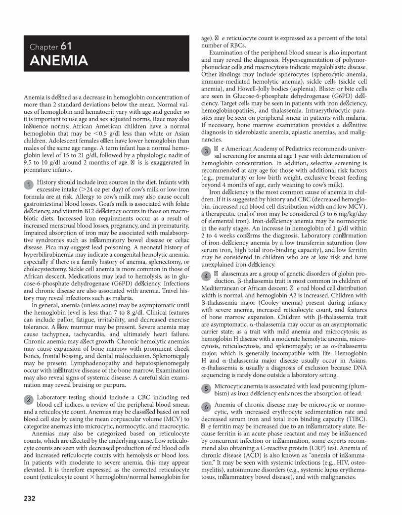
232
Chapter 61
ANEMIA
age). The reticulocyte count is expressed as a percent of the total number of RBCs.
Examination of the peripheral blood smear is also important and may reveal the diagnosis. Hypersegmentation of polymor-phonuclear cells and macrocytosis indicate megaloblastic disease. Other findings may include spherocytes (spherocytic anemia, immune-mediated hemolytic anemia), sickle cells (sickle cell anemia), and Howell-Jolly bodies (asplenia). Blister or bite cells are seen in Glucose-6-phosphate dehydrogenase (G6PD) defi-ciency. Target cells may be seen in patients with iron deficiency, hemoglobinopathies, and thalassemia. Intraerythrocytic para-sites may be seen on peripheral smear in patients with malaria. If necessary, bone marrow examination provides a definitive diagnosis in sideroblastic anemia, aplastic anemias, and malig-nancies.
3 The American Academy of Pediatrics recommends univer-sal screening for anemia at age 1 year with determination of
hemoglobin concentration. In addition, selective screening is recommended at any age for those with additional risk factors (e.g., prematurity or low birth weight, exclusive breast feeding beyond 4 months of age, early weaning to cow’s milk).
Iron deficiency is the most common cause of anemia in chil-dren. If it is suggested by history and CBC (decreased hemoglo-bin, increased red blood cell distribution width and low MCV), a therapeutic trial of iron may be considered (3 to 6 mg/kg/day of elemental iron). Iron-deficiency anemia may be normocytic in the early stages. An increase in hemoglobin of 1 g/dl within 2 to 4 weeks confirms the diagnosis. Laboratory confirmation of iron-deficiency anemia by a low transferrin saturation (low serum iron, high total iron-binding capacity), and low ferritin may be considered in children who are at low risk and have unexplained iron deficiency.
4 Thalassemias are a group of genetic disorders of globin pro-duction. b-thalassemia trait is most common in children of
Mediterranean or African descent. The red blood cell distribution width is normal, and hemoglobin A2 is increased. Children with b-thalassemia major (Cooley anemia) present during infancy with severe anemia, increased reticulocyte count, and features of bone marrow expansion. Children with b-thalassemia trait are asymptomatic. a-thalassemia may occur as an asymptomatic carrier state; as a trait with mild anemia and microcytosis; as hemoglobin H disease with a moderate hemolytic anemia, micro-cytosis, reticulocytosis, and splenomegaly; or as a-thalassemia major, which is generally incompatible with life. Hemoglobin H and a-thalassemia major disease usually occur in Asians. a-thalassemia is usually a diagnosis of exclusion because DNA sequencing is rarely done outside a laboratory setting.
5 Microcytic anemia is associated with lead poisoning (plum-bism) as iron deficiency enhances the absorption of lead.
6 Anemia of chronic disease may be microcytic or normo-cytic, with increased erythrocyte sedimentation rate and
decreased serum iron and total iron binding capacity (TIBC). The ferritin may be increased due to an inflammatory state. Be-cause ferritin is an acute phase reactant and may be influenced by concurrent infection or inflammation, some experts recom-mend also obtaining a C-reactive protein (CRP) test. Anemia of chronic disease (ACD) is also known as “anemia of inflamma-tion.” It may be seen with systemic infections (e.g., HIV, osteo-myelitis), autoimmune disorders (e.g., systemic lupus erythema-tosus, inflammatory bowel disease), and with malignancies.
Anemia is defined as a decrease in hemoglobin concentration of more than 2 standard deviations below the mean. Normal val-ues of hemoglobin and hematocrit vary with age and gender so it is important to use age and sex adjusted norms. Race may also influence norms; African American children have a normal hemoglobin that may be ,0.5 g/dl less than white or Asian children. Adolescent females often have lower hemoglobin than males of the same age range. A term infant has a normal hemo-globin level of 15 to 21 g/dl, followed by a physiologic nadir of 9.5 to 10 g/dl around 2 months of age. This is exaggerated in premature infants.
1 History should include iron sources in the diet. Infants with excessive intake (.24 oz per day) of cow’s milk or low-iron
formula are at risk. Allergy to cow’s milk may also cause occult gastrointestinal blood losses. Goat’s milk is associated with folate deficiency, and vitamin B12 deficiency occurs in those on macro-biotic diets. Increased iron requirements occur as a result of increased menstrual blood losses, pregnancy, and in prematurity. Impaired absorption of iron may be associated with malabsorp-tive syndromes such as inflammatory bowel disease or celiac disease. Pica may suggest lead poisoning. A neonatal history of hyperbilirubinemia may indicate a congenital hemolytic anemia, especially if there is a family history of anemia, splenectomy, or cholecystectomy. Sickle cell anemia is more common in those of African descent. Medications may lead to hemolysis, as in glu-cose-6-phosphate dehydrogenase (G6PD) deficiency. Infections and chronic disease are also associated with anemia. Travel his-tory may reveal infections such as malaria.
In general, anemia (unless acute) may be asymptomatic until the hemoglobin level is less than 7 to 8 g/dl. Clinical features can include pallor, fatigue, irritability, and decreased exercise tolerance. A flow murmur may be present. Severe anemia may cause tachypnea, tachycardia, and ultimately heart failure. Chronic anemia may affect growth. Chronic hemolytic anemias may cause expansion of bone marrow with prominent cheek bones, frontal bossing, and dental malocclusion. Splenomegaly may be present. Lymphadenopathy and hepatosplenomegaly occur with infiltrative disease of the bone marrow. Examination may also reveal signs of systemic disease. A careful skin exami-nation may reveal bruising or purpura.
2 Laboratory testing should include a CBC including red blood cell indices, a review of the peripheral blood smear,
and a reticulocyte count. Anemias may be classified based on red blood cell size by using the mean corpuscular volume (MCV) to categorize anemias into microcytic, normocytic, and macrocytic.
Anemias may also be categorized based on reticulocyte counts, which are affected by the underlying cause. Low reticulo-cyte counts are seen with decreased production of red blood cells and increased reticulocyte counts with hemolysis or blood loss. In patients with moderate to severe anemia, this may appear elevated. It is therefore expressed as the corrected reticulocyte count (reticulocyte count 3 hemoglobin/normal hemoglobin for
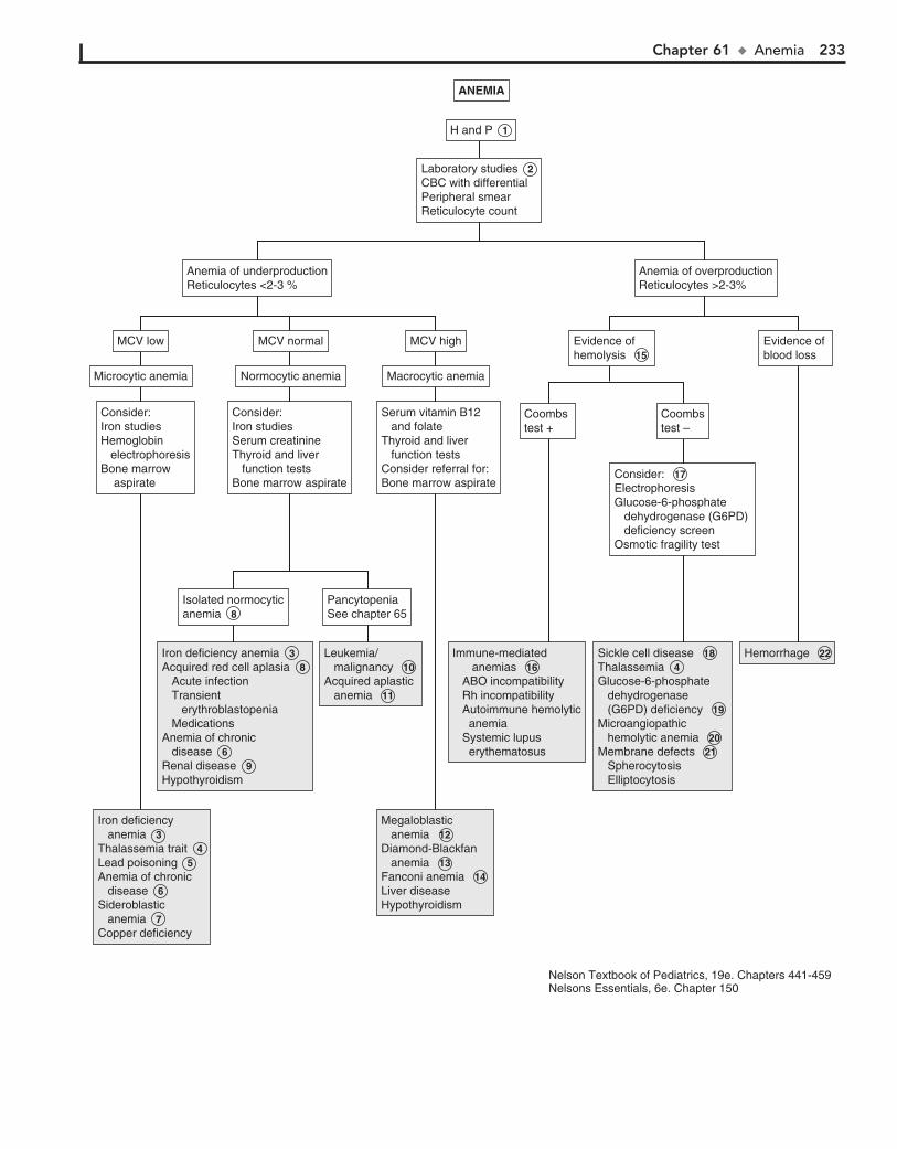
Chapter 61 u Anemia 233
Iron deficiency anemia 3 Thalassemia trait 4 Lead poisoning 5 Anemia of chronic disease 6 Sideroblastic anemia 7 Copper deficiency
Iron deficiency anemia 3 Thalassemia trait 4 Lead poisoning 5 Anemia of chronic disease 6 Sideroblastic anemia 7 Copper deficiency
Iron deficiency anemia 3 Acquired red cell aplasia 8 Acute infection Transient erythroblastopenia Medications Anemia of chronic disease 6 Renal disease 9 Hypothyroidism
Iron deficiency anemia 3 Acquired red cell aplasia 8 Acute infection Transient erythroblastopenia Medications Anemia of chronic disease 6 Renal disease 9 Hypothyroidism
Leukemia/ malignancy 10 Acquired aplastic anemia 11
Leukemia/ malignancy 10 Acquired aplastic anemia 11
Megaloblastic anemia 12 Diamond-Blackfan anemia 13 Fanconi anemia 14 Liver diseaseHypothyroidism
Megaloblastic anemia 12 Diamond-Blackfan anemia 13 Fanconi anemia 14 Liver diseaseHypothyroidism
Immune-mediated anemias 16 ABO incompatibility Rh incompatibility Autoimmune hemolytic anemia Systemic lupus erythematosus
Immune-mediated anemias 16 ABO incompatibility Rh incompatibility Autoimmune hemolytic anemia Systemic lupus erythematosus
Sickle cell disease 18 Thalassemia 4 Glucose-6-phosphate dehydrogenase (G6PD) deficiency 19 Microangiopathic hemolytic anemia 20 Membrane defects 21 Spherocytosis Elliptocytosis
Sickle cell disease 18 Thalassemia 4 Glucose-6-phosphate dehydrogenase (G6PD) deficiency 19 Microangiopathic hemolytic anemia 20 Membrane defects 21 Spherocytosis Elliptocytosis
Hemorrhage 22Hemorrhage 22
Laboratory studies 2 CBC with differential Peripheral smearReticulocyte count
Laboratory studies 2 CBC with differential Peripheral smearReticulocyte count
H and P 1 H and P 1
Consider:Iron studiesHemoglobin electrophoresisBone marrow aspirate
Consider:Iron studiesHemoglobin electrophoresisBone marrow aspirate
Microcytic anemiaMicrocytic anemia
MCV lowMCV low
Normocytic anemiaNormocytic anemia
MCV normalMCV normal
Anemia of underproductionReticulocytes <2-3 %Anemia of underproductionReticulocytes <2-3 %
Anemia of overproductionReticulocytes >2-3%Anemia of overproductionReticulocytes >2-3%
Macrocytic anemiaMacrocytic anemia
MCV highMCV high
Consider:Iron studiesSerum creatinineThyroid and liver function testsBone marrow aspirate
Consider:Iron studiesSerum creatinineThyroid and liver function testsBone marrow aspirate
Serum vitamin B12 and folateThyroid and liver function testsConsider referral for:Bone marrow aspirate
Serum vitamin B12 and folateThyroid and liver function testsConsider referral for:Bone marrow aspirate
Coombs test +Coombs test +
Coombstest –Coombstest –
Evidence ofhemolysis 15 Evidence ofhemolysis 15
Evidence of blood lossEvidence of blood loss
Consider: 17 ElectrophoresisGlucose-6-phosphate dehydrogenase (G6PD) deficiency screenOsmotic fragility test
Consider: 17 ElectrophoresisGlucose-6-phosphate dehydrogenase (G6PD) deficiency screenOsmotic fragility test
Isolated normocyticanemia 8 Isolated normocyticanemia 8
Pancytopenia See chapter 65Pancytopenia See chapter 65
ANEMIAANEMIA
Nelson Textbook of Pediatrics, 19e. Chapters 441-459Nelsons Essentials, 6e. Chapter 150
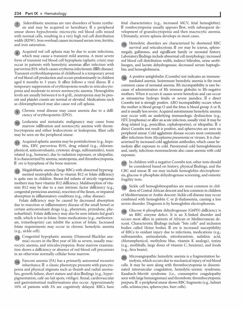
234 Part IX u Hematology
7 Sideroblastic anemias are rare disorders of heme synthe-sis and may be acquired or hereditary. The peripheral
smear shows hypochromic microcytic red blood cells mixed with normal cells, resulting in a very high red cell distribution width (RDW). Iron overload causes increased serum iron, ferritin, and iron saturation.
8 Acquired red cell aplasia may be due to acute infections, which may cause a transient mild anemia. A more severe
form of transient red blood cell hypoplasia (aplastic crisis) may occur in patients with hemolytic anemias after infection with parvovirus B19, which causes erythema infectiosum (fifth disease). Transient erythroblastopenia of childhood is a temporary arrest of red blood cell production and occurs predominantly in children aged 6 months to 3 years. It often follows a viral illness. The temporary suppression of erythropoiesis results in reticulocyto-penia and moderate to severe normocytic anemia. Hemoglobin levels are usually between 6 to 8 g/dL, neutropenia may be pres-ent and platelet counts are normal or elevated. Medications such as chloramphenicol may also cause red cell aplasia.
9 Chronic renal disease may cause anemia due to a defi-ciency of erythropoietin (EPO).
10 Leukemia and metastatic malignancy may cause bone marrow infiltration and normocytic anemia with throm-
bocytopenia and either leukocytosis or leukopenia. Blast cells may be seen on the peripheral smear.
11 Acquired aplastic anemia may be postinfectious (e.g., hepa-titis, EBV, parvovirus B19), drug related (e.g., chloram-
phenicol, anticonvulsants, cytotoxic drugs, sulfonamides), toxin related (e.g., benzene), due to radiation exposure, or idiopathic. It is characterized by anemia, neutropenia, and thrombocytopenia. There is hypoplasia of the bone marrow.
12 Megaloblastic anemia (large RBCs with abnormal hyperseg-mented neutrophils due to vitamin B12 or folate deficiency
is quite rare in children. Breast-fed infants of strictly vegetarian mothers may have vitamin B12 deficiency. Malabsorption of vita-min B12 may be due to a rare intrinsic factor deficiency (e.g., congenital pernicious anemia), resection of the ileum, or impaired absorption in inflammatory conditions (e.g., celiac disease).
Folate deficiency may be caused by decreased absorption due to resection or inflammatory disease of the small bowel or certain anticonvulsant drugs (e.g., phenytoin, primidone, phe-nobarbital). Folate deficiency may also be seen infants fed goat’s milk, which is low in folate. Some medications (e.g., methotrex-ate, trimethoprim) can inhibit the action of folate. Increased folate requirements may occur in chronic hemolytic anemia (e.g., sickle cell).
13 Congenital hypoplastic anemia (Diamond-Blackfan ane-mia) occurs in the first year of life as severe, usually mac-
rocytic anemia, and reticulocytopenia. Bone marrow examina-tion shows a deficiency or absence of red blood cell precursors in an otherwise normally cellular bone marrow.
14 Fanconi anemia (FA) has a primarily autosomal recessive inheritance. The classic phenotype presents with pancyto-
penia and physical stigmata such as thumb and radial anoma-lies, growth failure, short stature and skin findings (e.g., hyper-pigmentation, café-au-lait spots, vitiligo). Renal, cardiovascular and gastrointestinal malformations also occur. Approximately 10% of patients with FA are cognitively delayed. RBCs have
fetal characteristics (e.g., increased MCV, fetal hemoglobin). Thrombocytopenia usually appears first, with subsequent de-velopment of granulocytopenia and then macrocytic anemia. Ultimately, severe aplasia develops in most cases.
15 Hemolytic disorders are characterized by shortened RBC survival and reticulocytosis. There may be icterus, spleno-
megaly, gallstones, and significant family or neonatal history. Laboratory findings include abnormal cell morphology; increased red blood cell distribution width, indirect bilirubin, urine urobi-linogen, and lactate dehydrogenase; decreased serum haptoglo-bin; and hemoglobinuria.
16 A positive antiglobulin (Coombs) test indicates an immune-mediated anemia. Isoimmune hemolytic anemia is the most
common cause of neonatal anemia. Rh incompatibility is rare be-cause of administration of Rh immune globulin to Rh-negative mothers. When it occurs it causes severe hemolysis and can occur as intrauterine hydrops fetalis or severe jaundice. The direct Coombs test is strongly positive. ABO incompatibility occurs when the mother is blood group O and the fetus is blood group A or B, and is usually less severe. Acquired autoimmune hemolytic anemia may occur with an underlying immunologic dysfunction (e.g., HIV, lymphoma) or after an acute infection, usually viral. It may be drug related (e.g., penicillins, cephalosporins), or idiopathic. The direct Coombs test result is positive, and spherocytes are seen on peripheral smear. Cold agglutinin disease occurs most commonly after infections from Mycoplasma pneumonia or viruses. It is char-acterized by increased cold agglutinin antibodies, which cause he-molysis after exposure to cold. Paroxysmal cold hemoglobinuria and, rarely, infectious mononucleosis also cause anemia with cold exposure.
17 In children with a negative Coombs test, other tests should be considered based on history, physical findings, and the
CBC and smear. These may include hemoglobin electrophore-sis, glucose-6-phosphate dehydrogenase screening, and osmotic fragility tests.
18 Sickle cell hemoglobinopathies are most common in chil-dren of Central African descent and less common in children
of Mediterranean or Arabic descent. Sickle cell disease may occur combined with hemoglobin C or b-thalassemia, causing a less severe disorder. Diagnosis is by hemoglobin electrophoresis.
19 Glucose-6-phosphate dehydrogenase (G6PD) deficiency is an RBC enzyme defect. It is an X-linked disorder and
occurs most often in patients of African or Mediterranean de-scent. Characteristic findings include ”bite cells” and inclusion bodies called Heinz bodies. There is increased susceptibility of RBCs to oxidant injury due to infections, medications (e.g., sulfonamides, antimalarials, nitrofurantoin, nalidixic acid, chloramphenicol, methylene blue, vitamin K analogs), toxins (e.g., mothballs, large doses of vitamin C, benzene), and foods (e.g., fava beans).
20 Microangiopathic hemolytic anemia is a fragmentation he-molysis, which occurs due to mechanical injury of red blood
cells. It may be seen along with thrombocytopenia in dissemi-nated intravascular coagulation, hemolytic-uremic syndrome, Kasabach-Merritt syndrome (i.e., consumptive coagulopathy seen with large hemangiomas) and thrombotic thrombocytopenic purpura. The peripheral smear shows RBC fragments (e.g., helmet cells, schistocytes, spherocytes, burr cells).

Chapter 61 u Anemia 235
21 Membrane defects include hereditary spherocytosis and elliptocytosis. Presenting features are anemia, jaundice,
and splenomegaly. Diagnosis is confirmed by an osmotic fragility test.
22 A significant acute or subacute blood loss leads to anemia. The RBC morphology and size remain normal. It may require
3 to 5 days to produce an elevated reticulocyte count. Transfusion may be required to replace large blood losses when there are clinical
manifestations such as fatigue, lightheadedness, tachycardia, dyspnea, or heart failure.
BibliographyBaker RD, Greer FR: Diagnosis and prevention of iron deficiency and iron-
deficiency anemia in infants and young children (0-3 years of age), Pediatrics 126(5):1040–1050, 2010.
Janus J, Moerschel SK: Evaluation of anemia in children, Am Fam Physician 81(12):1462–1471, 2010.
236
Chapter 62
BLEEDING
PT and aPTT. Testing for an inhibitor is done by mixing one part of patient plasma with one part pooled plasma; if the PT or aPTT corrects, there is a factor deficiency; if it does not correct, an inhibitor is present. Inhibitors include anticoagulants (e.g., heparin), autoantibodies against clotting factors, and lupus type anticoagulants. Fibrinogen function is measured by thrombin time or fibrinogen activity. In von Willebrand disease, the PTT may be prolonged, but testing should be considered if mucocutaneous bleeding disorder is suspected because screening laboratory results may be normal in von Willebrand disease. Testing may include a quantitative assay for von Willebrand factor (vWF) antigen, testing for vWF activity (ristocetin cofactor activity), testing for plasma factor VIII activity, determination of vWF structure (vWF multimers), and a platelet count. If the patient is taking medications that might interfere with hemostasis, all tests should be done or repeated after stopping the medication. Bleeding time indirectly measures platelet number, function and plateletvessel wall interaction; it has low sensitivity. Platelet function studies may include PFA100 as an initial screen for platelet function abnormalities but there are problems with sensitivity and specificity. If platelet function abnormality is seriously being considered, platelet aggregation studies are needed.
3 Vitamin K deficiency is common in neonates, malnourished children, those receiving broadspectrum antibiot
ics, and children with cholestatic liver disease. Hemorrhagic disease of the newborn is rarely seen anymore because of prophylactic administration on Vitamin K in the newborn nursery. Vitamin K deficiency may present as generalized bleeding into skin, gastrointestinal tract, and central nervous system. Dicumarol and rat poison (superwarfarin) also act by affecting vitamin K and, therefore, also vitamin Kdependent factors II, VII, IX, and X.
4 Inhibitors may be directed against a specific coagulation factor or a reaction site in the coagulation pathway. In
hibitors against specific factors usually affect factors VIII, IX, or XI, causing prolonged PTT with normal PT. Lupus anticoagulant is directed against a reaction site and causes prolonged PT and PTT; it is more likely to cause thrombosis than bleeding.
5 Deficiency of factor VIII is hemophilia A, and that of factor IX is hemophilia B or Christmas disease. These are
transmitted as Xlinked traits; therefore, family history of bleeding in males in the mother’s family should be obtained. Hemophilia A or B usually occur as bleeding into muscles or joints and easy bruising. Because factor VIII does not cross the placenta, bleeding may occur during the neonatal period. Factor XI deficiency, or hemophilia C, has autosomal recessive transmission and milder symptoms than A and B. Factor XII or Hageman factor deficiency causes prolonged PTT but is usually asymptomatic.
6 The most common cause of lifelong symptoms of mucocutaneous bleeding is von Willebrand disease due to deficiency
of vWF, which is prevalent in approximately 1% of children. Inheritance is typically autosomal dominant. The vWF is a protein required for platelet plug formation; it is also a carrier for factor VIII. In severe deficiency of vWF there is also deficient factor VIII. PTT and bleeding time are often, but not always, abnormal, and so screening for vWF should always be considered when evaluating bleeding disorders. The PTT corrects with 1:1 mixing with pooled plasma. In addition, stress, medications, trauma, and difficult venipuncture may increase vWF levels. Levels of vWF are
Bleeding disorders are caused by a disturbance in the normal hemostasis. Components of the hemostatic mechanism include platelets, anticoagulant proteins, procoagulant proteins, and components of the vessel walls. The first stage of hemostasis consists of formation of “platelet plug” followed by activation of coagulation pathways leading to formation of a fibrin clot.
1 History should include age and acuity at onset of bleeding, triggers, whether bleeding was immediate or delayed, and
whether it was prolonged or exaggerated. Severity of bleeding must be determined, such as nosebleeds requiring cautery or packing or surgeries requiring transfusions. It is very important to differentiate bruising due to child abuse. Perinatal history should include details regarding bruising or petechiae, bleeding with circumcision, cephalhematoma, CNS or gastrointestinal bleeding, unexplained anemia or jaundice, or bleeding after cord separation. A history of vitamin K administration and maternal drugs should be obtained. In adolescent girls a history of dysfunctional uterine bleeding would be important. A detailed family history should be obtained, including maternal obstetric history of bleeding. For Xlinked disorders (e.g., hemophilias) a family history of male relatives on the maternal side should be solicited, and history of consanguinity for autosomal recessive conditions (Factor XIII deficiency). Medications such as aspirin, other nonsteroidal antiinflammatory drugs (NSAIDs), anticoagulants, antibiotics, and anticonvulsants may be associated with bleeding. The bleeding site may indicate the etiology. Acute mucocutaneous bleeding may indicate idiopathic thrombocytopenic purpura. Generalized bleeding may be associated with disseminated intravascular coagulation (DIC), vitamin K deficiency, liver disease, or uremia. Mucosal bleeding, petechiae, and bruising may indicate disorders of platelets, blood vessels, or von Willebrand disease. Bleeding into soft tissues, muscles and joints, or excessive bleeding with surgery may be due to deficiency of coagulation factors (e.g., hemophilia). Various findings on physical examination may be helpful in determining the etiology. Heart murmurs may indicate endocarditis; arthropathy is suggestive of hemophilia; joint laxity is seen with EhlersDanlos syndrome; and thumb or radial anomalies are seen with Fanconi anemia or thrombocytopeniaabsent radius (TAR) syndrome. Hepatosplenomegaly may indicate liver disease; lymphadenopathy may indicate a malignancy. Skin findings include hematomas, petechiae, ecchymoses, telangiectasia, purpura, poor wound healing, lax skin, and varicose veins.
2 The critical evaluation for a bleeding disorder should begin with a few specific laboratory tests. A CBC including a
platelet count may reveal thrombocytopenia or anemia. A peripheral smear can exclude pseudothrombocytopenia due to platelet clumping. Platelet morphology may indicate the diagnosis; platelets are small in Wiscott Aldrich syndrome and giant platelets are seen in Bernard Soulier syndrome. Prothrombin time (PT) and activated partial thromboplastin time (aPTT) are dependent on all coagulation factors except factor XIII. A clotting factor deficiency or the presence of an inhibitor can prolong

Chapter 62 u Bleeding 237
Thrombocytopenia (infection, ITP, leukemia) (see Chapter 63)Platelet abnormalities (Bernard-Soulier, Wiscott-Aldrich)
Thrombocytopenia (infection, ITP, leukemia) (see Chapter 63)Platelet abnormalities (Bernard-Soulier, Wiscott-Aldrich)
Vitamin K deficiency 3 DicumarolRat poison (warfarin)
Vitamin K deficiency 3 DicumarolRat poison (warfarin)
Factor deficiencyVIIIIVX
Factor deficiencyVIIIIVX
HeparinInhibitors 4 Systemic lupus erythematosus Lymphomas Penicillins Post viral infection
HeparinInhibitors 4 Systemic lupus erythematosus Lymphomas Penicillins Post viral infection
Hemophilias 5 Hemophilia A Hemophilia B Hemophilia CHageman factor deficiency
Hemophilias 5 Hemophilia A Hemophilia B Hemophilia CHageman factor deficiency
von Willebrand disease 6 von Willebrand disease 6
Fibrinogen disorders 7 Hypofibrinogenemia Congenital afibrinogenemia Dysfibrinogenemia UremiaLiver disease 9
Fibrinogen disorders 7 Hypofibrinogenemia Congenital afibrinogenemia Dysfibrinogenemia UremiaLiver disease 9
Disseminated intravascular coagulation (DIC) 8 Liver disease 9
Disseminated intravascular coagulation (DIC) 8 Liver disease 9
Factor XIII deficiency 10 Platelet function defects 11 Acquired (medications, systemic illness) Congenital (Glanzmann thrombasthenia, Bernard- Soulier syndrome)Nonhematological causes 12 Child abuse Ehlers-Danlos syndrome Henoch Schönlein purpura Hereditary hemorrhagic telangiectasia Vasculitis Scurvy
Factor XIII deficiency 10 Platelet function defects 11 Acquired (medications, systemic illness) Congenital (Glanzmann thrombasthenia, Bernard- Soulier syndrome)Nonhematological causes 12 Child abuse Ehlers-Danlos syndrome Henoch Schönlein purpura Hereditary hemorrhagic telangiectasia Vasculitis Scurvy
Laboratory tests 2 CBC with platelets and peripheral smearProthrombin time (PT)Activated partial thromboplastin time (aPTT)FibrinogenThrombin time (TT)± DIC screen (PT, aPTT, fibrinogen, fibrin degradation products, d dimers)± Platelet function study± von Willebrand factor (vWF) studies
Laboratory tests 2 CBC with platelets and peripheral smearProthrombin time (PT)Activated partial thromboplastin time (aPTT)FibrinogenThrombin time (TT)± DIC screen (PT, aPTT, fibrinogen, fibrin degradation products, d dimers)± Platelet function study± von Willebrand factor (vWF) studies
vWF studiesabnormalvWF studiesabnormal
↑ TT↑ TT ↑ PT↑ aPTT↑ TT↓ Fibrinogen↑ Fibrin degradation products↑ D-dimers
↑ PT↑ aPTT↑ TT↓ Fibrinogen↑ Fibrin degradation products↑ D-dimers
Normal laboratorystudiesNormal laboratorystudies
vWF studies if notpreviously doneConsider Platelet function studies 2 Factor XIII assay
vWF studies if notpreviously doneConsider Platelet function studies 2 Factor XIII assay
Assay factorsVII, II, V, XAssay factorsVII, II, V, X
Assay factorsVIII, IX, XI, XIIAssay factorsVIII, IX, XI, XII
PT correctsPT corrects
Vitamin K trialVitamin K trial
Repeat after 1:1 mix with plasmaRepeat after 1:1 mix with plasma
PT correctsPT corrects
Repeat after 1:1 mix with plasmaRepeat after 1:1 mix with plasma
aPTT correctsaPTT corrects
↓ platelets Abnormal platelet morphology
↓ platelets Abnormal platelet morphology
↑ PT↑ PT ↑ aPTT↑ aPTT
vWF studiesnormalvWF studiesnormal
YES NO YES NO
YES YESNO NO
H and P 1 H and P 1
BLEEDINGBLEEDING
Nelson Textbook of Pediatrics, 19e. Chapters 469-471, 474-478Nelsons Essentials, 6e. Chapter 151
238 Part IX u Hematology
valproate) as well as systemic illness (e.g., uremia, liver disease). Severe congenital platelet defects present shortly after birth; these include Glanzmann thrombasthenia (deficiency of a fibrinogen receptor) with normal platelet counts and BernardSoulier syndrome (deficiency of the von Willebrand receptor) with mild thrombocytopenia. If other tests are normal and examination does not suggest other nonhematological etiologies (see Footnote 12), platelet aggregation studies should be considered to evaluate for other less severe platelet function defects.
12 Many nonhematological conditions can mimic bleeding disorders. Significant bruising or bleeding with normal
laboratory studies should prompt consideration of child abuse. Disorders of the vessel walls or their supporting tissues may have a similar presentation as a bleeding disorder but coagulation studies are usually normal. Petechiae and purpurae may be caused by an underlying vasculitis (HenochSchönlein purpura) or disorders affecting collagen structures (e.g., EhlersDanlos syndrome, scurvy). Hereditary hemorrhagic telangiectasia may present with epistaxis or gastrointestinal bleeding.
dependent on blood type and may be influenced by age. Interpretation of tests requires a qualified laboratory and often the assistance of a hematologist.
7 Thrombin time measures the last step in the clotting cascade, the conversion of fibrinogen to fibrin. Thrombin
time is prolonged when fibrinogen levels are low (hypofibrinogenemia or afibrinogenemia), with dysfunctional fibrinogen (dysfibrinogenemia), or due to factors, which interfere with fibrin formation (e.g., heparin, fibrin split products). Congenital afibrinogenemia is a rare disorder. Although the blood is incoagulable, hemorrhage is rarely spontaneous and usually occurs with trauma or surgery.
8 DIC is a generalized consumption of clotting factors, anticoagulant proteins, and platelets. It is usually triggered by
a severe illness, hypoxia, acidosis, tissue necrosis, and endothelial damage, and may be accompanied by shock. It may appear as a hemorrhagic or thrombotic disorder or both.
9 Liver failure affects all coagulation factors except factor VIII. In both DIC and liver failure there is prolonged PT,
PTT, and thrombin time and decreased fibrinogen. Fibrin degradation products are elevated in DIC and are normal or elevated in liver failure. Platelets are decreased in DIC but may be normal or decreased with liver failure.
10 Factor XIII deficiency often presents in infancy as bleeding after separation of the umbilical stump and later as intra
cranial, gastrointestinal, and intraarticular bleeding. Results of routine coagulation studies are normal.
11 Platelet function defects may also be due to medications (e.g., aspirin, NSAIDs, alcohol, high doses of penicillin,
BibliographyJourneycake JM, Buchanan GR: Coagulation disorders, Pediatr Rev 24(3):
83–91, 2003.Sarnaik A, Kamat D, Kannikeswaran N: Diagnosis and management of
bleeding disorder in a child, Clin Pediatrics 49(5):422–431, 2010.Sharathkumar AA, Pipe SW: Bleeding disorders. Pediatr Rev 29(4):
121–130, 2008.

Chapter 62 u Bleeding 239
Thrombocytopenia (infection, ITP, leukemia) (see Chapter 63)Platelet abnormalities (Bernard-Soulier, Wiscott-Aldrich)
Thrombocytopenia (infection, ITP, leukemia) (see Chapter 63)Platelet abnormalities (Bernard-Soulier, Wiscott-Aldrich)
Vitamin K deficiency 3 DicumarolRat poison (warfarin)
Vitamin K deficiency 3 DicumarolRat poison (warfarin)
Factor deficiencyVIIIIVX
Factor deficiencyVIIIIVX
HeparinInhibitors 4 Systemic lupus erythematosus Lymphomas Penicillins Post viral infection
HeparinInhibitors 4 Systemic lupus erythematosus Lymphomas Penicillins Post viral infection
Hemophilias 5 Hemophilia A Hemophilia B Hemophilia CHageman factor deficiency
Hemophilias 5 Hemophilia A Hemophilia B Hemophilia CHageman factor deficiency
von Willebrand disease 6 von Willebrand disease 6
Fibrinogen disorders 7 Hypofibrinogenemia Congenital afibrinogenemia Dysfibrinogenemia UremiaLiver disease 9
Fibrinogen disorders 7 Hypofibrinogenemia Congenital afibrinogenemia Dysfibrinogenemia UremiaLiver disease 9
Disseminated intravascular coagulation (DIC) 8 Liver disease 9
Disseminated intravascular coagulation (DIC) 8 Liver disease 9
Factor XIII deficiency 10 Platelet function defects 11 Acquired (medications, systemic illness) Congenital (Glanzmann thrombasthenia, Bernard- Soulier syndrome)Nonhematological causes 12 Child abuse Ehlers-Danlos syndrome Henoch Schönlein purpura Hereditary hemorrhagic telangiectasia Vasculitis Scurvy
Factor XIII deficiency 10 Platelet function defects 11 Acquired (medications, systemic illness) Congenital (Glanzmann thrombasthenia, Bernard- Soulier syndrome)Nonhematological causes 12 Child abuse Ehlers-Danlos syndrome Henoch Schönlein purpura Hereditary hemorrhagic telangiectasia Vasculitis Scurvy
Laboratory tests 2 CBC with platelets and peripheral smearProthrombin time (PT)Activated partial thromboplastin time (aPTT)FibrinogenThrombin time (TT)± DIC screen (PT, aPTT, fibrinogen, fibrin degradation products, d dimers)± Platelet function study± von Willebrand factor (vWF) studies
Laboratory tests 2 CBC with platelets and peripheral smearProthrombin time (PT)Activated partial thromboplastin time (aPTT)FibrinogenThrombin time (TT)± DIC screen (PT, aPTT, fibrinogen, fibrin degradation products, d dimers)± Platelet function study± von Willebrand factor (vWF) studies
vWF studiesabnormalvWF studiesabnormal
↑ TT↑ TT ↑ PT↑ aPTT↑ TT↓ Fibrinogen↑ Fibrin degradation products↑ D-dimers
↑ PT↑ aPTT↑ TT↓ Fibrinogen↑ Fibrin degradation products↑ D-dimers
Normal laboratorystudiesNormal laboratorystudies
vWF studies if notpreviously doneConsider Platelet function studies 2 Factor XIII assay
vWF studies if notpreviously doneConsider Platelet function studies 2 Factor XIII assay
Assay factorsVII, II, V, XAssay factorsVII, II, V, X
Assay factorsVIII, IX, XI, XIIAssay factorsVIII, IX, XI, XII
PT correctsPT corrects
Vitamin K trialVitamin K trial
Repeat after 1:1 mix with plasmaRepeat after 1:1 mix with plasma
PT correctsPT corrects
Repeat after 1:1 mix with plasmaRepeat after 1:1 mix with plasma
aPTT correctsaPTT corrects
↓ platelets Abnormal platelet morphology
↓ platelets Abnormal platelet morphology
↑ PT↑ PT ↑ aPTT↑ aPTT
vWF studiesnormalvWF studiesnormal
YES NO YES NO
YES YESNO NO
H and P 1 H and P 1
BLEEDINGBLEEDING
Nelson Textbook of Pediatrics, 19e. Chapters 469-471, 474-478Nelsons Essentials, 6e. Chapter 151
240
Chapter 63
PETECHIAE/PURPURA
or lymphadenopathy. (See Chapter 60.) In adolescents, particu-larly in girls, an antinuclear antibody (ANA) test may be considered to rule out systemic lupus erythematosus (SLE). HIV infection may also occur initially as thrombocytopenia, and the appropriate laboratory studies should be obtained if this is suggested. Drugs that may cause thrombocytopenia include quinidine, carbamazepine, phenytoin, sulfonamides, trimethoprim-sulfamethoxazole, and chloramphenicol.
4 Bernard-Soulier syndrome is severe congenital disorder of platelet function with autosomal recessive inheritance and
characterized by thrombocytopenia with giant platelets. MYH9-related thrombocytopenia is a group of thrombocytopenia syndromes with autosomal dominant inheritance, character-ized by large platelets and associated with anomalies such as sensorineural deafness and renal or eye disease.
5 Thrombocytopenia with small platelets (hypoproductive) may be associated with congenital syndromes. TAR syn-
drome is associated with severe thrombocytopenia, aplasia of the radii and thumbs, and renal and cardiac anomalies. It is a familial condition and may appear in the neonatal period. Similar features may be present in Fanconi pancytopenia, which usually presents in the third or fourth year of life. Wis-kott-Aldrich syndrome, an X-linked recessive trait, includes eczema, thrombocytopenia, hemorrhage, and immunologic de-fects resulting in frequent infections. Congenital amegakaryo-cytic thrombocytopenia usually presents within the first week of life.
6 Fever and a petechial/purpuric rash should always alert the physician to the possibility of serious bacterial infec-
tion, in particular, infection with Neisseria meningitidis. Serious infections with other bacteria and rickettsial diseases can also produce this type of rash. (See Chapter 59.) Thrombocytopenia may or may not be present.
7 DIC is a generalized consumption of clotting factors, anti-coagulant proteins, and platelets. It is usually triggered by
a severe illness, hypoxia, acidosis, tissue necrosis, or endothelial damage and may be accompanied by shock. It may occur as a hemorrhagic or thrombotic disorder, or both. Fibrinogen is decreased. There is an increase in fibrin degradation products (FDPs) and D-dimers. PT and PTT are both prolonged.
8 HUS usually follows diarrheal infection (Escherichia coli O157:H7). Features include hemolytic anemia, thrombo-
cytopenia, and acute renal insufficiency.
9 Thrombotic thrombocytopenic purpura is similar to HUS with thrombocytopenia and microangiopathic hemolytic
anemia. It usually presents in adolescents and adults.
10 Kasabach-Merritt syndrome occurs in children with large cavernous hemangiomas of the trunk, extremities, or ab-
dominal organs where platelets are trapped in the vascular bed. Peripheral smear shows thrombocytopenia and red blood cell fragments; bone marrow examination shows the normal num-ber of megakaryocytes.
11 Thrombocytopenia occurs in patients with massive sple-nomegaly because the spleen sequesters platelets. Leuko-
penia and anemia may also be present. It is important to identify an etiology for the splenomegaly as part of the workup (See Chapter 28.)
Petechiae are pinpoint, flat, red lesions caused by capillary bleed-ing into the skin. Purpura are red, purple, or brown lesions of the skin or mucosa may be raised and palpable. Petechiae are often caused by thrombocytopenia, but they may also be caused by platelet function defects or blood vessel defects. The presence of purpura in an ill child often implies disseminated intravascular coagulation (DIC). Thrombocytopenia is a decrease in platelet count by 2 standard deviations below the mean (,150,000/mm3). Platelet counts .50,000/mm3 are rarely associated with clinical bleeding in the absence of trauma. When the platelet count is ,20,000/mm3 there may be spontaneous bleeding.
1 History should include that of trauma. Localized petechiae may occur with trauma and predominantly on the face
with prolonged crying or emesis; they may not require further evaluation. History of epistaxis, bleeding from the gums, men-orrhagia, bleeding during surgery, and exposure to toxins, drugs, or radiation should be obtained, as well as noting the presence of infections or systemic illness. Viral illnesses and occasionally MMR vaccination may precede immune thrombo-cytopenia (ITP). Bloody diarrhea may be indicative of hemo-lytic uremic syndrome (HUS). Duration of symptoms and acuity of presentation may indicate if the cause is congenital or acquired. Congenital anomalies may be associated with platelet defects, as well as some familial conditions. Lymphadenopathy or hepatosplenomegaly may indicate infiltrative processes. Sys-temic symptoms (e.g., fever, weight loss) may indicate underly-ing malignancy or systemic disease. Examination for joint laxity (Ehlers-Danlos syndrome) and thumb and radial anomalies (Fanconi anemia or TAR [thrombocytopenia-absent radius] syndrome) should be done. In addition to petechiae and pur-pura, skin findings may include hematomas, ecchymoses, telan-giectases, poor wound healing, and lax skin.
2 A CBC and review of peripheral smear is important. Clumping or aggregation of platelets due to interaction
with anticoagulants in collection tubes or due to platelet cold agglutinins should be identified. Thrombocytopenia may be due to decreased production (hypoproductive), characterized by small platelets, destructive with large platelets, or due to se-questration. Hypoproductive conditions often involve the marrow and are associated with anemia and neutropenia. Peripheral smear may also show fragmented red blood cells consistent with microangiopathy (e.g., DIC, HUS). Leukocytosis suggests sepsis; blasts suggest leukemia, and leukocyte inclusions, con-genital thrombocytopenias.
3 A common cause of immune-mediated thrombocytopenia in a well-appearing child is immune thrombocytopenic
purpura (ITP). The platelet count is usually ,20,000/mm3, and the presence of megakaryocytes indicates a rapid turnover of platelets. History may reveal a preceding viral illness. Mucosal bleeding such as epistaxis and from the gastrointestinal tract may be present. Marrow examination should be considered in children with atypical presentation or with hepatosplenomegaly
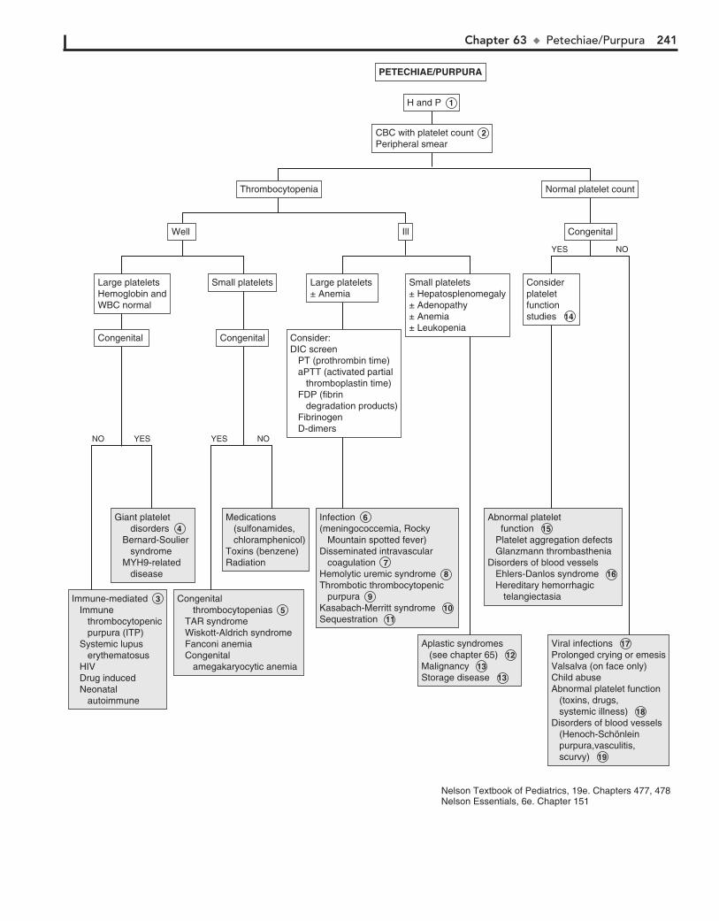
Chapter 63 u Petechiae/Purpura 241
Immune-mediated 3 Immune thrombocytopenic purpura (ITP) Systemic lupus erythematosus HIV Drug induced Neonatal autoimmune
Immune-mediated 3 Immune thrombocytopenic purpura (ITP) Systemic lupus erythematosus HIV Drug induced Neonatal autoimmune
Giant platelet disorders 4 Bernard-Soulier syndrome MYH9-related disease
Giant platelet disorders 4 Bernard-Soulier syndrome MYH9-related disease
Congenital thrombocytopenias 5 TAR syndrome Wiskott-Aldrich syndrome Fanconi anemia Congenital amegakaryocytic anemia
Congenital thrombocytopenias 5 TAR syndrome Wiskott-Aldrich syndrome Fanconi anemia Congenital amegakaryocytic anemia
Medications (sulfonamides, chloramphenicol)Toxins (benzene)Radiation
Medications (sulfonamides, chloramphenicol)Toxins (benzene)Radiation
Infection 6 (meningococcemia, Rocky Mountain spotted fever)Disseminated intravascular coagulation 7 Hemolytic uremic syndrome 8 Thrombotic thrombocytopenic purpura 9 Kasabach-Merritt syndrome 10 Sequestration 11
Infection 6 (meningococcemia, Rocky Mountain spotted fever)Disseminated intravascular coagulation 7 Hemolytic uremic syndrome 8 Thrombotic thrombocytopenic purpura 9 Kasabach-Merritt syndrome 10 Sequestration 11
Aplastic syndromes (see chapter 65) 12 Malignancy 13 Storage disease 13
Aplastic syndromes (see chapter 65) 12 Malignancy 13 Storage disease 13
Abnormal platelet function 15 Platelet aggregation defects Glanzmann thrombastheniaDisorders of blood vessels Ehlers-Danlos syndrome 16 Hereditary hemorrhagic telangiectasia
Abnormal platelet function 15 Platelet aggregation defects Glanzmann thrombastheniaDisorders of blood vessels Ehlers-Danlos syndrome 16 Hereditary hemorrhagic telangiectasia
Viral infections 17 Prolonged crying or emesisValsalva (on face only)Child abuse Abnormal platelet function (toxins, drugs, systemic illness) 18 Disorders of blood vessels (Henoch-Schönlein purpura,vasculitis, scurvy) 19
Viral infections 17 Prolonged crying or emesisValsalva (on face only)Child abuse Abnormal platelet function (toxins, drugs, systemic illness) 18 Disorders of blood vessels (Henoch-Schönlein purpura,vasculitis, scurvy) 19
Congenital Congenital
Large plateletsHemoglobin andWBC normal
Large plateletsHemoglobin andWBC normal
Small plateletsSmall platelets
Well Well
ThrombocytopeniaThrombocytopenia Normal platelet countNormal platelet count
IllIll CongenitalCongenital
Consider:DIC screen PT (prothrombin time) aPTT (activated partial thromboplastin time) FDP (fibrin degradation products) Fibrinogen D-dimers
Consider:DIC screen PT (prothrombin time) aPTT (activated partial thromboplastin time) FDP (fibrin degradation products) Fibrinogen D-dimers
Large platelets ± Anemia Large platelets ± Anemia
Small platelets± Hepatosplenomegaly± Adenopathy± Anemia± Leukopenia
Small platelets± Hepatosplenomegaly± Adenopathy± Anemia± Leukopenia
Considerplateletfunctionstudies 14
Considerplateletfunctionstudies 14
CongenitalCongenital
YESNO YES NO
H and P 1 H and P 1
CBC with platelet count 2 Peripheral smear CBC with platelet count 2 Peripheral smear
PETECHIAE/PURPURAPETECHIAE/PURPURA
Nelson Textbook of Pediatrics, 19e. Chapters 477, 478Nelson Essentials, 6e. Chapter 151
YES NO
242 Part IX u Hematology
16 In Ehlers-Danlos syndrome, features include joint laxity, hyperelastic skin and poor wound healing, often ecchymo-
ses is present and occasionally petechiae.
17 A number of viral infections also occur as petechial rashes. Most common of these is enterovirus infection, but rubella,
measles, varicella, Ebstein-Barr virus (EBV), cytomegalovirus (CMV), herpes (HSV), and HIV may also be involved.
18 Acquired platelet function defects may be acquired second-ary to medications (aspirin, nonsteroidal antiinflammatory
drugs, alcohol, high doses of penicillin, valproate, and others) as well as systemic illness (e.g., uremia, liver disease).
19 Vasculitic disorders occur as hemorrhagic lesions of skin and mucous membranes and symptoms relating to in-
volved organ systems. Coagulation studies are usually normal. Henoch-Schönlein purpura (HSP) presents as petechiae, pal-pable purpura over the buttocks and lower extremities, as well as arthritis, abdominal pain, and glomerulonephritis.
12 With aplastic syndromes (congenital and acquired aplastic anemias) there is thrombocytopenia with small platelets,
leukopenia, and anemia. (See Chapter 65.)
13 Infiltration of the marrow by malignant cells or due to storage disorders interferes with platelet production.
Malignancies may include acute lymphoblastic leukemia, lymphomas, histiocytosis X, as well as metastatic tumors (e.g., neuroblastoma). Other findings may include adenopa-thy, hepatosplenomegaly, masses, as well as other abnormali-ties of the peripheral smear. A bone marrow is diagnostic.
14 Platelet function studies may include PFA-100 as an initial screen for platelet function abnormalities, but there are
problems with sensitivity and specificity. If platelet function abnormality is seriously being considered, platelet aggregation studies are needed. Bleeding time indirectly measures platelet number and function and platelet-vessel wall interaction, and has low sensitivity.
15 Congenital platelet function defects include Glanzmann thrombasthenia (deficiency of a fibrinogen receptor).
Less severe platelet function defects may have more subtle presentation and may require platelet aggregation studies.
BibliographyConsolini DM: Thrombocytopenia in infants and children. Pediatr Rev
32(4):135–151, 2011.

Chapter 63 u Petechiae/Purpura 243
Immune-mediated 3 Immune thrombocytopenic purpura (ITP) Systemic lupus erythematosus HIV Drug induced Neonatal autoimmune
Immune-mediated 3 Immune thrombocytopenic purpura (ITP) Systemic lupus erythematosus HIV Drug induced Neonatal autoimmune
Giant platelet disorders 4 Bernard-Soulier syndrome MYH9-related disease
Giant platelet disorders 4 Bernard-Soulier syndrome MYH9-related disease
Congenital thrombocytopenias 5 TAR syndrome Wiskott-Aldrich syndrome Fanconi anemia Congenital amegakaryocytic anemia
Congenital thrombocytopenias 5 TAR syndrome Wiskott-Aldrich syndrome Fanconi anemia Congenital amegakaryocytic anemia
Medications (sulfonamides, chloramphenicol)Toxins (benzene)Radiation
Medications (sulfonamides, chloramphenicol)Toxins (benzene)Radiation
Infection 6 (meningococcemia, Rocky Mountain spotted fever)Disseminated intravascular coagulation 7 Hemolytic uremic syndrome 8 Thrombotic thrombocytopenic purpura 9 Kasabach-Merritt syndrome 10 Sequestration 11
Infection 6 (meningococcemia, Rocky Mountain spotted fever)Disseminated intravascular coagulation 7 Hemolytic uremic syndrome 8 Thrombotic thrombocytopenic purpura 9 Kasabach-Merritt syndrome 10 Sequestration 11
Aplastic syndromes (see chapter 65) 12 Malignancy 13 Storage disease 13
Aplastic syndromes (see chapter 65) 12 Malignancy 13 Storage disease 13
Abnormal platelet function 15 Platelet aggregation defects Glanzmann thrombastheniaDisorders of blood vessels Ehlers-Danlos syndrome 16 Hereditary hemorrhagic telangiectasia
Abnormal platelet function 15 Platelet aggregation defects Glanzmann thrombastheniaDisorders of blood vessels Ehlers-Danlos syndrome 16 Hereditary hemorrhagic telangiectasia
Viral infections 17 Prolonged crying or emesisValsalva (on face only)Child abuse Abnormal platelet function (toxins, drugs, systemic illness) 18 Disorders of blood vessels (Henoch-Schönlein purpura,vasculitis, scurvy) 19
Viral infections 17 Prolonged crying or emesisValsalva (on face only)Child abuse Abnormal platelet function (toxins, drugs, systemic illness) 18 Disorders of blood vessels (Henoch-Schönlein purpura,vasculitis, scurvy) 19
Congenital Congenital
Large plateletsHemoglobin andWBC normal
Large plateletsHemoglobin andWBC normal
Small plateletsSmall platelets
Well Well
ThrombocytopeniaThrombocytopenia Normal platelet countNormal platelet count
IllIll CongenitalCongenital
Consider:DIC screen PT (prothrombin time) aPTT (activated partial thromboplastin time) FDP (fibrin degradation products) Fibrinogen D-dimers
Consider:DIC screen PT (prothrombin time) aPTT (activated partial thromboplastin time) FDP (fibrin degradation products) Fibrinogen D-dimers
Large platelets ± Anemia Large platelets ± Anemia
Small platelets± Hepatosplenomegaly± Adenopathy± Anemia± Leukopenia
Small platelets± Hepatosplenomegaly± Adenopathy± Anemia± Leukopenia
Considerplateletfunctionstudies 14
Considerplateletfunctionstudies 14
CongenitalCongenital
YESNO YES NO
H and P 1 H and P 1
CBC with platelet count 2 Peripheral smear CBC with platelet count 2 Peripheral smear
PETECHIAE/PURPURAPETECHIAE/PURPURA
Nelson Textbook of Pediatrics, 19e. Chapters 477, 478Nelson Essentials, 6e. Chapter 151
YES NO

244
Chapter 64
NEUTROPENIA 4 There are many drugs that cause neutropenia, including
chemotherapeutic drugs. A partial list is presented in the algorithm. An idiosyncratic reaction generally affects only neu-trophils; other cell lines are usually unaffected.
5 The most common cause of transient neutropenia in children is viral infection. Viruses commonly causing neutropenia in-
clude hepatitis A and B, respiratory syncytial virus, influenza virus types A and B, measles, rubella, and varicella. It may also occur in early stages of infectious mononucleosis. Leukopenia may be seen in patients with HIV infection. This may be due to the virus or due to antiviral drugs.
6 There may be neutropenia associated with typhoid, paraty-phoid, tuberculosis, brucellosis, tularemia, and rickettsial
infections. In patients with an immunodeficiency, commonly cultured organisms include Staphylococcus aureus, coagulase-negative staphylococci, and gram-negative organisms, includ-ing E. coli and Klebsiella pneumoniae. Candida may also be cultured.
7 Severe malnutrition seen in anorexia nervosa and maras-mus may also cause neutropenia.
8 Cyclic neutropenia is a congenital condition with regular fluctuation in neutrophil counts. The nadir occurs ap-
proximately every 21 days, often to an absolute neutrophil count of ,200 cells/µl. During the neutropenic period there may be fever, oral ulcers, gingivitis, periodontitis, and pharyn-gitis with lymphadenopathy. More serious infections such as mastoiditis or pneumonia may also occur.
9 Congenital neutropenias include those associated with phenotypic abnormalities. Chédiak-Higashi syndrome is
characterized by oculocutaneous albinism. There is an in-creased susceptibility to infection due to neutropenia, as well as defective function of the remaining neutrophils. Dyskeratosis congenita is associated with nail dystrophy, leukoplakia, and reticulated hyperpigmentation of the skin. Shwachman syn-drome is characterized by dwarfism, growth failure, skeletal abnormalities, and exocrine pancreatic insufficiency, causing diarrhea, weight loss, and failure to thrive. Cartilage-hair hypo-plasia features neutropenia with short-limbed dwarfism and fine hair.
10 Autoimmune neutropenia may be associated with other autoimmune diseases (e.g., systemic lupus erythematosus,
autoimmune hemolytic anemia, and thrombocytopenia). It may also be associated with infection (e.g., infectious mono-nucleosis, HIV), malignancy (e.g., leukemia, lymphoma), and drugs. Antineutrophil antibodies may be present on testing; Coombs testing may identify associated hemolytic conditions.
11 Autoimmune neutropenia of infancy usually presents be-tween ages 5 to 15 months. Patients usually have severe
neutropenia with an ANC ,500/µL, however the total WBC count is usually normal. Diagnosis is usually by the presence of antineutrophil antibodies, but multiple screenings may be needed to detect these, and avoid the need for bone marrow studies.
12 Immune neonatal neutropenia is similar to Rh-hemolytic anemia. It occurs due to maternal sensitization caused by
fetal neutrophil antigens. The neutropenia may last for weeks and as long as 6 months. Antineutrophil antibodies are found in
Neutropenia is an absolute decrease in the number of circulat-ing neutrophils in the blood. It varies with age and race. In whites, the lower limit for normal absolute neutrophil counts (ANC 5 neutrophils 1 bands) is 1000 cells/µl in infants be-tween 2 weeks and 1 year of age, and 1500 cells/µl in children older than 1 year. In blacks, the lower limits may be 200 to 600 cells/µl less than whites. The severity of neutropenia is helpful in predicting an increased likelihood of pyogenic infection. Mild neutropenia ranges from 1000 to 1500 cells/µl; moderate neutropenia ranges from 500 to 1000 cells/µl and severe neutro-penia is ,500 cells/µl.
1 History of any recent or recurrent infections, mouth ulcers and drug exposure should be obtained. This history should
include fever, which may be a sign of infection. Infections may be both a cause and a consequence of neutropenia. The dura-tion and severity of the neutropenia is important in determin-ing infection risk. Severe neutropenia with counts ,500 cells/µl is associated with the greatest risk of pyogenic infections, in-cluding cellulitis, abscesses (including perirectal), furuncles, pneumonia, septicemia, as well as oral infections such as stoma-titis, gingivitis, and periodontitis. A detailed family history should include familial neutropenia and recurrent infections. A family history of short stature, dwarfism, skeletal abnormali-ties, and albinism may suggest congenital conditions associated with neutropenia. A careful physical examination is important in locating any sites of occult infection. The usual signs of infec-tion (e.g., erythema, warmth) may not be present because of the neutropenia. Examination should also include evaluation for pallor indicating anemia, petechiae suggesting thrombocy-topenia, lymphadenopathy, hepatosplenomegaly, and any other signs of underlying disease.
2 Although any severe infection may result in neutropenia, cyclic neutropenia and chronic neutropenia need to be
excluded with twice weekly WBC counts for 6 to 8 weeks. Re-ticulocyte, platelet, and other leukocyte counts may also cycle.
3 In patients with fever and neutropenia, particularly if the child is ill appearing, cultures of blood, urine, and any sus-
pected sites of infection should be done. Antibody responses, antigen detection tests, and polymerase chain reaction (PCR) methods may also be useful. In patients undergoing chemo-therapy, cultures should also be obtained from central venous lines. These should include aerobic and anaerobic bacteria as well as fungi. In chronically infected sites, mycobacterial and anaerobic cultures are recommended. If diarrhea is present, ob-tain stool cultures for bacteria, viruses, and parasites. Clostrid-ium difficile toxin should be sought. Tests for certain viruses may be considered in specific instances; for example, herpes cell cul-ture and PCR testing from vesicular skin lesions or PCR testing for respiratory viruses in children with respiratory symptoms. It is important to note that mild neutropenia in a child with a fe-brile viral-appearing illness and without a history of recurrent significant infections may not need further evaluation.

Chapter 64 u Neutropenia 245
See Chapter 65See Chapter 65
Drug induced neutropenia 4 Anticonvulsants (phenytoin, carbamazepine) Antibiotics (chloramphenicol, penicillins, sulfonamides) Analgesics (ibuprofen, indomethacin) Antithyroid drugs (propylthiouracil) Cardiovascular drugs (quinidine) Sedatives (phenothiazines) Chemotherapy Hypoglycemic agents (chlorpropamide)Radiation
Drug induced neutropenia 4 Anticonvulsants (phenytoin, carbamazepine) Antibiotics (chloramphenicol, penicillins, sulfonamides) Analgesics (ibuprofen, indomethacin) Antithyroid drugs (propylthiouracil) Cardiovascular drugs (quinidine) Sedatives (phenothiazines) Chemotherapy Hypoglycemic agents (chlorpropamide)Radiation
Infection Viral infection 5 Bacterial infection 6 Congenital/acquired neutropenia with superimposed infectionSevere malnutrition 7
Infection Viral infection 5 Bacterial infection 6 Congenital/acquired neutropenia with superimposed infectionSevere malnutrition 7
Viralsuppression 5 Viralsuppression 5
Cyclicneutropenia 8 Cyclicneutropenia 8
Autoimmune neutropenia 10 Autoimmune neutropenia of infancy 11 Immune neonatal neutropenia 12 Neutropenia associated with immune disorders 13
Autoimmune neutropenia 10 Autoimmune neutropenia of infancy 11 Immune neonatal neutropenia 12 Neutropenia associated with immune disorders 13
Nutritional deficiencies 14 Splenic sequestration 15 Metabolic disease 16 Glycogen storage disease 17 Reticular dysgenesis 18 Severe congenital neutropenia 19 Benign familial neutropenia 20 HIV infection
Nutritional deficiencies 14 Splenic sequestration 15 Metabolic disease 16 Glycogen storage disease 17 Reticular dysgenesis 18 Severe congenital neutropenia 19 Benign familial neutropenia 20 HIV infection
Chédiak-Higashi syndrome 9 Dyskeratosis congenita 9 Schwachman’s syndrome 9 Cartilage-hair hypoplasia 9
Chédiak-Higashi syndrome 9 Dyskeratosis congenita 9 Schwachman’s syndrome 9 Cartilage-hair hypoplasia 9
AbnormalAbnormal
Phenotypic abnormalities presentPhenotypic abnormalities present
Chronic neutropeniaChronic neutropenia
Neutropenia persistsNeutropenia persistsCyclical changesin WBC countsCyclical changesin WBC counts
Recheck in 2-4 weeks depending on absolute neutrophil count (ANC)If still neutropenic check WBC counts twice weekly for 6-8 weeks 2 Recheck in 2-4 weeks depending on absolute neutrophil count (ANC)If still neutropenic check WBC counts twice weekly for 6-8 weeks 2
WBC countnormalizesWBC countnormalizes
Ill appearing/febrile Ill appearing/febrile
Drugs ChemotherapyRadiation
Drugs ChemotherapyRadiation
Consider: 3 Bacterial, viral or fungal cultures/ PCR
Consider: 3 Bacterial, viral or fungal cultures/ PCR
NormalNormal
Anti-neutrophil antibodiesCoombs testImmunoglobulins (IgA, IgG, IgM)
Anti-neutrophil antibodiesCoombs testImmunoglobulins (IgA, IgG, IgM)
Consider: B12 and folate levels Copper levels HIV testing Bone marrow examination
Consider: B12 and folate levels Copper levels HIV testing Bone marrow examination
YES NO
YES NO
YES NO
YES NO
H and P 1 H and P 1
CBC with differentialPeripheral smear 2 CBC with differentialPeripheral smear 2
PancytopeniaPancytopenia
NEUTROPENIA NEUTROPENIA
Nelson Textbook of Pediatrics, 19e. Chapters 81, 118, 125Nelson Essentials, 6e. Chapter 74

246 Part IX u Hematology
maternal and infant serum. It can also occur in infants whose mothers have autoimmune neutropenia.
13 Neutropenia may be associated with disorders of immune dysfunction; these conditions include X-linked agamma-
globulinemia, hyper-IgM syndrome, cartilage-hair hypoplasia, and HIV infection.
14 Nutritional deficiencies including those of vitamin B12, folate, and copper may be associated with neutropenia.
15 Reticuloendothelial sequestration secondary to splenic en-largement can lead to neutropenia; causes include portal
hypertension, splenic disease, and splenic hyperplasia.
16 Many metabolic diseases are associated with neutropenia, such as hyperglycinemia, isovalericacidemia, propionic-
acidemia, methylmalonicacidemia, and tyrosinemia.
17 Neutropenia may also be noted in some glycogen storage disorder type 1b. Patients may present with hepatomegaly,
BibliographySegel GB, Halterman JS: Neutropenia in pediatric practice, Pediatr Rev 29:
12–24, 2008. Walkovich K, Boxer LA: How to approach neutropenia in childhood, Pediatr
Rev 34:173–184, 2013.
hypoglycemia, elevated lactate, cholesterol, triglyceride, and uric acid levels as well as associated neutropenia.
18 Another congenital cause of neutropenia includes reticu-lar dysgenesis, which is characterized by neutropenia and
lymphopenia. Lymphoid tissue (e.g., tonsils, lymph nodes, Peyer patches, and lymphoid follicles) is absent.
19 In severe congenital neutropenia the ANC ,200/µL. It presents in young infants with severe pyogenic infections
of skin, mouth, and rectum. It has an autosomal dominant form and a recessive form (Kostmann disease).
20 Familial benign neutropenia is mild with no increased tendency for infection.
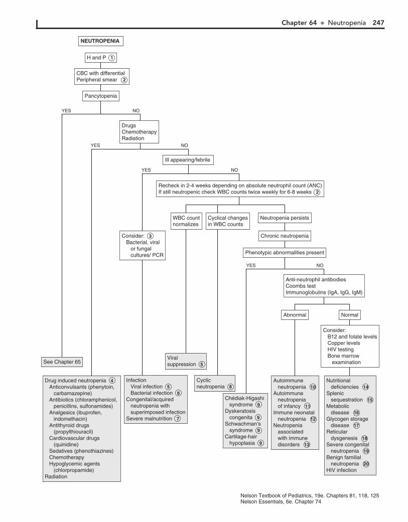
Chapter 64 u Neutropenia 247
See Chapter 65See Chapter 65
Drug induced neutropenia 4 Anticonvulsants (phenytoin, carbamazepine) Antibiotics (chloramphenicol, penicillins, sulfonamides) Analgesics (ibuprofen, indomethacin) Antithyroid drugs (propylthiouracil) Cardiovascular drugs (quinidine) Sedatives (phenothiazines) Chemotherapy Hypoglycemic agents (chlorpropamide)Radiation
Drug induced neutropenia 4 Anticonvulsants (phenytoin, carbamazepine) Antibiotics (chloramphenicol, penicillins, sulfonamides) Analgesics (ibuprofen, indomethacin) Antithyroid drugs (propylthiouracil) Cardiovascular drugs (quinidine) Sedatives (phenothiazines) Chemotherapy Hypoglycemic agents (chlorpropamide)Radiation
Infection Viral infection 5 Bacterial infection 6 Congenital/acquired neutropenia with superimposed infectionSevere malnutrition 7
Infection Viral infection 5 Bacterial infection 6 Congenital/acquired neutropenia with superimposed infectionSevere malnutrition 7
Viralsuppression 5 Viralsuppression 5
Cyclicneutropenia 8 Cyclicneutropenia 8
Autoimmune neutropenia 10 Autoimmune neutropenia of infancy 11 Immune neonatal neutropenia 12 Neutropenia associated with immune disorders 13
Autoimmune neutropenia 10 Autoimmune neutropenia of infancy 11 Immune neonatal neutropenia 12 Neutropenia associated with immune disorders 13
Nutritional deficiencies 14 Splenic sequestration 15 Metabolic disease 16 Glycogen storage disease 17 Reticular dysgenesis 18 Severe congenital neutropenia 19 Benign familial neutropenia 20 HIV infection
Nutritional deficiencies 14 Splenic sequestration 15 Metabolic disease 16 Glycogen storage disease 17 Reticular dysgenesis 18 Severe congenital neutropenia 19 Benign familial neutropenia 20 HIV infection
Chédiak-Higashi syndrome 9 Dyskeratosis congenita 9 Schwachman’s syndrome 9 Cartilage-hair hypoplasia 9
Chédiak-Higashi syndrome 9 Dyskeratosis congenita 9 Schwachman’s syndrome 9 Cartilage-hair hypoplasia 9
AbnormalAbnormal
Phenotypic abnormalities presentPhenotypic abnormalities present
Chronic neutropeniaChronic neutropenia
Neutropenia persistsNeutropenia persistsCyclical changesin WBC countsCyclical changesin WBC counts
Recheck in 2-4 weeks depending on absolute neutrophil count (ANC)If still neutropenic check WBC counts twice weekly for 6-8 weeks 2 Recheck in 2-4 weeks depending on absolute neutrophil count (ANC)If still neutropenic check WBC counts twice weekly for 6-8 weeks 2
WBC countnormalizesWBC countnormalizes
Ill appearing/febrile Ill appearing/febrile
Drugs ChemotherapyRadiation
Drugs ChemotherapyRadiation
Consider: 3 Bacterial, viral or fungal cultures/ PCR
Consider: 3 Bacterial, viral or fungal cultures/ PCR
NormalNormal
Anti-neutrophil antibodiesCoombs testImmunoglobulins (IgA, IgG, IgM)
Anti-neutrophil antibodiesCoombs testImmunoglobulins (IgA, IgG, IgM)
Consider: B12 and folate levels Copper levels HIV testing Bone marrow examination
Consider: B12 and folate levels Copper levels HIV testing Bone marrow examination
YES NO
YES NO
YES NO
YES NO
H and P 1 H and P 1
CBC with differentialPeripheral smear 2 CBC with differentialPeripheral smear 2
PancytopeniaPancytopenia
NEUTROPENIA NEUTROPENIA
Nelson Textbook of Pediatrics, 19e. Chapters 81, 118, 125Nelson Essentials, 6e. Chapter 74

248
Chapter 65
PANCYTOPENIA
Patients with HIV may have pancytopenia for a number of rea-sons, including opportunistic infections, drugs used in treat-ment, and neoplasms associated with the disease. Other viruses that may cause cytopenias include measles, mumps, rubella, varicella, and influenza A. If a viral etiology is suggested, it is reasonable to recheck the CBC in a few weeks. If the pancyto-penia persists or becomes more severe, referral to a hematolo-gist for further evaluation is recommended.
5 Patients with hemolytic anemia who have shortened red blood cell survival time are at risk of transient aplastic
crisis. This is most commonly associated with parvovirus and may occur in children with sickle cell disease, thalassemia, he-reditary spherocytosis, and other types of erythroid stress.
6 Fanconi anemia is an autosomal recessive condition. Two thirds of affected children have congenital anomalies.
These include microcephaly, microphthalmia, absent radii and thumbs, as well as heart and kidney abnormalities. There may be hypopigmentation of the skin and short stature.
7 Dyskeratosis congenita is a rare form of ectodermal dys-plasia associated with pancytopenia. Dermatologic mani-
festations include hyperpigmented skin, dystrophic nails, and mucous membrane leukoplakia.
8 Schwachman-Diamond syndrome is characterized by neu-tropenia with exocrine pancreatic insufficiency (e.g., mal-
absorption, steatorrhea, failure to thrive). About 50% develop aplastic anemia.
9 Pregnancy may be associated with aplastic anemia; estro-gens may play a role.
10 Paroxysmal nocturnal hemoglobinuria is characterized by intravascular hemolysis and hemoglobinuria as well as
venous thrombosis. There is a strong association with aplastic anemia.
11 Systemic diseases may be associated with pancytopenias. These may include systemic lupus erythematosus, meta-
bolic diseases, brucellosis, sarcoidosis, and tuberculosis.
12 Replacement of the marrow by malignant or nonhematopoi-etic cells may cause pancytopenias. Conditions include leu-
kemia, lymphomas, and neuroblastoma metastases to the bone marrow. Osteopetrosis may cause obliteration of the marrow. My-elofibrosis may also be a cause. Myelodysplastic syndrome is rare in children; there is an increased risk of development with Down syndrome, Kostmann syndrome, Noonan syndrome, Fanconi anemia, trisomy 8 mosaicism, neurofibromatosis, and Schwach-man syndrome.
13 In autoimmune pancytopenia the Coombs (direct anti-globulin) test is usually positive. There is evidence of hemo-
lysis with autoimmune hemolytic anemia. It is known as Evans syndrome when the patient has autoimmune hemolytic anemia and immune thrombocytopenic purpura (ITP). There may also be an associated autoimmune neutropenia. It may be associated with disorders such as systemic lupus erythematosus (SLE).
Pancytopenia is caused by a decrease in production of erythro-cytes, leukocytes, and platelets by the bone marrow. Clinically, this results in anemia, hemorrhage, and decreased resistance to infection. Severe aplastic anemia is diagnosed when at least two of the following are present: absolute neutrophil count (ANC) ,500/µl, platelet count ,20,000 µl, and corrected reticulocyte count ,1%; the bone marrow contains ,25% of the normal cel-lularity. Mild or moderate aplastic anemia (i.e., hypoplastic anemia) has a mild or moderate decrease in granulocytes, plate-lets, and erythrocytes, and there may be normal or increased bone marrow cellularity.
1 History should include exposure to agents that are potentially myelosuppressive. These include radiation and chemotherapy
(e.g., 6-mercaptopurine, methotrexate, nitrogen mustard). Other drugs include chloramphenicol, sulfonamides, phenyl-butazone, and anticonvulsants. Chemicals and toxins include benzene and other aromatic hydrocarbons present in insecticides and herbicides. A history and physical examination compatible with certain viral infections should be sought. An increased susceptibility to infection may suggest an immunodeficiency syndrome. A family history of congenital anomalies, aplastic syndromes, and leukemias may indicate syndromes associated with constitutional aplastic pancytopenias. Physical examina-tion may reveal the effects of the cytopenias, including anemia, which results in tachycardia and pallor; thrombocytopenia, which may cause bleeding, bruising, epistaxis, petechiae, or ec-chymoses; and neutropenia, which may be associated with oral ulcerations and fevers. Examination should include identifica-tion of congenital anomalies associated with Fanconi and other syndromes (e.g., Down syndrome).
2 When blasts are seen on peripheral smear, it indicates leu-kemia requiring referral for bone marrow examination.
Leukoerythroblastosis (myelophthisic anemia) is usually due to invasion of the bone marrow and resulting release of immature cells including erythroblasts (nucleated erythrocytes), imma-ture neutrophils and giant platelets.
3 Laboratory findings suggesting hemolysis include abnor-mal cell morphology, increased reticulocyte count, in-
creased red blood cell distribution width, indirect bilirubin, urine urobilinogen, and lactate dehydrogenase, decreased serum haptoglobin, and hemoglobinuria.
4 The most common cause of mild or moderate pancytope-nias in healthy patients is suppression due to infectious
agents. Specific viruses include human parvovirus B19, hepati-tis viruses (B, C, non-A non-B and non-C), dengue virus, cyto-megalovirus, human herpes virus 6, and Epstein-Barr virus.
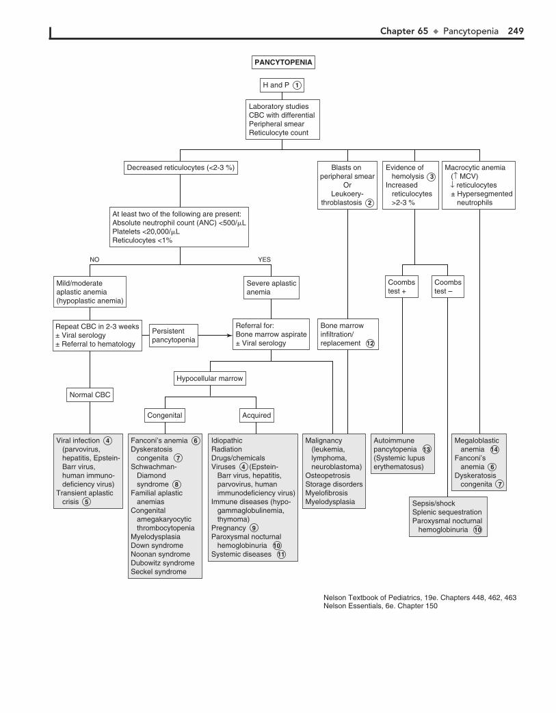
Chapter 65 u Pancytopenia 249
Viral infection 4 (parvovirus, hepatitis, Epstein- Barr virus, human immuno- deficiency virus)Transient aplastic crisis 5
Viral infection 4 (parvovirus, hepatitis, Epstein- Barr virus, human immuno- deficiency virus)Transient aplastic crisis 5
Fanconi’s anemia 6 Dyskeratosis congenita 7 Schwachman- Diamond syndrome 8 Familial aplastic anemiasCongenital amegakaryocytic thrombocytopenia MyelodysplasiaDown syndromeNoonan syndrome Dubowitz syndromeSeckel syndrome
Fanconi’s anemia 6 Dyskeratosis congenita 7 Schwachman- Diamond syndrome 8 Familial aplastic anemiasCongenital amegakaryocytic thrombocytopenia MyelodysplasiaDown syndromeNoonan syndrome Dubowitz syndromeSeckel syndrome
IdiopathicRadiationDrugs/chemicalsViruses 4 (Epstein- Barr virus, hepatitis, parvovirus, human immunodeficiency virus)Immune diseases (hypo- gammaglobulinemia, thymoma)Pregnancy 9 Paroxysmal nocturnal hemoglobinuria 10 Systemic diseases 11
IdiopathicRadiationDrugs/chemicalsViruses 4 (Epstein- Barr virus, hepatitis, parvovirus, human immunodeficiency virus)Immune diseases (hypo- gammaglobulinemia, thymoma)Pregnancy 9 Paroxysmal nocturnal hemoglobinuria 10 Systemic diseases 11
Malignancy (leukemia, lymphoma, neuroblastoma)OsteopetrosisStorage disordersMyelofibrosis Myelodysplasia
Malignancy (leukemia, lymphoma, neuroblastoma)OsteopetrosisStorage disordersMyelofibrosis Myelodysplasia
Autoimmune pancytopenia 13 (Systemic lupuserythematosus)
Autoimmune pancytopenia 13 (Systemic lupuserythematosus)
Sepsis/shock Splenic sequestration Paroxysmal nocturnal hemoglobinuria 10
Sepsis/shock Splenic sequestration Paroxysmal nocturnal hemoglobinuria 10
Megaloblastic anemia 14 Fanconi’s anemia 6 Dyskeratosis congenita 7
Megaloblastic anemia 14 Fanconi’s anemia 6 Dyskeratosis congenita 7
CongenitalCongenital AcquiredAcquired
Bone marrowinfiltration/replacement 12
Bone marrowinfiltration/replacement 12
Referral for:Bone marrow aspirate± Viral serology
Referral for:Bone marrow aspirate± Viral serology
Severe aplasticanemiaSevere aplasticanemia
Coombstest +Coombstest +
Coombstest –Coombstest –
Repeat CBC in 2-3 weeks± Viral serology± Referral to hematology
Repeat CBC in 2-3 weeks± Viral serology± Referral to hematology
PersistentpancytopeniaPersistentpancytopenia
Normal CBCNormal CBC
Mild/moderateaplastic anemia (hypoplastic anemia)
Mild/moderateaplastic anemia (hypoplastic anemia)
At least two of the following are present: Absolute neutrophil count (ANC) <500/�LPlatelets <20,000/�LReticulocytes <1%
At least two of the following are present: Absolute neutrophil count (ANC) <500/�LPlatelets <20,000/�LReticulocytes <1%
Decreased reticulocytes (<2-3 %)Decreased reticulocytes (<2-3 %) Blasts on peripheral smear
OrLeukoery-
throblastosis 2
Blasts on peripheral smear
OrLeukoery-
throblastosis 2
Evidence of hemolysis 3 Increased reticulocytes >2-3 %
Evidence of hemolysis 3 Increased reticulocytes >2-3 %
Macrocytic anemia (↑ MCV) ↓ reticulocytes ± Hypersegmented neutrophils
Macrocytic anemia (↑ MCV) ↓ reticulocytes ± Hypersegmented neutrophils
Hypocellular marrowHypocellular marrow
YESNO
H and P 1 H and P 1
Laboratory studies CBC with differential Peripheral smearReticulocyte count
Laboratory studies CBC with differential Peripheral smearReticulocyte count
PANCYTOPENIAPANCYTOPENIA
Nelson Textbook of Pediatrics, 19e. Chapters 448, 462, 463 Nelson Essentials, 6e. Chapter 150
250 Part IX u Hematology
certain anticonvulsant drugs (e.g., phenytoin, primidone, phe-nobarbital). Folate deficiency may occur in infants fed goat’s milk, which is low in folate). Some medications (e.g., metho-trexate, trimethoprim) can inhibit the action of folate. Increased folate requirements may occur in chronic hemolytic anemia (e.g., sickle cell).
14 Megaloblastic anemia (large RBCs with abnormal hyper-segmented neutrophils due to vitamin B12 or folate defi-
ciency is rare in children. Neutropenia and thrombocytopenia may be present, particularly in patients with long-standing and severe deficiencies.
Vitamin B12 deficiency may be seen in breast-fed infants of vegan mothers or with malabsorption due to a rare intrinsic factor deficiency (e.g., congenital pernicious anemia), resection of the ileum, or impaired absorption in inflammatory condi-tions (e.g., celiac disease).
Folate deficiency may be caused by decreased absorption due to resection or inflammatory disease of the small bowel or
BibliographySills RH, Deters A: Pancytopenia. In Sills RH, editor: Practical Algorithms
in Pediatric Hematology and Oncology, Basel, Switzerland, 2003, Karger, pp 12–13.

Chapter 65 u Pancytopenia 251
Viral infection 4 (parvovirus, hepatitis, Epstein- Barr virus, human immuno- deficiency virus)Transient aplastic crisis 5
Viral infection 4 (parvovirus, hepatitis, Epstein- Barr virus, human immuno- deficiency virus)Transient aplastic crisis 5
Fanconi’s anemia 6 Dyskeratosis congenita 7 Schwachman- Diamond syndrome 8 Familial aplastic anemiasCongenital amegakaryocytic thrombocytopenia MyelodysplasiaDown syndromeNoonan syndrome Dubowitz syndromeSeckel syndrome
Fanconi’s anemia 6 Dyskeratosis congenita 7 Schwachman- Diamond syndrome 8 Familial aplastic anemiasCongenital amegakaryocytic thrombocytopenia MyelodysplasiaDown syndromeNoonan syndrome Dubowitz syndromeSeckel syndrome
IdiopathicRadiationDrugs/chemicalsViruses 4 (Epstein- Barr virus, hepatitis, parvovirus, human immunodeficiency virus)Immune diseases (hypo- gammaglobulinemia, thymoma)Pregnancy 9 Paroxysmal nocturnal hemoglobinuria 10 Systemic diseases 11
IdiopathicRadiationDrugs/chemicalsViruses 4 (Epstein- Barr virus, hepatitis, parvovirus, human immunodeficiency virus)Immune diseases (hypo- gammaglobulinemia, thymoma)Pregnancy 9 Paroxysmal nocturnal hemoglobinuria 10 Systemic diseases 11
Malignancy (leukemia, lymphoma, neuroblastoma)OsteopetrosisStorage disordersMyelofibrosis Myelodysplasia
Malignancy (leukemia, lymphoma, neuroblastoma)OsteopetrosisStorage disordersMyelofibrosis Myelodysplasia
Autoimmune pancytopenia 13 (Systemic lupuserythematosus)
Autoimmune pancytopenia 13 (Systemic lupuserythematosus)
Sepsis/shock Splenic sequestration Paroxysmal nocturnal hemoglobinuria 10
Sepsis/shock Splenic sequestration Paroxysmal nocturnal hemoglobinuria 10
Megaloblastic anemia 14 Fanconi’s anemia 6 Dyskeratosis congenita 7
Megaloblastic anemia 14 Fanconi’s anemia 6 Dyskeratosis congenita 7
CongenitalCongenital AcquiredAcquired
Bone marrowinfiltration/replacement 12
Bone marrowinfiltration/replacement 12
Referral for:Bone marrow aspirate± Viral serology
Referral for:Bone marrow aspirate± Viral serology
Severe aplasticanemiaSevere aplasticanemia
Coombstest +Coombstest +
Coombstest –Coombstest –
Repeat CBC in 2-3 weeks± Viral serology± Referral to hematology
Repeat CBC in 2-3 weeks± Viral serology± Referral to hematology
PersistentpancytopeniaPersistentpancytopenia
Normal CBCNormal CBC
Mild/moderateaplastic anemia (hypoplastic anemia)
Mild/moderateaplastic anemia (hypoplastic anemia)
At least two of the following are present: Absolute neutrophil count (ANC) <500/�LPlatelets <20,000/�LReticulocytes <1%
At least two of the following are present: Absolute neutrophil count (ANC) <500/�LPlatelets <20,000/�LReticulocytes <1%
Decreased reticulocytes (<2-3 %)Decreased reticulocytes (<2-3 %) Blasts on peripheral smear
OrLeukoery-
throblastosis 2
Blasts on peripheral smear
OrLeukoery-
throblastosis 2
Evidence of hemolysis 3 Increased reticulocytes >2-3 %
Evidence of hemolysis 3 Increased reticulocytes >2-3 %
Macrocytic anemia (↑ MCV) ↓ reticulocytes ± Hypersegmented neutrophils
Macrocytic anemia (↑ MCV) ↓ reticulocytes ± Hypersegmented neutrophils
Hypocellular marrowHypocellular marrow
YESNO
H and P 1 H and P 1
Laboratory studies CBC with differential Peripheral smearReticulocyte count
Laboratory studies CBC with differential Peripheral smearReticulocyte count
PANCYTOPENIAPANCYTOPENIA
Nelson Textbook of Pediatrics, 19e. Chapters 448, 462, 463 Nelson Essentials, 6e. Chapter 150
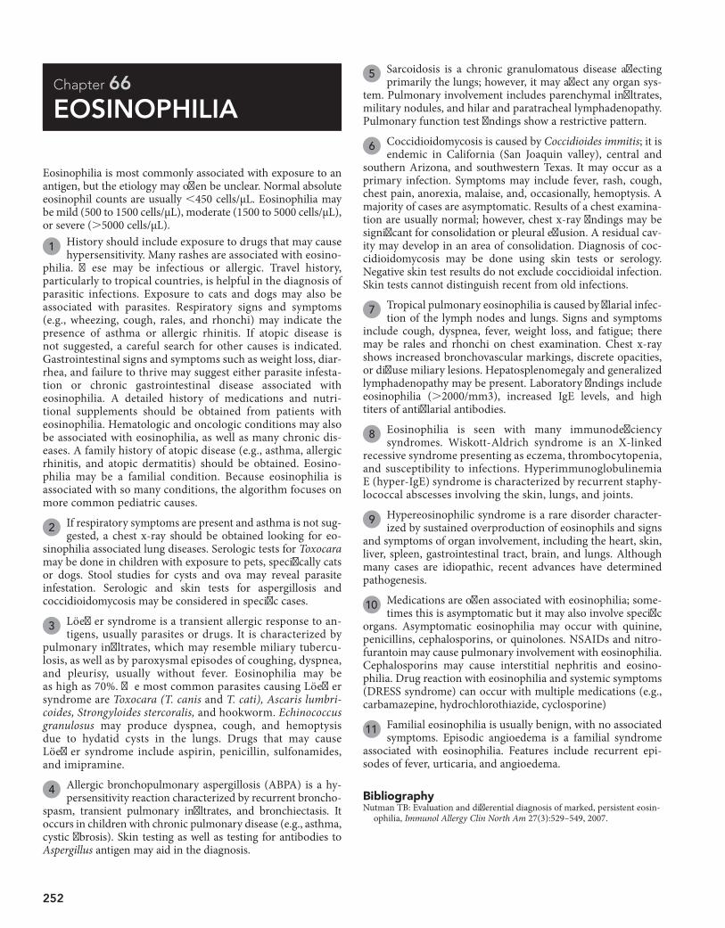
252
Chapter 66
EOSINOPHILIA
5 Sarcoidosis is a chronic granulomatous disease affecting primarily the lungs; however, it may affect any organ sys-
tem. Pulmonary involvement includes parenchymal infiltrates, military nodules, and hilar and paratracheal lymphadenopathy. Pulmonary function test findings show a restrictive pattern.
6 Coccidioidomycosis is caused by Coccidioides immitis; it is endemic in California (San Joaquin valley), central and
southern Arizona, and southwestern Texas. It may occur as a primary infection. Symptoms may include fever, rash, cough, chest pain, anorexia, malaise, and, occasionally, hemoptysis. A majority of cases are asymptomatic. Results of a chest examina-tion are usually normal; however, chest x-ray findings may be significant for consolidation or pleural effusion. A residual cav-ity may develop in an area of consolidation. Diagnosis of coc-cidioidomycosis may be done using skin tests or serology. Negative skin test results do not exclude coccidioidal infection. Skin tests cannot distinguish recent from old infections.
7 Tropical pulmonary eosinophilia is caused by filarial infec-tion of the lymph nodes and lungs. Signs and symptoms
include cough, dyspnea, fever, weight loss, and fatigue; there may be rales and rhonchi on chest examination. Chest x-ray shows increased bronchovascular markings, discrete opacities, or diffuse miliary lesions. Hepatosplenomegaly and generalized lymphadenopathy may be present. Laboratory findings include eosinophilia (.2000/mm3), increased IgE levels, and high titers of antifilarial antibodies.
8 Eosinophilia is seen with many immunodeficiency syndromes. Wiskott-Aldrich syndrome is an X-linked
recessive syndrome presenting as eczema, thrombocytopenia, and susceptibility to infections. Hyperimmunoglobulinemia E (hyper-IgE) syndrome is characterized by recurrent staphy-lococcal abscesses involving the skin, lungs, and joints.
9 Hypereosinophilic syndrome is a rare disorder character-ized by sustained overproduction of eosinophils and signs
and symptoms of organ involvement, including the heart, skin, liver, spleen, gastrointestinal tract, brain, and lungs. Although many cases are idiopathic, recent advances have determined pathogenesis.
10 Medications are often associated with eosinophilia; some-times this is asymptomatic but it may also involve specific
organs. Asymptomatic eosinophilia may occur with quinine, penicillins, cephalosporins, or quinolones. NSAIDs and nitro-furantoin may cause pulmonary involvement with eosinophilia. Cephalosporins may cause interstitial nephritis and eosino-philia. Drug reaction with eosinophilia and systemic symptoms (DRESS syndrome) can occur with multiple medications (e.g., carbamazepine, hydrochlorothiazide, cyclosporine)
11 Familial eosinophilia is usually benign, with no associated symptoms. Episodic angioedema is a familial syndrome
associated with eosinophilia. Features include recurrent epi-sodes of fever, urticaria, and angioedema.
Eosinophilia is most commonly associated with exposure to an antigen, but the etiology may often be unclear. Normal absolute eosinophil counts are usually ,450 cells/µL. Eosinophilia may be mild (500 to 1500 cells/µL), moderate (1500 to 5000 cells/µL), or severe (.5000 cells/µL).
1 History should include exposure to drugs that may cause hypersensitivity. Many rashes are associated with eosino-
philia. These may be infectious or allergic. Travel history, particularly to tropical countries, is helpful in the diagnosis of parasitic infections. Exposure to cats and dogs may also be associated with parasites. Respiratory signs and symptoms (e.g., wheezing, cough, rales, and rhonchi) may indicate the presence of asthma or allergic rhinitis. If atopic disease is not suggested, a careful search for other causes is indicated. Gastrointestinal signs and symptoms such as weight loss, diar-rhea, and failure to thrive may suggest either parasite infesta-tion or chronic gastrointestinal disease associated with eosinophilia. A detailed history of medications and nutri-tional supplements should be obtained from patients with eosinophilia. Hematologic and oncologic conditions may also be associated with eosinophilia, as well as many chronic dis-eases. A family history of atopic disease (e.g., asthma, allergic rhinitis, and atopic dermatitis) should be obtained. Eosino-philia may be a familial condition. Because eosinophilia is associated with so many conditions, the algorithm focuses on more common pediatric causes.
2 If respiratory symptoms are present and asthma is not sug-gested, a chest x-ray should be obtained looking for eo-
sinophilia associated lung diseases. Serologic tests for Toxocara may be done in children with exposure to pets, specifically cats or dogs. Stool studies for cysts and ova may reveal parasite infestation. Serologic and skin tests for aspergillosis and coccidioidomycosis may be considered in specific cases.
3 Löeffler syndrome is a transient allergic response to an-tigens, usually parasites or drugs. It is characterized by
pulmonary infiltrates, which may resemble miliary tubercu-losis, as well as by paroxysmal episodes of coughing, dyspnea, and pleurisy, usually without fever. Eosinophilia may be as high as 70%. The most common parasites causing Löeffler syndrome are Toxocara (T. canis and T. cati), Ascaris lumbri-coides, Strongyloides stercoralis, and hookworm. Echinococcus granulosus may produce dyspnea, cough, and hemoptysis due to hydatid cysts in the lungs. Drugs that may cause Löeffler syndrome include aspirin, penicillin, sulfonamides, and imipramine.
4 Allergic bronchopulmonary aspergillosis (ABPA) is a hy-persensitivity reaction characterized by recurrent broncho-
spasm, transient pulmonary infiltrates, and bronchiectasis. It occurs in children with chronic pulmonary disease (e.g., asthma, cystic fibrosis). Skin testing as well as testing for antibodies to Aspergillus antigen may aid in the diagnosis.
BibliographyNutman TB: Evaluation and differential diagnosis of marked, persistent eosin-
ophilia, Immunol Allergy Clin North Am 27(3):529–549, 2007.
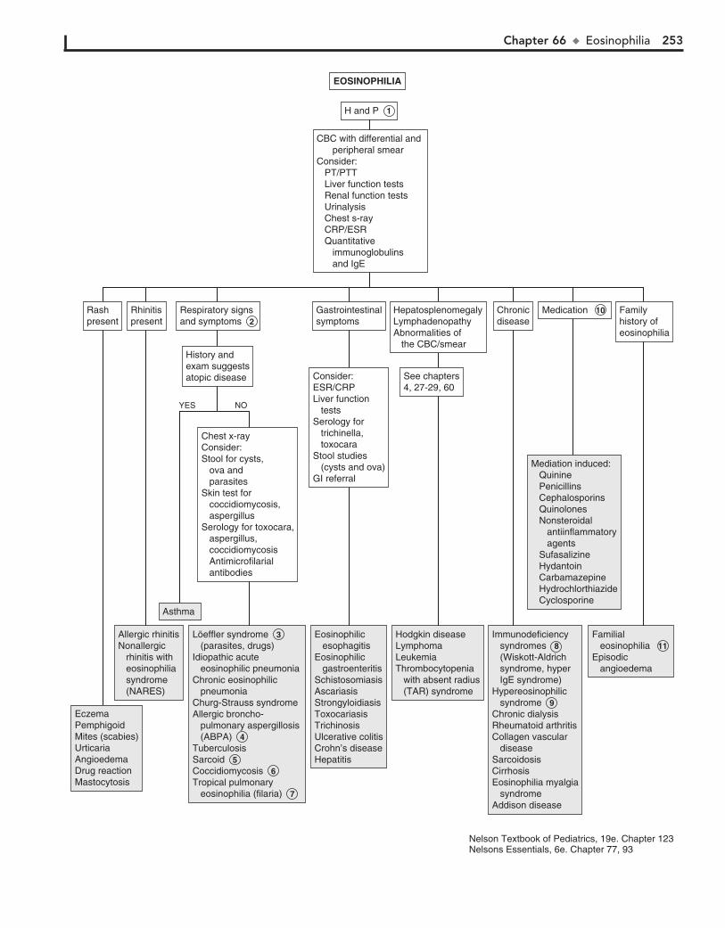
Chapter 66 u Eosinophilia 253
EczemaPemphigoidMites (scabies)UrticariaAngioedemaDrug reactionMastocytosis
EczemaPemphigoidMites (scabies)UrticariaAngioedemaDrug reactionMastocytosis
Allergic rhinitisNonallergic rhinitis with eosinophilia syndrome (NARES)
Allergic rhinitisNonallergic rhinitis with eosinophilia syndrome (NARES)
Asthma Asthma
Löeffler syndrome 3 (parasites, drugs)Idiopathic acute eosinophilic pneumoniaChronic eosinophilic pneumoniaChurg-Strauss syndromeAllergic broncho- pulmonary aspergillosis (ABPA) 4 TuberculosisSarcoid 5 Coccidiomycosis 6 Tropical pulmonary eosinophilia (filaria) 7
Löeffler syndrome 3 (parasites, drugs)Idiopathic acute eosinophilic pneumoniaChronic eosinophilic pneumoniaChurg-Strauss syndromeAllergic broncho- pulmonary aspergillosis (ABPA) 4 TuberculosisSarcoid 5 Coccidiomycosis 6 Tropical pulmonary eosinophilia (filaria) 7
Chest x-rayConsider:Stool for cysts, ova and parasitesSkin test for coccidiomycosis, aspergillusSerology for toxocara, aspergillus, coccidiomycosis Antimicrofilarial antibodies
Chest x-rayConsider:Stool for cysts, ova and parasitesSkin test for coccidiomycosis, aspergillusSerology for toxocara, aspergillus, coccidiomycosis Antimicrofilarial antibodies
History andexam suggestsatopic disease
History andexam suggestsatopic disease
Respiratory signsand symptoms 2 Respiratory signsand symptoms 2
RhinitispresentRhinitispresent
GastrointestinalsymptomsGastrointestinalsymptoms
ChronicdiseaseChronicdisease
Medication 10 Medication 10 Familyhistory of eosinophilia
Familyhistory of eosinophilia
Consider:ESR/CRPLiver function testsSerology for trichinella, toxocaraStool studies (cysts and ova)GI referral
Consider:ESR/CRPLiver function testsSerology for trichinella, toxocaraStool studies (cysts and ova)GI referral
HepatosplenomegalyLymphadenopathyAbnormalities of the CBC/smear
HepatosplenomegalyLymphadenopathyAbnormalities of the CBC/smear
See chapters4, 27-29, 60See chapters4, 27-29, 60
RashpresentRashpresent
Eosinophilic esophagitisEosinophilic gastroenteritisSchistosomiasisAscariasisStrongyloidiasisToxocariasisTrichinosisUlcerative colitisCrohn’s diseaseHepatitis
Eosinophilic esophagitisEosinophilic gastroenteritisSchistosomiasisAscariasisStrongyloidiasisToxocariasisTrichinosisUlcerative colitisCrohn’s diseaseHepatitis
Hodgkin diseaseLymphomaLeukemiaThrombocytopenia with absent radius (TAR) syndrome
Hodgkin diseaseLymphomaLeukemiaThrombocytopenia with absent radius (TAR) syndrome
Immunodeficiency syndromes 8 (Wiskott-Aldrich syndrome, hyper IgE syndrome)Hypereosinophilic syndrome 9 Chronic dialysisRheumatoid arthritisCollagen vascular diseaseSarcoidosisCirrhosisEosinophilia myalgia syndromeAddison disease
Immunodeficiency syndromes 8 (Wiskott-Aldrich syndrome, hyper IgE syndrome)Hypereosinophilic syndrome 9 Chronic dialysisRheumatoid arthritisCollagen vascular diseaseSarcoidosisCirrhosisEosinophilia myalgia syndromeAddison disease
Mediation induced: Quinine Penicillins Cephalosporins Quinolones Nonsteroidal antiinflammatory agents Sufasalizine Hydantoin Carbamazepine Hydrochlorthiazide Cyclosporine
Mediation induced: Quinine Penicillins Cephalosporins Quinolones Nonsteroidal antiinflammatory agents Sufasalizine Hydantoin Carbamazepine Hydrochlorthiazide Cyclosporine
Familial eosinophilia 11 Episodic angioedema
Familial eosinophilia 11 Episodic angioedema
YES NO
H and P 1 H and P 1
CBC with differential and peripheral smearConsider: PT/PTT Liver function tests Renal function tests Urinalysis Chest s-ray CRP/ESR Quantitative immunoglobulins and IgE
CBC with differential and peripheral smearConsider: PT/PTT Liver function tests Renal function tests Urinalysis Chest s-ray CRP/ESR Quantitative immunoglobulins and IgE
EOSINOPHILIAEOSINOPHILIA
Nelson Textbook of Pediatrics, 19e. Chapter 123 Nelsons Essentials, 6e. Chapter 77, 93

This page intentionally left blank

Endocrine System PART
X

256
Chapter 67
SHORT STATURE
is normal. The final adult height is short but within targeted range based on parental height. If there is deviation from the growth curve, further evaluation is needed.
4 Constitutional delay of growth and adolescence is a nor-mal variation, recognized more commonly in boys. There
is pubertal delay, delayed bone age, and delayed growth spurt, with subsequent attainment of normal adult height. Bone age usually correlates with height age. There is often also a family history of delayed puberty (usually the father). Laboratory test results are normal. It may be difficult to distinguish these chil-dren from those with mild GH defects, chronic disease, or central hypogonadism.
5 If the child has decelerating growth patterns or is signifi-cantly short (#2 SD), further evaluation should be guided
by the clinical findings. Bone age radiography (BA) is used to assess skeletal maturity. A chronic illness may be suggested by the results of a CBC, ESR, chemistry profile, and UA. Hypothy-roidism can be detected by obtaining free T4 and TSH levels. Suspicion of Turner syndrome in girls with unexplained short stature requires a karyotype. GH deficiency may be screened for by obtaining an insulin-like growth factor (IGF)-1 and IGF binding protein 3 (IGFBP3); however, the definitive test in-volves GH levels in response to pharmacologic stimulation. Further evaluation by an endocrinologist is required.
6 We suggest using a weight/height ratio to organize a dif-ferential diagnosis for a child with short stature. This is an
inexact method, and it is not a formal means of classification. The specific laboratory tests ordered should be based on the findings of the H and P. In general, however, weight for age less than height for age may indicate chronic illness or malnutrition, and weight for age greater than height for age may indicate endocrine disorders or genetic disorders and syndromes.
7 Children with weight disproportionately lower than height may be malnourished, malabsorbing, or have specific
nutritional deficiencies (e.g., rickets). Chronic illness may be a cause. Screening laboratory tests (e.g., ESR, CBC, UA, and chemistry profile) may indicate the diagnosis. RTA may result in FTT and short stature. A low serum bicarbonate on the chemistry profile may be the clue. Further evaluation reveals a normal anion gap metabolic acidosis. (See Chapter 82.) If the reason is not evident, the evaluation should also include tests to exclude celiac disease (tissue transglutaminase, antigliadin anti-bodies, immunoglobulin [Ig]A), and testing for HIV should be considered when appropriate. Cystic fibrosis testing should be considered when newborn screen results are not available.
8 Emotional deprivation may retard growth and mimic hy-popituitarism. The condition is known as psychosocial
dwarfism. The mechanism is poorly understood, but the child may have functional hypopituitarism with low levels of IGF-1. Growth velocity is slow, and bone age is less than the chrono-logical age. The child has short stature, and weight may be pro-portional to height or decreased compared to height. Puberty may be normal or premature. Close observation may reveal dis-turbed mother-child or family interactions. These children grow normally once they are removed from the poor environment.
9 Genetic syndromes or chromosomal abnormalities may be associated with short stature. If features of a particular
syndrome are present, karyotype should be obtained; a compre-hensive lab evaluation may not be necessary, and genetics
Short stature is defined as height less than 2 to 2.5 standard deviations (SD) below the mean for age. However, decreasing growth velocity is very important regardless of absolute height. It is important to distinguish normal (constitutional or familial) short stature from that due to a medical problem. Short stature must also be distinguished from failure to thrive (FTT), with associated poor weight gain. Because of the importance our culture places on height for males, boys are more likely to be brought to medical attention for this complaint. Many factors such as parental height and growth patterns, pubertal status, ethnicity, nutritional status, chronic illnesses, and emotional and psychological effects may influence stature. Infants of very low birth weight and small for gestational age (SGA) for various reasons (chromosomal disorders or syndromes, infections, or maternal alcohol use) are more likely to be of shorter stature.
1 History and physical examination should rule out any ob-vious dysmorphisms, syndromes, or diseases that may be
associated with short stature. Birth history should include height and weight, prenatal exposures and illnesses, as well as perinatal problems. Prolonged jaundice, hypoglycemia, and small phallus suggest growth hormone (GH) deficiency; puffy extremities suggest Turner syndrome.
A careful analysis of the growth curve is essential. Growth velocity and growth patterns must be plotted. Weight-for-height ratio is also helpful in determining the cause of short stature. The target (mid-parental) should be determined. For boys: mid–parental height 5 [(maternal height 1 13 cm) 1 paternal height]/2. For girls: mid–parental height 5 [(paternal height 1 13 cm) 1 maternal height]/2. A height range can then be calculated; 8.5 cm below and above the mid–parental height represent the 3rd to 97th percentile for the child. The ratio of upper to lower body segment (U/L) ratio can differentiate whether short stature is proportionate (involving both trunk and lower extremities) or disproportionate. The U/L ratio is decreased in skeletal dysplasias involving the spine. It is in-creased in dysplasias involving long bones and also in preco-cious puberty.
A short child who remains along his or her growth percen-tile may have familial or constitutional delay. Progressive devia-tion below the curve suggests a congenital disorder (Turner syndrome, GH deficiency). Developmental delay is associated with syndromes such as Prader-Willi syndrome. Weight for age that is less than height for age suggests malnutrition or chronic illness. A history of cranial irradiation for CNS malignancies suggests GH deficiency. Midline defects may be associated with hypopituitarism.
2 A child with a family history of short stature or pubertal delay who is staying along his or her growth curve may be
followed clinically.
3 A child with familial short stature has normal growth ve-locity with growth curves below but parallel to the normal
growth curve and has normal pubertal development. These chil-dren have no endocrine disorder or systemic illness and have a family history of short stature. Bone age may be considered and
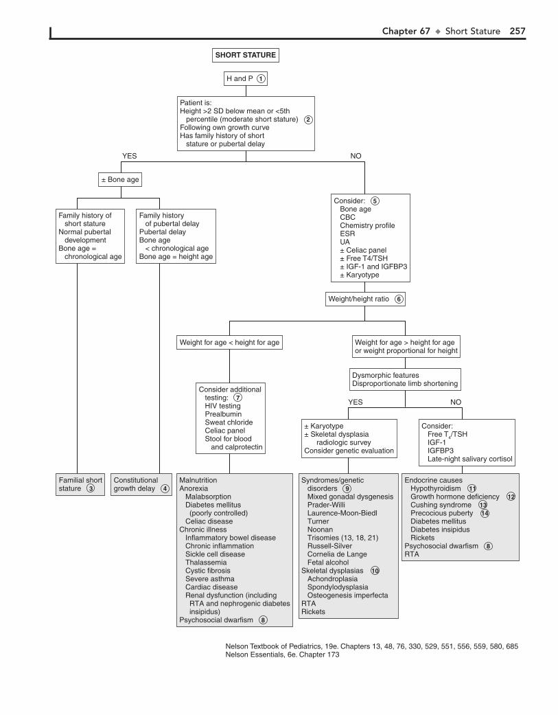
Chapter 67 u Short Stature 257
Patient is:Height >2 SD below mean or <5th percentile (moderate short stature) 2 Following own growth curveHas family history of short stature or pubertal delay
MalnutritionAnorexia Malabsorption Diabetes mellitus (poorly controlled) Celiac diseaseChronic illness Inflammatory bowel disease Chronic inflammation Sickle cell disease Thalassemia Cystic fibrosis Severe asthma Cardiac disease Renal dysfunction (including RTA and nephrogenic diabetes insipidus)Psychosocial dwarfism
MalnutritionAnorexia Malabsorption Diabetes mellitus (poorly controlled) Celiac diseaseChronic illness Inflammatory bowel disease Chronic inflammation Sickle cell disease Thalassemia Cystic fibrosis Severe asthma Cardiac disease Renal dysfunction (including RTA and nephrogenic diabetes insipidus)Psychosocial dwarfism 8
Familial shortstature Familial shortstature 3
Constitutionalgrowth delay Constitutionalgrowth delay 4
Syndromes/genetic disorders Mixed gonadal dysgenesis Prader-Willi Laurence-Moon-Biedl Turner Noonan Trisomies (13, 18, 21) Russell-Silver Cornelia de Lange Fetal alcoholSkeletal dysplasias Achondroplasia Spondylodysplasia Osteogenesis imperfectaRTARickets
Syndromes/genetic disorders 9 Mixed gonadal dysgenesis Prader-Willi Laurence-Moon-Biedl Turner Noonan Trisomies (13, 18, 21) Russell-Silver Cornelia de Lange Fetal alcoholSkeletal dysplasias 10 Achondroplasia Spondylodysplasia Osteogenesis imperfectaRTARickets
Endocrine causes Hypothyroidism Growth hormone deficiency Cushing syndrome Precocious puberty Diabetes mellitus Diabetes insipidus RicketsPsychosocial dwarfism RTA
Endocrine causes Hypothyroidism 11 Growth hormone deficiency 12 Cushing syndrome 13 Precocious puberty 14 Diabetes mellitus Diabetes insipidus RicketsPsychosocial dwarfism 8RTA
SHORT STATURE
H and P 1
± Karyotype± Skeletal dysplasia radiologic surveyConsider genetic evaluation
Consider additional testing: 7 HIV testing Prealbumin Sweat chloride Celiac panel Stool for blood and calprotectin
Weight for age < height for age Weight for age > height for ageor weight proportional for height
Weight/height ratio 6
Consider: 5 Bone age CBC Chemistry profile ESR UA ± Celiac panel ± Free T4/TSH ± IGF-1 and IGFBP3 ± Karyotype
± Bone age
Family history of short statureNormal pubertal developmentBone age = chronological age
Family history of pubertal delayPubertal delayBone age < chronological ageBone age = height age
Dysmorphic featuresDisproportionate limb shortening
Consider: Free T4/TSH IGF-1 IGFBP3 Late-night salivary cortisol
YES
YES NO
NO
Nelson Textbook of Pediatrics, 19e. Chapters 13, 48, 76, 330, 529, 551, 556, 559, 580, 685Nelson Essentials, 6e. Chapter 173
258 Part X u Endocrine System
may be consulted for specific tests. The more common syn-dromes include Turner syndrome, 45,X/46,XY mixed gonadal dysgenesis, Down syndrome, and other trisomies. Turner syn-drome should always be considered in short girls, because sometimes short stature may be the only feature. In mixed go-nadal dysgenesis, puberty is delayed or absent and bone age is slightly delayed compared to chronological age. Syndromes such as Prader-Willi and Laurence-Moon-Biedl syndromes may present as hypogonadism, obesity, and mental retardation.
10 Bone dysplasias include skeletal dysplasias and osteochon-drodysplasias. They usually present with short stature and
abnormal body proportions (predominantly with short limbs or short trunks). Genetic consultation may be helpful.
11 Hypothyroidism may be congenital or acquired. Growth velocity is slow and bone age is delayed compared to
chronological age. Children with Turner syndrome, Down syn-drome, Klinefelter syndrome, or diabetes mellitus are also at risk for autoimmune hypothyroidism. Free T4 is decreased and TSH is increased with acquired hypothyroidism. Low TSH and free T4 suggest a hypothalamic/pituitary defect.
12 GH deficiency can be congenital or acquired. There may be isolated GH deficiency or panhypopituitarism. Congenital
deficiencies are idiopathic, associated with midline defects (e.g., septooptic dysplasia, cleft palate, single central incisor), or in-herited. The infants tend to be slightly smaller at birth (most are appropriate for gestational age); have hypoglycemia, jaundice,
and micropenis; and show poor postnatal linear growth. Acquired GH deficiency may be due to CNS injury, infection, irradiation, or tumor. Bone age is delayed, and IGF-1 and IGFBP3 levels are often low. Levels are influenced by age, puberty, and nutrition. Further evaluation often requires referral to an endocrinologist for GH testing and testing for other pituitary hormone deficiencies (TSH, corticotropin, prolactin, gonadotropin deficiencies, or diabetes insipidus). Laron dwarf-ism features end-organ resistance to GH, with elevated GH levels and low levels of IGF-1 and IGFBP3.
13 Cushing syndrome is due to excessive levels of glucocorti-coids, which may be exogenous (i.e., high doses of oral/
topical corticosteroids) or endogenous due to excess corticotro-pin from pituitary tumors or ectopic production. Examination reveals an obese child who often has plethora, moon facies, buf-falo hump, striae, acne, and hypertension. Hyperpigmentation occurs when excess corticotropin is present. Significant viriliza-tion may indicate an adrenal tumor. Growth velocity is slow, and bone age is delayed relative to chronological age.
14 Precocious puberty is characterized by early acceleration of growth, with advanced bone age. This is followed by
early closure of epiphyses, which stops growth, leading to short stature. (See Chapters 69 and 70.)
BibliographyRose SR, Vogiatzi MG, Copeland KC: A general pediatric approach to evaluat-
ing a short child, Pediatr Rev 26:410–420, 2005.
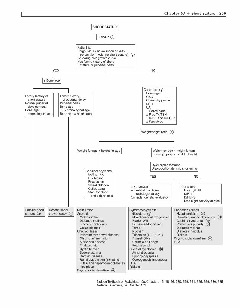
Chapter 67 u Short Stature 259
Patient is:Height >2 SD below mean or <5th percentile (moderate short stature) 2 Following own growth curveHas family history of short stature or pubertal delay
MalnutritionAnorexia Malabsorption Diabetes mellitus (poorly controlled) Celiac diseaseChronic illness Inflammatory bowel disease Chronic inflammation Sickle cell disease Thalassemia Cystic fibrosis Severe asthma Cardiac disease Renal dysfunction (including RTA and nephrogenic diabetes insipidus)Psychosocial dwarfism
MalnutritionAnorexia Malabsorption Diabetes mellitus (poorly controlled) Celiac diseaseChronic illness Inflammatory bowel disease Chronic inflammation Sickle cell disease Thalassemia Cystic fibrosis Severe asthma Cardiac disease Renal dysfunction (including RTA and nephrogenic diabetes insipidus)Psychosocial dwarfism 8
Familial shortstature Familial shortstature 3
Constitutionalgrowth delay Constitutionalgrowth delay 4
Syndromes/genetic disorders Mixed gonadal dysgenesis Prader-Willi Laurence-Moon-Biedl Turner Noonan Trisomies (13, 18, 21) Russell-Silver Cornelia de Lange Fetal alcoholSkeletal dysplasias Achondroplasia Spondylodysplasia Osteogenesis imperfectaRTARickets
Syndromes/genetic disorders 9 Mixed gonadal dysgenesis Prader-Willi Laurence-Moon-Biedl Turner Noonan Trisomies (13, 18, 21) Russell-Silver Cornelia de Lange Fetal alcoholSkeletal dysplasias 10 Achondroplasia Spondylodysplasia Osteogenesis imperfectaRTARickets
Endocrine causes Hypothyroidism Growth hormone deficiency Cushing syndrome Precocious puberty Diabetes mellitus Diabetes insipidus RicketsPsychosocial dwarfism RTA
Endocrine causes Hypothyroidism 11 Growth hormone deficiency 12 Cushing syndrome 13 Precocious puberty 14 Diabetes mellitus Diabetes insipidus RicketsPsychosocial dwarfism 8RTA
SHORT STATURE
H and P 1
± Karyotype± Skeletal dysplasia radiologic surveyConsider genetic evaluation
Consider additional testing: 7 HIV testing Prealbumin Sweat chloride Celiac panel Stool for blood and calprotectin
Weight for age < height for age Weight for age > height for ageor weight proportional for height
Weight/height ratio 6
Consider: 5 Bone age CBC Chemistry profile ESR UA ± Celiac panel ± Free T4/TSH ± IGF-1 and IGFBP3 ± Karyotype
± Bone age
Family history of short statureNormal pubertal developmentBone age = chronological age
Family history of pubertal delayPubertal delayBone age < chronological ageBone age = height age
Dysmorphic featuresDisproportionate limb shortening
Consider: Free T4/TSH IGF-1 IGFBP3 Late-night salivary cortisol
YES
YES NO
NO
Nelson Textbook of Pediatrics, 19e. Chapters 13, 48, 76, 330, 529, 551, 556, 559, 580, 685Nelson Essentials, 6e. Chapter 173

260
Chapter 68
PUBERTAL DELAY
common, but girls who are not diagnosed until their teens may have fewer examination findings and mosaicism (45,X/46,XX) may be present.
4 Mixed gonadal dysgenesis is a common cause of ambiguous genitalia in newborns. The karyotype usually shows mosa-
icism (45,X/46,XY). Patients often have a female phenotype and features of Turner syndrome. Some patients have no signs of masculinization, whereas in others there may be prepubertal clitoromegaly, with further virilization occurring at puberty.
5 Klinefelter syndrome is the most common cause of testicu-lar failure; the karyotype is usually 47,XXY; mosaics are
seen in 20%. The patients are usually tall and thin, testes are small, there is mild mental retardation, and gynecomastia may be present.
6 In pure gonadal dysgenesis, variants include XX or XY karyotype, with normal stature and streak gonads. In XY
gonadal dysgenesis (Swyer syndrome), children have a female phenotype, but at pubertal age, breast development and men-arche fail to occur.
7 Noonan syndrome has similar features to Turner syn-drome but a normal karyotype.
8 The genetic male with androgen insensitivity (i.e., 46,XY disorder of sex development, testicular feminization) ap-
pears phenotypically female, with primary amenorrhea, breast development, and absence of pubic hair. There is wide variation in the degree of androgen resistance. Levels of LH are usually elevated, but FSH levels may be normal because estrogen from the testes may suppress it.
9 A cause of primary ovarian failure may be autoimmune oophoritis, which is associated with autoimmune endocri-
nopathies (thyroiditis, Addison disease, diabetes) and may be screened for with antiovarian antibodies. Resistant ovary syn-drome is due to defects in the FSH or LH receptors. Premature gonadal failure may be associated with galactosemia, myotonic dystrophy, and fragile X–associated disorders. Anorchia is the absence of testes and must be distinguished from bilateral crypt-orchidism. If testes cannot be palpated in boys with normal ex-ternal genitalia, they have cryptorchidism. The testes are usually undescended or retractile, but rarely no testes are found even after investigation. This syndrome is known as “vanishing tes-tes,” congenital anorchia, or testicular regression syndrome. In cryptorchidism, owing to compromised Leydig cell function, normal testosterone levels may only be achieved with elevated FSH and LH levels. Irradiation or chemotherapy may also cause gonadal injury. Enzyme defects include 17-hydroxylase defi-ciency and 17-ketosteroid reductase deficiency, which result in hypertension, adrenal insufficiency, and deficiency of androgens and estrogens. The environmental influences that have been implicated in testicular dysgenesis syndrome include environ-mental chemicals that may act as endocrine disruptors (bisphe-nol A, phthalates), pesticides, phytoestrogens, and other chemi-cals. However, the evidence is only suggestive, not conclusive.
10 Constitutional delay is the most common cause of delayed puberty and is brought to medical attention more often in
boys. There is a family history of delayed puberty, a consistent growth velocity, and a normal examination. Usually the bone age equals height age, which is less than the chronological age.
Pubertal delay is defined as absence of development of second-ary sexual characteristics (breast budding in girls, testicular enlargement in boys) by age 13 in girls and by age 14 in boys. It is related to abnormality of the hypothalamic-pituitary-gonadal axis. Pubic and axillary hair development are due to adrenal androgens and may be present in children with pubertal delay. For discussion of girls with some sexual maturation but with primary amenorrhea, please see Chapter 39.
1 History must include infections, chronic illness, endocri-nopathies, trauma, chemotherapy, irradiation, CNS disor-
ders, and syndromes. Growth patterns and growth velocity should be evaluated. Severe growth retardation may indicate growth hormone (GH) deficiency (see Chapter 67, Short Stature). History of heights of family members should be obtained. Also history of pubertal onset, menarche, and fertility of female relatives may indicate familial disorders such as pubertal delay, androgen insensitivity, and congenital adrenal hyperplasia. It is also important to obtain a history of gonadal tumors, autoimmune endocrine disorders, inborn errors of metabolism, and fragile X syndrome.
Examination includes careful measurements of height, weight, arm span, and body mass index (BMI). Patterns of growth (velocity) and sexual maturity rating (Tanner stage) should be assessed. Body proportions may be helpful; with GH deficiency, height age is less than weight age. Eunuchoidal pro-portions (arm span . height, long legs) are noted with hypogo-nadism. Androgen effects include phallic growth, increased testicular size, sexual hair, voice change, increased stature and muscle mass, hairline recession, and body odor. In girls, exces-sive androgens may lead to acne, hirsutism, or clitoromegaly. Estrogen effects include vaginal cornification/discharge, breast development, uterine size, and onset of menarche 2 to 2.5 years after breast budding. A careful CNS examination should in-clude identification of midline facial defects and tests for olfac-tion (i.e., anosmia/hyposmia in Kallmann syndrome) as well as visual acuity. The presence of gynecomastia and galactorrhea should be noted. Skin examination includes café au lait spots (neurofibromatosis), tanning (adrenal insufficiency), and ich-thyosis (congenital ichthyosis, Kallmann syndrome). Stigmata of specific syndromes may be identified on initial exam (e.g., Turner, fragile X, Prader-Willi, etc.).
2 Plasma FSH and LH levels can be used to classify the causes of pubertal delay into hypogonadotropic (gFSH/LH)
or hypergonadotropic (hFSH/LH) hypogonadism. Bone age assessment, estradiol or testosterone levels, and prolactin and thyroid studies may be considered.
3 Stigmata of gonadal dysgenesis syndromes (e.g., Turner syn-drome) include short stature, pigmented nevi, high-arched
palate, low hairline, shield chest, ptosis, cutis laxa, pterygium coli (webbed neck), shortened fourth metacarpals, cubitus valgus, heart murmurs, nail changes, and deformed ears. There may be a history of congenital lymphedema. The 45,X karyotype is most
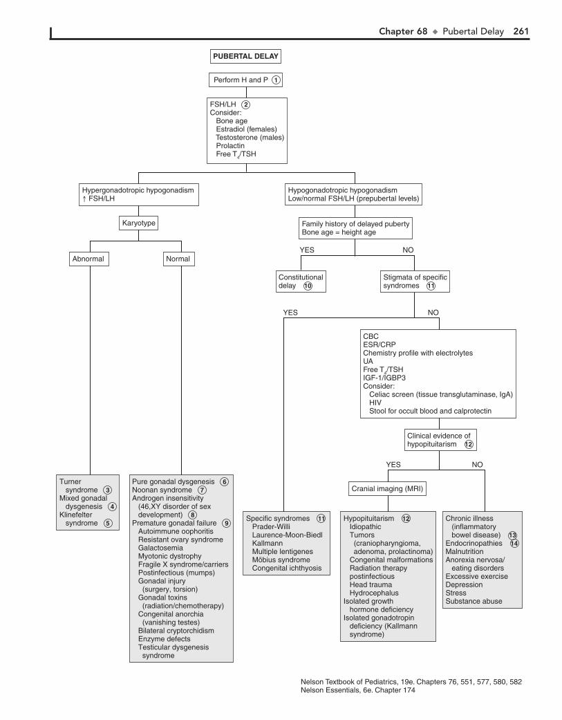
Chapter 68 u PubertalDelay 261
Stigmata of specificsyndromes 11
FSH/LH 2 Consider: Bone age Estradiol (females) Testosterone (males) Prolactin Free T4/TSH
Turner syndrome Mixed gonadal dysgenesis Klinefelter syndrome
Turner syndrome 3 Mixed gonadal dysgenesis 4 Klinefelter syndrome 5
Pure gonadal dysgenesis Noonan syndrome Androgen insensitivity (46,XY disorder of sex development) Premature gonadal failure Autoimmune oophoritis Resistant ovary syndrome Galactosemia Myotonic dystrophy Fragile X syndrome/carriers Postinfectious (mumps) Gonadal injury (surgery, torsion) Gonadal toxins (radiation/chemotherapy) Congenital anorchia (vanishing testes) Bilateral cryptorchidism Enzyme defects Testicular dysgenesis syndrome
Pure gonadal dysgenesis 6 Noonan syndrome 7 Androgen insensitivity (46,XY disorder of sex development) 8 Premature gonadal failure 9 Autoimmune oophoritis Resistant ovary syndrome Galactosemia Myotonic dystrophy Fragile X syndrome/carriers Postinfectious (mumps) Gonadal injury (surgery, torsion) Gonadal toxins (radiation/chemotherapy) Congenital anorchia (vanishing testes) Bilateral cryptorchidism Enzyme defects Testicular dysgenesis syndrome
Specific syndromes Prader-Willi Laurence-Moon-Biedl Kallmann Multiple lentigenes Möbius syndrome Congenital ichthyosis
Specific syndromes 11 Prader-Willi Laurence-Moon-Biedl Kallmann Multiple lentigenes Möbius syndrome Congenital ichthyosis
Hypopituitarism Idiopathic Tumors (craniopharyngioma, adenoma, prolactinoma) Congenital malformations Radiation therapy postinfectious Head trauma HydrocephalusIsolated growth hormone deficiencyIsolated gonadotropin deficiency (Kallmann syndrome)
Hypopituitarism 12 Idiopathic Tumors (craniopharyngioma, adenoma, prolactinoma) Congenital malformations Radiation therapy postinfectious Head trauma HydrocephalusIsolated growth hormone deficiencyIsolated gonadotropin deficiency (Kallmann syndrome)
Chronic illness (inflammatory bowel disease) Endocrinopathies MalnutritionAnorexia nervosa/ eating disordersExcessive exerciseDepressionStressSubstance abuse
Chronic illness (inflammatory bowel disease) 13 Endocrinopathies 14 MalnutritionAnorexia nervosa/ eating disordersExcessive exerciseDepressionStressSubstance abuse
PUBERTAL DELAY
Perform H and P 1
Cranial imaging (MRI)
CBCESR/CRPChemistry profile with electrolytesUAFree T4/TSHIGF-1/IGBP3Consider: Celiac screen (tissue transglutaminase, IgA) HIV Stool for occult blood and calprotectin
Constitutionaldelay 10
Abnormal Normal
Karyotype Family history of delayed pubertyBone age = height age
Hypogonadotropic hypogonadismLow/normal FSH/LH (prepubertal levels)
Clinical evidence ofhypopituitarism 12
YES NO
YES NO
YES NO
Hypergonadotropic hypogonadism FSH/LH
Nelson Textbook of Pediatrics, 19e. Chapters 76, 551, 577, 580, 582Nelson Essentials, 6e. Chapter 174
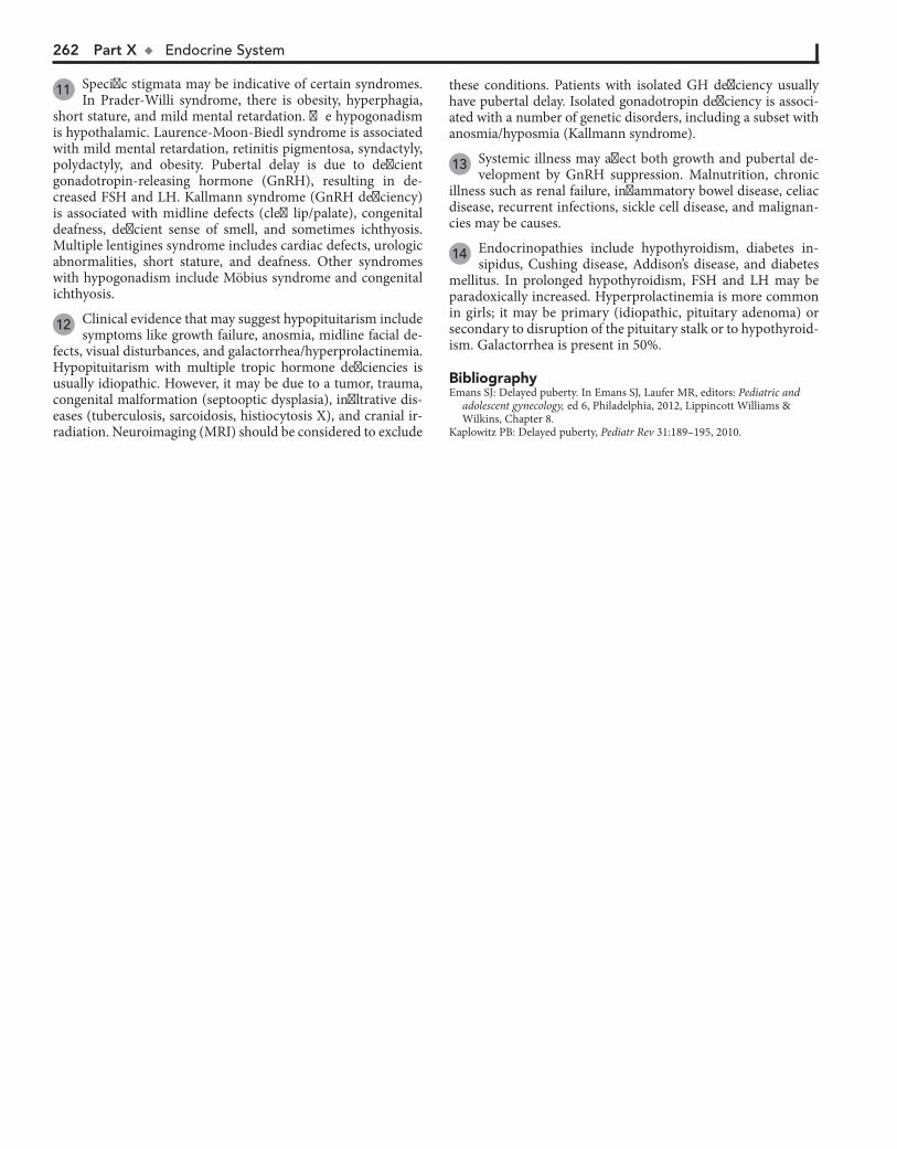
262 Part X u EndocrineSystem
11 Specific stigmata may be indicative of certain syndromes. In Prader-Willi syndrome, there is obesity, hyperphagia,
short stature, and mild mental retardation. The hypogonadism is hypothalamic. Laurence-Moon-Biedl syndrome is associated with mild mental retardation, retinitis pigmentosa, syndactyly, polydactyly, and obesity. Pubertal delay is due to deficient gonadotropin-releasing hormone (GnRH), resulting in de-creased FSH and LH. Kallmann syndrome (GnRH deficiency) is associated with midline defects (cleft lip/palate), congenital deafness, deficient sense of smell, and sometimes ichthyosis. Multiple lentigines syndrome includes cardiac defects, urologic abnormalities, short stature, and deafness. Other syndromes with hypogonadism include Möbius syndrome and congenital ichthyosis.
12 Clinical evidence that may suggest hypopituitarism include symptoms like growth failure, anosmia, midline facial de-
fects, visual disturbances, and galactorrhea/hyperprolactinemia. Hypopituitarism with multiple tropic hormone deficiencies is usually idiopathic. However, it may be due to a tumor, trauma, congenital malformation (septooptic dysplasia), infiltrative dis-eases (tuberculosis, sarcoidosis, histiocytosis X), and cranial ir-radiation. Neuroimaging (MRI) should be considered to exclude
BibliographyEmans SJ: Delayed puberty. In Emans SJ, Laufer MR, editors: Pediatric and
adolescent gynecology, ed 6, Philadelphia, 2012, Lippincott Williams & Wilkins, Chapter 8.
Kaplowitz PB: Delayed puberty, Pediatr Rev 31:189–195, 2010.
these conditions. Patients with isolated GH deficiency usually have pubertal delay. Isolated gonadotropin deficiency is associ-ated with a number of genetic disorders, including a subset with anosmia/hyposmia (Kallmann syndrome).
13 Systemic illness may affect both growth and pubertal de-velopment by GnRH suppression. Malnutrition, chronic
illness such as renal failure, inflammatory bowel disease, celiac disease, recurrent infections, sickle cell disease, and malignan-cies may be causes.
14 Endocrinopathies include hypothyroidism, diabetes in-sipidus, Cushing disease, Addison’s disease, and diabetes
mellitus. In prolonged hypothyroidism, FSH and LH may be paradoxically increased. Hyperprolactinemia is more common in girls; it may be primary (idiopathic, pituitary adenoma) or secondary to disruption of the pituitary stalk or to hypothyroid-ism. Galactorrhea is present in 50%.

Chapter 68 u PubertalDelay 263
Stigmata of specificsyndromes 11
FSH/LH 2 Consider: Bone age Estradiol (females) Testosterone (males) Prolactin Free T4/TSH
Turner syndrome Mixed gonadal dysgenesis Klinefelter syndrome
Turner syndrome 3 Mixed gonadal dysgenesis 4 Klinefelter syndrome 5
Pure gonadal dysgenesis Noonan syndrome Androgen insensitivity (46,XY disorder of sex development) Premature gonadal failure Autoimmune oophoritis Resistant ovary syndrome Galactosemia Myotonic dystrophy Fragile X syndrome/carriers Postinfectious (mumps) Gonadal injury (surgery, torsion) Gonadal toxins (radiation/chemotherapy) Congenital anorchia (vanishing testes) Bilateral cryptorchidism Enzyme defects Testicular dysgenesis syndrome
Pure gonadal dysgenesis 6 Noonan syndrome 7 Androgen insensitivity (46,XY disorder of sex development) 8 Premature gonadal failure 9 Autoimmune oophoritis Resistant ovary syndrome Galactosemia Myotonic dystrophy Fragile X syndrome/carriers Postinfectious (mumps) Gonadal injury (surgery, torsion) Gonadal toxins (radiation/chemotherapy) Congenital anorchia (vanishing testes) Bilateral cryptorchidism Enzyme defects Testicular dysgenesis syndrome
Specific syndromes Prader-Willi Laurence-Moon-Biedl Kallmann Multiple lentigenes Möbius syndrome Congenital ichthyosis
Specific syndromes 11 Prader-Willi Laurence-Moon-Biedl Kallmann Multiple lentigenes Möbius syndrome Congenital ichthyosis
Hypopituitarism Idiopathic Tumors (craniopharyngioma, adenoma, prolactinoma) Congenital malformations Radiation therapy postinfectious Head trauma HydrocephalusIsolated growth hormone deficiencyIsolated gonadotropin deficiency (Kallmann syndrome)
Hypopituitarism 12 Idiopathic Tumors (craniopharyngioma, adenoma, prolactinoma) Congenital malformations Radiation therapy postinfectious Head trauma HydrocephalusIsolated growth hormone deficiencyIsolated gonadotropin deficiency (Kallmann syndrome)
Chronic illness (inflammatory bowel disease) Endocrinopathies MalnutritionAnorexia nervosa/ eating disordersExcessive exerciseDepressionStressSubstance abuse
Chronic illness (inflammatory bowel disease) 13 Endocrinopathies 14 MalnutritionAnorexia nervosa/ eating disordersExcessive exerciseDepressionStressSubstance abuse
PUBERTAL DELAY
Perform H and P 1
Cranial imaging (MRI)
CBCESR/CRPChemistry profile with electrolytesUAFree T4/TSHIGF-1/IGBP3Consider: Celiac screen (tissue transglutaminase, IgA) HIV Stool for occult blood and calprotectin
Constitutionaldelay 10
Abnormal Normal
Karyotype Family history of delayed pubertyBone age = height age
Hypogonadotropic hypogonadismLow/normal FSH/LH (prepubertal levels)
Clinical evidence ofhypopituitarism 12
YES NO
YES NO
YES NO
Hypergonadotropic hypogonadism FSH/LH
Nelson Textbook of Pediatrics, 19e. Chapters 76, 551, 577, 580, 582Nelson Essentials, 6e. Chapter 174
264
Chapter 69
PRECOCIOUS PUBERTY IN THE MALE
5 McCune-Albright syndrome (MAS) is a rare disorder pre-senting with peripheral precocious puberty, café au lait skin
pigmentation, and fibrous dysplasia of bone. It is more common in girls. When it presents in boys there may be overproduction of androgens, resulting in accelerated growth and skeletal matu-ration. Prolonged exposure to elevated levels of sex steroids may cause accelerated growth and skeletal maturation.
Although the precocious puberty is typically peripheral (gonadotropin independent), a secondary central (gonadotropin dependent) component may develop, also because of prolonged exposure to sex steroids. This secondary central precocious puberty may also be seen in late treatment of congenital adrenal hyperplasia (CAH).
6 If the FSH and LH levels are prepubertal and testosterone levels are elevated to pubertal levels, b-hCG levels should
be checked. This hormone acts like LH, stimulating the testes to produce testosterone. Tumors producing b-hCG may be hepa-tomas, hepatoblastomas, teratomas, and chorioepitheliomas.
7 Prolonged untreated hypothyroidism may cause preco-cious puberty, in which case the bone age is delayed. TSH
levels are elevated, and prolactin levels are mildly elevated. Al-though serum FSH is low and LH is undetectable, the elevated TSH appears to stimulate the FSH receptor (with no corre-sponding LH effects). Unlike in central precocious puberty, testicular enlargement occurs without increase in testosterone. Thus, the precocious puberty associated with hypothyroidism behaves as an incomplete form of central precocious puberty.
8 In autosomal dominant familial male-limited precocious puberty (testotoxicosis), b-hCG levels are normal and tes-
tosterone is elevated. There is autonomous production of testos-terone from premature Leydig cell maturation; signs of puberty appear by age 2 to 3, and there is enlargement of the testes.
9 Elevated testosterone levels, particularly with asymmet-ric enlargement of one testis, may indicate a Leydig cell
adenoma.
10 Another source of androgens is an adrenal tumor. Eleva-tion of dehydroepiandrosterone sulfate (DHEAS) above
700 mg/dL or testosterone above 200 ng/dL warrants imaging for adrenal tumors. Adrenal tumors are also associated with increased dehydroepiandrosterone (DHEA) and androstenedi-one. Testosterone may be increased owing to production by the tumor or by peripheral conversion.
11 CAH is screened for by an early-morning 17-hydroxypro-gesterone level. Levels below 200 ng/dL are normal; levels
above 800 ng/dL are diagnostic of 21-hydroxylase deficiency, which is the most common form of CAH. Deficiency of 3b-hydroxysteroid dehydrogenase may result in precocious pubarche.
12 Premature pubarche is most commonly due to premature ad-renarche, when there is an early increase in adrenal androgens.
Testosterone and gonadotropin levels are prepubertal, and DHEAS may be moderately elevated but appropriate for stage 2 pubic hair. A 17-hydroxyprogesterone level may be considered to rule out late-onset CAH. Significantly elevated DHEAS or testosterone levels may require imaging studies to exclude a testicular or adrenal tumor. Clinical followup of premature adrenarche at 3-month intervals is usually adequate. Bone age is usually consistent with chronological
Precocious puberty in the male is defined as the beginning of secondary sexual characteristics before the age of 9 years. Iso-sexual puberty, with characteristic male sexual features, occurs as a result of normal male sex steroids. Heterosexual develop-ment with female characteristics (i.e., gynecomastia) is due to female sex steroids. Early pubarche is the isolated development of pubic or axillary hair.
1 History of growth patterns, chronological development of secondary sex characteristics, exposure to exogenous sex
steroids (e.g., creams, lotions, meats, anabolic steroids), and family history of ambiguous genitalia or early pubertal develop-ment may be helpful. History of CNS disease, head injury, or irradiation may indicate a central cause. Symptoms including behavioral or emotional changes, headache, and vision prob-lems require further assessment. Skin changes such as café au lait spots may indicate neurofibromatosis or McCune-Albright syndrome.
In boys, testicular enlargement is the earliest sign of puberty. Genital examination includes penile length and diameter and testicular volume, which is measured by orchidometer. Pubertal testes are more than 8 mL in volume or greater than 2.5 cm in longest diameter. Sexual Maturity Rating (SMR) or Tanner staging of pubic hair, and breasts in the case of gynecomastia, should be also be evaluated.
2 Central or gonadotropin-dependent precocious puberty is due to early activation of the hypothalamic-pituitary-gonadal
axis. There is increased FSH and LH, acceleration of growth, testicular enlargement, and pubertal levels of testosterone. The bone age is advanced beyond the height age and chrono-logical age.
3 Precocious puberty in males may be idiopathic, but up to 75% have an underlying CNS abnormality; therefore, im-
aging of the head, preferably by MRI, is imperative, in addition to a careful neurologic and visual examination.
4 Hypothalamic hamartomas are the most common brain lesion causing central precocious puberty and may be associated
with seizures. Other CNS lesions causing central gonadotropin- dependent precocity usually involve the hypothalamus. These include postencephalitic scars, tuberculous meningitis, tuberous sclerosis, severe head trauma, and hydrocephalus (with or without associated myelomeningocele).
Neoplasms causing precocious puberty include astrocytomas, ependymomas, and optic tract tumors, including optic gliomas in children with neurofibromatosis type 1 (NF-1). Pineal or hy-pothalamic germinomas can cause central precocious puberty by secreting human chorionic gonadotropin (hCG), which stimu-lates the LH receptors in the Leydig cells of the testes.
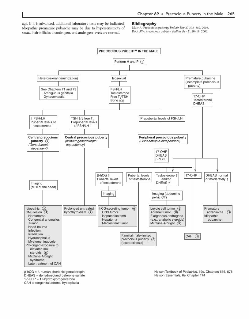
Chapter 69 u PrecociousPubertyintheMale 265
age. If it is advanced, additional laboratory tests may be indicated. Idiopathic premature pubarche may be due to hypersensitivity of sexual hair follicles to androgen, and androgen levels are normal.
BibliographyMuir A: Precocious puberty, Pediatr Rev 27:373–382, 2006.Root AW: Precocious puberty, Pediatr Rev 21:10–19, 2000.
Idiopathic CNS lesion Hamartoma Congential anomalies Tumor Head trauma Infection Irradiation Hydrocephalus Myelomeningocele Prolonged exposure to elevated sex steroids McCune-Albright syndrome Late treatment of CAH
Idiopathic 3 CNS lesion 4 Hamartoma Congential anomalies Tumor Head trauma Infection Irradiation Hydrocephalus Myelomeningocele Prolonged exposure to elevated sex steroids 5 McCune-Albright syndrome Late treatment of CAH
hCG-secreting tumor CNS tumor Hepatoblastoma Hepatoma Mediastinal tumor
hCG-secreting tumor 6 CNS tumor Hepatoblastoma Hepatoma Mediastinal tumor
Prolonged untreated hypothyroidism Prolonged untreated hypothyroidism 7
Familial male-limited precocious puberty (testotoxicosis)
Familial male-limited precocious puberty 8 (testotoxicosis)
Leydig cell tumor Adrenal tumor Exogenous androgens(e.g., anabolic steroids)McCune-Albright
Leydig cell tumor 9 Adrenal tumor 10 Exogenous androgens(e.g., anabolic steroids)McCune-Albright 5
CAH CAH 11
Premature adrenarche Idiopathic pubarche
Premature adrenarche 12 Idiopathic pubarche
PRECOCIOUS PUBERTY IN THE MALE
Perform H and P 1
Imaging
Imaging(MRI of the head)
Central precocious puberty 2 (Gonadotropin dependent)
Imaging (abdomino-pelvic CT)
Pubertal levels of testosterone
�-hCG Pubertal levels of testosterone
17-OHP DHEAS normalor moderately
17-OHPDHEAS�-hCG
Peripheral precocious puberty(Gonadotropin-independent)
Prepubertal levels of FSH/LH
17-OHPTestosteroneDHEAS
Premature pubarche (incomplete precocious puberty)
FSH/LHTestosteroneFree T4/TSHBone age
Isosexual
See Chapters 71 and 73 Ambiguous genitalia Gynecomastia
Heterosexual (feminization)
Central precocious puberty(without gonadotropin dependency)
Testosterone and/orDHEAS
FSH/LHPubertal levels of testosterone
TSH / free T4Prepubertal levels of FSH/LH
�-hCG = �-human chorionic gonadotropinDHEAS = dehydroepiandrosterone sulfate17-OHP = 17-hydroxyprogesteroneCAH = congenital adrenal hyperplasia
Nelson Textbook of Pediatrics, 19e. Chapters 556, 578Nelson Essentials, 6e. Chapter 174
266
Chapter 70
PRECOCIOUS PUBERTY IN THE FEMALE
Neoplasms causing precocious puberty include astrocytomas, ependymomas, and optic tract tumors, including optic gliomas in children with neurofibromatosis type 1 (NF-1). Pineal or hy-pothalamic germinomas can cause central precocious puberty by secreting human chorionic gonadotropin (hCG), which stimu-lates the LH receptors in the Leydig cells of the testes.
5 McCune-Albright syndrome is a rare condition and is more common in girls than in boys; affected girls tend to
have increased estrogen levels. It presents with a triad of periph-eral precocious puberty, café au lait skin pigmentation, and fibrous dysplasia of bone. Vaginal bleeding often precedes sig-nificant breast development. A skeletal survey or technetium bone scan may identify polyostotic fibrous dysplasia. Prolonged exposure to elevated levels of sex steroids may cause accelerated growth and skeletal maturation.
Although the precocious puberty is typically peripheral (gonadotropin independent), a secondary central (gonadotropin dependent) component may develop, also because of prolonged exposure to sex steroids. This secondary central precocious puberty may also be seen in late treatment of congenital adrenal hyperplasia (CAH).
6 Prolonged untreated hypothyroidism may cause preco-cious puberty, in which case the bone age is delayed. TSH
levels are elevated, and prolactin levels are mildly elevated. Al-though serum FSH is low and LH is usually undetectable, the elevated TSH appears to stimulate the FSH receptor (with no corresponding LH effects). Girls may have early breast develop-ment, galactorrhea, and recurrent menstrual bleeding. Preco-cious puberty associated with hypothyroidism behaves as an incomplete form of central precocious puberty.
7 If the FSH and LH levels are prepubertal and estradiol levels are elevated to pubertal levels, abdominopelvic im-
aging should be considered to exclude estrogen-secreting ovar-ian or adrenocortical tumors. The most common ovarian tumor causing precocious puberty is the granulosa cell tumor.
8 Autonomous ovarian follicular cysts are the most common estrogen-secreting masses. Plasma estradiol levels fluctu-
ate with the size of the cyst. These may also result in premature thelarche.
9 Exogenous sources of estrogens include oral contraceptive pills and estrogen-containing tonics, lotions, and creams.
Contamination of meat has been reported.
10 Benign premature thelarche is unilateral or bilateral breast enlargement, usually before age 2 years and resolving
within 6 months to 6 years. No other signs of estrogenization are present. Plasma estrogen levels may be normal to slightly elevated. Laboratory studies are usually not indicated for pre-mature thelarche. Clinical followup is adequate to detect pro-gression to precocious puberty or onset of virilization. Regression followed by recurrence may indicate autonomous ovarian fol-licular cysts. Occurrence in children 3 years and older should prompt further evaluation.
Precocious puberty is defined by the onset of secondary sexual characteristics before the age of 8 years in girls. The lower limit of normal puberty may be age 6 for blacks and age 7 for whites. Isosexual puberty occurs because of normal female sex steroids. Heterosexual development is due to male sex steroids. Early pubarche is the isolated development of pubic or axillary hair. Premature thelarche is the isolated development of the breasts, with no other secondary sexual development.
1 History of growth patterns, chronological development of secondary sex characteristics, exposure to exogenous sex
steroids (e.g., creams, lotions, meats, anabolic steroids), possible ambiguous genitalia, and family history of early pubertal develop-ment may be helpful. Evidence of CNS disease, such as behavioral or emotional changes, head trauma, hydrocephalus, headache, and vision problems, should be assessed. Skin changes such as café au lait spots may indicate McCune-Albright syndrome. Vaginal mucosal changes, enlargement of labia minora, and Tanner stag-ing of pubic hair and breasts should be assessed. Rectoabdominal examination may indicate ovarian or abdominal masses. Viriliza-tion is indicated by excessive hirsutism, clitoromegaly, deepening voice, acne, and muscle development.
2 Central or gonadotropin-dependent precocious puberty is due to early activation of the normal physiologic pubertal
development. There is increased FSH and LH, acceleration of growth, breast development, enlargement of the labia minora, vaginal mucosal change with enlargement of the ovaries and uterus, and pubertal levels of estradiol. The bone age is ad-vanced beyond the height age and chronological age.
3 Childhood obesity has been associated with early puberty in girls and has been noted in children adopted from a
developing country. Precocious puberty in females is often id-iopathic, but a CNS lesion should be considered. Imaging of the head, preferably using MRI, is imperative, in addition to a care-ful neurologic and visual examination.
4 Hypothalamic hamartomas are the most common brain lesion causing central precocious puberty and may be as-
sociated with seizures. Other CNS lesions causing central gonadotropin-dependent precocity usually involve the hypo-thalamus. These include postencephalitic scars, tuberculous meningitis, tuberous sclerosis, severe head trauma, and hydro-cephalus (with or without associated myelomeningocele).
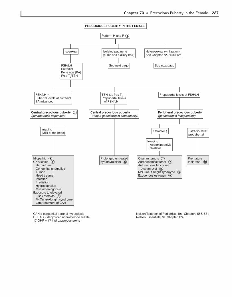
Chapter 70 u PrecociousPubertyintheFemale 267
Central precocious puberty 2 (gonadotropin dependent)
Idiopathic CNS lesion Hamartoma Congenital anomalies Tumor Head trauma Infection Irradiation Hydrocephalus Myelomeningocele Exposure to elevated sex steroids McCune-Albright syndrome Late treatment of CAH
Idiopathic 3 CNS lesion 4 Hamartoma Congenital anomalies Tumor Head trauma Infection Irradiation Hydrocephalus Myelomeningocele Exposure to elevated sex steroids 5 McCune-Albright syndrome Late treatment of CAH
Ovarian tumors Adrenocortical tumor Autonomous functional ovarian cyst McCune-Albright syndrome Exogenous estrogen
Ovarian tumors 7 Adrenocortical tumor 7 Autonomous functional ovarian cyst 8 McCune-Albright syndrome 5 Exogenous estrogen 9
Prematurethelarche Prematurethelarche 10
Prolonged untreatedhypothyroidism Prolonged untreatedhypothyroidism 6
PRECOCIOUS PUBERTY IN THE FEMALE
Perform H and P 1
Imaging Abdominopelvic Skeletal
Prepubertal levels of FSH/LH
Peripheral precocious puberty(gonadotropin-independent)
Central precocious puberty(without gonadotropin dependency)
Imaging(MRI of the head)
FSH/LHEstradiolBone age (BA)Free T4/TSH
Isosexual
See next page
Isolated pubarche(pubic and axillary hair)
See next page
Heterosexual (virilization)See Chapter 72, Hirsutism
Estradiol level prepubertal
TSH / free T4Prepubertal levels of FSH/LH
FSH/LH Pubertal levels of estradiolBA advanced
Estradiol
Nelson Textbook of Pediatrics, 19e. Chapters 556, 581Nelson Essentials, 6e. Chapter 174
CAH = congenital adrenal hyperplasiaDHEAS = dehydroepiandrosterone sulfate17-OHP = 17 hydroxyprogesterone
268 Part X u EndocrineSystem
11 Premature adrenarche is the most common form of iso-lated precocious puberty, with an early increase in adrenal
androgens. In addition to bone age, it is reasonable to obtain a 17-hydroxyprogesterone level to detect mild CAH due to 21-hydroxylase deficiency. Bone age is usually consistent with chronological age in premature adrenarche. Estradiol and go-nadotropin levels are prepubertal, and dehydroepiandrosterone sulfate (DHEAS) may be moderately elevated (appropriate for stage 2 pubic hair). Girls with premature adrenarche are at increased risk for hyperandrogenism and polycystic ovary syndrome. Idiopathic premature pubarche may be due to hypersensitivity of sexual hair follicles to androgen.
If bone age is advanced or signs of virilization are present, further testing is needed.
12 Virilization requires evaluation for androgen-producing adrenal or ovarian tumors, CAH, or exogenous androgen
exposure, particularly in female athletes. CAH is screened for by an early-morning 17-hydroxyprogesterone level. Levels below 200 ng/dL are normal; levels above 800 ng/dL are diag-nostic of 21-hydroxylase deficiency, which is the most common form of CAH. Bone age is greater than the chronological age. Deficiency of 3b-hydroxysteroid dehydrogenase may also result in precocious pubarche.
13 Elevation of DHEAS above 700 mg/dL or testosterone above 200 ng/dL warrants imaging for adrenal or ovarian
tumors. Bone age is usually greater than chronological age.
BibliographyMuir A: Precocious puberty, Pediatr Rev 27:373–382, 2006.Root AW: Precocious puberty, Pediatr Rev 21:10–19, 2000.
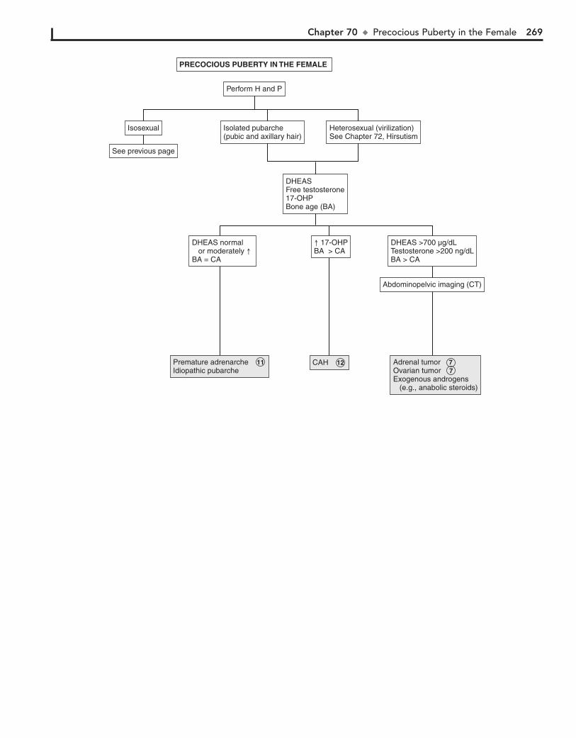
Chapter 70 u PrecociousPubertyintheFemale 269
Premature adrenarche Idiopathic pubarche Premature adrenarche 11 Idiopathic pubarche
CAH CAH 12 Adrenal tumor Ovarian tumor Exogenous androgens (e.g., anabolic steroids)
Adrenal tumor 7 Ovarian tumor 7 Exogenous androgens (e.g., anabolic steroids)
PRECOCIOUS PUBERTY IN THE FEMALE
Perform H and P
DHEAS >700 µg/dLTestosterone >200 ng/dLBA > CA
Abdominopelvic imaging (CT)
Isosexual
See previous page
Isolated pubarche(pubic and axillary hair)
DHEASFree testosterone17-OHPBone age (BA)
Heterosexual (virilization)See Chapter 72, Hirsutism
DHEAS normal or moderately BA = CA
17-OHPBA > CA
270
3 45,X/46,XY gonadal dysgenesis (mixed gonadal dysgenesis) is the second most common cause of ambiguous genitalia.
There is wide phenotypic variation. Most children are short, and stigmata of Turner syndrome are present in one third of patients. There is mosaicism involving the Y chromosome, usually a streak gonad on one side and a dysgenetic or normal-appearing testis on the other. Müllerian and wolffian duct development corre-spond to the ipsilateral gonad.
4 Ovotesticular DSD (true hermaphroditism) is rare. Gonads contain both ovarian and testicular tissue. Both tissue types
may be present in one gonad (ovotestis), or there may be a testis on one side and ovary on the other. About 70% have a 46,XX karyotype, 20% have 46,XX/46,XY mosaicism, and less than 10% of persons with ovotesticular DSD are 46,XY.
5 In partial gonadal dysgenesis the karyotype is 46,XY. It is a form of mixed gonadal dysgenesis.
6 46,XY DSD may be due to defects in testicular differentia-tion. Patients have female external and internal genitalia;
some have ambiguous genitals.Defects in testicular differentiation include the following
syndromes. Denys-Drash syndrome consists of nephropathy with ambiguous genitalia and bilateral Wilms tumor. Müllerian ducts are often present. WAGR syndrome includes Wilms tu-mor, aniridia, GU malformations, and mental retardation. There is a deletion of 1 copy of chromosome 11p13. Males with this syndrome have atypical genitalia. Camptomelic syndrome is a form of short-limbed dysplasia. The gonads may contain elements of both ovaries and testes. In XY pure gonadal dysgen-esis (Swyer syndrome), patients have normal stature, a female phenotype (including internal genitalia [uterus, fallopian tubes, vagina]), and streak gonads. They present with delayed puberty and hypergonadotropic primary amenorrhea (see chapters on Pubertal Delay and Amenorrhea).
7 Other causes of 46,XY DSD include defects in androgen action. In androgen resistance syndromes (testicular femi-
nization) there is a defect in androgen receptors. Although tes-tes are present but are often intraabdominal and testosterone and LH levels are high, external genitalia are female, the vagina is a blind pouch, the uterus is absent, but fallopian tube rem-nants may be present in approximately one third of patients. Diagnosis is usually at the time of puberty, when the phenotypic female presents with amenorrhea.
8 Defects in testicular hormones produce 46,XY males with inadequate masculinization. This was previously known as
male pseudohermaphroditism. Boys with Leydig cell aplasia usually appear phenotypically female with mild virilization. Testes, epididymis, and vas deferens are present; the uterus and fallopian tubes are absent. Testosterone levels are low and LH is elevated.
CAH in 46,XY males may be due to the following enzyme deficiencies. Lipoid adrenal hyperplasia is a severe form of CAH, presenting in salt-wasting crisis (hyponatremia and hyperkale-mia. Genetic males have no müllerian structures. Deficiency of 3b-hydroxysteroid dehydrogenase causes ambiguous genitalia in males and females and may also present with salt wasting. There is increased dehydroepiandrosterone (DHEA) and de-creased androstenedione, testosterone, and estradiol. Deficiency of 17-hydroxylase/17,20-lyase causes ambiguous genitalia in
Evaluation of ambiguous genitalia in an infant requires a great deal of sensitivity. A geneticist, endocrinologist, and urologist should all be included. Bilateral cryptorchidism, unilateral cryptorchidism with incomplete scrotal fusion, subcoronal hy-pospadias, labial fusion, or clitoromegaly should prompt evalu-ation for ambiguous genitalia. Also consider evaluation in children with discordance between prenatal karyotype and ap-pearance of the genitals or those with a family history of con-genital adrenal hyperplasia (CAH).
A recent consensus statement proposed the term disorders of sexual development (DSD) to include “congenital conditions in which the chromosomal, gonadal, or anatomic sex is atypical.” Some of these children present with ambiguous or atypical genitalia.
1 A careful history includes family history of male infants with increased scrotal pigmentation or rugae, infant deaths
due to vomiting and dehydration (CAH), female relatives with amenorrhea or infertility (i.e., 46,XY DSD), and any variant sexual development. History of maternal drug exposure (hor-mones, spironolactone, and environmental chemical disruptors), virilization, or CAH should be obtained. Vomiting, dehydra-tion, or failure to thrive may also suggest CAH.
Physical examination includes identification of stigmata of congenital malformation syndromes that may suggest the diag-nosis. Hypertension, hyperpigmentation of the areolae and la-bioscrotal folds, dehydration, and poor weight gain may suggest CAH. A careful examination of genitalia is important, includ-ing measurement of clitoris/penis (a stretched penile length , 2.5 cm, or a clitoris . 1 cm, are abnormal at term). The ure-thral site, whether perineal or penile, should be determined. The presence of labioscrotal fusion and whether the gonad (almost always a testis) is palpable in the scrotum or inguinal rings should be assessed. A rectal examination is done to assess for the presence of a uterus. Often the cervix can be felt. Stigmata of other syndromes should be noted.
2 Initial evaluation for children with ambiguous genitalia should include 17-hydroxyprogesterone (17-OHP), elec-
trolytes, imaging, and karyotype determination. In addition to US (pelvic, renal, adrenal) other imaging may include voiding cystourethrography and retrograde genitography. Pelvic CT or MRI may be needed for further evaluation. Endoscopy, lapa-rotomy, and gonadal biopsy may be required for complete examination of genitalia with ovotesticular DSD.
CAH due to 21-hydroxylase deficiency is the most common cause of genital ambiguity in females. Currently, all U.S. states and many other countries include testing for 17-OHP levels as part of newborn screening programs. Infants with positive screening tests should be brought in for further testing (electro-lytes and repeat 17-OHP).
Chapter 71
ATYPICAL OR AMBIGUOUS GENITALIA

Chapter 71 u AtypicalorAmbiguous 271
45,X/46,XY gonadal dysgenesis(mixed gonadal dysgenesis[MGD])
45,X/46,XY gonadal dysgenesis(mixed gonadal dysgenesis[MGD]) 3
Ovotesticular DSD(true hermaphroditism)Ovotesticular DSD 4 (true hermaphroditism)
Ovotesticular DSDPartial gonadal dygenesis (MGD with XY)Defects in testicular differentiation Denys-Drash syndrome WAGR syndrome Campomelic syndrome Swyer syndrome (pure gonadal dysgenesis)Defects in androgen action Androgen insensitivity syndromes
Ovotesticular DSD 4 Partial gonadal dygenesis (MGD with XY) 5 Defects in testicular differentiation 6 Denys-Drash syndrome WAGR syndrome Campomelic syndrome Swyer syndrome (pure gonadal dysgenesis)Defects in androgen action Androgen insensitivity syndromes 7
Disorders of testosterone synthesis Leydig cell aplasia Congenital adrenal hyperplasiaDefects in androgen action 5-� reductase deficiency Androgen insensitivity syndromesSmith-Lemli-Opitz syndrome
Disorders of testosterone synthesis 8 Leydig cell aplasia Congenital adrenal hyperplasiaDefects in androgen action 5-� reductase deficiency 9 Androgen insensitivity syndromes 7 Smith-Lemli-Opitz syndrome 10
ATYPICAL OR AMBIGUOUS GENITALIA (DISORDERS OF SEX DEVELOPMENT[DSD])
Perform H and P 1
Karyotype:45,X/46,XY mosaic
Karyotype:46,XX/46,XY mosaic
Müllerian structures present on imaging(fallopian tubes, uterus)
See next page
Gonad palpable/ present on US
Consider: 2 Imaging (US) Karyotype17-hydroxyprogesterone (17-OHP)Electrolytes
Karyotype:46,XY
ConsiderTestosteroneFSH/LH
Karyotype:46,XY
ConsiderTestosteroneEstradiolFSH/LHAndrostenedioneDHEADHT
YES NO
YES NO
Nelson Textbook of Pediatrics, 19e. Chapters 338, 538, 539, 569, 570, 582Nelsons Essentials, 6e. Chapter 177
DHEA = dehydroepiandrosteroneDHT = dihydrotestosterone

272 Part X u EndocrineSystem
males, with hypertension, hypokalemia, and decreased serum androgens. In 17-ketosteroid reductase deficiency, müllerian ducts are absent and a shallow vagina is present.
9 In 5a-reductase deficiency, inadequate conversion of tes-tosterone to dihydrotestosterone (DHT) occurs, which is
necessary for fetal masculinization. Consequently, the newborn is characterized by a small phallus/ambiguous genitalia, hypo-spadias, bifid scrotum, and on occasion, scrotal/labial testes. A high testosterone-to-DHT ratio is diagnostic.
10 Smith-Lemli-Opitz syndrome (SLOS) is an autosomal re-cessive disorder characterized by growth retardation, mi-
crocephaly, ptosis, syndactyly, mental retardation, and genital ambiguity in males. Müllerian remnants are absent.
or ovarian tumor in the mother, or may be idiopathic. The degree of virilization depends on the timing of fetal exposure.
14 Persistent müllerian duct syndrome may be seen in pheno-typic males with cryptorchidism. Müllerian structures are
often discovered during surgery for cryptorchidism.
15 CAH is the most common 46,XX DSD and is usually due to to 21-hydroxylase deficiency, with increased 17-OHP
and adrenal androgens (e.g., dehydroepiandrosterone, andro-stenedione, and androstenediol). The severe form may occur as salt wasting (hyponatremia, hyperkalemia, acidosis), vom-iting, dehydration, and circulatory collapse. Normal ovaries and müllerian structures are present. External genitalia changes depend on time of intrauterine exposure and can range from complete labioscrotal fusion to clitoral hypertro-phy. In 11b-hydroxylase deficiency, 11-deoxycortisol and de-oxycorticosterone levels are high, producing hypertension in infancy.
BibliographyLee PA, Houk CP, Ahmed SF, Hughes LA: International Consensus Confer-
ence on Intersex organized by the Lawson Wilkins Pediatric Endocrine Society and the European Society for Paediatric Endocrinology. Consensus Statement on Management of Intersex Disorders. International Consensus Conference on Intersex, Pediatrics 118:e488–500, 2006.
Emans SJH, Laufer MR: Ambiguous genitalia in the newborn and disorders of sex development. In Emans SJ, Laufer MR, editors: Emans, Laufer, Goldstein’s pediatric & adolescent gynecology, ed 6, Philadelphia, 2012, Lippincott Williams & Wilkins.
11 In XY gonadal agenesis syndrome (embryonic testicular re-gression syndrome) genitalia are female or atypical (ambigu-
ous) in appearance. Müllerian structures are absent. It is presumed that testicles regress between the 8th and 12th weeks of gestation.
12 Anorchia (vanishing testes syndrome) is a form of testicu-lar regression seen in 46,XY phenotypic boys who present
with cryptorchidism. Presumably there was active fetal testicu-lar function during the genital differentiation and with later regression (usually after 20th week). It may also be due to tes-ticular torsion. Testosterone levels are low, and gonadotropin levels are elevated. If this occurs before 8 weeks of gestation, it results in Swyer syndrome, and female genitalia develop.
13 Elevated maternal androgens may be due to maternal inges-tion of medications or hormones or to a virilizing adrenal
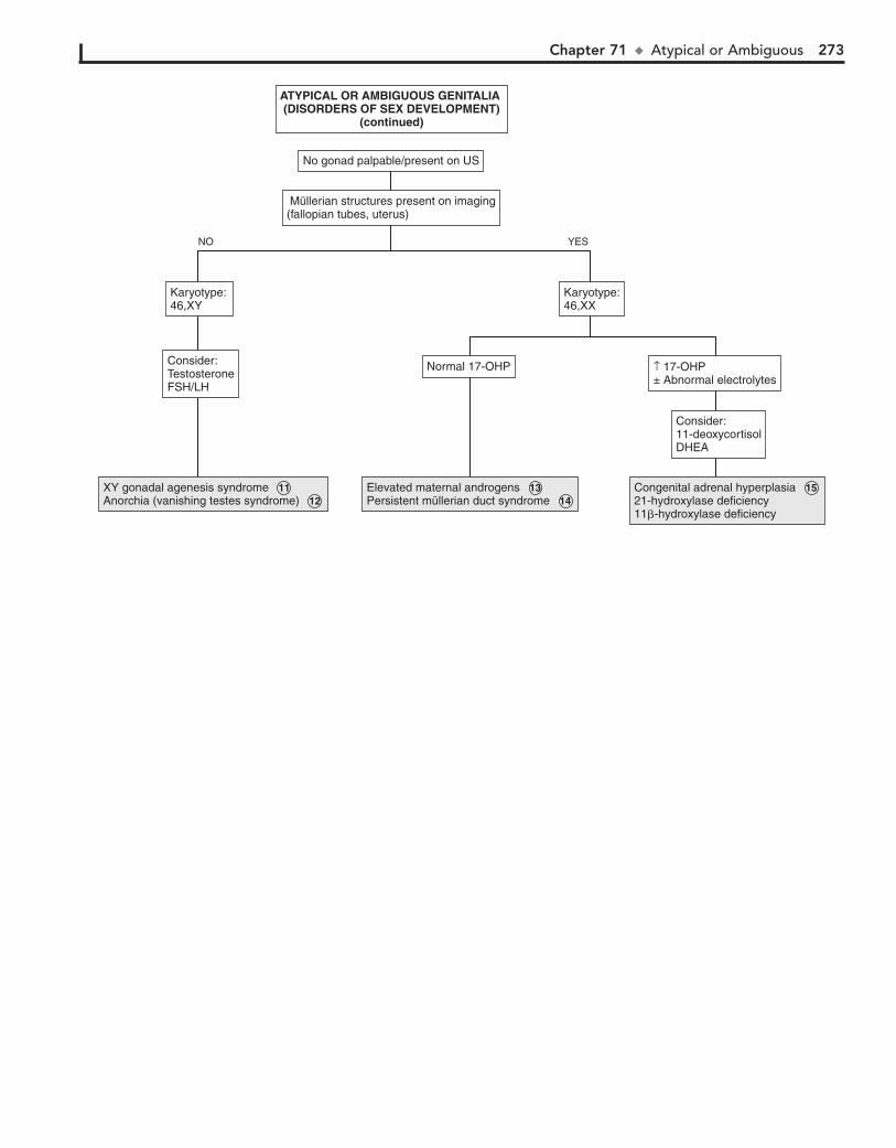
Chapter 71 u AtypicalorAmbiguous 273
XY gonadal agenesis syndrome Anorchia (vanishing testes syndrome)XY gonadal agenesis syndrome 11 Anorchia (vanishing testes syndrome) 12
Elevated maternal androgensPersistent müllerian duct syndromeElevated maternal androgens 13 Persistent müllerian duct syndrome 14
Congenital adrenal hyperplasia21-hydroxylase deficiency11�-hydroxylase deficiency
Congenital adrenal hyperplasia 15 21-hydroxylase deficiency11�-hydroxylase deficiency
ATYPICAL OR AMBIGUOUS GENITALIA (DISORDERS OF SEX DEVELOPMENT)
(continued)
Consider: 11-deoxycortisolDHEA
↑ 17-OHP± Abnormal electrolytes
Normal 17-OHP
Karyotype:46,XX
Karyotype: 46,XY
Consider:TestosteroneFSH/LH
Müllerian structures present on imaging(fallopian tubes, uterus)
No gonad palpable/present on US
YESNO
274
Chapter 72
HIRSUTISM
and male body habitus. The age and rapidity of onset may indi-cate the etiology of the hirsutism. Rapid onset of virilization may indicate a tumor. Early onset of hirsutism is often seen with congenital adrenal hyperplasia (CAH). Amenorrhea or galac-torrhea may indicate hyperprolactinemia. A history of CNS problems such as head trauma or encephalitis should be sought. Medications may at times be responsible and should be re-viewed. A family history of hirsutism, polycystic ovary syn-drome (PCOS), CAH, diabetes, hyperinsulinism, and infertility should be obtained. Careful examination should include the degree of virilization, and a search for thyromegaly, abdominal or pelvic masses, and skin changes (e.g., acanthosis nigricans or any chronic skin irritation such as from a cast). Features of PCOS include hirsutism, obesity, and oligomenorrhea. Presence of striae and a buffalo hump may indicate Cushing syndrome.
2 Idiopathic or familial hirsutism is seen in certain geographic areas, ethnic groups, or families. It is probably due to in-
creased sensitivity of the skin’s hair apparatus to normal levels of androgens, increased conversion of testosterone to dihydrotes-tosterone, or an excess of hair follicles. These women ovulate regularly and have normal menses and normal androgen levels.
3 Virilization during pregnancy may indicate a luteoma, which is an exaggerated response of ovarian stroma to
chorionic gonadotropin; this regresses postpartum.
Hirsutism implies an excessive growth of body hair in women and children in an adult male pattern. It is often due to androgen excess. Hirsutism is an increased density of terminal hair (coarse, adult type) and must be differentiated from hypertrichosis. Hypertrichosis is an increase in vellus hair (i.e., the downy hair seen in prepubertal years) and is often associated with drugs, malignancy, and anorexia. Hirsutism must also be distinguished from virilization (masculinization), which includes findings of increased muscle mass, voice changes, and clitoral enlargement in girls. Sexual hair grows in response to sex steroids. It grows on the face, lower abdomen, anterior thighs, chest, breasts, pubic area, and axilla. Androgens, especially testosterone, stimulate growth of sexual hair, whereas estrogens have an opposite effect. Other hormones (e.g., thyroxine, prolactin) also affect hair growth.
1 Increased androgen production usually causes hirsutism, acne, and increased oiliness of the skin. In extreme cases it
causes virilization or masculinization, with male baldness pat-terns, clitoromegaly, deepening voice, increased muscle mass,

Chapter 72 u Hirsutism 275
IdiopathichirsutismIdiopathichirsutism 2
Anabolic agentsPhenytoinDiazoxideDanazolCorticosteroidsMinoxidilPhenothiazines Penicillamine AcetazolamideValproateCyclosporineCNS disorders (mental and motor retardation) Chronic skin irritation/ hyperemiaRadiation
Anabolic agentsPhenytoinDiazoxideDanazolCorticosteroidsMinoxidilPhenothiazines Penicillamine AcetazolamideValproateCyclosporineCNS disorders (mental and motor retardation) Chronic skin irritation/ hyperemiaRadiation
LuteomaTheca-lutein cystsIatrogenic (progestin/ androgen administration)
Luteoma 3 Theca-lutein cystsIatrogenic (progestin/ androgen administration)
Anorexia nervosa MalnutritionAnorexia nervosa Malnutrition
Hurler syndromeEdward syndrome (trisomy 18)Cornelia de Lange syndromeJuvenile hypothyroidism
Hurler syndromeEdward syndrome (trisomy 18)Cornelia de Lange syndromeJuvenile hypothyroidism
Cushing syndromeCushing syndrome
HIRSUTISM
Perform H and P 1
+Ethnicity/family history of hirsutismNormal ovulatorycycles
Medications/drugsEnvironmental factorsCNS injury
Pregnancy Underweight(low BMI)
Stigmata ofcongenitalsyndromes
Moon faciesBuffalo humpTruncal obesityStriae
Late-night salivary cortisol24-hour urine free cortisolLate-evening plasma cortisol
Pelvic imaging(US/MRI)
Consider:DHEASFree and total testosterone 17-OHP
See next pageAbnormal
Etiology evident from H and P Ethnicity/family history Medications/drugs Pregnancy Severely underweight (low BMI) Features of Cushing syndromeNo evidence of virilization
YES NO
Nelson Textbook of Pediatrics, 19e. Chapters 546, 570, 582, 654BMI = body mass indexDHEAS = dehydroepiandrosterone sulfateHAIR-AN = hyperandrogenism, insulin resistance, acanthosis nigricans17-OHP = 17-hydroxyprogesterone
276 Part X u EndocrineSystem
4 PCOS is the most common cause of hirsutism in adoles-cents. This is a spectrum of clinical disease that is ill
defined, especially in adolescents. Features include persistent anovulation, irregular menstrual cycles, hirsutism, and obe-sity. On US evaluation the ovaries may appear normal or have a number of small cysts (25%). There is an increased sensitiv-ity to gonadotropin-releasing hormones, resulting in an increase in LH levels and LH:FSH ratios. The elevated LH causes an increase in ovarian androgen production and a decrease in sex hormone–binding globulin (SHBG), resulting in an overall increase in free androgen, including testosterone and dehydroepiandrosterone sulfate (DHEAS). If anovula-tion or galactorrhea are present, consider a prolactin level and thyroid function tests to exclude hyperprolactinemia and hypothyroidism.
5 Hyperandrogenism (HA) is often associated with insulin resistance (IR) and acanthosis nigricans (AN), otherwise
known as the HAIR-AN syndrome. The mechanism of this syndrome is unknown; however, there is a defect in membrane insulin receptors. Testing for hyperinsulinism is therefore indi-cated in women with persistent anovulation, android obesity, and acanthosis nigricans.
6 Elevation of DHEAS above 700 mg/dL (suggesting adrenal tumors), testosterone levels over 200 ng/dL (ovarian tumors),
or history of rapid onset of virilizing symptoms warrants imaging for adrenal or ovarian tumors.
7 Late-onset adrenal hyperplasia is screened for by an early-morning 17-hydroxyprogesterone level. Levels below 200
ng/dL are normal; levels above 800 ng/dL are diagnostic of 21-hydroxylase deficiency, which is the most common form of CAH. DHEAS and testosterone levels are also increased. Levels over 200 ng/dL require corticotropin testing for 3b-hydroxys-teroid dehydrogenase and 11b-hydroxylase deficiency.
8 If there is a history of ambiguous genitalia, consider karyotype testing to identify incomplete androgen insen-
sitivity and mixed gonadal dysgenesis. Females with 45,X/46,XY mixed gonadal dysgenesis with Y-containing mosaics, or males with defects in androgen action (partial androgen in-sensitivity, 5a-reductase deficiency) who are phenotypically female, may develop signs of androgen stimulation at puberty. US may be needed to identify an intraabdominal gonad (testis or dysgenetic gonad). (See Chapter 71, Atypical or Ambiguous Genitalia.)
BibliographyEmans SJH, Laufer MR: Androgen abnormalities in the adolescent girl. In Emans
SJ, Laufer MR, editors: Emans, Laufer, Goldstein’s pediatric & adolescent gynecology, ed 6, Philadelphia, 2012, Lippincott Williams & Wilkins.
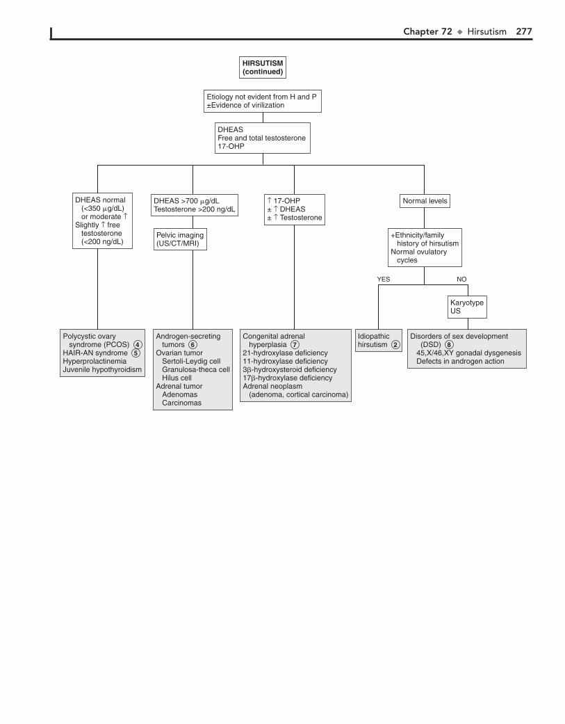
Chapter 72 u Hirsutism 277
Polycystic ovary syndrome (PCOS)HAIR-AN syndromeHyperprolactinemiaJuvenile hypothyroidism
Polycystic ovary syndrome (PCOS) 4 HAIR-AN syndrome 5 HyperprolactinemiaJuvenile hypothyroidism
Androgen-secreting tumorsOvarian tumor Sertoli-Leydig cell Granulosa-theca cell Hilus cellAdrenal tumor Adenomas Carcinomas
Androgen-secreting tumors 6 Ovarian tumor Sertoli-Leydig cell Granulosa-theca cell Hilus cellAdrenal tumor Adenomas Carcinomas
Congenital adrenal hyperplasia21-hydroxylase deficiency11-hydroxylase deficiency3�-hydroxysteroid deficiency17�-hydroxylase deficiencyAdrenal neoplasm (adenoma, cortical carcinoma)
Congenital adrenal hyperplasia 7 21-hydroxylase deficiency11-hydroxylase deficiency3�-hydroxysteroid deficiency17�-hydroxylase deficiencyAdrenal neoplasm (adenoma, cortical carcinoma)
IdiopathichirsutismIdiopathichirsutism 2
Disorders of sex development (DSD) 45,X/46,XY gonadal dysgenesis Defects in androgen action
Disorders of sex development (DSD) 8 45,X/46,XY gonadal dysgenesis Defects in androgen action
HIRSUTISM(continued)
KaryotypeUS
+Ethnicity/family history of hirsutismNormal ovulatory cycles
Pelvic imaging(US/CT/MRI)
Normal levelsDHEAS >700 �g/dLTestosterone >200 ng/dL
DHEASFree and total testosterone 17-OHP
Etiology not evident from H and P ±Evidence of virilization
YES NO
DHEAS normal (<350 �g/dL) or moderate ↑ Slightly ↑ free testosterone (<200 ng/dL)
↑ 17-OHP ± ↑ DHEAS± ↑ Testosterone
278
Chapter 73
GYNECOMASTIA
adolescents. They may be caused by neurofibromas, dermoid cysts, lipoma, lymphangioma, metastatic neuroblastoma, leuke-mia, lymphoma, and rhabdomyosarcoma.
7 Gynecomastia may be seen in some chronic diseases such as cystic fibrosis, ulcerative colitis, cirrhosis, malnutrition
followed by refeeding and acquired immunodeficiency syn-drome (AIDS). It may be related to hepatic dysfunction. Uremia due to renal failure may cause testicular damage and decreased testosterone.
8 If there is no evident etiology for the gynecomastia, or there is evidence of hypogonadism, precocious puberty, or
macrogynecomastia, obtain LH, FSH, estradiol, testosterone, dehydroepiandrosterone sulfate (DHEAS), free T4, TSH, and hCG levels. If testes are smaller than 3 cm in diameter or 8 mL in volume or there is a history of ambiguous genitalia, a karyo-type should also be obtained.
9 If DHEAS is increased, imaging for adrenal tumors is needed. Increased estradiol levels may require US of the
liver, adrenals, and testes for hormone-producing tumors. Adrenal tumors usually cause feminization by conversion of androstenedione to estradiol. MRI imaging of the brain, chest, abdomen, and testes should be considered for evaluation for b-hCG–secreting tumors, which may also cause precocious puberty. Other hormone-producing tumors include LH- secreting pituitary tumors and prolactinomas, which cause galactorrhea but usually not gynecomastia. If galactorrhea is present, obtain a prolactin level. Karyotype may also be con-sidered if there is a history of ambiguous genitalia at birth or if there is evidence of hypergonadotropic hypogonadism (testicular size ,8 mL, h FSH, h LH).
10 In hypogonadism, gynecomastia develops during adre-narche owing to aromatization of adrenal androgens to
estrogen. Klinefelter syndrome (47,XXY) usually presents as delayed puberty, gynecomastia, and small testes; patients with Klinefelter syndrome have decreased testosterone levels. In an-drogen insensitivity (XY), there is end-organ resistance to tes-tosterone. In both conditions, Leydig cell action is retained, and there is secretion of estradiol in response to the increased LH. Testicular damage due to orchitis, injury, chemotherapy, or radiation, and congenital anorchia due to fetal testicular regres-sion, may occur and present as gynecomastia at puberty. CNS injury (paraplegia) may be associated with decreased testicular function, leading to gynecomastia.
Enzyme defects of 17a-hydroxylase, 17/20-desmolase, and 17-ketosteroid reductase result in decreased synthesis of testos-terone. Patients with these defects may also present with am-biguous genitalia at birth. Gynecomastia may also be seen in the gonadal dysgenesis.
11 Adrenal, testicular, and hepatocellular tumors may pro-duce aromatase, causing increased estradiol. Familial
gynecomastia is an autosomal dominant condition and may be due to increased aromatase activity.
12 Estrogen-secreting tumors are a rare cause of gynecomas-tia. Sex cord tumors with annular tubules (SCTAT) may
occur in patients with Peutz-Jeghers syndrome. They are multi-focal and may secrete estradiol.
Gynecomastia is enlargement of the male breast tissue owing to a decrease in the ratio of androgens to estrogens. Testosterone weakly inhibits and estrogen strongly stimulates breast tissue development. In males, estrogen is usually produced by the aromatization of androgens in peripheral tissues. Macrogyneco-mastia is when the breast tissue is greater than 4 cm in diameter. It is similar to middle and late stages of female breast develop-ment and is less likely to resolve spontaneously. Asymmetry of breasts is common; however, any hardness of breast tissue or asymmetric placement of the nipple requires evaluation. Lipo-mastia, the fatty tissue in overweight boys, may cause pseudogy-necomastia.
1 History should include a family history of permanent gyneco-mastia, as well as history of systemic or chronic illness, includ-
ing endocrinopathies, renal failure, liver disease, and malnutrition. A careful history of drug or medication intake, including hormone-containing lotion, creams, foods, and other products, should be obtained. History of excessive alcohol intake as well as illicit drug intake (i.e., marijuana) should be sought. Gynecomas-tia that develops before the onset of other pubertal changes or before age 10 years or is associated with precocious or delayed puberty or macrogynecomastia requires further evaluation.
Physical examination should include a careful examination of the breast, including diameter and consistency of breast tissue and position of the nipple. Determination of pubertal staging and testicular size is also important.
2 Medications and drugs that can cause gynecomastia in-clude hormones: estrogens in meat, milk, lotions, and oils;
androgens, which are aromatized to estrogens; and human cho-rionic gonadotropin (hCG), which causes increased estradiol secretion from the testis.
3 Physiologic pubertal gynecomastia is common in adoles-cents. It occurs in up to 60% of boys aged 10 to 16 and
usually resolves spontaneously (i.e., 75% within 2 years). The breast tissue is less than 4 cm in diameter and resembles breast budding in females. Signs of pubertal development usually pre-cede gynecomastia by at least 6 months. Puberty is between Tanner stages II and IV. Obesity is commonly associated with gynecomastia and is usually due to increased aromatization of androgens to estrogen by adipose tissue. Gynecomastia may also persist longer in obese boys.
4 Lipomastia may also cause prominence of the breast tissue, known as pseudogynecomastia, and is usually seen in
obese boys.
5 Familial gynecomastia may be X-linked or autosomal dominant; it may be due to increased aromatase activity.
6 Unilateral enlargement, especially with asymmetry and hard breast tissue, suggests local tumors. These are rare in

Chapter 73 u Gynecomastia 279
HeroinMethadoneAlcoholMarijuanaAmphetaminesHormonesAntibiotics/antifungals Ketoconazole Antituberculous drugs Metronidazole AntiretroviralsGI Cimetidine Ranitdine OmeprazoleCardiovascular Furosemide Spironolactone ACE inhibitors Calcium channel blockersPsychiatric Tricyclic antidepressants Phenothiazines Risperidone Diazepam Clonidine Cytotoxic drugs
HeroinMethadoneAlcoholMarijuanaAmphetaminesHormonesAntibiotics/antifungals Ketoconazole Antituberculous drugs Metronidazole AntiretroviralsGI Cimetidine Ranitdine OmeprazoleCardiovascular Furosemide Spironolactone ACE inhibitors Calcium channel blockersPsychiatric Tricyclic antidepressants Phenothiazines Risperidone Diazepam Clonidine Cytotoxic drugs
Physiologic gynecomastiaPseudo- gynecomastia
Physiologic gynecomastia 3 Pseudo- gynecomastia 4
Familial gynecomastiaLocal tumor/ metastasesChronic illness
Familial gynecomastia 5 Local tumor/ metastases 6 Chronic illness 7
CNS tumorTesticular tumorAdrenal tumorLiver tumor
CNS tumorTesticular tumorAdrenal tumorLiver tumor
Hypogonadism Klinefelter syndrome Androgen insensitivity Enzyme defects Testicular injury Gonadal dysgenesis
Hypogonadism 10 Klinefelter syndrome Androgen insensitivity Enzyme defects Testicular injury Gonadal dysgenesis
Aromatase excess Aromatase -secreting tumors FamilialEstrogen-secreting tumor
Aromatase excess 11 Aromatase -secreting tumors FamilialEstrogen-secreting tumor 12
HyperthyroidismHyperthyroidism 13
ProlactinomaHyperprolactinemia (drug/tumor)
ProlactinomaHyperprolactinemia (drug/tumor)
Idiopathic Idiopathic
GYNECOMASTIAGYNECOMASTIA
Perform H and P 1
Imaging 9
FSH/LH Estradiol
Free T4 TSH
Prolactin Normallaboratorystudies
DHEAS �-hCG Estradiol
FSH/LH Testosterone
Imaging
DHEAS 8 FSH/LHTestosteroneEstradiol�-hCGFree T4/TSHProlactin ± Karyotype
Clinical clues suggestive of diagnosis Family history of permanent gynecomastia Abnormal/asymmetric breast tissue or nipple History of chronic illness
Normal pubertal onsetStage II- IV pubertyAge >10 years Breast tissue centered below nipple
Recreational drugsAlcohol Medications
Use of medications/drugs knownto cause gynecomastia 2
YES NO
YES NO
YES NO
Nelson Textbook of Pediatrics, 19e. Chapter 579Nelson Essentials, 6e. Chapter 68

280 Part X u EndocrineSystem
BibliographyDiamantopoulos SY: Gynecomastia and premature thelarche: A guide for
practitioners, Pediatr Rev 28:57–68, 2007.
13 Gynecomastia is seen in a third of patients with thyrotoxi-cosis. It is primarily due to increased androstenedione,
which is aromatized to estradiol. Hyperthyroidism also alters the androgen-to-estrogen ratio by increasing bound androgen and decreasing free testosterone.
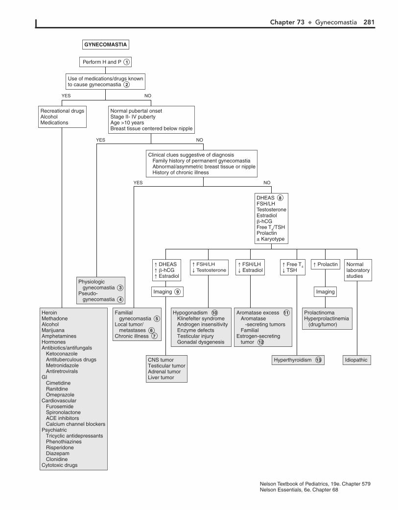
Chapter 73 u Gynecomastia 281
HeroinMethadoneAlcoholMarijuanaAmphetaminesHormonesAntibiotics/antifungals Ketoconazole Antituberculous drugs Metronidazole AntiretroviralsGI Cimetidine Ranitdine OmeprazoleCardiovascular Furosemide Spironolactone ACE inhibitors Calcium channel blockersPsychiatric Tricyclic antidepressants Phenothiazines Risperidone Diazepam Clonidine Cytotoxic drugs
HeroinMethadoneAlcoholMarijuanaAmphetaminesHormonesAntibiotics/antifungals Ketoconazole Antituberculous drugs Metronidazole AntiretroviralsGI Cimetidine Ranitdine OmeprazoleCardiovascular Furosemide Spironolactone ACE inhibitors Calcium channel blockersPsychiatric Tricyclic antidepressants Phenothiazines Risperidone Diazepam Clonidine Cytotoxic drugs
Physiologic gynecomastiaPseudo- gynecomastia
Physiologic gynecomastia 3 Pseudo- gynecomastia 4
Familial gynecomastiaLocal tumor/ metastasesChronic illness
Familial gynecomastia 5 Local tumor/ metastases 6 Chronic illness 7
CNS tumorTesticular tumorAdrenal tumorLiver tumor
CNS tumorTesticular tumorAdrenal tumorLiver tumor
Hypogonadism Klinefelter syndrome Androgen insensitivity Enzyme defects Testicular injury Gonadal dysgenesis
Hypogonadism 10 Klinefelter syndrome Androgen insensitivity Enzyme defects Testicular injury Gonadal dysgenesis
Aromatase excess Aromatase -secreting tumors FamilialEstrogen-secreting tumor
Aromatase excess 11 Aromatase -secreting tumors FamilialEstrogen-secreting tumor 12
HyperthyroidismHyperthyroidism 13
ProlactinomaHyperprolactinemia (drug/tumor)
ProlactinomaHyperprolactinemia (drug/tumor)
Idiopathic Idiopathic
GYNECOMASTIAGYNECOMASTIA
Perform H and P 1
Imaging 9
FSH/LH Estradiol
Free T4 TSH
Prolactin Normallaboratorystudies
DHEAS �-hCG Estradiol
FSH/LH Testosterone
Imaging
DHEAS 8 FSH/LHTestosteroneEstradiol�-hCGFree T4/TSHProlactin ± Karyotype
Clinical clues suggestive of diagnosis Family history of permanent gynecomastia Abnormal/asymmetric breast tissue or nipple History of chronic illness
Normal pubertal onsetStage II- IV pubertyAge >10 years Breast tissue centered below nipple
Recreational drugsAlcohol Medications
Use of medications/drugs knownto cause gynecomastia 2
YES NO
YES NO
YES NO
Nelson Textbook of Pediatrics, 19e. Chapter 579Nelson Essentials, 6e. Chapter 68
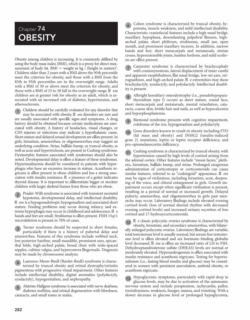
282
Chapter 74
OBESITY
6 Cohen syndrome is characterized by truncal obesity, hy-potonia, muscle weakness, and mild intellectual disability.
Characteristic craniofacial features include a high nasal bridge, maxillary hypoplasia, downslanting palpebral fissures, high-arched palate, short philtrum, strabismus, small jaw, open mouth, and prominent maxillary incisors. In addition, narrow hands and feet, short metacarpals and metatarsals, simian crease, hyperextensible joints, lumbar lordosis, and mild scolio-sis are often present.
7 Carpenter syndrome is characterized by brachycephaly with craniosynostosis, lateral displacement of inner canthi
and apparent exophthalmos, flat nasal bridge, low-set ears, ret-rognathism, and high-arched palate. The extremities may show brachydactyly, syndactyly, and polydactyly. Intellectual disabil-ity is present.
8 Albright hereditary osteodystrophy (i.e., pseudohypopara-thyroidism type I) occurs as short stature, round face,
short metacarpals and metatarsals, mental retardation, cata-racts, coarse skin, brittle hair and nails, as well as hypocalcemia and hyperphosphatemia.
9 Biemond syndrome presents with cognitive impairment, coloboma of the iris, hypogonadism and polydactyly.
10 Gene disorders known to result in obesity including FTO (fat mass and obesity) and INSIG2 (insulin-induced
gene 2) mutations, leptin or leptin receptor deficiency, and pro-opiomelanocortin deficiency.
11 Cushing syndrome is characterized by truncal obesity, with hypertension caused by high levels of cortisol arising from
the adrenal cortex. Other features include “moon facies,” pleth-ora, hirsutism, buffalo hump, and striae. Prolonged exogenous administration of corticotropin or corticosteroids can cause similar features, referred to as “cushingoid” appearance. There may be signs of virilization, including hirsutism, acne, deepen-ing of the voice, and clitoral enlargement in girls. Growth im-pairment occurs except when significant virilization is present, resulting in a period of normal or increased growth. Delayed puberty, amenorrhea, and oligomenorrhea in girls past men-arche may occur. Laboratory findings include elevated evening cortisol levels (loss of normal diurnal rhythm with decreased evening cortisol levels) and increased urinary excretion of free cortisol and 17-hydroxycorticosteroids.
12 The classic polycystic ovaries syndrome is characterized by obesity, hirsutism, and secondary amenorrhea, with bilater-
ally enlarged polycystic ovaries. Laboratory findings are variable; total testosterone level is usually normal, but serum free testoster-one level is often elevated and sex hormone–binding globulin level decreased. There is often an increased ratio of LH to FSH. Dehydroepiandrosterone sulfate (DHEAS) levels are normal or moderately elevated. Hyperandrogenism is often associated with insulin resistance and acanthosis nigricans. Testing for hyperin-sulinism (i.e., fasting blood insulin and glucose) may be consid-ered in women with persistent anovulation, android obesity, or acanthosis nigricans.
13 Hypoglycemic symptoms, particularly with rapid drop in glucose levels, may be due to activation of the autonomic
nervous system and include perspiration, tachycardia, pallor, tremulousness, weakness, hunger, nausea, and vomiting. With a slower decrease in glucose level or prolonged hypoglycemia,
Obesity among children is increasing. It is commonly defined by using the body mass index (BMI), which is a proxy for direct mea-surement of body fat. BMI 5 weight in kg / (height in meters)2. Children older than 2 years with a BMI above the 95th percentile meet the criterion for obesity, and those with a BMI from the 85th to 95th percentiles are in the overweight range. Adults with a BMI of 30 or above meet the criterion for obesity, and those with a BMI of 25 to 30 fall in the overweight range. These children are at greater risk for obesity as an adult, which is as-sociated with an increased risk of diabetes, hypertension, and atherosclerosis.
1 Children should be carefully evaluated for any disorder that may be associated with obesity. These disorders are rare and
are usually associated with specific signs and symptoms. A drug history should be obtained because certain medications are asso-ciated with obesity. A history of headaches, visual changes, or CNS injuries or infections may indicate a hypothalamic cause. Short stature and delayed sexual development are often present. In girls, hirsutism, amenorrhea, or oligomenorrhea may suggest an underlying condition. Striae, buffalo hump, or truncal obesity, as well as acne and hypertrichosis, are present in Cushing syndrome. Dysmorphic features associated with syndromes should also be noted. Developmental delay is often a feature of these syndromes. Hyperinsulinemia should be considered in patients with hyper-phagia who have an excessive increase in stature. Acanthosis ni-gricans is often present in obese children and has a strong asso-ciation with insulin resistance. The presence of a goiter indicates thyroid disease. It is important to differentiate bigger or stockier children with larger skeletal frames from those who are obese.
2 Prader-Willi syndrome is associated with transient neonatal hypotonia, developmental delay, and intellectual disability.
There is a hypogonadotropic hypogonadism and associated short stature. Feeding problems may occur during infancy, and ex-treme hyperphagia may occur in childhood and adolescence. The hands and feet are small. Strabismus is often present. FISH 15q11 microdeletion is present in 70% of cases.
3 Turner syndrome should be suspected in short females, particularly if there is a history of pubertal delay and
amenorrhea. Features of this syndrome include webbed neck, low posterior hairline, small mandible, prominent ears, epican-thal folds, high-arched palate, broad chest with wide-spaced nipples, cubitus valgus, and hyperconvex fingernails. Diagnosis may be made by chromosome analysis.
4 Laurence-Moon-Biedl (Bardet-Biedl) syndrome is charac-terized by truncal obesity and retinal dystrophy/retinitis
pigmentosa with progressive visual impairment. Other features include intellectual disability, digital anomalies (polydactyly, syndactyly), hypogenitalism, and nephropathy.
5 Alström-Hallgren syndrome is associated with nerve deafness, diabetes mellitus, and retinal degeneration with blindness,
cataracts, and small testes in males.
Chapter 74 u Obesity 283
Antipsychotics (risperidone, phenothiazines)Tricyclic antidepressantsLithiumAnticonvulsants (valproate, carbamazepine)Antihistamines (cyproheptadine)Antidiabetic drugs (insulin, sulfonylureas)Exogenous steroids�-adrenergic blockers (propranolol)Steroid hormones (progestins)
Antipsychotics (risperidone, phenothiazines)Tricyclic antidepressantsLithiumAnticonvulsants (valproate, carbamazepine)Antihistamines (cyproheptadine)Antidiabetic drugs (insulin, sulfonylureas)Exogenous steroids�-adrenergic blockers (propranolol)Steroid hormones (progestins)
Prader-Willi syndromeTurner syndromeLaurence-Moon-Biedl syndromeAlström-Hallgren syndromeCohen syndromeCarpenter syndromeAlbright hereditary osteodystrophyBiemond syndromeGene disorders
Prader-Willi syndrome 2 Turner syndrome 3 Laurence-Moon-Biedl syndrome 4 Alström-Hallgren syndrome 5 Cohen syndrome 6 Carpenter syndrome 7 Albright hereditary osteodystrophy 8 Biemond syndrome 9 Gene disorders 10
HypothyroidismGrowth hormone deficiencyHypothalamic dysfunctionPrader-Willi syndromeTurner syndrome
HypothyroidismGrowth hormone deficiencyHypothalamic dysfunctionPrader-Willi syndrome 2 Turner syndrome 3
CushingsyndromeCushingsyndrome 11
Polycysticovarydisease
Polycysticovarydisease 12
Hyperinsulinism Insulinoma Diabetes mellitus (excess insulin administration) Melanocortin 4 receptor gene mutation
Hyperinsulinism 14 Insulinoma Diabetes mellitus (excess insulin administration) Melanocortin 4 receptor gene mutation
PrimaryobesityPrimaryobesity 16
OBESITY
Perform H and P 1
Genetic consultKaryotype
Dysmorphisms±Developmental delay±Intellectual disability±Short stature
Consider :Free T4/TSHIGF-1IGFBP3±Karyotype Referrals to genetics/ endocrinology (See Chapters 67, 68.)
Short staturePubertal delay
Late-night salivary cortisolUrine free cortisol24-hour urine steroidsSerum cortisol
Short statureStriaeHypertensionTruncal obesityBuffalo hump
DHEASFree testosteroneLH/FSH(See Chapters 39, 72.)
Oligo- menorrheaAmenorrheaHirsutism
Referral toendocrinologist
Hypoglycemic symptoms 13 HyperphagiaNormal or increased stature
Referral topsychiatrist/psychologist
Meets criteria ofan eating disorder
Use of obesogenic medications/drugs
Signs and symptoms suggestive of underlying medical condition: Short or excessive stature Dysmorphic features Pubertal delay Menstrual irregularity Hirsutism Developmental delay Intellectual disability
BulimiaBinge-eating disorder
BulimiaBinge-eating disorder 15
YES NO
YES NO
YES NO
Nelson Textbook of Pediatrics, 19e. Chapter 44Nelsons Essentials, 6e. Chapter 29DHEAS = dehydroepiandrosterone sulfate

284 Part X u EndocrineSystem
symptoms include headache, confusion, and visual distur-bances. The symptoms of hypoglycemia in infants may be more subtle, such as cyanosis, apnea, hypothermia, hypotonia, poor feeding, lethargy, and seizures. In older children, hypoglycemia may cause behavior problems, inattention, ravenous appetite, or seizures.
14 Hyperinsulinemia may be due to an insulin-secreting pan-creatic tumor, hypersecretion of the pancreatic beta cells,
or a hypothalamic lesion. There is hyperinsulinemia with hy-perphagia and increased linear growth. It may also be caused by excessive amounts of insulin in patients with diabetes mellitus. Melanocortin 4 receptor gene mutation is the most common known genetic cause for obesity that is severe and of early onset.
15 Eating disorders are more common in females and among whites. Most patients first develop symptoms during ado-
lescence. Bulimia nervosa and binge-eating disorders are char-acterized by weight gain, in contrast to anorexia nervosa. In binge eating disorders, a “distorted” body image is often present.
Those affected may tend to eat alone and have feelings of guilt or disgust after binge eating. The hallmark of bulimia nervosa is binge eating followed by compensatory behavior such as purging, exercise, fasting, and laxative use.
16 In most cases, obesity is the result of a positive energy bal-ance that is stored as adipose tissue. Factors associated with
a positive balance include excessive intake of high-energy foods, inadequate exercise, sedentary lifestyle, low metabolic rate, and inadequate sleep. In primary obesity, screening for comorbidities is recommended. Screening tests may include fasting glucose and Hgb A1C to screen for diabetes or prediabetes, serum ALT to screen for fatty liver, and a lipid panel for hyperlipidemias. Vitamin D levels may also be obtained; vitamin D deficiency is commonly associated with obesity.
BibliographyAmerican Academy of Pediatrics Committee on Adolescence: Identifying and
treating eating disorders, Pediatrics 111:204–211, 2003.
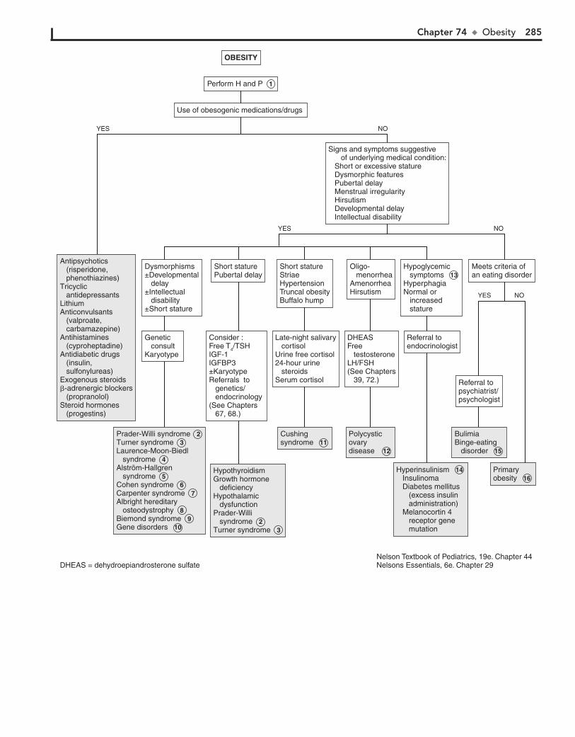
Chapter 74 u Obesity 285
Antipsychotics (risperidone, phenothiazines)Tricyclic antidepressantsLithiumAnticonvulsants (valproate, carbamazepine)Antihistamines (cyproheptadine)Antidiabetic drugs (insulin, sulfonylureas)Exogenous steroids�-adrenergic blockers (propranolol)Steroid hormones (progestins)
Antipsychotics (risperidone, phenothiazines)Tricyclic antidepressantsLithiumAnticonvulsants (valproate, carbamazepine)Antihistamines (cyproheptadine)Antidiabetic drugs (insulin, sulfonylureas)Exogenous steroids�-adrenergic blockers (propranolol)Steroid hormones (progestins)
Prader-Willi syndromeTurner syndromeLaurence-Moon-Biedl syndromeAlström-Hallgren syndromeCohen syndromeCarpenter syndromeAlbright hereditary osteodystrophyBiemond syndromeGene disorders
Prader-Willi syndrome 2 Turner syndrome 3 Laurence-Moon-Biedl syndrome 4 Alström-Hallgren syndrome 5 Cohen syndrome 6 Carpenter syndrome 7 Albright hereditary osteodystrophy 8 Biemond syndrome 9 Gene disorders 10
HypothyroidismGrowth hormone deficiencyHypothalamic dysfunctionPrader-Willi syndromeTurner syndrome
HypothyroidismGrowth hormone deficiencyHypothalamic dysfunctionPrader-Willi syndrome 2 Turner syndrome 3
CushingsyndromeCushingsyndrome 11
Polycysticovarydisease
Polycysticovarydisease 12
Hyperinsulinism Insulinoma Diabetes mellitus (excess insulin administration) Melanocortin 4 receptor gene mutation
Hyperinsulinism 14 Insulinoma Diabetes mellitus (excess insulin administration) Melanocortin 4 receptor gene mutation
PrimaryobesityPrimaryobesity 16
OBESITY
Perform H and P 1
Genetic consultKaryotype
Dysmorphisms±Developmental delay±Intellectual disability±Short stature
Consider :Free T4/TSHIGF-1IGFBP3±Karyotype Referrals to genetics/ endocrinology (See Chapters 67, 68.)
Short staturePubertal delay
Late-night salivary cortisolUrine free cortisol24-hour urine steroidsSerum cortisol
Short statureStriaeHypertensionTruncal obesityBuffalo hump
DHEASFree testosteroneLH/FSH(See Chapters 39, 72.)
Oligo- menorrheaAmenorrheaHirsutism
Referral toendocrinologist
Hypoglycemic symptoms 13 HyperphagiaNormal or increased stature
Referral topsychiatrist/psychologist
Meets criteria ofan eating disorder
Use of obesogenic medications/drugs
Signs and symptoms suggestive of underlying medical condition: Short or excessive stature Dysmorphic features Pubertal delay Menstrual irregularity Hirsutism Developmental delay Intellectual disability
BulimiaBinge-eating disorder
BulimiaBinge-eating disorder 15
YES NO
YES NO
YES NO
Nelson Textbook of Pediatrics, 19e. Chapter 44Nelsons Essentials, 6e. Chapter 29DHEAS = dehydroepiandrosterone sulfate
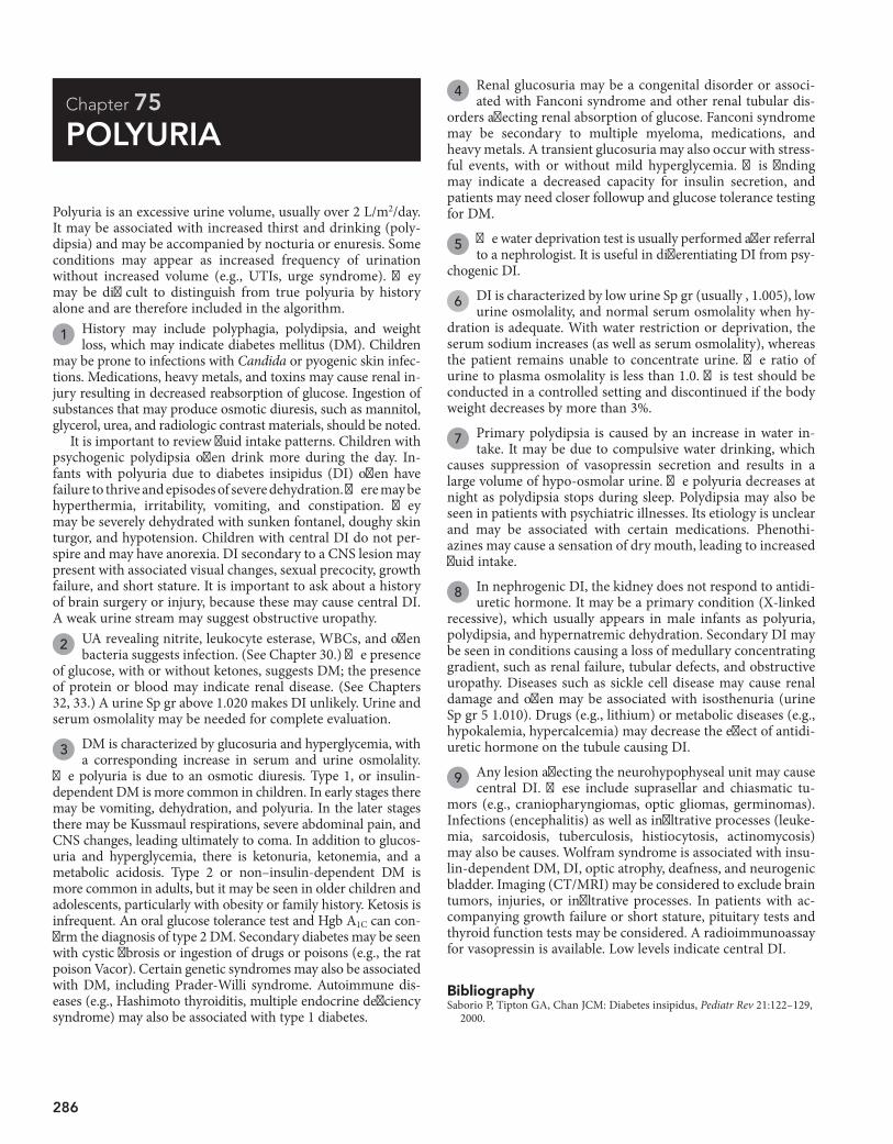
286
Chapter 75
POLYURIA
4 Renal glucosuria may be a congenital disorder or associ-ated with Fanconi syndrome and other renal tubular dis-
orders affecting renal absorption of glucose. Fanconi syndrome may be secondary to multiple myeloma, medications, and heavy metals. A transient glucosuria may also occur with stress-ful events, with or without mild hyperglycemia. This finding may indicate a decreased capacity for insulin secretion, and patients may need closer followup and glucose tolerance testing for DM.
5 The water deprivation test is usually performed after referral to a nephrologist. It is useful in differentiating DI from psy-
chogenic DI.
6 DI is characterized by low urine Sp gr (usually , 1.005), low urine osmolality, and normal serum osmolality when hy-
dration is adequate. With water restriction or deprivation, the serum sodium increases (as well as serum osmolality), whereas the patient remains unable to concentrate urine. The ratio of urine to plasma osmolality is less than 1.0. This test should be conducted in a controlled setting and discontinued if the body weight decreases by more than 3%.
7 Primary polydipsia is caused by an increase in water in-take. It may be due to compulsive water drinking, which
causes suppression of vasopressin secretion and results in a large volume of hypo-osmolar urine. The polyuria decreases at night as polydipsia stops during sleep. Polydipsia may also be seen in patients with psychiatric illnesses. Its etiology is unclear and may be associated with certain medications. Phenothi-azines may cause a sensation of dry mouth, leading to increased fluid intake.
8 In nephrogenic DI, the kidney does not respond to antidi-uretic hormone. It may be a primary condition (X-linked
recessive), which usually appears in male infants as polyuria, polydipsia, and hypernatremic dehydration. Secondary DI may be seen in conditions causing a loss of medullary concentrating gradient, such as renal failure, tubular defects, and obstructive uropathy. Diseases such as sickle cell disease may cause renal damage and often may be associated with isosthenuria (urine Sp gr 5 1.010). Drugs (e.g., lithium) or metabolic diseases (e.g., hypokalemia, hypercalcemia) may decrease the effect of antidi-uretic hormone on the tubule causing DI.
9 Any lesion affecting the neurohypophyseal unit may cause central DI. These include suprasellar and chiasmatic tu-
mors (e.g., craniopharyngiomas, optic gliomas, germinomas). Infections (encephalitis) as well as infiltrative processes (leuke-mia, sarcoidosis, tuberculosis, histiocytosis, actinomycosis) may also be causes. Wolfram syndrome is associated with insu-lin-dependent DM, DI, optic atrophy, deafness, and neurogenic bladder. Imaging (CT/MRI) may be considered to exclude brain tumors, injuries, or infiltrative processes. In patients with ac-companying growth failure or short stature, pituitary tests and thyroid function tests may be considered. A radioimmunoassay for vasopressin is available. Low levels indicate central DI.
Polyuria is an excessive urine volume, usually over 2 L/m2/day. It may be associated with increased thirst and drinking (poly-dipsia) and may be accompanied by nocturia or enuresis. Some conditions may appear as increased frequency of urination without increased volume (e.g., UTIs, urge syndrome). They may be difficult to distinguish from true polyuria by history alone and are therefore included in the algorithm.
1 History may include polyphagia, polydipsia, and weight loss, which may indicate diabetes mellitus (DM). Children
may be prone to infections with Candida or pyogenic skin infec-tions. Medications, heavy metals, and toxins may cause renal in-jury resulting in decreased reabsorption of glucose. Ingestion of substances that may produce osmotic diuresis, such as mannitol, glycerol, urea, and radiologic contrast materials, should be noted.
It is important to review fluid intake patterns. Children with psychogenic polydipsia often drink more during the day. In-fants with polyuria due to diabetes insipidus (DI) often have failure to thrive and episodes of severe dehydration. There may be hyperthermia, irritability, vomiting, and constipation. They may be severely dehydrated with sunken fontanel, doughy skin turgor, and hypotension. Children with central DI do not per-spire and may have anorexia. DI secondary to a CNS lesion may present with associated visual changes, sexual precocity, growth failure, and short stature. It is important to ask about a history of brain surgery or injury, because these may cause central DI. A weak urine stream may suggest obstructive uropathy.
2 UA revealing nitrite, leukocyte esterase, WBCs, and often bacteria suggests infection. (See Chapter 30.) The presence
of glucose, with or without ketones, suggests DM; the presence of protein or blood may indicate renal disease. (See Chapters 32, 33.) A urine Sp gr above 1.020 makes DI unlikely. Urine and serum osmolality may be needed for complete evaluation.
3 DM is characterized by glucosuria and hyperglycemia, with a corresponding increase in serum and urine osmolality.
The polyuria is due to an osmotic diuresis. Type 1, or insulin-dependent DM is more common in children. In early stages there may be vomiting, dehydration, and polyuria. In the later stages there may be Kussmaul respirations, severe abdominal pain, and CNS changes, leading ultimately to coma. In addition to glucos-uria and hyperglycemia, there is ketonuria, ketonemia, and a metabolic acidosis. Type 2 or non–insulin-dependent DM is more common in adults, but it may be seen in older children and adolescents, particularly with obesity or family history. Ketosis is infrequent. An oral glucose tolerance test and Hgb A1C can con-firm the diagnosis of type 2 DM. Secondary diabetes may be seen with cystic fibrosis or ingestion of drugs or poisons (e.g., the rat poison Vacor). Certain genetic syndromes may also be associated with DM, including Prader-Willi syndrome. Autoimmune dis-eases (e.g., Hashimoto thyroiditis, multiple endocrine deficiency syndrome) may also be associated with type 1 diabetes.
BibliographySaborio P, Tipton GA, Chan JCM: Diabetes insipidus, Pediatr Rev 21:122–129,
2000.
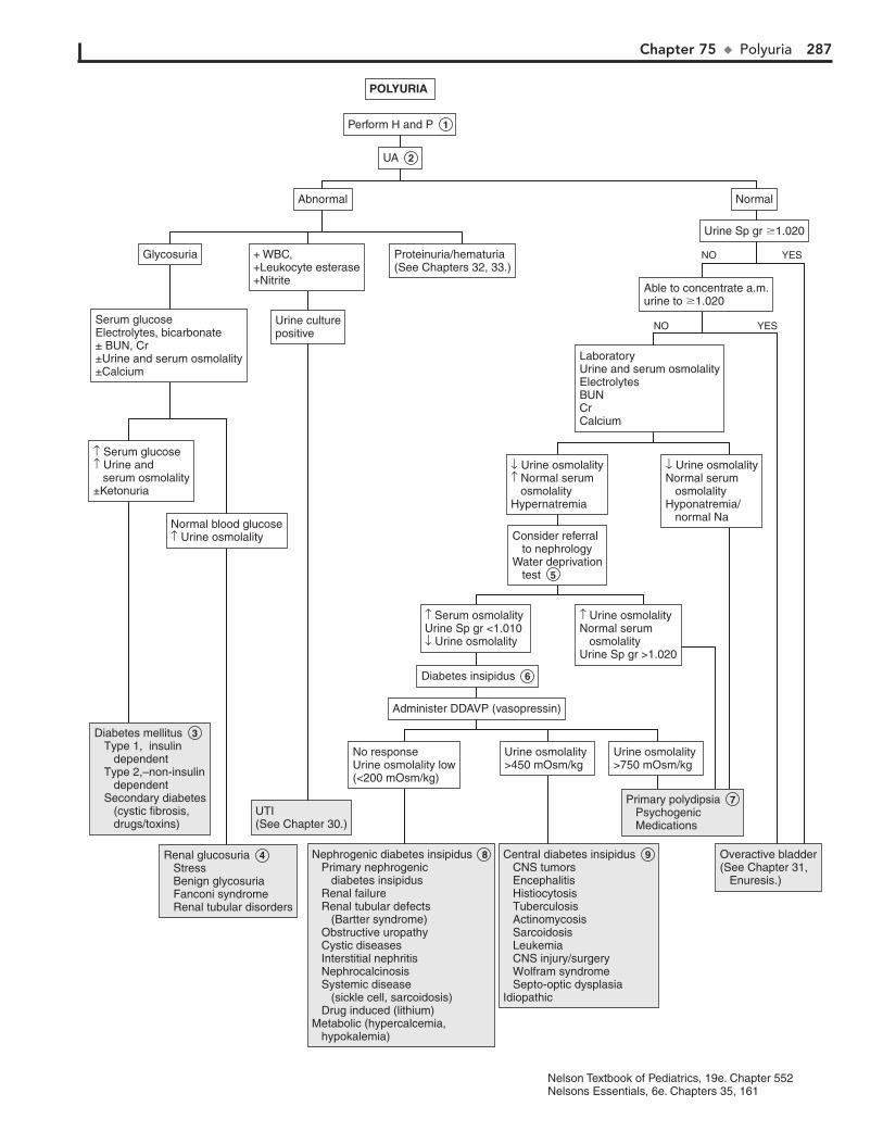
Chapter 75 u Polyuria 287
Diabetes mellitus Type 1, insulin dependent Type 2,–non-insulin dependent Secondary diabetes (cystic fibrosis, drugs/toxins)
Diabetes mellitus 3 Type 1, insulin dependent Type 2,–non-insulin dependent Secondary diabetes (cystic fibrosis, drugs/toxins)
Renal glucosuria Stress Benign glycosuria Fanconi syndrome Renal tubular disorders
Renal glucosuria 4 Stress Benign glycosuria Fanconi syndrome Renal tubular disorders
UTI(See Chapter 30.)UTI(See Chapter 30.)
Nephrogenic diabetes insipidus Primary nephrogenic diabetes insipidus Renal failure Renal tubular defects (Bartter syndrome) Obstructive uropathy Cystic diseases Interstitial nephritis Nephrocalcinosis Systemic disease (sickle cell, sarcoidosis) Drug induced (lithium)Metabolic (hypercalcemia, hypokalemia)
Nephrogenic diabetes insipidus 8 Primary nephrogenic diabetes insipidus Renal failure Renal tubular defects (Bartter syndrome) Obstructive uropathy Cystic diseases Interstitial nephritis Nephrocalcinosis Systemic disease (sickle cell, sarcoidosis) Drug induced (lithium)Metabolic (hypercalcemia, hypokalemia)
Central diabetes insipidus CNS tumors Encephalitis Histiocytosis Tuberculosis Actinomycosis Sarcoidosis Leukemia CNS injury/surgery Wolfram syndrome Septo-optic dysplasiaIdiopathic
Central diabetes insipidus 9 CNS tumors Encephalitis Histiocytosis Tuberculosis Actinomycosis Sarcoidosis Leukemia CNS injury/surgery Wolfram syndrome Septo-optic dysplasiaIdiopathic
Primary polydipsia Psychogenic Medications
Primary polydipsia 7 Psychogenic Medications
Overactive bladder(See Chapter 31, Enuresis.)
Overactive bladder(See Chapter 31, Enuresis.)
POLYURIA
Perform H and P 1
No responseUrine osmolality low(<200 mOsm/kg)
↑ Serum glucose↑ Urine and serum osmolality±Ketonuria
Serum glucoseElectrolytes, bicarbonate± BUN, Cr±Urine and serum osmolality±Calcium
Urine culturepositive
Glycosuria + WBC,+Leukocyte esterase+Nitrite
Proteinuria/hematuria(See Chapters 32, 33.)
Normal blood glucose↑ Urine osmolality
Urine osmolality >450 mOsm/kg
Administer DDAVP (vasopressin)
Diabetes insipidus 6
↑ Serum osmolality Urine Sp gr <1.010↓ Urine osmolality
Consider referral to nephrology Water deprivation test 5
LaboratoryUrine and serum osmolalityElectrolytesBUNCrCalcium
Able to concentrate a.m.urine to �1.020
Urine Sp gr �1.020
NormalAbnormal
↓ Urine osmolality↑ Normal serum osmolalityHypernatremia
↓ Urine osmolalityNormal serum osmolalityHyponatremia/ normal Na
↑ Urine osmolalityNormal serum osmolalityUrine Sp gr >1.020
Urine osmolality >750 mOsm/kg
UA 2
YESNO
YESNO
Nelson Textbook of Pediatrics, 19e. Chapter 552Nelsons Essentials, 6e. Chapters 35, 161

This page intentionally left blank

General PART
XI

290
Chapter 76
FEVER WITHOUT A SOURCE
infections) may be helpful, particularly in very young infants. A his-tory of exposure to sick contacts and any use of medication (antibi-otics, antipyretics) should also be elicited. A medical history of sickle cell disease, immunodeficiency, congenital heart disease, a central venous line, or malignancy is significant and will alter the approach to a febrile child. Individualized clinical judgment should be used when assessing fever in these high-risk children.
2 If pleocytosis is present in the CSF or if there is a history of maternal herpes simplex (HSV) infection, consider
adding acyclovir to the empirical treatment regimen.
3 Urine should be obtained by either catheterization or supra-pubic aspiration to avoid the risk of contamination. The
addition of viral testing (culture, PCR) to CSF studies can aid in the identification of infants at low risk for SBIs; infants with an identified viral infection are at very low risk for bacterial infec-tions. Stool should be examined for WBCs when diarrhea is pres-ent; culture should be sent if the microscopic analysis cannot be performed.
4 Criteria established prior to the use of the current Hib and pneumococcal vaccines to identify infants as “low risk” for
an SBI continue to be useful in the management of infants 29 to 90 days of age. A WBC of 15,000/mm3 is generally considered the upper limit of values considered low risk for SBI (although ,20, 000/mm3 has also been proposed as a low-risk value, as has a WBC between 5000/mm3 and 15,000/mm3). A negative leukocyte esterase on urine dipstick and microscopic analysis showing fewer than 5 WBCs per high-power field and no bac-teria on Gram stain suggest minimal risk for UTI.
5 Some protocols may defer an LP and CSF studies if other labs indicate low risk for SBI; however, a strong consensus
supports always obtaining CSF if empiric antibiotics are going to be administered. Definition of low risk CSF findings range between ,5 to 10 WBC/mm3 with negative Gram stains. The recommended empiric antibiotic therapy in young infants is ceftriaxone (50 mg/kg IM). In addition to fulfilling low-risk lab criteria, reliable follow-up in 24 hours should be ensured before planning outpatient treatment while culture results are pend-ing. Parents should be instructed in the importance of urgent follow up whenever a child’s clinical status seems to worsen.
6 Well-appearing infants with an abnormal UA in the face of normal blood tests (normal absolute neutrophil count and
band counts) can probably be safely treated with outpatient parental antibiotics for a presumed UTI. Children with a sus-pected UTI who appear severely ill or are vomiting should be hospitalized for IV antibiotic therapy. Beyond 2 months of age, oral therapy for suspected UTIs has been proven to be as effica-cious as parenteral therapy.
The most commonly used definition of fever is a rectal tem-perature of 38°C (100.4°F) or above. The terms fever without a source (FWS) and fever without localizing signs (FWLS) are used to describe fevers of a duration of less than 1 week that devel-oped acutely in a previously well child in whom no likely cause for the fever is evident after a thorough H and P.
Prior to the development of the conjugated Haemophilus in-fluenza type b (Hib) and pneumococcal vaccines, management guidelines (Boston, Rochester, Philadelphia) for fever in young children were aimed at identifying those at risk for bacteremia in order to reduce their risk of a subsequent serious bacterial infection (SBI) (meningitis, sepsis, osteomyelitis, septic arthritis, UTIs, pneumonias, bacterial enteritis). In the current era of widespread use of these conjugated vaccines, the risk of occult bacteremia in well-appearing children has been significantly reduced, diminishing the usefulness of such guidelines. Consen-sus remains strong, however, for the management of febrile in-fants younger than 3 months old, particularly those younger than 28 days old. A similar consensus exists regarding children who do not look well; any child who appears significantly ill or toxic should be hospitalized for further evaluation and treat-ment regardless of their age or immunization status.
1 A rectal temperature is the best method of assessment of body core temperature and is recommended when evaluat-
ing infants younger than 3 months of age; axillary and tympanic membrane temperature measurements are not recommended. Infants with fever documented at home by a reliable caretaker should be treated the same as if the fever were documented by a healthcare worker. The role of bundling in the elevation of body temperature is controversial; bundling and a warm ambient temperature may contribute to an elevation in skin temperature more so than rectal temperature in young infants, but elevation into a febrile range is unlikely. Some studies suggest an infant with a normal temperature obtained 15 to 30 minutes after un-bundling can be considered afebrile; others do not recommend attributing temperatures in the febrile range (especially temper-atures .38.5°C) to bundling.
The birth history, including duration of ruptured membranes and history of maternal infections (group B strep, sexually transmitted
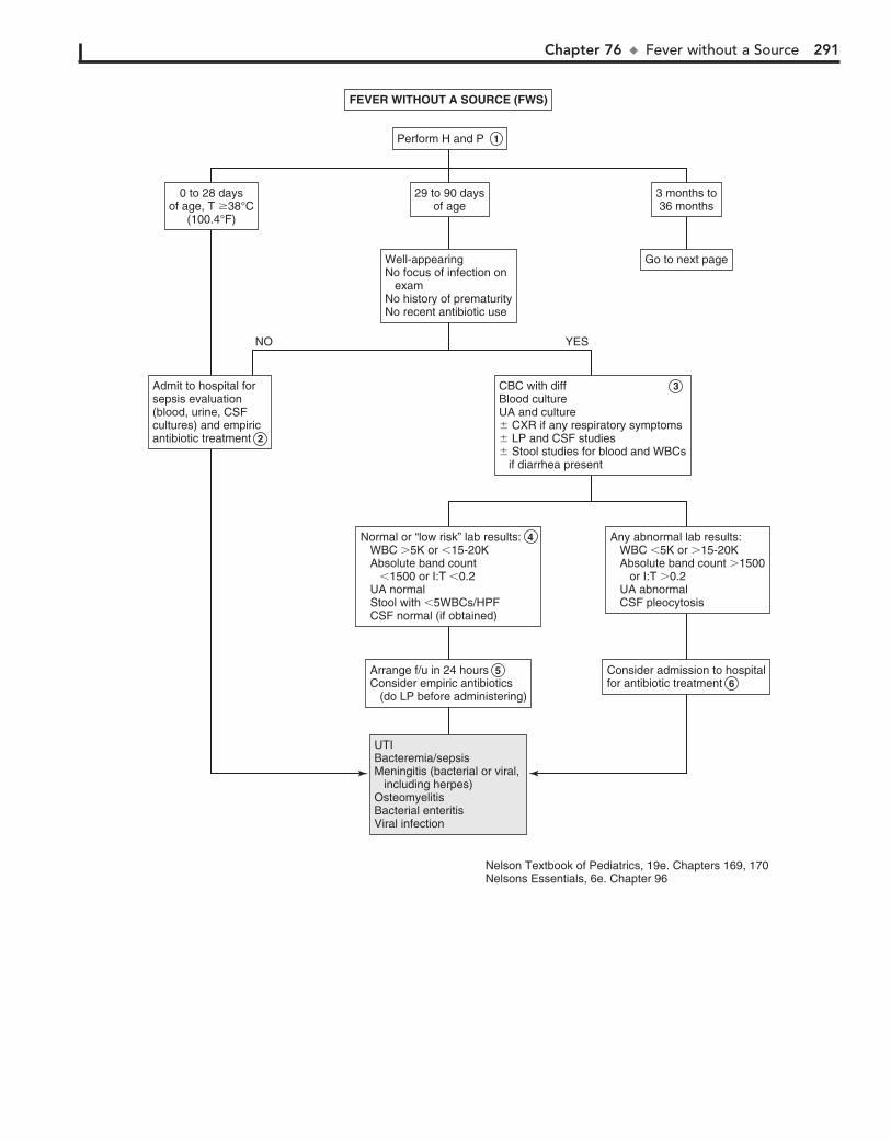
Chapter 76 u FeverwithoutaSource 291
Perform H and PPerform H and P 1
Go to next pageGo to next pageWell-appearingNo focus of infection on examNo history of prematurityNo recent antibiotic use
Well-appearingNo focus of infection on examNo history of prematurityNo recent antibiotic use
3 months to36 months3 months to36 months
29 to 90 daysof age
29 to 90 daysof age
0 to 28 daysof age, T �38°C
(100.4°F)
0 to 28 daysof age, T �38°C
(100.4°F)
UTIBacteremia/sepsisMeningitis (bacterial or viral, including herpes)OsteomyelitisBacterial enteritisViral infection
UTIBacteremia/sepsisMeningitis (bacterial or viral, including herpes)OsteomyelitisBacterial enteritisViral infection
CBC with diff Blood cultureUA and culture� CXR if any respiratory symptoms� LP and CSF studies� Stool studies for blood and WBCs
if diarrhea present
CBC with diff 3 Blood cultureUA and culture� CXR if any respiratory symptoms� LP and CSF studies� Stool studies for blood and WBCs if diarrhea present
Any abnormal lab results: WBC �5K or �15-20K Absolute band count �1500 or I:T �0.2 UA abnormal CSF pleocytosis
Any abnormal lab results: WBC �5K or �15-20K Absolute band count �1500 or I:T �0.2 UA abnormal CSF pleocytosis
Consider admission to hospitalfor antibiotic treatmentConsider admission to hospitalfor antibiotic treatment 6
Normal or “low risk” lab results: WBC �5K or �15-20K Absolute band count �1500 or I:T �0.2 UA normal Stool with �5WBCs/HPF CSF normal (if obtained)
Normal or “low risk” lab results: 4 WBC �5K or �15-20K Absolute band count �1500 or I:T �0.2 UA normal Stool with �5WBCs/HPF CSF normal (if obtained)
Arrange f/u in 24 hoursConsider empiric antibiotics (do LP before administering)
Arrange f/u in 24 hours 5 Consider empiric antibiotics (do LP before administering)
Admit to hospital forsepsis evaluation(blood, urine, CSFcultures) and empiricantibiotic treatment
Admit to hospital forsepsis evaluation(blood, urine, CSFcultures) and empiricantibiotic treatment 2
NO YES
FEVER WITHOUT A SOURCE (FWS)FEVER WITHOUT A SOURCE (FWS)
Nelson Textbook of Pediatrics, 19e. Chapters 169, 170Nelsons Essentials, 6e. Chapter 96

292 Part XI u General
7 A child who has received the primary series of 3 of a conjugated pneumococcal vaccine (PCV7 or PCV13) and
a complete primary series of a conjugated H. influenzae vaccine (2 or 3 doses, depending on the product) is considered com-pletely immunized. Because the primary series of pneumococcal vaccine cannot be completed until 6 months, all infants younger than 6 months are considered incompletely immunized.
8 Guidelines for evaluating fever in children who are incom-pletely immunized include using criteria for identifying
risk of occult bacteremia, as well as clinical judgment. Reliabil-ity of parental followup should always be considered in man-agement decisions for these children.
9 When blood cultures are positive for pathogens other than Streptococcus pneumoniae, the likelihood of a con-
taminant needs to be carefully considered. Neisseria meningiti-dis, H. influenzae type b, and Staphylococcus aureus are likely pathogens, and those children should be (if not already) admitted for a full sepsis evaluation and treatment. Other organisms may or may not be pathogens; if a contaminant is suspected and the child is well-appearing, continued outpa-tient observation may be appropriate.
10 Fever can rarely be caused by medications in children; al-though the onset can occur within days of administration
of a drug, this etiology is usually not considered until the fever has been present for a more prolonged period. Anticonvul-sants, allopurinol (and other chemotherapy agents), nitrofu-rantoin (and other antibiotics), and drugs with anticholinergic (atropine, antihistamines) and sympathomimetic (cocaine, amphetamines) effects are among the more common agents causing fever due to a variety of mechanisms (including, but not limited to, hypersensitivity). Other symptoms may or may not be associated.
11 Vaccine reactions may be a source of fever. Mild to moder-ate fever can occur after diphtheria and tetanus toxoids and
acellular pertussis (DTaP) vaccines; extremely high fevers (.40.5°C) that were occasionally seen after administration of the previously used DTP whole-cell preparation are extremely rare
following administration of the currently available acellular prep-aration. Fever occurs in approximately 5% to 15% of children within 5 to 12 days of receiving the measles, mumps, and rubella (MMR) vaccine; those receiving the combination MMR and varicella vaccine (MMRV) are more likely (22%) to develop fever.
12 A fever below 39°C in children 3 months of age or older is most likely to be a viral infection. It is reasonable in a well-
appearing 3- to 36-month-old child to forego any lab evaluation and observe closely, assuming a reliable means of followup (phone, transportation) has been ensured. Careful instructions should be given to parents to return if the fever persists more than 2 to 3 days.
Many children with FWS are in the prodromal phase of an infectious illness and develop more specific signs or symptoms within several hours to days of the initial fever onset. In some diseases, such as Rocky Mountain spotted fever, leptospirosis, and measles, the fever may precede more specific signs by up to 3 days. In illnesses such as roseola, viral hepatitis, infectious mononucleosis, typhus, typhoid fever, and Kawasaki disease, a longer interval may occur between fever onset and more specific findings.
BibliographyAmerican Academy of Pediatrics: In Pickering LK, editor: Red book: 2012
report of the Committee on Infectious Diseases, ed 29, Elk Grove Village, Ill, 2012, AAP.
Baraff LJ, Bass JW, et al: Practice guidelines for the management of infants and children 0 to 36 months of age with fever without source, Pediatrics 92:1, 1993.
Cheng TL, Partridge JC: Effect of bundling and high environmental tempera-ture on neonatal body temperature, Pediatrics 92:238, 1993.
Grover G, Berkowitz CD, Thompson M, et al: The effects of bundling on infant temperature, Pediatrics 945:669, 1994.
Schnadower D, Kuppermann N, Macias CG, et al; American Academy of Pediatrics Pediatric Emergency Medicine Collaborative Research Committee: Febrile infants with urinary tract infections at very low risk for adverse events and bacteremia, Pediatrics 126:1074, 2010.
Subcommittee on Urinary Tract Infection, Steering Committee on Quality Improvement and Management: Urinary tract infection: Clinical practice guideline for the diagnosis and management of the initial UTI in febrile infants and children 2 to 24 months, Pediatrics 128:595, 2011.
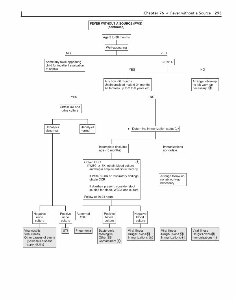
Chapter 76 u FeverwithoutaSource 293
Well-appearingWell-appearing
Age 3 to 36 monthsAge 3 to 36 months
T�39° CT�39° CAdmit any toxic-appearingchild for inpatient evaluationof sepsis
Admit any toxic-appearingchild for inpatient evaluationof sepsis
Viral cystitis Viral illnessOther causes of pyuria (Kawasaki disease, appendicitis)
Viral cystitis Viral illnessOther causes of pyuria (Kawasaki disease, appendicitis)
UTIUTI PneumoniaPneumonia BacteremiaMeningitisOther SBIContaminant
BacteremiaMeningitisOther SBIContaminant 9
Viral illnessDrugs/ToxinsImmunizations
Viral illnessDrugs/Toxins 10 Immunizations 11
Viral illnessDrugs/ToxinsImmunizations
Viral illnessDrugs/Toxins 10 Immunizations 11
Viral illnessDrugs/ToxinsImmunizations
Viral illnessDrugs/Toxins 10 Immunizations 11
Arrange follow-up;no lab work-upnecessary
Arrange follow-up;no lab work-upnecessary 12
Any boy �6 monthsUncircumcised male 6-24 monthsAll females up to 2 to 3 years old
Any boy �6 monthsUncircumcised male 6-24 monthsAll females up to 2 to 3 years old
NOYES
Determine immunization statusDetermine immunization status 7
Obtain UA andurine culture
Obtain UA andurine culture
UrinalysisnormalUrinalysisnormal
UrinalysisabnormalUrinalysisabnormal
Immunizationsup-to-dateImmunizationsup-to-date
Incomplete (includesage �6 months)Incomplete (includesage �6 months)
Arrange follow-up;no lab work-upnecessary
Arrange follow-up;no lab work-upnecessary
Obtain CBC If WBC �15K, obtain blood culture and begin empiric antibiotic therapy
If WBC �20K or respiratory findings, obtain CXR
If diarrhea present, consider stool studies for blood, WBCs and culture
Follow up in 24 hours
Obtain CBC 8 If WBC �15K, obtain blood culture and begin empiric antibiotic therapy
If WBC �20K or respiratory findings, obtain CXR
If diarrhea present, consider stool studies for blood, WBCs and culture
Follow up in 24 hours
Positiveurine
culture
Positiveurine
culture
Negativeurine
culture
Negativeurine
culture
Negativeblood culture
Negativeblood culture
Positiveblood culture
Positiveblood culture
AbnormalCXR
AbnormalCXR
NOYES
NO YES
FEVER WITHOUT A SOURCE (FWS)(continued)
FEVER WITHOUT A SOURCE (FWS)(continued)
294
Chapter 77
FEVER OF UNKNOWN ORIGIN
The PE should particularly note growth parameters, skin findings (rash, petechiae, sparse hair/eyebrows/eyelashes, bites or punctum), the ophthalmologic exam (conjunctivitis, abnor-mal corneal or pupillary reflexes, funduscopic abnormalities), ENT findings (nasal discharge, sinus tenderness, tooth abnor-malities, smooth tongue, pharyngeal injection, gingival hyper-trophy, dental abscesses), and a thorough musculoskeletal exam (arthritis, bone or muscle tenderness). Attempts should be made to examine a child while febrile to assess for the appear-ance of a rash with fever and the presence or absence of sweat-ing. (Absence of sweating could indicate hereditary sensory and autonomic neuropathy or ectodermal dysplasia.) Sexually ac-tive females should always have a pelvic examination to assess for pelvic inflammatory disease (PID) and pelvic abscesses. A rectal exam should be performed to assess for an abscess or tumor, and stool should always be checked for occult blood.
2 Fever can be a reaction to several types of medication owing to a variety of mechanisms (not just allergy). Discontinua-
tion of the drug followed by disappearance of the fever is sugges-tive of drug fever, although sometimes fever persists days to weeks as a result of slow excretion of the drug. Anticonvulsants, allopurinol (and other chemotherapy agents), nitrofurantoin (and other antibiotics), phenothiazines, drugs with anticholiner-gic effects (atropine, antihistamines) and sympathomimetic ef-fects (cocaine, epinephrine, amphetamines) are among the more common causative agents.
3 The extent of urgency of the evaluation depends on the degree of illness in the child. Hospitalization is not usually
necessary to evaluate FUO. It does offer the advantage, how-ever, of close observation and following up on every lead. Blood cultures should be obtained aerobically unless anaerobic infection is suspected. Repeated cultures may be necessary to diagnose certain conditions such as endocarditis, osteomyeli-tis, or deep-seated abscesses causing bacteremia. Specific me-dia need to be requested if particular infections are suspected (tularemia, leptospirosis, Yersinia); serology or PCR may be the preferred diagnostic method for some suspected pathogens. CXRs are commonly recommended in the initial testing for FUO; x-rays of sinuses, mastoids, and the GI tract should be performed selectively based on the clinical picture. Skin testing for tuberculosis (TB) with a PPD test is usually recommended. Both the CBC and inflammatory markers like ESR and CRP are often abnormal but nonspecific. Extreme WBC values (WBC .10K/mm3, ANC ,500/mm3) are suggestive of bacterial infec-tions; EBV infections are characterized by atypical lymphocy-tosis. An ESR greater than 100 mm/h warrants consideration of malignancy, autoimmune disease, Kawasaki disease, TB, and vasculitis. Lower values are nonspecific indicators of inflammation and may be more useful in monitoring disease course, whereas normal values should raise suspicion for facti-tious fever. CRP is similarly nonspecific; it normalizes more quickly than an ESR.
4 Malaria may be transmitted by a mosquito vector from an infected person (e.g., someone who has traveled in an
endemic area) to another person. It can also be transmitted by contaminated syringe needles and blood. In persons who took appropriate antimalarial drugs while traveling, the disease may not manifest until several months later. The organism can be iden-tified on blood smears. Trypanosomiasis and babesiosis are other parasites that can be identified on a blood smear. Splenomegaly is usually present; a relapsing fever pattern is characteristic.
The initial definition of fever of unknown origin (FUO) was pro-posed for adult patients in 1961 to provide a basis for data com-parison across study populations; less consensus (from both a research and a practical standpoint) exists regarding a definition for children. One of the most commonly used definitions of FUO in pediatrics is a history of fever greater than 38.3°C (101°F) for 8 or more days in a child for whom an initial evaluation (inpa-tient or outpatient), including history and physical and prelimi-nary lab evaluation, does not suggest a probable etiology of the fever. Extremely rare disorders can present with FUO in children, but the symptom is far more likely to be due to an uncommon presentation of a common illness. Infections are the leading cause of FUO in children, followed by connective tissue disorders, then neoplasm. The outcome of children with undiagnosed FUO is variable. Some without a diagnosis after a preliminary laboratory evaluation will eventually be diagnosed; no diagnosis, however, will be established for at least 10% to 20%. In general, the out-come for children with FUO is better than for adults with FUO.
1 The evaluation of FUO should be based on factors specific to each case, such as the age of a patient and their ability to
report specific complaints, underlying medical conditions, and exposures. The history is by far the most essential component of an FUO evaluation. Requesting a family to keep a fever diary may be helpful. Fever patterns may be described as intermittent (when the temperature returns to normal at least once daily), remittent (temperature fluctuates but never normalizes), or sustained (minimal fluctuation); these categorizations, however, can be blurred by the use of antipyretics. A relapsing fever pat-tern (fever resolves for 1 or more days then recurs) can be char-acteristic of a few specific problems (malaria, Lyme disease); a pattern of recurrent fevers for more than 6 months is suggestive of metabolic defects, temperature dysregulation due to a CNS ab-normality, or immunodeficiencies (particularly periodic disorders like cyclic neutropenia). The possibility of factitious fever should always be considered.
A surgical history (dental, abdominal, cardiac) may indicate a risk of endocarditis or abscesses. Ethnic background may be a clue to a very limited number of etiologies (nephrogenic diabe-tes insipidus, familial Mediterranean fever, familial dysautono-mia). A complete review of systems is important; be sure to include GI, musculoskeletal (including joint complaints), and risk factors for HIV.
The social history should include exposures to ill contacts, animals (domestic, farm, wild), insect or tick bites, lake or well water, and travel. Inquire whether any travel prophylaxis was recommended and/or taken and about exposure to people who have traveled; artifacts, rocks, or soil that have been brought from distant areas could serve as vectors. Exposure to or inges-tion of medications (including topicals, medications of house-hold members, and illicit substances) and foods (game meat, raw meat, raw shellfish, unpasteurized milk) as well as dirt (or other pica) could be significant.

Chapter 77 u FeverofUnknownOrigin 295
MalignancyEBV/CMVMalaria (and other parasites)Relapsing fever
MalignancyEBV/CMVMalaria (and other parasites) 4 Relapsing fever
MalignancySLEViral infections
MalignancySLEViral infections
Atypical Kawasaki diseaseAtypical Kawasaki disease
UTIUTIDrugfeverDrugfever 2
BacteremiaEndocarditisBacteremia 6 Endocarditis
EBVCMV Typhoid feverLeptospirosisBrucellosisTularemia
EBVCMV Typhoid feverLeptospirosisBrucellosisTularemia
Pneumonia/ pleural effusionSarcoidosisTuberculosisMalignancyPleuritis/pericarditis (infectious, rheumatic)
Pneumonia/ pleural effusionSarcoidosisTuberculosisMalignancyPleuritis/pericarditis (infectious, rheumatic)
Obtain complete history: Detailed fever history (duration, height, pattern, measurement technique) Past medical/surgical/social history Medications ROSPerform complete PE
Obtain complete history: 1 Detailed fever history (duration, height, pattern, measurement technique) Past medical/surgical/social history Medications ROSPerform complete PE
Travel, other exposures, symptomsor PE signs suggestive of specificdisorders present
Travel, other exposures, symptomsor PE signs suggestive of specificdisorders present
See p. 3 of algorithmSee p. 3 of algorithmPatient using or exposedto medication/ drugsPatient using or exposedto medication/ drugs
NO YES
Perform preliminary lab evaluation:CBC with diff and smearESR/CRPBlood cultureUA and urine cultureBUN/CrLFTHIV CXRPPD/interferon-gamma release assay
Perform preliminary lab evaluation: 3 CBC with diff and smearESR/CRPBlood cultureUA and urine cultureBUN/CrLFTHIV CXRPPD/interferon-gamma release assay
Consider serology for EBV, CMV, brucellosis, tularemia,Bartonella henselae (cat-scratch disease), Salmonella typhi,leptospirosis, toxoplasmosis, hepatitis
Consider serology for EBV, CMV, brucellosis, tularemia,Bartonella henselae (cat-scratch disease), Salmonella typhi,leptospirosis, toxoplasmosis, hepatitis
NOYES
Results normal or nonspecificResults normal or nonspecific Go to next pageGo to next pageResults abnormalResults abnormal
AbnormalserologyAbnormalserology
ThrombocytopeniaThrombocytopenia ThrombocytosisThrombocytosis Positiveurineculture
Positiveurineculture
AbnormalCXRAbnormalCXR 5
Positive bloodculturePositive bloodculture
Abnormal WBCAbnormal smearAbnormal WBCAbnormal smear
FEVER OF UNKNOWN ORIGINFEVER OF UNKNOWN ORIGIN
Nelson Textbook of Pediatrics, 19e. Chapters 149, 157, 214, 280, 431, 676, 677Nelsons Essentials, 6e. Chapters 96, 122
Consult infectious diseaseJIA = juvenile idiopathic arthritis SLE = systemic lupus erythematosus

296 Part XI u General
5 The finding of hilar adenopathy on CXR should raise sus-picion for sarcoidosis, TB, or malignancy.
6 In cases of FUO, identification of bacteremia will usually indicate a more specific infection. For example, Streptococ-
cus viridans is frequently associated with endocarditis, and Staphylococcus aureus, group A streptococci, pneumococcus, and Kingella kingae with bone and joint infections.
infectious disease specialist should be considered prior to ob-taining additional imaging or more specialized testing at this point in the evaluation in order to avoid a cycle of unfounded additional testing based on slightly abnormal but nonspecific results.
9 JIA is usually only diagnosed after an extended period of observation and evolution of more characteristic symp-
toms. The systemic form is most likely to present as an FUO with an intermittent fever pattern; arthritis is necessary for de-finitive diagnosis but may not develop for months. Serology may remain negative for an extended period. A characteristic salmon-colored rash frequently accompanies the fever. A slit lamp examination performed by an ophthalmologist may reveal uveitis.
10 Fever is more common in children than adults with in-flammatory bowel disease. Evaluation should be consid-
ered in cases of prolonged FUO, even without specific GI symptoms (and especially if weight loss or impaired growth has occurred or if there is anemia, an elevated ESR, or occult blood in the stool).
11 Persons of Mediterranean descent (Armenians, Arabs, Sephardic Jews) may inherit familial Mediterranean fever,
characterized by episodic fevers and abdominal pain. Ulster Scots may be at risk for nephrogenic diabetes insipidus, persons of Jewish descent may inherit familial dysautonomia, and Asians (particularly females) are at risk for Kikuchi-Fujimoto disease (a necrotizing lymphadenitis). Several other inherited (usually autosomal dominant) autoinflammatory disorders are recognized; they present with a spectrum of clinical manifesta-tions in addition to fever.
7 The possibility of factitious fever needs to be considered; it may be intentional (caregiver-fabricated illness, previously
known as Munchausen syndrome by proxy) or unintentional (inappropriate heating of the thermometer or skin or mucosal surface before taking the temperature). Factitious fevers should be suspected when no recognizable circadian rhythm of the fevers can be identified; also be suspicious if vasoconstriction, sweating, tachypnea, and tachycardia are not associated with the fever.
8 Initial lab results, additional historical information or evo-lution of exam findings may help guide subsequent evalua-
tion if the preliminary evaluation has not identified an etiology. An ANA level will be positive in systemic lupus erythematosus (SLE) but is not likely to be positive in systemic-onset juvenile idiopathic arthritis (JIA). Be aware that 30% of children without rheumatic disease will also have a positive ANA, so more specific immunologic testing (dsDNA or anti-Smith Abs) is recommended when a diagnosis of SLE is suspected. (It should definitely be obtained if there is a family history of lupus.) Ele-vated immunoglobulin levels may occur in systemic-onset JIA, certain immunodeficiencies, and HIV infection. Advanced im-aging studies may identify tumors or abscesses, but cost, risk, and expected yield need to be considered; consultation with an

Chapter 77 u FeverofUnknownOrigin 297
Reconsiderfactitious fever ORResolution
Reconsiderfactitious fever 7 ORResolution
JIASLEPolyarteritis nodosaOsteomyelitisAbscess (intraabdominal, liver, perinephric)Inflammatory bowel diseaseMalignancyHereditary periodic fever syndromes/ autoinflammatory disordersImmunodeficiency
JIA 9 SLEPolyarteritis nodosaOsteomyelitisAbscess (intraabdominal, liver, perinephric)Inflammatory bowel disease 10 MalignancyHereditary periodic fever syndromes/ autoinflammatory disorders 11 Immunodeficiency
Consider additional evaluation:Serum ANASerum immunoglobulinsSerologies (if not already done)Consult infectious diseaseOther investigations: X-rays (sinus, mastoid, abdomen) WBC scans (gallium, indium) Immunoscintigraphy Bone scan Liver/spleen scan PET scan Bone marrow biopsy
Consider additional evaluation: 8 Serum ANASerum immunoglobulinsSerologies (if not already done)Consult infectious diseaseOther investigations: X-rays (sinus, mastoid, abdomen) WBC scans (gallium, indium) Immunoscintigraphy Bone scan Liver/spleen scan PET scan Bone marrow biopsy
FEVER OF UNKNOWN ORIGIN(continued)
FEVER OF UNKNOWN ORIGIN(continued)
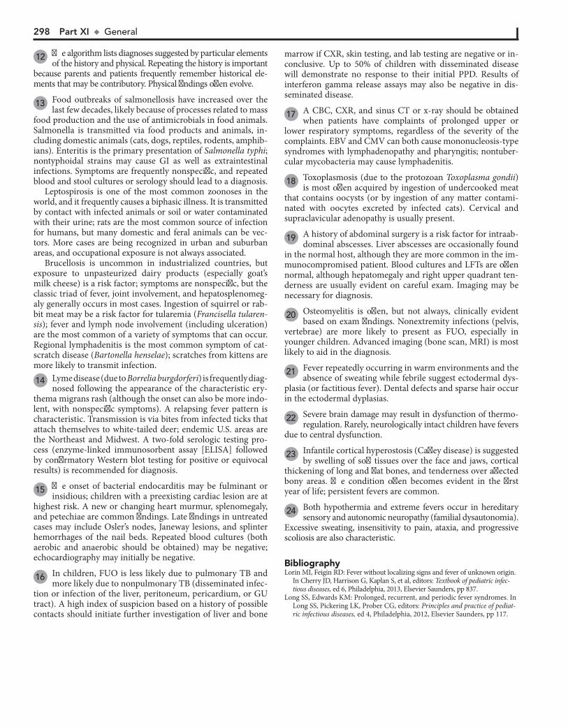
298 Part XI u General
12 The algorithm lists diagnoses suggested by particular elements of the history and physical. Repeating the history is important
because parents and patients frequently remember historical ele-ments that may be contributory. Physical findings often evolve.
13 Food outbreaks of salmonellosis have increased over the last few decades, likely because of processes related to mass
food production and the use of antimicrobials in food animals. Salmonella is transmitted via food products and animals, in-cluding domestic animals (cats, dogs, reptiles, rodents, amphib-ians). Enteritis is the primary presentation of Salmonella typhi; nontyphoidal strains may cause GI as well as extraintestinal infections. Symptoms are frequently nonspecific, and repeated blood and stool cultures or serology should lead to a diagnosis.
Leptospirosis is one of the most common zoonoses in the world, and it frequently causes a biphasic illness. It is transmitted by contact with infected animals or soil or water contaminated with their urine; rats are the most common source of infection for humans, but many domestic and feral animals can be vec-tors. More cases are being recognized in urban and suburban areas, and occupational exposure is not always associated.
Brucellosis is uncommon in industrialized countries, but exposure to unpasteurized dairy products (especially goat’s milk cheese) is a risk factor; symptoms are nonspecific, but the classic triad of fever, joint involvement, and hepatosplenomeg-aly generally occurs in most cases. Ingestion of squirrel or rab-bit meat may be a risk factor for tularemia (Francisella tularen-sis); fever and lymph node involvement (including ulceration) are the most common of a variety of symptoms that can occur. Regional lymphadenitis is the most common symptom of cat-scratch disease (Bartonella henselae); scratches from kittens are more likely to transmit infection. 14 Lyme disease (due to Borrelia burgdorferi) is frequently diag-
nosed following the appearance of the characteristic ery-thema migrans rash (although the onset can also be more indo-lent, with nonspecific symptoms). A relapsing fever pattern is characteristic. Transmission is via bites from infected ticks that attach themselves to white-tailed deer; endemic U.S. areas are the Northeast and Midwest. A two-fold serologic testing pro-cess (enzyme-linked immunosorbent assay [ELISA] followed by confirmatory Western blot testing for positive or equivocal results) is recommended for diagnosis.
15 The onset of bacterial endocarditis may be fulminant or insidious; children with a preexisting cardiac lesion are at
highest risk. A new or changing heart murmur, splenomegaly, and petechiae are common findings. Late findings in untreated cases may include Osler’s nodes, Janeway lesions, and splinter hemorrhages of the nail beds. Repeated blood cultures (both aerobic and anaerobic should be obtained) may be negative; echocardiography may initially be negative.
16 In children, FUO is less likely due to pulmonary TB and more likely due to nonpulmonary TB (disseminated infec-
tion or infection of the liver, peritoneum, pericardium, or GU tract). A high index of suspicion based on a history of possible contacts should initiate further investigation of liver and bone
marrow if CXR, skin testing, and lab testing are negative or in-conclusive. Up to 50% of children with disseminated disease will demonstrate no response to their initial PPD. Results of interferon gamma release assays may also be negative in dis-seminated disease.
17 A CBC, CXR, and sinus CT or x-ray should be obtained when patients have complaints of prolonged upper or
lower respiratory symptoms, regardless of the severity of the complaints. EBV and CMV can both cause mononucleosis-type syndromes with lymphadenopathy and pharyngitis; nontuber-cular mycobacteria may cause lymphadenitis.
18 Toxoplasmosis (due to the protozoan Toxoplasma gondii) is most often acquired by ingestion of undercooked meat
that contains oocysts (or by ingestion of any matter contami-nated with oocytes excreted by infected cats). Cervical and supraclavicular adenopathy is usually present.
19 A history of abdominal surgery is a risk factor for intraab-dominal abscesses. Liver abscesses are occasionally found
in the normal host, although they are more common in the im-munocompromised patient. Blood cultures and LFTs are often normal, although hepatomegaly and right upper quadrant ten-derness are usually evident on careful exam. Imaging may be necessary for diagnosis.
20 Osteomyelitis is often, but not always, clinically evident based on exam findings. Nonextremity infections (pelvis,
vertebrae) are more likely to present as FUO, especially in younger children. Advanced imaging (bone scan, MRI) is most likely to aid in the diagnosis.
21 Fever repeatedly occurring in warm environments and the absence of sweating while febrile suggest ectodermal dys-
plasia (or factitious fever). Dental defects and sparse hair occur in the ectodermal dyplasias.
22 Severe brain damage may result in dysfunction of thermo-regulation. Rarely, neurologically intact children have fevers
due to central dysfunction.
23 Infantile cortical hyperostosis (Caffey disease) is suggested by swelling of soft tissues over the face and jaws, cortical
thickening of long and flat bones, and tenderness over affected bony areas. The condition often becomes evident in the first year of life; persistent fevers are common.
24 Both hypothermia and extreme fevers occur in hereditary sensory and autonomic neuropathy (familial dysautonomia).
Excessive sweating, insensitivity to pain, ataxia, and progressive scoliosis are also characteristic.
BibliographyLorin MI, Feigin RD: Fever without localizing signs and fever of unknown origin.
In Cherry JD, Harrison G, Kaplan S, et al, editors: Textbook of pediatric infec-tious diseases, ed 6, Philadelphia, 2013, Elsevier Saunders, pp 837.
Long SS, Edwards KM: Prolonged, recurrent, and periodic fever syndromes. In Long SS, Pickering LK, Prober CG, editors: Principles and practice of pediat-ric infectious diseases, ed 4, Philadelphia, 2012, Elsevier Saunders, pp 117.
Chapter 77 u FeverofUnknownOrigin 299
Travel, other exposures, symptomsor PE signs suggestive of specificdisorders present
Travel, other exposures, symptomsor PE signs suggestive of specificdisorders present 12
Exposure to wild ordomesticated animalsExposure to wild ordomesticated animals
FEVER OF UNKNOWN ORIGIN(continued)
FEVER OF UNKNOWN ORIGIN(continued)
ENT or respiratorysymptomsENT or respiratorysymptoms 17
SinusitisPulmonary disease (pneumonia, TB, abscess, sarcoid)ANCA-associated vasculitis (Wegner’s granulomatosis) EBVCMVMycobacteriaMastoiditis MalignancyThyroiditis
SinusitisPulmonary disease (pneumonia, TB, abscess, sarcoid)ANCA-associated vasculitis (Wegner’s granulomatosis) EBVCMVMycobacteriaMastoiditis MalignancyThyroiditis
Bone, joint, or muscleBone, joint, or muscle
Rash or petechiaeRash or petechiae Ingestion of contaminatedfood or milk
ORPica
Ingestion of contaminatedfood or milk
ORPica
Abnormal cardiac examAbnormal cardiac exam
TravelOR
Exposure to persons recently traveled
TravelOR
Exposure to persons recently traveled
Weight loss or impaired lineargrowth, nonspecific abdominalcomplaints, diarrhea
Weight loss or impaired lineargrowth, nonspecific abdominalcomplaints, diarrhea
Recurrent fever (with periodsof wellness in between)Recurrent fever (with periodsof wellness in between)
Other physical abnormalities(neurologic, cutaneous, dental,orthopedic)
Other physical abnormalities(neurologic, cutaneous, dental,orthopedic)
Zoonoses Cat scratch disease Tularemia Salmonellosis Brucellosis Leptospirosis Q fever Rat bite fever Psittacosis Toxoplasmosis Visceral larva migrans
Zoonoses 13 Cat scratch disease Tularemia Salmonellosis Brucellosis Leptospirosis Q fever Rat bite fever Psittacosis Toxoplasmosis Visceral larva migrans
Lyme diseaseJIAAcute rheumatic feverEndocarditisDermatomyositisSLELE Kawasaki diseaseRocky Mountain spotted feverHistiocytosisSerum sicknessTularemiaEBV
Lyme disease 14JIA 9Acute rheumatic feverEndocarditis 15DermatomyositisSLELE Kawasaki diseaseRocky Mountain spotted feverHistiocytosisSerum sicknessTularemiaEBV
CoccidiomycosisHistoplasmosisMalariaBlastomycosisLyme diseaseRocky Mountain spotted feverTuberculosis (TB)EhrlichiosisBabesiosisTyphoid feverOther endemic diseases
CoccidiomycosisHistoplasmosisMalariaBlastomycosisLyme diseaseRocky Mountain spotted feverTuberculosis (TB) 16EhrlichiosisBabesiosisTyphoid feverOther endemic diseases
HepatitisInflammatory bowel diseaseAbscesses (intra abdominal, hepatic)MalignancyEnteritis (various pathogens)Diabetes insipidus (central or nephrogenic)
HepatitisInflammatory bowel diseaseAbscesses (intra abdominal, hepatic) 19MalignancyEnteritis (various pathogens)Diabetes insipidus (central or nephrogenic)
Visceral larva migransToxoplasmosisSalmonellosisBrucellosisRat bite fever
Visceral larva migransToxoplasmosis 18SalmonellosisBrucellosisRat bite fever
Ectodermal dysplasiaCNS/hypothalamic disordersInfantile cortical hyperostosisHereditary sensory and autonomic neuropathy (familial dysautonomia)Hereditary periodic fever syndromes/ autoinflammatory disorders
Ectodermal dysplasia 21 CNS/hypothalamic disorders 22 Infantile cortical hyperostosis 23 Hereditary sensory and autonomic neuropathy (familial dysautonomia) 24 Hereditary periodic fever syndromes/ autoinflammatory disorders 11
Hereditary periodic fever syndromes/ autoinflammatory disordersHereditary periodic fever syndromes/ autoinflammatory disorders 11
Carditis (rheumatic fever)EndocarditisJIASLELE Viral pericarditisMyocarditis
Carditis (rheumatic fever)Endocarditis 15JIA 9SLELE Viral pericarditisMyocarditis
OsteomyelitisMalignancyTrichinosisDermatomyositisPolyarteritis nodosaArboviral infectionsHereditary periodic fever syndromes/ autoinflammatory disorders
Osteomyelitis 20MalignancyTrichinosisDermatomyositisPolyarteritis nodosaArboviral infectionsHereditary periodic fever syndromes/ autoinflammatory disorders
300
Chapter 78
RECURRENT INFECTIONS
abnormalities, and findings suggestive of allergic disease (trans-verse nasal crease, allergic “shiners,” posterior pharyngeal cob-blestoning, swollen pale nasal mucosa) should all be carefully noted. Lymphadenopathy suggests chronic disease; absent lymph tissue (tonsils, lymph nodes, no thymus on CXR) suggest a congenital (lymphocyte) immune defect.
2 The majority of children with a complaint of recurrent infections are normal healthy children who are experienc-
ing frequent URIs. Children who have a large family or attend daycare will experience 6 to 10 viral infections annually, mostly URIs and GI infections, until age 3 to 5 years. A minimal workup, if any, is usually all that is necessary.
3 Rarely, the CBC may reveal an unsuspected malignancy or a WBC abnormality (number, morphology). Further
workup should be based on specific results.
4 Many children with recurrent respiratory symptoms have allergies; usually the diagnosis is made clinically. Al-
though usually unnecessary, antigen-specific immunoglobulin panels (as-IgE) and skin testing are the most sensitive tests to identify allergens. Other labs are not recommended; both eosinophilia and total IgE levels are nonspecific (up to half of allergic patients have normal IgE levels).
5 A portion of children presenting with recurrent infections have an underlying chronic nonimmunologic problem.
Structural or anatomic defects (cleft palate, congenital heart disease, pulmonary sequestration) are risk factors for recurrent local infections. Malnutrition, chronic illnesses, and protein-losing enteropathies alter immune function. Growth may or may not be normal, depending on the specific condition. Sug-gestive findings on physical examination include abnormal lung examination, digital clubbing, abdominal distention, hepato-splenomegaly, muscle wasting, and pallor. Secondary defects of the immune system, including the complement system, may develop owing to problems such as HIV infection, splenic dys-function, malignancy, immunosuppression (due to chemother-apy or transplantation), and various causes of neutropenia.
6 Congenital asplenia may occur as part of a syndrome in association with congenital heart disease; functional as-
plenia occurs in children with sickle cell disease. Howell-Jolly bodies and pitted or pocked erythrocytes on the peripheral blood smear suggest asplenia. Asplenic children are at espe-cially high risk for infection by pneumococci and Haemophilus influenzae as well as salmonellae, Staphylococcus aureus, gram-negative enteric bacilli, and meningococci. Children who have undergone surgical splenectomy are also at in-creased risk of serious bacterial infections, although their risk may be slightly lower because of the previous development of opsonizing antibodies.
Recurrent infections are a frequent complaint, but only rarely are they caused by primary disorders of the immune system. More likely explanations for recurrent infections are frequent benign URIs, respiratory allergies, or a single prolonged infec-tion in an otherwise normal child. Primary immunodeficien-cies are rare; secondary immunodeficiencies due to infection, drugs, malnutrition, or protein loss are more common.
1 Data gathered from the H and P should help categorize a patient as probably well, probably allergic, chronically
ill, or probably immunodeficient. Newborn screening results should be noted because an increasing number of states are screening for severe combined immunodeficiency (SCID).
Obtain a detailed history of the frequency and nature of the recurrent infections. High fevers and purulent secretions sug-gest bacterial infections; these cases are more likely to warrant consideration of an immune deficiency. Recurrent symptoms of a single site suggest allergy or local structural problems (ana-tomic obstruction, foreign bodies).
For young infants, an in-depth birth history should be ob-tained, including exposure to maternal infections (herpes sim-plex, HIV), a history of delayed umbilical cord separation (consistent with leukocyte adhesion deficiency) and risk factors for HIV. If the child was premature, inquire about associated complications (bronchopulmonary dysplasia, blood transfu-sions). Inquire about any chronic medical problems, conditions requiring indwelling equipment (catheters, shunts, prosthetic devices), and conditions disrupting the integrity of mucocuta-neous barriers (dermal sinus tracks, burns, surgical wounds). Some autoimmune disorders (endocrine, rheumatic) can in-clude primary immunodeficiencies as well.
The family history should inquire specifically about immu-nodeficiency disorders, as well as allergic disease, unexplained infant deaths, and risk factors for HIV in family members. The social history should inquire about risk factors for HIV in the patient plus exposure to environmental irritants (smoke, other fumes), animals, chemicals, and school or daycare attendance. Inquire about travel and any changes in the child’s routine that may expose the child to new allergens or infectious contacts.
A complete physical exam is essential; signs of chronic dis-ease (lung abnormalities, clubbing, impaired growth), cutaneous
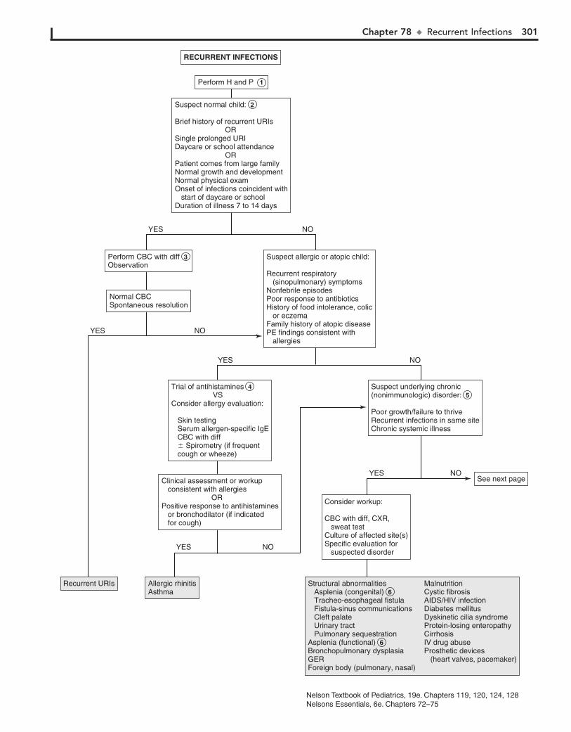
Chapter 78 u RecurrentInfections 301
Recurrent URIsRecurrent URIs Allergic rhinitisAsthmaAllergic rhinitisAsthma
Perform H and PPerform H and P 1
Suspect normal child:
Brief history of recurrent URIs ORSingle prolonged URIDaycare or school attendance ORPatient comes from large familyNormal growth and developmentNormal physical examOnset of infections coincident with start of daycare or schoolDuration of illness 7 to 14 days
Suspect normal child: 2
Brief history of recurrent URIs ORSingle prolonged URIDaycare or school attendance ORPatient comes from large familyNormal growth and developmentNormal physical examOnset of infections coincident with start of daycare or schoolDuration of illness 7 to 14 days
See next pageSee next page
Suspect allergic or atopic child:
Recurrent respiratory (sinopulmonary) symptomsNonfebrile episodesPoor response to antibioticsHistory of food intolerance, colic or eczemaFamily history of atopic diseasePE findings consistent with allergies
Suspect allergic or atopic child:
Recurrent respiratory (sinopulmonary) symptomsNonfebrile episodesPoor response to antibioticsHistory of food intolerance, colic or eczemaFamily history of atopic diseasePE findings consistent with allergies
Perform CBC with diffObservationPerform CBC with diff 3 Observation
Normal CBCSpontaneous resolutionNormal CBCSpontaneous resolution
Suspect underlying chronic (nonimmunologic) disorder:
Poor growth/failure to thriveRecurrent infections in same siteChronic systemic illness
Suspect underlying chronic (nonimmunologic) disorder: 5
Poor growth/failure to thriveRecurrent infections in same siteChronic systemic illness
Consider workup:
CBC with diff, CXR, sweat testCulture of affected site(s)Specific evaluation for suspected disorder
Consider workup:
CBC with diff, CXR, sweat testCulture of affected site(s)Specific evaluation for suspected disorder
Trial of antihistamines VSConsider allergy evaluation:
Skin testing Serum allergen-specific IgE CBC with diff � Spirometry (if frequent cough or wheeze)
Trial of antihistamines 4 VSConsider allergy evaluation:
Skin testing Serum allergen-specific IgE CBC with diff � Spirometry (if frequent cough or wheeze)
Clinical assessment or workup consistent with allergies ORPositive response to antihistamines or bronchodilator (if indicated for cough)
Clinical assessment or workup consistent with allergies ORPositive response to antihistamines or bronchodilator (if indicated for cough)
NOYES
NOYES
NOYES
NOYES
NOYES
RECURRENT INFECTIONSRECURRENT INFECTIONS
Structural abnormalities Asplenia (congenital) 6 Tracheo-esophageal fistula Fistula-sinus communications Cleft palate Urinary tract Pulmonary sequestrationAsplenia (functional) 6 Bronchopulmonary dysplasiaGERForeign body (pulmonary, nasal)
MalnutritionCystic fibrosisAIDS/HIV infectionDiabetes mellitusDyskinetic cilia syndromeProtein-losing enteropathyCirrhosisIV drug abuseProsthetic devices (heart valves, pacemaker)
Nelson Textbook of Pediatrics, 19e. Chapters 119, 120, 124, 128Nelsons Essentials, 6e. Chapters 72–75
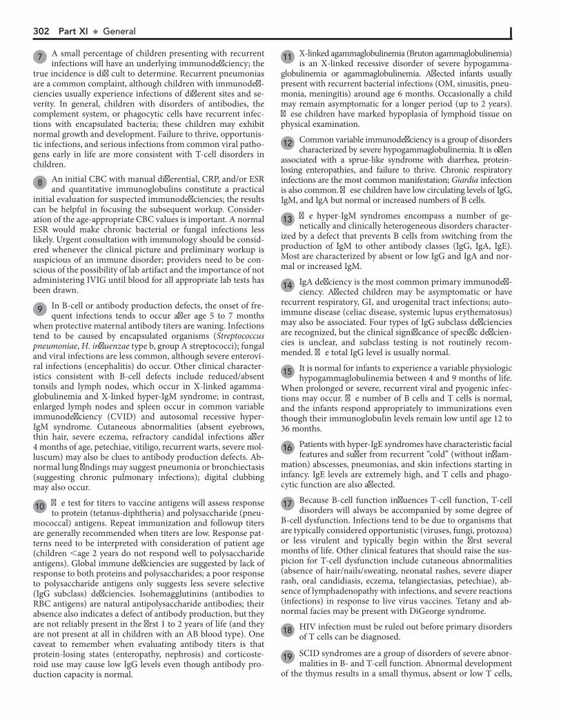
302 Part XI u General
7 A small percentage of children presenting with recurrent infections will have an underlying immunodeficiency; the
true incidence is difficult to determine. Recurrent pneumonias are a common complaint, although children with immunodefi-ciencies usually experience infections of different sites and se-verity. In general, children with disorders of antibodies, the complement system, or phagocytic cells have recurrent infec-tions with encapsulated bacteria; these children may exhibit normal growth and development. Failure to thrive, opportunis-tic infections, and serious infections from common viral patho-gens early in life are more consistent with T-cell disorders in children.
8 An initial CBC with manual differential, CRP, and/or ESR and quantitative immunoglobulins constitute a practical
initial evaluation for suspected immunodeficiencies; the results can be helpful in focusing the subsequent workup. Consider-ation of the age-appropriate CBC values is important. A normal ESR would make chronic bacterial or fungal infections less likely. Urgent consultation with immunology should be consid-ered whenever the clinical picture and preliminary workup is suspicious of an immune disorder; providers need to be con-scious of the possibility of lab artifact and the importance of not administering IVIG until blood for all appropriate lab tests has been drawn.
9 In B-cell or antibody production defects, the onset of fre-quent infections tends to occur after age 5 to 7 months
when protective maternal antibody titers are waning. Infections tend to be caused by encapsulated organisms (Streptococcus pneumoniae, H. influenzae type b, group A streptococci); fungal and viral infections are less common, although severe enterovi-ral infections (encephalitis) do occur. Other clinical character-istics consistent with B-cell defects include reduced/absent tonsils and lymph nodes, which occur in X-linked agamma-globulinemia and X-linked hyper-IgM syndrome; in contrast, enlarged lymph nodes and spleen occur in common variable immunodeficiency (CVID) and autosomal recessive hyper- IgM syndrome. Cutaneous abnormalities (absent eyebrows, thin hair, severe eczema, refractory candidal infections after 4 months of age, petechiae, vitiligo, recurrent warts, severe mol-luscum) may also be clues to antibody production defects. Ab-normal lung findings may suggest pneumonia or bronchiectasis (suggesting chronic pulmonary infections); digital clubbing may also occur.
10 The test for titers to vaccine antigens will assess response to protein (tetanus-diphtheria) and polysaccharide (pneu-
mococcal) antigens. Repeat immunization and followup titers are generally recommended when titers are low. Response pat-terns need to be interpreted with consideration of patient age (children ,age 2 years do not respond well to polysaccharide antigens). Global immune deficiencies are suggested by lack of response to both proteins and polysaccharides; a poor response to polysaccharide antigens only suggests less severe selective (IgG subclass) deficiencies. Isohemagglutinins (antibodies to RBC antigens) are natural antipolysaccharide antibodies; their absence also indicates a defect of antibody production, but they are not reliably present in the first 1 to 2 years of life (and they are not present at all in children with an AB blood type). One caveat to remember when evaluating antibody titers is that protein-losing states (enteropathy, nephrosis) and corticoste-roid use may cause low IgG levels even though antibody pro-duction capacity is normal.
11 X-linked agammaglobulinemia (Bruton agammaglobulinemia) is an X-linked recessive disorder of severe hypogamma-
globulinemia or agammaglobulinemia. Affected infants usually present with recurrent bacterial infections (OM, sinusitis, pneu-monia, meningitis) around age 6 months. Occasionally a child may remain asymptomatic for a longer period (up to 2 years). These children have marked hypoplasia of lymphoid tissue on physical examination.
12 Common variable immunodeficiency is a group of disorders characterized by severe hypogammaglobulinemia. It is often
associated with a sprue-like syndrome with diarrhea, protein-losing enteropathies, and failure to thrive. Chronic respiratory infections are the most common manifestation; Giardia infection is also common. These children have low circulating levels of IgG, IgM, and IgA but normal or increased numbers of B cells.
13 The hyper-IgM syndromes encompass a number of ge-netically and clinically heterogeneous disorders character-
ized by a defect that prevents B cells from switching from the production of IgM to other antibody classes (IgG, IgA, IgE). Most are characterized by absent or low IgG and IgA and nor-mal or increased IgM.
14 IgA deficiency is the most common primary immunodefi-ciency. Affected children may be asymptomatic or have
recurrent respiratory, GI, and urogenital tract infections; auto-immune disease (celiac disease, systemic lupus erythematosus) may also be associated. Four types of IgG subclass deficiencies are recognized, but the clinical significance of specific deficien-cies is unclear, and subclass testing is not routinely recom-mended. The total IgG level is usually normal.
15 It is normal for infants to experience a variable physiologic hypogammaglobulinemia between 4 and 9 months of life.
When prolonged or severe, recurrent viral and pyogenic infec-tions may occur. The number of B cells and T cells is normal, and the infants respond appropriately to immunizations even though their immunoglobulin levels remain low until age 12 to 36 months.
16 Patients with hyper-IgE syndromes have characteristic facial features and suffer from recurrent “cold” (without inflam-
mation) abscesses, pneumonias, and skin infections starting in infancy. IgE levels are extremely high, and T cells and phago-cytic function are also affected.
17 Because B-cell function influences T-cell function, T-cell disorders will always be accompanied by some degree of
B-cell dysfunction. Infections tend to be due to organisms that are typically considered opportunistic (viruses, fungi, protozoa) or less virulent and typically begin within the first several months of life. Other clinical features that should raise the sus-picion for T-cell dysfunction include cutaneous abnormalities (absence of hair/nails/sweating, neonatal rashes, severe diaper rash, oral candidiasis, eczema, telangiectasias, petechiae), ab-sence of lymphadenopathy with infections, and severe reactions (infections) in response to live virus vaccines. Tetany and ab-normal facies may be present with DiGeorge syndrome.
18 HIV infection must be ruled out before primary disorders of T cells can be diagnosed.
19 SCID syndromes are a group of disorders of severe abnor-malities in B- and T-cell function. Abnormal development
of the thymus results in a small thymus, absent or low T cells,
Chapter 78 u RecurrentInfections 303
X-linked agammaglobulinemia (Bruton)Common variable immunodeficiencyHyper-IgMIgA deficiencyCombined cellular and humoral disordersTransient hypogammaglobulinemia of infancyHyper-IgESpecific immunoglobulin deficiencies
X-linked agammaglobulinemia (Bruton) 11 Common variable immunodeficiency 12 Hyper-IgM 13 IgA deficiency 14 Combined cellular and humoral disordersTransient hypogammaglobulinemia of infancy 15 Hyper-IgE 16 Specific immunoglobulin deficiencies
Severe combined immunodeficiencyDiGeorge syndromeAtaxia-telangiectasiaWiskott-Aldrich syndromeHyper-IgECartilage-hair hypoplasiaHIV infectionSecondary immunodeficiencies
Severe combined immunodeficiency 19 DiGeorge syndrome 20 Ataxia-telangiectasia 21 Wiskott-Aldrich syndrome 22 Hyper-IgE 10 Cartilage-hair hypoplasia 23 HIV infectionSecondary immunodeficiencies
Suspect immunodeficiency:
Pyogenic infections involving multiple sitesTwo or more systemic bacterial infections by age 9 monthsFailure to thrive or growth failure Unusual pathogensUnexpected severity of infectionInfection following receipt of live vaccinesChronic oral or cutaneous Candida infectionsChronic diarrhea
Suspect immunodeficiency: 7
Pyogenic infections involving multiple sitesTwo or more systemic bacterial infections by age 9 monthsFailure to thrive or growth failure Unusual pathogensUnexpected severity of infectionInfection following receipt of live vaccinesChronic oral or cutaneous Candida infectionsChronic diarrhea
Perform preliminary evaluation:CBC & manual diffESR/CRPQuantitative IgG, IgA, IgM, and IgE levelsTotal complement (CH50)
Perform preliminary evaluation: 8 CBC & manual diffESR/CRPQuantitative IgG, IgA, IgM, and IgE levelsTotal complement (CH50)
Suspicious of humoral/antibody defect:Onset age 5-7 months (after maternal antibodies wane)Recurrent sinopulmonary infections: Sinusitis, pneumonia (with fevers), otitisMeningitisSepsisChronic diarrheaSkin infectionsAutoimmune disorderUnusual organisms
Lab: Abnormal immunoglobulin levels
Suspicious of humoral/antibody defect: 9 Onset age 5-7 months (after maternal antibodies wane)Recurrent sinopulmonary infections: Sinusitis, pneumonia (with fevers), otitisMeningitisSepsisChronic diarrheaSkin infectionsAutoimmune disorderUnusual organisms
Lab: Abnormal immunoglobulin levels
See p. 3 of algorithmSee p. 3 of algorithm
Suspect cellular or combined defects:
Onset in first several months of lifeSevere viral infections (RSV, parainfluenza, CMV, EBV, adenovirus)Fungal/opportunistic infectionsFailure to thriveChronic diarrheaCongenital heart diseaseAbsence of lymphadenopathy with infectionsAtaxia
Lab: Lymphopenia, thrombocytopenia
Suspect cellular or combined defects: 17
Onset in first several months of lifeSevere viral infections (RSV, parainfluenza, CMV, EBV, adenovirus)Fungal/opportunistic infectionsFailure to thriveChronic diarrheaCongenital heart diseaseAbsence of lymphadenopathy with infectionsAtaxia
Lab: Lymphopenia, thrombocytopenia
Additional evaluation: 10Antibody titers to vaccine antigens (diphtheria, tetanus, pneumococcal)
� Isohemagglutinins
Additional evaluation: 10Antibody titers to vaccine antigens (diphtheria, tetanus, pneumococcal)
� Isohemagglutinins
NOYES
Additional evaluation:HIVDelayed hypersensitivity skin testing (Candida, tetanus, mumps, trichophyton)T-cell levels with helper/suppressor ratio
Additional evaluation:HIV 18 Delayed hypersensitivity skin testing (Candida, tetanus, mumps, trichophyton)T-cell levels with helper/suppressor ratio
NOYES
RECURRENT INFECTIONS (continued)RECURRENT INFECTIONS (continued)
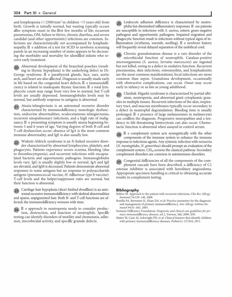
304 Part XI u General
and lymphopenia (,2500/mm3 in children ,5 years old) from birth. Growth is initially normal, but wasting typically occurs after symptom onset in the first few months of life; recurrent pneumonias, OM, failure to thrive, chronic diarrhea, and severe candidal (and other opportunistic) infections are common. In-fections are characteristically not accompanied by lymphade-nopathy. The addition of a test for SCID to newborn screening panels in an increasing number of states appears to be decreas-ing the morbidity and mortality for identified infants who re-ceive early treatment.
20 Abnormal development of the branchial pouches (result-ing in thymic hypoplasia) is the underlying defect in Di-
George syndrome. The parathyroid glands, face, ears, aortic arch, and heart are also affected. Diagnosis is usually made early in life based on the congenital heart defects. The immunodefi-ciency is related to inadequate thymic function. The total lym-phocyte count may range from very low to normal, but T-cell levels are usually depressed. Immunoglobulin levels may be normal, but antibody response to antigens is abnormal.
21 Ataxia-telangiectasia is an autosomal recessive disorder characterized by immunodeficiency, neurologic dysfunc-
tion, endocrine abnormalities, oculocutaneous telangiectasias, recurrent sinopulmonary infections, and a high rate of malig-nancy. The presenting symptom is usually ataxia beginning be-tween 3 and 6 years of age. Varying degrees of both B-cell and T-cell dysfunction occur; absence of IgA is the most common immune abnormality, and IgE is also usually low.
22 Wiskott-Aldrich syndrome is an X-linked recessive disor-der characterized by abnormal lymphocytes, platelets, and
phagocytes. Patients experience severe eczema, bleeding (due to thrombocytopenia), and recurrent infections with encapsu-lated bacteria and opportunistic pathogens. Immunoglobulin levels vary. IgG is usually slightly low or normal, IgA and IgE are elevated, and IgM is decreased. Patients demonstrate abnormal responses to some antigens but no response to polysaccharide antigens (pneumococcal vaccine, H. influenzae type b vaccine). T-cell levels and the helper/suppressor ratio are normal, but their function is abnormal.
23 Cartilage-hair hypoplasia (short-limbed dwarfism) is an auto-somal recessive immunodeficiency with skeletal abnormalities
and sparse, unpigmented hair. Both B- and T-cell functions are af-fected; the immunodeficiency worsens with time.
24 The approach to neutropenia needs to consider produc-tion, destruction, and function of neutrophils. Specific
testing can identify disorders of motility and chemotaxis, adhe-sion, microbicidal activity, and specific granule defects.
25 Leukocyte adhesion deficiency is characterized by neutro-philia but diminished inflammatory responses. These patients
are susceptible to infections with S. aureus, enteric gram-negative pathogens and opportunistic pathogens. Impaired migration and phagocytic function result in infections without typical signs of in-flammation (erythema, warmth, swelling). The newborn history will frequently reveal delayed separation of the umbilical cord.
26 Chronic granulomatous disease is a rare disorder of the microbicidal function of neutrophils. Catalase-positive
microorganisms (S. aureus, Serratia marcescens) are ingested but not killed, owing to a defect in oxidative function. Recurrent pneumonias, skin infections, osteomyelitis, and lymphadenitis are the most common manifestations; focal infections are more common than sepsis. Granuloma development, occasionally with obstructive complications, can occur. Onset may occur early in infancy or as late as young adulthood.
27 Chédiak-Higashi syndrome is characterized by partial albi-nism, neutropenia, and abnormal giant cytoplasmic gran-
ules in multiple tissues. Recurrent infections of the skin, respira-tory tract, and mucous membranes typically occur secondary to a defect in neutrophil degranulation. Bleeding time is usually prolonged. The presence of large melanosomes in melanocytes can confirm the diagnosis. Progressive neuropathies and a ten-dency to life-threatening histiocytosis can also occur. Chemo-tactic function is abnormal when assayed in control serum.
28 The complement system acts synergistically with the other components of the immune system to enhance the immune
response to infectious agents. Any systemic infection with neisseria (N. meningitides, N. gonorrhea) should prompt an evaluation of the complement system. CH50 screens the classical pathway. Secondary complement disorders are common in autoimmune disorders.
29 Congenital deficiencies of all the components of the com-plement cascade have been described; a deficiency of C1
esterase inhibitor is associated with hereditary angioedema. Appropriate specimen handling is critical to obtaining accurate results in complement testing.
BibliographyBallow M: Approach to the patient with recurrent infections, Clin Rev Allergy
Immunol 34:129–140, 2008.Bonilla FA, Bernstein IL, Khan DA, et al: Practice parameter for the diagnosis
and management of primary immunodeficiency, Ann Allergy Asthma Im-munol 94:S1–S63, 2005.
Immune Deficiency Foundation: Diagnostic and clinical care guidelines for pri-mary immunodeficiency diseases, ed 2, Towson, Md, 2009, IDF.
Slatter M, Cant AJ, Arkwright PD, et al: Clinical features that identify children with primary immunodeficiency diseases, Pediatrics 127:810, 2011.
Chapter 78 u RecurrentInfections 305
Leukocyte adhesion deficiencyChronic granulomatous diseaseChédiak-Higashi syndromeCyclic neutropenia Other phagocytic disordersComplement disorders
Leukocyte adhesion deficiency 25 Chronic granulomatous disease 26 Chédiak-Higashi syndrome 27 Cyclic neutropenia Other phagocytic disordersComplement disorders
Complement deficiencyComplement deficiency
Suspect phagocytic cell disorder:
Skin infections (abscesses, cellulitis)Lymphadenopathy/lymphadenitisDeep-seated infections (osteomyelitis, hepatic or pulmonary abscesses)Pulmonary aspergillosis Recurrent pneumoniaOral lesions (gingivitis, ulcers)Abscesses due to unusual pathogensHistory of delayed umbilical cord separationPoor wound healingOpen sores with scant pusUnexplained systemic symptoms (fever, malaise, fatigue)
Lab: Abnormal ANC (high or low)
Suspect complement disorder:
Systemic neisserial infection (sepsis, meningitis)Recurrent infections with encapsulated bacteria (OM, bacteremia, pneumonia)
Lab: Abnormal CH50
Suspect complement disorder: 28
Systemic neisserial infection (sepsis, meningitis)Recurrent infections with encapsulated bacteria (OM, bacteremia, pneumonia)
Lab: Abnormal CH50
CH50 (classical pathway)
If abnormal, refer for testing to identify specific complement components
CH50 (classical pathway) 29
If abnormal, refer for testing to identify specific complement components
See Chapter 64 if neutropenia present
Refer to immunology for CD11/CD18 expression (to exclude LAD) and a respiratory burst assay
See Chapter 64 if neutropenia present 24
Refer to immunology for CD11/CD18 expression (to exclude LAD) and a respiratory burst assay
NOYES
RECURRENT INFECTIONS (continued)RECURRENT INFECTIONS (continued)
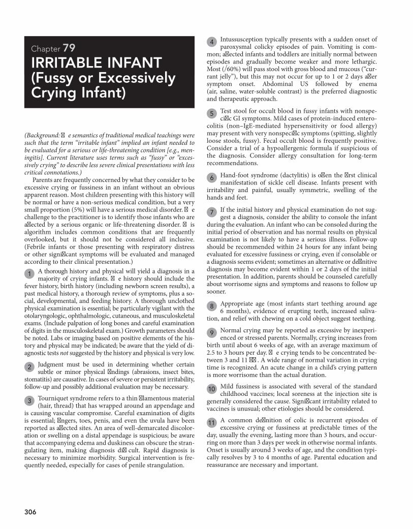
306
Chapter 79
IRRITABLE INFANT (Fussy or Excessively Crying Infant)
4 Intussusception typically presents with a sudden onset of paroxysmal colicky episodes of pain. Vomiting is com-
mon; affected infants and toddlers are initially normal between episodes and gradually become weaker and more lethargic. Most (≈60%) will pass stool with gross blood and mucous (“cur-rant jelly”), but this may not occur for up to 1 or 2 days after symptom onset. Abdominal US followed by enema (air, saline, water-soluble contrast) is the preferred diagnostic and therapeutic approach.
5 Test stool for occult blood in fussy infants with nonspe-cific GI symptoms. Mild cases of protein-induced entero-
colitis (non–IgE-mediated hypersensitivity or food allergy) may present with very nonspecific symptoms (spitting, slightly loose stools, fussy). Fecal occult blood is frequently positive. Consider a trial of a hypoallergenic formula if suspicious of the diagnosis. Consider allergy consultation for long-term recommendations.
6 Hand-foot syndrome (dactylitis) is often the first clinical manifestation of sickle cell disease. Infants present with
irritability and painful, usually symmetric, swelling of the hands and feet.
7 If the initial history and physical examination do not sug-gest a diagnosis, consider the ability to console the infant
during the evaluation. An infant who can be consoled during the initial period of observation and has normal results on physical examination is not likely to have a serious illness. Follow-up should be recommended within 24 hours for any infant being evaluated for excessive fussiness or crying, even if consolable or a diagnosis seems evident; sometimes an alternative or definitive diagnosis may become evident within 1 or 2 days of the initial presentation. In addition, parents should be counseled carefully about worrisome signs and symptoms and reasons to follow up sooner.
8 Appropriate age (most infants start teething around age 6 months), evidence of erupting teeth, increased saliva-
tion, and relief with chewing on a cold object suggest teething.
9 Normal crying may be reported as excessive by inexperi-enced or stressed parents. Normally, crying increases from
birth until about 6 weeks of age, with an average maximum of 2.5 to 3 hours per day. The crying tends to be concentrated be-tween 3 and 11 pm. A wide range of normal variation in crying time is recognized. An acute change in a child’s crying pattern is more worrisome than the actual duration.
10 Mild fussiness is associated with several of the standard childhood vaccines; local soreness at the injection site is
generally considered the cause. Significant irritability related to vaccines is unusual; other etiologies should be considered.
11 A common definition of colic is recurrent episodes of excessive crying or fussiness at predictable times of the
day, usually the evening, lasting more than 3 hours, and occur-ring on more than 3 days per week in otherwise normal infants. Onset is usually around 3 weeks of age, and the condition typi-cally resolves by 3 to 4 months of age. Parental education and reassurance are necessary and important.
(Background: The semantics of traditional medical teachings were such that the term “irritable infant” implied an infant needed to be evaluated for a serious or life-threatening condition [e.g., men-ingitis]. Current literature uses terms such as “fussy” or “exces-sively crying” to describe less severe clinical presentations with less critical connotations.)
Parents are frequently concerned by what they consider to be excessive crying or fussiness in an infant without an obvious apparent reason. Most children presenting with this history will be normal or have a non-serious medical condition, but a very small proportion (5%) will have a serious medical disorder. The challenge to the practitioner is to identify those infants who are affected by a serious organic or life-threatening disorder. This algorithm includes common conditions that are frequently overlooked, but it should not be considered all inclusive. (Febrile infants or those presenting with respiratory distress or other significant symptoms will be evaluated and managed according to their clinical presentation.)
1 A thorough history and physical will yield a diagnosis in a majority of crying infants. The history should include the
fever history, birth history (including newborn screen results), a past medical history, a thorough review of symptoms, plus a so-cial, developmental, and feeding history. A thorough unclothed physical examination is essential; be particularly vigilant with the otolaryngologic, ophthalmologic, cutaneous, and musculoskeletal exams. (Include palpation of long bones and careful examination of digits in the musculoskeletal exam.) Growth parameters should be noted. Labs or imaging based on positive elements of the his-tory and physical may be indicated; be aware that the yield of di-agnostic tests not suggested by the history and physical is very low.
2 Judgment must be used in determining whether certain subtle or minor physical findings (abrasions, insect bites,
stomatitis) are causative. In cases of severe or persistent irritability, follow-up and possibly additional evaluation may be necessary.
3 Tourniquet syndrome refers to a thin filamentous material (hair, thread) that has wrapped around an appendage and
is causing vascular compromise. Careful examination of digits is essential; fingers, toes, penis, and even the uvula have been reported as affected sites. An area of well-demarcated discolor-ation or swelling on a distal appendage is suspicious; be aware that accompanying edema and duskiness can obscure the stran-gulating item, making diagnosis difficult. Rapid diagnosis is necessary to minimize morbidity. Surgical intervention is fre-quently needed, especially for cases of penile strangulation.
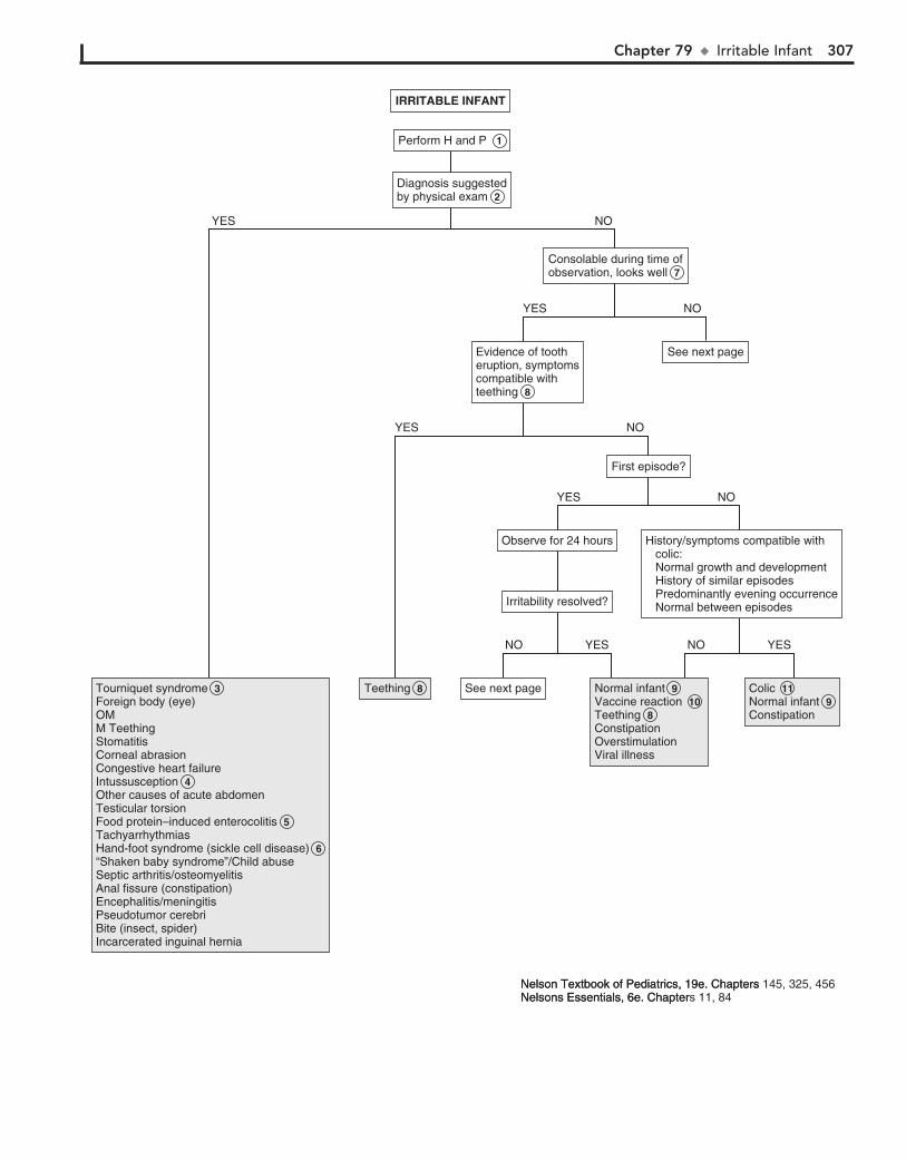
Chapter 79 u IrritableInfant 307
Perform H and PPerform H and P 1
Consolable during time ofobservation, looks wellConsolable during time ofobservation, looks well 7
Diagnosis suggestedby physical examDiagnosis suggestedby physical exam 2
Tourniquet syndromeForeign body (eye)OMM TeethingStomatitisCorneal abrasionCongestive heart failureIntussusceptionOther causes of acute abdomen Testicular torsionFood protein–induced enterocolitisTachyarrhythmiasHand-foot syndrome (sickle cell disease)“Shaken baby syndrome”/Child abuseSeptic arthritis/osteomyelitisAnal fissure (constipation) Encephalitis/meningitisPseudotumor cerebriBite (insect, spider)Incarcerated inguinal hernia
Tourniquet syndrome 3 Foreign body (eye)OMM TeethingStomatitisCorneal abrasionCongestive heart failureIntussusception 4 Other causes of acute abdomen Testicular torsionFood protein–induced enterocolitis 5 TachyarrhythmiasHand-foot syndrome (sickle cell disease) 6 “Shaken baby syndrome”/Child abuseSeptic arthritis/osteomyelitisAnal fissure (constipation) Encephalitis/meningitisPseudotumor cerebriBite (insect, spider)Incarcerated inguinal hernia
TeethingTeething 8 Normal infantVaccine reactionTeethingConstipationOverstimulationViral illness
Normal infant 9 Vaccine reaction 10 Teething 8 ConstipationOverstimulationViral illness
ColicNormal infantConstipation
Colic 11 Normal infant 9 Constipation
History/symptoms compatible with colic: Normal growth and development History of similar episodes Predominantly evening occurrence Normal between episodes
History/symptoms compatible with colic: Normal growth and development History of similar episodes Predominantly evening occurrence Normal between episodes
See next pageSee next page
Observe for 24 hoursObserve for 24 hours
Irritability resolved?Irritability resolved?
NOYES
See next pageSee next pageEvidence of tootheruption, symptomscompatible withteething
Evidence of tootheruption, symptomscompatible withteething 8
NOYES
NOYES
First episode?First episode?
NOYES
NO YES NO YES
IRRITABLE INFANTIRRITABLE INFANT
Nelson Textbook of Pediatrics, 19e. Chapters 145, 325, 456Nelsons Essentials, 6e. Chapters 11, 84Nelson Textbook of Pediatrics, 19e. ChaptersNelsons Essentials, 6e. Chapter
308 Part XI u General
12 A UA and culture are the most likely diagnostic tests to yield a positive result. Other testing has a very low yield
unless the history and physical suggests an indication for it.
13 Abstinence syndromes may occur in infants born to sub-stance-abusing mothers; withdrawal symptoms include
irritability, vomiting, diarrhea, hypertonicity, poor feeding, and sleep problems. Withdrawal usually occurs in the first week of life but may be delayed up to 2 to 3 weeks if the mother was using methadone. Beyond the neonatal period, breastfeeding infants may experience fussiness and irritability due to transfer of maternal drugs (decongestants, caffeine, nicotine, cocaine). Environmental toxins (carbon monoxide) may rarely be a cause of nonspecific infant fussiness.
14 The risk/benefit ratio of performing additional testing must be weighed against the option of simply ensuring
follow-up and reassessment in 24 hours. Very rarely will an infant with persistent or severe irritability of unclear cause war-rant hospitalization for observation and additional evaluation.
15 A head CT will diagnose intracranial hemorrhage (suspi-cious of child abuse) or other causes of increased ICP.
Meningitis needs to be considered in cases of severe or incon-solable irritability; some infants will present afebrile. A head CT should be obtained before performing an LP in these infants to rule out increased ICP.
16 Acidosis, hypernatremia, hypocalcemia, and hypoglyce-mia are rare but serious causes of irritability in infants.
Inborn errors of metabolism should be considered when there is associated vomiting, neurologic symptoms, failure to thrive, or a positive family history, including unexplained neonatal deaths.
BibliographyFreedman SB, Al-Harthy N, Thull-Freedman J: The crying infant: Diagnostic
testing and frequency of serious underlying disease, Pediatrics 123:841–848, 2009.
Herman M, Le A: The crying infant, Emerg Med Clin North Am 25:1137–1159, 2007.
Pawel BB, Henretig FM: Crying and colic in early infancy. In Fleisher G, Ludwig S, editors: Textbook of pediatric emergency medicine, ed 6, Philadelphia, 2010, Lippincott Williams & Wilkins, pp 203–205.
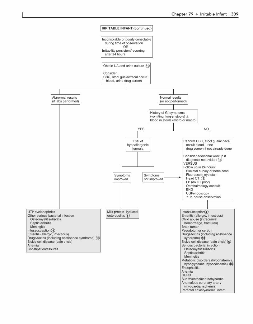
Chapter 79 u IrritableInfant 309
Inconsolable or poorly consolable during time of observation ORIrritability persistent/recurring after 24 hours
Inconsolable or poorly consolable during time of observation ORIrritability persistent/recurring after 24 hours
Normal results(or not performed)Normal results(or not performed)
Abnormal results(if labs performed)Abnormal results(if labs performed)
History of GI symptoms(vomiting, looser stools) �blood in stools (micro or macro)
History of GI symptoms(vomiting, looser stools) �blood in stools (micro or macro)
Obtain UA and urine culture 12
Consider:CBC, stool guaiac/fecal occult blood, urine drug screen
Obtain UA and urine culture 12
Consider:CBC, stool guaiac/fecal occult blood, urine drug screen
UTI/ pyelonephritisOther serious bacterial infection Osteomyelitis/discitis Septic arthritis MeningitisIntussusception 4Enteritis (allergic, infectious)Drugs/toxins (including abstinence syndrome) 13Sickle cell disease (pain crisis)AnemiaConstipation/fissures
UTI/ pyelonephritisOther serious bacterial infection Osteomyelitis/discitis Septic arthritis MeningitisIntussusception 4Enteritis (allergic, infectious)Drugs/toxins (including abstinence syndrome) 13Sickle cell disease (pain crisis)AnemiaConstipation/fissures
Milk protein–inducedenterocolitis 5Milk protein–inducedenterocolitis 5
Intussusception 4Enteritis (allergic, infectious)Child abuse (intracranial hemorrhage, fractures)Brain tumorPseudotumor cerebriDrugs/toxins (including abstinence syndrome) 13Sickle cell disease (pain crisis) 6Serious bacterial infection Osteomyelitis/discitis Septic arthritis MeningitisMetabolic disorders (hyponatremia, hypoglycemia, hypocalcemia) 16EncephalitisAnemia GERDSupraventricular tachycardiaAnomalous coronary artery (myocardial ischemia)Parental anxiety/normal infant
Intussusception 4Enteritis (allergic, infectious)Child abuse (intracranial hemorrhage, fractures)Brain tumorPseudotumor cerebriDrugs/toxins (including abstinence syndrome) 13Sickle cell disease (pain crisis) 6Serious bacterial infection Osteomyelitis/discitis Septic arthritis MeningitisMetabolic disorders (hyponatremia, hypoglycemia, hypocalcemia) 16EncephalitisAnemia GERDSupraventricular tachycardiaAnomalous coronary artery (myocardial ischemia)Parental anxiety/normal infant
Perform CBC, stool guaiac/fecal occult blood, urine drug screen if not already done
Consider additional workup if diagnosis not evident 14VERSUSFollow up in 24 hours: Skeletal survey or bone scan Fluorescein eye stain Head CT 15 LP (do CT prior) Ophthalmology consult EKG UGI/endoscopy � In-house observation
Trial ofhypoallergenic
formula
Trial ofhypoallergenic
formula
NOYES
Symptomsnot improvedSymptomsnot improved
SymptomsimprovedSymptomsimproved
IRRITABLE INFANT (continued)IRRITABLE INFANT (continued)
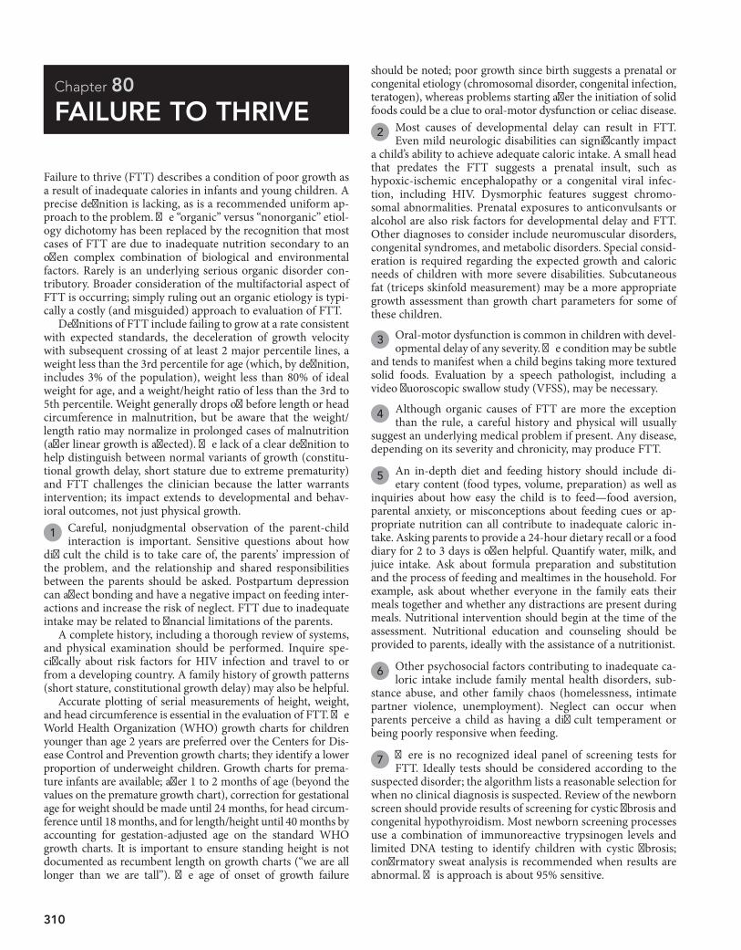
310
Chapter 80
FAILURE TO THRIVE
should be noted; poor growth since birth suggests a prenatal or congenital etiology (chromosomal disorder, congenital infection, teratogen), whereas problems starting after the initiation of solid foods could be a clue to oral-motor dysfunction or celiac disease.
2 Most causes of developmental delay can result in FTT. Even mild neurologic disabilities can significantly impact
a child’s ability to achieve adequate caloric intake. A small head that predates the FTT suggests a prenatal insult, such as hypoxic-ischemic encephalopathy or a congenital viral infec-tion, including HIV. Dysmorphic features suggest chromo-somal abnormalities. Prenatal exposures to anticonvulsants or alcohol are also risk factors for developmental delay and FTT. Other diagnoses to consider include neuromuscular disorders, congenital syndromes, and metabolic disorders. Special consid-eration is required regarding the expected growth and caloric needs of children with more severe disabilities. Subcutaneous fat (triceps skinfold measurement) may be a more appropriate growth assessment than growth chart parameters for some of these children.
3 Oral-motor dysfunction is common in children with devel-opmental delay of any severity. The condition may be subtle
and tends to manifest when a child begins taking more textured solid foods. Evaluation by a speech pathologist, including a video fluoroscopic swallow study (VFSS), may be necessary.
4 Although organic causes of FTT are more the exception than the rule, a careful history and physical will usually
suggest an underlying medical problem if present. Any disease, depending on its severity and chronicity, may produce FTT.
5 An in-depth diet and feeding history should include di-etary content (food types, volume, preparation) as well as
inquiries about how easy the child is to feed—food aversion, parental anxiety, or misconceptions about feeding cues or ap-propriate nutrition can all contribute to inadequate caloric in-take. Asking parents to provide a 24-hour dietary recall or a food diary for 2 to 3 days is often helpful. Quantify water, milk, and juice intake. Ask about formula preparation and substitution and the process of feeding and mealtimes in the household. For example, ask about whether everyone in the family eats their meals together and whether any distractions are present during meals. Nutritional intervention should begin at the time of the assessment. Nutritional education and counseling should be provided to parents, ideally with the assistance of a nutritionist.
6 Other psychosocial factors contributing to inadequate ca-loric intake include family mental health disorders, sub-
stance abuse, and other family chaos (homelessness, intimate partner violence, unemployment). Neglect can occur when parents perceive a child as having a difficult temperament or being poorly responsive when feeding.
7 There is no recognized ideal panel of screening tests for FTT. Ideally tests should be considered according to the
suspected disorder; the algorithm lists a reasonable selection for when no clinical diagnosis is suspected. Review of the newborn screen should provide results of screening for cystic fibrosis and congenital hypothyroidism. Most newborn screening processes use a combination of immunoreactive trypsinogen levels and limited DNA testing to identify children with cystic fibrosis; confirmatory sweat analysis is recommended when results are abnormal. This approach is about 95% sensitive.
Failure to thrive (FTT) describes a condition of poor growth as a result of inadequate calories in infants and young children. A precise definition is lacking, as is a recommended uniform ap-proach to the problem. The “organic” versus “nonorganic” etiol-ogy dichotomy has been replaced by the recognition that most cases of FTT are due to inadequate nutrition secondary to an often complex combination of biological and environmental factors. Rarely is an underlying serious organic disorder con-tributory. Broader consideration of the multifactorial aspect of FTT is occurring; simply ruling out an organic etiology is typi-cally a costly (and misguided) approach to evaluation of FTT.
Definitions of FTT include failing to grow at a rate consistent with expected standards, the deceleration of growth velocity with subsequent crossing of at least 2 major percentile lines, a weight less than the 3rd percentile for age (which, by definition, includes 3% of the population), weight less than 80% of ideal weight for age, and a weight/height ratio of less than the 3rd to 5th percentile. Weight generally drops off before length or head circumference in malnutrition, but be aware that the weight/length ratio may normalize in prolonged cases of malnutrition (after linear growth is affected). The lack of a clear definition to help distinguish between normal variants of growth (constitu-tional growth delay, short stature due to extreme prematurity) and FTT challenges the clinician because the latter warrants intervention; its impact extends to developmental and behav-ioral outcomes, not just physical growth.
1 Careful, nonjudgmental observation of the parent-child interaction is important. Sensitive questions about how
difficult the child is to take care of, the parents’ impression of the problem, and the relationship and shared responsibilities between the parents should be asked. Postpartum depression can affect bonding and have a negative impact on feeding inter-actions and increase the risk of neglect. FTT due to inadequate intake may be related to financial limitations of the parents.
A complete history, including a thorough review of systems, and physical examination should be performed. Inquire spe-cifically about risk factors for HIV infection and travel to or from a developing country. A family history of growth patterns (short stature, constitutional growth delay) may also be helpful.
Accurate plotting of serial measurements of height, weight, and head circumference is essential in the evaluation of FTT. The World Health Organization (WHO) growth charts for children younger than age 2 years are preferred over the Centers for Dis-ease Control and Prevention growth charts; they identify a lower proportion of underweight children. Growth charts for prema-ture infants are available; after 1 to 2 months of age (beyond the values on the premature growth chart), correction for gestational age for weight should be made until 24 months, for head circum-ference until 18 months, and for length/height until 40 months by accounting for gestation-adjusted age on the standard WHO growth charts. It is important to ensure standing height is not documented as recumbent length on growth charts (“we are all longer than we are tall”). The age of onset of growth failure
Chapter 80 u FailuretoThrive 311
FAILURE TO THRIVE (AND WEIGHT LOSS)FAILURE TO THRIVE (AND WEIGHT LOSS)
Signs or symptomssuggestive of anorganic etiology present 4
Signs or symptomssuggestive of anorganic etiology present 4
Carefully review feeding historyAssess caloric intakeConsider TORCH titers, speech pathology consult
Carefully review feeding historyAssess caloric intakeConsider TORCH titers, speech pathology consult
Signs or symptoms suggestive ofdevelopmental delay or
neuromuscular disorder present
Signs or symptoms suggestive ofdevelopmental delay or
neuromuscular disorder present
Perform H and P, accurate documentation of serial measurements 1
Perform H and P, accurate documentation of serial measurements 1
Developmental delay (multiple etiologies) 2 Inadequate calorie intakeOral-motor dysfunction 3
Developmental delay (multiple etiologies) 2 Inadequate calorie intakeOral-motor dysfunction 3
Inadequate intake Breastfeeding problems Improper formula preparation Improper feeding technique Insufficient food offered (food insecurity, inappropriate parental knowledge or expectations ) Aversion (pain, sensory) Other psychosocial factors 6
Inadequate intake Breastfeeding problems Improper formula preparation Improper feeding technique Insufficient food offered (food insecurity, inappropriate parental knowledge or expectations ) Aversion (pain, sensory) Other psychosocial factors 6
Renal disease Obstructive uropathy Reflux nephropathy Renal tubular acidosis GlomerulonephritisLiver diseaseCeliac diseaseCystic fibrosisOther causes of malabsorption
Renal disease Obstructive uropathy Reflux nephropathy Renal tubular acidosis GlomerulonephritisLiver diseaseCeliac diseaseCystic fibrosisOther causes of malabsorption
DiagnosisapparentDiagnosisapparent
Diagnosisnot apparentDiagnosisnot apparent
Normal resultsNormal results
Diagnosisnot apparentDiagnosisnot apparent
DiagnosisapparentDiagnosisapparent
Carefully review feeding history 5 and technique, psychosocial issues Nutritional intervention
Carefully review feeding history 5 and technique, psychosocial issues Nutritional intervention
(See next page)(See next page)
Abnormal resultsAbnormal results
Continue nutritional intervention 8Consider referral to a multidisciplinary feeding program VS Inpatient observation
Continue nutritional intervention 8Consider referral to a multidisciplinary feeding program VS Inpatient observation
CBC, lead 7 Blood chemistry profileUA and cultureAlbumin and serum proteinCeliac panel (anti-tTG and IgA)Review newborn screen resultsConsider sweat chloride, free T4, TSH
CBC, lead 7 Blood chemistry profileUA and cultureAlbumin and serum proteinCeliac panel (anti-tTG and IgA)Review newborn screen resultsConsider sweat chloride, free T4, TSH
Further consider organic disorders 9 (See next page)
Further consider organic disorders 9 (See next page)
NO
NO YES
YES
Nelson Textbook of Pediatrics, 19e. Chapters 149, 157, 214, 280, 431, 676, 677 Nelsons Essentials, 6e. Chapters 96, 122
312 Part XI u General
8 If a cause is not apparent after a thorough history and physical and preliminary laboratory evaluation, and no
improvement has occurred with initial nutritional interven-tions, involvement with a more intensive multidisciplinary team is ideal (if available). Therapists, nutritionists, social work-ers, speech pathologists, and psychologists or developmental specialists can all make valuable contributions to the manage-ment of a child with FTT, regardless of the cause; the availability of such comprehensive teams is, however, limited in most places. Hospitalization is indicated in cases of suspected abuse or neglect for the purpose of ensuring patient safety, in extreme cases of malnutrition in which a child is at risk for refeeding syndrome, and when more intense surveillance is needed be-cause outpatient evaluation and intervention has not resulted in any improvement. The benefits of hospitalization are limited by short stays (due to payer mandates) and the multiple variables an inpatient setting creates. Long-term outpatient and in-home therapies are preferred options.
9 When a period of multidisciplinary involvement and ob-servation still fail to improve growth or suggest an etiol-
ogy, further consideration should be given to organic disorders, especially those that may present with subtle symptoms. Assess-ing for malabsorption disorders is reasonable because they may occur without obvious diarrhea. Also remember that GER can be present without obvious spitting up or vomiting.
10 If GI causes are not identified in a child with a history of vomiting, neuroimaging should also be considered, since
increased ICP due to a variety of etiologies (including abusive head trauma) may be the cause.
11 Food protein–induced syndromes (enterocolitis, proctocoli-tis, enteropathy) are non–immunoglobulin (Ig)E-mediated
hypersensitivity reactions that can manifest as vomiting and/or diarrhea, with a wide range of clinical severity and usually begin in infancy.
12 Children with Hirschsprung disease may have a history of delayed passage of meconium, Down syndrome, or posi-
tive family history. Most cases will be diagnosed in the first days of life with abdominal distention, vomiting, and constipation. Occasionally, older children will present with FTT, abdominal distention, and constipation. A few will demonstrate intermit-tent diarrhea. Absent stool on rectal examination and immedi-ate passage of stool and gas after the rectal examination are also suggestive. A rectal suction biopsy demonstrating absent gan-glion cells is necessary for diagnosis.
13 Inborn errors of metabolism in the neonate are typically severe and life threatening. The infants are usually normal
at birth and often rapidly deteriorate over the hours to days af-ter their birth. Signs and symptoms are nonspecific and include vomiting, lethargy, poor feeding, convulsions, and coma. These infants are often presumed to have sepsis. The physical exami-nation may be normal or may reveal signs related to the CNS, hepatomegaly, or occasionally, a characteristic odor that may aid in diagnosis.
A high index of suspicion is necessary and should be in-creased by a history of consanguinity and/or a family history of neonatal deaths. For a metabolic workup, blood and urine
should be obtained during episodes of suggestive symptoms. Blood tests should include a CBC, electrolytes (calculate anion gap), ABG, glucose, ammonia, lactate, carnitine, acylcarnitine, and serum amino acids. Urine should be analyzed for ketones, reducing substances, organic acids, and carnitine. Consider amylase or lipase, particularly in older children who also com-plain of abdominal pain.
Mild variants of most inborn errors do occur and have an insidious presentation. Be suspicious when there is a history of unexplained mental retardation, seizures, an unusual odor (especially if manifest during an acute illness), or a family history of metabolic disorders or unexplained infant deaths. Some children will experience intermittent episodes of severe illness, including unexplained vomiting, acidosis, and mental deterioration or coma.
14 Celiac disease is the most common food protein–induced enteropathy. It typically presents as intestinal symptoms
(diarrhea, bloating, FTT, irritability, poor appetite) in the first 2 years of life after the introduction of gluten-containing pro-ducts (wheat, rye, barley, other grains) into the diet. Anti–tissue transglutaminase (anti-tTG) antibody is the preferred screen-ing test in most institutions; an IgA endomysial (EMA) anti-body titer is a reasonable alternative. Be aware that anti-tTG may be absent in 10% of children younger than age 2 with celiac disease. Checking total serum IgA is also recommended be-cause IgA deficiency is common in celiac disease; if present, additional serology or intestinal biopsy may be necessary for diagnosis.
15 Cystic fibrosis may manifest at birth with meconium ileus or later with steatorrhea, FTT, and cough. Not all
cases develop pancreatic insufficiency; of those that do, the malabsorption may not be evident until after the neonatal period. Shwachman syndrome is characterized by pancreatic insufficiency and chronic neutropenia.
16 Even the most exhaustive efforts and thorough evaluation may occasionally fail to determine the cause of a case of
FTT. Maintaining involvement and recurrent evaluation over time may be the key to diagnosis.
17 Eating disorders should always be carefully considered in adolescents, regardless of associated complaints.
18 In children without other obvious associated disorders, iatrogenic causes of anorexia (drug therapy, unpalatable
therapeutic diets) and depression should be considered.
19 It is important to inquire about risk factors for HIV be-cause it may present only as FTT with lymphadenopathy
and developmental delay. Associated infections are not always present.
20 Nearly any chronic disease can lead to FTT secondary either to altered intake, increased needs, or excessive irritability.
BibliographyGahagan S: Failure to thrive: A consequence of undernutrition, Pediatr Rev
27:e1, 2006.Jaffe AC: Failure to thrive: Current clinical concepts, Pediatr Rev 32:100, 2011.Schwartz D: Failure to thrive: An old nemesis in the new millennium, Pediatr Rev
21:257, 2000.
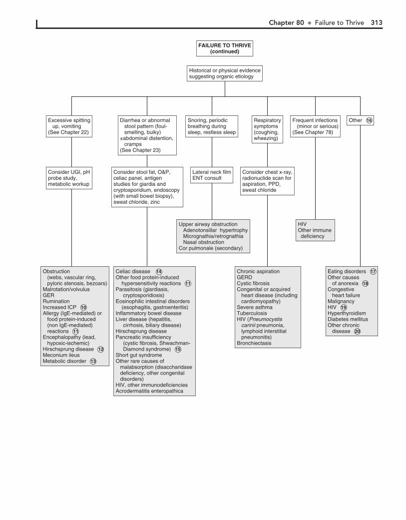
Chapter 80 u FailuretoThrive 313
FAILURE TO THRIVE(continued)
FAILURE TO THRIVE(continued)
Historical or physical evidencesuggesting organic etiologyHistorical or physical evidencesuggesting organic etiology
Obstruction (webs, vascular ring, pyloric stenosis, bezoars)Malrotation/volvulusGERRuminationIncreased ICP 10Allergy (IgE-mediated) or food protein-induced (non IgE-mediated) reactions 11Encephalopathy (lead, hypoxic-ischemic)Hirschsprung disease 12Meconium ileusMetabolic disorder 13
Obstruction (webs, vascular ring, pyloric stenosis, bezoars)Malrotation/volvulusGERRuminationIncreased ICP 10Allergy (IgE-mediated) or food protein-induced (non IgE-mediated) reactions 11Encephalopathy (lead, hypoxic-ischemic)Hirschsprung disease 12Meconium ileusMetabolic disorder 13
Celiac disease 14Other food protein-induced hypersensitivity reactions 11Parasitosis (giardiasis, cryptosporidiosis)Eosinophilic intestinal disorders (esophagitis, gastroenteritis)Inflammatory bowel diseaseLiver disease (hepatitis, cirrhosis, biliary disease)Hirschsprung diseasePancreatic insufficiency (cystic fibrosis, Shwachman- Diamond syndrome) 15Short gut syndromeOther rare causes of malabsorption (disaccharidase deficiency, other congenital disorders)HIV, other immunodeficienciesAcrodermatitis enteropathica
Celiac disease 14Other food protein-induced hypersensitivity reactions 11Parasitosis (giardiasis, cryptosporidiosis)Eosinophilic intestinal disorders (esophagitis, gastroenteritis)Inflammatory bowel diseaseLiver disease (hepatitis, cirrhosis, biliary disease)Hirschsprung diseasePancreatic insufficiency (cystic fibrosis, Shwachman- Diamond syndrome) 15Short gut syndromeOther rare causes of malabsorption (disaccharidase deficiency, other congenital disorders)HIV, other immunodeficienciesAcrodermatitis enteropathica
Upper airway obstruction Adenotonsillar hypertrophy Micrognathia/retrognathia Nasal obstructionCor pulmonale (secondary)
Upper airway obstruction Adenotonsillar hypertrophy Micrognathia/retrognathia Nasal obstructionCor pulmonale (secondary)
HIVOther immune deficiency
HIVOther immune deficiency
Eating disorders 17Other causes of anorexia 18 Congestive heart failureMalignancyHIV 19HyperthyroidismDiabetes mellitusOther chronic disease 20
Eating disorders 17Other causes of anorexia 18 Congestive heart failureMalignancyHIV 19HyperthyroidismDiabetes mellitusOther chronic disease 20
Chronic aspirationGERDCystic fibrosisCongenital or acquired heart disease (including cardiomyopathy)Severe asthmaTuberculosisHIV (Pneumocystis carinii pneumonia, lymphoid interstitial pneumonitis)Bronchiectasis
Chronic aspirationGERDCystic fibrosisCongenital or acquired heart disease (including cardiomyopathy)Severe asthmaTuberculosisHIV (Pneumocystis carinii pneumonia, lymphoid interstitial pneumonitis)Bronchiectasis
Consider UGI, pHprobe study,metabolic workup
Consider UGI, pHprobe study,metabolic workup
Consider stool fat, O&P, celiac panel, antigenstudies for giardia and cryptosporidium, endoscopy (with small bowel biopsy), sweat chloride, zinc
Consider stool fat, O&P, celiac panel, antigenstudies for giardia and cryptosporidium, endoscopy (with small bowel biopsy), sweat chloride, zinc
Lateral neck filmENT consultLateral neck filmENT consult
Consider chest x-ray,radionuclide scan foraspiration, PPD,sweat chloride
Consider chest x-ray,radionuclide scan foraspiration, PPD,sweat chloride
Excessive spitting up, vomiting(See Chapter 22)
Excessive spitting up, vomiting(See Chapter 22)
Diarrhea or abnormal stool pattern (foul- smelling, bulky)±abdominal distention, cramps(See Chapter 23)
Diarrhea or abnormal stool pattern (foul- smelling, bulky)±abdominal distention, cramps(See Chapter 23)
Snoring, periodicbreathing duringsleep, restless sleep
Snoring, periodicbreathing duringsleep, restless sleep
Respiratorysymptoms(coughing, wheezing)
Respiratorysymptoms(coughing, wheezing)
Frequent infections (minor or serious)(See Chapter 78)
Frequent infections (minor or serious)(See Chapter 78)
Other 16Other 16

314
Chapter 81
SLEEP DISTURBANCES
wake up for feeding. Bed sharing is controversial. In countries where bed sharing is common, it does not seem to be associated with sleep problems; studies in the United States suggest an as-sociation. Bed sharing is unsafe for younger children and has been associated with sudden infant death syndrome (SIDS) and other sleep-related causes of infant deaths. One must also deter-mine whether the bed sharing arrangement is due to the par-ents’ beliefs or a response to a sleep problem.
3 Obstructive sleep apnea is due to recurring episodes of upper airway obstruction and is associated with hypox-
emia and hypercapnia. There may be a history of snoring and mouth breathing, with episodes of apnea. It is often associated with airway obstruction due to hypertrophy of the tonsils and adenoids. Sleep apnea is associated with obesity and craniofa-cial abnormalities (micrognathia, Pierre-Robin syndrome). It may result in daytime hypersomnia and behavior problems. An overnight polysomnogram (sleep study) is helpful in making the diagnosis of obstructive sleep apnea.
4 Stimulant medications or caffeine-containing products are known to cause sleep problems. Additionally, corticoste-
roids may disrupt sleep. Some medications such as antihista-mines and anticonvulsants may cause excessive sleepiness during the day.
5 Sleep problems may be due to acute illness, such as otitis media, URI, and UTI. Other possible causes include teeth-
ing, food allergy, atopic disease, GER, and diaper dermatitis.
6 Injuries such as corneal abrasion, occult fracture, and those secondary to child abuse should be identified.
7 Children with neurodevelopmental disorders can have subclinical or nocturnal seizures, thereby causing sleep
disruptions. Children with blindness, cognitive delay, autism spectrum disorders, and some chromosomal syndromes (frag-ile X) often have sleep disturbances.
8 Behavioral insomnia of childhood includes the sleep-onset association type and the limit-setting type. Sleep-onset as-
sociation disorder describes a child who requires certain activi-ties to fall asleep, such as rocking, singing, playing, or feeding and does not develop the ability to self-soothe. Some associa-tions may be helpful in preparing the child for bed (reading); others may create problems (rocking) if the child tends to wake up and require the same behavior repeated to fall asleep. Trained night feeders are babies who have a prolonged need for nighttime feeding. Beyond 6 months of age, most night feeding may be considered a learned behavior. It is present in approxi-mately 5% of children aged 6 months to 3 years. It may be con-sidered a form of sleep-onset association disorder, because the child requires food or drink to return to sleep.
Bedtime struggles such as stalling and refusing to go to bed are more common in preschoolers and older children. They are often due to parental difficulties in setting limits such as consis-tent bedtimes and enforcing bedtime rules. The child’s tempera-ment plays an important role, with struggles for autonomy and attention-seeking behaviors further complicating the situation. Problems may begin after 6 to 9 months of age with the acquisi-tion of developmental skills such as object permanence, with consequent separation anxiety and the ability to pull to stand, which the child may use to avoid going to bed. Difficulty set-tling to sleep may occur during an acute illness and may persist after recovery.
Sleep problems or disturbances are a common concern of par-ents. They may be classified as dyssomnias (disturbance in amount or timing of sleep), parasomnias (abnormal sleep be-haviors such as nightmares and night terrors), and disruptions due to other physical or mental conditions. It is important to understand the normal maturation of the sleep cycle of new-borns, infants, and children. In the first year, infants often wake many times during the night, but they may not always cry. The average newborn sleeps about 16.5 hours a day, with relatively undifferentiated sleep cycles. Sleep/wake cycles become more clearly established by 3 to 4 months of age, with an increasing proportion of nighttime sleep. However, there is a great deal of individual variation. In evaluating sleep disorders, it is impor-tant to identify inappropriate parental expectations, excessive parental anxiety or stress, as well as parental behaviors that may reinforce the sleep problems.
1 History and physical examination should first of all iden-tify any underlying organic etiology for the sleep distur-
bance. This may be easier to identify in cases of an acute change in sleep due to illness or injury. A drug and medication history should include caffeine-containing beverages and over-the-counter medications such as cold preparations. Maternal be-haviors and attachment as well as psychosocial stress have an important role in sleep problems. Perinatal factors such as birth asphyxia and prematurity contribute to increased wakefulness. Children with developmental delay and autism commonly have sleep problems. The sleeping environment (e.g., temperature, noise, television in room) should also be assessed.
A screening tool (BEARS) is helpful in determining causes of sleep disturbances. It includes five sleep domains, each with trigger questions: Bedtime problems, Excessive daytime sleepi-ness, Awakenings during the night, Regularity and duration of sleep, and Snoring.
Snoring may indicate obstructive sleep apnea; other symp-toms may include choking and gasping. A history of repetitive kicking movements may suggest periodic leg movement disor-ders. An overnight sleep study may be indicated if history sug-gests obstructive sleep apnea, periodic leg movement disorders, or excessive daytime sleepiness.
2 If possible, parents should be asked to keep a sleep log. This should include the following information: the time
the child awoke in the morning, the time and duration of day-time naps, the time the child is put to bed at night, the time the child fell asleep, the time(s) awakened at night, and what the parent did. It is also important to review sleep practices and influences. The term sleep hygiene is used to describe habits acquired for readying for, falling, and staying asleep. Bedtime settling practices create sleep associations and include rituals and routines used, such as bedtime stories or songs, rocking, or comforting. The child’s temperament and ability to self-soothe (e.g., thumb sucking, transitional objects) are important in the ability to fall asleep and maintain sleep. Feeding practices play an important role. Breast-fed infants may be more likely to
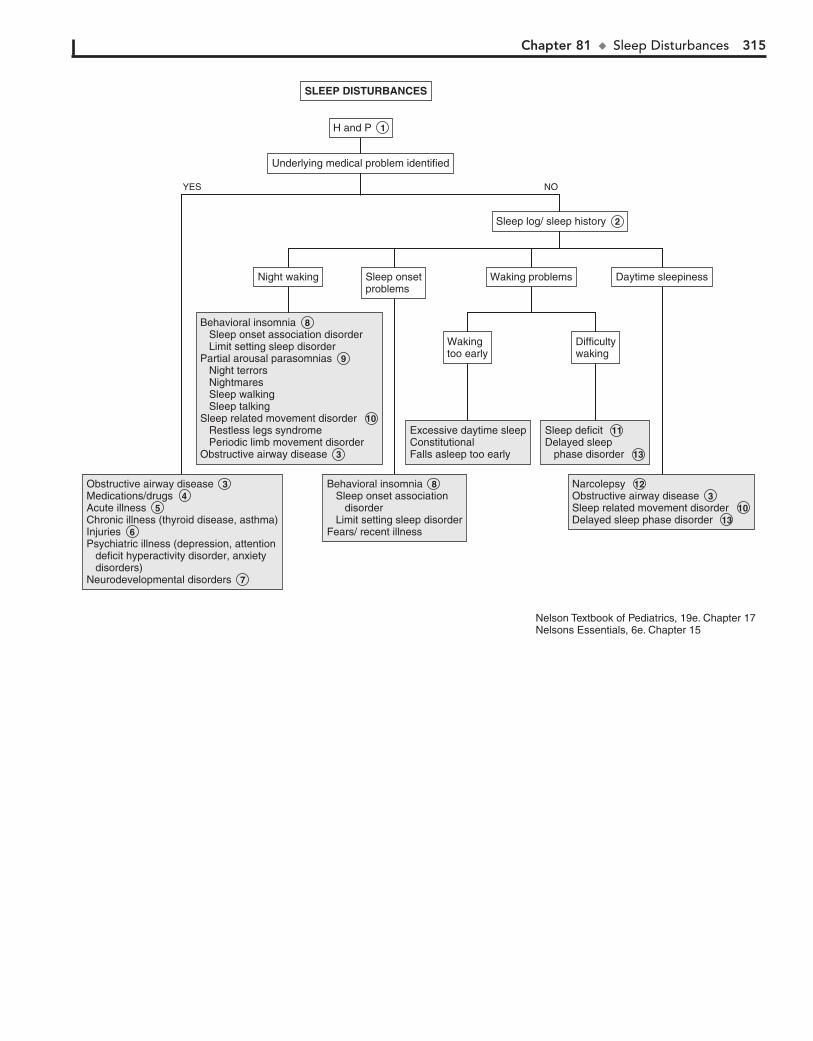
Chapter 81 u SleepDisturbances 315
SLEEP DISTURBANCESSLEEP DISTURBANCES
Sleep log/ sleep history 2 Sleep log/ sleep history 2
Underlying medical problem identifiedUnderlying medical problem identified
H and P 1 H and P 1
Obstructive airway disease 3 Medications/drugs 4 Acute illness 5 Chronic illness (thyroid disease, asthma)Injuries 6 Psychiatric illness (depression, attention deficit hyperactivity disorder, anxiety disorders)Neurodevelopmental disorders 7
Obstructive airway disease 3 Medications/drugs 4 Acute illness 5 Chronic illness (thyroid disease, asthma)Injuries 6 Psychiatric illness (depression, attention deficit hyperactivity disorder, anxiety disorders)Neurodevelopmental disorders 7
Behavioral insomnia 8 Sleep onset association disorder Limit setting sleep disorderPartial arousal parasomnias 9 Night terrors Nightmares Sleep walking Sleep talkingSleep related movement disorder 10 Restless legs syndrome Periodic limb movement disorderObstructive airway disease 3
Behavioral insomnia 8 Sleep onset association disorder Limit setting sleep disorderPartial arousal parasomnias 9 Night terrors Nightmares Sleep walking Sleep talkingSleep related movement disorder 10 Restless legs syndrome Periodic limb movement disorderObstructive airway disease 3
Behavioral insomnia 8 Sleep onset association disorder Limit setting sleep disorderFears/ recent illness
Behavioral insomnia 8 Sleep onset association disorder Limit setting sleep disorderFears/ recent illness
Excessive daytime sleepConstitutionalFalls asleep too early
Excessive daytime sleepConstitutionalFalls asleep too early
Sleep deficit 11 Delayed sleep phase disorder 13
Sleep deficit 11 Delayed sleep phase disorder 13
Narcolepsy 12 Obstructive airway disease 3 Sleep related movement disorder 10 Delayed sleep phase disorder 13
Narcolepsy 12 Obstructive airway disease 3 Sleep related movement disorder 10 Delayed sleep phase disorder 13
Wakingtoo earlyWakingtoo early
Night wakingNight waking Sleep onsetproblemsSleep onsetproblems
Waking problemsWaking problems Daytime sleepinessDaytime sleepiness
DifficultywakingDifficultywaking
NOYES
Nelson Textbook of Pediatrics, 19e. Chapter 17Nelsons Essentials, 6e. Chapter 15
316 Part XI u General
polysomnography. Both RLS and PLMD may be associated with low serum iron levels.
11 A child who has difficulty waking up in the morning often has a sleep deficit. This is most common in adolescents
because of increased activities and demands on their time. There is often an increased physiologic need for sleep at this time as well.
12 Narcolepsy is a rare disorder with a characteristic irre-sistible daytime sleepiness. It may be associated with
cataplexy (i.e., sudden loss of muscle tone with maintained consciousness) and rarely with hallucinations and sleep paralysis. It is rare in children; however, 25% of adults with narcolepsy report initial presentation during adolescence. Confirmation of the diagnosis requires referral to a sleep labo-ratory for polysomnography.
13 Delayed sleep phase disorder is due to a disorder of circa-dian rhythms and is common in adolescents. There is late
sleep onset with subsequent delayed wakening time. This causes difficulty adjusting to normal school or work times.
9 Parasomnias or disorders of arousal include night terrors and nightmares, as well as sleep walking and talking. Night
terrors occur early in the night during partial arousal from deep non–rapid eye movement (non-REM) sleep. The child may cry or scream and show signs of agitation; the child may appear disoriented or confused and seems unaware of the parent’s pres-ence. He or she may be difficult to arouse and on awakening usually has no memory of the event. This occurs most com-monly in children aged 2 to 6 years. Nightmares usually occur later during the night during REM sleep. The child usually re-members the dream vividly, seems upset on waking, but can be comforted by the parent. The peak age at onset of nightmares is 3 to 5 years, but they can occur at any age, presumably even in preverbal children. Sleep walking (somnambulism) occurs, like sleep terrors, during non-REM sleep. The most common age at onset is 4 to 8 years of age. Safety of the child is the primary concern with this disorder. Sleep talking (somniloquy) is not specific to any stage of sleep and has no clinical significance except that it may occur during nightmares or night terrors.
10 Restless legs syndrome (RLS) is a disorder characterized by uncomfortable sensations in the lower extremities, accom-
panied by a strong urge to move the legs. RLS is a clinical diag-nosis, and the patient is aware of the symptoms. With periodic limb movement disorder (PLMD), there are periodic repetitive limb jerks occurring at frequent intervals. These movements usually occur during sleep and involve the leg, with extension of the big toe and dorsiflexion of the ankle. The patient is unaware of these movements. The diagnosis may be confirmed by overnight
BibliographyAAP Task Force on Sudden Infant Death Syndrome: SIDS and other sleep-
related infant deaths: Expansion of recommendations for a safe infant sleeping environment, Pediatrics 128:e1341–e1367, 2011.
Bhargave S: Diagnosis and management of common sleep problems in children, Pediatr Rev 32:91–99, 2011.
Owens JA, Dalzell J: Use of the ‘BEARS’ sleep screening tool in a pediatric residents’ continuity clinic: A pilot study, Sleep Med 6:63–69, 2005.

Fluids and Electrolytes PART
XII
318
Chapter 82
ACIDEMIA 4 The anion gap (AG) reflects unmeasured anions, which, in
combination with bicarbonate (HCO3–) and chloride (Cl–),
counterbalance the positive charge of the sodium (Na1) ions.
AG 5 Na1 – (Cl– 1 HCO3–)
The normal anion gap is 4 to 11 mEq/L, although variations exist among laboratories. Elevation occurs secondary to an excess accu-mulation of acids (endogenous or ingested) or inadequate excretion of acids. Hyperchloremia occurs in a metabolic acidosis with a normal anion gap. An anion gap lower than expected may occur in the presence of hyperkalemia, hypercalcemia, hypoalbuminemia, hypermagnesemia, bromide intoxication, or laboratory error.
5 Ingestions should be suspected in young children or depressed adolescents at risk for suicide with an acute
onset of symptoms (usually multisystemic), a history of previ-ous accidental ingestions, or an altered level of consciousness.
6 A difference of greater than 10 to 15 mOsm/L between the measured serum osmolality and calculated serum osmo-
lality suggests unmeasured osmotic particles (e.g., methanol, ethylene glycol).
Calculated serum osmolality 5 2 [serum Na1 1 K1] 1 BUN/2.8 1 glucose/18
7 Ethylene glycol (radiator antifreeze) ingestion causes neurologic symptoms, respiratory failure, cardiovascular
collapse, and renal failure. The increased anion gap is due to the accumulation of the metabolite formic acid. Lactic acidosis also frequently occurs.
8 Methanol (wood alcohol) ingestion causes GI and neuro-logic symptoms. A severe blinding retinitis can also occur.
The anion gap is due to the metabolites glyoxylic acid, formic acid, and oxalic acid. Lactic acidosis also occurs.
9 Salicylate ingestion in children is characterized by neu-rologic symptoms (e.g., coma, altered mental status,
seizures) and hyperventilation. Fever may be prominent in infants. Younger children are more susceptible to metabolic acidosis. Respiratory alkalosis is the predominant acid-base abnormality in older patients.
10 Starvation, glycogen storage disease (type 1), and inborn errors of amino acid or organic acid metabolism are less
common causes of ketoacidosis.
11 Lactic acidosis with blood lactate levels of 5 mEq/L or higher most commonly is due to hypotension, hypovole-
mia, or sepsis. Other causes include exercise, ethanol ingestion, and inborn errors of metabolism, particularly mitochondrial and disorders of carbohydrate metabolism.
12 An increased anion gap in renal failure occurs because impaired H1 excretion prevents appropriate regeneration of
HCO3–. Anions such as sulfate and phosphate also accumulate.
Acidemia is a blood pH less than 7.35. Excessive acidity of body fluids (acidosis) may be acute or chronic, primarily metabolic (decreased plasma bicarbonate), or primarily respiratory (increased partial pressure of CO2). It may occur as part of a mixed acid-base disorder.
1 In acute acidosis, inquire about diarrhea or other GI losses, medications, and possibility of ingestions.
In young infants a history of poor feeding, failure to thrive, vomiting, lethargy, or seizures may indicate an inborn error of metabolism. Poor growth may be a clue to RTA in older children. Nonspecific symptoms of acidosis may include hyperventilation and Kussmaul breathing (i.e., deep, rapid respirations). Unusual breath odors may suggest diabetic ketoacidosis or ingestions.
2 The diagnosis is made by laboratory values. The pH reflecting extracellular free hydrogen ion [H1] concentra-
tion, partial pressure of CO2 (PCO2), and plasma bicarbonate level (HCO3
–) are considered in clinical acid-base disorders. Their relationship is described by this modified version of the Henderson-Hasselbalch equation:
[H1] 5 24 3 PCO2/HCO3–
When the HCO3– level is decreased, an increased respiratory
rate occurs as a compensatory mechanism to try to maintain a normal pH. Renal compensation (i.e., secretion of excess H1 and reabsorption of bicarbonate) is necessary to ultimately cor-rect the acidosis, because the lungs cannot create an absolute loss or gain of H1 ions.
In mixed acid-base disorders, a combination of simple disor-ders occurs, such as in the child with chronic lung disease who experiences a combined metabolic alkalosis and respiratory acidosis. Guidelines exist for expected renal and respiratory compensation of acidemia. Mixed disorders should be sus-pected when the compensatory response differs from the pre-dicted response. Compensation never overcorrects the pH and rarely corrects the pH to normal values.
Urine pH and urine electrolytes should be included in the preliminary analysis if RTA is suspected. A serum osmolality value will aid in narrowing the diagnosis of a metabolic acidosis with an increased anion gap.
3 Neuromuscular disorders that can result in a respiratory acidosis include brainstem or spinal cord disorders, in-
cluding tumors, Guillain-Barré syndrome, polio, myasthenia gravis, muscular dystrophy, botulism, encephalopathy, and drugs (e.g., depressants, sedatives).

Chapter 82 u Acidemia 319
Airway obstruction (laryngeal edema, croup, epiglottitis, foreign body)AspirationSevere bronchospasmCystic fibrosisBronchopulmonary dysplasiaInterstitial lung diseasePneumoniaPulmonary edemaPleural effusionPneumothoraxThoracic cage abnormalitiesPickwickian syndromeNeuromuscular diseases 3 CNS depression
Airway obstruction (laryngeal edema, croup, epiglottitis, foreign body)AspirationSevere bronchospasmCystic fibrosisBronchopulmonary dysplasiaInterstitial lung diseasePneumoniaPulmonary edemaPleural effusionPneumothoraxThoracic cage abnormalitiesPickwickian syndromeNeuromuscular diseases 3 CNS depression
Ethylene glycol ingestion 7 Methanol ingestion 8 Ethylene glycol ingestion 7 Methanol ingestion 8
Ingestions Paraldehyde Salicylate 9 MetforminKetoacidosis 10 (diabetic ketoacidosis)Lactic acidosis 11 Organic acidemias (e.g., methylmalonic acidemia)Propionyl-CoA carboxylase deficiencyMethionine administrationRenal failure 12
Ingestions Paraldehyde Salicylate 9 MetforminKetoacidosis 10 (diabetic ketoacidosis)Lactic acidosis 11 Organic acidemias (e.g., methylmalonic acidemia)Propionyl-CoA carboxylase deficiencyMethionine administrationRenal failure 12
Normal osmolal gapNormal osmolal gapElevated osmolal gapElevated osmolal gap
Calculate osmolal gap 6 Calculate osmolal gap 6
Suspect toxicingestion 5 Suspect toxicingestion 5
Metabolic acidosis with elevated AGMetabolic acidosis with elevated AG
Calculate AG 4 Calculate AG 4 RespiratoryacidosisRespiratoryacidosis
See next pageSee next page
Metabolic acidosis with normal AGMetabolic acidosis with normal AG
PCO2 >40PCO2 >40
ABGSerum electrolytes±Urine pH, urine electrolytes, serum osmolality 2
ABGSerum electrolytes±Urine pH, urine electrolytes, serum osmolality 2
PerformH and P 1 PerformH and P 1
ACIDEMIAACIDEMIA
YES NO
YES NO
Nelson Textbook of Pediatrics, 19e. Chapter 52
320 Part XII u FluidsandElectrolytes
13 Hyperkalemia can develop without the effect of aldoste-rone. It may be due to primary aldosterone deficiency or
result from acquired kidney disease, resulting in low renin levels.
14 Type IV RTA is characterized by a deficiency of or impaired distal tubular response to aldosterone.
15 In pseudohypoaldosteronism, both renin and aldosterone levels are elevated, but the response of the distal tubule to
the aldosterone is impaired.
16 Certain medications (e.g., potassium-sparing diuretics, nonsteroidal antiinflammatory drugs, b-blocking agents,
angiotensin-converting enzyme inhibitors, cyclosporine) can cause a functional hypoaldosteronism.
17 Urine pH should be measured on a fresh urine specimen. Serum electrolytes should be collected at the same time for
the HCO3– level. The urine specimen can be taken at any time
but should be obtained while the patient is still acidotic.The urine pH reflects only free H1 (,1% of the total H1
excreted). It is often helpful to consider it in conjunction with a measure of net acid excretion, primarily urine ammonium con-centration. Synthesis of ammonia (NH3) and its subsequent excretion as ammonium (NH4
1) are increased in most cases of systemic acidosis. The urinary anion gap is used to estimate the production of ammonium ion:
Urine AG 5 (Urine Na1 1 Urine K1) – Urine Cl–
The lower or more negative the gap, the greater the acid secretion in the form of NH4
1.
18 RTA is characterized by an inability to adequately acidify the urine in the presence of a normal anion gap (hyper-
chloremic) acidosis. It may occur primarily or in association with multiple inherited disorders or acquired systemic disor-ders. In the distal and proximal subtypes, volume contraction results in an aldosterone-mediated hypokalemia.
In distal (type I or classic) RTA, a permanent deficiency of H1 secretion by the distal tubules results in wasting of a portion of the filtered load of HCO3
–. The HCO3– wasting persists even
in the presence of severe systemic acidosis; urine pH is never more acidic than 5.8. The urine anion gap is a small or low negative value.
In proximal RTA (type II or HCO3– wasting) a defect in the
proximal tubular reabsorption of HCO3– results in a higher load
of HCO3– being presented to the distal tubule. In mild acidosis,
this excess HCO3– is lost in the urine because its amount
exceeds the capacity of the distal tubule for reabsorption in the normal distal tubule. In more severe acidosis a lower amount of HCO3
– is filtered and is able to be reabsorbed in the normal distal tubule. Because distal acidification mechanisms are intact in proximal RTA, urine pH will become more acidic (pH ,5.5) under these circumstances. The urine anion gap is a high nega-tive value, regardless of the severity or duration of the acidosis. Accompanying hypokalemia can be severe.
19 Rapid volume expansion with non–HCO3–-containing
solutions can cause transient small HCO3– losses in the
urine.
20 Premature infants and neonates with renal function that is normal for maturation may demonstrate a mild metabolic
acidosis due to a transient reduced threshold for HCO3– reab-
sorption. This normally corrects by 4 to 6 weeks in the prema-ture infant and by 3 weeks in the term infant.
21 Increased HCO3– losses in diarrhea can contribute to a
metabolic acidosis.
BibliographyBrewer ED: Disorders of acid-base balance, Pediatr Clin North Am 37:
429–447, 1990.Hanna JD, Scheinman JI, Chan JC: The kidney in acid-base balance, Pediatr
Clin North Am 42:1365–1395, 1995.
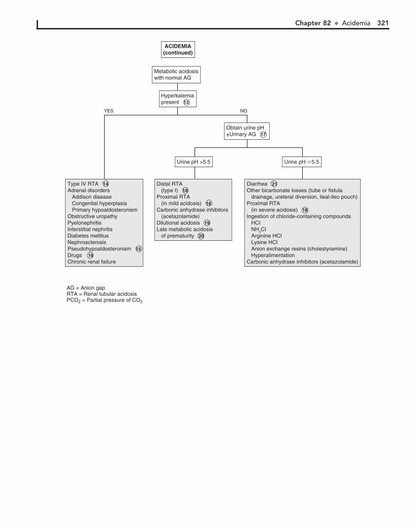
Chapter 82 u Acidemia 321
Type IV RTA 14 Adrenal disorders Addison disease Congenital hyperplasia Primary hypoaldosteronismObstructive uropathyPyelonephritisInterstitial nephritisDiabetes mellitusNephrosclerosisPseudohypoaldosteronism 15Drugs 16 Chronic renal failure
Type IV RTA 14 Adrenal disorders Addison disease Congenital hyperplasia Primary hypoaldosteronismObstructive uropathyPyelonephritisInterstitial nephritisDiabetes mellitusNephrosclerosisPseudohypoaldosteronism 15Drugs 16 Chronic renal failure
Distal RTA (type I) 18Proximal RTA (in mild acidosis) 18Carbonic anhydrase inhibitors (acetazolamide)Dilutional acidosis 19Late metabolic acidosis of prematurity 20
Distal RTA (type I) 18Proximal RTA (in mild acidosis) 18Carbonic anhydrase inhibitors (acetazolamide)Dilutional acidosis 19Late metabolic acidosis of prematurity 20
Diarrhea 21Other bicarbonate losses (tube or fistula drainage, ureteral diversion, ileal-ileo pouch)Proximal RTA (in severe acidosis) 18Ingestion of chloride-containing compounds HCI NH4CI Arginine HCI Lysine HCI Anion exchange resins (cholestyramine) HyperalimentationCarbonic anhydrase inhibitors (acetazolamide)
Diarrhea 21Other bicarbonate losses (tube or fistula drainage, ureteral diversion, ileal-ileo pouch)Proximal RTA (in severe acidosis) 18Ingestion of chloride-containing compounds HCI NH4CI Arginine HCI Lysine HCI Anion exchange resins (cholestyramine) HyperalimentationCarbonic anhydrase inhibitors (acetazolamide)
Urine pH �5.5Urine pH �5.5Urine pH >5.5Urine pH >5.5
Obtain urine pH±Urinary AG 17 Obtain urine pH±Urinary AG 17
Hyperkalemiapresent 13 Hyperkalemiapresent 13
Metabolic acidosiswith normal AGMetabolic acidosiswith normal AG
ACIDEMIA(continued)ACIDEMIA(continued)
YES NO
AG = Anion gapRTA = Renal tubular acidosisPCO2 = Partial pressure of CO2
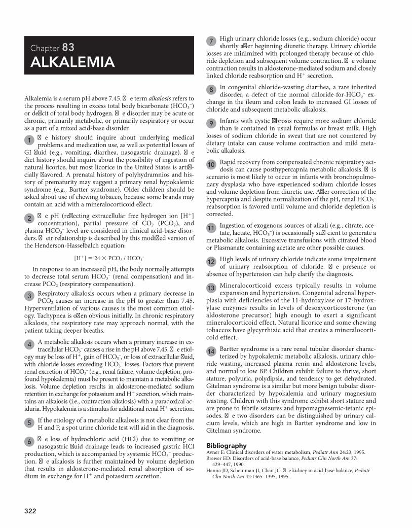
322
Chapter 83
ALKALEMIA
7 High urinary chloride losses (e.g., sodium chloride) occur shortly after beginning diuretic therapy. Urinary chloride
losses are minimized with prolonged therapy because of chlo-ride depletion and subsequent volume contraction. The volume contraction results in aldosterone-mediated sodium and closely linked chloride reabsorption and H1 secretion.
8 In congenital chloride-wasting diarrhea, a rare inherited disorder, a defect of the normal chloride-for-HCO3
– ex-change in the ileum and colon leads to increased GI losses of chloride and subsequent metabolic alkalosis.
9 Infants with cystic fibrosis require more sodium chloride than is contained in usual formulas or breast milk. High
losses of sodium chloride in sweat that are not countered by dietary intake can cause volume contraction and mild meta-bolic alkalosis.
10 Rapid recovery from compensated chronic respiratory aci-dosis can cause posthypercapnia metabolic alkalosis. This
scenario is most likely to occur in infants with bronchopulmo-nary dysplasia who have experienced sodium chloride losses and volume depletion from diuretic use. After correction of the hypercapnia and despite normalization of the pH, renal HCO3
– reabsorption is favored until volume and chloride depletion is corrected.
11 Ingestion of exogenous sources of alkali (e.g., citrate, ace-tate, lactate, HCO3
–) is occasionally sufficient to generate a metabolic alkalosis. Excessive transfusions with citrated blood or Plasmanate containing acetate are other possible causes.
12 High levels of urinary chloride indicate some impairment of urinary reabsorption of chloride. The presence or
absence of hypertension can help clarify the diagnosis.
13 Mineralocorticoid excess typically results in volume expansion and hypertension. Congenital adrenal hyper-
plasia with deficiencies of the 11-hydroxylase or 17-hydrox-ylase enzymes results in levels of desoxycorticosterone (an aldosterone precursor) high enough to exert a significant mineralocorticoid effect. Natural licorice and some chewing tobaccos have glycyrrhizic acid that creates a mineralocorti-coid effect.
14 Bartter syndrome is a rare renal tubular disorder charac-terized by hypokalemic metabolic alkalosis, urinary chlo-
ride wasting, increased plasma renin and aldosterone levels, and normal to low BP. Children exhibit failure to thrive, short stature, polyuria, polydipsia, and tendency to get dehydrated. Gitelman syndrome is a similar but more benign tubular disor-der characterized by hypokalemia and urinary magnesium wasting. Children with this syndrome exhibit short stature and are prone to febrile seizures and hypomagnesemic-tetanic epi-sodes. The two disorders can be distinguished by urinary cal-cium levels, which are high in Bartter syndrome and low in Gitelman syndrome.
Alkalemia is a serum pH above 7.45. The term alkalosis refers to the process resulting in excess total body bicarbonate (HCO3
–) or deficit of total body hydrogen. The disorder may be acute or chronic, primarily metabolic, or primarily respiratory or occur as a part of a mixed acid-base disorder.
1 The history should inquire about underlying medical problems and medication use, as well as potential losses of
GI fluid (e.g., vomiting, diarrhea, nasogastric drainage). The diet history should inquire about the possibility of ingestion of natural licorice, but most licorice in the United States is artifi-cially flavored. A prenatal history of polyhydramnios and his-tory of prematurity may suggest a primary renal hypokalemic syndrome (e.g., Bartter syndrome). Older children should be asked about use of chewing tobacco, because some brands may contain an acid with a mineralocorticoid effect.
2 The pH (reflecting extracellular free hydrogen ion [H1] concentration), partial pressure of CO2 (PCO2), and
plasma HCO3– level are considered in clinical acid-base disor-
ders. Their relationship is described by this modified version of the Henderson-Hasselbalch equation:
[H1] 5 24 3 PCO2 / HCO3–
In response to an increased pH, the body normally attempts to decrease total serum HCO3
– (renal compensation) and in-crease PCO2 (respiratory compensation).
3 Respiratory alkalosis occurs when a primary decrease in PCO2 causes an increase in the pH to greater than 7.45.
Hyperventilation of various causes is the most common etiol-ogy. Tachypnea is often obvious initially. In chronic respiratory alkalosis, the respiratory rate may approach normal, with the patient taking deeper breaths.
4 A metabolic alkalosis occurs when a primary increase in ex-tracellular HCO3
– causes a rise in the pH above 7.45. The etiol-ogy may be loss of H1, gain of HCO3
–, or loss of extracellular fluid, with chloride losses exceeding HCO3
– losses. Factors that prevent renal excretion of HCO3
– (e.g., renal failure, volume depletion, pro-found hypokalemia) must be present to maintain a metabolic alka-losis. Volume depletion results in aldosterone-mediated sodium retention in exchange for potassium and H1 secretion, which main-tains an alkalosis (i.e., contraction alkalosis) with a paradoxical ac-iduria. Hypokalemia is a stimulus for additional renal H1 secretion.
5 If the etiology of a metabolic alkalosis is not clear from the H and P, a spot urine chloride test will aid in the diagnosis.
6 The loss of hydrochloric acid (HCl) due to vomiting or nasogastric fluid drainage leads to increased gastric HCl
production, which is accompanied by systemic HCO3– produc-
tion. The alkalosis is further maintained by volume depletion that results in aldosterone-mediated renal absorption of so-dium in exchange for H1 and potassium secretion.
BibliographyAvner E: Clinical disorders of water metabolism, Pediatr Ann 24:23, 1995.Brewer ED: Disorders of acid-base balance, Pediatr Clin North Am 37:
429–447, 1990.Hanna JD, Scheinman JI, Chan JC: The kidney in acid-base balance, Pediatr
Clin North Am 42:1365–1395, 1995.

Chapter 83 u Alkalemia 323
Psychogenic/anxietyFeverSepsisHigh altitudePneumoniaPulmonary emboliCongestive heart failure with hypoxemiaCNS disordersLiver failureSalicylates, other medicationsMechanical ventilation
Psychogenic/anxietyFeverSepsisHigh altitudePneumoniaPulmonary emboliCongestive heart failure with hypoxemiaCNS disordersLiver failureSalicylates, other medicationsMechanical ventilation
Vomiting 6 Pyloric stenosisNasogastric drainage 6 Diuretics (prolonged use) 7 Congenital chloride losing diarrhea 8 Laxative abuseLow chloride intake (cystic fibrosis) 9 Resolving chronic hypercapnia 10 Exogenous alkali 11
Vomiting 6 Pyloric stenosisNasogastric drainage 6 Diuretics (prolonged use) 7 Congenital chloride losing diarrhea 8 Laxative abuseLow chloride intake (cystic fibrosis) 9 Resolving chronic hypercapnia 10 Exogenous alkali 11
Excess mineralocorticoid effect 13 Primary aldosteronism Renal artery stenosis Renin-secreting tumors Cushing's syndrome Congenital adrenal hyperplasia (17-hydroxylase deficiency and 11�-hydroxylase deficiency) Licorice or chewing tobacco Liddle's syndrome
Excess mineralocorticoid effect 13 Primary aldosteronism Renal artery stenosis Renin-secreting tumors Cushing's syndrome Congenital adrenal hyperplasia (17-hydroxylase deficiency and 11�-hydroxylase deficiency) Licorice or chewing tobacco Liddle's syndrome
Bartter's syndrome 14 Gitelman's syndrome 14 Diuretics (recent use) 7 Magnesium depletion
Bartter's syndrome 14 Gitelman's syndrome 14 Diuretics (recent use) 7 Magnesium depletion
Urine chloride>10 mEq/lUrine chloride>10 mEq/l
Urine chloride<10 mEq/lUrine chloride<10 mEq/l
HypertensionpresentHypertensionpresent
PCO2 <40PCO2 <40
Blood gasElectrolytesCreatinine±spot urine chloride 2
Blood gasElectrolytesCreatinine±spot urine chloride 2
Obtain spoturine chloride 5 Obtain spoturine chloride 5
Metabolic alkalosis 4 Metabolic alkalosis 4 Respiratoryalkalosis 3 Respiratoryalkalosis 3
PerformH and P 1 PerformH and P 1
ALKALEMIAALKALEMIA
YES NO
YES NO
PCO2 = Partial pressure of CO2 Nelson Textbook of Pediatrics, 19e. Chapter 52

324
Chapter 84
HYPERNATREMIA
(increased free water insensible losses), or hypertonic fluids are being given.
4 Insensible water losses from the respiratory tract occur secondary to hyperventilation or respiratory distress. Der-
mal losses are common in infants placed on a radiant warmer and in settings of increased ambient temperature.
5 Although adipsia is rare, it can occur as a primary entity or secondary to hypothalamic lesions, hydrocephalus, or
head trauma.
6 Hypotonic urine and polyuria in the presence of hyperna-tremia suggest DI. DI, which presents with polyuria and
polydipsia, represents an inability to concentrate urine because of either a lack of ADH production (central DI) or renal resis-tance to ADH (nephrogenic DI). DI is an inability to effectively conserve urinary water. Hypernatremia in DI most often occurs when access to water is restricted. Because young children do not usually have control over their fluid intake, most pediatric cases of DI occur as hypovolemia.
7 A water deprivation test should be carefully monitored in the hospital and should be done during daytime hours
rather than overnight. During water deprivation, the body weight should not be allowed to decline by more than 3%.
8 The response to intramuscular aqueous vasopressin (0.1-0.2 units/kg) will help confirm the diagnosis of DI
and distinguish between central and renal causes.
9 Central DI is probably the diagnosis if, after administra-tion of vasopressin, the urine/serum ratio becomes greater
than 1. Psychogenic polydipsia usually does not result in hy-pernatremia. If the administration of vasopressin produces a higher urinary concentration than dehydration does, DI is the diagnosis regardless of the level of urinary concentration. Psy-chogenic polydipsia usually does not result in hypernatremia.
Central DI may be primary (familial or nonfamilial) or sec-ondary to head trauma or several disease states (suprasellar or intrasellar tumors, granulomatous disease, histiocytosis, CNS infection or hemorrhage). ADH release can also be inhibited by stress (e.g., pain from surgery or trauma) and certain drugs and medications (e.g., a-adrenergic agonists, alcohol, opiate antago-nists, phenytoin, clonidine) and carbon monoxide poisoning. The disorder has also been associated with cleft lip and palate.
10 Nephrogenic DI may be congenital or secondary to mul-tiple types of renal disease. Males with primary nephro-
genic DI are likely to have a significant history of polyuria, polydipsia, and previous episodes of hypernatremic dehydra-tion. Females with a primary defect have milder symptoms and tend to be diagnosed later in life. It can also be caused by drugs or medications (e.g., lithium, demeclocycline, methoxyflurane, amphotericin B, cyclophosphamide, propoxyphene, cisplatin, angiographic dyes, osmotic diuretics), hypokalemia, and hyper-calcemia.
Sodium (Na1) is the primary cation of the ECF compartment and is the major osmotically active solute in the ECF. Osmolal-ity is the measurement of the number of solute particles in a unit of volume. It can be measured or estimated by the formula:
Calculated serum osmolality 5 2 [serum Na1 1 K1] 1 BUN/2.8 1 glucose/18
The body responds to changes in osmolality by increasing or suppressing thirst and antidiuretic hormone (ADH) release.
1 Signs and symptoms of hypernatremia are nonspecific. Thirst will be increased as long as the brain’s thirst centers
are intact. Excessive thirst to the point it disrupts sleep or play and enuresis may be a clue to diabetes. Neurologic symptoms (e.g., irritability, lethargy, confusion, seizures) may be present in severe cases.
Inquire about volume losses (diarrhea, vomiting), oral in-take, and urine output. Specifically ask about formula prepara-tion and the possibility of excessive salt intake (e.g., of table salt or sea water). Polyuria and polydipsia may suggest diabetes mellitus (DM) or insipidus (DI). Infants with fever, very low-birth-weight infants, and children with cystic fibrosis or heat stroke are at risk for hypernatremia because of excessive (hypo-tonic) sweat losses. The medical history should also inquire about renal disease and CNS disease or hemorrhage, which may be a risk factor for central DI.
2 The approach to hypernatremia is best initiated with an assessment of the patient’s volume status. Hypovolemia
manifests as lethargy, dry mucous membranes, and decreased skin turgor. Infants will exhibit decreased tearing and a sunken fontanel. Tachycardia, orthostatic hypotension, and oliguria are also common. Fever may occur as both a cause and an effect of hypernatremic dehydration. The signs of ECF volume losses are less pronounced in hypernatremic dehydration than in hypona-tremic dehydration.
Patients appear to have a normal volume status (euvolemia) when total body water is decreased in the presence of normal or near-normal sodium content. This situation occurs secondary to inadequate water intake or solute-free water losses. These water losses may be extrarenal or renal (DI). Extrarenal losses will produce hypertonic urine. Urine sodium may be variable. Renal losses result in hypotonic urine.
Hypervolemia occurs when total body water is essentially normal in the presence of increased total body sodium. Mani-festations of excess ECF volume include edema and congestive heart failure.
3 Diarrhea is most likely to lead to hyponatremic or isonatremic dehydration; hypernatremic dehydration is
likely if fluid intake is low (vomiting, anorexia), fever is present
BibliographyAvner E: Clinical disorders of water metabolism, Pediatr Ann 24:23, 1995.Conley SB: Hypernatremia, Pediatr Clin North Am 37:365–372, 1990.
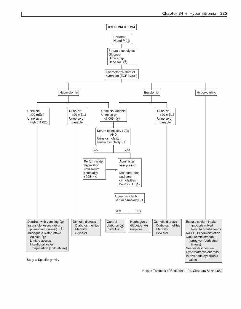
Chapter 84 u Hypernatremia 325
Diarrhea with vomiting 3 Insensible losses (fever, pulmonary, dermal) 4 Inadequate water intake Adipsia 5 Limited access Intentional water deprivation (child abuse)
Diarrhea with vomiting 3 Insensible losses (fever, pulmonary, dermal) 4 Inadequate water intake Adipsia 5 Limited access Intentional water deprivation (child abuse)
Osmotic diuresis Diabetes mellitus Mannitol Glycerol
Osmotic diuresis Diabetes mellitus Mannitol Glycerol
Nephogenic diabetes 10 insipidus
Nephogenic diabetes 10 insipidus
Osmotic diuresis Diabetes mellitus Mannitol Glycerol
Osmotic diuresis Diabetes mellitus Mannitol Glycerol
Excess sodium intake Improperly mixed formula or tube feedsNa HCO3 administrationNaCI administration (caregiver-fabricated illness)Sea water ingestionHypernatremic enemasIntravenous hypertonic saline
Excess sodium intake Improperly mixed formula or tube feedsNa HCO3 administrationNaCI administration (caregiver-fabricated illness)Sea water ingestionHypernatremic enemasIntravenous hypertonic saline
Central diabetes 9 insipidus
Central diabetes 9 insipidus
Characterize state ofhydration (ECF status)Characterize state ofhydration (ECF status)
Urine osmolality:serum osmolality >1Urine osmolality:serum osmolality >1
Administer vasopressin
Measure urine and serumosmolalities hourly × 4 8
Administer vasopressin
Measure urine and serumosmolalities hourly × 4 8
Urine Na variableUrine sp gr <1.005 6
Urine Na variableUrine sp gr <1.005 6
Urine Na <20 mEq/lUrine sp gr high (>1.025)
Urine Na <20 mEq/lUrine sp gr high (>1.025)
Urine Na >20 mEq/lUrine sp gr variable
Urine Na >20 mEq/lUrine sp gr variable
HypovolemicHypovolemic EuvolemicEuvolemic HypervolemicHypervolemic
Urine Na >20 mEq/lUrine sp gr variable
Urine Na >20 mEq/lUrine sp gr variable
Perform water deprivation until serumosmolality >295 7
Perform water deprivation until serumosmolality >295 7
Serum osmolality >295 ANDUrine osmolality:serum osmolality <1
Serum osmolality >295 ANDUrine osmolality:serum osmolality <1
PerformH and P 1 PerformH and P 1
Serum electrolytesGlucoseUrine sp grUrine Na 2
Serum electrolytesGlucoseUrine sp grUrine Na 2
HYPERNATREMIAHYPERNATREMIA
YES NO
YESNO
Sp gr = Specific gravity
Nelson Textbook of Pediatrics, 19e. Chapters 52 and 552

326
Chapter 85
HYPONATREMIA
clinical clues suggestive of diabetes mellitus (e.g., polyuria, polydipsia, weight loss, hyperglycemia) or a possible ingestion should prompt consideration of nonhypotonic states.
4 Older methods of electrolyte measurement determined sodium in mEq/L of plasma, as opposed to plasma water,
which could yield an artificially low sodium value (pseudohy-ponatremia) in the presence of hyperlipidemia (nephrotic syndrome) or hyperproteinemia, which is rare in children. Newer methods use ion-selective electrodes to measure serum sodium activity directly in plasma water and do not produce this artifact.
5 Hyponatremia occurs in a hypertonic setting when an osmotically active solute has been added to the ECF.
A difference of greater than 10 to 15 mOsm/L between the measured serum osmolality and the calculated serum osmo-lality is suggestive of a nonglucose solute in the ECF and may be the first clue to certain ingestions (e.g., of methanol, ethanol).
6 Assessment of the patient’s overall volume status is essen-tial in the evalua tion of hyponatremia.
Hypovolemia (dehydration) manifests as lethargy, dry mu-cous membranes, and decreased skin turgor. Infants will exhibit decreased tearing and a sunken fontanel. Tachycardia, ortho-static hypotension, and oliguria are also common.
Patients appear to have a normal volume status (euvolemia) when total body sodium is normal or near normal in the presence of a slight excess of body water. Most patients pre-senting with euvolemic hyponatremia have a syndrome of inappropriate antidiuretic hormone secretion (SIADH). Although characterized as euvolemic, many of these patients will have a slightly increased but clinically unimportant ECF volume.
Hypervolemia occurs when total body water is increased to a greater degree than total body sodium in either edema-forming states or renal failure.
Sodium (Na1) is the primary cation of the ECF compartment and is the major osmotically active solute in the ECF. Osmolal-ity is the measurement of the number of solute particles in a unit of volume. It can be measured or estimated by the formula:Calculated serum osmolality 5 2 [serum Na1 1 K1]
1 BUN/2.8 1 glucose/18
The body responds to changes in osmolality by increasing or suppressing thirst and antidiuretic hormone (ADH) release.
1 The history should inquire about GI losses (e.g., vomiting, diarrhea). Ask about fluid intake, urine output, medica-
tions, and possibility of a toxic ingestion. For infants, inquire specifically about formula preparation and amount of free water ingested. Ingestions should be suspected in young children aged 1 to 5 years with an acute onset of symptoms, a history of previous accidental ingestions, neurologic symptoms, or an unusual breath odor. Signs of hyponatremia usually manifest when the sodium level falls rapidly below 120 mEq/L. Signs and symptoms may include apathy, anorexia, nausea, vomiting, altered mental status, and seizures. Musculoskeletal symptoms include cramps and weakness.
2 If the preliminary laboratory evaluation reveals a signifi-cantly elevated BUN and Cr value not believed to be
caused by dehydration, a pediatric nephrologist should be consulted for further evaluation of presumed renal failure.
3 Hyponatremia most commonly occurs in conjunction with a hypotonic state. The presence of lipemic serum or
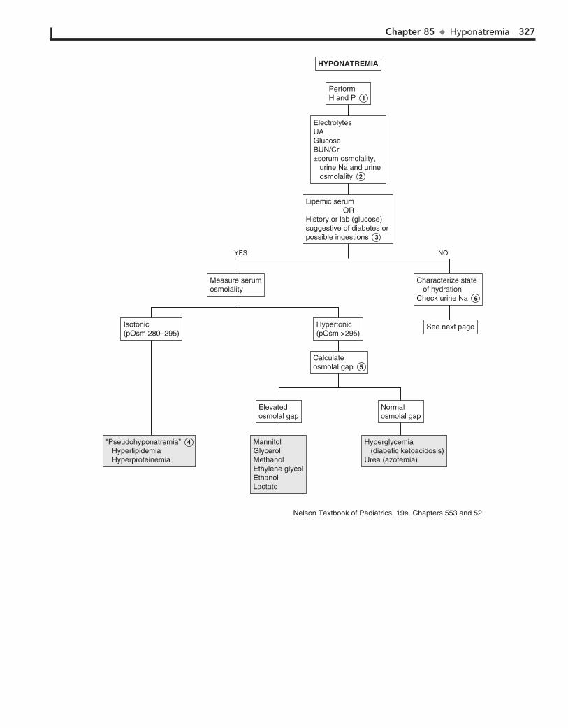
Chapter 85 u Hyponatremia 327
"Pseudohyponatremia” 4 Hyperlipidemia Hyperproteinemia
"Pseudohyponatremia” 4 Hyperlipidemia Hyperproteinemia
MannitolGlycerolMethanolEthylene glycolEthanolLactate
MannitolGlycerolMethanolEthylene glycolEthanolLactate
Hyperglycemia (diabetic ketoacidosis)Urea (azotemia)
Hyperglycemia (diabetic ketoacidosis)Urea (azotemia)
Hypertonic(pOsm >295)Hypertonic(pOsm >295)
Measure serumosmolalityMeasure serumosmolality
Characterize state of hydrationCheck urine Na 6
Characterize state of hydrationCheck urine Na 6
See next pageSee next pageIsotonic(pOsm 280–295)Isotonic(pOsm 280–295)
Calculateosmolal gap 5 Calculateosmolal gap 5
Elevatedosmolal gapElevatedosmolal gap
Normalosmolal gapNormalosmolal gap
Lipemic serum ORHistory or lab (glucose) suggestive of diabetes or possible ingestions 3
Lipemic serum ORHistory or lab (glucose) suggestive of diabetes or possible ingestions 3
ElectrolytesUAGlucoseBUN/Cr±serum osmolality, urine Na and urine osmolality 2
ElectrolytesUAGlucoseBUN/Cr±serum osmolality, urine Na and urine osmolality 2
PerformH and P 1 PerformH and P 1
HYPONATREMIAHYPONATREMIA
YES NO
Nelson Textbook of Pediatrics, 19e. Chapters 553 and 52
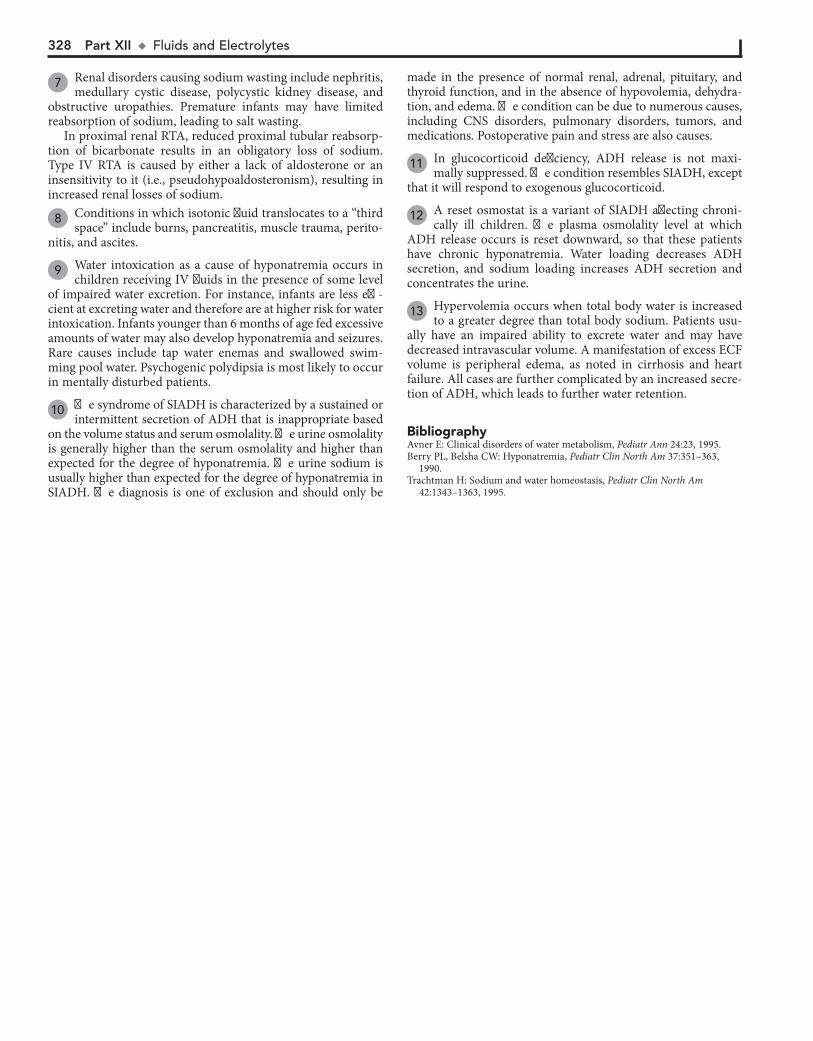
328 Part XII u FluidsandElectrolytes
7 Renal disorders causing sodium wasting include nephritis, medullary cystic disease, polycystic kidney disease, and
obstructive uropathies. Premature infants may have limited reabsorption of sodium, leading to salt wasting.
In proximal renal RTA, reduced proximal tubular reabsorp-tion of bicarbonate results in an obligatory loss of sodium. Type IV RTA is caused by either a lack of aldosterone or an insensitivity to it (i.e., pseudohypoaldosteronism), resulting in increased renal losses of sodium.
8 Conditions in which isotonic fluid translocates to a “third space” include burns, pancreatitis, muscle trauma, perito-
nitis, and ascites.
9 Water intoxication as a cause of hyponatremia occurs in children receiving IV fluids in the presence of some level
of impaired water excretion. For instance, infants are less effi-cient at excreting water and therefore are at higher risk for water intoxication. Infants younger than 6 months of age fed excessive amounts of water may also develop hyponatremia and seizures. Rare causes include tap water enemas and swallowed swim-ming pool water. Psychogenic polydipsia is most likely to occur in mentally disturbed patients.
10 The syndrome of SIADH is characterized by a sustained or intermittent secretion of ADH that is inappropriate based
on the volume status and serum osmolality. The urine osmolality is generally higher than the serum osmolality and higher than expected for the degree of hyponatremia. The urine sodium is usually higher than expected for the degree of hyponatremia in SIADH. The diagnosis is one of exclusion and should only be
made in the presence of normal renal, adrenal, pituitary, and thyroid function, and in the absence of hypovolemia, dehydra-tion, and edema. The condition can be due to numerous causes, including CNS disorders, pulmonary disorders, tumors, and medications. Postoperative pain and stress are also causes.
11 In glucocorticoid deficiency, ADH release is not maxi-mally suppressed. The condition resembles SIADH, except
that it will respond to exogenous glucocorticoid.
12 A reset osmostat is a variant of SIADH affecting chroni-cally ill children. The plasma osmolality level at which
ADH release occurs is reset downward, so that these patients have chronic hyponatremia. Water loading decreases ADH secretion, and sodium loading increases ADH secretion and concentrates the urine.
13 Hypervolemia occurs when total body water is increased to a greater degree than total body sodium. Patients usu-
ally have an impaired ability to excrete water and may have decreased intravascular volume. A manifestation of excess ECF volume is peripheral edema, as noted in cirrhosis and heart failure. All cases are further complicated by an increased secre-tion of ADH, which leads to further water retention.
BibliographyAvner E: Clinical disorders of water metabolism, Pediatr Ann 24:23, 1995.Berry PL, Belsha CW: Hyponatremia, Pediatr Clin North Am 37:351–363,
1990.Trachtman H: Sodium and water homeostasis, Pediatr Clin North Am
42:1343–1363, 1995.
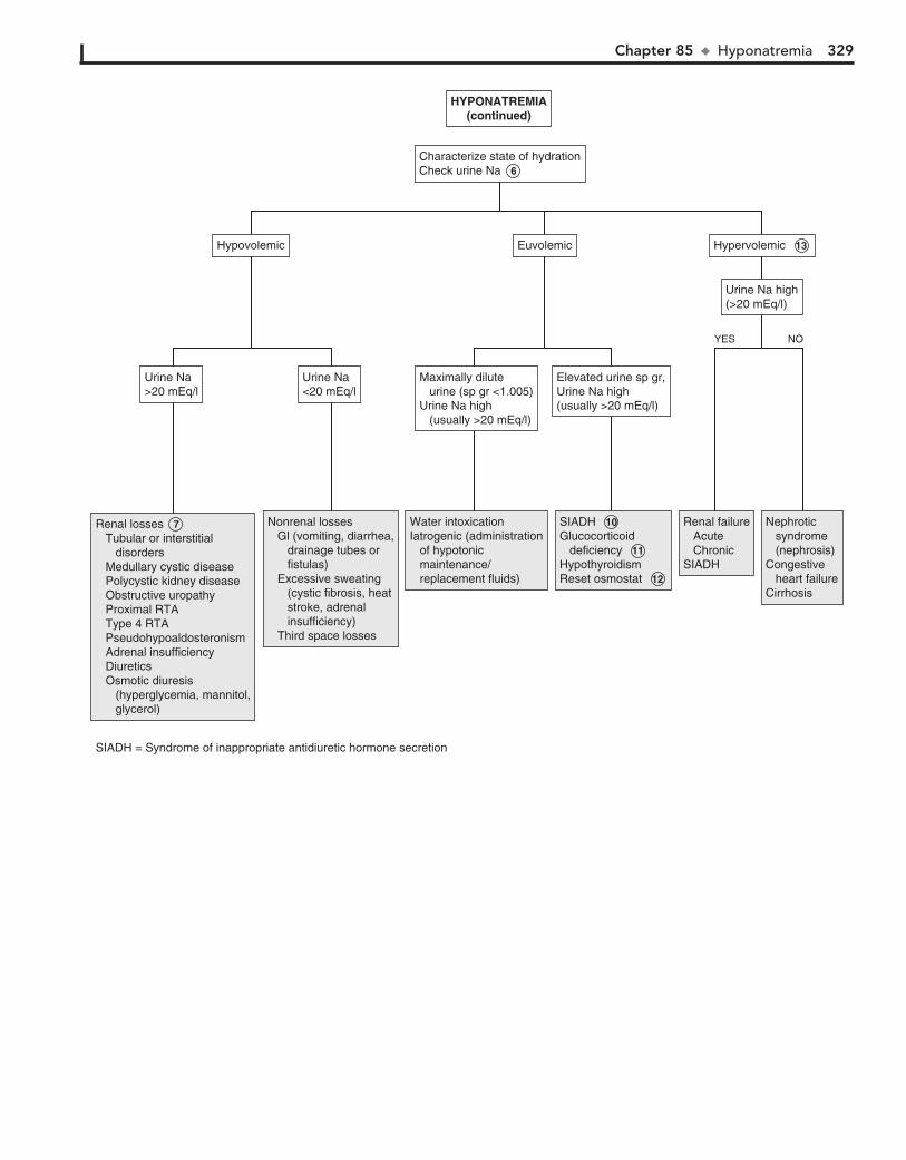
Chapter 85 u Hyponatremia 329
Nonrenal losses Gl (vomiting, diarrhea, drainage tubes or fistulas) Excessive sweating (cystic fibrosis, heat stroke, adrenal insufficiency) Third space losses
Nonrenal losses Gl (vomiting, diarrhea, drainage tubes or fistulas) Excessive sweating (cystic fibrosis, heat stroke, adrenal insufficiency) Third space losses
Renal losses 7 Tubular or interstitial disorders Medullary cystic disease Polycystic kidney disease Obstructive uropathy Proximal RTA Type 4 RTA Pseudohypoaldosteronism Adrenal insufficiency Diuretics Osmotic diuresis (hyperglycemia, mannitol, glycerol)
Renal losses 7 Tubular or interstitial disorders Medullary cystic disease Polycystic kidney disease Obstructive uropathy Proximal RTA Type 4 RTA Pseudohypoaldosteronism Adrenal insufficiency Diuretics Osmotic diuresis (hyperglycemia, mannitol, glycerol)
Water intoxicationIatrogenic (administration of hypotonic maintenance/ replacement fluids)
Water intoxicationIatrogenic (administration of hypotonic maintenance/ replacement fluids)
SIADH 10 Glucocorticoid deficiency 11 HypothyroidismReset osmostat 12
SIADH 10 Glucocorticoid deficiency 11 HypothyroidismReset osmostat 12
Renal failure Acute ChronicSIADH
Renal failure Acute ChronicSIADH
Nephrotic syndrome (nephrosis)Congestive heart failureCirrhosis
Nephrotic syndrome (nephrosis)Congestive heart failureCirrhosis
Characterize state of hydrationCheck urine Na 6 Characterize state of hydrationCheck urine Na 6
Urine Na>20 mEq/lUrine Na>20 mEq/l
Urine Na high(>20 mEq/l)Urine Na high(>20 mEq/l)
Hypervolemic 13 Hypervolemic 13 HypovolemicHypovolemic EuvolemicEuvolemic
Urine Na<20 mEq/lUrine Na<20 mEq/l
Maximally dilute urine (sp gr <1.005)Urine Na high (usually >20 mEq/l)
Maximally dilute urine (sp gr <1.005)Urine Na high (usually >20 mEq/l)
Elevated urine sp gr,Urine Na high(usually >20 mEq/l)
Elevated urine sp gr,Urine Na high(usually >20 mEq/l)
HYPONATREMIA(continued)
HYPONATREMIA(continued)
YES NO
SIADH = Syndrome of inappropriate antidiuretic hormone secretion

330
Chapter 86
HYPOKALEMIA
6 Insulin, catecholamines, and b-agonists (albuterol) acutely shift potassium intracellularly.
7 Potassium uptake by rapidly forming new RBCs and plate-lets in the treatment of megaloblastic anemia can result in
hypokalemia. Similarly, transfusion of frozen washed RBCs (not stored in acid-citrate-dextran) may lower potassium levels because of increased cellular uptake.
8 Rarely, episodic weakness or paralysis can be due to transient changes in potassium levels. The familial
forms result from mutations in genes that affect ion channels in muscle. In the hypokalemic version, triggers such as exer-cise, stress, b2-agonists, or eating a heavy meal cause a sud-den intracellular shift of potassium. Barium poisoning from foods (not radiologic barium) can cause a similar paralysis. In the hyperkalemic version, symptoms follow rest after ex-ercise or ingestion of potassium. Both are autosomal domi-nant disorders.
9 Chronic clay ingestion contributes to hypokalemia by binding dietary potassium. Nutritional potassium defi-
ciency is otherwise uncommon in patients with normal diets.
10 Gentamicin, amphotericin B, and chemotherapeutic agents can damage renal tubules, resulting in potassium wasting.
Hypercalcemic states also induce renal damage.
11 Glycyrrhizic acid is a component of natural licorice, rarely used today, which can exert a mineralocorticoid effect.
Some infants with Cushing syndrome may have tumors of the adrenal cortex that result in overproduction of aldosterone, as well as cortisol and other corticosteroids.
12 Increased excretion of cations (K1, H1) accompanies excretion of nonreabsorbable anions as a means of main-
taining electrical neutrality.
13 Bartter syndrome is characterized by hypokalemia second-ary to renal losses, normal BP, and vascular insensitivity
to pressor agents. Renin and aldosterone levels are elevated. Clinical manifestations include growth failure, weakness, con-stipation, polyuria, and dehydration. Gitelman and Liddle syndromes are other rare primary hypokalemic tubulopathies.
Potassium is the main cation of the intracellular fluid (ICF) compartment. The ECF concentration is carefully regulated to maintain a value around 4 mEq/L. Potassium (K1) plays a critical role in the excitability of nerve and muscle cells and contractility of muscles (smooth, skeletal, cardiac). Aldosterone normally regulates renal excretion of potassium. Aldosterone also causes sodium reabsorption and H1 secretion in the distal tubule. Aldosterone similarly affects potassium excretion in the stool. Hypokalemia is defined as a serum potassium concentra-tion , 3.5 mEq/L.
1 The history should inquire about medications, underlying medical problems, and diet, including pica and the use of
salt substitutes. Muscle weakness, hyporeflexia, and intestinal ileus are manifestations of hypokalemia. Cardiac arrhythmias are the most serious complications. Flattened T waves, a short PR interval, and a prolonged QT interval are characteristic EKG abnormalities. A U wave may develop after the QRS complex. Patients may also be lethargic or confused and may have muscle cramping, rhabdomyolysis, and myoglobinuria in cases of severe potassium depletion.
2 Acid-base disorders and disturbances in chloride or magne-sium levels may contribute to potassium imbalance and will
need to be corrected before correcting the potassium problem.
3 GI losses of potassium are exacerbated by accompanying vol-ume depletion and subsequent increased aldosterone effects.
4 If the etiology of hypokalemia is unclear from the history, a urine potassium level may aid in distinguishing between
renal and nonrenal losses.
5 Transcellular shifts of potassium occur in an effort to maintain electrical neutrality in varying conditions. In
metabolic acidosis, potassium shifts out of the cell in exchange for intracellular buffering of H1. The opposite exchange occurs to a lesser degree in metabolic alkalosis. These shifts also occur in acid-base disturbances that are primarily respiratory, although to a lesser degree.
BibliographyBrem AS: Disorders of potassium homeostasis, Pediatr Clin North Am
37:419–427, 1990.Watkins SL: Disorders of potassium balance, Pediatr Ann 24:31, 1995.
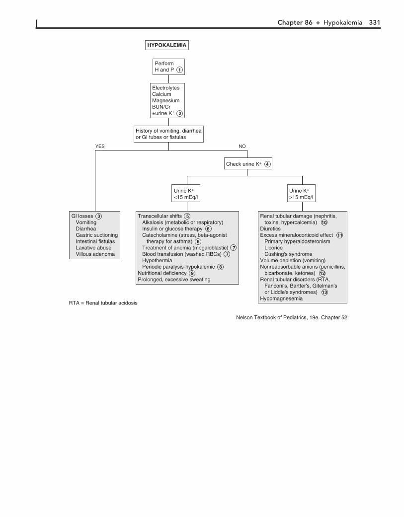
Chapter 86 u Hypokalemia 331
Gl losses 3 Vomiting Diarrhea Gastric suctioning Intestinal fistulas Laxative abuse Villous adenoma
Gl losses 3 Vomiting Diarrhea Gastric suctioning Intestinal fistulas Laxative abuse Villous adenoma
Transcellular shifts 5 Alkalosis (metabolic or respiratory) Insulin or glucose therapy 6 Catecholamine (stress, beta-agonist therapy for asthma) 6 Treatment of anemia (megaloblastic) 7 Blood transfusion (washed RBCs) 7 Hypothermia Periodic paralysis-hypokalemic 8 Nutritional deficiency 9 Prolonged, excessive sweating
Transcellular shifts 5 Alkalosis (metabolic or respiratory) Insulin or glucose therapy 6 Catecholamine (stress, beta-agonist therapy for asthma) 6 Treatment of anemia (megaloblastic) 7 Blood transfusion (washed RBCs) 7 Hypothermia Periodic paralysis-hypokalemic 8 Nutritional deficiency 9 Prolonged, excessive sweating
Renal tubular damage (nephritis, toxins, hypercalcemia) 10 DiureticsExcess mineralocorticoid effect 11 Primary hyperaldosteronism Licorice Cushing's syndromeVolume depletion (vomiting)Nonreabsorbable anions (penicillins, bicarbonate, ketones) 12 Renal tubular disorders (RTA, Fanconi's, Bartter's, Gitelman's or Liddle's syndromes) 13 Hypomagnesemia
Renal tubular damage (nephritis, toxins, hypercalcemia) 10 DiureticsExcess mineralocorticoid effect 11 Primary hyperaldosteronism Licorice Cushing's syndromeVolume depletion (vomiting)Nonreabsorbable anions (penicillins, bicarbonate, ketones) 12 Renal tubular disorders (RTA, Fanconi's, Bartter's, Gitelman's or Liddle's syndromes) 13 Hypomagnesemia
Urine K+
<15 mEq/lUrine K+
<15 mEq/l
Check urine K+ 4 Check urine K+ 4
Urine K+
>15 mEq/lUrine K+
>15 mEq/l
History of vomiting, diarrheaor GI tubes or fistulasHistory of vomiting, diarrheaor GI tubes or fistulas
PerformH and P 1 PerformH and P 1
ElectrolytesCalciumMagnesiumBUN/Cr±urine K+ 2
ElectrolytesCalciumMagnesiumBUN/Cr±urine K+ 2
HYPOKALEMIAHYPOKALEMIA
NOYES
RTA = Renal tubular acidosis
Nelson Textbook of Pediatrics, 19e. Chapter 52

332
Chapter 87
HYPERKALEMIA
4 Type IV renal tubular acidosis (RTA) is characterized by a deficiency of aldosterone or impaired aldosterone respon-
siveness. Pseudohypoaldosteronism is a subtype of type IV RTA that is characterized by high levels of aldosterone but an im-paired distal tubular response to it.
5 Trauma, use of cytotoxic agents, massive hemolysis, rhab-domyolysis, and (to a lesser extent) intense exercise can
release potassium as a result of tissue breakdown.
6 Transcellular shifts of potassium occur in an effort to maintain electrical neutrality in varying conditions. In
metabolic acidosis, potassium shifts out of the cell in exchange for intracellular buffering of H1. The opposite exchange occurs to a lesser degree in metabolic alkalosis. These shifts also occur in acid-base disturbances that are primarily respiratory, al-though to a lesser degree.
7 Potassium accompanies the osmotic shift of water from ICF to ECF in hypertonic states.
8 Rarely, episodic weakness or paralysis can be due to tran-sient changes in potassium levels. The familial forms result
from mutations in genes that affect ion channels in the muscle. In the hyperkalemic version, symptoms follow rest after exer-cise or ingestion of potassium. In the hypokalemic version, triggers such as exercise, stress, b2-agonists, or a heavy meal cause a sudden intracellular shift of potassium. Barium poison-ing from foods (not radiologic barium) can cause a similar paralysis. Both are autosomal dominant disorders.
Potassium is the main cation of the intracellular fluid (ICF) compartment. The ECF concentration is carefully regulated to maintain a value around 4 mEq/L. Potassium (K1) plays a critical role in the excitability of nerve and muscle cells and contractility of muscles (smooth, skeletal, cardiac). Aldosterone normally regulates renal excretion of potassium. Aldosterone also causes sodium reabsorption and H1 secretion in the distal tubule. Aldosterone similarly affects potassium excretion in the stool. Hyperkalemia is defined as a serum potassium concentra-tion above 5.5 mEq/L.
1 The history should inquire about medications, underlying medical problems, and diets, including pica and the use of
salt substitutes. Hyperkalemia may manifest with muscle weak-ness, as well as tingling, paresthesias, and paralysis. EKG changes include narrow peaked T waves and shortened QT interval initially. At higher serum potassium levels, delayed depolarization results in a widened QRS and P wave that may precede more serious arrhythmias (e.g., ventricular fibrillation, asystole).
2 Increased intake of potassium is a rare cause of hyperkale-mia in children with normal renal function. Salt substi-
tutes may be a cause.
3 Acute or chronic renal disorders may be responsible for impaired excretion of potassium.
BibliographyBrem AS: Disorders of potassium homeostasis, Pediatr Clin North Am 37:
419–427, 1990.Watkins SL: Disorders of potassium balance, Pediatr Ann 24:31, 1995.
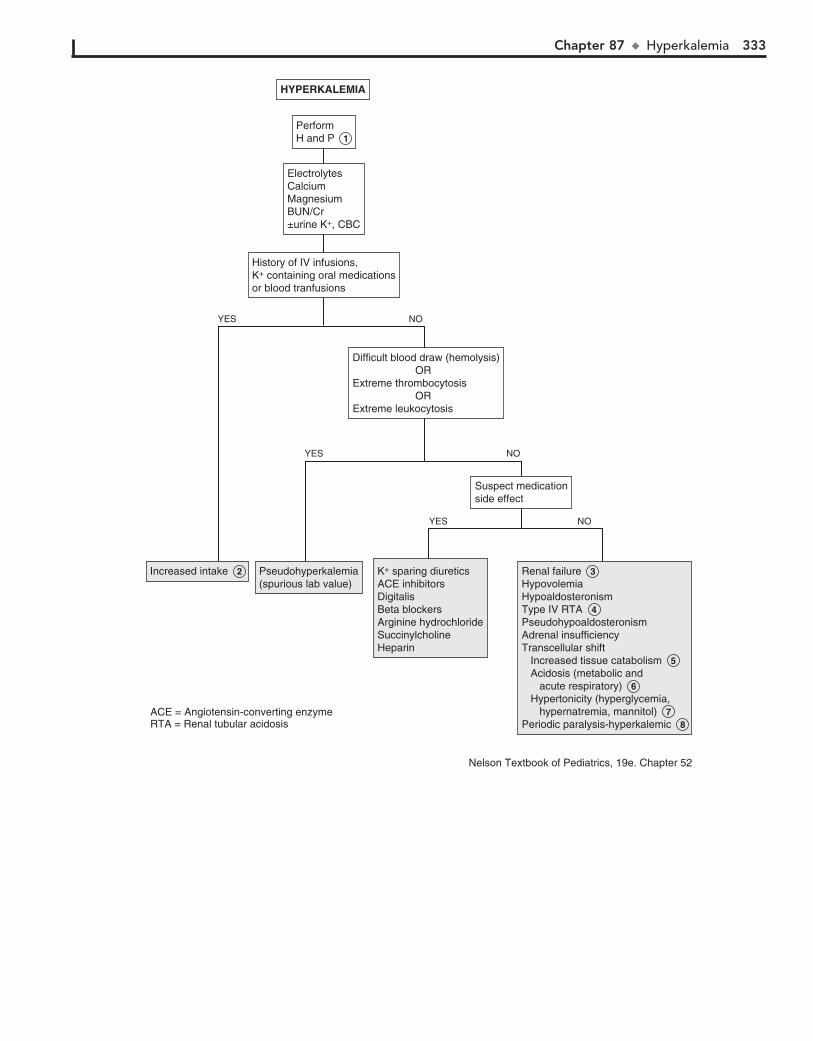
Chapter 87 u Hyperkalemia 333
Increased intake 2 Increased intake 2 Pseudohyperkalemia(spurious lab value)Pseudohyperkalemia(spurious lab value)
K+ sparing diureticsACE inhibitorsDigitalisBeta blockersArginine hydrochlorideSuccinylcholineHeparin
K+ sparing diureticsACE inhibitorsDigitalisBeta blockersArginine hydrochlorideSuccinylcholineHeparin
Renal failure 3HypovolemiaHypoaldosteronismType IV RTA 4PseudohypoaldosteronismAdrenal insufficiencyTranscellular shift Increased tissue catabolism 5 Acidosis (metabolic and acute respiratory) 6 Hypertonicity (hyperglycemia, hypernatremia, mannitol) 7Periodic paralysis-hyperkalemic 8
Renal failure 3HypovolemiaHypoaldosteronismType IV RTA 4PseudohypoaldosteronismAdrenal insufficiencyTranscellular shift Increased tissue catabolism 5 Acidosis (metabolic and acute respiratory) 6 Hypertonicity (hyperglycemia, hypernatremia, mannitol) 7Periodic paralysis-hyperkalemic 8
History of IV infusions, K+ containing oral medicationsor blood tranfusions
History of IV infusions, K+ containing oral medicationsor blood tranfusions
ElectrolytesCalciumMagnesiumBUN/Cr±urine K+, CBC
ElectrolytesCalciumMagnesiumBUN/Cr±urine K+, CBC
Difficult blood draw (hemolysis) ORExtreme thrombocytosis ORExtreme leukocytosis
Difficult blood draw (hemolysis) ORExtreme thrombocytosis ORExtreme leukocytosis
Suspect medicationside effectSuspect medicationside effect
PerformH and P 1 PerformH and P 1
HYPERKALEMIAHYPERKALEMIA
YES NO
YES NO
YES NO
ACE = Angiotensin-converting enzymeRTA = Renal tubular acidosis
Nelson Textbook of Pediatrics, 19e. Chapter 52
334
Chapter 88
HYPOCALCEMIA
8 Vitamin D deficiency occurs in dark-skinned breast-fed infants who live in areas with less sunlight. It may also oc-
cur in infants fed with unfortified cow’s milk. Inadequate expo-sure to ultraviolet light, particularly in dark-skinned children, may also cause vitamin D deficiency. Inadequate absorption of vitamin D or calcium may be seen in diseases such as celiac disease, liver disease (including biliary cirrhosis), and cystic fibrosis. Phenobarbital and phenytoin interfere with metabo-lism of vitamin D. Renal failure also decreases vitamin D syn-thesis. Clinical manifestations of vitamin D deficiency include rickets, which occurs as a result of the body’s attempt to maintain serum calcium levels. Vitamin D deficiency is best determined by obtaining a 25-OH vitamin D level; 1.25(OH)2 vitamin D can be normal with deficiency.
9 Vitamin D–dependent rickets (vitamin D–resistant rickets) type I usually presents between the ages of 3 and 6 months
in children receiving adequate quantities of vitamin D. This is believed to be caused by decreased activity of the enzyme 25-hydroxy-1a-hydroxylase, resulting in decreased serum levels of 1,25-dihydroxyvitamin D. Alkaline phosphatase and PTH are increased, and serum phosphorus is low.
10 In pseudohypoparathyroidism (i.e., Albright hereditary osteodystrophy), the parathyroid glands are normal or
even hyperplastic. PTH levels are normal or elevated; however, there is a peripheral resistance to PTH. This syndrome is associated with tetany and a distinctive phenotype with brachy-dactyly, skeletal abnormalities, short stature, and mild mental retardation.
11 Hyperphosphatemia may be associated with renal failure. It can occur secondary to rapid cell destruction due to
chemotherapy (i.e., tumor lysis syndrome). Trauma leading to rhabdomyolysis causes release of cellular phosphorus. Hyper-phosphatemia may also result from exogenous phosphate in the form of laxatives and enemas.
12 Pancreatitis causes release of pancreatic lipase, resulting in degradation of omental fat and binding of calcium in the
peritoneum.
13 “Hungry bone” syndrome may occur during initial therapy to correct chronic hyperparathyroidism, leading to a sud-
den fall in phosphorus and calcium levels. Calcium and phos-phorus are rapidly absorbed into severely demineralized bones.
14 Vitamin D–dependent rickets (type II) is a hereditary disorder due a receptor defect, resulting in hypocalcemia
despite elevated levels of 1,25-dihydroxyvitamin D.
15 Hypocalcemia may also occur with alkalosis or rapid correction or overcorrection of acidosis.
16 Hypomagnesemia often coexists with hypocalcemia and may be due to decreased absorption (e.g., in malabsorp-
tion syndromes) or intake. Familial hypomagnesemia with secondary hypocalcemia usually appears between the second and sixth weeks of life. Aminoglycoside therapy may cause increased urinary losses of magnesium. PTH may be normal or decreased.
Serum calcium concentration in the ECF is maintained by para-thyroid hormone (PTH), which acts on the kidneys and bones, and by 1,25-dihydroxyvitamin D, which acts on the intestines and bones. About 50% of the calcium is in the biologically im-portant ionized form, 40% is protein bound (i.e., mainly albu-min), and 10% is complexed to anions (e.g., bicarbonate, citrate, sulfate, phosphate, and lactate). Mild hypocalcemia is usually asymptomatic. Symptoms and signs of more severe hypocalce-mia include paresthesias of the extremities, Chvostek sign, Trousseau sign, muscle cramps or spasm, laryngospasm, tetany, and seizures. Cardiac manifestations include a prolonged QT interval, which may progress to heart block.
1 Calcium levels are affected by serum albumin levels and by pH. A low serum albumin will lower the total serum
calcium, and acidic pH will decrease protein binding and increase ionized calcium levels. It is important to obtain an ionized calcium level. If this test is not available, in order to correct for hypoalbuminemia, 0.2 mmol/L (0.8 mg/dL) of calcium must be added to the total calcium level for each 1 g/dL decrease in serum albumin from the normal 4.0 g/dL. Similarly, for each 0.1 decrease in pH, ionized calcium rises by 0.05 mmol/L. However, these corrections are a poor substitute for ionized calcium level.
2 Hypoalbuminemic states result in a lower serum calcium level. The ionized calcium that is the biologically impor-
tant level is usually normal, but it should be confirmed. Causes include liver disease, protein-losing enteropathy, and nephrotic syndrome.
3 Endotoxic shock is associated with hypocalcemia, although the mechanism is unknown. Hypocalcemia may also occur
with rapid correction or overcorrection of acidosis.
4 Aplasia or hypoplasia of the parathyroid glands is often associated with DiGeorge/velocardiofacial syndrome. Many
of the children have transient neonatal hypocalcemia; however, the hypocalcemia may recur later in life. Associated anomalies include conotruncal heart defects, velopharyngeal insufficiency, cleft palate, renal anomalies, and partial to complete aplasia of the thymus with varying severities of immunodeficiency.
5 Surgical hypoparathyroidism is a complication of thyroid-ectomy.
6 Autoimmune hypoparathyroidism is usually associated with other autoimmune disorders such as Addison disease
and chronic mucocutaneous candidiasis. Other associations, including vitiligo, alopecia areata, pernicious anemia, and mal-absorption may not appear until adulthood. Parathyroid anti-bodies are present.
7 Maternal hyperparathyroidism during pregnancy may cause a transient neonatal hypocalcemia. It may persist for
weeks or months. Symptoms such as tetany may be delayed, especially in breast-fed infants.
BibliographyBushinsky DA, Monk RD: Calcium, Lancet 352:306–311, 1998.Fouser L: Disorders of calcium, phosphorus, and magnesium, Pediatr Ann
24:38–46, 1995.

Chapter 88 u Hypocalcemia 335
Hypoalbuminemicconditions 2 Hypoalbuminemicconditions 2
Endotoxic shock 3 Endotoxic shock 3 Aplasia/hypoplasia 4 (DiGeorge syndrome)FamilialSurgical 5 Infiltrative lesions (hemosiderosis, Wilson's disease)Autoimmune 6 IdiopathicMaternal hyperparathyroidism (during pregnancy) 7
Aplasia/hypoplasia 4 (DiGeorge syndrome)FamilialSurgical 5 Infiltrative lesions (hemosiderosis, Wilson's disease)Autoimmune 6 IdiopathicMaternal hyperparathyroidism (during pregnancy) 7
Vitamin D deficiency 8 Deficient sunlight Nutritional/dietary deficiency Malabsorption Liver disease (biliary cirrhosis) Renal disease AnticonvulsantsVitamin D dependent rickets (Type I) 9 Pseudohypoparathyroidism 10 Hyperphosphatemia 11 Tumor-lysis syndrome Rhabdornyolysis Laxatives/enemasAcute pancreatitis 12Hungry bone syndrome 13Drugs (colchicine, estrogen, calcitonin)Large amounts of citrated bloodVitamin D dependent rickets (Type II) 14
Vitamin D deficiency 8 Deficient sunlight Nutritional/dietary deficiency Malabsorption Liver disease (biliary cirrhosis) Renal disease AnticonvulsantsVitamin D dependent rickets (Type I) 9 Pseudohypoparathyroidism 10 Hyperphosphatemia 11 Tumor-lysis syndrome Rhabdornyolysis Laxatives/enemasAcute pancreatitis 12Hungry bone syndrome 13Drugs (colchicine, estrogen, calcitonin)Large amounts of citrated bloodVitamin D dependent rickets (Type II) 14
Alkalosis 15 Rapid correction of acidosisHypomagnesemia 16 Familial Malabsorption Aminoglycosides
Alkalosis 15 Rapid correction of acidosisHypomagnesemia 16 Familial Malabsorption Aminoglycosides
Septic or ill appearingSeptic or ill appearing
AbnormalAbnormalNormalNormal
PTH1, 25 (OH)2 vitamin D25 OH vitamin DAlbuminAST, ALTBUNCreatinineMagnesiumPhosphorusAlkaline phosphataseElectrolytes (with bicarbonate)
PTH1, 25 (OH)2 vitamin D25 OH vitamin DAlbuminAST, ALTBUNCreatinineMagnesiumPhosphorusAlkaline phosphataseElectrolytes (with bicarbonate)
HypoparathyroidismHypoparathyroidism
PTH normalPTH normal
Confirm with ionized calcium 1 Confirm with ionized calcium 1
HYPOCALCEMIAHYPOCALCEMIA
PTH = Parathyroid hormone
PTH PTH ↓ PTH PTH ↑
YES NO
Nelson Textbook of Pediatrics, 19e. Chapters 565 and 48
336
Chapter 89
HYPERCALCEMIA
due to ectopic production of PTH; however, ectopic production of PTH-related peptide (PTHrP) is more common.
4 Hypercalcemia is present in 10% of children with Williams syndrome; the cause is unknown. Features of the syn-
drome include feeding difficulties, elfin facies, growth delay, gregarious personality, supravalvular aortic stenosis, renovas-cular disease, and developmental delay.
5 In familial hypocalciuric hypercalcemia (familial benign hypercalcemia), the patients are usually asymptomatic and
PTH levels are inappropriately normal. The calcium-to-Cr clearance ratio is decreased in spite of the hypercalcemia.
6 Idiopathic hypercalcemia of infancy manifests during the first year of life with failure to thrive and hypercalcemia. There is
increased absorption of calcium. Levels of 1,25-dihydroxyvitamin D may be normal or elevated. PTH and phosphorus levels are normal.
7 Ectopic production of vitamin D may occur with granuloma-tous disease, such as sarcoidosis (30%-50%) and tuberculosis. It
may rarely occur with tumors. The excessive 1,25-dihydroxyvitamin D suppresses the production of PTH.
8 Hypervitaminosis A results in excessive bone resorption. Thiazide diuretics cause increased renal calcium reabsorp-
tion. Milk-alkali syndrome is caused by the consumption of large amounts of calcium-containing nonabsorbable antacids, leading to hypercalcemia, alkalemia, nephrocalcinosis, and renal insufficiency.
9 Malignancy may cause hypercalcemia. Most commonly it is secondary to production of PTH-related peptide but can also
be caused by bone lysis, tumor production of 1,25-dihydroxyvitamin D, or ectopic PTH. In thyrotoxicosis, PTH is suppressed. Hypercalcemia is due to excessive bone resorption caused by the thyroid hormone. Patients undergoing dialysis may develop hypercalcemia, especially if they receive calcium supplements or are dialyzed against a high-calcium bath.
10 Jansen-type metaphyseal chondrodysplasia is a rare genetic disorder with features of short-limbed dwarfism. Circulating
levels of PTH and PTHrP are undetectable. The hypercalcemia is severe but asymptomatic.
Serum calcium concentration in the ECF is maintained by para-thyroid hormone (PTH) and 1,25-dihydroxyvitamin D. PTH acts on the kidneys and bones and stimulates the production of 1,25-dihydroxyvitamin D, which acts on the intestines and bones. About 50% of the calcium is in the biologically impor-tant ionized form, 40% is protein bound (mainly albumin), and 10% is complexed to anions (bicarbonate, citrate, sulfate, phos-phate, and lactate). Calcium levels are affected by serum albu-min levels and pH. An elevated serum albumin concentration will appear to raise the total serum calcium concentration; an alkaline pH will increase protein binding and decrease ionized calcium levels. It is important to obtain an ionized calcium level.
Mild hypercalcemia is usually asymptomatic. With more severe hypercalcemia there may be neurologic features ranging from drowsiness to depression, stupor, and coma. GI manifesta-tions such as constipation, nausea, vomiting, anorexia, and ul-cers may occur. Hypercalciuria leads to nephrogenic diabetes insipidus, resulting in polyuria. Other renal effects of hypercal-cemia include nephrolithiasis and nephrocalcinosis.
1 Primary hyperparathyroidism is caused by excessive pro-duction of PTH, owing to adenoma or hyperplasia. Inci-
dence may be sporadic or may occur as part of the multiple endocrine neoplasia (MEN) syndromes, with involvement of the pancreas and the anterior pituitary. Hyperparathyroidism–jaw tumor syndrome is characterized by parathyroid adenomas and fibro-osseous jaw tumors. Patients may also have polycystic kidney disease, renal hamartomas, and Wilms tumor.
2 Secondary hyperparathyroidism is increased production of PTH in response to hypocalcemia, as in chronic renal
failure. If this persists for a prolonged period, the glands begin to autonomously produce PTH even after the underlying rea-son for the hypocalcemia has been corrected, as after renal transplant. This is known as tertiary hyperparathyroidism and results in hypercalcemia.
3 Hypercalcemia may occur in association with malignancies (e.g., neuroblastoma, leukemia, renal tumors). This may be
BibliographyBushinsky DA, Monk RD: Calcium, Lancet 352:306–311, 1998.Fouser L: Disorders of calcium, phosphorus, and magnesium. Pediatr Ann
24:38–46, 1995.

Chapter 89 u Hypercalcemia 337
Sporadic (adenoma/ hyperplasia)Hereditary MEN I MEN II Hyperparathyroidism– jaw tumor syndrome
Sporadic (adenoma/ hyperplasia)Hereditary MEN I MEN II Hyperparathyroidism– jaw tumor syndrome
Post renaltransplantPost renaltransplant
Malignancies 9 Malignancies 9
Williamssyndrome 4 Williamssyndrome 4
Hypervitaminosis D latrogenic Excessive food fortification Food fads
Hypervitaminosis D latrogenic Excessive food fortification Food fads
Ectopic production of vitamin D 7 Tumors Sarcoid, other granulomatous diseases
Ectopic production of vitamin D 7 Tumors Sarcoid, other granulomatous diseases
Hypervitaminosis A 8 ThiazidesMilk-alkali syndrome
Hypervitaminosis A 8 ThiazidesMilk-alkali syndrome
ImmobilizationMalignancy 9 Thyrotoxicosis Dialysis osteomalacia Hypophosphatemia
ImmobilizationMalignancy 9 Thyrotoxicosis Dialysis osteomalacia Hypophosphatemia
Jansen-typemetaphyseal chondrodys-plasia 10
Jansen-typemetaphyseal chondrodys-plasia 10
Subcutaneous fat necrosisFamilial hypocalciuric hypercalcemia 5 HypophosphatemiaIdiopathic hypercalcemia of infancy 6 Lithium
Subcutaneous fat necrosisFamilial hypocalciuric hypercalcemia 5 HypophosphatemiaIdiopathic hypercalcemia of infancy 6 Lithium
Excessive intakeExcessive intake
Primaryhyperparathyroidism 1 Primaryhyperparathyroidism 1
Ectopic PTH production 3 Ectopic PTH production 3
Tertiaryhyperparathyroidism 2 Tertiaryhyperparathyroidism 2
PTH PTH ↑ PTH PTH ↓ UndetectablePTHUndetectablePTH
PTH normalPTH normal
Stigmata ofWilliams syndromeStigmata ofWilliams syndrome
MedicationsMedications
Increased1,25 (OH)2vitamin D
Increased1,25 (OH)2vitamin D
1,25 (OH)2vitamin D1,25 (OH)2vitamin D
Normal1,25 (OH)2vitamin D
Normal1,25 (OH)2vitamin D
ObtainPTHPhosphorus
ObtainPTHPhosphorus
HYPERCALCEMIAHYPERCALCEMIA
YES NOYES NOYES NO
PTH = parathyroid hormoneMEN = multiple endocrine neoplasia
Nelson Textbook of Pediatrics, 19e. Chapter 564

This page intentionally left blank

Index
AAbdominal masses, 110–112, 111fAbdominal pain, 71–77
acute, 73fsigns and symptoms of, 72, 78
chronic or recurrent, 74, 75fABO incompatibility, 233f, 234Abortion, abnormal vaginal bleeding and, 146, 147fAbrasions, back injuries and, 172, 173fAbscess
back pain and, 172, 173fheadaches and, 184, 185f, 187fliver, 297f, 298, 299fretropharyngeal, 6, 7f, 36, 37f
apnea from, 48Abstinence syndromes, 308Accommodation, 24
disorders of, 23f, 24Acetaminophen, hyperbilirubinemia from, 100Acidemia, 317–321, 321fAcidosis, 318
hyperventilation and, 208irritable infant and, 308lactic, 318, 321frenal tubular, 256, 257f, 320
type IV, 332, 333fAcquired immunodeficiency syndrome (AIDS)
hepatobiliary disorders and, 100, 102parotitis and, 10
Acrocyanosis, 42, 43fAcrodermatitis enteropathica, 84, 87f, 219fActinomycosis
central diabetes insipidus and, 286, 287fhepatosplenomegaly from, 102
Activated partial thromboplastin time (aPTT), 236Acute otitis media. see Otitis mediaAdenocarcinoma, diethylstilbestrol and, 148Adenoma
Leydig cell, 264, 265fin neck, 10, 11fprimary hyperparathyroidism from, 336
Adenopathy, 228hilar, on CXR, 296reactive, 228, 229f
Adenosis, vaginal discharge and, 148Adenovirus, 223f
adenopathy and, 229fconjunctivitis and, 16hemoptysis from, 44myocarditis from, 126pharyngitis and, 6, 7f
Adipsia, 324, 325fAdrenarche, premature, 264–265, 267f, 268, 269fAfibrinogenemia, congenital, 238Agammaglobulinemia, X-linked, 302, 303fb-Agonists, and potassium cellular shifts, 330Agranulocytosis, 6, 7fAirway
inflammation, 30lesions, 36obstruction, 6problems, apnea from, 48–49, 49f, 50f
Albright hereditary osteodystrophy, 282, 283f, 334Albumin, serum calcium and, 334Albuterol, and potassium cellular shifts, 330Alcohol
failure to thrive and, 310gynecomastia and, 278, 279fHodgkin disease and, 230
hyperbilirubinemia from, 100platelet function defects and, 238and short stature, 256
Aldosterone, 330Alkalemia, 322–323, 323fAlkaline phosphatase, and cholestasis, 102Alkalosis
metabolic, 322, 323frespiratory, 322, 323f
Allergic bronchopulmonary aspergillosis (ABPA), 252, 253fAlopecia, 215–217, 217fAlport syndrome, 120, 121f, 123f, 213fAlström-Hallgren syndrome, 282, 283fAltered mental status, 208–211, 209fAmanita phalloides mushrooms, hepatotoxicity of, 128Amblyopia, 20, 22, 23fAmegakaryocytic thrombocytopenia, congenital, 240, 241fAmenorrhea, 140–143, 141f, 143f
testicular feminization and, 270Aminoglycosides, hypomagnesemia and, 334Amphetamines, palpitations from, 62Amphotericin B, renal toxicity of, 330Anagen effluvium, 217, 217fAnalgesics
headaches from, 186neutropenia and, 245f
Androgenhirsutism and, 274hyperbilirubinemia from, 100maternal, ambiguous genitalia and, 272
Androgen insensitivity, 260, 261f, 278hirsutism and, 276syndrome, 140, 141f
Androgen resistance syndromes, 270, 271f, 273fAnemia, 232–235, 233f, 248–250
aplastic, 248acquired, 233f, 234
autoimmune hemolytic, 100, 101f, 233f, 234autoimmune neutropenia and, 244and blood loss, 235of chronic disease, 232, 233fFanconi, 233f, 234hemolytic
aplastic crisis and, 234Hodgkin disease and, 230isoimmune, 233f, 234microangiopathic, 233f, 234and pancytopenia, 248
history for, 232hypoplastic, 233f, 234, 248–250of inflammation, 232iron deficiency, 232, 233flaboratory testing for, 232megaloblastic, 233f, 234, 249f, 250
treatment of, hypokalemia and, 330, 331fscreening for, 232severe, and edema, 126, 127fsickle cell, abdominal pain and, 72–76sideroblastic, 233f, 234systolic outflow murmur and, 66, 67fthalassemia trait as, 232, 233f
Angioedema, 126, 127fepisodic, 252, 253f
Angiotensin-converting enzyme (ACE) inhibitorscough and, 32functional hypoaldosteronism from, 320, 333f
Anion gap (AG), 318, 321furinary, 320
339
Alcohol (Continued)
340 Index
Anisometropia, 23f, 24Anorchia, 260, 261f, 272Anorexia, 312, 313f
amenorrhea and, 140Anorexia nervosa, and neutropenia, 244Anovulatory cycles, 146, 147fAntacids, and hypercalcemia, 336Anterior cruciate ligament injury, 162, 163fAntibiotics
acute diarrhea and, 84, 85fand bleeding, 236Candida and, 150gastrointestinal bleeding and, 94, 96neutropenia and, 245f
Anticoagulants, and bleeding disorders, 236, 238Anticonvulsants
and bleeding, 236failure to thrive and, 310hyperbilirubinemia from, 100neutropenia and, 245fpancytopenia and, 248
Antidiuretic hormone, 324Antiemetics, and tardive dyskinesia, 194Anti-glomerular basement membrane antibody disease, 44Antineoplastics, hyperbilirubinemia from, 100Antineutrophil antibodies, 244–245Antineutrophil cytoplasmic antibodies (ANCA), 44Antithyroid drugs, neutropenia and, 245fa1-Antitrypsin deficiency, cough and, 32Aorta, coarctation of, 66, 67f, 68, 70f, 130, 131f
heart murmur with, 130–132Apnea, 48–50, 49f
cyanosis and, 42, 43f, 48in infants, 48neurologic causes of, 49f, 50obstructive sleep, 49, 49f, 50f, 314, 315f
pulmonary hypertension and, 66sleep, and enuresis, 118
Apophysitis, iliac, 170–171, 171fApparent life-threatening event (ALTE), 48Appendicitis, 72, 73fArcanobacterium haemolyticum, 6, 7fArrhythmia, 58, 59f, 62, 63f
sinus, 62, 63fsinus tachycardia as, 62ventricular tachycardia as, 64
Arthralgia, 156Arthritis, 156–161, 157f, 159f, 161f
enthesitis-related, 156–158, 157fjuvenile idiopathic, 158, 295f, 296, 297f, 299f
systemic-onset, 160, 161fmonoarticular, 156poststreptococcal, 160, 161fpsoriatic, 157f, 158reactive, 158, 159fseptic, 152, 153f, 156, 157f
knee pain and, 164vasculitis syndromes and, 158
Ascites, abdominal distention from, 110Aspergillosis, allergic bronchopulmonary, 252Aspirin, 94
and bleeding, 236cough and, 32Löeffler syndrome and, 252platelet function defects and, 237fReye syndrome and, 210rhinorrhea and, 4
Asplenia, congenital, 300, 303fAsthma, 32, 33f, 252
chest pain and, 52, 53f, 57fpneumothorax and, 53–54wheezing and, 38, 39f, 41f
Astigmatism, 23f, 24Ataxia, 204–207, 205f, 207f
acute cerebellar, 206, 207fAtaxia-telangiectasia, 204, 205f, 303f, 304Atherosclerosis, obesity and, 282–284
Atlantoaxial joints instability, 176, 177fAtrial fibrillation, 63f, 64Atrial flutter, 63f, 64Atrial septal defects, 67f, 68, 70fAudiometry, 212–213Autoantibodies, as inhibitors, 236
BBack pain, 172–175, 173fBacteremia, 290, 291f, 292, 293f
blood culture and, 294, 295fBalanitis, 114, 115f“Ballism,” 194Bardet-Biedl syndrome, 282, 283fBarium enema, 96Barium esophogram, 40Bartter syndrome, 322, 323f, 330, 331fBEARS screening tool, for sleep disturbances, 314Bernard-Soulier syndrome, 236, 238, 240Biemond syndrome, 282, 283fBiliary atresia, 100Bismuth, vs. gastrointestinal bleeding, 94Bladder
neurogenic, 116, 117f, 119foveractive, 116, 117f, 119f
Bleeding disorders, 146, 147f, 236–239, 237fBlisters, sucking, 218, 219fb-Blocking agents, functional hypoaldosteronism from, 320Blood
extravascular, 98, 99ffactor XIII deficiency of, 237f, 238red cell membrane defects, jaundice and, 98, 99f, 101f
Blood pressurecoarctation of aorta and, 66hypertension and, 130
Blood testsfor metabolic disorders, 78for streptococcal infection, 120, 121f, 123f
Blount’s disease, 180, 181f“Blue dot” sign, 134Body mass index, 282–284Bone(s)
accessory navicular, 170, 171fage of, precocious puberty in females and, 266, 267fprecocious puberty in males and, 264stature and, 256, 257f, 258tumor in, 166, 167f
Botulism, 199f, 200, 203frespiratory acidosis and, 318
Bowlegs, 180–182, 181fBrain lesions, and precocious puberty, 266, 267fBranchial cleft anomalies, 8, 9fBreath-holding spells, 42, 50, 50f, 190
in syncope, 59f, 60Bronchiectasis, 44, 45fBronchiolitis, 38, 39f, 41f
apnea from, 48cough and, 32respiratory syncytial virus, 48rhinorrhea and, 4
Bronchitis, 30, 31fBronchomalacia, wheezing in, 40Bronchoscopy, 30
and foreign body aspiration, 30for hemoptysis, 44, 46for wheezing, 38, 39f, 41f
Bronchospasm, 30, 38Brown syndrome, 21, 21fBrucellosis, 295f, 298, 299f
hepatosplenomegaly from, 102and pancytopenia, 248
Brugada syndrome, 58, 59fpalpitations from, 63f, 64
Bruton agammaglobulinemia, 302Budd-Chiari syndrome, 104
splenomegaly and, 108

Index 341
Bulimiaamenorrhea and, 140parotid enlargement and, 10, 11f
Bulimia nervosa, 283f, 284Bullae, 218–221, 219fBuphthalmos, 22“Burners,” 166, 167f
CCaffeine, irritable infant and, 308Caffey disease, 298Camptomelic syndrome, 270Campylobacter, arthritis and, 158Candida, 6, 7f, 223f
antibiotics and, 150diabetes mellitus and, 150hoarseness and, 34vaginal discharge and, 148
Carbamazepine, neutropenia and, 245fCarbon monoxide
headaches from, 186hemoglobinuria and, 120irritable infant and, 308
Cardiac arrhythmia, hypokalemia and, 330Cardiac disease, with hemoptysis, 44Cardiomyopathy, 126, 127f
by electrocardiogram, 126hypertrophic, 56, 57f
syncope from, 58, 59fCardiovascular drugs, and neutropenia, 245fCare-giver fabricated illness, 48, 50fCarpal tunnel syndrome, 168–169, 169f, 170, 171fCarpenter syndrome, 282, 283fCataplexy, 192, 193fCataracts, 22, 23fCatecholamines, and potassium cellular shifts, 330, 331fCat-scratch disease, 228, 230
hepatosplenomegaly from, 102Ceftriaxone, dosage of, 290Celiac disease, 74, 75f, 86–87, 89f, 311f, 312, 313f
calcium and, 334diarrhea and, 312, 313fIgA deficiency and, 302pubertal delay and, 262
Cellulitis, orbital, 18, 19fCephalosporins, autoimmune hemolytic anemia and, 234Cerebellitis, postinfectious, 206, 207fCerebral palsy (CP), 153f, 154–153, 200
neuromuscular disorder and, 181ftoe walking and, 179f
Cerebrospinal fluid (CSF), in rhinorrhea, 4, 5fCerumen, 2Cervical spine, subluxation of, 176, 177fCervicitis, 146, 147fChédiak-Higashi syndrome, 244, 245f, 304
recurrent infection with, 304, 305fChemicals, and nephrotic syndrome, 124Chemotherapeutic agents
neutropenia and, 245fpancytopenia and, 248renal toxicity of, 330
Chest pain, 51–57, 53f, 55f, 57fasthma and, 52, 53f, 57fcyanosis and, 42hyperventilation in, 52, 53fmuscular, 52, 53f, 55fmyocardial ischemia and, 56psychogenic, 52, 53fskeletal causes of, 52, 53f, 55f
Child abuseapnea and, 48, 49favulsion as, 216, 217f
Chlamydia, conjunctivitis and, 16Chlamydia trachomatis
in pelvic inflammatory disease, 72–73, 102vaginal discharge and, 148
Chlamydial infections, vaginal discharge and, 148, 149fChlamydophila pneumoniae, 38, 39fChloramphenicol
neutropenia and, 245fpancytopenia and, 248red cell aplasia and, 234
Chlorpromazine, hyperbilirubinemia from, 100Choanal atresia, bilateral, 4, 5fCholecystitis, 72, 73fCholelithiasis, 72, 73fCholesteatoma, 2, 3f, 213fChondrodysplasia, Jansen-type metaphyseal, 336, 337fChorea
benign hereditary (familial), 194–195, 195fSydenham, 194, 195f
Choreoathetosis, 194, 195fChristmas disease, 236Chronic renal disease, and anemia, 233f, 234Churg-Strauss syndrome, 44Cirrhosis
calcium and, 334eosinophilia and, 253fperitonitis with, 72–76splenomegaly and, 108
Clinitest reagent, reducing substance test from, 86Clostridium difficile
acute diarrhea and, 84, 87fbloody diarrhea and, 96
“Clubfoot,” 178, 179fCoagulation disorders, abnormal vaginal bleeding and, 146, 147fCoagulopathy, 95f, 96Coats disease, 24Cocaine
irritable infant and, 308palpitations from, 62pneumothorax and, 53–54, 57frhinorrhea from, 4
Coccidioidomycosis, 252, 253fcough and, 32
Cohen syndrome, 282, 283fCold agglutinin disease, 234Colic, definition of, 306, 307fColitis
infectious, gastrointestinal bleeding and, 96, 97fulcerative, 88, 253f
Complement system, disorders of, 304Complex regional pain syndrome, 168, 169fComputed tomography, for intracranial hemorrhage diagnosis, 308Congenital adrenal hyperplasia, 322
ambiguous genitalia and, 272–273hirsutism and, 275f, 276hypertension and, 130precocious puberty in females and, 266–268, 267fprecocious puberty in males and, 257f, 264–265screening for, 276
Congenital central hypoventilation syndrome (CCHS), 50, 50fCongenital fibrosis syndrome, 21, 21fCongenital heart disease, 42, 43f, 69–70, 126, 127f
cyanosis and, 42, 69–70risk factors for, 66severe choreoathetosis and, 194signs of, 70fsupraventricular tachycardias and, 64
Conjunctivitisallergic, 16, 17fbacterial, 16, 17fchemical, 16, 17flimbal vernal, 16, 17fmesenteric adenitis with, 72viral, 16, 17f
Connective tissue disorder(s), 253fhypotonia and, 199fpneumothorax and, 53–54
Constipation, 90–93, 91f, 93f, 117f, 119f, 307f, 309fencopresis as, 90functional, 90situational, 90

342 Index
Constitutional delayand pubertal delay, 260, 261fshort stature and, 256, 257f
Constitutional growth delay, 310Contraceptives
abnormal vaginal bleeding and, 146, 147famenorrhea and, 140–141, 141fhormonal, for dysmenorrhea, 138–139
Contusionback injuries and, 172, 173fpulmonary, 32, 33f
hemoptysis with, 44of quadriceps, 168
Conversion syncope, 59f, 60Cooley anemia, 232Coombs test, 244
for anemia, 234for autoimmune pancytopenia, 248
Copper deficiency, and neutropenia, 246Corneal abrasion, 16, 17fCoronary artery anomalies, 56–57, 57fCorticosteroids, 94
hirsutism and, 275fCorynebacterium diphtheriae, 6, 7fCostochondritis, 52, 53fCough, 29–33, 31f, 33f
habit, 32, 33fpsychogenic, 32
Cow’s milk, and anemia, 232Coxsackie virus, 6, 7f, 225f
conjunctivitis and, 16myocarditis from, 126parotitis and, 10
Cramping, during menstruation, 138Cranial nerve palsy, strabismus and, 20Craniopharyngioma, 22–23
central diabetes insipidus and, 286Craniosynostosis, 14, 15fCreatine kinase (CK), 198Crohn’s disease, 88, 253fCroup, 30, 31f, 36, 37f
apnea from, 48hoarseness and, 34, 35f
Cryptorchidism, bilateral, 260, 261fCryptosporidiosis, 313fCryptosporidium, acute diarrhea and, 84, 85f, 87fCushing disease, and pubertal delay, 262Cushing syndrome, 257f, 258, 275f, 282, 283f
aldosterone and, 330, 331fCushing triad, 208Cyanosis, 42–43, 43f, 48Cyclic vomiting syndrome, 82–83, 83fCyclosporine, functional hypoaldosteronism from, 320Cystic fibrosis, 30, 311f, 312, 313f
alkalosis and, 322, 323faspergillosis and, 252calcium and, 334hemoptysis and, 44, 45fhepatomegaly and, 104meconium ileus in, 92, 93fpancreatitis and, 72parotitis and, 10pneumothoraces and, 53–54secondary diabetes and, 286, 287fsteatorrhea and, 86, 87fwheezing and, 40–39, 41f
Cystitis, hemorrhagic, 114, 115f, 120Cyst(s)
autonomous ovarian follicular, 266, 267fbranchial cleft, 8, 9fdermoid, 8, 9f
gynecomastia and, 278headaches from, 184, 187f
epididymal, 136pericardial, cough and, 32third ventricle, 184, 185f, 187fthymic, 8, 9fthyroglossal duct, 8, 9f, 11f
Cytomegalovirus (CMV), 223fadenopathy from, 228, 229fhearing loss and, 212–213microcephaly and, 12in pancytopenia, 248, 249fpetechiae and, 242
DDacryoadenitis, 18–19, 19fDacryocystitis, 16, 19fDacryostenosis, 16, 17fDactylitis, 158Deafness. see also Hearing loss
Alport syndrome and, 120Kallmann syndrome and, 262long QT syndrome and, 58multiple lentigines syndrome and, 262
Decongestantsirritable infant and, 308supraventricular tachycardias and, 64
Dehydration, 324, 326Dehydroepiandrosterone sulfate (DHEAS), 264, 268Delayed language milestones, 212Delayed sleep phase disorder, 315f, 316Delirium, 208Dengue virus, in pancytopenia, 248, 249fDenys-Drash syndrome, 270, 271f, 273fDermatitis, diarrhea and, 84Dermatitis herpetiformis, 218–219, 219fDermatosis, linear immunoglobulin (Ig) A, 218, 219fDevelopmental delay, failure to thrive and, 310, 311fDevelopmental dysplasia of hip (DDH), 154Diabetes insipidus, 286, 287f, 324
central, 286, 324, 325fdehydration and, 324hypovolemia as, 324nephrogenic, 286, 324, 325fpubertal delay and, 262urinalysis for, 116water deprivation test for, 286
Diabetes mellitus, 286, 287fabnormal vaginal bleeding and, 146–147autoimmune hypothyroidism and, 258failure to thrive and, 313fhepatomegaly and, 104hyperinsulinemia and, 283f, 284obesity and, 282–284pancreatitis and, 72and pubertal delay, 262
Diamond-Blackfan anemia, 233f, 234Diarrhea, 84–89, 85f
bloody, 96chloride-losing, congenital, 86chloride-wasting, congenital, 322, 323fchronic, 84, 86, 87f, 88, 89fdermatitis and, 84failure to thrive and, 313fhypernatremia and, 324, 325fparenteral, 84–85, 85f“toddler’s,” 88
Dicumarol, 236Diethylstilbestrol, and adenocarcinoma, 148DiGeorge syndrome, 302, 303f, 334, 335fDimercaptosuccinic acid, pyelonephritis test by, 72Diphtheria
hoarseness and, 34myocarditis from, 126
Diphtheria-tetanus-pertussis vaccine, fever and, 292Diplegia, spastic, 154–153Discitis, 172, 173fDisorders of sexual development (DSD), 270Disseminated intravascular coagulation (DIC), 237f, 238, 240, 241fDiuretics
alkalemia from, 322, 323fpotassium-sparing, functional hypoaldosteronism from, 320, 333fin syncope, 58thiazide
hypercalcemia and, 336

Index 343
Down syndromeatlantoaxial instability and, 176, 177fautoimmune hypothyroidism and, 256–257hypotonia and weakness and, 198, 199f, 203fpancytopenia and, 248, 249f
Doxorubicin, and cardiomyopathy, 126Drugs
abnormal vaginal bleeding and, 146, 147facquired aplastic anemia and, 234altered mental status and, 208, 209fantidiuretic hormone inhibition from, 324apnea from, 48, 49fataxia from, 204and congenital nephrotic syndrome, 124–125, 125fconstipation and, 90, 91fdystonic reactions from, 176, 177fand eosinophilia, 252fever and, 292, 293f, 294, 295ffunctional hypoaldosteronism from, 320, 321f, 333fglucose-6-phosphate dehydrogenase (G6PD) deficiency and, 234goiter and, 8gynecomastia and, 278hearing loss and, 212hirsutism and, 275fhyperkalemia and, 332Löeffler syndrome and, 252long QT syndrome and, 58maternal, irritable infant and, 308nephrogenic diabetes insipidus from, 324and nephrotic syndrome, 124, 125fneutropenia and, 244, 245fototoxic, 212palpitations and, 62, 63fpancytopenia and, 248platelet function defects and, 238rhinorrhea and, 4, 5fsyncope and, 58thrombocytopenia from, 240, 241ftubulointerstitial nephritis and, 120–123
Duane syndrome, 20, 21fDuchenne muscular dystrophy (DMD), 200, 201fDwarfism, psychosocial, 256, 257fDysautonomia, familial, 298–299, 299fDysfunctional elimination syndrome, and enuresis, 116, 117f, 119fDyskeratosis congenita, 244, 245f, 248, 249fDyskinesia, tardive, 194, 195fDysmenorrhea, 138–139, 139fDysmetria, ocular, 27fDysostosis, cleidocranial, 14, 15fDyspepsia, functional, 74, 75fDysplasia. see also Camptomelic syndrome
bony, 180–181, 258developmental, of hip, 153f, 154ectodermal, 216, 217f, 298, 299f see also Dyskeratosis congenita
Dyspnea, cyanosis and, 42Dyssomnias, 314Dystonia, 195f, 196
dopa-responsive, 196focal, 195f, 196torsion, 178–179, 179f, 196torticollis and, 196
Dystrophycongenital myotonic, 198, 199f, 201fmuscular, 200, 201f
respiratory acidosis and, 318toe walking and, 179f
reflex sympathetic, 168Dysuria, 113–115, 115f
EEar pain, 1–3, 3fEating disorders, 312, 313fEchoviruses, conjunctivitis and, 16Edema, 126–129, 127f
acute idiopathic scrotal wall, 136, 137fangioneurotic, hoarseness and, 34, 35fcongestive heart failure related, hoarseness and, 34pulmonary, cough and, 32
Edrophonium, for myasthenia gravis, 198Effusions, knee pain and, 163f, 164Ehlers-Danlos syndrome, 236, 241f, 242Electrocardiogram, 58Electrocardiography
atrial flutter by, 64cardiomyopathy by, 126myocarditis by, 126sinus tachycardia by, 62supraventricular tachycardias by, 64
Electroencephalography (EEG)absence seizures by, 192juvenile myoclonic epilepsy by, 196seizures and, 188
Electromyography (EMG), 198Elliptocytosis, 235Emboli, pulmonary, chest pain from, 53f, 54Embolism
altered mental status and, 209ffat, fractures and, 208
Encephalitisaltered mental status and, 209fbrainstem, 206, 207fcentral diabetes insipidus and, 286, 287fheadaches and, 185fherpes simplex virus, seizures and, 188
Encephalopathiesaltered mental status and, 209fhypertensive, 130, 131f, 209fhyperventilation and, 208hypotonia and, 199fstatic, 199f, 200
Encopresis, 90Endocarditis, 223f
bacterial, 295f, 298, 299fblood culture and, 294hematuria and, 120–123, 121f, 123finfective, 67f, 68risk factors for, 66
Endocrinopathies, pubertal delay and, 261f, 262Endometriosis, dysmenorrhea and, 138, 139fEndometritis, vaginal discharge and, 148Endoscopy, for gastrointestinal bleeding, 94–96Endotoxic shock, hypocalcemia and, 334, 335fEnteritis
bacterial, 291fpostinfectious, 86, 87f, 89f
Enterocolitisnecrotizing, 96, 97fprotein-induced, 306, 307f
Enteropathygluten-sensitive, 74, 86–87protein-losing, 127f, 128
Enterovirus, 223fpetechiae and, 242petechial rashes and, 222weakness and, 203f
Enuresis, 116–119, 117f, 119fdiurnal, 116, 118nocturnal, 116
monosymptomatic primary, 118–119Eosin-methylene blue, in rhinorrhea, 4Eosinophilia, 252–254, 253fEpidermal necrolysis, toxic, 219f, 220, 225fEpidermolysis bullosa (EB), 218, 219fEpididymitis, 134, 135fEpididymoorchitis, 134, 135fEpiglottitis, 6, 7f, 36, 37f
apnea from, 48Epilepsy, 190. see also SeizuresEpstein-Barr virus (EBV), 223f, 225f. see also Pancytopenia
adenopathy and, 228, 229fin autoimmune hemolysis, 100fever and, 295f, 299fliver infection and, 128in pancytopenia, 248, 249fpetechiae and, 242
Equinus gait, 179f
344 Index
Erysipelas, 224, 225fErythema infectiosum, 234
rhinorrhea and, 4Erythema multiforme (EM), 218, 219f, 225f, 253fErythema toxicum (ET), 218, 219fErythroblastopenia, transient, 233f, 234Erythromycin, hyperbilirubinemia from, 100Escherichia coli
colitis and, 96enterohemorrhagic strain of, 84, 85fepididymitis and, 134hematuria and, 84in hemolytic-uremic syndrome, 240
Esophagitis, 94, 95feosinophilic, 54, 55fgastrointestinal bleeding and, 94GER, 74, 75f
Esotropiaacquired, 20, 21finfantile, 20, 21f
Estrogenabnormal vaginal bleeding and, 144, 145feffects of, 260–262exogenous sources of, 266gynecomastia and, 278hyperbilirubinemia from, 100ovarian tumors and, 110, 266precocious puberty in females and, 266progestin challenge and, 142
Ethylene glycol, 318, 321fEvans syndrome, 248Exotropia, infantile, 20, 21fExtremity pain, 166–171, 167f, 169f, 171fEye movements, abnormal, 26–28, 27fEye problems, stiff neck and, 176
FFailure to thrive, 310–313, 311f, 313fFamilial exudative vitreoretinopathy, 24Familial gynecomastia, 278, 279fFamilial hypocalciuric hypercalcemia, 336, 337fFamilial Mediterranean fever, 296–297Fanconi anemia (FA), 233f, 234, 248, 249fFanconi pancytopenia, 240, 241fFasciitis, plantar, 170, 171fFemale athlete triad, 140Femoral torsion
external (lateral), 178, 179finternal (medial), 178, 179f
Ferritin, and anemia, 232Fever, 222–226, 223f, 225f, 290
bacterial infection and, 290, 291f, 293ffactitious, 296in infant younger than 28 days, 290, 291fneutropenia and, 244sickle cell disease and, 290–292of unknown origin, 294–300, 295f, 297f, 299fvaccine reactions and, 292, 293fwithout source, 289–293, 291f, 293f
Fibrinogen, function of, 236, 237fFibromyalgia, juvenile, 166, 167fFissures, 96, 97fFistula, vaginal discharge and, 149f, 150Fitz-Hugh-Curtis syndrome, 102
gonorrhea and, 148Flat feet, 178, 179fFluoroscopy, and foreign body aspiration, 30Folate deficiency, 234
neutropenia and, 246pancytopenia and, 250
Follicle stimulating hormone (FSH) levels, 260Food poisoning, 84, 85fFood protein-induced enterocolitis syndrome, 96, 97fFood protein-induced syndromes, 312, 313fForeign body
in rhinorrhea, 4, 5fvaginal bleeding from, 144, 145f, 147fvaginal discharge from, 148, 149f
Foreign body aspiration, 30, 31f, 33fhemoptysis with, 44, 45fstridor due to, 36, 37fwheezing from, 38, 39f, 41f
Fournier gangrene, of scrotum, 134–135, 135fFractures
clavicular, 176epiphyseal, 152, 153fextremities, fat embolism and, 208knee, 162, 163fphyseal, 166, 167fstress, 166–167, 167f, 171f
Freiberg disease, 170, 171fFriedreich ataxia, 204, 205fFructose, malabsorption of, 74–76Fructose intolerance, hereditary, 80Furosemide
nephrocalcinosis and, 120for ureteropelvic junction obstruction, 78
Furuncle, in ear, 2, 3f
GGait, abnormal, 152Galactorrhea, 140, 142Galeazzi sign, 154Gangliosidosis, megalencephaly and, 12Gangrene, Fournier, of scrotum, 134–135, 135fGardner syndrome, 96Gastritis, 94, 95fGastroenteritis
acute, 73f, 74, 78, 79feosinophilic, 128
Gastroesophageal reflux, 32, 80, 81fapnea from, 48, 49fchest pain and, 54, 55fesophagitis, 74, 75ffailure to thrive and, 313fgastrointestinal bleeding with, 94, 95fhoarseness and, 34, 35fwheezing from, 40
Gastrografin enema, for meconium plug syndrome, 92–93Gastrointestinal bleeding, 94–97, 95f
fissures in, 96lower, 97fin neonates, 96
Gastrointestinal tractobstruction of, 78pseudo-obstruction of, 80, 90
Gaucher disease, 106Gene disorders, 282, 283fGenitalia, ambiguous, 260, 270–273
maternal androgen and, 272mixed gonadal dysgenesis and, 270, 271f, 273f
Gentamicin, renal toxicity of, 330Genu valgum, 180, 181fGenu varum, 180, 181fGerm cell tumors, 136Germinomas, central diabetes insipidus and, 286Giardiasis, 84, 313fGingivostomatitis, 6, 7fGitelman syndrome, 322, 323f, 330, 331fGlanzmann thrombasthenia, platelet function defects and, 238Glasgow Coma Scale (GCS), 208Glaucoma, 22, 23f
infantile, 16, 17f, 22Glioma, optic, 22–23
central diabetes insipidus and, 286Glomerulonephritis
hypertension and, 133fpostinfectious, 120, 121f, 123f, 124, 125f
Glucose-6-phosphate dehydrogenase (G6PD) deficiency, 106, 232, 233f, 234
Glucosuria, renal, 286, 287fGlycerol, osmotic diuresis and, 286Glycogen storage disorder type 1b, and neutropenia, 245f, 246Goiter, 8, 9f, 10, 11fGonadal dysgenesis, 260, 261f
ambiguous genitalia and, 270

Index 345
hirsutism and, 275f, 276partial, 270short stature and, 256–257, 257f
Gonadal stromal tumors, 136Gonadotropin deficiency, 262Gonococcal disease, arthritis and, 156, 159f Gonococcus
conjunctivitis and, 16pharyngeal infection and, 6, 7f
Gonorrheal infections, vaginal discharge and, 148, 149fGoodpasture syndrome, 44, 45fGradenigo syndrome, 20, 21fGraft-versus-host disease, cough and, 32Granulomatosis with polyangiitis, 44, 45fGranulomatous disease, chronic, 304, 305fGraves disease, hyperthyroidism in, 10, 11fGrisel syndrome, 176Growing pains, 168, 169f, 171fGrowth hormone (GH) deficiency, 256, 257f, 258, 260Guillain-Barré syndrome, 200, 201f, 203f
respiratory acidosis and, 318Gynecomastia, 278–281, 279f
HH2 blocker, for gastroesophageal reflux, 80, 81fHageman factor deficiency, 236, 237fHAIR-AN (hyperandrogenism, insulin resistance, acanthosis
nigricans) syndrome, 275f, 276Hallermann-Streiff syndrome, 217fHand-foot syndrome, 306, 307fHand-foot-mouth syndrome, 219f, 220Hartnup disease, 204Hashimoto thyroiditis, diabetes mellitus and, 286b-hCG levels, 264, 265fHead, size and shape of, abnormal, 12–15, 13f, 15fHead banging, 190, 191f, 193fHeadaches, 183–187, 185f, 187fHearing loss, 212–214, 213fHeart block, 58Heart failure
congestive, 126cough and, 32, 33fhypertension and, 130–132weakness and, 201f
Heart murmurs, 66–70, 67f, 70faortic coarctation and, 130–132innocent signs of, 67f, 68in neonate, 68, 70fpathological signs in, 67fpheochromocytomas and, 130–132
Heiner syndrome, 45, 45fHemangioma, 8–9, 9f
hoarseness and, 34, 35fstridor and, 36
Hematemesis, 44–46, 94Hematochezia, 94Hematoma
obstructive duodenal, 78, 79fossification of, 168
Hematopoiesis, extramedullary, 108, 109fHematuria, 114, 120–123, 121f, 123f
and enuresis, 116Hemiplegia, alternating, 191f, 192, 193fHemivagina, congenital partial obstruction of, 146Hemobilia, 95f, 96Hemoglobinuria, 120, 121f, 123f
paroxysmal cold, 234paroxysmal nocturnal, 248, 249f
Hemolysis, fragmentation, 100, 101f, 108, 109fHemolytic disorders, 233f, 234
family history and, 98, 106hemoglobinuria and, 120isoimmune, 98, 99f
Hemolytic-uremic syndrome (HUS), 84, 240, 241fhypertension and, 133f
Hemophilia, 236, 237f
Hemoptysis, 45fcyanosis and, 42
Hemorrhage, 237f, 238subconjunctival, 16, 17f
Hemosiderosis, pulmonary, 44, 45fcough and, 32idiopathic, 45–46, 45f
Henoch-Schönlein purpura, 45, 120–123, 121f, 123f, 134, 135f, 136, 137f, 224, 241f, 242
arthritis and, 158hypertension and, 133fand proteinuria, 124
Hepatic mass, 110, 111fHepatitis, 292
acute, 104, 105fautoimmune, 100–101fever and, 299fand glomerulonephritis, 124idiopathic neonatal, 99f, 100in liver failure, 128and neutropenia, 244rash and, 223f, 225frhinorrhea and, 4
Hepatitis B arthritis-dermatitis syndrome, 160–161, 161fHepatitis viruses, in pancytopenia, 248, 249fHepatomegaly, 102–105, 103f, 105fHepatosplenomegaly, abdominal masses and, 110Hepatotoxins, and edema, 127f, 128Hermaphroditism, 270, 271f, 273f
causes of, 270Hernias
diaphragmatic, cough and, 32incarcerated, 134, 135fscrotal, 136, 137f
Herpangina, 6Herpes simplex virus, 219f, 225f
adenopathy and, 229fconjunctivitis and, 16diagnostic options for, 218in dysuria, 114, 115fencephalitis, seizures and, 188liver infection and, 128microcephaly and, 12oral lesions and, 6, 7fpetechiae and, 242splenomegaly and, 106vaginal discharge and, 148, 149f
Herpes zoster, 219fin chest pain, 52, 53f
Heterophoria, 20, 21fHinman syndrome, 116, 117f, 119fHip exam, for limping, 152–153Hirschsprung disease, 86, 90–91, 91f, 92, 93f
as bleeding enterocolitis, 96, 97ffailure to thrive and, 312, 313f
Hirsutism, 274–277, 275fidiopathic or familial, 274, 275f
Histiocytosiscentral diabetes insipidus and, 286, 287fsinus, 230, 231f
Histoplasmosiscough and, 32hoarseness and, 34
Hoarseness, 34–35, 35fHodgkin disease, 230, 231f
adenopathy and, 228–231Holter monitoring, 62Hormones, hyperbilirubinemia from, 100Horner syndrome, 10–11Human herpes virus, in pancytopenia, 249fHuman immunodeficiency virus (HIV), 223f, 225f
adenopathy from, 228, 229fautoimmune hemolytic anemia and, 234failure to thrive and, 310, 313fand glomerulonephritis, 124hoarseness and, 34in pancytopenia, 248parotitis and, 10
Gonadal dysgenesis (Continued)
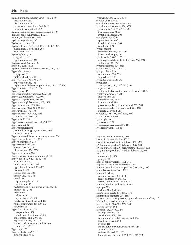
346 Index
petechiae and, 242pharyngitis and, 6, 7fthrombocytopenia from, 240, 241ftuberculin skin test with, 228
Human papillomavirus, hoarseness and, 34, 37“Hungry bone” syndrome, 334, 335fHuntington disease, 194, 195fHydranencephaly, 12, 13fHydroceles, scrotal, 136Hydrocephalus, 12, 13f, 15f, 184, 185f, 187f, 324
altered mental status and, 209fataxia and, 204, 205f
Hydronephrosis, 110congenital, 111fhypertension and, 133f
Hydroxylase deficiency, 132Hygroma, cystic, 8, 9fHymen, imperforate, amenorrhea and, 140, 141fHyperbilirubinemia
conjugated, 98prolonged indirect, 98
Hypercalcemia, 336–338, 337fhypertension and, 132nephrogenic diabetes insipidus from, 286, 287f, 336
Hypercalciuria, 120, 121f, 123fHypercapnea, 42Hypereosinophilic syndrome, 252, 253fHyper-IgE syndromes, 302, 303fHyper-IgM syndromes, 302, 303fHyperimmunoglobulinemia, 252, 253fHyperinsulinemia, 283f, 284Hyperkalemia, 320, 332–333, 333fHyperkyphosis, 172–173Hypernatremia, 324–325, 325f
irritable infant and, 308Hyperopia, 23f, 24Hyperostosis, infantile cortical, 298, 299fHyperoxia test, 42Hyperparathyroidism
maternal, during pregnancy, 334, 335fprimary, 336, 337f
Hyperparathyroidism-jaw tumor syndrome, 336Hyperphosphatemia, 334, 335fHyperpigmentation, 258Hyperprolactinemia, 262
amenorrhea and, 142hirsutism and, 274, 275f
Hyperproteinemia, 326Hypersensitivity pain syndromes, 52, 53fHypertension, 130–133, 131f, 133f
alkalemia and, 322headaches and, 186, 187fhyperthyroidism and, 130–132malignant, 130neutropenia and, 246obesity and, 282–284portal vein
splenomegaly and, 106varices and, 96
postinfectious glomerulonephritis and, 120primary, 131f, 132pulmonary
clues to, 66cyanosis and, 42, 43f
renal artery thrombosis and, 133fretinal examination for, 130–132secondary, 45
Hyperthyroidism, 10, 126chest pain from, 55fclinical characteristics of, 62, 63fgynecomastia and, 279f, 280hypertension and, 130–132systolic outflow murmur and, 66, 67ftremor and, 197f
Hypertropia, 20Hyperventilation, 52, 53f
syncope and, 59f, 60
Hypervitaminosis A, 336, 337fHypervolemia, 326–328Hypoalbuminemia, and edema, 128Hypoalbuminemic states, 334, 335fHypocalcemia, 334–335, 335f, 336
hoarseness and, 34, 35firritable infant and, 308
Hypoglycemia, 59f, 60apnea from, 48, 49firritable infant and, 308jaundice and, 98
Hypogonadismgynecomastia and, 278, 279fhypergonadotropic, 140
Hypokalemia, 330–331, 331fnephrogenic diabetes insipidus from, 286, 287f
Hypokinesia, 194, 195fHypomagnesemia, 334, 335fHyponatremia, 326–329, 327fHypoparathyroidism
autoimmune, 334, 335fsurgical, 334, 335f
Hypopituitarism, 261f, 262Hypoplasia
cartilage-hair, 244, 245f, 303f, 304thymic, 304
Hypothalamic dysfunction, amenorrhea and, 140, 141fHypothyroidism, 257f, 258
alopecia and, 217fhoarseness and, 34, 35fhypotonia and, 199fprecocious puberty in females and, 266, 267fprecocious puberty in males and, 264, 265fpubertal delay and, 262
Hypotonia, 198–203, 199f, 201f, 203fHypotrichosis, 216–217Hypotropia, 20Hypovolemia, 324Hypoxia, and headaches, 186, 187fHysterical syncope, 59f, 60
IIbuprofen, and neutropenia, 245fIdiopathic fat necrosis, 134, 135fIdiopathic hypercalcemia of infancy, 336, 337fIgA (immunoglobulin A) deficiency, 302, 303fIgA (immunoglobulin A) nephropathy, 120, 121f, 123fIgG (immunoglobulin G) subclass deficiencies, 302Ileus, 72
meconium, 92, 93fparalytic, 80
Iliotibial band syndrome, 163f, 164Imipramine, and Löeffler syndrome, 252Immune thrombocytopenic purpura (ITP), 240, 241fImmunization, fever from, 293fImmunodeficiency
common variable, 302, 303frecurrent infection and, 302severe combined, 302–303, 303f
Immunoglobulins, evaluation of, 302Impetigo, 225f
bullous, 218, 219f, 225fIncontinence, giggle, 116, 117f, 119fIncontinentia pigmenti, 218, 219fIncreased intracranial pressure, signs and symptoms of, 78, 81fIndomethacin, and neutropenia, 245fInfant, irritable, 306–309, 307f, 309fInfantile spasms, 192Infections, 26, 27f, 30, 223f
adenopathy and, 228arthritis and, 158, 161fautoimmune hemolytic anemia and, 234blood culture and, 294in bone, 166central nervous system, seizures and, 188cyanosis and, 42eosinophilia and, 252, 253ffever without source and, 290, 291f, 292, 293f
Human immunodeficiency virus (Continued)
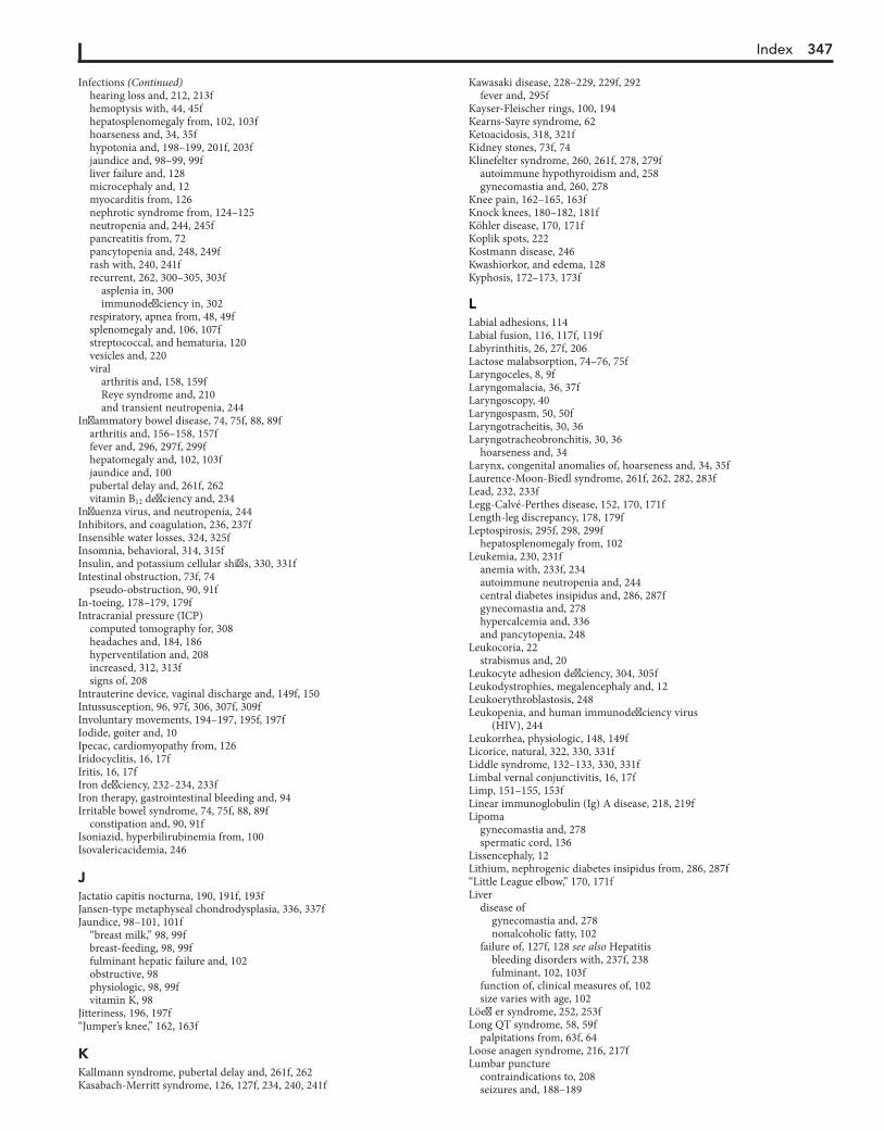
Index 347
hearing loss and, 212, 213fhemoptysis with, 44, 45fhepatosplenomegaly from, 102, 103fhoarseness and, 34, 35fhypotonia and, 198–199, 201f, 203fjaundice and, 98–99, 99fliver failure and, 128microcephaly and, 12myocarditis from, 126nephrotic syndrome from, 124–125neutropenia and, 244, 245fpancreatitis from, 72pancytopenia and, 248, 249frash with, 240, 241frecurrent, 262, 300–305, 303f
asplenia in, 300immunodeficiency in, 302
respiratory, apnea from, 48, 49fsplenomegaly and, 106, 107fstreptococcal, and hematuria, 120vesicles and, 220viral
arthritis and, 158, 159fReye syndrome and, 210and transient neutropenia, 244
Inflammatory bowel disease, 74, 75f, 88, 89farthritis and, 156–158, 157ffever and, 296, 297f, 299fhepatomegaly and, 102, 103fjaundice and, 100pubertal delay and, 261f, 262vitamin B12 deficiency and, 234
Influenza virus, and neutropenia, 244Inhibitors, and coagulation, 236, 237fInsensible water losses, 324, 325fInsomnia, behavioral, 314, 315fInsulin, and potassium cellular shifts, 330, 331fIntestinal obstruction, 73f, 74
pseudo-obstruction, 90, 91fIn-toeing, 178–179, 179fIntracranial pressure (ICP)
computed tomography for, 308headaches and, 184, 186hyperventilation and, 208increased, 312, 313fsigns of, 208
Intrauterine device, vaginal discharge and, 149f, 150Intussusception, 96, 97f, 306, 307f, 309fInvoluntary movements, 194–197, 195f, 197fIodide, goiter and, 10Ipecac, cardiomyopathy from, 126Iridocyclitis, 16, 17fIritis, 16, 17fIron deficiency, 232–234, 233fIron therapy, gastrointestinal bleeding and, 94Irritable bowel syndrome, 74, 75f, 88, 89f
constipation and, 90, 91fIsoniazid, hyperbilirubinemia from, 100Isovalericacidemia, 246
JJactatio capitis nocturna, 190, 191f, 193fJansen-type metaphyseal chondrodysplasia, 336, 337fJaundice, 98–101, 101f
“breast milk,” 98, 99fbreast-feeding, 98, 99ffulminant hepatic failure and, 102obstructive, 98physiologic, 98, 99fvitamin K, 98
Jitteriness, 196, 197f“Jumper’s knee,” 162, 163f
KKallmann syndrome, pubertal delay and, 261f, 262Kasabach-Merritt syndrome, 126, 127f, 234, 240, 241f
Kawasaki disease, 228–229, 229f, 292fever and, 295f
Kayser-Fleischer rings, 100, 194Kearns-Sayre syndrome, 62Ketoacidosis, 318, 321fKidney stones, 73f, 74Klinefelter syndrome, 260, 261f, 278, 279f
autoimmune hypothyroidism and, 258gynecomastia and, 260, 278
Knee pain, 162–165, 163fKnock knees, 180–182, 181fKöhler disease, 170, 171fKoplik spots, 222Kostmann disease, 246Kwashiorkor, and edema, 128Kyphosis, 172–173, 173f
LLabial adhesions, 114Labial fusion, 116, 117f, 119fLabyrinthitis, 26, 27f, 206Lactose malabsorption, 74–76, 75fLaryngoceles, 8, 9fLaryngomalacia, 36, 37fLaryngoscopy, 40Laryngospasm, 50, 50fLaryngotracheitis, 30, 36Laryngotracheobronchitis, 30, 36
hoarseness and, 34Larynx, congenital anomalies of, hoarseness and, 34, 35fLaurence-Moon-Biedl syndrome, 261f, 262, 282, 283fLead, 232, 233fLegg-Calvé-Perthes disease, 152, 170, 171fLength-leg discrepancy, 178, 179fLeptospirosis, 295f, 298, 299f
hepatosplenomegaly from, 102Leukemia, 230, 231f
anemia with, 233f, 234autoimmune neutropenia and, 244central diabetes insipidus and, 286, 287fgynecomastia and, 278hypercalcemia and, 336and pancytopenia, 248
Leukocoria, 22strabismus and, 20
Leukocyte adhesion deficiency, 304, 305fLeukodystrophies, megalencephaly and, 12Leukoerythroblastosis, 248Leukopenia, and human immunodeficiency virus
(HIV), 244Leukorrhea, physiologic, 148, 149fLicorice, natural, 322, 330, 331fLiddle syndrome, 132–133, 330, 331fLimbal vernal conjunctivitis, 16, 17fLimp, 151–155, 153fLinear immunoglobulin (Ig) A disease, 218, 219fLipoma
gynecomastia and, 278spermatic cord, 136
Lissencephaly, 12Lithium, nephrogenic diabetes insipidus from, 286, 287f“Little League elbow,” 170, 171fLiver
disease ofgynecomastia and, 278nonalcoholic fatty, 102
failure of, 127f, 128 see also Hepatitisbleeding disorders with, 237f, 238fulminant, 102, 103f
function of, clinical measures of, 102size varies with age, 102
Löeffler syndrome, 252, 253fLong QT syndrome, 58, 59f
palpitations from, 63f, 64Loose anagen syndrome, 216, 217fLumbar puncture
contraindications to, 208seizures and, 188–189
Infections (Continued)

348 Index
Lung disease, interstitial, cough and, 32, 33fLupus erythematosus, 219fLuteinizing hormone, 260Luteoma, 274, 275fLyme disease, 156–157, 157f, 159f, 223f, 298, 299f
hepatosplenomegaly from, 102rash of, 222, 225f
Lymphadenopathy, 227–231, 229f, 231fcough and, 32, 33f
Lymphangioma, 8, 18, 219fgynecomastia and, 278hoarseness and, 34
Lymphedema, 126, 127fLymphogranuloma venereum, 228Lymphoma
autoimmune hemolytic anemia and, 234autoimmune neutropenia and, 244bleeding disorders and, 237fgynecomastia and, 278non-Hodgkin, 230, 231fand pancytopenia, 248
Lymphoproliferative disorders, in autoimmune hemolytic anemia, 100Lysosomal diseases, megalencephaly and, 12
MMacrocephaly, 12–14Magnetic resonance imaging
for headaches, 184seizures and, 190
Malaria, 294, 295f, 299fMalformation(s). see also Camptomelic syndrome
arteriovenousaltered mental status and, 209fheadaches and, 185f, 187fheart failure and, 126–127hemoptysis and, 44systolic outflow murmur and, 66
brain, hypotonia and, 199fcongenital
ataxia and, 205fhypopituitarism and, 262
deformational, 14vascular, 36–37, 37f
Malignancyanemia with, 233f, 234autoimmune neutropenia and, 244on CXR, 296fever and, 295f, 299fhypercalcemia and, 336, 337fplatelet disorders and, 241f, 242pubertal delay and, 262signs and symptoms of, 231f
Mallory-Weiss tears, 94, 95fMalrotation, 75f, 76
with volvulus, 78, 79f, 81fMannitol, osmotic diuresis and, 286Manometry, 90, 91f, 93fMaple syrup urine disease, megalencephaly and, 12Marijuana, rhinorrhea from, 4Mass(es). see Tumor(s); specific anatomic siteMastalgia, 53fMayer-Rokitansky-Küster-Hauser syndrome, 140McCune-Albright syndrome (MAS), 264, 265f, 266, 267fMeasles, 6, 7f, 222, 223f, 292
hemoptysis from, 44petechiae and, 242rhinorrhea and, 4
Measles-mumps-rubella vaccine, and fever, 292Meatal stenosis
dysuria and, 114, 115fhematuria and, 120
Meckel diverticula, 95f, 96, 97fMeconium plug syndrome, 92–93, 93fMedications. see DrugsMedroxyprogesterone, amenorrhea and, 140–141Megacolon, congenital aganglionic, 90–91
Megalencephaly, 12–14, 13fMenarche, precocious, 144–145, 145fMeningitis, 176, 177f
apnea and, 49fataxia and, 207fbulging fontanel and, 14headaches and, 184–185, 185fhearing loss and, 212in irritable infant, 308, 309fmanagement of
in febrile infants 0 to 28 days, 291fin febrile infants 29 to 90 days, 291f
Meniscal injuries, knee pain and, 162, 163fMeniscus, discoid lateral, 163f, 164Menorrhagia, 144–147Menstrual cycles, 144–1476-Mercaptopurine, 248Mesenteric adenitis, 72, 73fMesenteric artery syndrome, superior, 78, 79fMetabolic disorders
altered mental status and, 208–209, 209fcongenital, apnea and, 48, 49f, 50ffailure to thrive and, 310, 313fhypotonia and, 199f, 200, 203fand liver failure, 128nephrogenic diabetes insipidus from, 286, 287fand pancytopenia, 248
Metabolic storage disorders, hepatomegaly and, 102Metabolism
errors of, 78inborn errors of, 312
Metatarsus adductus, 178, 179fMethanol, 318, 321fMethemoglobin, 42Methemoglobinemia, 42, 43fMethotrexate, pancytopenia and, 248, 250Metrorrhagia, 144–147Microcephaly, 12, 13f, 15fMicrolithiasis, testicular, 136–137, 137fMigraine, 184, 185f, 186, 187f
abdominal, 82, 83fbasilar-type, 59f, 60, 204, 205fophthalmoplegic, 20–21, 21f
Milk-alkali syndrome, 336Miller-Fisher syndrome, 206, 207fMineralocorticoid, 322, 323fMirror movements, 197Mitral valve prolapse, 53, 53f
long QT syndrome and, 58Möbius syndrome, 20, 21f
pubertal delay and, 261f, 262Mononucleosis, 6, 106, 292
anemia and, 234rhinorrhea and, 4
Monosymptomatic primary nocturnal enuresis, 118–119Movement disorders, 194Mucopolysaccharidoses, megalencephaly and, 12Müllerian agenesis, 140, 141fMüllerian anomalies, vaginal discharge and, 148, 149fMüllerian tract abnormalities, dysmenorrhea and, 138Multiple lentigines syndrome, 261f, 262Mumps, arthritis and, 158, 161fMurmurs. see Heart murmursMuscle pain, 166–171Myalgias, 166, 167fMyasthenia gravis, 20, 21f, 198, 199f, 201fMycoplasma pneumoniae, 38, 39fMyelitis, transverse, 200–201, 203fMyelodysplastic syndrome, and pancytopenia, 248Myelofibrosis, and pancytopenia, 248Myocarditis, 53, 53f
causes of, 126, 127fby electrocardiogram, 126long QT syndrome and, 58viral, and edema, 126
Myoclonus, 190, 193f, 196, 197focular, 26, 27f

Index 349
Myoclonus and opsoclonus syndrome, 196, 197fMyoglobinuria, 120, 121f, 123fMyopia, 23f, 24Myositis, viral-related, 166, 167fMyositis ossificans, 168
NNaloxone, 208, 209fNarcolepsy, 192, 193f, 315f, 316Narcotics, cyanosis and, 43Nasopharyngeal angiofibroma, juvenile, 4, 5fNavicular bones, accessory, 170, 171fNeck
masses of, 8–11stiff, 176–177, 177f
Neisseria gonorrhoeae, in pelvic inflammatory disease, 72–73, 102Neisseria meningitidis infection, 240, 241fNeoplasms
abnormal vaginal bleeding and, 145f, 146, 147fintracranial, 26, 27f
Nephrolithiasis, 120, 121f, 123fback pain and, 172, 173f
Nephrotic syndrome, 124, 326congenital, 124–125and glomerulonephritis, 124hematuria with, 120–123, 121f, 123fminimal change, 124, 125fperitonitis with, 72–76proteinuria and, 124
Nerve compression, 168–169, 169fNeuralgia, occipital, 186, 187fNeuritis, optic, 22, 23f
headaches and, 185fNeuroblastoma, 10–11, 11f, 18
abdominal masses and, 110, 111fhypercalcemia and, 336lymphadenopathy and, 229f, 231fmetastatic, gynecomastia and, 278opsoclonus and, 196in pancytopenia, 248
Neurofibromas, gynecomastia and, 278Neurofibromatosis
congenital pseudoarthrosis of tibia and, 180hearing loss and, 213f
Neurogenic bladder, 111fNeuroleptics, tardive dyskinesia and, 194Neurologic disorders, and acquired nystagmus, 26Neuropathies, and distal weakness, 200, 201fNeutropenia, 244–247, 245f
autoimmune, 244, 245fof infancy, 244
chronic, 244congenital, 244cyclic, 244, 245fand disorders of immune dysfunction, 246familial benign, 245f, 246immune neonatal, 244–245, 245fand pancytopenia, 248secondary defects of immune system and, 300
Nevus sebaceous lesions, 216, 217fNicotine, irritable infant and, 308Night pain, 154Night terrors, 192, 193f, 315f, 316–315Nightmares, 315f, 316–315Nitrogen mustard, and pancytopenia, 248Noise-induced hearing loss (NIHL), 212, 213fNonallergic inflammatory rhinitis with eosinophils (NARES), 4, 5fNon-Hodgkin lymphoma, 230, 231fNonsteroidal antiinflammatory drugs (NSAIDs), 94
functional hypoaldosteronism from, 320platelet function defects and, 237f
Noonan syndrome, 260, 261fNucleic acid amplification test, for vaginal discharge, 148Nucleic acid antigen tests, vaginal discharge and, 148“Nursemaid’s elbow,” 166Nutcracker syndrome, 120, 121f, 123f
Nutrition, parenteral, cholelithiasis and, 72Nystagmus, 26
acquired, 26, 27fcongenital
idiopathic, 26, 27fsensory, 26, 27f
OObesity, 49f, 282–285, 283f
in gynecomastia, 278and precocious puberty in females, 266
Occipitoatlantal joints instability, 176Ocular bobbing, 27fOcular flutter, 27fOcular instability of infancy, 20, 21fOculogyric crisis, 176Oligoarthritis, asymmetric, 158Oligomenorrhea, 140, 144–147Omphalitis, thrombus formation and, 96Oophoritis, autoimmune, 260, 261fOphthalmia neonatorum, 16Ophthalmic disorders, 16, 20–21, 21f, 22–25, 23f, 26–28, 27fOpsoclonus, 27f, 196Oral-motor dysfunction, 310, 311fOrchitis, 134, 135fOrthostatic hypotension, and syncope, 59f, 60Oscillopsia, 26Osgood-Schlatter disease, 162, 163fOsler-Weber-Rendu syndrome, 44Osteochondritis dissecans (OCD), 162–163, 163fOsteochondrosis of capitellum, 170, 171fOsteodystrophy, Albright hereditary, 282, 283f, 334Osteoma, osteoid, neck pain and, 176, 177fOsteomyelitis, 152, 153f, 166, 167f, 297f, 298, 299f
blood culture and, 294treatment of, 291fvertebral, 172, 173f
Osteopetrosis, 108, 109fOsteoporosis, in pancytopenia, 248Otitis media, 2, 3f, 212, 213f
diarrhea and, 84–85with effusion, 2, 3flabyrinthitis and, 26by tympanogram, 213
Out-toeing, 178–179, 179fOvarian cyst, 110, 111fOvernight polysomnogram, 314Overuse syndromes, 170, 171f
back pain and, 173f, 174Oxytocin, and jaundice, 98
PPachygyria, 12Pain
abdominal, 71–77acute, 73f
signs and symptoms of, 72chronic or recurrent, 74, 75f
associated with menstruation, 138back, 172–175, 173fchest, 51–57, 53f, 55f, 57f
asthma and, 52, 53f, 57fhyperventilation in, 52, 53fmuscular, 52, 53fmyocardial ischemia and, 56skeletal causes of, 52
chronic, 74ear, 1–3, 3fextremity, 166–171, 167f, 169f, 171fknee, 162–165, 163ffrom limp, 152neck, 176–177, 177fpatellofemoral, 162psychogenic, 74scrotal, 134–135
Palpitations, 62–65, 63f
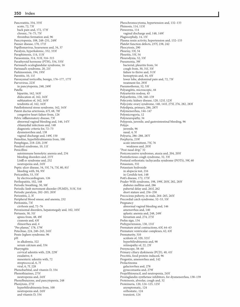
350 Index
Pancreatitis, 334, 335facute, 72, 73fback pain and, 172, 173fchronic, 74–75, 75fthrombus formation and, 96
Pancytopenia, 108, 248–251, 249fPanner disease, 170, 171fPapillomavirus, hoarseness and, 34, 37Paralysis, hyperkalemic, 332, 333fParaphimosis, 114, 115fParasomnia, 314, 315f, 316–315Parathyroid hormone (PTH), 334, 335fParinaud’s oculoglandular syndrome, 16Parinaud’s syndrome, 20, 21fParkinsonism, 194, 195fParotitis, 10, 11fParoxysmal torticollis, benign, 176–177, 177fParvovirus, 223f
in pancytopenia, 248, 249fPatella
bipartite, 162, 163fdislocation of, 162, 163fsubluxation of, 162, 163ftendinitis of, 162, 163f
Patellofemoral stress syndrome, 162, 163fPatent ductus arteriosus, 67f, 68, 70f
congestive heart failure from, 126Pelvic inflammatory disease, 73f
abnormal vaginal bleeding and, 146, 147fchlamydial infections and, 148diagnostic criteria for, 72–73dysmenorrhea and, 138vaginal discharge and, 149f, 150
Pemoline, hyperbilirubinemia from, 100Pemphigus, 218–220, 219fPendred syndrome, 10, 11fPenicillins
autoimmune hemolytic anemia and, 234bleeding disorders and, 237fLöeffler syndrome and, 252neutropenia and, 245f
Peptic ulcer disease, 54, 55f, 74, 75f, 80, 81fbleeding with, 94, 95f
Pericarditis, 53, 53fby electrocardiogram, 126
Perihepatitis, 102, 148Periodic breathing, 50, 50fPeriodic limb movement disorder (PLMD), 315f, 316Periodic paralysis, 202–203, 203fPeriostitis, 2, 3fPeripheral blood smear, and anemia, 232Peritonitis, 73f
cirrhosis and, 72–76Peroxisomal disorders, hepatomegaly and, 102, 105fPertussis, 30, 31f
apnea from, 48, 49fcyanosis and, 43frhinorrhea and, 4
“Pes planus,” 178, 179fPetechiae, 224, 240–243, 241fPeutz-Jeghers syndrome, 96pH
in alkalemia, 322serum calcium and, 334
Pharyngitiscervical adenitis with, 228, 229fexudative, 6mesenteric adenitis with, 72streptococcal, 6, 7fviral, 6, 7f, 228
Phenobarbital, and vitamin D, 334Phenothiazines, 275f
neutropenia and, 245fPhenylbutazone, and pancytopenia, 248Phenytoin, 275f
hyperbilirubinemia from, 100neutropenia and, 245fand vitamin D, 334
Pheochromocytoma, hypertension and, 132–133Phimosis, 114, 115fPinworms, 114
vaginal discharge and, 148, 149fPlagiocephaly, 14, 15fPlasma renin activity, hypertension and, 132–133Platelet function defects, 237f, 238, 242Pleocytosis, 290Pleurisy, 53f, 54Pleuritis, 53f, 54Pleurodynia, 52, 53fPneumonia, 39f
bacterial, pleuritis from, 54cough from, 30, 31f, 33ffailure to thrive and, 313fhemoptysis and, 44, 45flower lobe, abdominal pain and, 72, 73ftreatment for, 293f
Pneumothorax, 52, 53fPolyangiitis, microscopic, 44Polyarteritis nodosa, 45Polyarthritis, 158, 160–159Polycystic kidney disease, 120, 121f, 123fPolycystic ovary syndrome, 140, 141f, 275f, 276, 282, 283fPolydipsia, primary, 286, 287fPolymenorrhea, 144–147Polymicrogyria, 12Polyneuropathy, 34Polyposis, juvenile, and gastrointestinal bleeding, 96Polyps
juvenile, 96nasal, 4, 5f
Polyuria, 286–288, 287fPorphyria, 219f
acute intermittent, 75f, 76weakness and, 203f
“Post nasal drip,” 32Postconcussive syndromes, ataxia and, 204, 205fPostinfectious cough syndrome, 32, 33fPostural orthostatic tachycardia syndrome (POTS), 59f, 60Potassium, 332Potassium hydroxide
in alopecia test, 216in Candida test, 148
Pott’s disease, 172, 173fPrader-Willi syndrome, 198, 199f, 203f, 282, 283f
diabetes mellitus and, 286pubertal delay and, 261f, 262short stature and, 256–258
Precocious puberty, in male, 264–265, 265fPrecordial catch syndrome, 52–53, 53fPregnancy
abnormal vaginal bleeding and, 146amenorrhea and, 140aplastic anemia and, 248, 249fhirsutism and, 274, 275f
Prehn sign, 134Prehypertension, 130, 131fPremature atrial contractions, 63f, 64–63Premature ventricular complexes, 62, 63fPrematurity, 310
acidosis of, 320, 321fhyperbilirubinemia and, 98retinopathy of, 22, 23f
Presyncope, 58–60Primary ciliary dyskinesia (PCD), 40, 41fProctitis, food protein-induced, 96Progestin, amenorrhea and, 142Prolactinoma
galactorrhea and, 278gynecomastia and, 279f
Propylthiouracil, and neutropenia, 245fProstaglandin synthetase inhibitors, for dysmenorrhea, 138–139Proteinosis, alveolar, cough and, 32Proteinuria, 120, 124–125, 125f
asymptomatic, 124orthostatic, 124transient, 124
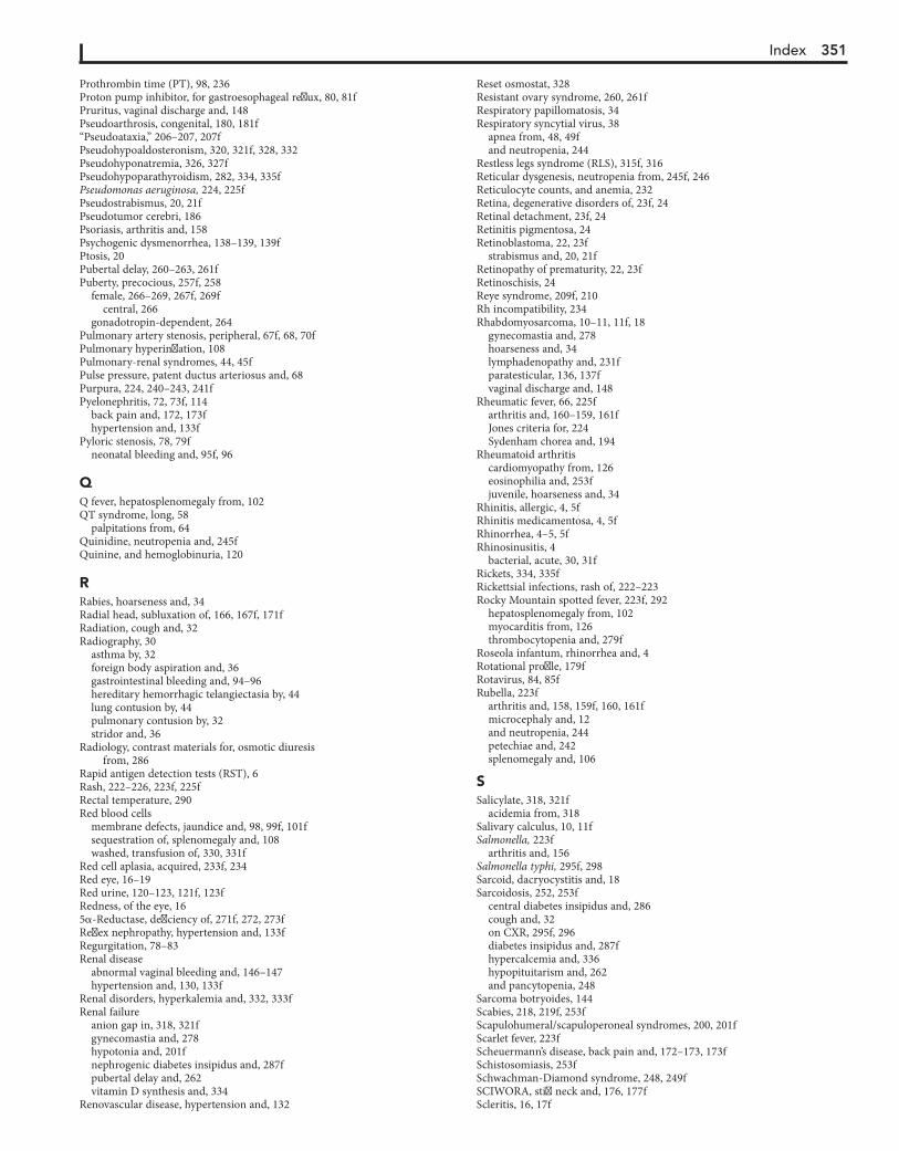
Index 351
Prothrombin time (PT), 98, 236Proton pump inhibitor, for gastroesophageal reflux, 80, 81fPruritus, vaginal discharge and, 148Pseudoarthrosis, congenital, 180, 181f“Pseudoataxia,” 206–207, 207fPseudohypoaldosteronism, 320, 321f, 328, 332Pseudohyponatremia, 326, 327fPseudohypoparathyroidism, 282, 334, 335fPseudomonas aeruginosa, 224, 225fPseudostrabismus, 20, 21fPseudotumor cerebri, 186Psoriasis, arthritis and, 158Psychogenic dysmenorrhea, 138–139, 139fPtosis, 20Pubertal delay, 260–263, 261fPuberty, precocious, 257f, 258
female, 266–269, 267f, 269fcentral, 266
gonadotropin-dependent, 264Pulmonary artery stenosis, peripheral, 67f, 68, 70fPulmonary hyperinflation, 108Pulmonary-renal syndromes, 44, 45fPulse pressure, patent ductus arteriosus and, 68Purpura, 224, 240–243, 241fPyelonephritis, 72, 73f, 114
back pain and, 172, 173fhypertension and, 133f
Pyloric stenosis, 78, 79fneonatal bleeding and, 95f, 96
QQ fever, hepatosplenomegaly from, 102QT syndrome, long, 58
palpitations from, 64Quinidine, neutropenia and, 245fQuinine, and hemoglobinuria, 120
RRabies, hoarseness and, 34Radial head, subluxation of, 166, 167f, 171fRadiation, cough and, 32Radiography, 30
asthma by, 32foreign body aspiration and, 36gastrointestinal bleeding and, 94–96hereditary hemorrhagic telangiectasia by, 44lung contusion by, 44pulmonary contusion by, 32stridor and, 36
Radiology, contrast materials for, osmotic diuresis from, 286
Rapid antigen detection tests (RST), 6Rash, 222–226, 223f, 225fRectal temperature, 290Red blood cells
membrane defects, jaundice and, 98, 99f, 101fsequestration of, splenomegaly and, 108washed, transfusion of, 330, 331f
Red cell aplasia, acquired, 233f, 234Red eye, 16–19Red urine, 120–123, 121f, 123fRedness, of the eye, 165a-Reductase, deficiency of, 271f, 272, 273fReflex nephropathy, hypertension and, 133fRegurgitation, 78–83Renal disease
abnormal vaginal bleeding and, 146–147hypertension and, 130, 133f
Renal disorders, hyperkalemia and, 332, 333fRenal failure
anion gap in, 318, 321fgynecomastia and, 278hypotonia and, 201fnephrogenic diabetes insipidus and, 287fpubertal delay and, 262vitamin D synthesis and, 334
Renovascular disease, hypertension and, 132
Reset osmostat, 328Resistant ovary syndrome, 260, 261fRespiratory papillomatosis, 34Respiratory syncytial virus, 38
apnea from, 48, 49fand neutropenia, 244
Restless legs syndrome (RLS), 315f, 316Reticular dysgenesis, neutropenia from, 245f, 246Reticulocyte counts, and anemia, 232Retina, degenerative disorders of, 23f, 24Retinal detachment, 23f, 24Retinitis pigmentosa, 24Retinoblastoma, 22, 23f
strabismus and, 20, 21fRetinopathy of prematurity, 22, 23fRetinoschisis, 24Reye syndrome, 209f, 210Rh incompatibility, 234Rhabdomyosarcoma, 10–11, 11f, 18
gynecomastia and, 278hoarseness and, 34lymphadenopathy and, 231fparatesticular, 136, 137fvaginal discharge and, 148
Rheumatic fever, 66, 225farthritis and, 160–159, 161fJones criteria for, 224Sydenham chorea and, 194
Rheumatoid arthritiscardiomyopathy from, 126eosinophilia and, 253fjuvenile, hoarseness and, 34
Rhinitis, allergic, 4, 5fRhinitis medicamentosa, 4, 5fRhinorrhea, 4–5, 5fRhinosinusitis, 4
bacterial, acute, 30, 31fRickets, 334, 335fRickettsial infections, rash of, 222–223Rocky Mountain spotted fever, 223f, 292
hepatosplenomegaly from, 102myocarditis from, 126thrombocytopenia and, 279f
Roseola infantum, rhinorrhea and, 4Rotational profile, 179fRotavirus, 84, 85fRubella, 223f
arthritis and, 158, 159f, 160, 161fmicrocephaly and, 12and neutropenia, 244petechiae and, 242splenomegaly and, 106
SSalicylate, 318, 321f
acidemia from, 318Salivary calculus, 10, 11fSalmonella, 223f
arthritis and, 156Salmonella typhi, 295f, 298Sarcoid, dacryocystitis and, 18Sarcoidosis, 252, 253f
central diabetes insipidus and, 286cough and, 32on CXR, 295f, 296diabetes insipidus and, 287fhypercalcemia and, 336hypopituitarism and, 262and pancytopenia, 248
Sarcoma botryoides, 144Scabies, 218, 219f, 253fScapulohumeral/scapuloperoneal syndromes, 200, 201fScarlet fever, 223fScheuermann’s disease, back pain and, 172–173, 173fSchistosomiasis, 253fSchwachman-Diamond syndrome, 248, 249fSCIWORA, stiff neck and, 176, 177fScleritis, 16, 17f
352 Index
Sclerosismultiple, ataxia and, 207ftuberous, 120–123, 121f, 123f
Sclerosus, lichen, 144, 145fScoliosis, 172–173, 173fScrotal pain, 134–135, 135fScrotal swelling, painless, 136–137, 137fSebaceous nevus of Jadassohn, 216, 217fSecondary hyperparathyroidism, 336, 337fSedatives
cyanosis and, 43neutropenia and, 245frespiratory acidosis and, 318
Segawa syndrome, 196Seizures, 188–193, 189f, 191f, 193f
absence, 191f, 192, 193facute episodic ataxia in, 206–207characteristics of, 194cyanosis and, 43ffebrile, 188, 189fheadaches and, 186, 187fhyperventilation and, 192–193, 193fmyoclonic, 191f, 192, 193frepetitive purposeless movements and, 192syncope and, 58, 59f
Sepsisapnea and, 49fjaundice and, 100myocarditis from, 126neonatal petechiae and, 240thrombus formation and, 96
Sever’s disease, 170, 171fSexual abuse, dysuria and, 114, 115fSexual activity
dysmenorrhea and, 138hepatomegaly and, 102jaundice and, 100pharyngitis and, 6
Sexual historyabnormal vaginal bleeding and, 144amenorrhea and, 140vaginal discharge and, 148
Sexually transmitted infections, dysuria and, 114Shaken baby syndrome, 208Shigella, vaginal discharge and, 148Shin splints, 166–167, 167f, 171fShingles, 52Shwachman-Diamond syndrome, 86, 87f, 89f, 244, 313fSickle cell disease, 106–108, 232, 234
congenital asplenia and, 300fever and, 290–292infant irritability and, 306, 309fnephrogenic diabetes insipidus from, 286, 287fpubertal delay and, 262transient aplastic crisis with, 248, 249f
“Silent aspiration,” 40Silver nitrate, conjunctivitis and, 16Sinding-Larsen-Johansson syndrome, 162, 163fSinus histiocytosis, 230, 231fSinusitis, 30
headaches and, 185fvomiting with, 78, 79f
Skeletal dysplasia, 181f, 257f, 258Skull, abnormalities of, 14Sleep disturbances, 314–316, 315fSleep hygiene, 314, 315fSlipped capital femoral epiphysis (SCFE), 152, 153f, 164,
170, 171fSlipping rib syndrome, 54, 55fSmith-Lemli-Opitz syndrome (SLOS), 272–271Snoring, 314
cyanosis and, 42hypertension and, 130
Sodium, 326Sodium wasting, renal disorders causing, 328Solvents, inhaled, rhinorrhea from, 4Somnambulism, 315f, 316–315
Sorbitol, malabsorption of, 74–76Sore throat, 6–7, 7fSpasmus nutans, 26, 27f, 191f, 192, 193fSpermatoceles, 136Spherocytosis, 235
transient aplastic crisis with, 248, 249fSpinal cord disorders, hypotonia and, 199f, 200, 201fSpinal muscular atrophies, 199f, 200, 201fSplenomegaly, 106–109, 107f, 109fSpondylitis
juvenile ankylosing, 156–158, 157ftuberculous, 172
Spondyloarthritis, 156–158Spondyloarthropathy, 156–158Spondylolisthesis, 172, 173fSpondylolysis, 172, 173fStance phase, 152–153Staphylococcal scalded skin syndrome, 218, 219f, 223f, 225fStaphylococcus aureus
discitis from, 172parotitis and, 10
Startle disease, 190, 191f, 193fStature, short, 255–259, 257fStatus epilepticus, 188–193Stevens-Johnson syndrome (SJS), 220, 225fStiff neck, 176–177, 177f“Still murmur,” 67f, 68Stimulant medications, for sleep disturbances, 314, 315f“Stingers,” 166, 167fStorage disorders, 108, 109fStrabismus, 20–21, 21f
comitant, 20restrictive, 21sensory, 20, 21f
Streptococcusadenopathy and, 228, 229farthritis and, 160gastrointestinal fissures and, 96rash and, 223f, 225f
Stridor, 36–37, 37fSubconjunctival hemorrhage, 16, 17fSubglottic stenosis, 36, 37fSulfonamides
Löeffler syndrome and, 252neutropenia and, 245fpancytopenia and, 248thrombocytopenia from, 241f
Supraclavicular bruit, 67f, 68Supraclavicular systolic murmur, 67f, 68Supraventricular tachycardia, 63f, 64
syncope from, 58Wolff-Parkinson-White syndrome and, 58
Swyer syndrome, 260Sydenham chorea, 194, 195fSyncope, 58–61, 59fSyndrome of inappropriate antidiuretic hormone (SIADH), 328Synostosis, cranial suture, 14Synovitis, transient, 152, 153f, 156, 157fSyphilis, 228
adenopathy and, 229f, 230dacryocystitis and, 18hearing loss and, 212–213hepatosplenomegaly from, 102hoarseness and, 34rash and, 223frhinorrhea and, 4, 5f
Systemic lupus erythematosusabnormal vaginal bleeding and, 146–147arthritis and, 160, 161fautoimmune neutropenia and, 244bleeding disorders and, 237f, 238cardiomyopathy from, 126fever and, 295f, 297fhematuria and, 120–123hemolytic anemia and, 100hypertension and, 133fIgA deficiency and, 302
Index 353
and pancytopenia, 248pulmonary hemorrhage and, 45
TTachycardia
sinus, 62ventricular, 63f, 64
Talipes equinus, in-toeing and, 179fTarsal coalitions, 170, 171fTay-Sachs disease, megalencephaly and, 1299mcTc-pertechnetate testicular flow scan, for scrotal pain, 134Teething, 306, 307fTelangiectasia
ataxia, 204, 205f, 304hereditary hemorrhagic, 44
Telogen effluvium, 216–217, 217fTemporomandibular joint (TMJ) syndrome, and headaches, 186, 187fTendinitis, patellar, 162, 163fTenosynovitis
gonococcal disease and, 156of wrist, 170, 171f
Teratomas, 8, 9fprecocious puberty in males and, 264, 265f
Testicular differentiation, defects in, 270, 271f, 273fTesticular feminization, 140, 260, 261fTesticular torsion, 134
XY gonadal agenesis syndrome, 272Testotoxicosis, 264, 265fTetanus, hoarseness and, 34Tetracyclines, hyperbilirubinemia from, 100Tetralogy of Fallot, cyanosis and, 42, 43fTexidor twinge, 52–53Thalassemia, 106, 232, 233ftransient aplastic crisis with, 248, 249fThelarche, premature, 266, 267fThiamine deficiency, hoarseness and, 34Thrombocytopenia, 240–242
abnormal vaginal bleeding and, 146autoimmune neutropenia and, 244and edema, 126and pancytopenia, 248–250
Thrombocytopenia-absent radius (TAR) syndrome, 236, 240, 241f, 253f
Thrombosisportal vein
splenomegaly and, 106varices and, 96
renal artery, hypertension and, 133frenal vein, 110, 111fvenous, risk factors for, 54
Thrombotic thrombocytopenic purpura, 240, 241fThyroglossal duct cysts, 8, 9f, 11fThyroiditis, 10, 11f
Hashimoto, diabetes mellitus and, 286Thyrotoxicosis, gynecomastia and, 280Tibia vara, 180, 181fTibial stress syndrome, medial, 166–167Tibial torsion, 178, 179fTick paralysis, 201f, 203fTics, 196–197, 197fTietze syndrome, 52Tinea capitis, 216, 217f
adenopathy and, 228Toe, atavistic great, 178, 179fToe walking, 178–179, 179fTorsion, 178–179, 179f
rotational profile and, 178, 179fTorsion dystonia, 178–179, 179f, 196Torsion of the testicular appendix, 134, 135fTorticollis, 8, 9f, 176–177, 195f, 196
congenital muscular, 176, 177fdystonia and, 196
Torticollis, paroxysmal, 191f, 193fTourette syndrome, 197Tourniquet syndrome, 306, 307f
Toxocara, 252, 253fToxoplasmosis, 298, 299f
hearing loss and, 212–213microcephaly and, 12splenomegaly and, 106
Tracheitis, 36, 37fTracheobronchitis, 30, 31f
hemoptysis with, 45fTracheomalacia, wheezing in, 40, 41fTransfusion
for blood loss, 235and potassium cellular shifts, 330
Transient erythroblastopenia of childhood, 234Trauma
acute pancreatitis and, 72, 73fbirth, and hoarseness, 34, 35fblunt, strabismus and, 21, 21fintentional, 166and petechiae, 240vulvovaginal, 144, 145f
Tremor, 196, 197fTrichinosis, 253fTrichomonas, vaginal discharge and, 148Trimethoprim, and pancytopenia, 250Tuberculosis, 228, 295f, 296, 299f
adenopathy and, 228, 229fcentral diabetes insipidus and, 286, 287fcough and, 32, 33fdacryocystitis and, 18disseminated, 230failure to thrive and, 313fhemoptysis from, 44hepatosplenomegaly from, 102hoarseness and, 34hypopituitarism and, 262nonpulmonary, 298and pancytopenia, 248skin test for, 228
Tubulointerstitial nephritis, 120–123, 121f, 123fTularemia, hepatosplenomegaly from, 102Tumor(s)
adenomatoid, 136, 137fadenopathy and, 228–231adrenal, 275f, 276, 278, 279faltered mental status and, 209fataxia and, 204, 205f, 207fback pain and, 172, 173f, 174bone, 166, 167fcentral diabetes insipidus and, 286, 287festrogen-secreting, 278, 279fgynecomastia and, 278, 279fheadaches and, 184, 185f, 187fhirsutism and, 274hypopituitarism and, 262hypotonia and, 199f, 203fnight pain and, 153f, 154orbital, 18, 19fovarian, 266, 267f, 275f, 276precocious puberty in females and, 266, 267fprecocious puberty in males and, 264renal, hypercalcemia and, 336respiratory acidosis and, 318stiff neck and, 176, 177ftesticular, 136, 137f
Turner syndrome, 256–257, 260, 261f, 282, 283famenorrhea and, 140, 141fautoimmune hypothyroidism and, 258
Tympanometry, 212Tympanosclerosis, 213fTyphoid fever, 223f, 292, 295f, 299f
hepatosplenomegaly from, 102
UUlcer, peptic, 54, 74, 75f
bleeding with, 94Ulnar nerve irritation/entrapment, 169f, 170, 171f
Systemic lupus erythematosus (Continued)
354 Index
Ultrasoundabdominal, 110as abdominal diagnostic aid, 72color Doppler, for scrotal pain, 134
Umbilical vein catheterization, thrombus formation and, 96Upper GI series, 80Urea, osmotic diuresis and, 286Ureter, ectopic, 116–117, 117f, 119f
vaginal discharge and, 148, 149fUreteral obstruction, hypertension and, 133fUreteropelvic junction obstruction, 76Urethra
obstruction of, 116, 117f, 119fprolapse of, 114, 144, 145f
Urethritis, nonspecific, 114Urinary incontinence, 116, 117f, 119fUrinary tract infection, 114
diarrhea and, 84–85enuresis and, 116hematuria and, 120, 121f, 123ftreatment of, 290–291, 291f, 293f
UrinepH of, acidemia and, 320red, 120–123, 121f, 123ftoxicology, apnea and, 48
Urine culture, 114Urine pregnancy test, 110Urine tests, for metabolic disorders, 78Urolithiasis, 73f, 74Uveitis, 22, 23f
VVaccine
infant irritability and, 306, 307freactions from, fever and, 292
Vaginal bleeding, abnormal, 144–147, 145f, 147fVaginal discharge, 148–150, 149f
chronic, 150Vaginal septum, transverse, amenorrhea and, 140, 141fVaginitis, 146, 147fVaginosis, bacterial, 148–149, 149fValproate, hyperbilirubinemia from, 100Varicella, 219f, 225f
acute cerebellar ataxia and, 206and neutropenia, 244petechiae and, 242
Varices, 95f, 96Varicoceles, 136Vasculitis, 223f, 241f, 242
altered mental status and, 209fheadaches and, 185f, 187fsyndromes, arthritis and, 158, 159f
Vasopressin, in diagnosis of diabetes insipidus, 324, 325fVasovagal syncope, 58, 59f, 60Velocardiofacial syndrome, 334Venous hums, 67f, 68Ventricular septal defects, 66–67, 67f, 70fVertigo, benign paroxysmal, 191f, 192, 193f, 204Vesicles, 218–221, 219fVincristine toxicity, hoarseness and, 34Vision loss, 24
psychogenic, 22, 23f
Vitamin B12 deficiency, 234neutropenia and, 246and pancytopenia, 250
Vitamin D deficiency, 334, 335fVitamin K deficiency, 236, 237fVitreoretinopathy, familial exudative, 24Vocal cords
dysfunction of, 40–39, 41fparalysis of, 37, 37fparalysis of, hoarseness and, 34, 35f
Volvulus, 78Vomiting, 78–83, 79f, 81f, 83f
abdominal migraine and, 82acute abdomen and, 78alkalosis from, 322, 323fchronic recurrent, 78cyclic recurrent, 78psychogenic, 80
von Willebrand disease, 236–237, 237fVulvar mucosa, 148Vulvovaginitis, 148, 149f
allergic, 149f, 150candidal, 148, 149finfectious, 144, 145f
WWAGR (Wilms tumor, aniridia, GU malformations, and mental
retardation) syndrome, 270Warfarin, 236Water intoxication, 328, 329fWeakness, 198–203, 199f, 201f, 203fWeber-Cockayne syndrome, 218Wegener granulomatosis, 44, 45fWheezing, 38–41, 39f, 41f
cyanosis and, 42“Whiff ” test, for bacterial vaginosis, 148“White-coat hypertension,” 130Williams syndrome, 336, 337fWilms tumor, 110, 111fWilson disease, 100, 104, 105f, 194, 195f
fulminant hepatic failure and, 102hypokinesia and, 195fjaundice and, 100
Wiskott-Aldrich syndrome, 236, 240, 241f, 252, 253frecurrent infection with, 303f, 304
Withdrawal bleeding, 144, 145fWolff-Parkinson-White syndrome
palpitations from, 63f, 64supraventricular tachycardia, 58, 59f
Wolfram syndrome, 286, 287f“Wry neck,” 176–177
XX-ray
abdominal pain and, 72chest, hilar adenopathy on, 296
ZZellweger syndrome, 104, 105f

Smarter, Faster Searchfor Better Patient CareUnlike a conventional search engine, ClinicalKeyis specifically designed to serve doctors by providingthree core components:
Start searching with ClinicalKey today!Visit ClinicalKey.com for more informationand subscription options.
Smarter search. Faster answers.
ComprehensiveContent1
The most current,evidence-basedanswers availablefor every medicaland surgicalspecialty.
2 TrustedAnswers
Content suppliedby Elsevier, theworld’s leadingprovider of healthand science information.
3 Unrivaled Speed to Answer
Faster, more relevantclinical answers, soyou can spend lesstime searching andmore time caring forpatients.