paula beatriz de araujo - core
TRANSCRIPT
PAULA BEATRIZ DE ARAUJO
DESENVOLVIMENTO PÓS-MARSUPIAL
E ECOLOGIA POPULACIONAL DE
ATLANTOSCIA FLORIDANA (VAN NAME, 1940)
(CRUSTACEA, ISOPODA, ONISCIDEA)
NA RESERVA BIOLÓGICA DO LAMI, PORTO ALEGRE, RS
UNIVERSIDADE FEDERAL DO RIO GRANDE DO SUL INSTITUTO DE BIOCIÊNCIAS
PORTO ALEGRE 2003
Tese de Doutorado apresentada ao Programa de
Pós-Graduação em Biologia Animal, Instituto de Biociências,da
Universidade Federal do Rio Grande do Sul, como requisito
parcial à obtenção do título de Doutor em Biologia Animal.
Área de concentração: Crustáceos Orientadora: Profa.Dra. Georgina Bond-Buckup
AGRADECIMENTOS
À Profa. Dra. Gerorgina Bond Buckup pela orientação e presença
marcante em todas as etapas do trabalho. Obrigada pelo apoio, amizade, incentivo,
dedicação e companheirismo.
A minha família, em especial ao Aldo e ao Pedro que “se viraram
sozinhos” nos momentos em que eu estava ausente, envolvida com a tese. Aos meus
pais que sempre me apoiaram.
Ao Biólogo Rodrigo Cambará Printes, Gerente da Reserva Biológica do
Lami, e a SMAM (Secretaria Municipal do Meio Ambiente) por permitirem a realização
deste trabalho na Reserva Biológica do Lami.
Ao Prof. Ludwig Buckup pelas críticas e sugestões e por estar sempre
disposto a ajudar. Obrigada por toda a ajuda com as fotografias.
À Aline, Minnelise e Caroline que estiveram comigo, participando dos
trabalhos de campo e laboratório, sempre muito entusiasmadas e vibrando com os
resultados. Aos colegas do laboratório de crustáceos, Clarissa, Thaís, Ana Horn, Harry,
Ana Morales, Bibiana, Maurício, Fábio e Raoni, pelas trocas de idéias e também pelo
ambiente de harmonia, indispensável para a realização da pesquisa. Um agradecimento
especial à colega Alessandra Bueno, pelo companheirismo, bom humor e disposição em
ajudar e à colega Ana Horn pelas ilustrações do capítulo 3: 1, 20 e 35 e do capítulo 4: 2,
3 e 4. Não posso deixar de agradecer a Aline Quadros em particular, por toda ajuda, em
vários momentos durante a realização desta tese.
Ao amigo Andreas Leistikow pelas excelentes discussões sobre os
isópodos terrestres.
- ii -
Às Professoras Helena Romanovski e Jocélia Grazia do Departamento de
Zoologia pela gentileza em disponibilizar a B.O.D. para a criação dos animais.
Ao Prof. Gilson Moreira pela gentileza em auxiliar na preparação do
material para microscopia eletrônica.
Ao Centro de Microscopia Eletrônica, em especial a Moema, Francis e
Mirian pelo auxílio com a preparação e exame das amostras.
Aos colegas do Departamento de Zoologia pela concessão do
afastamento de minhas atividades, o qual foi fundamental para a finalização da tese. À
secretária do Departamento, Sra. Dolores, que durante meu afastamento do
Departamento, sempre me lembrou de prazos e foi eficiente no encaminhamento da
documentação.
Ao Programa de Pós-Graduação em Biologia Animal, em especial às
Coordenadoras Dra. Jocélia Grazia e Dra. Suzana B. Amato, pela viabilização de muitas
etapas deste trabalho, pelo suporte e apoio financeiro para realização dos trabalhos de
campo e participação em eventos.
À PROPESQ-UFRGS pelo apoio financeiro dado ao longo da Tese e
pela concessão de uma Bolsa de Iniciação Científica (BIC).
A FAPERGS, pela concessão de uma Bolsa de Iniciação Científica.
- iii -

SUMÁRIO Prefácio ................................................................................................................ v
Resumo ................................................................................................................ viii
Abstract ............................................................................................................... xii
Capítulo 1 Introdução ................................................................................................... 2
Objetivos ..................................................................................................... 26
Capítulo 2 Material e métodos A área de estudo .......................................................................................... 28
As amostragens ........................................................................................... 32
A manutenção dos animais em laboratório ................................................. 35
Capítulo 3 Postmarsupial development of Atlantoscia floridana (van Name, 1940)
(Crustacea, Isopoda, Oniscidea): the manca stages ………………............. 38
Capítulo 4 Postmarsupial development of Atlantoscia floridana (van Name, 1940)
(Crustacea, Isopoda, Oniscidea): sexual differentiation and size at onset
of sexual maturity ……………………………………………………….. 67
Capítulo 5 A field study on the growth curve of of Atlantoscia floridana (van Name,
1940) (Crustacea, Isopoda, Oniscidea) in a Brazilian Restinga forest …… 94
Capítulo 6 Population and breeding biology of Atlantoscia floridana (van Name,
1940) (Crustacea, Isopoda, Oniscidea) in southern Brazil ….……………. 117
Capítulo 7 Discussão geral e conclusões....................................................................... 162
Referências Bibliográficas ............................................................................ 171
- iv -
PREFÁCIO
A natureza é um intrigante e gigantesco quebra-cabeça. Passo
a passo colocam-se novas peças, proporcionadas por novos conhecimentos
e novas interpretações. Esta tese é uma peça neste quebra-cabeça. A
espécie estudada, Atlantoscia floridana, um isópodo terrestre, é uma unidade
biológica com sua história evolutiva como tantas outras, mas que tem se
mostrado um excelente modelo de estudo.
As informações contidas nesta tese são apresentadas na forma
de capítulos, sendo que a partir do capítulo 3, elas estão na forma de artigos
científicos, na formatação das respectivas revistas para as quais foram ou
serão submetidos.
No capítulo introdutório, encontram-se generalidades sobre os
isópodos terrestres. Sendo eles crustáceos, mas que vivem tão longe do
habitat aquático dos seus ancestrais, são apresentadas informações sobre as
adaptações ao ambiente terrestre. Ainda, de forma a ilustrar o cenário sobre o
conhecimento da diversidade, é apresentado um histórico geral, no Brasil e
particular, na região meridional. Como se reproduzem, como se desenvolvem
e como se relacionam com o ambiente, são também temas abordados neste
capítulo.
O capítulo 2 trata brevemente das questões metodológicas que
não foram abordadas diretamente nos artigos, especialmente as ilustrações,
por razões de não adequação ao formato das revistas. Nos artigos, os
- v -
materiais e métodos utilizados estão descritos de forma mais detalhada.
Os capítulos 3 e 4 tratam do desenvolvimento pós-marsupial da
espécie em estudo. O primeiro apresenta os estágios iniciais, logo após o
nascimento até a terceira muda, quando os indivíduos passam a ser juvenis e
os sexos podem ser reconhecidos. Os estágios juvenis constituem o tema do
capitulo quatro, onde é abordado o desenvolvimento das características
sexuais secundárias e o estágio em que os machos e as fêmeas se tornam
aptos à reprodução.
No capítulo 5 pode-se conhecer a curva de crescimento da
espécie, para machos e para fêmeas, as quais foram calculadas com base em
dados de campo.
A biologia populacional e a reprodução são temas tratados no
capítulo 6.
Por fim, no capítulo 7 são apresentadas conclusões gerais e
perspectivas que este estudo forneceu.
- vi -

RESUMO
Resumo
-viii -
RESUMO A espécie de isópodo terrestre Atlantoscia floridana (van Name, 1940)
tem uma distribuição geográfica conhecida desde os Estados Unidos (Flórida) até o
norte da Argentina. No Estado do Rio Grande do Sul a espécie é registrada em muitas
localidades, tanto em áreas urbanas como não urbanas. O presente trabalho apresenta
dados sobre o desenvolvimento pós-marsupial e ecologia populacional de A. floridana.
As amostragens foram obtidas de ABR/00 a OUT/01 na Reserva Biológica do Lami,
Porto Alegre, Rio Grande do Sul.
Para o estudo do desenvolvimento pós-marsupial, fêmeas ovígeras foram
mantidas separadamente em pequenos potes a uma temperatura de 20˚ C ( ±1˚ C).
Depois do nascimento cada recém-nascido (manca I) foi mantido separadamente de
maneira a observar as mudas subseqüentes. Em um período de aproximadamente 12
horas os animais realizaram a primeira muda. O estágio de manca II durou 9,16 ± 1,57
dias, enquanto manca III durou um período 9,96 ± 1,05 dias. O crescimento foi
observado através das medidas da largura do cefalotórax. Os três estágios de manca são
descritos, ilustrados e comparados com o adulto. As principais características
diagnósticas entre as mancas I, II e III são apresentadas, bem como as coordenadas b/c
e d/c dos noduli laterales.
De maneira a estudar a diferenciação e tamanho da maturidade sexual, os
juvenis foram mantidos em laboratório e medidos da mesma forma como apresentado
acima. Os estágios juvenis foram observados e o desenvolvimento dos poros genitais
femininos e da genitália masculina foram descritos. Com base na menor fêmea ovígera,
a maturidade sexual foi estimada em 1,04mm de largura do cefalotórax. Para os machos
Resumo
-ix -
uma relação morfométrica entre a largura do cefalotórax e o comprimento da papila
genital indicou a maturidade sexual em 0,7mm de largura do cefalotórax. Seis e três
estágios juvenis foram reconhecidos para fêmeas e machos, respectivamente. Os
machos atingem a maturidade com a idade de um mês e meio e as fêmeas, em torno dos
três meses.
A curva de crescimento para A. floridana é apresentada com base em
dados de campo. Os animais capturados foram sexados, tiveram a largura do cefalotórax
medida e o crescimento foi analisado através do modelo de von Bertalanffy. A curva de
crescimento para machos e fêmeas é descrita pelas respectivas equações: Wt = 1,303
[1 - e –0,00941 (t + 50,37)] and Wt = 1,682 [1 - e –0,0575 (t + 59,13)]. As curvas mostraram
crescimento diferencial entre os sexos com as fêmeas atingindo maior W∝ com uma
taxa de crescimento menor. Com a curva de crescimento foi também possível estimar a
expectativa de vida para machos e fêmeas.
Um total de 7.833 indivíduos foi amostrado ao longo do período de 19 meses.
Destes, 2.792 eram machos, 3.400 fêmeas e 1.691 eram mancas. Houve uma diferença
significativa entre o tamanho de machos e de fêmeas coletados em 2000 e 2001, com
um tamanho médio menor no segundo ano devido a ausência de indivíduos nas classes
de maior tamanho. A densidade populacional variou ao longo das estações; a população
mínima foi de 131 indivíduos, a máxima 1.040 e a média de 450 m-2. A proporção
sexual operacional favoreceu os machos e não mostrou mudança com as estações. Com
base na presença de fêmeas ovígeras e pós-ovígeras ao longo do ano, a reprodução é
considerada contínua. Contudo, o pico da reprodução ocorre no outono e na primavera.
As fêmeas ovígeras foram medidas (CW=largura do cefalotórax) e o número de ovos foi
contado. A fecundidade (F) variou de 5 a 23 ovos ( x = 11,18 ± 3.64) por fêmea e é
expressa pela regressão F= -18,48 + 22,59 CW, com a largura do cefalotórax variando
Resumo
-x -
de 1,04 a 1,68 mm. A mortalidade no marsúpio foi somente 0,91% e a produção de ovos
na primavera foi 588 m–2 e durante o outono, 660 m–2. O recrutamento ocorreu em
todos os meses e ovos, embriões e mancas marsupiais também estiveram presentes
durante todo o ano. Atlantoscia floridana é a espécie de isópodo terrestre dominante na
área de estudo, sendo a característica mais notável o seu investimento reprodutivo.

ABSTRACT
Abstract
- xi-
ABSTRACT
The terrestrial isopod Atlantoscia floridana (van Name, 1940) is known
to occur from U.S.A. (Florida) to Argentina. In the southernmost Brazilian State, Rio
Grande do Sul, the species is recorded in many localities, urban and non-urban areas.
This work provides data on postmarsupial development and ecology of A. floridana.
Samplings were taken from APR/00 to OCT/01 at the Reserva Biológica do Lami, Porto
Alegre, Rio Grande do Sul.
Regarding the study of postmarsupial development, ovigerous females
were reared separately in small containers at 20˚ C (±1˚ C). After hatching, the first
stages, manca I, was reared separately in order to observe the subsequent moults. After
about 12 hours the animals underwent the first moult. Manca II stage lasts 9.16 ± 1.57
days while manca III 9.96 ± 1.05 days. Growth was observed through measurements of
cephalothorax width. The three manca stages were described, illustrated, and compared
with the adults. The main distinguishing characteristics among mancas I, II and III were
presented as well as the b/c and d/c coordinates of the noduli laterales.
In order to study the sexual differentiation and size at onset of sexual
maturity, juveniles were reared and measured as explained above. The juvenile stages
were observed and the development of female genital pores and male genitalia of
different juvenile stages described. On the basis of the smallest ovigerous female,
sexual maturity was estimated in 1.04mm of cephalothorax width. For males, a
morphometric relationship between cephalothorax width and genital papilla length
allowed to estimate sexual maturity in 0.7mm of cephalothorax width. Six and three
Abstract
- xii-
juvenil stages was recognized for females and males, respectively. Males attain maturity
with the age of one month and a half and females around three months.
The growth curve for A. floridana was presented based on field data.
Captured individuals were sexed and had their cephalothorax width measured, with the
data analyzed with von Bertalanffy´s model. The growth curves for males and females
are described, respectively, by the equations: Wt = 1.303 [1 - e -0.00941 (t + 50.37)] and Wt =
1.682 [1 - e -0.00575 (t + 59.13)]. The curves showed differential growth between the sexes,
where females reach a higher W∝ with a slower growth rate. Based on growth curve it
was also possible to estimate life expectancy for males and females.
A total of 7,833 individuals were sampled along a period of 19 months. Of these,
2,792 were males, 3,400 females and 1,691 were mancas. There was a significant
difference between the size of both, males and females collected in 2000 and 2001,
which had a smaller average size on the second year because of an absence of
individuals on the larger size classes. Population density varied along the seasons; the
minimum population was 131 individuals, the maximum 1,040 and the average 450 m-2.
The operational sex ratio favoured males and showed no changes with season. Based on
the presence of ovigerous and post-ovigerous females throughout the year, reproduction
is considered continuous. However, the reproduction peak occurs during autumn and
spring. Ovigerous females were measured (CW=cephalothorax width) and the number
of eggs was counted. Fecundity (F) varied from 5 to 23 eggs ( x =11.18±3.64) per
female, and was expressed by the regression F=-18.48+22.59 CW, with the female
cephalothorax width varying from 1.04 to 1.68 mm. Marsupial mortality was only
0.91% and egg production in spring was 588 m–2 and during autumn, 660 m–2.
Recruitment occurs in all months and eggs, embryos and marsupial mancas are also
present all year round. Atlantoscia floridana is the dominant species among terrestrial
Abstract
- xiii-
isopods in the study area, the most remarkable characteristic being its reproductive
investment.
CAPÍTULO 1
• Introdução
• Objetivos
Capítulo 1 Introdução
- 2 -
INTRODUÇÃO
Quem são os isópodos terrestres?
Os isópodos terrestres pertencem à Superordem Peracarida, Ordem
Isopoda, Subordem Oniscidea, e são conhecidos popularmente como “tatuzinhos”. Do
ponto de vista de ocupação de ambientes, são tão diferentes de outros crustáceos que se
poderia perguntar: como conseguiram se adaptar ao meio terrestre, considerando sua
evolução direta de ancestrais marinhos? De fato, representam uma linha
particularmente interessante de evolução, sendo um dos poucos grupos, entre os
crustáceos, a emergir do ambiente aquático e se tornar independente dele. Foram
capazes de habitar desde a zona litoral até campos, florestas e deserto, mostrando vários
graus de características adaptativas para a sobrevivência em seus respectivos hábitats
(WARBURG 1987). Em vista disso, pode-se dizer que os isópodos terrestres constituem,
entre os crustáceos, o grupo de maior sucesso quanto à exploração dos ambientes
terrestres, sob o ponto de vista de diversidade e adaptação. São conhecidas, até o
presente, cerca de 3.800 espécies de isópodos terrestres (SCHMALFUSS 2002).
Os isópodos são importantes representantes da fauna de solo,
constituindo grande parte da mesma e influenciando na sua dinâmica. Participam da
formação do solo e da reciclagem de nutrientes e são essenciais como fonte alimentar
para uma variedade de organismos, incluindo invertebrados e vertebrados, como aves e
répteis. Alimentam-se de plantas mortas, embora possam consumir, também, restos de
animais mortos. Algumas espécies, pelo fato de se alimentarem de brotos de plantas,
quando em lavouras ou jardins de residências, são consideradas pragas.
Capítulo 1 Introdução
- 3 -
A vida no ambiente terrestre
Para grupos animais que colonizam a terra a partir da água, as mudanças
nas características físicas e químicas do ambiente são muito grandes. As estratégias
adaptativas dos isópodos para a conquista do novo ambiente, onde a água é o fator
limitante para a sobrevivência, estão relacionados ao comportamento e à fisiologia,
associados com mudanças estruturais. Entre elas, a aquisição de sistemas condutores de
água, excreção do produto nitrogenado em forma gasosa, presença de lipídios na
cutícula e muda em duas etapas. Sendo animais criptozóicos, evitam a luz e requerem
ambientes úmidos. Durante o dia se escondem entre troncos podres e folhas em
decomposição, sob pedras ou entre rachaduras no solo (WARBURG 1993). Além destas,
duas outras são, talvez, as estratégias adaptativas mais importantes na conquista do
ambiente terrestre: pulmões pleopodais e marsúpio.
Para a respiração aérea, em substituição às brânquias das espécies
aquáticas, algumas espécies desenvolveram estruturas especiais, os pulmões pleopodais.
Estes estão localizados nos exópodos dos pleópodos e constam de pequenas cavidades
ramificadas, associadas às trocas gasosas (LEISTIKOW & ARAUJO 2001). Nem todas as
espécies possuem este sistema sendo que as que o possuem são mais eficientes na
obtenção do oxigênio do ar. Daí se atribui maior ou menor capacidade para sobreviver
em ambientes mais ou menos secos (LITTLE 1990).
As fêmeas, no período reprodutivo, realizam uma muda especial, onde
adquirem uma bolsa incubadora, o marsúpio, formado por estruturas laminares que se
desenvolvem desde a base dos primeiros pares de pleópodos e se projetam para dentro
para formar um falso assoalho. O marsúpio é um ponto chave na evolução dos isópodos
terrestres. Ele funciona como um micro aquário, protegendo, nutrindo e fornecendo
Capítulo 1 Introdução
- 4 -
oxigênio para os filhotes em desenvolvimento, independente de uma fonte externa de
água (HOESE 1984). Nos isópodos terrestres, diferente dos isópodos aquáticos, ele é um
sistema fechado e as fêmeas desenvolvem expansões cuticulares, os cotilédones,
responsáveis pela nutrição e oxigenação dos ovos/embriões/mancas marsupiais em
desenvolvimento (HOESE & JANSSEN 1989).
Capítulo 1 Introdução
- 5 -
O conhecimento da diversidade
As investigações sobre os isópodos terrestres iniciaram por volta de 1800
com estudos de sistemática, os quais constam da descrição de aproximadamente 100
espécies.
No início do século XX, novos temas começaram a ser abordados, pois
muito do inventariamento faunístico já havia sido realizado, pelo menos, na Europa.
Em um nível mundial, a literatura a respeito da diversidade do grupo é
vasta. No entanto, apenas cerca de 200 espécies são conhecidas para a América do Sul,
evidenciando um grande vazio no que se refere ao conhecimento da diversidade e
distribuição das espécies nesta vasta região caracterizada por uma grande variedade de
biomas. Dentro deste cenário inclui-se o Brasil, cuja perspectiva de aumento no número
de espécies é muito grande, visto que são conhecidas apenas pouco mais de uma
centena delas.
Capítulo 1 Introdução
- 6 -
Panorama geral sobre o conhecimento da fauna
de isópodos terrestres no Brasil
Grande parte do que se conhece da fauna de isópodos terrestres do Brasil
se deve ao trabalho do professor Alceu Lemos de Castro. Durante os anos de 1952 a
1986 este pesquisador do Museu Nacional, Rio de Janeiro, publicou dezenas de
trabalhos descrevendo espécies novas e registrando novas ocorrências em todo o
território nacional, especialmente nos Estados de São Paulo, Rio de Janeiro, Minas
Gerais, Espírito Santo, Bahia, Pará e Amazonas.
Mais recentemente, novas descrições foram publicadas por SOUZA-KURY
(1993; 1997) e se referem basicamente a isópodos terrestres das Regiões Norte e
Nordeste.
Há, no entanto, muitos Estados onde nunca foi registrada uma espécie
sequer, como é o caso de Alagoas, Ceará, Maranhão, Tocantins, Goiás, Roraima, Acre e
Rondônia. Outros Estados, por sua vez, tem registros pouco expressivos, como é o caso
de Mato Grosso do Sul, com apenas uma espécie, Paraíba, Rio Grande do Norte e Mato
Grosso, com duas espécies cada um, Pernambuco e Amapá, com cinco e seis espécies,
respectivamente. O Estado do Amazonas, por sua vez, tem nove espécies registradas.
Para o Estado de Sergipe ARAUJO & LEISTIKOW (1999) descreveram uma nova espécie.
Atualmente são conhecidas 117 espécies de isópodos terrestres para o Brasil (SOUZA-
KURY 1998).
Na região Sul houve um aumento expressivo de registros e novas
descrições a partir da década de 80, como pode ser visto em detalhe, a seguir.
Capítulo 1 Introdução
- 7 -
O conhecimento atual sobre a fauna de isópodos
terrestres do Brasil Meridional
As primeiras informações sobre a fauna de isópodos terrestres do Brasil
meridional aparecem na obra de VERHOEFF (1941) e constam da descrição de duas
espécies de Nova Teutônia (SC). A seguir, CAMARGO (1954) registrou para o Rio
Grande do Sul quatro espécies, sendo duas introduzidas no Brasil. ANDERSSON (1960)
analisando material coletado em Nova Teutônia (Santa Catarina) descreveu três espécies
novas. LEMOS DE CASTRO (1976) apresentou a distribuição geográfica de Balloniscus
sellowii, (Brandt, 1833) citando a espécie como ocorrente no Rio Grande do Sul. Para
Atlantoscia alceui [atualmente A. floridana (van Name, 1940)] LEMOS DE CASTRO
(1985) forneceu uma redescrição e novos registros em vários Estados, incluindo o Rio
Grande do Sul.
O conhecimento sobre os isópodos terrestres do Estado do Paraná deve-
se aos trabalhos de ZARDO (1989), com a descrição de uma espécie e de ZARDO &
LOYOLA E SILVA (1988), com o registro de duas espécies para Curitiba, ambas citadas
pela primeira vez para o Brasil.
Os resultados de ARAUJO & BUCKUP (1994a; 1994b, 1996a; 1996b),
ARAUJO et al. (1996) e ARAUJO & ZARDO (1995), obtidos a partir da análise de material
coletado nos Estados de Santa Catarina e Rio Grande do Sul revelaram 10 registros
novos para a área. Com este estudo, foi ampliado o conhecimento da área de ocorrência
de cinco espécies bem como foram descritas cinco espécies novas. Mais recentemente,
os trabalhos de ARAUJO & BUENO (1998) e ARAUJO (1999), apresentam o registro de
três espécies e a descrição de duas espécies novas, respectivamente, para o Rio Grande
Capítulo 1 Introdução
- 8 -
do Sul. ARAUJO & LOPES (2002) descreveram três espécies novas de Benthana Budde-
Lund, 1908 para o Rio Grande do Sul.
Na Reserva Biológica do Lami são conhecidas, até o presente, quatro
espécies: Atlantoscia floridana, Balloniscus sellowii, Trichorhina argentina Vandel,
1963 e Neotroponiscus daguerrei (Calabrese, 1939), sendo esta última muito comum
em bromélias.
Atualmente são registradas, portanto, 34 espécies, para os Estados da
Região Sul, totalizando 10 famílias.
Capítulo 1 Introdução
- 9 -
Reprodução em Isópodos Terrestres
Para os isópodos terrestres, os tipos de comportamento reprodutivo e as
adaptações para o sucesso reprodutivo podem variar entre as espécies e nos diferentes
ambientes. Por exemplo, a maioria das espécies reproduz-se sexualmente, porém, em
gêneros como Trichorhina Budde-Lund, 1908 e Nagurus Holthuis, 1949 (cosmopolitas)
os animais reproduzem-se através de partenogênese (SOUZA-KURY 1997; KWON &
TAITI 1993). A espécie dos desertos africanos Hemilepistus reaumuri (Audouin &
Savigny, 1926) por sua vez, vive em grupos familiares, em buracos, os quais são vitais e
têm que ser continuamente defendidos contra competidores. Na época da reprodução, os
adultos formam pares monógamos e são capazes de se reconhecer. Mais tarde, depois
do nascimento dos filhotes, eles reconhecem os filhotes e estes, reconhecem-se entre si
(LINSENMAIR 1984).
Considerando as adaptações sexuais como centrais em muitos aspectos
da biologia dos isópodos, WILSON (1990) descreveu as estruturas e funções dos órgãos
copulatórios. Incluiu no trabalho, uma detalhada descrição da morfologia e evolução do
aparelho genital de machos e fêmeas em espécies aquáticas e terrestres.
Nos isópodos terrestres, os sexos são facilmente reconhecíveis, uma vez
que os machos apresentam uma genitália formada pelo prolongamento dos endópodos e
exópodos dos pleópodos 1 e 2 e, no meio, a papila genital, análoga ao pênis (SUTTON
1980). As fêmeas adultas reprodutivas, por sua vez, são imediatamente reconhecidas
pela presença de marsúpio. O macho faz o reconhecimento da fêmea através de
feromônios, usando a antena como receptor olfativo e também por estímulos táteis
(LEFEVBRE & CAUBET 1999). A cópula ocorre primeiro em um poro genital feminino,
seguida pela cópula no outro poro genital (1 poro de cada lado) (SUTTON 1980). Os dois
Capítulo 1 Introdução
- 10 -
primeiros pleópodos formam uma espécie de funil, por onde passam os
espermatozóides, funcionando como uma expansão da papila genital.
Durante a estação reprodutiva as fêmeas passam por uma muda especial,
onde se desenvolve o marsúpio. HOESE & JANSSEN (1989) demonstraram que o
marsúpio proporciona água, oxigênio e matéria orgânica para os jovens através de
prolongamentos da cutícula, para dentro do marsúpio, chamados cotilédones. As
reservas maternas estocadas no tecido adiposo são consideradas a fonte para esta
provisão. O cuidado parental protege os filhotes da dessecação, doenças e predação,
além de proporcionar nutrição pela mãe. Ao nascer, os jovens são semelhantes aos
adultos, apenas com um par de pernas a menos (fase de manca), sem pigmento e com
um tegumento delicado. O número de filhotes é diferente em cada espécie, podendo
variar de poucos, como algo em torno de 2-9, como em Trichoniscus pygmaeus Sars,
1899 (SUTTON et al. 1984) a muitos, o que equivale dizer em torno de 100, como em
Armadillidium vulgare (Latreille, 1804) (HEELEY 1941).
O efeito do fotoperíodo e da temperatura sobre a reprodução e o
desenvolvimento embrionário foi estudado por HORNUNG & WARBURG (1993), em
Israel. A espécie estudada, Porcellio ficulneus Budde-Lund, 1885, vive em locais
quentes e secos; a vitelogênese ocorre no inverno e o nascimento dos filhotes ocorre na
primavera. Em condições de laboratório os autores constataram três pontos no que se
refere à influência dos fatores mencionados acima: (1) efeito sobre o tempo de
desenvolvimento; (2) efeito sobre o número de oócitos e ovos; (3) efeito sobre a
liberação das mancas. A temperatura alta e o longo fotoperíodo encurtam o
desenvolvimento, há menor número de amadurecimento de oócitos e em 47% dos casos,
as mancas morreram no marsúpio e causaram a morte da mãe. Assim, a espécie, que
vive em condições semelhantes às testadas, responde às condições ambientais
Capítulo 1 Introdução
- 11 -
reproduzindo-se cedo e reduzindo o número de filhotes. No entanto, foi observado que
a oogênese se desenvolve em qualquer fotoperíodo, mesmo na escuridão total, bem
como a formação do marsúpio e desenvolvimento das mancas. Outros trabalhos
demonstram, também, a influência destes fatores o que evidencia que na grande maioria
das espécies estudadas a reprodução é sazonal (NAIR 1984; WARBURG & COHEN 1993;
WARBURG 1993; MEDINI et al. 2000).
Capítulo 1 Introdução
- 12 -
Desenvolvimento pós-marsupial em Isópodos
Terrestres Poucos são os trabalhos que enfocam o acompanhamento do
desenvolvimento após o nascimento. Desconhece-se, para a maioria das espécies,
quantas mudas são necessárias até o aparecimento dos caracteres sexuais externos. Não
se tem uma estimativa de tamanho para poder relacionar este com o início da fase
adulta, por exemplo.
Os primeiros autores que descreveram os estágios de manca
evidenciaram padrões, especialmente relacionados com a morfologia das antenas e com
o aparecimento do sétimo par de pereiópodos (VERHOEFF 1920; MATSAKIS 1955).
HEELEY (1941) observou os primeiros estágios do desenvolvimento pós-marsupial em
uma variedade de espécies, e forneceu detalhes sobre o processo da muda, bem como o
tempo do período intermuda.
HADDAD (1982) acompanhou o desenvolvimento pós-embrionáro de
Balloniscus sellowii, uma espécie nativa da América do Sul, comum no Brasil. O autor
conduziu suas observações em condições de laboratório e concluiu que os jovens
realizam sua primeira muda em menos de 24h após saírem do marsúpio. Nesta fase há
um incipiente desenvolvimento do pereiópodo 7 o qual se desenvolve até a quarta
muda.
KATAKURA (1984) estudando o processo de diferenciação sexual em
Armadillidium vulgare observou que depois da primeira fase pós-marsupial, ou seja,
após a primeira muda, o dimorfismo sexual externo e interno não é detectável. Depois
da quarta muda (quinta fase), o macho pode ser diferenciado por um suave
Capítulo 1 Introdução
- 13 -
prolongamento do endópodo do primeiro par de pleópodos e pelo desenvolvimento
inicial da papila genital.
KACEM-LACHKAR (1997) não somente acompanhou o desenvolvimento
de H. reaumurii para determinar a muda em que ocorre a diferenciação sexual como
também analisou o desenvolvimento de outras estruturas e o tamanho dos animais após
as mudas. Na terceira muda, por exemplo, os indivíduos mediam entre 4 e 5mm e
tinham o sétimo par de pereiópodos completamente formados, embora pouco
funcionais. Nesta fase ainda era impossível distinguir machos de fêmeas. A distinção foi
possível após a quarta muda quando foi observado o desenvolvimento inicial da
genitália masculina. Durante esta fase, os indivíduos mediam entre 6 e 7mm. No sexo
feminino, a diferenciação ocorreu na quinta muda, com o desenvolvimento dos orifícios
genitais na base do pereiópodo 5. Também foi observada uma pequena modificação na
forma do pleópodo 1.
Capítulo 1 Introdução
- 14 -
Ecologia de Isópodos Terrestres
Apesar do avanço na investigação da diversidade da fauna de isópodos
terrestres no Brasil, pouco se tem investido na procura de informações a respeito da
ecologia das espécies já conhecidas. Assim, dados de campo sobre populações e
comunidades de isópodos tanto de espécies exóticas quanto de espécies nativas do
Brasil não existem. Os trabalhos de HADDAD (1982) e HADDAD & VERANI (1984),
figuram entre os únicos que não tratam de taxonomia. Os autores apresentam dados
sobre o desenvolvimento e crescimento pós-marsupial de B. sellowii a partir de
experimentos realizados em laboratório. Na América do Sul, estes dados também são
raros, podendo-se encontrar em PAOLETTI (1989) informações sobre estrutura de
comunidades de solo na Venezuela onde o autor inclui os isópodos na sua investigação.
A relação feita acima pode ser aplicada também a um nível mundial, uma
vez que apenas aproximadamente 20 espécies foram estudadas com respeito à dinâmica
populacional (WARBURG & COHEN 1992a). Dentre estas, em torno de 20% são
representadas por espécies cosmopolitas, como é o caso de A. vulgare, Porcellio laevis
Latreille, 1804, e Porcellio scaber Latreille, 1804.
O que facilita o estudo com uma espécie cosmopolita? Principalmente o
fato de que elas ocupam os ambientes associados ao homem e, nestes, ocorrem em
abundância, constituindo fonte permanente para estudos ecológicos. Em geral, por esta
característica, adaptam-se muito bem ao sistema de cultivo em laboratório. Além disso,
como é o caso de A. vulgare, têm um tamanho comparativamente grande (em torno de
12-15mm), sendo de fácil manipulação e coleta.
Armadillidium vulgare é uma das espécies de isópodos terrestres mais
estudadas. Para esta espécie podem ser encontrados dados referentes a características
Capítulo 1 Introdução
- 15 -
populacionais, no clássico e pioneiro trabalho de PARIS & PITELKA (1962), ecologia de
populações em dois hábitats diferentes (AL-DABBAGH & BLOCK 1981), efeitos da
qualidade do alimento na dinâmica das populações (RUSHTON & HASSALL 1983) e as
relações entre muda, crescimento e reprodução, no contexto da dinâmica populacional
(LAWLOR 1976). Estudos semelhantes existem para P. laevis (NAIR 1984). MCQUEEN
& CARNIO (1974) estudaram, em laboratório, os efeitos de fatores climáticos na
demografia de Porcellio spinicornis Say, 1818 no Canadá. Os autores estavam
interessados nas possíveis modificações causadas pela temperatura, umidade e
fotoperíodo. Assim, eles demonstraram que as taxas de crescimento são dependentes da
temperatura: baixa entre 0° e 10°, máxima até 30° e novamente baixa acima de 35°. As
taxas de sobrevivência são dependentes da temperatura e do tamanho do animal:
animais pequenos sobrevivem entre 10° e 35° com máximo em 25°; animais médios
(juvenis) e adultos sobrevivem entre 0° e 35° com o máximo em 25°. As fêmeas são
capazes de reproduzir-se entre 17,5° e 27,5° e o número de filhotes aumenta com o
tamanho da fêmea. Com relação à umidade, foi observado que a espécie tolera pouca
variação, mas os animais procuravam permanecer nos micro-hábitats com as condições
de umidade mais adequadas. O fotoperíodo, por sua vez, não demonstrou ter efeitos
sobre a demografia.
Estudos com espécies nativas foram feitos especialmente na Inglaterra,
Israel, Hungria e China. SUNDERLAND et al. (1976) analisando a dinâmica de uma
população de Philoscia muscorum Scopoli, 1763 na Inglaterra, observaram pequenas
flutuações na densidade populacional ao longo do ano e durante o período de cinco
anos. As maiores flutuações foram devidas ao recrutamento sendo este, anual. Com
estudos de laboratório, os autores demonstraram que a duração do período marsupial é
em torno de 34-37 dias e é independente do tamanho da fêmea. A natalidade variou
Capítulo 1 Introdução
- 16 -
entre 60 e 361 embriões 0,1 m-2 e a mortalidade durante a vida de uma coorte não foi
dependente da densidade inicial da mesma. Um outro dado importante refere-se à
reprodução: a análise da freqüência por tamanho demonstrou que as fêmeas de cada
coorte se dividem em dois grupos diferindo na velocidade do crescimento formando um
grupo de crescimento rápido e um grupo de crescimento lento. Durante o período
reprodutivo em um dos anos de estudo, as fêmeas do primeiro grupo reproduziram mais
cedo e tiveram duas proles, enquanto que as demais reproduziram mais tarde e tiveram
uma única prole. Neste caso se observa perfeitamente o que já foi mencionado por
HARTNOLL (1982): a questão do investimento no crescimento ou na reprodução. Como
alocar os recursos? Há uma grande necessidade de energia para realizar a ecdise e para
completar o crescimento após a mesma. No entanto, estes recursos não estarão
disponíveis para a reprodução e isto significa menos energia para a maturação ovariana.
No caso da espécie analisada pelos autores citados, as duas estratégias ocorrem,
garantindo, talvez, o sucesso da reprodução, uma vez que ela é anual. As fêmeas que
investem na reprodução e não no crescimento garantem um estoque inicial de filhotes e,
se não houver mortalidade, garantem um segundo. Já as fêmeas que investem no
crescimento, garantem também um estoque, mesmo que tardio e menor (elas produzem
menos ovos). A produção de um menor número de ovos, neste caso, é diferente do
esperado e do que foi mencionado para a espécie tratada anteriormente: aqui, o número
de filhotes por fêmea está relacionado ao tamanho da mesma.
HORNUNG (1984; 1989) estudando uma população de Trachelipus
nodulosus C.L. Koch, 1838 na Hungria, observou picos de densidade da espécie nos
meses de julho-agosto, referentes ao verão europeu, por um período de estudo de quatro
anos. O autor concluiu que os fatores climáticos são os responsáveis pela determinação
da densidade populacional e dinâmica anual.
Capítulo 1 Introdução
- 17 -
A dinâmica de populações de três espécies simpátricas foi estudada por
MA, DUDGEON & LAM (1991), em Hong Kong. As densidades variaram entre as
espécies e estação do ano. A densidade foi correlacionada com a temperatura do ar,
sendo que não foi significativa a correlação entre a densidade e a umidade relativa. Na
mesma área, MA, LAM & DUDGEON (1991) estudaram as variações inter e intra-
específicas nas histórias de vida das mesmas espécies simpátricas. As três espécies são
iteróparas (ver adiante) e, embora tenham longevidade diferente, a fecundidade é similar
entre elas. As fêmeas eram geralmente mais pesadas durante a primavera e verão, o que
provavelmente reflete um crescimento mais favorável durante os meses mais quentes.
Neste período a fecundidade foi mais alta e positivamente relacionada com o tamanho
da fêmea. Por outro lado, o acompanhamento da fecundidade de outra espécie
demonstrou que esta foi mais alta no ano chuvoso (HORNUNG 1984; 1989). É
importante ressaltar que, através do conhecimento da fecundidade, pode-se ter uma
estimativa do potencial reprodutivo e do valor do recrutamento em uma população
natural.
Na região mediterrânea do norte de Israel, WARBURG & COHEN (1992a)
estudaram a dinâmica de populações, crescimento e longevidade de Armadillo
officinalis Duméril, 1816. Os dados foram obtidos em campo, de uma população natural
e em laboratório, onde foi observado o crescimento dos indivíduos (acompanhamento
de uma coorte). Foi observado que a população tende a crescer durante o inverno e
decrescer durante o verão, quando, possivelmente, os jovens se movem para baixo e não
podem ser coletados. Na verdade, como os jovens não estão sendo coletados, o
recrutamento não está sendo acessado, o que poderia indicar que a densidade máxima é
no verão e não no inverno. No laboratório, os autores acompanharam o crescimento
através do peso. Houve uma grande variabilidade na taxa de crescimento dentro de uma
Capítulo 1 Introdução
- 18 -
mesma prole, bem como entre proles de fêmeas diferentes. Os animais foram
acompanhados por dois anos sendo que no primeiro ano eles crescem devagar e, durante
o segundo ano, o peso aumenta rapidamente; as fêmeas cresceram mais rapidamente
que os machos. A partir dos dados de peso, os autores estimaram a longevidade e
acreditam que as fêmeas desta espécie que pesam 0,55g vivam em torno de 6 anos,
0,60g vivam 7 anos, 0,66g vivam 8 anos e 0,72g vivam 9 anos.
A proporção sexual geralmente encontrada na natureza é de 1:1 e isto é
especialmente importante nas espécies em que um macho pode acasalar com somente
uma fêmea. Nas espécies onde um macho acasala com várias fêmeas, a população pode
ter mais fêmeas do que machos. No caso de uma proporção não balanceada, pode haver
uma diminuição na reprodução. GREENWOOD & ADAMS (1987) apresentam níveis de
análise de proporção sexual em uma população. A proporção sexual primária se refere à
quantidade de machos e fêmeas imediatamente após a formação do zigoto, sendo que a
variação na proporção está relacionada ao balanço na produção de gametas ou taxa de
sucesso entre eles. A proporção sexual secundária informa a quantidade relativa de
machos e fêmeas depois do investimento parental; no caso dos isópodos, depois da
saída da manca do marsúpio. A proporção sexual terciária é o balanço dos indivíduos
adultos na população sendo que o número de machos e de fêmeas sexualmente ativos
varia no tempo e no espaço. Este último é semelhante à proporção sexual operacional,
que leva em conta somente os indivíduos sexualmente aptos à reprodução de uma
população (EMLEN & ORING 1977), onde se excluem os indivíduos que em um
determinado momento, apesar de maduros sexualmente não podem se reproduzir, como
fêmeas ovígeras, por exemplo. Em isópodos, estudos de campo permitem estimar a
proporção sexual terciária e operacional, uma vez que os sexos só podem ser definidos
depois da fase juvenil.
Capítulo 1 Introdução
- 19 -
Segundo WARBURG & COHEN (1992a), para Trachelipus nodulosus a
proporção sexual se mostrou constante no ano e durante o período de estudo de quatro
anos, correspondendo sempre a um número maior de fêmeas (75%). Para Armadillo
officinalis, a proporção sexual encontrada foi próxima de 1:1 com algum aumento para
as fêmeas. Segundo os autores, este pequeno desvio se dá porque os machos morrem
antes na população de adultos, uma vez que a proporção inicial na população de jovens
é de 1:1. Nas três espécies estudadas por MA, LAM e DUDGEON (1991), a proporção
sexual sempre foi diferente de 1 com um número maior para fêmeas e, embora tenha
havido uma flutuação ao longo do ano, nunca houve um número significativo de
machos maior do que o de fêmeas. Por outro lado, existem populações de espécies de
isópodos onde os machos são mais freqüentes, como em Armadillidium vulgare: ela
muda de 1:1 para 1.38M: 1 F (PARIS & PITELKA 1962). Ressalta-se que nestes trabalhos,
os autores utilizam a proporção sexual terciária, uma vez que não há como estimar a
maturidade sexual através de caracteres morfológicos.
A partir de dados de campo e laboratório, pesquisadores procuraram investigar
os padrões de reprodução em isópodos terrestres, os quais, de acordo com BEGON et al.
(1996), podem ser de dois tipos: iteroparidade ou semelparidade. No primeiro caso, um
indivíduo normalmente passa por muitos eventos reprodutivos, o que, em última
análise, significa dizer que este indivíduo tem um período reprodutivo estendido. No
segundo caso, os indivíduos têm somente um evento reprodutivo durante sua vida, antes
do qual eles cessam seu crescimento e, durante há pouco ou nenhum investimento na
sobrevivência; depois, eles geralmente morrem.
WARBURG & COHEN (1992b) constataram, a partir de dados de
laboratório, a ocorrência de iteroparidade em A. officinalis. A espécie se reproduz várias
vezes durante a sua longa vida (ver adiante). Os autores chamam a atenção de que ainda
Capítulo 1 Introdução
- 20 -
há muita dúvida sobre o padrão reprodutivo de algumas espécies investigadas. Assim,
eles recomendam que dados de campo podem não ser suficientes, sendo necessário um
acompanhamento em laboratório das fêmeas após o nascimento dos filhotes, por tanto
tempo quanto possível, para a observação de novos eventos reprodutivos.
WARBURG & COHEN (1993) complementaram o estudo anterior com
dados de campo e confirmaram a iteroparidade na espécie mencionada. Ao mesmo
tempo, estudaram Schizidium tiberianum Verhoeff, 1923 e observaram que nesta
espécie, o padrão reprodutivo é do tipo semelparidade. Aparentemente, como já
disseram WARBURG et al. (1984), é possível que muitas espécies ditas como semélparas
sejam, na verdade iterópara, como é o caso de A. officinalis, tida na época, como
semélpara. O que faltam são estudos direcionados para esta questão; inclusive, estudos
que tenham acompanhamento em laboratório.
Os isópodos são geralmente encontrados em micro-hábitats criptozóicos.
Para WARBURG (1993) a umidade do solo é o principal fator que afeta a distribuição e
abundância em isópodos. HORNUNG (1991) encontrou duas espécies em depressões de
um terreno coberto por gramíneas, demonstrando que os animais tendem a se agregar
nos ambientes úmidos, aqui representados pelas depressões. Por outro lado, os dados de
HASSAL & DANGERFIELD (1989) mostraram que a distribuição de isópodos em um
campo está relacionada à intensidade de herbivoria que acontece no mesmo, sendo esta
relação diferente nas espécies estudadas. Porcellio scaber era mais abundante nas áreas
mais afetadas pela herbivoria, enquanto P. muscorum era mais abundante em áreas
menos afetada e Armadillidium vulgare, abundante nas duas.
É importante destacar que Philoscia muscorum pertence à mesma família
de Atlantoscia floridana e que, em um levantamento prévio realizado na área de estudo
do presente projeto, a espécie não foi encontrada em locais de campo aberto ou mesmo

Capítulo 1 Introdução
- 21 -
com pouca vegetação. Ainda, um acompanhamento do comportamento da população ao
longo do ano pode informar sobre as distribuição e abundância sob as diferentes
condições climáticas proporcionadas pela sazonalidade. Este é o tema desenvolvido por
HORNUNG & WARBURG (1995) quando analisaram a distribuição e abundância de 11
espécies em hábitats diferentes. A diversidade foi calculada sendo mais alta nos
ambientes de floresta de carvalho. Para os autores, o decréscimo no número de
espécimes é refletido pelas mudanças sazonais, especialmente no verão pelas condições
extremamente secas.
Comumente utilizada para decápodos (VALENTI et al 1994; BUENO et al
2000) e peixes (SANTOS 1978), a equação de von BERTALANFFY (1938), usada para o
cálculo da curva de crescimento, foi aplicada para isópodos terrestres somente por
HADDAD & VERANI (1984) e MCQUEEN & CARNIO (1974). A partir dela, conforme
SANTOS (1978), além de estimar o crescimento, é possível determinar a relação
existente entre o comprimento e a idade, outra forma de acessar a longevidade.
Capítulo 1 Introdução
- 22 -
Conservação
O sucesso do monitoramento e conservação das espécies depende, em
grande parte, do conhecimento da riqueza faunística de uma região. Sem medidas
quantitativas da diversidade, baseadas em inventários, não é possível produzir políticas
de conservação (SOULE & KOHM 1989). A descrição da diversidade constitui o
primeiro passo e não é, por si só, suficiente para o embasamento de estratégias de
conservação. É necessário um complemento, isto é, as informações sobre a diversidade
devem estar acompanhadas de dados sobre a biologia das espécies envolvidas.
Os isópodos terrestres por serem importantes integrantes da fauna do solo
têm papel fundamental na transferência de energia na medida em que participa
diretamente da formação do solo e da reciclagem de nutrientes podendo ser, inclusive,
uma fonte alimentar para uma variedade de outros organismos. Neste sentido, espera-se
contribuir para o conhecimento da biologia de A. floridana, uma espécie nativa da
América do Sul e característica de regiões de restinga, fornecendo subsídios para as
estratégias de conservação dos recursos naturais existentes na Reserva Biológica do
Lami.
Capítulo 1 Introdução
- 23 -
A espécie: Atlantoscia floridana
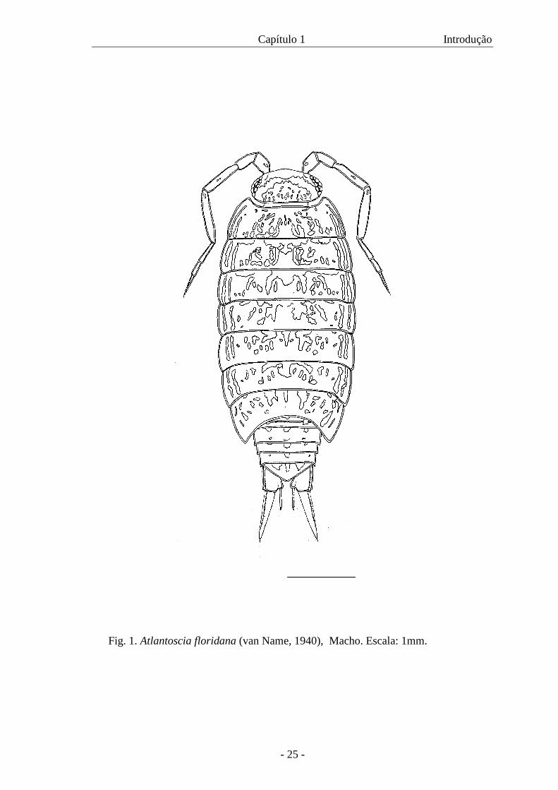
Atlantoscia floridana (Van Name, 1940) (Fig. 1) é uma espécie nativa
das Américas, sendo descrita pela primeira vez a partir de espécimes coletados na
Flórida (E.U.A.). Na América do Sul, ocorre desde o norte do Brasil até o norte da
Argentina, sendo registrada no Brasil nos Estados: AP, PA, RN, PB, PE, BA, ES, MG,
RJ, SP, PR, SC e RS, encontrada também nas ilhas Trindade e Abrolhos. Também foi
registrada nas ilhas Ascensão e Santa Helena onde, provavelmente, foi introduzida
(FERRARA & TAITI 1981; LEMOS DE CASTRO 1985; ARAUJO et al. 1996). Ela pode ser
encontrada principalmente nas regiões costeiras, sendo menos comum no interior do
continente.
Atlantoscia floridana tem uma história nomenclatural problemática, como pode
ser visto pelo número de sinônimos (ARAUJO & LEISTIKOW 1999):
Philoscia floridana van Name, 1940.
Chaetophiloscia paulensis Moreira, 1927 (sensu Vandel, 1963).
Ocelloscia floridana comb.n. Schultz & Johnson, 1984.
Atlantoscia alceui Ferrara & Taiti, 1981.
De acordo com LEMOS DE CASTRO (1985) é o representante de
Philosciidae predominante nos diferentes hábitats em que é encontrada: bromélias,
fungos da família Polyporacea, ninhos de formigas, húmus em matas ou capoeiras, paus
podres, sob folhas de bananeira ou de coqueiro caídas no chão, sob pedras, cascas de
coco e detritos em geral.
Capítulo 1 Introdução
- 24 -
No Rio Grande do Sul, A. floridana foi registrada em vários municípios,
incluindo Porto Alegre e arredores. Concordando com LEMOS DE CASTRO (1985), neste
Estado é também uma das espécies mais comuns, incluindo, além de áreas de vegetação
nativa, ambientes domésticos e bosques de eucalipto (ARAUJO et al. 1996). Ela pode ser
facilmente reconhecida através do seu padrão de cor: duas linhas claras paralelas que se
estendem ao longo dos epímeros e uma destacada faixa despigmentada em forma de
“U” invertido na cabeça. O dimorfismo sexual encontra-se, como nos demais isópodos
terrestres, nos dois primeiros pares de pleópodos, os quais são alongados nos machos.
Os machos apresentam um tamanho máximo em torno de 5mm de comprimento e as
fêmeas, em torno de 7mm de comprimento (medidas desde a região frontal do
cefalotórax até a ponta do telso).
Apesar de ser uma espécie com uma distribuição geográfica e hábitats
bem definidos, não há qualquer investigação a respeito de aspectos de sua ecologia e
tampouco de seu desenvolvimento pós-embrionário.
Capítulo 1 Introdução
- 25 -
Fig. 1. Atlantoscia floridana (van Name, 1940), Macho. Escala: 1mm.
Capítulo 1 Objetivos
- 26 -
OBJETIVOS
Diante do exposto e considerando a inexistência de informações sobre a biologia
de Atlantoscia floridana, propõe-se o desenvolvimento desta pesquisa com os seguintes
objetivos, os quais podem ser divididos em dois conjuntos:
Desenvolvimento pós-marsupial
1- Conhecer as fases de manca até adulto.
2- Identificar, através das mudas, fase jovem de machos e de fêmeas (início do
desenvolvimento de caracteres sexuais externos) e identificar machos e fêmeas adultos
(maturidade sexual).
3 - Definir o tempo médio entre uma muda e outra observando as mudas até a fase
adulta.
4 - Relacionar as mudas/fases com o tamanho.
Ecologia populacional
1 - Calcular a curva de crescimento da espécie
2 – Caracterizar o ciclo reprodutivo
3 - Identificar o padrão de reprodução: se semelparidade ou iteroparidade
4 - Estimar a fecundidade
5 - Estimar a proporção sexual
6 - Estimar a densidade populacional para a área estudada
Capítulo 1 Objetivos
- 27 -
CAPÍTULO 2
MATERIAL E MÉTODOS • A área de estudo
• As amostragens
• A manutenção dos animais em laboratório
Capítulo 2 A área de estudo
- 28 -
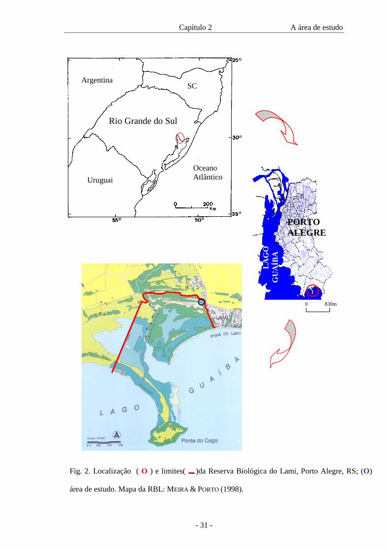
A ÁREA DE ESTUDO
Tanto para o estudo do desenvolvimento pós-embrionário quanto para o
estudo de aspectos da ecologia de Atlantoscia floridana, os animais foram coletados na
Reserva Biológica do Lami (RBL), Porto Alegre, Rio Grande do Sul (Fig. 2), onde
ocorrem em grande quantidade.
A partir de amostras realizadas durante um projeto piloto, estabeleceu-se
a área dentro da Reserva onde foi desenvolvido o trabalho. Os resultados mostraram que
a espécie é mais abundante na área de mata, especialmente aquela próxima ao início da
trilha que leva a vários pontos da reserva. Assim, neste local foram realizadas as
amostragens que constituíram a base de dados desta tese. Além disso, informações
relevantes sobre a área escolhida são apresentadas abaixo e foram decisivas na
confirmação da escolha do local de trabalho.
A Reserva Biológica do Lami constitui uma unidade de conservação do
Município de Porto Alegre (30°,15’S 51°05’W), criada, principalmente, para preservar
espécies ameaçadas de extinção, como é o caso da gimnosperma Ephedra tweediana
Fish & C.A. May, uma planta primitiva que encontra o limite norte da sua distribuição
na região de Porto Alegre. Ela é hoje rara devido à destruição dos ambientes naturais em
que se desenvolve. Espécies animais também ameaçadas de extinção (MARQUES et al
2002) podem ser encontradas na RBL: a lontra, Lontra longicaudis (Olfers, 1818) e o
bugio-ruivo, Alouata guariba Cabrera, 1940 (PORTO ALEGRE 1985 apud PRINTES 2002).
A Reserva, como unidade de conservação, foi criada em 31 de dezembro de
1975, contando originalmente com 77,3 hectares. Em 13 de dezembro de 1977, foram
acrescentados 21 hectares às margens do Arroio Lami, considerados de utilidade
pública. Recentemente, em 26/05/2002 foram anexados 102 hectares correspondentes à

Capítulo 2 A área de estudo
- 29 -
Ponta do Cego, área que inclui banhados e um morro que representa a paisagem natural
da vida nas margens do Lago Guaíba (MEIRA & PORTO 1998).
A reserva situa-se nas terras baixas formadas por terraços lacustres e
cordões arenosos que se alternam com zonas mais deprimidas formando uma variedade
de ambientes com diferentes formações vegetais. A RBL insere-se, geologicamente, em
terrenos sedimentares recentes, originários de eventos transgressivos-regressivos
durante as variações do nível do mar durante o Período Quaternário. Nas áreas mais
deprimidas encontram-se os banhados e nas áreas mais elevadas dos cordões arenosos,
paralelos à linha da praia, encontram-se campos e matas de restinga. Como
conseqüência da dinâmica desses ambientes, a Reserva possui uma grande diversidade
de fauna e flora (MEIRA & PORTO 1998).
Dados importantes podem ser extraídos a partir do zoneamento ambiental
da Reserva apresentado por MEIRA & PORTO (1998) e que interessam particularmente a
este estudo. As amplitudes topoclimáticas realizadas com duração de 24 horas nas
diferentes estações do ano, registraram a temperatura e umidade relativa do ar, a
intensidade luminosa, a velocidade instantânea do vento, a evaporação e a temperatura
do solo a 5, 10 e 20 cm de profundidade em três zonas distintas: banhado, com
amplitude intermediária, zona de areia, com amplitude máxima e zona da mata, com
amplitude mínima. Estas categorias são intercaladas com a estrutura da vegetação
integrando o mapa de zoneamento ambiental. A qualidade de conservação ou valor
ambiental identificados para os ambientes foram categorizados de I a IV: (I) área de
boa qualidade ambiental pela proximidade das condições ambientais originais, com
pouca ação humana; (II) locais que sofreram alterações no processo da formação da
paisagem, estando, contudo, em fase de estabilização; (III) ambientes em que ocorrem
práticas agrícolas e (IV) locais em que ocorreram modificações como extração de areia
Capítulo 2 A área de estudo
- 30 -
ou foram introduzidas espécies exóticas ou, ainda, locais próximos a áreas urbanas. A
área escolhida para o desenvolvimento deste projeto enquadra-se na categoria (I). A
cobertura vegetal deste local é do tipo “Floresta” e apresenta cobertura contínua e altura
que varia de cinco a dez metros, troncos e galhos cobertos por orquídeas e bromélias.
Ocorrem, ainda, cactáceas arborescentes e a E. tweediana. No solo da mata, encontram-
se gramíneas e ervas em distribuição esparsa, sendo que a variação de temperatura na
superfície do solo é mínima quando comparada à das áreas com solo descoberto.
No Plano de Manejo da Reserva (PRINTES 2002), a área de estudo é considerada
Zona Primitiva ou de Transição e se caracteriza por ter sofrido pequena ou mínima
intervenção humana. Ela contém espécies da flora e da fauna ou fenômenos naturais de
grande valor científico.
Capítulo 2 A área de estudo
- 31 -
PORTO ALEGRE
LA
GO
G
UA
ÍBA
Fig. 2. Localização ( O ) e limites( )da Reserva Biológica do Lami, Porto Alegre, RS; (O)
área de estudo. Mapa da RBL: MEIRA & PORTO (1998).
Argentina
Uruguai Oceano Atlântico
Rio Grande do Sul
SC
0 830m
Capítulo 2 As amostragens
- 32 -
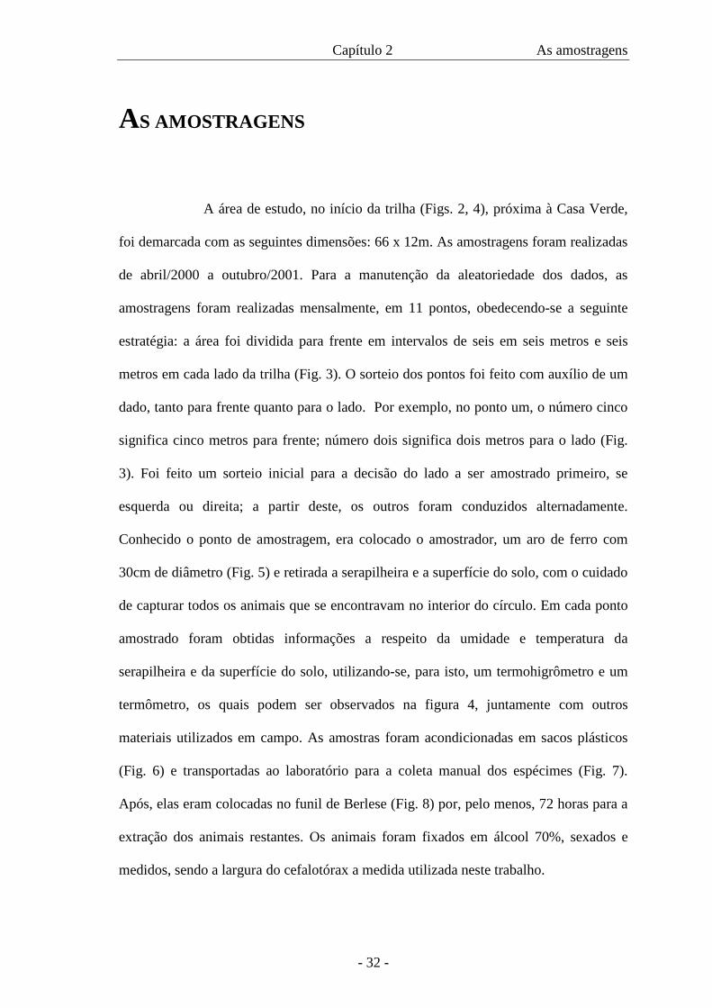
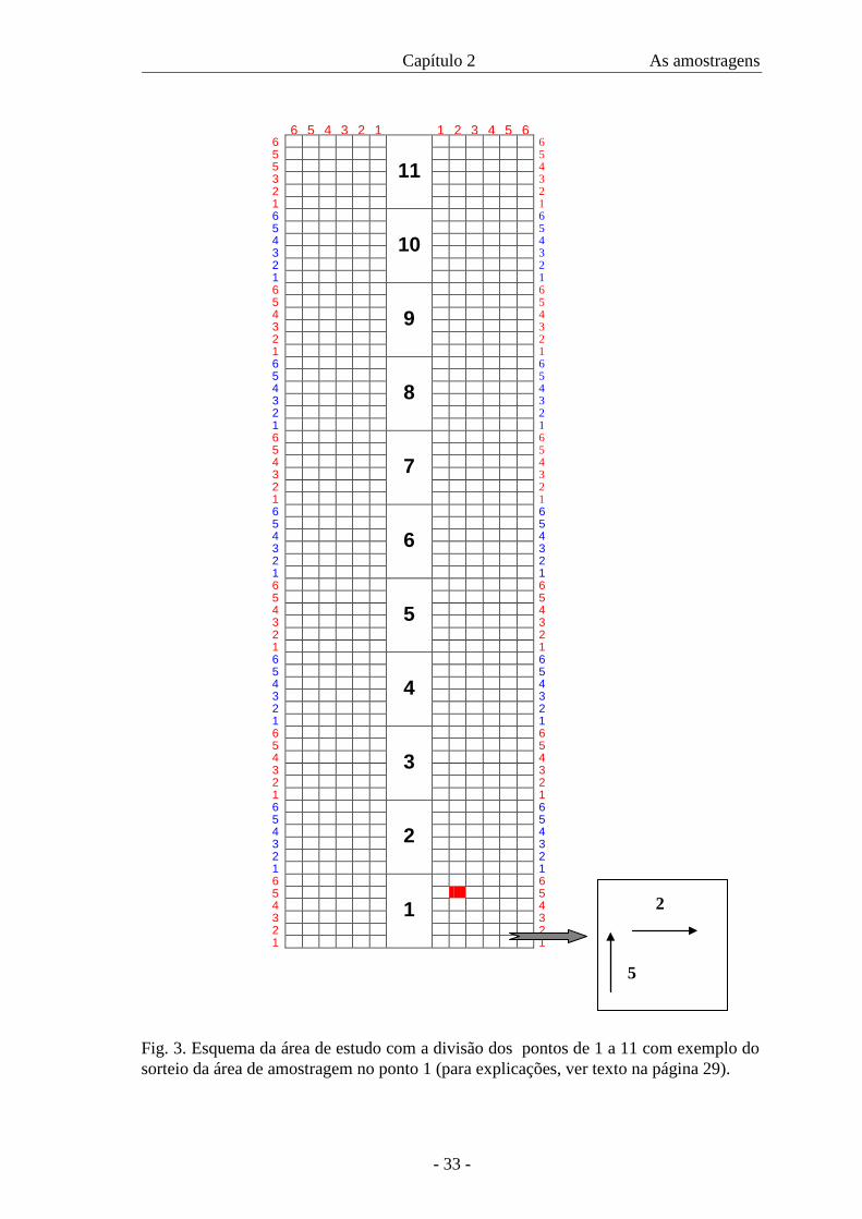
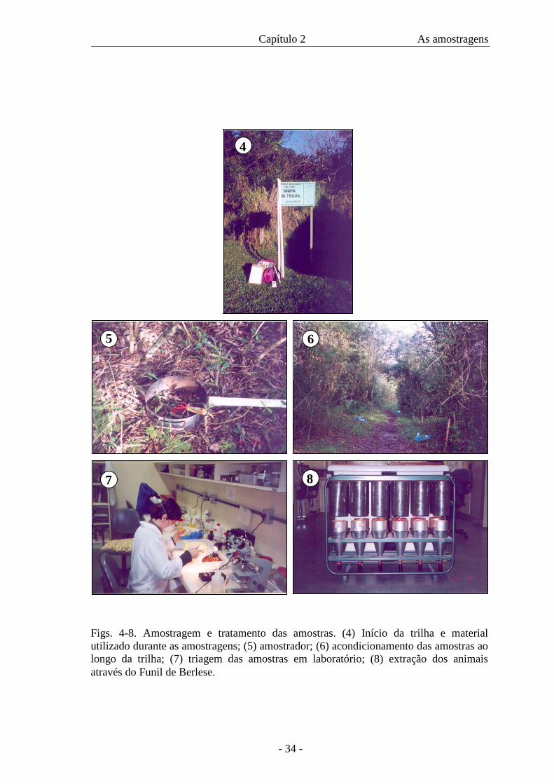
AS AMOSTRAGENS
A área de estudo, no início da trilha (Figs. 2, 4), próxima à Casa Verde,
foi demarcada com as seguintes dimensões: 66 x 12m. As amostragens foram realizadas
de abril/2000 a outubro/2001. Para a manutenção da aleatoriedade dos dados, as
amostragens foram realizadas mensalmente, em 11 pontos, obedecendo-se a seguinte
estratégia: a área foi dividida para frente em intervalos de seis em seis metros e seis
metros em cada lado da trilha (Fig. 3). O sorteio dos pontos foi feito com auxílio de um
dado, tanto para frente quanto para o lado. Por exemplo, no ponto um, o número cinco
significa cinco metros para frente; número dois significa dois metros para o lado (Fig.
3). Foi feito um sorteio inicial para a decisão do lado a ser amostrado primeiro, se
esquerda ou direita; a partir deste, os outros foram conduzidos alternadamente.
Conhecido o ponto de amostragem, era colocado o amostrador, um aro de ferro com
30cm de diâmetro (Fig. 5) e retirada a serapilheira e a superfície do solo, com o cuidado
de capturar todos os animais que se encontravam no interior do círculo. Em cada ponto
amostrado foram obtidas informações a respeito da umidade e temperatura da
serapilheira e da superfície do solo, utilizando-se, para isto, um termohigrômetro e um
termômetro, os quais podem ser observados na figura 4, juntamente com outros
materiais utilizados em campo. As amostras foram acondicionadas em sacos plásticos
(Fig. 6) e transportadas ao laboratório para a coleta manual dos espécimes (Fig. 7).
Após, elas eram colocadas no funil de Berlese (Fig. 8) por, pelo menos, 72 horas para a
extração dos animais restantes. Os animais foram fixados em álcool 70%, sexados e
medidos, sendo a largura do cefalotórax a medida utilizada neste trabalho.
Capítulo 2 As amostragens
- 33 -
6 5 4 3 2 1 1 2 3 4 5 6 6
11 6
5 5 5 4 3 3 2 2 1 1 6
10
6 5 5 4 4 3 3 2 2 1 1 6
9
6 5 5 4 4 3 3 2 2 1 1 6
8
6 5 5 4 4 3 3 2 2 1 1 6
7
6 5 5 4 4 3 3 2 2 1 1 6
6
6 5 5 4 4 3 3 2 2 1 1 6
5
6 5 5 4 4 3 3 2 2 1 1 6
4
6 5 5 4 4 3 3 2 2 1 1 6
3
6 5 5 4 4 3 3 2 2 1 1 6
2
6 5 5 4 4 3 3 2 2 1 1 6
1
6 5 X 5 4 4 3 3 2 2 1 1
Fig. 3. Esquema da área de estudo com a divisão dos pontos de 1 a 11 com exemplo do sorteio da área de amostragem no ponto 1 (para explicações, ver texto na página 29).
5
2
Capítulo 2 As amostragens
- 34 -
4
5 6
Figs. 4-8. Amostragem e tratamento das amostras. (4) Início da trilha e material utilizado durante as amostragens; (5) amostrador; (6) acondicionamento das amostras ao longo da trilha; (7) triagem das amostras em laboratório; (8) extração dos animais através do Funil de Berlese.
7 8
Capítulo 2 A manutenção dos animais em laboratório
- 35 -
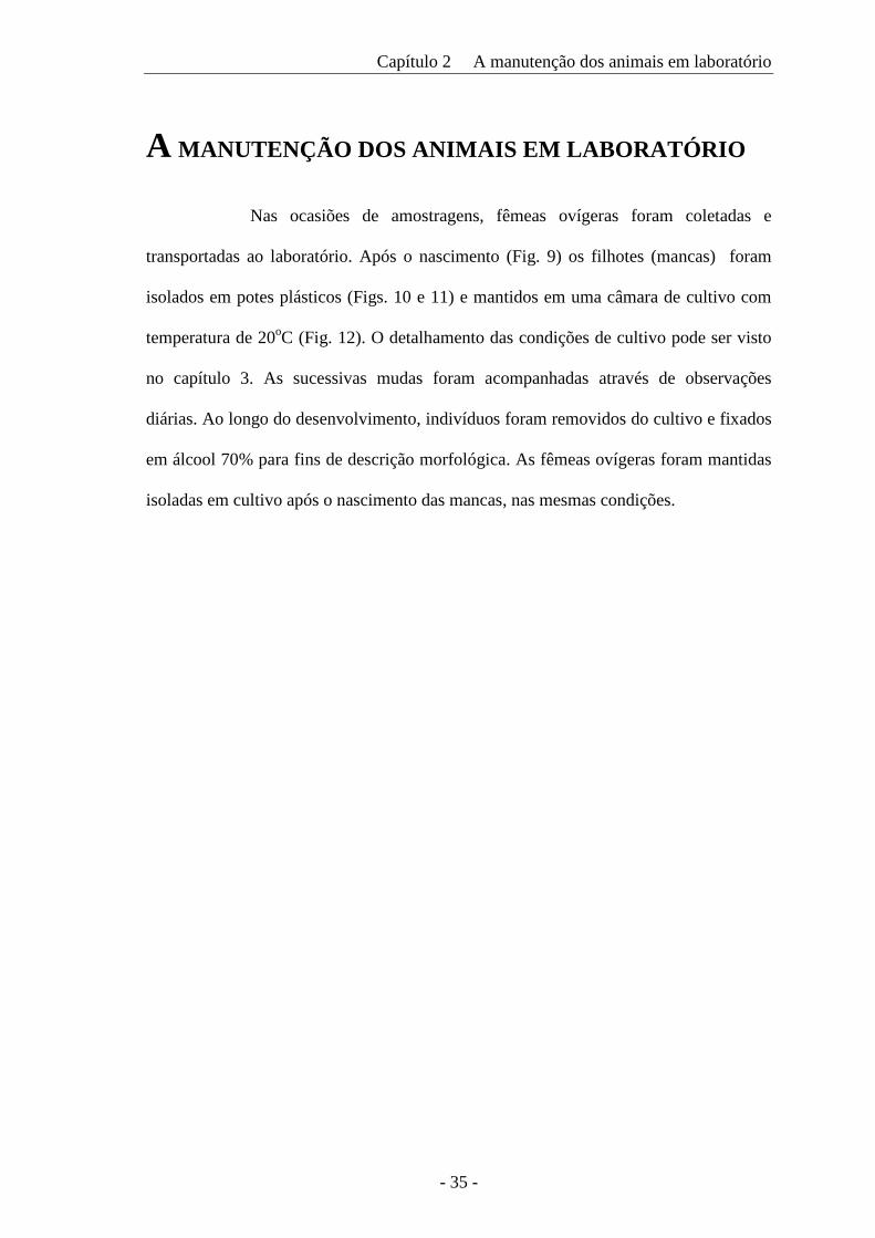
A MANUTENÇÃO DOS ANIMAIS EM LABORATÓRIO
Nas ocasiões de amostragens, fêmeas ovígeras foram coletadas e
transportadas ao laboratório. Após o nascimento (Fig. 9) os filhotes (mancas) foram
isolados em potes plásticos (Figs. 10 e 11) e mantidos em uma câmara de cultivo com
temperatura de 20oC (Fig. 12). O detalhamento das condições de cultivo pode ser visto
no capítulo 3. As sucessivas mudas foram acompanhadas através de observações
diárias. Ao longo do desenvolvimento, indivíduos foram removidos do cultivo e fixados
em álcool 70% para fins de descrição morfológica. As fêmeas ovígeras foram mantidas
isoladas em cultivo após o nascimento das mancas, nas mesmas condições.

Capítulo 2 A manutenção dos animais em laboratório
- 36 -
9
10 11
12
Figs. 9-12. Manutenção dos animais em laboratório. (9) fêmea de Atlantoscia floridana (van Name, 1940) com mancas; (10, 11) potes de cultivo; (12) câmara de cultivo.
CAPÍTULO 3
Postmarsupial development of Atlantoscia floridana (van Name, 1940)
(Crustacea, Isopoda, Oniscidea): the manca stages
- 38 -
Postmarsupial development of Atlantoscia floridana (van Name, 1940) (Crustacea,
Isopoda, Oniscidea): the manca stages
PAULA BEATRIZ ARAUJO1, MINNELISE MARTINS AUGUSTO and GEORGINA BOND-BUCKUP
- Programa de Pós-Graduação em Biologia Animal, Departamento de Zoologia,
Instituto de Biociências, Universidade Federal do Rio Grande do Sul, Av. Bento
Gonçalves, 9.500 prédio 43435, CEP 91501-970, Porto Alegre, RS, Brazil
Telephone: +55-51-33167709
Fax: +55-51-33167696
e-mail: [email protected]
Running Title: The manca stages of Atlantoscia floridana 1- Corresponding author ARTIGO ACEITO: JOURNAL OF NATURAL HISTORY
Capítulo 3 Os estágios de manca
- 39 -
In this paper we describe the postmarsupial development of the manca stages of
Atlantoscia floridana (van Name, 1940). Ovigerous females were collected in Reserva
Biológica do Lami, Porto Alegre, RS, Brazil and separately reared in small containers at
20º C (±1º C). After birth each newborn (manca I) was reared separately in order to
observe the subsequent moults. In a period of about 12 hours the animals underwent the
first moult. The manca II stage lasts 9.16 ± 1.57 days while manca III takes up a period
of 9.96 ± 1.05 days. Growth was observed through measurements of cephalothorax
width. The three manca stages are described, illustrated, and compared with the adults.
The main distinguishing characteristics among mancas I, II and III are presented as well
as the b/c and d/c coordinates of the noduli laterales.
Key words: Isopoda, Oniscidea, Atlantoscia floridana, postmarsupial development, manca stages
Capítulo 3 Os estágios de manca
- 40 -
Introduction
Like the other Peracarida, terrestrial isopods show direct development, involving
an intramarsupial and a postmarsupial phase (Holdich et al., 1984). During the
embryonic phase the individuals are nourished by a maternal fluid, which is released in
the marsupium (Hoese and Janssen, 1989). Among the adaptive strategies of the isopods
to tolerate the terrestrial environment, direct development was certainly of capital
importance, reducing physiological stress and consequently increasing survival during
the early stages of embryonic development. According to Hoese (1984), the great
success achieved by isopods can only be understood in the light of the importance of the
marsupium, functioning as a micro-aquarium that allows embryonic development to
occur without an external water source. By the time of birth the individuals crawl out of
the marsupium without moulting to become the first postmarsupial mancas; these stages
are called mancas instead of larvae since they have direct development (Holdich et al.,
1984).
There are a few studies on the postmarsupial development of the terrestrial
isopods (Verhoeff, 1920; Heeley, 1941; Vandel, 1943; Matsakis, 1955; Haddad, 1982;
Kacem-Lachkar, 1997). Overall, at birth the young look similar to adults, differing in
the absence of the seventh pair of pereiopods and in the secondary sexual characters,
which appear after subsequent moults.
The species Atlantoscia floridana (van Name, 1940) occurs from the southern
USA (Florida) to the north of Argentina. In Brazil, it has been found in nearly all
coastal States, being one of the most common representatives of the “Philosciidae”
(Araujo et al., 1996). Recently, it has been redescribed by Araujo and Leistikow (1999)
Capítulo 3 Os estágios de manca
- 41 -
and the phylogenetic position of the genus proposed by Leistikow (2001). This paper
aims to describe the early stages of the postmarsupial development, diagnosing the main
morphological characters and allowing easy identification.
Material and methods
Females of A. floridana were collected during the years 1999 and 2000 from the
Lami Biological Reservation (Reserva Biológica do Lami), Porto Alegre (30°15´S
51°05´W), RS, Brazil. In the laboratory they were individualised in plastic pots (6 cm
high to 6 cm wide) and kept at a constant temperature of 20° C ± 1°C with the
photoperiod set to natural durations, for example: 10h:14 h (light:darkness) during
winter and 12h:12h during summer. To the pot were added a damp piece of filter paper
and a bit of cotton to create a humidity gradient. Decaying leaves taken from the
sampled area were provided as food. Adult faeces were also added since they make up
an important part of the diet at the beginning of development (Helden and Hassall,
1998). The microorganisms thus ingested balance the pH of the gut and can be
important sources of nutrients (Zimmer and Topp, 1998).
After birth the postmarsupial mancas were isolated and their successive moults
followed. The animals were observed daily so as to identify the periods of moulting and
the exuviation. Representative examples of each stage were taken from the rearing for
description and measurement of the cephalothorax, done with the aid of a Zeiss Stemi
V8 stereomicroscope. The cephalothorax width was taken as an indicator of size
(Sutton, 1968). The measurement refers to the largest width with the body in dorsal
view on a horizontal plane, at eyes level (Sunderland et al., 1976). The manca stages
were described using general and appendages characters, as well as measurements of
Capítulo 3 Os estágios de manca
- 42 -
the noduli laterales sensu Vandel (1962). To establish comparisons between the manca
and adult morphology, we used the A. floridana redescription of Araujo and Leistikow
(1999). Illustrations were done with a camera lucida. The manca stages were labelled
manca I (M I), manca II (M II) and manca III (M III).
Results
Manca I
(figures 1-19, 45)
Colour. At birth the individuals are practically lacking pigmentation (figure 1). There
are small chestnut spots on the cephalothorax, distributed in an inverted-U shape that is
characteristic of the species. In the pereionites, the pigmentation is concentrated on the
lateral margins, with sparse pigmentation on the middle. The pleon, telson and uropods
also have sparse darker patches. The unpigmented appearance is further accentuated by
the absence of food in the gut.
Cephalothorax. Compound eyes with five ommatidia.
Pereion. Tegument smooth with scattered tricorn-like setae, coxal plates I to IV with the
posterior lateral margin round, V and VI pointed, without gland pores. Noduli laterales
conspicuous; coordinates b/c, d/c as in figure 45; nodulus lateralis of coxal plate IV
more dorsally than the others. Pereionite VII feebly developed without coxal plates;
nodulus lateralis VII lacking.
Pleon. Pleonites 3 to 5 with small neopleurae.
Appendages. Antennula: tri-articulate; distal article cone-shaped with only two apical
aesthetascs (figure 2). Antenna: flagellum bi-articulate, as long as peduncular article 5,
proximal article the largest, apical organ almost the same length as distal article,
Capítulo 3 Os estágios de manca
- 43 -
peduncular articles with sparse tricorn-like setae (figure 3). Mandible: molar penicil
with several branches on both mandibles; left mandible with two penicils and right
mandible with one penicil and a plumose seta (figures 4 and 5). Maxillula: lateral endite
with 4+5 teeth, (four cleft, one trifid), few lateral setae (figure 6); endite with two
penicils, lacking apical point. Maxilla: medial lobe prominent, less than half of the
breadth of lateral lobe, with fine setae and five cusps, lateral lobe marginally rounded
(figure 7).
Maxilliped: no tricorn-like setae, palp with apical setal tuft, inner setal set with long and
short setae, medial tuft with four setae, endite with two prominent tooth on the medial
distal margin (figure 8). Pereiopods: carpus 1 without developed antennal-grooming
brush; seta with serrate double fringed apex present (figure 9). Dactyl with ungual seta
and dactylar seta with knob-like apex (figure 15). All pereiopods with few setae (figures
10-14). Pereiopod 7 absent. Pleopods: pleopod 1 lacking; exopods 2-5 rectangular,
bearing one seta (figures 17-19). Uropod: exopod inserting distally of endopod.
Manca II
(Figures 20-34)
Colour. Stronger coloration but not as much as in the adult. Pereiopods and pleopods
with weak pigmentation (figure 20).
Cephalothorax. Compound eyes with five ommatidia.
Pereion. With gland pores. Pereionite VII with less than half the length of pereionite VI,
with slightly developed coxal plates. Noduli laterals as M I.
Pleon. Like Manca I.
Capítulo 3 Os estágios de manca
- 44 -
Appendages. Antennula: with one aesthetasc on the medial margin and two apical
(figure 21). Antenna: flagellum bi-articulate, as long as the peduncular article 5, distal
article the largest, apical organ half the length of distal article, peduncular articles with
sparse tricorn-like setae (figure 22). Mouthparts like adult. Pereiopods: carpus 1 with
the beginning of development of antennal-grooming brush, with hyaline scales distally,
pereiopods with setal tufts on latero-distal edge of carpus (figure 24). Pereiopods with
setal tuft on latero-distal margin of carpus (figures 25-29). Beginning of development of
pereiopod 7, which is found ventrally folded under the sixth and seventh pereionites
(figure 23); it is not possible to discern all articles (figure 30). Pleopods: pleopod 1:
only protopodite present; exopods 2-5 rectangular, slightly pointed, bearing one seta
with the exception of pleopod 5 that bears two setae (figures 31-34).
Manca III
(Figures 35-45)
Colour. Chestnut, like adult, with a dark brown band on the coxal plates bordered by
light areas not well defined (figure 35).
Cephalothorax. Compound eyes with six to seven ommatidia.
Pereion. With gland pores. Nodulus lateralis of pereionite VII present. Pereionite VII
with half the length of pereionite VI, with small coxal plates.
Pleon. Like adult.
Appendages. Antennula: with two aesthetascs on medial margin and two apical (figure
36).
Antenna: flagellum tri-articulate, as long as peduncular article 5; distal article the
largest, apical organ half the length of distal article (figure 37). Mouthparts like adult.
Capítulo 3 Os estágios de manca
- 45 -
Pereiopods: like Manca II, including pereiopod 1 (figure 38). Pereiopod 7 more
developed, found folded ventrally under the sixth and seventh pereionites (figure 39);
all articles are distinct (figure 40).
Pleopods: pleopod 1: only protopodite present; exopods 2-5 rectangular, slightly
pointed, bearing one seta with the exception of pleopod 4 and 5 that bears two setae
(figures 41-44).
Table I shows a summary of the main distinguishing characteristics of the manca
stages.
Table II presents the data on cephalothorax width and duration of each manca
stage. The width of the cephalothorax in the manca stages varies from 0.4 mm to 0.56
mm. There is an overlap of sizes among all the three stages as in figure 46, even though
the amplitude is 0.06 in all of them. The intermoult period is similar in M II and M III
stages. The minimum intermoult time for M II and III is 6 (n=1) and 8 (n=3) days
respectively and the maximum time is 12 days for both (n=3 for M II and n=1 for M
III).
In 46 about here Discussion
According to Matsakis (1955) the marsupial manca hatches inside the
marsupium and spends an important part of its existence there, in what Hoese and
Janssen (1989) classify as hatched young. All their reserves come from what is eaten
when hatched since they feed and drink within the marsupium (Hoese and Janssen,
1989). Free life begins only in the final stage, when they attain postmarsupial manca
status. Sternal calcium deposits are a clear indication of the premoult stage (Zidar et al.,
Capítulo 3 Os estágios de manca
- 46 -
1998). In manca I they can be seen when they are still inside the marsupium. In A.
floridana, moulting occurs in the same day as birth, being complete in approximately 12
hours. As it actually represents the final stage of the intramarsupial manca they are very
close to moulting, and therefore do not feed, a fact observed in the lack of food inside
the gut. For other species, the duration of this stage does not exceed 24 hours either
(Heeley, 1941; Haddad, 1982). A period between 24 and 48 hours was observed for
Hemilepistus reaumuri (Audouin and Savigny, 1826) a species studied by Kacem-
Lachkar (1997) in Tunisia.
Before the moult the animals are little active and during the moult they
do not walk, the only movement being that to get free of the exuvia. As in other
isopods, the moult is biphasic. First there is a moulting of the posterior part of the body
(from pleonite V on), which is eliminated through movements of elongation and
contraction towards this posterior part. These movements continuous through the
ecdysis process and for some time after. When the exuvia is freed, it begins to be
ingested. Then, it is possible to observe that the posterior part of the body is wider and
the anterior gut is filled with the ingested exuvia. After a couple of hours the animals
undergo moulting of the anterior part. The elongation and contraction movements
concentrate on the anterior region of the body. After exuviation the anterior pereiopods
are found facing front, under the cephalothorax, still attached by the lubricating liquid
of the moult (Heeley, 1941). The animals do not move until the pereiopods are hardened
and can be used to walk. The liberation of the anterior exuvia seems to be more difficult
than the posterior one. Several cases of death were observed during the final phase of
the process, when the exuvia collapses with the body wall and the animal cannot
separate from it, resulting in decease. Once moulting is successfully finished, the
anterior exuvia is also ingested, again with its presence detected in the gut by

Capítulo 3 Os estágios de manca
- 47 -
transparency. The posterior and the anterior moults were achieved in a period not
exceeding fifteen minutes each. With moulting ended, the individuals started feeding
normally.
Even when the moment the moult is happening cannot be observed, it is still
possible to recognise the process by the presence of the exuvia in the gut. The anterior
and posterior moults can also be distinguished by observing the contour of the body or
examining the articles of the flagellum of antenna I. The antennae in M II differ from M
I, since there is an inversion of the relative size of these articles: in M I, the proximal
article is longer than the distal one, and in M II it is shorter, a fact already observed by
Verhoeff (1920) and Matsakis (1955).
The M II can be identified by the presence of a more developed seventh pleonite,
not involved by the coxal plates of pereionite VI and with the beginning of the
formation of its own coxal plates. Differently from M I, the pigmentation is stronger
and the gut is full. Also, the seventh pair of pereiopods appears, still in a rudimentary
form (figures. 23, 30 and table II).
In M II the calcium plates were observed after half the period of the intermoult.
Another indication of the moult is the interruption of feeding, which can be observed by
the absence of food in the digestive tube. This starts approximately one day before the
moult. The moult process is similar to that of the M I.
In M III the body pigmentation becomes richer, nearing the adult pattern. The
coxal plates of pereionite VII are more developed than in M II, having a lateral nodule
on each side. The seventh pair of pereiopods is in progressive process of developing,
although it is not yet functional, since it is still folded ventrally. The eyes have one or
more ommatidia more than in M II (tab. II).
Capítulo 3 Os estágios de manca
- 48 -
Most of the species studied have three manca stages (Verhoeff, 1920; Heeley,
1941, Matsakis, 1955; Haddad, 1982). However, Trichoniscus pusillus Brandt, 1833
develops the seventh pair of pereiopods only after the fifth moult, which corresponds to
manca V (Heeley, 1941). Another species that presents a larger number of manca stages
is H. reaumuri, in which the seventh pair of pereiopods becomes functional after the
third moult (Kacem-Lachkar, 1997).
There are many similarities between manca I and the adults. The mouthparts,
particularly the mandibles, maxillulas and maxillipeds already show the adult pattern.
The maxilla differs only in the contour of the lateral lobe, in the septation and in the
distribution pattern of the cusps. The noduli laterales also have a distribution similar to
the species pattern. The more dorsal position of the lateral nodule of the coxal plate IV
is already evident in this first postmarsupial stage (fig. 46). In the pereiopods, in spite of
the reduced number of setae, they are similar to that of the adults, presenting a seta with
serrate double-fringed apex of the carpus I, and the dactyl ar seta with knob-like apex.
The similarity of certain morphological structures allows inference that soon after the
first moult, the young individuals would be prepared for the same conditions as faced by
the adults. Regarding feeding, at least in terms of mouthpart morphology they are able
to consume the same kind of food as the adults. Cleaning of the antennae is also
possible, even though the comb-like structure is weakly developed. When the role of the
noduli laterales is discovered, it may be possible to understand the consequences of
their pattern being so well developed in M I.
If on one hand certain structures do not change much through
development, others are weakly developed at birth. The antennula is still very
rudimentary and develops very little between the M I and the M III. However, it already
Capítulo 3 Os estágios de manca
- 49 -
shows clearly the two apical aesthetascs, with the medial ones appearing gradually with
the moults.
The secondary sexual dimorphism cannot be observed in the M III, since
the pleopods maintain the same pattern from the M I and the pleopod 1 has still not
developed, only the protopodite. Nevertheless, the internal sexual differentiation of the
reproductive organs begins before the appearance of the external sexual characters
(Katakura, 1984).
The intermoult period in M II and M III of A. floridana is shorter than the
average time for the species studied by Heeley (1941) in England. With the exception of
Armadillidium vulgare (Latreille, 1804), in which the second moult occurs at 22 days,
in the other species, T. pusillus, Porcellio dilatatus Brandt, 1833, Porcellio scaber
Latreille, 1804, Oniscus asellus (Linné, 1758) and Philoscia muscorum (Scopoli, 1763)
it occurs between 16 and 18 days. The third moult occurs at 41 days on average for A.
vulgare and between 25 and 37 days for the other species. On the other hand, the
average time of the intermoult of manca II for Atlantoscia floridana is very similar to
that of H. reaumuri, the species studied by Kacem-Lachkar (1997).
Following the postmarsupial development of a species can be of great
importance in ecological studies, especially at the population level. Once the diagnosis
of the non-sexually differentiated forms is possible and certain, we eliminate the
possibility of indiscriminately including them alongside males and females. In the case
of the mancas, diagnosis is simple, it being enough to observe whether the seventh pair
of pereiopods is folded ventrally or functional.
For systematic studies, characters can be recognised from the sexually
undifferentiated forms with more confidence from manca II on. Among them, the
mouthparts, the pattern of distribution of the noduli laterales (for species in which they
Capítulo 3 Os estágios de manca
- 50 -
occur), the dactyl of the pereiopods and, for manca III, the number of articles of the
antennal flagellum. The observation of these characters does not exclude the necessary
examination of other characters, mostly those related to specific identification.
However, the information obtained from the mancas can be enough for identification of
higher taxonomic categories (above species).
The rearing of A. floridana in the laboratory was reasonably efficient. The
species is easy in maintenance, manipulation and observation, making its laboratory
cultivation viable for experimental work or even environmental monitoring, especially
for conservation areas.
References
ARAUJO, P.B., BUCKUP, L. and BOND-BUCKUP, G., 1996, Isópodos terrestres
(Crustacea, Oniscidea) de Santa Catarina e Rio Grande do Sul, Brasil, Iheringia, Sér.
Zool., (81), 111-138.
ARAUJO, P.B. and LEISTIKOW, A., 1999, Philosciids with pleopodal lungs from
Brazil, with description of a new species, Contributions to Zoology 68(2), 109-141.
LEISTIKOW, A., 2001, Phylogeny and biogeography of South American Crinocheta,
traditionally placed in the family “Philosciidae” (Crustacea: Isopoda: Oniscidea),
Organisms Diversity and Evolution, 1 Electr. Suppl. 4, 1-85.
HADDAD, M.A., 1982, Estádios do crescimento pósmarsupial de Balloniscus sellowii
Brandt, 1833) (Isopoda, Oniscoidea), Dusenia, 13(3), 135-143.
HEELEY, W., 1941, Observations on the life-histories of some terrestrial isopods,
Proceedings of the Zoological Society of London, 111(B), 79-119.
Capítulo 3 Os estágios de manca
- 51 -
HELDEN, A.J. and HASSALL, H., 1998, Phenotypic plasticity in growth and
development rates of Armadillidium vulgare (Isopoda: Oniscidea), Israel Journal of
Zoology, 44, 379-394
HOESE, B., 1984, The marsupium in terrestrial isopods, In: SUTTON, S.L. and
HOLDICH, D.M., (eds), The biology of terrestrial isopods, Symposium of the
Zoological Society of London, 53, 65-76.
HOESE, B. and JANSSEN, H.H., 1989, Morphological and physiological studies on
the marsupium in terrestrial isopods, Monitore Zoologico Italiano, Monografia 4,153-
173.
HOLDICH, D.M., LINCOLN, R.J. and ELLIS, J.P., 1984, The biology of terrestrial
isopods: terminology and classification, In: SUTTON, S.L. and HOLDICH, D.M.
(eds), The biology of terrestrial isopods, Symposium of the Zoological Society of
London, 53, 1-6.
KACEM-LACHKAR, H., 1997, Étude du développement post-embryonnaire
d’Hemilepistus reaumuri (Audouin, 1826) (Isopoda, Oniscidea), Crustaceana, 70(5),
513-526.
KATAKURA, Y., 1984, Sex differentiation and androgenic gland hormone in the
terrestrial isopod Armadillidium vulgare, In: SUTTON, S.L. and HOLDICH, D.M.,
(eds), The biology of terrestrial isopods, Symposium of the Zoological Society of
London, 53, 127-142.
MATSAKIS, J., 1955, Contribution a l’etude du développement postembryonnaire et de
la croissance des Oniscoides, Bulletin de la Societé Zoologique de la France, 80, 52-
65.
Capítulo 3 Os estágios de manca
- 52 -
SUTTON, S.L., 1968, The population dynamics of Trichoniscus pusillus and Philoscia
muscorum (Crustacea: Oniscoidea) in limestone grassland, Journal of Animal
Ecology, 37, 425-444.
SUNDERLAND, K.D., HASSALL, M. and SUTTON, S.L., 1976, The population
dynamics of Philoscia muscorum (Crustacea: Oniscoidea) in a dune grassland
ecosystem, Journal of Animal Ecology, 45, 487-506.
VANDEL, A., 1943, Essai sur l’origine, l’evolution et la classification des Oniscoïdes
(Isopodes terrestres), Bulletin Biologique de la France et de la Belgique, (suppl.),
30,1-36.
VANDEL, A., 1962, Isopodes terrestres (Deuxiéme partie) In: Faune de France, P.
Lechevalier, Paris, Vol. 66, pp. 417-931.
VERHOEFF, K.W., 1920, Zur Kenntnis der Larven, des Brutsackes und der Bruten
der Oniscoidea, Zoologische Anzeiger, 51, 169-189.
ZIDAR, P., DROBNE, D. and STRUS, J., 1998, Determination of moult stages of
Porcellio scaber (Isopoda) for routine use, Crustaceana, 72, 1-9.
ZIMMER, M. and TOPP, W., 1998, Nutritional biology of terrestrial isopods (Isopoda:
Oniscidea): Copper revisited, Israel Journal of Zoology, 44, 473- 486.
Capítulo 3 Os estágios de manca
- 53 -
Acknowledgements
To Secretaria Municipal do Meio Ambiente (SMAM – Porto Alegre), especially MSc.
Rodrigo C. Printes for allowing and supporting our research in the Reserva Biológica do
Lami, to Professors Helena Romanowski and Jocélia Grazia (Departamento de
Zoologia, UFRGS), for the use of the B.O.D. for rearing the animals, to PPG-Biologia
Animal and to PROPESQ-UFRGS for the concession of an undergraduate scientific
scholarship (BIC). This is contribution No 384, Department of Zoology.
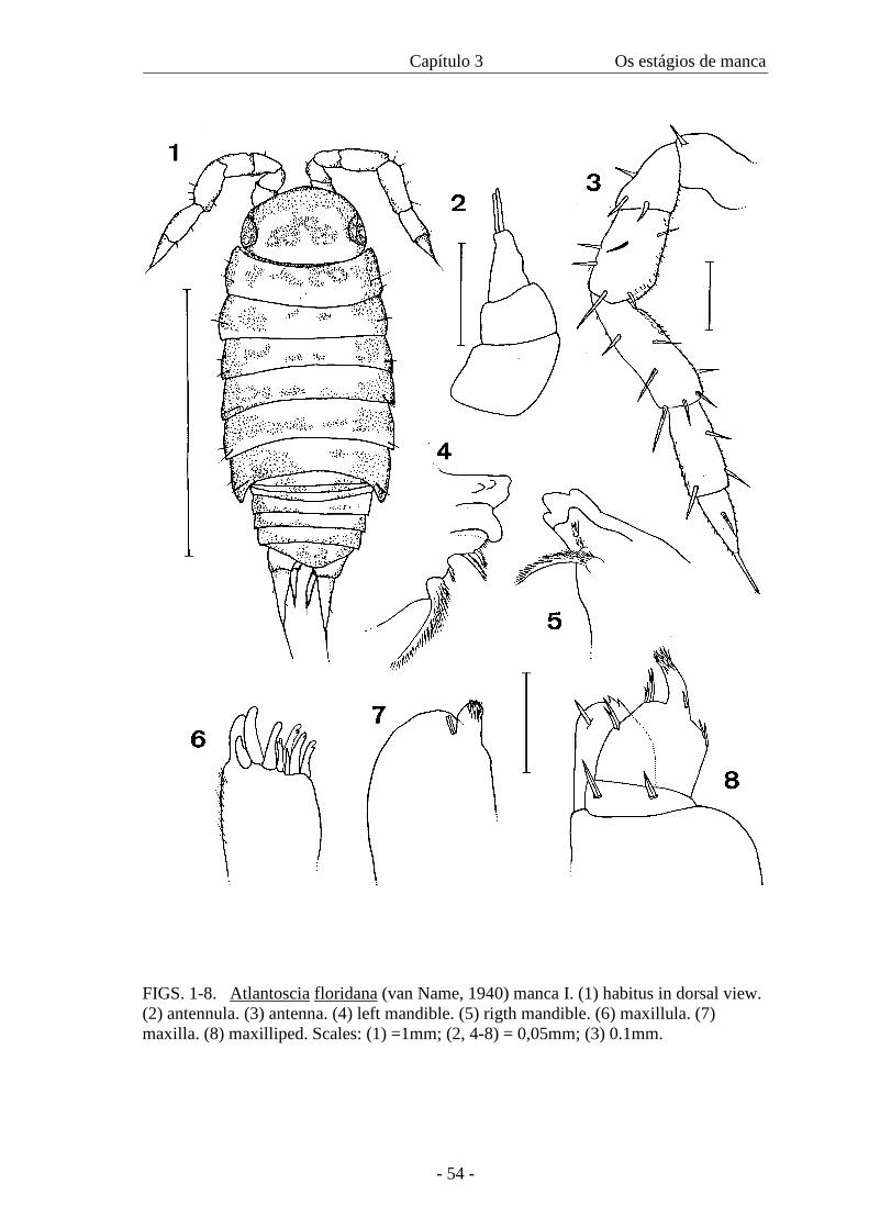
Capítulo 3 Os estágios de manca
- 54 -
FIGS. 1-8. Atlantoscia floridana (van Name, 1940) manca I. (1) habitus in dorsal view. (2) antennula. (3) antenna. (4) left mandible. (5) rigth mandible. (6) maxillula. (7) maxilla. (8) maxilliped. Scales: (1) =1mm; (2, 4-8) = 0,05mm; (3) 0.1mm.

Capítulo 3 Os estágios de manca
- 55 -
FIGS. 9 - 15. Atlantoscia floridana (van Name, 1940) manca I. (9) pereiopod 1. (10) pereiopod 2. (11) pereiopod 3. (12) pereiopod 4. (13) pereiopod 5. (14) pereiopod 6. (15) dactylus of pereiopod 1. Scales: (1-14) = 1mm; (15) = 0.025mm.
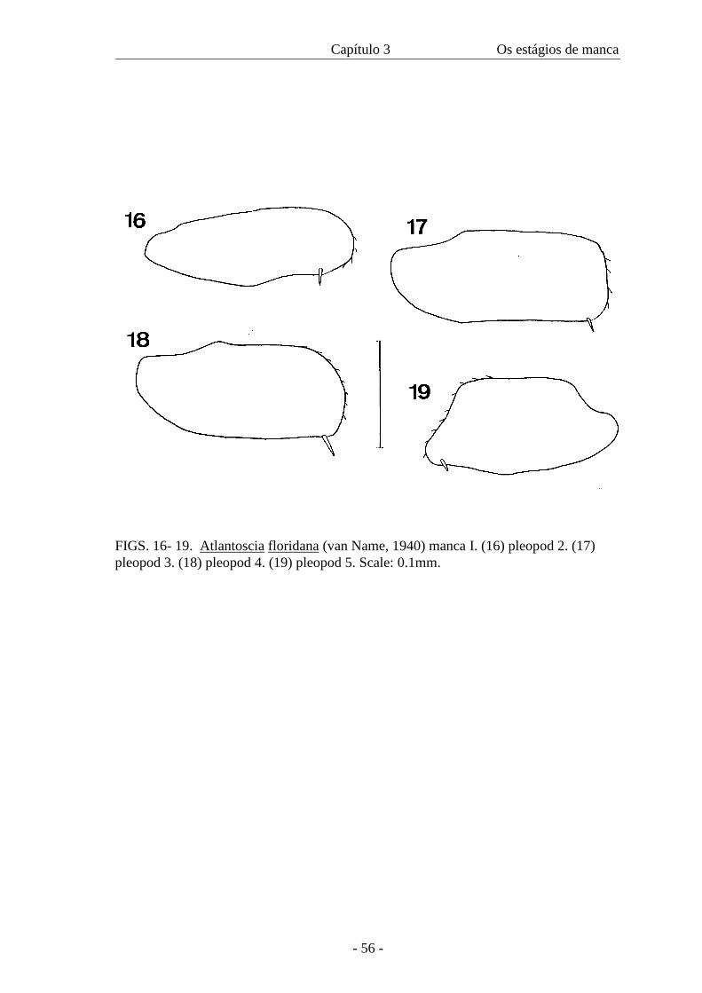
Capítulo 3 Os estágios de manca
- 56 -
FIGS. 16- 19. Atlantoscia floridana (van Name, 1940) manca I. (16) pleopod 2. (17) pleopod 3. (18) pleopod 4. (19) pleopod 5. Scale: 0.1mm.
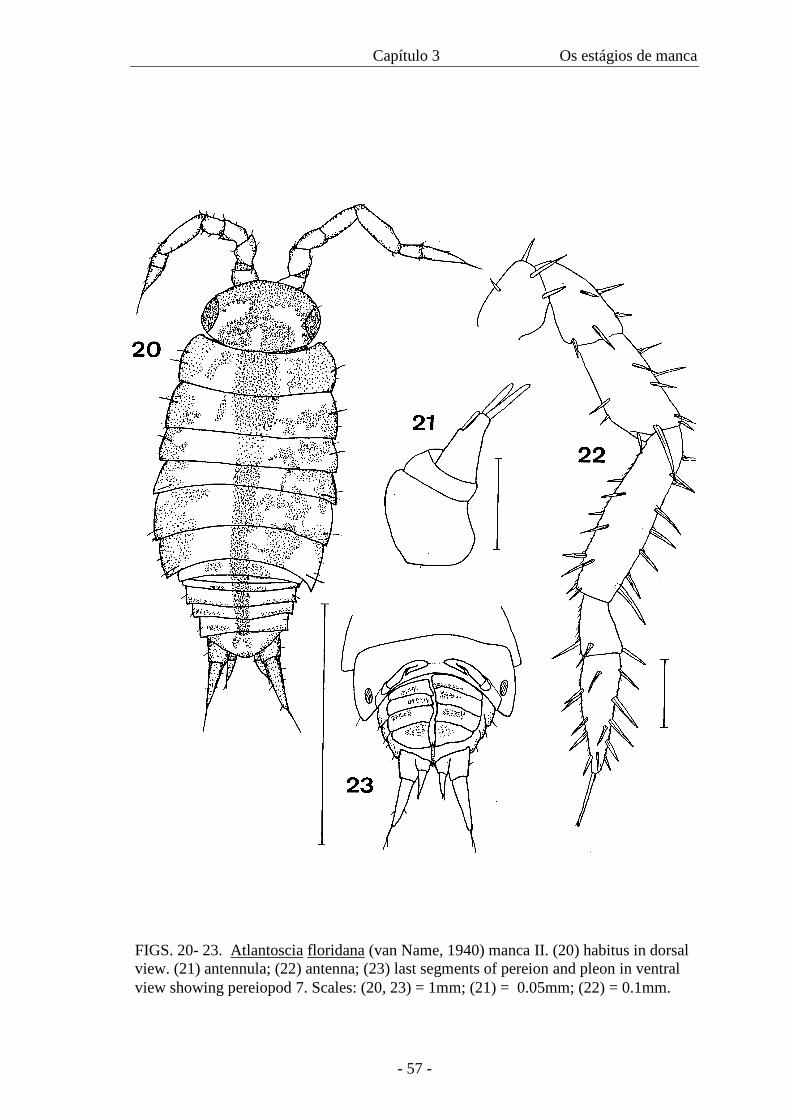
Capítulo 3 Os estágios de manca
- 57 -
FIGS. 20- 23. Atlantoscia floridana (van Name, 1940) manca II. (20) habitus in dorsal view. (21) antennula; (22) antenna; (23) last segments of pereion and pleon in ventral view showing pereiopod 7. Scales: (20, 23) = 1mm; (21) = 0.05mm; (22) = 0.1mm.
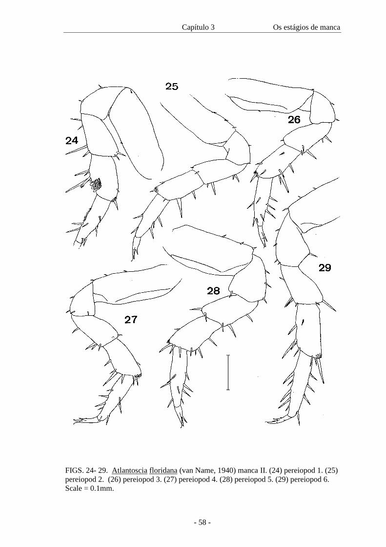
Capítulo 3 Os estágios de manca
- 58 -
FIGS. 24- 29. Atlantoscia floridana (van Name, 1940) manca II. (24) pereiopod 1. (25) pereiopod 2. (26) pereiopod 3. (27) pereiopod 4. (28) pereiopod 5. (29) pereiopod 6. Scale = 0.1mm.
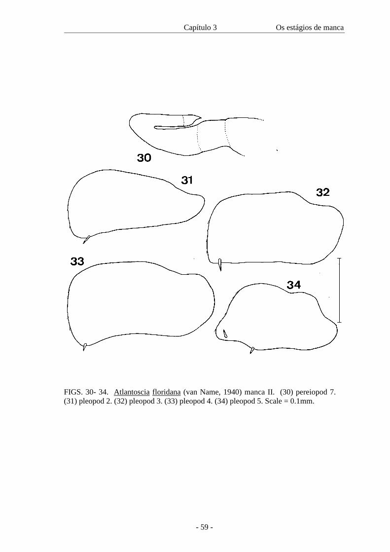
Capítulo 3 Os estágios de manca
- 59 -
FIGS. 30- 34. Atlantoscia floridana (van Name, 1940) manca II. (30) pereiopod 7. (31) pleopod 2. (32) pleopod 3. (33) pleopod 4. (34) pleopod 5. Scale = 0.1mm.
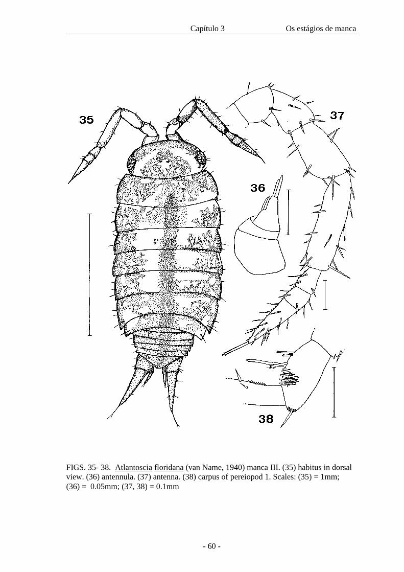
Capítulo 3 Os estágios de manca
- 60 -
FIGS. 35- 38. Atlantoscia floridana (van Name, 1940) manca III. (35) habitus in dorsal view. (36) antennula. (37) antenna. (38) carpus of pereiopod 1. Scales: (35) = 1mm; (36) = 0.05mm; (37, 38) = 0.1mm
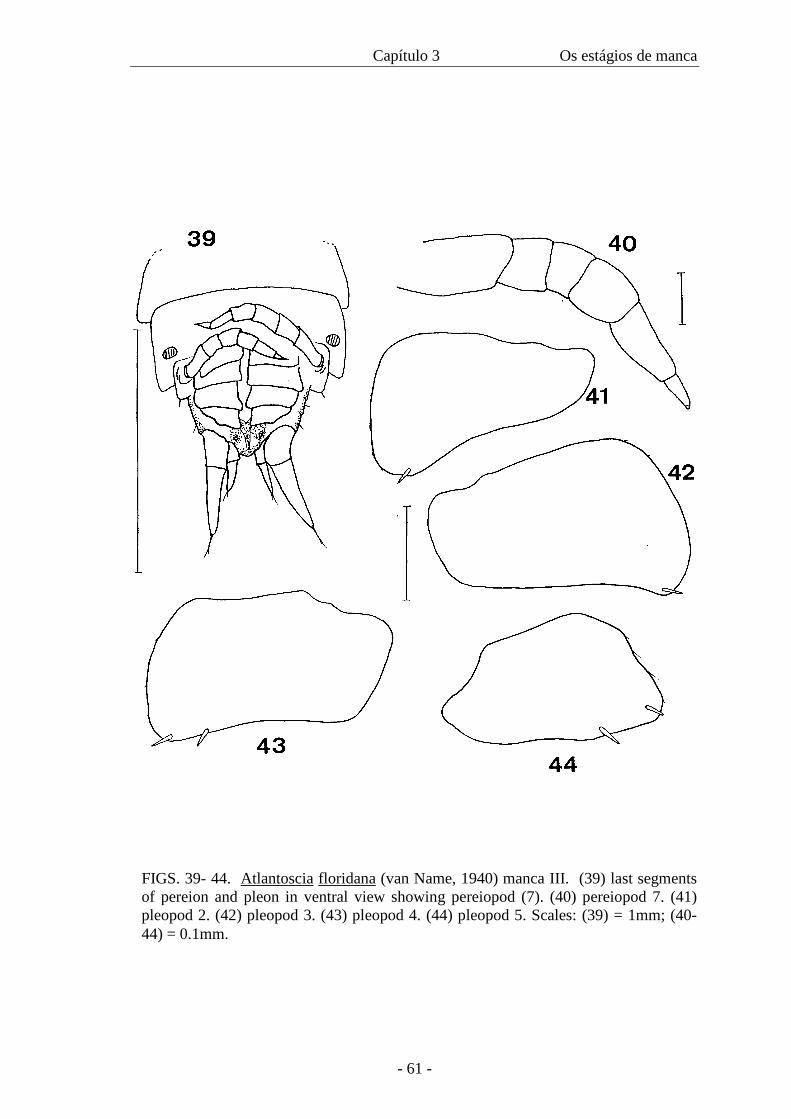
Capítulo 3 Os estágios de manca
- 61 -
FIGS. 39- 44. Atlantoscia floridana (van Name, 1940) manca III. (39) last segments of pereion and pleon in ventral view showing pereiopod (7). (40) pereiopod 7. (41) pleopod 2. (42) pleopod 3. (43) pleopod 4. (44) pleopod 5. Scales: (39) = 1mm; (40-44) = 0.1mm.
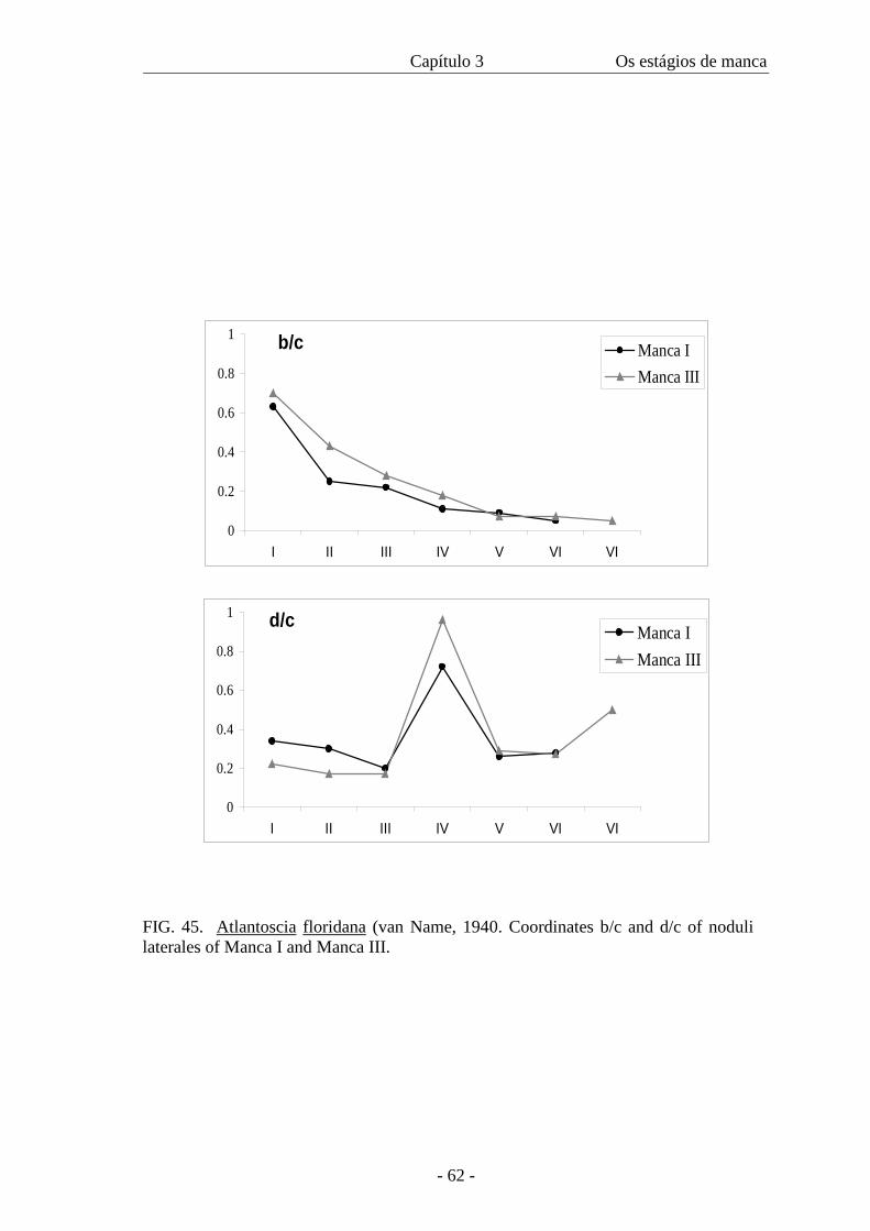
Capítulo 3 Os estágios de manca
- 62 -
0
0.2
0.4
0.6
0.8
1
I II III IV V VI VI
Manca IManca III
0
0.2
0.4
0.6
0.8
1
I II III IV V VI VI
Manca IManca III
d/c
b/c
FIG. 45. Atlantoscia floridana (van Name, 1940. Coordinates b/c and d/c of noduli laterales of Manca I and Manca III.
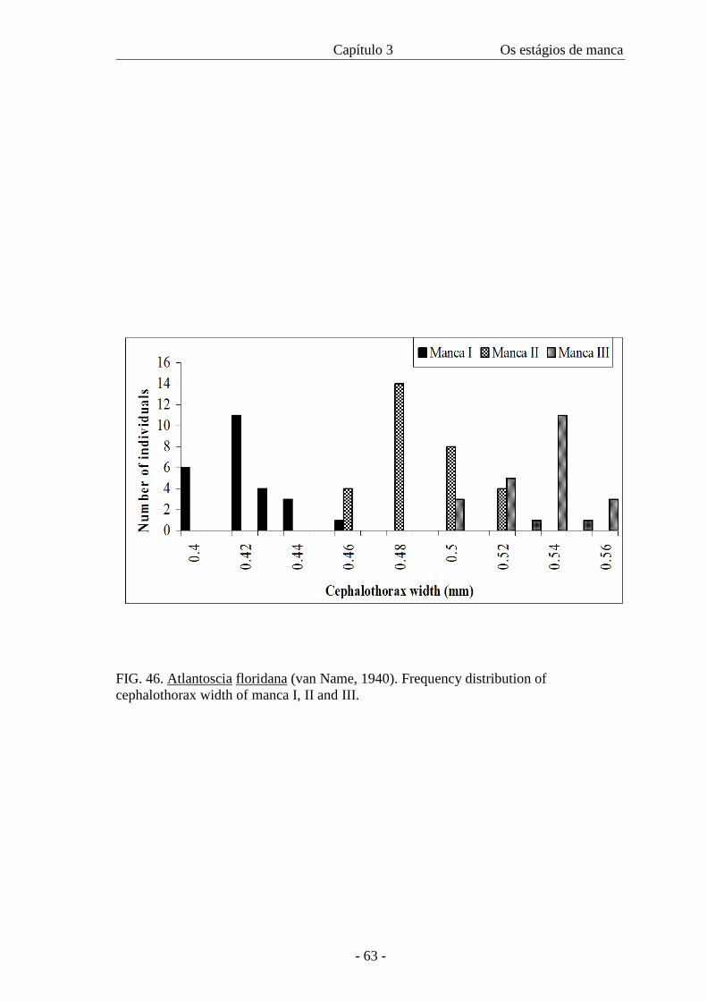
Capítulo 3 Os estágios de manca
- 63 -
FIG. 46. Atlantoscia floridana (van Name, 1940). Frequency distribution of cephalothorax width of manca I, II and III.
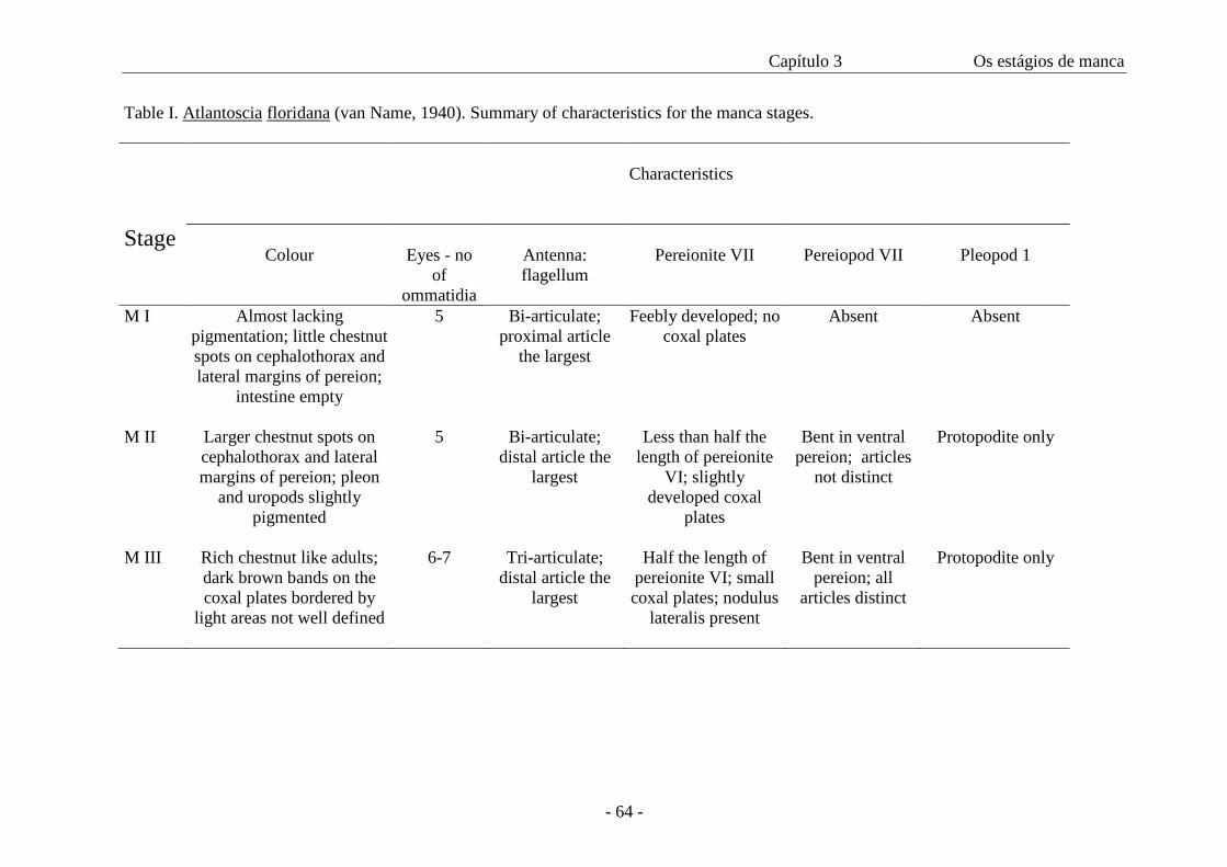
Capítulo 3 Os estágios de manca
- 64 -
Table I. Atlantoscia floridana (van Name, 1940). Summary of characteristics for the manca stages.
Characteristics
Stage Colour
Eyes - no
of ommatidia
Antenna: flagellum
Pereionite VII
Pereiopod VII
Pleopod 1
M I Almost lacking pigmentation; little chestnut spots on cephalothorax and lateral margins of pereion;
intestine empty
5 Bi-articulate; proximal article
the largest
Feebly developed; no coxal plates
Absent Absent
M II Larger chestnut spots on cephalothorax and lateral margins of pereion; pleon
and uropods slightly pigmented
5 Bi-articulate; distal article the
largest
Less than half the length of pereionite
VI; slightly developed coxal
plates
Bent in ventral pereion; articles
not distinct
Protopodite only
M III Rich chestnut like adults; dark brown bands on the coxal plates bordered by
light areas not well defined
6-7 Tri-articulate; distal article the
largest
Half the length of pereionite VI; small coxal plates; nodulus
lateralis present
Bent in ventral pereion; all
articles distinct
Protopodite only
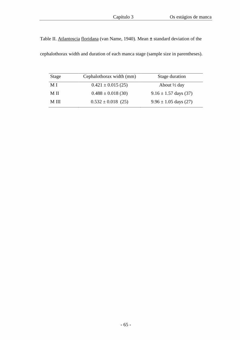
Capítulo 3 Os estágios de manca
- 65 -
Table II. Atlantoscia floridana (van Name, 1940). Mean ± standard deviation of the
cephalothorax width and duration of each manca stage (sample size in parentheses).
Stage Cephalothorax width (mm) Stage duration
M I 0.421 ± 0.015 (25) About ½ day
M II 0.488 ± 0.018 (30) 9.16 ± 1.57 days (37)
M III 0.532 ± 0.018 (25) 9.96 ± 1.05 days (27)
CAPÍTULO 4
Postmarsupial development of Atlantoscia floridana (van Name, 1940)
(Crustacea, Isopoda, Oniscidea): sexual differentiation and
size at onset of sexual maturity
- 67 -
Postmarsupial development of Atlantoscia floridana (van Name, 1940) (Crustacea,
Isopoda, Oniscidea): sexual differentiation and size at onset of sexual maturity
PAULA BEATRIZ ARAUJO* , ALINE FERREIRA QUADROS,
MINNELISE MARTINS AUGUSTO and GEORGINA BOND-BUCKUP
- Programa de Pós-Graduação em Biologia Animal, Departamento de Zoologia,
Instituto de Biociências, Universidade Federal do Rio Grande do Sul, Av. Bento
Gonçalves, 9.500 prédio 43435, CEP 91501-970, Porto Alegre, RS, Brazil
Telephone: +55-51-33167709
Fax: +55-51-33167696
e-mail: [email protected]
Key words: Crustacea, terrestrial isopods, Atlantoscia floridana, postmarsupial
development, sexual differentiation, sexual maturiy
* Corresponding author
ARTIGO SUBMETIDO: INVERTEBRATE REPRODUCTION AND
DEVELOPMENT
Capítulo 4 Os estágios juvenis
- 68 -
Summary
The postmarsupial development of Atlantoscia floridana (van Name, 1940) was
studied, focusing on the differentiation and sexual maturity. Samples were done in the
Reserva Biológica do Lami, Porto Alegre, RS, Brazil during 2000 and 2002. To study
sexual differentiation the animals were reared in the laboratory under temperatures of
about 20°±1°C and observed daily. To analyse sexual maturity, animals preserved in
ethanol 70% were used. Growth was followed through moulting, which permitted the
observation of sexual differentiation. The development of the male genitalia and the
female genital pore was described across the different juvenile stages. Female sexual
maturity is estimated to occur from a size of 1.04 mm of cephalothorax width, based on
the smaller ovigerous female sampled. Male sexual maturity is estimated using the
morphometric relationship between cephalothorax width and length of the genital
papilla, indicating that the puberal moult occurs from a size of 0.77 mm of
cephalothorax width. Identification of the size at which sexual maturity occurs allows
recognizing the juvenile stages of both males and females: JU I to JU III for males and
JU I to JU VI for females. Males reach sexual maturity in approximately 1.5 months and
the females, in about 3 months.
Capítulo 4 Os estágios juvenis
- 69 -
Introduction
Isopods have direct development and thus the offspring is very similar to the
adults from birth. In a previous work, Araujo et al. (2003) provided a description of the
manca stages of Atlantoscia floridana (van Name, 1940), when it was observed that up
to the manca III stage there is no development of secondary sexual characters.
Males can be easily recognised by the presence of the genitalia. It is composed
by the genital papilla and by the endopods of pleopods 1 and 2, which are elongated
forming a strict association with the genital papilla (Legrand, 1946; Sutton, 1980;
Ehrard, 1998). Mature females can be recognised by the presence of the marsupium, an
incubating pouch formed by oostegites connected to the esternites of segments 2-5
(Sutton, 1980; Hoese, 1984). The marsupium can be also observed in post-ovigerous
females, when it is found empty. Characters related to the sexual dimorphism of adults
of A. floridana were described in Araujo and Leistikow (1999) and refer to the male
genitalia.
There are only a few papers investigating sexual differentiation in terrestrial
isopods. Katakura (1984) described the development of the pleopods of males of
Armadillidium vulgare (Latreille, 1804), evidencing the beginning of the sexual
differentiation in this species. Kacem-Lachkar (1997) observed that the sexual stage in
Hemilepistus reaumuri (Audouin & Savigny, 1826) occurs after the fourth moult, when
the development of the genital papilla starts for the males. In a sequel to this work, they
described the development and differentiation of pleopods 1 and 2. Verhoeff (1920),
Matsakis (1955) and Haddad (1984) also followed the post-marsupial development of
other species and observed that after the third moult the pereiopod 7 is functional,
however not mentioning sexual differentiation.
Capítulo 4 Os estágios juvenis
- 70 -
Morphometric studies are used to estimate the beginning of sexual maturity,
mostly in crustacean amphipods (Charniaux-Cotton, 1957 apud Hartnoll, 1982) and
decapods, with knowledge about the puberal moult being of special interest (Hartnoll,
1974; 1982; Felder and Lovett, 1989; Pinheiro and Fransozo, 1993; 1998; Goes and
Fransozo, 1997; Mantelatto and Martinelli, 2001). In general, the puberal moult is
associated with a change in the relative growth of certain appendices or tagma. On
males of these crabs the morphological changes occur on the chelipods, which leads to
morphometric studies based on changes in relative growth rates of these appendices. On
the other hand, females usually have allometric changes in abdomen width.
Recognising the puberal moult is of fundamental importance to the knowledge
about growth and reproduction of a given species. The puberal moult is not usually
studied in terrestrial isopods, perhaps because of the absence of such an evident
secondary character, as in the Decapoda, allowing one to develop morphometric
analyses. Legrand (1946) studied the relative growth of endopod 2 of Porcellio scaber
Latreille, 1804 to determine the puberal moult. Heeley (1941) observed the growth of
the antennal flagellum in P. scaber and did not obtained changes in allometry. In
general, in terrestrial isopods the individuals are recognised as males or females when
recognising the male genitalia is possible.
This work describes the juvenile sexual differentiation for A. floridana, the
number of juvenile stages for males and females and proposes a morphometric analysis
to determine the reproductive status of males of A. floridana.
Material and methods
Capítulo 4 Os estágios juvenis
- 71 -
The specimens of A. floridana used in the present study were sampled in the
Reserva Biológica do Lami, Porto Alegre (30°15´ S 51°05´ W), RS, Brazil from 2000 to
2002. Development was followed in the laboratory, from the manca III stage, since this
is the last undifferentiated sexually stage. The individuals were reared separately using
methods described in Araujo et al. (2003) and observed daily, whenever possible, to see
indications of moult as calcium plates on the thoracic sternites and empty guts. On each
observation measurements of the cephalothorax width were taken (Araujo et al., 2003),
using a Zeiss Stemi V8 stereomicroscope with measuring ocular. The animals were
placed on a Petry dish with a small amount of water, where they stayed immobile long
enough for an adequate measuring. Illustrations were done with the help of a camara
lucida. The specimens used in SEM observation were dried based on Leistikow and
Araujo (2001), transferred to 1:1 ethanol-acetone (2 x 10 min) and 100% acetone (2 x
10 min). After critical point the dried material was mounted on stubs, coated with gold
with Baltec SCD 005 sputter and examined on a Jeol JSM 5800 Scanning Electron
Microscope.
Juvenile individuals are identified as JU followed by a roman number
according to its respective stage; adults are identified as AD. Cephalothorax width
intervals characteristic of the different stages were obtained through observation of the
moults, which indicated the passing from a stage to the next, and also minimum and
maximum widths showed by the animals on each stage. A Student t-test was used to
compare average cephalothorax widths and also development duration from JU I to JU
III for both males and females.
To study sexual maturity, animals preserved in ethanol 70% were used. Sexual
maturity in females was estimated based on the smallest ovigerous female found in the
field, sampled from April 2000 to October 2001. The reproductive status of males,
Capítulo 4 Os estágios juvenis
- 72 -
whether juveniles or adults, was determined through morphometric criteria relating the
genital papilla length (fig.1) with cephalothorax width. Specimens were selected to
represent all cephalothorax width classes. In order to recognise groups belonging to
different growth stages, that is, immature or mature, we used the Regrans software
(Pezzuto, 1983). The regression equation was calculated for all points, detecting the
inflexion point of the regressio and calculating regressions for the points to the left and
right. The validity of the regressions was tested through a comparison between the sum
of squares of the residuals in Y (residual SSY) of the total regression and the sum of the
SSY of the regressions to the left and right (Zar, 1974).
Results
Sexual differentiation
It has been shown that after birth individuals of A. floridana have three stages
of manca where no sexual differentiation can be seen. (Araujo et al., 2003). After that,
the individuals at the juvenil stages (third moult), shows the first signals of sexual
differentiation. The number of juvenile stages was based on the sexual maturity (see
below).
Males
Within each stage, apart from the observed variation on cephalothorax width,
variations on the size and shape (morphology) of the genitalia were also detected. Thus,
juvenile stages are described based on representative individuals, that is, ones with a
width corresponding to the average of that stage.
JU I - the individuals can be sexed by the presence of a slightly developed genitalia. The
genital papilla, which develops as outgrowths from the sternum (Haathela, 1978), is
Capítulo 4 Os estágios juvenis
- 73 -
situated in the middle of the membrane that separates the seventh thoracic sternite and
the abdominal segment (Kacem-Lachkar, 1997). At this age the papilla is short and
triangular without apical specializations (figs. 2, 18) and presents a lateral longitudinal
groove (fig. 19) The exopod of pleopod 1 is rectangular without a distinct point (fig. 5);
the endopod is slightly elongated and presents a smooth surface and rounded apex,
without setation (fig. 5). The exopod of pleopod 2 is rather elongate, slightly pointed
and without sensory setae (fig. 6); the endopod is elongate, longer than the exopod, with
a rounded apex (fig. 7).
JU II – The genital papilla is longer than in JU I (fig. 3). The ventral shield appears
more clearly (fig. 21), forming a triangular apex; below that, the beginning of the
development of cuticular scales occurring in groups forming small fans can be observed
(fig. 20); at this stage the genital pores are still not visible. By transparency, under the
microscope, the formation of ejaculatory ducts can be observed, however not
completely formed to the distal end (fig. 12). In smaller individuals of JU II, the
ejaculatory ducts cannot be observed. The exopod of pleopod 1, somewhat better
developed, starts to look like that of the adult, however still with the distal projection on
the internal margin not evident (fig. 8); the endopod 1 also begins to take the shape it
has on the adult, with the distal end turned outside, still with no setae (fig. 9). The
exopod of pleopod 2 has a distinct distal projection on the internal margin but the lateral
margin does not have setae (fig. 10); the endopod has a similar width in almost all of its
extension (fig. 11).
JU III – The genital papilla has a rectangular spatula below the ventral shield,
surpassing it in length (figs. 4, 22); the cuticular scales are longer and fill the lateral
margins of the spatula (fig. 23). The ejaculatory ducts reach the distal extremity, but the
orifices have small diameters, similar to that of JU II in its distal extremity, which are
Capítulo 4 Os estágios juvenis
- 74 -
smaller than the adult. In adults the presence of a seminal liquid on the extremity of the
papilla is very common, because of the preservation in ethanol 70%. In some
individuals of JU III small quantities of extrusion next to the genital orifices could be
observed. The exopod of pleopod 1 appears similar to that of the adult, with a concave
lateral margin, but with the distal projection of the internal margin still not completely
developed (fig. 13); the endopod is similar to that of the adult, bent laterally, with a
small sub-apical lateral protrusion, and a row of setae on the distal third (fig. 14).
Exopod of pleopod 2 with one seta (fig. 15); endopod acute, with a narrow distal third
(fig. 16), reaching the distal margin of the exopod of pleopod 2.
AD – The apex of the genital papilla has cuticular scales (figs. 24, 25). The endopod of
pleopod 2 reaches the distal margin of the exopod of pleopod 3.
Females
In JU I there is already a development of the exopod of pleopod 1, although still very
rudimentary (fig. 17). The development of the genital pores, located on the base of the
fifth pair of pereiopods (fig. 26) were followed from the JU I until JU VI. There is a
transition on the cuticular scales. In JU I, the genital pores are closed or else appear like
simple openings, without ornamentation, as in JU II. In JU III there is already the
beginning of the formation of cuticular scales that increase in JU IV and JU V (fig. 28);
in JU VI these are already formed, as in the adult (fig. 29).
Size and development time
Table 1 presents the data on body size of juveniles of both males and females. It
can be seen that for both sexes the size remains similar from JU I to JU III, with
differences not significant (p> 0.05). The duration of each JU stage is also similar ( p >
Capítulo 4 Os estágios juvenis
- 75 -
0.05), as can be seen in table 2. The large standard deviation is partly due to some
individuals that took much longer than expected to moult. It was also observed that
some individuals moulted, however without showing considerable changes in
measurements. A comparison between the growth of juvenile males and females can be
seen on fig. 30.
Sexual maturity
Mature females can be easily recognised by the presence of the marsupium.
Were examined 892 ovigerous and post-ovigerous females, which had from 1.04 to 1.68
mm cephalothorax width. Although appearing in low frequencies, the smaller ovigerous
females found in the field had 1.04 mm cephalothorax width (n=3). Based on this
information and in the following of the growth pattern with moult observation, it can be
concluded that in order to reach this size females pass through six juvenile stages, with
the maximum size in JU VI of 0.99mm (table 2). Thus, above this size a new stage is
expected, with this being considered AD. Observations made after JU VI stage show
that the interval of the next stage (AD I) extends from 1.01 to 1.06 mm cephalothorax
width, which corresponds to the interval where the smallests ovigerous females were
found. In laboratory conditions, females reach sexual maturity after three months,
including the manca stage.
The 110 males used in the analysis ranged from 0.59 – 1.32 mm of
cephalothorax width. The data on development fitted a linear regression. Regression
analysis, the software showed the presence of two linear regressions and an inflexion
point at 0.77 mm of CW. Table 3 shows the result of the equations for all data and
points to the left (< X ) and right (≥ X ) of the inflexion point (X) of regressions of
cephalothorax width (CW) against genital papilla length (GPL). The fit of the two-line
Capítulo 4 Os estágios juvenis
- 76 -
model was significantly better (F= 844.79, p < 0.001) than the fit of a single straight line
for all data. It can be seen that before the inflexion point the genital papilla grows
rapidly, in a proportion approximately twice as fast as cephalothorax growth. After the
inflexion point, the papilla has a slower growth, having a proportion next to half the
cephalothorax growth. This differentiated growth of the genital papilla at two moments
of the ontogeny characterises a passage from the juvenile stage to the adult, indicating
that the puberal moult occurs in JU III, with animals approximately 1.5 months old,
including the manca stage (fig. 31).
Discussion
It has been reported that in manca III only the protopodite of the pleopod 1 can
be found (Araujo et al., 2003). After the third moult, when individuals begin the
juvenile stage, the exopod do pleopod 1 appears both for males and females, but the
endopod only occurs in males. Pleopods in females do not change along development,
since these structures do not have secondary sexual characters. In males, the genitalia
form itself from an arrangement between the genital papilla and the two first pairs of
pleopods, composing an elongated structure with an auxiliary function in transferring
spermatozoids during copulation. The observation of this structure allows sex
identification in JU I, when even weakly developed, it is possible to observe the genital
papilla and the short endopods 1 and 2, an observation also done by Katakura (1984)
and Kacem-Lachkar (1997) to A. vulgare and H. reaumuri, respectively.
The juvenile stages in males correspond to a period of great changes in
genitalia, not only regarding growth, but also in terms of the specialisation of the genital
papilla. Legrand (1946) presented a morphometric analysis of endopods 2 used to
identify the puberty moult in P. scaber, showing a differentiated growth of this
Capítulo 4 Os estágios juvenis
- 77 -
appendix from the seventh to the ninth moult. This analysis, however, does not allow
certainty on telling which moult is the puberal one. In the seventh moult there is a
coaptation of the genital papilla and the endopod 1, but in the ninth moult occurs the
maximum extension of endopod 2, apart from the appearance of the sexual characters
on pereiopods 3 and 7. In A. floridana the endopod 2 was not used because this
appendix is elongated and narrow at the distal end which makes it subject to breaking
when under manipulation, resulting in potential errors in measurements. On the other
hand, the genital papilla is a more rigid structure and its growth is appropriate to an
identification of the reproductive status of the species. The analysis of figure 31 leads to
the conclusion that there is a puberal period with an accentuated growth of the genital
papilla, and a period of sexual maturity, when this papilla has a slower growth. In the
puberal stage are included the juveniles I, II and III, with the puberal moult occurring in
JU III. Thus, sexual maturity starts from the sixth moult. As for other crustaceans
(Haley, 1969; Hartnoll, 1974; Goes and Fransozo, 1997; Mantelatto and Martinelli,
2001), the inflexion point in the regression reflects sexual maturity. These results
indicate that the genital papilla, a structure associated directly to sperm transfer, grows
faster during a phase when there is no reproduction but afterwards the papilla must
grow more slowly or otherwise would reach dimensions incompatible with female body
size. Thus, when reaching an adequate configuration for its function, it maintains such
condition throughout its life (Haley, 1969). This tendency is confirmed by the
morphological analysis of the genital papilla, which changes rapidly in the juvenile
stages, with the development of cuticular specialisations until stabilising in the adults. It
is possible that cuticular scales are intimately linked to the passage of sperm from the
genital papilla to the endopods 2, which apparently are the only ones that are inserted in
the genital opening of the females (Sutton, 1980). According to Becker & Mann (1938)
Capítulo 4 Os estágios juvenis
- 78 -
the spermatozoa of P. scaber have very long tails and filamentous heads, which are in
general united forming bundles. Blanchard et al. (1961) mention that the long tail of the
spermatozoa of isopods may not have a direct role in propulsion, but can serve as a
cable, facilitating the transfer of the sperm bundle. Fain-Maurel (1966) found the same
pattern for aquatic isopods (Asellidae, Idoteidae and Cymothoidae) and terrestrial, on
the species Oniscus asellus (Linné, 1758), Armadillidium vulgare and Armadillo
officinalis Duméril, 1816, confirming the non-motility of the spermatozoa. The
observation of sperm in A. floridana indicates a similar pattern both in morphology of
spermatozoa and in the formation of bundles. That way, the cuticular scales at the same
time that have an anti-cohesive function, can help in the directing the elongated bundles
of spermatozoa. Besides, as it is a mechanical transfer, at the same time they help in the
exiting of spermatozoa, they can avoid their return.
For females, the observation of the oostegites is enough to recognise its
reproductive status. However, the morphological observation of the genital pores also
indicates changes along the development. In JU I, when the openings exist, they are
simple, and moult after moult they change due to the appearing of the cuticular scales
that, in this case, can also have the anti-cohesive function and helping in the directing of
the spermatozoa. Loyola e Silva and Coraiola (1999) describe sexual maturity in
females of P. dilatatus Brandt, 1833 from four month old individuals, a not too different
period from that found for A. floridana.
Both size and development time from JU I to III are similar between males and
females, although apparently females are slightly smaller. The growth curve for this
species (Araujo et al. submitted) also shows a small difference in size during the first
months of development, when females are smaller. After this initial period this
phenomenon reverts, with females growing more and reaching larger sizes. It is
Capítulo 4 Os estágios juvenis
- 79 -
interesting to note that average values obtained in the laboratory regarding size in
relation to duration of each juvenile stage for both sexes are very similar to the values,
for the same periods, showed in the growth curve obtained from field data. This
indicates that temperatures of 20° ± 1° C are ideal for rearing A. floridana in the lab,
since it expresses natural conditions found in this latitude.
Population studies depend on the knowledge of the biology of a species. It is
hoped that, in the future, this work could contribute for estimating the puberal moult or
the beginning of morphological sexual maturity in males of terrestrial isopods using this
method relating the growth of the genital papilla with the growth of the cephalothorax.
However, there is still a need for studies dealing with gonadal maturity as another tool
to access the same kind of information but providing a more complete picture.
Acknowledgements
To Secretaria Municipal do Meio Ambiente (SMAM – Porto Alegre), especially M.Sc.
Rodrigo C. Printes for allowing and supporting our research in the Reserva Biológica do
Lami; Dr. Helena Romanowski and Dr. Jocélia Grazia (Departamento de Zoologia,
UFRGS), for the use of the B.O.D. for rearing the animals; Ana Horn for the
illustrations 2-4; Dr. Gilson Moreira for his help with the SEM photographs; PPG-
Biologia Animal and PROPESQ-UFRGS for the concession of an undergraduate
scientific scholarship (BIC). This is contribution No , Department of Zoology.
References
Araujo, P.B. and Leistikow. A., Philosciids with pleopodal lungs from Brazil, with
Capítulo 4 Os estágios juvenis
- 80 -
description of a new species. Contrib. Zool., 68(1999) 109-141.
Araujo, P.B., Augusto, M.M. and Bond-Buckup, G., Postmarsupial development of
Atlantoscia floridana (van Name, 1940) (Crustacea, Isopoda, Oniscidea): the manca
stages. J. Nat. Hist. (2003) (in press).
Araujo, P.B., Lopes, C.G. and Bond-Buckup, G., A field study on the growth curve of
Atlantoscia floridana (van Name) (Crustacea, Isopoda, Philosciidae) in a Brazilian
restinga forest. Pedobiologia (submitted).
Becker, F.D. and Mann, M.E., The reproductive system of the male isopods, Porcellio
scaber Latreille. Trans. Am. Microsc. Soc., 57 (1938) 395-399.
Blanchard, R.E., Lewin, R.A. and Philpott, D.E., Fine structure of sperm tails of
isopods. Crustaceana, 2 (1961) 16-20.
Ehrard, F., Phylogenetic relationships within the Oniscidea (Crustacea, Isopoda).
Isr. J. Zool., 44 (1998) 303-309.
Fain-Maurel, M.A., Contribution a l´histologie et a la caryologie de quelques isopodes;
Spermiogenèse et infrastructure du espermatozoïde des Oniscidés et des
Cymothoïdés. Ann. Sci. Nat., Zoologie, 8 (1966) 1-188.
Felder, D.L. and Lovett, D.L., Relative growth and sexual maturation in the estuarine
ghost shrimp Callianassa louisianensis Schmitt, 1935. Jour. Crustacean Biol., 9(4) \
(1989) 540-553.
Goes, J.M. and Fransozo, A., Relative growth of Eriphia gonagra (Fabricius, 1781)
(Crustacea, decapoda, Xantidae) in Ubatuba, State of São Paulo, Brazil. Nauplius,
5 (2) (1997) 85-98.
Haahtela, I., Morphology as evidence of maturity in isopod crustacea, as exemplified
by Mesidotea entomon (L.). Ann. Zool. Fennici., 15 (1978) 186-190.
Capítulo 4 Os estágios juvenis
- 81 -
Haddad, M.A. Estádios do crescimento pósmarsupial de Balloniscus sellowii
(Brandt, 1833) (Isopoda, Oniscoidea). Dusenia, 13 (3) (1984) 135-143.
Haley, S.R., Relative growth and sexual maturity of the Texas ghost crab, Ocypoode
quadrata (Fabr.) (Brachyura, Ocypodidae). Crustaceana, 17 (1969) 285-297.
Hartnoll, R.G., Variation in growth pattern between some secondary sexual characters
in crabs (Decapoda, Brachyura). Crustaceana, 27 (1974) 131-136.
Hartnoll, R.G., Growth. In: The Biology of Crustacea. Embryology, morphology, and
genetics, Vol 2, L.G. Abele (ed), Academic Press, New York, pp. 111-196, 1982
Heeley, W., Observations on the life-histories of some terrestrial isopods. Proc. Zool.
Soc. Lond., 111(B) (1941) 79-119.
Hoese, B., 1984. The marsupium in terrestrial isopods. In: The biology of terrestrial
isopods, S.L. Sutton and D.M. Holdich (eds), Symp. Zool. Soc. London, 53 (1984)
65-76.
Kacem-Lachkar, H., Étude du développement post-embryonnaire d’Hemilepistus
reaumuri (Audouin, 1826) (Isopoda, Oniscidea). Crustaceana, 70 (1997) 513-526.
Katakura, Y., Sex differentiation and androgenic gland hormone in the terrestrial
isopod Armadillidium vulgare. In: The biology of terrestrial isopods, S.L. Sutton
and D.M. Holdich (eds), Symp. Zool. Soc. London, 53 (1984) 127-142.
Legrand, J.J., Les coaptations sexuelles des Oniscoidea. Bull. Biol. Fr. Belg., 80 (1946)
241-388.
Leistikow, A. and Araujo, P.B., Morphology of respiratory organs in South American
Oniscidea (“Philosciidae”). In: Isopod systematics and evolution, B. Kensley and
R.C. Brusca (eds), Crustacean Issues, Vol 13, R. Vonk and F.R. Schram (gen. eds),
Balkema, Rotterdam, pp.329-336, 2001.
Capítulo 4 Os estágios juvenis
- 82 -
Loyola e Silva, J. and Coraiola, M.A.S., A formação dos oostegitos em Porcellio
dilatatus Brandt (Crustacea, Isopoda, Oniscidea) em laboratório. Revta. bras. Zool.
16 (Supl. 1) (1999) 305-318.
Mantelatto, F.L.M. and Martinelli, J.M., Relative growth and sexual dimorphism of the
South Atlantic hermit crab Loxopagurus loxochelis (Anomura, Diogenidae) from
Ubatuba, Brazil. J. Nat. Hist., 35 (2001) 429-437.
Matsakis, J., Contribution a l’etude du développement postembryonnaire et de la
croissance des Oniscoides. Bull Soc. Zool. France, 80 (1955) 52-65.
Pezzuto, P.R., Regrans: a “basic” program for an extensive analysis of relative growth.
Atlântica, 15 (1983) 93-105.
Pinheiro, M.A.A. and Fransozo, A., Relative growth of the speckled swimming crab
Arenaeus cribbarius (Lamarck, 1818) (Brackyura, Portunidae), near Ubatuba, State
of São Paulo, Brazil. Crustaceana, 65 (1993) 377-389.
Pinheiro, M.A.A. and Fransozo, A., Sexual maturiry of the speckled swimming crab
Arenaeus cribbarius (Lamarck, 1818) (Brackyura, Portunidae) in the Ubatuba
littoral, São Paulo State, Brazil. Crustaceana, 71 (1998) 434-452.
Sutton, S.L., Woodlice, Pergamon Press, Oxford, 1980.
Verhoeff, K.W., Zur Kenntnis der Larven, des Brutsackes und der Bruten der
Oniscoidea. Zool. Anz., 51 (1920) 169- 189.
Zar, J.H., Biostatistical Analysis, Prentice-Hall, London, 1974.
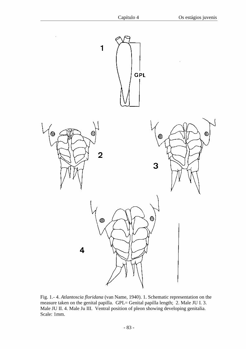
Capítulo 4 Os estágios juvenis
- 83 -
Fig. 1.- 4. Atlantoscia floridana (van Name, 1940). 1. Schematic representation on the measure taken on the genital papilla. GPL= Genital papilla length; 2. Male JU I. 3. Male JU II. 4. Male Ju III. Ventral position of pleon showing developing genitalia. Scale: 1mm.
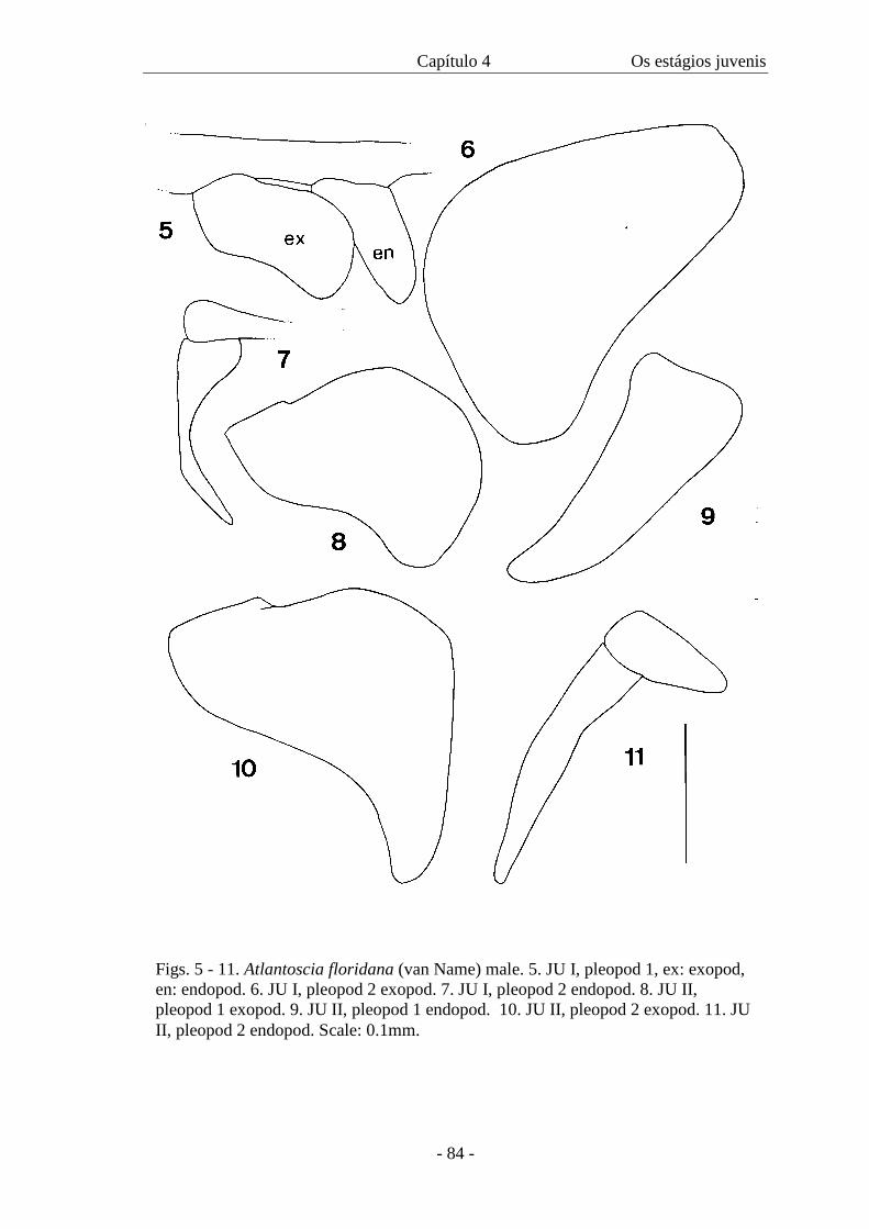
Capítulo 4 Os estágios juvenis
- 84 -
Figs. 5 - 11. Atlantoscia floridana (van Name) male. 5. JU I, pleopod 1, ex: exopod, en: endopod. 6. JU I, pleopod 2 exopod. 7. JU I, pleopod 2 endopod. 8. JU II, pleopod 1 exopod. 9. JU II, pleopod 1 endopod. 10. JU II, pleopod 2 exopod. 11. JU II, pleopod 2 endopod. Scale: 0.1mm.
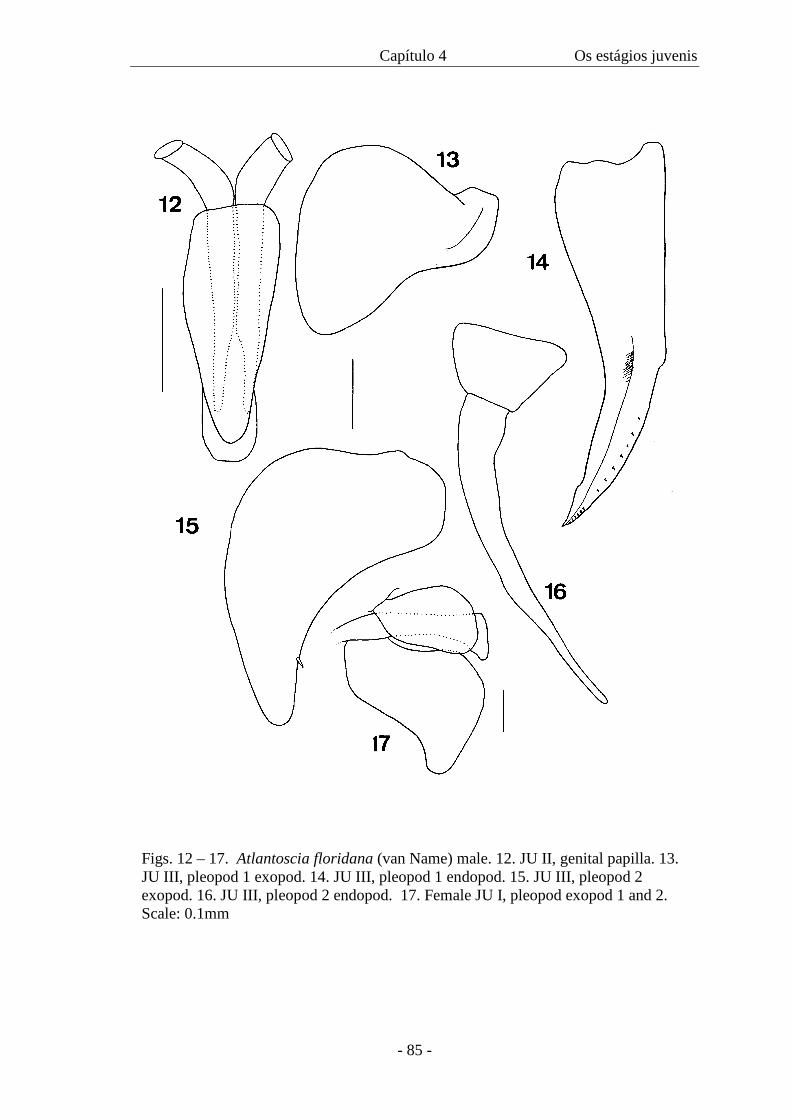
Capítulo 4 Os estágios juvenis
- 85 -
Figs. 12 – 17. Atlantoscia floridana (van Name) male. 12. JU II, genital papilla. 13. JU III, pleopod 1 exopod. 14. JU III, pleopod 1 endopod. 15. JU III, pleopod 2 exopod. 16. JU III, pleopod 2 endopod. 17. Female JU I, pleopod exopod 1 and 2. Scale: 0.1mm
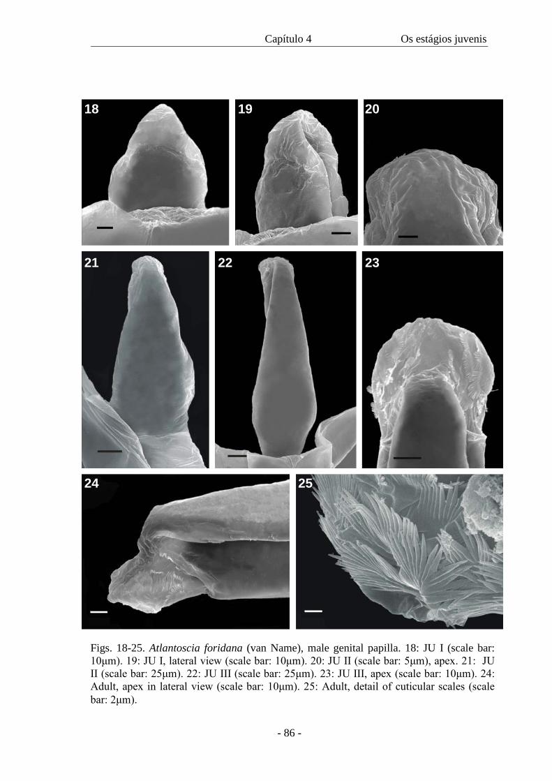
Capítulo 4 Os estágios juvenis
- 86 -
18 19 20
21 22 23
24 25
Figs. 18-25. Atlantoscia foridana (van Name), male genital papilla. 18: JU I (scale bar: 10μm). 19: JU I, lateral view (scale bar: 10μm). 20: JU II (scale bar: 5μm), apex. 21: JU II (scale bar: 25μm). 22: JU III (scale bar: 25μm). 23: JU III, apex (scale bar: 10μm). 24: Adult, apex in lateral view (scale bar: 10μm). 25: Adult, detail of cuticular scales (scale bar: 2μm).

Capítulo 4 Os estágios juvenis
- 87 -
26
27 28
29
Figs. 26-29. Atlantoscia floridana (van Name), female, genital pore. 26: Adult showing the genital pores (arrows) (scale bar: 0.2mm). 27: JU II (scale bar: 10μm). 28: JU V (scale bar: 10μm). 29: Adult (scale bar: 10μm).
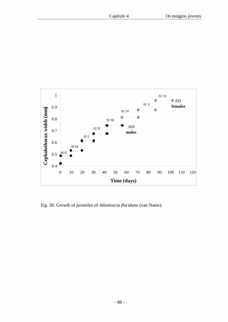
Capítulo 4 Os estágios juvenis
- 88 -
0.4
0.5
0.6
0.7
0.8
0.9
1
0 10 20 30 40 50 60 70 80 90 100 110 120
Time (days)
Cep
halo
thor
ax w
idth
(mm
)
JU I
JU II
JU III
AD males
JU IV
JU V
JU VI AD females
M III
M II
Fig. 30. Growth of juveniles of Atlantoscia floridana (van Name).
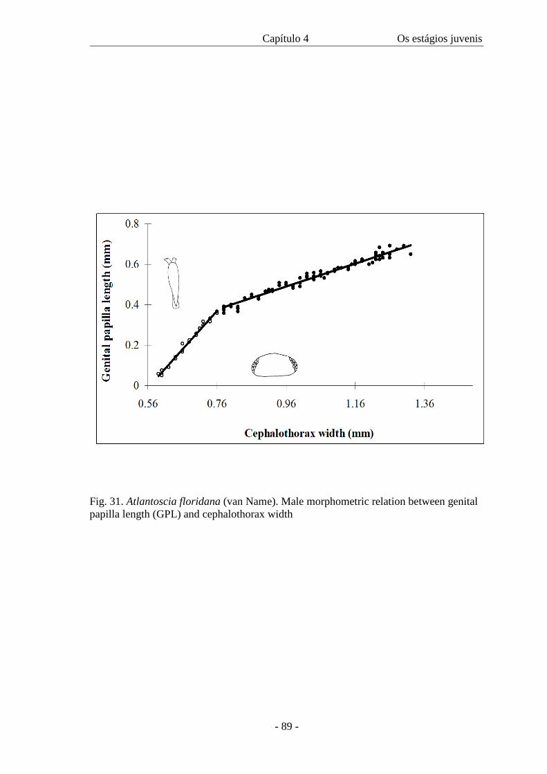
Capítulo 4 Os estágios juvenis
- 89 -
Fig. 31. Atlantoscia floridana (van Name). Male morphometric relation between genital papilla length (GPL) and cephalothorax width
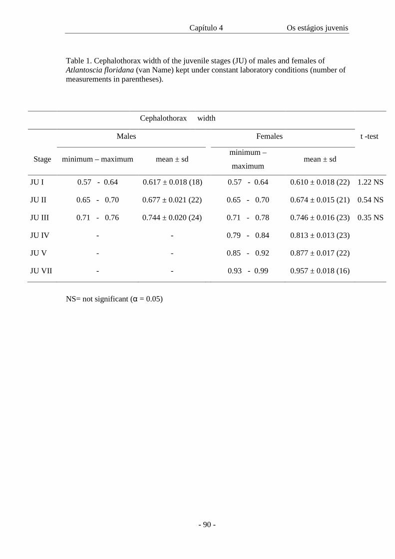
Capítulo 4 Os estágios juvenis
- 90 -
Table 1. Cephalothorax width of the juvenile stages (JU) of males and females of Atlantoscia floridana (van Name) kept under constant laboratory conditions (number of measurements in parentheses).
Cephalothorax width
Males Females t -test
Stage minimum – maximum mean ± sd minimum –
maximum mean ± sd
JU I 0.57 - 0.64 0.617 ± 0.018 (18) 0.57 - 0.64 0.610 ± 0.018 (22) 1.22 NS
JU II 0.65 - 0.70 0.677 ± 0.021 (22) 0.65 - 0.70 0.674 ± 0.015 (21) 0.54 NS
JU III 0.71 - 0.76 0.744 ± 0.020 (24) 0.71 - 0.78 0.746 ± 0.016 (23) 0.35 NS
JU IV - - 0.79 - 0.84 0.813 ± 0.013 (23)
JU V - - 0.85 - 0.92 0.877 ± 0.017 (22)
JU VII - - 0.93 - 0.99 0.957 ± 0.018 (16)
NS= not significant (α = 0.05)
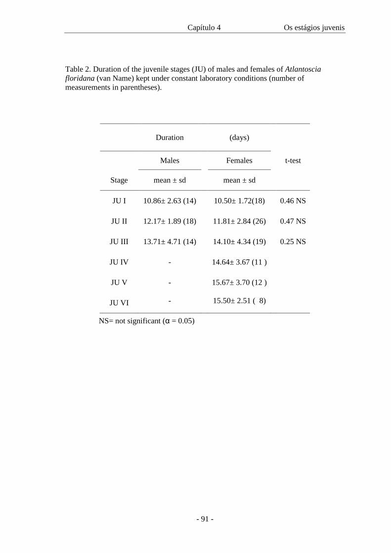
Capítulo 4 Os estágios juvenis
- 91 -
Table 2. Duration of the juvenile stages (JU) of males and females of Atlantoscia floridana (van Name) kept under constant laboratory conditions (number of measurements in parentheses).
Duration
(days)
Males Females t-test
Stage mean ± sd mean ± sd
JU I 10.86± 2.63 (14) 10.50± 1.72(18) 0.46 NS
JU II 12.17± 1.89 (18) 11.81± 2.84 (26) 0.47 NS
JU III 13.71± 4.71 (14) 14.10± 4.34 (19) 0.25 NS
JU IV - 14.64± 3.67 (11 )
JU V - 15.67± 3.70 (12 )
JU VI - 15.50± 2.51 ( 8)
NS= not significant (α = 0.05)
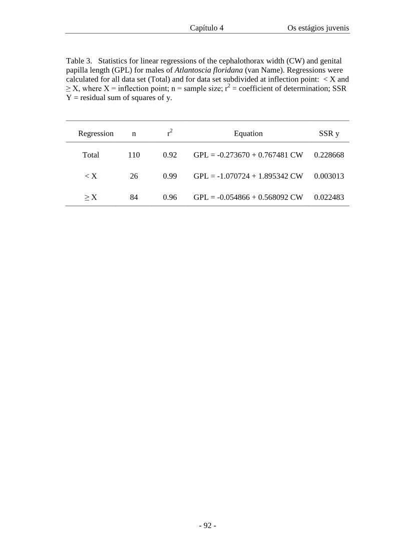
Capítulo 4 Os estágios juvenis
- 92 -
Table 3. Statistics for linear regressions of the cephalothorax width (CW) and genital papilla length (GPL) for males of Atlantoscia floridana (van Name). Regressions were calculated for all data set (Total) and for data set subdivided at inflection point: < X and ≥ X, where X = inflection point; n = sample size; r2 = coefficient of determination; SSR Y = residual sum of squares of y.
Regression n r2 Equation SSR y
Total 110 0.92 GPL = -0.273670 + 0.767481 CW 0.228668
< X 26 0.99 GPL = -1.070724 + 1.895342 CW 0.003013
≥ X 84 0.96 GPL = -0.054866 + 0.568092 CW 0.022483
CAPÍTULO 5
A field study on the growth curve of Atlantoscia floridana (van Name, 1940)
(Crustacea, Isopoda, Oniscidea) in a Brazilian Restinga Forest
- 94 -
A field study on the growth curve of Atlantoscia floridana (van Name, 1940)
(Crustacea, Isopoda, Oniscidea) in a Brazilian Restinga Forest
Paula Beatriz Araujo 1
Caroline G. Lopes
Georgina Bond-Buckup
-Departamento de Zoologia, PPG Biologia Animal, Instituto de Biociências,
Universidade Federal do Rio Grande do Sul, Av. Bento Gonçalves, 9.500 prédio 43435,
CEP 91501-970, Porto Alegre, RS, Brazil, e-mail: [email protected]
1- Corresponding author.
ARTIGO SUBMETIDO: PEDOBIOLOGIA
Capítulo 5 Crescimento
- 95 -
Summary
The terrestrial isopod Atlantoscia floridana (van Name, 1940) is known to occur in the
U.S.A. (Florida), Brazil and Argentina. In the southernmost Brazilian State, Rio Grande
do Sul, the species is recorded in many localities, in urban and in non-urban areas. In
this paper we present the growth curve of Atlantoscia floridana based on field data. The
specimens were sampled from APR/00 to OCT/01 at the Reserva Biológica do Lami
(RBL), Rio Grande do Sul. Captured individuals were sexed and had their
cephalothorax width measured, with the data analyzed with von Bertalanffy´s model.
The growth curves for males and females are described, respectively, by the equations:
Wt = 1.303 [1 - e -0.00941 (t + 50.37)] and Wt = 1.682 [1 - e -0.00575 (t + 59.13)]. The curves
showed differential growth between the sexes, where females reach a higher W∝ with a
slower growth rate. Based on growth curve it was also possible to estimate life
expectancy for males and females.
Key words: Isopoda, Oniscidea, Atlantoscia floridana, growth curve, Southern Brazil.
Capítulo 5 Crescimento
- 96 -
Introduction
The genus Atlantoscia Ferrara and Taiti, 1984, includes the species A. floridana
(van Name, 1940) and A. rubromarginata Araujo & Leistikow 1999. The latter species
has only one known record, in Sergipe State, Northeastern Brazil, where it is found in
litter and bromeliads in the Atlantic Forest (Araujo & Leistikow 1999). On the other
hand, A. floridana occurs in U.S.A. (Florida), Brazil (especially in the coastal States and
Trindade and Abrolhos islands), Argentina, and Ascension and Santa Helena islands
(Ferrara & Taiti 1981; Lemos de Castro 1985; Leistikow & Wägele 1999). In the
Southernmost Brazilian State, Rio Grande do Sul, the species is recorded in many
localities, in urban and in non-urban areas, where it can also be found in litter and
bromeliads (Araujo et al. 1996). Despite being a common and abundant species in
Brazil, there is no information on its ecology. However, important studies have been
developed for other species, e.g. Philoscia muscorum Scopoli, 1763 (Sutton 1968; 1970;
Sunderland et al. 1976) and Burmoniscus ocellatus (Verhoeff, 1928) (Ma et al 1991).
Von Bertalanffy´s growth curve (von Bertalanffy 1938), although commonly
used for fishes (Santos 1978) and for a variety of crustaceans, especially decapods
(Fontoura 1989; Valenti et al. 1994; Barros & Fontoura 1996; Bueno et al. 2000) has
rarely been used for terrestrial isopods. McQueen & Carnio (1974) studied the growth
of Porcellio spinicornis Say, 1818 using von Bertalanffy´s model, reformulated to
consider growth measured in terms of weight. Based on laboratory data Haddad &
Verani (1984) presented figures on the body length growth of Balloniscus sellowii
(Brandt, 1833), a native South American species.
The Reserva Biológica do Lami is a mosaic of different habitats, including
restinga forests, sandy plains, dry and wet grasslands, wetlands with shrub or herb
Capítulo 5 Crescimento
- 97 -
layers and Juncus-dominated shoreline vegetation. Four species of terrestrial isopods
have been recorded occurring especially in the areas of restinga forest: Balloniscus
sellowii, Neotroponiscus daguerrei (Giambiagi de Calabrese, 1939), Trichorhina
argentina, Vandel 1963, and A. floridana, which is the most abundant.
Since terrestrial isopods are, in general, beneficial because of their role in
enhancing nutrient cycling (Paoletti & Hassal 1999) and also because they are a food
source for a wide range of other invertebrates and vertebrates, specific knowledge on
their biology in a reserve area is also an important tool for ecological monitoring and
conservation. In this paper we present the growth curve of A. floridana based on field
data.
Materials and Methods
The study was carried out in Reserva Biológica do Lami (RBL), within the area
of Porto Alegre City (30°15´ S 51°05 W), Rio Grande do Sul State, Brazil (Fig. 1). The
total area of the Reserve comprises 200.6 ha. According to Meira & Porto (1998) the
characteristic vegetation is restinga forest, from 5 to 10 m tall, with orchids and
bromeliads as epiphytes, along with cactuses and the endemic gymnosperm Ephedra
tweediana Fish and C.A. Mey. Grass and sparsely distributed herbs cover the soil and
minimize temperature variation when compared to uncovered areas.
Samples of leaf litter were taken monthly, from April/00 to October/01, along a
footpath on a restinga forest area of approximately 12 x 66 m. Samples were transported
in a plastic bag to the laboratory, where specimens of A. floridana were hand searched.
After that, the field samples were further sorted out with a Berlese Funnel working for
more that 72 hours, to extract the remaining specimens.
Capítulo 5 Crescimento
- 98 -
The animals were separated by sex and stage of sex differentiation (adults and
mancas). Following Sutton (1968), instead of the body length the cephalothorax-width
of the animals extracted was taken, using a stereomicroscope Zeiss Stemi V8 with a
micrometer eyepiece. The measurements refer to the greatest width in the horizontal
plane of the animal, in dorsal view at the level of the eyes, including the eyes
(Sunderland et al. 1976).
The growth of males and females was estimated separately from the
displacement of the monthly modes from the frequency distribution by class interval of
cephalothorax width. For both males and females a size of 0.488 mm was used as the
mean size of manca II (M II), the second stage after birth (see discussion). All the
intramarsupial mancas released by the ovigerous females when put I alcohol after
sampling were included, considered as part of the monthly recruitment. All the manca
stages belong to the first class interval (figs. 2 and 3), which represents the total number
of individuals that cannot be identified as males or females.
The mathematical model used was that of von Bertalanffy (1938):
Wt = W∞ [ 1 – e –k (t-to) ], where Wt is the mean cephalothorax width, in mm, of
the individuals at age t; W∞ is the maximum mean cephalothorax width, in mm; k is the
parameter related to growth rate; t is the age in days and to is a parameter related with
the cephalothorax width at birth.
To calculate W∞, k and to, the following equations were used (Fabens 1965):
to = k –1 . ln [ ∑ (W∞ - Wt ) . e (-kt) / ∑ W∞ . e (-2kt)]
W∞ = -a/b
k = -b; where a and b are parameters of the linear regression transformed by the method
of Ford-Walford (Walford 1946).
The growth curve obtained for males and females was linearized (Allen 1976):
Capítulo 5 Crescimento
- 99 -
Wt = a – b . rt , where: a = W∞; b = W∞ . e kto ; r mean = (e –k male + e –k female) / 2
In the transformation the dependent variable was calculated using the data
obtained from the growth curves. The linear regressions were compared through
ANCOVA, with a confidence interval of 95% (Snedecor & Cochran 1967).
Results and Discussion
The frequency distributions (Figs. 2 and 3) have a high number of modes, and
indicate the coexistence of individuals of varying ages. Although the samples were
taken in the period between APR/00 and OCT/01, the frequency distributions refer only
to periods from which modes could be obtained for males and females, respectively.
Analyzing the mode charts in respect to time, it is possible to observe a mode
displacement. The modes chosen were those that after the transformation (Walford
1946) had the higher correlation coefficients and also the best approximation with the
values of maximum cephalothorax width obtained in nature. Table I shows the monthly
modes chosen for the calculation of the growth curve for males and females.
The growth curves obtained for males and females are shown in Figures 4 and 5,
respectively. The equations describing the curves are:
Males: Wt = 1.303 [ 1 - e –0.00941( t + 50.37) ]
Females: Wt = 1.682 [ 1 - e –0.00575 ( t + 59.13) ]
One of the indications that the curves have a correlation with field data is the
calculation of Wt for males and females considering t = 0. For both sexes, the values
obtained are very close to the average values of M II: 0.492 for males and 0.485 for
females. According to Araujo et al. (2003) we opted for using the mean cephalothorax
width of M II instead of M I. The M I stage has an approximate duration of 12 hours, a
Capítulo 5 Crescimento
- 100 -
very different period from the other stages after the first molt (over 9 days). Thus, M I is
considered as the final embryonic stage culminating with birth and a molt soon after.
Another reason for not using M I is the information in Matsakis (1955) that high
temperatures can induce the first molt while still inside the marsupium, with the youth
being born as M II, at least in Porcellio laevis Latreille, 1804.
Based on the curves it is possible to estimate age from cephalothorax width.
Additionally, the curves can help understanding the sexual dimorphism of different
patterns in the life cycle. In order to compare growth between the sexes, the curves were
linearized. Table 2 shows the values used for regressions, shown in Fig. 6. The results
are in Table 3, where it can be seen that there are no significant differences in
homogeneity variance, since Fs2 < F 0.05, gl 7,7. On the other hand, there are significant
differences between intercepts, since Fa > F 0.05, gl 1,14, and between slopes, Fb > F 0.05, gl
1,15. There is, therefore, differential growth between the sexes, which indicates that
growth in males and females must be treated separately. Females reach a higher W∝
than males, however with a slower growth rate. If, on one side, females grow more
slowly, on the other they grow more, which can be seen as an advantage in reproductive
terms, since they are responsible for incubating the eggs in the marsupium. The largest
male found in the population during the sampled period had a cephalothorax width of
1.30 mm (sampled in October/2000), which corresponds to a value close to W∝ = 1.303.
This suggests that male life expectancy exceeds 600 days (approximately one and a half
years), when they reach the average maximum cephalothorax width. The largest female
found had 1.68 mm of cephalothorax width (ovigerous female sampled in May/2001).
This value is also close to W∝ = 1.682, which confers the females a life expectancy
above two years and four months, according to the growth curve obtained. Such
longevity is very close to the average longevity known for other species, for example,

Capítulo 5 Crescimento
- 101 -
Trichoniscus pygmaeus Sars, 1899 and T. pusillus (Brandt, 1833) – 2 years, Philoscia
muscorum – 2 years and three months, Porcellio scaber Latreille, 1804– 3 years and
Armadillidium vulgare (Latreille, 1804)– 3.5 years (data summarized in Sutton et al.
1984). In contrast, there are species with shorter longevity, like Hemilepistus reaumuri
(Audouin & Savigny, 1826)– 1 year, or larger longevity, like Armadillo officinalis
Dumeril, 1816– 9 years (data summarized in Warburg 1993).
Other authors have treated the differential growth of males and females. Haddad
& Verani (1984) verified a difference in growth from the curves obtained for B.
sellowii. In this case, males also reach smaller sizes with a higher growth rate. A
differential treatment was also given to the growth of Philoscia muscorum by Sutton
(1970) and Sunderland et al. (1976), with the latter observing groups with faster or
slower growth in each sex. Based on these information we investigated the possibility of
more than one growth curve, making clear, for example, differences in growth rate
among seasons of the year. Both for males and females only one growth curve was
found. If there were more than one curve, clear differences could show up on the first
months of post-embryonary development, since Haddad & Verani (1984) found
different growth patterns for B. sellowii reared in laboratory at room temperature in a
region where seasons are marked (Brazil, Curitiba: 25°25’ S 49°15’ W). On the other
hand, seasonal curves could be obtained from laboratory experiments with variable
temperatures. Haddad (1984) conducted laboratory experiments comparing the average
time of molting for B. sellowii at constant temperatures (25°C ± 1) against variable
temperatures, related to natural conditions (a natural range of 13°C to 28°C). The results
show that animals raised on variable temperatures have longer stages during the winter,
when the molt frequency is lower. Growth in constant temperatures was uniform. Thus,
the growth curves of A. floridana obtained from field data describe rates and average
Capítulo 5 Crescimento
- 102 -
stage durations. The generality of the pattern compensates for the seasonal variation,
since longevity exceeds one year and all individuals in the population are subjected to
the same environmental conditions. During the first year of the sampling period the soil
and litter temperature were recorded along the sampling points. The lower temperatures
were at about 7°C (soil) and 6°C (litter) in July; the higher temperatures oscillated at
about 25°C (soil) and 28°C (litter) in March; the average soil temperature was of 18.8°C
and the average litter temperature of 20.9°C. The work by Nair (1998) with Pocellio
scaber Latreille, 1804, in Libya, conducted for 50 weeks illustrates this interpretation.
The author followed the growth of a group born during the warmer months and another
born during the colder ones. A differential growth was found among groups according
to temperature, with a slower growth rate for both groups during colder months,
irrespective of development stage, and a faster growth rate during warmer months. At
the end of the period, both groups reached similar body sizes.
The most frequent widths for males are between 0.72 and 1.04 mm, with a
maximum frequency for the 0.88 mm class. In females, most frequent widths were
between 0.64 and 1.32 mm, with a maximum frequency for the 0.64 mm class. For both
sexes there is a strong reduction in the frequencies in the larger classes, in males from
the 1.12 mm class on and in females from the 1.48 mm class on, which can be a result
of mortality in classes corresponding to older individuals. At no time during the
sampling period it was possible to observe anything similar to the phenomenon
described by Warburg (1993) of population explosion and migration, with the low
number of older individuals probably related to mortality but not to emigration.
Conclusion

Capítulo 5 Crescimento
- 103 -
The growth curve serves as a reference, supplying information on the growth
rate, maximum size attained and the age this size is reached; based on those, the
populations could be subject to periodic diagnoses. This kind of information is
important in monitoring studies of bioindicator species, not only in terms of abundance
levels (Paoletti & Hassall 1999) but also as a form of accessing the impact of
contamination levels on animal growth. Furthermore, we can obtain additional
information on growth since it can also be affected by a variety of sources. This is
particularly important in reserve areas in order to identify the components that are
influencing the populations at a specific time. A. floridana is the most common isopod
species in the Reserva Biológica do Lami. It certainly plays an important role not only
as part of soil nutrient cycle but also as prey for different animals, particularly
vertebrates. Thus, the knowledge on the population ecology of the isopod species can
offer information for monitoring the area as a whole.
Acknowledgements
The authors wish to thank the Secretaria Municipal do Meio Ambiente (SMAM),
especially M.Sc. Rodrigo C. Printes for allowing and supporting our research in the
Reserva Biológica do Lami, Aline F. de Quadros and Minnelise M. Augusto for help on
fieldwork, M.Sc. Clarissa K. Noro and Dr. Aldo M. de Araujo for discussion, Dr.
Ludwig Buckup for criticism and suggestions and to the PPG-Biologia Animal and
PROPESQ-UFRGS for financial support. Contribution n° 386 Department of Zoology.
References
Capítulo 5 Crescimento
- 104 -
Allen, R.L. (1976) Method for comparing fish growth curves. New Zealand Journal of
Marine and Freshwater Research 10, 687 - 692.
Araujo, P.B., Leistikow, A. (1999) Philosciids with pleopodal lungs from Brazil, with
description of a new species. Contributions to Zoology 68, 109-141.
Araujo, P.B., Augusto, M.M., Bond-Buckup, G. 2003. Postmarsupial development
of Atlantoscia floridana (van Name, 1940) (Crustacea, Isopoda, Oniscidea): the
manca stages, Journal of Natural History (in press).
Araujo, P.B., Buckup, L., Bond-Buckup, G. (1996) Isópodos terrestres de Santa
Catarina e Rio Grande do Sul (Crustacea, Oniscidea). Iheringia 81, 111-138.
Barros, M.P., Fontoura, N.F. (1996) Crescimento de Potimirim glabra (Kingsley 1878)
(Crustacea, Decapoda, Atyidae) na Praia da Vigia, Garopaba, Santa Catarina, Brasil.
Nauplius 4, 11-28.
Bueno, A.A.P., Bond-Buckup, G., Buckup, L. (2000) Crescimento de Aegla platensis
Schmitt em ambiente natural (Crustacea, Decapoda, Aeglidae). Revista Brasileira de
Zoologia 17, 51-60.
Fabens, A.J. (1965) Properties and fitting of the von Bertalanffy growth curve. Growth
29, 265-289.
Ferrara, F., Taiti, S. (1981) Terrestrial isopods from Ascension Island. Monitore
Zoologico Italiano (N.S.) (Suppl.) 14, 189-198.
Fontoura, N.F. (1989) Crescimento de Parastacus brasiliensis (von Martens, 1869)
(Crustacea, Decapoda, Parastacidae). Revista Brasileira de Biologia 49, 897-909.
Haddad, M.A. (1984) Estádios do desenvolvimento pós-marsupial de Balloniscus
sellowii (Brandt, 1833) (Isopoda, Oniscoidea). Dusenia 13, 135-143.
Haddad, M.A., Verani, L.C. (1984) Crescimento pós-marsupial de Balloniscus sellowii
(Brandt, 1833) (Isopoda, Oniscoidea). Dusenia 14, 197-209.
Capítulo 5 Crescimento
- 105 -
Leistikow, A., Wägele, J.W. (1999) Checklist of the terrestrial isopods of the new world
(Crustacea, Isopoda, Oniscidea). Revista Brasileira de Zoologia 16 (1), 1- 71.
Lemos de Castro, A. (1985) Considerações sobre Atlantoscia alceui Ferrara & Taiti,
1981(Isopoda, Oniscoidea, Philosciidae). Revista Brasileira de Biologia 45, 417- 422.
Ma, H.H.T., Lam, P.K.S., Dudgeon, D. (1991) Inter and intraspecific variation in the
life histories of three sympatric isopods in a Hong Kong forest. Journal of Zoology
224, 677-687.
Matsakis, J. (1955). Contribution a l’etude du développement postembryonnaire et de
la croissance des Oniscoides. Bulletin de la Societé Zoologique de la France 80, 52-
65.
McQueen, D.J., Carnio, J.S. (1974) A laboratory study of the effects of some climatic
factor on the demography of the terrestrial isopod Porcellio spinicornis Say.
Canadian Journal of Zoology 52, 599-611
Meira, J.R., Porto, M.L. (1998) Reserva Biológica do Lami: a vida na beira do
lago. In: Menegat, R. (coord. geral), Atlas Ambiental de Porto Alegre.
Editora Universidade/UFRGS, Porto Alegre, Brazil.
Nair, G.A. (1998) Reproductive and population biology of Porcellio scaber (Isopoda,
Oniscidea) in Benghazi, Lybia. Israel Journal of Zoology 44, 399-412.
Paoletti, M. G., Hassall, M. (1999) Woodlice (Isopoda: Oniscidea): their potential for
assessing sustainability and use as bioindicators. Agriculture Ecosystem &
Environment 74, 157-165.
Santos, E.P. (1978) Dinâmica de populações aplicada à pesca e piscicultura. São
Paulo, HUCITEC/EDUSP.
Snedecor, C.L., Cochran, W.G. (1967) Statistical Method. Ames, The Iowa State
University Press, 6th edition.
Capítulo 5 Crescimento
- 106 -
Sutton, S.L. (1968) The population dynamics of Trichoniscus pusillus and Philoscia
muscorum (Crustacea: Oniscoidea) in limestone grassland. Journal of Animal
Ecology 37, 425-444.
Sutton, S.L. (1970) Growth patterns in Trichoniscus pusillus and Philoscia muscorum
(Crustacea:Oniscoidea). Pedobiologia 10, 434-441.
Sutton, S.L., Hassall, M, Willows, R., Davis, R.C., Grundy, A., Sunderland, K.D.
(1984) Life histories of terrestrial isopods: a study of intra- and interespecific
variation. In: Sutton, S.L., Holdich, D.M., (eds), The biology of terrestrial isopods.
Symposium of the Zoological Society of London 53, 269-294.
Sunderland, K.D., Hassall, M., Sutton, S.L. (1976) The population dynamics of
Philoscia muscorum (Crustacea: Oniscoidea) in a dune grassland ecosystem. Journal
of Animal Ecology 45, 487-506.
Valenti, W.C., Mello, J.T.C., Lobão, V.L. (1994) Maturation and growth curves of
Macrobrachium carcinus (Linnaeus) (Crustacea, Decapoda, Palaemonidae). From
Ribeira do Iguape River, southern Brazil. Revista Brasileira de Zoologia 11, 649-
658.
von Bertalanffy, L. (1938) A quantitave theory of organic growth (inquiries on growth
laws). Human Biology 10, 181-213.
Walford, L.A. (1946) A new graphic method of describing the growth of animals.
Biological Bulletin 90, 141-147.
Warburg, M.R. (1993) Evolutionary Biology of Land Isopods. Springer-Verlag, Berlin.
Capítulo 5 Crescimento
- 107 -
Fig. 1. Map of Rio Grande do Sul showing the Reserva Biológica do Lami (RBL), the site of sampling.
RBL
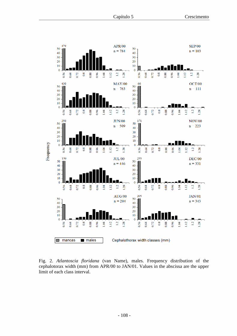
Capítulo 5 Crescimento
- 108 -
Fig. 2. Atlantoscia floridana (van Name), males. Frequency distribution of the cephalotorax width (mm) from APR/00 to JAN/01. Values in the abscissa are the upper limit of each class interval.

Capítulo 5 Crescimento
- 109 -
Fig. 3. Atlantoscia floridana (van Name), females. Frequency distribution of the cephalotorax width (mm) from APR/00 to MAY/01. Values in the abscissa are the upper limit of each class interval.
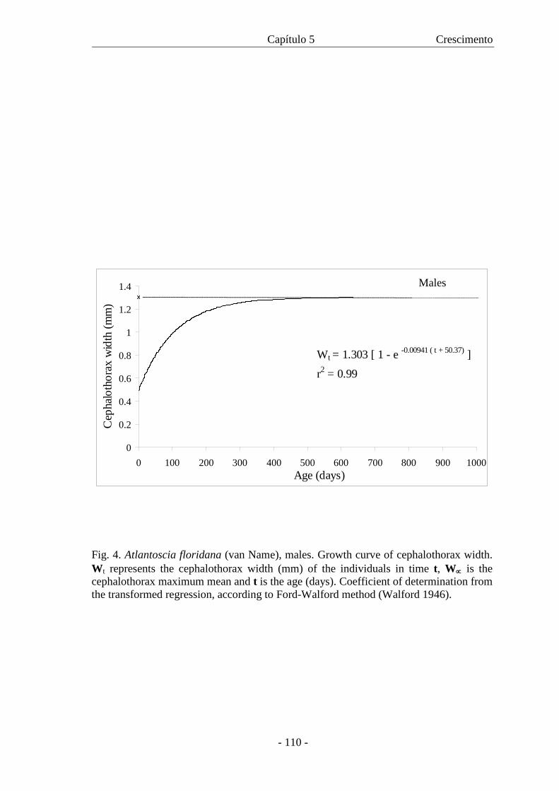
Capítulo 5 Crescimento
- 110 -
Fig. 4. Atlantoscia floridana (van Name), males. Growth curve of cephalothorax width. Wt represents the cephalothorax width (mm) of the individuals in time t, W∝ is the cephalothorax maximum mean and t is the age (days). Coefficient of determination from the transformed regression, according to Ford-Walford method (Walford 1946).
0
0.2
0.4
0.6
0.8
1
1.2
1.4
0 100 200 300 400 500 600 700 800 900 1000Age (days)
Cep
halo
thor
ax w
idth
(mm
)
Wt = 1.303 [ 1 - e -0.00941 ( t + 50.37) ]r2 = 0.99
Males
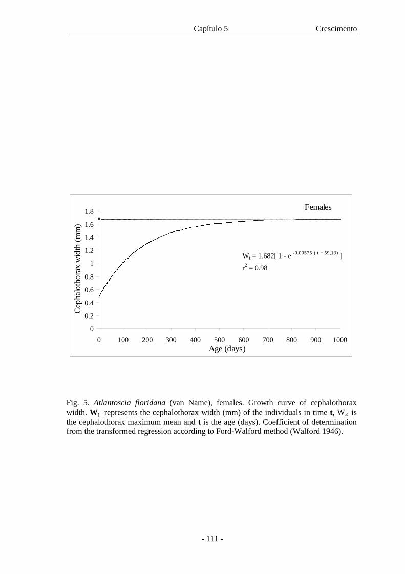
Capítulo 5 Crescimento
- 111 -
0
0.2
0.4
0.6
0.8
1
1.2
1.4
1.6
1.8
0 100 200 300 400 500 600 700 800 900 1000Age (days)
Cep
halo
thor
ax w
idth
(mm
)
Wt = 1.682[ 1 - e -0.00575 ( t + 59,13) ]
r2 = 0.98
Females
Fig. 5. Atlantoscia floridana (van Name), females. Growth curve of cephalothorax width. Wt represents the cephalothorax width (mm) of the individuals in time t, W∝ is the cephalothorax maximum mean and t is the age (days). Coefficient of determination from the transformed regression according to Ford-Walford method (Walford 1946).
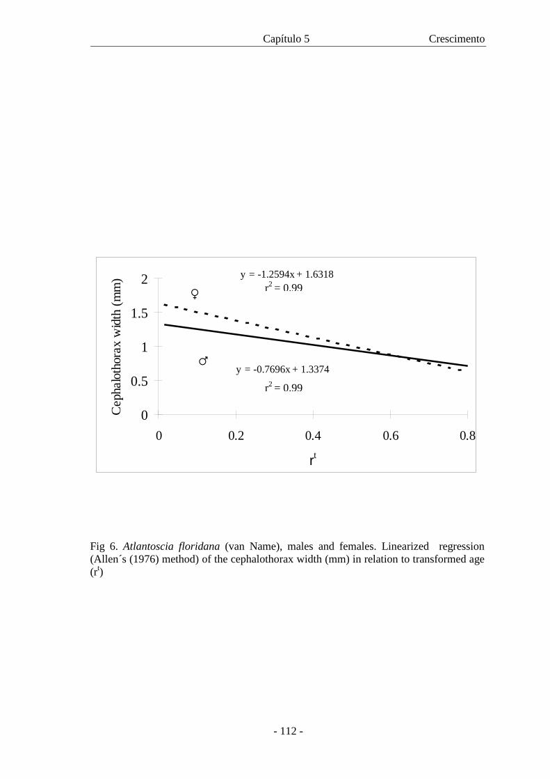
Capítulo 5 Crescimento
- 112 -
Fig 6. Atlantoscia floridana (van Name), males and females. Linearized regression (Allen´s (1976) method) of the cephalothorax width (mm) in relation to transformed age (rt)
y = -0.7696x + 1.3374R2 = 0.99
y = -1.2594x + 1.6318R2 = 0.99
0
0.5
1
1.5
2
0 0.2 0.4 0.6 0.8
rt
Cep
halo
thor
ax w
idth
(mm
)
♀
♂
r2 = 0.99
r2 = 0.99
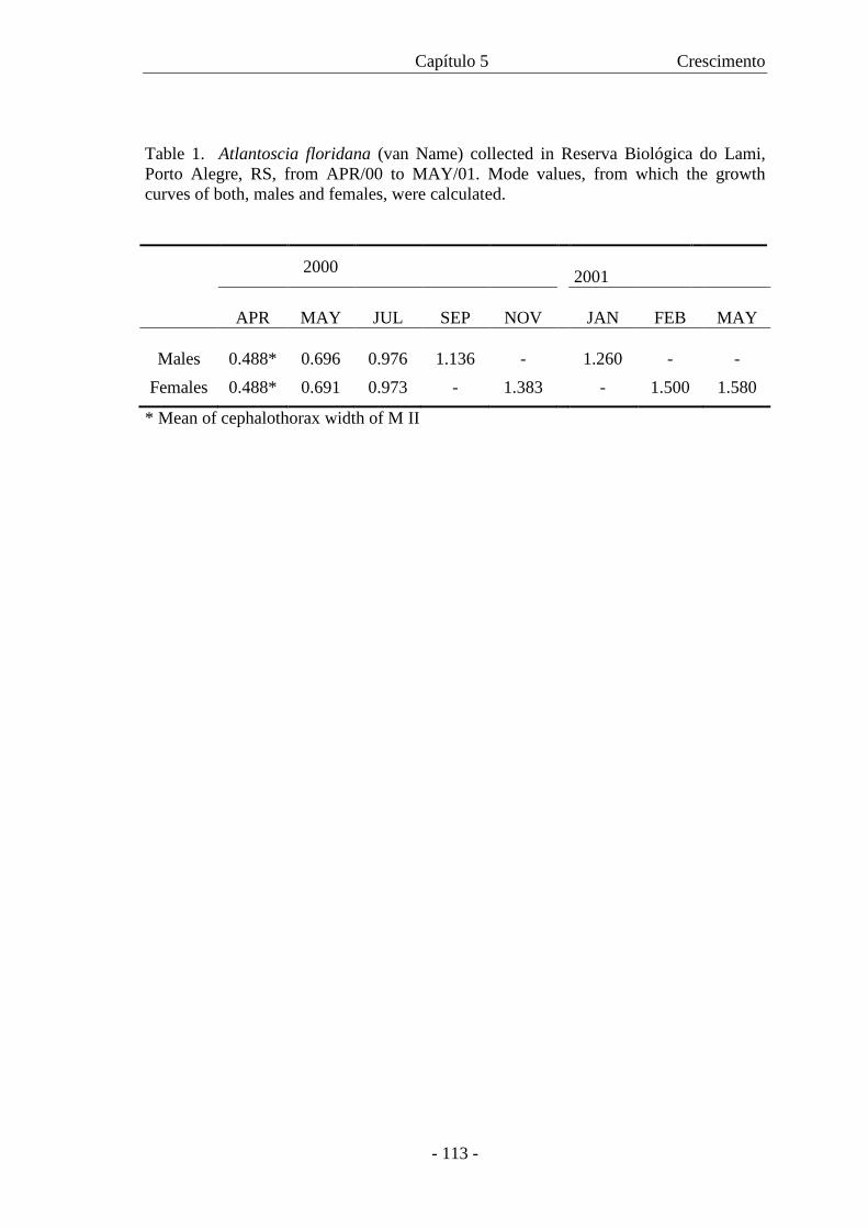
Capítulo 5 Crescimento
- 113 -
Table 1. Atlantoscia floridana (van Name) collected in Reserva Biológica do Lami, Porto Alegre, RS, from APR/00 to MAY/01. Mode values, from which the growth curves of both, males and females, were calculated.
2000 2001
APR
MAY
JUL
SEP
NOV
JAN
FEB
MAY
Males
0.488*
0.696
0.976
1.136 -
1.260 - - Females 0.488* 0.691 0.973 - 1.383 - 1.500 1.580
* Mean of cephalothorax width of M II
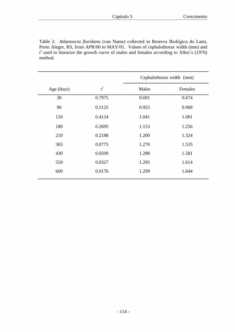
Capítulo 5 Crescimento
- 114 -
Table 2. Atlantoscia floridana (van Name) collected in Reserva Biológica do Lami, Porto Alegre, RS, from APR/00 to MAY/01. Values of cephalothorax width (mm) and rt used to linearize the growth curve of males and females according to Allen´s (1976) method.
Cephalothorax width (mm)
Age (days) rt Males Females
30 0.7975 0.691 0.674
90 0.5125 0.955 0.968
120 0.4124 1.041 1.081
180 0.2695 1.153 1.256
210 0.2188 1.200 1.324
365 0.0775 1.276 1.535
430 0.0509 1.288 1.581
550 0.0327 1.295 1.614
600 0.0176 1.299 1.644
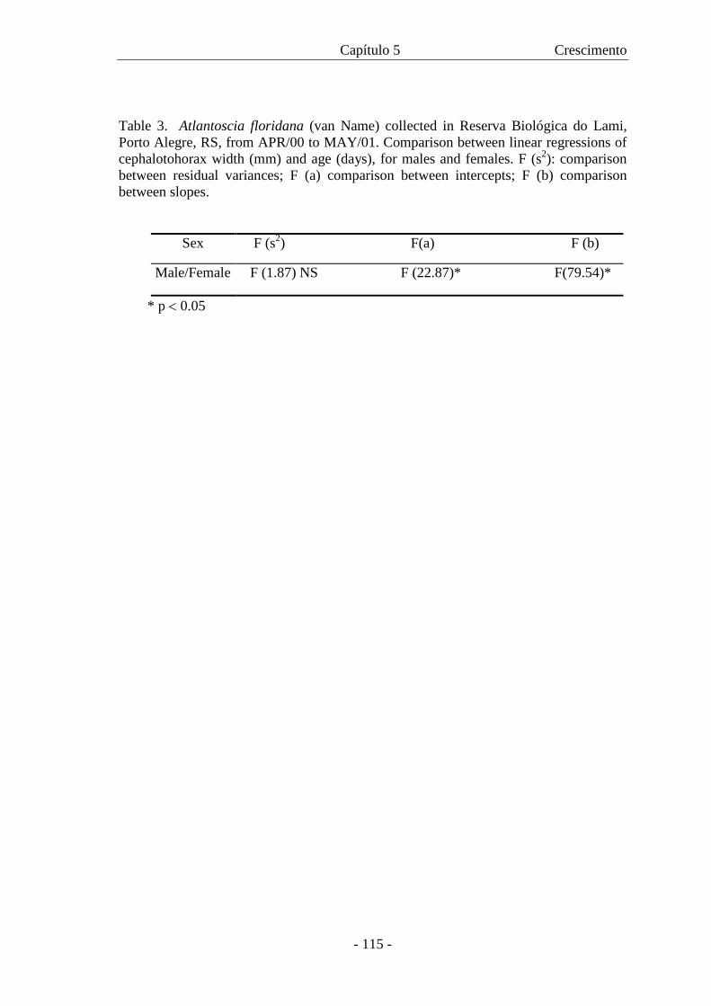
Capítulo 5 Crescimento
- 115 -
Table 3. Atlantoscia floridana (van Name) collected in Reserva Biológica do Lami, Porto Alegre, RS, from APR/00 to MAY/01. Comparison between linear regressions of cephalotohorax width (mm) and age (days), for males and females. F (s2): comparison between residual variances; F (a) comparison between intercepts; F (b) comparison between slopes.
Sex F (s2) F(a) F (b)
Male/Female F (1.87) NS F (22.87)* F(79.54)*
* p < 0.05
CAPÍTULO 6
Population and breeding biology of Atlantoscia floridana (van Name, 1940)
(Crustacea, Isopoda, Oniscidea) in Southern Brazil
- 117 -
Population structure and breeding biology of Atlantoscia floridana (van Name, 1940) (Crustacea, Isopoda, Oniscidea) in Southern Brazil” PAULA BEATRIZ ARAUJO1 and GEORGINA BOND-BUCKUP Programa de Pós-Graduação em Biologia Animal, Departamento de Zoologia, PPG
Biologia Animal, Instituto de Biociências, Universidade Federal do Rio Grande do Sul,
Av. Bento Gonçalves, 9.500 prédio 43435, CEP 91501-970,
Porto Alegre, RS, Brazil
e-mail: [email protected]
Running title: Population biology of Altantoscia floridana.
1 - Corresponding author. ARTIGO A SER SUBMETIDO: JOURNAL OF NATURAL HISTORY
Capítulo 6 Biologia populacional e reprodução
- 118 -
Data on population structure and reproduction of Atlantoscia floridana, one of
the most common species of terrestrial isopods in the restinga forests of Rio Grande do
Sul State, Brazil, were obtained during 2000 and 2001. A total of 7,833 individuals
were sampled along a period of 19 months. Of these, 2,792 were males, 3,400 females
and 1,691 were mancas. There was a significant difference between the size of both,
males and females collected in 2000 and 2001, which had a smaller average size on the
second year because of an absence of individuals on the larger size classes. Population
density varied along the seasons; the minimum population was 131 individuals, the
maximum 1,040 and the average 450 m-2. The operational sex ratio favoured males and
showed no changes with season. Based on the presence of ovigerous and post-ovigerous
females throughout the year, reproduction is considered continuous. However, the
reproduction peak occurs during autumn and spring. Ovigerous females were measured
(CW=cephalothorax width) and the number of eggs was counted. Fecundity (F) varied
from 5 to 23 eggs ( x =11.18±3.64) per female, and was expressed by the regression F=-
18.48+22.59 CW, with the female cephalothorax width varying from 1.04 to 1.68 mm.
Marsupial mortality was only 0.91% and egg production in spring was 588 m–2 and
during autumn, 660 m–2. Recruitment occurs in all months and eggs, embryos and
marsupial mancas are also present all year round. Atlantoscia floridana is the dominant
species among terrestrial isopods in the study area, the most remarkable characteristic
being its reproductive investment.
Key words. Terrestrial isopods; Atlantoscia floridana; population structure;
reproduction; Brazil.
Capítulo 6 Biologia populacional e reprodução
- 119 -
Introduction
Terrestrial isopods represent a significant portion of the soil fauna, where they
play an important role in the processes of decomposition and nutrient recycling (Paoletti
and Hassall, 1999). Investigations on aspects of the population structure and
reproductive biology of several species have been carried out in many countries.
About 20 species of terrestrial isopods were studied in their population dynamics
(Ma et al., 1991; Warburg and Cohen, 1992; Nair, 1998). Species with worldwide
distribution, as for example Armadillidium vulgare (Latreille, 1804), have been studied
in different environments in the United States (Paris and Pitelka, 1962, Lawlor, 1974),
in England (Al-Dabbag and Block, 1981) and in Italy (Caruso and Brandmayr, 1983).
Among the other species are Trichoniscus pusillus Brandt, 1833 and Philoscia
muscorum Scopoli, 1793 (Sutton, 1968; Sunderland, et al, 1976), Ligia oceanica
Linnaeus, 1767 (Willows, 1984), Armadillo officinalis Duméril, 1816 (Warburg and
Cohen, 1992; Al Jetlawi and Nair, 1994), Trachelipus nodulosus (C.L. Koch, 1838)
(Hornung, 1984; 1989), T. rathkei Brandt, 1833 (Farkas, 1998), Porcellio laevis
Latreille, 1804 (Nair, 1984), P. scaber Latreille, 1804 (Nair, 1998) and Hemilepistus
reaumuri (Audouin and Savigny, 1826) (Shachak and Yair, 1984).
Reproduction in terrestrial isopods depends on habitat conditions and in most of
the species it occurs over a determined period, as well as does the parturial molt in
females (Madhavan and Shribbs, 1981), since both temperature and photoperiod can
influence reproduction in this group (Mocquard et al, 1989; Hornung and Warburg,
1993). This influence is clear in cases of seasonal reproduction, characteristic of many
species (Nair, 1984; Warburg and Cohen, 1993; Warburg, 1995; Medini et al., 2000).
Capítulo 6 Biologia populacional e reprodução
- 120 -
There are species that, in laboratory conditions, differ from the natural pattern and show
continued reproduction, as for example, P. olivieri (Audouin, 1825), Agabiformius
obtusus Budde-Lund, 1909 (Warburg, 1995) and P. laevis (Warburg et al., 2001). On
the other hand, species with worldwide distribution can vary in terms of the
reproductive pattern. Porcellionides pruinosus (Brandt, 1833) can have seasonal
reproduction (Achouri and Charfi-Cheikhroua, 2001) as well as continued reproduction,
even in temperate regions (Juchault et al., 1985).
In spite of about 120 recorded species of terrestrial isopods in Brazil (Leistikow
and Wägele, 1998; Souza-Kury, 1998), no studies on the population ecology and
reproduction of any of these species have been conducted. Atlantoscia floridana (van
Name, 1940) is one of the most common species inhabiting restinga forests in Rio
Grande do Sul State, southern Brazil. It was recently redescribed by Araujo and
Leistikow (1999) as well as Leistikow and Araujo (2001) provided a morphological
description of its respiratory organs. Aspects of its post-embryonic development have
been reported by Araujo et al. (2003), where they describe the three manca stages,
whilst Araujo et al. (submitted) provide information on juvenile development,
describing sexual differentiation and establishing age and size at onset of sexual
maturity for males and females. From field data, Araujo et al. (submitted) present a
growth curve for both sexes, including life expectancy.
In order to provide more information on the biology of A. floridana, the present
study focuses on its population structure and reproductive biology using samples taken
from the Reserva Biológica do Lami (RBL), Porto Alegre, Rio Grande do Sul, Brazil.
Capítulo 6 Biologia populacional e reprodução
- 121 -
Material and Methods
The sampling was done in the Reserva Biológica do Lami (RBL), outside the
urban area of Porto Alegre city (30°15´ S 51°05´W), Rio Grande do Sul State, southern
Brazil (a map and more detailed information on the study site can be found in Araujo et
al., submitted). Samples of leaf litter were taken monthly, from April 2000 to October
2001 along a footpath on a patch of restinga forest approximately 12 m wide by 66 m
long. In order to maintain randomness in choosing sampling points, the path was
divided on each of its sides in 6 m intervals. An initial draw was done to decide which
side would be sampled first, and after that the other points alternated between both
sides. With the help of a die, at each point two rolls determined the sampling site by
giving distances in meters, one transversal and the other parallel to the path. For
example, a no. 5 means five meters ahead from the path and a following no. 2 means
two meters to the side. At each sampling site the same procedure was repeated, so that
11 points were sampled, five on one side and six on the other, alternately for each
month. The sampling area corresponded to a circle with 30 cm diameter and was
established based on Sunderland et al. (1976). Samples were transported to the
laboratory in plastic bags, where specimens of A. floridana were hand searched. After
that, the field samples were further sorted out with a Berlese Funnel working for more
that 72 hours to extract the remaining specimens.
The animals were separated by sex and sex differentiation stages (adults and
mancas). Three types of ovigerous females were recognized: females with eggs (EG),
females with embryos (EM) and females with mancas (M). Eggs were identified when
possible to see the amount of yolk and segmentation. Eggs with segmentation were
included in the embryo category, further classified from an initial to an advanced stage.
Capítulo 6 Biologia populacional e reprodução
- 122 -
Mancas were classified as such when, even in the marsupium, the pereiopods and
antennas were already free. This classification was based on Ma et al. (1991) where EG
corresponds to stages 0, 1 e 2, EM to stages 3 and 4, and M to stage 5. Post-ovigerous
females were also separated, having empty marsupia indicating previous birth of
offspring. Measurements were taken according to Araujo et al. (submitted).
The differences in size for individuals of both sexes between the two years of
sampling were tested through the Mann-Whitney test after verifying the normality of
the data distribution using a Kolmogorov-Smirnov test. To test the statistical
significance of the density change between years, data were transformed to log10(n+1)
to solve the problem caused by aggregation, as done in Sunderland et al, 1974.
Transformed data were grouped in three-month blocks and tested by ANOVA, followed
by comparison of means using the Student-Newman-Keuls (SNK) test (Sokal and
Rholf, 1969). In all density calculations the mancas were excluded, and treated
separately.
The operational sex ratio (OSR) is based on the rate of males and females that
are apt to reproduce at a given time (Emelen and Oring, 1977). To identify the existence
of significant differences between the proportion of each sex we employed a χ2 test
(α=0.05), using the OSR for each month and for all samples.
The relationship between the number of eggs (fecundity) and the cephalothorax
width is described by the equation: F = a + bCW, where F is fecundity and CW is
cephalothorax width. A comparison among regressions between cephalothorax width
and the number of EG/EM/M was done through ANCOVA (Snedecor and Cochran,
1973). Data on fecundity were separated in groups of four months beginning in June
2000 and the ANOVA, followed by Tukey multiple tests. The same procedure was used
for the fecundity analysis, separating females by cephalothorax width class intervals for
Capítulo 6 Biologia populacional e reprodução
- 123 -
the different seasons. Mortality in the marsupium was determined through a counting of
embryos in EM females, with special attention paid to undeveloped eggs. Ovigerous
females were kept in the laboratory (under the same conditions explained in Araujo et
al., 2003), so as to observe the incubation period. To calculate egg production, Sutton’s
formula (1968) was used:
Embryo production = Embryo standing crop X Sampling interval
Development time
The degree of correlation between the frequency of ovigerous and post-
ovigerous females and soil and litter temperature and between density and litter
humidity was tested using a Pearson correlation.
For each sampling point the temperatures on the soil surface and on the litter
were registered. Samples of soil and litter were done from April 2000 to March 2001 to
analyse relative humidity, which was done at the Centro de Ecologia - UFRGS. Values
presented here refer to the averages of the points in each sampling date. Samples were
always done during the period between 9:00 and 11:30 AM. Information on air
temperature and relative humidity were supplied by the management of RBL.
Information on the photoperiod was based on Livi (1998).
Results
Climatic conditions.
At the study area, during all the sampling period, soil temperature varied from
7.5 to 24.6, with an average of 17.5o C. Litter temperature oscillated between 7.0 and
Capítulo 6 Biologia populacional e reprodução
- 124 -
28.0o C with an average of 19.7 o C. The average minimum air temperature varied from
5.4 o C to 21.8 o C and the maximum, from 21.3 o C to 33.8 o C (figure 1). Air relative
humidity showed little variation: from 68.9 to 88.8% (figure 2). In the litter, relative
humidity was higher from June to October, when it reached approximately 70%. In the
soil, relative humidity was low for all months, with the maximum reaching
approximately 20%.
Population characteristics
A total of 7,833 individuals were sampled during a period of 19 months.
Of these, 2,792 were males, 3,400 females and 1,691 mancas. The range of the
cephalothorax width for males and females is shown on table 1. There was a significant
difference in size between males and females collected in 2000 and 2001 (p <0.001).
From the figure 3 it can be seen that males had a unimodal distribution, including
juvenile forms. Females, overall, had a bimodal distribution, showing a clear distinction
between juvenile and adults. The histogram shows a prevalence of males between sizes
from 0.78 to 1.02 mm and of non-ovigerous females on classes between 1.02 and
1.26mm. Ovigerous and post-ovigerous females reached the largest sizes ( x = 1.3 ±
0.13); they are more frequently found on classes between 1.15 and 1.44 mm of
cephalothorax width.
Figure 4 shows the variation on the average density of males, non-ovigerous
females and ovigerous and post-ovigerous females along the months sampled. There
were no correlations either between density and litter relative humidity (r = 0.06, p =
0.86) nor between density and litter temperature (r = -0.16, p = 0.51). Although there
are no detected correlations, in 2000 male and non-ovigerous female density decreased
intensely during months of low temperatures and high humidity on the litter. On the
Capítulo 6 Biologia populacional e reprodução
- 125 -
following year this decreasing was not as strong, and density was not as high in April
and May as in the previous year. Comparatively, ovigerous and post-ovigerous females
occur in low densities, which oscillate from warmer to colder months. The average
density of males and ovigerous and post- ovigerous and non-ovigerous females showed
significant variation (figure 5), being lower from October to December 2000, although
in the previous year the low number of individuals was already present in the winter
months (ANOVA, p <= 0.0001; SNK α= 0.05). Table 2 shows the average density by
(three monthly) sampling periods, with 95% CL. All k values (dispersion coefficient)
were higher than 1. Mancas were excluded from this analysis because mortality during
this stage is very high. The minimum density was of 131, the maximum 1,040 m-2 and
the average 450 m-2.
At the beginning of winter 2000 the population had individuals in all size classes
(figure 6). For both sexes the frequency of juveniles was high. At the end of the winter,
juveniles were less frequent, although recruiting (mancas) was high for almost all
periods. In spring, juveniles practically disappeared, with mostly large adults, males and
females, being found. In the beginning of summer, a considerable increase in the
number of juveniles could already be seen, a low occurrence of large males, but a high
occurrence of large females. At the end of summer the adult males became more
frequent and a higher number of non-ovigerous females could be seen. In autumn all
classes were again represented, although in lower numbers when compared to summer,
and there was also a number of large ovigerous and post-ovigerous females, which
occurred again at the beginning of winter. From August to October there was again a
decrease in all size classes, with large ovigerous and post-ovigerous females relatively
frequent.
From the ages estimated following the growth curve presented by Araujo et al.,
Capítulo 6 Biologia populacional e reprodução
- 126 -
(submitted), it can be seen that the number of individuals surpassing one year of life is
very low. Males more than one year old have a cephalothorax width higher than 1.27
mm and were found only in October 2000 (n = 1), January 2001 (n = 1) and September
2001 (n = 1). Females older than a year have a cephalothorax width higher than 1.52mm
and were found during most of the year, totalling 35 individuals. Females surpassing
two years, with cephalothoraxes of 1.66 to 1.68mm width were rare and occurred only
in June 2000 (n=1), December 2000 (n=1) and May 2001(n=1).
Sex ratio
To calculate the OSR the ovigerous females were discarded, since they bear the
eggs and are not apt for new copulations until at least the birth of the offspring. In this
case, the OSR for all months clearly favoured males, deviating significantly from a 1:1
proportion (p < 0.05). Even including post-ovigerous females in the calculation, which
are theoretically ready for a new reproductive event only after the moult, males are still
in larger numbers, approximately in a proportion of two for each female, excepting
October 2000 (p< 0.05) (figure 7). Using the total of months, males also outnumbered
females by 1:0.4 (p<0.001). The sex ratio in the juveniles shows a different pattern,
favouring females by 1:2.2 (p<0.01).
Ovigerous and post-ovigerous females
Ovigerous and post-ovigerous females represented circa 35% of the total of
mature females captured. Figure 8 shows that ovigerous and post-ovigerous females
occur all year long, with higher frequencies for spring and autumn. The occurrence of
ovigerous and post-ovigerous females showed a low positive correlation both with soil
temperature (r= 0.573, t0.05;17 = 2.88) and litter temperature (r= 0.571, t0.05;17= 2.87).
Capítulo 6 Biologia populacional e reprodução
- 127 -
The estimate of the average age of first maturation, which corresponds to 50% of
the reproductive females (Vazzoler, 1996), on the other hand, reaches larger sizes,
between 1.21 and 1.26 mm (figure 9). Females reach this size with approximately 6
months of age, according to the growth curve in Araujo et al. (submitted).
Fecundity and intramarsupial mortality
To estimate fecundity (brood size) females with eggs, embryos and mancas were
analysed. The comparison of the regressions between cephalothorax width and the
number of EG/EM/M showed that EM x EG and M x EG are significantly different
(table 3) and have different determination coefficients: EG= 0.60, EM = 0.51 e M =
0.41. EM or M females having undeveloped eggs were excluded for this analysis. Thus,
analyses always involved EG females. The reason why EM and M females had low
determination coefficients might be because when in contact with the ethanol females
move about too much and expel the marsupial content, especially when this volume is
large. It was observed that females with eggs kept their oostegites closed, whilst in other
situations this did not happen. The relationship between the number of eggs and
cephalothorax width for a total of 288 ovigerous females was: F= -18.057 + 22.241 CW
(figure 10). Brood size varied from five to 23 eggs, with an average of 11.18 ± 3.64
eggs. The size at first maturation was estimated through the size of the smallest
ovigerous female, 1.04 mm. Thus, the cephalothorax width corresponding to the
minimum brood size from the relationship obtained is in the interval between 1.00mm
and 1.06mm, corresponding to the size at first maturation.
On average, the largest ovigerous females were registered in spring and part of
summer, while the smallest occurred in autumn. The average fecundity followed this
trend, being higher in spring and summer and lower at the end of summer and autumn
Capítulo 6 Biologia populacional e reprodução
- 128 -
(figure 11) (ANOVA p< 0.0001; SNK α= 0.05). Fecundity within the same class
intervals did not vary seasonally, except for the sizes between 1.33-1.38, which had
lower fecundity during the period from June-September 2000 (table 4).
Mortality in the marsupium was determined through counting of embryos in
ovigerous females, observing on the latter the undeveloped eggs. Although these
females were excluded from the fecundity analysis, they were used in this case
excluding females with mancas, which had a higher chance of error in obtaining the real
number of individuals. On females with eggs it was impossible to verify mortality. In
the whole period of 19 months, 266 females with embryos in the marsupium were
analysed, of which nine had undeveloped eggs, corresponding to 3,4%. The total
counting of embryos was 2,623 and only 24 non-viable eggs were found. Mortality in
the marsupium, therefore, was 0.91% only.
Egg production
The incubation period was observed in six females (kept under temperatures of
20oC ± 1) and stayed between 19 and 27 days, with an average of 23 days.
Approximately three days after the birth of the offspring some females moulted and
became ovigerous again. In these cases, the second brood had a lower number of
individuals; importantly, these females were always kept isolated, without contact with
males. To calculate egg production the EG females were used and development time
was of 23 days. As this refers to development under controlled temperatures, egg
production (table 5) was only estimated for spring (excluding September and including
December) and autumn, which are the periods of higher fecundity and during which, in
the field, temperatures oscillate at about 20oC. Values of egg production presented in
table 5 are already corrected for marsupium mortality. Thus, the total estimated eggs
Capítulo 6 Biologia populacional e reprodução
- 129 -
produced in spring was 457, which is equivalent to 588 m-2. In autumn, production was
somewhat higher, totalling 518 eggs or 660 m-2.
Eggs, Embryos and Mancas: recruitment
The entry of individuals in the population is constant. Recruitment,
observed as free mancas (figure 12), occurs in practically all months. A more detailed
observation shows that different stages of development are also present in all months,
and in high frequencies (figure 13).
Discussion
From one year to the other there was a decrease in the number of individuals, as
well as in the average size of both males and females, which were smaller, except for
the ovigerous and post-ovigerous females. The smaller male size in the second year is
due to the low number of large individuals. Females also had a low number of large
individuals, with the larger ones ovigerous and post-ovigerous, thus generating a
smaller average size. The average number of individuals collected in each sampling
point varied greatly and the high standard errors are characteristic of a population with
aggregated individuals. Observations showed that A. floridana is a species characteristic
of litter and when found in the soil, individuals stay on the surface. If there is a
systematic vertical migration as a form of better standing climatic conditions, it occurs
only in the most superficial layers, as observed for some individuals during very cold
periods. Sutton (1968) did not observe vertical migration in Philoscia muscorum either.
It is known that the aggregated distribution pattern makes it harder to identify the
absolute density (Sunderland et al., 1976). However, increasing the sampling area or
even increasing the number of samples still give high standard errors (Sutton, 1968;
Capítulo 6 Biologia populacional e reprodução
- 130 -
Sunderland et al., 1976). In the case of species showing vertical migration, mostly as a
form of escaping dryer conditions, this behaviour is responsible for the difficulty in
estimating density (Paris and Pitelka, 1962). The population density in terrestrial
isopods varies according to the species and the environment where the study is
developed. Paris and Pitelka (1962) and Al-Dabbag and Block (1981) found densities of
500 individuals m-2 in the United States and between 200 and 1000 m-2 in England,
respectively. In two different environments in England P. muscorum had 40 to 265 m-2
(Sutton, 1968) and from 88 to 1,037 m-2 (Sunderland et al., 1976). Ma et al. (1991)
working in two different environments in Hong Kong found average densities from 35
to 114 m-2 for Burmoniscus ocellatus (Verhoeff, 1928), 16 to 147 m-2 for Formosillo
raffaelei (Arcangeli, 1927) and 98 to 104 m-2 for Orodillo maculatus Arcangeli, 1952.
The average population density of A. floridana varied during the study period from 131
to 1,040 m-2, showing a density similar to that of P. muscorum presented by Sunderland
et al. (1976). In the Reserva Biológica do Lami it was the dominant species. Trichorhina
tomentosa Vandel, 1963 and Balloniscus sellowii (Brandt, 1833) were not registered for
all months and when found had a relatively low number of individuals.
In A. floridana a density pattern associated to the climatic characteristics of the
region, especially regarding temperature and humidity can be clearly seen. Younger
individuals, especially mancas, seem to be unable to resist the cold and rains,
characteristic of July to September (Livi, 1998). During this period, the number of
juveniles is very low and stays like that until November, still as a reflex of the
conditions prevalent in the previous month, since in order to attain 0.66 mm
cephalothorax width (JU II) at least 30 days are necessary. This fact repeated itself in
the second year, but climatic conditions seemed to have been less rigorous since in
October there were more juveniles than in the previous year. Variations in density
Capítulo 6 Biologia populacional e reprodução
- 131 -
associated to the climatic characteristics have also been described in other terrestrial
isopod species (Al-Dabbag and Block, 1981; Hornung, 1989; Ma et al., 1991).
Although this work does not cover more than two years of observation, it is still
possible to notice a certain stability in density and age structure. Certainly other aspect
not contemplated here but deserving of equal attention due to its influence on the
density are the interactions with other species. Parasitism, for example, can seriously
affect many populations. Amato et al. (submitted) mention the presence of
Centrorhynchus sp. (Acanthocephala) in A. floridana. Thus, future work could perhaps
evaluate the degree of influence of this parasitism on the population, since this parasite
has birds and mammals as definitive hosts, also with reptiles and amphibians as
paratenic hosts. There are no papers (we are aware of) on the impact of predation on
populations of A. floridana.
Based on the growth curve obtained for this species (Araujo et al. (submitted),
survival after the first year in males is very low and corresponds to 0.1% only. Between
the longer lived, most last between eight and nine months. As life expectancy in females
is of more than two years, the number of individuals reaching the second year is a little
higher, corresponding to 1.0%. Those surpassing two years are few and represent only
0.1% of the total of sampled females.
Atlantoscia floridana exhibits a great morphological differentiation in various
aspects: males mature in a shorter time, and thus sex ratio in the juveniles favours
females, males reach a smaller size and a longevity nearly half that of the females. A
sex ratio based on size, as proposed by Wenner (1972), does not make sense in this
case, because the sexes are in different age and reproductive conditions when put in the
same class interval of cephalothorax width. On the other hand, a simple calculation of
Capítulo 6 Biologia populacional e reprodução
- 132 -
the proportion of males and females would include all individuals (adults, juveniles and
ovigerous females), leading to an erroneous evaluation of the meaning of the sex ratio.
The OSR for A. floridana in this study showed a strong male bias during all the study
period. This may indicate strong competition between males, which can be accentuated
because males have a lower life expectancy and begin their reproductive activities very
early. Theoretically, competition between males, in general, would result from their low
parental investment, which increases its reproductive rate and consequently its levels of
sexual activity (Alcock, 1997). However, the level of competition cannot be completely
evaluated, because there is no evidence of the number copulations males participate in,
or even if there is multiple insemination in this species, as registered for Venezillo
evergladensis Schultz, 1963 (Johnson, 1982). Considering the morphology of the genital
pore of females of A. floridana (Araujo et al., submitted) there does not seem to be any
indication of the presence of a decidual organ closing the female genital opening after
copulation, which could be responsible for the retention of sperm and avoidance of
copulation with other males, as described for species of aquatic isopods from the genus
Jaera (Veuille, 1978). On the other hand, post-ovigerous females can have another
brood without necessarily of having a second copulation (Heeley, 1941; personal
observation), which would represent, effectively, a lower number of available females,
and therefore an even more limited resource for males. The trend towards a higher
number of males could be associated with the absence of infection by Wolbachia, a
feminising bacteria (Rigaud et al., 1999; Moreau and Rigaud, 2000), as well as with an
absence of parthenogenesis, as shown by Johnson (1986) to A. floridana in some
populations in Florida (EUA). On the other hand, if the beginning of reproduction is
accelerated by the presence of males, as in Armadillidium vulgare (Jassem et al., 1991,
Caubet et al., 1988; Lefebvre and Caubet, 1999), then the presence of a higher number
Capítulo 6 Biologia populacional e reprodução
- 133 -
of males in a high density population, as in A. floridana, could guarantee the male-
induced stimulation, which according to the authors could be related to tactile or
chemical factors.
Differently from species in temperate regions having seasonal reproduction
(Warburg and Cohen, 1993; Warburg, 1995; Medini et al., 2000), continuous
reproduction is expected in tropical and subtropical species. Atlantoscia floridana has
continuous reproduction at the Reserva Biológica do Lami, where climatic conditions
do not lead to a harsh winter; in this region there is frost, but no snow. Reproduction,
although continuous, is more intense during months when temperature is higher, being
conversely less intense in wintertime, when soil temperatures reach 5o C. During winter,
the frequency of ovigerous and post-ovigerous females is lower, and stays so until
spring, when juveniles surviving the cold become adults and begin their reproductive
activities. In 2000 the cold days begun in July, which justifies the high frequency of
ovigerous and post-ovigerous females in June. In the following year the first cold days
begun in May, which provoked a decrease in the frequency of ovigerous and post-
ovigerous females in July. It is interesting to note that during the period of lower
population density (figure 5), large ovigerous and post-ovigerous females are more
frequent (figures 6, 8), contributing to the high fecundity (figure 11). It is possible that
larger animals have a better ability to survive cold and humid periods; particularly, at
that moment females have an important role in the recuperation of the population
density and maintaining population structure. It is important to establish a parallel
between egg production and fecundity. Egg production was higher during a period in
which fecundity was not at its highest. That is because to calculate egg production only
the total number of eggs produced is taken into account, independently of female size,
while to calculate fecundity the number of eggs is related to female size. Therefore, it is
Capítulo 6 Biologia populacional e reprodução
- 134 -
the largest females that, standing the climatic conditions during winter, guarantee even
with a not so elevated egg production the recruitment at moments in which younger
females, in small numbers in the population, are still not able to reproduce. When these
females reach maturity, they contribute with their eggs generating a higher egg
production. Differences in reproductive patterns can reflect environmental variations.
Experimental work with A. floridana controlling for temperature, humidity and
photoperiod would elucidate the influence these factors have in reproduction. From a
population point of view, photoperiod and temperature do not seem to be determinant
over reproduction, since in winter, when their influence is stronger, with shorter and
colder days, reproduction still occurs. It can be asked, in this case, whether reproduction
is lower in winter because temperature is also lower and photoperiod shorter and thus
there could be interference in vitelogenesis and parturial moult or because these
conditions actually influence survival? Studies with this and other species in other
latitudes, especially in tropical regions, could evidence these questions and then a
comparative analysis of the reproductive strategies could be done.
The number of eggs produced is a specific characteristic that bears on the
population dynamics. Although it has not been the object of this work to investigate
how many times during the life of a female she is capable of becoming ovigerous and
how much does egg number varies with age, laboratory observations showed that after
the mancas birth some females pass through moult and become ovigerous again. This is,
then, an iteroparous species. In the case of a second brood, the egg number was lower,
which was also observed by other authors in T. pusillus, Porcellio dilatatus Brandt,
1833, P. scaber Latreille, 1804, Oniscus asellus Linné, 1758, Armadillidium vulgare and
Philoscia muscorum (Heeley, 1941; Paris and Pitelka 1962, Sunderland et al., 1974).
Capítulo 6 Biologia populacional e reprodução
- 135 -
This and the possibility of a decrease in fecundity in older, senescing females would
explain the deviations in the regression of fecundity over size in larger females.
A very small percentage of ovigerous females had undeveloped eggs, which
represented a mortality in the marsupium lower than 1.0%. Other species also have low
intramarsupial mortality, as P. muscorum, 0.8% (Sutton, 1968) and 3.6% to 4.6% in the
first and second gestation, respectively (Sunderland et al., 1976). Working in different
places Ma et al. (1991) observed differences in mortality, which stayed between 0,91
and 2.69% for B. ocellatus, between 1.44 and 3.03% for F. raffaelei and between 0.5
and 1.0% for O. maculatus. The highest mortality was registered by Paris and Pitelka
(1962), of 7.4% and by Al-Dabbag and Block (1981), of 6.0% for A. vulgare. Mortality,
thus, varies not only between species, but also between populations of the same species.
Perhaps the most noteworthy characteristic of A. floridana, permitting the
capacity for recuperation and maintenance of its population characteristics, is its
elevated reproductive investment. This is evidenced not only by the continuous
reproduction, but also by the low marsupial mortality and high number of mancas
(figure 12) and of eggs, embryos and marsupial mancas (figure 13), observed during
practically all months. This could possibly compensate any difficulties in survival in
adverse conditions and permit re-establishing the population relatively rapidly,
particularly after periods of cold and high humidity. Along with this, a fast growth rate
(Araujo et al., submitted), allows sexual maturation also in a short time, favouring
reproduction and contribution with new individuals and a high recruitment rate.
Capítulo 6 Biologia populacional e reprodução
- 136 -
References
ACHOURI, M.S. and CHARFI-CHEIKHROUA, F., 2001, Biologie de la reproduction
de Porcelliodes pruinosus (Brandt, 1833) isopode terrestre de Garat Nâam (Tunisie),
Crustaceana, 74(1), 11-26.
ALCOCK, J.. 1998. Animal behavior, (Sunderland: Sinauer), 640 pp.
AL-DABBAG, K.Y and BLOCK, W., 1981, Population ecology of a terrestrial isopod
in two breckland grass heaths, Journal of Animal Ecology, 50, 61-77.
ALJETLAWI, A.A. and NAIR, G.A., 1994, Breeding and population biology of
Armadillo officinalis (Isopoda: Oniscidea) in Benghazi, Libya, Journal of Arid
Environments, 27, 241-255.
AMATO, J.F.R., AMATO, S.B., ARAUJO, P.B. and QUADROS, A.F., Atlantoscia
floridana (Isopoda: Oniscidea) as intermediate host of Centrorhynchus sp.
(Acanthocephala: Centrorhynchidae) and first report of pigmentation dystrophy in
terrestrial isopods, Memórias do Instituto Oswaldo Cruz, (submitted)
ARAUJO, P.B. and LEISTIKOW, A., 1999, Philosciids with pleopodal lungs from
Brazil, with description of a new species, Contributions to Zoology, 68 (2), 109-
141.
ARAUJO, P.B., AUGUSTO, M.M. and BOND-BUCKUP, G., 2003. Postmarsupial
development of Atlantoscia floridana (van Name, 1940) (Crustacea, Isopoda,
Oniscidea): the manca stages, Journal of Natural History (in press).
ARAUJO, P.B., QUADROS, A.F., AUGUSTO, M.M. and BOND-BUCKUP, G.,
Postmarsupial development of Atlantoscia floridana (van Name, 1940) (Crustacea,
Isopoda, Oniscidea): sexual differentiation and size at onset of sexual maturity,
Invertebrate Reproduction and Development (submitted).
Capítulo 6 Biologia populacional e reprodução
- 137 -
ARAUJO, P.B., LOPES, C.G. and BOND-BUCKUP, G., A field study on the growth
curve of Atlantoscia floridana (van Name) (Crustacea, Isopoda, Philosciidae) in a
Brazilian restinga forest, Pedobiologia (submitted).
CARUSO, D. and BRANDMAYR, T.Z., 1983, Osservazioni sul ciclo biologico di
alcuni isopodi di Sicilia e relazioni tra fenologia, clima e vegetazione (Crustacea,
Isopoda, Oniscoidea), Animalia, 10(1/3), 237-265.
CAUBET, Y., JUCHAULT, P. and MOCQUARD, J.-P., 1998, Biotic triggers of
female reproduction in the terrestrial isopod Armadillidium vulgare Latr. (Crustacea:
Oniscidea), Ethology, Ecology and Evolution, 10(3), 209-226.
EMLEN, S.T. and ORING, L.W., 1997, Ecology, sexual selection, and the evolution of
mating systems, Science, 197 (4300), 215-223.
FARKAS, S., 1998, Population dynamics, spatial distribution, and sex ratio of
Trachelipus rathkei Brandt (Isopoda: Oniscidea) in a wetland forest by the Drava
River, Israel Journal of Zoology, 44(3-4), 323-331.
HEELEY, W., 1941, Observations on the life-histories of some terrestrial isopods,
Proceedings of the Zoological Society of London, 111(B), 79-119.
HORNUNG, E., 1984, Characteristics of the population of an isopod species
(Trachelipus nodulosus C.L. Koch) at sandy soil grassland, Acta Biologica Szeged,
30, 153-158.
HORNUNG, E., 1989, Population dynamics and spatial distribution of Trachelipus
nodulosus (C.L. Koch, 1838) (Crustacea, Isopoda) in a sandy grassland, Monitore
Zoologico Italiano, Monografia, 4, 399-409.
HORNUNG, E. and WARBURG, M.R., 1993, Breeding patterns in the oniscid isopod,
Porcellio ficulneus Verh., at high temperature and under different photophases,
Invertebrate Reproduction and Development, 23(2-3), 151-158.
Capítulo 6 Biologia populacional e reprodução
- 138 -
JASSEM, W., JUCHAULT, P., SOUTY-GROSSET, C. and MOCQUARD, J.-P.,
1991, Male- induced stimulation of the initiation of female reproduction in the
terrestrial isopod Armadillidium vulgare Latr. (Crustacea, Oniscidea), Acta
Oecologica, 12(5), 643-653.
JOHNSON, C., 1982, Multiple insemination and sperm storage in the isopod, Venezillo
evergladensis Schultz, 1963, Crustaceana, 42(3), 226-232.
JOHNSON, C., 1986, Parthenogenetic reproduction in the philosciid isopod,
Ocelloscia floridana (van Name, 1940), Crustaceana, 51(2), 123-132.
JUCHAULT, P., MOCQUARD, J.-P. and KOUIGAN, S., 1985, Etude expérimentale
de l’ influence des facteurs externes (température et photopériode) sur le cycles de
reproduction du Crustacé Oniscoïde Porcellionides pruinosus (Brandt) provenant de
populations africaines (Togo) et européenne (France), Crustaceana, 48(3), 307-315.
LAWLOR, L.R., 1974, Molting, growth and reproductive strategies in the terrestrial
isopod, Armadillidium vulgare, Ecology, 57, 1179-1194.
LEFEBVRE, F. and CAUBET, Y., 1999, On the male-effect in the terrestrial
Crustacean Armadillidium vulgare (Latreille, 1804), Invertebrate Reproduction and
Development, 35(1), 55-64.
LEISTIKOW, A. and ARAUJO, P.B., 2001, Morphology of the respiratory organs in
South American Oniscidea (Philosciidae), in B. Kensley and R. Brusca (eds) Isopod
Systematics and Evolution, (Rotterdam: BALKEMA), pp. 329-336
LEISTIKOW, A. and WÄGELE, J.W., 1999, Checklist of the terrestrial isopods of the
new world (Crustacea, Isopoda, Oniscidea), Revista Brasileira de Zoologia, 16(1), 1-
72.

Capítulo 6 Biologia populacional e reprodução
- 139 -
LIVI, F.P., 1998, Elementos do clima: o contraste de tempos frios e quentes, in R.
MENEGAT (coord. geral) Atlas Ambiental de Porto Alegre, (Porto Alegre: Editora
Universidade/UFRGS), pp. 73-78.
MA, H.H.T., DUDGEON. D. and LAM, P.K.S., 1991, Seasonal changes in populations
of three sympatric isopods in a Hong Kong Forest, Journal of Zoology, London.,
224, 374-365.
MADHAVAN, K. and SHRIBBS, J.M., 1981, Role of photoperiod and low temperature
in the control of ovigerous molt in the terrestrial isopod, Armadillidium vulgare
(Latreille, 1804), Crustaceana, 41(3), 263-270.
MEDINI, L., NASRI-AMMAR, K. and CHARFI-CHEIKHROUHA, F., 2000,
Reproduction saisonnière de Porcellio variabilis Lucas, 1846, Comptes Rendus de l´
Academie de Sciences, Paris, Life Sciences, 323, 689- 695.
MOCQUARD, J.-P., JUCAHULT, P. and SOUTY-GROSSET, C., 1989, The role of
environmental factors (temperature and photoperiod) in the reproduction of the
terrestrial isopod Armadillidium vulgare (Latreille, 1804), Monitore Zoologico
Italiano, Monografia, 4, 455-475.
NAIR, G.A., 1984, Breeding and population biology of the terrestrial isopod, Porcellio
laevis (Latreille), in the Delhi Region, in S.L. Sutton and D.M. Holdich (eds) The
biology of terrestrial isopods, Symposium of the Zoological Society of London, 53,
315-337.
NAIR, G.A., 1998, Reproductive and population biology of Porcellio scaber (Isopoda,
Oniscidea) in Benghazi, Libya, Israel Journal of Zoology, 44(3-4), 399-412.
PAOLETTI, M. G. and M. HASSALL., 1999, Woodlice (Isopoda: Oniscidea): their
potential for assessing sustainability and use as bioindicators, Agriculture,
Ecosystem and Environment, 74, 157- 165.
Capítulo 6 Biologia populacional e reprodução
- 140 -
PARIS, O.H. and PITELKA, F.A., 1962, Population characteristics of the terrestrial
isopod Armadillidium vulgare in California grassland, Ecology, 43, 229-248.
SHACHAK, M. and YAIR, Y., 1984, Population dynamics and the role of Hemilepistus reaumuri in a desert ecosystem. in S.L. Sutton and D.M. HOLDICH (eds) The
biology of terrestrial isopods, Symposium of the Zoological Society of London, 53:
295-314.
SNEDECOR, C.L. and COCHRAN, W.G., 1967, Statistical Method, (Ames, The Iowa
State Univ.Press), 593 pp.
SOKAL, R.R. and ROHLF, F.J., 1969, Biometry (San Francisco: W, H, Freeman and
Company) 776pp.
SOUZA-KURY, L.A., 1998, Malacostraca – Peracarida. Isopooda. Oniscidea, in P.S.
YOUNG (ed) Catalogue of Crustacea of Brazil (Rio de Janeiro: Museu Nacional),
pp. 653-674.
SUNDERLAND, K.D.; M. HASSALL and SUTTON, S.L., 1976, The population
dynamics of Philoscia muscorum (Crustacea: Oniscoidea) in a dune grassland
ecosystem, Journal of Animal Ecology, 45, 487-506.
SUTTON, S.L., 1968, The population dynamics of Trichoniscus pusillus and Philoscia
muscorum (Crustacea: Oniscoidea) in limestone grassland, Journal of Animal
Ecology, 37, 425- 444.
VAZZOLER, A.E.A.M., 1996, Biologia da reprodução de peixes teleósteos: teoria e
prática. (Maringá: EDUEM), 169pp.
WARBURG, M.R., 1995, Growth and reproduction in a rare desert isopod: Porcellio
barroisi (Oniscidea; Porcellionidae) from the Central Negev Mountains, Journal of
Arid Environments, 31, 199-204.
Capítulo 6 Biologia populacional e reprodução
- 141 -
WARBURG, M.R. and COHEN, N., 1992, Population dynamics, growth and longevity
of Armadillo officinalis (Isopoda; Oniscidea), inhabiting the Mediterranean region of
northern Israel, Pedobiologia, 36, 262-273.
WARBURG, M.R. and COHEN, N., 1993, Breeding patterns in two terrestrial isopods
from the Mediterranean region of Israel, Biologia Gallo-Hellenica, 20, 55-68.
WARBURG, M.R., CALAHORRA, Y. and AMAR, K.O., 2001, Non-seasonal
breeding in Porcellionid isopod, Journal of Crustacean Biology, 21(2), 375-383.
WENNER, A.M., 1972, Sex ratio as a function of size in marine Crustacea, American
Naturalist, 106, 321- 350.
WILLOWS, R., 1984, Breeding phenology of woodlice and oostegite development in
Ligia oceanica (Crustacea). in S.L. Sutton and D.M. HOLDICH (eds) The biology
of terrestrial isopods, Symposium of the Zoological Society of London, 53, 469-485.
Capítulo 6 Biologia populacional e reprodução
- 142 -
Acknowledgements
To Secretaria Municipal do Meio Ambiente (SMAM – Porto Alegre), especially MSc.
Rodrigo C. Printes for allowing and supporting our research in the Reserva Biológica do
Lami, to Caroline G. Lopes for measuring animals and to PPG-Biologia Animal for
financial support. This is contribution No , Department of Zoology.
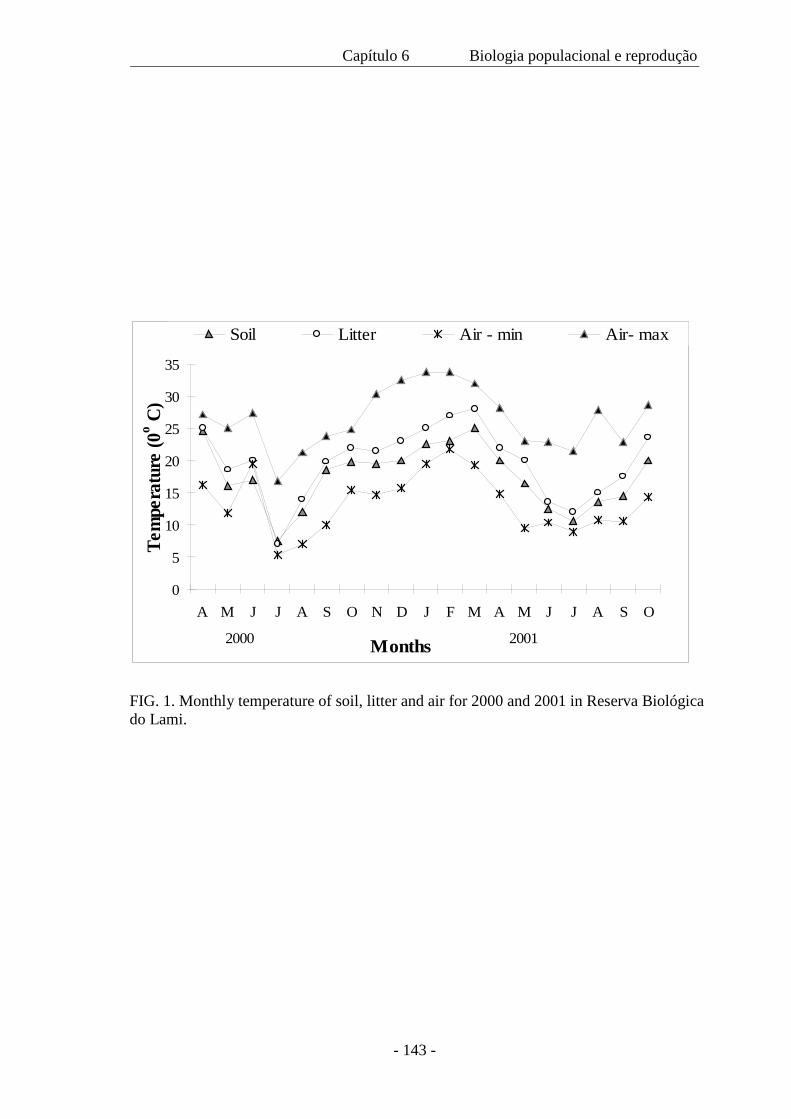
Capítulo 6 Biologia populacional e reprodução
- 143 -
FIG. 1. Monthly temperature of soil, litter and air for 2000 and 2001 in Reserva Biológica do Lami.
0
5
10
15
20
25
30
35
A M J J A S O N D J F M A M J J A S O
Months
Tem
pera
ture
(0o C
)
Soil Litter Air - min Air- max
2000 2001
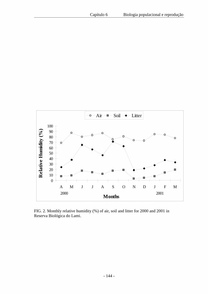
Capítulo 6 Biologia populacional e reprodução
- 144 -
FIG. 2. Monthly relative humidity (%) of air, soil and litter for 2000 and 2001 in Reserva Biológica do Lami.
0102030405060708090
100
A M J J A S O N D J F M
Months
Rel
ativ
e H
umid
ity (%
)
Air Soil Litter
2000 2001
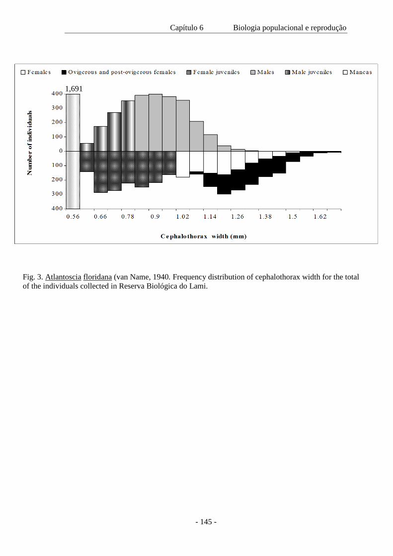
Capítulo 6 Biologia populacional e reprodução
- 145 -
1,691
Fig. 3. Atlantoscia floridana (van Name, 1940. Frequency distribution of cephalothorax width for the total of the individuals collected in Reserva Biológica do Lami.
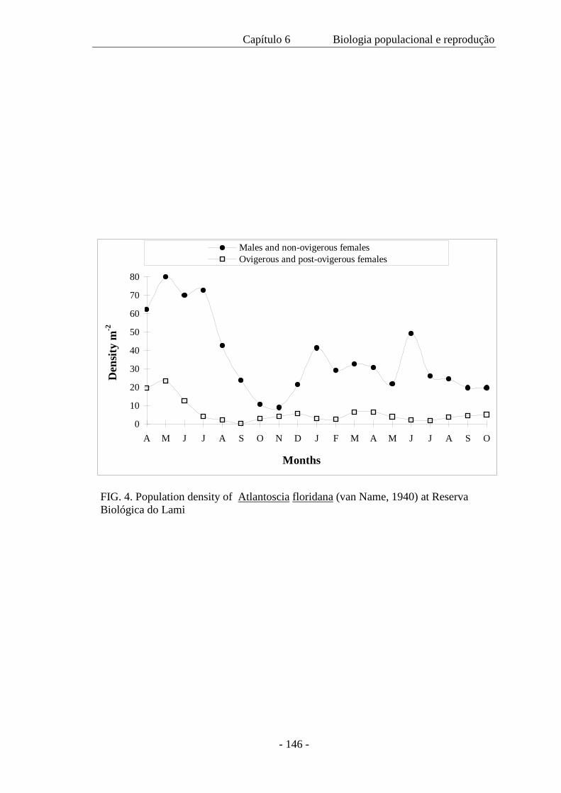
Capítulo 6 Biologia populacional e reprodução
- 146 -
0
10
20
30
40
50
60
70
80
A M J J A S O N D J F M A M J J A S O
Months
Den
sity
m-2
Males and non-ovigerous femalesOvigerous and post-ovigerous females
FIG. 4. Population density of Atlantoscia floridana (van Name, 1940) at Reserva Biológica do Lami
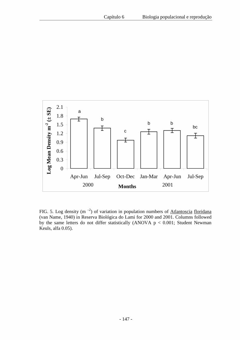
Capítulo 6 Biologia populacional e reprodução
- 147 -
00.30.60.91.21.51.82.1
Apr-Jun Jul-Sep Oct-Dec Jan-Mar Apr-Jun Jul-Sep
Months
Log
Mea
n D
ensit
y m
-2 (±
SE)
2000 2001
a
b
c
b bbc
a
FIG. 5. Log density (m –2) of variation in population numbers of Atlantoscia floridana (van Name, 1940) in Reserva Biológica do Lami for 2000 and 2001. Columns followed by the same letters do not differ statistically (ANOVA p < 0.001; Student Newman Keuls, alfa 0.05).
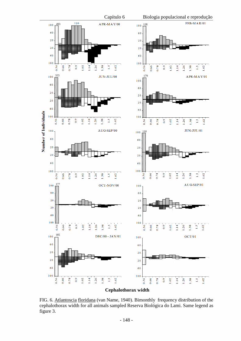
Capítulo 6 Biologia populacional e reprodução
- 148 -
FIG. 6. Atlantoscia floridana (van Name, 1940). Bimonthly frequency distribution of the cephalothorax width for all animals sampled Reserva Biológica do Lami. Same legend as figure 3.
323 179
493 128
Cephalothorax width
110
177
185
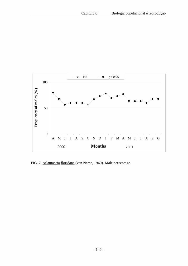
Capítulo 6 Biologia populacional e reprodução
- 149 -
0
50
100
A M J J A S O N D J F M A M J J A S O
Months
Fre
quen
cy o
f mal
es (%
)
NS p< 0.05
2000 2001
FIG. 7. Atlantoscia floridana (van Name, 1940). Male percentage.
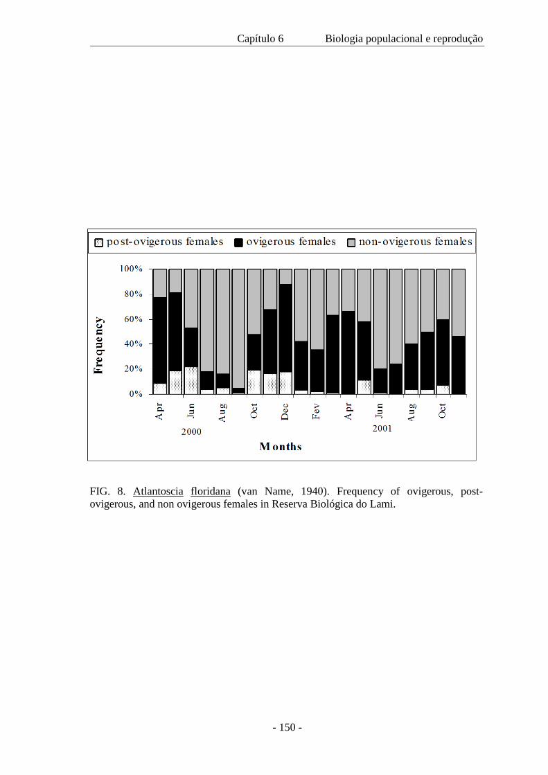
Capítulo 6 Biologia populacional e reprodução
- 150 -
FIG. 8. Atlantoscia floridana (van Name, 1940). Frequency of ovigerous, post-ovigerous, and non ovigerous females in Reserva Biológica do Lami.
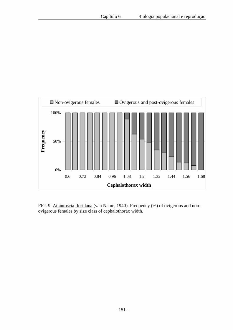
Capítulo 6 Biologia populacional e reprodução
- 151 -
FIG. 9. Atlantoscia floridana (van Name, 1940). Frequency (%) of ovigerous and non-ovigerous females by size class of cephalothorax width.
0%
50%
100%
0.6 0.72 0.84 0.96 1.08 1.2 1.32 1.44 1.56 1.68
Cephalothorax width
Freq
uenc
y
Non-ovigerous females Ovigerous and post-ovigerous females
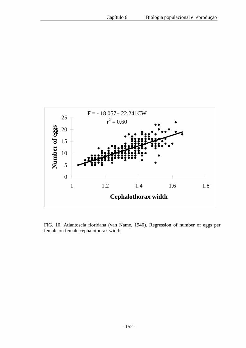
Capítulo 6 Biologia populacional e reprodução
- 152 -
FIG. 10. Atlantoscia floridana (van Name, 1940). Regression of number of eggs per female on female cephalothorax width.
F = - 18.057+ 22.241CWr2 = 0.60
0
5
10
15
20
25
1 1.2 1.4 1.6 1.8
Cephalothorax width
Num
ber
of e
ggs
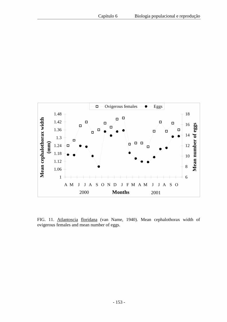
Capítulo 6 Biologia populacional e reprodução
- 153 -
FIG. 11. Atlantoscia floridana (van Name, 1940). Mean cephalothorax width of ovigerous females and mean number of eggs.
1
1.06
1.12
1.18
1.24
1.3
1.36
1.42
1.48
A M J J A S O N D J F M A M J J A S O
Months
Mea
n ce
phal
otho
rax
wid
th
(mm
)
6
8
10
12
14
16
18
Mea
n nu
mbe
r of
egg
s
Ovigerous females Eggs
2000 2001
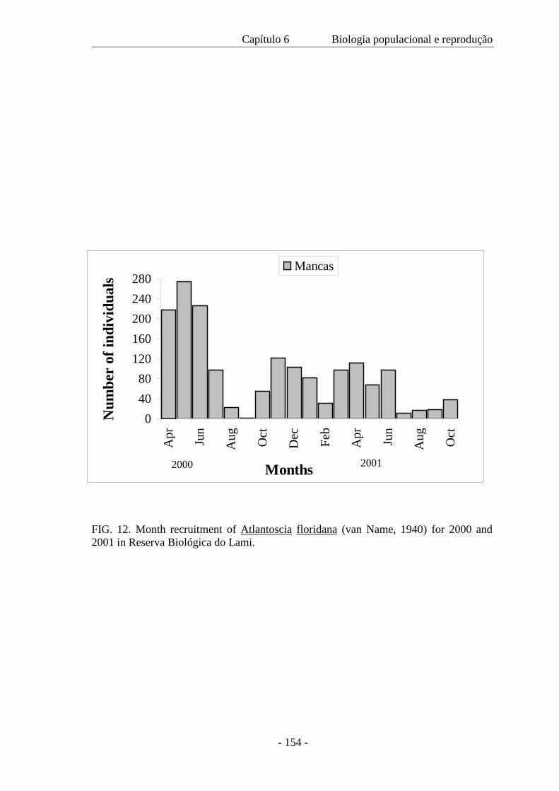
Capítulo 6 Biologia populacional e reprodução
- 154 -
FIG. 12. Month recruitment of Atlantoscia floridana (van Name, 1940) for 2000 and 2001 in Reserva Biológica do Lami.
04080
120160200240280
Apr Jun
Aug Oct
Dec Feb
Apr Jun
Aug Oct
Months
Num
ber
of in
divi
dual
s
Mancas
2000 2001
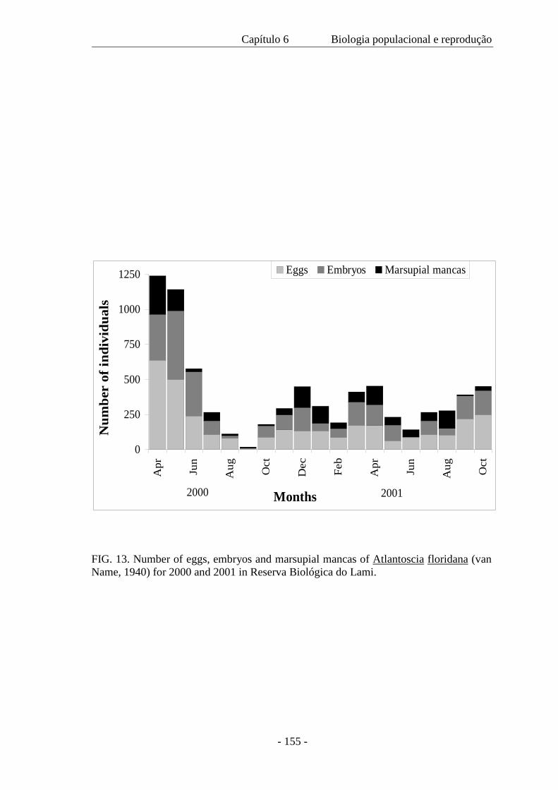
Capítulo 6 Biologia populacional e reprodução
- 155 -
0
250
500
750
1000
1250
Apr Jun
Aug Oct
Dec Feb
Apr Jun
Aug Oct
Months
Num
ber
of in
divi
dual
s
Eggs Embryos Marsupial mancas
2000 2001
FIG. 13. Number of eggs, embryos and marsupial mancas of Atlantoscia floridana (van Name, 1940) for 2000 and 2001 in Reserva Biológica do Lami.
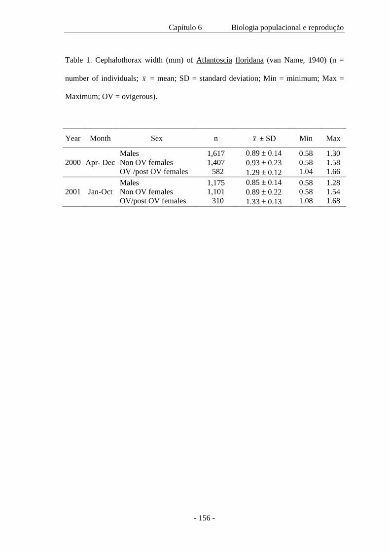
Capítulo 6 Biologia populacional e reprodução
- 156 -
Table 1. Cephalothorax width (mm) of Atlantoscia floridana (van Name, 1940) (n =
number of individuals; x = mean; SD = standard deviation; Min = minimum; Max =
Maximum; OV = ovigerous).
Year Month Sex n x ± SD Min Max
2000 Apr- Dec Males Non OV females OV /post OV females
1,617 1,407 582
0.89 ± 0.14 0.93 ± 0.23 1.29 ± 0.12
0.58 0.58 1.04
1.30 1.58 1.66
2001 Jan-Oct Males Non OV females OV/post OV females
1,175 1,101 310
0.85 ± 0.14 0.89 ± 0.22 1.33 ± 0.13
0.58 0.58 1.08
1.28 1.54 1.68
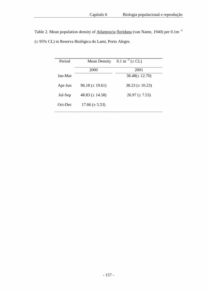
Capítulo 6 Biologia populacional e reprodução
- 157 -
Table 2. Mean population density of Atlantoscia floridana (van Name, 1940) per 0.1m -2
(± 95% CL) in Reserva Biológica do Lami, Porto Alegre.
Period Mean Density 0.1 m -2 (± CL)
2000 2001 Jan-Mar 38.48(± 12.70)
Apr-Jun 96.18 (± 19.61) 38.23 (± 10.23)
Jul-Sep 48.83 (± 14.58) 26.97 (± 7.53)
Oct-Dec 17.66 (± 5.53)
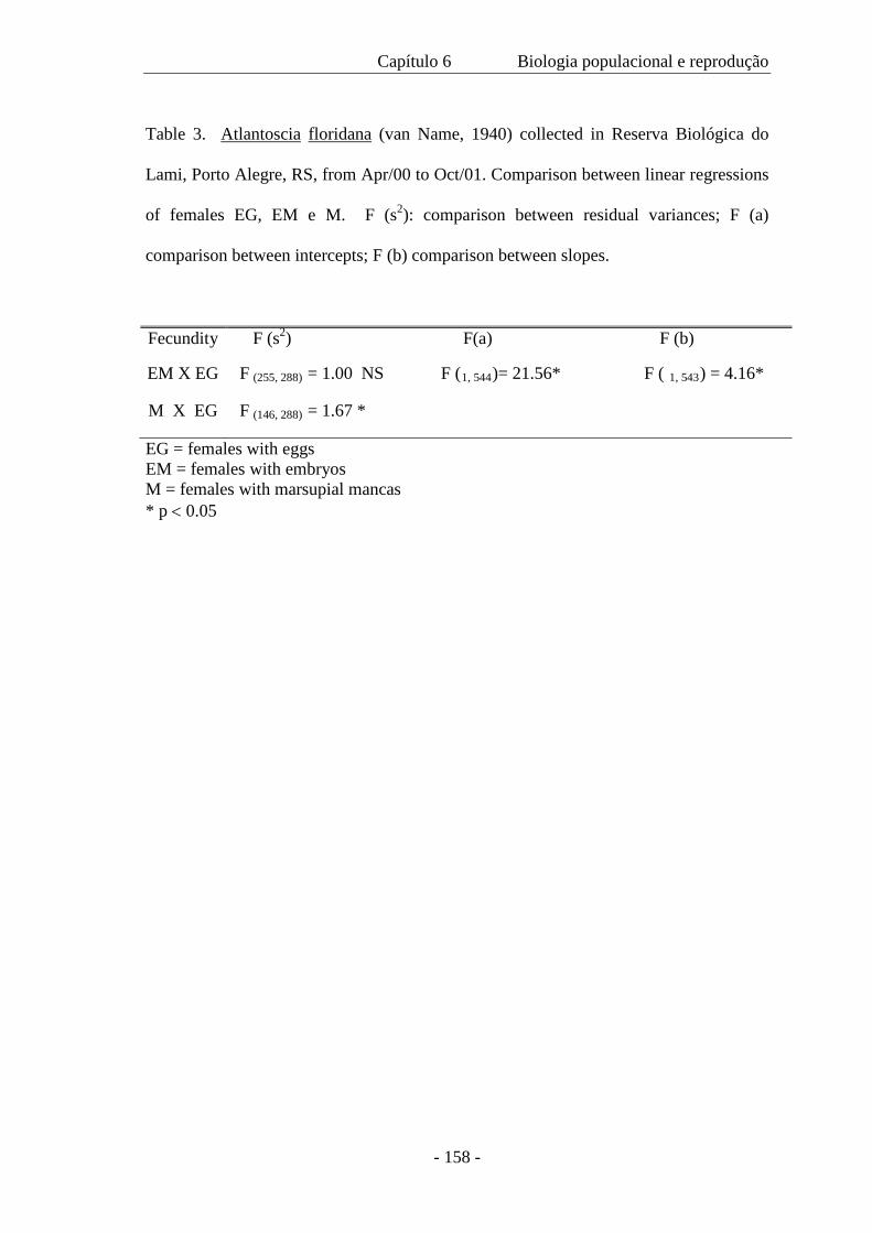
Capítulo 6 Biologia populacional e reprodução
- 158 -
Table 3. Atlantoscia floridana (van Name, 1940) collected in Reserva Biológica do
Lami, Porto Alegre, RS, from Apr/00 to Oct/01. Comparison between linear regressions
of females EG, EM e M. F (s2): comparison between residual variances; F (a)
comparison between intercepts; F (b) comparison between slopes.
Fecundity F (s2) F(a) F (b)
EM X EG F (255, 288) = 1.00 NS F (1, 544)= 21.56* F ( 1, 543) = 4.16*
M X EG F (146, 288) = 1.67 *
EG = females with eggs EM = females with embryos M = females with marsupial mancas * p < 0.05
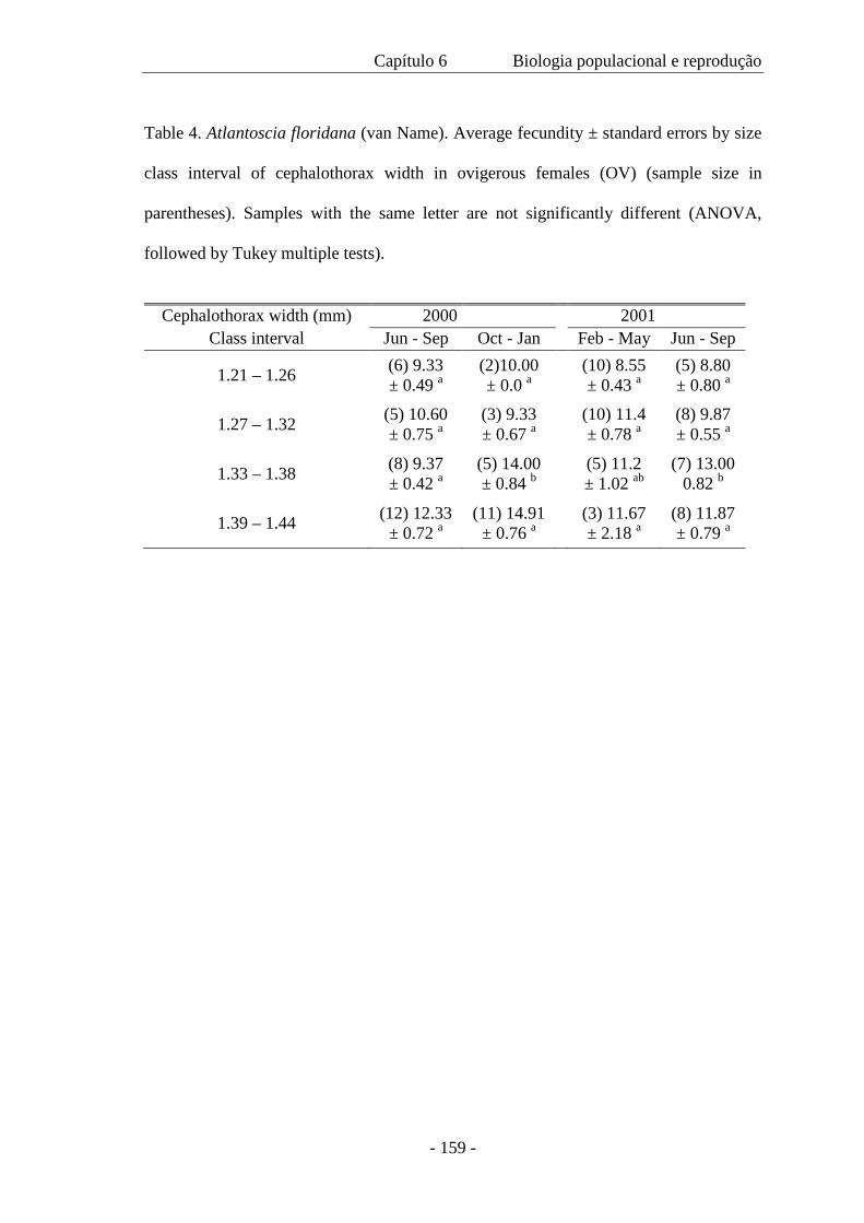
Capítulo 6 Biologia populacional e reprodução
- 159 -
Table 4. Atlantoscia floridana (van Name). Average fecundity ± standard errors by size
class interval of cephalothorax width in ovigerous females (OV) (sample size in
parentheses). Samples with the same letter are not significantly different (ANOVA,
followed by Tukey multiple tests).
Cephalothorax width (mm) 2000 2001
Class interval Jun - Sep Oct - Jan Feb - May Jun - Sep
1.21 – 1.26 (6) 9.33 ± 0.49 a
(2)10.00 ± 0.0 a (10) 8.55
± 0.43 a (5) 8.80 ± 0.80 a
1.27 – 1.32 (5) 10.60 ± 0.75 a
(3) 9.33 ± 0.67 a (10) 11.4
± 0.78 a (8) 9.87 ± 0.55 a
1.33 – 1.38 (8) 9.37 ± 0.42 a
(5) 14.00 ± 0.84 b (5) 11.2
± 1.02 ab (7) 13.00
0.82 b
1.39 – 1.44 (12) 12.33 ± 0.72 a
(11) 14.91 ± 0.76 a (3) 11.67
± 2.18 a (8) 11.87 ± 0.79 a
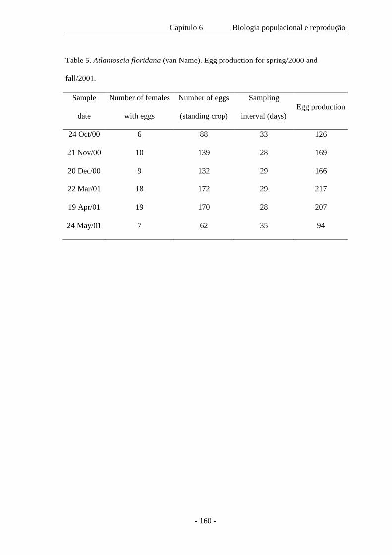
Capítulo 6 Biologia populacional e reprodução
- 160 -
Table 5. Atlantoscia floridana (van Name). Egg production for spring/2000 and
fall/2001.
Sample
date
Number of females
with eggs
Number of eggs
(standing crop)
Sampling
interval (days) Egg production
24 Oct/00 6 88 33 126
21 Nov/00 10 139 28 169
20 Dec/00 9 132 29 166
22 Mar/01 18 172 29 217
19 Apr/01 19 170 28 207
24 May/01 7 62 35 94
CAPÍTULO 7
Discussão geral e conclusões
Discussão e Conclusões
-162-
DISCUSSÃO GERAL E CONCLUSÕES
São descritas aproximadamente 4.000 espécies de isópodos terrestres e,
destas, o conhecimento da biologia restringe-se a apenas cerca de 20%. Não resta
dúvida de que é muito pouco para que se possam fazer generalizações ou propor
padrões em um universo tão pequeno. Isto, na verdade, indica a necessidade de estudar
mais e mais outras espécies, em vários tipos de ambientes em latitudes e altitudes
diferentes.
Em geral, na literatura específica, encontram-se informações diversas e
às vezes, pontuais. Talvez o maior ineditismo deste trabalho seja aliar observações em
laboratório com dados de campo e apresentar os resultados de forma integrada. Além
disso, apresenta informações detalhadas sobre o desenvolvimento pós-marsupial e
biologia reprodutiva. Pela primeira vez é apresentada a curva de crescimento com base
em dados de campo para uma espécie de isópodo terrestre.
O que motivou este trabalho? A coleta e observação ano após ano da
espécie, o que evidenciou que ela tinha algo singular: fêmeas ovígeras eram encontradas
em todas as épocas do ano. Então, começaram as perguntas que passaram a ser objetivos
e que resultaram nesta tese. Mas, não significa que as perguntas tenham terminado. À
medida que encontramos respostas, novas indagações são feitas. E, ao final, é isto que
nos motiva, saber que sempre há mais para conhecer.
Pode-se dizer que este trabalho começou, literalmente do ponto zero. No
princípio observar a muda era algo muito difícil o que, mais tarde, se tornou tão simples
e, com a familiarização dos caracteres, já era possível, inclusive, fazer previsões sobre a
data da muda. Alguns ajustes prévios foram feitos, como testar se a medida do
Discussão e Conclusões
-163-
cefalotórax era, de fato, representativa do tamanho. Foram tomadas medidas da largura
e comprimento do cefalotórax de machos e de fêmeas e calculadas as regressões, as
quais apresentaram um coeficiente de determinação superior a 95%. Este teste, por
outro lado, mostrou o cuidado que se deveria ter no posicionamento do cefalotórax para
que as medidas fossem obtidas corretamente. Também foram tomadas medidas do
corpo e os resultados confirmaram que estas não são confiáveis, uma vez que não
representam o tamanho real dos animais, pois estes em geral, quando morrem, ficam
com o corpo curvado.
A escolha da temperatura do cultivo baseou-se em duas observações: na
temperatura do solo obtida nos meses de março e abril de 1999 e nas observações
preliminares do cultivo em laboratório. Quando a temperatura subia além de 230C havia
condensação nos potes, formando gotas que se depositavam no fundo nas quais os
animais morriam afogados. Então, estabeleceu-se a temperatura de 200 C que ao final
demonstrou ser ideal, pois os dados de laboratório e de campo (usados para a curva de
crescimento) apresentaram resultados semelhantes com relação ao tempo do
desenvolvimento/crescimento.
O número reduzido de trabalhos sobre o desenvolvimento pós-
embrionário evidencia que o conhecimento sobre este tema em isópodos terrestres é
ainda muito incipiente se levarmos em conta o número de espécies conhecidas e a
variedade de ambientes em que elas vivem. A sistemática de Oniscidea ainda tem
muitas questões em aberto, embora em um nível taxonômico mais alto, muita coisa já
tenha sido feita. No entanto, sem este conhecimento, deixa-se de lado uma grande
possibilidade de incluir novos caracteres nas análises filogenéticas sobre o grupo.
Pela primeira vez é proposto um método para identificar a maturidade
sexual em machos. É um método mais simples do que análise histológica, embora não
Discussão e Conclusões
-164-
se descarte a importância de estudos histológicos para a observação do desenvolvimento
gonadal de forma a estimar a maturidade sexual.
Muitos trabalhos sobre ecologia populacional tratam de indivíduos
imaturos e maduros como formando uma única categoria. Ficou demonstrada aqui a
importância de identificar estes dois grupos, tanto para machos quanto para fêmeas, não
somente em termos de tamanho mas em termos de idade. Isto é particularmente
importante nos estudos sobre a estrutura populacional e vital na interpretação da
proporção sexual. Embora a proporção sexual encontrada favoreça os machos, ainda
assim é necessário investigar a presença da bactéria feminilizante Wolbachia. Nada
sabemos sobre esta relação nos diversos ambientes da América do Sul. Por exemplo, a
bactéria já existia antes da introdução de Armadillidium vulgare, uma espécie européia
que apresenta Wolbachia? Se não existia e por transferência horizontal passou a ser
simbionte de espécies nativas, qual o estado atual da relação? Como A. floridana é uma
espécie abundante que também pode ser encontrada em ambiente urbano, juntamente
com A. vulgare, ela constitui ótimo material para este estudo. Se não há a presença de
Wolbachia, como evoluiu tal proporção sexual? Há competição entre os machos? Há
cruzamento preferencial? Quais as características morfológicas envolvidas na escolha?
Se não há competição, ela é resultado de mortalidade menor entre os machos? Ou,
então, um maior número de machos na população garante os encontros, embora a
densidade populacional seja alta? São questões em aberto que poderão ser resolvidas em
trabalhos futuros.
O recrutamento foi analisado em vários níveis, desde a presença de ovos,
embriões, mancas marsupiais e mancas. Um outro nível poderá ser avaliado no futuro: a
oogênese e formação dos oócitos. O número de oócitos produzido é igual ao número
final de mancas liberadas? Com base na fecundidade da espécie, pode-se estimar o
Discussão e Conclusões
-165-
número esperado de ovos para um determinado tamanho de fêmea. Para a estimativa
dos oócitos poderia ser feita a mesma relação.
Quantas vezes durante a sua vida uma fêmea é capaz de ficar ovígera?
Quantas cópulas são necessárias? Estas questões não foram objetivo deste trabalho mas
ficou claro que as fêmeas podem produzir uma prole sem ter realizado cópula, o que
indica a presença de uma espermateca. Se existe uma espermateca, qual a capacidade de
armazernamento? Por que na segunda prole estas fêmeas têm um número menor de
filhotes? A produção de oócitos é menor ou o número disponível de espermatozóides é
reduzido? Observações em laboratório e análises histológicas poderão ser utilizadas
para esclarecer estas questões.
Atlantoscia floridana é uma espécie abundante na área de estudo na
Reserva Biológica do Lami. Por este motivo, seu papel nas cadeias tróficas é muito
importante. Esta evidência encaminha a novos estudos, os quais poderão quantificar esta
participação neste ambiente terrestre. Quem são os consumidores desta espécie? Com
um recrutamento tão alto, com fêmeas ovígeras todo o ano e uma densidade
populacional alta até mesmo nos meses de inverno, ela constitui uma fonte alimentar
contínua. Estaria alguma espécie utilizando este recurso todo o ano ou durante um
período em que outros recursos são escassos? Em uma unidade de conservação,
conhecer estas relações é importante na medida em que proporciona subsídios para o
monitoramento de espécies ameaçadas de extinção dependentes direta ou indiretamente
do isópodo terrestre. Neste cenário inclui-se, também a relação parasitária entre a
espécie e o Centrorhynchus (Acanthocephala) (AMATO et al. submetido) o que também
poderia estar ocorrendo na Reserva. Esta relação implica, necessariamente, na presença
de um hospedeiro definitivo, uma ave ou mamífero. Poderiam estar envolvidas, ainda,
espécies de anfíbios ou répteis como hospedeiros paratênicos. Além disso, por ser uma
Discussão e Conclusões
-166-
espécie com tais características biológicas, poderia ser usada em áreas de recuperação,
pois também tem importância na formação do húmus e reciclagem de nutrientes no
solo.
O estudo com Atlantoscia floridana apresentou os seguintes resultados e
conclusões:
• existem três estágios de manca: manca I, II e III, sendo o primeiro completado
em aproximadamente 12 horas após o nascimento e os outros em
aproximadamente 20 dias;
• os estágios de manca são identificados com base na pigmentação, número de
omatídios, flagelo da antena, pereionito VII, pereiópodo e pleópodo 1;
• em manca 1 os indivíduos têm em média 0,421mm de largura do cefalotórax,
pigmentação fraca, 5 omatídios, flagelo biarticulado, sendo o artículo proximal o
mais longo, sem placas coxais no pereionito VII e pereiópodo 7 e pleópodo 1
ausentes;
• em manca II os indivíduos têm em média 0,488 mm de largura do cefalotórax,
grandes manchas castanhas no cefalotórax e margens laterais do pereion, pleon e
urópodos, 5 omatídios, flagelo biarticulado, sendo o artículo distal o mais longo,
placas coxais do pereionito VII levemente desenvolvidas, pereiópodo 7 pouco
desenvolvido, cujos artículos não se encontram distintos e pleópodo 1 ausente,
somente com o protopodito;
• em manca III os indivíduos têm coloração semelhante a do adulto, de 6 a 7
omatídios, antena triarticulada, placas coxais do prerionito VII desenvolvidas,
Discussão e Conclusões
-167-
com a presença de nódulos laterais, pereiópodo 7 desenvolvido, ainda dobrado
sob o abdômen e somente o protopodito do pleópodo 1;
• após o estágio de manca III, iniciam os estágios juvenis, onde o sétimo par de
pereiópodos encontra-se funcional e já é possível observar o início da formação
da genitália nos machos;
• os machos possuem três estágios juvenis: JU I, II e III; a determinação da
maturidade sexual está baseada na relação morfométrica da papila genital com a
largura do cefalotórax, bem como na ultraestrutura da papila genital, onde se
observa o desenvolvimento de escamas cuticulares desde os juvenis até o adulto;
• os machos atingem a maturidade sexual com aproximadamente um mês e meio
de vida;
• a largura média do cefalotórax dos estágios juvenis de machos é: JU I –
0,617mm, JU II - 0,677 e JU III - 0,744mm;
• as fêmeas possuem seis estágios juvenis: JU I, II, III, IV ,V, VI; a determinação
da maturidade sexual está baseada na menor fêmea ovígera encontrada na
natureza, a qual apresenta 1,04mm de largura do cefalotórax e na morfologia dos
poros genitais, os quais apresentam, também, escamas cuticulares que se
desenvolvem à medida que as fêmeas atingem a maturidade;
• as fêmeas atingem a maturidade sexual com aproximadamente três meses de
vida;
• a largura média do cefalotórax dos estágios juvenis de fêmeas é: JU I – 0,610,
JU II – 0,674, JU III – 0,746, JU IV – 0,813, JU V – 0,877 e JU VI – 0,957;
• o tamanho e a duração dos estágios juvenis é semelhante entre machos e fêmeas
até JU III;
Discussão e Conclusões
-168-
• a curva de crescimento para machos é descrita pela equação:
Wt = 1.303 [1 - e -0.00941 (t + 50.37)];
• a curva de crescimento para fêmeas é descrita pela equação:
Wt = 1.682 [1 - e -0.00575 (t + 59.13)];
• as curvas mostram crescimento diferencial entre os sexos; as fêmeas atingem
tamanho maior com uma taxa de crescimento menor;
• a expectativa de vida para os machos é de aproximadamente um ano e meio;
• a expectativa de vida para as fêmeas é de aproximadamente dois anos e quatro
meses;
• houve uma mudança significativa nos tamanhos de machos e de fêmeas
amostrados em 2000 e 2001; no segundo ano os animais apresentaram tamanho
menor devido a ausência de indivíduos nas classes de maior tamanho;
• as fêmeas ovígeras e pós-ovígeras apresentaram os maiores tamanhos;
• a densidade populacional variou ao longo das estações do ano; a densidade
mínima encontrada foi de 131 indivíduos, o máximo 1,040 e a média de 450 m-2;
• o número de indivíduos machos que ultrapassam o primeiro ano de vida
apresentou-se baixo: um em outubro/00, um em janeiro/01 e um em
setembro/01;
• o número de fêmeas que atingiram o segundo anos de vida totalizou 35 e as que
ultrapassaram este período foram raras: uma em junho/00, uma em dezembro/00
e uma em maio/01;
• a proporção sexual operacional favoreceu os machos e não mostrou mudanças
com as estações do ano;
Discussão e Conclusões
-169-
• as fêmeas ovígeras e pós-ovígeras representaram cerca de 35% do total de
fêmeas capturadas;
• as fêmeas ovígeras foram encontradas durante todo o ano o que indica que a
reprodução é contínua;
• o pico da reprodução ocorreu durante o outono e primavera;
• a fecundidade variou de 5 a 23 ovos com uma média de 11 ovos por fêmea;
• a fecundidade é expressa pela equação F = 18,48 + 22, 59 CW;
• a largura do cefalotórax de fêmeas ovígeras variou de 1,04 a 1,68mm;
• a mortalidade no marsúpio foi de apenas 0,91%;
• a produção de ovos na primavera foi de588 m–2 e no outono, 660 m–2;
• o recrutamento ocorreu em todos os meses;
• ovos, embriões e mancas marsupiais também foram encontrados durante todo o
ano, o que indica um recrutamento contínuo;
• a característica mais notável da espécie é seu alto investimento reprodutivo o
que garante um estoque contínuo de indivíduos na população o que mantém a
recuperação da densidade após períodos adversos.
REFERÊNCIAS BIBLIOGRÁFICAS
- 171 -
REFERÊNCIAS BIBLIOGRÁFICAS
AL-DABBAGH, K.Y. & W. BLOCK. 1981. Population ecology of a terrestrial
isopod in two breckland grass heaths. J. Anim. Ecol. 50: 61-77.
ANDERSSON, A. 1960. South american terrestrial isopods in the collection of the
Swedish State Museum of Natural History. Ark. Zool. 12(5-6):537-570.
ARAUJO, P.B. 1999. Two new species of Alboscia Schultz, 1995 from Rio Grande
do Sul, Brazil (Isopoda, Oniscidea, Philosciidae). Crustaceana 72(5): 487-496.
ARAUJO, P.B. & L. BUCKUP. 1994a. Nova espécie de Trichorhina Budde-Lund,
1908 (Crustacea, Isopoda, Platyarthridae) do sul do Brasil. Iheringia Série
Zoologia, 77: 129-134.
ARAUJO, P.B. & L. BUCKUP. 1994b. Two new species of terrestrial ispods from
Southern Brazil (Crustacea, Isopoda, Oniscidea). Spixiana 17(3): 269-274.
ARAUJO, P.B. & L. BUCKUP. 1996a. Novos registros e uma espécie nova de
Trichorhina Budde-Lund no sul do Brasil (Isopoda, Oniscidea, Platyarthridae).
Revta bras. Zool. 13 (3): 799-810.
ARAUJO, P.B & L. BUCKUP. 1996.b. Ocorrência de Nagurus no Brasil meridional
(Isopoda, Trachelipodidae). Nauplius 4: 161-163.
ARAUJO, P.B.; L. BUCKUP L. & G. BOND-BUCKUP. 1996. Isópodos terrestres de
Santa Catarina e Rio Grande do Sul (Crustacea, Oniscidea). Iheringia 81:111-138.
ARAUJO, P.B. & A.A.P. BUENO. 1998. Novos registros de isópodos terrestres do sul
do Brasil: Porcellionidae e Trichoniscidae (Crustacea, Isopoda, Oniscidea).
Nauplius 6: 185-186.
ARAUJO, P.B. & LEISTIKOW, A. 1999. Philosciids with pleopodal lungs from Brazil,
with description of a new species (Crustacea, Isopoda). Contr. Zool. 68 (2): 109-141.

- 172 -
ARAUJO, P.B. & E.R.C. LOPES. 2002. Three new species of Benthana (Isopoda,
Philosciidae) from Brazil. J. Nat. Hist. (no prelo)
ARAUJO, P.B. & M.C.L. ZARDO. 1995. Uma nova espécie de Balloniscus
Budde- Lund (Crustacea, Isopoda, Balloniscidae) do sul do Brasil. Revta bras.
Zool. 12 (4):785-790.
BEGON, M; J.L. HARPER & C.R. TOWNSEND. 1996. Ecology. Blackwell, Oxford,
1068p.
BUENO, A.A.P.; G. BOND-BUCKUP & L. BUCKUP. 2000. Crescimento de Aegla
platensis Schmitt em ambiente natural (Crustacea, Decapoda, Aeglidae). Revta
bras. Zool. 17(1): 51-60.
CAMARGO, O.R. 1954. Isópodes terrestres do Rio Grande do Sul. Rev. Agr.,
Sér I 209-211:122-128.
EMLEN, S.T & L.W. ORING. 1977. Ecology, sexual selection and the evolution of
mating systems. Science 197: 215-223.
FERRARA, F. & S. TAITI. 1981. Terrestrial isopods from Ascension Islands.
Monitore zool. ital. (N.S.) Suppl. 12: 89-215,
GREENWOOD, P.J. & J. ADAMS. 1987. The ecology of Sex. Edward Arnol,
Londres, 74p.
HADDAD, M.A. 1982. Estádios do desenvolvimento pós-marsupial de Balloniscus
sellowii (Brandt, 1833) (Isopoda, Oniscoidea). Dusenia 13(3):135-143.
HADDAD, M.A. & L.C. VERANI. 1984. Crescimento pós-marsupial de Balloniscus
sellowii (Brandt, 1833) (Isopoda, Oniscoidea). Dusenia 14(4): 197-209.
HARTNOLL, R.G. 1982. Growth, p. 101-128. In: D.E. BLISS (ed). The Biology of
Crustacea, Embriology, Morphology, and Genetics. New York, Academic Press.
- 173 -
HASSAL, M. & J.M. DANGERFIELD. 1989. Inter-specific competition and the
relative abundance of grassland isopods. Monitore zool. ital. (N.S.) Monogr.
4: 379-397.
HEELEY, W. 1941. Observations on the life-histories of some terrestrial isopods,
Proc. Zool. Soc. Lond. 111(B): 79-119.
HOESE, B. 1984. The marsupium in terrestrial isopods. Symp. zool. Soc. Lond.
53:65-76.
HOESE, B & H.H. JANSSEN. 1989. Morphological and physiological studies on
the marsupium in terrestrial isopods. Monitore zool. ital. (N.S.) Monogr. 4:153-
173.
HORNUNG, E. 1984. Characteristics of the population of an isopod species
(Trachelipus nodulosus C.L. Koch) at sandy soil grassland. Acta Biol. Szeged. 30:
153-158.
HORNUNG, E. 1989. Population dynamics and spatial distribution of Trachelipus
nodulosus (C.L.Koch, 1838) (Crustacea, Isopoda) in a sandy grassland. Monitore
zool. ital. (N.S.) Monogr. 4: 399-409
HORNUNG, E. 1991. Isopod distribution in a heterogeneous grassland habitat, p. 73-
79. In: P. JUCHAULT & J.P. MOCQUARD (eds.) Biology of Terrestrial Isopods.
Poitiers, Univ. Press.
HORNUNG, E. & M.R. WARBURG. 1993. Breeding patterns in the oniscid isopod,
Porcellio ficulneus Verh., at high temperature and under different photophases.
Invert. Reprod. Develop. 23 (2-3): 151-158.
HORNUNG, E. & M.R. WARBURG. 1995. Seasonal changes in the distribution
and abundance of isopo species in different habitats within the Mediterranean region
of northern Israel. Acta Ecol. 16(4): 431-445.
- 174 -
KACEM-LACHKAR, H. 1997. Étude du développement post-embryonnaire
d’Hemilepistus reaumurii (Audouin, 1826) (Isopoda, Oniscidea). Crustaceana
70(5): 513-526.
KATAKURA, Y. 1984. Sex differentiation and androgenic gland hormone in the
terrestrial isopod Armadillidium vulgare. Symp. zool. Soc. Lond. 53: 97-107.
KWON, D. & S. TAITI. 1993. Terrestrial isopoda (Crustacea) from Southern China
Macao and Hong-Kong. Stuttg. Beitr. Naturk. Ser. A, 490: 1-83.
LAWLOR, L..R. 1976. Molting, growth and reproductive strategies in the
terrestrial isopod, Armadillidium vulgare. Ecology 57: 1179-1194.
LEFEBVRE, F. & Y. CAUBET. 1999. On the male-effect in the terrestrial Crustacean
Armadillidium vulgare (Latreille, 1804). Invert. Reprod. Develop. 35(1): 55-64.
LEISTIKOW, A. & P.B. ARAUJO. 2001. Morphology of respiratory organs in South
American Oniscidea (“Philosciidae”), p. 329-335. In: B. KENSLEY & R. BRUSCA
(eds). Isopod Systematics and Evolution. Rotterdam, Balkema, 357p.
LEMOS DE CASTRO, A. 1976. Considerações sobre a sinonímia e distribuição
de Balloniscus sellowii (Brandt, 1833) (Isopoda, Balloniscidae). Rev. Brasil.
Biol 36: 391-396.
LEMOS DE CASTRO, A. 1985. Considerações sobre Atlantoscia alceui Ferrara &
Taiti, 1981(Isopoda, Oniscoidea, Philosciidae). Rev. Brasil. Biol 45(4): 417-422.
LINSENMAIR, K.E. 1984. Comparative studies on the social behaviour of the
desert isopod Hemilepistus reaumuri and of a Porcellio species. Symp. zool.
Soc. Lond. 53: 423-453.
LITTLE, C. 1990. The terrestrial invasion. Cambridge Univ. Press, Cambridge,
304p.
- 175 -
MA, H.H.T.; D. DUDGEON & P.K.S. LAM. 1991. Seasonal changes in population
of three sympatric isopods in a Hong Kong forest. J. Zool. 224: 347-365.
MA, H.H.T.; P.K.S. LAM & D. DUDGEON. 1991. Inter and intraspecific variation in
the life histories of three sympatric isopods in a Hong Kong forest. J. Zool. 224:
677-687.
MARQUES, A.A.B; C.S. FONTANA; E. VÉLEZ; G.A. BENCKE; M. SCHNEIDER &
R.E. REIS. 2002. Lista das espécies da fauna ameaçadas de extinção no Rio
Grande do Sul. Porto Alegre, FZB/MCT-PUCRS/PANGEA, 52p.
MATSAKIS, J. 1955. Contribution a l’etude du développement postembryonnaire et de
la croissance des Oniscoides, Bull. Soc. Zool. France 80: 52- 65.
McQUEEN, D.J. & J.S. CARNIO, J.S. 1974. A laboratory study of the effects of some
climatic factor on the demography of the terrestrial isopod Porcellio spinicornis Say.
Can. J. Zool. 52: 599-611.
MEDINI, L.; K. NASRI-AMMAR & F. CHARFI-CHEKHROUHA. 2000.
Reproduction saisonnière de Porcellio variabilis Lucas, 1846. C. R. Acad. Sci.
Paris, Life Sciences, 323: 689- 695.
MEIRA, J.R & PORTO, M.L. 1998. Reserva Biológica do Lami: a vida na beira do
lago, 89-92. In: R. MENEGAT (coord. geral), Atlas Ambiental de Porto Alegre.
Porto Alegre, Editora Universidade/UFRGS, 228p.
NAIR, G.A. 1984. Breeding and population biology of the terrestrial isopod
Porcellio laevis (Latreille), in the Delhi Region. Symp. zool. Soc. Lond. 53: 315-
337.
PAOLETTI, M.G. 1989. Life strategies of isopods and “soil invertebrates” in
Venezuela. Monitore zool. ital. (N.S.) Monogr. 4: 435-453.
- 176 -
PARIS. O.H. & F.A. PITELKA. 1962. Population characteristics of the terrestrial
isopod Armadillidium vulgare in California grassland. Ecology, 43(2): 229-248.
PRINTES, R.C. 2002. Plano de Manejo Participativo da Reserva Biológica do
Lami. Porto Alegre, SMAM, 102p.
RUSHTON, S. & M. HASSAL. 1983. The effects of food quality on the life history
parameters of the terrestrial isopod (Armadillidium vulgare (Latreille). Oecologia,
57: 257-261.
SANTOS, E.P. 1978. Dinâmica de populações aplicada à pesca e piscicultura.
São Paulo, Editora da Universidade de São Paulo, 129p.
SCHMALFUSS, H. 2002. A bibliography of terrestrial isopods (Crustacea: Isopoda:
Oniscidea). Stuttg. Beitr. Naturk. (Ser A) 639: 1-120.
SOULÉ, M. & K.A. KOHM. 1989. Research priorities for conservation Biology.
Washington, Island Press critical issues series, Society for Conservation Biology,
98p.
SOUZA-KURY, L.A. 1993. Notes on Trichorhina I. Two new species of Northeastern
Brazil (Isopoda, Oniscidea, Platyarthridae). Rev. suiss. zool. 100(1): 197-210.
SOUZA-KURY, L.A. 1997. Two new species of Trichorhina from Brazilian
Amazonia (Isopoda, Oniscidea, Platyarthridae). Crustaceana, 70(2): 180-190.
SOUZA-KURY, L.A. 1998. Malacostraca – Peracarida. Isopooda. Oniscidea, p 653-
674. In: P.S. YOUNG (ed). Catalogue of Crustacea of Brazil. Rio de Janeiro,
Museu Nacional.
SUNDERLAND, K.D.; M. HASSAL. & S.L. SUTTON. 1976. The population
dynamics of Philoscia muscorum (Crustacea, Oniscoidea) in a dune grassland
ecosystem. J. Anim. Ecol. 45: 487-506.
SUTTON, S.L. 1980. Woodlice. Pergamon Press. Oxford, 143p.
- 177 -
SUTTON, S.L.; M. HASSAL; R. WILLOWS; R.C. DAVIS; A. GRUNDY & K.D.
SUNDERLAND. 1984. Life histories of terrestrial isopods: a study of intra and
interspecific variation. Symp.zool. Soc. Lond. 53: 269-294.
VALENTI, W.C.; J.T.C. MELO & V.L. LOBÃO. 1994. Maturation and growth of
Macrobrachium carcinus (Linnaeus) (Crustacea, Decapoda, Palaemonidae) from
Ribeira do Iguape River, Southern Brazil. Revta bras. Zool.11(4): 649-658.
VERHOEFF, K.W. 1920. Zur Kenntnis der Larven, des Brutsackes und der Bruten
der Oniscoidea. Zool. Anz. 51: 169-189.
VERHOEFF, K.W. 1941. Zur Kenntnis Südamerikanisher Oniscoideen. Zool. Anz.
133: 114-126.
von BERTALANFFY, L.. 1938. A quantitave theory of organic growth (inquiries on
growth laws). Human Biol. 10(2): 181-213.
WARBURG, M.A. 1987. Isopods and their terrestrial environment. Adv. Ecol. Res.
17: 187-242.
WARBURG, M.R. 1993. Evolutionary Biology of Land Isopods. Berlin, Springer-
Verlag, 159p.
WARBURG, M.R. & N. COHEN. 1992a. Population dynamics, growth and longevity
of Armadillo officinalis (Isopoda, Oniscidea), inhabiting the Mediterranean region of
northern Israel. Pedobiologia 36: 262-273.
WARBURG, M.R. & N. COHEN, N. 1992b. Reproductive pattern, allocation and
potential of an iteroparous isopod from a xeric habitat in the Mediterranean region.
J. Arid Environ. 22: 161-171.
WARBURG, M.R. & N. COHEN, N. 1993. Breeding patterns in two terrestrial isopods
from the Mediterranean region of Israel. Biologia Gallo-Hellenica 20(1): 55- 68.
- 178 -
WARBURG, M.R.; K. LINSENMAIR & K. BERCOVITZ, K. 1984. The effect of
climate on the distribution and abundance of isopods. Symp. zool. Soc. Lond. 53:
339-367.
WILSON, D.F. 1990. Functional morphology and evolution of isopod genitalia, p.228-
245. In: R.T. BAUER & J.W. MARTIN (eds). Crustacean Sexual Biology. New
York, Columbia University Press.
ZARDO, C.M.L 1989. Uma nova espécie de Phalloniscus Budde-Lund, 1908
(Crustacea, Oniscoidea) do sul do Brasil. Revta bras. zool. 6(4): 611-615.
ZARDO, C.M.L. & LOYOLA E SILVA, J. 1988. Primeira ocorrência de Oniscus
asellus Linné, 1758 e Porcellionides sexfasciatus (Koch, 1847) (Isopoda,
Oniscoidea) no Brasil. Cien. Cult., 40(8): 791-795.