part 9: first aid 2015 international consensus on first aid science with treatment recommendations
TRANSCRIPT
S269
IntroductionDefinition of First AidThe International Liaison Committee on Resuscitation (ILCOR) First Aid Task Force first met in June 2013. Comprising nominated members from around the globe appointed by each ILCOR member organization, the task force members first agreed to the goals of first aid and produced a definition of first aid as it might apply to the international set-ting. Task force members considered an agreed-upon definition essential for the subsequent development of research ques-tions, evidence evaluation, and treatment recommendations.
First aid is defined as the helping behaviors and initial care provided for an acute illness or injury. First aid can be initiated by anyone in any situation.
A first aid provider is defined as someone trained in first aid who should
• Recognize, assess, and prioritize the need for first aid• Provide care by using appropriate competencies• Recognize limitations, and seek additional care when
needed
The goals of first aid are to preserve life, alleviate suffer-ing, prevent further illness or injury, and promote recovery.
This definition of first aid addresses the need to recog-nize injury and illness, the requirement to develop a specific skill base, and the need for first aid providers to simultane-ously provide immediate care and activate emergency medi-cal services (EMS) or other medical care as required. First aid assessments and interventions should be medically sound and based on evidence-based medicine or, in the absence of such evidence, on expert medical consensus. The scope of first aid is not purely scientific, as both training and regu-latory requirements will influence it. Because the scope of first aid varies among countries, states, and provinces, the treatment recommendations contained herein may need to
be refined according to circumstances, need, and regulatory constraints.
One difference between this 2015 definition and that used for the 2010 process is that the task force did not restrict first aid to “assessments and interventions that can be performed…with minimal or no equipment.” We acknowledge that, in most cases, equipment might not be available to first aid providers, particularly for bystanders and lay providers. However, the First Aid Task Force noted that, in some countries, supple-mentary first aid supplies now include inexpensive and com-pact pulse oximeters, glucose meters, and other adjuncts never before considered to be in the realm of first aid. In the 2015 treatment recommendations, we have striven to remain true to the “minimal or no equipment” approach, but recognize that addition of equipment, used by those trained to use and main-tain it, may enhance care.
The task force strongly believes that education in first aid should be universal: everyone can and should learn first aid.
How and Why Topics Were ChosenIn the autumn of 2012, ILCOR approved the First Aid Task Force as a fully participating task force in the 2015 ILCOR international evidence evaluation and appointed 2 interna-tional co-chairs. In the spring of 2013, each member council of ILCOR nominated individuals for membership in the First Aid Task Force. In addition to the co-chairs, 11 task force members were appointed, representing the ILCOR member organizations of the American Heart Association (AHA), the European Resuscitation Council (ERC), the Heart and Stroke Foundation of Canada, the Australian Resuscitation Council, the InterAmerican Heart Foundation, and the Resuscitation Council of Asia. Members included physicians specializing in anesthesia, critical care/resuscitation, emergency medicine, cardiology, internal medicine, and pediatric emergency medi-cine, as well as paramedics specializing in prehospital care guideline development, specialists in first aid course education
(Circulation. 2015;132[suppl 1]:S269–S311. DOI: 10.1161/CIR.0000000000000278.)© 2015 American Heart Association, Inc., European Resuscitation Council, and International Liaison Committee on Resuscitation.
Circulation is available at http://circ.ahajournals.org DOI: 10.1161/CIR.0000000000000278
The American Heart Association requests that this document be cited as follows: Singletary EM, Zideman DA, De Buck EDJ, Chang WT, Jensen JL, Swain JM, Woodin JA, Blanchard IE, Herrington RA, Pellegrino JL, Hood NA, Lojero-Wheatley LF, Markenson DS, Yang HJ; on behalf of the First Aid Chapter Collaborators. Part 9: first aid: 2015 International Consensus on First Aid Science With Treatment Recommendations. Circulation. 2015;132(suppl 1):S269–S311.
*Co-chairs and equal first co-authors.This article has been co-published in Resuscitation. Published by Elsevier Ireland Ltd. All rights reserved.
Resuscitation and Emergency Cardiovascular Care Science With Treatment Recommendations
Part 9: First Aid2015 International Consensus on First Aid Science With Treatment
Recommendations
Eunice M. Singletary, Co-Chair*; David A. Zideman, Co-Chair*; Emmy D.J. De Buck; Wei-Tien Chang; Jan L. Jensen; Janel M. Swain; Jeff A. Woodin; Ian E. Blanchard; Rita A. Herrington; Jeffrey L. Pellegrino; Natalie A. Hood; Luis F. Lojero-Wheatley;
David S. Markenson; Hyuk Jun Yang; on behalf of the First Aid Chapter Collaborators
at AULTMAN HOSPITAL on November 4, 2015http://circ.ahajournals.org/Downloaded from

S270 Circulation October 20, 2015
and curriculum development, and a specialist in first aid evi-dence evaluation methodology and guideline development.
The task force convened in June 2013 to review the top-ics and questions that were evaluated in 2005 and 2010, past research questions formulated in the PICO style (population, intervention, comparator, outcomes) that were never com-pleted, and the new questions that had been submitted since 2010 to the task force, and a priority list created. Topics were reviewed for areas of controversy, known additional new sci-ence, and subject matter not previously evaluated. Task force members created a priority list for review, and the top 10 pri-ority-ranked PICO questions were assigned. After the success-ful commencement of the workflow, the task force co-chairs added a further 12 PICO questions, including 5 new questions, 1 derived question, and 6 that had been previously reviewed. Selected PICO questions that had been previously reviewed were, in some cases, reworded to facilitate literature searches, and outcomes were decided upon by group consensus.
Evidence reviewers were recruited through a call for volun-teers distributed by ILCOR to stakeholder organizations around the world. More than 30 individual reviewers were assigned to topics, usually by preference or expertise, but avoiding any direct conflicts of interest. In general, 2 evidence reviewers were assigned to each PICO, supervised by a member of the task force designated as the task force question owner. Evidence reviewers included physicians with diverse specialties including emergency medicine, EMS, wilderness medicine, critical care, cardiology, occupational medicine, toxicology, anesthesia, pedi-atric emergency medicine, public health, and epidemiology, as well as paramedics, nurse practitioners and first aid education specialists with experience in guideline and curriculum devel-opment, and professional evidence evaluation and methodology experts.
The Evidence Evaluation ProcessFor the 2015 international evidence evaluation process, the AHA developed a new Web-based information and docu-mentation platform, the Systematic Evidence Evaluation and Review System (SEERS), to support the ILCOR systematic reviews and to capture the data in reusable formats. This Web-based system facilitated structured reviews in a consistent for-mat that would support the ultimate development of science summaries and evidence-based treatment recommendations.
Each task force performed a detailed systematic review based on the recommendations of the Institute of Medicine of the National Academies,1 using the methodological approach proposed by the Grading of Recommendations, Assessment, Development, and Evaluation (GRADE) Working Group.2 After identifying and prioritizing the PICO questions to be addressed,3 and with the assistance of information specialists, a detailed search for relevant articles was performed in each of 3 online databases (PubMed, Embase, and the Cochrane Library).
By using detailed inclusion and exclusion criteria, articles were screened for further evaluation. The reviewers for each question created a reconciled risk of bias assessment for each of the included studies, using state-of-the-art tools: Cochrane for randomized controlled trials (RCTs),4 Quality Assessment of Diagnostic Accuracy Studies (QUADAS)-2 for studies of
diagnostic accuracy,5 and GRADE for observational studies that inform both therapy and prognosis questions.6
GRADE evidence profile tables7 were then created to facilitate an evaluation of the evidence in support of each of the critical and important outcomes. The quality of the evi-dence (or confidence in the estimate of the effect) was cat-egorized as high, moderate, low, or very low,8 based on the study methodologies and the 5 core GRADE domains of risk of bias, inconsistency, indirectness, imprecision, and other considerations (including publication bias).9
The GRADE evidence profile tables were then used to create a written summary of evidence for each outcome (the consensus on science statements). Whenever possible, con-sensus-based treatment recommendations were then created. These recommendations (designated as strong or weak) were accompanied by an overall assessment of the evidence and a statement from the task force about the values and preferences that underlie the recommendations. Strong recommendations use the words “we recommend,” and weak recommendations use the words “we suggest.”
Further details of the methodology that underpinned the evidence evaluation process are found in “Part 2: Evidence Evaluation and Management of Conflicts of Interest.”
The learning curve for use of the GRADE evidence evaluation methodology was steep and resulted in a total of 22 PICO questions, including 6 new questions, being com-pleted by the task force before the ILCOR 2015 International Consensus Conference on CPR and ECC Science With Treatment Recommendations in February 2015. The remain-ing topics not reviewed for 2015 have since been reprioritized, with the addition of several new questions that were identified during the ILCOR 2015 work process.
Very little research has been conducted in first aid, and most of the recommendations are extrapolations from research in the prehospital or hospital setting. The selected methodology for evaluation of the literature led to the elimination of lower-quality data from animal studies, case series, and case reports, except for topics where no human studies were identified that met the inclu-sion criteria. These more stringent requirements led to the inclu-sion of studies with a higher initial quality of evidence, but most studies were eventually downgraded due to indirectness for the first aid setting. The gaps in knowledge have been identified by the evidence reviewers and summarized at the end of each treat-ment recommendation. It is our hope that these knowledge gaps will be filled through future research. In the absence of evidence-based medicine to support a treatment recommendation, the task force has made many recommendations based on expert opinion, perceived best practice, and the principle of “do no harm.”
PICO Questions Reviewed First Aid for Medical Emergencies
• Recovery position (FA 517)• Optimal position for shock (FA 520)• Oxygen administration for first aid (FA 519)• Bronchodilator use for asthma with difficulty breathing
(FA 534)• Stroke recognition* (FA 801)
*Topics not previously reviewed.
at AULTMAN HOSPITAL on November 4, 2015http://circ.ahajournals.org/Downloaded from

Singletary et al Part 9: First Aid S271
Aspirin for Chest Pain
• Aspirin for chest pain: administration† (FA 871)• Aspirin for chest pain: early compared with late (FA 586)
Epinephrine for Anaphylaxis and Treatment of Hypoglycemia, Exertion-Related Dehydration, and Chemical Eye Injuries
• Second dose of epinephrine for anaphylaxis (FA 500)• Hypoglycemia treatment* (FA 795)• Exertion-related dehydration and oral rehydration (FA 584)• Eye chemical injury: irrigation (FA 540)
First Aid for Trauma Emergencies
• Control of bleeding (FA 530)• Hemostatic dressings (FA 769)• Use of a tourniquet (FA 768)• Straightening of an angulated fracture (FA 503)• First aid treatment for an open chest wound* (FA 525)• Cervical spinal motion restriction (FA 772)• Concussion* (FA 799)• Cooling of burns (FA 770)• Wet compared with dry burn dressing (FA 771)• Dental avulsion (FA 794)
Education
• First aid training* (FA 773)
First Aid for Medical EmergenciesImportant medical topics reviewed for 2015 include use of sup-plementary oxygen for purposes other than patients with chest pain, positioning for shock and recovery, use of bronchodilators for asthmatics with acute shortness of breath, use of a second dose of epinephrine for anaphylaxis, and the administration of aspirin for chest pain. The exhaustive ILCOR literature search, with the help of information specialists and the more rigorous GRADE methodology, led to a few additional recommenda-tions as well as differences in strength of recommendations.
• No evidence was found to support a change in current practice for the use of supplementary oxygen by first aid providers.
• The position recommended for the patient in shock remains the supine position, although there is some evidence sug-gesting passive raising of the legs between 30° and 60° may have a transient (7 minutes or less) benefit (Modified).
• There is a change in recommendations for the position of a normally breathing, unresponsive person. Because a poten-tial need has been shown for advanced airway management in the supine position compared with a lateral recumbent position, we are now recommending that the lateral recum-bent position be used as a “recovery” position (Modified).
• Assisting with the administration of inhaled bronchodi-lators is recommended for asthmatics with acute short-ness of breath (Unchanged).
• Although questions remain about the ability of a first aid provider to recognize anaphylaxis, the use of a second
dose of epinephrine via an autoinjector is beneficial when a first dose fails to improve symptoms. Adverse effects were not reported in studies included, although this may reflect the administration of epinephrine with an autoinjector, thus limiting opportunity for an inadver-tent overdose injection (Modified).
• The use of aspirin for chest pain has been previously reviewed; however, the task force agreed that this topic should be looked at again in light of the newly imple-mented GRADE methodology and the emergence of newer medications used for acute myocardial infarction (MI). Thus, the original question asking if aspirin should be administered for patients with MI was reviewed, fol-lowed by a review of the early (ie, prehospital) use of aspirin for chest pain versus delayed (ie, in-hospital) administration of aspirin (Modified).
• A new review topic is the use of stroke assessment sys-tems to aid with recognition of stroke, with findings that will have enormous implications for first aid and public health. This review found a significant decrease in time between symptom onset and arrival at a hospital or emer-gency department with the use of these assessment tools; use of such tools may reduce the degree of damage from stroke when treatment is initiated early (New).
• A new review looks at use of oral dietary sugars for mild symptomatic hypoglycemia in diabetics. The studies for this review administered various forms of dietary sug-ars, such as specific candies, dried fruit strips, juice, or milk, in a dose-equivalent amount compared with glu-cose tablets, to diabetics with symptomatic hypoglyce-mia who were conscious and able to swallow and follow commands. It was concluded that, as a group, dietary sugar products were not as effective as glucose tablets for relief of hypoglycemia, but all studied forms showed benefit and potential usefulness in cases where glucose tablets are not available (New).
Recovery Position (FA 517)Among adults who are breathing and unresponsive outside of a hospital (P), does positioning in a lateral, side-lying, recov-ery position (I), compared with supine position (C), change overall mortality, need for airway management, the incidence of aspiration, the likelihood of cervical spinal injury, compli-cations, incidence of cardiac arrest (O)?
IntroductionIn 2010, the treatment recommendation for this topic stated that there was no evidence that moving an individual into a recovery position was beneficial. It also stated that if an individual with a suspected cervical spine injury had to be turned onto his or her side, the high arm in endangered spine (HAINES) position seemed to be safer.10 An extensive litera-ture search and use of GRADE methodology resulted in some studies from the 2010 review being excluded from the 2015 review and other newly identified studies being included. The revised 2015 recommendations reflect this rigorous evidence evaluation process.
Although some studies included in this review showed no benefit to a recovery position over a supine position, there were studies that demonstrated significant benefit in terms of †Topics derived from existing questions.
*Topics not previously reviewed.
at AULTMAN HOSPITAL on November 4, 2015http://circ.ahajournals.org/Downloaded from

S272 Circulation October 20, 2015
maintaining an open airway. The task force thought a priority outcome for any recovery position would be maintenance of an open airway.
Consensus on Science
Lateral, Side-Lying Recovery Position Compared With Supine PositionFor the critical outcome of the incidence of aspiration, we identified very-low-quality evidence (downgraded for impre-cision) from 1 observational study with a total of 142 patients11 found in the left lateral decubitus or supine position demon-strating no benefit to being in the left lateral position (relative risk [RR], 0.93; 95% confidence interval [CI], 0.55–1.58). The same observational study had a total of 132 patients found in the right lateral decubitus or supine position and demonstrated no benefit to being in the right lateral position (RR, 1.15; 95% CI, 0.67–1.96).
For the critical outcome of need for airway manage-ment, only studies with indirect measures of potential need for airway management were identified, including measures of total airway volume and stridor scores. Very-low-quality evidence (downgraded for risk of bias, indirectness, and imprecision) from 1 observational study with 17 patients12 demonstrated the benefit of the lateral position by increasing total airway volume (mean difference [MD], 2.7; 95% CI, 0.88–4.52), and very-low-quality evidence (downgraded for indirectness, and imprecision) from 1 observational study with 30 patients13 demonstrated the benefit of the lateral position by decreasing stridor score (MD, −0.9; 95% CI, −1.21 to −0.59).
HAINES Modified Recovery Position Compared With Lateral Recovery PositionFor the critical outcome of the likelihood of cervical spinal injury, we identified very-low-quality evidence (downgraded for indirectness and imprecision) from 1 observational study with 2 healthy volunteers14 demonstrating less overall lateral cervical spine flexion with the HAINES position (MD, −17; 95% CI, −21.39 to −12.62), no difference in lateral flexion of the upper cervical spine with the HAINES position (MD, −4.5; 95% CI, −11.7 to 2.7), and less lateral flexion of the lower cervical spine with the HAINES position (MD, −12.5; 95% CI, −21.52 to −3.47). We have also identified very-low-quality evidence (downgraded for indirectness and impre-cision) from 1 observational study with 10 cadavers with surgically created cervical instability15 demonstrating no dif-ference in linear translation between the HAINES recovery position and the 1992 ERC lateral recovery position in terms of medial/lateral movement (MD, −1.1; 95% CI, −5.17 to 2.97), compression/distraction (MD, −1.06; 95% CI, −3.7 to 1.58), or anterior/posterior movement (MD, −0.24; 95% CI, −2.96 to 2.48).
Left Lateral Position Compared With Right Lateral PositionFor the critical outcome of the incidence of aspiration, we identified very-low-quality evidence (downgraded for impre-cision) from 1 observational study with a total of 50 patients11 who were found in the left lateral decubitus or right lateral decubitus position, demonstrating no benefit to the left versus the right lateral position (RR, 0.82; 95% CI, 0.42–1.6).
1992 ERC Recovery Position Compared With Old Left Lateral, Semiprone Resuscitation Council (UK) Recovery PositionFor the critical outcome of complications, we identified very-low-quality evidence (downgraded for imprecision) from 1 observational study with 6 healthy volunteers16 demonstrat-ing no difference in either position in terms of venous occlu-sion (RR, 5; 95% CI, 0.29–86.44), arterial insufficiency with venous occlusion (RR, 5; 95% CI, 0.29–86.44), or left arm discomfort (RR, 7; 95% CI, 0.44–111.92).
1997 Resuscitation Council (UK) Recovery Position Compared With 1992 ERC Recovery PositionFor the critical outcome of complications, we identified very-low-quality evidence (downgraded for risk of bias, impreci-sion, and indirectness) from 1 observational study with 100 healthy volunteers17 demonstrating less pain/discomfort with the 1992 ERC recovery position (RR, 3.25; 95% CI, 1.81–5.83).
AHA Semiprone Recovery Position Compared With 1992 ERC Recovery PositionFor the critical outcome of complications, we identified very-low-quality evidence (downgraded for risk of bias, impreci-sion, and indirectness) from 1 observational study with 40 healthy volunteers placed in 1 or both of the positions18 dem-onstrating less discomfort with the AHA recovery position (RR, 0.36; 95% CI, 0.14–0.95).
Morrison, Mirakhur, and Craig Recovery Position Compared With Rautek Recovery PositionFor the critical outcome of complications, we identified very-low-quality evidence (downgraded for risk of bias, impreci-sion, and indirectness) from 1 observational study with 20 healthy volunteers placed in 1 or both of the positions18 dem-onstrating no difference in discomfort between the positions (RR, 1.25; 95% CI, 0.47–3.33).
AHA Semiprone Recovery Position Compared With Morrison, Mirakhur, and Craig Recovery PositionFor the critical outcome of complications, we identified very-low-quality evidence (downgraded for risk of bias, impreci-sion, and indirectness) from 1 observational study with 30 healthy volunteers placed in 1 or both of the positions18 dem-onstrating no difference in discomfort between the positions (RR, 0.4; 95% CI, 0.14–1.17).
AHA Semiprone Recovery Position Compared With Rautek Recovery PositionFor the critical outcome of complications, we identified very-low-quality evidence (downgraded for risk of bias, impreci-sion, and indirectness) from 1 observational study with 30 healthy volunteers placed in 1 or both of the positions18 dem-onstrating no difference in discomfort between the positions (RR, 0.5; 95% CI, 0.16–1.59).
1992 ERC Recovery Position Compared With Morrison, Mirakhur, and Craig Recovery PositionFor the critical outcome of complications, we identified very-low-quality evidence (downgraded for risk of bias, impre-cision, and indirectness) from 1 observational study with 30 healthy volunteers placed in 1 or both of the positions18
at AULTMAN HOSPITAL on November 4, 2015http://circ.ahajournals.org/Downloaded from

Singletary et al Part 9: First Aid S273
demonstrating no difference in discomfort between the posi-tions (RR, 1.1; 95% CI, 0.53–2.23).
1992 ERC Recovery Position Compared With Rautek Recovery PositionFor the critical outcome of complications, we identified very-low-quality evidence (downgraded for risk of bias, impreci-sion, and indirectness) from 1 observational study with 30 healthy volunteers placed in 1 or both of the positions18 dem-onstrating no difference in discomfort between the positions (RR, 1.38; 95% CI, 0.58–3.24).
We did not identify any evidence to address the critical outcome of overall mortality or the important outcome of inci-dence of cardiac arrest.
Treatment RecommendationWe suggest that first aid providers position individuals who are unresponsive and breathing normally into a lateral, side-lying recovery (lateral recumbent) position as opposed to leaving them supine (weak recommendation, very-low-quality evidence).
There is little evidence to suggest the optimal recovery position.
Values, Preferences, and Task Force InsightsDue to the low-quality evidence, it was difficult to make a rec-ommendation as to the best recovery position. In terms of the HAINES position versus the standard left lateral position, the task force chose to put more value in the outcomes of a study that included cadavers with surgically created cervical spine instability over a study involving 2 healthy volunteers. We discussed the need for guideline developers to clearly address situations in which a first aid provider should not move a per-son into a recovery position, such as in the presence of pelvic or spinal injury.
Finally, discussions were held about the quality of breath-ing being used to help determine when it is appropriate to move an individual into the recovery position. The qualify-ing term “breathing normally” was included in the treatment recommendation so as to avoid the situation where a first aid provider recognizes that an individual is breathing and moves them into a recovery position when in fact chest compressions should be initiated.
Knowledge Gaps
• Given the poor and outdated evidence available, further research is needed as to the best recovery position.
• When should a first aid provider not move a person into the recovery position?
Optimal Position for Shock (FA 520)Among adults and children who receive first aid for shock (P), does positioning of the patient (I), compared with not posi-tioning the patient (C), change overall mortality, complica-tions, incidence of cardiac arrest, vital signs, hospital length of stay (O)?
IntroductionSimilar to many topics reviewed for 2015, the reviewers for this PICO question were challenged by the paucity of
good-quality scientific studies and the need to extrapolate data from studies in normotensive volunteers or from stud-ies designed to determine fluid responsiveness in hypotensive intensive care unit patients. The diversity of positions studied and the varying time intervals between change of position or maintenance in a position created difficulty with interpreting results. Results often differed for the same position between studies. The supine position remains a basic position that the First Aid Task Force thinks is the most appropriate position for an individual with signs or symptoms of shock.
Consensus on ScienceAfter application of inclusion and exclusion criteria, 1 RCT and 5 observational trials were included in evidence evalua-tion. For the critical outcome of vital signs, we identified 1 RCT and 5 observational trials.
In Normotensive Subjects (P), Passive Leg Raising to 60° for 5 Minutes (I) Compared With Supine Position (C)We identified very-low-quality evidence (downgraded for inconsistency, indirectness, and imprecision) from 1 observa-tional study19 enrolling 43 subjects (12 healthy subjects and 31 subjects with heart disease) showing no significant changes in systolic blood pressure (SBP), diastolic blood pressure (DBP), or heart rate (HR).
In Normotensive Subjects With Blood Loss (P), Passive Leg Raising to 45° for 5 Minutes (I) Compared With Supine Position for 5 Minutes (C)We identified low-quality evidence (downgraded for incon-sistency, indirectness, and imprecision) from 1 observational study20 enrolling 27 normotensive subjects with 500 mL blood loss, showing no benefit from passive leg raising (PLR) with a nonsignificant change in mean arterial blood pressure (MAP) but a benefit from PLR, with a significant
• Increase in thoracic bioimpedance cardiac index (MD, 0.8; 95% CI, 0.75–0.85)
• Increase in stroke index (SI) (MD, 15.00; 95% CI, 14.46–15.54)
• Decrease in HR (MD, −3; 95% CI, −3.56 to −2.44)
Subjects without blood loss showed a significant increase in cardiac index with PLR (MD, 0.3; 95% CI, 0.12–0.72) but no significant change in MAP or difference in HR.
In Normotensive Subjects With Blood Loss (P), Standing for 5 Minutes (I) Compared With Supine Position (C) for 5 MinutesWe identified low-quality evidence (downgraded for incon-sistency, indirectness, and imprecision) from 1 observational study20 enrolling 27 normotensive subjects with 500 mL blood loss, showing a nonsignificant increase in MAP.
The standing position showed a statistically significant decrease in cardiac index compared with supine position (MD, −0.3; 95% CI, −0.38 to −0.22), and an increase in HR (MD, 22; 95% CI, 20.84–23.16).
In Normotensive Subjects (P), Supine Position for 3 Minutes Followed by PLR to 60° for 20 Seconds (I) Compared With Supine Position (C) for 3 MinutesWe identified very-low-quality evidence (downgraded for inconsistency, indirectness, and imprecision) from 1
at AULTMAN HOSPITAL on November 4, 2015http://circ.ahajournals.org/Downloaded from
S274 Circulation October 20, 2015
observational study21 enrolling 10 normotensive subjects showing a benefit from the supine position plus PLR, with a significant increase in both cardiac output (CO) (MD, 0.6; 95% CI, 0.48–0.72) and stroke volume (SV) (MD, 7; 95% CI, 2.93–11.07).
In Normotensive Subjects (P), Supine Position for 3 Minutes Followed by PLR to 60° for 7 Minutes (I) Compared With Supine Position for 3 Minutes (C)We identified very-low-quality evidence (downgraded for inconsistency, indirectness, and imprecision) from 1 obser-vational study21 enrolling 10 normotensive subjects showing no significant difference in MAP, CO, or HR. Thus, improve-ments in CO and SV seen with PLR at 20 seconds disappeared by 7 minutes.
In Normotensive Subjects (P), PLR to 60° for 1 Minute (I) Compared With Supine Position (C)We identified very-low-quality evidence (downgraded for inconsistency, indirectness, and imprecision) from 1 observa-tional study22 enrolling 125 normotensive subjects. No cardio-vascular benefit was shown for PLR to 60° for 1 minute.
In Hypotensive Patients (P), PLR to 45° (I) for 2 Minutes Compared With Semirecumbent (Head at 45°) for 2 Minutes (C)We identified low-quality evidence (downgraded for inconsis-tency, indirectness, and imprecision) from 1 RCT23 enrolling 35 hypotensive subjects. No difference was found in HR, but a statistically significant benefit with PLR was demonstrated with
• An increase in MAP (median difference 7 higher, CI not estimable)
• An increase in SBP (median difference 12 higher, CI not estimable)
• An increase in central venous pressure (CVP) (median difference 2 higher, CI not estimable)
In Hypotensive Patients (P), Supine Position (C) for 2 Minutes Compared With Semirecumbent (Head at 45°) for 2 Minutes (I)We identified low-quality evidence (downgraded for incon-sistency, indirectness, and imprecision) from 1 RCT23 enroll-ing 35 hypotensive subjects. Placing patients in the supine position for 2 minutes compared with a semirecumbent 45° position failed to show any benefit for MAP, SBP, or HR. A significant increase in CVP was reported with transfer from semirecumbent to supine position (median difference 1 higher, CI not estimable).
In Hypotensive Patients (P), PLR to 45° for 2 Minutes (I) Compared With Supine for 2 Minutes (C)We identified very-low-quality evidence (downgraded for inconsistency, indirectness, and imprecision) from 1 RCT23 enrolling 35 hypotensive subjects. No difference was noted for HR, but a statistically significant benefit with PLR was shown with
• An increase in MAP (median difference 5 higher, CI not estimable)
• An increase in systolic arterial pressure (SAP) (median difference 8 higher, CI not estimable)
• An increase in CVP (median difference 1 higher, CI not estimable)
In Hypotensive Patients (P), Supine Position for 4 Minutes (C) Compared With PLR to 45° for 4 Minutes (I)We identified very-low-quality evidence (downgraded for inconsistency, indirectness, and imprecision) from 1 observa-tional study24 enrolling 15 hypotensive subjects. No statisti-cally significant difference in MAP or HR was shown between the supine position and PLR to 45° for 4 minutes. A statis-tically significant decrease in SAP was found for change in position from PLR to supine (MD, −4; 95% CI, −16.88 to 8.88) and for diastolic arterial pressure (DAP) (MD, −3; 95% CI, −14.81 to 8.81).
In Hypotensive Patients (P), PLR to 45° for 4 Minutes (I) Compared With Supine for 4 Minutes (C)We identified very-low-quality evidence (downgraded for inconsistency, indirectness, and imprecision) from 1 obser-vational study24 enrolling 15 hypotensive subjects. There was no statistically significant difference in MAP or HR between PLR to 45° for 4 minutes and the supine position for 4 min-utes. Statistically significant benefit with PLR was found for SAP (MD, 7; 95% CI, −10.89 to 24.89) and DAP (MD, 3.0; 95% CI, −8.47 to 14.47).
We did not identify any evidence to address the critical outcomes of complications, incidence of cardiac arrest, over-all mortality, or length of hospital stay.
Treatment RecommendationWe suggest first aid providers place individuals with shock in the supine position as opposed to the upright position (weak recommendation, low-quality evidence).
Values, Preferences, and Task Force InsightsIn regard to other positions studied, a review of the evi-dence suggests clinical equipoise in the first aid setting. For individuals with shock who are in the supine position and with no evidence of trauma, the use of PLR may provide a transient (less than 7 minutes) but statistically significant improvement in HR, MAP, cardiac index, or stroke volume. The clinical significance of this transient improvement is uncertain; however, no study reported adverse effects due to PLR.
Because improvement with PLR is brief and its clinical significance uncertain, this position is not recommended, although it may be appropriate in some first aid settings as a temporizing measure while awaiting more advanced emer-gency medical care. Studies included used PLR ranging between 30° and 60° elevation. An optimal degree of eleva-tion was not identified.
• Categories of hypotensive shock in studies included with this review were septic shock, cardiogenic shock, and hypovolemic shock.
• In making these recommendations, we place increased value on the potential but uncertain clinical benefit of improved vital signs and cardiac function by positioning an individual with shock in the supine position or supine with PLR position over the risk of movement to effect a change in position.
at AULTMAN HOSPITAL on November 4, 2015http://circ.ahajournals.org/Downloaded from

Singletary et al Part 9: First Aid S275
• The Trendelenburg position was excluded from evalu-ation in this review due to the inability or impracti-cality of first aid providers to place a person into the Trendelenburg position in an out-of-hospital setting.
Knowledge GapsWell-designed studies are needed to assess
• Clinical effects of position change in hypotensive patients
• Effect of position change in patients without fluid responsiveness
• Adverse effects of position change
Oxygen Administration for First Aid (FA 519)Among adults and children who exhibit symptoms or signs of shortness of breath, difficulty breathing, or hypoxemia outside of a hospital (P), does administration of supplemen-tary oxygen (I), compared with no administration of oxygen (C), change survival with favorable neurologic/functional outcome at discharge, 30 days, 60 days, 180 days, and/or 1 year; survival only at discharge, 30 days, 60 days, 180 days, and/or 1 year; shortness of breath; time to resolution of symptoms; or therapeutic endpoints (eg, oxygenation and ventilation) (O)?
IntroductionAdministration of supplementary oxygen is traditionally considered essential for individuals presenting with short-ness of breath, difficulty breathing, or hypoxemia. In cer-tain circumstances, oxygen supplementation might have potential adverse effects that complicate the disease course or even worsen clinical outcomes. In this PICO question, we sought to determine the impact of oxygen supplementation, as compared with no oxygen supplementation, on outcomes of patients who have shortness of breath, difficulty breathing, or hypoxemia.
This review differs from the 2010 review in the targeted population. In 2015, we focus on adults and children who exhibit signs and symptoms of shortness of breath, difficulty breathing, or hypoxemia in the out-of-hospital setting. In addition, we attempt to identify specific medical conditions that may benefit from supplementary oxygen administration by first aid providers. We excluded chest pain from the condi-tions evaluated for potential use of oxygen. Oxygen adminis-tration for individuals with chest pain due to acute coronary syndrome is separately reviewed by the ACS task force and described in “Part 5: Acute Coronary Syndromes.”
Consensus on ScienceFor the critical outcomes of survival and therapeutic end-points as measured by a composite of death, need for assisted ventilation, and respiratory failure, we identi-fied very-low-quality evidence (downgraded for risk of bias, indirectness, and imprecision) from 1 retrospective observa-tion study25 enrolling 232 patients with acute exacerbation of chronic obstructive pulmonary disease showing no benefit from supplementary oxygen administration (odds ratio [OR], 1.4; 95% CI, 0.6–2.9).
For the important outcome of shortness of breath, we identified very-low-quality evidence (downgraded for incon-sistency and serious indirectness) from 1 RCT26 enrolling 14 terminal cancer patients with dyspnea and hypoxemia show-ing benefit with supplementary oxygen administration (MD in visual analog scale score, −20.5; 95% CI, −27.6 to −13.5), and low-quality evidence (downgraded for inconsistency and indi-rectness) from 1 meta-analysis27 and 4 RCTs26,28–30 enrolling 134 advanced cancer patients with dyspnea without hypox-emia who did not show benefit from supplementary oxygen administration (standardized MD, −0.09; 95% CI, −0.22 to 0.04, P=0.16).
For the important outcome of oxygen saturation, we identified moderate-quality evidence (downgraded for indi-rectness) from 3 RCTs, 1 enrolling 14 terminal cancer patients with dyspnea and hypoxemia26 (MD in oxygen saturation, 8.6%; 95% CI, 7.0–10.3), 1 enrolling 6 patients with dyspnea and hypoxemia29 (MD in oxygen saturation, 10.0%; 95% CI, 6.3–13.7), and 1 enrolling 51 advanced cancer patients with dyspnea28 (mean increase in oxygen saturation, air 0.94% ver-sus oxygen 5.43%; P<0.001), all showing benefit with supple-mentary oxygen.
For the important outcome of complete relief of decom-pression injury after first recompression, we identified very-low-quality evidence (downgraded for risk of bias and indirectness) from 1 retrospective observation study31 enroll-ing 2231 patients with decompression injury from a registry database showing benefit from first aid supplementary oxygen administration (OR, 1.5; 95% CI, 1.2–1.8).
We did not identify any evidence to address the outcomes of survival, survival with favorable neurologic outcomes, or time to resolution of symptoms.
Treatment RecommendationNo recommendation; the confidence in effect estimate is so low that the task force thinks a recommendation to change current practice is too speculative.
Values, Preferences, and Task Force InsightsIn this review, the administration of supplementary oxygen was found to be of some benefit in the following specific circumstances:
• Advanced cancer patients who exhibit symptoms or signs of shortness of breath (dyspnea) and signs of hypoxia
• Individuals with decompression injury
The use of supplementary oxygen should be limited to individuals with specific training in oxygen administration.
Public commenting requested an oxygen saturation target for this review. We did not evaluate flow rates, but patients with hypoxemia in the included studies were provided supple-mentary oxygen that helped them reach normoxemia.
Knowledge Gaps
• Is oxygen beneficial to all patients with shortness of breath or dyspnea with diverse etiologies?
• Does administration of oxygen improve survival in patients presenting with shortness of breath or hypoxemia?
at AULTMAN HOSPITAL on November 4, 2015http://circ.ahajournals.org/Downloaded from

S276 Circulation October 20, 2015
Bronchodilator Use for Asthma with Difficulty Breathing (FA 534)Among adults and children in the prehospital setting who have asthma and are experiencing difficulty in breathing (P), does bronchodilator administration (I), compared with no broncho-dilator administration (C), change time to resolution of symp-toms, time to resumption of usual activity, complications, harm to patient, therapeutic endpoints (eg, oxygenation and ventilation), need for advanced medical care (O)?
IntroductionThe 2005 review of asthma and bronchodilator therapy noted that the incidences of severe asthma and deaths from asthma are increasing and found bronchodilator therapy for wheez-ing to be safe and effective.32 Although evidence in 2005 was extrapolated from prehospital and hospital studies, the poten-tial benefit of decreased mortality led to the recommendation that first aid rescuers assist with administration of broncho-dilator therapy for asthmatics with acute shortness of breath.
The use of bronchodilators in the first aid setting can take many forms, ranging from assisting someone with their bronchodilator to administering a bronchodilator as part of an organized response team with medical oversight. This review did not compare methods of bronchodilator therapy but sought evidence for or against patient outcomes with all inhaled bronchodilator therapies that might be used for acute asthma exacerbations.
Consensus on ScienceAfter application of inclusion and exclusion criteria, the search strategy yielded 8 double-blind RCTs,33–40 2 observa-tional studies,41,42 and 1 meta-analysis.43 It is important to note that all of these trials involved administration of the bron-chodilators in a healthcare setting (prehospital EMS setting, emergency department, or in-hospital setting); because none involved administration by first aid providers in a typical first aid setting, all have been downgraded for indirectness.
Regarding the critical outcome of time to resolution of symptoms, 2 RCTs were found. Very-low-quality evidence (downgraded for risk of bias, imprecision, and indirect-ness) from 1 RCT33 with 28 participants aged 3 months to 2 years showed benefit in reduction of respiratory rate (MD, 5.1; 95% CI, 0.45–9.75), wheezing score (MD, 0.8; 95% CI, 0.36–1.24), accessory muscle score (MD, 0.85; 95% CI, 0.45–1.23), and total clinical score (MD, 2.5; 95% CI, 1.06–3.94) when treatment (albuterol/salbutamol nebuliza-tion) was compared with placebo. Low-quality evidence (downgraded for imprecision and indirectness) from another RCT34 with 17 participants aged 18 to 41 years showed ben-efit in reduction of time to subjective improvement in dys-pnea in participants treated with fast-acting β
2-adrenergic
agonists (formoterol or salbutamol dry-powdered inhaler) compared with placebo dry-powdered inhaler or the slow-acting β
2-agonist (salmeterol dry-powdered inhaler). This
study also demonstrated a reduction in time to return to base-line symptoms in the fast-acting β
2-adrenergic agonist group
compared with the placebo or slow-acting β2-agonist groups
(MD indeterminable).Regarding the critical outcome of time to resumption of
usual activity, there were no human trials found.
Regarding the important outcome of complications, very-low-quality evidence (downgraded for risk of bias, indirect-ness, and imprecision) from 1 RCT33 with 28 participants aged 3 months to 2 years failed to demonstrate a significant differ-ence in mean HR between participants treated with nebulized albuterol/salbutamol and those treated with placebo (MD, 7; 95% CI, −9.6 to 23.6). Very-low-quality evidence (down-graded for risk of bias, imprecision, and indirectness) from a second RCT35 comprising 11 participants aged between 9 and 16 years failed to demonstrate a difference in mean HR or mean blood pressure when albuterol/salbutamol metered-dose aerosol was compared with placebo. A total of 4 patients on the albuterol/salbutamol days reported tremors, compared with 6 on the placebo days. All tremors were “fine” in qual-ity. Very-low-quality evidence (downgraded for risk of bias, imprecision, and indirectness) from a third RCT36 comprising 100 patients with an average age of 33 years failed to dem-onstrate a significant difference in potassium, SBP or DBP, tremor, headache, nervousness, weakness, palpitations, or dry mouth between the albuterol/salbutamol metered-dose aerosol given once group (T0), compared with every 30 minutes for 4 doses group (T30), compared with every 60 minutes for 2 doses group (T60). There was a statistically significant differ-ence in mean HR change between the T30 compared with T0 groups, where the T30 group’s HR (beats per minute [BPM]) increased and the T0 group’s decreased (MD, 9.2; 95% CI, 3.51–14.93). Very-low-quality evidence (downgraded for risk of bias, imprecision, and indirectness) from an observational study41 comprising 52 participants with an average age of 33.6 years failed to demonstrate a significant difference in respi-ratory rate and HR between the treatment group (nebulized isoetharine) and the control group. One participant in the treat-ment group reported headache and 2 participants in the control group reported headache or nausea (MD undeterminable).
Regarding the important outcome of harm to patient, there were no human trials found.
Regarding the important outcome of therapeutic end-points (eg, oxygenation and ventilation), 1 RCT35 with very-low-quality evidence (downgraded for bias, imprecision, and indirectness) showed benefit in an improvement in percentage maximal achievable forced expiratory volume over 1 second (FEV1) and forced vital capacity (FVC) at 60 minutes when comparing inhaled albuterol/salbutamol metered-dose aerosol or isoproterenol metered-dose aerosol to placebo and at 360 minutes (MD undeterminable). A second RCT37 with very-low-quality evidence (downgraded for bias, imprecision, and indirectness) enrolled 134 participants with an average age of 8.3 years, which demonstrated a statistically significant improvement in FEV1 after initial treatment dose (day 0) for levalbuterol/salbutamol and albuterol/salbutamol compared with placebo (33.1%, 29.6% versus 17.8%; P<0.05). Very-low-quality evidence (downgraded for serious indirectness and imprecision) from a third RCT36 involving 100 patients demonstrated a statistically significant improvement in FEV1 when albuterol/salbutamol metered-dose aerosol was given every 30 minutes for 4 doses (T0, 30, 60, 90) or every 60 minutes for 2 doses (T0, 60) compared with when albuterol/salbutamol metered-dose aerosol was given once at T0 (MD undeterminable). Very-low-quality evidence (downgraded for
at AULTMAN HOSPITAL on November 4, 2015http://circ.ahajournals.org/Downloaded from
Singletary et al Part 9: First Aid S277
serious indirectness and imprecision) was identified in another RCT38 enrolling 17 patients ranging in age from 18 to 41 years, who demonstrated a more rapid return to 85% of base-line FEV1 when treated with formoterol dry-powdered inhaler or albuterol/salbutamol dry-powdered inhaler compared with placebo (7.2 and 6.5 minutes versus 34.7 minutes, respec-tively). This study also showed benefit by demonstrating an increase in FEV1 at 60 minutes with formoterol, albuterol/salbutamol, and salmeterol all by dry-powdered inhaler com-pared with placebo (46.2%, 42.2%, and 41.2% versus 31.5%, respectively) (MD undeterminable).
Further very-low-quality evidence (downgraded for risk of bias, very serious indirectness, and imprecision) was identi-fied from an RCT39 enrolling 26 patients between 7 and 16 years of age, which showed a benefit in median recovery time to 95% of baseline FEV1 of 5.0 minutes for formoterol dry-powdered inhaler versus 44 minutes with placebo (MD undeterminable). Very-low-quality evidence (downgraded for very serious risk of bias, imprecision, and very serious indi-rectness) from an RCT40 enrolling 17 patients with an average age of 10.3 years demonstrated that formoterol dry-pow-dered inhaler and albuterol/salbutamol dry-powdered inhaler resulted in a mean recovery time to within 90% of baseline FEV1 that was shorter than that of placebo (8.3 minutes and 13.2 minutes versus 36.1 minutes, respectively) (MD unde-terminable). Very-low-quality evidence (downgraded for risk of bias, very serious imprecision, and indirectness) from an RCT33 showed an increase in arterial oxygen saturation in nebulized albuterol/salbutamol treated patients compared with those who were treated with placebo (MD of 1.6, 0.28, and 2.92, respectively). Very-low-quality evidence (down-graded for risk of bias and indirectness) from 1 observational study41 demonstrated an improvement in percent recovery of peak expiratory flow rate (PEFR) when patients were treated with nebulized isoetharine compared with placebo (MD, 55.3; 95% CI, 25.4–85.2). Very-low-quality evidence (downgraded for risk of bias and indirectness) from a second observational study42 enrolling 208 participants with an average age of 43.7 years showed a reduction in first posttreatment PEFRs of less than 120 L/min in the cohort given prehospital nebulized alb-uterol compared with a historic control (RR, 0.75; 95% CI, 0.58–0.98). In addition, the patient condition on arrival at the emergency department was not as severe in the prehospital nebulized albuterol group versus control (RR, 0.79; 95% CI, 0.64–0.98).
Regarding the low priority outcome of need for advanced medical care, very-low-quality evidence (downgraded for risk of bias, very serious indirectness, and imprecision) from 1 RCT36 showed a benefit with a significant association between early, frequent use of albuterol/salbutamol metered-dose aero-sol and fewer subsequent albuterol/salbutamol metered-dose aerosol treatments. Participants who received 30-minute or 60-minute albuterol/salbutamol metered-dose aerosol com-pared with a single dose placebo at study start required less subsequent bronchodilation after study end at 120 minutes (20.6%, 23.5%, and 42.4%, respectively; P<0.05).
Very-low-quality evidence (downgraded for very serious risk of bias, imprecision, and indirectness) from an observa-tional study42 showed no benefit, by failing to demonstrate
a difference in length of emergency department stay when patients were administered prehospital nebulized albuterol/salbutamol compared with those who were not. Very-low-quality evidence (downgraded for risk of bias, imprecision, and indirectness) from a meta-analysis43 failed to demonstrate a difference in clinical outcome or patient disposition in those patients treated with nebulized ipratropium bromide and neb-ulized albuterol/salbutamol compared with those treated with nebulized albuterol/salbutamol alone.
Treatment RecommendationWhen an individual with asthma is experiencing difficulty breathing, we suggest that trained first aid providers assist the individual with administration of a bronchodilator (weak rec-ommendation, very-low-quality evidence).
Values, Preferences, and Task Force InsightsIn making this recommendation, we place higher value in an intervention that may reduce mortality in a life-threatening situ-ation over the risk of potential adverse effects. This review found evidence that use of a bronchodilator in asthmatics with acute difficulty breathing is effective for reducing wheezing, dyspnea, and respiratory rate, while improving measures of effectiveness such as FEV1 or PEFR, and with few reported side effects.
As with the 2005 review and as noted above, no studies of bronchodilator administration in the first aid setting met the inclusion criteria; therefore, studies were used from the EMS and hospital settings. While these studies support the use of bronchodilators for asthmatics with difficulty in breathing, caution is required in extrapolating our findings to a first aid recommendation.
The task force recognizes that first aid providers may be limited in their abilities to administer or assist with broncho-dilator therapy due to clinical governance and local regula-tions. In addition, this recommendation must be appropriately operationalized by first aid organizations with due consider-ation to the setting and scope of practice in which the first aid is being applied.
Knowledge Gaps
• What is the optimal bronchodilator for administration?• What is the optimal dose of bronchodilator?• How should this bronchodilator be administered?• Is there evidence that prehospital use of bronchodilators
for asthmatics with acute shortness of breath reduces mortality?
Stroke Recognition (FA 801)Among adults with suspected acute stroke (P), does the use of a rapid stroke scoring system or scale (I), compared with stan-dard first aid assessment (C), change time to treatment (eg, door to drug), recognition of acute injury or illness, discharge with favorable neurologic status, survival with favorable neu-rologic outcome, or increased public/layperson recognition of stroke signs (O)?
IntroductionThe use of stroke assessment systems has become widespread by EMS and other healthcare providers to identify individuals
at AULTMAN HOSPITAL on November 4, 2015http://circ.ahajournals.org/Downloaded from

S278 Circulation October 20, 2015
with possible stroke, but in many countries, it is often not an educational component of first aid courses. In some regions, simple stroke assessment systems have been the focus of recent public campaigns, with the objective of raising pub-lic awareness of the signs of stroke and minimizing delays in recognition, diagnosis, and definitive treatment. This review evaluated the outcomes related to use of stroke assessment systems and showed reduced time to recognition of stroke with most stroke assessment systems, more accurate recogni-tion of stroke, and increased public/layperson recognition of signs of stroke.
The task force discussed the need to identify the relative sensitivities and specificities of each included stroke assess-ment system to discern which may be most useful in the first aid setting. The ideal stroke assessment system for use by first aid providers would have high sensitivity, thereby “casting a wide net” to identify possible stroke victims. Additional ben-efit may be gained if a stroke assessment system with both high sensitivity and specificity is used by those with advanced training (such as EMS providers). Thus, this review identified stroke assessment systems that may be preferred, based on sensitivity and specificity, to aid those developing guidelines for stroke recognition in various first aid and out-of-hospital settings (Figures 1 and 2).
Consensus on ScienceFor the critical outcome of time to treatment, we identified 6 studies with 6 different stroke assessment systems studied:
1. For the Face (facial drooping), Arm (arm weakness), Speech (speech difficulty), Time (time to call 9-1-1/EMS) (FAST) scale (measured as number of patients with time from symptom onset to hospital arrival within 3 hours), we identified moderate-quality evidence from 1 observational study44 enrolling 356 patients showing benefit where 48.2% patients who had the scale applied
arrived within 3 hours compared with 14.6% who did not have the scale applied (RR, 3.3; 95% CI, 2.29–4.75).
2. For the Kurashiki Prehospital Stroke Scale (KPSS; measured as number of patients with time from symp-tom onset to hospital arrival within 3 hours), we identi-fied very-low-quality evidence (downgraded for risk of bias) from 1 observational study45 enrolling 430 patients showing benefit where 62.9% patients who had the scale applied arrived within 3 hours compared with 52.3% who did not have the scale applied (RR, 1.2; 95% CI, 1.01–1.43). In the same study, the mean time was 2.1 hours for those who had a stroke screening scale applied compared with 2.7 hours for those who did not have a stroke screening scale applied (MD, −0.6; 95% CI, −2.45 to 1.25).
3. For the Ontario Prehospital Stroke Scale (OPSS; mea-sured as number of patients with time from symptom onset to hospital arrival within 3 hours), we identified very-low-quality evidence (downgraded for risk of bias) from 1 observational study46 enrolling 861 patients show-ing no significant benefit where 52.3% patients who had the scale applied arrived within 3 hours compared with 47.2% who did not have the scale applied (RR, 1.1; 95% CI, 0.96–1.28).
4. For the Los Angeles Prehospital Stroke Screen (LAPSS; measured in minutes from symptom onset to emergency department arrival time), we identified low-quality evidence from 1 observational study47 enrolling 1027 patients showing a mean time of 356 minutes for those who had a stroke screening scale applied compared with 359 minutes for those who did not have a stroke screen-ing scale applied (SMD, 0.11; 95% CI, 0.02–0.24).
5. For the Cincinnati Prehospital Stroke Scale (CPSS; measured with EMS on-scene time), we identified low-quality evidence (downgraded for risk of bias) from 1 observational study48 enrolling 308 patients showing no benefit, as the mean on-scene time was 17 minutes for those who had a stroke screening scale applied compared with 19 minutes for those who did not have a stroke screening scale applied (MD, −2.00; 95% CI, −3.34 to 0.66).
6. For the Face, Arm, Speech, Time, Emergency Response (FASTER) protocol (measured with symptom onset to emergency department arrival [door] time), we identi-fied very-low-quality evidence (downgraded for risk of bias) from 1 observational study49 enrolling 115 patients showing no significant benefit where the mean time was 59 minutes for those who had a stroke screening scale applied compared with 76 minutes for those who did not have a stroke screening scale applied (P=0.180).
For the important outcome of recognition of stroke (interventional studies, outcome defined as definitive stroke diagnosis or administration of thrombolytic/fibrinolytic; the publications varied in the term used), we identified 4 observa-tional studies of 4 different stroke scales:
1. For FAST (measured as number of patients with con-firmed stroke or transient ischemic attack), we identified moderate-quality evidence from 1 observational study44 enrolling 356 patients showing benefit where 48.2% patients who had the scale applied were diagnosed
Figure 1. Summary receiver operating characteristic plot of stroke screening systems.
at AULTMAN HOSPITAL on November 4, 2015http://circ.ahajournals.org/Downloaded from
Singletary et al Part 9: First Aid S279
compared with 14.6% who did not have the scale applied (RR, 3.3; 95% CI, 2.29–4.75).
2. For KPSS (measured as number of patients who received fibrinolytic), we identified very-low-quality evidence (downgraded for risk of bias) from 1 observational study45 enrolling 430 patients showing no benefit where 13.7% patients who had the scale applied were diag-nosed compared with 14.4% who did not have the scale applied (RR, 0.95; 95% CI, 0.59–1.53).
3. For the FASTER scale (measured as number of patients who received thrombolytic), we identi-fied very-low-quality evidence (downgraded for risk of bias) from 1 observational study49 enrolling 34 patients showing benefit where 19.1% patients who had the scale applied received fibrinolytic compared with 7.5% who did not have the scale applied (RR, 0.87; 95% CI, 0.78–0.98).
4. For CPSS (measured with patients who received fibri-nolytic), we identified moderate-quality evidence from 1 observational study50 enrolling 308 patients showing benefit where 45.7% patients who had the scale applied received fibrinolytic compared with 2.1% who did not have the scale applied (RR, 22.2%; 95% CI, 7.14–69.1).
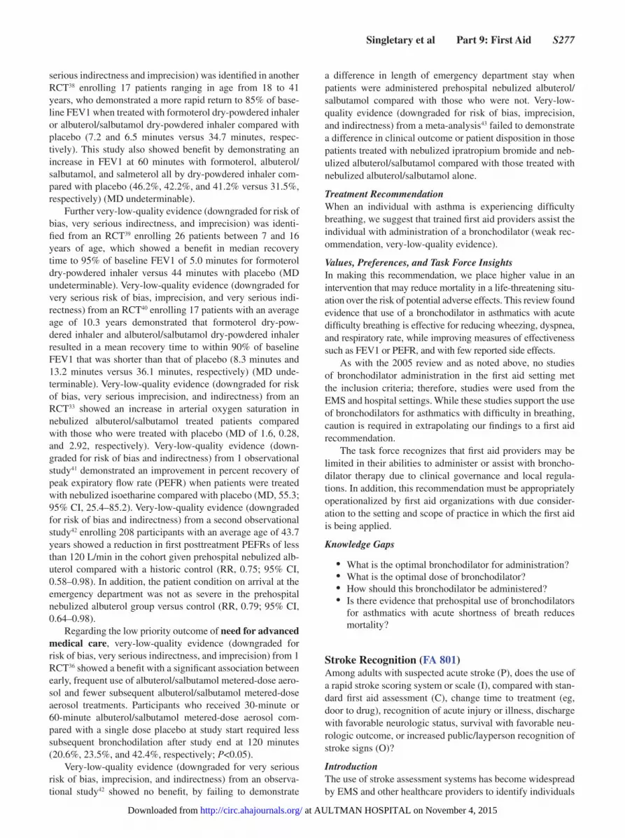
For the important outcome of recognition of stroke (diag-nostic studies, outcome defined as correct stroke diagnosis), we identified low-quality evidence (all downgraded for risk of bias) from 22 observational studies44,46–48,51–68 enrolling a total of 30 635 patients, studying 8 different stroke screening assessment systems, showing diagnostic performance across all stroke screening systems of sensitivity ranging from 0.41 to 0.97 and specificity ranging from 0.13 to 1.00. These stud-ies were divided into subgroups based on whether the stroke scales included glucose measurement or not. For studies that included stroke scales with glucose measurement (LAPSS, OPSS, KPSS, and Recognition of Stroke in the Emergency Room [ROSIER]), the pooled sensitivity was 0.84 (95% CI, 0.82–0.85) and pooled specificity was 0.97 (95% CI, 0.97–0.97), compared with stroke scales without glucose measure-ment (FAST, Melbourne Ambulance Stroke Screen [MASS], Los Angeles Motor Scale [LAMS], CPSS, Medical Priority Dispatch System [MPDS]), which have pooled sensitivity of 0.82 (95% CI, 0.81–0.83) and pooled specificity of 0.48 (95% CI, 0.46–0.49).
For the important outcome of increased public/layper-son recognition of signs of stroke, very-low-quality evidence
Figure 2. Forest plot of stroke assessment systems.
at AULTMAN HOSPITAL on November 4, 2015http://circ.ahajournals.org/Downloaded from

S280 Circulation October 20, 2015
(downgraded for risk of bias) from 1 human study69 enrolling 72 participants (members of the public) showed benefit where 76.4% of participants (55/72) were able to identify signs of stroke before training on a stroke screening assessment system compared with 94.4% (68/72) immediately after training (OR, 5.25; 95% CI, 1.67–16.52), and 96.9% of participants (63/65) were able to identify the signs of stroke 3 months after training (OR, 2.07; 95% CI, 0.36–11.69).
Treatment RecommendationWe recommend that first aid providers use stroke assess-ment systems (such as FAST or CPSS) for individuals with suspected acute stroke (strong recommendation, low-quality evidence).
We suggest the use of FAST or CPSS stroke assessment systems (weak recommendation, low-quality evidence).
We suggest the use of stroke assessment systems that include blood glucose measurement, when available, such as LAPSS, OPSS, ROSIER, or KPSS, to increase specific-ity of stroke recognition (weak recommendation, low-quality evidence).
In the absence of a glucometer, we suggest the use of FAST or CPSS stroke assessment systems compared with MASS, LAMS, or MPDS (weak recommendation, low-qual-ity evidence).
The literature search was rerun in January 2015 to cap-ture the most updated evidence possible. Two additional stud-ies were added51,59 and incorporated into the consensus on science and GRADE tables, both supporting this treatment recommendation.
Values, Preferences, and Task Force InsightsIn making this recommendation, we place increased value on the benefits of early stroke recognition, which could lead to early treatment to minimize potentially devastating neurologic injury.
Training first aid providers in stroke assessment systems outweighs the risks, largely limited to false-positive identifi-cation by first aid providers. The cost of the intervention is estimated to be low.
In this review of the literature, the stroke assessment sys-tems include various components, such as looking for specific signs and obtaining blood glucose levels. Our review found that stroke assessment systems that included blood glucose measurement had similar sensitivity and increased specific-ity to accurately identify stroke compared with those systems that did not include glucose measurement. We recognize that first aid providers may or may not have access to a properly calibrated glucose measurement device. Although use of these devices is not a standard component of first aid, glucose mea-surement devices are commonly available among the public.
Ideal stroke assessment systems for first aid use are accu-rate, have few steps, are easily understood and remembered, and take minimal time to complete. Those developing local guidelines for first aid providers can use the results of this review to determine if the benefit of increased specificity with systems that include glucose measurement would be desirable in their settings, compared with using simpler stroke assess-ment systems that do not include glucose measurement, which have similar sensitivity but lower specificity.
Knowledge GapsMore research is required to determine how much training is needed and what type of training should be used to enable first aid providers to correctly apply stroke assessment sys-tems and to compare the accuracy of use of stroke assessment systems by first aid providers to the accuracy of use of stroke assessment systems by healthcare providers. Research is also required to determine accuracy of assessment and its effect on survival and neurologic status at discharge. In addition, future research could include investigating direct transport to specified stroke centers when a stroke assessment system measurement is positive (bypassing community/small emer-gency departments).
Aspirin for Chest PainChest pain is one of the common symptoms of acute MI. Antiplatelet agents such as aspirin play a large role in manage-ment. In 2010, the first aid treatment recommendation stated that the administration of aspirin to individuals with chest dis-comfort was recommended.
In 2015, 2 PICOs were generated, 1 simply looking at the administration of aspirin and the other looking at the timing of this administration. The first PICO sought to determine if the administration of aspirin in the setting of MI was beneficial. Subsequently, the second PICO was used to determine if there was a difference in outcomes when aspirin is given early, in the first hours after symptom onset by a first aid provider, or later, in the setting of chest pain symptoms due to suspected acute MI. This same PICO was also used to see if there would be benefit to early administration of aspirin to adults with chest pain of unclear etiology.
Aspirin for Chest Pain: Administration (FA 871)Among adults experiencing chest pain due to suspected MI (P), does administration of aspirin (I), compared with no adminis-tration of aspirin (C), change cardiovascular mortality, com-plications, adverse effects, incidence of cardiac arrest, cardiac functional outcome, infarct size, hospital length of stay (O)?
IntroductionThis 2015 PICO question asks if administration versus no administration of aspirin changed outcomes in the setting of suspected acute MI. There are no major changes from what has been stated in previous treatment recommendations.
Consensus on ScienceFor the critical outcome of cardiovascular mortality (at 5 weeks), we identified high-quality evidence from 1 RCT70 enrolling 17 187 patients with acute MI showing benefit to aspirin (162.5 mg, enteric-coated) administration (RR, 0.79; 95% CI, 0.73–0.87).
For the critical outcome of cardiovascular mortality (at 3 months), we identified very-low-quality evidence (down-graded for risk of bias, indirectness, and imprecision) from 1 RCT71 enrolling 100 patients with acute MI showing no benefit to aspirin (100 mg, capsule) administration (RR, 0.83; 95% CI, 0.4–1.75).
For the critical outcome of cardiovascular mortality (at 28 days), we identified low-quality evidence (downgraded for
at AULTMAN HOSPITAL on November 4, 2015http://circ.ahajournals.org/Downloaded from

Singletary et al Part 9: First Aid S281
risk of bias and indirectness) from 1 RCT72 enrolling 1705 patients with acute MI showing no benefit to aspirin (300 mg, capsule) administration (RR, 0.98; 95% CI, 0.81–1.19).
For the critical outcome of cardiovascular mortality (in-hospital), we identified very-low-quality evidence (down-graded for risk of bias and indirectness) from 1 observational study73 with a total of 22 572 patients with acute MI showing benefit to aspirin (500 mg, oral or intravenous loading dose; 100 mg, oral; maintenance recommended) administration (RR, 0.33; 95% CI, 0.31–0.35).
For the critical outcome of adverse effects (bleeding), we identified high-quality evidence from 1 RCT70 enrolling 16 981 patients with acute MI showing adverse effects (minor bleeding) with aspirin (162.5 mg, enteric-coated) administra-tion (RR, 1.25; 95% CI, 1.04–1.51).
For the critical outcome of adverse effects (allergic reac-tion), we identified very-low-quality evidence (downgraded for risk of bias and imprecision) from 1 observational study74 with 219 patients with suspected acute MI showing no adverse effects (allergic reaction) with aspirin (dose not available) administration (unable to calculate RR as there was no control group).
For the critical outcome of complications, we identi-fied high-quality evidence from 1 RCT70 enrolling 16 981 patients with acute MI showing benefit to aspirin (162.5 mg, enteric-coated) administration (RR, 0.62; 95% CI, 0.52–0.73). We also found very-low-quality evidence (down-graded for risk of bias, imprecision, and indirectness) from 1 RCT71 enrolling 100 patients with acute MI showing benefit to aspirin (100 mg, capsule) administration (RR, 0.11; 95% CI, 0.05–0.98).
We identified very-low-quality evidence (downgraded for risk of bias and indirectness) from 1 observational study73 with a total of 22 572 patients with acute MI showing no ben-efit to aspirin (500 mg oral or intravenous loading, 100 mg oral maintenance recommended) administration (RR, 1.05; 95% CI, 0.78–1.42).
For the critical outcome of incidence of cardiac arrest, we identified high-quality evidence from 1 RCT70 enroll-ing 16 981 patients with acute MI showing benefit to aspirin (162.5 mg, enteric-coated) administration (RR, 0.87; 95% CI, 0.79–0.96).
For the important outcome of infarction size, we identi-fied very-low-quality evidence (downgraded for bias, impre-cision, and indirectness) from 1 RCT71 enrolling 89 patients with acute MI showing no benefit to aspirin (100 mg, capsule) administration (MD, −161; 95% CI, −445.57 to 230.57).
We did not identify any evidence to address the impor-tant outcomes of cardiac functional outcome or length of hospital stay.
Treatment RecommendationWe recommend the administration of aspirin to adults with chest pain due to suspected MI (strong recommendation, high-quality evidence).
Values, Preferences, and Task Force InsightsIn making this recommendation, we place a higher value on decreasing mortality and decreased complications of MI over the risks of adverse effects, such as bleeding.
Public comments for this question requested a suggestion for the optimal aspirin dose and form. Our PICO question was not designed to look at changes in outcomes based on various doses of aspirin, as all the articles selected for review compared administration to no administration, as opposed to 1 dose compared with another. Due to the heterogeneity in study design in the articles that were included in this review, the dose and form (eg, chewable or nonchewable, enteric-coated or nonenteric coated) of aspirin varied, and no recom-mendation could be made regarding the optimal dose or form of aspirin administered. Where available, the dose of aspirin used for each study has been identified in the consensus on science statement.
Knowledge Gaps
• Is aspirin safe if given to patients with chest pain who are not having an MI?
• Is aspirin safe when given by a first aid provider?• Is there high-quality evidence to indicate that the admin-
istration of aspirin after MI is time critical?
Aspirin for Chest Pain: Early Compared With Late (FA 586)Among adults who are experiencing chest pain outside of a hospital (P), does early administration of aspirin (I), compared with later administration of aspirin (C), change cardiovascular mortality, complications, incidence of cardiac arrest, cardiac functional outcome, infarct size, hospital length of stay, chest pain resolution (O)?
IntroductionThis 2015 PICO question asked if early administration ver-sus later administration of aspirin changes outcomes, which is different wording from the focus of the 2010 review. The recommendation in 2015 differs from that in 2010 as a result of the intent of the PICO question, as well as the studies iden-tified after using the rigorous literature search techniques and reviewed through the GRADE process.
Consensus on ScienceIn this review, early administration of aspirin is defined as prehospital or administration in the first hours from onset of symptoms of MI (ie, median 1.6 hours in 1 study).75
For the critical outcome of cardiovascular mortality (at 7 days), we identified very-low-quality evidence (down-graded for risk of bias and indirectness) from 2 observational studies75,76 with a total of 2122 patients with acute MI show-ing benefit to early aspirin administration (RR, 0.37; 95% CI, 0.23–0.62).
For the critical outcome of cardiovascular mortality (at 30 days), we identified very-low-quality evidence (down-graded for risk of bias and indirectness) from 2 observational studies75,76 with a total of 2122 patients with acute MI show-ing benefit to early aspirin administration (RR, 0.45; 95% CI, 0.3–0.68).
For the critical outcome of cardiovascular mortality (at 5 weeks), we identified low-quality evidence (downgraded for indirectness) from 1 RCT70 enrolling 8587 patients with acute MI showing no benefit to aspirin (162.5 mg, enteric-coated)
at AULTMAN HOSPITAL on November 4, 2015http://circ.ahajournals.org/Downloaded from
S282 Circulation October 20, 2015
administration within 2 hours of symptom onset (RR, 0.92; 95% CI, 0.76–1.11).
For the critical outcome of cardiovascular mortality (at 1 year), we identified very-low-quality evidence (down-graded for indirectness) from 1 observational study75 with 1200 patients with acute MI showing benefit to early aspirin (160 mg, oral) administration (RR, 0.47; 95% CI, 0.29–0.77).
For the critical outcome of complications, we identified very-low-quality evidence (downgraded for indirectness) from 1 observational study76 with a total of 922 patients with acute MI showing no increase in complication rate with early aspirin (greater than 200 mg, chewable) administration (RR, 0.61; 95% CI, 0.46–0.81). We also identified very-low-quality evidence (downgraded for risk of bias and indirectness) from 1 observational study75 with a total of 1200 patients with acute MI demonstrating an increase in complications (such as re-ischemia) in the group that received early aspirin (160 mg, oral) administration (RR, 1.22; 95% CI, 1.09–1.37).
For the critical outcome of incidence of cardiac arrest, we identified very-low-quality evidence (downgraded for indirectness) from 1 observational study76 with a total of 922 patients with acute MI showing no benefit to early aspirin (greater than 200 mg, chewable) administration (RR, 0.82; 95% CI, 0.56–1.2) and very-low-quality evidence (down-graded for risk of bias and indirectness) from 1 observational study75 with a total of 1200 patients with acute MI demon-strating an increased incidence of cardiac arrest in the group that received early aspirin (160 mg, oral) administration (RR, 1.53; 95% CI, 1.13–2.09).
We did not identify any evidence to address the important outcomes of cardiac functional outcome, infarct size, or hos-pital length of stay or the low importance outcome of chest pain resolution.
Treatment RecommendationWe suggest the early administration of aspirin by first aid pro-viders to adults with chest pain due to suspected MI (weak recommendation, very-low-quality evidence).
There is no evidence for the early administration of aspi-rin by first aid providers to adults with chest pain of unclear etiology.
Values, Preferences, and Task Force InsightsIn making this recommendation, we place a higher value on the benefits of aspirin, such as decreased mortality from MI, which outweigh possible risks of complications.
The task force discussed concerns about first aid providers being able to differentiate chest pain of cardiac origin from other causes of chest discomfort. With any treatment recom-mendations naming a particular clinical pathology, such as in this case with MI or chest pain of cardiac origin, it is very important that guidelines or educational materials clearly indi-cate what signs and symptoms the first aid provider should look for to recognize that clinical presentation.
Knowledge Gaps
• Is aspirin safe if given to patients with chest pain of other etiologies, particularly gastrointestinal?
• Is it safe for a first aid provider to administer 1 dose of aspirin?
• Is there any high-quality evidence demonstrating that there is a critical time window for the administration of aspirin after the onset of acute MI in terms of reducing morbidity and mortality?
• Is the prehospital administration of aspirin required if the patients are fast tracked to percutaneous coronary intervention (PCI)?
Epinephrine for Anaphylaxis and Treatment of Hypoglycemia, Exertion-Related
Dehydration, and Chemical Eye InjuriesThis section includes the topics of a second dose of epineph-rine for anaphylaxis and first aid treatment of hypoglycemia in diabetics, exertion-related dehydration, and chemical injuries of the eye.
Second Dose of Epinephrine for Anaphylaxis (FA 500)Among adults and children experiencing severe anaphylaxis requiring the use of epinephrine (P), does administration of a second dose of epinephrine (I), compared with administration of only 1 dose (C), change resolution of symptoms, adverse effects, complications (O)?
IntroductionIn 2010, evidence evaluation regarding effectiveness of administration of a second dose of epinephrine for ana-phylaxis concluded that there was insufficient evidence to make a recommendation regarding the routine first aid administration of a second dose of epinephrine. Use of a more rigorous literature search strategy and of the GRADE methodology for the 2015 review provided additional scien-tific evidence that has resulted in a change in the treatment recommendation.
The question’s specific focus was on the benefit of a sec-ond dose of epinephrine for severe anaphylaxis when signs and symptoms fail to respond to an initial dose. For the pur-pose of this review, if a study provided data for epinephrine administered after a first dose, unless the study specified that a second dose was given as part of a protocol, it was presumed that doses administered after a first dose were administered due to failure to respond.
Consensus on ScienceFor the critical outcome of resolution of symptoms, we iden-tified very-low-quality evidence (downgraded for risk of bias and confounding) from 9 observational studies77–85 showing benefit for giving a second dose (or multiple doses) of epi-nephrine to patients not responding to a first dose (RR, 1.16; 95% CI, 1.13–1.20).
In addition, for the critical outcome of resolution of symptoms, we identified very-low-quality evidence (down-graded for risk of bias) from 1 observational study86 showing no significant difference between the percentage of resolved reactions in an ambulance service routinely using 2 doses of epinephrine versus an ambulance service using a single dose (RR, 0.97; 95% CI, 0.9–1.04).
We did not identify any evidence to address the critical outcomes of adverse effects or complications.
at AULTMAN HOSPITAL on November 4, 2015http://circ.ahajournals.org/Downloaded from

Singletary et al Part 9: First Aid S283
Treatment RecommendationWe suggest a second dose of epinephrine be administered by autoinjector to individuals with severe anaphylaxis whose symptoms are not relieved by an initial dose (weak recom-mendation, very-low-quality evidence).
Values, Preferences, and Task Force InsightsIn making this recommendation, we place a higher value on the resolution of life-threatening symptoms, such as airway compromise, breathing difficulty, and circulatory collapse, over the potential risk of adverse effects from an unnecessary second injection.
When caring for a person with anaphylaxis, first aid pro-viders should always call EMS (eg, 9-1-1 or 1-1-2) rescue services.
Public comments and discussion on this topic centered on issues of dosing, interval time for a second dose, and the pos-sibility of adverse effects should epinephrine be inadvertently administered to a person not experiencing anaphylaxis. This evidence review did not evaluate the time interval between doses of epinephrine or the optimal dose. However, literature included in the review suggests that a second dose of epinephrine may be administered 10 to 15 minutes after the initial dose.80
While the included studies did not identify any adverse effects, selection bias might have prevented those effects from being identified. Adverse effects have previously been reported in the literature when epinephrine is administered in the incorrect dose or via inappropriate routes, such as the intra-venous route. Use of autoinjectors by first aid providers may minimize the opportunity for incorrect dosing of epinephrine.
Knowledge GapsIn 2010, first aid worksheet 303B attempted to define if or can “the First Aid Provider Appropriately Recognize the Signs and Symptoms of Anaphylaxis.” The task force did not address this PICO question in 2015, and thus the question “How can a first aid provider determine that a victim needs additional epinephrine?” remains.
• What should the time interval be between doses of epinephrine?
• Would a higher concentration (0.5 mg) recommended for standard therapy versus the injectable syringe dose (0.3 mg) be more effective and decrease the need for additional doses in the EMS setting?
• Should an initial injection be administered in the early stages of anaphylaxis, before the onset of severe symptoms?
Hypoglycemia Treatment (FA 795)Among adults and children with symptomatic hypoglycemia (P), does administration of dietary forms of sugar (I), compared with standard dose (15–20 g) of glucose tablets (C), change time to resolution of symptoms, risk of complications (eg, aspiration), blood glucose, hypoglycemia, hospital length of stay (O)?
IntroductionThis is a new topic for the 2015 consensus on science.
Because glucose tablets may not be readily available in all first aid settings, this task force performed a review to evaluate
the effectiveness of dietary (ie, food source) sugars compared with glucose tablets for the management of symptomatic hypoglycemia.
The literature search for this review identified 5 studies that compared glucose tablets to various commercial sugar-containing dietary products. The named commercial products cited in the consensus on science and in the treatment recom-mendation were specifically included in evaluated studies and are not particularly endorsed by the First Aid Task Force. To our knowledge, none of the product manufacturers contributed to or were involved with the identified studies. Two tables list-ing the specific sugar content for each studied product are pro-vided to assist with guideline development (Tables 1 and 2).
Consensus on Science
Dietary Sugars (I) Compared With Glucose Tablets (C)For the critical outcome of time to resolution of symptoms, none of the 4 studies identified87–90 showed that any form of dietary sugar or glucose tablets improved the blood glucose before 10 minutes.
For the important outcome of hypoglycemia (clinical relief in 15 minutes or less), we identified low-quality evi-dence (downgraded for risk of bias and imprecision) from 3 randomized controlled studies88–90 with pooled data from 502 diabetic patients treated with dietary sugars (sucrose, fructose, orange juice, jelly beans, Mentos, and milk) and 223 treated with glucose tablets (15–20 g) that showed a benefit with glu-cose tablets, with slower resolution of symptoms 15 minutes after diabetic patients were treated with dietary sugars com-pared with glucose tablets (RR, 0.89; 95% CI, 0.83–0.96).
For the important outcome of blood glucose (diabetic patients with at least a 20-mg/dL increase of blood glucose by 20 minutes), we found very-low-quality evidence (down-graded for risk of bias and imprecision) from 1 observational study87 with 13 diabetic patients treated with dietary sugars and 9 treated with glucose tablets that showed a benefit with glucose tablets, with fewer diabetic patients demonstrating a 20-mg/dL increase in blood glucose level 20 minutes after treatment when treated with dietary sugars compared with glucose tablets (RR, 0.3; 95% CI, 0.1–0.85). For the criti-cal outcome of time to resolution of symptoms, the impor-tant outcome of risk of complications (eg, aspiration), and the low-priority outcome of hospital length of stay, there were no human trials found.
Sucrose (I) Compared With Glucose Tablets (C)For the important outcome of hypoglycemia (clinical relief in 15 minutes or less), we found low-quality evidence (down-graded for risk of bias and imprecision) from 2 RCTs88,90 with pooled data from 177 diabetic patients treated with sucrose (165 with sucrose candy [Skittles] and 12 with sucrose tab-lets) and 171 treated with glucose tablets that showed no dif-ference in their effects on blood glucose. Sucrose (either as sucrose candy [Skittles] or sucrose tablets) and glucose tablets were equivalent in providing clinical relief of hypoglycemia 15 minutes after ingestion (RR, 0.99; 95% CI, 0.91–1.07). For the important outcome of blood glucose (mean change in blood glucose [mmol/L] after 15 minutes), we found low-quality evidence (downgraded for risk of bias and imprecision)
at AULTMAN HOSPITAL on November 4, 2015http://circ.ahajournals.org/Downloaded from

S284 Circulation October 20, 2015
from 1 randomized controlled study90 with 6 diabetic patients treated with sucrose (dissolved in water) and 6 treated with glucose tablets that showed a benefit to glucose administra-tion, with the MD (mmol/L) in blood glucose 15 minutes after ingestion lower with sucrose (dissolved in water) than glucose tablets (MD, −0.9; 95% CI, −1.78 to −0.02). A second arm of this same study with 6 diabetic patients treated with sucrose (chewed) and 6 treated with glucose tablets showed no ben-efit, with the MD (mmol/L) in blood glucose 15 minutes after ingestion similar between sucrose (chewed) and glucose tab-lets (MD, 0.3; 95% CI, −0.8 to 1.41). For the critical outcome of time to resolution of symptoms, the important outcome of risk of complications (eg, aspiration), and the lower-priority outcome of hospital length of stay, there were no human trials found.
Fructose (I) Compared With Glucose Tablets (C)For the important outcome of hypoglycemia (clinical relief in 15 minutes or less), we found low-quality evidence (down-graded for risk of bias and imprecision) from 1 RCT88 with 165
diabetic patients treated with fructose (Fruit to Go) and 165 treated with glucose tablets that showed benefit with glucose, with a lower incidence of resolution of symptoms 15 minutes after treatment for diabetic patients treated with fructose com-pared with glucose tablets (RR, 0.77; 95% CI, 0.68–0.86). For the critical outcome of time to resolution of symptoms, the important outcomes of risk of complications (eg, aspiration) and blood glucose, and the low-priority outcome of hospital length of stay, there were no human trials found.
Orange Juice (I) Compared With Glucose Tablets (C)For the important outcome of hypoglycemia (clinical relief in 15 minutes or less), we found very-low-quality evidence (downgraded for risk of bias, inconsistency, and imprecision) from 2 RCTs89,90 with the pooled data of 50 diabetic patients treated with orange juice and 58 treated with glucose tab-lets that showed no difference in the resolution of symptoms 15 minutes after treatment for diabetic patients treated with orange juice compared with glucose tablets (RR, 0.84; 95% CI, 0.69–1.02). For the important outcome of blood glucose,
Table 1. Major Dietary Sugars With About 20 g Carbohydrate From Sugar and Number of People With Improvement in Hypoglycemia Within 15 Minutes
Type of Food or FluidCarbohydrates
per ServingMeasure Representing 20 g Carbohydrates*
Clinical Relief 15 Minutes or Less After Ingestion
Glucose tablets Varies Varies 194/223 (87.0%)
Sucrose (Skittles)† 0.9 g/candy 20–25 candies 150/177 (84.7%)
Fructose (fruit leather, eg, Stretch Island)‡
10 g/strip 2 strips 111/165 (67.3%)
Orange juice (unsweetened, from concentrate)§
1 g/10 mL 200 mL 35/50 (70.0%)
Jelly beans§ 1.1 g/jelly bean 15–20 jelly beans 33/45 (73.3%)
Mentos║ 2.8 g/mint 5–10 mints 44/48 (91.7%)
Whole milk¶ 21.75 g/mL 435 mL Not reported
*These measurements may differ from those in the evaluated studies, because the amount was not standardized across studies.
†Manufacturer label.‡One study88 used fruit leather under the trade name Fruit to Go (Kelowna, British Columbia, Canada). We
were unable to find the nutritional information for this fruit leather, so we substituted another brand, Stretch Island Cherry Flavor (Stretch Island Fruit Co, La Jolla, CA); http://www.stretchislandfruit.com/en_US/Products.html, accessed February 2, 2015.
§http://onlinelibrary.wiley.com/doi/10.1002/pdi.953/pdf.║Manufacturer label.¶Brodows, 1984.87
Table 2. Type of Dietary Sugar Representing 15 g of Carbohydrates and Number of People With Improvement in Hypoglycemia Within 15 Minutes
Type of Dietary SugarCarbohydrates per
ServingMeasure Representing 15 g Carbohydrates*
Clinical Relief 15 Minutes or Less After Ingestion
Glucose tablets Varies Varies 194/223 (87.0%)
Glucose solution*† 1 g/10 mL 150 mL 5/6 (83.3%)
Glucose gel*‡ 15 g of glucose in 40 g of 40% dextrose gel
15 g 2/6 (33.3%)
Cornstarch hydrolysate*§ 15 g cornstarch 15 g 4/5 (80%)
*Glucose solution, glucose gel, and hydrolysate were evaluated in 1 study.90
†15 g of glucose dissolved in 150 mL of water.‡Hypostop, Novo Industries.§15 g of cornstarch hydrolysate containing 2% to 3% glucose, 6% to 8% maltose, 89% to 92% oligosaccharides and
polysaccharides, and 0.15% protein (Glucides 19, Roquette Freres, Lestrem, France) diluted in 150 mL of water.
at AULTMAN HOSPITAL on November 4, 2015http://circ.ahajournals.org/Downloaded from
Singletary et al Part 9: First Aid S285
we identified very-low-quality evidence (downgraded for risk of bias and imprecision) from 1 RCT90 with 6 diabetic patients treated with orange juice and 6 treated with glucose tablets that showed no benefit with glucose tablets, with the MD (mmol/L) in blood glucose 15 minutes after ingestion lower with orange juice than with glucose tablets (MD, −0.7; 95% CI, −1.55 to −0.15). Very-low-quality evidence (down-graded for risk of bias and imprecision) from 1 observational study87 with 8 diabetic patients treated with orange juice and 9 treated with glucose tablets showed no difference in a diabetic patient’s likelihood of having a 20-mg/dL increase in blood glucose level 20 minutes after treatment with orange juice compared with glucose tablets (RR, 0.48; 95% CI, 0.18–1.26). For the critical outcome of time to resolution of symptoms, the important outcome of risk of complications (eg, aspira-tion), and the low-priority outcome of hospital length of stay, no human trials were found.
Jelly Beans (I) Compared With Glucose Tablets (C)For the important outcome of hypoglycemia (clinical relief less in 15 minutes or less), we found very-low-quality evi-dence (downgraded for risk of bias and imprecision) from 1 RCT89 with 45 diabetic patients treated with jelly beans and 52 treated with glucose tablets that showed no difference in the resolution of symptoms 15 minutes after treatment, whether diabetic patients were treated with jelly beans or glucose tab-lets (RR, 0.85; 95% CI, 0.69–1.04). For the critical outcome of time to resolution of symptoms, the important outcomes of risk of complications (eg, aspiration) and blood glucose, and the low-priority outcome of hospital length of stay, no human trials were found.
Mentos (I) Compared With Glucose Tablets (C)For the important outcome of hypoglycemia (clinical relief in 15 minutes or less), we found very-low-quality evidence (downgraded for risk of bias and imprecision) from 1 RCT89 with 48 diabetic patients treated with Mentos and 52 treated with glucose tablets that showed no difference in the resolu-tion of symptoms 15 minutes after treatment, whether diabetic patients were treated with Mentos or glucose tablets (RR, 1.06; 95% CI, 0.92–1.21). For the critical outcome of time to resolution of symptoms, the important outcomes of risk of complications (eg, aspiration) and blood glucose, and the low-priority outcome of hospital length of stay, no human trials were found.
Milk (I) Compared With Glucose Tablets (C)For the important outcome of blood glucose (diabetic patients with at least a 20-mg/dL increase of blood glucose by 20 min-utes), we found very-low-quality evidence (downgraded for risk of bias and imprecision) from 1 observational study87 that included 5 diabetic patients treated with whole milk and 9 treated with glucose tablets, showing no difference in likeli-hood of a 20-mg/dL increase in blood glucose level 20 min-utes after treatment, whether diabetic patients were treated with milk or glucose tablets (RR, 0.11; 95% CI, 0.01–1.62). For the critical outcome of time to resolution of symptoms, the important outcomes of risk of complications (eg, aspira-tion) and hypoglycemia, and the low-priority outcome of hos-pital length of stay, no human trials were found.
Glucose Gel (I) Compared With Glucose Tablets (C)For the important outcome of hypoglycemia (clinical relief in 15 minutes or less), we identified very-low-quality evidence (downgraded for risk of bias and imprecision) from 1 RCT90 that included 6 diabetic patients treated with glucose gel and 6 treated with glucose tablets, finding no difference in the reso-lution of symptoms 15 minutes after treatment (RR, 0.5; 95% CI, 0.14–1.77).
For the critical outcome of time to resolution of symp-toms, the important outcomes of risk of complications (eg, aspiration) and blood glucose, and the low-priority outcome of hospital length of stay, no human trials were found.
Glucose Solution (I) Compared With Glucose Tablets (C)For the important outcome of hypoglycemia (clinical relief in 15 minutes or less), we identified very-low-quality evidence (downgraded for risk of bias and imprecision) from 1 RCT90 that included 6 diabetic patients treated with glucose solution and 6 treated with glucose tablets, finding no difference in the resolution of symptoms 15 minutes after treatment (RR, 1.25; 95% CI, 0.64–2.44).
For the critical outcome of time to resolution of symp-toms, the important outcomes of risk of complications (eg, aspiration) and blood glucose, and the low-priority outcome of hospital length of stay, no human trials were found.
Cornstarch Hydrolysate (I) Compared With Glucose Tablets (C)For the important outcome of hypoglycemia (clinical relief in 15 minutes or less), we found very-low-quality evidence (downgraded for risk of bias and imprecision) from 1 RCT90 that included 5 diabetic patients treated with cornstarch hydrolysate 15 g and 6 treated with glucose tablets, finding no difference in the resolution of symptoms 15 minutes after treatment (RR, 1.20; 95% CI, 0.59–2.45).
For the critical outcome of time to resolution of symp-toms, the important outcomes of risk of complications (eg, aspiration) and blood glucose, and the low-priority outcome of hospital length of stay, no human trials were found.
The following dietary sugars were evaluated in the included studies:
• Skittles: ~90 g carbohydrate per 100 g, sugar (sucrose) corn syrup, partially hydrogenated soybean oil, fruit juice from concentrate (grape, strawberry, lemon, lime, orange), citric acid, dextrin, natural and artificial flavors, gelatin, food starch–modified coloring, ascorbic acid
• Fruit to Go: apple pure concentrate; apple, cherry, and elderberry juice concentrates; wild berry concentrate (concentrated cherry, raspberry, blueberry, cranberry and boysenberry juices, natural flavor); citrus pectin; natural flavor; lemon juice concentrate
• Mentos: 2.8 g carbohydrate in each mint (71% glucose and 29% oligosaccharides), 91.6 g carbohydrate per 100 g, 69.3 g sugar per 100 g, sugar, glucose syrup (corn), reconstituted fruit juices (strawberry, orange, lemon; 2.5%), hydrogenated vegetable oil (coconut), acid (citric acid), rice starch, thickeners (gum arabic, gellan gum, flavorings, glazing agent [carnauba wax]), emulsifier (sucrose esters of fatty acids), colors
• Glucose gel: 15 g of glucose in 40 g of 40% dextrose gel (Hypostop, Novo Industries)
at AULTMAN HOSPITAL on November 4, 2015http://circ.ahajournals.org/Downloaded from
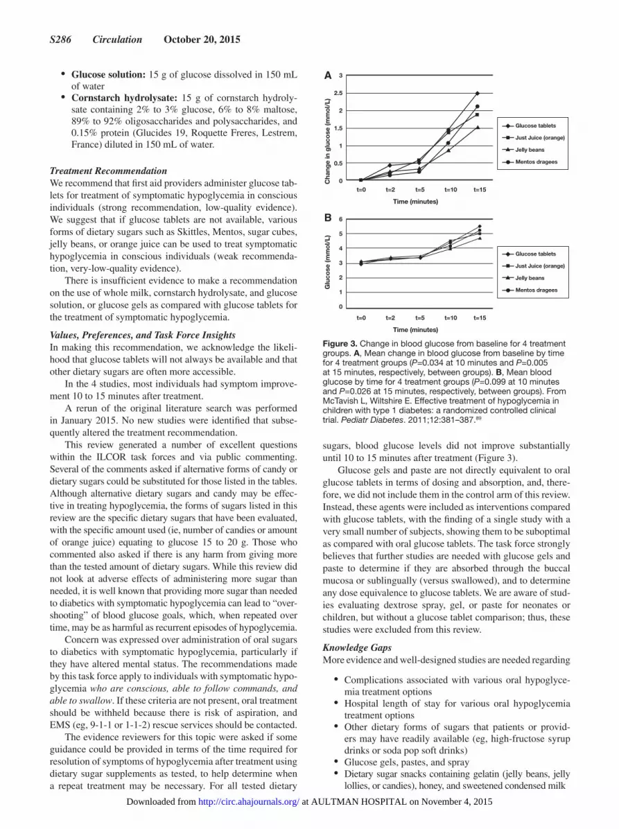
S286 Circulation October 20, 2015
• Glucose solution: 15 g of glucose dissolved in 150 mL of water
• Cornstarch hydrolysate: 15 g of cornstarch hydroly-sate containing 2% to 3% glucose, 6% to 8% maltose, 89% to 92% oligosaccharides and polysaccharides, and 0.15% protein (Glucides 19, Roquette Freres, Lestrem, France) diluted in 150 mL of water.
Treatment RecommendationWe recommend that first aid providers administer glucose tab-lets for treatment of symptomatic hypoglycemia in conscious individuals (strong recommendation, low-quality evidence). We suggest that if glucose tablets are not available, various forms of dietary sugars such as Skittles, Mentos, sugar cubes, jelly beans, or orange juice can be used to treat symptomatic hypoglycemia in conscious individuals (weak recommenda-tion, very-low-quality evidence).
There is insufficient evidence to make a recommendation on the use of whole milk, cornstarch hydrolysate, and glucose solution, or glucose gels as compared with glucose tablets for the treatment of symptomatic hypoglycemia.
Values, Preferences, and Task Force InsightsIn making this recommendation, we acknowledge the likeli-hood that glucose tablets will not always be available and that other dietary sugars are often more accessible.
In the 4 studies, most individuals had symptom improve-ment 10 to 15 minutes after treatment.
A rerun of the original literature search was performed in January 2015. No new studies were identified that subse-quently altered the treatment recommendation.
This review generated a number of excellent questions within the ILCOR task forces and via public commenting. Several of the comments asked if alternative forms of candy or dietary sugars could be substituted for those listed in the tables. Although alternative dietary sugars and candy may be effec-tive in treating hypoglycemia, the forms of sugars listed in this review are the specific dietary sugars that have been evaluated, with the specific amount used (ie, number of candies or amount of orange juice) equating to glucose 15 to 20 g. Those who commented also asked if there is any harm from giving more than the tested amount of dietary sugars. While this review did not look at adverse effects of administering more sugar than needed, it is well known that providing more sugar than needed to diabetics with symptomatic hypoglycemia can lead to “over-shooting” of blood glucose goals, which, when repeated over time, may be as harmful as recurrent episodes of hypoglycemia.
Concern was expressed over administration of oral sugars to diabetics with symptomatic hypoglycemia, particularly if they have altered mental status. The recommendations made by this task force apply to individuals with symptomatic hypo-glycemia who are conscious, able to follow commands, and able to swallow. If these criteria are not present, oral treatment should be withheld because there is risk of aspiration, and EMS (eg, 9-1-1 or 1-1-2) rescue services should be contacted.
The evidence reviewers for this topic were asked if some guidance could be provided in terms of the time required for resolution of symptoms of hypoglycemia after treatment using dietary sugar supplements as tested, to help determine when a repeat treatment may be necessary. For all tested dietary
sugars, blood glucose levels did not improve substantially until 10 to 15 minutes after treatment (Figure 3).
Glucose gels and paste are not directly equivalent to oral glucose tablets in terms of dosing and absorption, and, there-fore, we did not include them in the control arm of this review. Instead, these agents were included as interventions compared with glucose tablets, with the finding of a single study with a very small number of subjects, showing them to be suboptimal as compared with oral glucose tablets. The task force strongly believes that further studies are needed with glucose gels and paste to determine if they are absorbed through the buccal mucosa or sublingually (versus swallowed), and to determine any dose equivalence to glucose tablets. We are aware of stud-ies evaluating dextrose spray, gel, or paste for neonates or children, but without a glucose tablet comparison; thus, these studies were excluded from this review.
Knowledge GapsMore evidence and well-designed studies are needed regarding
• Complications associated with various oral hypoglyce-mia treatment options
• Hospital length of stay for various oral hypoglycemia treatment options
• Other dietary forms of sugars that patients or provid-ers may have readily available (eg, high-fructose syrup drinks or soda pop soft drinks)
• Glucose gels, pastes, and spray• Dietary sugar snacks containing gelatin (jelly beans, jelly
lollies, or candies), honey, and sweetened condensed milk
Time (minutes)
Cha
nge
in g
luco
se (m
mo
l/L)
3
2.5
2
1.5
1
0.5
0t=0 t=2 t=5 t=10 t=15
Glucose tablets
Just Juice (orange)
Jelly beans
Mentos dragees
Time (minutes)G
luco
se (m
mo
l/L)
6
5
4
3
2
1
0
t=0 t=2 t=5 t=10 t=15
Glucose tablets
Just Juice (orange)
Jelly beans
Mentos dragees
A
B
Figure 3. Change in blood glucose from baseline for 4 treatment groups. A, Mean change in blood glucose from baseline by time for 4 treatment groups (P=0.034 at 10 minutes and P=0.005 at 15 minutes, respectively, between groups). B, Mean blood glucose by time for 4 treatment groups (P=0.099 at 10 minutes and P=0.026 at 15 minutes, respectively, between groups). From McTavish L, Wiltshire E. Effective treatment of hypoglycemia in children with type 1 diabetes: a randomized controlled clinical trial. Pediatr Diabetes. 2011;12:381–387.89
at AULTMAN HOSPITAL on November 4, 2015http://circ.ahajournals.org/Downloaded from

Singletary et al Part 9: First Aid S287
Exertion-Related Dehydration and Oral Rehydration (FA 584)Among adults and children with exertion-related dehydration (P), does drinking oral carbohydrate-electrolyte (CE) liquids (I), compared with drinking water (C), change volume/hydra-tion status, vital signs, development of hyperthermia, develop-ment of hyponatremia, need for advanced medical care, blood glucose, patient satisfaction (O)?
IntroductionA review of this topic was performed in 2010 and concluded that CE beverages are recommended for rehydration of indi-viduals who become dehydrated through sweating in hot climates and/or exercise. For the 2015 review, the task force used an extensive literature search combined with GRADE methodology, resulting in a much larger number of included studies. In addition, we included several alternative beverages with varying CE content compared with water. The authors of some included studies noted that a relatively lower urine volume is considered an indicator for increased intravascu-lar volume during the immediate postexercise rehydration period.86,89,91 The physiologic basis of this relates to a fall in plasma osmolality and sodium concentration with plain water ingestion after exercise, which stimulates urine production and reduces the stimulus to drink, both of which delay the rehydration process. Addition of sodium chloride to plain water has been shown to increase fluid intake while reduc-ing urine output. Thus, for this review, a lower urine output in the first several hours after ingestion of studied fluids is considered a beneficial effect for rehydration. The rehydra-tion index is an indication of how much of the fluid ingested was actually used in body weight restoration,92,93 with a lower number reflecting a higher amount of ingested fluid used in body weight restoration.
Consensus on ScienceAfter the application of inclusion and exclusion criteria to the 1751 initial citations, a total of 12 studies were included. A summary of the evidence from these 12 studies is provided (Table 3).
12% CE Solution (I) Compared With Water (C)For the critical outcome of volume/hydration status, we identified very-low-quality evidence (downgraded for risk of bias and imprecision) from 1 RCT91 with 30 subjects show-ing a benefit with the use of CE solution, with increased fluid retention (%) at 2 hours after exercise (MD, 16.1; 95% CI, 7.45–24.75).
We did not identify any evidence to address the critical outcomes of vital signs, development of hyperthermia, and development of hyponatremia, or the important outcomes of blood glucose, need for advanced medical care, and patient satisfaction.
5% to 8% CE Solution (I) Compared With Water (C)For the critical outcome of volume/hydration status, we identified low-quality evidence (downgraded for risk of bias and imprecision) from 8 studies including 204 subjects showing an overall benefit to 5% to 8% CE solution com-pared with water in 10 of 15 outcomes, and 5 of 15 showing no difference:
• Very-low-quality evidence (downgraded for impreci-sion) from 1 observational study94 with 38 subjects showing at 2 hours after hydration no difference for body weight loss (kg) with CE solution compared with water, a benefit with CE solution with increased rehydration (%) (MD, 8; 95% CI, 6.09–9.91), and a benefit with CE solution for increased blood volume response (%) (MD, 2.8; 95% CI, 2.26–3.34).
• Moderate-quality evidence (downgraded for impreci-sion) from 1 RCT95 with 18 subjects showing no benefit for CE solution compared with water for rehydration (%) at 4 hours after hydration (MD, −1.6; 95% CI, −11.12 to 7.92).
• Very-low-quality evidence (downgraded for risk of bias and imprecision) from 2 RCTs91,96 with 54 subjects showing no difference in fluid retention (%) at 2 hours after hydration for CE solution compared with water; low-quality evidence (downgraded for risk of bias and imprecision) from 2 RCTs96,97 with 44 subjects showing a benefit of CE solution for increased fluid retention (%) at 3 hours (MD, 15.6; 95% CI, 12.44–18.8); very-low-quality evidence (downgraded for imprecision) from 1 observational study98 with 26 subjects showing a ben-efit with CE solution for increased fluid retention (%) at 3 hours (MD, 21.7; 95% CI, 9.89–33.51); very-low-quality evidence (downgraded for imprecision) from 1 observational study99 with 26 subjects showing a benefit with CE solution for increased fluid retention (%) at 4 hours (MD, 22; 95% CI, 9.6–34.4); low-quality evidence (downgraded for risk of bias and imprecision) from 1 RCT100 with 22 subjects showing no difference in fluid retention (%) at 4 hours.
• Low-quality evidence (downgraded for risk of bias and imprecision) from 1 RCT97 with 20 subjects showing a benefit with CE solution compared with water with decreased mean urine volume by weight (g) between 1 and 2 hours after hydration (MD, −175; 95% CI, −206.37 to −143.63) and a benefit of CE solution with decreased mean urine volume between 2 and 3 hours after hydration (MD, −41; 95% CI, −64.27 to −17.73); very-low-quality evidence (down-graded for imprecision) from 1 observational study94 with 38 subjects showing at 2 hours after hydration a benefit with CE solution with decreased mean urine volume (mL) (MD, −160; 95% CI, −198.15 to −121.85); very-low-quality evidence (downgraded for imprecision) from 1 observational study98 with 26 subjects showing a benefit with CE solution with decreased mean urine volume (mL) at 3 hours after hydration (MD, −465.3; 95% CI, −700.73 to −229.87); low-quality evidence (downgraded for risk of bias and imprecision) from 1 RCT100 with 22 sub-jects showing no difference for mean urine volume (mL) at 4 hours after hydration; and very-low-quality evidence (downgraded for imprecision) from 1 obser-vational study99 with 26 subjects showing a benefit with CE solution with decreased mean urine volume (mL) at 4 hours after hydration (MD, −277; 95% CI, −458.26 to −95.74).
• Very-low-quality evidence (downgraded for impreci-sion) from 1 observational study98 with 26 subjects
at AULTMAN HOSPITAL on November 4, 2015http://circ.ahajournals.org/Downloaded from
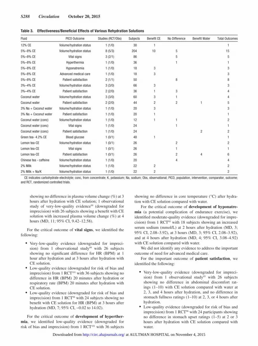
S288 Circulation October 20, 2015
showing no difference in plasma volume change (%) at 3 hours after hydration with CE solution; 1 observational study of very-low-quality evidence99 (downgraded for imprecision) with 26 subjects showing a benefit with CE solution with increased plasma volume change (%) at 4 hours (MD, 11; 95% CI, 9.42–12.58).
For the critical outcome of vital signs, we identified the following:
• Very-low-quality evidence (downgraded for impreci-sion) from 1 observational study99 with 26 subjects showing no significant difference for HR (BPM) at 1 hour after hydration and at 3 hours after hydration with CE solution.
• Low-quality evidence (downgraded for risk of bias and imprecision) from 1 RCT101 with 36 subjects showing no difference in HR (BPM) 20 minutes after hydration or respiratory rate (BPM) 20 minutes after hydration with CE solution.
• Low-quality evidence (downgraded for risk of bias and imprecision) from 1 RCT96 with 24 subjects showing no benefit with CE solution for HR (BPM) at 3 hours after hydration (MD, 7; 95% CI, −0.02 to 14.02).
For the critical outcome of development of hyperther-mia, we identified low-quality evidence (downgraded for risk of bias and imprecision) from 1 RCT101 with 36 subjects
showing no difference in core temperature (°C) after hydra-tion with CE solution compared with water.
For the critical outcome of development of hyponatre-mia (a potential complication of endurance exercise), we identified moderate-quality evidence (downgraded for impre-cision) from 1 RCT95 with 18 subjects showing an increased serum sodium (mmol/L) at 2 hours after hydration (MD, 3; 95% CI, 2.08–3.92), at 3 hours (MD, 3; 95% CI, 2.08–3.92), and at 4 hours after hydration (MD, 4; 95% CI, 3.08–4.92) with CE solution compared with water.
We did not identify any evidence to address the important outcome of need for advanced medical care.
For the important outcome of patient satisfaction, we identified the following:
• Very-low-quality evidence (downgraded for impreci-sion) from 1 observational study95 with 26 subjects showing no difference in abdominal discomfort rat-ings (1–10) with CE solution compared with water at 2, 3, and 4 hours after hydration, and no difference in stomach fullness ratings (1–10) at 2, 3, or 4 hours after hydration.
• Low-quality evidence (downgraded for risk of bias and imprecision) from 1 RCT96 with 24 participants showing no difference in stomach upset ratings (1–5) at 2 or 3 hours after hydration with CE solution compared with water.
Table 3. Effectiveness/Beneficial Effects of Various Rehydration Solutions
Fluid PICO Outcome Studies (RCT/Obs) Subjects Benefit CE No Difference Benefit Water Total Outcomes
12% CE Volume/hydration status 1 (1/0) 30 1 1
5%–8% CE Volume/hydration status 8 (5/3) 204 10 5 15
5%–8% CE Vital signs 3 (2/1) 86 5 5
5%–8% CE Hyperthermia 1 (1/0) 36 1 1
5%–8% CE Hyponatremia 1 (1/0) 18 3 3
5%–8% CE Advanced medical care 1 (1/0) 18 3 3
5%–8% CE Patient satisfaction 2 (1/1) 50 8 8
3%–4% CE Volume/hydration status 3 (3/0) 66 3 3
3%–4% CE Patient satisfaction 2 (2/0) 36 1 3 4
Coconut water Volume/hydration status 3 (3/0) 60 3 1 4
Coconut water Patient satisfaction 2 (2/0) 44 2 2 1 5
3% Na + Coconut water Volume/hydration status 1 (1/0) 20 3 3
3% Na + Coconut water Patient satisfaction 1 (1/0) 20 1 1
Coconut water (conc) Volume/hydration status 1 (1/0) 12 1 1 2
Coconut water (conc) Vital signs 1 (1/0) 24 1 1
Coconut water (conc) Patient satisfaction 1 (1/0) 24 2 2
Green tea- 4.2% CE Blood glucose 1 (0/1) 48 1 1
Lemon tea-CE Volume/hydration status 1 (0/1) 26 2 2
Lemon tea-CE Vital signs 1 (0/1) 26 1 1
Lemon tea-CE Patient satisfaction 1 (0/1) 26 2 4 6
Chinese tea - caffeine Volume/hydration status 1 (1/0) 20 4 4
2% Milk Volume/hydration status 1 (1/0) 22 2 2
2% Milk + Na/K Volume/hydration status 1 (1/0) 22 2 2
CE indicates carbohydrate-electrolyte; conc, from concentrate; K, potassium; Na, sodium; Obs, observational; PICO, population, intervention, comparator, outcome; and RCT, randomized controlled trials.
at AULTMAN HOSPITAL on November 4, 2015http://circ.ahajournals.org/Downloaded from
Singletary et al Part 9: First Aid S289
We did not identify any evidence to address the important outcome of blood glucose.
3% to 4% CE Solution (I) Compared With Water (C)For the critical outcome of volume/hydration status, we identified the following:
• Low-quality evidence (downgraded for risk of bias and imprecision) from 2 RCTs92,93 with 36 subjects showing no difference in the rehydration index for CE solution compared with water.
• Very-low-quality evidence (downgraded for risk of bias and imprecision) from 3 RCTs91–93 with 66 subjects showing a benefit with CE solution with increased fluid retention (%) at 2 hours after hydration (MD, 8.97; 95% CI, 7.54–10.4).
• Low-quality evidence (downgraded for risk of bias and imprecision) from 1 RCT93 with 20 subjects showing a benefit of CE solution with decreased cumulative urine output (mL) at 2 hours into the hydration period (MD, −174.5; 95% CI, −220.89 to −128.11).
For the important outcome of patient satisfaction, we identified the following:
• Low-quality evidence (downgraded for risk of bias and imprecision) from 1 RCT93 with 20 subjects showing no difference for nausea scores (1–5) at 90 minutes after hydration with CE solution, and low-quality evidence (downgraded for risk of bias and imprecision) from 2 RCTs92,93 with 36 subjects showing no difference for nausea scores (1–5) at 2 hours for CE solution compared with water.
• Low-quality evidence (downgraded for risk of bias and imprecision) from 1 RCT93 with 20 subjects showing no difference for stomach upset scores (1–5) at 90 minutes after hydration with CE solution compared with water, and low-quality evidence (downgraded for risk of bias and imprecision) from 2 RCTs92,93 with 36 subjects showing a benefit with CE solution with a decrease in stomach upset scores (1–5) at 2 hours after hydration (MD, −0.3; 95% CI, −0.45 to 0.16).
We did not identify any evidence to address the critical outcomes of vital signs, development of hyperthermia, and development of hyponatremia, or the important outcomes of blood glucose and need for advanced medical care.
Coconut Water (I) Compared With Water (C)For the critical outcome of volume/hydration status, we identified the following:
• Low-quality evidence (downgraded for risk of bias and imprecision) from 2 RCTs92,93 with 36 subjects show-ing no difference in rehydration index for coconut water compared with water.
• Very-low-quality evidence (downgraded for risk of bias and imprecision) from 3 RCTs92,93,96 with 60 subjects showing a benefit with coconut water with increased fluid retention (%) at 2 hours after hydration (MD, 5.81; 95% CI, 4.35–7.27), and very-low-quality evidence (downgraded for risk of bias and imprecision) from 1 RCT96 with 24 subjects showing no difference in fluid
retention (%) at 3 hours after hydration with coconut water compared with water.
• Low-quality evidence (downgraded for risk of bias and imprecision) from 1 RCT93 with 20 subjects showing a benefit with coconut water with decreased cumulative urine output (mL) at 2 hours into the hydration period (MD, −76.9; 95% CI, −120.34 to −33.46) compared with water.
For the important outcome of patient satisfaction, we identified the following:
• Low-quality evidence (downgraded for risk of bias and imprecision) from 1 RCT93 with 20 subjects showing no difference for nausea scores (1–5) with coconut water compared with water at 90 minutes after hydration and at 2 hours.
• Low-quality evidence (downgraded for risk of bias and imprecision) from 1 randomized trial93 with 20 subjects showing a benefit with coconut water with a decrease in stomach upset scores (1–5) at 90 min-utes after hydration (MD, −0.4; 95% CI, −0.54 to −0.26), very-low-quality evidence (downgraded for risk of bias and imprecision) from 2 RCTs93,96 with 44 subjects showing benefit with coconut water with a decrease in stomach upset scores (1–5) at 2 hours after hydration (MD, −0.41; 95% CI, −0.55 to −0.28), and very-low-quality evidence (downgraded for risk of bias and imprecision) from 1 RCT96 with 24 sub-jects showing no benefit with coconut water with an increase in stomach upset scores (1–5) at 3 hours after hydration with the coconut water compared with water (MD, 1.84; 95% CI, 1.08–2.6).
We did not identify any evidence to address the critical outcomes of vital signs, development of hyperthermia, or development of hyponatremia, or the important outcomes of blood glucose or need for advanced medical care.
3% Sodium Plus Coconut Water (I) Compared With Water (C)For the critical outcome of volume/hydration status, we identified low-quality evidence (downgraded for risk of bias and imprecision) from 1 RCT93 with 20 subjects showing a benefit with 3% sodium plus coconut water compared with water, with a decreased rehydration index (MD, −0.7; 95% CI, −0.81 to −0.59), a benefit with 3% sodium plus coconut water with increased retained fluid (%) at 2 hours after hydra-tion (MD, 10.5; 95% CI, 9.09–11.91), and a benefit with 3% sodium plus coconut water with decreased urine volume (mL) at 2 hours after hydration (MD, −150.3; 95% CI, −187.39 to −113.21).
For the important outcome of patient satisfaction, we identified low-quality evidence (downgraded for risk of bias and imprecision) from 1 randomized trial93 with 20 subjects showing a benefit with 3% sodium plus coconut water com-pared with water, with less nausea (1–5) at 90 minutes after hydration (MD, −0.2; 95% CI, −0.38 to −0.02).
We did not identify any evidence to address the critical outcomes of vital signs, development of hyperthermia, and development of hyponatremia, or the important outcomes of blood glucose or need for advanced medical care.
at AULTMAN HOSPITAL on November 4, 2015http://circ.ahajournals.org/Downloaded from

S290 Circulation October 20, 2015
Coconut Water From Concentrate (I) Compared With Water (C)For the critical outcome of volume/hydration status, we identified very-low-quality evidence (downgraded for risk of bias and imprecision) from 1 RCT96 with 24 subjects showing no difference in mean fluid retention at 120 minutes after exer-cise (MD, 10.7; 95% CI, −6.39 to 27.79) for coconut water from concentrate compared with water, but higher mean fluid retention with coconut water at 180 minutes after exercise (MD, 17; 95% CI, 0.86–33.14).
For the critical outcome of vital signs, we identified very-low-quality evidence (downgraded for risk of bias and impre-cision) from 1 RCT96 with 24 subjects showing no difference in mean HR (BPM) at 180 minutes after exercise with coconut water from concentrate compared with water.
For the important outcome of patient satisfaction, we identified very-low-quality evidence (downgraded for risk of bias and imprecision) from 1 RCT96 with 24 subjects showing no difference in mean stomach upset score (1–5) for coconut water from concentrate compared with water at 120 minutes (MD, 1.84; 95% CI, 0.91–2.77) and at 180 minutes (MD, 1.47; 95% CI, 0.6–2.34).
We did not identify any evidence to address the critical outcomes of development of hyperthermia or development of hyponatremia, or the important outcomes of blood glucose or need for advanced medical care.
Green Tea–Based 4.2% CE Solution (I) Compared With Water (C)For the important outcome of blood glucose, we identi-fied very-low-quality evidence (downgraded for risk of bias and imprecision) from 1 observational study102 with 48 sub-jects showing that a green tea–based 4.2% CE solution was associated with increased mean glucose (mg/dL) at 2 hours after hydration compared with water (MD, 6.9; 95% CI, 1.59–12.21).
We did not identify any evidence to address the critical outcomes of volume/hydration status, vital signs, develop-ment of hyperthermia, and development of hyponatremia, or the important outcomes of need for advanced medical care and patient satisfaction.
Lemon Tea–Based 12% CE (t-CE) Solution (I) Compared With Water (C)For the critical outcome of volume/hydration status, we identified of very-low-quality evidence (downgraded for risk of bias and imprecision) from 1 observational study99 with 26 subjects showing no difference in mean retained fluid (%) at 4 hours after hydration (MD, 6; 95% CI, −5.15 to 17.15) with t-CE solution compared with water and no difference in mean urine volume (mL) at 4 hours after hydration.
For the critical outcome of vital signs, we identified very-low-quality evidence (downgraded for risk of bias and impre-cision) from 1 observational study99 with 26 subjects showing no difference in mean HR (BPM) at 60 minutes after hydra-tion with t-CE solution compared with water.
For the important outcome of patient satisfaction, we identified very-low-quality evidence (downgraded for risk of bias and imprecision) from 1 observational study99 with 26 subjects showing no difference in mean abdominal dis-comfort scores (1–10) at 120 minutes after hydration with
t-CE, no benefit with t-CE with an increase in abdominal discomfort scores at 180 minutes (MD, 1.3; 95% CI, 0.69–1.91), and no benefit with t-CE compared with water with an increase in abdominal discomfort at 240 minutes; also, there was no difference for mean stomach fullness scores (1–10) with t-CE solution at 120 minutes after hydration, and no significant difference for mean stomach fullness scores with t-CE solution at 180 minutes or at 240 minutes as compared with water.
We did not identify any evidence to address the critical outcome of development of hyperthermia and development of hyponatremia, or the important outcome of blood glucose and need for advanced medical care.
Chinese Tea Plus Caffeine (I) Compared With Water (C)For the critical outcome of volume/hydration status, we identified low-quality evidence (downgraded for risk of bias and imprecision) from 1 RCT97 with 20 subjects show-ing no difference with Chinese tea plus caffeine compared with water in mean total body water loss (%), no difference in mean fluid retention (%) at 3 hours after hydration, and no significant difference in mean urine volume by weight (g) between 60 and 120 minutes or between 120 and 180 minutes after hydration.
We did not identify any evidence to address the critical outcomes of vital signs, development of hyperthermia, or development of hyponatremia, or the important outcomes of blood glucose, need for advanced medical care, or patient satisfaction.
Milk (2% Fat) (I) Compared With Water (C)For the critical outcome of volume/hydration status, we identified low-quality evidence (downgraded for risk of bias and imprecision) from 1 RCT100 with 22 subjects showing a benefit with milk (2% fat) compared with water at 4 hours after hydration for fluid retention (%) (MD, 33; 95% CI, 24.64–41.36) and for urine volume (mL) (MD, −594; 95% CI, −742.34 to −445.66).
We did not identify any evidence to address the critical outcomes of vital signs, development of hyperthermia, or development of hyponatremia, or the important outcomes of blood glucose, need for advanced medical care, or patient satisfaction.
Milk (2% Fat) Plus High Sodium (Na+) and Potassium (K+) Concentration (I) Compared With Water (C)For the critical outcome of volume/hydration status, we identified low-quality evidence (downgraded for risk of bias and imprecision) from 1 RCT100 with 22 subjects showing a benefit with milk (2% fat) with high Na+/K+ concentration compared with water at 4 hours after hydration for fluid reten-tion (%) (MD, 36; 95% CI, 29.64–42.36) and benefit with urine volume (mL) (MD, −655; 95% CI, −773.26 to −536.74). We recognize that this beverage is not a standard commercial product.
We did not identify any evidence to address the critical outcomes of vital signs, development of hyperthermia, or development of hyponatremia, or the important outcomes of blood glucose, need for advanced medical care, or patient satisfaction.
at AULTMAN HOSPITAL on November 4, 2015http://circ.ahajournals.org/Downloaded from
Singletary et al Part 9: First Aid S291
Treatment RecommendationWe suggest that first aid providers use 3% to 8% CE drinks for treating exertion-related dehydration. If 3% to 8% CE drinks are not available or not tolerated, alternative beverages for rehydration include water, 12% CE solution, coconut water, 2% milk, tea, tea-CE, or caffeinated tea beverages (weak rec-ommendation, very-low-quality evidence).
Values, Preferences, and Task Force InsightsIn making this recommendation, we recognize that first aid providers are commonly recruited to assist at first aid stations located at sporting and challenge events and that exercise-induced dehydration is a common problem. It may not be pos-sible to determine the exact quantity or percent of fluid loss in the first aid setting.
Public comment was made about the potential mortality associated with ingestion of water only during ultramarathons. The reviewers for this PICO question specifically looked at sodium levels reported after rehydration in the included stud-ies and agreed that oral rehydration with CE liquids may assist in preventing hyponatremia, although this review did not specifically address exercise-associated hyponatremia. In addition, all included trials conducted exercise in a controlled environment and time period. Extreme events such as ultrama-rathons were not included in the evidence evaluation.
Knowledge GapsHow can a first aid provider determine the amount of liquid required for rehydration?
Eye Chemical Injury: Irrigation (FA 540)Among adults and children who have a chemical or other unknown substance enter the conjunctival sac (P), does irri-gation with isotonic saline, balanced salt solution, or other commercial eye irrigation solutions (I), compared with irriga-tion with water (C), change tissue healing, functional recov-ery, pain, complications, time to resumption of usual activity, restoration to the preexposure condition, time to resolution of symptoms (O)?
IntroductionThe 2010 review of eye injuries focused on irrigation of eyes after exposure to an unknown toxic substance, with a rec-ommendation to use copious amounts of water unless a spe-cific antidote is available. For 2015, the First Aid Task Force looked at which solutions might be compared with water for the management of ocular injuries from chemicals or other substances. This use of water as a comparator made the litera-ture search extremely difficult, and no human comparative tri-als were identified. Thus, animal studies were later introduced into the search strategy, and 1 comparative animal study met all our inclusion criteria.
Consensus on Science
Saline (I) Compared With Water (C)For the critical outcome of pH level, studied as maximum pH of the anterior chamber after alkali application to the cornea, we identified very-low-quality evidence (downgraded for indi-rectness and imprecision) from 1 in vivo observational animal study103 using the eyes of 16 rabbits divided into 4 groups of
4 rabbits (8 eyes) in which twice normal sodium hydroxide (2N NaOH) was applied to the corneas, demonstrating benefit (ie, in reduction of the high, alkaline pH) with irrigation using water, including
• A statistically significant higher maximum pH when irri-gating with 0.5 L of 0.9% normal saline versus 0.5 L of tap water (MD, 0.62; 95% CI, 0.25–0.99)
• A statistically significant higher maximum pH when irri-gating with 1.5 L of 0.9% normal saline versus 0.5 L tap water (MD, 0.57; 95% CI, 0.035–1.105)
• A statistically significant higher maximum pH when irri-gating with 0.5 L of 0.9% normal saline versus 1.5 L of tap water (MD, 0.5; 95% CI, 0.119–0.881)
No significant difference in maximum pH was found after irrigation using 1.5 L of 0.9% normal saline versus 1.5 L of tap water (MD, 0.45; 95% CI, −0.09 to 0.994).
We did not identify any evidence to address the outcomes of intraocular penetration, risk of secondary glaucoma, cor-neal thickness (swelling), or intraocular pressure.
Treatment RecommendationWe suggest that first aid providers use continuous, large vol-umes of clean water for irrigation of chemical eye injuries (weak recommendation, very-low-quality evidence).
We did not identify any studies evaluating the use of irri-gation for other substances entering the eye comparing irriga-tion solutions with water.
Values, Preferences, and Task Force InsightsIn making this recommendation, we value the preservation of vision.
We recommend that the local poison center be called to assist with identification of any chemical involved in an ocu-lar injury. Because of the dangers associated with chemical eye injuries, a healthcare professional should evaluate these injuries immediately.
Public comments expressed concern that our recommen-dation could be made based on a single animal study. This is a valid concern. However, although the included animal study is of a very-low-quality evidence, it is important because it demonstrates the extreme caustic nature of an alkali injury to the cornea and the need to irrigate with large volumes of water. The included study showed persistently high pH levels of the alkali-injured corneas at 3 hours after irrigation with 1.5 L of either saline or water. Thus, based on this single study, we again recommend continuous irrigation of corneal injuries caused by alkaline substances with clean or tap water and to continue until a healthcare professional evaluates the injury and determines that the pH of the eye has returned to normal.
Knowledge GapsWell-designed studies are needed to evaluate
• Irrigation with commercial eye-rinsing solutions versus tap water (controlled trial)
• Comparison between different types of commercial eye-rinsing solutions and tap water, including irrigation times
• Civilian first aid setting• Control for confounders, type of toxin, or other substance
at AULTMAN HOSPITAL on November 4, 2015http://circ.ahajournals.org/Downloaded from
S292 Circulation October 20, 2015
First Aid Trauma EmergenciesImportant trauma topics reviewed for 2015 included the first aid management of hemorrhage, angulated fractures, open chest wounds, burns (cooling of burns and burn dressings), and dental avulsion. Two additional important trauma topics were cervical spinal motion restriction and the recognition of concussion by first aid providers.
The correct management of hemorrhage and the enhance-ment of hemostasis in the first aid setting are essential to maintaining the circulating blood volume in acute trauma. Three PICO reviews focused on critical interventions for severe bleeding:
• There was inadequate evidence to support the use of proximal pressure points or limb elevation to control bleeding. The use of localized cold therapy is suggested for closed bleeding in extremities to aid hemostasis, but there was no evidence to support this therapy for open bleeding (Revised).
• The use of hemostatic dressings in first aid is supported when standard first aid hemorrhage control (eg, direct wound pressure) fails to control severe bleeding or can-not be applied (Revised).
• Similarly, the evidence supports the use of tourniquets in the civilian setting when standard first aid hemorrhage control (eg, direct wound pressure) fails to control severe external limb bleeding (Revised).
The task force recognized that the use of hemostatic dress-ings and tourniquets will have cost and training implications. However, the task force thought that these costs would be moderate and justified considering the benefit of maintaining circulating blood volume in the management of trauma.
• There was no evidence to support the straightening of an angulated fracture in the first aid situation, and the task force did not make a recommendation. The task force recognized the need to protect the victim from further injury by splinting the fracture in position to reduce pain or to enable safe extrication and transportation (Revised).
• The application of an occlusive dressing or device by first aid providers to an open chest wound may lead to an unrecognized tension pneumothorax. The task force sug-gested that these wounds be left open with local control of bleeding rather than risk occlusion (New).
• There is a growing body of scientific evidence showing complications related to use of cervical collars. When combined with concern for potential secondary injury due to neck movement during attempts to apply a col-lar, this has led to a suggestion (weak recommendation) against the use of cervical collars by first aid provid-ers. The task force acknowledges that first aid provid-ers may not be able to distinguish between high- and low-risk criteria for spinal injuries, and recognizes the possible need for alternative methods of cervical spine motion restriction or stabilization, but these were not formally reviewed. The task force believes that formal spinal motion restriction in high-risk individuals is best accomplished by trained emergency medical rescuers or healthcare professionals (Revised).
• The recognition of concussion after head trauma is a common challenge of first aid. No simple concussion scoring system was found that would assist the first aid provider in making this important diagnosis; however, there are more advanced scoring systems for use by healthcare professionals (New).
• The correct first aid management of burns is critical to their eventual outcome. Cooling burns is a widespread first aid practice, but it is only supported by low-quality scien-tific evidence. No evidence was found as to the preferred method of cooling, the temperature of the coolant, or the duration of cooling. It was recommended that active cool-ing begin as soon as possible by using cool or nonfreezing water or cooling adjuncts such as gel pads (Revised).
• A comparison of wet dressings with dry dressings for thermal burns yielded no recommendation. There were no studies comparing plastic wrap, considered a dry dressing, with a wet dressing (Revised).
• It is widely recommended that an avulsed tooth be replanted immediately in the conscious victim. However, first aid providers may not have the skills or the willing-ness to undertake this procedure. This review suggests a series of commercially available storage solutions and simple household mediums, when available, for the short-term storage of an avulsed tooth until reimplanta-tion can be accomplished (New).
Control of Bleeding (FA 530)Among adults and children with bleeding (P), does applica-tion of localized cold therapy, elevation of extremity, and/or application of pressure over proximal pressure points (I), compared with direct pressure alone (C), change overall mor-tality, hemostasis, major bleeding, complications, hospital length of stay (O)?
IntroductionFor 2015, this review compared direct pressure with either localized cold therapy (such as a cold pack), elevation of an extremity, or proximal pressure points. The absence of litera-ture on all interventions except localized cold therapy, and the interpretive caution required when generalizing results from hospital to first aid settings, limited the treatment recommendations.
Consensus on ScienceFor the critical outcome of mortality, we identified no evidence.
For the critical outcome of hemostasis, we identified very-low-quality evidence (downgraded for risk of bias, indi-rectness, and imprecision) from 1 RCT104 showing a benefit in the reduction of femoral hematoma formation in post-PCI patients receiving cold pack (vasoconstriction) compared with sandbags (compression). This study enrolled 50 patients and reported a statistically significant reduction in femoral hema-toma formation, but no quantitative data were provided to calculate the MD and CI. The publication included an illustra-tion suggesting that cold compression reduced the size of the hematoma by approximately 20 cm2 over 180 minutes in the cold compression group and by less than approximately 10 cm2 in the compression-only group.
at AULTMAN HOSPITAL on November 4, 2015http://circ.ahajournals.org/Downloaded from
Singletary et al Part 9: First Aid S293
For the critical outcome of major bleeding, we identified very-low-quality evidence (downgraded for risk of bias, indi-rectness, and imprecision) from 1 RCT,105 which enrolled 80 patients who underwent total knee arthroplasty and reported an MD in calculated total body blood loss in the cold com-pression group of 610 mL (95% CI, 415.6–804.4) and an MD in extravasation of 357 mL (95% CI, 184.6–529.3).
For the important outcome of complications, we identified very-low-quality evidence (downgraded for risk of bias, indi-rectness, and imprecision) from 1 RCT105 showing a nonsig-nificant reduction in complications of the occurrence of deep vein thrombosis in the cold compression group (1/60 knees) compared with the non–cold compression group (2/40 knees).
For the important outcome of hospital length of stay, we identified no evidence.
Treatment RecommendationWe suggest that localized cold therapy with or without pres-sure may be beneficial in hemostasis for closed bleeding in extremities (weak recommendation, very-low-quality evidence).
There is inadequate evidence to make a treatment recom-mendation concerning the use of proximal pressure points, localized cold therapy for external bleeding, or the elevation of an extremity for control of bleeding.
Values, Preferences, and Task Force InsightsIn making this weak recommendation, we do so cautiously because we are generalizing results from the healthcare set-ting to the first aid setting.
Public comments on this topic expressed concern about the application of localized cold therapy to pediatric patients and the risk of hypothermia. The task force thought that local application of cold therapy to an area of closed bleeding, such as a bruise or hematoma, is intended to be directed at a rela-tively small, limited-size injury and would not result in hypo-thermia (eg, an instant cold pack applied to a bruise).
Knowledge GapsThere is a paucity of literature comparing different bleeding-control strategies commonly used by first aiders. Studies assessing the relative effectiveness of cold therapy, elevation of an extremity, and proximal pressure in addition to manual compression in the first aid setting are needed, as are studies assessing the effectiveness of combining these strategies with other interventions such as hemostatic agents and tourniquets. In addition, further research exploring how much pressure is required to control bleeding by using a proximal pressure point is required to determine if this is feasible by a first aid provider.
Hemostatic Dressings (FA 769)In patients with severe external bleeding (P), does the appli-cation of topical hemostatic dressings plus standard first aid (I), compared with standard first aid alone (C), change overall mortality, vital signs, hemostasis, complications, blood loss, major bleeding, incidence of cardiac arrest (O)?
IntroductionHemostatic dressings are commonly used to control bleeding in the surgical and military settings. Early-generation powder
or granular hemostatic agents were poured directly into the wound and were associated with exothermic reactions that could exacerbate tissue injury. These products have improved in recent years, and hemostatic agent–impregnated dressings are now believed to be associated with fewer adverse effects. Their use in the civilian setting is becoming more common.
The objective of this review was to evaluate the current evidence for the use of hemostatic dressings and to identify if their use by first aid providers can be safely recommended.
Consensus on ScienceFor the critical outcome of overall mortality, we identified very-low-quality evidence (downgraded for risk of bias and indirectness) from 1 human case series106 enrolling 26 patients, demonstrating that 7.7% of patients with hemostatic dressings (2/26) died (no comparison group). We also identified very-low-quality evidence (downgraded for indirectness) from 7 animal RCT studies107–113 showing benefit, where 29.1% (25/86) of subjects who were treated with hemostatic dress-ings died, compared with 65.8% (54/82) who were not treated with hemostatic dressings (RR, 0.44; 95% CI, 0.31–0.64).
For the critical outcome of hemostasis, very-low-quality evidence (downgraded for risk of bias, indirectness, and impre-cision) from 4 human case series106,114–116 enrolling 130 par-ticipants demonstrated that hemostasis occurred in 90.8% of participants (118/130) (no comparison group). We also identi-fied very-low-quality evidence (downgraded for risk of bias, indirectness, and imprecision) from 3 animal studies112,113,117 showing benefit where hemostasis occurred in 74.2% (23/31) who were treated with hemostatic dressings, compared with 50% (13/26) who were not treated with hemostatic dressings (RR, 1.48; 95% CI, 0.96–2.30).
For the critical outcome of complications, very-low-quality evidence (downgraded for indirectness and impreci-sion) from 4 human case series studies106,114–116 enrolling 96 participants demonstrated that complications from hemostatic dressings occurred in 3% of participants (3/96) (no compari-son group).
For the important outcome of time to bleeding cessation, very-low-quality evidence (downgraded for risk of bias, indi-rectness, and imprecision) from 1 human case series114 demon-strated that 73% of participants (25/34) achieved hemostasis in under 3 minutes after a hemostatic dressing was applied (no comparison group).
Treatment RecommendationWe suggest hemostatic dressings be used by first aid providers when standard first aid hemorrhage control (including direct pressure with or without a dressing) cannot control severe external bleeding (weak recommendation, very-low-quality evidence).
Values, Preferences, and Task Force InsightsIn making this recommendation, we place increased value on the benefits of hemostasis, which outweigh the risks (includ-ing infection and/or burns). The cost of the intervention is moderate.
This PICO question specifically addressed hemostatic dressings and does not apply to other agents (such as granules) that may be applied alone or followed by a gauze dressing.
at AULTMAN HOSPITAL on November 4, 2015http://circ.ahajournals.org/Downloaded from

S294 Circulation October 20, 2015
A rerun of the literature search performed in January 2015 found no new studies that would change the treatment recom-mendation or strength of recommendation.
The 2010 consensus on science treatment recommenda-tion stated that application of topical hemostatic agents to control life-threatening bleeding not controlled by standard techniques was “reasonable,” although the best agents and the conditions under which it should be applied were not known. For 2015, it remains unclear when hemostatic dressings com-pared with other interventions, such as direct pressure with or without gauze dressing and/or tourniquets, should be used for the control of severe bleeding. However, the task force thinks that hemostatic dressings may be of greatest use in severe external bleeding in locations where a tourniquet cannot be applied, or when a tourniquet is not available and standard hemorrhage control (direct pressure with or without gauze dressing) is not effective. Effective use of hemostatic dress-ings requires that first aid providers be trained in proper appli-cation techniques.
Knowledge GapsMore research is required to establish how much training is required and what type of training should be used for first aid providers to apply hemostatic dressings to bleeding wounds, what should be used, and when it should be used. Specific questions include
• Which specific hemostatic dressings should be used by first aid providers?
• In humans, how do hemostatic dressings compare with properly applied standard first aid for effective bleeding cessation, time to cessation, and complications?
• How do hemostatic dressings compare with tourniquet application by first aid providers?
• Compared with standard hemorrhage control, does the use of hemostatic dressings lead to differences in mortal-ity in humans?
Use of a Tourniquet (FA 768)Among adults and children with severe external limb bleeding (P), does the application of a tourniquet (I), compared with not applying a tourniquet (C), change hemostasis, overall mortal-ity, vital signs, functional limb recovery, complications, blood loss, incidence of cardiac arrest (O)?
IntroductionTourniquets have been used in military settings for severe external limb bleeding for many years. Various types of tour-niquets have been used, including improvised and commer-cially available devices. Until recently, there have been little data from the use of tourniquets in the civilian setting to estab-lish their safety and effectiveness, and their use has remained controversial.
In 2010, the evidence was reviewed for the following questions: When direct pressure fails to stop bleeding, does the application of a tourniquet improve outcome? In which circumstances is the application of a tourniquet appropriate? At that time, no studies were found on the use of tourniquets to control hemorrhage in a civilian setting by first aid provid-ers. However, evidence was reviewed from military settings.
In civilian settings, tourniquets were only recommended for control of extremity hemorrhage if direct pressure is not ade-quate or possible (eg, multiple injuries, inaccessible wounds, multiple victims). Further, specifically designed tourniquets were found to be superior to improvised ones, but they could be used only with proper training. There was insufficient evi-dence to determine how long a tourniquet could remain in place safely.
The objective of the 2015 question was to review the cur-rent evidence in the prehospital setting on the use of tourni-quets for control of severe external limb bleeding compared with standard hemorrhage control (such as direct pressure with or without a dressing) alone. Evaluated studies were from both civilian EMS and military settings and included a mix of commercial, improvised, and unspecified types of tourniquets. The evidence remains unclear regarding which type of tourniquet (improvised or commercially available) or specific brand of tourniquet is most effective. The body of literature on this topic is continuously growing and includes large civilian series, but controlled studies with a comparison group are lacking.
Consensus on ScienceFor the critical outcome of hemostasis, we identified low-quality evidence from 1 human study118 with a comparison group enrolling 70 patients showing benefit where 83% of those who had a tourniquet applied (35/42) achieved hemostasis compared with 61% of those who did not have a tourniquet applied (17/28) (RR, 10.54; 95% CI, 6.55–16.96), and very-low-quality evidence (downgraded for risk of bias and indirectness) from 6 human case series69,119–123 enrolling a total of 750 patients demonstrat-ing that 74.7% of patients who had a tourniquet applied (560/750) achieved hemostasis (MD not estimable because control group was lacking).
For the critical outcome of mortality, we identified low-quality evidence (downgraded for risk of bias) from 3 human studies118,124,125 with a comparison group enrolling 1768 patients showing no difference, where 12% of patients who had a tourniquet applied (91/791) died compared with 9% of patients who did not have a tourniquet applied (89/977) (RR, 1.08; 95% CI, 0.82–1.43) and 7 very-low-quality evidence (downgraded for risk of bias) human case series120–122,126–129 enrolling 903 patients, where 10% of those patients who had a tourniquet applied (92/903) died.
For the critical outcome of vital signs, we identified low-quality evidence (downgraded for risk of bias) from 3 human studies with a comparison group118,124,125 enrolling 1642 partic-ipants demonstrating no benefit, with an MD in HR of 3 BPM more (95% CI, 0.21–6.91) if a tourniquet was applied, and low-quality evidence (downgraded for risk of bias and impre-cision) from 2 human studies with a comparison group118,124 enrolling 284 participants demonstrating no benefit, with an MD in SBP of 9 mm Hg less (95% CI, −14.13 to −3.43) if a tourniquet was applied.
For the critical outcome of complications, low-quality evidence (downgraded for risk of bias and imprecision) from 1 human study with a comparison group118 enroll-ing 165 patients showed benefit to tourniquet application,
at AULTMAN HOSPITAL on November 4, 2015http://circ.ahajournals.org/Downloaded from

Singletary et al Part 9: First Aid S295
where 6% of patients who had a tourniquet applied (6/67) had complications compared with 9% who did not have a tourniquet applied (9/98) had complications (RR, 0.19; 95% CI, 0.06–0.55), and very-low-quality evidence (down-graded for risk of bias and imprecision) from 4 human case series studies121,122,126,128 enrolling 846 patients documented that complications from tourniquets occurred in 4.3% of patients (36/846).
Treatment RecommendationWe suggest first aid providers use a tourniquet when standard first aid hemorrhage control (including direct pressure with or without a dressing) cannot control severe external limb bleed-ing (weak recommendation, low-quality evidence).
Values, Preferences, and Task Force InsightsIn making this recommendation, we place increased value on the benefits of hemostasis, which outweigh the risks (such as compartment syndrome, nerve palsy, or secondary amputa-tion). The cost of the intervention is moderate.
The tourniquets used in the studies evaluated included a mix of improvised and commercial devices. The maximum length of time for leaving a tourniquet in place was not reviewed.
The literature search was rerun in January 2015, and 2 additional studies were added to the consensus on science and GRADE table, 1 from the military setting125 and 1 from the civilian EMS setting,121 both supporting our treatment recommendation.
The task force believes that application of a tourniquet will be most effective and safe if the provider is trained with the type(s) of tourniquet to be used and if the tourniquet is applied properly and rapidly. Other situations when a tourniquet might be used instead of direct pressure were discussed. Such situa-tions are thought to include mass casualty incidents, an unsafe scene, a complex or prolonged transfer, inability to access an injury, and caring for someone with multiple injuries requiring triage of injuries.
A major finding in this review is that the rate of adverse events with tourniquet application is low, and the rate of suc-cessful hemostasis is high. However, we did not find a rela-tionship between the application of tourniquet and improved survival.
Knowledge GapsMore research is required to establish how much training is required and what type of training should be used for first aid providers to apply tourniquets to bleeding wounds.
Specifically research should focus on
• Tourniquet use versus no tourniquet versus double tourniquet
• Use in the civilian setting• Control for confounders, such as concurrent use of
hemostatic dressings• For major external bleeding, a prospective registry study
would be useful, including a comparison between types of tourniquets and between commercial tourniquets, and including injury severity, provider types, time to surgery, etc.
• Can instructions be given by EMS dispatchers?
Straightening of an Angulated Fracture (FA 503)Among adults and children who receive first aid for an angu-lated long bone fracture (P), does realignment of the fracture prior to splinting (I), compared with splinting as found (C), change neurologic injury, vascular injury, splinting, pain, time to medical transportation (O)?
IntroductionAngulated extremity fractures vary in etiology and outcomes. In some circumstances, the degree of angulation of a long bone fracture may limit the ability to splint the extremity or to move the patient. We sought to learn what outcomes may result from attempts to gently realign a severely angulated fracture to facilitate splinting or transportation. Understanding outcomes from first aid procedures will help in developing training.
Consensus on ScienceFor the question of straightening an angulated fracture, com-pared with splinting as found, the literature search initially returned 458 citations. After application of inclusion and exclusion criteria by title and abstract (inclusion: care pro-vided before definitive treatment; exclusion: hospital settings, use of analgesics), 9 studies were identified for full review. Upon full review, all 9 studies were excluded because they did not completely meet criteria for inclusion; thus, no evidence was found to address the critical outcomes of neurologic injury, vascular injury, or splinting, nor was there evidence for the important outcome of pain.
There is no published evidence for or against the realign-ment of angulated long bone fractures as a first aid procedure in terms of neurologic or vascular injury, pain, or time to med-ical transportation outcomes.
Treatment RecommendationNo recommendation; we found no evidence regarding the risks and benefits of straightening an angulated fracture by first aid providers.
Values, Preferences, and Task Force InsightsConsistent with the first aid principle of preventing further harm, and based on training and circumstance, providers may need to move an injured limb or person. In such situations, first aid providers should protect the victim, which includes splinting in a way that limits pain, reduces the chance for fur-ther injury, and facilitates safe and prompt transport.
Knowledge GapsAs ethical and practical considerations prohibit RCTs, high-quality non-RCTs comparing realignment versus nonrealign-ment are important. Describing confounders is important for developing future outcomes to be studied. If or when realign-ment is appropriate, what instructions or training might be given to first aid providers to optimize outcomes?
First Aid Treatment for an Open Chest Wound (FA 525)Among adults and children who are being treated for an open chest wound outside of a hospital (P), does occlusive bandage application or occlusive device (I), compared with a nonoc-clusive dressing (C), change or improve survival, respiratory
at AULTMAN HOSPITAL on November 4, 2015http://circ.ahajournals.org/Downloaded from

S296 Circulation October 20, 2015
arrest, oxygen saturation, vital signs, the rate of cardiac and respiratory arrests, improve therapeutic endpoints (oxygen-ation and ventilation) (O)?
IntroductionThis is a new PICO question for 2015. The management of an open chest wound in the out-of-hospital setting is challenging. The most worrisome issue is the improper use of an occlu-sive dressing or device that potentially could lead to a tension pneumothorax. In this PICO question, we sought to compare the effects of an occlusive measure as opposed to a nonoc-clusive measure in individuals being treated for an open chest wound. Occlusion was the complete sealing of the wound, and nonocclusion was the maintenance of an open wound in communication with ambient air. In this review, we included animal studies because human comparative studies could not be identified.
Consensus on ScienceFor the critical outcome of respiratory arrest, we identified very-low-quality evidence (downgraded for risk of bias, indi-rectness, and imprecision) from 1 animal study130 showing benefit from using a nonocclusive device (RR, 0.059; 95% CI, 0.004–0.874).
For the critical outcome of oxygen saturation, we identi-fied very-low-quality evidence (downgraded for risk of bias, indirectness, and imprecision) from 1 animal study130 showing benefit from using a nonocclusive device (P<0.05, MD and CI not available).
For the important outcome of therapeutic endpoint (tidal volume), we identified very-low-quality evidence (downgraded for risk of bias, indirectness, and imprecision) from 1 animal study130 showing benefit from using a non-occlusive device in tidal volume (mL) (MD, 34.7; 95% CI, 28.8–40.6 mL).
For the important outcome of vital signs, we identified very-low-quality evidence (downgraded for risk of bias, indi-rectness, and imprecision) from the same animal study130 showing benefit from using a nonocclusive device in HR (BPM) (MD, −32.0; 95% CI, −42.8 to 21.2) and respiratory rate (respirations per minute) (MD, 3.0; 95% CI, 1.5–4.5). Finally, for the important outcome of vital signs, we also identified very-low-quality evidence (downgraded for risk of bias, indirectness, and imprecision) from the same animal study130 showing no significant benefit from using a nonocclu-sive device in MAP (mm Hg) (MD, 4.6; 95% CI, −0.4 to 9.6).
We did not identify any evidence to address the critical outcome of survival. We did not identify any evidence to address the important outcome of rate of cardiac and respira-tory arrests.
Treatment RecommendationsWe suggest against the application of an occlusive dressing or device by first aid providers to individuals with an open chest wound (weak recommendation, very-low-quality evidence).
Values, Preferences, and Task Force InsightsIn making this recommendation, we place higher value on the avoidance of the potential life-threatening complication of tension pneumothorax, compared with other risks associated with an open chest wound.
Public comments expressed concern about making a recommendation based solely on a single animal study. The task force took into consideration the potential life-threaten-ing complication of an unrecognized tension pneumothorax associated with the use of an occlusive dressing or device in the first aid setting. In addition, the review recognized the long-standing accepted clinical practice of treating a tension pneumothorax by creating and maintaining an open communi-cation between the pneumothorax and ambient air.
Furthermore, while this will require a change for some in current teaching, there was recognition of the practicality and acceptance in the first aid setting of leaving an open chest wound exposed to ambient air without a dressing or seal.
The task force discussed the reality that many dressings, both initially and over time, may themselves produce inadver-tent partial or full occlusion and that this needs to be recog-nized as a serious potential complication.
Knowledge Gaps
• Does the application of nonocclusive dressings or chest seals to patients with open chest wounds outside of a hospital improve survival and the rates of cardiac arrest and respiratory arrest (out-of-hospital or in-hospital)?
• Do nonocclusive chest seals differ in effects as compared with nonocclusive dressings?
• Does the application of nonocclusive devices delay the activation or transportation of EMS?
Cervical Spinal Motion Restriction (FA 772)Among adults and children with suspected blunt traumatic cervical spinal injury (P), does cervical spinal motion restric-tion (I), compared with no cervical spinal motion restriction (C), change neurologic injury, complications, overall mortal-ity, pain, patient comfort, movement of the spine, hospital length of stay (O)?
IntroductionFor more than 30 years, the cervical collar has been routinely applied by healthcare providers for patients with suspected cervical spine injury, with the aim of avoiding additional injury due to movement of the victim. However, there is no good quality evidence available showing clinical benefit of this intervention for injured patients, and this practice is based primarily on expert consensus and tradition. The 2010 con-sensus on science for the topic of spinal immobilization noted that there were no published studies to support or refute the benefit of spinal immobilization by first aid providers.10 For 2015, the task force evaluated all available evidence focused on the use of cervical collars and/or sandbags relevant for patients with blunt traumatic cervical spinal injury.
Consensus on ScienceCervical spinal motion restriction was defined as the reduction or limitation of cervical spinal movement. This definition may not be consistent with definitions used in some countries or by some organizations. Spinal stabilization was defined as the physical maintenance of the spine in a neutral position before applying spinal motion restriction devices. This evaluation was limited to mechanical cervical immobilization devices
at AULTMAN HOSPITAL on November 4, 2015http://circ.ahajournals.org/Downloaded from
Singletary et al Part 9: First Aid S297
accessible to first aid providers, including cervical collars and sandbags with tape, but did not include spine boards.
(Semi)rigid Collar (I) Compared With No Collar (C)For the critical outcome of neurologic injury, we identi-fied very-low-quality evidence (downgraded for risk of bias and imprecision) from 1 nonrandomized study131 with 5138 motorcycle crash victims, showing no difference in neuro-logic injury (no significant difference according to the article; however, we were unable to calculate the MD and CI, because the mean and standard deviation (SD) of the intervention and control group were not reported).
For the critical outcome of complications (intracranial pressure), we identified low-quality evidence from 5 non-randomized studies132–136 with 107 patients in total, show-ing increased intracranial pressure with the use of a cervical collar (MD [mm Hg], 4.69; 95% CI, 1.95–7.43; MD [mm H20], 20.48; 95% CI, 5.62–35.33). We also identified very-low-quality evidence (downgraded for indirectness) from 1 nonrandomized study137 with 42 healthy volunteers showing increased intracranial pressure (MD [internal jugular vein cross-sectional area], 0.19; 95% CI, 0.05–0.33) with the appli-cation of a cervical collar.
For the critical outcome of complications (tidal volume), we identified very-low-quality evidence (downgraded for risk of bias and imprecision) from 1 nonrandomized study138 with 38 patients, showing no decrease in tidal volume (a significant decrease was reported in the publication; however, we were unable to calculate the CI because the SD of the intervention and control group was not reported).
For the important outcome of cervical spine movement, we identified low-quality evidence from 1 nonrandomized study139 with 18 head-injured children showing no significant limitation of flexion (MD, −2.20; 95% CI, −7.75 to 3.35). For the same outcome, we also identified very-low-quality evi-dence (downgraded for indirectness) from 13 nonrandomized studies140–152 with 457 cadavers or healthy volunteers show-ing significant decrease in flexion, extension, lateral bending, axial rotation, and flexion/extension (flexion: MD, −12.50; 95% CI, −13.13 to −11.87; extension: MD, −0.91; 95% CI, −1.18 to −0.64; lateral bending: MD, −1.99; 95% CI, −2.33 to −1.65; axial rotation: MD, −4.73; 95% CI, −5.16 to −4.3; flexion/extension: MD, −19.13; 95% CI, −19.89 to −18.36]). Seven additional studies153–159 were not included in the final analysis because they were missing data (mean and/or SD of intervention and control group not reported).
For the important outcome of patient comfort, we iden-tified very-low-quality evidence (downgraded for indirect-ness and imprecision) from 1 nonrandomized study158 with 26 healthy volunteers, showing no change in patient com-fort score.
We did not identify any evidence to address the important outcomes of overall mortality and pain and the less important outcome of hospital length of stay.
Soft Collar (I) Compared With No Collar (C)For the important outcome of cervical spine movement, we identified very-low-quality evidence (downgraded for indirectness) from 3 nonrandomized studies140,147,151 with 36 cadavers or healthy volunteers showing a significant decrease
in flexion and axial rotation (flexion: MD, −3.04; 95% CI, −5.64 to −0.4; axial rotation: MD, −9.07; 95% CI, −14.17 to −3.96). The same studies showed no significant difference in terms of limiting extension, flexion/extension, and lateral bending.
We did not identify any evidence to address the critical outcomes of neurologic injury and complications; the impor-tant outcomes of overall mortality, pain, and patient comfort; and the less important outcome of hospital length of stay.
Sand Bags and Tape (I) Compared With No Motion Restriction (C)For the important outcome of cervical spine movement, we identified very-low-quality evidence (downgraded for indi-rectness) from 1 nonrandomized study140 with 25 healthy vol-unteers showing a significant decrease in flexion, extension, axial rotation, and lateral bending (flexion: MD, −35.60; 95% CI, −38.69 to −32.51; extension: MD, −6; 95% CI, −9.53 to −2.47; axial rotation: MD, −73.30; 95% CI, −75.99 to −70.61; lateral bending: MD, −19.40; 95% CI, −21.62 to −17.18).
We did not identify any evidence to address the critical outcomes of neurologic injury and complications; the impor-tant outcomes of overall mortality, pain, and patient comfort; and the less important outcome of hospital length of stay.
Treatment RecommendationsWe suggest against the use of cervical collars by first aid pro-viders (weak recommendation, very-low-quality evidence).
Values, Preferences, and Task Force InsightsConsistent with the first aid principle of preventing further harm, the potential benefits of applying a cervical collar do not outweigh harms such as increased intracranial pressure and the consequences of unnecessary neck movement.
We recognize that first aid providers might not be able to discriminate between high- or low-risk individuals. We also recognize the potential value of manual stabilization in certain circumstances, but this was not evaluated in this review.
Task force discussion about this review included the rec-ognition that, although evidence from the few studies that are available comes primarily from healthy volunteers and cadavers, there is a growing body of evidence demonstrating harmful effects, such as the development of raised intracranial pressure. In addition, there was concern expressed that the process for application of a cervical collar by a first aid pro-vider to an individual with cervical spinal trauma could result in further injury. Application of a cervical collar requires training and regular practice to be performed properly, and such training may not be a component of every first aid course curriculum. Another important discussion topic was whether a first aid provider is able to distinguish between high- and low-risk injury criteria. As a result of these concerns and the con-sensus on science findings, the task force suggests against the routine application of cervical collars by first aid providers.
Knowledge GapsMore evidence is needed on manual stabilization (using hands/knees to restrict motion), trauma patients in the prehos-pital setting, high-risk versus low-risk patients, other forms of physical cervical spinal stabilization, and implementation and education. A review of the adverse effects as a consequence
at AULTMAN HOSPITAL on November 4, 2015http://circ.ahajournals.org/Downloaded from

S298 Circulation October 20, 2015
of application of a cervical collar could be interesting in the future.
Concussion (FA 799)Among adults and children with suspected head injury without loss of consciousness (P), does use of a simple con-cussion scoring system (I), compared with standard first aid assessment without a scoring system (C), change time to recognition of the deteriorating patient, the likelihood of a poor neurologic outcome, survival to 30 days with good neurologic outcome, need for advanced medical care, time to medical transportation, or likelihood of differentiating between minor head contusion and more serious concus-sion (O)?
IntroductionThis is a new topic for the 2015 consensus on science.
First aid providers are commonly faced with the need to identify concussion. The identification of concussion can be complex, and if concussion is missed, this can lead to a delay in receiving proper postconcussion advice and a delay in for-mal assessment and definitive treatment that can result in life-changing or even life-threatening consequences.
The task force sought to evaluate the effectiveness of early clinical recognition of concussion by first aid providers using a simple scoring system.
Consensus on ScienceFor the critical outcome of likelihood of differentiating between minor head contusion and more serious concus-sion (brain injury), we identified very-low-quality evidence (downgraded for risk of bias and indirectness) from 1 observa-tional study160 with 19 408 patients in a trauma registry using a secondary analysis of rescoring prehospital Glasgow Coma Scale (GCS) scores showing no significant difference between a simple derived motor score versus the GCS score to deter-mine brain injury.
For the important outcome of need for advanced medi-cal care (neurosurgical intervention and emergency tracheal intubation), we identified very-low-quality evidence (down-graded for imprecision) from 1 nonrandomized study160 with 19 408 patients in a trauma registry using a secondary analysis of rescoring the prehospital GCS scores showing no signifi-cant difference between a simple derived motor score versus the GCS score for neurosurgical intervention (MD, 0.04; 95% CI, 0.01–0.09) and the need for emergency tracheal intubation (MD, 0.05; 95% CI, 0.01–0.11).
For the critical outcome of change in time to recognition of the deteriorating patient, for the important outcomes of sur-vival to 30 days with good neurologic outcome, and for the likelihood of a poor neurologic outcome, we did not identify any evidence.
Treatment RecommendationsNo recommendation; we acknowledge the role that a simple, validated, single-stage concussion scoring system could play in the first aid provider’s recognition and referral of victims of suspected head injury. However, review of the available lit-erature shows no evidence regarding the application of such scoring systems by the first aid provider.
Values, Preferences, and Task Force InsightsFailure to properly recognize concussion can result in delay or absence of referral for definitive evaluation and care or inap-propriate release to activity, which has the potential to worsen outcomes. We did identify concussion assessment tools cur-rently recommended for use in sports medicine, but these require a 2-stage assessment, before competition and after concussion, and were thought to be inappropriate for use in the standard first aid setting.
Our extensive search strategy yielded 1837 publications, but subsequent review resulted in the selection of only 1 pub-lished manuscript. Despite the finding of 1 prehospital scien-tific publication supporting a simplified motor score, it was decided that this single article, a retrospective observational study where prehospital GCS scoring extracted from an urban Level 1 trauma registry was rescored by using a 3-point sim-plified motor score and compared with 4 hospital-based out-comes, did not formally address the PICO question and was in itself a very weak level of scientific evidence.
Many of the studies identified in our literature search used the adult and pediatric GCS to grade concussion. The GCS was designed as a tool for use by advanced prehospital and hospital care providers, and it is not commonly used by first aid providers. The task force thought that this was not an appropriate tool to be used by first aid providers to assess concussion.
Our search and analysis did not identify any evidence to support or refute the use of a simplified scoring system, such as Sport Concussion Assessment Tool (SCAT); the GCS; or Alert, responds to Voice, responds to Pain, Unresponsive Scale (AVPU), versus standard first aid without a scoring system. It was thought that the serious consequences of not recognizing concussion in the first aid environment warranted an approach whereby any individual with a head injury and any alteration of level of consciousness requires immediate evaluation by an advanced healthcare provider or at a hospital.
Knowledge Gaps
• There is a need for a clearer definition of concussion supported by clinical data that can be used to support assessment made in the first aid environment.
• There is a need for RCTs to access the efficacy of scor-ing systems as used by non–healthcare professionals in prehospital environments.
• There is a need for RCTs to assess the efficacy of SCAT in the clinical environment and whether it can be applied to nonsport environments.
Cooling of Burns (FA 770)Among adults and children with thermal injuries (P), does active cooling of burns (I), compared with passive cooling (C), change pain, complications, wound healing, need for advanced medical care, patient satisfaction, rates of fasciot-omy, depth or breadth of burn (O)?
IntroductionThe evidence for the first aid care of thermal injuries is lim-ited. For this review, we focused on human studies that used active forms of cooling, defined as any method undertaken to
at AULTMAN HOSPITAL on November 4, 2015http://circ.ahajournals.org/Downloaded from

Singletary et al Part 9: First Aid S299
decrease local tissue temperature. Limited evidence was found to support cooling of thermal injuries for decreasing the depth of burns, decreasing the need for advanced medical care, and improving healing times. It remains unclear what effect cool-ing may have on the potential for contamination or infection.
Consensus on ScienceAfter application of inclusion and exclusion criteria, the search strategy yielded 1 single-blind RCT and 5 observa-tional studies. One of the observational studies was withdrawn from publication due to inconsistencies in data and was, there-fore, withdrawn from the evidence review, leaving a total of 5 studies for inclusion.161–165
For the critical outcome of pain, 1 RCT and 1 observa-tional study were found. Low-quality evidence (downgraded for risk of bias) from a single RCT161 with 24 subjects showed no benefit in reduction of tactile pain measurements in cooled versus noncooled first-degree burns (MD undeterminable). Low-quality evidence (downgraded for risk of bias) from a prospective observational study162 with 48 subjects showed no benefit in reduction of pain at 2, 4, and 24 hours in patients with active cooling of burns caused by electric cardioversion versus those without cooling (MD undeterminable).
For the important outcome of depth of burn, 1 RCT and 3 observational studies were found. Low-quality evidence (downgraded for risk of bias) from a single RCT161 with 24 subjects showed no difference in the amount of erythema between cooled and noncooled burns (MD undeterminable). Low-quality evidence (downgraded for risk of bias) from a prospective observational study162 with 48 patients showed a reduction in the number and depth of burns in those with cooling versus those without (12.5% versus 83.3%) (RR, 0.15; 95% CI, 0.05–0.44). Very-low-quality evidence (downgraded for indirectness) from a retrospective observational study163 with 695 patients reported an association between superficial burns and cooling and between deep burns and a lack of cool-ing (33.2% versus 48.5%) (RR, 0.68; 95% CI, 0.55–0.85). Very-low-quality evidence (downgraded for risk of bias) from a third observational study164 with 268 patients found no ben-efit in reducing depth of burns, as measured by the need for skin grafting, in the cooling versus control group (9.4% versus 10.7%; RR, 0.88; 95% CI, 0.35–2.21).
Regarding the important outcome of need for advanced medical care, 3 observational studies were identified. Very-low-quality evidence (downgraded for risk of bias) from 1 observational study164 with 268 patients showed no reduction in the need for advanced medical care after scald burns (includ-ing number of follow-up visits and need for scar management) for patients who received 20 minutes or more of cooling ver-sus those who did not (scar management 20.8% versus 20.9%; RR, 0.99; 95% CI, 0.55–1.78). Very-low-quality evidence (downgraded for risk of bias, indirectness, and imprecision) from another observational study165 comprising 125 patients showed an association between the use of water for first aid cooling of burns and decreased average length of hospital stay (10.3 days versus 5.3 days) for patients with less than 20% body surface area burns. It also showed a higher percentage of inpatient stays of less than 10 days in patients receiving first aid cooling of burns with water (88.5% versus 67.2%)
(RR, 1.32; 95% CI, 1.09–1.6). In this study, adequate cooling time was defined as 10 minutes or more. Very-low-quality evi-dence (downgraded for indirectness and imprecision) from a third prospective observational study166 enrolling 244 patients showed a benefit of cooling by demonstrating that a commu-nity and media campaign that increased use of first aid cooling for burns from 40% to 59% was associated with a decreased percentage of burns requiring hospital admission (64.4% pre-campaign versus 35.8% postcampaign) (RR, 0.55; 95% CI, 0.42–0.73).
Regarding the important outcome of wound healing, 1 observational study was found. Very-low-quality evidence (downgraded for risk of bias) from a single observational study164 showed no benefit in reducing re-epithelialization time for patients who received 20 minutes or more of cooling versus those who did not (MD undeterminable).
Regarding the critical outcome of complications, and the low-priority outcomes of patient satisfaction and rates of fas-ciotomy, there were no human trials found.
Treatment RecommendationsWe recommend that first aid providers actively cool thermal burns (strong recommendation, low-quality evidence).
Values, Preferences, and Task Force InsightsIn making this recommendation, we place higher value on decreased burn depth over the potential risk of infection or hypothermia.
• Method/temperature of cooling: Forms of active cooling evaluated in this review included cool/cold nonfreezing water and mechanical devices (eg, cold probes, cooled gel pads), but there is no evidence to recommend a spe-cific temperature or method of cooling.
• Time of cooling: Literature from this review suggests that active cooling should take place as soon as possible for a minimum of 10 minutes.
The risk of hypothermia from cooling large burns or in special populations is also unknown and was a topic of discus-sion within the task force.
Knowledge Gaps
• When is a burn sufficiently large that cold application creates risk of hypothermia?
• What is the optimal temperature of cold application for cooling burns?
• What is the optimal cooling duration?
Wet Compared With Dry Burn Dressings (FA 771)Among adults and children with thermal injuries (P), does the use of a wet dressing (I), compared with dry dressing (C), change complications, pain, tissue healing, need for advanced medical care, patient satisfaction, rates of fasciotomy (O)?
Introduction“Wet” and “dry” dressings were difficult to define for this review. After careful consideration of the PICO wording and the various available dressings that may be applied to a burn, the First Aid Task Force thought that this question would
at AULTMAN HOSPITAL on November 4, 2015http://circ.ahajournals.org/Downloaded from
S300 Circulation October 20, 2015
benefit from a future revision to one that compares specific dressings, rather than an arbitrary wet or dry categorization.
Consensus on ScienceThere are no studies directly evaluating wet versus dry dress-ings in the first aid context. All studies were performed in a healthcare professional setting, and caution should be used in generalizing findings to the first aid situation.
For the critical outcome of complications (infection), we identified low-quality evidence (downgraded for risk of bias, imprecision, and indirectness) from 1 RCT enrolling 104 sub-jects with superficial burns167 showing benefit with applica-tion of honey compared with silver sulfadiazine–impregnated gauze dressings, with resolution of infection at 7 days (RR, 12.40; 95% CI, 4.15–37.00). A second RCT enrolling 100 patients with partial thickness burns168 found benefit with application of honey compared with potato peel dressings, with resolution of infection at 7 days (absolute risk reduction, 0.90; 95% CI, 0.74–0.95).
We also identified very-low-quality evidence (downgraded for risk of bias and imprecision) from a non-RCT169 with 262 enrolled patients with partial thickness burns of less than 15% total body surface area, evaluating the difference in infec-tion rates with a topical, nonpenetrating antibacterial agent (Polysporin, wet; n=102); a topical, penetrating antibacterial agent (silver sulfadiazine, wet; n=58); and a petrolatum gauze dressing (Xeroform, dry; n=112). This study showed no sta-tistically significant difference in infection rate for the silver sulfadiazine wet dressings compared with the dry Xeroform dressing or for the Polysporin wet dressing compared with the dry Xeroform dressing.
For the critical outcome of complications (hypergranu-lation tissue, postburn contracture, or hypertrophic scar), we identified low-quality evidence (downgraded for risk of bias, imprecision, and indirectness) from 1 RCT167 show-ing benefit for honey dressings compared with silver sulfa-diazine–impregnated gauze dressings (RR, 0.13; 95% CI, 0.03–0.52).
For the important outcome of tissue healing, we iden-tified low-quality evidence (downgraded for risk of bias, imprecision, and indirectness) from 1 RCT167 showing ben-efit with honey (wet) compared with (dry) silver sulfadia-zine–impregnated gauze dressing. This study enrolled 104 subjects and showed a decreased mean duration of time to healing when a honey dressing was used (MD, −7.80; 95% CI, −8.78 to −6.63). In addition, further low-quality evidence (downgraded for risk of bias, imprecision, and indirect-ness) from 1 RCT168 enrolling 100 subjects also showed a decreased mean duration of time to healing with honey (wet) compared with (dry) potato peel dressings (MD, −5.80; 95% CI, −6.68 to −4.92).
We did not identify any evidence to address the impor-tant outcome of pain and the less important outcomes of need for advanced medical care, patient satisfaction, and rates of fasciotomy.
Treatment RecommendationsNo recommendation; there is insufficient evidence to show any benefits of wet compared with dry dressings applied to thermal burns in the prehospital setting.
Values, Preferences, and Task Force InsightsStudies included in this review evaluated out-of-hospital use of dressings and assumed that cooling had taken place before a dressing was applied. Public comment was made about the use of plastic wrap for burns. Plastic wrap (a dry dressing) was included in the search strategy, but no comparative studies to a wet dressing were identified.
Knowledge GapsFurther research is needed on the use of burn dressings in the prehospital setting. Specifically, it is unknown what type of dressing is optimal for use by first aid providers.
Dental Avulsion (FA 794)Among adults and children with an avulsed permanent tooth (P), does storage of the tooth in any solution prior to replanta-tion (I), compared with storage in whole milk or the patient’s saliva (C), change success of reimplantation, tooth survival or viability, infection rate, pain, malfunction (eating, speech), color of the tooth (O)?
IntroductionImmediate reimplantation of an avulsed tooth is thought by the dental community to result in the greatest chance of tooth survival. The First Aid Task Force believes that, in reality, few first aid providers have the skills or willingness to attempt this painful procedure, especially without protection from exposure to blood or possible sharp bone spicules. Therefore, if an avulsed tooth is not immediately reimplanted, the prior-ity is to quickly transfer the patient and the avulsed tooth to a healthcare professional capable of reimplanting the tooth. Placing the avulsed tooth in a temporary storage solution such as milk or saliva has been reported to extend the viabil-ity of the tooth before reimplantation. This PICO question evaluates the effectiveness of alternative solutions to whole milk or saliva.
Consensus on ScienceWe did not identify any evidence to address the important outcomes of infection rate, pain, malfunction, and cosmetic outcome.
Egg White (I) Compared With Milk (C)For the critical outcome of viability, we identified very-low-quality evidence (downgraded for risk of bias, indi-rectness, and imprecision) from 2 randomized studies170,171 with 10 extracted teeth in each study, showing benefit in 1 study170 (MD, 91.80; 95% CI, 90.53–93.07 for cell viabil-ity after 1 hour of immersion; MD, 90.00; 95% CI, 87.87–92.13 for cell viability after 2 hours of immersion) and not showing any benefit in the other study171 (MD, −4.03; 95% CI, −10.39 to 2.33 for cell viability after 1 hour of immer-sion; MD, 15.74; 95% CI, −9.76 to 41.24 after 3 hours of immersion).
Ricetral (I) Compared With Milk (C)For the critical outcome of viability, we identified very-low-quality evidence (downgraded for risk of bias, indirectness, and imprecision) from 1 randomized study172 with 20 extracted teeth, showing benefit (MD, 44.3; 95% CI, 12.82–75.78) for cell viability after 45 minutes of immersion.
at AULTMAN HOSPITAL on November 4, 2015http://circ.ahajournals.org/Downloaded from
Singletary et al Part 9: First Aid S301
Coconut Water (I) Compared With Milk (C)For the critical outcome of viability, we identified very-low-quality evidence (downgraded for risk of bias, indirectness, and imprecision) from 1 randomized study173 with 30 extracted teeth, showing benefit (MD, 339.4; 95% CI, 331.65–347.15) for cell viability after 45 minutes of immersion.
Lactobacillus reuteri Solution (I) Compared With Milk (C)For the critical outcome of viability, we identified very-low-quality evidence (downgraded for risk of bias, indirect-ness, and imprecision) from 1 observational study174 with 12 extracted teeth, but the MD for cell viability was not estimable (median difference 116 000).
Saliva and Thereafter Hank’s Balanced Salt Solution (I) Compared With Saliva and Thereafter Milk (C)For the critical outcome of viability, we identified very-low-quality evidence (downgraded for risk of bias, indirect-ness, and imprecision) from 1 observational study175 with 10 extracted teeth. The study found a lower MD for cell viability (MD 1% lower) after 30 minutes and a higher MD (MD, 2.4% higher) after 60 minutes, but the CI was not estimable.
Saliva (I) Compared With Saliva and Thereafter Milk (C)For the critical outcome of viability, we identified very-low-quality evidence (downgraded for risk of bias, indirect-ness, and imprecision) from 1 observational study175 with 10 extracted teeth. The study found a lower MD for cell viability (MD, 8.4% lower after 30 minutes, 2% lower after 60 min-utes), but the CI was not estimable.
Eagle’s Medium (aMEM) (I) Compared With Saliva and Thereafter Milk (C)For the critical outcome of viability, we identified very-low-quality evidence (downgraded for risk of bias, indirect-ness, and imprecision) from 1 observational study175 with 10 extracted teeth. The study found a higher MD for cell viability (MD, 5% higher after 30 minutes, 12.5% higher after 60 min-utes), but the CI was not estimable.
EGCG (Epigallocatechin-3-Gallate) (I) Compared With Milk (C)For the critical outcome of viability, we identified very-low-quality evidence (downgraded for risk of bias, indirect-ness, and imprecision) from 1 randomized study176 with 20 extracted teeth, showing no benefit (MD, 0.1; 95% CI, −0.09 to 0.28) for cell viability after 2 hours of immersion.
Tap Water (I) Compared With Milk (C)For the critical outcome of viability, we identified very-low-quality evidence (downgraded for risk of bias, indirectness, and imprecision) from 1 observational study,177 but the MD for cell viability was not estimable (mean percentage of 45.17±12.03 SD for intervention group compared with the mean percentage of 90.59±3.77 SD for control group).
Propolis 10% (I) Compared With Milk (C)For the critical outcome of viability, we identified very-low-quality evidence (downgraded for risk of bias, indirect-ness, and imprecision) from 1 randomized study171 with 10 extracted teeth, showing benefit for cell viability after 1 hour of immersion (MD, 14.73; 95% CI, 9.53–19.93), and for cell viability after 3 hours of immersion (MD, 45.33; 95% CI, 21.73–68.93).
Propolis 50% (I) Compared With Milk (C)For the critical outcome of viability, we identified very-low-quality evidence (downgraded for risk of bias, indirectness, and imprecision) from 2 randomized studies171,178 with 24 and 10 extracted teeth, showing benefit for cell viability after 45 minutes of immersion (MD, 1 192 290; 95% CI, 720 274.12–1 664 305.28), for cell viability after 1 hour of immersion (MD, 13.96; 95% CI, 4.9–23.02), and for cell viability after 3 hours of immersion (MD, 29.36; 95% CI, 2.37–56.35).
Propolis 100% (I) Compared With Milk (C)For the critical outcome of viability, we identified very-low-quality evidence (downgraded for risk of bias, indi-rectness, and imprecision) from 1 randomized study178 with 24 extracted teeth, showing benefit for cell viability after 45 minutes of immersion (MD, 1 077 710; 95% CI, 266 920.68–1 888 499.32).
Phosphate Buffered Saline (I) Compared With Milk (C)For the critical outcome of viability, we identified very-low-quality evidence (downgraded for risk of bias, indirectness, and imprecision) from 1 randomized study179 with 10 extracted teeth, showing no benefit for cell viability after 30 minutes of dry time followed by a 15-minute immersion (MD, 8.31; 95% CI, −0.09 to 16.71), but showing benefit for cell viability after both 60 minutes (MD, 8.76; 95% CI, 4.03–13.49) and 90 minutes of dry time (MD, −5.17; 95% CI, −9.93 to −0.41) followed by a 15-minute immersion.
Saline (I) Compared With Milk (C)For the critical outcome of viability, we identified very-low-quality evidence (downgraded for risk of bias, indirectness, and imprecision) from 1 randomized study with 24 extracted teeth178 showing no benefit for cell viability after 45 min-utes of immersion (MD, −143 540; 95% CI, −210 604.01 to −76 475.99). We identified very-low-quality evidence (down-graded for indirectness and imprecision) from 1 observational study180 with 24 teeth in which benefit for cell viability was not shown after 2 hours of immersion (MD, −161 000; 95% CI, −362 186.91 to 40 186.91). We identified very-low-quality evidence (downgraded for indirectness and imprecision) from 2 other observational studies174,177 in which the MD for cell viability was not estimable (median difference 376 000; mean percentage of 77.8±2.92 SD for intervention group versus mean percentage of 90.59±3.77 SD for control group).
For the critical outcome of viability (periodontal heal-ing), we identified very-low-quality evidence (downgraded for risk of bias and imprecision) from 1 observational study181 with 25 avulsed teeth showing no benefit (RR, 0.99; 95% CI, 0.48–2.04).
For the critical outcome of success of reimplantation (replacement resorption and extraction due to replacement resorption), we identified very-low-quality evidence (down-graded for risk of bias and imprecision) from 1 observa-tional study181 with 25 avulsed teeth showing no benefit (RR, 1.07; 95% CI, 0.33–3.46; and RR, 0.89; 95% CI, 0.09–8.50, respectively).
Hank’s Balanced Salt Solution (I) Compared With Milk (C)For the critical outcome of viability, we identified very-low-quality evidence (downgraded for risk of bias, indirectness,
at AULTMAN HOSPITAL on November 4, 2015http://circ.ahajournals.org/Downloaded from
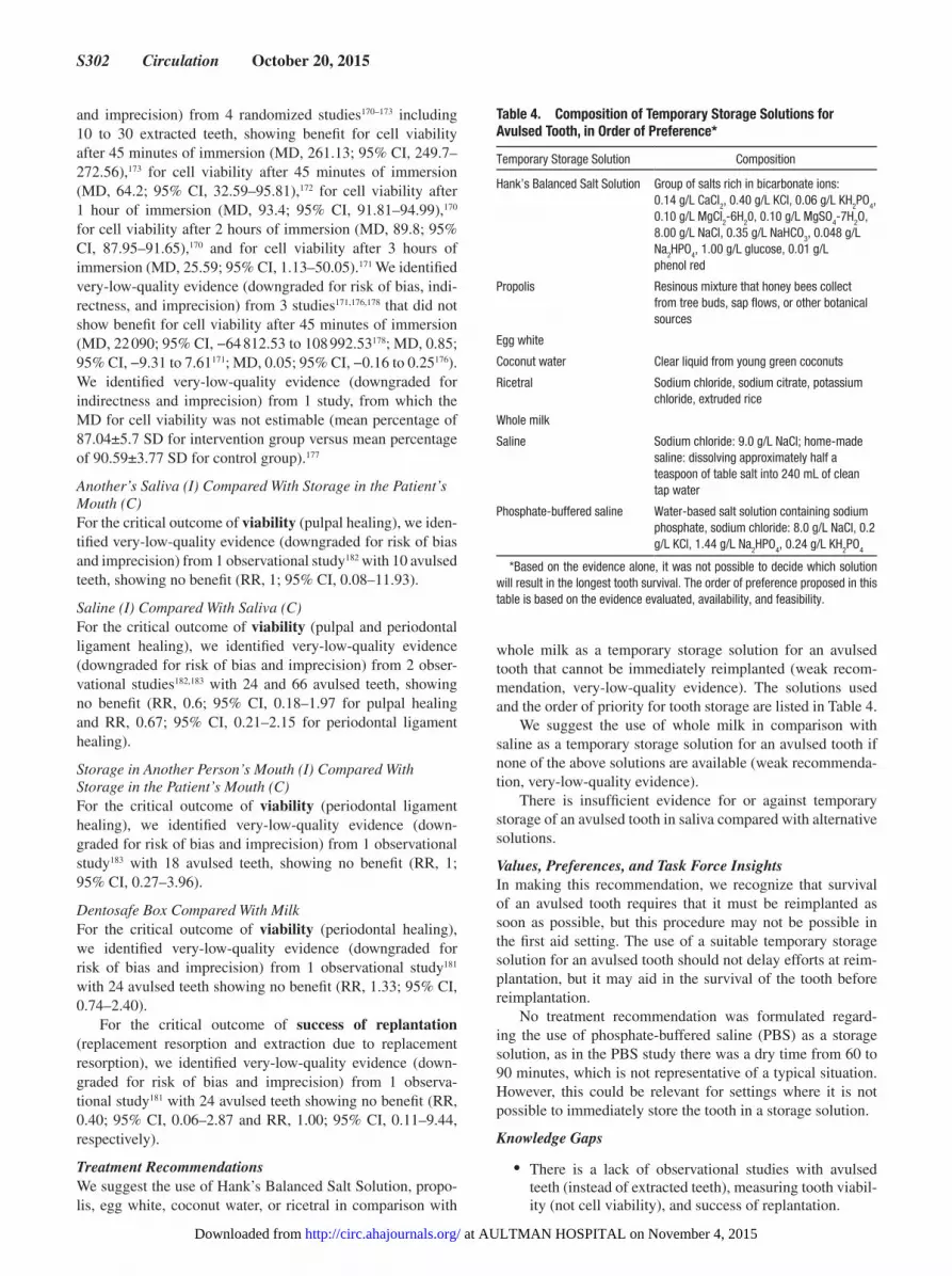
S302 Circulation October 20, 2015
and imprecision) from 4 randomized studies170–173 including 10 to 30 extracted teeth, showing benefit for cell viability after 45 minutes of immersion (MD, 261.13; 95% CI, 249.7–272.56),173 for cell viability after 45 minutes of immersion (MD, 64.2; 95% CI, 32.59–95.81),172 for cell viability after 1 hour of immersion (MD, 93.4; 95% CI, 91.81–94.99),170 for cell viability after 2 hours of immersion (MD, 89.8; 95% CI, 87.95–91.65),170 and for cell viability after 3 hours of immersion (MD, 25.59; 95% CI, 1.13–50.05).171 We identified very-low-quality evidence (downgraded for risk of bias, indi-rectness, and imprecision) from 3 studies171,176,178 that did not show benefit for cell viability after 45 minutes of immersion (MD, 22 090; 95% CI, −64 812.53 to 108 992.53178; MD, 0.85; 95% CI, −9.31 to 7.61171; MD, 0.05; 95% CI, −0.16 to 0.25176). We identified very-low-quality evidence (downgraded for indirectness and imprecision) from 1 study, from which the MD for cell viability was not estimable (mean percentage of 87.04±5.7 SD for intervention group versus mean percentage of 90.59±3.77 SD for control group).177
Another’s Saliva (I) Compared With Storage in the Patient’s Mouth (C)For the critical outcome of viability (pulpal healing), we iden-tified very-low-quality evidence (downgraded for risk of bias and imprecision) from 1 observational study182 with 10 avulsed teeth, showing no benefit (RR, 1; 95% CI, 0.08–11.93).
Saline (I) Compared With Saliva (C)For the critical outcome of viability (pulpal and periodontal ligament healing), we identified very-low-quality evidence (downgraded for risk of bias and imprecision) from 2 obser-vational studies182,183 with 24 and 66 avulsed teeth, showing no benefit (RR, 0.6; 95% CI, 0.18–1.97 for pulpal healing and RR, 0.67; 95% CI, 0.21–2.15 for periodontal ligament healing).
Storage in Another Person’s Mouth (I) Compared With Storage in the Patient’s Mouth (C)For the critical outcome of viability (periodontal ligament healing), we identified very-low-quality evidence (down-graded for risk of bias and imprecision) from 1 observational study183 with 18 avulsed teeth, showing no benefit (RR, 1; 95% CI, 0.27–3.96).
Dentosafe Box Compared With MilkFor the critical outcome of viability (periodontal healing), we identified very-low-quality evidence (downgraded for risk of bias and imprecision) from 1 observational study181 with 24 avulsed teeth showing no benefit (RR, 1.33; 95% CI, 0.74–2.40).
For the critical outcome of success of replantation (replacement resorption and extraction due to replacement resorption), we identified very-low-quality evidence (down-graded for risk of bias and imprecision) from 1 observa-tional study181 with 24 avulsed teeth showing no benefit (RR, 0.40; 95% CI, 0.06–2.87 and RR, 1.00; 95% CI, 0.11–9.44, respectively).
Treatment RecommendationsWe suggest the use of Hank’s Balanced Salt Solution, propo-lis, egg white, coconut water, or ricetral in comparison with
whole milk as a temporary storage solution for an avulsed tooth that cannot be immediately reimplanted (weak recom-mendation, very-low-quality evidence). The solutions used and the order of priority for tooth storage are listed in Table 4.
We suggest the use of whole milk in comparison with saline as a temporary storage solution for an avulsed tooth if none of the above solutions are available (weak recommenda-tion, very-low-quality evidence).
There is insufficient evidence for or against temporary storage of an avulsed tooth in saliva compared with alternative solutions.
Values, Preferences, and Task Force InsightsIn making this recommendation, we recognize that survival of an avulsed tooth requires that it must be reimplanted as soon as possible, but this procedure may not be possible in the first aid setting. The use of a suitable temporary storage solution for an avulsed tooth should not delay efforts at reim-plantation, but it may aid in the survival of the tooth before reimplantation.
No treatment recommendation was formulated regard-ing the use of phosphate-buffered saline (PBS) as a storage solution, as in the PBS study there was a dry time from 60 to 90 minutes, which is not representative of a typical situation. However, this could be relevant for settings where it is not possible to immediately store the tooth in a storage solution.
Knowledge Gaps
• There is a lack of observational studies with avulsed teeth (instead of extracted teeth), measuring tooth viabil-ity (not cell viability), and success of replantation.
Table 4. Composition of Temporary Storage Solutions for Avulsed Tooth, in Order of Preference*
Temporary Storage Solution Composition
Hank’s Balanced Salt Solution Group of salts rich in bicarbonate ions: 0.14 g/L CaCl2, 0.40 g/L KCl, 0.06 g/L KH2PO4, 0.10 g/L MgCl2-6H20, 0.10 g/L MgSO4-7H2O, 8.00 g/L NaCl, 0.35 g/L NaHCO3, 0.048 g/L Na2HPO4, 1.00 g/L glucose, 0.01 g/L phenol red
Propolis Resinous mixture that honey bees collect from tree buds, sap flows, or other botanical sources
Egg white
Coconut water Clear liquid from young green coconuts
Ricetral Sodium chloride, sodium citrate, potassium chloride, extruded rice
Whole milk
Saline Sodium chloride: 9.0 g/L NaCl; home-made saline: dissolving approximately half a teaspoon of table salt into 240 mL of clean tap water
Phosphate-buffered saline Water-based salt solution containing sodium phosphate, sodium chloride: 8.0 g/L NaCl, 0.2 g/L KCl, 1.44 g/L Na
2HPO4, 0.24 g/L KH2PO4
*Based on the evidence alone, it was not possible to decide which solution will result in the longest tooth survival. The order of preference proposed in this table is based on the evidence evaluated, availability, and feasibility.
at AULTMAN HOSPITAL on November 4, 2015http://circ.ahajournals.org/Downloaded from

Singletary et al Part 9: First Aid S303
• In a future PICO question, reimplanting the tooth in the mouth (in dental socket) versus storage in a temporary storage medium could be compared.
• Is training in dental reimplantation for first aid providers feasible and effective?
EducationEducation in first aid continues to be a topic with few sci-entific studies. In the 2010 review of educational topics, no evidence was found to support or recommend any method of evaluating or monitoring a first aid trainee’s educational prog-ress or the specific frequency of retraining to retain skills and knowledge.10 The task force decided to investigate the basic question, “Is there documented evidence of benefit in terms of patient outcomes as a result of first aid training?”
Many questions remain, and research is desperately needed, particularly in the realm of teaching techniques for first aid and methods to evaluate the retention of skills.
First Aid Training (FA 773)Among adults and children receiving first aid (P), does care from a trained first aid provider (I), compared with care from an untrained person (C), change survival rates, recognition of acute injury or illness, prevent further illness or injury (ie, harm), time to resolution of injury, the likelihood of harm (eg, infection), time to resolution of symptoms (O)?
IntroductionIn the ILCOR 2015 review process, first aid is defined as the helping behaviors and initial care provided for an acute ill-ness or injury. Training is, therefore, an essential core element of the practice of first aid. The task force thought that it was important to verify the impact of both formal and informal first aid training of individuals and communities.
Consensus on ScienceFor the critical outcome of increased survival rates from trauma, we identified low-quality evidence (downgraded for risk of bias) from 1 observational study184 enrolling 1341 patients showing a reduced mortality rate among patients ini-tially managed by trained first aid providers of 9.8% (32/325) compared with 15.6% (158/101) for patients without trained first aid support (OR, 0.59; 95% CI, 0.40–0.89).
For the important outcome of time to resolution of symptoms, we identified very-low-quality evidence (down-graded for indirectness and imprecision) from 1 observational study174 with 125 subjects with burns of less than 20% total body surface area showing benefit from first aid training, with 88.5% of patients who received first aid treatment of cooling the burn with water requiring hospitalization of less than 10 days, compared with only 67.2% who received no treatment requiring less than 10 days’ hospitalization (RR, 0.35; 95% CI, 0.16–0.76).
For the important outcome of preventing further injury, we identified very-low-quality evidence (downgraded for indirectness and imprecision) from 1 observational study175 with 244 patients (121 precampaign, 123 postcampaign) with acute burn injury treated either before or after a burn first aid treatment public education campaign, showing benefit with
burn first aid treatment by reduction of the percent of those burned requiring inpatient wound care or surgery from 64.2% (78/121) precampaign compared with 35.6% (44/123) post-campaign (OR, 0.307; 95% CI, 0.18–0.52).
Furthermore, we identified very-low-quality evidence (downgraded for risk of bias and imprecision) from 1 obser-vational study185 with 39 subjects without formal/advanced medical training who performed reduction of shoulder dis-locations in a wilderness environment. This study found no statistically significant difference in the rate of successful reduction by laypersons without first aid training (17/24, 70.8%) compared with the successful reduction rate when individuals with either wilderness first aid or first responder training were present or performed the reduction (11/15, 73%; OR, 0.88; 95% CI, 0.21–3.74).
For the critical outcome of recognition of acute injury or illness, and the important outcome of the likelihood of harm, there were no studies identified.
Treatment RecommendationsWe suggest that education and training in first aid is under-taken to improve morbidity and mortality from injury and ill-ness (weak recommendation, low-quality evidence).
Values, Preferences, and Task Force InsightsPositive outcomes were identified in both public health cam-paigns for specific injuries and course-based training for general trauma. Although no other formal PICO questions related to first aid education were evaluated, the review of stroke assessment systems (above) incidentally discovered that training of lay providers in a stroke assessment system led to improved ability to identify the signs of a stroke when assessed immediately after training (94.4% in those trained versus 76.4% in untrained lay providers), and that 96.9% of the trained lay providers were able to identify signs of stroke when assessed 3 months after training.69 This study supports the recommendation in this review, and specifi-cally shows that public health campaigns aimed at first aid for specific illnesses and injuries, as well as course-based first aid training, can positively impact outcomes of morbid-ity and mortality.
Knowledge GapsIndividual domains of first aid (eg, recognizing an emergency, calling for additional help, specific skills such as direct pres-sure) have not been studied as to what contributes to a victim’s health outcomes. Future reviews comparing first aid educa-tion modalities and context of first aid settings may contribute to developing training guidelines. Additionally, the period of time between a first aid provider’s initial training and refresh-ing those first aid skills to maintain competency needs to be identified. Along with patient outcomes, public health out-comes and cost-analysis of training versus no training may help prioritize resources. These questions and opportunities for research can also be valuable as new modalities emerge for learning (eg, social media or just-in-time).
AcknowledgmentsWe acknowledge the helpfulness of the insightful comments received during the public comment period. We thank the following individu-als (the First Aid Chapter Collaborators) for their collaborations on
at AULTMAN HOSPITAL on November 4, 2015http://circ.ahajournals.org/Downloaded from
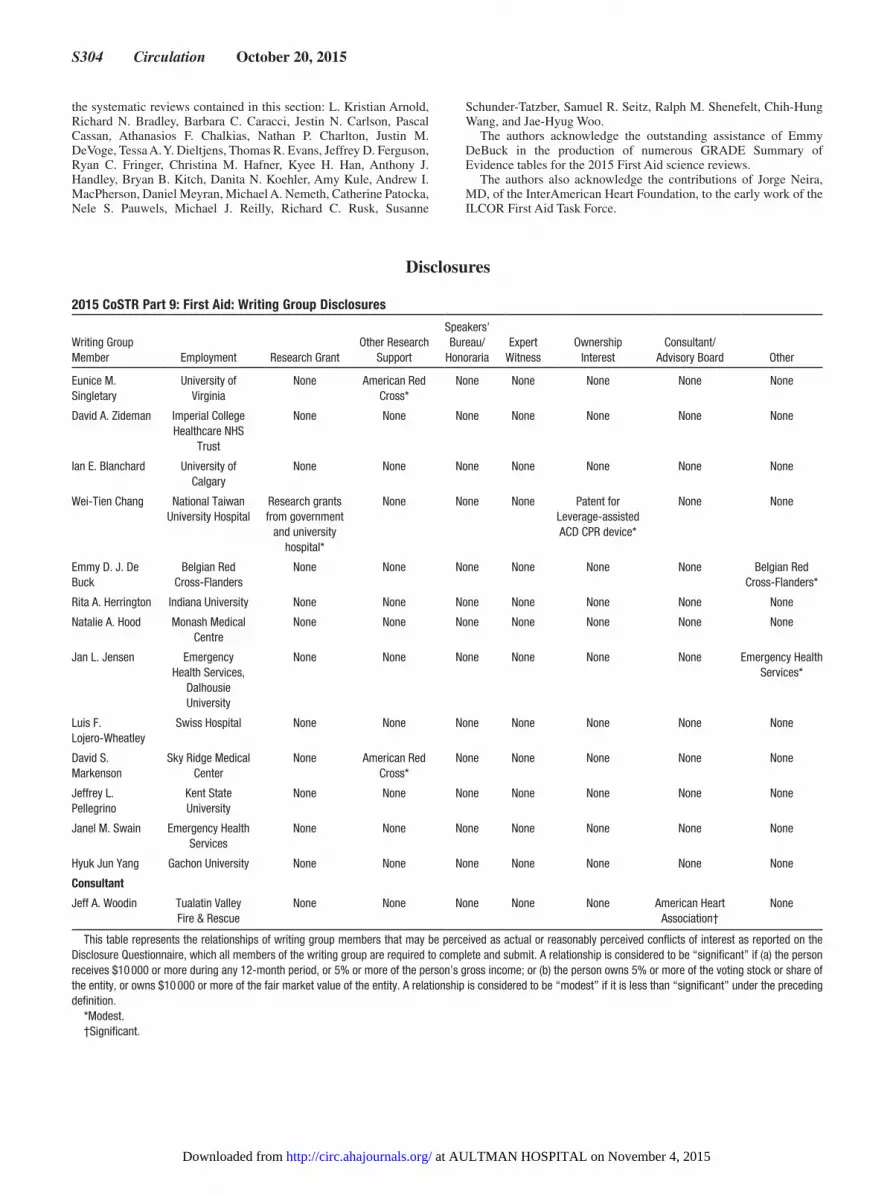
S304 Circulation October 20, 2015
the systematic reviews contained in this section: L. Kristian Arnold, Richard N. Bradley, Barbara C. Caracci, Jestin N. Carlson, Pascal Cassan, Athanasios F. Chalkias, Nathan P. Charlton, Justin M. DeVoge, Tessa A. Y. Dieltjens, Thomas R. Evans, Jeffrey D. Ferguson, Ryan C. Fringer, Christina M. Hafner, Kyee H. Han, Anthony J. Handley, Bryan B. Kitch, Danita N. Koehler, Amy Kule, Andrew I. MacPherson, Daniel Meyran, Michael A. Nemeth, Catherine Patocka, Nele S. Pauwels, Michael J. Reilly, Richard C. Rusk, Susanne
Schunder-Tatzber, Samuel R. Seitz, Ralph M. Shenefelt, Chih-Hung Wang, and Jae-Hyug Woo.
The authors acknowledge the outstanding assistance of Emmy DeBuck in the production of numerous GRADE Summary of Evidence tables for the 2015 First Aid science reviews.
The authors also acknowledge the contributions of Jorge Neira, MD, of the InterAmerican Heart Foundation, to the early work of the ILCOR First Aid Task Force.
2015 CoSTR Part 9: First Aid: Writing Group Disclosures
Writing Group Member Employment Research Grant
Other Research Support
Speakers’ Bureau/
HonorariaExpert
WitnessOwnership
InterestConsultant/
Advisory Board Other
Eunice M. Singletary
University of Virginia
None American Red Cross*
None None None None None
David A. Zideman Imperial College Healthcare NHS
Trust
None None None None None None None
Ian E. Blanchard University of Calgary
None None None None None None None
Wei-Tien Chang National Taiwan University Hospital
Research grants from government
and university hospital*
None None None Patent for Leverage-assisted ACD CPR device*
None None
Emmy D. J. De Buck
Belgian Red Cross-Flanders
None None None None None None Belgian Red Cross-Flanders*
Rita A. Herrington Indiana University None None None None None None None
Natalie A. Hood Monash Medical Centre
None None None None None None None
Jan L. Jensen Emergency Health Services,
Dalhousie University
None None None None None None Emergency Health Services*
Luis F. Lojero-Wheatley
Swiss Hospital None None None None None None None
David S. Markenson
Sky Ridge Medical Center
None American Red Cross*
None None None None None
Jeffrey L. Pellegrino
Kent State University
None None None None None None None
Janel M. Swain Emergency Health Services
None None None None None None None
Hyuk Jun Yang Gachon University None None None None None None None
Consultant
Jeff A. Woodin Tualatin Valley Fire & Rescue
None None None None None American Heart Association†
None
This table represents the relationships of writing group members that may be perceived as actual or reasonably perceived conflicts of interest as reported on the Disclosure Questionnaire, which all members of the writing group are required to complete and submit. A relationship is considered to be “significant” if (a) the person receives $10 000 or more during any 12-month period, or 5% or more of the person’s gross income; or (b) the person owns 5% or more of the voting stock or share of the entity, or owns $10 000 or more of the fair market value of the entity. A relationship is considered to be “modest” if it is less than “significant” under the preceding definition.
*Modest.†Significant.
Disclosures
at AULTMAN HOSPITAL on November 4, 2015http://circ.ahajournals.org/Downloaded from
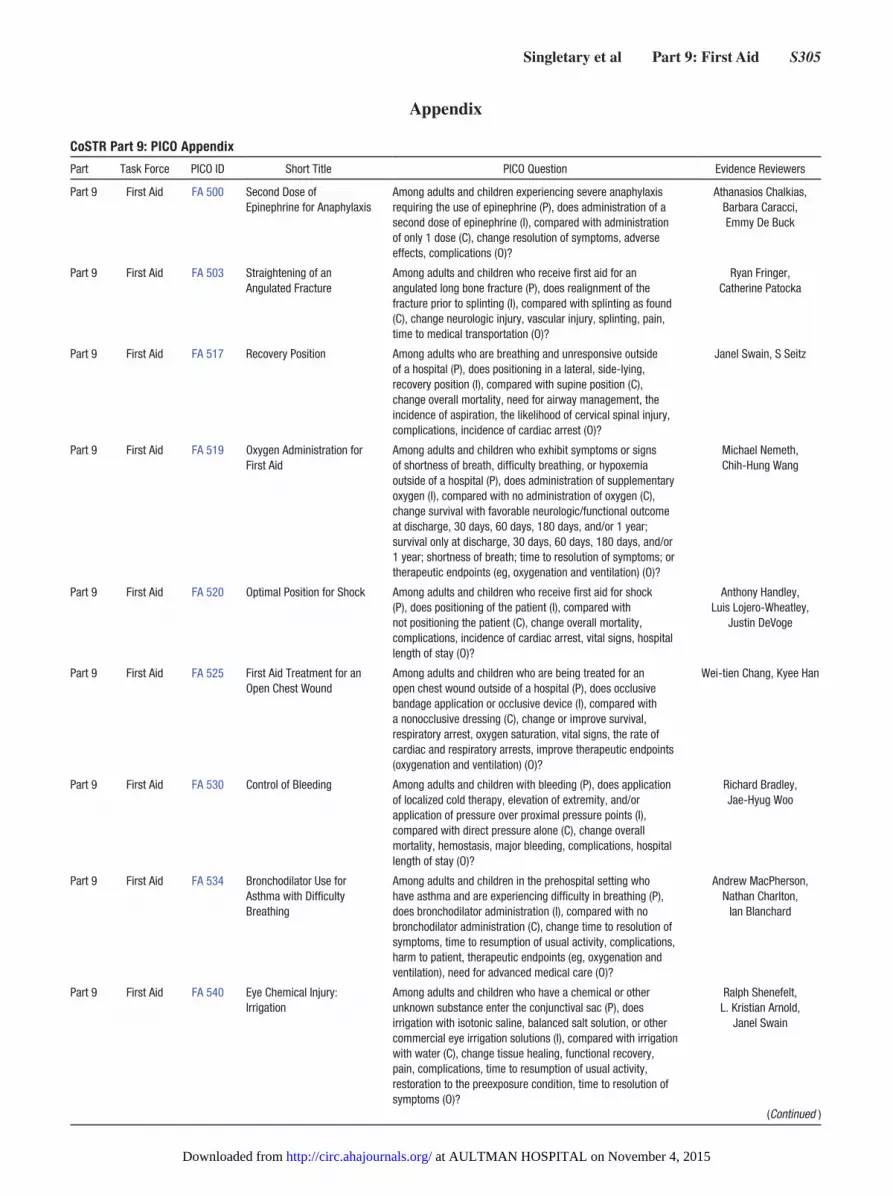
Singletary et al Part 9: First Aid S305
CoSTR Part 9: PICO Appendix
Part Task Force PICO ID Short Title PICO Question Evidence Reviewers
Part 9 First Aid FA 500 Second Dose of Epinephrine for Anaphylaxis
Among adults and children experiencing severe anaphylaxis requiring the use of epinephrine (P), does administration of a second dose of epinephrine (I), compared with administration of only 1 dose (C), change resolution of symptoms, adverse effects, complications (O)?
Athanasios Chalkias, Barbara Caracci, Emmy De Buck
Part 9 First Aid FA 503 Straightening of an Angulated Fracture
Among adults and children who receive first aid for an angulated long bone fracture (P), does realignment of the fracture prior to splinting (I), compared with splinting as found (C), change neurologic injury, vascular injury, splinting, pain, time to medical transportation (O)?
Ryan Fringer, Catherine Patocka
Part 9 First Aid FA 517 Recovery Position Among adults who are breathing and unresponsive outside of a hospital (P), does positioning in a lateral, side-lying, recovery position (I), compared with supine position (C), change overall mortality, need for airway management, the incidence of aspiration, the likelihood of cervical spinal injury, complications, incidence of cardiac arrest (O)?
Janel Swain, S Seitz
Part 9 First Aid FA 519 Oxygen Administration for First Aid
Among adults and children who exhibit symptoms or signs of shortness of breath, difficulty breathing, or hypoxemia outside of a hospital (P), does administration of supplementary oxygen (I), compared with no administration of oxygen (C), change survival with favorable neurologic/functional outcome at discharge, 30 days, 60 days, 180 days, and/or 1 year; survival only at discharge, 30 days, 60 days, 180 days, and/or 1 year; shortness of breath; time to resolution of symptoms; or therapeutic endpoints (eg, oxygenation and ventilation) (O)?
Michael Nemeth, Chih-Hung Wang
Part 9 First Aid FA 520 Optimal Position for Shock Among adults and children who receive first aid for shock (P), does positioning of the patient (I), compared with not positioning the patient (C), change overall mortality, complications, incidence of cardiac arrest, vital signs, hospital length of stay (O)?
Anthony Handley, Luis Lojero-Wheatley,
Justin DeVoge
Part 9 First Aid FA 525 First Aid Treatment for an Open Chest Wound
Among adults and children who are being treated for an open chest wound outside of a hospital (P), does occlusive bandage application or occlusive device (I), compared with a nonocclusive dressing (C), change or improve survival, respiratory arrest, oxygen saturation, vital signs, the rate of cardiac and respiratory arrests, improve therapeutic endpoints (oxygenation and ventilation) (O)?
Wei-tien Chang, Kyee Han
Part 9 First Aid FA 530 Control of Bleeding Among adults and children with bleeding (P), does application of localized cold therapy, elevation of extremity, and/or application of pressure over proximal pressure points (I), compared with direct pressure alone (C), change overall mortality, hemostasis, major bleeding, complications, hospital length of stay (O)?
Richard Bradley, Jae-Hyug Woo
Part 9 First Aid FA 534 Bronchodilator Use for Asthma with Difficulty Breathing
Among adults and children in the prehospital setting who have asthma and are experiencing difficulty in breathing (P), does bronchodilator administration (I), compared with no bronchodilator administration (C), change time to resolution of symptoms, time to resumption of usual activity, complications, harm to patient, therapeutic endpoints (eg, oxygenation and ventilation), need for advanced medical care (O)?
Andrew MacPherson, Nathan Charlton,
Ian Blanchard
Part 9 First Aid FA 540 Eye Chemical Injury: Irrigation
Among adults and children who have a chemical or other unknown substance enter the conjunctival sac (P), does irrigation with isotonic saline, balanced salt solution, or other commercial eye irrigation solutions (I), compared with irrigation with water (C), change tissue healing, functional recovery, pain, complications, time to resumption of usual activity, restoration to the preexposure condition, time to resolution of symptoms (O)?
Ralph Shenefelt, L. Kristian Arnold,
Janel Swain
(Continued )
Appendix
at AULTMAN HOSPITAL on November 4, 2015http://circ.ahajournals.org/Downloaded from
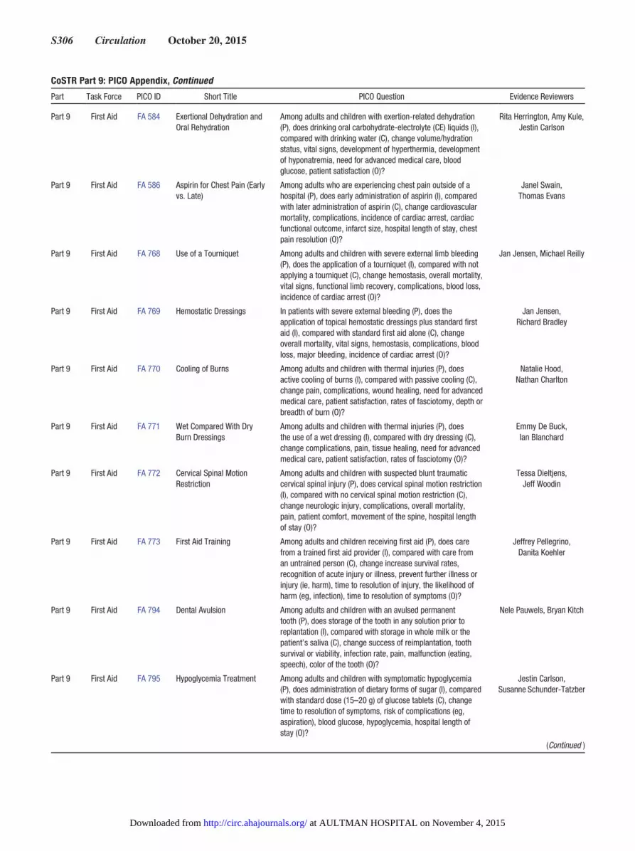
S306 Circulation October 20, 2015
Part 9 First Aid FA 584 Exertional Dehydration and Oral Rehydration
Among adults and children with exertion-related dehydration (P), does drinking oral carbohydrate-electrolyte (CE) liquids (I), compared with drinking water (C), change volume/hydration status, vital signs, development of hyperthermia, development of hyponatremia, need for advanced medical care, blood glucose, patient satisfaction (O)?
Rita Herrington, Amy Kule, Jestin Carlson
Part 9 First Aid FA 586 Aspirin for Chest Pain (Early vs. Late)
Among adults who are experiencing chest pain outside of a hospital (P), does early administration of aspirin (I), compared with later administration of aspirin (C), change cardiovascular mortality, complications, incidence of cardiac arrest, cardiac functional outcome, infarct size, hospital length of stay, chest pain resolution (O)?
Janel Swain, Thomas Evans
Part 9 First Aid FA 768 Use of a Tourniquet Among adults and children with severe external limb bleeding (P), does the application of a tourniquet (I), compared with not applying a tourniquet (C), change hemostasis, overall mortality, vital signs, functional limb recovery, complications, blood loss, incidence of cardiac arrest (O)?
Jan Jensen, Michael Reilly
Part 9 First Aid FA 769 Hemostatic Dressings In patients with severe external bleeding (P), does the application of topical hemostatic dressings plus standard first aid (I), compared with standard first aid alone (C), change overall mortality, vital signs, hemostasis, complications, blood loss, major bleeding, incidence of cardiac arrest (O)?
Jan Jensen, Richard Bradley
Part 9 First Aid FA 770 Cooling of Burns Among adults and children with thermal injuries (P), does active cooling of burns (I), compared with passive cooling (C), change pain, complications, wound healing, need for advanced medical care, patient satisfaction, rates of fasciotomy, depth or breadth of burn (O)?
Natalie Hood, Nathan Charlton
Part 9 First Aid FA 771 Wet Compared With Dry Burn Dressings
Among adults and children with thermal injuries (P), does the use of a wet dressing (I), compared with dry dressing (C), change complications, pain, tissue healing, need for advanced medical care, patient satisfaction, rates of fasciotomy (O)?
Emmy De Buck, Ian Blanchard
Part 9 First Aid FA 772 Cervical Spinal Motion Restriction
Among adults and children with suspected blunt traumatic cervical spinal injury (P), does cervical spinal motion restriction (I), compared with no cervical spinal motion restriction (C), change neurologic injury, complications, overall mortality, pain, patient comfort, movement of the spine, hospital length of stay (O)?
Tessa Dieltjens, Jeff Woodin
Part 9 First Aid FA 773 First Aid Training Among adults and children receiving first aid (P), does care from a trained first aid provider (I), compared with care from an untrained person (C), change increase survival rates, recognition of acute injury or illness, prevent further illness or injury (ie, harm), time to resolution of injury, the likelihood of harm (eg, infection), time to resolution of symptoms (O)?
Jeffrey Pellegrino, Danita Koehler
Part 9 First Aid FA 794 Dental Avulsion Among adults and children with an avulsed permanent tooth (P), does storage of the tooth in any solution prior to replantation (I), compared with storage in whole milk or the patient’s saliva (C), change success of reimplantation, tooth survival or viability, infection rate, pain, malfunction (eating, speech), color of the tooth (O)?
Nele Pauwels, Bryan Kitch
Part 9 First Aid FA 795 Hypoglycemia Treatment Among adults and children with symptomatic hypoglycemia (P), does administration of dietary forms of sugar (I), compared with standard dose (15–20 g) of glucose tablets (C), change time to resolution of symptoms, risk of complications (eg, aspiration), blood glucose, hypoglycemia, hospital length of stay (O)?
Jestin Carlson, Susanne Schunder-Tatzber
CoSTR Part 9: PICO Appendix, Continued
Part Task Force PICO ID Short Title PICO Question Evidence Reviewers
(Continued )
at AULTMAN HOSPITAL on November 4, 2015http://circ.ahajournals.org/Downloaded from
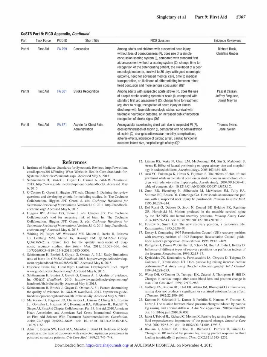
Singletary et al Part 9: First Aid S307
Part 9 First Aid FA 799 Concussion Among adults and children with suspected head injury without loss of consciousness (P), does use of a simple concussion scoring system (I), compared with standard first aid assessment without a scoring system (C), change time to recognition of the deteriorating patient, the likelihood of a poor neurologic outcome, survival to 30 days with good neurologic outcome, need for advanced medical care, time to medical transportation, or likelihood of differentiating between minor head contusion and more serious concussion (O)?
Richard Rusk, Christina Gruber
Part 9 First Aid FA 801 Stroke Recognition Among adults with suspected acute stroke (P), does the use of a rapid stroke scoring system or scale (I), compared with standard first aid assessment (C), change time to treatment (eg, door to drug), recognition of acute injury or illness, discharge with favorable neurologic status, survival with favorable neurologic outcome, or increased public/layperson recognition of stroke signs (O)?
Pascal Cassan, Jeffrey Ferguson,
Daniel Meyran
Part 9 First Aid FA 871 Aspirin for Chest Pain: Administration
Among adults experiencing chest pain due to suspected MI (P), does administration of aspirin (I), compared with no administration of aspirin (C), change cardiovascular mortality, complications, adverse effects, incidence of cardiac arrest, cardiac functional outcome, infarct size, hospital length of stay (O)?
Thomas Evans, Janel Swain
CoSTR Part 9: PICO Appendix, Continued
Part Task Force PICO ID Short Title PICO Question Evidence Reviewers
References 1. Institute of Medicine. Standards for Systematic Reviews. http://www.iom.
edu/Reports/2011/Finding-What-Works-in-Health-Care-Standards-for-Systematic-Reviews/Standards.aspx. Accessed May 6, 2015.
2. Schünemann H, Brożek J, Guyatt G, Oxman A. GRADE Handbook. 2013. http://www.guidelinedevelopment.org/handbook/. Accessed May 6, 2015.
3. O’Connor D, Green S, Higgins JPT, eds. Chapter 5: Defining the review questions and developing criteria for including studies. In: The Cochrane Collaboration. Higgins JPT, Green, S, eds. Cochrane Handbook for Systematic Reviews of Interventions. Version 5.1.0. 2011. http://handbook.cochrane.org/. Accessed May 6, 2015.
4. Higgins JPT, Altman DG, Sterne J, eds. Chapter 8.5: The Cochrane Collaboration’s tool for assessing risk of bias. In: The Cochrane Collaboration. Higgins JPT, Green, S, eds. Cochrane Handbook for Systematic Reviews of Interventions. Version 5.1.0. 2011. http://handbook.cochrane.org/. Accessed May 6, 2015.
5. Whiting PF, Rutjes AW, Westwood ME, Mallett S, Deeks JJ, Reitsma JB, Leeflang MM, Sterne JA, Bossuyt PM; QUADAS-2 Group. QUADAS-2: a revised tool for the quality assessment of diag-nostic accuracy studies. Ann Intern Med. 2011;155:529–536. doi: 10.7326/0003-4819-155-8-201110180-00009.
6. Schünemann H, Brożek J, Guyatt G, Oxman A. 5.2.1 Study limitations (risk of bias). In: GRADE Handbook 2013. http://www.guidelinedevelop-ment.org/handbook/#h.m9385o5z3li7. Accessed May 6, 2015.
7. Evidence Prime Inc. GRADEpro Guideline Development Tool. http://www.guidelinedevelopment.org/. Accessed May 6, 2015.
8. Schünemann H, Brożek J, Guyatt G, Oxman A. 5. Quality of evidence. In: GRADE Handbook. 2013. http://www.guidelinedevelopment.org/handbook/#h.9rdbelsnu4iy. Accessed May 6, 2015.
9. Schünemann H, Brożek J, Guyatt G, Oxman A. 5.1 Factors determining the quality of evidence. In: GRADE Handbook. 2013. http://www.guide-linedevelopment.org/handbook/#h.9rdbelsnu4iy. Accessed May 6, 2015.
10. Markenson D, Ferguson JD, Chameides L, Cassan P, Chung KL, Epstein JL, Gonzales L, Hazinski MF, Herrington RA, Pellegrino JL, Ratcliff N, Singer AJ; First Aid Chapter Collaborators. Part 13: first aid: 2010 American Heart Association and American Red Cross International Consensus on First Aid Science With Treatment Recommendations. Circulation. 2010;122(Suppl 2):S582–S605. doi: 10.1161/CIRCULATIONAHA. 110.971168.
11. Adnet F, Borron SW, Finot MA, Minadeo J, Baud FJ. Relation of body position at the time of discovery with suspected aspiration pneumonia in poisoned comatose patients. Crit Care Med. 1999;27:745–748.
12. Litman RS, Wake N, Chan LM, McDonough JM, Sin S, Mahboubi S, Arens R. Effect of lateral positioning on upper airway size and morphol-ogy in sedated children. Anesthesiology. 2005;103:484–488.
13. Arai YC, Fukunaga K, Hirota S, Fujimoto S. The effects of chin lift and jaw thrust while in the lateral position on stridor score in anesthetized chil-dren with adenotonsillar hypertrophy. Anesth Analg. 2004;99:1638–41, table of contents. doi: 10.1213/01.ANE.0000135637.95853.1C.
14. Gunn BD, Eizenberg N, Silberstein M, McMeeken JM, Tully EA, Stillman BC, Brown DJ, Gutteridge GA. How should an unconscious per-son with a suspected neck injury be positioned? Prehosp Disaster Med. 1995;10:239–244.
15. Del Rossi G, Dubose D, Scott N, Conrad BP, Hyldmo PK, Rechtine GR, Horodsyki M. Motion produced in the unstable cervical spine by the HAINES and lateral recovery positions. Prehosp Emerg Care. 2014;18:539–543. doi: 10.3109/10903127.2014.916019.
16. Fulstow R, Smith GB. The new recovery position, a cautionary tale. Resuscitation. 1993;26:89–91.
17. Doxey J. Comparing 1997 Resuscitation Council (UK) recovery position with recovery position of 1992 European Resuscitation Council guide-lines: a user’s perspective. Resuscitation. 1998;39:161–169.
18. Rathgeber J, Panzer W, Günther U, Scholz M, Hoeft A, Bahr J, Kettler D. Influence of different types of recovery positions on perfusion indices of the forearm. Resuscitation. 1996;32:13–17.
19. Kyriakides ZS, Koukoulas A, Paraskevaidis IA, Chrysos D, Tsiapras D, Galiotos C, Kremastinos DT. Does passive leg raising increase cardiac performance? A study using Doppler echocardiography. Int J Cardiol. 1994;44:288–293.
20. Wong DH, O’Connor D, Tremper KK, Zaccari J, Thompson P, Hill D. Changes in cardiac output after acute blood loss and position change in man. Crit Care Med. 1989;17:979–983.
21. Gaffney FA, Bastian BC, Thal ER, Atkins JM, Blomqvist CG. Passive leg raising does not produce a significant or sustained autotransfusion effect. J Trauma. 1982;22:190–193.
22. Kamran H, Salciccioli L, Kumar P, Pushilin S, Namana V, Trotman S, Lazar J. The relation between blood pressure changes induced by passive leg raising and arterial stiffness. J Am Soc Hypertens. 2010;4:284–289. doi: 10.1016/j.jash.2010.09.002.
23. Jabot J, Teboul JL, Richard C, Monnet X. Passive leg raising for predicting fluid responsiveness: importance of the postural change. Intensive Care Med. 2009;35:85–90. doi: 10.1007/s00134-008-1293-3.
24. Boulain T, Achard JM, Teboul JL, Richard C, Perrotin D, Ginies G. Changes in BP induced by passive leg raising predict response to fluid loading in critically ill patients. Chest. 2002;121:1245–1252.
at AULTMAN HOSPITAL on November 4, 2015http://circ.ahajournals.org/Downloaded from

S308 Circulation October 20, 2015
25. Wijesinghe M, Perrin K, Healy B, Hart K, Clay J, Weatherall M, Beasley R. Pre-hospital oxygen therapy in acute exacerbations of chronic obstructive pulmonary disease. Intern Med J. 2011;41:618–622. doi: 10.1111/j.1445-5994.2010.02207.x.
26. Bruera E, de Stoutz N, Velasco-Leiva A, Schoeller T, Hanson J. Effects of oxygen on dyspnoea in hypoxaemic terminal-cancer patients. Lancet. 1993;342:13–14.
27. Uronis HE, Currow DC, McCrory DC, Samsa GP, Abernethy AP. Oxygen for relief of dyspnoea in mildly- or non-hypoxaemic patients with cancer: a systematic review and meta-analysis. Br J Cancer. 2008;98:294–299. doi: 10.1038/sj.bjc.6604161.
28. Philip J, Gold M, Milner A, Di Iulio J, Miller B, Spruyt O. A randomized, double-blind, crossover trial of the effect of oxygen on dyspnea in patients with advanced cancer. J Pain Symptom Manage. 2006;32:541–550. doi: 10.1016/j.jpainsymman.2006.06.009.
29. Booth S, Kelly MJ, Cox NP, Adams L, Guz A. Does oxygen help dyspnea in patients with cancer? Am J Respir Crit Care Med. 1996;153:1515–1518. doi: 10.1164/ajrccm.153.5.8630595.
30. Ahmedzai SH, Laude E, Robertson A, Troy G, Vora V. A double-blind, randomised, controlled Phase II trial of Heliox28 gas mixture in lung can-cer patients with dyspnoea on exertion. Br J Cancer. 2004;90:366–371. doi: 10.1038/sj.bjc.6601527.
31. Longphre JM, Denoble PJ, Moon RE, Vann RD, Freiberger JJ. First aid normobaric oxygen for the treatment of recreational diving injuries. Undersea Hyperb Med. 2007;34:43–49.
32. 2005 International Consensus on Cardiopulmonary Resuscitation and Emergency Cardiovascular Care Science with Treatment Recommendations, Section 2 part 10: first aid. Circulation. 2005;112:III115–III125.
33. Bentur L, Canny GJ, Shields MD, Kerem E, Schuh S, Reisman JJ, Fakhoury K, Pedder L, Levison H. Controlled trial of nebulized albuterol in children younger than 2 years of age with acute asthma. Pediatrics. 1992;89:133–137.
34. van der Woude HJ, Postma DS, Politiek MJ, Winter TH, Aalbers R. Relief of dyspnoea by beta2-agonists after methacholine-induced bronchocon-striction. Respir Med. 2004;98:816–820.
35. Littner MR, Tashkin DP, Siegel SC, Katz R. Double-blind comparison of acute effects of inhaled albuterol, isoproterenol and placebo on cardio-pulmonary function and gas exchange in asthmatic children. Ann Allergy. 1983;50:309–316.
36. Karpel JP, Aldrich TK, Prezant DJ, Guguchev K, Gaitan-Salas A, Pathiparti R. Emergency treatment of acute asthma with albuterol metered-dose inhaler plus holding chamber: how often should treatments be adminis-tered? Chest. 1997;112:348–356.
37. Berger WE, Milgrom H, Skoner DP, Tripp K, Parsey MV, Baumgartner RA; Xopenex Pediatric Asthma Group. Evaluation of levalbuterol metered dose inhaler in pediatric patients with asthma: a double-blind, randomized, placebo- and active-controlled trial. Curr Med Res Opin. 2006;22:1217–1226. doi: 10.1185/030079906X112534.
38. Politiek MJ, Boorsma M, Aalbers R. Comparison of formoterol, salbuta-mol and salmeterol in methacholine-induced severe bronchoconstriction. Eur Respir J. 1999;13:988–992.
39. Hermansen MN, Nielsen KG, Buchvald F, Jespersen JJ, Bengtsson T, Bisgaard H. Acute relief of exercise-induced bronchoconstric-tion by inhaled formoterol in children with persistent asthma. Chest. 2006;129:1203–1209. doi: 10.1378/chest.129.5.1203.
40. Amirav I, Yacobov R, Luder AS. Formoterol turbuhaler is as effective as salbutamol diskus in relieving adenosine-induced bronchoconstriction in children. J Aerosol Med. 2007;20:1–6. doi: 10.1089/jam.2006.0561.
41. Emerman CL, Shade B, Kubincanek J. A controlled trial of nebulized iso-etharine in the prehospital treatment of acute asthma. Am J Emerg Med. 1990;8:512–514.
42. Weiss SJ, Anand P, Ernst AA, Orgeron D, May WL. Effect of out-of-hos-pital albuterol inhalation treatments on patient comfort and morbidity. Ann Emerg Med. 1994;24:873–878.
43. Osmond MH, Klassen TP. Efficacy of ipratropium bromide in acute child-hood asthma: a meta-analysis. Acad Emerg Med. 1995;2:651–656.
44. Harbison J, Hossain O, Jenkinson D, Davis J, Louw SJ, Ford GA. Diagnostic accuracy of stroke referrals from primary care, emergency room physicians, and ambulance staff using the face arm speech test. Stroke. 2003;34:71–76.
45. Iguchi Y, Kimura K, Watanabe M, Shibazaki K, Aoki J. Utility of the Kurashiki Prehospital Stroke Scale for hyperacute stroke. Cerebrovasc Dis. 2011;31:51–56. doi: 10.1159/000320854.
46. Chenkin J, Gladstone DJ, Verbeek PR, Lindsay P, Fang J, Black SE, Morrison L. Predictive value of the Ontario prehospital stroke screening
tool for the identification of patients with acute stroke. Prehosp Emerg Care. 2009;13:153–159. doi: 10.1080/10903120802706146.
47. Wojner-Alexandrov AW, Alexandrov AV, Rodriguez D, Persse D, Grotta JC. Houston paramedic and emergency stroke treatment and out-comes study (HoPSTO). Stroke. 2005;36:1512–1518. doi: 10.1161/01.STR.0000170700.45340.39.
48. Frendl DM, Strauss DG, Underhill BK, Goldstein LB. Lack of impact of paramedic training and use of the cincinnati prehospital stroke scale on stroke patient identification and on-scene time. Stroke. 2009;40:754–756. doi: 10.1161/STROKEAHA.108.531285.
49. O’Brien W, Crimmins D, Donaldson W, Risti R, Clarke TA, Whyte S, Sturm J. FASTER (Face, Arm, Speech, Time, Emergency Response): experience of Central Coast Stroke Services implementation of a pre-hos-pital notification system for expedient management of acute stroke. J Clin Neurosci. 2012;19:241–245. doi: 10.1016/j.jocn.2011.06.009.
50. You JS, Chung SP, Chung HS, Lee HS, Park JW, Kim HJ, Lee SH, Park I, Lee HS. Predictive value of the Cincinnati Prehospital Stroke Scale for identifying thrombolytic candidates in acute ischemic stroke. Am J Emerg Med. 2013;31:1699–1702. doi: 10.1016/j.ajem.2013.08.029.
51. Asimos AW, Ward S, Brice JH, Rosamond WD, Goldstein LB, Studnek J. Out-of-hospital stroke screen accuracy in a state with an emergency medical services protocol for routing patients to acute stroke centers. Ann Emerg Med. 2014;64:509–515. doi: 10.1016/j.annemergmed.2014.03.024.
52. Bergs J, Sabbe M, Moons P. Prehospital stroke scales in a Belgian prehos-pital setting: a pilot study. Eur J Emerg Med. 2010;17:2–6. doi: 10.1097/MEJ.0b013e32831040ec.
53. Bray JE, Coughlan K, Barger B, Bladin C. Paramedic diagnosis of stroke: examining long-term use of the Melbourne Ambulance Stroke Screen (MASS) in the field. Stroke. 2010;41:1363–1366. doi: 10.1161/STROKEAHA.109.571836.
54. Bray JE, Martin J, Cooper G, Barger B, Bernard S, Bladin C. Paramedic identification of stroke: community validation of the melbourne ambulance stroke screen. Cerebrovasc Dis. 2005;20:28–33. doi: 10.1159/000086201.
55. Buck BH, Starkman S, Eckstein M, Kidwell CS, Haines J, Huang R, Colby D, Saver JL. Dispatcher recognition of stroke using the National Academy Medical Priority Dispatch System. Stroke. 2009;40:2027–2030. doi: 10.1161/STROKEAHA.108.545574.
56. Chen S, Sun H, Lei Y, Gao D, Wang Y, Wang Y, Zhou Y, Wang A, Wang W, Zhao X. Validation of the Los Angeles pre-hospital stroke screen (LAPSS) in a Chinese urban emergency medical service population. PLoS One. 2013;8:e70742. doi: 10.1371/journal.pone.0070742.
57. De Luca A, Giorgi Rossi P, Villa GF; Stroke group Italian Society pre hos-pital emergency Services. The use of Cincinnati Prehospital Stroke Scale during telephone dispatch interview increases the accuracy in identifying stroke and transient ischemic attack symptoms. BMC Health Serv Res. 2013;13:513. doi: 10.1186/1472-6963-13-513.
58. Fothergill RT, Williams J, Edwards MJ, Russell IT, Gompertz P. Does use of the recognition of stroke in the emergency room stroke assess-ment tool enhance stroke recognition by ambulance clinicians? Stroke. 2013;44:3007–3012. doi: 10.1161/STROKEAHA.13.000851.
59. Jiang HL, Chan CP, Leung YK, Li YM, Graham CA, Rainer TH. Evaluation of the Recognition of Stroke in the Emergency Room (ROSIER) scale in Chinese patients in Hong Kong. PLoS One. 2014;9:e109762. doi: 10.1371/journal.pone.0109762.
60. Kidwell CS, Starkman S, Eckstein M, Weems K, Saver JL. Identifying stroke in the field. Prospective validation of the Los Angeles prehospital stroke screen (LAPSS). Stroke. 2000;31:71–76.
61. Kleindorfer DO, Miller R, Moomaw CJ, Alwell K, Broderick JP, Khoury J, Woo D, Flaherty ML, Zakaria T, Kissela BM. Designing a message for public education regarding stroke: does FAST capture enough stroke? Stroke. 2007;38:2864–2868. doi: 10.1161/STROKEAHA.107.484329.
62. Kothari RU, Pancioli A, Liu T, Brott T, Broderick J. Cincinnati Prehospital Stroke Scale: reproducibility and validity. Ann Emerg Med. 1999;33:373–378.
63. Nazliel B, Starkman S, Liebeskind DS, Ovbiagele B, Kim D, Sanossian N, Ali L, Buck B, Villablanca P, Vinuela F, Duckwiler G, Jahan R, Saver JL. A brief prehospital stroke severity scale identifies ischemic stroke patients harboring persisting large arterial occlusions. Stroke. 2008;39:2264–2267. doi: 10.1161/STROKEAHA.107.508127.
64. Nor AM, Davis J, Sen B, Shipsey D, Louw SJ, Dyker AG, Davis M, Ford GA. The Recognition of Stroke in the Emergency Room (ROSIER) scale: development and validation of a stroke recognition instrument. Lancet Neurol. 2005;4:727–734. doi: 10.1016/S1474-4422(05)70201-5.
65. Ramanujam P, Guluma KZ, Castillo EM, Chacon M, Jensen MB, Patel E, Linnick W, Dunford JV. Accuracy of stroke recognition by emergency
at AULTMAN HOSPITAL on November 4, 2015http://circ.ahajournals.org/Downloaded from

Singletary et al Part 9: First Aid S309
medical dispatchers and paramedics–San Diego experience. Prehosp Emerg Care. 2008;12:307–313. doi: 10.1080/10903120802099526.
66. Studnek JR, Asimos A, Dodds J, Swanson D. Assessing the validity of the Cincinnati prehospital stroke scale and the medic prehospital assessment for code stroke in an urban emergency medical services agency. Prehosp Emerg Care. 2013;17:348–353. doi: 10.3109/10903127.2013.773113.
67. Whiteley WN, Wardlaw JM, Dennis MS, Sandercock PA. Clinical scores for the identification of stroke and transient ischaemic attack in the emer-gency department: a cross-sectional study. J Neurol Neurosurg Psychiatry. 2011;82:1006–1010. doi: 10.1136/jnnp.2010.235010.
68. Yock-Corrales A, Babl FE, Mosley IT, Mackay MT. Can the FAST and ROSIER adult stroke recognition tools be applied to confirmed childhood arterial ischemic stroke? BMC Pediatr. 2011;11:93. doi: 10.1186/1471-2431-11-93.
69. Wall HK, Beagan BM, O’Neill J, Foell KM, Boddie-Willis CL. Addressing stroke signs and symptoms through public education: the Stroke Heroes Act FAST campaign. Prev Chronic Dis. 2008;5:A49.
70. Randomised trial of intravenous streptokinase, oral aspirin, both, or nei-ther among 17,187 cases of suspected acute myocardial infarction: ISIS-2. ISIS-2 (Second International Study of Infarct Survival) Collaborative Group. Lancet. 1988;2:349-360.
71. Verheugt FW, van der Laarse A, Funke-Küpper AJ, Sterkman LG, Galema TW, Roos JP. Effects of early intervention with low-dose aspirin (100 mg) on infarct size, reinfarction and mortality in anterior wall acute myocardial infarction. Am J Cardiol. 1990;66:267–270.
72. Elwood PC, Williams WO. A randomized controlled trial of aspirin in the prevention of early mortality in myocardial infarction. J R Coll Gen Pract. 1979;29:413–416.
73. Frilling B, Schiele R, Gitt AK, Zahn R, Schneider S, Glunz HG, Gieseler U, Baumgärtel B, Asbeck F, Senges J; Maximum Individual Therapy in Acute Myocardial Infarction (MITRA); Myocardial Infarction Registry (MIR) Study Groups. Characterization and clinical course of patients not receiving aspirin for acute myocardial infarction: results from the MITRA and MIR studies. Am Heart J. 2001;141:200–205. doi: 10.1067/mhj.2001.112681.
74. Quan D, LoVecchio F, Clark B, Gallagher JV 3rd. Prehospital use of aspirin rarely is associated with adverse events. Prehosp Disaster Med. 2004;19:362–365.
75. Freimark D, Matetzky S, Leor J, Boyko V, Barbash IM, Behar S, Hod H. Timing of aspirin administration as a determinant of survival of patients with acute myocardial infarction treated with thrombolysis. Am J Cardiol. 2002;89:381–385.
76. Barbash I, Freimark D, Gottlieb S, Hod H, Hasin Y, Battler A, Crystal E, Matetzky S, Boyko V, Mandelzweig L, Behar S, Leor J; Israeli working group on intensive cardiac care, Israel heart society. Outcome of myocar-dial infarction in patients treated with aspirin is enhanced by pre-hospital administration. Cardiology. 2002;98:141–147. doi: 66324.
77. Inoue N, Yamamoto A. Clinical evaluation of pediatric anaphylaxis and the necessity for multiple doses of epinephrine. Asia Pac Allergy. 2013;3:106–114. doi: 10.5415/apallergy.2013.3.2.106.
78. Järvinen KM, Sicherer SH, Sampson HA, Nowak-Wegrzyn A. Use of multiple doses of epinephrine in food-induced anaphylaxis in chil-dren. J Allergy Clin Immunol. 2008;122:133–138. doi: 10.1016/j.jaci.2008.04.031.
79. Noimark L, Wales J, Du Toit G, Pastacaldi C, Haddad D, Gardner J, Hyer W, Vance G, Townshend C, Alfaham M, Arkwright PD, Rao R, Kapoor S, Summerfield A, Warner JO, Roberts G. The use of adrenaline autoinjec-tors by children and teenagers. Clin Exp Allergy. 2012;42:284–292. doi: 10.1111/j.1365-2222.2011.03912.x.
80. Korenblat P, Lundie MJ, Dankner RE, Day JH. A retrospective study of epinephrine administration for anaphylaxis: how many doses are needed? Allergy Asthma Proc. 1999;20:383–386.
81. Oren E, Banerji A, Clark S, Camargo CA Jr. Food-induced anaphylaxis and repeated epinephrine treatments. Ann Allergy Asthma Immunol. 2007;99:429–432. doi: 10.1016/S1081-1206(10)60568-6.
82. Banerji A, Rudders SA, Corel B, Garth AM, Clark S, Camargo CA Jr. Repeat epinephrine treatments for food-related allergic reactions that pres-ent to the emergency department. Allergy Asthma Proc. 2010;31:308–316. doi: 10.2500/aap.2010.31.3375.
83. Rudders SA, Banerji A, Corel B, Clark S, Camargo CA Jr. Multicenter study of repeat epinephrine treatments for food-related anaphylaxis. Pediatrics. 2010;125:e711–e718. doi: 10.1542/peds.2009-2832.
84. Tsuang A, Menon N, Setia N, Geyman L, Nowak-Wegrzyn AH. Multiple epinephrine doses in food-induced anaphylaxis in children. J. Allergy Clin. Immunol. 2013;131:AB90.
85. Rudders SA, Banerji A, Katzman DP, Clark S, Camargo CA Jr. Multiple epinephrine doses for stinging insect hypersensitivity reactions treated in the emergency department. Ann Allergy Asthma Immunol. 2010;105:85–93. doi: 10.1016/j.anai.2010.05.004.
86. Ellis BC, Brown SG. Efficacy of intramuscular epinephrine for the treat-ment of severe anaphylaxis: a comparison of two ambulance services with different protocols. Ann. Emerg. Med. 2013;62:S146.
87. Brodows RG, Williams C, Amatruda JM. Treatment of insulin reactions in diabetics. JAMA. 1984;252:3378–3381.
88. Husband AC, Crawford S, McCoy LA, Pacaud D. The effectiveness of glucose, sucrose, and fructose in treating hypoglycemia in chil-dren with type 1 diabetes. Pediatr Diabetes. 2010;11:154–158. doi: 10.1111/j.1399-5448.2009.00558.x.
89. McTavish L, Wiltshire E. Effective treatment of hypoglyce-mia in children with type 1 diabetes: a randomized controlled clinical trial. Pediatr Diabetes. 2011;12(4 Pt 2):381–387. doi: 10.1111/j.1399-5448.2010.00725.x.
90. Slama G, Traynard PY, Desplanque N, Pudar H, Dhunputh I, Letanoux M, Bornet FR, Tchobroutsky G. The search for an optimized treatment of hypoglycemia. Carbohydrates in tablets, solutin, or gel for the correc-tion of insulin reactions. Arch Intern Med. 1990;150:589–593.
91. Osterberg KL, Pallardy SE, Johnson RJ, Horswill CA. Carbohydrate exerts a mild influence on fluid retention following exercise-induced dehydration. J Appl Physiol (1985). 2010;108:245–250. doi: 10.1152/japplphysiol.91275.2008.
92. Saat M, Singh R, Sirisinghe RG, Nawawi M. Rehydration after exercise with fresh young coconut water, carbohydrate-electrolyte beverage and plain water. J Physiol Anthropol Appl Human Sci. 2002;21:93–104.
93. Ismail I, Singh R, Sirisinghe RG. Rehydration with sodium-enriched coconut water after exercise-induced dehydration. Southeast Asian J Trop Med Public Health. 2007;38:769–785.
94. González-Alonso J, Heaps CL, Coyle EF. Rehydration after exercise with common beverages and water. Int J Sports Med. 1992;13:399–406.
95. Wong SH, Williams C, Adams N. Effects of ingesting a large volume of carbohydrate-electrolyte solution on rehydration during recov-ery and subsequent exercise capacity. Int J Sport Nutr Exerc Metab. 2000;10:375–393.
96. Kalman DS, Feldman S, Krieger DR, Bloomer RJ. Comparison of coco-nut water and a carbohydrate-electrolyte sport drink on measures of hydration and physical performance in exercise-trained men. J Int Soc Sports Nutr. 2012;9:1. doi: 10.1186/1550-2783-9-1.
97. Chang CQ, Chen YB, Chen ZM, Zhang LT. Effects of a carbohydrate-electrolyte beverage on blood viscosity after dehydration in healthy adults. Chin Med J (Engl). 2010;123:3220–3225.
98. Seifert J, Harmon J, DeClercq P. Protein added to a sports drink improves fluid retention. Int J Sport Nutr Exerc Metab. 2006;16:420–429.
99. Wong SH, Chen Y. Effect of a carbohydrate-electrolyte beverage, lemon tea, or water on rehydration during short-term recovery from exercise. Int J Sport Nutr Exerc Metab. 2011;21:300–310.
100. Shirreffs SM, Watson P, Maughan RJ. Milk as an effective post-exer-cise rehydration drink. Br J Nutr. 2007;98:173–180. doi: 10.1017/S0007114507695543.
101. Hostler D, Bednez JC, Kerin S, Reis SE, Kong PW, Morley J, Gallagher M, Suyama J. Comparison of rehydration regimens for rehabilitation of firefighters performing heavy exercise in thermal protective clothing: a report from the fireground rehab evaluation (FIRE) trial. Prehosp Emerg Care. 2010;14:194–201. doi: 10.3109/10903120903524963.
102. Miccheli A, Marini F, Capuani G, Miccheli AT, Delfini M, Di Cocco ME, Puccetti C, Paci M, Rizzo M, Spataro A. The influence of a sports drink on the postexercise metabolism of elite athletes as investigated by NMR-based metabolomics. J Am Coll Nutr. 2009;28:553–564.
103. Kompa S, Redbrake C, Hilgers C, Wüstemeyer H, Schrage N, Remky A. Effect of different irrigating solutions on aqueous humour pH changes, intraocular pressure and histological findings after induced alkali burns. Acta Ophthalmol Scand. 2005;83:467–470. doi: 10.1111/j.1600-0420.2005.00458.x.
104. King NA, Philpott SJ, Leary A. A randomized controlled trial assessing the use of compression versus vasoconstriction in the treatment of femo-ral hematoma occurring after percutaneous coronary intervention. Heart Lung. 2008;37:205–210. doi: 10.1016/j.hrtlng.2007.05.008.
105. Levy AS, Marmar E. The role of cold compression dressings in the post-operative treatment of total knee arthroplasty. Clin Orthop Relat Res. 1993:174–178.
at AULTMAN HOSPITAL on November 4, 2015http://circ.ahajournals.org/Downloaded from

S310 Circulation October 20, 2015
106. Cox ED, Schreiber MA, McManus J, Wade CE, Holcomb JB. New hemostatic agents in the combat setting. Transfusion. 2009;49 Suppl 5:248S–255S. doi: 10.1111/j.1537-2995.2008.01988.x.
107. Acheson EM, Kheirabadi BS, Deguzman R, Dick EJ Jr, Holcomb JB. Comparison of hemorrhage control agents applied to lethal extremity arte-rial hemorrhages in swine. J Trauma. 2005;59:865–74; discussion 874.
108. Alam HB, Uy GB, Miller D, Koustova E, Hancock T, Inocencio R, Anderson D, Llorente O, Rhee P. Comparative analysis of hemostatic agents in a swine model of lethal groin injury. J Trauma. 2003;54:1077–1082. doi: 10.1097/01.TA.0000068258.99048.70.
109. Arnaud F, Parreño-Sadalan D, Tomori T, Delima MG, Teranishi K, Carr W, McNamee G, McKeague A, Govindaraj K, Beadling C, Lutz C, Sharp T, Mog S, Burris D, McCarron R. Comparison of 10 hemostatic dress-ings in a groin transection model in swine. J Trauma. 2009;67:848–855. doi: 10.1097/TA.0b013e3181b2897f.
110. Arnaud F, Teranishi K, Tomori T, Carr W, McCarron R. Comparison of 10 hemostatic dressings in a groin puncture model in swine. J Vasc Surg. 2009;50:632–9, 639.e1. doi: 10.1016/j.jvs.2009.06.010.
111. Devlin JJ, Kircher S, Kozen BG, Littlejohn LF, Johnson AS. Comparison of ChitoFlex®, CELOX™, and QuikClot® in control of hemorrhage. J Emerg Med. 2011;41:237–245. doi: 10.1016/j.jemermed.2009.02.017.
112. Kheirabadi BS, Scherer MR, Estep JS, Dubick MA, Holcomb JB. Determination of efficacy of new hemostatic dressings in a model of extremity arterial hemorrhage in swine. J Trauma. 2009;67:450–9; dis-cussion 459. doi: 10.1097/TA.0b013e3181ac0c99.
113. Kozen BG, Kircher SJ, Henao J, Godinez FS, Johnson AS. An alternative hemostatic dressing: comparison of CELOX, HemCon, and QuikClot. Acad Emerg Med. 2008;15:74–81. doi: 10.1111/j.1553-2712.2007.00009.x.
114. Brown MA, Daya MR, Worley JA. Experience with chitosan dressings in a civilian EMS system. J Emerg Med. 2009;37:1–7. doi: 10.1016/j.jemermed.2007.05.043.
115. Ran Y, Hadad E, Daher S, Ganor O, Kohn J, Yegorov Y, Bartal C, Ash N, Hirschhorn G. QuikClot Combat Gauze use for hemorrhage control in military trauma: January 2009 Israel Defense Force experience in the Gaza Strip–a preliminary report of 14 cases. Prehosp Disaster Med. 2010;25:584–588.
116. Wedmore I, McManus JG, Pusateri AE, Holcomb JB. A special report on the chitosan-based hemostatic dressing: experience in cur-rent combat operations. J Trauma. 2006;60:655–658. doi: 10.1097/01.ta.0000199392.91772.44.
117. Causey MW, McVay DP, Miller S, Beekley A, Martin M. The effi-cacy of Combat Gauze in extreme physiologic conditions. J Surg Res. 2012;177:301–305. doi: 10.1016/j.jss.2012.06.020.
118. Beekley AC, Sebesta JA, Blackbourne LH, Herbert GS, Kauvar DS, Baer DG, Walters TJ, Mullenix PS, Holcomb JB; 31st Combat Support Hospital Research Group. Prehospital tourniquet use in Operation Iraqi Freedom: effect on hemorrhage control and outcomes. J Trauma. 2008;64(2 Suppl):S28–37; discussion S37. doi: 10.1097/TA.0b013e318160937e.
119. Guo JY, Liu Y, Ma YL, Pi HY, Wang JR. Evaluation of emer-gency tourniquets for prehospital use in China. Chin J Traumatol. 2011;14:151–155.
120. King DR, van der Wilden G, Kragh JF Jr, Blackbourne LH. Forward assessment of 79 prehospital battlefield tourniquets used in the current war. J Spec Oper Med. 2012;12:33–38.
121. Kue RC, Temin ES, Weiner SG, Gates J, Coleman MH, Fisher J, Dyer S. Tourniquet Use in a Civilian Emergency Medical Services Setting: A Descriptive Analysis of the Boston EMS Experience. Prehosp Emerg Care. 2015;19:399–404. doi: 10.3109/10903127.2014.995842.
122. Lakstein D, Blumenfeld A, Sokolov T, Lin G, Bssorai R, Lynn M, Ben-Abraham R. Tourniquets for hemorrhage control on the battlefield: a 4-year accumulated experience. J Trauma. 2003;54(5 Suppl):S221–S225. doi: 10.1097/01.TA.0000047227.33395.49.
123. Swan KG Jr, Wright DS, Barbagiovanni SS, Swan BC, Swan KG. Tourniquets revisited. J Trauma. 2009;66:672–675. doi: 10.1097/TA.0b013e3181986959.
124. Passos E, Dingley B, Smith A, Engels PT, Ball CG, Faidi S, Nathens A, Tien H; Canadian Trauma Trials Collaborative. Tourniquet use for peripheral vascular injuries in the civilian setting. Injury. 2014;45:573–577. doi: 10.1016/j.injury.2013.11.031.
125. Kragh JF Jr, Nam JJ, Berry KA, Mase VJ Jr, Aden JK 3rd, Walters TJ, Dubick MA, Baer DG, Wade CE, Blackbourne LH. Transfusion for shock in US military war casualties with and without tourniquet use. Ann Emerg Med. 2015;65:290–296. doi: 10.1016/j.annemergmed.2014.10.021.
126. Brodie S, Hodgetts TJ, Ollerton J, McLeod J, Lambert P, Mahoney P. Tourniquet use in combat trauma: UK military experience. J R Army Med Corps. 2007;153:310–313.
127. Kragh JF Jr, Littrel ML, Jones JA, Walters TJ, Baer DG, Wade CE, Holcomb JB. Battle casualty survival with emergency tourniquet use to stop limb bleeding. J Emerg Med. 2011;41:590–597. doi: 10.1016/j.jemermed.2009.07.022.
128. Kragh JF Jr, Cooper A, Aden JK, Dubick MA, Baer DG, Wade CE, Blackbourne LH. Survey of trauma registry data on tourniquet use in pediatric war casualties. Pediatr Emerg Care. 2012;28:1361–1365. doi: 10.1097/PEC.0b013e318276c260.
129. Tien HC, Jung V, Rizoli SB, Acharya SV, MacDonald JC. An evalu-ation of tactical combat casualty care interventions in a combat environment. J Am Coll Surg. 2008;207:174–178. doi: 10.1016/j.jamcollsurg.2008.01.065.
130. Kheirabadi BS, Terrazas IB, Koller A, Allen PB, Klemcke HG, Convertino VA, Dubick MA, Gerhardt RT, Blackbourne LH. Vented ver-sus unvented chest seals for treatment of pneumothorax and prevention of tension pneumothorax in a swine model. J Trauma Acute Care Surg. 2013;75:150–156.
131. Lin HL, Lee WC, Chen CW, Lin TY, Cheng YC, Yeh YS, Lin YK, Kuo LC. Neck collar used in treatment of victims of urban motorcycle acci-dents: over- or underprotection? Am J Emerg Med. 2011;29:1028–1033. doi: 10.1016/j.ajem.2010.06.003.
132. Davies G, Deakin C, Wilson A. The effect of a rigid collar on intracranial pressure. Injury. 1996;27:647–649.
133. Hunt K, Hallworth S, Smith M. The effects of rigid collar place-ment on intracranial and cerebral perfusion pressures. Anaesthesia. 2001;56:511–513.
134. Mobbs RJ, Stoodley MA, Fuller J. Effect of cervical hard collar on intra-cranial pressure after head injury. ANZ J Surg. 2002;72:389–391.
135. Kolb JC, Summers RL, Galli RL. Cervical collar-induced changes in intracranial pressure. Am J Emerg Med. 1999;17:135–137.
136. Raphael JH, Chotai R. Effects of the cervical collar on cerebrospinal fluid pressure. Anaesthesia. 1994;49:437–439.
137. Stone MB, Tubridy CM, Curran R. The effect of rigid cervical collars on internal jugular vein dimensions. Acad Emerg Med. 2010;17:100–102. doi: 10.1111/j.1553-2712.2009.00624.x.
138. Dodd FM, Simon E, McKeown D, Patrick MR. The effect of a cervical collar on the tidal volume of anaesthetised adult patients. Anaesthesia. 1995;50:961–963.
139. Treloar DJ, Nypaver M. Angulation of the pediatric cervical spine with and without cervical collar. Pediatr Emerg Care. 1997;13:5–8.
140. Podolsky S, Baraff LJ, Simon RR, Hoffman JR, Larmon B, Ablon W. Efficacy of cervical spine immobilization methods. J Trauma. 1983;23:461–465.
141. Tescher AN, Rindflesch AB, Youdas JW, Jacobson TM, Downer LL, Miers AG, Basford JR, Cullinane DC, Stevens SR, Pankratz VS, Decker PA. Range-of-motion restriction and craniofacial tissue-interface pres-sure from four cervical collars. J Trauma. 2007;63:1120–1126. doi: 10.1097/TA.0b013e3180487d0f.
142. Zhang S, Wortley M, Clowers K, Krusenklaus JH. Evaluation of efficacy and 3D kinematic characteristics of cervical orthoses. Clin Biomech (Bristol, Avon). 2005;20:264–269. doi: 10.1016/j.clinbiomech.2004.09.015.
143. Horodyski M, DiPaola CP, Conrad BP, Rechtine GR 2nd. Cervical col-lars are insufficient for immobilizing an unstable cervical spine injury. J Emerg Med. 2011;41:513–519. doi: 10.1016/j.jemermed.2011.02.001.
144. Conrad BP, Rechtine G, Weight M, Clarke J, Horodyski M. Motion in the unstable cervical spine during hospital bed transfers. J Trauma. 2010;69:432–436. doi: 10.1097/TA.0b013e3181e89f58.
145. Del Rossi G, Heffernan TP, Horodyski M, Rechtine GR. The effec-tiveness of extrication collars tested during the execution of spine-board transfer techniques. Spine J. 2004;4:619–623. doi: 10.1016/j.spinee.2004.06.018.
146. Rosen PB, McSwain NE Jr, Arata M, Stahl S, Mercer D. Comparison of two new immobilization collars. Ann Emerg Med. 1992;21:1189–1195.
147. Bednar DA. Efficacy of orthotic immobilization of the unstable subaxial cervical spine of the elderly patient: investigation in a cadaver model. Can J Surg. 2004;47:251–256.
148. Evans NR, Hooper G, Edwards R, Whatling G, Sparkes V, Holt C, Ahuja S. A 3D motion analysis study comparing the effectiveness of cervical spine orthoses at restricting spinal motion through physi-ological ranges. Eur Spine J. 2013;22 Suppl 1:S10–S15. doi: 10.1007/s00586-012-2641-0.
at AULTMAN HOSPITAL on November 4, 2015http://circ.ahajournals.org/Downloaded from
Singletary et al Part 9: First Aid S311
149. DiPaola MJ, DiPaola CP, Conrad BP, Horodyski M, Del Rossi G, Sawers A, Bloch D, Rechtine GR 2nd. Cervical spine motion in man-ual versus Jackson table turning methods in a cadaveric global insta-bility model. J Spinal Disord Tech. 2008;21:273–280. doi: 10.1097/BSD.0b013e31811513a4.
150. Fisher SV, Bowar JF, Awad EA, Gullickson G Jr. Cervical orthoses effect on cervical spine motion: roentgenographic and goniometric method of study. Arch Phys Med Rehabil. 1977;58:109–115.
151. Sandler AJ, Dvorak J, Humke T, Grob D, Daniels W. The effectiveness of various cervical orthoses. An in vivo comparison of the mechanical stability provided by several widely used models. Spine (Phila Pa 1976). 1996;21:1624–1629.
152. Hughes SJ. How effective is the Newport/Aspen collar? A prospec-tive radiographic evaluation in healthy adult volunteers. J Trauma. 1998;45:374–378.
153. Gavin TM, Carandang G, Havey R, Flanagan P, Ghanayem A, Patwardhan AG. Biomechanical analysis of cervical orthoses in flexion and extension: a comparison of cervical collars and cervical thoracic orthoses. J Rehabil Res Dev. 2003;40:527–537.
154. Askins V, Eismont FJ. Efficacy of five cervical orthoses in restricting cervical motion. A comparison study. Spine (Phila Pa 1976). 1997;22:1193–1198.
155. Cline JR, Scheidel E, Bigsby EF. A comparison of methods of cervi-cal immobilization used in patient extrication and transport. J Trauma. 1985;25:649–653.
156. Ben-Galim P, Dreiangel N, Mattox KL, Reitman CA, Kalantar SB, Hipp JA. Extrication collars can result in abnormal separation between ver-tebrae in the presence of a dissociative injury. J Trauma. 2010;69:447–450. doi: 10.1097/TA.0b013e3181be785a.
157. Burl MM. Effectiveness of cervical collars in limiting movement Physiotherapy. 1991;77:308–310.
158. Hamilton RS, Pons PT. The efficacy and comfort of full-body vacuum splints for cervical-spine immobilization. J Emerg Med. 1996;14:553–559.
159. Richter D, Latta LL, Milne EL, Varkarakis GM, Biedermann L, Ekkernkamp A, Ostermann PA. The stabilizing effects of different ortho-ses in the intact and unstable upper cervical spine: a cadaver study. J Trauma. 2001;50:848–854.
160. Thompson DO, Hurtado TR, Liao MM, Byyny RL, Gravitz C, Haukoos JS. Validation of the Simplified Motor Score in the out-of-hospital setting for the prediction of outcomes after traumatic brain injury. Ann Emerg Med. 2011;58:417–425. doi: 10.1016/j.annemergmed.2011.05.033.
161. Werner MU, Lassen B, Pedersen JL, Kehlet H. Local cooling does not prevent hyperalgesia following burn injury in humans. Pain. 2002;98:297–303.
162. Yava A, Koyuncu A, Tosun N, Kiliç S. Effectiveness of local cold appli-cation on skin burns and pain after transthoracic cardioversion. Emerg Med J. 2012;29:544–549. doi: 10.1136/emj.2010.098053.
163. Nguyen NL, Gun RT, Sparnon AL, Ryan P. The importance of imme-diate cooling–a case series of childhood burns in Vietnam. Burns. 2002;28:173–176.
164. Cuttle L, Kravchuk O, Wallis B, Kimble RM. An audit of first-aid treat-ment of pediatric burns patients and their clinical outcome. J Burn Care Res. 2009;30:1028–1034. doi: 10.1097/BCR.0b013e3181bfb7d1.
165. Sunder S, Bharat R. Industrial burns in Jamshedpur, India: epidemiol-ogy, prevention and first aid. Burns. 1998;24:444–447.
166. Skinner AM, Brown TL, Peat BG, Muller MJ. Reduced hospitalisation of burns patients following a multi-media campaign that increased ade-quacy of first aid treatment. Burns. 2004;30:82–85.
167. Subrahmanyam M. Topical application of honey in treatment of burns. Br J Surg. 1991;78:497–498.
168. Subrahmanyam M. Honey dressing versus boiled potato peel in the treat-ment of burns: a prospective randomized study. Burns. 1996;22:491–493.
169. Heinrich JJ, Brand DA, Cuono CB. The role of topical treatment as a determinant of infection in outpatient burns. J Burn Care Rehabil. 1988;9:253–257.
170. Khademi AA, Saei S, Mohajeri MR, Mirkheshti N, Ghassami F, Torabi nia N, Alavi SA. A new storage medium for an avulsed tooth. J Contemp Dent Pract. 2008;9:25–32.
171. Ahangari Z, Alborzi S, Yadegari Z, Dehghani F, Ahangari L, Naseri M. The effect of propolis as a biological storage media on periodon-tal ligament cell survival in an avulsed tooth: an in vitro study. Cell J. 2013;15:244–249.
172. Rajendran P, Varghese NO, Varughese JM, Murugaian E. Evaluation, using extracted human teeth, of Ricetral as a storage medium for avulsions–an in vitro study. Dent Traumatol. 2011;27:217–220. doi: 10.1111/j.1600-9657.2011.00988.x.
173. Gopikrishna V, Thomas T, Kandaswamy D. A quantitative analysis of coconut water: a new storage media for avulsed teeth. Oral Surg Oral Med Oral Pathol Oral Radiol Endod. 2008;105:e61–e65. doi: 10.1016/j.tripleo.2007.08.003.
174. Caglar E, Sandalli N, Kuscu OO, Durhan MA, Pisiriciler R, Caliskan EA, Kargul B. Viability of fibroblasts in a novel probiotic storage media. Dent Traumatol. 2010;26:383–387. doi: 10.1111/j.1600-9657.2010.00914.x.
175. Lekic PC, Kenny DJ, Barrett EJ. The influence of storage conditions on the clonogenic capacity of periodontal ligament cells: implications for tooth replantation. Int Endod J. 1998;31:137–140.
176. Chen H, Huang B. (-)-Epigallocatechin-3-gallate: a novel storage medium for avulsed teeth. Dent Traumatol. 2012;28:158–160. doi: 10.1111/j.1600-9657.2011.01066.x.
177. Pileggi R, Dumsha TC, Nor JE. Assessment of post-traumatic PDL cells viability by a novel collagenase assay. Dent Traumatol. 2002;18:186–189.
178. Martin MP, Pileggi R. A quantitative analysis of Propolis: a promising new storage media following avulsion. Dent Traumatol. 2004;20:85–89. doi: 10.1111/j.1600-4469.2004.00233.x.
179. Doyle DL, Dumsha TC, Sydiskis RJ. Effect of soaking in Hank’s bal-anced salt solution or milk on PDL cell viability of dry stored human teeth. Endod Dent Traumatol. 1998;14:221–224.
180. Patel S, Dumsha TC, Sydiskis RJ. Determining periodontal ligament (PDL) cell vitality from exarticulated teeth stored in saline or milk using fluorescein diacetate. Int Endod J. 1994;27:1–5.
181. Werder P, von Arx T, Chappuis V. Treatment outcome of 42 replanted permanent incisors with a median follow-up of 2.8 years. Schweiz Monatsschr Zahnmed. 2011;121:312–320.
182. Andreasen JO, Borum MK, Jacobsen HL, Andreasen FM. Replantation of 400 avulsed permanent incisors. 2. Factors related to pulpal healing. Endod Dent Traumatol. 1995;11:59–68.
183. Andreasen JO, Borum MK, Jacobsen HL, Andreasen FM. Replantation of 400 avulsed permanent incisors. 4. Factors related to periodontal liga-ment healing. Endod Dent Traumatol. 1995;11:76–89.
184. Murad MK, Husum H. Trained lay first responders reduce trauma mor-tality: a controlled study of rural trauma in Iraq. Prehosp Disaster Med. 2010;25:533–539.
185. Ditty J, Chisholm D, Davis SM, Estelle-Schmidt M. Safety and efficacy of attempts to reduce shoulder dislocations by non-medical personnel in the wilderness setting. Wilderness Environ Med. 2010;21:357–361.e2. doi: 10.1016/j.wem.2010.06.010.
KEY WORDS: anaphylaxis ◼ asthma ◼ burns ◼ hypoglycemia ◼ shock ◼ trauma emergencies
at AULTMAN HOSPITAL on November 4, 2015http://circ.ahajournals.org/Downloaded from
of the First Aid Chapter CollaboratorsNatalie A. Hood, Luis F. Lojero-Wheatley, David S. Markenson, Hyuk Jun Yang and on behalf
Janel M. Swain, Jeff A. Woodin, Ian E. Blanchard, Rita A. Herrington, Jeffrey L. Pellegrino, Eunice M. Singletary, David A. Zideman, Emmy D.J. De Buck, Wei-Tien Chang, Jan L. Jensen,
RecommendationsPart 9: First Aid: 2015 International Consensus on First Aid Science With Treatment
Print ISSN: 0009-7322. Online ISSN: 1524-4539 Copyright © 2015 American Heart Association, Inc. All rights reserved.
is published by the American Heart Association, 7272 Greenville Avenue, Dallas, TX 75231Circulation doi: 10.1161/CIR.0000000000000278
2015;132:S269-S311Circulation.
http://circ.ahajournals.org/content/132/16_suppl_1/S269World Wide Web at:
The online version of this article, along with updated information and services, is located on the
http://circ.ahajournals.org//subscriptions/
is online at: Circulation Information about subscribing to Subscriptions:
http://www.lww.com/reprints Information about reprints can be found online at: Reprints:
document. Permissions and Rights Question and Answer this process is available in the
click Request Permissions in the middle column of the Web page under Services. Further information aboutOffice. Once the online version of the published article for which permission is being requested is located,
can be obtained via RightsLink, a service of the Copyright Clearance Center, not the EditorialCirculationin Requests for permissions to reproduce figures, tables, or portions of articles originally publishedPermissions:
at AULTMAN HOSPITAL on November 4, 2015http://circ.ahajournals.org/Downloaded from