organic residue analysis in archaeological amphorae
TRANSCRIPT
HAL Id: tel-03648396https://tel.archives-ouvertes.fr/tel-03648396
Submitted on 21 Apr 2022
HAL is a multi-disciplinary open accessarchive for the deposit and dissemination of sci-entific research documents, whether they are pub-lished or not. The documents may come fromteaching and research institutions in France orabroad, or from public or private research centers.
L’archive ouverte pluridisciplinaire HAL, estdestinée au dépôt et à la diffusion de documentsscientifiques de niveau recherche, publiés ou non,émanant des établissements d’enseignement et derecherche français ou étrangers, des laboratoirespublics ou privés.
Organic residue analysis in archaeological amphoraeLouise Chassouant
To cite this version:Louise Chassouant. Organic residue analysis in archaeological amphorae. Other. Universitéd’Avignon; Università degli studi La Sapienza (Rome), 2021. English. �NNT : 2021AVIG0291�. �tel-03648396�

0
European Joint Doctorate degree
AVIGNON UNIVERSITY (Doctoral School N° 536, Sciences and Agrosciences)
in cotutelle with
SAPIENZA UNIVERSITY (34th Cycle, Curriculum Botany)
Chemistry / Environmental and Evolutionary Biology
IMBE laboratory– Mediterranean Institute of marine and terrestrial Biodiversity and Ecology,
IRPNC unit - Engineering for the Restoration of Natural and Cultural Heritage, UMR IMBE, CNRS 7263-IRD 237
-
Department of Environmental and Evolutionary Biology, Palaeobotanical and palynological
Presented by
Louise Chassouant
Organic residue analysis
in archaeological amphorae
Publicly defended on December 10th, 2021, with the jury composed by:
Pr. Maria Perla Colombini, Full Professor, University of Pisa Reviewer
Pr. Santiago Riera Mora, Full Professor, University of Barcelona Reviewer
Dr Fabienne Olmer, Researcher, Aix-Marseille University Examiner
Dr Nick Schiavon, Assistant Professor, University of Evora Examiner
Dr Carole Mathe, Associate Professor, Avignon University Thesis supervisor
Pr. Cathy Vieillescazes, Emeritus Professor, Avignon University Thesis co-supervisor
Pr. Donatella Magri, Full Professor, Sapienza University Thesis co-supervisor

0
1
Résumé
Les travaux présentés dans cette thèse se sont concentrés sur l'analyse de récipients archéologiques. A
travers la recherche de marqueurs moléculaires, identifiés par chromatographie gazeuse couplée à un
spectromètre de masse et l'observation de restes archéobotaniques, cette étude vise à identifier le
contenu originel des récipients étudiés. L'analyse des résidus organiques, aussi bien contenu dans le
tesson céramique que dans la couche imperméabilisante à l'intérieur de l'amphore, offre une première
lecture de la fonctionnalité de l'objet et de sa contenance. Une importance toute particulière est accordée
à l’identification botanique et les techniques de formulation utilisées pour produire une matrice
imperméabilisante qui était apposée à l’intérieur de l’amphore. Le volet paléobotanique, principalement
axé sur la recherche de pollen, apporte un angle d'analyse nouveau en se concentrant d’une part sur la
caractérisation des espèces fossiles environnementales et/ou économiques, et d’autre part sur l'origine
botanique des pollens identifiés.
Outre l'optimisation des protocoles existants en matière d'extraction de molécules considérées comme
biomarqueurs, cette étude met en avant les bénéfices d'une approche archéométrique multi-analytique
à travers l'analyse de différents artéfacts archéologiques provenant de périodes et de contextes
hétéroclites. En se concentrant sur l'époque romaine, cette thèse s'attarde sur l'analyse d'amphores à vin
et/ou huile provenant des fouilles de l'épave du Planier 3 (France) et de l'ancien ancrage de San Felice
Circeo (Italie) avant d'étendre la méthodologie et les acquis sur un vase « verseur » de typologie singulière
datant de l'Age du Bronze (Cisjordanie).
Mots clés : Analyse des résidus organiques, palynologie, archéobotanique, protocoles d’extraction,
chromatographie gazeuse, spectrométrie de masse, vin, huile, biomarqueur, acide tartrique

2
Abstract
The work presented in this thesis focused on the analysis of archaeological vessels. Through the
search for molecular markers, identified by Gas Chromatography – Mass Spectrometry and the
observation of archaeobotanical remains, this study aims to identify the original content of the
studied vessels. The analysis of organic residues, both contained in the ceramic sherd and in the
waterproofing layer inside the amphora, offers a first reading of the functionality of the object and
its content. Particular importance is given to the botanical identification and formulation techniques
used to produce a waterproofing matrix that was affixed to the inside of the amphora. The
paleobotanical investigation that mainly focused on the search for pollen, brings a new angle of
analysis by concentrating on the one hand on the characterization of environmental and/or economic
fossil species, and on the other hand on the botanical origin of the identified pollens.
In addition to the optimization of existing protocols for the extraction of molecules considered as
biomarkers, this study focuses on the benefits of a multi-analytical archaeometric approach through
the analysis of different archaeological artifacts from heterogeneous periods and contexts. Focusing
on the Roman period, this thesis focuses on the analysis of wine and/or oil amphorae from the Planier
3 shipwreck (France) and the ancient anchorage of San Felice Circeo (Italy) before extending the
methodology and the results to a "pouring" vase of singular typology dating from the Bronze Age
(West Bank).
Keywords: Organic residue analysis, palynology, archaeobotany, extraction protocols, Gas
Chromatography – Mass Spectrometry, wine, oil, biomarker, tartaric acid

3
Curriculum Vitae
Louise CHASSOUANT
6 traverse étoile84000 Avignon
FranceH +33656675356
Formation2018 - 2021 European Joint degree : PhD in Chemistry & PhD in Environmental and Evolutionary
Biology, Avignon University & Sapienza University, France & Italy,· Research Project : Organic residue in archaeological amphorae.European Joint Doctorate in ARCHMAT (ARchaeological and Cultural Heritage MATerialsScience) funded by the European Union’s Horizon 2020 under the Innovative Training NetworkMarie Skłodowska-Curie grant. Supervisors : Dr. C. Mathe, Pr. C. Vieillescazes & Pr. D.Magri.Skills : Autonomous project management, scientific writing, literature review, GC-MS, micro-scopy
2015 - 2018 MSc in Molecular and Biological Chemistry, EPFL, Switzerland,Specialization : Spectroscopy, inorganic and organic chemistry· Minor : Material Sciences· Master thesis : Determining carboxylic acid concentration in model paints systems. Unders-tanding of the interactions between linseed oil and zinc oxide related to metal soaps formation(University of Amsterdam - Rijksmuseum). Supervisors : Dr. K. Keune & Pr. P. Iedema.· Semester Project : Characterization of the composition of red footprints from markers. Studyof microtraces of red markers used for street graffiti (EPFL - Criminal Science School UNIL)Supervisors : Pr. G. Massonnet & Pr. C. Gervais.Skills : Autonomous & team work, presentation skills, data analysis, lab skills, ATR-FT-IR,Raman Spectroscopy and IR microscopy
2012 - 2015 BSc in Chemistry & Chemical Engineering, EPFL, Switzerland.,3rd year Bachelor International mobility PUC Chile
2009 - 2012 Baccalauréat Scientifique (S) mention Très Bien, Lycée Charles Baudelaire, France,. Specialization : Physics & Chemistry
ExperiencesProfessional
2015 - 2017 Teaching assistant, Physics ; General and Organic chemistry, EPFL, SwitzerlandSkills : Pedagogy, answer queries with precision
Extra-professional2015 - 2017 Committee member, Festival Balélec & Albaladejo Rugby Club, Switzerland,
· Signage supervisor for the european largest festival organized by students - Alba Ladies rugbyteam social manager
2009 - 2016 Class representatives,· 2 years at EPFL · 3 years in high school · 3 years in collegeSkills : Responsibility, communication

Publications· Chassouant, L., Delpino, C., Celant, A. et al., Archaeobotanical and chemical investigations onwine amphorae from San Felice Circeo (Italy) shed light on grape beverages at the Roman time.PlosOne PONE-D-21-28574R1 (accepted with minor revision)· Chassouant, L., Olmer, F., Delpino, C. et al., Protocol Comparison for Organic Residue Analysesfrom Waterproofing Materials and Shards of Roman Archaeological Amphorae. Crystals 202111(11), 1300· Baij, L., Astefanei, A., Hermans, J. et al., Solvent-mediated extraction of fatty acids in bilayeroil paint models : a comparative analysis of solvent application methods Heritage Science 2019 7 :31· Baij, L., Chassouant, L., Hermans, J. Keune, K. and Iedema, P.D., The concentration and originsof carboxylic acid groups in oil paint RSC Adv. 2019 9, 35559-35564
Computer skillsBasic : MathematicaAdvance : LATEX, Xcalibur, Omnic, CES EduPack, Origin, MS, Inkscape
LanguagesFrench : Mother tongue English : Good command Spanish : Fluent Italian : Intermediate
Personal interestSocial associations, Rugby, MuseumsNature protection, Outdoor sports, Mountain hiking, Climbing

PhD comitee· 2018 November 28th : 1st year PhD committee, Sapienza UniversityOrganic residue analysis in archaeological amphorae· 2019 October 17th : 2nd year PhD committee, Sapienza UniversityOrganic residue analysis in archaeological amphorae· 2020 July 3rd : 2nd year PhD committee, Avignon UniversityOrganic residue analysis in archaeological amphorae
Formation and trainings· 2018 November 7 − 17th : Bibliography and scientific diffusion formation2 days of formation with an overview of the various possibilities to organize our bibliography andhow to diffuse it· 2019 March 13 − 14th : Xlstat formation2 days of formation on the Xlstat software to understand more on the statistics and its use inarchaeological context· 2019 April 8 − 12th : Internship at Bibracte (France)One week of internship with an archaeologist to investigate amphorae discovered in the archaeolo-gical site of Bibracte· 2019 April 27 − 28th & May 2nd : Chromatographic tool formation3 days of formation on liquid and gas chromatography with an historical and technical overviewand how it can be used in archaeological and cultural heritage context· 2019 September 2nd − 6th : Summer School “Diagnosis on Heritage Science : 2. Focuson organic materials in archaeology”, Pisa (Italy)One week of lectures related to organic analysis in various field of research (archaeological lipids,proteins and DNA, plant macro and micro-remains, wood and waterlogged archaeological wood,dyes, paleopathology). Presentation of several analytical methods (pyrolysis GC-MS, HPLC-MS,radiocarbon dating)· 2019 Semester III (September – January) : Archaeometry and lab of archaeometry,Sapienza University (Italy)One semester of archaeometric lectures with practices on microscope (phytoliths, wood, pollen)· 2020 January 24 − 30th : Winter School “Developing entrepreneurial, Project ma-nagement and Communication transversal skills applied to the Cultural Heritagesector”, Evora (Portugal)Topics : Managing museum collections ; Project management and international networking oncultural heritage : from LABSTECH to IPERION and E-RIHS and beyond ; Museum and academicresearch - a fruitful relationship ; Risk prevention for cultural heritage materials and assets ; Digitalpreservation and dissemination.Workshops : Science and Communication ; Start-up and enterprise building in cultural heritage· 2020 May 4th : Inkscape formation6h formation on Inkscape software· 2021 January 19th : Workshop on “Interdisciplinary approaches in scientific research”(online)2h workshop from CIVIS agreement between universities (University of Madrid)· 2021 February 2nd : Workshop on “Social networks, public opinion and science”(online)2h workshop from CIVIS agreement between universities (University of Madrid)· 2021 February 9 − 12th : Training on “Hints and tips for publishing in ecology,conservation, and environmental journals”8h of formation (Sapienza University)· 2021 February 16 − 19th : “Grant writing and career perspectives in ecology andconservation science” formation8h of formation (Sapienza University)

Formation and trainings (suite)· 2021 February 16th : Workshop on “Transferability and impact of research” (online)2h workshop from CIVIS agreement between universities (University of Madrid)· 2021 March 9 − 10th & 16 − 17th : Training “Employabilité des doctorants”4 days of workshop focusing on Professional project and self-knowledge ; Professional pitch ; CV &Cover letter ; Group recruitment interview ; Individual recruitment interviews ; Salary negotiation ;Professional network development, Social networks· 2021 March 23rd : Workshop on “Publication scientifique”1 day of formation on the scientific writing for publishing· 2021 October 14 − 15th : STW1 workshop “Nanotechnology in Archaeological andCultural Heritage Material Science” (online)Workshop on the new methods and materials for the conservation of Cultural Heritage : fromrenaissance frescoes to modern and art and nanotechnologies applied to the Stone conservation”· 2021 October 27 − 28th : STW4 workshop “Biodegradation and Biotechnology inCultural Heritage Material Science”Practical sessions at the UEVORA Biochemistry labs· 2021 November 22nd : FI MATCH formation3h of formation regarding the employability of PhD doctors in industrySkills developed on how to understand and answer an application form· 2021 December 13 − 15th : STW3 3D technologies Workshop3 days of formation on 3D technologies, from definitions and basics to practical lectures on theArcheovision website
Communication· 2019 May 7 − 10th : Technart 2019 (Belgium)Organic residues in roman amphorae : a new perspective on ancient trade routesChassouant, L., Vieillescazes, C., Mathe C.· 2021 July 21st : Science and Sensitivity 2021 (online)Innovating in archaeometry : Combination of organic residue analysis and archaeobotanyChassouant, L., Vieillescazes, C., Magri, D., Mathe C.
Seminars· 2019 June 13th : Doctoriales 2019 (France)Organic residue in roman amphoraeChassouant, L.· 2019 July 1st : Journée Des Doctorants JDD (France)Organic residue in roman amphoraeChassouant, L.
Dissemination· 2018 October 5th : Science for kids (France)Standing and presenting to kids from 6 to 14 years the IMBE research unit on different topics :pigment detection, vegetal model printing· 2019 February 27 − 28th & March 1st − 2nd : Research Unit IMBE presentation(France)Supporting the IMBE unit and the IRPNC team presentations on the topics of research· 2021 December 13th : Declic 2021 (online)Dissemination on research activities and PhD focus to high school students

8
Acknowledgments
First of all, I would like to thank all the members of the PhD committee for their interest in the
manuscript and the time they spent in reading and reviewing this thesis. Thanks to Maria Perla Colombini
for reviewing my thesis (e per la domanda che mi ha fatta lei in una conferenza, che ho tenuto presente
e che ha informato alcune delle mie analisi cromatografiche). Many thanks to Santiago Riera Mora for
nicely reporting this thesis. I would like to thank Fabienne Olmer and Nick Schiavon for the examination
of this work. I feel specifically grateful to Nick for the management of these tremendous projects, and to
the European Union for funding this wonderful research. Without this grant, this thesis would not have
seen the light of day, to my great regret!
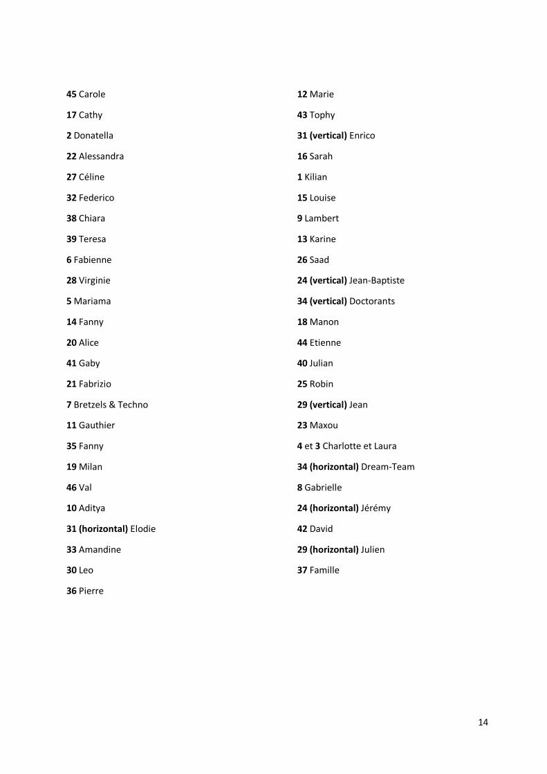
Selon une statistique établie par moi-même, 98% des lecteurs de thèse s’arrêtent à la partie
remerciements. Alors pour m’assurer qu’ils passent du bon temps, et parce que les gens cités ci-après
connaissent très probablement mon amour pour les blagues et les devinettes, j’ai décidé d’écrire mes
remerciements de thèse sous forme ludique. Les personnes sont ainsi décrites dans les pages suivantes.
9
45 Un immense merci à cette personne qui m’a aiguillée, dirigée, soutenue (et supportée) pendant ces
3 années. Tu as été d’une aide incommensurable, autant quand je passais la porte de ton bureau avec un
immense sourire parce que j’avais (enfin) trouvé de l’acide tartrique dans un échantillon que quand je la
passais les larmes aux yeux parce que les évènements de la vie font que ! Tu as été un support incroyable
et j’ai beaucoup appris à tes côtés. Un grand merci (à ma directrice) pour ton temps, tes conseils et
surtout, ton écoute, ta lucidité et ta gentilles dans les moments où ça n’allait pas au mieux.
17 Pour mon autre directrice de thèse, un grand merci aussi, pour le temps, les encouragements et les
commentaires toujours bienveillants de votre part. Ça a été un vrai plaisir de travailler à vos côtés et votre
connaissance du terrain hors-thèse m’a beaucoup appris !
2 Un immenso ringraziamento alla mia ultima direttrice di tesi. Non puoi immaginare quanto ti sono
grata per le porte che mi hai aperte, a livello scientifico, ma soprattutto nell'approccio, nella volontà e
nella metodologia. Devo ammettere una piccola storia d'amore con il polline, que spero duri ! Eppure,
non ero la più convinta. Mi ricordo che una volta eravamo sedute a tavola, quando mi hai spiegato come
una pianta maschio e una femmina possono avere un piccolo bambino. Ho imparato molto da allora, e tu
hai avuto molto a che fare con questo. Grazie per il benvenuto nel laboratorio! E grazie per gli intermezzi
culinari, anche se il formaggio rimane meglio da questa parte del confine!
22 Allora, non so quale definizione darti. Tra la madre e l'amica, sei stata un sostegno incredibile.
Sempre in ascolto, sempre sempre gentile, sempre sempre sempre disponibile, sei la persona più
premurosa che abbia mai incontrata. Questo dottorato non sarebbe stato così piacevole e non avrei
goduto così tanto la mia esperienza a Roma senza di te! E soprattuto quando arrivavi con le tue
expressioni romane o con i dolci appena usciti dal panificio ! Grazie infinite per tutto cio che hai fatto per
me !
27 Un immense merci pour toutes les fois où je t’ai dit « ******, j’ai un problème ! ». Tu as été d’un
soutien technique sans nom, et clairement, sans toi, ce manuscrit ne serait pas soumis parce que la GC
ne remarcherait plus et mon ordinateur serait déjà passé par la fenêtre 15 fois ! Merci d’avoir été autant
présente sur les réinstallations de mon ordinateur, qui ont été trop nombreuses, on est d’accord !
32 Grazie mille per tutto quello che mi hai insegnato sul polline. Non capisco come tante cose possano
entrare nella tua memoria! È stata una bella esperienza lavorare con te (ed imparare l'italiano allo stesso
tempo). Grazie per il tuo tempo e per rispondere sempre alle mie domande, anche se fossono le stesse!
38 All’archaeologua con la quale abbiamo fatto un bel lavoro insieme, grazie mille per la tua sonrisa. E
stato molto piacevole lavorare con te ed andare a campionare insieme.
39 Un grande ringraziamento a te per avere sempre un sorriso sul tuo viso e per avermi dato una buona
energia ogni volta che siamo viste. Sono contenta di avere lavorato insieme sul vaso di Gerico, anche se
non era un'anfora chiusa. Forse la prossima volta ;)
6 A nouveau, un grand merci à l’archéologue avec laquelle nous avons travaillé en France (même si tu
es déjà nommé dans la partie plus officielle). Merci pour le temps que tu m’as accordé dans cette thèse
et pour le stage à Bibracte ! Merci pour nous avoir permis d’échantillonner les amphores du Planier 3
avec le DRASSM. La novice que j’étais a découvert beaucoup de choses à tes côtés, et tu as fortement
aidé dans mon intérêt grandissant pour les amphores !
28 Un grand merci à notre gestionnaire comptable qui, au-delà de sa grande gentillesse et son sourire
agréable, a toujours été d’une aide incroyable quand j’ai eu un souci.
10
5 Quelle chance de t’avoir en « personne de contact » pour nous clarifier les centaines de page
d’informations qu’est ce fameux Grant Agreement. Merci beaucoup pour le temps que tu as passé pour
moi !
14 Ahhhhhh ! Je pourrais écrire ça que tu te reconnaitrais de toute façon. Avec une moyenne de deux
appels de plusieurs heures par semaine, c’est ma compagnie d’abonnement téléphonique qui doit être
contente ! Tu es un petit soleil dans la vie ! Et t’entendre glousser, c’est comme entendre la sonnerie de
récréation quand on était à l‘école : signe que les choses deviennent intéressantes et qu’on va rigoler !
Cette thèse n’aurait pas été la même sans t’avoir eu aussi proche et aussi loin ! On se revoit vite pour
manger pleins de trucs épicés et chopper la turista après que tu sois allée marcher dans le désert !
20 Une autre âme-sœur. Bon je sais, on ne peut en avoir qu’une seule, mais en fait, vous êtes deux !
Merci pour les rires, les sorties, les rires, les activités, les rires, les festivals, les pauses thés, les rires, le
sport le matin, les mois de colocation qu’on pourrait dire concubinage presque. Merci pour les plans
galères, les tentes sans arceaux, les plans absurdes, les plans pas réfléchis, les concerts les veilles de
grands jours. Toujours dans les bons coups =) C’était ouf de faire ma thèse avec toi à côté ! Et dire qu’au
début, je m’étais dit que tu avais l’air d’être une fille drôle !
41 Tu venida a Avignon ha sido un regalo increíble. ¡ Gracias por todas las risas, por las experiencias
divertidas contigo ! Tus aventuras y tu sonrisa alegraron alcunos meses de la tesis. Awuevo chiquilla !
21 Un grande ringrazimiento al mio amico del microscopio, che ha sempre risposto quando le dicevo
« *****, ti posso fare una domanda". E a volte "domandina" quando sentivo che era davvero un sacco di
volte che ti fermavi, ti alzavi e che venivi da me a dire "una quercha". Mi ricordo quando sono arrivata
nella tua officia, e stavi ascoltando il Vecchio Testamento, con la lista dei "figli di"! Che scherzo! Grazie
per avermi insegnato il romano cosi bene, per avermi portata a mangiare la pizza, per avermi fatto
complimenti per il mio italiano. Grazie per essere stato un'ancora di salvezza quando ero stufa del
microscopio e di questa tesi. Grazie per avermi fatto ridere semplicemente essendo te !
7 Ah là aussi je ne saurais pas trop par où commencer pour remercier cette fameuse bande de joyeux
lurons que vous êtes ! Malgré la distance qui limite un peu nos aventures, je suis super contente et je
vous remercie mille fois de votre support ! Des rires, des sorties et des WE où on n’a pas inventé des
vaccins contre grands choses ! Un grand merci à toi petite blondinette pour ton écoute bienveillante et
ta manière vraiment intéressante de voir les choses ! Merci à toi petit Ju-jitsuka pour tes blagues qui me
font souvent bien rire et les belles discussions ! Merci à toi petite dame qui va au cabinet tous les jours,
pour ton soutien, ton sourire et tes petites blagues quand on les attend le moins. Merci à toi Mr Docteur,
je te suis entièrement reconnaissante pour m’avoir permis de relativiser sur la rédaction. Je te dois une
fière chandelle parce que je me suis vraiment inspirée de toi pour gérer mon stress et passer du bon
temps dans ma rédaction, malgré les efforts. Tu m’as pas mal inspiré sur ce coup-là. Merci à toi petit
matheux qui envoie les messages les plus saugrenus de l’histoire des messages, ça me fait toujours bien
sourire. C’est toujours un plaisir de discuter et échanger avec toi. Et enfin, merci à toi futur Docteur pour
les bons moments passés ensemble ces derniers temps, entre Avignon et la Sicile ! J’aurais parié que tu
soutiendrais avant moi, et ça m’avait bien fait stresser de savoir que je serais la suivante après Tom ! Mais
j’ai déjà hâte de venir à Londres pour fêter ça !
11 Je ne pouvais pas ne pas te citer dans les remerciements, et tu méritais bien une ligne à ton nom !
Merci pour tous ces moments passés ensemble, que ce soit des WE de nature, de culture et de fêtes.
Merci pour les rires, les sorties, les bons repas, les discussions, les débats, les sourires, les petites
attentions qui font que le quotidien dans cette thèse a été vraiment chouette ! Merci de m’avoir initiée
à la grimpe, de m’avoir écoutée te raconter en long en large et en travers mes podcasts, mes grandes
11
interrogations, et même mes petites. C’est un peu grâce à toi que je suis venue à Avignon, et ça a vraiment
été une belle aventure !
35 Super personne que j’ai découvert au détour d’un couloir en terrain ennemi =) Merci pour ces pizzas
tous les mardis soir ! Pour ces loooooooongues conversations sur ton canapé, pour les pauses-cafés aussi,
qui me faisaient sortir les yeux de mon microscope !
19 Thank you for your support, your jokes and your smile! It has really great to have you in my back
during this thesis, to hear your weird music to concentrate and for the chit-chat in the lab! I still remember
the first time we met in the campus, looking at each-other like “Are you the other PhD?”. It was really
nice to have you in the office to talk, to claim, to cry (sometimes). Thank you for your amazing support.
46 Merci pour ton sourire dans le bureau, tes petites anecdotes et surtout, ton soutien hors pair ces
derniers jours avant la soumission ! La première fois que je t’ai vu, je m’étais dit que tu avais l’air d’être
un gars cool, parce que tu avais des chaussures qui te donnait un air cool. Comme quoi, on reconnait un
homme à ses chaussures. En tout cas, bien drôle de t’avoir eu à mes côtés pendant la thèse, que ce soit
pour les rires ou les plaintes ! Qui se serait moqué aussi souvent de ma nourriture le midi si tu n’avais pas
été là ?
10 Thank you for your jokes, your smile, your humor and the really tasty food you offered me to try so
many times. Although you did not convince me about Portugal, you tried very hard at least! I hope to see
you in India next time!
31 (horizontal) Partie – Revenue - Partie – Puis revenue! C’est mieux que les Feux de l’Amour cette
histoire ! J’ai commencé cette thèse avec toi, et tu m’as appris tellement de choses ! Je suis tellement
contente de la finir avec toi ! Merci pour les grandes conversations, les pauses-thé, les rires dans le
bureau, et les pauses clopes sans clope ! Merci aussi pour les merveilleux conseils qui m’ont évité bien
des soucis ! Et enfin, merci pour avoir toujours été une oreille (très) attentive ! Ton rire a vraiment égayé
le bureau et tes « Non mais Alice », signe annonciateur qu’on va rire, je les entends encore !
33 Un petit soleil ! Merci pour ta spontanéité, tes histoires drôles et ton écoute avisée de mes aventures,
toujours avec un œil critique bien entendu ! Quand je t’ai vu la première fois, j’ai su qu’on allait bien
s’entendre les deux ! Je suis juste déçue qu’on n’ait pas commencé cette thèse en même temps, on aurait
eu 1 an de plus pour profiter !
30 Ah ! Quel énergumène qui me régale avec ses histoires, son sens de l’humour et son gâteau au
chocolat ! Merci pour les conversations hyper intéressantes qu’on a eues et les débats, généralement
autour d’une bière ! On fêtera cette thèse comme il se doit : avec des frites !
36 Une magnifique rencontre ! J’ai beaucoup appris dans nos conversations où je ne vois pas trop le
temps passer, posés sur le canapé en train de manger. C’est un plaisir de te raconter mes petites
aventures parce que je sais qu’on va pouvoir en rire !
12 La dernière de notre petit groupe de fille du bureau B201 ! Mon amie des Picons ! Toujours là pour
discuter ou aller boire un coup ! Merci 1000 fois pour ces pauses-thé, qui se sont transformées en sorties
plus nocturnes avec le temps, mais l’intensité des discussions et le plaisir qui vont avec sont bien restés
le même ! Et merci pour cette idée fameuse de mise en page des remerciements !
43 Et merci à toi Jeune zythologue. Allez, c’est bien parce que ça va être publié que je ne fais pas la
blague ! Merci pour ta connaissance accrue des boisons maltées, qui m’a régalée pendant plusieurs
années ! Sans toi, on n’aurait probablement pas découvert le BD et sans vous (avec 25) et on n’aurait
surement pas autant ri ! Mille mercis pour les bonnes blagues ‘Bro’, les fléchettes, et surtout … Les
Blindtests !
12
31 (vertical) Alors, lui, euh, c’est un ami euh, enfin bon tu vois quoi, qui euh alors, sublime n’importe
quel, euh moment, tu vois, et surtout si, en fait, il y a, euh, de la nourriture, enfin, aux alentours ! Alors,
merci pour euh, avoir partagé mon stress pendant le premier stage de parapente ! Et alors merci pour
avoir été là quand on s’est fait phrasé sans comprendre un mot ! Daje, che ti potevo anche ringraziare in
italiano ! Grazie per tutte le volte che sei venuto a prepare il cibo ! Riccordo che teni ancora un bel pezzo
di parmeggaino per fare la pasta con le patate, che ci cendra buona sta volta.
16 Rencontre récente sur Avignon : un match parfait ! Merci pour ta bonne humeur, pour toujours noter
le détail drôle chez Killian qui va nous fait rire ! Et merci pour ses 10kg de pain ahah !
1 Merci pour m’avoir emmenée grimper à un moment où j’en avais beaucoup besoin je crois. C’est
toujours chouette de discuter avec vous (avec 16), et ça a vraiment été d’un soutien incroyable quand ça
n’allait pas trop même si je ne le montrais peut-être pas trop…
15 Un petit rayon de soleil dans une tornade matérielle et logistique et un presque-stress de rédaction,
voilà ce que tu es ! Sans toi, c’est clair que la rédaction n’aurait pas été aussi amusante et je te suis
reconnaissante de m’avoir offert des sorties, des pauses, des petites ellipses temporaires alors que
dehors, c’était le feu et la fatigue !
9 My mentor in science, maybe I shall call you this way! Thank you for teaching me all these things in
research, for trusting me and letting me improve! You opened me the doors of the labs of the
Rijksmuseum, and the conservation workshops, I will always have an incredible memory of it, even if it
was 4 years ago! Without you, I probably wouldn't have started a PhD.
13 Un grand merci pour le soutien technique! Un petit couteau-suisse qui a toujours une solution et en
plus, avec le sourire ! Tu manques au Campus Agroparc !
26 Un grand merci parce que tu m’as techniquement sauvé la thèse ! Oui oui ! Sans toi, j’aurais pleuré
pendant tout l’été la perte de cet ordinateur ! Merci pour toutes ces matinées et toutes ces soirées, où
te voir à l’entrée et à la sortie du bâtiment, avec ton sourire, donne le sourire !
24 (vertical) Un ancien membre de l’IRPNC qui joue au va-et-vient lui aussi ! Merci pour m’avoir
enseigné pleins de choses pendant les quelques temps où on a pu être au labo ensemble ! Toujours le
mot pour rire ce rugbyman !
34 (vertical) Merci à tous les étudiants des labos du deuxième étage, pour les conversations et les
petites pauses et les gâteaux ! Entre autres Emilie, Boutheina, Sarah, Léna, Léo, Hugo, Ombeline,
Emmanuelle.
18 Merci pour être venue toquer à la porte pour demander si on avait envie de faire connaissance avec
les voisins ! Bien trouvé ces voisins dis donc ! On a vraiment passé de belles soirées ensemble et en plus,
je suis sûre que tu seras dans l’assemblée pour la soutenance !
44 Membre actif des voisinades, merci à toi aussi pour ton esprit vif, ta connaissance culturelle et
littéraire, et tes bonnes blagues d’italien qui me font rire !
40 Membre actif des voisinades aussi, merci pour m’avoir régalé de ton accent sudiste et tes expressions
quand tu m’emmenais à l’université le matin parce que j’étais trop en retard pour prendre le bus. Une
chouette rencontre qui nous a conduit à des chouettes soirées !
25 Merci pour m’avoir laissé ta chambre à Lausanne et avoir gentiment choppé le Covid pile quand
j’étais venue te voir ! Quel BG celui-là !
13
29 (vertical) Merci ****-Chou pour être aussi … Chou ! Aussi partant pour tout et aussi désespéré par
tout ! Même si je continue à penser que glousser un peu plus te ferait le plus grand bien, c’est quand
même bien agréable de discuter avec toi, et d’échanger sur nos podcasts.
23 Merci pour m’avoir accueilli dans ton chez-toi Lausannois et ne pas avoir eu le Covid quand je suis
venue te voir ahah ! Toi t’es un vrai BG !
4 et 3 Un grand merci pour votre soutien à distance, ces audios qui nous maintiennent en contant
rapproché, les balades sous la pluie et les JO devant la télé avec un thé ! J’ai encore en tête cette fameuse
soirée au restaurant cet été, où on a vraiment beaucoup ri des cafetières à presse et des puzzles !
34 (horizontal) Au top cette fine équipe qui est là depuis le lycée ! Merci pour les balades et les
looooongues discussions. Et comme on ne se voit pas super souvent, vous m’avez permis de bien
visualiser l’évolution de cette thèse et comment c’était l’année précédente !
8 Merci merci merci pour la relecture ! Avec des parties plus drôles que d’autres à corriger j’imagine !
Tu as été top !!!
24 (horizontal) Merci pour m’avoir sauvé tant de fois de ces galères technologiques ! Le vol d’ordi, le
disque dur cassé, le dock, Bitlocker… il y en aura eu des péripéties ! Merci pour avoir répondu présent
même pendant tes vacances pour calmer mon stress naissant de perte d’ordinateur !
42 Sacrée rencontre dans les airs ! Merci de m’avoir fait voler pour la première, et j’espère, pas la
dernière fois !!! Je n’attends qu’une chose : revenir te raconter des petites blagues par talkie-walkie, moi
dans les airs, et toi sur la piste d’atterrissage !
29 (horizontal) Parce que je suis sûre que tu jettes un œil de temps en temps, d’où que tu sois ! C’est
peut-être toi qui m’as le plus aidée dans cette thèse, malgré toi, en me montrant qu’il y a des choses plus
graves qui existent.
37 Et le meilleur pour la fin. Le Larousse vous définit comme l’«Ensemble des personnes unies par un
lien de parenté ou d’alliance », mais c’est bien plus que ça. Si je devais écrire moi-même une définition,
je dirais plutôt que c’est un ensemble de personnes unies qui ont toujours été là pour moi, que ce soit
par des attentions, des conversations ou des moments partagés. Qu’aurait-été cette thèse si je ne vous
avais pas eu pour faire passer ma colère, ma tristesse ou ma peine ? Vous avez été un super incontestable
et je vous en remercierai à jamais. Merci pour m’avoir écouté au téléphone quand j’avais juste envie de
pleurer, pour m’avoir gâté de bons légumes, de délicieuses conserves et de fromage pour que mon
cerveau soit bien nourri, merci pour les WE nature, les activités culinaires, les jeux de cartes, les rires qui
ne partent de pas grands choses, les histoires drôles autour du thé et des tartes faites par la Reine des
tartes. Merci pour votre soutien, impossible à définir avec des mots !
Et enfin, un immense merci à toutes les personnes de l’université, du laboratoires IMBE et IBMM, et
plus généralement, toutes les personnes présentes au 2eme étage du bâtiment pour leur présence, leur
soutien, les conversations en salle de pause et autres petites activités. Puis, une petite pensée pour toutes
les personnes qui travaillent dans l’ombre (que je serais incapable de nommer de manière exhaustive) et
qui assurent des conditions de travail optimales avec des locaux propres et en bon état.
14
45 Carole
17 Cathy
2 Donatella
22 Alessandra
27 Céline
32 Federico
38 Chiara
39 Teresa
6 Fabienne
28 Virginie
5 Mariama
14 Fanny
20 Alice
41 Gaby
21 Fabrizio
7 Bretzels & Techno
11 Gauthier
35 Fanny
19 Milan
46 Val
10 Aditya
31 (horizontal) Elodie
33 Amandine
30 Leo
36 Pierre
12 Marie
43 Tophy
31 (vertical) Enrico
16 Sarah
1 Kilian
15 Louise
9 Lambert
13 Karine
26 Saad
24 (vertical) Jean-Baptiste
34 (vertical) Doctorants
18 Manon
44 Etienne
40 Julian
25 Robin
29 (vertical) Jean
23 Maxou
4 et 3 Charlotte et Laura
34 (horizontal) Dream-Team
8 Gabrielle
24 (horizontal) Jérémy
42 David
29 (horizontal) Julien
37 Famille

15
List of Abbreviations
AcOEt : Ethyl acetate
aDNA : ancient DNA
BSTFA/TMCS : N,O-Bis(triméthylsilyl)trifluoroacétamide / triméthylchlorosilane
CHCl3 : Chloroform
DAG: Diacylglyceride
DCM : Dichloromethane
DEE : Diethyl ether
DHA : Dehydroabietic acid
DHAM : Dehydroabietic acid methyl ester
DNA : Deoxyribonucleic acid
DTMS : Direct Temperature-resolved coupled to Mass Spectrometry
EtOH : Ethanol
FA : Fatty acid
FFA : Free fatty acid
GC-MS : Gas Chromatography coupled to Mass Spectrometry
HPLC : High Performance Liquid Chromatography
HPLC-ESI-QToF : HPLC coupled with Electrospray Ionization - Quadrupole Time-of-Flight Mass
Spectrometry
KBr : Potassium bromide
KOH : Potassium hydroxyde
MAG : Monoacylglyceride
MeOH : Methanol
MS : Mass Spectrometry
MW : Microwave
NMR : Nuclear Magnetic Resonance
NPP : Non-Pollen Palynomorph
ORA : Organic residue analysis
PTFE : Polytetrafluoroethylene
Py-GC-MS : Pyrolysis Gas Chromatography coupled to Mass Spectrometry
TAG : Triacylglyceride
THF : Tetrahydrofuran
TIC : Total Ion Chromatogram
TMS : Trimethylsilylation
US : Ultrasound

16
List of Figures
Figure 1. Most common amphorae shape differences ................................................................... 30
Figure 2. Stamp on an amphora found in Planier 3 shipwreck ........................................................ 31
Figure 3. Titulus pictus written on an Ostia type African amphora. ................................................ 31
Figure 4. Location of the Planier 3 shipwreck in the Mediterranean Sea ........................................ 34
Figure 5. Location of San Felice Circeo ........................................................................................... 39
Figure 6. San Felice Circeo typographies ........................................................................................ 40
Figure 7. Markers of heating treatment preparation for the diterpenic compound of abietic acid (A)
and the triterpenic compound of oleanonic acid (B) ...................................................................... 49
Figure 8. Example of diterpenic (A) and triterpenic (B) skeletons synthesized from isoprene C5 units
..................................................................................................................................................... 51
Figure 9. Abietane (A), pimarane (B) and labdane (C) skeletons with numbered carbons ............... 52
Figure 10. DHA and 7-oxo DHA, respective markers of Pinaceae resin and oxidized Pinaceae resin 52
Figure 11. Pimarane (A) and labdane (B-C) skeleton markers of Cupressaceae resin ...................... 53
Figure 12. Oxidized phenol markers of Cupressaceae resin ............................................................ 53
Figure 13. Abietane markers degradation pathway from a Pinaceae resin ..................................... 55
Figure 14. Molecular structure of polyphenols............................................................................... 58
Figure 15. Main anthocyanidins of grapes...................................................................................... 59
Figure 16. Essential anthocyanidins of grapes ................................................................................ 60
Figure 17. Principle organic acids produced during fermentation ................................................... 61
Figure 18. Equilibrium states of precipitation and adsorption of tartrate salts with the ceramic
matrix from the winemaking process to the extraction in the lab .................................................. 63
Figure 19. TAG unit degraded into free fatty acids and glycerol ..................................................... 65
Figure 20. Main degradation pathways of fatty acids in archaeological context ............................. 66
Figure 21. Sterane skeleton (A) into cholesterol (B); campesterol (C); stigmasterol (D) .................. 68
Figure 22. GC-MS schema equipped with a quadrupole ion trap .................................................... 71
Figure 23. Ion trap set-up .............................................................................................................. 73
Figure 24. “Pollen analysis” occurrence in scientific publications. .................................................. 79
Figure 25. Protocol comparison for the extraction of grape derivatives ....................................... 108
Figure 26. Radar plot of the shards .............................................................................................. 117
Figure 27. Radar plot of the coating materials ............................................................................. 122
17
Figure 28. TIC Chromatograms (A) Standard colophony extracted with DCM:MeOH; (B) Standard
colophony extracted with MW-assisted butylation and (C) Standard pimaric acid extracted with
MW-assisted butylation .............................................................................................................. 124
Figure 29. Archaeobotanical and chemical analysis of the San Felice Circeo amphorae ................ 133
Figure 30. Investigated archaeological amphorae ........................................................................ 137
Figure 31. GC-MS chromatogram for the pitch of SFC1 ................................................................ 139
Figure 32. Vitis pollen in polar and equatorial view ...................................................................... 142
Figure 33. Microscopic observation of (A) archaeological plant tissues trapped in the resin of SFC1;
(B) filament from the stamen of a modern wild Vitis vinifera flower, and (C) ESEM observation of a
transverse section of charred Pinus wood trapped in SFC2 coating .............................................. 143
SI Figure 34. ESEM pictures of Vitis vinifera pollen grains recovered from (A) grapefruits from wild
grapes in Tivoli and (B) Fossil sediment from Rignano Flaminio.................................................... 157
Figure 35. Palynological and chemical analysis applied on the Jericho 'teapot' vase ..................... 159
Figure 36. Archaeological site of Tell es-Sultan ............................................................................ 163
Figure 37. Vessel TS.19.TrIII.2000/1 ............................................................................................. 164
Figure 38. Pollen observed from the vessel .................................................................................. 166
Figure 39. Malva pollen grains observed with ESEM .................................................................... 167
Figure 40. Chromatograms from (A) Alkaline treatment and (B) DCM extraction ......................... 168
Figure 41. Protocol comparison applied on oil shards .................................................................. 181
Figure 42. 6781a TIC Chromatograms .......................................................................................... 185
Figure 43. Shard sections from Early African (sample 6849), ovoid (sample 728) and Brundisium
amphora (sample 579) ................................................................................................................ 188
Figure 44. Molecular structures of grape-fermented acids after butylation and derivatization ..... 202
Figure 45. TIC (black) and m/z (red) chromatograms of the sample 1014 after (A) the extraction
with traditional solvent (labelled 1LE) and (B) the butylation directly applied on the coating. ...... 203
Figure 46. Chromatograms of standard colophony with different chemical treatments ............... 204
Figure 47. Protocols to apply for the extraction of grape and fermentation markers.................... 205
Figure 48. Pollen recovered from the archaeological wood tar. ................................................... 208
Figure 49. TIC chromatogram of the Mañà C2 amphora (sample no. SFC3) .................................. 210

18
List of Tables
Table 1. Amphorae sampled from the shipwreck Planier 3 ............................................................ 38
Table 2. Amphorae sampled from San Felice Circeo ....................................................................... 42
Table 3. Amphora sample from Tell-es Sultan ................................................................................ 43
Table 4. Structural characterization from the main FA of vegetable oils ......................................... 65
Table 5. Archaeological amphorae investigated ........................................................................... 113
Table 6. Molecules identified in shards ........................................................................................ 119
Table 7. Molecules identified in pitch coatings ............................................................................ 122
Table 8. Pollen grains recovered from the analyses of amphorae SFC1, SFC2 and SFC5 ................ 141
Table 9. Oil amphorae shards analyzed ........................................................................................ 183
Table 10. Molecular evidence of the content of Planier 3 oil shards ............................................. 186
Table 11. Grape-fermented acids retention time and mass fragments after derivatization and
butylation followed by derivatization .......................................................................................... 202
Table 12. Pollen grains recovered from the analyses of amphorae SFC3, SFC4, 1014 and 749 ...... 207
Table 13. Chromatographic and archaeobotanical results and interpretations of the artefacts
studied ........................................................................................................................................ 215
19
Table of Contents
RESUME ....................................................................................................................................................... 1
ABSTRACT .................................................................................................................................................... 2
CURRICULUM VITAE .................................................................................................................................... 3
ACKNOWLEDGMENTS .................................................................................................................................. 8
LIST OF ABBREVIATIONS ............................................................................................................................ 15
LIST OF FIGURES ........................................................................................................................................ 16
LIST OF TABLES .......................................................................................................................................... 18
TABLE OF CONTENTS ................................................................................................................................. 19
GENERAL INTRODUCTION .......................................................................................................................... 23
ARCHAEOLOGICAL PROBLEMATIC .................................................................................................................... 24 ED-ARCHMAT ......................................................................................................................................... 25 MANUSCRIPT CONSTRUCTION ........................................................................................................................ 26
CHAPTER 1 ................................................................................................................................................. 28
1.1. ONCE UPON A TIME: THE AMPHORAE ................................................................................................. 29 1.2. AMPHORAE TYPOLOGY ................................................................................................................... 29 1.3. INSCRIPTIONS: A PRECIOUS HELP TO TRACE BACK AMPHORAE .................................................................... 31 1.4. AMPHORA FABRICATION PROCESS ..................................................................................................... 31
1.4.1. From the coarse clay to amphora .............................................................................................. 31 1.4.2. The resin: a natural resource to waterproof the amphorae ........................................................ 32
1.5. CASES OF STUDY ........................................................................................................................... 33 1.5.1. Shipwreck “Planier 3” ............................................................................................................... 33
1.5.1.1. Archaeological context ............................................................................................................... 33 1.5.1.2. Archaeological amphorae ........................................................................................................... 36
1.5.2. Archaeological site of San Felice Circeo ..................................................................................... 39 1.5.2.1. Archaeological context ............................................................................................................... 39 1.5.2.2. Archaeological amphorae ........................................................................................................... 41
1.5.3. Archaeological site of Jericho .................................................................................................... 41 1.5.3.1. Archaeological context ............................................................................................................... 41 1.5.3.2. Archaeological amphora ............................................................................................................. 43
1.6. SUMMARY .................................................................................................................................. 43
CHAPTER 2 ................................................................................................................................................. 45
2.1. IDENTIFIED SUBSTANCES WITHIN THE POTTERY CERAMIC .......................................................................... 46 2.1.1. Biomarker concept ................................................................................................................... 46
2.1.1.1. Biomarkers ................................................................................................................................ 47 2.1.1.2. Natural degradation markers, sustainability and preservation ..................................................... 47 2.1.1.3. Anthropic degradation markers, an interesting signature of human activities .............................. 48 2.1.1.4. Contamination markers .............................................................................................................. 49
2.1.2. Terpenes .................................................................................................................................. 50

20
2.1.2.1. Definition and structure ............................................................................................................. 50 2.1.2.2. Natural distribution of diterpenic resins ...................................................................................... 51 2.1.2.3. Degradation pathways................................................................................................................ 53 2.1.2.4. Distinguishing resin form pitch and tar ........................................................................................ 56
2.1.3. Wine residues ........................................................................................................................... 56 2.1.3.1. A brief introduction to wine ........................................................................................................ 56 2.1.3.2. Composition............................................................................................................................... 57
2.1.3.2.1. Phenolic acids............................................................................................................................. 58 2.1.3.2.2. Anthocyanins ............................................................................................................................. 59 2.1.3.2.3. Organic acids .............................................................................................................................. 60
2.1.3.3. From grape to wine .................................................................................................................... 61 2.1.3.4. Tartaric acid: a suitable biomarker? ............................................................................................ 62
2.1.4. Lipids........................................................................................................................................ 64 2.1.4.1. Triacylglycerols and fatty acids ................................................................................................... 64
2.1.4.1.1. Definition and structure.............................................................................................................. 64 2.1.4.1.2. Degradation reactions ................................................................................................................ 65
2.1.4.1.2.1. Unsaturated fatty acids auto-oxidation ............................................................................ 66 2.1.4.1.2.2. β-oxidation ..................................................................................................................... 67 2.1.4.1.2.3. Unsaturated fatty acids hydroxylation ............................................................................. 67
2.1.4.2. Sterols ....................................................................................................................................... 67 2.1.4.2.1. Definition, structure and natural distribution .............................................................................. 67 2.1.4.2.2. Degradation pathways ................................................................................................................ 68
2.2. ANALYTICAL METHODS ................................................................................................................... 68 2.2.1. History: Organic analysis residues introduction ......................................................................... 68 2.2.2. Chromatographic methods ....................................................................................................... 70
2.2.2.1. Gas Chromatography-based method .......................................................................................... 70 2.2.2.2. Liquid Chromatography-based method ....................................................................................... 71 2.2.2.3. Pyrolysis-GC-based method ........................................................................................................ 72
2.2.3. Mass Spectrometry ................................................................................................................... 72 2.2.3.1. SIM ............................................................................................................................................ 73 2.2.3.2. MSn............................................................................................................................................ 74
CHAPTER 3 ................................................................................................................................................. 76
3.1. ONCE UPON A TIME: THE POLLEN ...................................................................................................... 77 3.1.1. Pollen definition ....................................................................................................................... 77 3.1.2. Pollen dispersion....................................................................................................................... 77 3.1.3. Pollen preservation ................................................................................................................... 77
3.2. PALYNOLOGY: A STATE OF THE ART ON THE GROWING INTEREST FIELD......................................................... 78 3.2.1. Historical point of view ............................................................................................................. 78 3.2.2. From pollen analysis to archaeological meaning ....................................................................... 79
3.3. METHODOLOGICAL APPROACHES AND DATA ANALYSIS ............................................................................ 80 3.3.1. Sampling issues ........................................................................................................................ 80 3.3.2. Pollen extraction....................................................................................................................... 81 3.3.3. Pollen identification .................................................................................................................. 82 3.3.4. Pollen interpretation ................................................................................................................. 82
REFERENCES............................................................................................................................................... 85
CHAPTER 4 ................................................................................................................................................107
4.1. ABSTRACT ..................................................................................................................................110 4.2. INTRODUCTION ...........................................................................................................................111 4.3. MATERIAL ..................................................................................................................................112
4.3.1. Archaeological samples ...........................................................................................................112 4.3.2. Solvents and reagents..............................................................................................................114

21
4.4. METHODS ..................................................................................................................................114 4.4.1. Optimization of the acid-catalyzed esterification: microwave-assisted butylation .....................114 4.4.2. Analytical procedures for inorganic shards ...............................................................................115 4.4.3. Analytical procedures for organic coatings ...............................................................................115 4.4.4. GC-MS analyses .......................................................................................................................115 4.4.5. Radar plot construction ...........................................................................................................116
4.5. RESULTS AND DISCUSSION ..............................................................................................................116 4.5.1. Optimization of the acid-catalyzed butylation ..........................................................................116 4.5.2. Extracting capacities comparison on archaeological shards ......................................................117 4.5.3. Extracting capacities comparison on archaeological coatings ...................................................121
4.6. CONCLUSION ..............................................................................................................................125 4.7. REFERENCES ...............................................................................................................................128
CHAPTER 5 ................................................................................................................................................132
5.1. ABSTRACT ..................................................................................................................................134 5.2. INTRODUCTION ...........................................................................................................................134 5.3. MATERIALS AND METHODS .............................................................................................................136
5.3.1. Archaeological materials .........................................................................................................136 5.3.1.1. Archaeological context ............................................................................................................. 136
5.3.2. Chemical analyses ...................................................................................................................137 5.3.2.1. Solvents and reagents .............................................................................................................. 137 5.3.2.2. Sample preparation for chromatographic analyses .................................................................... 137 5.3.2.3. Gas chromatography – Mass Spectrometry ............................................................................... 138
5.3.3. Archaeobotany ........................................................................................................................138 5.4. RESULTS ....................................................................................................................................139
5.4.1. Chemical analyses ...................................................................................................................139 5.4.2. Archaeobotanical analyses ......................................................................................................140
5.4.2.1. Pollen ...................................................................................................................................... 140 5.4.2.2. Plant tissues ............................................................................................................................. 142
5.5. DISCUSSION................................................................................................................................143 5.5.1. Coating of the amphorae .........................................................................................................143 5.5.2. Content of the amphorae .........................................................................................................144
5.6. CONCLUSION ..............................................................................................................................147 5.7. REFERENCES ...............................................................................................................................149 5.8. SUPPORTING INFORMATION............................................................................................................157
CHAPTER 6 ................................................................................................................................................158
6.1. ABSTRACT ..................................................................................................................................161 6.2. INTRODUCTION ...........................................................................................................................161
6.2.1. Archaeological site ..................................................................................................................162 6.3. MATERIALS AND METHODS .............................................................................................................163
6.3.1. Vase sampling .........................................................................................................................163 6.3.2. Archaeopalynological analyses ................................................................................................164 6.3.3. Chromatographic analyses ......................................................................................................165
6.3.3.1. Solvents and reagents .............................................................................................................. 165 6.3.3.2. Sample preparation .................................................................................................................. 165 6.3.3.3. Gas Chromatography – Mass Spectrometry .............................................................................. 165
6.4. RESULTS AND DISCUSSION ..............................................................................................................165 6.5. CONCLUSION ..............................................................................................................................171 6.6. REFERENCES ...............................................................................................................................173

22
CHAPTER 7 ................................................................................................................................................180
7.1. INTRODUCTION ...........................................................................................................................182 7.2. MATERIAL AND METHODS ..............................................................................................................183
7.2.1. Materials .................................................................................................................................183 7.2.2. Protocols .................................................................................................................................183
7.3. RESULTS AND DISCUSSION ..............................................................................................................184 7.3.1. The amphorae ceramics: concealed indirect indications of the content .....................................188 7.3.2. Archaeological interpretations from enigmatic chemical markers .............................................190
7.4. CONCLUSION ..............................................................................................................................191 7.5. REFERENCES ...............................................................................................................................192
CHAPTER 8 ................................................................................................................................................199
8.1. CALLING FOR THESIS OBJECTIVES ......................................................................................................200 8.2. METHODOLOGY DEVELOPMENT AND PROTOCOL OPTIMIZATION ...............................................................201
8.2.1. Organic residue analysis: the improvements ............................................................................201 8.2.2. Archaeobotanical analysis .......................................................................................................205
8.3. ARCHAEOLOGICAL PERSPECTIVE: FROM THE OBJECT TO THE INTERPRETATION ..............................................206 8.4. CONCLUSION ..............................................................................................................................212 8.5. PERSPECTIVES .............................................................................................................................216 8.6. REFERENCES ...............................................................................................................................217
APPENDIX 1. DRAWINGS OF SEVERAL AMPHORAE FROM PLANIER 3 .......................................................222
APPENDIX 2. FATTY ACIDS AND DERIVATIVES IDENTIFIED IN THE OIL AMPHORAE ...................................224
APPENDIX 3. MASS SPECTRUM OF STANDARD MOLECULE .......................................................................225
APPENDIX 4. RETENTION TIME AND MAIN MASS FRAGMENTS OF THE MOLECULES.................................239

23
General introduction
24
Archaeological problematic
This PhD thesis falls within the promotion of interdisciplinary in the understanding of archaeological and
cultural heritages, with the specific aim of developing multi-analytical skills to fairly diagnose ancient
materials. The work presented results from the fruitful collaboration between Carole Mathe de Souza,
Cathy Vieillescazes and Donatella Magri. With the aim of developing intersectoral knowledge and
competences, the research was conjointly carried out at Avignon University and Sapienza University,
which possess complementary expertise fields. The organic and analytical parts were developed within
the IRPNC (Ingénierie de la Restauration des Patrimoines Naturel et Culturel) lab from the IMBE
department (Mediterranean Institute of marine and terrestrial Biodiversity and Ecology) in Avignon. The
organic research axis was supervised by my director Carole Mathe de Souza and co-director Cathy
Vieillescazes. The lab is specialized in molecular archaeometry, with a particular interest in natural
substances present in artistic and archaeological heritage. Through the analysis of color dyes and
pigments, easel paintings, mummies balms and ethnic objects, the lab has gained a notable expertise in
organic residues, with relevant competences in spectroscopy and chromatography. Natural plants
analyses have greatly contributed to the understanding of thermal and photochemical degradation of
organic molecular markers and biomarkers from resins, pigments and dyes. Then, archaeobotanical
analyses, specifically including palynology, were followed by my co-director Donatella Magri, and
admirably supported by Alessandra Celant, both from the Dipartimento di Biologia Ambientale. The long
expertise in paleobotany and palynology is included in environmental biology through the analysis from
the micro to macroscale. The research is conducted on botanical proxies such as pollen, spores, non-pollen
palynomorphs, diatoms, wood fragments, leaves and seeds.
My thesis work focused on the analyses of organic and archaeobotanical residues in archaeological
amphorae. The molecular characterization was carried in continuation of Hitomi Fujii’s PhD thesis that
dealt with the deciphering of amphorae content and coatings, handled at IRPNC and defended in 2018. In
collaboration with the archaeologists Fabienne Olmer (France) and Chiara Delpino (Italy), we had the
chance to develop our analytical lens on Roman materials from marine context. The samples having spent
more than 2000 years under the Mediterranean Sea, the recovery and the analyses of organic residues
was all the more challenging. In this regard, we used scientific lens to provide answers to the
interrogations raised by the archaeologists. Triggered by the dearth of collaboration between
anthropological and scientific disciplines themselves, the problematic was elaborated to overcome the
analytical limitations inherent to the objects through a multi-diagnostic study. The aim of this thesis
encompassed the development of innovative analytical tools to characterize the content of archaeological
amphorae, whether they exhibit visible traces of organic waterproofing. By studying the organic residues
of amphorae, we decided to place the archaeological object at the core of the problematic. This choice
was taken to integrate them into a broader historical, anthropological and ethno-geographical context.
Analyzing micro and macro remains allowed us to better understand the goods exchanged in the past,
their origins and their manufacturing processes. In this way, we aimed at unravelling trade routes
developed in ancient times and the geographical boundaries of this trade while interpreting the technical
prowess and the savoir-faire of past civilizations. Understanding the object in its entirety would finally
ensures an effective valorization of the object's history and durable conservation. Since objects are
singular, it is important to treat them as witnesses of the past, eager to reveal their secrets to future
generations.

25
In this regard, two different approaches are developed hereafter. The chemical analyses of organic
residues were carried out at Avignon University. The residues correspond to the microscopic traces and
macroscopic remains, preserved in the waterproofing layer of the amphorae or in the archaeological
shards themselves. The molecules, extracted from their original matrix by different analytical protocols
are then characterized by chromatographic tools. Through comparison with contemporary reference
materials, they shed light on the archaeological content, while giving clues on the nature and formulation
of the waterproofing coating coming from natural resinous materials, as well as its state of degradation.
Palynological investigation was developed and performed at Sapienza University. The pollen contained in
the pitch have allowed to support, increment, and even define new horizons in the archaeological
interpretation of the studied vases. In the light of the fresh collaboration between these two labs of
independent expertise domains, the project aims at developing multi-analytical tools in vessel and
ceramic analysis.
ED-ARCHMAT
The research work takes place in a generalized program aiming at developing scientific lens to
understand, diagnosis and prevent from degradation archaeological and cultural heritages. The project is
supported by the Doctoral School of ARCHMAT (Archaeological and Cultural Heritage in MATerials
Science). It accounts for one of the thirteen doctorates funded by European Union's Horizon 2020
research and innovation program (H2020-MSCA- ITN-2017- EJD): Marie Skłodowska-Curie Innovative
Training Networks (European Joint Doctorate) – Grant agreement nº 766311 (ESR9).
The research program provided us an astonishing support in terms of cultural and educative
opportunities of learning and networking, with a specific focus on trainings and dissemination. With the
purpose of bridging gaps in cultural heritage management by providing scientific support, the
methodology we are hereby developing focuses on the fundamental shift from a unilateral or
multidisciplinary understanding of the archaeological materials to an inter and transdisciplinary approach.
Associating independent fields of research together promotes the accumulation of combinative evidence
to better address the initial archaeological interrogations. The great collaboration all-over the
Mediterranean basin, with university laboratories, archaeologists, researchers, museums and business
institutions helped in building, structuring and orienting the research problematic. In order to access a
deep knowledge of the studied material, innovative tools that promote cutting edge science through
scientific and technological advances were developed. By encouraging a common language between
cultural heritage protagonists and scientific, we aim at accessing high technical levels of diagnostic
capacities. Aside from the valorization of such a fruitful program, the final axe of development focuses on
the dissemination of the out coming discoveries. The transfer of knowledge through entrepreneurial
initiatives is encouraged. The dissemination of technical advances together with the promotion of the
acquired knowledge need to be spread over to enhance communication between the diverse cultural
heritage communities. This way, the conservation of the cultural heritage can be ensured for the future
generation.

26
Manuscript construction
The manuscript is structured on the basis of a comprehensive and exhaustive state of the art focused on
the three fundamentals of this thesis: the archaeological amphorae, the molecular markers and the pollen
markers. The investigated materials are presented in the first chapter through a general overview of the
primordial role containers played in the development of trading under the Roman Empire. Archaeological
amphorae we worked on are described in their historical contexts from the sunken ship of Planier 3
(France) and the ancient anchorage of San Felice Circeo (Italy).
Molecular and analytical tools that help in interpreting residues contained in the amphorae are detailed
in the second chapter. A complete picture of the different types of molecules recoverable in the
chromatographic analyses is presented in order to introduce the concept of biomarker. The chapter
focuses on the analytical advances through a chronological and historical description of the tools
developed, highlighting their advantages and limitations.
Finishing with the introductive chapters, a comprehensive summary of the use of pollen in archaeology
is lastly developed. After giving an extensive definition of pollen grains dispersion and preservation over
time, methodological approaches are also discussed in the third chapter, from the sample collection to
the grain’s identification.
The following parts present the outcomes of the thesis. The artefacts investigated are thereupon
reported through different thematic, organized in independent chapters, spanning from the presumed
archaeological content or historical contexts to the methodological approach developed. Results and
interpretations are discussed in the form of scientific articles. The extraction of wine markers from
archaeological resinous materials and shards is primarily developed in the fourth chapter, accepted in the
special issue of the journal Crystals (DOI 10.3390/cryst11111300). It deals with the analysis of wine
containers from Planier 3 and from the anchorage of San Felice Circeo. The fifth chapter was accepted
with minor revision in the PlosOne journal. It details the archaeological value of an interdisciplinary study
combining chromatographic and archaeobotanical tools applied on amphorae thought to have contained
wine. Although the investigated amphorae belong to the already studied artefacts from San Felice Circeo,
the focus was settled on the archaeological and anthropological considerations based on the discovery of
Vitis pollen grains exhibiting a singular morphology. The sixth chapter deals with the application of the
transdisciplinary methodology we initially developed on a very different archaeological context. Leaving
the Roman period for the Bronze Age, the analysis of a spouted vase recovered from Jericho (Southern
Levant) were interpreted in the light of archaeological medicinal concoctions. The article might be
submitted in international journals of archaeometry. Finally, the seventh chapter that encompasses
chromatographic analyses of presumed oil amphorae recovered in Planier 3 interrogates on the cultural
use of oil at the Roman period. Following this, a general discussion is developed to support the importance
of a transdisciplinary methodological approaches to better understand the history of the object
highlighting the perspectives of such a fruitful research project.

27

28
Chapter 1
Amphorae, fabrication and uses

29
1.1. Once upon a time: the amphorae
Even if the earliest evidence of jar is dated back to the Neolithic period, the use of amphorae has been
developed by the Canaanites around the 15th century BC for ship trading. Due to several logistical
advantages in terms of solidity, transport and packing facilities, the Egyptians started to produce their
own amphorae a century later. The Phoenician civilization popularized the amphora-shape concept
around the Mediterranean Sea. From then on, following Greek and Roman populations developed
different amphora typologies, characteristic of the content but also of the manufacture place, the period
and/or the civilization (Grace, 1949; Garcia Vargas, 1996; Cipriano and Carre, 1989). Amphorae design
thereby evolved to rounder egg-shape bodies in order to optimize sailing goods quantities. For instance,
Whitbread reported that Greek ships could contained around 3000 amphorae of 29 liters each in the
average (Whitbread, 1995).
The expansion of the Roman Empire corresponded to an intensification of commercial activities
throughout the Mediterranean and therefore coincided with the expansion of amphorae manufacture. In
order to supply oil, wine and other consumable demands, containers were massively produced to the
extent that the Western regions of the Empire were ruling over the production during the late Roman
period (Formenti et al., 1978; Manacorda, 1989; Palazzo, 1989). Made of abundant and disposable local
clay, they widely contributed to coast local economies. Since the full container was quite heavy, amphorae
may have been mass produced nearby and empty transported to harbors where they were filled before
shipping. Not only the exported goods but also amphorae themselves were part of the commercial
activities (Lyding Will, 1992).
Their inorganic building material, which prevent from breakage, accounts for its slowness to degrade.
Hence, amphorae are commonly found in archaeological sites dated from 1500 BC to 500 AC.
1.2. Amphorae typology
Amphorology corresponds to the investigation field dedicated to amphorae morphology studies.
Heinrich Dressel first implemented this discipline in 1899 by relating their shape difference and
manufacture places. He built up a tailored classification to provide spatial and temporal information on
Roman trades. Enhanced by the discovery of many underwater amphorae, the classing discipline however
needed to wait for several years to get a real impact. This way, some archaeologists such as Lamboglia or
Beltran greatly contributed to the garnishment of the already existing classification (Twede, 2002). One
pillar of the identification is the shape identification as shown in Fig. 1.
“Amphora” comes from Ancient Greek: “amphi” and “phoreus” respectively meaning “both sides” and
“bearer”. As presented in Fig. 1, amphorae were built up to stand on a central heavy base, usually called
“foot” from which was extended the “body” (or “belly”). The “shoulder” corresponds to the upper part of
the body, which slightly bent to connect the neck. A thicker rim circled the top of the neck to control the
poured content flow. The ingenuity lies in the presence of two hand holders to link the shoulder to the
neck, which widely facilitate the handling. Grabbed by these holders, amphorae were maneuvered by
rotational movements to ease the transportation, the pointed base being used as a pivot. They were

30
sealed with cork stopper or ceramic mortar made of volcanic sand (pozzolana) and lime (Lyding Will,
1977).
Amphorae shape is a great symbol of packaging technology inherited from ancient civilizations. As
described by Parker and Katszev, the shape may have been adjusted to optimize amphorae stacking and
minimize empty spaces in the cargo, starting from the hull to the central part of the hold. Foot bases,
necks and bodies were indeed nested with each other in such a packed way that amphorae could not
move while sailing (Katzev, 1970; A. J. Parker, 1992).
Figure 1. Most common amphorae shape differences (Bezeczky, 1998)
Commercial activities and amphorae production became so substantial that amphora morphology gave
rise to a standardized measuring scale in the Roman Empire. For example, the Greco-Italic style showed
up to overcome the need of a common shape between Greco-Roman sailing trades. Every shape evolution
resulted from the improvement of the design capacity of the mass ratio. This way, the shape was
optimized to allow the container to transport the maximum beverage quantity at once. Usual dimensions

31
ranged from 45 cm to 1 m. They could weight up to 40 kg and carry until 80 liters (Peacock & Williams,
1986).
1.3. Inscriptions: a precious help to trace back amphorae
Diverse indications from the amphorae can help characterizing jars. Indirect hints such as the shape or
the coarse clay composition can shed light on the origin, the site of production as well as the possible
content. For instance, wine amphorae were longer and thinner compared to olive jars that had rounder
bodies. Elizabeth Lyding Will reports that Roman typologies dated from 30 BC to 400 AC highlighted more
the nature of the content rather than the manufacturing region (Lyding Will, 2000).
While ancient jars were only identified thanks to their emblematic shapes, the use of content labels
directly stamped or painted on the clay became systematic after 400 BC. Tweede classified them in two
categories: the explicit annotations, which clearly provided administrative indications (identity of the
manufacture site owner, eventual export duties, volumetric capacities, etc.) and the non-explicit ones
made of symbols and letters detailing the content (Twede, 2002). This way, stamps and tituli picti must
be distinguished (Fig. 2 and Fig. 3). While non-explicit indications can be directly
stamped on the neck, the holders or even the shoulder with a ceramic mold
marker, explicit tituli picti were hand painted. Unfortunately, it is not always
possible to identify such indications on the archaeological amphorae, especially
when the jars are not in optimum conservation states.
Figure 2. Stamp on an amphora found in Planier 3 shipwreck (DRASSM, credit photo: C. Joliot, 2018)
Figure 3. Titulus pictus written on an Ostia type African amphora.
“VIR” refers to the red wine content (ui(num) r(ubrum)) and “MPS” may correspond to the wine merchant) (Bonifay et al., 2015)
1.4. Amphora fabrication process
1.4.1. From the coarse clay to amphora
Amphorae are the product of complex shaping, drying and heating processes. The non-respect of the
manufacture conditions usually leads to physical weakness and the object may break.

32
From the fabrication point of view, after the choice and preparation of the raw coarse clay, the material
is shaped, the surface may be greased, and the object is dried to prevent from structural damages related
to residual water fast evaporation within the matrix during the following firing step. Firing temperature
must be high enough to promote evaporation of adsorbed and inter-planar water as well as structural
hydroxyl groups (Pollard & Heron, 2008). Roughly estimated between 500°C and 800°C, the temperature
strongly depends on various factors such as the raw material, the crystallinity degree and the firing
conditions (temperature profile, combustible nature, aerobic conditions) (A. M. Hunt, 2017). Some
organic post-firing treatments can finally be applied directly on the hot surfaces or more rarely, after
cooling. Several practices have been reported in past ethnographic investigations of internal jar or cooking
pot surfaces. Vegetal or animal evidence respectively made of tree sap or fats, milk or even blood have
been pointed out (Harry et al., 2009; Shepard, 1956; Skibo, 2013). Diverse coating techniques such as the
dipping, the smoking and the spraying or even the rubbing of natural components have also been detailed
depending on the liquid or solid state of the treatment (Lea Drieu, 2017; Rice, 1987; Skibo, 2013).
1.4.2. The resin: a natural resource to waterproof the amphorae
In the specific case of amphorae, post-firing coatings were principally developed to overcome the
ceramic permeability towards liquid content. Since the porosity was a real issue for sailing activities,
waterproofing coatings became necessary. Conventional techniques borrowed from material sciences
adequately detailed the extent of the porosity (Allegretta et al., 2017; Harry & Johnson, 2004). In 2000,
Stern established the concentration gradients of the fatty acids adsorbed from the inner to the outer part
of the Canaanite sherds (Ben Stern et al., 2000). Waterproofing treatments mainly corresponded to the
spreading of organic vegetal exudates from resinous trees onto the hot ceramic jar (Charters et al., 1995;
Diallo et al., 1995; R P Evershed et al., 1991; Rice, 1987; Ben Stern et al., 2008). Even if organic coatings
aimed at waterproofing the surface, some research groups however evidenced its permeation towards
the initial content. This way, Romanus et al. published on fatty acids and wine markers molecules diffusion
through the resinous coating in archaeological amphorae and concluded that wine enhanced lipid
permeation (Kerlijne Romanus et al., 2009) and Drieu et al. linked the porosity degree to lipids
preservation in the surface of potteries (Léa. Drieu et al., 2019).
Furthermore, aromatic plants and seasonings were remarkably frequent to formulate beverages. The
earliest biomolecular evidence of plant aromatization belongs to the early Neolithic period in the Middle
East and in China and may correspond to their respective plants domestication times (P. E. McGovern et
al., 2004; P. E. McGovern & Michel, 1996). Later on, the Egyptians followed by Greek and Roman
civilizations developed herbs maceration and spices aromatization for diverse reasons (P. E. McGovern et
al., 2009). Pliny the Elder documented the natural substances efficiency to overcome the dreaded “wine
disease” that turned wine into vinegar in Historia Naturalis. The point was if the resin allowed the tree to
be protected from microorganisms’ attacks, similar effects should be observed with beverages. This way,
Hippocrates formulated the vinum absinthianum by supplying regular wine beverages with medicinal
plants to provide it better digestive properties (Caballero et al., 2003). Aside from its bactericidal features
greatly detailed by Columella who qualified them as “mendicamentum”, another tremendous benefit may
arise from flavor enhancement. Natural substances were indeed supposed to cover up both the smell and
wine taste. Indeed, terpenic resins acted as curing agent to prevent from moisture and aroma alteration
33
during sailing periods. The addition anti-bacterial resin hereby tackled the oxidation process responsible
for wine color and taste alteration, which considerably extended the suitable wine lifetime. Hence, Zlateva
and Rangelov estimated that after a year, the oxygen in the amphora would have oxidized and altered too
much the perfume to appreciate the beverage (Zlateva & Rangelov, 2015).
In this field, the terebinth tree mastic has greatly contributed to the reputation of Chios wines.
Amphorae coming from the Aegean Sea were usually waterproofed with this particular Pistacia lentiscus
viscous resin (P. E. McGovern et al., 2003). Some other three Pistacia species such as P. atlantica, P.
khinjuk and P. terebinthus also greatly sustained waterproofing treatments, commonly known as Cyprus
basalm, Chios turpentine or terebinth (Mills & White, 1989). Broadly speaking, less exquisite terpenoid
exudates became regularly used. As for amphorae, the pitch was also locally produced. Hence, depending
on the prevalent botanical species, characteristic wood resin pitched the amphorae. Within the Roman
Empire, diterpenic resins of Pinaceae trees were dominant (Fujii et al., 2019, 2021). In Crete, triterpenic
birch bark species were used. Other tree sap like frankincense, fir or myrrh were rife (Izzo et al., 2013;
Machenaud, 2017; Mitkidou et al., 2008; Urem-Kotsou et al., 2002).
Besides the resin, other kinds of additives from the natural environment (such as fruits, flowers, roots,
leaves and herbs) were macerated, infused or steeped in the beverage to deliver their anesthetic,
aphrodisiac or psychotropic characters beyond their aromatic properties (P. E. McGovern et al., 2009;
Tonutti & Liddle, 2010).
1.5. Cases of study
The research topic was investigated under two different cases of study, both encompassing Roman
amphorae from marine context. We greatly collaborated with Nathalie Huet, research engineer in charge
of preventive conservation and furniture management at the DRASSM, who offered us to sample
amphorae from the shipwreck “Planier 3”, previously selected by Fabienne Olmer, and with the
Soprintendenza Archeologia Belle Arti e Paesaggio per le province di Frosinone e Latina (the local Office
of the Italian Ministry of Culture) that provided the access of the amphorae from San Felice Circeo, under
the supervision of the archaeologist Chiara Delpino.
After the multidisciplinary methodology was developed on waterproofing coatings, the approach was
then extended to inorganic shards. Since strict criteria are necessary to avoid pollen contamination, the
transdisciplinary approach required a very well-defined archaeological context vessel. The 'teapot' vase
from the Bronze Age at Jericho, excavated in 2018 from the archaeological site of Tell es-Sultan (Jericho
West Bank), provided the valuable material to open the methodology to an inorganic matrix.
1.5.1. Shipwreck “Planier 3”
1.5.1.1. Archaeological context Sank nearby the coast of Marseille (France), the wreck owes its name to the islet of Planier, to the south-
west of which the ship was wrecked. Located at the foot of a drop-off on the Pierre-de-la-Bague reef (Fig.

34
4, adapted from (Branger, 2012)), it is the third one to have been identified in this area of strong maritime
current. Discovered in 1961 by Mr Gelindo, it has been added to the long list of wrecks lying in the
Mediterranean (Liou & Pomey, 1985). The site constitutes an important element for the knowledge of the
maritime trade and the productions of the Late Roman republic. Four underwater expeditions under the
direction of the Département des Recherches Archéologiques Sous-Marines (DRASSM) took place from
1968 to 1975. The wreck exhibited two parts: the upper one represented a 'bed' of amphorae lying on the
sand, and the lower part with pieces of wood that originally formed the hull of the ship (Tchernia, 1968).
With the help of the Archaeonaute, the DRASSM research vessel, excavations began in June 1968 under
the direction of André Tchernia and ended in 1974 with Patrice Pomey. According to the
dendrochronological study of the spruce of the boat's pre-cavity, Marina Branger established the
construction of the ship between 180 and 60 BC (Branger, 2012; Pomey & Guibal, 1995).
Figure 4. Location of the Planier 3 shipwreck in the Mediterranean Sea

35
The description of the Planier's cargo is withdrawn from Branger's report (Branger, 2012). It contained
a majority of Dressel 1 (169 artefacts including necks, handles, feet and bodies, which may correspond to
42 amphorae) and Lamboglia 2 (68 artefacts including rims, necks and handles which may correspond to
23 amphorae). A large quantity of pozzolan stoppers were recorded, making the Planier 3 cargo the largest
discovered to date. Other wrecks such as the Madrague de Giens (Hesnard & Gianfrotta, 1989) or the
Dramont A (Benoit, 1962) wreck had initially reported the presence of these stoppers. Made from cork,
previously inserted into the neck of the amphora and covered with lime produced from pozzolan, they
were used to seal the Dressel 1. It is not uncommon to observe stamps on their external mineral surface.
66 objects have been referenced, hence highlighting the underestimation of the Dressel 1 quantity
established by Branger. 6 Brindisi amphorae, each stamped, were recorded alongside ovoid amphorae.
Under the ovoid name are grouped morphologically looking-like amphorae. Their identification and
provenance remained however uncertain. The amphorae exhibited a large body, beaded edges and round
section handles. 39 artefacts (corresponding to 17 amphorae) have been identified as ovoid, among which
'collared' and 'rimless' amphorae. Several groups of stamps are then observed, including 'M Tuccius
Galeo', 'AEC' ligatured and 'CESTI'. A significant number of Greek amphorae were noted, including Cos
amphorae (17 reconstructed handles indicating a minimum of 9 amphorae), an amphora from Chios and
a Rhodian amphora. 5 Iberian amphorae were referenced, including 2 Oberaden 74, 2 Dressel 7-11 and 1
Lomba do Canho 67 (LC 67). Finally, there is a long list of fragments excavated but not attributed to a
given typology. Indeed, even if the analysis of ceramic pates allowed considerable progress in the
definition of typologies, its limitation however lies in the absence of a universal photographic catalogue.
Defining the manufacture paste cannot always allow the attribution to a well-described typology. Thus,
no less than 54 fragments could not be attributed, including 4 amphorae supposedly of small size, 3
amphorae with rounded edges, fragments of unidentified edges, handles with round, oval, ribbed or
undefined sections, sometimes with lower or upper attachments, and amphora feet, mostly with flat
bottoms.
Furthermore, mineral pigments from Pozzuoli were recovered from the excavations (Tchernia, 1968).
Indeed, Tchernia’s excavation reports mentioned realgar, a red pigment extracted from arsenic sulphide
mineral, also reported as "sandaraca" by Pliny the Elder1, that might come from the Solfatares volcano,
located close to the Campanian city (Branger, 2012). Then, very dense cylinders identified as "litharge"
were recovered. Interestingly, Dioscorides2 reported the presence in Pozzuoli of ancient productive
hearths. Finally, blue frit balls identified as Egyptian blue were recuperated in significant quantities. The
colored pigment certainly came from the city of Pozzuoli, where the production is located. Indeed,
Vitruvius3 reported in De architectura, the installation in the Campanian region of Vestorius, then
considered to be the sole producer of Egyptian blue in the Roman (Cavassa, 2018).
Another sign of important economic influence is related to the numerous stamps observed from the
cargo. Stamps of Brindisium, or from the slaves of Mallaolus as well as from the illustrious M. Tuccius
Galeo clearly indicated Italian origins (Tchernia, 1968). At the departure of Brindisium with the shipment
of M Tuccius Galeo stamped amphorae, the ship would have made a stopover in the Sinus Tarentinus to
1 Pliny the Elder, Naturalis Historia, XXXV, 39 2 Discorides, De Materia Medica, V, 87 3 Vitruvius, De architectura, VII, 11

36
load Lamboglia 2. Assumption of a second stopover in Pozzuoli seems appropriate regarding the typical
dyes aforementioned, that are produced in this region. Finally, the ship had set course for the Narbonnaise
before the wrecking. The Planier 3 shipwreck is even more interesting since it has benefited from a precise
dating, based on factual events, amphorae typologies and precisely referenced stamps. The presence of
Campanian B ceramics of Morel 71 and Lamboglia 5-7 had opened the way to a first dating in the 1st
century BC, before being deeply detailed through the observations of C. Goudineau. Indeed, by attributing
the ceramic material to "pre-arretine" productions, he tightened the period to 50-45 BC (Goudineau,
1980; Goudineau et al., 1968). Another striking fact resulted from the M. Tuccius Galeo stamp exhibited
in several amphorae. Tuccius was a close friend of Cicero, as attested by a 47 BC letter in which he reported
his decision of accepting Galeo inheritance4, who passed away in 47 BC. Even if no facts are established,
the coincidences are disturbing and led to believe that Tuccius and Galeo were the same person (Tchernia,
1968). Consequently, Tchernia assumed the sinking earlier to Galeo’s death, putting then an end to the
marketing of the eponymous amphorae (Desy, 1987; Tchernia, 1968).
The interesting heterogeneity of Planier 3's cargo highlights the great wealth that the ship must have
represented for its time. Indeed, the presence of different typologies, among which the production of
Dressel 1 and Lamboglia 2 are largely geographically referenced during the Roman period, gave insight on
the ship course (Hesnard et al., 1989; Tchernia & Olmer, 2004). The numerous stamps fit perfectly into
the study and understanding of maritime flows and Mediterranean trade (Branger, 2012). The amphorae
from Cos, loaded onto the ship in Italy, attested commercial links between the Adriatic ports and the
eastern Mediterranean. Chios, Rhodian and Hispanic amphorae, found in lesser extent, could have had a
utilitarian function on the ship. Indeed, only 7 amphorae of these types have been attested. This low
representation could be explained by its use to feed the crew with garum, fish sauces and oil, during the
journey. However, despite the great wealth and the important economic representation that the cargo
ship may suggest, the publications referring to it are, for the best of our knowledge, limited to the sole
excavation report of Tchernia and by the Branger’s master in 2012.
1.5.1.2. Archaeological amphorae Although the great diversity in terms of amphorae has been developed earlier, this section will only
focus on the typology that have been selected for the analytical corpus. Investigated artefacts are
presented in Table 1. Amphorae and stamps drawings, withdrawn from the Branger’s thesis (Branger,
2012) are presented in the appendix 1.
4 Cicéron, Atticus, XI,12, 4

37
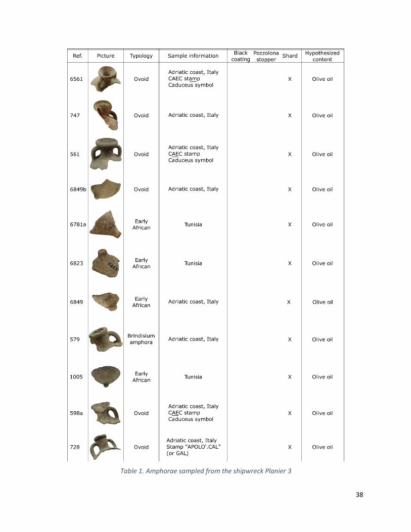
38
Table 1. Amphorae sampled from the shipwreck Planier 3

39
1.5.2. Archaeological site of San Felice Circeo
1.5.2.1. Archaeological context
The archaeological site of San Felice Circeo is located in the Latina province (Lazio), 90 km southeast of
Rome (Fig. 5, adapted from (Grimaldi & Spinapolice, 2010)). The Roman settlement was built on a rocky
plateau, between the plain that connects Circeo to Terracina on the one hand and the foothills of the
Circeo promontory on the other. The ancient city extended over a few hundred metres (150x200 m) at an
altitude of about 100 m (Fig. 6). The urban architecture developed in the 19th century bears witness to the
remains of the Roman era by including the main axes in the current layout, in particular the decumanus
axis, nowadays represented by the Corso Vittorio Emanuele, which belongs to the Roman period (de Rossi,
1973; Ronchi, 2017).
Figure 5. Location of San Felice Circeo
Although the city was occupied by the Volscians before the installation of the Roman colony in 393 BC
(as attested by Diodorus Siculus5), no visible traces of this occupation remain. Indeed, the great walls and
the acropolis at the top of the promontory were erected by the Romans. The settlement of the Circeii,
which was disputed for a long time between the Volscians and the Romans, justified the special “Latin-
right” the city suffered for years. The first historical writings testify to the sending of colonists to Circeo,
led by Tarquinio il Superbo, with the aim of setting up a policy of organization, referred as "praesidia Urbi
futura terra marique6". It is well established that in 340 BC the city belonged to the Latin League. This
5 Diodorus Siculus, Bibliotheca historica, XIV, 102 6 Livy, Ab Urbe Condita Libri, 56, 3

40
status implied, among others, a military and pecuniary duty towards the Empire, without enjoying the
advantages and privileges of the Roman citizenship. This intermediate position led the habitants of Circeii
to revolt against Rome in the first half of the fourth century BC. It is not excluded that this insurrection
was fomented by the descendants of the Volscians, partially present in the Circeii and its surroundings. At
the international level, the ratification of bilateral agreements between Rome and Carthage in 509 BC
ensured the economic development of the city by protecting it from foreign invasions for the next two
centuries. Indeed, the two capitals of the Empire agreed on regulating their respective interests by limiting
their geographical expansion. This mutual arrangement came to an end in 209 BC, during the Second Punic
War, when the Carthaginian army led by Hannibal's brother was sent into Italian territory. According to
the historical writings of Livy7, Circeii was one of the 12 colonies to rebel against the Rome's additional
demand for military contributions. The colony was heavily punished, 6 years later, by the injunction to
double the contingents of soldiers and provisions requested from the Empire. The lack of historical sources
does not allow a precise understanding of Circeo's occupation until the end of the Republic's status.
Although Circeii was granted Roman citizenship during the 1st century BC, the colony remains absent from
historical literature. This new statutory acquisition marked the beginning of the urban development of
the city, with the construction of villas and residential complexes under Roman influence. The city
therefore became a place of frequentation of the Roman politicians.
The port area was a major asset that ensured the connection with foreign cargoes. The existence of
ancient anchorage is supported by the ancient mouth of the Ufente river, that would have supplied water
for the city development (Cancellieri, 1986). The landing of goods was then relegated to support local
trading by terrestrial meant or foreigner marketing by sea exportation throughout the Empire.
Figure 6. San Felice Circeo typographies from: A; B. The oriental sector of San Felice Circeo with polygonal organisation of the actual city (on the right), the antic anchorage (on the bottom) and the
Neronis pit (on the top) (Quilici & Quilici Gigli, 2005); C: Circeo promontory (Ceruleo, 2021).
Thanks to the dominant position of the promontory of Circeo on the Tyrrhenian Sea (about 500 m far
from the coastal border), the city enjoyed an important strategic advantage in terms of trade but above
all in terms of protection against invasions. Circeo was included among the Roman cities with a strong
maritime and commercial influence following the Roman-Carthaginian treaty8. Indeed, the promontory
protected against north-west and north-east winds and allowed ships to anchor close to the Circeo coasts.
7 Livy, Ab Urbe Condita Libri, 27, 9 and 29, 15 8 Polibio, III, 22, 4-13.
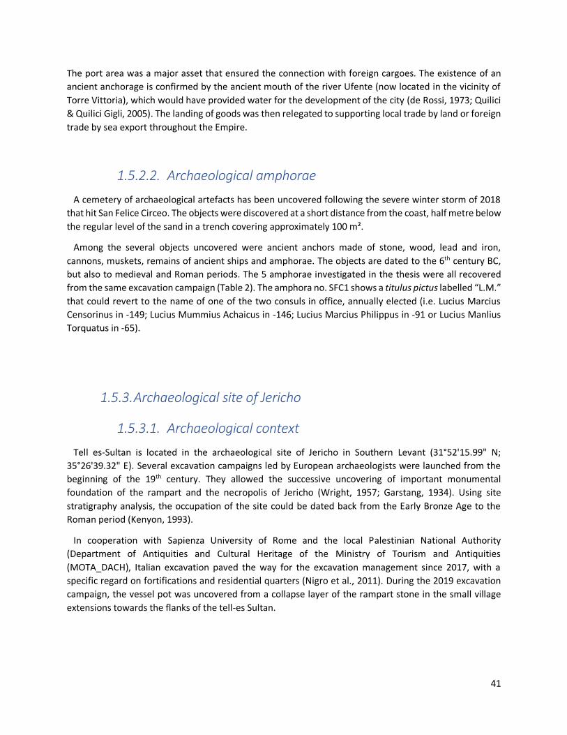
41
The port area was a major asset that ensured the connection with foreign cargoes. The existence of an
ancient anchorage is confirmed by the ancient mouth of the river Ufente (now located in the vicinity of
Torre Vittoria), which would have provided water for the development of the city (de Rossi, 1973; Quilici
& Quilici Gigli, 2005). The landing of goods was then relegated to supporting local trade by land or foreign
trade by sea export throughout the Empire.
1.5.2.2. Archaeological amphorae
A cemetery of archaeological artefacts has been uncovered following the severe winter storm of 2018
that hit San Felice Circeo. The objects were discovered at a short distance from the coast, half metre below
the regular level of the sand in a trench covering approximately 100 m².
Among the several objects uncovered were ancient anchors made of stone, wood, lead and iron,
cannons, muskets, remains of ancient ships and amphorae. The objects are dated to the 6th century BC,
but also to medieval and Roman periods. The 5 amphorae investigated in the thesis were all recovered
from the same excavation campaign (Table 2). The amphora no. SFC1 shows a titulus pictus labelled “L.M.”
that could revert to the name of one of the two consuls in office, annually elected (i.e. Lucius Marcius
Censorinus in ‐149; Lucius Mummius Achaicus in -146; Lucius Marcius Philippus in ‐91 or Lucius Manlius
Torquatus in -65).
1.5.3. Archaeological site of Jericho
1.5.3.1. Archaeological context
Tell es-Sultan is located in the archaeological site of Jericho in Southern Levant (31°52'15.99" N;
35°26'39.32" E). Several excavation campaigns led by European archaeologists were launched from the
beginning of the 19th century. They allowed the successive uncovering of important monumental
foundation of the rampart and the necropolis of Jericho (Wright, 1957; Garstang, 1934). Using site
stratigraphy analysis, the occupation of the site could be dated back from the Early Bronze Age to the
Roman period (Kenyon, 1993).
In cooperation with Sapienza University of Rome and the local Palestinian National Authority
(Department of Antiquities and Cultural Heritage of the Ministry of Tourism and Antiquities
(MOTA_DACH), Italian excavation paved the way for the excavation management since 2017, with a
specific regard on fortifications and residential quarters (Nigro et al., 2011). During the 2019 excavation
campaign, the vessel pot was uncovered from a collapse layer of the rampart stone in the small village
extensions towards the flanks of the tell-es Sultan.
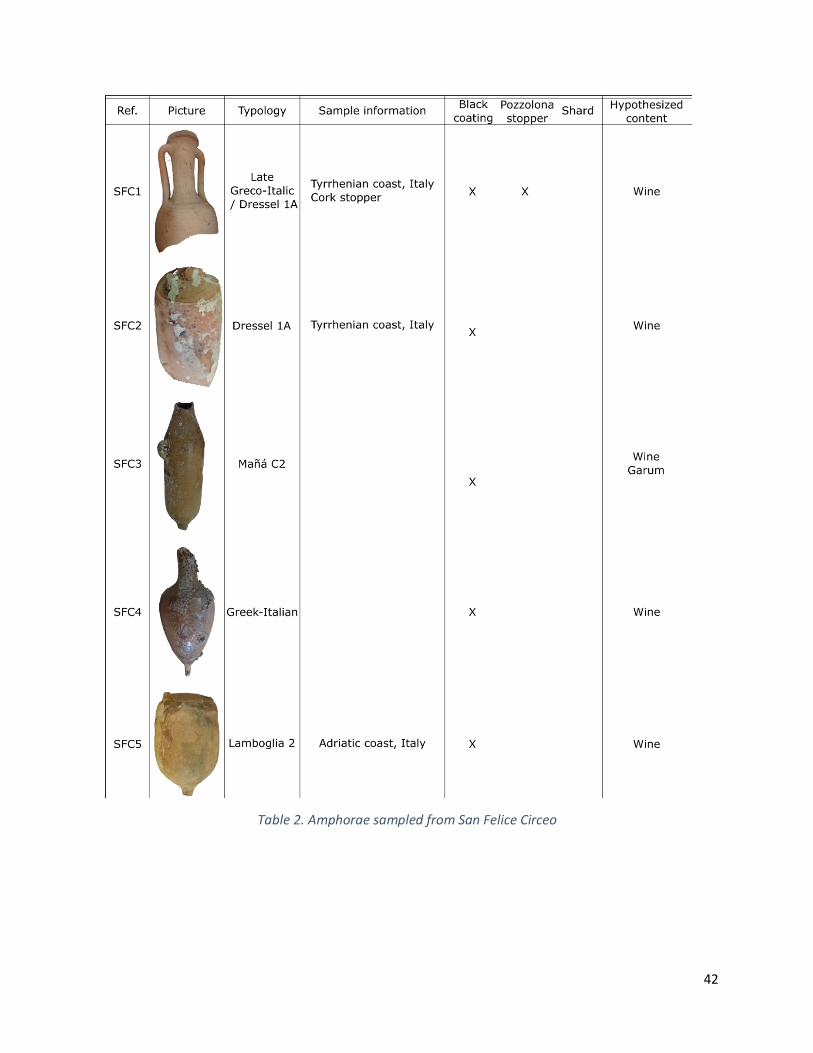
42
Table 2. Amphorae sampled from San Felice Circeo
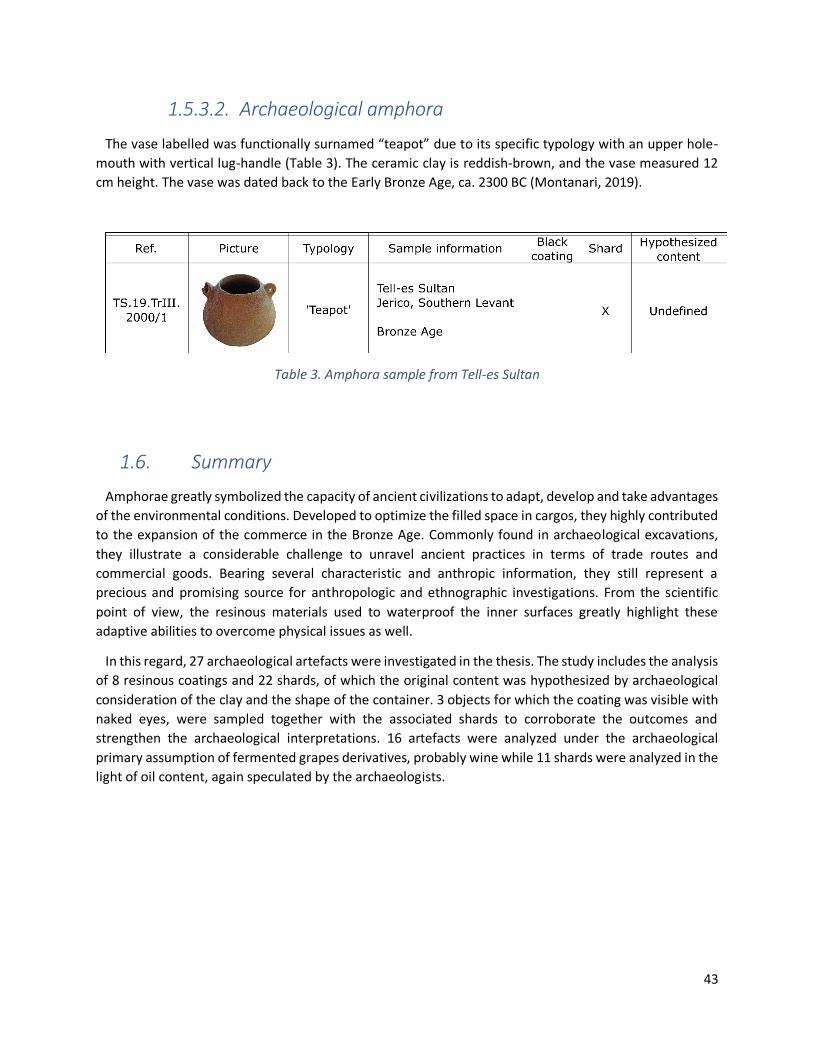
43
1.5.3.2. Archaeological amphora
The vase labelled was functionally surnamed “teapot” due to its specific typology with an upper hole-
mouth with vertical lug-handle (Table 3). The ceramic clay is reddish-brown, and the vase measured 12
cm height. The vase was dated back to the Early Bronze Age, ca. 2300 BC (Montanari, 2019).
Table 3. Amphora sample from Tell-es Sultan
1.6. Summary
Amphorae greatly symbolized the capacity of ancient civilizations to adapt, develop and take advantages
of the environmental conditions. Developed to optimize the filled space in cargos, they highly contributed
to the expansion of the commerce in the Bronze Age. Commonly found in archaeological excavations,
they illustrate a considerable challenge to unravel ancient practices in terms of trade routes and
commercial goods. Bearing several characteristic and anthropic information, they still represent a
precious and promising source for anthropologic and ethnographic investigations. From the scientific
point of view, the resinous materials used to waterproof the inner surfaces greatly highlight these
adaptive abilities to overcome physical issues as well.
In this regard, 27 archaeological artefacts were investigated in the thesis. The study includes the analysis
of 8 resinous coatings and 22 shards, of which the original content was hypothesized by archaeological
consideration of the clay and the shape of the container. 3 objects for which the coating was visible with
naked eyes, were sampled together with the associated shards to corroborate the outcomes and
strengthen the archaeological interpretations. 16 artefacts were analyzed under the archaeological
primary assumption of fermented grapes derivatives, probably wine while 11 shards were analyzed in the
light of oil content, again speculated by the archaeologists.

44

45
Chapter 2
The organic residues analysis: a state of the art

46
2.1. Identified substances within the pottery ceramic
Over the last century, the contribution of science and more specifically chemical investigations at the
molecular level have greatly influenced the archaeology. Advances in research, both in terms of analytical
technique development and characterization capacities have enabled the traceability of many contents.
In particular, the identification of fatty acid compounds from plant oils (Maria Perla Colombini, Giachi, et
al., 2005; J. Condamin et al., 1976) and animal fats from terrestrial or marine context (Copley et al., 2004;
R. P. Evershed et al., 2008; E. Ribechini et al., 2009; K. Romanus et al., 2007; Steele et al., 2010), but also
the botanical characterization of resins among natural exudates from all over the world (Mills & White,
1977; Erika Ribechini et al., 2008; Urem-Kotsou et al., 2002) and plants or animal waxes (Charters et al.,
1995; Garnier et al., 2002; Martine Regert et al., 2003).
2.1.1. Biomarker concept
In order to assess the nature of the content, Evershed et al. popularized what they called the
archaeological biomarker concept (Richard P Evershed, 1993). Currently used in the geochemistry to
characterize bitumen composition, the concept lies on the presence or absence of some molecular
markers whose carbon structure or distribution among the molecular content matched with the
archaeological compound ones. Rightly named “chemical fingerprints”, those components correspond to
organic features whose molecular structures are not highly altered with time. They are usually preserved
from microorganisms or bacterial deterioration activities (Philp & Oung, 1988). Surviving over centuries,
they can greatly give insight to the initial composition thereby (Maria Perla Colombini, Modugno, et al.,
2005; Martine Regert, 2011; Erika Ribechini et al., 2008).
Two identification pathways have been developed (Richard P Evershed, 1993; Carl Heron & Evershed,
1993). While some molecules are highly specific and facilitate direct content hypothesis, some other
characterizations rather depend on the molecular distribution. The molecule itself does not directly
illustrate the composition, but associated to other compounds, they can nevertheless provide indication.
Some patterns hence became relevant to decipher the archaeological composition acting as chemical
indirect markers. In the specific case of ceramic reutilization, the widened molecular profile generally
complicates the identification. Fortunately, recycling amphorae was unlikely although it occurred
frequently for vessel pottery (Pecci et al., 2017). However, it remains primordial to not only focus on the
target indication but to insert it in a broader picture to systematically measure the related archaeological,
botanical, palaeocological means to prevent overinterpretation.
Chemical marker, described hereafter, are generally classified into biomarkers, markers confirming
natural degradation or ageing, markers accrediting anthropic degradation and markers attesting
contamination.

47
2.1.1.1. Biomarkers
The identification step is driven by the presence or absence of biomarkers. At the molecular level, not
only the organic skeleton is informative but also its stereochemistry, the presence of unsaturations, their
locations and quantities. Any atomic or structural particularities became relevant and might be significant.
Heteroatoms and functional groups such as alcohols, ketones, hydrocarbons and fatty acids are of a great
support in the identification task. The assignment uncertainty degree decreases with the rareness of the
identified targets. For example, di- or triterpenoic acids thoroughly highlight the botanical nature of the
resin at the genus level, acting as a chimiotaxonomic guide (Font et al., 2007; Urem-Kotsou et al., 2002;
Zareva & Kuleff, 2010).
2.1.1.2. Natural degradation markers, sustainability and
preservation
The fundamental benefit of the biomarker concept lies in the ability to structurally target a molecule as
an exclusive characteristic toward species or compounds despite the variations it could have undergone.
Recognizing altered molecular patterns also means obtaining information not only on the natural origin
of the compounds but also on the degradation conditions involved. This way, biomarkers can offer an
additional degree of diagnosis.
By introducing the biomarker concept, Evershed et al. also recommended the careful interpretation of
the markers regarding their natural degradation pathway possibilities. Indeed, it is often possible to
observe slightly altered structures rather than the original ones. As described previously, various
transformation pathways could occur to modify the molecular pattern of biomarkers, spanning from
microbial and enzymatic deterioration to atomic changes through oxidative reactions. Chemical and
biochemical transformations could be stimulated by archaeological conditions of storage. For example,
light exposure, aerobic conditions, humidity and temperature can strongly affect biomarkers (R P
Evershed et al., 1991; Martine Regert, 2011). Based on tailored replica ceramics, Evershed et al. studied
the oxidative aerial effect on acyl lipids conservation over time and concluded to faster degradation under
oxic environments (R. P. Evershed et al., 2008). Archaeological jars recovered from tombs under anaerobic
and isothermal conditions usually display good preservation state. Arid climates such as desert sites,
where the moisture content is specifically low guarantee molecular preservation preventing from
microbial development.
Aside from the chemical nature and physico-chemical properties that largely affected the lifetime of
organic residues biomarkers, several factors could account for biomarkers preservation against microbial
degradation (Carl Heron & Evershed, 1993). Two environmental aspects need to be distinguished. Firstly,
the presence of mineral matrices and ceramic porosity can notably enhance molecular stability over
archaeological times. Indeed, organic residues adsorbed in the porous ceramic layer remain therefore less
available because harder to reach for microorganisms (Eglinton & Logan, 1991). Additionally, the organic
matrices seemed to act as a physical barrier. For instance, resin or bitumen are good example of residues
preservation because microorganisms could not attain them and thereby they prevent from organic
residues decay (Font et al., 2007; T.F.M. Oudemans & Boon, 1991). In archaeometry, lipids, in the broadest
48
sense, are the predominant marker family. The long carbon chain is responsible for their hydrophobic
properties and low reaction abilities. Secondly, some biomarkers can strongly interact with mineral
cations from the ceramic to form salts. This environmental bonding possibility can partially ensure their
preservation. This is the case for the tartaric acid, usually preserved as tartrate salts in ceramic shards (Léa
Drieu et al., 2020; P. McGovern et al., 2017; P Ribéreau-Gayon et al., 2006).
2.1.1.3. Anthropic degradation markers, an interesting signature
of human activities
In the early days of archaeometry, some experimental studies led by Charters, Evershed et al. became
pillars of the field, notably by establishing the beginnings of scientific input. The main purpose was a
deeper understanding of the molecular modifications undergone through heat treatment on the natural
organic matter. They performed laboratory experiments on replica ceramic potteries to simulate
archaeological vessel uses and extended it to excavated jars (Charters et al., 1995, 1997; R. Evershed et
al., 1995; Raven et al., 1997). They referred to these organic altered features as anthropic markers. They
notably give insight on human practices and manufacture technologies applied to the matter to change
its physical properties (R. P. Evershed et al., 2008; R P Evershed et al., 1991).
Some great examples emanate from harsh heating treatments implemented on resin. High temperature
of approximately 300°C represented enough energy to promote molecular markers of the natural
exudates to stabilize upon aromatization as well as losing some functional groups. Resulting markers
showed a coherent carbon skeleton, slightly but significantly modified from the precursor ones. For
example, retene and 28-norolean-17-en-3-one compounds have been identified from respectively
diterpenic resin from Coniferous and triterpenic resin from Pistacia spp. (Fig. 7) Their occurrences in resin
indicate a considerable heating during their preparation (Mills & White, 1977, 1989; B. Stern et al., 2003).
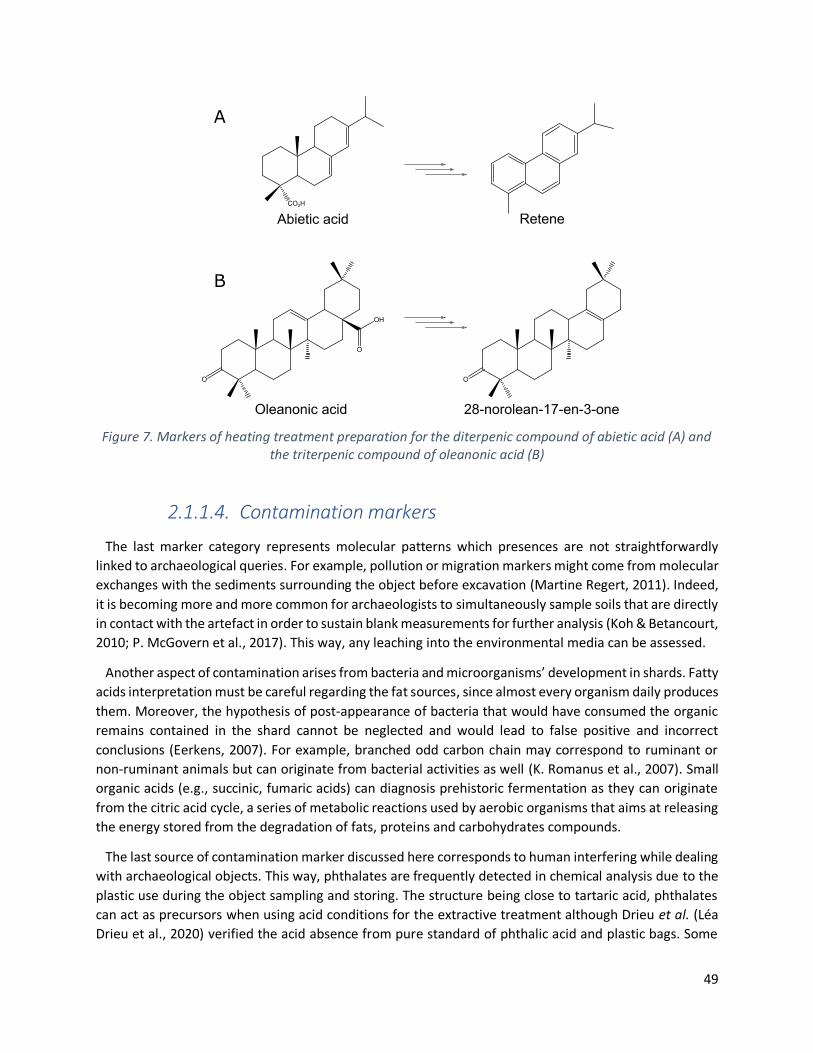
49
Figure 7. Markers of heating treatment preparation for the diterpenic compound of abietic acid (A) and the triterpenic compound of oleanonic acid (B)
2.1.1.4. Contamination markers
The last marker category represents molecular patterns which presences are not straightforwardly
linked to archaeological queries. For example, pollution or migration markers might come from molecular
exchanges with the sediments surrounding the object before excavation (Martine Regert, 2011). Indeed,
it is becoming more and more common for archaeologists to simultaneously sample soils that are directly
in contact with the artefact in order to sustain blank measurements for further analysis (Koh & Betancourt,
2010; P. McGovern et al., 2017). This way, any leaching into the environmental media can be assessed.
Another aspect of contamination arises from bacteria and microorganisms’ development in shards. Fatty
acids interpretation must be careful regarding the fat sources, since almost every organism daily produces
them. Moreover, the hypothesis of post-appearance of bacteria that would have consumed the organic
remains contained in the shard cannot be neglected and would lead to false positive and incorrect
conclusions (Eerkens, 2007). For example, branched odd carbon chain may correspond to ruminant or
non-ruminant animals but can originate from bacterial activities as well (K. Romanus et al., 2007). Small
organic acids (e.g., succinic, fumaric acids) can diagnosis prehistoric fermentation as they can originate
from the citric acid cycle, a series of metabolic reactions used by aerobic organisms that aims at releasing
the energy stored from the degradation of fats, proteins and carbohydrates compounds.
The last source of contamination marker discussed here corresponds to human interfering while dealing
with archaeological objects. This way, phthalates are frequently detected in chemical analysis due to the
plastic use during the object sampling and storing. The structure being close to tartaric acid, phthalates
can act as precursors when using acid conditions for the extractive treatment although Drieu et al. (Léa
Drieu et al., 2020) verified the acid absence from pure standard of phthalic acid and plastic bags. Some
50
contaminants can appear during the chemical extraction of the material, from solvents for example, even
though analytical grades are preconized, or from filter cartridges made of plastic derivatives. Additionally,
the occurrence of cholesterol and other squalene precursors can arise from ungloved handling. The
contamination might go through physical contact without good protection. Besides the fact that squalene
is a characteristic organic compound produced by the sebaceous gland (Picardo et al., 2009), its important
poly-unsaturation feature would indeed not survive over archaeological centuries. Sterol presences must
be interpreted with caution, in particular when abundantly detected. Considered as biomarkers of plant
oils due to their natural presence therein, they can become contamination markers when present in large
extent since their regular distribution in plants is not so important. Additionally, some fungal markers such
as ergosterol can help in noticing contamination from soil fungi or yeast (Stephanie N. Dudd et al., 1998)
as well as characterizing alcoholic fermentation (Isaksson et al., 2010). In general, lipid compounds such
as fatty acids, wax esters, squalene, sterols and its ester derivatives must be considered with prudence
since human skins can produce them (Richard P Evershed, 1993).
2.1.2. Terpenes
2.1.2.1. Definition and structure
To protect themselves from external attacks, resinous plants can produce viscous exudates to dress their
wounds, singularly characterized by their chemical composition. Resins are better itemized as oleo-
gommo-resins. “Oleo” points out their high fragrant features providing antibacterial properties and
corresponds to the essential oil fraction made of mono- and sesquiterpenoids, respectively made of 10
and 15 carbon atoms. “Gommo” invokes their sticky aspect related to the presence of many
polysaccharides chains and “resin” describes their molecular organic composition represented by di- (C20)
or triterpenoids (C30) depending on the nature of the resinous materials (Fig. 7). In archaeological samples,
it is unfortunately rare to notice oleo and gommo fractions. The former is composed of volatile organic
compounds that disappeared with time. The latter is hydrosoluble. Consequently, the resin fragment,
which displays the best molecular stability, remains the more relevant to characterize in order to decipher
their botanical and geographical origins as well as the technologies involved to prepare them.
From a chemical point of view, resins exhibit great molecular pluralities, which make their
characterization even more complex. Since terpenes are not playing any role in plant cell growth, they are
considered secondary metabolites. Terpenes are biosynthesized by plants through the “head to tail”
assemblage C5 isoprene units. Formed chains are then arranged in rings (Fig. 8).
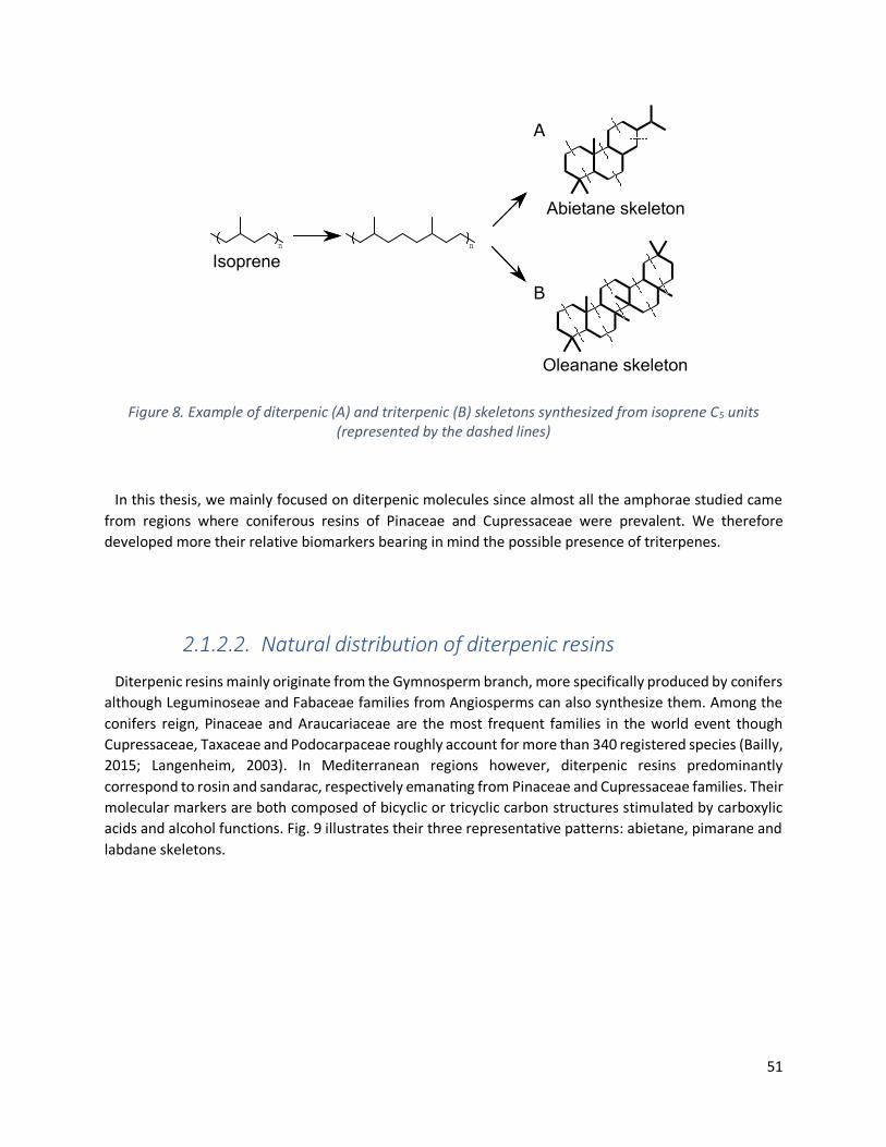
51
Figure 8. Example of diterpenic (A) and triterpenic (B) skeletons synthesized from isoprene C5 units (represented by the dashed lines)
In this thesis, we mainly focused on diterpenic molecules since almost all the amphorae studied came
from regions where coniferous resins of Pinaceae and Cupressaceae were prevalent. We therefore
developed more their relative biomarkers bearing in mind the possible presence of triterpenes.
2.1.2.2. Natural distribution of diterpenic resins
Diterpenic resins mainly originate from the Gymnosperm branch, more specifically produced by conifers
although Leguminoseae and Fabaceae families from Angiosperms can also synthesize them. Among the
conifers reign, Pinaceae and Araucariaceae are the most frequent families in the world event though
Cupressaceae, Taxaceae and Podocarpaceae roughly account for more than 340 registered species (Bailly,
2015; Langenheim, 2003). In Mediterranean regions however, diterpenic resins predominantly
correspond to rosin and sandarac, respectively emanating from Pinaceae and Cupressaceae families. Their
molecular markers are both composed of bicyclic or tricyclic carbon structures stimulated by carboxylic
acids and alcohol functions. Fig. 9 illustrates their three representative patterns: abietane, pimarane and
labdane skeletons.
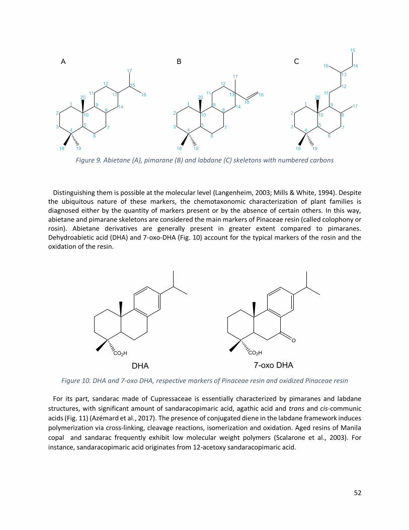
52
Figure 9. Abietane (A), pimarane (B) and labdane (C) skeletons with numbered carbons
Distinguishing them is possible at the molecular level (Langenheim, 2003; Mills & White, 1994). Despite the ubiquitous nature of these markers, the chemotaxonomic characterization of plant families is diagnosed either by the quantity of markers present or by the absence of certain others. In this way, abietane and pimarane skeletons are considered the main markers of Pinaceae resin (called colophony or rosin). Abietane derivatives are generally present in greater extent compared to pimaranes. Dehydroabietic acid (DHA) and 7-oxo-DHA (Fig. 10) account for the typical markers of the rosin and the oxidation of the resin.
Figure 10. DHA and 7-oxo DHA, respective markers of Pinaceae resin and oxidized Pinaceae resin
For its part, sandarac made of Cupressaceae is essentially characterized by pimaranes and labdane
structures, with significant amount of sandaracopimaric acid, agathic acid and trans and cis-communic
acids (Fig. 11) (Azémard et al., 2017). The presence of conjugated diene in the labdane framework induces
polymerization via cross-linking, cleavage reactions, isomerization and oxidation. Aged resins of Manila
copal and sandarac frequently exhibit low molecular weight polymers (Scalarone et al., 2003). For
instance, sandaracopimaric acid originates from 12-acetoxy sandaracopimaric acid.

53
Figure 11. Pimarane (A) and labdane (B-C) skeleton markers of Cupressaceae resin
Likewise, some phenolic compounds derived from oxidation and rearrangement of abietane skeletons
such as ferruginol, totarol, sempervirol and sugiol are considered as Cupressaceae biomarkers (Fig. 12)
(Steigenberger, 2013).
Figure 12. Oxidized phenol markers of Cupressaceae resin
2.1.2.3. Degradation pathways
Besides the botanical indication provided by the presence of characteristic frames, expression of past
technologies to acquire the resin can be traduced by natural and anthropic markers related to abietane
skeleton transformation under specific conditions. Abietanes represent the main source of archaeological
information since the pattern is the most reactive one. Indeed, one structural information is primordial to
notice. Abietanes exhibit diene conjugation compared to pimaranes. This conjugation is notably the
chemical reason for the great reactivity of abietanes towards alterations or heat treatments. Pimaranes
that do not have this conjugation because of a quaternary carbon then present a more restricted set of
markers (Scalarone et al., 2003).
Once the resin exuded from the tree, the diterpenic molecular composition starts to change with aerial
oxygen. Isomerizations occur to stabilize abietic (A1), palustric (A2), neoabietic (A3) and laevopimaric (A4)

54
acids (Fig. 13, adapted from (Mezzatesta et al., 2021a)). Among them, abietic acid is the most prevalent
isomer. The natural degradation may be assimilated to the ageing of the resin, triggered by the oxidation
of the conjugated diene of the abietic acid to form DHA (A5) (Mills & White, 1994). Several further
oxidations occur then to integrate oxygen via hydroxy or ketone groups. The exact mechanism remains
obscure, but it shall involve peroxide formation, loss of water and/or even peroxide reduction.
Oxygenation takes place at three likely reactive positions (3, 7 and 15), giving rise to 3-hydroxy-DHA (A8),
7-hydroxy-DHA (A6), 15-hydroxy-DHA (A9) and 7-oxo-DHA (A7). In turn, molecules can keep going on with
oxidation and isomerization to form dihydroxy-DHA and hydroxy-oxo-DHA, respectively named 7,15-
dihydroxy-DHA (A10) and 15-hydroxy-7-oxo-DHA (A11) (Fujii et al., 2021; Serpico & White, 2000; van Den
Berg et al., 2000).
Another part of the reaction pathway concerns anthropic heating treatment. Above 300°C, DHA is
transformed in norabietatriene and norabietatetraene species (usually 18 or 19-norabietatriene (A14 and
A15)) via the removal of acidic function. The resulting compounds are stabilized through partial or
complete aromatization, giving first rise to tetrahydroretene (A18) and finally retene (A19).
Simultaneously, DHA dehydrogenation generated dehydrate-DHA formation, which forms
tetrahydroretene after aromatization of the intermediate (Serpico & White, 2000). Likewise,
dehydroabietane (A12) can generate simonellite (A13) by aromatization and finally generate
tetrahydroretene and retene upon decarboxylation (Bailly, 2015).
At last, when the tar was prepared by pyrolysis of the coniferous tree, DHA is esterified by the methanol
present in the wood slowly released by the raise of temperature, producing methyl dehydroabietate
(DHAM). It is interesting to notice that these methylated compounds are subject to similar oxidative
pathways as DHA (Pollard & Heron, 2008).
As presented above, the wide variety of potential chemical reactions illustrates the ability of abietanes
to provide archaeological evidence. First, the botanical source is deducted from the molecular
composition and the occurrence of abietadienic, pimaradienic or labdadienic species. Indeed, the
presence of oxidized DHA is then indicative of the natural degradation process followed by the resin over
time. Stable markers such as 7,15-dihydroxy-DHA and 15-hydroxy-7-oxo-DHA make it possible to describe
the ageing of the pitch as well as environmental factors such as oxygen exposure. Additionally, the
presence of highly aromatic molecules such as retene clearly points out artificial transformations triggered
by human activities, suggesting high temperature heating during preparation. Finally, methyl ester
compounds shed light on the pyrolytic manufacture treatment from the resinous softwood.

55
Figure 13. Abietane markers degradation pathway from a Pinaceae resin

56
2.1.2.4. Distinguishing resin form pitch and tar
The archaeological difference between resin, pitch and tar can be transposed to chemical queries. The
main difference lies in their molecular composition. Nevertheless, how do they evolve under formulation
and heating practices? Thermally altered features have been mentioned previously to distinguish them.
However, caution regarding the interpretation is necessary and DHAM becomes a biomarker of
formulation via wood distillation only when hydrocarbon polycyclic derivatives are also characterized.
Indeed, even if phenanthrenes, norabietatrienes and simonellite are markers of artificial activities, their
production requires high-energy, usually provided by intensive heating under anaerobic conditions
(Rageot, 2015).
To describe plant substances from the Pinaceae family, the term resin covers the natural exudate of
Gymnosperm trees. When the resin has undergone heating treatment, the common appellation becomes
pitch. Lastly, the wood tar corresponds to the pyrolysis of the wood with the resin.
2.1.3. Wine residues
2.1.3.1. A brief introduction to wine
Wine remains are one of the most promising content to study in order to gain knowledge on ancient
trade routes and Mediterranean economy (Pecci, Cau Ontiveros, et al., 2013). Besides the great analytical
challenge to face, fermented grape characterization might greatly supply practical and philosophical
queries spanning from the ancestral methods to prepare wine to the religious, social, political or hedonic
aspect it might confer (Maria Rosa Guasch-Jané, Andrés-Lacueva, Jáuregui, et al., 2006). Indeed, wine has
been identified in Shedeh jars found in the Tutankhamun’s Tomb in Egypt, which greatly highlight its divine
bestowed aspect. The earliest method to attest wine presence were going through amphorae typologies.
Various morphologies dated from the 5th century BC to the 1st century AC have been mentioned for
containing wine. Within the following non-exhaustive list, wine residues have been notably detected from
Corinthian amphorae coming from Greek islands of Corfu and Mende, Roman Dressels and Late Roman or
African Keay amphorae (Foley et al., 2012; Fujii et al., 2019; Pecci et al., 2010, 2017; Martine Regert et al.,
2003; Woodworth et al., 2015).
From the historical point of view, wine assumption is also relevant to chronologically address the
domestication of grape cultivars. Earliest evidence therefore became more important since they also drew
back in time our knowledge about the population abilities to take advantage of their environment. Based
on molecular markers and supported by palynological and xylological observations, McGovern et al.
recently recorded the earliest evidence of wine from an Early Neolithic pottery roughly dated to 6000 -
5800 BC (P. McGovern et al., 2017). Both excavations took place in the northern regions of the Zagros
Mountains in Caucasus. From there, they suggested that large-scale winemaking might have been
initiated (P. E. McGovern, 2013). Even if no relevant proof of domesticated grape cultivars has been
evidenced in these sites, the domestication hypothesis relied on the fact that archaeological artefacts
have been found out of the extension area of wild plants. Indeed, wild and cultivated Vitis species usually

57
develop in different environments from each other (Bouby et al., 2013). The cultivated V. vinifera is
supposed to emanate from its wild V. vinifera subsp silvestris ancestor (Louis Levadoux, 1956). Despite
the contribution of various investigative fields including natural sciences, history and archaeology, the
origins of the cultivated vines, as well as the conditions involved in the extension and development of its
cultivation in the Mediterranean basin are far from being perfectly understood. However, it remains
important to notice that several studies of different cultivated species find their roots in the Caucasus
regions, where the observed morphological and genetic diversity among wild species and local cultivars is
significantly ample (Coito et al., 2019; Milanesi et al., 2016; This et al., 2006; Zohary, 1995).
Back to the historical context, grape markers have also been detected from Neolithic potteries from the
5th millennium BC in the archaeological site of Dikili Tash in Greece (Garnier & Valamoti, 2016; S. M.
Valamoti et al., 2007). Chemical traces of fermented grapes were found in Ancient Egyptian vessels,
roughly dated to 3000 BC (Maria Rosa Guasch-Jané et al., 2004). Domesticated cultivars have been
identified by carpological analysis of grape pips. Grape cultivars have been attested in the 3rd millennium
BC in Minor Asia, Southern Greece (Banilas et al., 2012). The early cultivation in Cyprus corresponds to
the same period, when the island was under Phoenician’s domination (Grassi et al., 2003). Slowly
spreading over, wine production has also been evidenced by the presence of cultivar vines in 2200 BC in
Crete to finally expend westwards from Northern Italy to Southern France, Spain and Portugal two
centuries later under the influence of the Roman Empire (Arroyo-Garcia et al., 2006; Coito et al., 2019;
Hardie, 2000; Hopf, 1991; P. E. McGovern et al., 2004).
The identification of archaeological wine is the result of multidisciplinary studies. Morphological analysis
of pips and woods, molecular characterization and finally genetic analysis represent the three pillars of
current scientific investigations. In addition to the archaeological contributions abovementioned, the
chemical characterization of wine may corroborate with the study of grapevine domestication and cross-
conclusions might be therefore drawn. It however remains primordial to consider the grapevine
domestication as a complex phenomenon, multi-located and slow processing (Bouby et al., 2013; Soultana
Maria Valamoti et al., 2020). Indeed, the emergence of alternative hypothesis regarding the multi origins
of vineyard domestication is widely supported by genetic and morphometric evidence. Evolution of DNA
possibilities and routine genetic sequencing brought on light the relevant contribution of local wild species
in the genome of the modern cultivars. Autochthonous are additionally identified all over the
Mediterranean basin (Cargnello et al., 1980; Lombardo et al., 1978; Maletić et al., 2015). Northern Greece,
Sardinia but also the Iberian Peninsula have been mentioned to be the epicenters of vineyard
domestication along with the Caucasus (Arroyo-Garcia et al., 2006; Bouby et al., 2013; Grassi et al., 2003;
Myles et al., 2011; Ucchesu et al., 2015; Soultana Maria Valamoti et al., 2020).
2.1.3.2. Composition
The chemical composition of wine, to say the least, is complex. While the major components remain
water and ethanol, hundreds of other compounds are also present. Among them, some key organic acids
and polyphenol patterns, more specifically phenolic acids and anthocyanins will be discussed further since
they represent the major degradation markers to trace back wine. These biosynthetic components explain
the great diversity in terms of aroma, color, and wine taste. Phenolic compounds greatly balance the
beverage astringency, bitterness, sourness and account for the body aroma (Jackson, 2008).

58
2.1.3.2.1. Phenolic acids
Chemically speaking, phenolic acids correspond to molecules whose organic structures present an
aromatic ring substituted with functional groups such as hydroxyl, ester or methyl ether. Plants are rich
of these non-flavonoid acids, conferring those flavors and gustative characters. The astringency is for
instance due to the dimerization of specific phenolic acids giving rose to hydrosoluble tannins (Garnier,
2003). While monomeric flavonoids influence wine bitterness, some phenolic acids such as caffeic or p-
coumaric acting as H+ donors account for wine sourness (Salameh et al., 2008). Only the case of grape and
vine derivatives composition will be developed since this thesis mainly focused on wine characterization.
As illustrated in Fig. 14, polyphenols are classified upon their lateral carbon chain number into benzoic
acids (single carbon), phenylacetic acids (two carbons) and cinnamic acids (three carbons with central
trans unsaturation). Gallic and p-coumaric acids are respectively the most abundant benzoic and cinnamic
compounds in grape and vine. The latter is usually present through its esterified form, bound to tartaric
acid.
Figure 14. Molecular structure of polyphenols (Garnier, 2003)
Phenolic compounds are mainly constrained in the flesh of the fruits, where they are biosynthesized.
Their concentrations increase with time and ripening of the fruit before being consumed as a precursor
for the formation of flavonoids, which marks their decline (Belitz et al., 2009). After being grabbed and
transformed, polyphenol concentrations keep evolving and can decrease with time upon their antioxidant
actions towards lipids auto-oxidation. Easily oxidized, polyphenols can indeed be consumed by capturing
peroxy radicals. Alternatively, the concentration can also increase by oxidative flavonoid degradation
while wine is ageing or by oxidative enzymatic reaction occurring to adapt and facilitate the alcoholic
fermentation (Edwin N. Frankel, 2010; Pascal Ribéreau-Gayon et al., 1998).
Wine ageing coincides with the oxidation of polyphenols and notably unsaturated cinnamic acids
triggered by radical reactions. One striking observation arises from the ability of resulting compounds to
polymerize with anthocyanins and other flavonoids, giving rise to insoluble high molecular weight
polymers (Garnier, 2003).
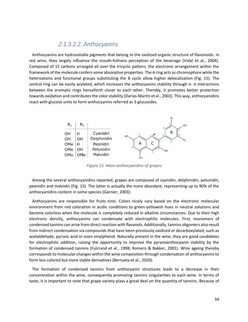
59
2.1.3.2.2. Anthocyanins
Anthocyanins are hydrosoluble pigments that belong to the oxidized organic structure of flavonoids. In
red wine, they largely influence the mouth-fullness perception of the beverage (Vidal et al., 2004).
Composed of 15 carbons arranged all over the tricyclic pattern, the electronic arrangement within the
framework of the molecule confers some absorptive properties. The A ring acts as chromophore while the
heteroatoms and functional groups substituting the B cycle allow higher delocalization (Fig. 15). The
central ring can be easily acylated, which increases the anthocyanins stability through π- π interactions
between the aromatic rings henceforth closer to each other. Thereby, it promotes better protection
towards oxidation and contributes the color stability (Darias-Martín et al., 2002). This way, anthocyanidins
react with glucose units to form anthocyanins referred as 3-glucosides.
Figure 15. Main anthocyanidins of grapes
Among the several anthocyanidins reported, grapes are composed of cyanidin, delphinidin, petunidin,
peonidin and malvidin (Fig. 15). The latter is actually the more abundant, representing up to 90% of the
anthocyanidins content in some species (Garnier, 2003).
Anthocyanins are responsible for fruits tints. Colors nicely vary based on the electronic molecular
environment from red coloration in acidic conditions to green-yellowish hues in neutral solutions and
become colorless when the molecule is completely reduced in alkaline circumstances. Due to their high
electronic density, anthocyanins can condensate with electrophilic molecules. First, monomers of
condensed tannins can arise from direct reaction with flavanols. Additionally, tannins oligomers also result
from indirect condensation via compounds that have been previously oxidized or decarboxylated, such as
acetaldehyde, pyruvic acid or even vinylphenol. Naturally present in the wine, they are good candidates
for electrophilic addition, raising the opportunity to improve the pyranoanthocyanin stability by the
formation of condensed tannins (Fulcrand et al., 1998; Romero & Bakker, 2001). Wine ageing thereby
corresponds to molecular changes within the wine composition through condensation of anthocyanins to
form less colored but more stable derivatives (Berrueta et al., 2020).
The formation of condensed tannins from anthocyanin structures leads to a decrease in their
concentration within the wine, consequently promoting tannins singularities to each wine. In terms of
taste, it is important to note that grape variety plays a great deal on the quantity of tannins. Because of

60
this important capacity to polymerize, it seems unlikely to characterize anthocyanins in archaeological
potteries. A first indication of their absence directly comes from the bland color of the wine.
2.1.3.2.3. Organic acids
Organic acids are prevalent in wine and largely account for the appreciated sourness of beverages,
generating disagreeable acidity that covers the other wine flavors (Polaskova et al., 2008). Among the
recurrent organic acids reported in grapes (Fig. 16), tartaric acid remains the most abundant one from the
unripe grapes to the must (Margalit, 2012). Significantly biosynthesized by the Vitis vine, it has an essential
influence on wine taste and can deteriorate flavors when highly concentrated (Sass-Kiss et al., 2008). It is
important to note that malic, citric and tartaric acids account all together for many of the acidic grape
components. Unfortunately, malic and citric acids are naturally present in living organisms. Citric acid is
highly involved in organism growth since it has a crucial biochemical and metabolic role in the Krebs cycle.
The excess of malic acid can induce unpleasant wine acidity that can be tackled by malolactic
fermentation, producing lactic acid out of malic acid. Their wide distribution does not make them suitable
biomarkers.
Figure 16. Essential anthocyanidins of grapes
Additionally, the oxidized aldehyde or primary alcohol functions of ketose may indicate that grapes or
must were rotting. Resulting gluconic acid and its derivatives are markers of noble or gray rot. Ketoglutaric
acid has been mentioned to nicely enhance wine’s flavor by bonding with free sulphur compounds and
consequently reducing distasteful perfumes (Arias-Gil et al., 2007).
The molecular composition of wine, as well as the composition of grapes, is not stable upon time. Both
keep evolving after winemaking. Illustrated in Fig. 17, several acids are directly involved in the
fermentation process. Even if pyruvic acid is unlikely to find in wine, its crucial precursor role in cell
metabolism needs to be noticed. For example, it gives rise to lactic acid through enzymatic reductions.
The resulted stereoisomers can either emanate from bacterial or yeast origins (Swiegers et al., 2005).
Likely, oxaloacetic acid comes from carboxylation of pyruvic acid; acetic acid arises from enzymatic
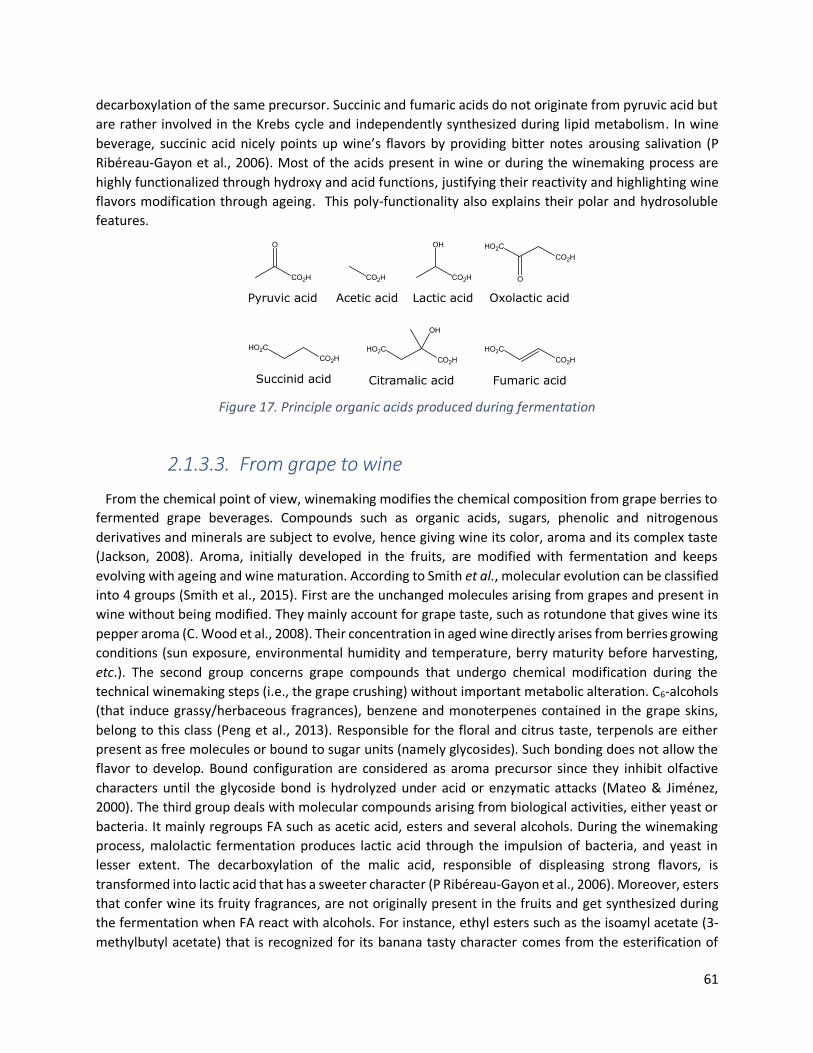
61
decarboxylation of the same precursor. Succinic and fumaric acids do not originate from pyruvic acid but
are rather involved in the Krebs cycle and independently synthesized during lipid metabolism. In wine
beverage, succinic acid nicely points up wine’s flavors by providing bitter notes arousing salivation (P
Ribéreau-Gayon et al., 2006). Most of the acids present in wine or during the winemaking process are
highly functionalized through hydroxy and acid functions, justifying their reactivity and highlighting wine
flavors modification through ageing. This poly-functionality also explains their polar and hydrosoluble
features.
Figure 17. Principle organic acids produced during fermentation
2.1.3.3. From grape to wine
From the chemical point of view, winemaking modifies the chemical composition from grape berries to
fermented grape beverages. Compounds such as organic acids, sugars, phenolic and nitrogenous
derivatives and minerals are subject to evolve, hence giving wine its color, aroma and its complex taste
(Jackson, 2008). Aroma, initially developed in the fruits, are modified with fermentation and keeps
evolving with ageing and wine maturation. According to Smith et al., molecular evolution can be classified
into 4 groups (Smith et al., 2015). First are the unchanged molecules arising from grapes and present in
wine without being modified. They mainly account for grape taste, such as rotundone that gives wine its
pepper aroma (C. Wood et al., 2008). Their concentration in aged wine directly arises from berries growing
conditions (sun exposure, environmental humidity and temperature, berry maturity before harvesting,
etc.). The second group concerns grape compounds that undergo chemical modification during the
technical winemaking steps (i.e., the grape crushing) without important metabolic alteration. C6-alcohols
(that induce grassy/herbaceous fragrances), benzene and monoterpenes contained in the grape skins,
belong to this class (Peng et al., 2013). Responsible for the floral and citrus taste, terpenols are either
present as free molecules or bound to sugar units (namely glycosides). Such bonding does not allow the
flavor to develop. Bound configuration are considered as aroma precursor since they inhibit olfactive
characters until the glycoside bond is hydrolyzed under acid or enzymatic attacks (Mateo & Jiménez,
2000). The third group deals with molecular compounds arising from biological activities, either yeast or
bacteria. It mainly regroups FA such as acetic acid, esters and several alcohols. During the winemaking
process, malolactic fermentation produces lactic acid through the impulsion of bacteria, and yeast in
lesser extent. The decarboxylation of the malic acid, responsible of displeasing strong flavors, is
transformed into lactic acid that has a sweeter character (P Ribéreau-Gayon et al., 2006). Moreover, esters
that confer wine its fruity fragrances, are not originally present in the fruits and get synthesized during
the fermentation when FA react with alcohols. For instance, ethyl esters such as the isoamyl acetate (3-
methylbutyl acetate) that is recognized for its banana tasty character comes from the esterification of
62
acetic acid with alcohol produced during the fermentation. The last category specifically targets
compounds responsible for the wine color that are also produced by metabolic activities of yeast or
bacteria. It includes thiols, tannins and polysaccharides. Anthocyanins, mentioned above, promote
reddish coloration. Although they are not stable over a long time period, they disappear slightly and let
the tannins take precedence over the coloration (Šuklje et al., 2016). During the winemaking process, this
last category of compounds might arise from enzymatic reactions, oxidation or pre-fermentation of the
crushed skins in the must.
Early on fermentation was surely triggered by the addition of fruits into the beverages (Nigro & Rinaldi,
2020). The assumption comes from the recent discovery of Saccharomyces cerevisiae, the yeast
responsible for fermentation, into the pollinator’s guts (Meriggi et al., 2020; Stefanini et al., 2016). Insects
could therefore transfer the yeast to the fruits while pollinating, hence giving a valuable way to control
the spontaneous fermentation induced by the reaction with the initial sugar contained in the fruit meshes.
Alternatively, honey has been highly mentioned to trigger the fermentation process as well, since
pollinator insects surely provided the Saccharomyces cerevisiae yeast (P. McGovern et al., 2017; P. E.
McGovern, 2013). Egyptian used honey to modulate the beverage taste: when added before the
fermentation was achieved, it raised the alcohol degree while it rather sweetened wines when added at
the end of the fermentation (Rösch, 2005). Besides the taste benefit, honey provided a great deal to
control the fermentation process.
2.1.3.4. Tartaric acid: a suitable biomarker?
Chemical characterization of wine in archaeological artefacts is nowadays routinely achieved through
the biomarker concept, alike the identification of resin. Nevertheless, one central query remains how
viable are the markers for wine traceability.
Among the wide scope of organic molecules present in wine, tartaric acid has been greatly awarded to
evidence grape beverages since it is indeed one of the most abundant organic acid naturally present in
grapes (Maria Rosa Guasch-Jané et al., 2004; Maria Rosa Guasch-Jané, Andrés-Lacueva, Jáuregui, et al.,
2006). It is also interesting to note that grapes ripeness, varieties as well as their origins influence its
concentration (Ben Stern et al., 2008).
Tartaric acid potential as biomarker arises from its high concentration in grape towards other plant
material. Two cases need to be distinguished. First, the molecule is synthesized with the metabolism of
ascorbic acid in many vegetables and fruits. However, and therein lies all the subtlety, it is present in lower
extent (roughly 100 mg/L compared to approximatively 5 g/L in grapes) (Singleton, 1996). Other edible
products are consequently not supposed to release enough acid to be remarkably extracted during the
analysis, especially if they underwent degradation. Contrariwise, producing wine out of grapes does not
consume tartaric acid, affording fermented beverages to remain with significant acid concentration (P
Ribéreau-Gayon et al., 2006). Secondly, some exotic fruits such as tamarinds, yellow plums or
pomegranates display high concentrations of tartaric acid (Barnard et al., 2011; Hasnaoui et al., 2011; Rao
& Mathew, 2012). Notwithstanding, it would be very unlikely from an archaeological point of view to find
them in Mediterranean amphorae dating from Roman times. Indeed, interpreting analytical results
requires caution. Archaeological considerations must be included to not misunderstand the meaning of

63
the conclusions. In spite of this, it is important to notice that some uncertainties may origin from the fact
that neither wild nor modern plants have been quantified (Barnard et al., 2011; Léa Drieu et al., 2020).
Although tartaric acid is not exclusively but mainly produced by Vitis species, it displays some drawbacks
regarding its physico-chemical properties. Present in acidic form (TH2) in aqueous solution, the polar
molecule is grandly soluble in water which clearly reduces its subsistence over archaeological times.
Despite the formation of tartrate (TH-) or bitartrate (T2-) salts with surrounding alkaline cations that slightly
decreases its water solubility, the acid marker can leach out by water ground percolation (Barnard et al.,
2011; P Ribéreau-Gayon et al., 2006). Its presence or absence might not be sufficient to draw relevant
interpretation. Especially in ground context, surrounding sediments should also be analyzed regarding the
hydrosolubility of tartaric acid since water runoff might have dissolved it and promoted its migration
(Barnard et al., 2011).
Figure 18. Equilibrium states of precipitation and adsorption of tartrate salts with the ceramic matrix from the winemaking process to the extraction in the lab
In wine artefacts, salts are unlikely to find since crystallized structures could not pass through the organic
layer of pitch. However, tartaric acid molecules might have got across due to their small sizes and
chemically bound with the ceramic matrix. Indeed, ceramics present the advantage of being silicate-rich
favoring intramolecular hydrogen bonding with the acid. Acting as a Lewis acid, the polar character of the
dihydroxy dicarboxylic acid is enhanced by a rich electronic density enabling the molecule to strongly
interact with electrophile components from the matrix (Garnier & Valamoti, 2016; Michel et al., 1993b).
The formation of tartrate salts is thereby the best transformation perspective to allow the marker to
persist. The precipitation as salts is triggered by the bonding with alkaline ions or alkaline earth metals.
Recurrent species are mono potassium (THK), dipotassium (TK2), calcic (TCa-4H20) or even mixed
potassium and calcium salts (T2CaK2) or calcium tartaromalate salts (Pascal Ribéreau-Gayon et al., 1998).
By adsorption on the inorganic matrix, the salts formed are then preserved from leaching, turning tartaric
acid into a suitable biomarker (Fig. 18, adapted from (Garnier, 2003)). Several investigations have

64
highlighted the great ability of tartaric acid to subsist, versus other small organic acids naturally present
in wine (Pecci, Cau Ontiveros, et al., 2013; Pecci, Giorgi, et al., 2013).
Finally, rigorous care regarding sampling and storing protocols must be pointed since tartaric acid can
arise from plastic bags phthalates (R. P. Evershed et al., 2008).
Considering what we know, tartaric acid can thus be considered as a reliable marker although its
presence does not exclusively prove the former presence of alcoholic beverage. It does instead evidence
grape derivatives such as juice, raisin fruits, grape syrup or defrutum (Garnier, 2003; Garnier & Valamoti,
2016). Considering amphorae characterization, the archaeological context is primordial for archaeometric
analysis and should never be discarded from analytical conclusions.
Palaeoenvironmental considerations, anthropologic and ethnographic indications are indivisible from
the object history. The artefact must be considered as a whole. As a pledge of quality, conclusions must
be drawn after interpretation, put in a broader picture and supported by archaeological insights.
2.1.4. Lipids
In the broadest sense of the term, lipids can be defined by their molecular structures made of carbon
and hydrogen units, giving them a lipophilic character that increases with the length of the carbon chain
and the absence of functional groups. Following the scientific appellation of lipid, this thesis will also
restrict its use and meaning to that of fatty acid.
In the chemical sense of the term, lipid definition helps characterizing the chemical composition of fresh
oil, as described below. It essentially corresponds to a mixture of triacylglycerides (TAG). In less extent,
some other compounds such as sterols can be present.
Fatty acids are common compounds in living organisms. They have been reported in vegetal oil
composition, animal fats but also in the grape composition.
2.1.4.1. Triacylglycerols and fatty acids
2.1.4.1.1. Definition and structure
Structurally speaking, a TAG presents a glycerol skeleton linked to three fatty acid (FA) units with ester
bounding instead of hydroxy functions (respectively represented in black and red in Fig. 19). They
qualitatively and quantitatively account for the major components of fats and oils of natural origin. In the
case of vegetable oils, FA can be saturated or unsaturated. Conversely, fats of animal origin are generally
saturated in order to promote better fluidity (Nicholson & Shaw, 2000). Unsaturated fatty acids, mostly
have double bonds in cis position. Indeed, trans position is very little represented, with the exception of
certain products such as dairy products or ruminant animal fats (Bastien, 2011). For the following part,
only the chemistry of vegetable oils will be detailed since amphorae are more likely to have contained
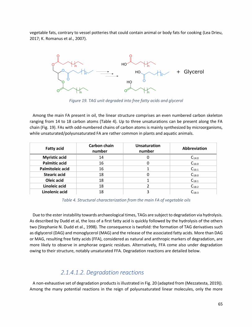
65
vegetable fats, contrary to vessel potteries that could contain animal or body fats for cooking (Lea Drieu,
2017; K. Romanus et al., 2007).
Figure 19. TAG unit degraded into free fatty acids and glycerol
Among the main FA present in oil, the linear structure comprises an even numbered carbon skeleton
ranging from 14 to 18 carbon atoms (Table 4). Up to three unsaturations can be present along the FA
chain (Fig. 19). FAs with odd-numbered chains of carbon atoms is mainly synthesized by microorganisms,
while unsaturated/polyunsaturated FA are rather common in plants and aquatic animals.
Table 4. Structural characterization from the main FA of vegetable oils
Due to the ester instability towards archaeological times, TAGs are subject to degradation via hydrolysis.
As described by Dudd et al, the loss of a first fatty acid is quickly followed by the hydrolysis of the others
two (Stephanie N. Dudd et al., 1998). The consequence is twofold: the formation of TAG derivatives such
as diglycerol (DAG) and monoglycerol (MAG) and the release of the associated fatty acids. More than DAG
or MAG, resulting free fatty acids (FFA), considered as natural and anthropic markers of degradation, are
more likely to observe in amphorae organic residues. Alternatively, FFA come also under degradation
owing to their structure, notably unsaturated FFA. Degradation reactions are detailed below.
2.1.4.1.2. Degradation reactions
A non-exhaustive set of degradation products is illustrated in Fig. 20 (adapted from (Mezzatesta, 2019)).
Among the many potential reactions in the reign of polyunsaturated linear molecules, only the more
Fatty acid Carbon chain
number Unsaturation
number Abbreviation
Myristic acid 14 0 C14:0
Palmitic acid 16 0 C16:0
Palmitoleic acid 16 1 C16:1
Stearic acid 18 0 C18:0
Oleic acid 18 1 C18:1
Linoleic acid 18 2 C18:2
Linolenic acid 18 3 C18:3
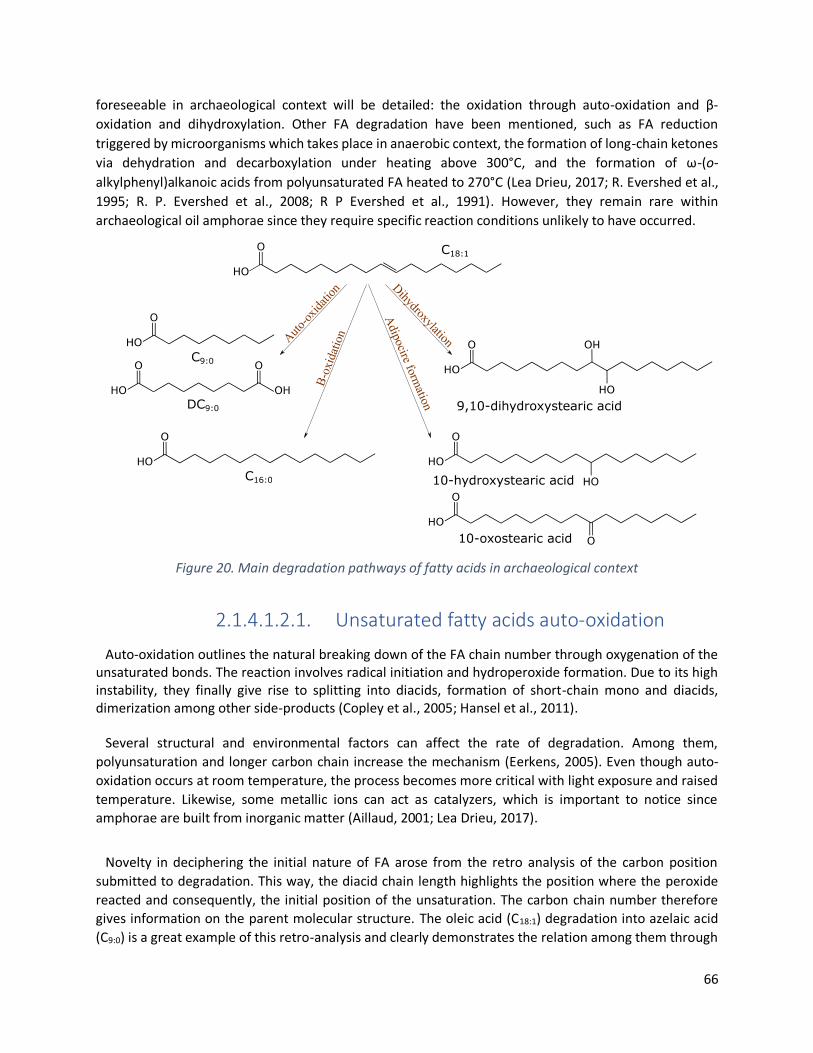
66
foreseeable in archaeological context will be detailed: the oxidation through auto-oxidation and β-
oxidation and dihydroxylation. Other FA degradation have been mentioned, such as FA reduction
triggered by microorganisms which takes place in anaerobic context, the formation of long-chain ketones
via dehydration and decarboxylation under heating above 300°C, and the formation of ω-(o-
alkylphenyl)alkanoic acids from polyunsaturated FA heated to 270°C (Lea Drieu, 2017; R. Evershed et al.,
1995; R. P. Evershed et al., 2008; R P Evershed et al., 1991). However, they remain rare within
archaeological oil amphorae since they require specific reaction conditions unlikely to have occurred.
Figure 20. Main degradation pathways of fatty acids in archaeological context
2.1.4.1.2.1. Unsaturated fatty acids auto-oxidation
Auto-oxidation outlines the natural breaking down of the FA chain number through oxygenation of the unsaturated bonds. The reaction involves radical initiation and hydroperoxide formation. Due to its high instability, they finally give rise to splitting into diacids, formation of short-chain mono and diacids, dimerization among other side-products (Copley et al., 2005; Hansel et al., 2011).
Several structural and environmental factors can affect the rate of degradation. Among them,
polyunsaturation and longer carbon chain increase the mechanism (Eerkens, 2005). Even though auto-
oxidation occurs at room temperature, the process becomes more critical with light exposure and raised
temperature. Likewise, some metallic ions can act as catalyzers, which is important to notice since
amphorae are built from inorganic matter (Aillaud, 2001; Lea Drieu, 2017).
Novelty in deciphering the initial nature of FA arose from the retro analysis of the carbon position
submitted to degradation. This way, the diacid chain length highlights the position where the peroxide
reacted and consequently, the initial position of the unsaturation. The carbon chain number therefore
gives information on the parent molecular structure. The oleic acid (C18:1) degradation into azelaic acid
(C9:0) is a great example of this retro-analysis and clearly demonstrates the relation among them through

67
oxidative cleavage of C18:1. Additionally, Copley et al. evidenced the direct formation of α,ω-diacids from
unsaturated FA within archaeological potteries by qualitative correlation of isotopic analysis (Copley et
al., 2005).
2.1.4.1.2.2. β-oxidation
β-oxidation features the segmentation of FA carbon chains, inducing a decrease in chain length. Even
though the mechanism can affect linear and branched FA, unsaturated linear skeletons are more sensible
to β-oxidation, especially for long carbon chains (R P Evershed et al., 1991). The biological mechanism
triggered by the acetyl-coenzyme A, is operating in aerobic conditions (Den Dooren De Jong et al., 1961).
2.1.4.1.2.3. Unsaturated fatty acids hydroxylation
Monosaturated FA are also subjects to dihydroxylation. As described earlier for the oxidative cleavage
and splitting of the carbon chain, vicinal diols take place on both sides where the unsaturation was initially
located (K. Romanus et al., 2007). For instance, Colombini et al. brought to light the affiliation of α, ω-
dicarboxylic and ω-hydroxycarboxylic species towards the unsaturated of the same chain length by
degrading intentionally gondoic (C20:1) and erucic acids (C22:1). They also confirmed the formation of
dihydrxyocarboxylic acids identifying 11,12-dihydroxyeicosanoic acid and 13,14-dihydroxydocosanoic acid
from their acid precursors (Maria Perla Colombini, Giachi, et al., 2005).
Although FA are omnipresent in natural edible substances, their poly or monounsaturated structures for
some do not ensure their preservation over archaeological periods. This is the reason why organic residue
analysis must consider the potential degradation endured in order to better evaluate the originally
present products. However, the oxidation of original fatty acids induces a quantitative loss of lipids. In
addition, by-products of degradation are also less hydrophobic (in particular by reduction of carbon chains
or by addition of functional groups) which makes their presence rarer in non-arid archaeological
environments.
2.1.3.3. Sterols
2.1.4.1.3. Definition, structure and natural distribution In archaeology, sterols refer to triterpenic molecules exhibiting a functionalized sterane backbone (Fig.
21a). They are an integral part of the composition of natural plants or animal substances. One of the
common animal sterols is cholesterol (Fig. 21b). Contrarywise to the animal kingdom where sterols have
C-27 structures, plants show C-29 patterns phytosterols such as campesterol, stigmasterol and β-sitosterol
which are predominant (Fig. 21c, d and e).

68
Figure 21. Sterane skeleton (A) into cholesterol (B); campesterol (C); stigmasterol (D)
and β-sitosterol (E)
2.1.4.1.4. Degradation pathways
Constrained by their molecular structure, sterols are relatively stable over time. Indeed, it is not unusual
to observe campesterol, stigmasterol or β-sitosterol when analyzing organic residues of pottery that have
contained vegetable oils (Erika Ribechini et al., 2008; Spangenberg et al., 2014). However, sterols can be
subject to oxidative degradation by ambient atmosphere when natural substances are heated. Even
though cholesterol is likely to find in analysis, its interpretation needs to be careful regarding possible
human contamination.
2.2. Analytical methods
2.2.1. History: Organic analysis residues introduction
Organic residue analysis (ORA) is strongly related with the biomolecular approach (detailed in 2.1.1.
Biomarker concept). It deals with the capability of analytical techniques to extract, at the molecular level,
organic residues trapped in their original matrix, that can be organic or inorganic. Such molecules cannot
be observed through traditional analytical method since they would not have survived archaeological
times without being physically and/or chemically protected (Richard P Evershed, 2008). Following the
biomolecular approach, organic residues translate archaeological information regarding the original
compounds. Dealing with the laborious task of understanding the ceramic becomes fundamental at the
molecular level since residues can be altered by human activities and burial conditions (Bethell et al.,
1994; Gülaçar et al., 1989). Estimation up to 80% of the absorbed molecular residues are supposed to
survive in the ceramic wall surface (Charters et al., 1993; Ben Stern et al., 2000).

69
Although the organic matter was early on lusted by archaeologists because sometimes visible to the
naked eye, the development of more advanced analytical techniques allowed the matter to reveal its
secrets. In the 1900s, the organic surface of ceramics as well as organic fragments recovered from
sediments during archaeological excavations were initially characterized by their visual and physico-
chemical properties, such as the melting point or viscosity, etc. (Lucas, 1914). The use of ‘spot tests’, such
as the Feigl test for tartaric acid (P. E. McGovern, 1997; Michel et al., 1993b), and the characterization by
chemical reaction (Cotte, 1917) led to certain advances, nowadays controverted due to their imprecision
and low selectivity (Léa Drieu et al., 2020; Ben Stern et al., 2008). Mainly based on the presence/absence
of compounds, these techniques ultimately reflected very little of the original material.
Then, the middle of the 20th century has seen the emergence of spectroscopic techniques, notably
infrared, Raman and nuclear magnetic resonance approaches. Shedding light on the bulk composition,
spectroscopic techniques were on the cutting edge of innovation and turned to be greatly used to assess
the fingerprint of the overall material. For instance, calcium tartrate had been identified in Haji Firuz
potsherds thanks to the carboxylate infrared bonds, hence highlighting the presence of archaeological
grape-derivative content (P. E. McGovern & Michel, 1996; Michel et al., 1993b). Likewise, other attempts
have been supported by diffuse-reflectance infrared spectroscopy coupled with liquid chromatography
with ultraviolet detection (P. E. McGovern, 1997; P. E. McGovern et al., 1999, 2004). Although strongly
questioned today because of the additional nature of the infrared spectroscopy and the complexity of
targeting specific bands indicative of specific compounds in molecular mixtures, it was nevertheless
perceived as a major advance in the ORA field at this time (Léa Drieu et al., 2020; Garnier & Valamoti,
2016; C Heron et al., 1991; Ben Stern et al., 2008).
Taking precedence over spectroscopic methods, the emergence of chromatographic tools in the 20th
century led to great outcomes. Selective and sensitive enough to target organic residues, they started to
be routinely used on archaeological artefacts. (J. Condamin et al., 1976; P. E. McGovern, 1997). The
analysis of medieval ceramics demonstrated the capacity of the porous material to absorb organic matter
on the one hand, and its capacity to conserve it on the other. (J. Condamin et al., 1976; Jeanne Condamin
& Formenti, 1978). The presence of organic residues could not be attributed to the environment, given
the different molecular distribution and quantities involved (C Heron et al., 1991). From then on, the
absorption of various natural substances, such as vegetal and animal fats have been tested simultaneously
to molecular characterizations of natural substances to obtain references to compare with (Charters et
al., 1993, 1995; S. N. Dudd, 1999). Laboratory experiments of controlled degradation and anthropic
influence of heating actively took part in the understanding of the molecular alteration of the residues
trapped in ceramic vessels (Charters et al., 1995, 1997; Stephanie N. Dudd et al., 1998; R. Evershed et al.,
1995; Raven et al., 1997).
To get archaeometric analysis optimal, two fundamentals’ steps are required after the molecules are
extracted from their environment: the molecular separation first and their identification then. Since the
organic mixtures usually contained a great extent of molecules, chromatographic devices are strongly
recommended to ensure a good separation and mass-spectrometric (MS) technologies are necessary to
improve molecular characterization (Richard P Evershed, 2008). Indeed, without MS analyzers, molecular
characterization is undergone with retention and migration time instead of structural molecular patterns,
which clearly suffer from a lack of specificity (Léa Drieu et al., 2020).

70
2.2.2. Chromatographic methods
Developed from the 1950s onwards, chromatography has established itself as the leading technique for
compounds separation. Resolution at the molecular level has consequently had a strong influence on
various sciences, including archaeometry. Pioneer studies conducted by Condamin established the
premises for the ORA, notably by tracing back fat remains from amphorae (J. Condamin et al., 1976).
Being the universal technique of choice, numerous are the publications calling on GC-MS to reveal
organic hints. (Colombini and Modugno, 2009; Garnier and Valamoti, 2016; Mezzatesta et al., 2021;
Modugno et al., 2006; Pecci et al., 2020, 2013; Ribechini et al., 2008). Indeed, Brassicaceae seed oil was
characterized in Egyptian lamps using GC-MS thanks to the identification of dihydroxy and dicarboxylic
erucic and gondoic acids. Archaeological grape-based derivatives were successfully identified from
potsherds dating back to the Early Medieval Islamic Sicily, the Early Neolithic in the South Caucasus, the
Prehistoric Northern Greece or the Late Chalcolithic Near Eastern (Barnard et al., 2011; Léa Drieu et al.,
2021; Garnier & Valamoti, 2016; P. McGovern et al., 2017).
Being a referential method, we strongly considered GC-MS as the method of choice for this thesis
research. The analytical set-up is described below.
2.2.2.1. Gas Chromatography-based method
The GC device is composed of 4 units: the injector, the chromatographic column, the oven and the
detector (Fig. 22, adapted from ©CHROMacademy and (Cajka et al., 2016)). The solution is injected into
the apparatus by vaporization and carried to the stationary phase (SP) by the mobile phase (usually an
inert carrier gas, which also guarantees the transport of the component all along the chromatographic
column). The routine mobile phase is helium. The injection can be manual or automatic. Both the split and
splitless mode exist, allowing respectively partial or total injection of the solution into the column. In
archaeological context, splitless is favored since concentrations are usually low.
The stationary phase corresponds to the inner coating of the open tubular column. The separation is
achieved all along the chromatographic column thanks to affinity difference towards SP composition and
polarity. The column is placed into an oven whose temperature increases gradually in order to slowly
volatilize and eluate the compounds of the mixture, starting from the lowest volatilization temperature
molecules to the highest.
When the carrier gas finally emerges from the column, the detector records a signal according to the difference in composition. Within the various types of detectors, the most powerful for organic traces remains the mass spectrometer which allows component identification through characteristic mass fragmentation. Its high sensitivity and selectivity perfectly allow residues analysis in archaeometrical fields of research.

71
Figure 22. GC-MS schema equipped with a quadrupole ion trap
Samples are derivatized with N, O-Bis(trimethylsilyl) trifluoroacetamide (BSTFA): trimethylchlorosilane
(TMCS) to improve transform hydroxyl and carboxylic acid polar groups into trimethylsilylated groups that
are more suitable for volatilization. Different chemical options exist for derivatization, but
trimethylsilylation was favored over methylation due to some naturally methylated compounds whose
presence is archaeologically characteristic.
2.2.2.2. Liquid Chromatography-based method
High Performance Liquid chromatography (HPLC) is aimed to target high molecular weight molecules,
considered to be of low volatility or with high polarity. Thus, the analysis of organic dyes or even proteins
has been possible by LC (Buckley et al., 2013; Dallongeville et al., 2011; Mazzitelli et al., 2019; Pronti et al.,
2018). Applied to the archaeometric field, LC is not the method of choice because of the detectors, since
it requires the use of detectors that are specific to each family of compounds and does not allow for a
global screening of the molecular composition but rather a specific search.
Among the rare exception, organic residues alterations have been followed by HPLC coupled with
Electrospray Ionization - Quadrupole Time-of-Flight MS (HPLC-ESI-QToF) in order to target potential
diagnostic biomarkers of fresh whale oil (Blanco-Zubiaguirre et al., 2018). Likewise, triglyceride profiles
from drying oil, characterized with HPLC-ESI-Q-ToF, allowed to trace back the composition of plant oils (La
Nasa et al., 2013; Saliu, Degano and Colombini, 2014) or from the binding paint medium (La Nasa et al.,
2015) and helped identifying the pigments in the polymeric paint network (La Nasa et al., 2019). Such
advanced method has also been mentioned for the analysis of TAGs in archaeological vessels (Lucejko et
al., 2018) and high molecular weight esters in archaeological beeswax and resinous substances (La Nasa
et al., 2020).
72
Finally, the coupling of LC with a MS2 detector allowed the detection of wine molecules in Egyptian and
Nordic potsherds (Guasch-Jané et al., 2004; Guasch-Jané, Andrés-Lacueva, Járegui, et al., 2006; Guasch-
Jané, Andrés-Lacueva, Jáuregui, et al., 2006; McGovern, Hall and Mirzoian, 2013).
2.2.2.3. Pyrolysis-GC-based method
Since the beginning of the 21st century, new techniques for the analysis of lipids, proteins or
polysaccharides, namely as pyrolysis GC-MS or Direct Temperature-resolved MS DTMS have emerged
(Oudemans and Boon, 1991; Oudemans, Eijkel and Boon, 2007; La Nasa et al., 2019). Allowing the
screening of the overall composition by the analysis of different compounds families at the same time,
they have grandly improved the overall characterization (Bonaduce et al., 2017). Intense heating makes
it possible to obtain the chemical fingerprint of the sample by showing off the different chemical families
present. Direct exposure MS and electrospray–MS2 indeed overcome the non-volatile compound barrier
constrained by GC-MS to give insight on the composition of triterpenoid resins and beeswax (Garnier et
al., 2002; Modugno, Ribechini and Colombini, 2006; Kaal et al., 2014; Kaal, López-Costas and Martínez
Cortizas, 2016).
2.2.3. Mass Spectrometry
The MS apparatus is following 3 steps: ionization in the ion source, mass filtration in the mass analyzer
and electromagnetic detection (Fig. 22). An inlet system guarantees the release of the gas stream from
the mobile phase into the ion source at a controlled rate. 70 eV electronic impact (EI) is the prevalent
method to produce positive ions. The incoming stream of gas phase molecules is ionized by interactions
between an electron beam coming from a hot filament with the analytes (Fig. 23). Once ionized, resulting
unstable radical cations are usually fragmented. Indeed, the electronic energy is largely sufficient to
enhance the fragmentation of organic molecules. A positive voltage from a repelled electrode electrically
pushes them out, generating a stream of positive ions and fragments to move out from the ion source to
the mass analyzer through the hole located the superior electrode.
Ion trap analyzer looks like a “doughnut-shaped ring” with an annular electrode equipped with
radiofrequency (Rf) voltage and two end caps supplied with alternative or direct voltages (AC or DC) (Fig.
22). It is commonly named “quadrupole” due to the shape of the magnetic field generated by the
superposition of voltages applied from the electrodes. Submitted to a tridimensional electric field in the
analyzer, ions start oscillating until reaching a so-called “infinite” stable shape whose oscillation amplitude
and frequencies strictly depend on the ionic m/z ratio and Rf. In order to gradually eject the ions, present
in the analyzer, Rf increases, therefore generating higher axial oscillation amplitude. Ions with lower m/z
are destabilized first. They move out the analyzer to the MS detector which monitors the ion counting and
converts this electric signal into a digital one, giving rise to the mass spectrum (Varella, 2012).
MS analyzers pave the way to isotopic techniques, such as stable isotope ratio MS analysis. Indeed, Gas
Chromatography – Combustion - Isotope Ratio MS (GC–C–IRMS) help in identifying ruminant fats from
non-ruminant dairies. (Mottram et al., 1999; Mottram and Evershed, 2003; Romanus et al., 2007). From

73
palmitic and stearic acids in archaeological ceramic vessels, Copley et al. demonstrated the presence of
domesticated ruminants dated back to Neolithic, therefore early on exploited for dairy productions
(Copley et al., 2003).
Figure 23. Ion trap set-up MS gives the option to operate in different modes. Full scan is routinely used since it generates a
qualitative picture of the different separated compounds, scanning from the smallest mass to the highest.
It allows mass scouting and fragmentation pattern analysis. Selected ion monitoring (SIM) and multiple
reaction monitoring (MRM) modes rather target quantitative focus on specific ions of interest.
2.2.3.1. SIM
Selected ion monitoring (SIM) is an analytical option provided by the detector to record ion currents
only for preselected m/z ratio. Since the mass spectrometer does not record the entire mass range, SIM
guarantees higher sensitivity and selectivity. Interferences are minimized increasing the detection limit to
nanograms or picograms when the matrix permits it (Sparkman, Penton and Kitson, 2013). Ionization
remains the same but only selected ion mass can reach the detector, which accounts for a better
sensitivity compared to TIC chromatograms because the background is not acquired (Clench and Tetler,
2000). Although SIM analysis are not frequent in ORA, some researches have led to great conclusions
regarding the presence of fermented beverages or beeswax in Iberian and Etruscan potsherds (Garnier et
al., 2002; Manzano et al., 2016). For instance, SIM mode was employed to target bacteriohopanoids,
revealing the archaeological presence of alcoholic beverages made out of fermented sap from agave plant
in pottery vessels (Correa-Ascencio et al., 2014).

74
2.2.3.2. MSn
Tandem MS, also known as MS/MS or MS2 involves two or more MS analyzers coupling which allows
additional fragmentation steps. The main purpose is the great increase of specificity and sensitivity since
many fragmented ions get ignored, which allows molecular compound focus within complex mixtures.
Coupled to MS/MS, multiple reaction monitoring (MRM) targets a singular m/z ion for fragmentation to
generate a series of daughter ions and selects one (or more) of these daughter ions for the second MS
detection. Guasch-Jané et al. reported the identification of wine markers from Egyptian vessels thanks to
traces of tartaric and syringic acids, identified in tandem mode LC/MS/MS in MRM mode (Guasch-Jané et
al., 2004), with a limit of detection established to 0.01µg/mL (Stern et al., 2008). Indeed, the second
fragmentation gave great support for diagnosing specific wine markers.

75
76
Chapter 3
Pollen in archaeology:
A State of the Art

77
3.1. Once upon a time: the pollen
3.1.1. Pollen definition
Pollen grains are single-cell gametophyte produced by the reproductive systems of flowering plants.
They carry the genetic information and account for the genetic variability of the. Pollen grains are small,
usually ranging from 10 µm to 200 µm. During meiosis, pollen grains are initially formed as a tetrad. There
exists as many pollen morphologies as plants, the impressive variety arises from the varying chemical
composition and the structure of the exine, the outer wall of the grains. Among the main constituents of
the exine (i.e., cellulose, hemicellulose, lignin, pectic compounds that compose the exine), sporopollenin
is the one that confers indestructible properties to pollen grains. Being highly resistant to biological decay
and non-oxidative chemical attacks, exine ensures pollen survival over geological times in a variety of
sedimentary deposits. Indeed, pollen grains dating back to 15.7 Myr ago have recently been recovered
from Antarctica ice cores (Feakins, Warny and Lee, 2012). Although the protecting effect of sporopollenin
is established, little is known on its chemical composition. Oxidative copolymers of carotenoids and
carotenoid esters have been identified though (Shaw, 1971; Brooks and Shaw, 1978). Recent
investigations relying on solid-state NMR techniques highlighted the presence of aliphatic-polyketide-
derived polyvinyl alcohol and 7-O-p-coumaroylated C16 aliphatic units within the very dense crosslinked
biopolymer (F.-S. Li et al., 2019).
3.1.2. Pollen dispersion
Pollen grains can be classified through their different dispersion mechanisms. Anemophilous pollen
species are dominant in archaeological record. Being wind-pollinated, they can easily scatter over few
kilometers from the generative plant (Bajpai and Kar, 2018; D. Li et al., 2019). Pinus grains are a good
example of anemophily that greatly scatter thanks to its saccate morphologies that favor lift capacities by
increasing its surface area (Schwendemann et al., 2007). Pollen get released when wind constrains
balance the adhesives electrostatic and Van der Waals forces exerted from the anthers (Timerman and
Barrett, 2021). Zoophilous pollen are spread thanks to animals and insects that promote plant pollination,
carrying pollen grains from the anther to the stigma of another plant. In addition of being less represented
in the plant kingdom, pollen is naturally covered by lipids to ensure pollen stickiness, which explains why
zoophilous pollen are less likely to be recovered in fossil samples (Shillito et al., 2020). Water-pollination
from hydrophilous plants and self-pollination from cleistogamous plants are also barely represented in
fossil records because of their little representation in plants (Bryant and Holloway, 1983).
3.1.3. Pollen preservation
Preserved by the chemically inert biopolymers of sporopollenin, pollen keep its structural integrity over
extended times. An interesting study conducted over 20 years by Havinga showed the positive impact of
the amount of sporopollenin on the longevity of pollen grains. The richer in sporopollenin the exine is, the
better the resistance was observed (Havinga, 1964, 1984). As such, pollen taxa do not survive equally.

78
Indeed, thick-exine pollen transported by insects, together with pine, oak, grass or goosefoot grains are
more preserved in fossil assemblages (Bryant, 1978; Bryant and Schoenwetter, 1987; Holloway, 1989;
Bryant and Hall, 1993; Campbell and Campbell, 1994; Gorham and Bryant, 2001; Phuphumirat, Mildenhall
and Purintavaragul, 2009).
In archaeological contexts, pollen deterioration can arise from different mechanisms. First, mechanical
degradation induced by transportation and sedimentation phenomena can damage pollen grains
(Campbell, 2010; Hunt and Fiacconi, 2018). To not misinterpret pollen representation, the taphonomy of
the grains needs to be carefully evaluated to address any redeposition or differential preservation of more
ancient sediments. Increasing the sediment compaction also alters the assemblage taphonomy (Ellison,
2008). Moreover, experimental wet-dry cycles demonstrate the rapid breakage of Picea and Artemisia
pollen when recovered from desalinated sediments (Campbell and Campbell, 1994). In comparison,
salinity promotes pollen structures preservation (or damage in lesser extent). Salt crystals are thought to
absorb the mechanical constraints of water evaporation. Pollen grains trapped in the metal crystal
network are also more protected (Rösch, 2005). Secondly, pollen grains are subject to chemical oxidation.
Indeed, great evidence of pollen integrity over time has been reported in anaerobic sediments since the
hydrogen released by the bacteria ensures reducing environment (Tschudy, 1969). Pollen has also been
used to target the oxidation of organic matter in marine sediments. Keil et al. pointed out the great
preservation of pollen for 100 000 years in anoxic environment from marine sediment cores while 10 000
years were sufficient for oxygen to degrade pollen in sediment where the gas was continuously diffused
(Keil et al., 1994). Regarding the degradation timescale, pollen is thus able to trace the oxygen content of
the sediments. A recent study conducted on seven different pollen taxa from trees, shrubs and
herbaceous plants aimed at outlining the factors that increase or delay pollen oxidative damage (Lebreton
et al., 2010). Although oxidation is intractable, both the morphology and the exine thickness have been
shown to play an important slowing-down role. To a lesser extent, low pH seems to account for pollen
preservation, while soil alkalinity is unfavorable (Dimbleby, 1957; Bryant et al., 1994; Phuphumirat,
Mildenhall and Purintavaragul, 2009). Finally, biological agents, namely fungi and bacteria, are
pollinivores. Although the exact mechanisms remain unclear, may use the internal protoplast of pollen
grains as nutrient, hence perforating the exine and triggering pollen degradation (Elsik, 1971; Bryant and
Holloway, 1983; Twiddle and Bunting, 2010).
3.2. Palynology: a state of the art on the growing interest field
3.2.1. Historical point of view
The first record of pollen-based analysis was speculated by Iversen (1941) to corroborate the prehistoric
introduction of agriculture with the disappearance of forest in Denmark (Iversen, 1941). He hereby
interpreted the decline of elm pollen together with the great appearance of herbs and weedy plants as
the human direct impact on the vegetational land through human occupation. Relying on the pollen
diagram he made, he was also able to overview the prehistorical farmed plants. Although pollen diagrams
were slowly showing up to clarify changing land assessment, pollen was nevertheless sparsely used and
few are the evidence of pollen-based interpretation before the 1970 (Greig and Turner, 1974). While the
discipline was still in its beginning stages, fundamental studies accounted for its emergence, notably

79
clarifying pollen morphologies and analysis methodologies (Erdtman, 1969; Moore and Webb, 1978;
Faegri, Kaland and Krzywinski, 1989). In this way, pollen turned to be a reliable tool to assess the relative
chronologies of past events, among which the beginning of animal domestication in Denmark (Troels-
Smith, 1959); the buffalo grasslands expansion in Texas (Bryant, 1969), the inference of human activities
on the Mesolithic vegetational land in England (Dimbleby, 1963) as well as the introduction of maize
agriculture in Canada (McAndrews, 1976). Finally, Dimbleby and Evans (Dimbleby and Evans, 1974)
unraveled the occurrence of two successive ecological environments from the same Neolithic chalk land.
Although molluscan snail analysis gave insights into woodland, pollen surprisingly highlighted farmland.
The authors marked a turning point in the discipline while publishing in the Journal of Archaeological
Science, hence making clear the impressive benefits of pollen analysis and its concerning in anthropology,
archaeology, palaeoecology and more broadly in the Quaternary science (Yeung et al., 2015). As shown in
Fig. 24, the number of publications of pollen-based investigations started raising from the 1970s (Edwards,
Fyfe and Jackson, 2017). Data were extracted from https://app.dimensions.ai, with occurrence selected
in titles and abstracts (n = 17930) from 1972-2020, all journals confounded.
Figure 24. “Pollen analysis” occurrence in scientific publications.
3.2.2. From pollen analysis to archaeological meaning
Pollen analyses show a great potential for landscape reconstruction. Initially performed on single cores,
the approach has been extended to multiple site sediment assemblages in order to provide comparative
data (Magri, 1997; Tipping, 1998; Ledger, Edwards and Schofield, 2014). The comparison of pollen taxa
also outlines species correlation, especially regarding their appearance or disappearance (Mercuri et al.,
2013). Thus, ecological, vegetational and spatial reconstructions are enhanced. The powerfulness of
pollen markers have been enforced by the great development of pollen datasets such as the European
Pollen Database (http://www.europeanpollendatabase.net/), the Neotoma Paleoecology Database
(https://www.neotomadb.org/), the Global Pollen Project (https://globalpollenproject.org/), PalDat
which is specific to fossil pollen (https://www.paldat.org/) and pollen atlases (Reille, 1992b, 1992a, 1995,
1998).

80
Human activities having a direct effect on the surrounding vegetation, pollen analysis has further
supported the identification of past environmental variations, as well as climate changes. Enhanced by
other research fields of archaeobotany, pollen demonstrated its great reliability as a reconstructive
marker. Integrated archaeobotanical studies, including microproxies such as NPP (Non Pollen
Palynomorphs), spores, diatoms, phytoliths, plants macrofossils and charcoals, have emerged (Di Rita,
Celant and Magri, 2010; Burjachs and Expósito, 2015; Celant, Magri and Stasolla, 2015; Di Rita et al., 2015;
Mariotti Lippi et al., 2018). Whereas pollen grains mirror the local landscape, charred plant macrofossils
reveal the direct impact of human culture, sedentarization and agriculture (Lechterbeck and Jensen,
2020). When coupled to geochemistry or zooarchaeology, they can draw out a complete ecological
reconstruction over time (Schofield et al., 2010; Williams, 2012; Gismondi et al., 2013; Tolksdorf et al.,
2020). For instance, palynological, anthracological, stratigraphical, geochemical and radiocarbon analysis
of soil sediments, supported by historical evidence, helped in disclosing the negative impact of mining
activities in archaeo-ecological systems in the Middle Bronze Age in France and Austria (Jouffroy-Bapicot
et al., 2006; Breitenlechner et al., 2010). Multidisciplinary approaches are all the more favored to address
heterogeneity in archaeological assembles, that might arise from time or spatial instabilities or even from
the nature of the materials themselves. In contrast, lakes and mires are often considered to be stable
enough to provide valuable insights on their own (Tweddle and Edwards, 2010; Edwards et al., 2015).
Nowadays, pollen analysis is additionally included in models and simulations, notably thanks to the
incorporation of statistical tools to address transport or dispersal queries (Sugita, 2007a, 2007b; Edwards
et al., 2015). Recent interdisciplinary investigations have integrated ancient DNA data into pollen analysis
(Jones et al., 2017; Ekram et al., 2021), notably for fossil coprolites (Beck, Bryant and McDonough, 2019;
Petrigh et al., 2021), with the specific aim of better understanding diet, disease, cultural and daily-life past
practices. More broadly speaking, pollen analysis has supported animal and plant domestication and
geographical distribution (Argant, López-Sáez and Bintz, 2006; Zarrillo et al., 2008; Nott, 2010; Lipe et al.,
2016; Mercuri et al., 2021); burial rituals (Mora et al., 2018; Amerongen, 2019); on and off-site dating
(Scott and Woodborne, 2007); archaeological technologies, manufactures and savoir-faire (Muller, 2004;
Willis, 2017).
3.3. Methodological approaches and data analysis
3.3.1. Sampling issues
The highly preserved nature of pollen that makes it so powerful in palaeocology also confers it certain
limitations. Being the primary tool for past plant reconstructions, any contamination by pollen that do not
belong to the targeted time scale or any discriminatory degradation of certain pollen due to a lower
resistance of its sporopollenin (Lebreton et al., 2010) can lead to misinterpretation. Indeed, pollen
progressive degradation or partial preservation biases pollen counts (Bottema, 1975; Hall, 1981). To avoid
such pollen distorted representation, different rules have been set up. Sampling must be performed after
considering the potential surrounding degradation to prevent the inclusion of reworked grains in the
analysis. Each site is different and must be carefully studied to limit any contamination. Undisturbed
sediments are fundamental. When dealing with archaeological artefacts such as potteries, sealed jars are
obviously favored. If not, the archaeological site needs to be carefully studied before any sampling to
assess any contamination from erosion, suffusion, post sedimentation or sediments relocation due to
81
mechanical displacement of unstable sites, bioturbation, recycling soil sediments, etc. The position of the
objects regarding the site is extremely relevant. Then, the reliability of the archaeological interpretation
requires multiple-sample analysis. A certain pollen occurrence must be established in order to draw stable
conclusions and limited total pollen concentration are to interpret with caution (Hall, 1981). A sole
sediment sample cannot induce valuable palaeoecological reconstruction. The trends it may raise will
necessarily have to be corroborated with other sediment analysis or integrated into multidisciplinary
studies. Indeed, the impact of human activities on the land environment cannot be translated by a single
analysis and must be part of a larger amount of analysis. The use of control samples is mandatory. External
samples from the surrounding near-environment can address “background” contamination from soil or
water currents when studying potteries content (Gorham and Bryant, 2001; Torrence, 2016; Pecci et al.,
2020). Off-site and non-site control comparison can sort post depositional contamination out (Loy and
Barton, 2006). Stratigraphic and flotation controls may evaluate chronology and sediment fill deposits.
For too long, sampling has suffered from the absence of assistance from experienced researchers (Bryant
and Holloway, 1983), who then established various pre- and post-sampling protocols (Pearsall, 1990;
Hunt, 1994; Weinstein-Evron, 1994; Bunting and Tipping, 2000; Tipping, 2000). A close collaboration
between archaeobotanists and archaeologists is more than recommended before undertaking
excavations, in order to organize the field study and plan sediment sampling and its frequency (Bryant
and Hall, 1993). Only this way anthropogenic repercussion and post depositional contamination can be
estimated, and the original pollen content can be accurately interpreted.
Recently, UV-fluorescence microscopy has been mentioned to outline pollen contamination from
younger specimen intrusion and recycled pollen (Hunt, 1998; Yeloff and Hunt, 2005). With time, pollen
grains age and the relative fluorescent intensity of the outer wall diminishes. This gives a valuable key for
fresh and fossil pollen distinction. The technique also provides meaningful insights on the thermal
maturity of pollen, outlining burnt material (Hunt, Rushworth and Dykes, 2007).
3.3.2. Pollen extraction
After sediments are sampled and contamination issues sorted out, pollen grains need to be extracted
from their matrix to be observed under a microscope. The extraction being destructive, specific care must
be taken regarding the protocol and sample amounts. It consists of chemical and/or physical processes.
Since pollen can be lost at any step if precaution care is not sufficient, it remains fundamental to avoid
useless treatments (Horowitz, 1992).
From the beginning of the palynological research, many extractive approaches have been published to
remove organic and inorganic materials microfossils are trapped in (Erdtman, 1960; Faegri, Kaland and
Krzywinski, 1989; Pearsall, 1990; Wood, Gabriel and Lawson, 1996; Magri and Di Rita, 2015; Riding, 2021).
Protocols usually consist of: (i) chemical breakdown of the matrix, (ii) concentration of pollen grains and
(iii) mounting on slides to preserve pollen from oxidation and make them observable under the
microscope. The chemical treatment chosen is directly dictated by the sample composition (Erdtman,
1960; Magri and Di Rita, 2015). In presence of carbonate minerals, recurrent in sediments, undiluted
hydrochloric acid (HCL) is utilized, sometimes with glacial acetic acid (Bryant and Holloway, 1983). Sand
and silicates are digested with highly concentrated hydrofluoric acid (HF). Produced fluoride crystals need
then to be removed before continuing. Then, palynomorphs are concentrated as follows: deflocculation,

82
assisted by sonication, can sort out clay persistence in the samples. Ultrasound will help in fragmenting
the amorphous organic phase, then removed by sieving. Oxidation and alkali treatments can be
performed if extraneous organic matter is still present. When the ratio of pollen over mineral sediments
is extremely low, gravity separation may be an option. The last step specifically targets coarse particles (>
200 µm) that can be drawn away with sieving. When the samples still exhibit organic matter coming from
insufficient degradation of plant remains, such as resins, lignin, waxes, carbohydrates and its relative
biopolymers of cellulose and hemicellulose, acetolysis becomes inevitable to dissolve the organic fraction.
Although it removes the remains from uncomplete degradation of microfossils by disintegrating residual
fragments of sporopollenin (Erdtman, 1960), it can additionally distort pollen counting since acetolysis
breaks down pollen protoplasms. Fresh pollen usually identified by their “filled” aspect are thus emptied,
making them indistinguishable from fossil pollen. A staining step can be added to dark pale specimen and
make them more contrasted. Different mounting media have been proposed. Glycerol is frequently used
since it facilitates grain motions under slight pressure during microscope observation, although it does
not ensure long-term preservation. Cellosize and polyvinyl alcohol are also widely named (Riding, 2021).
In any case, the mounting medium must be selected based on its durability, contrast effect and its optical
properties to enhance pollen morphological observation.
3.3.3. Pollen identification
Pollen morphologies being different from each other, the characterization is performed through
successive key point identification. The pollen unit, the size, the shape, the aperture numbers and types
and the exine ornamentation do matter. The taxonomic level, up to which pollen grains can be identified,
strongly depend on the taxa. Indeed, the ornamentation visible on the exine does not strongly vary within
the same genus. For instance, Pinus species are hardly distinguishable from each other although some
attempts tried to discriminate them (Desprat et al., 2015). From fresh pollen records, they proposed to
cluster P. halepensis, P. pinea and P. pinaster together as Mediterranean while P. nigra and P. sylvestris
belong to the highland group. Additionally, it is important to notice that fresh, hydrated and fossil pollen
differ in size. In fresh pollen, the protoplasmic material prevents detailed observation of the
ornamentation.
3.3.4. Pollen interpretation
After identification and counting, pollen is evaluated upon its concentration regarding the initial amount
of analyzed material. Calculations are based on the known quantity of exotic markers (usually Lycopodium
spores (Stockmarr, 1971)) and pollen representation are expressed in numbers of fossil grains per weight
or volume as follows (Benninghoff, 1962; Magri and Di Rita, 2015):
𝑃𝑜𝑙𝑙𝑒𝑛 𝑐𝑜𝑛𝑐𝑒𝑛𝑡𝑟𝑎𝑡𝑖𝑜𝑛 =
𝑒𝑥𝑜𝑡𝑖𝑐 𝑠𝑝𝑜𝑟𝑒 𝑎𝑑𝑑𝑒𝑑 ∗ 𝑝𝑜𝑙𝑙𝑒𝑛 𝑐𝑜𝑢𝑛𝑡𝑒𝑑𝑒𝑥𝑜𝑡𝑖𝑐 𝑠𝑝𝑜𝑟𝑒 𝑐𝑜𝑢𝑛𝑡𝑒𝑑
𝑔𝑟𝑎𝑚𝑠 𝑜𝑓 𝑠𝑒𝑑𝑖𝑚𝑒𝑛𝑡 𝑡𝑟𝑒𝑎𝑡𝑒𝑑

83
The total pollen content is chronologically plot in pollen diagrams of core sediment analysis. Statistical
tools integrating principal components analysis and cluster analysis help generating transfer functions
with relative pollen frequencies or radiocarbon dating (Horbe et al., 2011; Michczyński et al., 2013).
Recovered from fossil sediments, pollen is classified into background or economic entities. While
background refers to usual environmental taxa commonly found on the ground, in water, on food,
economic pollen targets the taxa related to anthropic activities. For instance, in the analysis of amphorae,
background pollen would revert to the atmospheric conditions where the amphora has been produced,
located, transported, etc. and economic pollen would regard the taxa indicative of the content. Setting
aside landscape reconstruction and focusing more on the analysis of ceramic vessels, microfossils have
provided valuable insights to shed light on vessels functionalities and civilizations diets (Zarrillo et al.,
2008; Torrence, 2016). Information on the provenance, the vessel function or the manufacture
technologies employed have been drawn from palynomorph analysis from baskets (Bohrer, 1968), stone
tools such as mortars or milling stones (Hevly, 1964; Bryant and Morris, 1986; Fullagar, Furby and Hardy,
1996), as well as from the surface of potsherds (Magid and Krzywinski, 1988; Jones et al., 1998; Ghosh,
D’Rozario and Bera, 2006; Peto et al., 2013; Dumpe and Stivrins, 2015). Since such artefacts were originally
used for food storing, cooking and eating, pollen can be recovered nowadays thanks to their trapping into
the porous ceramic. This way, pollen has greatly supported fermented beverages tracing (Guerra-Doce,
2014). Archaeological beers and meads have been evidenced through the presence of cereal pollen and
plant macroremains (Barclay et al., 1994; Dineley and Dineley, 2000; Lageras, 200; Bartel et al., 1997;
Bueno Ramírez et al., 2005; Rösch, 2005, 1999), sometimes corroborated with organic residue analysis.
For instance, Moe and Oeggl (Moe and Oeggl, 2014) understood the important presence of Filipendula
ulmaria together with Cerealia pollen as a sign of mead consumption since the beverage was usually
prepared with meadowsweet to enhance flavour. Henbane-based preparations have been evidenced by
the presence of Hyoscyamus niger in Late Neolithic British vessels (Barclay et al., 1994). The flower was
currently used for ritual and medicinal purposes. Furthermore, the important diversity in pollen recovered
from a bronze vessel dated to the German Iron Age was interpreted as a sign of archaeological honey
(Rösch, 1999; Kvavadze, 2006). Grape beverages have been mentioned in the understanding of amphorae
content with findings of Vitis vinifera pollen (Gorham and Bryant, 2001; Arobba et al., 2014). Moreover,
the ceramic clays themselves can trace the provenance. For instance, Ghosh et al. assessed the
socioeconomic development through the analysis of pollen trapped in Gangetic West Bengal potsherds.
Following his minds, micro and macro vegetal remains (i.e., pollen, spores, phytoliths, fruits and seeds)
from local consumption get deposited on the clay. Since surrounding clays were used for vessels
manufacture, dissolving the ceramic matrix can indicate ancient anthropological activities (Ghosh,
D’Rozario and Bera, 2006). They demonstrated that pollen contained in the ceramic clay can survive to
high temperature firing, up to 1000°C when anoxic conditions are ensured. Such conditions were in fact
originally used to decorate the vessels (Shaw et al., 2001).
Like any spectacularly powerful marker, pollen also exhibit limitations and a few words must be said
about this. As often stated by Bryant et al. (Bryant, 1978; Bryant and Holloway, 1983), it is important to
remember that the only information pollen give access to is about palaeovegetation. Any further
settlement regarding climate, animal life, past cultures and anthropic activities derive from
palaeovegetation interpretation. First, it requires a high degree of precaution during all steps, from
sampling to identification. Individual responsibility is then engaged in each step, in order to provide the
most reliable possible data. The reconciliation of data with similar spatial, geographical, temporal or

84
anthropic characteristics can only be done when the rigor of analysis is comparable. Aside from the
individual impact on the analysis, the phenomena inherent to pollen (dispersion) and external to it
(contamination by post deposition, redistribution of sediments, partial and/or selective degradation) bias
the analysis. Statistical treatments, although powerful regarding the interpretations they suggest, cannot
deviate from this rigor (Mosimann and Greenstreet, 1971; Ogden, 2010).

85
References Aillaud, S. (2001) Field and laboratory studies of diagenetic reactions affecting lipid residues absorbed in unglazed archaeological pottery vessels. University of Bristol.
Allegretta, I. et al. (2017) ‘The effect of mineralogy, microstructure and firing temperature on the effective thermal conductivity of traditional hot processing ceramics’, Applied Clay Science, 135, pp. 260–270. doi: 10.1016/j.clay.2016.10.001.
Amerongen, Y. F. van (2019) ‘All’s well? Comparing on- and off-site pollen samples and exploring the potential of pollen from man-made contexts’, Vegetation History and Archaeobotany 2019 29:2, 29(2), pp. 125–131. doi: 10.1007/S00334-019-00759-W.
Argant, J., López-Sáez, J. A. and Bintz, P. (2006) ‘Exploring the ancient occupation of a high altitude site (Lake Lauzon, France): Comparison between pollen and non-pollen palynomorphs’, Review of Palaeobotany and Palynology, 141(1–2), pp. 151–163. doi: 10.1016/J.REVPALBO.2006.01.010.
Arias-Gil, M., Garde-Cerdán, T. and Ancín-Azpilicueta, C. (2007) ‘Influence of addition of ammonium and different amino acid concentrations on nitrogen metabolism in spontaneous must fermentation’, Food Chemistry, 103(4), pp. 1312–1318. doi: 10.1016/j.foodchem.2006.10.037.
Arobba, D. et al. (2014) ‘Palaeobotanical, chemical and physical investigation of the content of an ancient wine amphora from the northern Tyrrhenian sea in Italy’, Journal of Archaeological Science, 45(1), pp. 226–233. doi: 10.1016/j.jas.2014.02.024.
Arroyo-Garcia, R. et al. (2006) ‘Multiple origins of cultivated grapevine (Vitis vinifera L. ssp. sativa) based on chloroplast DNA polymorphisms’, Molecular Ecology, 15(12), pp. 3707–3714. doi: 10.1111/j.1365-294X.2006.03049.x.
Azémard, C., Ménager, M. and Vieillescazes, C. (2017) ‘On the tracks of sandarac, review and chemical analysis’, Environmental Science and Pollution Research, 24(36), pp. 27746–27754. doi: 10.1007/s11356-017-0522-0.
Bailly, L. (2015) Caractérisation moléculaire et isotopique de goudrons et résines archéologiques dérivés de conifères en contexte maritime. Université de Strasbourg, France.
Bajpai, R. and Kar, R. (2018) ‘Modern pollen deposition in glacial settings in the Himalaya (India): abundance of Pinus pollen and its significance’, Palynology, 42(4), pp. 475–482. doi: 10.1080/01916122.2017.1407835.
Banilas, G. et al. (2012) ‘Olive and Grapevine biodiversity in Greece and Cyprus - A review’, in Lichtfouse, E. (ed.) Climate Change, Intercropping, Pest Control and Beneficial Microorganisms. Volume 2. Springer, pp. 401–428. doi: 10.1017/CBO9781107415324.004.
Barclay, G. et al. (1994) ‘Excavations in the ceremonial complex of the fourth to second millennium BC at Balfarg/Balbirnie, Glenrothes, Fife’, in Proceedings of the Society of Antiquaries of Scotland, pp. 43–210.
Barnard, H. et al. (2011) ‘Chemical evidence for wine production around 4000 BCE in the Late Chalcolithic Near Eastern highlands’, Journal of Archaeological Science, 38(5), pp. 977–984. doi: 10.1016/j.jas.2010.11.012.
Bastien, C. (2011) Étude chimique des substances contenues dans une collection exceptionnelle de poteries provenant de Deir el-Médineh (Egypte) : une population et ses produits. Université de Strasbourg.

86
Beck, C. W., Bryant, V. M. and McDonough, K. N. (2019) ‘Evidence for non-random distribution of pollen in human coprolites’, Archaeological and Anthropological Sciences 2019 11:11, 11(11), pp. 5983–5998. doi: 10.1007/S12520-019-00839-Y.
Belitz, H. D., Grosch, W. and Schieberle, P. (2009) Food chemistry. Fourth edi. Berlin: Springer.
Benninghoff, W. S. (1962) ‘Calculation of pollen and spore density in sediments by addition of exotic pollen in known amounts’, in Pollen et Spores 4. Muséum National d’histoire Naturelle, p. 332.
Benoit, F. (1962) ‘Nouvelles épaves de Provence (III)’, Gallia, 20(1), pp. 147–176. doi: 10.3406/galia.1962.2352.
van Den Berg, K. J. et al. (2000) ‘Mass spectrometric methodology for the analysis of highly oxidized diterpenoid acids in Old Master paintings’, Journal of Mass Spectrometry, 35(4), pp. 512–533. doi: 10.1002/(SICI)1096-9888(200004)35:4<512::AID-JMS963>3.0.CO;2-3.
Berrueta, L. A. et al. (2020) ‘Formation and evolution profiles of anthocyanin derivatives and tannins during fermentations and aging of red wines’, European Food Research and Technology, 246(1), pp. 149–165. doi: 10.1007/s00217-019-03405-x.
Bethell, P. H. et al. (1994) ‘The Study of Molecular Markers of Human Activity: The Use of Coprostanol in the Soil as an Indicator of Human Faecal Material’, Journal of Archaeological Science, 21(5), pp. 619–632. doi: 10.1006/jasc.1994.1061.
Bezeczky, T. (1998) ‘Amphora types of Magdalensberg’, Arheološki vestnik, 49, pp. 4–18.
Blanco-Zubiaguirre, L. et al. (2018) ‘GC–MS and HPLC-ESI-QToF characterization of organic lipid residues from ceramic vessels used by Basque whalers from 16th to 17th centuries’, Microchemical Journal, 137, pp. 190–203. doi: 10.1016/j.microc.2017.10.017.
Bohrer, V. L. (1968) Paleocology of an archaeological site near Snow Flake Arizona. University of Arizona Library, Tucson.
Bonaduce, I. et al. (2017) ‘Analytical approaches based on gas chromatography mass spectrometry (GC/MS) to study organic materials in artworks and archaeological objects’, Analytical Chemistry for Cultural Heritage, pp. 291–327. doi: 10.1007/s41061-015-0007-x.
Bonifay, M. et al. (2015) ‘Nouvelles hypothèses sur l’ origine et le contenu des amphores africaines Ostia LIX et XXIII’, Antiquités africaines. CNRS Editi, 51.
Bottema, S. (1975) ‘The interpretation of pollen spectra from prehistoric settlements (with special attention of Liguliflorae)’, Palaeohistoria, pp. 17–35.
Bouby, L. et al. (2013) ‘Bioarchaeological insights into the process of domestication of grapevine (Vitis vinifera L.) during Roman times in Southern France’, PLoS ONE, 8(5), p. e63195. doi: 10.1371/journal.pone.0063195.
Branger, M. (2012) Les amphores de l’épave 3 de Planier Typologie et épigraphie. Université de Provence (Aix-Marseille 1).
Breitenlechner, E. et al. (2010) ‘The impact of mining activities on the environment reflected by pollen, charcoal and geochemical analyses’, Journal of Archaeological Science, 37(7), pp. 1458–1467. doi: 10.1016/j.jas.2010.01.006.
87
Brooks, J. and Shaw, G. (1978) ‘Sporopollenin: A review of its chemistry, palaeochemistry and geochemistry’, Grana, 17(2), pp. 91–97. doi: 10.1080/00173137809428858.
Bryant, V. M. (1969) Late fullglacial and postglacial pollen analysis of Texas sediments. University of Texas Library, Austin.
Bryant, V. M. (1978) ‘Palynology: A Useful Method for Determining Paleo-Environment’, The Texas Journal of Science, 30(1), pp. 25–42.
Bryant, V. M. et al. (1994) ‘Pollen preservation in alkaline soilsof the American Southwest’, in Traverse, A. (ed.) Sedimentation of organic particles. Cambridge. London.
Bryant, V. M. and Hall, S. A. (1993) ‘Archaeological Palynology in the United States: A Critique’, American Antiquity, 58(2), pp. 277–286. doi: 10.2307/281970.
Bryant, V. M. and Holloway, R. G. (1983) ‘The Role of Palynology in Archaeology’, in Advances in Archaeological Method and Theory. Elsevier, pp. 191–224. doi: 10.1016/B978-0-12-003106-1.50010-9.
Bryant, V. M. and Morris, D. (1986) ‘Use of ceramic vessels and grinding implements: The pollen evidence’, in Morris, D. (ed.) Archaeological investigations at Antelope House. National Park Service Publication in Archaeology, pp. 489–500.
Bryant, V. M. and Schoenwetter, J. (1987) ‘Pollen Records from Lubbock Lake’, in Johnson, E. (ed.) Lubbock Lake: Late Quaternary Studies on Plains. College Station: Texas University Press, pp. 6–40.
Buckley, M., Melton, N. D. and Montgomery, J. (2013) ‘Proteomics analysis of ancient food vessel stitching reveals 4000-year-old milk protein’, Rapid Communications in Mass Spectrometry, 27(4), pp. 531–538. doi: 10.1002/rcm.6481.
Bunting, M. J. and Tipping, R. (2000) ‘Sorting dross from data: possible indicators of postdepositional assemblage biasing in archaeological palynology’, in Human Ecodynamics. Oxbow Books Oxford, pp. 63–69.
Burjachs, F. and Expósito, I. (2015) ‘Charcoal and pollen analysis: Examples of Holocene fire dynamics in Mediterranean Iberian Peninsula’, CATENA, 135, pp. 340–349. doi: 10.1016/j.catena.2014.10.006.
Caballero, B., Trugo, L. C. and Finglas, P. M. (2003) Encyclopedia of food sciences and nutrition. London: Academic Press.
Cajka, T., Hajslova, J. and Mastovska, K. (2016) ‘Mass spectrometry and hyphenated instruments in food analysis’, in Otles, S. (ed.) Handbook of Food Analysis Instruments. CRC Press, pp. 213–244. doi: 10.1201/9781420045673.
Campbell, I. D. (2010) ‘Experimental mechanical destruction of pollen grains’, http://dx.doi.org/10.1080/01916122.1991.9989387, 15(1), pp. 29–33. doi: 10.1080/01916122.1991.9989387.
Campbell, I. D. and Campbell, C. (1994) ‘Pollen preservation: Experimental wet‐dry cycles in saline and desalinated sediments’, Palynology, 18(1), pp. 5–10. doi: 10.1080/01916122.1994.9989434.
Cancellieri, M. (1986) ‘Le vie d’acqua dell’area pontina’, in Il tevere e le altre vie d’acqua del Lazio Antico. Consiglio Nazionale delle Ricerche, pp. 143–156.
Cargnello, G. et al. (1980) ‘Pollen morphology of Picolit grown in different Italian’, Vitis, 19, pp. 201–206.
88
Cavassa, L. (2018) ‘La production du bleu égyptien durant l’époque hellénistique et l’Empire romain (IIIe av. JC - Ier s. apr. JC)’, Bulletin de correspondance hellénique-supplément, 56, pp. 13–34.
Celant, A., Magri, D. and Stasolla, F. R. (2015) ‘Collection of Plant Remains from Archaeological Contexts’, in Plant Microtechniques and Protocols. Springer, pp. 469–485. doi: 10.1007/978-3-319-19944-3_25.
Ceruleo, L. (2021) ‘Il Picco di Circe’, Layers. Archeologia Territorio Contesti, pp. 1–20.
Charters, S. et al. (1993) ‘Quantification and distribution of lipid in archaeological ceramics: implications for sampling potcherds for organic residue analysis and the classification of vessel use’, Archaeometry, 35(2), pp. 211–223. doi: 10.1111/j.1475-4754.1993.tb01036.x.
Charters, S. et al. (1995) ‘Evidence for the mixing of waxes of fats and waxes in archaeological ceramics’, Archaeometry, 37(1), pp. 113–127. doi: 10.1111/j.1475-4754.1995.tb00730.x.
Charters, S. et al. (1997) ‘Simulation experiments for determining the use of ancient pottery vessels: the behaviour of epicuticular leaf wax during boiling of a leafy vegetable’, Journal of Archaeological Science, 24, pp. 1–7.
Cipriano, M. T., & Carre, M.-B. (1989). Production et typologie des amphores sur la côte adriatique de l’Italie. Amphores romaines et histoire economique. Dix ans de recherche. Actes du colloque de Sienne (22-24 mai 1986). Publications de l’École Française de Rome, 114(1), 67–104.
Clench, M. R. and Tetler, L. W. (2000) ‘Detectors: Mass Spectrometry’, Encyclopedia of separation science. Ian Wilson, pp. 448–455.
Coito, J. L. et al. (2019) ‘Vitis flower types: from the wild to crop plants’, PeerJ, 7, p. e7879. doi: 10.7717/peerj.7879.
Colombini, M. P. et al. (2005) ‘Characterisation of organic residues in pottery vessels of the Roman age from Antinoe (Egypt)’, Microchemical Journal, 79(1–2), pp. 83–90. doi: 10.1016/j.microc.2004.05.004.
Colombini, M. P. and Modugno, F. (2009) Organic Mass Spectrometry in Art and Archaeology. Edited by M. P. Colombini and F. Modugno. Chichester: John Wiley & Sons. doi: 10.1002/9780470741917.
Colombini, M. P., Modugno, F. and Ribechini, E. (2005) ‘Direct exposure electron ionization mass spectrometry and gas chromatography/mass spectrometry techniques to study organic coatings on archaeological amphorae’, Journal of Mass Spectrometry, 40(5), pp. 675–687. doi: 10.1002/jms.841.
Condamin, J. et al. (1976) ‘The application of gas chromatography to the tracing of oil in ancient amophorae’, Archaeometry, 18(2), pp. 195–201. doi: 10.1111/j.1475-4754.1976.tb00160.x.
Condamin, J. and Formenti, F. (1978) ‘Détection du contenu d’amphores antiques (huiles, vin). Etude méthodologique’, Revue d’Archéométrie, 2(1), pp. 43–58. doi: 10.3406/arsci.1978.1092.
Copley, M. S. et al. (2003) ‘Direct chemical evidence for widespread dairying in prehistoric Britain’, Proceedings of the National Academy of Sciences, 100(4), pp. 1524–1529. doi: 10.1073/pnas.0335955100.
Copley, M. S. et al. (2004) ‘Organic residue evidence for the processing of marine animal products in pottery vessels from the pre-Colonial archaeological site of Kasteelberg D East, South Africa’, South African Journal of Science, 100(5), pp. 279–283.
Copley, M. S. et al. (2005) ‘Gas chromatographic, mass spectrometric and stable carbon isotopic investigations of organic residues of plant oils and animal fats employed as illuminants in archaeological
89
lamps from Egypt’, Analyst, 130(6), pp. 860–871. doi: 10.1039/b500403a.
Correa-Ascencio, M. et al. (2014) ‘Pulque production from fermented agave sap as a dietary supplement in Prehispanic Mesoamerica’, Proceedings of the National Academy of Sciences, 111(39), pp. 14223–14228. doi: 10.1073/pnas.1408339111.
Cotte, J. (1917) ‘Analyses de résidus organiques de l’époque néolithique (Caverne de l’Adaouste)’, Bulletins et Mémoires de la Société d’anthropologie de Paris, 8(1), pp. 66–115. doi: 10.3406/bmsap.1917.8822.
Dallongeville, S. et al. (2011) ‘Identification of animal glue species in artworks using proteomics: application to a 18th century gilt sample’, Analytical Chemistry, 83(24), pp. 9431–9437. doi: 10.1021/ac201978j.
Darias-Martín, J. et al. (2002) ‘Effect of caffeic acid on the color of red wine’, Journal of Agricultural and Food Chemistry, 50(7), pp. 2062–2067. doi: 10.1021/jf010931+.
Desprat, S. et al. (2015) ‘Pinus nigra (European black pine) as the dominant species of the last glacial pinewoods in south-western to central Iberia: A morphological study of modern and fossil pollen’, Journal of Biogeography, 42(10), pp. 1998–2009. doi: 10.1111/jbi.12566.
Desy, P. (1987) ‘Les amphores de M. Tuccius Galeo et la Correspondance de Cicerón’, Latomus, 46(1), pp. 191–195.
Diallo, B., Vanhaelen, M. and Gosselain, O. P. (1995) ‘Plant constitutents involved in coating practices among traditional African potters’, Experientia, 51(1), pp. 95–97. doi: 10.1007/BF01964928.
Dimbleby, G. W. (1957) ‘Pollen analysis of terrestrial soils’, New Phytologist, 56(1), pp. 12–28. doi: 10.1111/j.1469-8137.1957.tb07446.x.
Dimbleby, G. W. (1963) ‘Pollen analyses from two Cornish barrows’, Journal of the Royal Institution of Cornwell, New Series, pp. 364–375.
Dimbleby, G. W. and Evans, J. G. (1974) ‘Pollen and land-snail analysis of calcareous soils’, Journal of Archaeological Science, 1(2), pp. 117-IN6. doi: 10.1016/0305-4403(74)90038-7.
Dineley, M. and Dineley, G. (2000) ‘Neolithic ale: barley as a source of sugars for fermentation’, in Plants in the Neolithic and beyond. Neolithic Studies Group Seminar Papers. Oxbow Book, pp. 137–155.
Den Dooren De Jong, I. E., Dauvillier, M. M. S. and Roman, W. B. (1961) ‘On the formation of adipocere from fats’, Antonie van Leeuwenhoek, 27(1), pp. 337–361. doi: 10.1007/BF02538464.
Drieu, L. (2017) Fabrication et usages des poteries durant le Néolithique et la Protohistoire en Europe : les apports de l’archéologie biomoléculaire. Université Côte d’Azur (ComUE).
Drieu, L. et al. (2019) ‘Influence of porosity on lipid preservation in the wall of archaeological pottery’, Archaeometry, 61(5), pp. 1081–1096. doi: 10.1111/arcm.12479.
Drieu, L. et al. (2020) ‘Is it possible to identify ancient wine production using biomolecular approaches?’, STAR: Science & Technology of Archaeological Research, pp. 1–14. doi: 10.1080/20548923.2020.1738728.
Drieu, L. et al. (2021) ‘Chemical evidence for the persistence of wine production and trade in Early Medieval Islamic Sicily’, Proceedings of the National Academy of Sciences, 118(10). doi: 10.1073/pnas.2017983118.

90
Dudd, S. N. (1999) Molecular and isotopic characterisation of animal fats in archaeological pottery. University of Bristol.
Dudd, S. N., Regert, M. and Evershed, R. P. (1998) ‘Assessing microbial lipid contributions during laboratory degradations of fats and oils and pure triacylglycerols absorbed in ceramic potsherds’, Organic Geochemistry, 29(5–7), pp. 1345–1354. doi: 10.1016/S0146-6380(98)00093-X.
Dumpe, B. and Stivrins, N. (2015) ‘Organic inclusions in Middle and Late Iron Age (5th–12th century) hand-built pottery in present-day Latvia’, Journal of Archaeological Science, 57, pp. 239–247. doi: 10.1016/J.JAS.2015.03.008.
Edwards, K. J. et al. (2015) ‘Moving forwards? Palynology and the human dimension’, Journal of Archaeological Science, 56, pp. 117–132. doi: 10.1016/j.jas.2015.02.010.
Edwards, K. J., Fyfe, R. M. and Jackson, S. T. (2017) ‘The first 100 years of pollen analysis’, Nature Plants, 3(2), p. 17001. doi: 10.1038/nplants.2017.1.
Eerkens, J. W. (2005) ‘GC-MS analysis and fatty acid ratios of archaeolological potsherds from the western Great Basin of North America’, Archaeometry, 47(1), pp. 83–102. doi: 10.1111/j.1475-4754.2005.00189.x.
Eerkens, J. W. (2007) ‘Organic Residue Analysis and the Decomposition of Fatty Acids in Ancient Potsherds’, BAR International Series, 1650, pp. 90–98.
Eglinton, G. and Logan, G. A. (1991) ‘Molecular preservation’, Philosophical Transactions of the Royal Society of London. Series B: Biological Sciences, 333(1268), pp. 315–328. doi: 10.1098/rstb.1991.0081.
Ekram, A.-E. et al. (2021) ‘A 1 Ma record of climate-induced vegetation changes using sed aDNA and pollen in a biodiversity hotspot: Lake Towuti, Sulawesi, Indonesia’, in EGU General Assembly Conference Abstracts, pp. EGU21-4003.
Ellison, J. C. (2008) ‘Long-term retrospection on mangrove development using sediment cores and pollen analysis: A review’, Aquatic Botany, 89(2), pp. 93–104. doi: 10.1016/J.AQUABOT.2008.02.007.
Elsik, W. C. (1971) ‘Microbial degradation of sporopollenin’, in Sporopollenin. London: Elseveier, pp. 480–511.
Erdtman, G. (1960) ‘The acetolysis method: a revised description’, Svensk Botanisk Tidskrift, 54, pp. 561–564.
Erdtman, G. (1969) Palynology New York. Hafner.
Evershed, R. et al. (1995) ‘Formation of long-chain ketones in ancient-pottery vessels by pyrolysis of acyl lipids’, Tetrahedron Letters, 36(48), pp. 8875–8878.
Evershed, R. P. et al. (1991) ‘The survival of food residues: New methods of analysis, interpretation and application’, Proceedings of the British Academy, 77(2), pp. 187–208.
Evershed, R. P. (1993) ‘Biomolecular archaeology and lipids’, World Archaeology, 25(1), pp. 74–93. doi: 10.1080/00438243.1993.9980229.
Evershed, R. P. et al. (2008) ‘Experimental evidence for the processing of marine animal products and other commodities containing polyunsaturated fatty acids in pottery vessels’, Archaeometry, 50(1), pp. 101–113. doi: 10.1111/j.1475-4754.2007.00368.x.
Evershed, R. P. (2008) ‘Organic residue analysis in archaeology : the archaeological biomarker revolution’,

91
Archaeometry, 50(6), pp. 895–924. doi: 10.1111/j.1475-4754.2008.00446.x.
Faegri, K., Kaland, P. E. and Krzywinski, K. (1989) Textbook of Pollen Analysis. 4th ed. Hafner. New York: John Wiley & Sons Ltd.
Feakins, S. J., Warny, S. and Lee, J.-E. (2012) ‘Hydrologic cycling over Antarctica during the middle Miocene warming’, Nature Geoscience, 5(8), pp. 557–560. doi: 10.1038/ngeo1498.
Foley, B. P. et al. (2012) ‘Aspects of ancient Greek trade re-evaluated with amphora DNA evidence’, Journal of Archaeological Science, 39(2), pp. 389–398. doi: 10.1016/j.jas.2011.09.025.
Font, J. et al. (2007) ‘Fourier transform infrared spectroscopy as a suitable technique in the study of the materials used in waterproofing of archaeological amphorae’, Analytica Chimica Acta, 598(1), pp. 119–127. doi: 10.1016/j.aca.2007.07.021.
Formenti, F., Hesnard, A., & Tchernia, A. (1978). Note sur le contenu d’une amphore Lamboglia 2 de l’épave de la Madrague de Giens. In Archaeonautica (Vol. 2, Issue 1). https://doi.org/10.3406/nauti.1978.868
Frankel, E. N. (2010) ‘Chemistry of extra virgin olive oil: Adulteration, oxidative stability, and antioxidants’, Journal of Agricultural and Food Chemistry, 58(10), pp. 5991–6006. doi: 10.1021/jf1007677.
Fujii, H. et al. (2019) ‘FT-IR and GC–MS analyses of Dressel IA amphorae from the Grand Congloué 2 wreck’, Journal of Archaeological Science: Reports, 28, p. 102007. doi: 10.1016/j.jasrep.2019.102007.
Fujii, H. et al. (2021) ‘Analysis of organic residues from the Châteaumeillant oppidum (Cher, France) using GC–MS’, Journal of Cultural Heritage, 51, pp. 50–58. doi: 10.1016/j.culher.2021.07.001.
Fulcrand, H. et al. (1998) ‘A new class of wine pigments generated by reaction between pyruvic acid and grape anthocyanins’, Phytochemistry, 47(7), pp. 1401–1407. doi: 10.1016/S0031-9422(97)00772-3.
Fullagar, R., Furby, J. and Hardy, B. (1996) ‘Residues on stone artefacts: state of a scientific art’, Antiquity, 70(270), pp. 740–745. doi: 10.1017/S0003598X00084027.
García Vargas, E. (1996). La producción anfórica en la Bahía de Cádiz durante la República como índice de romanización. Habis, 27, 49–62.
Garnier, N. et al. (2002) ‘Characterization of archaeological beeswax by electron ionization and electrospray ionization mass spectrometry’, Analytical Chemistry, 74(19), pp. 4868–4877. doi: 10.1021/ac025637a.
Garnier, N. (2003) Analyse structurale de matériaux organiques conservés dans des céramiques antiques. Apports de la chromatographie et de la spectrométrie de masse. Université Pierre et Marie Curie Paris VI.
Garnier, N. and Valamoti, S. M. (2016) ‘Prehistoric wine-making at Dikili Tash (Northern Greece): Integrating residue analysis and archaeobotany’, Journal of Archaeological Science, 74, pp. 195–206. doi: 10.1016/j.jas.2016.03.003.
Garstang, J. (1934) ‘Jericho: city and necropolis. 6. The Palace Area (cont.). Palace and Store Rooms, MBii
Pottery and Houses LBi. Upper Stone Building, EBi’, Liverpool Annals of Archaeology and Anthropology,
21, pp. 99–136.
Ghosh, R., D’Rozario, A. and Bera, S. (2006) ‘Can palynomorphs occur in burnt ancient potsherds? An experimental proof’, Journal of Archaeological Science, 33(10), pp. 1445–1451. doi:

92
10.1016/j.jas.2006.01.015.
Gismondi, A. et al. (2013) ‘Identification of Plant Remains in Underwater Archaeological Areas by Morphological Analysis and DNA Barcoding’, Advances in Anthropology, 3(4), pp. 240–248. doi: 10.4236/aa.2013.34034.
Gorham, L. D. and Bryant, V. M. (2001) ‘Pollen, phytoliths, and other microscopic plant remains in underwater archaeology’, International Journal of Nautical Archaeology, 30(2), pp. 282–298. doi: 10.1006/ijna.2001.0352.
Goudineau, C. (1980) ‘La céramique arétine’, Collection de l’Institut des Sciences et Techniques de l’Antiquité, 242(1), pp. 123--134.
Goudineau, C., Gilardi, R. and Hallier, G. (1968) ‘Fouilles de l’École française de Rome à Bolsena (Poggio Moscini). IV, La céramique arétine lisse (1962-1967)’, Publications de l’École Française de Rome, 6(4).
Grace, V. (1949). Standard pottery containers of the ancient Greek World. Hesperia Supplements, 8, 175–456.
Grassi, F. et al. (2003) ‘Evidence of a secondary grapevine domestication centre detected by SSR analysis’, Theoretical and Applied Genetics, 107(7), pp. 1315–1320. doi: 10.1007/s00122-003-1321-1.
Greig, J. R. and Turner, J. (1974) ‘Some pollen diagrams from Greece and their archaeological significance’, Journal of Archaeological Science, 1(2), pp. 177–194. doi: 10.1016/0305-4403(74)90041-7.
Grimaldi, S. and Spinapolice, E. (2010) ‘The late mousterian of the Grotta Breuil (Monte Circeo, Lazio, Italy): Interpreting new and anciente data’, ACTA UNIVERSITATIS WRATISLAVIENSIS No 3207 Studia Archeologiczne XLI, (3207), pp. 1–11.
Guasch-Jané, M. R. et al. (2004) ‘Liquid chromatography with mass spectrometry in tandem mode applied for the identification of wine markers in residues from ancient Egyptian vessels’, Analytical Chemistry, 76(6), pp. 1672–1677. doi: 10.1021/ac035082z.
Guasch-Jané, M. R., Andrés-Lacueva, C., Jáuregui, O., et al. (2006) ‘First evidence of white wine in ancient Egypt from Tutankhamun’s tomb’, Journal of Archaeological Science, 33(8), pp. 1075–1080. doi: 10.1016/j.jas.2005.11.012.
Guasch-Jané, M. R., Andrés-Lacueva, C., Járegui, O., et al. (2006) ‘The origin of the ancient Egyptian drink Shedeh revealed using LC/MS/MS’, Journal of Archaeological Science, 33(1), pp. 98–101. doi: 10.1016/j.jas.2005.06.013.
Guerra-Doce, E. (2014) ‘The Origins of Inebriation: Archaeological Evidence of the Consumption of Fermented Beverages and Drugs in Prehistoric Eurasia’, Journal of Archaeological Method and Theory 2014 22:3, 22(3), pp. 751–782. doi: 10.1007/S10816-014-9205-Z.
Gülaçar, F. O., Buchs, A. and Susini, A. (1989) ‘Capillary gas chromatography-mass spectrometry and identification of substituted carboxylic acids in lipids extracted from a 4000-year-old nubian burial’, Journal of Chromatography A, 479(C), pp. 61–72. doi: 10.1016/S0021-9673(01)83317-3.
Hall, S. A. (1981) ‘Deteriorated pollen grains and the interpretation of quaternary pollen diagrams’, Review of Palaeobotany and Palynology, 32(2–3), pp. 193–206. doi: 10.1016/0034-6667(81)90003-8.
Hansel, F. A., Bull, I. D. and Evershed, R. P. (2011) ‘Gas chromatographic mass spectrometric detection of dihydroxy fatty acids preserved in the “bound” phase of organic residues of archaeological pottery

93
vessels’, Rapid Communications in Mass Spectrometry, 25(13), pp. 1893–1898. doi: 10.1002/rcm.5038.
Hardie, W. J. (2000) ‘Grapevine biology and adaptation to viticulture’, Australian Journal of Grape and Wine Research, 6(2), pp. 74–81. doi: 10.1111/j.1755-0238.2000.tb00165.x.
Harry, K. G. et al. (2009) ‘How to make an unfired clay cooking pot: Understanding the technological choices made by Arctic potters’, Journal of Archaeological Method and Theory, 16(1), pp. 33–50. doi: 10.1007/s10816-009-9061-4.
Harry, K. G. and Johnson, A. (2004) ‘A non-destructive technique for measuring ceramic porosity using liquid nitrogen’, Journal of Archaeological Science, 31(11), pp. 1567–1575. doi: 10.1016/j.jas.2004.03.020.
Hasnaoui, N. et al. (2011) ‘Organic acids, sugars, and anthocyanins contents in juices of Tunisian pomegranate fruits’, International Journal of Food Properties, 14(4), pp. 741–757. doi: 10.1080/10942910903383438.
Havinga, A. J. (1964) ‘Investigations into the Differential Corrosion Susceptibility of Pollen and Spores’, in Pollen et Spores 6. Muséum National d’histoire Naturelle, pp. 621–635.
Havinga, A. J. (1984) ‘20-Year Experimental Investigation into the Differential Corrosion Susceptibility of Pollen and Spores in Various Soil Type’, in Pollen et Spores 26, pp. 541–558.
Heron, C. and Evershed, R. P. (1993) ‘The analysis of organic residues and the study of pottery use’, Archaeological Method and Theory, 5, pp. 247–284. doi: 10.2307/20170233.
Heron, C., Evershed, R. P. and Goad, L. J. (1991) ‘Effects of migration of soil lipids on organic residues associated with buried potsherds’, Journal of Archaeological Science, 18(6), pp. 641–659. doi: 10.1016/0305-4403(91)90027-M.
Hesnard, A. et al. (1989) ‘Aires de production des gréco-italiques et des Dr. 1. Amphores romaines et histoire économique. Dix ans de recherche. Actes du colloque de Sienne (22- 24 mai 1986)’, Publications de l’École Française de Rome, 114(1), pp. 21–65.
Hesnard, A. and Gianfrotta, P. A. (1989) ‘Les bouchons d’amphore en Pouzzolane’, Publications de l’École Française de Rome, 114(1), pp. 393–441.
Hevly, R. H. (1964) Pollen analysis of Quarternary archaeological and lacustrine sediments from the Colorado Plateau. University of Arizona Library, Tucson.
Holloway, R. G. (1989) ‘Experimental Mechanical Pollen Degradation and Its Application to Quaternary Age Deposits’, Texas Journal of Science, 41, pp. 131–145.
Hopf, M. (1991) ‘South and Southwest Europe’, Progress in old world palaeoethnobotany. Rotterdam, pp. 241–277.
Horbe, A. M. C. et al. (2011) ‘Environmental changes in the western Amazônia: morphological framework, geochemistry, palynology and radiocarbon dating data’, Anais da Academia Brasileira de Ciências, 83(3), pp. 863–874. doi: 10.1590/S0001-37652011005000030.
Horowitz, A. (1992) Palynology of arid lands. Amsterdam: Elsevier.
Hunt, A. M. (2017) The Oxford Handbook of Archaeological Ceramic Analysis. Oxford University Press. doi: 10.1093/oxfordhb/9780199681532.001.0001.
Hunt, C. O. (1994) ‘Palynomorph taphonomy in the fluvial environment: an example from the Palaeolithic

94
site at High Lodge, Mildenhall, UK’, in Aspects of Archaeological Palynology: Methodology and Applications. American Association of Stratigraphic Palynologists, pp. 115–126.
Hunt, C. O. (1998) ‘Palynology of the Barnham sequence’, in Excavations at the Lower Palaeolithic Site at East Farm, Barnham, Suffolk 1993-4. London: British Museum Occasional Publications 125, pp. 153–163.
Hunt, C. O. and Fiacconi, M. (2018) ‘Pollen taphonomy of cave sediments: What does the pollen record in caves tell us about external environments and how do we assess its reliability?’, Quaternary International, 485, pp. 68–75. doi: 10.1016/J.QUAINT.2017.05.016.
Hunt, C. O., Rushworth, G. and Dykes, A. P. (2007) ‘UV-fluorescence microscopy and the coherence of pollen assemblages in environmental archaeology and Quaternary geology’, Journal of Archaeological Science, 34(4), pp. 562–571. doi: 10.1016/J.JAS.2006.06.011.
Isaksson, S., Karlsson, C. and Eriksson, T. (2010) ‘Ergosterol (5, 7, 22-ergostatrien-3β-ol) as a potential biomarker for alcohol fermentation in lipid residues from prehistoric pottery’, Journal of Archaeological Science, 37(12), pp. 3263–3268. doi: 10.1016/j.jas.2010.07.027.
Iversen, J. (1941) ‘Land occupation in Denmark’s stoneage’, Danmarks Geologiske UndersopeIse, 2(66), pp. 1–67.
Izzo, F. C. et al. (2013) ‘The study of pitch via gas chromatography - mass spectrometry and Fourier - transformed infrared spectroscopy : the case of the Roman amphoras from Monte Poro, Calabria (Italy)’, Journal of Archaeological Science, 40(1), pp. 595–600. doi: 10.1016/j.jas.2012.06.017.
Jackson, R. S. (2008) ‘Chemical constituents of grapes and wine’, in Wine Science - Principles and Applications. Ontario: Elsevier, pp. 270--331. doi: 10.1016/B978-012379062-0/50007-X.
Jones, J. G. et al. (1998) ‘Pollen analysis of ceramic containers from a late Iron Age II or Persian period shipwreck near Haifa, Israel’, Contributions Series-American Association of Stratigraphic Palynologists. Edited by V. M. B. and J. H. Wrenn, 33, pp. 61–74.
Jones, R. et al. (2017) ‘The application of Flow Cytometry to the study of ancient agriculture: Evidence for Mesolithic farming in Northern Britain 7200 Cal yr BP’, in EGU General Assembly Conference Abstracts, p. 8034.
Jouffroy-Bapicot, I. et al. (2006) ‘Environmental impact of early palaeometallurgy: pollen and geochemical analysis’, Vegetation History and Archaeobotany 2006 16:4, 16(4), pp. 251–258. doi: 10.1007/S00334-006-0039-9.
Kaal, J. et al. (2014) ‘How useful is pyrolysis-GC/MS for the assessment of molecular properties of organic matter in archaeological pottery matrix? An exploratory case study from north-west Spain’, Archaeometry, 56, pp. 187–207. doi: 10.1111/arcm.12057.
Kaal, J., López-Costas, O. and Martínez Cortizas, A. (2016) ‘Diagenetic effects on pyrolysis fingerprints of extracted collagen in archaeological human bones from NW Spain, as determined by pyrolysis-GC-MS’, Journal of Archaeological Science, 65, pp. 1–10. doi: 10.1016/j.jas.2015.11.001.
Katzev, M. L. (1970) ‘Resurrecting the oldest known greek ship’, National Geographic, 37(6), pp. 841–857.
Keil, R. G. et al. (1994) ‘Pollen in marine sediments as an indicator of oxidation of organic matter’, Nature 1994 369:6482, 369(6482), pp. 639–641. doi: 10.1038/369639a0.

95
Kenyon, K. M. (1993) “Tell es-Sultan”: The New Encyclopedia of Archaeological Excavations in the Holy
Land. Edited by E. Stern. Jerusalem.
Koh, A. J. and Betancourt, P. P. (2010) ‘Wine and olive oil from an early Minoan I hilltop fort’, Mediterranean Archaeology and Archaeometry, 10(2), pp. 15–23.
Kvavadze, E. V. (2006) ‘The use of fossilized honey for paleoecological reconstruction: A palynological study of archeological material from Georgia’, Paleontological Journal 2006 40:5, 40(5), pp. S595–S603. doi: 10.1134/S0031030106110074.
Lageras, P. (2000) ‘Burial rituals inferred from palynological evidence: Results from a late Neolithic stone cist in southern Sweden’, Vegetation History and Archaeobotany, 9(3), pp. 169–173. doi: 10.1007/BF01299801.
Langenheim, J. (2003) Plant resins: chemistry, evolution, ecology and ethnobotany., Annals of Botany. Portland, Cambridge: Timber press. doi: 10.1093/aob/mch103.
Lebreton, V. et al. (2010) ‘A neotaphonomic experiment in pollen oxidation and its implications for archaeopalynology’, Review of Palaeobotany and Palynology, 162(1), pp. 29–38. doi: 10.1016/J.REVPALBO.2010.05.002.
Lechterbeck, J. and Jensen, C. E. (2020) ‘Exploring the potential of palynology in archaeological contexts: proceedings of the session held at the 24th Annual Meeting of the European Association of Archaeologists in Barcelona 2018’, Vegetation History and Archaeobotany, 29(2), pp. 111–112. doi: 10.1007/s00334-020-00773-3.
Ledger, P. M., Edwards, K. J. and Schofield, J. E. (2014) ‘A multiple profile approach to the palynological reconstruction of Norse landscapes in Greenland’s Eastern Settlement’, Quaternary Research, 82(1), pp. 22–37. doi: 10.1016/J.YQRES.2014.04.003.
Levadoux, L. (1956) Les populations sauvages et cultivées de Vitis vinifera L. Institut national de la recherche agronomique.
Li, D. et al. (2019) ‘Pollen dispersal and representation of the “Forest Island” within the Chinese desert-loess transitional zone’, Review of Palaeobotany and Palynology, 263, pp. 134–146. doi: 10.1016/j.revpalbo.2019.02.001.
Li, F.-S. et al. (2019) ‘The molecular structure of plant sporopollenin’, Nature Plants, 5(1), pp. 41–46. doi: 10.1038/s41477-018-0330-7.
Liou, B. and Pomey, P. (1985) ‘Recherches archéologiques sous-marines’, Gallia, 43(2), pp. 547–576.
Lipe, W. D. et al. (2016) ‘Cultural and Genetic Contexts for Early Turkey Domestication in the Northern Southwest’, American Antiquity, 81(1), pp. 97–113. doi: 10.7183/0002-7316.81.1.97.
Lombardo, G. et al. (1978) ‘Pollen ultrastructure in different vine cultivars with low productivy’, Vitis, 17, pp. 221–228.
Loy, T. H. and Barton, H. (2006) ‘Post-Excavation Contamination and Measures for Preservation’, in Barton, R. T. and H. (ed.) Ancient Starch Research. Left Coast. Walnut Creek, CA, pp. 165–167.
Lucas, A. (1914) ‘The question of the use of bitumen or pitch by the ancient Egyptians in mummification’, The Journal of Egyptian Archaeology, 1(4), pp. 241–245.

96
Lyding Will, E. (1977) ‘The Ancient Commercial Amphora’, Archaeology, 30(4), pp. 264–270.
Lyding Will, E. (1992) ‘Production, distribution and disposal of Roman amphoras’, Ceramic production and distribution: An integrated approach, pp. 262–74.
Lyding Will, E. (2000) ‘The Roman amphora’, Archaeology Odyssey, 3(1), pp. 26–35.
Machenaud, J. (2017) Étude bibliographique et analytique des acides β-boswelliques et des guggulstérones, molécules constitutives des résines d’encens et de myrrhe, excipients d’un médicament à usage humain dans un nouveau contexte règlementaire. Université de Bordeaux.
Magid, A. A. and Krzywinski, K. (1988) ‘Extraction of Pollen from Pottery’, Arkeologiske Skrifter, 4, pp. 196–204.
Magri, D. (1997) ‘Middle and Late Holocene Vegetation and Climate Changes in Peninsular Italy’, in Third Millennium BC Climate Change and Old World Collapse. Berlin Heidelberg: Springer, pp. 517–530. doi: 10.1007/978-3-642-60616-8_22.
Magri, D. and Di Rita, F. (2015) ‘Archaeopalynological Preparation Techniques’, in Yeung, E.C.T., Stasolla, C., Sumner, M.J., Huang, B. Q. (ed.) Plant Microtechniques and Protocols. Cham: Springer International Publishing, pp. 495–506. doi: 10.1007/978-3-319-19944-3_27.
Maletić, E. et al. (2015) ‘Green Book: Indigenous grapevine varieties of Croatia’, Zagreb: State Institute for Nature Protection.
Manacorda, D. (1989). Le anfore dell’Italia repubblicana: aspetti economici e sociali. Publications de l’École Française de Rome, 114(1), 443–467.
Manzano, E. et al. (2016) ‘A multi-analytical approach applied to the archaeological residues in Iberian glasses. Earliest evidences on the consumption of fermented beverages in votive rituals’, Microchemical Journal, 129, pp. 286–292. doi: 10.1016/j.microc.2016.07.006.
Margalit, Y. (2012) Concepts in wine chemistry. San Francisco: Board and Bench Publishing.
Mariotti Lippi, M. et al. (2018) ‘The Botanical Record of Archaeobotany Italian Network - BRAIN: a cooperative network, database and website’, Flora Mediterranea, 28(June), pp. 365–376. doi: 10.7320/FlMedit28.365.
Mateo, J. J. and Jiménez, M. (2000) ‘Monoterpenes in grape juice and wines’, Journal of Chromatography A, 881(1–2), pp. 557–567. doi: 10.1016/S0021-9673(99)01342-4.
Mazzitelli, J. B., Mathe, C. and Vieillescazes, C. (2019) ‘Decomplexing madder lakes using oxalic acid: A novel method coupled with microwave or ultrasound processes’, Comptes Rendus Chimie, 22(5), pp. 428–434. doi: 10.1016/j.crci.2019.03.007.
McAndrews, J. H. (1976) ‘Fossil history of maize impact on the Canadian flora: an example from southern Ontario’, Canadian Botanical Association Bulletin, supplement, pp. 1–6.
McGovern, P. et al. (2017) ‘Early Neolithic wine of Georgia in the South Caucasus’, Proceedings of the National Academy of Sciences, 114(48), pp. E10309–E10318. doi: 10.1073/pnas.1714728114.
McGovern, P. E. (1997) ‘Wine of Egypt’s Golden Age: An archaeochemical perspective’, The journal of Egyptian archaeology, 83(1), pp. 69–108.
McGovern, P. E. et al. (1999) ‘A funerary feast fit for King’, Nature, 402(6784), pp. 863–864.

97
McGovern, P. E. et al. (2004) ‘Fermented beverages of pre- and proto-historic China’, Proceedings of the National Academy of Sciences, 101(51), pp. 17593–17598. doi: 10.1073/pnas.0407921102.
McGovern, P. E. (2013) Ancient Wine : The search of the origins of vinicutlure. Princeton: Princeton University Press. doi: 10.1515/9781400849536.
McGovern, P. E., Fleming, S. J. and Katz, S. H. (2003) The origins and ancient history of wine: food and nutrition in history and antropology. London: Routledge.
McGovern, P. E., Hall, G. R. and Mirzoian, A. (2013) ‘A biomolecular archaeological approach to “Nordic grog”’, Danish Journal of Archaeology, 2(2), pp. 112–131. doi: 10.1080/21662282.2013.867101.
McGovern, P. E. and Michel, R. H. (1996) ‘The analytical and archaeological challenge of detecting ancient wine: two case studies from the Ancient Near East’, in The Origins and Ancient History of Wine. Langhorne: Routledge, pp. 57–65.
McGovern, P. E., Mirzoian, A. and Hall, G. R. (2009) ‘Ancient Egyptian herbal wines’, Proceedings of the National Academy of Sciences, 106(18), pp. 7361–7366. doi: 10.1073/pnas.0811578106.
Mercuri, A. M. et al. (2013) ‘Olea, Juglans and Castanea: The OJC group as pollen evidence of the development of human-induced environments in the Italian peninsula’, Quaternary International, 303, pp. 24–42. doi: 10.1016/j.quaint.2013.01.005.
Mercuri, A. M. et al. (2021) ‘Sharing the agrarian knowledge with archaeology: First evidence of the dimorphism of Vitis pollen from the middle bronze age of n italy (terramara santa rosa di poviglio)’, Sustainability (Switzerland), 13(4), pp. 1–20. doi: 10.3390/su13042287.
Meriggi, N. et al. (2020) ‘Saccharomyces cerevisiae – Insects Association: Impacts, Biogeography, and Extent’, Frontiers in Microbiology, 11, p. 1629. doi: 10.3389/FMICB.2020.01629.
Mezzatesta, E. (2019) Caractérisation moléculaire de baumes de momies humaines d’Egypte ancienne. Université d’Avignon.
Mezzatesta, E. et al. (2021) ‘GC–MS and PCA analyses of diterpenoids degradation state in 21 human mummies of Ancient Egypt dating from New Kingdom to Graeco-Roman Period’, Journal of Cultural Heritage, 47, pp. 43–49. doi: 10.1016/j.culher.2020.09.008.
Michczyński, A. et al. (2013) ‘Radiocarbon Age-Depth Modeling Prevents Misinterpretation of Past Vegetation Dynamics: Case Study of Wierchomla Mire (Polish Outer Carpathians)’, Radiocarbon, 55(3), pp. 1724–1734. doi: 10.1017/S0033822200048645.
Michel, R. H., McGovern, P. E. and Badler, V. R. (1993) ‘The first wine & beer. Chemical detection of ancient fermented beverages’, Analytical Chemistry, 65(8), pp. 408A-413A. doi: 10.1021/ac00056a002.
Milanesi, C. et al. (2016) ‘Archaeobotanical reconstructions of vegetation and report of mummified apple seeds found in the cellar of a first-century Roman villa on Elba Island’, Comptes Rendus Biologies, 339(11–12), pp. 487–497. doi: 10.1016/j.crvi.2016.09.003.
Mills, J. S. and White, R. (1977) ‘Natural resins of art and archaeology: their sources, chemistry and identification’, Studies in Conservation, 22(1), pp. 12–31. doi: 10.1179/sic.1977.003.
Mills, J. S. and White, R. (1989) ‘The identity of the resins from the Late Bronze Age shipwreck at Ulu Burun (Kas)’, Archaeometry, 31(1), pp. 37–44.

98
Mills, J. S. and White, R. (1994) The organic chemistry of museum objects. Butterworth-Heinemann.
Mitkidou, S. et al. (2008) ‘Organic residue analysis of Neolithic pottery from North Greece’, Microchimica Acta, 160(4), pp. 493–498. doi: 10.1007/s00604-007-0811-2.
Modugno, F., Ribechini, E. and Colombini, M. P. (2006) ‘Chemical study of triterpenoid resinous materials in archaeological findings by means of direct exposure electron ionisation mass spectrometry and gas chromatography/mass spectrometry’, Rapid Communications in Mass Spectrometry: An International Journal Devoted to the Rapid Dissemination of Up-to-the-Minute Research in Mass Spectrometry, 20(11), pp. 1787–1800. doi: 10.1002/rcm.2507.
Moe, D. and Oeggl, K. (2014) ‘Palynological evidence of mead: A prehistoric drink dating back to the 3rd millennium BC’, Vegetation History and Archaeobotany, 23(5), pp. 515–526. doi: 10.1007/s00334-013-0419-x.
Montanari, D. (2019) ‘Tell es-Sultan/ancient Jerico in the Early Bronze Age IV: a summary’, in
Conceptualizing Urban Experiences: Tell es-Sultan and Tall al-Hammām Early Bronze cities across the
Jordan, pp. 135–154.
Moore, P. B. and Webb, J. A. (1978) An Illustrated Guide to Pollen Analysis. London.
Mora, S. R. et al. (2018) ‘Pollen signatures of a ritual process in the collective burial cave of Cova des Pas (Late Bronze Age, Minorca, Balearic Islands, Spain)’, in The Bioarchaeology of Ritual and Religion, pp. 28–43.
Mosimann, J. E. and Greenstreet, R. I. (1971) ‘Representation insensitive methods in paleoecological pollen studies’, in Statistical ecology, Vol. I. Spatial patterns and statistical distribution. Penn: Penn State University Press University Park, pp. 23–58.
Mottram, H. R. et al. (1999) ‘New chromatographic, mass spectrometric and stable isotope approaches to the classification of degraded animal fats preserved in archaeological pottery’, Journal of Chromatography A, 833(2), pp. 209–221. doi: 10.1016/S0021-9673(98)01041-3.
Mottram, H. R. and Evershed, R. P. (2003) ‘Practical considerations in the gas chromatography / combustion / isotope ratio monitoring mass spectrometry of 13C-enriched compounds: Detection limits and carryover effects’, Rapid Communications in Mass Spectrometry, 17(23), pp. 2669–2674. doi: 10.1002/rcm.1230.
Muller, S. D. (2004) ‘Palynological study of Antique shipwrecks from the western Mediterranean Sea, France’, Journal of Archaeological Science, 31(3), pp. 343–349. doi: 10.1016/j.jas.2003.09.005.
Myles, S. et al. (2011) ‘Genetic structure and domestication history of the grape’, Proceedings of the National Academy of Sciences, 108(9), pp. 3530–3535. doi: 10.1073/pnas.1009363108.
La Nasa, J. et al. (2013) ‘Core shell stationary phases for a novel separation of triglycerides in plant oils by high performance liquid chromatography with electrospray-quadrupole-time of flight mass spectrometer’, Journal of Chromatography A, 1308, pp. 114–124. doi: 10.1016/j.chroma.2013.08.015.
La Nasa, J. et al. (2015) ‘Novel application of liquid chromatography/mass spectrometry for the characterization of drying oils in art: Elucidation on the composition of original paint materials used by Edvard Munch (1863-1944)’, Analytica Chimica Acta, 896, pp. 177–189. doi: 10.1016/j.aca.2015.09.023.
La Nasa, J. et al. (2019) ‘Synthetic materials in art: a new comprehensive approach for the characterization
99
of multi-material artworks by analytical pyrolysis’, Heritage Science, 7(1), p. 8. doi: 10.1186/s40494-019-0251-4.
La Nasa, J. et al. (2020) ‘Profiling of high molecular weight esters by flow injection analysis-high resolution mass spectrometry for the characterization of raw and archaeological beeswax and resinous substances’, Talanta, 212(November 2019), p. 120800. doi: 10.1016/j.talanta.2020.120800.
Nicholson, P. T. and Shaw, I. (2000) Ancient Egyptian Materials and Technology. Cambridge: Cambridge University Press.
Nigro, L. et al. (2011) ‘The Bronze Age Palace and Fortifications at Tell Es-Sultan/Jericho: the 6th-7th
seasons (2010-2011) by Rome La SapienzaUniversity and the Palestinian Mota-Dach’, The Bronze Age
Palace and Fortifications at Tell Es-Sultan/Jericho: the 6th-7th seasons (2010-2011) by Rome La Sapienza
University and the Palestinian Mota-Dach. Edizioni Q, pp. 571–597.
Nigro, L. and Rinaldi, T. (2020) ‘The divine spirit of bees: A note on honey and the origins of yeast-driven fermentation’, Vicino Oriente, pp. 185–196.
Nigro, L. et al. (2021) ‘Teapot or milkpot? About the content of a small, spouted jar from Eb iv (2300-2000
B.C.E.) Tell Es-sultan, ancient Jericho’, Mediterranean Archaeology and Archaeometry, 21(1), pp. 281–290.
doi: 10.5281/zenodo.4575728.
Nott, B. M. (2010) Documenting domestication: molecular and palynological analysis of ancient turkey coprolites from the American Southwest.
Ogden, G. J. (2010) ‘Pollen analysis: state of the art’, Géographie physique et Quaternaire, 31(1–2), pp. 151–159. doi: 10.7202/1000061ar.
Oudemans, T. F. M. and Boon, J. J. (1991) ‘Molecular archaeology: Analysis of charred (food) remains from prehistoric pottery by pyrolysis-gas chromatography/mass spectrometry’, Journal of Analytical and Applied Pyrolysis, 20, pp. 197–227. doi: 10.1016/0165-2370(91)80073-H.
Oudemans, T. F. M., Eijkel, G. B. and Boon, J. J. (2007) ‘Identifying biomolecular origins of solid organic residues preserved in Iron Age Pottery using DTMS and MVA’, Journal of Archaeological Science, 34(2), pp. 173–193. doi: 10.1016/j.jas.2006.04.007.
Palazzo, P. (1989). Le anfore di Apani (Brindisi). Publications de l’École Française de Rome, 114(1), 548–553.
Parker, A. J. (1992) ‘Cargoes, containers and stowage: the ancient Mediterranean’, International Journal of Nautical Archaeology, 21(2), pp. 89–100. doi: 10.1111/j.1095-9270.1992.tb00351.x.
Peacock, D. P. and Williams, D. F. (1986) Amphorae and the Roman Economy: An Introductory Guide. London and New York: Longman.
Pearsall, D. M. (1990) ‘Paleoethnobotany: A Handbook of Procedures’, in American Anthropologist. San Diego: Academic Press, pp. 542–543. doi: 10.1525/aa.1990.92.2.02a00590.
Pecci, A. et al. (2013) ‘Identifying wine markers in ceramics and plasters using gas chromatography–mass spectrometry. Experimental and archaeological materials’, Journal of Archaeological Science, 40(1), pp. 109–115. doi: 10.1016/j.jas.2012.05.001.
Pecci, A. et al. (2017) ‘Use and reuse of amphorae. Wine residues in Dressel 2–4 amphorae from Oplontis

100
Villa B (Torre Annunziata, Italy)’, Journal of Archaeological Science: Reports, 12, pp. 515–521. doi: 10.1016/j.jasrep.2017.02.025.
Pecci, A. et al. (2020) ‘Wine consumption in Bronze Age Italy: combining organic residue analysis, botanical data and ceramic variability’, Journal of Archaeological Science, 123, p. 105256. doi: 10.1016/j.jas.2020.105256.
Pecci, A., Cau Ontiveros, M. Á. and Garnier, N. (2013) ‘Identifying wine and oil production: analysis of residues from Roman and Late Antique plastered vats’, Journal of Archaeological Science, 40(12), pp. 4491–4498. doi: 10.1016/J.JAS.2013.06.019.
Pecci, A., Salvini, F. and Cantini, L. (2010) ‘Residue analysis of some Late Roman amphora coming from the excavations of the historical center of Florence’, in Menchelli, S. et al. (eds) LRCW 3. 3rd International Conference on Late Roman Coarse Wares, Cooking Wares and Amphorae in the Mediterranean: Archaeology and Archaeometry. Oxford: Archaeopress BAR Series, pp. 363–367.
Peng, C.-T. et al. (2013) ‘Modulating the formation of Meili wine aroma by prefermentative freezing process’, Journal of Agricultural and Food Chemistry, 61(7), pp. 1542–1553. doi: 10.1021/JF3043874.
Peto, Á. et al. (2013) ‘Macro- and micro-archaeobotanical study of a vessel content from a Late Neolithic structured deposition from southeastern Hungary’, Journal of Archaeological Science, 40(1), pp. 58–71. doi: 10.1016/J.JAS.2012.08.027.
Petrigh, R. S. et al. (2021) ‘Herbivore coprolites from the South-Central Andes. A multiproxy study at Los Viscos Archaeological Site, Catamarca, Argentina’, Journal of Archaeological Science: Reports, 38, p. 103063. doi: 10.1016/J.JASREP.2021.103063.
Philp, R. P. and Oung, J.-N. (1988) ‘Biomarkers: Occurence, utility and detection’, Analytical Chemistry, 60(15), pp. 887A-896A. doi: 10.1021/ac00166a720.
Phuphumirat, W., Mildenhall, D. C. and Purintavaragul, C. (2009) ‘Pollen Deterioration in a Tropical Surface Soil and its Impact on Forensic Palynology’, The Open Forensic Science Journal, 2(1), pp. 34–40. doi: 10.2174/1874402800902010034.
Picardo, M. et al. (2009) ‘Sebaceous gland lipids’, Dermato-Endocrinology, 1(2), pp. 68–71. doi: 10.4161/derm.1.2.8472.
Polaskova, P., Herszage, J. and Ebeler, S. E. (2008) ‘Wine flavor: Chemistry in a glass’, Chemical Society Reviews, 37(11), pp. 2478--2489. doi: 10.1002/chin.200906246.
Pollard, A. M. and Heron, C. (2008) Archaeological Chemistry. Cambridge: Royal Society of Chemistry. doi: 10.1039/9781847558299.
Pomey, P. and Guibal, F. (1995) ‘Dendrochronologie et dendromorphologie des épaves antiques de Méditerranée’, Bilan scientifique 1994, 35.
Pronti, L. et al. (2018) ‘Multi-technique characterisation of commercial alizarin-based lakes’, Spectrochimica Acta Part A: Molecular and Biomolecular Spectroscopy, 200, pp. 10–19. doi: 10.1016/j.saa.2018.04.008.
Quilici, L. and Quilici Gigli, S. (2005) ‘La cosiddetta acropoli del Circeo’, in La forma della città e del territorio. L’Erma Di Bretschneider, pp. 123–146.
Rageot, M. (2015) Les substances naturelles en Méditerranée nord-occidentale (VIe-Ier millénaire BCE) :

101
chimie et archéologie des matériaux exploités leurs propriétés adhésives et hydrophobes. Université Nice Sophia Antipolis.
Rao, Y. S. and Mathew, K. M. (2012) ‘Tamarind’, in Peter, K. W. (ed.) Handbook of Herbs and Spices. 2nd editio. Oxford: Elseveier, pp. 512–533. doi: 10.1533/9781855736450.287.
Raven, A. et al. (1997) ‘Formation of long-chain ketones in archaeological pottery vessels by pyrolysis of acyl lipids’, Journal of Analytical and Applied Pyrolysis, 40(1), pp. 267–285.
Regert, M. et al. (2003) ‘Structural characterisation of lipid constituents from natural substances preserved in archaeological environments’, Measurement Science & Technology, 14, pp. 1620–1630.
Regert, M. (2011) ‘Analytical strategies for discriminating archeological fatty substances from animal origin’, Mass Spectrometry Reviews, 30(2), pp. 177–220. doi: 10.1002/mas.20271.
Reille, M. (1992a) Pollen et spores d’Afrique du nord. Marseille: Laboratoire de Botanique historique et Palynologie.
Reille, M. (1992b) Pollen et spores d’Europe et d’Afrique du Nord, vol. I. Marseille: Laboratoire de Botanique Historique et Palynologie de Marseille.
Reille, M. (1995) Pollen et spores d’Europe et d’Afrique du nord. Supplément 1. Marseille: Laboratoire de Botanique Historique et Palynologie de Marseille.
Reille, M. (1998) Pollen et spores d’Europe et d’Afrique du nord. Supplément 2. Marseille: Laboratoire de botanique historique et palynologie.
Ribechini, E. et al. (2008) ‘Gas chromatographic and mass spectrometric investigations of organic residues from Roman glass unguentaria’, Journal of Chromatography A, 1183(1–2), pp. 158–169. doi: 10.1016/j.chroma.2007.12.090.
Ribechini, E. et al. (2009) ‘A multi-analytical approach for the characterization of commodities ina ceramic jar from antinoe (Egypt)’, Archaeometry, 51(3), pp. 480–494. doi: 10.1111/j.1475-4754.2008.00406.x.
Ribéreau-Gayon, P. et al. (1998) Traité d’oenologie. Tome 2: Chimie du vin. Second edi. Paris: Dunod.
Ribéreau-Gayon, P. et al. (2006) The Chemistry of Wine Stabilization and Treatment, Handbook of Enology. Vol. 2. Hoboken: Johan Wiley & Sons.
Rice, P. M. (1987) Pottery Analysis, a sourcebook. Chicago and London: The University of Chicago Press.
Riding, J. B. (2021) ‘A guide to preparation protocols in palynology’, Palynology, 45(sup1), pp. 1–110. doi: 10.1080/01916122.2021.1878305.
Di Rita, F. et al. (2015) ‘Lateglacial–early Holocene vegetation history of the Tiber delta (Rome, Italy) under the influence of climate change and sea level rise’, Review of Palaeobotany and Palynology, 218, pp. 204–216. doi: 10.1016/J.REVPALBO.2014.12.005.
Di Rita, F., Celant, A. and Magri, D. (2010) ‘Holocene environmental instability in the wetland north of the Tiber delta (Rome, Italy): sea-lake-man interactions’, Journal of Paleolimnology, 44(1), pp. 51–67. doi: 10.1007/s10933-009-9385-9.
Romanus, K. et al. (2007) ‘An evaluation of analytical and interpretative methodologies for the extraction and identification of lipids associated with pottery sherds from the site of Sagalossos, Turkey’, Archaeometry, 49(4), pp. 729–747. doi: 10.1111/j.1475-4754.2007.00332.x.

102
Romanus, K. et al. (2009) ‘Wine and olive oil permeation in pitched and non-pitched ceramics: relation with results from archaeological amphorae from Sagalassos, Turkey’, Journal of Archaeological Science, 36(3), pp. 900–909. doi: 10.1016/j.jas.2008.11.024.
Romero, C. and Bakker, J. (2001) ‘Anthocyanin and colour evolution during maturation of four port wines: Effect of pyruvic acid addition’, Journal of the Science of Food and Agriculture, 81(2), pp. 252–260. doi: 10.1002/1097-0010(20010115)81:2<252::AID-JSFA810>3.0.CO;2-5.
Ronchi, D. (2017) La colonia di Circeii: Dal tardo arcaismo alla colonia di Cesare padre: santuari ed evidenze monumentali. Edizioni ETS.
Rösch, M. (1999) ‘Evaluation of honey residues from Iron Age hill-top sites in southwestern Germany: Implications for local and regional land use and vegetation dynamics’, Vegetation History and Archaeobotany 1999 8:1, 8(1), pp. 105–112. doi: 10.1007/BF02042848.
Rösch, M. (2005) ‘Pollen analysis of the contents of excavated vessels—direct archaeobotanical evidence of beverages’, Vegetation History and Archaeobotany, 14(3), pp. 179–188. doi: 10.1007/s00334-005-0015-9.
de Rossi, G. M. (1973) Il Circeo. Itinerari archeologici. Edited by D. Luca. Roma.
Salameh, D. et al. (2008) ‘Analytical Methods Highlight on the problems generated by p-coumaric acid analysis in wine fermentations’, Food Chemistry, 107(4), pp. 1661–1667.
Saliu, F., Degano, I. and Colombini, M. P. (2014) ‘Identification of triacylglycerols in archaelogical organic residues by core–shell reversed phase liquid chromatography coupled to electrospray ionization-quadrupole-time of flight mass spectrometry’, Journal of Chromatography A, 1346, pp. 78–87. doi: 10.1016/j.chroma.2014.04.049.
Sass-Kiss, A. et al. (2008) ‘Multivariate statistical analysis of botrytised wines of different origin’, Food Chemistry, 110(3), pp. 742–750. doi: 10.1016/j.foodchem.2008.02.059.
Scalarone, D., Lazzari, M. and Chiantore, O. (2003) ‘Ageing behaviour and analytical pyrolysis characterisation of diterpenic resins used as art materials: Manila copal and sandarac’, Journal of Analytical and Applied Pyrolysis, 68–69, pp. 115–136. doi: 10.1016/S0165-2370(03)00005-6.
Schofield, J. E. et al. (2010) ‘An integrated geochemical and palynological study of human impacts, soil erosion and storminess from southern Greenland since c. AD 1000’, Palaeogeography, Palaeoclimatology, Palaeoecology, 295(1–2), pp. 19–30. doi: 10.1016/j.palaeo.2010.05.011.
Schwendemann, A. B. et al. (2007) ‘Aerodynamics of saccate pollen and its implications for wind pollination’, American Journal of Botany, 94(8), pp. 1371–1381. doi: 10.3732/ajb.94.8.1371.
Scott, L. and Woodborne, S. (2007) ‘Pollen analysis and dating of Late Quaternary faecal deposits (hyraceum) in the Cederberg, Western Cape, South Africa’, Review of Palaeobotany and Palynology, 144(3–4), pp. 123–134. doi: 10.1016/J.REVPALBO.2006.07.004.
Serpico, M. and White, R. (2000) Resins, amber and bitumen: Ancient Egyptian materials and technology. Edited by P. Nicholson and I. Shaw. Cambridge: Cambridge University Press.
Shaw, G. (1971) ‘The chemistry of sporopollenin’, in Sporopollenin. Elsevier, pp. 305–350. doi: 10.1016/B978-0-12-135750-4.50017-1.
Shaw, J. W. et al. (2001) ‘A LMIA ceramic kiln in south-central Crete: function and pottery production’, in

103
Hesperia Supplement 30. Princeton: American School of Classical Studies at Athens.
Shepard, A. O. (1956) Ceramics for the archaeologist. Carnegie Institution of Washington.
Shillito, L.-M. et al. (2020) ‘The what, how and why of archaeological coprolite analysis’, Earth-Science Reviews, 207, p. 103196. doi: 10.1016/j.earscirev.2020.103196.
Singleton, V. L. (1996) ‘An Enologist’s Commentary on Ancient Wines’, in The Origins and Ancient History of Wine. New York: Routledge, pp. 67–77.
Skibo, J. M. (2013) Understanding Pottery Function. New York: Springer. doi: 10.1007/978-1-4614-4199-1.
Smith, P. et al. (2015) ‘Applying the latest understanding of grape composition’, Wine & Viticulture Journal, 30(6). doi: 10.3316/informit.651170576976046.
Spangenberg, J. E. et al. (2014) ‘Molecular and isotopic characterization of lipids staining bone and antler tools in the Late Neolithic settlement, Zurich Opera Parking, Switzerland’, Organic Geochemistry, 69, pp. 11–25. doi: 10.1016/j.orggeochem.2014.01.015.
Sparkman, O. D., Penton, Z. E. and Kitson, F. G. (2013) Gas Chromatography and Mass Spectrometry, Gas Chromatography and Mass Spectrometry. Elsevier. doi: 10.1016/B978-0-12-483385-2.X5000-3.
Steele, V. J., Stern, B. and Stott, A. W. (2010) ‘Olive oil or lard ?: Distinguishing plant oils from animal fats in the archeological record of the eastern Mediterranean using gas chromatography / combustion / isotope ratio mass spectrometry’, Rapid Communications in Mass Spectrometry, 24(23), pp. 3478–3484. doi: 10.1002/rcm.4790.
Stefanini, I. et al. (2016) ‘Social wasps are a Saccharomyces mating nest’, Proceedings of the National Academy of Sciences, 113(8), pp. 2247–2251. doi: 10.1073/PNAS.1516453113.
Steigenberger, G. (2013) The Vigani Cabinet – Analysis of historical resinous materials by gas chromatography - mass spectrometry and infrared spectroscopy. Technical University Dresden.
Stern, B. et al. (2000) ‘A comparison of methods for establishing fatty acid concentration gradients across potsherds: A case study using Late Bronze Age Canaanite amphorae’, Archaeometry, 42(2), pp. 399–414. doi: 10.1111/j.1475-4754.2000.tb00890.x.
Stern, B. et al. (2003) ‘Compositional variations in aged and heated Pistacia resin found in Late Bronze Age Canaanite amphorae and bowls from Armana, Egypt’, Archaeometry, 45(3), pp. 457–469. doi: 10.1111/1475-4754.00121.
Stern, B. et al. (2008) ‘New investigations into the Uluburun resin cargo’, Journal of Archaeological Science, 35(5237), pp. 2188–2203. doi: 10.1016/j.jas.2008.02.004.
Stockmarr, J. (1971) ‘Tablets with Spores Used in Absolute Pollen Analysis’, in Pollen et Spores 13. Muséum National d’histoire Naturelle, pp. 615–621.
Sugita, S. (2007a) ‘Theory of quantitative reconstruction of vegetation I: pollen from large sites REVEALS regional vegetation composition’, The Holocene, 17(2), pp. 229–241. doi: 10.1177/0959683607075837.
Sugita, S. (2007b) ‘Theory of quantitative reconstruction of vegetation II: all you need is LOVE’, The Holocene, 17(2), pp. 243–257. doi: 10.1177/0959683607075838.
Šuklje, K. et al. (2016) ‘Berry Shriveling Significantly Alters Shiraz (Vitis vinifera L.) Grape and Wine

104
Chemical Composition’, Journal of Agricultural and Food Chemistry, 64(4), pp. 870–880. doi: 10.1021/acs.jafc.5b05158.
Swiegers, J. H. et al. (2005) ‘Yeast and bacterial modulation of wine aroma and flavour’, Australian Journal of Grape and Wine Research, 11(2), pp. 139–173. doi: 10.1111/j.1755-0238.2005.tb00285.x.
Tchernia, A. (1968) Premiers résultats des fouilles de juin 1968 sur l’épave 3 du Planier. Etudes classiques.
Tchernia, A. and Olmer, F. (2004) ‘Le vin de l’Italie romaine. Dynamique et zone de production’, Le Vin. Nectar des Dieux, Génie des Homme, pp. 105–121.
This, P., Lacombe, T. and Thomas, M. R. (2006) ‘Historical origins and genetic diversity of wine grapes’, TRENDS in Genetics, 22(9), pp. 511–519. doi: 10.1016/j.tig.2006.07.008.
Timerman, D. and Barrett, S. C. H. (2021) ‘The biomechanics of pollen release: new perspectives on the evolution of wind pollination in angiosperms’, Biological Reviews, p. brv.12745. doi: 10.1111/brv.12745.
Tipping, R. (1998) ‘Towards an environmental history of the Bowmont Valley and the northern Cheviot Hills’, Landscape History, 20(1), pp. 41–50. doi: 10.1080/01433768.1998.10594501.
Tipping, R. (2000) ‘Pollen preservation analysis as a necessity in Holocene palynology’, in Taphonomy and Interpretation. Oxbow Books Oxford, pp. 23–33.
Tolksdorf, J. F. et al. (2020) ‘Past human impact in a mountain forest: geoarchaeology of a medieval glass production and charcoal hearth site in the Erzgebirge, Germany’, Regional Environmental Change, 20(3), p. 71. doi: 10.1007/s10113-020-01638-1.
Tonutti, I. and Liddle, P. (2010) ‘Aromatic plants in alcoholic beverages. A review’, Flavour and Fragrance Journal, 25(5), pp. 341–350. doi: 10.1002/ffj.2001.
Torrence, R. (2016) ‘Starch and Archaeology’, in Ancient Starch Research. Left Coast. Walnut Creek: Routledge, pp. 17–34. doi: 10.4324/9781315434896-9.
Troels-Smith, J. (1959) The Muldbjerg dwelling place: an early Neolithic archaeological site in the Aamosen bog, west Zealand, Denmark. Smithsonian Institution.
Tschudy, R. (1969) ‘Relationship of Palynomorphs to Sedimentation’, Aspects of Palvnology, pp. 79–96.
Tweddle, J. C. and Edwards, K. J. (2010) ‘Pollen preservation zones as an interpretative tool in Holocene palynology’, Review of Palaeobotany and Palynology, 161(1–2), pp. 59–76. doi: 10.1016/j.revpalbo.2010.03.004.
Twede, D. (2002) ‘Commercial Amphoras: The Earliest Consumer Packages?’, Journal of Macromarketing, 22(1), pp. 98–108. doi: 10.1177/027467022001009.
Twiddle, C. L. and Bunting, M. J. (2010) ‘Experimental investigations into the preservation of pollen grains: A pilot study of four pollen types’, Review of Palaeobotany and Palynology, 162(4), pp. 621–630. doi: 10.1016/j.revpalbo.2010.08.003.
Ucchesu, M. et al. (2015) ‘Earliest evidence of a primitive cultivar of Vitis vinifera L. during the Bronze Age in Sardinia (Italy)’, Vegetation History and Archaeobotany, 24(5), pp. 587–600. doi: 10.1007/s00334-014-0512-9.
Urem-Kotsou, D. et al. (2002) ‘Birch-bark tar at Neolithic Makriyalos, Greece’, Antiquity, 76(294), pp. 962–967.

105
Valamoti, S. M. et al. (2007) ‘Grape-pressings from northern Greece: The earliest wine in the Aegean?’, Antiquity, 81(311), pp. 54–61. doi: 10.1017/S0003598X00094837.
Valamoti, S. M. et al. (2020) ‘More than meets the eye: new archaeobotanical evidence on Bronze Age viticulture and wine making in the Peloponnese, Greece’, Vegetation History and Archaeobotany, 29(1), pp. 35–50. doi: 10.1007/s00334-019-00733-6.
Varella, E. A. (2012) Conservation science for the Cultural Heritage: Applications of instrumental analysis. Berlin: Springer Science & Business Media. doi: 10.1007/978-3-642-30985-4.
Vidal, S. et al. (2004) ‘The mouth-feel properties of polysaccharides and anthocyanins in a wine like medium’, Food Chemistry, 85(4), pp. 519–525. doi: 10.1016/S0308-8146(03)00084-0.
Weinstein-Evron, M. (1994) ‘Biases in archaeological pollen assemblages’, in Davis, O. K. (ed.) Aspects of Archaeological Palynology: Methodology and Applications. Texas University: American Association of Stratigraphic Palynologists Contributions Series 29, pp. 193–205.
Whitbread, I. K. (1995) Greek transport amphorae: A petrological and archaeological study. Exete: British School at Athens.
Williams, M. (2012) ‘Did the 73 ka Toba super-eruption have an enduring effect? Insights from genetics, prehistoric archaeology, pollen analysis, stable isotope geochemistry, geomorphology, ice cores, and climate models’, Quaternary International, 269, pp. 87–93. doi: 10.1016/J.QUAINT.2011.03.045.
Willis, S. (2017) ‘Insights into the manufacture of laced watercraft through a pollen analysis of the fibrous materials from Roman-era laced boats of the upper Adriatic, Italy’, Palynology, 41(4), pp. 484–492. doi: 10.1080/01916122.2016.1263247.
Wood, C. et al. (2008) ‘From wine to pepper: rotundone, an obscure sesquiterpene, is a potent spicy aroma compound’, Journal of Agricultural and Food Chemistry, 56(10), pp. 3738–3744. doi: 10.1021/JF800183K.
Wood, D., Gabriel, A. and Lawson, J. (1996) ‘Palynological techniques-processing and microscopy’, in Palynology: principles and applications, vol 1. American A. Dallas: American association of stratigraphic palynologists foundation, pp. 29–50.
Woodworth, M. et al. (2015) ‘The content of African Keay 25 / Africana 3 Amphorae: Initial results of the CORONAM project’, in ArchaeoAnalytics - Chromatography and DNA Analysis in Archaeology, pp. 41–57.
Wright, G. E. (1957) ‘The Archaeology of the City’, The Biblical Archaeologist, 20(1), pp. 19–32.
Yeloff, D. and Hunt, C. (2005) ‘Fluorescence microscopy of pollen and spores: a tool for investigating environmental change’, Review of Palaeobotany and Palynology, 133(3–4), pp. 203–219. doi: 10.1016/J.REVPALBO.2004.10.002.
Yeung, E. C. T. et al. (2015) Plant Microtechniques and Protocols, Journal of Chemical Information and Modeling. Cham: Springer. doi: 10.1007/978-3-319-19944-3.
Zareva, S. and Kuleff, I. (2010) ‘The application of the derivative IR-spectroscopy and HPLC-ESI-MS/MS in the analysis of archaeology resin’, Spectrochimica Acta - Part A: Molecular and Biomolecular Spectroscopy, 76(2), pp. 283–286. doi: 10.1016/j.saa.2010.03.021.
Zarrillo, S. et al. (2008) ‘Directly dated starch residues document early formative maize (Zea mays L.) in tropical Ecuador’, Proceedings of the National Academy of Sciences, 105(13), pp. 5006–5011. doi:

106
10.1073/pnas.0800894105.
Zlateva, B. and Rangelov, M. (2015) ‘Chemical analysis of organic residues found in hellenistic time amphorae from SE bulgaria’, Journal of Applied Spectroscopy, 82(2), pp. 227–233. doi: 10.1007/s10812-015-0089-9.
Zohary, D. (1995) ‘Domestication of the Grapevine Vitis vinifera L. in the Near East’, in The origins and ancient history of wine. Routledge, pp. 44–51.

107
Chapter 4
Protocol comparison for organic residue analyses
from waterproofing materials and shards
of Roman archaeological amphorae
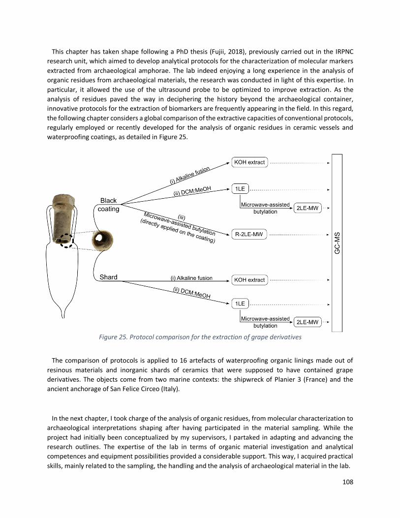
108
This chapter has taken shape following a PhD thesis (Fujii, 2018), previously carried out in the IRPNC
research unit, which aimed to develop analytical protocols for the characterization of molecular markers
extracted from archaeological amphorae. The lab indeed enjoying a long experience in the analysis of
organic residues from archaeological materials, the research was conducted in light of this expertise. In
particular, it allowed the use of the ultrasound probe to be optimized to improve extraction. As the
analysis of residues paved the way in deciphering the history beyond the archaeological container,
innovative protocols for the extraction of biomarkers are frequently appearing in the field. In this regard,
the following chapter considers a global comparison of the extractive capacities of conventional protocols,
regularly employed or recently developed for the analysis of organic residues in ceramic vessels and
waterproofing coatings, as detailed in Figure 25.
Figure 25. Protocol comparison for the extraction of grape derivatives
The comparison of protocols is applied to 16 artefacts of waterproofing organic linings made out of
resinous materials and inorganic shards of ceramics that were supposed to have contained grape
derivatives. The objects come from two marine contexts: the shipwreck of Planier 3 (France) and the
ancient anchorage of San Felice Circeo (Italy).
In the next chapter, I took charge of the analysis of organic residues, from molecular characterization to
archaeological interpretations shaping after having participated in the material sampling. While the
project had initially been conceptualized by my supervisors, I partaked in adapting and advancing the
research outlines. The expertise of the lab in terms of organic material investigation and analytical
competences and equipment possibilities provided a considerable support. This way, I acquired practical
skills, mainly related to the sampling, the handling and the analysis of archaeological material in the lab.

109
The protocol never being handled in the lab since it was published in 2016 and not included in the
optimization performed during the previous thesis, I matured methodological competences through the
development of the microwave-assisted butylation protocol, initially meant for ceramics and through the
adaptation to the internal waterproofing lining. I improved technical skills thanks to the use of gas
chromatography, mass spectrometry and the associated analytical modules. The archaeological samples
being analyzed with several protocols to get the more information out, I was trained to chemometric tools
with the aim of synthesizing results since I had to deal with important data quantities obtained from
molecular identifications. Finally, I also gained experience in by generating huge scientific writing and by
setting up the original draft and edition of the following publication.
This article was accepted in the special issue of Crystals 2021 that dealt with “Characterization of
Pigments, Binders and Organic Residues in Art, Archaeology and Cultural Heritage” (DOI
10.3390/cryst11111300; https://doi.org/10.3390/cryst11111300).

110
Protocol comparison for organic residue analyses from waterproofing
materials and shards of Roman archaeological amphorae
Louise Chassouant1,2*, Fabienne Olmer3, Chiara Delpino4, Alessandra Celant2,
Cathy Vieillescazes1, Donatella Magri2, Carole Mathe1*
1 IMBE UMR7263/ IRD237, Avignon University/ CNRS/ IRD/ AMU, Restoration Engineering of Natural and
Cultural Heritage F-84000, Avignon, France; [email protected] 2 Dipartimento di Biologia Ambientale, Sapienza Università di Roma, Piazzale Aldo Moro, 5, 00185,
Roma, Italy; [email protected]; [email protected];
[email protected] 3 French National Center for Scientific Research, Centre Camille Jullian - UMR 7299, Maison
Méditerranéenne des Sciences de l'Homme, 13097 Aix-en-Provence Cedex 2, France ;
[email protected] 4 Soprintendenza APAB National per le province di Frosinone e Latina, via Pompeo Magno 2, 00192,
Rome, Italy ; [email protected]
* Correspondence : [email protected] ; [email protected]
Highlights
• Microwave assisted butylation is proposed for the identification of grape markers
• BF3-catalyzed esterification is developed for the analyses of pitch coatings
• Molecular signatures of grape-derivatives, pine pitch and wood tar are formulated
• Extractive protocols are compared based on their extractive capacities
• Archaeological fermented grape beverages are detected in Dressel 5 and 18
4.1. Abstract
With the aim of addressing the impact of extractive protocols in molecular characterization of ceramic
content, sixteen archaeological shards and waterproofing coatings of Roman amphorae were studied to
compare the extractive capacities of protocols prevalently mentioned in wine amphorae analysis. A
microwave-assisted protocol is developed in order to esterify grape-derivative markers from
archaeological pitch and shard. Gas Chromatography-Mass Spectrometry is used to highlight the great
capacities of a two-step protocol that combines organic extraction with BF3-etherate complex butylation
applied on archaeological shards. Instead, simultaneous alkaline fusion and direct-resin acid-catalyzed
butylation are favored for the characterization of waterproofing material. The identification of tartaric
acid, together with succinic, fumaric pyruvic and syringic acids provides valuable insights on the
archaeological grape-derivative content, possibly wine. Diterpenic markers highlighted Pinus pitch and
111
wood tar, originally used to waterproof the amphorae. Since markers are reliable tools in organic residue
analyses, protocols exhibiting high extractive capacities are favored to avoid false conclusions drawn
through the absence of markers.
Keywords: Organic residue analyses, Biomolecular archaeology, Tartaric acid, Microwave-assisted
butylation, Gas Chromatography-Mass Spectrometry, Ceramic content, Wine
4.2. Introduction
In the early 1990s, Evershed introduced the archaeological biomarker concept to trace back the original
use of potteries. Focusing on either the carbon structures or the pattern distributions, molecules act as
chemical fingerprints (Heron and Evershed, 1993). They can reveal information regarding the initial
composition, natural ageing, anthropic degradation as well as contamination. This way, organic residue
analyses became an established field of research; fundamental to address the archaeological content by
rending microremains identifiable (Evershed, 2008). For instance, the earliest consumption of wine could
be dated back to the Neolithic in the South Caucasus through molecular markers (McGovern et al., 2017).
Although pioneering studies laid the foundation using Feigl spots, infrared spectroscopy or HPLC to
identify tartaric acid (Michel, McGovern and Badler, 1993; McGovern and Michel, 1996; McGovern et al.,
2004), the emergence of chromatographic tools offered great substitutes to traditional methods to
prevent from false-positives (Garnier et al., 2003; Jerković et al., 2011; Pecci et al., 2013). As rightly
criticized in literature, neither molecular identification through sole retention or migration time, nor the
precision of UV detection are reliable (Stern et al., 2008; Drieu et al., 2020). Enhancing the specificity and
sensitivity required for wine markers identification as well as providing structural information, Gas
Chromatography coupled with Mass Spectrometry (GC-MS) became frequently employed (Garnier and
Valamoti, 2016; Fujii et al., 2019, 2021; Pecci et al., 2020). Although recent techniques on the cutting edge
of technology showed great analytical advancements, equipment costs remain highly limiting. Even
though tandem MS mode or selected ion monitoring mode in liquid or gas chromatography coupled with
MS were published to grandly improve the limit of detection to specifically target the presence of
archaeological wines (Guasch-Jané et al., 2004, 2006; Barnard et al., 2011; Manzano et al., 2016;
McGovern et al., 2017), their usage remains rare since it implies ultra-advanced equipment. GC-MS
consequently appears as a good compromise to turn routine analyses into efficient molecular markers
searches, decisive for their identification. This way, fermented grape-beverage has been evidenced in
archaeological ceramic jars and amphorae through the presence of tartaric acid (Michel, McGovern and
Badler, 1993; Rageot et al., 2019). Although the molecule is not exclusively produced by grapes, its
concentration remains higher than in other exotic plant sources such as tamarind, yellow plums (Barnard
et al., 2011) or pomegranates (Hasnaoui et al., 2011). Moreover, it better survives upon archaeological
time compared to other grape acids (Pecci et al., 2013). For this reason, it is usually considered as a grape
biomarker when supported by archaeological contexts and/or archaeobotanical grape evidence (Garnier
and Valamoti, 2016; McGovern et al., 2017; Pecci et al., 2018; Drieu et al., 2020). Although synthesized by
numerous plants and fungi, pyruvic, fumaric and malic acids are additionally produced during the
fermentation process (Ribéreau-Gayon et al., 2006). Syringic acid highlights red wine since it arises from
malvidin, the pigment responsible for dark grape coloration (Guasch-Jané et al., 2004; Barnard et al.,
2011).

112
Beyond the presence of markers, two concepts need however to be separated: the extraction and the
analytical detection. While analytical developments focused on the former, the latter remains of a first
interest. Indeed, robust protocols must be employed to increase the chance of extracting tartaric acid and
other grape beverage markers from the ceramic matrix. Since the biomarker concept relies on the
presence/absence of characteristic features, it becomes necessary to avoid false-negative induced by
extraction strength flaw (Blanco-Zubiaguirre et al., 2019). Among the protocols most mentioned to
promote the breaking of the intramolecular hydrogen bonding between tartrate salts and the silicate-rich
ceramic, either acid or alkaline conditions are favored (Pecci et al., 2013; Correa-Ascencio and Evershed,
2014; Garnier and Valamoti, 2016). Alkaline treatment is followed by acidification to make tartaric acid
more soluble in ethyl acetate before extraction (Pecci et al., 2017, 2020). Differently, the acido-catalyzed
butylation of the unsolved material after polar extraction greatly evidenced grape derivatives in Neolithic,
Etruscan or even Early Medieval Islamic jars (Garnier and Valamoti, 2016; Frère and Garnier, 2017; Drieu
et al., 2021). The esterification of organic markers aimed at enhancing their solubility into the extractive
solvent.
The primary aim of this paper is to describe an adapted microwave-assisted protocol for the acid-
catalyzed extraction in order to ensure easy-going and accelerated approach for grape-derivative
detection, applicable on both organic and inorganic artefacts. Using GC-MS, we additionally provide a
comparative study of the extractive capacities offered by alkaline, acidic and polar extractions with the
specific aim of proposing a robust and cost-effective methodology directly applicable on archaeological
artefacts (waterproofing material and shard). In this study, three protocols were conducted depending on
the nature of the samples. The first one consisted of a basic extraction applied on both coatings and shard.
The second one encompassed a two-step protocol with consecutive lipid extraction, including microwave-
assisted optimization for the butylation. This protocol was tested on coatings and shard. The last protocol
corresponded to a variant of the second one, with the application of the two-step extraction handled
separately (i.e., not consecutively but in two different ways) and both steps directly conducted on the
waterproofing matter. This study was carried on sixteen Roman amphorae coming from two different
maritime archaeological sites. 10 shards and 8 resinous coatings were investigated in order to promote a
great representativeness of the comparison and prevent from any bias that would arise from local or
material specificities. A glossary of all of the identified compounds was presented at the end of the article.
4.3. Material
4.3.1. Archaeological samples
This comparative study integrated 16 archaeological amphorae coming from two different contexts. 11
amphorae were excavated from the shipwreck of Planier 3 (France) and 5 came from the ancient
anchorage of San Felice Circeo (Italy). The amphorae were selected to increase the variety between
objects in order to make the protocol comparison the more likely to be applied on any further artefacts.
Thus, the objects present a strong variability in terms of ceramic pastes, provenance and marine
archaeological context, visual presence of coating and conservative conditions (Table 5). Although they all
belong to the Roman period, they came from maritime context that can be interpreted differently. The
amphorae from Planier 3 were all excavated from the same shipwreck, sunk in 49 BC near the Planier’s

113
island (43°11′54″N; 5°13′48″E) close to the Marseille coasts (France). Coming from the region of Brindes,
the cargo might have stopped first in the Sinus Tarentinus region to load Lamboglia 2 amphorae and then
at Pozzuoli before heading towards the Narbonnaise. Excavation campaigns conducted from 1968 to 1975
revealed the important distribution of Dressel 1B, Lamboglia 2, ovoid and Brindisium amphorae in the
cargo (Tchernia, 1968). Along with the tremendous quantity of amphorae, Planier 3 exhibits the more
important evidence of archaeological pouzzolane stoppers. Different mineral pigments were also
reported in the cargo, such as realgar, white lead and the precious Egyptian blue of which the production
was settled in Pozzuoli (Tchernia, 1969). Such discoveries outlined the luxurious assumptions of traded
products transported on the ship.
Table 5. Archaeological amphorae investigated
Five amphorae were excavated in the ancient anchorage of San Felice Circeo (41°13’49.0”N;
13°06’30.1”E) located 90 km SE of Roma. They were uncovered due to the winter storm of 2018, along
with an important scattering of archaeological records located few hundred meters from the coast at a
depth of 5 to 7m under the sea level. The Soprintendenza (the local Office of the Italian Ministry of Culture)
supervises ongoing excavation campaigns. The time scale established by the archaeological finds ranges
from the Republican period through the Late Roman period to the post-medieval period. The diversity of
the excavated ceramics, in terms of morphology and the time period they belong to provided valuable
insights of ancient anchorage (Delpino and Melandri, unpublished paper) although the hypothesis of a
shipwreck cannot be excluded.
The Late Greco-Italian/Dressel 1 typologies refer to 150 to 10 BC. They originate from south-central Italy,
from Campania to Etruria, as attested by important kiln finds along the coastal area (Olmer, 2012). These
amphorae were widely used to trade wine in the Mediterranean Basin, from Central Europe to Spain via
Gaul. Lamboglia 2 amphora originate from the Adriatic coast (Cipriano and Carre, 1989; Panella, 1998).
They have largely contributed to the trade of wine in the western Mediterranean (Lamboglia, 1952). Chios
amphora were surely meant for wine trading since the island was prized for the great quality of wine they
produced (Okan, Atila and Akyol, 2015). Maña C2 amphora arose from the Punic tradition amphorae,

114
originating from North African coasts. Although oil and fish preserves are frequently mentioned in such
typology, the amphora was included in the study (Bonifay, 2016). Dressel 5, also referenced as Rhodian
amphorae, usually contained wine from the Aegean coast (Peña, 2007). The present study focuses on 8
samples from waterproofing coatings and 10 samples of ceramic shards (see Table 5). Samples no. 749
and 1014 were analyzed with both the coating and the ceramic clay.
4.3.2. Solvents and reagents
All of the organic solvents were of analytical grade. Methanol (MeOH), dichloromethane (DCM), diethyl
ether (DEE), ethyl acetate and KOH were purchased by Merck (Darmstadt, Germany). Hexane and N,O-
Bis(trimethylsilyl)trifluoroacetamide/trimethylchlorosilane (BSTFA/TMCS) and commercial standard
molecules such as maleic, succinic, fumaric, malic, pyruvic, tartaric, syringic and dehydroabietic (DHA)
acids were supplied from Sigma-Aldrich (Darmstadt, Germany). Anhydrous butanol and cyclohexane were
purchased by Acros Organics (Illkirch, Germany) and BF3 diethyl etherate from Alfa Aesar (Kanderl,
Germany). The fresh colophony resin standard was purchased by Kremer Pigmente GmbH & Co KG
(Aichstetten, Germany).
4.4. Methods
4.4.1. Optimization of the acid-catalyzed esterification: microwave-
assisted butylation
The protocol aiming at butylating wine acid markers was adapted from Garnier and Valamoti (Garnier
and Valamoti, 2016). It was firstly developed for tartaric acid before being extended to other standard
molecules of maleic, succinic, fumaric, pyruvic, malic, syringic acids. 5 mg of commercial standard were
treated with a mixture of boron trifluoride, butan-1-ol and cyclohexane (1:2:4 v/v/v) in a sealed vial placed
in CEM Discover® LabMate microwave synthesizer (MW) (CEM Corp., Orsay, France). The instrument was
used in single mode (50 Hz; 300 W maximum output power). An infrared sensor positioned below the
circular vessel continuously measure the routine temperature. The self-adaptive circular waveguide
technique allows the circular cavity to automatically be adjusted in order to optimize the energy provided
for the reaction (Kappe, Dallinger and Murphree, 2008). Continuous pressure measurements permit on-
the-fly changes for power control to maintain a maximal temperature, set at 80°C with the CEM Synergy™
software (version 0.9). Different reaction times were tested (single dynamic cycle of 5 min, 10 min and
three successive dynamic cycles of 5 min each) to evaluate the heating run necessary for the butylation
with high stirring speed. The butylation advancement was followed by thin-layer chromatography with
cyclohexane:ethyl acetate (1:1 v/v). After the butylation was completed, the solution was neutralized with
a saturated solution of sodium carbonate. The esterified compounds were extracted two times with DEE.
The combined organic fractions were washed twice with Milli-Q water and dried with anhydrous sodium
sulphate before filtration on a PTFE cartridge (0.45 µm).
115
4.4.2. Analytical procedures for inorganic shards
For the comparative analyses of the archaeological shards, two protocols were applied: a basic
extraction adapted from Pecci et al. (Pecci et al., 2013) and a two-step lipid extraction including the MW-
assisted optimization for the butylation. To reduce the risk of external contamination, a thin layer was
initially removed from the inner surface of the shard before sampling the drilled ceramic over 1-2 mm in
depth.
On the one hand, 100 mg of crushed shard were extracted three times with KOH (1M; 2 mL) using an
ultrasound probe (VCX 130 Vibra-Cell Sonics, Sonics and Materials, Newtown, U.S.A.) for 3 min. After
centrifugation, the successive extracts were combined and acidified up to pH 2. The organic phase was
extracted 3 times with ethyl acetate (3 mL), filtered on a PTFE cartridge (0.45 µm) and evaporated to
dryness.
On the other hand, 100 mg of crushed shard were extracted 3 times with DCM:MeOH (1:1 v/v) with the
ultrasound probe for 3 min. After centrifugation, the organic supernatant of was filtered with PTFE (0.45
µm) and evaporated to dryness. This first step of the lipid extraction was recorded as 1LE. The remaining
powder after the organic extraction was treated for the MW-assisted butylation with a mixture of BF3
etherate complex, butan-1-ol and cyclohexane (1:2:4 v/v/v) for 3 times 5 min. After neutralization with a
saturated sodium carbonate solution, the organic fractions were extracted with DEE and washed twice
with H20 before drying over sodium sulphate. The corresponding extract was labelled 2LE-MW. After
filtration on a PTFE cartridge (0.45 µm), both extracts (1LE and 2LE-MW) were individually evaporated
under a gentle N2 stream.
4.4.3. Analytical procedures for organic coatings
For the comparative analyses of the archaeological coatings, three protocols were tested. The first one
was the basic extraction already reported (see section 4.4.2.). The second was the adapted two-step lipid
extraction with MW optimization for the butylation as previously described (see section 4.4.2.). The only
difference was the starting amount of 50 mg of organic coating versus 100 mg of shard. The third protocol
consisted of the same two-step lipid extraction, with each step independently conducted on the coating:
the organic extraction with DCM:MeOH (1LE) and the extraction after butylation (R-2LE-MW) were hence
handled separately. 10 mg of coating material were necessary for the direct-pitch esterification.
Butylation was performed as previously described for the MW parameter and heating runs.
4.4.4. GC-MS analyses
GC-MS analyses were carried out on a Thermo Scientific™ Focus system equipped with an Al 3000
autosampler and an ITQ™ 700 Series Ion Trap Mass Spectrometer (ThermoFischer Scientific, Illkirch-
Graffenstaden, France). A ThermoGOLD™ TG-5MS fused silica capillary column (5% diphenyl; 95%
dimethyl polysiloxane) of 30 m length x 0.25 mm i.d. x 0.25 μm thickness ensured the separation of the
mixture carried with helium at a constant flow rate of 1 mL min−1. 1 µL solution was injected in splitless

116
mode at 250°C. Transfer line, ion trap and manifold temperatures were respectively 300°C, 200°C and
50°C. Mass spectra were recorded in electron impact mode with an electron ionization energy of 70 eV,
with ionization time of 25,000 µs. Scan are recorded in the range of 40–650 m/z. The oven temperature
stayed isothermal for 2 min at 50°C, increased at 8°C/min to 140°C held for 2 min before heating to 160°C
at 2.5°C/min and finally 330°C at 15°C/min and held for 3 min.
After evaporation, all of the extracts were derivatized with BSTFA (200 µL, 70°C, 30 min) before injection
in splitless mode in GC-MS in 200 µL of hexane:DCM (1:1 v/v).
Data treatment was performed on Xcalibur™ software (version 4.3). Peak identification was achieved by
comparison of retention time and mass spectra with molecular commercial standards and from the NIST
MS Search 2.0 database (last access in May 2021).
4.4.5. Radar plot construction
The building of the radar plots (Figs. 26 and 27) relies on the presence/absence of 22 targeted molecules,
that account for the characterization of the grape derivatives content and the resinous coating. Among
them, 7 organic acids referred to grape composition (tartaric and syringic acids), fermentation (maleic,
succinic, pyruvic, fumaric, malic acids) and 15 diterpenic derivatives indicative of the nature of the coating
(dehydroabietic acid DHA, dehydroabietic methyl ester DHAM and retene) and its ageing (3-hydroxy-, 7-
hyrdoxy-, 15-hydroxy-; 7,15-dihydroxy-, 7-oxo- and 15-hydroxy-7-oxo-DHA and DHAM-derivatives). We
notably emphasized on highly significant markers, indispensable for grape derivative and coating
identification (i.e., tartaric acid, retene and DHAM compounds).
4.5. Results and discussion
4.5.1. Optimization of the acid-catalyzed butylation
The esterification advancement, followed by thin-layer chromatography, ensured the effective
butylation of standard molecules after 3 dynamic cycles of 5 min each. Butylated compounds were
additionally controlled with infrared spectroscopy (FT-IR) and NMR (1H, 13C). GC-MS analyses allowing
dibutyl tartrate (DBT) to be characterized with fragment ions at m/z 276, 305 and 391. The same
butylation protocol was then applied on commercial standards acids considered as dark grape (i.e.,
syringic acid) and fermentation markers (i.e., maleic, succinic, pyruvic, fumaric and malic acids) to obtain
their retention time and fragmentation patterns.

117
4.5.2. Extracting capacities comparison on archaeological shards
Archaeological artefacts being different from each other, the studied shards cannot be generalized to a
set of amphorae. Protocol comparisons and analyses interpretations must respect individual molecular
specificities, turning conclusions on the extraction capacities to be singular and object dependent. For this
reason, the use of a radar plot was favored to independently outline the number of molecules extracted
by each protocol and for each of the ten archaeological shards (Fig. 26). The protocol comparison applied
on shards encompassed: (i) an alkaline fusion with KOH extraction, (ii) a DCM-MeOH organic extraction
and (iii) its coupling with BF3-catalyzed MW-butylation applied on the dried remaining powder after the
organic extraction (2LE-MW extract).
Figure 26. Radar plot of the shards
For all the 10 shards, only the BF3-catalyzed butylation allowed DBT, highlighting the presence of tartaric
acid (Fig. 26). Partially dissolving the ceramic clay, BF3, optimized the release of the organic compounds
strongly bonded, or even polymerized (Correa-Ascencio and Evershed, 2014; Garnier et al., 2018).
Increasing the apolar character of the esterified acids, butylation favored their rapid extraction in
cyclohexane, hence favoring the butylation of remaining acids by shifting the equilibrium (Garnier and
Valamoti, 2016). From there, the extraction from the co-solvent is enhanced with DEE that has a low
dielectric constant solvent.

118
Even though hydroxyl anions arising from the alkaline fusion are supposed to interact with the ceramic
matrix to enhance the release of bonded acids (Pecci et al., 2013, 2017, 2020), no tartaric acid could be
identified with this protocol (Fig. 26). By shifting the solubility equilibrium of tartrate salts, KOH should
favor the bonding cleavage with the ceramic and leave the marker soluble in the aqueous phase (Guasch-
Jané et al., 2004; Pecci et al., 2013). In fact, tartaric acid resists archaeological time thanks to the formation
of salts that strongly interact with the clay matrix (Michel, McGovern and Badler, 1993). Conversion into
free tartaric acid would ensure the recovery in the aqueous phase before extraction with ethyl acetate.
Extraction issues could originate from the insufficient alkaline robustness or from the poor solubility of
tartaric acid in ethyl acetate (Guasch-Jané et al., 2004; Drieu et al., 2020). The hypothesis of tartrate salt
that would be formed once the alkaline fusion released tartaric acid from the ceramic cannot be ruled out
(Drieu et al., 2020). It is worth noting the limit of detection involved in every protocols. Starting from pure
standard, quantitative analysis comparing the amount of tartaric acid recovered after extraction reported
to identify 77% of the acid with butylation while it did not reach 0.1% with alkaline fusion (Drieu et al.,
2020). Additionally, Garnier and Valamoti reported the detection of tartaric acid up to 10 ng/g shard with
the acido-catalyzed protocol (Garnier and Valamoti, 2016). In conclusion, neither KOH fusion, nor the
organic extraction with DCM-MeOH were suitable for the characterization of grape derivatives.
Aside from the considerable extraction of DBT, esterification also accounted for the extraction of grape
acids (Table 6). Maleic acid was only characterized with butylation (m/z 99; 117). Malic acid, although
hardly characterized with KOH fusion, was always identified as dibutyl malate (m/z 101; 145; 161; 303).
The most important increase of molecules extracted was observed for the amphora no. 6904, where 3
fermentation acids (over the 5 considered) could be identified with butylation, hitherto absent with
alkaline fusion and DCM:MeOH extraction. Surprisingly, pyruvic acid was directly extracted with
traditional solvents in 9 shards. Neither KOH nor butylation reached such great extent (Table 6) and the
molecule that originates from malolactic fermentation (Ribéreau-Gayon et al., 2006) was only identified
in 2 shards with alkaline fusion and never recovered as dibutylcetal. To the contrary, fumaric acid which
is considered as marker of alcoholic fermentation (Garnier et al., 2018), was preferentially extracted with
alkaline fusion and never identified with butylation (Table 6). Since maleic, succinic, pyruvic, fumaric and
malic acids can originate from the fermentation of grapes, they are considered as fermentation markers
of wine. However, to compensate for their lack of exclusivity towards grape fermentation only, the greater
the number of fermentation markers extracted, the more reliable the fermentation assumption. For this
reason, the combination of extractive protocols would allow the number of molecules extracted to be
increased. Since only the butylation proved to surely trace tartaric acid, the most fruitful coupling would
include it to widen its ex-tractive capacities.
Although syringic acid is naturally present in many plants and in wood lignin (Barnard et al., 2011; Pecci
et al., 2020), malvidin origin has been clearly evidenced (Guasch-Jané et al., 2004). After the reaction of
crushed grapes with yeasts present at the fruit surface, the alcohol produced by the spontaneous
fermentation starts hydrolyzing malvidin contained in the dark grape skins. To surely state on red grapes,
the benefits of a two-step protocol with the first part targeting the free syringic acid are not to be
demonstrated anymore (Garnier and Valamoti, 2016; Pecci et al., 2017). Indeed, while the first extraction
can include free syringic acid, the second one rather focuses on the acids that are deeply impregnated in
the clay matrix. The ac-id-catalyzed extraction hence promotes the tracing of malvidin origin. Following
Table 6, none of the samples revealed the presence of syringic acid as free acid and butyl syringate was
identified in almost all the samples (m/z 240, 296, 311, 326), shedding light on the dark-color beverage
119
contained in amphorae. The Chios amphora was the only one that did not show the dark marker,
highlighting the white nature of the content.
Coupling organic extraction with esterification extended the overall number of identified compounds
from 2 to 10 molecules per sample over the 22 molecules targeted (Fig. 26). No DHA was extracted for
the amphorae nos. 6904, 6566 and 6828c using alkaline fusion whereas this compound was extracted
with DCM:MeOH for all the items (Table 6). Exceptions were nonetheless noticed for amphorae nos.
6828b and 6793 where organic solvents barely supported the extraction of diterpene derivatives, hence
grandly diminishing the number of characterized molecules (Fig. 26). Oxidized diterpenes, identified in all
the amphorae, provided insights on the ageing of the pitch that occurred through oxygen incorporation
into the DHA skeleton to form peroxide intermediates. Hydroxyl and ketone derivatives are produced by
peroxide reduction or dehydration, respectively (Mezzatesta et al., 2021).
Table 6. Molecules identified in shards. Presence (+) and absence (-) of molecular markers under alkaline
fusion (KOH ext.), organic extraction with DCM:MeOH, butylation applied on the remaining fraction (2LE-
MW). The number of ‘+’ refers to the number of molecules present. ac.: acid; OH-DHA: hydroxy-DHA (i.e.,
3-hydroxy-DHA; 7-hydroxy-DHA and 15-hydroxy-DHA); Oxo-DHA: 7-oxo-DHA; DiOH-DHA: 7,15-dihydroxy-
DHA; Oxo-OH-DHA: 7-oxo-15hydroxy-DHA. DHAM and oxidized derivatives (OH-DHAM, Oxo-DHAM, DiOH-
DHAM and Oxo-OH-DHAM) refer to the same skeleton with methyl ester derivatives instead of the
carboxylic acid function.

120
Sample Protocol Maleic
ac. Succinic
ac. Pyruvic
ac. Fumaric
ac. Malic
ac. Tartaric
ac. Syringic
ac. Retene DHA
OH-DHA
Oxo-DHA
DiOH-DHA
Oxo-OH-DHA
DHAM OH-
DHAM Oxo-
DHAM DiOH-DHAM
Oxo-OH-
DHAM
1014
KOH ext - + - + - - + + + +++ + - - + + - - +
DCM:MeOH - + + - - - - + + +++ + + - + ++ + + -
2LE-MW + + - + + + +
749
KOH ext - + - + + - + + + +++ + + + + +++ + + +
DCM:MeOH - + + - - - - + + +++ + + + + +++ + + +
2LE-MW + + - - + + +
6904
KOH ext - - - - - - - - - - - - - - - - - - DCM:MeOH - + + - - - - + + - - - - - - - - -
2LE-MW + + - + + + -
6828A
KOH ext - + - + + - + + + +++ + + + + ++ - + +
DCM:MeOH - + + + - - - + + +++ - + + + +++ + + +
2LE-MW - + - - + + +
6828B
KOH ext - + + + + - + + + +++ + + + + + - + +
DCM:MeOH - + + - - - + + + ++ - - - + +++ + - -
2LE-MW + + - - + + +
6565
KOH ext - + + - + - + + + - - - - + + + - +
DCM:MeOH - + + - - - - + + ++ - - - + ++ + - +
2LE-MW - + - - + + +
6793
KOH ext - + - - - - + + + +++ - + - + ++ + + + DCM:MeOH - + + - - - + + + + - - - + - - - -
2LE-MW - + - - + + +
6545
KOH ext - - - - - - - - - - - - - + - - - -
DCM:MeOH - + - - - - - - - - - + - + - - + -
2LE-MW - + - - + + +
6566
KOH ext - - - - - - - - - - - - - + + - - -
DCM:MeOH - - + - - - - + + - - + - + + - + -
2LE-MW - + - - + + +
6828C
KOH ext - - - + - - - - - - - - - + - - - -
DCM:MeOH - - + - - - - + + - - + - + + + + -
2LE-MW - + - - + + +
121
Almost all the protocols extracted DHAM markers, traducing an archaeological use of Pinaceae wood tar
(Colombini and Modugno, 2009) (Fig. 26). Indeed, DHAM characterizes wood distillation: DHA is esterified
by the methanol contained in the wood during pyrolysis at high temperature. Interestingly, the amphora
from Chios (no. 6904) is the unique potsherd only containing Pinus pitch. Neither DHAM derivatives, nor
triterpenoids were identified yet the island was famous for its mastic resin of Pistacia lentiscus L.
(Modugno, Ribechini and Colombini, 2006). The presence of resinated wine such as retsina can be assumed
since the practice was common in Greece. Aside from antibacterial properties, small amounts of Pinus
pitch could be added during the alcoholic fermentation to flavor and color wines (Proestos et al., 2005)
which can be related to the insufficient. No retene was observed for the amphorae nos. 6566 and 6828c
with alkaline fusion whereas it did with DCM:MeOH (Fig. 26). Even though no chemical explanation can
afford it, the archaeological meaning refers to the heating treatment of the resin (Mezzatesta et al., 2021).
Thermal degradation above 300°C induces abietane aromatization into retene. Only the amphora no. 6545
(Dressel 5), where retene has never been identified, was coated with a resinous material that had not been
produced under a high temperature.
4.5.3. Extracting capacities comparison on archaeological coatings
The protocol comparison applied on coatings encompassed: (i) an alkaline fusion with KOH extraction,
(ii) a DCM:MeOH organic extraction, (iii) its coupling with BF3-catalyzed MW-butylation applied on the
dried remaining powder after the organic extraction (2LE-MW) and (iv) the same BF3-catalyzed MW-
butylation but directly applied on the coating matter (R-2LE-MW).
The radar plot (Fig. 27) overviews the extractive molecular capacities of the different protocols for each
of the 8 coatings. Samples nos. 1014 and 749 have already been discussed through the analyses of their
associated shards. Coatings and shards analyses provided identical insights on the nature of the coating
and the content even though poor insignificant molecular differences regarding DHA and DHAM
derivatives were observed (Tables 2 and 3).
Tartaric acid was successfully identified in all of the 8 coatings when the butylation was directly applied
on the pitch (R-2LE-MW) (Fig. 27). In comparison, when it was applied after organic extraction (2LE-MW),
the esterification allowed DBT to be identified from only 7 pitch samples. The amphora no. 1014 (Dressel
1) did not exhibit DBT in the butylated fraction, which can be related to the insufficient remaining sample
quantity. Indeed, the remaining powders were usually less than 1 mg, a major part of the initial mass being
dissolved during DCM:MeOH extraction. Moreover, DBT was identified as traces for all the samples in 2LE-
MW, which clearly argued for the limited quantity effect. The protocol was therefore hardly suitable for
content identification. Except for the amphorae nos. SFC1 and SFC3, the alkaline fusion did not allow
tartaric acid to be extracted (Fig. 27), which mirrors the unsuitability already reported for shards.
Considering fermented markers, pyruvic acid was only identified with butylation, as dibutylcetal (m/z 61,
117, 173) (Kawamura, 1993). Maleic acid was never characterized. Succinic and fumaric acids were
respectively rarely and never identified with butylation, although they were successfully extracted with
alkaline fusion or organic solvent extraction (Table 7). Moreover, KOH fusion allowed 8 additional grape-
acids (i.e., succinic, fumaric and malic acids) to be extracted from 5 amphorae (nos. 6570a, SFC1, SFC3,
SFC4 and SFC5) that traditional solvents did not provide.

122
Figure 27. Radar plot of the coating materials
Syringic acid was not detected in any of the amphorae SFC2, SFC4 and SFC5 (Table 7), hence suggesting
a white winemaking process. On the contrary, red beverages were conjectured for all the other samples
(nos. 1014, 749, 6570a, SFC1 and SFC3). Although a two-step protocol with successive alkaline fusion and
butylation would favor the identification of free syringic acid, it is technically hardly feasible because the
alkaline fusion left an aqueous matrix difficult to dry completely. Remaining water strongly reacts with BF3,
thus inhibiting butylation (data not shown).
Table 7. Molecules identified in pitch coatings. Presence (+) and absence (-) of molecular markers under alkaline fusion (KOH ext.), organic extraction with DCM:MeOH, butylation applied on the remaining
fraction (2LE-MW) or applied directly on the pitch

123
Sample Protocol Maleic
ac. Succinic
ac. Pyruvic
ac. Fumaric
ac. Malic
ac. Tartaric
ac. Syringic
ac. Retene DHA
OH-DHA
Oxo-DHA
DiOH-DHA
Oxo-OH-DHA
DHAM OH-
DHAM
Oxo-DHA
M
DiOH-DHAM
Oxo-OH-
DHAM
1014
KOH ext - + - - - - + + + +++ + + + + +++ - + +
DCM:MeOH - + - + - - + + + ++ - + - + +++ + + +
2LE-MW - - - - - - +
R-2LE-MW - + + - + + +
749
KOH ext - + - + + - + + + +++ + + + + ++ - + +
DCM:MeOH - + - + + - + + + ++ - - - + +++ + - +
2LE-MW - - + - + + -
R-2LE-MW - + + - + + +
6570a
KOH ext - + - + + - + + + +++ + + + + ++ - + +
DCM:MeOH - - - - - - - + - - - + - + + + + +
2LE-MW - - - - + + -
R-2LE-MW - - + - + + +
SFC1
KOH ext - + - + + + - + + +++ + + - + + + + -
DCM:MeOH - + - - - - - + + - - - - + +++ + - -
2LE-MW - - + - + + +
R-2LE-MW - + + - + + +
SFC2
KOH ext - + - - - - - + + + - - - + - + - -
DCM:MeOH - + - - - - - + + - - - - + ++ + - -
2LE-MW - - - - + + -
R-2LE-MW - - + - + + -
SFC3
KOH ext - + - + + + + + + +++ + + - + +++ + + -
DCM:MeOH - + - + - - - + + - - - - + +++ + - +
2LE-MW - - + - - + +
R-2LE-MW - - + - + + +
SFC4
KOH ext - + - - - - - + + + - - - + ++ + + +
DCM:MeOH - - - - - - - + + - - - - + ++ + - -
2LE-MW - - - - + + -
R-2LE-MW - - + - + + -
SFC5
KOH ext - + - + - - - + + +++ + + + + +++ + + +
DCM:MeOH - + - - - - - + + - - - - + ++ + - +
2LE-MW - - + - - + -
R-2LE-MW - + + - + + -
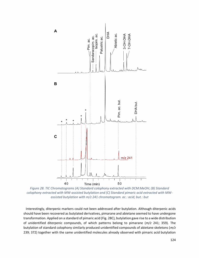
124
Figure 28. TIC Chromatograms (A) Standard colophony extracted with DCM:MeOH; (B) Standard colophony extracted with MW-assisted butylation and (C) Standard pimaric acid extracted with MW-
assisted butylation with m/z 241 chromatogram. ac.: acid; but.: but
Interestingly, diterpenic markers could not been addressed after butylation. Although diterpenic acids
should have been recovered as butylated derivatives, pimarane and abietane seemed to have undergone
transformation. Applied on a standard of pimaric acid (Fig. 28C), butylation gave rise to a wide distribution
of unidentified diterpenic compounds, of which patterns belong to pimarane (m/z 241; 359). The
butylation of standard colophony similarly produced unidentified compounds of abietane skeletons (m/z
239; 372) together with the same unidentified molecules already observed with pimaric acid butylation

125
(Fig. 28B-C). Induced by the harsh Lewis-acid conditions, diterpenic skeletons were reported to undergo
isomerization, skeletal transposition, isomerization, rearrangement and proton migration (Delmond,
Taran and Valade, 1978; Fujita et al., 1979; Taran and Delmond, 1986). Moreover, the butylation of
standard colophony selectively esterified the diterpenoids present in the resin (Fig. 28A-B). Only butyl
dehydroabietate and pimarate could be identified (m/z 239; 356 and m/z 241; 343, respectively). The
remaining presence of DHA in the butylated fraction outlined the incomplete esterification of
diterpenoids.
Aside from showing unequivocal extractive capacities to target tartaric acid when directly applied on the
pitch, the butylation is consequently not self-sufficient to describe the resinous material. The protocol
must be coupled to broaden the scope of characterization and alkaline fusion coupled to direct pitch
butylation managed to give rise to more quantitative extractions (Fig. 27). Emphasizing on coating
markers, the numerical difference reverted to the extractive capacities of alkaline fusion versus traditional
solvents and homogeneously concerned DHA derivatives (hydroxy-DHA for all the coatings, oxo-DHA for
6 of them, dihydroxy-DHA for 4 of them and hydroxy-oxo-DHA for 3 pitches over 8). Again, the important
presence of oxidized Pinaceae diterpenoids highlighted the significant ageing of Pinus pitch. Retene and
DHAM markers, characterized in all the coatings independently from the protocol employed (Fig. 27),
attested of Pinaceae wood tar produced under high temperature pyrolytic treatment.
4.6. Conclusion
Beyond the successful MW parameters set to esterify molecules, the analyses show different trends. On
the one hand, the butylation of standard acids familiar of wine composition is successfully achieved.
Grape-acids were recovered as butylated products using MW. Indeed, the energy conduction of MW-
assisted reactions being different from reflux (Kappe, Dallinger and Murphree, 2008), it is essential to
verify the molecular integrity. On the other hand, even though butylation undoubtedly promoted tartrate
extraction from ceramic and coating materials, the protocol, when solely applied, did not provide a
representative picture of archaeological artefacts, specifically for coating material characterization.
Although MW are widely encouraged to reduce reaction times and costs, butylation has to be strengthen
up using another extractive protocol. For the analyses of shards, organic extraction with traditional
solvents followed by esterification were remarkably complementary, offering more reliability for the
identification of grape-derivatives in terms of number of extracted molecules and retene extraction.
In parallel, the butylation protocol, initially developed for inorganic potsherds, was successfully adapted
to organic coatings, hence ensuring a reliable tracing of archaeological grape-beverage directly from the
coating. Alkaline fusion allowed diterpenic markers to be extracted, concluding on the nature; the
formulation and the ageing of the coating matter, as well as it suggested the presence of fermentation
markers. Although a two-step protocol would prevent from free-acid misinterpretation, further
investigation is needed to overcome BF3 reaction with water.
Except for the amphora from Chios coated with pine pitch, all the other artefacts were waterproofed
with Pinus wood tar. All the samples validate the presence of archaeological grape contents, which could
have been wine, vinegar or any other fermented derivatives (McGovern et al., 2017). Even though Mañà
C2 (no. SFC3) are mostly considered for oil or fish preserves, the identification of wine markers may be

126
interpreted in the light of a reutilization, that needs to be further investigated. Among the frequent
typologies mentioned for wine trading, the presence of red beverage was confirmed in Dressel 5 (no.
6545), hitherto barely studied.
Glossary
DBT: Dibutyl tartrate
DCM: Dichloromethane
DHA: Dehydroabietic acid
DHAM: Dehydroabietic methyl ester
DiOH-DHA: 7,15-dihydroxy-dehydroabietic acid
DiOH-DHAM: 7,15-dihydroxy-dehydroabietic methyl ester
KOH: Potassium hydroxide
OH-DHA: Hydroxy-dehydroabietic acid (3-hydroxy-dehydroabietic acid; 7-hydroxy-dehydroabietic acid;
15-hydroxy-dehydroabietic acid)
OH-DHAM: Hydroxy-dehydroabietic methyl ester (3-hydroxy-dehydroabietic methyl ester; 7-hydroxy-
dehydroabietic methyl ester; 15-hydroxy-dehydroabietic methyl ester)
Oxo-DHA: 7-oxo-dehydroabietic acid
Oxo-DHAM: 7-oxo-dehydroabietic methyl ester
Oxo-OH-DHA: 7-oxo-15-hydroxy-dehydroabietic acid
Oxo-OH-DHAM: 7-oxo-15-hydroxy-dehydroabietic methyl ester
Acknowledgment
This research was financially supported by Marie Skłodowska-Curie Innovative Training Network, The
ED-ARCHMAT European Joint Doctorate, H2020-MSCA-ITN-EJD ED-ARCHMAT Joint Doctorate (Project
ESR9, grant agreement no 766311). The authors wish to thank Nathalie Huet, research engineer in charge
of preventive conservation and furniture management at the DRASSM (France).
Author contribution
Conceptualization, L.C., C.V., C.M.; methodology, L.C.; formal analysis, L.C.; investigation, L.C., C.M.;
resources, F.O., C.D., A.C.; visualization, L.C.; supervision, C.M.; project administration, C.V., D.M., C.M.;
funding acquisition, A.C., C.V., D.M., C.M.; writing—original draft preparation, L.C., C.M.; writing—
review and editing, L.C., F.O., C.D., A.C., C.V., D.M., C.M. All authors have read and agreed to the published
version of the manuscript.

127
Conflict of interest
The authors declare no conflict of interest. The funders had no role in the design of the study; in the
collection, analyses, or interpretation of data; in the writing of the manuscript, or in the decision to publish
the results.
Funding
This research and the APC were funded by Marie Skłodowska-Curie Innovative Training Network H2020-
MSCA-ITN-EJD ED-ARCHMAT Joint Doctorate, ESR9, grant number 766311. Work in Rome was funded by
Sapienza University project DISCOVER (RM120172AE4E11D6).
128
4.7. References Barnard, H. et al. (2011) ‘Chemical evidence for wine production around 4000 BCE in the Late Chalcolithic Near Eastern highlands’, Journal of Archaeological Science, 38(5), pp. 977–984. doi: 10.1016/j.jas.2010.11.012.
Blanco-Zubiaguirre, L. et al. (2019) ‘Wine markers in archeological potteries: detection by GC-MS at ultratrace levels’, Analytical and Bioanalytical Chemistry, 411(25), pp. 6711–6722. doi: 10.1007/s00216-019-02044-1.
Bonifay, M. (2016) ‘Amphores de l’Afrique romaine : nouvelles avancées sur la production, la typo-chronologie et le contenu’, in Amphorae ex Hispania. Paisajes de producción y consumo, III Congreso internacional de la SECAH - EX OFFICINA HISPANA (Tarragone, 10-13 décembre 2014). Tarragona: Monografías Ex Officina Hispana III, pp. 595–611. Available at: https://halshs.archives-ouvertes.fr/halshs-01956342.
Cipriano, M. T. and Carre, M.-B. (1989) ‘Production et typologie des amphores sur la côte adriatique de l’Italie. Amphores romaines et histoire economique. Dix ans de recherche. Actes du colloque de Sienne (22-24 mai 1986)’, Publications de l’École Française de Rome, 114(1), pp. 67–104.
Colombini, M. P. and Modugno, F. (2009) Organic Mass Spectrometry in Art and Archaeology. Edited by M. P. Colombini and F. Modugno. Chichester: John Wiley & Sons. doi: 10.1002/9780470741917.
Correa-Ascencio, M. and Evershed, R. P. (2014) ‘High throughput screening of organic residues in archaeological potsherds using direct acidified methanol extraction’, Analytical Methods, 6(5), p. 1330. doi: 10.1039/c3ay41678j.
Delmond, B., Taran, M. and Valade, J. (1978) ‘Réarrangements de squelette au cours de l’isomérisation catalysée par l’éthérate de trifluorure de bore d’epoxyde diterpénique’, Tetrahedron Letters, 19(48), pp. 4791–4794. doi: 10.1016/S0040-4039(01)85733-6.
Drieu, L. et al. (2020) ‘Is it possible to identify ancient wine production using biomolecular approaches?’, STAR: Science & Technology of Archaeological Research, pp. 1–14. doi: 10.1080/20548923.2020.1738728.
Drieu, L. et al. (2021) ‘Chemical evidence for the persistence of wine production and trade in Early Medieval Islamic Sicily’, Proceedings of the National Academy of Sciences, 118(10). doi: 10.1073/pnas.2017983118.
Evershed, R. P. (2008) ‘Organic residue analysis in archaeology : the archaeological biomarker revolution’, Archaeometry, 50(6), pp. 895–924. doi: 10.1111/j.1475-4754.2008.00446.x.
Frère, D. and Garnier, N. (2017) ‘Dairy Product and Wine in Funerary Rituals: The Case of a Hellenistic Etruscan Tomb’, Journal of Historical Archaeology & Anthropological Sciences, 1(6), pp. 222–227. doi: 10.15406/jhaas.2017.01.00034.
Fujii, H. (2018) Etude chromatographique de matériaux organiques contenus dans des amphores romaines. Université d’Avignon et des pays de Vaucluse.
Fujii, H. et al. (2019) ‘FT-IR and GC–MS analyses of Dressel IA amphorae from the Grand Congloué 2 wreck’, Journal of Archaeological Science: Reports, 28, p. 102007. doi: 10.1016/j.jasrep.2019.102007.
Fujii, H. et al. (2021) ‘Analysis of organic residues from the Châteaumeillant oppidum (Cher, France) using GC–MS’, Journal of Cultural Heritage, 51, pp. 50–58. doi: 10.1016/j.culher.2021.07.001.

129
Fujita, E. et al. (1979) ‘The Chemistry on Diterpenoids in 1978. Part-III’, Bulletin of the Institute for Chemical Research, Kyoto University, 57(5–6), pp. 385–410.
Garnier, N. et al. (2003) ‘Characterization of thermally assisted hydrolysis and methylation products of polyphenols from modern and archaeological vine derivatives using gas chromatography–mass spectrometry’, Analytica Chimica Acta, 493(2), pp. 137–157. doi: 10.1016/S0003-2670(03)00869-9.
Garnier, N. et al. (2018) ‘Looking for ancient fish products through invisible biomolecular residues in the Roman production vats from the Atlantic coast’, Journal of Maritime Archaeology, 13(3), pp. 285–328. doi: 10.1007/s11457-018-9219-x.
Garnier, N. and Valamoti, S. M. (2016) ‘Prehistoric wine-making at Dikili Tash (Northern Greece): Integrating residue analysis and archaeobotany’, Journal of Archaeological Science, 74, pp. 195–206. doi: 10.1016/j.jas.2016.03.003.
Guasch-Jané, M. R. et al. (2004) ‘Liquid chromatography with mass spectrometry in tandem mode applied for the identification of wine markers in residues from ancient Egyptian vessels’, Analytical Chemistry, 76(6), pp. 1672–1677. doi: 10.1021/ac035082z.
Guasch-Jané, M. R. et al. (2006) ‘The origin of the ancient Egyptian drink Shedeh revealed using LC/MS/MS’, Journal of Archaeological Science, 33(1), pp. 98–101. doi: 10.1016/j.jas.2005.06.013.
Hasnaoui, N. et al. (2011) ‘Organic acids, sugars, and anthocyanins contents in juices of Tunisian pomegranate fruits’, International Journal of Food Properties, 14(4), pp. 741–757. doi: 10.1080/10942910903383438.
Heron, C. and Evershed, R. P. (1993) ‘The analysis of organic residues and the study of pottery use’, Archaeological Method and Theory, 5, pp. 247–284. doi: 10.2307/20170233.
Jerković, I. et al. (2011) ‘Chemical profile of the organic residue from ancient amphora found in the adriatic sea determined by direct GC and GC-MS analysis’, Molecules, 16(9), pp. 7936–7948. doi: 10.3390/molecules16097936.
Kappe, C. O., Dallinger, D. and Murphree, S. S. (2008) Practical microwave synthesis for organic chemists: strategies, instruments, and protocols. John Wiley & Sons.
Kawamura, K. (1993) ‘Identification of C2-C10 ω-oxocarboxylic acids, pyruvic acid, and C2-C3 α-dicarbonyls in wet precipitation and aerosol samples by capillary GC and GC/MS’, Analytical Chemistry, 65(23), pp. 3505–3511. doi: 10.1021/ac00071a030.
Lamboglia, N. (1952) ‘La Nave Romana di Albenga’, Revista degli Studi Liguri, 18(3–4), pp. 131–236.
Manzano, E. et al. (2016) ‘A multi-analytical approach applied to the archaeological residues in Iberian glasses. Earliest evidences on the consumption of fermented beverages in votive rituals’, Microchemical Journal, 129, pp. 286–292. doi: 10.1016/j.microc.2016.07.006.
McGovern, P. et al. (2017) ‘Early Neolithic wine of Georgia in the South Caucasus’, Proceedings of the National Academy of Sciences, 114(48), pp. E10309–E10318. doi: 10.1073/pnas.1714728114.
McGovern, P. E. et al. (2004) ‘Fermented beverages of pre- and proto-historic China’, Proceedings of the National Academy of Sciences, 101(51), pp. 17593–17598. doi: 10.1073/pnas.0407921102.
McGovern, P. E. and Michel, R. H. (1996) ‘The analytical and archaeological challenge of detecting ancient wine: two case studies from the Ancient Near East’, in The Origins and Ancient History of Wine. Langhorne:

130
Routledge, pp. 57–65.
Mezzatesta, E. et al. (2021) ‘GC–MS and PCA analyses of diterpenoids degradation state in 21 human mummies of Ancient Egypt dating from New Kingdom to Graeco-Roman Period’, Journal of Cultural Heritage, 47, pp. 43–49. doi: 10.1016/j.culher.2020.09.008.
Michel, R. H., McGovern, P. E. and Badler, V. R. (1993) ‘The first wine & beer’, Analytical Chemistry, 65(8), pp. 408A-413A. doi: 10.1021/ac00056a002.
Modugno, F., Ribechini, E. and Colombini, M. P. (2006) ‘Chemical study of triterpenoid resinous materials in archaeological findings by means of direct exposure electron ionisation mass spectrometry and gas chromatography/mass spectrometry’, Rapid Communications in Mass Spectrometry: An International Journal Devoted to the Rapid Dissemination of Up-to-the-Minute Research in Mass Spectrometry, 20(11), pp. 1787–1800. doi: 10.1002/rcm.2507.
Okan, E., Atila, C. and Akyol, A. A. (2015) ‘The production of chios-style amphorae at a ceramic workshop in Phocaea (Foça)’, Mediterranean Archaeology and Archaeometry, 15(3), pp. 259–276. doi: 10.5281/zenodo.32068.
Olmer, F. (2012) ‘Les amphores sont-elles utiles à la chronologie de la fin de l’âge du Fer ?’, in Barral, P. and Fichtl, S. (eds) Regards sur la chronologie de la fin de l’âge du Fer (IIIe-Ier siècles avant notre ère) en Gaule non méditerranéenne, actes de la table ronde de Bibracte (15-17 octobre 2007). Bibracte, pp. 317–341.
Panella, C. (1998) ‘Anfore e archeologia subacquea’, Archeologia subacquea. Come opera l’archeologo sott’acqua. Storie dalle acque. VII Ciclo di lezioni sulla ricerca applicata in archeologia, Firenze. All’Insegn. Edited by G. Volpe, pp. 531–559.
Pecci, A. et al. (2013) ‘Identifying wine markers in ceramics and plasters using gas chromatography–mass spectrometry. Experimental and archaeological materials’, Journal of Archaeological Science, 40(1), pp. 109–115. doi: 10.1016/j.jas.2012.05.001.
Pecci, A. et al. (2017) ‘Use and reuse of amphorae. Wine residues in Dressel 2–4 amphorae from Oplontis Villa B (Torre Annunziata, Italy)’, Journal of Archaeological Science: Reports, 12, pp. 515–521. doi: 10.1016/j.jasrep.2017.02.025.
Pecci, A. et al. (2018) ‘Combining residue analysis of floors and ceramics for the study of activity areas at the Garum Shop at Pompeii’, Archaeological and Anthropological Sciences, 10(2), pp. 485–502. doi: 10.1007/s12520-017-0573-7.
Pecci, A. et al. (2020) ‘Wine consumption in Bronze Age Italy: combining organic residue analysis, botanical data and ceramic variability’, Journal of Archaeological Science, 123, p. 105256. doi: 10.1016/j.jas.2020.105256.
Peña, J. T. (2007) Roman Pottery in the Archaeological Record. Cambridge: Cambridge University Press. doi: 10.1017/CBO9780511499685.
Proestos, C. et al. (2005) ‘High performance liquid chromatography analysis of phenolic substances in Greek wines’, Food Control, 16(4), pp. 319–323. doi: 10.1016/j.foodcont.2004.03.011.
Rageot, M. et al. (2019) ‘New insights into Early Celtic consumption practices: Organic residue analyses of local and imported pottery from Vix-Mont Lassois’, PLoS ONE, 14(6), p. e0218001. doi: 10.1371/journal.pone.0218001.

131
Ribéreau-Gayon, P. et al. (2006) The Chemistry of Wine Stabilization and Treatment, Handbook of Enology. Vol. 2. Hoboken: Johan Wiley & Sons.
Stern, B. et al. (2008) ‘New investigations into the Uluburun resin cargo’, Journal of Archaeological Science, 35(5237), pp. 2188–2203. doi: 10.1016/j.jas.2008.02.004.
Taran, M. and Delmond, B. (1986) ‘Part. IV - Obtention de nouveaux squelettes diterpeniques tetracycliques lors d’isomerisation d’epoxydes-8,9 diterpeniques’, Tetrahedron, 42(17), pp. 4795–4806. doi: 10.1016/S0040-4020(01)82060-3.
Tchernia, A. (1968) Premiers résultats des fouilles de juin 1968 sur l’épave 3 du Planier. Etudes classiques.
Tchernia, A. (1969) ‘Les fouilles sous-marines de Planier (Bouches-du-Rhône)’, Comptes rendus des séances de l’Académie des Inscriptions et Belles-Lettres, 113(2), pp. 292–309. doi: 10.3406/crai.1969.12394.
132
Chapter 5
Archaeobotanical and chemical investigations on wine amphorae
from San Felice Circeo (Italy) shed light on
grape beverages at the Roman time
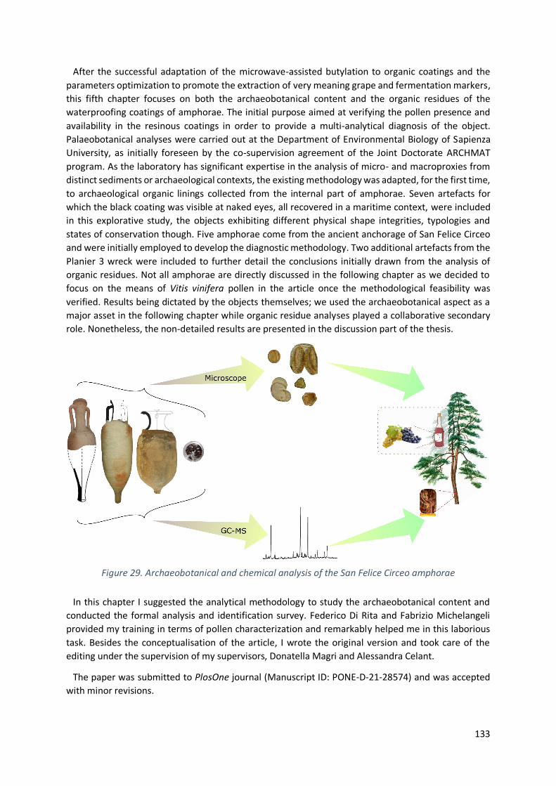
133
After the successful adaptation of the microwave-assisted butylation to organic coatings and the
parameters optimization to promote the extraction of very meaning grape and fermentation markers,
this fifth chapter focuses on both the archaeobotanical content and the organic residues of the
waterproofing coatings of amphorae. The initial purpose aimed at verifying the pollen presence and
availability in the resinous coatings in order to provide a multi-analytical diagnosis of the object.
Palaeobotanical analyses were carried out at the Department of Environmental Biology of Sapienza
University, as initially foreseen by the co-supervision agreement of the Joint Doctorate ARCHMAT
program. As the laboratory has significant expertise in the analysis of micro- and macroproxies from
distinct sediments or archaeological contexts, the existing methodology was adapted, for the first time,
to archaeological organic linings collected from the internal part of amphorae. Seven artefacts for
which the black coating was visible at naked eyes, all recovered in a maritime context, were included
in this explorative study, the objects exhibiting different physical shape integrities, typologies and
states of conservation though. Five amphorae come from the ancient anchorage of San Felice Circeo
and were initially employed to develop the diagnostic methodology. Two additional artefacts from the
Planier 3 wreck were included to further detail the conclusions initially drawn from the analysis of
organic residues. Not all amphorae are directly discussed in the following chapter as we decided to
focus on the means of Vitis vinifera pollen in the article once the methodological feasibility was
verified. Results being dictated by the objects themselves; we used the archaeobotanical aspect as a
major asset in the following chapter while organic residue analyses played a collaborative secondary
role. Nonetheless, the non-detailed results are presented in the discussion part of the thesis.
Figure 29. Archaeobotanical and chemical analysis of the San Felice Circeo amphorae
In this chapter I suggested the analytical methodology to study the archaeobotanical content and
conducted the formal analysis and identification survey. Federico Di Rita and Fabrizio Michelangeli
provided my training in terms of pollen characterization and remarkably helped me in this laborious
task. Besides the conceptualisation of the article, I wrote the original version and took care of the
editing under the supervision of my supervisors, Donatella Magri and Alessandra Celant.
The paper was submitted to PlosOne journal (Manuscript ID: PONE-D-21-28574) and was accepted
with minor revisions.
134
Archaeobotanical and chemical investigations on wine amphorae
from San Felice Circeo (Italy) shed light on grape beverages
at the Roman time
Louise Chassouant1,2*, Alessandra Celant2, Chiara Delpino3, Federico Di Rita2,
Cathy Vieillescazes1, Carole Mathe1, Donatella Magri2
1 IMBE UMR7263 / IRD237, Avignon University/ CNRS/ IRD/ AMU, Restoration Engineering of Natural
and Cultural Heritage F-84000, Avignon, France 2 Dipartimento di Biologia Ambientale, Sapienza Università di Roma, Piazzale Aldo Moro, 5, 00185,
Roma, Italy 3 Soprintendenza ABAP per le province di Frosinone e Latina, via Pompeo Magno 2, 00192, Rome, Italy
* Correspondence : [email protected]
5.1. Abstract
We hereby investigate the pitch used for coating three Roman amphorae from San Felice Circeo
(Italy) through a multidisciplinary study. The identification of molecular biomarkers by gas
chromatography - mass spectrometry is combined with archaeobotanical evidence of pollen and plant
tissues of Vitis flowers. Diterpenic chemical markers together with Pinus pollen and wood revealed
Pinaceae tar coating. Aporate 3-zonocolpate pollen, identified as Vitis, together with tartaric, malic
and pyruvic acids elucidate the grape-fermented nature of the content. Our conclusions open new
consideration on the use of grape derivatives that cannot be supported by traditional analytical
methods. Based on the finds of aporate Vitis pollen, found also in local modern and Middle Pleistocene
samples, we hypothesize the use of autochthonous vines. The presence of a medicinal wine
(historically reported as oenanthium) is also considered. We interrogate Vitis pollen capacity to target
grapevine domestication, thereby providing innovative tools to understand such an important process.
We anticipate our study to encourage a more systematic multidisciplinary approach regarding the
analyses of wine amphorae.
5.2. Introduction
Pilot experimental protocols with the specific aim of accessing pollen trapped in organic resins of
archaeological artefacts were advanced by Pons (Pons, 1961), Arobba (Arobba, 1976), Jones et al.
(Jones et al., 1998), and Jacobsen and Bryant (Jacobsen and Bryant, 1998). From there, only a limited
number of pollen studies have been conducted on amphorae. They mainly focus on the liquid
recovered from sealed jars (Gorham, 2000; Arobba et al., 2014) and on sediments contained in the
ceramics from cargo containers from marine contexts, with the objective of identifying pollen or
phytoliths (Gorham and Bryant, 2001). Pollen analyses from resins of archaeological artefacts has been
little used with the purpose of better understanding the history beyond the object. Significant Pinus
and Vitis pollen representation highlighted pine pitch coating used for wine jars (Arobba, 1976; Arobba
135
et al., 1983; Vogt et al., 2002; Rösch, 2005; Kvavadze et al., 2019). Coupling palaeobotanical to isotopic
and chemical characterization, Arobba et al. (Arobba et al., 2014) were able to trace back the
oenological content of sealed amphorae from a Roman shipwreck, as well as the central-southern
Italian provenance of the cargo. Although they demonstrated the effectiveness of pollen analyses in
the identification and characterization of the nature and geographical origin of the transported wine,
their methodologies have barely been followed and similar investigations are still rare. Even other
types of organic materials, e.g., rope, caulking material, laces watercrafts, and organic coffin have been
seldom investigated through pollen (Mariotti Lippi and Mercuri, 1992; Ciuffarella, 1998; Muller, 2004;
Deforce et al., 2014; Willis, 2017). Archaeobotany has been often combined with other analytical
disciplines to promote interdisciplinary approaches (Cappellini et al., 2010; Figueiral et al., 2010; Bouby
et al., 2013; Brown et al., 2015; Garnier and Valamoti, 2016; Mariotti Lippi et al., 2018, 2020) but
palynology is still barely associated to chemical analyses (Maghradze et al., 2016; McGovern et al.,
2017; Kvavadze et al., 2019; Pecci et al., 2020).
At the same time, analytical methods are increasingly interested in using cutting edge techniques
applied to archaeological materials. Among them, liquid or gas chromatography coupled with mass
spectrometry (GC-MS) dominate the field, due to highly sensitive and selective capacities to target
molecules (Guasch-Jané et al., 2004; Pecci et al., 2013; Garnier and Valamoti, 2016; McGovern et al.,
2017). Retention time and molecular fragmentation account for trustworthy molecular identifications
(Stern et al., 2008). However, specific care should be taken to not overinterpret the results. Scientific
and archaeological consensus are prevailing, herewith stating that independent evidence must be
sustained to assess with certainty the history of the containers (Drieu et al., 2020). Only this way false
positive can be tackled and contamination averted. Indeed, organic analyses residues aims at
extracting and interpreting molecular markers either trapped in the organic coating or in the potsherd
matrix of vessels (Evershed, 1993). Archaeological interpretation naturally derives from the absence
and/or presence of such biomolecular indicators (Evershed, 2008). However, caution is needed when
interpreting chemical analyses in archaeological terms. For example, regarding the possible
overinterpretation due to the presence of the tartaric acid in chemical analyses, up to now considered
as a grape marker (McGovern et al., 2004). The resort of control becomes indispensable since chemical
analyses cannot support archaeological interpretation on its own. Indeed, tartaric acid can be released
from phthalates contained in plastic bags under acidic treatment (Drieu et al., 2020), and it can also
migrate from surrounding soils (Barnard et al., 2011). Systematic sampling and analyses of sediments
associated to the studied materials are highly recommended to prevent false positives (McGovern et
al., 2017). However, the awareness of this problem is recent, and the question remains open for
artefacts excavated long time ago, washed, restored and preserved in deposits and museums, for
which no associated sediment is available.
In the present work, three marine amphorae, retrieved in 2018 from the ancient anchorage of San
Felice Circeo (Italy), offered a rare opportunity to develop interdisciplinary research through
archaeobotanical and chemical analyses. The aim of this article is to discuss the effectiveness of a
multidisciplinary approach, initially developed to identify the nature of the organic content of the
amphorae, to trace back the history beyond the artefacts.

136
5.3. Materials and methods
5.3.1. Archaeological materials
5.3.1.1. Archaeological context
In 2018 notable winter storm tides have allowed to identify a huge scattering of different
archaeological finds on a seabed close to the modern harbor of San Felice Circeo (Latina – Italy), ca. 90
km SE of Rome (41°13’49.0”N, 13°06’30.1”E). The area is located at a distance of about 500 m from
the present coastline; the depth of the seabed varies from 5 to 7 m under the sea level.
Since then, regular underwater archaeological surveys have been conducted by the Soprintendenza
Archeologia Belle Arti e Paesaggio per le province di Frosinone e Latina (the local Office of the Italian
Ministry of Culture) in order to elaborate a seabed mapping of the archaeological area, to delimit the
zones of sherd scattering, and to obtain a clearer framework of underwater record. These surveys,
which are still ongoing, revealed a broadly consistent chronological representation, with ceramic finds
spanning from the Republican period through the Late Roman period up to the post medieval period.
The limited amount of morphologically and chronologically similar ceramic containers, the
fragmentary state of most of the recovered pots, and the pattern of dispersion may be interpreted as
an evidence of an ancient anchorage area (Delpino and Melandri 2018, unpublished data). In previous
topographic studies, the existence of a Roman port close to the finding area was supposed mainly
because of the presence of an ancient mouth of the Ufente river (Cancellieri, 1986). As a working
hypothesis, the recent discovery of various late Greco-Italic/transitional Dressel 1A amphorae also
suggests the possible presence of a small shipwreck, which needs to be confirmed by underwater
surveys. The majority of the recovered Roman amphorae belongs to late Greco-Italic (referred to as
Lyding Will e) and Dressel 1A type, dating from the second half of 2nd century BC to the middle of the
1st century BC. The late Greco-Italic type is a wine amphora with a wide distribution in the
Mediterranean from the second quarter of 2nd century up to around 140-130 BC. The latter Italic
Dressel 1A amphora is an evolution of the late Greco-Italic type (Lyding Will, 1982). The transition to
the new container is not sudden and does not involve a clear break with the previous production; the
transitional type is known as “Lyding Will e”. Dressel 1A, the most common among late Republican
Roman amphorae, were mostly filled with wine (Peacock and Williams, 1986; Panella, 1998). Mainly
produced in southern-central Italy, from Campania to Etruria where a number of kiln sites along the
coastal area are known, these amphorae have widely circulated in Gaul, Britain, Spain and central
Europe (Peacock and Williams, 1986; Tchernia, 1986; Panella, 1998). The manufacture area does not
necessarily coincide with the loading site. However, considering the possibility of San Felice to be a
center of redistribution and assuming the presence of a manufacture site nearby (Hesnard et al., 1989),
we can hypothesize that the loading site was San Felice itself, highlighting a production site in Latium
for the studied amphorae SFC1 and SFC2 (Fig. 30).
The third investigated amphora SFC 5 belongs to Lamboglia 2 amphorae (Fig. 30). Coming from the
Adriatic coast (Cipriano and Carre, 1989; Panella, 1998), Lamboglia 2 were widely distributed
throughout the western Mediterranean but a production in western Italy alongside the Dressel 1
amphorae has also been suggested (Cipriano and Carre, 1989). This typology was meant for the
maritime transport of wine or olive oil (Buchi, 1971; Lyding Will, 1989). The analyses of the vessels
from the Madrague de Giens shipwreck suggested wine content (Formenti, Hesnard and Tchernia,
1978). Wine is strongly suggested regarding the remaining presence of internal resin coatings
observable in numerous Lamboglia 2 (Panella, 1998) found in different shipwrecks such as Cavaliére A
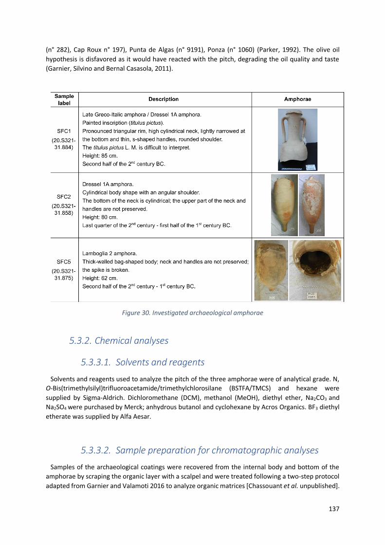
137
(n° 282), Cap Roux n° 197), Punta de Algas (n° 9191), Ponza (n° 1060) (Parker, 1992). The olive oil
hypothesis is disfavored as it would have reacted with the pitch, degrading the oil quality and taste
(Garnier, Silvino and Bernal Casasola, 2011).
Figure 30. Investigated archaeological amphorae
5.3.2. Chemical analyses
5.3.3.1. Solvents and reagents
Solvents and reagents used to analyze the pitch of the three amphorae were of analytical grade. N,
O-Bis(trimethylsilyl)trifluoroacetamide/trimethylchlorosilane (BSTFA/TMCS) and hexane were
supplied by Sigma-Aldrich. Dichloromethane (DCM), methanol (MeOH), diethyl ether, Na2CO3 and
Na2SO4 were purchased by Merck; anhydrous butanol and cyclohexane by Acros Organics. BF3 diethyl
etherate was supplied by Alfa Aesar.
5.3.3.2. Sample preparation for chromatographic analyses
Samples of the archaeological coatings were recovered from the internal body and bottom of the
amphorae by scraping the organic layer with a scalpel and were treated following a two-step protocol
adapted from Garnier and Valamoti 2016 to analyze organic matrices [Chassouant et al. unpublished].

138
Briefly, the first extraction corresponds to an organic lipid extraction (1LE) while the second step is a
microwave-assisted transesterification catalyzed by a Lewis acid (2LE-MW). 20 mg of crushed sample
were extracted 3 times with DCM:MeOH (1:1, v/v) under VCX 130 Vibra-Cell Sonics probe
ultrasonication (20 kHz, 130 W). After centrifugation, the supernatant was recovered, evaporated to
dryness before being trimethylsilylated with BSTFA/TMCS (99/1, v/v) (70°C, 30 min). The first extract
was dissolved in 1-2 mL of hexane/DCM (1/1, v/v) and filtered on a PTFE cartridge (0.45µm, VWR)
before injection in GC-MS. After the first extraction completed, the solid powder remaining was
treated with a mixture of BF3-etherate complex, butanol and cyclohexane (1:2:4, v/v/v). The butylation
was achieved through 3 dynamic cycles of microwaves of 5 min each, with automatically regulated
pulse to maintain the temperature at 80°C using a single-mode CEM Discover Lab-mate microwave
operating at 50 Hz with a maximum power of 300 W. The reaction mixture was then neutralized with
1 mL of saturated Na2CO3. The organic phase was extracted 2 times with diethyl ether. After washing
2 times with Milli-Q water, the mixture is dried with anhydrous Na2SO4. Trimethylsilylation is
completed following the same step than described earlier and the 2LE-MW fraction was dissolved in
0.2-0.6 mL of hexane/DCM (1/1, v/v) before injection in GC-MS.
5.3.3.3. Gas chromatography – Mass Spectrometry
GC–MS analyses were performed on a Thermo Scientific™ Focus system equipped with a Thermo
Scientific Al 3000 autosampler and coupled to a Thermo Fisher Scientific™ ITQ™ 700 Series Ion Trap
Mass Spectrometer. The separation was achieved on a 30 m x 0.25 mm internal diameter x 0.25 μm
film thickness fused silica capillary column ThermoGOLD™ TG-5MS (5% diphenyl; 95% dimethyl
polysiloxane). 1 µL solution was injected in splitless mode at 250°C. The transfer line and the ion trap
were respectively maintained at 300 and 200°C. Molecular components were carried by a constant 1
mL/min helium flow. Data treatments were carried out on Xcalibur software. Molecular compounds
were identified by retention time, comparison with mass spectrum of commercial molecular
standards, with the internal molecular library of the laboratory and with NIST MS Search 2.0 database
recorded with an electronic ionization of 70 eV. The oven temperature was held at 50°C for 2 min,
increased to 140°C at 8°C/min held for 2 min before reaching 160°C at 2.5°C/min and finally 330°C at
15°C/min and held for 3 min. Spectra were recorded in the 50-650 m/z mass range.
5.3.3. Archaeobotany
Reference modern grapevine flowers, both male and female, as well as fruits, used in this study were
collected near Rome, in the municipality of Morlupo (42°09'19"N; 12°30'26"E). Reference pollen grains
were also collected from the surface of the fruits of a wild grape from Tivoli (41°57’09"N; 12°49’04"E).
All grapes were sampled in wooded rims of river valleys, within a riparian vegetation characterized by
Quercus cerris, Q. pubescens, Fraxinus ornus, Ulmus minor, Populus nigra, and Alnus glutinosa.
As a reference for pre-domestication Vitis pollen, the Middle Pleistocene diatomite sediments from
Fosso di San Martino (Di Rita and Sottili, 2019), located in the municipality of Rignano Flaminio
(42°11‘26''N, 12°31'13''E), near Rome, were re-analysed to observe the morphological characters of
wild pollen grains in the region.
Adapted from (Muller, 2004), pitch samples of ca. 0.5 g were systematically dissolved in
tetrahydrofuran and ethanol before the addition of a tablet with a known number of exotic

139
Lycopodium spores to estimate the pollen concentration. To limit contamination from modern pollen
grains, whole pieces of pitch were treated. Acetolysis was not needed.
Modern pollen was acetolyzed following the standard procedure (Magri and Di Rita, 2015). Modern
fruits from Vitis vinifera subsp. sylvestris were hydrated in water for 12 hours before being heated for
10 min in NaOH (10%) and acetolyzed.
Pollen was observed under a Zeiss Axioscope light microscope at 400x and 630x magnifications.
Identifications were supported by pollen morphology atlases (Reille, 1992; Punt, Marks and Hoen,
2003; Beug, 2004); websites https://www.paldat.org; https://globalpollenproject.org, and the reference
collection of the Laboratory of Palaeobotany and Palynology of Sapienza University of Rome.
Morphological pollen and wood observations were also performed by Environmental Scanning
Electron Microscope (ESEM) Hitachi TM-3000 Tabletop operating at 15Kv without previous coating.
The images were recorded at magnifications varying from 150x to 700x.
5.4. Results
5.4.1. Chemical analyses
For all the amphorae SFC1, SFC2 and SFC5, the first extraction (1LE) with polar organic solvents
(DCM:MeOH) interestingly revealed a conifer resin made out of Pinaceae wood tar (dehydroabietic
acid (DHA); methyldehydroabietate (DHAM)) (Fig. 31A). Aromatized (retene, norabietatrienes) and
oxidized (hydroxy- and oxo-DHAM derivatives) abietanes highlighted a high temperature formulation
and the ageing of the resin (Mills and White, 1994; Mezzatesta et al., 2021). Oxidized abietanes were
identified through their characteristic fragment ions (m/z 191; 253) (van Den Berg et al., 2000).
Figure 31. GC-MS chromatogram for the pitch of SFC1. A. represents the first extraction (1LE) and B. corresponds to 2LE-MW, the butylated second extraction. Total Ion Current (TIC) is black-colored. Red,
blue and green colors refer to fragment ions searching (respectively m/z 147 (succinic and glutaric acids) and m/z 191; 253 (hydroxy- and oxo-abietanes) in 1LE and m/z 61; 101; 276; 296 (dibutylacetal
pyruvate; dibutyl malate; dibutyl tartrate; butyl syringate in 2LE-MW)
140
All the archaeological coatings contained tartaric (m/z 276), malic (m/z 303) and pyruvic (m/z 61, 89,
117, 173) acids, greatly identified as butylated and butylacetal derivatives. Syringic acid was only
identified in the amphora SFC1 and never present in both extractions for SFC2 and SFC5. For all the
amphorae, succinic and glutaric acids were identified in 1LE through their characteristic fragment ion
m/z 147 in the trimethylsilylated form while no traces could be identified in 2LE-MW.
Since acids were transesterified in the second step, molecules identification in 2LE-MW was
restricted to grape derivatives markers (succinic, pyruvic, malic, tartaric and syringic acids). Considering
the presence of diethyl or butyl ethyl grape acids reported by Garnier and Valamoti (Garnier and
Valamoti, 2016) in a Neolithic jar, similar reactions were controlled in our samples but no esters were
observed. Such compounds would indeed be produced by esterification with the ethanol contained in
the fermented beverage.
5.4.2. Archaeobotanical analyses
5.4.2.1. Pollen
The pollen concentration of samples SFC1, SFC2 and SFC5 is noticeably low (1771, 175 and 213 pollen
grains/g resin, respectively: Table 8). The number of identified pollen grains are 150, 61 and 48
respectively. SFC1 displayed the richest pollen content with a major presence of Quercus (46%), Pinus
(28.7%) and minor contributions of Olea (6.7%), Phillyrea (6%), Brassicaceae (2%), CarPinus and Erica
(1.3%), Myrtus, Plantago and Ranunculus (0.66%). SFC2 showed a significant presence of Pinus (65.6%),
followed by Quercus (16.4%), Phillyrea (3.3%), Cedrus (3.3%), and Erica (1.6%). SFC5 exhibited Pinus
(35.4%) and Quercus (29.1%) followed by Artemisia and Phillyrea (8.3%), Ranunculus (6.3%), Alnus,
Erica and Ostrya type (2.1%). Vitis represented 4.7%, 9.8% and 6.3% of total pollen, respectively (Table
8).
Assumption of highland pine species is suggested regarding the small pollen size (55 to 80 µm).
Following the classification by Desprat et al. (Desprat et al., 2015) and keeping in mind that fossilization
can alter the grain size, the identification was possible up to the subsection including P. mugo, P. nigra
and P. sylvestris. Unfortunately, it remained unreliable to identify the pollen grains to the species level.
In all the three pitch samples, pollen observations featured the presence of aporate 3-zonocolpate
grains, ranging 20-25 µm, with psilate to micro-scabrate ornamentation (Fig. 32). They displayed
narrow and long slit-like colpi, making the pollen round to slightly oval in equatorial view (EV) and
obtuse triangular to hexagonal outline in polar view (PV) with straight to softly concave sides. As
illustrated in Fig. 32, the main feature remained the total absence of porus within the colpi and a thick
exine (up to almost 5 µm for the pollen grain in Fig. 32I-J, with small residues stuck to the exine).
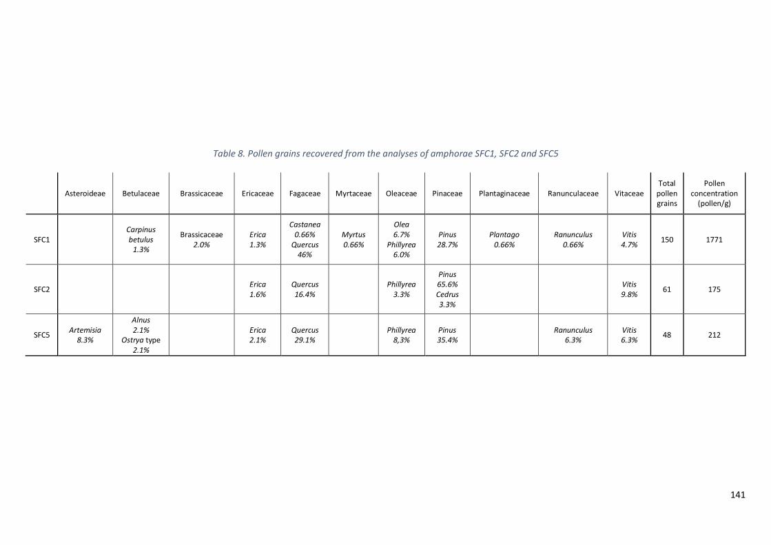
141
Table 8. Pollen grains recovered from the analyses of amphorae SFC1, SFC2 and SFC5
Asteroideae Betulaceae Brassicaceae Ericaceae Fagaceae Myrtaceae Oleaceae Pinaceae Plantaginaceae Ranunculaceae Vitaceae Total
pollen grains
Pollen concentration
(pollen/g)
SFC1 Carpinus betulus
1.3%
Brassicaceae 2.0%
Erica 1.3%
Castanea 0.66%
Quercus 46%
Myrtus 0.66%
Olea 6.7%
Phillyrea 6.0%
Pinus 28.7%
Plantago 0.66%
Ranunculus 0.66%
Vitis 4.7%
150 1771
SFC2 Erica 1.6%
Quercus 16.4%
Phillyrea 3.3%
Pinus 65.6% Cedrus 3.3%
Vitis 9.8%
61 175
SFC5 Artemisia
8.3%
Alnus 2.1%
Ostrya type 2.1%
Erica 2.1%
Quercus 29.1%
Phillyrea 8,3%
Pinus 35.4%
Ranunculus 6.3%
Vitis 6.3%
48 212
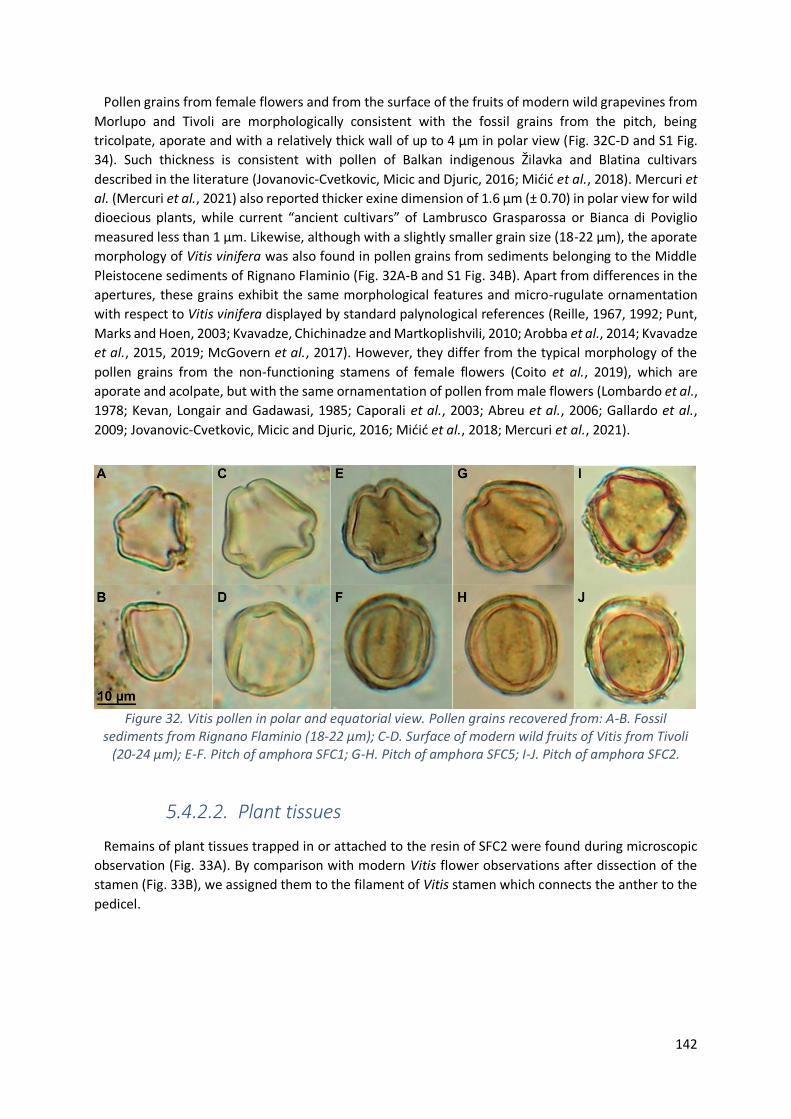
142
Pollen grains from female flowers and from the surface of the fruits of modern wild grapevines from
Morlupo and Tivoli are morphologically consistent with the fossil grains from the pitch, being
tricolpate, aporate and with a relatively thick wall of up to 4 µm in polar view (Fig. 32C-D and S1 Fig.
34). Such thickness is consistent with pollen of Balkan indigenous Žilavka and Blatina cultivars
described in the literature (Jovanovic-Cvetkovic, Micic and Djuric, 2016; Mićić et al., 2018). Mercuri et
al. (Mercuri et al., 2021) also reported thicker exine dimension of 1.6 µm (± 0.70) in polar view for wild
dioecious plants, while current “ancient cultivars” of Lambrusco Grasparossa or Bianca di Poviglio
measured less than 1 µm. Likewise, although with a slightly smaller grain size (18-22 µm), the aporate
morphology of Vitis vinifera was also found in pollen grains from sediments belonging to the Middle
Pleistocene sediments of Rignano Flaminio (Fig. 32A-B and S1 Fig. 34B). Apart from differences in the
apertures, these grains exhibit the same morphological features and micro-rugulate ornamentation
with respect to Vitis vinifera displayed by standard palynological references (Reille, 1967, 1992; Punt,
Marks and Hoen, 2003; Kvavadze, Chichinadze and Martkoplishvili, 2010; Arobba et al., 2014; Kvavadze
et al., 2015, 2019; McGovern et al., 2017). However, they differ from the typical morphology of the
pollen grains from the non-functioning stamens of female flowers (Coito et al., 2019), which are
aporate and acolpate, but with the same ornamentation of pollen from male flowers (Lombardo et al.,
1978; Kevan, Longair and Gadawasi, 1985; Caporali et al., 2003; Abreu et al., 2006; Gallardo et al.,
2009; Jovanovic-Cvetkovic, Micic and Djuric, 2016; Mićić et al., 2018; Mercuri et al., 2021).
Figure 32. Vitis pollen in polar and equatorial view. Pollen grains recovered from: A-B. Fossil sediments from Rignano Flaminio (18-22 µm); C-D. Surface of modern wild fruits of Vitis from Tivoli
(20-24 µm); E-F. Pitch of amphora SFC1; G-H. Pitch of amphora SFC5; I-J. Pitch of amphora SFC2.
5.4.2.2. Plant tissues
Remains of plant tissues trapped in or attached to the resin of SFC2 were found during microscopic
observation (Fig. 33A). By comparison with modern Vitis flower observations after dissection of the
stamen (Fig. 33B), we assigned them to the filament of Vitis stamen which connects the anther to the
pedicel.
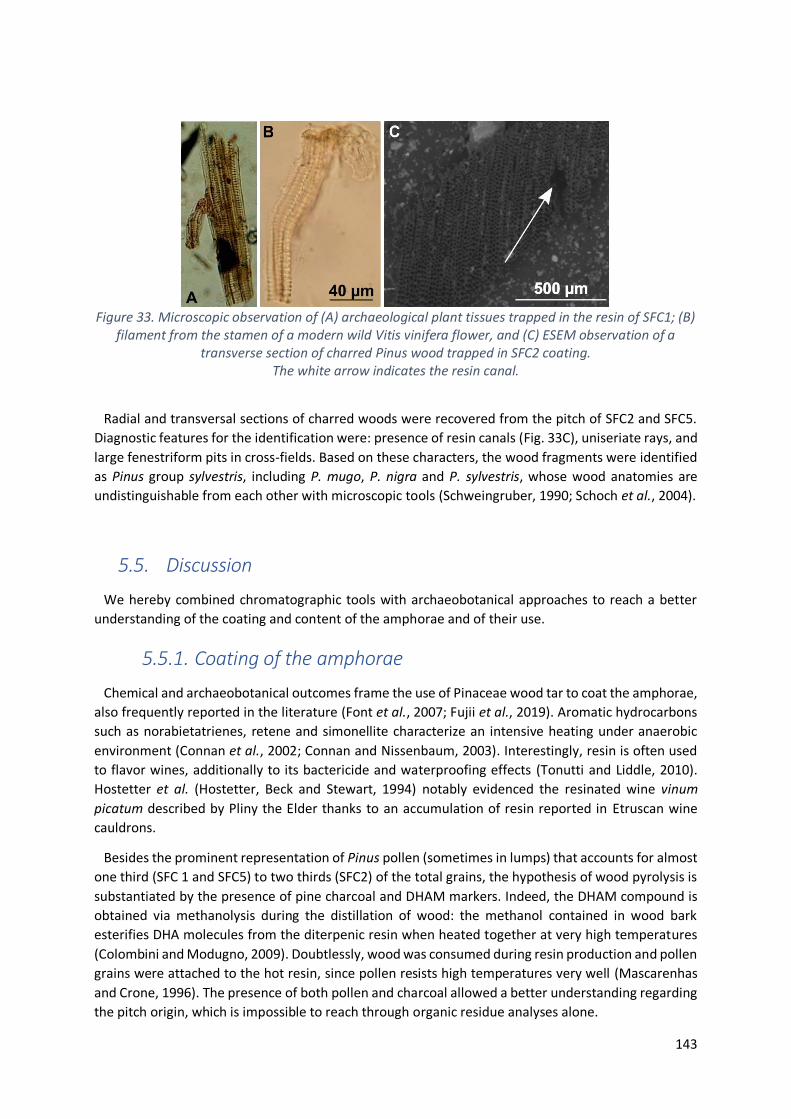
143
Figure 33. Microscopic observation of (A) archaeological plant tissues trapped in the resin of SFC1; (B) filament from the stamen of a modern wild Vitis vinifera flower, and (C) ESEM observation of a
transverse section of charred Pinus wood trapped in SFC2 coating. The white arrow indicates the resin canal.
Radial and transversal sections of charred woods were recovered from the pitch of SFC2 and SFC5.
Diagnostic features for the identification were: presence of resin canals (Fig. 33C), uniseriate rays, and
large fenestriform pits in cross-fields. Based on these characters, the wood fragments were identified
as Pinus group sylvestris, including P. mugo, P. nigra and P. sylvestris, whose wood anatomies are
undistinguishable from each other with microscopic tools (Schweingruber, 1990; Schoch et al., 2004).
5.5. Discussion
We hereby combined chromatographic tools with archaeobotanical approaches to reach a better
understanding of the coating and content of the amphorae and of their use.
5.5.1. Coating of the amphorae
Chemical and archaeobotanical outcomes frame the use of Pinaceae wood tar to coat the amphorae,
also frequently reported in the literature (Font et al., 2007; Fujii et al., 2019). Aromatic hydrocarbons
such as norabietatrienes, retene and simonellite characterize an intensive heating under anaerobic
environment (Connan et al., 2002; Connan and Nissenbaum, 2003). Interestingly, resin is often used
to flavor wines, additionally to its bactericide and waterproofing effects (Tonutti and Liddle, 2010).
Hostetter et al. (Hostetter, Beck and Stewart, 1994) notably evidenced the resinated wine vinum
picatum described by Pliny the Elder thanks to an accumulation of resin reported in Etruscan wine
cauldrons.
Besides the prominent representation of Pinus pollen (sometimes in lumps) that accounts for almost
one third (SFC 1 and SFC5) to two thirds (SFC2) of the total grains, the hypothesis of wood pyrolysis is
substantiated by the presence of pine charcoal and DHAM markers. Indeed, the DHAM compound is
obtained via methanolysis during the distillation of wood: the methanol contained in wood bark
esterifies DHA molecules from the diterpenic resin when heated together at very high temperatures
(Colombini and Modugno, 2009). Doubtlessly, wood was consumed during resin production and pollen
grains were attached to the hot resin, since pollen resists high temperatures very well (Mascarenhas
and Crone, 1996). The presence of both pollen and charcoal allowed a better understanding regarding
the pitch origin, which is impossible to reach through organic residue analyses alone.
144
Despite the identification of Pinus to the species level is not possible, the botanical assignment to
highland pine species (P. mugo, nigra or sylvestris) is strengthened by Pliny, whose Naturalis Historia
stated that fire-extracted pitch from mountainous species logs of P. mugo (namely “taeda”) is resin-
richer, and notwithstanding its restricted spatial distribution, highland species were abundantly
manufactured (Pliny, N. H. XIV, 9, 17, 21, 22) (Orengo et al., 2013). The current distribution of P. mugo
is very restricted in central Italy, while it is commonly found in alpine environments, where also P.
sylvestris is widely distributed (Caudullo, Welk and San-Miguel-Ayanz, 2017). In contrast, P. nigra is
uncommon on the Alps, and sparse in the central and northern Apennines, while the subspecies P.
nigra subsp. laricio is present in Calabria, in Sicily (slopes of Mount Etna) and in Corsica. In Roman times
the production of pitch from the mountains of Calabria and Sicily was renowned (Dionysius of
Halicarnassus, XX, 6; Pliny, N. H. III; Cicero, Brutus XXII, 85-88). In any case, we can exclude a local origin
of the pine used for the production of pitch of the studied amphorae.
The dominance of Quercus (46% in SFC1 and 29% in SFC5, Table 8) can be explained by environmental
abundance in the region of wood tar production (Spada et al., 2008).
5.5.2. Content of the amphorae
The amphora typologies of Dressel 1A (SFC1 and SFC2) and Lamboglia 2 (SFC5) have been frequently
reported as grape-derivatives containers (Panella, 1998; Arobba et al., 2014; Fujii et al., 2019). Tartaric
acid, together with malic acid (although less specific), identified by GC-MS, point out a grape-based
content. Fermentation assumptions is enhanced by succinic and glutaric acids. Pyruvic acid resulting
from spontaneous malolactic fermentation refers to wine content (Ribéreau-Gayon et al., 2006). For
SFC1, syringic acid present in the second extract, despite its absence in the first extract, originates from
malvidin oxidation, thus ruling out potential contamination from free extractible origin (Guasch-Jané
et al., 2004; Garnier and Valamoti, 2016). Red (for SFC1) and white winemaking processes (for SFC2
and SFC5) are therefore brought to light. Although the amount of tartaric acid is remarkably higher in
grape bunches than in other edible products, its use as a reliable grape biomarker must be confirmed
by other evidence (McGovern et al., 2017; Drieu et al., 2020). Thus, macroremains of plant tissues
recovered from the pitch and identified as part of the filament of Vitis flower in samples SFC1 and SFC5
bring this needed evidence of grape derivatives content.
Regarding microremains, although the tricolpate pollen type (Fig. 32) does not exhibit any apparent
pore, attribution to Vitis is straightforwardly demonstrated by the identical aporate grains observed
from wild vines (Fig. 32C-D) and from the Rignano Flaminio fossil sediments from the same Lazio region
(Fig. 32A-B) (Di Rita and Sottili, 2019). The micro-rugulate ornamentation evidenced by SEM
observation of Rignano Flaminio sediments and from the surface of wild fruits from Tivoli (S1 Fig. 34)
is in accordance with the literature for Vitaceae pollen (Punt, Marks and Hoen, 2003). Nevertheless,
attribution to Vitis vinifera subsp. vinifera is highly questionable since grapevines were surely not
domesticated in the Middle Pleistocene (This, Lacombe and Thomas, 2006; Zhou et al., 2017).
Furthermore, the fossil impression of a grapevine leaf, identified as V. sylvestris, coupled with pollen
grains, reported from another fossil site 16 km away from Rignano Flaminio, confirms the presence of
wild grapevines in this area since at least the Middle Pleistocene (Follieri, 1958). Vitis vinifera subsp.
sylvestris is well represented in Italy, especially along the Tyrrhenian coast (Arnold, 2002; Biagini et al.,
2014). Although its survival is highly threatened (Magri, Celant and Di Rita, 2019), wild grapevines are
still present also in southern Latium, close to San Felice Circeo (Arnold, Gillet and Gobat, 1998; Biagini
et al., 2014).

145
As far as we know, this is the first time tricolpate aporate Vitis vinifera is found in Roman amphorae,
although inaperturate (aporate and acolpate) Vitis grains were recently retrieved from the Middle
Bronze Age site of Terramara di Poviglio (Mercuri et al., 2021). Interestingly, some morphological
abnormalities of Vitis grains displaying one, two or four pores have also been published in modern
cultivars (Pereira et al., 2018).
To truly assume a grape derivative content in the amphorae, the presence of aporate grape pollen in
the pitch shall be explained. Three hypotheses are discussed.
A first possible answer regards the sterility of the grapevine used. The presence of aporate pollen
grains on Vitis fruits from wild plants near Tivoli demonstrates the permanence of Vitis pollen from
female flowers on the fruit surface, despite its development (Fig. 32C-D). Pollen, subsisting upon time,
weather and environmental circumstances, may be plucked with the fruits and remain even in the
fermented beverage. Since beverages were not filtered at this time, pollen could remain in the
amphorae attached to the pitch and bring evidence of the ancient content (Arobba, 1976; Vogt et al.,
2002; Rösch, 2005). The pollen we observed might be sterile as suggested by the strikingly thick exine
observed, reaching up to 5 µm (Fig. 32I-J) whereas tricolporate pollen rather exhibits a “thin to fairly
thin” wall (Punt, Marks and Hoen, 2003). Although cytogenetic characteristics are preserved during
the grain development, Caporali et al. (Caporali et al., 2003) explained Vitis pollen sterility by
morphological germination inhibition caused by wall structure abnormalities: during the grain
formation, the pollen surface is covered by structural nutrients contained in the exine (Caporali et al.,
2003). An excess of exine cover during pollen hydration may turn a mechanical hurdle into sterility
(Mićić et al., 2018). Physiological and cytological functions are nevertheless maintained, and pollen
grains can disperse once released by the anthers. The absence of germinative pores causes grain
sterility by avoiding pollen tube development even though pollen grains are viable (Lombardo et al.,
1978; Jovanovic-Cvetkovic, Micic and Djuric, 2016).
Pollen sterility is strictly related to wild features. At early stages, flowers of Vitis vinifera are all
hermaphrodite, until they may become unisexual due to the abortion of one reproductive organ
(Levadoux, 1946; Caporali et al., 2003; Ramos et al., 2014). Sex determining genes therefore divide
flowers into functional “male” (or staminate) and “female” (or pistillate), showing up fertile pollen (and
rudimentary pistil) or functional pistil (and sterile pollen), respectively (Negi and Olmo, 1970; Coito et
al., 2019). Pollination is achieved through the intermediary of a fertile pollen coming from either a
functional male (V. vinifera sylvestris) or hermaphrodite plant (V. vinifera vinifera).
Pollen sterility also involves dioecy, which was lost during the domestication process (This, Lacombe
and Thomas, 2006; Massonnet et al., 2020). Grapevine domestication targets the ensemble of
“genotypic, phenotypic, plastic and contextual impacts that can be used as markers of evolving
domesticatory relationships” (Zeder, 2015). Asides the increase of sugar content in the fruits, berry
sizes or changes in pips morphology, more factual definitions point to the shift from dioecy to
hermaphrodism (This, Lacombe and Thomas, 2006). The reverting to hermaphrodism is assumed to
have occurred through a rare event of male and female haplotypes recombination (Massonnet et al.,
2020; Mercuri et al., 2021). Nonetheless, domestication and hermaphrodism have to be clearly
separated from each other, since they differently relate to cultivation. Although cultivated V. vinifera
are thought to have been domesticated from their wild V. vinifera sylvestris ancestors (Levadoux,
1956), not all the cultivated plants were necessarily hermaphrodite at the beginning of domestication,
which was a long and multi-located process (Arroyo-Garcia et al., 2006; Bouby et al., 2013; Ucchesu et
al., 2015). As observed from wild cereals, grapevine cultivation for food consumption is presumed to
have started long before its domestication (Mercuri et al., 2015, 2018; McGovern et al., 2017). One
point remains certain: the switch to hermaphroditism grandly facilitates the fruit production, turning

146
grapevines into self‐pollinating plants, with entomophilous and anemophilous cross-pollination
(Turner and Brown, 2004; This, Lacombe and Thomas, 2006).
Additional evidence for cultivation is based on a considerable progress of statistic and modeling
morphometric tools applied to pips and the emerging field of ancient DNA (Bouby et al., 2013, 2020;
Wales et al., 2016; Aversano et al., 2017; Zhou et al., 2017; Mariotti Lippi et al., 2020). SSR and SNP
markers allowed genotype classification into cultivated and wild types (Grassi et al., 2003; Butorac et
al., 2018). However, despite the important genetic dissimilarity reported between V. vinifera and V.
sylvestris, a remaining presence of wild characters cannot be ruled out (Grassi et al., 2003; Arroyo-
Garcia et al., 2006; Myles et al., 2011).
Historical and archaeological evidence supports the use of wild grapes at the same time of cultivated
grapes. In his Naturalis Historia, Pliny repeatedly reported the use of V. sylvestris grapes, wood and
leaves in addition to cultivated grapevines (Pliny, N. H. XIV). In the Middle to Late Bronze Age site of
Santa Rosa di Poviglio in the Po Plain, tricolporate Vitis pollen was abundantly recovered, up to 18%
(Cremaschi et al., 2016). The re-examination of the archaeological sediments highlighted 15
inaperturate Vitis pollen grains that demonstrate the use of V. sylvestris (Pecci et al., 2020; Mercuri et
al., 2021). Although present in limited quantities (7.7% of the total grape pips), Mariotti Lippi et al.
(Aranguren et al., 200AD; Mariotti Lippi et al., 2009, 2020) identified grape pips as wild morphotypes
in Tuscany, belonging to Middle Bronze Age and Etruscan-Roman archaeological contexts. Castellano
(Castellano et al., 2017) pointed out the presence of V. sylvestris pollen in honey dated to the Iron Age
in northern Italy, suggesting that bees fed on nectar of pre-domesticated or early-domesticated
varieties of V. vinifera. Bouby (Bouby et al., 2013) highlighted an intermediate form of Roman grape
pips, between “highly cultivated” and “primitive cultivars” from wild ancestors, in the South of France.
The Vitis pollen retrieved from the Roman amphorae of San Felice Circeo may therefore represent
an intermediary stage of domestication, being characterized by thick exine, absence of germinative
pores and presence of colpi. This intermediate morphology recalls the morphological variety observed
in stamens and pistils of Vitis flowers (functional female flowers present either erect but crinkled
stamens, or semi- as well as entirely reflexed stamens) [85]. Advanced archaeopalynological analyses
are needed for a better understanding of the grapevine evolution from wild, through intermediate to
cultivated forms (Cardarelli et al., 2015; Cremaschi et al., 2016; Pecci et al., 2020). This field of research
offers new eyes for an innovative archaeological interpretation of the data (Mariotti Lippi and Secci,
2002; Mercuri et al., 2015; Cremaschi et al., 2018).
A second hypothesis to explain the presence of aporate Vitis pollen in the Roman amphorae of Circeo
concerns the use of archaeological indigenous cultivars to produce fermented grape derivatives.
Several modern autochthonous cultivars, such as ‘Loureiro’, ‘Moscato rosa’ or ‘Blatina’, have been
documented to compose with a dioecious mating system (Cargnello et al., 1980; Tischelmayer, 2001;
Abreu et al., 2006; Maletić et al., 2015; Stupić et al., 2019). Some of them, like ‘Picolit’ and ‘Lambrusco
di Sorbara’ are endemic of northern Italy (Lombardo et al., 1978; Mercuri et al., 2021). Since the plants
of these refined wines cannot self-fertilize due to infertile pollen from functionally female flowers,
grapevine productivity is consequently limited and wine prices may be high (Schamel and Ros, 2021).
Our study of modern pollen from different sites near Rome, showing tricolpate aporate grains,
supported by Middle Pleistocene records with the same pollen morphology, strongly support the use
of indigenous grape cultivars to produce the wine content of the Roman amphorae from San Felice
Circeo.
The third hypothesis refers to the nature of the content. As previously described, chemical markers
evidenced fermented grape derivatives, which can consist of either wine, vinegar or other beverages,
147
such as the cooked wine ‘defrutum’ or aged sweetened wine ‘mulsum’ (Peña, 2007; Arobba et al.,
2014; Bernal Casasola, 2015). Some Roman recipes reported in De re coquinaria V.II.9, translated and
abridged by Feldman (Feldman, 2005), attested of the use of grape wine in traditional cooking made
out of roses, spices and fish sauces of liquamen or garum. Unfortunately, in the absence of tituli picti,
i.e., a commercial inscription on the surface of the amphorae, the possibilities of wine derivatives
remain hypothetical since no chemical biomarker is able to distinguish wine from other traditional
grape fermented recipes (Bernal Casasola et al., 2004; García Vargas, 2004). Relying on the botanical
evidence, we hereby consider the possibility of oenanthium, a flavored wine famous for its medicinal
properties. Following Pliny recipe “with the wild grapevine one makes what is called oenanthia: one
macerates two pounds of wild vine flower in a cadus (30 or 40 liters) of must, one decants after thirty
days […]. These grapes, shortly after flowering, are a remedy of singular virtue to temper the heat of
the body in diseases” (Pliny, N. H. XIV, 18). Besides attesting the use of wildflowers at Roman times,
Pliny gives an interesting justification of Vitis pollen in jars through the presence of flowers. More
importantly, we have also found stamen filaments (Fig. 33), which support the presence of flowers in
the content of the amphorae. Moreover, a few non-arboreal pollen types may account for medicinal
aromatization, specifically in SFC5 where Artemisia reached 8.3% of the total grains and SFC1 where
Myrtus has been identified. Such plants are recognized for their medicinal benefits (McGovern, Hall
and Mirzoian, 2013; Li et al., 2018). Broadly speaking, there is abundant ethnopharmacological
evidence for the common use of herbal concoctions in alcoholic or fermented beverages (Stika, 1996).
The use of Artemisia for anticancer activity has been reported in Ancient Egyptian herbal wines from
Abydos potsherd (McGovern, Mirzoian and Hall, 2009; McGovern et al., 2010). Myrtus communis was
recognized as a therapeutic drug in ancient Greece (Sisay and Gashaw, 2017) and used as herbal
additives in early medieval beers (Nelson, 2005). Nonetheless, the limited pollen representation of
Myrtus (less than 2%) can also refer to the surrounding vegetation.
In synthesis, our results suggest the use of autochthonous grapevines either cultivated, such as the
modern ‘Picolit’, or wild, as demonstrated by the similarities with Vitis pollen from indigenous wild
plants, as well as the possible use of grapevines at intermediate stages of domestication. Medicinal
wine is another possibility that would explain the presence of Vitis stamens, and of Artemisia and
Myrtus pollen, whose plants are used as flavoring. Likewise, Pinus, besides ensuring the waterproofing
of the amphora, would have flavored the beverage due to its aromatic character. Indeed, herbal wines
such as vinum absinthianum or picatum were common at that time.
5.6. Conclusion
The analyses of the pitch contained in three Roman amphorae from San Felice Circeo illustrates the
benefits of applying a multidisciplinary approach. The combined evidence of amphorae typologies,
Pliny's testimony regarding the use of V. sylvestris, previous archaeobotanical finds indicating the
archaeological use of wild grapes, the chromatographic outcomes, and the morphology of Vitis pollen
and tissues led to new archaeological and anthropological interpretations.
The identification of Pinus group sylvestris used to produce wood tar for waterproofing, matching
the methyl ester diterpenic chemical markers characterized by GC-MS, indicates a non-local origin of
the wood tar, as also suggested by ancient historical sources, reporting Calabria and Sicily as important
production areas for pitch.
Aside from confirming the usage of Lamboglia 2 as wine containers, chemical analyses highlight the
usage of both red and white wines.
148
The observation of aporate pollen of Vitis, compared with different types of fossil and modern wild
grapevines, suggests the use of autochthonous grapevines, either wild or cultivated, without excluding
a possible intermediary stage of domestication of cultivars still bearing V. sylvestris features. It is also
possible to conjecture the archaeological presence of a medicinal grape beverage made as an infusion
of wild Vitis flowers in the must, reported by Pliny as oenanthium. However, this hypothesis contrasts
with the diverse typology of the amphorae involved in the pitch analyses.
Vitis pollen appears to be a fruitful anthropological indicator of ancient habits by opening a field of
archaeological assumptions hitherto inaccessible, such as the inclusion of Roman grapevines into the
long process of domestication. Within the long-standing question of distinguishing wild and cultivated
grapes from past archives, the archaeopalynological study of Vitis may bring new evidence to define
the timing and modes of grapevine cultivation.
Since false chemical positive must be tackled by external controls, we provided a straightforward
methodology that brought independent evidence of grape derivatives in Roman wine amphorae,
based on chromatographical and archaeobotanical tools, allowing to suggest a history beyond the
artefacts that could not be identified by single analytical techniques.
Author contribution
Conceptualization, L.C., A.C., C.D., C.V., C.M., D.M.; methodology, L.C.; formal analysis, L.C., A.C.,
F.D., C.M., D.M.; investigation, L.C., A.C., C.D., F.D., D.M.; resources, A.C., C.D., D.M.; visualization,
L.C.; supervision, C.M.; project administration, C.M., D.M.; funding acquisition, A.C., C.V., C.M., D.M.;
writing—original draft preparation, L.C., A.C., C.D., D.M.; writing—review and editing, all authors
Funding
This research and the APC were funded by Marie Skłodowska-Curie Innovative Training Network
H2020-MSCA-ITN-EJD ED-ARCHMAT Joint Doctorate, ESR9, grant number 766311. Work in Rome was
funded by Sapienza University project DISCOVER (RM120172AE4E11D6).
149
5.7. References Abreu, I. et al. (2006) ‘Ultrastructure and germination of Vitis vinifera cv. Loureiro pollen’, Protoplasma, 228(1–3), pp. 131–135. doi: 10.1007/s00709-006-0167-1.
Aranguren, B. et al. (200AD) ‘L’avvio della coltura della vite in Toscana: l’esempio di San Lorenzo a Greve (Firenze)’, Atti del Convegno Internazionale di Studi Scansano. Archeologia della vite e del vino in Etruria. Teatro Castagnoli, Scansano. Edited by R. P. Ciacci A and Z. A, pp. 9–10.
Arnold, C. (2002) Ecologie de la vigne sauvage, Vitis vinifera L. ssp. sylvestris (Gmelin) Hegi, dans les forêts alluviales et colluviales d’Europe. vdf Hochschulverlag AG.
Arnold, C., Gillet, F. and Gobat, J. M. (1998) ‘Situation de la vigne sauvage Vitis vinifera ssp. silvestris en Europe’, Vitis, 37(4), pp. 159–170.
Arobba, D. (1976) ‘Analisi pollinica di una resina fossile’, in Pollen et Spores 18. Muséum National d’histoire Naturelle, pp. 385–393.
Arobba, D. et al. (1983) ‘Studio pluridisciplinare del materiale proveniente da un carico navale proveniente del I-III secolo d.C. scoperto sui fondali dell’isola del Giglio (Grosseto-Italia)’, Rivista di Studi Liguri, 11, pp. 117–144.
Arobba, D. et al. (2014) ‘Palaeobotanical, chemical and physical investigation of the content of an ancient wine amphora from the northern Tyrrhenian sea in Italy’, Journal of Archaeological Science, 45(1), pp. 226–233. doi: 10.1016/j.jas.2014.02.024.
Arroyo-Garcia, R. et al. (2006) ‘Multiple origins of cultivated grapevine (Vitis vinifera L. ssp. sativa) based on chloroplast DNA polymorphisms’, Molecular Ecology, 15(12), pp. 3707–3714. doi: 10.1111/j.1365-294X.2006.03049.x.
Aversano, R. et al. (2017) ‘Dating the beginning of the Roman viticultural model in the Western Mediterranean: The case study of Chianti (Central Italy)’, PLOS ONE. Edited by M. Scali, 12(11), p. e0186298. doi: 10.1371/journal.pone.0186298.
Barnard, H. et al. (2011) ‘Chemical evidence for wine production around 4000 BCE in the Late Chalcolithic Near Eastern highlands’, Journal of Archaeological Science, 38(5), pp. 977–984. doi: 10.1016/j.jas.2010.11.012.
van Den Berg, K. J. et al. (2000) ‘Mass spectrometric methodology for the analysis of highly oxidized diterpenoid acids in Old Master paintings’, Journal of Mass Spectrometry, 35(4), pp. 512–533. doi: 10.1002/(SICI)1096-9888(200004)35:4<512::AID-JMS963>3.0.CO;2-3.
Bernal Casasola, D. et al. (2004) ‘Las DR. 2/4 Béticas. primeras evidencias de su manufactura en el conventus graditanus’, Figlinae Baeticae, pp. 633-648. doi: 10.1088/1751-8113/44/8/085201.
Bernal Casasola, D. (2015) ‘What Contents do we characterise in Roman Amphorae? Methodological and Archaeological Thoughts on a “Trending Topic”’, ArchaeoAnalytics: Chromatography and DNA Analysis in Archaeology, pp. 61–83.
Beug, H.-J. (2004) Leitfaden der Pollenbestimmung fiir Mitteleuropaund angrenzende Gebiete. Munchen: Pfeil Verlag.
Biagini, B. et al. (2014) ‘Italian wild grapevine (Vitis vinifera L. subsp. sylvestris) population: insights into eco-geographical aspects and genetic structure’, Tree Genetics & Genomes, 10(5), pp. 1369–1385. doi: 10.1007/s11295-014-0767-4.
Bouby, L. et al. (2013) ‘Bioarchaeological insights into the process of domestication of grapevine (Vitis vinifera L.) during Roman times in Southern France’, PLoS ONE, 8(5), p. e63195. doi:

150
10.1371/journal.pone.0063195.
Bouby, L. et al. (2020) ‘Tracking the history of grapevine cultivation in Georgia by combining geometric morphometrics and ancient DNA’, Vegetation History and Archaeobotany, (0123456789). doi: 10.1007/s00334-020-00803-0.
Brown, T. A. et al. (2015) ‘Recent advances in ancient DNA research and their implications for archaeobotany’, Vegetation History and Archaeobotany, 24(1), pp. 207–214. doi: 10.1007/s00334-014-0489-4.
Buchi, E. (1971) ‘Banchi di anfore romane a Verona’, Note sui commerci cisalpini, Il territorio veronese in età romana, pp. 531–637.
Butorac, L. et al. (2018) ‘Assessment of wild grapevine (Vitis vinifera ssp. sylvestris) chlorotypes and accompanying woody species in the Eastern Adriatic region’, PLOS ONE, 13(6), p. e0199495. doi: 10.1371/journal.pone.0199495.
Cancellieri, M. (1986) ‘Le vie d’acqua dell’area pontina’, in Il tevere e le altre vie d’acqua del Lazio Antico. Consiglio Nazionale delle Ricerche, pp. 143–156.
Caporali, E. et al. (2003) ‘The arrest of development of abortive reproductive organs in the unisexual flower of Vitis vinifera ssp. silvestris’, Sexual Plant Reproduction, 15(6), pp. 291–300. doi: 10.1007/s00497-003-0169-5.
Cappellini, E. et al. (2010) ‘A multidisciplinary study of archaeological grape seeds’, Naturwissenschaften, 97(2), pp. 205–217. doi: 10.1007/s00114-009-0629-3.
Cardarelli, A. et al. (2015) ‘Vino o non vino ? Nuovi dati sui vinaccioli della Terramara di Montale (Modena) tra la fine della media età del Bronzo e il Bronzo recente’, 50ma Riunione IIPP Preistoria del cibo. L’alimentazione nella preistoria e nella protostoria. Roma (5-9 Ottobre 2015), pp. 1–9.
Cargnello, G. et al. (1980) ‘Pollen morphology of Picolit grown in different Italian’, Vitis, 19, pp. 201–206.
Castellano, L. et al. (2017) ‘Charred honeycombs discovered in Iron Age Northern Italy. A new light on boat beekeeping and bee pollination in pre-modern world’, Journal of Archaeological Science, 83, pp. 26–40. doi: 10.1016/j.jas.2017.06.005.
Caudullo, G., Welk, E. and San-Miguel-Ayanz, J. (2017) ‘Chorological maps for the main European woody species’, Data in Brief, 12, pp. 662–666. doi: 10.1016/j.dib.2017.05.007.
Cipriano, M. T. and Carre, M.-B. (1989) ‘Production et typologie des amphores sur la côte adriatique de l’Italie. Amphores romaines et histoire economique. Dix ans de recherche. Actes du colloque de Sienne (22-24 mai 1986)’, Publications de l’École Française de Rome, 114(1), pp. 67–104.
Ciuffarella, L. (1998) ‘Palynological analyses of resinuous materials from the roman mummy of Grottarossa, second century A.D.: a new hypothesis about the site of mummification’, Review of Palaeobotany and Palynology, 103(3–4), pp. 201–208. doi: 10.1016/S0034-6667(98)00035-9.
Coito, J. L. et al. (2019) ‘Vitis flower types: from the wild to crop plants’, PeerJ, 7, p. e7879. doi: 10.7717/peerj.7879.
Colombini, M. P. and Modugno, F. (2009) Organic Mass Spectrometry in Art and Archaeology. Edited by M. P. Colombini and F. Modugno. Chichester: John Wiley & Sons. doi: 10.1002/9780470741917.
Connan, J. et al. (2002) ‘Identification de poix et de résine de conifère dans des échantillons archéologiques du lac de Sanguinet’, Revue d’archéométrie, 26, pp. 177–196.
Connan, J. and Nissenbaum, A. (2003) ‘Conifer tar on the keel and hull planking of the Ma’agan Mikhael
151
Ship (Israel, 5th century BC): identification and comparison with natural products and artefacts employed in boat construction’, Journal of Archaeological Science, 30(6), pp. 709–719. doi: 10.1016/S0305-4403(02)00243-1.
Cremaschi, M. et al. (2016) ‘Climate change versus land management in the Po Plain (Northern Italy) during the Bronze Age: New insights from the VP/VG sequence of the Terramara Santa Rosa di Poviglio’, Quaternary Science Reviews, 136, pp. 153–172. doi: 10.1016/j.quascirev.2015.08.011.
Cremaschi, M. et al. (2018) ‘The SUCCESSO-TERRA Project: a Lesson of Sustainability from the Terramare Culture, Middle Bronze Age of the Po Plain (Northern Italy)’, Interdisciplinaria Archaeologica - Natural Sciences in Archaeology, IX(2), pp. 221–229. doi: 10.24916/iansa.2018.2.8.
Deforce, K. et al. (2014) ‘Tracking ancient ship routes through the analysis of caulking material from shipwrecks? The case study of two 14th century cogs from Doel (northern Belgium)’, Journal of Archaeological Science, 43, pp. 299–314. doi: 10.1016/j.jas.2014.01.005.
Desprat, S. et al. (2015) ‘Pinus nigra (European black pine) as the dominant species of the last glacial pinewoods in south-western to central Iberia: A morphological study of modern and fossil pollen’, Journal of Biogeography, 42(10), pp. 1998–2009. doi: 10.1111/jbi.12566.
Drieu, L. et al. (2020) ‘Is it possible to identify ancient wine production using biomolecular approaches?’, STAR: Science & Technology of Archaeological Research, pp. 1–14. doi: 10.1080/20548923.2020.1738728.
Evershed, R. P. (1993) ‘Biomolecular archaeology and lipids’, World Archaeology, 25(1), pp. 74–93. doi: 10.1080/00438243.1993.9980229.
Evershed, R. P. (2008) ‘Organic residue analysis in archaeology : the archaeological biomarker revolution’, Archaeometry, 50(6), pp. 895–924. doi: 10.1111/j.1475-4754.2008.00446.x.
Feldman, C. (2005) ‘Roman Taste’, Food, Culture & Society, 8(1), pp. 7–30. doi: 10.2752/155280105778055407.
Figueiral, I. et al. (2010) ‘Archaeobotany, vine growing and wine producing in Roman Southern France: the site of Gasquinoy (Béziers, Hérault)’, Journal of Archaeological Science, 37(1), pp. 139–149. doi: 10.1016/j.jas.2009.09.024.
Follieri, M. (1958) ‘La foresta colchica fossile di Riano Romano. I. Studio dei fossili vegetali macroscopici’, Annali di Botanica, 26(1), pp. 129–142.
Font, J. et al. (2007) ‘Fourier transform infrared spectroscopy as a suitable technique in the study of the materials used in waterproofing of archaeological amphorae’, Analytica Chimica Acta, 598(1), pp. 119–127. doi: 10.1016/j.aca.2007.07.021.
Formenti, F., Hesnard, A. and Tchernia, A. (1978) ‘Une amphore «Lamboglia 2» contenant du vin dans l’epave de la Mandrague de Giens’, Archeonautica, 2, pp. 95–100.
Fujii, H. et al. (2019) ‘FT-IR and GC–MS analyses of Dressel IA amphorae from the Grand Congloué 2 wreck’, Journal of Archaeological Science: Reports, 28, p. 102007. doi: 10.1016/j.jasrep.2019.102007.
Gallardo, A. et al. (2009) ‘Assessment of pollen dimorphism in populations of Vitis vinifera L. subsp. sylvestris (Gmelin) Hegi in Spain’, Vitis - Journal of Grapevine Research, 48(2), pp. 59–62.
García Vargas, E. (2004) ‘El vino de la Bética altoimperial y las ánforas: a propósito de algunas novedades epigráficas’, Gallaecia: revista de arqueoloxia e antiguidade, (23), pp. 117–134.
Garnier, N., Silvino, T. and Bernal Casasola, D. (2011) ‘L’identification du contenu des amphores: huile, conserves de poissons et poissage’, in Actes du congrès d’Arles, 2-5 juin. Marseille, Société française
152
d’étude de la céramique antique en Gaule.
Garnier, N. and Valamoti, S. M. (2016) ‘Prehistoric wine-making at Dikili Tash (Northern Greece): Integrating residue analysis and archaeobotany’, Journal of Archaeological Science, 74, pp. 195–206. doi: 10.1016/j.jas.2016.03.003.
Gorham, L. D. (2000) ‘Grape, Wine, Olives: Commodities and other cargo of the Bozburun Bizantine Shipwreck’, The INA Quartely, 27(1), pp. 11–17.
Gorham, L. D. and Bryant, V. M. (2001) ‘Pollen, phytoliths, and other microscopic plant remains in underwater archaeology’, International Journal of Nautical Archaeology, 30(2), pp. 282–298. doi: 10.1006/ijna.2001.0352.
Grassi, F. et al. (2003) ‘Evidence of a secondary grapevine domestication centre detected by SSR analysis’, Theoretical and Applied Genetics, 107(7), pp. 1315–1320. doi: 10.1007/s00122-003-1321-1.
Guasch-Jané, M. R. et al. (2004) ‘Liquid chromatography with mass spectrometry in tandem mode applied for the identification of wine markers in residues from ancient Egyptian vessels’, Analytical Chemistry, 76(6), pp. 1672–1677. doi: 10.1021/ac035082z.
Hesnard, A. et al. (1989) ‘Aires de production des gréco-italiques et des Dr. 1. Amphores romaines et histoire économique. Dix ans de recherche. Actes du colloque de Sienne (22- 24 mai 1986)’, Publications de l’École Française de Rome, 114(1), pp. 21–65.
Hostetter, E., Beck, C. W. and Stewart, D. R. (1994) ‘A bronze situla from tomb 128, Valle Trebba: chemical evidence for resinated wine at Spina’, Studi Etruschi 59, pp. 211–225.
Jacobsen, M. and Bryant, V. M. (1998) ‘Preliminary fossil pollen analysis of terebinth resin from a 15-century BC shipwreck at Ulu Burun, Turkey’, New Developments in Palynomorph Sampling, Extraction, and Analysis. Edited by V. J. Bryant and J. Wrenn, pp. 75–82.
Jones, J. G. et al. (1998) ‘Pollen analysis of ceramic containers from a late Iron Age II or Persian period shipwreck near Haifa, Israel’, Contributions Series-American Association of Stratigraphic Palynologists. Edited by V. M. B. and J. H. Wrenn, 33, pp. 61–74.
Jovanovic-Cvetkovic, T., Micic, N. and Djuric, G. (2016) ‘Pollen morpholgy and germination of indigeneous grapevine cultivars Zilavka Ž Ilavka and Blatina (Vitis vinifera L.)’, 5(1), pp. 105–109.
Kevan, P. G., Longair, R. W. and Gadawasi, R. M. (1985) ‘Dioecy and pollen dimorphism in Vitis riparia (Vitaceae)’, Canadian Journal of Botany, 63(12), pp. 2263–2267. doi: https://doi.org/10.1139/b85-321.
Kvavadze, E. et al. (2015) ‘The hidden side of ritual: New palynological data from Early Bronze Age Georgia, the Southern Caucasus’, Journal of Archaeological Science: Reports, 2, pp. 235–245. doi: 10.1016/j.jasrep.2015.02.003.
Kvavadze, E. et al. (2019) ‘Palynological and archaeological evidence for ritual use of wine in the Kura-Araxes Period at Aradetis Orgora (Georgia, Caucasus)’, Journal of Field Archaeology, 44(8), pp. 500–522. doi: 10.1080/00934690.2019.1669254.
Kvavadze, E., Chichinadze, M. and Martkoplishvili, I. (2010) ‘The pollen production and yeld in vineyard of Kakheti according to the results of pollen monitoring’, in Proceedings of 33rd World Congress of Vine and Wine, 8th General Assembly of the OIV, 20-25 June 2010, pp. 25–30.
Levadoux, L. (1946) ‘Etude de la fleur et de la sexualité chez la vigne’, Ann Ec natl Agric de Montpellier, (27), pp. 1–89.
Levadoux, L. (1956) Les populations sauvages et cultivées de Vitis vinifera L. Institut national de la recherche agronomique.

153
Li, Q. et al. (2018) ‘Natural Medicines Used in the Traditional Tibetan Medical System for the Treatment of Liver Diseases’, Frontiers in Pharmacology, 9(JAN). doi: 10.3389/fphar.2018.00029.
Lombardo, G. et al. (1978) ‘Pollen ultrastructure in different vine cultivars with low productivy’, Vitis, 17, pp. 221–228.
Lyding Will, E. (1982) ‘Greco-italic amphoras’, Hesperia: The Journal of the American School of Classical Studies at Athens, 51(3), pp. 338–356.
Lyding Will, E. (1989) ‘Relazioni mutue tra le anfore romane. I ritrovamenti in Oriente alla luce dei dati ottenuti nell’Occidente. Amphores romaines et histoire économique. Dix ans de recherche. Actes du colloque de Sienne (22- 24 mai 1986)’, Publications de l’École Française de Rome, 114(1), pp. 297–309.
Maghradze, D. et al. (2016) ‘Grape and wine culture in Georgia, the South Caucasus’, BIO Web of Conferences. Edited by J.-M. Aurand, 7, p. 03027. doi: 10.1051/bioconf/20160703027.
Magri, D., Celant, A. and Di Rita, F. (2019) ‘The vanished Alnus-dominated forests along the Tyrrhenian coast’, Catena, 182, p. 104136. doi: 10.1016/j.catena.2019.104136.
Magri, D. and Di Rita, F. (2015) ‘Archaeopalynological Preparation Techniques’, in Yeung, E.C.T., Stasolla, C., Sumner, M.J., Huang, B. Q. (ed.) Plant Microtechniques and Protocols. Cham: Springer International Publishing, pp. 495–506. doi: 10.1007/978-3-319-19944-3_27.
Maletić, E. et al. (2015) ‘Green Book: Indigenous grapevine varieties of Croatia’, Zagreb: State Institute for Nature Protection.
Mariotti Lippi, M. et al. (2009) ‘Comparing seeds/fruits and pollen from a Middle Bronze Age pit in Florence (Italy)’, Journal of Archaeological Science, 36(5), pp. 1135–1141. doi: 10.1016/j.jas.2008.12.017.
Mariotti Lippi, M. et al. (2018) ‘The Botanical Record of Archaeobotany Italian Network - BRAIN: a cooperative network, database and website’, Flora Mediterranea, 28(June), pp. 365–376. doi: 10.7320/FlMedit28.365.
Mariotti Lippi, M. et al. (2020) ‘Plant remains in an Etruscan-Roman well at Cetamura del Chianti, Italy’, Archaeological and Anthropological Sciences, 12(1), p. 35. doi: 10.1007/s12520-019-00992-4.
Mariotti Lippi, M. and Mercuri, A. M. (1992) ‘Palynology of a resin from an Egyptian coffin of the second century B.C.’, Review of Palaeobotany and Palynology, 71, pp. 207–218.
Mariotti Lippi, M. and Secci, M. M. (2002) ‘Palynological investigation at Bilancino dam (Mugello, Central Italy)’, Webbia, 57(2), pp. 251–264. doi: 10.1080/00837792.2002.10670738.
Mascarenhas, J. P. and Crone, D. E. (1996) ‘Pollen and the heat shock response’, Sexual Plant Reproduction, 9(6), pp. 370–374. doi: 10.1007/BF02441959.
Massonnet, M. et al. (2020) ‘The genetic basis of sex determination in grapes’, Nature Communications, 11(1), pp. 1–12. doi: 10.1038/s41467-020-16700-z.
McGovern, P. et al. (2017) ‘Early Neolithic wine of Georgia in the South Caucasus’, Proceedings of the National Academy of Sciences, 114(48), pp. E10309–E10318. doi: 10.1073/pnas.1714728114.
McGovern, P. E. et al. (2004) ‘Fermented beverages of pre- and proto-historic China’, Proceedings of the National Academy of Sciences, 101(51), pp. 17593–17598. doi: 10.1073/pnas.0407921102.
McGovern, P. E. et al. (2010) ‘Anticancer activity of botanical compounds in ancient fermented beverages (Review)’, International Journal of Oncology, 37(1), pp. 547–557. doi: 10.3892/ijo_00000647.

154
McGovern, P. E., Hall, G. R. and Mirzoian, A. (2013) ‘A biomolecular archaeological approach to “Nordic grog”’, Danish Journal of Archaeology, 2(2), pp. 112–131. doi: 10.1080/21662282.2013.867101.
McGovern, P. E., Mirzoian, A. and Hall, G. R. (2009) ‘Ancient Egyptian herbal wines’, Proceedings of the National Academy of Sciences, 106(18), pp. 7361–7366. doi: 10.1073/pnas.0811578106.
Mercuri, A. M. et al. (2015) ‘Environment, human impact and the role of trees on the Po plain during the Middle and Recent Bronze Age: Pollen evidence from the local influence of the terramare of Baggiovara and Casinalbo’, Review of Palaeobotany and Palynology, 218(1), pp. 231–249. doi: 10.1016/j.revpalbo.2014.08.009.
Mercuri, A. M. et al. (2018) ‘Plant behaviour from human imprints and the cultivation of wild cereals in Holocene Sahara’, Nature Plants, 4(2), pp. 71–81. doi: 10.1038/s41477-017-0098-1.
Mercuri, A. M. et al. (2021) ‘Sharing the agrarian knowledge with archaeology: First evidence of the dimorphism of Vitis pollen from the middle bronze age of n italy (terramara santa rosa di poviglio)’, Sustainability (Switzerland), 13(4), pp. 1–20. doi: 10.3390/su13042287.
Mezzatesta, E. et al. (2021) ‘GC–MS and PCA analyses of diterpenoids degradation state in 21 human mummies of Ancient Egypt dating from New Kingdom to Graeco-Roman Period’, Journal of Cultural Heritage, 47, pp. 43–49. doi: 10.1016/j.culher.2020.09.008.
Mićić, N. et al. (2018) ‘Pollen functional ability in two indigenous grapevine cultivars in Bosnia and Herzegovina’, European Journal of Horticultural Science, 83(1), pp. 35–41. doi: 10.17660/eJHS.2018/83.1.5.
Mills, J. S. and White, R. (1994) The organic chemistry of museum objects. Butterworth-Heinemann.
Muller, S. D. (2004) ‘Palynological study of Antique shipwrecks from the western Mediterranean Sea, France’, Journal of Archaeological Science, 31(3), pp. 343–349. doi: 10.1016/j.jas.2003.09.005.
Myles, S. et al. (2011) ‘Genetic structure and domestication history of the grape’, Proceedings of the National Academy of Sciences, 108(9), pp. 3530–3535. doi: 10.1073/pnas.1009363108.
Negi, S. S. and Olmo, H. (1970) ‘Studies on sex conversion in male Vitis vinifera L. (sylvestris)’, Vitis, 9, pp. 89–96.
Nelson, M. (2005) The Barbarian’s Beverage: a history of beer in ancient Europe. Routledge. doi: 10.4324/9780203309124.
Orengo, H. A. et al. (2013) ‘Pitch production during the Roman period: an intensive mountain industry for a globalised economy?’, Antiquity, 87(337), pp. 802–814. doi: 10.1017/S0003598X00049474.
Panella, C. (1998) ‘Anfore e archeologia subacquea’, Archeologia subacquea. Come opera l’archeologo sott’acqua. Storie dalle acque. VII Ciclo di lezioni sulla ricerca applicata in archeologia, Firenze. All’Insegn. Edited by G. Volpe, pp. 531–559.
Parker, A. J. (1992) Ancient Shipwrecks of the Mediterranean and the Roman Provinces. Oxford: Tempus reparatum.
Peacock, D. P. and Williams, D. F. (1986) Amphorae and the Roman Economy: An Introductory Guide. London and New York: Longman.
Pecci, A. et al. (2013) ‘Identifying wine markers in ceramics and plasters using gas chromatography–mass spectrometry. Experimental and archaeological materials’, Journal of Archaeological Science, 40(1), pp. 109–115. doi: 10.1016/j.jas.2012.05.001.
Pecci, A. et al. (2020) ‘Wine consumption in Bronze Age Italy: combining organic residue analysis, botanical data and ceramic variability’, Journal of Archaeological Science, 123, p. 105256. doi:

155
10.1016/j.jas.2020.105256.
Peña, J. T. (2007) Roman Pottery in the Archaeological Record. Cambridge: Cambridge University Press. doi: 10.1017/CBO9780511499685.
Pereira, M. R. et al. (2018) ‘Comparison of pollen quality in Vitis vinifera L. cultivars’, Scientia Horticulturae, 227, pp. 112–116. doi: 10.1016/j.scienta.2017.09.038.
Pons, A. (1961) ‘Analyse botanique du contenu d’une amphore antique découverte au large du Cap d’Agde (Hérault)’, Naturalia Monspeliensia, 13, pp. 43–50.
Punt, W., Marks, A. and Hoen, P. P. (2003) ‘Vitaceae’, Review of Palaeobotany and Palynology, 123(1–2), pp. 67–70.
Ramos, M. J. et al. (2014) ‘Flower development and sex specification in wild grapevine’, BMC Genomics, 15(1), p. 1095. doi: 10.1186/1471-2164-15-1095.
Reille, M. (1967) ‘Contribution à l’étude palynologique des Vitacées’, in Pollen et Spores 9. Paris: Muséum National d’histoire Naturelle, pp. 279–304.
Reille, M. (1992) Pollen et spores d’Europe et d’Afrique du Nord, vol. I. Marseille: Laboratoire de Botanique Historique et Palynologie de Marseille.
Ribéreau-Gayon, P. et al. (2006) The Chemistry of Wine Stabilization and Treatment, Handbook of Enology. Vol. 2. Hoboken: Johan Wiley & Sons.
Di Rita, F. and Sottili, G. (2019) ‘Pollen analysis and tephrochronology of a MIS 13 lacustrine succession from Eastern Sabatini Volcanic District (Rignano Flaminio, central Italy)’, Quaternary Science Reviews, 204, pp. 78–93. doi: 10.1016/j.quascirev.2018.11.027.
Rösch, M. (2005) ‘Pollen analysis of the contents of excavated vessels—direct archaeobotanical evidence of beverages’, Vegetation History and Archaeobotany, 14(3), pp. 179–188. doi: 10.1007/s00334-005-0015-9.
Schamel, G. and Ros, A. (2021) ‘Indicators of Individual Wine Reputation for Friuli Venezia Giulia’, Italian Economic Journal. doi: 10.1007/s40797-020-00138-9.
Schoch, W. et al. (2004) Wood anatomy of central European Species. Swiss Federal Institute for Forest.
Schweingruber, F. H. (1990) Anatomie microscopique du bois. Birmensdorf: Institut fédéral de recherches sur la forêt, la neige et le paysage.
Sisay, M. and Gashaw, T. (2017) ‘Ethnobotanical, Ethnopharmacological, and Phytochemical Studies of Myrtus communis Linn: A Popular Herb in Unani System of Medicine’, Journal of Evidence-Based Complementary & Alternative Medicine, 22(4), pp. 1035–1043. doi: 10.1177/2156587217718958.
Spada, F. et al. (2008) ‘Phytogeography of Quercus suber L. In Lazio (Central Italy): A causalistic approach’, Annali di Botanica, 8(January), pp. 43–54. doi: 10.4462/annbotrm-9100.
Stern, B. et al. (2008) ‘New investigations into the Uluburun resin cargo’, Journal of Archaeological Science, 35(5237), pp. 2188–2203. doi: 10.1016/j.jas.2008.02.004.
Stika, H. (1996) ‘Traces of a possible Celtic brewery in Eberdingen-Hochdorf, Kreis Ludwigsburg, southwest Germany’, Vegetation History and Archaeobotany, 5(1–2), pp. 81–88. doi: 10.1007/BF00189437.
Stupić, D. et al. (2019) ‘Reproductive potential of the functionally female native croatian grapevine “grk bijeli”’, Vitis, 58(2), pp. 61–70. doi: 10.5073/Vitis.2019.58.61-70.
156
Tchernia, A. (1986) Le vin de l’Italie romaine. Essai d’histoire économique d’après les amphores. Persée Portail des revues scientifiques en SHS (Bibliothèque des écoles françaises d’Athènes et de Rome). doi: 10.3406/befar.1986.1221.
This, P., Lacombe, T. and Thomas, M. R. (2006) ‘Historical origins and genetic diversity of wine grapes’, TRENDS in Genetics, 22(9), pp. 511–519. doi: 10.1016/j.tig.2006.07.008.
Tischelmayer, N. (2001) Tischelmayer’s Wein-Glossar: 2777 Begriffe rund um der Wein. Edited by NP Buchverl. St. Pölten: Niederoesterr. Pressehaus.
Tonutti, I. and Liddle, P. (2010) ‘Aromatic plants in alcoholic beverages. A review’, Flavour and Fragrance Journal, 25(5), pp. 341–350. doi: 10.1002/ffj.2001.
Turner, S. D. and Brown, A. G. (2004) ‘Vitis pollen dispersal in and from organic vineyards: I. Pollen trap and soil pollen data’, Review of Palaeobotany and Palynology, 129(3), pp. 117–132. doi: 10.1016/j.revpalbo.2003.12.002.
Ucchesu, M. et al. (2015) ‘Earliest evidence of a primitive cultivar of Vitis vinifera L. during the Bronze Age in Sardinia (Italy)’, Vegetation History and Archaeobotany, 24(5), pp. 587–600. doi: 10.1007/s00334-014-0512-9.
Vogt, C. et al. (2002) ‘Notes on some of the Abbasid amphorae of Istabl Antar-Fustat (Egypt)’, Bulletin of the American schools of Oriental research, 326(1), pp. 65–80.
Wales, N. et al. (2016) ‘The limits and potential of paleogenomic techniques for reconstructing grapevine domestication’, Journal of Archaeological Science, 72, pp. 57–70. doi: 10.1016/j.jas.2016.05.014.
Willis, S. (2017) ‘Insights into the manufacture of laced watercraft through a pollen analysis of the fibrous materials from Roman-era laced boats of the upper Adriatic, Italy’, Palynology, 41(4), pp. 484–492. doi: 10.1080/01916122.2016.1263247.
Zeder, M. A. (2015) ‘Core questions in domestication research’, Proceedings of the National Academy of Sciences, 112(11), pp. 3191–3198. doi: 10.1073/pnas.1501711112.
Zhou, Y. et al. (2017) ‘Evolutionary genomics of grape (Vitis vinifera ssp. vinifera) domestication’, Proceedings of the National Academy of Sciences, 114(44), pp. 11715–11720. doi: 10.1073/pnas.1709257114.

157
5.8. Supporting information
SI Figure 34. ESEM pictures of Vitis vinifera pollen grains recovered from (A) grapefruits from wild grapes in Tivoli and (B) Fossil sediment from Rignano Flaminio

158
Chapter 6
Tracing the medicinal content of a Bronze Age Jericho vase:
how Malva parviflora pollen meet Pinaceae resin markers
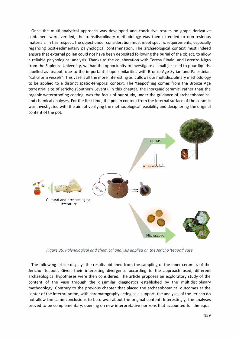
159
Once the multi-analytical approach was developed and conclusive results on grape derivative
containers were verified, the transdisciplinary methodology was then extended to non-resinous
materials. In this respect, the object under consideration must meet specific requirements, especially
regarding post-sedimentary palynological contamination. The archaeological context must indeed
ensure that external pollen could not have been deposited following the burial of the object, to allow
a reliable palynological analysis. Thanks to the collaboration with Teresa Rinaldi and Lorenzo Nigro
from the Sapienza University, we had the opportunity to investigate a small jar used to pour liquids,
labelled as ‘teapot’ due to the important shape similarities with Bronze Age Syrian and Palestinian
“caliciform vessels”. This vase is all the more interesting as it allows our multidisciplinary methodology
to be applied to a distinct spatio-temporal context. The ‘teapot’ jug comes from the Bronze Age
terrestrial site of Jericho (Southern Levant). In this chapter, the inorganic ceramic, rather than the
organic waterproofing coating, was the focus of our study, under the guidance of archaeobotanical
and chemical analyses. For the first time, the pollen content from the internal surface of the ceramic
was investigated with the aim of verifying the methodological feasibility and deciphering the original
content of the pot.
Figure 35. Palynological and chemical analysis applied on the Jericho 'teapot' vase
The following article displays the results obtained from the sampling of the inner ceramics of the
Jericho ‘teapot’. Given their interesting divergence according to the approach used, different
archaeological hypotheses were then considered. The article proposes an exploratory study of the
content of the vase through the dissimilar diagnostics established by the multidisciplinary
methodology. Contrary to the previous chapter that placed the archaeobotanical outcomes at the
center of the interpretation, with chromatography acting as a support, the analyses of the Jericho do
not allow the same conclusions to be drawn about the original content. Interestingly, the analyses
proved to be complementary, opening on new interpretative horizons that accounted for the equal

160
participation of molecular characterization and pollen records. The results are discussed considering
several archaeological hypotheses. Archaeologist’s initial assumptions are therefore tested in light of
the analytical outcomes.
Through the extension of pollen identification knowledge to other botanical families than the one
previously encountered, I gained learning of palynological formal and morphological analysis. Since
the two approaches we employed did not provide corroborating results, I had to deeply investigate
historical and scientific literature to blossom the potential archaeological understanding. The diverging
palaeoevidence therefore drove further targeting of molecular investigation. The exercise of linking
analytical results trained me to ripen the discussion. Besides the application of diagnostic protocols to
ceramics, I acquired methodological skills with the aim of interpreting analytical results by including
them in a broader archaeological picture. This way, I used the bibliography as a major asset to
contextualize the results. Because of the limited amount of material available for the analysis, the
chromatographic study could unfortunately not be taken any further, and the lack of material did not
allow us to continue with more specific characterization protocols. Although a new sampling is not
excluded, the homogeneity of the results remains nonetheless questionable. Further analysis would
allow us to clarify certain hypotheses, hence modifying the exploratory structure of the article before
submission. The paper might then be submitted in international journals of archaeometry, such as to
the Journal of Archaeological Science.

161
Tracing the medicinal content of a Bronze Age Jericho vase: how
Malva parviflora pollen meet Pinaceae resin markers
Louise Chassouant1,2*, Federico Di Rita2, Alessandra Celant2, Cathy Vieillescazes1, Teresa Rinaldi3,
Lorenzo Nigro4, Carole Mathe1, Donatella Magri2
1 IMBE UMR7263/ IRD237, Avignon University/ CNRS/ IRD/ AMU, Restoration Engineering of Natural
and Cultural Heritage F-84000, Avignon, France 2 Dipartimento di Biologia Ambientale, Sapienza Università di Roma, Piazzale Aldo Moro, 5, 00185,
Roma, Italy 3 Dipartimento di Studi Orientali, Sapienza Università di Roma, Piazzale Aldo Moro, 5, 00185, Roma,
Italy 4 Dipartimento di Biologia e Biotechnologia “Charles Darwin”, Sapienza Università di Roma, Piazzale
Aldo Moro, 5, 00185, Roma, Italy
*Correspondance: [email protected]
6.1. Abstract
Pollen and chemical analyses were applied on the inner ceramic of a Bronze Age pot (ca. 2300-2000
BC) recovered from the archaeological site of Jericho (Southern Levant). The peculiar shape of the
vessel, symbolized by a spouted typology characterized as ‘teapot’, represents a major asset to the
understanding of cultural and daily practices the vessel reverts to. Archaeobotanical analyses revealed
a significant amount of Malva parviflora pollen and lumps of Artemisia and Malva. Organic residues,
characterized by Gas Chromatography - Mass Spectrometry, highlighted diterpenic markers of
Pinaceae resin. As the identified plant are notorious for their health benefits and no vessel coating
being macroscopically observed, we interpreted the results as a medicinal concoction. Induced by the
archaeologist's hypotheses regarding the functionality of the pouring vessel and supported by the non-
overlapping results obtained by palaeobotanical and chromatographic approaches, this study
presented exploratory work that considered the various possibilities of liquid content. By obtaining
scientific evidence that does not overlap with each other, the multidisciplinary approach then allowed
to open the perspectives of anthropological and archaeological interpretation.
6.2. Introduction
Palynology has experienced a great deal of accomplishment in the last decades. The contribution of
pollen to the reconstruction of past vegetation (Di Rita, Anzidei and Magri, 2013; Magri et al., 2015;
Edwards, Fyfe and Jackson, 2017), the deciphering of palaeoecological environment (Di Rita et al.,
2015; Di Rita and Magri, 2019; Mercuri et al., 2020) and to the understanding of ancient cultural
practices (Mercuri et al., 2013; Bowes et al., 2015) are not to be boasted anymore. While palynology
finds its origins in the early 1940s (Iversen, 1941), the field really developed in the second part of the
20th century, with pioneers studies touting the yielded outcomes through pollen observation (Greig
and Turner, 1974; Faegri, Kaland and Krzywinski, 1989).
162
While palynology is frequently integrated into wider archaeobotanical studies of micro- (non-pollen
palynomorphs, spores, diatoms, and phytoliths) and macrofossil (charcoals, leaves, seeds, etc.)
remains, interdisciplinary approaches that combine distinctive research fields are still too occasionally
developed. Besides encouraging a common language between cultural, historical and scientific
communities, bridging the gap from independent disciplines greatly strengthens the archaeological
interpretation (Bouby et al., 2013; Pecci et al., 2020). As raised by Izdebski et al. (Izdebski et al., 2016),
interdisciplinary collaborations innovatively overreach the frontiers between humanities and
palaeoscience by providing valuable tools to integrate societal impacts into the deciphering of
environmental, cultural and climate changes.
In this respect, the archaeological site of Tell es-Sultan (Jericho West Bank) represents a meaningful
pool for multidisciplinary investigations. The recent discovery of a spouted vase, dated back to the
Early Bronze Age (EBA)/Intermediate Bronze Age (IBA), offered a rare opportunity to carry out
collaborative investigations with the aim of deciphering the original content of the vessel. Indeed, the
singular vessel typology, anachronistically designated as a “teapot” (Bunimovitz and Greenberg, 2004;
Nigro et al., 2021), outlined the very meaningful archaeological context it may raise. The upper
decoration and typological similarity with Bronze Age Syrian and Palestinian “caliciform vessels”
suggest a symbolic content that would have been poured (Albright, 1932; Wright, 1971; Dever, 1973).
Based on the typological observation, various possible beverages were hypothesized by the
archaeologists that performed the excavation campaigns (Nigro et al., 2021), such as water, beer, wine,
oil, milk, and even tea. The application of palynological and chromatographic tools aimed at bringing
scientific insights to identify the content of the pot advanced by the archaeologists.
The aim of the article is the characterization of the vessel content with scientific lens to confirm and
blossom the archaeologist initial hypotheses. Besides the archaeological meaning, the purpose of this
study was to verify the possibility of acceding fossil pollen contained in the ceramic of the vase. In this
study, Gas chromatography - Mass Spectrometry (GC-MS) and archaeobotanical tools were used to
provide the molecular fingerprint of microresidues and pollen impregnated in inner surface of the
ceramic.
6.2.1. Archaeological site
The archaeological site of Tell es-Sultan is located in the Southern Levant (31°52'15.99" N;
35°26'39.32" E). Integrated into the Jericho Oasis (Fig. 36), the holy place has early on attracted past
civilizations to settle down. The first excavation started during the 19th century, under the supervision
of the Captain Charles Warren, before Austro-German archaeologists took precedence on the
expeditions during the beginning of the 20th century. They notably discovered the "Cyclopean Wall",
the monumental foundation of the rampart, dated to Middle BA III (Wright, 1957). They established
the continuous occupation of the site from the 10th to 6th century BC. Few years later, British
expeditions uncovered the important necropolis of Jericho, testifying to the prominent place the site
occupied in the Fertile Crescent and outlined Jericho’s intense activity during the Mesolithic and
Neolithic periods (Garstang, 1932, 1933, 1934). Later on, Kenyon revolutionized the archaeological
methodologies employed by developing innovative techniques of excavation (Kenyon, 1953, 1981,
1993). Improving the accuracy of chronological assessment through the analysis of the site
stratigraphy, she notably shed light on the site's occupation from the EBA to the Roman period. Since
2017, the archaeological site is excavated in cooperation with Sapienza University of Rome, following
an agreement of the Palestinian National Authority (Department of Antiquities and Cultural Heritage
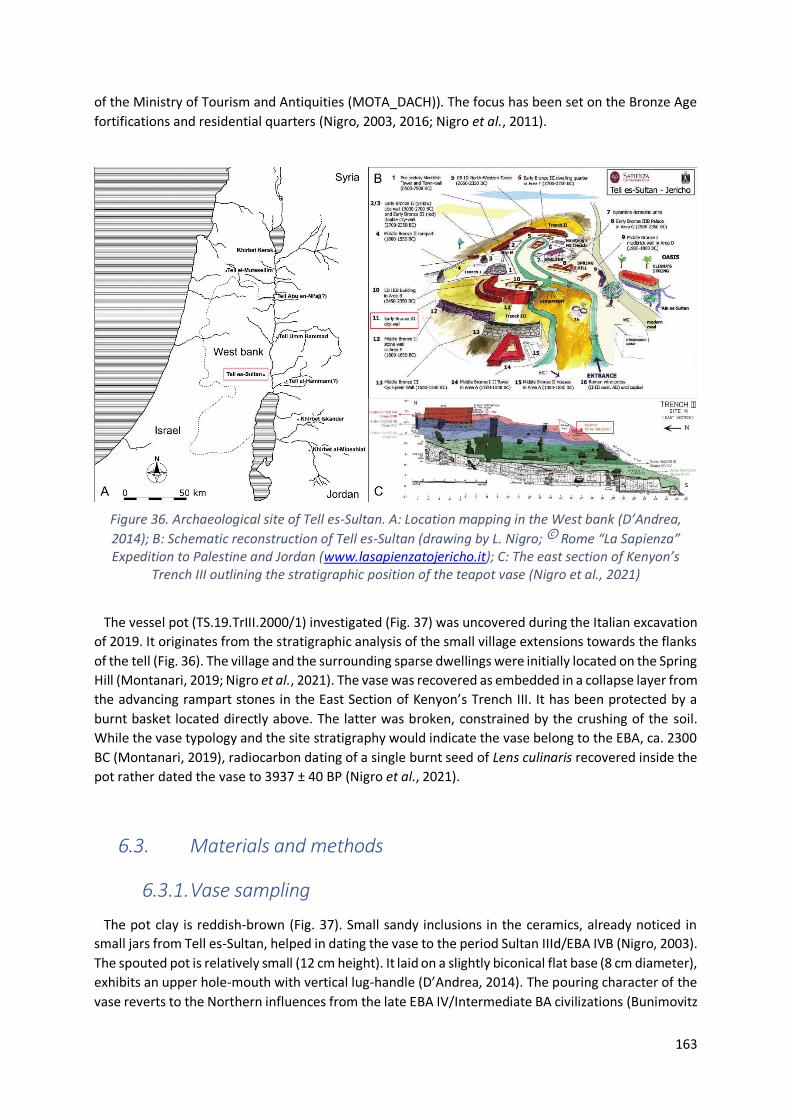
163
of the Ministry of Tourism and Antiquities (MOTA_DACH)). The focus has been set on the Bronze Age
fortifications and residential quarters (Nigro, 2003, 2016; Nigro et al., 2011).
Figure 36. Archaeological site of Tell es-Sultan. A: Location mapping in the West bank (D’Andrea,
2014); B: Schematic reconstruction of Tell es-Sultan (drawing by L. Nigro; © Rome “La Sapienza” Expedition to Palestine and Jordan (www.lasapienzatojericho.it); C: The east section of Kenyon’s
Trench III outlining the stratigraphic position of the teapot vase (Nigro et al., 2021)
The vessel pot (TS.19.TrIII.2000/1) investigated (Fig. 37) was uncovered during the Italian excavation
of 2019. It originates from the stratigraphic analysis of the small village extensions towards the flanks
of the tell (Fig. 36). The village and the surrounding sparse dwellings were initially located on the Spring
Hill (Montanari, 2019; Nigro et al., 2021). The vase was recovered as embedded in a collapse layer from
the advancing rampart stones in the East Section of Kenyon’s Trench III. It has been protected by a
burnt basket located directly above. The latter was broken, constrained by the crushing of the soil.
While the vase typology and the site stratigraphy would indicate the vase belong to the EBA, ca. 2300
BC (Montanari, 2019), radiocarbon dating of a single burnt seed of Lens culinaris recovered inside the
pot rather dated the vase to 3937 ± 40 BP (Nigro et al., 2021).
6.3. Materials and methods
6.3.1. Vase sampling
The pot clay is reddish-brown (Fig. 37). Small sandy inclusions in the ceramics, already noticed in
small jars from Tell es-Sultan, helped in dating the vase to the period Sultan IIId/EBA IVB (Nigro, 2003).
The spouted pot is relatively small (12 cm height). It laid on a slightly biconical flat base (8 cm diameter),
exhibits an upper hole-mouth with vertical lug-handle (D’Andrea, 2014). The pouring character of the
vase reverts to the Northern influences from the late EBA IV/Intermediate BA civilizations (Bunimovitz

164
and Greenberg, 2004; Joffe, 2018; D’Andrea, 2019). As suggested by the great resemblance with
Egyptian and Near East Mesopotamian copper ewers recovered in the tomb of Khasekhemwy, the
teapot might be used for ceremonial practices (Schorsch, 1992; Nigro et al., 2021). The lug-handle of
the vase that would inconvenience hand holding raises the assumption of a suspended vase that could
be used as a dipper or a pithos (Nigro et al., 2021).
Figure 37. Vessel TS.19.TrIII.2000/1 (Nigro et al., 2021). Potter decorations are visible
on the upper part
6.3.2. Archaeopalynological analyses
The internal surface of the vase was scratched with a lancet, over 1-2 mm in depth. No black coating
was observed. 400 mg of the recovered powder were treated following the standard
archaeopalynological procedure (Magri and Di Rita, 2015). HCl 37% (10 mL) was added to remove
carbonate compounds and to dissolve the Lycopodium tablet that contains a known number of exotic
spores to evaluate the pollen concentration. After centrifugation and removal of the supernatant, HF
was carefully added to remove silica-based fractions. HCl was then added again to dissolve the
potential colloidal silica and silicofluorides generated. NaOH was added after centrifugation and left in
boiling water for 10 min to remove humic acids. Washing with water helped regulating the pH to
neutral values. The mixture was mounted in glycerol before light microscope observation.
Samples were observed under a Zeiss Axioscope light microscope at 400x and 630x magnifications.
Identifications were supported by pollen atlases (Saad, 1960; Culhane and Blackmore, 1988; Bibi et al.,
2010), identification keys (El Naggar, 2004; Khalik et al., 2021), and the reference collection of the
Laboratory of Palaeobotany and Palynology of Sapienza University of Rome. Pollen morphologies were
also analyzed by Environmental Scanning Electron Microscope (ESEM) Hitachi TM-3000 Tabletop
operating at 15Kv without previous coating. ESEM pollen images were recorded at magnifications
varying from 1.2kx to 4.0kx.

165
6.3.3. Chromatographic analyses
6.3.3.1. Solvents and reagents
Solvents and reagents were of analytical grade. Dichloromethane (DCM), ethyl acetate, KOH and
Na2SO4 were supplied by Merck. N,O-Bis(trimethylsilyl)trifluoroacetamide / trimethylchlorosilane
(BSTFA/TMCS) and hexane were purchased by Sigma-Aldrich.
6.3.3.2. Sample preparation
Two extractive protocols were applied. On the first hand, 100 mg of crushed shard were extracted 3
times with DCM (1 mL) with the ultrasound probe (VCX 130 Vibra-Cell Sonics) for 3 min. After
centrifugation, the organic supernatants were combined, filtered with PTFE (0.45 µm) and evaporated
to dryness. On the second hand, an alkaline treatment was performed with 100 mg of crushed shard.
The sample was extracted three times with 2 mL KOH (1M) with an ultrasound probe for 3 min. After
centrifugation, the supernatants were added together and acidified up to pH 2. The organic phase was
extracted 3 times with 3 mL ethyl acetate, filtered on a PTFE cartridge (0.45 µm) and evaporated to
dryness.
6.3.3.3. Gas Chromatography – Mass Spectrometry
GC–MS analyses were carried out with a Thermo Scientific™ Focus system equipped with a Thermo
Scientific Al 3000 autosampler and coupled to a Thermo Fisher Scientific™ ITQ™ 700 Series Ion Trap
Mass Spectrometer. The separation was ensured with a 30 m x 0.25 mm internal diameter x 0.25 μm
film thickness fused silica capillary column ThermoGOLD™ TG-5MS (5% diphenyl; 95% dimethyl
polysiloxane). 1 µL solution was injected in splitless mode at 250°C. The transfer line and the ion trap
were respectively maintained at 300 and 200°C. Molecular components were carried by a constant 1
mL/min helium flow. Data treatments were carried out on Xcalibur software. Molecular compounds
were identified by retention time, comparison with mass spectrum of commercial molecular
standards, with the lab molecular library and with NIST MS Search 2.0 database recorded with an
electronic ionization of 70 eV. The oven temperature was held at 50°C for 2 min, increased to 140°C at
8°C/min held for 2 min before reaching 160°C at 2.5°C/min and finally 330°C at 15°C/min and held for
3 min. Spectra were recorded in the 50-650 m/z range.
All the samples were trimethylsilylated with BSTFA/TMCS (200 µL; 70°C; 30 min) and evaporated to
dryness before being dissolved in a mixture of DCM:hexane (1:1) and injected in splitless mode into
the GC-MS apparatus.
6.4. Results and discussion
Palynological analysis revealed a limited pollen concentration content of 242 grains/g sample. The
following pollen grains were identified: Malva (12 grains + 1 lump); Artemisia (1 grain + 1 lump);
Brassicaceae (1 grain); Amaranthaceae (1 grain) and Cedrus (1 grain).
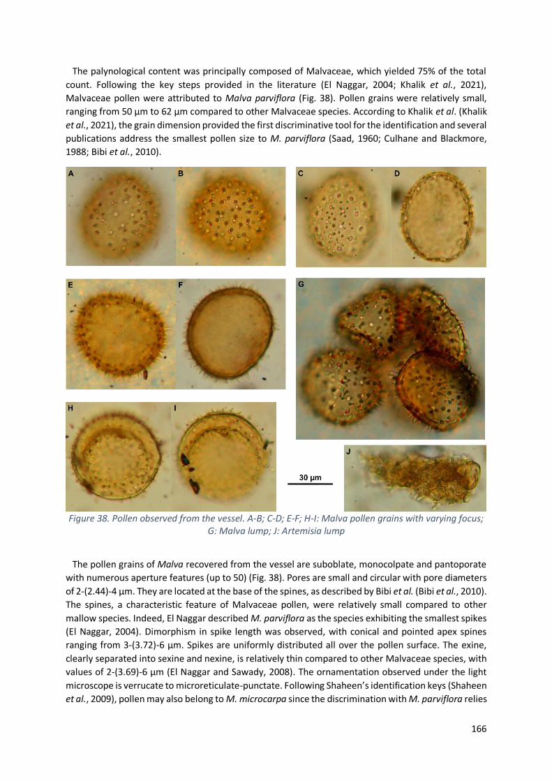
166
The palynological content was principally composed of Malvaceae, which yielded 75% of the total
count. Following the key steps provided in the literature (El Naggar, 2004; Khalik et al., 2021),
Malvaceae pollen were attributed to Malva parviflora (Fig. 38). Pollen grains were relatively small,
ranging from 50 µm to 62 µm compared to other Malvaceae species. According to Khalik et al. (Khalik
et al., 2021), the grain dimension provided the first discriminative tool for the identification and several
publications address the smallest pollen size to M. parviflora (Saad, 1960; Culhane and Blackmore,
1988; Bibi et al., 2010).
Figure 38. Pollen observed from the vessel. A-B; C-D; E-F; H-I: Malva pollen grains with varying focus; G: Malva lump; J: Artemisia lump
The pollen grains of Malva recovered from the vessel are suboblate, monocolpate and pantoporate
with numerous aperture features (up to 50) (Fig. 38). Pores are small and circular with pore diameters
of 2-(2.44)-4 µm. They are located at the base of the spines, as described by Bibi et al. (Bibi et al., 2010).
The spines, a characteristic feature of Malvaceae pollen, were relatively small compared to other
mallow species. Indeed, El Naggar described M. parviflora as the species exhibiting the smallest spikes
(El Naggar, 2004). Dimorphism in spike length was observed, with conical and pointed apex spines
ranging from 3-(3.72)-6 µm. Spikes are uniformly distributed all over the pollen surface. The exine,
clearly separated into sexine and nexine, is relatively thin compared to other Malvaceae species, with
values of 2-(3.69)-6 µm (El Naggar and Sawady, 2008). The ornamentation observed under the light
microscope is verrucate to microreticulate-punctate. Following Shaheen’s identification keys (Shaheen
et al., 2009), pollen may also belong to M. microcarpa since the discrimination with M. parviflora relies

167
on a pore diameter exceeding 2 µm. However, this species does not live in the Southern Levant, the
taxon hitherto occurs in Northern India and West Bengal (Biswa, Lakshminarasimhan and Lokho, 2016).
The pollen is additionally rarely described in literature (Azab, 2017; Ali-Shtayeh and Jamous, 2018). M.
sylvestris, nicaaensis and aegyptia were excluded on the basis of morphological criteria of size and
spine length, despite their great representation in the West Bank (Ali-Shtayeh and Jamous, 2018). Even
though Hosni et al. speculated small pollen dimension for M. neglecta (Hosni and Araffa, 1999), the
hypothesis of an attribution to M. neglecta was rejected since other publications stated bigger
dimension of 70-100 µm with spine length of 7-9 µm (Culhane and Blackmore, 1988; Shaheen et al.,
2009; Bibi et al., 2010).
One pollen grain however exhibited a bigger dimension (ca. 83 µm) even though the diameter could
not be accurately measured since the grain was importantly damaged and the grain was opened
(pollen not shown). Considerable morphological and structural damages regarding some Malvaceae
grains were indeed noticed. The poor state of pollen preservation was notably visible through grain
opening or from the loss of spikes all around the exine (Fig. 39A). Since such damages could have
interfered with the identification tools at the species level, the concerned grains were not included in
the identification. Likewise, chemical treatments and the slide mounting media have been mentioned
to alter pollen diameter (Reitsma, 1969; Sluyter, 1997; Meltsov, Poska and Saar, 2008).
Figure 39. Malva pollen grains observed with ESEM
As reported in Figure 5, chromatographic analyses of the ceramic shard revealed the presence of
short chains of dicarboxylic acids, namely adipic, pimelic and azelaic acids. They probably originated
from the degradation of unsaturated fatty acids through auto-oxidative reactions triggered by radical
mechanism of initiation and hydroperoxide formation (Aillaud, 2001), hence traducing the ageing of
the lipid content. Palmitic, stearic and oleic acids were also identified. Additionally, the analyses
highlighted the presence of 4-hydroxy benzoic acid, succinic acid and hydrocarbons. Although not
specific to Malva sp., such compounds were nonetheless characterized in Malvaceae chemical
composition (Hasimi et al., 2017; Al-Snafi, 2019). Hydrocarbons, and more specifically terpene
structures, including mono-, sesqui-, di- and more rarely triterpenoids were also evidenced (Cutillo et
al., 2006). Since the literature concentrated on the fresh material, unstable compounds such as
flavonoids and tannins were reported. Being hardly preserved with time, they are however unlikely to
be recovered in archaeological analyses.
The recovery of mericarps attributed to M. parviflora in the Upper Palaeolithic archaeological site of
Ohalo II in Israel provided evidence of the early on presence of the flower in Southern Levant (Snir,
Nadel and Weiss, 2015; Weide et al., 2017). The important extent in which fossil fruitlets of mallows
were found (i.e., up to 14% or the current weed species) associated to the meaning recovery location

168
nearby the grinding stone traduced the flower daily life usage as well as its widespread distribution in
the region around 23000 BP (Piperno et al., 2004; Snir et al., 2015). Then, M. parviflora was evidenced
on the alluvium fields of the late 3rd millennium village of Iktanu, located at the Eastern North of Jericho
(de Vries and Bikai, 1993). Malva sp. was also identified in the Dead Sea Plain macrofossils recovered
from the Middle BA village of Tell el-Hayyat along the Jordan River (Fall, Falconer and Porson, 2019).
Such archaeological records attest of the early presence of the flower in the local vegetal environment
and its inclusion into past civilizations cultures.
Figure 40. Chromatograms from (A) Alkaline treatment and (B) DCM extraction.Ac.: acid; HCB Chains: Hydrocarbon Chains; *: Contaminant; Cx;y: Fatty acid of x carbon atoms and y unsaturations; Cx-OH:
Alcohol of x carbon chains
Moreover, similar pouring vessels were recovered during the excavations of several tombs in the
Southern Levant (e.g., Megiddo tombs (Braun and Ilan, 2013), Tell el-Farʿah North (de Vaux and Steve,
1949) or Qiryat Ata (Golani and Bankirer, 2003)). Such discoveries supported the pouring vase
relevance in funeral contexts. Bunimovitz (Bunimovitz and Greenberg, 2004) further distinguished the
tombs into the one containing teapots and pouring-shaped vessels and the others, rather flourished
with jars and four-spouted lamps. The ubiquity of teapot vessels, reported in both mortuary and
domestic Canaan contexts, is therefore asserted to the founder role beverages might have played in
bringing populations and cultures closer to each other. The typological heterogeneity noticed within
the fine ware ceramic (e.g., dimension size, neck presence, horizontal tube handles, bent spout, etc.)
and the wide distribution all along the Jordan River suggest numerous manufacture production sites
and a lively trades of such potteries (Braun and Ilan, 2013). Combined with the evidence of recurrent
use of M. parviflora, the assumption of a flower functionality in ritual practices and daily use is well
grounded. To go further on, the recent bridging with a copper ewer from in the Umm el-Qaab
necropolis at Abydos (Egypt) exhibiting similar typology (Nigro et al., 2021) reinforces the ceremonial
and symbolic character of `Egyptian mallows`, the common name for M. parviflora.
Medicinal benefits of mallows were greatly referenced in literature to chemically address their
biological and anti-oxidant properties (Azab, 2017; Delfine et al., 2017; Akbar, 2020; Sharifi‐Rad et al.,
2020). Malva sp. is still currently consumed in decoction, as attested by current ethnopharmacological
surveys of the millennial character of the plant (Ali-Shtayeh et al., 2008; Gasparetto et al., 2012). This
way, indigenous civilizations already used mallows to cure respiratory and digestive diseases with
herbal decoctions (Said et al., 2002). Bible recipes referred to the ethnobotanical use of mallow leaves

169
and flowers: Egyptians used poultice pounded leaves of M. parviflora to diminish arachnid’s stings
(Duke, 1983). Roman consumed them as edible vegetable or infused as tea substituent, as boasted in
Palestinian folkloric songs which designated M. rotundifolia as the commonest salad herb in the Middle
Age (Crowfoot and Baldensperger, 1932). In the Northern Jordan, local people conferred to M.
parviflora an important economic, floristic and cultural feature (Nawash et al., 2014). This current high
use value can find its roots in the remarkable flower representation in the vegetal landscape.
The recovery of pollen lumps of Malva and Artemisia (Fig. 38G; 3J) enhanced the assumption of
medicinal herbal tea flourished with flowers, since lump agglomerates can only originate from the
flower itself. Moreover, Artemisia has long been employed for its anti-inflammatory, anti-microbial
and anti-oxidant properties (Said et al., 2002; Khan and Khatoon, 2008). Brassicaceae and
Amaranthaceae were assigned to the environmental vegetation and did not help in characterizing the
content functionality.
Independently from the Malva species, ethnobotanical investigations in the Siran Valley (Pakistan)
focused on the pharmacological benefit regarding sexual disorders (Shah and Khan, 2006; Ahmad et
al., 2009). Historically, Pliny the Elder (Naturalis Historia, XX, 84) reported the curative and aphrodisiac
benefits of mallow consumption, the plant being already well-known from Greeks. He widely detailed
healing recipes against animal venom, ulcer and even toothache by leaving the plant macerating with
urine or even lichen and honey mixing. In fact, the archaeological presence of honey would
alternatively provide a valuable explanation to the presence of pollen grains and lumps in the fossil
record. Even though a significant amount of sugar in M. sylvestris flowers (i.e., 10-12 mg over 10
flowers) was measured (Jablonski and Koltowski, 2001), Malva are not the privileged flowers for bee
foraging compared to Cirsium, Trifolium or even Centaurea (Fussell and Corbet, 1992; Rösch, 2005).
However, being an integral part of the Jericho floral environment and widely distributed as indicated
by the palynological analyses of Neolithic beehives from Tel Rehov, Israel (Weinstein-Evron and Chaim,
2016), it is very likely that mallows afforded bee-forages. In that sense, other archaeological contexts
such as Italian charred honeycombs dated to the Iron Age provided evidence of Malva occurrence
(Castellano et al., 2017). Rösch interpreted the great pollen diversity recovered from a Bavarian BA
bowl as an indicator of archaeological mead or honey, assigning Artemisia as the most frequent
anemophilous pollen foraged (Rösch, 2005). In the hypothesis of a medicinal herbal content, honey
would have a real meaning on health benefits in parallel of sweetening the taste. In this case and
considering the small size of the pot, only a limited amount of honey would have been added to the
vase, which explains the mitigated number of pollen grains identified and the low pollen concentration.
De facto, pollen grains undergo differential degradation due to the different chemical compositions of
the sporopollenin (Campbell, 1999; Twiddle and Bunting, 2010). To the best of our knowledge,
Malvaceae have never been included in partial degradation studies. Nonetheless, a better preservation
of mallow pollen, combined with the widespread presence of Malva in the natural environment may
address the major distribution of Malva in the overall counting and explain the damage state observed
for Malva grains. However, chromatography did not molecularly support honeybee assumption since
no linear long-chain alkanes, fatty acids or alcohols, nor fatty acyl monoesters and hydroxyl fatty acyl
monoesters were characterized (Roffet-Salque et al., 2015). More targeted extractive protocols should
be further applied to definitively address such hypothesis.
Alternatively, the presence of wine was tested since the grape beverage was reported as a vector of
medicinal recipes (McGovern, Mirzoian and Hall, 2009). Based on the butylation protocol that was
previously reported to surely trace tartaric acid (Chassouant et al., unpublished paper), the wine
hypothesis was refuted in the Jericho vase since no grape-fermentation markers were identified in the
inorganic shard (data not shown).
170
The West Bank being close to the Bible birthplace and Israel being referred as ‘A Land Flowing with
Milk and Honey’ (Exodus 3; 8), the vessel could contain milk: this hypothesis, providing a ymbolic
understanding of the content, needs to be verified by proteomics analysis.
Reverting to Figure 5, diterpenic markers of pimarane and abietane skeletons were characterized by
chromatography. Dehydroabietic acid (DHA), together with pimaric, isopimaric, sandaracopimaric and
abietic acids gave insight into the archaeological presence of Pinaceae resin in the vase. Given to the
fact that no macroscopic coating was noticed during the sampling and corroborated to the absence of
Pinus pollen in the vase, one reasonable hypothesis considered the resin addition to the mixture, made
on purpose either as coating or external addition. Different aims can be suggested. First, the Pinaceae
resin may have been used to modify the taste of the concoction, as for resinated wine (McGovern et
al., 1996). Then, the oleoresin fraction may have conferred anti-microbial and anti-inflammatory
virtues. The essential oil fraction being compounded of mono and sesquiterpenoids, it may not have
survived over archaeological times, leaving only the diterpenic resin fraction visible. Moreover,
common is the ethnopharmacological evidence of resin-derivatives used as traditional drugs, as much
for turpentine oil as for pine tar (Lev and Amar, 2002). For instance, the Turkish Çam preparation is a
traditional curing mixture made of pine resin and honey (Satil, Selvi and Polat, 2011).
Since Pinus pollen has a large dispersal radius due to its saccate morphology (Schwendemann et al.,
2007), its absence in the palynological analyses of the vase highlighted the will of uniquely using the
natural exudate. Indeed, if the resin had undergone any calorific treatment such as wood pyrolysis,
then pollen would have been recovered, as demonstrated by the important proportion of Pinus pollen
in the waterproofing coating of archaeological amphorae recovered from the ancient anchorage of San
Felice Circeo (Chassouant et al., unpublished paper). In this respect, no methyl ester DHA and retene
markers were characterized by chromatography although the protocols applied have shown their
extractive efficiency in the past (Pecci, Cau Ontiveros and Garnier, 2013). Such molecules allow wood
distillation and high temperature treatment to be traced (Colombini et al., 2005). The absence of
oxidized DHA derivatives additionally indicates the resin did not suffer from ageing (Mezzatesta et al.,
2021b).
Interestingly, Cedrus pollen was also identified in the vase. The teapot having been uncovered in a
close room space in the excavation field, collected with care and maintained in close and clean
conditions (Nigro et al., 2021), the possibility of post-deposition contamination was excluded although
Cedrus is widely spread in Lebanon. The pollen should therefore originate from the vessel itself. Cedrus
essential oil, namely “Cedrium”, was mentioned in Egyptian cultural contexts, notably for
mummification rituals (Koller et al., 2003; Charrié-Duhaut et al., 2007; Łucejko et al., 2012). The resin
exhibits anti-microbial properties strongly appreciated for embalming, structural buildings and
medicinal treatments (Kizil et al., 2002; Termentzi, Fokialakis and Leandros Skaltsounis, 2011). The
chemical composition mainly accounts for sesquiterpenic skeletons, with himalachene considered as
biomarker of true cedar resin (Loizzo et al., 2008; Sarret et al., 2017). Back to the chromatographic
analysis, no himalachene derivatives were identified, hence highlighting the more pronounced
instability of sesquiterpenoids towards diterpenoids. Although the absence of such Cedrus markers did
not rule out hypothetical cedar exudate in the teapot, notably plausible through the added value of
health benefits of the resin, no certainties can either be formulated regarding its presence. Further
investigation involving aged cedar exudate may provide a deeper understanding of the molecular
alteration triggered by the resin degradation over time.
171
6.5. Conclusion
This study provided valuable evidence of the consumption of mallow at Bronze Age time in the
Levant. Pollen analysis revealed the extensive presence of pantoporate spiked grains, identified as
Malva parviflora. Pollen grains were relatively small compared to other Malvaceae. Fossil records
additionally attest of M. Parviflora wide contribution to the local vegetal environment at Paleolithic
times.
Chromatographic analyses did not explicitly confirm the presence of Malvaceae but did not invalidate
it either: hydrocarbon chains, alcohols and acids belong to the common composition of plants. Short
chains dicarboxylic acids witnessed the degradation of fatty acids that certainly occurred during
archaeological times. Organic residues additionally indicated the presence of diterpenic markers. The
absence of aged and heated molecular derivatives gave credits to the exudate nature of Pinaceae resin.
Besides its exploratory character, this study provided valuable scientific evidence to draw the attention
on a medicinal beverage, notably raised by the archaeobotanical records. Supported by Malva and
Artemisia pollen lumps that can only originate from flower anthers, results were interpreted as traces
of an ancient medicinal concoction. Enhanced by archaeological assumptions regarding the spouted
character of the vessel and the biological properties of the botanic species identified, this
multidisciplinary approach provided great signs of ancient cultural practices employing Malva
parviflora and Artemisia flowers with Pinaceae resin.
Since palynological and chromatographic results did not cross-check each other, further
investigations would be requested to unravel archaeological recipes and cultural consumption. The
lack of material did not allow organic residue analysis to be pushed with more focalized technics to
straightly investigate the presence of beeswax, fatty compounds such as long aliphatic chain carboxylic
acids or terpenoid typical of herbal plant or cedar. Molecular markers, although tremendously
necessary to evidence Pinaceae unnoticeable with palynological tools, probably suffered from
degradation and leaching out. More in-depth analyses with solid phase extractions are encouraged to
molecularly address the presence of cedar or honey, until then hardly identifiable with standard
chromatographic methods (McGovern, Mirzoian and Hall, 2009; Mezzatesta et al., 2021a).Typological
resemblances with teapots recovered from funeral contexts alternatively suggested the symbolic role
of the archaeological preparation, that could be supported by the presence of Cedrus pollen since
cedars are highly emblematic in the nearby Lebanon.
The present study accounted for the development of collaborative approaches in order to provide
valuable insights from independent fields of research. In this regard, archaeometric tools provided
meaningful bridges to overcome gaps raised by single-centered investigations.

172
Funding
This research was financially supported by Marie Skłodowska-Curie Innovative Training Network, The
ED-ARCHMAT European Joint Doctorate, H2020-MSCA-ITN-EJD ED-ARCHMAT Joint Doctorate (Project
ESR9, grant agreement no 766311).
Author contributions
Conceptualization, L.C., A.C., T.R., C.M., D.M.; methodology, L.C.; formal analysis, L.C., F.D.;
investigation, L.C., F.D., C.M., D.M.; resources, A.C., T.R., L.N.; visualization, L.C.; supervision, C.M.,
D.M.; project administration, A.C., C.M., D.M.; funding acquisition, C.V., C.M., D.M.; writing - original
draft preparation, L.C.

173
6.6. References Ahmad, H. et al. (2009) ‘Ethnobotanical study of upper siran’, Journal of Herbs, Spices and Medicinal
Plants, 15(1), pp. 86–97. doi: 10.1080/10496470902787519.
Aillaud, S. (2001) Field and laboratory studies of diagenetic reactions affecting lipid residues absorbed
in unglazed archaeological pottery vessels. University of Bristol.
Akbar, S. (2020) ‘Malva sylvestris L. (Malvaceae)’, Handbook of 200 Medicinal Plants, 200, pp. 1129–
1136. doi: 10.1007/978-3-030-16807-0_121.
Al-Snafi, A. E. (2019) ‘Medical benefit of Malva neglecta - A review’, IOSR Journal of Pharmacy, 9(6),
pp. 60–67. Available at: www.iosrphr.org.
Albright, W. F. (1932) ‘The Excavation of Tell Beit Mirsim I: pottery of the First three Campaings’, New
Haven.
Ali-Shtayeh, M. S. et al. (2008) ‘Traditional knowledge of wild edible plants used in Palestine (Northern
West Bank): A comparative study’, Journal of Ethnobiology and Ethnomedicine, 4(1), p. 13. doi:
10.1186/1746-4269-4-13.
Ali-Shtayeh, M. S. and Jamous, R. M. (2018) ‘Updating the plant “Red List” of Palestine (West Bank and
Gaza Strip): conservation assessment and recommendations’, Journal of Biodiversity & Endangered
Species, 6(3).
Azab, A. (2017) ‘Malva: Food, Medicine and Chemistry’, European Chemical Bulletin, 6(7), pp. 295–320.
doi: 10.17628/ecb.2017.6.295-320.
Bibi, N. et al. (2010) ‘Systematic implications of pollen morphology in the family Malvaceae from north
west frontier province, Pakistan’, Pakistan Journal of Botany, 42(4), pp. 2205–2214.
Biswa, S., Lakshminarasimhan, P. and Lokho, A. (2016) ‘A new locality for Malva parviflora var.
microcarpa (Pers.) Loscos (Malvaceae)’, Indian Journal of Forestry, 39(2), pp. 197–199.
Bouby, L. et al. (2013) ‘Bioarchaeological insights into the process of domestication of grapevine (Vitis
vinifera L.) during Roman times in Southern France’, PLoS ONE, 8(5), p. e63195. doi:
10.1371/journal.pone.0063195.
Bowes, K. et al. (2015) ‘Palaeoenvironment and land use of Roman peasant farmhouses in southern
Tuscany’, Plant Biosystems - An International Journal Dealing with all Aspects of Plant Biology, 149(1),
pp. 174–184. doi: 10.1080/11263504.2014.992997.
Braun, E. and Ilan, D. (2013) Early Megiddo on the East Slope (The ‘Megiddo Stages’): a report on the
early occupation of the East Slope of Megiddo-result of the Oriental Institute’s excavations, 1925-1933,
A Report on the Early Occupation of the East Slope of Megiddo. Result of the Oriental Institute's
Excavations, 1925-1933. Oriental Institute of the University of Chicago. Available at:
http://books.google.com/books?id=EcMLlwEACAAJ&dq=intitle:early+megiddoon+the+east+slope+th
e+megiddo+stages&hl=&cd=1&source=gbs_api.
Bunimovitz, S. and Greenberg, R. (2004) ‘Revealed in Their Cups: Syrian Drinking Customs in
Intermediate Bronze Age Canaan’, Bulletin of the American Schools of Oriental Research, 334(1), pp.
19–31. doi: 10.2307/4150104.

174
Campbell, I. D. (1999) ‘Quaternary pollen taphonomy: examples of differential redeposition and
differential preservation’, Palaeogeography, Palaeoclimatology, Palaeoecology, 149(1–4), pp. 245–
256.
Castellano, L. et al. (2017) ‘Charred honeycombs discovered in Iron Age Northern Italy. A new light on
boat beekeeping and bee pollination in pre-modern world’, Journal of Archaeological Science, 83, pp.
26–40. doi: 10.1016/j.jas.2017.06.005.
Charrié-Duhaut, A. et al. (2007) ‘The canopic jars of Rameses II: real use revealed by molecular study
of organic residues’, Journal of Archaeological Science, 34(6), pp. 957–967. doi:
10.1016/J.JAS.2006.09.012.
Colombini, M. P. et al. (2005) ‘Characterisation of organic residues in pottery vessels of the Roman age
from Antinoe (Egypt)’, Microchemical Journal, 79(1–2), pp. 83–90. doi: 10.1016/j.microc.2004.05.004.
Crowfoot, G. M. and Baldensperger, L. (1932) From Cedar to Hyssop - A Study in the Folklore of Plants
in Palestine. London: The Sheldon Press. Available at: http://folkmasa.org/fr/fr1.htm.
Culhane, K. J. and Blackmore, S. (1988) ‘Malvaceae’, Review of Palaeobotany and Palynology, 57(1–2),
pp. 45–74.
Cutillo, F. et al. (2006) ‘Terpenoids and phenol derivatives from Malva silvestris’, Phytochemistry, 67(5),
pp. 481–485. doi: 10.1016/j.phytochem.2005.11.023.
D’Andrea, M. (2014) ‘The Southern Levant in Early Bronze IV. Issues and Perspectives in the Pottery
Evidence’, in Contributi e Materiali di Archeologia Orientale XVII. Roma: “Sapienza” Università di Roma
– Dipartimento di Archeologia.
D’Andrea, M. (2019) ‘The periodization of Early Bronze IV in the Southern Levant: bridging the gap
between stratigraphy and absolute chronology’, in Gallo, E. (ed.) Conceptualizing Urban Experiences:
Tell es-Sultan and Tall al-Ḥammām Early Bronze cities across the Jordan. Proceedings of a workshop
held in Palermo, G. Whitaker Foundation, Villa Malfitano, June 19th, 2017. Rome: Rome «La Sapienza»
Expedition to Palestine & Jordan, pp. 61–78.
Delfine, S. et al. (2017) ‘Variation of Malva sylvestris essential oil yield, chemical composition and
biological activity in response to different environments across Southern Italy’, Industrial Crops and
Products, 98, pp. 29–37. doi: 10.1016/j.indcrop.2017.01.016.
Dever, W. G. (1973) ‘The EB IV-MB I Horizon in Transjordan and Southern Palestine’, Bulletin of the
American Schools of Oriental Research, 210(1), pp. 37–63.
Duke, J. A. (1983) Medicinal plants of the Bible. Trado-Medi. Buffalo, NY.
Edwards, K. J., Fyfe, R. M. and Jackson, S. T. (2017) ‘The first 100 years of pollen analysis’, Nature Plants,
3(2), p. 17001. doi: 10.1038/nplants.2017.1.
Faegri, K., Kaland, P. E. and Krzywinski, K. (1989) Textbook of Pollen Analysis. 4th ed. Hafner. New York:
John Wiley & Sons Ltd.
Fall, P. L., Falconer, S. E. and Porson, S. (2019) ‘Archaeobotanical inference of intermittent settlement
and agriculture at Middle Bronze Age Zahrat adh-Dhra’1, Jordan’, Journal of Archaeological Science:
Reports, 26(June), p. 101884. doi: 10.1016/j.jasrep.2019.101884.
Fussell, M. and Corbet, S. A. (1992) ‘Flower Usage by Bumble-Bees: A Basis for Forage Plant
Management’, The Journal of Applied Ecology, 29(2), p. 451. doi: 10.2307/2404513.
175
Garstang, J. (1932) ‘Jericho: city and necropolis. 1. Late Stone Age. 2. Early Bronze Age. 3. Middle
Bronze Age’, Liverpool Annals of Archaeology and Anthropology, 19, pp. 3–22, 35–54.
Garstang, J. (1933) ‘Jericho: city and necropolis. 4. Tombs of MBAii. 5. Tombs of MBAii and LBAi. 6. The
Palace Area’, Liverpool Annals of Archaeology and Anthropology, 20, pp. 3–42.
Garstang, J. (1934) ‘Jericho: city and necropolis. 6. The Palace Area (cont.). Palace and Store Rooms,
MBii Pottery and Houses LBi. Upper Stone Building, EBi’, Liverpool Annals of Archaeology and
Anthropology, 21, pp. 99–136.
Gasparetto, J. C. et al. (2012) ‘Ethnobotanical and scientific aspects of Malva sylvestris L.: a millennial
herbal medicine’, Journal of Pharmacy and Pharmacology, 64(2), pp. 172–189. doi: 10.1111/j.2042-
7158.2011.01383.x.
Golani, A. and Bankirer, R. Y. (2003) Salvage Excavations at the Early Bronze Age Site of Qiryat Ata.
Israel Antiquities Authority.
Greig, J. R. and Turner, J. (1974) ‘Some pollen diagrams from Greece and their archaeological
significance’, Journal of Archaeological Science, 1(2), pp. 177–194. doi: 10.1016/0305-4403(74)90041-
7.
Hasimi, N. et al. (2017) ‘Chemical Profile of Malva Neglecta and Malvella Sherardiana by LC-MS/MS,
GC/MS and their Anticholinesterase, Antimicrobial and Antioxidant Properties With Aflatoxin-
Contents’, Marmara Pharmaceutical Journal, 21(3), pp. 471–484. doi: 10.12991/marupj.307461.
Hosni, H. and Araffa, S. (1999) ‘Malvaceae in the flora of Egypt 2. Pollen morphology and its taxonomic
significance’, Taeckholmia, 19(2), pp. 147–156. doi: 10.21608/taec.1999.12642.
Iversen, J. (1941) ‘Land occupation in Denmark’s stoneage’, Danmarks Geologiske UndersopeIse, 2(66),
pp. 1–67.
Izdebski, A. et al. (2016) ‘Realising consilience: How better communication between archaeologists,
historians and natural scientists can transform the study of past climate change in the Mediterranean’,
Quaternary Science Reviews, 136, pp. 5–22. doi: 10.1016/j.quascirev.2015.10.038.
Jablonski, B. and Koltowski, Z. (2001) ‘Nectar secretion and honey potential of honey-plants growing
under Poland’s conditions’, Journal of Apiculture Science, 45, pp. 29–34.
Joffe, A. (2018) ‘Notes on Early Bronze Age Commensality’, in Shai, I. et al. (eds) Tell it in Gath. Studies
in the History and Archaeology of Israel Essays in Honor of Aren M. Maeir on the Occasion of his Sixtieth
Birthday. Münster: Zaphon, pp. 41–70.
Kenyon, K. M. (1953) ‘Excavation at Jericho, 1953’, Palestine Exploration Quarterly, 85, pp. 81–95.
Kenyon, K. M. (1981) Excavations at Jericho: The Architecture and Stratigraphy of the Tell. London.
Kenyon, K. M. (1993) “Tell es-Sultan”: The New Encyclopedia of Archaeological Excavations in the Holy
Land. Edited by E. Stern. Jerusalem.
Khalik, K. A. et al. (2021) ‘Pollen morphology of Malvaceae genera from Saudi Arabia and its taxonomic
significance’, Australian Journal of Crop Science, 15(5), pp. 725–742. doi:
10.21475/ajcs.21.15.05.p3129.
Khan, S. W. and Khatoon, S. (2008) ‘Ethnobotanical studies on some useful herbs of haramosh and
Burgote valleys in Gilgit, northern areas of Pakistan’, Pakistan Journal of Botany, 40(1), pp. 43–58.

176
Kizil, M. et al. (2002) ‘Antimicrobial activity of the resins obtained from the roots and stems of Cedrus
libani and Abies cilicica.’, Prikladnaia biokhimiia i mikrobiologiia, 38(2), pp. 166–168.
Koller, J. et al. (2003) ‘Analysis of a pharaonic embalming tar’, Nature, 425(6960), pp. 784–784. doi:
10.1038/425784a.
Lev, E. and Amar, Z. (2002) ‘Ethnopharmacological survey of traditional drugs sold in the Kingdom of
Jordan’, Journal of Ethnopharmacology, 82(2–3), pp. 131–145. doi: 10.1016/S0378-8741(02)00182-4.
Loizzo, M. R. et al. (2008) ‘Phytochemical analysis and in vitro evaluation of the biological activity
against herpes simplex virus type 1 (HSV-1) of Cedrus libani A. Rich.’, Phytomedicine, 15(1–2), pp. 79–
83. doi: 10.1016/j.phymed.2007.03.013.
Łucejko, J. J. et al. (2012) ‘An analytical approach based on X-ray diffraction, Fourier transform infrared
spectroscopy and gas chromatography/mass spectrometry to characterize Egyptian embalming
materials’, Microchemical Journal, 103, pp. 110–118. doi: 10.1016/j.microc.2012.01.014.
Magri, D. et al. (2015) ‘Holocene dynamics of tree taxa populations in Italy’, Review of Palaeobotany
and Palynology, 218, pp. 267–284. doi: 10.1016/j.revpalbo.2014.08.012.
Magri, D. and Di Rita, F. (2015) ‘Archaeopalynological Preparation Techniques’, in Yeung, E.C.T.,
Stasolla, C., Sumner, M.J., Huang, B. Q. (ed.) Plant Microtechniques and Protocols. Cham: Springer
International Publishing, pp. 495–506. doi: 10.1007/978-3-319-19944-3_27.
McGovern, P. E. et al. (1996) ‘Neolithic resinated wine’, Nature, 381(6582), pp. 480–481. doi:
10.1038/381480a0.
McGovern, P. E., Mirzoian, A. and Hall, G. R. (2009) ‘Ancient Egyptian herbal wines’, Proceedings of the
National Academy of Sciences, 106(18), pp. 7361–7366. doi: 10.1073/pnas.0811578106.
Meltsov, V., Poska, A. and Saar, M. (2008) ‘Pollen size in Carex : The effect of different chemical
treatments and mounting media’, Grana, 47(3), pp. 220–233. doi: 10.1080/00173130802435970.
Mercuri, A. M. et al. (2013) ‘Olea, Juglans and Castanea: The OJC group as pollen evidence of the
development of human-induced environments in the Italian peninsula’, Quaternary International, 303,
pp. 24–42. doi: 10.1016/j.quaint.2013.01.005.
Mercuri, A. M. et al. (2020) ‘Palynology of San Vincenzo-Stromboli: Interdisciplinary perspective for
the diachronic palaeoenvironmental reconstruction of an island of Sicily’, Journal of Archaeological
Science: Reports, 30, p. 102235. doi: 10.1016/j.jasrep.2020.102235.
Mezzatesta, E. et al. (2021a) ‘Analysis of balms taken from Egyptian human mummies using solid‐phase
extraction and gas chromatography–mass spectrometry’, Journal of Separation Science, 44(4), pp.
850–859. doi: 10.1002/jssc.202000746.
Mezzatesta, E. et al. (2021b) ‘GC–MS and PCA analyses of diterpenoids degradation state in 21 human
mummies of Ancient Egypt dating from New Kingdom to Graeco-Roman Period’, Journal of Cultural
Heritage, 47, pp. 43–49. doi: 10.1016/j.culher.2020.09.008.
Montanari, D. (2019) ‘Tell es-Sultan/ancient Jerico in the Early Bronze Age IV: a summary’, in
Conceptualizing Urban Experiences: Tell es-Sultan and Tall al-Hammām Early Bronze cities across the
Jordan, pp. 135–154.
El Naggar, S. M. (2004) ‘Pollen morphology of Egyptian Malvaceae: An assessment of taxonomic value’,
Turkish Journal of Botany, 28(1–2), pp. 227–240. doi: 10.3923/ijb.2008.67.76.

177
El Naggar, S. M. and Sawady, N. (2008) ‘Pollen morphology in Malvaceae and its taxonomic significance
in Yemen’, Flora Mediterranea, 18, pp. 431–439.
Nawash, O. et al. (2014) ‘Floristic Features, Distribution, and Ethnobotany of Plants Gathered and Used
by Local People from the Mediterranean Forest in Northern Jordan’, Ethnobotany Research and
Applications, 12, pp. 385–396. doi: 10.17348/era.12.0.385-396.
Nigro, L. (2003) ‘Tell es-Sultan in the Early Bronze Age IV (2300-2000 BC). Settlement vs Necropolis - A
Stratigraphic Periodization’, Contributi e Materiali di Archeologia Orientale, 9, pp. 121–158.
Nigro, L. et al. (2011) ‘The Bronze Age Palace and Fortifications at Tell Es-Sultan/Jericho: the 6th-7th
seasons (2010-2011) by Rome La SapienzaUniversity and the Palestinian Mota-Dach’, The Bronze Age
Palace and Fortifications at Tell Es-Sultan/Jericho: the 6th-7th seasons (2010-2011) by Rome La
Sapienza University and the Palestinian Mota-Dach. Edizioni Q, pp. 571–597.
Nigro, L. (2016) ‘Tell es-Sultan 2015: A pilot project for archaeology in Palestine’, Near Eastern
Archaeology, 79(1), pp. 4–17.
Nigro, L. et al. (2021) ‘Teapot or milkpot? About the content of a small, spouted jar from Eb iv (2300-
2000 B.C.E.) Tell Es-sultan, ancient Jericho’, Mediterranean Archaeology and Archaeometry, 21(1), pp.
281–290. doi: 10.5281/zenodo.4575728.
Pecci, A. et al. (2020) ‘Wine consumption in Bronze Age Italy: combining organic residue analysis,
botanical data and ceramic variability’, Journal of Archaeological Science, 123, p. 105256. doi:
10.1016/j.jas.2020.105256.
Pecci, A., Cau Ontiveros, M. Á. and Garnier, N. (2013) ‘Identifying wine and oil production: analysis of
residues from Roman and Late Antique plastered vats’, Journal of Archaeological Science, 40(12), pp.
4491–4498. doi: 10.1016/J.JAS.2013.06.019.
Piperno, D. R. et al. (2004) ‘Processing of wild cereal grains in the Upper Palaeolithic revealed by starch
grain analysis’, Nature, 430(7000), pp. 670–673. doi: 10.1038/nature02734.
Reitsma, T. (1969) ‘Size modification of recent pollen grains under different treatments’, Review of
Palaeobotany and Palynology, 9(3–4), pp. 175–202. doi: 10.1016/0034-6667(69)90003-7.
Di Rita, F. et al. (2015) ‘Lateglacial–early Holocene vegetation history of the Tiber delta (Rome, Italy)
under the influence of climate change and sea level rise’, Review of Palaeobotany and Palynology, 218,
pp. 204–216. doi: 10.1016/J.REVPALBO.2014.12.005.
Di Rita, F., Anzidei, A. P. and Magri, D. (2013) ‘A Lateglacial and early Holocene pollen record from Valle
di Castiglione (Rome): Vegetation dynamics and climate implications’, Quaternary International, 288,
pp. 73–80. doi: 10.1016/j.quaint.2011.11.011.
Di Rita, F. and Magri, D. (2019) ‘The 4.2 ka event in the vegetation record of the central Mediterranean’,
Climate of the Past, 15(1), pp. 237–251. doi: 10.5194/cp-15-237-2019.
Roffet-Salque, M. et al. (2015) ‘Widespread exploitation of the honeybee by early Neolithic farmers’,
Nature, 527(7577), pp. 226–230. doi: 10.1038/nature15757.
Rösch, M. (2005) ‘Pollen analysis of the contents of excavated vessels—direct archaeobotanical
evidence of beverages’, Vegetation History and Archaeobotany, 14(3), pp. 179–188. doi:
10.1007/s00334-005-0015-9.

178
Saad, S. I. (1960) ‘The sporoderm stratification in the Malvaceae’, in Pollen et Spores 1. Paris: Muséum
National d’histoire Naturelle, pp. 13–41.
Said, O. et al. (2002) ‘Ethnopharmacological survey of medicinal herbs in Israel, the Golan Heights and
the West Bank region’, Journal of Ethnopharmacology, 83(3), pp. 251–265. doi: 10.1016/S0378-
8741(02)00253-2.
Sarret, M. et al. (2017) ‘Organic substances from Egyptian jars of the Early Dynastic period (3100–2700
BCE): Mode of preparation, alteration processes and botanical (re)assessment of “cedrium”’, Journal
of Archaeological Science: Reports, 14, pp. 420–431. doi: 10.1016/j.jasrep.2017.06.021.
Satil, F., Selvi, S. and Polat, R. (2011) ‘Ethnic uses of pine resin production from Pinus brutia by native
people on the Kazdag Mountain (Mt. Ida) in Western Turkey’, Journal of Food, Agriculture and
Environment, 9(3–4), pp. 1059–1063.
Schorsch, D. (1992) ‘Copper ewers of Early Dynastic and Old Kingdom Egypt: an investigation of the art
of smithing in antiquity’, Mittleilungen des Deutschen Archaeologischen Instituts, Abteilung Kairo, 48,
pp. 145–159.
Schwendemann, A. B. et al. (2007) ‘Aerodynamics of saccate pollen and its implications for wind
pollination’, American Journal of Botany, 94(8), pp. 1371–1381. doi: 10.3732/ajb.94.8.1371.
Shah, G. M. and Khan, M. A. (2006) ‘Check List of Medicinal Plants of Siran Valley Mansehra-Pakistan’,
Ethnobotanical Leaflets, 10, pp. 63–71.
Shaheen, N. et al. (2009) ‘Taxonomic utility of palynological characters in the genus Fallopia, family
Polygonaceae from Pakistan’, American-Eurasian Journal of Agricultural & Environmental, 6(6), pp.
716–722.
Sharifi‐Rad, J. et al. (2020) ‘Malva species: Insights on its chemical composition towards
pharmacological applications’, Phytotherapy Research, 34(3), pp. 546–567. doi: 10.1002/ptr.6550.
Sluyter, A. (1997) ‘Analysis of maize (Zea mays subsp. mays) pollen: Normalizing the effects of
microscope‐slide mounting media on diameter determinations’, Palynology, 21(1), pp. 35–39. doi:
10.1080/01916122.1997.9989485.
Snir, A. et al. (2015) ‘The Origin of Cultivation and Proto-Weeds, Long Before Neolithic Farming’, PLOS
ONE. Edited by S. Volis, 10(7), p. e0131422. doi: 10.1371/journal.pone.0131422.
Snir, A., Nadel, D. and Weiss, E. (2015) ‘Plant-food preparation on two consecutive floors at Upper
Paleolithic Ohalo II, Israel’, Journal of Archaeological Science, 53, pp. 61–71. doi:
10.1016/j.jas.2014.09.023.
Termentzi, A., Fokialakis, N. and Leandros Skaltsounis, A. (2011) ‘Natural Resins and Bioactive Natural
Products thereof as Potential Anitimicrobial Agents’, Current Pharmaceutical Design, 17(13), pp. 1267–
1290. doi: 10.2174/138161211795703807.
Twiddle, C. L. and Bunting, M. J. (2010) ‘Experimental investigations into the preservation of pollen
grains: A pilot study of four pollen types’, Review of Palaeobotany and Palynology, 162(4), pp. 621–
630. doi: 10.1016/j.revpalbo.2010.08.003.
de Vaux, R. and Steve, A. M. (1949) ‘La deuxième campagne de fouilles à Tell el-Faràh près Naplouse:
Rapport préliminaire (suite)’, Revue Biblique, 56(1), pp. 102–138.

179
de Vries, B. and Bikai, P. (1993) ‘Archaeology in Jordan’, American Journal of Archaeology, 97(3), p.
457. doi: 10.2307/506366.
Weide, A. et al. (2017) ‘Reconstructing subsistence practices: taphonomic constraints and the
interpretation of wild plant remains at aceramic Neolithic Chogha Golan, Iran’, Vegetation History and
Archaeobotany, 26(5), pp. 487–504. doi: 10.1007/s00334-017-0607-1.
Weinstein-Evron, M. and Chaim, S. (2016) ‘Palynological investigations of tenth- to early ninth-century
BCE beehives from Tel Rehov, Jordan Valley, northern Israel’, Palynology, 40(3), pp. 289–301. doi:
10.1080/01916122.2015.1115433.
Wright, G. E. (1957) ‘The Archaeology of the City’, The Biblical Archaeologist, 20(1), pp. 19–32.
Wright, G. E. (1971) ‘The Archaeology of Palestine from the Neolithic through the Middle Bronze Age’,
Journal of the American Oriental Society, 91(2), p. 276. doi: 10.2307/600101.

180
Chapter 7
The analysis of oil amphorae

181
The analytical background provided by the IRPNC unit’s expertise in optimizing protocols extractive
capacities was clearly established through the characterization of fatty acid composition of amphorae
or human mummies in previous PhD thesis. As with the organic residue analysis detailed earlier for
grape derivatives content, lipid residues are of significant value in the interpretation of amphorae.
Even though the protocols regularly mentioned remain traditional, notably saponification and lipid
extraction, the need for a robust protocol is necessary in order to not bias archaeological interpretation
raised by the absence of molecular markers. Applying a similar approach than the one developed for
grape-derivative containers to characterize the fatty acid content of oil amphorae, the following
chapter deals with a protocol comparison of the extractive capacities. This study focuses on the
ceramic shard of amphorae supposed to have contained oil that were recovered in the cargo of Planier
3.
Figure 41. Protocol comparison applied on oil shards
Aside from adapting the methodology we already developed to different archaeological material, I
trained myself on the analysis of oil content. Since the results we obtained by chromatography were
not straightforwardly addressing olive oil, as initially expected, I had to put them in perspective. Since
ambiguous results are always more difficult to explain at first sight, the analysis of the oil shards
allowed me to detail different archaeological hypotheses with the help of the bibliography and the
archaeologist advice. In the same way as the previous chapter, this study presents an exploratory work
that puts forward different archaeological hypotheses in order to contextualize the molecular
analyses. This study is not currently intended to be published in an international journal although the
possibility is not ruled out.

182
7.1. Introduction
Lipid residue analysis have greatly participated to gain knowledge on ancient civilizations. The
research field strongly participated in decoding the diet from animal and vegetal sources (Copley et
al., 2003, 2005; Craig et al., 2004; Evershed, 2008; Whelton et al., 2021). For instance, Dunne et al.
(2019) shed light on ruminant milk used to feed children using FA isotopic δ13C values from Bavarian
Bronze and Iron Age vessels, specifically meant for children alimentation. Dealing with animal milk
nourishment, they managed to address ruminant domestication at prehistorian time (Dunne et al.,
2019). Furthermore, it remarkably helped in assessing anthropological activities, products
manufacture and ancient savoir-faire (Drieu, 2017; Rageot et al., 2019). For instance, lipid residues
played an interesting role in vessel use identification (Urem-Kotsou et al., 2002; Connan and
Nissenbaum, 2003; Regert et al., 2003; Stern et al., 2008). Supported by archaeological considerations
regarding the shape, the design, the ceramic clay and the manufacture mode of production,
archaeometry yielded to clarify the specific use vessel was meant for. Fatty acid analyses conducted
with GC - Combustion Isotope ratio - MS allowed the distinction between ruminant (ovine or bovine),
non-ruminant carcass fats (swine and equine) and fish or dairy fats (Steele et al., 2010; Manzano et al.,
2016) to be realized. Stable carbon isotope ratios of palmitic and stearic acids provided a means to
trace back fat’s origin (Mottram and Evershed, 2003; Colombini and Modugno, 2009). δ13C values were
nevertheless collected from modern samples, leaving some doubt on the possible transposition to FA
ratios in archaeological context since degradation alters the chemical profile of lipids, especially for
plant oils (Steele et al., 2010). The distinction between ruminant and porcine adipose is usually
evaluated thanks to Δ13C, the difference in isotopic carbon measurements from stearic to palmitic (i.e.,
δ13C18:0 - δ13C16:0) (Evershed, 2009).
Moreover, lipid analysis highly supplied unravelling civilization management strategies and
population moves (Correa-Ascencio and Evershed, 2014; Carrer et al., 2016; Matlova et al., 2017;
Suryanarayan et al., 2021). This way, the isotopic compositions of hundreds of potsherds were
integrated into radiocarbon dating, environmental, farming and climate understanding of the past
civilizations. The agricultural reconstruction can then be scaled-up from determined spatiotemporal
sites to broader environments (Roffet-Salque et al., 2017), notably integrating lipid residues into
complementary studies of zooarchaeology, archaeobotany, petrography, etc. Assessing the origin of
fatty acids from ruminant, non-ruminant, plant oils or waxes, dairy or aquatic derivatives helped in
understanding the advantages the civilizations took on their environments. However, such
interpretations need solid and equivocal results. Indeed, caution must be taken while using lipids as
biomarkers since they are widespread in molecular composition of edible products.
Altough the lipid source can be ambiguous, it is worth noting the ability of some very specific fatty
acid to trace characteristic vegetal oil. Such lipidic patterns, as well as their degraded relatives, have
actually been identified in archaeological context (Romanus et al., 2008; Garnier et al., 2009; Pecci,
Cau Ontiveros and Garnier, 2013). For instance, erucic and gondoic acids were identified as traces of
brassica, linseed and sesame seeds oil in Egyptian lamps (Colombini, Modugno and Ribechini, 2005;
Copley et al., 2005). Ricinoleic acid revealed castor oil in Late Antique amphorae of Spatheia Bonifay
type (Pecci, Salvini and Cantini, 2010). More recently, even FA from C16:0 to C30:0 indicated ben oil from
Moringa sp. in Ostia LIX African amphorae (Djaoui, Garnier and Dodinet, 2015).
Furthermore, unsaturated lipids are likely to degrade, turning their structures to not be preserved
overtime (Philp and Oung, 1988). Degraded patterns can nevertheless be used as markers since they
can allow the original fatty acid to be identified. For example, oxidized derivatives such as
dihydroxycarboxylic, -hydroxycarboxylic or --dicarboxylic acids were identified with accelerated

183
ageing performed on fresh oil (Colombini, Modugno and Ribechini, 2005), hence highlighting the main
degradation pathways.
Since degraded molecules proved to be meaningfull, robust protocols are favored for the extraction
of any marker, degraded or not, when it comes to achaeological artefacts analysis. For this reason, a
special interest was taken in investigating the extraction capacities of the protocols employed. The aim
of the study was twofold: first, the comparison of the extractive capacities of traditional lipid extraction
with organic solvents, saponification and the microwave-assisted butylation; then, the
characterization of the lipid content of amphorae shards that supposedly contained oil. Gas
chromatography – Mass Spectrometry was employed to supply the analysis of lipid residues extracted.
The comparison was performed on oil ceramics recovered in the Planier 3 shipwreck. The
archaeological corpus exhibited different typologies to favor the representability of the protocol
comparison.
7.2. Material and methods
7.2.1. Materials
In this section were considered 11 amphorae from the Planier 3 cargo (see Table 9). 4 of them were
classified as Early African (6781a; 6823; 1005 and 6849), 1 was a Brundisium amphora (579) and all the
5 others belong to the ovoid typology (747; 6849b; 598a; 728; 6561 and 561). Objects are referenced
in the Chapter 1.5 “Cases of study” and archaeological profile drawings are shown in the appendix 1).
Table 9. Oil amphorae shards analyzed
7.2.2. Protocols
Among the protocols most mentioned to extract the lipid fractions, the following three were
evaluated. First, a lipid extraction (LE) proposed by Mottram et al. (Mottram et al., 1999); then the
same lipid extraction followed by butylation (Garnier and Valamoti, 2016) and finally saponification
(Mezzatesta et al., 2021). The amphorae were always sampled bearing in mind the possible human
184
contamination, especially for Planier 3 artefacts that were excavated longtime ago, passed into the
hands of various protagonists and stored in several places. The thin external layer of the shard clay
was removed with a cleaned lancet before scraping the ceramic over 1-2 mm in depth. The clay was
then crushed into powder before starting any extractive protocols.
Adapted from Mottram et al., 100 mg of crushed shard were, on the first hand, extracted 2 times
with 1.5 mL of CHCl3:MeOH (2/1 v/v) with the ultrasound probe for 5 min, centrifuged during 5 min at
6000 rpm and 2 other times with 1 mL DCM following the same extractive pathway (Mottram et al.,
1999). Organic extracts were all mixed up together and evaporated to dryness.
On the second hand, the lipid extraction performed by microwave-assisted butylation was well-
detailed in the chapter 4 (see “Protocol comparison for organic residue analyses from waterproofing
materials and shards of Roman archaeological amphorae”). 100 mg of powder shard were extracted
first with 1 mL of DCM:MeOH (1:1 v/v) using the ultrasound probe for 3 min. After centrifugation, the
supernatant was taken apart and the extractive step was performed two times more. This first step
gave rise to the first lipid extract (1LE) that is not detailed below since the extraction looks really similar
to the one above-mentioned with CHCl3:MeOH. After the remaining powder was dried enough, the
second step of the lipid extraction (2LE-MW) could be performed. A mixture of BF3 diethyl etherate
complex, butan-1-ol and cyclohexane (1:2:4 v/v/v) was added to the dried powder, placed in the CEM
Discover LabMate MW synthesizer (single dynamic cycle of 5 min; 80°C). After 3 repetitions, the
mixture was neutralized with a saturated Na2CO3 solution. The organic phase was extracted twice with
DEE and washed twice with Milli-Q water before being dried over sodium sulphate and evaporated to
dryness.
Finally, 100 mg of shard amphorae were extracted 3 times with THF (1 mL) under 5 min US and
centrifuged (5 min, 5000 rpm). Supernatant was recovered and maintained at 65°C for 60 min with 2
mL of a KOH 10% solution in MeOH:H20 (9:1 v/v). After one third evaporation of the solution, 3 mL
Milli-Q H20 and 1 mL HCl (5M) were added. The organic phase was extracted 3 times with 5 mL DEE
and dried with anhydrous Na2SO4 before filtration, evaporation under N2 stream.
For all the protocols, after the extraction was achieved and extracts were evaporated, samples were
trimethylsilylated (BSTFA/TMCS; 70°C; 30 min) before evaporation to dryness, dissolved in 1 mL
hexane:DCM (1:1 v/v), filtered on PTFE cartridge (0,45 µm) and then injected splitless in GC-MS.
7.3. Results and discussion
As the first step, GC-MS highlighted the presence of diterpenes for 5 of the 11 samples investigated.
As presented in Table 10 and Fig. 42, molecular markers such as DHA and/or DHAM were identified in
the amphorae 6781a; 598; 6561 and for 1005 (at the trace level), hence unravelling the presence of
archaeological Pinaceae wood tar within the amphorae. Retene, a marker of intense heating treatment
(Mills and White, 1989), was simultaneously identified in the same samples. Although only 7-
oxodehydroabietic acid methyl ester (7-oxo-DHAM) was characterized in the sample 6849, this
amphora was surely coated with diterpenic wood tar since the molecule arose from the oxidation of
DHAM (see Fig. 13) (van Den Berg et al., 2000). Such highly oxidized compound indicates the important
ageing character of the coating.

185
Figure 42. 6781a TIC Chromatograms obtained from the analysis of the sample 6781a after lipid extraction (labelled 1LE), saponification and butylation (2LE-MW). Butylated patterns cannot be time-
compared to lipid extraction and saponified compounds since molecular structures have been esterified. di-Cx;y-BE: dicarboxylic fatty acid of x carbon atoms and y unsaturation, Butyl ester; OH-
DHA: hydroxy-DHA; DHAM: DHA methyl ester; Ac.: acid; deriv.: derivatives ; *: Contaminant
Although the use of Pinaceae pitch is well established for waterproofing wine amphorae, its
occurrence in oil containers is interestingly less reported in literature. Invisible to the naked eyes, the
detection requires analytical tools which can explain the lately on emergence of evident cases
(Romanus et al., 2009; Pecci, Cau Ontiveros and Garnier, 2013; Djaoui, Garnier and Dodinet, 2015;
Pecci et al., 2017, 2021; Manhita et al., 2020). Waterproofing linings were characterized, though, in
Late Roman amphorae (Garnier, Silvino and Bernal Casasola, 2011), Dressel 20, Early and Late African
(Woodworth et al., 2015; Allevato et al., 2017). Some research groups questioned the capacity of oil
to dissolve the coating. This would justify the absence of waterproofing linings at a macroscopic scale,
and its presence at the molecular level since the chemical markers were trapped in the shard (Peña,
2007). However, the coating would have damaged the taste of oil (Garnier, Silvino and Bernal Casasola,
2011). Based on lab ceramics to model fats diffusion through the ceramic layer, Romanus argued its
infeasibility considering the tendency of oil to leak from the inner to the outer surface after 45 days
(Romanus et al., 2009). According to him, the coating would have impermeabilize and prevented the
loss of oil good while trading.
In a second instance, chromatographic analysis after Lipid Extraction (1LE) also revealed the presence
of fatty acids, mainly palmitic and stearic acids, as reported in Table 10. Oleic acid was only extracted
for the samples 6781a and 598a, not as a major compound though. Since the lipid profile did not
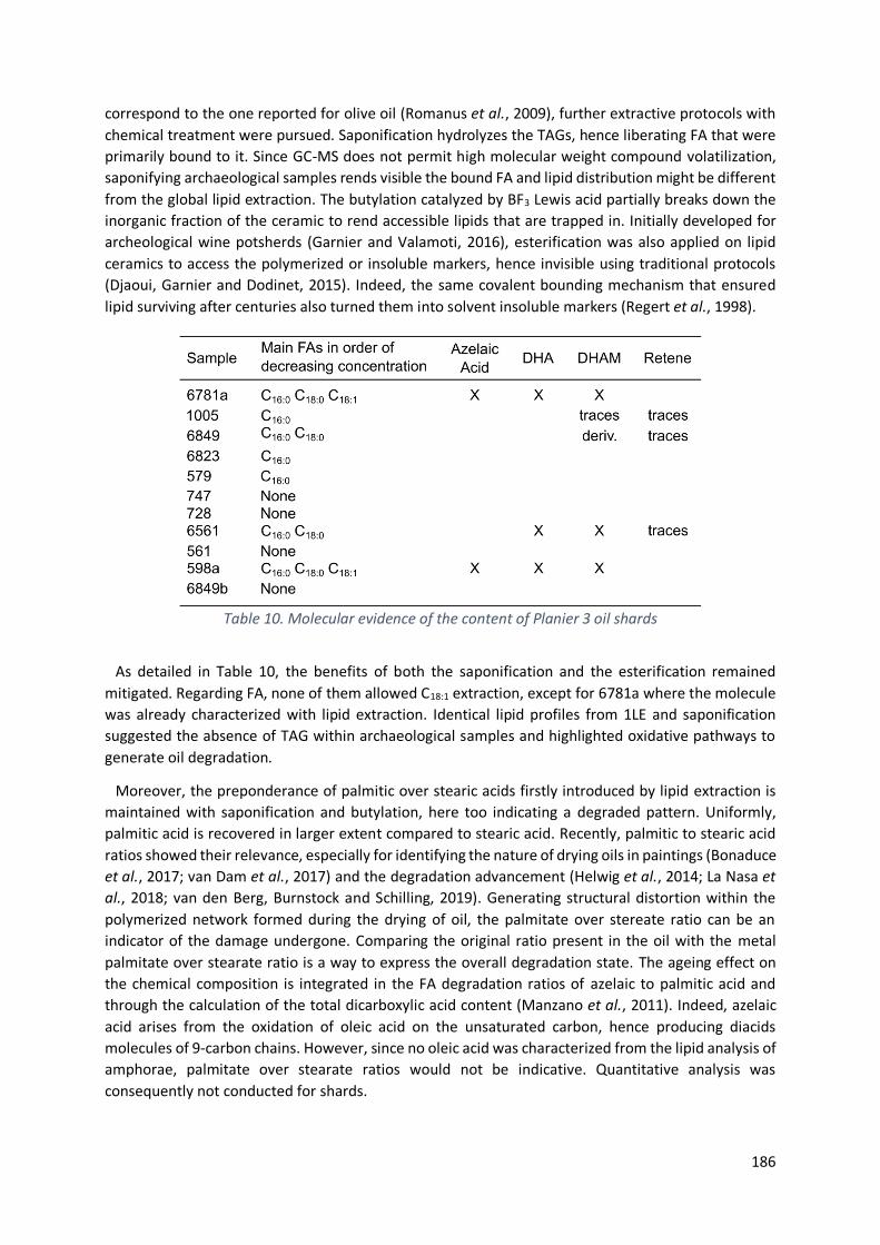
186
correspond to the one reported for olive oil (Romanus et al., 2009), further extractive protocols with
chemical treatment were pursued. Saponification hydrolyzes the TAGs, hence liberating FA that were
primarily bound to it. Since GC-MS does not permit high molecular weight compound volatilization,
saponifying archaeological samples rends visible the bound FA and lipid distribution might be different
from the global lipid extraction. The butylation catalyzed by BF3 Lewis acid partially breaks down the
inorganic fraction of the ceramic to rend accessible lipids that are trapped in. Initially developed for
archeological wine potsherds (Garnier and Valamoti, 2016), esterification was also applied on lipid
ceramics to access the polymerized or insoluble markers, hence invisible using traditional protocols
(Djaoui, Garnier and Dodinet, 2015). Indeed, the same covalent bounding mechanism that ensured
lipid surviving after centuries also turned them into solvent insoluble markers (Regert et al., 1998).
Table 10. Molecular evidence of the content of Planier 3 oil shards
As detailed in Table 10, the benefits of both the saponification and the esterification remained
mitigated. Regarding FA, none of them allowed C18:1 extraction, except for 6781a where the molecule
was already characterized with lipid extraction. Identical lipid profiles from 1LE and saponification
suggested the absence of TAG within archaeological samples and highlighted oxidative pathways to
generate oil degradation.
Moreover, the preponderance of palmitic over stearic acids firstly introduced by lipid extraction is
maintained with saponification and butylation, here too indicating a degraded pattern. Uniformly,
palmitic acid is recovered in larger extent compared to stearic acid. Recently, palmitic to stearic acid
ratios showed their relevance, especially for identifying the nature of drying oils in paintings (Bonaduce
et al., 2017; van Dam et al., 2017) and the degradation advancement (Helwig et al., 2014; La Nasa et
al., 2018; van den Berg, Burnstock and Schilling, 2019). Generating structural distortion within the
polymerized network formed during the drying of oil, the palmitate over stereate ratio can be an
indicator of the damage undergone. Comparing the original ratio present in the oil with the metal
palmitate over stearate ratio is a way to express the overall degradation state. The ageing effect on
the chemical composition is integrated in the FA degradation ratios of azelaic to palmitic acid and
through the calculation of the total dicarboxylic acid content (Manzano et al., 2011). Indeed, azelaic
acid arises from the oxidation of oleic acid on the unsaturated carbon, hence producing diacids
molecules of 9-carbon chains. However, since no oleic acid was characterized from the lipid analysis of
amphorae, palmitate over stearate ratios would not be indicative. Quantitative analysis was
consequently not conducted for shards.

187
Although no characteristic lipid profile can be extracted, some words can nonetheless be said on their
comparative benefits. The butylation of the samples notably promoted the extraction of C16:0, C18:0 for
5 samples (728; 561; 6561; 6823 and 1005) and azelaic acid only for 1005 compared to lipid extraction
(see the appendix 2 for the molecular relative quantity extracted). Saponification supported the
extraction of stearic and azelaic acids, primarily absent. The extraction of palmitic acid is more
quantitative with butylation than with traditional lipid extraction for the amphorae 6823 and 1005
while it did not provide supplementary information for the samples 6849; 598a and 6781a. Finally,
butylating allowed C16:0, C18:0 and azelaic acid to be either newly or more quantitatively extracted for
6781a; 598a and 6849 whereas it did not provide any difference for 1005 and 6823. Important is also
to notice that none of the protocols highlighted the presence of sterols such as β-sitosterol or
stigmasterol, despite its great distribution in plants and its frequent recovery in ceramics (Kimpe et al.,
2004; Pecci et al., 2018; Reber, 2021; Whelton et al., 2021).
Presented in Fig. 20, various molecules can be generated from FA degradation. Specific care was
hence taken for FA and degraded derivatives identification, sinking specific mass fragments.
Independently from the protocol applied, no straight trend could be drawn. No extractive protocol was
consequently favored to provide valuable insight on the content. The absence of unequivocal FA
distribution pointed again the specific character of archaeological amphorae, underlining the
individual care the characterization required. Degraded markers were carefully targeted since they
helped in diagnosing either the initial good or the underwent conditions. Markers of auto-oxidation,
namely pimelic, azelaic, sebacic and/or undecanedioic acids provided insights on heat and light
exposure as marker of natural degradation (Den Dooren De Jong, Dauvillier and Roman, 1961; Frankel
et al., 1977; Frankel, 2010; Pecci et al., 2018). Many evidence of short chains degraded products were
also linked to the use of archaeological ceramics (Regert et al., 1998; Drieu, 2017; Blanco-Zubiaguirre
et al., 2018). Although the entire reactional mechanisms are still unclear, the respective α, ω-
dicarboxylic acids distribution mirrors the oxidative pathway undergone. From unsaturated FA
precursors, direct cleavage of the double bond, hydration or oxidation followed by oxidative cleavage
as well as a successive oxidation, hydration and bond cleavage were reported by Regert et al. (Regert
et al., 1998). Then, dihydroxy-FA recovered from archaeological artefacts traduced degradation
occurring via dihydroxylation. Relative products are dominated by 8-carbons chain patterns (Regert et
al., 1998). Despite the degraded aspect revealed by the analysis of Planier 3 shards, no dihydroxy-FA
was identified by any protocol.
Operating in aerobic conditions, ß-oxidation could address the over-representation of C16:0 over the
other FA, indicating a stepwise degradation in living organisms (Aillaud, 2001). Similar FA profile with
palmitic dominance were reported in glass bottle dated back to the 79 A.D. Vesuvius eruption. Stearic
and oleic acids were identified in equivalent amount, quantitatively estimated to a quarter of the
palmitic acid. Such results highlighted the degraded effects on olive oil lipids over the last 20 centuries
(Sacchi et al., 2020). Although degradation markers could provide valuable insights on the initial
content following the biomolecular concept, it unfortunately remained complicated to trace back the
content based on FA distribution. The chemical composition is evolving with molecules alteration.
Many oxidative degradation pathways exist. Forecasting alteration would be insufficient to understand
FA lifecycle (Heron and Evershed, 1993; Drieu, 2017). Moreover, the loss of material over time is
inevitable, even without apparent leaching by groundwater percolation or microorganisms presence
(Reber and Evershed, 2004; Evershed, 2008; Spiteri, Heron and Craig, 2011).
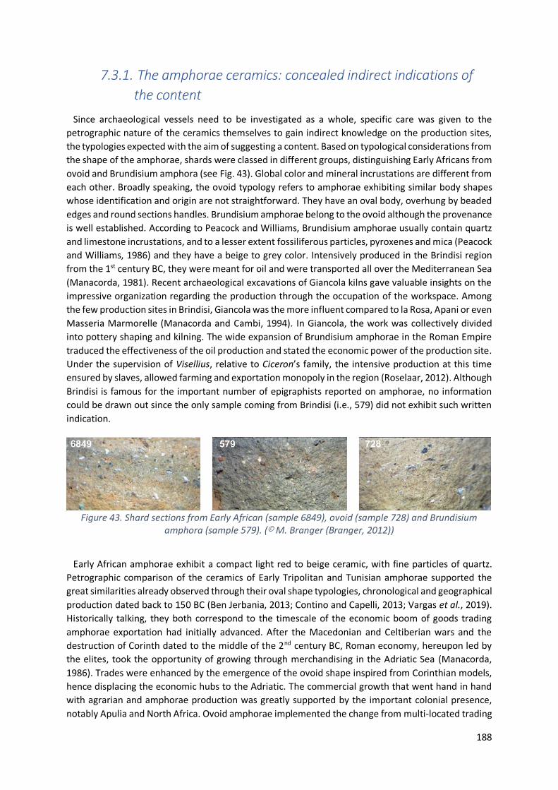
188
7.3.1. The amphorae ceramics: concealed indirect indications of
the content
Since archaeological vessels need to be investigated as a whole, specific care was given to the
petrographic nature of the ceramics themselves to gain indirect knowledge on the production sites,
the typologies expected with the aim of suggesting a content. Based on typological considerations from
the shape of the amphorae, shards were classed in different groups, distinguishing Early Africans from
ovoid and Brundisium amphora (see Fig. 43). Global color and mineral incrustations are different from
each other. Broadly speaking, the ovoid typology refers to amphorae exhibiting similar body shapes
whose identification and origin are not straightforward. They have an oval body, overhung by beaded
edges and round sections handles. Brundisium amphorae belong to the ovoid although the provenance
is well established. According to Peacock and Williams, Brundisium amphorae usually contain quartz
and limestone incrustations, and to a lesser extent fossiliferous particles, pyroxenes and mica (Peacock
and Williams, 1986) and they have a beige to grey color. Intensively produced in the Brindisi region
from the 1st century BC, they were meant for oil and were transported all over the Mediterranean Sea
(Manacorda, 1981). Recent archaeological excavations of Giancola kilns gave valuable insights on the
impressive organization regarding the production through the occupation of the workspace. Among
the few production sites in Brindisi, Giancola was the more influent compared to la Rosa, Apani or even
Masseria Marmorelle (Manacorda and Cambi, 1994). In Giancola, the work was collectively divided
into pottery shaping and kilning. The wide expansion of Brundisium amphorae in the Roman Empire
traduced the effectiveness of the oil production and stated the economic power of the production site.
Under the supervision of Visellius, relative to Ciceron’s family, the intensive production at this time
ensured by slaves, allowed farming and exportation monopoly in the region (Roselaar, 2012). Although
Brindisi is famous for the important number of epigraphists reported on amphorae, no information
could be drawn out since the only sample coming from Brindisi (i.e., 579) did not exhibit such written
indication.
Figure 43. Shard sections from Early African (sample 6849), ovoid (sample 728) and Brundisium amphora (sample 579). (© M. Branger (Branger, 2012))
Early African amphorae exhibit a compact light red to beige ceramic, with fine particles of quartz.
Petrographic comparison of the ceramics of Early Tripolitan and Tunisian amphorae supported the
great similarities already observed through their oval shape typologies, chronological and geographical
production dated back to 150 BC (Ben Jerbania, 2013; Contino and Capelli, 2013; Vargas et al., 2019).
Historically talking, they both correspond to the timescale of the economic boom of goods trading
amphorae exportation had initially advanced. After the Macedonian and Celtiberian wars and the
destruction of Corinth dated to the middle of the 2nd century BC, Roman economy, hereupon led by
the elites, took the opportunity of growing through merchandising in the Adriatic Sea (Manacorda,
1986). Trades were enhanced by the emergence of the ovoid shape inspired from Corinthian models,
hence displacing the economic hubs to the Adriatic. The commercial growth that went hand in hand
with agrarian and amphorae production was greatly supported by the important colonial presence,
notably Apulia and North Africa. Ovoid amphorae implemented the change from multi-located trading

189
all around the Mediterranean to a single center located in Rome (Vargas et al., 2019). After clustering
amphorae typologies together as Early African, Contino et al. concluded that part of the production
was surely located in the Northern Africa, in Tunisia. They considered the site of El Mnihla, Carthage
as the production site before the production expansion to the African coast (Contino and Capelli,
2013). Indeed, recent excavations of a ceramic dump at El Mnihla near Tunis shed light on the probable
production site within the Carthage-Tunis-Utica triangle (Ben Jerbania, 2013). Little is nonetheless
known from archaeometric analysis, epigraphy information and kiln production site (Bonifay, 2007)
and too much interest was devoted to olive oil trade (Bonifay, 2004). Highlighted by the discovery of
olive fruits from the Plemmirio B wreck in Sicily, Bonifay et al. argued the possibility for African
amphorae to have contained fruits instead of oil (Bonifay et al., 2015). Indeed, Pliny’s testimony9
targeted the higher quality of Italian olives to produce oil compared to overseas fruits, that are
preferred for the table as staple food. Facing the absence of olive oil profile in Ostia LIX, Djaoui et al.
hypothesized the olive preservation in brine. Indeed, olive as a fruit would not leak on the internal
surface and not leave chemical traces since FA markers are contained in the flesh (Djaoui, Garnier and
Dodinet, 2015). They assumed the amphorae provenance from Thabraca, Tunisia, before the
redistribution in Rome and the recycling in Egypt with Moringa oil to explain the presence of Moringa
markers besides the absence of FA markers and the natural distribution of Moringa sp. out of Africa.
From the best of our knowledge, little was reported on olive trades except the recent publications
aforementioned. Regarding the Early African amphorae investigated (6781a; 6823; 1005 and 6849),
the hypothesis of a non-edible use remained nonetheless relevant. Indeed, since the oil produced in
Tunisia seemed to be of poor quality as pointed out by Pliny, it is not unlikely that it was used in other
ways, such as consumable for oil lamps. Several historic writers like Pliny10, Theophrastus11 or Strabo12
reported the contribution of olive oil for supplying lamp lighting combustion (Copley et al., 2005).
Considering the archaeological site of Pompeii, Griffiths provided a first estimation of the oil fuel
quantities necessary to supply artificial lights in domestic and commercial activities (Griffiths, 2019).
No less than 20 586 L of olive oil were necessary to ensure the lighting of the 1884 lamps. The discovery
of two ceramic lamp workshops within the city (Cerulli Irelli, 1977; Peña and McCallum, 2009)
additionally attested of the predominance of nocturnal activities, hitherto underestimated. Scaled-up
to the entireness of the Roman Empire, the impressive amount of fuel raises interrogations on the
supplying possibilities. Olive oil was consequently produced, with production area expanded all over
the Mediterranean basin. Olive farming became a major contribution of the Roman economy (Bowman
and Wilson, 2013; Marzano, 2013). Olive was hence integrated in daily life from the production to the
consumption (Mattingly, 1988a, 1988b).
Another underestimated aspect of oil consumption in the Roman Empire regards the body-caring
and cosmetic. Olive oil was indeed a ‘star’ ingredient in the preparation of hygiene, medicaments and
beauty products (Griffiths, 2019). Romans valuated olive oil for massages and skin care, referred as
‘Beauty baths’ (Gorini et al., 2018). Cosmetics were prepared on the base of olive oil, sometimes
perfumed for make-up usage (Pérez-Arantegui et al., 2009) or using with white lead pigments to cure
hair loss (Armocida, 1991; Rudock, 2019). Olive oil was considered as a ‘high moisturizing medicine’
for skin treatment (Sarkar et al., 2017).
Porosity investigations gave reliable insights on the ceramic absorption towards lipids. Experiments
conducted on ceramic replica demonstrated the pore-size effect on the preservation of lipids and the
9 Pliny the Elder, Naturalis Historia, XV, 16 “Quam ob causam Italicis transmarinae praeferuntur in cibis, cum
oleo uincantur” 10 Pliny the Elder, Naturalis Historia, XV, IV, 5 11 Enquiry into Plants IV: 2, 9 12 Geography VII, 1, 35

190
impact of the operational-chain on the lipid distribution (Rosiak, Kałużna-Czaplińska and Gątarek,
2020). Ceramic pores act as trapping holes for molecules. Lipid absorption ensures their survival over
archaeological time, as well as it diminishes the probability of leaching and degrading because of
microorganisms and/or oxidation. Heating, even at low temperatures increased the porosity by
altering the stone microstructure. Lipids are consequently better absorbed. So is the chance of profiling
lipids to trace back the original content with thermally induced porosity (Charters et al., 1993).
Moreover, lipid accumulation differs within the vessel itself, conducting higher absorption in the upper
body and the rims rather than in the base (Charters et al., 1993; Evershed, 2008). Finally, the lipid
concentration also differs from a ceramic to another, basically because of the porosity absorption, but
also because of the dependence of lipid preservation towards the porosity (Namdar, Stacey and
Simpson, 2009; Drieu et al., 2019). Micropores strengthen lipid concentration because lipids are
physically preserved from biological and chemical attacks such as hydrolysis, oxidative reaction or
radical oxidation (Dudd, Regert and Evershed, 1998; Evershed, 2008). Although lipid residue analysis is
widely reported in ceramics (Heron, Evershed and Goad, 1991; Dudd, Regert and Evershed, 1998;
Namdar, Stacey and Simpson, 2009; Allegretta et al., 2017; Drieu et al., 2019), and even though
mineralogy and petrography allowed the ceramic to be geographically located (Medeghini et al., 2016;
Medeghini and Nigro, 2017; Botticelli et al., 2020), little has been studied on the dependency of lipid
absorption towards the different kinds of ceramics, starting from petrography analyses.
7.3.2. Archaeological interpretations from enigmatic chemical
markers
Another hypothesis arised from the re-use of amphorae. As raised by Pecci, this aspect is effectively
rarely considered in the ceramic life-cycle (Pecci et al., 2017). Little are the evidence of reusing
containers. Peña assumed the recycling as storage containers, to pack food or non-food substances or
even for water storing (Peña, 2007). In the Northern necropolis of Potentia (Italy), different purposes
were mentioned for amphorae second lives, such as burial goods, funerary libations or even grave
markers (Monsieur, 2007). Cases of Dressel 2-4 reuses were also evidenced at Villa B Oplontis in
Southern Italy since residue analysis of the empty amphorae unraveled wine markers. Containers were
recovered in upside down positions, ready to be filled again before the Vesuvius eruption occurred
(Pecci et al., 2017). Secondly, olive oil lipid profile was identified together with grape beverage
markers, hence revealing the second use of wine amphorae for plant oil (Pecci, Salvini and Cantini,
2010). It would be unlikely to fill oil container with wine due to the bad taste oil would confer to the
ceramic. In the same way the ceramic absorbs wine, the wine would absorb oil residues trapped in the
ceramic (Pecci, 2009; Romanus et al., 2009). Organic residue analysis, although being a powerful
reliable tool, seems to show its limits since markers are identified all together and cannot be
chronologically distinguished from each other. Tracing the original content can present some issues if
the container was reused or the content mixed with some other goods. Re-use occurrence is
consequently difficult to address. One striking example, mentioned earlier, needs however to be
noticed since they interpreted the absence of chemical markers in Ostia LIX as a matter of fact (Djaoui,
Garnier and Dodinet, 2015). Surprisingly, wine markers were characterized, together with FA, diacids,
cholesterol, vegetal sterols and long-chain alcohols. Since the transport of olive fruits in brine would
not leave chemical markers on the ceramic, the extractive outcomes were conjectured as a sign of the
second life of the amphorae. Containers were reused after olive fruits were traded from North Africa
with red wine, dairy products, plant oil and/or Moringa sp. oil. Although spatio-temporality may face
some uncertainties because residue analysis did not provide straightforward outcomes easing the
191
interpretation, they attempted to take advantage of the ambivalence, filling the knowledge gap and
providing innovative suggestions to understand trade at a global level.
7.4. Conclusion
In conclusion, little was described in literature about the oxidized profile lipid residue analysis may
suffer while analyzing oil amphorae. The situation we are facing in Planier 3 is all the more interesting
since it opens a field of interpretation. First of all, no protocols used in this study seemed to give
valuable outcomes. Even though solvent extraction, butylation and saponification have different
reaction mechanisms and chemical approaches for lipid extraction, none of them provided relevant
insight to straightforwardly understand the object history. A universal protocol does not exist. Results
showed an important heterogeneity regarding the molecules extracted. Palmitic acid usually
dominates the lipids distribution, followed by stearic acid. The frequent absence of oleic acid, together
with the presence of degradation markers of azelaic acid, indicated oxidative reactions. From the
degraded pattern, different hypotheses can be raised. The fatty content could be meant for body
caring or lamp illuminant, instead of edible purpose. The early African amphorae might have contained
olive fruits, since North Africa’s olives were preferred to be consumed at table compared to Italian
fruits that gave tastier oil. Finally, the hypothesis of reusing amphorae cannot be discarded although
organic analysis of the residues did not provide any further indication on the initial content.
Funding
This research was financially supported by Marie Skłodowska-Curie Innovative Training Network, The
ED-ARCHMAT European Joint Doctorate, H2020-MSCA-ITN-EJD ED-ARCHMAT Joint Doctorate (Project
ESR9, grant agreement no 766311).
Author contributions
Conceptualization, L.C., F.O., C.M.; methodology, L.C., C.M.; formal analysis, L.C.; investigation, L.C.,
C.M.; resources, F.O.; visualization, L.C.; supervision, C.M.; project administration, C.M.; funding
acquisition, C.V., C.M.; writing—original draft preparation, L.C.

192
7.5. References Aillaud, S. (2001) Field and laboratory studies of diagenetic reactions affecting lipid residues absorbed in unglazed archaeological pottery vessels. University of Bristol.
Allegretta, I. et al. (2017) ‘The effect of mineralogy, microstructure and firing temperature on the effective thermal conductivity of traditional hot processing ceramics’, Applied Clay Science, 135, pp. 260–270. doi: 10.1016/j.clay.2016.10.001.
Allevato, E. et al. (2017) ‘Wood exploitation and food supply at the border of the Roman Empire: the case of the vicus of Thamusida – Sidi Ali ben Ahmed (Morocco)’, Environmental Archaeology, 22(2), pp. 200–217. doi: 10.1080/14614103.2015.1126685.
Armocida, G. (1991) ‘Le ricette di interesse medico’, in Tosatti, B. (ed.) Manoscritto Veneziano. Milano: Carma Acanthus, p. 37‐43.
van Den Berg, K. J. et al. (2000) ‘Mass spectrometric methodology for the analysis of highly oxidized diterpenoid acids in Old Master paintings’, Journal of Mass Spectrometry, 35(4), pp. 512–533. doi: 10.1002/(SICI)1096-9888(200004)35:4<512::AID-JMS963>3.0.CO;2-3.
van den Berg, K. J., Burnstock, A. and Schilling, M. (2019) ‘Notes on Metal Soap Extenders in Modern Oil Paints: History, Use, Degradation, and Analysis’, in Metal Soaps in Art. Springer, Cham, pp. 329–342. doi: 10.1007/978-3-319-90617-1_19.
Blanco-Zubiaguirre, L. et al. (2018) ‘GC–MS and HPLC-ESI-QToF characterization of organic lipid residues from ceramic vessels used by Basque whalers from 16th to 17th centuries’, Microchemical Journal, 137, pp. 190–203. doi: 10.1016/j.microc.2017.10.017.
Bonaduce, I. et al. (2017) ‘Analytical approaches based on gas chromatography mass spectrometry (GC/MS) to study organic materials in artworks and archaeological objects’, Analytical Chemistry for Cultural Heritage, pp. 291–327. doi: 10.1007/s41061-015-0007-x.
Bonifay, M. (2004) ‘La céramique africaine, un indice du développement économique ?’, Antiquité Tardive, 11, pp. 113–128. doi: 10.1484/J.AT.2.300253.
Bonifay, M. (2007) ‘Que transportaient donc les amphores africaines ?’, in Supplying Rome and the Empire, pp. 10–11, 18–23, 25–29.
Bonifay, M. et al. (2015) ‘Nouvelles hypothèses sur l’ origine et le contenu des amphores africaines Ostia LIX et XXIII’, Antiquités africaines. CNRS Editi, 51.
Botticelli, M. et al. (2020) ‘Variability in pottery production at Khalet al-Jam’a necropolis, Bethlehem (West Bank): From the Early-Middle Bronze to the Iron Age’, Ceramics International, 46(10), pp. 16405–16415. doi: 10.1016/J.CERAMINT.2020.03.200.
Bowman, A. and Wilson, A. (2013) The Roman Agricultural Economy: Organization, Investment and Production. Oxford: Oxford University Press.
Branger, M. (2012) Les amphores de l’épave 3 de Planier Typologie et épigraphie. Université de Provence (Aix-Marseille 1).
Carrer, F. et al. (2016) ‘Chemical Analysis of Pottery Demonstrates Prehistoric Origin for High-Altitude Alpine Dairying’, PLOS ONE, 11(4), p. e0151442. doi: 10.1371/JOURNAL.PONE.0151442.
Cerulli Irelli, G. (1977) ‘Una officina di lucerne fittili a Pompeii’, in Carandini, A. (ed.) L’instrumentum domesticum di Ercolano e Pompei nella prima eta imperiale, Quaderni di cultura materiale. Rome: L’Erma di Bretschneider, pp. 53–72.
Charters, S. et al. (1993) ‘Quantification and distribution of lipid in archaeological ceramics:

193
implications for sampling potcherds for organic residue analysis and the classification of vessel use’, Archaeometry, 35(2), pp. 211–223. doi: 10.1111/j.1475-4754.1993.tb01036.x.
Colombini, M. P., Modugno, F. and Ribechini, E. (2005) ‘Organic mass spectrometry in archaeology: evidence for Brassicaceae seed oil in Egyptian ceramic lamps’, Journal of Mass Spectrometry, 40(7), pp. 890–898. doi: 10.1002/jms.865.
Connan, J. and Nissenbaum, A. (2003) ‘Conifer tar on the keel and hull planking of the Ma’agan Mikhael Ship (Israel, 5th century BC): identification and comparison with natural products and artefacts employed in boat construction’, Journal of Archaeological Science, 30(6), pp. 709–719. doi: 10.1016/S0305-4403(02)00243-1.
Contino, A. and Capelli, C. (2013) ‘Amphores tripolitaines anciennes ou amphores africaines anciennes ?’, Antiquités africaines, 49(1), pp. 199–208. doi: 10.3406/antaf.2013.1550.
Copley, M. S. et al. (2003) ‘Direct chemical evidence for widespread dairying in prehistoric Britain’, Proceedings of the National Academy of Sciences, 100(4), pp. 1524–1529. doi: 10.1073/pnas.0335955100.
Copley, M. S. et al. (2005) ‘Gas chromatographic, mass spectrometric and stable carbon isotopic investigations of organic residues of plant oils and animal fats employed as illuminants in archaeological lamps from Egypt’, Analyst, 130(6), pp. 860–871. doi: 10.1039/b500403a.
Correa-Ascencio, M. and Evershed, R. P. (2014) ‘High throughput screening of organic residues in archaeological potsherds using direct acidified methanol extraction’, Analytical Methods, 6(5), p. 1330. doi: 10.1039/c3ay41678j.
Craig, O. E. et al. (2004) ‘Stable carbon isotopic characterisation of free and bound lipid constituents of archaeological ceramic vessels released by solvent extraction, alkaline hydrolysis and catalytic hydropyrolysis’, Journal of Analytical and Applied Pyrolysis, 71(2), pp. 613–634. doi: 10.1016/j.jaap.2003.09.001.
van Dam, E. P. et al. (2017) ‘Analysis of triglyceride degradation products in drying oils and oil paints using LC–ESI-MS’, International Journal of Mass Spectrometry, 413, pp. 33–42. doi: 10.1016/J.IJMS.2016.09.004.
Djaoui, D., Garnier, N. and Dodinet, E. (2015) ‘L’huile de ben identifiée dans quatre amphores africaines de type Ostia LIX provenant d’Arles : difficultés d’interprétation’, Antiquités africaines, 51(1), pp. 179–187. doi: 10.3406/antaf.2015.1580.
Den Dooren De Jong, I. E., Dauvillier, M. M. S. and Roman, W. B. (1961) ‘On the formation of adipocere from fats’, Antonie van Leeuwenhoek, 27(1), pp. 337–361. doi: 10.1007/BF02538464.
Drieu, L. (2017) Fabrication et usages des poteries durant le Néolithique et la Protohistoire en Europe : les apports de l’archéologie biomoléculaire. Université Côte d’Azur (ComUE).
Drieu, L. et al. (2019) ‘Influence of porosity on lipid preservation in the wall of archaeological pottery’, Archaeometry, 61(5), pp. 1081–1096. doi: 10.1111/arcm.12479.
Dudd, S. N., Regert, M. and Evershed, R. P. (1998) ‘Assessing microbial lipid contributions during laboratory degradations of fats and oils and pure triacylglycerols absorbed in ceramic potsherds’, Organic Geochemistry, 29(5–7), pp. 1345–1354. doi: 10.1016/S0146-6380(98)00093-X.
Dunne, J. et al. (2019) ‘Milk of ruminants in ceramic baby bottles from prehistoric child graves’, Nature, 574(7777), pp. 246–248. doi: 10.1038/s41586-019-1572-x.
Evershed, R. P. (2008) ‘Organic residue analysis in archaeology : the archaeological biomarker revolution’, Archaeometry, 50(6), pp. 895–924. doi: 10.1111/j.1475-4754.2008.00446.x.

194
Evershed, R. P. (2009) ‘Compound-specific stable isotopes in organic residue analysis in archaeology’, in Colombini, M. and Modugno, F. (eds) Organic mass spectrometry in art and archaeology. Wiley. New York, pp. 389–432.
Frankel, E. N. et al. (1977) ‘Analysis of autoxidized fats by gas chromatography-mass spectrometry: II. Methyl linoleate’, Lipids, 12(11), pp. 908–913. doi: 10.1007/BF02533310.
Frankel, E. N. (2010) ‘Chemistry of extra virgin olive oil: Adulteration, oxidative stability, and antioxidants’, Journal of Agricultural and Food Chemistry, 58(10), pp. 5991–6006. doi: 10.1021/jf1007677.
Garnier, N. et al. (2009) ‘Analysis of archaeological triacylglycerols by high resolution nanoESI, FT-ICR MS and IRMPD MS/MS: Application to 5th century BC-4th century AD oil lamps from Olbia (Ukraine)’, International Journal of Mass Spectrometry, 284(1–3), pp. 47–56. doi: 10.1016/J.IJMS.2009.03.003.
Garnier, N., Silvino, T. and Bernal Casasola, D. (2011) ‘L’identification du contenu des amphores: huile, conserves de poissons et poissage’, in Actes du congrès d’Arles, 2-5 juin. Marseille, Société française d’étude de la céramique antique en Gaule.
Garnier, N. and Valamoti, S. M. (2016) ‘Prehistoric wine-making at Dikili Tash (Northern Greece): Integrating residue analysis and archaeobotany’, Journal of Archaeological Science, 74, pp. 195–206. doi: 10.1016/j.jas.2016.03.003.
Gorini, I. et al. (2018) ‘Olive oil in pharmacological and cosmetic traditions’, Journal of cosmetic dermatology, 18(5), pp. 1575–1579. doi: 10.1111/jocd.12838.
Griffiths, D. (2019) ‘Commercialization of the night at Pompeii’, in Veal, R. and Leitch, V. (eds) Fuel and Fire in the Ancient Roman World Towards an integrated economic understanding. McDonald Institute for Archaeological Research. doi: 10.17863/CAM.46322.
Helwig, K. et al. (2014) ‘Conservation Issues in Several Twentieth-Century Canadian Oil Paintings: The Role of Zinc Carboxylate Reaction Products’, Issues in Contemporary Oil Paint, pp. 167–184. doi: 10.1007/978-3-319-10100-2_11.
Heron, C. and Evershed, R. P. (1993) ‘The analysis of organic residues and the study of pottery use’, Archaeological Method and Theory, 5, pp. 247–284. doi: 10.2307/20170233.
Heron, C., Evershed, R. P. and Goad, L. J. (1991) ‘Effects of migration of soil lipids on organic residues associated with buried potsherds’, Journal of Archaeological Science, 18(6), pp. 641–659. doi: 10.1016/0305-4403(91)90027-M.
Ben Jerbania, I. (2013) ‘Observations sur les amphores de tradition punique d’après une nouvelle découverte près de Tunis’, Antiquités africaines, 49(1), pp. 179–192. doi: 10.3406/antaf.2013.1548.
Kimpe, K. et al. (2004) ‘Assessing the relationship between form and use of different kinds of pottery from the archaeological site Sagalassos (southwest Turkey) with lipid analysis’, Journal of Archaeological Science, 31(11), pp. 1503–1510. doi: 10.1016/j.jas.2004.03.012.
Manacorda, D. (1981) ‘Produzione agricola, produzione ceramica e proprietari nell’Ager Cosanus nel I aC’, in Schiavone, A. and Giardina, E. (eds) Società romana e produzione schiavistica, II, Merci, mercati e scambi nel Mediterraneo. Rome - Bari: Editori Laterza, pp. 11–13.
Manacorda, D. (1986) ‘Per uno studio dei centri produttori delle anfore brindisine, in La Puglia in età repubblicana’, in Atti del I Convegno di Studi sulla Puglia romana. Mesagne: Galatina, pp. 91–117.
Manacorda, D. and Cambi, F. (1994) ‘Recherches sur l’ager Brundisinus à l’époque romaine’, Collection de l’Institut des Sciences et Techniques de l’Antiquité, 508(1), pp. 283–292.

195
Manhita, A. et al. (2020) ‘Transporting olive oil in Roman Times: Chromatographic analysis of Dressel 20 Amphorae from Pax Julia Civitas, Lusitania’, Chromatographia, 83(9), pp. 1055–1064. doi: 10.1007/s10337-020-03927-7.
Manzano, E. et al. (2011) ‘Study of the GC–MS determination of the palmitic–stearic acid ratio for the characterisation of drying oil in painting: La Encarnación by Alonso Cano as a case study’, Talanta, 84(4), pp. 1148–1154. doi: 10.1016/j.talanta.2011.03.012.
Manzano, E. et al. (2016) ‘A multi-analytical approach applied to the archaeological residues in Iberian glasses. Earliest evidences on the consumption of fermented beverages in votive rituals’, Microchemical Journal, 129, pp. 286–292. doi: 10.1016/j.microc.2016.07.006.
Marzano, A. (2013) Agricultural Production in the Hinterland of Rome: Wine and Olive Oil. Oxford: Oxford University Press.
Matlova, V. et al. (2017) ‘Defining pottery use and animal management at the Neolithic site of Bylany (Czech Republic)’, Journal of Archaeological Science: Reports, 14, pp. 262–274. doi: 10.1016/j.jasrep.2017.05.028.
Mattingly, D. J. (1988a) ‘Oil for export? A comparison of Libyan, Spanish and Tunisian olive oil production in the Roman empire’, Journal of Roman Archaeology, 1, pp. 33–56.
Mattingly, D. J. (1988b) ‘Olea mediterranea?’, Journal of Roman Archaeology, 1, pp. 153–161.
Medeghini, L. et al. (2016) ‘New insights on Early Bronze Age IV pottery production and consumption in the southern Levant: The case of Khirbat Iskandar, Jordan’, Ceramics International, 42(16), pp. 18991–19005. doi: 10.1016/J.CERAMINT.2016.09.054.
Medeghini, L. and Nigro, L. (2017) ‘Khirbet al-Batrawy ceramics: a systematic mineralogical and petrographic study for investigating the material culture’, Periodico di Mineralogia, (86), pp. 19–35. doi: 10.2451/2016PM665.
Mezzatesta, E. et al. (2021) ‘Analysis of balms taken from Egyptian human mummies using solid‐phase extraction and gas chromatography–mass spectrometry’, Journal of Separation Science, 44(4), pp. 850–859. doi: 10.1002/jssc.202000746.
Mills, J. S. and White, R. (1989) ‘The identity of the resins from the Late Bronze Age shipwreck at Ulu Burun (Kas)’, Archaeometry, 31(1), pp. 37–44.
Monsieur, P. (2007) ‘Amphora Burials and Burials with Amphorae: On the Reuse of Amphorae in the Northern Necropolis of Potentia (Porto Recanati, Marche)’, Theoretical Roman Archaeology Journal, 0(2006), p. 133. doi: 10.16995/TRAC2006_133_149.
Mottram, H. R. et al. (1999) ‘New chromatographic, mass spectrometric and stable isotope approaches to the classification of degraded animal fats preserved in archaeological pottery’, Journal of Chromatography A, 833(2), pp. 209–221. doi: 10.1016/S0021-9673(98)01041-3.
Mottram, H. R. and Evershed, R. P. (2003) ‘Practical considerations in the gas chromatography / combustion / isotope ratio monitoring mass spectrometry of 13C-enriched compounds: Detection limits and carryover effects’, Rapid Communications in Mass Spectrometry, 17(23), pp. 2669–2674. doi: 10.1002/rcm.1230.
Namdar, D., Stacey, R. J. and Simpson, S. J. (2009) ‘First results on thermally induced porosity in chlorite cooking vessels from Merv (Turkmenistan) and implications for the formation and preservation of archaeological lipid residues’, Journal of Archaeological Science, 36(11), pp. 2507–2516. doi: 10.1016/j.jas.2009.07.003.
La Nasa, J. et al. (2018) ‘Development of a GC/MS method for the qualitative and quantitative analysis

196
of mixtures of free fatty acids and metal soaps in paint samples’, Analytica Chimica Acta, 1001, pp. 51–58. doi: 10.1016/J.ACA.2017.11.017.
Peacock, D. P. and Williams, D. F. (1986) Amphorae and the Roman Economy: An Introductory Guide. London and New York: Longman.
Pecci, A. (2009) ‘Analisi funzionale della ceramica e alimentazione medievale’, Analisi funzionale della ceramica e alimentazione medievale, 36, pp. 21–42. doi: 10.1400/183086.
Pecci, A. et al. (2017) ‘Use and reuse of amphorae. Wine residues in Dressel 2–4 amphorae from Oplontis Villa B (Torre Annunziata, Italy)’, Journal of Archaeological Science: Reports, 12, pp. 515–521. doi: 10.1016/j.jasrep.2017.02.025.
Pecci, A. et al. (2018) ‘Combining residue analysis of floors and ceramics for the study of activity areas at the Garum Shop at Pompeii’, Archaeological and Anthropological Sciences, 10(2), pp. 485–502. doi: 10.1007/s12520-017-0573-7.
Pecci, A. et al. (2021) ‘Production and Transport of Goods in the Roman Period: Residue Analysis and Wine Derivatives in Late Republican Baetican Ovoid Amphorae’, Environmental Archaeology, pp. 1–13. doi: 10.1080/14614103.2020.1867291.
Pecci, A., Cau Ontiveros, M. Á. and Garnier, N. (2013) ‘Identifying wine and oil production: analysis of residues from Roman and Late Antique plastered vats’, Journal of Archaeological Science, 40(12), pp. 4491–4498. doi: 10.1016/J.JAS.2013.06.019.
Pecci, A., Salvini, F. and Cantini, L. (2010) ‘Residue analysis of some Late Roman amphora coming from the excavations of the historical center of Florence’, in Menchelli, S. et al. (eds) LRCW 3. 3rd International Conference on Late Roman Coarse Wares, Cooking Wares and Amphorae in the Mediterranean: Archaeology and Archaeometry. Oxford: Archaeopress BAR Series, pp. 363–367.
Peña, J. T. (2007) Roman Pottery in the Archaeological Record. Cambridge: Cambridge University Press. doi: 10.1017/CBO9780511499685.
Peña, J. T. and McCallum, M. (2009) ‘The production and distribution of pottery at Pompeii: A review of the evidence; part 1, production’, American Journal of Archaeology, 113(1), pp. 57–79.
Pérez-Arantegui, J. et al. (2009) ‘Colorants and oils in Roman make-ups – an eye witness account’, Trends in Analytical Chemistry, 28(8), pp. 1019–1028. doi: 10.1016/j.trac.2009.05.006.
Philp, R. P. and Oung, J.-N. (1988) ‘Biomarkers: Occurence, utility and detection’, Analytical Chemistry, 60(15), pp. 887A-896A. doi: 10.1021/ac00166a720.
Rageot, M. et al. (2019) ‘New insights into Early Celtic consumption practices: Organic residue analyses of local and imported pottery from Vix-Mont Lassois’, PLoS ONE, 14(6), p. e0218001. doi: 10.1371/journal.pone.0218001.
Reber, E. A. (2021) ‘Comparison of neutral compound extraction from archaeological residues in pottery using two methodologies: A preliminary study’, Separations, 8(1), pp. 1–17. doi: 10.3390/SEPARATIONS8010006.
Reber, E. A. and Evershed, R. P. (2004) ‘How Did Mississippians Prepare Maize? The Application of Compound-Specific Carbon Isotope Analysis to Absorbed Pottery Residues From Several Mississippi Valley Sites’, Archaeometry, 46(1), pp. 19–33. doi: 10.1111/j.1475-4754.2004.00141.x.
Regert, M. et al. (1998) ‘Free and bound fatty acid oxidation products in archaeological ceramic vessels’, Proceedings of the Royal Society of London. Series B: Biological Sciences, 265(1409), pp. 2027–2032. doi: 10.1098/rspb.1998.0536.

197
Regert, M. et al. (2003) ‘Structural characterisation of lipid constituents from natural substances preserved in archaeological environments’, Measurement Science & Technology, 14, pp. 1620–1630.
Roffet-Salque, M. et al. (2017) ‘From the inside out: Upscaling organic residue analyses of archaeological ceramics’, Journal of Archaeological Science: Reports, 16, pp. 627–640. doi: 10.1016/J.JASREP.2016.04.005.
Romanus, K. et al. (2008) ‘Brassicaceae seed oil identified as illuminant in Nilotic shells from a first millennium AD Coptic church in Bawit, Egypt’, Analytical and Bioanalytical Chemistry, 390(2), pp. 783–793. doi: 10.1007/s00216-007-1704-2.
Romanus, K. et al. (2009) ‘Wine and olive oil permeation in pitched and non-pitched ceramics: relation with results from archaeological amphorae from Sagalassos, Turkey’, Journal of Archaeological Science, 36(3), pp. 900–909. doi: 10.1016/j.jas.2008.11.024.
Roselaar, S. T. (2012) ‘The role of Italians in local economies of the late Roman Republic’, Revue belge de philologie et d’histoire, 90(1), pp. 93–110. doi: 10.3406/rbph.2012.8391.
Rosiak, A., Kałużna-Czaplińska, J. and Gątarek, P. (2020) ‘Analytical Interpretation of Organic Residues From Ceramics As a Source of Knowledge About Our Ancestors’, Critical Reviews in Analytical Chemistry, 50(3), pp. 189–195. doi: 10.1080/10408347.2019.1602821.
Rudock, A. (2019) ‘Comparative Cosmetic Costs: Ancient Roman and Modern American Self-Adornment’, Alpenglow: Binghamton University Undergraduate Journal of Research and Creative Activity, 5(1).
Sacchi, R. et al. (2020) ‘Olive oil from the 79 A.D. Vesuvius eruption stored at the Naples National Archaeological Museum (Italy)’, npj Science of Food, 4(1), pp. 1–9. doi: 10.1038/s41538-020-00077-w.
Sarkar, R. et al. (2017) ‘Use of vegetable oils in dermatology: an overview.’, International Journal of Dermatology, 56(11), p. 1080‐1086.
Spiteri, C. D., Heron, C. and Craig, 0. (2011) ‘Characterising Surviving residues from archaeological ceramics: a biomolecular approach’, Archaeological ceramics: a review of current research. BAR Intern, pp. 40–56.
Steele, V. J., Stern, B. and Stott, A. W. (2010) ‘Olive oil or lard ?: Distinguishing plant oils from animal fats in the archeological record of the eastern Mediterranean using gas chromatography / combustion / isotope ratio mass spectrometry’, Rapid Communications in Mass Spectrometry, 24(23), pp. 3478–3484. doi: 10.1002/rcm.4790.
Stern, B. et al. (2008) ‘New investigations into the Uluburun resin cargo’, Journal of Archaeological Science, 35(5237), pp. 2188–2203. doi: 10.1016/j.jas.2008.02.004.
Suryanarayan, A. et al. (2021) ‘Lipid residues in pottery from the Indus Civilisation in northwest India’, Journal of Archaeological Science, 125, p. 105291. doi: 10.1016/J.JAS.2020.105291.
Urem-Kotsou, D. et al. (2002) ‘Birch-bark tar at Neolithic Makriyalos, Greece’, Antiquity, 76(294), pp. 962–967.
Vargas, E. G. et al. (2019) ‘Ovoid Amphorae in the Mediterranean (2nd century BC–early 1st century AD).’, in The Ovoid Amphorae in the Central and Western Mediterranean. Archaeopress Publishing Ltd, pp. 403–413. doi: 10.2307/j.ctvpmw4m6.23.
Whelton, H. L. et al. (2021) ‘A call for caution in the analysis of lipids and other small biomolecules from archaeological contexts’, Journal of Archaeological Science, 132, p. 105397. doi: 10.1016/j.jas.2021.105397.

198
Woodworth, M. et al. (2015) ‘The content of African Keay 25 / Africana 3 Amphorae: Initial results of the CORONAM project’, in ArchaeoAnalytics - Chromatography and DNA Analysis in Archaeology, pp. 41–57.

199
Chapter 8
General discussion, conclusion and perspectives
200
8.1. Calling for thesis objectives
As a short reminder, the objective of this PhD thesis regarded the development of innovative
analytical tools to address the archaeological content of amphorae. The collaboration with
archaeologists, scientists and cultural offices aimed at providing a common language to better
understand the object history with the specific purpose of including the assessments into broader
anthropological, ethnocultural and societal considerations. To that end, we needed to overcome the
analytical barrier inherent in the objects: the artefacts having been immersed underwater or
underground for thousands of years, the search for traces, both macroscopic and microscopic, proved
difficult.
The main part of the thesis focused on grape-derivative containers. Grape-beverages were indeed
ubiquitous during the Roman Empire, in terms of production and consumption. Wine trading was so
important that Panella and Tchernia (Panella and Tchernia, 1994) estimated that half of the traded
amphorae would have contained wine in 50 BC, considering Italian and Roman provinces in the
selected context of Rome and Ostia. This proportion remarkably increased to 65% before the
generalized economic decline Roman trading suffered around 200 AC (Bowman and Wilson, 2009).
Produced in such extent, wine naturally became one of the pillars of the imperial economy and of the
Roman culture. Roman writers such as Columella, Virgil, Cicero, Cato the Elder or Horace abundantly
referred to grape in all its forms. Grape agriculture was notably detailed by Pliny the Elder who, among
others, reported the common grapevine cultivars13 as well as the different types of presses used for
winemaking14 (Rossiter, 2008). Wall paintings and decorative features attested of the cultural daily
custom extensiveness and its related socioeconomic impacts on the society (Fleming, 1997; Feige,
2021). Its use is also greatly detailed, from grape fruits consumption to numerous wine Mediterranean
recipes, including resinated, cooked and medicinal wine beverages (Hostetter, Beck and Stewart, 1994;
Garnier et al., 2003; Feier et al., 2019). Abundant is the archaeological evidence of ceramics having
contained grape derivatives considering uses and transport (Pecci, Cau Ontiveros and Garnier, 2013;
Pecci et al., 2018). For instance, fermented grape-beverages have been reported in several Roman
Dressel amphorae ranging from the 1st century BC to the 4th century AD (Condamin and Formenti,
1978; Pecci, Salvini and Cantini, 2010; Arobba et al., 2014; Pecci et al., 2017). Archaeological wine over-
representation nicely turned the research field of grape derivatives into a promising anthropological
approach to better understand past civilizations cultures and ancient Mediterranean trade routes.
Another field of investigation regarded oil amphorae. Oil was indeed one of the ‘star’ ingredients in
the Roman Empire (Finley, 1973; Hitchner, Amouretti and Brun, 2002). Among the many usages, oil
was widely mentioned for cooking, as lamp fuel for lighting, for lubrication, for perfuming or skin-
caring, for ritual or religious practices (Mattingly, 1988; Edmondson, 2015; Ayllon-Martin, Perez
Gonzalez and Remesal Rodriguez, 2019). Historical writers abundantly reported its contribution in daily
life practices, stating on its physical and culinary benefits, as for example in the renowned Apicius’
recipes book of De Re Coquinaria. Forbes and Foxhall (Foxhall and Forbes, 1982) estimated the Roman
yearly olive oil consumption around 50 L per person. Rough estimation of the consumption as light fuel
to supply domestic and nocturnal commercial activities of Pompeii exceeded 20000 L per year, which
corresponds to 275 Dressel 20 containers (Griffiths, 2019). To supply such important demand, the
production of olive oil was largely scaled in Southern Spain, Tunisia and Lybia (Mattingly, 1988).
13 Naturalis Historia III ; XIV 14 Naturalis Historia XVIII

201
8.2. Methodology development and protocol optimization
The diagnosis approach we established was twofold. We first concentrated on the development of
chemical protocols able to extract meaningful molecules from shard and waterproofing matrices. We
then focused on the archaeobotanical content of amphorae to support and flourish the initial
assumptions raised by molecular biomarkers.
8.2.1. Organic residue analysis: the improvements
Regarding the preponderance of wine amphorae in the sample corpus (i.e., 16 amphorae over the
27 artefacts), the extraction optimization was axed on fermented grape-beverages and wine markers.
Several protocols were tested on archaeological shards and organic linings with the aim of comparing
their relative extractive capacities. Protocols were chosen regarding their ubiquity in literature. They
exhibited different chemical mechanisms to access the markers bound to the original matrix. The
organic extraction with traditional solvents does not aim at reacting with the sample but rather
extracts molecules present therein. Ultrasound generates highly energetic waves that travel within the
extractive medium. The alternation of high- and low-pressure cycles creates acoustic cavitation, which
enhance the extraction of compounds when the cavitation bubbles implode at the solid surface. The
turbulences generated by ultrasound improve mass transfer from the matrix to the medium. Secondly,
alkaline treatment promotes the release of tartaric acid from the corresponding salts. The mechanism
involves alkaline ions to shift the solubility equilibrium of tartrate salts, which favors the chemical
cleavage with the residual matrix (Pecci et al., 2013). Thirdly, butylation eases the extraction of
molecular compounds in the organic phase by esterifying the acid function. The protocol accounts for
the presence of a harsh Lewis acid that partially breaks down the matrix to rend more accessible the
compounds bonded to it (Garnier and Valamoti, 2016).In a first step, the analysis of grape markers
focused on the microresidues impregnated in shards, as suggested by the esterification publication we
got inspired by. As neither Garnier and Valamoti, nor the next publications detailed the heating
method they used (Garnier and Valamoti, 2016; Frère and Garnier, 2017; Drieu et al., 2020, 2021), we
had to develop the esterification protocol by ourselves. The parent publication indeed mentions 2
hours of heating at 80°C with butanol, BF3 and cyclohexane. The latter solvent having a boiling point
at this same temperature, we proposed microwave (MW) to ensure the butylation rather than a reflux
set-up or an oil bath. The choice was highly motivated by the cost-effectiveness of the technique, the
technology being common in organic chemistry to extensively reduce reaction times and energy costs
(Kappe, Dallinger and Murphree, 2008). To not compromise precious archaeological material, we
exploited commercial molecular standards of grape acids to set-up the MW parameters necessary to
complete the esterification. We used tartaric acid as the major asset to decide the operating
parameters and then extended the protocol to the other standards. The esterification advancement
was followed by thin layer chromatography and dibutyl tartrate was further controlled with FT-IR, NMR
(1H, 13C, 19F) and GC-MS injection. Based on the dibutyl tartrate we characterized, we were able to
conclude on the MW parameters in addition to obtaining chromatographic and molecular information
of the compounds to be searched for in further archaeological analyses. Grape compounds were
identified as butylated and derivatized with the GC-MS (Fig. 44). Retention times and mass fragments
of derivatized and butylated-derivatized acids are shown in Table 11. Mass spectra are further detailed
in the Appendix 3.
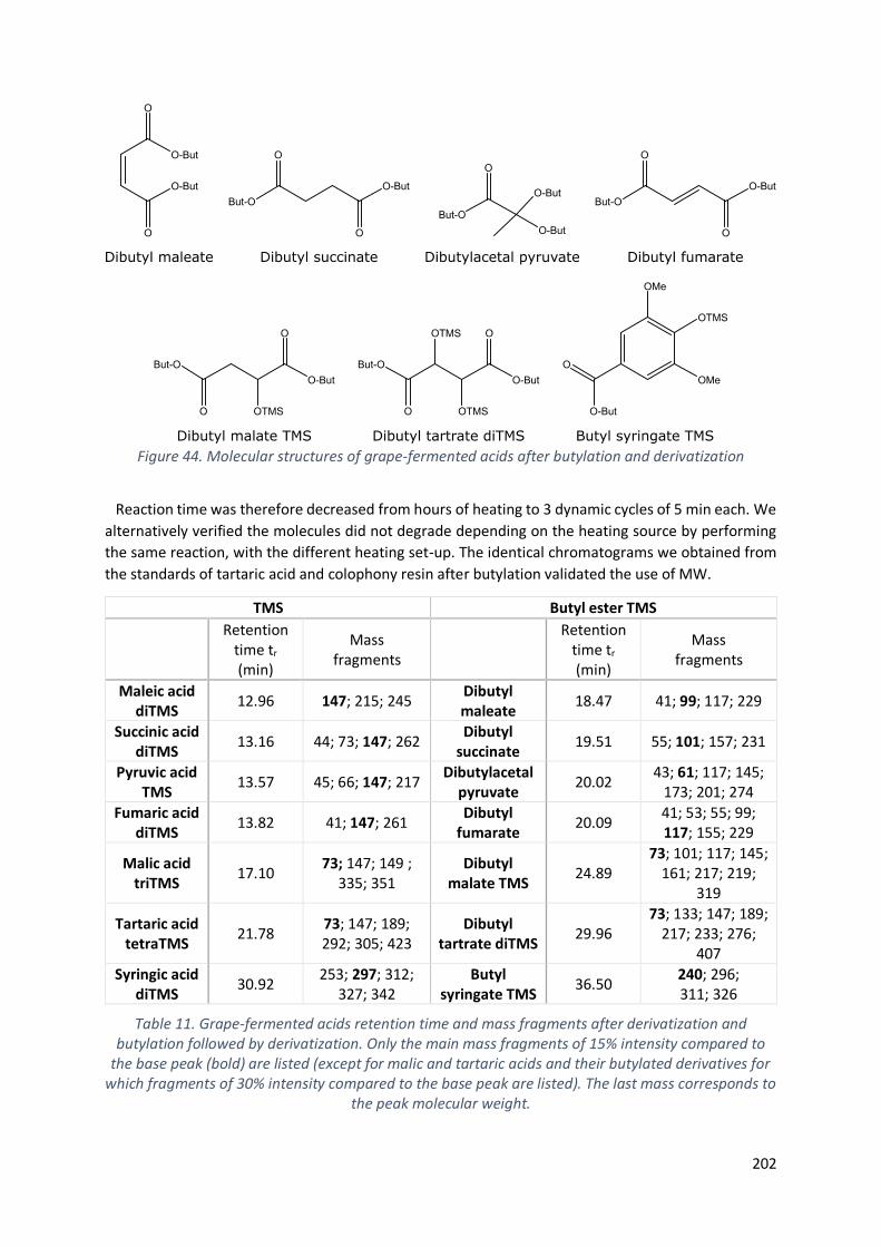
202
Figure 44. Molecular structures of grape-fermented acids after butylation and derivatization
Reaction time was therefore decreased from hours of heating to 3 dynamic cycles of 5 min each. We
alternatively verified the molecules did not degrade depending on the heating source by performing
the same reaction, with the different heating set-up. The identical chromatograms we obtained from
the standards of tartaric acid and colophony resin after butylation validated the use of MW.
TMS Butyl ester TMS
Retention
time tr (min)
Mass fragments
Retention
time tr
(min)
Mass fragments
Maleic acid diTMS
12.96 147; 215; 245 Dibutyl maleate
18.47 41; 99; 117; 229
Succinic acid diTMS
13.16 44; 73; 147; 262 Dibutyl
succinate 19.51 55; 101; 157; 231
Pyruvic acid TMS
13.57 45; 66; 147; 217 Dibutylacetal
pyruvate 20.02
43; 61; 117; 145; 173; 201; 274
Fumaric acid diTMS
13.82 41; 147; 261 Dibutyl
fumarate 20.09
41; 53; 55; 99; 117; 155; 229
Malic acid triTMS
17.10 73; 147; 149 ;
335; 351 Dibutyl
malate TMS 24.89
73; 101; 117; 145; 161; 217; 219;
319
Tartaric acid tetraTMS
21.78 73; 147; 189; 292; 305; 423
Dibutyl tartrate diTMS
29.96 73; 133; 147; 189;
217; 233; 276; 407
Syringic acid diTMS
30.92 253; 297; 312;
327; 342 Butyl
syringate TMS 36.50
240; 296; 311; 326
Table 11. Grape-fermented acids retention time and mass fragments after derivatization and butylation followed by derivatization. Only the main mass fragments of 15% intensity compared to
the base peak (bold) are listed (except for malic and tartaric acids and their butylated derivatives for which fragments of 30% intensity compared to the base peak are listed). The last mass corresponds to
the peak molecular weight.

203
Once the parameters were optimized for the commercial standards, the butylation was smoothly run
on the archaeological shards of Planier 3 with the aim of characterizing grape markers impregnated in
the ceramic. Since the esterification proved to be efficient, the research question then concentrated
on the ability of butylating the grape acids when it came to deal with organic coatings. The same MW
protocol was therefore tested on an archaeological pitch sample that had already been considered for
a protocol development in a previous PhD thesis (Fujii, 2018). The Dressel 2-4 amphora labelled C98,
originating from Pompeii, already gave excellent results in terms of grape markers. The choice to use
this object as a vector for the coating adaptation was additionally justified by the important material
quantity, sufficient to supply the protocol adaptation and any further adjustment if needed. Grape
derivative markers of tartaric, syringic, malic and glutaric acids, initially identified by alkaline treatment
in the previous study, were also recovered after butylation. The esterification led to the identification
of pyruvic acid, hitherto unnoticed. The fruitful characterization of fermented grape derivatives
highlighted the robustness of the butylation to extract bound acids from organic coatings, especially
the very meaning tartaric acid.
After the protocol parameters were developed on the archaeological coating of the sample C98, they
were tested on the lining of a Dressel 1 originating from Planier 3 (i.e., sample no. 1014), and then
applied on the other artefacts of the cargo. The amphora 1014 indeed exhibited enough material to
initialize the coating-butylation attempts among the corpus samples investigated in the theses. Results
proved to be successful as well, as identified by the non-butylated and butylated compounds in Fig.
45.
Figure 45. TIC (black) and m/z (red) chromatograms of the sample 1014 after (A) the extraction with traditional solvent (labelled 1LE) and (B) the butylation directly applied on the coating.S: Succinic
acid ; F: Fumaric acid ; G: Glutaric acid ; Sy: Syringic acid ; DBP: Dibutylcetal pyruvate ; DBM: Dibutyl malate ; DBT: Dibutyl tartrate ; BSy: Butyl synringate.
At this point of the protocol development, one indication needs to be detailed. Besides the great
characterization of fermentation and grape markers, the ability of the butylation protocol to
completely characterize the object, from the content to the coating nature, was questioned. The
colophony standard resin we initially butylated gave us the insurance that esterification was unable to
surely support the identification of diterpenic compounds, independently from the heating source. The

204
reaction conditions, as much with MW than reflux heating or oil bath, indicated diterpenoid side-
reactions, thus avoiding the molecular characterization of diterpenic markers. Figure 46 presents the
chromatograms obtained from reflux and MW, demonstrating the production of identical compounds,
hardly identifiable though. Further on, the same protocol was distinctively applied on molecular
standards of pimaric, sandaracopimaric, isopimaric and 7-oxo-DHA acids. Results unequivocally
showed a bench of molecular compounds exhibiting diterpenic skeletons, confirming the unreliability
to provide insights on the chemical composition of the waterproofing material with esterification.
Indeed, the diterpenic markers are the molecular trackers allowing to distinguish resin, pitch and wood
tar by the presence of heating and oxidized markers and wood pyrolysis with the presence of methyl
ester derivatives, respectively.
Figure 46. Chromatograms of standard colophony with different chemical treatments: A. No butylation; B. Butylation completed with reflux (2h; 80°C); C. Butylation MW-assisted (3*5 min; 80°C).
Pim.: Pimaric; Sandarcopim.: Sandaracopimaric; Isopim.: Isopimaric; ac.: acid; But.: Butylated
To reach a complete characterization of the archaeological material (content and lining nature), the
optimized MW-butylation was strengthen up with other extractive pathways. Even though we raised
the number of characterized compounds to a total of 97 molecules, the decision was taken to
concentrate on highly valued molecules while elaborating the extractive protocols to apply since not
all of them help in diagnosing the original content and coating. The main molecules are listed with
relative retention times and MS fragments in the appendix 4). For example, fatty acids and diacids,
small organic acids recurrent in living organisms (e.g., benzoic acid and its derivatives, vanillic acid,
isovanillic acid, etc.) or tricyclic derivatives from diterpene structures (e.g., norabietatriene derivatives,
5−podocarpatrienoic acid derivatives, etc.) were not considered. 22 molecules were therefore
targeted based on their ability to trace archaeological grape derivatives content, fermentation and to
give insight on the nature of the waterproofing coating and the treatment it went through for
formulation. The molecules considered were detailed in the fourth chapter, together with the
development of the extractive protocols considered. Several attempts performed on archaeological
materials of Planier 3 and San Felice Circeo (i.e., 10 shards and 8 coatings) shed light on the most

205
fruitful couplings to apply. For the analysis of shards, a two-step protocol consisting of a lipid extraction
with traditional solvents followed by MW-butylation reached better outcomes. For the analysis of
organic resins, simultaneous alkaline treatment and acid-catalyzed butylation directly applied on the
coating material are favored (Fig. 47).
Figure 47. Protocols to apply for the extraction of grape and fermentation markers
8.2.2. Archaeobotanical analysis The archaeobotanical analyses focused on plant remains contained in the amphorae. In this regard,
an analytical protocol aiming at dissolving the pitch and collecting the botanical residues it contained
was initially developed. The treatment combined the dissolution of the pitch with organic solvents and
traditional palynological methods to physically and chemically breakdown the matrix, remove
undesired compounds and concentrate archaeobotanical features. The methodology was first applied
to 7 archaeological coatings and then to a teapot shard which had a particular excavation context
assuring of a low risk of contamination from post deposition.
In conclusion, the methodological novelty arises from the reinforcement of analytical protocols able
to extract highly significant molecules, considered as markers of the original content and the
application of such protocols on organic coatings and inorganic shards of amphorae, until now never
considered with a multidisciplinary perspective. The interference of archaeological considerations,
chemical and archaeobotanical evidence was developed to encourage further investigation.

206
8.3. Archaeological perspective: From the object to the
interpretation
As reported at length through the analyses of several archaeological items from different contexts,
organic residues and archaeobotany demonstrated their collaborative richness in terms of scientific
support. To the best of our knowledge, such fruitful collaboration has been too occasionally tested
regarding the number of organic residue analysis applied on vessels and pottery. Sparse is the evidence
of multi-analytical approaches although botanical investigation empowered the archaeological
interpretation, both to avoid misinterpretation due to contamination and to attain a global
understanding of the material studied. In this section, we will revert to the artefacts investigated,
paying particular attention to the benefits provided by the application of a multidisciplinary
methodology.
For the amphorae labelled SFC1, SFC2 and SFC5 coming from the ancient anchorage of San Felice
Circeo (discussed in the fifth chapter), organic residues revealed fermented grape derivatives and
diterpenic markers associated to the presence of Pinaceae wood tar. Pinus fossil pollen present in
considerable extent, together with wood fragments, substantiated the assumption of resinous tree
pyrolysis in addition of providing more accurate insights on the botanical nature of the coating allowing
the identification up to the subsection including P. mugo, P. nigra and P. sylvestris. The geographical
distribution of such species, virtually absent in Central Italy, provided great evidence of the non-local
origin of the Pinus used to prepare the pitch, hence shedding light on exportations and Roman trading.
Palynological observation of the coatings showed aporate tricolpate pollen grains, identified as Vitis
vinifera. Grapevine stamens were also identified, hence confirming the archaeological grape
derivatives content. The morphological singularity of the Vitis pollen was interpreted as a possible
trace of autochthonous vineyards used to prepare grape beverages. Aporate Vitis pollen additionally
interrogate the state of domestication of the grapevines, giving insights on the possibility to use pollen
grains as an indicative tool to understand grape domestication. Alternatively, the speculation of a
medicinal infusion prepared with wild Vitis flowers and referred in historical testimonies as
oenanthium could not be discarded. Although this hypothesis is unlikely since the three amphorae had
different geographical origin, it would corroborate the herbal pollen of Artemisia and Myrtus
recovered in the amphorae. Such interpretation could not have been raised without the support of
transdisciplinary investigations, with archaeological considerations bridging the gap between
molecular markers that certified the presence of fermented grape derivatives and archaeobotanical
observations that attested of peculiar Vitis morphology.
For the spouted vase of Jericho discussed in the sixth chapter, diterpenic markers unraveled the
presence of Pinaceae in the teapot, unnoticeable with palynological analysis. Since Pinus pollen are
easily dispersed, the absence of such pollen in the teapot was hence interpreted in the light of natural
resin, confirmed by the absence of oxidized diterpenoids and heating-preparation markers. Regarding
the pollen content, Malva parviflora grains were abundantly recovered in the vessel, suggesting a
medicinal beverage. Such interpretation was supported by the presence of Artemisia grains and lumps
of pollen that could originate from flower anthers or honey. Both species additionally exhibit healing
properties. Even though molecular markers of cedar were not identified by gas chromatography,
Cedrus pollen identified in the vessel enhances the symbolic character, already conferred to the vase
from typological considerations. The morphological resemblance with teapots recovered from funeral
contexts indeed outlines the cultural functionality the vessel could have afforded. Giving new insights
on ancient medicinal recipes consumed at Bronze Age times, pollen and molecular compounds
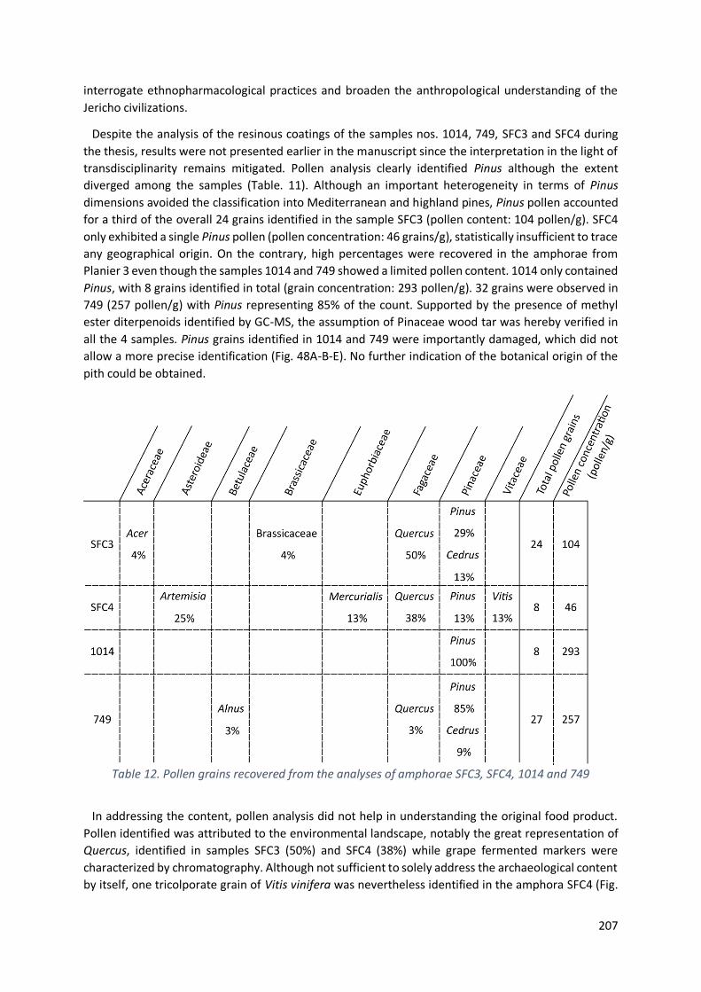
207
interrogate ethnopharmacological practices and broaden the anthropological understanding of the
Jericho civilizations.
Despite the analysis of the resinous coatings of the samples nos. 1014, 749, SFC3 and SFC4 during
the thesis, results were not presented earlier in the manuscript since the interpretation in the light of
transdisciplinarity remains mitigated. Pollen analysis clearly identified Pinus although the extent
diverged among the samples (Table. 11). Although an important heterogeneity in terms of Pinus
dimensions avoided the classification into Mediterranean and highland pines, Pinus pollen accounted
for a third of the overall 24 grains identified in the sample SFC3 (pollen content: 104 pollen/g). SFC4
only exhibited a single Pinus pollen (pollen concentration: 46 grains/g), statistically insufficient to trace
any geographical origin. On the contrary, high percentages were recovered in the amphorae from
Planier 3 even though the samples 1014 and 749 showed a limited pollen content. 1014 only contained
Pinus, with 8 grains identified in total (grain concentration: 293 pollen/g). 32 grains were observed in
749 (257 pollen/g) with Pinus representing 85% of the count. Supported by the presence of methyl
ester diterpenoids identified by GC-MS, the assumption of Pinaceae wood tar was hereby verified in
all the 4 samples. Pinus grains identified in 1014 and 749 were importantly damaged, which did not
allow a more precise identification (Fig. 48A-B-E). No further indication of the botanical origin of the
pith could be obtained.
Table 12. Pollen grains recovered from the analyses of amphorae SFC3, SFC4, 1014 and 749
In addressing the content, pollen analysis did not help in understanding the original food product.
Pollen identified was attributed to the environmental landscape, notably the great representation of
Quercus, identified in samples SFC3 (50%) and SFC4 (38%) while grape fermented markers were
characterized by chromatography. Although not sufficient to solely address the archaeological content
by itself, one tricolporate grain of Vitis vinifera was nevertheless identified in the amphora SFC4 (Fig.
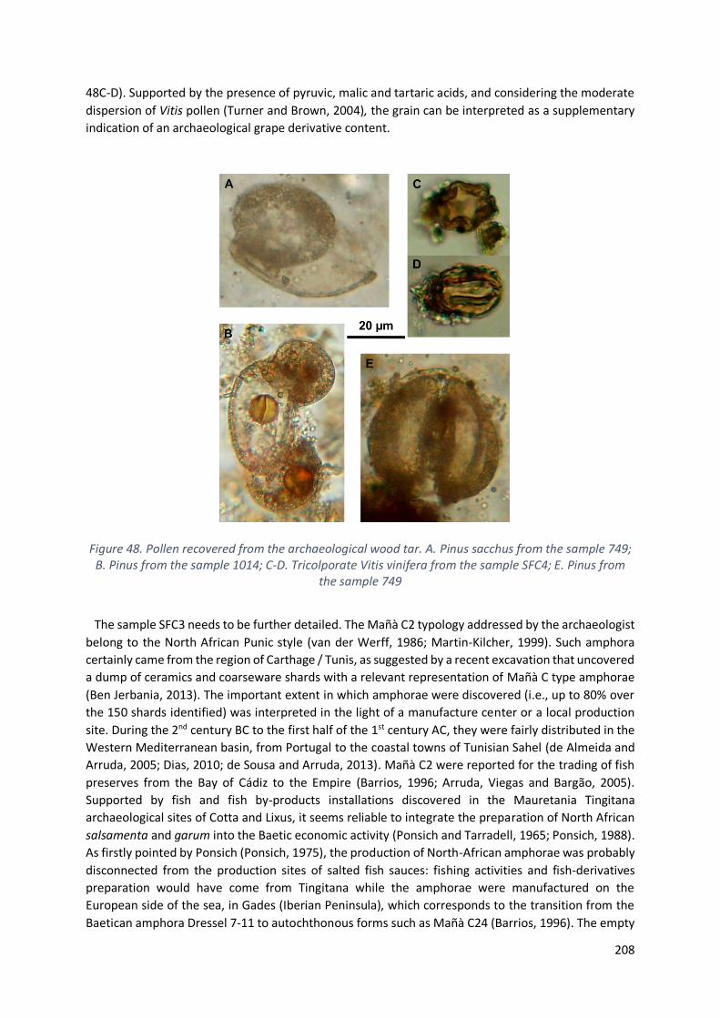
208
48C-D). Supported by the presence of pyruvic, malic and tartaric acids, and considering the moderate
dispersion of Vitis pollen (Turner and Brown, 2004), the grain can be interpreted as a supplementary
indication of an archaeological grape derivative content.
Figure 48. Pollen recovered from the archaeological wood tar. A. Pinus sacchus from the sample 749; B. Pinus from the sample 1014; C-D. Tricolporate Vitis vinifera from the sample SFC4; E. Pinus from
the sample 749
The sample SFC3 needs to be further detailed. The Mañà C2 typology addressed by the archaeologist
belong to the North African Punic style (van der Werff, 1986; Martin-Kilcher, 1999). Such amphora
certainly came from the region of Carthage / Tunis, as suggested by a recent excavation that uncovered
a dump of ceramics and coarseware shards with a relevant representation of Mañà C type amphorae
(Ben Jerbania, 2013). The important extent in which amphorae were discovered (i.e., up to 80% over
the 150 shards identified) was interpreted in the light of a manufacture center or a local production
site. During the 2nd century BC to the first half of the 1st century AC, they were fairly distributed in the
Western Mediterranean basin, from Portugal to the coastal towns of Tunisian Sahel (de Almeida and
Arruda, 2005; Dias, 2010; de Sousa and Arruda, 2013). Mañà C2 were reported for the trading of fish
preserves from the Bay of Cádiz to the Empire (Barrios, 1996; Arruda, Viegas and Bargão, 2005).
Supported by fish and fish by-products installations discovered in the Mauretania Tingitana
archaeological sites of Cotta and Lixus, it seems reliable to integrate the preparation of North African
salsamenta and garum into the Baetic economic activity (Ponsich and Tarradell, 1965; Ponsich, 1988).
As firstly pointed by Ponsich (Ponsich, 1975), the production of North-African amphorae was probably
disconnected from the production sites of salted fish sauces: fishing activities and fish-derivatives
preparation would have come from Tingitana while the amphorae were manufactured on the
European side of the sea, in Gades (Iberian Peninsula), which corresponds to the transition from the
Baetican amphora Dressel 7-11 to autochthonous forms such as Mañà C24 (Barrios, 1996). The empty
209
amphorae leaving Gades were sent to Tingitana to be filled with fish sauces, then returned to Gades
before being exported throughout the Mediterranean as a product of Baetican origin (Ponsich, 1975;
Étienne and Mayet, 1998). This theory of usurpation finds its roots in the absence of kilns in Mauretania
Tingitana (Teichner and Pujol, 2008) despite the presence of tituli picti (e.g., CO(r)D(ula) LIX(itana)
VE(tus) or CO(r)D(ula) TING(itana) VET(us)) attesting of the African origin of the containers (Liou, 1987;
Martín-kilcher and Thierrin-Michael, 1994; Martinez Maganto, 2000).
Oil has alternatively been mentioned in Mañà C containers (Peña, 2018), but no chemical profile
looking like olive oil or other plant oil was identified from chromatographic analysis. Palmitic, oleic,
stearic and azelaic acids were recovered, with important quantities of unsaturated fatty acids that do
not fit fresh olive oil profile. Also, in the hypothesis of oil, the coating would have been dissolved by
the fatty content, altering the original taste (Bonifay, 2007). Even though one waterproofed Dressel 20
oil amphora was reported with macroremains of the black lining (Manhita et al., 2020), oil supposition
was discarded based on chemical markers. The absence of Olea europea pollen further discredits the
assumption of oil.
From the organic residue analysis, no fish fats could be identified although the C18:1 characterized
could account for elaidic acid (i.e., the oleic acid trans-isomer C18:1 trans-9) or vaccenic acid (i.e., C18:1
trans-11), both being naturally present in fish and animal milk fed with fish oil (AbuGhazaleh et al.,
2003; Islam et al., 2018; Dutta and Dutta, 2019). Further investigation is be needed to surely address
the isomerism, notably with MS² approach since a second fragmentation would distinguish patterns
from isomers or with Matrix Assisted Laser Desorption Ionization-Time of Flight/MS (MALDI-TOF/MS)
equipment (Gómez-Cortés et al., 2009; Peršurić et al., 2018). Chemical reactions can be an alternative
to identify the double bond position in lipid isomers, as stated in recent publications calling for meta-
chloroperoxybenzoic acid epoxidation coupled with collision-induced dissociation MS-MS devices
(Feng et al., 2019) or ozonolysis with electrospray ionization MS (Thomas et al., 2007).
However, chromatography revealed the presence of grape and fermentation markers (Fig. 49), giving
valuable insights on the archaeological fermented grape beverage that was originally contained in the
amphorae. It is worth noting that palynology has not provided any indication on aromatic plants
despite their common use in fish preparations. Indeed, garum, liquamen and other fish preserves
called for coriander, leek, cumin, celery, etc. to bitter the flavor (Feldman, 2005). Supported by
diverging evidence from archaeology and organic residues, the hypothesis of reuse, although barely
considered in archaeology, becomes even more probable. For instance, the Officina del Garum degli
Umbrici in Pompeii provides another great example of reuse assessment based on archaeological
consideration regarding the number of amphorae discovered, their upside-down positions and the
original content originating from typology assumptions. In a specific room of the rear courtyard of a
house considered as the manufacture center for fish derivatives preparation and direct sale, more than
200 amphorae were stored (Peña, 2007; Bernal et al., 2014). Among them, olive oil containers of Mañà
C type, Ostia and Tripolitanian were recovered empty in reversed position which suggests a further
filling with local fish preserves (Peña, 2018). Worth considering historical literature, little is described
about amphorae reuse although rare evidence was testified by Pliny15. For example, he reported the
use of oil containers freshly dried for transporting halmyrídia, a maritime specie of cabbages to date
known as sea kale (Crambe maritima L.). Interestingly, such packaging was reported to better conserve
the vegetables and ensured them a green color despite the lengthy maritime journey. Following Peña’s
understanding of the scarce evidence of container reuse (Peña, 2018), the presence of an epigraphic
indication on the tomb of Gaius Commissius Succesus attested of his retail activities in the 2nd century
15 Naturalis Historia XIX 41

210
AD. The merchant, whose epitaph clearly designated him as a jug and small amphorae dealer at the
Portus Vinarius, was unequivocally involved in buying and selling used wine amphorae in Rome.
Figure 49. TIC chromatogram of the Mañà C2 amphora (sample no. SFC3)
As highlighted by the residue analysis of the above-mentioned Dressel 20 from Pax Julia Civitas
(Lusitania) (Manhita et al., 2020) or from the Keay 62A amphora from the La Palud 1 shipwreck (France)
(Garnier, 2007) that unexpectedly revealed pitch coating together with olive oil fatty acids interpreted
as a sign of amphora reuse, with wine or fish preserves as prime-content, the recycling of amphorae is
complex to consider since it raises several aspects. No standard practices can be assumed, and the
reuse can have different faces. Recycling may be considered for the same substances, but it may also
be part of a wider supply chain, considering amphorae as a vector of packaging. In the case of reuse, a
lower quality regarding the second content can be hypothesized, as amphorae already used may have
been cheaper compared to fresh ones and presumably altered the taste (Peña, 2007). Local wine
meant for nearby regional supply was hence suggested for Dressel 2-4 re-filling in the wine bottling
warehouse of Oplontis Villa B in Pompeii (Pecci et al., 2017).
The extent to which amphorae were re-employed is also questionable. Rarely considered in
literature, the analysis undertaken on containers reflect the dearth of attention paid to these
possibilities. Apart from the economic advantage of using low-priced containers, there may also be
geographical reasons to justify the reuse. It is worth considering the need of amphorae as a packaging
support for the export of local production. Assuming that local manufacture of amphorae was non-
existent or insufficient, then reuse might have played an important role (Peña, 2018). If the re-
assemblage of amphorae was indeed common, then it becomes essential to adapt the applied
archaeometric analyses. In this regard, general and global analysis has already been encouraged, in
order to understand the object as a whole and not to bias the interpretation by specific and too
targeted analytical research (Drieu et al., 2020). In this way, not only will the understanding of the
object be more relevant, but also its integration into an economic, fluvial, geographical, chronological
and anthropo-sociological scheme will be more pertinent.
211
In other respect, the 11 amphorae from Planier 3 that were supposed to have contained oil were
only analyzed with chromatographic traditional methods and results were discussed in the seventh
chapter. Lipid residues showed a remarkable distribution of saturated fatty acids, with a clear
predominance of palmitic over stearic acid. Despite its major contribution in fresh olive oil, oleic acid
was hardly ever recovered while azelaic acid was identified in several shards. The different molecular
compounds extracted underlined the degraded state of the original oil. Understanding why such
oxidative processes occurred relies on the assessments of archaeological interpretations regarding the
content and the objects’ history. This way, several hypotheses were suggested even though none could
be verified. Characteristic fatty markers that would have highlighted a vegetal source differing from
olive (e.g., castor oil, rapeseed oil, etc.) were not identified. Amphorae could have transported olive
fruits since the flesh does not release fatty acids. Such consideration was supported by the Northern
provenance of the early African amphorae, famous to produce table olives rather than oil. Otherwise,
amphorae could have been filled with degraded olive oil on purpose: the function of the good not
being associated with consumption and cooking, the oil did not require high quality. For example, body
lotions or lamp fuel accounted for poor quality of oil. Olive oil being a basic food source during Roman
times, its occurrence was alternatively reported in several derivatives products such as medicaments,
soaps or perfumes (Rowan, 2019).
In the case of the Planier 3 oil amphorae, results illustrate the limits of a single analytical
methodology. Chromatography highlights the degraded profile of the contents without bringing
additional evidence of an exotic or atypical product. The application of a multi-analytical approach
would then be interesting because it could open the field of interpretation, as previously demonstrated
with the wine amphorae from San Felice Circeo or the Jericho vase. Although little considered in
archaeometry, the example of perfumed oil could provide valuable explanation. Such derivatives were
indeed used as a base for archaeological cosmetics and olive oil was initially scented for preparation
(Pérez-Arantegui et al., 2009). In this case, palaeobotany could be an excellent option to address this
hypothesis since pollen would be the witness of flower or herbal flourishment
However, as the search for pollen requires a reliable and well-detailed archaeological environment,
the main risk being obviously the integration of post-sedimented grains in the interpretation of the
results, the selection of objects and sampling are essential for a valuable palaeobotanical analysis.
Most of the amphorae from Planier 3, all contents included, corresponded to shards with no apparent
resinous coating (i.e., 19 over the 22 artefacts), which were recovered underwater. Unfortunately,
these objects could not be included in palynological analysis for various reasons. Firstly, because the
superficial seabed does not represent a terrain free of post-sedimentary pollen contamination and the
objects were excavated more than 50 years ago. Secondly, because they were fragments of amphorae,
some with several centimeters of concretion on the surface of the ceramic. Indeed, objects recovered
in their integrity are favored when dealing with shard pollen analysis, always with the idea of limiting
contamination. When archaeological excavations ensure a physically bordered environment that
drastically reduces the risks of contamination, such as domestic or funerary contexts for example,
palynological analysis from the shard becomes possible.
Since pollen analysis of pottery has rarely been carried out so far, the first challenge we faced was to
verify the possibility of such an approach, using the residual pitch contained in amphorae. Once
confirmed, we then validated the feasibility of pollen analysis from the ceramic shard itself. In this
regard, we carefully chose an object whose archaeological context ensured a low risk of post-sediment
contamination.
212
8.4. Conclusion
The following table gives an overview of the main results obtained from chromatography and
archaeobotany for all the artefacts studied all along the different chapters.
The twofold analytical approach undoubtedly allowed more quantitative information on the
investigated samples to be acquired. The methodology greatly participated to a better definition of
the Pinus origin of diterpenic markers (for the samples no. 1014 and 749), to the identification of the
Mediterranean subfamily of the Pinus waterproofing coating (samples no. SFC1, SFC2 and SFC5). The
multi-analytical approach additionally promoted the contextualization of the presence of grapes in
certain amphorae, thus joining anthropological perspectives to the archaeometric analysis of the
objects.
Finally, the scientific lens we adopted showed a new aspect through the analysis of the Jericho vase,
by providing information that do not overlap with each other. Rather than corroborating each other,
chromatographic and archaeobotanical analyses proved to be complementary and opened up a new
interpretative field regarding the recipes of a medicinal beverage.

213

214
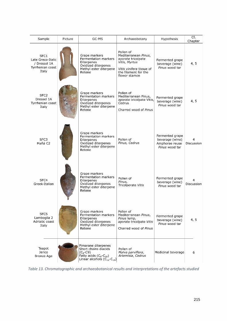
215
Table 13. Chromatographic and archaeobotanical results and interpretations of the artefacts studied
216
8.5. Perspectives
The current demand for a better understanding of archaeological objects has stimulated the search
for powerful and cutting-edge protocols for the characterization of ceramic vessels and pottery.
Beyond the innovative analytical aspect, this thesis aims at filling knowledge gaps between cultural,
archaeological and scientific fields. The objectives concerning the development of reliable analytical
protocols, both in terms of extraction of organic residues and in terms of archaeobotanical
observations, have been achieved. The main challenge therefore relies in the object itself and directly
infers on the decision of the analytical treatment to follow.
The multi-analytical approach allows to better define the archaeological and anthropological
contours of the studied contexts. The collaboration of independent techniques remarkably promotes
the consideration of new archaeological hypotheses, from the nature of the product itself to its
integration in the understanding of goods trading in the Mediterranean basin. The application of such
a methodology on a larger scale would open new horizons. Indeed, as objects do not equally survive
archaeological time, the application of a strict multidisciplinary method cannot be sufficient on its own.
It is therefore necessary to use and corroborate the observations provided by different analyses.
In this regard, DNA, and more precisely ancient DNA (aDNA) have recently joined the field thanks to
polymerase chain reactions that amplify the remaining genetic material. The archaeological artefacts
considered for analysis must be carefully selected and sampled to avoid contamination or
misinterpretation from false positives. Climatic, environmental and storage conditions can play an
important role in DNA preservation. Cold, dry and anoxic atmosphere are favored (Schlumbaum,
Tensen and Jaenicke-Després, 2008). Following Di Donato et al. (Di Donato et al., 2018), aDNA proved
to be a valuable tool to address plant evolution and domestication of modern crops. Genetic
approaches revolutionized the understanding of plant adaptation and migration (Gepts, 2004;
Doebley, Gaut and Smith, 2006; Meyer, DuVal and Jensen, 2012; Guasch-Jané, 2019; Zhou, Muyle and
Gaut, 2019). aDNA analyses of grapevines revealed sequence insertions into the nuclear genome of
Vitis vinifera, hence outlining genetic introgression from local wild species (Wales et al., 2016).
Furthermore, grape, oil and herbs genetic markers were successfully identified in Classical Greek
containers of a 2400-years old shipwreck (Hansson and Foley, 2008; Foley et al., 2012). The inorganic
building material seems to act as a support to which DNA binds (Briggs, 2020). As for tartrate salts in
organic residue analysis, the binding with the ceramic medium would protect genetic markers from
degradation (Grunenwald et al., 2014). Additionally, the analysis of aDNA from fermentation bacteria
provide a new avenue for studying prehistoric beverages. aDNA fragments of Saccharomyces
cerevisiae, the main yeast used in fermentation processes have been identified in 5000-years-old
Egyptian jars, hence providing new evidence on the archaeological wine contained herein (Cavalieri et
al., 2003). Such investigations, although they are on the cutting edge of science and technology are
still barely conducted on archaeological pottery. Most promising would be the integration of genetic
markers and DNA fingerprinting in multi-analytical approaches to decipher the intricate history beyond
the archaeological ceramic.
217
8.6. References AbuGhazaleh, A. A. et al. (2003) ‘Conjugated linoleic acid and vaccenic acid in rumen, plasma, and milk of cows fed fish oil and fats differing in saturation of 18 carbon fatty acids’, Journal of Dairy Science, 86(11), pp. 3648–3660. doi: 10.3168/jds.S0022-0302(03)73970-8.
de Almeida, R. R. and Arruda, A. M. (2005) ‘As ânforas de tipo Mañá C em Portugal’, in Ati del V Congresso Internazionale di Studi Fenici i Punici. 3. Università degli studi di Palermo, pp. 1319–1329.
Arobba, D. et al. (2014) ‘Palaeobotanical, chemical and physical investigation of the content of an ancient wine amphora from the northern Tyrrhenian sea in Italy’, Journal of Archaeological Science, 45(1), pp. 226–233. doi: 10.1016/j.jas.2014.02.024.
Arruda, A. M., Viegas, C. and Bargão, P. (2005) ‘As ânforas da Bética costeira na Alcáçova de Santarém’, Revista Portuguesa de Arqueologia, 8, pp. 279–297.
Ayllon-Martin, R., Perez Gonzalez, J. and Remesal Rodriguez, J. (2019) ‘Olive oil at the border of the Roman Empire. Stamps on Baetican Dressel 20 found on the Tyne-Solway Isthmus’, Marburger Beitrage zur antiken Handels-, Wirtschafts-und Sozialgeschichte, 36, pp. 167–216.
Barrios, L. L. (1996) ‘Explotación del salazón en la Bahía de Cádiz en la Antigüedad: Aportación al conocimiento de su evolución a través de la producción de las ánforas Mañá C’, Florentia iliberritana, 7, pp. 141–169.
Bernal, D. et al. (2014) ‘Un contexto excepcional en Pompeya: la pila de ánforas de la Bottega del Garum (I, 12, 8). Avance de un estudio interdisciplinar’, Rei Cretariae Romanae Fautorum Acta, 43, pp. 219–232.
Bonifay, M. (2007) ‘Que transportaient donc les amphores africaines ?’, in Supplying Rome and the Empire, pp. 10–11, 18–23, 25–29.
Bowman, A. and Wilson, A. (2009) Quantifying the Roman economy: methods and problems. Oxford University Press.
Briggs, L. (2020) ‘Ancient DNA Research in Maritime and Underwater Archaeology: Pitfalls, Promise, and Future Directions’, Open Quaternary, 6(1). doi: 10.5334/oq.71.
Cavalieri, D. et al. (2003) ‘Evidence for S. cerevisiae Fermentation in Ancient Wine’, Journal of Molecular Evolution, 57(1), pp. S226–S232. doi: 10.1007/s00239-003-0031-2.
Condamin, J. and Formenti, F. (1978) ‘Détection du contenu d’amphores antiques (huiles, vin). Etude méthodologique’, Revue d’Archéométrie, 2(1), pp. 43–58. doi: 10.3406/arsci.1978.1092.
Dias, V. F. S. (2010) A cerâmica campaniense do Monte Molião, Lagos. Universidade de Lisboa.
Doebley, J. F., Gaut, B. S. and Smith, B. D. (2006) ‘The Molecular Genetics of Crop Domestication’, Cell, 127(7), pp. 1309–1321. doi: 10.1016/j.cell.2006.12.006.
Di Donato, A. et al. (2018) ‘Genome Sequencing of Ancient Plant Remains: Findings, Uses and Potential Applications for the Study and Improvement of Modern Crops’, Frontiers in Plant Science, 9. doi: 10.3389/fpls.2018.00441.
Drieu, L. et al. (2020) ‘Is it possible to identify ancient wine production using biomolecular approaches?’, STAR: Science & Technology of Archaeological Research, pp. 1–14. doi: 10.1080/20548923.2020.1738728.
Drieu, L. et al. (2021) ‘Chemical evidence for the persistence of wine production and trade in Early Medieval Islamic Sicily’, Proceedings of the National Academy of Sciences, 118(10). doi: 10.1073/pnas.2017983118.

218
Dutta, P. and Dutta, M. (2019) ‘Analysis of fatty acid composition in the flesh of boal (Wallagu Attu)’, Biosciences, Biotechnology Research Asia, 16(2), pp. 477–481. doi: 10.13005/bbra/2762.
Edmondson, J. (2015) ‘Economic Life in the Roman Empire’, in The Oxford handbook of Roman epigraphy. Oxford University Press. doi: 10.1093/oxfordhb/9780195336467.013.031.
Étienne, R. and Mayet, F. (1998) ‘Les mercatores de saumure hispanique’, Mélanges de l’Ecole française de Rome, 110(1), pp. 147–165. doi: 10.4000/books.ausonius.8363.
Feier, I. et al. (2019) ‘Roman Wine in Barbaricum. Preliminary Studies on Ancient Wine Recreation’, Heritage, 2(1), pp. 331–338. doi: 10.3390/heritage2010022.
Feige, M. (2021) ‘Decorative Features and Social Practices in Spaces for Agricultural Production in Roman Villas’, in Principles of Decoration in the Roman World. De Gruyter, pp. 33–52. doi: 10.1515/9783110732139-003.
Feldman, C. (2005) ‘Roman Taste’, Food, Culture & Society, 8(1), pp. 7–30. doi: 10.2752/155280105778055407.
Feng, Y. et al. (2019) ‘Identification of double bond position isomers in unsaturated lipids by m -CPBA Epoxidation and Mass Spectrometry fragmentation’, Analytical Chemistry, 91(3), pp. 1791–1795. doi: 10.1021/acs.analchem.8b04905.
Finley, M. I. (1973) The Ancient Economy. University of California Press.
Fleming, S. J. (1997) Roman Glass: Reflections of Everyday Life. University of Pennsylvania Museum of Archaeology and Anthropology.
Foley, B. P. et al. (2012) ‘Aspects of ancient Greek trade re-evaluated with amphora DNA evidence’, Journal of Archaeological Science, 39(2), pp. 389–398. doi: 10.1016/j.jas.2011.09.025.
Foxhall, L. A. and Forbes, H. A. (1982) Sitometreia: the role of grain as a staple food in classical antiquity. Beck.
Frère, D. and Garnier, N. (2017) ‘Dairy Product and Wine in Funerary Rituals: The Case of a Hellenistic Etruscan Tomb’, Journal of Historical Archaeology & Anthropological Sciences, 1(6), pp. 222–227. doi: 10.15406/jhaas.2017.01.00034.
Fujii, H. (2018) Etude chromatographique de matériaux organiques contenus dans des amphores romaines. Université d’Avignon et des pays de Vaucluse.
Garnier, N. et al. (2003) ‘Characterization of thermally assisted hydrolysis and methylation products of polyphenols from modern and archaeological vine derivatives using gas chromatography–mass spectrometry’, Analytica Chimica Acta, 493(2), pp. 137–157. doi: 10.1016/S0003-2670(03)00869-9.
Garnier, N. (2007) ‘Analyse du contenu d’amphores africaines’, Supplying Rome and the Empire. Edited by E. Papi, pp. 25–31.
Garnier, N. and Valamoti, S. M. (2016) ‘Prehistoric wine-making at Dikili Tash (Northern Greece): Integrating residue analysis and archaeobotany’, Journal of Archaeological Science, 74, pp. 195–206. doi: 10.1016/j.jas.2016.03.003.
Gepts, P. (2004) ‘Crop domestication as a long-term selection experiment’, Plant Breeding reviews, 24(2), pp. 1–44.
Gómez-Cortés, P. et al. (2009) ‘Characterization of cis-9 trans-11 trans-15 C18:3 in milk fat by GC and covalent adduct chemical ionization tandem MS’, Journal of Lipid Research, 50(12), pp. 2412–2420. doi: 10.1194/jlr.M800662-JLR200.

219
Griffiths, D. (2019) ‘Commercialization of the night at Pompeii’, in Veal, R. and Leitch, V. (eds) Fuel and Fire in the Ancient Roman World Towards an integrated economic understanding. McDonald Institute for Archaeological Research. doi: 10.17863/CAM.46322.
Grunenwald, A. et al. (2014) ‘Adsorption of DNA on biomimetic apatites: Toward the understanding of the role of bone and tooth mineral on the preservation of ancient DNA’, Applied Surface Science, 292, pp. 867–875. doi: 10.1016/j.apsusc.2013.12.063.
Guasch-Jané, M. R. (2019) ‘Grape Archaeology and Ancient DNA Sequencing’, in The grape genome. Cham: Springer, pp. 57–75.
Hansson, M. C. and Foley, B. P. (2008) ‘Ancient DNA fragments inside Classical Greek amphoras reveal cargo of 2400-year-old shipwreck’, Journal of Archaeological Science, 35(5), pp. 1169–1176. doi: 10.1016/j.jas.2007.08.009.
Hitchner, R. B., Amouretti, M. and Brun, J. (2002) ‘Olive production and the Roman economy: the case for intensive growth in the Roman Empire’, The ancient economy, pp. 71–83.
Hostetter, E., Beck, C. W. and Stewart, D. R. (1994) ‘A bronze situla from tomb 128, Valle Trebba: chemical evidence for resinated wine at Spina’, Studi Etruschi 59, pp. 211–225.
Islam, R. et al. (2018) ‘Fatty acid compositions in marine fish samples of Bangladesh’, Journal of Nutrition & Food Sciences, 8(5), pp. 8–10. doi: 10.4172/2155-9600.1000723.
Ben Jerbania, I. (2013) ‘Observations sur les amphores de tradition punique d’après une nouvelle découverte près de Tunis’, Antiquités africaines, 49(1), pp. 179–192. doi: 10.3406/antaf.2013.1548.
Kappe, C. O., Dallinger, D. and Murphree, S. S. (2008) Practical microwave synthesis for organic chemists: strategies, instruments, and protocols. John Wiley & Sons.
Liou, B. (1987) ‘Inscriptions peintes sur amphores: Fos (suite), Marseille, Toulon, Port-La-Nautique, Arles, Saint-Blaise, Saint-Martin-De-Crau, Mâcon, Calvi’, Archaeonautica, 7(1), pp. 55–139.
Manhita, A. et al. (2020) ‘Transporting olive oil in Roman Times: Chromatographic analysis of Dressel 20 Amphorae from Pax Julia Civitas, Lusitania’, Chromatographia, 83(9), pp. 1055–1064. doi: 10.1007/s10337-020-03927-7.
Martin-Kilcher, S. (1999) ‘Karthago 1993. Die Füllung eines frühkaiserzeitlichen Pozzo’, Karthago, 3, pp. 404–434.
Martín-kilcher, S. and Thierrin-Michael, G. (1994) Die römischen Amphoren aus Augst und Kaiseraugst. Ein Beitrag zur römischen Handels-und Kulturgeschichte. Augst: Romermuseum.
Martinez Maganto, J. (2000) ‘Inscripciones sobre ánforas de salazón: interpretación sobre la estructura y significación comercial de los tituli picti’, in Congreso Internacional Ex Baetica Amphorae, pp. 1207–1219.
Mattingly, D. J. (1988) ‘Oil for export? A comparison of Libyan, Spanish and Tunisian olive oil production in the Roman empire’, Journal of Roman Archaeology, 1, pp. 33–56.
Meyer, R. S., DuVal, A. E. and Jensen, H. R. (2012) ‘Patterns and processes in crop domestication: an historical review and quantitative analysis of 203 global food crops’, New Phytologist, 196(1), pp. 29–48. doi: 10.1111/j.1469-8137.2012.04253.x.
Panella, C. and Tchernia, A. (1994) ‘Produits agricoles transportés en amphores: l’huile et surtout le vin’, Publications de l’Ecole française de Rome, 198(1), pp. 145–165.
Pecci, A. et al. (2013) ‘Identifying wine markers in ceramics and plasters using gas chromatography–mass spectrometry. Experimental and archaeological materials’, Journal of Archaeological Science,
220
40(1), pp. 109–115. doi: 10.1016/j.jas.2012.05.001.
Pecci, A. et al. (2017) ‘Use and reuse of amphorae. Wine residues in Dressel 2–4 amphorae from Oplontis Villa B (Torre Annunziata, Italy)’, Journal of Archaeological Science: Reports, 12, pp. 515–521. doi: 10.1016/j.jasrep.2017.02.025.
Pecci, A. et al. (2018) ‘Combining residue analysis of floors and ceramics for the study of activity areas at the Garum Shop at Pompeii’, Archaeological and Anthropological Sciences, 10(2), pp. 485–502. doi: 10.1007/s12520-017-0573-7.
Pecci, A., Cau Ontiveros, M. Á. and Garnier, N. (2013) ‘Identifying wine and oil production: analysis of residues from Roman and Late Antique plastered vats’, Journal of Archaeological Science, 40(12), pp. 4491–4498. doi: 10.1016/J.JAS.2013.06.019.
Pecci, A., Salvini, F. and Cantini, L. (2010) ‘Residue analysis of some Late Roman amphora coming from the excavations of the historical center of Florence’, in Menchelli, S. et al. (eds) LRCW 3. 3rd International Conference on Late Roman Coarse Wares, Cooking Wares and Amphorae in the Mediterranean: Archaeology and Archaeometry. Oxford: Archaeopress BAR Series, pp. 363–367.
Peña, J. T. (2007) Roman Pottery in the Archaeological Record. Cambridge: Cambridge University Press. doi: 10.1017/CBO9780511499685.
Peña, J. T. (2018) ‘The Reuse of Transport Amphorae as Packaging Containers in the Roman world: an overview’, Roman Amphora Contents: Reflecting on Maritime Trade of Foodstuffs in Antiquity. Roman and Late Antique Mediterranean Pottery, 9, pp. 1–22.
Pérez-Arantegui, J. et al. (2009) ‘Colorants and oils in Roman make-ups – an eye witness account’, Trends in Analytical Chemistry, 28(8), pp. 1019–1028. doi: 10.1016/j.trac.2009.05.006.
Peršurić, Ž. et al. (2018) ‘Comparison of triacylglycerol analysis by MALDI-TOF/MS, fatty acid analysis by GC-MS and non-selective analysis by NIRS in combination with chemometrics for determination of extra virgin olive oil geographical origin. A case study’, LWT, 95, pp. 326–332. doi: 10.1016/j.lwt.2018.04.072.
Ponsich, M. (1975) ‘Perennité des relations dans le circuit du Détroit de Gibraltar’, in Aufstieg und Niedergang der römischen Welt II.3. De Gruyter, pp. 654–684.
Ponsich, M. (1988) Aceite de oliva y salazones de pescado. Factores geoeconómicos de Bética y Tingitania. Madrid: Editorial Complutense.
Ponsich, M. and Tarradell, M. (1965) Garum et industries antiques de salaison dans la Méditerranée occidentale. Paris: Presses universitaires de France.
Rossiter, J. (2008) ‘Wine-Making After Pliny: Viticulture And Farming Technology In Late Antique Italy’, in Technology in Transition AD 300-650. Brill, pp. 93–118. doi: 10.1163/ej.9789004165496.i-573.41.
Rowan, E. (2019) The utility of olive oil pressing waste as a fuel source in the Roman world. McDonald Institute for Archaeological Research.
Schlumbaum, A., Tensen, M. and Jaenicke-Després, V. (2008) ‘Ancient plant DNA in archaeobotany’, Vegetation History and Archaeobotany, 17(2), pp. 233–244. doi: 10.1007/s00334-007-0125-7.
de Sousa, E. and Arruda, A. M. (2013) ‘A cerâmica de tipo Kuass de Monte Molião (Lagos)’, Arqueologia em Portugal. 150 anos, pp. 651–659.
Teichner, F. and Pujol, L. P. (2008) ‘Roman amphora trade across the straits of Gibraltar: An ancient “anti-economic practice”?’, Oxford Journal of Archaeology, 27(3), pp. 303–314. doi: 10.1111/j.1468-0092.2008.00309.x.

221
Thomas, M. C. et al. (2007) ‘Elucidation of double bond position in unsaturated lipids by Ozone Electrospray Ionization Mass Spectrometry’, Analytical Chemistry, 79(13), pp. 5013–5022. doi: 10.1021/ac0702185.
Turner, S. D. and Brown, A. G. (2004) ‘Vitis pollen dispersal in and from organic vineyards: I. Pollen trap and soil pollen data’, Review of Palaeobotany and Palynology, 129(3), pp. 117–132. doi: 10.1016/j.revpalbo.2003.12.002.
Wales, N. et al. (2016) ‘The limits and potential of paleogenomic techniques for reconstructing grapevine domestication’, Journal of Archaeological Science, 72, pp. 57–70. doi: 10.1016/j.jas.2016.05.014.
van der Werff, J. H. (1986) ‘The Amphora Wall in the House of the Porch, Ostia’, Excavations in the House of the Porch (V;ii.4-5) at Ostia. Bulletin antieke beschaving, 61, pp. 96–137.
Zhou, Y., Muyle, A. and Gaut, B. S. (2019) ‘Evolutionary Genomics and the Domestication of Grapes’, in The grape genome. Cham: Springer, pp. 39–55.
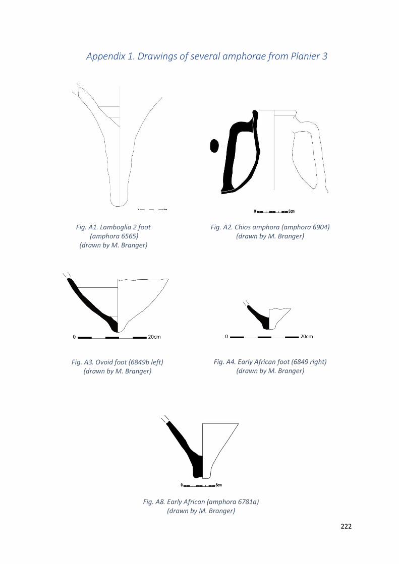
222
Appendix 1. Drawings of several amphorae from Planier 3
Fig. A2. Chios amphora (amphora 6904) (drawn by M. Branger)
Fig. A3. Ovoid foot (6849b left) (drawn by M. Branger)
Fig. A4. Early African foot (6849 right) (drawn by M. Branger)
Fig. A1. Lamboglia 2 foot (amphora 6565)
(drawn by M. Branger)
Fig. A8. Early African (amphora 6781a)
(drawn by M. Branger)

223
Reference
Branger, M. (2012) Les amphores de l’épave 3 de Planier Typologie et épigraphie. Université de Provence (Aix-Marseille 1).
Fig. A6. “CAEC” stamp from an ovoid (amphora 6561) (drawn by M. Branger and stamp picture from C. Durand, CCJ, CNRS)
Fig. A7. “APOLO.CAL” or “GAL” stamp from an ovoid (728) (drawn by M. Branger and stamp picture from C. Durand, CCJ, CNRS)
Fig. A8. “CAEC” stamp from an ovoid (561) (drawn by M. Branger and stamp picture from C. Durand, CCJ, CNRS)
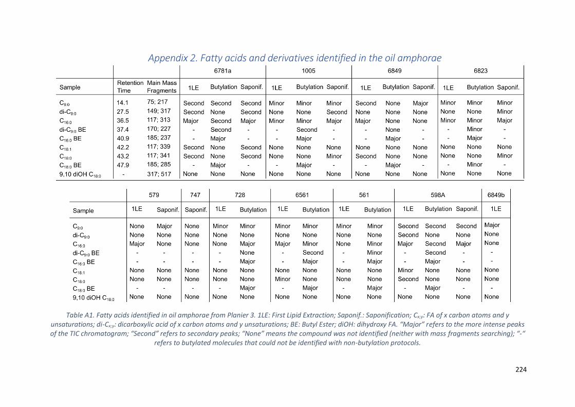
224
Appendix 2. Fatty acids and derivatives identified in the oil amphorae
Table A1. Fatty acids identified in oil amphorae from Planier 3. 1LE: First Lipid Extraction; Saponif.: Saponification; Cx;y: FA of x carbon atoms and y unsaturations; di-Cx;y: dicarboxylic acid of x carbon atoms and y unsaturations; BE: Butyl Ester; diOH: dihydroxy FA. “Major” refers to the more intense peaks of the TIC chromatogram; “Second” refers to secondary peaks; “None” means the compound was not identified (neither with mass fragments searching); “-“
refers to butylated molecules that could not be identified with non-butylation protocols.

225
Appendix 3. Mass spectrum of standard molecule
This appendix provides the mass spectra of the main molecules characterized and discussed in the
thesis. The molecules are classified according to their chemical family and appear in increasing order
of retention time. The following molecules are further detailed:
Fatty acids
Palmitic acid TMS
Oleic acid TMS
Stearic acid TMS
Azelaic acid TMS
Terpenes
Retene
Pimaric acid TMS
Sandaracopimaric acid TMS
Dehydroabietic acid methyl ester
Isopimaric acid TMS
Palustric acid TMS
Dehydroabietic acid TMS
Abietic acid TMS
7-oxo-dehydroabietic acid TMS
Organic acids
Succinic acid TMS
Fumaric acid diTMS
Malic acid triTMS
Tartaric acid tetraTMS
Syringic acid diTMS
Butylated acids
Dibutyl malate
Dibutyl succinate
Dibutylacetal pyruvate
Dibutyl fumarate
Dibutyl malate TMS
Dibutyl tartrate diTMS
Butyl syringate TMS
Dibutyl azelaic acid

226
Fatty acids
Palmitic acid TMS
C19H41O2Si
Molecular weight: 328.61 g.mol-1
Retention time: 36.54 min
Oleic acid TMS
C21H42O2Si
Molecular weight: 354.65 g.mol-1
Retention time: 42.22 min
200910_Palmitic_1LE #3137 RT: 36.44 AV: 1 NL: 4.88E6T: + c Full ms [40.00-650.00]
40 60 80 100 120 140 160 180 200 220 240 260 280 300 320 340
m/z
10
20
30
40
50
60
70
80
90
100
Re
lative
Ab
un
da
nce
313.28
75.03
117.03
330.96
129.04
73.08
131.08
95.1744.99 185.13143.13 201.07 269.2367.13 285.17243.13171.13 229.07109.19 159.06 299.22347.44
190605_std_oleic_opt13 #3643 RT: 42.18 AV: 1 NL: 3.92E5T: + c Full ms [40.00-650.00]
50 100 150 200 250 300 350
m/z
10
20
30
40
50
60
70
80
90
100
Re
lative
Ab
un
da
nce
75.06
129.11117.08 339.28
357.15
67.07
81.09
95.10
96.09131.0955.13
97.17 199.10143.11 264.24185.13
145.06 171.14 241.15222.16 311.33286.20
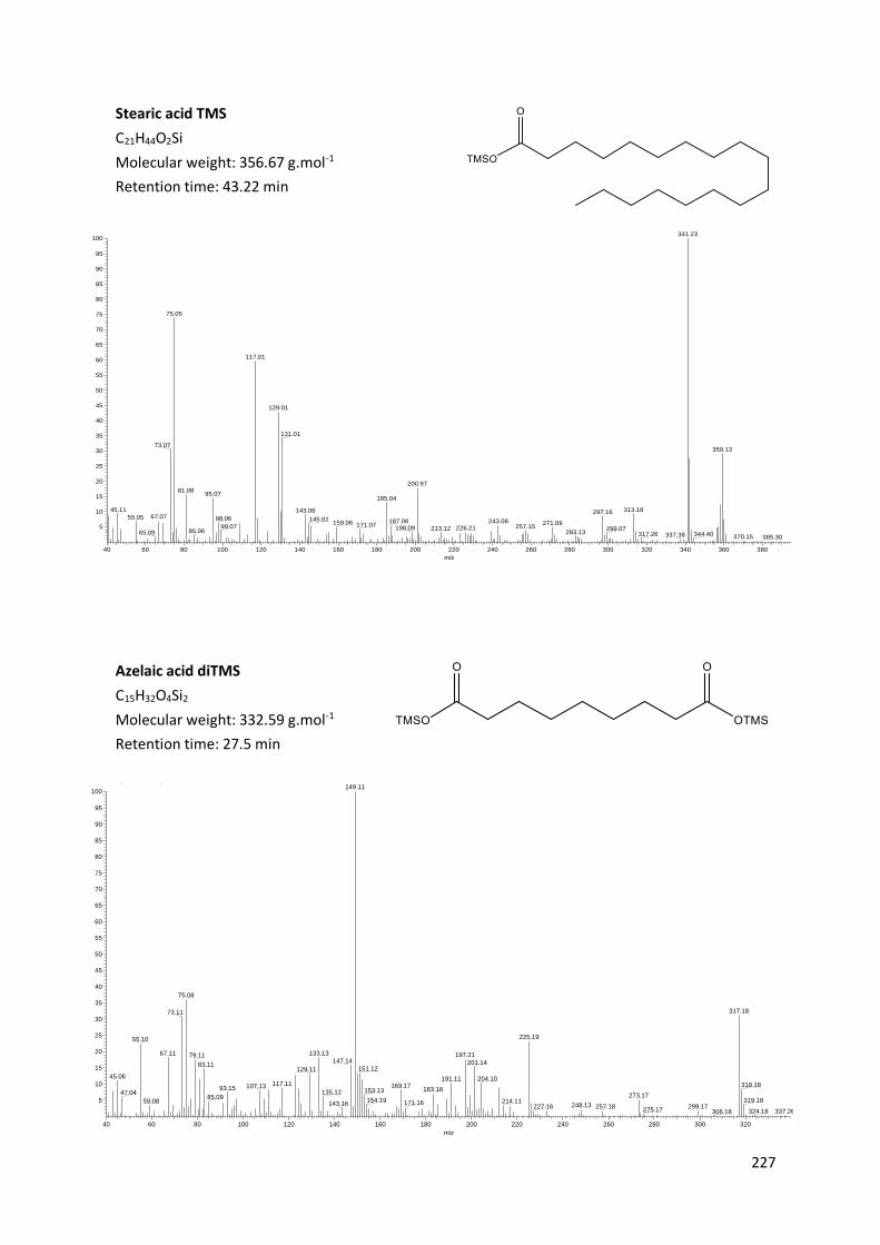
227
Stearic acid TMS
C21H44O2Si
Molecular weight: 356.67 g.mol-1
Retention time: 43.22 min
Azelaic acid diTMS
C15H32O4Si2
Molecular weight: 332.59 g.mol-1
Retention time: 27.5 min
250321_Jerico_Pecci #3941 RT: 43.10 AV: 1 SB: 658 43.24-45.30 , 38.97-42.97 NL: 1.17E5T: + c Full ms [40.00-650.00]
40 60 80 100 120 140 160 180 200 220 240 260 280 300 320 340 360 380
m/z
5
10
15
20
25
30
35
40
45
50
55
60
65
70
75
80
85
90
95
100
Re
lativ
e A
bu
nd
an
ce
341.23
75.05
117.01
129.01
131.01
73.07359.13
200.9781.08
95.07185.04
313.1845.11 143.06 297.1667.0755.05 98.06 145.02 187.08 243.08159.06 271.09171.0799.07 257.15226.21198.09 213.12 299.0785.06 283.1365.09 317.26 344.40337.38 370.15 385.30
190521_6781A_tesson_Mottram_100mg_opt13 #2237-2243 RT: 27.45-27.50 AV: 7 SB: 1364 27.51-32.83 , 19.95-27.40 NL: 4.68E4T: + c Full ms [40.00-650.00]
40 60 80 100 120 140 160 180 200 220 240 260 280 300 320
m/z
5
10
15
20
25
30
35
40
45
50
55
60
65
70
75
80
85
90
95
100
Re
lative
Ab
un
da
nce
149.11
75.08
317.1873.11
225.1955.10
67.11 133.13 197.2179.11147.14 201.1483.11
151.12129.1145.06 191.11 204.10
117.11 318.18169.17107.1393.15 183.18153.13135.1247.04 273.1785.09154.19 319.18214.1159.08 171.16143.16 248.13 257.18 299.17227.16
275.17 324.18 337.26306.18

228
Terpenes Retene
C18H18
Molecular weight: 234.34 g.mol-1
Retention time: 41.50 min
Pimaric acid TMS
C23H38O2 Si
Molecular weight: 374.64 g.mol-1
Retention time: 44.46 min 200628_pimaric_acid_std #3973 RT: 44.54 AV: 1 NL: 4.85E4T: + c Full ms [40.00-650.00]
50 100 150 200 250 300 350
m/z
0
10
20
30
40
50
60
70
80
90
100
Re
lative
Ab
un
da
nce
121.12
105.12
241.2091.11
359.25133.08257.15
77.08
147.08 173.10
159.14187.09 227.17
207.0867.10267.16 281.14 374.2165.00
357.22284.21 309.00378.19
190520_1014_poix_bouchon_Pecci_50mg_opt13 #3721 RT: 41.48 AV: 1 NL: 1.32E6T: + c Full ms [40.00-650.00]
40 60 80 100 120 140 160 180 200 220 240
m/z
10
20
30
40
50
60
70
80
Re
lative
Ab
un
da
nce
219.15
234.19
204.18
189.18101.21 205.26
178.1894.57 165.21152.2089.20 108.3275.1163.11 241.16129.19 253.1956.75

229
Sandaracopimaric acid TMS
C23H38O2Si
Molecular weight: 374.64 g.mol-1
Retention time: 45.04 min
Dehydroabietic acid methyl ester
C21H30O2
Molecular weight: 314.47 g.mol-1
Retention time: 45.48 min
200628_isopimaric+sandaraco_acid_std #3932 RT: 44.96 AV: 1 NL: 6.32E5T: + c Full ms [40.00-650.00]
50 100 150 200 250 300 350
m/z
10
20
30
40
50
60
70
80
90
100
Re
lative
Ab
un
da
nce
121.14
241.13
105.09
91.04359.22
73.09257.20
147.1579.04
159.15 173.11
201.1267.11 227.17 374.12345.2145.03
269.1853.01 284.21 331.06378.13303.28
201016_1014_poix_1LE_MW #4181 RT: 45.39 AV: 1 NL: 6.83E6T: + c Full ms [40.00-650.00]
40 60 80 100 120 140 160 180 200 220 240 260 280 300 320
m/z
10
20
30
40
50
60
70
80
90
100
Re
lative
Ab
un
da
nce
239.21
299.06
197.20237.29
141.20 195.22155.22 312.22129.20 211.25 253.20115.25 233.2391.2373.17 297.24269.1265.16

230
Isopimaric acid TMS
C23H38O2Si
Molecular weight: 374.64 g.mol-1
Retention time: 45.62 min
Palustric acid TMS
C23H38O2Si
Molecular weight: 374.64 g.mol-1
Retention time: 46.28 min
200628_pimaric+isopimaric_acid_std #4041 RT: 45.61 AV: 1 NL: 6.42E6T: + c Full ms [40.00-650.00]
50 100 150 200 250 300 350
m/z
10
20
30
40
50
60
70
80
90
100
Re
lative
Ab
un
da
nce
241.18
256.14
185.13227.1691.06 214.21157.12117.12 145.20 359.18
173.1379.1167.09
269.21 346.22 376.85332.25285.11 390.85
200628_palustric_acid_std #4046 RT: 46.26 AV: 1 NL: 1.30E7T: + c Full ms [40.00-650.00]
50 100 150 200 250 300 350
m/z
5
10
15
20
25
30
35
40
45
Re
lative
Ab
un
da
nce
241.20
359.16
185.22 374.22
109.1873.05 91.13213.21131.19 256.13159.18
117.17239.27
269.16227.21171.1975.1167.12 284.11
331.1646.99 291.06 379.24
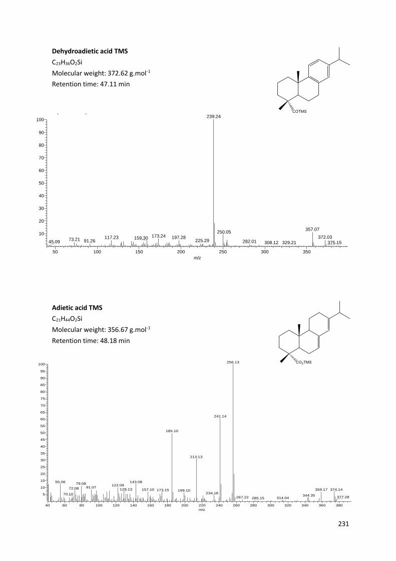
231
Dehydroadietic acid TMS
C23H36O2Si
Molecular weight: 372.62 g.mol-1
Retention time: 47.11 min
Adietic acid TMS
C21H44O2Si
Molecular weight: 356.67 g.mol-1
Retention time: 48.18 min
201016_1014_poix_1LE_MW #4358 RT: 47.03 AV: 1 NL: 2.30E7T: + c Full ms [40.00-650.00]
50 100 150 200 250 300 350
m/z
10
20
30
40
50
60
70
80
90
100
Re
lative
Ab
un
da
nce
239.24
357.07250.05
173.24 372.03117.23 197.28159.3073.21 225.2991.26 282.0145.09 375.15329.21308.12
250321_Jerico_Pecci #4483 RT: 48.07 AV: 1 SB: 762 48.12-51.34 , 44.24-48.00 NL: 2.79E5T: + c Full ms [40.00-650.00]
40 60 80 100 120 140 160 180 200 220 240 260 280 300 320 340 360 380
m/z
5
10
15
20
25
30
35
40
45
50
55
60
65
70
75
80
85
90
95
100
Rel
ativ
e A
bund
ance
256.13
241.14
185.10
213.13
143.0855.0679.08
122.0991.0772.08 129.13 359.17 374.14157.10 173.15 199.10
234.1870.10 344.35377.28267.22 314.04285.15

232
7-oxo-dehydroadietic acid TMS
C23H34O3Si
Molecular weight: 386.61 g.mol-1
Retention time: 53.72 min
Organic acids Succinic acid TMS
C10H22O4Si2
Molecular weight: 262.45 g.mol-1
Retention time: 13.16 min
190520_1014_poix_bouchon_Pecci_50mg_opt13 #5026 RT: 53.66 AV: 1 NL: 4.75E6T: + c Full ms [40.00-650.00]
50 100 150 200 250 300 350
m/z
10
20
30
40
50
60
70
80
90
100
Re
lative
Ab
un
da
nce
253.19
268.16
285.21251.21327.20211.2573.14 187.18
237.19 296.20181.26 386.19129.2075.06 143.23115.2245.00 309.26 371.09342.30
190520_1014_poix_bouchon_Pecci_50mg_opt13 #718 RT: 13.19 AV: 1 SB: 815 13.22-16.75 , 9.03-13.17 NL: 6.12E3T: + c Full ms [40.00-650.00]
40 50 60 70 80 90 100 110 120 130 140 150 160 170 180 190 200 210 220 230 240 250 260 270
m/z
5
10
15
20
25
30
35
40
45
50
55
60
65
70
75
80
85
90
95
100
Re
lative
Ab
un
da
nce
147.13
149.05
73.1244.05
247.07
131.14 150.09 172.0745.02 208.14133.13104.14 115.1374.11 156.1355.11 221.1485.0466.14 167.14 173.20 248.25129.16 265.10195.14 226.15 244.0995.12 277.20186.18139.28
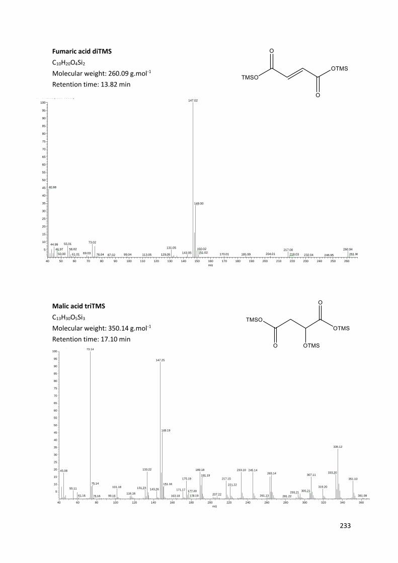
233
Fumaric acid diTMS
C10H20O4Si2
Molecular weight: 260.09 g.mol-1
Retention time: 13.82 min
Malic acid triTMS
C13H30O5Si3
Molecular weight: 350.14 g.mol-1
Retention time: 17.10 min
190403_749_poix_Pecci_50mg_opt13 #795-817 RT: 13.68-13.88 AV: 23 SB: 1874 13.98-26.63 , 8.99-13.65 NL: 4.30E4T: + c Full ms [40.00-650.00]
40 50 60 70 80 90 100 110 120 130 140 150 160 170 180 190 200 210 220 230 240 250 260 270
m/z
5
10
15
20
25
30
35
40
45
50
55
60
65
70
75
80
85
90
95
100
Re
lative
Ab
un
da
nce
147.02
40.98
149.00
73.0255.0144.99
131.05 150.0256.0246.97 260.94217.00151.02143.0569.0350.00 204.01170.01 261.96185.9961.01 218.0376.04 129.0599.04 113.0587.02 232.04 246.95 276.51
200628_malic_acid_std_200703033330 #1162-1180 RT: 17.17-17.36 AV: 19 SB: 2 24.70 , 14.49 NL: 2.72E7T: + c Full ms [40.00-650.00]
40 60 80 100 120 140 160 180 200 220 240 260 280 300 320 340 360
m/z
5
10
15
20
25
30
35
40
45
50
55
60
65
70
75
80
85
90
95
100
Re
lative
Ab
un
da
nce
73.14
147.25
149.19
335.12
133.22 189.18 233.10 245.1445.08333.20265.14
307.11191.19217.15175.19 351.10
75.14 151.16 221.22319.20101.18 131.2355.11 143.26 171.17 305.21177.20 293.21116.16 207.22178.19 261.13 361.0961.16 163.1899.1676.16 281.22

234
Tartaric acid tetraTMS
C16H38O6Si4
Molecular weight: 438.17 g.mol-1
Retention time: 21.78 min
Syringic acid diTMS
C15H26O5Si2
Molecular weight: 342.13 g.mol-1
Retention time: 30.92 min
200628_tartaric_acid_std #1534-1539 RT: 21.75-21.79 AV: 6 NL: 1.42E5T: + c Full ms [40.00-650.00]
40 60 80 100 120 140 160 180 200 220 240 260 280 300 320 340 360 380 400 420
m/z
5
10
15
20
25
30
35
40
45
50
55
60
65
70
75
80
85
90
95
100
Re
lative
Ab
un
da
nce
73.04
147.12
189.05
292.07
305.09
149.10
101.91
221.12333.02143.09
129.9945.03219.07
191.0774.08 171.09 277.06217.1075.09 263.05 423.03
367.13163.08 193.07 351.1090.97 231.0559.08 259.06115.06 320.91 395.03379.17343.09 437.72415.09359.10
200628_syringic_acid_std #2462-2467 RT: 30.89-30.93 AV: 6 NL: 3.86E6T: + c Full ms [40.00-650.00]
40 60 80 100 120 140 160 180 200 220 240 260 280 300 320 340 360
m/z
5
10
15
20
25
30
35
40
45
50
55
60
65
70
75
80
85
90
95
100
Re
lative
Ab
un
da
nce
297.09327.06
312.08
253.18
342.07
255.11 283.23329.12211.11 223.12
73.07271.17195.07150.12 344.10141.21 284.2289.06 179.1844.99 237.16 273.0875.07 209.16167.15 300.11212.1459.02 105.05 119.14 295.13 346.19

235
Butylated acids Dibutyl maleate
C12H20O4
Molecular weight: 228.29 g.mol-1
Retention time: 18.47 min
Dibutyl succinate
C10H18O4
Molecular weight: 230.30 g.mol-1
Retention time: 19.51 min 200707_DBSuc_std_MW #1421 RT: 19.42 AV: 1 NL: 7.75E7T: + c Full ms [40.00-650.00]
40 50 60 70 80 90 100 110 120 130 140 150 160 170 180 190 200 210 220 230 240
m/z
5
10
15
20
25
30
35
40
45
50
55
60
65
70
75
80
85
90
95
100
Re
lative
Ab
un
da
nce
100.99
55.12
230.9473.15156.84
41.07
101.9756.1445.10 100.26 232.0074.10 118.94 157.87102.97 174.9972.09 155.00145.0553.14 127.0183.22 139.01 215.03 228.9498.2959.04 91.07 200.94 242.89194.04184.36
200707_DBMaleic_DBSyr_std_MW_split20 #1296 RT: 18.50 AV: 1 NL: 5.68E6T: + c Full ms [40.00-650.00]
40 50 60 70 80 90 100 110 120 130 140 150 160 170 180 190 200 210 220 230
m/z
5
10
15
20
25
30
35
40
45
50
55
60
65
70
75
80
85
90
95
100
Re
lative
Ab
un
da
nce
98.98
116.9240.97
99.9757.01 70.99
54.97
52.94 155.0082.0643.04 126.02101.06 172.97139.0172.04 228.99108.99 143.0598.1567.0858.03 84.98 162.89 195.60 209.11 217.42185.79 234.11
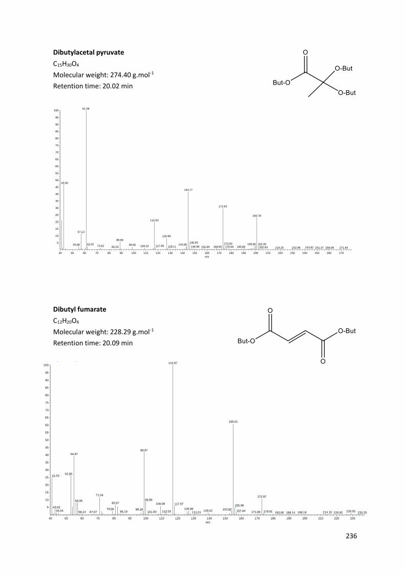
236
Dibutylacetal pyruvate
C15H30O4
Molecular weight: 274.40 g.mol-1
Retention time: 20.02 min
Dibutyl fumarate
C12H20O4
Molecular weight: 228.29 g.mol-1
Retention time: 20.09 min
210221_pyruvic_std_Butyle #1362 RT: 20.09 AV: 1 NL: 2.46E7T: + c Full ms [40.00-650.00]
40 50 60 70 80 90 100 110 120 130 140 150 160 170 180 190 200 210 220 230 240 250 260 270
m/z
5
10
15
20
25
30
35
40
45
50
55
60
65
70
75
80
85
90
95
100
Re
lative
Ab
un
da
nce
61.08
43.00
144.77
172.93
200.78
116.93
57.13
126.99
88.89145.83 173.92 199.95 201.8262.02 143.6055.06 99.0673.02 117.99109.02 128.11 146.98 168.65 190.68175.0085.03 155.89 202.83 243.82 271.83251.37219.25 232.98 258.08
200707_DBFum_std_MW_split20 #1460 RT: 20.11 AV: 1 NL: 2.89E6T: + c Full ms [40.00-650.00]
40 50 60 70 80 90 100 110 120 130 140 150 160 170 180 190 200 210 220 230
m/z
5
10
15
20
25
30
35
40
45
50
55
60
65
70
75
80
85
90
95
100
Re
lative
Ab
un
da
nce
116.97
155.01
98.97
54.97
52.9541.03
71.04172.97
99.9656.0680.97 109.09 117.97
155.9843.02
126.9979.06 98.19 153.9244.04 139.02 157.04 228.93112.03 174.0186.19 101.0067.07 171.0858.14 214.10131.01 198.18183.06 233.25220.92189.14

237
Dibutyl malate TMS
C15H30O5Si
Molecular weight: 318.49 g.mol-1
Retention time: 24.89 min
Dibutyl tartrate diTMS
C18H38O6Si2
Molecular weight: 406.67 g.mol-1
Retention time: 29.89 min 200702_DBT_std_Essai10_MW_split20 #2364-2366 RT: 29.99-30.00 AV: 3 SB: 277 30.00-31.60 , 28.89-29.99 NL: 3.43E5T: + c Full ms [40.00-650.00]
40 60 80 100 120 140 160 180 200 220 240 260 280 300 320 340 360 380 400
m/z
5
10
15
20
25
30
35
40
45
50
55
60
65
70
75
80
85
90
95
100
Re
lative
Ab
un
da
nce
73.06
276.04
147.10
133.03232.95189.04 216.93
75.05 129.97 149.08 220.01
176.97160.95117.05
335.02101.9445.01 205.03 289.02
175.08 234.9859.08 391.12304.9691.07
105.01 245.04135.0647.01 262.9776.09 291.13
61.03 317.07 338.03 406.85358.97 389.18
200707_DBMalic_std_MW_split20 #1988 RT: 24.89 AV: 1 NL: 1.55E7T: + c Full ms [40.00-650.00]
40 60 80 100 120 140 160 180 200 220 240 260 280 300 320 340
m/z
5
10
15
20
25
30
35
40
45
50
55
60
65
70
75
80
85
90
95
100
Re
lative
Ab
un
da
nce
73.10
101.07
144.91 161.00
117.02
216.9075.07
172.99
103.04
59.0971.10 162.85
131.0445.07 146.96143.06
174.92246.95
319.02176.94 219.90202.94190.8961.0847.07 121.07 303.0277.09 105.0389.11
229.03148.10133.08 320.07203.98 304.13290.98178.09 344.9170.22 248.99243.77 263.11 274.78

238
Butyl syringate TMS
C16H26O5Si
Molecular weight: 326.46 g.mol-1
Retention time: 36.50 min
Dibutyl azelaic acid
C17H32O4
Molecular weight: 300.44 g.mol-1
Retention time: 37.40 min
201007_DBSyr_std_MW_201006135014 #2959 RT: 36.19 AV: 1 NL: 2.42E5T: + c Full ms [40.00-650.00]
40 60 80 100 120 140 160 180 200 220 240 260 280 300 320
m/z
0
5
10
15
20
25
30
35
40
45
50
55
60
65
70
75
80
85
90
95
100
Re
lative
Ab
un
da
nce
240.11
311.16
296.15
326.08
312.16241.18
253.17223.11
255.13 327.16298.17225.15 242.17
196.28169.1273.14 270.06 328.1845.11 181.16 210.15137.19 153.11105.25 237.1181.09 91.20 251.27 280.2859.12 299.21125.19 314.09
310521_DBazelaic_std_MW #3548 RT: 40.77 AV: 1 NL: 9.58E4T: + c Full ms [40.00-650.00]
40 60 80 100 120 140 160 180 200 220 240 260 280 300
m/z
0
5
10
15
20
25
30
35
40
45
50
55
60
65
70
75
80
85
90
95
100
Re
lative
Ab
un
da
nce
170.93
124.97
226.97
55.0582.97
96.99128.94
151.97
40.92 171.97124.02
67.03 143.0179.05 107.04
43.01 96.03 207.12 228.07185.1173.05 281.12101.0156.07 193.05154.03123.0153.02 267.10220.99 244.97176.03 251.01 305.09231.15 294.91

239
Appendix 4. Retention time and main mass fragments of the
molecules
Molecule TMS Retention time (tr) Main mass fragments
Octanoic acid 12’34 75; 201
Maleic acid 12'96 147; 215; 245
Succinic acid 13'16 147; 172; 247; 262
Pyruvic acid 13’57 45; 66; 147; 217
Fumaric acid 13'82 147; 217; 261
Nonanoic acid 14’07 75; 215
Glutaric acid 15’01 147; 261
Decanoic acid 16’34 75; 229
Citramalic acid 16’52 147; 321
Mandelic acid 16’69 147; 296
Malic acid 17'10 147; 189; 233; 245; 335; 351
Adipic acid 17’65 149; 207
Cinnemic acid 18’83 161; 205; 220
m-benzoic acid 19’41 205; 267
Pimelic acid 20’70 155; 239
p-benzoic acid 21’39 223; 267
Tartaric acid 21'78 147; 189; 292; 305; 333; 423
Dodecanoic acid 22’32 75; 257
Napthalene 23’00 183; 198
Suberic acid 23’92 149; 309
Vannilic acid 26’14 267; 312
Isovannilic acid 26’3 297; 312
Azelaic acid 27’48 149; 317
Myristic acid 29’36 117; 285; 300
Syringic acid 30'92 253; 297; 312; 327; 342
Sebacic acid 31’05 331; 346
18-Norabiéta-8,11,13-triene 33’70 159; 241
19-Norabiéta-8,11,13-triene 34’99 159; 241
10,18-Bisnorabieta-5,7,9(10),11,13-triene
35’85 181; 223; 238
Palmitic acid 36’51 117; 313; 328
Tetrahydroretene 36’66 223; 314
10,18-Bisnorabieta-5,7,9(10),11,13-pentaene
37’63 223; 238
Retene 41’5 219; 234
Dehydroabietane 41’77 173; 255; 270
Oleic acid 42’32 117; 128; 198; 338; 356
Stearic acid 43’32 117; 341; 356
Pimaric acid 44’46 121; 241; 257; 359
Sandaracopimaric acid 45’04 121; 241; 257; 359
Isopimaric acid 45’62 241; 256; 359
DHAM 45’79 239; 314
Palustric acid 46’28 241; 359; 374
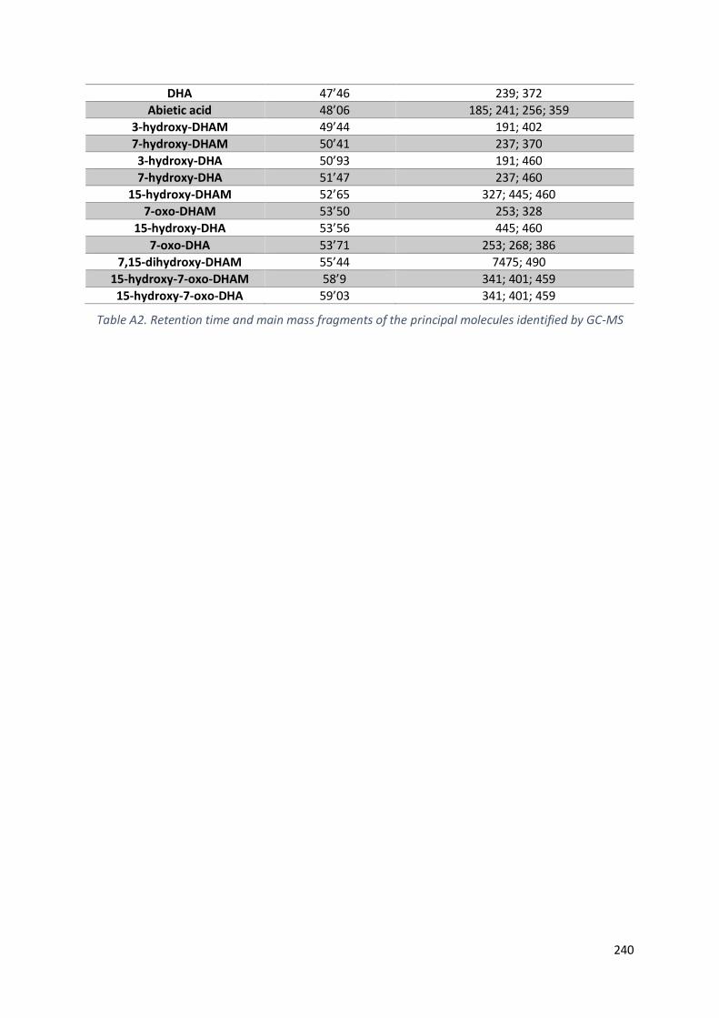
240
DHA 47’46 239; 372
Abietic acid 48’06 185; 241; 256; 359
3-hydroxy-DHAM 49’44 191; 402
7-hydroxy-DHAM 50’41 237; 370
3-hydroxy-DHA 50’93 191; 460
7-hydroxy-DHA 51’47 237; 460
15-hydroxy-DHAM 52’65 327; 445; 460
7-oxo-DHAM 53’50 253; 328
15-hydroxy-DHA 53’56 445; 460
7-oxo-DHA 53’71 253; 268; 386
7,15-dihydroxy-DHAM 55’44 7475; 490
15-hydroxy-7-oxo-DHAM 58’9 341; 401; 459
15-hydroxy-7-oxo-DHA 59’03 341; 401; 459
Table A2. Retention time and main mass fragments of the principal molecules identified by GC-MS