optimization of precursor preparation in psma-11 ... - mdpi
TRANSCRIPT
Citation: Iudicello, A.; Boschi, S.;
Ghedini, P.; Lohr, F.; Panareo, S.
Optimization of Precursor
Preparation in PSMA-11
Radiolabeling to Obtain a Highly
Reproducible Radiochemical Yield.
Pharmaceuticals 2022, 15, 343.
https://doi.org/10.3390/
ph15030343
Academic Editor: Irina Velikyan
Received: 7 February 2022
Accepted: 7 March 2022
Published: 11 March 2022
Publisher’s Note: MDPI stays neutral
with regard to jurisdictional claims in
published maps and institutional affil-
iations.
Copyright: © 2022 by the authors.
Licensee MDPI, Basel, Switzerland.
This article is an open access article
distributed under the terms and
conditions of the Creative Commons
Attribution (CC BY) license (https://
creativecommons.org/licenses/by/
4.0/).
pharmaceuticals
Article
Optimization of Precursor Preparation in PSMA-11 Radiolabelingto Obtain a Highly Reproducible Radiochemical YieldAntonella Iudicello 1,2,* , Stefano Boschi 3 , Pietro Ghedini 2, Frank Lohr 4 and Stefano Panareo 2
1 Pharmaceutical Department, Azienda USL di Modena, Largo del Pozzo 71, 41121 Modena, Italy2 Nuclear Medicine Unit, Department of Oncology and Hematology, Azienda Ospedaliero-Universitaria di
Modena, Largo del Pozzo 71, 41124 Modena, Italy; [email protected] (P.G.);[email protected] (S.P.)
3 Department for Life Quality Studies, University of Bologna, Corso D’Augusto, 237, 47921 Rimini, Italy;[email protected]
4 Radiotherapy Unit, Department of Oncology and Hematology, Azienda Ospedaliero-Universitaria di Modena,Largo del Pozzo 71, 41124 Modena, Italy; [email protected]
* Correspondence: [email protected]; Tel.: +39-059-422-5167
Abstract: [68Ga]Ga-PSMA-11 PET/CT plays a pivotal role in the diagnosis and staging of prostatecancer because of its higher sensitivity and detection rate compared with traditional choline PET/CT.A highly reproducible radiochemical yield of the radiopharmaceutical to be used in the clinical routineis an important parameter for planning and optimization of clinical activity. During radiometallationof PSMA-11, the presence of metal ion contaminants in the peptide precursor may cause a decreasein the [68Ga]Ga-PSMA-11 radiochemical yield because of metal ion contaminants competition withgallium-68. To optimize the radiochemical yield of [68Ga]Ga-PSMA-11 radiosynthesis, data obtainedby preparing the solution of the PSMA-11 precursor with three different methods (A, B, and C)were compared. Methods A and B consisted of the reconstitution of different quantities of precursor(1000 µg and 30 µg, respectively) to obtain a 1 µg/mL solution. In Method A, the precursor solutionwas aliquoted and stored frozen, while the precursor solution obtained with Method B was entirelyused. Method C consisted of the reconstitution of 1000 µg of precursor taking into account netpeptide content as described in European Pharmacopoeia. Radiosynthesis data demonstrated thatreconstitution methods B and C gave a consistently higher and reproducible radiochemical yield,highlighting the role of metals and precursor storage conditions on the synthesis performance.
Keywords: gallium-68; PSMA-11; radiochemical yield; molar activity; net precursor content; clini-cal routine
1. Introduction
Chemical precursors for radiopharmaceutical preparations are non-radioactive sub-stances obtained by chemical synthesis to be combined with a radionuclide (in contrast toprecursors manufactured using substances of human or animal origin) [1].
Chemical precursors require an adequate characterization, according to the generalrequirements, as a part of their quality assurance in order to demonstrate the safety andefficacy of the final radiopharmaceutical preparation [1,2].
Stability testing is part of the chemical precursor’s characterization. The purpose ofstability testing is to provide evidence on how the quality of a substance varies with timeunder the influence of a variety of environmental factors such as temperature, humidity,and light, and to establish a re-test period and recommended storage conditions [3].
The PSMA (prostate-specific membrane antigen) conjugated to HBED-CC (N,N’-bis[2-hydroxy-5-(carboxyethyl)benzyl]ethylenediamine-N,N’-diacetic acid), better known asPSMA-11, is one of the most widespread precursors for PET imaging of prostate cancer
Pharmaceuticals 2022, 15, 343. https://doi.org/10.3390/ph15030343 https://www.mdpi.com/journal/pharmaceuticals
Pharmaceuticals 2022, 15, 343 2 of 11
after radiolabeling with gallium-68 because the PSMA is highly expressed on most prostatecancer (PCa) cells (Figure 1).
Pharmaceuticals 2022, 15, x FOR PEER REVIEW 2 of 12
The PSMA (prostate-specific membrane antigen) conjugated to HBED-CC (N,N’-bis [2-hydroxy-5-(carboxyethyl)benzyl]ethylenediamine-N,N’-diacetic acid), better known as PSMA-11, is one of the most widespread precursors for PET imaging of prostate cancer after radiolabeling with gallium-68 because the PSMA is highly expressed on most prostate cancer (PCa) cells (Figure 1).
Figure 1. Chemical structure of PSMA-11.
Stability testing for the PSMA-11 has provided evidence that the quality of precursor in aqueous solution varies with time due to the presence of Fe (III) in the precursor ma-terial and the consequent interaction between HBED-CC and Fe (III) already at room temperature [4].
The formation of the complex between HBED-CC and Fe (III) in the PSMA-11 aqueous solutions causes a decrease of the [68Ga]Ga-PSMA-11 radiochemical yield (RCY) because the Fe (III) competes with gallium-68 during the complexation reac-tion with the chelator [4].
Generally, a highly reproducible % RCY of the tracers to be used in the clinical routine is an important parameter for the planning and optimization of clinical activity. Radiochemical M3+-L complex formation yields, in most cases, can be increased by providing a higher concentration of the ligand [5].
However, a high molar activity (Am) or specific activity (As) of the radiopharmaceu-tical is preferred, and it is known that if the precursor (ligand) increases, the molar activi-ty decreases [6].
Recent EANM guidelines on harmonization of the molar activity or specific activity of radiopharmaceuticals reported that in the case of radiometallation of peptides the presence in the peptide precursor of metal ion contaminants indirectly affects Am by ne-cessitating higher amounts of peptide precursor to achieving high radionuclide incor-poration. For such radiotracers, we are therefore normally referring to the apparent Am, empirically determined by dividing the amount of radioactivity present in an exact vol-ume of the final formulation by the amount of peptide precursor used in the labeling process in mol (or µmol) [6,7].
Nevertheless, if a monograph has been published such as e.g., for [68Ga]Ga-PSMA-11, the maximum amount of precursor (ligand) to use in the synthesis has already been defined.
Figure 1. Chemical structure of PSMA-11.
Stability testing for the PSMA-11 has provided evidence that the quality of precursorin aqueous solution varies with time due to the presence of Fe (III) in the precursormaterial and the consequent interaction between HBED-CC and Fe (III) already at roomtemperature [4].
The formation of the complex between HBED-CC and Fe (III) in the PSMA-11 aqueoussolutions causes a decrease of the [68Ga]Ga-PSMA-11 radiochemical yield (RCY) becausethe Fe (III) competes with gallium-68 during the complexation reaction with the chelator [4].
Generally, a highly reproducible % RCY of the tracers to be used in the clinical routineis an important parameter for the planning and optimization of clinical activity. Radio-chemical M3+-L complex formation yields, in most cases, can be increased by providing ahigher concentration of the ligand [5].
However, a high molar activity (Am) or specific activity (As) of the radiopharmaceuticalis preferred, and it is known that if the precursor (ligand) increases, the molar activitydecreases [6].
Recent EANM guidelines on harmonization of the molar activity or specific activity ofradiopharmaceuticals reported that in the case of radiometallation of peptides the presencein the peptide precursor of metal ion contaminants indirectly affects Am by necessitatinghigher amounts of peptide precursor to achieving high radionuclide incorporation. Forsuch radiotracers, we are therefore normally referring to the apparent Am, empiricallydetermined by dividing the amount of radioactivity present in an exact volume of the finalformulation by the amount of peptide precursor used in the labeling process in mol (orµmol) [6,7].
Nevertheless, if a monograph has been published such as e.g., for [68Ga]Ga-PSMA-11,the maximum amount of precursor (ligand) to use in the synthesis has already been defined.
In this manuscript, we present the data obtained by preparing the solution of PSMA-11synthesis precursor following various methods in order to obtain a high % RCY that is alsoreproducible over time. All tested methods took into account that: generally, a high Am ispreferred; the European Pharmacopoeia (Ph. Eur.) Monograph Gallium (68Ga) PSMA-11injection (3044) prescribes to use of a maximum of 30 µg of PSMA-11 for [68Ga]Ga-PSMA-11 synthesis [8]; the Ph. Eur. monographs are referred to net precursor content, and thelyophilized PSMA-11 provided by the manufacturer contains also water, counter ions, andresidual solvents [3].
The net peptide content is the percentage of peptide material in the lyophilisate.
Pharmaceuticals 2022, 15, 343 3 of 11
2. Materials and Methods2.1. Chemicals and Reagents
Gallium-68 (t1/2 = 68 min, β + = 89%, and EC = 11%) for the [68Ga]Ga-PSMA-11production, was routinely obtained as gallium-68 chloride ([68Ga]GaCl3) solution froma pharmaceutical-grade 1.85 GBq germanium-68/gallium-68 generator (GalliaPharm®,Eckert&Ziegler Radiopharma GmbH, Berlin, Germany).
Sterile and ultrapure 0.1 M Hydrochloric Acid (HCl) for the elution of germanium68/gallium-68 generator was purchased from Eckert & Ziegler Radiopharma GmbH(Berlin, Germany).
Synthesis was performed on a fully automated radiosynthesis cassette module (GAIAV2™, Elysia-Raytest, Straubenhard, Germany), operating in a laminar flow isolator class A.
The detectors of the synthesis module were calibrated with a dose calibrator (ISOMED2010, MED Nuklear-Medizintechnik Dresden GmbH, Dresden, Germany) as reference.
PSMA-11 lyophilized vials for [68Ga]Ga-PSMA-11 radiosynthesis, natGa-PSMA-11reference standard for [68Ga]Ga-PSMA-11, disposable sterile cassettes (SCX fluidic kitfor the [68Ga]Ga-labeling of peptides), and disposable reagent kits (SCX reagent kit forthe [68Ga]Ga-labeling of peptides) containing all consumables necessary for gallium-68radiolabelling of peptides except peptide, all were of GMP grade and were purchased fromAdvanced Biochemical Compounds, ABX (Radeberg, Germany).
Ammonium Acetate, Methanol, and Acetonitrile (ACN) were purchased from CarloErba Reagents S.r.l. (Cornaredo, Milan, Italy); Trifluoroacetic Acid (TFA) and metal-freewater (Fluka Water Trace-Select® for trace Analysis) were purchased from Merck LifeScience S.r.l. (Milan, Italy); Ultrapure water (Milli-Q, 18.2 MΩ) was obtained from a Milli-Q® IQ Element purification (Merck KGaA, Darmstadt, Germany). All chemicals were ofanalytical grade and they were used without further purification. HPLC eluents (Milli-Qwater, ACN, and TFA) were of high-grade purity.
2.2. Precursor Preparation and Data Collection
The aqueous solution of PSMA-11 synthesis precursor (1 µg/µL) for [68Ga]Ga-PSMA-11 radiosynthesis was obtained as follows:
• reconstituting a 1000 µg vial of PSMA-11 precursor for [68Ga]Ga-PSMA-11 with1000 µL of metal-free water (hereinafter Method A);
• reconstituting a 30 µg vial of PSMA-11 precursor for [68Ga]Ga-PSMA-11 with 30 µL ofmetal-free water (hereinafter Method B);
• reconstituting a 1000 µg vial of PSMA-11 precursor for [68Ga]Ga-PSMA-11 with 840 µLof metal-free water (hereinafter Method C), considering that the percentage of peptidematerial in the lyophilisate (i.e., net precursor content) was 84% according to CoAprovided by the manufacturer.
The precursor solutions obtained with Methods A and C were immediately aliquoted(30 µL) in 0.5 mL Eppendorf tubes and frozen at −25 C. For method B, the precursorsolution was reconstituted just before starting the synthesis.
To perform the [68Ga]Ga-PSMA-11 synthesis, 30 µL of precursor solution (1 µg/µL)was used. Each method described was used ten times within 1–2 months.
Since 30 µg vials of PSMA-11 precursor were supplied in a box of 5 vials × 30 µg andone vial was used to prepare the reference solution b in order to perform the quality controlof the [68Ga]Ga-PSMA-11 injectable solutions according to Ph. Eur. Monograph Gallium(68Ga)PSMA-11 injection (3044) [8], Method B was used nine times.
2.3. Automated [68Ga]Ga-PSMA-11 Synthesis
Synthesis was performed utilizing the standard radiolabelling method [9–12]. Analiquot of 30 µL of the aqueous solution of PSMA-11 synthesis precursor (1 µg/µL), takenwith a precision micropipette (Pipetman® Gilson, variable volume 10–100 µL), was radiola-belled in 3.00 ± 0.2 mL ammonium acetate buffer (0.08 M; reagent kit) and 550.0 ± 78 µL
Pharmaceuticals 2022, 15, 343 4 of 11
eluent (reagent kit) with 785 ± 323 MBq gallium-68. After radiolabelling (90 ± 2 C;250.0 ± 13 s), the reaction mixture was passed over a C18 cartridge (reagent kit) andwashed with sterile water for injection (reagent kit). The purified product was eluted with1.5 mL 60 vol% ethanol (reagent kit) and 15 mL saline solution (0.9% sodium chloride)followed by sterile filtration to obtain the final formulation.
2.4. Quality Control
For quality control, an aliquot of 100 µL was retained from the final product beforemeasurement of the radioactivity.
The quality control was performed according to the specifications given by the Ph.Eur. in the Monograph Gallium (68Ga) PSMA-11 injection (3044) [8].
Radiochemical purity (RCP), Chemical purity (CP), and identification of the productspecies were determined using radioHPLC analysis. Additionally, RCP was also deter-mined by radio thin-layer chromatography (radioTLC).
Thin-layer chromatography was performed using a glass microfiber chromatographypaper impregnated with silica-gel (iTLC-SG, Agilent Technologies Italia Spa, Cernusco SulNaviglio, Italy) developed in 1 M ammonium acetate/methanol (1:1) and analyzed using asingle trace radio TLC-scanner (PET-miniGita, Elysia-Raytest, Straubenhardt, Germany)and evaluation software (Gina Star TLC, Elysia-Raytest, Straubenhardt, Germany).
RadioHPLC analysis was performed on a Thermo Scientific Dionex Ultimate 3000 HPLCsystem (Thermo Scientific, Bremen, Germany) equipped with LPG-3400SD pump, TCC-3000 column oven, UV VWD-3100 detector, and radiometric detector at NaI (Gabi Star,Elysia-Raytest, Straubenhardt, Germany) connected in series. Reversed-Phase High-Performance Liquid Chromatography (RP-HPLC; ACE 3 µm C18, l = 0.6 m, Ø = 7 mm;Thermo Scientific, Bremen, Germany) with a linear A–B gradient (0–0.5 min 5% B,0.5–10 min 5% B to 40% B, 10–11 min 40% B to 5% B, 11–16 min 5% B) at a flow rateof 0.6 mL/min and a total run time of 16 min was performed. Solvent A consisted of 0.1%TFA in Milli-Q water and solvent B of 0.1% TFA in ACN.
UV absorbance was measured at 280 nm. The column temperature was kept at 24 C.The injection volume was 20 µL. The Chromeleon data system software (Version 7.2.8) wasused for data acquisition and mathematical calculations.
The approximate half-life of gallium-68 was determined using the dose calibrator(ISOMED 2010, MED Nuklear-Medizintechnik Dresden GmbH, Dresden, Germany).
For radionuclidic identification and determination of germanium-68 breakthrough, theenergy of gamma photons was measured using a CZT-based gamma-ray detector designedand produced by KromekTM (Sedgefield, County Durham, UK) [13] and evaluation soft-ware (MultiSpect Analysis gamma spectroscopy software version 22.2, Sedgefield, CountyDurham, UK).
The appearance was checked visually. pH was measured using micro-electrode and amillivoltmeter (pH meter) from Thermo Scientific™ Orion™ Dual Star.
Bacterial endotoxins were evaluated with Endosafe® Portable Testing System™ (PTS™)portable system for endotoxin testing (Charles River, Charleston, SC, USA). The methodused to check the filter integrity was the bubble point test, which was automatically carriedout by the radiosynthesis module to reduce the radiation exposure of the operators.
2.5. Radiochemical Yield (RCY) and Radioactivity at EOS Determination
Performance evaluation of the used methods was performed in terms of % RCY andactivity at the end of the synthesis (EOS).
RCY was calculated based on the radioactivity of the final product vial, measuredusing a dose calibrator (ISOMED 2010, MED Nuklear-Medizintechnik Dresden GmbH,Dresden, Germany), and the radioactivity of the eluate of gallium-68 collected at the end ofa generator elution performed 24 h before the radiosynthesis and measured with the dosecalibrator. This value was decay-corrected at EOS.
Pharmaceuticals 2022, 15, 343 5 of 11
The radioactivity at EOS was the amount of radioactivity of the final product calculatedusing the dose calibrator and expressed in MBq.
All data obtained from routine clinical production were retrospectively analyzed.
2.6. Statistical Analysis
All data were expressed as mean ± standard deviation (S.D.) and percentage ofcoefficient of variation (% CV) to monitor the reliability and reproducibility of methods,and they were reported in a summary table (Table 1). The coefficient of variation shouldnot exceed 5% (acceptance criterion).
Table 1. Comparison of [68Ga]Ga-PSMA-11 synthesis data from three methods.
Method A Method B Method C
Generatorcalibration date 2020/08/11 a 2020/08/11 b 2020/08/11 c
Precursorreconstitution
date2020/11/10 Before starting synthesis 2021/03/16
Process number Date RCY, % dRadioactivity
at EOS,MBq d
Date RCY, % dRadioactivity
at EOS,MBq d
Date RCY, % dRadioactivity
at EOS,MBq d
1 2020/11/10 77 570 2021/02/09 82 482 2021/03/16 83 4422 2020/11/11 76 560 2021/02/10 72 422 2021/03/17 84 4483 2020/11/12 77 567 2021/02/16 79 460 2021/03/24 79 4164 2020/11/17 70 512 2021/02/17 79 457 2021/03/30 81 4115 2020/11/18 74 537 2021/02/23 84 478 2021/03/31 78 4016 2020/11/19 72 520 2021/03/02 82 460 2021/04/07 79 3997 2020/11/24 72 511 2021/03/03 76 425 2021/04/21 79 3878 2020/12/01 66 465 2021/03/09 84 456 2021/04/22 78 3809 2020/12/02 64 448 2021/03/10 78 426 2021/05/05 80 372
10 2020/12/10 58 401 n.d. n.d. 2021/05/12 78 360
Means 70.6 509.1 79.6 451.8 79.9 401.6SD 6.24 55.70 3.94 22.50 2.13 28.62
CV% 8.84 4.95 2.67
a Germanium-68/gallium-68 generator at the start of shelf life. b Germanium-68/gallium-68 generator in themiddle of shelf life. c Germanium-68/gallium-68 generator at the end of shelf life. d Non-decay corrected. Notes:As shown in Table 1, assuming 70-kg patients and administering 2-MBq/kg body weight, for Method A, overtime, one less patient dose would be available resulting in a need for another synthesis including all consequences(e.g., radiation exposure for the operator, costs for materials).
To compare the different methods a two-sample t-test with a significance level ofα = 0.05 (95% of confidence interval) was used. The null hypothesis assumes that nostatistically significant difference exists between the methods means.
3. Results
Table 1 shows [68Ga]Ga-PSMA-11 synthesis data obtained from three different meth-ods of precursor preparation.
The data set includes data from all batches of [68Ga]Ga-PSMA-11 produced consecu-tively and starting from the day of precursor solution preparation for Methods A and C.None of the syntheses was excluded.
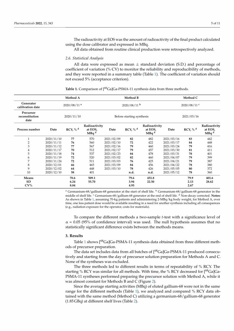
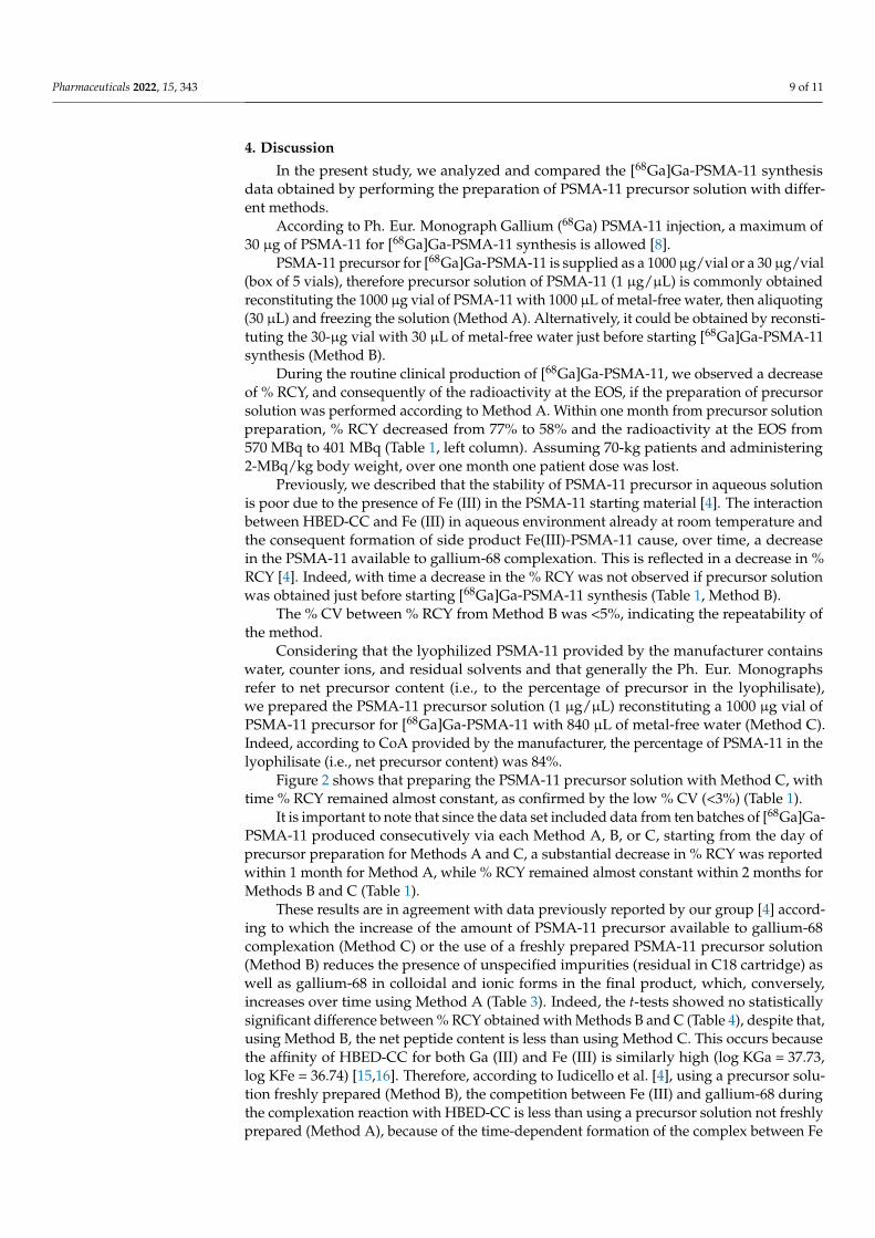
The three methods led to different results in terms of repeatability of % RCY. Thestarting % RCY was similar for all methods. With time, the % RCY decreased for [68Ga]Ga-PSMA-11 syntheses performed preparing the precursor solution with Method A, while itwas almost constant for Methods B and C (Figure 2).
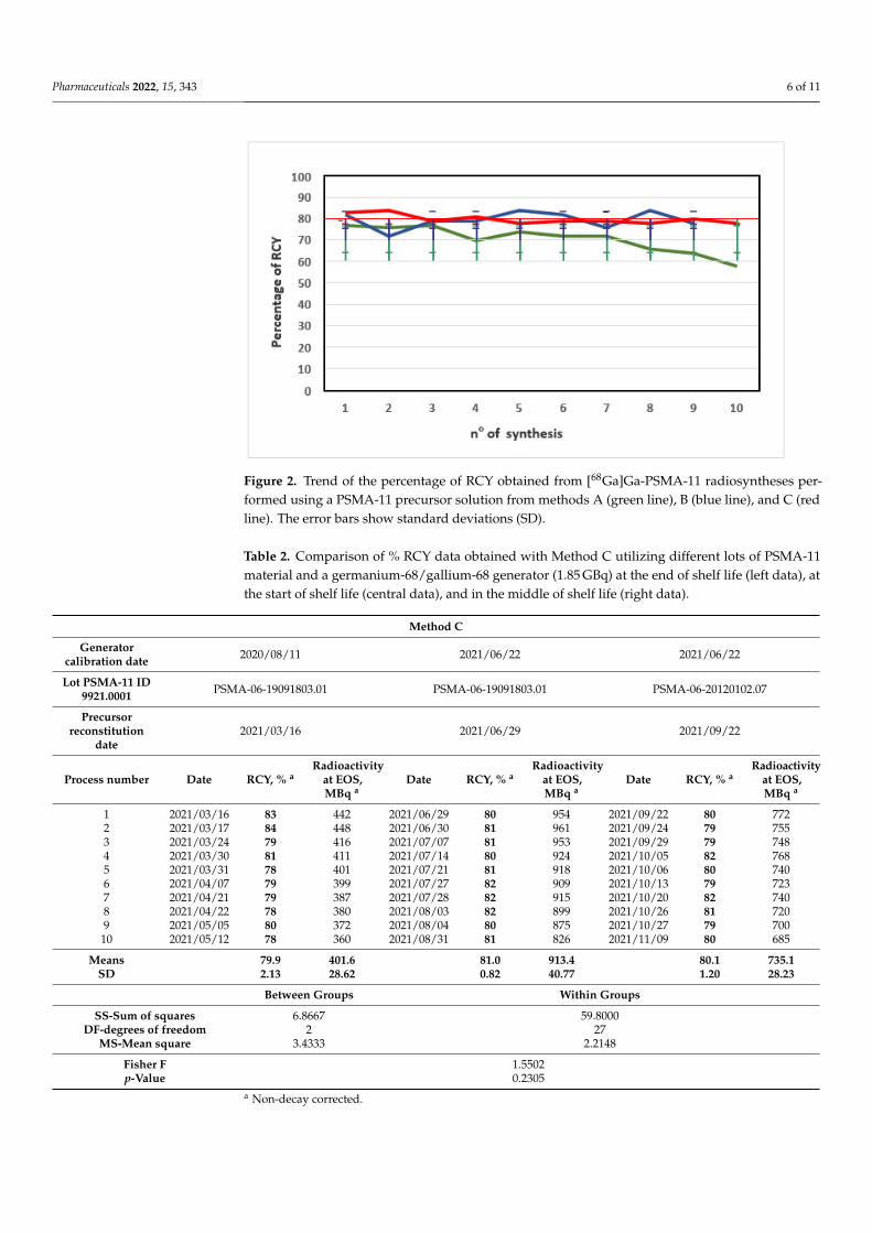
Since the average starting activities (MBq) of eluted gallium-68 were not in the samerange for the different methods (Table 1), we analyzed and compared % RCY data ob-tained with the same method (Method C) utilizing a germanium-68/gallium-68 generator(1.85 GBq) at different shelf lives (Table 2).
Pharmaceuticals 2022, 15, 343 6 of 11
Pharmaceuticals 2022, 15, x FOR PEER REVIEW 6 of 12
Table 1 shows [68Ga]Ga-PSMA-11 synthesis data obtained from three different methods of precursor preparation.
The data set includes data from all batches of [68Ga]Ga-PSMA-11 produced consec-utively and starting from the day of precursor solution preparation for Methods A and C. None of the syntheses was excluded.
The three methods led to different results in terms of repeatability of % RCY. The starting % RCY was similar for all methods. With time, the % RCY decreased for [68Ga]Ga-PSMA-11 syntheses performed preparing the precursor solution with Method A, while it was almost constant for Methods B and C (Figure 2).
Figure 2. Trend of the percentage of RCY obtained from [68Ga]Ga-PSMA-11 radiosyntheses per-formed using a PSMA-11 precursor solution from methods A (green line), B (blue line), and C (red line). The error bars show standard deviations (SD).
Since the average starting activities (MBq) of eluted gallium-68 were not in the same range for the different methods (Table 1), we analyzed and compared % RCY data ob-tained with the same method (Method C) utilizing a germanium-68/gallium-68 generator (1.85 GBq) at different shelf lives (Table 2).
Table 2. Comparison of % RCY data obtained with Method C utilizing different lots of PSMA-11 material and a germanium-68/gallium-68 generator (1.85 GBq) at the end of shelf life (left data), at the start of shelf life (central data), and in the middle of shelf life (right data).
Method C Generator calibration date 2020/08/11 2021/06/22 2021/06/22
Lot PSMA-11 ID 9921.0001 PSMA-06-19091803.01 PSMA-06-19091803.01 PSMA-06-20120102.07
Precursor reconstitution date
2021/03/16 2021/06/29 2021/09/22
Process number Date RCY, %a Radioactivity at EOS, MBq a Date RCY, %a Radioactivity
at EOS, MBq a Date RCY, %a Radioactivity at EOS, MBq a
1 2021/03/16 83 442 2021/06/29 80 954 2021/09/22 80 772
Figure 2. Trend of the percentage of RCY obtained from [68Ga]Ga-PSMA-11 radiosyntheses per-formed using a PSMA-11 precursor solution from methods A (green line), B (blue line), and C (redline). The error bars show standard deviations (SD).
Table 2. Comparison of % RCY data obtained with Method C utilizing different lots of PSMA-11material and a germanium-68/gallium-68 generator (1.85 GBq) at the end of shelf life (left data), atthe start of shelf life (central data), and in the middle of shelf life (right data).
Method C
Generatorcalibration date 2020/08/11 2021/06/22 2021/06/22
Lot PSMA-11 ID9921.0001 PSMA-06-19091803.01 PSMA-06-19091803.01 PSMA-06-20120102.07
Precursorreconstitution
date2021/03/16 2021/06/29 2021/09/22
Process number Date RCY, % aRadioactivity
at EOS,MBq a
Date RCY, % aRadioactivity
at EOS,MBq a
Date RCY, % aRadioactivity
at EOS,MBq a
1 2021/03/16 83 442 2021/06/29 80 954 2021/09/22 80 7722 2021/03/17 84 448 2021/06/30 81 961 2021/09/24 79 7553 2021/03/24 79 416 2021/07/07 81 953 2021/09/29 79 7484 2021/03/30 81 411 2021/07/14 80 924 2021/10/05 82 7685 2021/03/31 78 401 2021/07/21 81 918 2021/10/06 80 7406 2021/04/07 79 399 2021/07/27 82 909 2021/10/13 79 7237 2021/04/21 79 387 2021/07/28 82 915 2021/10/20 82 7408 2021/04/22 78 380 2021/08/03 82 899 2021/10/26 81 7209 2021/05/05 80 372 2021/08/04 80 875 2021/10/27 79 700
10 2021/05/12 78 360 2021/08/31 81 826 2021/11/09 80 685
Means 79.9 401.6 81.0 913.4 80.1 735.1SD 2.13 28.62 0.82 40.77 1.20 28.23
Between Groups Within Groups
SS-Sum of squares 6.8667 59.8000DF-degrees of freedom 2 27
MS-Mean square 3.4333 2.2148
Fisher F 1.5502p-Value 0.2305
a Non-decay corrected.
Pharmaceuticals 2022, 15, 343 7 of 11
The analysis of variance (ANOVA) allowed evaluating the % RCY data with the post-Fischer test using a significance level of α = 0.05 (95% of confidence interval). As shown inTable 2, the calculated F-value (1.5502) is lower than the tabulated F-value (3.35), indicatingno statistically significant difference between % RCY data obtained from Method C utilizinga germanium-68/gallium-68 generator at the end, at the start, or in the middle of shelf life(p > 0.05). It can be assumed that this result may be transferred to other methods.
3.1. Quality Control
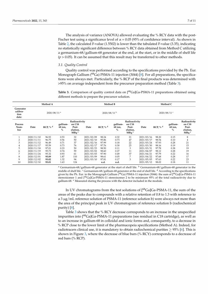
Quality control was performed according to the specifications provided by the Ph. Eur.Monograph Gallium (68Ga) PSMA-11 injection (3044) [8]. For all preparations, the specifica-tions were always met. Particularly, the % RCP of the final products was determined with>95% on average independent from the precursor preparation method (Table 3).
Table 3. Comparison of quality control data on [68Ga]Ga-PSMA-11 preparations obtained usingdifferent methods to prepare the precursor solution.
Method A Method B Method C
Generatorcalibra-
tiondate:
2020/08/11 a 2020/08/11 b 2020/08/11 c
ProcessNum-ber
Date RCP, % dgallium-68 ion,
%
Radioactivityon C18
Post-elution,MBq e
Date RCP, % dgallium-68 ion,
%
Radioactivityon C18
Post-elution,MBq e
Date RCP, % dgallium-68 ion,
%
Radioactivityon C18
Post-elution,MBq e
1 2020/11/10 96.92 0.53 32 2021/02/09 98.24 0.32 21 2021/03/16 98.20 0.15 82 2020/11/11 96.75 0.56 50 2021/02/10 95.86 0.73 47 2021/03/17 98.54 0.10 43 2020/11/12 96.49 0.71 37 2021/02/16 97.99 0.18 22 2021/03/24 97.93 0.28 214 2020/11/17 95.59 0.73 76 2021/02/17 97.76 0.30 25 2021/03/30 98.16 0.19 155 2020/11/18 97.01 0.33 50 2021/02/23 98.50 0.11 3 2021/03/31 97.78 0.38 196 2020/11/19 95.73 0.74 59 2021/03/02 98.60 0.27 11 2021/04/07 98.12 0.28 197 2020/11/24 95.32 0.93 61 2021/03/03 97.90 0.49 37 2021/04/21 97.79 0.22 168 2020/12/01 95.09 1.87 110 2021/03/09 98.61 0.05 7 2021/04/22 97.68 0.28 179 2020/12/02 95.02 1.32 94 2021/03/10 97.81 0.17 3 2021/05/05 97.63 0.22 23
10 2020/12/10 95.01 1.63 124 n.d. n.d. 2021/05/12 98.02 0.18 11
a Germanium-68/gallium-68 generator at the start of shelf life. b Germanium-68/gallium-68 generator in themiddle of shelf life. c Germanium-68/gallium-68 generator at the end of shelf life. d According to the specificationsgiven by the Ph. Eur. in the Monograph Gallium (68Ga) PSMA-11 injection (3044): the sum of [68Ga]Ga-PSMA-11stereoisomer 1 and [68Ga]Ga-PSMA-11 stereoisomer 2 to be minimum 95% of the total radioactivity due togallium-68. e Measured during the process with the detector included in the module.
In UV chromatograms from the test solutions of [68Ga]Ga-PSMA-11, the sum of theareas of the peaks due to compounds with a relative retention of 0.8 to 1.3 with reference toa 3 µg/mL reference solution of PSMA-11 (reference solution b) were always not more thanthe area of the principal peak in UV chromatogram of reference solution b (radiochemicalpurity) [8].
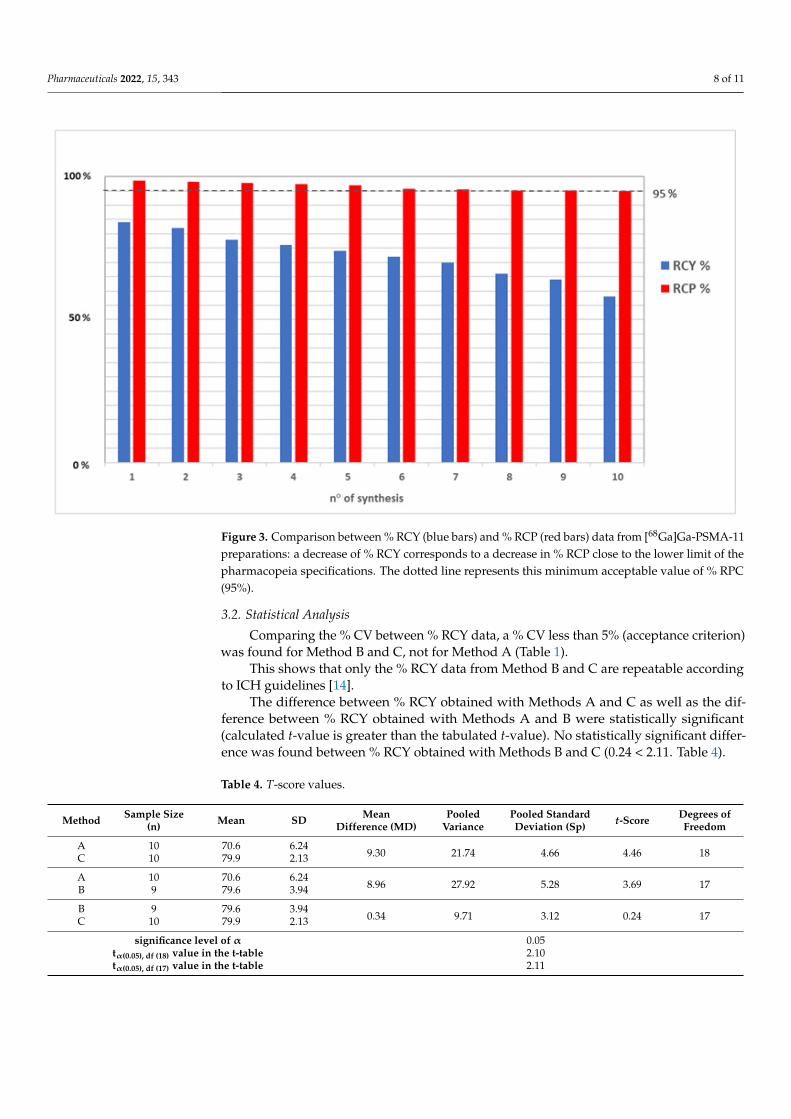
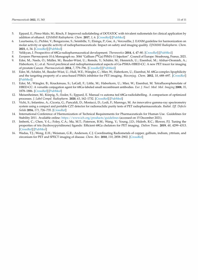
Table 3 shows that the % RCY decrease corresponds to an increase in the unspecifiedimpurities into [68Ga]Ga-PSMA-11 preparations (see residual in C18 cartridge), as well asto an increase in gallium-68 in colloidal and ionic forms and, consequently, to a decrease in% RCP close to the lower limit of the pharmacopeia specifications (Method A). Indeed, forradiotracers clinical use, it is mandatory to obtain radiochemical purities ≥ 95% [6]. This isshown in Figure 3, where the decrease of blue bars (% RCY) corresponds to a decrease ofred bars (% RCP).
Pharmaceuticals 2022, 15, 343 8 of 11
Pharmaceuticals 2022, 15, x FOR PEER REVIEW 9 of 12
Figure 3. Comparison between % RCY (blue bars) and % RCP (red bars) data from [68Ga]Ga-PSMA-11 preparations: a decrease of % RCY corresponds to a decrease in % RCP close to the lower limit of the pharmacopeia specifications. The dotted line represents this minimum ac-ceptable value of % RPC (95%).
3.2. Statistical Analysis Comparing the % CV between % RCY data, a % CV less than 5% (acceptance cri-
terion) was found for Method B and C, not for Method A (Table 1). This shows that only the % RCY data from Method B and C are repeatable according
to ICH guidelines [14]. The difference between % RCY obtained with Methods A and C as well as the dif-
ference between % RCY obtained with Methods A and B were statistically significant (calculated t-value is greater than the tabulated t-value). No statistically significant difference was found between % RCY obtained with Methods B and C (0.24 < 2.11. Table 4).
Table 4. T-score values.
Method Sample Size (n)
Mean SD Mean Difference (MD)
Pooled Variance
Pooled Standard Deviation (Sp)
t-Score Degrees of Freedom
A 10 70.6 6.24 9.30 21.74 4.66 4.46 18
C 10 79.9 2.13 A 10 70.6 6.24
8.96 27.92 5.28 3.69 17 B 9 79.6 3.94 B 9 79.6 3.94
0.34 9.71 3.12 0.24 17 C 10 79.9 2.13 significance level of α0.05
tα(0.05), df (18) value in the t-table2.10
Figure 3. Comparison between % RCY (blue bars) and % RCP (red bars) data from [68Ga]Ga-PSMA-11preparations: a decrease of % RCY corresponds to a decrease in % RCP close to the lower limit of thepharmacopeia specifications. The dotted line represents this minimum acceptable value of % RPC(95%).
3.2. Statistical Analysis
Comparing the % CV between % RCY data, a % CV less than 5% (acceptance criterion)was found for Method B and C, not for Method A (Table 1).
This shows that only the % RCY data from Method B and C are repeatable accordingto ICH guidelines [14].
The difference between % RCY obtained with Methods A and C as well as the dif-ference between % RCY obtained with Methods A and B were statistically significant(calculated t-value is greater than the tabulated t-value). No statistically significant differ-ence was found between % RCY obtained with Methods B and C (0.24 < 2.11. Table 4).
Table 4. T-score values.
Method Sample Size(n) Mean SD Mean
Difference (MD)Pooled
VariancePooled StandardDeviation (Sp) t-Score Degrees of
Freedom
A 10 70.6 6.249.30 21.74 4.66 4.46 18C 10 79.9 2.13
A 10 70.6 6.248.96 27.92 5.28 3.69 17B 9 79.6 3.94
B 9 79.6 3.940.34 9.71 3.12 0.24 17C 10 79.9 2.13
significance level of α 0.05tα(0.05), df (18) value in the t-table 2.10tα(0.05), df (17) value in the t-table 2.11
Pharmaceuticals 2022, 15, 343 9 of 11
4. Discussion
In the present study, we analyzed and compared the [68Ga]Ga-PSMA-11 synthesisdata obtained by performing the preparation of PSMA-11 precursor solution with differ-ent methods.
According to Ph. Eur. Monograph Gallium (68Ga) PSMA-11 injection, a maximum of30 µg of PSMA-11 for [68Ga]Ga-PSMA-11 synthesis is allowed [8].
PSMA-11 precursor for [68Ga]Ga-PSMA-11 is supplied as a 1000 µg/vial or a 30 µg/vial(box of 5 vials), therefore precursor solution of PSMA-11 (1 µg/µL) is commonly obtainedreconstituting the 1000 µg vial of PSMA-11 with 1000 µL of metal-free water, then aliquoting(30 µL) and freezing the solution (Method A). Alternatively, it could be obtained by reconsti-tuting the 30-µg vial with 30 µL of metal-free water just before starting [68Ga]Ga-PSMA-11synthesis (Method B).
During the routine clinical production of [68Ga]Ga-PSMA-11, we observed a decreaseof % RCY, and consequently of the radioactivity at the EOS, if the preparation of precursorsolution was performed according to Method A. Within one month from precursor solutionpreparation, % RCY decreased from 77% to 58% and the radioactivity at the EOS from570 MBq to 401 MBq (Table 1, left column). Assuming 70-kg patients and administering2-MBq/kg body weight, over one month one patient dose was lost.
Previously, we described that the stability of PSMA-11 precursor in aqueous solutionis poor due to the presence of Fe (III) in the PSMA-11 starting material [4]. The interactionbetween HBED-CC and Fe (III) in aqueous environment already at room temperature andthe consequent formation of side product Fe(III)-PSMA-11 cause, over time, a decreasein the PSMA-11 available to gallium-68 complexation. This is reflected in a decrease in %RCY [4]. Indeed, with time a decrease in the % RCY was not observed if precursor solutionwas obtained just before starting [68Ga]Ga-PSMA-11 synthesis (Table 1, Method B).
The % CV between % RCY from Method B was <5%, indicating the repeatability ofthe method.
Considering that the lyophilized PSMA-11 provided by the manufacturer containswater, counter ions, and residual solvents and that generally the Ph. Eur. Monographsrefer to net precursor content (i.e., to the percentage of precursor in the lyophilisate),we prepared the PSMA-11 precursor solution (1 µg/µL) reconstituting a 1000 µg vial ofPSMA-11 precursor for [68Ga]Ga-PSMA-11 with 840 µL of metal-free water (Method C).Indeed, according to CoA provided by the manufacturer, the percentage of PSMA-11 in thelyophilisate (i.e., net precursor content) was 84%.
Figure 2 shows that preparing the PSMA-11 precursor solution with Method C, withtime % RCY remained almost constant, as confirmed by the low % CV (<3%) (Table 1).
It is important to note that since the data set included data from ten batches of [68Ga]Ga-PSMA-11 produced consecutively via each Method A, B, or C, starting from the day ofprecursor preparation for Methods A and C, a substantial decrease in % RCY was reportedwithin 1 month for Method A, while % RCY remained almost constant within 2 months forMethods B and C (Table 1).
These results are in agreement with data previously reported by our group [4] accord-ing to which the increase of the amount of PSMA-11 precursor available to gallium-68complexation (Method C) or the use of a freshly prepared PSMA-11 precursor solution(Method B) reduces the presence of unspecified impurities (residual in C18 cartridge) aswell as gallium-68 in colloidal and ionic forms in the final product, which, conversely,increases over time using Method A (Table 3). Indeed, the t-tests showed no statisticallysignificant difference between % RCY obtained with Methods B and C (Table 4), despite that,using Method B, the net peptide content is less than using Method C. This occurs becausethe affinity of HBED-CC for both Ga (III) and Fe (III) is similarly high (log KGa = 37.73,log KFe = 36.74) [15,16]. Therefore, according to Iudicello et al. [4], using a precursor solu-tion freshly prepared (Method B), the competition between Fe (III) and gallium-68 duringthe complexation reaction with HBED-CC is less than using a precursor solution not freshlyprepared (Method A), because of the time-dependent formation of the complex between Fe
Pharmaceuticals 2022, 15, 343 10 of 11
(III) and HBED-CC during storage. Differently, it can be assumed that using a precursorsolution more concentrated (Method C), the availability of HBED-CC for the complexationwith the gallium-68 is similar to the use of a precursor solution freshly prepared (Method B).
Both methods B and C are superior to Method A in providing the [68Ga]Ga-PSMA-11radiopharmaceutical with high and highly reproducible % RCY, but Method C is morefeasible than Method B because it reduces the cost for precursor (1 vial × 1000 µg costless than 5 vials × 30 µg and it is available for 20 syntheses) and permits to prepare theprecursor solution once a month instead of just before starting all syntheses.
Regarding the apparent Am, since the difference in the precursor micrograms used inthe different methods is very small, it can be excluded that the higher concentration of thepeptide used in Method C can cause a significant decrease in the apparent Am.
5. Conclusions
The contamination from starting materials during the radiosynthesis can dilute theradiolabelled compound with its non-labeled analog and cause a decrease of % RCY [6].
This occurs also in the radiomettallation of PSMA-11 where the presence of Fe (III)in the precursor material and the consequent interaction between HBED-CC and Fe (III)in aqueous environment already, at room temperature, causes a decrease of the [68Ga]Ga-PSMA-11 RCY [4].
In the present study, [68Ga]Ga-PSMA-11 synthesis data obtained preparing the solutionof PSMA-11 precursor following various methods in order to obtain a highly reproducibleRCY were analyzed.
Our data demonstrate that it is possible to obtain over time a high (81 ± 3% non-decay corrected) and highly reproducible RCY either using a PSMA-11 precursor solutionreconstituted just before starting the synthesis or preparing the solution of the precursorup to two months earlier starting the synthesis. In this case, however, the net PSMA-11content in the lyophilisate for the precursor reconstitution must be considered.
No statistical difference between % RCY obtained with these two methods was ob-served; however, the second method (Method C) is more feasible in terms of cost, reliability,and preparation time.
Author Contributions: Conceptualization, Investigation, Methodology, Validation, Formal analysis,Writing—Original Draft, Writing—Review & Editing, Visualization, Project administration, A.I.;Conceptualization, Investigation, Visualization, Validation, Writing—Original Draft, S.B.; Concep-tualization, Writing—Original Draft, F.L.; Writing—Review & Editing, P.G.; Supervision, Projectadministration, Writing—Review & Editing, S.B., F.L. and S.P. All authors have read and approvedthe final manuscript.
Funding: This research received no external funding.
Institutional Review Board Statement: Not applicable.
Informed Consent Statement: Not applicable.
Data Availability Statement: The data presented in this study are available on request from thecorresponding author.
Conflicts of Interest: The authors declare that they have no known competing financial interests orpersonal relationships that could have appeared to influence the work reported in this paper.
References1. European Directorate for the Quality of Medicines. Monograph: 2902, Chemical precursor for radiopharmaceutical preparations.
In European Pharmacopeia, 10th ed.; Council of Europe: Strasbourg, France, 2020.2. European Directorate for the Quality of Medicines. Monograph: 2034, Substances for pharmaceutical use. In European Pharma-
copeia, 10th ed.; Council of Europe: Strasbourg, France, 2020.3. Pijarowska-Kruszyna, J.; Garnuszek, P.; Decristoforo, C.; Mikołajczak, R. Radiopharmaceutical Precursors for Theranostics.
In Book Theranostics—An Old Concept in New Clothing; Eppard, E., Ed.; IntechOpen: London, UK, 2021. [CrossRef]4. Iudicello, A.; Genovese, F.; Di Iorio, V.; Cicoria, G.; Boschi, S. An HPLC and UHPLC-HRMS approach to study PSMA-11 instability
in aqueous solution. EJNMMI Radiopharm. Chem. 2021, 6, 14. [CrossRef] [PubMed]
Pharmaceuticals 2022, 15, 343 11 of 11
5. Eppard, E.; Pèrez-Malo, M.; Rösch, F. Improved radiolabeling of DOTATOC with trivalent radiometals for clinical application byaddition of ethanol. EJNMMI Radiopharm. Chem. 2017, 1, 6. [CrossRef] [PubMed]
6. Luurtsema, G.; Pichler, V.; Bongarzone, S.; Seimbille, Y.; Elsinga, P.; Gee, A.; Vercouillie, J. EANM guideline for harmonization onmolar activity or specific activity of radiopharmaceuticals: Impact on safety and imaging quality. EJNMMI Radiopharm. Chem.2021, 6, 34. [CrossRef] [PubMed]
7. Velikyan, I. Prospective of 68Ga-radiopharmaceutical development. Theranostics 2014, 4, 47–80. [CrossRef] [PubMed]8. European Pharmacopoeia 10.4; Monograph no. 3044 “Gallium (68Ga) PSMA-11 Injection”. Council of Europe: Strasbourg, France, 2021.9. Eder, M.; Neels, O.; Müller, M.; Bauder-Wüst, U.; Remde, Y.; Schäfer, M.; Hennrich, U.; Eisenhut, M.; Afshar-Oromieh, A.;
Haberkorn, U.; et al. Novel preclinical and radiopharmaceutical aspects of Ga-PSMA-HBED-CC: A new PET tracer for imagingof prostate Cancer. Pharmaceuticals 2014, 7, 779–796. [CrossRef] [PubMed]
10. Eder, M.; Schäfer, M.; Bauder-Wüst, U.; Hull, W.E.; Wängler, C.; Mier, W.; Haberkorn, U.; Eisenhut, M. 68Ga-complex lipophilicityand the targeting property of a urea-based PSMA inhibitor for PET imaging. Bioconjug. Chem. 2012, 18, 688–697. [CrossRef][PubMed]
11. Eder, M.; Wängler, B.; Knackmuss, S.; LeGall, F.; Little, M.; Haberkorn, U.; Mier, W.; Eisenhut, M. Tetrafluorophenolate ofHBED-CC: A versatile conjugation agent for 68Ga-labeled small recombinant antibodies. Eur. J. Nucl. Med. Mol. Imaging 2008, 35,1878–1886. [CrossRef] [PubMed]
12. Meisenheimer, M.; Kürpig, S.; Essler, S.; Eppard, E. Manual vs automa ted 68Ga-radiolabelling. A comparison of optimizedprocesses. J. Label Compd. Radiopharm. 2020, 63, 162–1732. [CrossRef] [PubMed]
13. Vichi, S.; Infantino, A.; Cicoria, G.; Pancaldi, D.; Mostacci, D.; Lodi, F.; Marengo, M. An innovative gamma-ray spectrometrysystem using a compact and portable CZT detector for radionuclidic purity tests of PET radiopharmaceuticals. Radiat. Eff. DefectsSolids 2016, 171, 726–735. [CrossRef]
14. International Conference of Harmonization of Technical Requirements for Pharmaceuticals for Human Use. Guidelines forStability 2011. Available online: https://www.ich.org/products/guidelines (accessed on 15 December 2021).
15. Imberti, C.; Chen, Y.-L.; Foley, C.A.; Ma, M.T.; Paterson, B.M.; Wang, Y.; Young, J.D.; Hiderb, R.C.; Blower, P.J. Tuning theproperties of tris (hydroxypyridinone) ligands: Efficient 68Ga chelators for PET imaging. Dalton Trans. 2019, 48, 4299–4313.[CrossRef] [PubMed]
16. Wadas, T.J.; Wong, E.H.; Weisman, G.R.; Anderson, C.J. Coordinating Radiometals of copper, gallium, indium, yttrium, andzirconium for PET and SPECT imaging of disease. Chem. Rev. 2010, 110, 2858–2902. [CrossRef]