optimization of a procedure to accurately detect equine tnfα in serum samples
TRANSCRIPT
Veterinary Immunology and Immunopathology 138 (2010) 118–123
Contents lists available at ScienceDirect
Veterinary Immunology and Immunopathology
journa l homepage: www.e lsev ier .com/ locate /vet imm
Short communication
Optimization of a procedure to accurately detect equine TNF� inserum samples
Anouk Lavoie-Lamoureuxa,∗, Karim Maghnib, Jean-Pierre Lavoiea
a Faculté de Médecine Vétérinaire, Université de Montréal, 3200 rue Sicotte, St-Hyacinthe, J2S 2M2 Québec,Canadab Laboratory of Asthma Neuro-immunology, Research Center of the Hôtpital Sacré-Cœur, Université de Montréal,5400, Gouin Ouest, H4J 1C5 Montreal, Canada
a r t i c l e i n f o
Article history:Received 12 February 2010Received in revised form 16 May 2010Accepted 30 June 2010
Keywords:Sandwich ELISATNF�HorseSerum
a b s t r a c t
The systemic component of chronic inflammatory diseases may lead to clinical complica-tions. High levels of TNF�, a pro-inflammatory cytokine, are found in human patients withCOPD and asthma. Horses are also susceptible to an array of chronic inflammatory disorderspossibly associated with systemic inflammation, including respiratory diseases. Currently,there is no commercially available ELISA validated to assess TNF� in equine serum samples.Moreover, the reported normal mean concentration of serum TNF� in horses vary greatly.Hence, we sought to optimize and validate a procedure to quantify this cytokine in equineserum samples using a sandwich ELISA. Our results indicate that the nature of diluentbuffers greatly impact the detection of TNF� in equine serum samples as its quantification
Matrix effectHeaves
increased in some cases from non-detectable levels to the ng/ml range. Linearity assaysperformed with serum samples from six animals serially diluted in four different buffersshowed that serum matrix interference was animal-dependant. The specificity of TNF�detection was also assessed. Our optimized assay conditions were validated by quantify-ing levels of TNF� in serum samples from normal horses and horses affected with chronic
(heave
pulmonary disease1. Introduction
There is an increased interest for the study of the sys-temic component of a variety of chronic inflammatorydiseases. Acute phase proteins as well as pro-inflammatorycytokines are increased in peripheral blood of patients withobesity (Park et al., 2005), cancer (Deans and Wigmore,2005), rheumatoid arthritis (RA) (Snow and Mikuls, 2005),
inflammatory bowel diseases (Niederau et al., 1997),asthma (Jousilahti et al., 2002) and chronic obstructivepulmonary disease (COPD) (Gan et al., 2004; Higashimotoet al., 2008). The importance and the impact of this phe-∗ Corresponding author. Tel.: +1 450 773 8521x8213;fax: +1 450 778 8102.
E-mail address: [email protected](A. Lavoie-Lamoureux).
0165-2427/$ – see front matter © 2010 Elsevier B.V. All rights reserved.doi:10.1016/j.vetimm.2010.06.018
s).© 2010 Elsevier B.V. All rights reserved.
nomenon on disease progression and exacerbation remainto be determined. One hypothesis suggests that primedperipheral blood leukocytes favours perpetuating tissueinflammation (Nikolaus et al., 1998). Increased levels ofTNF�, a pro-inflammatory cytokine produced by a vari-ety of immune cells, were reported in the bloodstreamof obese peoples (Olszanecka-Glinianowicz et al., 2004)and subjects with pulmonary diseases such as asthma andCOPD (Barnes, 2003; Higashimoto et al., 2008). Moreover,TNF� is already a therapeutic target to treat patients withRA (Ariza-Ariza et al., 2007) and is being considered forthe treatment of a subset of patients with severe asthma(Matera et al., 2009).
Horses can be affected with pathologic conditions sim-ilar to the above-mentioned human diseases (Kalck, 2009;Sutton et al., 2009; Theon, 1998; Vick et al., 2007). Ourcurrent focus of research is heaves, a common chronicinflammatory disease of the airways affecting mature
nology
hitmadIftHue2FWt2u
o(sepdswci�
2
2
Sessc(bCB
2
((5fces7hp(a
A. Lavoie-Lamoureux et al. / Veterinary Immu
orses that shares several pathophysiological character-stics with human asthma (Robinson, 2001). Currently,here is no commercially available ELISA designed for the
easure of serum TNF� in horses: both R&D Systems®
nd Thermo Fisher scientific (Endogen) offer kits vali-ated for the assessment of cell culture supernatants only.
n addition, considerable variability amongst reports isound for TNF� concentrations in normal horse sera (100o 500,000 pg/ml) (Donovan et al., 2007; McFarlane andolbrook, 2008; Woodward et al., 2007). The methodssed in these studies are mainly “homemade” ELISAs withither unpublished assay validation (Woodward et al.,007) and/or variable assay conditions (Adams et al., 2008;igueiredo et al., 2008; McFarlane and Holbrook, 2008;oodward et al., 2007). One study describes the optimiza-
ion of Endogen’s ELISA for equine serum TNF� (Vick et al.,007), however the mean serum concentration obtainedsing this method is not reported.
The aim of the present study was to validate the usef R&D Systems’ equine TNF-�/TNFSF1A DuoSet ELISA kitcat. No. DY1814) to quantify TNF� concentration in bloodamples from horses affected with heaves. We providedvidences that interacting factors present in blood sam-les prevented TNF� detection. This revealed to be stronglyependant on the buffer used to dilute serum samples andubjected to considerable inter-animal variability. Finally,e established suitable and reproducible experimental
onditions to allow linear and specific detection of TNF�n equine serum samples using R&D Systems’ equine TNF-/TNFSF1A DuoSet ELISA kit.
. Material and methods
.1. Reagents
All chemical reagents were purchased fromigma–Aldrich (Mississauga, ON, Canada) if not oth-rwise stated. Bio-Plex Pro Isotyping Diluent used forample dilution was from Bio-Rad (#171-305030, Mis-issauga, ON, Canada). Tetramethylbenzidine (TMB)ontaining peroxide was purchased from Calbiochem#613544, Cincinnati, OH, USA). Ninety-six wells flatottom plates for ELISA were from VWR, Mississauga, ON,anada (Microlon 96W, High binding Flat bottom, Greinerio-One # 655061).
.2. Animals
Horses with a diagnosis of heaves (Robinson, 2001)mean weight 473.8 (range 386–534 kg), mean age 16.8range 15–20 years), n = 6) and control horses (mean weight15.0 (485–548 kg), mean age 13.7 (12–15 years), n = 3)rom our research herd were studied. Blood samples wereollected while heaves-affected horses were in clinicalxacerbation of the disease (≥3 weeks antigenic expo-ure with mouldy hay and straw, mean clinical scores
.18 ± 0.40 versus 3.0 ± 0.58 in heaves-affected and controlorses respectively (Robinson et al., 2000)). Serum sam-les collected in previous research projects from controln = 4) and heaves-affected horses (n = 8) either before orfter antigenic exposure were also used in preliminaryand Immunopathology 138 (2010) 118–123 119
studies. All experimental procedures were performed inaccordance with the guidelines of the Canadian Councilfor Animal Care and were approved by the Animal CareCommittee of the Faculty of Veterinary Medicine of theUniversité de Montréal.
2.3. Samples
Blood samples were collected by venipuncture in dryVacutainers tubes (BD Biosciences). Blood was allowed toclot at room temperature and centrifuged for 10 min at1500 rpm (GS-6R Centrifuge, BECKMAN). Serum was col-lected and frozen at −80 ◦C within 2 h of collection. Sampleswere subjected to a maximum of three freeze–thaw cycles.Repeated freeze–thaw cycles do not affect TNF� quantifi-cation in human serum samples (Aziz et al., 1999).
2.4. ELISA assay
ELISA was performed according to the manufacturer’sinstructions with minor modifications: primary anti-body coating was performed in 0.1 M Na2HPO4 (pH 9.0)overnight at 4 ◦C and the duration of incubations withStreptavidin-HRP and TMB were both increased to 30 mininstead of 20 min. Quantification of all serum and stan-dard curve samples were performed in duplicates. Resultsare expressed as either mean specific OD values (sOD,blank-corrected) or as mean TNF� concentration (pg/ml) ofduplicate samples. Inter- and intra-assay coefficient of vari-ations (CVs) were assessed by comparing standard curvesOD (15.63–1000 pg/mL) between tests performed on threedifferent days and sOD from sample duplicate on one plate,respectively.
2.5. Statistics
For statistical analysis, a value of 7.8 pg/ml correspond-ing to the assay detection limit was attributed to sampleswith non-detectable TNF�. Also, where sOD values werecompared, a minimum value of 0.001 was assigned tosamples with null OD. Data from the specificity assaywere analyzed using Friedman’s test on log10 transformedsOD values and selected means were further comparedusing Bonferroni’s multiple comparison test. An unpairedone-tailed t-test with Welch’s correction was performedon log10 transformed data from the comparative studyof control and heaves-affected horse samples as higherTNF� levels were expected in serum of heaves-affectedhorses versus controls. Analysis was done using Graph-pad Prism 5 software (GraphPad Software Inc., La Jolla,CA). P values less than 0.05 were considered statisticallysignificant.
3. Results and discussion
3.1. Detection of TNF˛ in 20 serum samples using
manufacturer’s instructionsThe assay linearity was assessed using the manufac-turer’s instructions by performing a standard curve usingrecombinant equine TNF� (rTNF�) serially diluted in PBS
120 A. Lavoie-Lamoureux et al. / Veterinary Immunology and Immunopathology 138 (2010) 118–123
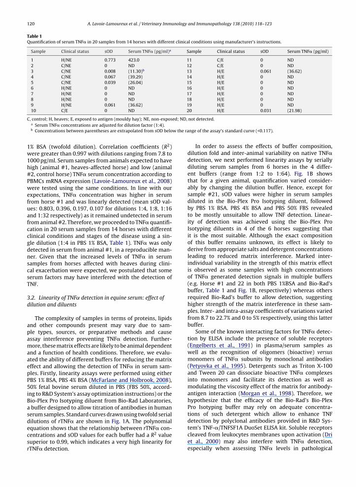
Table 1Quantification of serum TNF� in 20 samples from 14 horses with different clinical conditions using manufacturer’s instructions.
Sample Clinical status sOD Serum TNF� (pg/ml)a Sample Clinical status sOD Serum TNF� (pg/ml)
1 H/NE 0.773 423.0 11 C/E 0 ND2 C/NE 0 ND 12 C/E 0 ND3 C/NE 0.008 (11.30)b 13 H/E 0.061 (36.62)4 C/NE 0.067 (39.29) 14 H/E 0 ND5 C/NE 0.039 (26.04) 15 H/E 0 ND6 H/NE 0 ND 16 H/E 0 ND7 H/NE 0 ND 17 H/E 0 ND8 H/NE 0 ND 18 H/E 0 ND9 H/NE 0.061 (36.62) 19 H/E 0 ND10 C/E 0 ND 20 H/E 0.031 (21.98)
ed; ND,
w the ra
C, control; H, heaves; E, exposed to antigen (mouldy hay); NE, non-exposa Serum TNF� concentrations are adjusted for dilution factor (1:4).b Concentrations between parentheses are extrapolated from sOD belo
1% BSA (twofold dilution). Correlation coefficients (R2)were greater than 0.997 with dilutions ranging from 7.8 to1000 pg/ml. Serum samples from animals expected to havehigh (animal #1, heaves-affected horse) and low (animal#2, control horse) TNF� serum concentration according toPBMCs mRNA expression (Lavoie-Lamoureux et al., 2008)were tested using the same conditions. In line with ourexpectations, TNF� concentration was higher in serumfrom horse #1 and was linearly detected (mean sOD val-ues: 0.803, 0.396, 0.197, 0.107 for dilutions 1:4, 1:8, 1:16and 1:32 respectively) as it remained undetected in serumfrom animal #2. Therefore, we proceeded to TNF� quantifi-cation in 20 serum samples from 14 horses with differentclinical conditions and stages of the disease using a sin-gle dilution (1:4 in PBS 1% BSA, Table 1). TNF� was onlydetected in serum from animal #1, in a reproducible man-ner. Given that the increased levels of TNF� in serumsamples from horses affected with heaves during clini-cal exacerbation were expected, we postulated that someserum factors may have interfered with the detection ofTNF.
3.2. Linearity of TNF˛ detection in equine serum: effect ofdilution and diluents
The complexity of samples in terms of proteins, lipidsand other compounds present may vary due to sam-ple types, sources, or preparative methods and causeassay interference preventing TNF� detection. Further-more, these matrix effects are likely to be animal dependentand a function of health conditions. Therefore, we evalu-ated the ability of different buffers for reducing the matrixeffect and allowing the detection of TNF� in serum sam-ples. Firstly, linearity assays were performed using eitherPBS 1% BSA, PBS 4% BSA (McFarlane and Holbrook, 2008),50% fetal bovine serum diluted in PBS (FBS 50%, accord-ing to R&D System’s assay optimization instructions) or theBio-Plex Pro Isotyping diluent from Bio-Rad Laboratories,a buffer designed to allow titration of antibodies in humanserum samples. Standard curves drawn using twofold serial
dilutions of rTNF� are shown in Fig. 1A. The polynomialequation shows that the relationship between rTNF� con-centrations and sOD values for each buffer had a R2 valuesuperior to 0.99, which indicates a very high linearity forrTNF� detection.not detected.
nge of the assay’s standard curve (<0.117).
In order to assess the effects of buffer composition,dilution fold and inter-animal variability on native TNF�detection, we next performed linearity assays by seriallydiluting serum samples from 6 horses in the 4 differ-ent buffers (range from 1:2 to 1:64). Fig. 1B showsthat for a given animal, quantification varied consider-ably by changing the dilution buffer. Hence, except forsample #21, sOD values were higher in serum samplesdiluted in the Bio-Plex Pro Isotyping diluent, followedby PBS 1% BSA. PBS 4% BSA and PBS 50% FBS revealedto be mostly unsuitable to allow TNF detection. Linear-ity of detection was achieved using the Bio-Plex ProIsotyping diluents in 4 of the 6 horses suggesting thatit is the most suitable. Although the exact compositionof this buffer remains unknown, its effect is likely toderive from appropriate salts and detergent concentrationsleading to reduced matrix interference. Marked inter-individual variability in the strength of this matrix effectis observed as some samples with high concentrationsof TNF� generated detection signals in multiple buffers(e.g. Horse #1 and 22 in both PBS 1%BSA and Bio-Rad’sbuffer, Table 1 and Fig. 1B, respectively) whereas othersrequired Bio-Rad’s buffer to allow detection, suggestinghigher strength of the matrix interference in these sam-ples. Inter- and intra-assay coefficients of variations variedfrom 8.7 to 22.7% and 0 to 5% respectively, using this latterbuffer.
Some of the known interacting factors for TNF� detec-tion by ELISA include the presence of soluble receptors(Engelberts et al., 1991) in plasma/serum samples aswell as the recognition of oligomers (bioactive) versusmonomers of TNF� subunits by monoclonal antibodies(Petyovka et al., 1995). Detergents such as Triton X-100and Tween 20 can dissociate bioactive TNF� complexesinto monomers and facilitate its detection as well asmodulating the viscosity effect of the matrix for antibody-antigen interaction (Morgan et al., 1998). Therefore, wehypothesize that the efficacy of the Bio-Rad’s Bio-PlexPro Isotyping buffer may rely on adequate concentra-tions of such detergent which allow to enhance TNF
detection by polyclonal antibodies provided in R&D Sys-tem’s TNF-�/TNFSF1A DuoSet ELISA kit. Soluble receptorscleaved from leukocytes membranes upon activation (Driet al., 2000) may also interfere with TNF� detection,especially when assessing TNF� levels in pathological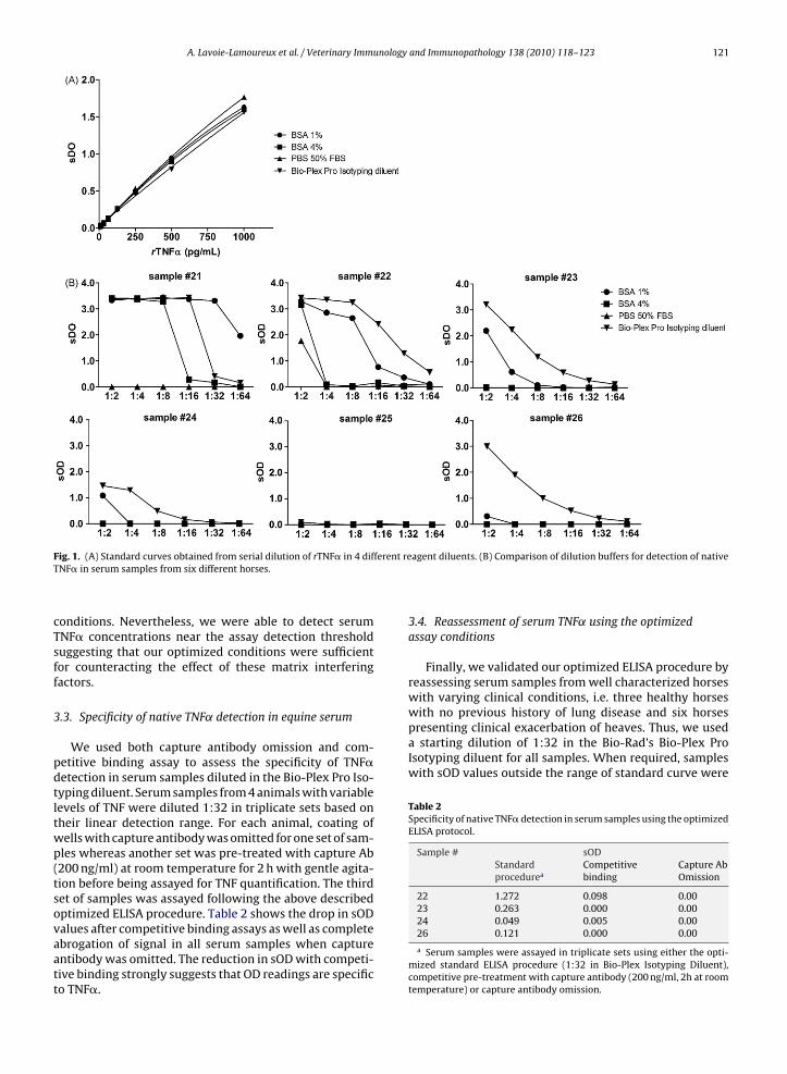
A. Lavoie-Lamoureux et al. / Veterinary Immunology and Immunopathology 138 (2010) 118–123 121
F ferent reT
cTsff
3
pdtltwp(tsovaatt
with no previous history of lung disease and six horsespresenting clinical exacerbation of heaves. Thus, we useda starting dilution of 1:32 in the Bio-Rad’s Bio-Plex ProIsotyping diluent for all samples. When required, sampleswith sOD values outside the range of standard curve were
Table 2Specificity of native TNF� detection in serum samples using the optimizedELISA protocol.
Sample # sODStandardprocedurea
Competitivebinding
Capture AbOmission
22 1.272 0.098 0.0023 0.263 0.000 0.0024 0.049 0.005 0.00
ig. 1. (A) Standard curves obtained from serial dilution of rTNF� in 4 difNF� in serum samples from six different horses.
onditions. Nevertheless, we were able to detect serumNF� concentrations near the assay detection thresholduggesting that our optimized conditions were sufficientor counteracting the effect of these matrix interferingactors.
.3. Specificity of native TNF˛ detection in equine serum
We used both capture antibody omission and com-etitive binding assay to assess the specificity of TNF�etection in serum samples diluted in the Bio-Plex Pro Iso-yping diluent. Serum samples from 4 animals with variableevels of TNF were diluted 1:32 in triplicate sets based onheir linear detection range. For each animal, coating ofells with capture antibody was omitted for one set of sam-les whereas another set was pre-treated with capture Ab200 ng/ml) at room temperature for 2 h with gentle agita-ion before being assayed for TNF quantification. The thirdet of samples was assayed following the above describedptimized ELISA procedure. Table 2 shows the drop in sOD
alues after competitive binding assays as well as completebrogation of signal in all serum samples when capturentibody was omitted. The reduction in sOD with competi-ive binding strongly suggests that OD readings are specifico TNF�.agent diluents. (B) Comparison of dilution buffers for detection of native
3.4. Reassessment of serum TNF˛ using the optimizedassay conditions
Finally, we validated our optimized ELISA procedure byreassessing serum samples from well characterized horseswith varying clinical conditions, i.e. three healthy horses
26 0.121 0.000 0.00
a Serum samples were assayed in triplicate sets using either the opti-mized standard ELISA procedure (1:32 in Bio-Plex Isotyping Diluent),competitive pre-treatment with capture antibody (200 ng/ml, 2h at roomtemperature) or capture antibody omission.
122 A. Lavoie-Lamoureux et al. / Veterinary Immunology
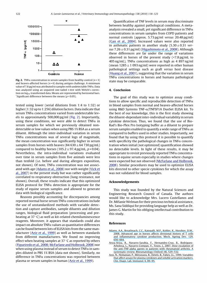
Fig. 2. TNF� concentration in serum samples from healthy control (n = 3)and heaves-affected horses (n = 6) during antigen challenge. A minimum
value of 7.8 pg/ml was attributed to samples with undetectable TNF�. Datawas analyzed using an unpaired one-tailed t-test with Welch’s correc-tion on log10 transformed data. Means are represented by horizontal bars.*Significant difference between the means (p < 0.05).tested using lower (serial dilutions from 1:4 to 1:32) orhigher (1:32 up to 1:256) dilution factors. Data indicate thatserum TNF� concentrations varied from undetectable lev-els to approximately 500,000 pg/ml (Fig. 2). Importantly,using these conditions, we were able to detect TNF� inserum samples for which we previously obtained non-detectable or low values when using PBS 1% BSA as a serumdiluent. Although the inter-individual variation in serumTNF� concentrations was of several logs of magnitude,the mean concentration was significantly higher in serumsamples from horses with heaves (84 630 ± 64 730 pg/mL)compared to healthy horses (105.2 ± 97.4 pg/mL, p = 0.04).Nevertheless, the intra-individual variations evaluatedover time in serum samples from five animals were lessthan tenfold (i.e. before and during allergen exposition,not shown). Of note, TNF� concentration was not associ-ated with age (Adams et al., 2008) nor with weight (Vick etal., 2007) in the present study but was rather significantlycorrelated to respiratory obstruction (lung resistance, notshown). Overall, these results indicate that this optimizedELISA protocol for TNF� detection is appropriate for thestudy of equine serum samples and allowed to generatedata with biological significance.
Reasons possibly accounting for discrepancies amongreported normal horse serum TNF� concentrations includethe use of unstandardized methods with variable detec-tion and capture antibodies, sample diluents and dilutionranges, biological fluid preparation (processing and pre-heating at 37 ◦C) as well as kit-related chemiluminescencereagents. Moreover, it appears that standards could alsoimpact on absolute TNF� values as quantitative differencescan be found between lots of ELISA kits from the same man-ufacturer (Aziz et al., 1999) as well as between standardsfrom different manufacturers. We found no improvingeffect when heating samples at 37 ◦C as reported by others
(Figueiredo et al., 2008; McFarlane and Holbrook, 2008) norwhen using plasma instead of serum to detect TNF� in sam-ples diluted in PBS 1% BSA (data not shown). Similarly, nodifference in TNF� concentrations was reported betweenplasma or serum samples in human (Aziz et al., 1999).and Immunopathology 138 (2010) 118–123
Quantification of TNF levels in serum may discriminatebetween healthy against pathological conditions. A meta-analysis revealed a small, yet significant difference in TNF�concentrations in serum samples from COPD patients andnormal controls (approx. 5.73 pg/ml versus 20.48 pg/ml)(Gan et al., 2004). Increased values were also reportedin asthmatic patients in another study (5.30 ± 0.31 ver-sus 7.26 ± 0.31 pg/ml) (Higashimoto et al., 2008). Althoughthese differences are far under the range of variationsobserved in horses of the present study (<7.8 pg/mL to405 ng/mL), TNF� concentrations as high as 4 897 ng/ml(mean 1285 ± 1 093 ng/ml) were reported in other humanpathological settings such as graft versus host disease(Huang et al., 2001), suggesting that the variation in serumTNF� concentrations in horses and humans pathologicalstate may be comparable.
4. Conclusion
The goal of this study was to optimize assay condi-tions to allow specific and reproducible detection of TNF�in blood samples from normal and heaves-affected horsesusing R&D Systems TNF-�/TNFSF1A DuoSet ELISA kit. Tothe best of our knowledge, this is the first study showingthe diluent-dependant inter-individual variability in serumcytokine detection. Thus, we found that the use of Bio-Rad’s Bio-Plex Pro Isotyping buffer as a diluent to prepareserum samples enabled to quantify a wide range of TNF� ascompared to buffers used in other studies. Importantly, wefound that by using this protocol it is possible to quantifywith specificity the pathological changes in TNF� concen-tration when initial (not optimized) quantification showedno detectable levels. In light of these results, it may beappropriate to revisit previously reported TNF� concentra-tions in equine serum especially in studies where changeswere expected but not observed (McFarlane and Holbrook,2008). Similar procedures may be used to optimize ELISAkits directed to other specie cytokines for which the assaywas not validated for blood samples.
Acknowledgements
This study was founded by the Natural Sciences andEngineering Research Council of Canada. The authorswould like to acknowledge Mrs. Lucero Castellanos andDr. Mélanie Welman for their precious technical assistance,Ms. Sana Siddiqui for providing language help as well as Dr.James G. Martin for his obliging intellectual contribution tothis study.
References
Adams, A.A., Breathnach, C.C., Katepalli, M.P., Kohler, K., Horohov, D.W.,2008. Advanced age in horses affects divisional history of T cellsand inflammatory cytokine production. Mech. Ageing Dev. 129,656–664.
Ariza-Ariza, R., Navarro-Sarabia, F., Hernandez-Cruz, B., Rodriguez-
Arboleya, L., Navarro-Compan, V., Toyos, J., 2007. Dose escalation ofthe anti-TNF-alpha agents in patients with rheumatoid arthritis. Asystematic review. Rheumatology (Oxford) 46, 529–532.Aziz, N., Nishanian, P., Mitsuyasu, R., Detels, R., Fahey, J.L., 1999. Variablesthat affect assays for plasma cytokines and soluble activation markers.Clin. Diagn. Lab. Immunol. 6, 89–95.
nology
B
D
D
D
E
F
G
H
H
J
K
L
M
M
A. Lavoie-Lamoureux et al. / Veterinary Immu
arnes, P.J., 2003. Cytokine-directed therapies for the treatment of chronicairway diseases. Cytokine Growth Factor Rev. 14, 511–522.
eans, C., Wigmore, S.J., 2005. Systemic inflammation, cachexia and prog-nosis in patients with cancer. Curr. Opin. Clin. Nutr. Metab. Care 8,265–269.
onovan, D.C., Jackson, C.A., Colahan, P.T., Norton, N., Hurley, D.J.,2007. Exercise-induced alterations in pro-inflammatory cytokinesand prostaglandin F2� in horses. Vet. Immunol. Immunopathol. 118,263–269.
ri, P., Gasparini, C., Menegazzi, R., Cramer, R., Alberi, L., Presani, G., Gar-bisa, S., Patriarca, P., 2000. TNF-Induced shedding of TNF receptorsin human polymorphonuclear leukocytes: role of the 55-kDa TNFreceptor and involvement of a membrane-bound and non-matrixmetalloproteinase. J. Immunol. 165, 2165–2172.
ngelberts, I., Stephens, S., Francot, G.J., van der Linden, C.J., Buurman,W.A., 1991. Evidence for different effects of soluble TNF-receptors onvarious TNF measurements in human biological fluids. Lancet 338,515–516.
igueiredo, M.D., Moore, J.N., Vandenplas, M.L., Sun, W.C., Murray, T.F.,2008. Effects of the second-generation synthetic lipid A analogueE5564 on responses to endotoxin in [corrected] equine whole bloodand monocytes. Am. J. Vet. Res. 69, 796–803.
an, W.Q., Man, S.F., Senthilselvan, A., Sin, D.D., 2004. Association betweenchronic obstructive pulmonary disease and systemic inflammation: asystematic review and a meta-analysis. Thorax 59, 574–580.
igashimoto, Y., Yamagata, Y., Taya, S., Iwata, T., Okada, M., Ishiguchi, T.,Sato, H., Itoh, H., 2008. Systemic inflammation in chronic obstructivepulmonary disease and asthma: similarities and differences. Respirol-ogy 13, 128–133.
uang, X.J., Wan, J., Lu, D.P., 2001. Serum TNFalpha levels in patients withacute graft-versus-host disease after bone marrow transplantation.Leukemia 15, 1089–1091.
ousilahti, P., Salomaa, V., Hakala, K., Rasi, V., Vahtera, E., Palosuo, T.,2002. The association of sensitive systemic inflammation markerswith bronchial asthma. Ann. Allergy Asthma Immunol. 89, 381–385.
alck, K.A., 2009. Inflammatory bowel disease in horses. Vet. Clin. NorthAm. Equine Pract. 25, 303–315.
avoie-Lamoureux, A., Beauchamp, G., Quessy, S., Martin, J.G., Lavoie,J.-P., 2008. Altered inflammatory response to microbial stimuli byperipheral blood leukocytes in an equine model of chronic asthma(heaves). In: American Thoracic Society 2008 International Confer-ence, Toronto.
atera, M.G., Calzetta, L., Cazzola, M., 2009. TNF-alpha inhibitors inasthma and COPD: we must not throw the baby out with the bathwater. Pulm. Pharmacol. Ther..
cFarlane, D., Holbrook, T.C., 2008. Cytokine dysregulation in aged horsesand horses with pituitary pars intermedia dysfunction. J. Vet. Intern.Med. 22, 436–442.
and Immunopathology 138 (2010) 118–123 123
Morgan, C.L., Newman, D.J., Burrin, J.M., Price, C.P., 1998. The matrix effectson kinetic rate constants of antibody-antigen interactions reflect sol-vent viscosity. J. Immunol. Methods 217, 51–60.
Niederau, C., Backmerhoff, F., Schumacher, B., 1997. Inflammatorymediators and acute phase proteins in patients with Crohn’sdisease and ulcerative colitis. Hepatogastroenterology 44, 90–107.
Nikolaus, S., Bauditz, J., Gionchetti, P., Witt, C., Lochs, H., Schreiber, S.,1998. Increased secretion of pro-inflammatory cytokines by circulat-ing polymorphonuclear neutrophils and regulation by interleukin 10during intestinal inflammation. Gut 42, 470–476.
Olszanecka-Glinianowicz, M., Zahorska-Markiewicz, B., Janowska, J.,Zurakowski, A., 2004. Serum concentrations of nitric oxide,tumor necrosis factor (TNF)-alpha and TNF soluble receptorsin women with overweight and obesity. Metabolism 53, 1268–1273.
Park, H.S., Park, J.Y., Yu, R., 2005. Relationship of obesity and visceral adi-posity with serum concentrations of CRP, TNF-alpha and IL-6. Diab.Res. Clin. Pract. 69, 29–35.
Petyovka, N., Lyach, L., Voitenok, N.N., 1995. Homologous ELISA for detec-tion of oligomeric human TNF: properties of the assay. J. Immunol.Methods 186, 161–170.
Robinson, N., 2001. International workshop on equine chronic airway dis-ease. Michigan State University 16–18 June 2000. Equine Vet. J. 33,5–19.
Robinson, N.E., Olszewski, M.A., Boehler, D., Berney, C., Hakala, J., Mat-son, C., Derksen, F.J., 2000. Relationship between clinical signsand lung function in horses with recurrent airway obstruction(heaves) during a bronchodilator trial. Equine Vet. J. 32, 393–400.
Snow, M.H., Mikuls, T.R., 2005. Rheumatoid arthritis and cardiovasculardisease: the role of systemic inflammation and evolving strategies ofprevention. Curr. Opin. Rheumatol. 17, 234–241.
Sutton, S., Clutterbuck, A., Harris, P., Gent, T., Freeman, S., Foster, N.,Barrett-Jolley, R., Mobasheri, A., 2009. The contribution of the syn-ovium, synovial derived inflammatory cytokines and neuropeptidesto the pathogenesis of osteoarthritis. Vet. J. 179, 10–24.
Theon, A.P., 1998. Intralesional and topical chemotherapy andimmunotherapy. Vet. Clin. North Am. Equine Pract. 14, 659–671, viii.
Vick, M.M., Adams, A.A., Murphy, B.A., Sessions, D.R., Horohov, D.W., Cook,R.F., Shelton, B.J., Fitzgerald, B.P., 2007. Relationships among inflam-matory cytokines, obesity, and insulin sensitivity in the horse. J. Anim.
Sci. 85, 1144–1155.Woodward, A.D., O’Connor, N.B., Skelly, C.I., Webel, C.D., Orth, S.K., W, M.,2007. Supplementation of dietary long-chain polyunsaturated omega-3 fatty acids high in docosahexaenoic acid (DHA) increases plasmaDHA concentration and may increase trot stride lengths in horses.Equine Comp. Exerc. Physiol. 4, 71–78.