olfactory deficits in niemann-pick type c1 (npc1) disease
TRANSCRIPT
Olfactory Deficits in Niemann-Pick Type C1 (NPC1)DiseaseMarina Hovakimyan1, Anja Meyer1, Jan Lukas2, Jiankai Luo2, Volker Gudziol3, Thomas Hummel3,
Arndt Rolfs2, Andreas Wree1, Martin Witt1*
1 Institute of Anatomy, Rostock University Medical Center, Rostock, Germany, 2 Albrecht-Kossel Institute for Neuroregeneration, Rostock University Medical Center,
Rostock, Germany, 3 Department of Otorhinolaryngology, University of Dresden Medical School, Dresden, Germany
Abstract
Background: Niemann-Pick type C disease (NPC) is a rare autosomal recessive lipid storage disease characterized byprogressive neurodegeneration. As only a few studies have been conducted on the impact of NPC on sensory systems, weused a mutant mouse model (NPC12/2) to examine the effects of this disorder to morphologically distinct regions of theolfactory system, namely the olfactory epithelium (OE) and olfactory bulb (OB).
Methodology/Principal findings: For structural and functional analysis immunohistochemistry, electron microscopy,western blotting, and electrophysiology have been applied. For histochemistry and western blotting, we used antibodiesagainst a series of neuronal and glia marker proteins, as well as macrophage markers. NPC12/2 animals present myelin-like lysosomal deposits in virtually all types of cells of the peripheral and central olfactory system. Especially supporting cellsof the OE and central glia cells are affected, resulting in pronounced astrocytosis and microgliosis in the OB and otherolfactory cortices. Up-regulation of Galectin-3, Cathepsin D and GFAP in the cortical layers of the OB underlines the criticalrole and location of the OB as a possible entrance gate for noxious substances. Unmyelinated olfactory afferents of thelamina propria seem less affected than ensheathing cells. Supporting the structural findings, electro-olfactometry of theolfactory mucosa suggests that NPC12/2 animals exhibit olfactory and trigeminal deficits.
Conclusions/Significance: Our data demonstrate a pronounced neurodegeneration and glia activation in the olfactorysystem of NPC12/2, which is accompanied by sensory deficits.
Citation: Hovakimyan M, Meyer A, Lukas J, Luo J, Gudziol V, et al. (2013) Olfactory Deficits in Niemann-Pick Type C1 (NPC1) Disease. PLoS ONE 8(12): e82216.doi:10.1371/journal.pone.0082216
Editor: Michelle L. Block, Virginia Commonwealth University, United States of America
Received August 19, 2013; Accepted October 24, 2013; Published December 31, 2013
Copyright: � 2013 Hovakimyan et al. This is an open-access article distributed under the terms of the Creative Commons Attribution License, which permitsunrestricted use, distribution, and reproduction in any medium, provided the original author and source are credited.
Funding: These authors have no support or funding to report.
Competing Interests: The authors have declared that no competing interests exist.
* E-mail: [email protected]
Introduction
Niemann Pick Type C (NPC) is a fatal autosomal recessive
neurovisceral disorder with an estimated prevalence of approxi-
mately 1:150,000 in Western Europe [1]. The disorder is caused
by mutations in the NPC1 (in 95% of patients) or NPC2 gene [2].
The NPC1 gene has been identified by positional cloning [3],
and its genomic structure was reported two years later [4]. NPC1
gene codes for a membrane protein that contains a sterol-sensing
domain and resides in late endosomes [5]. This glycoprotein with
a molecular weight of 142 kDa is involved in the intracellular
transport of cholesterol, glycolipids and other cellular components.
Mutations in NPC1 lead to a deficient intracellular lipid
trafficking, abnormal regulation of cholesterol biosynthesis and
intracellular accumulation of unesterified cholesterol and gangli-
osides GM2 and GM3 in the late endosomes/lysosomes [6,7].
Clinical symptoms include hepatosplenomegaly, ataxia, dystonia,
and progressive neurodegeneration [8,9]. Most patients die during
the first two decades [10].
In feline, canine and mouse animal models of the disease, a
similar phenotype is observed involving tremor, ataxia, and other
signs of neurologic impairment [11,12].
The most widely used mutant mouse model of NPC1 disease,
named NPC12/2, has a retrotransposon insertion into the N-
terminus of the NPC1 gene, along with a 703-bp deletion, causing
premature termination of the coding region that excludes most of
the sterol-sensing domain [13]. The NPC12/2 mice lack NPC1
protein and exhibit hepatosplenomegaly and progressive neuro-
degeneration [14]. The symptoms appear at 42–49 days of age
with tremors, lack of motor coordination, progressive weight loss,
all leading to death by 10 weeks of age [15].
Previous investigations in this NPC12/2 mouse model have
reported severe damage and loss of Purkinje cells and other CNS
neurons [16,17,18] as well as neurodegeneration and transmission
defects in the retina [19].
So far, it is not fully understood why defects in NPC1 cause
neurodegeneration. Abnormal activity of autophagic/lysosomal
systems, which are closely associated with cholesterol accumula-
tion in the endosomal/lysosomal system, has been implicated in
NPC1 neuropathology [20,21]. Microglia- and astrocyte-mediated
inflammation has also been proposed to contribute to the
progression of neurodegeneration [22]. Except for retinal degen-
eration [19], sensory systems such as olfactory, trigeminal or
auditory pathways in NPC1 disease have not been studied so far.
PLOS ONE | www.plosone.org 1 December 2013 | Volume 8 | Issue 12 | e82216
An important reason to investigate the olfactory system is the
unique regenerative nature of some olfactory components.
Olfactory receptor neurons can, in contrast to other peripheral
neuron-like cells, constantly regenerate from precursor cells. The
same is true for central olfactory interneurons that differentiate
from neuron precursors migrating from the subventricular zone
into the olfactory bulb [23]. Thus, the olfactory system constitutes
a prominent example for adult neurogenesis, which may rapidly
adapt during neurodegeneration [24].
What is more, many neurodegenerative diseases are associated
with early deterioration of olfactory performance. For example, in
Parkinson’s disease olfactory impairment occurs at least two years
before motor symptoms become evident [25]. Similar associations
are known for Alzheimer disease [26–28], or in the neurologic
form of Gaucher’s disease, the most common lysosomal storage
disorder [29,30]. In earlier work, we focused on motor acuity and
behavioral as well as central molecular aspects in NPC1 [31,32].
Although the characteristic olfactory impairment in neurodegen-
erative diseases is well established [33,34], morphologically distinct
regions of the olfactory system have not yet been analysed in
NPC1 disease.
Since early diagnosis and a reasonable standard to follow up on
disease progression in the patients are crucial for therapeutic
intervention in Niemann-Pick type C, we hypothesized a clinical
value of olfactory performance in monitoring patients with NPC1
disease. Therefore, in the present study we used NPC12/2 mice to
investigate the effects of this disorder on peripheral level, the
olfactory epithelium (OE) and the first central relay structure, the
olfactory bulb (OB).
Materials and Methods
AnimalsHeterozygous breeding pairs of NPC1 mice (BALB/cNctr-
Npc1m1N/J, # 3092, The Jackson Laboratories, Bar Harbor,
Maine, USA) were used to generate NPC12/2 and control wild
type mice. The protocol was approved by the Committee on the
Ethics of Animal Experiments of the University of Rostock
(approval ID: 7221.3-1.1-088/10) and conducted according to the
guidelines for the Care and Use of Laboratory Animals. All efforts
were made to minimize suffering.
Mice were maintained on a 12 hours light-dark cycle with water
and food ad libitum. The mouse pups were genotyped by using a
polymerase chain reaction (PCR) assay.
Nineteen inbred female homozygous NPC1 mutant mice,
lacking NPC1 protein (NPC12/2), aged from 5 to 10 weeks,
and 16 wild type siblings (NPC1+/+) of the same age were used for
immunohistochemistry, electron microscopy and PCR/Western
blot analysis, and 12 animals of each group were used for the
electrophysiological study.
PCR analysisFor genotyping, 1–2 mm of mice tails were clipped at postnatal
day 6 and homogenized in 200 ml DirectPCR-Tail (Peqlab,
Erlangen, Germany) supplemented with 20 ml Proteinase K
(Qiagen, Hilden, Germany). Three hours of incubation at 56uCand agitation at 1000 rpm on a Thermo Mixer (Eppendorf) were
followed by 45 minutes of heating at 85uC to inactivate the
proteinase. Samples were then spun at full speed in a benchtop
centrifuge for 1 minute. The PCR reactions were performed with
0.5 ml of the obtained extracts. Each lysate underwent two PCRs;
Primers 59-tctcacagccacaagcttcc-39 and 59-ctgtagctcatctgccatcg-39
identified the wild type allele (obtained fragment size 173 bp) and
primers 59-ggtgctggacagccaagta-39 and 59-tgagcccaagcataactt-39
identified the mutant allele (obtained fragment size 475 bp). Both
PCRs were carried out under similar cycling conditions, 39 at
94uC, 3-step cycling 300 at 94uC - 450 at 67uC - 450 at 72uC (35
cycles) and a final elongation for 29 at 72uC.
Preparation of the samplesFor immunohistochemistry and electron microscopy, the
animals were deeply anesthetized with sodium pentobarbital and
killed by an overdose of sodium pentobarbital. Then, cardiac
perfusion with phosphate-buffered saline (PBS, pH = 7.4) was
followed by 4% paraformaldehyde (PFA) in 0.1 M PBS.
Subsequently, the heads were cut in median- sagittal direction
and fixed by immersion in the same fixative for additional 24 h.
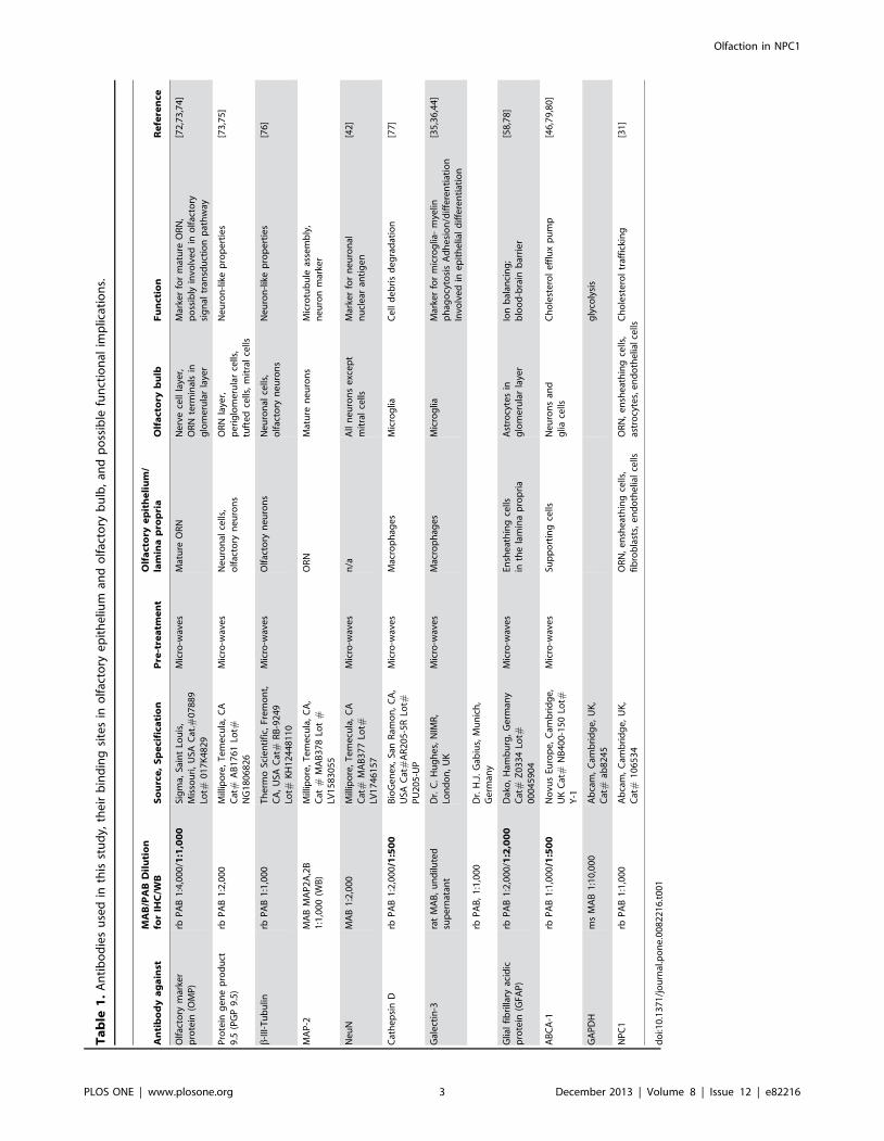
Antisera and antibodiesAntisera and antibodies used in this study are compiled in
Table 1. The polyclonal Galectin-3 (Gal-3) antiserum was kindly
provided by Dr. H.-J. Gabius, Munich, and was made and
checked for specificity and absence of cross-reactivity to other
galectins as previously described [35]. A monoclonal antibody
directed against Gal-3, kindly provided by Dr. H. Hughes,
London, was checked for specificity and absence of other galectins
as described [36].
ImmunohistochemistryThe specimens were decalcified in 1% EDTA for 6–24 h at
room temperature (RT) or 37uC, shortly rinsed in distilled water,
dehydrated and embedded in paraffin. Heads were cut in the
sagittal plane in order to visualize both OE and OB on the same
section (thickness 5 mm). In order to improve antigen retrieval,
deparaffinized and re-hydrated tissue sections were pretreated
with microwaves [37] (10 min. in 0.05 Mol/l citrate buffer, pH 6,
800 W) (see Table 1) and exposed to 0.3% aqueous H2O2 to block
endogenous peroxidases. The sections were then incubated with
various primary antibodies as listed in Table 1 (pH 7.2; containing
1% bovine serum albumin) for 1 h at 37uC. After washing in PBS,
the sections were exposed to biotinylated secondary antibodies for
45 min. at RT. The reaction products were visualized by an
avidin-biotin-peroxidase complex (ABC; Vectastain-Elite; Vector,
Burlingame, CA, USA) followed by incubation with 0.3%
diaminobenzidine/H2O2 according to the ABC technique [38].
Sections were counterstained with hematoxylin.
Indirect immunofluorescenceFor co-localization experiments a double immunofluorescence
protocol was performed as described earlier [39]. Briefly, paraffin
sections were dewaxed and incubated with antisera against GFAP
followed by donkey anti rabbit Texas Red secondary antibody
(1:80; Molecular Probes, MobiTec, Gottingen, Germany) at 37uCfor 1 h. Subsequently, the sections were incubated with either the
microglia marker anti-Gal-3 or anti- olfactory marker protein
(OMP) at 37uC for 1 h followed by incubation with donkey anti-
goat or donkey anti-rabbit FITC secondary antibody (1:80) for
30 min at 37uC. The sections were mounted in buffered glycerine
gelatine and observed with an Olympus BX60 microscope.
Photographs were taken using a CCD camera connected to a
soft-imaging analysis system (Olympus ANAlysis, Munster,
Germany). Separate images for GFAP and Gal-3/OMP immu-
nohistochemistry were obtained from double-labelled specimens,
and the individual images were colour-separated into their RGB
components. The red (GFAP) and green (Gal-3, OMP) were
merged and the composite images imported as TIFF files into
Adobe Photoshop CS2 (Adobe Systems) for size reduction.
Olfaction in NPC1
PLOS ONE | www.plosone.org 2 December 2013 | Volume 8 | Issue 12 | e82216
Ta
ble
1.
An
tib
od
ies
use
din
this
stu
dy,
the
irb
ind
ing
site
sin
olf
acto
rye
pit
he
lium
and
olf
acto
ryb
ulb
,an
dp
oss
ible
fun
ctio
nal
imp
licat
ion
s.
An
tib
od
ya
ga
inst
MA
B/P
AB
Dil
uti
on
for
IHC
/WB
So
urc
e,
Sp
eci
fica
tio
nP
re-t
rea
tme
nt
Olf
act
ory
ep
ith
eli
um
/la
min
ap
rop
ria
Olf
act
ory
bu
lbF
un
ctio
nR
efe
ren
ce
Olf
acto
rym
arke
rp
rote
in(O
MP
)rb
PA
B1
:4,0
00
/1:1
,00
0Si
gm
a,Sa
int
Lou
is,
Mis
sou
ri,
USA
Cat
.#0
78
89
Lot#
01
7K
48
29
Mic
ro-w
ave
sM
atu
reO
RN
Ne
rve
cell
laye
r,O
RN
term
inal
sin
glo
me
rula
rla
yer
Mar
ker
for
mat
ure
OR
N,
po
ssib
lyin
volv
ed
ino
lfac
tory
sig
nal
tran
sdu
ctio
np
ath
way
[72
,73
,74
]
Pro
tein
ge
ne
pro
du
ct9
.5(P
GP
9.5
)rb
PA
B1
:2,0
00
Mill
ipo
re,
Te
me
cula
,C
AC
at#
AB
17
61
Lot#
NG
18
06
82
6
Mic
ro-w
ave
sN
eu
ron
alce
lls,
olf
acto
ryn
eu
ron
sO
RN
laye
r,p
eri
glo
me
rula
rce
lls,
tuft
ed
cells
,m
itra
lce
lls
Ne
uro
n-l
ike
pro
pe
rtie
s[7
3,7
5]
b-I
II-T
ub
ulin
rbP
AB
1:1
,00
0T
he
rmo
Scie
nti
fic,
Fre
mo
nt,
CA
,U
SAC
at#
RB
-92
49
Lot#
KH
12
44
81
10
Mic
ro-w
ave
sO
lfac
tory
ne
uro
ns
Ne
uro
nal
cells
,o
lfac
tory
ne
uro
ns
Ne
uro
n-l
ike
pro
pe
rtie
s[7
6]
MA
P-2
MA
BM
AP
2A
,2B
1:1
,00
0(W
B)
Mill
ipo
re,
Te
me
cula
,C
A,
Cat
#M
AB
37
8Lo
t#
LV1
58
30
55
OR
NM
atu
ren
eu
ron
sM
icro
tub
ule
asse
mb
ly,
ne
uro
nm
arke
r
Ne
uN
MA
B1
:2,0
00
Mill
ipo
re,
Te
me
cula
,C
AC
at#
MA
B3
77
Lot#
LV1
74
61
57
Mic
ro-w
ave
sn
/aA
lln
eu
ron
se
xce
pt
mit
ral
cells
Mar
ker
for
ne
uro
nal
nu
cle
aran
tig
en
[42
]
Cat
he
psi
nD
rbP
AB
1:2
,00
0/1
:50
0B
ioG
en
ex,
San
Ram
on
,C
A,
USA
Cat
#A
R2
05
-5R
Lot#
PU
20
5-U
P
Mic
ro-w
ave
sM
acro
ph
age
sM
icro
glia
Ce
lld
eb
ris
de
gra
dat
ion
[77
]
Gal
ect
in-3
rat
MA
B,
un
dilu
ted
sup
ern
atan
tD
r.C
.H
ug
he
s,N
IMR
,Lo
nd
on
,U
KM
icro
-wav
es
Mac
rop
hag
es
Mic
rog
liaM
arke
rfo
rm
icro
glia
-m
yelin
ph
ago
cyto
sis
Ad
he
sio
n/d
iffe
ren
tiat
ion
Invo
lve
din
ep
ith
elia
ld
iffe
ren
tiat
ion
[35
,36
,44
]
rbP
AB
,1
:1,0
00
Dr.
H.J
.G
abiu
s,M
un
ich
,G
erm
any
Glia
lfi
bri
llary
acid
icp
rote
in(G
FAP
)rb
PA
B1
:2,0
00
/1:2
,00
0D
ako
,H
amb
urg
,G
erm
any
Cat
#Z
03
34
Lot#
00
04
59
04
Mic
ro-w
ave
sEn
she
ath
ing
cells
inth
ela
min
ap
rop
ria
Ast
rocy
tes
ing
lom
eru
lar
laye
rIo
nb
alan
cin
g;
blo
od
-bra
inb
arri
er
[58
,78
]
AB
CA
-1rb
PA
B1
:1,0
00
/1:5
00
No
vus
Euro
pe
,C
amb
rid
ge
,U
KC
at#
NB
40
0-1
50
Lot#
Y-1
Mic
ro-w
ave
sSu
pp
ort
ing
cells
Ne
uro
ns
and
glia
cells
Ch
ole
ste
rol
eff
lux
pu
mp
[46
,79
,80
]
GA
PD
Hm
sM
AB
1:1
0,0
00
Ab
cam
,C
amb
rid
ge
,U
K,
Cat
#ab
82
45
gly
coly
sis
NP
C1
rbP
AB
1:1
,00
0A
bca
m,
Cam
bri
dg
e,
UK
,C
at#
10
65
34
OR
N,
en
she
ath
ing
cells
,fi
bro
bla
sts,
en
do
the
lial
cells
OR
N,
en
she
ath
ing
cells
,as
tro
cyte
s,e
nd
oth
elia
lce
llsC
ho
lest
ero
ltr
affi
ckin
g[3
1]
do
i:10
.13
71
/jo
urn
al.p
on
e.0
08
22
16
.t0
01
Olfaction in NPC1
PLOS ONE | www.plosone.org 3 December 2013 | Volume 8 | Issue 12 | e82216
The following controls were carried out: (1) omission of the
primary antibody to rule out non-specific binding of the secondary
antibodies and (2) parallel incubation of tissue previously reported
to be immunoreactive to the markers tested.
Electron microscopyAfter initial perfusion and preparation (see above), samples of
five NPC12/2 and NPC1+/+ mice were postfixed in 0.1M
cacodylate buffer containing 2.5% glutaraldehyde for at least
24 hours at 4uC. Subsequently, turbinates containing olfactory
mucosa and cross-sectioned samples of the olfactory bulb (OB) as
well as trigeminal ganglia were excised and kept in the same
fixative. Thereafter, the specimens were osmicated, washed, block
contrasted with 2% aqueous uranyl acetate, dehydrated through a
graded series of ethanol, and embedded in Epon 812 (Plano
GmbH, Marburg, Germany). Ultrathin sections (about 70 nm)
were mounted on pioloform-coated slot copper grids and
contrasted with uranyl acetate (4 minutes) followed by lead citrate
(2 minutes). The specimens were examined with a Zeiss EM 902
transmission electron microscope (Zeiss, Oberkochen, Germany)
at 80 kV. Photographs were taken using a CCD camera (Proscan,
Lagerlechfeld, Germany) and adjusted using Photoshop CS2
software (Adobe Systems).
Western blot analysisAnimals subjected to western blotting were sacrificed by cervical
dislocation. Tissue lysates were obtained by the addition of ice-
cold RIPA buffer (500 ml/100 mg tissue) and passing through a
1 ml syringe equipped with a 206g needle. Subsequent sonication
with a UP200S Ultrasonic Processor (Hielscher, Teltow, Ger-
many) (2615 sec. on/off pulses with a two minute cooling on ice
in between) was applied to ensure that homogenisation was
completed. Typically, 50 mg of lysate were loaded on a 4–15%
CriterionTM precast polyacrylamide gel (Bio-Rad, Munich,
Germany). Semiquantitative Western blot detection was carried
out using the Odyssey Infrared ImagingSystem (LI-COR Biosci-
ences GmbH, Bad Homburg, Germany) according to protocols
described previously [40]. Antibodies (for sources see Table 1)
against the following proteins were used: Cathepsin D (1:500),
beta-III tubulin (1:1,000), ABCA-1 (1:500), NPC1 (1:1,000),
MAP2 (1:1,000), OMP (1:1,000), GFAP (1:2,000), and GAPDH
(1:10,000), which served as a loading control. Alexa-Fluor 680 and
goat anti-mouse IRDye 800 (1:10,000; Invitrogen) were used as
secondary antibodies.
Electro-olfactometryAnimal preparation for EOG recording. The recordings
were performed as described earlier [41]. Twenty-four mice
[divided in two groups of 31 day-old and 67-day-old animals,
respectively] were investigated. Mice were killed by cervical
dislocation. Heads were cut median-sagitally. The nasal septum
and the mucosa of the nasal wall were dissected under a stereo
microscope. The nasal mucosa was kept moist with Ringer’s
solution and stored at 4uC.
Olfactometer and olfactory stimuli. Volatiles were applied
using a calibrated air-dilution olfactometer (OM2sl; Burghart
Instruments, Wedel, Germany) that allows embedding the stimuli
in a constant air flow (2 l/min) of constant temperature (36.5uC)
and humidity (80% relative humidity). Phenylethyl alcohol (PEA)
(40% and 20%, v/v) and hydrogen sulfide (H2S) (40% and 20%,
v/v) were used as olfactory stimuli, and CO2 (50% and 40%, v/v)
was used as trigeminal stimulus. Control stimulation was
performed using odorless air.
EOG recording. A powerlab 26T device (AD instruments,
Bella Vista, Australia) and Chart 5.5.5 for WindowsTM were used
to record the EOG. Recordings from the olfactory mucosa were
made using tubular electrodes made from TeflonTM tubing
(Labokron, Sinsheim, Germany; outer diameter 0.8 mm), filled
with 1% Ringer-agar (Agar: Sigma-Aldrich Chemie GmbH,
Steinheim, Germany) and containing a silver chloride coated
silver wire (electrode resistance ,5 kOhm).
Results
AnimalsAdult NPC12/2 mice (65–70 d) exhibited a reduced body
weight compared to control litter mates (17.6 g62 g in NPC12/2
vs. 20.7 g61.5 g in NPC1+/+, p = 0.0221). Young animals (30 d,
typically showing first neuropathology symptoms and cellular
alterations, see below) did not yet show significant weight
differences (13.4 g62.4 g in NPC12/2 vs. 15.9 g62.6 g in
NPC1+/+, p = 0.1014). Brain weights of old animals were
significantly decreased (0.38 g60.01 g in NPC12/2 vs.
0.43 g60.02 g in NPC1+/+, p = 0.0221, Fig. S1).
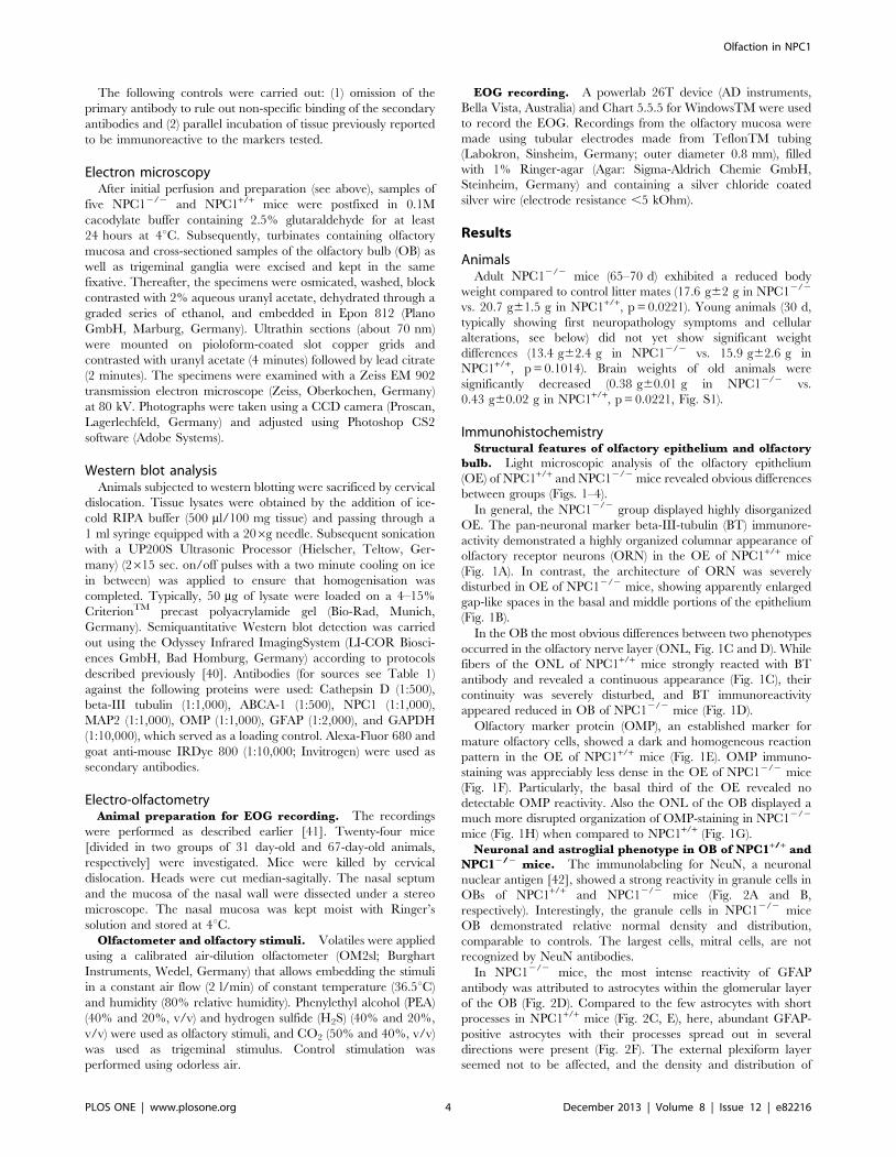
ImmunohistochemistryStructural features of olfactory epithelium and olfactory
bulb. Light microscopic analysis of the olfactory epithelium
(OE) of NPC1+/+ and NPC12/2 mice revealed obvious differences
between groups (Figs. 1–4).
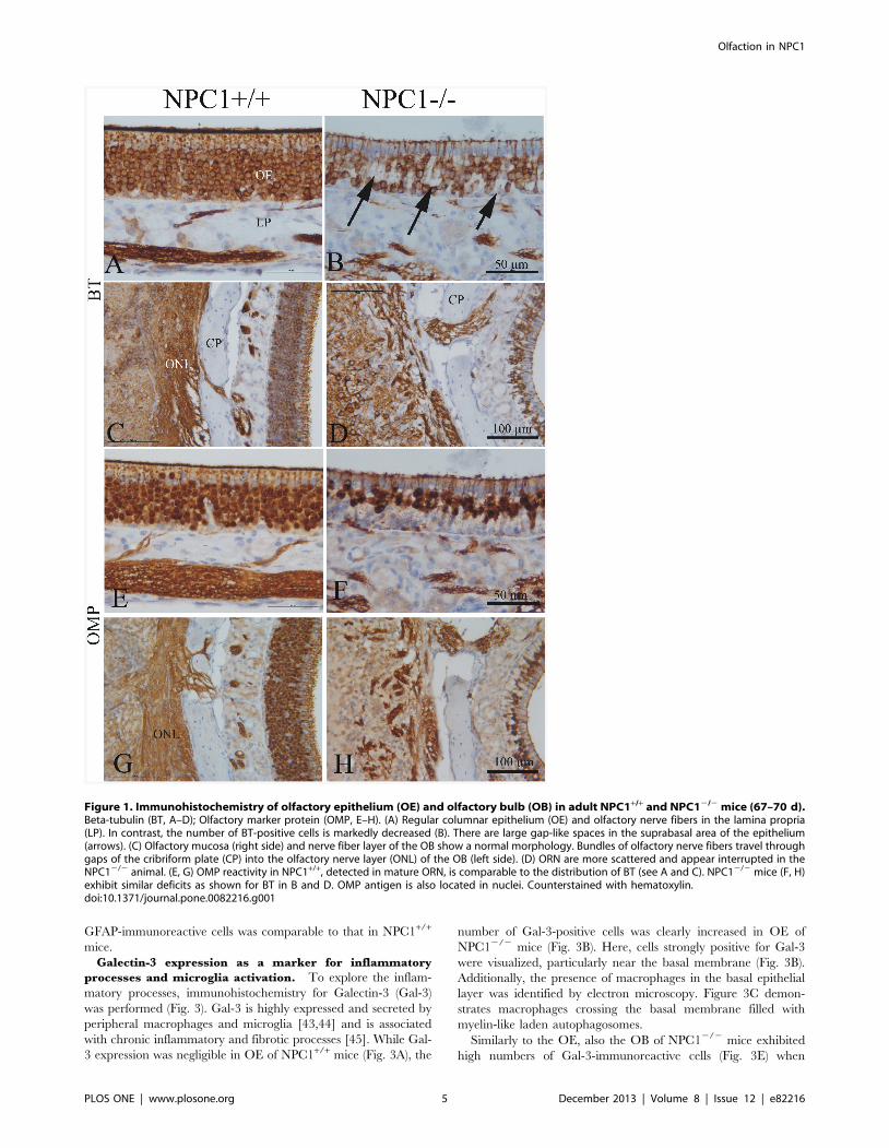
In general, the NPC12/2 group displayed highly disorganized
OE. The pan-neuronal marker beta-III-tubulin (BT) immunore-
activity demonstrated a highly organized columnar appearance of
olfactory receptor neurons (ORN) in the OE of NPC1+/+ mice
(Fig. 1A). In contrast, the architecture of ORN was severely
disturbed in OE of NPC12/2 mice, showing apparently enlarged
gap-like spaces in the basal and middle portions of the epithelium
(Fig. 1B).
In the OB the most obvious differences between two phenotypes
occurred in the olfactory nerve layer (ONL, Fig. 1C and D). While
fibers of the ONL of NPC1+/+ mice strongly reacted with BT
antibody and revealed a continuous appearance (Fig. 1C), their
continuity was severely disturbed, and BT immunoreactivity
appeared reduced in OB of NPC12/2 mice (Fig. 1D).
Olfactory marker protein (OMP), an established marker for
mature olfactory cells, showed a dark and homogeneous reaction
pattern in the OE of NPC1+/+ mice (Fig. 1E). OMP immuno-
staining was appreciably less dense in the OE of NPC12/2 mice
(Fig. 1F). Particularly, the basal third of the OE revealed no
detectable OMP reactivity. Also the ONL of the OB displayed a
much more disrupted organization of OMP-staining in NPC12/2
mice (Fig. 1H) when compared to NPC1+/+ (Fig. 1G).
Neuronal and astroglial phenotype in OB of NPC1+/+ and
NPC12/2 mice. The immunolabeling for NeuN, a neuronal
nuclear antigen [42], showed a strong reactivity in granule cells in
OBs of NPC1+/+ and NPC12/2 mice (Fig. 2A and B,
respectively). Interestingly, the granule cells in NPC12/2 mice
OB demonstrated relative normal density and distribution,
comparable to controls. The largest cells, mitral cells, are not
recognized by NeuN antibodies.
In NPC12/2 mice, the most intense reactivity of GFAP
antibody was attributed to astrocytes within the glomerular layer
of the OB (Fig. 2D). Compared to the few astrocytes with short
processes in NPC1+/+ mice (Fig. 2C, E), here, abundant GFAP-
positive astrocytes with their processes spread out in several
directions were present (Fig. 2F). The external plexiform layer
seemed not to be affected, and the density and distribution of
Olfaction in NPC1
PLOS ONE | www.plosone.org 4 December 2013 | Volume 8 | Issue 12 | e82216
GFAP-immunoreactive cells was comparable to that in NPC1+/+
mice.
Galectin-3 expression as a marker for inflammatory
processes and microglia activation. To explore the inflam-
matory processes, immunohistochemistry for Galectin-3 (Gal-3)
was performed (Fig. 3). Gal-3 is highly expressed and secreted by
peripheral macrophages and microglia [43,44] and is associated
with chronic inflammatory and fibrotic processes [45]. While Gal-
3 expression was negligible in OE of NPC1+/+ mice (Fig. 3A), the
number of Gal-3-positive cells was clearly increased in OE of
NPC12/2 mice (Fig. 3B). Here, cells strongly positive for Gal-3
were visualized, particularly near the basal membrane (Fig. 3B).
Additionally, the presence of macrophages in the basal epithelial
layer was identified by electron microscopy. Figure 3C demon-
strates macrophages crossing the basal membrane filled with
myelin-like laden autophagosomes.
Similarly to the OE, also the OB of NPC12/2 mice exhibited
high numbers of Gal-3-immunoreactive cells (Fig. 3E) when
Figure 1. Immunohistochemistry of olfactory epithelium (OE) and olfactory bulb (OB) in adult NPC1+/+ and NPC12/2 mice (67–70 d).Beta-tubulin (BT, A–D); Olfactory marker protein (OMP, E–H). (A) Regular columnar epithelium (OE) and olfactory nerve fibers in the lamina propria(LP). In contrast, the number of BT-positive cells is markedly decreased (B). There are large gap-like spaces in the suprabasal area of the epithelium(arrows). (C) Olfactory mucosa (right side) and nerve fiber layer of the OB show a normal morphology. Bundles of olfactory nerve fibers travel throughgaps of the cribriform plate (CP) into the olfactory nerve layer (ONL) of the OB (left side). (D) ORN are more scattered and appear interrupted in theNPC12/2 animal. (E, G) OMP reactivity in NPC1+/+, detected in mature ORN, is comparable to the distribution of BT (see A and C). NPC12/2 mice (F, H)exhibit similar deficits as shown for BT in B and D. OMP antigen is also located in nuclei. Counterstained with hematoxylin.doi:10.1371/journal.pone.0082216.g001
Olfaction in NPC1
PLOS ONE | www.plosone.org 5 December 2013 | Volume 8 | Issue 12 | e82216

compared to NPC1+/+ animals (Fig. 3D). Particularly, accumula-
tion of these cells was seen in the glomerular cell layer (Fig. 3E).
To clarify if the excessive Gal-3 production was associated with
activated astrocytes, a double immunofluorescence reaction was
performed against Gal-3 and the astroglial marker GFAP (Fig. 3F,
G). In good agreement with light microscopy, only a few scattered
Gal-3 positive cells were identified in the NPC1+/+ OB (Fig. 3F),
while the OB of NPC12/2 mice showed a clearly increased
number of Gal-3 positive cells (Fig. 3G). However, no co-
localization of Gal-3 and GFAP could be observed suggesting that
macrophage activity was most likely associated with microglia
rather than with activated astrocytes.
Cathepsin D expression level for evaluation of lysosomal
activity. Cathepsin D (CatD) was used as a marker enzyme to
evaluate the endolysosome function in OE and OB of both groups.
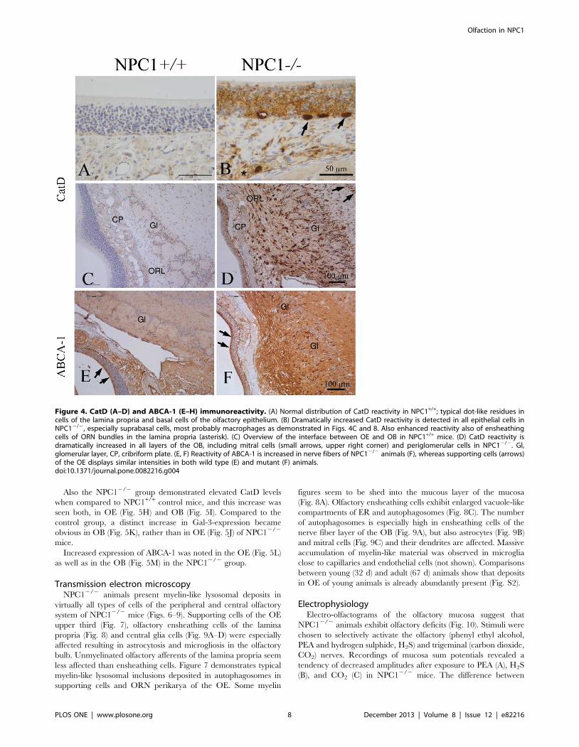
A faint background staining was seen in all cellular layers of OE in
NPC1+/+ group (Fig. 4A, C). In contrast, large (20–30 mm) CatD
immunoreactive cells, resembling macrophage-like cells as already
shown in Fig. 3B, were seen in the basal epithelium of NPC12/2
mice (Fig. 4B). Furthermore, CatD-positive cells were identified in
the lamina propria. Also an appreciable immunoreactivity for
CatD became obvious in the OB of NPC12/2 mice (Fig. 4D).
These cells were distributed throughout the entire OB, being most
abundant in ONL and glomeruli. When compared to NPC12/2,
the OB of the NPC1+/+ group exhibited only a few occasional
positive cells (Fig. 4C).
ABCA-1 expression as a marker for lipid efflux. The
ATP-binding cassette transporter 1 (ABCA-1) constitutes the
major mediator of cellular cholesterol across the plasma
membrane [46]. ABCA-1 is expressed mainly by sustentacular
cells of the OE and by numerous cell processes within the OB
(Fig. 4E). Compared to NPC1+/+ individuals, the overall
expression of ABCA-1 in NPC12/2 mice was clearly increased
throughout the OB (Fig. 4F).
Age-related differences. In comparison to adult (65–70 d)
NPC12/2 mice, young animals (35 d) generally showed less
distinct, though already clearly visible abnormalities with respect
to all markers used in this study (data now shown, except for
electron microscopy and electrophysiology, see below).
Western blot analysisTo confirm the immunohistochemical findings, respective
protein levels were examined using standard immunoblotting
techniques (Fig. 5). Age-matched (.P60) NPC12/2 (end-stage of
the disease) and wild type control animals were subjected to this
Figure 2. Immunohistochemistry of NeuN (A, B) and GFAP (C through F) in the OB. Distribution of NeuN-positive neuronal cells do notreveal any obvious differences between NPC1+/+ (A) and NPC12/2 mice (B) (67–70 d). There is a clear increase of GFAP expression, especially in theglomerular layer (Gl) in NPC12/2 animals (D) compared to NPC1+/+ (C). The area marked by the rectangles is enlarged in E and F, respectively,demonstrating the astrogliosis of the granular layer (Gr) in NPC12/2 mice. Counterstained with hematoxylin.doi:10.1371/journal.pone.0082216.g002
Olfaction in NPC1
PLOS ONE | www.plosone.org 6 December 2013 | Volume 8 | Issue 12 | e82216

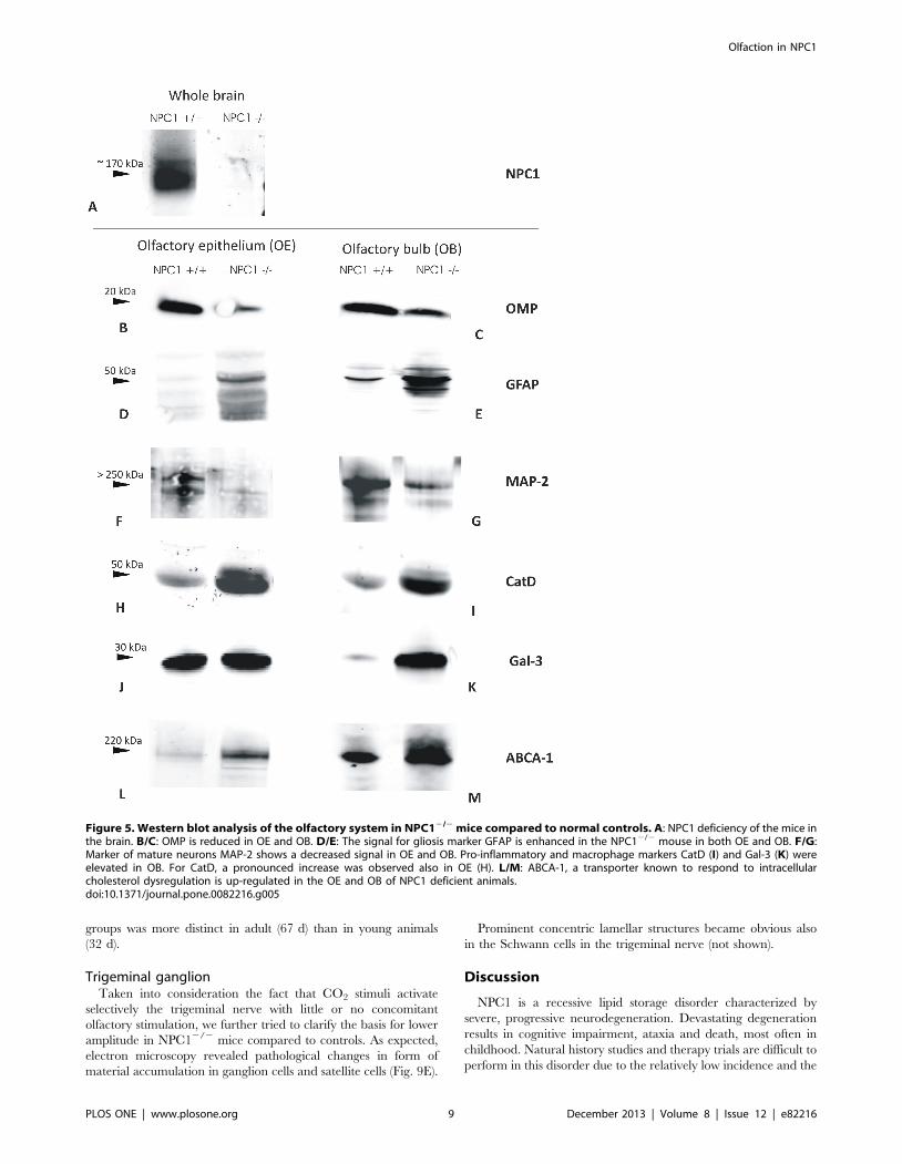
analysis. Firstly, it was confirmed that the mice were deficient of
NPC1 protein. Herein, whole brain lysates served as specimen
(Fig. 5A). Qualitative analysis of the blots revealed a lower
expression level of neuronal marker OMP in the OE and OB of
NPC12/2 mice (Fig. 5B and C, respectively). In good agreement
with the gliosis observed in immunohistochemistry in the
NPC12/2 group, the levels of astroglial marker GFAP were
increased both in OE (Fig. 5D) and OB (Fig. 5E). An apparent
neuronal decline was evidenced by a remarkable decrease in
neuronal MAP-2 protein in the NPC12/2 group (Fig. 5F/G).
Figure 3. Immunohistochemistry of Gal-3 in the OE (A, B) and OB (D, E) (67–70 d). (A) Gal-3 in NPC1+/+ is restricted to olfactory axonbundles in the lamina propria, whereas in NPC12/2 animals (B), large spot-like Gal-3 reaction product is detectable above and below the basalmembrane. Note tender processes of these cells, presumably macrophages (arrows). These cells correspond most likely to those depicted in C. (C)Interface between olfactory mucosa and lamina propria. Electron microscopic resolution of two macrophages (red) filled with myelin-like material(arrows). These cells penetrate the basal lamina (BL). N- nuclei of horizontal basal cells; Ap- nucleus of an apoptotic cell. (D) NPC1+/+, cortical layers ofthe olfactory bulb express Gal-3 immunoreactivity only in nuclei of ensheathing cells within the nerve fiber layer (arrows). Deeper bulb areas, i.e., asglomerular (Gl), mitral cell (Mi), and granular (Gr) layers, are not affected. (E) Numerous Gal-3-positive cells occupy the glomerular and adjacent part ofthe external plexiform layer in an NPC12/2 animal. These cells have partly short and interrupted processes different from those of astroglia. Glomeruliare barely recognizable. (F, G) Double immunofluorescence, using antibodies against GFAP (red) and Gal-3 (green). (F) GFAP reactive astrocytes aremainly distributed within the glomerular layer. There are only a few Gal-3-positive microglia cells in NPC1+/+ animals. (G) Prominent microgliosis (Gal-3expression) especially around glomeruli goes along with enhanced reactivity for GFAP, indicating which indicates astrogliosis. Macrophage activity ismost likely associated with microglia rather than with activated astrocytes. Astrocytes do not co-express Gal-3.doi:10.1371/journal.pone.0082216.g003
Olfaction in NPC1
PLOS ONE | www.plosone.org 7 December 2013 | Volume 8 | Issue 12 | e82216
Also the NPC12/2 group demonstrated elevated CatD levels
when compared to NPC1+/+ control mice, and this increase was
seen both, in OE (Fig. 5H) and OB (Fig. 5I). Compared to the
control group, a distinct increase in Gal-3-expression became
obvious in OB (Fig. 5K), rather than in OE (Fig. 5J) of NPC12/2
mice.
Increased expression of ABCA-1 was noted in the OE (Fig. 5L)
as well as in the OB (Fig. 5M) in the NPC12/2 group.
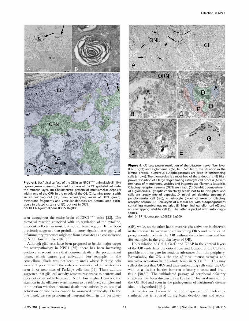
Transmission electron microscopyNPC12/2 animals present myelin-like lysosomal deposits in
virtually all types of cells of the peripheral and central olfactory
system of NPC12/2 mice (Figs. 6–9). Supporting cells of the OE
upper third (Fig. 7), olfactory ensheathing cells of the lamina
propria (Fig. 8) and central glia cells (Fig. 9A–D) were especially
affected resulting in astrocytosis and microgliosis in the olfactory
bulb. Unmyelinated olfactory afferents of the lamina propria seem
less affected than ensheathing cells. Figure 7 demonstrates typical
myelin-like lysosomal inclusions deposited in autophagosomes in
supporting cells and ORN perikarya of the OE. Some myelin
figures seem to be shed into the mucous layer of the mucosa
(Fig. 8A). Olfactory ensheathing cells exhibit enlarged vacuole-like
compartments of ER and autophagosomes (Fig. 8C). The number
of autophagosomes is especially high in ensheathing cells of the
nerve fiber layer of the OB (Fig. 9A), but also astrocytes (Fig. 9B)
and mitral cells (Fig. 9C) and their dendrites are affected. Massive
accumulation of myelin-like material was observed in microglia
close to capillaries and endothelial cells (not shown). Comparisons
between young (32 d) and adult (67 d) animals show that deposits
in OE of young animals is already abundantly present (Fig. S2).
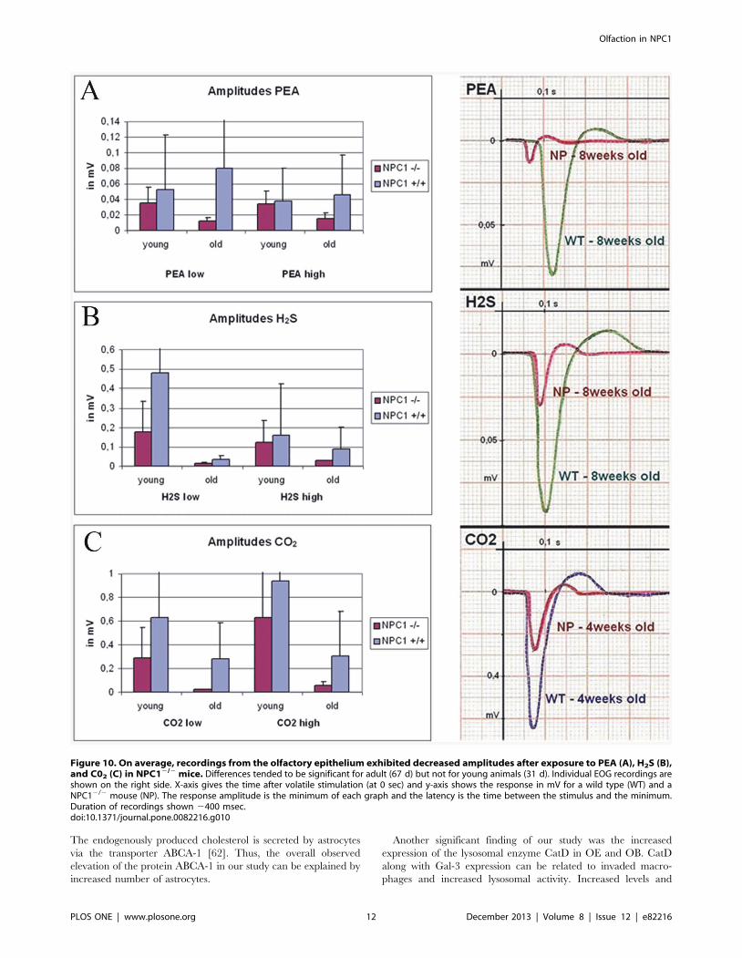
ElectrophysiologyElectro-olfactograms of the olfactory mucosa suggest that
NPC12/2 animals exhibit olfactory deficits (Fig. 10). Stimuli were
chosen to selectively activate the olfactory (phenyl ethyl alcohol,
PEA and hydrogen sulphide, H2S) and trigeminal (carbon dioxide,
CO2) nerves. Recordings of mucosa sum potentials revealed a
tendency of decreased amplitudes after exposure to PEA (A), H2S
(B), and CO2 (C) in NPC12/2 mice. The difference between
Figure 4. CatD (A–D) and ABCA-1 (E–H) immunoreactivity. (A) Normal distribution of CatD reactivity in NPC1+/+; typical dot-like residues incells of the lamina propria and basal cells of the olfactory epithelium. (B) Dramatically increased CatD reactivity is detected in all epithelial cells inNPC12/2, especially suprabasal cells, most probably macrophages as demonstrated in Figs. 4C and 8. Also enhanced reactivity also of ensheathingcells of ORN bundles in the lamina propria (asterisk). (C) Overview of the interface between OE and OB in NPC1+/+ mice. (D) CatD reactivity isdramatically increased in all layers of the OB, including mitral cells (small arrows, upper right corner) and periglomerular cells in NPC12/2. Gl,glomerular layer, CP, cribriform plate. (E, F) Reactivity of ABCA-1 is increased in nerve fibers of NPC12/2 animals (F), whereas supporting cells (arrows)of the OE displays similar intensities in both wild type (E) and mutant (F) animals.doi:10.1371/journal.pone.0082216.g004
Olfaction in NPC1
PLOS ONE | www.plosone.org 8 December 2013 | Volume 8 | Issue 12 | e82216
groups was more distinct in adult (67 d) than in young animals
(32 d).
Trigeminal ganglionTaken into consideration the fact that CO2 stimuli activate
selectively the trigeminal nerve with little or no concomitant
olfactory stimulation, we further tried to clarify the basis for lower
amplitude in NPC12/2 mice compared to controls. As expected,
electron microscopy revealed pathological changes in form of
material accumulation in ganglion cells and satellite cells (Fig. 9E).
Prominent concentric lamellar structures became obvious also
in the Schwann cells in the trigeminal nerve (not shown).
Discussion
NPC1 is a recessive lipid storage disorder characterized by
severe, progressive neurodegeneration. Devastating degeneration
results in cognitive impairment, ataxia and death, most often in
childhood. Natural history studies and therapy trials are difficult to
perform in this disorder due to the relatively low incidence and the
Figure 5. Western blot analysis of the olfactory system in NPC12/2 mice compared to normal controls. A: NPC1 deficiency of the mice inthe brain. B/C: OMP is reduced in OE and OB. D/E: The signal for gliosis marker GFAP is enhanced in the NPC12/2 mouse in both OE and OB. F/G:Marker of mature neurons MAP-2 shows a decreased signal in OE and OB. Pro-inflammatory and macrophage markers CatD (I) and Gal-3 (K) wereelevated in OB. For CatD, a pronounced increase was observed also in OE (H). L/M: ABCA-1, a transporter known to respond to intracellularcholesterol dysregulation is up-regulated in the OE and OB of NPC1 deficient animals.doi:10.1371/journal.pone.0082216.g005
Olfaction in NPC1
PLOS ONE | www.plosone.org 9 December 2013 | Volume 8 | Issue 12 | e82216

heterogeneity of disease in human patients. In recent years mutant
mouse models have facilitated the understanding of structural and
molecular events occurring as a result of NPC1 gene mutation. A
mouse model of NPC1 disease, the BALB/cJ NPC1NIH, has been
shown to resemble human NPC1 and is used to investigate closely
molecular and biochemical aspects of the disease [13]. The NPC1
gene is mutated in these mice, and the locus belongs to the same
complementation group as human NPC1.
Olfactory dysfunction may be an early sign inneurodegenerative diseases
The present study was undertaken to examine the olfactory
system at histological, ultrastructural and functional levels. While
one of the earliest clinical symptoms reported by patients with
neurodegenerative diseases such as Alzheimer’s and Parkinson’s
diseases is olfactory dysfunction [25,47], investigations of the
olfactory system are scarce in lysosomal storage diseases both in
humans and experimental animal models. Assessment of olfactory
function in Gaucher patients revealed significantly lower scores
compared with healthy individuals [29]. In NPC1 disease, the
structures of olfactory epithelium and central pathways as well as
olfactory function have not been studied so far.
General symptoms of NPC12/2 mutant miceNPC12/2 mutant mice are asymptomatic and even undistin-
guishable from their littermates at birth. The earliest definitive
symptoms of the disease become apparent by 4 to 6 weeks of age,
and as they reach adulthood symptoms of ataxia and hind limb
paralysis emerge [48]. Abnormalities in intracellular cholesterol
transport with subsequent accumulation of lipids could be found in
many organs of these mutant mice, including brain [49]. At the
cellular level, NPC12/2 mice show an age-related loss of neurons
in the prefrontal cortex, thalamus, brainstem, and of cerebellar
Purkinje cells, as well as activation of microglia and astrocytes with
phenotypes that are similar to those observed in human NPC1
disease [16,48,50].
Neurodegeneration, activation of peripheralmacrophages and cerebral glial cells in components ofthe olfactory system
As expected, the main findings of the present study reveal
pronounced peripheral and central neurodegeneration as well as
glia activation in the olfactory system- a part of CNS which has not
been examined in NPC1 before. The neurodegeneration in NPC1
has been demonstrated to be an autonomous process, caused
primarily by the lack of NPC1 in the central nervous system [51].
Prior mammalian studies have revealed that NPC1 predominantly
localized in glia [52], and both, astrocytes and microglia have been
suggested to mediate inflammation and neurodegeneration in
NPC1 mice [53,54]. It has been shown that at two weeks of age
the reactive astrocytes were only observed in the ventral lateral
thalamus, while another two weeks later massive astrogliosis was
Figure 6. Electron microscopic depiction of the olfactoryepithelium of a young (32 d) NPC12/2 animal. Some of the cellsare exemplarily colored. The OE consists 1) of olfactory receptorneurons in the middle portion of the OE (O, green) with thin apicalprocesses extending with knob-like structures into the mucous layer, 2)supporting cells, the nuclei of which lie in the upper third, and withbasal footplates (S, yellow) above the basal membrane (magenta line),and 3) basal cells (B). There are many irregular, myelin-like inclusionswithin most of the cells (arrows). Most of these autophagosomes occurin subnuclear portions of supporting cells, but also, to a lesser degree,in perinuclear locations of ORN. Apical processes of ORN are notaffected (see also Fig. 7). Singular, huge accumulations of myelin-likedeposits are occasionally seen near the basal membrane, most likelycorresponding to Gal-3 – and CatD-positive macrophages (M, red; seealso Figs. 4,5). BG, Bowman glands of the lamina propria.doi:10.1371/journal.pone.0082216.g006
Figure 7. Superficial part of the olfactory epithelium in anNPC12/2 animal. Most prominent are perinuclear autophagosomes insupporting cells (yellow, arrows). ORN (green) occasionally contain suchdeposits, but appear normal at the surface. Tight junctions are intact(arrowheads). One microvillar cell (MV) is not affected.doi:10.1371/journal.pone.0082216.g007
Olfaction in NPC1
PLOS ONE | www.plosone.org 10 December 2013 | Volume 8 | Issue 12 | e82216
seen throughout the entire brain of NPC12/2 mice [22]. The
astroglial reaction coincided with up-regulation of the cytokine,
interleukin-1beta, in most, but not all brain regions. It has been
previously suggested that proinflammatory signals that trigger glial
inflammatory responses originate from astrocytes as a consequence
of NPC1 loss in these cells [55].
Although glial cells have been proposed to be the major target
for neuropathology in NPC1 [56], there has been increasing
evidence in recent years that neuronal death is the predominant
factor, which causes glia activation. For example, in the
cerebellum, gliosis was not seen in areas where Purkinje cells
were still present, and the only concentration of astrocytes was
seen in or near sites of Purkinje cells loss [57]. These authors
suggested that glial cell activity remains responsive to neurons and
does not occur solely because of NPC1 loss in glia. However, the
situation in the olfactory system seems to be relatively complex and
the question whether neuronal death mechanistically causes glial
activation or vice versa cannot be answered univocally. On the
one hand, we see pronounced neuronal death in the periphery
(OE), while, on the other hand, massive glia activation is observed
in the interface between axons of incoming ORN and mitral cells/
periglomerular cells in the OB without distinctive neuronal loss
(for example, in the granular layer of OB).
Up-regulation of Gal-3, CatD and GFAP in the cortical layers
of the OB underlines the critical role and location of the OB as a
possible entrance gate for noxious substances from the periphery.
Remarkably, the OB is the site of most intense astroglia and
microglia activation in the whole brain in NPC12/2. This may
reflect the fact that ORN and their ensheathing cells enter the OB
without a distinct barrier between olfactory mucosa and brain
tissue [58,59]. The unhindered passage of peripheral olfactory
structures has been discussed as a key factor for viral invasion of
the OB [60] and even in the pathogenesis of Parkinson’s disease
(dual hit hypothesis) [61].
Astrocytes are known to be the major site of cholesterol
synthesis that is required during brain development and repair.
Figure 8. (A) Apical surface of the OE in an NPC12/2 animal. Myelin-likefigures (arrows) seem to be shed from one of the OE epithelial cells intothe mucous layer. (B) Characteristic pattern of multilamellar depositswithin one of the ORN in the middle of the OE. (C) Lamina propria withan ensheathing cell (EC, blue), enwrapping axons of ORN (green).Membrane fragments and vesicular deposits are accumulated exclu-sively in dilated cisterns of EC, but not in ORN.doi:10.1371/journal.pone.0082216.g008
Figure 9. (A) Low power resolution of the olfactory nerve fiber layer(ONL, right) and a glomerulus (GL, left). Similar to the situation in thelamina propria, numerous autophagosomes are seen in ensheathingcells (arrows). The glomerulus is almost free of these deposits. (B) Highpower resolution of a large degenerating astrocyte cell process (A) withremnants of membranes, vesicles and intermediate filaments (asterisk).Olfactory receptor neurons (ORN) are intact. (C) Dendritic compartmentof a glomerulus. Synaptic connectivity seems not to be disrupted, andcells are largely free of deposits. D- mitral cell dendrite (green); P,periglomerular cell (red), A, astrocyte (blue); O, axon of olfactoryreceptor neuron. (D) Perikaryon of a mitral cell with autophagosomescontaining membranous material. (E) Trigeminal ganglion cell (G) andan enwrapping satellite cell (S). The latter is packed with autophago-somes.doi:10.1371/journal.pone.0082216.g009
Olfaction in NPC1
PLOS ONE | www.plosone.org 11 December 2013 | Volume 8 | Issue 12 | e82216
The endogenously produced cholesterol is secreted by astrocytes
via the transporter ABCA-1 [62]. Thus, the overall observed
elevation of the protein ABCA-1 in our study can be explained by
increased number of astrocytes.
Another significant finding of our study was the increased
expression of the lysosomal enzyme CatD in OE and OB. CatD
along with Gal-3 expression can be related to invaded macro-
phages and increased lysosomal activity. Increased levels and
Figure 10. On average, recordings from the olfactory epithelium exhibited decreased amplitudes after exposure to PEA (A), H2S (B),and C02 (C) in NPC12/2 mice. Differences tended to be significant for adult (67 d) but not for young animals (31 d). Individual EOG recordings areshown on the right side. X-axis gives the time after volatile stimulation (at 0 sec) and y-axis shows the response in mV for a wild type (WT) and aNPC12/2 mouse (NP). The response amplitude is the minimum of each graph and the latency is the time between the stimulus and the minimum.Duration of recordings shown 2400 msec.doi:10.1371/journal.pone.0082216.g010
Olfaction in NPC1
PLOS ONE | www.plosone.org 12 December 2013 | Volume 8 | Issue 12 | e82216

activity of CatD have been previously shown in hippocampus and
cerebellum of NPC12/2 mice [63]. It has been suggested that
increased expression/release/activation of CatD in neurons and
astrocytes can trigger neurodegeneration and development of
NPC pathology [64]. Elevated levels of CatD expression and
activity have been also shown to be involved in the pathogenesis of
Alzheimer disease, atherosclerosis and cancer [65].
The trigeminal system is affected in NPC pathologyIn contrast to primarily telencephalic olfactory projections,
fibers of the trigeminal system travel via the trigeminal ganglion to
brainstem nuclei and reach postcentral gyri after relaying in the
ventral posteromedial thalamic nucleus [33,66]. Apart from
mechanosensory inputs, trigeminal fibers also carry ‘‘general
chemosensory’’ modalities [67], which allow an increasingly
important crosstalk with olfactory stimuli [68]. The myelin-like
deposits in trigeminal ganglion cells and satellite cells suggest
impairment of trigeminal function in NPC12/2 mice, as
demonstrated by electrophysiology.
As a consequence of all pathological changes observed at the
structural level we further demonstrate functional impairment in
olfaction as shown by decreased amplitudes after exposure of the
OE to different olfactory and trigeminal stimuli. Interestingly, the
deterioration was more evident in adult animals, rather than in
young ones. According to the literature, distinct clinical symptoms
in NPC1 occur after the neuronal impairment has reached a
threshold level [69]. Previously, impaired retinal function has been
shown in the same mouse model [19]. Similarly to the olfactory
system, also in the retina the lipid accumulation leads to
destructive cellular changes, deformation of layers and degener-
ation of photoreceptors.
One of the important implications as a result from our
observations in this murine model is that both the olfactory and
trigeminal impairment are early events in NPC1 pathogenesis, at
least in comparison with impairment of motor acuity that does not
occur before 42–49 days of age [15,32]. These data will lead to
future studies, focusing on the olfactory system of NPC1 patients.
The olfactory system offers the opportunity of in vivo functional
measurements using simple psychophysical or electrophysiological
tests. The latter could become a helpful tool to estimate the degree
of neurodegeneration and monitor a therapy success, e.g. during a
combined treatment with cyclodextrin/allopregnanolone and
miglustat in follow-up studies that has become available in recent
years [70,71]. Another intriguing question to be addressed in the
future is how the olfactory system may compensate for neuronal
loss in NPC1. Studies concerning the behavior of migrating
neuronal precursors are in progress.
Supporting Information
Figure S1 (A) body weight of young and adult NPC12/2 mice
in comparison with age-matched NPC1+/+ group. (B) whole brain
weights of NPC1+/+ and NPC12/2 mice at P64 after perfusion.
The difference between groups is significant (p = 0.0221). Data are
presented as mean 6 SD.
(TIF)
Figure S2 Comparative electron micrographs of OE inyoung (32d) and adult (67d) NPC12/2animals. (A) Myelin-
like deposits are already visible in most supporting cells (S) and
ORN (O) of young animals. (B) In addition, basally located cells
are more affected in adult mice. BG, excretory duct of a Bowman
gland. The basal lamina is indicated with dotted lines. Scale bar:
5 mm.
(TIF)
Acknowledgments
The authors would like to thank Robert Ladegast for his help with
recording of the electro-olfactograms.
Author Contributions
Conceived and designed the experiments: MW AW TH. Performed the
experiments: AM J. Lukas VG. Analyzed the data: MW MH J. Luo VG
TH. Contributed reagents/materials/analysis tools: AW AR. Wrote the
paper: MH MW J. Lukas.
References
1. Patterson MC, Vanier M, Suzuki K, Morris J, Carstea E, et al. (2001) Niemann-
Pick disease type C: a lipid trafficking disorder. In: Scriver C, Beaudet A, Sly W,
D V, editors. The metabolic and molecular bases of inherited disease. 8 ed. New
York: McGraw Hill. pp. 3611–3634.
2. Pentchev PG (2004) Niemann-Pick C research from mouse to gene. Biochim
Biophys Acta 1685: 3–7.
3. Carstea ED, Morris JA, Coleman KG, Loftus SK, Zhang D, et al. (1997)
Niemann-Pick C1 disease gene: homology to mediators of cholesterol
homeostasis. Science 277: 228–231.
4. Morris JA, Zhang D, Coleman KG, Nagle J, Pentchev PG, et al. (1999) The
genomic organization and polymorphism analysis of the human Niemann-Pick
C1 gene. Biochem Biophys Res Commun 261: 493–498.
5. Neufeld EB, Wastney M, Patel S, Suresh S, Cooney AM, et al. (1999) The
Niemann-Pick C1 protein resides in a vesicular compartment linked to
retrograde transport of multiple lysosomal cargo. J Biol Chem 274: 9627–9635.
6. Reid PC, Sugii S, Chang TY (2003) Trafficking defects in endogenously
synthesized cholesterol in fibroblasts, macrophages, hepatocytes, and glial cells
from Niemann-Pick type C1 mice. J Lipid Res 44: 1010–1019.
7. Rosenbaum AI, Maxfield FR (2011) Niemann-Pick type C disease: molecular
mechanisms and potential therapeutic approaches. J Neurochem 116: 789–795.
8. Garver WS, Francis GA, Jelinek D, Shepherd G, Flynn J, et al. (2007) The
National Niemann-Pick C1 disease database: report of clinical features and
health problems. Am J Med Genet A 143A: 1204–1211.
9. Spiegel R, Raas-Rothschild A, Reish O, Regev M, Meiner V, et al. (2009) The
clinical spectrum of fetal Niemann-Pick type C. Am J Med Genet A 149A: 446–
450.
10. Vanier MT, Millat G (2003) Niemann-Pick disease type C. Clin Genet 64: 269–
281.
11. Mellon S, Gong W, Griffin LD (2004) Niemann pick type C disease as a model
for defects in neurosteroidogenesis. Endocr Res 30: 727–735.
12. Ward S, O’Donnell P, Fernandez S, Vite CH (2010) 2-hydroxypropyl-beta-
cyclodextrin raises hearing threshold in normal cats and in cats with Niemann-Pick type C disease. Pediatr Res 68: 52–56.
13. Loftus SK, Morris JA, Carstea ED, Gu JZ, Cummings C, et al. (1997) Murinemodel of Niemann-Pick C disease: mutation in a cholesterol homeostasis gene.
Science 277: 232–235.
14. Erickson RP, Bhattacharyya A, Hunter RJ, Heidenreich RA, Cherrington NJ(2005) Liver disease with altered bile acid transport in Niemann-Pick C mice on
a high-fat, 1% cholesterol diet. Am J Physiol Gastrointest Liver Physiol 289:G300–307.
15. Voikar V, Rauvala H, Ikonen E (2002) Cognitive deficit and development ofmotor impairment in a mouse model of Niemann-Pick type C disease. Behav
Brain Res 132: 1–10.
16. Sarna JR, Larouche M, Marzban H, Sillitoe RV, Rancourt DE, et al. (2003)Patterned Purkinje cell degeneration in mouse models of Niemann-Pick type C
disease. J Comp Neurol 456: 279–291.17. Yamada A, Saji M, Ukita Y, Shinoda Y, Taniguchi M, et al. (2001) Progressive
neuronal loss in the ventral posterior lateral and medial nuclei of thalamus in
Niemann-Pick disease type C mouse brain. Brain Dev 23: 288–297.18. Zervas M, Dobrenis K, Walkley SU (2001) Neurons in Niemann-Pick disease
type C accumulate gangliosides as well as unesterified cholesterol and undergodendritic and axonal alterations. J Neuropathol Exp Neurol 60: 49–64.
19. Claudepierre T, Paques M, Simonutti M, Buard I, Sahel J, et al. (2009) Lack ofNiemann-Pick type C1 induces age-related degeneration in the mouse retina.
Mol Cell Neurosci 43: 164–176.
20. Bi X, Liao G (2007) Autophagic-lysosomal dysfunction and neurodegenerationin Niemann-Pick Type C mice: lipid starvation or indigestion? Autophagy 3:
646–648.21. Liao G, Yao Y, Liu J, Yu Z, Cheung S, et al. (2007) Cholesterol accumulation is
associated with lysosomal dysfunction and autophagic stress in Npc1 2/2
mouse brain. Am J Pathol 171: 962–975.
Olfaction in NPC1
PLOS ONE | www.plosone.org 13 December 2013 | Volume 8 | Issue 12 | e82216
22. Baudry M, Yao Y, Simmons D, Liu J, Bi X (2003) Postnatal development of
inflammation in a murine model of Niemann-Pick type C disease: immunohis-
tochemical observations of microglia and astroglia. Exp Neurol 184: 887–903.
23. Doetsch F (2003) The glial identity of neural stem cells. Nat Neurosci 6: 1127–
1134.
24. Lledo PM, Gheusi G (2003) Olfactory processing in a changing brain.
Neuroreport 14: 1655–1663.
25. Berendse HW, Booij J, Francot CM, Bergmans PL, Hijman R, et al. (2001)
Subclinical dopaminergic dysfunction in asymptomatic Parkinson’s disease
patients’ relatives with a decreased sense of smell. Ann Neurol 50: 34–41.
26. Attems J, Jellinger KA (2006) Olfactory tau pathology in Alzheimer disease and
mild cognitive impairment. Clin Neuropathol 25: 265–271.
27. Bahar-Fuchs A, Chetelat G, Villemagne VL, Moss S, Pike K, et al. (2010)
Olfactory deficits and amyloid-beta burden in Alzheimer’s disease, mild
cognitive impairment, and healthy aging: a PiB PET study. J Alzheimers Dis
22: 1081–1087.
28. Wesson DW, Wilson DA, Nixon RA (2010) Should olfactory dysfunction be
used as a biomarker of Alzheimer’s disease? Expert Rev Neurother 10: 633–635.
29. McNeill A, Duran R, Proukakis C, Bras J, Hughes D, et al. (2012) Hyposmia
and cognitive impairment in Gaucher disease patients and carriers. Mov Disord
27: 526–532.
30. Saunders-Pullman R, Hagenah J, Dhawan V, Stanley K, Pastores G, et al.
(2010) Gaucher disease ascertained through a Parkinson’s center: imaging and
clinical characterization. Mov Disord 25: 1364–1372.
31. Yan X, Lukas J, Witt M, Wree A, Hubner R, et al. (2011) Decreased expression
of myelin gene regulatory factor in Niemann-Pick type C 1 mouse. Metab Brain
Dis 26: 299–306.
32. Hovakimyan M, Maass F, Petersen J, Holzmann C, Witt M, et al. (2013)
Combined therapy with cyclodextrin/allopregnanolone and miglustat improves
motor but not cognitive functions in Niemann-Pick Type C1 mice. Neuroscience
(in press).
33. Doty RL (2008) The olfactory vector hypothesis of neurodegenerative disease: is
it viable? Ann Neurol 63: 7–15.
34. Duda JE, Shah U, Arnold SE, Lee VM-Y, Trojanowski JQ (1999) The
Expression of [alpha]-, [beta]-, and [gamma]-Synucleins in Olfactory Mucosa
from Patients with and without Neurodegenerative Diseases. Experimental
Neurology 160: 515–522.
35. Kaltner H, Seyrek K, Heck A, Sinowatz F, Gabius HJ (2002) Galectin-1 and
galectin-3 in fetal development of bovine respiratory and digestive tracts.
Comparison of cell type-specific expression profiles and subcellular localization.
Cell Tissue Res 307: 35–46.
36. Dahm R, Bramke S, Dawczynski J, Nagaraj RH, Kasper M (2003)
Developmental aspects of galectin-3 expression in the lens. Histochem Cell Biol
119: 219–226.
37. Taylor CR, Shi SR, Chaiwun B, Young L, Imam SA, et al. (1994) Strategies for
improving the immunohistochemical staining of various intranuclear prognostic
markers in formalin-paraffin sections: androgen receptor, estrogen receptor,
progesterone receptor, p53 protein, proliferating cell nuclear antigen, and Ki-67
antigen revealed by antigen retrieval techniques. Hum Pathol 25: 263–270.
38. Hsu SM, Raine L, Fanger H (1981) Use of avidin-biotin-peroxidase complex
(ABC) in immunoperoxidase techniques: a comparison between ABC and
unlabeled antibody (PAP) procedures. J Histochem Cytochem 29: 577–580.
39. Ostalska-Nowicka D, Zachwieja J, Nowicki M, Kaczmarek E, Siwinska A, et al.
(2007) Immunohistochemical detection of galectin-1 in renal biopsy specimens of
children and its possible role in proteinuric glomerulopathies. Histopathology
51: 468–476.
40. Hubner R, Schmole AC, Liedmann A, Frech MJ, Rolfs A, et al. (2010)
Differentiation of human neural progenitor cells regulated by Wnt-3a. Biochem
Biophys Res Commun 400: 358–362.
41. Gudziol V, Pietsch J, Witt M, Hummel T (2010) Theophylline induces changes
in the electro-olfactogram of the mouse. Eur Arch Otorhinolaryngol 267: 239–
243.
42. Mullen RJ, Buck CR, Smith AM (1992) NeuN, a neuronal specific nuclear
protein in vertebrates. Development 116: 201–211.
43. Pasquini LA, Millet V, Hoyos HC, Giannoni JP, Croci DO, et al. (2011)
Galectin-3 drives oligodendrocyte differentiation to control myelin integrity and
function. Cell Death Differ 18: 1746–1756.
44. Rotshenker S, Reichert F, Gitik M, Haklai R, Elad-Sfadia G, et al. (2008)
Galectin-3/MAC-2, Ras and PI3K activate complement receptor-3 and
scavenger receptor-AI/II mediated myelin phagocytosis in microglia. Glia 56:
1607–1613.
45. Henderson NC, Sethi T (2009) The regulation of inflammation by galectin-3.
Immunol Rev 230: 160–171.
46. Bodzioch M, Orso E, Klucken J, Langmann T, Bottcher A, et al. (1999) The
gene encoding ATP-binding cassette transporter 1 is mutated in Tangier disease.
Nat Genet 22: 347–351.
47. Hummel T, Witt M, Reichmann H, Welge-Luessen A, Haehner A (2009)
Immunohistochemical, volumetric, and functional neuroimaging studies in
patients with idiopathic Parkinson’s disease. J Neurol Sci. 289: 119–122.
48. Li H, Repa JJ, Valasek MA, Beltroy EP, Turley SD, et al. (2005) Molecular,
anatomical, and biochemical events associated with neurodegeneration in mice
with Niemann-Pick type C disease. J Neuropathol Exp Neurol 64: 323–333.
49. Xie C, Burns DK, Turley SD, Dietschy JM (2000) Cholesterol is sequestered inthe brains of mice with Niemann-Pick type C disease but turnover is increased.
J Neuropathol Exp Neurol 59: 1106–1117.
50. Higashi Y, Murayama S, Pentchev PG, Suzuki K (1993) Cerebellardegeneration in the Niemann-Pick type C mouse. Acta Neuropathol 85: 175–
184.
51. Loftus SK, Erickson RP, Walkley SU, Bryant MA, Incao A, et al. (2002) Rescue
of neurodegeneration in Niemann-Pick C mice by a prion-promoter-driven
Npc1 cDNA transgene. Hum Mol Genet 11: 3107–3114.
52. Patel SC, Suresh S, Kumar U, Hu CY, Cooney A, et al. (1999) Localization of
Niemann-Pick C1 protein in astrocytes: implications for neuronal degenerationin Niemann- Pick type C disease. Proc Natl Acad Sci U S A 96: 1657–1662.
53. Chen G, Li HM, Chen YR, Gu XS, Duan S (2007) Decreased estradiol release
from astrocytes contributes to the neurodegeneration in a mouse model ofNiemann-Pick disease type C. Glia 55: 1509–1518.
54. Suzuki H, Sakiyama T, Harada N, Abe M, Tadokoro M (2003) Pathologicchanges of glial cells in murine model of Niemann-Pick disease type C:
immunohistochemical, lectin-histochemical and ultrastructural observations.
Pediatr Int 45: 1–4.
55. Suzuki M, Sugimoto Y, Ohsaki Y, Ueno M, Kato S, et al. (2007) Endosomal
accumulation of Toll-like receptor 4 causes constitutive secretion of cytokinesand activation of signal transducers and activators of transcription in Niemann-
Pick disease type C (NPC) fibroblasts: a potential basis for glial cell activation in
the NPC brain. J Neurosci 27: 1879–1891.
56. German DC, Liang CL, Song T, Yazdani U, Xie C, et al. (2002)
Neurodegeneration in the Niemann-Pick C mouse: glial involvement. Neuro-science 109: 437–450.
57. Lopez ME, Klein AD, Dimbil UJ, Scott MP (2011) Anatomically defined
neuron-based rescue of neurodegenerative Niemann-Pick type C disorder.J Neurosci 31: 4367–4378.
58. Au E, Roskams AJ (2003) Olfactory ensheathing cells of the lamina propria in
vivo and in vitro. Glia 41: 224–236.
59. Schwob JE (2002) Neural regeneration and the peripheral olfactory system. Anat
Rec 269: 33–49.
60. Barnett EM, Cassell MD, Perlman S (1993) Two neurotropic viruses, herpes
simplex virus type 1 and mouse hepatitis virus, spread along different neural
pathways from the main olfactory bulb. Neuroscience 57: 1007–1025.
61. Hawkes CH, Del Tredici K, Braak H (2009) Parkinson’s disease: the dual hit
theory revisited. Ann N Y Acad Sci 1170: 615–622.
62. Pfrieger FW (2003) Outsourcing in the brain: do neurons depend on cholesteroldelivery by astrocytes? Bioessays 25: 72–78.
63. Amritraj A, Peake K, Kodam A, Salio C, Merighi A, et al. (2009) IncreasedActivity and Altered Subcellular Distribution of Lysosomal Enzymes Determine
Neuronal Vulnerability in Niemann-Pick Type C1-Deficient Mice. Am J Pathol
175: 2540–2556.
64. Amritraj A, Wang Y, Revett TJ, Vergote D, Westaway D, et al. (2012) Role of
Cathepsin D in U18666A-induced Neuronal Cell Death: Potential implication inNiemann-Pick Type C disease pathogenesis. J Biol Chem 288: 3136–3152.
65. Benes P, Vetvicka V, Fusek M (2008) Cathepsin D–many functions of one
aspartic protease. Crit Rev Oncol Hematol 68: 12–28.
66. Dhaka A, Earley TJ, Watson J, Patapoutian A (2008) Visualizing cold spots:
TRPM8-expressing sensory neurons and their projections. J Neurosci 28: 566–575.
67. Caterina MJ, Schumacher MA, Tominaga M, Rosen TA, Levine JD, et al.
(1997) The capsaicin receptor: a heat-activated ion channel in the pain pathway.Nature 389: 816–824.
68. Frasnelli J, Lundstrom JN, Schopf V, Negoias S, Hummel T, et al. (2012) Dualprocessing streams in chemosensory perception. Front Hum Neurosci 6: 288.
69. Yanjanin NM, Velez JI, Gropman A, King K, Bianconi SE, et al. (2010) Linear
clinical progression, independent of age of onset, in Niemann-Pick disease, typeC. Am J Med Genet B Neuropsychiatr Genet 153B: 132–140.
70. Davidson CD, Ali NF, Micsenyi MC, Stephney G, Renault S, et al. (2009)
Chronic cyclodextrin treatment of murine Niemann-Pick C disease amelioratesneuronal cholesterol and glycosphingolipid storage and disease progression.
PLoS One 4: e6951.
71. Hovakimyan M, Petersen J, Maass F, Reichard M, Witt M, et al. (2011) Corneal
alterations during combined therapy with cyclodextrin/allopregnanolone and
miglustat in a knock-out mouse model of NPC1 disease. PLoS One 6: e28418.
72. Baldisseri DM, Margolis JW, Weber DJ, Koo JH, Margolis FL (2002) Olfactory
marker protein (OMP) exhibits a beta-clam fold in solution: implications fortarget peptide interaction and olfactory signal transduction. J Mol Biol 319: 823–
837.
73. Getchell ML, Boggess MA, Pruden SJ 2nd, Little SS, Buch S, et al. (2002)Expression of TGF-beta type II receptors in the olfactory epithelium and their
regulation in TGF-alpha transgenic mice. Brain Res 945: 232–241.
74. Margolis FL (1982) Olfactory marker protein (OMP). Scand J Immunol Suppl 9:
181–199.
75. Weiler E, Benali A (2005) Olfactory epithelia differentially express neuronalmarkers. J Neurocytol 34: 217–240.
76. Witt M, Bormann K, Gudziol V, Pehlke K, Barth K, et al. (2009) Biopsies ofolfactory epithelium in patients with Parkinson’s disease. Mov Disord 24: 906–
914.
77. Bejarano-Escobar R, Holguin-Arevalo MS, Montero JA, Francisco-Morcillo J,Martin-Partido G (2011) Macrophage and microglia ontogeny in the mouse
Olfaction in NPC1
PLOS ONE | www.plosone.org 14 December 2013 | Volume 8 | Issue 12 | e82216

visual system can be traced by the expression of Cathepsins B and D. Dev Dyn
240: 1841–1855.78. Mackay-Sim A, Chuah MI (2000) Neurotrophic factors in the primary olfactory
pathway. Prog Neurobiol 62: 527–559.
79. Karasinska JM, de Haan W, Franciosi S, Ruddle P, Fan J, et al. (2013) ABCA1
influences neuroinflammation and neuronal death. Neurobiol Dis.80. Schmitz G, Langmann T (2001) Structure, function and regulation of the ABC1
gene product. Curr Opin Lipidol 12: 129–140.
Olfaction in NPC1
PLOS ONE | www.plosone.org 15 December 2013 | Volume 8 | Issue 12 | e82216