oestrogen receptor alpha increases p21(waf1/cip1) gene expression and the antiproliferative activity...
TRANSCRIPT

Oestrogen receptor � increases p21WAF1/CIP1 gene expression andthe antiproliferative activity of histone deacetylase inhibitors inhuman breast cancer cells
R Margueron, A Licznar, G Lazennec, F Vignon and V CavaillèsINSERM U540, Endocrinologie Moléculaire et Cellulaire des Cancers and Université de Montpellier I, 60, rue de Navacelles, 34090 Montpellier, France
(Requests for offprints should be addressed to V Cavaillès; Email: [email protected])
Abstract
We analysed the antiproliferative activity of various his-tone deacetylase (HDAC) inhibitors such as trichostatin A(TSA) on human breast cancer cells. We observed a lowersensitivity to HDAC inhibition for oestrogen receptornegative (ER�) versus positive (ER+) cell lines. Thisdifferential response was associated neither with a modifi-cation of drug efflux via the multidrug resistance systemnor with a global modification of histone acetyltransferase(HAT)/HDAC activities. In contrast, we demonstratedthat in ER+ breast cancer cells the p21WAF1/CIP1 gene wasmore sensitive to TSA regulation and was expressed athigher levels. These differences were observed both intransient transfection experiments and on the endogenous
p21WAF1/CIP1 gene. The Sp1 transcription factor, whichwas shown to interact in vitro with both class I and class IIHDACs, is sufficient to confer the differential sensitivityto TSA and participated in the control of p21WAF1/CIP1
basal expression. Finally, re-expression of ER� followingadenoviral infection of ER� breast cancer cells increasedboth p21WAF1/CIP1 protein accumulation and the growthinhibitory activity of TSA. Altogether, our resultshighlight the key role of ER� and p21WAF1/CIP1 geneexpression in the sensitivity of breast cancer cells tohyperacetylating agents.Journal of Endocrinology (2003) 179, 41–53
Introduction
The implication of ovarian hormones in the developmentof breast cancer has been widely demonstrated and oes-trogen dependency of these tumours has been successfullyexploited to develop an antihormonal chemotherapy(Santen et al. 1990). Unfortunately, treatments with anti-oestrogens are not effective in all tumours and acquiredresistance also results in patient relapse. The effects ofoestrogens and antioestrogens are primarily mediatedthrough the interaction with specific nuclear oestrogenreceptors (ER� and ER�), acting as ligand-dependenttranscription factors (Sommer & Fuqua 2001). ER� ex-pression decreases during breast cancer carcinogenesis(Roger et al. 2001) and the major form of ER in breastcancers appears to be ER� at both the protein and mRNAlevels (Brouillet et al. 2001). The absence of ER� expres-sion in so-called ER-negative (ER�) breast cancers is themain cause of de novo resistance to antihormonal therapies,which is associated with aggressive cancer (Jordan 1998).
Besides endocrine therapies, other types of chemo-therapy are available and during the last few years newtherapeutic targets have been characterised, in particularthose involving chromatin structure. The basic unit ofchromatin is the nucleosome, which is formed by the
packaging of eukaryotic DNA with core histones(Woodcock & Dimitrov 2001). Post-translational modifi-cations of histone tails (acetylation, methylation and phos-phorylation) alter the nucleosomal structure and play amajor role in the regulation of gene transcription (Strahl &Allis 2000). The steady state level of histone acetylation isregulated by the activities of two types of enzymes, histoneacetyltransferase (HAT) and histone deacetylase (HDAC).Alterations of these enzymes (mutation, translocation,amplification) have been described as being associatedwith the appearance of various cancer types, mainlymyeloid leukaemia or lymphoma (Pandolfi 2001).
Several specific HDAC inhibitors (trichostatin A,SAHA, apicidin, FR901228{) have been shown to modifyreversibly or irreversibly the balance between HAT andHDAC activities (Marks et al. 2000). In vitro, in variouscell models, these compounds induce histone hyper-acetylation and regulate gene expression, leading in gen-eral to cell differentiation and inhibition of proliferation.Several studies in animal models have reported the efficacyof some of these inhibitors in blocking tumour growth(Marks et al. 2000), mammary tumours in particular(Vigushin et al. 2001). In addition, the use of such a‘transcriptional therapy’ has been reported for the treat-ment of leukaemia using phenylbutyrate in combination
41
Journal of Endocrinology (2003) 179, 41–530022–0795/03/0179–041 � 2003 Society for Endocrinology Printed in Great Britain
Online version via http://www.endocrinology.org

with retinoic acid, leading in one case to a completeremission at six months (Warrell et al. 1998). Phase Iclinical trials are currently under way for several of thesemolecules (Kramer et al. 2001).
Upon HDAC inhibition, and despite the strong acetyla-tion of bulk histones, only a small subset of genes has beenshown to be significantly regulated either positively ornegatively (Van Lint et al. 1996). Some of these genes,such as p21WAF1/CIP1, are involved in the control of cellgrowth and could therefore mediate the cellular effects ofHDAC inhibitors (Kramer et al. 2001).
In this study, we have analysed the repressive activity ofvarious HDAC inhibitors on proliferation of human breastcancer cells and have observed a difference in sensitivitybetween ER+ and ER� breast cancer cell lines. The com-parative analysis was performed between the two cell typesat the level of endogenous histone acetylation, HAT andHDAC activities and gene expression. Our results suggestthat ER�, by controlling the expression of the p21WAF1/CIP1
gene, could be a key regulator of the differential response ofbreast cancer cells to HDAC inhibitory drugs.
Materials and Methods
Plasmids and reagentsPlasmids containing p21WAF1/CIP1 promoter sequences infront of the luciferase gene (pWWP and pWP101 con-structs) (Sowa et al. 1997) were kindly provided by DrSowa (Kyoto, Japan). The glutathione S-transferase(GST)-Sp1 and HDAC1 expressing plasmids, togetherwith the Sp1-luc reporter (Doetzlhofer et al. 1999), wereobtained from Dr C Seiser (VBC, Vienna, Austria) and theHDAC5 and 6 expressing vectors from Dr S Khochbin(INSERM U309, La Tronche, France). The MCK-Lucreporter vector contains the enhancer of muscle creatinekinase gene in front of the TATA box of the major lateadenovirus promoter (Zambetti et al. 1992). The ER�expression vector (HEGO) was a gift of P Chambon(IGBMC, Strasbourg, France). Trichostatin A (TSA) anddoxorubicin were from Sigma (Saint-Quentin, France).
Cell cultureMCF7 (ER+/p53wt), T-47D (ER+/p53 mt), MDA-MB-231 (ER�/p53 mt) and MDA-MB-435 (ER�/p53 mt) breast cancer cells were derived from stocksroutinely maintained in the laboratory. MCF7/DOX cells(Julia et al. 1994) were obtained from Dr F Pinguet(CRLC, Montpellier, France). Monolayer cell cultureswere grown in Ham’s F-12/Dulbecco’s modified Eagle’smedium (1:1) (F12/DMEM) supplemented with 10%foetal calf serum (FCS) (Life Technologies, Cergy-Pontoise,France) and antibiotics. The propagation of the parentalAd5 and recombinant Ad-hER� adenovirus, together withthe infection of MDA-MB-231 cells was carried out asdescribed previously (Lazennec et al. 2001). Cell prolifer-
ation was evaluated on replicate wells fixed in situ withmethanol by total DNA measurement using the DABAfluorimetric assay (excitation at 405 nm and emission at495 nm) on a fluorimeter (Molecular Device, Saint Gre-goire, France). When appropriate, statistical analyses wereperformed using the independent samples t-test.
Transfection and luciferase assaysFor transient transfection experiments, cells were plated atabout 80% confluence (106 cells/35-mm diameter well).Plasmids (reporter, expression vector for ER� and CMV-�Gal as an internal control) were transfected using thecalcium phosphate method as previously described (Philipset al. 1993). Cell extract preparation was carried out asrecommended by Promega Corporation. Cells were lysedat 4 �C for 10 min in 0·4 ml lysis buffer (25 mM Tris pH7·8, 2 mM EDTA, 10% glycerol, 1% Triton X-100).Luciferase activity was measured on 100 µl supernatantaliquots during 1 s after injection of 100 µl luciferasedetection solution (20 mM Tricine pH 7·08, 1·07 mM(MgCO3)4Mg(OH)25H2O, 2·67 mM MgSO4, 0·2 mMEDTA, 0·53 mM ATP, 0·27 mM coenzyme A, 0·48 mMluciferin) using a luminometer (Labsystem, Les Ulis,France). When comparing basal levels between differentcell lines, transfection data were normalised by the�-galactosidase activities determined as described (Philipset al. 1993) and expressed as relative luciferase activities.
Extraction and analysis of histone acetylationHistones were extracted according to the procedure ofYoshida et al. (1990). Briefly, after homogenisation,H2SO4-soluble proteins were precipitated by acetone andanalysed on acid/urea/Triton (AUT) gels (1 M acetic acid,8 M urea, 0·5% Triton X-100, 15% acrylamide) whichwere then stained with Coomassie Brilliant Blue R-250,dried and scanned.
HAT assaysWhole cell extracts were prepared in HAT buffer (50 mMTris pH 8, 150 mM NaCl, 5 mM EDTA, 0·5% NP40).Assays were performed using 25 µg histones (Sigma) and1 µl of 10-fold diluted 14C acetyl CoA (63 mCi/mM;ICN, Orsay, France) in 30 µl final volume of HAT-bufferand incubating for 1 h at 30 �C. Acetylated histones werethen retained on Whatman P-81 phosphocellulose filterpaper. The filter papers were air-dried, washed three timesin 0·2 M sodium carbonate buffer (pH 9·2) and radio-activity was determined by liquid scintillation counting.
HDAC assayAcetyl-lysine-labelled histones were extracted from K562cells treated by sodium butyrate as previously described(Rundlett et al. 1996). For standard assay, 10 µg 3H-labelled histones were mixed with 20 µg nuclear extract in
R MARGUERON and others · ER� increases sensitivity to HDAC inhibitors42
www.endocrinology.orgJournal of Endocrinology (2003) 179, 41–53

50 µl buffer (50 mM Tris pH 7·5, 100 mM NaCl, 0·1 mMNaEDTA, 0·1 mM AEBSF). The mixture was incubatedat 37 �C for 90 min and stopped by the addition of 36 µl1 M HCl–0·4 M acetate followed by 800 µl ethyl acetate.After centrifugation, 600 µl of the solvent layer was takeninto 5 ml toluene scintillation solution for determination ofradioactivity.
Protein and RNA analysisWhole cell extracts were prepared in 0·4 M KCl, 20 mMHEPES (pH 7·4), 20% glycerol, 1 mM dithiothreitol(DTT) and proteases inhibitors. Proteins were quantifiedusing the Bradford assay (Bio-Rad Laboratories, Marnes,France) and 60 µg were usually analysed. The blots weresaturated in TBST buffer (50 mM Tris, 150 mM NaCl,0·1% Tween 20 (v/v), 5% dehydrated milk (w/v)), incu-bated with specific primary antibodies for p21WAF1/CIP1
(Merck Eurolab, Strasbourg, France), gelsolin or actin(Sigma-Aldrich, Saint Quentin, France), ER� or c-myc(Tebu, Le Perray, France) and with the appropriate secondantibody (Sigma-Aldrich). Detection was carried out usingthe Chemiluminescence Reagent Plus kit (PerkinElmerLife Science, Courtaboeuf, France).
Total RNA was isolated from MDA-MB-231 cellsusing the TRIzol reagent (Life Technologies) as describedby the manufacturer. A specific p21WAF1/CIP1 probewas generated and hybridised as previously described(Lazennec et al. 2001).
Band shift assayNuclear extracts were prepared with the NE-PER kit(Perbio, Bezons, France) and gel retardation assays withthe consensus Sp1 DNA-binding probe were subsequentlyperformed as previously described (Thenot et al. 1999).Gels were dried and exposed to photo stimulatable plates,scanned and quantified with a Fujix-Bas 1000 phos-phorimager (RAYTEST, Courbevoie, France).
GST pull-down assayIn vitro translation and GST pull-down assays were per-formed as previously described (Thenot et al. 1999) exceptthat the bacterial pellet was sonicated in TST buffer(50 mM Tris–HCl pH 7·5, 150 mM NaCl and 0·05%Tween 20). Binding and washes were carried out in20 mM HEPES pH 7·5, 100 mM KCl, 10 mM MgCl2,10% glycerol, 1 mM DTT, 0·5 mM EDTA and 0·1%Nonidet P-40.
Results
ER� breast cancer cells are less sensitive to HDACinhibition
The antiproliferative effect of TSA, a potent but reversibleHDAC inhibitor, was analysed by total DNA quantifi-
cation in human breast cancer cell lines expressing(MCF7, T-47D) or not expressing (MDA-MB-231 andMDA-MB-435) functional ER�. As shown in Fig. 1A,TSA exhibited a growth-inhibitory activity in all cell lines,but ER+ breast cancer cells were clearly more sensitive tolow concentrations of TSA than ER� cells. For instance,under the conditions of our assay, TSA at a concentrationof 12·5 ng/ml was totally ineffective on ER� cell pro-liferation, whereas it decreased cell number by 62·8% and52% in MCF7 and T-47D cells respectively. Similarresults were obtained in MCF7 and MDA-MB-231 cellswith other known HDAC inhibitors such as the short-chain fatty acid n-butyrate or HC-toxin, a naturallyoccurring cyclic tetrapeptide (Fig. 1B). Together, theseresults indicated that ER+ breast cancer cells exhibit anincreased sensitivity to structurally unrelated HDAC in-hibitors.
Overexpression of membrane pumps that extrude awide range of anticancer drugs (Filipits et al. 1999) couldexplain the different efficacy of HDAC inhibitors in ER+and ER� breast cancer cells. We therefore wanted toeliminate the possibility that the resistance of ER� breastcancer cells to HDAC inhibitors could be caused by anincrease of the general multidrug resistance (MDR)phenotype. We first used an MCF7 subline selected for itsresistance to doxorubicin (Julia et al. 1994) and showedthat in these cells, the efficacy of TSA was not decreased atall but rather it was slightly increased (Fig. 1C). The useof two MDR inhibitors, namely verapamil (a calciumchannel blocker, inhibitor of P-gp) and probenecid (in-hibitor of MDR-associated protein) revealed that thesecompounds did not increase the sensitivity of MDA-MB-231 cells to TSA (not shown). Altogether, these resultssuggested that the different sensitivity of ER� breastcancer cells was not due to a modification of the MDRphenotype.
Effect of HDAC inhibitors on histone acetylation
To further analyse the mechanism of this differentialsensitivity of ER+ and ER� breast cancer cells to HDACinhibitors, we then compared the effect of TSA at the levelof histone hyperacetylation, which is likely to be one of thefirst consequences of HDAC blockade. Using AUT gelelectrophoresis, both the non-acetylated and the mono-acetylated forms of histone H4 were detected in theabsence of TSA (Fig. 2A). A strong dose-dependentincrease in the accumulation of acetylated forms of H4 wasobserved upon TSA addition in both MCF7 and MDA-MB-231 cells without significant differences between thetwo cell lines. We noted that in MDA-MB-231 cells athigh concentrations of TSA, the fraction of non-acetylatedforms of H4 was more important than in MCF7 cells butthis difference was not observed when comparing T-47Dand MDA-MB-435 cells (data not shown). These resultsdemonstrated that in both cell lines HDAC inhibition
ER� increases sensitivity to HDAC inhibitors · R MARGUERON and others 43
www.endocrinology.org Journal of Endocrinology (2003) 179, 41–53

Figure 1 Effect of histone deacetylase inhibitors on proliferation of humanbreast cancer cell lines. (A) The four cell lines expressing (white bars) ornot expressing (black bars) endogenous ER� were treated during 96 hwith vehicle alone or increasing amounts of TSA (ng/ml). Cell count wasquantified as described in Materials and Methods. Results are the means(�S.D.) of four experiments and are expressed as a percentage ofcontrols. (B) The same experiment as in A was realised in MCF7 (whitebars) or MDA-MB-231 (black bars) cells treated with increasingconcentrations of HC-toxin (ng/ml) or sodium butyrate (mM). (C) MCF-R(doxorubicin (Dox) resistant cell line, black symbols) and MCF7-S (controlMCF7 cells, white symbols) cells were treated by increasingconcentrations of either doxorubucin (circles) or TSA (triangles).Treatments were performed exactly as in A and the results, expressed as apercentage of control, are the means (�S.D.) of three experiments.
R MARGUERON and others · ER� increases sensitivity to HDAC inhibitors44
www.endocrinology.orgJournal of Endocrinology (2003) 179, 41–53

was effective and that no major difference in terms ofsensitivity to TSA was detected at the level of bulk histoneacetylation.
HAT and HDAC enzymatic activities were then ana-lysed in the four breast cancer cell lines. No significantquantitative differences concerning the total HAT orHDAC activities were observed between the different celllines in relation to ER status (Fig. 2B). Moreover, we alsoshowed that the in vitro activity of endogenous HDACsisolated from MCF7 and MDA-MB-231 cells was inhib-ited to the same extent by increasing concentrations ofTSA (not shown). This result indicated that global modi-fications of the HAT/HDAC ratio were probably notdirectly involved in the differential response of breastcancer cells to hyperacetylating agents.
Effect of TSA on gene expression
We then tested the effect of HDAC inhibition at the genelevel and measured by Western blot the TSA regulation of
endogenous gene expression in ER+ and ER� breastcancer cells. As shown in Fig. 3A, the accumulation ofgelsolin, an actin binding protein, was increased by TSA inboth MCF7 and MDA-MB-231 cells. Other genes such asc-myc (Fig. 3A) or HDAC1 (data not shown) reported asregulated by HDAC inhibitors (for review see Krameret al. 2001), were insensitive in our conditions.
Interestingly, the levels of p21WAF1/CIP1, a cyclin-dependent kinase inhibitor, were transiently increased inMCF7 and MDA-MB-231 cells, and the effects of TSAoccurred at lower concentrations of drug in ER+ breastcancer cells. Maximal induction of p21WAF1/CIP1 wasobtained for both MCF7 and T-47D cells at 100 ng/ml,whereas induction was only slightly detectable at 500 ng/ml in MDA-MB-231 and undetectable in MDA-MB-435cells at any of the concentrations used (Fig. 3B). Therefore,the increased sensitivity of p21WAF1/CIP1 gene expressionto TSA in ER+ cell lines appeared to be correlatedwith the antiproliferative activity mediated by HDACinhibitors.
Figure 2 Effect of TSA on histone H4 acetylation and HAT/HDAC activities. (A) Histonespurified from MCF7 and MDA-MB-231 cells treated during 9 h by vehicle or increasingconcentrations of TSA were analysed on an acid-urea-Triton gel. (B) HAT (left panel) orHDAC (right panel) enzymatic activities were measured as described in Materials andMethods using MCF7, T-47D, MDA-MB-231 and MDA-MB-435 whole cell extracts.Results are the means (�S.D.) of three independent experiments and are expressed as apercentage of MCF7 levels. White bars, cell lines expressing endogenous ER�; black bars,cell lines not expressing endogenous ER�.
ER� increases sensitivity to HDAC inhibitors · R MARGUERON and others 45
www.endocrinology.org Journal of Endocrinology (2003) 179, 41–53

Differential regulation of the p21WAF1/CIP1 gene by TSATo analyse in more detail the regulation of p21WAF1/CIP1
gene expression in ER+ and ER� breast cancer cells, weperformed transient transfection experiments. We usedplasmids containing the luciferase reporter gene under thecontrol of the human p21WAF1/CIP1 promoter (pWWPand pWP101 reporter constructs corresponding respect-ively to regions of 2400 bp and 101 bp upstream of thetranscription start site). As shown in Fig. 4, both the large(pWWP in panel A) and the proximal (pWP101 constructin panel B) promoter region of the p21WAF1/CIP1 geneconferred a strong induction by TSA both in ER� andER+ breast cancer cells. However, in accordance with ourresults on the endogenous gene (Fig. 3), ER+ cells weremore sensitive to TSA with either reporter, the EC50 forthe induction of pWWP ranging from 50 ng/ml forMCF7 and T-47D to about 320 ng/ml for the two ER�cell lines.
Since one of the proximal Sp1 sites had been identi-fied as the major TSA-responsive sequences in thep21WAF1/CIP1 promoter in MG63 osteosarcoma cells (Sowaet al. 1997), we analysed the response of the Sp1-lucreporter plasmid in the ER+ and ER� breast cancer celllines. This construct, which is a luciferase reporter constructbearing only three copies of the GC rich Sp1 binding site infront of a TATA box, also exhibited a strong response toTSA treatment which again was more efficient in MCF7cells as compared with MDA-MB-231 cells (Fig. 4C). As a
control, the MCK-Luc construct that contains the musclecreatine kinase enhancer in front of a minimal promoter(Zambetti et al. 1992) was similarly induced in both celltypes (Fig. 4D). This indicated that the differential TSAregulation through Sp1 sites was not a general effect, inaccordance with the results shown in Fig. 2.
Next, we sought to determine whether the increase insensitivity to TSA of ER+ breast cancer cells could beassociated with a modification, either in the nature or inthe amounts of transcription factors that interact with theSp1 binding sites. We first carried out gel retardation assaysusing nuclear extracts from either MCF7 or MDA-MB-231 cells treated or not with TSA for 9 or 24 h. As shownin Fig. 5A, no significant differences were observedbetween the two cell types in the absence or presence ofinhibitor indicating that TSA-induced hyperacetylation inbreast cancer cells did not alter the total DNA bindingactivity on Sp1 binding sites.
We then performed GST-pull-down experiments inorder to define which type of HDACs could interactin vitro with Sp1 (Fig. 5B). HDAC1, 3, 5 and 6 were allsimilarly recruited by GST-Sp1, whereas no signal wasobtained with GST alone. The strength of these inter-actions was comparable to that of ER� previouslydescribed as binding to Sp1. These results suggest thatboth class I and class II HDACs could be recruited by Sp1and play a role in the differential response of p21WAF1/CIP1
promoter to TSA treatment.
Figure 3 Effect of TSA on the regulation of gene expression. (A) MCF7 and MDA-MB-231cell lines were exposed to TSA (500 ng/ml) for the indicated periods and gelsolin, c-mycand p21WAF1/CIP1 expressions were analysed by Western blot. (B) p21WAF1/CIP1 levels weredetermined in MCF7, T-47D, MDA-MB-231 and MDA-MB-435 cell lines treated withincreasing amounts of TSA for 17 h.
R MARGUERON and others · ER� increases sensitivity to HDAC inhibitors46
www.endocrinology.orgJournal of Endocrinology (2003) 179, 41–53

Basal levels of p21WAF1/CIP1 gene expression
As the basal level of p21WAF1/CIP1 gene expression washardly distinguishable in Fig. 3, we modified our Westernblot experimental conditions to compare the levels in thefour cell lines. We showed that the rates of proteinaccumulation were significantly higher in the two ER+cells lines (Fig. 6A upper panel). Moreover, the differencein the basal expression of p21WAF1/CIP1 gene betweenER+ and ER� breast cancer cells was reproducedin transient transfection experiments using the pWWPplasmid (Fig. 6A lower panel). Low levels of luciferasewere measured in the two ER� cell lines, whereassignificantly higher amounts were found in the two ER+cell lines, MCF7 cells exhibiting the highest expression.
Among the four cell lines tested, MCF7 cells are theonly ones which express wild-type p53, a strong positiveregulator of p21WAF1/CIP1 gene expression, which actsthrough two p53 response elements and also via the Sp1binding site (Koutsodontis et al. 2001). In order to definewhich regions of the p21WAF1/CIP1 promoter were impli-cated in the differential expression independently of afunctional p53, we used T-47D and MDA-MB-231 celllines which both expressed mutated p53. As shown inFig. 6B, we noted in both cell lines a weaker expression ofthe pWP101 reporter as compared with the pWWP
construct, thus indicating the importance of distal sites forthe control of the basal expression. Interestingly, bothpWP101 and Sp1-Luc reporter plasmids reproduced thedifference observed between the two cell lines on thepWWP construct (between 10- and 15-fold).
Together, these results revealed differences in basalexpression of the p21WAF1/CIP1 gene between ER+ andER� cell lines, both on the accumulation of theendogenous protein and on the expression of transientlytransfected reporters. They also demonstrated that Sp1binding sites seemed to be key mediators of this differentialbasal expression of p21WAF1/CIP1.
Effect of ER� expression in ER� breast cancer cells
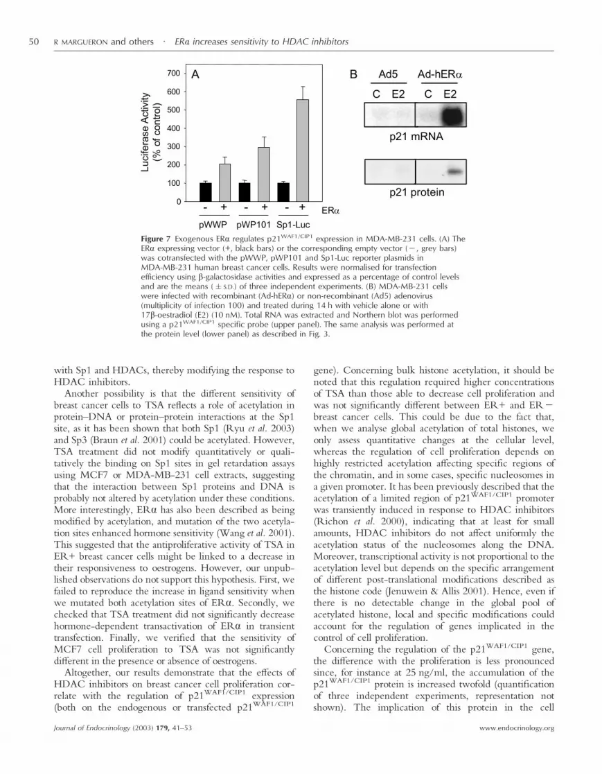
The increase in p21WAF1/CIP1 basal levels in ER+breast cancer cells could reflect transactivation of thep21WAF1/CIP1 gene by oestrogen receptors. In support ofthis hypothesis, we showed in transient transfection assaysthat ER� overexpression in MDA-MB-231 cells increasedthe expression of pWWP and pWP101 constructs by two-to threefold (Fig. 7A). This regulation might result from anSp1-mediated transactivation as shown by the strong effectof ER� (more than fivefold) on the expression of theSp1-Luc reporter.
Figure 4 Differential responsiveness to TSA of p21WAF1/CIP1 promoter constructs. Transient transfection experiments usingpWWP (A), pWP101 (B), Sp1-Luc (C) or MCK-Luc (D) constructs were performed in the two ER+ (open symbols) orER� (closed symbols) breast cancer cell lines treated with increasing amounts of TSA for 24 h. Results are expressed as apercentage of the maximum luciferase activity and are the means (�S.D.) of three independent experiments.
ER� increases sensitivity to HDAC inhibitors · R MARGUERON and others 47
www.endocrinology.org Journal of Endocrinology (2003) 179, 41–53

In order to investigate further the role of ER� on theexpression of endogenous p21WAF1/CIP1 gene, we used anadenoviral-based strategy to reintroduce the ER� proteinin receptor-negative breast cancer cells. Infection ofMDA-MB-231 cells by the recombinant adenovirusAd-hER� was previously shown to allow the ex-pression of a functional ER� protein able to activateoestrogen-sensitive reporter genes (Lazennec et al. 2001).As shown in Fig. 7B, we confirmed that the levels ofendogenous p21WAF1/CIP1 mRNA and protein werestrongly up-regulated by oestradiol treatment in cellsinfected with Ad-hER� as compared with control cellsinfected with the non-recombinant virus Ad5. Inaddition, we also noticed that, in the absence of hormone,infection with Ad-hER� slightly increased the levels ofp21WAF1/CIP1 mRNA.
Finally, we investigated whether ER� expression couldmodulate the proliferative response of MDA-MB-231 cellsto HDAC inhibition. As previously described (Lazennecet al. 2001), activated ER� by itself reduced cell pro-liferation (Fig. 8A). This result correlated with thep21WAF1/CIP1 gene up-regulation by ER� in the presenceof oestradiol (Fig. 7B). Interestingly, TSA inhibitedcell proliferation more efficiently in cells infected withAd-hER� as compared with cells infected with thenon-recombinant Ad5 adenovirus. As shown in Fig. 8A,treatment of cells with 20 ng/ml TSA resulted in growthinhibition of 16, 32 and 46% in the presence of ER�compared with 8, 17 and 19% in the absence of ER� at 2,4 and 6 days respectively. The difference, althoughdetectable after two days of treatment, was more signifi-cant at 6 days. Moreover, Fig. 8B showed that ER�expression markedly increased the sensitivity to HDAC
inhibition at all the concentrations of TSA that weretested. Together, these data support our initial observationon ER+ and ER� breast cancer cells since they (1)demonstrate a link between ER� and p21WAF1/CIP1
expression and (2) suggest that the presence of ER�influences the sensitivity of breast cancer cells to growthinhibition by TSA.
Discussion
The use of specific inhibitors to target HDACs hasrecently emerged as a new potential therapeutic approachin the treatment of cancer (Kramer et al. 2001). A betterunderstanding of the mechanisms responsible for thesensitivity of a given tumour cell to this class of drugs isrequired in order to define diagnostic tools allowing theidentification of responsive cancers.
In the present study, we have examined the response ofhuman breast cancer cells expressing or not the oestrogenreceptor to hyperacetylating agents such as trichostatin Aand showed that ER� breast cancer cells are less sensitiveto the growth inhibitory activity of these agents. Previousdata published on MCF7 and MDA-MB-231 cells usingmillimolar concentrations of sodium butyrate as an inhibi-tor of HDACs (Davis et al. 2000) also support a highersensitivity of ER+ breast cancer cells. According to ourresults, this differential sensitivity is not linked to themultidrug resistance (MDR) phenotype, in agreementwith data obtained with phenylbutyrate (Shack et al. 1996)or SAHA (Kim et al. 1999).
Our work demonstrates that the resistance to hyper-acetylating agents observed in ER� breast cancer cell
Figure 5 Analysis of the in vitro interaction of Sp1 with DNA and HDACs. (A) MCF7 orMDA-MB-231 cells were treated or not treated with 500 ng/ml TSA for the indicated timeprior to preparation of nuclear extract for band shift assay using an end-labelled Sp1probe. The filled arrowheads indicate the specific retarded DNA–protein complexes.(B) Pull-down assays were carried out as described in Materials and Methods usingbacterially expressed GST-Sp1 versus GST alone to precipitate 35S-labelled HDACs or ER�.Inputs correspond to 10% of the material used in the assay.
R MARGUERON and others · ER� increases sensitivity to HDAC inhibitors48
www.endocrinology.orgJournal of Endocrinology (2003) 179, 41–53

lines is associated with low basal levels of p21WAF1/CIP1
gene expression and with a decreased sensitivity to TSA ofboth the endogenous and transfected p21WAF1/CIP1 gene.Moreover, we show that ER� itself is able to transactivatethe p21WAF1/CIP1 promoter and to increase TSA growthinhibitory activity in ER� cells.
This study highlights the importance of Sp1 sites in thetranscriptional regulation by HDAC inhibitor (Sowa et al.1997) by demonstrating that these elements are sufficientto confer a differential sensitivity to TSA in transienttransfection experiments of ER� and ER+ breast cancercells. We show that different HDACs (from either class Ior class II) could be recruited in vitro by Sp1, as previouslyreported for HDAC1 (Doetzlhofer et al. 1999). Althoughthe global HDAC activity (Fig. 2B) and the expression ofseveral class I enzymes (HDAC1–3) (data not shown) werenot significantly different between ER+ and ER� breastcancer cells, we cannot rule out the possibility that otherHDACs do not present a different expression patternbetween the two cell types. Moreover, it has been shown
that the enzymatic activity of HDAC3 is regulatedthrough the interaction with the SMRT and N-CoRcorepressors (Guenther et al. 2001), and it is thereforepossible that the nature of such regulatory partners inHDAC-containing complexes recruited by Sp1 variesfrom one cell type to another. In addition to HDACs,other factors such as p300 (Xiao et al. 2000) participate inthe regulation of p21WAF1/CIP1 by HDAC inhibitors andcould also be differentially expressed or recruited betweenER+ and ER� breast cancer cells.
Sp1 family factors have been shown to mediate acti-vation of the p21WAF1/CIP1 promoter by a wide variety oftranscription factors including p53 (Koutsodontis et al.2001) and some nuclear receptors (Lu et al. 2000).In support of the direct interaction of Sp1 with ERs (Fig.6B and Porter et al. 1997), we show that ER�, in additionto p53, participates in the difference in p21WAF1/CIP1
expression. Some of these transcription factors could com-pete for the binding of HDACs on Sp1, as recently shownfor p53 (Lagger et al. 2003), or form ternary complexes
Figure 6 Analysis of basal expression of p21WAF1/CIP1 gene. (A) Basal levels of p21WAF1/CIP1 expression inthe four human breast cancer cell lines were analysed either by Western blot (upper panel) or in transienttransfection experiments using the pWWP reporter vector (lower panel). For Western blot, higheramounts of whole cell extracts were analysed (120 �g) and exposure time was increased as comparedwith Fig. 3. Sample loading was verified using actin as an internal control. For transient transfection,luciferase activities were corrected by �-galactosidase. Results are the means (�S.D.) of threeindependent experiments and are expressed as a percentage of MCF7 levels. (B) Basal levels of pWWP,pWP101 and Sp1-Luc in T-47D (white bars) and MDA-MB-231 (black bars) cells. Results, which areexpressed in luciferase arbitrary units after correction by the �-galactosidase, are the means (�S.D.) ofthree independent experiments.
ER� increases sensitivity to HDAC inhibitors · R MARGUERON and others 49
www.endocrinology.org Journal of Endocrinology (2003) 179, 41–53
with Sp1 and HDACs, thereby modifying the response toHDAC inhibitors.
Another possibility is that the different sensitivity ofbreast cancer cells to TSA reflects a role of acetylation inprotein–DNA or protein–protein interactions at the Sp1site, as it has been shown that both Sp1 (Ryu et al. 2003)and Sp3 (Braun et al. 2001) could be acetylated. However,TSA treatment did not modify quantitatively or quali-tatively the binding on Sp1 sites in gel retardation assaysusing MCF7 or MDA-MB-231 cell extracts, suggestingthat the interaction between Sp1 proteins and DNA isprobably not altered by acetylation under these conditions.More interestingly, ER� has also been described as beingmodified by acetylation, and mutation of the two acetyla-tion sites enhanced hormone sensitivity (Wang et al. 2001).This suggested that the antiproliferative activity of TSA inER+ breast cancer cells might be linked to a decrease intheir responsiveness to oestrogens. However, our unpub-lished observations do not support this hypothesis. First, wefailed to reproduce the increase in ligand sensitivity whenwe mutated both acetylation sites of ER�. Secondly, wechecked that TSA treatment did not significantly decreasehormone-dependent transactivation of ER� in transienttransfection. Finally, we verified that the sensitivity ofMCF7 cell proliferation to TSA was not significantlydifferent in the presence or absence of oestrogens.
Altogether, our results demonstrate that the effects ofHDAC inhibitors on breast cancer cell proliferation cor-relate with the regulation of p21WAF1/CIP1 expression(both on the endogenous or transfected p21WAF1/CIP1
gene). Concerning bulk histone acetylation, it should benoted that this regulation required higher concentrationsof TSA than those able to decrease cell proliferation andwas not significantly different between ER+ and ER�breast cancer cells. This could be due to the fact that,when we analyse global acetylation of total histones, weonly assess quantitative changes at the cellular level,whereas the regulation of cell proliferation depends onhighly restricted acetylation affecting specific regions ofthe chromatin, and in some cases, specific nucleosomes ina given promoter. It has been previously described that theacetylation of a limited region of p21WAF1/CIP1 promoterwas transiently induced in response to HDAC inhibitors(Richon et al. 2000), indicating that at least for smallamounts, HDAC inhibitors do not affect uniformly theacetylation status of the nucleosomes along the DNA.Moreover, transcriptional activity is not proportional to theacetylation level but depends on the specific arrangementof different post-translational modifications described asthe histone code (Jenuwein & Allis 2001). Hence, even ifthere is no detectable change in the global pool ofacetylated histone, local and specific modifications couldaccount for the regulation of genes implicated in thecontrol of cell proliferation.
Concerning the regulation of the p21WAF1/CIP1 gene,the difference with the proliferation is less pronouncedsince, for instance at 25 ng/ml, the accumulation of thep21WAF1/CIP1 protein is increased twofold (quantificationof three independent experiments, representation notshown). The implication of this protein in the cell
Figure 7 Exogenous ER� regulates p21WAF1/CIP1 expression in MDA-MB-231 cells. (A) TheER� expressing vector (+, black bars) or the corresponding empty vector (�, grey bars)was cotransfected with the pWWP, pWP101 and Sp1-Luc reporter plasmids inMDA-MB-231 human breast cancer cells. Results were normalised for transfectionefficiency using �-galactosidase activities and expressed as a percentage of control levelsand are the means (�S.D.) of three independent experiments. (B) MDA-MB-231 cellswere infected with recombinant (Ad-hER�) or non-recombinant (Ad5) adenovirus(multiplicity of infection 100) and treated during 14 h with vehicle alone or with17�-oestradiol (E2) (10 nM). Total RNA was extracted and Northern blot was performedusing a p21WAF1/CIP1 specific probe (upper panel). The same analysis was performed atthe protein level (lower panel) as described in Fig. 3.
R MARGUERON and others · ER� increases sensitivity to HDAC inhibitors50
www.endocrinology.orgJournal of Endocrinology (2003) 179, 41–53

proliferation blockade in response to HDAC inhibitors haspreviously been well documented. The use of mouseembryo fibroblasts (MEF) or HCT116 colon cancer cellsdeleted for the p21WAF1/CIP1 gene showed that the growthinhibitory effects of the FR901228 molecule (Sandor et al.2000) or butyrate derivatives (Archer et al. 1998) wereimpaired. When associated with initial high levels, a slightincrease in p21WAF1/CIP1 gene expression could thereforemediate important modifications of cell proliferation.However, it is clear that the p21WAF1/CIP1 gene is not theunique mediator of the antiproliferative properties ofHDAC inhibitors, but more likely it is a key protein acting
in combination with other regulators. A differentialexpression or regulation of these other target genes couldexplain why, in some cases, p21WAF1/CIP1 gene expressionis not associated with an increased sensitivity to HDACinhibitors (Vaziri et al. 1998).
With regard to the association between ER� andp21WAF1/CIP1 levels, our data are supported by a clinicalanalysis in breast tumour samples which reported a positivecorrelation between the expression of these two proteinsmeasured by Western blot (Chen et al. 2000). Moreover,the crosstalk between ER� and p21WAF1/CIP1 could beeven more complex since p21WAF1/CIP1 appears to beinvolved in the activation of the oestrogen-signallingpathway. Transient transfection experiments haveshown that the expression of p21WAF1/CIP1 amplifiedtranscriptional activation by ER� in a CREB-bindingprotein-dependent manner (Redeuilh et al. 2002).
From a clinical point of view, the in vivo antitumouractivity of hyperacetylating agents (in particular onbreast cancer) is becoming more and more documented(Saito et al. 1999, Saunders et al. 1999, Vigushin et al.2001) and appears to be associated with low toxicity,thus supporting their potential use in anticancer therapy.The need for markers of responsiveness to these drugscould therefore be important, and ER� is a goodcandidate. Other genes that are linked to the increase ofprogrammed cell death induced upon hyperacetylationcould also be of interest and are currently underinvestigation in our laboratory.
Acknowledgments
We thank F Pinguet for cell lines, Y Sowa, S Khochbin,C Seiser and H Neel for plasmids. We are grateful to theVector Core of the University Hospital of Nantes sup-ported by the Association Française contre les Myopathies(AFM) for the production of adenovirus. R M was arecipient of a three-month fellowship from the ‘Fondationpour la Recherche Médicale’ and a six-month fellowshipfrom the ‘Ligue Nationale contre le Cancer’. We alsothank J Y Cance for photographs and D Chalbos, MGutman, A Castet and D Sarruf for critical reading of themanuscript. This work was supported by the ‘InstitutNational de la Santé et de la Recherche Médicale’, theUniversity of Montpellier I, the ‘Ligue Nationale contre leCancer’ and the ‘Association pour la Recherche sur leCancer’. Part of this work has been presented at theJacques Monod Conference on Signaling and Control ofTranscription, June 2001, Aussois, France.
References
Archer SY, Meng S, Shei A & Hodin RA 1998 p21(WAF1) isrequired for butyrate-mediated growth inhibition of human coloncancer cells. PNAS 95 6791–6796.
Figure 8 Effect of ER� expression on MDA-MB-231 cellproliferation. MDA-MB-231 cells were infected with recombinant(Ad-hER�) or non-recombinant (Ad5) adenovirus (multiplicity ofinfection 100) and treated with vehicle alone (control) or with TSAin oestrogen-containing medium. Cell number was quantified asdescribed in Materials and Methods. Results are the means(�S.D.) of three quantifications. (A) Proliferation of Ad5- andAd-hER�-infected MDA-MB-231 cells was analysed between 24 hafter infection (day=0) and six days after infection in controlconditions or in response to TSA (20 ng/ml). Results are expressedas �g/plates. (B) The growth inhibitory activity of TSA wasanalysed at six days in Ad5- and Ad-hER�-infected MDA-MB-231cells, treated by increasing amounts of TSA (ng/ml). Results areexpressed as a percentage of the value obtained in the absenceof TSA. *P,0·05, **P,0·001 compared with values fromAd5-infected control cells.
ER� increases sensitivity to HDAC inhibitors · R MARGUERON and others 51
www.endocrinology.org Journal of Endocrinology (2003) 179, 41–53

Braun H, Koop R, Ertmer A, Nacht S & Suske G 2001 Transcriptionfactor Sp3 is regulated by acetylation. Nucleic Acids Research 294994–5000.
Brouillet JP, Dujardin MA, Chalbos D, Rey JM, Grenier J, Lamy PJ,Maudelonde T & Pujol P 2001 Analysis of the potentialcontribution of oestrogen receptor (ER) beta in ER cytosolic assayof breast cancer. International Journal of Cancer 20 205–208.
Chen X, Danes C, Lowe M, Herliczek TW & Keyomarsi K 2000Activation of the oestrogen-signaling pathway by p21(WAF1/CIP1)
in oestrogen receptor-negative breast cancer cells. Journal of theNational Cancer Institute 92 1403–1413.
Davis T, Kennedy C, Chiew YE, Clarke CL & deFazio A 2000Histone deacetylase inhibitors decrease proliferation and modulatecell cycle gene expression in normal mammary epithelial cells.Clinical Cancer Research 6 4334–4342.
Doetzlhofer A, Rotheneder H, Lagger G, Koranda M, Kurtev V,Brosch G, Wintersberger E & Seiser C 1999 Histone deacetylase 1can repress transcription by binding to Sp1. Molecular and CellularBiology 19 5504–5511.
Filipits M, Malayeri R, Suchomel RW, Pohl G, Stranzl T, Dekan G,Kaider A, Stiglbauer W, Depisch D & Pirker R 1999 Expression ofthe multidrug resistance protein (MRP1) in breast cancer. AnticancerResearch 19 5043–5049.
Guenther MG, Barak O & Lazar MA 2001 The SMRT and N-CoRcorepressors are activating cofactors for histone deacetylase 3.Molecular and Cellular Biology 21 6091–6101.
Jenuwein T & Allis CD 2001 Translating the histone code. Science 2931074–1080.
Jordan VC 1998 Molecular biology of the oestrogen receptor aids inthe understanding of tamoxifen resistance and breast cancerprevention with raloxifene. Recent Results in Cancer Research 152265–276.
Julia AM, Roche H, Berlion M, Lucas C, Milano G, Robert J, BizzariJP & Canal P 1994 Multidrug resistance circumvention by a newtriazinoaminopiperidine derivative S9788 in vitro: definition of theoptimal schedule and comparison with verapamil. British Journal ofCancer 69 868–874.
Kim YB, Lee KH, Sugita K, Yoshida M & Horinouchi S 1999Oxamflatin is a novel antitumor compound that inhibits mammalianhistone deacetylase. Oncogene 18 2461–2470.
Koutsodontis G, Tentes I, Papakosta P, Moustakas A & Kardassis D2001 Sp1 plays a critical role in the transcriptional activation of thehuman cyclin-dependent kinase inhibitor p21(WAF1/Cip1) gene bythe p53 tumor suppressor protein. Journal of Biological Chemistry 27629116–29125.
Kramer OH, Gottlicher M & Heinzel T 2001 Histone deacetylase asa therapeutic target. Trends in Endocrinology and Metabolism 12294–300.
Lagger G, Doetzlhofer A, Schuettengruber B, Haidweger E, SimboeckE, Tischler J, Chiocca S, Suske G, Rotheneder H, Wintersberger E& Seiser C 2003 The tumor suppressor p53 and histone deacetylase1 are antagonistic regulators of the cyclin-dependent kinaseinhibitor p21/WAF1/CIP1 gene. Molecular and Cellular Biology 232669–2679.
Lazennec G, Bresson D, Lucas A, Chauveau C & Vignon F 2001 ERbeta inhibits proliferation and invasion of breast cancer cells.Endocrinology 142 4120–4130.
Lu S, Jenster G & Epner DE 2000 Androgen induction of cyclin-dependent kinase inhibitor p21 gene: role of androgen receptor andtranscription factor Sp1 complex. Molecular Endocrinology 14753–760.
Marks PA, Richon VM & Rifkind RA 2000 Histone deacetylaseinhibitors: inducers of differentiation or apoptosis of transformedcells. Journal of the National Cancer Institute 92 1210–1216.
Pandolfi PP 2001 Transcription therapy for cancer. Oncogene 203116–3127.
Philips A, Chalbos D & Rochefort H 1993 Estradiol increases andanti-oestrogens antagonize the growth factor-induced activator
protein-1 activity in MCF7 breast cancer cells without affectingc-fos and c-jun synthesis. Journal of Biological Chemistry 26814103–14108.
Porter W, Saville B, Hoivik D & Safe S 1997 Functional synergybetween the transcription factor Sp1 and the oestrogen receptor.Molecular Endocrinology 11 1569–1580.
Redeuilh G, Attia A, Mester J & Sabbah M 2002 Transcriptionalactivation by the oestrogen receptor alpha is modulatedthrough inhibition of cyclin-dependent kinases. Oncogene 215773–5782.
Richon VM, Sandhoff TW, Rifkind RA & Marks PA 2000Histone deacetylase inhibitor selectively induces p21 WAF1expression and gene-associated histone acetylation. PNAS 9710014–10019.
Roger P, Sahla ME, Makela S, Gustafsson JA, Baldet P & RochefortH 2001 Decreased expression of estrogen receptor beta protein inproliferative preinvasive mammary tumors. Cancer Research 612537–2541.
Rundlett SE, Carmen AA, Kobayashi R, Bavykin S, Turner BM &Grunstein M 1996 HDA1 and RPD3 are members of distinct yeasthistone deacetylase complexes that regulate silencing andtranscription. PNAS 93 14503–14508.
Ryu H, Lee J, Olofsson BA, Mwidau A, Deodoglu A, Escudero M,Flemington E, Azizkhan-Clifford J, Ferrante RJ & Ratan RR 2003Histone deacetylase inhibitors prevent oxidative neuronal deathindependent of expanded polyglutamine repeats via anSp1-dependent pathway. PNAS 100 4281–4286.
Saito A, Yamashita T, Mariko Y, Nosaka Y, Tsuchiya K, Ando T,Suzuki T, Tsuruo T & Nakanishi O 1999 A synthetic inhibitor ofhistone deacetylase, MS-27–275, with marked in vivo antitumoractivity against human tumors. PNAS 96 4592–4597.
Sandor V, Senderowicz A, Mertins S, Sackett D, Sausville E,Blagosklonny MV & Bates SE 2000 P21-dependent g(1)arrest withdownregulation of cyclin D1 and upregulation of cyclin E by thehistone deacetylase inhibitor FR901228. British Journal of Cancer 83817–825.
Santen RJ, Manni A, Harvey H & Redmond C 1990 Endocrinetreatment of breast cancer in women. Endocrine Reviews 11221–265.
Saunders N, Dicker A, Popa C, Jones S & Dahler A 1999 Histonedeacetylase inhibitors as potential anti-skin cancer agents. CancerResearch 59 399–404.
Shack S, Miller A, Liu L, Prasanna P, Thibault A & Samid D 1996Vulnerability of multidrug-resistant tumor cells to the aromatic fattyacids phenylacetate and phenylbutyrate. Cliinical Cancer Research 2865–872.
Sommer S & Fuqua SA 2001 Oestrogen receptor and breast cancer.Seminars in Cancer Biology 11 339–352.
Sowa Y, Orita T, Minamikawa S, Nakano K, Mizuno T, Nomura H& Sakai T 1997 Histone deacetylase inhibitor activates theWAF1/Cip1 gene promoter through the Sp1 sites. Biochemical andBiophysical Research Communications 241 142–150.
Strahl BD & Allis CD 2000 The language of covalent histonemodifications. Nature 403 41–45.
Thenot S, Charpin M, Bonnet S & Cavaillès V 1999 Oestrogenreceptor cofactors expression in breast and endometrialhuman cancer cells. Molecular and Cellular Endocrinology 15685–93.
Van Lint C, Emiliani S, Ott M & Verdin E 1996 Transcriptionalactivation and chromatin remodeling of the HIV-1 promoter inresponse to histone acetylation. EMBO Journal 15 1112–1120.
Vaziri C, Stice L & Faller DV 1998 Butyrate-induced G1 arrestresults from p21-independent disruption of retinoblastomaprotein-mediated signals. Cell Growth and Differentiation 9 465–474.
Vigushin DM, Ali S, Pace PE, Mirsaidi N, Ito K, Adcock I &Coombes RC 2001 Trichostatin A is a histone deacetylase inhibitorwith potent antitumor activity against breast cancer in vivo. ClinicalCancer Research 7 971–976.
R MARGUERON and others · ER� increases sensitivity to HDAC inhibitors52
www.endocrinology.orgJournal of Endocrinology (2003) 179, 41–53

Wang C, Fu M, Angeletti RH, Siconolfi-Baez L, Reutens AT,Albanese C, Lisanti MP, Katzenellenbogen BS, Kato S, Hopp T,Fuqua SA, Lopez GN, Kushner PJ & Pestell RG 2001 Directacetylation of the oestrogen receptor alpha hinge region by p300regulates transactivation and hormone sensitivity. Journal of BiologicalChemistry 276 18375–18383.
Warrell RPJ, He LZ, Richon V, Calleja E & Pandolfi PP 1998Therapeutic targeting of transcription in acute promyelocyticleukemia by use of an inhibitor of histone deacetylase. Journal of theNational Cancer Institute 90 1621–1625.
Woodcock CL & Dimitrov S 2001 Higher-order structure ofchromatin and chromosomes. Current Opinion in GeneticsDevelopment 11 130–135.
Xiao H, Hasegawa T & Isobe K 2000 p300 collaborates with Sp1 andSp3 in p21(waf1/cip1) promoter activation induced by histone
deacetylase inhibitor. Journal of Biological Chemistry 2751371–1376.
Yoshida M, Kijima M, Akita M & Beppu T 1990 Potent and specificinhibition of mammalian histone deacetylase both in vivo andin vitro by trichostatin A. Journal of Biological Chemistry 26517174–17179.
Zambetti GP, Bargonetti J, Walker K, Prives C & Levine AJ 1992Wild-type p53 mediates positive regulation of gene expressionthrough a specific DNA sequence element. Genes and Development6 1143–1152.
Received 6 May 2003Accepted 25 June 2003
ER� increases sensitivity to HDAC inhibitors · R MARGUERON and others 53
www.endocrinology.org Journal of Endocrinology (2003) 179, 41–53