obesity surgery principles and practice mcgraw fill
TRANSCRIPT


To my parents, brothers, and to my fiance for always understanding that dedication and responsibility
inevitably lead to absence.
Cid Pitombo
To my wife, Anne, for believing in and supporting my career-long involvement in Bariatric Surgery,
especially in the early days, when our small group of pioneers were looked upon by so many as eccentric
opportunists.
Kenneth B. Jones, Jr.
To my wife and children for their understanding and compassion.
Kelvin D. Higa
To my wife, son, and daughter.
Jose Carlos Pareja

Obesity SurgeryPrinciples and Practice
Cid Pitombo, MD, PhD, TCBC (Editor-in-Chief)General and Bariatric SurgeryMinimally Invasive SurgeryCampinas State University (UNICAMP)Rio de Janeiro/Campinas, Brazil
Kenneth B. Jones, Jr., MD (Co-Editor)Clinical Assistant Professor, Department of SurgeryLouisiana State University Health Sciences CenterFormer President, American Society of Bariatric SurgeryShreveport, Louisiana
Kelvin D. Higa, MD, FACS (Co-Editor)President Elect, American Society for Metabolic and Bariatric SurgeryClinical Professor of SurgeryUniversity of California, San FranciscoPrivate PracticeFresno, California
Jose Carlos Pareja, MD, PhD (Co-Editor)Professor, Department of Surgery, Head of Pancreas, Biliary, and Obesity SurgeryCampinas State University (UNICAMP)President, Scientific Committee, Brazilian Society for Bariatric SurgeryCampinas, Brazil
New York Chicago San Francisco Lisbon London Madrid Mexico City MilanNew Delhi San Juan Seoul Singapore Sydney Toronto

Copyright © 2008 by The McGraw-Hill Companies, Inc. All rights reserved. Manufactured in the United States of America. Except as permitted under theUnited States Copyright Act of 1976, no part of this publication may be reproduced or distributed in any form or by any means, or stored in a database orretrieval system, without the prior written permission of the publisher.
0-07-159434-5
The material in this eBook also appears in the print version of this title: 0-07-148281-4.
All trademarks are trademarks of their respective owners. Rather than put a trademark symbol after every occurrence of a trademarked name, we use namesin an editorial fashion only, and to the benefit of the trademark owner, with no intention of infringement of the trademark. Where such designations appearin this book, they have been printed with initial caps.
McGraw-Hill eBooks are available at special quantity discounts to use as premiums and sales promotions, or for use in corporate training programs. For moreinformation, please contact George Hoare, Special Sales, at [email protected] or (212) 904-4069.
TERMS OF USE
This is a copyrighted work and The McGraw-Hill Companies, Inc. (“McGraw-Hill”) and its licensors reserve all rights in and to the work. Use of this workis subject to these terms. Except as permitted under the Copyright Act of 1976 and the right to store and retrieve one copy of the work, you may not decom-pile, disassemble, reverse engineer, reproduce, modify, create derivative works based upon, transmit, distribute, disseminate, sell, publish or sublicense thework or any part of it without McGraw-Hill’s prior consent. You may use the work for your own noncommercial and personal use; any other use of the workis strictly prohibited. Your right to use the work may be terminated if you fail to comply with these terms.
THE WORK IS PROVIDED “AS IS.” McGRAW-HILL AND ITS LICENSORS MAKE NO GUARANTEES OR WARRANTIES AS TO THE ACCURACY, ADEQUACY OR COMPLETENESS OF OR RESULTS TO BE OBTAINED FROM USING THE WORK, INCLUDING ANY INFORMATION THAT CAN BE ACCESSED THROUGH THE WORK VIA HYPERLINK OR OTHERWISE, AND EXPRESSLY DISCLAIM ANYWARRANTY, EXPRESS OR IMPLIED, INCLUDING BUT NOT LIMITED TO IMPLIED WARRANTIES OF MERCHANTABILITY OR FITNESSFOR A PARTICULAR PURPOSE. McGraw-Hill and its licensors do not warrant or guarantee that the functions contained in the work will meet your requirements or that its operation will be uninterrupted or error free. Neither McGraw-Hill nor its licensors shall be liable to you or anyone else for any inaccuracy, error or omission, regardless of cause, in the work or for any damages resulting therefrom. McGraw-Hill has no responsibility for the content ofany information accessed through the work. Under no circumstances shall McGraw-Hill and/or its licensors be liable for any indirect, incidental, special, punitive, consequential or similar damages that result from the use of or inability to use the work, even if any of them has been advised of the possibility ofsuch damages. This limitation of liability shall apply to any claim or cause whatsoever whether such claim or cause arises in contract, tort or otherwise.
DOI: 10.1036/0071482814

We hope you enjoy thisMcGraw-Hill eBook! If
you’d like more information about this book,its author, or related books and websites,please click here.
Professional
Want to learn more?

Contents
Contributors / viiForeword / xiiiPreface / xiv
PART I BASIC PRINCIPLES.. . . . . . . . . . . . . . . . . . . . . . . . . . . . . . . . . . . .1
1. Evolution of Surgery for Morbid Obesity . . . . . . . . . . . . . . . . .3Henry Buchwald & Jane N. Buchwald
2. Pathophysiology of Severe Obesity and theEffects of Surgically Induced Weight Loss . . . . . . . . . . . . . 15Harvey J. Sugerman
3. Rationale for Minimally Invasive BariatricSurgery . . . . . . . . . . . . . . . . . . . . . . . . . . . . . . . . . . . . . . . . . . . . . . . . . . . . . . . . 27Ninh T. Nguyen, Esteban Varela, & Samuel E. Wilson
4. Current Role of Open Bariatric Surgery . . . . . . . . . . . . . . . . . 33Kenneth B. Jones, Jr
5. Central Nervous System Regulation andHormonal Signaling . . . . . . . . . . . . . . . . . . . . . . . . . . . . . . . . . . . . . . . . .41Cid Pitombo
PART II OPERATIVE ISSUES .. . . . . . . . . . . . . . . . . . . . . . . . . . . . . . . . 53
6. Requirements of the Clinic and Institution. . . . . . . . . . . . .55Luiz Henrique de Sousa, Edson Lemes Sardinha,& Luiz Henrique de Sousa Filho
7. Preoperative Evaluation of Patients . . . . . . . . . . . . . . . . . . . . . 61Marcos Tambascia
8. Intraoperative Issues . . . . . . . . . . . . . . . . . . . . . . . . . . . . . . . . . . . . . . . 67Marcel Milcent, Angelo Loss, & Jose E. Manso
9. Bariatric Surgery Psychology . . . . . . . . . . . . . . . . . . . . . . . . . . . . . .75Melodie Moorehead
10. Anesthetic Concerns . . . . . . . . . . . . . . . . . . . . . . . . . . . . . . . . . . . . . . . .83Jay B. Brodsky & Luiz C. Lerner
PART III PHYSIOLOGY OF BARIATRICOPERATIONS.. . . . . . . . . . . . . . . . . . . . . . . . . . . . . . . . . . . . . . . . . . . . . . . . . . . . .93
11. Restrictive Surgery . . . . . . . . . . . . . . . . . . . . . . . . . . . . . . . . . . . . . . . . . . 95Edward E. Mason
12. Physiology and Metabolism in Obesity Surgery:Roux-en-Y Gastric Bypass . . . . . . . . . . . . . . . . . . . . . . . . . . . . . . . . 101Carolina G. Goncalves, Francesco Rubino,Stacy A. Brethauer, & Philip R. Schauer
13. Malabsorptive Procedures: BiliopancreaticDiversion Scopinaro Procedure. . . . . . . . . . . . . . . . . . . . . . . .111Nicola Scopinaro
14. Malabsorptive Procedures: Duodenal Switch . . . . . . . 131Picard Marceau, Simon Biron, Frederic-Simon Hould,Stefane Lebel, Simon Marceau, Odette Lescelleur,Christine Simard, & Serge Simard
15. Possible Hormonal Mechanisms Mediating theEffects of Bariatric Surgery . . . . . . . . . . . . . . . . . . . . . . . . . . . . . . . 137David E. Cummings, Karen E. Foster-Schubert, Molly J.Carlson, Michael H. Shannon, & Joost Overduin
16. Metabolic Syndrome: Diagnosis, ClinicalPresentations, and Surgical Treatment . . . . . . . . . . . . . . . .149Bruno Geloneze
17. The Learning Curve . . . . . . . . . . . . . . . . . . . . . . . . . . . . . . . . . . . . . . . . 161Michael L. Schwartz & Raymond L. Drew
PART IV TECHNICAL PROCEDURES .. . . . . . . . . . . . . . . . . . . . 167
18. Laparoscopic Restrictive Procedures:Adjustable Gastric Banding . . . . . . . . . . . . . . . . . . . . . . . . . . . . . .169Karl Miller
19. Laparoscopic Restrictive Procedures:Sleeve Gastrectomy . . . . . . . . . . . . . . . . . . . . . . . . . . . . . . . . . . . . . . . 177Camilo Boza & Michel Gagner
For more information about this title, click here

vi
Contents
20. Laparoscopic Gastric Bypass: Trans-OralCircular Stapling. . . . . . . . . . . . . . . . . . . . . . . . . . . . . . . . . . . . . . . . . . . .183Renam Catharina Tinoco & Augusto Claudio Tinoco
21. Laparoscopic Gastric Bypass: CircularStapler Technique . . . . . . . . . . . . . . . . . . . . . . . . . . . . . . . . . . . . . . . . . 189Alan Wittgrove
22. Laparoscopic Gastric Bypass: TransgastricCircular Stapler . . . . . . . . . . . . . . . . . . . . . . . . . . . . . . . . . . . . . . . . . . . . . 195Roger de la Torre, J. Stephen Scott, & Matthew Fitzer
23. Laparoscopic Gastric Bypass: Linear Technique . . . . . 207Michael D. Williams & J.K. Champion
24. Laparoscopic Roux-en-Y Banded Gastric Bypass . . . .211Thomas Szego & Carlos Jose--Lazzarini Mendes
25. Laparoscopic Gastric Bypass: Hand Sewn. . . . . . . . . . . . .215Kelvin D. Higa & Jennifer Elizabeth Higa
26. Laparoscopic Gastric Bypass: Evolution, Safety,and Efficacy of the Banded Gastric Bypass . . . . . . . . . . . 221Robert T. Marema, RoseMarie Toussaint, Michael Perez,& Cynthia K. Buffington
27. Laparoscopic Gastric Bypass: Silastic Ring . . . . . . . . . . . . 229Daoud Nasser & Adriana Sales Finizola
28. Laparoscopic Biliopancreatic Diversion:Approach . . . . . . . . . . . . . . . . . . . . . . . . . . . . . . . . . . . . . . . . . . . . . . . . . . . . 233Dyker Santos Paiva & Lucineia Bernardes Lima
29. Laparoscopic Biliopancreatic Diversion:Duodenal Switch. . . . . . . . . . . . . . . . . . . . . . . . . . . . . . . . . . . . . . . . . . .241Aniceto Baltasar
30. Two-Stage Approach for High-Risk Patients . . . . . . . . . 245Camilo Boza & Michel Gagner
PART V LAPAROSCOPIC REOPERATIVESURGERY.. . . . . . . . . . . . . . . . . . . . . . . . . . . . . . . . . . . . . . . . . . . . . . . . . . . . . . . .251
31. Restrictive Procedures: AdjustableGastric Band. . . . . . . . . . . . . . . . . . . . . . . . . . . . . . . . . . . . . . . . . . . . . . . . .253Mitiku Belachew
32. Restrictive Procedures: Utilization ofAdjustable Gastric Banding for Failed StapledOperations. . . . . . . . . . . . . . . . . . . . . . . . . . . . . . . . . . . . . . . . . . . . . . . . . . .265Paul E. O’Brien
33. Restrictive Procedures: Laparoscopic Revisionof Vertical Banding to Gastric Bypass . . . . . . . . . . . . . . . . . .269Daniel Gagne
34. Biliopancreatic Diversion: Revisional Surgery . . . . . . . . 277Nicola Scopinaro
35. Biliopancreatic Diversion: Duodenal Switch . . . . . . . . . 285Joao Batista Marchesini
PART VI POSTOPERATIVE MANAGEMENT.. . . . . . . . . . . .293
36. Infection in Obesity Surgery . . . . . . . . . . . . . . . . . . . . . . . . . . . . .295Alvaro Antonio Bandeira Ferraz & Edmundo MachadoFerraz
37. Early Complications in Bariatric Surgery . . . . . . . . . . . . . . 307Kenneth G. MacDonald, Jr
38. Late Complications: Ulcers, Stenosis, and Fistula. . . .313Scott A. Shikora, Leonardo Claros, Julie J. Kim,& Michael E. Tarnoff
39. Nutritional Consequences and Management . . . . . . . 319Ampadi Thampi, Robert N L Corprew, Jr.,Michael Barker, & Walter J. Pories
40. Weight Recidivism . . . . . . . . . . . . . . . . . . . . . . . . . . . . . . . . . . . . . . . . . 327Jose Carlos Pareja & Daniela Magro
41. Radiographic Evaluation and Treatment. . . . . . . . . . . . . .333Ester Labrunie, Edson Marchiori, & Cid Pitombo
42. Radiographic Evaluation and Treatment:Intervention . . . . . . . . . . . . . . . . . . . . . . . . . . . . . . . . . . . . . . . . . . . . . . . . .355Ester Labrunie, Edson Marchiori, & Cid Pitombo
43. Endoscopic Evaluation and Treatment . . . . . . . . . . . . . . . . 357Paulo Sakai, Fauze Maluf Filho, Marcelo Lima,& Kendi Yamazaki
PART VII NEW TECHNOLOGY... . . . . . . . . . . . . . . . . . . . . . . . . . .365
44. Gastric Pacing. . . . . . . . . . . . . . . . . . . . . . . . . . . . . . . . . . . . . . . . . . . . . . .367Angelo Loss, Marcel Milcent, & Georgia Bartholdi
45. Intragastric Balloon. . . . . . . . . . . . . . . . . . . . . . . . . . . . . . . . . . . . . . . .373Jose A. Sallet, Joao C. Marchesini, Pablo Miguel,& Paulo C. Sallet
46. Hand-Assisted Laparoscopic Duodenal Switch . . . . . .383Robert A. Rabkin
PART VIII SPECIAL TOPICS IN BARIATRICSURGERY.. . . . . . . . . . . . . . . . . . . . . . . . . . . . . . . . . . . . . . . . . . . . . . . . . . . . . . . .387
47. Cost and Economic Impact of Bariatric Surgery . . . . . 389Eldo E. Frezza
48. Adolescent Bariatric Surgery . . . . . . . . . . . . . . . . . . . . . . . . . . . . 397Venita Chandra & Sanjeev Dutta
Index / 409

Contributors
Aniceto Baltasar, MDClınica San Jorge y Hospital “Virgen de los Lirios”Alicante, SpainLaparoscopic Biliopancreatic Diversion: Duodenal Switch
Mitiku Belachew, MDProfessor, Department of Surgery Centre Hospitalier
Regional de Huy, Belgium,Honorary Professor of Surgery,Addis Ababa University,Addis Ababa, Ethiopia,Huy, BelgiumRestrictive Procedures: Adjustable Gastric Band
Simon Biron, MD, MScProfessor and Head, Department of Surgery, Laval UniversityLaval HospitalQuebec, CanadaMalabsorptive Procedures: Duodenal Switch
Camilo Boza, MDDepartment of SurgeryHospital Clınico, PontificiaUniversidad Catolica de ChileSantiago, ChileLaparoscopic Restrictive Procedures: Sleeve Gastrectomy;
Two-Stage Approach for High-Risk Patients
Stacy A. Brethauer, MDFellow, Advanced Laparoscopic and Bariatric SurgeryDepartment of General Surgery, Cleveland ClinicCleveland, OhioPhysiology and Metabolism in Obesity Surgery: Roux-en-YGastric Bypass
Jay B. Brodsky, MDProfessor of Anesthesiology, Stanford University School of
Medicine, Medical Director of Perioperative ServicesStanford University Medical Center
Stanford, CaliforniaAnesthetic Concerns
Henry Buchwald, MD, PhDProfessor of Surgery and Biomedical Engineering
Owen H. and Sarah Davidson Wangensteen Chair inExperimental Surgery Emeritus
Department of Surgery, University of MinnesotaMinneapolis, MinnesotaEvolution of Surgery for Morbid Obesity
Jane N. Buchwald, MACEO and Director of PublicationsMEDWRITE Medical CommunicationsMaiden Rock, WisconsinEvolution of Surgery for Morbid Obesity
Cynthia K. Buffington, PhDFlorida Hospitals Celebration Health Celebration, FloridaLaparoscopic Gastric Bypass: Evolution, Safety, and Efficacy ofthe Banded Gastric Bypass
Molly J. Carlson, MDFellow, Division of Metabolism, Endocrinology,
and NutritionDepartment of Medicine, University of WashingtonVA Puget Sound Health Care SystemSeattle, WashingtonPossible Hormonal Mechanisms Mediating the Effects ofBariatric Surgery
J.K. Champion, MDClinical Professor of SurgeryMercer University School of MedicineDirector of Bariatric Surgery
Emory Dunwoody Medical CenterVideoscopic Institute of AtlantaMarietta, GeorgiaLaparoscopic Gastric Bypass: Linear Technique
Copyright © 2008 by The McGraw-Hill Companies, Inc. Click here for terms of use.

viii
Contributors
Venita Chandra, MDResident, Department of Pediatric Surgery,Stanford University, Stanford, CaliforniaAdolescent Bariatric Surgery
Leonardo Claros, MDAssistant Professor of Surgery, Tufts University
Medical School, Co-Director, Center for Weight ControlCaritas Saint Elizabeth’s Medical CenterBoston, MassachusettsLate Complications: Ulcers, Stenosis, and Fistula
Robert N.L. Corprew, Jr.Brody School of Medicine at East Carolina UniversityGreenville, North CarolinaNutritional Consequences and Management
David E. Cummings, MDAssociate Professor of Medicine, Division of MetabolismEndocrinology, and Nutrition, University of WashingtonVA Puget Sound Health Care SystemSeattle, WashingtonPossible Hormonal Mechanisms Mediating the Effects ofBariatric Surgery
Roger A. de la Torre, MDAssociate Professor, Deptarment of SurgeryUniversity of Missouri School of MedicineDirector of Minimally Invasive SurgeryDePaul Weight Loss Institute, DePaul Health CenterColumbia, MissouriLaparoscopic Gastric Bypass: Transgastric Circular Stapler
Lucineia Bernardes de Lima, MDBelo Horizonte, BrazilLaparoscopic Biliopancreatic Diversion: Approach
Raymond L. Drew, MDAdjunct Assistant Professor, Department of SurgeryUniversity of Minnesota, Minneapolis, MinnesotaThe Learning Curve
Sanjeev Dutta, MD, MA, FRCSCAssistant Professor of Surgery and PediatricsLucile Packard Children’s HospitalStanford University Medical CenterStanford, CaliforniaAdolescent Bariatric Surgery
Alvaro Antonio Bandeira Ferraz, MD, PhDAssocite Professor, Department of SurgeryFederal University of PernambucoHospital das ClınicasRecife, BrazilInfection in Obesity Surgery
Edmundo Machado Ferraz, MD, PhD, FACSChief, Professor, and Chairman, Department of SurgeryPernambuco Federal University (UFPE)Recife, BrazilInfection in Obesity Surgery
Fauze Maluf Filho, MDAssociate Professor, Department of GastroenterologyGastrointestinal Endoscopy UnitUniversity of Sao Paulo Medical SchoolSao Paulo, BrazilEndoscopic Evaluation and Treatment
Adriana Sales Finizola, MDSurgeon, Maringa Obesity CenterMaringa, BrazilLaparoscopic Gastric Bypass: Silastic Ring
Matthew A. Fitzer, MDFaxton-St. Luke’s Healthcare,New Hartford, New YorkLaparoscopic Gastric Bypass: Transgastric Circular Stapler
Karen E. Foster-Schubert, MDActing Instructor, Department of MedicineDivision of MetabolismEndocrinology, and NutritionUniversity of Washington School of MedicineVA Puget Sound Health Care SystemSeattle, WashingtonPossible Hormonal Mechanisms Mediating the Effects ofBariatric Surgery
Eldo E. Frezza, MD, MBA, FACSDivision of General Surgery, Department of SurgeryTexas Tech University Health Sciences CenterLubbock, TexasCost and Economic Impact of Bariatric Surgery
Daniel J. Gagne, MDAssistant Professor of Surgery, Temple University School ofMedicine, Director of Bariatric and Minimally Invasive
SurgeryWestern Pennsylvania HospitalPittsburgh, PennsylvaniaRestrictive Procedures: Laparoscopic Revision of VerticalBanding to Gastric Bypass
Michel Gagner, MD, FRCSC, FACSProfessor of Surgery, Joan and Stanford I. Weill Medical
College of Cornell UniversityChief of Laparoscopic and Bariatric SurgeryNew York-Presbyterian HospitalNew York, New YorkLaparoscopic Restrictive Procedures: Sleeve Gastrectomy;Two-Stage Approach for High-Risk Patients

ix
Contributors
Bruno Geloneze, MD, PhDProfessor of Endocrinology and Medical
Director of Laboratory of Investigation on ObesityMetabolism, and Diabetes (LIMED)University of Campinas (UNICAMP)Campinas, BrazilMetabolic Syndrome: Diagnosis, Clinical Presentations, andSurgical Treatment
Carolina G. Goncalves, MDClinical Fellow, Department of General SurgeryDivision of Advanced Laparoscopic and Bariatric SurgeryCleveland ClinicCleveland, OhioPhysiology and Metabolism in Obesity Surgery: Roux-en-YGastric Bypass
Luiz Henrique de Sousa, MDProfessor, University of Sao Paulo, Chief of StaffDivision of Obesity Surgery, Hospital FeminaGoiania, BrazilRequirements of the Clinic and Institution
Luiz Henrique de Sousa Filho, MDClinical Staff, Division of Bariatric SurgeryHospital FeminaGoiania, BrazilRequirements of the Clinic and Institution
Kelvin D. Higa, MD, FACSPresident Elect, American Society for Metabolic and
Bariatric SurgeryClinical Professor of SurgeryUniversity of California, San FranciscoPrivate PracticeFresno, CaliforniaLaparoscopic Gastric Bypass: Hand Sewn
Jennifer Elizabeth HigaFresno, CaliforniaLaparoscopic Gastric Bypass: Hand Sewn
Frederic-Simon Hould, MDDepartment of Surgery, Laval University, Laval HospitalQuebec, CanadaMalabsorptive Procedures: Duodenal Switch
Kenneth B. Jones, Jr., MDClinical Assistant Professor, Department of SurgeryLouisiana State University Health Sciences CenterFormer President, American Society of Bariatric SurgeryShreveport, LouisianaCurrent Role of Open Bariatric Surgery
Julie J. Kim, MDCenter for Minimally Invasive Obesity SurgeryTufts-New England, Medical Center
Boston, MassachusettsLate Complications: Ulcers, Stenosis, and Fistula
Ester Labrunie, MD, PhDProfessor, Department of MedicineFederal University of Rio de Janeiro (UFRJ)Rio de Janerio, BrazilRadiographic Evaluation and Treatment; RadiographicEvaluation and Treatment: Intervention
Stefane Lebel, MDDepartment of Surgery, Laval Hospital, Laval UniversityQuebec, CanadaMalabsorptive Procedures: Duodenal Switch
Luiz Claudio Lerner, MDMedical Staff, Department of AnesthesiologyHospital UniversitarioClementino Fraga FilhoUniversidade Federal do Rio de JaneiroRio de Janerio, BrazilAnesthetic Concerns
Odette Lescelleur, MDDepartment of Surgery, Laval University, Laval HospitalQuebec, CanadaMalabsorptive Procedures: Duodenal Switch
Marcelo Simas Lima, MDResearch Fellow, Gastrointestinal Endoscopy UnitUniversity of Sao Paulo Medical SchoolSao Paulo, BrazilEndoscopic Evaluation and Treatment
Angelo Loss, MD, ACBCAbdominal Surgery Post-graduation SchoolRio de Janeiro Federal UniversityCid Pitombo Clinic, MemberBrazilian College of Surgeons, MemberBrazilian Society of Laparoscopic SurgeryRio de Janeiro, BrazilIntraoperative Issues; Gastric Pacing
Kenneth G. MacDonald, Jr., MDProfessor, Department of SurgeryBrody School of Medicine at East Carolina UniversitySouthern Surgical AssociatesGreenville, North CarolinaEarly Complications in Bariatric Surgery
Daniela Magro, PhDProfessor, Researcher, and CollaboratorDepartment of Preventive and Social MedicineMedical Science School (FCM)University of Campinas (UNICAMP)Campinas, BrazilWeight Recidivism

x
Contributors
Jose E. Manso, MDAssociate Professor, Department of SurgeryFederal University of Rio de Janeiro, Chief EditorThe Journal of the Brazilian College of SurgeonsRio de Janeiro, BrazilIntraoperative Issues
Picard Marceau, MD, PhD, FRCS, FACSProfessor, Department of Surgery, Laval UniversityLaval Hospital, Quebec, CanadaMalabsorptive Procedures: Duodenal Switch
Simon Marceau, MDDepartment of Surgery, Laval University, Laval HospitalQuebec, CanadaMalabsorptive Procedures: Duodenal Switch
Joeo Batista Marchesini, MD, PhD, FACSSenior Professor of SurgeryPost Graduate Course in Clinical Surgery,Associate Professor of Surgery (retired)Parana University Medical SchoolFormer President of the Brazilian Society for Bariatric
Surgery, Certified in General Surgery by the AmericanBoard of Surgery (USA)
Curitiba, BrazilBiliopancreatic Diversion: Duodenal Switch; IntragastricBalloon
Edson Marchiori, MDProfessor and Head, Department of MedicineUniversity Federal FluminenseRio de Janeiro, BrazilRadiographic Evaluation and Treatment; RadiographicEvaluation and Treatment: Intervention
Robert T. Marema, MD, FACSMedical Director, Division of Bariatric Surgery,
Flagler HospitalSt. Augustine, FloridaLaparoscopic Gastric Bypass: Evolution, Safety, and Efficacy ofthe Banded Gastric Bypass
Edward E. Mason, MD, PhD, FACSEmeritus Professor of Surgery, Roy J. and Lucille A. CarverCollege of Medicine, University of Iowa Hospitals and ClinicsIowa City, IowaRestrictive Surgery
CJL. Mendes, MDSpecialist in General SurgeryMember of the Sociedade Brasileira de Cirurgia BariatricaATLS Instructor, the USP Medicine UniversityFormer Professor and Head of Emergency RoomHospital Escola in the Medicine University of Santo AmaroSao Paulo, BrazilLaparoscopic Roux-en-Y Banded Gastric Bypass
Pablo R. Miguel, MDLaparoscopic Surgeon, Mae de Deus HospitalPorto Allegre, RS, BrazilIntragastric Balloon
Marcel Milcent, MD, ACBCAbdominal Surgery Post-Graduation SchoolRio de Janeiro Federal University,Cid Pitombo Clinic, Brazilian College of Surgeons Member,IFSO Member, Brazilian Society for Bariatric Surgery
MemberBrazilian Society for Laparoscopic Surgery MemberRio de Janeiro, BrazilIntraoperative Issues; Gastric Pacing
Karl A. Miller, MD, FACSAssociate Professor of Surgery, Head, Department of Surgery,Ludwig-Boltzmann-Institute for Gastroenterology and
Experimental Surgery, Hospital HalleinObesity Surgery Center, Krakenhaus HalleinHallein, AustriaLaparoscopic Restrictive Procedures: Adjustable GastricBanding
Melodie K. Moorehead, PhDDrs. Moorehead, Prish, & Associates, P.A.Fort Lauderdale, FloridaBariatric Surgery Psychology
Daoud Nasser, MDProfessor of Surgery, Maringa State UniversityChief Surgeon, Maringa Obesity CenterMaringa, BrazilLaparoscopic Gastric Bypass: Silastic Ring
Ninh T. Nguyen, MDAssociate Professor of Surgery, Department of Surgery, ChiefDivision of Gastrointestinal Surgery, University of CaliforniaIrvine Medical CenterOrange, CaliforniaRationale for Minimally Invasive Bariatric Surgery
Paul E. O’Brien, MD, FRACSDirector, Centre for Obesity Research and Education,Monash University Medical SchoolThe Center for Obesity Research and Education (CORE)Melbourne, AustraliaRestrictive Procedures: Utilization of Adjustable GastricBanding for Failed Stapled Operations
Joost Overduin, PhDDivision of Metabolism, Endocrinology, and NutritionDepartment of Medicine, University of WashingtonVA Puget Sound Health Care SystemSeattle, WashingtonPossible Hormonal Mechanisms Mediating the Effects ofBariatric Surgery

xi
Contributors
Dyker Santos Paiva, MDLaparoscopic Biliopancreatic Diversion: Approach
Jose Carlos Pareja, MD, PhDProfessor, Department of Surgery, Head of Pancreas,Biliary, and Obesity SurgeryCampinas State University (UNICAMP)President, Scientific Committee, Brazilian Society for
Bariatric SurgeryCampinas, BrazilWeight Recidivism
Michael Perez, MD, FACSU.S. BariatricFort Lauderdale, FloridaLaparoscopic Gastric Bypass: Evolution, Safety, and Efficacyof the Banded Gastric Bypass
Cid Pitombo, MD, PhD, TCBCGeneral and Bariatric SurgeryMinimally Invasive SurgeryCampinas State University (UNICAMP)Rio de Janeiro/Campinas, BrazilCentral Nervous System Regulation and Hormonal Signaling;Radiographic Evaluation and Treatment; RadiographicEvaluation and Treatment: Intervention
Walter J. Pories, MD, FACSProfessor of Surgery, Biochemistry, Exercise, and Sport
Sciences, Metabolic InstituteBrody School of Medicine at East Carolina UniversityChief, Metabolic Institute, Former President ASBSGreenville, North CarolinaNutritional Consequences and Management
Robert A. Rabkin, MD, FACSPacific Laparoscopy, St. Mary’s Medical CenterCalifornia Pacific Medical CenterSan Francisco, CaliforniaHand-Assisted Laparoscopic Duodenal Switch
Francesco Rubino, MDAssistant Professor, Department of SurgeryCatholic University, Rome, ItalyAttending Surgeon, European Institute of Technology
(IRCAD-EITS), University Louis PasterStrasbourg, FrancePhysiology and Metabolism in Obesity Surgery: Roux-en-YGastric Bypass
Paulo Sakai, MD, PhDAssociate Professor of GastroenterologyGastrointestinal Endoscopy UnitUniversity of Sao Paulo Medical SchoolSao Paulo, BrazilEndoscopic Evaluation and Treatment
Jose A. Sallet, MDSao Paulo, BrazilIntragastric Balloon
Paulo C. Sallet, MDSao Paulo, BrazilIntragastric Balloon
Edson Lemes Sardinha, MDCollaborative Professor of CET-SBAFederal University of GoiasGoiania, BrazilRequirements of the Clinic and Institution
Philip R. Schauer, MDProfessor of Surgery, Cleveland Clinic Lerner College of
MedicineDirector, Division of Advanced Laparoscopic and Bariatric
Surgery, Bariatric and Metabolic InstituteCleveland, OhioPhysiology and Metabolism in Obesity Surgery: Roux-en-YGastric Bypass
Michael L. Schwartz, MD, PhDAdjunct Professor of Surgery, University of MinnesotaMinneapolis, MinnesotaThe Learning Curve
Nicola Scopinaro, MD, FACS (Hon)Professor of Surgery, University of Genoa School of Medicine,Genoa, ItalyMalabsorptive Procedures: BiliopancreaticDiversion-Scopinaro Procedure; Biliopancreatic Diversion:Revisional Surgery
J. Stephen Scott, MD, FACSAssociate Professor, Department of SurgeryUniversity of Missouri School of MedicineMedical Director, DePaul Weight Loss InstituteDePaul Health Center, Columbia, MissouriLaparoscopic Gastric Bypass: Transgastric Circular Stapler
Michael H. Shannon, MDSenior Fellow, Division of NedocrinologyDepartment of Medicine, University of WashingtonVA Puget Sound Health Care SystemSeattle, WashingtonPossible Hormonal Mechanisms Mediating the Effects ofBariatric Surgery
Scott A. Shikora, MDProfessor of Srugery, Tufts University School of Medicine,Chief of Bariatric SurgeryCenter for Minimally Invasive Obesity SurgeryTufts-New England Medical CenterBoston, MassachusettsLate Complications: Ulcers, Stenosis, and Fistula

xii
Contributors
Christine Simard, MD, FRCP, CDDepartment of Surgery, Laval University, Laval HospitalQuebec, CanadaMalabsorptive Procedures: Duodenal Switch
Serge Simard, MScBiostatistician, Laval UniversityLaval Hospital Research CenterQuebec, CanadaMalabsorptive Procedures: Duodenal Switch
Harvey J. Sugerman, MD, FACSProfessor Emeritus, Department of SurgeryVirginia Commonwealth University,Editor-in-Chief, Surgery for Obesity and Related Diseases
Sanibel, FloridaPathophysiology of Severe Obesity and the Effects of SurgicallyInduced Weight Loss
Thomas Szego, MD, PhDAlbert Einstein HospitalSao Paulo, BrazilLaparoscopic Roux-en-Y Banded Gastric Bypass
Marcos Tambascia, MDProfessor, University of CampinasCampinas, BrazilPreoperative Evaluation of Patients
Michael E. Tarnoff, MDAssistant Professor, Division of Bariatric and Minimally
Invasive Surgery, Department of SurgeryTufts-New England Medical CenterBoston, MassachusettsLate Complications: Ulcers, Stenosis, and Fistula
Ampadi Thampi, MD, MS, FRCSFellow in Bariatric and Laparoscopic SurgeryDivision of Gastrointestinal SurgeryDepartment of Surgery, Brody School of Medicine
at East Carolina UniversityPitt County Memorial Hospital
Greenville, North CarolinaNutritional Consequences and Management
Renam Catharina Tinoco, MD, TCBC, FACSProfessor, Department of SurgeryUniversity Rio de Janeiro StateSao Jose do Avai HospitalItaperuna, BrazilLaparoscopic Gastric Bypass: Trans-Oral Circular Stapling
Augusto Claudio Tinoco, MDProfessor, Department of General SurgerySao Jose do Avai HospitalItaperuna, BrazilLaparoscopic Gastric Bypass: Trans-Oral Circular Stapling
RoseMarie Toussaint, MD, FACSYpsilanti, MichiganLaparoscopic Gastric Bypass: Evolution, Safety, and Efficacyof the Banded Gastric Bypass
Esteban Varela, MDDepartment of Surgery, University of CaliforniaIrvine Medical CenterOrange, CaliforniaRationale for Minimally Invasive Bariatric Surgery
Michael D. Williams, MD, FACSChief of Surgery, Emory Dunwoody Medical CenterLaparoscopic and Endoscopic Surgery Institute, P.C.Marietta, GeorgiaLaparoscopic Gastric Bypass: Linear Technique
Samuel E. Wilson, MDDepartment of Surgery, University of CaliforniaIrvine Medical CenterOrange, CaliforniaRationale for Minimally Invasive Bariatric Surgery
Alan Wittgrove, MDMedical Director, Wittgrove Bariatric CenterScripps Memorial HospitalLa Jolla, CaliforniaLaparoscopic Gastric Bypass: Circular Stapler Technique
Kendi Yamazaki, MDSenior Resident, Gastrointestinal Endoscopy UnitHospital das Clinicas,University of Sao Paulo Medical SchoolSao Paulo, BrazilEndoscopic Evaluation and Treatment
Michael Barker, MDBrody School of Medicine,East Carolina UniversityGreenvilleNutritional Consequences and Management
Georgia Bartholdi, MDGeneral and Bariatric SurgeryCid Pitombo ClinicRio de Janiero, BrazilGastric Pacing

Foreword
Anticipation is warranted in the reading of this book. Obe-sity surgery has attained wide acceptance. Each author has adifferent special interest based upon training and back-ground, but all with a common interest and desire to helpthe severely obese through the use of one or more surgicaloperations. This monumental effort marks a time of change.We have a rich retrospective view of 100 years of operationson the stomach, used for treatment of so-called peptic ulcer,and 40 years for treatment of severe obesity. In the handsof surgeons with extensive experience, the 30-day operativemortality has decreased to less than one-half percent. Halfof the deaths occur after discharge. A low risk combinedwith increasing numbers of operations can result in unac-ceptable numbers of deaths. We must find ways to reducethe prevalence of operative mortality. This will probably beby decreasing lethal complications but more importantly in-volving the patient in his or her postoperative care throughgreater knowledge of what has been done to anatomy, howit can cause lethal complications, and how to recognize theneed for emergency return to the hospital.
Laparoscopic and robotic surgery is so kind to the ab-dominal wall that patients can often leave the hospital in afew days. There are fewer adhesions, wound infections, andhernias. Between 1980 and 2000 the prevalence of people inthe United States with a BMI of 30 doubled. There is increas-ing awareness that obesity is a disease that can be successfullytreated with a surgical operation. There is currently a rapidincrease in the number of excellent centers for obesity surgery
but this will not provide for the needs of millions of severelyobese patients. We should be operating earlier before there areirreversible changes but this increases the demand for limitedresources. The more complex operations require more life-long monitoring and care. Patients need to become the mostimportant member of their care team, able to inform oth-ers of their new anatomy and the lifelong complications thatneed prevention, diagnosis, and treatment.
A well-designed, simple restriction operation wouldseem to be the logical solution to some of the problems that area result of radical changes in anatomy and ever-increasing useof surgical operations. In the United States very few patientsare offered a simple restrictive operation that has a minimumof foreign material. The history of stomach surgery is one ofdecreasing complexity and abandonment of surgery, as newand more effective medications become available and as thelifelong side effects and complications increase in prevalence.Prevalence of complications increases with time after an op-eration and also with increasing use of an operation. Thereremain many questions about the lifelong effects. Some willbe answered in this book and more will be raised. The futureof surgical treatment of obesity rests with those who find thebest answers for the most patients. Are we making adequateuse of the greatest-available and ever-present resource, well-informed and involved patients?
Edward E. Mason, MD, PhD, FACS
Copyright © 2008 by The McGraw-Hill Companies, Inc. Click here for terms of use.

Preface
The way modern men lead their lives—or sometimes are ledby it—is far different than the way our ancestors did. Thespeed of this change has not been followed, however, by thenatural physiological modifications that would adapt us toless physical effort and more food availability. We ate the nec-essary, now we eat unnecessarily. We strived for food, now wecall for delivery. The advance of obesity (and its well-knownconsequences) is the most trivial result in this conjuncture.
Bariatric surgery has grown exponentially in recentyears, but its contribution has had little impact on the pan-demic of morbid obesity. This book goes into some detail ofthe history, the present day application of our science to theproblem, and predictions for the future.
Several bariatric procedures have been developed in thepast decades and were diffused throughout the world. Tech-niques such as the gastric bypass procedure (GBP), the treat-ment of choice in some countries (i.e., US and Brazil), may notdisplay the same status in others, with different cultural andsocioeconomic conditions. Biliopancreatic diversions (BPDs)are the choice in some other places (Italy, Canada) and gainmore adepts in the United States every day. The adjustablegastric banding is the preference in most of Europe and inAustralia.
The reader will quickly see that this is not a book basedsolely on technique, pre- and post-op care, minimally inva-sive surgery, or any other specific entities peculiar to bariatric
surgery. It is all the above, and, as a special treat, a numberof videos on technique are included. Our purpose is to em-phasize the fact that bariatric operations go far beyond being“just an operation.” A book a bit different from the usual wasthen pursued, for being a bariatric (or metabolic) surgeondemands knowledge not only of technical aspects of the pro-cedures accepted to date, but also, and perhaps even more,of the physiological complexities involved in the anatomicalmodifications imposed by each one of them. These alterationshave indeed led us to a wide range of new information that westill begin to assimilate. Today, we bariatric surgeons discussphysiology, hormones, central nervous system (CNS) signal-ing and interact with a variety of medical specialists.
The physiological understanding of each technique wasdescribed by its respective inventor. Endocrinologists, psy-chologists, clinicians, and researchers were also invited to thisdebate.
We could summarize all this as a great achievement. Itis extremely exciting to know that we managed to unite inone publication the most reliable worldwide experience onobesity surgery so far.
Cid Pitombo, MD, PhDKenneth B. Jones, Jr., MD
Kelvin D. Higa, MDJose Carlos Pareja, MD, PhD
Copyright © 2008 by The McGraw-Hill Companies, Inc. Click here for terms of use.

PART
I
Basic Principles
Copyright © 2008 by The McGraw-Hill Companies, Inc. Click here for terms of use.

1Evolution of Surgery forMorbid ObesityHenry Buchwald, MD, PhD � Jane N. Buchwald, MA
I. INTRODUCTION
II. 1950s
III. 1960s
IV. 1970s
V. 1980s
VI. 1990s
VII. 2000s
VIII. ADDENDUM
IX. CONCLUSION
INTRODUCTION
The history of bariatric surgery is relatively short, highly pro-ductive, imaginative, and one with tremendous impact on thehistory of surgery, the development of laparoscopic surgery,and the world epidemic of morbid obesity.
This history extends only slightly longer than 50 yearsin duration, a post–World War II phenomenon of the latetwentieth century. During these 50 years, over 50 operations,or variations of operations, have been proposed to man-age morbid obesity. These procedures have involved induc-ing malabsorption, restricting consumption, combinations ofmalabsorption and restriction, electrical stimulation, gastricballoons, and extra-gastrointestinal innovations. The nu-ances proposed within these broad categories are a testimo-
nial to the varied, and often convoluted, thought processes ofsurgeons and other pioneers in obesity therapy.
There have been few, if any, surgical applications thathave so radically changed the field of surgery as has bariatricsurgery. From an obscure procedure occasionally performed,often derided by other physicians and even surgeons, bariatricsurgery has come to dominate general surgery. In many hospi-tals and specialty centers, bariatric surgery is the most com-mon class of operations being performed. As laparoscopictechniques have been applied to more and more procedures,nowhere has laparoscopy become more prevalent than inbariatric surgery. This development of laparoscopic bariatricsurgery has impacted hospitals and device manufacturers, alleager to offer up-to-date laparoscopic operating room suites,laparoscopic instrumentation, and robotics.
Copyright © 2008 by The McGraw-Hill Companies, Inc. Click here for terms of use.

4
Part I Basic Principles
At the turn of the twenty-first century, overweight(body mass index (BMI) ≥ 25 kg/m2) is a world epidemicinvolving nearly two billion people, including nearly two-thirds of US citizens, of whom 50–60 million are obese (BMI≥ 30 kg/m2) and about 10 million are morbidly obese (BMI ≥40 or ≥ 35 with significant comorbidities). This epidemic ofobesity affects one out of every four adults in the United Statesand one out of every five children. The comorbid conditionsof morbid obesity are responsible for a decrease in life ex-pectancy of about 9 years in women and of about 12 yearsin men. Unfortunately, long-term weight loss results withdiet therapy, with and without support organizations, hasfailed in the treatment of this disease. There are currently notruly effective pharmaceutical agents to treat obesity, espe-cially morbid obesity. Bariatric surgery today is the treatmentof choice and the only effective therapy in the managementof morbid obesity. It is, therefore, incumbent on every sur-geon, in particular the bariatric surgeon, as well as the restof the medical profession, and, to some extent, private andgovernment health-care providers, the public, especially theobese public, to have knowledge of the evolution of surgicalprocedures for morbid obesity.
Previous publications have traced this history in a linearthematic manner—i.e., the history of malabsorptive proce-dures, malabsorptive/restrictive procedures, purely restrictiveprocedures, and other procedures (Figure 1–1).1 This chapterwill look at the bariatric surgery innovations over the past50 years in cross-section, or horizontally, by decade, in orderto provide a chronology of concurrent concepts, at times com-petitive, at times complementary, and at times oblivious ofparallel developments. Table 1–1 provides a chronologicaloverview of the surgical innovations for morbid obesity.
1950s
The first operation specifically performed to induce weightloss was the jejunoileal bypass. This was a strictly malab-sorptive procedure. In 1953, Dr. Richard L. Varco of the De-partment of Surgery at the University of Minnesota probablyperformed the first jejunoileal bypass.2 His operation con-sisted of an end-to-end jejunoileostomy with a separate ileo-cecostomy for drainage of the bypassed segment (Figure 1–2).Varco did not publish this innovation, and his contributionmay well have been preceded by an intestinal resection specif-ically for the management of obesity by Victor Henriksson ofGothenberg, Sweden.3
In 1954, Kremen, Linner, and Nelson published the firstcitation of jejunoileal bypass as a weight-loss procedure.4 Inthe Discussion section of their research article on the nutri-tional aspects of the small intestine in dogs, they describeda patient upon whom they had performed an end-to-endjejunoileal bypass for reduction of body weight.
The 1950s ended with no formal publication on a seriesof patients undergoing bariatric surgery, reflecting a generallack of interest by surgeons and others in this concept.
1960s
The concept of selective malnutrition did not, however, dieout. There were other innovators who asked the question ofwhether the “short-gut syndrome” could not be harnessed astreatment for the morbidly obese. Surgical-instituted con-trolled malabsorption would ideally provide for massiveweight loss, eventual weight maintenance, minimal short-and long-term side effects and complications, and reversibil-ity. The complications of malabsorptive surgery would deter-mine the cost/benefit ratios of the malabsorptive procedures.Reversibility was essential in order to provide a safe retreatfrom a potential iatrogenic catastrophe.
About 10 years after its origin, bariatric surgery im-merged into its exploratory phase. In 1963, Payne, DeWind,and Commons published the results of the first clinical pro-gram with massive intestinal bypass for the management ofmorbidly obese patients.5 In a series they had actually initi-ated in 1956, they described bypassing nearly the entire smallintestine, the right colon, and half of the transverse colonin 10 morbidly obese female patients. They restored intesti-nal continuity by a T-shaped end-to-side anastomosis of theproximal 37.5 cm of jejunum to the mid-transverse colon.Weight loss was dramatic, but electrolyte imbalance, uncon-trolled diarrhea, and liver failure proved prohibitive and re-quired eventual reversal of this jejunoileal bypass.6 Payne andDeWind actually conceived their original procedure to leadto unlimited weight loss, requiring a second operation torestore additional intestinal length and weight equilibriumwhen ideal body weight was attained. They never achievedbody-weight maintenance following their second operation,since their patients regained their lost weight.7 This provedto be a valuable lesson for the future: If the bariatric pro-cedure is reversed, morbidly obese patients will regain theirpreoperative weight.
Proceeding in a similar fashion to the original workof Payne and DeWind, Lewis, Turnbull, and Page reportedon their series of end-to-side jejunotransverse colostomy by-passes, in which the proximal 75 cm of jejunum was anasto-mosed to the transverse colon. As in Payne’s and DeWind’sseries, significant complications eventually required reversalof these bypasses.8
In the mid-1960s, Sherman et al.,9 along with Payneand Dewind,7 proposed abandonment of anastomosis to thecolon and, instead, advocated restoration of intestinal conti-nuity proximal to the ileocecal valve by an end-to-side je-junoileostomy. The aim of this less radical bypass was toachieve an eventual balance between caloric intake and bodycaloric needs, eliminating the necessity for a second operationand minimizing postoperative side effects. In 1966, Lewis,Turnbull, and Page described their second series of jejunoilealbypass patients, 11 individuals in whom they had performedan end-to-side jejunocecostomy.10 In 1969, in a series of80 morbidly obese patients, Payne and DeWind establishedwhat was to become a standard for the end-to-side je-junoileostomy procedure: Anastomosis of the proximal 35 cm

5
Chapter 1 Evolution of Surgery for Morbid Obesity
JossartJohnstonRabkinBaltasar
Hess &Hess
Marceau
Cleator &Gourlay
ErikssonPalmer & Marliss
Scopinaro
LovoratoStarkloffForestieri
Buchwald& VarcoSalmonScottPayne
& DeWindSherman
Lewis,Turnbull& Page
Payne & DeWind
Mason & Ito
Kremen, Linner, & NelsonVarco
GriffenAlden
Mason& Printen
QuadeGarren
Tretbar
LaFave& Alden
Pace& CareyGomez
Wilkinson
KuzmakEckhout
& WillbanksBashour& Hill
Molina & OriaMasonKolleLawsFabito
Wilkinson
Torres, Oca& Garrison
FobiFobi
SalmonTorres& OcaLinner
& Drew
Cigaina
Niville
CardiereChampion
HigaSchauer
de la Torre& Scott
VassalloWittgrove& Clark
Hess &Hess
CardiereBelachew
ForsellCatona
Broadbent
2000
1990
1980
1970
1960
1950 MALABSORPTIVE
MALABSORPTIVERESTRICTIVE
RESTRICTIVEOTHER
Figure 1–1. Genealogical tree of bariatric surgery.

6
Part I Basic Principles
Table 1–1.
Chronological Overview of Procedures for Morbid Obesity
Malabsorptive Malabsorptive/Restrictive Restrictive Other
1950s 1953 Varco1954 Kremen, Linner, Nelson
1960s 1963 Payne & DeWind1965 Sherman1966 Lewis, Turnbull, Page 1966 Mason & Ito1969 Payne & DeWind
1970s 1971 Scott 1971 Mason & Printen1971 Salmon 1974 Quaade1971 Buchwald & Varco 1976 Tretbar1977 Forestieri 1977 Alden1978 Starkloff 1977 Griffen 1978 Wilkinson1978 Lavorato1979 Scopinaro 1979 Gomez
1979 Pace & Carey1979 LaFave & Alden
1980s 1980 Palmer & Marliss 1980 Wilkinson1981 Erikson 1981 Fabito
1981 Laws1982 Kolle1982 Mason
1983 Torres, Oca, Garrison 1983 Molina & Oria1985 Bashour & Hill 1985 Garren
1986 Linner & Drew 1986 Eckhout & Willbanks1986 Kuzmak
1987 Torres & Oca1988 Cleator & Gourley 1988 Salmon
1989 Fobi
1990s 1991 Fobi1993 Marceau 1993 Broadbent
1993 Catona1993 Forsell1993 Belachew
1994 Wittgrove & Clark 1994 Cardiere1994 Hess & Hess
1997 Vassallo 1996 Cigaina1998 Hess & Hess 1998 Niville
1999 de la Torre & Scott 1999 Champion1999 Schauer 1999 Cadiere1999 Higa
2000s 2001 Baltasar2003 Rabkin2003 Johnston2006 Jossart
(14 inches) of jejunum to the terminal ileum, 10 cm (4inches) from the ileocecal valve.7 This operation, known as the“14 + 4,” became the most commonly used bariatric opera-tion and version of the jejunoileal bypass in the United States(Figure 1–3). This procedure allowed for significant weight
loss with moderate long-term side effects and complications,and did not require a second restorative operation.
In the mid-1960s, a separate genus of bariatric op-erations evolved independently from the jejunoileal by-pass. These procedures combined intestinal malabsorption

7
Chapter 1 Evolution of Surgery for Morbid Obesity
Figure 1–2. 1953 Varco and 1954 Kremen, Linner, Nelson:Jejunoileal bypass end-to-end jejunoileostomy with ileocecos-tomy.
with gastric restriction, relying primarily on gastric re-striction for weight loss. Minimal long-term complica-tions associated with the malabsorptive/restrictive gastricbypass procedures made them preferable to the jejunoilealbypass procedures. Though the jejunoileal bypass was ahighly effective weight-reduction operation, it was associatedwith gas-bloat syndrome, steatorrhea, electrolyte imbalance,nephrolithiasis, hepatic fibrosis, cutaneous eruptions, andimpaired mentation.2 The gastric bypass easily supplantedthe jejunoileal bypass but it was not free of problems. Gastricbypass can cause dumping, iron deficiency anemia, vitaminB12 malabsorption, and loss of the ability to visualize the dis-tal stomach and duodenum. In addition, bowel obstructionin a patient with a gastric bypass can be catastrophic withreflux into the bypassed gastric remnant, which, without anoutlet for decompression, can rupture.
The first gastric bypass was developed by Mason andIto in 1966.11 In their original procedure, the stomach wasdivided horizontally, without the benefit of a stapling device,and a loop (not Roux) gastrojejunostomy was created be-tween the proximal gastric pouch and the proximal jejunum(Figure 1–4). The original upper gastric pouch was 100–150 mL in volume, with a stoma of 12 mm in diameter.
Figure 1–3. 1969 Payne and DeWind: Jejunoileal bypass classic14′′ + 4′′ end-to-side jejunoileostomy.
Figure 1–4. 1967 Mason and Ito: Gastric bypass gastric transec-tion with loop gastrojejunostomy.

8
Part I Basic Principles
Thus, the 1960s closed out with the jejunoileal by-pass as the primary bariatric procedure, itself in a state ofdevelopment, and gastric restrictive surgery making a quietappearance.
1970s
The 1970s was a decade of high activity in bariatric surgery.Modifications and large series of jejunoileal bypass proce-dures were published before the decline and essential dis-appearance of that approach. Its malabsorptive successor—the biliopancreatic diversion—emerged. Gastric bypass wasrefined and dominated bariatric surgery in the United States.Mason moved on to restrictive bariatric surgery and thedevelopment of gastroplasty. Others sowed the seeds for pros-thetic gastric restriction. And at least one innovator started toexplore entirely extra-gastrointestinal means to treat morbidobesity.
Although Payne and DeWind’s classic jejunoileal by-pass was widely adopted, nearly 10% of patients did notachieve significant weight loss. It was believed that this mightbe due to a reflux of nutrients into the bypassed ileum.6 Thus,Scott et al.,12 Salmon,13 and Buchwald and Varco14 indepen-dently returned to the original Varco and Kremen procedureof an end-to-end anastomosis to prevent reflux into the ter-minal ileum. In all these end-to-end operations, the ileocecalvalve was preserved in order to decrease postoperative di-arrhea and electrolyte loss, the appendix was removed, andthe jejunal stump was attached to the transverse mesocolonor the cecum to avoid intussusception. In 1971, Scott et al.reported 12 morbidly obese patients in whom 30 cm of je-junum was anastomosed to 30 cm of ileum, with the by-passed bowel drained into the transverse or sigmoid colon.12
Also in 1971, Salmon reported his variation of the end-to-end jejunoileostomy: 25 cm of jejunum was anastomosed to50 cm of ileum, and the bypassed bowel was drained into themid-transverse colon.13 In the same year, 1971, Buchwald andVarco reported a series in which 40 cm of jejunum was anasto-mosed to 4 cm of ileum, with the bypassed bowel drained intothe cecum near the ileocecal valve.14 In addition to produc-ing significant weight loss, Buchwald and Varco reported thattheir operation provided marked amelioration of hyperlipi-demia. In their series, preoperatively hyperlipidemic patientsshowed an average decrease in cholesterol of 90% (from 898to 90 mg/dL), and a reduction in average triglycerides of 96%(from 7,255 to 230 mg/dL).
In other attempts to deal with the ileal reflux prob-lem, a series of modifications were introduced but rarely per-formed by others than the primary authors. Forestieri et al.,15
Starkloff et al.,16 and Palmer and Marliss17 created variousmodifications of the end-to-side jejunoileal anastomosis toprevent reflux into the bypassed segment without the neces-sity of dividing the ileum and separately draining the bypassedsegment.
In the 1970s, the modern malabsorptive proceduresemerged. These operations share a key trait: No limb of the
small intestine is left without flow through it. The entericlimb has the flow of food, and the biliary or biliopancreaticlimb contains the flow of either bile or bile and pancreaticjuice. In 1978, Lavorato et al. performed a standard end-to-side jejunoileal bypass; however, they anastomosed theproximal end of the bypassed segment of the small intestineto the gallbladder, thereby diverting bile into the bypassedlimb.18 Several years later, actually in the 1980s, a similaroperation was published by Eriksson.19 These biliointesti-nal bypasses have not been widely performed, and the mod-ern malabsorptive operative era began with Scopinaro et al.’sbiliopancreatic diversion, first described in 1979.20
Thousands of biliopancreatic diversions have now beenperformed, particularly in Italy, mainly by Dr. Scopinaro.His current biliopancreatic diversion consists of a horizontalpartial gastrectomy (leaving 200–500 mL of proximal stom-ach) with closure of the duodenal stump, gastrojejunostomywith a 250-cm Roux limb, and anastomosis of the long bil-iopancreatic limb to the Roux limb 50 cm proximal to theileocecal valve, creating an extremely short common channel(Figure 1–5).21
Even though Mason turned his attention to gas-troplasty, others did not. In 1977, Alden performed a
Figure 1–5. 1979 Scopinaro: Biliopancreatic diversion.

9
Chapter 1 Evolution of Surgery for Morbid Obesity
Figure 1–6. 1977 Griffen: Gastric bypass horizontal gastric sta-pling with Roux gastrojejunostomy.
gastrointestinal bypass consisting of a horizontal staple-linewithout gastric division and a loop gastrojejunostomy.22 Alsoin 1977, Griffen et al. reported on the use of a horizontal gas-tric bypass, but with a Roux-en-Y gastrojejunostomy ratherthan a loop gastrojejunostomy (Figure 1–6).23 This gastroin-testinal bypass had the advantage of avoiding tension on theloop and preventing bile reflux into the upper gastric pouch.24
Both Griffen et al.23 and Buckwalter25 reported randomizedstudies demonstrating that Roux-en-Y gastrointestinal by-pass patients were achieving weight loss equivalent to thatpreviously seen after the jejunoileal bypass and associatedwith far fewer side effects and complications. The simplemodifications of Mason’s gastric bypass by Alden and Griffenchanged the tenor of bariatric surgery and for the next twodecades established gastric bypass as the dominant operationin this field.
Very early in the 1970s (1971) Mason and Printen per-formed the first restrictive bariatric surgery procedure.26 Intheir 1971 gastroplasty procedure, they divided the stom-ach horizontally from lesser curvature to greater curvature,leaving an undivided gastric conduit at the greater curvature(Figure 1–7). In principle, the solely restrictive operationshave fewer problems than the gastric bypass, can be per-formed more rapidly, do not involve entry into the bowel or ananastomosis, and are fundamentally more physiologic, sinceno part of the gastrointestinal tract is bypassed or rerouted.
Figure 1–7. 1971 Mason and Printen: Gastroplasty partial gas-tric transection, greater curvature conduit.
The original gastric restrictive procedure of Mason wasunsuccessful in maintaining weight loss.26,27 In 1979, Gomezintroduced the horizontal gastric stapling modification of thedivided gastroplasty and, in addition, reinforced the greatercurvature outlet with a running suture.27 This, too, did notresult in lasting success. Similarly, the gastric partitioning op-eration of Pace et al., introduced in 1979, was unsuccessful,since the partitioning created by the removal of several staplesfrom the middle of the stapling instrument was followed bywidening of the gastrogastrostomy outlet.28 To overcome thisproblem, in 1979, LaFave and Alden performed a total gas-tric cross stapling and a sewed anterior gastrogastrostomy;this operation also had no weight loss in the long term.29
Thus, at the close of the 1970s, gastroplasty was in its de-velopmental phase and performed horizontally, rather thanvertically.
In the productive 70s, the precursors of gastric bandingare to be found in the fundoplication procedure of Tretbaret al. (1976)30 and the mesh wrapping of the entire stomachby Wilkinson (1980).31 Wilkinson et al., in 1978, actuallyinitiated gastric banding per se.32 His band was placed duringopen surgery and was not adjustable.
Finally, for the 1970s, it should be mentioned that, in1974, Quaade et al. described stereotaxic stimulation andelectrocoagulation of sites in the lateral hypothalamus.33 Inthree of the five patients receiving unilateral electrocoagula-tion lesions, there was a statistically significant but transientreduction in caloric intake and a short-term reduction inbody weight.
10
Part I Basic Principles
1980s
In the 1980s, malabsorptive procedures were essentiallylimited to the biliopancreatic diversion of Scopinaro, whobrought physiologic understanding in compliance with thetechnique of his procedure and maintained biliopancreaticdiversion on the world stage of bariatric surgery. Therewas one more attempt, however, by Cleator and Gourlay tomodify drainage of the old jejunoileal bypass by use of anileogastrostomy.34
With respect to gastric bypass, there was a flurry ofactivity. Multiple variations of the Roux gastric bypass wereintroduced. Torres, Oca, and Garrison stapled the stomachvertically rather than horizontally.35 Linner and Drew rein-forced the gastrojejunal outlet with a fascial band.36 Torresand Oca reported a modification of the vertical Roux nondi-vided gastric bypass with the creation of a long Roux limb forindividuals who had failed their original procedure.37 Thismarked the origin of the long-limb gastric bypass, a decadelater popularized as a primary operation for the super obeseby Brolin et al.38 Salmon combined two procedures—a verti-cal banded gastroplasty and a distal gastric bypass.39 In 1989,Fobi introduced his first Silastic ring vertical gastric bypass,creating a small vertical pouch drained by a gastrojejunos-tomy and containing a restrictive Silastic ring proximal to thegastrojejunostomy.40
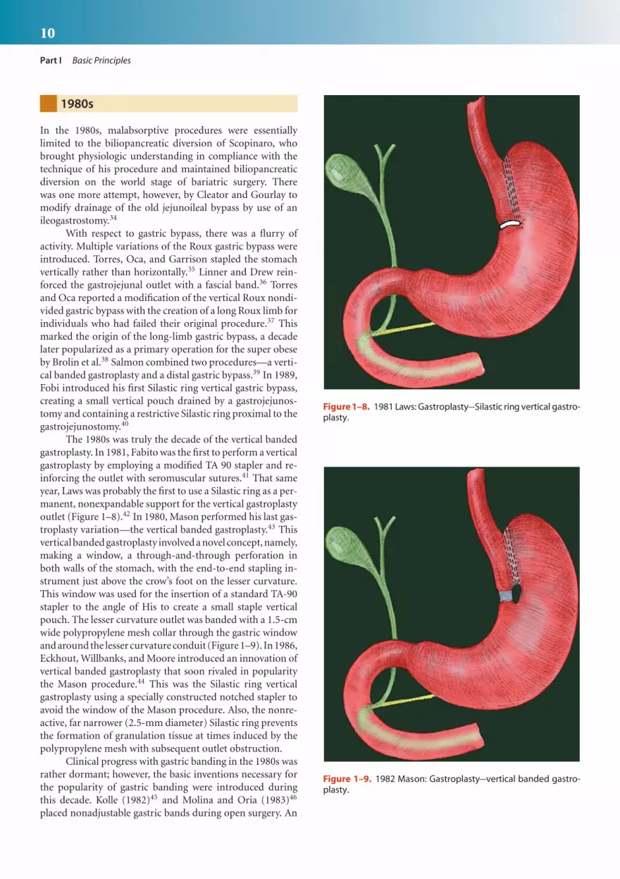
The 1980s was truly the decade of the vertical bandedgastroplasty. In 1981, Fabito was the first to perform a verticalgastroplasty by employing a modified TA 90 stapler and re-inforcing the outlet with seromuscular sutures.41 That sameyear, Laws was probably the first to use a Silastic ring as a per-manent, nonexpandable support for the vertical gastroplastyoutlet (Figure 1–8).42 In 1980, Mason performed his last gas-troplasty variation—the vertical banded gastroplasty.43 Thisvertical banded gastroplasty involved a novel concept, namely,making a window, a through-and-through perforation inboth walls of the stomach, with the end-to-end stapling in-strument just above the crow’s foot on the lesser curvature.This window was used for the insertion of a standard TA-90stapler to the angle of His to create a small staple verticalpouch. The lesser curvature outlet was banded with a 1.5-cmwide polypropylene mesh collar through the gastric windowand around the lesser curvature conduit (Figure 1–9). In 1986,Eckhout, Willbanks, and Moore introduced an innovation ofvertical banded gastroplasty that soon rivaled in popularitythe Mason procedure.44 This was the Silastic ring verticalgastroplasty using a specially constructed notched stapler toavoid the window of the Mason procedure. Also, the nonre-active, far narrower (2.5-mm diameter) Silastic ring preventsthe formation of granulation tissue at times induced by thepolypropylene mesh with subsequent outlet obstruction.
Clinical progress with gastric banding in the 1980s wasrather dormant; however, the basic inventions necessary forthe popularity of gastric banding were introduced duringthis decade. Kolle (1982)45 and Molina and Oria (1983)46
placed nonadjustable gastric bands during open surgery. An
Figure 1–8. 1981 Laws: Gastroplasty Silastic ring vertical gastro-plasty.
Figure 1–9. 1982 Mason: Gastroplasty vertical banded gastro-plasty.

11
Chapter 1 Evolution of Surgery for Morbid Obesity
Figure 1–10. 1986 Kuzmak: Gastric band adjustable Silastic.
interesting device modification of the Pace procedure28 wasintroduced by Bashour and Hill with their “gastro-clip.”47 In1986, Kuzmak introduced the inflatable Silastic band con-nected to a subcutaneous port, which is used for the percuta-neous introduction or removal of fluid to adjust the caliber ofthe gastric band (Figure 1–10).48 This first adjustable gastricband was placed during open surgery.
1990s
In the 1990s, the biliopancreatic diversion underwent a mod-ification on this side of the Atlantic with the introductionof the duodenal switch by Hess and Marceau. The Hessand Hess procedure deviated from the Scopinaro proce-dure by 1) making a lesser curvature gastric tube with agreater curvature gastric resection, rather than perform-ing a horizontal gastrectomy; 2) preserving the pylorus; 3)anastomosing the enteric limb to the proximal duodenum;and 4) dividing the duodenum, with closure of the distalduodenal stump (Figure 1–11).49 The Marceau et al. versionof the duodenal switch, cross-stapled the duodenum distalto the duodenoileostomy, without dividing the duodenum.50
The duodenum, however, unlike the stomach, does not toler-ate cross-stapling and these patients displayed disruption ofthe staple-line with regaining of weight. The currently pop-ular biliopancreatic diversion/duodenal switch is based onthe procedure introduced by Hess and Hess.49 Hess has beenactive in organizing standardization of this operation usinga 100-mL gastric reservoir. At present, there is no generalagreement as to the varying lengths of the enteric and bil-iopancreatic limbs and the common channel, as well as themethod for performing the duodenoileostomy.
In the 1990s, Fobi modified his combination verticalgastric bypass and gastroplasty by dividing the stomach andinterposing the jejunal Roux limb between the gastric pouch
Figure 1–11. 1998 Hess and Hess: Duodenal switch with divisionof the duodenum.
and the bypassed stomach to ensure maintenance of the gas-tric division (Figure 1–12).53 Over the years, the Fobi pro-cedure has gained an ever-increasing following of advocates.Another combined procedure innovation was introduced byVassallo et al., who performed a distal biliopancreatic diver-sion with no gastric resection and a transient vertical bandedgastroplasty constructed with a polydioxane band that is ab-sorbed within 6 months.54
The 1990s became the heyday for gastric bypass and,in particular, laparoscopic gastric bypass. In 1994, Wittgrove,Clark, and Tremblay were the first to report their results withlaparoscopic Roux gastric bypass.55 They performed the gas-trojejunostomy by introducing the anvil of an end-to-endstapler endoscopically. This procedure was modified by dela Torre and Scott by the introduction of the anvil of thestapler intra-abdominally to allow greater precision in anvilplacement and avoid esophageal complications.56 Schaueret al. abandoned use of the end-to-end stapler and intro-duced the intra-abdominal linear endostapler instrument forperforming the gastrojejunostomy.57 Finally, Higa et al., in1999, to avoid the relatively high incidence of gastrointestinalanastomotic leaks in employing a stapled gastrojejunostomy

12
Part I Basic Principles
Figure 1–12. 1991 Fobi: Gastric bypass vertical gastric divisionwith interposed Roux gastrojejunostomy and proximal Silasticring.
anastomosis, described a technique for hand-sewing the gas-trojejunostomy laparoscopically.58
In 1994, Cadiere59 and Hess and Hess60 introduced la-paroscopic technique to the vertical banded gastroplasty. Hessand Hess performed the gastroplasty procedure with com-plete transaction of the staple line.60 In 1999, Champion et al.reported on doing a laparoscopic vertical banded gastroplastyusing a wedge excision of the fundus of the stomach.61
Adjustable gastric banding did not become a popu-lar bariatric procedure until its employment laparoscopicallyand this occurred in the 1990s. In 1992–1993, Broadbentet al.62 and Catona et al.63 were probably the first to per-form gastric banding laparoscopically and in 1993, Belachewet al.64 and Forsell et al.65 were the first to perform adjustablegastric banding laparoscopically. Niville et al. reported plac-ing the posterior aspect of the band at the distal esophagus andconstructing an extremely small (“virtual”) gastric pouch.66
Currently, optimized results with laparoscopic adjustable gas-tric banding have been reported with 1) a high retrogastrictunnel above the lesser sac (pars flaccida approach); 2) a small
Figure 1–13. 1999 Cigaina: Gastric electrode bipolar pulsation.
gastric pouch of 15 mL; 3) band imbrication anteriorly by atleast four gastrogastric sutures; and 4) delaying band infla-tion until about 4 weeks postoperatively.67 In 1999, Cadiereet al. reported the world’s first laparoscopic adjustable gastricbanding executed robotically.68
In 1996, Cigaina et al. were the first to report on the useof electrical pacing of the stomach to induce weight loss.69 Inthis procedure, and subsequent modifications, electrodes areplaced into the wall of the stomach and connected to a pacerin a subcutaneous pocket (Figure 1–13). These systems arestill in the experimental phase.
2000s
Fifty years after the introduction of the malabsorptive je-junoileal bypass, the descendants of this operation continueto flourish and evolve. In order to overcome the extremetechnical difficulties of performing biliopancreatic diver-sion/duodenal switch laparoscopically, and the relatively highrate of complications encountered early in the developmentof the laparoscopic technique, hybrid procedures, combin-ing open and laparoscopic surgery,70 as well as hand-assistedtechniques were developed.70,71
The difficulty in mastering the laparoscopic duodenalswitch, the ultimate malabsorptive procedure, led to advo-cacy of the sleeve gastrectomy, a purely restrictive operation.Free-standing gastric sleeve resection is truly a developmentof the twenty-first century, and today has several advocatesnotably Jossart.72 In full fairness, the modern advocates ofsleeve gastrectomy owe their thinking not only to the diffi-culties of performing a duodenal switch laparoscopically butalso to the pioneering work done by Johnston et al. with theMagnestrasse and Mill operation.73
With respect to gastric bypass and vertical banded gas-troplasty there have been few innovative modifications so far

13
Chapter 1 Evolution of Surgery for Morbid Obesity
in the twenty-first century. Experiments are under way to per-form a gastric bypass with the assistance of the endoscope butthese efforts are, as yet, experiments.
ADDENDUM
Although it is not a truly surgical procedure, the endoscopicinsertion of gastric balloons or other space-occupying de-vices to create satiety and to induce weight loss should bementioned. This approach was popularized by the 1985 FDAapproval of the Garren-Edwards gastric bubble.74 The origi-nal use of this device was for the most part a failure and asso-ciated with serious complications. Currently, the concept ofan intraluminal gastric prosthesis, using improved designs, isagain gaining popularity as a technique to induce weight lossprior to a standard bariatric procedure or as a free-standinginstrument for weight reduction.
CONCLUSION
This chapter has delineated the evolutionary tree of bariatricsurgery. In the future, certain branches of this tree will be-come obsolete, whereas other branches will sprout new andexciting concepts. The richness of ingenuity displayed in theevolution of bariatric surgery gives ample testimony to theever-increasing need for effective obesity management. Con-trary to the expectations of some in the 1980s that bariatricsurgery will converge toward a single, “gold standard,” proce-dure, such as the middle-of-the-road gastric bypass, we haveseen a divergence of procedures and a plethora of new thoughtin this field. Currently, there is a trend both toward the leastinvasive, simplest, and safest restrictive operations, as well astoward the most weight-effective, slightly more risky, compli-cated malabsorptive operations. Some day, bariatric surgerymay be a historical footnote, but it will always be a page in thehistory of surgery, of which bariatric surgeons can be proudof their record of helping humankind and of providing thefirst effective therapy for the disease of morbid obesity.
References1. Buchwald H, Buchwald JN. Evolution of operative procedures for the
management of morbid obesity 1950–2000. Obes Surg 12:705–717,2002.
2. Buchwald H, Rucker RD. The rise and fall of jejunoileal bypass. In:Nelson RL, Nyhus LM (eds.): Surgery of the Small Intestine. Norwalk,Appleton Century Crofts, 1987; p. 529–541.
3. Henrikson V: Kan tunnfarmsresektion forsvaras som terapi motfettsot? Nordisk Medicin 47:744, 1952. Translations: Can small re-section be defended as therapy for obesity? Obes Surg 4:54, 1994.
4. Kremen AJ, Linner LH, Nelson CH: An experimental evaluation ofthe nutritional importance of proximal and distal small intestine.Ann Surg 140:439–444, 1954.
5. Payne JH, DeWind LT, Commons RR: Metabolic observations inpatients with jejunocolic shunts. Obes Res 4:304–315, 1996.
6. Deitel M: Jejunocolic and jejunoileal bypass: An historical perspec-tive. In: Deitel M (ed.): Surgery for the Morbidly Obese Patient.Philadelphia, PA, Lea & Febiger, 1998; p. 81–89.
7. Payne JH, DeWind LT: Surgical treatment of obesity. Am J Surg118:141, 1969.
8. Lewis LA, Turnbull RB, Page LH: “Short-circuiting” of the smallintestine. JAMA 182:77–79, 1962.
9. Sherman CD, May AG, Nye W: Clinical and metabolic studies fol-lowing bowel bypassing for obesity. Ann NY Acad Sci 131:614–622,1965.
10. Lewis LA, Turnbull RB, Page LH: Effects of jejunocolic shunt onobesity, serum lipoproteins, lipids, and electrolytes. Arch Intern Med117:4–16, 1966.
11. Mason EE, Ito C: Gastric bypass in obesity. Surg Clin N Am 47:1345–1352, 1967.
12. Scott HW, Sandstead HH, Brill AB: Experience with a new techniqueof intestinal bypass in the treatment of morbid obesity. Ann Surg174:560–572, 1971.
13. Salmon PA: The results of small intestine bypass operationsfor the treatment of obesity. Surg Gynecol Obstet 132:965–979,1971.
14. Buchwald H, Varco RL: A bypass operation for obese hyperlipidemicpatients. Surgery 70:62–70, 1971.
15. Forestieri P, DeLuca L, Bucci L: Surgical treatment of high degreeobesity. Our own criteria to choose the appropriate type of jejuno-ileal bypass: A modified Payne technique. Chir Gastroenterol 11:401–408, 1977.
16. Starkloff GB, Stothert JC, Sundaram M: Intestinal bypass: A modi-fication. Ann Surg 188:697–700, 1978.
17. Palmer JA, Marliss EB: The present status of surgical procedures forobesity. In: Deitel M (ed.): Nutrition in Clinical Surgery. Baltimore,MD, Williams & Wilkins, 1980. p. 281–292.
18. Lavorato F, Doldi SB, Scaramella R: Evoluzione storica della ter-apia chirurgica della grande obesita. Minerva Med 69:3847–3857,1978.
19. Eriksson F: Biliointestinal bypass. Int J Obes 5:437–447, 1981.20. Scopinaro N, Gianetta E, Civalleri D: Biliopancreatic bypass for obe-
sity, II: Initial experiences in man. Br J Surg 66:618–620, 1979.21. Scopinaro N, Adami GF, Marinari GM, et al.: Biliopancreatic diver-
sion: Two decades of experience. In: Deitel M, Cowan SM Jr (eds.):Update: Surgery for the Morbidly Obese Patient. Toronto, Canada:FD-Communications, 2000; p. 227–258.
22. Alden JF: Gastric and jejuno-ileal bypass: A comparison in the treat-ment of morbid obesity. Arch Surg 112:799–806, 1977.
23. Griffen WO, Young VL, Stevenson CC: A prospective comparison ofgastric and jejunoileal bypass procedures for morbid obesity. AnnSurg 186:500–507, 1977.
24. McCarthy HB, Rucker RD, Chan EK, et al.: Gastritis after gastricbypass surgery. Surgery 98:68–71, 1985.
25. Buckwalter JA: Clinical trial of jejunoileal and gastric bypass for thetreatment of obesity: Four-year progress report. Am Surg 46:377–381, 1980.
26. Printen KJ, Mason EE: Gastric surgery for relief of morbid obesity.Arch Surg 106:428–431, 1973.
27. Gomez CA: Gastroplasty in morbid obesity. Surg Clin North Am59:1113–1120, 1979.
28. Pace WG, Martin EW, Tetirick CE, et al.: Gastric partitioning formorbid obesity. Ann Surg 190:392–400, 1979.
29. LaFave JW, Alden JF: Gastric bypass in the operative revision of thefailed jejuno-ileal bypass. Arch Surg 114:438–444, 1979.
30. Tretbar LL, Taylor TL, Sifers EC: Weight reduction: Gastric plicationfor morbid obesity. J Kans Med Soc 77:488–490, 1976.
31. Wilkinson LH: Reduction of gastric reservoir capacity. J Clin Nutr33:515–517, 1980.
32. Wilkinson LH, Peloso OA: Gastric (reservoir) reduction for morbidobesity. Arch Surg 116:602–605, 1981.
33. Quaade F, Vaernet K, Larsson S: Sterotaxic stimulation and elec-trocoagulation of the lateral hypothalamus in obese humans. ActaNeurochir (Wien) 30:1111–1117, 1974.
34. Cleator IGM, Gourlay RH: Ileogastrostomy for morbid obesity. CanJ Surg 31:114–116, 1988.

14
Part I Basic Principles
35. Torres JC, Oca CF, Garrison RN: Gastric bypass: Roux-en-Y gastro-jejunostomy from the lesser curvature. South Med J 76:1217–1221,1983.
36. Linner JR, Drew RL: New modification of Roux-en-Y gastric bypassprocedure. Clin Nutr 5:33–34, 1986.
37. Torres J, Oca C: Gastric bypass lesser curvature with distal Roux-en-Y. Bariatric Surg 5:10–15, 1987.
38. Brolin RE, Kenler HA, Gorman JH, et al.: Long-limb gastric bypass inthe super obese. A prospective randomized study. Ann Surg 21:387–395, 1992.
39. Salmon PA: Gastroplasty with distal gastric bypass: A new and moresuccessful weight loss operation for the morbidly obese. Can J Surg31:111–113, 1988.
40. Fobi MA: The surgical technique of the banded Roux-en-Y gastricbypass. J Obes Weight Reg 8:99–102, 1989.
41. Fabito DC: Gastric vertical stapling. Read before the Bariatric SurgeryColloquium, Iowa City, IA, June 1, 1981.
42. Laws HL, Piatadosi S: Superior gastric reduction procedure for mor-bid obesity. A prospective, randomized trial. Am J Surg 193:334–336,1981.
43. Mason EE: Vertical banded gastroplasty. Arch Surg 117:701–706,1982.
44. Eckhout GV, Willbanks OL, Moore JT: Vertical ring gastroplasty forobesity: Five year experience with 1463 patients. Am J Surg 152:713–716, 1986.
45. Kolle K: Gastric banding. In: OMGI 7th Congress, Stockholm, 1982,Abst No 145:37.
46. Molina M, Oria HE: Gastric segmentation: A new, safe, effective,simple, readily revised and fully reversible surgical procedure for thecorrection of morbid obesity. In: 6th Bariatric Surgery Colloquium,Iowa City, IA, 2–3 June 1983, Abst 15.
47. Bashour SB, Hill RW: The gastro-clip gastroplasty: An alternativesurgical procedure for the treatment of morbid obesity. Tex Med81:36–38, 1985.
48. Kuzmak LI: Silicone gastric banding: A simple and effective operationfor morbid obesity. Contemp Surg 28:13–18, 1986.
49. Hess DW, Hess DS: Biliopancreatic diversion with a duodenal switch.Obes Surg 8:267–282, 1998.
50. Marceau P, Biron S, Bourque R-A, et al.: Biliopancreatic diversionwith a new type of gastrectomy. Obes Surg 3:29–35, 1993.
51. de Csepel J, Burpee S, Jossart G, et al.: Laparoscopic biliopancreaticdiversion with a duodenal switch for morbid obesity: A feasibilitystudy in pigs. J Laparoendosc Adv Surg Tech A 11:79–83, 2001.
52. Ren CJ, Patterson E, Gagner M: Early results of laparoscopic biliopan-creatic diversion with duodenal switch: A case series of 40 consecutivepatients. Obes Surg 10:514–523, 2000.
53. Fobi MA: Why the operation I prefer is Silastic ring vertical bandedgastric bypass. Obes Surg 1:423–426, 1991.
54. Vassallo C, Negri L, Della Valle A, et al.: Biliopancreatic diversion withtransitory gastroplasty preserving duodenal bulb: 3 years experience.Obes Surg 7:30–33, 1997.
55. Wittgrove AC, Clark GW, Tremblay, LJ: Laparoscopic gastric bypass,Roux-en-Y: Preliminary report of five cases. Obes Surg 4:353–357,1994.
56. de la Torre RA, Scott JS: Laparoscopic Roux-en-Y gastric bypass: Atotally intra-abdominal approach—technique and preliminary re-port. Obes Surg 9:492–497, 1999.
57. Schauer PR, Ikramuddin S, Gourash W, et al.: Outcomes of la-paroscopic Roux-en-Y gastric bypass for morbid obesity. Ann Surg232:515–529, 2000.
58. Higa KD, Boone KB, Ho T: Laparoscopic Roux-en-Y gastric bypassfor morbid obesity in 850 patients: Technique and follow-up. Posterpresented at American Society of Bariatric Surgery, 1999.
59. Cadiere GB, Bruyns J, Himpens J, Favretti F: Laparoscopic gastro-plasty for morbid obesity. Br J Surg 81:524, 1994.
60. Hess DW, Hess DS: Laparoscopic vertical banded gastroplastywith complete transaction of the staple-line. Obes Surg 4:44–46,1994.
61. Champion JK, Hunt T, DeLisle N: Laparoscopic vertical banded gas-troplasty and Roux-en-Y gastric bypass in morbid obesity. Obes Surg9:123, 1999.
62. Broadbent R, Tracy M, Harrington P: Laparoscopic gastric banding:A preliminary report. Obes Surg 3:63–67, 1993.
63. Catona A, Gossenberg M, La Manna A, et al.: Laparoscopic gastricbanding: Preliminary series. Obes Surg 3:207–209, 1993.
64. Belachew M, Legrand M, Jacquet N: Laparoscopic placement of ad-justable silicone gastric banding in the treatment of morbid obesity:An animal model experimental study. A video film: A preliminaryreport [abstract]. Obes Surg 3:140, 1993. Abstract 5.
65. Forsell P, Hallberg D, Hellers G: Gastric banding for morbid obesity:Initial experience with a new adjustable band. Obes Surg 3:369–374,1993.
66. Niville E, Vankeirsblick J, Dams A, et al.: Laparoscopic adjustableesophagogastric banding: A preliminary experience. Obes Surg 8:39–42, 1998.
67. Belachew M, Legrand MJ, Vincent V ; History of Lap-Band r©: Fromdream to reality. Obes Surg 11:297–302, 2001.
68. Cadiere GB, Himpens J, Vertruyen M: The world’s first obesitysurgery performed by a surgeon at a distance. Obes Surg (England)9:206–209, 1999.
69. Cigaina V, Pinato G, Rigo V: Gastric peristalsis control by mono situelectrical stimulation: A preliminary study. Obes Surg 6:247–249,1996.
70. Rabkin RA, Rabkin JM, Metcalf B, et al.: Laparoscopic techniquefor performing duodenal switch with gastric reduction. Obes Surg13:263–268, 2003.
71. Baltasar A, Bou R, Bengochea M, et al.: Duodenal switch: an effectivetherapy for morbid obesity—intermediate results. Obes Surg 11:54–58, 2001.
72. Lee CM, Feng JJ, Cirangle PT, et al.: Evolution of the laparoscopicduodenal switch and sleeve gastrectomy procedures. In: BuchwaldH, Pories W, Cowan GM Jr (eds.): Bariatric Surgery. Philadelphia,PA, Elsevier. In press
73. Johnston D, Dachtler J, Sue-Ling HM, et al.: The Magenstrasseand Mill operation for morbid obesity. Obes Surg 13:10–16,2003.
74. Garren M, Garren L, Giordano F: The Garren gastric bubble: An Rxfor the morbidly obese. Endoc Rev 2:57–60, 1984.

2Pathophysiology of Severe Obesity and theEffects of Surgically Induced Weight LossHarvey J. Sugerman, MD
I. INTRODUCTION
II. CENTRAL (ANDROID) VERSUS PERIPHERAL(GYNOID) FAT DISTRIBUTIONHypertensionCardiac Dysfunction and DyslipidemiaPulmonary DysfunctionDiabetesVenous Stasis DiseaseDegenerative Joint DiseaseGastroesophageal Reflux and Asthma
Urinary IncontinenceFemale Sexual Hormone DysfunctionPseudotumor CerebriMalignancy Risk with ObesityHernia Risk with ObesityInfectious Problems Associated with Morbid ObesityNonalcoholic Liver Disease/Nonalcoholic
SteatohepatitisQuality of LifeMortality
Severe obesity is associated with multiple comorbiditieswhich reduce the life expectancy and markedly impair thequality of life. Obesity-related problems begin at the head andend at the toes, affecting almost EVERY ORGAN in between.Morbidly obese patients can suffer from central (android)obesity or peripheral (gynoid) obesity or a combination ofthe two. Gynoid obesity is associated with degenerative jointdisease and venous stasis in the lower extremities. Androidobesity is associated with the highest risk of mortality-relatedproblems due to the “Metabolic Syndrome” or “SyndromeX,” as well as increased intra-abdominal pressure (IAP). Themetabolic syndrome is associated with insulin resistance, hy-perglycemia, and type 2 diabetes mellitus (DM) and this isassociated with nonalcoholic liver disease (NALD) or nonal-
coholic steatohepatitis (NASH), polycystic ovary syndrome,nephrotic syndrome of obesity, and systemic hypertension.Increased IAP is probably responsible in part or totally forobesity hypoventilation, venous stasis disease, pseudotumorcerebri, gastroesophageal reflux disease, stress urinary incon-tinence, and systemic hypertension. Central obesity is also as-sociated with increased neck circumference and sleep apnea.Additional comorbidities include an increased risk of severalcancers (uterine, breast, prostate, esophagus, colon, kidney),problems with pregnancy and delivery, and problems withinfections (necrotizing panniculitis and pancreatitis) as wellas difficulty in diagnosing peritonitis. The encouraging dataare that almost all of these comorbidities resolve or improvesignificantly with surgically induced weight loss.
Copyright © 2008 by The McGraw-Hill Companies, Inc. Click here for terms of use.

16
Part I Basic Principles
INTRODUCTION
Severe obesity is associated with a large number of problemsthat have given rise to the term “morbid obesity” (Table 2–1).
Table 2–1.
Morbidity of Severe Obesity
Central obesityMetabolic complications (Syndrome X)
Non-insulin dependent diabetes (adult onset/Type II)HypertensionDyslipidemia: Elevated triglycerides, cholesterolCholelithiasis, cholecystitis
Increased intra-abdominal pressureStress overflow urinary incontinenceGastroesophageal refluxVenous disease: Thrombophlebitis, venous stasis
ulcersPulmonary embolism
Obesity hypoventilation syndromeNephrotic syndromeHernias (Incisional, Inguinal)PreeclampsiaPseudotumor cerebri
Respiratory insufficiency of obesity (Pickwickiansyndrome)Obesity hypoventilation syndromeObstructive sleep apnea syndrome
Cardiovascular dysfunctionCoronary artery diseaseIncreased complications after coronary bypass
surgeryHeart failure subsequent to
Left ventricular concentrichypertrophy—hypertension
Left ventricular eccentric hypertrophy—obesityRight ventricular hypertrophy—pulmonary failure
Prolonged Q-T interval with sudden deathSexual hormone dysfunction
Amenorrhea, hypermenorrheaStein-Leventhal or polycystic ovary syndrome:
hirsutism, ovarian cystsInfertilityEndometrial carcinomaBreast carcinoma
Other carcinomas: colon, renal cell, prostateInfectious complications
Difficulty recognizing peritonitisNecrotizing pancreatitisNecrotizing subcutaneous infectionsWound infections, dehiscence
Pseudotumor cerebri (idiopathic intracranialhypertension)
Degenerative osteoarthritisFeet, ankles, knees, hips, back, shoulders
Psychosocial impairmentDecreased employability, work discrimination
Although some consider this term to be pejorative, severeobesity is often associated with a number of problems thatare truly morbid and incapacitating. The medical problemscaused by obesity begin with the head and end with the toesand involve almost every organ in between. Several of theseproblems contribute to the earlier mortality associated withobesity and include coronary artery disease, severe hyper-tension that may be refractory to medical management, im-paired cardiac function, adult onset (type 2) diabetes mellitus,obesity hypoventilation, and sleep apnea syndromes (SASs),cirrhosis, venous stasis, and hypercoagulability leading toan increased risk of pulmonary embolism, and necrotizingpanniculitis.1,2 Morbidly obese patients can also die as a re-sult of difficulties in recognizing the signs and symptoms ofperitonitis.3 There is an increased risk of prostate, uterine,breast, kidney, esophageal, liver, and colon cancer. Prematuredeath is much more common in the severely obese individual;one study noted a 12-fold excess mortality in morbidly obesemen in the 25–34 year age group.4 Increased morbidity andmortality have been noted in several other studies.5−8
A number of obesity-related problems may not beassociated with death but can lead to significant physicalor psychological disability. These include degenerative os-teoarthritis, pseudotumor cerebri (idiopathic intracranialhypertension), cholecystitis, skin infections, chronic venousstasis ulcers, stress overflow urinary incontinence, gastro-esophageal reflux, sex hormone imbalance with dysmenor-rhea, hirsutism, infertility (polycystic ovary syndrome), thenephrotic syndrome, and nonalcoholic liver disease (NALD)or, in its more severe form, nonalcoholic steatohepatitis(NASH). Many morbidly obese patients suffer from severepsychological and social disability, including marked preju-dice regarding employment.9,10
CENTRAL (ANDROID) VERSUS PERIPHERAL(GYNOID) FAT DISTRIBUTION
It has been noted that central obesity is associated with ahigher mortality than is peripheral obesity, commonly re-ferred to as android versus gynoid obesity because of theirrelative prevalence in men and women, respectively. This hasbeen attributed to metabolically more active visceral adiposetissue than to subcutaneous fat so that there is a greater rateof glucose production, type 2 diabetes, and hyperinsulinism.Increased insulin secretion is thought to increase sodium re-absorption and, thus, cause hypertension.11−18 Central obe-sity also is associated with a greater production of cholesterol,primarily in the form of low-density lipoprotein, leading toa higher than normal incidence of atherosclerotic cardiovas-cular disease, and an increased incidence of gallstones. Theincreased visceral fat has been related to an increased waist:hipratio, or in more common terms as the “apple” versus “pear”distribution of fat. Computerized axial tomographic (CAT)scans, however, have noted a much better correlation betweenanterior–posterior abdominal diameter and visceral fat distri-bution than with the waist:hip ratio,19,20 especially in women

17
Chapter 2 Pathophysiology of Severe Obesity and Effects of Surgically Induced Weight Loss
who may have both central and peripheral obesity. In thissituation, the peripheral obesity “dilutes” the central obesityusing a W:H ratio, so that either waist circumference alone orsagittal abdominal diameter should be used as a measurementof central obesity.
A recent study documented an increased bladder pres-sure in morbidly obese women, which was associated with ahigh incidence of urinary incontinence.21 It is quite probablethat much of the comorbidity of severe obesity is related toan increased intra-abdominal pressure (IAP)22 secondary toa central distribution of fat (Table 2–1) and that the urinarybladder pressure, a surrogate for IAP, is highly correlated withthe sagittal abdominal diameter or waist circumference. In an-imal studies,23 a close relationship between urinary bladderpressure and directly measured IAP has been found. In addi-tion to urinary incontinence, this increased IAP is probablyresponsible for increased venous stasis disease and venous sta-sis ulcers, gastroesophageal reflux, nephrotic syndrome sec-ondary to increased renal venous pressure, incisional and in-guinal hernias, and obesity hypoventilation syndrome (OHS)secondary to a high-riding diaphragm and restrictive lung dis-ease. This can also lead to an increased intrapleural pressurewhich can then cause increased intracardiac pressures so thatseverely obese patients with OHS may require high cardiacfilling pressures to maintain an adequate cardiac output.23,24
It is also quite possible that increased IAP is also responsiblefor the high incidence of systemic hypertension in the mor-bidly obese, as well as the cause of “idiopathic” intracranialhypertension.23,25−27
In a previous study,22 we noted a much higher IAP inmorbidly obese patients at the time of gastric bypass surgerythan five nonobese patients who were undergoing colectomy,ileal pouch anal anastomosis (IPAA) for ulcerative colitis.To realize how high these pressures can be in severe obe-sity, one needs to relate them to the pressures seen in pa-tients with an acute abdominal compartment syndrome28,29
where it is generally thought that a urinary bladder pressure of≥20–25 cm H2O is an indication to return the patient to theoperating room for emergency laparotomy and abdominaldecompression. Many severely obese patients have urinarybladder pressures well above 25 cm H2O. Surgically inducedweight loss was associated with a significant decrease in sagit-tal abdominal diameter, urinary bladder pressures, and obe-sity comorbidity.30
HypertensionWe hypothesize that obesity-associated hypertension is sec-ondary to an increased IAP rather than to increased insulin-induced sodium reabsorption. The presumed pathophys-iology is related to activation of the renin–angiotensin–aldosterone system through one or a combination of threepossible renal mechanisms. The first presumed mechanismis due to direct pressure on the renal veins, leading to an in-creased glomerular capillary pressure, a capillary leak witheither a microalbuminuria commonly seen in the morbidlyobese or, on occasion, a large protein leak with development
of the nephrotic syndrome.31,32 The increased glomerularpressure probably stimulates the juxta-glomerular appara-tus (JGA) to increased renin secretion.33 A second mecha-nism relates to a direct pressure on the renal capsule, lead-ing to a renal compartment syndrome and activation of therenin–angiotensin–aldosterone system. The third possibilityrelates to the increased pleural pressure secondary to a ris-ing diaphragm, which impedes venous return to the heart,leading to a decrease in cardiac output and renal arterialpressure which again stimulates activation of the JGA to pro-duce renin.23,34 Increased renin and aldosterone levels areseen in a porcine model of acutely increased IAP.34 Activa-tion of the renin–angiotensin–aldosterone system leads tosalt and water retention, commonly seen in the severely obese,and vasoconstriction. Surgically induced weight loss is associ-ated with a clinically significant, long-lasting improvement inblood pressure with elimination of antihypertensive medica-tions in two-thirds to three-quarters of hypertensive patientsor a marked decrease in their use.35−43 The Swedish ObesityStudy (SOS) initially noted a significant improvement in bothdiabetes and hypertension in the surgically treated patients ascompared to matched controls.36 However, the improvementin hypertension for the overall group was no longer presentat 8 years after surgery.38 Unfortunately, only 6% of the sur-gical patients in the SOS trial had a gastric bypass, 70% avertical banded gastroplasty, and 24% a gastric banding. Thegastric bypass patients had a significantly greater weight lossthan either the gastroplasty or gastric banded patients andalso maintained a significant decrease in both systolic anddiastolic pressure at 5 years after surgery.39
Cardiac Dysfunction and DyslipidemiaMorbid obesity may be associated with cardiomegaly andimpaired left, right, or biventricular function (Table 2–2).Severe obesity may be associated with a high cardiac out-put and a low systemic vascular resistance leading to leftventricular hypertrophy. Obesity also is associated with hy-pertension, which leads to concentric left ventricular hy-pertrophy. This combination of obesity and hypertensionwith left ventricular eccentric and concentric hypertrophymay lead to left ventricular failure.44,45 Correction of morbidobesity improves cardiac function in these patients.24,46−49
Morbid obesity is also associated with an accelerated rate ofcoronary atherosclerosis. These patients often have hyper-cholesterolemia and a decreased high-density to low-densitylipoprotein (HDL:LDL) ratio. In the Nurses Study, womenwith a BMI >29 kg/m2 have a significantly increased inci-dence of myocardial angina and/or infarction.50,51 Surgicallyinduced weight loss has been shown in several studies to sig-nificantly reduce triglyceride and LDL cholesterol levels whileincreasing HDL levels.38,43,52−56 Respiratory insufficiency as-sociated with morbid obesity can result in hypoxemic pul-monary artery vasoconstriction, which in severe cases maylead to right, or biventricular, heart failure associated withtricuspid valvular insufficiency. Correction of hypoxemia andhypercarbia with surgically induced weight loss will correct

18
Part I Basic Principles
Table 2–2.
Obesity Comorbidity “From the Top of the Head to the Tip of the Toes and Almost EveryOrgan IN BETWEEN”
HeadBrainStrokeHeadaches
HypertensionPseudotumor cerebri: Headaches
I. Optic Nerve: Visual field cuts; blindnessIII. Oculomotor nerve palsy
V. Trigeminal nerve: Tic DouloureuxVII. Facial nerve: Bell’s palsy
VIII. Auditory nerve: Pulsatile tinnitusEyes: Diabetic retinopathy + Pseudotumor
complicationsMouth/Throat
Sleep Apnea
ChestBreast cancerObesity hypoventilation
Heavy chest wallElevated diaphragmIncreased intra-thoracic pressureDecreased expiratory reserve volume
HeartLeft ventricular hypertrophy
Eccentric: Increased cardiac outputConcentric: Increased peripheral vascular
resistanceIncreased cardiac filling pressures (CVP, PAP, WP)Right heart failure
Tricuspid insufficiency
EsophagusAcid refluxAsthmaAdenocarcinomaEsophageal varices
AbdomenGallbladder
CholecystitisAdenocarcinoma
LiverNonalcoholic liver disease (NALD)Nonalcoholic steatohepatitis (NASH)CirrhosisType 2 diabetes mellitus
SpleenSplenomegaly (portal hypertension)Hypersplenism (portal hypertension)
PancreasType 2 diabetes mellitusNecrotizing pancreatitis
ColonAdenocarcinomaDiverticulitis
GeneralDifficulty diagnosing peritonitisHernia
IncisionalInguinalSpigelian
Wound infection“Apron”
Peau d’orange lymphatic stasis
KidneyHypertensionProteinuriaRenal cell carcinoma
Urinary bladderStress incontinence
Ovaries/UterusIncreased estradiol, androstenedionePolycystic ovary syndrome, Stein-Leventhal syndrome
InfertilityDysmenorrheaHirsutism
Endometrial carcinomaBreast cancer
Prostate: Adenocarcinoma
AnusPerianal abscessesNecrotzing panniculitis
IntegumentNecrotizing panniculitisHirsutism
Increased risk of operative complicationsColectomyHysterectomyKidney, liver transplantation
SpineHerniated disc
Upper extremitiesShoulder girdle painEdema
Lower extremitiesOsteoarthritis
Hip arthralgiaKnee arthralgia
Venous stasisEdemaThrombophlebitisStasis ulcersPulmonary embolism
ToesDiabetic neuropathyDiabetic ulcers

19
Chapter 2 Pathophysiology of Severe Obesity and Effects of Surgically Induced Weight Loss
the elevated pulmonary artery and wedge pressures within3–9 months after surgery.24 Severe obstructive SAS may beassociated with prolonged sinus arrest, premature ventricu-lar contractions, and sudden death.
Pulmonary DysfunctionRespiratory insufficiency of obesity is associated with eitherOHS, obstructive SAS, or a combination of the two, com-monly called the Pickwickian syndrome.24,57−64
Obesity Hypoventilation Syndrome: This problem arisesprimarily from the increased IAP in patients with cen-tral, abdominal obesity, which leads to high-ridingdiaphragm.22,65 As a result, the lungs are squeezed, pro-ducing a restrictive pulmonary defect. A heavy, obesethoracic cage may also contribute to the pathophysi-ology secondary to a decreased chest wall compliance.These patients have a markedly decreased expiratoryreserve volume, leading to alveolar collapse and arte-riovenous shunting at end expiration. They also havesmaller reductions in all other lung volumes.59 Theyhave hypoxemia and hypercarbia while awake and ablunted ventilator response to CO2.
Chronic hypoxemia leads to pulmonary artery vaso-constriction. However, obesity hypoventilation pa-tients often have both markedly elevated pulmonaryartery pressures and pulmonary capillary wedge pres-sures, suggesting both right and left ventricular failures.Despite these high pressures, overt heart failure is un-usual, even with pulmonary capillary wedge pressuresas high as 40 mm Hg.24 These pressures are increasedas a result of a marked increase in pleural pressuressecondary to the high-riding diaphragm and increasedIAP, so that the transatrial and transventricular pres-sures may be, in fact, normal.23 These findings havebeen noted in a porcine model of acutely increasedIAP. Thus, morbidly obese patients with OHS may notrespond well to diuresis and may need these elevatedpressures to maintain an adequate cardiac output.
As a result of the increased inferior vena caval andpulmonary artery pressures, obesity hypoventilationpatients are at risk for a fatal pulmonary embolism.57
Thus, right heart catheterization should be consideredprior to obesity surgery and a prophylactic inferior venacaval filter inserted at the time of surgery in patientswith a mean pulmonary artery pressure ≥40 mm Hg.Surgically induced weight loss24,59,60,63 corrects hypox-emia, hypercarbia, and increased cardiac filling pres-sures associated with OHS.
Sleep Apnea Syndrome: SAS is associated with centralobesity and is due to both depression of the normal ge-nioglossus reflex, possibly secondary to a large, heavytongue, and deposition of fat within the hypopharynxwith narrowing of the cervical airway.58 These patientssnore loudly while asleep and suffer from severe day-time somnolence with tendencies to fall asleep while
driving or at work. The daytime somnolence is proba-bly secondary to impaired stage III, IV, and Rapid EyeMovement (REM) sleep. The diagnosis of obstructiveSAS is suggested by a history of severe daytime som-nolence, frequent nocturnal awakening, loud snoring,and morning headaches and is confirmed with sleeppolysomnography. This technique documents cessa-tion of airflow during sleep associated with persistentrespiratory efforts. Most of the apneic episodes are ob-structive, but some are central. The latter is seen withcentral nervous system hypoxemia, analogous to theCheyne-Stokes respirations seen at high altitude. Theseverity of sleep apnea is usually determined by therespiratory disturbance index (RDI), which is a com-bination of apneic episodes (cessation of airflow for≥10 seconds) and hypopneic episodes (diminution by50% of airflow for ≥10 seconds) and is divided intomild (RDI ≤ 19), moderate (RDI 20–39), and severe(RDI ≥ 40). Moderate to severe sleep apnea should betreated with nocturnal nasal continuous positive airwaypressure (nasal CPAP); if the patient cannot toleratenasal CPAP and has severe sleep apnea, a tracheostomyshould be considered at the time of obesity surgery. Allpatients with moderate to severe sleep apnea should bemanaged with intubation and mechanical ventilationin the intensive care unit, and weaned on the morningafter surgery. If they also have obesity hypoventilationthen they may have to be ventilated for several days un-til their arterial blood gases return to their preoperativevalues.
Patients with severe sleep apnea have difficulty stay-ing awake during the day. This syndrome may be as-sociated with sudden death and should always be con-sidered in trauma victims who have fallen asleep whiledriving. We have seen many patients with severe sleepapnea who are in occupations that are incompatiblewith this syndrome: taxicab and interstate truck driversand state prison guards! Surgically induced weight losscorrects or markedly improves SAS, permitting removalof their tracheostomy tube or discontinuation of nasalCPAP.59−64
In a series from the Medical College of Virginia,12.5% of the patients who underwent gastric surgeryfor morbid obesity had respiratory insufficiency.59 Ofthe affected individuals, 51% had SAS alone, 12% hadOHS alone, and 37% had both. Of these, 64% weremen in contrast to only 14% of the entire group of pa-tients who underwent surgery for obesity. Patients withrespiratory insufficiency were significantly more obesethan those without pulmonary dysfunction. However,obesity is not the only factor causing respiratory embar-rassment, since many patients who underwent surgeryfor morbid obesity and did not have a clinically sig-nificant pulmonary problem, weighed more than thepatients with respiratory insufficiency. Most of theobese patients with respiratory dysfunction had anadditional pulmonary problem, such as sarcoidosis,

20
Part I Basic Principles
heavy cigarette use, recurrent pulmonary embolism,myotonic dystrophy, or idiopathic pulmonary fibrosis.Obstructive SAS and OHS are associated with a highmortality and serious morbidity; weight reduction willcorrect both.24,59−64
DiabetesObesity is a frequent etiologic factor in the development oftype 2 adult onset diabetes mellitus. Morbidly obese pa-tients can be very resistant to insulin due to the markeddown-regulation of insulin receptors. The tendency towardhyperglycemia manifested by obese patients is another riskfactor for coronary artery disease as well as for fatal sub-cutaneous infections. Gastric-surgery-induced weight loss,performed both open and laparoscopically, is associated withresolution of the diabetes38,40,42,66−79 in 85% of patients andthis effect is long-lasting (Table 2–3). Pories and colleagueshave found that the earlier the patients with type 2 diabetes
Table 2–3.
Decreases in Obesity Comorbidity AfterWeight-Loss Surgery
Comorbidity Reference
Type 2 diabetes mellitus 38, 40, 42, 66–79
Systemic hypertension 35–43
Obstructive sleep apnea 59–64
Obesity hypoventilation 24, 59, 60, 63
Cardiac dysfunction 24, 45–49
Pseudotumor cerebri 27, 110
Venous stasis disease 81
Gastroesophageal reflux 90–92
Asthma 98, 99
Dyslipidemia 38, 43, 52–56
Stress urinary incontinence 21
Polycystic ovary syndrome 102
Pregnancy outcomes 104–107
Fatty liver disease 122–125
Musculoskeletal pain 85–87
Health-related quality of life 126–130
Psychological status 131–136
Mortality 70, 137
mellitus undergo weight reduction gastric bypass surgery, thegreater is their likelihood to have complete resolution of theirdiabetes.67,68 In another study, it was noted that patientswithout diabetes or hypertension are significantly youngerby 5 years than those with either diabetes or hypertensionand are significantly younger by another 5 years than thosewith both diabetes and hypertension.42 Furthermore, patientswere more likely to correct their diabetes and hypertension,the more weight they lost.42 These data support earlier opera-tions in morbidly obese diabetic patients before their diabetic-related complications develop (neuropathy, retinopathy,renal insufficiency). Peripheral insulin resistance, primarilyin skeletal muscle, is present in morbidly obese patients andmay resolve with surgically induced weight loss. There are datasuggesting that type 2 diabetes mellitus could be a disease ofthe foregut.69 The gastric bypass operation has been foundto decrease the progression and mortality of non-insulin-dependent diabetes mellitus when compared to a matchedcontrol group of patients who either chose not to have surgeryor were unable to obtain insurance coverage.70
Venous Stasis DiseaseMorbidly obese patients have an increased risk for deep ve-nous thrombosis, venous stasis ulcers, and pulmonary em-bolism. Low levels of antithrombin III may increase their riskof blood clots.80 The increased weight within the abdomenraises the IAP and, therefore, the inferior vena caval pressurewith an increased resistance to venous return, leading to thepretibial bronze edema, lower extremity venous stasis ulcers,and tendency to deep venous thrombosis. A similar mecha-nism may be responsible for the increased risk of pulmonaryembolism in patients with right heart failure secondary tohypoxemic pulmonary artery vasoconstriction. Venous stasisulcers can be incapacitating and extremely difficult to treatin the morbidly obese; weight reduction may be the criti-cal factor, as skin grafts, pressure stockings, medicated rigidcompression boots, and wound care are often ineffective. Sur-gically induced weight loss reduces IAP and permits healingof these stasis ulcers.81
Degenerative Joint DiseaseThe increased weight in the morbidly obese leads to early de-generative arthritic changes of the weight-bearing joints, in-cluding the knees, hips, and spine. Many orthopedic surgeonsrefuse to insert total hip or knee prosthetics in patients weigh-ing over 250 pounds because of an unacceptable incidence ofprosthetic loosening.82 There is a high risk of complicationsin obese patients following intramedullary nailing of femoralfractures.83 Severe obesity is a common problem in patientsrequiring intervertebral disc surgery.84 Weight reduction fol-lowing gastric surgery for obesity permits subsequent suc-cessful joint replacement85 and is associated with decreases inmusculoskeletal and lower back pain.86,87 In some instances,the decrease in pain following weight loss obviates the needfor joint or intervertebral disc surgery.

21
Chapter 2 Pathophysiology of Severe Obesity and Effects of Surgically Induced Weight Loss
Gastroesophageal Reflux and AsthmaMorbidly obese patients frequently suffer from gastro-esophageal reflux disease (GERD). Although one study didnot find a relationship to severe obesity,88 others have.89 Thisis also probably secondary to an increased IAP. The loweresophageal sphincter may be normal in these patients, but theincreased IAP can overcome a normal sphincter pressure. Sur-gically induced weight loss has corrected this problem.90−92
GERD resolves promptly following gastric bypass for obe-sity as neither acid nor bile can reflux into the esophagusafter this procedure. Some studies have noted an increasedGERD following vertical banded gastroplasty,93−95 which re-solves following conversion to gastric bypass.94,95 An an-tireflux procedure (e.g., Nissen fundoplication) is probablyinappropriate in a severely obese patient, as gastric surgeryinduced weight loss not only corrects acid and bile reflux butalso improves additional comorbidity usually present in theseindividuals. The increase risk of GERD in obese individualsincreases their risk of esophageal adenocarcinoma and theepidemic of obesity may be one explanation for the markedincrease in the incidence of this cancer.96,97 Two studies havedocumented improvement in asthma following surgically in-duced weight loss, presumably due to prevention of noctur-nal gastroesophageal reflux and tracheobronchial aspirationor spasm.98,99
Urinary IncontinenceSeverely obese women often have stress overflow urinary in-continence. Some men with central obesity also complain ofurinary urgency, although incontinence in men is rare. Signif-icantly increased urinary bladder pressures have been noted inwomen with this problem.21 Surgically induced weight loss isassociated with correction of urinary incontinence in 95% ofpatients, often within a few months of surgery, and this is asso-ciated with a significant decrease in urinary bladder pressurewhen measured 1 year after surgery. The rapid resolution ofthis vexing problem, often within 1–2 months after surgery,may be related to a rapid decrease in intra-abdominal andurinary bladder pressures following obesity-surgery-inducedweight loss as a result of the relationship between wall tensionand volume of a sphere according to LaPlace’s Law, wherepressure is proportional to the radius to the fourth power.This is best thought of as a tense balloon that rapidly losestension when a small amount of air escapes.
Female Sexual Hormone DysfunctionWomen often suffer from sexual dysfunction due to excessivelevels of both the virilizing hormone, androstenedione, andthe feminizing hormone, estradiol. These may produce infer-tility, hirsutism, ovarian cysts (Stein-Leventhal or polycysticovary syndrome), hypermenorrhea, and a significantly in-creased risk of breast and endometrial carcinoma. Polycysticovary syndrome has also been found to be associated with type2 diabetes and, in mild cases, may improve with metformintreatment.100,101 Surgically induced weight loss often returns
sex hormone levels to normal, increasing fertility and men-strual regularity.102 It is thought that there may be a higherincidence of neural tube defects in infants born to women dur-ing the rapid weight loss that occurs after surgery,103 probablydue to deficient folic acid levels. Thus, birth control is stronglyrecommended for 1 year after surgically induced weight lossprocedures. Surgery has permitted pregnancies in previouslyinfertile morbidly obese women. Increased weight also in-creases the risk of complications of pregnancy, including thehigher incidence of preeclampsia seen in obese women, prob-lems with delivery, and an increased risk of venous throm-bosis and pulmonary embolism. Surgically induced weightloss is associated with decreased pregnancy-related compli-cations, including preeclampsia, and reduces the frequencyof cesarean section.104−107
Pseudotumor CerebriPseudotumor cerebri, also known as idiopathic intracranialhypertension, may be associated with morbid obesity. Theproblem is almost always seen in women. Symptoms includesevere headache, which is usually worse in the morning, bilat-eral pulsatile auditory tinnitus, and visual field cuts. Severelyincreased intracranial pressure (ICP) can lead to permanentblindness. Additional cranial nerves that can be involved in-clude the Vth (Tic Douloureux), the VIth (oculomotor nerveparalysis), and the VIIth (Bell’s palsy). Studies suggest thatpseudotumor cerebri is secondary to an increased IAP lead-ing to an increased pleural pressure and decreased venousdrainage from the brain with a consequent cerebral venousengorgement and an increased ICP. Increased ICP occurs in aporcine model of acutely increased IAP, which is prevented bymedian sternotomy.25,26 Patients with impending blindnessshould undergo emergent optic nerve fenestration.108 In thepast, pseudotumor cerebri has been treated with ventriculo-peritoneal or lumboperitoneal cerebrospinal fluid (CSF)shunts. There is a high incidence of shunt occlusion109 andin some instances patients can have continued headache andauditory tinnitus despite a patent shunt. These failures areprobably secondary to shunting from a high-pressure sys-tem to another high-pressure system. Patients may also de-velop major neurological complications following insertionof ventriculo-peritoneal or lumboperitoneal shunts. Surgi-cally induced weight loss decreases CSF pressures, relievesheadache and tinnitus,27,110 and is the procedure of choicerather than CSF-peritoneal shunting. The rapid resolutionof headache, tinnitus, and other pseudotumor cerebri symp-toms is probably a result of the rapid decrease in IAP. It isdifficult to understand, however, why this syndrome is almostentirely restricted to women since men with central obesityalso have increased intra-abdominal and pleural pressures.
Malignancy Risk with ObesityIn addition to uterine carcinoma, there is also a significantlyincreased risk of breast, prostate, kidney, and esophageal andcolon cancer in the morbidly obese.96,97,111−114 The increased

22
Part I Basic Principles
risk of breast, uterine, and prostate cancers are probably sec-ondary to the high levels of sex hormones seen in these pa-tients. As previously mentioned, the increased incidence ofGERD in the morbidly obese, a probable consequence ofincreased IAP, is probably responsible for the increased in-cidence of esophageal adenocarcinoma. The causes for theincreased colon and renal cell cancer incidence are unknown.
Hernia Risk with ObesitySevere obesity is also associated with a significantly increasedrisk of all types of hernias. This is also probably secondary tothe increased IAP associated with central obesity. A signifi-cantly higher incidence of incisional hernia has been notedfollowing open gastric bypass surgery for morbid obesity thanafter total colectomy, proctectomy, and stapled IPAA for ulcer-ative colitis, of whom 60% were taking approximately 30 mgof prednisone and their incisions were lower midabdomento above the umbilicus, both of which are thought to in-crease the risk of hernia.115 Reduced risk of incisional herniasand other wound-related complications are significant ad-vantages of the laparoscopic approach for obesity surgery.We have seen bilateral spigelian hernias in one of our mor-bidly obese patients who also developed an incisional hernia.Repair of incisional hernias in morbidly obese patients hasa high risk of recurrence and should probably be reinforcedwith a polypropylene mesh.115
Infectious Problems Associated with MorbidObesityThere is a higher incidence of fulminant diverticulitis,116,117
necrotizing pancreatitis,118 and necrotizing panniculitis119 inseverely obese patients. This is probably due to the increasedretroperitoneal and subcutaneous fat, as well as diabetes,which provides an ideal growth medium for bacteria.
Nonalcoholic Liver Disease/NonalcoholicSteatohepatitisNALD is also associated with type 2 diabetes mellitus. Theexact pathophysiology is unknown. It is presumably sec-ondary to the increased glycogen deposition in the liver withsubsequent conversion to fat.120 Increased fatty infiltrationproduces markedly enlarged livers which can make bariatricsurgery much more difficult and dangerous, and may preventa successful laparoscopic procedure because of the difficultyin elevating the left lobe of the liver from the gastroesophagealtissues. The increased fat in the liver may be metabolized tofree fatty acids, which may be quite toxic to tissues and beresponsible for the development of NASH and the subse-quent development of severe cirrhosis.88 There is a seriousconcern that NASH may overtake a combination of hepati-tis A, B, and C and the primary cause for liver failure andthe need for liver transplantation if the epidemic of severeobesity remains unchecked.121 Surgically induced weight loss
has been found to significantly decrease the severity of hepaticsteatosis.122−125
Quality of LifeSevere obesity is associated with a marked reduction in thequality of life. Several studies have documented an improve-ment in the quality of life following surgically induced weightloss.126−130 Psychological profiles document significant psy-chological impairment in the morbidly obese which alsoimproves following surgically induced weight loss.10,131−136
However, there may also be an increased risk of suicide ifthe individual believes that severe obesity was the total causeof their emotional impairment, which does not resolve fol-lowing major weight loss after surgery. Lastly, the morbidlyobese are at risk for prejudice in many areas, including physi-cians, but more economically important, with regards toemployment.137,138
MortalityTwo studies have documented a decrease in mortality fol-lowing bariatric surgery. One noted a significantly decreasedmortality (1% vs 4.5%) in diabetic patients who under-went gastric bypass as compared to diabetic patients whofor insurance or personal reasons did not undergo surgeryfor obesity.70 The other study from Canada noted a signifi-cantly lower long-term mortality (0.7% vs 6.2%, p < 0.001)in patients who underwent surgery for obesity from 1983 to2002 [194 vertical banded gastroplasty (35% subsequentlyconverted to gastric bypass) and 841 gastric bypass] whencompared to an age and sex matched nonoperated cohort.139
Furthermore, the surgical patients also had a significantly de-creased rate of developing new medical conditions (cardio-vascular, cancer, endocrine, infectious diseases, psychiatric,and digestive). The SOS has not yet reported a significantlydecreased mortality in the surgical patients as compared tothe control cohort, but 94% of the surgical patients in thisstudy underwent a purely gastric restrictive procedure andhave not lost as much weight as the 6% of patients who un-derwent a gastric bypass operation and could be a reason forthe absence of a mortality benefit to date.41
References1. Sjostrom LV: Morbidity of severely obese subjects. Am J Clin Nutr
55:508S–515S, 1992.2. Sjostrom LV: Mortality of severely obese subjects. Am J Clin Nutr
55:516S–523S, 1992.3. Mason EE, Printen KJ, Barron P, et al.: Risk reduction in gastric
operations for obesity. Ann Surg 190:158–165, 1979.4. Drennick EJ, Bale GS, Seltter F, et al.: Excessive mortality and causes
of death in morbidly obese men. JAMA 243:443–445, 1980.5. Folsom AR, Kaye SA, Sellers TA, et al.: Body fat distribution and
5-year risk of death in older women. JAMA 269:483–487, 1993.6. VanItalie TB: Obesity: Adverse effects on health and longevity. Am
J Clin Nutr 32:2723–2733, 1979.7. Lew EA, Garfinkel L: Variations in mortality by weight among
750,000 men and women. J Chronic Dis 32:563–568, 1979.

23
Chapter 2 Pathophysiology of Severe Obesity and Effects of Surgically Induced Weight Loss
8. Kral JG: Morbid obesity and health related risks. Ann Intern Med103:1043–1046, 1985.
9. Sarlio-Lahteenkorva S, Stunkard A, Rissanen A: Psychosocial fac-tors and quality of life in obesity. Int J Obes Relat Metab Disord19:1S–5S, 1995.
10. Stunkard AJ, Wadden TA: Psychological aspects of severe obesity.Am J Clin Nutr 55:532S–534S, 1991.
11. Kissebah A, Vydelingum N, Murray R, et al.: Relation of body fatdistribution to metabolic complications of obesity. J Clin EndocrinolMetab 54:254–257, 1982.
12. Reaven GM: Syndrome X: 6 years later. J Intern Med Suppl 736:13–22, 1994.
13. Kvist A, Chowdhury B, Grangard U, Tylen U, Sjostrom L: Totaland visceral adipose tissue volumes derived from measurementswith computed tomography in adult men and women: Predictiveequations. Am J Clin Nutr 48:1351–1361, 1988.
14. Gillum RF: The association of body fat distribution with hyperten-sion, hypertensive heart disease, coronary heart disease, diabetesand cardiovascular risk factors in men and women aged 18–79 years.J Chronic Dis 40:421–428, 1987.
15. Micciolo R, Bosello O, Ferrari P, Armellini F: The association ofbody fat location with hemodynamic and metabolic status in menand women aged 21–60 years. J Clin Epidemiol 44:591–608, 1991.
16. Bjorntorp P: Abdominal obesity and the metabolic syndrome. AnnMed 24:465–468, 1992.
17. Johnson D, Prud’homme D, Despres JP, Nadeau A, Tremblay A,Bouchard C: Relation of abdominal obesity to hyperinsulinemiaand high blood pressure in men. Int J Obes 16:881–890, 1992.
18. Mauriege P, Despres JP, Marcotte M, et al.: Abdominal fat cell lipoly-sis, body fat distribution, and metabolic variables in premenopausalwomen. J Clin Endocrinol Metab 71:1028–1035, 1990.
19. Sjostrom L: A computer-tomography based multicompartmentbody composition technique and anthropometric predictions oflean body mass, total and subcutaneous adipose tissue. Int J Obes15 (Suppl 2):19–30, 1991.
20. Lemiuex S, Prud’homme D, Tremblay A, Bouchard C, J-P Despres:Anthropometric correlates to changes in visceral adipose tissue over7 years in women. Int J Obes 20:618–624, 1996.
21. Bump RC, Sugerman HJ, Fantl JA, et al.: Obesity and lower urinarytract function in women: Effects of surgically induced weight loss.Am J Obstet Gynecol 167:392–397, 1992.
22. Sugerman H, Windsor A, Bessos M, Wolfe M: Abdominal pressure,sagittal abdominal diameter and obesity co-morbidity. J Int Med241:71–79, 1997.
23. Ridings PC, Bloomfield GL, Blocher CR, Sugerman HJ: Cardiopul-monary effects of raised intra-abdominal pressure before and aftervolume expansion. J Trauma 39:1071–1075, 1995.
24. Sugerman HJ, Baron PL, Fairman RP, Evans CR, Vetrovec GW:Hemodynamic dysfunction in obesity hypoventilation syndromeand the effects of treatment with surgically induced weight loss.Ann Surg 207:604–613, 1988.
25. Bloomfield GL, Ridings PC, Blocher CS, Marmarou A, SugermanHJ: Effects of increased intra-abdominal pressure upon intracranialand cerebral perfusion pressure before and after volume expansion.J Trauma 40:936–943, 1996.
26. Bloomfield GL, Ridings PC, Blocher CR, Sugerman HJ: Increasedpleural pressure mediates the effects of elevated intra-abdominalpressure upon the central nervous and cardiovascular systems.CritCare Med 25:496–503, 1997.
27. Sugerman HJ, Felton WL, Sismanis A, Salvant JB, Kellum JM: Effectsof surgically induced weight loss on pseudotumor cerebri in morbidobesity. Neurology 45:1655–1659, 1995.
28. Harman PK, Kron IL, McLachlan HD, et al.: Elevated intra-abdominal pressure and renal function. Ann Surg 196:594–599,1982.
29. Iberti TJ, Lieber CE, Benjamin E: Determination of intra-abdominalpressure using a transurethral bladder catheter: Clinical validationof the technique. Anesthesiology 70:47–50, 1989.
30. Sugerman H, Windsor A, Bessos M, Kellum J, Reines H, DeMaria E:Effects of surgically induced weight loss on urinary bladder pressure,sagittal abdominal diameter and obesity co-morbidity. Int J ObesRelat Metab Disord 22:230–235, 1998.
31. Valensi P, Assayag M, Busby M, Paries J, Lormeau B, Attali J-R:Microalbuminuria in obese patients with or without hypertension.Int J Obes Relat Metab Disord 20:574–579, 1996.
32. Weisinger JR, Kempson RL, Eldridge FL, Swenson RS: The nephroticsyndrome: A complication of massive obesity. Ann Intern Med81:440–447, 1974.
33. Doty JM, Saggi BH, Sugerman HJ, et al.: The effect of increasedrenal venous pressure on renal function. J Trauma 47:1000–1004,1999.
34. Bloomfield GL, Blocher CR, Sugerman HJ: Elevated intra-abdominal pressure increases plasma renin activity and aldosteronelevels. J Trauma 42:997–1004, 1997.
35. Alaud-din A, Meterissian S, Lisbona R, MacLean LD, Forse RA: As-sessment of cardiac function in patients who were morbidly obese.Surgery 108:809–818, 1990.
36. Foley EF, Benotti PN, Borlase BC, Hollingshead J, Blackburn GL:Impact of gastric restrictive surgery on hypertension in the mor-bidly obese. Am J Surg 163:294–297, 1992.
37. Carson JL, Ruddy ME, Duff AE, Holmes NJ, Cody RP, Brolin RE:The effect of gastric bypass surgery on hypertension in morbidlyobese patients. Ann Int Med 154:193–200, 1994.
38. Sjostrom CD, Lissner L, Wedel H, et al.: Reduction in incidenceof diabetes, hypertension and lipid disturbances after intentionalweight loss induced by bariatric surgery: The SOS InterventionStudy. Obes Res 7:477–484, 1999.
39. Ben-Dov I, Grossman E, Stein A, Shachor D, Gaides M: Markedweight reduction lowers resting and exercise blood pressure in mor-bidly obese subjects. Am J Hypertens 13:251–255, 2000.
40. Sjostrom CD, Peltonen M, Wedel H, et al.: Differentiated long-term effects of intentional weight loss on diabetes and hypertension.Hypertension 36:20–25, 2000.
41. Sjostrom CD, Peltonen M, Sjostrom L: Blood pressure and pulsepressure during long-term weight loss in the obese: The SwedishObese Subjects (S0S) Intervention Study. Obes Res 9:188–195,2001.
42. Sugerman HJ, Wolfe LG, Sica DA, Clore JN: Diabetes and hyperten-sion in severe obesity and effects of gastric bypass-induced weightloss. Ann Surg 237:751–756, 2003.
43. Frigg A, Peterli R, Peters T, Ackermann C, Tondelli P: Reduction inco-morbidities 4 years after laparoscopic adjustable gastric banding.Obes Surg 14:216–223, 2004.
44. Messerli FH, Sundgaard-Riise K, Reisin ED, et al.: Disparate car-diovascular effects of obesity and arterial hypertension. Am J Med74:808–811, 1983.
45. Alpert MA, Terry BE, Kelly DL: Effect of weight loss on cardiacchamber size, wall thickness and left ventricular function in morbidobesity. Am J Cardiol 55:783–786, 1985.
46. Alpert MA, Lambert CR, Panayiotou H, et al.: Relation of durationof morbid obesity to left ventricular mass, systolic function, anddiastolic filling, and effect of weight loss. Am J Cardiol 76:1194–1197, 1995.
47. Karason K, Wallentin I, Larsson B, Sjostrom L: Effects of obesityand weight loss on left ventricular mass and relative wall thickness:Survey and intervention study. Br Med J 315:912–916, 1997.
48. Karason K, Wallentin I, Larsson B, Sjostrom L: Effects of obesityand weight loss on cardiac function and valvular performance. ObesRes 6:422–429, 1998.
49. Kanoupakis E, Michaloudis D, Fraidakis O, Parthenakis F, Vardas P,Melissas J: Left ventricular function and cardiopulmonary perfor-mance following surgical treatment of morbid obesity. Obes Surg11:552–558, 2001.
50. Manson JE, Colditz GA, Stampfer MJ, et al.: A prospective study ofobesity and risk of coronary heart disease in women. N Engl J Med322:882–889, 1990.

24
Part I Basic Principles
51. Manson JE, Willett WC, Stampfer MJ, et al.: Body weight and mor-tality among women. N Engl J Med 333:677–685, 1995.
52. Gleysteen JJ: Results of surgery: Long-term effects on hyperlipi-demia. Am J Clin Nutr 55:591S–593S, 1992.
53. Buffington CK, Cowan GS Jr, Smith H: Significant changes in thelipid-lipoprotein status of premenopausal morbidly obese femalesfollowing gastric bypass surgery. Obes Surg 4:328–335, 1994.
54. Wolf AM, Beisiegel U, Kornter B, Kuhlmann HW: Does gastricrestriction surgery reduce the risks of metabolic diseases? Obes Surg8:9–13, 1998.
55. Busseto L, Pisent C, Rinaldi D, et al.: Variation in lipid levels in mor-bidly obese patients operated with Lap-Band r© adjustable gastricbanding system: Effects of different levels of weight loss. Obes Surg10:569–577, 2000.
56. Brolin RE, Bradley LJ, Wilson AC, Cody RP: Lipid risk profile andweight stability after gastric restrictive operations for morbid obe-sity. J Gastrointest Surg 4:464–469, 2000.
57. MacGregor MI, Block AJ, Ball WC: Topics in clinical medicine. Seri-ous complications and sudden death in the Pickwickian syndrome.Hopkins Med J 189:279–295, 1970.
58. Guilleminault C, Partinen M, Quera-salva MA, Hayes B, DementWC, Nino-Murcia G: Determinants of daytime sleepiness and ob-structive sleep apnea. Chest 94:32–37, 1986.
59. Sugerman HJ, Fairman RP, Baron PL, Kwentus JA: Gastric surgeryfor respiratory insufficiency of obesity. Chest 90:82–91, 1986.
60. Sugerman HJ, Fairman RP, Sood RK, et al.: Long-term effects ofgastric surgery for treating respiratory insufficiency of obesity. AmJ Clin Nutr 55:597S–601S, 1992.
61. Charuzi I, Ovnat A, Peiser J, et al.: The effect of surgical weightreduction on sleep quality in obesity-related sleep apnea syndrome.Surgery 97:535–538, 1985.
62. Charuzi I, Lavie P, Peiser J, Peled R: Bariatric surgery in morbidlyobese sleep-apnea patients: Short and long-term follow-up. Am JClin Nutr 55:594S–596S, 1992.
63. Boone KA, Cullen JJ, Mason EE, Scott DH, Doherty C, Maher JW:Impact of vertical banded gastroplasty on respiratory insufficiencyof severe obesity. Obes Surg 6:454–458, 1996.
64. Rasheid S, Banasiak M, Gallagher SF, et al.: Gastric bypass is an effec-tive treatment for obstructive sleep apnea in patients with clinicallysignificant obesity. Obes Surg 13:58–61, 2003.
65. Hackney JD, Crane MG, Collier CC, Rokaw S, Griggs DE: Syndromeof extreme obesity and hypoventilation: Studies of etiology. AnnIntern Med 51:541–552, 1959.
66. Herbst CA, Hughes TA, Gwynne JT, et al.: Gastric bariatric opera-tion in insulin-treated adults. Surgery 95:201–204, 1984.
67. Pories WJ, MacDonald KG, Morgan EJ, et al.: Surgical treatment ofobesity and its effect on diabetes: 10-y follow-up. Am J Clin Nutr55:582S–585S, 1992.
68. Pories WJ, Swanson MS, MacDonald KG, et al.: Who would havethought it? An operation proves to be the most effective therapy foradult-onset diabetes mellitus? Ann Surg 222:339–350, 1995.
69. Hickey MS, Pories WJ, MacDonald KG Jr, et al.: A new paradigmfor type 2 diabetes mellitus: Could it be a disease of the foregut?Ann Surg 227:637–643, 1998.
70. MacDonald KG Jr, Long SD, Swanson MS, et al.: The gastric bypassoperation reduces the progression and mortality of non-insulindependent diabetes mellitus. J Gastrointest Surg 1:213–220, 1997.
71. Pories WJ, Albrecht RJ: Etiology of type II diabetes mellitus: Roleof the foregut. World J Surg 25:527–531, 2001.
72. Neve HJ, Soulsby CT, Whiteley GS, Kincey J, Taylor TV: Resolu-tion of diabetes following vertical gastroplasty in morbidly obesepatients. Obes Surg 3:75–78, 1993.
73. Castagneto M, De Gaetano A, Mingrone G, et al.: Normalizationof insulin sensitivity in obese patients after stable weight reductionwith biliopancreatic diversion. Obes Surg 4:161–168, 1994.
74. Luyckx FH, Scheen AJ, Desaive C, Dewe W, Gielen JE, LefebvrePJ: Effects of gastroplasty on body weight and related biologicalabnormalities in morbid obesity. Diabetes Metab 24:355–361, 1998.
75. Sjostrom CD, Lissner L, Wedel H, Sjostrom L: Reduction in in-cidence of diabetes, hypertension and lipid disturbances afterintentional weight loss induced by bariatric surgery: The SOS In-tervention Study. Obes Res 7:477–484, 1999.
76. Dixon JB, O’Brien PE: Health outcomes of severely obese type 2 di-abetic subjects 1 year after laparoscopic adjustable gastric banding.Diabetes Care 25:397–398, 2002.
77. Schauer PR, Burguera B, Ikramuddin S, et al.: Effect of laparoscopicRoux-en-Y gastric bypass on type 2 diabetes mellitus. Ann Surg238:467–484, 2003.
78. Polyzogopoulou EV, Kalfarentzos F, Vagenakis AG, AlexandridesTK: Restoration of euglycemia and normal acute insulin responseto glucose in obese subjects with type 2 diabetes following bariatricsurgery. Diabetes 52:1098–1103, 2003.
79. Giusti V, Suter M, Heraief E, Gaillard RC, Burckhardt P: Effectsof laparoscopic gastric banding on body composition, metabolicprofile and nutritional status of obese women: 12-months follow-up. Obes Surg 14:239–245, 2004.
80. Chan P, Lin TH, Pan WH, Lee YH: Thrombophilia associated withobesity in ethnic Chinese. Int J Obes Relat Metab Disord 19:756–759,1995.
81. Sugerman HJ, Kellum JM, DeMaria EJ: Risks and benefits of gastricbypass in morbidly obese patients with severe venous stasis disease.Ann Surg 234:41–46, 2001.
82. Winiarsky R, Barth P, Lotke P: Total knee arthroplasty in morbidlyobese patients. J Bone Joint Surg Am 80(12):1770–1774, 1998.
83. McKee MD, Waddekk JP: Intramedullary nailing of femoral frac-tures in morbidly obese patients. J Trauma 36:208–210, 1994.
84. Bostman OM: Body mass index and height in patients requiringsurgery for lumbar intervertebral disc herniation. Spine 18:851–854, 1993.
85. Parvizi J, Trousadale RT, Sarr MG: Total joint arthroplasty inpatients surgically treated for morbid obesity. J Arthroplasty15(8):1003–1008, 2000.
86. Peltonen M, Lindroos AK, Torgerson JS: Musculoskeletal pain inthe obese: A comparison with the general population and long-term changes after conventional and surgical obesity treatment.Pain 104:549–557, 2003.
87. Melissas J, Volakakis E, Hadjipavlou A: Low-back pain in morbidlyobese patients and the effect of weight loss following surgery. ObesSurg 13:389–393, 2003.
88. Zacchi P, Mearin F, Humbert P, et al.: Effect of obesity on gas-troesophageal resistance to flow in man. Dig Dis Sci 36:1473–1480,1991.
89. Rigaud D, Merrouche M, LeMod G, et al.: Facteurs de reflux gastro-oesophagien acide dans l’obesite severe. Gastroenterol Clin Biol19:818–825, 1995.
90. Deitel M, Khanna RK, Hagen J, Ilves R: Vertical banded gastroplastyas an antireflux procedure. Am J Surg 155:512–514, 1988.
91. Smith SC, Edwards CB, Goodman GN: Symptomatic and clini-cal improvement in morbidly obese patients with gastroesophagealreflux disease following Roux-en-Y gastric bypass. Obes Surg 7:479–484, 1997.
92. Frezza EE, Ikramuddin S, Gourash W, et al.: Symptomatic im-provement in gastroesophageal reflux disease (GERD) followinglaparoscopic Roux-en-Y gastric bypass. Surg Endosc 16:1027–1031,2002.
93. Naslund E, Granstrom L, Melcher A, Stockeld D, Backman L:Gastro-oesophageal reflux before and after vertical banded gastro-plasty in the treatment of obesity. Eur J Surg 162:303–306, 1996.
94. Kim CH, Sarr MG: Severe reflux esophagitis after vertical bandedgastroplasty for treatment of morbid obesity. Mayo Clin Proc 67:33–35, 1992.
95. Sugerman HJ, Kellum JM Jr, DeMaria EJ, Reines HD: Conversion offailed or complicated vertical banded gastroplasty to gastric bypassin morbid obesity. Am J Surg 171:263–269, 1996.
96. Wei JT, Shaheen N: The changing epidemiology of esophageal ade-nocarcinoma. Semin Gastroinetest Dis 14:112–127, 2003.

25
Chapter 2 Pathophysiology of Severe Obesity and Effects of Surgically Induced Weight Loss
97. Macgregor A, Greenberg RA: Effect of surgically induced weightloss on asthma in the morbidly obese. Obes Surg 3:15–21, 1993.
98. Engel LS, Chow WH, Vaughan TL, et al.: Population attributablerisks of esophageal and gastric cancers. J Natl Cancer Inst 17:1404–1413, 2003.
99. Dixon JB, Chapman L, O’Brien P: Marked improvement in asthmaafter Lap-Band r© surgery for morbid obesity. Obes Surg 9:385–389,1999.
100. Nestler JE, Jakubowicz DJ, Evans WS, Pasquali R: Effects of met-formin on spontaneous and clomiphene-induced ovulation inthe polycystic ovary syndrome. N Eng J Med 338:1876–1880,1998.
101. Nestler JE, Jakubowicz DJ, Reamer P, Gunn RD, Allan G: Ovulatoryand metabolic effects of D-chiro-inositol in the polycystic ovarysyndrome. N Engl J Med 340:1314–1320, 1999.
102. Deitel M, Toan BT, Stone EM, et al.: Sex hormone changes ac-companying loss of massive excess weight. Gastroenterol Clin N Am16:511–515, 1987.
103. Robert E, Francannet C, Shaw G: Neural tube defects and maternalweight reduction in early pregnancy. Reprod Toxicol 9:57–59, 1995.
104. Friedman D, Cunco S, Valenzano M, et al.: Pregnancies in an 18-year follow-up after biliopancreatic diversion. Obes Surg 5:308–313,1995.
105. Wittgrove AC, Jester L, Wittgrove P, Clark GW: Pregnancy followinggastric bypass for morbid obesity. Obes Surg 8:461–464, 1998.
106. Dixon JB, Dixon ME, O’Brien PE: Pregnancy after Lap-Bandsurgery: Management of the band to achieve health weight out-comes. Obes Surg 11:59–65, 2001.
107. Skull AJ, Slater GH, Duncombe JE, Fielding GA: Laparoscopic ad-justable banding in pregnancy: Safety, patient tolerance and ef-fect on obesity-related pregnancy outcomes. Obes Surg 14:230–235,2004.
108. Liu GT, Volpe NJ, Schatz NJ, Galetta SL, Farrar JT, Raps EC: Severesudden visual loss caused by pseudotumor cerebri and lumboperi-toneal shunt failure. Am J Ophthalmol 122:129–131, 1966.
109. Rosenberg ML, Corbett JJ, Smith C, et al.: Cerebrospinal fluid diver-sion procedures in pseudotumor cerebri. Neurology 43:1071–1072,1993.
110. Sugerman HJ, Felton WL 3rd, Sismanis A, et al.: Gastric surgeryfor pseudotumor cerebri associated with severe obesity. Ann Surg21:682–685, 1999.
111. Bayderdorffer E, Mannes GA, Ochsenkuhn T, Kopcke W, WiebeckeB, Paumgartner G: Increased risk of ‘high-risk’ colorectal adeno-mas in overweight men. Gastroenterol 104:137–144, 1993.
112. Garfinkel L: Overweight and cancer. Ann Int Med 103:1034–1036,1985.
113. Snowdon DA, Phillips R, Choi W: Diet, obesity, and risk of fatalprostate cancer. Am J Epidemiol 120:244–250, 1984.
114. Yu MC, Mack TM, Hanisch R, Cicioni C, Henderson BE: Cigarettesmoking, obesity, diuretic use, and coffee consumption as risk fac-tors for renal cell carcinoma. JNCI 77:351–356, 1986.
115. Sugerman HJ, Kellum JM Jr, Reines HD, DeMaria EJ, Newsome HH,Lowry JW: Greater risk of incisional hernia with morbidly obesethan steroid-dependent patients and low recurrence with prefascialpolypropylene mesh. Am J Surg 171:80–84, 1996.
116. Schauer PR, Ramos R, Ghiatas AA, Sirinek KR: Virulent diverticulardisease in young obese men. Am J Surg 164:443–448, 1992.
117. Konvolinka CW: Acute diverticulitis under age forty. Am J Surg167:562–565, 1994.
118. Funnell IC, Bornman PC, Weakley SP, Terblanche J, Marks IN:Obesity: An important prognostic factor in acute pancreatitis. Br JSurg 80:484–486, 1993.
119. Nauta RJ: A radical approach to bacterial panniculitis of the ab-dominal wall in the morbidly obese. Surgery 107:134–139, 1990.
120. Harrison SA, Kadakia S, Lang KA, Schenker S: Nonalcoholic steato-hepatitis: What we know in the new millennium. Am J Gastroenterol97:2714–2724, 2002.
121. Hui JM, Kench JG, Chitturi S, et al.: Long-term outcomes of cir-rhosis in nonalcoholic steatohepatitis compared with hepatitis C.Hepatology 38:420–427, 2003.
122. Ranlov I, Hardt F: Regression of liver steatosis following gastroplastyor gastric bypass for morbid obesity. Digestion 47:208–214, 1990.
123. Silverman EM, Sapala JA, Appelman HD: Regression of hepaticsteatosis in morbidly obese persons after gastric bypass. Am J ClinPathol 104:23–31, 1995.
124. Luyckx FH, Desaive C, Thiry A, et al.: Liver abnormalities in severelyobese subjects: Effect of drastic weight loss after gastroplasty. Int JObes Relat Metab Disord 22:222–226, 1998.
125. Kral JG, Thung SN, Biron S, et al.: Effects of surgical treatmentof the metabolic syndrome on liver fibrosis and cirrhosis. Surgery135:48–58, 2004.
126. Rand CS, Macgregor A, Hankins G: Gastric bypass surgery for obe-sity: Weight loss, psychosocial outcome, and morbidity one andthree years later. South Med J 79(12):1511–1514, 1986.
127. Kral JG, Sjostrom LV, Sullivan MB: Assessment of quality of lifebefore and after surgery for severe obesity. Am J Clin Nutr 55(Suppl2):611S–614S, 1992.
128. Schauer PR, Ikramuddin S, Gourash W, et al.: Outcomes after la-paroscopic Roux-en-Y gastric bypass for morbid obesity. Ann Surg232(4):515–529, 2000.
129. Nguyen NT, Goldman C, Rosenquist CJ, et al.: Laparoscopic versusopen gastric bypass: A randomized study of outcomes, quality oflife, and costs. Ann Surg 234:279–291, 2001.
130. Dixon JB, O’Brien PE: Changes in comorbidities and improvementsin quality of life after LAP-BAND placement. Am J Surg 184:51S–54S, 2002.
131. van Gemert WG, Severijas RM, Gree JW, Groenman N, Soeters PB:Psychological functioning of morbidly obese patients after surgicaltreatment. Int J Obes Relat Metab Disord 22:393–398, 1998.
132. Wadden TA, Sarwer DB, Womble LG, Foster GB, McGuckin BG,Schimmel A: Psychological aspects of obesity and obesity surgery.Surg Clin North Am 81:1001–1024, 2001.
133. Maddi SR, Fox SR, Khoshaba DM, Harvey RH, Lu JL, Persico M: Re-duction in psychopathology following bariatric surgery for morbidobesity. Obes Surg 11:680–685, 2001.
134. Papageorgiou GM, Papakonstantinou A, Mamplekou E, Terzis I,Melissas J: Pre- and postoperative psychological characteristics inmorbidly obese patients. Obes Surg 12:534–539, 2002.
135. Dixon JB, Dixon ME, O’Brien PE: Depression in association withsevere obesity: Changes with weight loss. Arch Intern Med 163:2058–2065, 2003.
136. Herpertz S, Kleimann R, Wolf AM, Langkafei M, Scaf W, Hebe-brand J: Does obesity surgery improve psychosocial functioning?A systematic review. Int J Obes Relat Metab Disord 27:1300–1314,2003.
137. Klesges RC, Klem ML, Hanson CL, et al.: The effects of applicant’shealth status and qualifications on simulating hiring decisions. IntJ Obes 14:527–535, 1990.
138. Pingitore R, Dugoni BL, Tindale RS, Spring B: Bias against over-weight job applicants in a simulated employment interview. J ApplPsychol 79:909–917, 1994.
139. Christou NV, Sampalis JS, Lieberman M, et al.: Surgery decreaseslong-term mortality, morbidity and health care use in morbidlyobese patients. Ann Surg 240:416–23, 2004; discussion 423–4.

3Rationale for Minimally InvasiveBariatric SurgeryNinh T. Nguyen, MD � Esteban Varela, MD � Samuel E. Wilson, MD
I. BACKGROUND
II. RATIONALE FOR MINIMALLY INVASIVE BARIATRICSURGERY
III. TRIALS OF LAPAROSCOPIC VERSUS OPENBARIATRIC SURGERY
IV. OUTCOMES OF LAPAROSCOPIC VERSUS OPENGASTRIC BYPASSPostoperative Pulmonary FunctionPostoperative PainWeight Loss
V. COMPLICATIONS OF LAPAROSCOPICVERSUS OPEN GASTRIC BYPASSLeakWound ComplicationsRetained Foreign BodyBowel ObstructionGastrointestinal HemorrhageAnastomotic StrictureMortality
VI. CONCLUSIONS
BACKGROUND
Bariatric surgery is increasing in North America. In apopulation-based study, Pope et al.1 reported that the num-ber of bariatric procedures performed in the United Statesincreased from 4,925 operations in 1990 to 12,541 opera-tions in 1997, then increased sharply to 70,256 procedures in2002.2 Although the first case series of laparoscopic gastricbypasses (GBPs) was reported in 1994, the dissemination ofthe laparoscopic approach did not occur until 1999.2,3 Thesubsequent growth of bariatric surgery in 1999 was similarto the expansion of antireflux surgery in 1993 after introduc-tion of the laparoscopic Nissen fundoplication technique.4
The impact of laparoscopy on bariatric surgery has been evengreater with bariatric operations, including GBP and otherprocedures, estimated to reach more than 120,000 proceduresin 2005.
Despite acceptance of laparoscopic bariatric surgery byboth surgeons and the public, some third-party payers arereluctant to provide insurance coverage for the laparoscopicmethod. In September 2003, the Blue Cross and Blue ShieldAssociation’s Technology Evaluation Center indicated thatthere was insufficient evidence to form conclusions aboutthe relative efficacy and morbidity of the laparoscopic ap-proach to GBP.5 The high demand for minimally invasivebariatric surgery combined with increase in surgeons who
Copyright © 2008 by The McGraw-Hill Companies, Inc. Click here for terms of use.

28
Part I Basic Principles
are skilled in the technique demands a clear understandingof the evidence-based data on which the safety of minimallyinvasive bariatric surgery is based. This chapter reviews thecurrent scientific rationale for the use of minimally invasivetechnique in bariatric surgery.
RATIONALE FOR MINIMALLY INVASIVEBARIATRIC SURGERY
At the onset, it is important to consider why laparoscopicbariatric surgery was introduced as an alternative to openbariatric surgery. No doubt, open bariatric surgery can beperformed with a good outcome, but the wound-related com-plications such as infection and late incisional hernia canbe troublesome. Postoperative wound infections occur inas many as 15% of morbidly obese patients and late inci-sional hernias occur in up to 20% of patients.6−8 Accord-ingly, bariatric surgery would be improved by minimizingthe morbidity of the access incision. In addition, morbidlyobese patients undergoing the laparoscopic approach benefitfrom a reduction in postoperative pain, shorter length of hos-pital stay, and faster recovery. These attributes have all beenwell-documented from several laparoscopic intra-abdominaloperations including cholecystectomy, antireflux surgery, andremoval of solid organs. The minimally invasive technique isa reasonable approach in the morbidly obese as these patientshave many comorbidities that can magnify the likelihood forpostoperative complications. In essence, minimally invasivebariatric surgery was initiated to improve the perioperativeoutcomes—primarily a reduction in postoperative compli-cations arising directly or indirectly from the abdominal wallincision.
TRIALS OF LAPAROSCOPIC VERSUS OPENBARIATRIC SURGERY
The ideal model for evaluating the safety and efficacy of min-imally invasive bariatric surgery is a trial of the laparoscopicversus open GBP. Gastric bypass is a complex bariatric op-eration that can be associated with certain perioperative andlong-term morbidity. The laparoscopic approach to GBP wasdesigned to achieve the same intra-abdominal procedure asthe open GBP but through multiple small access incisions.Large clinical series have demonstrated the safety and ef-ficacy of laparoscopic GBP; however, there have been onlythree prospective, randomized trials comparing laparoscopicversus open GBP.9−11 The first randomized trial reported byWestling et al.9 involved 51 patients (laparoscopic = 30, open= 21). The results of this small trial are difficult to interpretbecause of the author’s high conversion rate from laparo-scopic to open procedures (23%), which probably reflects dataaccrued during the learning curve for the laparoscopic pro-cedure. A comparison between laparoscopic and open GBPis valid only if (1) the anatomic and metabolic principles ofthe laparoscopic operation are similar to those of the open
operation and (2) the surgeon has completed the learningcurve of the laparoscopic approach. The second prospective,randomized trial was published by Nguyen et al. in 2001 andthe last trial was published by a group from Murcia, Spain, in2004.10,11 The results from these two trials will be consideredin detail in the following sections.
OUTCOMES OF LAPAROSCOPIC VERSUSOPEN GASTRIC BYPASS
Postoperative Pulmonary FunctionBy reducing the size of the access incision and therefore oper-ative trauma to the host, minimally invasive bariatric surgeryhas physiological advantages over open bariatric surgery. Thisconcept has been demonstrated for other minimally invasiveprocedure such as cholecystectomy, antireflux surgery, andcolectomy.12 In a study examining changes in pulmonaryfunction after laparoscopic and open gastric, Nguyen andcolleagues13 demonstrated significantly less impairment ofpostoperative pulmonary function after the laparoscopic pro-cedure. The forced expiratory volume at 1 second was 38%higher on the first postoperative day after laparoscopic thanafter open GBP.13 There was also a lower rate of segmentalatelectasis after laparoscopic GBP.13 Less pulmonary impair-ment after laparoscopic GBP represents an objective measureof the physiologic advantage of the laparoscopic approach.
Postoperative PainThe magnitude of postoperative pain is often a reflection ofthe extent of the surgical incision and operative trauma asso-ciated with the procedure. Postoperative pain is significantlyless after laparoscopic GBP, as demonstrated by a lower uti-lization of intravenous morphine sulfate on the first postop-erative day.13 Despite utilizing greater dosages of narcotics,open GBP patients still reported higher visual analog painscores.13
Weight LossTheoretically, long-term weight loss after minimally invasivebariatric surgery should not differ from that of open bariatricsurgery as the primary difference between the two techniquesis in the method of access and not the gastrointestinal (GI)anatomic construction. Short-term weight loss, however, ap-pears to be better if the patient has had a minimally invasiveapproach. In Nguyen’s randomized trial of laparoscopic ver-sus open GBP, a higher percentage of excess body weight waslost at 6 months after laparoscopic compared to open GBP(54% vs 45%, respectively, p < 0.05) but not at 1 year (68%for laparoscopy vs 62% for open).10 Similar results were re-ported by Courcoulas et al.,14 who found that excess bodyweight loss at 6 months was higher after laparoscopic GBPthan after open GBP (52% vs 45%, respectively) but similarweight loss was observed at 1 year (69% for laparoscopy vs65% for open). Greater weight loss in patients who underwent

29
Chapter 3 Rationale for Minimally Invasive Bariatric Surgery
minimally invasive bariatric surgery within the first 6 monthspostoperatively is probably due to the earlier resumption ofphysical activities and initiation of an exercise program asthese patients experienced a shorter recovery time. Long-termweight loss appears to be similar after laparoscopic and openbariatric surgery. Wittgrove and Clark15 reported that 78%of patients who underwent laparoscopic GBP had lost greaterthan 50% of their excess body weight at 4-year follow-up. Ina randomized trial of laparoscopic versus open GBP, Luganet al.11 reported similar weight loss between the two groupsat 3 years.
COMPLICATIONS OF LAPAROSCOPICVERSUS OPEN GASTRIC BYPASS
Minimally invasive bariatric surgery, particularly laparo-scopic GBP, is technically challenging, as it requires skill inintestinal dissection and reconstruction techniques coupledwith advanced suturing and intracorporeal knot-tying tech-niques. Therefore, the development of any new laparoscopicoperations can be associated with a “learning curve.”16 Mas-tering the technique of laparoscopic GBP often requires be-tween 75 and 100 cases.17,18 Results of minimally invasivebariatric surgery are more fairly compared to that of openbariatric surgery after the learning curve of the laparoscopicoperation has been achieved.
LeakIn a review of the literature on studies reporting outcomeof laparoscopic and open GBP published between 1994 and2002, Podnos et al.19 reported that the rate of anastomotic leakwas 1.7% for open GBP (range, 0.5%–6.1%) and 2.1% for la-paroscopic GBP (range, 0.9%–4.3%). The higher reportedleak rate after laparoscopic GBP may represent the “learningcurve” of the laparoscopic procedure. Wittgrove and Clark15
reported 9 anastomotic leaks (3.0%) in their first 300 laparo-scopic GBP procedures and only 2 leaks (1.0%) in their last200 laparoscopic GBP procedures. Similarly, See et al.20 re-ported a 20% leak rate in a small cohort of 20 laparoscopicGBP patients during their first-year experience. In contrast,Higa et al.21 reported a 0.3% incidence of anastomotic leakin 1,500 laparoscopic GBP procedures, demonstrating thatanastomotic leak can be low when performed by an experi-enced surgical team.
Wound ComplicationsThe incidence of wound infection after minimally invasivebariatric surgery is lower than that of open bariatric surgery.19
Additionally, abdominal wall infection after open bariatricsurgery is often complex requiring opening of a large wound,which results in a protracted course of wound care. Con-versely, trocar-site infection after minimally invasive bariatricsurgery can be managed with short courses of local woundcare. In a randomized trial of laparoscopic versus open GBP,
the rate of wound infection was significantly less after laparo-scopic GBP (1.3% vs 10.5%).10Another clinical advantage ofminimally invasive bariatric surgery is the reduced incidenceof late incisional hernia, which can be as high as 20% afteropen bariatric surgery.6−8 Podnos et al.19 confirmed that in-cisional hernia formation after open GBP was higher thanthat after laparoscopic GBP (8.6% vs 0.5%, respectively). Inaddition, wound dehiscence and evisceration have been com-pletely eliminated in minimally invasive bariatric surgery. Ina matched cohort analysis of laparoscopic versus open GBP,Courcoulas et al.14 reported a 7.5% incidence of wound de-hiscence after open GBP compared to none after laparoscopicGBP.
Retained Foreign BodyAn unappreciated benefit of the minimally invasive bariatricsurgery is the reduced risk for retained foreign body. Morbidlyobese patients undergoing bariatric surgery are at high risk forretained intra-abdominal instrumentation and laparotomysponges. Gawande and colleagues22 reported that the risk fora retained foreign body after surgery increases in emergencyoperations, unplanned changes in procedure, and patientswith a high body mass index. The excessive intra-abdominalfat in morbidly obese individual may conceal operative instru-ments and laparotomy sponges and manual palpation maynot detect the presence of an intra-abdominal foreign body.The risk of retained instruments and sponges is essentiallyeliminated with the laparoscopic approach as it is impossibleto insert these items through the trocar. However, small for-eign objects such as a Penrose drain or the spike from the cir-cular stapler can still be inadvertently left intra-abdominallyduring laparoscopic GBP. In Nguyen’s randomized trial oflaparoscopic versus open GBP, a retained laparotomy spongeoccurred in one patient in the open group.10
Bowel ObstructionOne of the potential benefits of minimally invasive bariatricsurgery is the reduction of adhesions, but has not been real-ized as a clinical reduction in postoperative bowel obstruc-tion. Podnos et al.19 reported that the frequency of both earlyand late postoperative bowel obstruction was higher afterlaparoscopic GBP. The reason for the higher rate of earlybowel obstruction after laparoscopic GBP appears to be atechnical factor relating to the construction of the jejunoje-junostomy. Use of a linear stapler to close the jejunojejunos-tomy anastomotic defect can narrow the afferent aspect of theRoux limb resulting in an early bowel obstruction. In addi-tion, angulation of the afferent aspect of the Roux limb at thelevel of the jejunojejunostomy may occur because of failurein placement of the antiobstruction suture. The majority ofearly bowel obstructions after laparoscopic GBP are techni-cally preventable by making the appropriate changes in tech-nique. The frequency of late bowel obstruction is also reportedto be higher after laparoscopic compared to open GBP (3.1%vs 2.1%, respectively).19 The higher frequency of late bowel

30
Part I Basic Principles
obstruction is a result of internal herniation, which can occurat the jejunojejunostomy, transverse mesocolon, or at thePetersen mesenteric defect. In a large series of laparoscopicGBP (n = 2,000), Higa et al.23 reported an incidence of 3.1%for postoperative bowel obstruction.
Gastrointestinal HemorrhageGastrointestinal hemorrhage is not an infrequent compli-cation after laparoscopic GBP. The presentation consists ofhematemesis, bright red blood per rectum, and/or hypoten-sion. The source of postoperative GI bleeding is from the gas-tric remnant, gastrojejunostomy, or the jejunojejunostomystaple-lines. The frequency of postoperative GI hemorrhageis higher after laparoscopic than after GBP (1.9% vs 0.6%),which may be related to the aggressive use of anticoagulantsfor deep venous thrombosis prophylaxis, the frequent use ofa stapled gastrojejunostomy, and less frequent oversewing ofstaple-lines.19 Intraoperative measures to prevent postoper-ative GI hemorrhage include the use of shorter staple heightstapler (3.5 mm for stomach tissue and 2.5 mm for smallbowel tissue), routine oversewing of staple-line edges, or theuse of staple-line reinforcement products.
Anastomotic StrictureStricture at the gastrojejunostomy anastomosis is a frequentcomplication after both open and laparoscopic GBP. In acomparative study of laparoscopic versus open GBP, DeMariaand colleagues24 reported no significant difference in stomalstenosis rate (24% vs 20%) between the two techniques. Fac-tors contributing to development of anastomotic strictureinclude technical factors such as tension or ischemia and thetechniques for construction of the anastomosis such as theuse of mechanical stapler versus hand-sewn. In a study com-paring the rate of stenosis between the three techniques forcreation of the gastrojejunostomy, Gonzalez et al.25 reportedthat the circular stapler technique has the highest rate of stric-ture (31%) compared to hand-sewn (3%) or linear stapler(0%) technique.
MortalityThe mortality after minimally invasive bariatric surgery ap-pears to be similar or lower than that of open bariatric surgery.Podnos et al.19 reported a lower rate of mortality after laparo-scopic compared to open GBP (0.23% vs 0.87%, respectively).In a large cohort of patients (n = 1,035) who underwent openGBP, Christou et al.26 reported a 0.4% perioperative mortalityrate. Using multivariate analysis, Fernandez et al.27 reportedthat patient characteristics (preoperative weight and hyper-tension) and complications (leak and pulmonary embolism)were independent risk factors for perioperative death, andthe access methods (open vs laparoscopic) were not indepen-dently predictive of death. In their series, the mortality ratesafter open GBP (n = 1,431) and laparoscopic GBP (n = 580)were 1.9% and 0.7%, respectively.26 Whether laparoscopic
GBP will ultimately result in lower operative mortality thanwould open GBP remains to be seen; however, there is nowsufficient evidence from the literature to refute the claim thatlaparoscopic GBP is associated with a higher mortality rate.
CONCLUSIONS
The fundamental differences between minimally invasivebariatric surgery and open bariatric surgery are the methodsof abdominal wall access and operative exposure. By reduc-ing the size of the surgical incision and the trauma associ-ated with the operative exposure, the physiologic insult is lessin minimally invasive bariatric surgery. Advantages of min-imally invasive bariatric surgery include less impairment ofpostoperative pulmonary function and pulmonary atelecta-sis. Other advantages of the laparoscopic approach includelower operative blood loss, a shorter hospital stay, reductionin postoperative pain, and faster recovery. The main disadvan-tage of the laparoscopic approach is the steep learning curve,which may require experience in as much as 100 operationsto overcome. Minimally invasive bariatric surgery does notdiffer from open bariatric surgery with respect to long-termweight loss and improvement of comorbidities. Surgeons ex-perienced in minimally invasive bariatric surgery may confi-dently offer the minimally invasive approach to their patientsundergoing surgical treatment for morbid obesity, realizingsignificant clinical advantage.
References1. Pope GD, Birkmeyer JD, Finlayson SR: National trends in utilization
and in-hospital outcomes of bariatric surgery. J Gastrointest Surg6:855–861, 2002.
2. Nguyen NT, Root J, Zainabadi K, et al.: Accelerated growth ofbariatric surgery with the introduction of minimally invasive surgery.Arch Surg 140:1198–1202, 2005.
3. Wittgrove AC, Clark GW, Tremblay LJ: Laparoscopic gastric bypass,Roux-en-Y: Preliminary report of five cases. Obes Surg 4:353–357,1994.
4. Finlayson SRG, Laycock WS, Birkmeyer JD: National trends in uti-lization and outcomes of antireflux surgery. Surg Endosc 17:864–867,2003.
5. Newer techniques in bariatric surgery for morbid obesity: Blue Crossand Blue Shield Association’s Technology Evaluation Center. Assess-ment Program, 2003;18. Available at: www.bluecares.com.
6. Kellum JM, DeMaria EJ, Sugerman HJ: The surgical treatment ofmorbid obesity. Curr Probl Surg 35:791–858, 1998.
7. Pories WJ, Swanson MS, MacDonald KG, et al.: Who would havethought it? An operation proves to be the most effective therapy foradult-onset diabetes mellitus. Ann Surg 222:339–350, 1995.
8. Oh CH, Kim HJ, Oh S: Weight loss following transected gastric bypasswith proximal Roux-en-Y. Obes Surg 7:142–147, 1997.
9. Westling A, Gustavsson S: Laparoscopic vs open Roux-en-Y gastricbypass: A prospective, randomized trial. Obes Surg 11:284–292, 2001.
10. Nguyen NT, Goldman C, Rosenquist CJ, et al.: Laparoscopic versusopen gastric bypass: A randomized study of outcomes, quality of life,and costs. Ann Surg 234:279–289, 2001.
11. Lugan JA, Frutos D, Hernandez Q, et al.: Laparoscopic versus opengastric bypass in the treatment of morbid obesity: A randomizedprospective study. Ann Surg 239:433–437, 2004.

31
Chapter 3 Rationale for Minimally Invasive Bariatric Surgery
12. Schwenk W, Bohm B, Witt C, et al.: Pulmonary function followinglaparoscopic or conventional colorectal resection. Arch Surg 134:6–12, 1999.
13. Nguyen NT, Lee SL, Goldman C, et al.: Comparison of pulmonaryfunction and postoperative pain after laparoscopic versus opengastric bypass: A randomized trial. J Am Coll Surg 192:469–476,2001.
14. Courcoulas A, Perry Y, Buenaventura P, Luketich J: Comparing theoutcomes after laparoscopic versus open gastric bypass: A matchedpaired analysis. Obes Surg 13:341–346, 2003.
15. Wittgrove AC, Clark GW: Laparoscopic gastric bypass, Roux-en-Y500 patients: Technique and results, with 3–60 month follow-up.Obes Surg 10:233–239, 2000.
16. Oliak D, Ballantyne GH, Weber P, et al.: Laparoscopic Roux-en-Ygastric bypass: Defining the learning curve. Surg Endosc 17:405–408,2003.
17. Nguyen NT, Rivers R, Wolfe BM: Factors associated with operativeoutcomes in laparoscopic gastric bypass. J Am Coll Surg 197:548–557,2003.
18. Schauer PR, Ikramuddin S, Hamad G, et al.: The learning curve forlaparoscopic Roux-en-Y gastric bypass is 100 cases [abstract]. SurgEndosc 16:S190, 2002.
19. Podnos YD, Jimenez JC, Wilson SE, Stevens M, Nguyen NT: Com-plications after laparoscopic gastric bypass. Arch Surg 138:957–961,2003.
20. See C, Carter PL, Elliott D, et al.: An institutional experience withlaparoscopic gastric bypass complications seen in the first year com-pared with open gastric bypass complications during the same pe-riod. Am J Surg 183:533–538, 2002.
21. Higa KD, Ho T, Boone KB: Laparoscopic Roux-en-Y gastric by-pass: Technique and 3-year follow-up. J Laparoendosc Adv Surg Tech11:377–382, 2001.
22. Gawande AA, Studdert DM, Orav EJ, et al.: Risk factors for retainedinstruments and sponges after surgery. N Engl J Med 348:229–235,2003.
23. Higa KB, Ho T, Boone KB: Internal hernias after laparoscopic Roux-en-Y gastric bypass: Incidence, treatment and prevention. Obes Surg13:350–354, 2004.
24. DeMaria EJ, Schweitzer MA, Kellum JM, Sugerman HJ: Prospectivecomparison of open versus laparoscopic Roux-en-Y proximal gastricbypass for morbid obesity [abstract]. Obes Surg 10:131, 2000.
25. Gonzalez R, Lin E, Venkatesh KR, Bowers SP, Smith CD: Gastroje-junostomy during laparoscopic gastric bypass: Analysis of 3 tech-niques. Arch Surg 138:181–184, 2003.
26. Christou NV, Sampalis JS, Liberman M, et al.: Surgery decreaseslong-term mortality, morbidity, and health care use in morbidlyobese patients. Ann Surg 240:416–424, 2004.
27. Fernandez AZ, Demaria EJ, Tichansky DS, et al.: Multivariate anal-ysis of risk factors for death following gastric bypass for treatmentof morbid obesity. Ann Surg 239:698–703, 2004.
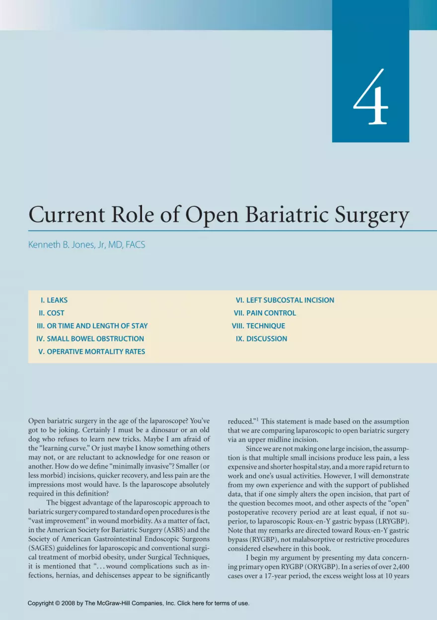
4Current Role of Open Bariatric SurgeryKenneth B. Jones, Jr, MD, FACS
I. LEAKS
II. COST
III. OR TIME AND LENGTH OF STAY
IV. SMALL BOWEL OBSTRUCTION
V. OPERATIVE MORTALITY RATES
VI. LEFT SUBCOSTAL INCISION
VII. PAIN CONTROL
VIII. TECHNIQUE
IX. DISCUSSION
Open bariatric surgery in the age of the laparoscope? You’vegot to be joking. Certainly I must be a dinosaur or an olddog who refuses to learn new tricks. Maybe I am afraid ofthe “learning curve.” Or just maybe I know something othersmay not, or are reluctant to acknowledge for one reason oranother. How do we define “minimally invasive”? Smaller (orless morbid) incisions, quicker recovery, and less pain are theimpressions most would have. Is the laparoscope absolutelyrequired in this definition?
The biggest advantage of the laparoscopic approach tobariatric surgery compared to standard open procedures is the“vast improvement” in wound morbidity. As a matter of fact,in the American Society for Bariatric Surgery (ASBS) and theSociety of American Gastrointestinal Endoscopic Surgeons(SAGES) guidelines for laparoscopic and conventional surgi-cal treatment of morbid obesity, under Surgical Techniques,it is mentioned that “. . . wound complications such as in-fections, hernias, and dehiscenses appear to be significantly
reduced.”1 This statement is made based on the assumptionthat we are comparing laparoscopic to open bariatric surgeryvia an upper midline incision.
Since we are not making one large incision, the assump-tion is that multiple small incisions produce less pain, a lessexpensive and shorter hospital stay, and a more rapid return towork and one’s usual activities. However, I will demonstratefrom my own experience and with the support of publisheddata, that if one simply alters the open incision, that part ofthe question becomes moot, and other aspects of the “open”postoperative recovery period are at least equal, if not su-perior, to laparoscopic Roux-en-Y gastric bypass (LRYGBP).Note that my remarks are directed toward Roux-en-Y gastricbypass (RYGBP), not malabsorptive or restrictive proceduresconsidered elsewhere in this book.
I begin my argument by presenting my data concern-ing primary open RYGBP (ORYGBP). In a series of over 2,400cases over a 17-year period, the excess weight loss at 10 years
Copyright © 2008 by The McGraw-Hill Companies, Inc. Click here for terms of use.

34
Part I Basic Principles
postop was 62%, comparable to several other publishedseries.2−5 The leak rate was 0.5% in primary (1◦) RYGBPand the mortality rate was 0.2%.5 A more recent report6 plusover 700 gastroplasty procedures beginning in 1979 now totalsover 3,800 primary and revision bariatric procedures. Reoper-ations in this series of 1◦ RYGBP procedures has been 1.4% 33
due to leaks, staple-line failures, incisional hernias, wound de-hiscence, and definitive surgery for peptic ulcer disease. I haveexcluded many dermatopanniculectomies, which were donefollowing successful weight loss, as well as a few cholecystec-tomies. I have used the following criteria for cholecystectomyat the time of bariatric surgery: (1) gallstones, symptomaticor not, (2) a strong family history of gallbladder disease,(3) a relatively strong family history of American Indian orLatin American heritage, or (4) cholesterolosis of the gallblad-der at the time of surgery. Using these criteria, the handfulof patients who have returned for laparoscopic cholecystec-tomy at a later date is about what one would expect from thenormal population.7
With this vast experience in bariatric surgery, why do Icontinue to prefer the procedure open? To put it simply, thereis an increased incidence of complications using the laparo-
scopic approach compared to my open approach, especiallyduring the “learning curve,” as well as a higher cost. When youcombine this with significantly less wound morbidity of theleft subcostal incision (LSI), there is simply no real advantagein doing the minimally invasive RYGBP laparoscopically.
I instigated a study along with 15 other seasoned,“open” bariatric surgeons with a combined total of 25,759cases representing over 200 surgeon years’ experience, whopooled their ORYGBP data and compared the results to theleading LRYGBP papers in the literature.6
In the overall series, our incisional hernia rate was 6.4%,using the standard midline incision and only 0.3% with theLSI. Return to surgery in less than 30 days was 0.7%, deaths0.25%, leaks 0.4%. The average length of stay (LOS) was3.4 days and return to usual activity was 21 days. Small bowelobstruction (SBO) was significantly higher with the LRYGBP,and surgical equipment costs averaged approximately $3,000less for open cases, with an added expense for longer opera-tive time. This more than makes up for the shorter LOS withthe laparoscopic approach (Table 4–1.)6 These data comparedquite favorably in comparison to the leading published seriesin the world literature.8−12
Table 4–1.
Complete Data, Orlando 2005 Group of Open Bariatric Surgeons6
Staple-linefailure orgastro-gastric Incisional Return to Return to
No. fistula (%) hernias (%) SX 30 d. Mortality Leaks ALOS work (days)
Jaroch 1106 0 210/19 17 6 1 4 21
Flanagan 1413 0 141/10 33 9 7 3.3 28
Benotti 858 58/7 ? 8 5 6 4.5 7
Afram 2076 80/4 207/10 16 8 16 2.5 12
Capella 3648 40/1 73/2 8 7 4 3 10
Wood, Kole, 7033 1/<.02 281/4 10 15 10 2.5 21Schuhknecht,Hendrick, Sapala
Cooper 984 20/2 20/2 16 2 3 4 17
Shapiro 782 1/<0.13 63/8 14 0 14 3.1 42
Lorio 619 3/0.5 93/15 0 1 0 3 28
Sweet 994 0 20/2 24 5 12 3 21
Howell 3500 11/0.3 535/15 33 2 8 4.75 28
Jones 2746 20/0.7 8/0.3 13/0.5% 5/0.2% 13/0.5% 3.2 17
25,759 234/0.9% 1641/6.6% 176/0.7% 65/0.25% 94/0.36% 3.4 21

35
Chapter 4 Current Role of Open Bariatric Surgery
LEAKS
If one looks at the data from Schauer,8 DeMaria andSugerman,9 and Wittgrove and Clark10 in their publishedseries, we see that their leak rate is almost 3%. However,Champion,11 who does gastroscopy on all of his patients dur-ing surgery, has a leak rate very similar to mine, 0.4%. Higa,12
who does a double-layer, hand-sewn anastomosis withoutstaples at the gastrojejunostomy, had only 12 leaks in his first1,040 cases (Table 4–2). We compared our leak data to thisgroup and noted a significant difference favoring our opentechnique: 2% vs 0.4%, a 500% difference ( p < .001).6 Wealso compared our mean leak rate to three other large, com-bined lap versus open retrospective combined review serieswith rates of 2%,14 2%,15 and 1.9%,16 all statistically signifi-cantly inferior to our experience of 0.36% ( p < .001).
LRYGBP requires stomach transection, which has a sig-nificantly higher incidence of leaks compared to stapling incontinuity. Kirkpatrick and Zapas in 212 patients in primarydivided ORYGBP had 13 leaks, or 6%.4 Suter et al. in 107patients in primary divided LRYGBP had a leak rate of 5%.13
Smith, Goodman, and Edwards17 doing divided ORYGBP hada leak rate of 1.8% compared to the combined experience ofLinner,18 Yale,19 and this author of 0.6%.20 However, in our2005 Orlando series, the pouch transectors’ leak rate was 0.3%compared to the stapled in continuity rate of 0.6% ( p = NS).6
Although not of clinical significance, this likely indicates theadvantages of experience or perhaps that the open linear cut-ting instruments are more efficient than laparoscopic surgery.
Why the higher incidence of leaks with LRYGBP? Inmy own experience, I have always tried to adhere to the“one-centimeter rule,” meaning that if the anastomosis ismade incorporating the staple line, there will be some ele-ment of ischemia. If staple lines are crossed at approximately
Table 4–2.
Leaks in lap vs open RYGBP
# Cases Leaks %
Schauer2 275 12 4.4
DeMaria & Sugerman9 281 11 5.1
Wittgrove & Clark4 500 13 2.5
Champion5 825 3 0.4
Higa6 1,040 12 1.22,921 51 1.75
(’04)Jones [open]7 2,421 13 0.5%p < .0012005 Orlando group 25,759 94 0.36%p < .001
[open]
90◦ angle, the risk of ischemia will be less, and if staple lines areparallel and less than 1-cm apart (the “one-centimeter rule”),or as the crossing staple lines approach parallel, there will bea higher risk of leakage.21,22 In addition to the above, if onetransects a hollow viscus, cut ends must heal and seal. How-ever, when stapling in continuity with no transection, sealingtakes place immediately and healing will occur, as there is nocompromise of blood supply to the tissue. In my entire expe-rience, I have had only one patient who has had a perforationalong the upper/lower pouch staple line in a 1◦ ORYGBP. Thiswas an individual who had a previously undiagnosed insuli-noma and a seizure on her fourth postop day, which rippedholes in the proximal and distal pouches at the staple line, aswell as causing a 180◦ disruption at the gastrojejunostomy. Atreexploration, there was no evidence of ischemia, with goodbleeding of the edges that had perforated.
Technical error at surgery is a rare cause of postop leakssince “leak tests” are standard prior to completion of our op-erations. Therefore, leaks are primarily caused by ischemiaand/or excess tension at the anastomoses, plain and simple.Do laparoscopic surgeons ignore the “one-centimeter rule,”causing more ischemia and necrosis? Does the abandonmentof the retrocolic approach to the gastrojejunostomy to de-crease SBOs increase tension at the anastomosis? These arequestions needing answers before the safer, time-tested opentechnique can be legitimately abandoned.
COST
The laparoscopists argue that patients get out of the hospitalsooner. In several published series,6,8,10 this appears to be thecase, that is, about 2 rather than 3 days, saving approximately$1,000. However, I have never seen a leak that was clinicallymanifested in the first 72 hours after surgery. Should the pa-tient be released in 1 or 2 days to return home several hundredmiles away, a leak could be catastrophic. I compared equip-ment costs with laparoscopic versus open RYGBP in one of mytwo hospitals, and found that laparoscopic equipment costsapproximately $5,200 versus the open stapling equipment ofabout $1,700, or a $3,500 difference. Two other contributingsurgeons in our Orlando 2005 study6 compared their open ex-perience to LRYGBP relative to cost: Howell in North Dakota(+$2,900) and Sapala in New York City (+$3,400). Hospi-tals will probably double that cost to make a profit, mak-ing a difference to the patient of about $6,000. Subtracting$1,000 for one less hospital day, and adding the additionalOR time at $1,250 per hour, the total charge to the patient isgoing to be about $7,000 more for LRYGBP. Assuming that atleast half of the 140,000 bariatric cases in 2004 were LRYGBP(ASBS estimate), at an added hospital cost of $7,000/casemultiplied by 70,000, this results in one half billion dollarsin added overall cost in the United States, which has reducedsurgical access due to third-party reimbursement backlash. Itis no wonder the insurance carriers are balking and addingnuisance red tape, like mandatory preoperative 6–12 monthMD-supervised dieting, which we know is only a delay tactic

36
Part I Basic Principles
to deny access, and enhance their bottom lines. This becomesa stewardship issue for us.
OR TIME AND LENGTH OF STAY
Paxton and Matthews found that LRYGBP requires an extraone and a half hours in the OR compared to ORYGBP. Theyalso noted an average LOS of two and a half days for LRYGBPcompared to our 3.4 days. It is also interesting to note that inthe ORYGBP group, the patients averaged 20 pounds heav-ier, indicating that the more technically difficult, higher BMIpatients undergo open surgery more frequently than laparo-scopic surgery.16 From a strictly practical standpoint, if I amgoing to spend less time per case in the OR as the laparoscopicgroup, and since there is little difference in reimbursement foreither approach, time will allow higher volume with a signif-icantly positive effect on my reimbursement compared to thelaparoscopic group.
SMALL BOWEL OBSTRUCTION
Higa23 and Podnos’14 studies totaling 4,887 patients revealeda postop SBO rate of 3%. Capella in the Orlando 2005 studycompared 483 LRYGBP patients to 739 ORYGBP patients andfound a return to surgery for SBO rate of 7.45% in the for-mer group, 0 in the latter.6 However, in a previous publishedreport of 1,174 patients I found 7 (0.6%) in my patient pop-ulation, with only one due to an internal hernia (<0.1%),three due to retrograde intussusception, two from adhesionsof previous surgery, and one from a spontaneous bowel wallhematoma.24 All of these results are statistically significant(p ≤ .001) (Table 4–2).
OPERATIVE MORTALITY RATES
Comparing our group of 25,759 open cases6 to Podnos’ 3,464LRYGBP cases, and the International Bariatric Surgery Reg-istry (IBSR) 2002 pooled data of 13,554 cases, mortality rateresults were virtually the same: 0.25%, 0.23%, 0.27%, respec-tively ( p = NS). Podnos’ open cases revealed a significantlyhigher mortality rate (0.87%). IBSR data had an LRYGBPmortality rate of only 0.15%, lower than the four studies men-tioned above.
LEFT SUBCOSTAL INCISION
Again, when we compare LRYGBP to open, the traditionalassumption is that open procedures are being done throughmidline incisions. However, when one compares my resultsas well as those of Alvarez-Cordero, using the LSI, and severalother published series, our incidence of incisional hernia is38 times less than those series done through a large midlineincision (Table 4–3).25−29 Also, simply stated, muscle has amuch better blood supply and heals considerably better thanrelatively thin midline fascia.
Table 4–3.
Incisional Hernias, Vertical vs LSI29
No. ofprocedures Hernias
Mason, Amoral, Sugerman,Alvarez-Cordero,[vertical: midline]
1,147 87 (7.6%)[38× more]
Alvarez-Cordero, Jones 2,200 4 (0.2%)[LSI]
Why is my incisional hernia rate so low when tradi-tional bariatric surgical patient follow-up is so poor? I use asampling technique. For instance, all bariatric patients seenfor a 4-month period in 1996 who came to the clinic fora variety of reasons, primarily follow-up RYGBP anywherefrom 1–10 years, were “spot checked” for hernias. We ex-amined 173 consecutive patients and found no hernias. Inanother study, the incisional hernia rate was 5/1,367 (0.4%).5
Our wound morbidity was 2.2%.29 More recently, examiningmy data from November 2003 through November 2004, 720consecutive patients were checked during routine visits, with5 hernias (0.7%) found.
If one compares the wound morbidity of several of theLRYGBP series to my ORYGBP–LSI experience, it is easy to seethat the rate of hernias and other wound morbidity is actuallyless than with the laparoscopic approach.8−10 I frequentlytell my patients that in addition to the higher complicationrate and cost of LRYGBP, you may choose a procedure donethrough a single 7-inch incision, or several incisions equaling7 inches, take your choice. Both heal equally well.
It is interesting that in the pre-laparoscopic era, inci-sional hernias were rarely mentioned, were considered almosttotally harmless with rare incarcerations, and although un-sightly, they could afford an opportunity for the patient toreturn to the operating room for a repair and have a relativelyinexpensive panniculectomy done at the same time, which or-dinarily would be covered by the third-party insurance payer.In the present day laparoscopic era, an incisional hernia is con-sidered to be very undesirable, as the absence of same justifiesLRYGBP’s almost negligible incisional morbidity rates. Forthese reasons, the LSI has not achieved widespread popularity.
As a preceptor for the ASBS, two of my preceptees werequite accomplished laparoscopic bariatric surgeons. Theircombined series of 476 cases demonstrated an incisional her-nia rate with the LSI of 1.5% compared to 7.6% when onelooks at the combined series mentioned above.25−28
PAIN CONTROL
At our Bariatric Surgery Center of the Mid South atCHRISTUS Schumpert Medical Center in Shreveport, LA,

37
Chapter 4 Current Role of Open Bariatric Surgery
the pain control staff has indicated there is no real differencein the pain endured by either approach—an anecdotal con-clusion, but what subjective studies are not?
TECHNIQUE
My procedure is a modification of the Oca–Torresprocedure,30 with the following changes: (1) an LSI, (2) theTA-90B four row Autosuture stapler fired two times, rein-forced proximally and distally with ligaclips, (3) a verticalpouch, with no short gastric vessels being taken down and notransection of the pouch, stapled in continuity, (4) a retrocolicantegastric gastrojejunostomy, hand-sewn in two layers, uti-lizing a #38 Mercury bougie (13 mm in diameter) for accuratestoma sizing, and (5) an enteroenterostomy that is done uti-lizing linear cutting and stapling instruments, a gastrojejunallimb of 75–150 cm, and biliopancreatic limb of 60–100 cm,depending on the patient’s BMI according to the recommen-dations of Brolin.31
I pay particular attention to the following: (1) “one-centimeter rule,” (2) care in transection and freeing up ofjejunum and the need for adequate length and freedom ofthe distally transected jejunum to avoid tension at the gastro-jejunostomy, (3) care in freeing up the EG junction to avoidperforation and ischemia, (4) a leak test (I prefer the “air bub-ble” test), and (5) I frequently use gastrostomy tubes in thebypassed stomach: in apple-shaped men; in long, difficult re-visions; in patients with diabetes mellitus to avoid problemswith diabetic gastroparesis; frequently with BMI’s greaterthan 50 and always when greater than 60; in all JI bypassconversions; and in patients with marginal pulmonary status.
Because of the higher incidence of SBO that hasbeen reported with the laparoscopic approach compared toopen,6,12,23,32 I always take special care to make an adequatetransverse colonic mesenteric opening and secure it to theRoux limb prior to closure to prevent herniation. I also do anadequate closure of Peterson’s hernia as well as the potentialhernia associated with the enteroenterostomy, which may bemuch more difficult or even ignored laparoscopically. Thelack of significant adhesion formation may have an effect onthe lack of sealing after laparoscopic closure. Following dis-charge from the hospital, I insist that the patients stay on asemi-soft “gooey” liquid diet for 6 weeks postop. In addi-tion to the above, I try to preserve all blood supply possible,and avoid usage of the electrocautery near hollow viscera andstaple-lines, as necrosis and leaks may follow several days later.
If one feels that it is necessary to transect the stomach inorder to get adequate freedom of the proximal gastric pouchto reduce tension at the gastrojejunostomy, I instead use the“Jones stitch” from the lower end of the staple-line to thelower lip of the LSI fascia, which effectively and safely pullsthe pouch into the operative field. However, transection issometimes necessary to get adequate length of the proximalpouch, especially with revision procedures.21,22
Relative to pouch transection, many felt about 15 yearsago that this was always necessary because of the inordinately
high incidence of staple-line disruption and secondary gas-trogastric fistulae. Capella33 noted that when stapled in conti-nuity with no transection, disruption of the staples occurredin as much as 23% of the patient population. However, Pories’group34 demonstrated that there was a 6% gastro-gastric fis-tula rate when dividing pouches, while Capella noted 2% inhis series.
One might ask, “How do I know that my staple linefailure rate is less than 1%?” In a study done several yearsago, in 650 patients, my assumption was that my staple-linefailure rate at that time was 0.6% with the double applicationof the TA-90B four row Autosuture stapling instrument.I came to this conclusion by UGI series examination of160 voluntary asymptomatic post-ORYGBP patients andfound one staple-line failure. In 19 symptomatic patients(GERD or rapid weight regain) during this period of time,their UGIs revealed four staple-line failures.35 When patientshave problems, they come back. I revisited this again fromNovember 1999 to April 2003 in a series of 724 patients, inwhom we did UGI series on 62 symptomatic patients andfound only five staple-line failures. Again, this samplingtechnique indicated the staple-line failure rate was less than0.7% (5/724).
DISCUSSION
“Lap RYGBP . . . is the most technically demanding of alllaparoscopic operations. Consequently, many programsreserve LRYGBP procedures for surgical attendings andfellows, while ORYGBP procedures are more frequently per-formed or assisted by surgical residents. Undoubtedly, thisplays a role in the variation between open and lap com-plication rates . . . therefore, there is a significant (learningcurve) . . . attributed to (one’s first) 200 LRYGBP.”16 I couldnot say it any better. Podnos, in a retrospective review of theliterature, compared all reported series greater than 50 casesof lap or open RYGBP at the time. Age and BMI were similar,in the 8 open and 10 laparoscopic series studied. He foundsignificant differences favoring the open technique in gas-trointestinal hemorrhage, SBO, and stomal stenosis. Woundproblems were significantly greater in the open approach viathe standard upper midline incision. There was no signifi-cant difference, although a trend toward fewer leaks with theopen technique.14 This discussion adds to the argument thatORYGBP still stands the test of time quite well.
There is a perception that laparoscopic bariatric surgeryis patient driven, but would a patient opt to have a proceduredone by a new bariatric surgeon during his or her “learn-ing curve,” if they knew the significantly higher risk? Higa,12
Wittgrove,10 and Schwarz and Drew (see Chapter 17 in thisvolume) confirm better results with an increase in experi-ence. We therefore should not succumb to the pressure of ourpatients and industry and their perception that laparoscopicbariatric surgery is always easier and better, for I have demon-strated that there is indeed a higher complication rate andgreater cost, with the anticipated end point virtually the same.

38
Part I Basic Principles
The three large comparative studies mentioned in thischapter14−16 are at best level III laparoscopic versus opengastric bypass combined series, very much like comparingapples to oranges. In these principally retrospective compar-isons with no controls in which there is considerable bias,as most bariatric surgeons would agree that open gastric by-pass is easier than laparoscopic, the more difficult cases werefrequently reserved for the open technique, that is, males, pa-tients with high BMIs, and revisions. Subsequently there werehigher complication rates in the open group. This consider-ably reduces the credibility of these comparative studies andtheir conclusions that LRYGBP is superior to ORYGBP.
The primary justification for the laparoscopic approachto any type of surgery is based on the assumption that there ismuch less wound morbidity than in the open technique. Thisexperiment has been going on for over 15 years with inguinalhernias, and there appears to be no difference in efficacy ifone does lap or open, and also long-term studies indicate thatrecurrences in laparoscopic repairs are at a higher rate thanin the open.36
Colectomies are done laparoscopically, but the trendnow appears to be shifting to indicate that it may be bestto do them utilizing hand-assisted techniques, as an incisionabout the size of a human fist is necessary in order to removethe resected colonic lesion from the abdomen, so why nottake advantage of better tactile feeling?37,38
Nguyen and associates are champions for LRYGBP, withthe primary argument that wound morbidity will be virtu-ally avoided. There are no comparative studies in the litera-ture comparing LRYGBP to open RYGBP–LSI until recently.6
They agree that there is a significantly higher incidence of in-ternal hernias associated with SBO utilizing LRYGBP, as wellas gastrointestinal hemorrhage and stricture requiring fur-ther intervention, and that the cost is greater while utilizingthe lap technique, with no real difference in the ultimate qual-ity of life.39 While participating in the ACS Bariatric SurgeryPrimer in October 2004 (New Orleans), Dr. Nicola Scopinaro,the father of biliopancreatic diversion (BPD), stated, “I pre-dict that in Italy we will soon cease doing laparoscopic BPDand RYGBP in favor of open, due to the added cost, whichcan’t be made up in shorter hospital lengths of stay” [para-phrased] [(personal communication)].
Revisions can be done laparoscopically, but shouldthey? I reviewed the literature for a paper on revisions re-cently and it appears feasible, but is it practical? The Emorygroup’s experience is an example of this. Thirty-nine revisionswere done, 18 open. Twenty one were begun laparoscopi-cally, but 10 converted to open due to technical problems. Ofthe 11 remaining lap cases, one died (9%). No deaths wereseen in the open cases.40 In three other laparoscopic series(64 patients), only 9% experienced major complications, butthe average OR time was four and a half hours.41−43 Is it worththe even greater learning curve for laparoscopic revisions, ifthe LSI takes wound morbidity out of the equation? Lap re-visions are being done more frequently (see Chapter 15), butwhy struggle so hard to achieve the same or inferior results(more leaks, etc.)?
Why are we so rapidly adopting laparoscopic RYGBP,when we know that there is a higher risk of leaks, a greaterlearning curve with a higher risk of medical malpractice ex-posure, earlier discharge in less than 3 days when leaks arerarely manifested before that time, and when there is anadded expense of about $7,000 per case? We also know thatour patients’ return to work is directly proportional to theirmotivation,6 as a very small percentage actually do such vig-orous labor that they must be off for 6 weeks.
In spite of these arguments, I am not so naıve to believethe horse is not out of the barn. So, if one feels compelledto do exclusively LRYGBP, I would recommend the follow-ing: (1) a basic background with considerable experience inadvanced laparoscopic surgery; (2) be exposed to at least10 open bariatric procedures, preferably RYGBP, (3) takea laparoscopic bariatric surgery mini-fellowship if one hasnot had considerable residency training in same, and (4)use the Schwarz-Drew philosophy (see Chapter 11): Theyhad a vast experience with open gastric bypass, then tooka short course in laparoscopic bariatric surgery, oper-ated on several pigs, then did open RYGBP using laparo-scopic instruments before they did their first LRYGBP, andtheir results have been comparable to their previous openexperience.
On the basis of above comparative data, the usual ar-guments suggesting the superiority of the LRYGBP appearflawed. If we take the vertical incision out of the equation,assuming no difference in wound morbidity, we have in theLRYGBP significantly more leaks, GI hemorrhage, SBO, andstomal stenosis, markedly increasing the reoperation rate.This along with the higher cost and a higher learning curve,all contributes to higher medical malpractice risks. I questionthe wisdom of discharge at 2.5 days or less, as most leaks arenot apparent until 3+ days postop. Well-motivated open pa-tients return to work or normal activities in 3 weeks or less.6
If we sincerely believe it is truly in our patients’ best inter-est to use LRYGBP technique, it is an acceptable modality.If we do so merely to get ahead of our competition acrosstown in spite of our knowledge of greater LRYGBP morbid-ity, and we have not made the effort to get the proper skillsand experience, then we need to reexamine our priorities andmotivations.
In conclusion, these data indicate no real advantage oflaparoscopic over open RYGBP, and really quite the opposite.If the incision is really the issue, we should change it to ei-ther the LSI or mini-midline. Then we would have a safer,less expensive, MUCH EASIER, equally effective procedure;and truly “minimally invasive.” In spite of all the evidenceI have presented herein, I know that many, if not most, ofus will be compelled by our patients, competition, referringMDs, and industry to abandon open in favor of lap RYGBP.Because the ORYGBP–LSI is easier to do, especially in themore difficult cases, including heavier patients, males, andrevisions, it must always be available as a reasonable option.Above all, remember the (Dr. Eddie) Reddick Rule: “If you aremaking no progress laparoscopically for 15 minutes, OPEN;and get ’r done” [paraphrased].

39
Chapter 4 Current Role of Open Bariatric Surgery
References
1. Guidelines for Laparoscopic and Open Surgical Treatment of MorbidObesity. (Document adopted by the American Society for BariatricSurgery and the Society of American Gastrointestinal EndoscopicSurgeons, June 2000). Obes Surg 10:378–379, 2000.
2. Pories WJ, Swanson JS, MacDonald KG, et al.: Who would havethought it? An operation proves to be the most effective therapy foradult onset diabetes mellitus. Ann Surg 222:339–350, 1995.
3. DeMaria EJ, Sugerman HJ, Kellum JM, et al.: Results of 281 consec-utive total laparoscopic Roux-en-Y gastric bypasses to treat morbidobesity. Ann Surg 235:640–647, 2002.
4. Kirkpatrick JR, Zapas JL: Divided gastric bypass: A fifteen-year ex-perience. Am Surg 64 (1):62–66, 1998.
5. Jones KB: Experience with the Roux-en-Y gastric bypass, and com-mentary on current trends. Obes Surg 10:183–185, 2000.
6. Jones KB, Afram JD, Benotti PN, et al.: Open Roux-en-Y gastricbypass versus laparoscopic: A comparative study of over 25,000 opencases and the major laparoscopic bariatric reported series. In press.
7. Jones KB: Simultaneous cholecystectomy: to be or not to be. ObesSurg 5:52–54, 1995.
8. Schauer PR, Ikramuddin S, Gourash W, et al.: Outcomes after la-paroscopic Roux-en-Y gastric bypass for morbid obesity. Ann Surg232:515–529, 2000.
9. Sugerman HJ, Kellum JM, Engle KM: Gastric bypass for treatingsevere obesity. Am J Clin Nutr 55(Suppl 12):560S–566S, 1992.
10. Wittgrove AC, Clark GW: Laparoscopic gastric bypass, Roux-en-Y—500 patients: Technique and results, with 3–60 month follow-up.Obes Surg 10:233–239, 2000.
11. Champion JK, Hunt T, DeLisle N: Role of routine intraoperative en-doscopy in laparoscopic bariatric surgery. Surg Endosc 16(12):1663–1665, 2002.
12. Higa KD, Bone K, Ho T: Complications of the laparoscopic Roux-en-Y gastric bypass: 1,040 patients—what have we learned? Obes Surg10:509–513, 2000.
13. Suter M, Giusti V, Heraif E: Laparoscopic Roux-en-Y gastric bypass:Initial 2-year experience. Surg Endosc 17(4):603–609, 2003.
14. Podnos YD, Jimenez JC, Wilson SE, et al.: Complications after laparo-scopic gastric bypass—a review of 3464 cases. Arch Surg 138:957–961,2003.
15. Baker RS, Foote J, Kemmeter P, et al.: The Science of stapling andleaks. Obes Surg 14:1290–1298.
16. Paxton JH, Matthews JB: The cost effectiveness of laparoscopic versusopen gastric bypass surgery. Obes Surg 15:24–34, 2005.
17. Smith SC, Goodman GN, Edwards LB: Roux-en-Y gastric bypass: Aseven year retrospective review of 3,855 patients. Obes Surg 5:314–318, 1995.
18. Linner JH: Surgery for Morbid Obesity. New York, Springer-Verlag,1984; p. 97.
19. Yale CE: Gastric surgery for morbid obesity. Arch Surg 124:941–946,1989.
20. Jones KB: The double application of the TA-90B four row staplerand pouch formation: Eight rows are safe and effective in Roux-en-Ygastric bypass. Obes Surg 3:262–268, 1994.
21. Jones KB: Revisional bariatric surgery—potentially safe and effective.Surg Obes Relat Disord 1(6):599–603, 2005.
22. Jones KB: Revisional bariatric surgery—safe and effective. Obes Surg11:183–189, 2001.
The author acknowledges the contributions of Ms. Kimberly King for tech-nical preparation of the manuscript and Ms. Sue Wainwright and Ms. DonnaFoshee for data retrieval and analysis.
23. Higa KD, Ho T, Boone K: Internal hernias after laparoscopic Roux-en-Y gastric bypass: Incidence, treatment and prevention. Obes Surg13:350–354, 2003.
24. Jones KB: Biliopancreatic limb obstruction in gastric bypass at orproximal to the jejuno-jejunostomy: A potentially deadly, catas-trophic event. Obes Surg 6:485–493, 1996.
25. Mason EE: Surgical Treatment of Obesity. London, WB Saunders,1981; pp. 340–341.
26. Sugerman HJ, McNeill PM: Continuous absorbable vs. interruptednon-absorbable suture for mid line fascial closure. In: Proceedingsof the Second Annual Meeting of the American Society for BariatricSurgery, Iowa City, IA, 1985; pp. 153–154.
27. Amaral JF, Thompson WR: Abdominal closure in the mor-bidly obese. In: Proceedings of the Third Annual Meeting of theAmerican Society for Bariatric Surgery, Iowa city, IA, 1986; pp. 191–202.
28. Alvarez-Cordero R, Aragon-Virvette E: Incisions for obesity surgery:A brief report. Obes Surg 1:409–411, 1991.
29. Jones KB: The left subcostal incision revisited. Obes Surg 8:225–228,1998.
30. Torres JC, Oca CF, Garrison RN: Gastric bypass Roux-en-Y gastro-jejunostomy from the lesser curvature. Sou Med J 76:1217–1221,1983.
31. Brolin RE, La Marca LB, Kenler HA, et al.: Malabsorptive gastricbypass in patients with super obesity. J Gastrointest Surg 6(2):195–205, 2002.
32. Courcoulas A, Perry Y, Buenaventuro P, et al.: Comparing the out-comes after laparoscopic versus open gastric bypass: A matchedpaired analysis. Obes Surg 13:341–346, 2003.
33. Capella JF, Capella RF: Staple disruption and marginal ulceration ingastric bypass procedures for weight reduction. Obes Surg 1:44–49,1996.
34. Cucchi SGD, Pories WJ, MacDonald KG, et al.: Gastro-gastric fistulas,a complication of divided gastric bypass surgery. Ann Surg 221:387–391, 1995.
35. Jones KB, Homza W, Peavy P, et al.: Double application of the TA-90Bfour-row autosuture r© stapling instrument: A safe, effective methodof staple-line production indicated by follow-up GI series. Obes Surg6:494–499, 1996.
36. Grunwaldt LJ, Schwaitzberg SD, Rassner DW: Is laparoscopic in-guinal hernia repair an operation of the past? J Am Coll Surg200(4):616–620, 2005.
37. Rivadeneira DE, Marcello PW, Roberts PL, et al.: Benefits of hand-assisted laparoscopic restorative proctocolectomy: A comparativestudy. Dis Colon Rectum 77:1371–1376, 2004.
38. Chang YJ, Marcello PW, Rusin LC: Hand-assisted laparoscopic sig-moid colectomy: Helping hand or hindrance? Surg Endosc 19:656–661, 2005.
39. Nguyen NT, Ho HS, Palmer LS, et al.: A comparison study of laparo-scopic versus open gastric bypass for morbid obesity. J Am Coll Surg191(2):149–157, 2000.
40. Khaitan L, Van Sickle K, Gonzalez R, et al.: Laparoscopic revi-sion of bariatric procedures: Is it feasible? Am Surg 71(1):6–10,2005.
41. Weber M, Muller MK, Michel JM, et al.: Laparoscopic Roux-en-Ygastric bypass, but not rebanding, should be proposed as a rescueprocedure for patients with failed laparoscopic gastric banding. AnnSurg 238(6):827–833, 2003.
42. Gagner M, Gentileschi P, de Csepel J, et al.: Laparoscopic reoperativebariatric surgery: Experience from 27 consecutive patients. Obes Surg12:254–260, 2002.
43. McCormick JT, Papasavas PK, Caushaj PF, Gagne DJ: Laparoscopicrevision of failed open bariatric procedures. Surg Endosc 77(3):413–415, 2003.

5Central Nervous System Regulationand Hormonal SignalingCid Pitombo, MD, PhD, TCBC
I. INTRODUCTION
II. HISTORY
III. CNS REGULATION
IV. GASTROINTESTINAL HORMONESAND FOOD INTAKECholecystokininGhrelinOxytomodulinGlucagon-Like Peptide-2Peptide Tyrosine-Tyrosine3−36
Apolipoprotein A-IV
V. INCRETINGlucose-Dependent Insulinotropic PeptideGlucagon-like-peptide-1
VI. ADIPOSE TISSUELeptinAdiponectinResistinVisceral Fat’s Relationship with Comorbidities
VII. SURGERY EFFECTS ON INTESTINAL HORMONES
VIII. OBESITY SURGERY AND ADVANCES IN DIABETESTREATMENT
IX. CONCLUSIONS
INTRODUCTION
It is essential that we understand the normal physiologicalmechanism that makes us feel the hunger and the mechanismsof absorption and control of ingested food amounts as well asthe consequences produced by weight gain as adipose tissuesincrease and the development of many kinds of diseases. Theneed to eat involves a sequence of control mechanisms. Eatingis necessary for a variety of our organs’ functions, be it growthor absorption of micronutrients like vitamins and minerals
and macronutrients like carbohydrates, fat, proteins, and wa-ter. Obesity surgery radically modifies this entire system, andunderstanding the consequences of these modifications is es-sential.
What do we know today?We understand that our body has several mechanisms
to control food ingestion and absorption. We also know thatbasically the central nervous system (CNS), through the hy-pothalamus, fatty tissue, and the signals produced by a varietyof hormones in our digestive system, provides this control.
Copyright © 2008 by The McGraw-Hill Companies, Inc. Click here for terms of use.

42
Part I Basic Principles
Table 5–1.
Afferent Signals
Signals coming from digestive tractGhrelinGLP-1GIPPYY3-36CCK
Afferent signals descendant from fat tissueLepitinAdiponectin
Afferent signals descendant from pancreasInsulin
The majority of obesity surgery techniques involve aradical alteration in the digestive tract, therefore resulting ina totally different way of intestinal hormone stimulation andproduction. With weight loss, there is a complete modifica-tion in the morphology and physiology of the adipose tissues,be it visceral or parietal (it is now well-known that this tissueis a powerful “endocrine organ”). The CNS, obviously, willalso receive and produce these signals differently. In otherwords, obesity surgery is not merely a surgery technique, buta metabolic surgery that also implicates a variety of undis-covered mysteries.
The system that controls these mechanisms has bothafferent and efferent signals. The afferent ones will be deter-mined by many stimuli beyond the need or surplus of energyaccumulation and may or may not work together (see Tables5–1 and 5–2). The CNS and, specifically, the hypothalamuswill manage the efferent signs, mainly the need or not for usto accumulate or burn more energy, as well as our hunger(this topic will be discussed in detail below).
Since survival is more acutely threatened by starvationthan obesity, it should come as no surprise that this systemis more robustly organized to galvanize in response to defi-cient energy intake and stores than to excess energy.1 Sincethis complex protection system will only engage after obe-
Table 5–2.
Efferent Signals
Appetite
Energy expenditure
Food intake
Satiety effects
Glucose levels
sity surgery, it is essential that we continue to increase ourunderstanding of it.
Today we are aware of, and also study, a huge diversity ofmechanisms produced in consequence of many varied tech-niques, (these will be discussed in specific chapters). Here wewill discuss the historical factors and current research linesthat are being developed in the bariatric surgery field, dividedbasically into understanding intestinal hormones and theirmany functions, fat tissue metabolism and weight loss con-sequences, interaction of the diabetes “cure” and the effectsof bariatric surgery, and, finally, CNS control.
HISTORY
In 1902, William M. Bayliss and Ernest H. Starling2 publishedtheir paper “The mechanism of pancreatic secretion.” Thiswas the bird hour of gastrointestinal endocrinology. Mooreet al.3 describe that Starling had already considered the possi-bility that the duodenum does also supply a chemical excitantfor the internal secretion of the pancreas. Not until the discov-ery of insulin by Banting and Best in 1921, a systematic searchfor a gut hormone influencing carbohydrate metabolism tookplace. Different groups published the results of animal exper-iments, in which the effect of extracts of duodenal mucosaon fasting blood glucose levels or hyperglycemia induced byglucose ingestion or injection had been investigated.
In 1932, La Barre4 published for the first time the name“incretine” (incretin) for the substance extracted from theupper gut mucosa, which produces hypoglycemia and doesnot stimulate pancreatic exocrine secretion. He introducedan idea that considered the use of incretin for the treatmentof human diabetes for the years to come. Since several groupsdemonstrated that the plasma insulin level increased afterthe injection of an extract of intestinal mucosa, all knowngastrointestinal hormones were considered either alone or incombination as possible incretin candidates. In 1970, JohnC. Brown discovered a new peptide which he named gastricinhibitory polypeptide (GIP) because of its inhibitory effecton gastric acid secretion in Heidenhain pouch dogs.5
Dr. Joel Habener discovered the glucagon-like peptide-1 (GLP-1) in 1980. The interest in GLP-1 and the other pre-proglucagon derived peptides has risen almost exponentially,as seminal papers in the early 1990s proposed to use GLP-1 agonists as therapeutic agents for the treatment of type 2diabetes.
In 1994, the ob gene was discovered by Zhang et al.,36
and the leptin receptor, first reported in Cell, was discoveredin 1995 by Tartaglia.37 These discoveries have initiated a greathope in the development of anti-obesity drugs.
Obesity surgery has also been around for a long time.Since the fifties, techniques to approach this population havebeen known and over four decades many approaches havebeen “experimented” (see Chapter 1), but very little was stud-ied or understood in relation to their real physiologic effects.Twenty years following jejunoileal bypass, patients had sig-nificant intestinal hormonal changes. These findings there-fore support a role for gut hormones as important mediators

43
Chapter 5 Central Nervous System Regulation and Hormonal Signaling
in the hypophagia and weight-reducing effects of bariatricsurgery.6−8 The surprise came in 1982, in the early days ofbariatric surgery, when our diabetic morbidly obese patientswere relieved, yes, fully relieved, from their insulin require-ments in a matter of days.9
In 1991, after a National Institutes of Health (NIH)consensus,10 the surgical techniques were standardized andthe indications were organized. From that moment, a greatincrease in the number of procedures occurred and, con-sequently, a more refined development of the effects theycaused. But what does 15 years mean to Medicine? Very lit-tle. About 10 years after the NIH, Dr. Cummings describedthe Ghrelin peptides (see Chapter 15). It is the only intesti-nal hormone known to circulate orexigen (appetite stimu-lant). And now, it is five years later. The effects from obe-sity surgery on reversing important diseases such as diabetesled to the development of several techniques that take ad-vantage of these studies to try to promote a cure for thispopulation.
CNS REGULATION
Gastrointestinal signals that influence the brain to stop anongoing meal are collectively called satiety signals becausewhen they are administered exogenously, animals behave asif they are sated, i.e., they eat smaller meals and engage inbehaviors that normally occur when meals end.11,12 Humansgiven the same compounds also eat smaller meals and reportthat they are more sated.13−15
During the last 10 years, great progress has beenachieved in the characterization of the molecular mecha-nisms involved in the control of food ingestion and energywastage. The hypothalamus is the region that integrates andcoordinates the signals influencing the energy balance. In ad-dition to signals from neuropeptides and neurocytokines, hy-pothalamic centers involved in energy homeostasis can also beinfluenced by metabolic substrates. Neurons that receive sig-nals from regulators such as leptin may also sense changesin the levels of glucose and free fatty acids. Hypothalamicneurons that sense both low and high levels of glucose havebeen identified.16 Many genetic studies have been trying todemonstrate the circuit action that delivers this operation.Many signs coming from the digestive tract through incretins,GLP-1, and glucose-dependent insulinotropic peptide (GIP),and intestinal hormones like peptide YY3−36(PYY3−36) andghrelin, as from the fat tissue by leptin, resistin, andadiponectin, besides organs like pancreas and by insulin,promote a variety of responses (efferent signs) by hypothal-amus (see Tables 5–1 and 5–2). Each one of these signalshas a response from the hypothalamus and will be detailednext. Several surgical techniques have promoted a true shortcircuit in this signalization. All procedures resulting in in-duced weight loss reduce the parietal fat and visceral fat,leading to a modification on signalization produced by leptin,adiponectin, and resistin.
An important component of the body weight regula-tory systems is melanocortin receptors (MR). The ligands
for these receptors’ signaling result in weight gain, e.g., agoutimice are obese because of decreased melanocortin signaling.17
Analogous to the situation with Leptin,18 Qi et al. found thatagouti mice were insensitive to the actions of adiponectinin the brain. This suggests that leptin and adiponectinshare common mediators in the CNS.19 However, leptinand adiponectin elicit different changes in gene expressionfor ligands of MR.19 Consistent with this difference, lep-tin decreases food intake and increases energy expenditure,whereas adiponectin seems to only affect energy expenditure(Figure 5–4).
Cholecystokinin (CCK) is liberated by the small in-testine into the circulation in response to the contact withnutrients such as fatty acids, and influences satiety by ac-tions on CCK receptors located on peripheral vagal afferentterminals, which transmit neural signals to the brainstem.20
The true effects of this hormone in patients submitted tosurgeries that promote duodenal bypass are still unknown.The absence of nutrient passage through the duodenum and,therefore, the lack of stimulus to its production apparentlymarkedly decrease its effects after these surgeries.
The surgeries that involve duodenal exclusion, likeRoux-en-Y gastric bypass (RYGB) (Figure 5–1) and bil-iopancreatic diversion (BPD) (Figure 5–2), lead to an earlysignalization of distal intestinal hormones like GLP-1 andPYY3−36, with this being one of the most studied and de-fended factors as responsible for the best results in diabetes
Figure 5–1. Roux-en-Y gastric bypass.

44
Part I Basic Principles
Figure 5–2. Biliopancreatic diversion.
resolution and weight loss in the long term, when compared topurely restrictive methods. Ghrelin modifies its signaling, es-pecially after RYGB, since it is mostly produced in the gastricfundus, which is excluded from the digestive tract in thiscase.
What really happens and what are or will be the conse-quences of that situation in the hypothalamus signalizationstill remain unknown. Is any neurological alteration shownby this population perhaps related to this signalization? Howmany more afferent and efferent signals will be produced
after obesity surgery? Does the hypothalamus perhaps inter-pret and answer the “new” signals produced after the surgeryin the same way?
GASTROINTESTINAL HORMONES ANDFOOD INTAKE
The many techniques in obesity surgery, which involve mod-ification in the digestive tract, like the RYGB and BPD lead,consequently, to a true short circuit in the production of in-testinal hormones as well as the signalization to the CNS.Understanding the good and bad consequences of this fact isa priority for us. Finding out what we are really doing withthis population is our duty as doctors.
The contemporarily most commonly performedsurgery in the United States and Brazil, the RYGB, involvesboth restrictive and malabsorptive features. A considerablesegment of small intestine is bypassed, resulting in a muchshorter gastrointestinal tract (Figure 5–1). Well, it is funda-mental to understand that, each time we eat, we produce avariety of stimuli, both mechanical and chemical in the en-teroendocrine cells, and as a consequence, the productionof substances, be they hormones, enzymes, vitamin absorp-tion factors, etc., lead to many effects in the signals to theCNS, as well as in the food digestion and absorption of sub-stances fundamental to our existence, like vitamins, iron, etc.(see Chapter 39). The surgeries that modify this “natural”passage obviously confound this entire system. What reallydoes happen? What effects are produced? We are startingto answer some of these questions, but the more we study,the more questions appear. We will try to explain a littleof what we already understand about intestinal hormones(Figure 5–3).
Figure 5–3. Normal afferent signals.
45
Chapter 5 Central Nervous System Regulation and Hormonal Signaling
Digestion and absorption of nutrients are time-intensive processes needed to protect against malabsorptionand metabolic disturbances during the postprandial period.This includes the fine-tuned interaction of gastrointestinalnutrient transit, endocrine and exocrine secretion, and gut–brain responses like the induction of satiety or the feeling offullness, bloating, or nausea. The interplay between gastroin-testinal transit and endocrine pancreatic secretion plays a keyrole in order to optimize nutrient digestion and absorption.21
The most studied intestinal hormones in obesity surgery to-day are ghrelin, CCK, GIP, GLP1, PYY3−36, and oxytomodulin(OXM).
CholecystokininThis is one of the most studied hormones. It is a powerful sati-ation trigger and is secreted primarily in two forms: CCK-33,also known as alimentary (CCK-A or CCK-1), from duo-denum and jejunum cells, and CCK-8, the cerebral (CCK-Bor CCK-2) made in the CNS. That produced in the duode-num has very well defined functions in the digestive pro-cess, like stimulating gallbladder emptying, pancreatic en-zyme secretion, gastric emptying, and acid production in thestomach.22,23
Surgeries like bypass and the BPD exclude the duode-num, and the true effects in all this signalization will be altered(but, until this moment, is very little understood). There isa discussion on the idea, for example, that the high chole-cystopathy incidence in the postoperative period of surgerieslike bypass is related to CCK.
GhrelinThis is a peptide recently discovered by Dr. Cummings (seeChapter 15). Ghrelin is unique among gut hormones instimulating food intake, and its chronic administration torats causes obesity.24 However, its levels have been shownto be smaller in the obese when compared to people ofnormal weight.25 The place with the highest ghrelin con-centration is the gastric fundus. When this gastric seg-ment is disconnected and isolated, there is a hypothesis thata fatigue in this hormone production exists, which stopsacting.
In humans, the serum concentration levels of ghre-lin increase before meals and decrease after them.26 Inpatients submitted to gastric bypass, its circulating lev-els do not vary in relation with food and are muchlower when compared to nonoperated obese patients, de-spite massive weight loss. Geloneze et al. has demonstratedthat the ghrelin levels decrease dramatically in patientsthat have undergone gastric bypass.27 The physiologic andanatomic modification and subsequent effect on ghrelin lev-els are probably the factors responsible for better, long-termweight loss when we compare bypass methods to bandingprocedures.
OxytomodulinOxytomodulin decreases intake. Its receptors seem to be re-lated also with GLP-1.39 At present some studies that involvesynthetic OXM infusion in humans, observing the appetitesuppressing effects. Long-standing treatments in rats havedemonstrated a decrease in weight gain and progressive de-crease of food intake.28 Some view this with hope for obesitytreatment.
Glucagon-Like Peptide-2This has a composition similar to GLP-1 and is also secretedby the ileum cells. Therefore, it is thought that GLP-2 alsofunctions in “alimentary control.” However, current studiesinvolving humans have not demonstrated changes.29,30 Up tonow, neither of the hormones appears to have a significantrole postoperative to bypass surgery.
Peptide Tyrosine-Tyrosine3−36
PYY is a member of the pancreatic polypeptide family,which also includes the pancreatic polypeptides (PPs) andthe neuropeptides (NPYs). There are many NPY receptorsthat feel the PYY effects. It is secreted by terminal ileumand colon cells. Many cells that secrete GLP-1 also secretePYY. PYY1−36 is broken down into PYY3−36 by the depeptidilpeptidase IV(DPP-IV) enzyme. When the PYY3-36 is pro-duced, the CNS Y2 receptors selectively absorb it, resultingin reduced food intake.31 The PYY secretion by the gut isproportional to the caloric density of food intake. The pres-ence of carbohydrates and lipids is the first factor to stim-ulate its production. PYY is also involved in “ileal brake”(see GLP-1).
We suppose that this hormone plays an important partin the weight loss in bypass surgeries, because the promoteddeflect leads to a shortened distance in which the food willtravel to the ileum, resulting in quicker contact of the nutri-ents with these cells and, consequently, the PYY will be morerapidly produced, will affect faster, leading to faster satiation.Besides its effects on “ileal brake,” there is a hypothesis thatthe PYY influences the food intake when interacts with Y2receptors in the hypothalamic arcuate nucleus, which is thecontrol center for food intake.32,33
Apolipoprotein A-IVThis is a peptide produced by small intestine cells during lipiddigestion, but not in response to carbohydrates. It is also se-creted in the arcuate nucleus. Its injection in rat brains reducesingestion.34 As it is not affected by carbohydrates, it may havean important role in short- and long-term control of corpo-ral fat. Surgeries like RYGB and BPD exclude the duodenumand a segment of the small intestine, thereby altering the trueeffects of this peptide. Until now, very little of this mechanismhas been understood.

46
Part I Basic Principles
INCRETIN
Glucose-Dependent InsulinotropicPeptide (GIP)This is released from intestinal K cells. It stimulates insulin se-cretion and increases β-cell production, inhibiting apoptosis.Different from GLP-1, it does not inhibit gastric emptying inhumans.35 Its action in CNS is also unknown. In surgeries in-volving gastric bypass, theoretically due to stimulus absence,the GLP-1 does not appear to be a significant factor. (We willstudy the incretins later in this chapter.) Besides the importantfunction of GLP-1 as GIP over food intake and its anorexigenicmechanisms, it is inescapable to separate incretins from theirintimate relation over diabetic effects after obesity surgery.
Glucagon-like-peptide-1 (GLP-1)� Is an insulinotropic peptide-glucose dependent;� is an incretin that stimulates insulin secretion in the pres-
ence of enteral nutrients;� slows gastric emptying (which results in reduced food in-
take);� is derived from the proglucagon large precursor after mod-
ified translation.
This proglucagon is synthesized by enteroendocrine in-testinal cells in the ileum and colon. It is an incretin and stim-ulates insulin secretion in alimentation. In about 2 minutes,the major part of plasmatic GLP-1 is broken down by theenzyme DPP-IV. GLP1 receptors are found in the brain andpancreas.
What GLP-1 does: It decreases intestinal motility andgastric emptying and improves the β-cell function. It is thepeptide primarily responsible for the “ileal brake,” which is thenegative feedback mechanism that regulates nutrient trafficthrough the digestive tract.
Central and periphery GLP-1 receptors are sensitiveto its action and produce reduced food intake.Because of theeffect of reducing ingestion and insulin secretion, it is a strongcandidate in the treatment of diabetes. The problem is howto extend its “life” and, with it, effects.
The L-cell is an endocrine cell with a triangular formand a long cytoplasm a tic process reaching the gut lumen.This process is equipped with microvilli that protrude intothe lumen.74 Maybe through these microvilli the L cell cansense the presence of nutrients in the lumen and transformthis information into a stimulation of secretion.
Several studies have shown that infusions of GLP-1 sig-nificantly and dose dependently enhance satiety and reducefood intake in normal subjects.75 The concentration of in-tact GLP-1 does rise after meal intake and rises more thelarger the meal is.74 One of the main functions of GLP-1 isto act as one of the hormones of the so-called “ileal brake,”a mechanism by which the presence of nutrients in the distalsmall intestine causes inhibition of upper gastrointestinal mo-tor and secretors’ activity. Therefore, besides its effects over
glucose metabolism, like PYY, the early production of GLP-1leads to an incipient satiating effect. Nutrients in the ileumare thought to have satiating effect, curtailing food intake andGLP-1 is released simultaneously.75 GLP-1 is secreted, alongwith he other intestinal product of the proglucagon gene,GLP-2 from the L cells, which are found in highest density inthe distal ileum. However, the L cells are also found through-out the rest of the small intestine and in high density in thelarge intestine.76,77 More recently, a population of cells hasbeen described, in which GLP-1 and GIP are colocalized.78
What we see is that this variety of places where GLP-1 andGIP are produced makes us wonder that each time we mod-ify the digestive tract position, with the “innovation” of newtechniques, new information will present for our discovery.
Because GLP-1 co-exists with GLP-2 and OXM in as-cending neurons of the nucleus of the solitary tract, it hasbeen speculated that OXM and GLP-2 also contribute to theregulation of energy homeostasis including satiety. But af-ter gastric bypass, until now, we really do not know what ishappening. While peptide YY (PYY) coexpressed with GLP-1and released from L cells within the ileocolonic junction inparallel to ileal nutrient perfusion is suggested as a humoralmediator of the ileal brake.
Five publications studying a total of 3,568 patients sub-mitted to bypass surgery have demonstrated that diabeticsshowed a complete remission of the disease in 82%–98% andthe majority of the studies show a resolution in about 83%.79
The most impressive observation is that patients previouslydiabetic, in many cases, stopped using medication almost im-mediately after the surgery.79 The logical mechanism is thatwith the weight loss insulin sensibility improves. This occursbasically by hormonal action, triggered by adipocytic prod-ucts, which we will describe later.
Patients with important weight loss have increasedadiponectin levels (which also improves insulin sensibil-ity) and muscle receptors for insulin concentration as wellas reduced lipids and fatty coenzyme A concentrations inmuscle cells, which are known to cause insulin resistance(Figure 5–6).
The bypass benefits to diabetes cannot be measuredonly with weight loss. What is the mechanism that can explainthe dramatic diabetes regression? The most logical idea isthe patient ingests practically no food in the postoperativeperiod. Therefore, the pancreatic β-cells are not stimulated.Some days after, the patient starts to feed, but also starts tolose weight and have a negative energy balance, a conditionthat improves glucose tolerance.
The most interesting possibilities, however, are theintestinal modifications promoted by bypass, improving in-sulin secretion and action. Ghrelin disturbs insulin secretionand action; therefore, if the ghrelin effects are anti-incretins,worsening the glucose peripheral use, the ghrelin suppressionpromoted by the bypass can modify glucose availability. Con-comitantly, the bypass leads to an earlier stimulus of GLP-1production and its effects lead to precocious remission ofdiabetes (Figure 5–6).

47
Chapter 5 Central Nervous System Regulation and Hormonal Signaling
ADIPOSE TISSUE
We have already discussed the mechanisms of intestinalhormones and their importance in weight control and thechanges that occur after obesity surgery. We also know thatfatty tissue is a powerful endocrine organ, i.e., producer ofmany factors like leptin, adiponectin, resistin, etc., which areinvolved in weight loss and resolution of comorbidities suchas diabetes in obese population.
Leptin
Besides fatty tissue, other locations have been identified asleptin-producing sites, such as the placenta, skeletal muscles,and the stomach. Leptin is produced by the stomach in thefundic cells (pepsinogen secretors cells). Currently, it is es-timated that about 25% of circulating blood leptin comesfrom the stomach. It also has the capacity of reducing foodintake.
The biological role of leptin continues to be activelyexplored. Leptin was initially viewed as an adipocyte-derivedsignal that functions primarily to prevent obesity, and this wasthe basis for the name leptin—from the Greek word leptos forthin.38 Today, we know that it is an important indicator of fattytissue for the brain—informing the brain, by its suppression,that the body is starving, and this function is likely to be asimportant, or more important, as its antiobesity role. In theabsence of leptin, the brain senses starvation despite massiveobesity.39
Today we know that the gut produces a variety of sig-nals that influence hunger and, particularly, signals that limitfood amounts. The leptin levels do not alter after food intake,making it clear that it does not participate in this mechanism.On the other hand, leptin deficiency nullifies the efficacy ofmeal-related signals, since individuals lacking leptin have lit-tle or no satiety in response to meals.40
The identification of the product of the ob gene asthe hormone/cytokine leptin led to the understanding ofsome of the principles of hormone-controlled anorexigenicinputs.39 Leptin is produced by the white adipose tissuein direct relation to the total body fat mass. An increaseof blood leptin levels leads to the activation of the ObRb(leptin receptor) located in neuron bodies at the arcu-ate nucleus. NPY/AGRPergic neurons are inhibited, whilePOMC/CARTergic neurons are activated by leptin.41 Theseevents will ultimately lead to satiety and increased ther-mogenesis. Thus, increased fat deposition in white adiposetissue exerts a direct effect on the control of hunger andenergy wastage.40,41 Besides, leptin offers a robust anorex-igenic signal to the hypothalamus, insulin, which is se-creted by the pancreatic β-cells in response to glucoseand other nutrient and neural signals, and participates inthe central control of feeding and energy conservation.40
(Figures 5–4 and 5–5). Recent evidence indicates that pro-gressive impairment of insulin and leptin signaling to thehypothalamus may exert a central role in the development of
Figure 5–4. Normal efferent signals.
obesity.42,43 Little is known about the reasons and the mech-anisms that lead to central resistance to insulin and leptinaction.39,40
Adiponectin
Following the characterization of leptin, a series of studieshave demonstrated that the adipose tissue produces and se-cretes several other hormones and cytokines that participatein the control of metabolic events. One striking example is thecapacity of tumor necrosis factor α (TNF-α) and interleukin6 (IL-6), which are produced by adipose tissue proportion-ally to the total body mass of fat, to impair insulin signaltransduction in muscle and liver, by promoting the activa-tion of serine kinases such as Jun N-terminal kinase (JNK)and p38-MAP.44−46
Another, and also exciting, example is the proteinadiponectin, a 30 kDa protein, which is the product of the
Figure 5–5. Signalization after RYGB.

48
Part I Basic Principles
Figure5–6. Hypothesis of diabetes resolu-tion after RYGB.
most abundant gene transcript of the white adipose tissue,the apM1 gene.47 Interestingly, adiponectin is secreted ininverse relation to the total body mass of fat. Low blood lev-els of adiponectin are found in obese humans and animalmodels of obesity. Moreover, low levels of adiponectin havebeen implicated in the development of insulin resistance.48
According to recent studies, adiponectin improves insulinaction in the liver, reducing the expression of the glu-coneogenic enzymes PEPKC and G6Pase49 (Figures 5–5and 5–6).
The induction of weight loss by dieting leads to signif-icant improvement of most metabolic parameters in insulinresistant and diabetic patients.50 During weight loss the levelsof adiponectin increase significantly and this increase is paral-leled by improved responsiveness to insulin51(Figures 5–5 and5–6). Weight loss achieved after bariatric surgery also leads toimpressive amelioration of several metabolic parameters.52
Thus, reduction of blood insulin and leptin levels, reduc-tion of glycated hemoglobin index (HbA1c), and improvedinsulin action as determined by the clamp method, by theHOMA and by the minimal model, has been reported. Molec-ular transduction of the insulin signal is also significantlyimproved after surgery-induced weight loss.53,54 Finally, theblood levels of adiponectin increase by 40% after weightloss.52,55
Resistin
This is another adipoctye-secreted protein. Initial studies sug-gested that resistin was induced in obesity and might be inpart responsible for systemic insulin resistance. The effect ofrecombinant resistin to induce hepatic insulin resistance hasbeen confirmed,56 although data on its expression in obesityare conflicting and its cellular mechanism of action remainsunknown.39
Visceral Fat’s Relationship with ComorbiditiesToday, there is a truly large population of patients undergo-ing obesity surgeries daily all around the world. Along withthe weight loss there is consequently a great loss of fat, bothparietal and visceral, and the resulting effects are generallybeneficial.
Visceral adiposity is one of the main risk factors forthe development of insulin resistance, diabetes mellitus, hy-pertension, and cardiovascular disease.56,57 The mechanismsinvolved in these common clinical associations are not com-pletely known but include the impaired suppression of hep-atic glucose production,58 increased portal release of free fattyacids,59 increased visceral production of glycerol,60 and ab-normal production of adipose tissue—derived hormones andcytokines such as TNF-α, IL-1α, IL-6, leptin, adiponectin,and resistin.61,62
Recent studies have shown that the removal of visceralfat reverses insulin resistance in two models of obesity63,65
and one model of aging.66 The metabolic consequences ofvisceral fat removal were associated with improved hepaticinsulin action63,65 and with reduced adipose tissue expressionof pro-inflammatory cytokines.65
Acting on insulin sensitive tissues, pro-inflammatorycytokines can impair insulin signal transduction by promot-ing the serine phosphorylation of key elements of the insulinsignaling pathway.67 This inhibitory effect is dependent on theactivation of serine kinases such as JNK and IKK.67,68 Onceserine phosphorylated, proteins such as the insulin receptor,IRS-1, and IRS-2 can no longer be appropriately tyrosinephosphorylated in response to an incoming insulin signal. Asa result, the activation of downstream effectors of the insulinsignaling pathway is hampered.67,69
Pitombo and Velloso70 described an animal model withswiss mice. A study shows that removal of visceral fat improves

49
Chapter 5 Central Nervous System Regulation and Hormonal Signaling
insulin signal transduction and glucose homeostasis in an an-imal model of diet-induced obesity and diabetes mellitus andthese metabolic and molecular outcomes are accompanied bythe restoration of adipokine levels.
The worldwide increased incidence of obesity has re-sulted in various consequences to human health, diabetesamong them. At the same time of this epidemic, a greatgrowth in the knowledge of mechanisms involved in obe-sity and its diseases have arisen and a lot of this is due to thewise observation and studies of groups involved with obesitysurgery.
SURGERY EFFECTS ON INTESTINALHORMONES
These effects prove the powerful effect of intestinal hormonesas mediators in hypophagia and weight loss after bypasssurgery and bileopancreatic derivations. The increased secre-tion of primarily GLP-1 and the effects from visceral fat afterobesity surgery are mainly responsible for glycemic normal-ization after surgery. The ileal “transposition” produced bythe surgery increases the secretion and distal hormone synthe-sis like PYY and GLP-1. (This has been proven by jejuno/ilealtransposition surgeries in rats.) This verifies the hypothesisthat an increase in the independent intestinal hormones con-trol weight and food intake.
Diabetes and morbid obesity have always had a closerelationship since weight increase is one of the greatest villainsin the production of insulin resistance and diabetes. Diabetesmellitus is a chronic disease of epidemic proportions andnew treatments are required for the effective treatment ofboth type 1 and type 2 diabetes mellitus. Whereas type 1diabetes is characterized by β-cell failure due to autoimmuneinsulitis, type 2 diabetes arises as a result of β-cell failureoften in the setting of concomitant insulin resistance. Sincepatients with insulin resistance do not develop hyperglycemiauntil the β cell is unable to meet the demand for insulin,enhancement of insulin secretion from the islet β cell is animportant goal for treatment of patients with type 2 diabetesmellitus.
Glucagon-like-peptide-1 (GLP-1) secretion is clearlymeal-related. In the fasting state, the plasma concentrationsare very low. Meal intake causes a rapid increase in L-cellsecretion, most evident when measured with C-terminalassays.
It has been suggested that the purpose of having twoincretin hormones is related to their location with one, GIP,being predominantly secreted from the upper small intestine,and the other mainly secreted from the lower small intestinewhere the density of the L cells is higher. Therefore, in sur-geries that involve the duodenum bypass, the non-contact ofnutrients with the duodenal cells can explain the minimaleffect on GIP and the great response in GLP-1 secretion. Thevarious techniques in the bariatric surgery field have becomea true laboratory for us to study how these mechanisms func-tion and affect this population.
OBESITY SURGERY AND ADVANCES INDIABETES TREATMENT
Today we discuss whether there is a type of surgery that usesthe benefits of bariatric ones and which may be utilized onnonobese diabetic population with BMI < 35. Diabetes is achronic disease and normally progressive and incurable, withsevere consequences on individuals’ life quality and longevity.In this case, we surgeons will have not 200,000 or 300,000patients to be operated on each year, but about 150 million.
In Asian countries, which culturally have vegetarianand rural workers’ population and as a consequence smallcaloric intake and huge energy waste, over thousands of years,have shown a population with no serious obesity problems.Today with globalization and commercial expansion, manycountries that had this feature before are showing a chang-ing in life habits, with high calorie food and reduced physicalactivities, and as a result the obesity epidemic has been in-creasing day by day. However, due to the specific peculiarity ofthis population, being obese “during the millennium” is notsomething expected for these bodies. In response, the obesein these areas develop severe comorbidities like diabetes, hy-pertension, etc., in a more serious and early way. Its habitualpatients with BMI between 30 and 35 with severe diabetes incountries like China and India. In which criteria should weindicate surgery in this population? What would be the mostadequate technique? Suddenly, it is more important to reversethe comorbidities than weight loss itself. Pure restrictive sur-geries may reduce weight, modify fat tissue signalization, butwould not modify incretins’ signalization. In this case, prob-ably, we should think about duodenal exclusion surgeries andearly activation of incretins.
Rubino and Marescaux80 have developed an experi-mental animal model with duodenal exclusion. A surgerywith only two anastomoses was performed on rats of theGoto-Kakizaki species, the most widely used animal modelof nonobese type 2 diabetes81 (Figure 5–7). A duodenal exclu-sion and a simple entero-entero anastomosis was performed,preserving the gastric volume. The obvious purpose was notcounting on an expressive weight loss and possible effectsover diabetes, but to demonstrate that duodenal exclusionand “pathway” promoted in the distal hormones activationwould lead to diabetes reversion without counting on weightloss. The aim was reached.
CONCLUSIONS
In the NIH of 1991, more than a decade ago, only gastric by-pass and the vertical banded gastroplasty had been approved.However, today, many other techniques, like bileopancreaticdiversion and adjustable gastric band, are accepted by themedical community. All the different groups defend their po-sition and try to claim their technique as the best.
There are many works showing different short- andlong-term results according to the country/area being studied.

50
Part I Basic Principles
Figure 5–7. Rubino procedure.
Today, we live in a world with a massive exchange of commu-nication through the international computer network. Coun-tries such as the United States promote meetings which attractsurgeons from all over the world. The conception of resultsmust be minutely studied by the events’ participants.
In Brazil, like in the United States, the “gold standard”surgery is the bypass, maybe by the great habit similarity and,as we say, an easier access to the health system. In Europeancountries like Belgium and France, the restrictive surgeriesoccupy a huge space, maybe due to easier control and doctorfollow-up, as well as a less rebellious and more conservativeculture. But is there a worldwide surgery? I do not think so.The pattern of the so-called “gold-standard” surgery must bediscussed regionally.
Would countries like India and China, for example,with a population habitually vegetarian and mostly with lowpurchasing power have good results with BPD surgeries? Inthe same way, could countries where access to the healthsystem is precarious have good results with surgeries thatneed vitamin replacement, regular laboratory exams, aswell as proper image studying like, for example, a late ob-struction of the long biliopancreatic channel of a BPD, inwhich the air absence denotes the need of a CT for di-agnosis? Would the execution of malabsorbtive proceduresbe a safe surgical option? How many hospitals have CT inAfrica?
Over the years, extremely effective techniques have beendeveloped in the treatment of this population, using whatwe call open access. When we see the world as a whole, themajority of surgeons are more adapted to the open access.
How many surgeons have training in advanced laparoscopicsurgery? How many hospitals own video laparoscopy equip-ment and appropriated instruments to safely attend this pop-ulation? We have also learned that new technology is an ex-cellent instrument, which we can and shall use when avail-able, but we are happy to know we have an instrument lessmodern technologically, but extremely effective, which is theopen approach (see Chapter 4). We can compare the mod-ern American spacecrafts with the Russian spaceships, whichserve cosmonautics in the same safe and effective way sincethe beginning of the space era. The regional logistics must beevaluated along with safety and efficacy. We are talking aboutlives and not about vanity.
Today, we have learned, after years of effort by thebariatric surgery “dinosaurs,” that adequate training (seeChapter 13) leads the surgeon to perform any technique withsecurity. This is what we can call an “operator’s formation.”Obesity surgery needs surgeons.
What is the difference?Operators are basically technicians, not necessarily
physicians, who learn tasks mechanically and execute them.Surgeons are those who execute the technique with the re-sponsibility of consequences and perception of risks and ben-efits. We have learned that, in this field, there is no place foroperators. Being obesity surgeons requires technical abilityand extensive medical knowledge because without which, theresults will be drastic. Based on this, adapting the kind oftechnique to the population to be attended is a must. Likeany other field, in medicine we look for cheap methods andsafe and secure results.
Educational and historical experience in the bariatricsurgery field have led us to a wonderful universe. Today, wehave surgeries with a high degree of safety. By reversing obe-sity, we have decreased enormously the cost that this popula-tion caused to the health system.We have learned mechanismsof disease resolution which could be used in the healing of dia-betes, for example. But, concomitantly, we have mixed severalfindings and there remain questions still far away from finalanswers.
Medical history has always shown us that technical ad-vance makes progress only with accurate clinical observationand logical, scientific studies. We all still have work to do,if we believe that obesity surgery is the right course for ourpatients.
References1. Ahima RS, et al.: Role of Leptin in the neuroendocrine response to
fasting. Nature 382, 250–252, 1996.2. Bayliss WM, Starling EH: The mechanism of pancreatic secretion, J
Physiol 28:325–353, 1902.3. Moore B, Edie ES, Abram JH: On the treatment of diabetes mellitus
by acid extract of duodenal mucous membrane. Biochem J 1:28–38,1906.
4. La Barre J: Sur les possibilites dun traitement du diabete parlıncretine. Bull Acad R Med Belg 12:620–634, 1932.
5. Brown JC, Mutt V, Pederson RA: Further purification of a polypep-tide demonstrating enterogastrone activity. J Physiol 209:57–64,1970.

51
Chapter 5 Central Nervous System Regulation and Hormonal Signaling
6. Naslund E, Gryback P, Hellstrom PM, et al.: Gastrointestinal hor-mones and gastric emptying 20 years after jejunoileal bypass formassive obesity. Int J Obes Relat Metab Disord 21:387–392, 1997.
7. Kellum JM, Kuemmerle JF, O’Dorisio TM, et al.: Gastrointestinalhormone responses to meals before and after gastric bypass andvertical banded gastroplasty. Ann Surg 211:763–771, 1990.
8. Wilson P, Welch NT, Hinder RA, et al.: Abnormal plasma gut hor-mones in pathologic duodenogastric reflux and their response tosurgery. Am J Surg 165:169–177, 1993.
9. Pories WJ: Editorial Ann Surg 239(1).10. National Institute of Health (NIH) Consensus Statements: 84. Gas-
troint. Surg. Severe Obes. 9(1):1–20, 1991.11. Antin J, Gibbs J, Young RC, et al.: Cholecystokinin elicits the complete
behavioral sequence of satiety in rats. J Comp Physiol Psychol 89:784–790, 1975.
12. Strubbe JH, Van DG: Temporal organization of ingestive behaviourand its interaction with regulation of energy balance. Neurosci Biobe-hav Rev 26:485–498, 2002.
13. Smith GP, Gibbs J: The development and proof of the cholecystokininhypothesis of satiety. In: Dourish CT, Cooper SJ, Iversen SD, IversenLL (eds.): Multiple Cholecystokinin Receptors in the CNS. Oxford,Oxford Univ. Press, 1992; pp.166–182.
14. Kissileff HR, Pi-Sunyer FX, Thorton J, Smith GP: Cholecystokinindecreases food intake in man. Am J Clin Nutr 34:154–160, 1981.
15. Pi-Sunyer, et al.: C-terminal octapeptide of cholecystokinin decreasesfood intake in obese men. Physiol Behav 29:627–630, 1982.
16. Dunn-Meynell, et al.: Glucokinases is the likely mediator of glu-cosensing in both glucose-excited and glucose-inhibited central neu-rons. Diabetes 51, 2056–2065, 2002.
17. Olmann M, et al.: Antagonism of Central Melanocortin Receptors inVitro and in Vivo by Agouti-Related Protein. Science 278:135–138,1997.
18. Halaas JL, et al.: Physiological response to long-term peripheral andcentral leptin infusion in lean and obese mice. Proc Natl Acad Sci US A 94:8878–8883, 1997.
19. Seeley RJ, D’Alessio DA, Woods SC: Fat hormones pull their weightin the CNS. Nature 454–455, 2004.
20. Moram TH: Cholecystokinin and satiety: Current perspective. Nu-trition 16:858–865, 2000.
21. Schirra J, Goke B: The physiological role of GLP-1 in human: Incretin,ileal brake or more? Regul Pept 128:109–115, 2005.
22. Schwartz GJ, Moran TH, White WO, et al.: Relationships betweengastric motility and gastric vagal afferent response to CCK and GRPin rats differ. Am J Physiol 272:R1726–R1733, 1997.
23. Grider JR: Role of cholecystokinin in the regulation of gastrointesti-nal motility. J Nutr 124:1334S–1339S, 1994.
24. Tschop M, Smiley DL, and Heiman ML: Ghrelin induces adiposityin rodents. Nature 407, 908–913, 2000.
25. Metab Tschop et al., Diabetes 2001; Shiiya et al J Clin Endocrin Metab.26. Cummings 2001; Diabetes.27. Geloneze B, Tambascia MA, Pareja JC, et al.: Ghrelin: A gut-brain
hormone. Effect of gastric bypass surgery. Obes Surg 13:17–22, 2003.28. Cohen MA, Ellis SM, Le Roux CW, et al.: Oxyntomodulin suppresses
appetite and reduces food intake in humans. J Clin Endocrinol Metab88:4696–4701, 2003.
29. Sorense LB, Flint a, et al.: No effect of physiological concentrationsof glucagons-like peptide-2, on appetite and energy intake in normalweight subjects. Int J Obes Relat Metab Disord 27:450–456, 2003.
30. Schmidt PT, Naslund E, et al.: Peripheral administration of GLP-2to humans has no effect on gastric emptying or satiety. Regul Pept116:21–25, 2003.
31. Sarson DL, Scopinaro N, Bloom SR: Gut hormone changes afterjejunoileal (JIB) or Biliopancreatic (BPD) bypass surgery for morbidobesity. Int J Obes 5:471–480, 1981
32. Woods Sc, Seeley RJ, Porte DJ, et al.: Signals that regulate food intakeand energy homeostasis. Science 280:1378–1383, 1998.
33. Schwartz MW, Woods Sc, Porte DJ, et al.: Central nervous systemcontrol of food intake. Nature 404:661–671, 2000.
34. Atkinson RL, Brent EL: Appetite suppressant activity in plasmaof rats after intestinal bypass surgery. Am J Physiol 243:R60–R64,1982.
35. Meier JJ, Goetze O, Asstipp J, et al.: Gastric inhibitory polypeptidedoes not inhibit gastric emptying in humans. Am J Physiol EndocrinolMetab2 86:E621–E625, 2004.
36. Zhang, et al.: Positional cloning of the mouse obese gene and itshuman homologue. Nature 372: 425–432, 1994.
37. Tartaglia, et al.: Identification and expression cloning of a leptinreceptor, OB-R. Cell 83:1263–1271, 1995.
38. Halaas, et al.: Weight-reducing effects of the plasma protein encodedby the obese gene. Science 269:543–546, 1995.
39. Flier JS, et al.: Obesity wars: Molecular progress confronts an ex-panding epidemic. Cell 116:337–350, 2004.
40. Schwartz MW, Kahn SE: Insulin resistance and obesity. Nature402:860–861, 1999.
41. Friedman JM, Halaas JL: Leptin and the regulation of body weightin mammals. Nature 395:763–770, 1998.
42. Carvalheira JB, Ribeiro EB, Araujo EP, et al.: Selective impairmentof insulin signaling in the hypothalamus of obese Zucker rats. Dia-betologia 46:1629–1640, 2003.
43. Torsoni MA, Carvalheira JB, et al.: Molecular and functional resis-tance to insulin in hypothalamus of rats exposed to cold. Am J PhysiolEndocrinol Metab 285:E216–E223, 2003.
44. Uysal KT, Wiesbrock SM, Marino MW, Hotamisligil, GS: Protectionfrom obesity-induced insulin resistance in mice lacking TNF-alphafunction. Nature 389:610–614, 1997.
45. Fujishiro M, Gotoh Y, Katagiri H, et al.: Three mitogen-activatedprotein kinases inhibit insulin signaling by different mechanisms in3T3-L1 adipocytes. Mol Endocrinol 17:487–497, 2003.
46. Rotter V, Nagaev I, Smith U: Interleukin-6 (IL-6) induces insulinresistance in 3T3-L1 adipocytes and is, like IL-8 and tumor necrosisfactor-alpha, over expressed in human fat cells from insulin-resistantsubjects. J Biol Chem 278:45777–45784, 2003.
47. Arner P: The adipocyte in insulin resistance: key molecules and theimpact of the thiazolidinediones. Trends Endocrinol Metab 14:137–145, 2003.
48. Chandran M, Phillips SA, Ciaraldi T, Henry RR: Adiponectin: Morethan just another fat cell hormone? Diabetes Care 26:2442–2450,2003.
49. Combs TP, Berg AH, Obici S, Scherer PE, Rossetti L: Endogenous glu-cose production is inhibited by the adipose-derived protein Acrp30.J Clin Invest 108:1875–1881, 2001.
50. Nicklas BJ, Dennis KE, Berman DM, Sorkin J, Ryan AS, Goldberg AP:Lifestyle intervention of hypocaloric dieting and walking reduces ab-dominal obesity and improves coronary heart disease risk factors inobese, postmenopausal, African-American and Caucasian women. JGerontol A Biol Sci Med Sci 58:181–189, 2003.
51. Esposito K, Pontillo A, Di Palo C, et al.: Effect of weight loss andlifestyle changes on vascular inflammatory markers in obese women:A randomized trial. JAMA 289:1799–1804, 2003.
52. Guldstrand M, Ahren B, Adamson U: Improved beta-cell functionafter standardized weight reduction in severely obese subjects. Am JPhysiol Endocrinol Metab 284:E557–E565, 2003.
53. Geloneze B, Tambascia MA, Pareja JC, Repetto EM, Magna LA: Theinsulin tolerance test in morbidly obese patients undergoing bariatricsurgery. Obes Res 9:763–769, 2001a.
54. Geloneze B, Tambascia MA, Pareja JC, Repetto EM, Magna LA,Pereira SG: Serum leptin levels after bariatric surgery across a rangeof glucose tolerance from normal to diabetes. Obes Surg 11:693–698,2001b.
55. Pender C, Goldfine ID, Tanner CJ, et al.: Muscle insulin receptorconcentrations in obese patients post bariatric surgery: Relation-ship to hyperinsulinemia. Int J Obes Relat Metab Disord 28:363–369,2004.
56. Rajala MW, Obici S, et al.: Adipose-derived resistin and gut-derivedresistin-like molecule-beta selectively impair insulin action on glu-cose production. J Clin Invest 111:225–230, 2003.

52
Part I Basic Principles
57. Ferrannini E, Natali A, Capaldo B, Lehtovirta M, Jacob S, Yki-Jarvinen H: Insulin resistance, hyperinsulinemia, and blood pressure:Role of age and obesity. European Group for the Study of Insulin Re-sistance (EGIR). Hypertension 30:1144–1149, 1997.
58. Fujimoto WY, Bergstrom RW, Boyko E, et al.: Visceral adiposityand incident coronary heart disease in Japanese-American men. The10-year follow-up results of the Seattle Japanese-American Commu-nity Diabetes Study. Diabetes Care 22:1808–1812, 1999.
59. O’Shaughnessy IM, Myers TJ, Stepniakowski K, et al.: Glucosemetabolism in abdominally obese hypertensive and normotensivesubjects. Hypertension 26:186–192, 1995.
60. Bjorntorp P: “Portal” adipose tissue as a generator of risk factorsfor cardiovascular disease and diabetes. Arteriosclerosis 10:493–496,1990.
61. Williamson JR, Kreisberg RA, Felts PW: Mechanism for the stimula-tion of gluconeogenesis by fatty acids in perfused rat liver. Proc NatlAcad Sci U S A 56:247–254, 1966.
62. Hotamisligil GS, Peraldi P, Budavari A, Ellis R, White MF, SpiegelmanBM: IRS-1-mediated inhibition of insulin receptor tyrosine kinaseactivity in TNF-alpha- and obesity-induced insulin resistance. Sci-ence 271:665–668, 1996.
63. Barzilai N, She L, Liu BQ, et al.: Surgical removal of visceral fatreverses hepatic insulin resistance. Diabetes 48:94–98, 1999.
64. Barzilai N, She L, Liu BQ, et al.: Surgical removal of visceral fatreverses hepatic insulin resistance. Diabetes 48:94–98, 1999.
65. Kim YW, Kim JY, Lee SK: Surgical removal of visceral fat decreasesplasma free fatty acid and increases insulin sensitivity on liver andperipheral tissue in monosodium glutamate (MSG)-obese rats. JKorean Med Sci 14:539–545, 1999.
66. Gabriely I, Ma XH, Yang XM, et al.: Removal of visceral fat preventsinsulin resistance and glucose intolerance of aging: an adipokine-mediated process? Diabetes 51:2951–2958, 2002.
67. Hotamisligil GS: Inflammatory pathways and insulin action. Int JObes Relat Metab Disord 27(Suppl 3):S53–S55, 2003.
68. Barreiro GC, Prattali RR, Caliseo CT, et al.: Aspirin inhibits serinephosphorylation of IRS-1 in muscle and adipose tissue of septic rats.Biochem Biophys Res Commun 320:992–997, 2004.
69. Hotamisligil GS: Molecular mechanisms of insulin resistance andthe role of the adipocyte. Int J Obes Relat Metab Disord 24(Suppl4):S23–S27, 2000.
70. Pitombo C, Velloso L, et al.: Amelioration of diet-induced diabetesmellitus by removal of visceral fat. J Endocrinol 191(3): 699–706,2006.
71. Bonny C, Oberson A, Negri S, et al.: Cell-permeable peptide in-hibitors of JNK: Novel blockers of beta-cell death. Diabetes 50:77–82,2001.
72. Hotamisligil GS, Spiegelman BM: Tumor necrosis factor α: a keycomponent of the obesity-diabetes link. Diabetes 43:1271–1278,1994.
73. Hotamisligil GS, Murray DL, Spiegelman BM, et al.: Tumor necrosisfactor α inhibits signaling from the insulin receptor. Proc Natl AcadSci U S A 91:4854–4858, 1994.
74. Eissele R, Gke R, et al.: Glucagon-like peptide-1 cells in the gastroin-testinal tract and pancreas of rat, pig and man. Eur J Clin Invest22:283–291, 1992
75. Verdich C, et al.: A meta-analysis of the effect of glucagons-likepeptide-1(7-36) amide on ad libitum energy intake in humans. JClin Endocrinol Metab 86:4382–4389, 2001.
76. Damholt AB, Kofod H, Bucham AM: Immunocytochemical evi-dence for a paracrine interaction between GIP and GLP-1 pro-ducing cells in canine small intestine. Cell Tissue Res 298:287–293,1999.
77. Eissele R, et al.: Glucagon-like peptide-1 cells in the gastrointestinaltract and pancreas of rat, pig and man. Eur J Clin Invest 22(4):283–291, 1992
78. Mortensen K, et al.: GLP-1 and GIP are localized in a subsetof endocrine cells in the small intestine. Regul Pept 114:189–196,2003.
79. Rubino F, Marescaux J: Effect of duodenal-jejunal exclusion in anon-obese animal model of type 2 diabetes. Ann Surg 239(1):1–11,2004.
80. Galli J, Li LS, Glasser A, et al.: Genetic analysis of non-insulindependent diabetes mellitus in the GK rat. Nat Genet 12:31–37,1996.

PART
II
Operative Issues
Copyright © 2008 by The McGraw-Hill Companies, Inc. Click here for terms of use.

6Requirements of the Clinic and InstitutionLuiz Henrique de Sousa, MD � Edson Lemes Sardinha, MD � Luiz Henrique de Sousa Filho, MD
I. INTRODUCTION
II. QUALIFICATIONS AND TRAINING FORBARIATRIC SURGEONS‘‘ The Cancun IFSO Statement on Bariatric Surgeon
Qualifications: 04-10-1997’’
III. NECESSARY ADAPTATIONS FOR PREOPERATIVEEVALUATIONS IN CLINICS AND EXAMINING ROOMSOF THE MULTIDISCIPLINARY TEAM
IV. ADAPTATIONS NECESSARY FOR ACCOMMODATIONAND PREOPERATIVE HOSPITAL ADMITTANCE
V. ADAPTATIONS NECESSARY IN THESURGICAL CENTER
INTRODUCTION
In the last 10 years the growth of the prevalence of obesepeople worldwide has influenced society as a whole and var-ious sectors of the medical community which are adaptingphysically to offer necessary comfort to this segment of thepopulation which, in one way or another, has come to influ-ence considerably all aspects of life in society.
In the United States, for example, the number of obesepeople currently represents about 18%–38% of the popula-tion, in France about 12%–15%, in Australia 18%–20%, inGermany about 18%–27%, and in Brazil, about 15%–30%.
The specific characteristics of the disease of obe-sity and its comorbidities demand that a multidisciplinaryteam of specialists function in the preoperative preparationas well as in the postoperative phase. Thus, the physical
adaptations necessary for the preoperative phase must in-clude all the places where these professionals who are partof the multidisciplinary team do their work. In addition,there must be a well-defined schedule of service, from themoment the patients seek out the endocrinologist to hearhis/her opinion regarding surgical treatment, where therewas no previous success with clinical, dietary, or medicinaltreatments.
Often patients go directly to a surgeon to get informa-tion as to whether or not surgery would be indicated for them,what techniques would be most appropriate, what is the sur-gical risk, what form of surgery would be used (laparoscopyor open surgery), length of time in the hospital and beforereturning to routine activities, as well as how much weightthey will lose after the procedure and in what time frame theweight loss will occur.
Copyright © 2008 by The McGraw-Hill Companies, Inc. Click here for terms of use.

56
Part II Operative Issues
This multidisciplinary team must know the fundamen-tal steps of laparoscopic surgery, the risks, costs, and especiallythe advantages of this procedure as compared to open surgery,such as less postoperative pain, lack of protracted ileus prob-lem, early food and movement recuperation with less inci-dence of pulmonary complications and embolic thrombosis,less incidence of infection in the abdominal wall and inci-sional hernias, and a postoperative recuperation that is muchfaster, thus resulting in an improved quality of life beginningwith the immediate postoperative phase.
QUALIFICATIONS AND TRAINING FORBARIATRIC SURGEONS
The qualifications for a surgeon to perform bariatric surgeriesfollow the norms adopted by the International Federation forthe Surgery of Obesity (IFSO) approved in Cancun in 1997.1
‘‘The Cancun IFSO Statement on BariatricSurgeon Qualifications: 04-10-1997’’This IFSO Statement is made with the intent to guide thosesurgeons interested in, or engaged in the practice of, bariatricsurgery to understand what qualifications are considered ac-ceptable to the international community of bariatric surgeons(IFSO). This is based upon the IFSO’s dedication to optimiz-ing the overall safety and long-term effectiveness of bariatricsurgical procedures for those patients who qualify for thissurgery.
The IFSO acknowledges that an average, formally certi-fied, general surgeon may be technically capable of perform-ing most primary bariatric surgical procedures. However, likeany other area of surgery, the techniques that improve eachsurgical procedure’s safety and effectiveness are exemplifiedby experienced and frequent practitioners of each particu-lar type of bariatric surgery. The IFSO also acknowledgesthat the proper patient selection and education as well asshort-, intermediate-, and long-term management of primarybariatric surgery patients is extremely complex. This requiresconsiderable experience and judgment, which should bestbe learned from a preceptor(s) with in-depth experience inbariatric surgery.
IFSO, therefore, strongly recommends that, prior to in-dependently performing primary bariatric surgery, each sur-geon meet the following minimal standards:
1. Be a fully trained, qualified, certified general or gastroin-testinal surgeon who has completed a recognized gen-eral/gastrointestinal surgery program.
2. Has completed a preceptorship in all aspects of bariatricsurgery, including patient education, support groups,operative techniques, and postoperative follow-up withan IFSO or IFSO Affiliate Society-designated bariatricsurgeon or one who has performed at least 200 bariatricsurgical procedures and has five or more years of experi-ence in the field of bariatric surgery.
3. Has received a written approval from his preceptor ofhis/her satisfactory bariatric surgical abilities (Preceptor-
ship or certification is not required of already established,practicing bariatric surgeons as of October 4th, 1997).
4. Maintains a well-informed, up-to-date knowledge ofbariatrics and bariatric surgery literature such as containedin the journal Surgery for Obesity and Related Diseases(SOARD).
5. Holds, or has applied for, membership in an AdheringBody of IFSO or, if no such national body is available tohim/her, to IFSO directly;
6. Has attended at least one meeting of IFSO or one of itsAdhering Bodies or one of its bariatric surgery courses.
7. Is personally committed to strongly encouraging the nec-essary education and lifelong follow-up of his/her bariatricsurgery patients.
8. Performs bariatric surgery in institutions where he/she hasmade every reasonable effort to obtain equipment, facil-ities, and support systems adequate for the comfort andsafety of bariatric surgery patients.
IFSO also acknowledges that reoperative bariatricsurgery is an even more complex and demanding area re-quiring considerable primary bariatric surgical experience,as well as knowledge of surgical options, precautions, risks,benefits, possible complications, and implications. This re-quires considerable experience and judgment, which shouldbest be learned from one or more colleagues with in-depthexperience of reoperative bariatric surgery. It therefore rec-ommends that the bariatric surgeon, at least early in his/herreoperative bariatric surgical practice, refer back to, conferwith, or otherwise work with one or more bariatric surgi-cal colleagues who have extensive experience in reoperativebariatric surgery, ideally 60 or more such cases.
The American Society for Bariatric Surgery and theSociety of American Gastrointestinal Endoscopic Surgeonshave jointly prepared “Guidelines for Granting Privileges inBariatric Surgery.”2
Courses in the United States offer audio-visual classes,training on animals and cadavers, as well as surgical accom-paniment in the actual surgical center.3 The Brazilian Societyfor Bariatric Surgery’s requirements for a bariatric surgeonare that a professional be certified in general or gastrointesti-nal surgery and has proved the performance of 50 bariatricsurgeries. To be certified in laparoscopic surgery, it is nec-essary to pass the certification exams of the Brazilian Col-lege of Gastrointestinal Surgery or the Brazilian College ofSurgeons.
There are private and university courses in Brazil that al-low the practice of and training in laparoscopic surgery. In thetraining center under our coordination in the city of Goiania,we offer an immersion experience where, besides seeing var-ious films with technical details, surgeons perform all thecurrent procedures on animals (gastric banding, gastroplastywith ring bypass, Duodenal Switch procedure, biliopancre-atic diversion type Scopinaro). Each surgeon participates in25 surgeries during 1 week of training. The training may be re-peated as often as necessary or desired. After the surgeries onanimals, the surgeons assist in performing surgeries with ex-perienced professors in hospital surgical centers (Figure 6–1).

57
Chapter 6 Requirements of the Clinic and Institution
Figure 6–1. Experiment training. Roux-en-Y in dog.
NECESSARY ADAPTATIONS FORPREOPERATIVE EVALUATIONS IN CLINICSAND EXAMINING ROOMS OF THEMULTIDISCIPLINARY TEAM
Obesity and its comorbidities are diagnosed and evaluated inthe preoperative phase by the multidisciplinary team guidedby a defined schedule in almost all the services associatedwith bariatric surgery (Table 6–1). This preoperative serviceprogram for patients who are candidates for laparoscopicbariatric surgery follows the same system as that of opensurgery; therefore, all the sectors involved in this phase nec-essarily require human and infrastructure adaptations.
The principal adaptations in the clinics of each special-ist on the multidisciplinary team involve the physical space.In the clinic, access to the reception area should be easy,and the clinic be pleasant and preferably connected with agarage, since the transportation of some patients is necessar-ily by car. The reception area access must avoid having steps,the floor should be level and antiskid, and there should behandrails for better patient safety and movement. If the clin-ics/doctor’s offices are located in buildings, access should in-clude wide elevators such as those in hospitals, to allow for the
Table 6–1.
Principal Comorbities That AccompanyObese Patients
Arterial hypertension, diabetes, sleep apnea
Cholelithiasis, umbilical hernia, osteoarthritis
Gastro-esophageal reflux disease, hepatic steatosis,dyslipidemia
Psychological–social disturbances
Figure 6–2. Normal-sized chairs and large-sized chairs for obesepatients.
movement of hospital beds/carts made for extremely obesepatients.
The doors of the reception area, the restrooms, the doc-tor’s office, and the physical examining room should be suffi-ciently wide (with a width of 1.20 m) to allow for the passing oftransport carts adequate for the extremely obese patient. Allcorridors should have handrails firmly secured on the floor,for a greater weight tolerance. These areas, without exception,should be air-conditioned, to provide safety and comfort,since patients in the preoperative preparation phase spend agreat deal of their time in the doctor’s office and examiningrooms.
There should be enough chairs/seats in the receptionarea and doctor’s offices to accommodate all the family mem-bers involved in the preparatory phase of the patient who willundergo surgical treatment. These chairs should be made tosustain weight of above 300 kg, measuring approximately 1 min width by 80 cm in depth, to offer the comfort necessary forprolonged appointment times, which can last up to 2 hours(Figure 6–2). Employees should be trained to offer the bestcare possible to these patients, starting with the receptionistwho is the first point of contact, to the specialized nursingstaff.
The weighing scales should be appropriate to weighpatients of up to 300 kg or more, with a practical heightmeasurement rod to facilitate calculating the body massindex (Figure 6–3). Wheelchairs should be available forpatients, with the same measurements as the chairs, andsufficiently strong enough to transport patients weighing asmuch as 300 kg or more. The toilets in the restrooms shouldbe reinforced with supports directly secured to the floor,in order to support the weight of the patients who will usethem (Figure 6–4).
The attending doctor must arrange the furniture in hisoffice/clinic, including tables, chairs, bookcases, etc., in sucha way that they do not interfere with the movement of peoplewithin the room and, if necessary, the passing of a transportcart.
This doctor should be a good listener and attend thepatient in an objective, serious and scientific manner, usingaccessible language to explain and orient family members

58
Part II Operative Issues
Figure 6–3. Scale for 500 kg and measuring rod to measureheight.
and patients about all the aspects involved in this complexdisease and its surgical treatment. He can make use of practi-cal computer software, geared to obesity, to collect thoroughinformation about each particular case, filling in relevant
Figure 6–4. Reinforced toilet with handrail supports.
Figure 6–5. Examining table. Two meters length by one-meterwidth, with a capacity for more than 300 kg.
information about patients including the results of the physi-cal examination, complementary examinations, and requestsfor opinions and evaluations. Creating an Internet site forpatients helps to impart basic knowledge about the surgicaltechniques, their appropriateness, risks, and complications.
The physical examining table should measure 2.2 m inlength and 1.0 m in width, and be able to withstand adequatelyup to 300 kg. It should have wheels with breaks easily re-leasable to transport patients from one place to another whennecessary (Figure 6–5). The physical examination should beaccompanied by a specialized nursing staff who knows howto use equipment specifically designed for the obese patient,for example, the sphygmomanometer with a large cuff forthese patients. The equipment and instruments used for pre-operative evaluation of obese patients, in the great majorityof cases, are the same as that used with nonobese patients.However, for some specialists on the multidisciplinary team,the preoperative evaluation also demands special equipment(Figure 6–6).
Figure 6–6. Cuffs for arterial pressure of normal size and specialsize for obese patients.

59
Chapter 6 Requirements of the Clinic and Institution
Also, the preoperative examination rooms for ultra-sonography, x-rays, endoscopy, cicloergometry, echocardi-ography, laboratory tests, catheterization, etc., should haveadequate measurements, air conditioning, be comfortable,and have large chairs, examination tables, transport carts,and reclining chairs for postsedation repose especially de-signed for obese patients.
Computerized tomography examinations are rare inthe preoperative period; however, in the postoperative phasethey are frequently necessary especially to facilitate the diag-nosis of complications and the necessity for reinterventions.The tomography equipment for the obese patient must belarger than the common equipment and also should be in-cluded in the radiology departments of the hospitals wherebariatric surgery is practiced (see Chapter 20).
Polysomnography clinics also require adaptation, withbeds specially designed for obese patients, as the great major-ity of them suffer from sleep disturbances, the most seriousbeing sleep apnea.
The preanesthetic evaluation is particularly importantfor obese patients, not only because of the high level of anxietypresent, but also due to the comorbidities associated withobesity (Chapter 9).
The patients in the preoperative preparatory phaseshould participate in meetings which explain and clarify thescientific–ethical issues regarding their disease and its comor-bidities. These meetings, which include patients who havealready had this surgery as well as those in the preopera-tive phase, should be held in an appropriate setting whichis comfortable and which has chairs for the obese as well asnonobese. In these meetings, the patients are oriented and cantalk among themselves in a relaxed atmosphere, accompaniedby the multidisciplinary team that has the goal of helping thepatients with whatever questions might arise after the classesor conferences with each specialist.
ADAPTATIONS NECESSARY FORACCOMMODATION AND PREOPERATIVEHOSPITAL ADMITTANCE
The staff responsible for admitting procedures needs trainingand basic knowledge sufficient to enable them to understandthe psychological conditions of the patients and their familiesat the time of admittance.
Patients should be taken to their rooms in a reason-able time, since they have already been informed by theanesthesiologist about the necessity of premedication. Theserooms should have wide doors, and oversized hospital bedswith electronic controls to accommodate the obese patient,restrooms with wide doors which open from the outside, asufficiently large shower area that can be accessed by wheel-chairs strong enough for transporting, and other chairs ad-equate for bathing, as well as for intestinal and vesicularexoneration.
Hospital clothing should be specifically designed withmeasurements appropriate for the obese patient, which can
be easily put on and removed while on the surgical table. Thenursing staff should have specific training in preoperative caresuch as how to help with bathing, personal hygiene, tricotomy,venous punction, and psychic comfort. The transport cartsused in the pre- and postoperative phase must be able tosupport weight above 350 kg, and be easily moveable in thecorridors, elevators, and wide spaces.
If there is premedication, it should be administeredintravenously or orally. The attempt to use the intramusculartechnique generally results in injecting adipose tissue insteadof muscle tissue, with unpredictable results. In the morbidlyobese patient, the effects of depressant drugs on the centralnervous system are unpredictable and in view of the highprevalence of respiratory disease, premedication should notbe administered until the patient is in a setting monitoredfor safety. This is particularly important for patients withcardiovascular and respiratory diseases, especially because ofthe risk of obstruction of airways and hypoxemia.
Since the risk of gastric regurgitation is elevated in obesepatients, specific measures and care must be taken to pre-vent this. In certain cases, the preoperative administrationof metoclopramide and H2 receptor blockers is advisable toreduce the volume and increase the pH of gastric content.6,7
Suggested doses are 10 mg of metoclopramide and 150 mg ofranitidine 1 hour before the administration of the anesthesia.Regarding the preanesthetic medication to reduce anxiety,we use bromazepam 3–6 mg VO the night before and 7.5–15mg of midazolam 1 hour before surgery with the minimumamount of water necessary to swallow the medicine. This canbe given venous also, administered with doses of 0.05 mg/kglight weight, with the patient being monitored in the admis-sion area of the operating room, taking care to avoid possibledifficulties such as the obstruction of airways and its conse-quences.
ADAPTATIONS NECESSARY IN THESURGICAL CENTER
The doors of the surgical center should be wide with sufficientspace for the movement of transport carts for obese patients.The operating room should be equipped with a system fortransference from the cart to the operating table and viceversa, which offers easy management without excessive strainon the team.
The principal anesthetic and surgical implications ofsurgery for the extremely obese patient begin with technicaldifficulties: Having adequate equipment in the surgical roomappropriate for the measurements of the patient, venous ac-cess, and tracheal intubations.
Surgical tables should be specifically designed for obesepatients, and should be x-ray transparent, and possess ac-cessories such as padded supports on the right/left and endsides, which open easily, permitting electronic table move-ments in various positions, especially lying on one’s side, theTrendelenburg position, raised head position, and others. Asthe heels, buttocks, and shoulders of obese people are at risk

60
Part II Operative Issues
for developing ulcerated sores, all the vulnerable areas shouldbe padded and protected. The distribution of body fat canmake a common operating table dangerous for the obese pa-tient. For example, excessive posterior extension of the shoul-der, with the potential risk of lesions of the brachial plexus,can happen when a patient is put in a supine position withthe arms at a 90◦ angle with the body. Electronic and ultra-sound cauterizing instruments are used in the same surgicalaction, and their pedals should be in strategic positions at thesurgeon’s feet.
With regard to laparoscopic bariatric surgery, the idealis to possess two laparoscopes, and obviously two monitors,for better comfort of the team, principally when, in the case ofextremely obese patients, it is necessary to use two insufflators(see Chapter 7) .
With respect to the instruments used, there should beavailable short and long scopes of 30◦–45◦, short graspers(45 cm), and long graspers (80 cm) as, for example, bowelclamps, needle drivers, auxiliary graspers for knots, in order toalternate with facility in the upper and lower abdominal cavityat the time of surgery. In morbidly obese patients, extra-longtrocars are necessary, disposable or not, since regular trocarsare not able to reach the abdominal cavity, thus impeding thesurgical procedure and allowing the leakage of carbon dioxidegas causing a reduction of pneumoperitoneum.
Indispensable equipment for any operating room is adefibrillator, for treatment of critical situations of fatal ar-rhythmias, such as ventricular fibrillation. As obese patientshave more tendency to lose heat during surgery, central bodytemperature should be monitored by a thermometer con-nected to the multiparametric monitor and maintained withheat by forced air, which is effective in the obese patient as wellas infusion with heated fluids, during the entire surgery andthe immediate postoperative phase by means of the systemof heating fluids.10 It is best to avoid postoperative tremblingin obese patients, because this could result in more hypoxia,venous, and arterial, in borderline patients at risk of myocar-dial ischemia. Special care should be continued during thepatient’s stay in the anesthetic recovery room or in the inten-sive care unit.
It is important to have in the operating room and the re-covery room a monitor to evaluate blood sugar levels, consid-ering the elevated index of diabetes mellitus among morbidlyobese patients. We also need to give consideration to spe-cific material for urinary catheterization, especially in longsurgeries, as well as catheterization of the gastric pouch, inrestrictive gastroplasty.
The prophylaxis of deep vein thrombosis initially isdone with elastic stockings connected to equipment that in-flates compression intermittently, which also should be avail-able in the operating room and the recovery room. Anotherhelpful method is the use of low-molecular-weight heparin,initiated at the end of surgery, and maintained for 10 days.Immediate physical therapy and early ambulatory recoveryare supportive measures for the prevention of Deep VenousThrombosis (DVP)/Pulmonary Thromboembolism (DTE)6
(Figure 6–7).
Figure 6–7. Pneumatic compression stockings. Prophylaxis ofdeep vein thrombosis.
All these structural and technological modificationsare extremely necessary, and thus indispensable for serviceswhich propose to treat surgically this complex disease that isobesity.
References1. The Cancun IFSO statement on bariatric surgeon qualifications,
Cancun, Mexico. October 4,1997.2. SAGES/ASBS guidelines for laparoscopic and conventional surgical
treatment of morbid obesity. American Society for Bariatric Surgery.May 2000. Available at: http://www.asbs.org/html/quidelines.html.Accessed June 26, 2003.
3. SAGES Endorsed courses. Society of American Gastrointestinal En-doscopic Surgeons. June 12, 2003. Available at: http://www.sages.org/endorsed.html. Accessed July 5, 2003.
4. Sousa LH. Cirurgia videolaparoscopica em obesidade morbida.Tecnicas de Fobi/Capella e Scopinaro 2000 March 10, 2006. Availableat: www.imersao.ccdo.com.br.
5. Herron D. Weight loss surgery using minimally invasive surgical tech-niques. SurgicallySlim July 3, 2003; http://www.surgicalltslim.com.
6. Wilson SL, Manaltea NR, Malvesa JD: Effects of atropine, glycopyrro-late and cimetidine on gastric secretions in markedly obese patients.Anesth Analg 60:37, 1981.
7. Manchikanti L, Roush JR, Colliver JR: Effect of preanesthetic ran-itidine and metoclopramide on gastric contents of morbidly obesepatients. Anesth Analg 65:195, 1986.
8. Ogunnaike BO, MD, Jones SB, MD, et al: Anesthetic considerationsfor bariatric surgery. Anesth Analg 95:1793–1805, 2002.
9. Viana PTG, Carraretto AR, de Almeida AV, et al.: Hemodynamic andventilatory effects of volume or pressure controlled ventilations indogs submitted to pneumoperitoneum. Comparative study. Rev BrasAnestesiol 55(6) 639–654, 2005.
10. Bergalp B, Cesur V, Corapcioglu D, et al.: Obesity and left ventriculardiastolic function. Int J Cardiol 52:23, 1995.
11. Buckley FP, Martay K: Anestesia e obesidade e doencas gastrin-testinais, In: Barash, PG, Cullen, BF, Stoelting, RK (eds.): AnestesiaClınica, 4th ed. Sao Paulo, Editora Manole, 2004; p 1.035–1.049.
12. Mason DS, Sapaal JA, Wood MH, et al.: Influence of forced air warm-ing on morbidly obese patients undergoing Roux-en-Y gastric by-pass. Obes Surg 8:453, 1998.

7Preoperative Evaluation of PatientsMarcos Tambascia MD, PhD
I. INTRODUCTION
II. CLINICAL EXAMINATION
III. CARDIOVASCULAR RISK FACTORS
IV. HEMATOLOGIC ABNORMALITIES
V. PULMONARY FUNCTION
VI. RENAL FUNCTION
VII. HEPATIC FUNCTION
VIII. GASTROINTESTINAL FUNCTION
IX. ENDOCRINE FUNCTION
X. DIABETES MELLITUS
XI. PSYCHOLOGIC EVALUATION
XII. CONCLUSION
INTRODUCTION
Success of a bariatric surgery will always depend on a seriesof factors. To be an effective and safe procedure requires agreat dedication and a complete understanding of how tomanage obese patients. The clinical and surgical team shouldconsider morbid obesity as a disease and not a concern re-garding moral failing. When a medical team decides to beinvolved with a bariatric surgery program, all must be pre-pared to care for critically ill bariatric surgical patients withregards to cardiopulmonary failure, serious wound problems,and ventilatory and nutritional support. Besides doctors itis necessary to have a complex team prepared to give sup-port, such as nurses, social workers, dieticians, and physicaltherapists. The infrastructure of the hospital must have beds,wheelchairs, stretchers, chairs, and image and surgical equip-
ment capable of managing very obese patients. The greatmajority of patients have clinical problems that are poorlycontrolled and also a number of cardiovascular risks thatcertainly should be treated and established before surgery.The active participation of all patients in both preoperativeand postoperative periods is mandatory. This is an oppor-tunity for patients and family to understand the necessityof modifications in life habits for all life. The patients’ un-derstanding of the future consequences of bariatric surgeryis a precondition for the success.1 The list of comorbid dis-eases associated with obesity is long and all of them mustbe documented and controlled to decrease the risk of theprocedure. The preoperative period should involve patienteducation and behavior modification, wide-approach med-ical evaluation for risk assessment, and intensive care for itsreduction.
Copyright © 2008 by The McGraw-Hill Companies, Inc. Click here for terms of use.

62
Part II Operative Issues
Expert anesthesiology support, knowledgeable in thespecific problems of the bariatric patient, is certainly neces-sary (see Chapter 10). The anesthesiology support includesan understanding of patient positioning, blood volume andcardiac output changes, airway maintenance, and drug phar-macokinetics in the morbidly obese. It is advisable to havepreoperative, intraoperative, and postoperative written pro-tocols. The bariatric surgeon must be able to manage, andhave coverage to manage, the postoperative patient and anyproblems and complications that may occur.
Patients eligible for surgical correction of obesity mustbe aware that this procedure is only an option for severelyobese patients who fail to maintain weight loss after tryingmedical weight loss strategies, such as modifications in diet,behavior, and exercise, and medically supervised weight lossregimens. Most importantly, only those patients, who are will-ing to make this complete modification a way of life, shouldbe considered for surgery. Psychiatric disorders, drugs, andalcohol abuse and important organ failure are contraindica-tions for surgery. Considering all these particulars, the mor-tality rate will be less than 1% and acceptable for bariatricsurgery.
The education for patients and family must include therationale for surgical treatment of obesity. This will consistof information about health risks of obesity by itself and allmedical hazards associated with treatment. Certainly, the im-provement of quality of life must be the base for discussion,and all the risks and limitations secondary to surgery willbe considered as the price to pay for it. The low probabilityof long-term weight control using conservative methods willbe the rationale for undergoing this complete nonreturnableevolution. The improvement of quality of life and the impacton all risk factors associated with obesity must be reviewedas also the possible complications associated with each typeof surgical technique. The problems of noncompliance withlife-style modification should not be addressed as a difficultybut as part of a new style of life. The mechanisms of weightloss induced by surgery should be very well explained as thedifferences of gastric restriction or malabsorption or both inany case. It is necessary for all patients to understand thatsome large amount intake, like alcohol or sweet foods, willprevent weight loss with any surgical procedure.
Morbid obese patient candidates for surgical proce-dures should be aware of the differences of any technique,especially the advantages and disadvantages of it and whysuch procedure was chosen for them.2 Late complicationsand especially the nutritional issues derived from decreasedfood intake should be understood and accepted by the pa-tient. This fact is essential for emphasizing the necessity forlong-term follow-up and for the use of vitamin and mineralsupplementations for life.
CLINICAL EXAMINATION
Obese patient candidates for bariatric surgery usually havean increased risk for perioperative complications related to
comorbidities associated with obesity and the excess weight byitself. All comorbidities should be identified prior to surgery.Some patients are not aware of all morbidities they have andare informed just before surgery. Comorbidities like sleep ap-nea and related disorders, like snoring or somnolence and alsohypoventilation syndrome with shortness of breath or tired-ness, can be diagnosed in the preoperative period. A greatdifficulty with such extremely obese patients is the examina-tion of lung and heart, which can be very poor, as well asthat of the abdomen. X-ray, CT scan, and EKG are less infor-mative in such patients than in normal weight persons (seeChapter 20). A careful peripheral examination can reveal ve-nous stasis ulcers or hyperpigmentation and edema, suggest-ing potential risk to deep venous thrombolysis or congestiveheart failure. The laboratory evaluation should analyze iron,B12, minerals and especially calcium levels, due to malab-sorption of these elements by the surgical procedure and thissituation must be corrected. Baseline deficiency can identifypreexisting problems and a prompt correction must occur,not a lengthy period of observation to identify the possibledeficiency.
CARDIOVASCULAR RISK FACTORS
Morbid obese patients have a cluster of cardiovascular riskfactors like hypertension, dyslipidemia, and type 2 diabetesmellitus. The main cause of death among these patients isrelated to macrovascular diseases like myocardial infarction,cerebrovascular accident, congestive heart failure, and car-diac arrhythmias. Left ventricular hypertrophy resulting fromincreased blood volume and cardiac output demands byhemodynamic overload, secondary to increased body massindex and hypertension, is a usual feature.3 Decreased ejec-tion fraction by ventricular dilatation is an important riskfactor and a careful examination should exclude or indicatenecessary treatment prior to surgery. Right ventricular hy-pertrophy may also develop and these conditions predisposepatients to arrhythmias and heart failure. EKG should beperformed on all candidates for surgery and especially forbariatric procedures. All patients with cardiac complaints,old-aged patients, or those with extremely limited physical ac-tivity must be submitted to stress testing or dobutamine stressechocardiography4 or even to a stress thallium scan. Patientswith history of previous use of anorectic drugs like dexfen-fluramine, fenfluramine, or even serotonin agonist drugsmust be submitted to echocardiography to exclude valvularabnormalities.
HEMATOLOGIC ABNORMALITIES
To avoid deep venous thrombosis, patients should be sub-mitted to prophylaxis with anticoagulation due to the highrisk for secondary pulmonary embolism.5 Although there isno guideline to the prophylactic anticoagulation, the use oflow-molecular-weight heparin and unfractionated heparin

63
Chapter 7 Preoperative Evaluation of Patients
is equally effective. The use of calf compressive device andearly mobilization is also recommended.6,7 To patients withBMI over 60 or those suffering of pulmonary hypertensionor even with previous history of deep venous thrombosis, aprophylactic vena cava filter placement should be considered.For all patients with high risk for thrombosis a lower extrem-ity duplex ultrasonography and arterial blood gas evaluationshould be done.
PULMONARY FUNCTION
Respiratory function is impaired in morbid obese patients.Large chest wall and increased abdominal pressure contributeto respiratory insufficiency. As a result, patients usually havea decreased functional reserve capacity and tendency to de-saturation. Preoperative evaluation for pulmonary problemsshould include questionnaire for sleep apnea symptoms. Allclosely associated with the patient, like wife or other relative,should be asked about sleep apnea or snoring.8 The incidenceof obstructive sleep apnea and related comorbidities are veryhigh in morbid obese patients. The evaluation prior to surgerycan dictate whether continuous positive airway pressure ornasal oxygen are indicated. This procedure usually will recom-mend patients to use this device prior and in the postoperativeperiod. Postoperative pulmonary complications are the mostcommon morbidity in major surgeries and unfortunately pre-operative evaluation usually tends to focus more on cardiacproblems. The major pulmonary complications that shouldbe diminished through proper preoperative evaluation andintervention include pneumonia, prolonged mechanical ven-tilation or respiratory failure, atelectasis, bronchospasm, andexacerbations of chronic obstructive pulmonary disease. Pa-tients who develop a postoperative pulmonary complicationare more common than patients who develop a postoperativecardiovascular complication. Chronic obstructive pulmonarydisease, besides general health status and older age, is the fac-tor most frequently associated to postoperative pulmonarycomplications. Physical examination findings can be helpfulin assessing risk magnitude and decreased breath sounds aswell as prolonged expiration, rales, and wheezes, or rhonchi.These are associated with an increase in pulmonary compli-cations compared with the absence of any of these findings.Preoperative arterial blood gases should be a base for poten-tial postoperative hypoxia or hypercapnia. All patients withsignificant preoperative abnormalities of arterial oxygenationare at risk for pulmonary complications and should be sub-mitted to continuous monitoring of arterial oxygen satura-tion. Some patients will require home oxygen prior and aftersurgery.
RENAL FUNCTION
There is no contraindication of renal insufficiency in the con-sideration for bariatric surgery, although such patients shouldbe evaluated for coexisting medical problems, like cardiac or
pulmonary diseases. The treatment of obesity should bene-fit in maintaining and preserving kidney function, regardlessof whether weight reduction is achieved by diet and exerciseor by bariatric surgery. Weight loss will reduce hypertensionand improve type 2 diabetes control, the two main risk factorsfor development of end stage renal disease. In patients withwell-established renal insufficiency, particularly in those withcreatinine levels above 2 mg/dL, the preoperative evaluationof the causes of this insufficiency is mandatory to treat the co-existing medical problems like diabetes and hypertension. Insuch patients, good care should be given to avoid episodes ofhypotension or hypovolemia. Acute postoperative renal fail-ure is usually more associated with bad prognosis than priorchronic renal insufficiency.
HEPATIC FUNCTION
Nonalcoholic steatotic hepatitis (NASH) has a high probabil-ity to occur in morbidly obese patients.9 The incidence of thiscomorbidity can be around 60% or 80% in previous studiesusing liver biopsy at the time of surgery. Most of these patientswere asymptomatic prior surgery with any major increasesin perioperative mortality. Patients with the combination ofdiabetes, hypertension, and dyslipidemia, all components ofmetabolic syndrome, and diagnosed to be NASH should havea liver biopsy during surgery to observe fibrosis or the evo-lution to cirrhosis.10 Rarely a micronodular cirrhosis of theliver will be detected, but the great majority of services pre-fer to conduct the gastric restrictive operation safely and notinterrupt it. Patients with known hepatic disease must becarefully evaluated for hepatic function preoperatively. Cer-tainly, a patient with hypoalbuminemia secondary to hepaticfailure would be at a very high risk for exacerbation of thehypoalbuminemia after surgery, by the relative restriction ofnormal oral protein intake.11 Active chronic viral hepatitishas a potential risk to the operative personnel and should beconsidered.
GASTROINTESTINAL FUNCTION
Obese patients are prone to suffer from a number of gastroin-testinal disorders and the most common condition is gastro-esophageal reflux disease. Usually this comorbidity will im-prove or disappear after surgery. Patients with known historyof gastroesophageal reflux disease should undergo an evalua-tion for peptic ulcer disease, Helicobacter pylori, gastric ulcer-ations, and polyps and should be properly treated. Cholelithi-asis is frequently found in morbidly obese patients, placingthem at risk for biliary complications. The extremely com-mon occurrence of cholelithiasis and cholecystitis supportsthe argument for routine cholecystectomy during bariatricsurgery. Other gastrointestinal complications are ventral her-nias associated to morbid obesity, and the decision to repairit during the bariatric surgery or after that will depend on thesurgeon’s experience.

64
Part II Operative Issues
ENDOCRINE FUNCTION
A preoperative laboratorial evaluation should include thy-roid function and if the patients are hypothyroid, replacementtherapy is indicated and the surgery should only be realizedwhen a steady euthyroid state is achieved. Hyperthyroidism,although rare in obese patients, similarly needs to achievea euthyroid condition prior the surgery. If there is clinicalsuspicion of Cushing’s disease, a careful evaluation shouldexclude it before the procedure, or indicate appropriatetherapy.
DIABETES MELLITUS
It will be very probable that a morbidly obese patient willpresent with diabetes mellitus. Poor glucose control has beenassociated with poor wound healing, decreased resistance toinfections, and increased mortality in critically ill patients.Bariatric surgery offers a highly successful way of curing orameliorating diabetes mellitus. Among morbidly obese pa-tients undergoing weight loss surgery there is an approxi-mately 80%–98% cure of the diabetes mellitus.16 Preoperativetesting usually includes serum glycated hemoglobin, fasting,and postprandial glucose. Optimization of glucose controlshould be encouraged to reduce intraoperative complicationsand A1c levels should be under 7%. Some of the patients willbe on oral therapy but the majority will be using insulin tocontrol the disease. When admitted to hospital, all patientson insulin, including those who were on insulin at home andthe glucose level well controlled, should receive their usual in-sulin regimen until the surgery day; for patients on oral drugs,insulin requirements vary widely from individual to individ-ual. Type II diabetic patients, who by definition are insulinresistant, may have insulin requirements severals fold higherthan usual nonobese patients. The goal for glucose control isto avoid the undesirable short-term effects of hypo- and hy-perglycemia and this means that glucose should be between100–200 mg/dL. Glucose under 200 mg/dL should decreasethe short-term diabetes complications that may occur dur-ing a hospitalization, while lower glucoses (with a HgA1cgoal of about 7%) are desired for the long-term preventionof microvascular and macrovascular disease. The preferredmethod of managing a diabetic patient who is not eating isan intravenous insulin drip.17 The key features are a fixeddextrose drip and an algorithm allowing patients to receivethe amount of insulin required to maintain their glucose levelfrom 120 to 180 mg/dL. Diabetic patients should be broughtunder strict control prior to surgery because of the risk of in-fection and to avoid the interactions of hyperglycemia and theimmune system. Usually on the morning of surgery, patientsneed to decrease their insulin dose to half, and during thesurgical procedure hypoglycemia must be avoided by glucosemonitorization and proper glucose infusion, if necessary, toprevent counterregulatory-hormone-induced cardiovascularcomplications.
PSYCHOLOGIC EVALUATION
All patients considered for bariatric surgery should be an-alyzed for a comprehensive psychological review.18 The ex-amination performed by an individual with expertise in themanagement of bariatric patients must evaluate patients’ per-sonal and social history, eating habits, history of psychiatricdisorders, personality profiling, and level of compliance. Thisevaluation can exclude some patients of surgical indicationor indicate those who may benefit from preoperative andpostoperative psychotherapy. Noncompliance with previousmedical care, untreated depression, personality disorders, andschizophrenia can be a contraindication for surgery in suchpatients.
CONCLUSION
The clinical and surgical team should always keep in mindthe importance of careful evaluation and care of surgical can-didates, before, during, and after bariatric surgery, in orderto afford them the best opportunity to alleviate their comor-bidities, thereby allowing them an integration to a new longer,healthier, happier life.
References1. Harvey EL, Glenny AM, Kirk SF, Summerbell CD: A systematic re-
view of interventions to improve health professionals’ managementof obesity. Int J Obes 23:1213–222, 1999.
2. Balsiger BM, Luque deLeon EL, Sarr MG: Surgical treatment of obe-sity: Who is an appropriate candidate? Mayo Clin Proc 72:551–57,1997.
3. Karason K, Lindroos AK, Stenlof K, Sjostrom L: Relief of cardiores-piratory symptoms and increased physical activity after surgical in-duced weight loss: Results from the Swedish Obese Subjects Study.Arch Intern Med 160:1797–1802, 2000.
4. Madu EC: Transesophageal dobutamine stress echocardiography inthe evaluation of myocardial ischemia in morbidly obese patientssubjects. Chest 117:657–61, 2000.
5. Wu EC, Barba CA: Current practices in the prophylaxis of ve-nous thromboembolism in bariatric surgery.Obes Surg 10:7–13,2000.
6. Heit JA, Silverstein MD, Mohr DN, Petterson TM, O’Fallon WM,Melton L: Risk factors for deep thrombosis and pulmonary em-bolism: A population-based case-control study. Arch Intern Med160:809–15, 2000.
7. Mismetti P, Laporte S, Darmon JY, Buchmuller A, Decousus H:Meta-analysis of low molecular weight heparin in the prevention ofvenous thromboembolism in general surgery. Br J Surg 88:913–30,2001.
8. Benumoff JL: Obstructive sleep apnea in the adult obese pa-tient: Implication for airway management. J Clin Anesth 13:144–56,2001.
9. Moretto M, Kupski C, Mottin CC, Repetto G, Gprcia Toneto M,Rizzolli J, Berleze D, de Souza Brito CL, Casagrande D, Colossi:Hepatic steatosis in patients undergoing bariatric surgery and itsrelationship to body mass index and co-morbidities. Obes Surg 13:622–24, 2003.
10. Dixon JB, Bhathal PS, O’Brich PE: Nonalcoholic fatty liver disease:Predictors of nonalcoholic steatohepatitis and liver fibrosis in theseverely obese. Gastroenterology 121:91–100, 2001.

65
Chapter 7 Preoperative Evaluation of Patients
11. Beymer C, Kowdley KV, Larson A, Edmonson P, Dellinger EP, FlumDR: Prevalence and predictors of asymptomatic liver disease in pa-tients undergoing gastric bypass surgery. Arch Surg 138:1240–244,2003.
12. Amaral JF, Thompson WR: Gallbladder disease in the morbidlyobese. Am J Surg 149:551–557, 1985.
13. Shiffman ML, Sugerman HJ, Kellum JH, Brewer WH, Moore EW:Gallstone formation after rapid weight loss: A prospective study inpatients undergoing gastric bypass surgery for treatment of morbidobesity. Am J Gastroenterol 86:1000–1005, 1991.
14. Cucchi SG, Pories WJ, MacDonald KG, Morgan EJ: Gastrogastricfistulas, a complication of divided gastric bypass surgery. Ann Surg221:387–391, 1995.
15. MacLean LD, Rhode BM, Nohr C, Katz S, McLean AP: Stomal ulcerafter gastric bypass. J Am Coll Surg 185:1–7, 1997.
16. Pories WJ, MacDonald KG, Flickenger EG, Dohm GL, Sinha MK,Barkat HA, May HJ, Khazanie P, Swamson MS, Morgan E: Is typeII diabetes mellitus (NIDDM) a surgical disease? Ann Surg 215:633–643, 1992.
17. Van den Berghe G, Wouters P, Weekers F, Verwaest C, Bruyninckx F,Schetz M, Vlasselaers D, Ferdinande P, Lauwers P, Bouillon R: Inten-sive insulin therapy in critically ill patients. N Engl J Med 345:1359–1367, 2001.
18. Beck AT, Steer RT: Manual for the Beck Depression Inven-tory (MBDI). San Antonio, TX: Psychological Corporation,1993.

8Intraoperative IssuesMarcel Milcent, MD � Angelo Loss, MD � Jose E. Manso, MD, PhD
I. INTRODUCTION
II. OPERATING ROOM
III. OPEN SURGERY
IV. LAPAROSCOPIC SETLight SourceLight CableInsufflationCamera Body
MicrocameraMonitors
V. INSTRUMENTSAccess to Peritoneal CavityOptic ScopesLaparoscopic Instruments
VI. ROBOTIC SURGERY
INTRODUCTION
Laparoscopic surgery is quite dependent on specific materialand instruments. This relation gets even closer in laparoscopicbariatric procedures. The characteristics of the patients asso-ciated with the complexities of the different techniques re-quire nothing less than the best designed and properly work-ing tools.1 The constant development of new instrumentsmakes laparoscopic bariatric interventions easier and safer,2
minimizing complications and reducing costs.3
When considering the material part of laparoscopy inbariatrics, there is a conjunct of environments that surroundthe patient and relate with each other: (1) the operating roomsettlement; (2) the laparoscopy kit (equipments in the set);(3) the instruments that get in direct contact with the patient
(trocars, graspers, etc.); and (4) the tubes and cables thatinterface group 2 with group 3.
OPERATING ROOM
The surgical room should be wide, in order to have allequipment fit comfortably, and permit easy transit of per-sonnel. The surgical table should be able to take excessweight, as well as move in accordance with the surgeon’sneeds, preferably remote-controlled. In addition, the ta-ble’s design ought to enable concomitant procedures, suchas cholangiographies during associated cholecystectomies.For fixing the patient, comfortable and self-adhering bandsshould be applied around the arms, legs, and over the
Copyright © 2008 by The McGraw-Hill Companies, Inc. Click here for terms of use.

68
Part II Operative Issues
Figure 8–1. Modern surgical suite.
hips. (For material specific to anesthesia, please refer toChapter 10.)
Electric outlets should be evenly distributed to obviatethe need for crossing wires. CO2 supply coming from the wallpipes discards space-demanding gas canisters and the need toreplace them when empty. In modernly designed operatingrooms, most of the outlets and gas tubes come from ceilingbooms; these can also accommodate the entire laparoscopicset and video recording devices (Figure 8–1). If the institutionis involved with teaching programs, i.e., university hospitals,there should be video and voice live transmission for educa-tion; this feature also provides with the capability of intra-operative consultation. The nurse’s desk should contain allroom controls, such as lighting, video recording, video signalswitchers, still-image recorder/printer, overhead boom motorcontrols, and sound system. Considering ergonomics, the useof additional, opposite-side placed monitors in the operatingroom during laparoscopic bariatric procedures facilitates theassistants’ work, mostly for the one at the same side as thelaparoscopy kit (Figure 8–1). A smaller monitor dedicated tothe anesthesia helps the anesthesiologist follow the procedureand perform tasks (such as bougie insertion) without leavingthe head of the table.4
The use of calf-length pneumatic compression hose,along with low-weight heparin, short operative time, andearly postoperative ambulation, reduces the risk of deep ve-nous thrombosis and its consequences.5,6
After finishing the operation, the patient should bemoved to the transport bed with aid of a sliding device orair mattress so as to avoid arm or back lesions, which may oc-cur when OR staff moves a noncooperative, heavy, somnolentperson.
OPEN SURGERY
The likelihood to proceed to open surgery can never be for-gotten, even if the team has enough experience. The rate of
conversion to open procedure ranges from 0.5% to 4.2% inlarger series.7−12 Sometimes, difficulty comes just when onedoes not exercise prevention. Long instruments (clamps, nee-dle holders) and proper autostatic retractors must be in standby, especially if the surgeon foresees any trouble possibility.Among the factors identified as predictors of a greater possi-bility of conversion to open operation are steatohepatitis, di-abetes mellitus, adhesions from various causes, previous bileleaks, BMI, weight,9 large waist size,9,11 increased waist/hipratio, and male gender.11
LAPAROSCOPIC SET
The laparoscopic set is comprised of the apparatuses that pro-vide illumination, receive the images from inside the patient,process them into visual images, and sustain the peritonealcavity dilated by gas. Usually, the kit is composed of partsfrom the same manufacturer, as they function better this way.The hospital surgical unit should have one complete stand-by laparoscopic set, to attend to unpredicted material failurewithout imposing unnecessary conversion to open surgery.
Light SourceThis device must provide light as white as possible, not todisturb color perception from inner organs and tissues. Italso has to be powerful enough to brighten the focus objectand the scope surroundings. A xenon gas bulb is the one thatbest fits this profile so far. Moreover, the xenon gas bulb hasa longer lifetime, about 500 hours, while the halogen lastsabout 250 hours. After the lifetime (which the manufacturerinforms) of the bulb expires, the bulb should be replaced, foreven the still functioning bulbs tend to cause narrowing ofvisual field.13
Light CableThe fiber-optic cable must be chosen having in mind that thethicker the cable, more units of light transmitting optic fibersit has, hence more brightness.13 The difference in cable lengthshould not be important in this specific case.
InsufflationThe insufflator is a device that provides carbon dioxide (CO2)flow at a determined rate, in liters per minute (L/min),until the intraperitoneal pressure achieves an also predeter-mined value, in centimeters of water (cm H2O). The intra-abdominal pressure of morbidly obese patients is alreadyelevated as a consequence of the enlarged visceral and parietalfat, one of the accepted causes for the metabolic syndromeaffecting this group (Chapter 11). Not surprisingly, it isnot unusual to measure initial pressures (just after the gasis turned on) as high as 15 cm H2O. Thus, the insuffla-tion apparatus has to function with high-pressure levels; theauthors believe that 20 cm H2O is enough for most bariatric

69
Chapter 8 Intraoperative Issues
Figure8–2. On-screen insufflator controls.
operations carried out by laparoscopy, and this can be in-creased with the advice of an anesthesiologist. Furthermore,as surgical exposition depends on sustained abdominal dis-tension, the flow capacity has to be high too (20–30 L/minmaximum flow rate), since the amount of gas to propel intoa larger abdominal cavity to reach a fixed pressure is bigger.The insufflator is to respond quickly and “strongly” after suc-tions or gas leaks, especially in the event of bleeding. In theabsence of such material, two insufflators may be connectedto two trocars with separate gas tubes, or a Y tube (with twotrocar ends) may be used to minimize the flow-limiting ef-fect of the trocar connection. In addition, warming of theinjected gas prevents endoscope fading; there is no evidence,however, that it helps to avoid hypothermia unless the gasis humidified too.14 Automatic exsufflation is an importantsecurity feature, as it diminishes the probability of hyper-pressure states.14 Coupling with the monitor may display theinsufflator parameters on the screen, facilitating the captureof information and ergonomics (Figure 8–2).14
Camera BodyClear image is a cornerstone of videolaparoscopic surgery.The video unit should reproduce with fidelity what happensinside. The camera has to be precise in signal capturing andhave trustful transmitting capability to the monitors, suchas tri-chip (RGB) or digital connections (depending on themonitor used). Automatic white balancing is also useful.
MicrocameraThe camera head, being the piece held by the surgeon, hasto be easy to grab and contain simple but essential controls,like zoom and focus. It may as well have other options likestill-image capturing and recording start/stop buttons. Its at-
taching to the endoscope must be facilitated and firm. It isinside the camera head where lies the charge-coupled-device(CCD) complex; this tool is responsible for converting pho-tons into electrical signals.15 The tri-chip cameras are so calledfor having three CCDs compounding their optic system, eachcapturing one different primary color (red, green, blue) andtransmitting them separately to the camera body. In the ana-log tri-chip cameras, the video information is also passed tothe monitor separately, one cable for each primary color. Thedigital camera, on its turn, after receiving the signals fromthe CCDs, converts them to digital image and passes them tothe monitor via a digital cable, diminishing signal loss.
MonitorsThe most commonly used monitors nowadays are thecathode-ray tube (CRT) ones. They receive analog signalsfrom the video camera and transduce them into electronbeams divided into the three primary colors (red, green, blue).These beams hit a phosphor plate that lies in the screen, andthe amount of each color beam will result in different colorson the dots in the screen, building the respective image.16
The CRT monitors are consequently best exploited when thecamera signaling comes separately, one cable for each primarycolor (RGB).
The development of digital television has brought thiskind of screen into the operating room, and among the typesof digital monitors, liquid crystal display (LCD) is currentlyused in laparoscopy. In its screen every dot is controlled byone transistor. This kind of monitor gains more preferenceeveryday because of its light weight and higher possibility offaster resolution improvement. Being a digital screen, it has tobe connected to the camera by a digital video interface cable,or equivalent, i.e.,15 pin RGB-S. It should be stated that LCDs

70
Part II Operative Issues
have shorter lifetimes, about 4–5 years, while CRTs operatein good conditions for decades.
Monitor Resolution
Monitors resolution is a product of the vertical versushorizontal-line distribution along the screen. The vertical-line arrangement is dictated by the color system used(PAL, NTSC, HDTV-PAL, HDTV-NTSC). The horizontal-line capacity may be adjusted during the manufacturing. Amonitor with capacity for 800–900 horizontal lines providesgood image. CRT monitors still provide the best resolutions,while LCDs try to keep up by improving fast. Larger screens(19 inches or more) facilitate the viewing, but it is importantto mention that an increase in size does not necessarily meana gain in resolution, and that bigger screens should be viewedfrom longer distances, keeping a range of about 4 to 5 timesthe size of the screen, for the surgeon’s eyes comfort. Also, it isuseless to have a high-definition monitor if the camera doesnot generate such images, or the cables do not transmit themfaithfully.
Three-Dimensional Vision
There are many ways developed to date to enhance surgeon’svision and sense of depth in laparoscopy, including modifi-cations in the endoscope, camera, monitors, and eye piecesthat decode specially created images from the monitors. Mostof these features add costs to the procedure, without re-sulting in better image quality when comparing to the con-ventional two-dimensional devices. Further improvement isneeded before routine adoption of 3D imaging in laparo-scopic surgery.17
INSTRUMENTS
Access to Peritoneal CavityVeress’s Needle
Closed access to peritoneal cavity is attained with this device.Usually, the puncture site is at the left upper quadrant in themorbidly obese. The puncture part length has to overcomethese patients’ thick subcutaneous fat layer.
Trocars
Size: For the same reason described above, for somepatients (the experienced surgeon will notice it is notalways necessary), trocars must be longer.
Retractile blades: These are useful when puncturing theabdominal cavity, mostly for the first port acquisition,to avoid injury to internal organs as the cutting tiprecoils just after entering the cavity.
Optical Tip: This specially designed trocar has a trans-parent plastic tip, through which the maneuvers canbe visualized when the scope is inside the trocar. It isparticularly helpful when dealing with adhesions.18
Hasson: Should the open technique be used, the Has-son’s trocar aids in avoiding air leak through the largerabdominal wall orifice.
Optic ScopesThe endoscope, in order to provide good exposition, shouldhave an angled tip, at least 30◦. Although it is sometimesdifficult to show structures distant from the endoscope portin wider abdominal cavities, it should be kept in mind thatlonger endoscopes tend to lose image quality because of themore intricated lens conjunct.19
Laparoscopic InstrumentsLiver Retractors
The way of exposing the angle of His junction by retract-ing the liver left lobe varies according to the surgeon pref-erences. Some use specific retractors, while others prefer toclamp the right crux during the dissection of the angle of His.We consider it important to provide steadiness to whateverinstrument is applied in retracting the liver so as to avoidinadvertent hepatic lesions. For this reason, we use an auto-static instrument-grabbing arm attached to operating tablethat holds the retractor firm and frees one assistant’s hand,bringing utility and comfort together (Figure 8–3).
Graspers
Laparoscopic grasping clamps may serve the surgeon’s pref-erences as long as they are as atraumatic as possible, i.e., fen-estrated graspers. Harsh manipulation of digestive segmentsmay cause injuries, occasionally hard to notice during the pro-cedure. For some graspers it is important to have mechanismsto hold the closed position (Figure 8–4).20
Staplers
Linear staplers: The stapler applier must be thin enoughto fit the trocar, the 12-mm one being the thinner
Figure 8–3. Autostatic grabbing arm holding liver retractor dur-ing gastric bypass.

71
Chapter 8 Intraoperative Issues
Figure 8–4. Endoscopic grasper.
to date. The length does matter in this case, as dis-tant places must be reached to acquire safe and precisestapling/cutting. Also, it has to be prone to attachmentwith the different stapling loading units (Figure 8–5).
Loading units: The stapling loading units are theparts that actually staple and cut the tissues. They varyaccording to length (30, 45, and 60 mm) and staplesize (2.5, 3.5, 4.5, and 5.5 mm). Thicker wall organsshould be stapled with longer staple (4.5 mm up) units(Figure 8–6). The smallest staple cartridges (2.5 mm)are vascular-targeted and are used when there is hemo-static concern. We prefer to disconnect the stomachwith blue loads (3.5 mm) and use the white ones(2.5 mm) to divide the small bowel and make the je-junojejunostomy.
Circular staplers: Two of the gastric bypass techniquesemploy open surgery circular staplers (Figure 8–7) toperform the gastrojejunostomy. The stapler anvil isput in the recently constructed pouch whether bypulling in by a nasogastric tube or by a gastrotomy(see Chapter 14), and the stapler is inserted in one ofthe ports and passed through the edge of the Roux limb.The anvil is then connected to the stapler and then fired,forming the anastomosis.
Staple-Line Reinforcements
Mechanisms of reinforcement of the staple-line have emergedwith the intent to lower the rates of stapling-related bleedingand fistulas derived from staple-line failure. A publicationstated that the use of bovine pericardial strips to buttress thestaple-line during laparoscopic gastric bypass has been shownto reduce bleeding and operative time in humans21; the sameproduct increases staple-line burst pressure in animal models,suggesting that pericardial strips strengthen the staple-lines.22
Reinforcement with absorbable glycolide copolymer was alsofound to decrease bleeding, either during laparoscopic sleevegastrectomy with or without duodenal switch (which may
Figure 8–5. Endoscopic stapler (applier).
Figure 8–6. Loading units.
have contributed to shorter hospital stay, decreased costs, andlower morbidity)23 or during laparoscopic gastric bypass.24
The application of fibrin sealant is also object of study asan adjunct to laparoscopic suturing.25 Should these data beconfirmed in larger series, the cost of these products wouldbe easily justified by their benefits.22
Clip Appliers
This instrument’s use not only relies on its obvious hemostaticcharacteristics, but serves to set important surgical referencestoo, such as proximal/distal ends of bowel loops; it is alsouseful in possibly associated cholecystectomy.
Figure 8–7. Circular stapler.

72
Part II Operative Issues
Suction and Irrigation
Good suction and irrigation are essential for endoscopicbariatric surgery, as for laparoscopy in general. Blood notonly hampers exposition, but also steals brightness from theoptic system, and should be promptly aspirated. Digestivesecretions that escape from opened bowel segments shouldalso be evacuated at once, for septic reasons. Irrigation allowscleansing of clotted blood and fibrin in the complication sce-nario, as well as confirming hermeticism of anastomoses bywater-bubble tests.
Suturing
Even with the advent of stapling/cutting instruments, handsewing is still required for orifice closure (stapling open-ings, mesenteric defects), bowel fixation, staple-line rein-forcement, or even the main anastomoses (see Chapter 25).Thus, one should be familiar with laparoscopic suturing, asthis can be inexcusably time-consuming.
Needle holders: Long needle drivers are almost alwaysrequired in the surgical treatment of obesity—not only inbariatric surgery, but also in general laparoscopic surgery per-formed in morbid obese individuals, as this population growsevery day and is affected by other surgical conditions. Beingusually a 5-mm-thickness instrument, making it longer in-creases the possibility of it getting deformed by the strengthnecessary to manipulate it inside the abdominal cavity of aperson with a thick abdominal wall (wall torque)—reason forwhich it should be delicate, yet resistant. Disposable needleholders that have an alternating side needle are some sur-geons’ preference, as they may facilitate needle handling andknot tying.
Energy Instruments
The importance of energy conducting devices resides in theircapability to cut, coagulate, or do both at the same time withhemostatic ends. Monopolar and bipolar energies have beenextensively used in surgical procedures and can be applied tolaparoscopic surgery, namely bariatrics, as long as the instru-ments have proper connections for them. The development ofultrasonic dissectors, which contain a more than 55,000-Hzvibrating blade, has allowed cutting and coagulation withoutthe hazard of heat dispersion to surrounding structures.26
ROBOTIC SURGERY
Usage of robotic-assisted surgery has taken place in the per-formance of the most common bariatric operations, namelygastric bypass, gastric banding, and biliopancreatic diversionwith duodenal switch.27,28 Although frequently applied inthe most technically demanding parts, such as gastrojeju-nal and duodenojejunal anastomoses, and retrogastric dissec-tion, there has been report of totally robotic gastric bypass.29
Its advantages comprise 3-dimensional view, motion scalingfor precise hand movement, mechanical forces that counter-act the abdominal wall torque, and articulated wrists that
facilitate manipulation in small working areas.27 It is still anuncommon feature in surgical units, mainly due to costs30;this poses delay on the publication of larger series, necessaryat present. So far, results have been encouraging; longer oper-ative time has been noted with laparoscopic gastric banding.27
Robotic arms for the camera have also been used. Thesemay be voice-commanded or guided by optoelectronic track-ing systems that use light-emitting diodes in the instruments’tips, whose light is followed by a sensitive camera.30
References1. Carbonell AM, Joels CS, Sing RF, et al.: Laparoscopic gastric bypass
surgery: Equipment and necessary tools. J Laparoendosc Adv SurgTech A 13(4):241–245, 2003.
2. Parikh MS, Shen R, Weiner M, et al.: Laparoscopic bariatric surgeryin super-obese patients (BMI>50) is safe and effective: A review of332 patients. Obes Surg 15(6):858–863, 2005.
3. Nguyen NT, Goldman C, Rosenquist CJ, et al.: Laparoscopic versusopen gastric bypass: A randomized study of outcomes, quality of life,and costs. Ann Surg 234(3):279–291, 2001.
4. Herron DM, Gagner M, Kenyon TL, et al.: The minimally inva-sive surgical suite enters the 21st century. Surg Endosc 15:415–422,2001.
5. Gonzalez QH, Tishler DS, Plata-Munoz JJ, et al.: Incidence of clini-cally evident deep venous thrombosis after laparoscopic Roux-en-Ygastric bypass. Surg Endosc 18(7):1082–1084, 2004.
6. Prystowsky JB, Morasch MD, Eskandari MK, et al.: Prospective anal-ysis of the incidence of deep venous thrombosis in bariatric surgerypatients. Surgery 138(4):759–763, 2005; discussion: 763–765.
7. Alami RS, Morton JM, Sanchez BR, et al.: Laparoscopic Roux-en-Ygastric bypass at a veterans affairs and high-volume academic facili-ties: A comparison of institutional outcomes. Am J Surg 190:821–825,2005.
8. Olbers T, Fagevik-Olsen M, Maleckas A, et al.: Randomized clinicaltrial of laparoscopic Roux-en-Y gastric bypass versus laparoscopicvertical banded gastroplasty for obesity. Br J Surg 92:557–562, 2005.
9. Schwartz ML, Drew RL, Chazin-Caldie M: Factors determining con-version from laparoscopic to open Roux-en-Y gastric bypass. ObesSurg 14(9):1193–1197, 2004.
10. Felix EL, Swartz DE: Conversion of laparoscopic Roux-en-Y gastricbypass. Am J Surg 186:648–651, 2003.
11. Schwartz ML, Drew RL, Chazin-Caldie M: Laparoscopic Roux-en-Y gastric bypass: Preoperative determinants of prolonged operativetimes, conversion to open gastric bypasses, and postoperative com-plications. Obes Surg 13(5):734–738, 2003.
12. Schauer PR, Ikramuddin S, Gourash W, et al.: Outcomes afterlaparoscopic Roux-en-Y gastric bypass for morbid obesity. Ann Surg232(4):515–529, 2000.
13. Mutter D, Garcia A: All technologies for equipment: Coldlight source. Online. 2003. Available at: http://www.websurg.com/tableofcontents/index.php?spec=5mla. Accessed February 15, 2006.
14. Garcia A, Mutter D, Jourdan I: All technologies for equipment: Theinsufflator in laparoscopy. Online. 2005. Available at: http://www.websurg.com/tableofcontents/index.php?spec=5. Accessed Febru-ary 15, 2006.
15. Garcia A, Mutter D, Jourdan I: All technologies for equipment:Video camera. Online. 2003. Available at: http://www.websurg.com/tableofcontents/index.php?spec=5. Accessed February 15, 2006.
16. Garcia A, Mutter D: All technologies for equipment: Videomonitor. Online. 2003 Available at: http://www.websurg.com/tableofcontents/index.php?spec=5. Accessed February 15, 2006.
17. Garcia A, Mutter D, Jourdan I: All technologies for equipment:3D vision. Online. 2005. Available at: http://www.websurg.com/tableofcontents/index.php?spec=5. Accessed February 15, 2006.

73
Chapter 8 Intraoperative Issues
18. Leroy J, Dutson E, Henri M: All technologies for equipment: Accessand trocar complications. Online. 2005. Available at: http://www.websurg.com/tableofcontents/index.php?spec=5. Accessed Febru-ary 15, 2006.
19. Mutter D, Garcia A, Jourdan I: All technologies for equipment:Endoscopes. Online. 2005. Available at: http://www.websurg.com/tableofcontents/index.php?spec=5. Accessed February 15, 2006.
20. Mutter D, Garcia A, Jourdan I: All technologies for equip-ment: Laparoscopic instruments. Online. 2005. Available at:http://www.websurg.com/tableofcontents/index.php?spec=5. Acce-ssed February 15, 2006.
21. Angrisani L, Lorenzo M, Borrelli V, et al.: The use of bovine pericar-dial strips on linear stapler to reduce extraluminal bleeding duringlaparoscopic gastric bypass: Prospective randomized clinical trial.Obes Surg 14(9):1198–1202, 2004.
22. Shikora SA: The use of staple-line reinforcement during laparoscopicgastric bypass. Obes Surg 14(10):1313–1320, 2004.
23. Consten ECJ, Gagner M, Pomp A, et al.: Decreased bleeding afterlaparoscopic sleeve gastrectomy with or without duodenal switchfor morbid obesity using a stapled buttressed absorbable polymermembrane. Obes Surg 14(10):1360–1366, 2004.
24. Nguyen NT, Longoria M, Welbourne S, et al.: Glycolide copoly-mer staple-line reinforcement reduces staple site bleeding duringlaparoscopic gastric bypass: A prospective randomized trial. ArchSurg 140(8):773–778, 2005.
25. Lee MM, Provost DA, Jones DB: Use of fibrin sealant in laparoscopicgastric bypass for the morbidly obese. Obes Surg 14(10):1321–1326,2004.
26. Mutter D: All technologies for equipment. Basic principles:Electrocautery and high-frequency currents in surgery. On-line. 2001. Available at: http://www.websurg.com/tableofcontents/index.php?spec=5. Accessed February 15, 2006.
27. Moser F, Horgan S: Robotically assisted bariatric surgery. Am J Surg;88(Suppl):38S–44S, 2004.
28. Jacobsen G, Berger R, Horgan S: The role of robotic surgeryin morbid obesity. J Laparoendosc Adv Surg Tech A 13:279–283,2003.
29. Mohr CJ, Nadzam GS, Curet MJ: Totally robotic Roux-en-Y gastricbypass. Arch Surg 140(8):779–786, 2005.
30. Camarillo DB, Krummel TM, Salisbury JK: Robotic technology insurgery: Past, present, and future. Am J Surg 188(Suppl):2S–15S,2004.

9Bariatric Surgery PsychologyMelodie Moorehead, PhD
I. WHO
II. WHY
III. WHAT, WHERE, AND WHEN
IV. DISCUSSION
V. ACKNOWLEDGMENTS
Bariatric surgery psychology is a specialty area of psychol-ogy, requiring specialized training for mental health experts.This training would have a parallel to what would be requiredof general surgeons obtaining the specialized training to en-able them to perform safe and effective bariatric surgery. Innumerous bariatric surgery forums, such as The AmericanSociety for Bariatric Surgery (ASBS), the International Fed-eration for the Surgery of Obesity (IFSO), and the AmericanCollege of Surgeons (ACS), the bariatric surgery healthcarefield has demonstrated recognition of the valued role of thebariatric mental health provider.
The discussions in this chapter focus on both theoreti-cal and applied aspects for psychological assessment and careof the morbidly/super obese patient presenting for surgery.The primary purpose of this chapter is to briefly highlight andelucidate the “Who, What, Where, When, and Why” regard-ing the role and benefits of bariatric surgery psychology. Theclinical data and observations presented are gleaned froma biomedical literature review and 20 plus years of clinicalexperience in the design and implementation of the psycho-logical standardized protocol presented at both the ASBS and
IFSO. The chapter is designed to be a user-friendly documentthat can help sensitize any interested party to obtain a betterunderstanding of the strengths and challenges faced by thebariatric surgery patient. Another goal is to support the con-sideration, awareness, and decision to integrate the bariatricsurgery mental health specialist into the multidisciplinaryteam.
The clinical cases supporting the data reviewed in thefollowing sections have been compiled from hundreds of ver-tical band gastroplasty patients, thousands of Roux-en-Y gas-tric bypass cases, an ever growing number of adjustable gastricband patients, and a clinical trial of gastric stimulator cases.Considerations of the psychological sequelae of depression,anxiety, and addictions are discussed with specific referenceto the impact of psychosocial stress, coping skills, and specificbehaviors common to the severe/super obese archetype.
In furtherance of the learning goals outlined above, theliterature review and psychological impressions presented aregathered heuristically from direct practice and are clinical innature rather than anecdotal. As consideration is given tothe often underserved and misconceived needs of this patient
Copyright © 2008 by The McGraw-Hill Companies, Inc. Click here for terms of use.

76
Part II Operative Issues
population, it is anticipated that a comprehensive multidisci-plinary program that incorporates psychological service willbe seen, not only of critical benefit in enhancing patient com-pliance, but also in securing patients’ dramatic improvementin longer-term quality of life. A standardized psychologicalcare model is outlined for a bariatric surgery program andthoughts offered regarding future research possibilities willconclude the discussion section of the chapter.
WHO
While the psychologist is well-suited for the advanced spe-cialty training and delivery of services to the bariatric surgerypatient population, psychiatry, psychiatric nursing, and so-cial workers have also proved to be invaluable in meetingthe needs of a comprehensive program. Key ingredients toconsider in determining the efficacy of mental health special-ists include empathy, authenticity, passion, and willingnessto learn and better understand the maturational needs of theseverely/super obese patient and their loved ones. It is alsoof the utmost importance for professionals to maintain anability to be self-reflective in examining their own personalprejudices regarding the insidious nature of the disease.
WHY
Much of current literature reflects clinical appreciation for thefact that there is not a distinct obese personality. It is very im-portant not to pathologize our brave and courageous patients,many of whom have demonstrated the lifelong determina-tion and willpower to endure such humiliating treatments asjaw wiring and self-imposed starvation diets in an effort tocontrol their disease. Clinical interviews routinely find thatmany patients have attempted and failed in excess of 20 dietprograms before seeking bariatric surgery. Furthermore, it isimportant to note that only a very small percentage of peoplewho need surgery actually present for surgery. In these spe-cial individuals, the spirit of hope is ignited. The power withinthis commonality can be tapped to help the patient take theneeded steps beyond surgery and after weight loss to safelyface the fact that they still have the disease of morbid obesity.(The healthy defense mechanism of denial, as it continues af-ter surgery, can remain a contributing factor to the disease.)Despite the range of their commonalities, we must not laborunder the misconception that bariatric surgery patients areall the same because, in practice, each of our patients presentswith his or her own unique story. We must similarly not losesight of the fact that the psychological dynamics of the mor-bid/super obese patients does differ from those who are lessobese or their nonobese counterparts.
Clinical experience often coincides with research find-ings. Both perspectives reflect that, while the bariatric surgerypatient does not suffer any greater psychiatric ailments ormental illness such as bipolar disorder or schizophrenia thanthe population at large, affective disorders (or simply put,emotional disorders such as depression, anxiety, symptoms
of somatization, defense mechanism of denial, qualities likesuspiciousness, and sense of loneliness) do occur with greaterfrequency. In fact, respectable reports identify that up to 95%of bariatric surgery patients acknowledge depressive symp-toms, as identified in the Diagnostic and Statistical man-ual copyrighted by the American Psychiatric Association. Pa-tients’ self-defense mechanism of denial very often preventsthem from recognizing these reported symptoms as signs ofdepression. There are various reasons for these affective dis-orders.
While researching and developing the Ardelt–Moorehead Addiction Scale, two potent factors impactingthe behaviors of the morbidly/super obese individuals seek-ing surgical treatment were examined. These two factors havebeen identified as craving and an inability to control. Examin-ing these two factors against the well-known Three Factor Eat-ing Inventory developed by American psychologists Stunkardand Messick, two of their factors, i.e., hunger and disinhibition,were found to significantly correlate with the craving and theinability to control factors of the Ardelt–Moorehead (Addic-tion Scale). These findings indicate that high craving and highinability to control scores are related to our patients’ feelingsregarding hunger and a perceived inability to resist temptingsmells or sight of food. This interpretation made sense to in-vestigators and was not surprising; however, what was surpris-ing and possibly ground breaking at that time was the findingthat there was absolutely no correlation found between thecraving and inability to control factors when compared toStunkard and Messick’s third factor, cognitive restraint. Whatthis finding indicates is that for the morbidly/super obese pa-tient seeking surgery, experiencing craving and inability tocontrol their consumption of food represented a reaction totheir affect, feelings, and emotions. It did not involve ratio-nal thoughts or the cognitive process. These findings offeran explanation for why the attempt to control eating behav-ior through behavioral modification alone is simply not suc-cessful long-term. It is felt that imparting this informationto the patient may help them avoid experiencing shame fortheir inability to control their obesity by means other thansurgery.
Removal of shame is an extremely relevant goal whenworking with the severely/super obese. To this end, it is im-portant to understand the maturation and developmentalneeds of these patients to best appreciate how their ingrainedthoughts trigger the affective system/emotional portion ofthe brain. We know through research that more than 75% ofbariatric surgery patients have been obese since childhood.We also know that obese children receive less love, attention,and care from their caregivers and teachers than do their leansiblings. All too often obese children have learned to hatethemselves because of the cultural prejudice and discrimina-tion linked to severe obesity. Albert Stunkard and ThomasWadden conducted research underscoring these points in aproject where children under the age of six were showed sil-houettes of an obese child. The children described the obesechild as “lazy, dirty, stupid, ugly, cheat, and liar.” As part of theproject, drawings of children were shown of various weightsand disfigurements to a variety of audiences, both adult and

77
Chapter 9 Bariatric Surgery Psychology
children. The audience, including the obese, rated the draw-ing of the obese child as least likable.
It has also been demonstrated that there is a progres-sion in the severity of depression that correlates to the ageof onset of obesity. The highest depression scores on paper-and-pencil psychological testing are found in adults who havebeen obese since early childhood, followed in severity by thosewho became obese during middle childhood and as adoles-cents with the least depression among individuals who hadbecome obese as adults. Similar associations exist betweenage of obesity onset and the severity of food addiction and in-ability to control assessed by the Ardelt–Moorhead AddictionScale, i.e., the more severe these conditions, the earlier the ageof obesity onset. Many patients (2/3 of our population), inaddition, have suffered early childhood abuse and more than30% of patients report having grown up or currently live inan alcoholic household. It is apparent from such findings thatsurgery alone will not be sufficient to resolve these issues that,if left unattended, may interfere with weight-loss success.
Many preoperative bariatric patients report eating forreasons beyond physical hunger or need of physical satiety.These patients must come to terms with the fact that weightloss and, more importantly, weight-loss maintenance, evenwhen achieved through bariatric surgery, requires a lifetimecommitment of adhering to program guidelines. For a signif-icant number of postoperative patients, this effort requiresbecoming aware of and taking responsibility for their deci-sions, choices, lifestyle behaviors, and learning to satisfy theneed for emotional and psychological fulfillment and satietyby means other than eating. Thus, the successful bariatric pa-tient may develop the motto of “I eat to live verses I live to eat.”When both the bariatric team and patient population recog-nize the above realities, it becomes much easier to understandthe essential importance of the Mental Health Specialist’s in-volvement in a bariatric program beyond patient screening.
The Bariatric team must come to terms with the realitythat, often following the weight loss or Honeymoon period,i.e., within the first 18 months following surgery, a patientwho has not learned alternative coping skills and stress man-agement techniques in managing both feelings and relation-ships may return to a preoperative, regressive style of eating.This behavior can return despite the fact that their new bod-ies tell them that they are physically full or even in the faceof negative consequences that may occur due to noncom-pliant eating behaviors. The “diet of thoughts” beginning inearly childhood, namely “I’m not good enough, not lovableenough, not deserving enough attention to meet my needs,”can leave any heart starving. Helping the patient acquire thetaste for a healthier diet of thoughts (e.g., through positiveaffirmations and progressive relaxation) can alter the nega-tive self-fulfilling prophecy. Psychological satiety is needed tomaintain weight loss following surgery once appetite returns.Therefore, it is important to address the reasons why peoplecontinue to eat when no longer feeling physically hungry andafter reaching physical satiety.
The literature points out that proactive transforma-tional coping skill, i.e., activities that help overcome destruc-tive impulses, must be learned as a replacement for more
regressive styles that rely on eating to manage chronic oracute stress. In addition to learning coping skills it is extremelyvaluable for bariatric surgery patients to own their legitimaterights in life and return to the absolute truth of deserving allthe best that life has to offer.
WHAT, WHERE, AND WHEN
Overall, a psychodynamically structured environment found-ed on empathy, relationship, and rapport is a useful treatmentmilieu when treating bariatric surgery patients and their lovedones. In consideration of the cultural stigmatization, personalhistory, and often-neglected consequent maturational needs,the morbidly/super obese are well served by purposefully us-ing empathy and warmth in the doctor–patient exchange.This requires the conscious implementation of a planned goodparent treatment environment. Responding in an analytic orgood parent manner rather than taking an authoritative meanparent stance, along with strong structural consistency in themultidisciplinary treatment settings, will help even the chal-lenging suspicious patient have a positive emotional healing.Many patients attempt to recreate or recapitulate the dynam-ics involved in their family of origin while participating in abariatric program. Remember Freud’s discussion on repeti-tion compulsion? The purpose of this repetition is an attemptat correcting and healing the early harmful experience.
Specifically, the range of mental health services of-fered to bariatric surgery patients may include the following:(1) An initial psychological or mental health evaluation withspecialized assessment tools, (2) in-hospital psychologicalsupport, (3) ongoing brief support sessions when the pa-tient is in the surgeon’s office for follow-up care, (4) facili-tation of the Bariatric Surgery Support Group meeting, and(5) when appropriate, referral to individual, group, or familypsychotherapy.
Applied to this clinical patient group are both presurgi-cal screening and clinical services for perioperative psycholog-ical planning purposes as well as the development of adequatepostoperative psychosocial therapeutic support services. Theformer services document the patient’s psychological informedconsent and the latter create a climate of emotional healingthroughout the first postoperative year. To this end, the bestpostoperative care begins with preoperative care. (In an ef-fort to accomplish this, a useful audio CD, The Gift and TheTool, ∗ may be offered to your patients). The principal goal
∗The Gift and The Tool: A Personal Guide for a Lifelong Journey. ( c© TheGift and the Tool Enterprise) This is a 2-CD audio set for those living withthe disease of severe/super obesity. Dramatically presented, it is a completeemotional guide for the bariatric surgery patient and their loved ones. CD 1is an inspirational presentation helping to promote psychological informedconsent. CD 2 contains a combination of relaxation techniques and positiveaffirmations to help prepare patients for surgery—the surgery itself—andfor facing life and its challenges after surgery. Also included are five Qualityof Life Questionnaires that will help measure progress—from presurgery torecovery to maintenance. Melodie K. Moorehead, PhD, the author of thischapter can be reached at the website (Drmoorehead.com).

78
Part II Operative Issues
of treatment services is to facilitate patient cooperation andadjustment in order to enhance long-lasting quality-of-lifemodifications in the face of patients’ characteristics, emo-tional vulnerability, and any social withdrawal tendencies.Presurgery, patients are highly motivated. Seeking to set inplace proactive habit formation, therefore, is wise at that time.Through active involvement in a comprehensive bariatricsurgery program, the patient can develop skills that can helpalter the diet of their thoughts while rediscovering their body.In order to secure these behavioral and attitudinal changes,in light of chronic psychological comorbidity, patients shouldbe approached with comprehensive and pragmatic psycho-logical programs of care.
It is valuable for each member of the multidisciplinaryteam to understand that the patient’s decision to have bariatricsurgery represents a transformational decision. This decisionis typically arrived at following much deliberation and reviewof one’s life and loss. It is important for the team to be awareof the value and power of the decision when referring the pa-tient for the mental health evaluation. For most patients, theirmental health evaluation, in fact, proves to be the first timethey have had the ability to tell their story. Communicatingthe broad range of challenges and losses endured as a result ofweight-related issues can prove to be a source of strength forthe action that follows and that is required. Detailing one’shistory in a safe, empathic environment with the support ofan understanding specialist can be a key step in the processof healing. The patients begin to let go of a lifelong historyof personal shame and self-blame. Therefore, the evaluationis formulated on a broad psychoeducational model ratherthan as a narrower and more traditional clinical psycholog-ical evaluation. In this context, the evaluation provides im-portant patient education while serving as a consultation tothe bariatric surgeon, without the evaluation being reducedto a RED or GREEN LIGHT on the road to surgery. The psy-chological/mental health evaluation performed in this spiritcan be used to help even psychologically challenged peopleface the emotional and behavior demands of bariatric surgerysafely.
The evaluation report is designed, among other things,to advise the bariatric multidisciplinary team regarding theindividual patient’s needs and vulnerabilities, as well as anyrecommendations offered. The team together can then assistthe patient to put in place any needed recommendations. Thisapproach creates a safety net that addresses and helps cushionany anticipated potential postoperative adjustment difficul-ties. The successful multidisciplinary team uses the evaluationreport to better understand their patient within providers’specific subspecialty. Some of the signs and symptoms ofconcern that may stand in the way of a person’s ability tocomply with needed protocols and postoperative adjustmentmay include
� chaotic/unstable lifestyle� bulimia nervosa (active)� dangerous noncompliance in psychiatric or medical
treatment
� active substance abuse� untreated psychiatric symptoms or symptoms not
stabilized on medications� suicide attempts or psychiatric hospitalizations within the
last 12 months prior to assessment.
As these problem areas are identified, the mental health spe-cialist’s consulting relationship with the multidisciplinaryteam can prove critical in monitoring and directing the pa-tient to those services necessary for adequate stabilization ofproblematic symptoms. At every step, it is essential for themental health consultant to understand the surgeon’s philos-ophy and expectations. While the above specific conditionsmay pose either significant or irreconcilable psychologicalbarriers to patients’ adaptation of program protocol, symp-toms of severe (nonpsychotic) depressions, anxiety, posttrau-matic shock, distorted body image/eating disorders, com-monly applicable to our presenting surgical candidates, mostoften do not prelude bariatric surgery success.
The concept of the morbid/super obesity archetype,when well understood from a psychodynamic perspective,can be augmented from its iatrogenic nature for both providerand patient, to encompass an enhanced bridge of empathy,with targeted clinical pathways for specialized service delivery.It is also at this juncture that the team’s mental health profes-sional provides consultation to the other members in orderthat each provider may heighten their purposeful utilizationof services to meet the needs of this deserving patient. Thepsychological understanding of the morbidly obese bariatricsurgery patient affords each subspecialty enriched opportu-nities for quality care.
A comprehensive paper is available from the ASBS,Suggestions for the Pre-Surgical Assessment of Bariatric SurgeryCandidates. For those most interested in this section, refer-ring to the ASBS paper will further expound on the variousaspects of a presurgery mental health evaluation as well asoffer considerations regarding the choice and use of psycho-logical instruments.
The evaluation includes a comprehensive, structuredpsychoeducationally oriented clinical interview. This inter-view focuses on obtaining a thorough psychosocial historyof the individual and the individual’s perception of familylife. The weight history, with specific emphasis on the pa-tient’s relationship psychologically to food, dieting, and frus-tration tolerance, is of primary concern. Part of the weighthistory assessment includes an evaluation of the family weightand food use patterns. The clinical evaluation measures thelevel and depth of psychological functioning for each sur-gical candidate’s emotional stamina, ego strength, defenseand personality structure, coping strategies, characteristiccognitive and behavioral patterns, reasoning styles/problem-solving methods, and body image and self-esteem profiles.Of further interest is a conceptualization of existing socialsupports for each patient. Medical and psychological co-factors, including psychiatric history, are all pertinent. Ofunique consideration is the patient’s detailed understandingof the surgery, the after-care regimen, and longer term issues

79
Chapter 9 Bariatric Surgery Psychology
regarding compliance to program guidelines. Assessments ofthe patient’s ad hoc problem-solving skills, a thorough men-tal status exam, and an evaluation of the patient’s typical painmanagement threshold are additional factors of diagnosticrelevance.
Psychological informed consent accomplishes two goals:(1) Helping prevent the patient from feeling overwhelmedand neglected, as has often been the perceived pattern ofmuch of their lives and (2) reducing the program’s litiga-tion vulnerability. The clinical interview when structured asa psychoeducational model will afford the patient educationregarding the possibilities of dramatic changes in many ar-eas, such as important relationships, spiking in preoperativedepression or anxiety, and transfer of addiction from food toother substances or activities. (It is often said by the bariatricsurgery candidate, “I am a food addict.”) Psychological in-formed consent is valuable and needed presurgically to helpthe patient who is vulnerable to experiencing one or more ofthese natural consequences.
For the purpose of this chapter, when evaluating anypotential candidate for weight loss surgery, it is very impor-tant to maintain awareness that the disease of morbid or se-vere obesity kills and/or substantially reduces the quality oflife of a person, while impacting the lives of family mem-bers. As life and death are often in balance, it is important forthe evaluator to see their professional role within the contextof the decision-making process of the larger team of whomthe surgeon bears responsibility for determining the patientsultimate suitability for surgery. The evaluator’s role, in thecontext of decision making is one of psychological consul-tation. This consultation provides an understanding of thepatient’s strengths and challenges rather than being a red flagto surgical candidacy. In this latter regard, it may be help-ful to consider the mental health specialist in the bariatricarena being likened to the child custody evaluator in the legalarena during a contested marital divorce proceeding. Whilethe judge or the attorneys may wish the psychologist to deter-mine which parent might afford the preferred placement forthe child, many commentators, relying on interpretation ofAmerican Psychological Association Guidelines, suggest thatmaking such an ultimate determination involves a legal con-clusion which, in fact, is outside the psychologist’s expertiseand training. It is suggested that just as in the legal arena,the preferred role of the psychologist is to limit his or herrole to that of educating the courts as to the strengths andchallenges of the particular parent, and thereby allowing thecourt to make a more informed legal decision as to the bestplacement for the child. So too, in the surgical arena, a well-prepared, in-depth evaluation report can similarly help thesurgeon in better appreciating patient psychosocial dynam-ics, and thereby assist the surgeon in making a more informedmedical/surgical determination as to the appropriateness ofa particular patient. The surgeon, like the judge, has the re-sponsibility for making ultimate decisions regarding patientcare.
Clinicians and researchers specializing in the bariatricsurgery field agree that people struggling with the disease
of morbid/super obesity experience prototypic psychologicaldistress. Certainly, following surgery coupled with existingpretreatment psychological stressors, it becomes clearer thatthe required, monumental postsurgical attitudinal and be-havioral modifications needed can exacerbate the recoveringpatient’s often fragile ego status, serving as precipitants to aperiod of more pronounced psychological vulnerability. Thecomorbidity of depression, following surgery, can support aprofile of those patients with truncated cognitive perspec-tives, impaired self-esteem, and low frustration tolerance. Inthis prototypic, regressed psychological state, there is defini-tive indication for hand-in-hand psychological care of thepopulation concurrent to the surgical intervention. Whilespecial attention may be needed for this portion of the popu-lation, all bariatric surgery patients can benefit by a programof psychological support. Including all patients in such a pro-gram negates further stigmatization of an already stigmatizedgroup.
As the actual surgery is the central element of the en-tire change process, some have questioned whether there is ameaningful role of psychology during the hospital stay andsubsequent outpatient medical follow-up visits. Psychologi-cal support at every stage can be invaluable in assisting thepatient. One aspect of ongoing, supportive service is a fo-cus on helping each person to manage the emotional stress,behavioral transitions, and demands that often occur dur-ing the course of hospitalization and the early adjustmentperiod following discharge from hospital. Another aspect ofservice centers on working as part of a multidisciplinary out-patient team. In the surgeon’s office, psychoeducational sup-port offers an opportunity to the patients as they acquire therequisite new transformational coping skills as well as neces-sary personal comfort for successfully adapting to the lifestylechanges. Affording these services helps implement the posi-tive changes necessary for patients to achieve their goals life-long. It is one thing for the patient to plan and arrange forsurgery; however, it is quite another dimension to actuallyenter the hospital, prepare for and recover from surgery, facethe unknown, and cope with the uncertainties of the behav-ioral and lifestyle changes. One must also work through thesense of loss which can occur as the patient adjusts not onlyto what they have given up, i.e., the ability to turn to foodfor comfort, but also acknowledge, take ownership of, andto appreciate the positive changes that ensue. These changesmust include the belief, “I am good enough, I am loveable,and I do deserve the attention I need.” At every step along theway it is the goal of the mental health specialist to assist thepatient in making the smoothest transition possible to theirnew life and new beginning.
In addition to the above range of psychological andmental health services, another significant aspect of the psy-chological and emotional journey for the bariatric surgerypatient or prospective patient is the Bariatric Support Group.For many, these meetings are their first opportunities to freethemselves from a profound social and emotional isolationand to encounter and become a part of a community of hope.It is quite common for our patients to feel misunderstood

80
Part II Operative Issues
even blamed for seeking bariatric surgery, which is at timesmistakenly viewed as the easy way out. The Support Groupproves a venue to dispel this myth and educate the patientas to the credit they deserve as they embrace the rangeof challenges and demands that are a part of the bariatricprogram.
With the patient’s commonly experienced history ofdespair, discrimination, hopelessness, depression, and anxi-ety, the overarching theme of the Support Group becomesone of personal transformation, new life, and a pride inaccomplishment. Repeatedly, bariatric surgery patients willattest that through the surgery and the attendant improve-ments in health status and quality of life, they now have twobirthdays, the day of their actual birth and the day of theirsurgery. What is it about the concept of new birth that makesit such a common theme? The answer becomes self-evidentas the stories are shared: Abusive relationships end as patientsno longer accept victimization; diabetes, chronic debilitatingpain, and other life-threatening conditions resolve; parentsbecome more actively engaged in their children’s develop-ment; infertile couples give birth to long dreamed of chil-dren; and even less profound activities such as public seating,walking through a store, and participating in a range of leisureactivities become an accepted part of life; job prospects, socialand economic possibilities unfold. Is it any wonder that ex-hilarated patients proclaim that they are so pleased with theirsuccess that they would have the surgery again?” Even mostof those unfortunates who experience significant complica-tions reply “In a heart beat.” Preoperatively, support groupsgive the patient scheduled for surgery a valuable opportu-nity to interact with people who have had the procedure.The preoperative patient is also able to meet people at all thedifferent stages of recovery. This becomes an effective toolfor managing patient expectations and reducing anxiety. It isalso an effective way to help achieve and document informedconsent.
Support group meetings can also be very useful to thepostoperative patient. It can serve as a format for giving backto their program something of value. By sharing their per-sonal stories, triumphs, trials, and tribulations, self-esteemis increased. By trusting and sharing one’s experience in agroup, people can learn that slips or even lapses in follow-ing program protocols do not have to end in relapses orfailure.
The meetings can provide opportunities to learn howto handle feelings in new ways rather than turning to food.They can help individuals understand that while it is naturalto fear the regain of weight, the power to sustain weight lossfollowing surgery is always within their grasp.
DISCUSSION
Bariatric surgery is on the rise worldwide. One thing we canall agree on is that there is still so much to be learned aboutthe disease of obesity. Some surgeons have asked psychology
specialists to help them better identify which surgical proce-dure is best suited for which patient. To this end, research isvery much needed.
Using Bariatric Analysis and Reporting OutcomeSystem (BAROS), a standardized reporting outcome systemto quantify and define success and failure following bariatricsurgery could help compare the effects of medical/surgical in-tervention across many different techniques and proceduresin the following domains: (1) Percentage of excess weightloss, (2) resolution of comorbidities, and (3) quality of life.The information provided along with other psychology in-struments may help the field to one day have the neededknowledge to identify patient characteristics that would bebest suited to one surgery-type over another. Further, imple-menting BAROS clinically helps patients remain accountable.Documenting success versus failure with a broad range of scor-ing possibilities, including excellent, very good, good, fair, andfailure, can help even patients with significant weight regainnot score as failure. BAROS weighs all three domains (weightloss, resolution of comorbidities, quality of life) equally, soeven when points are lost in the percentage of excess-weight-loss field, resolution of comorbidities and quality of life scoresoften maintain high value and when calculated in the stan-dardized scoring system of BAROS an outcome of good or fairas opposed to failure can result.
Stratifying the range of success versus failure with stan-dardized values can serve to reassure patients, hence loweranxiety for people returning to their surgeon with weight re-gain issues. The shame of weight regain, coupled with the his-tory of multiple weight loss failures, leads a patient to think,“I have once again failed.” Objectified reassurance is valu-able to an anxious and shamed patient given that we knowchronic anxiety plays a contributing role in the disease processof obesity.
Research is sorely warranted because it will scientifi-cally determine the benefit that specialized bariatric mentalhealth services offer, i.e., how building transformational cop-ing skills (affirmations, changing the diet of thoughts, coupledwith progressive relaxation) not only transforms regressivestyles but also protects against transferring addiction quali-ties and behavior to another, leaving bariatric surgery patientsvulnerable.
Further areas of interest might be: what addiction fac-tors the morbidly obese share with their nonobese counter-parts, the association between food addiction and body massindex, and the impact that different bariatric surgical proce-dures have on addiction factors.
Finally, the development of a superstructure for cost-effective psychological practice standards, responding specif-ically to the needs of bariatric surgery patients, is anotherworthy area of research. Such inquiry must lead to thedevelopment of sound clinical pathways aimed to increasepatient cooperation and maximize successful weight reduc-tion, resolving comorbidities and increasing the quality of lifewhile minimizing the litigious impact often seen in bariatricsurgery.

81
Chapter 9 Bariatric Surgery Psychology
ACKNOWLEDGMENTS
Great appreciation is offered to my professional partnerMichael S. Parish, JD LCSW, Psy, D. Thank you Michaelfor supporting the writing of this chapter, and particularlyfor permitting me to incorporate segments of your writingfound in one of our office documents, Why Are PsychologicalSupport Services an Important Part of Your Bariatric SurgeryProgram? Several of your thoughts and ideas are found inthis chapter. While originally this office document was writ-ten to help our patients understand the value of psychologyin bariatric surgery, your sensitivity and knowledge will nowalso help our professional community. Thank you Joan M.DiGregorio, PhD, my first professional partner; your originalinterest in the field of bariatric surgery psychology introducedme to bariatric surgery. To that end, I owe endless gratitudeand recognition to Norman Samuels, MD, an early pioneerin bariatric surgery and the individual who is responsible forlaunching me into this third decade of service. The collegial re-source and genuine friendship of Elisabeth Ardelt-Gattinger,PhD, and Cynthia Buffington, PhD, is more important thansimple words can say. I offer lifelong appreciation for EmanuelHell, MD; he is responsible for the international awareness re-garding specialized psychology in bariatric surgery. HoracioOria, MD, is needed to continue parenting BAROS through-out adoption into our bariatric community. Thanks to all theindividuals involved in the writing of this book. The willing-ness to share our talents and experiences with one another aredemonstrations of the passion inherent in this field. Recogni-tion is given to all the early surgeons and allied health profes-sionals of the ASBS and IFSO for helping our field continue toaddress the following: Bariatric surgery presents the greatestmedical hope for lasting quality of life correction. When treat-ing the disease, one cannot separate the emotional and psy-chological factors from the physical and biological. For thatreason, every bariatric surgery patient is best served within amultidisciplinary team that includes a bariatric mental healthspecialist.
Suggested Readings
1. DiGregorio JM, Moorehead MK: The psychology of the bariatricsurgery patients: A clinical report. Obes Surg 4:361–369, 1994.
2. Kleiner KD, Gold MS, Frost-Pineda K, Lenz-Brunsman B, Perri MG,Jacobs WS: Body mass index and alcohol use. J Addict Dis 23:105–118,2004.
3. Kolotkin RL, Crosby RD, Pendelton R, Strong M, Gress RE, AdamsT: Health-related quality of life in patients seeking gastric bypasssurgery vs. non-treatment-seeking controls. Obes Surg 13:371–377,2003.
4. Maddi SR, Fox SR, Khoshaba DM, Harvey RH, Lu JL, Persico M:Reduction psychopathology following bariatric surgery for morbidobesity. Obes Surg 11:680–685, 2001.
5. Maddi SR, Khoshaba DM, Persico, M, Bleeker, F, VanArsdall, G.Psychosocial correlates of psychopathology in a national sample ofthe morbidly obese. Obes Surg 7:397–404, 1997.
6. Mahoney Michael J: Constructive Psychotherapy: A PracticalGuide. New York, Guilford Press, 2003.
7. Moorehead MK, Ardelt-Gattinger E, Lechner H, Oria HE: The vali-dation of the Moorehead-Ardelt quality of life questionnaire II. ObesSurg 13: 684–692, 2003.
8. Sogg S, Mori D: The Boston interview for gastric bypass: Determiningthe psychological suitability of surgical candidates. Obes Surg 14,370–380, 2004.
9. Stunkard Albert J, Wadden Thomas A: Psychological aspects ofhuman obesity. Human Obesity: General Aspects. 1992; p. 352–358.
10. Allied Health Science Section Ad Hoc Behavioral Health Committee(Co-Chaired: Melodie K. Moorehead, Ph.D. and Cathy S. Reto, Ph.D.,et al.): Suggestions for the pre-surgical psychological assessment ofbariatric surgery candidates. American Society for Bariatric Surgery.2004.
11. Oria HE, Moorehead MK: Bariatric analysis and reporting outcomesystem (BAROS). Obes Surg: 487–499, 1998.
12. Wadden TA, Sarwer DB, Arnold ME, Gruen D, O’Neil PM: Psychoso-cial status of severely obese patients before and after bariatric surgery.Problems General Surg 17:13–22, 2000.
13. Wadden TA, Stunkard AJ (eds.): Handbook of Obesity Treatment.New York, Guilford Press, 2002.
14. Wang GJ, Volkow ND, Thanos PK, Fowler JS: Similarity betweenobesity and drug addiction as assessed by neurofunctional imaging:A concept review. J Addict Dis 23:39–53, 2004.
15. Wolf AM, Falcone AR, Kortner B, Kuhlmann HW: BAROS: An effec-tive system to evaluate the results of patients after bariatric surgery.Obes Surg 10:445–450, 2000.

10Anesthetic ConcernsJay B. Brodsky, MD � Luiz C. Lerner, MD
I. INTRODUCTION
II. DEFINITIONS
III. PREOPERATIVE EVALUATIONMedical HistoryPhysical Examination
IV. PERIOPERATIVE CONSIDERATIONSPremedicationPatient PositionMonitoringPharmacologic Considerations
V. GENERAL ANESTHESIAPreoxygenation
Tracheal IntubationMechanical VentilationHemodynamic Changes
VI. ANESTHETIC TECHNIQUE
VII. TEMPERATURE MAINTENANCE
VIII. POSTOPERATIVE CONSIDERATIONSPosition and OxygenationAntithrombosisPostoperative Nausea and VomitingAnalgesia
INTRODUCTION
Today more than 30% of adults in the United States are obese,and it is estimated that by the year 2025 that number will ex-ceed 40%. Obesity is not confined to industrialized, developednations like the United States, but has become a worldwideproblem of epidemic proportions. There is no precise defi-nition of when obesity actually begins. A patient is consid-ered obese when the amount of body fat increases beyondthe point where health deteriorates and life expectancy is
shortened. The precursors of obesity include gender, geneticand environmental effects, ethnicity, education, and socio-economic status. In industrialized countries obesity is morecommon in the lower socioeconomic groups, while in devel-oping countries it is often associated with affluence.
Obesity affects every organ system and is the cause ofmany chronic medical problems. Obese patients have moreannual admissions to the hospital, more outpatient visits, andhigher prescription drug costs than nonobese adults. Obesepatients also have “quality of life” issues than can include
Copyright © 2008 by The McGraw-Hill Companies, Inc. Click here for terms of use.
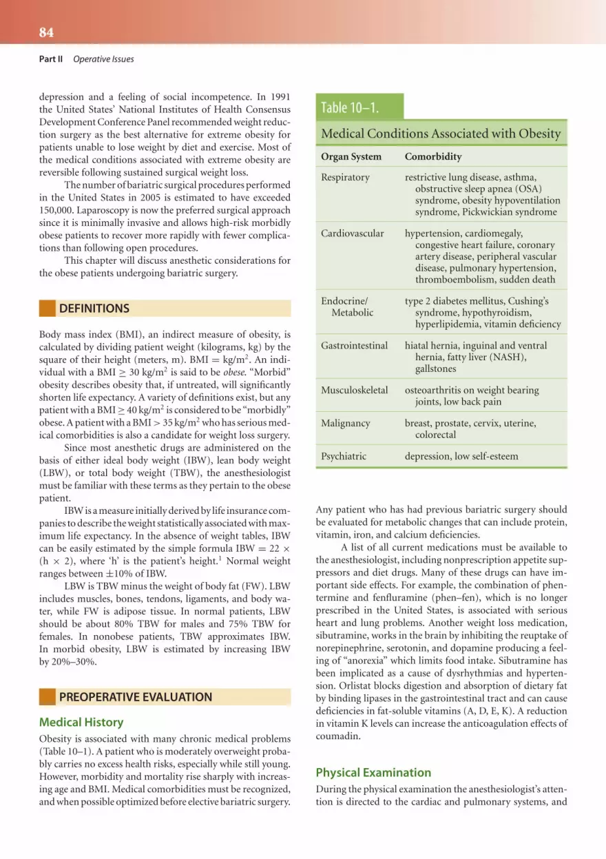
84
Part II Operative Issues
depression and a feeling of social incompetence. In 1991the United States’ National Institutes of Health ConsensusDevelopment Conference Panel recommended weight reduc-tion surgery as the best alternative for extreme obesity forpatients unable to lose weight by diet and exercise. Most ofthe medical conditions associated with extreme obesity arereversible following sustained surgical weight loss.
The number of bariatric surgical procedures performedin the United States in 2005 is estimated to have exceeded150,000. Laparoscopy is now the preferred surgical approachsince it is minimally invasive and allows high-risk morbidlyobese patients to recover more rapidly with fewer complica-tions than following open procedures.
This chapter will discuss anesthetic considerations forthe obese patients undergoing bariatric surgery.
DEFINITIONS
Body mass index (BMI), an indirect measure of obesity, iscalculated by dividing patient weight (kilograms, kg) by thesquare of their height (meters, m). BMI = kg/m2. An indi-vidual with a BMI ≥ 30 kg/m2 is said to be obese. “Morbid”obesity describes obesity that, if untreated, will significantlyshorten life expectancy. A variety of definitions exist, but anypatient with a BMI ≥ 40 kg/m2 is considered to be “morbidly”obese. A patient with a BMI > 35 kg/m2 who has serious med-ical comorbidities is also a candidate for weight loss surgery.
Since most anesthetic drugs are administered on thebasis of either ideal body weight (IBW), lean body weight(LBW), or total body weight (TBW), the anesthesiologistmust be familiar with these terms as they pertain to the obesepatient.
IBW is a measure initially derived by life insurance com-panies to describe the weight statistically associated with max-imum life expectancy. In the absence of weight tables, IBWcan be easily estimated by the simple formula IBW = 22 ×(h × 2), where ‘h’ is the patient’s height.1 Normal weightranges between ±10% of IBW.
LBW is TBW minus the weight of body fat (FW). LBWincludes muscles, bones, tendons, ligaments, and body wa-ter, while FW is adipose tissue. In normal patients, LBWshould be about 80% TBW for males and 75% TBW forfemales. In nonobese patients, TBW approximates IBW.In morbid obesity, LBW is estimated by increasing IBWby 20%–30%.
PREOPERATIVE EVALUATION
Medical HistoryObesity is associated with many chronic medical problems(Table 10–1). A patient who is moderately overweight proba-bly carries no excess health risks, especially while still young.However, morbidity and mortality rise sharply with increas-ing age and BMI. Medical comorbidities must be recognized,and when possible optimized before elective bariatric surgery.
Table 10–1.
Medical Conditions Associated with Obesity
Organ System Comorbidity
Respiratory restrictive lung disease, asthma,obstructive sleep apnea (OSA)syndrome, obesity hypoventilationsyndrome, Pickwickian syndrome
Cardiovascular hypertension, cardiomegaly,congestive heart failure, coronaryartery disease, peripheral vasculardisease, pulmonary hypertension,thromboembolism, sudden death
Endocrine/Metabolic
type 2 diabetes mellitus, Cushing’ssyndrome, hypothyroidism,hyperlipidemia, vitamin deficiency
Gastrointestinal hiatal hernia, inguinal and ventralhernia, fatty liver (NASH),gallstones
Musculoskeletal osteoarthritis on weight bearingjoints, low back pain
Malignancy breast, prostate, cervix, uterine,colorectal
Psychiatric depression, low self-esteem
Any patient who has had previous bariatric surgery shouldbe evaluated for metabolic changes that can include protein,vitamin, iron, and calcium deficiencies.
A list of all current medications must be available tothe anesthesiologist, including nonprescription appetite sup-pressors and diet drugs. Many of these drugs can have im-portant side effects. For example, the combination of phen-termine and fenfluramine (phen–fen), which is no longerprescribed in the United States, is associated with seriousheart and lung problems. Another weight loss medication,sibutramine, works in the brain by inhibiting the reuptake ofnorepinephrine, serotonin, and dopamine producing a feel-ing of “anorexia” which limits food intake. Sibutramine hasbeen implicated as a cause of dysrhythmias and hyperten-sion. Orlistat blocks digestion and absorption of dietary fatby binding lipases in the gastrointestinal tract and can causedeficiencies in fat-soluble vitamins (A, D, E, K). A reductionin vitamin K levels can increase the anticoagulation effects ofcoumadin.
Physical ExaminationDuring the physical examination the anesthesiologist’s atten-tion is directed to the cardiac and pulmonary systems, and

85
Chapter 10 Anesthetic Concerns
to head and neck anatomy in order to evaluate the airway fortracheal intubation.
Cardiovascular System
Cardiac output rises proportionally with increased weight.Stroke volume also increases since a greater total blood vol-ume is needed to perfuse the added body fat. Increased cardiacoutput combined with normal peripheral vascular resistanceleads to systemic hypertension. Mild to moderate hyperten-sion is seen in most morbidly obese patients. The increasedleft ventricular wall stress caused by increased stroke vol-ume and the resultant ventricular dilation leads to cardiachypertrophy.2
Left ventricular dysfunction is often present in young,asymptomatic patients. Even normotensive patients have in-creased preload and after-load, increased mean pulmonaryartery pressure (PAP), and elevated right and left ventricu-lar stroke work. Since these patients are often not physicallyactive, they may appear to be asymptomatic even in the pres-ence of significant cardiovascular disease. Signs of pulmonaryhypertension (exertional dyspnea, fatigue, syncope) shouldbe sought and trans-esophageal echocardiography (TEE) ob-tained in symptomatic patients. Right heart failure is com-mon in older patients. A medical consultation with a cardi-ologist may be indicated before bariatric surgery.
The ECG may show increased rate, changes in QRSvoltage, left QRS axis shift, slowed conduction, and evidenceof ischemia or previous myocardial infarction. The ECG evenin normotensive morbidly obese patients may reveal left ven-tricular hypertrophy, cardiac chamber enlargement, ventric-ular ectopy, and other arrhythmias. Cardiac dysrhythmiasare precipitated by chronic hypoxia (especially in patientswith obstructive sleep apnea, OSA), hypercapnia, increasedcirculating levels of catecholamines, electrolyte disturbancescaused by diuretic therapy, fatty infiltration of the conductionsystem, and ischemic heart disease.3 Polycythemia suggestschronic hypoxemia.
Pulmonary System
Adipose tissue is metabolically active. Oxygen consumptionand CO2 production rise with increasing weight due to in-creased metabolic demands. The work of breathing is in-creased since more energy must be expended to carry theadditional body mass, while respiratory muscle performanceis impaired. The fatty chest and abdominal walls plus theincreased pulmonary blood volume contribute to reducedpulmonary compliance. Mass loading of the thoracic andabdominal chest walls causes abnormalities in both lung vol-umes and gas exchange, especially when the patient is supine.The increased total respiratory resistance and decreased com-pliance associated with extreme obesity results in shallow,rapid breathing. Functional residual capacity (FRC) is sig-nificantly reduced due to a decrease in expiratory reservevolume, therefore total lung capacity is reduced. Airways closeduring normal ventilation. Continued perfusion of nonven-tilated alveoli will result in an oxygen tension (PaO2) that
is lower than predicted for similar-aged nonobese patients.These changes increase in direct proportion with increasingBMI.4 General anesthesia further reduces FRC.
Preoperative pulmonary function tests show a restric-tive breathing pattern. For symptomatic patients, an arterialblood gas obtained while the patient breathes room air isuseful to establish a baseline. Younger obese patients havean increased ventilatory response to hypoxia and a rela-tively decreased response to hypercapnia. Their arterial bloodsample often demonstrates alveolar hyperventilation (PaCO2
30–35 mm Hg) and relative hypoxemia (PaO2 70–90 mm Hg)while breathing air.5 With increasing age, sensitivity to CO2
decreases so PaCO2 rises and PaO2 falls further.A careful thorough preoperative assessment of patient’s
face, neck, and upper airway is always required since maskventilation and tracheal intubation can be a challenge in someobese patients. A review of the patient’s previous anestheticrecords will reveal whether airway problems had been en-countered during previous surgical procedures.
Obstructive Sleep Apnea Syndrome
Many obese patients maintain a normal PaCO2 during theday but have CO2 retention, sleep disturbances, intermittentairway obstruction with hypoxemia, pulmonary hyperten-sion, and cardiac arrhythmias at night. OSA syndrome ischaracterized by frequent episodes of apnea (>10-s cessationof airflow despite continuous respiratory effort against aclosed airway) and hypopnea (50% reduction in airflow orreduction associated with a decrease of SpO2 > 4%). OSAis frequently undiagnosed in patients scheduled for bariatricsurgery.
Obesity is an important risk factor for OSA, but notevery obese patient suffers from OSA.6 OSA occurs moreoften in patients with large fat necks and high Mallampati(III and IV) scores. The patients may not be aware of symp-toms, so it is important to interview their spouse. If OSA ispresent, they will describe loud snoring followed by silence asairflow ceases with obstruction, then gasping or choking asthe patient awakes and airflow restarts. A definitive diagno-sis of OSA can only be confirmed by polysomnography in asleep laboratory. Because of fragmented sleep patterns, OSApatients may complain of daytime sleepiness and headaches.Chronic sleep apnea leads to secondary polycythemia, hy-poxemia, and hypercapnia, all of which increase the risk ofcardiac and cerebral vascular disease.
Patients with a history of snoring or a definitive di-agnosis of OSA are often difficult to ventilate by mask, andtheir tracheas may be more difficult to intubate than simi-lar weight patients without OSA. And OSA patients who usenasal continuous positive airway pressure (CPAP) devices athome should be instructed to bring them to the hospital touse following surgery.
If the patient is known or even suspected of having OSAthey should be continuously monitored by pulse oximetry inthe postoperative period, even following a completely un-eventful operation.

86
Part II Operative Issues
Obesity Hypoventilation Syndrome
A small number of patients have the “obesity hypoventilationsyndrome” (OHS), which is characterized by somnolence,cardiac enlargement, polycythemia, hypoxemia, and hyper-capnia. OHS patients tend to be older, super obese (BMI >
50 kg/m2), and have more restricted pulmonary function thanother patients with OSA.
Hypoventilation is central and independent of intrinsiclung disease, and is probably due to a progressive desensitiza-tion of the respiratory center to hypercapnia from nocturnalsleep disturbances. In its most severe form, the “PickwickianSyndrome,” there is hypersomnolence, hypoxia, hypercapnia,pulmonary hypertension, right ventricular enlargement, andhypervolemia. These patients rely on a hypoxic ventilatorydrive and may hypoventilate or even become apneic follow-ing emergence from general anesthesia after being given 100%O2 to breathe.
Gastrointestinal and Urinary Systems
It is widely believed that morbidly obese patients are at greaterrisk for acid aspiration during induction of general anesthe-sia. Risk factors include increased intra-abdominal pressure,high incidence of gastroesophageal reflux disease (GERD) andhiatus hernia, increased gastric volume (usually >25 mL),and decreased gastric fluid pH (usually <2.5).7 Recently thisbelief has been challenged. One study reported that fastingobese patients actually had a lower incidence of high-volume,low-pH gastric fluid than lean patients,8 while another foundno differences in gastric volume or pH between lean andmoderately obese surgical patients.9 Obese patients withoutsymptoms of GERD have relatively normal gastroesophagealsphincter tone. Obese patients at special risk for gastric acidaspiration may be those with diabetes and gastroparesis.
Nonalcoholic steatohepatitis (NASH, “fatty hepatitis”),with or without liver dysfunction, is extremely common. His-tologic abnormalities are present in the livers of as many as90% of morbidly obese patients.10 Preoperative liver functiontests should be obtained, but they often do not reflect the ac-tual severity of liver dysfunction. Alanine aminotransferase isthe most frequently elevated liver enzyme. Surprisingly, liverclearance of many anesthetic agents is usually not altered withNASH.
There is increased renal blood flow and an increasedglomerular filtration rate (GFR) associated with obesity. Re-nal clearance of drugs may be greater compared to the normal-weight patient. The most common renal abnormality seen isproteinuria.
PERIOPERATIVE CONSIDERATIONS
PremedicationSedatives should be avoided. For the very anxious patientsmall amounts of midazolam (1–2 mg, iv) can be given.If a fiberoptic airway intubation is planned, atropine orglycopyrrolate will decrease oral secretions. Most medications
for chronic hypertension are continued before surgery. Anexception is the angiotensin-converting enzyme inhibitors,which should be stopped preoperatively since they cancause profound hypotension following induction of anes-thesia. Diabetic medications (insulin, oral hypoglycemics)are usually withheld on the morning of surgery, but bloodsugar levels must be closely monitored in the periopera-tive period. Antibiotics prophylaxis for wound infection,and heparin as prophylaxis against deep venous thrombo-sis are usually administered prior to surgery at the surgeon’srequest.
For protection against acid aspiration, an H2-receptorantagonist can be given the night before and again on themorning of surgery along with 30 mL of nonparticulateantacid to increase gastric fluid pH and decrease gastric fluidvolume.11
Patient PositionIt is our practice to have the unpremedicated patient climboff the gurney and position him or herself on the operatingroom table. All pressure points must be carefully padded toavoid pressure sores, neurologic injury, and rhabdomyolysis.Special operating tables are available for patients weighing asmuch as 500 kg, and occasionally two conventional operatingroom tables must be placed together to accommodate evenlarger patients.
Laparoscopic bariatric operations are performed withthe patient in the supine or reverse Trendelenburg position(RTP). In the supine position, FRC is markedly reduced caus-ing V/Q mismatch and significant increases in O2 consump-tion, cardiac output, and PAP.12 A left lateral tilt will preventinferior vena cava compression in the supine patient. TheTrendelenburg (TP) and lithotomy positions further decreaselung volumes. If possible, the patient should always be in theRTP during surgery since in this position the diaphragm is“unloaded” and FRC is maximized.
MonitoringStandard monitors (ECG, blood pressure cuff, pulse oxime-try, end-tidal capnography, and temperature probe) are ap-plied. Noninvasive cuff pressure may be inaccurate when thewrong size cuff is used or if the anatomy of the upper armdoes not allow a proper fit. Cuff pressures can be obtainedfrom the wrist or ankle. An arterial line is usually not needed,but is helpful for continuous blood pressure monitoring andarterial blood gas sampling in high-risk patients.
It is important to monitor urine output intraopera-tively with an indwelling catheter. There is normally a tran-sient decrease in urine output during laparoscopy, with re-turn of normal kidney function following the release of thepneumoperitoneum. 13 A nerve stimulator is used to assessneuromuscular blockade, but excess fat may make surfaceelectrodes inaccurate so needle electrodes are recommended.A depth of anesthesia monitor such as the BIS monitor canalso be useful.

87
Chapter 10 Anesthetic Concerns
Central Venous Pressure (CVP) or Pulmonary Artery(PA) lines are usually not needed for routine procedures. Sincevenous access is often limited, a central line can be help-ful for postoperative needs. It is important to recognize thatthe length of a standard intravenous catheter placed percu-taneously in the neck of a very large patient may not be longenough to reach an intrathoracic location.
To date, there have been no published studies of TEEmonitoring during bariatric surgery, so its utility in this pa-tient population is still to be determined. However, if a TEEmonitor is used, the probe must be withdrawn from theesophagus during critical parts of the procedure.
Pharmacologic ConsiderationsThe physiologic changes in obesity affect the distribution,binding, and elimination of the various agents administeredduring general anesthesia. In routine, anesthetic practicedrugs are usually administered on the basis of dose per unitbody weight. This assumes that clearances and distributionvolumes are proportional to weight—assumptions that maynot be valid in extreme obesity.
Obese patients have a smaller than normal fraction oftotal body water, increased blood volume and cardiac output,greater than normal adipose content, increased total LBW,changed tissue protein binding from increased concentra-tions of free fatty acids, triglycerides, lipoproteins, cholesterol,and other serum constituents (Table 10–2). In addition, re-nal blood flow and GFR are increased and cardiopulmonaryfunction may not be optimal. Hepatic clearance is usuallynormal or even increased in obese patients despite the pres-ence of NASH. Drug dosing by monitoring clinical endpoints
Table 10–2.
Pharmacologic Considerations for theObese Patient
Greater Than Normal Amount Adipose Tissue
Increased lean body weight
Increased blood volume
Smaller than normal fraction total body water
Increased cardiac output
Decreased pulmonary function
Increased protein and free fatty acids
Increased renal blood flow
Increased glomerular filtration rate
Abnormal liver function
(heart rate, arterial pressure, degree of sedation) may be moreimportant than empirical dosing based on patient weightformulae.14
Highly lipophilic drugs have a significant increase involume of distribution compared to nonobese patients andtheir loading dose is usually increased. Since their eliminationhalf-lives are longer, maintenance dosing should be decreasedto reflect IBW.15 Non- or weakly lipophilic drugs are givenbased on LBW.16 Systemic absorption of oral medications isnot significantly affected by obesity.
Inhalational Agents
Desflurane with its low lipid solubility has been recom-mended based on the belief that a slow release of more lipid-soluble anesthetics from excess adipose tissue could prolongemergence from anesthesia.17 Lower blood flow to adiposetissue may limit the initial delivery of volatile agent to thefat and liver and inhalational anesthetics are stored in thefat long after completion of surgery, but the concentrationin the well-perfused brain and lungs of all volatile anesthet-ics rapidly decrease once that anesthetic is discontinued.18
Therefore, all inhalational anesthetics, including isoflurane,are rapidly eliminated. With appropriate timing, there are noclinical differences in the recovery time after general anesthe-sia with any inhalational anesthetic agent.19
Induction Agents
Larger than normal doses of highly lipophilic drugs likepropofol or thiopental are needed due to increased bloodvolume, cardiac output, and adipose. For anesthetic mainte-nance, the intravenous dosing regimen for propofol in theoryshould be based on TBW as in normal-weight patients. How-ever, the cardiovascular effects of very large doses limit theamount that can be given to obese patients. Although patientsrequire more induction agent, they are also more sensitive tothese agents and maintenance dosage is based on LBW.
Muscle Relaxants
Pseudocholinesterase levels and extracellular fluid space areboth increased in obesity. Relatively high doses of succinyl-choline (1.0 mg/kg TBW) are needed for a rapid sequenceanesthetic induction.20
Complete paralysis is especially important duringlaparoscopy to facilitate ventilation and to provide ade-quate space for visualization and maneuvering of the sur-gical equipment. Loss of pneumoperitoneum may indicateincomplete paralysis. Since non-depolarizing muscle relax-ants are hydrophilic, there is limited distribution to adiposetissue and no clinical advantage between any of the com-monly used agents. Neuromuscular recovery time is similar inobese and nonobese patients with atracurium, vecuronium,or rocuronium.21 Relaxants should be administered in in-cremental doses based on IBW, and neuromuscular block-ade must be completely reversed before extubation of thetrachea.

88
Part II Operative Issues
Opioids
Opioids are highly lipophilic, and in theory, loading dosesshould be based on TBW. There is no evidence that lipophilicopioids last longer in morbidly obese patients and generoususe of opioids must be discouraged to avoid respiratory de-pression. Small doses of fentanyl combined with a continuousinfusion of a short-acting opioid like remifentanil is preferredfor laparoscopy. The volume of distribution of remifentanilin obese patients is less than expected, probably because ofhydrolysis by blood and tissue esterases. Remifentanil dosingis based on IBW.
Intravenous Fluids
Intraoperative fluid requirements are usually greater thanwould be anticipated in a normal-weight patient. Several litersof crystalloid should be given during a laparoscopic bariatricoperation. Obese patients receiving 40 mL/kg of intravenouscrystalloid had a faster recovery and fewer complications af-ter cholecystectomy than patients receiving 15 mL/kg.22 Theseresults can only be extrapolated to morbidly obese patientssince no investigation of fluid requirements for laparoscopicbariatric surgery has been performed. It is essential that ade-quate amounts of intravenous fluid be given to reduce post-operative renal failure, and to avoid other rare but seriouscomplications such as rhabdomyolysis.23
GENERAL ANESTHESIA
PreoxygenationDespite conflicting evidence that morbidly obese patients areat greater risk for acid aspiration, it remains prudent to es-tablish a secure airway as quickly and as safely as possible.Patients cannot tolerate the supine position and should bepreoxygenated in the RTP until SpO2 is 100% for severalminutes.24 The hemoglobin of an apneic obese patient willdesaturate very quickly since FRC is reduced and O2 reservesare limited.25 Preoxygenation in the RTP can increase O2 re-serves, but may cause pooling of blood and hypotension.26
Tracheal IntubationAll patients undergoing bariatric surgery must have their tra-cheas intubated to allow controlled ventilation and to protecttheir airway from acid aspiration. Potential airway manage-ment problems (fat face and cheeks; limited range of motionof the head, neck, and jaw; small mouth and large tongue; ex-cessive palatal and pharyngeal tissue; short/large neck; highMallampati (III or IV) score) should all be evaluated duringthe preoperative visit. High Mallampati score and large neckcircumference are the most reliable predictors of potentialintubation difficulties.27−29 If a problem is anticipated pre-operatively, an “awake intubation” with a fiberoptic broncho-scope is recommended. Appropriate nerve blocks and topicalanesthesia to the airway are applied, and sedative drugs are
Figure 10–1. The proper position for a morbidly obese patientduring preoxygenation, induction of general anesthesia, and tra-cheal intubation is shown. The head and upper body are rampedup in the ‘‘head elevated laryngoscopy position” in which animaginary horizontal line connects the sternum with the ear. Inthis position the endoscopist’s view of the vocal cords during di-rect laryngoscopy is significantly improved. The head of the bedis also in the reverse Trendelenburg position (RTP). The RTP maxi-mizes lung volumes and delays hemoglobin desaturation duringthe intubation sequence.
kept to a minimum. It is important that the patient breathessupplemental O2 during the intubation procedure.
Increasing weight or BMI are not risk factors for diffi-cult laryngoscopy.30 The most important strategy for insur-ing successful direct laryngoscopy is proper patient position.The patient must be placed with their head, upper body, andshoulders significantly elevated (stacked or ramped) so thattheir ear is level with the sternum (head elevated laryngoscopyposition) (Figure 10–1). When a morbidly obese patient isin this position the endoscopist’s view during direct laryn-goscopy is significantly improved.31
For most patients a rapid intravenous induction withpropofol and succinylcholine, combined with cricoid pres-sure, is the best means for securing the airway. A rapid se-quence technique is important since bag and mask ventila-tion is often difficult due to upper airway obstruction, andreduced pulmonary compliance and gastric insufflation dur-ing ineffective mask ventilation will further increase the riskof regurgitation and acid aspiration. A second person experi-enced with airway management, preferably another anesthe-siologist, must always be present to assist when difficulty isencountered.
Aids for difficult intubation, including a short laryn-goscope handle, a variety of laryngoscope blades, speciallaryngoscopy equipment (Bullard laryngoscope, Wu laryngo-scope) a gum elastic bougie, a light-wand, and equipment forcrico-thyroidotomy and trans-tracheal jet ventilation, shouldalso be available. A proseal laryngeal mask airway (LMA) orintubating LMA can serve as a bridge until an endotrachealtube is placed when difficulty is encountered.32,33

89
Chapter 10 Anesthetic Concerns
Mechanical VentilationVentilation should be mechanically controlled duringsurgery. Morbidly obese patients breathing without assistanceunder general anesthesia are likely to hypoventilate. Patientsshould be ventilated with an FiO2 of 0.5–1.0 and a tidal vol-ume 12–15 mL/kg IBW, preferably in the RTP. Larger tidalvolumes will only marginally improve oxygenation while pro-ducing hypocapnia and potentially causing lung trauma.34
Peak ventilatory pressures and end-tidal CO2 levelsincrease during laparoscopy.35 If one wishes to limit thesechanges, minute ventilation can be adjusted by decreas-ing tidal volume and increasing respiratory rate. PulmonaryEnd Expiratory Pressure (PEEP) superimposed upon a largetidal volume can actually worsen hypoxemia by depressingcardiac output, which in turn will reduce O2 delivery. Place-ment of subdiaphragmatic packs or retractors or changingto lithotomy or Trendelenburg positions will also impairventilation.
The physiologic effects of the CO2 pneumoperitoneumare usually well tolerated by the patient and require nointervention.13 These effects include an elevation in peak in-spiratory pressure combined with a decrease in tidal volume,a rise in end-tidal CO2 and PaCO2, and a decrease in pH, and arise in heart rate and mean arterial blood pressure. Maintain-ing the patient in the RTP minimizes the restriction of res-piratory mechanics from the CO2 insufflation.36 All changesreturn to normal once the pneumoperitoneum is relieved.
As with any laparoscopic procedure, catastrophic com-plications such as massive gas embolism, pneumothorax, andpneumomediastinum can occur. The anesthesiologist mustalways be vigilant and recognize and treat these complica-tions immediately.
The pneumoperitoneum can displace the diaphragmcephalad causing the position of the endotracheal tube tochange. Occasionally, the endotracheal tube’s tip can entera bronchus, therefore tube displacement should always beconsidered in the differential diagnosis of hypoxemia duringlaparoscopy.28,29
Hemodynamic ChangesPulmonary capillary wedge and PAP pressures may be ele-vated secondary to increased pulmonary blood volume andchronic hypoxemia. The surgical pneumoperitoneum cancompress the inferior vena cava and femoral veins resulting indecreased blood return to the heart. During surgery there isusually an increase in heart rate, mean arterial pressure, andsystemic vascular resistance, and a decrease in stroke volume,but the net result is cardiac output remaining unchanged orbecoming only slightly decreased.13
ANESTHETIC TECHNIQUE
Our general anesthetic technique consists of an intravenousinfusion of a short-acting opioid (remifentanil) supple-mented with small amounts of fentanyl and/or dexmedetomi-
dine. The patient is ventilated with an inhalational anesthetic(isoflurane, sevoflurane, or desflurane) with a FiO2 of 50%–100% O2. Nitrous oxide can be used since it does not dilatethe bowel during laparoscopic bariatric surgery, but its role islimited due to the high oxygen demand of many patients.37
Since the surgeon infiltrates the trocar sites with localanesthetics, deep levels of anesthesia are not required duringwound closure. The inhalation agent is discontinued severalminutes before surgery is completed but the remifentanil in-fusion is continued until the very end of the procedure. Oncethe remifentanil is stopped the patient is awake and their tra-chea can be extubated within 3 minutes.38
During laparoscopic bariatric procedures, the anesthe-siologist is usually responsible for proper placement of thegastric tube to decompress the stomach and to help size thegastric pouch. They may be asked to help perform leak testsfor anastomotic integrity, either by insufflation of the gastrictube or placement of saline or dye down the tube. It is ex-tremely important that the gastric tube and anything else inthe esophagus (such as a temperature probe or TEE probe)be completely withdrawn before the gastric pouch is stapled.
TEMPERATURE MAINTENANCE
Even though adipose tissue is a thermal isolator, patients be-come poikilothermic during general anesthesia. Heat loss maybe exaggerated by the CO2 pneumoperitoneum and whencold irrigating fluids are used. Warming blankets and otherdevices should be employed intraoperatively, and warmedintravenous and irrigating fluid are occasionally needed ifthere is a significant drop in temperature. Attempts to mini-mize heat loss with heated and humidified gas for the surgicalpneumoperitoneum have been unsuccessful.39
POSTOPERATIVE CONSIDERATIONS
Position and OxygenationThe semirecumbent and RTP positions maximize oxygena-tion by allowing the diaphragm to fall and FRC to increase.If hemodynamically stable, patients should have their airwayextubated with their upper body elevated 30◦–45◦, and thenbe transferred from the operating room in that position.
Postoperative admission to an intensive care unitand/or mechanical ventilation is rarely needed. Factors thatmay necessitate ventilatory support include extremes of age,super obesity, coexisting cardiac disease or pulmonary dis-ease and CO2 retention, fever or infection, and an uncooper-ative or extremely anxious patient. The need for postoperativeadmission to an intensive care unit is relatively common af-ter open bariatric procedures,40 but rare after laparoscopicsurgery.41
General anesthesia in morbidly obese patients results ina significant incidence of postoperative atelectasis.42 Patientscan become hypoxemic if supplemental O2 is withheld in

90
Part II Operative Issues
the immediate recovery period. Restoration of normal pul-monary function after open abdominal surgery may take sev-eral days.
OSA patients using nasal CPAP or bi-level positive air-way pressure (BiPAP) at home should be instructed to bringtheir equipment to the hospital to use in the recovery room.These devices allow alveolar recruitment during inspirationand prevent alveolar collapse during expiration. In theory,CPAP could distend the gastric pouch, but its use followingbariatric surgery has not been associated with anastomoticleaks.43
AntithrombosisThromboembolism is a major cause of postoperative mor-tality. Prolonged immobilization can lead to phlebothrom-bosis. The risk of thrombosis is further increased becauseof greater blood volume and relative polycythemia commonin obese patients. Other risk factors include high fatty acidlevels, hypercholesterolemia, and diabetes. In addition, mor-bidly obese patients demonstrate accelerated fibrin forma-tion, fibrinogen-platelet interaction, and platelet functioncompared with controls. Anticoagulation or other prophy-laxis measures should always be considered in the postopera-tive period, even for patients with epidural catheters.44 A venacava umbrella is occasionally placed preoperatively in olderand high-risk patients, and sequential compression boots areoften used during surgery. Early ambulation must be encour-aged. Many patients can be ready for hospital discharge onthe first or second postoperative day following laparoscopy.41
Postoperative Nausea and VomitingObesity, per se, is not a risk factor for postoperative nauseaand vomiting (PONV).45 However, many patients undergo-ing bariatric procedures are at high risk (e.g., females, thosereceiving opioids, those undergoing emetogenic surgery) forPONV. Multimodal intraoperative prophylaxis with severalantiemetic agents will reduce, but not eliminate, PONV. Dex-amethasone (4–8 mg) should be a part of the therapeuticregimen.
AnalgesiaLocal anesthetic is infiltrated into the trocar sites during theprocedure, so incisional pain in the immediate recovery pe-riod is much less than after a laparotomy.46 Intravenous opi-oid patient-controlled analgesia (PCA) with drug dose basedon IBW is usually satisfactory. Large amounts of opioids, espe-cially longer acting opioids (morphine, demerol, hydromor-phone), which can depress ventilation, should be avoided.
The use of non-opioid analgesic adjuncts should beinstituted early. Dexmedetomidine, which has no respiratorydepressant effects, is a useful alternative or supplement toopioids.41 Nonsteroidal anti-inflammatory drugs are helpfulinitially, but should be discontinued within a day or two toavoid the potential complication of gastric ulceration.
References1. Alpert MA, Hashimi MN: Obesity and the heart. Am J Med Sci
306:117–123, 1993.2. Altermatt FR, Munoz HR, Delfino AE, Cortinez LI: Pre-oxygenation
in the obese patient: Effects of position on tolerance to apnoea. Br JAnaesth 95:706–709, 2005.
3. Arain SR, Barth CD, Shankar H, Ebert TJ: Choice of volatile anes-thetic for the morbidly obese patient: Sevoflurane or desflurane. JClin Anesth 17:413–419, 2005.
4. Bardoczky GI, Yernault JC, Houben JJ, d’Hollander AA: Large tidalvolume ventilation does not improve oxygenation in morbidly obesepatients during anesthesia. Anesth Analg 81:385–388, 1995.
5. Bharati S and Lev M: Cardiac conduction system involvement insudden death of obese young people. Am Heart J 129:273–281, 1995.
6. Brodsky JB: Positioning the morbidly obese patient for anesthesia.Obes Surg 12:751–758, 2002.
7. Brodsky JB, Lemmens HJ, Brock-Utne JG, Vierra M, Saidman LJ:Morbid obesity and tracheal intubation. Anesth Analg 94:732–736,2003.
8. Brodsky JB, Lemmens HJM, Collins JS, Morton JM, Curet MJ, Brock-Utne JG: Nitrous oxide and laparoscopic bariatric surgery. Obes Surg15:494–496, 2005.
9. Collins JS, Lemmens HJM, Brodsky JB, Brock-Utne JG, Levitan RM:Laryngoscopy and morbid obesity: A comparison of the “sniff” and“ramped” positions. Obes Surg 14:1171–1175, 2004.
10. Davila-Cervantes A, Dominguez-Cherit G, Borunda D, et al.: Im-pact of surgically-induced weight loss on respiratory function: Aprospective analysis. Obes Surg 14:1389–1392, 2004.
11. de Menezes Ettinger JE, dos Santos Filho PV, Azaro E, Melo CA, FahelE, Batista PB: Prevention of rhabdomyolysis in bariatric surgery. ObesSurg 15:874–879, 2005.
12. Dumont L, Mattys M, Mardirosoff C, Vervloesem N, Alle JL, Mas-saut J: Changes in pulmonary mechanics during laparoscopic gastro-plasty in morbidly obese patients. Acta Anaesthesiol Scand 41:408–413, 1997.
13. Eichenberger A, Proietti S, Wicky S, et al.: Morbid obesity and post-operative pulmonary atelectasis: An underestimated problem. AnesthAnalg 95:1788–1792, 2002.
14. Eriksson S, Backman L, Ljungstrom KG: The incidence of clinicalpostoperative thrombosis after gastric surgery for obesity during 16years. Obes Surg 7:332–335, 1997.
15. Ezri T, Hazin V, Warters D, Szmuk P, Weinbroum AA: The endo-tracheal tube moves more often in obese patients undergoing la-paroscopy compared with open abdominal surgery. Anesth Analg96:278–282, 2003.
16. Ezri T, Medalion B, Weisenberg M, Szmuk P, Warters RD, CharuziI: Increased body mass index per se is not a predictor of difficultlaryngoscopy. Can J Anesth 50:179–183, 2003.
17. Farley DR, Greenlee SM, Larson DR, Harrington JR: Double-blind,prospective, randomized study of warmed humidified carbon diox-ide insufflation vs standard carbon dioxide for patients undergoinglaparoscopic cholecystectomy. Arch Surg 139:739–743, 2004.
18. Frappier J, Guenoun T, Journois D, et al.: Airway management usingthe intubating laryngeal mask airway for the morbidly obese patient.Anesth Analg 96:1510–1515, 2003.
19. Gaszynski TM, Strzelczyk JM, Gaszynski WP: Post-anesthesia re-covery after infusion of propofol with remifentanil or alfentanil orfentanyl in morbidly obese patients. Obes Surg 14:1–7, 2004
20. Harter RL, Kelly WB, Kramer MG, Perez CE, Dzwonczyk RR: Acomparison of the volume and pH of gastric contents of obese andlean surgical patients. Anesth Analg 86:147–152, 1998.
21. Helling TS, Willoughby TL, Maxfield DM, Ryan P: Determinantsof the need for intensive care and prolonged mechanical ventilationin patients undergoing bariatric surgery. Obes Surg 14:1036–1041,2004.
22. Holte K, Klarskov B, Christensen DS, et al.: Liberal versus restric-tive fluid administration to improve recovery after laparoscopic

91
Chapter 10 Anesthetic Concerns
cholecystectomy. A randomize, double-blind study. Ann Surg240:892–899, 2004.
23. Huerta S, DeShields S, Shpiner R, et al.: Safety and efficacy of post-operative continuous positive airway pressure to prevent pulmonarycomplications after Roux-en-Y gastric bypass. J Gastrointest Surg6:354–358, 2002.
24. Jense HG, Dubin SA, Silverstein PI, O’Leary-Escolas U: Effect ofobesity on safe duration of apnea in anesthetized humans. AnesthAnalg 72:89–93, 1991.
25. Juvin P, Lavaut E, Dupont H, et al.: Difficult tracheal intubation ismore common in obese than lean patients. Anesth Analg 97:595–600,2003.
26. Juvin P, Vadam C, Malek L, Dupont H, Marmuse JP, Desmonts JM:Postoperative recovery after desflurane, propofol, or isoflurane anes-thesia among morbidly obese patients: A prospective, randomizedstudy. Anesth Analg 91:714–719, 2000.
27. Keller C, Brimacombe J, Kleinsasser A, Brimacombe L: The LaryngealMask Airway ProSeal(TM) as a temporary ventilatory device in grosslyand morbidly obese patients before laryngoscope-guided trachealintubation. Anesth Analg 94:737–740, 2002.
28. Kranke P, Apefel CC, Papenfuss T, et al.: An increased body massindex is no risk factor for postoperative nausea and vomiting. ActaAnaesthesiol Scand 45:160–166, 2001.
29. Lam AM, Grace DM, Manninen PH, Diamond C: The effects ofcimetidine and ranitidine with and without metoclopramide on gas-tric volume and pH in morbidly obese patients. Can Anaesth Soc J33:773–779, 1986.
30. Lemmens HJM, Brodsky JB: The dose of succinylcholine in morbidobesity. Anesth Analg 102(2): 438–42, 2006.
31. Lemmens HJM, Brodsky JB, Bernstein DP: Estimating ideal bodyweight—a new formula. Obes Surg 15:1082–1083, 2005.
32. Maltby JR, Pytka S, Watson NC, Cowan RA, Fick GH: Drinking 300mL of clear fluid two hours before surgery has no effect on gastricfluid volume and pH in fasting and non-fasting obese patients. CanJ Anaesth 51:111–115, 2004.
33. Marik P, Varon J: The obese patient in the ICU. Chest 113:492–498,1998.
34. McCarty TM, Arnold DT, Lamont JP, Fisher TL, Kuhn JA: Optimiz-ing outcomes in bariatric surgery. Outpatient laparoscopic gastricbypass. Ann Surg 242:494–501, 2005.
35. Moretto M, Kupski C, Mottin CC, et al.: Hepatic steatosis in patientsundergoing bariatric surgery and its relationship to body mass indexand comorbidities. Obes Surg 13:622–624, 2003.
36. Mortimore IL, Marshall I, Wraith PK: Neck and total body fat de-position in nonobese and obese patients with sleep apnea comparedwith that in control subjects. Am J Resp Crit Care Med 157:280–283,1998.
37. Nguyen NT, Lee SL, Goldman C, et al.: Comparison of pulmonaryfunction and postoperative pain after laparoscopic versus opengastric bypass: A randomized trial. J Am Coll Surg 192:469–477,2001.
38. Nguyen NT, Wolfe BM: The physiologic effects of pneumoperi-toneum in the morbidly obese. Ann Surg 241, 219–226, 2005.
39. Ogunnaike BO, Jones SB, Jones DB, Provost D, Whitten CW: Anes-thetic considerations for bariatric surgery. Anesth Analg 95:1793–1805, 2004.
40. Pelosi P, Croci M, Ravagnan I, et al.: The effects of body mass on lungvolumes, respiratory mechanics, and gas exchange during generalanesthesia. Anesth Analg 87:654–660, 1998.
41. Perilli V, Sollazzi L, Bozza P, et al.: The effects of the reverse Trendelen-burg position on respiratory mechanics and blood gases in morbidlyobese patients during bariatric surgery. Anesth Analg 91:1520–1525,2000.
42. Perilli V, Sollazzi L, Modesti C, et al.: Comparison of positiveend-expiratory pressure with reverse Trendelenburg position inmorbidly obese patients undergoing bariatric surgery: Effects onhemodynamics and pulmonary gas exchange. Obes Surg 13:605–609,2003.
43. Servin F, Farinotti R, Haberer JP, Desmonts JM: Propofol infusion forthe maintenance of anesthesia in morbidly obese patients receivingnitrous oxide. Anesthesiology 78:657–665, 1993.
44. Torri G, Casati A, Albertin A, et al.: Randomized comparison ofisoflurane and sevoflurane for laparoscopic gastric banding in mor-bidly obese patients. J Clin Anesth 13:565–570, 2001.
45. Varin F, Ducharme J, Theoret Y, Besner JG, Bevan DR, Donati F:Influence of extreme obesity on the body disposition and neuromus-cular blocking effect of atracurium. Clin Pharmacol Ther 48:18–25,1990.
46. Vaughan RW, Bauer S, Wise L: Volume and ph of gastric juice inobese patients. Anesthesiology 43:686–689, 1975.

PART
III
Physiology of BariatricOperations
Copyright © 2008 by The McGraw-Hill Companies, Inc. Click here for terms of use.

11Restrictive SurgeryEdward E. Mason, MD, PhD, FACS
I. HISTORY
II. REQUIREMENTS FOR SUCCESS OF RESTRICTIONSURGERYPatient Education
III. LONGEVITY AND FOLLOW-UP
IV. DESIGN AND OPERATIVE TECHNIQUEPouch Configuration
Pouch VolumeOutlet LengthOutlet SizeReoperations
V. CONCLUSIONS
Gastroplasty was designed to restrict food intake without in-troducing bypass complications. It began in 1971 with hori-zontal gastroplasty and was changed to vertical in 1980, inde-pendently by many surgeons. The history helps explain whatcan cause success or failure. Small changes in design and tech-nique too often lead to failure. Patients need to understandhow to take care of their diminutive pouch in order to suc-ceed. Overflow is a normal consequence of improper eating,but uncontrolled vomiting is abnormal and must be correctedbefore thiamine deficiency causes permanent injury or death(Wernicke–Korsakoff syndrome). Restriction operations areas effective as bypass operations in keeping patients alive for atleast the first 8.3 years studied. Lifelong data may be neededto determine the best match of patients to operation type.Vertical pouches preserve the normal anatomy, which is im-portant for preventing esophageal reflux. The ≤20 mL pouchsize for gastroplasty is too small for an operation that bypasses
most of the small bowel. The pouch outlet for restriction op-erations is also too small for adding extensive small-bowelbypass in order to increase weight loss. Attempts to increaseweight control by decreasing the pouch outlet below that rec-ommended will lead to complications. Revision of verticalpouches that were too large initially and have enlarged tocause progressive increase in vomiting and weight gain shouldbe revised to ≤20 mL, which will correct both complaints.Conversion to a bypass operation is not indicated for thesecomplaints. Operative technique is important in determiningwhether the pouch is antireflux or causes reflux.
HISTORY
Dietary restriction of intake is the oldest and the leasteffective treatment for obesity. Surgery for obesity uses
Copyright © 2008 by The McGraw-Hill Companies, Inc. Click here for terms of use.

96
Part III Physiology of Bariatric Operations
(1) restriction, (2) malabsorption, and (3) manipulation ofneurohumoral mechanisms for weight control. Gastric by-pass for obesity was introduced in 1967 to restrict intake whileavoiding the complications of intestinal bypass.1 In 1971 hor-izontal gastroplasty was introduced as a means of eliminat-ing the complications of gastric bypass, while maintainingthe intake deficiency created by a small stomach pouch.2
This was unsatisfactory because pouches were not measuredand outlets were not stabilized. In addition, mobilization ofthe greater curvature brought the stomach into view for hor-izontal stapling, but when the fundus returned to its originalposition, the staple-line was slanted upward and emptyingwas like a cascade. This predisposed to obstruction, especiallyif the outlet was stabilized with a band that became adherentto adjacent tissues. Obstruction required endoscopy, whichwas difficult with the outlet high on the greater curvature. Weneeded a straight shot, which meant an outlet on the lessercurvature.
Beginning in 1980–81 pouches were changed from hor-izontal to vertical and moved to the lesser curvature to facil-itate endoscopy. Michael Long described moving the pouchoutlet to the lesser curvature and then, in successive patients,changing the slope of the staple-line from horizontal untilit was vertical.3 At the University of Iowa, a stapled circularwindow was made near the lesser curvature in order to fa-cilitate vertical stapling and to allow placement of a Marlexmesh collar around the outlet without sewing or stapling it tothe stomach wall. The volume of these vertical banded gas-troplasty (VBG) pouches was measured and detailed descrip-tions of the operative technique were published and providedat surgical meetings.4 Since restriction of intake was the onlyapparent mechanism for causing weight loss and maintain-ing a lower weight, a small measured pouch and a controlledoutlet were crucial to success.
Failure occurred when pouches were too large. Sur-geons, who attempted to increase weight loss by decreasingoutlet size, soon abandoned the operation, blaming failureon the name of the operation instead of deviations from rec-ommended technique.5 As a result of the obesity epidemicthat began in 1980, patients presenting were heavier eachyear. Surgeons and patients wanted more weight loss than arestriction operation could produce, which led to increaseduse of bypass operations. Training programs in laparoscopicsurgery focused upon RYGB and adjustable gastric banding,which further decreased the use of any form of vertical gas-troplasty. In 2005, in the United States, most of the operationswere LRYGB. Concern remains about the late-in-life compli-cations of bypass of the upper digestive tract, which was thereason for developing VBG in 1980.
REQUIREMENTS FOR SUCCESS OFRESTRICTION SURGERY
Both obesity and the surgical operations are life long and theoperations are adjunctive. This means that success should
be defined in terms of the effects of both the obesity and theoperation over each patient’s remaining life. Our medical caresystem is poorly designed for the lifelong monitoring, treat-ment, and documentation needed to determine success. Tostudy the effect of an operation requires that the techniqueand patient care be standardized before lifelong study can be-gin. Our society worships what is new(s). What follows willbe some observations about the changing practice of oper-ations based upon the thesis that restriction of intake is thesimplest and safest way to surgically control excessive bodyweight. The best restriction operation for the future has notbeen studied but can be hypothesized based upon a centuryof accumulated experience of many surgeons.
Patient EducationSuccess not only depends upon providing an optimum pouch,but also on a compliant patient who will chew well, eat smallportions, and stop before vomiting (overflow/regurgitation).Control of intake is especially important with an operation,which is totally dependent upon restriction. These patientshave not been able to reduce their meal size or change theireating habits before being provided a meal-sizing pouch ofless than 20 mL compared with their total stomach capac-ity averaging 1700 mL. In addition, the outlet is stabilized topreserve a diameter of 11 mm so that the pouch will retainenough food to provide some sense of fullness. Both surgeonsand patients must learn the anatomic requirements and lim-itations of the restriction pouch in order to make it work.Patients should be able to recognize a drawing of their op-eration and should know the measurements of pouch andoutlet. They should practice eating out of a medicine glassbefore and early after the operation. If there is vomiting afterthe operation, more time should be spent in patient trainingas well as in review of the operative technique. Overflow isnormal when patients overload the pouch but they should beable to prevent this by changes in eating habits. If a patienthas uncontrollable vomiting, the reason must be found andcorrected before depletion of thiamine results in nystagmus,loss of deep tendon reflexes, confusion, and rapid progressionto coma and death, the Wernicke–Korsakoff syndrome.6
LONGEVITY AND FOLLOW-UP
We used the National Death Index (NDI) to provide what webelieve to be a nearly complete follow-up for the length of life.7
At an average postoperative time of 8.3 years for the first 654deaths in a comparison of 7,185 patients after simple restric-tion operations with 11,787 patients after bypass operations,neither operative type has shown any advantage in keepingpatients alive. Age, operative body mass index (BMI), gen-der, diabetes, hypertension, and smoking were predictive oflongevity. It is important to extend the follow-up for mortalityin these patients for sufficient years to detect any difference inthe effect of operation type as early as it appears and to study

97
Chapter 11 Restrictive Surgery
the causes of death in the two groups of patients. This shouldprovide help in advising patients regarding their choice ofsimple versus complex operations. It should also guide sur-geons in recommending the type of operation for patientswith type 2 diabetes. Unless simple restrictive operations areavailable, the information obtained from this study may bepurely academic. The choice will then remain between bypassand gastric banding, or the surgeon’s favorite operation andno operation.
DESIGN AND OPERATIVE TECHNIQUE
Pouch ConfigurationVertical banded gastroplasty was designed with the antirefluxCollis gastroplasty in mind.8 There are muscle fibers calledthe sling of Helvitius that encircle the junction of esophagusand stomach, cross at the angle of His, and descend along thelesser curvature of the stomach. This anatomy is importantin preventing reflux and is preserved in the design of VBG.VBG, when properly performed, exaggerates the angle ofHis. Deitel et al. demonstrated that VBG is an antirefluxoperation.10 Horizontal gastroplasty by contrast cuts acrossthis complex musculature. One of the original requirementsof any operation for obesity was reversibility. The stomachhas long been known as a long tube (Magenstrasse) with adistensible attachment (sleeve). Vertical pouches make use ofthe upper end of the Magenstrasse, which preserves normalanatomy.
Placing a band around the upper stomach to create asmall meal-limiting pouch has the advantages of no cuttingor stapling but the resulting pouch is more like the one inhorizontal gastroplasty rather than the vertical, lesser cur-vature pouch that replaced it. There tends also to be slip-page between the band and the stomach wall, with enlarge-ment of the pouch and tilting of the band, leading to ob-struction of the pouch. Positioning of the band outside ofthe peritoneum posterior, and imbrication of the stomachanterior, has reduced the slippage. The importance of ad-justment of the band has not been established. Comparisonsmade in the Swedish Obesity Subjects Study indicate that theweight loss and weight control are less after the adjustableband gastroplasty than after VBG, and much less than af-ter RYGB.11 A comparison by Suter et al. between resultsobtained with the Swedish Band and Lap Band showed nodifference, with 50%–60% of patients obtaining satisfactoryweight loss, but at least 10% developing severe long-termcomplications.12
The design and operative technique of gastroplastyshould eliminate capacity for a large meal without creatingacid reflux. The resulting pouch must have a stable con-figuration, volume, and outlet diameter. Foreign materialused to stabilize the outlet should be kept at a minimumand protected from contamination since infection leads tomigration into the lumen. The ideal gastroplasty pouch is
too small for the short common channel of biliopancreaticdiversion.
Decreasing the pouch outlet cannot increase weight lossand improve weight maintenance without increasing the riskof obstruction, staple-line disruption, migration of the for-eign material into the lumen, and an unacceptable rate of re-operation. Failure to adhere to recommended technique hasled to unsatisfactory results and abandonment of gastroplasty.When more is known about the lifelong effects of availableoperations, the need for a restriction operation may becomeevident for at least those patients whose obesity is less severe.Whether adjustable banding can substitute for vertical gas-troplasty is yet to be proven. Operations are like dwellings—their protection and benefit depends upon the way they areconstructed and the way they are used. The lifelong effectsincrease in importance over time for those operations thatpass the earlier tests and remain in place. Experience from100 years of digestive tract surgery serves as a guide for pre-dicting the future of bypass operations but is of less help withrestriction operations, which came with the newly recognizeddisease of severe obesity.
Pouch VolumeAn advantage of the open technique is the ease of measuringthe pouch volume under 70 cm of water pressure and thenchanging the placement of the stapler if the pouch is not thedesired volume. The recording (documentation) of pouchmeasurements led to the use of smaller and smaller pouchesover many years of experience with returning patients whosepouches had dilated to unacceptable size. Prevention of pouchdilatation depends upon decreasing the tension on the pouchwall from excessive pouch volume as dictated by the law ofLaPlace (T = Pr) where T is the tension stretching the pouchwall, P is the difference in pressure across the wall, and r theradius of a cylindrical pouch. The pressure is dependent uponpatient compliance in eating small amounts and chewing wellso that the pouch is not over distended. The importance ofLaPlace’s law is that the larger the pouch the more it will dis-tend. It is also evident that if the pouch is long it must beuniform in radius. If the upper end of the pouch is larger dueto the staple-line reaching out on the fundus rather than atthe angle of His, this area will distend and assume the shapeof a sphere. The consequence may be sufficient tension atthe cardia to cause reflux. Thus the operative technique de-termines whether the operation is antireflux or causes reflux.The pouch must be 20 mL or less in measured volume in orderthat over time the law of LaPlace will not result in excessiveenlargement. A pouch that is too large at the outset may en-large to a degree that the walls will overhang the outlet. If thisis concentric enlargement, the pouch outlet sits in the mid-dle with the shape of a volcano (Jamieson’s description).13
More often an enlarged pouch is asymmetric and the over-hang is on one side. This may tilt the ring or collar and causeobstruction. This complication is analogous to the develop-ment of a Zenker’s diverticulum, with the overhanging pouch

98
Part III Physiology of Bariatric Operations
assuming alignment with the vertical pouch and esophagus.The outlet into the main stomach may be displaced to the sideand compressed by the tilted collar, silastic ring, or stabilizingsutures.
Outlet LengthMichael Long introduced a vertical gastroplasty that is similarto the silastic ring gastroplasty and VBG except for the outlet.Jamieson worked with Michael Long when this operation wasintroduced in 1980 and continued the study of the operation.He used three number-1 Ethibond sutures through the ver-tical staple-line starting at 4 cm below the angle of His andextending to 5.2 cm. These were tied around the lesser cur-vature over an indwelling 38–40 French bougie to provide a12-mm outlet length. This compares with the 15-mm widthof Marlex mesh used in VBG and 3 mm for silastic ring gas-troplasty. In 1984, Jamieson used a 6-mm outlet length withtwo sutures (as used by Long) in 206 patients and comparedthe results with his preferred 12 mm with three sutures, usedduring 1983 and 1985. Percent excess weight loss at 5 yearswas 61% and 69% with the 12-mm and 52% with the 6-mmlength of outlet. The revision rates were 3.5% and 3.7% forthe 12 mm and 8.7% for the 6 mm length of outlet. These arethe only data available comparing the effect of length of theoutlet upon weight loss and revision rate for vertical gastro-plasty. The distribution of operative weights for Jamieson’s512 patients was low with the most frequent BMI (kg/m2)35 and the range 25 to >60. These were open operations. TheMichael Long gastroplasty would need to be adapted to thelaparoscopic approach if it were to be competitive in today’smarket. Stapling in continuity from the angle of His, parallelto the lesser curvature, may be more difficult than staplingfrom a window to the angle as is performed laparoscopicallyin VBG.14
Outlet SizeWhen VBG was introduced, the Marlex mesh circumferenceused to stabilize the outlet was 5.0 cm. A few patients returnedearly following operation because of vomiting. Our responsewas to increase the circumference of the encircling mesh to5.5 cm and to teach patients to drink and eat from a medicineglass during the early postoperative period. Patients were toldto not swallow anything that was not converted to liquid bychewing. Fibrous foods that did not liquefy were to be spit outand then avoided. It was soon possible to resume the use ofthe 5.0-cm band with infrequent need for admission becauseof vomiting. Patients who were 225% of normal weight wereprovided 4.5-cm-circumference bands until it became evi-dent that this was increasing the need for repair of disruptedstaple-lines. Following this early experience, 5-cm bands wereused in all patients regardless of weight.
In 1980, at the University of Iowa, we introduced thestapled window near the lesser curvature to facilitate verti-cal stapling to the angle of His and also allow placement ofthe Marlex mesh band around the pouch outlet and have
it remain in place without suturing the mesh to the stom-ach wall. The outlet required stabilization with an 11–12mm diameter lumen, which was provided with an external5.0-cm-circumference band. The goal was stabilization with-out obstruction. With small measured pouches it should notbe necessary to divide the pouch from the rest of the stom-ach. Division does in theory increase the risk of contami-nation and infection, when there is foreign material, whichincreases the risk of migration into the lumen. The rapidinfiltration of a mesh with a connective tissue should pro-tect from contamination. Marlex mesh is relatively nonreac-tive but it is possible that low-grade irritation could combinewith carcinogens in food and from the environment to causecancer.
Four cancers have been reported, two after Marlexmesh,15,16 one after a silastic ring,17 and one after Gore-Tex.18
Two additional patients with cancer after VBG have been op-erated upon at the University of Iowa, both following Marlexmesh. No cancers were found in the 7,185 patients with re-striction operations in the IBSR search for deaths, which isanother reason for continuing the NDI searches in this groupof patients. There could be an effect of restriction in caus-ing chronic irritation to the stomach lining. The thickness ofstomach wall in the area of the outlet should be sufficient tofill the circle of Marlex mesh or other material used to sta-bilize the outlet. When the pouch was made long, stomachwas pulled tightly through the stapler around the indwellingbougie in order to provide a small pouch. With the later useof shorter pouches, such tension was less necessary and theamount of stomach wall was increased, including that areawhere the outlet was created.
There are two major ways of calibrating the outlet of arestrictive pouch, which can be conveniently labeled externaland internal. External calibration is with measurement of theMarlex mesh or silastic tube before it is applied. The internaldiameter of the outlet should be functionally 11 mm if thesewn-in-place, circumferential, stabilizing material is 5 cmin length. This works fine with the mesh, measured withouttension and marked before placement. The silastic ring maybe secured with too much tension on the tie that is threadedthrough the lumen so that the outlet is too narrow. This iseasy to correct if detected. The silastic ring can be replacedwith less tension during the tie. A 32 French bougie must passeasily to avoid the need for a second operation. This is a testof lumen adequacy and not the calibration bougie. Jamiesonuses three number-1 Ethibond sutures, stitched through thevertical staple-line over a length of pouch located 4–5.2 cmbelow the angle of His and tied around an indwelling 38–40French bougie. The large bougie is used for calibration so thatwhen the bougie is removed, and the stomach wall expandsfrom the compression of the ties, the lumen will easily accepta 32 French bougie.
ReoperationsSome patients do not lose enough weight with a restrictionoperation. If the pouch is not large, conversion to RYGB may

99
Chapter 11 Restrictive Surgery
be justified. If however the history is one of satisfactory weightcontrol for many years and a more recent failure of weightcontrol, then restoration of a pouch ≤20 mL would seem thebetter operation. In a patient with a large pouch and a satis-factory result followed by a history of progressive vomiting,reflux, heartburn, and weight gain, the combination of weightgain and vomiting occurs because of the patient’s inability torecognize when the large obstructed pouch is full. Reductionof a large pouch to 20 mL volume will relieve the vomitingand restore weight control in such a patient.
If a patient develops diabetes or has resumed treatmentfor diabetes with weight gain following a restriction operationthen conversion to RYGB may be a better choice than revi-sion of the restriction operation. Bypass of the pyloric muscleresults in hypertonic gastric contents emptying directly intothe small bowel, which stimulates peristalsis and this in turncauses food to reach the distal ileum where it stimulates the se-cretion of GLP-1, which is deficient in T2DM in the severelyobese. GLP-1 increases insulin secretion and insulin recep-tor function.19 It should be possible to treat recurrent type2 diabetes with GLP-1 mimetics although there is as yet noexperience with these new medications in patients who havehad restriction operations. An advantage of the medicationover bypass would be the ease of stopping the stimulationof islet tissue by stopping the medication. Bypass operationshave caused nesidioblastosis, requiring pancreatic resectionto control hypoglycemia from excessive insulin secretion.20
This adds to the list of reasons for avoiding bypass operationsif restriction will suffice.
When there is a need to reduce the size of the pouchof a VBG or other vertical gastroplasty, either a silastic ringgastroplasty or the Michael Long gastroplasty should be con-sidered. At one time we were performing a complete new VBGwithin the old VBG and called this a Faberge. There may notbe room for a second window without obstructing the emp-tying of the space between the old and new staple-lines. Theuse of a new vertical staple-line from the angle of His, parallelto the lesser curvature, and then stabilization with the threeEthibond sutures used by Jamieson does not require as muchroom for the new outlet and decreases the risk of contami-nation of Marlex mesh in an area that has more dissectionand the associated serum and capillary bleeding, which cancreate an excellent culture medium. As with primary opera-tions, it is important to make sure that the outlet is adequatebefore closing the operation by passing a 32 French bougiethrough the new outlet (and the old outlet if it is still present).The bougie should pass easily and if it does not, immediatereplacement with less constricting ties will avoid prolongedinability to eat solid foods and another operation.
One of the concerns about converting a restriction op-eration to a bypass operation relates to pouch size. The moresevere is the malabsorption the more is the need for eatinglarger meals, meats, and other foods. If the old pouch hasremained small, it may not allow the overeating and eating ofmeat and other proteins that are needed to prevent proteinmalnutrition.
CONCLUSIONS
Restriction operations remain attractive for patients and sur-geons who are concerned about the lifelong complicationsof bypass operations. The laparoscopic approach does notchange the risks of bypass of the upper digestive tract, whichled to the abandonment of Billroth II gastrectomy in mid-twentieth century and the effort to replace gastric bypass withgastroplasty beginning in 1971.21 This chapter provides someprinciples that should be considered in the evaluation and useof restriction operations.
References1. Mason EE, Ito C: Gastric bypass. Surg Clin N Am 47:1345–1351,
1967.2. Printon KJ, Mason EE: Gastric surgery for relief of morbid obesity.
Arch Surg 106:428–431, 1973.3. Long M, Collins JP: The technique and early results of high gastric
reduction for obesity. Aust N Z J Surg 50:146–149, 1980.4. Mason EE, Doherty C: Vertical banded gastroplasty for morbid obe-
sity. Dig Surg 14:355–360, 1997.5. Mason EE: Development and future of gastroplasties for morbid
obesity. Arch Surg 138:361–366, 2003.6. Mason EE: Starvation injury after gastric reduction for obesity. World
J Surg 22:1002–1007, 1998.7. Zhang W, Mason EE, Renquist KE, Zimmerman MB, IBSR Contrib-
utors: Factors influencing survival following surgical treatment ofobesity. Obes Surg 15:43–50, 2005.
8. Mason EE: Vertical banded gastroplasty for obesity. Arch Surg117:701–706, 1982.
9. Jackson AJ: The spiral constrictor of the gastroesophageal junction.Am J Anat 151:265–275, 1978.
10. Deitel M, Khanna RK, Hagen J, Ilves R: Vertical banded gastroplastyas an anti-reflux procedure. Am J Surg 155:512–516, 1988.
11. Sjostrom L: Surgical intervention as a strategy for treatment of obe-sity. Endocrine 13:213–230, 2000.
12. Suter M, Giusti V, Worreth M, Heraief E, Calmes J-M: Laparoscopicgastric banding: A prospective, randomized study comparingthe Lapband and the SAGB: Early results. Ann Surg 241:55–62,2005.
13. Jamieson AC: Determinants of weight loss after gastroplasty. ProbGen Surg 9:290–297, 1992.
14. Lee W-J, Lai J-R, Huang M-T, Wu CC, Wei PL: Laparoscopic ver-sus open vertical banded gastroplasty for the treatment of morbidobesity. Surg Laparosc Endosc Percutan Tech 11:9–13, 2001.
15. Sweet WA: Linitis plastica presenting as pouch outlet stenosis 13 yearsafter vertical banded gastroplasty. Obes Surg 6:66–70, 1996.
16. Papakonstantinou A, Moustafellos P, Terzis I, Stratopoulos C,Hadjiyannakis E: Gastric cancer occurring after vertical banded gas-troplasty. Obes Surg 12:118–120, 2002.
17. Zirak C, Lemaitre J, Lebrun E, Journe S, Carlier P: Adenocarcinomaof the pouch after silastic ring vertical gastroplasty. Obes Surg 12:693–694, 2002.
18. Jain PK, Ray B, Royston CM: Carcinoma in the gastric pouch aftervertical banded gastroplasty. Obes Surg 13:136–137, 2003.
19. Mason EE: Ileal transposition and enteroglucagon/GLP-1 in obesityand (diabetic?) surgery. Obes Surg 9:223–228, 1999.
20. Service GJ, Thompson GB, Service FJ, Andrews JC, Collazo-ClavellML, Lloyd RV: Hyperinsulinemic hypoglycemia with nesidioblas-tosis after gastric-bypass surgery. N Engl J Med 353:249–254,2005.
21. Mason EE: History of obesity surgery. Surg Obes Relat Dis 1:123–125,2005.

12Physiology and Metabolism in ObesitySurgery: Roux-en-Y Gastric BypassCarolina G. Goncalves � Francesco Rubino � Stacy A. Brethauer � Philip R. Schauer, MD
I. INTRODUCTION
II. ANATOMICAL CHANGES AFTER RYGB
III. PHYSIOLOGICAL CHANGES AFTER RYGBRestriction/DumpingMalabsorption
IV. NEUROHORMONAL CHANGESGut HormonesFat Hormones
V. MECHANISM OF DIABETES CONTROL AFTER RYGB
VI. CONCLUSIONS AND FUTURE RESEARCH
INTRODUCTION
Surgical intervention has proven to be the most effectivemethod for achieving persistent weight loss and reversingobesity-related metabolic abnormalities. The outcome of sur-gical treatment for morbid obesity varies according to theirweight loss mechanisms.
Although Roux-en-Y gastric bypass (RYGB) is generallyconsidered a mixed, restrictive–malabsorptive procedure, themechanisms underlying the effects of this operation are in-completely understood. Growing evidence now suggests thatdiverse physiological changes also contribute to the impactof RYGB on body weight and amelioration of obesity-relatedcomorbidities.1,2
Elucidating the actual mechanism of action of RYGBhas become a priority because such knowledge may help de-vise new surgical procedures as well as identify targets fornovel antiobesity and antidiabetic medications. This chapter
reviews available data and the hypothesized mechanisms me-diating the effects of RYGB.
ANATOMICAL CHANGES AFTER RYGB
There are several variations in the technique for the RYGBoperation (antegastric, antecolic Roux-limb vs retrogastric,retrocolic; one layer vs two layers anastomosis; stapled vshand-sewn-please see section IV); however, the commonlyperformed steps involve the creation of a 15–30 mL di-vided gastric pouch, a jejunojejunostomy with a 75–150 cmRoux limb, and an end-to-side gastrojejunostomy. The RYGBreduces gastric capacity by 90%–95%. The biliopancreaticlimb includes the excluded stomach, duodenum, and prox-imal jejunum and drains bile, digestive enzymes, and gas-tric secretions. The mid-jejunum is anastomosed to the gas-tric pouch (Roux or alimentary limb) and carries ingested
Copyright © 2008 by The McGraw-Hill Companies, Inc. Click here for terms of use.

102
Part III Physiology of Bariatric Operations
food. Food and digestive enzymes mix at the level of the je-junojejunostomy, and absorption takes place in this com-mon channel that may vary in length depending on the pa-tient’s BMI.3,4 Our current technique includes four mainsteps: (1) division of the jejunum 30–50 cm from the lig-ament of Treitz and measurement of a 75- or 150-cm (forpatient with BMI > 50) Roux limb, (2) side-to-side stapledjejunojejunostomy between the Roux and biliopancreaticlimb with closure of the mesenteric defect, (3) creation ofa 15-mL divided gastric pouch, and (4) creation of a linearstapled end-to-side gastrojejunostomy with hand-sewn clo-sure of the common opening over an endoscope.
The 75–100 cm Roux limb constitutes a short bypass;therefore the technique most commonly used for RYBG is notprimarily a malabsorptive procedure. In an attempt to pro-vide a more powerful malabsorptive element to the operation,the long limb or “distal gastric bypass” has been developed.The “distal” RYGB usually consists of an extended Roux limb(150–200 cm) and a 75–150-cm common channel.5,6 Studieshave shown that increasing the Roux limb in RYGB effectivelyincreases the excess weight loss in super obese patients (BMI> 50 kg/m2).5,7−9 This increase in weight loss is attributed tothe lengthened bypass of the foregut and induction of a stateof increase malabsorption. However, a randomized prospec-tive trial demonstrated that lengthening of the Roux limb didnot increase weight loss for patients with a BMI lower than50 kg/m2.10
PHYSIOLOGICAL CHANGES AFTER RYGB
RYGB includes creation of a small gastric pouch and rear-rangement of the gastrointestinal (GI) tract. This implies thatthere are several potential mechanisms that could influenceenergy homeostasis as a result of this operation:
� Gastric restriction, which could limit oral intake.� Bypass of the duodenum and proximal small bowel (jeju-
num), which by diverting the bile and pancreatic enzymesmay decrease the amount of bowel available for absorp-tion as well as induce changes in proximal gut hormonesecretion.
� Rapid delivery of food to ileum and colon, which couldenhance the release of gut hormones produced by the in-testinal L cells of the hindgut.2
� Gut hormonal changes. This anatomical rearrangement ofthe small bowel has the potential to change the hormonalmilieu of the foregut and hindgut and ultimately contributeto the physiological changes and the sustained weight lossobserved after the RYGB operation.
Restriction/DumpingGastric restriction is widely considered the primary mediatorof weight loss after RYGB. In fact, the small residual gastricvolume and the narrow outlet of the pouch both could reason-ably reduce food intake. However, some evidences suggest thatphysiology of weight loss after RYGB is not so straightforward.
Indeed, unlike for purely restrictive procedures such asLAGB and VBG, the effect of RYGB on food intake behavioris not limited to early satiety during a meal and smaller mealsizes. Typically, in patients who have undergone RYGB, thefeeling of satiety extends well beyond the immediate post-prandial period, despite a lack of change in the perception ofsweets or in the overall enjoyment of food.11,12
Furthermore, if gastric restriction was the only mech-anism by which RYGB influences food intake, the energyhomeostasis system would drive patients to compensate withincreased meal frequency and calorie-dense foods in responseto massive weight loss. Instead, patients who have undergoneRYGB typically eat fewer meals and snacks per day and re-strict consumption of calorie-dense foods.13,14 In contrast,after purely restrictive procedures, patients can fail to loseweight if they consume highly caloric liquids that can passeasily through a small pouch and outlet.
The presence of the dumping symptoms after ingest-ing high-calorie drinks and sweets may cause some RYGBpatients to develop an aversion to these foods. The dumpingsyndrome, a well-known complication of gastric resection forulcer disease and of gastric bypass, may also be regarded as apotential beneficial side effect of the procedure. Early dump-ing symptoms occur within minutes of eating refined sugarsand can include rapid heart beat, nausea, tremor, and faintfeeling, sometimes followed by diarrhea. The small gastricreservoir and the increased gastric emptying after RYGB leadto rapid filling of the small bowel with hyperosmolar chyme,causing an osmotic overload and a shift of extracellular fluidinto the bowel to restore isotonicity. This increase of fluid inthe small bowel causes intestinal distension, cramping, vom-iting, and diarrhea. Bowel distention stimulates the releaseof gut hormones including vasoactive intestinal peptide,15
neurotensin,16 serotonin,17 and GLP-1,18 and these hormonesare related to diaphoresis, dizziness, and palpitations.
The late dumping syndrome occurs 1–4 hours after eat-ing and is characterized by reactive hypoglycemia and sweat-ing, dizziness, and fatigue. GLP-1 is suggested to be one ofthe modulators of the hypoglycemic response19,20 becausethis hormone promotes nutrient assimilation via the stimu-lation of glucose-dependent insulin release and the inhibitionof glucagons secretion.21
The association of dumping subsequent to sweet inges-tion has been used to select bariatric surgical procedures basedon patients’ reported preoperative sweet intake.22,23 Dump-ing symptoms, however, correlate poorly with the efficacy ofRYGB as they occur only in some patients and after high-carbohydrate meals, and their intensity usually declines withtime.24 No difference in weight loss was found between RYGBclassified as nondumpers versus dumpers, or between RYGBpatients classified as sweet eaters versus nonsweet eaters.25,26
MalabsorptionThe different techniques of RYGB operation can encompassdifferent degrees of malabsorption. As described above, theusual length of the Roux limb is 75–100 cm, which promotes

103
Chapter 12 Physiology and Metabolism in Obesity Surgery
a small, and probably transient, degree of malabsorption.In the “distal” RYGB, however, the usual limb length variesfrom 150–200 cm. This operation maintains the same lengthof foregut bypass but is likely to result in greater nutrientsmalabsorption.
The majority of the ingested protein is absorbed in theduodenum. After RYGB, protein intake is limited due to re-striction, and absorption occurs in the distal jejunum andileum. Although protein malnutrition is very common af-ter truly malabsorptive surgical procedures, it is rare afterRYGB. It can occur, though, especially after long-limb or dis-tal RYGB, and continuous monitoring of the patient’s caloricand protein intake is mandatory (please see Chapter 39).27
Malabsorption of fat is also associated with RYGBprocedure.28,29 The causes include the exclusion of the duo-denum from the regular intestinal transit that eliminatesthe physiological release of cholecystokinin (CCK), bile, andlipolytic enzymes.30 Therefore, the delayed breakdown of di-etary fats and delayed formation of micelles will limit the fatavailable for absorption. These changes are more pronouncedin the “distal” RYGB. Undigested fat will eventually producemalabsorption and steatorrhea.
Gastric bypass may additionally alter carbohydratedigestion and absorption through several mechanisms.Reduced contact with mucosa and pancreatic enzymesand shortened transit time are the major contributors tocarbohydrate malabsorption.31 After RYGB, ingested carbo-hydrates pass through the Roux limb as intact polysaccharidesand their digestion will not start until they pass the jejuno-jejunostomy. In jejunojejunostomy, they will interact with asmall amount of basal amylase secreted from the pancreasthrough the biliopancreatic limb and will be hydrolyzed andabsorbed in the remaining small instestine.32 Carbohydratesare the sole energy source for the brain and red blood cells. Ifsufficient carbohydrate substrate is lacking, fat and protein arebroken down through gluconeogenesis into carbohydrates tosupply the nutritional substrate for the brain and red bloodcells. Therefore, the deficiency of carbohydrates can secon-darily lead to a deficiency in protein and reduction of fatstores.32
Vitamins and mineral deficiencies are a major concernafter RYGB operations. The main micronutrients affectedby bariatric surgery are vitamin B12 and folate, iron, andcalcium.33 After gastric bypass, vitamin B12 deficiency is dueto malabsorption of dietary vitamin B12. The absence ofhydrochloric acid and pepsin restricts the cleavage of food-bound vitamin B12 from its protein carrier. Furthermore, theexcluded stomach and bypassed duodenum prohibit bind-ing of free B12 to intrinsic factor, preventing the absorptionof crystalline vitamin B12 in the distal ileum. In spite of thesubstantial body storage of vitamin B12, the estimated preva-lence of vitamin B12 deficiency after RYGB range from 12%to 33%.34
Folate deficiency after RYGB range from 0% to 38%26,34
and usually occurs secondary to decreased dietary intake. Al-though folate absorption occurs preferentially in the upperthird of the small bowel, there is evidence that absorption
can take place in the middle and distal small bowel withadaptation after surgery. Folate deficiency is preventable andcorrected with multivitamin supplementation (1 mg/day).
The incidence of iron deficiency after RYGB rangesfrom 33% to 50% with a greater incidence in menstruat-ing woman.35−37 Iron deficiency is common secondary todecreased intake of heme iron and the decreased productionof hydrochloric acid in the pouch. This relative achlorhydriadoes not permit the conversion of ferrous iron to the moreabsorbable ferric iron. Furthermore, the absorption of ironnormally occurs in the duodenum, which is bypassed afterthe RYGB operation.
Calcium and thiamine (vitamin B1) are also primar-ily absorbed in the duodenum. After RYGB these nutrientsnever enter the duodenum; therefore their absorption is sig-nificantly decreased. Calcium deficiency is common afterRYGB, and many patients experience progressive bone lossover time to maintain normal serum calcium.38,39 Conse-quently, metabolic bone disease represents a long-term poten-tial risk associated with this operation. Thiamine deficiencyoccurs through the combination of a reduction in acid pro-duction by the gastric pouch, decreased food intake, and canbe worsened by frequent episodes of vomiting. Due to the in-volvement of thiamine in carbohydrate metabolism, admin-istration of glucose in the presence of thiamine deficiencycan precipitate Wernicke’s encephalopathy.40−42 Therefore,patients presenting with mental status changes after bariatricsurgery should receive parenteral thiamine prior to receivingglucose-containing intravenous fluids.
All fat-soluble vitamins (A, D, E, and K) are poorlyabsorbed after RYGB. Deficiency is more prevalent after“distal” or “long limb” RYGB because of the greater degreeof fat malabsorption. Although there are no reports regard-ing complications due to fat-soluble vitamin deficiency afterRYGB, delayed mixing of fat with pancreatic enzymes andbile salts is a potential problem when there is a short com-mon channel.4
NEUROHORMONAL CHANGES
Changes in the network of signals involved in energy balancecould also contribute to the effects of RYGB on body weightand glucose metabolism (see Chapter 5). As mentioned be-fore, whereas the RYGB leads to significant weight loss withassociated changes in fuel homeostasis, this does not resultin a compensatory increase in appetite that typically followsmost forms of nonsurgical weight loss.43
Peptides released from the GI tract have been shown toplay an important role in appetite and food intake regulationthrough their actions on the hypothalamic arcuate nucleus(ARC).44 The restructuring of the GI tract typical of the RYGBcould alter the normal (or abnormal) gut–brain axis in obesepatients.
In fact, after the RYGB operation, ingested nutrients by-pass the remnant stomach, the duodenum, and the proximaljejunum, thus potentially changing the usual neuronal and

104
Part III Physiology of Bariatric Operations
CortexAppetiteFood intakeEnergyHomeostasis
VagalAfferents
Gut HormonesGhrelin
PYYGLP-1
Fat HormonesLeptin
AdiponectinResistin
Mechano/ChemoreceptorsGI stretch
Type of nutritionLevel of intake
Hypothalamus
NTS
Figure 12–1. Gut–brain axis. Vagal sen-sory neurons convey information from guthormones (ghrelin, PYY, and GLP-1), fathormones (leptin, adiponectin, resistin),as well as mechano/chemoreceptor fromthe GI tract into the NTS. NTS trans-mit this information to the hypothalamus,which integrate neurohormonal signalsand transmits it to the cortex that ulti-mately regulates appetite, food intake, andenergy homeostasis. NTS has also connec-tions with the cortex independently of thehypothalamus.
hormonal afferent gut signals to the nucleus tractus solitarius(NTS) and area postrema (AP).
Indeed, as illustrated in Figure 12–1, under normalphysiologic conditions, vagal sensory neurons convey infor-mation from the GI tract (e.g., type of nutrition and levelof intake, hormonal signals) into the NTS. From the NTS,these signals are transmitted to hypothalamic nuclei, whichintegrate neurohormonal signals from the gut and the adi-pose tissue and transmit the sum of hunger and satiety signalsto the cortex that ultimately regulates appetite, food intake,and energy balance.44−46 NTS, which is the viscerosensorynucleus of the dorsal vagus complex, can also induce satietyindependently of the hypothalamus.44−46 In summary, theCNS receives information from the periphery regarding theindividual’s energy balance through metabolic, neural, andendocrine signals, integrating and interpreting these signalsto subsequently direct information to numerous organs tomaintain energy homeostasis.47 The fact that RYGB signifi-cantly alters the anatomy of the GI tract (where some of thecomponents of this system are produced) means that the op-eration has the potential to influence energy homeostasis bymore powerful ways than just mechanical restriction of foodintake and nutrient malabsorption.
Gut HormonesSeveral peptides released from the GI tract have been shownto regulate appetite and food intake, effecting orexigenic andanorexigenic outcomes.47−50
CCK is largely known as a short-term meal-related sig-nal that is effective in maintaining appropriate meal size sothat the daily regulation of energy intake is coordinated withenergy usage and long-term body weight regulation. The roleof CCK in inducing satiety after RYGB has not been estab-lished. CCK concentrations are expected to be lower afterRYGB, because it is produced in intestinal I cells that arelocated primarily in the duodenum. Rubino et al. reporteddecreased levels of CCK after RYGB but the changes were notstatistically significant.51
Ghrelin is another gut hormone that is strongly in-volved in the regulation of energy homeostasis.52 Ghrelin isproduced from the stomach in the preprandial state; it in-creases the expression of the orexigenic hypothalamic neu-ropeptide Y (NPY). In humans, plasma ghrelin concentrationincreases twofold before a meal and decreases within 1 hourafter eating. In states of negative energy balance such as thosecaused by low-calorie diets, regular exercise, cancer anorexia,and anorexia nervosa, ghrelin concentrations are increased.53
Cummings and coworkers published a report suggest-ing that impairment in the secretion of ghrelin, an orexigenichormone mainly produced in the stomach, might accountfor the loss of appetite and contribute to the weight loss effectof RYGB (see Chapter 15).54 Other studies reporting ghre-lin concentrations after RYGB have shown conflicting results(Table 12–1). The variations are possibly related to factorssuch as patient selection, size and orientation of gastric pouch,vagal denervation, and limb length between surgical anasto-mosis. Furthermore, acylated ghrelin stimulates food intake,

105
Chapter 12 Physiology and Metabolism in Obesity Surgery
Table 12–1.
Changes in Ghrelin Concentrations After Gastric Bypass
Study Change in Ghrelin Levels Comments
Geloneze et al. 200355 56% decrease in nondiabetics59% decrease in diabetics
Ghrelin inversely related to changes in leptin
Fruhbeck et al. 200456 Decreased ghrelin Greater decrease after RYGB compared toAGB or BPD
Tritos et al. 200357 45% reduction from preoperative57% lower than lean controls
Low ghrelin response to OGTT
Cummings et al. 200254 77% lower than normal weight controls72% lower than matched obese controls
Meal fluctuation and diurnal rhythm ofghrelin were absent after gastric bypass
Leonetti et al. 200358 Decreased ghrelin Ghrelin after RYGB lower than after LASGBLowest levels were in gastrectomy patients
Morinigo et al. 20041 Decreased 6 weeks after surgery Ghrelin changes correlated with insulinsensitivity and caloric intake
Stoeckli et al. 200459 Ghrelin unchanged after surgery Interpreted as impaired ghrelin response toweight loss
Faraj et al. 200360 Increased during rapid weight-loss periodUnchanged in weight-stable patients
Normal ghrelin secretion impaired inweight-stable patients
Holdstock et al. 200361 62% increase at 12 months Ghrelin and insulin inversely correlated
AGB, adjustable gastric banding; BPD, biliopancreatic diversion; OGTT, oral glucose tolerance testing; LASGB, laparoscopic adjustable silicone gastric banding.
while desacylated induces a negative energy balance by de-creasing food intake and delaying gastric emptying. This dif-ference may also confound ghrelin measurements and mustbe taken into consideration.62
Fasting plasma ghrelin concentrations have been re-ported to be decreased immediately,63 6 weeks,1 0.7 years,64
and 1 year after the RYGB operation.55 However, other stud-ies have reported no change59 or even an increase in fastingghrelin concentrations after RYGB.61
In addition to changes in fasting plasma ghrelinconcentrations, diurnal fluctuations on ghrelin have alsobeen studied after gastric bypass. Gastric bypass promotesmeal-related suppression,1 premeal increases, and intermealfluctuations in ghrelin levels.54 Recent animal investiga-tions also suggest that the exclusion of the intestinal foregutmay per se be responsible for the restoration of physio-logic patterns of ghrelin regulation in hyperphagic obeserats.65
Peptide YY (PYY) is a satiety hormone with highesttissue concentrations in distal segments of the GI tract (ileumand colon). This peptide is produced by intestinal L cells, isreleased into the circulation postprandially proportional tothe food intake,66 and acts within the ARC to inhibit therelease of NPY.67
Glucagon-like peptide 1 (GLP-1) is an insulinotropichormone secreted from the gut in response to luminalglucose.68 GLP-1 acts mainly as an incretin, promoting post-prandial insulin release, and improving pancreatic beta-cellfunction.69 GLP-1 is suppressed in non-insulin-dependentdiabetes mellitus (NIDDM). Administration of GLP-1 low-ers blood glucose and reduces food intake in NIDDMpatients.70,71
PYY and GLP-1 levels are expected to be higher afterRYGB due to the greater nutrient flow to the ileum, wherethe majority of L cells are located. In fact, both GLP-1 andPYY levels are increased after surgical manipulations of the GItract that accelerate nutrient delivery to the hindgut such asthe jejuno–ileal bypass surgery in humans.72 and ileal trans-position in rodent models.73,74
Accordingly, Korner et al. showed an early-exaggeratedrise in PYY after RYGB in humans.75 These findings are con-sistent with experimental investigations using a RYGB ratmodel.76 A recent prospective study showed a significant in-crease of both GLP-1 and PYY in response to a liquid meal6 weeks after RYGB.43
Several studies have documented significant decreasein fasting and postprandial insulin levels, and reversal of in-sulin resistance has been observed as early as 1 week after

106
Part III Physiology of Bariatric Operations
RYGB.77 These observations are intriguing since insulin is amajor player in the regulation of appetite as well as in glucosehomeostasis.
Fat HormonesSignals from body fat stores are related to the long-term reg-ulation of energy homeostasis and maintenance of stablebody weight. Leptin, adiponectin, and resistin are secretedby adipose tissue,78 and these circulating bioactive peptideshave emerged as factors that may modulate insulin resis-tance (see Chapter 5). The rapid improvement in insulin re-sistance following bariatric surgery is possibly mediated bychanges in levels of incretin hormones, whereas the long-term changes may be modulated by adjustments in levels ofcirculating bioactive peptides generated by the decrease in fatmass.79
Leptin and insulin have critical roles in the regulationof food intake. Their receptors are richly expressed in thehypothalamic ARC. Leptin is secreted by adipose tissue inproportion to body fat mass.78,80 Leptin acts on the CNS,particularly in the hypothalamus, suppressing food intakeand stimulating energy expenditure.81
Obese individuals, however, often have increased leptinconcentration. This is explained by a desensitization for theleptin signal by saturable transport of leptin across the blood–brain barrier and abnormalities in the extent of leptin receptoractivation or signal transduction,82 a phenomenon known asleptin resistance.83 Leptin signaling is a decisive determinantof the ability of satiety signals to induce meal termination,and its action in the forebrain modulates both meal size andthe hindbrain response to CCK.84−86
Leptin levels are decreased significantly after RYGB,with reports of reversal of the leptin-resistant state as earlyas 3 weeks postsurgery.61,87 However, despite the signifi-cant decrease in leptin after RYGB, the relationship betweenchanges in leptin concentrations and insulin resistance re-main uncertain.
Adiponectin concentrations correlate inversely withtotal fat mass.61,78 Adiponectin modulates insulin sensitiv-ity, effectively enhancing glucose uptake while suppress-ing hepatic gluconeogensis.88 Insulin resistance improvesafter weight loss and correlates with increased adiponectinconcentrations.60
In rodents, adiponectin administration improves in-sulin sensitivity.89 Adiponectin levels increase followingRYGB.60,90 Pories and colleagues reported an approximately40% increase in adiponectin concentrations after RYGB.91
In a recent study, Vendrell et al. reported that preopera-tive resistin concentrations were a relevant predictor of weightloss 6 months after RYGB. However, there was no significantchange in resistin levels following surgery (3.5–3.4 ng/mL).90
The relationship between resistin and insulin resistanceremains controversial and human studies have producedinconsistent results.92−95 Further studies are necessary to es-tablish whether an association exists between resistin, obesity,and insulin resistance.
Remarkably, both insulin and leptin levels decrease be-fore substantial weight loss occurs following procedures thatbypass the foregut (RYGB and BPD).96,97 Therefore, it hasbeen suggested that alteration of gut signaling secondary tothe bypass of the small bowel may be responsible for thesechanges.98
MECHANISM OF DIABETES CONTROLAFTER RYGB
RYGB is not only effective in inducing weight loss but alsoresults in a dramatic and rapid resolution of type 2 diabetesmellitus (T2DM).77,98 More than 80% of patients with T2DMexperience long-lasting remission of their disease and are ableto discontinue all diabetic medications.99
This dramatic effect of surgery on T2DM cannot beaccounted for by weight loss alone since most diabetic patientstypically discontinue all of their medications at the time ofdischarge from the hospital, long before significant weightloss has occurred. These observations lead to speculations thatalterations in gut-hormone release after RYGB may improveinsulin secretion and/or action.98
Elucidating the mechanism of improvement of diabetesafter RYGB would have crucial implications for both the man-agement and understanding of this condition.
Rubino and Marescaux100 have shown that Goto-Kakizki (GK) rats (a spontaneous nonobese model of T2DM)subjected to gastrojejunal bypass had improved glucose tol-erance compared to sham-operated, food-restricted, androsiglitazone-treated rats. This experiment suggests that theimprovement of diabetes is a direct effect of the operationand not a secondary outcome of treating obesity or decreas-ing food intake and body weight.
In order to elucidate the molecular explanation of dia-betes control after RYGB, it would be critical to understandwhich part of the typical anatomical rearrangement of RYGBis essential for the effect on diabetes.
Two hypotheses have been proposed. The “hindgut hy-pothesis” holds that diabetes control results from the expe-dited delivery of nutrient chime to the distal intestine, enhanc-ing a physiologic signal that improves glucose metabolism.101
A potential candidate mediator of this effect is GLP-1 or otherdistal gut peptides.
An alternative hypothesis is that the exclusion ofthe duodenum and proximal jejunum from the transit ofnutrients may prevent secretion of a putative signal thatpromotes insulin resistance and type 2 diabetes (“foreguthypothesis”)98,102 Although no obvious candidate moleculescan be identified with current knowledge, if proven true, thishypothesis might open new avenues in the search for the causeand cures of diabetes.
A recent study by Rubino and coworkers support theforegut hypothesis as a dominant mechanism in improvingglucose homeostasis after RYGB.103 In fact, this study showsthat whereas DJB (gastrojejunostomy + duodenal exclusionas in RYGB) greatly improves diabetes in GK rats, performing

107
Chapter 12 Physiology and Metabolism in Obesity Surgery
an equivalent shortcut for ingested nutrients to the hindgutwithout excluding nutrient flow through the proximal in-testine (via a simple gastrojejunostomy) does not improvediabetes in the same animal model. In addition, diabetic ab-normalities of glucose tolerance return in DJB-treated ani-mals when nutrient flow through the proximal intestine issurgically reestablished via the normal gastroduodenal route,despite preserving the gastrojejunostomy. Similarly, in ani-mals that originally underwent a simple gastrojejunostomywithout benefits, diabetes is greatly improved by a reopera-tion in which the proximal intestine is excluded from nutrientflow, but the gastrojejunostomy is left intact.
These findings demonstrate that isolating a segment ofproximal intestine from nutrient flow is important in mediat-ing the improvement of glucose tolerance in diabetic animalsand suggest that a putative signal originating in the foregutmight be involved in the pathophysiology of type 2 diabetes.
CONCLUSIONS AND FUTURE RESEARCH
Roux-en-Y gastric bypass is a powerful tool that results inmassive weight loss and resolution of numerous obesity-related comorbidities. While the clinical success of thisoperation has been repeatedly demonstrated in large series,the underlying physiology that leads to this clinical success isnot well understood. The complex interplay between adiposecells, the gut, the brain, and the immune system is dramat-ically altered by RYGB. The rapid induction of a negativeenergy balance after surgery, the limited foregut bypass, rapidnutrient delivery to the hindgut, and decreased adipocytemass may all play important roles in durable weight loss,decreased satiety, improved insulin resistance, and resolu-tion of the proinflammatory state associated with obesity.Further research is necessary to better define the exact phys-iologic changes that occur after RYGB and that mediate theeffect of the operation. Improved understanding of this com-plex physiology will undoubtedly lead to important changesin the medical and surgical treatment of obesity and type2 diabetes.
References1. Morinigo R, Moize V, Musri M, et al.: GLP-1, PYY, Hunger and
satiety following gastric bypass surgery in morbidly obese subjects.J Clin Endocrinol Metab 91:1735–1740, 2006.
2. Cummings DE, Shannon MH, Foster-Schubert KE: Hormonalmechanisms of weight loss and diabetes resolution after bariatricsurgery. Surg Obes Related Dis 1:358–368, 2005.
3. Schauer PR, Ikramuddin S, Hamad G, et al.: Laparoscopic gastricbypass surgery: Current technique. J Laparoendosc Adv Surg Tech A13:229–239, 2003.
4. Alvarez-Leite JI: Nutrient deficiencies secondary to bariatricsurgery. Curr Opin Clin Nutr Metab Care 7:569–575, 2004.
5. Brolin RE, LaMarca LB, Kenler HA, et al.: Malabsorptive gastricbypass in patients with superobesity. J Gastrointest Surg 6:195–203,2002; discussion: 204–205.
6. Sugerman HJ, Kellum JM, DeMaria EJ: Conversion of proximalto distal gastric bypass for failed gastric bypass for superobesity. JGastrointest Surg 1:517–525, 1997.
7. Freeman JB, Kotlarewsky M, Phoenix C: Weight loss after extendedgastric bypass. Obes Surg 7:337–344, 1997.
8. Choban PS, Flancbaum L: The effect of Roux limb lengths on out-come after Roux-en-Y gastric bypass: A prospective, randomizedclinical trial. Obes Surg 12:540–545, 2002.
9. MacLean LD, Rhode BM, Nohr CW: Long- or short-limb gastricbypass? J Gastrointest Surg 5:525–530, 2001.
10. Inabnet WB, Quinn T, Gagner M, et al.: Laparoscopic Roux-en-Ygastric bypass in patients with BMI <50: A prospective randomizedtrial comparing short and long limb lengths. Obes Surg 15:51–57,2005.
11. Hafner RJ, Watts JM, Rogers J: Quality of life after gastric bypassfor morbid obesity. Int J Obes 15:555–560, 1991.
12. Rand CS, Macgregor AM, Hankins GC: Eating behavior after gastricbypass surgery for obesity. South Med J 80:961–964, 1987.
13. Halmi KA, Mason E, Falk JR, et al.: Appetitive behavior after gastricbypass for obesity. Int J Obes 5:457–464, 1981.
14. Kenler HA, Brolin RE, Cody RP: Changes in eating behavior afterhorizontal gastroplasty and Roux-en-Y gastric bypass. Am J ClinNutr 52:87–92, 1990.
15. Pan XR, Li GW, Hu YH, et al.: Effects of diet and exercise in pre-venting NIDDM in people with impaired glucose tolerance. The DaQing IGT and diabetes study. Diab Care 20:537–544, 1997.
16. Blackburn AM, Christofides ND, Ghatei MA, et al.: Elevation ofplasma neurotensin in the dumping syndrome. Clin Sci (Lond)59:237–243, 1980.
17. Johnson LP, Sloop RD, Jesseph JE: Treatment of “dumping” withserotonin antagonists: Preliminary report. Jama 180:493–494, 1962.
18. Yamamoto H, Mori T, Tsuchihashi H, et al.: A possible role of GLP-1 in the pathophysiology of early dumping syndrome. Dig Dis Sci50:2263–2267, 2005.
19. Gebhard B, Holst JJ, Biegelmayer C, et al.: Postprandial GLP-1,norepinephrine, and reactive hypoglycemia in dumping syndrome.Dig Dis Sci 46:1915–1923, 2001.
20. Toft-Nielsen M, Madsbad S, Holst JJ: Exaggerated secretion ofglucagon-like peptide-1 (GLP-1) could cause reactive hypogly-caemia. Diabetologia 41:1180–1186, 1998.
21. Kreymann B, Williams G, Ghatei MA, et al.: Glucagon-likepeptide-1 7-36: A physiological incretin in man. Lancet 2:1300–1334, 1987.
22. Sugerman HJ, Starkey JV, Birkenhauer R: A randomized prospectivetrial of gastric bypass versus vertical banded gastroplasty for morbidobesity and their effects on sweets versus non-sweets eaters. AnnSurg 205:613–624, 1987.
23. Sugerman HJ, Londrey GL, Kellum JM, et al.: Weight loss withvertical banded gastroplasty and Roux-Y gastric bypass for morbidobesity with selective versus random assignment. Am J Surg 157:93–102, 1989.
24. Cummings DE, Overduin J, Foster-Schubert KE: Gastric bypass forobesity: Mechanisms of weight loss and diabetes resolution. J ClinEndocrinol Metab 89:2608–2615, 2004.
25. Sugerman HJ, Kellum JM, Engle KM, et al.: Gastric bypass for treat-ing severe obesity. Am J Clin Nutr 55:560S–566S, 1992.
26. Mallory GN, Macgregor AM, Rand CS: The influence of dumpingon weight loss after gastric restrictive surgery for morbid obesity.Obes Surg 6:474–478, 1996.
27. Moize V, Geliebter A, Gluck ME, et al.: Obese patients have inad-equate protein intake related to protein intolerance up to 1 yearfollowing Roux-en-Y gastric bypass. Obes Surg 13:23–28, 2003.
28. Pories WJ, Flickinger EG, Meelheim D, et al.: The effectiveness ofgastric bypass over gastric partition in morbid obesity: consequenceof distal gastric and duodenal exclusion. Ann Surg 196:389–399,1982.
29. Leth RD, Abrahamsson H, Kilander A, et al.: Malabsorption of fatafter partial gastric resection. A study of pathophysiologic mecha-nisms. Eur J Surg 157:205–208, 1991.
30. Tabrez S, Roberts IM: Malabsorption and malnutrition. Prim Care28:505–522, 2001.

108
Part III Physiology of Bariatric Operations
31. Kellum JM, Kuemmerle JF, O’Dorisio TM, et al.: Gastrointestinalhormone responses to meals before and after gastric bypass and ver-tical banded gastroplasty. Ann Surg 211:763–770, 1990; discussion:770–771.
32. Ponsky TA, Brody F, Pucci E: Alterations in gastrointestinal physi-ology after Roux-en-Y gastric bypass. J Am Coll Surg 201:125–131,2005.
33. Marcason W: What are the dietary guidelines following bariatricsurgery? J Am Diet Assoc 104:487–488, 2004.
34. Brolin RE, Leung M: Survey of vitamin and mineral supplementa-tion after gastric bypass and biliopancreatic diversion for morbidobesity. Obes Surg 9:150–154, 1999.
35. Amaral JF, Thompson WR, Caldwell MD, et al.: Prospective hema-tologic evaluation of gastric exclusion surgery for morbid obesity.Ann Surg 201:186–193, 1985.
36. Crowley LV, Seay J, Mullin G: Late effects of gastric bypass for obe-sity. Am J Gastroenterol 79:850–860, 1984.
37. Halverson JD: Micronutrient deficiencies after gastric bypass formorbid obesity. Am Surg 52:594–598, 1986.
38. von Mach MA, Stoeckli R, Bilz S, et al.: Changes in bone min-eral content after surgical treatment of morbid obesity. Metabolism53:918–921, 2004.
39. Coates PS, Fernstrom JD, Fernstrom MH, et al.: Gastric bypasssurgery for morbid obesity leads to an increase in bone turnoverand a decrease in bone mass. J Clin Endocrinol Metab 89:1061–1065,2004.
40. Sola E, Morillas C, Garzon S, et al.: Rapid onset of Wernicke’sencephalopathy following gastric restrictive surgery. Obes Surg13:661–662, 2003.
41. Chaves LC, Faintuch J, Kahwage S, et al.: A cluster of polyneuropathyand Wernicke-Korsakoff syndrome in a bariatric unit. Obes Surg12:328–334, 2002.
42. Loh Y, Watson WD, Verma A, et al.: Acute Wernicke’s encephalopa-thy following bariatric surgery: clinical course and MRI correlation.Obes Surg 14:129–132, 2004.
43. le Roux CW, Aylwin SJ, Batterham RL, et al.: Gut hormone profilesfollowing bariatric surgery favor an anorectic state, facilitate weightloss, and improve metabolic parameters. Ann Surg 243:108–114,2006.
44. Berthoud HR: Mind versus metabolism in the control of food intakeand energy balance. Physiol Behav 81:781–793, 2004.
45. Grill HJ, Kaplan JM: Interoceptive and integrative contributions offorebrain and brainstem to energy balance control. Int J Obes RelatMetab Disord 25(Suppl 5):S73–S77, 2001.
46. Grill HJ, Kaplan JM: The neuroanatomical axis for control of energybalance. Front Neuroendocrinol 23:2–40, 2002.
47. Strader AD, Woods SC: Gastrointestinal hormones and food intake.Gastroenterology 128:175–191, 2005.
48. Shepherd MF, Rosborough TK, Schwartz ML: Heparin thrombo-prophylaxis in gastric bypass surgery. Obes Surg 13:249–253, 2003.
49. Roth KA, Gordon JI: Spatial differentiation of the intestinal epithe-lium: Analysis of enteroendocrine cells containing immunoreactiveserotonin, secretin, and substance P in normal and transgenic mice.Proc Natl Acad Sci USA 87:6408–6412, 1990.
50. Roth KA, Hertz JM, Gordon JI: Mapping enteroendocrine cell pop-ulations in transgenic mice reveals an unexpected degree of com-plexity in cellular differentiation within the gastrointestinal tract. JCell Biol 110:1791–1801, 1990.
51. Rubino F, Gagner M, Gentileschi P, et al.: The early effect ofthe Roux-en-Y gastric bypass on hormones involved in bodyweight regulation and glucose metabolism. Ann Surg 240:236–242,2004.
52. Gualillo O, Lago F, Gomez-Reino J, et al.: Ghrelin, a widespreadhormone: insights into molecular and cellular regulation of its ex-pression and mechanism of action. FEBS Lett 552:105–119, 2003.
53. Zigman JM, Elmquist JK: Minireview: From anorexia to obesity—the yin and yang of body weight control. Endocrinology 144:3749–3756, 2003.
54. Cummings DE, Weigle DS, Frayo RS, et al.: Plasma ghrelin levelsafter diet-induced weight loss or gastric bypass surgery. N Engl JMed 346:1623–1630, 2002.
55. Geloneze B, Tambascia MA, Pilla VF, et al.: Ghrelin: A gut-brainhormone: Effect of gastric bypass surgery. Obes Surg 13:17–22,2003.
56. Fruhbeck G, Diez-Caballero A, Gil MJ, et al.: The decrease in plasmaghrelin concentrations following bariatric surgery depends on thefunctional integrity of the fundus. Obes Surg 14:606–612, 2004.
57. Tritos NA, Mun E, Bertkau A, et al.: Serum ghrelin levels in responseto glucose load in obese subjects post-gastric bypass surgery. ObesRes 11:919–924, 2003.
58. Leonetti F, Silecchia G, Iacobellis G, et al.: Different plasma ghrelinlevels after laparoscopic gastric bypass and adjustable gastric band-ing in morbid obese subjects. J Clin Endocrinol Metab 88:4227–4231,2003.
59. Stoeckli R, Chanda R, Langer I, et al.: Changes of body weight andplasma ghrelin levels after gastric banding and gastric bypass. ObesRes 12:346–350, 2004.
60. Faraj M, Havel PJ, Phelis S, et al.: Plasma acylation-stimulatingprotein, adiponectin, leptin, and ghrelin before and after weightloss induced by gastric bypass surgery in morbidly obese subjects.J Clin Endocrinol Metab 88:1594–1602, 2003.
61. Holdstock C, Engstrom BE, Ohrvall M, et al.: Ghrelin and adiposetissue regulatory peptides: Effect of gastric bypass surgery in obesehumans. J Clin Endocrinol Metab 88:3177–3183, 2003.
62. Asakawa A, Inui A, Fujimiya M, et al.: Stomach regulates energybalance via acylated ghrelin and desacyl ghrelin. Gut 54:18–24,2005.
63. Lin E, Gletsu N, Fugate K, et al.: The effects of gastric surgery onsystemic ghrelin levels in the morbidly obese. Arch Surg 139:780–784, 2004.
64. Chan JL, Mun EC, Stoyneva V, et al.: Peptide YY levels are elevatedafter gastric bypass surgery. Obes Res 14:194–198, 2006.
65. Rubino F, Zizzari P, Tomasetto C, et al.: The role of the small bowelin the regulation of circulating ghrelin levels and food intake in theobese Zucker rat. Endocrinology 146:1745–1751, 2005.
66. Adrian TE, Ferri GL, Bacarese-Hamilton AJ, et al.: Human distri-bution and release of a putative new gut hormone, peptide YY.Gastroenterology 89:1070–1077, 1985.
67. Batterham RL, Cowley MA, Small CJ, et al.: Gut hormonePYY(3-36) physiologically inhibits food intake. Nature 418: 650–654, 2002.
68. Orskov C, Wettergren A, Holst JJ: Secretion of the incretin hor-mones glucagon-like peptide-1 and gastric inhibitory polypeptidecorrelates with insulin secretion in normal man throughout the day.Scand J Gastroenterol 31:665–670, 1996.
69. Farilla L, Bulotta A, Hirshberg B, et al.: Glucagon-like peptide1 inhibits cell apoptosis and improves glucose responsivenessof freshly isolated human islets. Endocrinology 144:5149–5158,2003.
70. Vilsboll T, Krarup T, Sonne J, et al.: Incretin secretion in relation tomeal size and body weight in healthy subjects and people with type 1and type 2 diabetes mellitus. J Clin Endocrinol Metab 88:2706–2713,2003.
71. Elahi D, McAloon-Dyke M, Fukagawa NK, et al.: The insulinotropicactions of glucose-dependent insulinotropic polypeptide (GIP) andglucagon-like peptide-1 (7-37) in normal and diabetic subjects.Regul Pept 51:63–74, 1994.
72. Narbro K, Agren G, Jonsson E, et al.: Pharmaceutical costs in obeseindividuals: Comparison with a randomly selected population sam-ple and long-term changes after conventional and surgical treat-ment: The SOS intervention study. Arch Intern Med 162:2061–2069,2002.
73. Rubino F, Gagner M, Marescaux J: Surgical treatment of type 2diabetes mellitus. Lancet 358:668–669, 2001.
74. Strader AD, Vahl TP, Jandacek RJ, et al.: Weight loss through ilealtransposition is accompanied by increased ileal hormone secretion

109
Chapter 12 Physiology and Metabolism in Obesity Surgery
and synthesis in rats. Am J Physiol Endocrinol Metab 288:E 447–E453, 2005.
75. Korner J, Bessler M, Cirilo LJ, et al.: Effects of Roux-en-Y gastric by-pass surgery on fasting and postprandial concentrations of plasmaghrelin, peptide YY, and insulin. J Clin Endocrinol Metab 90:359–365, 2005.
76. Suzuki S, Ramos EJ, Goncalves CG, et al.: Changes in GI hormonesand their effect on gastric emptying and transit times after Roux-en-Y gastric bypass in rat model. Surgery 138:283–90, 2005.
77. Pories WJ, Swanson MS, MacDonald KG, et al.: Who would havethought it? An operation proves to be the most effective therapy foradult-onset diabetes mellitus. Ann Surg 222:339–350, 1995; discus-sion: 350–352.
78. Bartness TJ: Dual innervation of white adipose tissue: someevidence for parasympathetic nervous system involvement. J ClinInvest 110:1235–1237, 2002.
79. Ballantyne GH, Gumbs A, Modlin IM: Changes in insulin resistancefollowing bariatric surgery and the adipoinsular axis: role of theadipocytokines, leptin, adiponectin and resistin. Obes Surg 15:692–699, 2005.
80. Mueller WM, Gregoire FM, Stanhope KL, et al.: Evidence thatglucose metabolism regulates leptin secretion from cultured ratadipocytes. Endocrinology 139:551–558, 1998.
81. Webber J: Energy balance in obesity. Proc Nutr Soc 62:539–543,2003.
82. El-Haschimi K, Pierroz DD, Hileman SM, et al.: Two defects con-tribute to hypothalamic leptin resistance in mice with diet-inducedobesity. J Clin Invest 105:1827–1832, 2000.
83. Rosicka M, Krsek M, Matoulek M, et al.: Serum ghrelin levels inobese patients: The relationship to serum leptin levels and solubleleptin receptors levels. Physiol Res 52:61–66, 2003.
84. Peters JH, Ritter RC, Simasko SM: Leptin and CCK selectivelyactivate vagal afferent neurons innervating the stomach and duo-denum. Am J Physiol Regul Integr Comp Physiol 290:R1542–R1543,2006.
85. Peters JH, Ritter RC, Simasko SM: Leptin and CCK modulate com-plementary background conductances to depolarize cultured no-dose neurons. Am J Physiol Cell Physiol 290:C427–C432, 2006.
86. Kiely JM, Graewin SJ, Pitt HA, et al.: Leptin increases small intestinalresponse to cholecystokinin in leptin-deficient obese mice. J SurgRes 124:146–150, 2005.
87. Molina A, Vendrell J, Gutierrez C, et al.: Insulin resistance, leptin andTNF-alpha system in morbidly obese women after gastric bypass.Obes Surg 13:615–621, 2003.
88. Yamauchi T, Kamon J, Minokoshi Y, et al.: Adiponectin stimulatesglucose utilization and fatty-acid oxidation by activating AMP-activated protein kinase. Nat Med 8:1288–1295, 2002.
89. Kubota N, Terauchi Y, Yamauchi T, et al.: Disruption of adiponectincauses insulin resistance and neointimal formation. J Biol Chem277:25863–25866, 2002.
90. Vendrell J, Broch M, Vilarrasa N, et al.: Resistin, adiponectin, ghre-lin, leptin, and proinflammatory cytokines: relationships in obesity.Obes Res 12:962–971, 2004.
91. Pender C, Goldfine ID, Tanner CJ, et al.: Muscle insulin receptorconcentrations in obese patients post bariatric surgery: relationshipto hyperinsulinemia. Int J Obes Relat Metab Disord 28:363–369,2004.
92. Pfutzner A, Langenfeld M, Kunt T, et al.: Evaluation of humanresistin assays with serum from patients with type 2 diabetes anddifferent degrees of insulin resistance. Clin Lab 49:571–576, 2003.
93. Satoh H, Nguyen MT, Miles PD, et al.: Adenovirus-mediatedchronic “hyper-resistinemia” leads to in vivo insulin resistance innormal rats. J Clin Invest 114:224–231, 2004.
94. Silha JV, Krsek M, Skrha JV, et al.: Plasma resistin, adiponectin andleptin levels in lean and obese subjects: Correlations with insulinresistance. Eur J Endocrinol 149:331–335, 2003.
95. Heilbronn LK, Rood J, Janderova L, et al.: Relationship betweenserum resistin concentrations and insulin resistance in nonobese,obese, and obese diabetic subjects. J Clin Endocrinol Metab 89:1844–1848, 2004.
96. Hickey MS, Pories WJ, MacDonald KG, Jr, et al.: A new paradigmfor type 2 diabetes mellitus: Could it be a disease of the foregut?Ann Surg 227:637–643, 1998; discussion: 643–644.
97. De Marinis L, Bianchi A, Mancini A, et al.: Growth hormone secre-tion and leptin in morbid obesity before and after biliopancreaticdiversion: relationships with insulin and body composition. J ClinEndocrinol Metab 89:174–180, 2004.
98. Rubino F, Gagner M: Potential of surgery for curing type 2 diabetesmellitus. Ann Surg 236:554–559, 2002.
99. Schauer PR, Burguera B, Ikramuddin S, et al.: Effect of laparoscopicRoux-en Y gastric bypass on type 2 diabetes mellitus. Ann Surg238:467–484, 2003; discussion: 84–85.
100. Rubino F, Marescaux J: Effect of duodenal-jejunal exclusion in anon-obese animal model of type 2 diabetes: a new perspective foran old disease. Ann Surg 239:1–11, 2004.
101. Mason EE: Ileal [correction of ilial] transposition andenteroglucagon/GLP-1 in obesity (and diabetic?) surgery. Obes Surg9:223–228, 1999.
102. Pories WJ, Albrecht RJ: Etiology of type II diabetes mellitus: role ofthe foregut. World J Surg 25:527–531, 2001.
103. Rubino F, Forgione A, Cummings DE, et al.: The mechanism ofdiabetes control after gastrointestinal bypass surgery reveals a roleof the proximal small intestine in the pathophysiology of type 2diabetes. Ann Surg 244:741–749, 2006.

13Malabsorptive Procedures: BiliopancreaticDiversion—Scopinaro ProcedureNicola Scopinaro, MD, FACS (Hons)
I. RATIONALE OF BILIOPANCREATIC DIVERSION
II. CASE MATERIAL
III. SURGICAL TECHNIQUELaparotomicLaparoscopic
IV. EATING HABITS
V. BOWEL HABITS
VI. WEIGHT LOSS
VII. WEIGHT MAINTENANCE
VIII. OTHER BENEFICIAL EFFECTS
IX. NUTRITION
X. NONSPECIFIC COMPLICATIONS
XI. SPECIFIC LATE COMPLICATIONSAnemiaStomal UlcerBone DemineralizationNeurological ComplicationsProtein Malnutrition
XII. MINOR OR RARE LATE COMPLICATIONS
XIII. LATE MORTALITY
XIV. LAPAROSCOPIC BILIOPANCREATIC DIVERSION
XV. BAROS EVALUATION
XVI. CONCLUSIONS
RATIONALE OF BILIOPANCREATICDIVERSION
The main reason for failure of medical therapy of obesity inobtaining a stable body weight normalization is the reluctanceof obese patients to permanently change their lifestyle andeating habits. These changes are temporarily easy to obtainby means of food-limitation surgical procedure, but when, inthe long run, the patients are left with the original problem,the majority of them will not be able to maintain the changesindefinitely.
Forty years of worldwide experience with surgical treat-ment of obesity have demonstrated that the more the successof a procedure depends on patient’s cooperation, the poorerthe results are. This is the reason why malabsorptive meth-ods, which require minimal patient’s compliance, have al-ways been the most effective obesity operations. The reduc-tion of nutrient absorption was actually the first approach tosurgical treatment of obesity. The fact that a new way hadbeen opened and the early weight loss results with jejunoilealbypass (JIB) led to more than 100,000 of these operationsperformed in the United States through the years 1960s and
Copyright © 2008 by The McGraw-Hill Companies, Inc. Click here for terms of use.

112
Part III Physiology of Bariatric Operations
1970s. However, the analysis of late results and complicationsof JIB caused a drastic coolness of the initial enthusiasm. Inaddition to its complications, essentially due to indiscrimi-nate malabsorption and the harmful effects of the long blindloop, the main problem with JIB is its narrow “therapeu-tic interval.” In fact, the total length of the small bowel leftin continuity is restrained within the range of 40–60 cm, ashorter or longer bypass resulting in life-threatening malab-sorption or no weight reduction, respectively. On the otherhand, the massive intestinal adaptation phenomena cause anincreased absorptive surface leading out of the upper limitsof the above range, with ensuing substantial recovery of en-ergy absorption capacity.1 This, in addition to the frequentneed of restoration for major complications, ends in a highrate of failure with weight regain.2,3 The high complicationrate and the overall unsatisfactory weight loss results of JIBduring the years around 1980 led to the general abandon-ing of malabsorption approach for obesity surgery, the gas-tric restriction procedures becoming those most frequentlyused.
Because of the absence of a blind loop and of the malab-sorption essentially selective for fat and starch, biliopancre-atic diversion (BPD) is free of the complications pertaining toJIB.4,5 Moreover, BPD has a very wide “therapeutic interval”because by varying the length of the intestinal limbs, any de-gree of fat, starch, and protein malabsorption can be created,thereby adapting the procedure to the population’s or eventhe patient’s characteristics, to obtain the best possible weightloss results with the minimum of complications.6 Finally, theselective energy malabsorption, in conjunction with this ex-treme flexibility, makes it easy to neutralize the consequencesof intestinal adaptive phenomena, which, on the other hand,are little effective in BPD. In fact, since the absorption of pro-tein and the other essential nutrients occurs in the alimentarylimb (AL), the common limb can be created of any length atoperation, including that which will result exactly in the de-sired fat absorption capacity after intestinal adaptation. Thesame applies to the length of the AL as far as starch absorptionis concerned. The qualities of BPD have gradually led to reac-ceptance of malabsorption as a surgical approach to obesitytherapy, the procedure in its many versions being increasinglyperformed worldwide.
Results and complications of BPD, after nearly 30 yearsof clinical use, are well known by all bariatric surgeons. There-fore, the main purpose of this chapter is to describe thephysiology of the operation, the knowledge of which has in-creased considerably in the last years. A full understandingof the mechanisms of action of BPD is of paramount clinicalimportance, as it enables each surgeon to exploit the flexi-bility of the procedure to adapt it to its particular patients’population.7
CASE MATERIAL
Of the 2,900 patients operated on since May 1976, 1,993 (648men and 1,345 women) underwent the “ad hoc stomach”
200–500 mL
200 cm
50 cm
jejunum ileum
Figure 13–1. Ad hoc stomach biliopancreatic diversion. Alimen-tary limb, from gastroenterostomy to enteroenterostomy (EEA);biliopancreatic limb, from duodenum to EEA; and common limb,from EEA to ileocecal valve (ICV).
(AHS) type of BPD performed in open surgery by the samesurgical team between June 1984 and Feb 2006. Mean age was37 years (range, 11–70 years), mean weight was 128 kg (range,73–236 kg), and mean excess weight was 69 kg (20–156), cor-responding to 117% (41–311) and to a mean BMI of 47 kg/m2
(29–87). Maximum follow-up was 21 years. The availabil-ity for follow-up evaluation was essentially total. The seriesof 143 patients submitted to laparoscopic BPD is describedseparately at the end of this chapter.
In the AHS BPD (Figure 13–1), the gastric volume,which is the main determinant of the initial weight loss (tem-porary food intake limitation due to decrease of appetite andoccurrence of postcibal syndrome), and also influences thestabilization weight (see below), is adapted to the preoper-ative excess weight and other individual characteristics (sex,age, eating habits, socioeconomic status, and expected degreeof compliance), with the aim of obtaining the best weightloss results with the minimum of nutritional complications.8

113
Chapter 13 Biliopancreatic Diversion Scopinaro Procedure
Intestinal lengths, which determine energy absorption andthus the weight of stabilization and its indefinite maintenance,were adapted to patient characteristics only in the last 14 years(as described below).
SURGICAL TECHNIQUE
LaparotomicThe operation consists of a distal gastrectomy with closure ofthe duodenal stump and a long Roux-en-Y reconstruction,where the enteroenterostomy (EEA) is placed on the distalileum 50 cm proximal to the ileocecal valve (ICV), while thegastroenterostomy (GEA) is at 250 cm. Three intestinal limbscan then be recognized: the AL, from the GEA to the EEA(200 cm); the common limb (CL), from the EEA to the ICV(50 cm); and the biliopancreatic limb (BPL), from the liga-ment of Treitz to the EEA, which contains all the rest of thesmall bowel. A prophylactic cholecystectomy completes theoperation.
After opening the abdomen through a midline xifoum-bilical incision, the first intestinal step is the intestinal mea-surement. The small bowel is measured backward from theICV to the ligament of Treitz and marking stitches are placedat 50 and 250 cm. It is very important that the small bowelis measured fully stretched, to make intestinal measurementsreproducible in all hands. The ratio between the same smallbowel fully loose and fully stretched is approximately half.The small bowel is then transected at the 250-cm level andthe ileal mesentery is sectioned in depth. The EEA can be donewith any technique, bringing the BPL to the left side of the AL.
The distal gastrectomy is done, the duodenal stump isclosed, the gallbladder is removed, and a wedge liver biopsyis obtained. We are used to cutting the stomach on a TA 90linear stapler placed as obliquously as possible, in order tocompensate for the shortness of the ileal mesentery.
The gastric stump should be measured at all instances,which is very easy by filling with water (at the pressure of35-cm H2O) a condom which has been tied at the end of anasogastric tube and pushed into the gastric stump. Gastricvolume should be measured until the surgeon becomes ableto evaluate it by sight.
The mesocolon is incised and the AL is brought intothe supramesocolic space, checking for possible torsion. Anytechnique can be used for the GEA. We prefer to do it end-to-side, by cutting away the left corner of the gastric stump. TheGEA is then anchored by two stitches to the mesocolic rent, toavoid intestinal kinkings and internal hernias. We always closethe distal mesenteric defect and never the proximal. The lastmaneuver is the final intestinal check, starting from the ICV,with the surgeon following the AL and the first aid followingthe BPD.
LaparoscopicThe surgical technique entails the use of five trocars, placedin the positions illustrated in Figure 13–2. The gastrocolic
4
3 1 2 5
Figure 13–2. Trocar position in laparoscopic standard biliopan-creatic diversion. 1: supraumbilical (10–12 mm), on the midline,3–4 cm above the superior margin of the umbilicus; 2: left hypon-driac (10–12 mm), along the left midclavicular line, about 6 cmbelow the costal margin; 3 right hypondriac (10–12 mm), alongthe right midclavicular line, about 6 cm below the costal margin;4: xiphoid (10–12 mm), on the midline, 3 cm below the xiphoid;5: left subcostal (5 mm), on the left costal margin, along the leftmiddle axillary line.
ligament is incised at about its midpoint and the dissection,which is carried out with a harmonic scalpel, is performeduntil the traction of the large curve allows for the mobilizationof the gastric fundus, which implies that the avascular area isalways sectioned. The sectioning of the right gastroepiploicand the right gastric vessels completes the isolation of theduodenum, which is divided with single or double applicationof endoGIA 45. The small curve is then isolated cranially withthe ultrasound scissors, stopping 1 or 2 cm before the trunkof the left gastric artery, and the gastric resection is carriedout by repeated firing of endoGIA 45. The gastric volumeis measured with the technique described above. A gastricsectioning from the greater curve, moderately stretched, atapproximately 15 cm from the cardias to the lesser curve at5 cm from the cardias corresponds to a gastric volume ofabout 300 mL. A distance of 20 cm along the greater curvecorresponds to a volume of about 400 mL.
After the cholecystectomy has been carried out, the pa-tient is placed in a slightly Trendelenburg position. The smallbowel is measured backward from the cecum, fully stretched,using two forceps marked at 10 cm in alternating movements(Figure 13–3). A mark is left at 50 cm, the ileum is dividedat 250 cm by using an endoGIA 45, and the mesentery issectioned in depth with the ultrasonic scissors. The EEA isfashioned with a laterolateral technique, with an endoGIA45 through two small enterotomies made by the harmonic

114
Part III Physiology of Bariatric Operations
Figure 13–3. The small bowel is measured backward from thececum, fully stretched, using two forceps marked at 10, 15, and20 cm.
scalpel. The conjoined defect is closed with a manual run-ning seromuscular suture.
The left angle of the gastric stump is then pulledinto the submesocolic space through an incision performedin the transverse mesocolon over the ligament of Treitz(Figure 13–4). The distal intestinal stump is identified andperforated with the ultrasonic scissors at a distance from thesuture line equal to the operative length of the endoGIA 45.The latter is used to perform a laterolateral isoperistaltic GEAon the posterior wall of the stomach, as close as possible tothe distal angle and at midway between the suture line andthe greater curve (Figure 13–5), with manual closure of theconjoined defect.
Figure 13–4. The left angle of the gastric stump is pulled into thesubmesocolic space through the mesocolic rent.
Figure 13–5. The stapler is positioned toward the greater curvein order to avoid the two suture lines being too close.
EATING HABITS
During the first postoperative months all patients undergoingBPD, due to the food stimulation of the ileum,9 have reducedappetite, and they have early satiety, occasionally in associa-tion with epigastric pain and/or vomiting. These symptomscharacterize the postcibal syndrome and are caused by rapidgastric emptying with subsequent distention of the postanas-tomotic loop. All these symptoms, which are more intense andlast longer the smaller the gastric volume is, rapidly regresswith time, most likely due to intestinal adaptation. One yearafter operation, the appetite and the eating capacity are fullyrestored and the patient’s mean self-reported food intake isone and a half times as much as preoperatively, independentlyof gastric volume. Patients undergoing BPD must be awarethat for the rest of their lives they will absorb minimal fat,10,11
little starch, sufficient protein,12,11 and nearly all mono- anddisaccharides, short-chain triglycerides, and alcohol (i.e., theenergy content of sugar, fruit, sweets, soft drinks, milk, andalcoholic beverages). They must also understand that whentheir body weight will have reached the level of stabilizationthe intake of these aliments may be varied as needed for in-dividual weight adjustments.
Interestingly, the vasomotory phenomena characteriz-ing the dumping syndrome are always absent after BPD, thisindicating the lack of the specific receptors and/or the va-soactive gut hormones in the ileum that are thought to beimplicated in the pathogenesis of dumping syndrome.
BOWEL HABITS
After full resumption of food intake, BPD subjects gener-ally have two to four daily bowel movements of soft stools.

115
Chapter 13 Biliopancreatic Diversion Scopinaro Procedure
Most have foul-smelling stools and flatulence. These phe-nomena, which can be reduced by modifying eating habits orby neomycin or metronidazole or pancreatic enzyme admin-istration, tend to decrease with time along with a reduction ofbowel movement frequency and increased stool consistency.Diarrhea usually appears only in the context of postcibal syn-drome, and then it rapidly disappears, being practically ab-sent by the fourth month.6 Sporadic acute gastroenterocolitis,generally lasting not more than a few days, may be observed,especially during the summer. Plasma prothrombin shouldbe checked in these instances, as colonic bacterial flora arethe main source of vitamin K in BPD subjects. Because thecolon is also the main site of protein digestion/absorptionafter BPD, a longer period of diarrhea may result in loweringof serum protein concentration with spontaneous recoveryafter diarrhea has ceased.
The absence of diarrhea after BPD is easily explainedconsidering that, unlike following JIB, the loss of bile salt intothe colon was calibrated to about 750 mg/day by choosingthe appropriate length for the common limb11 and that, dueto the lack of fat digestion, steatorrhea is essentially neutral,fecal pH being around 7. In fact, studies on intestinal transittime 1, 4, and 12 months after BPD showed, in comparisonwith preoperatively, a transport speed decreased by 50% inthe small bowel but unchanged in the large bowel,11,13 thisbeing in keeping with the observed changes in gut hormonesactive on intestinal motility.14
WEIGHT LOSS
Weight reduction after AHS BPD in unrevised patients, whenexpressed as percent loss of the initial excess weight (IEW%L),was 71 ± 16 at 2 years (1,741 cases), 72 ± 16 at 4 years (1,555cases), 73 ± 16 at 6 years (1,423 cases), 73 ± 15 at 8 years(1,347 cases), 74 ± 15 at 10 years (1,280 cases), 74 ± 15 at 12years (1,120 cases), 75 ± 16 at 14 years (839 cases), 75 ± 16at 16 years (417 cases), 76 ± 15 at 18 years (146 cases), and77 ± 17 at 20 years (87 cases), with no differences betweenmorbidly obeses and super obeses (IEW > 120%).6 At 10 years,90% of the operated patients had a reduction of the initialexcess equal to or greater than 50%, the failure rate (loss ofless than 25% of the excess) being 0.5%
As noted above, the initial weight loss is determinedby the temporary forced food limitation that occurs imme-diately after operation. On the contrary, the weight of stabi-lization depends on the amount of daily energy absorptionallowed by the BPD, as a consequence of a mechanism thatacts permanently. As a rule, the operated patient fully re-covers appetite and eating capacity before the stabilizationweight is attained and therefore the final weight loss dependson the reduced energy intestinal absorption. The weight ofstabilization is also influenced by the gastric volume, mostlikely because a smaller stomach, resulting in more rapid gas-tric emptying, accelerates intestinal transit, thereby reducingabsorption.15
WEIGHT MAINTENANCE
The original philosophy for limitation of digestion in BPDwas to delay the meeting between food and biliopancreaticjuice in order to confine the pancreatic digestion to a shortsegment of small bowel. In the first experimental model ofBPD, the length of BPL was only 30 cm and the one of theCL was equal to 1/7–1/10 of the small bowel total length, i.e.,about 100 cm, so that the AL was about 7-m long. The analysisof changes in weight loss and in protein intestinal absorptionin the BPD models that followed each other in the evolution ofthe operation12,16−19 demonstrated that in the present modelof BPD no pancreatic digestion occurs in the CL. Protein andstarch digestion, which is only due to intestinal brush-borderenzymes, occurs in the entire small bowel from the GEA tothe ICV, while only fat absorption, which needs the presenceof bile salts, is confined to the CL.
The extraordinarily good weight maintenance that oc-curs after BPD is exemplified by a group of 40 subjects whounderwent the “half-half” (HH) type of operation, whichdiffers from the AHS type only in that the stomach is largerand the AL is longer.8 Comprehensibly, the weight reductionwas smaller, but the weight attained was strictly maintainedup to the twenty-fifth year of follow-up (Figure 13–6). Inter-estingly, these data are the only 25-year results ever reportedin obesity therapy.
Some clinical-statistical observations on the modalitiesof this very long term weight maintenance indicate that bodyweight after BPD is essentially independent of individual andinterindividual variations of food intake. This prompted usto investigate the relations between usual energy intake andenergy intestinal absorption.
An absorption study was carried out,20 the results ofwhich are reported in Table 13–1, which demonstrated thatthe BPD digestive/absorptive apparatus has a maximumtransport capacity for fat and starch, and thus energy. Con-sequently, all the energy intake that exceeds the maximumtransport threshold is not absorbed; therefore, assuming thatdaily energy intake is largely higher than the aforementioned
0
10
20
30
40
50
60
70
80
90
1000 1 2 4 6 8 10 12 14 16 18 20 22 24 25
T years
% reduction of IEW
25 s.29 s.34 s.36 s.40 s.
Figure 13–6. Changes in body weight after half-half biliopancre-atic diversion.

116
Part III Physiology of Bariatric Operations
Table 13–1.
Energy, Fat, Nitrogen, and Calcium Intestinal Apparent Absorption in 15 Subjects (3 Men)With Stable Body Weight 2–3 Years After BPD∗
Alimentary Apparent ApparentIntake Fecal Loss Absorption† Absorption (%)
Energy (kcal/24 h) mean 3070 1329 1741 58range 1840–4060 210–2590 1012–2827 32–71
Fat (g/24 h) mean 130 89 39 28range 88–185 22–251 13–94 12–59
Nitrogen (g/24 h) mean 27 12 15 57range 15–48 2.5–36 6.7–20 25–82
Calcium (mg/24 h) mean 1994 1443 551 26range 1037–3979 −453–2565 −251–1414 −24–69
Fat and energy intake were not correlated with intestinal AA as absolute values, whereas a negative correlation was found between intake and intestinal AA aspercent of the intake (Kendall rank test: fat p < 0.05, energy p < 0.02).Nitrogen and calcium intake were correlated with intestinal AA as absolute values (Kendall rank test: nitrogen and calcium p < 0.03).∗(Mean ± SD body weight: at the time of the operation 119 ± 24 kg; at the time of the study 75 ± 14 kg.)†Apparent absorption: Intake minus fecal loss.
threshold, daily energy absorption is constant for eachsubject. In conclusion, the original intestinal lengths andgastric volume being equal, the interindividual variabilityof the weight of stabilization in BPD subjects is accountedfor by interindividual differences of (1) original energyintestinal digestive-absorptive capacity per unit of surface;(2) intestinal adaptation phenomena; (3) intestinal transittime (which, in addition to gastric volume, can be influencedby the intake of fluids); (4) simple sugar intake; and (5)energy expenditure per unit of body mass. However, ineach BPD individual, the weight of stabilization cannot bemodified by any increase or decrease of fat–starch intake,provided the intake is greater than the maximum transportthreshold. In reality, since the intestinal carrier becomesrapidly desaturated after the passage of food, an increasednumber of meals per day can also increase energy absorption,and this is confirmed by clinical experience.
The aforementioned results were confirmed by an over-feeding study, where 10 long-term BPD subjects kept a strictlystable body weight when fed their usual diet for 15 days and thesame diet plus 2,000 fat–starch kcal/day (without increasingthe number of meals per day) for 15 more days (Table 13–2).
When the food limitation effect has subsided and ap-petite and food intake are fully restored, the daily amount ofenergy absorption allowed by the BPD then remains the onlydeterminant of body weight. Now, as it is known, in condi-tions of energy balance, energy expenditure must be equal toenergy intake. If the latter changes and then remains constant,energy expenditure must also change until it equals the energyintake, and then remain constant. This change of energy ex-penditure can occur only as a consequence of change of bodyweight, and thus of the energy consumption that it produces.
Table 13–2.
Overfeeding Study in 10 Subjects 3–9 YearsAfter BPD∗
Initial BW BW on Usual BW AfterSubjects (kg) Food Intake (kg) Overfeeding
1 77.7 78.0 78.0
2 90.0 90.5 89.2
3 97.0 96.5 95.7
4 73.0 72.7 73.4
5 89.1 88.8 90.3
6 68.5 68.0 68.5
7 102.8 103.5 103.0
8 87.0 87.0 86.5
9 66.5 66.0 66.0
10 70.5 70.0 71.0
∗Individual data of body weight at the beginning of the study, after a15-day period on usual food intake (mean: ∼3800 kcal/day) and after a15-day period of overfeeding (usual food intake plus 2000 fat/starchkcal/day).

117
Chapter 13 Biliopancreatic Diversion Scopinaro Procedure
Table 13–3.
Body Weight, Body Mass Index, Fat Free Mass, and Resting Energy Expenditure in 53 ObeseSubjects (11 Men) at Various Times Relative to BPD and in 30 Never-Obese Healthy Controls(Means ± SD)
BW (kg) BMI (kg/m2) FFM (kg) REE (kcal/24 h)
Prior to BPD 127.6 ± 26.9 47.4 ± 9.5 64.3 ± 12.9 1591 ± 638
At one year 83.0 ± 15.4∗ 30.8 ± 5.6∗ 55.5 ± 8.3∗ 1578 ± 305
At two years 78.0 ± 12.6∗ 28.9 ± 4.4∗ 54.2 ± 8.6∗ 1600 ± 310
At three years 79.4 ± 13.8∗ 29.4 ± 4.8∗ 55.8 ± 8.6∗ 1580 ± 229
Controls 77.2 ± 11.0∗ 28.5 ± 4.3∗ 53.9 ± 7.1∗ 1317 ± 199
one-way ANOVA: ∗p < 0.0001 vs preop.
Since after BPD, the energy intake is represented byintestinal energy absorption, which is reduced in compari-son with the preoperative energy intake/expenditure, bodyweight must decrease—as a consequence of the negative en-ergy balance—until the consequent decrease of energy con-sumption leads to a total energy expenditure (TEE) equalto daily energy intestinal absorption. If we consider that inour patient population the mean preoperative TEE is about2,100 kcal/day, the mean energy absorption is about 1,750kcal/day, and the mean total energy consumption per kilo-gram of weight loss should not be less than 10 kcal/day,20 it isevident that the difference between mean preoperative TEEand mean postoperative daily energy absorption cannot ac-count for the mean weight lost at stabilization, correspondingto about 50 kg. Certainly, this phenomenon could also be ex-plained by hypothesizing a reduced energy consumption ofthe kilograms of weight lost. Still, all of the many obese womenwith a preoperative TEE lower than 1,750 kcal/day lost weightafter BPD. Therefore, an increase of energy expenditure perunit of body mass after BPD had to be hypothesized, alreadysuggested by three previous studies,21−23 demonstrating af-ter BPD an energy expenditure greater than that theoreticallyexpected after the observed weight reduction.
A longitudinal study on resting energy expenditure(REE) in 53 subjects with stable preoperative body weightprior to operation and at 1, 2, and 3 years after BPD was thencarried out,24 the results of which are reported in Table 13–3.Although the mean body weight was reduced by about 50 kgand the mean Fat Free Mass (FFM) by 8 kg, the mean REEat 1, 2, and 3 years was similar to the preoperative one.∗ This
∗ The difference existing between the mean postoperative TEE in this groupcalculated from REE with the 1.3 coefficient and the mean measured energyabsorption (which corresponds to the mean TEE) in the absorption studygroup can be explained considering the intergroup variability (mean REEwas 1424 kcal/day in the absorption study group and around 1600 in theenergy expenditure study group) and a possible underreport of simple sugarintake in the alimentary diary of the absorption study group.
means that, on an average, our subjects after BPD absorbed asmuch energy as they were eating before, which raises obviousquestions about the reason for the weight loss.
The weight reduction in our sample can be explainedconsidering the different energy expenditure of human bodysectors. In fact, whereas adipose tissue consumes only about5 kcal/kg/day,25 the REE of muscle is about 18 kcal/kg/dayand the energy consumption of internal organs is as high asabout 360 kcal/kg/day.26 Therefore, if the energy intake re-mains constant, a relatively small variation in internal organmass must be balanced by a relatively large variation of theless-consuming body sectors, namely adipose tissue and mus-cles, so that the overall energy expenditure, which must equalthe energy intake, remains unchanged.
Although plasma levels of the gut hormones that stim-ulate intestinal adaptation changes were found to be greatlyincreased after both JIB and BPD,14 after the latter, differ-ently from what happens following JIB, the entire bowel re-ceives an intraluminal stimulus to adaptation,27 which causesan increase in the size (Figure 13–7) and functional activityof the whole intestinal tract.28,29 This obviously results inincreased energy consumption, and thus, because the dailyenergy absorption cannot be modified, a negative energy bal-ance causing a loss in the other body sectors such as to pro-duce an identical decrease in energy consumption so that theeventual overall energy expenditure equals the energy intesti-nal absorption. Actually, the increase of energy expenditureattributable to the augmented bowel size–function fully ac-counts for the corresponding decrease owing to the loss ofadipose tissue, muscle mass, and non-bowel visceral mass inour sample of operated patients, the net increase of visceralmass corresponding to about 1 kg or 360 kcal/day.30 There-fore, in our 53 BPD subjects, on an average the weight losscan be entirely explained by the changes of body compositionthat follow the operation. Anyway, the presence of a great in-terindividual variability suggests that very different situationsmay exist. On one extreme, to a large increase of bowel masswith a low energy transport threshold should correspond the

118
Part III Physiology of Bariatric Operations
Figure 13–7. The small bowel 2 years after biliopancreatic diver-sion. Alimentary limb (Left). Biliopancreatic limb (Top right) andcommon limb (Bottom right).
maximum weight loss, the minimum corresponding to theopposite case, with all the possible intermediate situationswhere the weight loss is mainly due either to the decreasedenergy absorption or to the increased energy expenditure. Forexample, if in our sample men and women are considered sep-arately (Table 13–4), the striking fall of REE after BPD in mensuggests that weight loss is mainly due to decreased energyintestinal absorption. In contrast, the significant increase ofREE observed in women, despite a 43-kg reduction of bodyweight, clearly indicates that in most cases the weight loss isentirely accounted for by the changes of LBM composition,with the consequent increase of energy expenditure per unitof body mass.
In conclusion, after BPD the weight maintenance isensured by the existence of an intestinal energy transportthreshold. The weight of stabilization depends partly on that
threshold and partly on the changes of body compositionconsequent to the operation.
Another interesting consideration may be done con-cerning the differences existing in our population betweenmen and women. In our sample, after weight stabilization,women weighed and consumed significantly less than men,and this means that they absorbed less than men per unitof intestinal surface. In fact, as said above, after BPD, energyexpenditure is determined by intestinal energy absorption,so that a smaller energy expenditure must correspond to asmaller energy absorption. Therefore, intestinal lengths be-ing equal, as is the case of our operated subjects, a smallerenergy absorption can be explained only by a smaller absorp-tion capacity per unit of intestinal length, and thus, since nosignificant differences were found in small bowel diameterand thickness between men and women in our population, asmaller absorption capacity per unit of intestinal surface. Thesame applies to height, i.e., taller individuals absorb more perunit of intestinal surface.
Finally, the existence of a positive correlation betweenthe small bowel total length and the weight of stabilizationdemonstrates unequivocally that the absorption capacity perunit of intestinal surface, and thus length, is higher in longersmall bowels. The existence of the above-mentioned naturaldifferences between men and women and taller and shorterpeople, together with the correlations showing that the smallbowel is longer in males and in taller people, makes it notnecessary to create limb lengths inversely proportional to thesmall bowel total length to compensate this phenomenon.However, the surgeons who make the intestinal limbs directlyproportional to the bowel length go into the opposite wrongdirection.
After BPD, each operated subject has a fixed intestinalabsorption capacity for fat and starch, which, in our studypopulation, corresponds to about 40 g/day for fat and about225 g/day for starch. Protein absorption varies, representing
Table 13–4.
Body Weight, Body Mass Index, Fat Free Mass, and Resting Energy Expenditure in ObeseSubjects Prior to and 1 Year, 2 Years, and 3 Years After BPD (Mean ± SD)
BW (kg) BMI (kg/m2) FFM (kg) REE (kcal/24 h)
Men (11)Prior to BPD 147.8 ± 32.9 49.3 ± 11.5 76.9 ± 18.6 2128 ± 979At one year 91.5 ± 17.1∗ 30.4 ± 6.7∗ 62.6 ± 11.6† 1787 ± 346§
At two years 83.4 ± 13.0∗ 27.5 ± 4.0∗ 62.0 ± 12.6† 1752 ± 365§
At three years 85.0 ± 13.2∗ 28.0 ± 4.0∗ 63.7 ± 9.9† 1720 ± 251§
Women (42)Prior to BPD 120.5 ± 20.7 46.5 ± 8.8 60.1 ± 6.2 1425 ± 336At one year 80.0 ± 13.7∗ 30.8 ± 5.3∗ 53.1 ± 5.0∗ 1508 ± 254At two years 76.2 ± 11.9∗ 29.2 ± 4.5∗ 52.5 ± 4.3∗ 1546 ± 270†
At three years 77.6 ± 13.6∗ 29.8 ± 5.0∗ 53.3 ± 6.3∗ 1533 ± 200§
one-way ANOVA: §p < 0.05 vs preop; †p < 0.02 vs preop; ∗p < 0.0001 vs preop.

119
Chapter 13 Biliopancreatic Diversion Scopinaro Procedure
about 70% of the intake,20 but it would be difficult to producesubstantial changes in energy absorption by varying proteinintake. Now, even considering the correlations with sex andheight, the interindividual differences in energy absorptioncapacity per unit of intestinal length, producing different en-ergy absorptions and thus energy expenditures with equallimb lengths, and the interindividual differences in body com-position, producing different body weights with equal energyexpenditures, could result, the limb lengths being equal, invery different stabilization weights. Therefore, the existenceof highly caloric aliments (simple sugars, short-chain triglyc-erides, and alcohol), the absorption of which is unaffectedby BPD, is of paramount importance for preventing excessiveweight loss. Actually, many of our operated subjects (morefrequently men) maintain their desired weight eating consid-erable amounts of the above aliments, in absence of whichthe use of BPD would be very difficult. The reverse is obvi-ously represented by those operated subjects (more frequentlywomen) who need to reduce to a minimum, or even to abolishat all the intake of simple sugars in order to attain and main-tain the desired weight. Hopefully, routine preoperative mea-surements of REE and body composition will allow us to adaptlimb lengths, and thus energy absorption, to these variables.
OTHER BENEFICIAL EFFECTS
The other benefits obtained after BPD are listed in Table13–5. The percents of changes observed after the operation
were calculated for each complication in patients with a min-imum follow-up corresponding to the postoperative timeafter which there was generally no further substantial mod-ification. Recovery and improvement were considered onlywhen favorable changes were essentially maintained at allsubsequent reexaminations. The observed beneficial effectsare obviously not attributable to the BPD itself, but to theweight loss and/or the reduced nutrient absorption, the onlytwo exceptions being the effects on glucose and cholesterolmetabolism.31,32
In fact, out of the 2,410 (total series) AHS BPD pa-tients with a minimum follow-up of 1 year, not only the 338(14%) with preoperative simple hyperglycemia, or only the150 (6.2%) with type 2 diabetes mellitus manageable with oralhypoglycemics, but also the 42 (1.7%) patients with preoper-ative type 2 diabetes mellitus requiring insulin therapy, 1 yearafter BPD and permanently thereafter, had normal serumglucose level without medication and on totally free diet.Comprehensibly, this is accompanied by serum insulin levelsnormalization, as demonstrated by us in cross-sectional33 andlongitudinal [serum insulin (mcU/mL) in 53 AHS BPD sub-jects: preop. 18 ± 10 mcU/mL; at 1 year, 5.2 ± 2.3; at 2 years,4.6 ± 2.0; at 3 years, 6.0 ± 3.1; controls, 6.9 ± 2.6; ANOVA:each group vs preop. < 0.0001] studies, as well as normal-ization of insulin sensitivity (Table 13–6). Considering thatabout 20% of type 2 diabetes mellitus patients are not obese,and about 20% of formerly obese patients with type 2 diabetesmellitus still require insulin therapy after weight normaliza-tion by dieting, it must be concluded that simple weight loss
Table 13–5.
Other Beneficial Effects of AHS BPD
Minimum Disappeared Improved Unchanged ImpairedFollow-Up (mo) (%) (%) (%) (%)
Pickwickian syndrome∗ (2%) 1 100Somnolence† (7%) 1 100Hypertension‡ (42%) 12 80 14 6Fatty liver§ (52%) 24 87 9 4Leg stasis• (32%) 12 44 40 16Hypercholesterolemia¶ (54%) 1 100Hypertriglyceridemia (35%) 12 94 6Hyperglycemia (14%) 4 100Diabetes mellitus (6%) 4 100Diabetes mellitus requiring insulin (2%) 12 100Hyperuricemia (17%) 4 94 4 2Gout (2%) 4 100#
(%) percent of patients with condition.∗Somnolence with cyanosis, polycythemia, and hypercapnia.†In absence of one or more characteristics of pickwickian syndrome.‡Systolic ≥ 155, diastolic ≥ 95 mm Hg, or both.§More than 10%.•Moderate or severe.¶More than 200 mg/mL (21% more than 240 mg/mL).#Serum uric acid normalized, no more clinical symptoms.

120
Part III Physiology of Bariatric Operations
Table 13–6.
Serum Glucose and Insulin Concentrations and Insulin Sensitivity (EuglycemicHyperinsulinemic Clamp) in Obese Patients, in Subjects 2–4 years After BPD and inLean Controls
Obese Subjects BPD Subjects Lean Controls
No. 9 6 6
Glycemia (mg/dL) Mean 99.1∗ 74.6 86.6∗Range 63–116 69–81 83–92
Insulinemia (mcU/mL) Mean 21.7 4.4§ 10.3§
Range 11–41 1–13 9–12
Glucose uptake (mg/kg/min) Mean 2.9 9.3† 10.5†
Range 1.7–7.2 6.8–11 8.5–12
Mann Whitney U test: ∗p < 0.01 vs BPD subjects; §p < 0.03 vs obese patients; †p < 0.001 vs obese patients.
cannot account for the observed 100% recovery from type 2diabetes mellitus after BPD.
The primum movens in the onset of type 2 diabetes mel-litus in obesity could be, according to Randle,34,35 an increasedFree Fatty Acid (FFA) oxidation, which in turn inhibits glu-cose oxidation thus causing insulin resistance. More recently,it was suggested that insulin resistance could be caused byhyperinsulinemia due to decreased hepatic clearance of in-sulin secondary to increased FFA concentration in the portalblood.36,37 In either case, the normalization of insulin sensi-tivity after BPD could be due to decreased lipid absorptionand/or to reduction of intra-abdominal adipose tissue. There-fore, it would not be a specific action of BPD and it wouldalso not explain the serum glucose normalization in the 20%of obese patients with type 2 diabetes mellitus who still re-quire insulin therapy after weight normalization by dieting.Moreover, after BPD, serum glucose in previously diabeticpatients is on the average already normalized 1 month afteroperation, when overweight is still greater than 80%, and thisalso suggests a specific action of BPD on glucose metabolism.
Now, if we consider that, independently of the primummovens, type 2 diabetes mellitus is characterized by a viciouscircle where the high serum insulin level increases insulin re-sistance and vice versa, it is easily understood that any factorresulting in reduction of insulin production would benefi-cially affect that vicious circle. An interruption of the en-teroinsular axis would be such a factor and this is what thespecific action of BPD could be identified with. Indeed, theserum GIP concentration after BPD shows a substantially flatcurve in response to the test meal, along with normalizationof basal and meal-stimulated serum insulin levels.13
If this hypothesis is correct, BPD, reducing the insulinproduction, should result in impairment of type 1 diabetesmellitus, and this was the case for two young women in ourseries who, after BPD, in spite of the very good weight reduc-
tion, had to increase their insulin therapy by about 10 unitsper day.
Finally, though reduction of fat intake, and thus absorp-tion, is common to any weight reduction method, the extremeand selective limitation of fat absorption consequent to BPDis not obtainable by any other means. Therefore, this shouldalso be considered a specific action of the operation, withthe consequent enhanced insulin sensitivity and reduced fatbeta-toxicity.
There were four cases of late relapse of hyperglycemiain the 192 patients with preoperative type 2 diabetes mellitus,one at 3 years, and three at 5 years. None of them needed anytherapy, as none ever had serum glucose values higher than150 mg/dL. Two patients with severe preoperative diabetesnever reached serum glucose normalization, but kept valuesbelow 150 mg/dL for more than 10 years on totally free dietand without any therapy.
Two specific actions of BPD account for the perma-nent serum cholesterol normalization in 100% of operatedpatients: the first is the calibrated interruption of the entero-hepatic bile salt circulation (bile acids are electively absorbedby the distal ileum) that causes enhanced synthesis of bileacids at the expense of the cholesterol pool; the second spe-cific action is the strongly reduced absorption of endogenouscholesterol consequent to the limitation of fat absorption.
The serum cholesterol level shows a stable mean re-duction of approximately 30% in patients with normalpreoperative values and 45% in patients who were hyperc-holesterolemic before the operation.38 High-density lipopro-tein (HDL) cholesterol remains unchanged, the reduction be-ing entirely at the expense of low-density lipoprotein (LDL)and very low density lipoprotein (VLDL) cholesterol.39 Theseresults were maintained at long term, the HDL cholesterolshowing a significant increase, in 51 BPD subjects at 6 years(total serum cholesterol: preop. 210 ± 46 mg/dL, postop.

121
Chapter 13 Biliopancreatic Diversion Scopinaro Procedure
Table 13–7.
Serum Cholesterol in 10 Subjects Beforeand 15–20 Years After HH BPD
Serum Cholesterol15-20 Years AfterHH BPD (mg/dL)
PreoperativeTotal SerumCholesterol
Subjects (mg/dL) Total HDL
1 205 116 47
2 140 125 38
3 150 140 52
4 210 158 65
5 280 158 73
6 230 127 61
7 180 118 35
8 285 120 36
9 189 130 59
10 260 171 33
Mean 213 136∗ 50
Wilcoxon test: ∗p < 0.002 vs preop.
124 ± 25 mg/dL, Student’s t test, p < 0.0001; HDL choles-terol: preop. 44 ± 12 mg/dL, postop. 50 ± 15 mg/dL, Student’st test, p < 0.03) and at very long term in the 10 HH BPD sub-jects whose values were available 15–20 years after operation(Table 13–7). With the National Institutes of Health criterionof 200 mg/dL as the upper recommended limit for serumcholesterol, of the 2,888 (total series) obese patients submit-ted to BPD with a minimum follow-up of 1 month, 1,542 hadhypercholesterolemia (612 had values higher than 240 mg/dLand 110 had values higher than 300 mg/dL). All of these pa-tients had serum cholesterol values lower than 200 mg/dL1 month after operation, and the values remained below thatlevel at all subsequent examinations.
A very recent analysis of changes in glucose, triglyceride,cholesterol serum levels, and arterial blood pressure observedin all diabetic (fasting serum glucose > 125) AHS BPD pa-tients in our series with a minimum follow-up of 10 yearsconfirmed the excellent very long term effects of the opera-tion on the major components of the metabolic syndrome.40
Gasbarrini et al.41submitted to HH BPD a lean youngwoman with familial chylomicronemia (serum triglycerides:4,500 mg/dL; serum cholesterol: 502 mg/dL) and secondarytype 2 diabetes mellitus (insulin 150 U/day to maintain serum
glucose around 250 mg/dL). One year after operation shehad gained 2 kg in weight; blood glucose, serum insulin, andinsulin sensitivity were normal; serum triglycerides were 380mg/dL; and serum cholesterol was 137 mg/dL, on totally freediet and without any medication. One year later, her youngersister with the same condition underwent the same operationwith similar results.42
It is concluded that BPD may be effectively and safelyused for the treatment of metabolic syndrome also in leansubjects.
NUTRITION
The easiest way to appraise nutritional status is to observeand talk to the subject. A person who looks well and exhibitscomplete well-being and ability to work, generally, has a goodnutritional status. This has been the case for nearly all the AHSBPD patients once the early postoperative period has passed.Our studies on particular aspects of nutritional status showednormal immunological status,43 physiological compositionof the weight lost with attainment of a stable healthy bodycomposition,44 and capacity of getting pregnant, carrying outa normal pregnancy, and delivering healthy babies.45
NONSPECIFIC COMPLICATIONS
Increasing practice and experience led us out of the learn-ing curve with BPD as far as immediate complications areconcerned. When the first 738 subjects, the subsequent 500subjects, and the last 755 subjects undergoing AHS BPD areevaluated separately, a stable reduction of operative mortalityto less than 0.5% and the near disappearance of general andintra-abdominal complications are seen. Similarly, the inci-dence of wound complications appears to be steadily reducedto approximately 1% (Table 13–8).
In contrast, the incidence of nonspecific late compli-cations, around 15% for incisional hernia (≥3 cm)46 and1% for intestinal obstruction has not substantially changedthroughout the years. Of note is the potential seriousness ofbiliopancreatic limb obstruction, which, because of its partic-ular anatomo-functional situation, does not cause any specificclinical or radiological signs. However, it may lead to acutepancreatitis, which is more probable and more rapid in onsetwhen the obstruction is more proximal. Any acute abdominalpain that raises suspicion of this complication should lead toan immediate search for duodenal and proximal jejunal dis-tention (by ultrasound scan) and testing for abnormally highlevels of serum amylase and bilirubin. If one or more of thesesigns are present, immediate laparotomy is mandatory.47
SPECIFIC LATE COMPLICATIONS
AnemiaThe exclusion of the primary site for iron absorption in thealimentary tract causes this unavoidable complication. More

122
Part III Physiology of Bariatric Operations
Table 13–8.
Immediate Complications Following AHS Biliopancreatic Diversion
First 738 Subjects Subsequent 500 Subjects Last 755 Subjects(June 1984–December 1990) (December 1990–October 1999) (October 1999–Feb 2006)
Operative mortality 1.1% 0.4% 0.3%3 heart arrest 1 pulmonary embolism 1 heart arrest3 pulmonary embolism 1 GEA bleeding 1 GEA leak and MOF1 malignant hyperthermia1 wound infection
General complications 1.2% 0.2% 0.4%5 pulmonary embolism 1 pulmonary embolism 1 pulmonary embolism1 pneumonia 3 deep thrombophlebitis3 deep thrombophlebitis
Surgical complications 2.7% 1.2% 1.3%2 GEA leak 3 wound infection 2 intraperitoneal bleeding1 gastric perforation 2 wound dehiscence 3 wound dehiscence2 intraperitoneal bleeding 1 intraperitoneal bleeding 5 wound infection6 wound dehiscence9 wound infection
rarely, the anemia is due to folate deficiency and, exception-ally, to vitamin B12 deficiency (Schilling test gives normal re-sults short term after BPD11,48). Anemia appears only in BPDpatients with chronic physiologic (menstruation) or patho-logic (hemorrhoids, stomal ulcer) bleeding. Reflecting thecause of the anemia, most cases are microcytic, fewer arenormocytic, and a few are macrocytic. The general incidenceof anemia after BPD in our population would probably bearound 40%, but supplementation with periodic iron, folate,or both can reduce its occurrence to less than 5%. Over time,less supplementation is required.
Stomal UlcerBPD is a potentially ulcerogenic procedure.49 Since the be-ginning of experimental work in dogs,4 distal gastrectomywas preferred to gastric bypass50 because it was thought tobe more effective in preventing stomal ulcer51 and because ofthe concern for the fate of the bypassed stomach.52
The incidence of stomal ulcer was initially rather high(12.5% with the HH BPD) because of the large residual pari-etal cell mass. Considering only the ulcers diagnosed in thefirst two postoperative years in order to allow comparisonsamong groups, the incidence was successively reduced to 9.1%in the first 132 consecutive patients submitted to AHS BPD,simply due to the reduced stomach size.53 Some changes ofsurgical technique, namely preserving as much as possible ofthe gastrolienal ligament with its sympathetic nerve fibers11
and shifting from end-to-end to end-to-side GEA, the lat-ter being better vascularized and less prone to stenosis,54
led to further progressive reduction (5.8% in the subsequent
650 cases). In the following group of 640 AHS patients oper-ated on from January 1991 to March 1999 with a minimumfollow-up of 2 years, thanks to H2-blockers’ oral prophylaxis55
during the first postoperative year in patients at risk (see be-low), started at the beginning of 1991, the incidence of stomalulcer in the first 2 years was further reduced to 3.3%.
If the totality of stomal ulcers in the first two groupsare considered, they were significantly more frequent in men(14.4%) than in women (5.2%). Differently than what wasreported in previous articles,6,7,52 the incidence of stomal ul-cer appeared unaffected by alcohol consumption, increasedin men (though not significantly) by cigarette smoking, andsignificantly increased, more in women than in men, by theassociation of alcohol and smoke. Stomal ulcers respondedwell to medical treatment (100% healing with H2-blockertherapy) and they showed no tendency to recur, providedthe patient refrained from smoking. Endoscopic evidence ofstomal ulcer was obtained in 52% of cases within the firstpostoperative year, in 26% of cases within the second year,and in 22% of cases, with progressively decreasing frequency,between the third and the tenth year. However, it must beconsidered that (1) most patients diagnosed in the secondand the third year were symptomatic already in the first one;(2) most patients diagnosed at a greater distance from theoperation had been treated (one or more times) previouslybecause of specific symptoms; (3) many patients once or re-peatedly treated because of specific symptoms had refusedendoscopy at all instances; (4) in some cases operated pa-tients with no endoscopic diagnosis had received H2-blockertherapy from their family doctors; and (5) with the exceptionof one man, all patients with specific symptoms appearing

123
Chapter 13 Biliopancreatic Diversion Scopinaro Procedure
after the second postoperative year were smokers, or smokersand drinkers. The consideration of all the above facts leadsto the conclusions that (1) for BPD patients, not smokers orsmokers/drinkers, the risk of developing a peptic ulcer is es-sentially confined to the first postoperative year and (b) thereal incidence of stomal ulcer after BPD is certainly higherthan that reported above.
Oral prophylaxis with H2-blockers was started in 1991because about one-fifth of ulcer healings in patients in thefirst two groups had occurred with GEA stenosis, requiringendoscopic dilatation or surgical revision with higher gastrec-tomy. Ranitidine was chosen because its intestinal absorptionhad proved to be normal.54 Since in previous analyses6,53
cigarette consumption appeared to increase the incidence ofstomal ulcer in women more than in men, though not sig-nificantly, oral prophylaxis was given to all men and smokingwomen for the entire first postoperative year and to non-smoking women only for the first 2 months. The result ofthe prophylaxis was a reduction of stomal ulcer incidencein all the subgroups (men and women with or without al-cohol and/or smoke), which was statistically significant onlyfor the nonsmoking men, where no ulcer appeared in first 56operated subjects during an overall 8-year follow-up period,while the incidence in nonsmoking women was unchanged.The differences present in the first two groups (men vs womenand smokers/drinkers vs double abstinents) were still signif-icant in the prophylaxis group, while the difference betweensmoking and nonsmoking men became significant, evidentlydue to the disappearance of ulcer in the second subgroup.The obvious conclusion was that oral H2-blocker (more re-cently PPI) prophylaxis should be given to all BPD subjectsfor the entire first postoperative year. In fact, only one ul-cer appeared in the 191 nonsmoking women operated onfrom March 1999, when all operated patients started receiv-ing prophylaxis for the entire first postoperative year, with aminimum follow-up of 2 years. The difference with the inci-dence in the former group, receiving prophylaxis only for thefirst 2 months (7/186 or 3.8%), was not yet significant.
Bone DemineralizationThe duodenum and proximal jejunum are selective sites forcalcium absorption. However, our study on calcium intesti-nal absorption showed a more than sufficient mean appar-ent absorption in the 15 subjects on a free diet (Table 13–1).Moreover, intestinal absorption as an absolute value was posi-tively correlated with the intake (Kendall rank test: p < 0.03),which means that, unlike fat and energy and similarly to pro-tein, an increase of calcium intake results in increased absorp-tion. Therefore, all of our patients are encouraged to maintainan oral calcium intake of 2 g/day (with tablets supplementa-tion, if needed), while the daily requirement of vitamin D, aswell as of all other vitamins and trace elements, is contained ina multi-integrator that all patients are recommended to takefor all life, though both in old56,57 and recent (unpublisheddata) studies serum levels of vitamin D2 and D3 were foundto be normal in nearly all our BPD subjects.
When natural history of bone disease was investigatedby us in obese patients and operated subjects not taking anysupplementations 1–10 years after BPD, histomorphologicsigns of mild to severe bone demineralization (cross-sectionalstudy on 252 transiliac bone biopsies after double-labelingwith tetracycline, 58 of which preoperatively) were presentin 28% of the obese patients and 62% of the operated sub-jects. Slightly low levels of serum calcium and high levels ofalkaline phosphatase were found in about 20% of the sub-jects in that study, with no significant differences betweenobese patients and operated subjects or between operatedsubjects with and without bone alterations. Serum magne-sium, phosphorus, and 25-hydroxyvitamin D levels were es-sentially normal both prior to and after operation. The preva-lence and severity of metabolic bone disease (MBD) increasedafter BPD until the fourth year [prevalence: preop. 16/58, at 4year 15/21, chi-square test p < 0.001; severity (subjects withmoderate or severe MBD): preop. 7/58, at 4 year 8/21; chi-square test p < 0.01], at which point they tended to regress.Long-term (6–10 year) mineralization status was not signif-icantly worse than that observed before operation. Patientswith the most severe preoperative alterations, i.e., the olderand the heavier patients, showed a sharp improvement inbone mineralization status compared to their preoperativestatus (prevalence of moderate or severe MBD in patientsover 45-year-old: preop. 25%, at 1–2 year 29%, at 3–5 year33%, and at 6–10 year 11%; in patients with an IEW greaterthan 120% these values were, respectively, 24%, 28%, 53%,and 14%).11,56−58
The histomorphology data were in total agreement withthe clinical findings. Bone pain attributable to demineraliza-tion (with prompt regression after calcium, vitamin D onlywhen needed, and diphosphonate therapy) was observed in6% of patients, generally between the second and fifth postop-erative years (maximum prevalence: 2.4% during the fourthyear) and more rarely on long term (10–20 years). Four casesof rib fractures and one case of vertebral crash were reportedso far in patients undergoing AHS BPD.
The pathogenesis of bone demineralization in obesepatients is unclear. The bone problems caused by BPD donot seem to differ substantially from those reported in 25%–35% of postgastrectomy subjects with duodenal exclusion forpeptic ulcer59−61 and in one-third of patients with gastric by-pass for obesity.62 The mechanism is very likely a decreasedcalcium absorption causing an augmented parathyroid hor-mone (PTH) release which is generally sufficient to normalizeserum calcium level at the expense of bone calcium content.During the first postoperative years, the adverse effect of re-duced calcium absorption seems to prevail over the beneficialone of the weight loss, whereas the opposite happens at longterm, this being more evident in the subjects with the mostsevere preoperative alterations.
Recently, it has been suggested that low albumin levelis also implicated in the pathogenesis of MBD after BPD.63
In our experience, oral calcium supplementation seemsto be able both to prevent and to cure bone alterations causedby BPD, monitored by computerized bone mineralometry.

124
Part III Physiology of Bariatric Operations
Still, the observations that, on one hand, most of our operatedpatients do not take the recommended calcium supplementa-tions while very few develop serious MBD, and, on the otherhand, the bone problems caused by BPD seem to be moresevere in the United States,64,65 suggest that great differencesin calcium requirement and metabolism exist among popu-lations and individuals in the same population. Vitamin Dsynthesis in the skin at different latitudes probably also playsa major role. It is important to remember that parenteral vita-min D supplementation should not be used in the treatmentof MBD unless low serum levels have been documented. Infact, an excess of vitamin D can cause bone damage similarto that caused by its deficiency.
Neurological ComplicationsPeripheral neuropathy and Wernicke encephalopathy, earlycomplications caused by excessive food limitation,66 have nowtotally disappeared (none in the last consecutive 1,969 oper-ated subjects of the total series with a minimum follow-up of1 year) because of prompt administration of large doses ofthiamin to patients at risk, i.e., those reporting a very smallfood intake during the early postoperative weeks.
Protein MalnutritionCharacterized by hypoalbuminemia, anemia, edema, asthe-nia, and alopecia, protein malnutrition (PM) represents themost serious late specific complication of BPD, and its cor-rection generally requires 2–3 weeks of parenteral feeding.
Our understanding of the pathogenesis of PM follow-ing BPD has considerably improved during the last years.Protein intestinal absorption (measured by means of I-125albumin) was investigated at the beginning of the clinicalexperimentation12 and the study was repeated after comple-tion of the developmental phase,17 and later again,18 withsimilar results. Still, the observed, about 30%, protein mal-absorption did not seem to explain the occurrence of PM.A determinant contribution came from the more recent andcomplete study on intestinal absorption mentioned above.In fact, the comparison between alimentary protein intesti-nal absorption (73%) and nitrogen apparent absorption(Table 13–1) revealed a mean loss of endogenous nitrogen ofabout 5 g/day, corresponding to protein about 30 g/day, i.e.,approximately fivefold the normal value. The extra nitrogenlost, which, due to the length of the BPL, should not containun-reabsorbed pancreatic enzymes, could be represented byincreased cell desquamation and, hypothetically, by active al-bumin secretion, both caused by the chronic irritation dueto malabsorption. Assuming that 40 g/day protein require-ment with a loss of about 6 g/day is normal, all calculationsmade, the average post-BPD protein requirement should beabout 90 g/day, which is quite reasonable considering that the15 long-term subjects in our study had an average intake ofabout 170 g/day.
The increased loss of endogenous nitrogen, if con-firmed at short term, would result of much greater
importance in the first months following BPD, when theforced food limitation causes a negative balance both for en-ergy and nitrogen, thus creating a condition of protein-energymalnutrition (PEM). For the latter, as is known, two subtypescan be identified: the marasmic form (MF) and the hypoalbu-minemic form (HAF). In MF PEM, which represents effectivemetabolic adaptation to starvation, both energy and nitro-gen deficits are present. The ensuing hypoinsulinemia allowslipolysis and proteolysis from skeletal muscles, which supplyaminoacids for visceral pool preservation and hepatic syn-thesis of glucose, which in turn is necessary for brain, heart,and kidney metabolism as well as for the oxidation of fattyacids. This, in association with protein sparing due to negativeenergy balance, ensures both energy and protein homeosta-sis. The result is a loss of weight due to reduction of adiposetissue and muscle mass in a state of complete well-being. Incontrast, in HAF PEM the nitrogen deficit is associated witha normal or near-normal energy (carbohydrate) supply. Thiscauses hyperinsulinemia, which inhibits both lipolysis andskeletal muscle proteolysis. Not being able to draw on its pro-tein stores, and in the absence of protein sparing, the organismreduces visceral protein synthesis, with consequent hypoal-buminemia, anemia, and immunodepression. The result is aseverely ill person with body weight unchanged or increased,maintained adipose tissue size, and lean body compositionpathologically altered with decreased visceral cell mass andincreased extracellular water.
During the early post-BPD period, preservation of pro-tein homeostasis, already threatened by the negative energy–protein balance due to the food limitation, would be mademore difficult by the presence of an increased endogenousnitrogen loss which should be counterbalanced. If operatedpatients devote the reduced eating capacity mainly to protein-rich food, they compensate for the loss and, like starving indi-viduals, develop MF PEM, which is the goal of the procedure.If, on the contrary, they eat mainly carbohydrates, the nitro-gen loss would make the HAF PEM even more severe thankwashiorkor. Paradoxically, starving patients are in a bettermetabolic situation, because they can draw on their proteinstore to try to satisfy the requirement and compensate forthe loss. Therefore, HAF PEM is milder in patients who takeprotein-rich food compared to carbohydrate -eaters. Betweenthe two extremes, HAF PEM of varying severity can take placein patients with mixed intake, depending on (1) how muchsmaller the protein intake is than the protein loss and (2) howmuch the relatively excessive energy intake prevents skeletalmuscle proteolysis and protein sparing.
The presence of increased endogenous nitrogen lossalso explains late sporadic PM, which, even if rarely, may occurat any length of time from the BPD in case of reduced foodintake for any reason, or in case of prolonged diarrhea due tononspecific enterocholitis. PM is usually more severe in thelatter situation because, the colon being an important site ofprotein digestion/absorption after BPD,17 protein absorptionmay be more affected than carbohydrate absorption.
The goal of treatment of early PM, when significant ex-cess weight is still present, is to change the PEM from HAF

125
Chapter 13 Biliopancreatic Diversion Scopinaro Procedure
into MF, providing the patient the possibility of exploitingtheir energy and protein stores. This state is easily obtainedby annulling alimentary carbohydrate intake and, taking intoaccount the protein intake, administering intravenously onlyamino acids in amounts sufficient to compensate for the en-dogenous protein loss. In contrast, therapy for late PM, whenbody weight is normal or near-normal, must be aimed ateliminating PEM and restoring normal nutritional status,with parenteral feeding that includes both the nitrogen andthe energy necessary to restore the amino acid pool, reestab-lish the anabolic condition, and resynthesize deficient visceralprotein.
The pathogenesis of PM after BPD is then multifacto-rial, depending on some operation-related (biological) vari-ables (gastric volume, intestinal limb lengths, individual ca-pacity of intestinal absorption and adaptation, amount ofendogenous nitrogen loss) and some patient-related (psy-chological and environmental) variables (customary eatinghabits, ability to adapt them to the requirements, socioeco-nomic status). In most cases, PM is limited to a single episodethat occurs during the first or the second year, the patient-related factors being preeminent. Delayed appearance of spo-radic PM (see above) is increasingly less frequent as timepasses.67 The operation-related factors are of greater impor-tance in the recurrent form of PM, usually caused by exces-sive malabsorption and requiring elongation of the commonlimb; rarely, it is due to excessive duration of the food limita-tion mechanism (permanent decrease of appetite and occur-rence of the postcibal syndrome), generally in conjunctionwith poor protein intake, which may require restoration ofintestinal continuity.6,68
In addition to the increased endogenous nitrogen loss,with its impact on daily protein requirement, another impor-tant phenomenon acting in the same direction is the over-growth of colonic bacterial flora. The latter would not af-fect protein requirement if protein were not absorbed by thecolonic mucosa. In reality, we demonstrated that, both inBPD and in intact subjects, colon has the capacity of absorb-ing about 50% of a load of 10-g albumin directly instilledinto the cecum.17 This absorption capacity is considerableand it is fully exploited in BPD, otherwise there would be noprotein malabsorption. If we consider that in the experimentmentioned above albumin was given as a bolus, whereas inour absorption study the fraction of the 60-g protein mealnot absorbed in the small bowel reached the cecum dilutedby the intestinal transit, we must conclude that the colon is avery important site for protein absorption in BPD. Therefore,overgrown bacterial flora, the synthesis of which partly or to-tally occurs at the expense of alimentary protein escaped toabsorption in the small bowel, reduces protein absorption bythe colonic mucosa, thus increasing protein malabsorptionand protein requirement.
It is interesting to note that in BPD there is a sort ofcounterbalance mechanism between increased endogenousnitrogen loss and bacterial overgrowth on one side, bothcaused by malabsorption and both increasing protein re-quirement, and food intake on the other side, which exerts
a protective action against PM. Actually, the BPD subjectswho eat more, having a greater malabsorption, are likely tohave a greater endogenous nitrogen loss and a greater colonicbacterial overgrowth, but this is compensated by the greaterprotein intake. On the contrary, the BPD subjects who eat lessare also likely to have a smaller occurrence of these two factorsof increased protein requirement. Clearly, this phenomenondoes not favor the subjects with great intake of protein-poorfood. The absorption study mentioned above was carried outin conditions of free diet, and thus in the presence of consid-erable malabsorption. A new study is being planned aimedat determining endogenous nitrogen loss in a group of long-term BPD subjects kept at a balanced diet containing 1,000cal/day. Theoretically, these subjects should not have fat andstarch malabsorption, and thus they should have little, if any,increase of endogenous nitrogen loss. Moreover, having littleor no colonic bacterial overgrowth, they could exploit colonicprotein absorption capacity to complete alimentary proteinabsorption. In other words, our hypothesis is that in condi-tions of energy intake lower than energy absorption threshold,BPD has little effect on energy and protein absorption and re-quirement. The observation that the two subjects with famil-ial chylomicronemia submitted to BPD did not lose weight,and that many of our long-term BPD subjects who, especiallywith aging, spontaneously reduce their food intake, probablyin many cases below the energy absorption threshold, loseweight without becoming malnourished, would support thishypothesis. If it were confirmed by the planned study, thiswould explain why the incidence of PM is so little both in theearly postoperative period and in the late sporadic form.
At a remote phase of BPD development, in an attemptto accelerate and increase the weight loss, we drastically re-duced mean gastric volume to about 150 mL, obtaining, inaddition to excellent weight reduction (near 90% of the IEWat 2 years), a catastrophic approximate 30% incidence of PMwith 10% recurrence rate. With the aim of decreasing the PMincidence without losing the benefit of the small stomach,the gastric volume was adapted to the patient’s initial EW(ad AHS BPD, June 1984). In fact, the original philosophyof the AHS was to confine the risk of PM to patients whorequired greater weight loss. It resulted in a 17.1% incidenceof PM with 8.3% recurrence, and the mean weight reductionremained at a very satisfactory 77% of the IEW (initial 192AHS BPD patients with a minimum follow-up of 2 years),the higher weight of stabilization being evidently due to thelarger mean stomach volume (about 350 mL) with the conse-quent slower intestinal transit and greater energy absorption.Indeed, BPD patients with a more than 300-mL gastric rem-nant lost significantly less than those with a smaller stomach.Subsequently, sex, age, and the other patient-related variableswere also taken into account in the choice of gastric volume,with the new rationale of allowing only patients with the bestindividual characteristics to take advantage of the risk/benefitof a smaller stomach. This new policy, entailing an increase ofmean gastric volume to about 400 mL, led to a further reduc-tion of PM incidence, which progressed with the progressiveincrease in number and improvement in use of the criteria

126
Part III Physiology of Bariatric Operations
for adapting the stomach volume to the patient’s character-istics. In fact, among the subsequent 859 AHS BPD patients,the first 430 had a PM incidence of 13.5% with 6.0% recur-rence, the IEW loss decreasing to 75%, whereas the second429 had a PM incidence of 8.2% with 3.5% recurrence, at theprice of only 2% mean reduction of the IEW loss (decreasedto 73%). At that point, the disappearance of any significantnegative correlation between gastric volume and occurrenceof PM meant that stomach size had no more influence onPM incidence. In order to further reduce the incidence of thiscomplication while preserving the best possible weight loss,we decided to increase intestinal absorption in the patients atrisk. Most of those, in our series, are in the southern Italianpopulation, where the main energy source is carbohydrates,the compliance is smaller, and the financial status is lower. Inthe first subgroup noted above, the incidence of PM was 8.7%with 4.3% recurrence in the 230 patients from northern Italy,whereas it was 19.0% with 8.0% recurrence in the 200 patientsfrom southern Italy; in the second subgroup the correspond-ing values were 4.5% with 1.8% in 221 northern Italian pa-tients and 11.9% with 5.3% in 208 southern Italian patients.
Starting from September 1992, our policy was to createa 200-cm AL in all patients from North, unless their individualcharacteristics were strongly negative, and a 300-cm AL in thepatients from South, unless their individual characteristicswere strongly positive; some patients in each group had anintermediate length of 250 cm. Among the 146 patients fromNorth who had reached a minimum follow-up of 2 yearsin 1999 (10 with a >200-cm AL) only two subjects with a200-cm AL had early episodic PM (1.4%). Among the 147from the South (125 with a >200-cm AL) PM occurred inthe early episodic form in two cases with a >200-cm AL andone case with a 200-cm AL (2.0%), and in the recurrent formin two cases of the first group and one of the second (2.0%).The overall incidence in the whole population of 293 operatedpatients was 2.7%, with 1.0% recurrence. This excellent resultwas obtained, as expected, at the expense of mean reductionof IEW, which dropped to 71% (73% in the 158 patientswith 200-cm AL and 68% in the 135 with >200-cm AL); thepatients from North lost 73% and those from South lost 69%.
Interestingly, a longer AL should reduce the risk ofPM with a double mechanism of action. On one hand, itincreases protein absorption in the small bowel. On the otherhand, by also increasing starch absorption, it reduces energymalabsorption and then the loss of endogenous nitrogen andthe colonic bacterial overgrowth, which results in an increasedprotein absorption also in the colon and in a decreased pro-tein requirement.
On the basis of this experience, starting from March1999, we are now adapting gastric volume and intestinallengths (200–250 cm) to each patient according to all his/herindividual characteristics, independently of his/her origin,this new policy being called “ad hoc stomach–ad hoc alimen-tary limb (AHS–AHAL)” BPD (Figure 13–8).
Out of the 392 patients operated on from March 1999to February 2004 (min. follow-up 2 years), 282 had a 200-cm AL and 110 had a 250-cm AL. One psychotic patient had
Figure 13–8. Ad hoc stomach–ad hoc alimentary limb biliopan-creatic diversion, with the alimentary limb varying from 200 to250 cm.
a 300-cm AL. Four cases of early PM (1.4%) and one caseof recurrent PM (0.4%) were observed in the first group,while one early (0.9%) and one recurrent PM occurred in thesecond group. Therefore, the overall incidence of PM in the392 operated patients was 1.8%, with 0.5% recurrence rate.Both patients with recurrent PM underwent elongation, whileanother patient in the first group was elongated because ofchronic diarrhea without PM. Mean excess weight reductionwas 70% (71% in the first group and 66% in the second).
The updated figure of recurrent PM in the above-mentioned group of 293 patients who have now reached aminimum follow-up of more than 8 years (max. 13 years)is 2.0% (4 patients with 200-cm AL and 2 with a >200-cmAL). Three of these patients underwent elongation of the CL,and 2 restoration. Late sporadic PM occurred in 4 patients(1.4%) in the same group. These figures make BPD, in ourhands, not only the most effective but also the safest (lowestrevision rate) of all the existing bariatric procedures.
MINOR OR RARE LATE COMPLICATIONS
Among the 1,804 AHS BPD patients with a minimum follow-up of 2 years, the following minor or rare complications were

127
Chapter 13 Biliopancreatic Diversion Scopinaro Procedure
observed or reported: 80 (4.4%) cases of impairment or ap-pearance of hemorrhoids, 29 (1.6%) cases of anal rhagades,9 (0.5%) cases of perianal abscess, 58 (3.2%) cases of acne,12 (0.7%) cases of inguino-perineal furuncolosis, 46 (2.5%)cases of night blindness, 4 cases of lipothymias from hypo-glycemia, 2 cases of transient dumping syndrome, 1 case ofbypass arthritis, and 1 case of gallstone ileus. These compli-cations showed a decreasing incidence in our population ofoperated patients. Anyway, they occur more rarely as timepasses and tend to disappear in the long term.
Halitosis after BPD could be due either to food stagna-tion in a virtually achloridric stomach, which can be avoidedby correct execution of the GEA, or to pulmonary expirationof ill-smelling substances resulting from malabsorption, theoral administration of pancreatic enzymes being of use inthese cases. This unpleasant side effect has also become lesscommon in our series, currently affecting less than 5% of theoperated patients.
BPD causes oxalate hyperabsorption, but not hyper-oxaluria, though oxalate urinary excretion in the operatedpatients is significantly higher than in controls.69 The proce-dure can then be considered a remote cause of kidney stoneformation, keeping in mind that not even hyperoxaluria cancause this complication in the absence of cofactors, the first ofwhich is decreased urinary volume from dehydration. The in-cidence of kidney stones in our series (5/1,804 or 0.3%) doesnot differ from that of the general population. Thirty-twoneedle kidney biopsies obtained at long-term relaparotomyin BPD patients failed to demonstrate any microscopic orultrastructural alterations (unpublished data: study in coop-eration with Dr. Thomas Stanley, VA Hospital, Los Angeles,CA, 1984).
LATE MORTALITY
Specific late mortality consisted of eight cases of PM(inadequately treated elsewhere) and one case of Wernicke’sencephalopathy. Semispecific mortality (i.e., the operationbeing a remote cause of death) included eight cases of alco-holic cirrhosis (the pharmacological effect of alcohol is en-hanced by the distal gastrectomy due to more rapid intestinalabsorption) and four cases of obstruction of the biliopancre-atic limb (late or no diagnosis elsewhere).
LAPAROSCOPIC BILIOPANCREATICDIVERSION
One hundred and forty-three subjects underwent laparo-scopic BPD between March 2000 and May 2003.70 Thesurgical technique was the one described above, with theexception of the first six cases, who had a GEA performedaccording to the technique described by Gagner71 using a25-mm circular stapler. This technique was abandoned dueto the difficulty with introducing the 25-mm stapler intothe distal ileum. Moreover, we detected a high incidence of
stenosis of the gastroileal anastomosis. Conversion to opensurgery was necessary for 7 cases (3 of whom were treatedwith Gagner’s technique) in the first 24 (9%) patients and forno case in the last consecutive 119 operated patients.
There were 25 males and 118 females. Mean age at op-eration was 34 years (18–57), mean weight was 120 kg (range80–192), mean excess weight was 106% (64–186%), and meanBMI was 44 (range 35–61). Mean follow-up was 36 months(max. 74). Early complications were three wound infections(all after conversion) and four stomal ulcers. Late complica-tions were three GEA stenosis (two with Gagner’s technique);six stomal ulcers; one PM, seven incisional hernias; two in-testinal obstructions (one due to internal hernia). Mean lossof the preoperative excess weight was 62% in the 119 subjectsat 1 year, 71% in the 99 at 2 years, 68% in the 71 at 3 years,70% in the 45 at 4 years, and 73% in the 23 subjects at 5 years.The other beneficial effects (specific and nonspecific) werethe same as in the open series.
The similarity of these results to the ones obtained in theopen series allows us to believe that the long-term outcome oflaparoscopic BPD will be the same as that of the laparotomic.An exception is represented by the strikingly higher incidenceof stomal ulcer, with 2.8% appearance as an early complica-tion, which is an exceptional event in the open series. Thiscould be interpreted as a negative effect of the learning curve,with too high dissection of the greater curve (see above).11
BAROS EVALUATION
In order to assess BPD outcome according to the BAROScriteria,72 on December 2000, we sent out a questionnaire tothe first consecutive 1,800 AHS BPD subjects with a minimumfollow-up of 2 years (max. 14 years). The response rate was51%. The mean total score was 5.1 ± 2.2, which represents avery good result according the outcome group scoring key. Indetails, 3% of patients were classified as failures, while resultswere fair in 11%, good in 23%, very good in 40%, and excellentin 23% of cases.
When the AHS group was compared with the AHS–AHAL group, the results were: failure 6%, fair 11%, good24%, very good 36%, and excellent 23% in the first group;failure 2%, fair 6%, good 20%, very good 47%, and excellent25% in the second group. Mean reduction of the IEW atthe maximum follow-up for each subject (2–14 years) was71% in the first group and 65% in the second group, with arevision rate of 9% in the first group and 1% in the second.Particularly, the increase in the “quality of life” score (0.8 inthe first group and 1.3 in the second, p < .001) shows greaterpatients’ satisfaction, despite lower weight loss, in the AHS–AHAL BPD patients.
A random sample of 100 nonresponder patients werereached by phone and asked about their satisfaction with theoperation. Eighty-seven of the interviewees, independently ofthe different reasons why the questionnaire had not been sentback, declared to be pleased with their BPD, thus proving thereliability of the examined sample.

128
Part III Physiology of Bariatric Operations
CONCLUSIONS
The studies carried out during the last years have greatly en-larged our knowledge of the physiology of BPD. Such knowl-edge has enabled us to make better use of the procedure,thereby improving its cost/benefit ratio considerably.
Biliopancreatic diversion is unanimously consideredthe most effective procedure for the surgical treatment ofobesity. Like any other powerful weapon, it can be dangerousif used improperly. Twenty-seven years of careful investiga-tion and clinical experience made it, in our hands, also a verysafe remedy. It was a very a long “learning curve,” consistingessentially of increasingly adapting the operation to the pa-tient’s individual characteristics, so the best weight loss resultsbe reserved to the subjects at low risk of nutritional compli-cations, accepting less weight reduction in the less compliantpatients to minimize potential nutritional problems.
Our criteria of assessment are based on our personalexperience with our cohort of patients, and therefore they arelargely subjective; on the other hand, to try to standardizethem would be of little use when dealing with a differentpopulation. All surgeons willing to obtain the best resultswith BPD should follow our example, finding the criteriato be used to adapt the operation to the patients in theirpopulation according to their individual characteristics. Theflexibility of the procedure is such that, theoretically, the bestcombination of stomach volume and intestinal lengths couldbe identified for each patient. Obviously, the adaptation mustbe based on a profound knowledge of all the mechanisms ofaction of BPD, which are sufficiently understood today toallow any good-will surgeon to obtain the best results at thelowest price in all patients.
References1. Scopinaro N: Intervento in Tavola rotonda su: Trattamento medico-
chirurgico della obesita grave. Accad Med 88–89:215–234, 1974.2. Halverson JD, Scheff RJ, Gentry K, et al.: Jeunoileal bypass. Late
metabolic sequelae and weight gain. Am J Surg 140:347–350, 1980.3. MacLean LD, Rhode BM: Surgical treatment of obesity: metabolic
implications. In: Griffen WO, Printen KJ (eds.): Surgical Managementof Morbid Obesity. New York, Marcel Dekker, 1987; p. 205–233.
4. Scopinaro N, Gianetta E, Civalleri D, et al.: Bilio-pancreatic by-passfor obesity, I: An experimental study in dogs. Br J Surg 66:613–617,1979.
5. Scopinaro N, Gianetta E, Civalleri D, et al.: Bilio-pancreatic by-passfor obesity, II: Initial experience in man. Br J Surg 66:619–620, 1979.
6. Scopinaro N, Gianetta E, Adami GF, et al.: Biliopancreatic diversionfor obesity at eighteen years. Surgery 119:261–268, 1996.
7. Scopinaro N, Adami GF, Marinari GM, et al.: Biliopancreatic diver-sion. World J Surg 22:936–946, 1998.
8. Scopinaro N, Gianetta E, Friedman D, et al.: Evolution of biliopan-creatic bypass. Clin Nutr 5(Suppl):137–146, 1986.
9. Koopmans HS, Sclafani A: Control of body weight by lower gutsignals. Int J Obes 5:491–494, 1981.
10. Gianetta E, Civalleri D, Bonalumi U, et al.: Studio dell’assorbimentolipidico dopo bypass biliopancreatico per l’obesita. Min Diet e Gastr27:65–70, 1981.
11. Scopinaro N, Gianetta E, Civalleri D, et al.: Biliopancreatic diversion.In: Griffen WO, Printen, KJ (eds.): Surgical Management of MorbidObesity. New York, Marcel Dekker, 1987; p. 93–162.
12. Gianetta E, Civalleri D, Bonalumi U, et al.: Studio dell’assorbimentoproteico dopo bypass bilio-pancreatico per l’obesita. Min Diet e Gastr26:251–256, 1980.
13. Bonalumi U, Moresco L, Gianetta E, et al.: Il tempo di tran-sito intestinale nel bypass biliopancreatico. Min Chir 35:993–996,1980.
14. Sarson DL, Scopinaro N, Bloom SR: Gut hormone changes afterjeunoileal or biliopancreatic bypass surgery for morbid obesity. Int JObes 5:513–518, 1981.
15. Scopinaro N, Marinari GM, Adami GF, et al.: The influence of gas-tric volume on energy and protein absorption after BPD. Obes Surg2:125–126, 1999.
16. Scopinaro N, Gianetta E, Civalleri D, et al.: Two years of clinicalexperience with biliopancreatic bypass for obesity. Amer J Clin Nutr33:506–514, 1980.
17. Bonalumi U, Cafiero F, Caponnetto A, et al.: Protein absorptionstudies in biliopancreatic bypass patients. Int J Obes 5:543, 1981.
18. Friedman D, Caponnetto A, Gianetta E, et al.: Protein absorption(PA) and protein malnutrition (PM) after biliopancreatic diversion(BPD). In: Proceedings of the Third International Symposium on Obe-sity Surgery. Genoa, Italy, September 1987; p. 20–23.
19. Scopinaro N, Marinari GM, Gianetta E, et al.: The respective impor-tance of the alimentary limb (AL) and the common limb (CL) inprotein absorption (PA) after BPD. Obes Surg 7:108, 1997.
20. Scopinaro N, Marinari GM, Camerini G, et al: Energy and nitrogenabsorption after biliopancreatic diversion. Obes Surg 10:436–441,2000.
21. Adami GF, Campostano A, Bessarione D et al.: Resting energy expen-diture in long-term postobese subjects after weight normalization bydieting or biliopancreatic diversion. Obes Surg 3:397–399, 1993.
22. Greco AV, Tataranni PA, Tacchino RM, et al.: Daily energy expendi-ture in postobese patients. Int J Obes 17:27, 1993.
23. Marinari G, Simonelli A, Friedman D, et al.: Very long-term assess-ment of subjects with ”half-half” biliopancreatic diversion. Obes Surg5:124, 1995.
24. Adami GF, Campostano A, Gandolfo P, et al.: Body compositionand energy expenditure in obese patients prior to and followingbiliopancreatic diversion for obesity. Eur Surg Res 28:295–298, 1996.
25. Nelson KM, Weinsier RL, Long CL, et al.: Prediction of resting energyexpenditure from fat-free mass and fat mass. Am J Clin Nutr 56:848–856, 1992.
26. Holliday MA: Metabolic rate and organ size during growth frominfancy to adolescence and during late gestation and early infancy.Pediatrics 47:101–117, 1971.
27. Dowling RH: Small bowel adaptation and its regulation. Scand JGastroenterol 74(Suppl):53–75, 1982.
28. Stock-Damge C, Aprahamian M, Raul F, et al.: Small intestinaland colonic changes after biliopancreatic bypass for morbid obesity.Scand J Gastroenterol 21:1115–1123, 1986.
29. Evrard S, Aprahamian M, Hoeltzel A, et al.: Trophic and enzymaticadaptation of the intestine to biliopancreatcic bypass in the rat. Int JObes 17:541–547, 1993.
30. Scopinaro N, Gianetta E, Adami GF, et al.: Recenti acquisizioni fi-siopatologiche e nuove strategie d’uso nella diversione biliopancre-atica. In: Proceedings of the “Novantottesimo Congresso della SocietaItaliana di Chirurgia., Rome, October 13–16, 1966; 2: p. 37–62.
31. Scopinaro N, Adami GF, Marinari G, et al.: The effect of biliopancre-atic diversion on glucose metabolism. Obes Surg 7:296–297, 1997.
32. Marinari G, Adami GF, Camerini G, et al.: The effect of biliopancre-atic diversion on serum cholesterol. Obes Surg 7:297, 1997.
33. Scopinaro N, Sarson DL, Civalleri D, et al.: Changes in plasma guthormones after biliopancreatic bypass for obesity. A preliminary re-port. Ital J Gastroenterol 12:93–96, 1980.
34. Randle PJ, Newsholme EA, Garland PB: Regulation of glucose uptakeby muscle. 8. Effects of fatty acids, ketone bodies and pyruvate, and ofalloxan-diabetes and starvation, on the uptake and metabolic fate ofglucose in rat heart and diaphragm muscles. Biochem J 93:652–665,1964.

129
Chapter 13 Biliopancreatic Diversion Scopinaro Procedure
35. Randle PJ, Garland PB, Newsholme EA, et al.: The glucose fatty acidcycle in obesity and maturity onset diabetes mellitus. Ann N Y AcadSci 131:324–333, 1965.
36. Stromblad G, Bjorntorp P: Reduced hepatic insulin clearance in ratswith dietary-induced obesity. Metabolism 35:323–327, 1986.
37. Peiris AN, Mueller RA, Smith GA, et al.: Splanchnic insulinmetabolism in obesity. Influence of body fat distribution. J Clin Invest78:1648–1657, 1986.
38. Gianetta E, Friedman D, Adami GF, et al.: Effects of biliopancre-atic bypass on hypercholesterolemia and hypertriglyceridemia. In:Proceedings of the Second Annual Meeting of the American Society forBariatric Surgery, Iowa City, IA, June 13–14, 1985; p. 138–142.
39. Montagna G, Gianetta E, Elicio N, et al.: Plasma lipid and apoproteinpattern in patients with morbid obesity before and after biliopan-creatic bypass. Atheroscl Cardiovasc Dis 3:1069–1074, 1987.
40. Scopinaro N, Marinari GM, Camerini GB, et al.: Specific effects ofbiliopancreatic diversion on the major components of metabolic syn-drome. Diab Care 28:2406–2411, 2005.
41. Gasbarrini G, Mingrone G, Greco AV, et al.: An 18-year-old womanwith familial chylomicronaemia who would not stick to a diet. Lancet348:794, 1996.
42. Mingrone G, Henriksen FL, Greco AV, et al.: Triglyceride-induced di-abetes associated with familial lipoprotein lipase deficiency. Diabetes48:1258–1263, 1999.
43. Adami GF, Civalleri D, Gianetta E, et al.: In vivo evaluation of im-munological status after biliopancreatic bypass for obesity. Int J Obes9:171–175, 1985.
44. Adami GF, Barreca A, Gianetta E, et al.: Body composition in subjectswith surgically obtained stable body weight normalization. Int J Obes13:55–58, 1989.
45. Friedman D, Cuneo S, Valenzano M, et al.: Pregnancies in an 18-yearfollow-up after biliopancreatic diversion. Obes Surg 5:308–313, 1995.
46. Friedman D, Traverso E, Adami G, et al.: Incisional hernias followingbiliopancreatic diversion (BPD). Obes Surg 6:304, 1996.
47. Gianetta E, Friedman D, Traverso E, et al.: Small bowel obstructionafter biliopancreatic diversion. Probl Gen Surg 9:386–389, 1992.
48. Civalleri D, Scopinaro G, Gianetta E, et al.: Assorbimento della vita-mina B12 dopo bypass biliopancreatico per l’obesita. Min Diet Gas-troenterol 28:181–188, 1982.
49. Mann FC, Williamson CS: The experimental production of pepticulcer. Ann Surg 77:409–422, 1923.
50. Mason EE, Ito C: Gastric bypass in obesity. Surg Clin North Am47:1345–1352, 1967.
51. Storer EH, Woodward ER, Dragstedt LR: The effect of vagotomy andantrum resection on the Mann-Williamson ulcer. Surgery 27:526–530, 1950.
52. Scopinaro N, Gianetta E, Friedman D, et al.: Biliopancreatic diversionfor obesity. Probl Gen Surg 9:362–379, 1992.
53. Civalleri D, Gianetta E, Friedman D, et al.: Changes of gastric acid se-cretion after partial biliopancreatic bypass. Clin Nutr 5(Suppl):215–220, 1986.
54. Gianetta E, Friedman D, Adami GF, et al.: Present status of biliopan-creatic diversion (BPD). In: Proceedings of the Third International
Symposium on Obesity Surgery. Genoa, Italy, September, 20–23, 1987;p. 11–13.
55. Adami GF, Gandolfo P, Esposito M, et al.: Orally-administered serumranitidine concentration after biliopancreatic diversion. Obes Surg1:293–294, 1991.
56. Compston JE, Vedi S, Gianetta E, et al.: Bone histomorphometry andvitamin D status after biliopancreatic bypass for obesity. Gastroen-terology 87:350–356, 1984.
57. Compston JE, Vedi S, Watson GJ, et al.: Metabolic bone disease inpatients with biliopancreatic bypass. Clin Nutr 5(Suppl): 221–224,1986.
58. Adami GF, Compston JE, Gianetta E, et al.: Changes in bone his-tomorphometry following biliopancreatic diversion. In: Proceedingsof the III International Symposium on Obesity Surgery, Genoa, Italy,September, 20–23, 1987; p. 46–47.
59. Williams JA: Effects of upper gastro-intestinal surgery on blood for-mation and bone metabolism. Br J Surg 51:125–134, 1964.
60. Eddy RL: Metabolic bone disease after gastrectomy. Am J Med 8:293–302, 1984.
61. Fisher AB: Twenty-five years after Billroth II gastrectomy for duode-nal ulcer. World J Surg 8:293–302, 1984.
62. Crowley LV, Seay J, Mullin GT Jr, et al.: Long term hematopoieticand skeletal effects of gastric bypass. Clin Nutr 5(Suppl):185–187,1986.
63. Marceau P, Biron S, Lebel S, et al.: Does bone change after biliopan-creatic diversion? J Gastrointest Surg 6:690–698, 2002.
64. Fox SR: The use of biliopancreatic diversion as a treatment for failedgastric partitioning in the morbidly obese. Obes Surg 1:89–93, 1991.
65. Chapin BL, LeMar, HJ, Jr, Knodel DH, et al.: Secondary hy-perparathyroidism following biliopancreatic diversion. Arch Surg131:1048–1053.
66. Primavera A, Schenone A, Simonetti S, et al.: Neurological disordersfollowing biliopancreatic diversion. In: Proceedings of the Third In-ternational Symposium on Obesity Surgery, Genoa, Italy, September,20–23, 1987; p. 48–49.
67. Gianetta E, Friedman D, Adami GF, et al.: Etiological factors of pro-tein malnutrition after biliopancreatic diversion. Gastroenterol ClinNorth Am 16:503–504, 1987.
68. Scopinaro N, Gianetta E, Friedman D, et al.: Surgical revision ofbiliopancreatic bypass. Gastroenterol Clin North Am 16:529–531,1987.
69. Hofmann AF, Schnuck G, Scopinaro N, et al.: Hyperoxaluria associ-ated with intestinal bypass surgery for morbid obesity: Occurrence,pathogenesis and approaches to treatment. Int J Obes 5:513–518,1981.
70. Scopinaro N, Marinari GM, Camerini G: Laparoscopic standard bil-iopancreatic diversion: Technique and preliminary results. Obes Surg12:362–365, 2002.
71. Ren GJ, Patterson E, Gagner M: Early results of laparoscopic biliopan-creatic diversion with duodenal switch: A case series of 40 consecutivepatients. Obes Surg 10:514–523, 2000.
72. Oria HE, Moorehead MK: Bariatric analysis and reporting outcomesystem (BAROS). Obes Surg 8:487–499, 1998.

14Malabsorptive Procedures:Duodenal SwitchPicard Marceau, MD, PhD � Simon Biron, MD, MSc � Frederic-Simon Hould, MD �
Stefane Lebel, MD � Simon Marceau, MD � Odette Lescelleur, MD �
Christine Simard, MD � Serge Simard, MSc
I. FAILURE OF A STAPLED DUODENUM
II. EXPERIENCE WITH REVISIONS
III. EXTENT OF STOMACH REMOVED
IV. EXPERIENCE WITH TWO-STAGE DUODENAL SWITCH
V. CONCLUSION
Our understanding of the physiological basis underlyingbiliopancreatic diversion (BPD) has evolved during the lasttwo decades. Initially, 25 years ago, when presented byScopinaro and his colleagues, the procedure was seen as asimple technique to decrease caloric absorption particularlyby decreasing fat absorption. Shortening the gut and control-ling the action of bile were the two fundamental mechanismson which the procedure was based. The reversibility of theprocedure and the fact that normal eating habits were pre-served made the procedure very appealing. Later, anothermajor mechanism was discovered. Absence of food in thefirst part of the gut decreases levels of hormones (knownto be involved in the development of obesity and diabetes)secreted by this part of the gut. At the same time, food di-verted directly into the last part of the gut increases levels ofhormones (known to decrease insulin secretion and to helpprevent obesity) secreted by this segment. These hormonal
changes, which directly affect the cause of obesity, are an ad-ditional reason to prefer this approach.1
Convinced from our clinical experience that morbidobesity is a metabolic disease, rather than due to abnor-mal eating habits, BPD became our procedure of choice andhelped demonstrate that obesity is indeed a disease of thegut. BPD is a procedure intended to change the pathologyunderlying morbid obesity.
Removing bile from the outlet of the stomach is an ul-cerogenic procedure.2 To prevent ulcers, it was initially sug-gested that BPD be accompanied by a partial gastrectomy,which was the classic ulcer preventing procedure at the time,in similar circumstances. Later it was shown that replacingdistal gastrectomy by a sleeve gastrectomy (SG) and a duode-nal switch better preserved normal gastric physiology, with-out interfering with the basic function of the procedure. Thechanges meant same weight loss without compromising the
Copyright © 2008 by The McGraw-Hill Companies, Inc. Click here for terms of use.

132
Part III Physiology of Bariatric Operations
Figure 14–1. Duodenal switch.
basic function of the procedure and produced fewer side ef-fects. The procedure was called “duodenal switch” and rep-resents a variant of the conventional BPD (Figure 14–1). Ithas the same fundamental physiologic mechanism with fewerside effects.3−5
BPD has three components, and all three have bothpositive and negative effects. The components are as follows:(1) the length of gut left in contact with food: the “alimentarychannel”; (2) the length of intestine where bile is allowed to beinvolved in the digestive process: the “common channel”; (3)the size of the remaining stomach: the “gastric remnant.” Allthree factors individually and concomitantly influence weightloss, side effects, as well as complications.
The ideal construction of a BPD is not yet established. Itis not known whether it would be preferable to individualizethe construction, according to certain patient characteristicssuch as age, sex, height, BMI, etc. There are no data relat-ing any of the three components of the BPD with long-termresults. Evaluation is difficult since one must balance main-tained weight loss against subjective and objective side effectsand short- and long-term complications. Even if good ran-domized long-term series existed, the subjective aspect relatedto “quality of life” remains unevaluated.
It has already been shown3,4 that a 100-cm commonchannel produces the same weight loss with lesser diarrhea
and lesser protein deficiency compared to a 50-cm channel,and it has also been shown that the SG causes less side ef-fects, such as dumping, than does the distal gastrectomy. Weconsider these two matters as settled not only from the avail-able data but also because these changes are so physiologicallysound that until new challenging data are presented there isno interest in debating the question.
On the other hand, there are no data comparing dif-ferent lengths of the “alimentary channel.” For convenience,we have used the 250-cm channel as suggested by Scopinaro.6
However, we are conscious that in the future, for better re-sults, it may become apparent that other factors should beconsidered in setting the length of the “alimentary channel.”
Many other questions remain unanswered about duo-denal switch (DS), for example: Is there a need for individu-alization of intestinal length? Is gastric resection always nec-essary? How much stomach should be resected? In the longterm, is food ingestion decreased after gastric resection? Un-til better data are available our best source of informationremains our clinical experience. In the absence of a prospec-tive study, we are limited to retrospective studies and searchfor data that could help understand the physiology involved.In this chapter, we report four clinical situations that providevaluable information on how DS works:
1. The first situation was the failure of stapled duodenum;this revealed the predominant role of the bypass itself inproducing both the early loss and its maintenance.
2. The second source of information was the retrospectivestudy of revisions for either excessive or insufficient weightloss. It showed that precision in the measurement of in-testinal lengths is unrealistic and unnecessary.
3. A third source of information was from the study of agastric specimen removed during surgery. We can demon-strate that the extent of the resection did not influencelong-term results.
4. Finally, other information came from our experience withsimple gastrectomy versus intestinal bypass (IB) for thefirst stage of a two-stage duodenal switch.
Each of these clinical experiences has contributed to our com-prehension of DS physiology.
FAILURE OF A STAPLED DUODENUM
In 1990, while very pleased with our 6 years of experiencewith Scopinaro’s BPD, we felt that avoiding distal gastrec-tomy and its side effects would improve the procedure. Wedecided to preserve the pylorus and do a SG and a duodenalswitch as proposed by Demeester for alkaline gastritis.7 Sinceit was an innovative procedure, in the context of bariatricsurgery, for greater safety and reversibility, we did not sec-tion the duodenum but only obstructed the normal duode-nal pathway with a stapling device. And just proximal to theobstructed duodenum a new food passage was constructedby an end-to-side ileo duodenal anastomosis (Figure 14–2).Unfortunately, over a period of time, the normal duodenal

133
Chapter 14 Malabsorptive Procedures
Figure 14–2. Duodenal switch with stapled or sectioned duode-num.
pathway reopened (partially or totally) in some patients, let-ting food and bile pass through and thus making the bypassnonfunctional. The reopening was a slow process whose ma-jor impact became evident only after 2 years. Many patientsdid not obtain adequate weight loss while others started re-gaining weight.8,9 In subsequent operations, the duodenumwas sectioned instead of being clamped. There were 216 pa-tients who had the DS done with the clamping method.
Patients were appropriately informed and together wedecided to let nature take its course and not do systematicsearch. When it was evident that recanalization had occurred,the patients were reoperated to section the duodenum exactlywhere it had been clamped initially. In most cases, the reopen-ing process was asymptomatic but manifested by inadequateweight loss.
Out of the 216 patients, 158 (73%) required duodenumclosure. In 63 patients (29%), the expected initial weight lossdid not occur. In 95 patients (44%), weight regain occurred af-ter the expected weight loss. In 58 patients (27%), secondaryclosure was not necessary since the weight loss was main-tained or there was no endoscopic and radiologic evidence ofdehiscence. After the secondary duodenal closure, patients’weight loss resumed but did not reach the loss obtained after
primary duodenal closure. The reason for this is not clear(Table 14–1).
This experience showed that the IB itself contributesnot only to the maintenance of weight but is also responsiblefor the “initial weight loss.” It is evident that in a DS, we cre-ate a short gut syndrome. Leaving only 250 cm of intestinein contact with food creates a temporary insufficient absorp-tion causing weight loss until natural mechanisms succeed inimproving absorption and stop the weight loss. This is thebasis of this procedure. The gastric resection increases earlyweight loss but does little to maintain the loss.
EXPERIENCE WITH REVISIONS
Our experience with revisions done to increase or decrease theeffect of duodenal switch provided another source of infor-mation. Out of 1,171 patients, after 8 years of follow-up, it wasfound that 21 patients (1.8%) have been reoperated for in-sufficient weight loss and 7 (0.6%) for malabsorption. Whenrevision was done to increase weight loss in the 21 patients,the alimentary channel was found to be much longer thanexpected. In 19 patients, the length of both the alimentaryand the common channels were found to be more than 40%longer than the expected 250/100 cm. On the other hand, in7 patients when revisions were done to decrease malabsorp-tion, all measurements of both channels were found to bewithin 15%, the expected 250/100 cm representing the nor-mal variation of different measuring techniques. In cases ofexcessive weight regain, we interpret these findings to meanthat the lengthening of the channels was not due to differ-ent measuring techniques but was due to a compensatorymechanism. On the contrary, in the cases of excessive mal-absorption, there was no compensatory lengthening. Initialfaulty measurement remains a possible explanation, but inour opinion very unlikely.
When revision was done to increase weight loss, threedifferent approaches were used. In 12 patients, channelswere shortened to the conventional 250/100. This caused an
Table 14–1.
Failure of Stapled Duodenum∗
n Weight Loss (kg) % Weight Loss Follow Up (mo)
Pts with clamped duodenum 216 37.0 ± 18.4 29.7 ± 12.0 160.7
Pts with second closure 158 35.4 ± 17.8 28.6 ± 12.0 161.5
Pts without second closure 58 41.3 ± 19.2 32.7 ± 11.6 161.0
Pts with primary closure 229 45.2 ± 22.0 34.3 ± 10.9 133.5
∗In some patients submitted to a duodenal switch, the duodenum was obstructed by staple line instead of being sectioned. With time the duodenum reopened,weight loss stopped, and patients regained weight. Once the duodenum was closed again, weight loss resumed, without reaching the level of loss obtained afterprimary duodenal closure. The reason for that is not clear.

134
Part III Physiology of Bariatric Operations
additional loss of 13 ± 8 kg. In 5 patients, in addition to short-ening the channels to 250/100, an additional piece of stomachwas removed along the greater curvature. This caused an ad-ditional loss of 12.8 ± 7.4 kg. Finally in 4 patients, insteadof shortening the gut only an additional SG was done. Thiscaused an additional weight loss of 10.5 ± 6.8 kg. There wasno relation between the changes in intestinal length and thesubsequent weight loss.
When revision was done to improve absorption in 7 pa-tients, both channels were lengthened. The common channelwas doubled to a mean of 212 cm (range 150–325 cm) andthe alimentary channel lengthened by a third to a mean of 334cm (range 250–460 cm). These changes corrected the malab-sorption but produced only a small weight gain of 6.7 ± 9kg. Here again, there was no relation between the amount ofintestine lengthened and the subsequent weight gain.
We found two important sources of information inthis clinical experience. One was the great variation in theindividual capacity to recover from a “too short” intestine.Most patients will increase absorption to meet their needsand a few will not or will remain borderline; while otherswill over compensate the shortening. The second point wasthat precise measurements of limbs are not necessary. Withincertain limits, individual capacity to recover from intesti-nal shortening makes variations in measuring techniques lessimportant.
Of the two channels, the common and the alimentary,greater attention should be given to the common, which isresponsible for the steatorrhea. Being shorter, its capacity tolengthen is more limited. The alimentary channel need onlybe short enough to decrease absorption but long enough toremain within the range of compensation. In our view, it isthis individual compensatory mechanism, which makes usingthe same measurements appropriate for all patients.
EXTENT OF STOMACH REMOVED
More information came from studying the amount of stom-ach removed and its effect on weight loss and protein absorp-tion. We do not routinely measure the size of the stomach leftin place and it varies between surgeons. To see whether ex-tended gastric resection contributed to long-term weight loss,we looked at the weight of the stomach removed (availableon the pathology report) and the actual weight loss of thesepatients. We studied 504 consecutive “duodenal switches”performed more than 5 years earlier.
Since male stomachs are bigger, specimens from menwere heavier (137 ± 36 g versus 106 ± 29 g, p < 0.0001).Specimens were also weakly related to patients’ weight (r =0.34, p < 0.0001), height (r = 0.32, p < 0.0001), and BMI(r = 0.21, p < 0.0001) in both men and women, account-ing for about 10% in the variation. For all practical purposes,there was no relation between the amount of stomach re-moved and the long-term weight loss. After 8 years, the vari-ation in the weight of the resected stomach explained less than2% of the variation in the long-term weight loss.
On the other hand, considering the important role ofthe stomach in protein digestion 10,11 and the advantage inpreserving it, we looked for a relationship between the extentof resection and the future risk of protein deficiency. Proteindeficiency was defined as the risk of having had a serum albu-min level below 32 gm/L anytime, during follow up. This hasbeen seen in 22% of patients. The more the stomach removed,the greater the risk of protein deficiency. For each additional25 gm of stomach removed, the risk was increased by 32% inmen and by 66% in women (p < 0.03). In women the preva-lence of incidental hypoalbuminemia increased from 16 to28.6%, depending on whether the stomach removed was inthe lower or the upper third.
This clinical observation speaks in favour of limiting thegastric resection, which does not contribute greatly to long-term weight loss but may compromise protein absorption.We need longer follow up to evaluate the indication to dore-resection in cases of revision as it has been proposed.12
EXPERIENCE WITH TWO-STAGEDUODENAL SWITCH
Another experience helped better understand the respectiveroles of the intestinal switch itself versus gastrectomy. In rarecircumstances, to decrease the surgical risk of a complete pro-cedure, it may be indicated to perform it in two stages. Thisraises a question: What should be done first? Among 892 con-secutive DS, this situation was met on 20 occasions (2.2%). IBwas done first on 16 occasions and SG alone on 4 occasions.Despite the limited number of cases and the complexity ofeach situation, a review of these patients helped better under-stand the respective role of the two components in a DS. Themain reasons for choosing a two-stage procedure are givenin Table 14–2. In 11 patients, the decision was made before
Table 14–2.
Reason for Choosing a Two-Stage DuodenalSwitch
Decision BeforeSurgery
Decision DuringSurgery
Intestinalbypass only(n = 16) (IB)
7 severe medicalproblem
2 strangulatedinc hernia
3 technicaldifficulties
2 cirrhosis2 carcinoid
Sleevegastrectomyonly (n = 4)(SG)
1 fecalincontinence
1 revisiongastroplasty
1 carcinoid1 cirrhosis
In 20 patients, it was decided to do a two-stage duodenal switch. Thedecision was made before surgery in 11 patients and at laparatomy in 9patients. In most patients, the clinical situation was complex and manyfactors were involved.

135
Chapter 14 Malabsorptive Procedures
Table 14–3.
Clinical Characteristics of PatientsSubmitted to Intestinal Bypass Alone VersusGastrectomy Alone as the First Stage of aTwo-Stage Duodenal Switch
Intestinal SleeveBypass Gastrectomy(n = 16) (n = 4)
Sex F/M 9/7 2/2
Age (mean, years) 56.8 ± 12.0 45.9 ± 12.3>60 yrs 10 1
BMI (mean, kg/m2) 50.2 ± 7.1 47.6 ± 16.5
Diabetes 12 1with insulin 9 0
HTA 15 2Sleep apnea 11 2
Dyslipidemia 9 1
Cirrhosis 4 1
Follow up (mo, mean) 21.9 ± 15.4 30 ± 20.7
As the first stage of a two-stage duodenal switch, a group had the intestinalbypass (IB) without the gastrectomy and the other group, only the sleevegastrectomy (SG) without the intestinal bypass. The characteristics of eachgroup are presented. IB group was heavier, sicker, and older.
surgery and in 9 patients the decision was made after lapa-rotomy findings. In most patients, the situation was com-plex with more than one reason for choosing the two-stageapproach. Age was often a deciding factor.
Clinical characteristics of the two groups are presentedin Table 14–3. The IB group was heavier, older, and sickerwith more comorbidities. IB alone rather than gastrectomyalone gave better results both in terms of weight loss andimprovement in comorbidities. For IB patients, the secondstage has not been necessary even after 5 years.
For both IB and SG groups, early weight loss (withinthe first 6 months) was about the same, between 20–35 kg.But after a year, 3 of the 4 SG patients started to regain weight:during the second year they gained 14, 23, and 26 kg, respec-tively. Only 1 of the 2 SG patients, with a follow up greaterthan 2 years, has maintained a weight loss (26.6 kg). All 6IB patients, with a follow up greater than 2 years, have main-tained their loss, which averages 39.9 kg; 4 have been followedfor more than 3 years.
The effect on comorbidities was also much greater inthe IB group. Dyslipidemia present in 9 IB patients resolved inall cases but not in the one with dylipidemia in the SG group.Sleep apnea in all 16 IB patients was cured or improved. It
was improved in only 1 out of 2 in SG patients. Non-insulin-dependant diabetes was similarly cured or partly improvedin both groups. All 9 IB patients who had been taking insulinwere able to discontinue it. Hypertension was only partlyimproved in both groups.
Patients in IB groups required appropriate follow up.Diarrhea (not requiring treatment) was present in 1 patient.Ulcer-preventing medication and supplements (vitamins,calcium, and iron) were supervised. None of the patientscomplained of hyperacidity symptoms, and for them to takeantiulcer medication requires insistence. It has been reportedthat DS alone without gastrectomy is followed by as muchas 30% incidence of peptic ulcer.13 We have no explanationfor this major difference. Our experience with SG alone waslimited to only 4 cases. Our “early” results were similar tothose reported by others.14−16 It is with longer follow up thatthe procedure failed. We do not think this to be a matter oftechnique.
The concordance between these four clinical experi-ences demonstrates that in a DS, the basic mechanism residesin the intestinal switch itself. The role of the gastrectomy ismarginal. Shortening the intestine, decreasing the role of bile,and letting food enter directly into the distal intestine formthe basis of this procedure. The gastrectomy increases earlyweight loss but only slightly influences the long-term weightmaintenance. We consider using IB alone more often in olderand sicker patients when the operative risk is high since gas-tric secretion decreases with age 17and the danger of pepticulcer is decreased.
CONCLUSION
These four observations emphasize the predominant role ofthe intestinal switch over the gastrectomy in the process ofweight loss. This procedure is not based on food restriction.Its basic mechanisms are as follows: (1) the entrance of fooddirectly into the distal gut, bypassing the proximal gut; (2) thedecreased role of bile; (3) the creation of a short gut situation.This changes intestinal hormone secretion, decreases fat ab-sorption, and forces nature itself to improve absorption untila new equilibrium is established. The capacity to improve ab-sorption varies with individuals but it is usually within reachwhen about half the intestine remains.
Spontaneous reopening of the stapled duodenumstopped weight loss and caused weight regain despite the pres-ence of a smaller stomach. A BPD without gastrectomy is asefficient in improving comorbidities even if weight loss is notas important (not shown here). The gastrectomy itself con-tributes little to the long-term weight maintenance. Increasedgastric resection does not increase long-term weight loss butdoes increase the risk of protein deficiency.
In our opinion precise technique in measuring the ali-mentary channel (250 cm) is not very important. The capacityof each individual to compensate surpasses the variation inmeasuring technique. The 40% lengthening in the situationsof insufficient weight loss and the absence of lengthening

136
Part III Physiology of Bariatric Operations
in the situations of malabsorption cannot be simply ex-plained by inaccurate measurement. This conclusion is sup-ported by the absence of correlation between change inlength and the resulting weight loss after revisions. It is re-markable that a simple reversible switch of intestine is soefficient in correcting the disorder of morbid obesity. It rep-resents a major contribution to the understanding of this dis-ease and it demonstrates that morbid obesity is a disease ofthe gut.
References1. Marceau P: Contribution of bariatric surgery to the comprehension
of morbid obesity. Obes Surg 15:3–10, 2005.2. Mann FC, Williamson CS: The experimental production of peptic
ulcer. Ann Surg 77:409–422, 1923.3. Marceau P, Hould FS, Simard S, et al.: Biliopancreatic diversion with
duodenal switch. World J. Surg 22:947–954, 1998.4. Marceau P, Hould FS, Lebel S, Marceau S, Biron S: Malabsorptive
obesity surgery. Surg Clin North Am 81:1113–1127, 2001.5. Marceau P, Biron S, Hould FS, Lebel S, Marceau S: Malabsorption
procedure in surgical treatment of morbid obesity. In: Problems inGeneral Surgery. Philadelphia, PA, Lipponcott Williams & Wilkins,2000; 17: p. 29–39.
6. Scopinaro N, Gianetta E, Friedman D, Adami GF, Traverso E,Bachi V: Evolution of biliopancreatic bypass. Clin Nutr 5:137–146,1986.
7. Demeester TR, Fuchs KH, Ball CS, Albertucci M, Smyrk TC, MarcusJN: Experimental and clinical results with proximal end-to-end
duodeno jejunostomy for pathologic duodenogastric reflux. AnnSurg 206:414–426, 1987.
8. Marceau P, Biron S, Bourque RA, Potvin M, Hould FS, Simard S:Biliopancreatic diversion with a new type of gastrectomy. Obes surg3:29–35, 1993.
9. Lagace M, Marceau P, Marceau S, et al.: Biliopancreatic diversion witha new type of gastrectomy: Some previous conclusions revisited. ObesSurg 5:411–118, 1995.
10. Gianetta E, Friedman D, Adami GF, et al.: Etiologic factors of proteinmalnutrition after BPD. Gastrpenterol Clin NA 16:503–504, 1987.
11. Scopinaro N, Marinari GM, Adami GF, et al.: The influence of gas-tric volume on energy and protein absorption after BPD. Obes Surg2:125–126, 1999.
12. Gagner M, Rogula T: Laparoscopic reoperative sleeve gastrectomyfor poor weight loss after biliopancreatic diversion with duodenalswitch. Obes Surg 13:649–654, 2003.
13. Cossu ML, Noya G, Tonolo GC, et al.: Duodenal switch withoutgastric resection: Results and observations after 6 years. Obes Surg14:1354–1359, 2004.
14. Baltasar A, Serra C, Perez N, Bon R, Bengochea M, Ferri L: Laparo-scopic sleeve gastrectomy: A multi-purpose bariatric operation. ObesSurg 15:1124–1128, 2005.
15. Almogy G, Crookes PF, Anthone GJ: Longitudinal gastrectomy as atreatment for the high-risk super-obese patient. Obes surg 14:492–497, 2004
16. Moonhan S, Kim WW, Oh JH: Results of laparascopic sleeve gas-trectomy at 1 year in morbidly obese Korean patients. Obes Surg14:492–497, 2004.
17. Grossman MI, Kirsner JB, Gillespie IE: Basal and histalog-stimulatedgastric secretion in control subjects and in patient with peptic ulceror gastric cancer. Gastroenterology 45:14, 1963.
15Possible HormonalMechanisms Mediating theEffects of Bariatric SurgeryDavid E. Cummings, MD � Karen E. Foster-Schubert, MD � Molly J. Carlson, MD �
Michael H. Shannon, MD � Joost Overduin, PhD
I. INTRODUCTION
II. TYPES OF BARIATRIC SURGERY
III. MECHANISMS CAUSING WEIGHT LOSS AFTER RYGBTraditional Mechanisms: Gastric Restriction,
Malabsorption, and DumpingEffects of RYGB on Levels of the Orexigenic
Hormone GhrelinEffects of Gastric Bypass Surgery on Hindgut Satiety
Peptides: PYY, GLP-1, and Oxyntomodulin
IV. MECHANISMS OF DIABETES RESOLUTION AFTERBARIATRIC SURGERYDiabetes Resolution Following Bariatric Surgery: Role
of GhrelinDiabetes Resolution Following Bariatric Surgery: Role
of the Hindgut HormonesDiabetes Resolution Following Bariatric Surgery: Role
of the Intestinal Foregut
V. SUMMARY AND CONCLUSIONS
INTRODUCTION
The ever-worsening problem of obesity has become so preva-lent and is so strongly associated with medical comorbiditiesand mortality that it has begun to overtake infectious diseasesas the most significant contributor to ill health worldwide.1−7
Despite the obvious public health benefits (and financial re-wards) that would accrue from the development of truly ef-fective pharmacotherapy for obesity, medical and behavioralapproaches remain limited in their efficacy, facilitating nomore than a 5–10% loss of body weight, and recidivism af-ter even this modest weight loss is nearly universal.8−11 Aprincipal reason for this is that body weight is regulated by a
powerful homeostatic system that, in response to weight loss,triggers compensatory changes in appetite and energy expen-diture to promote weight regain.12 Importantly, even minorweight loss confers disproportionate health benefits, in termsof ameliorating obesity-related comorbidities.7 Thus, obeseindividuals benefit greatly from the mild weight loss that canoften be achieved with medical/behavioral approaches; how-ever, more substantial and durable weight reduction wouldclearly be of even greater value. Fortunately, body-weight reg-ulation researchers have made stunning recent advances,13
and it is widely hoped that these insights into the physiologyof energy homeostasis will yield far more effective antiobesitypharmaceuticals in the future.
Copyright © 2008 by The McGraw-Hill Companies, Inc. Click here for terms of use.

138
Part III Physiology of Bariatric Operations
At present, however, bariatric surgery is the onlymethod that dependably produces major, long-lasting weightloss.14,15 In contrast to the transient 5–10% weight reductiontypical of nonsurgical methods, modern bariatric operationscan facilitate 35–40% loss of body weight, and most of this ismaintained for at least 15 years.14−19 Not surprisingly, suchmassive weight loss is accompanied by improvements in virtu-ally every obesity-related complication yet studied.10,15−18,20
This includes an almost miraculous resolution after certainprocedures of diabetes mellitus (DM), which is traditionallyconsidered a relentless, progressive disease. Selected opera-tions completely reverse DM in over 80% of cases,16−18,21−24
by mechanisms that very likely extend beyond the effects ofweight reduction alone.20,25
The energy homeostasis system that normally con-strains weight loss is evidently either overcome or circum-vented by bariatric surgery, which improves body weight andglucose tolerance by mechanisms that are incompletely un-derstood. Elucidating these mechanisms is a high priority,and as such knowledge should lead not only to the greaterrefinement of bariatric procedures but also, hopefully, to theultimate harnessing of some of these capabilities into novelpharmaceuticals. Moreover, it is important to improve ourunderstanding of bariatric surgery because the number ofoperations performed is increasing dramatically, e.g., in theUnited States the number increased from an estimated 29,000in 1999 to over 100,000 in 2003.26 In this chapter, we reviewthe known and hypothesized mechanisms that mediate someof the impressive effects of bariatric surgery on body weightand diabetes, focusing particularly on the Roux-en-Y gastricbypass (RYGB), which, in the United States, is considered tobe the gold-standard operation.27
TYPES OF BARIATRIC SURGERY
Intestinal malabsorption and gastric restriction are the twomost obvious mechanisms to explain weight loss afterbariatric surgery, and the types of operations are traditionallycategorized based upon which of these changes they primar-ily induce. Malabsorptive procedures, including the formerjejunoileal bypass (JIB)28 and more modern biliopancreaticdiversion (BPD) and duodenal switch duodenal switch, re-construct the small intestine to reduce the area of mucosaavailable for nutrient absorption. BPD and duodenal switchcauses at least as much long-term weight loss as does anybariatric operation,14 but their complications are also greaterthan those following most other procedures.15
Purely restrictive bariatric operations promote weightloss by limiting gastric capacity and constricting the flow ofingested food. Gastroplasty is the classic example,29 mod-ified by Edward Mason and colleagues from the originalhorizontal-banded version to the improved vertical-bandedgastroplasty (VBG).30 Although VBG effectively limits theamount of food that can be consumed at one sitting andcauses 30–50% reduction of excess body weight within thefirst 1–2 years, long-term results are often disappointing.15
Patients can accommodate to gastric restriction by eat-ing frequent, small meals and calorie-dense foods, such asmilkshakes.14 A nearly 80% failure rate has been reportedafter 10 years,31 and randomized, prospective trials as wellas sequential, comparative studies consistently show thatVBG is less effective than RYGB at inducing and maintain-ing weight loss.17,18,32−36 Consequently, VBG has fallen outof favor14,19 and has been replaced by another purely re-strictive operation, adjustable gastric banding (AGB).37,38
Approved for use in the United States in 2001, this opera-tion is rapidly becoming more prevalent there, and it is themost common bariatric procedure in many other parts ofthe world. Essentially an adjustable variant of VBG, this ap-proach offers the advantages of a gastric constriction thatcan be noninvasively titrated according to needs, a lack ofanastomoses, and ease of laparoscopic placement. Weightloss after AGB is usually less impressive than that followingRYGB, but short- and long-term complications are also lesscommon.14,17,18
The modern RYGB resulted from several improvementsupon a gastric bypass operation developed in 1969 by Masonand Ito,39 who observed that postgastrectomy patients with asmall residual gastric remnant experienced substantial weightloss. Results from RYBG are genuinely impressive. Patientstypically lose 35–40% of total body weight, and most of thiseffect persists for at least 15 years.14−19 As expected, suchmassive weight loss ameliorates almost all obesity-relatedmorbidities, most notably DM.16 Although RYGB creates sig-nificant gastric restriction, and malabsorption can also occurwith long-limb variants, the precise mechanisms mediatingthe effects of this procedure on body weight and glucosehomeostasis are not fully understood. (These will be discussedin detail later in this chapter.) The 1991 NIH Consensus De-velopment Panel endorsed only VBG, gastric banding, andRYGB,27 and the last of these has emerged as the most widelyused bariatric operation in the United States.40
MECHANISMS CAUSING WEIGHT LOSSAFTER RYGB
Traditional Mechanisms: Gastric Restriction,Malabsorption, and DumpingAlthough the mechanisms causing weight loss after purelymalabsorptive or restrictive bariatric operations are readilyapparent, less obvious physiologic changes may also con-tribute to the profound impact of RYGB on body weightand glucose homeostasis. Unquestionably, gastric restrictionplays a role. Because of the reduced functional gastric ca-pacity, post-RYGB patients experience early satiety, and con-sequently, they eat smaller meals.41,42 If this were the onlymechanism at work, however, the energy homeostasis systemwould drive patients to compensate with increased meal fre-quency and to favor calorie-dense foods, in response to mas-sive weight loss. Instead, people who have undergone gastricbypass paradoxically eat fewer meals and snacks per day.42,43
Similarly, in a rat model of RYGB, postsurgical animals lose

139
Chapter 15 Possible Hormonal Mechanisms Mediating the Effects of Bariatric Surgery
weight by consuming not only smaller meals but also fewerof them.44 Equally counter-intuitively, post-RYGB patientsvoluntarily restrict consumption of calorie-dense foods, suchas fats, concentrated carbohydrates, ice cream, and sweetenedbeverages.42,43 Presumably because of these changes in eat-ing behavior, RYGB is more effective than VBG, even thoughthe degree of gastric restriction (i.e., proximal pouch volumeand stomal aperture) is at least as great or greater after VBGas after RYGB.45 Randomized, prospective trials consistentlydemonstrate that RYGB causes 50–80% loss of excess bodyweight as opposed to only 30–50% loss after the equally re-strictive VBG, and the former operation is also more effectiveat reducing appetite.17,18,20,32−36 Moreover, weight loss re-sulting from RYGB is considerably more durable than thatfollowing VBG. Similarly, gastric bypass is usually more ef-fective than AGB,17,18,46 even though the degree of gastricrestriction in the latter procedure can be increased essentiallyinfinitely.
It is often hypothesized that RYGB produces moreweight loss than does VBG because of malabsorption and/ordumping syndrome, which can result only from the formeroperation. Although some malabsorption does transientlyoccur even after a standard proximal RYGB, the portionof the small intestine still in digestive continuity (repre-senting a majority of intestinal length) subsequently adaptswith villous hypertrophy to increase absorption.47−49 Con-sequently, clinically significant malabsorption—judged byindices such as levels of albumin, prealbumin, and fecalfat—is not observed after proximal RYGB.14,47,50−53 More-over, randomized, prospective trials show that the weightloss from RYGB is comparable to that following the radi-cal, malabsorptive JIB,54,55 even though only ∼3 feet of smallintestine is bypassed with RYGB compared to 12–18 feet withJIB.45
Dumping symptoms (nausea, bloating, colic, diarrhea,lightheadedness, diaphoresis, and palpitations) occur in somepatients who have undergone RYGB, typically after high-carbohydrate meals. Although these symptoms can promotea negative conditioning response to sweets, the severity ofdumping does not correlate with the efficacy of weight lossafter RYGB, 56 and it is unlikely that dumping plays a majorrole in weight loss overall.
A key reason explaining why RYGB is more effectivethan VBG is because of the profound loss of appetite that typi-cally results from RYGB but is less consistent after VBG.14 Thedecrease in hunger after RYGB is not explained by early satietyfrom gastric restriction alone because it extends well beyondthe immediate postprandial period. Moreover, it occurs de-spite a lack of change in the perception of sweets as being deli-cious or in the overall enjoyment of food.42,57,58 Levels of thetwo most well-established anorexigenic adiposity hormones,leptin and insulin,12 decrease appropriately after RYGB,59,60
as expected with weight loss. Thus, these hormones do notaccount for the reduction in hunger and the postoperativeregulation of body weight at a new, reduced level. Alterationsin gut hormones have long been hypothesized to mediatethese effects, but those initially examined—cholecystokinin,
serotonin, and vasoactive intestinal peptide—are unaffectedby gastric bypass.61,62
Effects of RYGB on Levels of the OrexigenicHormone GhrelinSeveral years ago, we hypothesized that impairment ofghrelin secretion might account, in part, for the loss ofhunger that accompanies RYGB, potentially contributingto weight reduction.63 Ghrelin is a gastrointestinal pep-tide hormone that is the only known circulating orexi-gen (appetite stimulant).64−67 Endogenous levels increasemarkedly before meals and decrease after food intake inhumans as well as other species—both among individualsfed on fixed schedules and in those initiating meals volun-tarily in the absence of cues related to time or food.36,68,69
These and other findings support the hypothesis that ghre-lin stimulates mealtime hunger and contributes to mealinitiation.
Additional data also implicate ghrelin in long-termbody weight regulation—a potential unique orexigenic coun-terpart to leptin in this process.36 Several lines of evidencesupport this assertion.70 Ghrelin regulates neuronal activity inclassical body-weight regulatory centers in the brain, includ-ing the hypothalamus, caudal brainstem, and midbrain re-ward centers. Chronic ghrelin administration increases bodyweight, through anabolic effects on food intake, energy ex-penditure, and fuel utilization. Conversely, chronic blockadeof ghrelin signaling in adult animals decreases body weight,and genetic ablation of the gene for either ghrelin or its recep-tor yields mice that are resistant to diet-induced obesity.71,72
Lastly, circulating ghrelin levels correlate inversely with mea-sures of adiposity, and ghrelin secretion responds to alter-ations in energy stores with compensatory adaptations.73 Forexample, ghrelin levels increase with weight loss resultingfrom numerous causes, including caloric restriction, can-cer anorexia, eating disorders, Huntington’s disease, chronicexercise, and end-stage failure of the heart, lungs, liver, orkidneys.36,63,70 Conversely, weight gain resulting from diverseinterventions reduces ghrelin levels, indicating bidirectionaladaptive regulation. The implication of these observations isthat an increase in ghrelin levels may constitute one of theadaptive responses to weight loss, which characterizes long-term energy homeostasis and promotes weight regain aftervolitional weight reduction.
Ghrelin is produced primarily by the stomach and, toa lesser extent, the duodenum, i.e., the exact organs mostaffected by RYGB.74 Because ingested nutrients are dominantregulators of ghrelin production and because the majorityof ghrelin-producing tissue is permanently excluded fromcontact with enteral nutrients after RYGB, we hypothesizedthat RYGB disrupts ghrelin regulation. In a study examining24-h ghrelin profiles, we found that among people whohad undergone RYGB 1.4 ± 0.4 years earlier, integratedarea-under-curve daily ghrelin values were 77% lower thanthose of lean controls and 72% lower than those of matched-obese controls.63 These low-ghrelin levels were especially

140
Part III Physiology of Bariatric Operations
remarkable in view of the 36% weight loss that had beenexperienced by the RYGB group—a change that would stim-ulate ghrelin secretion if achieved by other means. Moreover,the 24-h profiles of post-RYGB individuals were completelyflat, displaying neither the prandial oscillations nor diurnalrhythm characteristic of normal ghrelin profiles.68
Numerous other groups have subsequently examinedthe effects on ghrelin of RYGB as performed at their cen-ters, and there are now at least 17 reports from human stud-ies on this topic. Six prospective investigations found thatghrelin levels decreased after RYGB, despite massive weightloss.75−80 Five others, including our own, reported abnor-mally low levels of ghrelin in post-RYGB patients, comparedwith those in matched-obese or lean controls.63,81−84 Fouradditional prospective studies observed no change in ghrelinlevels after RYGB, despite massive weight loss—an observa-tion generally interpreted as reflecting an impairment of thenormal response of ghrelin to weight loss.60,85−87 Finally, twogroups found that ghrelin levels increased after RYGB, as ex-pected with other modes of weight loss.88,89 Although thesetwo reports are outliers, they are solid studies with relativelylarge numbers of subjects and prospective designs. One im-plication of this heterogeneity is that there may be subtledifferences in surgical technique, which lead to suppressionor at least constraint of ghrelin levels after RYGB in many,but not all, surgeons’ hands. A significant longitudinal de-crease in ghrelin levels, despite major weight loss, was alsoobserved in two recently developed rat models of RYGB.90,91
In one of these, the magnitude of the fall in ghrelin concen-trations was the best predictor of the amount of weight lost, afinding that provides more direct evidence favoring a causalrole for impaired ghrelin secretion in RYGB-induced weightreduction.90 If it is ultimately proven that impairment of theghrelin response to weight loss contributes to the long-lastingefficacy of RYGB, it will be important to elucidate the mech-anisms by which this occurs so that the effect can be soughtexpressly.
Our initial hypothesis was that RYGB suppresses ghrelinsecretion through a process known as “override inhibition.”63
This is a phenomenon in which hormones that are normallysecreted in response to an episodic stimulus are paradoxi-cally inhibited when that stimulus occurs continuously. Bythis mechanism, gonadotropins and GH are paradoxicallysuppressed by continuous delivery of their normally pulsatilesecretagogues, GnRH and GHRH, respectively.92,93 In the caseof ghrelin, the normal stimulus would be an empty stomachand duodenum—a condition that is rendered permanent byRYGB. The possibility that ghrelin-producing cells in the gutare subject to override inhibition is suggested by several linesof evidence that we have summarized elsewhere.36 The over-ride inhibition hypothesis predicts that bariatric proceduresthat do not exclude the majority of ghrelin-producing tissue(i.e., the gastric fundus) from contact with enteral nutrientswould not impair ghrelin secretion. Consistent with this pre-diction, weight loss achieved by VBG, AGB, or BPD is asso-ciated with either unchanged or, more commonly, increasedghrelin levels in longitudinal studies (Cummings, D.E. and
Clement, K. unpublished observations; see also Refs. 60, 76,77, 80, and 94–106.)
The override inhibition hypothesis, if valid, has clinicalimplications for surgical design.107 According to this model,the position of the staple line partitioning the stomach inRYGB could be a critical determinant of weight loss. Placingthis partition even slightly too far to the left would include partof the fundus—the richest source of ghrelin—in the uppergastric pouch, thus failing to exclude it from contact with foodand undermining override inhibition. It is conceivable thatthis physiology contributes to the lesser efficacy of horizontal-compared to vertical-banded gastric bypasses.15 Similarly, ashort biliopancreatic limb (between the distal gastric rem-nant and the jejunojejunal anastomosis) could allow refluxof ingested nutrients from the Roux anastomosis into theghrelin-rich stomach and duodenum. Interestingly, in one ofthe two studies of RYGB that reported a normal increase ofghrelin levels in response to weight loss,88 patients had amongthe shortest biliopancreatic limbs and the widest upper gas-tric pouches of RYGB patients examined in ghrelin investiga-tions to date.107 The study also reported more modest weightloss (29.7%) than is typically seen after RYGB (35–40%), andless than was reported in most other studies of RYGB andghrelin.107
The override inhibition hypothesis is called into ques-tion, however, by our rat experiments designed to locate theputative sensor that detects ingested nutrients and suppressesghrelin levels in response to them. Unexpectedly, we foundthat nutrients infused into the stomach and constrainedthere with a reversible pyloric cuff did not affect ghrelinlevels, which were significantly suppressed by nutrients in-fused when the cuff was opened.108 Complimentary resultshave subsequently been reported in humans.109 Moreover,we found that in rats nutrients delivered into the proximaljejunum suppressed ghrelin entirely as well as did nutrientsinjected into either the stomach or proximal duodenum.110
In other words, prandial ghrelin regulation does not requirethe presence of nutrients in either the stomach or duodenum,i.e., the principal sites of ghrelin production. Thus, it seemsless likely that exclusion of ingested nutrients from these areasafter RYGB would disrupt ghrelin regulation.
An alternate hypothesis is that ghrelin regulation is per-turbed after RYGB because of denervation of autonomic inputto ghrelin-producing tissue in the foregut. We found that inrats vagotomy eliminates the increase of ghrelin levels thatnormally accompanies weight loss, as is often observed afterRYGB.111 Severing of vagal input to the foregut is accom-plished variably by different surgeons performing RYGB andso this hypothesis could explain some of the heterogeneityin data pertaining to the effect of this operation on ghrelinlevels.107 A neural mechanism might also explain the veryrapid decline in ghrelin levels, which is observed within 24 hafter the operation, before suppression via override inhibitionfrom an empty stomach and duodenum would be predictedto occur.76,77 Interestingly, vertical-banded gastroplasty pro-motes greater weight loss if accompanied by a full truncalvagotomy.112

141
Chapter 15 Possible Hormonal Mechanisms Mediating the Effects of Bariatric Surgery
Effects of Gastric Bypass Surgery onHindgut Satiety Peptides: PYY, GLP-1,and Oxyntomodulin
In addition to altering circulating ghrelin levels, certain kindsof bariatric surgery, including RYGB, may promote weightloss, in part, by expediting delivery of ingested nutrients tothe distal intestine, thereby reducing food intake by accentu-ating the complex mechanisms underlying the “ileal brake.”In this long-observed phenomenon, the presence of nutri-ents in the ileum suppresses gastrointestinal motility, gas-tric emptying, small intestinal transit, and ultimately, foodintake. Neural mechanisms are implicated in this response,as well as hormones such as neurotensin and several prod-ucts of proglucagon secreted from intestinal L-cells, includingglucagon-like peptide-1 (GLP-1), peptide YY (PYY), oxynto-modulin, and enteroglucagon.113,114 All of these peptides areproduced primarily by the ileum and colon.
Beyond inhibiting gastrointestinal motility, some ofthese hindgut hormones function as satiety factors by mod-ulating neuronal activity in the afferent vagus nerve, by act-ing directly on the brain, or both. A processed form of PYYcalled PYY3−36 decreases appetite and food intake when in-jected at physiologic levels in humans, and chronic admin-istration can reduce body weight in rodents.115,116 Similarly,exogenous administration of either GLP-1 or oxyntomodulindecreases food intake in humans as well as in experimentalanimals.114,117−119 All of these hindgut peptides are releasedpostprandially into the circulation in proportion to the caloriccontent of ingested food, consistent with their proposed rolesas satiety hormones.117,120
Because PYY, GLP-1, and oxyntomodulin are secretedfrom the distal intestine in response to enteral nutrient expo-sure, bariatric procedures that expedite delivery of ingestednutrients to the hindgut should increase postprandial andpossibly basal levels of these peptides. Operations predictedto exert such an effect include JIB, BPD, and RYGB because allof these procedures remove the pylorus and some portion ofthe small intestine from the pathway taken by ingested food.As predicted, PYY, GLP-1, neurotensin, and enteroglucagonare all increased in response to meals and/or at baseline fol-lowing JIB.61,103,121−125 Although this operation is no longerperformed, it represents a useful and very longstanding modelof markedly enhanced nutrient delivery to the distal intes-tine. Early studies indicated that JIB stimulated humoral lev-els of one or more nonaversive, appetite-suppressing factors,because nonoperated rats ate substantially less after receiv-ing injections of post-JIB donor blood than after receiv-ing control blood, without manifesting any signs of illnessor discomfort.126 Although the identity of this circulatinganorexic factor was unknown at that time, subsequent studieshave shown that basal and postprandial levels of PYY, GLP-1,and neurotensin are elevated for as many as 20 years afterJIB in humans.123 Similarly, fasting and postprandial PYYlevels increase after nonbariatric surgeries that expedite nu-trient delivery to the hindgut, such as extensive small-bowelresection.127,128 In a rat model of JIB, postoperative PYY levels
are also increased, and pharmacologic antagonism of PYY sig-naling increases food intake in these animals.103 This findingprovides direct evidence for a causal role of increased PYYlevels in reducing food intake after bariatric surgery.
The meal-related rise of PYY levels is also markedlyenhanced after RYGB.84,87,103,129,130 Following ingestion of asmall meal, people who have undergone this operation dis-play an early and exaggerated PYY response, with as muchas a 10-fold postprandial increase in plasma levels, com-pared with only a minimal prandial response among leanor BMI-matched, nonsurgical controls.84,103,129,130 Peak PYYlevels, as well as area–under–curve values, are markedly higherin post-RYGB patients than in controls.84,103,129,130 ElevatedPYY concentrations are also observed in a recently developedrat model of RYGB.91 These findings are in marked contrast tothe blunted prandial PYY response normally observed in non-operated obese, compared to lean, individuals.84,103,116,129
Moreover, the degree of postprandial satiety reported by pa-tients with RYBG has been found in some studies to correlatewith the magnitude of the meal-related PYY surge.84,87
Like PYY, other products of distal intestinal L-cells,including GLP-1, also display markedly increased circulat-ing levels following bariatric operations that significantly ex-pedite nutrient delivery to the hindgut. Several studies ofJIB, which greatly enhances hindgut nutrient exposure, showincreased GLP-1 levels after surgery, both within the firstyear and as late as 20 years postoperatively.103,121−123 Sim-ilarly, the major intestinal bypass created with BPD causesclear increases of GLP-1 levels, especially in response tomeals.131,132 The impact of the much shorter intestinal by-pass rendered by RYGB is less obvious, and some studiesin humans and rodents have shown only equivocal changesin GLP-1 or none at all.91,133 However, enteroglucagon, amarker of L-cell secretion, is increased after RYGB as it is afterBPD,87,125,134 and several very recent studies have, indeed, re-ported exaggerated GLP-1 responses to meals following RYGBin humans.87,103,130
The effects of bariatric surgery on oxyntomodulin lev-els have not been reported. However, since this satiety pep-tide is released from distal intestinal L-cells in response toenteral nutrient exposure, the impact of bariatric operationson oxyntomodulin levels should mirror the effects describedabove for PYY, GLP-1, and enteroglucagon.
MECHANISMS OF DIABETES RESOLUTIONAFTER BARIATRIC SURGERY
Type 2 DM is the obesity-related comorbidity that is most dra-matically ameliorated by RYGB. In five large studies, examin-ing a total of 3,568 people undergoing RYGB, diabetic patientsenjoyed complete remission of their disease at rates rangingfrom 82% to 98%, with most studies showing resolution in∼83% of cases.16,21−24 Similarly, a systematic meta-analysisof 136 studies, involving a total of 22,094 patients, reportedthat gastric bypass completely resolved DM in 84% of cases.18
In these studies, the reversal of impaired glucose tolerance

142
Part III Physiology of Bariatric Operations
without DM was nearly universal. Patients whose DM re-mitted were able to discontinue all diabetic medications andmanifest normal fasting glucose and glycosylated hemoglobinlevels. In a longitudinal examination of obese people withimpaired glucose tolerance followed for ∼5.5 years, bariatricsurgery lowered the rate of progression from this conditionto frank DM by more than 30-fold.135 Thus, RYGB is a highlyeffective method to prevent and reverse DM, which is tradi-tionally regarded as a progressive, incurable disease.
The most obvious mechanism to explain this effect isthe beneficial impact of weight loss on insulin sensitivity.Indeed, patients who have lost substantial weight followingRYGB display increased levels of adiponectin (which en-hances insulin sensitivity) and muscle insulin receptor con-centration, as well as reductions in intramuscular lipidsand fatty acyl-CoA molecules (moieties that cause insulinresistance).136−138 As predicted from these changes, glucosetransport in incubated muscle fibers is greatly enhanced afterRYGB, and whole-body glucose disposal during a euglycemicclamp has been reported to increase from 27% to 78% ofthat observed in normal-weight controls.139 Similarly, insulinsensitivity as measured by minimal modeling increases ap-proximately 4–5-fold after RYGB-induced weight loss.136,138
Importantly, all these changes have been observed a year ormore after surgery and could thus result from major weightloss, rather than from RYGB per se.
The beneficial effects of RYGB on DM, however, can-not be accounted for by weight loss alone. Perhaps the mostimpressive observation supporting this assertion is that pre-viously diabetic patients can very often discontinue all of theirdiabetes medications by the time of discharge from the hos-pital after RYGB (∼1 week or less post-operatively), which islong before major weight loss has occurred.21,25
What mechanisms could explain this dramatic, rapidreversal of DM? The most pedestrian (although quite possi-bly valid) is that patients consume no food in the immedi-ate postoperative period and so their pancreatic β-cells arenot challenged. Starvation-induced alleviation of DM is wellknown. A few days later, patients gradually escalate their oralintake; but by the time they begin to eat reasonably normallyat home, they are losing weight and in a state of negative en-ergy balance—a condition that improves glucose tolerance.Eventually, amelioration of DM can be accounted for by thewell-known effects of weight loss to increase insulin sensi-tivity, thereby decreasing glucotoxicity and lipotoxicity, toimprove β-cell function.
Although these mechanisms undoubtedly help im-prove glucose tolerance after RYGB, they are unlikely to ex-plain the entire phenomenon. Patients who undergo purelyrestrictive bariatric operations, such as gastroplasty or gas-tric banding, also experience a period of postoperative fooddeprivation followed by major weight loss. Yet DM resolvesin only approximately half of cases following these proce-dures, compared with 84% of cases after gastric bypass.18
More importantly, DM resolves over months to years afterrestrictive procedures, consistent with mechanisms resultingsecondarily from weight loss. In contrast, resolution of DM
occurs within days to weeks following RYGB, long beforemajor weight loss has occurred,21 suggesting that additionalmechanisms related to the operation itself are also at work.
Diabetes Resolution Following BariatricSurgery: Role of GhrelinTo explain RYGB-induced DM resolution, a more interest-ing possibility (which may act in concert with the abovemechanisms) is that favorable alterations in gut hormonesimprove insulin secretion and/or action. Ghrelin, which typi-cally decreases after this operation, exerts several diabetogeniceffects.36 Exogenous ghrelin delivery increases levels of GH,cortisol, and epinephrine—three of the four classical counter-regulatory hormones—in addition to decreasing secretionof the insulin-sensitizing hormone, adiponectin.70 Beyondthese hormonally mediated mechanisms that oppose insulinaction, ghrelin directly antagonizes insulin-mediated intra-cellular signaling events pertaining to glucose metabolism incultured hepatocytes.140 Lastly, ghrelin administration sup-presses insulin secretion, even in the face of ghrelin-inducedhyperglycemia.141 Thus, at least at pharmacologic doses, ghre-lin hinders insulin secretion and action, and accordingly,chronic administration of ghrelin receptor agonists impairsglucose tolerance in humans.142 If these effects are physio-logic, and ghrelin acts as an “anti-incretin” to limit peripheralglucose utilization in the fasted and preprandial state, thensuppression of ghrelin levels after RYGB could help improveglucose homeostasis.
Diabetes Resolution Following BariatricSurgery: Role of Hindgut HormonesAn even more attractive putative mediator of the antidia-betic effects of RYGB is GLP-1. This hormone and glucose-dependent insulinotropic peptide (GIP) are the classical in-cretins that augment insulin secretion in response to ingestedfood. Furthermore, GLP-1 exerts proliferative and antiapop-totic effects on pancreatic β-cells.143 It may also improve in-sulin sensitivity, at least indirectly,144 while decreasing foodintake and body weight. Consequently, methods to enhanceGLP-1 signaling show great promise for the treatment of type2 DM,114 and the first of many new medications in this arenahas recently been approved for clinical use. As detailed above,enhanced secretion of GLP-1 and other L-cell products oc-curs following bariatric operations that expedite delivery ofnutrients to the hindgut, including RYGB, BPD, and JIB; andindeed, these are the bariatric procedures that most effectivelyand rapidly reverse DM.18,125 Remarkably, BPD improves in-sulin sensitivity even more than does RYGB,145 and nearlyeveryone with DM who undergoes BPD experiences com-plete remission of the disease.18,21 Augmentation of GLP-llevels could plausibly account for some of the impressive an-tidiabetic and weight-reducing effects of bariatric proceduresthat creates shortcuts for ingested nutrients to access the distalintestine.
In support of this hypothesis are intriguing rodent ex-periments in which a portion of the ileum is resected and

143
Chapter 15 Possible Hormonal Mechanisms Mediating the Effects of Bariatric Surgery
inserted into the duodenum or proximal jejunum.146 With-out creating any restrictive or malabsorptive physiology, suchileal interpositions can cause significant reductions in foodintake and body weight, with improved glucose homeosta-sis, presumably by placing the hormone-rich ileum in closecontact with ingested nutrients, thus enhancing ileal brakephysiology. Consistent with this mechanism, ileal interpo-sition increases levels of PYY, GLP-1, and enteroglucagon,and it inhibits gastric motility and emptying.113,146−149 Im-portantly, even in experiments in which no change in foodintake or body weight was observed following ileal interpo-sition, diabetic rats nevertheless displayed improved glucosetolerance and substantially elevated GLP-1 levels.150 Thesefindings are consistent with a role for GLP-1 in mediating theantidiabetic effects of bariatric operations that enhance ilealnutrient exposure, independent of, or in addition to, bodyweight alterations.
Recent independent publications have described anumber of patients suffering pathologic overgrowth ofpancreatic beta cells (nesidioblastosis) following RYGB, ac-companied by life-threatening hyperinsulinemic hypogly-cemia.151,152 Although there are only a handful of such casesknown to date, the vanishingly rare de novo development ofnesidioblastosis in adulthood and the overrepresentation ofthis condition in post-RYGB patients make it likely that thereis a causal link between that operation and occasional beta-cellovergrowth.153 Since GLP-1 levels are increased after RYGB,and since GLP-1 can stimulate beta-cell neogenesis and prolif-eration while inhibiting apoptosis (at least in rodents),143 it isconceivable that enhanced GLP-1 secretion could contributeto beta-cell overgrowth following RYGB. Indeed, some of thepost-RYGB patients with nesidioblastosis had extraordinarilyhigh GLP-1 levels.152 However, GLP-1 receptor agonists havenever been found to cause beta-cell overgrowth when usedclinically, and even at doses that substantially exceed peakendogenous activity, such agents ameliorate diabetes far lessimpressively than does either RYGB or BPD. These operationsimprove glucose homeostasis through multiple mechanisms,among which increased GLP-1 secretion may act in concertwith antidiabetic effects of weight loss, reduced caloric in-take, decreased ghrelin levels, and perhaps changes in PYY,oxyntomodulin, or factors yet to be discovered.
Importantly, nesidioblastosis probably represents thepathologic extreme of a phenomenon that benefits the vastmajority of patients with diabetes who undergo RYGB. Al-though rare nesidioblastosis should now technically be addedto the list of bariatric surgical complications, the concept thatRYGB might stimulate beta-cell re-growth is far more excit-ing than worrisome, as it offers prospects for a genuine cureof DM.153
Diabetes Resolution Following BariatricSurgery: Role of the Intestinal ForegutAlthough all bariatric operations promote weight loss andimprove glucose homeostasis, RYGB and BPD are thefastest and most effective current procedures to achieve
both endpoints.18,21,154,155 These operations cause durableremissions of DM in >80% of cases, typically within daysafter surgery.16,18,59,155,156 RYGB and BPD both exclude theintestinal foregut from digestive continuity, whereas purelyrestrictive operations, which exert less impact on DM, donot exclude this segment. Thus, it has been hypothesizedthat bypass of the hormonally active intestinal foregut is animportant determinant of the antidiabetic effects of theseprocedures.59,154 As articulated above, suppression or con-straint of ghrelin secretion from the bypassed foregut is onecandidate mechanism to explain some of the effects of RYGBon weight loss and glucose homeostasis, but it is very unlikelythat ghrelin explains all of these actions.
A series of elegant studies by Rubino et al. providessupport for the hypothesis that bypassing only the intesti-nal foregut exerts antidiabetic effects.157 Using Goto-Kakizakirats—a spontaneous, nonobese model of type 2 DM—theseinvestigators evaluated the actions of RYGB that are specif-ically related to exclusion of the duodenum and proximaljejunum, independent of those resulting from gastric restric-tion and bypass. In a novel surgical procedure, the stomachwas left unperturbed, but food was diverted from the stom-ach to the proximal jejunum with a gastrojejunal anastomosis.This “duodenal–jejunal bypass” (DJB) represents a stomach-sparing bypass of approximately the same amount of intesti-nal foregut as is excluded in RYGB. (The entire duodenum wasbypassed. Of the jejunum, 12 cm was included in the alimen-tary limb and 8 cm in the biliopancreatic limb. Thus, 20 cm ofjejunum was incapable of absorbing nutrients, i.e., ∼20% ofthe ∼100-cm-long rat small intestine.) Experimental animalsdisplayed similar food intake and body weight as did sham-operated controls, indicating that foregut bypass alone is notsufficient to cause weight loss in these rats. This is not surpris-ing, since the DJB creates no gastric restriction, bypasses toolittle intestine to cause malabsorption, and does not involvea vagotomy. The results support assertions made above thatmalabsorption following the degree of foregut bypass typicalof proximal RYGB is unlikely to be a major contributor toweight loss.
The most interesting finding in this study was that DJBrats displayed significant improvements in glucose tolerancecompared with sham-operated controls, despite equivalentbody weights in the two groups. Compared with controls, by-passed animals had lower fasting glucose levels at all postoper-ative time points for 9 months, which is equivalent to decadesof life in humans. They also had a lower glucose nadir afterinsulin injection and lower area–under–curve glucose valuesin response to oral glucose loads at 1, 2, and 32 weeks aftersurgery. The DJB resulted in better glycemic control than dideither rosiglitazone therapy or substantial weight loss fromfood restriction.
The markedly improved glucose homeostasis of dia-betic rats following DJB could result from one or both of thetwo main components of this operation: exclusion of the in-testinal foregut from digestive continuity and/or expediteddelivery of nutrients to the hindgut due to bypass of thepylorus and a short segment of upper intestine. The latter

144
Part III Physiology of Bariatric Operations
feature might, theoretically, increase GLP-1 levels, therebyimproving glucose tolerance. To distinguish between thesetwo possibilities, Rubino et al. modified the DJB in Goto-Kakizaki rats, allowing nutrients to exit the stomach into notonly the proximal jejunum (via the gastrojejunostomy) butalso through an intact pylorus into the beginning of the duo-denum (via the normal route).158 Without altering the degreeof shortcut for ingested nutrients to reach the hindgut, thisreestablishment of nutrient passage through the duodenumand proximal jejunum eliminated the antidiabetic effects ofDJB. The authors went on to show that they could dynami-cally reverse or reestablish diabetes by excluding or restoring,respectively, nutrient passage through the intestinal foregut,independent of the gastrojejunal shortcut for nutrients toreach the hindgut. Finally, they showed that the normal DJBis not associated with elevated GLP-1 levels, presumably be-cause this bypass is too short to enhance substantially thedelivery of nutrients to the ileum and colon.
The important implication of these findings is thatbypass of the intestinal foregut (e.g., as accomplished byRYGB and BPD) can ameliorate type 2 DM independentlyof changes in food intake and body weight, through mecha-nisms that remain unclear. Rubino et al. hypothesized alter-ations in gut hormones, to explain their results, and consistentwith this, they subsequently showed that DJB lowers ghrelinlevels in obese Zucker rats.159 Because ghrelin exerts prodia-betic effects (vide supra), this reduction in the hormone couldcontribute to improved glucose homeostasis. Furthermore,unlike normal or Goto-Kakizaki rats, obese Zucker rats sub-jected to DJB ate less and lost weight compared with animalsthat underwent gastric banding or sham operations, an effectpotentially related to the impact of DJB on ghrelin in Zuckerrats. Other candidate foregut molecules to explain the effectsof DJB are not obvious. The incretin hormone GIP, producedprimarily by the foregut, is stimulated by ingested nutrientsand promotes insulin secretion. Bypass of the foregut should,theoretically, decrease GIP levels, and there is little consen-sus on the actual effect of intestinal bypass operations onthis hormone; various reports claim decreased, unchanged,or increased postoperative levels.
SUMMARY AND CONCLUSIONS
In summary, the mechanisms mediating weight loss and im-proved glucose tolerance after RYGB may include the follow-ing: (1) gastric restriction, leading to early satiety, small mealsize, and negative conditioning; (2) bypass of the foregut, im-pairing ghrelin secretion via still-cryptic mechanisms, possi-bly affecting additional undiscovered intestinal foregut fac-tors, and causing mild malabsorption in the case of long-limbvariations only; (3) expedited delivery of nutrients to thehindgut, enhancing the ileal brake, and stimulating the re-lease of PYY and GLP-1, which may decrease food intake andincrease glucose tolerance. Dumping symptoms accompany-ing ingestion of concentrated carbohydrates may contributein some people. These hypotheses and the others articulated
herein are but a few of many possible explanations for theweight-reducing and antidiabetic effects of bariatric surgery,as numerous gut hormones have yet to be examined in thiscontext. Clearly, this is an arena rich with opportunities forresearch that should ultimately elucidate all of the mecha-nisms underlying the dramatic actions of bariatric operations,hopefully allowing us to replicate some of these effects withnew medications to treat obesity and diabetes.
References1. Ogden CL, Carroll MD, Curtin LR, et al.: Prevalence of overweight
and obesity in the United States, 1999–2004. JAMA 295:1549, 2006.2. Mokdad AH, Ford ES, Bowman BA, et al.: Prevalence of obesity,
diabetes, and obesity-related risk factors 2001. JAMA 289:76, 2003.3. Deitel M: Overweight and obesity worldwide now estimated to in-
volve 1.7 million people. Obes Surg 13:329, 2003.4. Manson JE, Willett WC, Stampfer MJ, et al.: Body weight and mor-
tality among women. N Engl J Med 333:677, 1995.5. Calle E, Thun M, Petrelli J, et al.: Body-mass index and mortality
in a prospective cohort of U.S. adults. N Engl J Med 341:1097, 1999.6. Friedrich MJ: Epidemic of obesity expands its spread to developing
countries. JAMA 287:1382, 2002.7. Kopelman PG: Obesity as a medical problem. Nature 404:635, 2000.8. Yanovski SZ, Yanovski JA: Obesity. N Engl J Med 346:591, 2002.9. Bray GA, Tartaglia LA: Medicinal strategies in the treatment of
obesity. Nature 404:672, 2000.10. McTigue KM, Harris R, Hemphill B, et al.: Screening and interven-
tions for obesity in adults: Summary of the evidence for the U.S.Prevention Services Task Force. Ann Intern Med 139:933, 2003.
11. Safer DJ: Diet, behavior modification, and exercise: A review ofobesity treatments from a long-term perspective. Southern Med J84:1470, 1991.
12. Cummings DE, Schwartz MW: Genetics and pathophysiology ofhuman obesity. Annu Rev Med 54:453, 2003.
13. Schwartz MW, Woods SC, Porte D, et al.: Central nervous systemcontrol of food intake. Nature 404:661, 2000.
14. Brolin RE: Bariatric surgery and long-term control of morbid obe-sity. JAMA 288:2793, 2002.
15. Mun EC, Blackburn GL, Matthews JB: Current status of medicaland surgical therapy for obesity. Gastroenterology 120:669, 2001.
16. Pories WJ, Swanson MS, MacDonald KG, et al.: Who would havethought it? An operation proves to be the most effective therapy foradult-onset diabetes mellitus. Ann Surg 222:339, 1995.
17. Sjostrom L, Lindroos AK, Peltonen M, et al.: Lifestyle, diabetes, andcardiovascular risk factors 10 years after bariatric surgery. New EnglJ Med 351:2683, 2004.
18. Buchwald H, Avidor Y, Braunwald E, et al.: Bariatric surgery: Asystematic review and meta-analysis. JAMA 292:1724, 2004.
19. Jones KB, Jr.: Experience with the Roux-en-Y gastric bypass, andcommentary on current trends. Obes Surg 10:183, 2000.
20. Cummings DE, Overduin J, Foster-Schubert KE: Gastric bypass forobesity: Mechanisms of weight loss and diabetes resolution. J ClinEndocrinol Metab 89:2608, 2004.
21. Schauer PR, Burguera B, Ikramuddin S, et al.: Effect of laparoscopicRoux-en Y gastric bypass on type 2 diabetes mellitus. Ann Surg238:467, 2003.
22. Sugerman HJ, Wolfe LG, Sica DA, et al.: Diabetes and hypertensionin severe obesity and effects of gastric bypass-induced weight loss.Ann Surg 237:751, 2003.
23. Wittgrove AC, Clark GW: Laparoscopic gastric bypass, Roux-en-Y-500 patients: Technique and results, with 3–60 month follow-up.Obes Surg 10:233, 2000.
24. Schauer PR, Ikramuddin S, Gourash W, et al.: Outcomes after la-paroscopic Roux-en-Y gastric bypass for morbid obesity. Ann Surg232:515, 2000.

145
Chapter 15 Possible Hormonal Mechanisms Mediating the Effects of Bariatric Surgery
25. Pories WJ: Diabetes: The evolution of a new paradigm. Ann Surg239:12, 2004.
26. Steinbrook MD: Surgery for severe obesity. New Engl J Med350:1075, 2004.
27. NIH CDCP: Gastrointestinal surgery for severe obesity. Ann InternMed 115:956, 1991.
28. Payne JH, DeWind LT: Surgical treatment of obesity. Am J Surg118:141, 1969.
29. Pace WG, Martin EW, Tetrick T, et al.: Gastric partitioning for mor-bid obesity. Ann Surg 190:392, 1979.
30. Mason EE: Vertical banded gastroplasty for obesity. Arch Surg117:701, 1982.
31. Balsiger BM, Poggio JL, Mai J, et al.: Ten and more years after verticalbanded gastroplasty as primary operation for morbid obesity. JGastrointest Surg 4:598, 2000.
32. Nightengale ML, Sarr MG, Kelly KA, et al.: Prospective evaluationof vertical banded gastroplasty as the primary operation for morbidobesity. Mayo Clin Proc 67:304, 1991.
33. Howard L, Malone M, Michalek A, et al.: Gastric bypass and verticalbanded gastroplasty—A prospective randomized comparison and5-year follow-up. Obes Surg 5:55, 1995.
34. Sugerman HJ, Starkey J, Birkenhauer R: A randomized prospectivetrial of gastric bypass versus vertical banded gastroplasty for morbidobesity and their effects on sweets versus non-sweets eaters. AnnSurg 205:613, 1987.
35. Naslund I: A prospective randomized comparison of gastric bypassand gastroplasty. Acta Chir Scand 152:681, 1986.
36. Cummings DE, Shannon MH: Roles for ghrelin in the regulationof appetite and body weight. Arch Surg 138:389, 2003.
37. Bo O, Modalsli O: Gastric banding, a surgical method of treatingmorbid obesity: Preliminary report. Int J Obes 7:493, 1983.
38. Kuzmak L: Stoma adjustable silicone gastric banding. Prob Gen Surg9:298, 1992.
39. Mason EE: Gastric bypass. Ann Surg 170:329, 1969.40. Must A, Spadano J, Coakley EH, et al.: The disease burden associated
with overweight and obesity. JAMA 282:1523, 1999.41. Trostler N, Mann A, Zilberbush N, et al.: Weight loss and food intake
18 months following vertical banded gastroplasty or gastric bypassfor severe obesity. Obes Surg 5:39, 1995.
42. Halmi KA, Mason E, Falk JR, et al.: Appetitive behavior after gastricbypass for obesity. Int J Obes 5:457, 1981.
43. Kenler HA, Brolin RE, Cody RP: Changes in eating behavior afterhorizontal gastroplasty and Roux-en-Y gastric bypass. Am J ClinNutr 52:87, 1990.
44. Xu Y, Ohinata K, Meguid MM, et al.: Gastric bypass model in theobese rat to study metabolic mechanisms of weight loss. J Surg Res107:56, 2002.
45. Sugerman HJ: Morbid Obesity. In: Greenfield LJ (ed.): Surgery:Scientific Principles and Practice. Philadelphia, PA, J.B. Lippincott,1993; p. 702.
46. Biertho L, Steffen R, Ricklin T, et al.: Laparoscopic gastric bypassversus laparoscopic adjustable gastric banding: A comparative studyof 1200 cases. J Am Coll Surg 197:536, 2003.
47. Brolin RE, LaMarca LB, Kenler HA, et al.: Malabsorptive gastricbypass in patients with superobesity. J Gastrointest Surg 6:195,2002.
48. le Roux CW, Bloom SR: Why do patients lose weight after Roux-en-Y gastric bypass? J Clin Endocrinol Metab 90:591, 2005.
49. Friedman HI, Chandler JG, Peck CC, et al.: Alterations in intestinalstructure, fat absorption and body weight after intestinal bypass formorbid obesity. Surg Gynecol Obstet 146:757, 1978.
50. Faraj M, Jones P, Sniderman AD, et al.: Enhanced dietary fat clear-ance in postobese women. J Lipid Res 42:571, 2001.
51. MacLean LD, Rhode BM, Nohr CW: Long- or short-limb gastricbypass? J Gastrointest Surg 5:525, 2001.
52. Naslund I: Gastric bypass versus gastroplasty: A prospective studyof differences in two surgical procedures for morbid obesity. ActaChir Scand Suppl 536:44, 1987.
53. MacLean LD, Rhode BM, Nohr CW: Late outcome of isolated gastricbypass. Ann Surg 231:524, 2000.
54. Griffen WO, Young VL, Stevenson CC: A prospective comparison ofgastric and jejunoileal bypass for morbid obesity. Ann Surg 186:500,1977.
55. Mason EE, Printen KJ, Blommers TJ, et al.: Gastric bypass for obesityafter ten years experience. Int J Obes 2:197, 1978.
56. Mallory GN, Macgregor AM, Rand CS: The Influence of dumpingon weight loss after gastric restrictive surgery for morbid obesity.Obes Surg 6:474, 1996.
57. Rand CS, Macgregor AM, Hankins GC: Eating behavior after gastricbypass surgery for obesity. South Med J 80:961, 1987.
58. Hafner RJ, Watts JM, Rogers J: Quality of life after gastric bypassfor morbid obesity. Int J Obes 15:555, 1991.
59. Hickey MS, Pories WJ, MacDonald KG, Jr, et al.: A new paradigmfor type 2 diabetes mellitus: Could it be a disease of the foregut?Ann Surg 227:637, 1998.
60. Stoeckli R, Chanda R, Langer I, et al.: Changes of body weight andplasma ghrelin levels after gastric banding and gastric bypass. ObesRes 12:346, 2004.
61. Kellum JM, Kuemmerle JF, O’Dorisio TM, et al.: Gastrointestinalhormone responses to meals before and after gastric bypass andvertical banded gastroplasty. Ann Surg 211:763, 1990.
62. Pappas TN: Physiological satiety implications of gastrointestinalantiobesity surgery. Am J Clin Nutr 55:571S, 1992.
63. Cummings DE, Weigle DS, Frayo RS, et al.: Human plasma ghrelinlevels after diet-induced weight loss and gastric bypass surgery. NewEngl J Med 346:1623, 2002.
64. Kojima M, Hosoda H, Date Y, et al.: Ghrelin is a growth-hormone-releasing acylated peptide from stomach. Nature 402:656, 1999.
65. Tschop M, Smiley DL, Heiman ML: Ghrelin induces adiposity inrodents. Nature 407:908, 2000.
66. Nakazato M, Murakami N, Date Y, et al.: A role for ghrelin in thecentral regulation of feeding. Nature 409:194, 2001.
67. Wren AM, Small CJ, Ward HL, et al.: The novel hypothalamic pep-tide ghrelin stimulates food intake and growth hormone secretion.Endocrinology 141:4325, 2000.
68. Cummings DE, Purnell JQ, Frayo RS, et al.: A preprandial rise inplasma ghrelin levels suggests a role in meal initiation in humans.Diabetes 50:1714, 2001.
69. Cummings DE, Frayo RS, Marmonier C, et al.: Plasma ghrelin levelsand hunger scores in humans initiating meals voluntarily with-out time- and food-related cues. Am J Physiol Endocrinol Metab287:E297, 2004.
70. Cummings DE, Foster-Schubert KE, Overduin J: Ghrelin and en-ergy balance: Focus on current controversies. Curr Drug Targets6:153, 2005.
71. Wortley KE, del Rincon JP, Murray JD, et al.: Absence of ghrelinprotects against early-onset obesity. J Clin Invest 115:3573, 2005.
72. Zigman JM, Nakano Y, Coppari R, et al.: Mice lacking ghrelin re-ceptors resist the development of diet-induced obesity. J Clin Invest115:3564, 2005.
73. McLaughlin T, Abbasi F, Lamendola C, et al.: Plasma ghrelin con-centrations are decreased in insulin-resistant obese adults relativeto equally obese insulin-sensitive controls. J Clin Endocrinol Metab89:1630, 2004.
74. Ariyasu H, Takaya K, Tagami T, et al.: Stomach is a major sourceof circulating ghrelin, and feeding state determines plasma ghrelin-like immunoreactivity levels in humans. J Clin Endocrinol Metab86:4753, 2001.
75. Geloneze B, Tambascia MA, Pilla VF, et al.: Ghrelin: A gut-brainhormone: Effect of gastric bypass surgery. Obe Surg 13:17, 2003.
76. Fruhbeck G, Caballero AD, Gil MJ: Fundus functionality and ghre-lin concentrations after bariatric surgery. New Engl J Med 350:308,2004.
77. Lin E, Gletsu N, Fugate K, et al.: The effects of gastric surgery onsystemic ghrelin levels in the morbidly obese. Arch Surg 139:780,2004.

146
Part III Physiology of Bariatric Operations
78. Couce M, Cottam D, Esplen J, et al.: Central vs. peripheral ghre-lin: Impact on human obesity. NAASO annual meeting.Obes Res11(Suppl A):35, 2003.
79. Morinigo R, Casamitjana R, Moize V, et al.: Short-term effectsof gastric bypass surgery on circulating ghrelin levels. Obes Res12:1108, 2004.
80. Fruhbeck G, Rotellar F, Hernandez-Lizoain JL, et al.: Fasting plasmaghrelin concentrations 6 months after gastric bypass are not deter-mined by weight loss or changes in insulinemia. Obes Surg 14:1208,2004.
81. Tritos NA, Mun E, Bertkau A, et al.: Serum ghrelin levels in responseto glucose load in obese subjects post-gastric bypass surgery. ObesRes 11:919, 2003.
82. Leonetti F, Silecchia G, Iacobellis G, et al.: Different plasma ghrelinlevels after laparoscopic gastric bypass and adjustable gastric band-ing in morbid obese subjects. J Clin Endocrinol Metab 88:4227,2003.
83. Fruhbeck G, Diez-Caballero A, Gil MJ, et al.: The decrease inplasma ghrelin concentrations following bariatric surgery dependson the functional integrity of the fundus. Obes Surg 14:606,2004.
84. Korner J, Bessler M, Cirilo LJ, et al.: Effects of Roux-en-Y gastric by-pass surgery on fasting and postprandial concentrations of plasmaghrelin, peptide YY, and insulin. J Clin Endocrinol Metab 90:359,2005.
85. Faraj M, Havel PJ, Phelis S, et al.: Plasma acylation-stimulatingprotein, adiponectin, leptin, and ghrelin before and after weightloss induced by gastric bypass surgery in morbidly obese subjects.J Clin Endocrinol Metab 88:1594, 2003.
86. Copeland P, Davis P, Kaplan L: Weight loss after gastric bypassis associated with decreased plasma gastric inhibitory polypeptidewithout a significant change in circulating ghrelin. NAASO annualmeeting. Obes Res 11(Suppl A):17, 2003.
87. Borg CM, le Roux CW, Ghatei MA, et al.: Progressive rise in gut hor-mone levels after Roux-en-Y gastric bypass suggests gut adaptationand explains altered satiety. Br J Surg 93:210, 2006.
88. Holdstock C, Engstrom BE, Obrvall M, et al.: Ghrelin and adiposetissue regulatory peptides: Effect of gastric bypass surgery in obesehumans. J Clin Endocrinol Metab 88:3177, 2003.
89. Vendrell J, Broch M, Vilarrasa N, et al.: Resistin, adiponectin, ghre-lin, leptin, and proinflammatory cytokines: Relationships in obesity.Obes Res 12:962, 2004.
90. Stylopoulos N, Davis P, Pettit JD, et al.: Changes in serum ghre-lin predict weight loss after Roux-en-Y gastric bypass in rats. SurgEndosc 19:942, 2005.
91. Suzuki S, Ramos EJ, Goncalves CG, et al.: Changes in GI hormonesand their effect on gastric emptying and transit times after Roux-en-Y gastric bypass in rat model. Surgery 138:283, 2005.
92. Belchetz PE, Plant TM, Nakai Y, et al.: Hypophysial responses to con-tinuous and intermittent delivery of hypothalamic gonadotropin-releasing hormone. Science 202:631, 1978.
93. Rittmaster RS, Loriaux DL, Merriam GR: Effect of continuous so-matostatin and growth hormone-releasing hormone (GHRH) infu-sions on the subsequent growth hormone (GH) response to GHRH:Evidence for somatotroph desensitization independent of GH pooldepletion. Neuroendocrinology 45:118, 1987.
94. Foschi D, Corsi F, Rizzi A, et al.: Vertical banded gastroplasty mod-ifies plasma ghrelin secretion in obese patients. Obes Surg 15:1129,2005.
95. Hanusch-Enserer U, Brabant G, Roden M: Ghrelin concentrationsin morbidly obese patients after adjustable gastric banding. NewEngl J Med 348:2159, 2003.
96. Nijhuis J, van Dielen FM, Buurman WA, et al.: Ghrelin, leptin andinsulin levels after restrictive surgery: A 2-year follow-up study.Obes Surg 14:783, 2004.
97. Schindler K, Prager G, Ballaban T, et al.: Impact of laparoscopicadjustable gastric banding on plasma ghrelin, eating behaviour andbody weight. Eur J Clin Invest 34:549, 2004.
98. Dixon AF, Dixon JB, O’Brien P E: Laparoscopic adjustable gastricbanding induces prolonged satiety: A randomised blind crossoverstudy. J Clin Endocrinol Metab 90:813, 2005.
99. Ram E, Vishne T, Diker D, et al.: Impact of gastric banding on plasmaghrelin, growth hormone, cortisol, DHEA and DHEA-S levels. ObesSurg 15:1118, 2005.
100. Hanusch-Enserer U, Cauza E, Brabant G, et al.: Plasma ghrelin inobesity before and after weight loss after laparoscopical adjustablegastric banding. J Clin Endocrinol Metab 89:3352, 2004.
101. Langer FB, Reza Hoda MA, Bohdjalian A, et al.: Sleeve gastrectomyand gastric banding: Effects on plasma ghrelin levels. Obes Surg15:1024, 2005.
102. Mariani LM, Fusco A, Turriziani M, et al.: Transient increase ofplasma ghrelin after laparoscopic adjustable gastric banding in mor-bid obesity. Horm Metab Res 37:242, 2005.
103. le Roux CW, Aylwin SJ, Batterham RL, et al.: Gut hormone profilesfollowing bariatric surgery favor an anorectic state, facilitate weightloss, and improve metabolic parameters. Ann Surg 243:108, 2006.
104. Adami GF, Cordera R, Marinari G, et al.: Plasma ghrelin concen-tration in the short-term following biliopancreatic diversion. ObesSurg 13:889, 2003.
105. Garcia-Unzueta MT, Fernandez-Santiago R, Dominguez-Diez A,et al.: Fasting plasma ghrelin levels increase progressively after bil-iopancreatic diversion: One-year follow-up. Obes Surg 15:187, 2005.
106. Adami GF, Cordera R, Andraghetti G, et al.: Changes in serumghrelin concentration following biliopancreatic diversion for obe-sity. Obes Res 12:684, 2004.
107. Cummings DE, Shannon MH: Ghrelin and gastric bypass: Is therea hormonal contribution to surgical weight loss? J Clin EndocrinolMetab 88:2999, 2003.
108. Williams DL, Cummings DE, Grill HJ, et al.: Meal-related ghrelinsuppression requires postgastric feedback. Endocrinology 144:2765,2003.
109. Parker BA, Doran S, Wishart J, et al.: Effects of small intestinal andgastric glucose administration on the suppression of plasma ghrelinconcentrations in healthy older men and women. Clin Endocrinol(Oxf) 62:539, 2005.
110. Overduin J, Frayo RS, Cummings DE: Role of the duodenum andmacronutrient type in prandial suppression of ghrelin. NAASOannual meeting.Obes Res 11(suppl A):21, 2003.
111. Williams DL, Grill HJ, Cummings DE, et al.: Vagotomy dissociatesshort- and long-term controls of circulating ghrelin. Endocrinology144:5184, 2003.
112. Kral JG, Gortz L, Hermansson G, et al.: Gastroplasty for obesity:Long-term weight loss improved by vagotomy. World J Surg 17:75,1993.
113. Strader AD, Vahl TP, Jandacek RJ, et al.: Weight loss through ilealtransposition is accompanied by increased ileal hormone secretionand synthesis in rats. Am J Physiol Endocrinol Metab 288:E447, 2005.
114. Drucker DJ: Enhancing incretin action for the treatment of type 2diabetes. Diabetes Care 26:2929, 2003.
115. Batterham RL, Cowley MA, Small CJ, et al.: Gut hormone PYY(3-36) physiologically inhibits food intake. Nature 418:650, 2002.
116. Batterham RL, Cohen MA, Ellis SM, et al.: Inhibition of food intakein obese subjects by peptide YY3-36. N Engl J Med 349:941, 2003.
117. Deacon CF: Therapeutic strategies based on glucagon-like peptide1. Diabetes 53:2181, 2004.
118. Cohen MA, Ellis SM, Le Roux CW, et al.: Oxyntomodulin suppressesappetite and reduces food intake in humans. J Clin Endocrinol Metab88:4696, 2003.
119. Gutzwiller JP, Goke B, Drewe J, et al.: Glucagon-like peptide-1: Apotent regulator of food intake in humans. Gut 44:81, 1999.
120. Adrian TE, Ferri GL, Bacarese-Hamilton AJ, et al.: Human distri-bution and release of a putative new gut hormone, peptide YY.Gastroenterology 89:1070, 1985.
121. Naslund E, Backman L, Holst JJ, et al.: Importance of small bowelpeptides for the improved glucose metabolism 20 years after je-junoileal bypass for obesity. Obes Surg 8:253, 1998.

147
Chapter 15 Possible Hormonal Mechanisms Mediating the Effects of Bariatric Surgery
122. Naslund E, Gryback P, Backman L, et al.: Distal small bowel hor-mones: Correlation with fasting antroduodenal motility and gastricemptying. Dig Dis Sci 43:945, 1998.
123. Naslund E, Gryback P, Hellstrom PM, et al.: Gastrointestinalhormones and gastric emptying 20 years after jejunoileal bypassfor massive obesity. Int J Obes Relat Metab Disord 21:387, 1997.
124. Sorensen TI, Lauritsen KB, Holst JJ, et al.: Gut and pancreatic hor-mones after jejunoileal bypass with 3:1 or 1:3 jejunoileal ratio. Di-gestion 26:137, 1983.
125. Sarson DL, Scopinaro N, Bloom SR: Gut hormone changes afterjejunoileal (JIB) or biliopancreatic (BPB) bypass surgery for morbidobesity. Int J Obes 5:471, 1981.
126. Atkinson RL, Brent EL: Appetite suppressant activity in plasma ofrats after intestinal bypass surgery. Am J Physiol 243:R60, 1982.
127. Andrews NJ, Irving MH: Human gut hormone profiles in patientswith short bowel syndrome. Dig Dis Sci 37:729, 1992.
128. Adrian TE, Savage AP, Fuessl HS, et al.: Release of peptide YY (PYY)after resection of small bowel, colon, or pancreas in man. Surgery101:715, 1987.
129. Chan JL, Mun EC, Stoyneva V, et al.: Peptide YY levels are elevatedafter gastric bypass surgery. Obesity 14:194, 2006.
130. Morinigo R, Moize V, Musri M, et al.: GLP-1, PYY, hunger, andsatiety following gastric bypass surgery in morbidly obese subjects.J Clin Endo Metab 91:1735, 2006.
131. Lugari R, Dei Cas A, Ugolotti D, et al.: GLP-1 secretion andplasma dipeptidyl peptidase IV activity in morbidly obese patientsundergoing biliopancreatic diversion. Horm Metab Res 36:111,2004.
132. Valverde I, Puente J, Martin-Duce A, et al.: Changes in GLP-1 secre-tion after biliopancreatic diversion or vertical banded gastroplastyin obese subjects. Obes Surg 15:387, 2005.
133. Clements RH, Gonzalez QH, Long CI, et al.: Hormonal changesafter Roux-en Y gastric bypass for morbid obesity and the controlof type-II diabetes mellitus. Am Surg 70:1, 2004.
134. Meryn S, Stein D, Straus EW: Pancreatic polypeptide, pancreaticglucagon, and enteroglucagon in morbid obesity and following gas-tric bypass operation. Int J Obes 10:37, 1986.
135. Long SD, O’Brien K, MacDonald KG Jr,et al.: Weight loss in severelyobese subjects prevents the progression of impaired glucose toler-ance to type II diabetes. A longitudinal interventional study. Dia-betes Care 17:372, 1994.
136. Pender C, Goldfine ID, Tanner CJ, et al.: Muscle insulin receptorconcentrations in obese patients post bariatric surgery: Relationshipto hyperinsulinemia. Int J Obes Relat Metab Disord 28:363, 2004.
137. Gray RE, Tanner CJ, Pories WJ, et al.: Effect of weight loss on musclelipid content in morbidly obese subjects. Am J Physiol EndocrinolMetab 284:E726, 2003.
138. Houmard JA, Tanner CJ, Yu C, et al.: Effect of weight loss on in-sulin sensitivity and intramuscular long-chain fatty acyl-CoAs inmorbidly obese subjects. Diabetes 51:2959, 2002.
139. Friedman JE, Dohm GL, Leggett-Frazier N, et al.: Restoration ofinsulin responsiveness in skeletal muscle of morbidly obese patientsafter weight loss. Effect on muscle glucose transport and glucosetransporter GLUT4. J Clin Invest 89:701, 1992.
140. Murata M, Okimura Y, Iida K, et al.: Ghrelin modulates the down-stream molecules of insulin signaling in hepatoma cells. J Biol Chem277:5667, 2002.
141. Broglio F, Arvat E, Benso A, et al.: Ghrelin, a natural GH secreta-gogue produced by the stomach, induces hyperglycemia and reducesinsulin secretion in humans. J Clin Endocrinol Metab 86:5083, 2001.
142. Svensson J, Lonn L, Jansson JO, et al.: Two-month treatment of obesesubjects with the oral growth hormone (GH) secretagogue MK-677increases GH secretion, fat-free mass, and energy expenditure. J ClinEndocrinol Metab 83:362, 1998.
143. Drucker DJ: Glucagon-like peptide-1 and the islet beta-cell: Aug-mentation of cell proliferation and inhibition of apoptosis. En-docrinology 144:5145, 2003.
144. Zander M, Madsbad S, Madsen JL, et al.: Effect of 6-week courseof glucagon-like peptide 1 on glycaemic control, insulin sensitivity,and beta-cell function in type 2 diabetes: A parallel-group study.Lancet 359:824, 2002.
145. Muscelli E, Mingrone G, Camastra S, et al.: Differential effect ofweight loss on insulin resistance in surgically treated obese patients.Amer J Med 118:51, 2005.
146. Koopmans HS, Ferri GL, Sarson DL, et al.: The effects of ileal trans-position and jejunoileal bypass on food intake and GI hormonelevels in rats. Physiol Behav 33:601, 1984.
147. Ueno T, Shibata C, Naito H, et al.: Ileojejunal transposition delaysgastric emptying and decreases fecal water content in dogs withtotal colectomy. Dis Colon Rectum 45:109, 2002.
148. Ohtani N, Sasaki I, Naito H, et al.: Effect of ileojejunal transpositionof gastrointestinal motility, gastric emptying, and small intestinaltransit in dogs. J Gastrointest Surg 3:516, 1999.
149. Mason EE: Ileal [correction of ilial] transposition andenteroglucagon/GLP-1 in obesity (and diabetic?) surgery. Obes Surg9:223, 1999.
150. Patriti A, Facchiano E, Annetti C, et al.: Early improvement ofglucose tolerance after ileal transpositition in a non-obese type 2diabetes rat model. Obes Surg 15:1258, 2005.
151. Service GJ, Thompson GB, Service FJ, et al.: Hyperinsulinemic hy-poglycemia with nesidioblastosis after gastric bypass surgery. NewEngl J Med 353:249, 2005.
152. Patti ME, McMahon G, Mun EC, et al.: Severe hypoglycemiapost-gastric bypass requiring parital pancreatectomy: Evidence forinappropriate insulin secretion and pancreatic islet hyperplasia.Diabetologia 48:2236, 2005.
153. Cummings DE: Gastric bypass and nesidioblastosis—Too much ofa good thing for islets? N Engl J Med 353:300, 2005.
154. Greenway SE, Greenway FL, Klein S: Effects of obesity surgery onnon-insulin-dependent diabetes mellitus. Arch Surg 137:1109, 2002.
155. Rubino F, Gagner M: Potential of surgery for curing type 2 diabetesmellitus. Ann Surg 236:554, 2002.
156. Scopinaro N, Adami GF, Marinari GM, et al.: Biliopancreatic diver-sion. World J Surg 22:936, 1998.
157. Rubino F, Marescaux J: Effect of duodenal-jejunal exclusion in anon-obese animal model of type 2 diabetes: A new perspective foran old disease. Ann Surg 239:1, 2004.
158. Rubino F, Forgione A, Cummings DE, et al.: The mechanism ofdiabetes control after gastrointestinal surgery reveals a role of theproximal small intestine in the pathophysiology of type 2 diabetes.Ann Surg 244:741, 2006.
159. Rubino F, Zizzari P, Tomasetto C, et al.: The role of the small bowelin the regulation of circulating ghrelin levels and food intake in theobese Zucker rat. Endocrinology 146:1745, 2005.

16Metabolic Syndrome: Diagnosis,Clinical Presentations, andSurgical TreatmentBruno Geloneze, MD, PhD
I. INTRODUCTION
II. METABOLIC SYNDROME HISTORICAL NOTES
III. DEFINITION OF THE METABOLIC SYNDROMENew Definition for Metabolic Syndrome
IV. METHODS FOR ASSESSING INSULIN RESISTANCE
V. ETIOLOGY OF METABOLIC SYNDROME
VI. MOLECULAR MECHANISMS OF INSULINRESISTANCE
VII. CLINICAL PRESENTATIONS OF METABOLICSYNDROME IN THE OBESE PATIENTType 2 DiabetesAccelerated Atherosclerosis and Macrovascular
Disease
VIII. IMPROVEMENT AND RESOLUTION OF CLINICALCONDITIONS ASSOCIATED WITH METABOLICSYNDROME AFTER BARIATRIC SURGERYType 2 DiabetesDyslipidemiaHypertensionInfertilityProcoagulant StateEndothelial Dysfunction
IX. BARIATRIC SURGERY AND NATURAL HISTORY OFMETABOLIC SYNDROMEPrevention of DiabetesCardiovascular Mortality
X. CONCLUSIONS
INTRODUCTION
Over the last 60 years, there has been an impressive changein the human environment, behaviors, and lifestyle. Thesechanges have resulted in an increase in both type 2 dia-betes and macrovascular disease (myocardial infarction andcerebral ischemic disease). These rises must be attributedto the greater prevalence of obesity and consequent patho-physiologic condition, the so-called metabolic syndrome.1 Ithas been estimated that 190 million people worldwide have
diabetes and it is likely that this will increase to 324 millionby 2025.2 This epidemic is taking place both in developedand developing countries and the combination of obesity, di-abetes, and metabolic syndrome is now recognized as one ofthe major threats to human health in the twenty-first century.
Considering the obesity epidemic as the major causeof increasing prevalence of metabolic syndrome, we assumedthat obesity-related insulin resistance is the major cause ofthe metabolic derangement in this population. Insulin resis-tance is defined clinically as a state in which a given increase in
Copyright © 2008 by The McGraw-Hill Companies, Inc. Click here for terms of use.

150
Part III Physiology of Bariatric Operations
plasma insulin in an individual causes less of an effect in low-ering the plasma glucose than it does in a normal population.
METABOLIC SYNDROME HISTORICALNOTES
The first description corresponding to the metabolic syn-drome comes from a paper of Kylin, a Swedish physicianwho, in 1923, pointed out a clustering of hypertension, hy-perglycemia, and gout.3 In 1947, Vague reported that obesityphenotype, android or male-type obesity, was associated withthe metabolic abnormalities often seen with diabetes and withcardiovascular disease.4 The clinical importance of the syn-drome was highlighted some years later by Reaven, who de-scribed the existence of a cluster of metabolic abnormalities,with insulin resistance as a central pathophysiological fea-ture, and named it “Syndrome X.”5 The metabolic syndromehas several synonymous syndromes including the deadlyquartet,6 insulin resistance syndrome,7 and dysmetabolicsyndrome.8 More important than giving a name is provid-ing a definition for the syndrome.
DEFINITION OF THE METABOLICSYNDROME
The first attempt at a global definition of the metabolic syn-drome was in 1999 by the World Health Organization (WHO)
Consultative Group9 (Table 16–1). Critics of the WHO def-inition identified several limitations, of which the most im-portant related to the use of the euglycemic clamp to measureinsulin sensitivity, making the definition virtually impossibleto use either in clinical practice or epidemiological studies.Later, a new version of WHO definition had considered thefasting levels of insulin instead of the euglycemic clamp tomeasure insulin resistance.10 This definition also introducedwaist circumference (94 cm for men and 80 cm for women) asthe measure of centripetal adiposity and included modifiedcut points for other components.
The National Cholesterol Education Program of theUnited Sates introduced the ATP III definition 2 years later11
(Table16–1). This definition did not have a specific measureof insulin resistance and adopted a less glucose-centric ap-proach by treating all components with equal importance.Waist circumference was maintained as the measure of obe-sity using higher cut points (102 cm for men and 88 cm forwomen). The ATP definition has been popular because ofits simplicity in that its components are easily and routinelymeasured in most clinical and research settings, also in severeobese subjects.
A modification of the ATPIII definition was also devel-oped by the American Association of Clinical Endocrinolo-gists (AACE) based on the belief that insulin resistance was thecore feature.12 The AACE listed four factors identifying ab-normalities of the metabolic syndrome: elevated triglycerides,reduced HDL-cholesterol (HDL-C), elevated blood pressure,and elevated fasting and post-load glucose. Obesity, diagnosis
Table 16–1.
Current Definitions of the Metabolic Syndrome
WHO (1999) EGIR (1999) ATP III (2001)
Diabetes or impaired glucosetolerance or insulin resistance∗plus two or more of the following:
1. Obesity: BMI >30 kg/m2
orWHR >0.9 (M), >0.85 (F)
2. Dyslipidemia:Triglycerides ≥150 mg/dLorHDL-C <35 mg/dL (M), <39 (F)
3. Hypertension:Blood pressure ≥140/90 mm Hgormedication
4. Microalbuminuria:Albumin excretion ≥20 µg/minorAlb/Creatinine >30 mg/g
Insulin resistance∗or hyperinsulinemia (fornondiabetics) plus two or more of thefollowing:
1. Central obesity: Waist circumference≥94 cm (M), ≥80 cm (F)
2. Dyslipidemia: Triglycerides ≥177 mg/dLorHDL-C <39 mg/dL
3. Hypertension: Blood pressure≥140/90 mm Hgormedication
4. Fasting plasma glucose: ≥110 mg/dL(6.1 mmol/L)
Three or more of the following:1. Central obesity: Waist
circumference ≥ 102 cm (M),≥88 cm (F)
2. Dyslipidemia: Triglycerides≥150 mg/dL
3. Low HDL-C: <40 mg/dL (M),<50 (F)
4. Hypertension: Blood pressure≥135/85 mm Hg or medication
5. Fasting plasma glucose:≥110 mg/dL (6.1 mmol/L)
∗Defined as the top quartile of fasting insulin in the nondiabetic population.WHR, waist hip ratio; HDL, high-density lipoprotein.

151
Chapter 16 Metabolic Syndrome
of hypertension, non-European ancestry, or age greater than40 years and a sedentary lifestyle were listed as factors thatincrease the likelihood of the syndrome, rather than as keyidentifying abnormalities. The omission of abdominal obe-sity as a component in the AACE definition has evoked muchcriticism, considering the growing evidence that it is a majorrisk factor for type 2 diabetes and cardiovascular disease.13
A review on the prevalence of the syndrome, using dif-ferent criteria, has been published.14 However, comparisonsof prevalence for different populations are difficult consid-ering the utilization of several definitions of metabolic syn-drome. According to the studies that include subjects 20–25years and older, the prevalence varies in urban populationfrom 8% (India) to 24% (United States) in men and from 7%(France) to 46% (Iran) in women. In an interesting demon-stration of the effect of ethnicity on the metabolic syndrome,Ford et al. studied the prevalence of the syndrome amongUS adults and found that non-Hispanic whites had lowerrates compared to Mexican Americans, and African Americanmen had lower rates compared to non-Hispanic white andMexican American men.15
The prevalence of metabolic syndrome in the morbidobese population has not been described. There are also someopened questions: (1) What is the best definition of metabolicsyndrome to use in a given morbid obese population or in-dividual? (2) Having a definition, is it necessary to apply it inorder to identify high risk individuals for type 2 diabetes orcardiovascular disease? In fact, the prevalence of these condi-tions in massive obese subjects is high and the morbid obesityper se would be enough to identify high-risk individuals.
During the last 7 years we have been conducting anepidemiological study named BRAMS (Brazilian MetabolicSyndrome Study) in regard to study clinical presentationsof the metabolic syndrome and its underlying physiopatho-logical mechanisms in about 2,000 patients.16 In this cohort,there are 480 individuals with BMI above 40. Considering theNCEP/ATP III criteria, the prevalence of metabolic syndromein this group is 89%. The remaining eumetabolic group isyounger than the whole group (35 vs 39 years) and has a lowerBMI (40.6 ± 2 vs 50.3 ± 2 kg/m2; p < .001). We can specu-late that a significant part of this group will be dysmetabolicand classified as having metabolic syndrome if they gain someadditional weight or when they became older.
New Definition for Metabolic SyndromeIn light to overcome controversies on the limitations in thecurrent definitions, the International Diabetes Federation(IDF) has proposed a new, more practical definition whichwould be applicable globally for the identification of people athigh risk of cardiovascular disease and diabetes17 (Table 16–2). The IDF Group recognized that central obesity was an im-portant determinant of the metabolic syndrome (Table 16–2).In fact, visceral fat accumulation determined by CT scan orultrasound has been demonstrated to have a strong correla-tion with the development of metabolic and cardiovasculardisease.18,19 The consensus group placed particular empha-
Table 16–2.
New Definition for Metabolic Syndrome(International Diabetes Federation)
Central ObesityWaist circumference ∗ (ethnicity specific)
Euripides: male ≥94 cm, female ≥80 cmSouth Asians: male ≥90 cm, female ≥80 cmChinese: male ≥90 cm, female ≥80 cmJapanese: male ≥85 cm, female ≥90 cmEthnic South and Central America = male ≥90 cm,
female ≥80 cmSub Saharan Africans, Eastern Mediterranean,
Arabians = use European data ∗∗
Plus any two of the followingRaised triglycerides: ≥150 mg/dLReduced HDL-C: <40 mg/dL (males), <50 mg/dL
(females)Raised blood pressure: ≥130 (systolic) or ≥85 mm
HGg (diastolic)Raised fasting plasma glucose ∗∗∗
Fasting plasma glucose ≥100 mg/dLor previously diagnosed type 2 diabetes
∗If BMI >30 kg/m2 then central obesity can be assumed and waistcircumference does not need to be measured.∗∗In the United States the ATP III values (102 cm for male and 88 cm forfemale) are likely to continue to be used for clinical purpose.∗∗∗In clinical practice, impaired glucose tolerance is also acceptable.
sis on developing criteria for central obesity, which would beappropriate for a wide variation of populations. However, nomention was placed in regard to the use of this new definitionin a special and also prevalent population, the morbidly obesesubjects.
In recent times, the metabolic syndrome has been thesubject of vigorous debate. Some authors questioned whetherthis condition can in fact be labeled a syndrome, as the cause isnot fully recognized, and whether it serves any purpose otherthan labeling and medicalizing people.20,21 There are evensome suggestions that the concept of metabolic syndromehas been driven by the pharmaceutical industry to create newmarkets.22
Despite these concerns, the necessity in identifying peo-ple with increasing risk for diabetes and cardiovascular dis-eases exists. Furthermore, it is reasonable to accept the causalassociation between obesity, insulin resistance, and increas-ing risk for these conditions. Then, we believe that metabolicsyndrome was not created for commercial purposes, since thesyndrome would not be incidentally created 80 years ago forfuture exploratory purposes.
It is reasonable to assume that in people with severeobesity, the presence or not of metabolic syndrome could in-fluence metabolic and cardiovascular outcomes. The Scandi-navian Obesity Survey will answer these questions following

152
Part III Physiology of Bariatric Operations
severe obese subjects submitted or not to a bariatric proce-dure. As expected, the great majority of this cohort of severeobese subjects has metabolic syndrome.
METHODS FOR ASSESSING INSULINRESISTANCE
Insulin resistance can be assessed by several methods, allof which estimate the relationship between plasma glucoseand plasma insulin. The gold standard for assessing insulinresistance is the euglycemic clamp.23 This difficult, time-consuming research technique measures the effect of a con-stant infusion of insulin on glucose utilization under condi-tions in which plasma glucose level is kept constant by ad-ministering intravenous glucose. A somewhat easier researchtechnique that can be applied to larger numbers of subjectsand to repeated measurements in the frequently sampled in-travenous glucose tolerance test has been analyzed by theMinimal Model of Bergman.24 This involves administeringan intravenous bolus of glucose and measuring plasma glu-cose and insulin frequently over 2 hours. Despite the intrinsicdifficulty of complex methods, our group has applied clampstudies for severe subjects showing significant improvementin insulin sensitivity along with weight loss.25,26
Another short-term and suitable test is the insulin toler-ance test (ITT).27 The ITT consists of a bolus of 0.1 U/insulinand collections of plasma glucose frequently from 0 to 15minutes. Plasma glucose t1/2 is calculated from the slope ofleast square analysis of plasma glucose concentrations from 3to 15 minutes after insulin injection. Using this technique wedemonstrate a clear correlation between improvement in in-sulin sensitivity and amelioration in glucose and lipid home-ostasis after bariatric surgery, reinforcing the importance ofinsulin resistance in the physiopathological basis of metabolicprofile in severe obese subjects.28
Currently, the most commonly used method in clin-ical practice and in studies of bariatric surgery follow-ups,because of its simplicity, is the homeostasis model assess-ment (HOMA) model, which requires only the measurementsof the fasting plasma insulin and fasting plasma glucose.29
The model is derived from a computer program that as-sesses the relationship between fasting plasma insulin andplasma glucose levels, in a large population of normallyglucose-tolerant individuals. From the data, a simple for-mula for the calculation of HOMA index for insulin resistance(HOMA-IR) can be used:
HOMA-IR = (fasting plasma insulin [uU/ml])
× fasting plasma glucose [mmol/l]/22.5
or
HOMA-IR = (fasting plasma insulin [uU/ml])
× fasting plasma glucose [mg/1]/405
The HOMA model has been used in several epidemio-logical studies.16,30−32 In the BRAMS study, normal subjectsshowed a HOMA-IR of 1.66±0.79 (1.65±0.81 in women and
0
5
10
15
20
25
<25 25–30 30–35 35–40 40–50 50–60 >60
NGT IGT DM2
BMI
HOMA-IR
Figure 16–1. Insulin resistance measured by HOMA modelacross a range of glucose tolerance and adiposity: From normalglucose tolerance to diabetes and from normal weight to supersuper obesity. NGT, normal glucose tolerant; IGT, impaired glu-cose tolerant; DM2, type 2 diabetes. Data from BRAMS (BrazilianMetabolic Syndrome Study).
1.69 ± 0.72 in men). Considering the patients whose HOMA-IR is above the ninetieth percentile as insulin resistant sub-jects, we found a threshold value for insulin resistance of 2.71.This threshold is comparable to that in Bonora’s study (i.e.,2.77),27 but it is fairly low compared with the recently pub-lished study of Stern et al. (i.e., 4.65 for BMI > 28.9 kg/m2
and 3.60 for BMI > 27.5 kg/m2).33 Therefore, we calculatedthe HOMA-IR threshold in those subjects who fulfilled thecriteria of normal subjects. There is a marked rise in HOMA-IR in high degrees of adiposity. HOMA-IR is also influencedby the glucose tolerance status (Figure 16–1).
ETIOLOGY OF METABOLIC SYNDROME
Insulin resistance, as noted previously, can be caused bymany different factors. In the severe obese group of patients,the major cause of insulin resistance is the obesity per se.34
However, even severe obese subjects may have other under-lying causes for the presence and intensity of metabolic de-rangement. Most individuals have insulin resistance for thefollowing reasons: a genetic abnormality involving one ormore steps of the insulin action cascade,35 an increase inthe secretion of counter-regulatory hormones (e.g., glucorti-coids, catecholamines, glucagons, or growth hormone), theuse of pharmacological agents that can generate insulin resis-tance (e.g., glucocorticoids), fetal malnutrion predisposingto the development of insulin resistance in postnatal life, andobesity and decreased physical activity.36 The factors leadingto the development of insulin resistance are not mutually ex-clusive, and frequently interrelated. Insulin resistance causesphysiologic alterations that bring out specific metabolicdiseases in individuals who have the susceptible genetic back-ground. In fact, after similar surgical-induced weight loss,patients have different metabolic impact probably related bythe maintenance of other pathophysiological mechanisms be-yond obesity, i.e., unknown genetic factors.

153
Chapter 16 Metabolic Syndrome
S-SS-S
S - S
Lys
PPP
Glucose
Insulin
Glucose uptake (muscle and adipocytes)
Amino acid uptake (all the cell types)
Lipogenesis
Glucogen synthesis (muscle and liver)
Differentitation adipocytes
Apoptosis
Protein synthesis (all type of cells)
Genes expression (all type of cells)
DNA synthesis (all type of cells)
+
+/-
++
+
+
+
Anti-lipolitic effect--
Nitric Oxide synthesis
+
+
Symbols: (+) = positive effect; (-) = negative effect
Figure 16–2. Diverse biological effects of insulin. When insulinbinds its receptor, resultant activation of the insulin signal-ing cascade leads to multiple effects on several biological pro-cesses, including glucose and lipid uptake/metabolism, geneexpression/protein synthesis, and cell growth, division, andsurvival.
In human beings and various other vertebrates, themain anabolic and anticatabolic hormone is insulin. Thishormone stimulates glucose, protein, and lipid metabolism,as well as RNA and DNA synthesis, by modifying the activ-ity of a variety of enzymes and transport processes withinthe cell (Figure 16–2). The knowledge of the molecular path-ways of insulin action is crucial to unravel the pathogenesis oftype 2 diabetes mellitus and metabolic syndrome, and otherinsulin-resistant states such as obesity, an insulin resistancestate per se.34 Insulin is the most potent anabolic hormoneknown and is essential for appropriate tissue development,
S-SS-S
S - Salfa subunit
beta subunit PO4
ATP
PPP
Insulin
IRS-1
PO4PO4
IRS-2
PO4PO4
Celular growthMetabolic action (Glucose uptake)
(muscle/adipocyte)
Beta-cell survival and growthInsulin action (liver)Brain growth/ReproductionFood intake
Insulin receptor
Figure 16–3. Interaction between insulinand the insulin receptor substrate (IRS)proteins. Insulin activates the insulin sig-naling cascade largely through its interac-tion with the IRS proteins. Phosphoryla-tion of IRSs proteins results in a number ofdownstream effects. IRS-1 and IRS-2 havedifferent but overlapping functions. WhileIRS-1 has a predominant role in cell growthand insulin metabolic action in muscle andadipose tissue, the effects of IRS-2 are bet-ter defined in the beta cell and liver, in ad-dition to its role in brain growth, reproduc-tion, and food intake.
growth, and maintenance of whole-body glucose homeosta-sis (Figure 16–3). This hormone is secreted by the beta cellsof the pancreatic islets of Langerhans in response to increasedcirculating levels of glucose and amino acids after a meal, witha response modulated by gastrointestinal hormones namedincretins.37 Insulin regulates glucose homeostasis at manysites, by reducing hepatic glucose output (via deceased glu-coneogenesis and glucogenolysis) and increasing the rate ofglucose uptake, mainly into skeletal muscle, heart, and adi-pose tissue. In muscle and fat cells, the clearance of circulatingglucose depends on the insulin-stimulated translocation ofGLUT-4 glucose transporters to the cell surface. In addition,insulin stimulates the synthesis in liver and muscle by acti-vation of glycogen synthase and inhibition of glycogen phos-phorylase. Insulin also affects lipid metabolism by increasingfatty acid and triglyceride synthesis in liver and fat cells, alsoby inhibiting lipolysis and fatty acid release from triglyceridesynthesis in liver and fat, and by inhibiting ketogenesis in liver.Regarding the metabolism of proteins, insulin stimulates theuptake of amino acids by hepatocytes and muscle cells and,in parallel, enhances protein synthesis in muscle cells, liver,and adipose tissue. In addition, this hormone inhibits pro-tein degradation in skeletal muscle. As a result of the action ofinsulin, hepatocytes accumulate glycogen, triglycerides, andprotein and inhibit glucose release, while in skeletal muscle,this hormone favors glucose uptake, glycogen synthesis, andprotein accretion. In adipose tissue, insulin enhances glucoseuptake, lipogenesis, and esterification of fatty acids, while itinhibits the release of fatty acids; all these effects lead to en-hanced accumulation of triglycerides in adipose cells.38 In agiven patient, as he/she eats large amounts of food, an in-creased production of insulin will act as an anabolic hor-mone, accumulating energy in the main tissue for storage,the adipose tissue.

154
Part III Physiology of Bariatric Operations
MOLECULAR MECHANISMS OF INSULINRESISTANCE
Insulin resistance means an impaired biological response bytarget organs to insulin.39 Impaired signaling at the receptorlevel results in a cascade of events within the target tissue,which in turn results in impaired glucose transport and acompensatory insulin secretion to overcome the insulin re-sistance. There are several signaling pathway defects impli-cated in insulin resistance. The most studied pathway involvesthe phospatidylinositide 3-kinase (PI3-kinase) and proteinkinase B (Akt) pathway. Both insulin and insulin growth fac-tor have effects on the same receptor but have different intra-cellular signaling pathways35 (Figure 16–4). Upon binding totheir receptor, in the insulin sensitive state, there is autophos-phorylation of the beta subunit, which mediates noncovalentbut stable interaction between the receptor and cellular pro-teins. Several proteins are then rapidly phosphorylated ontyrosine residues by ligand-bound insulin receptors, includ-ing receptor substrate 1 (IRS-1). IRS docking proteins bindstrongly to the enzyme PI3-kinase, a heterodimer consistingof a p85 regulatory subunit and a p110 catalytic subunit, viaSH-2 domain interaction with the p85 subunit.40 Insulin andinsulin-like growth factor 1 stimulation increases the amountof PI3-kinase associated with IRSs, and the binding processis associated with increased activity of the enzyme. Activa-tion of this enzyme is essential for transducing the actions ofthese peptides in insulin sensitive tissues. The interruptionor the impairment of this signaling cascade results in a resis-tance to actions of insulin with resulting hyperinsulinemia.41
Although hyperinsulinemia may compensate for resistanceto some biological actions of insulin, it may result inoverexpression of insulin action in tissues with normal or
GLUT 4GLUT 4
GLUCOGEN STORAGE
LIPOLYSIS
GLUCOSE TRANSPORT
Glucose
Glucose
GLUT 4GLUT 4
S S -- SSαααααααα subunisubunitt
SS--SS
subunidade subunidade ββββββββPOPO44
ATPATP
PI 3PI 3quinasequinase
SS--SS
AKT
PPPPPP
PP
IRSIRS--1/21/2
POPO44POPO44
POPO44 POPO44
POPO44
Insulin
Figure 16–4. Molecular mechanism for in-sulin action. Binding of insulin to its recep-tors activates tyrosine kinase, resulting inautophosphorylation of tyrosine residueson the receptor β-subunit. This in turnleads to phosphorylation of several pro-tein substrates (IRSs) activating the down-stream cascade necessary for stimulationof glucose transport.
minimally impaired insulin sensitivity. One simple clinicalsignal of hyperinsulinism in obese subjects is the presence ofacantose nigricans, a gray-brown velvety discoloration andincreased thickness of the skin, usually at the neck, groin,axillae, and under the breasts.42 Thus, accentuation to otheractions gives rise to diverse clinical manifestations and se-quelae of the insulin resistance syndrome. Hyperinsulinemiamay continually stimulate the mitogenic insulin-signalingpathway, thus exerting its detrimental influence on cardio-vascular disease.43
CLINICAL PRESENTATIONS OF METABOLICSYNDROME IN THE OBESE PATIENT
Obese subjects develop insulin resistance, which usually com-pensates for the decreased effects on glucose metabolism byincreasing insulin secretion to maintain a normal glucosemetabolism.44 This state of compensated hyperinsulinemiainteracts with the underlying genetic constitution of the indi-vidual. This interaction determines the phenotypic responseand the clinical effects of the insulin resistance and hyper-insulinemia. If insulin resistance develops in an individualwho has pancreatic beta cells with limited functional capacity,then type 2 diabetes will develop. Polycystic ovary syndrome(PCOS) will develop if insulin resistance occurs in womenwho have ovarian enzymes that are abnormally regulated,promoting a state of hyperandrogenism.45
Insulin resistance also interacts with each individual’sgenetic background to determine which of the components ofmetabolic syndrome will be manifest. Besides, this interactionwill determine how severe the metabolic syndrome will beand the extent to which they will accelerate atherosclerosisand cause macrovascular disease.

155
Chapter 16 Metabolic Syndrome
Type 2 DiabetesThe vast majority of population has beta cell function that isadequate to fulfill normal tissue insulin requirements. Theseindividuals have beta cells that have considerable reservefunction and could increase insulin secretion as much asnecessary.46 There is, however, a significant segment of everypopulation whose beta cells have limited reserve function. Inthese individuals, the development of insulin resistance raisesthe tissue insulin requirements beyond insulin secretory capa-bilities of the beta cells, and hyperglycemia ensues.47 The greatincrease in the prevalence of type 2 diabetes can be attributedto changing lifestyles that have created an enormous numberof people with insulin resistance, and probably 25%–33% ofthese individuals will have beta cell abnormalities, which willlead to the development of type 2 diabetes.48 In severely obesesubjects, the degree of insulin resistance is so high that betacell function could be exhausted after an intense exposure toconstant and intense necessities of insulin production.
The key to understanding the natural history of type 2diabetes in obese subjects is the knowledge of the evolution ofbeta cell function in individuals who have the genetic predis-position for, and go on to develop, type 2 diabetes.49 If a givensevere obese subject has a production of insulin that over-comes the prevailing insulin resistance, then type 2 diabeteswill not develop. Otherwise, these patients would develop theinsulin resistance syndrome with the intrinsic increasing riskof cardiovascular disease. The presence of diabetes in thesepatients raises the risk of cardiovascular diseases. The earliestclinically significant change in insulin secretion is a decreasein early meal-mediated insulin secretion. The consequence ofthis delay following a meal is an exaggerated rise in plasmaglucose in the first hour (normal value <140 mg/dL) and aprolongation of the rise such that the 2-hour plasma glucoseis elevated above normal levels (<120 mg/dL). As the beta cellabnormalities progress, the next defect to develop is a pro-gressive loss in the quantity of insulin secreted in responseto nutrients, such that the amount of insulin secreted is in-adequate relative to the degree of insulin resistance.50 At thisstage, the 2-hour postprandial plasma glucose rises progres-sively with time from 140 mg/dL to finally exceed 200 mg/dL.From this point in time, there is a progressive loss of beta-cellinsulin secretory function. At the time of clinical diagnosisof type 2 diabetes, beta cell function is approximately 50%of normal for the prevalent BMI, and this decreases, approx-imately, 50% each subsequent 6 years.51 After 10–15 years,most patients are sufficiently beta cell deficient and they re-quire some form of insulin in their treatment program.52
Accelerated Atherosclerosis andMacrovascular DiseaseMany prospective studies in diverse ethnic populations haveshown that hyperinsulinemia is a powerful predictor of thedevelopment of type 2 diabetes (relative risk five- to sixfoldincreased).53−55 Similarly, other components of insulin re-sistance syndrome, such as central obesity, 2-hour postglu-
cose challenge plasma glucose levels, blood pressure, plasmatriglycerides, and low HDL-C have also been shown to bepredictors of the development of type 2 diabetes. Appropri-ate models show that insulin resistance cluster has a relativerisk for type 2 diabetes, which is increased by 13-fold.
Individuals with insulin resistance and hyperinsuline-mia have other components of metabolic syndrome at thetime when they are normoglycemic or have intolerance toglucose. This means that cardiovascular risks, and acceler-ated atherosclerosis, are occurring many years before the de-velopment of any of the clinical syndromes, such as type 2diabetes or vascular disease. Insulin resistance is associatedwith other components of metabolic syndrome. A numberof studies measuring intimal–medial thickness of the carotidartery by B-mode ultrasound have shown an increase in in-dividuals with insulin resistance and obesity. It is presumedthat this reflects changes in other arteries, such as the coronaryarteries.56
The question whether insulin resistance is a significantrisk factor for a cardiovascular disease is a difficult one. Scantdata are available in severe obese subjects. The problem thatis difficult to solve is the determination of cardiovascular riskof metabolic syndrome as a whole and how much each ofthe components of the syndrome independently, and selec-tively, contribute to the cardiovascular risk. Three questionsare still open to discussion: (1) Are the cardiovascular risksof the components greater than the sum of the parts? (2) Arethe cardiovascular risks of the components of the metabolicsyndrome additive, or are a few more dominant? (3) Is there astage in the development of cardiovascular disease where thedisease process is no longer dependent on insulin-resistance-associated risk factors, and, in contrast, where the subclinicaldisease progresses with little or no dependence on risk factors?These questions are still a matter of discussion, but there is nodoubt in regard to the potential beneficial metabolic effect oftreating obesity. There are several bariatric series showing af-ter surgery some improvement in insulin resistance, individ-ual components of the metabolic syndrome, and the overallreversion of the syndrome.
IMPROVEMENT AND RESOLUTION OFCLINICAL CONDITIONS ASSOCIATEDWITH METABOLIC SYNDROME AFTERBARIATRIC SURGERY
Type 2 DiabetesThe prevention, improvement, and reversion of type 2 dia-betes are observed across all the bariatric surgical techniques(Table 16–3).
One of the first surgical series on diabetic subjects wasthe Greenville study in which 165 patients were operated bythe gastric bypass and 83% of them had a remission of thediabetes along with a 14-year follow-up.57 Other importantstudy is the Swedish Obesity Subjects, in which 2,010 patientsafter different bariatric procedures were compared to 2,037

156
Part III Physiology of Bariatric Operations
Table 16–3.
Reversion of Diabetes 2 Year After BariatricSurgery
Surgical Reversion ofProcedure Type 2 Diabetes % 95% CI
Biliopancreatic 98.9 96.8–100%diversion orDuodenal switch
Gastric bypass 83.7 77.3–90.1%
Gastroplasty 71.6 55.1–88.2%
Gastric Banding 47.9 29.1–66.7%
Adapted, with permission, from Buchwald et al.: Bariatric Surgery. Asystematic review and Meta-Analysis. JAMA 292: 1724–1737, 2004.
controls who underwent conventional clinical management.After 2 years, the prevalence was 8% in the control groupand 1% in the operated group. After 10 years, the prevalenceraised to 24% in the control group and to 7% in the operatedgroup.58 In a recent meta-analysis, Buchwald and coworkersreported a resolution of diabetes in 76.8% of 1,846 patients.59
Otherwise, improvement of glucose homeostasis is usuallyobserved in all patients, along with reducing requirementsof antidiabetic drugs.60 There was a difference in metabolicoutcomes, according to the type of surgery, with better resultsreported in disabsorptive procedures (biliopancreatic diver-sion and duodenal switch) (Table 16–3). There is no specificdata available in regard to micro- and macrovascular diabeticcomplications after surgical treatment of diabetes.
DyslipidemiaThe outcome of dyslipidemia was significantly improvedacross all surgical procedures. These results extend to hyper-lipidemia, hypercholesterolemia, and hypertriglyceridemia.Surgical-induced weight loss leads to a decrease in plasmafree fatty acids, lower triglycerides, raise in HDL-C, decreasein LDL-cholesterol (LDL-C), and shift in the LDL particleatherogenic pattern from a preponderance of the small denseparticles to one of the large buoyant particles.61 The best re-sults are found in the disabsorptive techniques, showing im-provement in 99.1% patients with biliopancreatic diversionand 96.9% patients with duodenal switch.59 Scopinaro andcoworkers have reported a reversion of dyslipidemia in neartotal of 2,241 operated subjects with a maximum follow-up of21 years.62 In the meta-analysis of Buchwald, considering thepooled data of all surgical procedures, there was an averagereduction of 33.2 mg/dL in total cholesterol, 29.3 mg/dL inLDL-C, 79.6 mg/dL in tryglicerides, and a raise in HDL-C of5 mg/dL.
The underlying mechanisms for the antidyslipidemiceffect of bariatric surgery are related with reduction in thetotal lipidic and caloric ingestion, improvement in insulinsensitivity, and lipid disabsorption in biliopancreatic diver-sion/duodenal switch.
HypertensionThe studies have shown an average reversion of hypertensionin 61.7% (95% CI, 55.6%–67.8%).59 The clinical outcomeof hypertension, including reversion or improvement, was78.5% (95% CI, 70.8%–86.1%). The underlying mechanismsfor the antihypertensive surgical effect are related with reduc-tion in hyperinsulinemia and insulin resistance,28 reductionin sympathetic hyperactivation due to reduction in leptinlevels,63 and reduction in intra-abdominal hypertension.64
This reduction in blood pressure seems to be independent ofthe surgical procedure performed.59 One of the major com-plications of hypertension is the renal failure. Recent studieshave shown stabilization or even better an improvement inrenal function after massive weight loss.65
InfertilitySevere obesity is characterized by several sexual hormonaldysfunctions leading to some degree of infertility. In men,there is hypogonadotrophic hypogonadism due to an en-hanced testosterone conversion to estrogen mediated by aro-matase in the increased amount of adipose tissue.66 In women,there is a massive conversion of estradiol in estrone by thesame mechanism observed in severe obese men, a raised ac-tion of adipose aromatase. In a recent series was reported aprevalence of PCOS in 35% of morbid obese women thatwas resolved after bariatric surgery along with improvementin the hyperandrogenism.67 The ultimate proof that insulinresistance through its compensatory hyperinsulinemia sig-nificantly contributes to the pathogenesis of PCOS has comefrom interventional studies with drugs that decrease insulinresistance. Metformin and glitazones, both insulin sensitizers,have been shown to decrease hyperinsulinemia and plasmaandrogen levels and correct menstrual irregularities throughrestoration of ovulation.68 The amelioration of fertility in thePCOS group after surgery may be dependent on the improve-ment in the insulin sensitivity.
Procoagulant StateIt has been shown for several decades that individuals withobesity are at increased risk of death from coronary arterydisease. It is now suspected that this increase in macrovas-cular disease is, at least in part, the result of the presence ofmetabolic syndrome and insulin resistance. Detailed analysisof coagulation and fibrinolytic systems in individuals withsevere obesity has shown that several factors, which influencethrombus development or dissolution, are altered and appearto be a part of metabolic syndrome.69 These factors, whichhave a significant correlation with either insulin resistance

157
Chapter 16 Metabolic Syndrome
or hyperinsulinemia, are elevated levels of plasminogen acti-vator inhibitor-1 (PAI-1), von Willebrand factor, fibrinogen,and factor VII.70 Some bariatric series have shown an over-all improvement in these factors contributing for the reduc-tion in the procoagulant state of the obesity-related metabolicsyndrome.71,72
Endothelial DysfunctionThe endothelium lines the blood vessels and regulates bloodflow and proliferation and migration of the adjacent vascularsmooth muscle cells. Endothelial cells generate vasoconstric-tor and vasodilator molecules to control vascular tone. Theyalso produce growth factors (i.e., IGF-1), generate an inflam-matory response, and modulate the thrombotic process.73
Endothelial cells can be viewed as having two opposite func-tions: On the one hand these cells promote vasoconstriction,inflammation, proliferation, and thrombosis, which can ulti-mately lead to atherosclerosis. On the other hand, these cellspromote vasodilatation and minimize inflammation, prolif-eration, and thrombosis, and are antiatherogenic.74 Changesin insulin sensitivity and in adiposity after bariatric surgeryare related to reduction in products of a dysfunctionalendothelium as E-selectin, PAI-1, adhesion molecules, andoxidative stress biomarkers.75
BARIATRIC SURGERY AND NATURALHISTORY OF METABOLIC SYNDROME
Prevention of DiabetesThe metabolic syndrome is a condition with increasing riskfor the development of type 2 diabetes. An expected benefi-cial effect of bariatric surgery is to prevent type 2 diabetes inthis population. Clinical interventional studies have demon-strated some beneficial effect of weight loss in preventing theevolution from prediabetic states (impaired fasting glucose orimpaired glucose tolerance) to overt diabetes.76 In bariatricsurgical studies, the described impact is more pronounced.In fact, gastric bypass has been associated with diabetic pre-vention in high-risk individuals with pre-diabetes, in about99%–100%.77 The first work focusing the surgical diabeticprevention was published by Long and coworkers.78 Theyfound a relative risk for developing diabetes 30 times greaterin obese patients who refused bariatric surgeries for severalreasons in comparison to the operated subjects.
Cardiovascular MortalityThe reversion of metabolic syndrome could lead to a po-tential improvement in life expectancy. An extensive studywith 43,457 women with a 12-year follow-up has shown thatan intentional weight loss of at least 9 kg was associatedwith a 53% reduction in obesity-associated mortality.79 In-creasing evidences associate longevity with bariatric surgery.The Swedish Obese Subjects Study in diabetic patients hasshown 9% mortality at 9 years in the operated group and
Hepatic steatosis
Hypertension
Diabetes
Dyslipidemia
SubclinicalInflammation
Athersosclerosis, cardiovascular disease, type 2 diabetes
Sleep apneaHypercoagulability
MorbidMorbid ObesityObesity
↓↓↓↓ Ectopic fat (liver and muscle)
↓↓↓↓ Sleep apnea
↓↓↓↓ Hypertension
↓↓↓↓ Hyperglycemia
↑↑↑↑ HDL-chol ↓↓↓↓ Trig
↓Fibrinogen/
coagulation factors
↓↓↓↓ TNF, IL6, PCR↑↑↑↑ Adiponectin
Cure/Reversion of DM2 / Metabolic Syndrome↓↓↓↓ Atherosclerosis and Cardiovascular disease
BariatricBariatricSurgerySurgery
(a)
(b)
Figure 16–5. Clinical presentations of metabolic syndrome inmorbid obesity and its potential reversion with bariatric surgicaltreatment.
28% mortality in the control group.80 The major cause ofdeaths in this group is cardiovascular disease. Other groupsstudying 1,035 operated patients in comparison with 5,746controls have demonstrated a 5-year follow-up reduction inthe relative risk of death by 89% with an absolute significantmortality reduction of 5.49%.81
Considering the underlying physiopathological mech-anisms linking cardiovascular events and features of themetabolic syndrome, we can assume that reduction in mor-tality rates in surgical-treated patients is, at least in part,mediated by the reversion or amelioration of the metabolicsyndrome82 (Figure 16–5(a) and (b)).
CONCLUSIONS
Bariatric surgery is an effective treatment for sustained weightloss in morbid obese subjects. A marked reduction in theinsulin resistance is found after surgical-induced weightloss.25,26,28,44 We have shown a sustained improvement in in-sulin sensitivity at 6 years after surgery.83 This phenomenonaffects directly the pathophysiologic basis of the metabolicsyndrome. Considering the prevailing diagnostic criteria,there is a reversion or cure of the metabolic syndrome inthe great majority of the patients after bariatric surgery(Figure 16–5(a) and (b)).

158
Part III Physiology of Bariatric Operations
In 1995, a landmark article on bariatric surgery waspublished.84 That paper had a provocative title, “Who wouldhave thought it? An operation proves to be the most effec-tive therapy for adult-onset diabetes mellitus.” The authorspointed for the clinical findings, but they also recognizedthe limited knowledge about the reasons for such markedmetabolic improvement. Few years later, the same group hadasked a new question: “A new paradigm for type 2 diabetesmellitus: could it be a disease of the foregut?” reinforcingtheir perception for considering type 2 diabetes a surgicalcondition.85
In times of increasing rates of obesity, we propose an-other provocative question: Is metabolic syndrome a surgicalcondition in severe obese subjects? Considering the impres-sive data in regard to beneficial effects of bariatric surgery onmajor components of metabolic syndrome and also on theimprovement in insulin resistance, the answer is yes.86
References1. Zimmet P, Magliano D, Matsuzawa Y, Alberti G, Shaw J: The
metabolic syndrome: A global health problem and a new definition.J Atheroscler Thromb 12:295–300, 2006.
2. Alberti G, Zimmet P, Shaw J, Bloomgarden Z, Kaufman F, SilinkM: Type 2 diabetes in the young: The evolving epidemic. The in-ternational diabetes federation consensus workshop. Diabetes Care27:1798–1811, 2004.
3. Kylin E. Studien ueber das Hypertonie-Hyperglyka “mie-Hyperurika” miesyndrome. Zentralblatt fuer Innere Medicin 44:105–127, 1923.
4. Vague J: La differentiation sexualle. Facteur determinant des formesde l’obesite. Presse Med 30:339–340, 1947.
5. Reaven G: The role of insulin resistance in human disease. Diabetes37:1595–1607, 1988.
6. Kaplan N: The deadly quartet: Upper-body obesity, glucose intol-erance, hypertriglyceridemia, and hypertension. Arch Intern Med149:1514–1520, 1989.
7. DeFronzo R, Ferrannini E: Insulin resistance. A multifaceted syn-drome responsible for NIDDM, obesity, hypertension, dyslipidemiaand atherosclerotic cardiovascular disease. Diabetes Care 14:173–194, 1991.
8. Groop L, Orho-Melander M: The dysmetaboli syndrome. J InternMed 250:105–120, 2001.
9. Alberti K, Zimmet P: Definition, diagnosis and classification ofdiabetes mellitus. Part 1: Diagnosis and classification of diabetes mel-litus. Report of WHO consultation. Diabet Med 15:539–553, 1998.
10. Balkau B, Charles MA: Comment on the provisional report fromthe WHO consultation. European group for the study of insulinresistance (EGIR). Diabet Med 16:442–443, 1999.
11. Executive summary of the third report of the National CholesterolEducation Program (NCEP): Expert panel on detection, evaluation,and treament of high blood cholesterol in adults (ATP III). JAMA285:2486–2497, 2001.
12. American College of Endocrinology Task Force on the Insulin Resis-tance Syndrome: American College of Endocrinology Position State-ment on the insulin resistance syndrome. Endocr Pract 9:236–252,2003.
13. Zimmet P, Alberti KG, Shaw J: Global and societal implications ofthe diabetes epidemic. Nature 414:782–787, 2001.
14. Cameron AJ, Shaw JE, Zimmet P: The metabolic syndrome: Preva-lence in worldwide populations. Endocrinol Metab Clin North Am33:351–376, 2004.
15. Ford ES, Giles WH, Dietz WH: Prevalence of the metabolicsyndrome among US adults: Findings from the third national
health and nutrition examination survey. JAMA 287:356–359,2002.
16. Geloneze B, Repetto EM, Geloneze SR, Tambascia MA, Ermetice MN:The threshold value for insulin resistance (HOMA-IR) in an admix-tured population. IR in the Brazilian Metabolic Syndrome Study.Diabetes Res Clin Pract 72:219–220, 2006.
17. Alberti KG, Zimmet P, Shaw J., Metabolic syndrome—a new worlddefinition. A consensus statement from the International DiabetesFederation. Diabet Med 23:469–480, 2006.
18. Ribeiro-Filho FF, Faria AN, Azjen S, Zanella MT, Ferreira SRG: Meth-ods of estimation of visceral fat: Advantages of ultrasonography. ObesRes 11:1488–1494, 2003.
19. Ribeiro-Filho FF, Faria AN, Kohlmann O Jr, et al.: Ultrasonographyfor the evaluation of visceral fat and cardiovascular risk. Hypertension38:713–717, 2001.
20. Kahn R, Buse J, Ferrannini E, Stern M: The metabolic syndrome:Time for a critical appraisal. Joint statement from the AmericanDiabetes Association and the European Association for the study ofdiabetes. Diabetes Care 28:2289–2304, 2005.
21. Grundy SM: Metabolic syndrome: Connecting and reconciling car-diovascular and diabetes worlds. J Am Coll Cardiol 47:1093–1100,2006.
22. Gale EA: The myth of the metabolic syndrome. Diabetologia 48:1679–1683, 2005.
23. DeFronzo R, Tobin J, Andres R. Glucose clamp technique: Amethod for quantifying insulin secretion and resistance. Am J Physiol237:E214–E223, 1979.
24. Bergman RN: Lilly lecture. Toward physiological understandingof glucose tolerance. Minimal-model approach. Diabetes 38:1512–1527, 1989.
25. Pereira JA, Lazarin MA, Pareja JC, de Souza A, Muscelli E: Insulinresistance in nondiabetic morbidly obese patients: Effect of bariatricsurgery. Obes Res 11:1495–1501, 2003.
26. Muscelli E, Mingrone G, Camastra S, et al.: Differential effect ofweight loss on insulin resistance in surgically treated obese patients.Am J Med 118:51–57, 2005.
27. Bonora E, Moghetti P, Zancanaro C, et al.: Estimates of in vivo insulinaction in man: Comparison of insulin tolerance tests with euglycemicand hyperglycemic glucose clamp studies. J Clin Endocrinol Metab68:374–378, 1989.
28. Geloneze B, Tambascia MA, Pareja JC, Repetto EM, Magna LA: Theinsulin tolerance test in severely obese patients undergoing bariatricsurgery. Obes Res 9:763–769, 2001.
29. Matthews D, Hosker JP, Rudenski AS, Naylor BA, Trecher DF, TurnerRC: Homeostasis model assessment: Insulin resistance and β-cellfunction from fasting plasma glucose and insulin concentration inman. Diabetologia 28:412–419, 1985.
30. Bonora E, Targher G, Alberiche M, et al.: Homeostasis model assess-ment closely mirrors the glucose clamp technique in the assessmentof insulin sensitivity. Diabetes Care 23:57–63, 2000.
31. Bonora E, Formentini G, Calcaterra F, et al.: HOMA-estimatedinsulin resistance is an independent predictor of cardiovasculardisease in type 2 diabetic subjects: Prospective data from theVerona Diabetes Complications Study. Diabetes Care 25:1135–1141,2002.
32. Haffner SM, Kennedy E, Gonzalez C, Stern MP, Miettinen H: Aprospective analysis of the HOMA model. The Mexico City DiabetesStudy. Diabetes Care 19:1138–1141, 1996.
33. Stern SE, Williams K, Ferrannini E, DeFronzo RA, Bogardus C, SternMP: Identification of individuals with insulin resistance using routineclinical measurements. Diabetes 54:333–339, 2005.
34. Natali A, Ferrannini E: Hypertension, insulin resistance, and themetabolic syndrome. Endocrinol Metab Clin North Am 33:417–429,2004.
35. Saad MJ, Araki E, Miralpeix M, Rothenberg PL, White MF, KahnCR: Regulation of insulin receptor substrate-1 in liver and muscleof animal models of insulin resistance. J Clin Invest 90:1839–1849,1992.
159
Chapter 16 Metabolic Syndrome
36. Kahn CR: Diabetes. Causes of insulin resistance. Nature 373:384–385,1995.
37. Gautier JF, Fetita S, Sobngwi E, Salaun-Martin C: Biological actions ofthe incretins GIP and GLP-1 and therapeutic perspectives in patientswith type 2 diabetes. Diabetes Metab 31:233–242, 2005.
38. Saltiel AR: New perspectives into molecular pathogenesis and treat-ment of type 2 diabetes. Cell 104:517–529, 2001.
39. Sowers JR, Sowers PS, Peuler JD: Role of insulin resistance and hy-perinsulinemia in development of hypertension and atherosclerosis.J Lab Clin Med 123:647–652, 1994.
40. Rhodes CJ, White MF: Molecular insights into insulin action andsecretion. Eur J Clin Invest 32(Suppl3):3–13, 2002.
41. Sowers JR: Insulin and insulin-like growth factor in normal andpathological cardiovascular physiology. Hypertension 29:691–699,1997.
42. Dunaif A, Green G, Phelps R, et al.: Acanthosis nigricans, insulin ac-tion, and hyperandrogenism: Clinical, histological, and biochemicalfindings. J Clin Endocrinol Metab 73:590–595, 1991.
43. Wang CC, Goalstone ML, Draznin B: Molecular mechanismsof insulin resistance that impact cardiovascular biology. Diabetes53:2735–2740, 2004.
44. Ferrannini E, Camastra S, Gastaldelli A, et al.: Beta-cell function inobesity: Effects of weight loss. Diabetes 53(Suppl 3):S26–S33, 2004.
45. Schroder AK, Tauchert S, Ortmann O, Diedrich K, Weiss JM: In-sulin resistance in patients with polycystic ovary syndrome. Ann Med36:426–439, 2004.
46. Gerich JE: The genetic basis of type 2 diabetes mellitus: Impairedinsulin secretion versus impaired insulin sensitivity. Endocr Rev19:491–503, 1998.
47. Kahn SE: The importance of beta-cell failure in the development andprogression of type 2 diabetes. J Clin Endocrinol Metab 4:372–389,2001.
48. Stumvoll M, Gerich J: Clinical features of insulin resistance and betacell dysfunction and the relationship to type 2 diabetes. Clin Lab Med21:31–51, 2001.
49. Scheen AJ: From obesity to diabetes: Why, when and who? Acta ClinBelg 55:9–15, 2000.
50. Pimenta W, Korytkowski M, Mitrakou A, et al.: Pancreatic beta-celldysfunction as the primary genetic lesion in NIDDM. Evidence fromstudies in normal individuals with a first-degree NIDDM relative. JAm Med Assoc 273:1855–1861, 1995.
51. UK Prospective Diabetes Study (UKPDS) Group: UKPDS 16.Overview of 6 years’ therapy of type II diabetes: A progressive disease.Diabetes 44:1249–1258, 1995.
52. Levy J, Atkinson AB, Bell PM, McCance DR, Hadden DR: Beta-cell deterioration determines the onset and rate of progressionof secondary dietary failure in type 2 daibetes mellitus: The 10-year follow-up of the Belfast Diet Study. Diabet Med 15:290–296,1998.
53. Festa A, D’Agostino R Jr, Howard G, et al.: Chronic subclinicalinflammation as part of the insulin resistance syndrome: The In-sulin Resistance Atherosclerosis Study (IRAS). Circulation 102:42–47, 2000.
54. Hanley AJG, Stern MP, Willians K, Haffner SM: Homeostasis modelassessment of insulin resistance in relation to the incidence of car-diovascular disease. Diabetes Care 25:1177–1184, 2002.
55. Haffner SM: The metabolic syndrome: Inflammation, diabetesmellitus, and cardiovascular disease. Am J Cardiol 97:3A–11A,2005.
56. Rohani M, Jogestrand T, Ekberg M, et al.: Interrelation between theextent of atherosclerosis in the thoracic aorta, carotid intima-mediathickness and the extent of coronary artery disease. Atherosclerosis179:311–316, 2005.
57. Sjostrom CD, Peltonen M, Wedel H, Sjostrom L: Differentiated long-term effects of intentional weight loss on diabetes and hypertension.Hypertension 36:20–25, 2000.
58. Sjostrom L, Lindroos AK, Peltonen M, et al.: Swedish obese subjectsstudy scientific group. Lifestyle, diabetes, and cardiovascular risk
factors 10 years after bariatric surgery. N Engl J Med 351:2283–2293,2004.
59. Bushwald H, Avidor Y, Braunwad E, et al.: Bariatric surgery.A systematic review and meta-analysis. JAMA 292:1724–1737,2004.
60. Cummings DE, Overduin J, Foster-Schubert KE: Gastric bypass forobesity: Mechanisms of weight loss and diabetes resolution. J ClinEndocrinol Metab 89:2608–2615, 2004.
61. Brizzi P, Angius MF, Carboni A, et al.: Plasma lipids and lipoproteinchanges after biliopancreatic diversion for morbid obesity. Dig Surg20:18–23, 2003.
62. Scopinaro N, Marinari GM, Camerini GD, Papadia FS, Adami GF:Specific effects of biliopancreatic diversion on the major componentsof metabolic syndrome: A long-term follow-up study. Diabetes Care28:2406–2411, 2005.
63. Geloneze B, Tambascia MA, Pareja JC, Repetto EM, Magna LA,Pereira SG: Serum leptin levels after bariatric surgery across a rangeof glucose tolerance from normal to diabetes. Obes Surg 11:693–698,2001.
64. Sugerman HJ: Effects of increased intra-abdominal pressure in severeobesity. Surg Clin North Am 81:1063–1075, 2001.
65. Agnani S, Vachharajani VT, Gupta R, Atray NK, Vachharajani TJ:Does treating obesity stabilize chronic kidney disease? BMC Nephrol6:7 2005.
66. Karagiannis A, Harsoulis F: Gonadal dysfunction in systemic dis-eases.Eur J Endocrinol 152:501–513, 2005.
67. Escobar-Morreale HF, Botella-Carretero JI, Alvarez-Blanco F, SanchoJ, Millan JL: The polycystic ovary syndrome associated with morbidobesity may resolve after weight loss induced by bariatric surgery. JClin Endocrinol Metab 90:6364–6369, 2005.
68. Erhmann DA: Polycystic ovary syndrome. N Engl J Med 352:1223–1236, 2005.
69. Goichot B, Grunebaum L, Desprez D, et al.: Circulating pro-coagulant microparticles in obesity. Diabetes Metab 32:82–85,2006.
70. Mertens I, Verrijken A, Michiels JJ, Van der Planken M, Ruige JB,Van Gaal LF: Among inflammation and coagulation markers, PAI-1is a true component of the metabolic syndrome. Int J Obes. 30:1308–1314, 2006.
71. Uzun H, Zengin K, Taskin M, Aydin S, Simsek G, Dariyerli N: Changesin leptin, plasminogen activator factor and oxidative stress in mor-bidly obese patients following open and laparoscopic Swedish ad-justable gastric banding. Obes Surg 14:659–665, 2004.
72. van Dielen FM, Buurman WA, Hadfoune M, Nijhuis J, Greve JW:Macrophage inhibitory factor, plasminogen activator inhibitor-1,other acute phase proteins, and inflammatory mediators normal-ize as a result of weight loss in morbidly obese subjects treated withgastric restrictive surgery. J Clin Endocrinol Metab 89:4062–4068,2004.
73. Vita JA: Endothelial function and clinical outcome. Heart 91:1278–1279, 2005.
74. Landmesser U, Hornig B, Drexler H: Endothelial function: A criticaldeterminant in atherosclerosis? Circulation 109(21 Suppl 1):II27–II33, 2004.
75. Willians IL, Chowienczyk PJ, Wheatcroft SB, et al.: Endothelial func-tion and weight loss in obese humans. Obes Surg 15:1055–1060,2005.
76. Inzucchi SE, Sherwin RS: The prevention of type 2 diabetes mellitus.Endocrinol Metab Clin North Am 34:199–219, 2005.
77. Eisenberg D, Bell RL: The impact of bariatric surgery onseverely obese patients with diabetes. Diabetes Spectr 16:240–245,2003.
78. Long SD, O’Brien K, MacDonald KG Jr, et al.: Weight loss in severelyobese subjects prevents the progression of impaired glucose toleranceto type II diabetes. A longitudinal interventional study. Diabetes Care17:372–375, 1994.
79. Williamson DF, Pamuk E, Thun M, Flanders D, Byers T, Heath C:National Prospective study of intentional weight loss and mortality

160
Part III Physiology of Bariatric Operations
in overweight white men aged 40–64 years. Am J Epidemiol 149:491–503, 1999.
80. Sjostrom CD, Peltonen M, Sjostrom L: Blood pressure and pulsepressure during long-term weight loss in the obese: The SwedishObese Subjects (SOS) Intervention Study. Obes Res 9:188–195,2001.
81. Christou NV, Sampalis JS, Liberman M, et al.: Surgery decreaseslong-term mortality, morbidity, and health care use in morbidlyobese patients. Ann Surg 240:416–423, 2004.
82. Geloneze B, Geloneze SR, Picolo M, Repetto EM, Murro AL, Tam-bascia MA: Metabolic syndrome as a surgical condition. Obes Surg12:459, 2002.
83. Lima MMO, Pareja JC, Tambascia MA, Repetto EM, The C, GelonezeB: Insulin sensitivity after gastric bypass. A 6-year prospective study.Diabetes 54(Suppl 2):A203, 2005.
84. Pories WJ, Swanson MS, MacDonald KG, et al.: Who wouldhave thought it? An operation proves to be the most effectivetherapy for adult-onset diabetes mellitus. Ann Surg 222:339–350,1995.
85. Hickey MS, Pories WJ, MacDonald KG Jr, et al.: A new paradigm fortype 2 diabetes mellitus: Could it be a disease of the foregut? AnnSurg 227:637–643, 1998.
86. Geloneze B, Pareja JC: Does bariatric surgery cure metabolic syn-drome? Arq Bras Endocrinol Metab 50:400-407, 2006.

17The Learning CurveMichael L. Schwartz, MD, PhD � Raymond L. Drew, MD
I. METHODS
II. RESULTS
III. DISCUSSION
The learning curve in laparoscopic bariatric surgery has beendescribed by experts in the field as
1. “daunting and far more demanding of the surgeon’spatience and tenacity”1;
2. “a long and arduous journey”2;3. “a technically challenging procedure with a steep learning
curve”3;4. “a high degree of difficulty translates into a steep learning
curve.”4
These statements certainly cause anxiety in the surgeon whostarts a program on laparoscopic bariatric surgery.
Every paper that has addressed the issue of the learn-ing curve has noted that operative times decrease with ex-perience. Operative time is really not an issue. The issue iswhether the surgeon experiences a cluster of serious com-plications and deaths early in their experience. The lengthof this steep and treacherous learning curve is around 100cases, as measured by various complications such as bowelobstruction,5 leaks,6 or all complications.7−9 Flum10 reportedthat this learning curve applies to open as well as laparoscopicbariatric operations. We, along with several other authors,
do not think that the learning curve need be dangerous forpatients.11−13
METHODS
We have attempted to measure the slope of our learningcurve by quantifying our errors. We conceptualized a pro-gram of laparoscopic Roux-en-Y gastric bypass (lap RYGBP)in August 1998 and performed our first laparoscopic op-eration in February 1999. Both authors were experiencedin bariatric surgery, having performed approximately 1,500open bariatric operations during their careers. Each surgeonattended a separate 2-day course in laparoscopic bariatricsurgery. In addition, laparoscopic gastric bypasses were per-formed on seven experimental animals, with approval of theanimal experimentation committee. Laparoscopic staplerswere used on six open cases prior to our first clinical lapRYGBP.
Guidelines for patient selection for laparoscopicsurgery were established prior to the series. Initially pa-tients with a waist measurement less than 125 cm, BMI less
Copyright © 2008 by The McGraw-Hill Companies, Inc. Click here for terms of use.

162
Part III Physiology of Bariatric Operations
than 50 kg/m2, and no prior upper abdominal surgery wereselected. Patients with central obesity, as determined by phys-ical examination and waist/hip ratio, were avoided. Theseguidelines were later liberalized to include patients with awaist measurement of 150 cm. No revisions of any previousbariatric operation were attempted with the laparoscopic ap-proach.
The laparoscopic operation attempted to duplicate ourexperience with open operations.14 The laparoscopic op-eration created a 20 mL or less gastric pouch. A 12-mmbanded gastroenterostomy was created with a linear stapler.The stapler defect was sutured to complete the anastomo-sis. The biliopancreatic limb was 15–30 cm in length and theRoux-en-Y limb was 75–150 cm depending on the patients’BMI.15 The enteroenterostomy was created with two 45-mmor one 60-mm stapler. The stapler defect was closed with anadditional 45-mm stapler or hand sutured. The Roux-en-Ylimb was routed retrocolic and retrogastric. The mesenteric,transverse mesocolon and Petersen’s defects were closed withpermanent suture.
There were several differences between the laparo-scopic and the open procedure (see Chapters 3 and 4). Inthe lap procedure the gastric pouch was lesser curvaturebased. In the open procedure, the gastric pouch was basedon anterior-oblique pouch.14,16 The open RYGBP band wascreated from the linea alba. The lap RYGBP band was createdfrom cadaver or autogenous fascia lata. With both the lapand the open operation, the fascia band was marked forradiographic identification with 90-mm stapler and placedat the gastroenterostomy.
We analyzed our first 1,000 consecutive cases to estimatethe difficulty of learning the laparoscopic gastric bypass. Pre-operatively, waist, hips, height, and weight were measured. Allpatients conformed to NIH guidelines for bariatric surgery.17
Data was placed into a prospectively designed database (eMDBariatrics, Exemplo Medical). Postoperatively, operative timewas recorded. The frequency and seriousness of complica-tions and the rate of conversion from laparoscopic to openbariatric surgery was used to estimate the difficulty of learningbariatric surgery. The patients were divided into consecutivegroups of 100 patients each to analyze the data.
Complications included a leak from the GI tract,pulmonary embolus, small bowel obstruction/internal her-nia, postoperative GI bleed causing a 5-gm/dL decrease inhemoglobin or requiring transfusion, stenosis of the GI tractrequiring dilation or reoperation, intraoperative bleeding re-quiring transfusion or conversion to an open operation, andintra-abdominal infection or a wound problem that requiredreturn to the operating room.
Conversion from laparoscopic to open gastric bypasswas performed whenever the operating surgeon thought asafe operation could not be conducted laparoscopically. Thereason for the conversion was noted in the operative reportand recorded in the database.
RESULTS
The average age of the patients was 41.9 years, average BMIwas 47.1 kg/m2, and 89.1% of the patients were female. Aver-age waist measurement per patient group is shown in Figure17–1. This trend was significant (p < 0.001) using a linear re-gression analysis. Operating time decreased during the series(Figure 17–1). This was also significant ( p < 0.001) using alinear regression analysis.
Complications of lap RYGBP are listed in Table 17–1.The numbers for each individual complication were too small
Learning curve
0
50
100
150
200
250
300
001–100 101–200 201–300 301–400 401–500 501–600 601–700 701–800 801–900 901–1000
Patient groups
Wai
st (
cm)/
OR
tim
e (m
in)
Waist
OR Time
Figure 17–1. Average OR time (minutes)and average waist size (centimeters) in1,000 laparoscopic gastric bypass patientsgrouped by 100s.

163
Chapter 17 The Learning Curve
Table 17–1.
Major Complications in 1,000 Laparoscopic Gastric Bypass Patients Grouped by 100s
SBO/Internal Post-Op Intra-OP AbdominalPatient Group Stenosis Leak PE Hernia Bleed Bleed Infection Total
001–100 5 1 0 2 0 0 2 10
101–200 6 2 0 1 2 0 0 11
201–300 4 1 1 0 2 0 0 8
301–400 6 0 0 2 2 1 1 12
401–500 2 0 0 0 1 2 0 5
501–600 7 0 1 0 3 1 1 13
601–700 5 0 1 0 0 1 0 7
701–800 0 0 0 2 3 1 0 6
801–900 2 2 0 0 2 1 1 8
901–1000 5 0 0 0 0 0 0 5
Totals 42 6 3 7 15 7 5 85
to analyze separately therefore they were added together.Figure 17–2 shows the number of total serious complica-tions graphed in sequential patient groups. The Cochrane-Armitage test showed that a significant trend (p < 0.05) oc-curred after 500 cases. Stenosis of the GI tract accounted for42 of the 85 complications. Stenosis was analyzed separately(Figure 17–3). There were no trends in this data analyzed
by the Cochrane-Armitage test. The frequency of conversionvaried considerably from group to group (Figure 17–4), butthere were no trends evident.
In 2002, a surgeon in our group decided to begin do-ing bariatric surgery. He had been trained in open bariatricsurgery during his residency. He assisted on 14 open and 19laparoscopic gastric bypasses performed by the authors. He
0
2
4
6
8
10
12
14
Nu
mb
er o
f m
ajo
r co
mp
licat
ion
s
001–100 101–200 201–300 301–400 401–500 501–600 601–700 701–800 801–900 901–1000
Patient groups
Major complications
Figure 17–2. Major complications in1,000 laparoscopic gastric bypass patientsgrouped by 100s.

164
Part III Physiology of Bariatric Operations
0
1
2
3
4
5
6
7
Nu
mb
er o
f p
atie
nts
wit
h s
ten
osi
s
001–100 101–200 201–300 301–400 501–600 601–700 701–800 801–900 901–1000
Patient groups
Stenosis
401–500 Figure 17–3. Stenosis in 1,000 laparoscopicgastric bypass patients grouped by 100s.
performed, as primary surgeon, 20 open and 21 laparoscopicbypasses with assistance of the authors. He then began hisown series performing open and laparoscopic surgery withthe assistance of a surgical technician who had assisted theauthors with over 1,000 laparoscopic bariatric procedures.During his first 100 laparoscopic gastric bypasses, he had nodeaths, no leaks, and no pulmonary emboli. Early complica-tions consisted of one wound infection. Late complicationswere one stenosis at the gastroenterostomy, two incisionalhernias, and one internal hernia.
DISCUSSION
Table 17–2 compares our complications to the series ofWittgrove,20 Schauer,4,19 and Higa.18 Both our major andminor complications are included. Our data compare veryfavorably with these series. While we were learning laparo-scopic bariatric, our patients were not experiencing unusuallyhigh complication rates.
The learning curve of bariatric laparoscopic surgerywas difficult but it was not dangerous for our patients. The
0
1
2
3
4
5
6
Nu
mb
er o
f co
nve
rsio
ns
001–100 101–200 201–300 301–400 401–500 501–600 601–700 701–800 801–900 901–1000
Patient groups
Convert lap to open gastric bypass
Figure 17–4. Conversion from laparoscopicto open gastric bypass in 1,000 patientsgrouped by 100s.

165
Chapter 17 The Learning Curve
Table 17–2.
Literature Comparison of ComplicationsAfter Laparoscopic Roux-En-Y GastricBypass
PresentStudy Higa18 Schauer4,19 Wittgrove20
Cases 1,000 1,040 275 1,000
Deaths 0.1% 0.1% 0.4% 0.0%
GI leaks 0.6% 0.0% 1.5% 2.8%
PE 0.3% 0.3% 0.8% —
Hemorrhage 2.2% 0.6% 3.3% 1.2%
GI stenosis 4.2% 5.8% 4.7% 3.8%
Infection 1.5% 0.1% — 8.0%
SBO 0.3% — 1.5% 0.8%
Internal hernia 0.3% 2.5% 0.7% —
operative time for our first 100 cases averaged 240 minutescompared to 156 minutes for our last 100 cases. We operatedon progressively larger patients during our learning curve.The average waist measurement increased by 10 cm duringthe series. Therefore, we learned to operate on larger patientsmore efficiently.
The complication rate in this series was not differentthan the largest published series in the literature. There wasno evidence of a cluster of complications near the initial partof this series. Our only death was near the end of the se-ries. The most dreaded and lethal21 complications, leak, andpulmonary emboli occurred at a rate similar to other largeseries.
There are actually three separate learning curves inlaparoscopic bariatric surgery. The first is learning to do openbariatric surgery. The second is learning to care for bariatricpatients. The third is learning to perform advanced laparo-scopic surgery. Our preparation before beginning the serieswas directed at our deficiency, which was advanced laparo-scopic surgery. We attempted to overcome this deficiencyby extra preparation on an animal model and the use oflaparoscopic staple devices on open cases. This strategy wassuccessful.
The effect of fellowship training in eliminating thelearning curve has been shown.12,13 We have demonstratedthat this learning curve can be eliminated by proctoring asurgeon. Kligman11 has started a laparoscopic bariatric pro-gram without experiencing an initial cluster of complications.He was selective in choosing patients for the laparoscopicapproach. Fernandez 22 reported that older patients, males,
and patients with multiple comorbidities are more likely toleak and/or expire after gastric bypass surgery. Perhaps, thesepatients should be avoided as well.
In conclusion, laparoscopic bariatric surgery can belearned without undue risk to the patient. Following are ourrecommendations for starting a laparoscopic program:
1. Training—obtain proper training by a proctor orfellowship.
2. Patient selection—Select patients with BMI <50 kg/m2,waist <125 cm. Avoid central obesity and reoperativebariatric surgery.
3. Operating team—Assemble a reproducible operative teamincluding anesthesia, surgical assistants, nurses, and scrubtechnicians.
4. Equipment—Obtain crystal-clear video equipment, sur-gical staplers, suturing devices, trocars, and laparoscopicinstruments of high quality and train the operating teamin their use.
References1. Wittgrove AC, Clark G, Schubert KR: Laparoscopic gastric bypass,
Roux-en-Y: Technique and results in 75 patients with 3-30 monthsfollow-up. Obes Surg 6:500–504, 1996.
2. Higa KD, Boone KB, Ho T, et al.: Laparoscopic Roux-en-Y gastricbypass in morbid obesity: Technique and preliminary results of ourfirst 400 patients. Arch Surg 135:1029–1033, 2000.
3. Papasavas PK, Caushaj PF, McCormick JT, et al.: Laparoscopic man-agement of complications following laparoscopic Roux-en-Y gastricbypass for morbid obesity. Surg Endosc 17: 610–614, 2003.
4. Schauer PR, Ikramuddin S: Laparoscopic surgery for morbid obesity.Surg Clin North Am 81:1145–1179, 2001.
5. Nguyen NT, Huerta S, Gelfand D, et al.: Bowel obstruction afterRoux-en-Y gastric bypass. Obes Surg 14:190–196, 2004.
6. DeMaria EJ, Sugerman, HJ, Kellum JM: Results of 281 consecutivetotal laparoscopic Roux-en-Y gastric bypasses. Ann Surg 235:640–647, 2002.
7. Gould JC, Garren MJ, Starling JR: Lessons learned from the first 100cases in a new minimally invasive bariatric surgery program. ObesSurg 14:618–625, 2004.
8. Oliak D, Ballantyne GH, Weber P, et al.: Laparoscopic Roux-en-Ygastric bypass: Defining the learning curve. Surg Endosc 17:405–408,2003.
9. Schauer P, Ikramuddin S, Hamad G, et al.: The learning curve forlaparoscopic Roux-en-Y gastric bypass is 100 cases. Surg Endosc17:212–215, 2003.
10. Flum DR, Dellinger EP: Impact of gastric bypass operation on sur-vival: A population based analysis. J Am Coll Surg 199:543–551,2004.
11. Kligman MD, Thomas C, Saxe J: Effect of the learning curve onearly outcomes of laparoscopic Roux-en-Y gastric bypass. Am Surg69:304–309, 2003.
12. Kothari SN, Boyd WC, Larson CA, et al.: Training of a minimallyinvasive bariatric surgeon: Are laparoscopic fellowships the answer?Obes Surg 15:323–329, 2005.
13. Oliak D, Owens M, Schmidt HJ: Impact of fellowship training on thelearning curve for laparoscopic gastric bypass. Obes Surg 14:197–200,2004.
14. Drew RL, Linner JH: Revisional surgery for severe obesity with fas-cia banded stoma Roux-en-Y gastric bypass. Obes Surg 2:249–354,1992.
15. Brolin RE, Kenler HA, Gorman JY, et al.: Long-limb gastric bypassin the super obese. Ann Surg 215:387–395, 1992.

166
Part III Physiology of Bariatric Operations
16. Linner JH: Gastric operations: Specific techniques. In: Linner JH(ed.):Surgery for Morbid Obesity. New York, Springer, 1984; pp. 65–93.
17. Gastrointestinal surgery for severe obesity. Consensus developmentconference panel, National Institutes of Health. Ann Intern Med115:956–961, 1991.
18. Higa KD, Boone KB, Ho T: Complications of the laparoscopicRoux-en-Y gastric bypass: 1,040 patients—what have we learned?Obes Surg 10:509–513, 2000.
19. Schauer PR, Ikramuddin S, Gourash W, et al.: Outcomes afterlaparoscopic Roux-en-Y gastric bypass for morbid obesity. Ann Surg232:515–529, 2000.
20. Wittgrove AC, Endres JE, Davis M, et al.: Perioperative complicationsin a single surgeons experience wit 1000 consecutive laparoscopicRoux-en-Y gastric bypass operations for morbid obesity. Obes Surg12:457–458, 2002.
21. Melinek J, Livingston E, Cortina G, et al.: Autopsy findings follow-ing gastric bypass surgery for morbid obesity. Arch Pathol Lab Med126:1091–1095, 2002.
22. Fernandez AZ Jr, DeMaria EJ, Tichansky DS, et al.: Experience withover 3000 open and laparoscopic bariatric procedures: Multivariateanalysis of factors related to leak and resultant mortality. Surg Endosc18:193–197, 2004.

PART
IV
Technical Procedures
Copyright © 2008 by The McGraw-Hill Companies, Inc. Click here for terms of use.

18Laparoscopic Restrictive Procedures:Adjustable Gastric BandingKarl Miller, MD
I. PATIENT SELECTION
II. SURGICAL TECHNIQUEPerigastric Technique‘‘ Pars Flaccida’’ TechniqueCombination (Pars Flaccida Perigastric) Technique
III. POSTOPERATIVE FOLLOW UPLow Pressure High Pressure Bands
IV. WEIGHT LOSS AND COMORBIDITY
V. QUALITY OF LIFE
VI. EDUCATION AND FUTURE ASPECTS
Bariatric surgery offers a long-term solution for the prob-lem of severe obesity. The major benefits of sustained weightloss include the reversal of numerous obesity-related illnesses,making bariatric surgery one of the most powerful thera-pies in current clinical practice. Laparoscopic adjustable gas-tric banding (LAGB) is proving to be an acceptable form ofbariatric surgery because of its safety, effectiveness, long-termweight-loss maintenance, and reversibility. The success or fail-ure of this type of operation depends more than almost anyother on the patient’s cooperation and compliance. The pa-tient needs to be fully informed about obesity as a disorder, theoperative procedure, possible complications, warning signsand symptoms, and the postoperative follow-up. In 2003,worldwide adjustable gastric banding was one of the threeleading procedures, with 24% of all bariatric procedures.1,2
The adjustable gastric band is a 12 mm wide soft siliconeband with an elastic balloon that can be inflated by injection,
according to individual need. The band is fitted around theupper part of the stomach, dividing it into two sections: thesmaller section is above the band and has a capacity of about10–20 mL (pouch), and the larger section is below the band(Figure 18–1). This constriction is called stoma. Besides theobvious assets of laparoscopic surgery, the proposed advan-tages of gastric banding include reversibility and the possi-bility of adjusting the band’s stoma size. It should be hopedand expected that the treatments for obesity available todaywill be superceded by better treatments in the next 20 years.It is an asset of the band that it can be removed easily and thestomach anatomy remains intact. Removing the band is aminor laparoscopic procedure that can be performed on anoutpatient basis, but this results in weight regain. Adjustablegastric banding has become popular since 1985 as a meansof achieving gastric restriction and treating morbid obesity.Adjustable silicone gastric banding as described by Kuzmak3
Copyright © 2008 by The McGraw-Hill Companies, Inc. Click here for terms of use.

170
Part IV Technical Procedures
Figure 18–1. Adjustable gastric banding with gastropexy.
and the Swedish adjustable gastric banding proposed byHallberg and Forsell4 permit regulation of gastric restriction.
Early experience gained in Europe with the LAP-BANDSystem made by Bioenterics (Inamed Corp., USA) led to re-peated modification of the technique and resulted in greatimprovements in the outcome.5,6 As with the adjustable band,the so-called Swedish Adjustable Gastric Band (SAGB, ObtechAG, ETHICON), which makes a smaller pouch, significantlyreduced the postoperative complication rate.7,8 Meanwhile,an increasing number of different bands are now available(Table 18–1).
Public and professional concerns about the quality ofcare and surgical outcomes are associated with the explosivegrowth in bariatric surgery over the past decade. The use ofstandardized operations and clinical pathways to permit ob-jective evaluation and interinstitutional comparisons of out-comes is one criterion of a program of Centers of Excellence(COE) in bariatric surgery.9
Regardless of the surgical technique, the allied health-supporting aspect of patient care requires a primary place inany bariatric program. As patients prepare for their surgery,education and guidance for nutrition, lifestyle changes, phys-iology, and psychosocial development should be clearly ad-dressed. When proctoring new surgeons, it is importantto advise utilization of the multidisciplinary model from apractical standpoint. A standardized pathway of the patient,accompanied by the multidisciplinary team, is mandatoryin this field of surgery (Figure 18–2), and recommendedby the National Institutes of Health, the multidisciplinaryapproach addresses the complexity of the disease beingtreated.10
PATIENT SELECTION
In the case of obesity surgery, it is quite difficult to ascertainthe relative importance of patients’ true expectations; oftenmedical reasons are given, but the driving force is cosmetic orsocial. Denial of disease affects the patient’s cooperation withmonitoring and follow-up. Obesity is a “chronic disorder thatrequires a continuous care model of treatment.”11 Althoughthere are only a few comparative studies on the frequency,intensity, or mode of follow-up, close regular follow-up visitshave become routine in most centers.12
Table 18–1.
Different Brands and Features
SAGB and InamedSAGB Quick (LAP- Inamed
Feature Close BAND R©) (Vanguard R©) Heliogast R© AMI Band R© Mid-Band R© Minimizer R©
Type of bandsystem
high volume/lowpressure
low volume/high pressure
high volume/low pressure
low volume/highpressure
low volume/highpressure
high volume/lowpressure
low volume/highpressure
Balloonconfiguration
large balloon,9 mLinflatesinternally
small balloon,4 mL inflatesinternally
large balloon,10 mL inflatesinternally
balloon 5 mLinflatesinternallyand lateral
5 mL inflatesinternally
9 mL inflatesinternallyandexternally
smallballoon,3 mLinflatesinternally
Clinical efficacy multipleclinicalpapers
multiple clinicalpapers
no clinicalpapers
small numberof clinicalpapers,limitedlong-termweight loss
no clinicalpapers
no clinicalpapers
no clinicalpapers

171
Chapter 18 Laparoscopic Restrictive Procedures
Figure 18–2. Clinical pathway.
Kral and Kissileff13 list numerous preoperative param-eters, the predictive powers of which have been investigatedthroughout the history of obesity surgery. Relatively few pre-operative predictive parameters have been evaluated over longperiods. Although it is possible to create a risk profile fromseveral parameters, it is questionable whether such a profilehas any use in determining candidacy for surgery, becausethere are no other effective treatment options for severe obe-sity; even relatively poor results after restrictive surgery aresuperior to the results of nonoperative modalities. The mostimportant uses of predictors are for preoperative educationand optimization and potentially for patient allocation togenerically different operations according to the mode of ac-tion (restrictive vs diversionary). The majority of parametersare behavioral. This is logical, recognizing that antiobesitysurgery is truly a behavioral surgery.13
SURGICAL TECHNIQUE
The main differences in technique for gastric banding are: Bymeans of a calibration balloon positioned in the stomach, thesite of incision is determined at the small curvature. Anotherso called “pars flaccida technique” starts at the medial edgeof the right crus of the diaphragm after incision of the parsflaccida of the lesser omentum dissecting to the angle of HIS.Tunnelled suturing is obligatory to prevent band slippage andto ensure that the fundus does not slide under the band. Wealso recommend gastropexy in addition to the stomach wallsuture (fundus sutured to the left side of the diaphragm)(Figure 18–1).
Perigastric TechniqueFour to five trocars are introduced with the patient lying inhalf-sedentary position (Figure 18–3, A). With a calibrationballoon positioned in the stomach, the site of the incision isdetermined at the small curvature. At this site, a 0.5–1-cmwindow is placed closed to the cardia. The fenestration iscontinued along the posterior wall of the gastroesophagealjunction up to the angle of His. Thereafter, the endodissec-tor (Inamed Corp., USA) is introduced and attached to theend of the catheter. The catheter end of the gastric band is
Figure 18–3. Surgical techniques. (A) Perigastric technique; (B)pars flaccida technique; (C) Angle of His. (1) Lesser omentum (parsflaccida); (2) caudate lobe of the liver; (3) right crus of the di-aphragm.
placed around the cardia. For final positioning of the band,the calibration balloon in the cardia is filled up to 10–15 mL.Beneath this so-called pouch the band is closed with a specialsealing device. Three to four seromuscular sutures are madein the large curvature to prevent the band from slipping. Thecatheter end of the band is brought outward via a 15-mmor 18-mm trocar and is connected here to the port, which isfixed below the anterior rectus sheet with four nonresorbableinterrupted sutures.
‘‘Pars Flaccida’’ TechniqueAfter the pars flaccida is opened, the right crus is dissected(Figure 18–3, B). The procedure involves blunt dissection ofthe esophageal hiatus, with special care taken to preserve thehepatic branch of the vagus nerve. After the dissection of theleft crus, the phrenogastric ligament at the angle of His isopened. The band is introduced through a 15-mm or 18-mmtrocar. A blunt articulating dissector “Goldfinger” (Obtech,Ethicon) is passed under laparoscopic visualization throughthe retroesophagogastric opening. The band is placed, with-out entering the omental bursa on the posterior side of thestomach, in a dissected tunnel above the arteria gastica sin-istra and the vagus branch. The fat and vagus nerve on thelesser curvature are included within the loop of the band. Asmall pouch (relaxed pouch of 10 mL) is created and securedwith a row of tunnellating sutures. Three to four tunnellatingsutures are placed on the ventral side, below and above theband, to ensure its stable anterior position. The sutured endflaps of the band are rotated all the way down at the lessercurvature. The port is placed on the anterior rectus sheathor on the lower part of the sternum with four nonresorbableinterrupted sutures. A large loop of the tube is left inside theabdomen to prevent the tube from ripping off the port dueto extensive movement of the patient.

172
Part IV Technical Procedures
Combination (Pars Flaccida--Perigastrica)TechniqueThe band is placed following the pars flaccida technique. Thelesser curvature containing the nerve of Latarjet is dissectedfrom the gastric wall in the direction of the right crus. Theend of the tube is transported behind the esophagus from theleft to the right side of the hiatus. The next step is to advancethe tube along the gastric wall to the marked opening. Afterthe removal of the articulating dissector, the band is pulledinto position without force and the adherent tissue can bedissected under laparoscopic view. The closing procedure isnot different from previous techniques. Stomach-to-pouchsutures are placed as close as possible to the greater curvatureto prevent band migration.
POSTOPERATIVE FOLLOW-UP
By using gastrografin, a contrast medium-enhancedroentgenogram is made on the first postoperative day. If thefindings are negative, the patient is permitted to take fluids,followed by a structured diet. The stoma is finally adjustedabove the port after 4–6 weeks in the outpatient department.The bands are slowly filled by consecutive injections into theport of a radiology contrast medium (Iopamiro r© isotonic300 mg J/mL, Gerot Pharmazeutika Vienna, Austria). Theband’s stoma diameter is adjusted in all bands postopera-tively after 5–6 weeks by qualified medical staff. The finaltotal volume in the band averages between 6 and 8.5 mL inthe SAGB and 3–5 mL in the LAP-BAND. Patients are alwaysrequested to drink some liquid after filling the band and arekept in hospital for a short while to make sure that they arecomfortable with the new stoma diameter. The port area isdisinfected with a swab imbibed in chlorhexidine spirit 5%for at least 3–5 minutes before the injection. Previous studieshave pointed out the importance of disinfection to avoid portinfection. Patient follow-up is performed every third monthduring the first year if requested by the patient and there-after routinely once a year. Band adjustments and follow-upis routinely performed under fluoroscopy.
Low Pressure High Pressure BandsThe bands function on two concepts: low pressure–high vol-ume and high pressure–low volume. To fullfill the criteriaof a low pressure–high volume system, the band must havea large overall balloon surface. Fried and Lechner8 carriedout a study to investigate the theoretical and clinical levels ofadjustable band volume–pressure features and their possibleinfluence on band-related complications.
When the low pressure–high volume system (SAGB)was filled with 5.2 mL of fluid, needed on an average toachieve a sufficient stoma diameter, a pressure of 40 mm Hgwas created. By filling the high pressure–low volume system(LAP-BAND System) with 2.8 mL of fluid, needed to achievea sufficient stoma diameter, a mean pressure of 120 mm Hg
was created. Intake of barium for the x-ray studies caused arise of pressure to 82 mm Hg on an average in SAGB patientsand a rise of pressure to 135 mm Hg on average in LAP-BANDpatients. Some of these adverse effects could be related to therelatively high pressure exerted by the band on the esophagealand gastric wall. The SAGB (Obtech Medical, Baar, Switzer-land) was engineered as a low-pressure device and has beenintroduced into clinical practice.14−16
The reason for band-related complications afteradjustable banding is likely multifactorial. The restrictivefunction of the adjustable band is based on the physical prin-ciples of gastric narrowing over the stoma by pressure andforce applied through the inner-band balloon and vice versaby food passage through the stoma toward the band. Accord-ing to Fried et al.,8 band slippage and erosions are signif-icantly linked to a high pressure–low volume band system(Table 18–2).
Table 18–2.
Meta-Analysis of Band Slippage/ErosionRates of High Pressure vs. Low PressureBand Systems8,17,18
Follow-upBand Slippage/ SAGB R© Lap-Band R© MonthsErosion % % (max)
Miller and Hell19
(n = 156)1 2 28
Berrevoet et al.20
(n = 91)0 9 18
Frering21
(n = 1418)0 64 16
Nowara22
(n = 108)0 3 48
Fabre23 (n = 229) 3 38 18
Ponson et al.24
2002 (n = 101)3 3 9
Nocca et al.25
(n = 214)12 42 24
Suter et al.17∗(n = 180)
8 11 36
Miller and Hell18∗(n = 100)
2 8 88
Total (mean value,n = 2597)
3.2 20 31.4
∗Prospective randomized study.

173
Chapter 18 Laparoscopic Restrictive Procedures
WEIGHT LOSS AND COMORBIDITY
The most commonly reported measures were weight loss(WL), reduction of BMI, and percentage of excess weightlost (EWL), and these provide a broad perspective on thelong-term weight reduction achieved by LAGB. All studiesreported achieved weight loss in their overall patient pop-ulations with both the LAP-BAND and SAGB. Weight lossis given in the literature as BMI 43–46 kg/m2 preop to BMI28–32 kg/m2 postop. A target of 50–60% reduction of excessweight is achievable.26 Combined international data showthat weight loss after gastric banding is characterized bysteady progressive weight loss over a 2–3-year period, fol-lowed by stable weight up to 6 years. This pattern reflectsthe benefit of adjustability. For the international series, the%EWL at 2 years has been between 52% and 65%. Weineret al.27 reported that the mean BMI of all patients fell from46.8 to 34.0 kg/m2 within 1 year. After 3 years, BMI fell to32 kg/m2. The median BMI of the first 100 patients, witha band in place, was 32 kg/m2 after 8 years. Distribution at8 years showed that 19% of patients had an excess weightloss of 61–100%. Substantial weight reduction was observedin a study by Buchwald et al.,1,2 which was conducted byboth meta-analytic techniques and simple pooling acrossstudies using weighted means. The mean (95% CI) percent-age of excess weight loss by meta-analysis at the outcometime point for which comorbidities were assessed was 47.5%(40.7%–54.2%) for gastric banding.1,2 Belachew et al.28 havedemonstrated that 80% of patients reduced their excessweight by 60%. O’Brien et al.29 report excess weight loss of51% in the first year, 58% in the second, 61% in the third,and 68% in the fourth year postop. Studies with a follow-upof over 5 years confirm that the weight loss is long term.26
A prospective randomized study in our department, com-paring two bands, documented a mean excessive weight lossof 64% and no difference in weight loss between LAP-BANDsystem r© and SAGB r© after 9-years follow-up.18 Combined in-ternational data show that weight loss after LAGB placementis characterized by steady progressive weight loss over a 2–3-year period, followed by stable weight for 6 years. This patternreflects the benefit of adjustability. Vella and Galloway30 havethat LAGB results in approximately 60% (43%–78%) excessweight loss at 3 years. A random effects model was used inthe meta-analysis published by Buchwald et al.1,2 The mean(95% CI) percentage of excess weight loss was 61.2% (58.1–64.4%) for all patients, 47.5% (40.7–54.2%) for patients whounderwent gastric banding, 61.6% (56.7–66.5%) for thosewho underwent gastric bypass, 68.2% (61.5–74.8%) for thosewho underwent gastroplasty, and 70.1% (66.3–73.9%) forthose who underwent biliopancreatic diversion or duodenalswitch. Chapman et al.26 reported, in a summary of com-parative studies, comparable results of LAGB and Roux-enY gastric bypass (RYGB). Up to 2 years, LAGB results in lessweight loss compared to RYGB; from 2 to 4 years there is nosignificant difference in the weight loss between LAGB andRYGB, but the quality of data is only moderate.26
The Adelaide Study, found that medical comorbiditieseither improved (47%) or resolved (43%) in all but 9% ofpatients who had unsatisfactory weight loss.31 They reportedthat 60% of the patients who initially had any obesity-relatedcomorbidity, were free of medication for those comorbidities3 years after surgery.31 Buchwald et al.1,2 reported that forall types of bariatric surgery, diabetes resolved completely in77% of cases and improved or resolved in 86%, hypertensionresolved completely in 62% and improved or resolved in 78%,hyperlipidemia improved in 70%, and obstructive sleep apnearesolved in 85%. The LAGB is proving to be extremely safeand is able to facilitate good weight loss as well as maintainweight loss over time.
QUALITY OF LIFE
Severe obesity has been associated with disordered eating,impaired quality of life (QoL), and decreased physical activ-ity. QoL is significantly improved after obesity surgery, bothin terms of life expectancy32 and physical activity as well assatisfaction.33 According to an analysis by Weiner et al.,34−36
QoL improves significantly in 92% of patients. In one of ourstudies we have shown a direct correlation between QoL andBMI.37 Moreover, statistically significant improvements in allareas of life (social contacts, physical activity, self-confidence,sexuality, and work and family life) were demonstratedafter a BMI reduction of 5.7. The Moorehead-ArdeltQuality of Life Questionnaire was originally developedas a disease-specific instrument to measure postoperativeoutcomes of self-perceived QoL in obese patients.38 Five keyareas were examined: self-esteem, physical well-being, socialrelationships, work, and sexuality. Each of these questionsoffered five possible answers, which were given + or −points according to a scoring key. The Bariatric Analysis andReporting Outcome System (BAROS), developed by Oriaand Moorehead,32 has now become the accepted assessmentmethod for QoL and treatment outcome after bariatricsurgery. The BAROS assessment score covers weight loss (–1for weight increase to +3 for 75–100% excess weight loss),comorbidity (−1 for deterioration to +3 for completelyresolved), and the QoL questionnaire (self-esteem, physicalactivity), social contacts, job satisfaction, and sexuality (−3max. and +3 min.). Points are lost for complications (1point) and reoperation (1 point). A score of 7–9 points isthus an excellent result, that of 4–6 points is good, and thatof 1–3 points is a satisfactory score, with –3 to 0 pointsindicating a failed treatment. The health status and QoLassessment after surgery for obesity is summarized in Table18–3. Favretti et al.40 reports on 170 LAP-BAND systempatients, with excellent and good results in 48%. The failurerate in this group is reported as 10%. A large number of caseseries reported postoperative increases in QoL or satisfactionwith operative outcomes for LAGB.17,34−36,40−42 The BAROSscore revealed good, very good, and excellent results in 92% ofpatients.

174
Part IV Technical Procedures
Table 18–3.
Health Status and Quality of Life (QoL)After Surgical Treatment for ObesityAccording to Hell et al.39
VBG AGB Y-RGB P
Weight loss 1.6 1.5 2.7 P < 0.05
Comorbidity 2.57 2.48 1.9 n.s.
QoL 1.96 2.01 2.55 n.s.
BAROS 6.13 5.99 7.15 P < 0.05
Total score
Failure % 3 3 0 n.s.
Fair % 3 7 7 n.s.
Good % 13 17 10 n.s.
Very good and 71 73 83 n.sexcellent %
BAROS, bariatric analysis and reporting outcome system; VBG, verticalbanded gastroplasty; AGB, adjustable gastric banding; Y-GBP, roux-en-Y gastric bypass.
EDUCATION AND FUTURE ASPECTS
Surgical treatment for obesity has proved that it is the best andthe most effective means of preventing the life-threateningcomplications and serious degenerative problems associatedwith pathological obesity. It is indicated by the ineffectivenessof nonsurgical treatment methods and the high risk resultingfrom untreated obesity.43,44 Safe, effective surgical treatmentmethods increase life expectancy and QoL for patients withextreme excess weight and is by the way health cost effective.45
Attention to technical details is of utmost importancefor a safe, standardized, and effective operation. Most studiesshow the importance of complying both with the developedoperating technique and the correct follow-up procedure toavoid complications. According to the available data, ad-justable gastric banding operations meet the criteria of alow-risk laparoscopic alternative in the treatment of obesity.Notwithstanding its advantages, minimally invasive opera-tive treatment of morbid obesity is associated with a certaindegree of surgical risk, which is reported to be less than 1% inthe literature.46 Nevertheless, laparoscopic adjustable gastricbanding appears to be the surgical method of choice in thefuture. The results reported in the literature as well as our ownexperience show that the method is difficult to learn but isassociated with a markedly lower postoperative morbidity forthe patient. Adjustable gastric banding is a surgical methodthat provides the patient a very early feeling of satiety and
increases the QoL in morbidly obese patients. It should bekept in mind that the operation marks the beginning of treat-ment. Control examinations at standardized close intervals,performed in cooperation with psychologists and dietitians,are essential to ensure the long-term success of this therapy.Without the patient’s cooperation, no adiposity operationwill be successful on a long-term basis.
References1. Buchwald H, Williams SE: Bariatric surgery worldwide 2003 Obes
Surg 14:1157–1164, 2004.2. Buchwald H, Avidor Y, Braunwald E, et al.: Bariatric surgery. A sys-
tematic review and meta-analysis JAMA 292:1724–1737, 2004.3. Kuzmak L: Silicone gastric banding: A simple and effective operation
for morbid obesity. Contemp Surg 28:13–18, 1986.4. Hallberg D, Forsell P: Ballongband vid behandling av massiv
Gvervikt. Svensk Kirwgi 43:106, 1985.5. Belachew M, Legrand M, Jaquet N: Laparoscopic placement of ad-
justable silicone gastric banding in the treatment of morbid obesity:An animal model experimental study. Obes Surg 3:140–141, 1993.
6. Favretti F, Cadiere GB, Segato G: Laparoscopic adjustable gastricbanding (LAP-BAND r©): How to avoid complications. Obes Surg7:352–358, 1997.
7. Forsell P, Hellers G: The Swedish adjustable gastric banding for mor-bid obesity—Nine year experience and a four year follow-up of pa-tients operated with a new adjustable band. Obes Surg 7:345–351,1997.
8. Fried M, Lechner W, Kormanova K: Physical principles of availableadjustable gastric bands: How they work. Obes Surg 14:1118–1122,2004.
9. Champion JK, Pories WJ. 2004 ASBS Consensus Conference. Centersof Excellence for bariatric surgery. Surg Obes Relat Dis 1:148–151,2005.
10. National Institutes of Health Consensus Development Conference:Gastrointestinal surgery for severe obesity. Obes Surg 1:257–266,1991.
11. Goodrick GK, Poston WS, II Foreyt JP: Methods for voluntary weightloss and control: Update 1996. Nutrition 12:672–676, 1996.
12. Miller K, Hell E: Laparoscopic surgical concepts of morbid obesity.Langenbecks Arch Surg 388:375–384, 2003.
13. Kral JG, Kissileff HR: Surgical approaches to the treatment of obesity.Ann Behav Med 9:15–19, 1987.
14. Forsell P, Hallberg D, Hellers G: Gastric banding for morbid obesity:Initial experience with a new adjustable band. Obes Surg 3:369–374,1993.
15. Wright TA, Kow L, Wilson T, et al.: Early results of laparoscopicSwedish adjustable gastric banding for morbid obesity. Br J Surg87:362, 2000.
16. Hesse UJ, Berrevoet F, Ceelen W, et al.: Adjustable silicone gastricbanding and the Swedish adjustable gastric banding in treatment ofmorbid obesity. Chirurgie 72:14–18, 2001.
17. Suter M, Giusti V, Worreth M, Heraief E, Calmes JM: Laparoscopicgastric banding: A prospective, randomized study comparing theLapband and the SAGB: early results. Ann Surg 241(1):55–62, 2005.
18. Miller KA, Hell E: Adjustable silicone gastric band (Lap-Band) vs.Swedish adjustable gastric band (SAGB)—long-term results of aprospective randomized study. Surg Obes Rel Dis 1:(3):222, 2005.
19. Miller K, Hell E: Laparoscopic adjustable gastric banding: A prospec-tive 4-year follow-up study. Obes Surg 9:183–187, 1999.
20. Berrevoet F, Pattyn P, Cardon A, et al.: Retrospective analysis oflaparoscopic gastric banding technique: Short-term and mid-termfollow-up. Obes Surg 9:272–275, 1999.
21. Frering V, Vicard P, Stagni R, et al.: Laparoscopic gastric banding:Incidence of complications according to type of gastric band. Le Journde Coeliochir 35:21–24, 2000.

175
Chapter 18 Laparoscopic Restrictive Procedures
22. Nowara H: Egyptian experience in laparoscopic adjustable gastricbanding. Obes Surg 11:70–75, 2001.
23. Fabre J, Nocca D, Lemoin C, et al.: Comparative study between Lap-Band r© and Swedish adjustable gastric banding. Obes Surg 11:404,2001 (Abstr 91).
24. Ponson A, Janssen I, Klinkenbijl J: Laparoscopic adjustable gastricbanding: A prospective comparison of two commonly used bands.Obes Surg 12:579–582, 2002.
25. Nocca D, Fabre JM, Jacquet E, et al.: Perigastric versus pars flaccidatechnique in laparoscopic horizontal banded gastroplasty. Le Journde Coeliochir 42:23–28, 2002.
26. Chapman AE, Kiroff G, Game P, et al.: Laparoscopic adjustable gastricbanding in the treatment of obesity: A systematic literature review.Surgery 135:326–351, 2004.
27. Weiner R, Blanco-Engert R, Weiner S, Matkowitz R, Schaefer L,Pomhoff I: Outcome after laparoscopic adjustable gastric banding—8 years experience. Obes Surg 13(3):427–434, 2003.
28. Belachew M, Legrand M, Vincent V, et al.: Laparoscopic adjustablegastric banding. World J Surg 22:955–963, 1998.
29. O’Brien P, BrownW, Smith A, et al.: Prospective study of a laparo-scopically placed, adjustable gastric band in the treatment of morbidobesity. Br J Surg 85:113–118, 1999.
30. Vella M, Galloway DJ: Laparoscopic adjustable gastric banding forsevere obesity. Obes Surg 13(4):642–648, 2003.
31. National Institutes of Health, National Heart, Lung, and Blood In-stitute, in cooperation with the National Institute of Diabetes andDigestive and Kidney Diseases. Clinical guidelines on the identifica-tion evaluation, and treatment of overweight and obesity in adults:The evidence report. NHLBI report 98-4083, September 1998.
32. Oria HE, Moorehead MK: Bariatric analysis and reporting outcomesystem (BAROS). Obes Surg 8:487–499, 1998.
33. Miller K, Hell E, Schoen E, Ardelt E: Quality of life outcome ofpatients with the LAP BAND vs vertical banded gastroplasty: Resultsof a long-term follow-up study. Obes Surg 8:359, 1998.
34. Weiner R, Wagner D, Datz M, Bockhom H: Quality of life outcomeafter laparoscopic gastric banding. Obes Surg 9:336, 1999.
35. Weiner R, Datz M, Wagner D, et al.: Quality-of-life outcome afterlaparoscopic adjustable gastric banding for morbid obesity. Obes Surg9:539–545, 1999.
36. Weiner R, Wagner D, Bockhorn H: Laparoscopic gastric banding formorbid obesity. J Laparoendosc Adv Surg Tech A 9:23–30, 1999.
37. Miller K, Mayer E, Pichler M, Hell E: Quality-of-life outcomes ofpatients with the LAP-BAND r© versus nonoperative treatment ofobesity. Preliminary results of an ongoing long-term follow-up study.Obes Surg 7:280, 1997.
38. Moorehead MK, Ardelt-Gattinger E, Lechner H, Oria HE: The vali-dation of the Moorehead-Ardelt quality of life questionnaire II. ObesSurg 13(5):684–692, 2003.
39. Hell E, Miller K, Moorehead MK, Samuels N: Evaluation of healthstatus and quality of life after bariatric surgery: Comparison of stan-dard roux-en-Y gastric bypass, vertical banded gastroplasty and la-paroscopic adjustable gastric banding. Obes Surg 10:214–219, 2000.
40. Favretti F, Cadiere GB, Segato G, et al.: Bariatric analysis and re-porting outcome system (BAROS) applied to laparoscopic gastricbanding patients. Obes Surg 8:500–504, 1998.
41. Schok M, Geenen R, van Antwerpen T, et al.: Quality of life afterlaparoscopic adjustable gastric banding for severe obesity: Postoper-ative and retrospective preoperative evaluations. Obes Surg 10:502–508, 2000.
42. Stieger R, Thurnheer M, Lange J: Morbid obesity: 130 consecutivepatients with laparoscopic gastric banding. Schweiz Med Wochenschr128:1239, 2000.
43. Drenick EJ, Bale GS, Seltzer F, Johnson DG: Excessive mortality andcauses of death in morbidly obese men. JAMA 243:443–445, 1980.
44. Wadden TA: Treatment of obesity by moderate and severe caloric re-striction. Results of clinical research trials. Ann Intern Med 119:688–693, 1993.
45. Sampalis JS, Liberman M, Auger S, Christou NV: The impact ofweight reduction surgery on health-care costs in morbidly obesepatients. Obes Surg 14:725–730, 2004.
46. De Jong JR, van Ramshorst B: Re-interventions after laparoscopicgastric banding. Obes Surg 8:386, 1998.

19Laparoscopic Restrictive Procedures:Sleeve GastrectomyCamilo Boza, MD � Michel Gagner, MD, FRCSC, FACS
I. OPERATIVE TECHNIQUE
II. RESULTS
III. DISCUSSION
IV. CONCLUSION
Laparoscopic sleeve gastrectomy (LSG) for treatment of mor-bid obesity was first described as a part of the more complexoperation, biliopancreatic diversion with duodenal switch(BPD/DS). The LSG was developed as a first-stage procedurein high-risk patients with a body mass index (BMI) of morethan 60 to obtain an initial weight loss with low morbidityand mortality.1 Early series of laparoscopic BPD/DS reporteda 38% rate of major complications, with 6% mortality ratein patients with BMI greater than 65. Our group described atwo-stage laparoscopic BPD/DS on these patients. With thisapproach, the mortality rate dropped to 0% and morbidityrate to 6%. LSG has been described increasingly as a first-stageprocedure of gastric bypass, and even more as an alternativeto other restrictive procedures such as gastric banding in pa-tients with lower BMI. Recent studies have shown encourag-ing results during the first year with low morbidity and goodquality of life.2
The sleeve gastrectomy (not laparoscopic) was incor-porated by Marceau and colleagues3 as an improvement ofthe distal gastrectomy performed by Scopinaro in BPD.4 Thismodification decreased the capacity of the gastric reservoirand lowered the parietal cell mass to minimize the ulcero-genicity. However, it maintained the antropyloric pump and
avoided the dumping syndrome. Conceptually, a similar tech-nique has been described with good results: The Magenstrasseand Mill procedure. The “street of stomach” (gastric tube) iscreated by dividing the gastric mill completely from a holeperformed in the antrum with a circular stapler, and multi-ple firings on linear staplers toward the angle of His. In thisprocedure, the stomach is left in place.5
As a restrictive procedure, the LSG could have an advan-tage over the adjustable gastric banding and vertical bandedgastroplasty, because of the hormonal effect of the procedure.Complete removal of the greater curvature and fundus pro-duces lower levels of ghrelin, which enhances the results onthe control of food intake.
OPERATIVE TECHNIQUE
Patients are positioned with the legs split in the reverseTrendelenburg position with assurance of proper support tothe extremities. The surgeon stands between the legs withthe assistants on both sides. Seven ports are inserted rou-tinely. Pneumoperitoneum is established to 15 mm Hg and a30◦ angled scope is used. The short gastric vessels of the greater
Copyright © 2008 by The McGraw-Hill Companies, Inc. Click here for terms of use.

178
Part IV Technical Procedures
curvature and retrogastric attachments are divided with theHarmonic Scalpel (Ethicon Endosurgery, Cincinnati, OH)or a sealer/divider instrument (LigaSure, Valleylab, Boulder,CO). The dissection extends proximally to the esophagogas-tric junction and distally toward the pylorus.
The antrum is preserved and the greater curvatureof the stomach divided 8–10 cm from the pylorus. Thisprocedure is performed using two firings of 60-mm greencartridge (4.8-mm staple height) endoscopic linear stapler(Tyco Healthcare, Norwalk, CN). A 40F Maloney bougie isthen inserted transorally and aligned along the lesser curva-ture (a 40F bougie is used for sleeve alone, and a 50–60 oneis used if a complete DS is performed).
A vertical subtotal, sleeve gastrectomy is then fash-ioned along the lesser curvature 1 cm away from the bougietoward the esophagogastric junction. This procedure is per-formed with multiple firings of a 60-mm blue cartridge(3–5-mm staple height) endoscopic linear stapler. To decreaseblood loss from the transected gastric plane, a reinforcementabsorbable polymer membrane (Bioabsorbable Seamguard,Gore, Flagstaff, AZ) is used with the stapler.6 Some authorshave used a continuous running suture along the stapler linefor this purpose and to prevent leakage.
The authors have discontinued the use of bovine peri-cardial strips due to an intraluminal migration seen in a pa-tient (Peri-strips, Synovis Life Technologies, St. Paul, MN).7
The resected stomach is retrieved by way of the right para-median trocar site with a large plastic impermeable bag(Tyco Healthcare, Norwalk, CN). The bougie is removed andreplaced with an orogastric tube for a methylene blue study.The proximal duodenum is compressed with atraumatic in-struments to allow stomach distension with dye, which sug-gests the pouch size to be in the range of 100–150 mL. Fasciaclosure of all port sites 10 mm or larger is accomplished witha fascia closure device (Karl Storz, Tutlingen, Germany).
RESULTS
In the authors’ initial series, from September 2000 to Septem-ber 2001, 33 patients underwent an LSG as a first-stage proce-dure. These patients had a good postoperative course with nomajor morbidity or mortality. The median operative time forLSG in patients with BMI greater than 60 was 97 ± 28 minutes(66 ± 175), with minimal blood loss and no conversions toopen surgery. Hospital stay averaged 3 days (range 2–5 days).Of these patients, 23 completed the second stage: laparoscopicduodenoileostomy/ileo-ileostomy (LDI-II). After 6 monthsof follow-up, the mean BMI dropped from 66 ± 5 to 53. Atthe time of the second stage, the mean BMI was 51 ± 6 kg/m2
(excess weight loss was 32% ± 10%).These encouraging results have been reproduced by
other authors in superobese patients. Almogy and coworkersreported a series of 21 patients who underwent longitudi-nal gastrectomy for treatment of morbid obesity.8 The pre-operative mean BMI was 56 kg/m2. Of these 21 patients, 9were offered the procedure preoperatively due to known high
perioperative risks. In 12 of these patients, a laparoscopicBPD/DS was planned, but because of intraoperative findings,or hemodynamic instability, they received a sleeve gastrec-tomy. Five patients developed complications, but no deathswere reported. With a median follow-up of 18 months, themedian weight loss at 12 months was 45%. After a year, 40%of the patients had achieved more than 50% excess weightloss. Baltasar and colleagues reported different indications in31 patients who underwent LSG: 7 procedures were done asa first stage of the DS, 7 as the only alternative in very high-risk patients, 16 in patients with a BMI from 35 to 43, and 1sleeve gastrectomy as revisional surgery for gastric banding.9
One (3%) patient died due to intra-abdominal bleeding. Theexcess body weight loss (EWL) was 56% (4–27 months afterLSG) in the superobese group. However, patients with lowerBMI had an EWL of 62%.
Mognol and coworkers performed LSG in 10 patientswith a BMI of more than 60 kg/m2 (Ref. 2). They reported noconversions, no morbidities, and no mortalities. EWL, after 1year, was 51%. They concluded that LSG was an appropriatefirst-step procedure and an acceptable one-stage restrictiveprocedure if long-term results were good.
Indications for LSG have gone further based on the fa-vorable results in superobese patients. Some groups have of-fered the procedure as a sole restrictive bariatric procedure. In2002, Moon Han and colleagues, in Korea, reported a series of130 patients (mean BMI of 37 kg/m2; range 30–56 kg/m2) whounderwent LSG.10 They offered this procedure as a first stepin all patients with the intention to continue to a second stagedependent on weight regained after 1 year. Their inclusioncriteria were a BMI of more than 35, or a BMI of more than30, with associated comorbid conditions. They performed theLSG using four 12-mm trocars with laparoscopic staplers, re-secting the greater curvature and fundus over a 48F bougie.The average operative time was 70 minutes (45–100) with1 patient who required conversion to open surgery due to ashort gastric vessel bleeding. In this series, they reported 5(4%) postoperative complications, 2 of them, major: 1 leak-age and 1 delayed bleeding that required reexploration fordrainage and irrigation. Another patient experienced nauseaand vomiting for 21 days after surgery. The patient requiredtotal parenteral nutrition during this period. One (1%) pa-tient died 21 days after surgery. An autopsy showed peritonitiswith no evident leakage. EWL was 55% at 3 months, 72% at6 months, and 83% at 12 months. In 60 patients with a follow-up of at least 1 year, the appetite continued to be 54% of thepreoperative score and the food intake was 41% of the preop-erative food intake. At the 1-year follow up, 1 patient failedto achieve satisfactory weight loss (EWL: 21%; BMI: 56–51).This patient underwent a Roux-en-Y gastric bypass as a sec-ond stage. After a year, another 4 patients began to regainweight and will likely need a second-stage DS. Most of thesepatients had an initial BMI of more than 50.
Another interesting experience was published recentlyby Langer and coworkers.11 They studied the LSG as a solebariatric procedure and the rate of success and gastric dilata-tion in 23 patients. They performed an LSG over a 48F bougie,

179
Chapter 19 Sleeve Gastrectomy
starting the sleeve opposite to the Vagus nerve branches, re-secting the greater curvature and fundus. The staple-line wasoversewn using a running suture to prevent bleeding andleakage. A water-soluble contrast swallow was performed rou-tinely on the first day and a liquid diet was then started for thenext 4 weeks. BMI at the time of surgery was 49 ± 7 kg/m2
(range 40–73 kg/m2). No morbidities and mortalities were re-ported. EWL after 6 and 12 months was 46% and 56%, respec-tively. Two patients underwent conversion to gastric bypass:One due to severe gastroesophageal reflux 15 months aftersurgery (EWL: 98%) and one for poor weight loss at 2 years.After a year of follow-up, 14 patients underwent an upper gas-trointestinal series to rule out gastric dilatation. Only 1 patienthad radiologic signs of dilatation; however, the patient hadachieved an EWL of 59% at the annual follow-up obtained30 months after the procedure. They concluded that the LSGcould be a reasonable approach in patients with a BMI of lessthan 50 and that gastric dilatation may not be a limiting fac-tor as a single bariatric procedure. However, they providedthe patient with explicit information regarding the possibledilatation of the sleeve and need of a second-stage procedure.
DISCUSSION
Why is the LSG successful? The smaller longitudinal gas-tric pouch restricts food intake mechanically, which preventsovereating in the first 6–12 months postoperatively. The roleof the gastric pouch in weight control is complex and involvessatiety-regulating mechanisms. The hypothesis that explainsthe role of the gastric pouch in satiety emphasizes the im-portance of stretching walls with eating or drinking. The sig-nals from wall receptors in the stomach are relayed by neuralpathways to the appetite centers in the brain.12 Successfulmaintenance of satiety may depend on the neurohormonalgut-brain axis.
Ghrelin,13 a recently discovered orexigenic hormonesecreted primarily by the fundus of the stomach, has beenimplicated in both mealtime hunger and in long-term regu-lation of body weight.13−16 Sleeve gastrectomy may contributeto weight loss by decreasing ghrelin secretion that increasessatiety, as well as by regulating gastric emptying. Langer andcolleagues observed a significant decrease of plasma ghre-lin at 1 and 6 months after LSG. Ghrelin remained un-changed in the banding patients.17 LSG patients achieved a61% EWL compared to 28% in the banding group at 6 months(p < 0.001).
Can the LSG be used as a sole bariatric procedure? Re-cent data has shown good results in patients with BMI lessthan 50 (Table 19–1). However, all the published studies have ashort-term (1 year) follow-up period. It has been evident thata subgroup of patients do regain weight after the year, and theauthors speculate that this proportion will rise with a longerfollow-up. Dilatation may be the first cause of failure.18 It maybe a result of an excessively large pouch being created at theinitial operation because of missed posterior gastric folds.19
Excessive pressure against the pouch walls by large meals, re-peated vomiting, or distal obstruction leads to its dilatation.Increasing proximal pouch diameter may also be a result ofhiatal hernia, missed preoperatively or intraoperatively.20
An adequate patient selection and appropriate surgi-cal technique may not prevent pouch dilatation. The pouchexpansion may be controlled by external wrapping or withan external silastic ring support, as proposed by Fobi andcoworkers, in gastric bypass.21,22 It is unlikely that a laparo-scopic adjustable gastric banding will be useful to preventgastric dilatation, because it is a known frequent complica-tion of this method,23 and when used in superobese patients,a lesser percent of EWL is achieved at 1 year.24 The subgroupof patients with a BMI more than 50 kg/m2 have had less EWLthan those with a lower BMI. Most of these patients requirea second-stage procedure to assure weight control.
Table 19–1.
Results of Laparoscopic Sleeve Gastrectomy
Complication Mortality EWL EWLStudies n Preop BMI (%) (%) (6 months) (%) (12 months) (%)
Almogy8 (2004) 21 >50 23 0 42 44
Mognol2 (2005) 10 64 (61–80) 0 0 41 51
Baltasar9 (2005) 31 35–74 ND 3 ND 46
Gagner18 (2002) 33 66 3 0 30 32
Moon Han10 (2005) 130 37 1 0 72 83
Langer11 (2006) 23 49 (40–73) 0 0 46 56
BMI, body mass index; EWL, excess body weight loss; ND, none determined.

180
Part IV Technical Procedures
In comparison, LSG has an advantage over other re-strictive procedures. Lower ghrelin plasma levels can enhanceweight control. However, the absence of a foreign body avoidsdistortion of gastric anatomy in case of revisional surgery.
Dapri and coworkers reported a prospective random-ized trial in 80 patients which compared laparoscopic ad-justable banding and LSG.25 After 1 year, the mean excessweight loss was 36% for the banding group and 56% for theLSG (p < 0.002). They reported a loss of feeling of hungerin 75% of the LSG patients compared to 43% in the bandinggroup (p < 0.007). However, they reported gastroesophagealreflux in 22% of the LSG compared to 9% in the bandinggroup. They also observed a higher reoperation rate in thebanding patients for complications of the system.
The intragastric balloon was described by Nieben andHarboe to produce food intake restriction in obese patients.26
With time, new, smooth, spherical saline-filled balloonsbecame available (e.g., Bioenterics intragastric balloon, BIB)and were accepted in some countries as an option for first-stage treatment for weight loss. The BIB seems an appropri-ate alternative, especially in high-risk patients, because of itssimplicity.27
The senior author compared a series of LSG with 2 seriesof BIB retrospectively.28 The mean BMI was 69 for the LSGseries, 60 for the Weiner and colleagues series,29 and 58 forthe Busetto and coworkers’ series.30 No procedure-relatedcomplications were reported except for 1 trocar-infection inthe LSG. However, 4 (7%) patients had the BIB removed: onefor balloon dysfunction, one for abdominal pain, and 2 fornoncompliance. One patient had spontaneous elimination ofthe balloon in the stools. The EWL at 6 months for the LSGwas 35% compared to 26% and 21% for the two BIB series.They concluded that LSG allowed a rapid and greater weightloss compared to the BIB, and although it is a procedure thatrequires a 2–3-day hospital stay, it can be performed safelywith only minor complications. However, although the BIBis an ambulatory procedure, it carries a risk of intoleranceand bad quality of life.
CONCLUSION
In conclusion, LSG has become an appropriate restrictive pro-cedure with good short-term results and low morbidity rates.It has been adopted increasingly with 2 main indications: as afirst-stage procedure in high-risk patients with high BMI andas a restrictive operation in patients with lower BMI. Longerfollow-up is required for LSG to prove its efficacy as a sole op-eration. The possibility to offer a second-stage procedure incase of weight regain with low morbidity and no anatomicaldistortion precludes an increasing adoption in the bariatricarsenal.
References1. Regan JP, Inabnet WB, Gagner M, Pomp A: Early experience with
two-stage laparoscopic Roux-en-Y gastric bypass as an alternative inthe super-super obese patient. Obes Surg 13(6):861–864, 2003.
2. Mognol P, Chosidow D, Marmuse JP: Laparoscopic sleeve gastrec-tomy as an initial bariatric operation for high-risk patients: Initialresults in 10 patients. Obes Surg 15(7):1030–1033, 2005.
3. Marceau P, Hould FS, Simard S, et al.: Biliopancreatic diversion withduodenal switch. World J Surg 22(9):947–954, 1998.
4. Marceau P: Contribution of bariatric surgery to the comprehensionof morbid obesity. Obes Surg 15(1):3–10, 2005.
5. Carmichael AR, Sue-Ling HM, Johnston D: Quality of life afterthe Magenstrasse and Mill procedure for morbid obesity. Obes Surg11(6):708–715, 2001.
6. Consten EC, Gagner M, Pomp A, Inabnet WB: Decreased bleeding af-ter laparoscopic sleeve gastrectomy with or without duodenal switchfor morbid obesity using a stapled buttressed absorbable polymermembrane. Obes Surg 14(10):1360–1366, 2004.
7. Consten EC, Dakin GF, Gagner M: Intraluminal migration of bovinepericardial strips used to reinforce the gastric staple-line in laparo-scopic bariatric surgery. Obes Surg 14(4):549–554, 2004.
8. Almogy G, Crookes PF, Anthone GJ: Longitudinal gastrectomy as atreatment for the high-risk super-obese patient. Obes Surg 14(4):492–497, 2004.
9. Baltasar A, Serra C, Perez N, et al.: Laparoscopic sleeve gastrectomy:A multi-purpose bariatric operation. Obes Surg 15(8):1124–1128,2005.
10. Moon Han S, Kim WW, Oh JH: Results of laparoscopic sleeve gas-trectomy (LSG) at 1 year in morbidly obese Korean patients. ObesSurg 15(10):1469–1475, 2005.
11. Langer FB, Bohdjalian A, Felberbauer FX, et al.: Does gastric dilata-tion limit the success of sleeve gastrectomy as a sole operation formorbid obesity? Obes Surg 16(2):166–171, 2006.
12. Flanagan L: Understanding the function of the small gastric pouch.In: Deitel M, et al. (eds.): Update: Surgery for the Morbidly ObesePatients. FD-Communications, 2000; p. 147–160, Chapter 18.
13. Kojima M, Hosoda H, Date Y, et al.: Ghrelin is a growth-hormone-releasing acylated peptide from stomach. Nature 402(6762):656–660,1999.
14. Cummings DE, Weigle DS, Frayo RS, et al.: Plasma ghrelin levelsafter diet-induced weight loss or gastric bypass surgery. N Engl J Med346(21):1623–1630, 2002.
15. Date Y, Kojima M, Hosoda H, et al.: Ghrelin, a novel growthhormone-releasing acylated peptide, is synthesized in a distinct en-docrine cell type in the gastrointestinal tracts of rats and humans.Endocrinology 141(11):4255–4261, 2000.
16. Nakazato M, Murakami N, Date Y, et al.: A role for ghrelinin the central regulation of feeding. Nature 409(6817):194–198,2001.
17. Langer FB, Reza Hoda MA, Bohdjalian A, et al.: Sleeve gastrec-tomy and gastric banding: effects on plasma ghrelin levels. Obes Surg15(7):1024–1029, 2005.
18. Gagner M, Rogula T: Laparoscopic reoperative sleeve gastrectomyfor poor weight loss after biliopancreatic diversion with duodenalswitch. Obes Surg 13(4):649–654, 2003.
19. Deitel GC (ed.): Update: Surgery for the Morbidly Obese Patients.FD-Communications, 2000; Chapter 16, p. 135 and Chapter 20,p. 171.
20. Wilson LJ, Ma W, Hirschowitz BI: Association of obesity with hiatalhernia and esophagitis. Am J Gastroenterol 94(10):2840–2844, 1999.
21. Fobi MA, Lee H, Felahy B, et al.: Choosing an operation forweight control, and the transected banded gastric bypass. Obes Surg15(1):114–121, 2005.
22. Fobi MA: Placement of the GaBP ring system in the banded gastricbypass operation.Obes Surg 15(8):1196–1201, 2005.
23. de Csepel J, Quinn T, Pomp A, Gagner M: Conversion to a laparo-scopic biliopancreatic diversion with a duodenal switch for failedlaparoscopic adjustable silicone gastric banding. J Laparoendosc AdvSurg Tech A 12(4):237–240, 2002.
24. Dolan K, Hatzifotis M, Newbury L, Fielding G: A comparison oflaparoscopic adjustable gastric banding and biliopancreatic diversionin superobesity. Obes Surg 14(2):165–169, 2004.

181
Chapter 19 Sleeve Gastrectomy
25. Dapri JH, de Bilde D, Leman G, Cadiere GB: A prospective random-ized trial between the band gastroplasty and the sleeve gastrectomy:Results after 1 year [abstract]. In: 13 International Congress of theEuropean Association for Endoscopic Surgery. 2005; p. 112. Oral pre-sentation 0172.
26. Nieben OG, Harboe H: Intragastric balloon as an artifi-cial bezoar for treatment of obesity. Lancet 1(8265):198–199,1982.
27. Genco A, Bruni T, Doldi SB, et al.: BioEnterics intragastric balloon:The Italian experience with 2,515 patients. Obes Surg 15(8):1161–1164, 2005.
28. Milone L, Strong V, Gagner M: Laparoscopic sleeve gastrectomy issuperior to endoscopic intragastric balloon as a first stage procedurefor super-obese patients (BMI > or = 50). Obes Surg 15(5):612–617,2005.
29. Weiner R, Gutberlet H, Bockhorn H: Preparation of extremely obesepatients for laparoscopic gastric banding by gastric-balloon therapy.Obes Surg 9(3):261–264, 1999.
30. Busetto L, Segato G, De Luca M, et al.: Preoperative weight lossby intragastric balloon in super-obese patients treated with laparo-scopic gastric banding: A case-control study. Obes Surg 14(5):671–676, 2004.

20Laparoscopic Gastric Bypass: Trans-OralCircular StaplingRenam Catharina Tinoco, MD, FACS, TCBC � Augusto Claudio Tinoco, MD, TCBC
I. INTRODUCTION
II. ANESTHESIA AND SPECIAL CARE
III. TECHNIQUE
IV. COMMENTS
INTRODUCTION
The Roux-en-Y gastric bypass (RYGP) is considered to begold standard in the United States for the surgical treatmentof morbid obesity. The anastomosis of the gastric pouch tothe jejunum can be done manually,1 with a linear stapler,2
with a circular stapler with the collocation of an anvil, trans-gastric, or with the circular stapler passing the anvil throughthe mouth.3,4 The latter method of anastomosis has been ourpreference.
The laparoscopic RYGP was first performed byWittgrove and Clark, 1994. The gastric-jejunal anastomo-sis was first performed via retrocolic passage, but nowadaysalmost all of the surgeons perform this via ante-colic passage.The proximal gastric pouch is narrowed by a silicone ring assuggested by Fobi and Capella.5,6 However, most surgeonstoday make a calibrated anastomosis, about 1.2 cm, therebyavoiding the use of foreign material.
To make the jejunal stretch to the gastric pouch,surgeons section the greater omentum, gaining extracentimeters.7 We use a 10–12-cm devascularization of the je-junum, which is taken to the gastrojejunal anastomosis. The
anastomosis with the gastric pouch being completed, the seg-ment of the devascularized jejunum is resected.
ANESTHESIA AND SPECIAL CARE
It is necessary to use an adequate anesthetic considering re-laxation and depth, intubations with the patient awake, andthe induction of a general anesthesia. The patient walks tothe operating room, and at the end of the intervention thepatient is already awake and returns to the room on his or herown. After staying in the postanesthetic recovery room for 2hours, the patient walks to his or her room, without the needto visit intensive care unit (ICU).
Pneumatic calf sequential compression is used for theprevention of thromboembolism. With the patient in supineposition, without bending the inferior members, a reducedsurgical time (60–90 minutes), the use of pneumatic leggings,being able to walk the same day of surgery, and the applicationof intense physical therapy, we feel justified in the selective useof prophylactic anticoagulants.
Copyright © 2008 by The McGraw-Hill Companies, Inc. Click here for terms of use.

184
Part IV Technical Procedures
The patient is placed adequately on the surgical tableappropriate for the obese patient, which permits reverse andlateral movement without there being dislocation during thesurgical procedure. No gastric tube is left in place and thebladder catheter is removed within 24 hours. A balanced dietis begun on the first postoperative day.
TECHNIQUE
The surgeon and the camera are positioned on the right andthe assistants on the left of the patient for better use of thecircular stapler. Seven trocars, longer ones preferred, are used:One 10/11 mm for optics, three 12 mm for work, and three5 mm to retract the liver, the stomach and for aspiration(Figure 20–1).
Before starting the surgery, the anvil of the stapler (CEA25) is removed from the body of the instrument and is con-nected to the nasogastric tube 22, removing the small springthat is on the anvil so that it will be loose and can be fixedwith a wire in the vertical position (Figure 20–2). This wayit can be easily passed through the esophagus with the aid oftraction.
The access is obtained on the superior margin of theumbilicus with a long Veress needle, creating a pneumoperi-toneum of 15 mm Hg. Seven trocars are placed, using #2, #3,and #7 for work. The surgery is begun with downward trac-tion of the stomach with a Babcock grasper and retractionof the liver by a grasper attached to the diaphragm to the
Figure 20–1. Positioning of the trocars. Seven trocars are used,longer ones preferred. One 10-mm long for optic (naval), three12-mm long for the use of the staplers, harmonic shears, and three5-mm long to retract the liver and the aspirator.
Figure 20–2. Circular stapler and anvil attached to nasogastrictube.
right of the esophageal hiatus, avoiding the use of the liverretractor.
Approximately 3 cm from the esophageal–gastric junc-tion, two veins are located, which are grasped and sec-tioned with harmonic shears to have access to the pos-terior wall of the stomach. With blunt dissection on thegastric wall, we reach the greater curvature. Through tro-car #3 an endoGIA 45 blue load stapler is introduced andfired (Figure 20–2). In sequence, through trocar #2 this sta-pler is used once or twice more, completing the section(Figure 20–3).
The anesthetist passed a #22 gastric catheter with ananvil from the CEEA 25 stapler through the mouth. Aftermaking a small opening of the gastric pouch with a cautery,the catheter is passed into the abdomen (Figure 20–4). Thecatheter is removed through the assistant trocar and sectionedwith harmonic shears. A suture is placed using a poliglycolic3.0 suture attaching the anvil of the stapler to the gastricpouch.
The table is in normal position, and the ligament ofTreitz is identified after the elevation of the mesocolon.After opening the mesentery with the use of harmonicshears, the jejunum is transected 50 cm from the liga-ment of Treitz and fixed using endoGIA 45 white load. Thedistal portion of the jejunum is devascularized for about10–12 cm. When using the antecolic approach, this stretchingof the mesentery decreases tension at the gastrojejunostomy(Figure 20–5).
In the morbid obese, we use a 150-cm Roux limband 200 cm in the super obese. A jejunojejunal anasto-mosis is done using endoGIA 45 white load via a smallopening with a cautery in each limb. The anastomosisis completed with a manual continuous suture, using 3.0Vycril.
The table is in right lateral position and trocar #4 isdilated for the introduction of the circular stapler, and at thismoment glucagon 1 mg is injected intravenously to relax thejejunum and to make the introduction of the stapler eas-ier. The jejunum is opened with a monopolar hook and the

185
Chapter 20 Laparoscopic Gastric Bypass
Figure 20–3. High section of the stomach with a linear stapler 45 blue load. Most of the time, three loads of this stapler are used.
stapler penetrates 7–10 cm, perforating it on its antimesen-teric side. The perforating plastic portion of the stapler isremoved, which is connected to the anvil situated in the gas-tric pouch (Figure 20–6). The stapler is closed and fired. It isimportant to observe the position of the anvil, which shouldmold to the rest of the stapler; the assistant surgeon adequatelypositions the proximal gastric pouch.
The excess of the jejunum is resected using endoGIA 45white load (Figure 20–7). Both mesenteric orifices are closedusing silk 3-0 suture. An orogastric catheter is passed and 100mL of methylene blue is injected to test the anastomosis. If
there are no leaks, the catheter is removed. A needle biopsyof the liver is performed and the vacuum drain is placed.
COMMENTS
Nowadays most surgeons are changing from open surgery tolaparoscopic bariatric surgery. The learning curve is a reallylong one, demanding strict training. From 2000 to 2004 wehave operated on 942 patients, and the gastrojejunostomyhas always been performed with antecolic approach. In the
Figure 20–4. An opening is made in the gastric pouch with a cautery and the tube is pulled down through by one ofthe trocars and sectioned with harmonic shears.

186
Part IV Technical Procedures
Figure 20–5. A 10–12-cm segment of the jejunum is devascu-larized with harmonic shears. This facilitates the elevation of thejejunal handle to the gastric pouch for an anastomosis withouttension.
first 400 cases, we did not close the mesenteric defects, andsimilar to what most surgeons find, we have had a high rateof intestinal obstruction. However, we currently close two ofthe orifices and have not seen this type of complication. Theanastomoses made using the stapler are not reinforced withthe use of manual sutures.
We have observed fistulas at the gastrojejunostomy, andwe believe that these occur due to the injection of the bluedye. Currently 100 mL of methylene blue dye is injected. Ifleaking occurs, we reinforce the sutures. We also have hadsome cases of jejunojejunostomy obstructions due to bezoar,including one case of perforation.
We do not routinely use thromboembolism prophylaxiswith anticoagulants. The use of supine position, pneumaticcalf compression, and early deambulation should suffice inour opinion.
Gastrojejunostomy stenosis, which in the beginningreached 20%, today is only 5%. This has happened because assoon as the patient presented with dysphagia they are submit-ted to early endoscopic dilation. Nowadays we recommendthat the patient with premature dysphagia be maintained ona liquid diet, and generally this symptom disappears within30 days. Unfortunately, in the cases where we performed pre-mature dilation many of the patients presented a wider anas-tomosis and consequently began gaining weight. The meanoperating time varies between 50 and 90 minutes.
Figure 20–6. After finishing the intestinal portion in which the sectioning of the jejunum is performed at 50 cm fromthe Treitz angle and the jejunojejunal anastomosis is done, the stapler is introduced through the abdominal wallthrough one of the orifices made for the trocars in the left flank, then in the jejunal handle. Then the connection ofthe stapler with the tip of the anvil is done and only after the approximation of the gastric pouch to the jejunum, thestapler is triggered completing the anastomosis.

187
Chapter 20 Laparoscopic Gastric Bypass
Figure 20–7. A completed gastrojejunal anastomosis and the portion of the devascularized jejunal handle is resectedwith the application of the linear stapler 45 white load.
References1. Higa KD, Bone KB, Ho T: Complications of the laparoscopic Roux-
en-Y gastric bypass: 1,040 patients—What have we learned? Obes Surg10:509, 2000.
2. Schauer PR, Sayeed I: Laparoscopic surgery for morbid obesity. SurgClin North Am 81:1143, 2001.
3. Wittgrove AC, Clark WO, Tremblay LJ: Laparoscopic gastric bypassRoux-en-Y. Obes Surg 4:353, 1994.
4. Tinoco RC, Tinoco ACA: Cirurgia Laparoscopica da ObesidadeMorbida. In: Madureira F (ed.): Tecnicas avancadas de cirurgia la-paroscopica. Sao Paulo, Editora Atheneu, 2001; p. 197.
5. Fobi MA: Vertical banded gastroplasty VS. Gastric bypass: Ten yearsfollow-up. Obes Surg 3:369, 1993.
6. Capella JF, Capella RF: The weight reduction operation of choice:Vertical banded gastroplasty or gastric bypass. Am J Surg 171:74, 1996.
7. Gagner M:Roux-en-Y gastric bypass after previous unsuccessful gas-tric restritive surgery. J Gastrointest Surg 7:429, 2003.

21Laparoscopic Gastric Bypass: CircularStapler TechniqueAlan Wittgrove, MD, FACS
I. EVOLUTION OF LAPAROSCOPIC GASTRIC BYPASSTECHNIQUE
II. TECHNIQUE FOR CIRCULARGASTROENTEROANASTOMOSIS
III. MODIFICATIONS OF THE STAPLING TECHNIQUE
IV. RESULTS
V. ALTERNATIVE SURGICAL TECHNIQUES
The gastric bypass operation was described by Mason in19691 and has evolved, with many variations, since the origi-nal loop technique. Many investigators, including Dr. HenryBuchwald, abandoned the loop technique because of manycomplications, such as alkaline gastritis and esophagitis, andthe Roux-en-Y technique was brought forward and refined.The Roux en-Y has been the standard for the “hybrid” ofbariatric operations for over three decades understanding thatthe Roux limb needed to be at least 60 cm to eliminate thereflux but many aspects of the operation remained unproven.It has been felt that the size of the gastric pouch, the restric-tion at the level of the gastroenterostomy, and the mainte-nance of an adequate length of common channel (to maintainabsorption) are important in this operation.
As the laparoscopic era developed in bariatric surgery,three different ways to perform the gastroenterostomyemerged. The circular stapler was first used by me, as the pri-mary author of this chapter, and Dr Champion2 and Dr Higa3
spearheaded the linear stapling and hand-sewn techniques,respectively, which will be discussed in other chapters.
In 1993, we developed the laparoscopic gastric bypass,and in 1994 the technique was first reported with results.4
In this operation, the gastroenterostomy was created with a21-mm circular stapling device. The first technique of circularstapler anvil placement involved pulling the anvil down fromthe mouth to the stomach pouch with a wire being placedpercutaneously into the peritoneal cavity. This technique wasderived from the method used in the percutaneous endo-scopic gastrostomy tube placement. The key step was the de-velopment of the Endopath Stealth endoscopic/conventionalcircular stapler, 21 mm, by Ethicon Endo-Surgery.5 Histori-cally, our group had used a hand-sewn gastroenterostomy of12 mm since the early 1980s and we were comfortable withthe results of that operation. As I was in Belgium, studyingtechniques for the performance of the Nissen Fundoplication,I was reviewing a study on our postoperative gastric bypasspatients. It turned out that our highest complication was in-cisional hernia, at a rate of about 15%. We discussed variousways to try to decrease this hernia rate and decided to trythe operation laparoscopically to decrease our number one
Copyright © 2008 by The McGraw-Hill Companies, Inc. Click here for terms of use.

190
Part IV Technical Procedures
complication. We had worked in the laboratory for monthstrying different techniques and then the 21-mm circular sta-pler was being readied for release and that seemed to fit nicelywith the planned procedure. The 21-mm stapler creates a uni-form and reproducible 12-mm anastomosis and that was thesize of the gastroenterostomy we were used to creating. Thistechnique also allowed us to preserve the small gastric pouch,which is felt to be essential to long-term weight control.6 Inthe animal model, we had tried several methods of anvil place-ment as we were developing the concept of the laparoscopicgastric bypass but settled on the transoral placement becausewe had extensive experience in placing Percutaneous Endo-scopic Gastrostomy (PEG) catheters and it was a relativelyeasy adaptation.
Over time, several different surgical groups began touse this technique but changed the type and size of the anvil.As this occurred, some concerns were raised pertaining topossible esophageal injuries and the size of an anvil. In or-der to avoid complications, surgeons should adhere to basicguidelines and use the device they are familiar with, as circu-lar staplers are not generically equivalent. To avoid injury, anyforced maneuvers and pushing the anvil from above are to beavoided as those techniques may lead to esophageal trauma.In addition, elevation of the angle of the jaw, forward, duringtraction of the pull wire from below, while deflating the endo-tracheal tube, are important technical details which may avoidinjury and facilitate passage of the anvil. A stuck anvil is veryrare and can be retrieved using endoscopic techniques.6 Noesophageal injuries were noted in the first 1,400 patients onwhom this technique was done, as confirmed by endoscopyin every case.4
EVOLUTION OF LAPAROSCOPIC GASTRICBYPASS TECHNIQUE
Laparoscopic Roux-en-Y gastric bypass was first described in1993 by Wittgrove et al.7 The technique involves creation ofa 15–30 mL isolated gastric pouch, a 21-mm stapled circu-lar anastomosis, a 75-cm retrocolic, retrogastric Roux limb,and stapled side-to-side jejunojejunostomy. They employeda transoral pull-wire technique to advance the anvil, as will bediscussed below. Many surgeons currently follow this tech-nique; however, some prefer to extend the Roux-limb lengthto 150 or 250 cm for superobese patients. Gagner and col-leagues used an antecolic, antegastric Roux limb and a 25-mmcircular stapler. This avoids the creation of retrocolic tun-nel, though there are suggestions that this may create tensionand increase risk of stricture at the gastroenterostomy. Mostsurgeons agree that mesenteric defect and Petersen’s defectshould be routinely closed, in some fashion. Internal her-nias and bowel obstructions have been reported, which haveprompted surgeons to begin closing all mesenteric defects.
There is a debate concerning the formation of the gas-trojejunostomy in RYGBP. Before lapaparoscopic era, the gas-tric anastomosis was commonly performed in hand-sewnfashion. In general, three schools dominated the laparoscopic
technique. One, described by my group, uses a circular staplerto perform gastrojejunostomy. On the other hand, Championreported good results with a linear stapler. The third, somefeel more difficult technique, is the hand-sewn anastomosis,as described by Higa. Despite the controversies, most agreethat the selection of a particular technique mainly dependson the individual surgeon’s preferences, familiarity with thetechnique, and the expertise. None of these approaches areconsidered as a standard in laparoscopic bariatric surgery butit is recommended that any individual surgeon perform onetechnique, mainly. This allows for a further development ofcomfort and expertise and the ability to follow the patientsas a series. Whichever technique is chosen, it is not com-pletely free of possible complications. Among other prob-lems, gastrointestinal anastomosis leak remains one of themost important. It is considered prudent (but not standard ofcare) to check the gastrojejunostomy for leaks, before closingthe procedure. Many surgeons instill methylene blue solutionin the gastric pouch and observe for any coloring around theanastomosis. Our technique has always been to use the endo-scope to insufflate air into the gastric pouch, with the smallbowel cross clamped several centimeters distal to the anas-tomosis. The anastomosis is then visualized while it is underirrigation fluid to see if air bubbles escape, much like doing apatch test on a tire inner tube. This technique allows imme-diate evaluation of the anastomosis and if problems are notedthey can be sutured directly, prior to closing.
TECHNIQUE FOR CIRCULARGASTROENTEROANASTOMOSIS
The patient is placed in standard supine position on theoperating table. Pneumoperitoneum is induced by insertinga Verese needle in the left upper abdominal quadrant, justbelow the rib margin. (This step is often avoided by surgeonswho enter the abdomen with a technique that allows visu-alization, through the trocar, with the laparoscopy, directly.)The initial operating port is inserted at the umbilicus with theassistance of the laparoscope and subsequent cannulas are in-troduced under direct, laparoscopic, vision. The trocars at theumbilicus, right upper quadrants, and left upper quadrantsare 10/12 mm. We use smooth cannulas but there are ridgedcannulas available if the ports come out with repeated instru-ment movement. A 5-mm port is placed at the subxyphoidarea for the liver retractor.
A 5-mm toothed grasper is placed through thesubxyphoid port and attached to the diaphragm near theesophagus, to retract the liver. The esophagus is identifiedand the cardioesophageal junction (angle of His) is broughtinto view so the adhesions can be taken down under directvision. A balloon catheter, such as a Baker jejunostomy tube,is inflated in the body of the stomach and snugged into theesophagogastric junction for sizing purposes. The dissectionis begun along the lower edge of the balloon, along the lessercurvature, directly on the gastric wall. The anterior wall ofthe stomach is elevated and a tunnel is created adjacent to

191
Chapter 21 Laparoscopic Gastric Bypass: Circular Stapler Technique
the gastric wall around the lesser curvature and extendingalong the posterior gastric wall. An initial application of thelinear (45 mm) stapling device is then made in a horizontaldirection. The dissection is then continued, and the directionof the transection line is turned more vertically, aiming forthe angle of His. Care should be taken to remove the ballooncatheter before the stapling device is fired so as to limit therisk of transfixing these catheters in the staple-line.
The pouch should be vertically oriented and of suffi-cient size to admit the anvil of the Stealth 21-mm circularstapler (about 15 cc). Two to four applications of the 45-mmlinear stapler are generally sufficient to create the proper sizedgastric pouch.
The endoscopist then performs flexible endoscopy ofthe gastric pouch, and a percutaneous venous cannula is usedto introduce a loop suture into the lumen of the stomach,where it is grasped by the endoscopic snare and retrievedthrough the mouth. The loop is easily passed through the stemof the Stealth anvil, and is then used to draw the anvil, stemfirst, through the oropharynx and esophagus into the stom-ach pouch. The narrowest area of the anvil’s transit is at thelevel of the balloon on the endotracheal tube and that is whywe described the jaw manipulation and endotracheal tubeballoon deflation above. The anesthesiologist should main-tain control of the endotrachial tube during that maneuver.The anvil is placed under tension and gentle manipulationis applied to create an opening just large enough to bringthe stem through the wall of the stomach pouch. Cauterymay be used if the loop is wire; however, we currently use aloop suture and cautery should be avoided. The anvil is gen-erally placed on the gastric wall, posterior to the staple-linewith this technique; however, it may be more advantageous toplace the anvil through the linear staple-line to avoid areas ofischemia. Placing the anvil through the posterior wall allowsthe suture on the anvil to elevate the gastric pouch and retractit anteriorly, which tends to facilitate the completion of thegastroenterostomy.
The hepatogastric omentum is opened into the lessersac and a Penrose drain is placed behind the stomach. Thiswill be used to bring the small bowel into the upper abdomenafter the enteroenterostomy is completed.
The omentum is retracted into the upper abdomen. Thecolon is retracted anteriorly and cephalad, and a peritonealincision is made anterior and to the left of the ligament ofTreitz. Dissection at this location will lead to penetration ofthe mesocolon, into the lesser peritoneal sac, through an areawhich is generally avascular. A pair of blunt grasping forceps,or reticulating forceps, are then passed behind the colon andstomach into the lesser sac, and the Penrose drain is graspedand drawn back into the lower abdominal field.
The small bowel is examined and the proximal jejunumis identified at the ligament of Treitz. The peritoneal reflec-tion must be clearly demonstrated, to avoid misidentifica-tion. The small bowel is followed distally for approximately10–12 cm, to reach a comfortable length of small bowel andmesentery. The small bowel is then transected with the lin-ear 45-mm stapling device with minimal transaction of the
small bowel mesentery and the proximal end is immediatelygrasped by the assistant surgeon for identification. The distalend of the transected small bowel is then used to construct the75-cm Roux limb. A side-to-side enteroenterostomy is thenconstructed. This is accomplished with two applications ofthe linear 35-mm stapler, and the opening for the introduc-tion of the 35-mm stapler is closed with the linear 45-mmstapler.
The Penrose drain is then sutured to the distal por-tion of the previously transected small bowel, and a sufficientlength of small bowel, approximately 10 cm, is drawn into theupper abdomen. A longitudinal incision is made on the an-timesenteric aspect, 5–6 cm from the stapled end. The Stealthis then inserted directly through the skin at the lower port siteon the left. The stapler is introduced into the lumen of thesmall bowel through the enterotomy and advanced to the sta-pled end. The stem of the anvil is grasped, using the anvilgrasping forceps, through the upper right lateral port. Thepenetrator of the Stealth is then extended and united withthe anvil stem. The orientation of the bowel is observed andmaintained as the Stealth is closed and discharged to avoidrotation of the small bowel. The Stealth is withdrawn andthe enterotomy is closed with an application of the linear45-mm stapling device. Three sutures are placed from thesmall bowel to the pouch to involute the gastroenterostomy,anteriorly. This closes any potential crossed staple-lines andbuttresses any potential ischemic gastric tissue between thetwo staple-lines. The small bowel is then cross-clamped andit is at this point that we perform our final endoscopy with airinsufflation to check the integrity of the gastroenterostomy.
The small bowel is then returned below the mesocolon,without excess tension, so that the staple-line rests at the trans-verse colon mesentery. This helps to fixate the small bowel andeliminate the potential space for an internal hernia. We ad-ditionally place a suture from the small bowel to the colonmesentery, on the patient’s left side, to further fix the Rouxlimb and avoid potential internal hernias. The small bowelmesentery, at the enteroenterostomy, is then closed to mini-mize internal hernias at that site as well.
A drain is placed in the sulcus of the liver and di-aphragm, cephalad to the gastroenterostomy and into thesubdiaphragmatic space on the left.
MODIFICATIONS OF THE STAPLINGTECHNIQUE
Since the initial laparoscopic gastric bypass we did in 1993several modifications have been made by our surgical team.The size of the gastric pouch continues to be 15 cc, sized eachtime with a sizing balloon and that has been constant for bothour laparotomy as well as the laparoscopic operations.
The endoscopy, for the introduction of the 21-mm cir-cular stapler anvil, can be performed immediately after thegastric division, or just prior to performing the gastroenteros-tomy. The pull wire is grasped by the endoscopic snare eitherintragastric or by allowing the snare to penetrate the gastric

192
Part IV Technical Procedures
wall and grasp the loop as the snare is in the inraperitonealspace.
As noted above, we currently place more sutures in themesentery to close the defects rather than the technique weused for the first 1,000 cases or so. Prior to closing the defectsour internal hernia rate was about 5%. Over the past 2 yearswe have seen only a rare internal hernia.
The proximal Roux limb is passed retrocolic andretrogastric, at the base of the transverse mesocolon. Thisapproach shortens the path of the Roux limb to the proximalgastric pouch and reduces bowel tension. This pull-throughtechnique is facilitated by using a length of Penrose drain.The Penrose drain is retrieved through the clear area of thelesser omentum, near the caudate lobe of the liver, and thesmall bowel limb is drawn into the upper abdominal space inrelation to the proximal gastric pouch. We have used severaldifferent techniques for this maneuver but we find this easyand efficient.
Intraoperative endoscopy, upon completion of thegastroenterostomy, with the proximal small bowel cross-clamped, helps with the early detection of air leakage while thebowel wall is distended with air from the endoscope.7 Severaldifferent techniques had been tried, including instillation ofmethylene blue, but we find this quite effective and we be-lieve there is benefit in performing an endoscopic evaluationintraoperatively.
The most significant change in technique was placingthe circular stapler directly through the skin incision ratherthan placing it through a 33-mm trocar. Initially, the circu-lar stapler was introduced via a 33-mm port. Several sur-geons began using other techniques for the placement of thisstapler and we adopted the technique after discussion withDr. Baltasar. As we first discarded the port, we found ourwound infection rate went up since the potentially contami-nated stapler was brought directly through the subcutaneoustissue. We tried several methods of protecting the woundfrom the contaminated anvil of this instrument but the onethat worked best for us was simply using the plastic sheathalready as a part of the instrument. We loosen the plastic aswe are ready to remove the instrument and slide the plasticdown over the end of the stapler, into the subcutaneous tissue.With this maneuver our wound infection rate dropped to lessthan 1%.
RESULTS
Weight loss exceeds 50% of excess body weight (EBW) within6 months of surgery, and rises steadily to an average EBWloss of 75% at 18 months. Graded, significant comorbidi-ties were reduced or completely resolved in over 91% of thesituations. Gastroesophageal reflux disease was completelyresolved in 100% of patients. Diabetes mellitus resolved in92% and was reduced in the remaining patients. Sleep apneawas eliminated in all afflicted patients. Of 18 hypertensivepatients, 94% experienced clinical remission, while one re-mained mildly hypertensive, on medications.7
In the first 1,000 cases of the primary author, the com-plications are as listed below. Leak that required reoperationwas 0.8%. Hemorrhage requiring transfusion was 1.2%.Wound infection was 6% and prompted us to change theway we sheathed the circular stapler as noted above. Stricturerequiring dilation was 3.8%.
Various techniques of the construction of the gastro-jejunostomy were described by many authors with the in-tention to decrease complications. Among possible early andlate complications, anastomotic leaks remain the most con-siderable, as the outcome may be fatal. Current literatureon gastric bypass reports a 2%–5% incidence of leaks. Car-rasquilla et al. report very low incidence of leaks: 0.1% (1in 1,000 cases). This technique involves an antecolic and an-tegastric approach and the use of a circular stapler for thegastroenterostomy.8
Stenosis of the gastroenterostomy after laparoscopicRoux-en-Y gastric bypass is another situation that occursafter either stapled or hand-sewn anastomoses. Prospectiveanalysis of 1,000 patients who underwent LRYGBP withthe gastroenteroanastomosis constructed with a linear sta-pler revealed 3.2% patients with stenosis at the gastroen-terostomy. The majority of strictures occur within the firstfour to six weeks after surgery. Some strictures occur laterand are generally related to smoking or medication us-age. In our series, endoscopic dilation was quite effectivein treating the strictures at the gastrooenterostomy. An ex-perienced endoscopist is needed to dilate the stricture for-mation as there is the potential for perforation. In my per-sonal series, all early strictures responded to dilation andthere were no reoperations. Some series show dilation lesseffective and emphasize the need for surgical revision.9 Overdilation may lead to less weight loss secondary to loss ofrestriction.
Nguyen et al. analyzed the frequency of anastomoticstricture following laparoscopic GBP using a 21-mm versusa 25-mm circular stapler for construction of the gastroje-junostomy and the safety and efficacy of endoscopic balloondilation in the management of anastomotic stricture. Anas-tomotic stricture occurred more frequently with the use ofthe 21-mm compared to the 25-mm circular stapler. Symp-toms of stricture are usually presented within 6 weeks afterthe primary operation Recurrent stricture develops in 17%of patients. The excess body weight loss at 1 year for pa-tients in whom the 21-mm circular stapler was used forcreation of the gastrojejunostomy was similar to that forpatients in whom the 25-mm circular stapler was used.10
Longer study is needed to determine if long-term weightloss is similar to using either the 21-mm or the 25-mm circularstapler.
Early gastrointestinal hemorrhage after gastric bypass isan infrequent complication. Nguyen et al. reported that 3.2%of patients who underwent an LRYGBP with creation of thegastrojejunostomy anastomosis with a circular stapler devel-oped postoperative hemorrhage in 24 hours after surgery.Clinical presentations may include hematemesis, brightred blood per rectum, melena, and hypotension. Nuclear

193
Chapter 21 Laparoscopic Gastric Bypass: Circular Stapler Technique
scintigraphy is rarely required for identification of hemor-rhage site. Conservative management is usually sufficient;however, patients with hemodynamic instability and patientswith early onset of hemorrhage may require operative inter-vention for control of hemorrhage. The sites of hemorrhageinclude not only the gastrojejunostomy but also the gastricremnant staple-lines.11
ALTERNATIVE SURGICAL TECHNIQUES
Majority of technical modifications of circular staplertechnique for the creation of the gastroenterostomy involvethe anvil placement. Although there are very few reports de-scribing pharyngeal or esophageal injuries, the risk of such in-juries and difficulties in maneuvering the anvil from the phar-ynx to the proximal part of the stomach is still a potential con-cern. A case of hypopharyngeal perforation after an attemptedtransoral insertion of an anvil was reported by Nguyen et al.12
In an effort to overcome this potential risk and to obviate theneed for intraoperative endoscopy, alternative techniques ofguiding the anvil into its position through a distal gastrotomyhave been described. Dr Scott and Dr de la Torre reportedplacement of the anvil by attaching a suture and directing ittoward a chosen site, in the soon to be gastric pouch.13 Murret al. described a technique for introducing the anvil of the cir-cular stapler using a totally transabdominal approach.14 Someoptional techniques avoid upper endoscopy for the transoralintroduction of the 21-mm circular stapler anvil down to thegastric pouch.15 Dr Marema and Dr Gagner have popular-ized an approach whereby they use a nasogastric tube con-nected to the anvil and introduce the anvil transorally. Noendoscopy is used. The integrity of the gastroenterostomy isverified with the injection of methylene blue into the gastricpouch. Modification described by Gould et al. involves thecreation of a gastrostomy for transgastric placement of theanvil.16
References1. Mason EE, Ito C: Gastric bypass. Ann Surg 170(3):329–339, 1969.2. Champion JK, Williams MD: Prospective randomized comparison
of linear staplers during laparoscopic Roux-en-Y gastric bypass. ObesSurg 13(6):855–859, 2003; discussion: 860.
3. Higa KD, et al.: Laparoscopic Roux-en-Y gastric bypass for morbidobesity: Technique and preliminary results of our first 400 patients.Arch Surg 135(9):1029–1033, 2000; discussion: 1033–1034.
4. Wittgrove AC, Clark GW: Combined laparoscopic/endoscopic anvilplacement for the performance of the gastroenterostomy. Obes Surg11(5):565–569, 2001.
5. Wittgrove AC, Clark GW, Tremblay LJ: Laparoscopic gastric bypass,Roux-en-Y: Preliminary Report of Five Cases. Obes Surg 4(4):353–357, 1994.
6. Wittgrove AC, Clark GW: Laparoscopic gastric bypass: Endostaplertransoral or transabdominal anvil placement. Obes Surg 10(4):376–377, 2000.
7. Wittgrove AC, Clark GW, Schubert KR: Laparoscopic gastric bypass,Roux-en-Y: Technique and results in 75 patients with 3–30 monthsfollow-up. Obes Surg 6(6):500–504, 1996.
8. Carrasquilla C, English WJ, Esposito P, et al.: Total stapled, total intra-abdominal (TSTI) laparoscopic Roux-en-Y gastric bypass: One leakin 1000 cases. Obes Surg 14(5):613–617, 2004.
9. Schwartz ML, et al.: Stenosis of the gastroenterostomy after laparo-scopic gastric bypass. Obes Surg 14(4):484–491, 2004.
10. Nguyen NT, Stevens CM, Wolfe BM: Incidence and outcome of anas-tomotic stricture after laparoscopic gastric bypass. J Gastrointest Surg7(8):997–1003, 2003; discussion: 1003.
11. Nguyen NT, Rivers R, Wolfe BM: Early gastrointestinal hemorrhageafter laparoscopic gastric bypass. Obes Surg 13(1):62–65, 2003.
12. Nguyen NT and Wolfe BM: Hypopharyngeal perforation during la-paroscopic Roux-en-Y gastric bypass. Obes Surg 10(1):64–67, 2000.
13. de la Torre RA, Scott JS: Laparoscopic Roux-en-Y gastric bypass: Atotally intra-abdominal approach—technique and preliminary re-port. Obes Surg 9(5):492–498, 1999.
14. Murr MM, Gallagher SF: Technical considerations for transabdomi-nal loading of the circular stapler in laparoscopic Roux-en-Y gastricbypass. Am J Surg 185(6):585–588, 2003.
15. Borao FJ, Thomas TA, Steichen FM: Alternative operative techniquesin laparoscopic Roux-en-Y gastric bypass for morbid obesity. JSLS5(2):123–129, 2001.
16. Gould JC, Garren MJ, Starling JR: Lessons learned from the first 100cases in a new minimally invasive bariatric surgery program. ObesSurg 14(5) 618–625, 2004.

22Laparoscopic Gastric Bypass:Transgastric Circular StaplerRoger de la Torre, MD � J. Stephen Scott, MD � Matthew Fitzer, MD
I. INTRODUCTION
II. PROCEDUREPerioperative PreparationTrocar PlacementDividing the OmentumThe Jejuno-jejunostomyClosing the Intermesenteric SpacePlacing the Liver RetractorDissecting the Angle of His
Sizing the PouchPlacing the Anvil of the Circular StaplerDividing the StomachCreating the GastrojejunostomyOversewing the GastrojejunostomyAir Insufflation Leak Check
III. POSTOPERATIVE CARE
IV. CONCLUSIONS
INTRODUCTION
Numerous techniques have been published for performinglaparoscopic Roux-en-Y gastric bypass. Some of the mostwidely adopted of these techniques are described in otherchapters of this book, and each of these techniques has beenshown to produce successful outcomes when performed byexperienced surgeons.
This chapter describes the authors’ preferred tech-nique: antecolic, antegastric, circular-stapled gastrojejunos-tomy with transgastric anvil placement. We favor a circular-stapled anastomosis because of its efficiency and repro-ducibility. Transgastric delivery of the anvil has several advan-
tages: It allows precise anvil placement by the operating sur-geon, requires no special equipment, and can be performedwithout the assistance of the anesthesiologist. Furthermore, iteliminates the risk of hypopharyngeal and esophageal trauma,which has been described when the transoral route is used foranvil placement.1,2
The choice of the antecolic route for the roux limb isbased on the consideration of internal hernias. Two reviewsof internal hernias after laparoscopic gastric bypass foundthat the retrocolic window was the most common site ofsymptomatic herniation.3,4 Antecolic placement of the Rouxlimb avoids creation of a retrocolic defect. Only on rareoccasion, such as when a Roux limb has poor mobility and
Copyright © 2008 by The McGraw-Hill Companies, Inc. Click here for terms of use.

196
Part IV Technical Procedures
would create anastomotic tension at the gastrojejunostomy,do we prefer the retrocolic route for the Roux limb to reachthe gastric pouch.
PROCEDURE
Perioperative PreparationPreoperatively, the patient is given 5,000 U of heparin subcu-taneously, sequential compression devices are placed on thepatient’s legs, and prophylactic antibiotics are administered.Following induction, a Foley catheter is introduced. A soft,16 F balloon tipped orogastric tube is placed by the anes-thesiologist. The patient lies supine with the arms abductedand secured on arm boards. Because steep reverse Trendelen-burg positioning will be required, the patient is secured to theoperating room table with a hip belt and footplate.
Trocar PlacementWhen the draping is complete, the surgeon and camera op-erator take position on the patient’s right side, while the as-sistant and scrub tech are on the opposite side. To initiatethe operation, the xiphoid is identified by palpation, and15 cm caudad to this and 3 cm to the left of the midline a 1-cmtransverse skin incision is made. An optical, bladeless 10-mmtrocar (trocar #1) is advanced under vision with a zero degreelaparoscope into the abdomen. After insufflation, a 10-mm45◦ scope is used, instead of the straight laparoscope, for theoperation. Working ports are introduced, as shown in Figures22–1 and 22–2. A 12-mm trocar (trocar #2) is placed in the
Figure 22–1. Trocar positions 1–5 marked prior to placement.
Figure 22–2. Trocars after placement. Note that the liver retrac-tor does not require a trocar.
left anterior axillary line at the same level as the camera port.A 5-mm port (trocar #3) is then placed at the midpoint ofthe line between the xiphoid and trocar #2. The surgeon thenplaces the right-sided working ports. The first is a 12-mmport, which is positioned 7 cm caudal and 4 cm to the rightof the xiphoid (trocar #4). A 5-mm port (trocar #5) is placed5 cm inferior to trocar #4, just to the right of the midline.Occasionally, liver enlargement requires that some or all ofthe working ports be shifted caudally.
Dividing the OmentumAtraumatic graspers are used to elevate the gastrocolic omen-tum, and it is divided with the ultrasonic shears on high powerthrough trocar #3. The goal is to create a slit in the drapingportion of the omentum just to the left of midline, as this iswhere the Roux limb will lie with least tension. The divisionbegins at the free edge and proceeds toward the transversecolon (Figure 22–3). In patients with prior pelvic surgery, ad-hesions often fix the omentum in the pelvis. This may preventthe assistant from beginning the omental split on the drapingedge. Instead, it should be started laterally and arc toward themidline, ending as usual just left of midline at the transversecolon.
The Jejuno-jejunostomyThe transverse colon is gently swept cephalad by two atrau-matic graspers to help expose the ligament of Treitz (Figure22–4). The surgeon then measures 50 cm from the ligament ofTreitz, directing the jejunum in a clockwise fashion as atrau-matic graspers are used to run this part of the small bowel dis-tally. A linear cutter stapler with a 2.5 mm leg length cartridgeis used to divide the small bowel at the 50-cm mark throughtrocar #4 (Figure 22–5). The small-bowel mesentery is furtherdivided with the ultrasonic shears for 3–4 cm to achieve more

197
Chapter 22 Laparoscopic Gastric Bypass
Figure 22–3. Care must be taken to avoid injury to the transversecolon while creating the omental split.
Figure 22–4. The ligament of Treitz is positively identified beforemeasuring out the biliopancreatic limb.
Figure 22–5. Division of the jejunum.
Figure 22–6. The mobility of the Roux limb depends on an ade-quate mesenteric division.
mobility of the Roux limb (Figure 22–6). Although a longermesenteric split produces better mobility, problematic bleed-ing may be encountered if the split approaches too close tothe base of the mesentery. Also, when dividing the mesentery,care must be taken not to devascularize the biliopancreaticlimb. The blood supply to the distal-most Roux limb is lessimportant because several centimeters will be sacrificed at theend of the operation.
With the biliopancreatic limb now created, the Rouxlimb is measured by the surgeon. While the assistant holdsthe biliopancreatic limb superiorly, the surgeon measures100 cm from the distal segment of divided jejunum, guid-ing the bowel in a counterclockwise direction this time. Thisdistance can be increased to 150 cm if the patient is a diabeticor has a BMI above 50.
The surgeon brings a 76-cm silk 2-0 suture on a ta-per needle into the abdomen through trocar #4 and uses itto approximate the antimesenteric edges of the Roux andbiliopancreatic limbs (see Figure 22–7). Note that the backend of the suture is left dangling out of trocar #4 (Figure22–8). This traction suture facilitates the subsequent con-struction of the jejunal anastomosis by bringing the long axisof both bowel limbs parallel with the long axis of the linearstapler. The ultrasonic shears create a 1-cm enterotomy onthe antimesenteric aspect of both limbs of the small bowelwhile in-line countertraction is maintained on the tractionsuture (Figure 22–9). The assistant also helps to steady thebowel, using atraumatic graspers. The linear stapler is read-ied with 2.5-mm cartridge and brought into the abdomenthrough trocar #4. It is opened, and its two arms are guidedinto the enterotomies. Before the stapler is deployed, it is po-sitioned away from the bowel mesentery to ensure that theanastomosis is made on the antimesenteric aspect of the bowel(Figure 22–10). The stapler is fired and removed. This nowleaves a single enterotomy, which the surgeon will close with a15-cm silk suture 2-0 and the assistant follows (Figure 22–11).

198
Part IV Technical Procedures
Figure 22–7. The jejunal traction suture greatly facilitates thecreation of the small bowel anastomosis.
Figure 22–8. The end of the long traction suture hangs out oftrocar #4.
Figure 22–9. The ultrasonic shears create small bowel enter-otomies.
Figure 22–10. Upward traction on the shaft of the linear staplerensures that the anastomosis is created of the bowel’s antimesen-teric aspect.
Note that the surgeon sews toward the traction suture and tiesto it when the closure is complete. Both sutures are then cut.
Closing the Intermesenteric SpaceThe intermesenteric space must next be closed to reduce therisk of a subsequent internal hernia. In one of the largest pub-lished reviews of internal hernias, the frequency of herniationat this space was second only to that of the retrocolic window.3
The defect is closed with a 20-cm silk 2-0 suture in a runningfashion. The assistant grasps the cut tail of the traction sutureand uses it to expose the defect for the surgeon. Closure be-gins at the bowel with a seromuscular bite of both the Rouxlimb and the biliopancreatic limb. From there, it is advancedtoward the mesenteric end of the defect (Figure 22–12). Bitesshould be shallow and incorporate serosa and the fibrofatty
Figure 22–11. Closure of the enterotomy.

199
Chapter 22 Laparoscopic Gastric Bypass
Figure 22–12. Good retraction by the assistant is required toproperly close the intermesenteric defect. The triangle denotesthe intermesenteric defect.
connective tissue of the mesentery of both limbs. Care mustbe taken to spare the blood vessels, as injury to these can resultin troublesome bleeding or bowel devascularization. As theclosure approaches the base of the mesentery, the endpointof the defect must be identified (Figure 22–13). This is bestachieved by swinging the Roux limb clockwise around thedefect toward the upper abdomen just prior to completingthe closure.
Placing the Liver RetractorThe focus now shifts to the upper abdomen. The patient is insteep reverse-Trendelenburg position. Placement of the liverretractor at this point facilitates exposure of the esophagogas-tric junction. The site chosen for the retractor is usually justinferior and 1–2 cm to the left of the xiphoid. If the liver is
Figure 22–13. Properly positioning the Roux limb reveals theendpoint of the mesenteric closure. The line denotes the nowclosed intermesenteric defect.
Figure 22–14. Correct positioning of the liver retractor facilitatesthe gastric dissection.
large, it may need to be lower. A 5-mm stab wound is madeat the chosen location. A pointed surgical clamp is pushedthrough the fascia into the peritoneal cavity thereby creatinga passage for the locking atraumatic grasper that will serveas the retractor. When the passage is made, the grasper isplaced into the abdomen through the stab incision; no trocaris needed. The grasper is guided under the left lobe of the liverto the esophageal hiatus; here it grasps the right crural muscleand is locked into position (Figure 22–14). The purpose ofthe grasper thus placed is twofold: It facilitates visualization ofthe upper stomach, and it serves as a reminder of the locationof the esophagus during the division of the stomach.
Dissecting the Angle of HisDuring the dissection of the angle of His, the gastric fundus isseparated from the left crus of the diaphragm. The purpose ofthis dissection is to help clarify the endpoint of the retrogastricdissection that will be performed when creating the pouch.The angle of His is addressed now because exposure to thisarea will become more difficult to handle after the anvil to thecircular stapler is positioned. The assistant exposes the angleof His for dissection by grasping the fundus high and laterally,and retracting it inferolaterally. The surgeon dissects bluntlywith a 10-mm Babcock clamp, beginning in the tissue justsuperior to the cardia of the stomach. The dissection proceedsposteriorly, gently separating the fundus from the left crusuntil the retrofundic fat pad is identified (Figure 22–15). Thislandmark is the endpoint of the dissection.
Sizing the PouchThe 16 F balloon tipped orogastric tube, which was advancedinto the stomach by the anesthesiologist prior to initiation ofthe case, is now identified within the stomach. The anes-thesiologist inflates the balloon with 15 cc of air. To as-sure that there is no redundant tubing in the stomach, the

200
Part IV Technical Procedures
Figure 22–15. The retrofundic fat pad: Endpoint of the angle ofHis dissection.
anesthesiologist is asked to slowly retract the tube while thesurgeon grasps it from outside the stomach, adjacent to theballoon using a Babcock clamp through trocar #4 (Figure 22–16). When the anesthesiologist feels resistance, the surgeonreleases the tube allowing further retraction of the tube andballoon. The anesthesiologist is asked to stop when there isa second point of resistance, which signals that the balloonis held up at the gastroesophageal junction (Figure 22–17).Visual inspection confirms the position of the balloon, anda site is chosen on the balloon’s equator for the placementof the anvil. The anesthesiologist is now asked to mark thetube at the patient’s teeth, to deflate the balloon, and then towithdraw it 10 cm back into the esophagus.
Placing the Anvil of the Circular StaplerThe 21-mm anvil is prepared with a traction suture priorto beginning the case. The pointed, plastic anvil dilator isattached to the anvil shaft and a silk 2-0 ligature is threaded
Figure 22–16. Preparing to position the inflated balloon.
Figure 22–17. The balloon is retracted to the esophageal hiatus.
through the eyelet of the dilator. The ligature is tied into twoloops: the one most proximal to the eyelet of the anvil dilatoris 2 cm and the second loop is 4 cm in length. These loopsaid in subsequent intraabdominal manipulations of the anvil.Next, the anvil is placed into the abdomen by the assistant. Toachieve this, trocar #2 is removed and the shaft of the anvilto the circular stapler is placed within it (Figure 22–18). Notethat the assistant must often dilate the fascia of this trocar siteto facilitate insertion of the anvil. The trocar is then replacedinto the abdominal incision, pushing the anvil through theabdominal wall as it advances (Figure 22–19). Once insidethe abdominal cavity, the surgeon removes the anvil from thetrocar and places it to the side.
Attention now turns to the stomach. The greater curva-ture is positioned by the surgeon so that a small, full-thicknessgastrotomy can be made by the assistant with the ultrasonic
Figure 22–18. Preparing to place the anvil into the abdomen.

201
Chapter 22 Laparoscopic Gastric Bypass
Figure 22–19. The anvil is delivered into the abdomen.
shears through trocar #3 (Figure 22–20). The surgeon con-firms that it is full thickness by placing a blunt grasper throughit and into the stomach. The gastrotomy is dilated slightly withthis grasper to permit subsequent passage of the anvil. Theassistant places a “dolphin-nose” or other sharply pointedgrasper through trocar #2. After grasping the free end of thesilk ligature that had been looped through the anvil dilator,the pointed grasper is guided into the gastrotomy (Figure22–21), from where it is carefully advanced to the locationselected earlier on the proximal, anterior stomach. The tipof the pointed grasper is then advanced through the ante-rior wall of the stomach while the remaining three graspershold counter tension as shown in Figure 22–22. The surgeonusing the grasper in the right hand holds the stomach nearthe gastrotomy, pulling inferolaterally. With a closed 10-mm
Figure 22–20. The gastrotomy is made on the distal greater cur-vature, distant from the proposed site of the anvil.
Figure 22–21. Preparing to deliver the anvil through thestomach wall.
Babcock clamp through trocar #4, posterior traction is main-tained on the upper stomach beneath the advancing pointedgrasper. The the assistant holds with the right hand an atrau-matic grasper, which provides posterolateral traction as thepointed grasper is advanced through the gastric wall. Oncethrough, the surgeon takes the silk ligature from the assistantand the pointed grasper is withdrawn. Cephalad traction onthe silk draws the shaft of the anvil into the stomach, throughthe stomach wall, and places the anvil in the desired position(Figure 22–23).
With the anvil placed, the distal of the two silk loopsis cut to shorten the suture. The assistant uses the remainingloop to help position the anvil for the surgeon, who securesit with a purse string of 3-0 vicryl (Figure 22–24). Following
Figure 22–22. Appropriate retraction is essential for deliveringthe anvil. The arrow denotes the advancing tip of the pointedgrasper.

202
Part IV Technical Procedures
Figure 22–23. The anvil in position after traversing the stomach.The distal silk loop is cut free by the assistant.
this, a running silk 2-0 suture is used to close the gastrotomy(Figure 22–25).
Dividing the StomachDissection of the hepatogastric ligament is now performed inorder to create a retrogastric tunnel for subsequent divisionof the stomach. The dissection is started on the hepatogas-tric ligament 1-cm distal to the level of the anvil, where theultrasonic shears are used to open the serosa longitudinallyfor a distance of 1.5–2 cm (Figure 22–26). The surgeon thencarefully negotiates a blunt grasper through the fatty tissue ofthe ligament toward the lesser sac, taking care not to injuresurrounding blood vessels. This process is aided by retrac-tion from the assistant, who should elevate the anvil throughtrocar #3 while helping to expose the dissection plane in theligament through trocar #2 with a 10-mm Babcock (Figure22–27). Once the lesser sac has been entered, the linear stapleris introduced into the abdomen to begin dividing the stom-ach. Triple-row 3.5-mm stapler cartridges are used for thispurpose. Before firing the stapler, it is important to confirm
Figure 22–24. The purse string secures the anvil in place.
Figure 22–25. A running 20-cm silk 2-0 closes the gastrotomy.
Figure 22–26. Scoring the serosa minimizes bleeding and allowsthe hepatogastric ligament dissection to proceed with optimalvisualization.
Figure 22–27. Retraction for the hepatogastric dissection.

203
Chapter 22 Laparoscopic Gastric Bypass
Figure 22–28. The first bite of stomach is divided with the linearstapler.
with the anesthesiologist that the 16 F balloon tipped tubeused earlier has been properly withdrawn into the esopha-gus. The stapler is positioned near the anvil of the circularstapler such that the anvil acts as a template for creating thepouch (Figure 22–28).
After firing the linear stapler, the staple lines should beinspected. Additional blunt dissection is sometimes necessaryposterior to the stomach to facilitate stapler positioning forprogressive firings. Additional linear stapler firings are per-formed around the edge of the circular stapler’s anvil. Typ-ically, the first two firings are performed through trocar #4,and the assistant finishes the division working toward the dis-sected angle of His with another 2–3 firings through trocar #2(Figure 22–29). Care should be taken to completely excludethe fundic tissue from the pouch and to perform completedivision of the stomach.
Figure 22–29. The stomach is completely divided to finish thepouch.
Figure 22–30. An enterotomy is created in the Roux limb toadmit the circular stapler.
Creating the GastrojejunostomyAfter the pouch is completed, the assistant removes the anvildilator from the anvil in preparation for stapling the anasto-mosis. The Roux limb is retrieved from the lower abdomen bythe surgeon, who positions it for the assistant so that he canmake an enterotomy near the divided edge of the Roux limbwith the ultrasonic shears through trocar #3 (Figure 22–30).
The 21-mm circular stapler is now brought into the ab-domen (Figure 22–31). To achieve this, the skin incision fortrocar #2 must be lengthened slightly. The stapler is thenpushed through the abdominal wall and guided into theRoux limb through the enterotomy. It is then advanced fora distance of 10–15 cm. The surgeon then holds traction onthe Roux limb so that the trocar of the circular stapler canbe extended through the bowel wall on the antimesentericside (Figure 22–32). The now extended trocar is guided into
Figure 22–31. The circular stapler is placed within the abdomen.

204
Part IV Technical Procedures
Figure 22–32. The surgeon holds two-handed traction while thecircular stapler’s trocar is extended by the assistant.
the shaft of the anvil and locked into place (Figure 22–33). Thestapler is closed and fired according to the manufacturer’s in-structions, after which it is very carefully withdrawn from theRoux limb and removed from the abdomen. The tissue ringsare then extracted from it and inspected for completeness.
Trocar #2 is now replaced into the abdomen. A towelclamp at the skin is often necessary to keep the pneumoperi-toneum from escaping through this somewhat dilated site.Next the blind, distal segment of the Roux limb that containsthe enterotomy must be excised. First, the mesentery to thatportion of bowel is divided with the ultrasonic shears throughtrocar #2. This process is facilitated by appropriate exposurefrom the surgeon (Figure 22–34). When the mesentery is ade-quately divided, the intestine is resected using the linear cutterwith 2.5 mm stapler loads (Figure 22–35). When resecting theextra bowel, a margin of at least 1 cm of intestine should be
Figure 22–33. The circular stapler is prepared to fire.
Figure 22–34. Transecting the mesentery of the redundant seg-ment of Roux limb.
Figure 22–35. The redundant, blind end of the Roux limb isremoved.
left between the end of the Roux limb and the circular anas-tomosis. If the Roux limb is cut closer, an ischemic bridgeof tissue may result. The resected Roux segment is removedthrough the enlarged opening at the site of trocar #2.
Oversewing the GastrojejunostomyAt this point the surgeon may choose to oversew the gastroje-junostomy to assure adequate hemostasis. This may be donewith either continuous or interrupted sutures (Figure 22–36).
Air Insufflation Leak CheckWith the anastomosis complete, the operating table is thenreturned to the flat position. The surgeon gently occludes theRoux limb 6–8 cm distal to the gastrojejunostomy with a Bab-cock and infuses saline with a suction/irrigation device untilthe pouch and anastomosis are submerged (Figure 22–37).

205
Chapter 22 Laparoscopic Gastric Bypass
Figure 22–36. Oversewing the anastomosis.
The anesthesiologist advances the 16 F balloon tipped tube,which had been retracted into the esophagus, back to themark made previously. The balloon is not inflated. Oxygenis then applied through the distal port of the tube at a flowrate of 1 L/min. The surgeons observe the gastric pouch andRoux limb for appropriate distention and to note if any bub-bles are visible (Figure 22–38). If the leak check is negative,the catheter is put to suction briefly and then removed. Deci-sions regarding the appropriate action for a positive leak testmust be made on a case-by-case basis. The problem must beidentified and appropriate corrective action taken.
With the leak check complete, the irrigation fluid isevacuated. A 1.6 cm wide Penrose drain is placed so that itlies along the lateral staple line of the gastric pouch and the
Figure 22–37. The bowel is occluded distal to the anastomosiswith a Babcock in preparation for the air insufflation leak lest.
Figure 22–38. A negative leak test is confirmed when the Rouxlimb distends without the appearance of bubbles.
gastrojejunostomy. The drain is brought out through trocar#2 and secured to the skin with a suture. The liver retractoris carefully removed, and then the working ports are seriallypulled out while the camera surveys their sites to confirmthe absence of bleeding. The camera is then withdrawn andtrocar #1 is removed. The skin edges at all trocar sites are reap-proximated and sterile dressings are applied. A gauze dressingis placed over the Penrose site.
POSTOPERATIVE CARE
Patients ambulate with assistance on the day of surgery. Witheach nursing assessment, they are encouraged to perform calfflexion/extension exercises and incentive spirometry. Pain iscontrolled with the use of IV narcotics.
On the first postoperative day, patients undergo a water-soluble contrast study of their pouch in radiology. If this examis negative for leakage or obstruction, the patients undergo amethylene blue dye test: 5 cc of methylene blue dye is mixedwith 25 cc of ice water, which the patient is instructed to ingest.An hour after the patient drinks the blue mixture, the dressingover the Penrose drain is inspected. The finding of blue dyeindicates the presence of a leak. If the dye test is negative, thepatient is allowed sugar-restricted liquids. Appropriate homemedications are resumed.
Patients are discharged home on the second postoper-ative day if they tolerate the liquid diet and no problems areidentified. The first follow-up visit is at 7–10 days. At thisappointment, patients are queried regarding the output ofthe Penrose drain. Usually, after 1 week, the output consistsof scanty serosanguinous fluid that requires only one or twogauze dressing changes per day. The presence of more copiousdrainage, output resembling ingested liquids, bile, or saliva, orthe presence of concerning symptoms will prompt a contraststudy to search for a leak. If there are no such concerns, the

206
Part IV Technical Procedures
drain is removed. The patient is seen again at 1 month. Thefocus of that and future visits shifts to matters of nutrition,exercise, and maximizing weight loss and well-being.
CONCLUSIONS
There are several effective techniques for performing laparo-scopic Roux-en-Y gastric bypass. The transgastric, circular-stapled approach described above is offered as an efficient andreproducible method for performing this operation.
References1. Nguyen NT, Wolfe BM: Hypopharyngeal perforation during
laparoscopic Roux-en-Y gastric bypass. Obes Surg 10:64–67,2000.
2. Scott DJ, Provost DA, Jones DB: Laparoscopic Roux-en-Y gastric by-pass: Transoral or transgastric anvil placement? Obes Surg 10:361–365,2000.
3. Higa KD, Ho T, Boone KB: Internal hernias after laparoscopic Roux-en-Y gastric bypass: Incidence, treatment and prevention. Obes Surg13:350–354, 2003.
4. Champion JK, Williams M: Small bowel obstruction and internal her-nias after laparoscopic Roux-en-Y gastric bypass. Obes Surg 13:596–600, 2003.

23Laparoscopic Gastric Bypass:Linear TechniqueMichael D. Williams, MD, FACS � J.K. Champion, MD, FACS
I. INTRODUCTION
II. PREPARATION AND SETUPPouch ConstructionForming the Enteroenterostomy
Forming the GastrojejunostomyIntraoperative Leak TestDrain
III. CONCLUSION
INTRODUCTION
The laparoscopic Roux-en-Y gastric bypass (LRYGBP)remains the procedure of choice for the treatment of mor-bid obesity by the majority of bariatric surgeons in theUnited States. The laparoscopic approach, first described byWittgrove and Clark,1 is one of the most significant advancesin the surgical treatment of morbid obesity. Variations in con-struction of the gastrojejunostomy (GJ) include the hand-sewn technique2 described by Higa as well as the circularstapler technique.1 We prefer the linear stapler technique3
to construct our GJ because it is less technically demandingand time saving when compared to the circular or hand-sewntechniques. The skills and technology employed have enabledus to successfully perform over 2,300 hundred cases to date.This chapter describes our linear technique of the GJ duringthe LRYGBP.
PREPARATION AND SETUP
Case preparation and setup is paramount to performing theprocedure in a safe, consistent, and expeditious manner. Pa-tients receive a preoperative dose of low molecular weightheparin, sequential compression device, and antibiotic pro-phylaxis. A footboard is attached to the operating table inanticipation of steep reverse Trendelenburg position. A sup-ply cart with any potential extra supplies is maintained withinthe operating room. We use four reusable 5-mm trocars andtwo disposable 12-mm trocars to perform the procedure. Thereusable trocars limit the overall expense of the procedurewhen compared to disposable trocars. The abdomen is en-tered using a direct entry approach with a 12-mm direct-viewtrocar and a zero degree 10-mm laparoscope. The remaining5-mm trocars and 12-mm trocar are inserted under directvisualization (Figure 23–1). An Allis clamp is placed into the
Copyright © 2008 by The McGraw-Hill Companies, Inc. Click here for terms of use.

208
Part IV Technical Procedures
Figure 23–1. Location of trocar sites.
epigastric liver and positioned under the left lobe of the liverand attached to the diaphragm to maintain exposure of theupper stomach. The procedure is divided into three steps:pouch construction, enteroenterostomy, and the gastroen-terostomy formation.
Pouch ConstructionThe construction of the gastric pouch is initiated by dividingthe peritoneal attachments along the left crus to expose theangle of His. The angle of His serves as a target point whenforming the vertical staple-line during pouch formation. Anendoscopic ruler is used to measure 5 cm from the angle ofHis along the lesser curvature of the stomach. Blunt dissectionis used to create a window into the lesser sac, 5 cm from theangle of His, adjacent to the lesser curvature to avoid injury tothe vagal nerve branch. A 45-mm linear surgical stapler witha 3.5-mm staple is then positioned and fired horizontally.The lesser sac window allows for precise placement of thestapler jaws onto the stomach. A 50F blunt tip bougie is passedtransorally into the upper stomach along the lesser curvatureand placed against the horizontal staple-line. The stapler is
nestle repositioned in a vertical position along the bougie andfired to create a vertically oriented pouch. Several firings ofthe 45-mm linear cartridges may be required to create a totallyisolated pouch.
A consistent pouch size is maintained by the use of the50F bougie and an endoscopic ruler to measure a 5-cm pouchlength. The vertical orientation of the gastric pouch mini-mizes the incorporation of parietal cells and allows creationof an anastomosis on an area of the stomach that is less disten-sible than the greater curvature. The staple-line is inspectedfor bleeding and uniform staple formation. Homoclips areplaced at any bleeding areas or vulnerable sites such as staple-line crossings. The operating table is then repositioned to ahorizontal position and formation of the enteroenterostomyis initiated.
Forming the EnteroenterostomyThe enteroenterotomy is considered the second step in ourtechnique. The omentum and transverse colon is lifted in acranial position and placed under the left lobe of the liverto maintain exposure of the ligament of Treitz. The smallbowel is measured using an endoscopic ruler 40 cm distal tothe ligament of Treitz and transected at this location usinga linear stapler with a 60-mm cartridge with 2.5-mm sta-ples (Endo GIA II Universal, US Surgical, Norwalk, CN). Thesmall bowel mesentery of the divided bowel is lengthened us-ing the LigasSure (Valleylab, Boulder, CO) device to allow thedistal small bowel to reach the gastric pouch without tension.The distal small bowel is then measured, as a general guide,60 cm if the body mass index (BMI) is 40–47 kg/m2, 80 cmif the BMI is 48–49 kg/m2, and 100 cm if the BMI is ≥50kg/m2. The distal small bowel is then anchored to the proxi-mal small bowel at the measured location using two nonab-sorbable anchoring sutures via an extracorporeal technique.The purpose of the anchoring sutures is twofold, it maintainsbowel alignment during the stapled anastomosis and preventskinking of the anastomosis as in the Brolin’s antiobstructionstitch.4
The enteroenterotomy construction requires a com-bination of intracorporeal suturing and use of the linearstapler technique. An enterotomy is made on the proximaland distal small bowel between the two anchoring sutures.A linear 45-cm stapler with 2.5-mm staples is inserted intothe enterotomies and fired to create a side-to-side anasto-mosis. The enterotomy is subsequently closed with a 2–0nonabsorbable continuous suture. The mesenteric defect isthen approximated with a nonabsorbable continuous sutureto decrease the incidence of internal hernia at this location(Figure 23–2). The end of the Roux limb is then passed an-tecolic and antegastric and anchored to the end of the gastricpouch using two nonabsorbable sutures. The omentum isdivided in the midline with the 10-mm LigaSure device todecrease the tension on the Roux limb due to excess weightand bulk. Occasionally, the omentum may be especially thinand splitting of the omentum may not be necessary.

209
Chapter 23 Laparoscopic Gastric Bypass: Linear Technique
Figure 23–2. Closure of the mesenteric defect.
Patients with an extremely short mesentery will sporad-ically be encountered. In such cases, the antecolic passage maynot be possible due to excessive tension on the GJ anastomosis.This situation requires a retrocolic passage of the Roux limb.In performing a retrocolic passage, a defect in the gastrocolicligament wide enough to accommodate the small bowel iscreated. A 4-inch Penrose drain is placed in the lesser sac anddirected in a caudal position. The omentum and transversecolon is then lifted cranially to expose the transverse meso-colon and the ligament of Treitz. A defect in the mesocolonis created superior and lateral to the ligament of Treitz. Thismaneuver allows visualization of the Penrose drain, which isanchored to the end of the Roux limb. The Roux limb is ad-vanced into the lesser sac and subsequently retrieved via thegastrocolic defect. The suture attached to the Penrose drainis transected and the Penrose drain removed from the ab-dominal cavity. This retrocolic passage creates two additionaldefects (Peterson’s defect and transverse mesocolon defect),which should be closed with a continuous nonabsorbable su-ture. We have demonstrated a 4.5% incidence of small bowelobstruction in our retrocolic group compared to 0.43% in ourantecolic group; therefore, we prefer the antecolic approachwhen possible.5
Forming the GastrojejunostomyThe third step of our technique is construction of the GJ. Theoperating table is repositioned to reverse Trendelenburg posi-tion and the antimesenteric end of the Roux limb is anchoredto the end of the anterior edge of the gastric pouch with twononabsorbable sutures. A gastrotomy is made between theanchoring sutures, as well as an enterotomy on the adjacentRoux limb. A linear 30-mm stapler with 3.5-mm staples is in-serted halfway into the gastrotomy and enterotomy then firedto create an anastomosis between the gastric pouch and Rouxlimb. A 30F, blunt-tip bougie is passed transorally into the gas-tric pouch and advanced into the Roux limb. Closure of theGJ is completed by a continuous 2.0 nonabsorbable suture.
Figure 23–3. The completed gastrojejunostomy.
Placement of the bougie allows for precise calibration of theGJ and prevents inadvertent incorporation of the “back-wall”of mucosa during the hand sew of the anterior anastomosis.The completed anastomosis is demonstrated (Figure 23–3).
Intraoperative Leak TestSeveral techniques may be used to check for a gastrointestinalleak at the GJ. A Glassman clamp is used to occlude the lumenof the alimentary limb distal to the GJ. This allows for disten-sion of the GJ with air or fluid based on the method chosen bythe operating surgeon. A nasogastric tube is then advancedto a position just proximal to the GJ and dilute methyleneblue is instilled into the nasogastric tube as the anastomo-sis is inspected for leakage of dye. Some surgeons prefer toinsert air into the nasogastric tube as the GJ is submergedwith irrigation fluid. A positive leak is manifested as air bub-bles at the anastomotic site. We prefer to use intraoperativeendoscopy to identify any correctable technical errors at theGJ.6 An endoscope allows intraluminal visualization of anas-tomosis and identifies mucosal hemorrhage. Air is instilledvia the gastroscope and leak sites are identified as bubbles areproduced in the irrigation fluid surrounding the anastomo-sis. The endoscope is advanced into the alimentary limb toensure patency of the anastomosis. Any identified leak shouldbe repaired prior to completion of the procedure.
DrainWe use certain criteria to place closed-suction drains adja-cent to the GJ. Patients with comorbidities that affect healingsuch as insulin-dependent diabetes, collagen vascular disease,or steroid dependent patients receive a drain. Drains are alsoplaced after repair of a gastrointestinal leak or in patients whorequire continuous positive airway pressure for sleep apnea.Some patients who receive a drain and experience a post-operative gastrointestinal leak may be treated conservatively.Conservative management however should be abandoned if

210
Part IV Technical Procedures
the patient develops signs of sepsis or a collection is inade-quately drained.
CONCLUSION
The LRYGBP is a reliable method used to treat morbid obe-sity. Proper surgeon training should include mastering in-tracorporeal and extracorporeal laparoscopic suturing andtwo-handed choreography. The linear technique utilizes thelaparoscopic linear stapler to construct the gastric pouch andperform the anastomosis of the alimentary limb to the gas-tric pouch. This technique is relatively easy to reproduce bydividing the procedure into the three steps we described. Thelaparoscopic linear stapler obviates the concern of port sitecontamination during retrieval of the circular stapler fromthe abdominal cavity as well as saves the time required to sewthe posterior wall of the GJ during the hand-sewn technique.
Bariatric surgeons, however, generally choose the techniqueof GJ based on their training and comfort level with the var-ious methods.
References1. Wittgrove AC, Clark GW, Tremblay LJ: Laparoscopic gastric bypass,
Roux-enY: Preliminary report of five cases. Obes Surg 4:353–357, 1994.2. Higa KD, Boone KB, Ho T, et al.: Laparoscopic Roux-en-Y gastric
bypass for morbid obesity: Technique and preliminary results of ourfirst 400 patients. Arch Surg 135(9):1029–1033, 2000.
3. Champion JK: Laparoscopic Roux-en-Y gastric bypass with a linearendostapler technique. Obes Surg 10:13, 2000.
4. Brolin RE: The antiobstruction stitch in staple Roux-en-Y enteroen-terstomy. Am J Surg 169(3):355–357, 1995.
5. Champion JK, Williams MD: Small bowel obstruction and inter-nal hernias after laparoscopic Roux-en-Y gastric bypass. Obes Surg13(4):596–600, 2003.
6. Champion JK: Role of routine intraoperative endoscopy in laparo-scopic bariatric surgery. Surg Endosc 16(12):1663–1665, 2002.

24Laparoscopic Roux-en-Y BandedGastric BypassThomas Szego, MD, PhD � Carlos Jose--Lazzarini Mendes, MD
I. INTRODUCTION
II. TECHNIQUE DESCRIPTION
III. COMMENTS
INTRODUCTION
The advent of videolaparoscopy at the end of the 1980s rev-olutionized digestive tract surgery. Skepticism of the surgicalcommunity was overcome by proof of the real advantagesprovided by this new abdominal access. Less surgical trauma,shorter length of stay, shorter sick leave from work, less pain,and better esthetic result convinced surgeons they had to learnabout and train in the new technique.
Bariatric surgery was not different, particularly gastricbypass, in which surgeons have improved to better master gas-tric videolaparoscopy, since Wittgrove et al.1 (see Chapter 14)described the applicability of laparoscopy to perform thiscomplex procedure. However, laparoscopic approach shouldnot substantially change the principles that guide the tech-nique to be employed. The technique already proven bylaparotomy must be reproduced through a minimally invasiveprocedure.
On the basis of these assumptions, we initiated our ex-perience with bariatric surgery reproducing the techniqueextensively used by the San Diego Group, coordinated by Dr.Allan Wittgrove and Wesley Clark.1 It consists of no banding,
performing gastrojejunal anastomosis with a no. 21 circularstapler; the anvil is introduced through the mouth and the sta-pler through the abdomen, not closing the defects (mesentery,mesocolon, and “Petersen”) created by the surgery. Since it isdifficult to introduce the anvil through the mouth and thereis an additional cost of the stapler, we decided to perform atotally manual two-layer gastrojejunal anastomosis. Becauseof the high incidence of bowel loop sliding to the suprameso-colic area, we carefully close the intracavitary spaces to avoidformation of internal hernias.2 The manual anastomosis is acheaper procedure; however, it takes longer and is not com-pletely reproductible.
On the basis of the experience of Ken Champion(Atlanta, GA), we started performing gastrojejunal anasto-mosis with a linear stapler3,4 and transposing the jejunumby an antecolic and antegastric approach. The purpose wasto reduce the incidence of internal hernias, which are morefrequent in laparoscopic surgeries than in laparotomies.2,3,5
After the initial experience with this technique, weobserved that patients submitted to laparoscopy with nobanding, as compared with the historic group with band-ing, had a smaller and inconsistent weight loss.6 On the basis
Copyright © 2008 by The McGraw-Hill Companies, Inc. Click here for terms of use.

212
Part IV Technical Procedures
of this finding we decided to perform the following routine:Laparoscopic Roux-en-Y banded gastric bypass.7−10
TECHNIQUE DESCRIPTION
The surgery is performed with the patient in supine position,under general anesthesia, and the surgical team is displacedas follows: The surgeon and the camera are on the right andthe assistant surgeon and the scrubbing nurse are on the left.
One 10-mm portal is used for the camera and one12-mm portal is used for the staplers. Four 5-mm portals areused for the other forceps. A 15-mm Hg pneumoperitoneumis used to initiate the operation.
The first step consists of releasing the angle of His andsectioning the gastrophrenic ligament. This maneuver makesgastric section easier. Then, the smaller gastric curvature isdissected at approximately 2-cm cranially to the incisuraangularis. The dissection is performed close to the gastricwall, between the vascular and nervous bundle of the smalleromentum and the gastric wall.
The vertical section is done under the guide of a 12-mmdiameter probe introduced by the anesthesiologist. Close tothe probe, the stomach is sectioned with a linear stapler withsix staple lines, checking if the stomach has been completelydivided and determining an excluded gastric chamber andanother small chamber continuing with the esophagus, withno communication between them.
A 3-0 PDS continuous seromuscular suture is madeinvaginating the staple-line of the excluded stomach. Thissuture is intended to increase safety of the procedure, thusreducing the risk of leakage and postoperative adherences. A6.5-cm long silicon band with a 2-0 Ethibond suture inside isused. The band is passed with a Mixter forceps. This maneuverrequires special care since there is a risk of perforating thestomach when passing the forceps behind it. The band wireis tied up and we make a reinforcement suture in the gastricpouch, starting from the angle of His and trying to invaginatethe corner of the mechanical suture, up to roughly 2 cm abovethe band. The needle remains in the cavity and will be usedto fix the jejunum to the gastric stump.
Now we move to inframesocolic phase. The first jejunalloop is identified (angle of Treitz) and we measure 100 cm ofjejunum. By antecolic and antegastric approach, we transposethe loop close to the gastric pouch. In about 10% of patientsit is necessary to extensively open the greater omentum toenable approximating the loop with no tension.
The needle previously left to fix the loop is used anda continuous seromuscular suture is made between the je-junum and gastric stump. The aim of this suture is to coverthe staple-line with jejunum and to efficiently fix the band tothe left with the pouch; on the right, the fixation is made withthe gastrohepatic ligament. The jejunal loop is cut very closeto the stomach and approximately 1 cm of the mesentery isreleased. Then, 100-cm of jejunum is measured in order tomake the alimentary loop.
The jejunojejunal anastomosis is made with a 45-mmlinear stapler in anisoperistaltic position. The jejunal orifice
is closed with a 3-0 PDS continuous two-layer suture. Thelaterolateral gastrojejunal anastomosis is performed with a45-mm linear stapler and the orifice is closed with a PDScontinuous two-layer suture, similar to the jejunojejunalanastomosis.
In order to test the anastomosis, the anesthesiologistintroduces the probe through it and infuses a methylene bluesolution in the loop clamped with a forceps. If the anastomosisis patent and there is no leakage, the mesenteric defect is closedwith an Ethibond continuous suture.
The first 700 patients of our series used laminar or tubu-lolaminar drains. Assessing the efficacy of this procedure, weconcluded that routine drainage is not necessary and shouldbe reserved for specific indications. In the next 400, we usedselective drainage (less then 1%) and we had no complicationsdue to the drain or the lack of it. The cutaneous incisions areintradermally closed with a fast absorption suture (4-0 rapidVycril). Patients are re-fed the next day and discharged fromhospital on the second postoperative day.
COMMENTS
The advances in operative technique have allowed tacticchanges, thus improving short- and long-term results. Asof 1998, after operating over 1,100 patients by laparoscopicRoux-en-Y banded gastric bypass, we could reduce the op-erative time from an average of 6 hours (first experiences)to 70 minutes. Silicon banding showed better results inweight loss and lower incidence of short-term failure in up to18 months of follow-up. The evaluation of patients followedup for over 5 years is ongoing; hence, there are no conclusiveresults.
The transposition of the antecolic, antegastric jejunalloop has so far eliminated internal hernias. However, it hasnot avoided intestinal obstruction due to adherences or kink-ing of the jejunojejunal anastomosis. We observed three casesof intestinal obstruction, in which 2 (0.6%) were due to kink-ing and 1 (0.4%) was due to adherence in the last 300 patientssubmitted to the antecolic technique. We found four fistulasin the free peritoneum in the first 200 patients (2%) but thenext 900 patients had no fistula; in the whole series, the in-cidence of fistulas was 0.4%. Only one of these patients died(0.1%).
Pulmonary embolism prophylaxis was made with an-ticoagulant agents, pneumatic compression stockings, andearly ambulation. There were 2 (0.2%) nonfatal cases of pul-monary embolism. We did not close the fascia in the orifices.Five (0.5%) patients had hernia in some incision and oneof them presented hernia in two incisions. The total was 6hernias in 5 patients.
The surgical treatment of morbid obesity has beenunder reevaluation of managements and techniques. Thebest, or perfect, technique has not been developed yet. Anynew alteration should be carefully studied, analyzed, andtested according to the most strict clinical research standards.Each group should use the technique(s) that better fits theirneeds and possibilities, provided they perform a usual and

213
Chapter 24 Laparoscopic Roux-en-Y Banded Gastric Bypass
already proven technique. The careful selection of patients,techniques, and management will lead to better results.
References1. Wittgrove AC, Clark W, Tremblay LJ: Laparoscopic gastric bypass,
Roux-en-Y: Preliminary report of five cases. Obes Surg 4:353–357,1994.
2. Higa KO, Ho T, Boone KB: Internal hernias after laparoscopic Roux-en-Y gastric bypass: Incidence, treatment and prevention. Obes Surg13:350–354, 2003.
3. Champion JK, Williams M: Small bowel obstruction and internalhernias after laparoscopic Roux-en-Y gastric bypasss. Obes Surg13:596–600, 2003.
4. Shope TR, Cooney RN, McLeod J, Miller CA, Haluck RS: Early resultsafter laparoscopic gastric bypass: EEA vs. GIA stapled gastrojejunalanastomosis. Obes Surg 13:355–359, 2003.
5. Szego T, Mendes CJL, Bitran A: Derivacao gastrojejunal por la-paroscopia com e sem anel.– In: Garrido. AB Jr (ed.): Cirurgia daObesidade. St. Paulo, Editora Atheneu, 2002.
6. Schauer P: Gastric bypass for severe obesity: Approaches and out-comes. Surg Obes Relat Dis 1:297–300, 2005.
7. Capella JF, Capella RF: The weight reduction operation of choice:Vertical banded gastroplasty or gastric bypass. Am J Surg 171:74–79,1996.
8. Capella RF, Capella JF: Open transected banded gastric bypass withjejunal interposition without gastrostomy Capella Pouch Operation.In: Szego T (ed.): Video Atlas of Obesity Surgery. St. Paulo, EditoraAtheneu, 2002.
9. Fobi MAL, Lee H, Holness R, Cabinda D: Gastric bypasss operationfor obesity. World J Surg 22:925–935, 1998.
10. Fobi MAL, Lee H, Igwe D, Felahy B, Stanczyk M, Fobi N: Opentransected banded gastric bypass with jejunal interposition and gas-trostomy Fobi Pouch Operation (FPO). In: Szego T (ed.): Video Atlasof Obesity Surgery. St. Paulo, Editora Atheneu, 2002.

25Laparoscopic Gastric Bypass:Hand SewnKelvin D. Higa, MD, FACS � Jennifer Elizabeth Higa
I. INTRODUCTION
II. PREPARATION FOR SURGERY AND POSITIONING
III. SURGICAL PROCEDURE
IV. POSTOPERATIVE MANAGEMENT
V. RESULTS
VI. COMMENTS
INTRODUCTION
Wittgrove, Clark, and Tremblay performed the first laparo-scopic gastric bypass in 1993.1 Since then, there has beena dramatic increase in the number of bariatric proceduresworldwide.2 Although improved outcomes and the dissemi-nation of knowledge through the Internet have increased theawareness of the public of the benefits of bariatric surgery,the minimally invasive approach is primarily responsible forthe increased demand.3 The laparoscopic/endoscopic anvilplacement for creation of a circular-stapled gastrojejunalanastomosis remains one of the most ingenious applicationsof available technology to date. However, with initial anas-tomotic leakage rates of up to 5%,4 similar to leakage ratesreported by colorectal surgeons5 using the circular stapler,we concluded that failure was probably related more to thelimitations of the endomechanical device rather than surgicaltechnique.
We had no anastomotic leaks in our open gastric bypassexperience (<500 patients), similar to others using manual
suturing techniques.6 Therefore, it seemed logical to use man-ual suturing for the gastrojejunal anastomosis in our laparo-scopic operation.7 Michele Gagner was the first to describethe hand-sewn gastrojejunal anastomosis, but abandoned itfor the transoral circular stapler technique.
Novice surgeons often evaluate new procedures basedon the speed at which they can learn and master the techniqueas well as the total operative time. Safety and long-term out-comes are often secondary to the objective of completingthe task at hand. Seasoned bariatric surgeons understandthe importance of consistent anatomic construct for weightoptimization, weight maintenance, and avoidance of sec-ondary complications.8,9 Early laparoscopic solutions for thegastric bypass adopted methods of gastric pouch formationbased more on a horizontal orientation4,10,11 rather than atrue lesser curve based pouch, vertical in orientation that weadvocate.9 Fortunately, it now appears that weight loss, weightmaintenance, and complications are similar among the var-ious pouch configurations and are probably related more tooverall volume rather than orientation.
Copyright © 2008 by The McGraw-Hill Companies, Inc. Click here for terms of use.

216
Part IV Technical Procedures
Figure 25–1. Position and port placement.
We performed our first laparoscopic gastric bypass in1998, and our team has performed over 5000 procedures as of2006. The basic orientation and approach has remained thesame with few modifications based on experience and out-comes. For example, we now use continuous nonabsorbablesutures to close all mesenteric defects to decrease the inci-dence of postoperative internal hernias.12 Our laparoscopicapproach to the gastric bypass is very familiar to that of theveterans of bariatric surgery. We have tried to incorporatethe principles learned over decades of research and anecdotalexperience and have tried not to violate those principles justto perform it laparoscopically. Our approach is serviceable,reproducible, and revisable, although perhaps the most tech-nically demanding of procedures. However, it is useful as aplatform for more advanced bariatric procedures, includingrevision operations.
PREPARATION FOR SURGERY ANDPOSITIONING
A multidisciplinary team consisting of a surgeon, psycholo-gist, nutritionist, anesthesiologist, and medical bariatricianevaluates each patient. In addition, a cardiac evaluation anddirected weight management is advised on an individualbasis. Weight loss prior to surgery has not only been shown todecrease the size of the liver and volume of intra-abdominalfat,13 but patient education may also improve overallperformance.14
Bowel preparation is unnecessary. A liquid diet 24 hoursprior to surgery will prevent the possibility of retained food inthe stomach from obstructing the jejunojejunal anastomosisimmediately after surgery—a potential cause of acute gastricdistension.15
Bariatric patients are a moderate risk for periopera-tive venous thromboembolism.16 Prophylaxis in the formof mechanical (sequential compression boots and early am-bulation) and pharmacological (subcutaneous fractionatedor unfractionated heparin) is advised. Traditional parenteralantibiotic prophylaxis is standard. Catheterization of thebladder allows the anesthesiologist to be liberal with intraop-erative fluids—an important consideration in postoperativenausea management.17
Positioning of the patient in the operating room mustinclude attention to the prevention of pressure sores and neu-ropathy (Figure 25–1). Dedicated operative tables must beweight rated appropriately with lateral extensions to accom-modate the larger patients. Protocols for patient transfer andother safety issues should be included as part of a hospitalwideawareness program. The patient is supine and maintainedin a gentle reverse Trendelenburg position that helps withmechanical ventilation and allows for a more natural angle ofattack of the laparoscopic instruments. Dramatic changes inorientation or the split leg scenario are unnecessary and timeconsuming to set up.
SURGICAL PROCEDURE
The original model for this operation was the laparoscopicNissen fundoplication. This port arrangement allows for ex-cellent exposure of the proximal stomach and surprisinglygood exposure of the ligament of Treitz. Extremes of size canbe challenging: adequate space to allow the formation of theRoux limb in smaller patients can be as problematic as theinadequate length of instrumentation and difficulties associ-ated with visualization of the proximal stomach in the largerpatients (Figure 25–2). However, this five-port arrangement

217
Chapter 25 Laparoscopic Gastric Bypass: Hand Sewn
Figure 25–2. Formation of the Roux limband jejunojejunostomy.
is applicable to almost every patient and body type. This ar-rangement also allows for concomitant cholecystectomy ifindicated.
The epigastric port is used for liver retraction. This isunnecessary in some patients, but most will require eitherlifting the liver anteriorly or displacing it to the patient’s rightin larger individuals. The left upper quadrant port is used forretraction and vertical pouch stapling, while the right upperquadrant port and left lower port are used for suturing boththe jejunojejunostomy as well as the gastrojejunostomy. Theleft lower port is also the site of introduction of the linearstapler for the formation of the Roux limb.
Initial entry is performed without insufflation utiliz-ing a nonbladed optical trocar system. The camera is placedmidline, 8–12 cm from the xyphoid, while other ports areplaced to allow creation of the Roux limb, formation of thegastric pouch, and performance of the gastrojejunal anasto-mosis (Figure 25–3). Attention to the angle of entry of theport can reduce the resistance of the abdominal wall to theinstrumentation allowing for a more precise and less fatiguingoperation. The ports can be “redirected” by creation of a newfascial pathway preserving the original skin entry site. Thesespecific ports do require fascial closure, which greatly im-proves operative efficiency and also reduces a potential sourceof postoperative pain.
The omentum is displaced cephalad to expose the lig-ament of Treitz. In patients whose omentum is adherent topelvic structures or involved in an incarcerated ventral her-nia, we prefer to incise the gastrocolic omentum and open thetransverse mesocolon from above, thus exposing the ligamentof Treitz directly. Ventral hernias are repaired at a later datewhen optimal weight loss and nutrition insure a greater de-gree of primary success and the use of prosthetic mesh is not
compromised by contamination of enteric contents. How-ever, if a hernia is reduced, then it must be repaired at thistime.18
The proximal jejunum is transected with a 2.5-mm lin-ear stapler, and the mesentery is divided with another firingof the instrument or with the harmonic scalpel. The Rouxlimb is measured and a side-to-side linear anastomosis isperformed. Typically, the length of the Roux limb can beup to 150 cm without an associated increased incidence ofmalabsorptive complications.19 However, there is no con-vincing evidence that extending Roux limb lengths in thisrange is of any benefit, especially in patients with BMI < 50kg/m2.20 The enterotomy is closed with a single layer of ab-sorbable suture. The mesenteric defect must be closed witha continuous, nonabsorbable suture to limit the possibil-ity of internal herniation. This modification has effectivelyreduced our internal hernia rate from 4–5% to less than0.5%.
The Roux limb is passed through a retrocolic tunneland fixed to the transverse mesocolon with nonabsorbablesutures, which also includes closing the Petersen’s space.Alternatively, some surgeons prefer an antecolic route for theRoux limb claiming a lower incidence of postoperative bowelobstructions.21
There are times when the mesocolon is uncomfortablyshort and will not allow for the safe passage of a retrocolicRoux limb. In these rare instances, the decision to route theRoux limb antecolic should be made prior to the transectionof the jejunum. This site must be more distal from the liga-ment of Treitz, typically 50–100 cm, to limit the tension onthe gastrojejunal anastomosis. By lengthening the biliopan-creatic limb, iron and calcium absorption may be less efficientand the incidence of these deficiencies may be theoretically

218
Part IV Technical Procedures
Figure 25–3. Formation of gastric pouchand gastrojejunostomy.
increased or more difficult to manage with oral supplemen-tation alone. Also, it is more difficult for the endoscopist toaccess the common bile duct or the gastric remnant.
Controversy exists as to whether the large resultant Pe-tersen’s space associated with an antecolic Roux limb requiresclosure. Clearly, these patients are still at risk for intestinalvolvulous.22,23 Therefore, our philosophy as well as others24
is to eliminate the risk of postoperative bowel obstructionrather than simply settling for a reduction in the incidence.Therefore, we strongly advocate closing all mesenteric de-fects. However, the long-term stability of suture closure ofthese defects is as yet to be determined.
The liver retractor is now placed to allow dissection ofthe proximal stomach. Occasionally, a very large liver will notallow for sufficient visualization—an indication for open con-version. However, displacement of the liver to the right, ratherthan anterior, will allow sufficient exposure in the largest ofpatients. Alternatively, the surgeon may decide to abort theprocedure, evaluate the cause of hepatic enlargement (usuallysteatosis), and institute therapy (medical weight reduction)in anticipation of performing the procedure at a later timeunder more ideal circumstances. Alternatively, a sleeve gas-trectomy may be an option if the patient has been properlycounseled and consented.25 In this way, surgical restraint andproper judgment may reduce the morbidity associated withthese operations.
Perigastric dissection along the lesser curve of the stom-ach is performed 3–5 cm distal to the gastroesophageal junc-tion and continues until the retrogastric space is reached.At times, dense adhesions to the pancreas are encountered.Visualization is enhanced by opening a gastrocolic windowand approaching this area from behind the stomach. Careis taken to avoid thermal injury to the adjacent viscera andvagus nerves.
A six row—3.5 mm—linear cutter stapler is used toform the lesser curve based, proximal gastric pouch. Fourrow staplers have been unreliable, in our experience. It isessential to exclude the distensible gastric fundus in order toobtain optimal long-term weight management. This requiresmeticulous dissection behind the stomach at the level of theangle of His and also helps to prevent injury to the esophagusor spleen. The gastric pouch is estimated to be 10–15 mL involume.
The inferior aspect of the pouch is determined with thefirst horizontal stapler brought in via the right upper quadrantport. All subsequent firings are vertically oriented through theleft upper quadrant port. High, subcostal placement of thisport will allow the standard length stapling instrument toreach the angle of His in every instance. It is preferable todivide the fat pad at the hiatus to better visualize the gastro-esophageal junction prior to stapling. Occasionally, a 4.5-mmstapler is required for exceptionally thick tissue. Care must be

219
Chapter 25 Laparoscopic Gastric Bypass: Hand Sewn
taken in order to avoid stapling across the inferior aspect ofthe esophagus in an attempt to eliminate all of the distensiblegastric fundus. This is an area of potential leak as the esoph-agus does not hold staples well and is a particularly difficultarea to repair or manage nonoperatively.
The 34F orogastric tube is advanced into the stomachafter the first horizontal stapling, and it assists in the esti-mation of pouch size. It is imperative to completely separatethe gastric pouch from the gastric remnant. An esophagealretractor or “gold-finger” type instrument can greatly assistin this maneuver.
The retrocolic Roux limb is brought anterior to thegastric remnant to lie in close approximation to the newlyformed gastric pouch. Although some surgeons prefer a ret-rogastric route, subsequent access and visualization of theanastomosis is more difficult if revision surgery is necessary.A two-layer, hand-sewn anastomosis completes intestinalcontinuity.
The formation of the gastrojejunostomy begins with arunning posterior, exterior layer of 3-0 polyglactin (Vicryl)sutures. Beginning distally and sewing proximally, the an-timesenteric side of the Roux limb is approximated to theinferior staple line of the gastric pouch—incorporating thestaples in the suture line. Enterotomies are performed on thegastric pouch and Roux limb adjacent to the suture line. Asecond posterior, full thickness, running suture line is per-formed and continued anterior beyond the termination ofthe first posterior suture.
Two anterior suture lines are run from the distal ante-rior aspect of the enterotomy, the first being full thickness andthe second seromuscular. Prior to completion of the anasto-mosis, the 34F tube is carefully inserted across the anastomo-sis to help calibrate the opening as well as providing assuranceof a patent anastomosis. The anterior sutures are tied withtheir respective posterior counterparts.
The anastomosis and proximal staple lines can be testedwith blue dye, air insufflation via the orogastric tube, or withoperative endoscopy. However, we do not employ routinetesting or drainage of the anastomosis unless dictated by clin-ical suspicion.
The port sites are inspected for bleeding upon with-drawal of the trocars, and the skin is closed with simpleabsorbable monofilament sutures.
POSTOPERATIVE MANAGEMENT
Perioperative antibiotic is continued for 24 hours, whilethromboembolism prophylaxis continues until the patientis discharged. Analgesia is in the form of patient-controllednarcotic delivery systems and intravenous ketoralac. Oral nar-cotics are offered when clear liquids are tolerated. Metoclo-pramide is administered routinely, and a variety of antiemeticpharmacological agents are available for the nurses to use attheir discretion.
Routine postoperative contrast studies add little tothe management of these patients and serve only to delay
discharge secondary to nausea.26 A normal postoperative up-per gastrointestinal study should not preclude the surgeonfrom intervening based on clinical suspicion of a leak.27
The patients are started on clear liquids the day ofsurgery and are required to ambulate with assistance. Preop-erative oral medications can be resumed as soon as the patientcan tolerate clear liquids. Most patients are discharged by thesecond postoperative day.
Patients are continued on a clear liquid diet for 1 weekand slowly advanced to solids over a 3–4-week period. Patientsare instructed to take either an H2 blocker or proton pumpinhibitor for 30 days. Routine follow-up visits are at 1 week,3 weeks, and quarterly for the first year, then on a yearly basis.Ongoing nutritional, emotional, and exercise counseling aswell as support groups are provided. Complete nutritionalassessment occurs on a yearly basis or when symptoms orclinical suspicion dictates.
RESULTS
Wittgrove’s 8-year data28 suggests long-term weight lossequivalent to or better than 5-year data for open gastricbypass reported by MacLean et al.29 and 14-year data reportedby Pories.30 Our data suggests the same. More importantly,reduction in medical morbidities is quite remarkable, under-scoring the impact and importance of surgical weight reduc-tion in health care maintenance.
Early complication rates and operative times sufferfrom a very steep learning curve. This is not only depen-dent on the initial experience of the surgeon but also on hisor her ability to organize a systematic method of approachingthis complex operation. Efficiency as a result of preparednessof the operative team is critical. Our data suggests that morethan 100 procedures, as primary surgeon, may be necessaryfor this process and correlates with the experience of others.31
Short-term percent excessive weight loss, reduction inmedical comorbidities, and improvement in quality of lifehave been well documented for the open as well as the la-paroscopic gastric bypass. However, and just as importantly,definitive 5–10-year data is lacking for but a few selectiveseries. Interestingly, short-term data appears to be superiorto “open” standard gastric bypass series suggesting a subtledifference in the anatomical construct of the laparoscopicprocedures.
COMMENTS
The laparoscopic gastric bypass is one of the most challengingsurgical procedures performed today. The distortion and ob-scuration of anatomy by intra-abdominal fat in combinationwith limitations of instrumentation has led to many inge-nious solutions in an attempt to emulate proven, standardtechniques. Although current endomechanical staplers haveproven to be reliable, initial designs were less forgiving in this

220
Part IV Technical Procedures
application. Despite reliable anastomotic stapling techniques,experts agree that advanced laparoscopic suturing skills arestill required in order to perform this operation safely.
Current procedural refinements have allowed for op-erative efficiencies surpassing the open gastric bypass. Thepatient benefits of minimally invasive surgery in terms ofwound morbidity, cardiovascular compromise, and immunefunction have been demonstrated.32−35 However, the learn-ing curve is a long and tedious endeavor. In addition, thebariatric patient presents more than just a technical challenge.Ultimately, the treatment of obesity requires a multidis-ciplinary team dedicated to life-long management of thisserious disease process. Morbid obesity, unlike its associatedcomorbidities, cannot be cured—only controlled. Surgeonsunable to appreciate the management of obesity beyond justthe surgical procedure should not venture into this specialty.However, there is no more powerful therapy for the treatmentand prevention of disease than weight reduction. There is nomore effective a method of initial weight reduction and long-term weight control than bariatric surgery. The laparoscopicRoux-en-Y gastric bypass with hand-sewn gastrojejunostomyhas proven itself in this regard.
References1. Wittgrove AC, Clark GW, Tremblay LJ: Laparoscopic gastric bypass,
Roux-en-Y: preliminary report of five cases. Obes Surg 4:353–357,1994.
2. Buchwald H, Williams S: Bariatric Surgery Worldwide 2003. ObesSurg 14:1157–1164, 2004.
3. Nguyen NT, Root J, Zainabadi K, et al.: Accelerated growth ofbariatric surgery with the introduction of minimally invasive surgery.Arch Surg 140(12):1198–202, 2005.
4. Wittgrove AC, Clark GW, Schubert KR: Laparoscopic gastric bypass,Roux-en-Y: technique and results in 75 patients with 3–30 monthfollow-up. Obes Surg 6:500–504, 1996.
5. Vignali A, Fazio VW, Lavery IC, et al.: Factors associated with theoccurrence of leaks in stapled rectal anastomoses: a review of 1,014patients. J Am Coll Surg 185(2):185–186, 1997 Aug.
6. Jones KB Jr, Afram JD, Benotti PN, et al.: Open versus LaparoscopicRoux-en-Y Gastric Bypass: A Comparative Study of Over 25,000Open Cases and the Major Laparoscopic Bariatric Reported Series.Obes Surg16: 721–727, 2006.
7. Higa KD, Boone KB, Ho T, Davies OG: Laparoscopic Roux-en-Ygastric bypass for morbid obesity: technique and preliminary resultsof our first 400 patients. Arch Surg 135(9):1029–1033, 2000.
8. Mason EE, Maher JW, Scott DH, et al.: Ten years of vertical bandedgastroplasty for severe obesity. Problems in general surgery series. In:Mason EE, Nyhus LM (eds): Surgical Treatment of Morbid Obesity.Vol. 9. Philadelphia, PA, JB Lippincott, 1992; p. 280–289.
9. MacLean LD, Rhode BM, Forse RA: Surgery for obesity: an updateof a randomized trial. Obes Surg. 5:145–150, 1995.
10. Wittgrove AC, Clark GW, Schubert KR: Laparoscopic gastric bypass,Roux-en-Y: technique and results in 75 patients with 3–30 monthfollow-up. Obes Surg. 6:500–504, 1996.
11. de la Torre RA, Scott JS: Laparoscopic Roux-en-Y gastric bypass: atotally intra-abdominal approach: technique and preliminary report.Obes Surg. 9:492–498, 1999.
12. Schauer PR, Ikramuddin S, Hamad G, et al.: Laparoscopic gastricbypass surgery: current technique. J Laparoendosc Adv Surg Tech A.13(4):229–239, 2003.
13. MacLean LD, Rhode BM, Forse RA: Surgery for obesity: an updateof a randomized trial. Obes Surg. 5:145–150, 1995.
14. Higa KD, Ho T, Boone KB: Internal hernias after laparoscopicRoux-en-Y gastric bypass: incidence, treatment and prevention. ObesSurg13(3):350–354, 2003.
15. Lewis MC, Phillips ML, Slavotinek JP, Kow L, Thompson CH, ToouliJ: Change in liver size and fat content after treatment with optifast?very low calorie diet. Obes Surg. 16(6):697–701, 2006.
16. Giusti V, De Lucia A, Di Vetta V, et al.: Impact of preoperative teachingon surgical option of patients qualifying for bariatric surgery. ObesSurg. 14(9):1241–1246, 2004.
17. Higa KD, Boone KB, Ho T: Complications of the laparoscopic Roux-en-Y gastric bypass: 1040 patients – what have we learned? Obes Surg10: 509–513, 2000.
18. Westling, A, Bergvist D, Bostrom A, et al.: Incidence of deep venousthrombosis in patients undergoing obesity surgery. World J Surg.26:470–473, 2000.
19. Ali SZ, Taguchi A, Holtmann B, Kurz A: Effect of supplemental pre-operative fluid on postoperative nausea and vomiting. Anaesthesia58(8):780–784, 2003.
20. Eid GM, Mattar SG, Hamad G et al.: Repair of ventral hernias in mor-bidly obese patients undergoing laparoscopic gastric bypass shouldnot be deferred. Surg Endosc. 18(2):207–210, 2004.
21. Brolin RE, Kenler HA, Gorman JH, Cody RP: Long-limb gastricbypass in the super obese: a prospective randomized trial. Ann Surg.215:387–395, 1991.
22. Inabnet WB, Quinn T, Gagner M, Urban M, Pomp A: LaparoscopicRoux-en-Y gastric bypass in patients with BMI <50: a prospectiverandomized trial comparing short and long limb lengths. Obes Surg.15(1):51–7, 2005.
23. Champion JK: Small bowl obstruction after laparoscopic Roux-en-Ygastric bypass. Obes Surg. 12:197–198, 2002 (Abstract 17).
24. Khanna A, Newman B, Reyes J, Fung JJ, Todo S, Starzl TL: Internalhernia and volvulus of the small bowel following liver transplanta-tion. Transpl Int 10(2): 133–136, 1997.
25. MacLean LD, Rhode BM, Nohr CW: Late outcome of isolated gastricbypass. Ann Surg. 231:524–528, 2000.
26. Coleman MH, Awad ZT, Pomp A, Gagner M: Laparoscopic closureof the petersen mesenteric defect. Obes Surg 16(6):770–772, 2006.
27. Cottam D, Qureshi FG, Mattar SG, et al.: Laparoscopic sleeve gas-trectomy as an initial weight-loss procedure for high-risk patientswith morbid obesity. Surg Endosc. 20(6):859–863, 2006.
28. Singh R, Fisher B: Sensitivity and specificity of postoperative uppergi series following gastric bypass. Obes Surg. 13:73–75, 2003.
29. Sims TL, Mullican MA, Hamilton EC, et al.: Routine upper gastroin-testinal gastrografin swallow after laparoscopic Roux-en-Y gastricbypass. Obes Surg. 13:66–72, 2003.
30. Wittgrove AC, Endres JE, Davis M et al.: Perioperative complicationsin a single surgeon’s experience with 1000 consecutive laparoscopicRoux-en-Y gastric bypass operations for morbid obesity. Obes Surg12:457–458, 2002 (Abstract L4).
31. MacLean LD, Rhode BM, Forse RA: Results of the surgical treatmentof obesity. Am J Surg 165: 155–162, 1993.
32. Poires WJ, Swanson MS, MacDonald KG: Who would have thoughtit? An operation proves to be the most effective therapy for adult-onset diabetes mellitus. Ann Surg 222: 339–352, 1995.
33. Schauer PR, Ikramuddin S, Hammad G, et al.: The learning curvefor laparoscopic Roux-en-Y gastric bypass in 100 cases. Surg Endosc.17:212–215, 2003.
34. Schauer PR: Physiologic consequences of laparoscopic surgery. In:Eubanks WS, Soper NJ, Swanstrom LL (eds.) Mastery of Endo-scopic Surgery and Laparoscopic Surgery. Philadelphia, PA, LippincottWilliams and Wilkins, 2000; p. 22–38.
35. Nguyen NT, Lee SL, Goldman C, et al.: Comparison of pulmonaryfunction and postoperative pain after laparoscopic vs open gastricbypass: a randomized trial. J Am Coll Surg. 192:469–476, 2001.

26Laparoscopic Gastric Bypass:Evolution, Safety, and Efficacyof the Banded Gastric BypassRobert T. Marema, MD, FACS � RoseMarie Toussaint, MD, FACS �
Michael Perez, MD, FACS � Cynthia K. Buffington, PhD
I. INTRODUCTION
II. EVOLUTION OF THE BANDED GASTRICBYPASS
III. BANDED GASTRIC BYPASS TECHNIQUES FORPOUCH FORMATION AND RING PLACEMENTThe Marema BGBP Surgery (Our Procedure)
IV. SAFETY OF THE BANDED GASTRIC BYPASSPROCEDURERing Migration and ErosionFood Intolerance and Chronic Regurgitation
V. EFFICACY OF BGBP
VI. SUMMARY
INTRODUCTION
The banded gastric bypass (BGBP), also known as a silasticringed gastric bypass, is a combination of a Roux-en-Y GBPand a vertical banded or ringed gastroplasty. The procedureinvolves inclusion of a ring or band proximal to the gastro-jejunostomy to simulate the pylorus and assist in restrictionof stoma enlargement. The long-term weight loss success ofpatients who have had the banded or silastic ringed GBP sug-gests that the procedure may be more effective in weight-lossmaintenance than the GBP alone. However, there are cur-rently no long-term, randomized comparative studies of thebenefits or complications of the banded versus nonbandedGBP procedures.
This chapter discusses (1) the evolution of the BGBP toits more common form, the laparoscopic banded or ringed
transected GBP, (2) our technique and that of others in inclu-sion of the restrictive band or ring to the GBP, (3) complica-tions associated with BGBP and the treatment or prevention,thereof, and (4) data available pertaining to the procedure’seffectiveness in the short- and long-term maintenance ofweight loss.
EVOLUTION OF THE BANDEDGASTRIC BYPASS
Prosthetic devices have been used in bariatric operations tocontrol the outlet of the gastric pouch for maintenance ofweight loss for more than a quarter of a century. Mason,1
for instance, improved the efficacy of gastroplasty throughthe use of a band that restricted the outflow of food from
Copyright © 2008 by The McGraw-Hill Companies, Inc. Click here for terms of use.

222
Part IV Technical Procedures
the pouch and increased feelings of satiety. The procedurewas known as the vertical banded gastroplasty (VBG) andwas found in a short-term study1 (1 year) to be as effectivein inducing weight loss as the GBP. Based upon these earlyobservations, a number of surgeons abandoned the GBP forthe less invasive VBG procedure. Longer-term comparativestudies2−4 (2 and 3 years postoperatively) of the two proce-dures showed that the VBG, over time, produced significantlyless weight loss than did the GBP and the rate of recidivism ofthe VBG was considerably higher. Many bariatric practices,thereafter, abandoned the VBG and converted any failed VBGsurgeries to GBP.
The BGBP evolved out of such conversions. In the pro-cess of converting the VBG to a GBP, the removal of the meshband proved technically difficult. In most instances, the orig-inal band had to be left intact, requiring that the gastroen-terostomy anastomosis be constructed distal to the band. Theinitial BGBP was, therefore, a VBG containing a malabsorp-tive component.5
Fobi was among the first to recognize that BGBPproduces superior weight loss to that of the nonbanded GBPprocedure.5,6 At the 1988 meetings of the American Societyfor Bariatric Surgery, Fobi presented data showing that VBG–GBP conversion patients not only have superior weight lossto primary nonbanded GBP patients but also demonstratesignificantly greater long-term (5 years or more) weight-lossmaintenance. Other investigators7 confirmed these findingsand found that excess weight loss with the VBG–GBP is com-parable to that of more malabsorptive procedures, such as thebiliopancreatic diversion.8 These observations gave birth tothe BGBP as a primary procedure.5,9−11
The initial BGBP described by Fobi and associates5 con-sisted of a silastic ring placed no more than 2 cm proximalto the gastrojejunostomy of a stapled GBP procedure. Thering circumference of the procedure was 5.0 cm but was laterenlarged for improved food tolerance. In a series of revisionsdesigned to reduce the incidence of staple-line dehiscence andfistula formation, Fobi et al.12−15 converted their BGBP pro-cedure from a silastic ring stapled GBP to a silastic ring tran-sected GBP and finally to a transected bypass with the efferentlimb of jejunum interposed between the bypassed stomachand pouch, a procedure presently known as the “Fobi Pouch.”
In ongoing studies, Fobi et al.10,12−19 demonstrated thesafety and long-term effectiveness of the silastic ring BGBP.The data show that the procedure is safe and highly effectivein inducing weight loss, not only for the routine morbidlyobese population but also for patients with super morbid obe-sity, older surgical candidates, and individuals with moderateobesity. The data further show that the band is particularlyeffective in long-term weight-loss maintenance. The investi-gators found that 90% of BGBP patients 10 years postsurgerymaintain 50% or more of their excess weight loss.18−19
Capella and Capella11,20 also found that BGBP is highlyeffective in inducing exceptional weight loss and long-termmaintenance. Similar to Fobi, the surgery evolved from theinvestigators’ observations of improved weight loss withVBG–GBP conversion. The primary BGBP of Capella and
Capella, similar to that of Fobi, went through a series of modi-fications to eventually involve a banded transected pouch withinterposition of the jejunum between the pouch and bypassedstomach for reduced risk of fistula formation. The band usedby Capella and Capella is a 7.5 × 1.3 cm polypropylene bandplaced around the distal pouch approximately 1.5 cm proxi-mal to the gastrojejunostomy. The final circumference of theband is 5.5 cm.
In a population of 652 patients having had the BGBPoperation of Capella and Capella, exceptionally low incidencerates for either early or late complications were observed.20
Weight loss success with BGBP 5 years postsurgery wasmore than 90%. Based upon these observations, Capella andCapella20 concluded that BGBP is safe and efficacious andproduces weight loss superior to that reported for nonbandedGBP21 and comparable to the weight loss that occurs withmore malabsorptive procedures.22
Our own studies23−24 have, likewise, found highly suc-cessful weight loss with the laparoscopic BGBP procedure,i.e., 82% 2–3 years postsurgery. The BGBP of our procedureis similar to that described by Fobi12−14 but with a rectangularpouch (see next section). To our knowledge, our center wasthe first to routinely perform the BGBP laparoscopic, withdevelopment of technique that allows for ring placement intwo additional minutes of operative time.
A report25 published nearly a decade ago estimated thatapproximately 25% of American bariatric surgeons select theBGBP as their primary procedure. Today, the number of sur-geons performing the BGBP is believed to be considerablyhigher. Surgeons who choose the procedure are convincedthat maximal weight loss and long-term weight loss successare much improved. However, there are currently no pub-lished data of the results of long-term randomized, compar-ative trials pertaining to the weight loss or the complicationsof banded versus nonbanded GBP procedures. Such trials arecurrently in progress.
BANDED GASTRIC BYPASS TECHNIQUESFOR POUCH FORMATION AND RINGPLACEMENT
Most surgeons performing the BGBP use a variation of eitherthe Fobi or Capella procedures for pouch formation and ringmaterials or placement. In the Fobi procedure, the pouch(<25 mL) is transected from the bypassed stomach and theproximal section of the Roux limb is interposed between thepouch and bypass stomach. The stoma is supported by asilastic ring fashioned by the surgeon. According to the inves-tigators’ technique, the stomach is transected obliquely to theleft of the gastroesophageal junction (GEJ) down and acrossto a point 6–7 cm from the GEJ on the lesser curvature to apoint approximately 1 cm to the left of the GEJ using a linearcutter (Endo-Surgical). An 8F silastic tube is placed aroundthe pouch, approximately 2 cm from the inferior tip of thepouch. The ends of the tubing, overlapped by 1 cm, are tied

223
Chapter 26 Laparoscopic Gastric Bypass
with a 2-0 Prolene suture, creating a ring circumference of6–6.5 cm around the pouch.
Recenty, Fobi26 reported on the safety and efficacy ofa premanufactured lock-in-place ring. This report denotesthat the GaBP Ring System is a set containing a prostheticautolocking band and a radiopaque gastrostomy marker. Theband is placed around the transected vertical gastric pouch,approximately 2 cm proximal to the tip of the pouch. Less than5 minutes is required to place and secure the premanufacturedband.
The operation of Capella and Capella11,20 also involvescreation of a transected vertical pouch with interposition ofthe jejunum between the pouch and bypassed gastric seg-ment. The pouch of the Capella procedure, however, is trape-zoidal and the distal width approximates that of the jejunum.According to their technique, a 7.5 cm × 1.3 cm polypropy-lene band is placed around the distal portion of the pouchapproximately 1.5 cm from its distal margin. The band ismarked with 1-cm stitches for a final circumference of 5.5 cmwhen closed. The mesh is then closed around the pouch withmetal clips and the excess band is excised. In the Capella op-eration, the flared distal portion of the pouch and an intactgastrohepatic ligament through which the ring transverseskeep the ring from descending toward the anastomosis, re-ducing the risk for ring migration and associated leaks orulceration.
The Marema BGBP Surgery (Our Procedure)The laparoscopic technique for a ringed GBP was first at-tempted by Marema.23 To create the pouch, mobilization oftissues begins at the GEJ and along the lesser curvature us-ing the harmonic scapel. The stomach is transversely dividedwith the endoscopic linear cutter for a length of 4 cm at apoint 5 cm distal to the GEJ. Subsequently, vertical transec-tions from the first transverse division are performed withsequential firing of the linear cutter toward the angle ofHis. The size of the pouch created is 15–20 mL with a 32FHurst bougie. The transoral placement of the anvil of a 21-mm circular stapler (EES) into the distal gastric pouch isperformed.
A silastic tubing 3 mm in diameter (Heyer-Schulte Sili-cone Elastomer) is used. This comes in a presterilized package20 cm in length. The tubing is cut to a length of 7 cm andis introduced through the middle port of the patient’s leftside. A tunnel is created behind the gastric pouch, throughthe gastrohepatic mesentery, 1 cm above the proposed gas-trojejunostomy. The tunneling is done by blunt dissection inorder to limit the trauma and keep the gastrohepatic ligamentvirtually intact. This avoids interference with the blood sup-ply to the gastric pouch and assists in preventing migration ofthe ring. A curved grasper is passed through the tunnel and isattached to the silastic ring, which is then advanced throughthe tunnel.
A preformed loop of silk is prepared with a slipknot,and a grasper is passed from the patients’ left, through theloop, to receive the end of the silastic ring (Figure 26–1). This
Figure 26–1. Introduction of preformed loop of silk.
end is pulled through the loop to the patient’s left. A secondgrasper is then advanced from the patient’s right side, throughthe loop, to secure the end of the silastic ring. It is pulled backthrough the loop, advancing to the right (Figure 26–2).
The ends of the silastic tubing are overlapped by 1 cmto give a 6-cm circumference to the ring (Figure 26–3). Theknot of the silk is then set around both ends of the ring andstabilized with an additional knot (Figure 26–4). The ringusually leads to a soft tissue reaction or scar tissue that isolatesthe band. The silastic ring is held in this site by the soft tissuetunnel; it is seen to lie with no compression on the stomachwall. The usual time required for this additional procedure is2 minutes.
SAFETY OF THE BANDED GASTRICBYPASS PROCEDURE
Presently, there are no available data from randomized, com-parative studies of the incidence rates of short- or long-termcomplications with the banded versus nonbanded GBP pro-cedures. However, several studies13,14,19,20,24,27,28 report thatthe incidence rates for complications with open or laparo-scopic BGBP are similar to those of other GBP procedures.Incidence rates for perioperative complications, i.e., atelec-tasis, DVT, PE, outlet stenosis, leaks, wound infection, pul-monary problems, or early mortality, do not exceed thosereported either for GBP procedures or the incidence rates for

224
Part IV Technical Procedures
Figure 26–2. Second grasper through the preformed silk loop.
late complications, such as incisional hernias, small-bowelobstruction, anemia and vitamin deficiencies, protein malnu-trition, anorexia, transient hair loss, hypoglycemia, gastrogas-tric fistulas, and strictures. Ring erosion16,27 is an additionalcomplication of the BGBP procedure, as is the increased riskfor food intolerance.16,27−29
Ring Migration and ErosionRing erosion with BGBP, according to Fobi et al.,16 is morelikely to occur in revision and conversion operations than forprimary procedures. In a series of nearly 3000 BGBP patients,the investigators found that the incidence of ring erosion was1.6% for all patients, 0.9% for primary patients, 5.5% in sec-ondary operations, and 28.6% for patients who had bandreplacement after erosion. The incidence of ring erosion forour own series of 3544 primary BGBP patients was 1.07%. Ina more recent series18 Fobi and associates reported a ring ero-sion incidence only 0.48% and, in their trials of the preman-ufactured autolock band system, the investigators26 found noband erosions over a period of up to 27 postoperative months.White and associates28 also found an exceptionally low inci-dence of ring erosion over a postoperative period of up to14 years. Out of their series of 342 BGBP patients, only 2cases of ring erosion occurred, both in the context of staple-line disruption and ulceration.
Figure 26–3. Overlapped ends of the Silastic tubing.
The design of the pouch with the Capella BGBPprocedure11,20 may reduce the risk for band migration. In theCapella procedure, the distal portion of the pouch is trape-zoidal. This flared portion of the ring, coupled with an intactgastrohepatic ligament through which the ring traverses, pre-vents the ring from descending toward the anastomosis. Intheir series of more than 650 patients followed for greater than5 years postoperatively, not a single episode of ring migrationoccurred and band erosion was rare.
As discussed by Fobi,13,16,19 ring erosion can presentas an intraluminal erosion and as a gastric bezoar. Erosioncan also present as a migrating ring with or without an asso-ciated bezoar formation obstructing the gastrojejunal outletor obstructing distally in the GI tract. Possible causes forband erosion, as discussed by Fobi,16 are (1) a band thatis too constrictive, (2) suturing of the band to the stom-ach, (3) imbricating the band with stomach, or (4) infection.Symptoms of band erosion may include those similar to out-let stenosis or obstruction, dysphagia, weight regain, epigas-tric pain, dyspepsia, anemia, hematemesis, or melena. Endo-scopic evaluation is the most effective tool for diagnosis andtreatment of band erosion, although spontaneous extrusionof the band may occur in some instances. Surgical removal ofthe band may be required in certain cases but only as a lastresort.

225
Chapter 26 Laparoscopic Gastric Bypass
Figure 26–4. Additional knots stabilizing both ends of the Silas-tic tubing into the SR.
Food Intolerance and Chronic RegurgitationThe occurrence of vomiting and food intolerance is oftenhigher for the banded, as compared to nonbanded, GBP pro-cedure because of the reduced size of the exit point of thepouch. The condition, in fact, may be so severe as to warrantring removal or ring circumference enlargement. Ideally, theband or ring should reduce the gastric lumen to a size thatallows a variety of foods to be eaten while, at the same time,provides for successful weight loss.
The ring circumference of the early primary BGBP pro-cedures averaged 5–5.5 cm.5,9,11 In a comparative study ofring size circumference, Crampton and associates30,31 foundthat a 6-cm ring caused a significantly lower incidence offood intolerance than did a ring circumference of 5.5 cmwhile providing equivalent weight loss. The investigatorsfound, over a period of up to 24 months after surgery,that 28% of patients with a ring size of 5.5 cm had foodintolerance and 14% of patients had to have their rings re-moved to improve their quality of eating. In comparison,only 12.5% of patients having a 6-cm ring experienced foodintolerance and only 1 individual required ring removal.Based on these findings, the investigators recommended aring size of 6 cm and a larger ring for patients experiencing
difficulty postsurgery or for those at high risk for food intol-erance preoperatively.
The initial BGBP procedure of Fobi et al.5,9 was per-formed with a 5-cm silastic ring. Fobi, however, found thatthis ring circumference was associated with a high incidenceof food intolerance that could be largely resolved by increas-ing the ring circumference. Presently, these investigators use a6-cm ring for women and a 6.5-cm ring for males. Accordingto Fobi, the 6 and 6.5 cm rings lower the risk for food intoler-ance to rates similar to other short-limb GBP procedures.19
Our own series show a relatively low incidence of food intol-erance in association with a standard ring size of 6.0 cm incircumference.
White and associates28 in a series of more than 350BGBP patients studied the association between ring size andthe need for ring removal to improve eating. The data showan inverse relationship between ring size and the percentageof patients requiring ring removal. According to the studyresults, 15% of patients with a ring size of 5.5 cm requiredring removal to allow for appropriate eating, as did 6% ofpatients with a ring of 6.0 cm. However, only 2% of individualswhose ring size was 6.5 cm needed to have their rings removed.Based on these findings, the investigators suggested that a ringsize of 6.5 cm may be most appropriate for lowering the riskof food intolerance while maintaining optimal weight-losssuccess.
Arasaki et al.29 found that the occurrence of loweresophageal sphincter (LES) hyptonia, in addition to ringsize, is an important prognostic factor for chronic regurgi-tation with BGBP. In their studies, all preoperative patientshad an esophageal manometry examination with diagnosisof hypotonia of the LES for 31% of the study population.All study patients were divided into two groups accordingto ring length, 6.2 or 7.2 cm. Over the course of six post-operative months, those patients who experienced chronicregurgitation were identified. According to the findings, pa-tients who had the smaller, 6.2 cm rings were 4.5 times morelikely to suffer from chronic regurgitation than those with thelarger ring. The incidence of chronic regurgitation among pa-tients with LES hypotonia was seven times greater than forthose with normal LES pressure. The investigators concludedthat both ring size and LES hypotonia are prognostic factorsfor chronic regurgitation, and that preoperative manome-try may be helpful in predicting patients requiring a largerring size.
Future prospective studies of other prognostic indi-cators of food intolerance may prove extremely valuable inhelping surgeons more appropriately adapt ring size to the pa-tient. Other prognostic indicators of food intolerance may in-clude specific psychological and behavioral issues, metabolicaberrations, dentition, ethnicity, or even aging. White andassociates,28 in their series of BGBP patients, used age as apredictor of ring size. They believed that the motility of theesophagus and gastric pouch above the ring are likely to bereduced in older individuals, necessitating a larger ring size.Although the investigators did not directly study the associa-tion between esophageal motility and ring size, they did find

226
Part IV Technical Procedures
a significant reduction in the number of patients requiringring removals after putting their “aging policy” into practice.Knowledge of the effects of age, along with the other pos-sible prognostic factors, on food tolerance with BGBP mayprove extremely valuable in helping surgeons to select themost appropriate ring size for maximal weight loss withoutthe additional risk of food intolerance.
EFFICACY OF BGBP
Although there are presently no comparative, randomized tri-als pertaining to the effectiveness of BGBP, it is generally rec-ognized that the procedure produces greater weight loss thaneither the short- or long-limb nonbanded GBP procedures.32
Our own data show an average EWL of 82% with BGBP.23,24
Others have, likewise, reported EWL with BGBP that ex-ceeds or approaches 80%.18−20,28,33,34 Such weight loss isequivalent to that of more malabsorptive bariatric surgicalprocedures.8,22 Unlike the malabsorptive surgeries, however,the procedure has a far lesser incidence of diarrhea, gas bloat-ing syndrome, pungent body odor, protein malnutrition, andvitamin and mineral deficiencies.
Not only does BGBP seem to produce superior weightloss compared to nonbanded procedures but, more impor-tantly, superior weight loss maintenance, as well. With otherGBP procedures, some weight regain occurs around the timethat the dumping syndrome diminishes at 2–3 years post-surgery. The pouch dilates and there is less restriction atthe anastomosis, reducing feelings of satiety and allowingfor greater intake of food.35,36 The restrictive nature of theBGBP surgery is maintained, decreasing stoma enlargementand associated weight regain.
Fobi and associates18,19 found that their BGBP pa-tients maintain maximal EWL achieved at weight loss nadir(average = 77%) for as long as 5 years postsurgery. Over7–10 years postsurgery, more than 90% of patients maintainan EWL of at least 50% or higher, with an EWL of nearly70% after 10 postoperative years. Other investigators havealso demonstrated the long-term effectiveness of the BGBPprocedure. Howard et al.34 found 5 years postsurgery an av-erage EWL of 77% and a success rate (>50% EWL) of 90%among their BGBP patients. Capella and Capella20 also foundafter five postoperative years an average EWL of 77% and asuccess rate of 93%.
White and associates28 studied the weight loss effective-ness of the BGBP for a postoperative period of up to 14 years.The investigators found that their BGBP patients achievedmaximal EWL (87%) at 18 months postsurgery with excep-tional weight loss maintenance long term. After 10 and 14postoperative years, EWL averaged 75% and 59%, respec-tively. In these same studies, White et al.28 found that pa-tients whose rings had to be removed lost far less weightlong-term. Fobi and associates,16 likewise, found significantlymore weight regain over time for patients whose restrictiverings had to be removed, i.e., average regain of weight approx-imately 14% EWL. These findings provide further evidence
of the role of the restrictive band in long-term weight-lossmaintenance.
BGBP may also help to improve the weight-loss out-comes of subgroups of patients who generally fail to loseas much weight with surgery as do the general bariatricpopulation, such as the elderly or super morbidly obese.The weight-loss failure rate (<50% EWL) of morbidly obesepatients having had a nonbanded GBP procedure has beenreported to be as high as 43%.21 The failure rate for the supermorbidly obese patients (average BMI = 60) of the CapellaBGBP series,20 however, was only 3% after 5 years postsurgeryand their average EWL was 74%. Fobi et al.18,19 also foundhighly significant and long-term weight-loss success (∼65%EWL at five postoperative years) among patients with supersuper morbid obesity (>500 pounds), and our own studies37
found that our super morbidly obese BGBP patients lose evenmore total body weight than their less-obese cohort over astudy period of three postoperative years.
Advanced age is another predictor of reduced weight-loss success with bariatric procedures. Our studies,38 as well asthose of Fobi and associates,18,19,39 show that BGBP is highlyeffective in inducing weight loss among older bariatric pa-tients (age ≥ 60 years). Excess weight loss of our older BGBPpatients 2–3 years postsurgery averages to 74%. These find-ings are nearly identical to those of Fobi and associates18,19,39
who found an average EWL of 72–73% among their olderbariatric patients 2–3 years postsurgery. The studies of Fobiet al.18,19,39 further show that the older BGBP patients haveexcellent weight-loss outcomes long term, i.e., average EWL =66% and 62% after 5 and 9 years postoperatively. BGBP is,therefore, highly effective in inducing and sustaining weightloss of older bariatric patients.
SUMMARY
The BGBP, as a primary procedure, evolved out of the findingsthat VBG patients, whose band was left intact during conver-sion to a GBP, had greater sustained weight loss than thosewho had a nonbanded GBP. Over time, surgeons have foundthat the BGBP produces weight loss that is comparable to thatof more malabsorptive procedures and is highly effective insustaining weight loss long term (10–14 years). The BGBPis performed both laparoscopic and open, and placement ofthe ring with either access approach generally requires onlyabout 2 minutes. Incidence rates for complications (short-and long-term) with BGBP are similar to those reported fornonbanded GBP surgeries. Band erosion is a complicationinherent to the BGBP, although the incidence of band ero-sion for primary procedures is generally less than 1%. Today,it is believed that greater than 25% of bariatric surgeons usethe BGBP as their primary bariatric procedure.
References1. Mason EE: Vertical banded gastroplasty for obesity. Arch Surg
193:334–337, 1982.

227
Chapter 26 Laparoscopic Gastric Bypass
2. Sugerman HJ, Starkey JV, Birkenhauer R: A randomized prospectivetrial of gastric bypass versus vertical banded gastroplasty for morbidobesity and their effects on sweets versus non-sweets eaters. Ann Surg205:613–624, 1987.
3. Sugarman HJ, Wolper JL: Failed gastroplasty for morbid obesity:revised gastroplasty versus Roux-en-Y gastric bypass. Am J Surg148:331–336, 1987.
4. Deitel M, Jones B, Petrov I, et al.: vertical banded gastroplasty: Resultsin 233 patients. Can J Surg 29:322–324, 1986.
5. Fobi MA, Lee H, Fleming AW: surgical techniques of the banded R-Ygastric bypass. J Obesity Weight Reg 8:99–103, 1989.
6. Fobi MAL: Paper presented at the American Society of BariatricSurgery, 1988.
7. Salmon PA: Gastroplasty with distal gastric bypass: A new and moresuccessful weight loss operation for morbidly obese. Can J Surg31:111–114, 1988.
8. Scopinaro N, Adami GF, Marinari GM et al.: Biliopancreatic diver-sion. World J Surg 22:936–946, 1998.
9. Fobi MAL: Why the operation I prefer is silastic ring vertical bandedgastric bypass. Obes Surg 1:423–426, 1991.
10. Fobi MAL, Lee H: Silastic ring vertical banded gastric bypass fortreatment of obesity, two years follow up in 84 patients. J Natl MedAssoc 86:125–128, 1994.
11. Capella RF, Capella JF, Mandac H, et al.: Vertical bandedgastroplasty–gastric bypass: Preliminary report. Obes Surg 1:389–395, 1991.
12. Fobi MAL, Lee H: The surgical technique of the Fobi-pouch opera-tion for obesity (the transected silastic vertical gastric bypass). ObesSurg 8:283–288, 1998.
13. Fobi MAL, Lee H, Holness R, Cabinda D: Gastric bypass operationfor obesity. World J Surg 22:925–935, 1998.
14. Fobi MA, Lee H, Igwe D, Stanczyk M, Tambi JN: Transected silas-tic ring vertical gastric bypass with jejunal interposition, a gastros-tomy and a gastrostomy site marker (Fobi Pouch operation forobesity). In: Deitel M, Cowan GSM, Jr, (eds): Update: Surgery forthe Morbidly Obese Patient. Toronto, FD-Communications, 2000,p. 203–226.
15. Fobi MA, Lee H, Igwe D Jr, Stanczyk M, Tambi JN: Prospective com-parative evaluation of stapled versus transected silastic ring gastricbypass: 6-year follow-up. Obes Surg 11:18–24, 2001.
16. Fobi M, Lee H, Igwe D, et al.: Band erosion: incidence, etiology,management and outcome after banded vertical gastric bypass. ObesSurg 11:699–707, 2001.
17. Fobi MAL, Lee H, Igwe D, et al.: Gastric bypass in patients with BMI<40 but >32 without life-threatening co-morbidities: Preliminaryreport. Obes Surg 12:52–56, 2002.
18. Fobi MA, Lee H, Felahy B, et al.: Choosing an operation for weightcontrol and the transected banded gastric bypass. Obes Surg 15:114–121, 2005.
19. Fobi MA: The banded gastric bypass. BariMD ask the experts. Jan-uary, 1996. Available at //www.barimd.com.
20. Capella JF, Capella RF: An assessment of vertical banded gastroplasty-Roux-en-Y gastric bypass for the treatment of morbid obesity. Am JSurg 183:117–123, 2002.
21. McLean LD, Rhode BM, Norh CW: Late outcome of isolated gastricbypass. Ann Surg 23:524–528, 1999.
22. Marceau P, Hould FS, Simard S, et al.: Biliopancreatic diversion withduodenal switch. World J Surg 22:947–954, 1998.
23. Marema RT: Laparoscopic Roux-en-Y gastric bypass: a step-by-stepapproach. J Am Coll Surg 200:979–982, 2005.
24. Marema RT, Perez M, Buffington CK: Comparison of the benefitsand complications between laparoscopic and open Roux-en-Y gastricbypass surgeries. Surg Endosc 19:525–530, 2005.
25. Talich J, Kirgan D, Fisher BL: Gastric bypass for morbid obesity:A standard surgical technique by consensus. Obes Surg 7:198–202,1997.
26. Fobi MAL: Placement of the GaBP ring system in the banded gastricbypass operation. Obes Surg 15:1196–1201, 2005.
27. Salinas A, Santiago E, Yeguez J, Antor M, Salinas H: Silastic ringvertical gastric bypass: Evolution of an open surgical technique, andreview of 1588 cases. Obes Surg 15:1403–1407, 2005.
28. White S, Brooks E, Jurikova L, Stubbs RS: Long-term outcomes aftergastric bypass. Obes Surg 15:155–163, 2005.
29. Arasaki CH, Del Grande JC, Yanagita ET, Alves AKS, Riccioppo D,Oliveira CF: Incidence of regurgitation after the banded gastric by-pass. Obes Surg 15:1408–1417, 2005.
30. Crampton NA, Isvornikov V, Stubbs RS: Silastic ring gastric bypass:Results in 64 patients. Obes Surg 17:74–79, 1997.
31. Crampton NA, Izvornikov V, Stubbs RS: Silastic ring gastric bypass:A comparison of two ring sizes: A Preliminary Report. Obes Surg7:495–499, 1997.
32. Fisher BC, Barber AE: Gastric bypass procedures. Eur J GastroenterolHepatol 11:93–97, 1999.
33. Zorrilla PG, Salinas RJ, Salinas-Martinez AM: Vertical bandedgastroplasty-gastric bypass in Mexican patients with severe obesity:1 year experience. Obes Surg 7:322–325, 1997.
34. Howard L, Malone M, Michael A, et al.: Gastric bypass and verticalbanded gastroplasty—a prospective randomized comparison and 5year follow-up. Obes Surg 5:55–60, 1995.
35. Halverson JD, Koehler RE: Assessment of patients with failed gastricoperation for morbid obesity. Am J Surg 145:357–363.
36. Halverson JD, Koehler RD: Gastric bypass: analysis of weight lossand factors determining success. Surgery 90:446–455, 1981.
37. Marema RT, Buffington CK: Are the surgical risks of laparoscopicor open gastric bypass greater for patients with super-obesity? ObesSurg 15:950 (Abstract). Paper presented at the 2005 IFSO meetingsin Maastrich, Netherlands.
38. Toussaint R-M, Marema RT, Buffington CK: Safety and efficacy ofgastric bypass for patients 62 years and older. Paper presented at the2006 meetings of the ASBS in San Francisco, CA.
39. Fobi MAL: Paper presented at the 2003 IFSO Meetings in Salamanca,Spain.

27Laparoscopic Gastric Bypass:Silastic RingDaoud Nasser, MD � Adriana Sales Finizola, MD
I. INTRODUCTION
II. OPERATIVE TECHNIQUEPatient Care
Trocars and Team PositionGastric Pouch and Enteroenteric Anastomoses
III. COMMENTS
INTRODUCTION
The Roux-en-Y Gastric Bypass with ring had already beenpopularized by Capella and Fobi1,2 through the open tech-nique for the treatment of morbid obesity.3 It started beingperformed by laparoscopy in 1994 by Wittgrove and Clark.4
The videolaparoscopic benefits have been shown bymany authors,5−9 including less postoperative pain, lessincisional hernias and adhesions incidence, less cardiopul-monary repercussion, lower trauma stimuli, and shorter hos-pital stay.
These data show a description of the videolaparo-scopic gastric bypass with ring and Roux-en-Y diversion, withhand-sewn gastrojejunal anastomosis.
OPERATIVE TECHNIQUE
Patient CarePatients go to the hospital the night before surgery. Two hoursbefore the procedure, they receive instructions from the sur-
gical team, prophylactic low-weight heparin, and sedation.During surgery, the patients are submitted to leg sequentialcompression devices, antibiotic prophylaxis, and total venousanesthesia.
Trocars and Team PositionThe trocar sites are very important to carrying out the pro-cedure in an ergonomic and comfortable position.)10 Thesurgeon stays on the right side of the patient. Five trocars areused (Figure 27–1). The Verres needle is inserted on the lefthypochondrium to start the pneumoperitoneum.
Trocar A—The first 10-mm trocar punction is made inthe umbilicus on the female and brevilineal patients. Inother patients, this trocar is inserted at 15 cm from theXifoid appendix, on the midline. A 30◦–40◦ laparoscopeis inserted through this trocar.
Trocar B—The second port is 5 cm to the left and paral-lel to trocar A in the hemiclavicular line, with a 12-mmtrocar.
Copyright © 2008 by The McGraw-Hill Companies, Inc. Click here for terms of use.

230
Part IV Technical Procedures
Figure 27–1. Trocar placement.
Trocar C—The third puncture is done at the left ante-rior axillary line, under the costal margin, with a 12-mmtrocar.
Trocar D—The fourth port is positioned under the Xi-foid appendix, where a 5- or 10-mm trocar is insertedfor liver retraction.
Trocar E—The fifth puncture is done at the right hemi-clavicular line, under the costal margin.
We use Trocar B and E as working ports; eventu-ally, trocar C can be used for that purpose. The operatingtable is turned right to improve ergonomics. The five portswere enough for the great majority of patients, but in super-obese, longilineal patients, or with severe steatotic liver, otherpunctures may be necessary for a safe surgery.
Gastric Pouch and EnteroentericAnastomosesThe first step is the dissection of the His angle and the peri-gastric (cardia) fat tissue removal, which improves the pouchfinal stapling.
The lesser curvature of the stomach is opened 8–10 cmfar from the esophagogastric junction to reach the retrogas-tric space, and that is where gastric transection starts withan endoscopic linear stapler, through trocar E, in an obliqueand ascending way. During this step, it is important to cutthe retrogastric adhesions and pull the posterior gastric walllaterally to the left side of the patient using trocar B, inorder to perfectly adjust the pouch size. The second staplecartridge is passed through trocar C, headed cranially (lon-gitudinally) parallel to the lesser curvature and immediately
Figure 27–2. Silastic ring.
lateral to a 30F Fouchet catheter passed through the mouthinto the upper stomach along the lesser curvature. The thirdstaple load for gastric transection is through trocar C, at thecranial way, parallel to the orogastric catheter and followingthe way of the last fire to the His angle. The next step is todissect the retrogastric part of the angle of His and to positionthe “Gold Finger” instrument, which will retract caudally andlaterally the stomach, using trocar B. The fourth staple unitthrough trocar C heads toward the His angle and completesthe gastric transaction. Care should be taken not to staplethe esophagus. We use blue loads (3.5-mm length staples) inthese steps.10
The gastric pouch becomes thin and tubular. We pre-pare a silastic ring catheterr with a diameter of 3.2 mm andlength of 6–6.2 cm using a mononylon inside it (Figure 27–2), which is tied around the gastric pouch, 3 or 4 cm fromthe esophagogastric junction (Figure 27–3). We reinforce allthe staple lines with a Prolene 3-0 stich, and a soft fixationof the ring at the anterior-lateral pouch wall is done. Theomentum is transected with the ultrasonic scalpel. We thenmeasure 50 cm of the small intestine from the Treitz ligament;the bowel is pulled up in an antecolic and antegastric fashion,
Ring
Pouch
Figure 27–3. Gastric pouch.

231
Chapter 27 Laparoscopic Gastric Bypass: Silastic Ring
Figure 27–4. Gastric bypass with ring.
and the gastrojejunal anastomoses is performed, hand sewnin two layers with PDS 3-0 before the transection of the smallintestine (Figure 27–4). This transection is done near the gas-trojejunal anastomoses with stapler. We measure 100–120 cmthe alimentary limb, where the jejunojejunostomy is to bemade with the transected bowel with stapler through trocarE in a side-to-side fashion; the intestinal orifice is closed witha one layer PDS 3-0, hand sewn. We use white stapling loads(2.5-mm length staples) this time. The mesenteric defect isclosed with a Prolene 3-0 continuous suture. The MetilenBlue test is done in all patients routinely. The Petersen space isclosed afterwards. The proximal alimentary limb is fixed to theexcluded stomach. All patients received abdominal drainage(Figure 27–5).
Figure 27–5. Gastric bypass with ring and Roux-en-Y diversion.
COMMENTS
The first gastric bypass with a Roux-en-Y surgery was per-formed in January 2000, and until March 2006, 512 pro-cedures were done. We had one patient (0.19%) with pul-monary embolism, one pneumonia, one esophageal lesion,one intestinal perforation who had a lethal evolution, andthree patients (0.58%) with gastric fistula who had goodclinical resolution. Complications related to the ring wereas follows: three patients (0.58%) had slippage with stenosis(Figure 27–6); two of those patients had the ring removedby laparoscopy, and both are keeping stable weight; the thirdpatient was submitted to a degastrectomy with gastric by-pass without ring. Two patients (0.38%) had ring erosion(Figure 27–7), and the rings were removed by endoscopicprocedure. Both patients keep stabilized in weight, becausethe gastrojejunal anastomosis was good enough. Our statisticsconfirm the literature data.5,11−14
Based on our results, we routinely use some steps toimprove the technique:
� Long and slim (8–10 cm) gastric pouch� Staple line invagination of both pouch and excluded
stomach� Fixation of the alimentary limb at the excluded stomach
Figure 27–6. Blind loop slippage.

232
Part IV Technical Procedures
Figure 27–7. Ring erosion.
About the ring
� Positioned 3–4 cm from the esophagogastric junction� Good flexibility� Located 3–4 cm from the gastrojejunal anastomoses� Soft fixation on the anterior gastric wall� Protect the inside ring stitch knot
Five years after, the excess weight loss is 73%. After atechnical improvement in 2005, we hope for better result asin the next follow-up analyses of that technique, which hasalready proven to be effective in the treatment of the morbidobese patients.3,9,15
References1. Capella RF, Capella JF, Mandac H: Vertical banded gastroplasty-
Gastric bypass: preliminary reprt. Obes Surg 1:389, 1991.
2. Fobi MAL, Lee H, Holness R, Cabinda D: Gastric bypass operationfor obesity. World J Surg 22:925–935, 1998.
3. Nasser D, Elias AA: Indication of surgical treatment in severe obesity.Cirurgia da Obesidade. Atheneu 10(1):45–46, 2003.
4. Wittgrove AC, Clark GW, Tremblay LJ: Laparoscopic gastric Bypass,Roux-en-Y: Preliminary report of Five cases. Obes Surg. 4(4):353–357, 1994.
5. Marema RT, Perez M, Buffington CK: Comparison of benefits andcomplications between laparoscopic and open Roux-en-Y gastricbypass series. Surg End 3, 2005.
6. Nguyen NT, Ho HS, Palmer LS, Wolfe BM: A comparison study oflaparoscopic versus open gastric bypass for morbid obesity. J Am CollSurg 191(2):149–157, 2000.
7. Nguyen NT, Lee SL, Goldman C, et al.: Comparison of pulmonaryfunction and pos operative pain after laparoscopic versus open gas-tric bypass: a randomized trial. J Am Coll Surg 192(4):469–477,2001.
8. Podnos YD, Jimenez JC, Wilson SE, Stevens CM, Nguyen NT: Com-plications after laparoscopic gastric bypass: a review of 3464 cases.Arch Surg 138(9):957–961, 2003.
9. Rosenthal R, Simpfendeorfer CH, Szomstein S: Laparoscopic gastricbypass for refractory morbid obesity. Surg Clin N Am 85:119–127,2005.
10. Nasser D: Pre and pos operative care in Obesity Surgery. AGE editora195–201, 2005.
11. Higa KD, Boone HB, Ho T: Comparison of the laparoscopic Roux-en-Y gastric bypass: 1.040 patients what have learned? Obes Surg10(6):509–513, 2000.
12. Nguyen NT, Huerta S, Gelfand D, Stevens CM, Jim J: Bowel ob-struction after laparoscopic Roux-en-Y gastric bypass. Obes Surg14(2):190–196, 2004.
13. Schauer PR, Ikramuddin S, Gourash W, Ramanatha R, Luketich J:Outcomes after laparoscopic Roux-en-Y gastric bypass for morbidobesity. Ann Surg. 232(4):515–529, 2000.
14. Higa KD, Ho T, Boone KB: Internal Hernias after laparoscopic Roux-en-Y gastric bypass: incidence, treatment and prevention. Obes Surg.13(3):350–354, 2003.
15. Wittgrove AC, Clark GW: Laparoscopic gastric bypass, Roux-em-Y-500 patients: technique and results, with 3–60 month follow-up.Obes Surg 10(3): 233–239, 2000.

28Laparoscopic BiliopancreaticDiversion: ApproachDyker Santos Paiva, MD � Lucineia Bernardes Lima, MD
I. INTRODUCTION
II. TECHNICAL ASPECTSPatient PositionPositioning of the TrocarsFirst Stage of the Surgery
Second Stage of the SurgeryThird Stage of the Surgery
III. POSTOPERATIVE CONTROL
IV. CONCLUSIONS
INTRODUCTION
Nicola Scopinaro introduced the biliopancreatic diversionin 1976, when he started studies in dogs, aiming at reduc-ing or preventing serious complications from the jejunoilealbypass, such as unstable diarrhea, electrolyte disturbance,hypovitaminosis, protein malnutrition, hepatic insufficiency,nephrolithiasis, nausea, vomiting, and polyarthritis.1 (Pleasesee Chapter 13.)
Due to the lack of blind-loop syndrome and malab-sorption being selective as to starch and fat, the biliopancre-atic diversion is free of those complications pertaining to thejejunoileal bypass.1,2
From the surgical model proposed at first by Scopinaro,several modifications were made in the length, at times ofthe alimentary limb, now in the biliopancreatic limb or thecommon channel, and also in the size of the remainingstomach, always in an attempt to achieve the best weight-lossresults with minimal nutritional complications.3 After 1984
a standard was set consisting of a gastrectomy with the re-maining stomach with a volume from 200 to 500 mL, a 2.5-malimentary limb, and a 50-cm common channel. It is crucialfor the measurement of the intestinal loops to be taken alwayson the antimesenteric side with maximum traction (please seeChapter 10) (Figure 28–1).4
Cholecystectomy was added to the procedure due tothe high rate of biliary stones, probably due to a failure ofthe enterohormonal stimulus, with dismotility of the gall-bladder associated with an increase in cholesterol biliaryexcretion.3
Laparoscopic surgery for the treatment of morbid obe-sity began in the 1990s with the implantation of lap bands,both adjustable or not, by Catona, Belachew, Forsell, andCadiere,5−7 and by Clark and Wittgrove, who introducedthe first laparoscopic gastric bypass.8 (Please see Chapters 14and 16.)
In 1999, Gagner did the first biliopancreatic diversionwith duodenal switch laparoscopically.9 In 2000, Paiva did the
Copyright © 2008 by The McGraw-Hill Companies, Inc. Click here for terms of use.

234
Part IV Technical Procedures
Figure 28–1. BPD standard.
first biliopancreatic diversion—as described by Scopinaro—through laparoscopy.10
It is apparent that laparoscopic surgery has many ad-vantages compared to conventional surgery. These includereduced postoperative recovery time and return to normalactivities, decrease in the rate of cardiopulmonary compli-cations, lower hypermetabolic response, and a decrease incomplications related to the abdominal wall, including infec-tions, herniae, and postoperative dehiscence. It also has theadvantage of reducing adhesions and decreasing postopera-tive ileus.9,11−16
Indications for laparoscopic surgery are the same asthose for open surgery, and these benefits certainly are ex-tensive in the obese population. Every bariatric surgical canconceivably be done by laparoscopic surgery.17
The presence of previous procedures is not a con-traindication to laparoscopic surgery; actually, the mobi-lization of adhesions with delicate dissection and minimummobilization of structures represents advantages in rela-tion to conventional surgery—it may provide good exposurealternatives without compromising visualization.17
The proper positioning of the trocars and the abilityof using several suture techniques are important things totake into account. This is especially true for patients whohad previous surgery with intra-abdominal adhesions, thuspresenting a further challenge to the surgeon.17
TECHNICAL ASPECTS
Patient PositionThe positioning of the patient requires an adequate surgicaltable capable of safely bearing the weight and must also be
Figure 28–2. Positioning of the team during the first and thirdstages of the surgery.
capable of changing position during the procedure. We mustpay especially close attention to the pressure areas, since dueto the patient’s weight, there is a greater risk of ischemic, vein,and nervous injuries.
The patient is placed supine with abducted legs; rightupper member abducted and left upper arm along side thebody. The anesthesiologist is at the patient’s head.
The laparoscopy monitor should be placed to the rightof the patient, next to the head of the table.
The surgery is divided into three distinctive stages. Inthe first stage, the surgical table is placed in a moderate reverseTrendelenburg. During this stage, the surgeon stands betweenthe legs of the patient. The first assistant, who also operatesthe camera, is placed to the left of the surgeon, the secondassistant on the right, and the scrub nurse holder next to thesecond assistant (Figure 28–2).
During the second stage of the surgery, the table isplaced in moderate Trendelenburg. The laparoscopy moni-tor is moved toward the trunk of the patient; the surgeon is atthe left of the patient, close to the patient’s shoulder, havingthe first assistant to his left. The second assistant should beplaced to the right of the patient (Figure 28–3).
During the third and last stage of the surgery, the wholeteam should return to the start position.
Positioning of the TrocarsThe pneumoperitoneum is done using the Veress needle intothe left upper quadrant, close to the costal margin, at the levelof the mid-clavicular line. CO2 pressure should be maintainedat 15 mm/Hg during the procedure.
A total of six6 trocars are used (Figure 28–4):
Trocar 1 (10 mm) is introduced in the midline,supraumbilically and about 20 cm below the xiphoidprocess. In morbidly obese patients, the umbilical scarshould never be used as an anatomic point of referencefor the introduction of the trocars.

235
Chapter 28 Laparoscopic Biliopancreatic Diversion: Approach
Figure 28–3. Positioning of the team during the second stage.
Trocar 2 (12 mm) is positioned on the outer border ofthe left abdominal rectus close to the costal margin, atthe level of the mid-clavicular line.
Trocar 3 (10 mm) is introduced close to the xiphoidprocess.
Trocar 4 (5 mm) is introduced in the right upper quad-rant, close to the costal margin, at the level of the rightaxillary line.
Trocar 5 (12 mm) is introduced halfway up an imagi-nary line traced between trocars 1 and 4.
Trocar 6 (10 mm) is introduced about 10 cm belowtrocar 2, at the same level of that one.
The procedure is entirely done with a 10-mm and 30◦
optical system.
Figure 28–5. Cholecystectomy.
First Stage of the SurgeryCholecystectomy
The first stage of the surgery is the cholecystectomy and thegastrectomy.
During the cholecystectomy, optical system is placedat portal 1, and the grasping forceps at portal 4 so that theassistant can lift the falciform ligament, thus exposing thegallbladder. The surgeon then works with a grasping forcepsat portal 5 and a dissection forceps at portal 2. The expo-sure of the liver is made by means of portal 3 by using theliver retractor. The cholecystectomy is made by sectioningthe cystic artery and ducts with a metallic clip and later dis-section with a harmonic scalpel. The gallbladder is left in theright hypochondrium over the liver to be removed from theabdominal cavity at the end of the surgery (Figure 28–5).
Gastrectomy and Duodenal Section
During the gastrectomy, the optical system stays in portal 1;the first assistant uses the grasping forceps in portal 4 to exposethe stomach. The surgeon uses portal 2 for the harmonic
Figure 28–4. Positioning of the trocars.

236
Part IV Technical Procedures
Figure 28–6. Dissection of the great curvature.
scalpel and portal 5 for the grasping forceps, thus exposingand dissecting the stomach. The second assistant pushes theliver away by means of portal 3.
The gastrectomy is made in the cranium-caudal direc-tion following the great curvature and starting about 15 cmbelow the angle of His. This dissection is always made withthe harmonic scalpel, and close to the gastric wall, aiming atreducing bleeding, and it extends down to 2 cm distal fromthe pylorus (Figure 28–6).
After the greater curve is freed by means of a smallhole in the lesser gastric curvature, about 3 cm above thepylorus, from this point on the lesser curvature is dissectedin the caudal direction, down to 2 cm distal to the pylorus inorder to fully free the proximal duodenum. After this stage,the duodenal transaction is done with a linear endostapler(Figure 28–7).
After the duodenal transection, the stomach is cra-nially pulled in order to facilitate the dissection of the lessergastric curvature, which is made up to the level of the leftgastric artery, always close to the gastric wall, so as to preventbleeding.
Once the level of the left gastric artery is reached, thefull gastric transaction from the greater curve is done withthe linear endostapler (Figure 28–8).
Figure 28–7. Section of the duodenum.
Figure 28–8. Gastrectomy.
At this point the orogastric catheter is removed. Theremaining stomach is left with a volume of approximately400 mL.
The divided stomach is left in the patient’s lefthypochondrium to be removed at the end of surgery togetherwith the gallbladder.
Second Stage of the SurgeryDuring this stage, the surgeon positions himself at the levelof the patient’s left shoulder, having the first assistant on hisleft and the second assistant on the right of the patient.
The surgical table is placed in a moderate Trendelenburgposition. The optical system is placed in portal 2 by the firstassistant.
The surgeon uses portals 5 and 6 by grasping forcepsfor measuring the intestinal loops, and one of them has amark cut 10 cm from the distal extremity. Via portal 3, theintroduction of the needle holder for stitches is done whennecessary. The second assistant uses portal 4 for exposing theloops and helping with the stitches.
Measurement of the Loop and Enteroanastomosis
In the second part of the operation, the ileocecal junctionis identified, and the ileal loop is measured from this point.The measurement is made with two forceps, measuring at10 cm increments on the antimesenteric margin, completelystretched in order to obtain a correct measurement. Whenthe 50 cm measurement is completed, a stitch is applied,and the enteroanastomosis will be performed at this point(Figure 28–9).
The measurement of the loop continues up to 250 cmfrom the ileocecal valve, at which level it is divided usingthe linear endostapler, which is introduced into the cavitythrough portal 5 (Figure 28–10).
Thus, we have already defined the alimentary limb,which will later be anastomosed to the stomach.
At this point of the surgery, a Penrose drain is stitchedto the margin of the alimentary limb to help in passing itthrough the orifice made on the mesocolon (Figure 28–11).

237
Chapter 28 Laparoscopic Biliopancreatic Diversion: Approach
Figure 28–9. Measurement of the intestinal loop.
Figure 28–10. Section of the ileum.
We proceed now to the opening of the mesentery downto its root, which can be done with the linear endostapler or byusing the harmonic scalpel. At this point, one can sporadicallyuse metallic clips to improve hemostasis (Figure 28–12).
Care is taken to run grasping forceps along the entirealimentary limb and to arrange it on the right side of the ab-
Figure 28–11. Alimentary limb.
Figure 28–12. Opening of the mesentery.
dominal cavity, thus identifying the stitch left 50 cm from theileocecal valve. We have already identified the alimentary limband the biliopancreatic limb; at this time, therefore, the en-teroanastomosis in isoperistatic position is done. This is donefirst by opening a small orifice into both loops with the har-monic scalpel and passing a linear endostapler through thisorifice. The orifice of the endostapler is closed in a runningseromuscular suture (Figure 28–13).
Once the anastomosis is concluded, the mesentericopening should always be closed with running suture (Figure28–14).
Third Stage of the SurgeryAt this point, the entire surgical team returns to the positionof the first stage of the surgery, as well as the laparoscopyset.
The first assistant is responsible for the optical system,which is introduced into portal 1 and, by means of a graspingforceps, into portal 4. The surgeon uses portals 2, 5, and 6for exposure, mesocolon opening, and gastroenteroanasto-mosis. And the second assistant uses portal 3 to expose theliver.

238
Part IV Technical Procedures
Figure 28–13. Enteroanastomosis.
Figure 28–14. Mesenteric suture.
Opening of the Mesocolon
The transverse colon is pulled cephalic in order to identify theligament of Treitz, and about 2 cm above it the mesocolonis opened about 3 cm in order to pass the alimentary limb(Figure 28–15).
Figure 28–15. Opening of the mesocolon.
Gastroenteroanastomosis
After the proper positioning of the alimentary limb throughthe transverse mesocolon, the continuous seromuscular su-ture between the posterior wall of the stomach and the al-imentary limb is made. This suture is made so as to facil-itate the simultaneous introduction of the linear endosta-pler into the stomach and the loop in order to performthe gastroenteroanastomosis. The orifice of the endosta-pler is closed by means of a running seromuscular suture(Figure 28–16).
After the gastroenteroanastomosis is finished, themesocolon opening is closed. The gastroenteroanastomosiscan also be done in the same way described above, but withthe alimentary limb in a precolic position, at the surgeon’soption.
After the surgical procedure is finished, portal 2 iswidened to 2 cm, and through it gallbladder and the stom-ach are removed. In this place, the aponeurosis is closed withseparate stitches.
POSTOPERATIVE CONTROL
After discharge from the hospital, the patients are instructedby a weekly control during the first month. During this period,diet is at first restricted to liquids, and then gradually evolvingto viscous liquids, and at the end of the first month a soliddiet, always respecting the maximum volume of 200 mL permeal. The need for the ingestion of protein and calcium isemphasized, and all kinds of fat should be avoided.
Hematological tests are run every 3 months during thefirst year, then every 6 months during the second and thirdyears. These tests aim at an advanced diagnosis for anemia,protein deficiency, calcium disturbances, and any tendenciestoward hypovitaminosis.
All patients are recommended to daily ingest polyvi-tamins, omeprazole, and iron during the first postoperativeyear; the use of this medication should be revised after thistime, depending on the patient’s evolution.

239
Chapter 28 Laparoscopic Biliopancreatic Diversion: Approach
Figure 28–16. Gastroenteroanastomosis.
CONCLUSIONS
Biliopancreatic diversion is an excellent procedure because itoffers the patients advantages in relation to restrictive pro-cedures, since they can continue to eat normally,18 besidesbeing a more effective surgery with better maintenance oflong-term weight loss,18 and with a very large satisfactionrate of the patient as to the procedure.19
As to the issue of postoperative control and supple-menting of iron, calcium, or vitamins for a long term, it is nodifferent from other procedures.
The greater risk of malabasortive procedures is pro-tein malnutrition, which Scopinaro reports being of 2.7%,with 1.0% of recurrence in a minimum segment of 2 years(see Chapter 10). This rate can differ for different kinds ofpopulations. This represents an acceptable level for a seri-ous complication, but can be solved by the elongation of thecommon channel, thus providing the patient with a largerabsorption area and a definitive solution for the problem.
The objective of the biliopancreatic diversion is not onlythe loss of excess weight or the healing of comorbidities, butespecially the maintenance of long-term weight loss, whichthis malabsortive surgery has already proven capable of pro-viding. As shown by Scopinaro, the surgery maintains morethan 70% of the excess weight loss during a follow-up of20 years.
Laparoscopic surgery is an important technological ad-vancement that allows complex and much less invasive pro-cedures to be done with significant reduction of trauma tothe abdominal wall.
The current stage of the laparoscopic surgery allowsfor increasingly complex techniques to be done this way withincreasingly auspicious results. The biliopancreatic diversioncan be done by laparoscopy with no need for modificationsof the technical principles proposed by Scopinaro.
The technique presents a high level of complexity andcan be safely done as long as the surgeon is experienced inadvanced laparoscopic surgery, has a well-trained team, andhas the adequate material.
The harmonic scalpel is indispensable for the safe un-dertaking of the surgery. The measurement of the intestinalloop, an important factor for the good result of the surgery,can be done by precision by laparoscopy. This measurementis done between two forceps, being thus equivalent to man-ual traction. By following the standardization presented, ourresults reproduce the ones described in the literature.
All advantages of the laparoscopic surgery are con-firmed when applied to the morbid obese patient. Besidesbeing an achievement, the accomplishment of a techniquefor the treatment of morbid obesity by laparoscopy is the cer-tainty of the decrease of risks and complications in this groupof patients.
We can conclude that the biliopancreatic diversion isfeasible by laparoscopy, without changes in the original tech-nique: it does not increase either risks or complications, andespecially, it does not compromise the results.
References1. Scopinaro N, Gianetta, Civalleri D, et al.: Biliopancreatic by-pass for
obesity: I. An experimental study in dogs. Br J Surg 66:613, 1979.2. Scopinaro N, Gianetta, Civalleri D: Biliopancreatic bypass for obe-
sity: II. Initial experience in man. Br J Surg 66:619, 1979.3. Scopinaro N, Gianetta, Civalleri D, et al.: Two years of clinical expe-
rience with biliopancreatic bypass for obesity. Am J Clin Nutr 33:506,1980.
4. Scopinaro N, Gianetta, Friedman D, et al.: Evolution of biliopancre-atic bypass. Clin Nutr 5:137, 1986.
5. Cadiere GB, Bruyns J, Himpens J, et al.: Laparoscopic gastroplasty.Br J Surg. 81:1524, 1994.
6. Catona A, Gossemberg M, La Manna A, et al.: Laparoscopic gastricbanding: preliminary series. Obes Surg 3:207, 1993.
7. Buchwald H, Buchwald JN: Evolution of operative procedures for themanagement of morbid obesity 1950–2000. Obs Surg 12:705, 2002.
8. Wittgrove AC, Clark GW, Tremblay LJ: Laparoscopic gastric bypass,Roux-en-Y: Preliminary report of five cases. Obes Surg 4: 353, 1994.
9. Ren CJ, Patterson, Gagner M: Early results of laparoscopic biliopan-creatic diversion with duodenal switch: A case series of 40 consecutivepatients. Obes Surg 10: 514, 2000.
10. Paiva D, Bernardes L, Suretti L: Laparoscopic biliopancreatic diver-sion for the treatment of morbid obesity: Initial experience. ObesSurg 11:619, 2001.

240
Part IV Technical Procedures
11. Westling A, Gustavsson S: Laparoscopic vs. open Roux-en-Y gas-tric bypass: A prospective, randomized trial. Obes Surg 11:284,2001.
12. Fried M, Krska Z, Danzig V: Does the laparoscopic approach signif-icantly affect cardiac functions in laparoscopic surgery? Pilot studyin non-obese and morbidly obese patients. Obes Surg 11(3):293–296,2001.
13. Schauer PR, Sirineke KR: The laparoscopic approach reduces the en-docrine response to elective cholecystectomy. Am Surg 61:106, 1995.
14. Wittgrove AC, Clark GW, Schulort KR: Laparoscopic gastric bypass:5 years prospective study of 500 patients followed from 3–60 moths.Obes Surg 10:233, 2000.
15. Schauer PR, Sayeed I: Laparoscopic surgery for morbid obesity. SurgClin North Am 81:1145, 2001.
16. Nguyen NT, Lee SL, Anderson JT, Palmer LS, Canet F, Wolf BM:Evolution of intra-abdominal pressure after laparoscopic and opengastric bypass. Obes Surg 11:40, 2001.
17. Higa KD, Boone KB: Tienchin Ho: Complications of the laparoscopicRoux-en-Y gastric bypass: 1040 patients—what have we learned?Obes Surg 10:509, 2000.
18. Scopinaro N, Adami GF, Marinari GM et al.: Biliopancreatic diver-sion. World J Surg 22:936, 1998.
19. Marceau P, Hould FS, Simard S, et al.: Biliopancreatic diversion withduodenal switch. World J Surg 22:947, 1998.

29Laparoscopic Biliopancreatic Diversion:Duodenal SwitchAniceto Baltasar, MD
I. SUMMARY
II. INTRODUCTION
III. SURGICAL TECHNIQUEPatients
IV. CONCLUSION
SUMMARY
� The biliopancreatic diversion (BPD) with duodenal switch(DS) is a mixed, hybrid surgery to treat morbid obesity.
� The vertical sleeve gastrectomy with a >50 cc pouch is therestrictive part.
� The biliopancreatic diversion of the DS is the malabsorptivepart.
� Technically difficult but with excellent weight loss and qual-ity of life.
INTRODUCTION
The duodenal switch (DS) is one alternative to the Scopinarobiliopancreatic diversion (BPD). Hess1 did the first case inMarch 1988 (in a woman with BMI = 60 and she has BMI =29 17 years later), and Marceau2 made the first publication.Baltasar3,4 showed more cases. Gagner5 performed the firstLDS in July 1999, and Baltasar6,7 published the second worldexperience.
LDS is (1) vertical sleeve gastrectomy (VSG) with pyloricpreservation of less than 60 cc and (2) BPD of common channel(CC) of 65 cm, a alimentary loop (AL) of 235 cm, and theremaining biliopancreatic loop (BPL) as the proximal smallbowel.
SURGICAL TECHNIQUE
General endothracheal anesthesia is given. The patient issupine with the separated legs. Three surgeons perform theoperation, one in between the legs and the other two on thesides. Direct vision approach is always used for the first tro-car with an Ethicon Endopath#12 on the lateral border ofthe right rectus muscle, 3–4 fingerbreadths below the rightcostal margin. This is the only large trocar, the working tro-car (WT). Trocars are positioned. The camera is 30◦ andplaced in the midline, and the other four trocars are 5 mm.A silk suture passed from the right costal margin aroundthe round ligament brings the liver and round ligament to
Copyright © 2008 by The McGraw-Hill Companies, Inc. Click here for terms of use.

242
Part IV Technical Procedures
the right and leaves the antro and duodenum well exposed.Cholecystectomy is done at this time.
The harmonic ultrasound (HUS) is used to cut the ves-sels at the greater curvature of the stomach, starting opposedfrom the incisure angularis, and progressing to the top andfreeing the stomach from the left crura. Sometimes adhesionsfrom the posterior stomach wall and pancreas have to be sep-arated.
The left-side-placed surgeon cuts the vessels the distalstomach down and passes the pylorus, for at least 3 cm, andhe creates a tunnel posterior to the pylorus and in betweenduodenum and the pyloric artery.
The right-side-placed surgeon passes the stapler by theWT, divides the duodenum with a linear white stapler with asingle 6-cm cartridge firing.
The surgeon in between the legs places a seroserosacontinuous running suture of 3-0 PDS to prevent duodenalleaks.
The anesthetist inserts a #12-mm nasogastric tube(RUSCH) in the antrum with a guide wire inserted withinand to the tip. The right-placed surgeon divides the antrumby the WT, starting 1 cm proximal to the pylorus and firinga blue 4.5 cm cartridge twice very close to the gastric stent.Starting at the incisure angularis, he uses a 6-cm-long bluecartridge to divide sequentially and very close to the stent upto and lateral to the His angle fat pad.
The anesthetist removes the stent, leaves the guide wireinside, and then passes a small 7-mm regular nasogastric(NG) tube, under guidance, to the antrum. This maneuversaves time since sometimes without a guide it is very cumber-some to try to pass a NG tube. The surgeon who is betweenthe legs places a continuous running seroserosa suture of 3-0PDS from the esophageal gastric junction (EGJ) to the mid-dle of the suture line and then a second one to the distal.This suture effectively controls bleeding at the pouch sutureline and prevents leakages. The remaining lesser-curvature-based pouch is tested for leaks with less than 50 cc of dilutedMethylene blue.
The surgical team changes position to the head of thepatient. The patient is placed in a Trendelenburg. The greateromentum is split in the middle up to the transverse colonwith the HUS.
Measurements of the first 65 cm of the CC are done byusing marked, curved, smooth clamps in 5 cm steps. One clipis placed to mark the place for the CC–BPL anastomosis andtwo proximal. The rest of the bowel is measured up to 300 cmfrom the ileocecal valve. The mesentery of the small bowel isdivided with a bloodless field by the HUS. The bowel is di-vided transversally with a linear white 45 mm cartridge passedthrough the only WT. The distal AL is held by a clamp fromthe xiphoid and kept identified and superior to the transversecolon.
The BPL is divided transversally with the HUS, the dis-tal bowel identified again, and the union on the BPL–CCdivided longitudinally. The BPL–CC anastomosis is a singlelayer, end-to-side, hand sewn with two PDS threads united inthe middle with knots. One is used for a continuous running
suture of the posterior wall and the other for the extramucousanterior wall. The mesenteric defect is closed by a running3-0 suture.
The patient and surgical team change position to re-verse Trendelenburg situation as in the beginning. The duo-denoileal anastomosis (DIA) is always done end-to-end intwo layers. The first layer brings together the posterior wallof the duodenum to the posterior one of the ileum by inter-rupted stitches of silk to release tension at the suture line. Thesutures can include the end of both bowel staples to increaseholding strength. Both anterior loops, ileum and duodenum,are divided transversally with the HUS. A double PDS 3-0 su-ture is used for a whole-wall, watertight, continuous sutureof the posterior first and then the anterior wall. A second an-terior suture line of interrupted silk or continuous PDS addsthe final second layer to the DIA.
The pouch is tested for leaks with the nasogastric tubeplaced in the antrum using Methylene blue or air. Revisionof the cavity and aspiration is done. The stomach is removedby enlarging the #12 WT, directly and without any bag. Twodrains are placed under vision posterior and anterior to thestomach and the DIA. The WT opening is closed with Maxonby one suture and the skin with silk.
Operating times range from 2.15 to 4 hours but with amean of 2.45 hours.
Patients are extubated in the recovery room and usuallytransferred to a regular ward, where they sit 2 hours aftersurgery and incentive spirometry and active leg exercises areencouraged.
They have a control GI series with Gastrografin nextday and are discharged with drains.
Patients drink a diluted Methylene blue everyday to ruleout leaks, and the drains are removed on the 7th POD (post-operative day). They are allowed to drink liquids starting nextday for 2 weeks, mashed food for another week, and then afree diet. Supplementation with calcium, iron, and fat-solublevitamins (A, D, E and K) are recommended for life.
PatientsTwo hundred and ninety-nine MO patients have been treatedwith the Laproscopic Duodenal Switch (LDS) hand-sewntechnique starting May 10, 2000. Mean BMI is 50.6 (38–71).Conversion was required in 12 out of the first 60 patients. Noconversions have been done in the last 100 cases.
Two patients died within 30 days due to a duo-denal stump leak (without seroserosa reinforcement) anda pulmonary emboli (0.66% mortality). Another patient(BMI 69) died at home 34 days after surgery due to pul-monary emboli also.
There were 21 leaks (6%), 10 of them at the GEJ, 9 atthe DIA, and 1 duodenal stump leak in a patient who died.
The GEJ leaks required: four of them several surgeriesand two of them a total gastrectomy. Two of them had a patchof ascended BPL and both healed. The DIA leaks were fourasymptomatic, three well drained and cured conservatively,and two required resection and new DIA.

243
Chapter 29 Laparoscopic Biliopancreatic Diversion
One patient had Roux-O and required reoperation.One patient had gastroparesis and did not respond to anytherapy and finally had a total gastrectomy. Two patients hadfunctional stenosis at the jejunojejunostomy and were treatedby interventional radiology by decompression of the wholeAL with a long tube.
There were three patients with intestinal obstructionswho required laparotomy and two of them bowel resection.None of them had internal hernias.
Three patients developed protein-caloric malnutritionand one had a lengthening procedure by laparoscopy.
One patient died of undiagnosed acute appendicitis ina different community.
This is a short-term follow-up, and percentages of EWLand EBMIL8 are expected to be similar than in the opensurgery. Our long-term OPEN DS patients have an EWLpercentage of 69 and EBMIL percentage of 73 at 5 years.
CONCLUSION
The hand-sewn LDS is a very complex procedure with a verydifficult learning curve (more than with the LRNYGBP). Early
complications are expected at the beginning of the experience,but it can be an excellent procedure with quality of life andweight loss.
References1. Hess DS, Hess DW: Biliopancreatic diversion with a duodenal switch.
Obes Surg 8:267–282, 1998.2. Marceau P, Biron S, Bourque RA, Potvin M, Hould FS, Simard S:
Biliopancreatic Diversion with a New Type of Gastrectomy. Obes Surg3(1):29–35, 1993 Feb.
3. Baltasar A, del Rıo J, Bengochea M, et al.: Cirugıa hıbrida bariatrica:Cruce duodenal en la derivacion bilio-pancreatica. Cir Esp 59:483–486, 1996.
4. Baltasar M, Bou R, Bengochea M, Serra C, Perez N: Mil operacionesbariatricas. Cir Esp Mayo 79(6):349–355, 2006.
5. Ren CJ, Patterson E, Gagner M: Early results of laparoscopic biliopan-creatic diversion with duodenal switch: a case series of 40 consecutivepatients. Obes Surg 10(6):514–523, 2000 Dec.
6. Baltasar A, Bou R, Miro J, Perez N: Cruce duodenal por laparoscopiaen el tratamiento de la obesidad morbida: tecnica y estudio preliminar.Cir Esp 70(2):102–104, 2001.
7. Baltasar A, Bou R, Miro J, Bengochea M, Serra C, Perez N: Laparo-scopic biliopancreatic diversion with duodenal switch: Technique andinitial experience. Obes Surg 12(2):245–248, 2002.
8. Deitel M, Greenstein RJ: Editorial recommendations for reportingweight loss. Obes Surg Apr 13:159–160, 2003.

30Two-Stage Approach forHigh-Risk PatientsCamilo Boza, MD � Michel Gagner, MD, FRCSC, FACS
I. BACKGROUND
II. OPERATIVE TECHNIQUEFirst Stage: Laparoscopic Sleeve GastrectomySecond Stage: Laparoscopic Duodenoileostomy/
Ileoileostomy
III. RESULTSPatients
IV. DISCUSSION
V. CONCLUSION
BACKGROUND
During the past decade, bariatric surgery has evolved intomultiple forms characterized by restriction, limited absorp-tion, or both. Roux-en-Y gastric bypass has become the “goldstandard” for obesity surgery.1 MacLean, Sugerman, and theircolleagues, however, have reported that construction of asmall gastric pouch with proximal small-bowel bypass hasnot yielded comparable weight-loss results in patients inthe higher end of the spectrum of obesity.2,3 These super-obese (body mass index, BMI > 50) and super-superobese(BMI > 60) patients lose more weight, but stabilize at a BMIconsidered to be obese or even morbidly obese. Therefore,these proximal-bypass procedures may decrease the actuarialmortality risk in superobese patients.
Biliopancreatic diversion with duodenal switch(BPD/DS) is often regarded as the most extreme obesitysurgery practiced currently. A vertical, subtotal, laparoscopicsleeve gastrectomy (LSG) is fashioned along the lesser cur-vature, and with the pylorus preserved, the duodenum is
transected to form the biliopancreatic limb. The distal ileumis transected 250 cm from the ileocecal valve and distal endis brought up to create a duodenoileostomy. This alimentarylimb then joins the biliopancreatic limb in the distal ileumwith a 100-cm common channel.4−6
In one study, review of the historical cohort of openBPD–DS in the super super obese patients (N = 28) showeda 17.0% morbidity and 3.5% mortality rate.7 A minimally in-vasive approach could potentially offer better postoperativepulmonary function, earlier return of physical activity, betterwound healing, and in turn, reduced morbidity and mortal-ity, because this population of patients often has underlyingcardiovascular, pulmonary, and metabolic diseases that putthem at adversely increased surgical risks.8,9
The authors began performing laparoscopic BPD–DS(LBPD–DS) in 1999.10,11 The following year, the technicalapproach was reported and results of the early series of 25and 40 patients with a mean BMI of 60 kg/m2 were noted. Anoverall 15.0% major morbidity and 2.5% mortality rates wereachieved. When the data were stratified to patients with BMI
Copyright © 2008 by The McGraw-Hill Companies, Inc. Click here for terms of use.

246
Part IV Technical Procedures
greater than 65 kg/m2, a 38.0% versus 8.3% complicationrate was noted in patients with BMI less than 65 kg/m2. Morerecent review of the unpublished 138 LBPD–DS experience(mean BMI: 54 kg/m2) showed excellent excess body weightloss (EBWL) of 68.0% at 6 months. Overall 13.0% majormorbidity and 1.4% mortality rates were noted. In the supersuper morbidly obese patients with BMI > 60 kg/m2 (N =31), the morbidity (23.0%) and mortality (6.5%) rates ofLBPD–DS were significantly higher.
For the surgeons, LBPD–DS is technically difficult,physically demanding, and requires advanced laparoscopicskills. For the super-superobese patients who carry much oftheir weight in the neck, torso, and abdomen, the various posi-tions of the operative table to facilitate exposure may compro-mise their ventilation and preclude them from extended anes-thesia. At times, increased pneumoperitoneum to 20 mm Hgmay be necessary to counter the excessive weight of the ab-dominal wall. A large, fatty liver—as well as heavily ladenomentum and mesentery—also could jeopardize the anasto-mosis. Tremendous and sustainable weight loss is desirable,but should not compromise patient safety. Given the prelim-inary results, it became apparent that an alternative approachto LBPD–DS was necessary in the super-superobese patients.From the authors’ experience of more than 100 LBPD–DSprocedures, a two-stage procedure was offered to the super-superobese patient—stage one: LSG and stage two: laparo-scopic duodenoileostomy/ileoileostomy (LDI–II). Earlyweight loss in any bariatric procedure is suspected to be aresult of decreased caloric intake because of a smaller gas-tric capacity. By separating the restrictive and malabsorptiveprocedures, these patients were postulated to lose approxi-mately 100 lbs in 6–9 months. However, the concern was thatpatients might begin regaining weight as the stomach adaptsand expands after this period of time. The authors’ algorithmwas to bring patients back into this window of opportunityfor the second-stage malabsorptive surgery to create limitedabsorption of food by way of a shortened common channeland alimentary limb.
The authors hypothesized that, with a relatively lowerBMI and an improved pulmonary and cardiovascular status,as well as other medical comorbidities,12 the patients wouldhave a reduced morbidity and mortality rate. Findings of thefirst series of super-superobese patients who completed thisnovel concept of a two-stage LBPD–DS are reported herein.
OPERATIVE TECHNIQUE
First Stage: Laparoscopic Sleeve GastrectomyThe surgical technique of a complete LBPD–DS has beendescribed previously.13,14 Patients are positioned with thelegs split in reverse, the Trendelenburg position, with as-surance of proper support to the extremities. The surgeonstands between the legs, with the assistants on both sides.Seven ports are inserted routinely. Pneumoperitoneum isestablished to 15 mm Hg and a 30◦ angled scope is used.The short gastric vessels of the greater curvature and ret-
rogastric attachments are divided with a sealer/divider in-strument (LigaSure, Valleylab, Boulder, CO). The dissectionextends proximally to the esophagogastric junction and dis-tally toward the pylorus. The antrum is preserved and greatercurvature of the stomach is divided 8–10 cm from the py-lorus. This procedure is performed using two firings of 60mm, green cartridge (4.8-mm staple height) endoscopic lin-ear stapler (Tyco Healthcare, Norwalk, CO). A 60F Maloneybougie is then inserted transorally and aligned along the lessercurvature. A vertical subtotal, sleeve gastrectomy is then fash-ioned along the lesser curvature 1 cm away from the bougietoward the esophagogastric junction. This procedure is per-formed with multiple firings of a 60-mm blue cartridge (3–5-mm staple height) endoscopic linear stapler. To decreaseblood loss from the transected gastric plane, a reinforcementabsorbable polymer membrane (Seamguard, Gore, Flagstaff,AZ) is used with the stapler.15 Use of bovine pericardial stripsis then discontinued because of an intraluminal migrationseen in the patient (Peristrips, Synovis Life Technologies, St.Paul, MN).16 The resected stomach is retrieved by way ofthe right paramedian trocar site with a large plastic imper-meable bag (Tyco Healthcare, Norwalk, CO). The bougie isremoved and replaced with an orogastric tube for a methy-lene blue study. The proximal duodenum is compressed withatraumatic instruments to allow stomach distension with dye,which suggests the pouch size to be in the range of 100–150 mi.Fascia closure of all port sites 10 mm or larger is accomplishedwith a fascia closure device (Karl Storz, Tutlingen, Germany).
Second Stage: LaparoscopicDuodenoileostomy/IleoileostomyPatients return for the second-stage procedure after signifi-cant weight loss with a minimal interval time of 6 months.Operative setting, trocar placement, and surgical instrumentsare similar to that of the first stage; however, some incisionshave to be created lower because of the rapid loss of abdomi-nal fat following the first procedure. The gastrocolic ligamentfrom the distal antrum to the proximal duodenum is di-vided using ultrasonic dissector (LCS, Ethicon-endosurgery,Cincinnati, OH). The operative plane is immediately adjacentto the stomach to avoid injury to the duodenal vessels. A smallwindow is opened in the inferior border of the proximal duo-denum and a retroduodenal plane developed. This step is crit-ical and the most distal dissection of the duodenum is stoppedwhen the anterior pancreatic tissue joins the duodenal wall.Superiorly, this dissection is performed between the commonbile duct, laterally, and hepatic artery, medially. The superiorarterial arcade is preserved to prevent a severe ischemia ofthe duodenopyloric area. A 45-mm white cartridge (2.5-mmstaple height) linear stapler is used to transect the duode-num. Surgical clips for hemostasis are avoided at the proximalstump to prevent interference with the circular stapler, oftenused to fashion the duodenoileostomy. Using a transoral tech-nique, the anvil of a 25-mm circular end-to-end anastomosis(CEEA) (Tyco Healthcare, Norwalk, CO) device is placed ina flip-top position, anchored to a transected 18F nasogastric

247
Chapter 30 Two-Stage Approach for High-Risk Patients
tube and advanced using techniques described previously.The anvil is brought out through a small enterotomy madein the midduodenal stump and advanced gently beyond thepylorus. Using a 50-cm umbilical tape measured previously,the ileum is measured 100 cm proximally and marked witha suture on the antimesenteric border. An additional 150 cmis measured and the bowel divided with a 60-mm white car-tridge linear stapler. Using an ultrasonic dissector (LCS), themesenteric fat of the transected ileum is partially divided be-tween vessels and the distal stump opened. The skin and fasciaof the right paramedian port is enlarged to accommodate in-sertion of a 25-mm CEEA device stapler. A sterile camera bagis placed routinely around this device to protect the woundfrom the contaminated stapler upon removal. The circularstapler is advanced into the lumen at a minimum of 10 cm,and the unit is brought antecolic in a clockwise manner to-ward the anvil. The spike is advanced under mild tensionon the antimesenteric side and removed. The stapler is thencoupled with the spike of the anvil and fired. The sterile cam-era bag is advanced over the entire stapler, then removed.The opened distal stump is closed with several firings of the45-mm white cartridge (2.5-mm staple height) linear stapler.Alternatively, a linear stapler with primary closure of the en-terotomy can be used to fashion the proximal anastomosis. Amethylene blue test is then performed with occlusion of boweldistal to the anastomosis. The alimentary limb is followed tothe marking suture or clip placed previously. The transectedproximal ileum can be located easily by following the mesen-tery. A side-to-side ileoileostomy is then performed with a60-mm white cartridge linear stapler (USSC, Norfolk, CO).The enterotomy is then closed with running #2-0 silk sutures.Both mesenteric defects (ileoileostomy and Petersen’s space)are closed with a running #2-0 silk suture.
RESULTS
PatientsFrom September 2000 to September 2001, 33 patients un-derwent first-stage LBPD–DS with LSG. Of these patients,
23 completed the second stage: LDI–II. Data from 18 pa-tients with BMI greater than, or equal to, 60 kg/m2 complet-ing the two-stage LBPD–DS were collected prospectively andreviewed retrospectively.
Eighteen patients completed the two-stage LBPD–DS.The patients were 13 women and 5 men, with a mean age of41 years (range 25–56 years). Preoperative BMI was 65.8 ±4.7 kg/m2 (range 60.2–75.7 kg/m2), weight was 187 ± 26 kg(range 154–227 kg), and percentage above ideal body weightwas 305% ± 24.0% (range 281%–365%). Cardiopulmonaryor metabolic comorbidities existed in 12 patients (66.7%).Three patients had a previous cholecystectomy—two with alaparoscopic and one with an open approach. In addition to alarge right subcostal incision, the latter patient also had a mid-line ventral hernia repair, which necessitated extensive laparo-scopic lysis of adhesions at surgery. Two additional patientshad secondary procedures performed at the time of LSG—one cholecystectomy with intraoperative cholangiogram andone hysterectomy for chronic anemia due to menorrhagia.The median operative time for LSG, excluding the three pa-tients with secondary procedures, was 97± 28 minutes (range66–175 minutes). Blood loss was minimal. No conversions tolaparotomy were noted. Hospital stay averaged 3 days (range:2–5 days). A 20.0% decrease in BMI and 30.5% EBWL wasnoted during 6 months. No deaths and no major compli-cations followed LSG. One patient developed a superficialwound infection at a port site and was treated with a courseof oral antibiotics.
The second-stage procedure was performed between71 and 321 (median 196) days after the first. The data at thetime of the second stage was BMI 50.7 ± 5.9 kg/m2, weight144 ± 21 kg, EBWL 31.5% ± 9.7%, and the percentage abovewas 239.0% ± 31.0%. Minimal adhesions to the stapled stom-ach were noted after the first-stage procedure. The medianoperative time was 141 ± 37.7 minutes. No conversions tolaparotomy were noted. The mean hospital stay was 3 days(range 2–9 days). Analysis of the data disclosed that 56.0%(N = 10) of patients had a 3-month follow-up and 22.0%(N = 4) had a 6-month follow-up. Again, no morbidity ormortality was noted. One late major complication was thatone patient developed deep vein thrombosis (DVT) of the
Table 30–1.
Laparoscopic Sleeve Gastrectomy Versus Intragastric Ballon
Preop Follow-up Mean BMI Loss Final BMIAuthor N BMI (kg/m2) (months) Weight (kg) % EWL (kg/m2) (kg/m2)
Busetto10 BIB 43 58.4 5.4 171 26.1 9.4 49
Weiner8 BIB 17 60.2 4 195 21 6.4 53.8
Gagner LSG 20 68.9 6 200 34.9 15.9 53.0
BMI, body mass index; BIB, bioenterics intragastric balloon; LSG, laparoscopic sleeve gastrectomy.

248
Part IV Technical Procedures
posterior tibial vein 3 months after the second-stage proce-dure and was hospitalized for systemic heparinization andempiric caval filter placement. He became rapidly decondi-tioned, hypoproteinemic, and required parenteral nutrition.Otherwise, this series showed no wound infection, anasto-motic leakage, or incision hernia.
Comorbidities were improved overall. All four patientswho required diabetes management before were off hypo-glycemic medications, insulin, or both. One patient hadintermittent biliary colic without evidence of cholelithiasis.She was placed on ursodiol (Actigall, Ciba-Geigy, Summit,NJ) for gallstone prophylaxis, and her symptoms disappeared.An overall 46.8% decrease in BMI and 68.5% change of EBWLwere noted from the first stage to 6 months after the secondstage. When data were combined from a total of 36 proce-dures in 18 patients, no mortality, one minor complicationof wound infection (2.8%), and a major late complicationof deep vein thrombosis with subsequent transient hypopro-teinemia were noted in the same patient.
DISCUSSION
Although impressive EBWL is seen, BPD patients havesignificant nutritional and metabolic complications thatwarrant modifications of the original Scopinaro proce-dure.17,18 BPD–DS is a hybrid bariatric procedure of restric-tion and malabsorption. In the 1990s, Hess, Marceau, andcolleagues published their experiences with BPD–DS withmodifications, which included a vertical subtotal gastrectomy,pylorus-preserving DS, and elongation of the common chan-nel length.4−6 Again, excellent and sustainable weight loss wasachieved and, in properly selected patients, the morbidity andmortality rates were acceptable and compatible to that of themore commonly performed Roux-en-Y gastric bypass.
Super-superobese patients (BMI > 60 kg/m2) are onthe extreme end of the spectrum of morbid obesity with allits ramifications. Although BPD–DS could achieve excellentweight loss with acceptable risks in obese patients, review ofthe authors’ open BPD–DS patients with BMI greater than60 kg/m2 (N = 28) showed 17.0% morbidity and 3.5% mor-tality rates.7 A laparoscopic approach in this population ispotentially beneficial from the cardiovascular, pulmonary,infectious, and wound-healing point of view.8,9 However, theauthors’ review of more than 30 super-superobese patientswho underwent LBPD–DS showed 7 (23.0%) morbiditiesand 2 (6.5%) deaths among these patients.7 Neither open norlaparoscopic one-stage approaches have been satisfactory inthis group of patients.
Gratifying to see is that these dreadful statistics dras-tically reduced to zero mortality and 5.6% total morbidityrates by separating the operative procedures based on itstwo distinctive components—restriction and malabsorption.Patients did not encounter any increased anesthesia risks byhaving the procedure staged into two operations. In fact,many of these patients returned with improved or cured
comorbidities and significant EBWL. Patients were able tobetter tolerate pneumoperitoneum and operative-table po-sitioning adjustments without hemodynamic compromise.Operative exposure was greatly improved because of the sig-nificant weight loss in the anterior abdominal wall, which fa-cilitated pneumoperitoneum. Each procedure seems to posea smaller risk for the patient. An early convalescence withoutcomplications allowed the patients to be motivated in theirdietary change and attitude toward weight loss. When theyreturned for the second stage, patients often reported im-proved comorbidities that would otherwise have added extrarisk to their surgery. These patients have almost no reserve forany type of physiologic challenge and a minor problem couldrapidly develop into major morbidity and mortality. Couldthese improved results be an effect of the learning curve?Possibly, the learning curve is a complex phenomenon thatincludes not only technical improvement but also better judg-ment in selecting appropriate patients. Perhaps the two-stageoperation in itself is the result of this maturation process.
Recently, Almogy et al. reported a series of 21 patientswho underwent longitudinal gastrectomy for treatment ofmorbid obesity.19 Preoperative mean BMI was 56 kg/m2. Ofthese patients, 9 were offered the procedure preoperatively be-cause of known high perioperative risks, and in 12 an LBPD–DS was planned but because of intraoperative findings orhemodynamic instability they received an LSG. Five patientsdeveloped complications, but no deaths occurred. With a me-dian follow-up of 17.5 months, the median weight loss at12 months was 45.1%. After a year, 40.0% of the patientshad achieved more than 50.0% excess weight loss. Baltasarreported different indications in 31 patients who underwentLSG: 7 procedures were done as a first stage of the DS, 7 casesas the only alternative in very high risk patients, 16 casesof patients with a BMI from 35 to 43, and 1 LSG as revi-sional surgery for gastric banding.20 One patient died dueto intra-abdominal bleeding (3.2%). The EBWL was 56.0%(4–27 months after the LSG) in the superobese group. How-ever, patients with lower BMI had an EWL of 62.0%. Mognolet al. performed LSG in 10 patients with a BMI more than60 kg/m2.21 They reported no conversions, no morbidity, orno mortality. EWL after 1 year was 51.0%. They concludedthat LSG was an attractive first-step procedure and an accept-able one-stage restrictive procedure if long-term results aregood.
Why is the LSG so successful? The smaller longitudinalgastric pouch restricts food intake mechanically, which pre-vents overeating in the first 6–12 months postoperatively. Infact, the role of the gastric pouch in weight control is complexand involves satiety-regulating mechanisms. The hypothesisthat explains the role of the gastric pouch in satiety emphasizesthe importance of stretching walls with eating or drinking.The signals from wall receptors in the stomach are relayed byneural pathways to the appetite centers in the brain.22 Success-ful maintenance of satiety may depend on the neurohormonalgut–brain axis. Ghrelin, a recently discovered orexigenic hor-mone secreted primarily by the fundus of the stomach, hasbeen implicated in both mealtime hunger and long-term

249
Chapter 30 Two-Stage Approach for High-Risk Patients
regulation of body weight.23−26 LSG may contribute to weightloss by decreasing ghrelin secretion, increasing satiety, andregulating gastric emptying.27 Increased weight lost with theLSG with DS suggests that gastrectomy contributes to weightloss itself.28 The role of longitudinal gastrectomy in weightcontrol has also been confirmed in an animal model.29
Other authors have used different methods to achieveinitial weight loss in high-risk patients. The intragastric bal-loon was described by Nieben and Harboe to produce foodintake restriction in obese patients.30 With time, new, smooth,spherical saline-filled balloons became available (BioentericsIntragastric Balloon, BIB) and accepted in some countries asan option for first-stage treatment for weight loss. The BIBseems to be an attractive alternative, especially in high-riskpatients, because of its simplicity.31 Milone et al. compareda series of LSG retrospectively with two series of BIB.32 Themean BMI was 68.8 kg/m2 for the LSG series, 60.2 kg/m2 forthe Weiner et al.33 series, and 58.4 kg/m2 for Busetto et al.34
series. No procedure-related complications were reported ex-cept for 1 trocar infection in the LSG. However, four (7.0%)patients had the BIB removed: one for balloon dysfunction,one for abdominal pain, and two for noncompliance. Onepatient had spontaneous elimination of the balloon in thestools. The EWL at 6 months for the LSG was 34.9% comparedto 26.0% and 21.0% for the two BIB series. They concludedthat LSG allowed a more rapid and greater weight loss com-pared to the BIB, and although it is a procedure that requiresa 2–3-day hospital stay it can be performed safely with oneminor complication. However, although the BIB is an am-bulatory procedure, it carried a risk of intolerance and badquality of life. However, they concluded that both procedureswere effective as a first stage for patients.
Dapri et al. presented a prospective randomized trialin 80 patients that compared laparoscopic adjustable band-ing and LSG.35 After 1 year, the mean excess weight loss was36.0% for the banding group and 56.0% for the LSG group(p < 0.002). They reported a loss of feeling of hunger in75.0% of the LSG patients compared to 42.5% in the bandinggroup (p < 0.007). However, they reported gastroesophagealreflux in 22.0% of the LSG compared to 9.0% in the band-ing group. They also observed a higher reoperation rate inthe banding patients for complications of the system. Langeret al. observed a significant decrease of plasma ghrelin at 1and 6 months after LSG.27 Ghrelin remained unchanged inthe banding patients. LSG patients achieved a 61.0% EWLcompared to 28.0% in the banding group (p < 0.001) at6 months.
Why not just stop there after a successful LSG and cancelthe second stage, the LDI–II? Similar to vertical-banded gas-troplasty, gastric pouch dilatation is anticipated. Dilatationmay be a result of an excessively large pouch created at theinitial operation because of missed posterior gastric folds.36
Excessive pressure against the pouch walls by large meals, re-peated vomiting, or distal obstruction leads to its dilatation.Increasing proximal-pouch diameter may also be a result ofhiatal hernia, missed preoperatively or intraoperatively.37 Anadequate patient selection and appropriate surgical technique
may not prevent pouch dilatation. The pouch expansion maybe controlled by external wrapping, or with an external silas-tic ring support as proposed by Fobi in gastric bypass.38,39 Itis unlikely that a laparoscopic adjustable gastric banding willbe useful to prevent gastric dilatation, because it is known asa frequent complication of this method,40 and when used insuperobese patients, a lesser percent of EWL is achieved at1 year.41
Despite the impressive weight-loss data with BPD–DS for the super morbidly obese patients, many surgeonsand patients are cognizant of the potentially life-threateninghypoproteinemia and metabolic derangement like hypocal-cemia. This risk is decreased in BPD–DS compared to BPD.Protein malnutrition is the main early problem with BPD,and gastric volume greatly influences the incidence of hy-poproteinemia: the smaller the stomach, the greater the risk.18
Malnutrition states have been measured and identified in the“very little stomach” biliopancreatic bypass, and persist even1 year after surgery.18
A recent report by Marceau and colleagues has con-firmed that bone demineralization is not encountered 10 yearslater when patients are supplemented with regular oral cal-cium and when parathyroid hormone levels are maintainedbelow 100.42 Nonetheless, for the severely morbidly obesepatients, a less-extreme measure of weight loss has been ex-plored. Brolin et al. showed that the long limb Roux-en-Ygastric bypass results in better weight loss than the standardRoux-en-Y bypass without an increased morbidity and mor-tality in the superobese.43 Recently, Sugerman and colleaguespublished their experience with malabsorptive distal gastricbypass (DGB) for failed standard gastric bypass, in 27 su-perobese patients.44 Five had a common channel of 50 cm,and 22 had a common channel of 150 cm, with an alimen-tary limb of 250 cm. All of the former 5 patients had to bereversed due to severe malnutrition, of whom 2 died of hep-atic failure. In addition, 3 of the 22 patients who had thelonger common channel had to be reversed and needed nu-tritional support for protein malnutrition. These results showthat DGB has a worse protein deficiency rate (14.0%) thanBPD–DS (1.0–2.0%), with a higher long-term complicationrate. In 3 adolescents who underwent a DGB, 1 had to bereversed to a standard gastric bypass due to severe malnutri-tion. Even Sugerman and colleagues, long-time proponentsof gastric bypass, stated recently in the same paper that lateweight regain after gastric bypass is a concern in this studyand perhaps the DS procedure may provide longer-lastingbenefit.45
CONCLUSION
In conclusion, the staged procedures of LSG and LDI–II isour preferred surgical option in the super-superobese pa-tients, because this alternative is both safe and effective. It hasresulted in a drastic reduction of morbidity and mortalitycompared to the traditional one-stage approach for super-superobese patients.

250
Part IV Technical Procedures
References
1. Gentileschi P, Kini S, Catarci M, Gagner M: Evidence-based medicine:Open and laparoscopic bariatric surgery. Surg Endosc 16(5):736–744,2002.
2. MacLean LD, Rhode BM, Nohr CW: Late outcome of isolated gastricbypass. Ann Surg 231(4):524–528, 2000.
3. Sugerman HJ, Londrey GL, Kellum JM, et al.: Weight loss with verticalbanded gastroplasty and Roux-en-Y gastric bypass for morbid obe-sity with selective versus random assignment. Am J Surg 157(1):93–102, 1989.
4. Hess DS, Hess DW: Biliopancreatic diversion with a duodenal switch.Obes Surg 8(3):267–282, 1998.
5. Marceau S, Biron S, Lagace M, et al.: Biliopancreatic diversion, withdistal gastrectomy, 250 cm and 50 cm limbs: long-term results. ObesSurg 5(3):302–307, 1995.
6. Marceau P, Hould FS, Simard S, et al.: Biliopancreatic diversion withduodenal switch. World J Surg 22(9):947–954, 1998.
7. Kim WW, Gagner M, Kini S, et al.: Laparoscopic vs. open biliopan-creatic diversion with duodenal switch: A comparative study. J Gas-trointest Surg 7(4):552–557, 2003.
8. Nguyen NT, Ho HS, Palmer LS, Wolfe BM: A comparison study oflaparoscopic versus open gastric bypass for morbid obesity. J Am CollSurg 191(2):149–155, 2000; discussion: 155–157.
9. Nguyen NT, Goldman C, Rosenquist CJ, et al.: Laparoscopic versusopen gastric bypass: A randomized study of outcomes, quality of life,and costs. Ann Surg 234(3):279–289, 2001; discussion: 289–291.
10. Ren CJ, Patterson E, Gagner M: Early results of laparoscopic biliopan-creatic diversion with duodenal switch: A case series of 40 consecutivepatients. Obes Surg 10(6):514–523, 2000; discussion: 524.
11. de Csepel J, Burpee S, Jossart G, et al.: Laparoscopic biliopancreaticdiversion with a duodenal switch for morbid obesity: A feasibilitystudy in pigs. J Laparoendosc Adv Surg Tech A 11(2):79–83, 2001.
12. Rubino F, Gagner M: Potential of surgery for curing type 2 diabetesmellitus. Ann Surg 236(5):554–559, 2002.
13. Ren CJ, Cabrera I, Rajaram K, Fielding GA: Factors influencing pa-tient choice for bariatric operation. Obes Surg 15(2):202–206, 2005.
14. Feng JJ, Gagner M: Laparoscopic biliopancreatic diversion with duo-denal switch. Semin Laparosc Surg 9(2):125–129, 2002.
15. Consten EC, Gagner M, Pomp A, Inabnet WB: Decreased bleeding af-ter laparoscopic sleeve gastrectomy with or without duodenal switchfor morbid obesity using a stapled buttressed absorbable polymermembrane. Obes Surg 14(10):1360–1366, 2004.
16. Consten EC, Dakin GF, Gagner M: Intraluminal migration of bovinepericardial strips used to reinforce the gastric staple-line in laparo-scopic bariatric surgery. Obes Surg 14(4):549–554, 2004.
17. Scopinaro N, Adami GF, Marinari GM, et al.: Biliopancreatic diver-sion. World J Surg 22(9):936–946, 1998.
18. Gianetta E, Friedman D, Adami GF, et al.: Etiological factors of pro-tein malnutrition after biliopancreatic diversion. Gastroenterol ClinNorth Am 16(3):503–504, 1987.
19. Almogy G, Crookes PF, Anthone GJ: Longitudinal gastrectomy as atreatment for the high-risk super-obese patient. Obes Surg 14(4):492–497, 2004.
20. Baltasar A, Serra C, Perez N, et al.: Laparoscopic sleeve gastrectomy: Amultipurpose bariatric operation. Obes Surg 15(8):1124–1128, 2005.
21. Mognol P, Chosidow D, Marmuse JP: Laparoscopic sleeve gastrec-tomy as an initial bariatric operation for high-risk patients: Initialresults in 10 patients. Obes Surg 15(7):1030–1033, 2005.
22. Flanagan L: Understanding the function of the small gastric pouch.In: Deitel M, et al. (ed.): Update: Surgery for the Morbidly ObesePatients. Toronto, ON, FD-Communications, 2000;pp. 147–160,Chapter 18.
23. Cummings DE, Weigle DS, Frayo RS, et al.: Plasma ghrelin levelsafter diet-induced weight loss or gastric bypass surgery. N Engl J Med346(21):1623–1630, 2002.
24. Date Y, Kojima M, Hosoda H, et al.: Ghrelin, a novel growthhormone-releasing acylated peptide, is synthesized in a distinct en-docrine cell type in the gastrointestinal tracts of rats and humans.Endocrinology 141(11):4255–4261, 2000.
25. Kojima M, Hosoda H, Date Y, et al.: Ghrelin is a growth-hormone-releasing acylated peptide from stomach. Nature 402(6762):656–660,1999.
26. Nakazato M, Murakami N, Date Y, et al.: A role for ghrelin in thecentral regulation of feeding. Nature 409(6817):194–198, 2001.
27. Langer FB, Reza Hoda MA, Bohdjalian A, et al.: Sleeve gastrectomyand gastric banding: Effects on plasma ghrelin levels. Obes Surg15(7):1024–1029, 2005.
28. Lagace M, Marceau P, Marceau S, et al.: Biliopancreatic diversion witha new type of gastrectomy: Some previous conclusions revisited. ObesSurg 5(4):411–418, 1995.
29. Papachristou D, Fotiadis C, Baramily B, et al.: Prevention of obesityin swine by longitudinal gastrectomy. Ann Chir 42(5):357–359, 1988.
30. Nieben OG, Harboe H: Intragastric balloon as an artificial bezoarfor treatment of obesity. Lancet 1(8265):198–199, 1982.
31. Genco A, Bruni T, Doldi SB, et al.: BioEnterics intragastric balloon:The Italian experience with 2515 patients. Obes Surg 15(8):1161–1164, 2005.
32. Milone L, Strong V, Gagner M: Laparoscopic sleeve gastrectomy issuperior to endoscopic intragastric balloon as a first stage procedurefor super-obese patients (BMI) > or = 50). Obes Surg 15(5):612–617,2005.
33. Weiner R, Gutberlet H, Bockhorn H: Preparation of extremely obesepatients for laparoscopic gastric banding by gastric-balloon therapy.Obes Surg 9(3):261–264, 1999.
34. Busetto L, Segato G, De Luca M, et al.: Preoperative weight lossby intragastric balloon in super-obese patients treated with laparo-scopic gastric banding: A case-control study. Obes Surg 14(5):671–676, 2004.
35. Dapri JH, de Bilde D, Leman G, Cadiere GB: A prospective random-ized trial between the band gastroplasty and the sleeve gastrectomy:Results after 1 Year. In: 13 International Congress of the EuropeanAssociation for Endoscopic Surgery. 2005; p. 112. Oral presentation0172.
36. Deitel GC (ed.): Update: Surgery for the Morbidly Obese Patients.FD-Communications, 2000; Chapter 16, p. 135 and Chapter 20, p.171.
37. Wilson LJ, Ma W, Hirschowitz BI: Association of obesity with hiatalhernia and esophagitis. Am J Gastroenterol 94(10):2840–2844, 1999.
38. Fobi MA, Lee H, Felahy B, et al.: Choosing an operation forweight control, and the transected banded gastric bypass. Obes Surg15(1):114–121, 2005.
39. Fobi MA: Placement of the GaBP ring system in the banded gastricbypass operation. Obes Surg 15(8):1196–1201, 2005.
40. de Csepel J, Quinn T, Pomp A, Gagner M: Conversion to a laparo-scopic biliopancreatic diversion with a duodenal switch for failedlaparoscopic adjustable silicone gastric banding. J Laparoendosc AdvSurg Tech A 12(4):237–240, 2002.
41. Dolan K, Hatzifotis M, Newbury L, Fielding G: A comparison oflaparoscopic adjustable gastric banding and biliopancreatic diversionin superobesity. Obes Surg 14(2):165–169, 2004.
42. Marceau P, Biron S, Lebel S, et al.: Does bone change after biliopan-creatic diversion? J Gastrointest Surg 6(5):690–698, 2002.
43. Brolin RE, Kenler HA, Gorman JH, Cody RP: Long-limb gastricbypass in the superobese. A prospective randomized study. Ann Surg215(4):387–395, 1992.
44. Sugerman HJ, Kellum JM, DeMaria EJ: Conversion of proximal todistal gastric bypass for failed gastric bypass for superobesity. J Gas-trointest Surg 1(6):517–525, 1997.
45. Sugerman HJ, Sugerman EL, DeMaria EJ, et al.: Bariatric surgery forseverely obese adolescents. J Gastrointest Surg 7(1):102–107, 2003;discussion: 107–108.

PART
V
Laparoscopic ReoperativeSurgery
Copyright © 2008 by The McGraw-Hill Companies, Inc. Click here for terms of use.

31Restrictive Procedures: AdjustableGastric BandMitiku Belachew, MD
I. BACKGROUND
II. OTHER ADJUSTABLE GASTRIC BANDS
III. INTRODUCTION
IV. ADVANTAGES AND DISADVANTAGES OF THELAPAROSCOPIC BANDING SYSTEMSAdvantagesDisadvantages
V. COMPLICATIONS OF LAP-BANDHow to Avoid Complications
Early ComplicationsLate Complications
VI. REOPERATIONSTechniques of Laparoscopic Reoperations After
Lap-BandLaparoscopic Versus Open Reoperations
VII. DISCUSSION AND CONCLUSION
BACKGROUND
In its early days, bariatric surgery proved it could generateeffective weight loss.1,2 Conventional laparotomic surgery,however, presented a greater risk of morbidity and mortalityin severely obese patients.1,3 Then, with the laparoscopic ex-plosion, came the challenge: Laparoscopy created the hope ofa minimally invasive solution to these at-risk patients but la-paroscopic surgery was still considered a contraindication forthis group of patients due to a deep operative field, massivevisceral fat, hypertrophic and steatotic liver, and inadequateinstrumentation. So, the challenge—and the opportunity—became overcoming the technical difficulties to create a sur-gical treatment that would be both effective and safe.
Thus, laparoscopic banding for the treatment of mor-bid obesity was born in 1991. Along with the manufacturer,Inamed Health, we worked through the first phase, the AnimalModel phase, where we gradually refined the operative tech-nique to the point of standardization and succeeded in creat-ing the laparoscopic adjustable gastric band. Satisfied thatthe first phase was complete, we presented a video of la-paroscopic banding in pigs at the June 1993 meeting of theAmerican Society for Bariatric Surgery. The concept wasreceived with great enthusiasm and we moved on to thesecond phase.4
We submitted a protocol for the device’s applicationin human models to the Ethics Committee of the CentreHospitalier Hutois in Huy, Belgium, where the project was
Copyright © 2008 by The McGraw-Hill Companies, Inc. Click here for terms of use.

254
Part V Laparoscopic Reoperative Surgery
approved unanimously. We then performed the first hu-man laparoscopic adjustable gastric banding (LAGB) onSeptember 1, 1993, in a procedure that lasted 3 hours,with no intra- or postoperative complications.5 The patienthas had good long-term weight loss and is still followedregularly.
From 1993 to 1994, 25 patients underwent this newprocedure in the clinical trial program and in March 1994we conducted the First International Workshop in Belgiumwith 30 surgeons in attendance. We developed the work-shop in conjunction with the manufacturer to include liveoperations with complete interaction and training in the an-imal lab to give the participants the opportunity to learn thetechnique. At this time we also formulated our Ten Com-mandments (Table 31–1).6 Since that first training, hun-dreds of workshops have been held internationally and in theUnited States. The LAP-BAND was approved in the UnitedStates by the Food and Drug Administration in June 2001and over 200,000 LAP-BANDs have been sold worldwide todate.
The design of the band has changed very little since1993. Modifications to the access port and evolutionary andtechnical changes to the surgical procedure have been made,which have significantly reduced the complication rate.
OTHER ADJUSTABLE GASTRIC BANDS
The author’s practical experience with LAGB involved thedevelopment and implementation of a single device—theLAP-BAND System.7 Therefore, the main focus of the chap-ter is on this particular system, although the manage-ment of complications after laparoscopic banding is thesame for all of the banding systems. Hence, the chapteralso applies to the following laparoscopic adjustable gastricbands:
� LAP-BAND, Inamed Health, USA (formerly BioEntericsCorporation).
� Swedish Adjustable Gastric Band, Obtech MedicalAG, Switzerland (acquired by Ethicon Endo-Surgery,Inc.—Johnson & Johnson).
� MIDBAND, Medical Innovation Developpment, France.� Heliogast Adjustable Gastric Ring, Helioscopie, France.� Others.
INTRODUCTION
Reported complications specific to the adjustable gastric bandare early or late.
Early complications
� Gastrointestinal perforation;� bleeding (hepatic or spleen injury or gastric vessels).
These complications have become very rare.
Late complications
� Pouch dilatation due to over-inflation of band and/orexcessive food intake;
� band slippage (gastric prolapse, herniation);� a combination of pouch dilatation and band slippage;� band erosion;� port leakage or migration.
As with any surgical procedure, the incidence of com-plications decreases with individual surgeon experience (the“learning curve”).8 Additionally, complication rates have gen-erally decreased over the years with improvements in surgicaltechnique.9,10 Thus, initial rates of band slippage (gastric pro-lapse) have decreased from the early reported rates of around22% to less than 5%.8,11−13 For example, Dargent reportedthat changing from a perigastric to the pars flaccida procedureproduced a decrease in band slippage from 5.2% to 0.6%.14
This surgeon stated that he had benefited from previous sur-geons’ experience and was able to achieve low rates of bandslippage by placing the band higher from the beginning ofhis series. Similarly, by changing to the pars flaccida tech-nique, another bariatric surgery group reduced the incidenceof gastric prolapse from 15% to 1.8%.13
Improper placement of the access port can lead to ro-tation of up to 180◦ or to fracture at the tube junction.Satisfactory placement, with suturing to the fascia, ensuresaccess for adjustments, patient comfort and convenience, andstabilization.10,15 Complications with the access port affectthe integrity of the LAP-BAND system and mitigate effectiveweight loss. However, many problems are preventable.16
LAP-BAND erosion is rare and may occur months afterplacement. One possible cause is minute injury to the gastricwall during the initial procedure.17 Mortality following LAP-BAND placement is virtually nonexistent and is zero in manyseries, both large and small.11,18−28
It is also noteworthy that reoperation in laparoscopicbanding does not have the same significance as in otherbariatric surgery procedures. Actually, in more than 80% ofthe patients the complications can be corrected by minimallyinvasive (laparoscopic) surgery.29 Operations such as bandremoval, band repositioning, and conversion to Roux-en-Ygastric bypass (RYGBP) may be performed laparoscopicallyafter LAP-BAND complications.
ADVANTAGES AND DISADVANTAGES OFTHE LAPAROSCOPIC BANDING SYSTEMS
AdvantagesThe advantages of LAP-BAND are many: It is the safestbariatric surgery in the world today.30 Its most importanteffect may be its satiety mechanism that causes patients tofeel full after a small meal without becoming hungry be-tween meals, easily facilitating compliance with new dietaryrules.

255
Chapter 31 Restrictive Procedures: Adjustable Gastric Band
Table 31–1.
Ten Commandments
The recommendations which concerned at least the first 10 procedures the surgeons undertook after their participation atthe LAGB workshop were termed the Ten Commandments:
1. Make a good patient selection. Avoid dealing with superobese patients. The ideal patient during the learning curve is afemale with BMI 40–45 kg/m2 with gynoid-type obesity.
2. Know all the details of the operation. Study again and again the real-time video of the procedure. Follow the protocolstrictly, including patient positioning, surgical technique, and instrumentation.
3. Go back to the animal lab if necessary to improve your laparoscopic skill.
4. Train your surgical colleagues, residents, anesthesiologists, and nurses who are working with you in the operatingroom. They also should know all the details of the procedure before it starts.
5. Be sure to have adequate instrumentation—a good liver retractor, good cautery hook, a good suction-irrigation.
6. Make sure that a Company’s advisor is present. They know the technical details of the operation and can be of help.
7. Do not take any unreasonable risk by persisting in the laparoscopic approach when there is danger. In any of thefollowing conditions, convert to laparotomy:
Inadequate laparoscopic image where anatomic landmarks of the stomach cannot be well defined; huge fatty livercovering the whole upper abdomen (invisible stomach);
important and/or uncontrollable bleeding;
danger of stomach perforation (risky posterior dissection);
if 2 hours after placement of trocars, establishment of retraction, and position of instruments no progress can be seenin the dissection of the retrogastric space. Have pity on the patient, anesthesiologist, and nursing staff. Avoidmarathon operations.
8. If the dissection of the retrogastric space has been difficult and if you have any doubt concerning gastric integrity, askthe anesthesiologist to inject methylene blue through the nasogastric tube. A gastric perforation looked for,discovered, and well managed during the operation may avoid insurmountable problems postoperatively.
9. In the early postoperative days, if a patient complains of abnormal abdominal pain or respiratory problems or if thechart shows moderate fever and/or tachycardia, suspect gastric perforation. We have learned from our conventionalbariatric surgery that obese patients do not behave in the same way as nonobese patients in the presence ofintra-abdominal sepsis. They often do not complain of pain or show high fever. Abdominal palpation does notshow the usual signs of peritonitis. Indirect and nonspecific signs such as respiratory distress and/or tachycardiashould be considered seriously as they may mean perforation evolving toward peritonitis. Peritonitis in an obesepatient, not managed in time and properly, can result in death. Hence, never hesitate to request a Gastrografin studywhenever there is a doubt and ask the radiologist to look for a leak.
10. Do not leave the balloon of the silicone band over-inflated at the operation because this may result in early foodintolerance due to postoperative edema. It may also result in pouch dilatation and/or stomach slippage in thepostoperative weeks because of high pressure inside the pouch and repeated vomiting. Reduce by half the volume ofsaline needed to achieve the fourth light on the sensor if used, or leave the balloon empty after calibration. Becauseof the postoperative edema induced by the use of the cautery hook, the stomach wall may be thicker and the stomadiameter too narrow in the postoperative period. Wait for a capsule to develop around the band before inflation(about 2 months) to obtain an optimal size stoma.
The LAP-BAND is adjustable to the patient’s needs andthe procedure is completely reversible. Eighty percent of thecomplications can be managed laparoscopically and, in caseof failure, the band can be removed with no adverse sequelae.Mortality is approximately 0.05%.30
Intra- and Perioperative AdvantagesThe procedure is minimally invasive and standardized.Surgeons are trained and fully proctored in the procedureso learning curve issues are minimized and operative compli-cations are infrequent. Pain is minimal, with early ambulation

256
Part V Laparoscopic Reoperative Surgery
and fast recovery. Hospital stay is short and may be performedas day surgery.
Additionally, gastric banding is purely restrictive sothere are none of the nutritional issues that accompany mal-absorptive procedures.
DisadvantagesBecause adjustable gastric banding is purely restrictive, weightloss may take place more slowly than with procedures thatcontain a malabsorptive element (RYGBP and biliopancreaticdiversion, BPD).22 This slower weight loss, however, is oftenmischaracterized as “less weight loss,” though in the longerterm (≥3 years) LAP-BAND weight loss has been shown torival the weight loss yielded by RYGBP.22,30 In fact, there issome evidence to indicate that as time goes on gastric by-pass (and vertical banded gastroplasty, VBG) patients beginto regain weight,31−33 whereas LAP-BAND patients tend tomaintain their weight loss or continue to lose.
In addition to the risks inherent in all surgical pro-cedures, there are complications specific to the LAP-BANDprocedure. Some can be serious, requiring reoperation andremoval of the band or the access port. The most commonof these are stomach slippage with dilatation of the pouch,band erosion into the lumen of the stomach, and access portcomplications. In this chapter we will discuss the causes ofthese problems and address their remedies.
COMPLICATIONS OF LAP-BAND
How to Avoid ComplicationsBefore talking about the management of complications afterLap-Banding I would like to highly emphasize the importanceof taking all available precautions to avoid complications.“Prevention is better than cure.”
Good patient and surgeon selection, as well as goodsurgical technique and follow-up procedures, can go a longway to prevent and minimize complications and to recognizeproblems before they become serious.
Good Patient Selection
Choosing appropriate candidates for the procedure is cru-cial to achieving optimal results and avoiding unsatisfactoryoutcomes.34
The first duty of a surgeon is to not harm the patient.We must identify issues early and choose not to operate ifthere is a significant chance we can do more harm than goodor when undesirable outcomes are likely. At the same time, wemust be reluctant to exclude anyone from reaping the benefitsof this surgery in terms of weight loss, health, and quality oflife. A sound and practical balance must be achieved.
The guidelines for bariatric surgery established by theNational Institutes of Health35 and the inclusion criteria setforth by the LAP-BAND’s manufacturer and the FDA serveas the basis for patient selection. Essentially, the inclusion
criteria stipulate BMI ≥ 40 or 35 kg/m2 with comorbiditiesrelated to the obesity and a documented history of morbidobesity with a number of failed weight loss attempts.
Above all, patients must understand that successful out-comes with LAP-BAND surgery require the partnership ofpatient, surgeon, and multidisciplinary team. Patients mustunderstand the procedure, have realistic expectations and fullcomprehension of their role in effecting outcomes—good orbad. Complete information must be disseminated and fulldiscussion must take place to allow the surgeon and the multi-disciplinary team to properly assess the patient’s understand-ing of the process and his or her commitment to it. Patientswho do not or cannot understand this, or those who see theoperation as a “magic bullet” without appreciating the re-sponsibilities involved, over the short term and the long term,are not candidates for this surgery.34
Multidisciplinary Approach
Preoperative
The multidisciplinary team is, minimally, made up of pri-mary care physician, surgeon, dietician, and psycholo-gist/psychiatrist. All team members possess expertise not onlyin their particular fields, but also in the management of obe-sity. Additional requirements for team members include sen-sitivity to the special needs of morbidly obese individuals andcommitment to the multidisciplinary approach.36
Patients meet with each team member individually forevaluation and counseling prior to surgery. A thorough phys-ical examination that includes medical evaluation and diag-nostic workup is performed, weight history is taken, and foodpreferences are recorded. Because life-long changes will berequired, the dietician and psychologist begin the diet andbehavior modification process prior to surgery. Team confer-encing following individual evaluations serves to determinethe patient’s appropriateness for the surgery and identify in-dividual needs to be considered.
Postoperative
Patients are discharged from the hospital with complete di-etary, activity, and medication instructions that are impor-tant both in the immediate postoperative period and over thelong term. Firm dietary guidelines are crucial in the immedi-ate postoperative period. During the first 2–4 weeks followingsurgery, the patient is instructed to take only liquids as unduefullness or vomiting while the band is settling into positionand the membrane is forming around it could cause a strainon the proximal pouch and contribute to slippage.37
Because the capacity for food is substantially reduced,the dietician aids the patient in formulating a diet planto include all essential nutrients and counsels the patientregarding how to eat to avoid obstruction and other prob-lems. And, as weight loss increases the patient’s mobility, en-hancing the ability and desire to be more physically active,an exercise therapist may be called upon to assist with anexercise regimen. With such physical and lifestyle changes

257
Chapter 31 Restrictive Procedures: Adjustable Gastric Band
taking place, patients are encouraged to participate in indi-vidual and/or group psychological counseling and in supportgroups.
The primary physician continues to oversee thepatient’s physical condition and medication, monitoring co-morbidities and heeding signs of potential complications. Theinitial surgeon follow-up visit normally takes place 5–8 weeksafter surgery, and the first adjustment (addition of saline tothe band) may take place at that time. However, if weight lossup to that point has been good and the patient is still experi-encing satiety, this first adjustment may be postponed untilnecessary.37
Life-Long Follow-Up
As we have previously noted, successful outcomes from LAP-BAND surgery require the long-term commitment of patient,surgeon, and multidisciplinary team. All parties must remainresolute that the evaluation and care that began preoperativelybe continued after surgery and extended indefinitely.
Follow-up requires continuing supervision of weightloss and the appropriate administration of band adjust-ments. Special circumstances, such as pregnancy and illness,must be accommodated and the band emptied if necessary.Comorbidities and metabolic and nutritional status must bemonitored. Food choices, exercise, and behavioral modifi-cation must be overseen and modified when necessary. Itis the ongoing collaboration with all multidisciplinary teammembers that provides the comprehensive care necessary foroptimal patient outcomes.
Good Technique
Good technique must begin with good surgeon selection andtraining. This surgery requires both mastery of laparoscopicsurgical technique and the ability to manage obese patientsfor a long term.38
Since we began the surgeon training workshops in1993, it has been the policy of the LAP-BAND’s manu-facturer (Inamed Health, Santa Barbara, CA) to make theproduct available only to surgeons who have participated inan approved workshop. And only surgeons with advancedlaparoscopic skills and experience (such as Nissen Fundopli-cation or RYGBP) are invited to attend. Additionally, surgeonsmust have extensive experience dealing with morbidly obesepatients. As a further safeguard, all newly trained surgeonsare proctored by an expert LAP-BAND surgeon in their firstprocedures. The manufacturer takes seriously the imperativeto properly prepare surgeons for performing this surgery onsuch high-risk patients. Advanced workshops are held severaltimes a year for surgeons to improve their skills and to addressissues that may arise. The technique taught in the LAP-BANDsurgeon training workshops has been modified over the yearsto render the procedure simple, safe, reproducible, and easilyteachable.13
Early on, we used the perigastric technique and placedthe band somewhat low on the stomach. We subsequentlyfound the dissection rather difficult and plagued by a too-high
slippage rate. The now-standardized pars flaccida dissectionwe teach involves minimal dissection and placement of theband out of the lesser sac, leading to a higher position of theband away from the body of the stomach. The technique isstraightforward and has proved to be one of the most impor-tant factors in significantly reducing the slippage rate. Otherfactors we teach to help prevent complications are strict at-tention to gastric-to-gastric suturing and leaving the bandempty for at least 6 weeks after surgery.
Guidelines for Stoma Adjustment
Unlike other bariatric procedures in which the amount ofrestriction achieved at operation is fixed, the key benefit ofthe LAP-BAND is its adjustability. Optimal use of this ad-justability feature is fundamental to weight loss success. Theprinciple is simple—a properly placed and adjusted band pro-duces prolonged satiety after a small meal, facilitating a majorreduction in food intake leading to weight loss. If the bandis not properly adjusted, weight loss outcomes will suffer.Therefore, we place a heavy emphasis on proper follow-upand adjustment in our LAP-BAND surgeon workshops.
The simplest and least expensive adjustment is the“in-office,” where the surgeon makes adjustment decisionsbased on weight loss and symptoms. This procedure canbe performed by simple palpation and passing a noncor-ing needle into the port.37 Radiographic adjustment is lesscost effective but permits viewing of the status of esophagus,pouch, outlet, and the entire system. This way, the presenceof a complication is immediately apparent and the surgeonis ready to take corrective steps.37 Determinants of the needfor adjustment are the rate of weight loss, the degree to whichsatiety has been induced, and the presence of symptoms thatmay suggest obstruction.
Overfilling of the device can lead to complicationsand underfilling thwarts weight loss. The importance ofproper stoma adjustment is taught and emphasized in thesurgeon training workshops and recommended adjustmentschedules have been published by several expert LAP-BANDsurgeons.37,39,40
Early ComplicationsBleeding and Other Reasons to DiscontinueLaparoscopy
The intraoperative section of the instruction I provided in myTen Commandments for surgeons (the seventh command-ment) in their first 10 procedures continues to apply for allprocedures and merits (restating here):
Do not take any unreasonable risk by persisting in thelaparoscopic approach when there is danger. In any of the fol-lowing conditions, convert to laparotomy:
� Inadequate laparoscopic image where anatomic landmarks ofthe stomach cannot be well defined;
� huge fatty liver covering the whole upper abdomen (invisiblestomach);
� important and/or uncontrollable bleeding;

258
Part V Laparoscopic Reoperative Surgery
� danger of stomach perforation (risky posterior dissection);� if 2 hours after placement of trocars, establishment of retrac-
tion, and position of instruments no progress can be seen in thedissection of the retrogastric space. Have pity on the patient,anesthesiologist, and nursing staff. Avoid marathon opera-tions.
Perforation and Sepsis
Both gastric and esophageal perforations are possible duringgastric banding surgery. The gastric perforation/injury ratefor LAP-BAND surgery is approximately 0.8%.30 Intraoper-ative perforation, especially on the posterior wall, is an ex-tremely serious complication that, if overlooked, causes leak-age, sepsis, and eventual death.7 Therefore, if the dissectionhas been difficult and if there is any doubt concerning gastricintegrity, methylene blue should be injected through the na-sogastric tube. A gastric perforation looked for, discovered,and well managed during the operation may avoid insur-mountable problems postoperatively.
Fortunately, the pars flaccida dissection is some dis-tance away from the stomach wall, thus reducing the risk ofintraoperative damage to the posterior wall. A Gastrografinswallow performed prior to food or water intake can ruleout gastric or esophageal injury.7 If perforation of the pos-terior wall is found with the contrast medium, reoperationmust be undertaken immediately to suture the perforationand remove the band. It is not advisable to put the band aftersuturing the perforation as this will enhance septic compli-cations and later be responsible for erosion.
Esophageal perforation can be caused by passage of theorogastric calibration tube during laparoscopic placement ofthe band. Diagnosed immediately, however, it may be success-fully treated laparoscopically with band removal, drainage,and parenteral nutrition.41
Acute Obstruction
The major perioperative complication associated with theLAP-BAND placement is acute obstruction immediately af-ter surgery, usually caused by a band that is too tight.42 Inthe “old days” of banding we used the Gastrostenometer forperioperative calibration to avoid the outcome of a too-tightstoma. This device, however, is no longer available. The useof the pars flaccida technique may actually have increased theincidence of this complication due to the incorporation ofperigastric fat, particularly in patients with greater visceralobesity.43 Esophagograms performed the day after surgery todocument band position and pouch size will also detect thepresence of both perforation and obstruction.
Acute esophageal or stoma obstruction can appear withor without gastric slippage. If there is no slippage, the ob-struction can usually be managed conservatively. If slippagehas occurred, laparoscopic reintervention will be required toremove and replace the band.
Preventing these types of obstructions, however, ishighly preferable and can be accomplished during the surgi-cal procedure. The team of Shen and Ren43 has modified the
surgical technique to incorporate the routine removal of theperigastric fat pad during LAP-BAND placement. They havesince seen a drastic reduction in their obstruction rate. AndBernante et al.42 now use the newer, larger 11-cm LAP-BANDfor their higher BMI patients and have seen no obstructionssince adoption of this practice.
Late ComplicationsSlippage and Dilatation
Gastric slippage has historically been the most frequently oc-curring complication associated with LAP-BAND placement.
Two different types of slippage can be identified: an-terior and posterior. Anterior slippage occurs when higherpressure in the upper pouch pushes the band downward overthe anterior aspect of the stomach. Posterior slippage is theupward herniation of the posterior stomach wall through theband.
Modification of the surgical technique from the peri-gastric dissection to the pars flaccida has significantly reducedthe rate of posterior slippage because the technique of cre-ating the retrogastric tunnel no longer involves the exces-sive dissection posterior to the stomach.13,44−49 Dissection isthrough the pars flaccida, away from the stomach. This allowsthe band to fit snugly to the posterior aspect of the stomach,without much room for slippage. Chevallier et al.50 in a studyof 1,000 patients saw their slippage rate drop from 24% withthe perigastric approach to 2% after adopting the pars flac-cida dissection. Dolan and Fielding51 report a band removalrate of 5.9%. Since the change to pars flaccida from perigas-tric dissection, the slippage rate has dropped from 14.2%to 1.3%. Still, the complication is not likely to disappearcompletely.
Slippage should be suspected when a patient who hashad a normal postoperative course begins to experiencesymptoms such as food intolerance, dysphagia, reflux, vom-iting, and upper abdominal or back pain.52,53
Slippage is accompanied by pouch dilatation. Dilata-tion, however, may be present without slippage and can bemanaged conservatively by deflation of the band and di-etary instructions. If there is no significant improvement orif symptoms resume after deflation, an esophagogram willconfirm or rule out slippage. A true slip is not amenableto conservative treatment and requires surgical intervention,most often laparoscopically, where the band can be removedand replaced. Some surgeons have managed to laparoscopi-cally salvage the LAP-BAND and reposition it, although thistactic is not recommended by the manufacturer. Another al-ternative is conversion to RYGBP.
Erosion
Erosion is defined as the partial or complete movement of theband into the gastric lumen of the stomach. It is a well-knownyet uncommon complication of adjustable gastric banding,with a rate of occurrence that has remained at approximately1%–2%54 since development of the band over 12 years ago.

259
Chapter 31 Restrictive Procedures: Adjustable Gastric Band
Band erosion is a serious complication, however, as it alwaysrequires reoperation and removal of the band.
Symptoms are almost always benign, nonurgent, andnon-life-threatening and the erosion may only be found bygastroscopy.55 A problem may first present as lack of satietyand cessation of weight loss, even after induction of additionalsaline into the band. At times, the condition may manifest asback or shoulder pain, with or without fever. The first indica-tion may be an infected port site, especially if it is protractedand unresponsive to drainage or antibiotics.
One of the causes of gastric erosion may be undetectedintraoperative microperforation or other surgical damage tothe gastric wall. A port site infection traveling up the tube tocontaminate the band may also cause erosion. And anythingthat places excessive stress on the upper pouch—overeating,vomiting, or overfilling of the band—may be a causative orcontributory factor.
Again, the key to managing erosion is prevention. Sur-geons must have the necessary laparoscopic skill and expe-rience; strict attention to the technical details of the surgicalprocedure is critical in order to prevent intraoperative injuryor detect and repair it immediately, should it occur; correctband placement is essential—it must be left empty, not be tootight, and there must be no suturing to stomach. Of course,regular and long-term follow-up will prevent and diagnoseproblems early.
Erosion always requires revisional surgery, althoughrarely on an urgent basis. In the past, management involvedband removal and suturing of the stomach wall, followed byseveral months’ healing time and delayed replacement at athird operation.56 More recently, surgeons have found thatthe erosion can be treated safely with simultaneous laparo-scopic band removal, gastric wall suturing, and immediatereplacement of the band, thereby averting weight gain, thereappearance of comorbidities, and the need for additionalsurgery.55 And, because the LAP-BAND procedure has notaltered the anatomy and there has been no cutting or sta-pling, even an immediate conversion to another proceduresuch as RYGBP can be accomplished with low morbidityrates.57
Port-Related Complications
Correct placement of the port and its secure fixation are im-portant aspects of the surgical procedure.58 After band place-ment is completed, the reservoir is fixed to the anterior ab-dominal wall in the subcutaneous space and is connected bya radiopaque tube to an inflatable band that is placed aroundthe proximal part of the stomach. Its purpose is to be punc-tured percutaneously postsurgery at fixed intervals to injector withdraw normal saline in order to adjust the diameter ofthe band, thus the degree of gastric restriction.59 The accessport is an essential part of the LAP-BAND system as it enablesone of its major benefits—adjustability.
Access port complications are usually minor but can beserious enough to negate the procedure’s purpose, which is,of course, weight loss. The inability to adjust the band may
result in failure of the operation.60 Most port complicationsare considered minor because they are rarely life threateningand usually do not require an abdominal reoperation. Some-times, however, they do necessitate a reoperation at the portsite, and occasionally at the level of the band, which may re-quire removal. Reoperation at the port site creates new risksof port site infection, and port repair/replacement may re-quire general anesthesia, which exposes the patient to newrisks.61
Prevention of access port complications must includestrict attention to correct placement and anchoring of theport as well as the meticulous maintenance of sterility duringsurgery and adjustments. The most frequently reported com-plications related to the port site are port site infection, tubingdisconnection with port dislodgement, and tubing leaks.
Port Site Infection
Infection of the injection port must be diagnosed promptlyin order to prevent the infection from diffusing to the rest ofthe device, which may lead to the removal of the entire band.Again, sterility cannot be overemphasized. Additionally, portsite infection may be an early sign of band erosion.61
Tubing Disconnection and Port Dislodgement
Incorrect placement of the port could be the cause of port dis-connection from the band and connecting tube.62 Accordingto Spivak,58 tunneling the access port along the left subcostalarea is an important technique to protect the port systemfrom breaking. And, by changing the access port tube posi-tion from vertical to horizontal in relation to the abdominalwall movement protects from “wear and tear” forces. Plus,the addition of fixation at the subxiphoidal location helpsmaintain a straight orientation of the access port for easieradjustments.
Tubing Leaks
Puncture of the tube can lead to a leak at the level of theconnections. A leaking tube may be suspected with cessationof weight loss in a patient who had previously been losingweight. Change of the port may be required but can usuallybe accomplished under local anesthesia.
REOPERATIONS
Expertise in performing reoperations for complications orinsufficient weight loss is an essential part of any bariatricsurgery practice. Even as all appropriate steps are taken pre-operatively, intraoperatively, and postoperatively, procedureswill sometimes fail and surgeons must become skilled at bothrepairing complications and in converting to other proce-dures when the need arises.
The rate of revisional surgery following LAP-BANDis similar to that of other forms of bariatric surgery.51
LAP-BAND failure can be defined as band removal due tolack of weight loss or a complication. The LAP-BAND failure

260
Part V Laparoscopic Reoperative Surgery
rate is approximately 5%, similar to other forms of bariatricsurgery.51 The most common reasons for reoperation areintraoperative injury/perforation, slippage/dilatation, insuf-ficient weight loss, persistent dysphagia, persistent portinfection, and patient request.
Because the LAP-BAND as a primary bariatric opera-tion is minimally invasive and leaves the anatomy intact, mostreoperations can be accomplished laparoscopically with lowincreased risk to the patient. Should the primary LAP-BANDprocedure fail, the available remedies are: band removal, bandrepositioning (when appropriate), band removal with re-placement, and band removal with conversion to anotherprocedure. Most times the removal and the replacement cantake place within the same laparoscopic procedure. Patientswho have undergone successful revision after complicationsfrom LAP-BAND have scored highly in the Moorehead-Ardeltpart of the BAROS quality-of-life test.
Techniques of Laparoscopic ReoperationsAfter Lap-BandThe basic techniques of laparoscopic reoperations after la-paroscopic banding may be summarized as follows.
1. Laparoscopic access to the operation site: The importanceof adhesion is variable from one patient to the other.The adhesion could be very loose and easy to remove insome cases and tough and organic in others, especiallyif there has been sepsis (erosion). It is essential to pro-ceed methodically and have good exposure at all times.Cautery hook and scissors may suffice in most cases butthe use of ultrasonic dissector may be useful. Adhesioly-sis with the liver, the omentum, the stomach, and the di-aphragm should be undertaken carefully until the band isvisualized.
2. Removal of the band: Once the adhesion has been freedup to the buckle of the band, removal of the band andthe tubing is relatively easy. The band is cut anywhere onits body and pooled out of the capsula. Some surgeonshave managed to open the LAP-BAND laparoscopicallyand reposition it, although this tactic is not recommendedby the manufacturer. The reason for reusing the bandis based on the fact that in many places the patientmust pay for the new band, a sometimes unbearableburden.
3. Perform a new bariatric procedure: Once the LAP-BANDis safely and successfully removed, another bariatric pro-cedure may be considered. Whatever the procedure, thenew site on the stomach (band placement or stomachtransaction) should be outside the initial band site. It isessential to make sure that the integrity of the organs (thestomach) has been respected after the extensive dissectionprior to the new procedure. Again I would like to reiter-ate the Ten Commandments (number 8): If the surgeonhas any doubt concerning gastric integrity, he/she shouldask the anesthesiologist to inject methylene blue through the
nasogastric tube. A gastric perforation looked for, discovered,and well managed during the operation may avoid insur-mountable problems postoperatively. If there has been per-foration and/or the procedure is nonfeasible with safety,conversion to laparotomy or a two-step operation may beenvisaged. The type of bariatric procedure will dependon the surgeon’s/patient’s preference and the characterof the complication as well as the local perioperativeanatomy.
Band Removal
A procedure to remove the band due to a complication orpatient request can almost always be accomplished laparo-scopically and the anatomy will return to its original form.Patients must be advised, however, that without rebanding orconversion to another procedure, the lack of restriction willlikely result in the regain of all lost weight.
Band Replacement
The first line of treatment for many complications is removaland replacement of the band. In the past, the usual practiceconsisted of a procedure to remove the band and repair thestomach wall (perforation), then, after some months of heal-ing, initiating another procedure to place another band. Inrecent times, however, the more common process involves justone revisional procedure to perform both band removal andreplacement. This one-step revision/replacement serves toavoid the weight gain that occurs between procedures and toprevent the risk associated with an additional surgery.14,55,56
Conversions to Other Procedures
LAP-BAND failures due to insufficient weight loss, persistentdysphagia, reflux, esophageal dilatation/motility disorders, orpersistent food intolerance may dictate the decision to convertto a bariatric procedure with malabsorption such as RYGBPor BPD.
Patient compliance with the necessary changes in eatingbehavior is an important aspect of gastric restrictive proce-dures, including the LAP-BAND, and these types of failureshighlight the imperative of intense preoperative counselingand communication so patients understand the role they areto play. Patients who overeat, sabotage the band with highcalorie liquids, or otherwise violate the eating rules on a con-sistent basis will be at high risk for failure. These noncompli-ant individuals may do well when converted to a procedure inwhich their active participation is not necessary. Malabsorp-tive procedures such as RYGBP use a kind of aversion therapy,called dumping syndrome, to compel compliance.
Conversion to Vertical Banded Gastroplasty
Conversion from LAP-BAND to VBG is highly unusual to-day. Both are gastric restrictive procedures, although VBGhas a history of higher complication rate. In addition, VBGinvolves cutting and stapling of the stomach and has a muchhigher mortality rate than gastric banding. In truth, VBG has

261
Chapter 31 Restrictive Procedures: Adjustable Gastric Band
become all but obsolete, having been essentially replaced inthe bariatric world by LAP-BAND. Conversion to a procedureother than VBG seems much more likely.
Conversion to GBP
Because of the malabsorptive element, conversion to RYGBPis most commonly performed when LAP-BAND failure hasbeen the result of insufficient weight loss. However, it is alsoperformed after the occurrence of other complications suchas pouch dilatation/slippage, esophageal motility issues, andband erosion.
The conversion can be accomplished as a one-step(band removal plus conversion in the same operation) ortwo-step (conversion to gastric bypass delayed for healingtime after band removal) process. In the case of banding fail-ure due to proximal pouch dilatation, the two-step operationmay be chosen to avoid gastric pouch staple-line insufficiencyon a thin, dilated stomach.63
Laparoscopic Roux-en-Y gastric bypass (LRYGBP) isa complex procedure even when performed as a primaryoperation. Performed as revisional surgery, it is especiallychallenging63 and early morbidity may be fairly high.57 Forthe most part, however, results have been good and it has beendeemed safe and effective in most patients. Before attemptingto perform LRYGBP as a revisional procedure, however, sur-geons must be well past the learning curve for laparoscopicRoux-en-Y.
Conversion to BPD
Because BPD is a mostly malabsorptive procedure, somesurgeons prefer its use for revision of failed restrictiveoperations—especially when that failure has been due toinsufficient weight loss. Like revisional RYGBP, it can beperformed simultaneous with band removal and can beperformed laparoscopically or laparotomically. Weight lossis reportedly good after revisional BPD, with or withoutduodenal switch, but is less than that after primary BPD.51
For surgeons with the proper skills and experience, this re-visional procedure can be performed laparoscopically andsafely.
Laparoscopic Versus Open ReoperationsThe advantages of laparoscopy compared with open surgeryare well known. This applies also to reoperations. The possi-bility of performing reoperations by laparoscopy would makethe procedure minimally invasive. The big challenge is over-coming the technical difficulties as they relate to adhesionsand anatomical modifications. This is advanced laparoscopyand should be reserved only for surgeons with high exper-tise and long experience in advanced laparoscopic surgery.Even then, there could be situations where this will not bepossible, especially if the new procedure is complex per se.Then conversion to laparotomy may be envisaged withoutshame. Again the Ten Commandments (number 7): Do not
take any unreasonable risk by persisting in the laparoscopic ap-proach when there is danger. Under insurmountable conditions,convert to laparotomy.
DISCUSSION AND CONCLUSION
The introduction of LAGB into bariatric surgery onSeptember 1, 1993, was a considerable breakthrough. Besidesthe innovations of total reversibility and adjustability, it in-troduced the notion of minimal invasiveness. It is not by merechance that the laparoscopic band gave a new boon to bariatricsurgery in general and to laparoscopic surgery in particularfor the treatment of morbid obesity. The overall complicationrate is lower with LAGB than VBG and much lower than gas-tric bypass or other highly invasive bariatric procedures. Inaddition, the types of complications associated with gastricbanding are, on the whole, less severe than those followinggastric bypass and are rarely life threatening.
Actually, in more than 80% of the patients the com-plications can be corrected by minimally invasive (laparo-scopic) surgery.29 Reoperations such as band removal, bandrepositioning, and conversion to RYGBP may be performedlaparoscopically after LAP-BAND complications. Conversionto BPD is much more challenging and perhaps its relevanceis a matter of discussion.
References1. Buchwald H, Buchwald J: Evolution of operative procedures for the
management of morbid obesity 1950–2000. Obes Surg 12:705–717,2002.
2. Mason EE: Vertical banded gastroplasty for obesity. Arch Surg117:701–706, 1982.
3. Kral JG: Surgical treatment of obesity. Med Clin North Am 73(1):251–264, 1989.
4. Belachew M, Legrand M, Jacquet N: Laparoscopic placement of ad-justable silicone gastric banding in the treatment of morbid obesity:An animal model experimental study. A video film. A preliminaryreport. Obes Surg 3:140, 1993.
5. Belachew M, Legrand M, Defechereux TH, Burtheret MP, JacquetN: Laparoscopic adjustable silicone gastric banding in the treatmentof morbid obesity. A preliminary report. Surg Endosc 8:1354–1356,1994.
6. Belachew M: Les Dix Commandements pour Eviter les Complica-tions de la Chirurgie Bariatrique sous Laparoscopie. Le journal decoelio-chirurgie 29:58–60, 1999.
7. Belachew M, Legrand M, Vincent V: History of LAP-BAND: Fromdream to reality. Obes Surg 11:297–302, 2001.
8. O’Brien PE, Brown WA, Smith A, McMurrick PJ, Stephens M:Prospective study of a laparoscopically placed, adjustable gastric bandin the treatment of morbid obesity. Br J Surg 86(1):113–118, 1999.
9. Fielding GA, Rhodes M, Nathanson LK: Laparoscopic gastric band-ing for morbid obesity. Surgical outcome in 335 cases. Surg Endosc13(6):550–554, 1999.
10. Spivak H, Favretti F: Avoiding postoperative complications with theLAP-BAND system. Am J Surg 184(6B):31S–37S, 2002.
11. Favretti F, Cadiere GB, Segato G, et al.: Laparoscopic adjustable sili-cone gastric banding (Lap-Band): How to avoid complications. ObesSurg 7(4):352–358, 1997.

262
Part V Laparoscopic Reoperative Surgery
12. Belachew M, Legrand M, Vincent V, Lismonde M, Le Docte N, De-schamps V: Laparoscopic adjustable gastric banding. World J Surg22(9):955–963, 1998.
13. Fielding GA, Allen JW: A step-by-step guide to placement of the LAP-BAND adjustable gastric banding system. Am J Surg 184(6B):26S–30S, 2002.
14. Dargent J: Surgical treatment of morbid obesity by adjustable gastricband: The case for a conservative strategy in the case of failure—a9-year series. Obes Surg 14:986–990, 2004.
15. Gambinotti G, Robortella ME, Furbetta F: Personal experience withlaparoscopic adjustable silicone gastric banding in the treatment ofmorbid obesity. Eat Weight Disord 3(1):43–45, 1998.
16. Susmallian S, Ezri T, Elis M, Charuzi I: Access-port complicationsafter laparoscopic gastric banding. Obes Surg 13:128–131, 2003.
17. Abu-Abeid S, Szold A: Laparoscopic management of Lap-Band ero-sion. Obes Surg 11:87–89, 2001.
18. Rosenthal RJ, Szomstein S, Kennedy CI, Soto FC, Zundel N. Laparo-scopic surgery for morbid obesity: 1001 consecutive bariatric oper-ations performed at the bariatric institute, cleveland clinic Florida.Obes Surg 16:119–124, 2006.
19. Burhop JW, Chiang MC, Engstrand DJ, O’Driscoll M: Laparoscopicbariatric surgery can be performed safely in the community hospitalsetting. WMJ 104(5):48–53, 2005.
20. Parikh MS, Shen R, Weiner M, Siegel N, Ren CJ: Laparoscopicbariatric surgery in super-obese patients (BMI>50) is safe and ef-fective: A review of 332 patients. Obes Surg 15:858–863, 2005.
21. Spivak H, Hewitt MF, Onn A, Half EE: Weight loss and improve-ment of obesity-related illness in 500 U.S. patients following laparo-scopic adjustable gastric banding procedure. Am J Surg 189(1):27–32,2005.
22. Jan JC, Hong D, Pereira N, Patterson EJ. Laparoscopic adjustable gas-tric banding versus laparoscopic gastric bypass for morbid obesity:A single-institution comparison study of early results. J GastrointestSurg 9(1):30–39, 2005; discussion: 40–41.
23. Fielding GA: Laparoscopic adjustable gastric banding for massive su-perobesity (>60 body mass index kg/m2). Surg Endosc 17(10):1541–1545, 2003.
24. Weiner R, Blanco-Engert R, Weiner S, Matkowitz R, Schaefer L,Pomhoff I. Outcome after laparoscopic adjustable gastric banding—8 years experience. Obes Surg 13:427–434, 2003.
25. Zinzindohoue F, Chevallier JM, Douard R, et al.: Laparoscopic gastricbanding: A minimally invasive surgical treatment for morbid obesity.Prospective study of 500 consecutive patients. Ann Surg 237(1):1–9,2003.
26. Favretti F, Cadiere GB, Segato G, et al.: Laparoscopic banding: Selec-tion and technique in 830 patients. Obes Surg 12:385–390, 2002.
27. Doldi SB, Micheletto G, Lattuada E, Zappa MA, Bona D, Sonvico U.Adjustable gastric banding: 5-year experience. Obes Surg 10:171–173,2000.
28. Lucchese M, Alessio F, Valeri A, Cantelli G, Venneri F, Borrelli D. Ad-justable silicone gastric banding: Complications in a personal series.Obes Surg 8:207–209, 1998.
29. Belachew M, Belva PH, Desaive C. Long-term results of laparoscopicadjustable gastric banding for the treatment of morbid obesity. ObesSurg 12:564–568, 2002.
30. Chapman A, Kiroff G, Game P, et al.: Laparoscopic adjustable gastricbanding in the treatment of obesity: A systematic literature review.Surgery, 135(3):326–351, 2004.
31. Kalarchian MA, Marcus MD, Wilson GT, Labouvie EW, Brolin RE,LaMarca LB: Binge eating among gastric bypass patients at long-termfollow-up. Obes Surg 12:270–275, 2002.
32. Hsu LK, Benotti PN, Dwyer J, et al.: Nonsurgical factors that in-fluence the outcome of bariatric surgery: A review. Psychosom Med60(3):338–346, 1998.
33. Kolanowski J: Surgical treatment for morbid obesity. Br Med Bull53(2):433–444, 1997.
34. Dixon JB, O’Brien PE: Selecting the optimal patient for LAP-BANDplacement. Am J Surg 184(6B):17S–20S, 2002.
35. NIH conference. Gastrointestinal surgery for severe obesity. Consen-sus Development Conference Panel. Ann Intern Med 115:956–961,1991.
36. Ferraro D: Preparing patients for bariatric surgery. Patient, ClinicianRev 14(1):57–64, 2004.
37. Favretti F, O’Brien PE, Dixon JB: Patient management after LAP-BAND placement. Am J Surg184(6B):38S–41S, 2002.
38. Belachew M, Zimmermann JM: Evolution of a paradigm for laparo-scopic adjustable gastric banding. Am J Surg 184(6B):21S–25S, 2002.
39. Shen R, Dugay G, Rajaram K, Cabrera I, Siegel N, Ren CJ: Impactof patient follow-up on weight loss after bariatric surgery. Obes Surg14:514–519, 2004.
40. Belachew M, Ernnould D: Directives pour l’ajustement de l’anneaumodulable laparoscopique. Le journal de coelio-chirurgie 42:5–11,2002.
41. Soto FC, Szomstein S, Higa-Sansone G, et al.: Esophageal perforationduring laparoscopic gastric band placement. Obes Surg 14:422–425,2004.
42. Bernante P, Francini Pesenti F, Toniato A, Zangrandi F, Pomerri F,Pelizzo MR: Obstructive symptoms associated with the 9.75-cm Lap-Band in the first 24 hours using the pars flaccida approach. Obes Surg15:357–360, 2005.
43. Shen R, Ren CJ: Removal of peri-gastric fat prevents acute obstruc-tion after Lap-Band surgery. Obes Surg 14:224–229, 2004.
44. Zimmermann JM: LAP-BAND: Changes in surgical technique: Out-come of 1410 surgeries performed from July 1995 through April 2001.Presented at the 6 thWorld Congress of the International Federation forthe Surgery of Obesity, Crete, Greece, September 5–8, 2001.
45. Rubin M, Benchetrit S, Lustigman H, Lelcuk S, Spivak H. Laparo-scopic gastric banding with Lap-Band for morbid obesity: Two-steptechnique may improve outcome. Obes Surg 11:315–317, 2001.
46. Weiner R, Bockhorn H, Rosenthal R, Wagner D: A prospective ran-domized trial of different laparoscopic gastric banding techniquesfor morbid obesity. Surg Endosc 15(1):63–68, 2001.
47. Fielding GA: LAP-BAND persisting good result with slipped bandby modified technique. Presented at the 6thWorld Congress of theInternational Federation for the Surgery of Obesity, Crete, Greece,September 5–8, 2001.
48. Fielding GA, Rhodes M, Nathanson LK: Laparoscopic gastric band-ing for morbid obesity. Surgical outcome in 335 cases. Surg Endosc13(6):550–554, 1999.
49. Dargent J: Pouch dilatation and slippage after adjustable gastricbanding: Is it still an issue? Obes Surg 13:111–115, 2003.
50. Chevallier JM, Zinzindohoue F, Douard R, et al.: Complications afterlaparoscopic adjustable gastric banding for morbid obesity: Experi-ence with 1000 patients over 7 years. Obes Surg 14:407–414, 2004.
51. Dolan K, Fielding G: Bilio pancreatic diversion following failure oflaparoscopic adjustable gastric banding. Surg Endosc 18(1):60–63,2004.
52. Tran D, Rhoden DH, Cacchione RN, Baldwin L, Allen JW: Techniquesfor repair of gastric prolapse after laparoscopic gastric banding. JLaparoendosc Adv Surg Tech A 14(2):117–120, 2004.
53. Keidar A, Szold A, Carmon E, Blanc A, Abu-Abeid S: Band slippage af-ter laparoscopic adjustable gastric banding: Etiology and treatment.Surg Endosc 19(2):262–267, 2005.
54. Msika S: Surgery for morbid obesity: 2. Complications. Results ofa technologic evaluation by the ANAES, J Chir (Paris) 140(1):4–21,2003.
55. Abu-Abeid S, Bar Zohar D, Sagie B, Klausner J: Treatment of intra-gastric band migration following laparoscopic banding: Safety andfeasibility of simultaneous laparoscopic band removal and replace-ment. Obes Surg 15(6):849–852, 2005.
56. Niville E, Dams A, Van Der Speeten K, Verhelst H: Results of laprebanding procedures after Lap-Band removal for band erosion—amid-term evaluation. Obes Surg 15:630–633, 2005.
57. Suter M, Giusti V, Heraief E, Calmes JM: Band erosion after laparo-scopic gastric banding: Occurrence and results after conversion toRoux-en-Y gastric bypass. Obes Surg 14:381–386, 2004.

263
Chapter 31 Restrictive Procedures: Adjustable Gastric Band
58. Spivak H, Gold D, Guerrero C: Optimization of access-portplacement for the lap-band system. Obes Surg 13:909–912,2003.
59. Ammori BJ: A simple technique to fix the reservoir in laparoscopicgastric banding. J Laparoendosc Adv Surg Tech A 14(4):250–252,2004.
60. Susmallian S, Ezri T, Elis M, Charuzi I: Access-port complica-tions after laparoscopic gastric banding. Obes Surg 13:128–131,2003.
61. Fabry H, Van Hee R, Hendrickx L, Totte E: A technique for preventionof port complications after laparoscopic adjustable silicone gastricbanding. Obes Surg 12:285–288, 2002.
62. Zieren J, Menenakos C, Paul M, Muller JM: Prevention of catheterdisconnection after laparoscopic adjustable gastric banding. J La-paroendosc Adv Surg Tech A 14(2):77–79, 2004.
63. Mognol P, Chosidow D, Marmuse JP: Laparoscopic conversion oflaparoscopic gastric banding to Roux-en-Y gastric bypass: A reviewof 70 patients. Obes Surg 14:1349–1353, 2004.

32Restrictive Procedures: Utilization ofAdjustable Gastric Banding for FailedStapled OperationsPaul E. O’Brien MD, FRACS
I. INTRODUCTION
II. EXPERIENCE OF REVISION OF STAPLING TOSTAPLING
III. EXPERIENCE OF REVISION OF STAPLING TO LAGB
IV. SELECTION AND ASSESSMENT OF PATIENTSAssessment of the Patient
V. TECHNIQUES OF REVISION
VI. CONCLUSIONS
INTRODUCTION
Bariatric surgical procedures are now well established as ef-fective in achieving major weight loss in the severely obese.In particular, gastric stapling procedures have been in com-mon usage now for over 35 years and considerable data ontheir benefits and their hazards are now available. They arehighly effective in achieving strong weight loss in the first2 years after operation and several systematic reviews havedocumented this impressive effect.1
However, there are failures. There are patients who failto lose sufficient weight to solve the problems caused by theirobesity. There are patients who have an initial successful out-come only to find there is a progressive regain of weight after2 or 3 years. And there are patients who suffer side effects andcomplications of the gastric stapling procedure that leads tothe need for its reversal or revision.
In this chapter we look at the prevalence of failed gas-tric stapling, including gastric bypass and gastroplasty andthe option of conversion to laparoscopic adjustable gastric
banding (LAGB). We will review the appropriate selection ofpatients and their preoperative assessment, review the surgi-cal options for these patients, and seek to determine what isa realistic expectation of outcomes.
EXPERIENCE OF REVISION OF STAPLINGTO STAPLING
Gastric bypass is commonly regarded as the most effectiveform of gastric stapling and yet nearly 50% of the RYGBPpatients will have lost less than 50% of the excess weight at5 years after surgery. In a systematic review which lookedat the medium-term (3–10 years) outcomes after all currentbariatric procedures, the published reports of RYGBP patientsshowed mean excess weight loss of 58%, 45%, 55%, and 53%at 5, 6, 7, and 10 years after surgery. The patient doing poorlyis most likely to cease attending follow-up clinics and gen-erally by 5 years less than 50% of patients are still attendingfollow-up. Rarely do published reports gave figures for loss to
Copyright © 2008 by The McGraw-Hill Companies, Inc. Click here for terms of use.

266
Part V Laparoscopic Reoperative Surgery
Table 32–1.
Indication for Revision in 310 Patients in a Randomized Controlled Trial of Three Forms ofGastric Stapling4
Indications Gastroplasty Gastrogastrostomy Gastric Bypass Total
Stomal dilatation 8 22 5 35 (11%)
Stomal stenosis 12 5 2 19 (6%)
Staple-line dehiscence 3 6 3 12 (12%)
follow-up. The true %EWL for all patients is therefore almostcertainly below the published figures.
Multiple randomized controlled trials have shown thatgastroplasty patients do less well than do RYGBP patients2,3
and this group of operations is now becoming of historicinterest only. Nevertheless, there are many tens of thousandsof patients who were treated by gastroplasty in the 1980s and1990s and who are now seeking relief of their symptoms ofmaladaptive eating or better weight loss or both. It couldtherefore be estimated that, on the basis of 30,000 bariatricprocedures in the United States alone per year during thelast two decades of the twentieth century and an expectationof less than 50% of them have achieved loss of half of theirexcess weight, there are likely to be 300,000 patients from thatera in that country alone who would benefit by better weightloss.
However, the principal reason for revisional surgery isnot failure of weight loss but the presence of complications. Inthe Adelaide obesity trial, a randomized controlled trial test-ing three different forms of gastric stapling, Roux-en-Y gastricbypass, gastroplasty, and gastrogastrostomy,4 we reported ona total of 63 revisional procedures that were performed onthe 310 patients of the trial (20%) at a median of 32 monthsafter the original surgery, all for complications of the pro-cedures. Table 32–1 lists the reasons for revisional surgery.Revision rates were highest for gastrogastrostomy at 30%,mostly for stomal dilatation, 22% for gastroplasty, usually forstomal stenosis, and 10% for gastric bypass. Overall, the out-come of the revisional surgery, always to the same or anotherform of stapling, was disappointing with only 23% of pa-tients finally judged to have had a worthwhile outcome. Thiswas achieved at significant cost, including one potentially life-threatening complication due to an anastomotic leak, 3 rever-sals, 10 further revisions, and 44 endoscopic dilatations. Notsurprisingly, the enthusiasm for revision to stapling has notthrived.
Others have reported similar outcomes. Benotti andForse5 reported a complication rate of 16% and a modestbut statistically significant weight reduction from a BMI of39 to 34 kg/m2 in 63 patients who had had a range of previ-ous bariatric procedures. Behrns et al.6 performed revisionalprocedures on 61 patients, with one death, serious morbid-ity in 11%, and a mean weight loss of 16 kg. Conversion to
Roux-en-Y gastric bypass leads to better outcomes than con-version to gastroplasty.
EXPERIENCE OF REVISION OF STAPLINGTO LAGB
The LAGB procedure became available in 1993 and exten-sive subsequent study has shown it to be safe and effective inachieving weight loss, improved heath, and improved qual-ity of life.7,8 Inevitably, soon after its introductions, patientswho had had a poor result from stapling procedures asked ifa better outcome for them could be achieved with the LAGB.We commenced treating such patients after July 1994 and todate have performed LAGB placement as a revisional pro-cedure from other bariatric procedures in 135 patients. Weformally reviewed and reported our experience in the late1990s9 at which time 50 patients had been treated. The initialprocedure for these patients was gastroplasty in 35, gastricbypass in 2, fixed gastric banding in 11, and jejunoileal by-pass in 2. The predominance of gastroplasty reflects the ac-tivity of the surgical community in the state of Victoria in the1980s when very few gastric bypass procedures were beingperformed.
The reasons for seeking revision are listed in Table 32–2. Most patients had failure to lose weight even when stomalstenosis was present. The maladaptive eating caused by thestomal stenosis induced an eating pattern that led to weight
Table 32–2.
Reasons for Conversion From GastricStapling to LAGB in 50 Patients9
Indication Total
Failure of weight loss or weight regain alone 32 (64%)
Maladaptive eating and weight regain 14 (28%)
Maladaptive eating with adequate weight loss 3 (6%)

267
Chapter 32 Utilization of AGB for Failed Stapled Operations
gain in the presence of severe difficulties with normal food.They therefore suffered the worst of both outcomes—an in-tolerable restriction on eating and yet weight gain and severeobesity.
There were three particular outcomes from that studythat deserve emphasis. First, there were more complicationsin the perioperative period than occurred with the primaryLAGB patients. LAGB is one of the safest procedures insurgery, especially given the high-risk status of the patients.In over 3,000 patients treated by our group, there has notbeen a death and the complications are almost always minorand present in less than 2% of patients. The revisional LAGBpatients however had a complication rate of 17%. Partly thisreflected the use of open surgery, which was standard for re-visional procedures at that time. However clearly the risk ofthe procedure is higher and therefore the gains need to bescrutinized to ensure that the risk was worthwhile.
The second feature of this group of patients was theirtotal lack of late problems. The principal problem with LAGBas a primary operation for obesity had been the need to doadditional surgery late after primary placement for prolapseof stomach through the band or erosion of the band into thegastric lumen. Neither problem occurred in the 50 patientswe had followed for up to 6 years. What was lost in the pe-rioperative period with a higher incidence of perioperativecomplications was regained in the follow-up period with thetotal absence of late complications.
The third particular feature of this group of revi-sional patients was that their outcomes in terms of weightloss, resolution of comorbidities, and improvement in qual-ity of life was very good. The weight loss of 46% ± 18%EWL was not different to that achieved in our primaryLAGB group at that time (53% ± 19% EWL). There wasdocumented improvement in hypertension, diabetes, dyslipi-demia, gastroesophageal reflux, and sleep apnea. Quality oflife was measured using the SF-36 and showed highly statis-tically significant improvement in seven of the eight scaledscores.9 We therefore concluded that, contrary to our experi-ence with revision to further gastric stapling, revision to LAGBachieved worthwhile outcomes while retaining the safety ofLAGB as a primary operation.
SELECTION AND ASSESSMENTOF PATIENTS
Not all patients who are failing a bariatric procedure shouldbe considered for revisional surgery. There are always reasonsfor failure—patient factors, doctor factors, and operation fac-tors. If, after careful analysis, you feel that patient factorsare a significant contributor, careful consideration should begiven before proceeding with revision. However, it is impor-tant to recognize the maladaptive eating pattern of the gastricstapling patient, i.e., the tendency to eat sweet foods suchas ice cream and chocolate, as a feature of the stenosis ofthe gastroplasty or gastric bypass and not a weakness of thepatient.
Assessment of the PatientAll patients should undergo a full clinical assessment as wouldoccur for any bariatric patient. In addition, on history, it isimportant to identify the cause for the failure. Technical er-rors with the original procedure, failure of staple-lines, orstenosis, or dilatation of the stoma, are all problems that canbe addressed by revision. Noncompliance by the patient or abelief by the patient that it is someone else’s problem or faultare more difficult to repair. In general, if a good weight losshad occurred initially followed by failure the prognosis of arevision is better than if weight loss never occurred.
All patients should have a barium meal and an uppergastrointestinal endoscopy to delineate the anatomy as com-pletely as possible. The ideal structure for revision to LAGBhas a medium sized or large gastric pouch with the stoma atsome distance from the esophagogastric junction. This pro-vides room for placement of the band and usually sufficienttissue for anterior fixation. Particular attention needs to bepaid to the size of the stoma. Any stenosis will need to bereversed as a part of the revisional procedure. The reason forfailure should be evident from these studies.
We would order further specific investigations such assleep studies, respiratory function tests, etc., on their meritfrom the clinical assessment.
TECHNIQUES OF REVISION
In our initial experience, we performed all revisional proce-dures by open surgery. More recently, we have used a laparo-scopic approach if there have not been more than two previousbariatric procedures and we do not need to reverse a stenosisby division of a staple-line. All attempted laparoscopic proce-dures have been successful but you must be prepared to spendtime dividing adhesions.
All adhesions should be divided up to the diaphragm,completely mobilizing the lateral segment of the left lobe ofliver from the anterior wall of the stomach. In open proce-dures, we also divide the left triangular ligament sufficientto allow retraction of the liver across to the right. Otherwisegood exposure of the distal esophagus is not possible.
Any reversal of the existing procedure is then under-taken. This is almost always for stomal stenosis as gastricpouch enlargement, staple-line dehiscence, or stomal dilata-tion are an advantage at this stage. To reverse stomal stenosis,we perform a gastrotomy immediately distal to the stomaand use heavy curved Mayo scissors to divide the staple-linesufficient to create a 4-cm diameter pathway. The dividedmargins of gastric wall are sealed closed with a running su-ture of 2/0 vicryl. The gastrotomy is then closed also with2/0 vicryl.
The pathway for placement of the band is then dissectedusing the standard pars flaccida approach. Be careful to mea-sure the path before determining if a LAP BAND VG or 10-cmband is indicated. If in doubt, use the larger band. Anteriorfixation can be more difficult than with primary placement

268
Part V Laparoscopic Reoperative Surgery
due to the abnormal anatomy present. Achieve fixation asbest you can using the tissues adjacent to the band, with thereassurance that prolapse has proved to be very unlikely inthis setting.
If laparotomy has been performed and particularly, ifa gastrotomy was needed, it is strongly advised to place theaccess port through a incision separate from the laparotomy asrisk of infection of the main wound is real and would be mademore likely by the presence of the port. Initial adjustmentof the band is performed at 4 weeks after placement andsubsequent schedule of adjustments and support is identicalto that used for our primary LAGB patients.
CONCLUSIONS
All bariatric procedures are subject to possible failure. If thisoccurs in gastric stapling patients due to technical problemswith the procedure, revision to LAGB has been shown tobe feasible, safe, and effective. Most revisions can be donelaparoscopically and patients can then be expected to followa path not different from the primary LAGB patient.
References1. Buchwald H, Avidor Y, Braunwald E, et al.: Bariatric surgery:
A systematic review and meta-analysis. JAMA 292(14):1724–1737,2004.
2. Hall JC, Watts JM, O’Brien PE, et al.: Gastric surgery for morbidobesity. The adelaide study. Ann Surg 211(4):419–427, 1990.
3. Sugerman HJ, Starkey JV, Birkenhauer R: A randomized prospectivetrial of gastric bypass versus vertical banded gastroplasty for morbidobesity and their effects on sweets versus non-sweets eaters. Ann Surg205(6):613–624, 1987.
4. Hunter R, Watts JM, Dunstan R, et al.: Revisional surgery for failedgastric restrictive procedures for morbid obesity. Obes Surg 2(3):245–252, 1992.
5. Benotti PN, Forse RA: Safety and long-term efficacy of revisionalsurgery in severe obesity. Am J Surg 172(3):232–235, 1996.
6. Behrns KE, Smith CD, Kelly KA, et al.: Reoperative bariatric surgery.Lessons learned to improve patient selection and results. Ann Surg218(5):646–653, 1993.
7. O’Brien PE, Dixon JB, Brown W, et al.: The laparoscopic adjustablegastric band (Lap-Band): A prospective study of medium-term effectson weight, health and quality of life. Obes Surg 12(5):652–660, 2002.
8. Chapman A, Kiroff G, Game P, et al.: Laparoscopic adjustablegastric banding in the treatment of obesity: A systematic review.Surgery135:326–351, 2004.
9. O’Brien P, Brown W, Dixon J: Revisional surgery for morbid obesity—conversion to the Lap-Band system. Obes Surg 10(6):557–563, 2000.

33Restrictive Procedures: LaparoscopicRevision of Vertical Banding toGastric BypassDaniel Gagne, MD, FACS
I. INTRODUCTION AND BACKGROUND
II. INDICATIONS FOR REVISION OF VBG TO RYGBPUnsatisfactory Weight LossStomal Stenosis/DysfunctionReflux Symptoms and ComplicationsBand Erosion
III. PREOPERATIVE EVALUATION
IV. OPERATIVE TECHNIQUE OF LAPAROSCOPICREVISION OF VBG TO RYGBP
V. RESULTS AND OUTCOMES
VI. SUMMARY
INTRODUCTION AND BACKGROUND
The vertical banded gastroplasty (VBG) was first describedby Mason in 1982.1 It is a restrictive procedure that consists ofa vertically oriented proximal small pouch (less than 30 mL)that drains through a narrow (10–12 mm) gastric channel.The outlet channel is reinforced with a band of polypropy-lene (Marlex) mesh, PTFE (Gore-tex), or a silastic ring. The1991 NIH Consensus Conference for the treatment of morbidobesity endorsed both the Roux-en-Y gastric bypass (RYGBP)and VBG.2 Although VBG was commonly performed in the1980s and 1990s for the treatment of morbid obesity,3 it haslargely been abandoned in the United States due to poor long-term results.4 The VBG was still mentioned as an optionin the 2004 ASBS Consensus Conference Statement5; how-ever, in February 2006, the US Department of Health andHuman Services Centers for Medicare and Medicaid Servicesannounced “that the evidence [for VBG] is not adequate to
conclude that it is reasonable and necessary; therefore, it isnon-covered for all Medicare beneficiaries”6; other privateinsurers in the United States may follow suit.
Balsinger et al. reported the Mayo clinic 10 year resultsafter VBG and demonstrated a 79% failure rate.7 Failure af-ter VBG is most commonly due to poor long-term weightloss, which can result from staple-line dehiscence or frommaladaptive diet behavior.7,8 A shortcoming of purely re-strictive operations is that bulky solid meals are limited, andpatients have a tendency to adopt a high-calorie liquid dietwhich can pass rapidly through the stoma, leading to regainof weight. Only 26% of the patients in the Mayo study havemaintained a weight loss of at least 50% of their excess bodyweight at 10 years. Other complications that commonly occurafter VBG that may require reoperation include stomal ob-struction, persistent vomiting, solid food intolerance, pouchdilatation, gastroesophageal reflux disease, aspiration pneu-monia, and erosion of the prosthetic band.9
Copyright © 2008 by The McGraw-Hill Companies, Inc. Click here for terms of use.

270
Part V Laparoscopic Reoperative Surgery
Reoperation rates reported in larger VBG series rangefrom 6% to 44%.4,10−15 Although revision of VBG to VBG hasbeen reported,9,12 almost all of the bariatric surgical literaturesupports the conversion to RYGBP as the operation of choiceafter failed restrictive procedures.16−19 Multiple studies havedemonstrated that RYGBP has a weight loss success rate su-perior to that seen in VBG.20−26 VBG is also not as effec-tive as gastric bypass for control of type 2 diabetes mellitus.Several studies have reported conversions of VBG to RYGBPvia an open technique, with results of improved weightloss, comorbidities, and correction of complications.18,27−32
There are several reports of laparoscopic revision of VBG toRYGBP.15,33−39 Though feasible, revisional bariatric surgeryis technically complicated, whether performed open orlaparoscopic, and has a higher complication rate than primarybariatric surgery.40
INDICATIONS FOR REVISION OF VBGTO RYGBP
Revision of VBG to RYGBP should be considered if a patienthas any of the following conditions or complications:
1. Inadequate or unsatisfactory weight loss
a. with or without staple-line dehiscence
b. without resolution of obesity-related health problems(BMI ≥ 35 kg/m2)
c. weight regain after initial weight loss.2. Stomal stenosis or stomal symptoms
a. solid food intolerance
b. stomal dysfunction
c. frequent or persistent vomiting
d. protein malnutrition3. Gastroesophageal reflux symptoms and complications
a. Barrett’s esophagus
b. aspiration & aspiration pneumonia4. Band erosion
Unsatisfactory Weight LossInadequate weight loss or weight regain after VBG mayoccur with or without staple-line dehiscence. VBG staple-line breakdown has been reported to occur in almost 50%of patients.22,41,42 Dehiscence of the vertical stapled partitioneliminates the restrictive nature of the surgery, leading to in-gestion of larger portions and subsequent weight gain.
An anatomically intact VBG may be a more commoncause of weight gain in patients. Patients are prone to developa maladaptive eating behavior due to the restrictive natureof the band. Nutritious foods such as solid protein and veg-etables are not tolerated comfortably. Patients substitute withhigh-calorie soft foods such as ice cream, cookies, sweets, non-diet beverages, or potato chips, which are easily ingested. This
maladaptive eating behavior results in persistent weight gainand an “overfed but undernourished” patient.
Stomal Stenosis/DysfunctionGastric outlet obstruction of the pouch can result from theband. Mechanical symptomatic stomal stenosis may resultfrom a band that is too tight, an external fibrosing reaction tothe band, or chronic ulceration of the stomal channel. Patientsusually present with intolerance of solids or persistent vom-iting. This occurs in up to 40% of VBG patients. Endoscopymay be required to push a bezoar of food through the channelfor temporary relief if there is outlet obstruction. Endoscopicdilatation of the stenotic stoma is usually temporary due tothe permanent nature of the band. Physicians and gastroen-terologists should refer patients with recurrent problems toa bariatric surgeon to prevent further nutritional complica-tions such as Wernicke’s encephalopathy or severe proteincalorie malnutrition.
As a pouch enlarges over time due to the fixedband stenosis, a functional stomal stenosis and poor pouchemptying can occur.9,43 Patients will have symptoms of vom-iting, intolerance of solids, and gastroesophageal reflux. Inendoscopy, a mechanical narrowing at the level of the bandmay not be evident, as the endoscope can be maneuveredand can easily traverse the stoma.43 An upper gastrointestinal(GI) radiological study may demonstrate a dilated, or floppy,pouch and normal VBG stoma. A barium meal or gastricemptying study may demonstrate poor pouch emptying orkinking of the outlet. The contribution of pouch volume en-largement to pouch emptying failure, functional obstruction,and reflux symptoms can be explained by the law of LaPlace.44
Reflux Symptoms and ComplicationsUp to 40% of patients will have reflux symptoms that maybe persistent. This may be due to mechanical stenosis orfunctional stenosis, both of which lead to poor pouch emp-tying. Also, acid-secreting mucosa may be present in largepouches. Barrett’s esophagus has been observed in patientswith long-standing reflux after VBG.43 Other complicationsof severe reflux after VBG that we have observed include distalesophageal stricture as well as recurrent aspiration pneumo-nia. Revision of VBG to RYGBP improves these symptomsand complications.
Band ErosionBand erosion into the wall of the stomach may result in stric-ture, obstruction, ulceration, bleeding, or perforation. Thechronic scarring and inflammation associated with erosionof the band usually requires surgical removal, though endo-scopic removal has been performed.
PREOPERATIVE EVALUATION
The preoperative office evaluation of patients presenting forrevisional bariatric surgery needs to be extremely thorough.

271
Chapter 33 Laparoscopic Revision of Vertical Banding to Gastric Bypass
Evaluation of these patients needs to include the compre-hensive multidisciplinary evaluation of the routine morbidlyobese patient as well as full evaluation of the complicationsor failure of the VBG. A systematic approach should be un-dertaken in these potentially difficult and frustrated patientswho, for one reason or another, have failed surgical treatmentof their morbid obesity.
� Medical history : Full evaluation of the obesity-related healthproblems (type 2 diabetes mellitus, hypertension, sleep ap-nea, GERD, etc.). Were these present before VBG? Did theyresolve after weight loss? Did they recur with weight gain?Did they worsen after VBG? Most of these obesity-relatedhealth problems will improve after revision.
� Operative reports of the previous surgery should be ob-tained whenever possible. Operative reports of revisionsattempted, hernia repair, and abdominal surgery may alsobe used as a guide to help determine the GI tract anatomy,laparoscopic access issues, and plan for surgery. The pri-mary bariatric operative note should contain the informa-tion about the type of band, dissected tissue planes, use ofSeprafilm, gastrostomy tube, as well as any complicationsor difficulties.
� Weight history : A detailed history of the patient’s weightgain(s) and loss(es) includes weight-age time-line, highestweight, weight at time of VBG, weight loss results after VBG,and time of weight regain.
� Dietician consult & history : All patients should obtain aconsultation with a dietician. The patient’s diet historyshould be reviewed and should include dietary compli-ance since VBG, portion sizes tolerated (Can patients eat a“normal-sized” meal without difficulty?), meal frequencyand choices, foods not tolerated, foods that are avoidedfor fear of vomiting or dysphagia, grazing/foraging habits,frequency of vomiting, hospital visits for endoscopic push-through of obstructed food. Patients with a vertical staple-line dehiscence usually will tolerate a “normal-sized” meal.Dietary counseling, education, and recommendations areinstituted along with the patient assignment of a daily fooddiary that the patient is to bring to each preop visit. Postop-erative diet instructions (emphasis on protein, avoidanceof dumping syndrome, etc.) are reviewed as well. Serumalbumin and vitamin levels should be checked. Patientswith stomal obstructive symptoms, as well as the “overfedand undernourished” poor weight loss patients, may bemalnourished. These patients are started on multivitaminsupplements and liquid protein supplementation prior tosurgery to improve their nutritional status. For patients thatpresent with poor weight loss or significant weight regain,an attempt at preoperative weight loss is used as a test ofpatient compliance.
� Upper GI x-ray series : Radiological studies are needed inall patients to evaluate the anatomy and anatomical com-plications: “normal” VBG anatomy, vertical staple-line de-hiscence (Figure 33–1), intact VBG anatomy with stomalstricture (Figure 33–2), pouch size, pouch emptying, andreflux. Staple-line dehiscence with resultant gastrogastric
Figure 33–1. Upper GI x-ray demonstrating vertical staple-linedehiscence.
fistula is often small and located proximally near the GEjunction. The upper GI series provides a “road map” forplanning surgery.
� Upper endoscopy: All patients should undergo upper en-doscopy to evaluate the VBG anatomy for staple-line de-hiscence (gastrogastric fistula) not apparent on upper GIx-ray, size of stoma, stricture, gastroesophageal reflux dis-ease (GERD), esophagitis, stricture from GERD, Barrett’schanges, retained food, and band erosion.
Figure 33–2. Upper GI x-ray demonstrating intact VBG anatomywith stomal stricture.

272
Part V Laparoscopic Reoperative Surgery
� Psychology consult : Routine psychologist consultation is re-quired in all patients to determine the patient’s ability forbehavior modification, coping, and support.
� Informed consent, patient expectations, and compliance needto be discussed and communicated with the patient andtheir family. Informed consent needs to be honest andfull, including the increased risk of complications com-pared to primary bariatric surgery, possible prolonged ICUstay, and use of drains and tubes. It is important that thepatient demonstrate motivation and compliance with as-signments (food diary, preop appointments, no continuedweight gain) prior to surgery before being considered can-didates for revisional bariatric surgery. Patients noncom-pliant prior to surgery will most likely be noncompliantafterwards and doomed to fail again. Patient expectationsabout weight loss, diet, exercise, and follow-up need to berealistic and discussed at length. Patients need to under-stand their own role in the long-term results of weight lossafter surgery.
OPERATIVE TECHNIQUE OFLAPAROSCOPIC REVISION OF VBGTO RYGBP
Several papers and publications have described the approach,necessary steps, and technical “pearls” required in the revi-sion of VBG to RYGBP via an open technique.27,28,31,33,43 Alaparoscopic approach has also been described.45 Many ofthe steps and goals of the laparoscopic revisional operationare the same as a primary laparoscopic RYGBP in regards toperioperative care, operating room equipment, patient posi-tioning, trocar placement, creation of a small divided gastricpouch, and Roux limb alignment. The issues more specific torevision of VBG to RYGBP are as follows:
� Careful lysis of adhesions, adequate visualization, identifi-cation of tissue planes, hemostasis, and avoiding injury toliver and spleen;
� attention to staple-lines, care when stapling across staple-lines, and avoiding ischemic or devascularized tissue;
� avoid undrained stomach segments in-between staple-lines(gastric sequestration);
� large caliber bougie to help identify proximal stomach andband;
� liberal use of gastrostomy tube placement or gastropexy(for gastric remnant decompression or nutrition);
� liberal use of drains.
The patient is initially placed in the supine positionwith the arms abducted 60◦. The surgeon stands on the pa-tient’s right side with assistants on the patient’s left. Modifiedrapid sequence intubation is performed by anesthesia as thesepatients are at high risk of reflux and aspiration due to theirband. Anesthesia then passes an oral-gastric tube to decom-press the pouch and stomach, with care not to perforate thepouch.
Figure 33–3. Trocar position.
The patient’s abdomen is widely prepped and draped.Pneumoperitoneum and access may need to be performedin a few manners, depending on the patient’s previous sur-geries and incisions. The first choice for Veress needle access,if possible, is in a location similar to a primary LRYGBP—usually in the periumbilical area, measured about 20 cm awayfrom the right costal margin, depending on the size of thepatient. Many VBG patients have an upper midline incisionwith a “free space” caudally. Pneumoperitoneum is achievedat 15 mm Hg pressure; a 12-mm Visiport optical trocar and 0◦
laparoscope are used to enter the abdominal cavity initially.If the patient’s previous midline incision extends to the lowerabdomen, then the initial access incision is placed in the rightupper quadrant (if there is no previous open cholecystectomyincision) or, alternatively, in the left upper quadrant if thereis no evidence of an old gastrostomy tube site.
Once laparoscopic and camera access into the abdomi-nal cavity is gained, an attempt is made to place additional tro-cars into the same positions as used during a primary LRYGBP(Figure 33–3). Most patients will have adhesions of the omen-tum, transverse colon, and stomach to the abdominal wall.After initial periumbilical camera port placement, space maybe available in the left upper quadrant or right upper quad-rant for additional trocars, in positions similar to primaryLRYGBP. The camera is also placed into the right and left sub-costal 12-mm ports and omental adhesions are taken downusing scissors and blunt dissection through the other trocarsites. Working counterclockwise around the abdomen, the ad-hesions are divided; sometimes a 5 mm right lower quadranttrocar is placed. The omental adhesions are then taken downfrom the abdominal wall with scissors, blunt dissection, andharmonic scalpel as necessary, from caudal to cephalad. Anadditional trocar (5 mm) is placed in the left lower quadrant.
The 0◦ scope is used initially and then changed to the45◦ angled scope once the adhesions in the upper abdomenbetween the stomach and liver are approached. The patient isalso placed into a steep reverse Trendelenburg position to helpexposure. If the left lobe of the liver is stuck to the diaphragmand abdominal wall, these adhesions are usually not divided.The interface of the undersurface of the liver and anterior wall

273
Chapter 33 Laparoscopic Revision of Vertical Banding to Gastric Bypass
Figure 33–4. Dissected proximal stomach demonstratingpouch, vertical staple-line, and band.
of the stomach is identified. Sharp dissection or ultrasonicshears are used initially, and then blunt dissection will usuallyfree most of the stomach away from the liver. The area ofthe band, band sutures, and staple-lines tend to be denselyscarred to the liver. Careful dissection is used at this portionof the procedure, separating the liver away from the lessercurvature of the stomach, hepatogastric ligament, and vessels.The liver “capsule” may tear, causing slight ooze that soonstops. The anatomy of the stomach is often distorted andnormal landmarks are obscured. Greater omental adhesionsto the anterior stomach, band, and staple-lines need to bedivided in the proper plane to better visualize the anatomy ofthe band and vertical staple-line (Figure 33–4).
Once the stomach is dissected free from adhesions,anesthesia then passes a 32F blunt-tip bougie orally, whichis guided into the proximal stomach to delineate the anatomyin the area of the GE junction, the gastric pouch, and the band(Figure 33–5). The angle of His and vertical staple-line are dis-sected away from the diaphragm with care to avoid splenic in-
Figure 33–5. Large, blunt-tipped bougie passed into pouch.
Figure 33–6. Stapler above band during formation of new gas-tric pouch.
jury. Opening the gastrocolic ligament to enter into the lessersac helps gain access to the posterior stomach, band, verticalstaple-line, and pouch to divide adhesions. This will help dur-ing the creation and mobilization of the new gastric pouch aswell as for Roux limb passage later. A flexible liver retractorcan be placed into this space to elevate the stomach anteriorly.
Creation of the new gastric pouch is then performed byusing perigastric blunt dissection along the lesser curvatureof the stomach, 4 cm distal to the gastroesophageal junction.This is about 1.5–2 cm above the band. The bougie is used as aguide to identify the lesser curvature of the proximal stomachas well the banded area. The perigastric approach is used tohelp preserve the vasculature and branches of the vagus nerve.Care needs to be taken to avoid an inadvertent gastrostomy.If this occurs, the dissection can resume just cephalad to thegastrostomy site, and the gastrostomy can be resected withstaplers. Pouch creation can also be performed by openingthe gastrohepatic ligament and stapling with a 2.5-mm height(white load) up to the lesser curvature of the stomach. Oncea space is created into the lesser sac, the bougie is pulled backinto the esophagus, and an endoscopic-GIA stapler (45-mmlength, 3.5-mm staple blue load) is fired transversely across,above the band and up to the vertical staple-line (Figure 33–6).If this portion of the stomach is not soft and pliable, or thickerand scarred, then a green (4.8-mm staple) stapler cartridgeis used. The distal portion of the old VBG pouch needs tobe below the new staple-line to allow the old retained pouchto drain into the distal stomach through the VBG stoma. Ifthere is a vertical staple-line dehiscence, then the old retainedstomach segment of pouch may drain through the dehiscence.When the stoma is stenotic and the VBG staple-line is intact,a gastrogastrostomy between the distal pouch of the VBGand the distal stomach may be needed to help drainage ofthe distal part of the VBG pouch; this is crucial to preventany “undrained” area of the stomach to prevent a gastricsequestration. The banded area and the vertical staple-linesmay need to be resected.

274
Part V Laparoscopic Reoperative Surgery
Blunt dissection is then performed in the lesser sac,medially along the old vertical staple-line up to the angle ofHis. The proximal stomach is mobilized from the adhesionsin the lesser sac and diaphragm, with care taken to avoid in-jury to the splenic vessels and pancreas. Once the proximalstomach and angle of His are freed and a retrofundic tunnel iscreated, endoscopic GIA (4.8-mm staple height) staplers arefired to create the pouch, with the bougie as a guide withinthe pouch and the staplers fired medial to the old verticalstaple-line. The left upper quadrant trocar is removed andan endoscopic GIA green load is placed directly through theabdominal wall, coordinating and choreographing the stepsneeded to (1) remove the trocar, (2) plug the abdominal inci-sional to avoid escape of pneumoperitoneum, (3) placing andfiring the stapler, (4) removing the stapler, and (5) replacingthe trocar. This is repeated for each application of the stapler.Usually two to three green staple loads 60 mm in length areneeded to create a small (20 mL) divided gastric pouch. Thebougie is advanced and withdrawn for each staple fire to helpguide with the anatomy. The staple-lines are inspected andreinforced with suture if necessary. In my series of 52 patients,the band was PTFE (Gortex) in 49 patients and polypropy-lene (Marlex) in 3, so the band was left in place due to thedense inflammatory reaction to the stomach.
The omentum and transverse mesocolon are then ele-vated cephalad to expose the proximal small bowel. The colonand omentum are retracted by a bowel grasper through theleft upper quadrant trocar. The small bowel is then measuredfrom the ligament of Treitz for a length of 45 cm and dividedwith a 60-mm endoscopic-GIA white (2.5-mm) staple load.The small bowel mesentery is then divided 2–3 cm with theultrasonic shears. A Penrose drain is sutured to the distal smallbowel stump to identify this as the Roux limb. The hepato-biliary limb of bowel is kept to the patient’s right side and theRoux limb is measured to the patients left upper quadrant toa length of 90–150 cm. Enterotomies are created with ultra-sonic shears to allow passage of the 60-mm endoscopic-GIAwhite (2.5-mm) staple load, which is fired to create the jejuno-jejunostomy. The resulting enterotomy defect is then closedwith a running 2.0 Vicryl (absorbable) suture. A 2.0 silk sutureis used just distal to the anastomosis as an antiobstructionstitch; this same suture is then used in a running fashion toclose the mesenteric defect between the two limbs of smallbowel.
Ultrasonic shears and blunt dissection are used to cre-ate a defect in the transverse mesocolon near the ligamentof Treitz to gain access into the lesser sac. The Penrose drainand Roux limb are then passed behind the colon and by-passed portion of stomach. The drain and Roux limb stumpare brought anteriorly and aligned with the gastric pouchwith stay sutures of 2-0 absorbable suture. The bougie is ad-vanced by anesthesia to help identify the pouch anatomy.Ultrasonic shears are used to create a gastrotomy in the pouchand enterotomy in the Roux limb stump. The mucosa of therevised gastric pouch is often thick and hypertrophied. Anendoscopic-GIA stapler (3.5-mm staple) is then passed to alength of 2 cm and fired to create the posterior wall of the gas-
trojejunostomy. Anesthesia then advances the bougie acrossthe anastomosis and the gastrojejunostomy is completed witha running 2-0 absorbable suture over the bougie. This run-ning suture usually incorporates the inferior staple-line ofthe pouch. A hand-sewn double layer over a bougie, insteadof the linear stapled anastomosis, may be required in somepatients if there is limited mobility of the gastric pouch. Thebougie is removed and an oral-gastric tube is advanced for anair-insufflation test using O2. Intraoperative endoscopy maybe required on occasion to help identify the anatomy and testthe anastomosis.
The oral-gastric tube, Penrose drain, and liver retractorare removed. The transverse colon is elevated again to exposethe small bowel. A single suture of 2-0 silk is placed just prox-imal to the jejunojejunostomy staple-line to prevent tension.A running 2-0 silk suture is used to close the space behind theRoux limb mesentery up to the transverse mesocolon. Thetransverse mesocolon defect around the Roux limb is closedcircumferentially with a running, interlocking 2-0 silk suture.The transverse colon and omentum are then placed back totheir normal anatomical position.
A gastrostomy is then made in the gastric remnantfor suction and decompression. G-tube placement should beconsidered at this point. Alternatively, a gastropexy can beperformed by suturing the bypassed stomach to abdominalwall and marking the site with clips for radiological guidanceif a G-tube is needed in the future for drainage or nutrition.Unfortunately, it is not always possible to mobilize the by-passed portion of stomach to reach the abdominal wall dueto adhesions, even after releasing the pneumoperitoneum toa pressure of 8–10 mm Hg.
The camera is placed through the left upper quadranttrocar to visualize the umbilical port-site. This is closed witha 0-0 absorbable suture using an Endoclose device. The cam-era is then placed back into the umbilical trocar. A 10-mmJackson-Pratt drain is placed into the lesser sac along the gas-tric remnant staple-line and gastrojejunal (GJ) anastomosis,and brought out through the left upper quadrant trocar site.
The pneumoperitoneum is then released, trocars areremoved, and skin incisions are approximated with subcutic-ular 4-0 absorbable suture. The drain is sutured to the skinwith 2-0 Prolene. The operating room table is leveled off.Benzoine, steri-strips, and Band-Aids are applied. The patientis then extubated in the operating room and transported tothe recovery room.
Postoperatively, patient management is similar to thatof a primary LRYGBP patient. The drain output and characterneed to be monitored. A water-soluble swallow study shouldbe obtained. Patients are started on clear liquids, multivita-mins, and advanced as usual. Leaks after revisional surgerycan occur up to 10 days after surgery so the drain is left in atthe time of discharge of the patient and removed 10–14 dayslater. If the patient is “not perfect” clinically, then imagingstudies or diagnostic laparoscopy should be considered.
The incidence of ventral incisional hernia from the pri-mary VBG surgery in this group of patients is high. These havebeen repaired electively after weight loss, though may need

275
Chapter 33 Laparoscopic Revision of Vertical Banding to Gastric Bypass
to be repaired concurrently. Postoperative care and follow-upare similar to primary LRYGBP. Patients need to be educatedabout the slightly higher risk of stricture and the need forearly and aggressive dilatation.
RESULTS AND OUTCOMES
Several series of open revision of VBG to RYGBP have beenreported.7,18,27−32 The results have uniformly demonstratedimprovement in weight loss, comorbidities, and resolution ofband-related complications. But there is also a higher rate ofcomplications. Small series and case reports of laparoscopicrevision of VBG to RYGBP have yielded similar results.33−39
The laparoscopic operative times were slightly longer com-pared to open, and one series had a high rate of conversionto open.38
In my recent (unpublished) series of 52 laparoscopicrevisions of VBG to RYGBP patients, operative time has av-eraged about 4 hours, there have been no conversions, nomortalities, and median length of stay was 2 days. Early com-plications (leaks = 2, abscess = 2, strictures = 3) were morecommon than in our primary LRYGBP patients. Weight losshas averaged 60% of excess weight at 1 year and 50% at years2, 3, and 4. Patients with obesity-related health problems hadresolution or improvement in most of these conditions. Type2 diabetes mellitus resolved completely in 7 of 10 patients(70%) and improved in 3 (30%). Hypertension resolved orimproved in all; sleep apnea resolved in 3 of 3 (100%). Gas-troesophageal reflux resolved or improved in 78%. All pa-tients were able to tolerate solid foods and protein, and hadresolution of VBG stomal-related symptoms.
SUMMARY
Long-term results of VBG have been disappointing.Laparoscopic revision of failed open VBG to RYGBP is achallenging but feasible procedure. It provides acceptableweight loss, improvement of obesity-related comorbidities,and improvement in VBG-related complications.
References1. Mason EE: Vertical banded gastroplasty for obesity. Arch Surg
117:701–706, 1982.2. Consensus Development Conference Panel: Gastrointestinal surgery
for severe obesity. Ann Intern Med 115:956–961, 1991.3. Trus TL, Pope GD, Finlayson SR: National trends in utilization and
outcomes of bariatric surgery. Surg Endosc 19(5):616–620, 2005.4. Nightengale ML, Sarr MG, Kelly KA: Prospective evaluation of verti-
cal banded gastroplasty as the primary operation for morbid obesity.Mayo Clin Proc 66:773–782, 1991.
5. Buchwald H, Consensus Conference Panel: Bariatric surgery for mor-bid obesity: Health implications for patients, health professionals,and third-party payers. J Am Coll Surg 200(4):593–604, 2005.
6. Decision Memo for Bariatric Surgery for the Treatmentof Morbid Obesity. Available at: http://www.cms.hhs.gov/mcd/viewdecisionmemo.asp?id=160. Accessed March 1, 2006.
7. Balsinger BM, Poggio JL, Mai J, et al.: Ten and more years aftervertical banded gastroplasty as primary operation for morbid obesity.J Gastrointest Surg 4:598–605, 2000.
8. Brolin RE, Robertson LB, Kenler HA, et al.: Weight loss and dietaryintake after vertical banded gastroplasty and Roux-en-Y gastric by-pass. Ann Surg 220:782–790, 1994.
9. Mason EE, Cullen JJ: Management of complications in verticalbanded gastroplasty. Curr Surg 60:33–37, 2003.
10. Ashley S, Bird DL, Sugden G, et al.: Vertical banded gastroplasty forthe treatment of morbid obesity. Br J Surg 80:1421–1423, 1993.
11. Baltasar A, Bou R, Arlandis F, et al.: Vertical banded gastroplasty atmore than 5 years. Obes Surg 8:29–34, 1998.
12. Mason EE, Maher JW, Scott DH, et al.: Ten years of vertical bandedgastroplasty for severe obesity. Prob Gen Surg 9:280–289, 1992.
13. Naslund E, Backman L, Granstrom L, et al.: Seven-year results ofvertical banded gastroplasty for morbid obesity. Eur J Surg 163:281–286, 1997.
14. Suter M, Jayet C, Jayet A: Vertical banded gastroplasty: Long-termresults comparing three different techniques. Obes Surg 10:41–46,2000.
15. Wang W, Yu PJ, Lee YC, et al.: Laparoscopic vertical banded gastro-plasty: 5-year results. Obes Surg 15:1299–1303, 2005.
16. Yale CE: Conversion surgery for morbid obesity: Complications andlong term weight control. Surgery 106:474–480, 1989.
17. Linner JH, Drew RL: Reoperative surgery-indications, efficacy, andlong-term follow up. Am J Clin Nutr 55(Suppl 2):606S–610S, 1992.
18. Behrns KE, Smith CD, Kelly KA, et al.: Reoperative bariatric surgery:Lessons learned to improve patient selection results. Ann Surg218(5):646–652, 1993.
19. Hunter RA, Watts JM, Dunstan RE, et al.: Revisional surgery forfailed gastric restrictive procedures for morbid obesity. Obes Surg2:245–252, 1992.
20. Balsiger BM, Murr MM, Poggio JL, et al.: Bariatric surgery. Surgeryfor weight control in patients with morbid obesity. Med Clin NorthAm 84:477–489, 2000.
21. Kral JG: Surgery for obesity. Curr Opin Gastroenterol 17:154–161,2001.
22. MacLean LD, Rhode BM, Sampalis J, et al.: Results of the surgicaltreatment of obesity. Am J Surg 165:155–162, 1993.
23. Meyer JH: Nutritional outcomes of gastric operations. GastroenterolClin North Am 23: 227–260, 1994.
24. Sugerman HJ, Starkey JV, Birkenhauer R: A randomized prospectivetrial of gastric bypass versus vertical banded gastroplasty for morbidobesity and their effects on sweets versus non-sweets eaters. Ann Surg205:613–624, 1987.
25. Capella JF, Capella RF: The weight reduction operation of choice:Vertical banded gastroplasty or gastric bypass? Am J Surg 171:74–79,1996.
26. Sugerman HJ, Londrey GL, Kellum JM, et al.: Weight loss with verticalbanded gastroplasty and Roux-Y gastric bypass for morbid obesitywith selective versus random assignment. Am J Surg 157:93–102,1989
27. Jones KB: Revisional bariatric surgery—safe and effective. Obes Surg11, 183–189, 2001.
28. Jones KB: Revisional bariatric surgery—potentially safe and effective:Surg Obes Relat Dis 1:599–603, 2005.
29. Balsiger BM, Murr MM, Mai J, et al: Gastroesophageal reflux af-ter intact vertical banded gastroplasty: Correction by conversion toRoux-en-Y gastric bypass. J Gastrointest Surg 4(3):276–281, 2000.
30. Sugerman HJ, Kellum JM Jr, DeMaria EJ, et al.: Conversion of failed orcomplicated vertical banded gastroplasty to gastric bypass in morbidobesity. Am J Surg 171(2):263–269, 1996.
31. Cordera F, Mai JL, Thompson GB, et al.: Unsatisfactory weight lossafter vertical banded gastroplasty: Is conversion to Roux-en-Y gastricbypass successful? Surgery 136(4):731–737, 2004.
32. Gonzalez R, Gallagher SF, Haines K, et al.: Operative technique forconverting a failed vertical banded gastroplasty to Roux-en-Y gastricbypass. J Am Coll Surg 201:366–374, 2005.

276
Part V Laparoscopic Reoperative Surgery
33. de Cespel J, Nahouraii R, Gagner M: Laparoscopic gastric bypass as areoperative bariatric procedure for failed restrictive procedures. SurgEndosc 15:393–397, 2001.
34. Gagner M, Gentileschi P, de Csepel J, et al.: Laparoscopic reoperativebariatric surgery: Experience from 27 consecutive patients. Obes Surg12:254–260, 2002.
35. Bloomberg RD, Urbach DR: Laparoscopic Roux-en-Y gastric bypassfor severe gastroesophageal reflux after vertical banded gastroplasty.Obes Surg 12:408–411, 2002.
36. McCormick JT, Papasavas PK, Caushaj PF, et al.: Laparoscopic re-vision of failed open bariatric procedures. Surg Endosc 17(3):413–5,2003.
37. Cohen R, Pinhero JS, Correa JL, et al.: Laparoscopic revisionalbariatric surgery: Myths and facts. Surg Endosc 19:822–825, 2005.
38. Khaitan L, Van Sickle K, Gonzolez R, et al.: Laparoscopic revision ofbariatric procedures: Is it feasible? Am Surg 71(1):6–10, 2005.
39. Gagne DJ, Goitein D, Papasavas PK, et al.: Laparoscopic revisionof vertical banded gastroplasty to Roux-en-Y gastric bypass: Anoutcomes analysis. Surg Obes Relat Dis 1:243, 2005.
40. Cendan JC, Abu-aouf D, Gabrielli A, et al.: Utilization of intensivecare resources in bariatric surgeryObes Surg 15:1247–1251, 2005.
41. MacLean L, Rhode BM, Forse RA: Late results of vertical bandedgastroplasty for morbid and super obesity. Surgery 107:20–27,1990.
42. Svenheden KE, Akesson LA, Holmdahl C: Staple disruption in ver-tical banded gastroplasty. Obes Surg 7:136–138, 1997.
43. Gallagher SF, Sarr MG, Murr MM: Indications for revisional bariatricsurgery. In: Inabnet WB, DeMaria EJ, Ikramuddin S (eds.): Laparo-scopic Bariatric Surgery. Philadelphia, Lippincott Williams & Wilkins,2005; p. 153–173.
44. Mason EE: Law of LaPlace and obesity surgery. ISBR Newsletter.1998;13:2. Accessed at: http://aboutplastic.surgery.uiowa.edu/ibsr/summer98.htm.
45. Kellogg TA, Ikramuddin S: Laparoscopic conversion of verti-cal banded gastroplasty to Roux-en-Y gastric bypass, in Inab-net WB, DeMaria EJ, Ikramuddin S (eds.): Laparoscopic BariatricSurgery. Philadelphia, Lippincott Williams & Wilkins, 2005; p. 174–176.

34Biliopancreatic Diversion:Revisional SurgeryNicola Scopinaro, MD, FACS
With the exception of eight cases of higher gastrectomyrequired for GEA stenosis following stomal ulcer, nearlyall specific late reoperations after biliopancreatic diversion(BPD) consisted of elongations of the common limb (CL)or restorations of intestinal continuity, protein malnutrition(PM) (with or without additional problems) being the con-dition to be cured in the vast majority of cases.1 As both thereoperations are generally implemented to correct an excessof effect of the original operation, and they entail a permanentmodification of intestinal absorption, it is critical to ensurethat intestinal adaptation mechanisms have been substan-tially completed, what requires at least 1 year. If the problemspersist and reoperation is then indicated, the risk of reoper-ating prematurely with resultant overcorrection and undueweight regain is minimal.
The elongation of the CL is indicated whenever arecurrent PM occurs in a BPD subject with normal food in-take. In this case, PM is due to insufficient protein intestinalabsorption, either absolute (insufficient absorption capacityper unit of intestinal length, too rapid intestinal transit dueto excessively little stomach) or relative (insufficient proteincontent of ingested food, excessive loss of endogenous ni-trogen). In both cases the aim of the surgical revision is toincrease protein absorption, and this, keeping in mind thephysiology of the operation, would not be obtained by elon-gating the CL along the alimentary one. Since, as said above,protein absorption after BPD substantially depends on thetotal intestinal length from the GEA to the ileocecal valve(ICV), the elongation of the CL for correction of a recurrentPM must be performed at the expense of the biliopancreatic
limb, the length which in our experience has proven effectivein all cases being 150 cm, with the result of a total of 400 cmof small bowel in the food stream (Figure 34–1). Similar towhat happens with the ad hoc stomach–ad hoc alimentarylimb (AHS–AHAL) BPD, this type of revision has a doublemechanism of action. In fact, by also increasing fat and starchabsorption, it reduces the loss of endogenous nitrogen andthe colonic bacteria overgrowth, with the result of a decreasedprotein requirement and an increased protein absorption inthe colon. Incidentally, the fact that mean alimentary proteinabsorption after elongation is still reduced to ∼80%2,3 in-dicates that (1) colonic protein absorption is fully exploitedalso after elongation; (2) the amount of protein absorbed inthe colon after BPD is probably greater than that absorbed inthe small bowel; and (3) increasing the length of small bowelbetween the GEA and the ICV most probably acts by increas-ing protein absorption more in the colon than in the smallbowel.
On the basis of the same rationale, this type of elonga-tion is also indicated in case of excessive weight loss in pres-ence of a normal food intake, as well as to reduce problemsof foul-smelling stools and flatulence. Since water absorptioncapacity is also increased, another indication is the presenceof diarrhea due to excessive fluid intake.
Rarely, diarrhea is due to excessive reduction of ilealbile salt absorption. This condition can be easily diagnosedby cholestyramine administration, and it represents the onlyindication to the elongation of the CL along the alimentaryone (two cases in our experience), 100 cm being sufficient inour experience (Figure 34–2).
Copyright © 2008 by The McGraw-Hill Companies, Inc. Click here for terms of use.

278
Part V Laparoscopic Reoperative Surgery
200 cm
50 cm
150 cm
jejunum ileum
Figure 34–1. Elongation of the common limb along the biliopan-creatic limb.
Restoration of the intestinal continuity is specificallyindicated in presence of a recurrent PM and/or an excessiveweight loss due to permanence of the food limitation effectwith or without poor protein intake. The goal in these casesis to restore a normal intestinal absorption capacity in a sub-ject who will maintain his/her weight reduction because ofthe permanently reduced food intake. This can be obtainedwith different operations, from a simple high side-to-sideenteroenterostomy to the complete reconstruction of the gas-trointestinal tract, with a 50-cm ileal loop being interposedbetween the stomach and the duodenum (Figure 34–3). Weprefer to section the alimentary limb (AL) immediately prox-imal to the enteroenterostomy (EEA) and join the ileal stumpto the jejunum, immediately distal to the ligament of Treitz.This type of restoration allows the resumption of a normalprotein-energy absorption, still partially preserving both thespecific effects of BPD on glucose and cholesterol metabolism(Figure 34–4).
Restoration may be necessary if a disease occurs whoseconsequences would be worsened by malabsorption, e.g., a
100 cm
50 cm
jejunum ileum
Figure 34–2. Elongation of the common limb along the alimen-tary limb.
liver cirrhosis, a nephrotic syndrome, a chronic inflamma-tory bowel disease, a malignancy, or a psychosis. It may alsobe requested by the BPD subject instead of the elongationafter a long period of recurrent PM, or for different reasons,e.g., intolerance of the stool/gas problems, or psychologicalintolerance of the environmental problems originated by thechanged body shape, or what we simply call “intolerance ofthe operation.” These latter subjects do not have one majorproblem. They seem to magnify a series of minor discomfortsjust in order to justify their desire of not having the operationanymore, which is often due to fear caused by the negativeattitude of the environment.
Only in case of untractable severe bone demineral-ization (no cases in our experience), or of moderate bonedemineralization associated with the condition that indi-cates the restoration (four cases in our experience), we acceptthe sacrifice of the effect on glucose metabolism by puttingthe duodenum in the alimentary continuity, which can beaccomplished as shown in Figure 34–3, or, if the effect oncholesterol metabolism is to be preserved, by keeping the

279
Chapter 34 Biliopancreatic Diversion: Revisional Surgery
150 cm
150 cm
50 cm
jejunum ileum
Figure 34–3. Full restoration of intestinal continuity.
entire AL interposed between the stomach and the duode-num (Figure 34–5).
Exceptionally, a shortening of the intestinal limbs maybe considered. The most common cause for weight regainafter BPD is excessive intake of simple sugar. Only in casethis type of patient’s noncompliance can be reasonably ex-cluded, an excess of the adaptive phenomena leading to lateenergy absorption increase may be suspected. A preliminarymeasurement of alimentary protein intestinal absorption is,in our opinion, mandatory. A more than 90% absorptionon the one hand confirms the excessive adaptation, on theother hand it means that there is a wide margin for shorten-ing without causing excessive protein malabsorption. In twocases, both with 370-cm AL, we resected the proximal part ofthe AL exceeding the 240 cm which, as a mean of the intestinalmeasurements taken at late reoperations for any cause, resultafter adaptation, with redo of GEA. Since these two patientsfailed to lose weight, we thought it could be due to the highlyconsuming small bowel removed, which compensated for thedecreased absorption. In a third case, with 520-cm AL and80-cm CL, we sectioned the AL 240 cm distal to the GEA, de-
200cm
50 cm
jejunum ileum
Figure 34–4. Restoration bringing the alimentary limb to the lig-ament of Treitz.
tached the biliopancreatic limb (BPL) from the distal ileum,and then anastomosed the BPL to the distal stump of the ALsection and the proximal stump to the distal ileum, thus creat-ing a CL of 70 cm (mean CL length after adaptation), as shownin Figure 34–6. The patient slowly lost all the excessive weightpreviously regained. The same operation was subsequentlysuccessfully used in another case who had been submittedto BPD elsewhere and had had minimal weight reduction.Both the CL and the AL were excessively long, and this mostprobably was due to wrong intestinal measurement, even ifa rapidly occurring excessive intestinal adaptation could notbe ruled out.
The total number of revisions in the 1,639 AHS BPDsubjects with a minimum follow-up of 1 year was 112 or 6.8%,77 (4.7%) being elongation of the CL and 35 (2.1%) restora-tion of intestinal continuity. If the percentage of reoperationsper year in the population at risk is considered (Table 34–1),it is interesting to note that, though almost half of both thereoperations were done in the second and third year, the fre-quency decreases with time but still exists in the seventeenth

280
Part V Laparoscopic Reoperative Surgery
200cm
50 cm
jejunum ileum
Figure 34–5. Restoration bringing the alimentary limb to theduodenum.
year after BPD, the no incidence in the last 2 years being ev-idently due to the paucity of the populations. This is easilyexplained considering that, since the operation is active in-definitely, the risks associated with it, though progressivelyreduced, also persist indefinitely. Elongations were still doneuntil the seventeenth year and this is surprising if we con-sider that the latter operation is requested in 90% of casesto correct a recurrent PM with or without excessive weightloss, both conditions which, being due to insufficient intesti-nal absorption, are not likely to appear at long term. Actually,on the one hand in some BPD subjects the first episode ofPM occurs in the third year or later and the recurrence canalso be very delayed, on the other hand BPD subjects are of-ten very reluctant to accept an operation that is expected tocause at least some weight regain. Furthermore, in some casesthe recurrent PM does not present with severe episodes butrather with a continuous status of submalnutrition, whichis compatible with an acceptable quality of life. Finally, re-current PM may also be due to insufficient protein contentof ingested food, and this, due to changes in eating habits,
240 cm
70 cm
jejunum ileum
Figure 34–6. Shortening of both the alimentary limb and thecommon limb.
may, even if exceptionally, occur in any time of the subject’slife.
When the three groups defined in Chapter 8 in theevolution of AHS BPD are considered, the revision rate inthe first group of 192 operated subjects was 10.4% (elonga-tions 8.3%, restorations 2.1%). In the second group, the first430 subjects had a revision rate of 11.2% (elongations 7.9%,restorations 3.3%), and the subsequent 429 had a revisionrate of 8.1% (elongations 5.5%, restorations 2.6%), in spiteof the reduced incidence of PM. The fact that the frequencyof elongation, which is 90% required for recurrent PM, washigher than the incidence of this complication, is explainedconsidering that in that period, thanks to facilities offered byour national health system, we had the possibility of operat-ing on a great number of obese patients of low economicalstatus. In 12 occasions, when the PM episode occurred in thesecond or third year, after full resumption of eating, and thusit had great probability of recurring, the patients expressedtheir will of not taking the risk of a second long hospital staywith the consequent loss of income. In those cases, contrarily

281
Chapter 34 Biliopancreatic Diversion: Revisional Surgery
Table 34–1.
Revision Rate in a 19-Year Follow-Up in AHS BPD Subjects
Year of Number ofFollow-up Subjects at Risk Total Revisions Elongations Restorations
Second 1639 28 (1.7%) 16 (1.0%) 12 (0.7%)
Third 1555 19 (1.2%) 15 (1.0%) 4 (0.3%)
Fourth 1486 16 (1.1%) 12 (0.8%) 4 (0.3%)
Fifth 1423 12 (0.8%) 9 (0.6%) 3 (0.2%)
Sixth 1372 8 (0.6%) 6 (0.4%) 2 (0.1%)
Seventh 1347 12 (0.9%) 9 (0.7%) 3 (0.2%)
Eighth 1316 2 (0.2%) 1 (0.1%) 1 (0.1%)
Ninth 1280 5 (0.4%) 4 (0.3%) 1 (0.1%)
Tenth 1240 1 (0.1%) 1 (0.1%)
Eleventh 1120 2 (0.2%) 2 (0.2%)
Twelfth 1031 2 (0.2%) 2 (0.2%)
Thirteenth 839 1 (0.1%) 1 (0.1%)
Fourteenth 648 1 (0.2%) 1 (0.2%)
Fifteenth 417 1 (0.2%) 1 (0.2%)
Sixteenth 192
Seventeenth 146 1 (0.7%) 1 (0.7%)
Eighteenth 122
Nineteenth 87
to our rule, the elongation was done after a single episode ofPM. The same applies for three cases of late sporadic PM. Inthe third group of 293 BPD subjects with a minimum follow-up of 7 years (and maximum of 13 years), as said above, onlythree elongations (1.0%) and two restorations (0.7%) wereperformed, with an overall revision rate of only 1.7%. The re-vision rate was further reduced (0.8%) in the 392 AHS–AHALBPD patients operated on from March 1999 to February 2004(minimum 2-year follow-up).
Out of the total number of 77 elongations, 55 (71%)were required for recurrent PM alone (11 after a single PMepisode), 13 (17%) for recurrent PM with excessive weightloss (1 after a single PM episode), 3 for excessive weight lossalone, 3 for diarrhea due to excessive fluid intake, 2 for di-arrhea due to excessive bile salt malabsorption (elongationalong the AL), and 1 for intolerance of stool/gas problems.
Of the 35 restorations, in 7 cases (19%) the indicationwas permanence of the food-limitation effect (1 case withrecurrent PM, 2 cases with excessive weight loss, and 4 cases
with both conditions). One of these patients, rejecting our ad-vice, had previously asked to be elongated for fear of weightregain. As expected, the elongation was inadequate to cureher recurrent PM and she was eventually restored. Eight sub-jects with recurrent PM (1 with diarrhea from excessive fluidintake) and normal food intake preferred to be restored in-stead of elongated. In 6 cases (16%), restoration was requiredbecause of liver cirrhosis (1 concomitant with liver transplan-tation) and in 2 because of psychosis. Five subjects (1 withsporadic PM) requested restoration because of “intoleranceof the operation,” 2 because of intolerance of stool/gas prob-lems, and 2 because of intolerance of problems due to thechanged body shape. Recurrent PM with kidney cancer wasthe indication in one case and with ovarian cancer in anotherone. One subject, in total well-being, requested restoration,refusing to explain why.
All problems of recurrent PM, excessive weight loss,diarrhea, stool/gas, and bone demineralization perma-nently disappeared after revisions. While no recurrences of

282
Part V Laparoscopic Reoperative Surgery
Table 34–2.
Weight Changes (%IEW, mean ± SD) Following Surgical Revisions of BPD
Elongation for Restoration forType and Cause of Recurrent PM and/or Elongation for Permanence of Food Restoration forRevision Excessive WL (71 s.) Other Causes (6 s.) Limitation Effect (7 s.) Other Causes (28 s.)
Mean % IEW 121 ± 39 90 ± 30 146 ± 47 107 ± 40
At revision 92 ± 12 96 ± 10 77 ± 16 85 ± 16
At 1 year 80 ± 26 (71 s) 74 ± 11 (6 s.) 62 ± 9 (7 s.) 73 ± 14 (28 s.)
At 2 years 71 ± 15 (65 s.) 76 ± 24 (6 s.) 56 ± 14 (7 s.) 65 ± 16 (26 s.)
At 5 years 68 ± 21 (49 s.) 61 ± 22 (6 s.) 60 ± 14 (7 s.) 51 ± 19 (25 s.)
At 10 years 63 ± 21 (41 s.) 48 ± 21 (5 s.) 53 ± 20 (5 s.) 40 ± 20(20 s.)
Mean GV (mL) 375 ± 95 400 ± 55 335 ± 98 420 ± 80
Mean FI before revision Slightly increased Greatly increased Greatly reduced Equal
Mean FI after revision Equal Moderately increased Moderately reduced Equal
GV, gastric volume; FI, self-reported food intake compared with preoperatively.
hypercholesterolemia were ever observed, there were twocases of mild hyperglycemia recurrence in previously diabeticpatients after elongation. Mean postrevision changes in bodyweight, together with mean %IEW at BPD and at revision, arereported in Table 34–2, where mean gastric volumes and pre-and postrevision food intake are also related.
Since protein and starch digestion/absorption occur inthe entire small bowel between the GEA and the ICV, and fatabsorption in the intestinal segment between the EEA and theICV, the elongation of the CL along the BPL which is necessaryto increase protein absorption also causes an increase of starchand fat absorption. This obviously results in a higher energyabsorption threshold, which explains the restabilization at ahigher body weight of the subjects undergoing elongation ofthe CL for recurrent PM and/or excessive weight loss.
The reason why, on the contrary, the subjects elongatedfor other reasons kept gaining weight until the tenth year ismost probably an excessive and uncontrolled intake of simplesugar due to the profound noncompliance of these subjects,proved by the persisting diarrhea from excessive fluid intakeand by the fact that mean food intake in these subjects was andremained higher than that of all the other groups. Actually,the two subjects in this group who were elongated along theAL because of diarrhea from excessive bile salt malabsorptionhad and maintained a food intake equal to the preoperativeone, and they did not gain any weight in a 6- and 11-yearfollow-up, respectively.
Comprehensibly, the subjects restored for permanenceof the food limitation effect had a preoperative mean excessweight sharply higher than that of the other groups. Con-
sequently, they had a smaller mean gastric volume and thusan increased risk of the above condition. In fact, their meanself-reported food intake was halved in comparison with thepreoperative one, and it remained reduced after revision.The indication to restoration of the intestinal continuity isevidently in keeping with the physiology of BPD. These sub-jects were actually eating less than the maximum they couldabsorb, so they were suffering the negative effects of the mal-absorption without taking any benefit by it. The restabiliza-tion on a higher weight is explained by the fact that on theaverage they increased their food intake after restoration.
Conversely, and as expected, the subjects with normalfood intake who asked for restoration without having theindication to it, as well as those who had to be restored forother diseases, showed postoperatively a progressive weightregain without any change of food intake.
Anyway, the weight changes after BPD revisions in-dicate that, when the indication was correctly given by usaccording to the physiology of the operation, the revisedsubjects had a moderate weight regain with restabilization,the success being maintained, while in the other cases theyregained weight progressively toward failure.
Interestingly, in both the second and the fourth group,where the majority of subjects requested a revision whichwas not indicated by medical problems, the mean pre-BPDexcessive body weight was significantly lower than the averageinitial excess weight (IEW) in our series. This could mean thatthe subjects who had a smaller weight problem, and thus asmaller benefit by the procedure, are less inclined to acceptthe side effects of it.

283
Chapter 34 Biliopancreatic Diversion: Revisional Surgery
A final consideration regards the mean weight loss atthe time of revision, which was in all groups lower than theaverage in our series. Any degree of weight reduction canbe obtained with BPD, but, as we learned at our expenses,the greater the mean weight loss, the greater the number ofproblems. Unless a surgeon has an experience sufficient toenable him to tailor the operation on each single subject,any mean reduction of the IEW greater than 70% should beconsidered potentially dangerous. Weight maintenance, notweight loss, is the real magic of BPD.
References1. Scopinaro N, Gianetta E, Friedman D, et al.: Surgical revision of
biliopancreatic bypass. Gastroenterol Clin North Am 16:529–531,1987.
2. Friedman D, Caponnetto A, Gianetta E, et al.: Protein absorption (PA)and protein malnutrition (PM) after biliopancreatic diversion (BPD).Proceedings of the Third International Symposium on Obesity Surgery,Genoa, Italy, September, 20–23, 1987, p. 50.
3. Scopinaro N, Marinari GM, Gianetta E, et al.: The respective impor-tance of the alimentary limb (AL) and the common limb (CL) inprotein absorption (PA) after BPD. Obes Surg 7:108, 1997

35Biliopancreatic Diversion:Duodenal SwitchJoao Batista Marchesini, MD, PhD, FACS
I. INSUFFICIENT WEIGHT LOSS AND WEIGHT REGAIN
II. MALNUTRITION
III. INTESTINAL OBSTRUCTION
IV. PERSISTENT SYMPTOMS
V. PERINEAL COMPLICATIONS
VI. DUODENAL SWITCH AS AN ALTERNATIVE
Morbid obesity has become a worldwide pandemic and hasbeen treated by different means according to surgeon and/orpatient preferences. It encompasses 1.7 billion people andbariatric surgery is the only effective treatment of morbidobesity.1,2 The formal indications for surgical treatmentare still based on body mass index above 40 kg/m2 orabove 35 kg/m2 associated with comorbidities.3 The surgi-cal techniques applied for bariatric surgeries are essentiallyrestrictive, malabsorptive, or combined.4
Among restrictive procedures, the most commonlydescribed are the vertical banded gastroplasty, the laparo-scopic silicone adjustable gastric band, and the sleeve gastricresection.4,5 Gastric pacemaker that produces early satietymay be considered restrictive as well.6,7 Intragastric balloon,which is restrictive, is not a formal surgical intervention.8
Malabsorptive procedures that have been used for manyyears, mainly in the United States, have now been abandoneddue to their side effects. Jejunoileal bypass and all its variantsare still used by some surgeons, but are not recommended be-cause there are better methods available with lower incidenceof long-term complications.3,9
The combined procedures may be more restrictiveor more malabsorptive.4 Gastric bypass with or withoutbanding is more restrictive.2,4 The malabsorptive part ofthese operations is minimal and needs to be better investi-gated. Among the combined malabsorptive interventions arethe biliopancreatic diversion with distal gastrectomy (BPD),biliopancreatic diversion with duodenal switch (BPD–DS),and the distal roux-en-Y gastric bypass (RYGBP). Othertechniques are variations of these three.10−17
The most popular BPDs are those proposed byScopinaro,17,18 including distal gastrectomy with gastroilealanastomosis and its variant, the duodenal switch, which isa vertical gastrectomy with pyloric preservation and duo-denoileal anastomosis.12,13,14,19 The gastric sleeve resectionis a restrictive component and the jejunoileal bypass is themalabsorptive one. The method proposed by Hess,12,13 whichhas been initially well studied and published by Marceau andcoworkers,14,20 carries the name duodenal switch (DS) differ-ent from the procedure of duodenal diversion proposed byDeMeester et al.21 for the surgical treatment of duodenogas-tric reflux and alkaline gastritis. Variations also occurred with
Copyright © 2008 by The McGraw-Hill Companies, Inc. Click here for terms of use.

286
Part V Laparoscopic Reoperative Surgery
the DS, e.g., Gagner proposed a method wherein a laparo-scopic adjustable gastric banding (LAGB) was added to theBPD–DS, avoiding the vertical gastrectomy.22
There is no consensus as to what constitutes successor failure after a weight loss procedure, and the long-termresults remain a subject of controversies.20 The ideal weightloss surgery should allow significant durable weight loss, re-lief of obesity-related associated diseases, and should havelow surgical morbidity and mortality rates.23 It is not pos-sible to separate the amount of weight loss, the presenceor absence of comorbidities, and changes in quality of lifewhen evaluating the success of a bariatric treatment. This sub-ject is beyond the scope of this chapter, but deserves specialattention.
The main purpose of the present chapter is to study DSfrom two different viewpoints as far as revision operationsare concerned:
1. As a procedure that needs correction or repair due to poorresults or side effects.
2. A procedure that is used to correct others that are notreaching their goals.
DS is a very effective weight loss surgical procedure andit is among the most aggressive treatments for morbid obesityas far as weight loss and sustained results are concerned.3,19
Its effectiveness is, however, defeated by its complications.When it is not possible to control them medically, a revisionoperation is imposed. We now review these short- and long-term complications and the different choices for their surgicalcorrection.
DS may interfere with the oral intake of foods and sup-plements, the intestinal pathway integrity and function, theabsorption of proteins, vitamins, minerals, and other nu-trients, and also the bowel habits. It depends very muchon the patient’s compliance.24 The conditions that may re-quire special medical attention and eventual surgical re-visions are related to weight regain or insufficient weightloss as well as malnutrition and other complications. Thesecomplications may be particular to the DS or common toother bariatric procedures that need also to be mentioned asfollows:
1. Insufficient weight loss and weight regain
a. gastric pouch too big
b. common channel too long
c. uncontrolled eating disorders
d. wrong choice of operation
e. alcohol addiction or drug side effect2. Malnutrition
a. protein malnutrition
b. vitamin deficiency
c. mineral deficiency3. Intestinal obstruction
a. internal hernias
b. adhesions
4. Persistent symptoms
a. diarrhea
b. vomiting
c. foul-smelling flatus5. Anal complications
INSUFFICIENT WEIGHT LOSS ANDWEIGHT REGAIN
When the sleeve gastrectomy is performed, the resection mayleave a bigger gastric antrum or a fundic pouch too big. Therestrictive part of this operation may not be complete andthe weight loss may not be sufficient. When this occurs, itbecomes necessary to perform a revision operation to trimthe stomach and to decrease its volume capacity (Figures 35–1and 35–2). The same may happen for a longer common chan-nel. It has been established that there are three classical lengthsof common channels: 50, 75, and 100 cm. To choose the rightlength for the right patient may become the problem, leadingto insufficient weight loss or weight regain. McConnell hasdemonstrated, in a comparative study, a relationship betweencommon channel lengths and weight loss.23
There is no uniform method for measuring the lengthof the intestine. When measured if it is loose, the results are
Dilatationof thegastricfundus
Figure 35–1. Gastric fundus dilatation found in a patient whohad regained weight. Nonresponsive to medical treatment, shewas submitted to a revision operation.

287
Chapter 35 Biliopancreatic Diversion: Duodenal Switch
Gastricfundus
resected
Figure 35–2. The gastric fundus was resected creating a tube-like gastric remnant, which was followed by adequate and sus-tained weight loss.
different from those when measuring it stretched. For sure,the absorptive area will be different with different lengths ofintestine. Besides this, absorption depends on the width of theintestine and the mucosal area, and the number of villi and
microvilli as well as their absorptive surface. It depends alsoon the blood flow and the functionality of the cells. Measuringof the intestine needs to be reviewed from an anatomical andphysiological basis. Hess has proposed to perform commonchannels and alimentary limbs according to the total length ofthe small bowel.25 For common channels, he recommendedaround 10% of the total length of the small intestine, but thisstatement needs to be tested by others under more rigid pro-tocols. Scopinaro disagrees with making the intestinal lengthsproportional to total bowel length.17
Bariatric operations deal with gastrointestinal tract.They do not interfere with the brain’s function and little witheating disorders. There are a great number of patients that tryto cheat on the weight loss surgery because the eating disorderis not adequately treated.26 High-risk eating behavior needsto be identified preoperatively in order to prevent postoper-ative unsuccessful outcome such as insufficient weight lossor weight regain. Grazing26 and bingeing may be controlledwith medication such as the mood modulators, antidepres-sant agents, medications for anxiety, and others.
Bariatric surgery ameliorates the affective disorders byrecovering the self-esteem, but is not the solution for psy-chiatric or psychological diseases.27 Sometimes drug treat-ment for psychiatric conditions is responsible for the weightregain.28 In certain situations, medical treatment is not effec-tive and complementary revision surgery is necessary.
Interventions such as resizing the stomach and short-ening the common channel may be applied, but other choicesare also valid. Adding an LAGB to the DS29,30 or convertingDS to GBP may be a choice. Sometimes the wrong choice ofan operation is the explanation of unsuccessful weight lossor weight regain. Restrictive procedures, for instance, are abad operation for sweet eaters. Changing from an LAGB to aDS is the answer for many patients unhappy with their pre-vious operation (Figures 35–3 and 35–4). To change from aGBP to a DS or a BPD may be a good choice for fat eaters.Alcohol abuse goes along with a high caloric intake. Alcoholis one of the main enemies of weight loss surgery. It is very
Figure 35–3. Unsuccessful laparoscopicsilicone gastric banding placed 7 yearsbefore that was deflated due to malfunc-tioning. Slippage detected through anupper GI series. Patient maintaining morbidobesity.

288
Part V Laparoscopic Reoperative Surgery
Figure 35–4. Final aspect of the gastric sleeve resection and duo-denal switch from the revised LASGB.
important to quit drinking or to have alcohol addiction dulytreated.
MALNUTRITION
Malnutrition needs to be treated medically before having sur-gical revision. Patients submitted to DS may develop proteinmalnutrition due to poor intake or due to poor absorption.The same may happen with other nutrients, vitamins, min-erals, and oligoelements. Compliance is very important.24
When surgical revision is imposed for DS malnutrition,patients may be treated in two different ways: Increasing thelength of the common channel at the expense of the biliopan-creatic limb or by enteroenterostomy between the alimentarylimb and the jejunum (biliopancreatic limb), close to theTreitz ligament (Figure 35–5). By increasing the absorptivearea, protein malnutrition and deficiency of other nutrientsare easily corrected.
Scopinaro17 recommends transferring the alimentarylimb from its original position to the biliopancreatic limbhigher up, around 200–250 cm from the ileocecal valve(Figures 35–6 and 35–7). One has to pay attention to do it
Figure 35–5. Revision operations for postoperative malnutritionafter duodenal switch: 1. Roux-en-Y intestinal bypass; 2. Proxi-mal jejunoileostomy; 3. GBP anastomosis transferred proximally;4. Section of the alimentary limb; and 5. Common channel.
Figure 35–6. Revision operation for malnutrition due to a 50 cmcommon channel. Anatomic positioning: 1. Alimentary limb; 2.Common channel; and 3. Biliopancreatic limb.
correctly. Transferring the biliopancreatic limb to a higherportion of the alimentary limb in order to increase the lengthof the common channel is not enough to solve the problem.
Postoperative malnutrition may be related to stric-tures of the duodenoileal anastomosis or somewhere elsein the food pathway. Restriction is imposed by the obstruc-tive pathologic component. This may occur due to suturingreasons such as bad technique, healed leakages, inadequate

289
Chapter 35 Biliopancreatic Diversion: Duodenal Switch
Figure 35–7. Alimentary limb (a) transected at the level of theRNY anastomosis and (b) transferred proximally to 200 cm farfrom the ileocecal valve.
stapling, etc., or due to secondary diseases such as postopera-tive peptic ulcers or inflammatory diseases among others.24,31
INTESTINAL OBSTRUCTION
This is a complication that is common to all bariatric surgery,and also occurs sometimes in DS patients. Internal herniasand adhesions need emergency surgical intervention, butrarely does the DS anatomy itself need to be modified. Closureof the mesenteric defects to avoid new internal hernias andlysis of adhesions are usually enough to solve these problems.Internal hernias have been described mostly in GBPs.32
PERSISTENT SYMPTOMS
Patients submitted to DS as in other malabsorptiveprocedures may have persistent symptoms of diarrhea and/orfoul-smelling flatus due to excessive fatty food intake and ex-cessive anaerobic bacterial growth as well. Besides medicaltreatment and diet orientation, some patients do not acceptthese annoying conditions and require surgical correction.Taking down the intestinal biliopancreatic bypass usuallysolves the problem. The patient ends up solely with a gastricsleeve resection. Weight regain is a risk after such revisionsurgery. Vomiting may be related to mechanical obstructionof the stomach outlet or proximal partial intestinal occlu-sion. Stomal ulcers may cause nausea and vomiting. Medicaltreatment is usually enough. Surgical revisions are seldomrequired.
PERINEAL COMPLICATIONS
Liquid or acidic stools may cause anal fissure, anal ulcer, andanal bleeding besides triggering a hemorrhoidal crisis of painand bleeding. Changing bowel habits by changing diet andeventually adding drugs for diarrhea may improve anal condi-tions. Severe anal fissures may become chronic and difficultto treat. They may require local surgical treatment. Whenthese anal conditions become intractable, DS may need revi-sion and eventually may be modified to GBP, SGR, or simplytaken down.
DUODENAL SWITCH AS AN ALTERNATIVE
Duodenal switch will be now considered as a revisional op-eration for other bariatric interventions that have not ac-complished acceptable weight loss. According to the initialclassification of the bariatric procedures, it may be appliedto correct previous restrictive, malabsorptive, and combinedprocedures:
1. vertical banded gastroplasty (VBG)2. laparoscopic adjustable gastric band (LAGB)3. sleeve gastric resection (SGR)4. jejunoileal bypass (JIB)5. Roux-en-Y gastric bypass (banded and nonbanded)
(RYGBP)
1. VBG, as proposed by Mason or MacLean,3 and its vari-ants as well may be unsuccessful due to poor weight lossor weight regain. The bad result of the VBG may be re-lated to high caloric intake of liquids or semiliquid mealsor due to the disruption of the gastrogastric septum anda gastrogastric fistula. The shortcut of the foods throughthe communication between both gastric chambers maybe the explanation for the bad results. Otherwise, stric-ture at the food pathway may lead to excessive weight loss,repeated vomiting, and nutrient deficiencies.
The high rate of surgical revisions and the inconsistentpostoperative weight loss no longer supports the recom-mendation for VBG as a treatment for morbid obesity.10
DS is a good alternative for revision operations for unsuc-cessful VBGs.33 A conversion from a VBG to a DS starts bycutting the gastric septum through and communicatingboth gastric chambers. This maneuver is followed by anSGR.
Mucocele of the gastric tube after conversion of a VBG toa DS has been described.34 It is a consequence of entrappinggastric mucosa between the old and the new stapling linesand should be avoided. This is a very important reason tocut the gastric septum before proceeding with the sleevegastrectomy.
From this point on, the DS intestinal Roux-en-Y bypassis performed. As this procedure is done with mechanicalsuturing devices, one has to pay attention to the thick-ness of the gastric wall in order to apply the proper sta-ples. Duodenal switch without SGR plus restoration of theoriginal gastric capacity has been proposed as a revisional

290
Part V Laparoscopic Reoperative Surgery
Figure 35–8. Conversion from an RYGBP to a BPD-like procedure.
procedure after failed VBG or LAGB.35 The advantage ofthis gastric preserving operation is to avoid gastric resec-tion thereby decreasing surgical morbidity; however, thismethod needs better evaluation.
2. Laparoscopic silicone adjustable gastric banding is the newversion of a VBG. It is a restrictive procedure complyingwith the principles of Mason’s operation. The weight lossis usually insufficient or the weight is regained as time goesby. The weight regain is also secondary to an adaptation forcaloric liquid intake. Compliance to restriction is anotherpitfall. Patients may not tolerate restriction and repeatedvomiting. DS is a good alternative for these patients. Theywill be able to eat better and lose more weight, which willbe sustained for a longer period of time.36
Conversion from an LAGB to a BPD or BPD–DS may bedone successfully by laparoscopic approach.37 It is a goodoption to treat unsuccessful gastric banding. It is impor-tant to remove the band and dissect free all the adhesions,leaving the gastric anatomy close to the original one inorder to perform a correct DS. An incomplete dissectionmay lead to abnormal rotation of the stomach or excessivegastric fundus may be left in place. This condition is re-sponsible for malfunctioning and poor results for the DS.
3. Only sleeve gastric resection, which is used primarilyas the first step for a DS in superobese patients or as arestrictive method for patients with borderline bariatricsurgical indications, may need to be completed to a fullDS in case of weight regain.5,22,38
4. Patients who had a JIB and need conversion due tonutritional or metabolic complications may have the JIBmodified to a DS. However, it is necessary to considerthat once a patient has not adapted himself or herself to
a malabsorptive procedure it may not be wise to changeit to another one.
5. Patients submitted to RYGBP, those regaining or notlosing enough weight or those not adapted to dumpingsyndrome, to excessive vomiting, or other conditions,need to be adequately treated.39
Keshishian and coworkers propose DS as the solutionfor other Weight Loss Surgery (WLS) procedures that havefailed.39 The same proposal is found in Rabkin’s publication.2
This has been our preference too, when possible. In these se-ries, the main reasons for revision to DS are weight regain orinadequate loss, significant dumping syndrome, solid foodintolerance, persistent nausea and vomiting, and severe gas-troesophageal reflux.
Like other patients undergoing different procedures,these RYGBP patients need to undergo medical treatment.Psychological orientation, diet programming, exercising, anddrug treatment consist in the first approach to these patients.Controlling grazing and bingeing is very important. Besidesmedication for anxiety, depression, panic, or other psychiatriccondition, the use of mood modulators may be helpful. Whenall the attempts fail, a revision operation may be imposed.
To modify an RYGBP to a DS may not be that simple.To anastomose a dilated and/or functioning proximal gastricpouch to the distal stomach that is nonfunctioning, smallerthan normal or atrophic, may lead to complications. Malfunc-tion of the new gastric reservoir or leakages may occur. Oncethis is accomplished, patients will have a better quality of life.
The compromised blood supply of the proximal stom-ach when dissected free from the short gastric vessels may bea reason for not proceeding with a DS. The left gastric arterial

291
Chapter 35 Biliopancreatic Diversion: Duodenal Switch
blood supply may be interrupted during the original gastricbypass operation and now, in the absence of the short gastricvessels (interrupted for the DS), this gastric pouch may be-come dependent only on the esophageal blood vessels.39 Thispoor vascular condition may become a contraindication tosuch technical choice. Transforming a proximal gastric by-pass to a distal bypass40 may be the answer to the problem ofuncontrolled body weight.
Another choice for weight regain or insufficient weightloss is to change an RYGBP in a BPD by working only in theintestinal part of the RYGBP as we previously proposed.40,41
Cutting the intestine just proximal to the gastroenterostomy,below the mesocolon if transmesocolic, anastomosing theileum to the proximal transected end of the jejunum, andfollowing with a Roux-en-Y ileoileal anastomosis one mayreproduce a BPD, which fills the principles of a combined mal-absorptive operation (Figure 35–8). The clinical results willbe similar to a DS without the technical difficulties imposedby the previous gastric operation. If the previous surgery is abanded RNY, this restriction needs to be removed.41
Combining a restrictive procedure with a malabsorp-tive one has been criticized in the medical literature42,43; but asa revisional operation for insufficient weight loss or weight re-gain it needs to be better studied by different groups. Duode-nal switch is an operation to be performed by skillful bariatricsurgeons acquainted with all nutritional issues, aware that thepostoperative care is for life, well trained in open and laparo-scopic approaches, and sustained by a multidisciplinary team.It is one of the most difficult bariatric procedures, and if done,laparoscopically is one of the most difficult operations as well.When DS is used for revision operation, it becomes a defeatfor the bariatric surgeon.
Finally, we need to remember that there is always theright patient for the right surgery. It has not been determinedso far how to “fit” the patient to the “right size” operation.Compliance, however, is responsible many times for the suc-cess or bad results of a bariatric procedure.
The patient who does not understand the importanceof, or cannot afford, having correct protein intake is proneto develop severe protein malnutrition. A similar situationis created by inadequate replacements of vitamins, minerals,and other nutrients. If a patient is to benefit from weight losssurgery, he or she has to realize that there will be a cost for it.Tolerance and acceptance for some side effects is very impor-tant. The cost and benefit analysis needs to be perfectly un-derstood preoperatively in order to guarantee good or at leastacceptable results from a bariatric operation. Compliance isa prime condition for a patient yet to undergo malabsorptiveprocedure. The socioeconomic status and eating habits of thepatient do interfere many times with the end results.
References1. Buchwald H, Williams SE: Bariatric surgery worldwide 2003. Obes
Surg 14(9):1157–1164, 2004.2. Rabkin RA: The duodenal switch as an increasing and highly effective
operation for morbid obesity. Obes Surg 14:861–865, 2004.
3. DeMaria EJ, Jamal MK: Surgical options for obesity. GastroenterolClin North Am 34:127–142, 2005.
4. Marchesini JB, Marchesini JCDM: Insucesso terapeutico,complicacoes tardias e reoperacoes In: Garrido AB, FerrazEM, Barroso FL, Marchesini JB, Szego T, Cirurgia da Obesidade.Sociedade Brasileira de Cirurgia Bariatrica, Ed Atheneu, Sao Paulo,2002; p. 227–244.
5. Baltasar A, Serra C, Perez N, Bou R, Bergonchea M, Ferri L: Laparo-scopic sleeve gastrectomy: A multipurpose bariatric operation ObesSurg 15:1124–1128, 2005.
6. Chen J: Mechanisms of action of the implantable gastric stimulatorfor obesity. Obes Surg 14(Suppl):28–32, 2004.
7. Shikora SA: Implantable gastric stimulation—The surgical proce-dure: Combining safety with simplicity. Obes Surg 14(Suppl 1):9–13,2004.
8. Doldi SB, Micheletto G, Perrini MN, Librenti MC, Rella S. Treatmentof morbid obesity with intragastric balloon in association with diet.Obes Surg 12:583–587, 2002.
9. Requarth JA, Burchard KW, Colacchio TA, et al.: Long-term mor-bidity following jejunoileal bypass. The continuing potential needfor surgical reversal. Arch Surg 130:318–325, 1995.
10. Brolin RL, Robertson LB, Kenler HA, Kody RP: Weight loss and di-etary intake after vertical banded gastroplasty and Roux-en-Y gastricbypass. Ann Surg 220(6): 782–790, 1994.
11. Gagner M, Matteotti R: Laparoscopic biliopancreatic diversion withduodenal switch. Surg Clin North Am 85:141–149, 2005.
12. Hess DS, Hess DW: Biliopancreatic diversion with a duodenal switch.Obes Surg 8:267–282, 1998.
13. Hess DS, Hess DW, Oakley RS: The biliopancreatic diversion withthe duodenal switch: Results beyond 10 years. Obes Surg 15:408–416,2005.
14. Marceau P, Simard S, Lebel S, Bourque RA, Potvin M, Biron S: Bil-iopancreatic diversion with duodenal switch. World J Surg 22:947–954, 1998.
15. Murr MM, Balsiger BM, Kennedy FP, Mai JL, Sarr MG: Malabsorp-tive procedures for severe obesity: Comparison of pancreaticobiliarybypass and very very long limb Roux-en-Y gastric bypass. J Gastroin-test Surg 3:607–612, 1999.
16. Scopinaro N, Gianetta E, Civalleri D, Bonalumi U, Bachi V: Bilio-pancreatic bypass for obesity: II. Initial experience in man. Br J Surg66:618–620, 1979.
17. Scopinaro N, Adami FG, Marinari GM, Traverso E, Papadia F,Camerini G: Biliopancreatic diversion: Two decades of experienceIn: Dietel M, Cowan GSM Jr (eds.): Update: Surgery for the MorbidObese Patient. Toronto, Canada, FD Communications; 2000; p. 227–265.
18. Scopinaro N: Limb lengths in BPD-invited commentary. Obes Surg14:333, 2004.
19. Anthone GJ, Lord RV, DeMeester TR, Crookes PF: The duodenalswitch operation for the treatment of morbid obesity. Ann Surg238:618–628, 2003.
20. Biron S, Hould FS, Lebel S, et al.: Twenty years of biliopancreaticdiversion. What is the goal of the surgery? Obes Surg 14:160–164,2004.
21. DeMeester TR, Fuchs KH, Ball CS, et al.: Experimental and clinicalresults with proximal end-to-end duodenojejunostomy for patho-logic duodenogastric reflux. Ann Surg 206:414–426, 1987.
22. Gagner M, Steffen R, Biertho L, Horber F: Laparoscopic adjustablegastric banding with duodenal switch for morbid obesity: Techniqueand preliminary results. Obes Surg 13:444–449, 2003.
23. McConnell DB, O’Rourke RW, Deveney CW: Common channellength predicts outcomes of biliopancreatic diversion alone and withduodenal switch surgery. Am J Surg 189:536–540, 2005.
24. Rabkin RA, Rabkin JM, Metcalf B, Lazo M, Rossi M, Lehman-BeckerLB: Nutritional markers following duodenal switch for morbid obe-sity. Obes Surg 14:84–90, 2004.
25. Hess DS: Limb measurements in duodenal switch—Correspon-dence. Obes Surg 13:966, 2003.

292
Part V Laparoscopic Reoperative Surgery
26. Saunders R: “Grazing”: A high-risk factor. Obes Surg 14:98–102,2004.
27. Herpertz S, Kielmann R, Wolf AM, Langkafel M, Senf W, Hebe-brand J: Does obesity surgery improve psychosocial functioning?A systematic review. Int J Obes Relat Metab Disord 27:1300–1314,2003.
28. Hamoui N, Kingsbury S, Anthone GJ, Crookes PF: Surgical treatmentfor morbid obesity in schizophrenic patients. Obes Surg 14:349–352,2004.
29. Slater GH, Fielding GA: Combining laparoscopic adjustable gastricbanding and biliopancreatic diversion after failed bariatric surgery.Obes Surg 14:677–682, 2004.
30. Sotirios G, Karaindros CA, Papaioannou, et al.: Biliopancreatic di-version with duodenal switch combined with laparoscopic adjustablegastric banding. Obes Surg 15(4):517–522, 2005.
31. Petrolesi F: Crohn disease obstruction of the biliopancreatic limbin a patient operated for morbid obesity. Emerg Radiol 12:116–118,2006.
32. Champion JK, Williams M: Small bowel obstruction and inter-nal hernias after laparoscopic Roux-en-Y gastric bypass. Obes Surg13:596–600, 2003.
33. Yashkov YI, Oppel TA, Shishlo LA, Vinnitsky LI: Improvement ofweight loss and metabolic effects of vertical banded gastroplasty byan added duodenal switch procedure. Obes Surg 11:635–639, 2001.
34. Sanchez-Pernaute A, Perez-Aguirre E, Talavera P, Robin A, et al.:Mucocele of the gastric tube after conversion of vertical banded gas-troplasty to duodenal switch: Not just a radiological image. Obes Surg16:524–527, 2006.
35. Di Betta E, Mittempergher F, Di Fabio F, Casella C, Terraroli C,Salerni B: Duodenal switch without gastric resection after failed gas-tric restrictive surgery for morbid obesity. Obes Surg 16:258–261,2006.
36. De Csepel J, Quinn T, Pomp A, Gagner M: Conversion to a laparo-scopic biliopancreatic diversion with a duodenal switch for failed
laparoscopic adjustable silicone gastric banding. J Laparoendosc AdvSurg A 12(4):237–240, 2002.
37. Dolan K: Biliopancreatic diversion following failure of laparoscopicadjustable gastric banding. Surg Endosc 18:60–63, 2004.
38. Gagner M, Rogula T: Laparoscopic reoperative sleeve gastrectomyfor poor weight loss after biliopancreatic diversion with duodenalswitch. Obes Surg 13:649–654, 2003.
39. Keshishian A, Zahriya K, Hartoonian T, Ayagian C: Duodenal switchis a safe operation for patients who have failed other bariatric oper-ation. Obes Surg 14:1187–1192, 2004.
40. Pareja JC, Pilla VF, Callejas Neto F, Coelho-Neto JS, Chaim EA, MagroD: Gastroplastia redutora com bypass gastrojejunal em Y-de-Roux:Conversao para bypass gastrointestinal distal por perda insuficientede peso—experiencia em 41 casos. Arq Gastroenterol 42:196–200,2005.
41. Marchesini JB, Marchesini JC, Marchesini SD. “Scopinarizacao”—uma proposta para correcao de operacoes bariatricas mal sucedi-das (resumo). Sao Paulo, Anais do Congresso Brasileiro de CirurgiaBariatrica—1991. (Marchesini JB, Marchesini JC, Marchesini SD.“Scopinarizacao”—a proposal for correction of bariatricas opera-tions badly-succeeded [summary]. Brazilian congressional recordsof Surgery Bariatrica, Sao Paulo, 1991)
42. Papadia F: Biliopancreatic diversion and gastric restriction—Correspondence. Obes Surg 14:145–146, 2004.
43. Papadia F: Combining gastric banding and biliopancreatic diversion.Obes Surg 14:1141–1142, 2004.
44. Cowan GSM Jr, Buffington CK, Hiler ML: Enteric limb lengths inbariatric surgery. In: Dietel M, Cowan GSM Jr (eds.): Update: Surgeryfor the Morbid Obese Patient. Toronto, Canada, FD Communications,2000; p. 267–276.
45. Cowan GSM Jr, Hiler ML, Buffington CK: Reoperative bariatricsurgery. In: Dietel M, Cowan GSM Jr (eds.): Update: Surgery forthe Morbid Obese Patient. Toronto, Canada, FD Communications,2000; p. 417–425.

PART
VI
Postoperative Management
Copyright © 2008 by The McGraw-Hill Companies, Inc. Click here for terms of use.

36Infection in Obesity SurgeryAlvaro Antonio Bandeira Ferraz, MD, PhD, TCBC � Edmundo Machado Ferraz, MD, PhD, TCBC
I. INTRODUCTION
II. RISK FACTORS
III. ANTIBIOTIC PROPHYLAXY
IV. POSTOPERATIVE INFECTIONSurgical Site InfectionIntra-Abdominal InfectionHemodynamic, Immunological, and MetabolicSupport
Antimicrobial TherapyInfectious Focus and Basis of Disease Control
V. EVIDENCE BASED ANTIBIOTIC THERAPY
VI. GUT DYSFUNCTION ON INFECTION GENESISAND MAINTENANCE
VII. IMMUNOMODULATION
VIII. LATENT INFECTION
INTRODUCTION
In spite of the worldwide effort to decrease and treat obesity,the increasing prevalence rates have been concerning manycountries.1−6 This preoccupation rises especially when severeobesity is analyzed. In this type of obesity, the only long-termeffective treatment is surgery.7−10
Despite the low rates of infectious complications, theexponential increase of numbers of bariatric surgery makes usdeal more and more with this kind of patient. Postoperativeinfections continue to represent a great challenge for sur-geons and health professionals. The incidence rate of surgicalinfection varies from surgeon to surgeon, from hospital tohospital, from one kind of surgery to another, and, especially,from patient to patient.11−13 The aim of this chapter is to
discuss some features of essential importance in the phys-iopathology, prophylaxis, and treatment of postoperative in-fection in bariatric surgery.
RISK FACTORS
The attempt to prevent the real risk of the patient to developa postoperative infectious complication has been a reasonfor a big effort by the scientific community. Patients with ahigh risk for postoperative infection could be submitted todifferent types of surgical preparation and management.
In this way, the SENIC Project (Hooton andcolleagues14) published, in 1981, the result of a multicentric
Copyright © 2008 by The McGraw-Hill Companies, Inc. Click here for terms of use.

296
Part VI Postoperative Management
analysis of 58,498 operated patients, identifying risk factors,to develop a valuation index. Evaluating risk factors, the au-thors have made a statistical analysis through the CHAID sys-tem (chi-square automatic interaction detection). The exam-ined risk factors were age, gender, time of surgery, abdominalsurgery, previous infection, immunodeficiency, preoperativestay, intrinsic risk of the surgery and preexisting pathology.Therefore, this index was applied in 59,352 operated patientsin the period between 1975 and 1976. They previewed 73%of the infections.
In 1985, Haley and colleagues,15 trying to simplify thismethodology and analyzing the same data of the SENICProject, reduced to a number of four the risk factors andstudied them using a system of multiple regression. Withthis methodology, Haley and colleagues have previewed 69%of the postoperative infections. The four risk factors werecontamination level (if the surgery was contaminated or in-fected), abdominal surgery, extended surgery (more than 2hours), and the existence of more than three diagnoses orcomorbidities in the patient.
In 1987, Christou and colleagues16 also proposed amethodology of prognostic evaluation of the infection. Theyhave analyzed in this proposal, through a logistic system ofmultiple regression, the contamination level of the surgery,serum albumin, age, cutaneous retarded sensibility test, andthe time of surgery. The authors did not quantify the risk ofthe patient to have infection. The aim was to determine val-ues that added together would show the chances of the patientto become infected after the surgical act. The final equationwould be
P = 1 − {1 + exp[−3.49 + 1.05 (albumine in g/L)
+ 0.17 (DTH score) + 0.02 (age)
− 0.27 (surgery contamination)
+ 0.11 (time of surgery)]}
where DTH score = logarithm expression of cutaneous reac-tion to hypersensibility test; surgery contamination = clean= 1; potential cont. = 2; cont. = 3; infected = 4; exp. =exponential; and P = risk of infection.
However, maybe the NISS system is the most used indexof risk factors for infection, which was published in 1991by Culver and colleagues.17 In the NISS system, the time ofsurgery is used related to specific procedures, type of wounds,and ASA system of preoperative evaluation. In Table 36–1 wepresent the risk of infection according to the SENIC index, ofHaley and colleagues, and the NISS.
The obese patient presents, in most varied analysis, ahigher risk to develop infectious complications.18−26 How-ever, this kind of recognition has been based on empiricanalysis and without a clear physiopathologic basis.1 Ex-perimental works in mice and rats have shown that, inobesity, there is retardation in the healing process, T celllymphopenia, reduced macrophages and neutrophil acti-vation, and chemotaxis and decrease in the expression ofpro-inflammatory cytokines.27−34 Clinical studies have also
confirmed this compromise in the inflammatory responseand healing process.27,33,34
The inclusion of obesity as a risk factor in the devel-opment of infectious complications is related essentially tothe existence of comorbidities, which usually comes with it.Among these comorbidities hyperglycemia and low oxygentension in the tissues stand out. The chronic hyperglycemia,among many alterations, increases the surgical infection ratesin a significant way.1,35−40 Studies in intensive care unit pa-tients have shown a great decrease in the bacteremia ratesin those with rigorous control of blood sugar using insulininfusion.35 The oxygen levels in tissues also influence in asignificant way the incidence rate of the surgical site in-fection. It is believed that the bactericidal activity of neu-trophils is dependent on superoxide radical production,which is dependent on the oxygen partial pressure in thetissues.41,42
A high surgical risk resulting from comorbidities re-quires special care and concern. Worried with this additionalrisk, Ferraz and colleagues,43 in 2002, have compared the post-operative comorbidities and complications in the superobesewith those of the operated severe obese patients, showingthat the frequency in the first group is higher, formulating thescore of Recife. This score quantifies the risk of the high sever-ity of complications and death in the preoperative period.43
The results of this present study indicate a 9.6% frequency ofsevere postoperative complications and 39.3% of mild post-operative complications in a superobese group of patientssubmitted to the Fobi-Capella gastric derivation surgery. Themortality rate was 3.6%. The main factors associated withthe presence of mild complications were body mass index(BMI) over 55 kg/m2, diabetes, and sleep apnea, while thefactors associated with severe complications and death wereBMI over 55 kg/m2 and existence of coronary heart disease.In a multivariate analysis, the only factor that persisted andwas associated with death in a significant way was BMI higherthan 55 kg/m2.
ANTIBIOTIC PROPHYLAXIS
Christou and colleagues, in a recent publication,44 have con-sidered that the incidence rate of surgical site infection in thesurgical treatment for morbid obesity is high and the currentrecommendations of antibiotic prophylaxis are scant. In thistype of patient, the surgical site infection tends to presentsignificant morbidity.
Keighley,45 in 1977, has postulated that prophylacticantibiotics should be prescribed in three occasions.
1. When the bacterial contamination risk is high (gastroin-testinal, genitourinary, and respiratory surgeries amongothers).
2. When the contamination is not frequent, but the infec-tion risks are high (amputation associated with vasculardisorders, prosthesis and valve use, grafts).

297
Chapter 36 Infection in Obesity Surgery
Table 36–1.
Infectious Risk Distribution (According to the SENIC Project index, Haley & colleagues, and toNISS)
SENIC Project
Number of Factors Clean Pot. Contaminated Contaminated Infected
1 0.6 0.2 — —
2 1.2 0.6 — —
3 1.2 1.3 — 4.7
4 2.4 3.0 5.6 —
5 4.0 3.8 4.9 6.3
6 6.4 6.4 9.9 8.7
7 8.5 10.6 9.5 10.5
8 9.4 14.3 12.6 14.7
9 14.5 19.9 18.7 24.6
10 20.4 26.3 29.2 30.0
Haley and colleagues
0 1.1 0.6 — —
1 3.9 2.8 4.5 6.7
2 8.4 8.4 8.3 10.9
3 15.8 17.7 11.0 18.8
4 — — 23.9 27.4
NISS SYSTEM
0 1.0 2.1 — —
1 2.3 4.0 3.4 3.1
2 5.4 9.5 6.8 8.1
3 — — 13.2 12.8
3. When the contamination is not frequent but thehost is immunosuppressed (transplants, chemo, andradiotherapy).
The use of prophylactic antibiotics in clean orpotentially contaminated surgeries does not reduce the in-fection rate of surgical wounds.46−49 In this kind of surgery,the benefits of antibiotic prophylaxis do not outweigh therisks, not being, therefore, a recommended application.50−54
In order to execute the antibiotic prophylaxis in an effec-
tive way, it is necessary, among the minimal requirement,besides those recommended by Ferraz and Ferraz12,13 (spec-trum, pharmacokinetics, duration, toxicity), that it reachesmaximum concentrations in the tissues at the incisiontime. Burke’s work,55 which has served as a foundation forthese studies, however, was based on healthy and nonobesepatients.
Under normal weight and conditions, the blood flow inthe fat tissue is low and represents approximately 5% of car-diac output, while in the non-fat tissue this flow comes to

298
Part VI Postoperative Management
22%.56 Therefore, it is important to reconsider antibioticprophylaxis in morbid obesity. Information on the use ofdrugs and their distribution in obese patients and, particu-larly, in the morbidly obese is scarce. Very little is known aboutantibiotic pharmacokinetics in patients with BMI above40 kg/m2.57
Edmiston and colleagues58 have emphasized the needof a better analysis of prophylaxis in morbid obesity, includingthe subgroups with BMI from 50 to 59 kg/m2 and BMI over60 kg/m2. Evaluating the cefazolin concentration in its actionplace, it was observed that therapeutic levels were obtainedin only 28.6% of the patients with BMI between 50 and 59kg/m2, and in only 10.2% of the patients with BMI equal orsuperior to 60 kg/m2. A simple increase in the dose is notalways sufficient to guarantee good prophylactic coverage.Forse et al.59 have reduced the incidence rate of surgical siteinfection from 16.5% to 5.6%, enhancing the prophylacticcefazolin from 1 to 2 g. However, some prophylactic schemesmust be reconsidered and even be replaced in the case ofmorbid obesity. The gastroplasty prophylaxis with cefazolinshows infection rates of the surgical site that varies from 5%to 20%.44,58,59
Ferraz and colleagues,60 comparing patients submittedto gastric derivation with proximal Roux-en-Y, in which an-timicrobial prophylaxis with ampicillin/sulbactam was usedin a dose of 3 g (two doses), with those who received cef-triaxone in a dose of 1 g (single dose), did not observe asignificant difference related with the incidence of infectionin the surgical site. More information about drugs and dosesin the prophylaxis of surgical site infection in severe obesepatients is still missing. However, we must be aware that theincreasing doses of known schemes, new drug utilization, andeven modifications in the antimicrobial infusion techniquemay determine changes in the nosocomial microflora andin the appearance of resistant bacteria. Related with video-surgery, the choice of antibiotic, infusion technique, and an-timicrobial duration do not differ from recommended basicrules concerning the use of prophylactic antibiotics in opensurgery.12,13
POSTOPERATIVE INFECTION
Among the common infections of patients submitted to mor-bid obesity surgical treatment, we highlight the surgical siteinfection and the suture line leakage associated with intra-abdominal infections.
Surgical Site InfectionThe infection rate in the surgical site in patients who weresubmitted to morbid obesity surgical treatment varies, in theliterature, from 1% to 11%.60−66 This large variation is relatedwith high incidence of seroma in this type of patient, whichmixes up and increases the incidence of this complication.In April 1999, the Centers for Disease Control published a
consensus67 about prevention of surgical site infection, whichis still used by many people today. The incidence of abdominalwall infection is frequently higher in open surgeries.
Intra-Abdominal InfectionThe management of the patient who has intra-abdominalinfection must be aggressive, precise, and fast. The identifica-tion of intra-abdominal infection is merely clinical in 72% ofthe cases, a complementary confirmation with image examsbeing necessary in about 25%.68 In the severe obese patients,the clinical appearance is nonspecific and the image exami-nations help us very little in the diagnosis. This way, we mustsuspect intra-abdominal infectious complication when thepatient has tachypnea, tachycardia, fever and chills, and pleu-ral effusion. The appropriate control of the infectious focusis the main factor that influences the reduction of mortalityin this kind of patient.
The principles in the abdominal sepsis managementare69−71
1. hemodynamic, immunological, and metabolic support;2. antimicrobial therapy3. infectious focus and basis disease control
Hemodynamic, Immunological, andMetabolic SupportBeyond the hydroelectrolytic and metabolic support, whichthe patient with abdominal sepsis must have, we could stratifythe aims of management in this type of patient.72
Hemodynamic Support
Hemodynamic support to maximize oxygen support fortissues are
� hydroelectrolytic replacement� inotropic agents� vasoactive agents� mechanical ventilation
Metabolic Support
Metabolic support aims essentially to reverse the catabolismstate of the patient and the most effective measure is the con-trol of the infectious focus. The nutritional support must beaggressive through enteral nutrition, preferably. The enteralnutrition presents a clear advantage in the inflammatory re-sponse modulation, decreasing TNF response.73 The institu-tion of enteral nutrition ameliorates, in a general way, the ca-pacity of an individual to deal with and decrease the bacterialtranslocation, through reduction in catabolism, decreasingthe cortisol serum level, and preventing gut mucosa atrophy.74
But the translocation through the gastrointestinal barrier wasnot affected.74 In case of an improper intestinal function, wemust start parenteral nutrition. The nutritional therapy, be-sides preventing and treating deficiency of diet components,has been used to obtain similar responses to those of pharma-cological agents, in a way to improve the immune response

299
Chapter 36 Infection in Obesity Surgery
of the patients affecting some kinds of aggressive infections.A great variety of nutrients are associated with immuno-stimulator alterations in addition to their nutritionaladvantages.
Omega-3 fatty acids: The use of omega-3 fatty acids hasaroused the attention of researchers, in the improvement ofthe patient immune response and also in the antirejectionproperties determined by this element.75 The omega-3 fattyacid disjoins arachidonic acid, reducing the eicosanoid pro-duction. The use of this substance defines a certain immuno-suppression degree, increasing the transplant’s long life,75 likeanti-inflammatory action, with a significant decrease in theIL-1 and TNF synthesis, going back to normal levels 20 weeksafter.76 Clinically, a diet rich in fatty acids has been used inpatients with inflammatory arthritis, psoriasis, and systemiclupus erythematosus, with the intention of reducing the sever-ity of the cases.77 The mortality of septic rats fed with omega-3fatty acids was decreased.
Glutamine: Glutamine is a nonessential amino acid, andis considered as a primary fuel for lymphocytes, macrophages,and enterocytes. The use of solutions containing glutamineincreases the nitrogenous balance of surgical and trauma-tized patients.78 The mechanism through which glutaminecontributes to this balance is by
� participating in protein synthesis of skeletal muscles;� maintaining the structure and function of intestinal tract;� improving glutathione levels inside the cell (an important
cytosolic antioxidant);� reducing the bacterial translocation rate; and� improving the immune function.
Arginine: Is a partly essential amino acid which has im-portant pharmacological properties. It is a source of nitrousand nitric oxides.79 It is a powerful growth hormone stimula-tor of prolactin, pancreatic insulin, and glucagons; modulatesprotein metabolism; increases nitrogen retention; and in-creases collagen synthesis. Supplementary arginine decreasestrauma effects over peripheral lymphocytic response.80,81
Arginine also has important effects on organ rejection,increasing life span, and over malignant neoplasia, decreasingtumor size.80,81
The institution of balanced enteral nutrition has a fun-damental role, as in maintaining and recuperation of thenutritional state of patients and also in inflammatory re-sponse modulation. Other elements have shown importantproperties on immune response modulation of the septicpatient. Studies have demonstrated that the institution ofbalanced diets supplemented with arginine, nucleotides, andfish oil decrease patient’s stay in the ICU as well as the infec-tious complications’ incidence rates.82
Immunological Support
To prevent nosocomial infections, to eradicate existent in-fections, and to minimize to the maximum the metabolicresponse effects to infection, immunomodulation is thetherapeutic intervention which aims to modify the damaged
immune response.83,84 The immunosuppressed patients,particularly those with basic diseases that do not suggest areduction in immune defense, and patients with an overac-tive inflammatory response demand that we discover thosepatients who are sensitive to a detrimental organic responseto the surgical-anesthetic experience. The surgery can beimmunomodulating while controlling the infectious focus,but can be immunosuppressing while decreasing the reserveand resistance of the host immunological response.85 In theliterature, there are many efforts to modulate the surgicalpatient’s immune response, but in practice only a few can stillinterfere in this response. Probably, they have not learnedin an appropriate manner the number of each one of thesecountless variables that occur in sepsis and in systemic inflam-matory response syndrome (SIRS). On the contrary, whathas been obtained is how we interfere, in an inappropriatemanner, in the healing of tissues by wrong use of antimi-crobials and the incoherent utilization of invasive apparatusas well as using improper surgical techniques for infectiousfocus controlling.86 Although until the last decade the de-creasing mortality rate of the surgical patient was apparent,with larger use of this knowledge, the indication of a long roadstill to be traveled clearly exists. According to Baue et al.,87
“When we reach the millennium, we recognize ourselves inthe distance between our scientifical biologic process knowl-edge and the restricted capacity to treat our patients. Ourscience is strong. The molecular biology is powerful, but ourtherapeutic capacity is weaker and limited.”
Antimicrobial TherapyThe obese patients, because of their physical composition, ab-sorb, distribute, metabolize, and excrete drugs in a differentway.88−91 The relation between the size of the body, physiolog-ical functions, and pharmacokinetical variables on the obesepopulation have been recently evaluated. Some physiologi-cal alterations, peculiar of the morbid obese, show potentialconsequences for drug kinetics. Among these alterations wecan mention the following:
� Increased physical mass;� increased total blood volume and cardiac debit;� increased kidney clearance;� increasing fat deposition on the liver; and� serum levels of protein variations.
The antibiotic therapy in intra-abdominal infection,many times, starts in an empiric way and must cover apolymicrobial flora, essentially composed by gram-negativeand anaerobic germs. The Gram stain is recommended andthe culture will indicate the correct sensibility of pathogenicbacterias. The Surgical Infection Society92 has presented somerecommendations related to the choice of antibiotics in theintra-abdominal infection treatment, based on clinical re-hearsals and the knowledge about pharmacokinetics and se-curity profile of the antibiotics. The proposed schemes arepresented in Table 36–2.

300
Part VI Postoperative Management
Table 36–2.
Antimicrobial Proposed Schemes forIntra-Abdominal Infection
Primary PeritonitisCefotaximeCiprofloxacin
Acquired Peritonitis in community, from mild to moderateMonotherapy
CefoxitinCefotetanCefmetazoleAmpicillin-sulbactamTicarcillin-clavulanic acidErtapenem
Antibiotic combinationAminoglycoside + Metronidazole
Severe peritonitis with bacterial resistance possibilityMonotherapy
CarbapenemsImipenem-cilastatin or meropenemPiperacillin-tazobactam
Antibiotic CombinationsMetronidazole + Third Generation Cephalosporins
(or Cefepime)Clindamycin + AztreonamCiprofloxacin + metronidazole
Adapted, with permission, from Nathens AB, Rotstein, OD: Antimicrobialtherapy for intra-abdominal infection. Am J Surg 172:1s–6s, 1996.
The specific treatment for Candida is recommendedin immunosuppressed patients, who have peritonitis. Theempiric addition of antimycotic therapy with fluconazoleis recommended for patients who have postoperative intra-abdominal infection with high risk for Candida infection.92
Particularly related to Enterococcus, the specific treatment isnot recommended when it is for a polymicrobial infection ofa peritonitis acquired in the community. When Enterococcusis identified in a remaining or recurrent process, it must beguaranteed an active treatment against this pathogen.92
After the infectious focus removal, the antibiotic ther-apy should be maintained until the patient presents44
� normal leucogram for more than 48 hours,� no fever for more than 48 hours,� no anorexia, and� resettle conscious level.
The improper antibiotics use, especially in this kind of apatient, can determine infection by multiresistant pathogensand massive liberation of endotoxins. The endotoxin liber-ation determined by the antibiotic action is related with anincrease in the mortality rate and must be considered in thechoice of the therapeutic scheme.93,94
Vancomycin and aminoglycosides are one of the few an-tibiotics whose pharmacokinetics was extensively studied inthe obese population. Despite the data not yet being definitive,primary recommendations can be made. Vancomycin dosesshould be calculated according to the total corporal weight(TCW). Aminoglycoside doses should be based on its dis-tribution volume, using an FCDP of 0.4 approximately. Theinterval between the doses must be individualized, throughdosaging of serum concentrations of these drugs.95
Allard and colleagues96 have studied the ciprofloxacindistribution volume and drug degradation in the obeseand observed that the maximum plasmatic concentration ofciprofloxacin is less in obese people than in nonobese after a400 mg IV infusion, but the concentrations stay in the rec-ommended therapeutic zone. These authors conclude thatciprofloxacin dose must be based on the TCW plus FCDP of0.45. Few literature data can relate antimicrobial dose withmorbid obesity. In Table 36–3, some suggestions of dosageoptimization in the obese are presented.57,59,96
Table 36–3.
Antimicrobial Dosage Related to thePatient’s Weight
Antibiotic Dose Calculation Based on Weight
Beta-lactams ICW + 0.30 (TCW – ICW)
Cefazolin Double dose (2 g/qid)
Cefoxitin Empiric – ICW
Ceftriaxone ICW + 0.40 (TCW – ICW)
Cefepime Empiric (2 g/bid)
Ceftazidime Empiric – ICW
Ampicillin Empiric – ICW
Imipenem Empiric – ICW
Metronidazole TCW
Gentamicin ICW + 0.43 (TCW – ICW)
Amikacin ICW + 0.38 (TCW – ICW)
Vancomycin TCW
Ciprofloxacin ICW + 0.45 (TCW – ICW)
Macrolides ICW
Amphotericin B TCW
Acyclovir ICW
ICW, ideal corporal weight; TCW, total corporal weight.

301
Chapter 36 Infection in Obesity Surgery
Infectious Focus and Basis Disease ControlThe intra-abdominal infection resulting from bariatricsurgery can express itself in two manners: secondary peri-tonitis or intracavitary abscess. In the secondary peritonitisform, we recommend surgical treatment, objectifying:
1. elimination of the contaminant focus,2. removal of secondary contamination sources,3. drainage of established abscesses,4. intense cavity washing, and5. primary fascia closing.
In cases of secondary peritonitis, we do programmedrelaparotomies every 48 hours until there are no infec-tious macroscopic signs in the abdomen. Our philosophy isnow changing, as we do relaparotomies based on clinical/laboratorial indications.1,97 The peritoniostomy constitutesan exception indication. It can bring damage to patientprogress and needs to have its benefit clearly established.It is indicated in the impossibility of a primary closure ofabdominal cavity and in fecal etiologies and diffuse peri-tonitis, with patient instability, secondary necrotic focus,tissue ischemia, and no focus control.
In the intra-abdominal abscess, the treatment is, in-variably, drainage. The cavity abscess drainage can be doneopen or percutaneous. Analyzing the kind of drainage andcorrelating it with the APACHE II score, there was no differ-ence between the drainage types in patients with low mor-tality risk. However, in severe patients, with high APACHEII scores, better results were obtained when the abscess wastreated with open drainage.98 Excellent results are associatedwith percutaneous drainage when some of the following arepresent99:
1. well-established unilocular liquid collection,2. well-established drainage route, and3. appropriate equipment and materials.
Surgical drainage would be indicated, which in turnleads to
1. percutaneous drainage fail,2. multiples abscesses, and3. abscesses associated with abdominal pathologies and
fistulas.
But most important is effective drainage.The antibiotic use in the intra-abdominal abscesses
still is a polemic topic. Stable patients who have a singlepurulent collection, quite limited, precisely diagnosed byradiologic imaging method, can be submitted to surgicaldrainage, extra-serosa or by punch with or without ultra-sonography or computerized tomography, with a single doseof the antibiotic administrated 30 or 60 minutes before theprocedure. The antibiotic must cover anaerobic and gram-negative aerobic bacteria.99 Except those cases, many pa-tients with intra-abdominal abscess have an indication forantimicrobial use.
EVIDENCE BASED ANTIBIOTIC THERAPY
Evidence based medicine is a much explored field and it isnecessary nowadays, especially if we consider the amount ofavailable information and propagation speed of this informa-tion, which is not always authentic and based on controlledstudies.
The antibiotic agent use in a systemic prophylactic wayis a controversial question among surgeons, in greater partbecause of a lack of comprehension of the basic principlesinvolved. The decision to use prophylactic antibiotic ther-apy, however, must be based on the weight of the evidencethat has possible benefit in relation to the weight of evidencethat has possible contrary effects. The improper use of pro-phylactic antibiotics increases the infection rate and adds toan unnecessary cost.46−49 The wrong use of an antimicrobialdetermines, besides the immediate consequences of bad uti-lization in a specific patient, an important compromise forthe whole nosocomial community, because it can determineor worsen the bacterial resistance effects.
In a general way, we can enumerate some evidencethat must be followed when prophylactic antibiotic therapyis used.
1. Use it only in surgeries that probably have their infectionrates reduced by the antibiotic administration;
2. use first line agents;3. get antibiotic maximum serum levels on the exposure mo-
ment of the infecting agent (surgical moment);4. restrict the prophylactic antibiotic use to the time of
surgery. There is no evidence, in most of the surgeries,that the postoperative use reduces infection rates;
5. select an antimicrobial that is active against the majorityof infecting agents of a certain surgery.
GUT DYSFUNCTION ON INFECTIONGENESIS AND MAINTENANCE
The gastrointestinal tract takes part in an effective way notonly on genesis, but also in sepsis perpetuation.100−103 Thisperception has earned acceptance especially after the studypublished by Marshall, which is directly related with the bac-terial translocation process.
In spite of the unquestionable occurrence, its clini-cal and pathological meanings are not properly explainedyet, because clinical studies have not confirmed experimen-tal finds. This does not mean there is no clinical impor-tance of translocation. The correct reading is that, clini-cally, bacterial translocations have not had a defined role,despite probably existing. Bacterial translocation is directlyrelated with bacterial adhesion capacity to the gut mu-cosa. This adhesion is avoided through mucous and IgAaction.104,105
In trauma, humoral and cellular immunities are ad-versely compromised.106 Sepsis and stress have a profoundimpact on B cell levels and, consequently, on IgA mucosa

302
Part VI Postoperative Management
levels.107−109 This impact is related to circulating glycocor-ticoid levels in trauma and sepsis situations. Glycocorti-coids have a powerful immunosuppressive effect, acting onmacrophages, inhibiting monocyte differentiation and B cellmaturation.106
In Recife, Ferraz operated on 5 severe patients who hadintra-abdominal sepsis and studied pieces of small intestine.Immunohistologic study by Coutinho and colleagues110 inOswaldo Cruz Institute, Recife, has revealed important alter-ations in the histological structure of small intestine, whichcould determine important alterations in the hemodynamicsof patients. The authors have analyzed the alterations in smallintestine mucosa and submucosa level, especially on IgA, IgM,J chain, HLA-DR, and plasmocytes level. The findings haveencountered plasmocytes apoptosis which resulted in a de-creased IgA and IgM expression that, certainly, favors bacterialadhesion to the intestinal mucosa and, consequently, bacte-rial translocation. The expressive increase of HLA-DR notifiesthe presence of a higher number of macrophages, necessaryfor the apoptotic cell phagocytosis. These findings plus smallintestine pieces obtained in another 12 patients with similarclinical presentations from Aberdeen and Nottinghan Uni-versities (England) have confirmed the alterations describedin Recife and were an object of a multicentric publicationthat has defined these aspects in literature.110 Meanwhile, notonly mucous and IgA protect small intestine from bacterialand endotoxin passage through the intestinal lumen, but thereare two other components that participate in an effective wayon the mechanic protection of gastrointestinal tract, bacterialsynergism and gastrointestinal motility.
In many sepsis cases, there is inappropriate antibioticuse, effectively eliminating the protector effect. The antimi-crobial scheme utilization with large spectrum drugs favorsbacterial selection and the colonization by multiresistant bac-teria, like Pseudomonas and Candida.12,13 The use of antibi-otics by oral administration has caused a break on bacte-rial balance and a consequent proliferation and concomitantbacterial translocation.111 Wells112 has demonstrated the im-portance of anaerobic flora on the prevention of bacterialtranslocation.
This type of patient also shows significant alterationson intestinal motility, tending to paralytic ileus.113 The para-lytic ileus shows, in its physiopathology, multifactorial com-ponents that determine a hard control and complicate a ther-apeutic acting on its regression.113 There is a gas and liquidaccumulation and a bacteria proliferation that compromisesnormal intestinal functions. The main alteration on the my-oelectric activity, evidenced on sepsis by peritonitis, is phaseII absence. Phase III is preserved and phase I is also altered.113
In this way, the protector effect of gastrointestinal motility isthreatened.
Early enteral nutrition clearly benefits surgical patientsand can be administrated in small volumes (10 mL/h), con-tending fundamental nutrients and, in turn, dispensing thetraditional return of intestinal movements for its institution.This conduct can abbreviate the postoperative paralytic ileus,
decreasing the complication rate, as was very well demon-strated by Moore and Jones,114 Moore and colleagues,115 andReissman and colleagues.116 This relatively small volume ofenteral feeding can establish the difference between a more ef-fective postoperative recuperation, particularly in the severesurgical patient in whom enterocytic atrophy, reduction ofsystemic and intestinal induced by protein malnutrition thatestablish itself, initiates a whole sequence of events which iscalled intestinal dysfunction.
The presence of intestinal anastomoses, the fear of as-piration, and secondary nosocomial pneumonia have estab-lished a controversy in literature, which can only be lightenedby random multicentric and well-controlled studies. How-ever, precursory studies in patients submitted to this levelof surgery suggest that early enteral alimentation is safe,well sustained, and reduces complication rates and improvesprognosis. The intestinal dysfunction has a relevant role ingenesis, continuity, and, eventually, in sepsis irreversibility ofsevere surgical patients, and the maintenance of an enteralalimentation, even in small quantities, acts in an efficient wayon the enterocytes’ atrophy prophylaxis and, this way, on theintestinal dysfunction prophylaxis.
IMMUNOMODULATION
We believe that the future of surgical infection treatmentand sepsis is in immunotherapy utilization. The inhibitionof neutrophil–endothelin interaction by the integrin or se-lectin blockade (CD 11 and CD 18) in inflammatory or in-fectious models has demonstrated conflicting results. On theother hand, the neutrophils’ stimulation by administratinggrowth factors (G-CSF), which increases the PMNs numberand activities, has demonstrated a rise in the long life of someexperimental models, and there has also been an improve-ment in the symptoms of patients with pneumonia, with noalteration or decreasing mortality.
Clinical assays with cytokine antagonists have notshown benefits of this utilization. Studies with macrophagemanipulation have resulted in the alteration of prognosis. Thereduction in pulmonary macrophages has decreased the mor-tality and this reduction in liver and spleen has provocated adecrease on the MOF gravity.
In the eighties there were great expectations that theglucocorticoids could control the magnitude of inflamma-tory response. However, controlled studies have not demon-strated this expected action on sepsis and ARDS, and it waspractically abandoned in clinic. Still, in 1998, two controlledstudies—prospective, randomized, double-blind, and usingplacebo117,118—have demonstrated statistically significant re-duction of morbidity and mortality and in turn have justifiednew studies, in progress, that aim to establish the glucocorti-coid action before sepsis and inflammation.
The recombinant C protein utilization has been usedin the treatment of severe sepsis patients who have high mor-tality risk. Its mechanism action promotes anti-thrombotics,

303
Chapter 36 Infection in Obesity Surgery
Table 36–4.
Number of Number of MortalityExperiment Type Experiments Patients Placebo Therapy
Antiendotoxin 4 2,010 35% 35%
Ab against IL-1 3 1,898 35% 31%
Antibradykinin 2 755 36% 39%
AntiPAF 2 870 50% 45%
AntiTNF 8 4,132 41% 40%
TN sol receiver 2 688 38% 40%
NSAI 3 514 40% 37%
Steroids 9 1,267 35% 39%
All studies 33 12,034 38% 38%
PAF, platelet activator factor; TNF, tumoral necrosis factor; NSAI, nonsteroidal anti-inflammatory.
anti-inflammatories, and profibrinolytic activities in the mi-crocirculation, modulating the SIRS. Studies have shown thatits use has caused a significant reduction in the mortality ofpatients with sepsis and acute organic dysfunction.119
The significant reductions of C protein levels in manypatients with sepsis have raised the hypothesis regarding anexcessive consumption of thrombomodulin by inflammatorycytokines.119 However, rigorous controls of hemorrhagic phe-nomena must be followed during the recombinant C proteinadministration. While these studies are complete in them-selves, it is really clear the importance of infectious focuscontrol, like in the intra-abdominal infection and the effortsto avoid tissular hypoxia.120,121 Luiz Alberto Toro and AlbertoGarcia have synthesized in a table,122,135 random, controlledexperiments of immunotherapy in SIRS, sepsis, and septicshock (Table 36–4).
LATENT INFECTION
Viable bacteria can be present in wounds which have beenhealed for many years123−125 and, in this way, represents aconsiderable factor for the development of a new infectionepisode. In 1989, Houck and colleagues139 discovered a riskfactor that was unsuspected before. The history of surgi-cal site infection elevated the risk of a new infection in asignificant way. A 41% surgical site infection rate is statedin patients with a history of infection and of 12% in pa-tients without a history of infection, even after complete skinhealing and total absence of cutaneous infection signs. Hes-selink and colleagues140 have also caught attention to this
fact; however, they have not shown statistically significantdifference.
In 2003, Ferraz and Ferraz,127,141 analyzing patientssubmitted to incisional hernioplasty, have observed that theglobal infection rate of surgical site was 6.7% (26/389). Butthis incidence was 27.6% (19/69) in the patients who had pre-vious infection in the surgical site, while the wound infectionrate of patients who did not have previous episodes of surgicalsite infection was 2.2% (7/320). In our study we observed adifference statistically significant between the infection ratein patients with a previous history of surgical wound infec-tion (27.6% against 2.2%). These data take us to considerthe possibility, also reminded by Houck and colleagues,125
of existence of latent surgical site infection. The 80% coinci-dence of cultures (4/5), in our material, does not mean thatthe bacteria be the same. Only the bacterial DNA result couldconfirm this hypothesis. Houck and colleagues125 have relateda bacterial coincidence of 71.4% (5/7). In spite of the smallnumber of cultures, this high percent of etiological agent’scoincidence must not be unvalued.
The lifted hypothesis that a wound infection could de-termine the colonization by viable bacteria over years, andthat new aggression or fall in defense mechanisms couldfavor the development of a new surgical site infection or evendistant sites, still needs more evidence.
References1. Chong AJ, Dellinger EP: Infectious Complications of Surgery in
Morbidly Obese Patients. Available at: http://www.biomedcentral.com/1523–3820/5/387. Accessed December 27, 2005.

304
Part VI Postoperative Management
2. Mokdad AH, Serdula MK, Dietz WH: The spread of the obesityepidemic in the United States, 1991–1998. JAMA 282:1519–1522,1999.
3. Flegal KM, Carroll MD, Ogden CL: Prevalence and trends in obesityamong US adults, 1999–2000. JAMA 288:1723–1727, 2002.
4. Kluthe R, Schubert A: Obesity in Europe. Ann Intern Med 103:1037–1042, 1985.
5. Malheiros CA, Freitas Junior WR: Obesidade no Brasil e no mundo.In: Garrido A Jr, Ferraz EM, Barroso FL, Marchesini JB, Szego T(eds.): Cirurgia da Obesidade. Sao Paulo, Atheneu, 2002; p. 19–23.
6. Baratieri R: Aspectos sociais e demograficos da obesidade. In: SilvaRS, Kawahara NT (eds.): Cuidados pre e pos-operatorios na cirurgiada obesidade. AGE Editora, 2005. p. 39–45.
7. Balsiger BM, Kennedy FP, Abu-Lebdeh HS: Prospective evaluationof Roux-en-Y gastric bypass as primary operation for medicallycomplicated obesity. Mayo Clin Proc 75:673–680, 2000.
8. Brolin RE: Bariatric surgery and long-term control of morbid obe-sity. JAMA 288:2793–2796, 2002.
9. Mason EE, Ito C: Gastric bypass in obesity. Surg Clin North Am47:1345–1351, 1967.
10. Livingston EH: Obesity and its surgical management. Am J Surg184:103–113, 2002.
11. Nichols RL: Postoperative infections in the age of drug-resistantgram-positive bacteria. Am J Med 104:11S–16S, 1998.
12. Ferraz EM, Ferraz AAB: Antibioticoprofilaxia. In: Ferraz EM(ed.): Infeccao em Cirurgia. Rio de Janeiro, MEDSI, 1997; p. 345–352.
13. Ferraz AAB, Ferraz EM: Antibioticoprofilaxia em cirurgia. In:Colegio Brasileiro de Cirurgioes—Programa de atualizacao. AnoI, n 2, vol. 1. 2002. Pag.4–18.
14. Hooton TM, Haley RW, Culver DH, White JW, Morgan WM, Car-roll RJ: The joint association of multiple risk factors with the oc-currence of nosocomial infection. Am J Med 70:960, 1981.
15. Haley RW, Culver DH, Morgan WM, White JW, Emori TJ, HootonTM: Identifying patients at high risk of surgical wound infection:A simple multivariate index of patient susceptibility and woundcontamination. Am J Epidemiol 121:206, 1985.
16. Christou NV, Nohr CW, Meakins JL: Assessing operative site infec-tion in surgical patients. Arch Surg 122:165, 1987.
17. Culver DH, Horan TC, Gaynes RP, et al.: Surgical wound infec-tion rates by wound class, operative procedure, and patient riskindex. National Nosocomial Infections Surveillance System. Am JMed 16;91(3B):152S–157S, 1991.
18. Choban PS, Heckler R, Burge JC, et al.: Increased incidence of noso-comial infections in obese surgical patients. Am Surg 61:1001–1005,1995.
19. Birkmeyer NJ, Charlesworth DC, Hernandez, et al.: Obesity and riskof adverse outcomes associated with coronary artery bypass surgery.Northern New England Cardiovascular Disease Study Group.Circulation 97:1689–1694, 1998.
20. Engelman DT, Adams DH, Byrne JG, et al.: Impact of body massindex and albumin on morbidity and mortality after cardiac surgery.J Thorac Cardiovasc Surg 118:866–873, 1999.
21. Olsen MA, Mayfield J, Lauryssen A, et al.: Risk factors for surgicalsite infection in spinal surgery. J Neurosurg 98:149–155, 2003.
22. Knight RJ, Bodian C, Rodriguez-Laiz G, et al.: Risk factors forintra-abdominal infection after pancreas transplantation. Am J Surg179:99–102, 2000.
23. Grady KL, White–Williams C, Naftel D, et al.: Are preoperative obe-sity and cachexia risk factors for post heart transplant morbidityand mortality: A multi-institutional study of preoperative weight-height indices. Cardiac Transplant Research Database (CTRD)Group. J Heart Lung Transplant 18:750–763, 1999.
24. Sawyer RG, Pellectier SJ, Pruett TL: Increased early morbidity andmortality with acceptable long-term function in severely obese pa-tients undergoing liver transplantation. Clin Transplant 13:126–130, 1999.
25. Lauwers S, de Smet F: Surgical site infections. Acta Clin Belg 53:303–310, 1998.
26. Delgado–Rodriguez M, Medina-Cuadros M, Martinez-Gallego G,et al.: Usefulness of intrinsic surgical wound infection risk indices aspredictors of post-operative pneumonia risk. J Hosp Infect 35:269–276, 1997.
27. Ozata M, Ozdemir IC, Licinio J: Human leptin deficiency caused bya missense mutation: Multiple endocrine defects, decreased sympa-thetic tone, and immune system dysfunction indicate new targetsfor leptin action, greater central than peripheral resistance to theeffects of leptin, and spontaneous correction of leptin-mediateddefects. J Clin Endocrinol Metab 84:3686–3695, 1999.
28. Tanaka S, Isoda F, Yamakawa T, et al.: T lymphopenia in geneticallyobese rats. Clin Immunol Immunopathol 86:219–225, 1998.
29. Loffreda S, Yang SQ, Lin HZ, et al.: Leptin regulates proinflamma-tory immune responses.FASEB J 12:57–65, 1998.
30. Fantuzzi G, Faggioni R: Leptin in the regulation of immunity, in-flammation, and hematopoiesis. J Leukoc Biol 68:437–446, 2000.
31. Goodson WH 3rd, Hunt TK: Wound collagen accumulation inobese hyperglycemic mice. Diabetes 35:491–495, 1986.
32. Tsuboi R, Rifkin DB: Recombinant basic fibroblast growth factorstimulates wound healing in healing-impaired db/db mice. J ExpMed 172: 245–251, 1990.
33. Cottam DR, Schaefer PA, Fahmy D, et al.: The effect of obesity onneutrophil Fc receptors and adhesion molecules (CD 16, CD 11b,CD 62 L). Obes Surg 12:230–235, 2002.
34. Cottan DR, Schaefer PA, Shaftan GW, et al.: Effect of surgically-induced weight loss on leukocyte indicators of chronic inflamma-tion in morbid obesity. Obes Surg 12:335–342, 2002.
35. van den Berghe G, Wouters P, Weekers F, et al.: Intensive insulintherapv in the critically ill patients. N Engl J Med 345:1359–1367,2001.
36. Joshi N, Caputo GM, Weitekamp MR, et al.: Infections in patientswith diabetes mellitus. N Engl J Med 341:1906–1912, 1999.
37. Malone DL, Genuit T, Tracy JK, et al.: Surgical site infections: Re-analysis of risk factors. J Surg Res 103:89–95, 2002.
38. Lathan R, Lancaster AD, Convington JF, et al.: The association of di-abetes and glucose control with surgical-site infections among car-diothoracic surgery patients. Infect Control Hosp Epidemiol 22:607–612, 2001.
39. Golden SH, Peart-Virgilance C, Kao WH, et al.: Perioperativeglycemic control and the risk of infectious complications in a cohortof adults with diabetes. Diabetes Care 22:1408–1414, 1999.
40. Pomposelli JJ, Baxter JK 3rd, Babineau TJ, et al.: Early postoperativeglucose control predictsnosomial infection rate in diabetic patient.JPEN J Parenter Enteral Nutr 22:77–81, 1998.
41. Babion BM: Oxygen-dependent microbial killing by phagocytes(first of two parts). N Engl J Med 298:659–668, 1978.
42. Allen DB, Maguire JJ, Mahdavian M, et al.: Wound hypoxia andacidosis limit neutrophil bacterial killing mechanisms. Arch Surg132:991–996, 1997.
43. Ferraz E, Arruda P, Ferraz A, Bacelar T, Albuquerque A: Severe obesepacients have a low incidence of operative mortality? The Recifescore: A new morbidity and mortality grading scale. A preliminaryReport. Paper presented at The VII World Congress of BariatricSurgery, Sao Paulo, Brazil, 2002.
44. Christou NV, Jarand J, Sylvestre JL, McLean AP: Analysis of theincidence and risk factors for wound infections in open bariatricsurgery. Obes Surg 14(1):16–22, 2004.
45. Keighley MRB: Prevention of wound sepsis in gastrointestinalsurgery. Br J Surg 64:315–321, 1977.
46. Condon RE, Wittmann DH: The use of antibiotics in generalsurgery. Curr Probl Surg 12:807–907, 1991.
47. Ferraz EM, Bacelar TS, Aguiar JLA, et al.: Wound infection ratesin clean surgery : A potentially misleading risk classification. InfectControl Hosp Epidemiol 13(8): 457–462, 1992.
48. Page CP, Bohnmem JMA, Fletcher R, et al.: Antimicrobial prophy-laxis for surgical wound. Curr Probl Surg 128:79–88, 1993.

305
Chapter 36 Infection in Obesity Surgery
49. Daschner F, Kunin CM, Wittmann DH, Ferraz, EM, et al.: WHOSymposium: Use and abuse of antibiotics worldwide. Infection17(1):46–57, 1989.
50. Baum ML, Anish DS, Chalmers TC: A survey of clinical trials ofantibiotic prophylaxis in colon surgery: Evidence against further ofno-treatment controls. N Engl J Med 305:795–799, 1981.
51. Ergina PL, Goold S, Meakins JL: Antibiotic prophylaxis for hernior-rhaphy and breast surgery. N Eng J Med 322:1884, 1984.
52. Gross PA, Barret TL, Dellinger EP, et al.: Padrao de qualidade paraprofilaxia antimicrobiana em procedimentos cirurgicos. Infect Con-trol Hosp Epidemiol 15(3):182–188, 1994.
53. Hopkins CC: Antibiotic prophylaxis in clean surgery. Peripheralvascular surgery, noncardiovascular thoracic surgery, herniorrha-phy and mastectomy. Rev Infect Dis 13(Suppl 10):S869–S873, 1991.
54. Platt R, Zaleznik DF, Hopkins CC: Perioperative antibiotic prophy-laxis for herniorrhaphy and breast surgery. N Engl J Med 322:253–260, 1990.
55. Burke JF: The effective period of preventive antibiotic action in ex-perimental incisions and dermal lesions. Surgery 50:161–168, 1961.
56. Lesser GT, Deutsch S: Measurement of adipose tissue blood flow andperfusion in man by uptake of 85Kr. J Appl Physiol 23(5):621–630,1967.
57. Ferraz AAB, Albuquerque AC: Farmacocinetica no tratamentocirurgico da Obesidade morbida. In: Garrido A Jr, Ferraz EM, Bar-roso FL, Marchesini JB, Szego T (eds.): Cirurgia da Obesidade. SaoPaulo, Atheneu, 2002; p. 135–140.
58. Edmiston CE, Krepel C, Kelly H, et al.: Perioperative antibioticprophylaxis in the gastric bypass patient: Do we achieve therapeuticlevels ? Surgery 136(4):738–747, 2004.
59. Forse RA, Karam B, MacLean LD, Christou NV: Antibiotic prophy-laxis for surgery in morbidly obese patients. Surgery 106(4):750–756, 1989.
60. Ferraz AAB, Arruda PCL, Spencer Netto FAC, Albuquerque AC,Lima MHOLA, Ferraz EM: Estudo comparativo entre ampi-cilina/sulbactam e ceftriaxona na antibioticoprofilaxia de cirurgiabariatrica. Rev Bras Med 60(8):617–621, 2003.
61. Byrne TK: Complications of surgery for obesity. Surg Clin NorthAm 81:1181–1193, 2001, vii-viii.
62. Schauer PR, Ikramuddin S, Gourash W, et al.: Outcomes after la-paroscopic Roux-en-Y gastric bypass for morbid obesity. Ann Surg232:515–529, 2000.
63. Capella JF, Capella RF: The weight reduction operation of choice:Vertical banded gastroplasty or gastric bypass? Am J Surg 171:74–79,1996.
64. Brolin RE, Kenler HA, Gorman JH, et al.: Long-limb gastric by-pass in the superobese. A prospective randomized study. Ann Surg215:387–395, 1992.
65. Demaria EJ, Sugerman HJ, Kellum JM, et al.: Results of 281 consec-utive total laparoscopic Roux-en-Y gastric bypasses to treat morbidobesity. Ann Surg 235:640–645, 2002; discussion: 645–647.
66. Nguyen NT, Goldman C, Rosenquist CJ, et al.: Laparoscopic versusopen gastric bypass: A randomized study of outcomes, quality oflife, and costs. Ann Surg 234:279–289, 2001; discussion: 289–291.
67. Mangram AJ, Horan TC, Pearson ML, Silver LC, Jarvis WR: Guide-line for prevention of surgical site infection, 1999. Infect ControlHosp Epidemiol 20(4):247–278, 1999.
68. Conor TJ, Garcha IS, Ramshaw BJ, et al.: Diagnostic laparoscopyfor suspected appendicitis. Am Surg 61(2):187–189, 1995.
69. Christou NV, Barie PS, Dellinger EP, Waymack JP, Stone HH: Sur-gical infection society intra-abdominal infection study. Prospec-tive evaluation of management techniques and outcome. Arch Surg128(2):193–198, 1993.
70. Ferraz AAB, Ferraz EM: Abordagem cirurgica da sepse abdominal.In: Petroianu A (ed.): Terapeutica Cirurgica. Rio de Janeiro, Guan-abara Koogan 2001; p. 640–645.
71. Wittmann DH: Tratamento cirurgico das peritonites. In: Ferraz EM(ed.): Infeccao em cirurgia. Rio de Janeiro, MEDSI, 1997; p. 387–420.
72. Marshall JC, Nathens AB: Multiple organ dysfunction syndrome.In: Wilmore DW, Cheung LY, Harken AH, Holcroft JW, MeakinsJL, Soper NT (eds.): ACS Surgery: Principles & Practice. WEBMDCorporation, 2001; p. 1473–1494.
73. Fong Y, Lowry S: Cytokines and the cellular response to injury andinfection. In: Meakins (ed.): Surgical Infections. New York, ScientificAmerican, 1994; p. 65–86.
74. Faist E, Storck M, Hueltner L, et al.: Functional analysis of monocyte(MO) activity via synthesis patterns of interleukin 1, 6, 8 (IL-1, IL-6, IL-8) and neopterin (NPT) in surgical intensive care patients.Surgery 9:562–572, 1992.
75. Forse RA: Omega-3 PUFA. J Intensive Care Med 20(1):134, 1994.76. Browder W, Williams D, Pretus H, et al.: Beneficial effects of en-
hanced macrophage function in the trauma patient. Ann Surg211(5):605–613, 1999.
77. Bittiner SB, Tucker WF, Cartwright I, et al: Effects of manipulation ofdietary fatty acids on clinical manifestation of rheumatoid arthritis.Lancet 1:184, 1988.
78. Wilmore DW: Role of glutamine. J Intensive Care Med 20(1):133,1994.
79. Stuehr D, Gross S, Sakuma I, et al.: Activated murine macrophagessecrete a metabolite of arginine with the bioactivity of endotheliumderived relaxing factor and chemical reactivity of nitric oxide. J ExpMed 169:1011, 1989.
80. Barbul A: Arginine: Biochemistry, physiology and therapeutic im-plication. JPEN 10:227–238, 1986.
81. Barbul A, Sisto DA, Wasserkrug HL, Efron G: Arginine stimulateslymphocyte immune response in healthy humans. Surgery 90:244–251, 1981.
82. Alexander W: Can nutrition influence translocation? The effect ofnutrition on translocation and host defense. J Intensive Care Med20(1):134, 1994.
83. Horn JK: Origem da Sepse, In: Ferraz EM (ed.) Infeccao em Cirurgia.Rio de Janeiro: MEDSI, 1997.
84. Faist E, Bawe AE: Imunoconsequencias do Trauma, do Choque eda Sepse: Mecanismos e abordagens contra-reguladoras. In: FerrazEM (ed.): Infeccao em Cirurgia. Rio de Janeiro: MEDSI, 1997.
85. Meakins JL: Surgical Infections in Critical Care Medicine. New York,Churchill Livingstone, 1985.
86. Condon RE: Infeccoes Cirurgicas nos hospedeiros comprometidos.In: Ferraz EM (ed.): Infeccao em Cirurgia. Rio de Janeiro: MEDSI,1997.
87. Baue AE, Faist E, Fry DE: Preface. Multiple Organ Failure, Patho-physiology, Prevention and Therapy. New York, Springer-Verlag,2000.
88. Cossu ML, Caccia S, Coppola M, et al.: Orally administered rani-tidine plasma concentrations before and after bibliopancreatic di-version in morbidly obese patients, Obes Surg 9(1):36–39, 1999.
89. Macgregor AMC, Boggs L: Drug distribution in obesity and fol-lowing bariatric surgery: A literature review. Obes Surg 6(1):17–27,1996.
90. Dionne RE, Bauer LA, Gibson GA: Estimating creatinine clearencein morbidity obese patients. Am J Hosp Pharm 38:841–844, 1981.
91. Christoff PB, Conti DR, Naylor C, Procainamide disposition inobesity. Drug Intell Clin Pharm 17:516–522, 1983.
92. Nathens AB, Rotstein OD: Antimicrobial therapy for intra-abdominal infection. Am J Surg 172:1s–6s, 1996.
93. Mock CN, Jurkovich GJ, Dries DJ, Maier RV: Clinical significance ofantibiotic endotoxin-releasing properties in trauma patients. ArchSurg 130(11):1234–1240, 1995.
94. Fry DE: Pathophysiology of peritonites. In: Fry DE (ed.): Peritonites.New York, Futura, 1993.
95. Bearden DT, Rodvold KA: Dosage adjustments for antibacterials inobese patients. Clin Pharmacokinet 38(5): 415–426, 2000.
96. Allard S, Kinzing M, Boivin G, Sorgel F, Lebel M: Intravenousciprofloxacin disposition in obesity. Clin Pharmacol Ther 54:368–373, 1993.

306
Part VI Postoperative Management
97. Adeodato LCL, Pagnosin G, Ferraz EM: Relaparotomias progra-madas. In: Ferraz EM (ed.): Infeccao em cirurgia. Rio de Janeiro,MEDSI, 1997; p. 441–468.
98. Levison MA, Zeigler D: Correlation of APACHE II score, drainagetechnique and outcome in postoperative intra-abdominal abscess.Surg Gynecol Obstet 172:89–94, 1991.
99. Ferraz EM, Ferraz AAB: Abscesso intra-abdominais. In: CoelhoJCU (ed.): Aparelho Digestivo: Clınica e Cirurgia. Rio de Janeiro,Atheneu, 2005; p. 1929–1939.
100. Marshall JC, Christou NV, Horn R, Meakins JL: The microbiologyof multiple organ failure. Arch Surg 123:309, 1988.
101. Bounous G: The intestinal factor in multiple organ failure andshock. Surgery 107:118, 1989.
102. Berg RD: Mechanisms confining indigenous bacteria to the gas-trointestinal tract. Am J Clin Nutr 33:2472, 1980.
103. Alexander JW, Boyce ST, Babcock GF, et al.: The process of microbialtranslocation. Ann Surg 212:496, 1990.
104. Albanese CT, Cardona M, Smith SD, et al.: Role of intestinal mucusin transepithelial passage of bacteria across the intact ileum in vitro.Surgery 116:76, 1994.
105. Albanese CT, Smith SD, Watkins S, et al.: Effect of secretory IgA ontransepithelial passage of bacteria across the intact ileum in vitro. JAm Coll Surg 179:679, 1994.
106. Cech AC, Shou J, Gallagher H, et al.: Glucocorticoid receptorblockade reverses postinjury macrophage suppression. Arch Surg129(12):1227–1232, 1994.
107. Dhabhar FS, Miller AH, Mcewen BS, et al.: Effects of stress onimmune cell distribution: Dynamics and hormonal mechanisms. JImmunol 154:5511, 1995.
108. Cox G: Glucocorticoid treatment inhibits apoptosis in human neu-trophils: Separation of survival and activation outcomes. J Immunol154:4719, 1995.
109. Arends MJ, Wyllie AH: Apoptosis: Mechanisms and roles in pathol-ogy. Int Rev Exp Pathol 32:223, 1991.
110. Coutinho HB, Robalinho TI, Coutinho VB, et al.: Intra-abdominalsepsis: An immunocytochemical study of the small intestine mu-cosa. J Clin Pathol 50(4):294–298, 1997.
111. Berg RD, Wommack E, Deitch EA: Immunosuppression and intesti-nal bacterial overgrowth synergistically promote bacterial translo-cation. Arch Surg 123:1359, 1988.
112. Wells CL, Maddaus MA, Reynolds CM, et al.: Role of anaerobic florain the translocation of aerobic and facultatively anaerobic intestinalbacteria. Infect Immun 55:2689, 1987.
113. Frantzides CT, Mathias C, Ludwig KA, et al.: Small bowel myoelec-tric activity in peritonitis. Am J Surg 165:681, 1993.
114. Moore EE, Jones TN: Benefits of immediate jejunal feeding after ma-jor abdominal trauma: A prospective, randomized study. J Trauma26:874–880, 1986.
115. Moore FA, Feliciano DV, Andrassy RJ, et al.: Early enteral feedingcompared with parenteral reduce postoperative septic complica-tions. Ann Surg 216:172–183, 1992.
116. Reissman P, Teoh TA, Cohen SM, et al.: Is early oral feed-ing safe after elective colorectal surgery? Ann Surg 222:73–77,1995.
117. Dellinger RP, Carlet JM, Masur H, et al. Surviving sepsis campaignguidelines for management of severe sepsis and septic shock. CritCare Med 32(3):858–873, 2004.
118. Rivers E, Nguyen B, Havstad S, et al. For The Early Goal-DirectedTherapy Collaborative Group: Early goal-directed therapy in thetreatment of severe sepsis and septic shock. N Engl J Med 345:1368–1377, 2001.
119. Van den Berghe G, Wouters P, Weekers F, et al.: Intensive in-sulin therapy in critically ill patients. N Engl J Med 345:1359–1367,2001.
120. Van den Berghe G, Wilmer A, Hermans G et al.: Intensive in-sulin therapy in the medical ICU. N Engl J Med 354:449–461,2006.
121. The Acute Respiratory Distress Syndrome Network (ARDSNET).Ventilation with low tidal volumes as compared with traditionaltidal volumes for acute lung injury and the acute respiratory distresssyndrome. N Engl J Med 342:1301–1308, 2000.
122. Annane D, Sebille V, Charpentier C, et al.: Effect of treatment withlow doses of hydrocortisone and fludrocortisone on mortality inpatients with septic shock. JAMA 288:862–871, 2002.
123. Bernard GR, Vincent J-L, Laterre P-F, et al.: The RecombinantHuman Activated Protein C Worldwide Evaluation in Severe Sep-sis (PROWESS) Study Group. Efficacy and safety of recombinanthuman activated protein C for severe sepsis. N Engl J Med 344:699–709, 2001.
124. Abraham E, Laterre P-F, Garg R, et al.: The administration ofdrotrecogin alfa (activated) in early stage severe sepsis (ADDRESS).Drotrecogin alfa (activated) for adults with severe sepsis and a lowrisk of death. N Engl J Med 353:1332–1341, 2005.
125. Warren B, Alain Eid, Pierre Singer, et al. For the KyberSept TrialStudy Group: High-dose antithrombin III in severe sepsis: A ran-domized controlled trial. JAMA 286:1869–1878, 2001.
126. Abraham E, Wunderink R, Silverman H, et al.: Efficacy and safetyof monoclonal antibody to human tumor necrosis factor alpha inpatients with sepsis syndrome. A randomized, controlled, double-blind, multicenter clinical trial. TNF-alpha MAb Sepsis StudyGroup. JAMA 273:934–941, 1995.
127. Abraham E, Reinhart K, Opal S, et al.: Efficacy and safety of tifacogin(recombinant tissue factor pathway inhibitor) in severe sepsis: Arandomized controlled trial. JAMA 290: 238–247, 2003.
128. Pieracci FM, Barie PS, Pomp A: Critical care of the bariatric patient.Crit Care Med 34(6):1796–1804, 2006.

37Early Complications in Bariatric SurgeryKenneth G. MacDonald, Jr, MD
I. INTRODUCTION
II. COMPLICATIONSLeaks
Venous ThromboembolismBowel Obstruction/Acute Gastric Distension
INTRODUCTION
This chapter will cover the diagnosis and management of earlycomplications after bariatric surgery. While certain problemsare more generic and may occur after any operation, othersare specific to the particular procedure performed. In thischapter, I will concentrate on the problems after Roux gastricbypass, although some of the generic complications may alsobe discussed in other chapters.
The morbidly obese are challenging surgical patients;the technical aspects of any intra-abdominal procedure aremore difficult than with normal weight individuals. Intra-venous access, airway management, monitoring of the vitalsigns, and general logistics of transport and ambulation areall more problematic. Diagnosis and management of compli-cations is hampered by difficulty with physical examinationand radiologic diagnostics.
It is difficult to compare complication rates betweenopen and laparoscopic bariatric procedures, as the more cur-rent reports on laparoscopic series can be compared only
to historical control groups. Experience, instrumentation,surgical techniques, diagnostic and therapeutic radiology,and critical care have evolved considerably in the last decade,no doubt improving outcomes in this group of patients.Concurrent groups cannot be compared, as open bariatricoperations are now generally performed on the highest riskpatients, either because of higher body mass index (BMI),past operations, or some other complicating factor. Thereis little doubt that abdominal wall complications, such aswound infections and incisional hernias, have declined sig-nificantly with the laparoscopic procedures. Mortality rateand length of hospitalization are also reduced from earlierexperience, although the same is true of most major operativeprocedures.
Finally, the complication and mortality rates of manyoperations, ranging in complexity from laparoscopic in-guinal hernia repair to Whipple pancreaticoduodenectomyand coronary artery bypass, have been shown to decreasewith increased numbers of operations performed by in-dividual surgeons and institutions. A similar association
Copyright © 2008 by The McGraw-Hill Companies, Inc. Click here for terms of use.

308
Part VI Postoperative Management
has been documented with Roux gastric bypass. Nguyenand others demonstrated significantly reduced hospital stay,overall complications, and cost in high-volume hospitals(>100 cases/year) compared to that in low-volume hospi-tals (<50 cases/year).1 The observed mortality, particularlyin patients older than 55 years, was also lower in the high-volume centers. Courcoulas and colleagues have reported sig-nificantly reduced adverse outcomes and mortality with high-volume surgeons compared to low-volume (<10 cases/year)surgeons.2
COMPLICATIONS
LeaksA leak is perhaps the most feared single complicationafter gastric bypass, occurring with a reported incidence of0%–5.6%.3,4 Leaks are one of the most common causes ofmortality and a source of serious morbidity. While most oc-cur at the gastrojejunostomy, the staple-line closures of thegastric pouch or the distal stomach and the jejunojejunos-tomy anastomosis are other potential sites. Potential causesinclude technical factors, stapler malfunction, and tissue is-chemia. Ischemia may occur from division of blood supply,dissection injury, or tension. However, the etiology of a leakcannot be defined frequently, even with reoperation.
Diagnosis
Most leaks present in the first week after surgery, most com-monly on postoperative days 1 through 3. Those with amore delayed presentation generally are more contained andhave a better prognosis. Physical examination is notoriouslymisleading; significant tenderness and peritonitis are oftenabsent. Tachycardia, with a heart rate greater than 120 perminute, is the most common early finding. In fact, persistentunexplained tachycardia must be presumed to represent a leakuntil proven otherwise. Fever is very nonspecific and may beabsent. Patients usually do not look well, with a flushed andanxious appearance, although they may not have a specificcomplaint other than an inability to “get comfortable.” Anx-iety and a “feeling of doom” are not uncommon. Tachypneaand confusion may occur. Oliguria, hypoxemia, and hypoten-sion are late findings that occur with progressive developmentof sepsis. Leukocytosis and/or left shift is usually present, butnot required for diagnosis. Later abnormalities in the labora-tory values may include increased creatinine, rise in bilirubin,decreased serum bicarbonate, and hypoalbuminemia.
A Gastrografin swallow may reveal a leak at the gas-trojejunostomy, but false negative examinations do occur.Further, this study does nothing to evaluate leaks from thedistal stomach or the jejunojejunostomy. A left pleural effu-sion is frequently noted if a chest x-ray or abdominal seriesis performed during evaluation. Computed tomography ofthe abdomen will usually show a fluid collection or perhapsfree intraperitoneal air, but the patient’s size and weight may
limit the value of this examination or prevent it entirely byexceeding the limits of the scanner.
Management
The key to management of a leak is early diagnosis and re-turn to the operating room for exploration. Delay resultsin increased morbidity and mortality. While the proceduremay be performed laparoscopically, exposure and visualiza-tion may require conversion to an open operation. In addi-tion, the thickened, inflamed tissue and the dilated bowelincrease the difficulty of manipulation with laparoscopicinstruments.
The source of the leak should be identified and repairshould be attempted, using adjacent bowel or omentum tobuttress the repair if at all possible. Injection of air or methy-lene blue via endoscope or tube may aid in identification ofa leak from the pouch or gastrojejunostomy if not readilyapparent. Wide drainage of the area of the leak is essential,as it should be assumed that any surgical repair will breakdown. Closed suction or sump drains are recommended.As collections frequently occur in the left subdiaphragmaticspace, the splenic hilum, and the lesser sac, the drainageshould include these areas. Placement of a gastrostomy tubein the distal stomach should be considered to allow postoper-ative decompression and provide access for enteral nutrition,which should begin as soon as the patient’s condition permits.A nasogastric tube placed through the gastrojejunostomymay help to maintain patency of the anastomosis or preventstenosis.
Postoperative management should include broad-spectrum antibiotic coverage, including an antifungal agent.Thromboembolism prophylaxis is crucial. Aggressive fluidresuscitation is important to try to minimize the incidenceof acute renal failure. Anticipate a difficult course with highprobability of pulmonary problems, which could require pro-longed mechanical ventilation. Use of appropriate beds andnursing care to avoid prolonged pressure on any part of thebody is essential to prevent decubitus ulcers or nerve com-pression syndromes. The operative drains should be left inplace until the patient’s nutritional parameters are close tonormal levels and a tract has formed around the drains, usu-ally at least 3 weeks. It is not unusual for drains to erode intoadjacent bowel or stomach, or to prevent complete closureof a leak. Once a stable fistulous tract has formed, the drainscan be removed, allowing the tracts to drain as needed andsubsequently close without problem.
Occasionally, a contained leak will present several daysor even weeks after operation as a discrete abscess. In retro-spect, these patients often had unexplained fever, tachycardia,or leukocytosis in the immediate postoperative period. Thepresentation may be subtle, with complaints of intermittentfever or nausea. Abdominal pain may occur, but is frequentlyabsent. The more frequent laboratory abnormalities includeleukocytosis, left shift, and hypoalbuminemia.
Fortunately, these cases are adequately managed bypercutaneous drainage in radiology with a course of

309
Chapter 37 Early Complications in Bariatric Surgery
antibiotics. Bowel rest and parenteral nutrition may berequired, depending on overall status of the patient andwhether or not an ongoing leak is demonstrated by contraststudies.
Venous ThromboembolismPulmonary embolism is another common cause of mortal-ity after gastric bypass. The morbidly obese are at higherrisk for development of deep venous thrombosis, and theyhave little cardiopulmonary reserve if a pulmonary embo-lus occurs. The incidence of symptomatic pulmonary emboliafter bariatric surgery is generally reported as 1%–2%,3,5 al-though many are likely not diagnosed and the actual incidencein these patients is probably much higher. The estimatedmortality of pulmonary emboli in this population has beenreported to be 20%–30%.6 The importance of pulmonaryembolus in overall mortality from gastric bypass was empha-sized by a small autopsy study, which reported PE as a cause in3 of 10 gastric bypass deaths, while 80% of this group werefound to have microscopic evidence of pulmonary emboli.7 Incomparison, a large review of 6,833 autopsies performed onpatients who died while hospitalized in England confirmedpulmonary emboli as the cause of death in 5.2% of adultcases.8
Diagnosis
As with many complications after bariatric surgery, a high in-dex of suspicion must be maintained to assure timely diagno-sis and treatment of pulmonary emboli. The most commonpresentation includes tachycardia and hypoxia, often com-bined with hypocarbia due to hyperventilation. Patients mayexhibit similar anxiety, dyspnea, and feelings of doom as dopatients with a leak. In fact, differentiating between these twocomplications is not an uncommon dilemma. Abrupt onsetof symptoms would seem to favor a pulmonary embolus, butthis observation is generally not helpful in making a diagno-sis. Sudden cardiovascular collapse with pulseless electricalactivity does strongly suggest pulmonary embolus, althoughthis condition is rarely remediable.
Contrast studies are usually not helpful, as a negativeexamination does not rule out intraperitoneal sepsis. Nuclearmedicine ventilation–perfusion studies offer only probabili-ties, expend considerable time, perhaps delaying treatment,and again do not evaluate the abdomen. The traditional goldstandard examination for diagnosis of pulmonary embolism,pulmonary angiography, is not possible in many morbidlyobese patients because of weight limitations. For patients whoare sufficiently stable and of appropriate weight for com-puted tomography, a CT of the chest and abdomen is themost productive examination. The negative predictive valueof a normal helical chest CT is greater than 99%.9 Quantita-tive D-dimer assays also are reported to have a high negativepredictive value, but they currently appear to be most usefulin ruling out embolism in combination with a low clinicalprobability.10
Management
Management of a patient with suspected pulmonary embo-lus depends on the clinical probability of the complicationand the results of the diagnostic procedures performed. Ina patient with high clinical suspicion for PE or leak with anormal CT examination of the chest and the abdomen, or ina patient whose weight precludes CT scanning, prompt ab-dominal exploration should be performed. If no evidence ofleak or intra-abdominal sepsis is found, then anticoagulationis indicated. This sequence is recommended as anticoagula-tion is best initiated after abdominal exploration and becausemany patients with suspected pulmonary emboli are ulti-mately found to have leaks. In patients with low or moderateclinical suspicion of PE or leak, additional supportive testingmay be performed, such as ventilation–perfusion scanning,compression ultrasonography, and quantitative D-dimerassay.
Because of the high risk of venous thromboembolismin the morbidly obese, any discussion of treatment shouldinclude prophylaxis. Perioperative subcutaneous heparin ad-ministration and use of sequential compression devices on thelegs or pumps on the feet is considered standard. However,fixed or weight-based dosing may not be optimal, leadingto either inadequate prophylaxis or to bleeding.11 Addi-tional measures, including early ambulation and limiting theamount of time in reverse Trendelenburg position in theoperating room, should be employed.
Although of yet unproven benefit, preoperative place-ment of inferior vena cava filters is increasingly consideredin morbidly obese patients at increased risk for occurrenceof pulmonary embolism. Risk factors include high BMI(greater than 50 kg/m2), marked truncal obesity, history ofvenous stasis disease, presence of a hereditary hypercoagu-lable condition (such as Factor V Leiden mutation; lupusanticoagulant; and Antithrombin III, protein C, or protein Sdeficiencies), personal history of prior deep venous thrombo-sis or pulmonary embolus, and immobility.12,13 Patients withpulmonary artery hypertension should also be considered forfilter placement because of the increased risk of mortality withpulmonary embolism. Options for filter placement have in-creased with introduction of retrievable filters as well as useof intravascular ultrasound for accurate localization.
Bowel Obstruction/Acute Gastric DistensionEarly postoperative bowel obstruction after gastric bypass hasmany potential causes, including internal hernia (Peterson’s,mesocolic, and mesenteric), kinking or luminal narrowingat the jejunojejunostomy, incarcerated abdominal wall her-nia, twisted alimentary limb, and the “Roux-en-Y” and themore generic and traditional adhesive obstruction. Otheruncommon causes include scarring around the alimen-tary limb at the level of the transverse mesocolon and je-junojejunal intussusception, both antiperistaltic (retrograde)and isoperistaltic.14 Altered motility of the Roux limb hasbeen documented by manometric evaluation of one pa-tient after suffering both an antiperistaltic followed by an

310
Part VI Postoperative Management
isoperistaltic intussusception, suggesting the etiology of thiscomplication.15
Early obstructions have significant potential for seriouscomplications, such as leakage from a proximal anastomosisor staple-line, acute distal gastric distension, aspiration, andsepsis. Acute distension of the distal stomach can occasionallyoccur even in absence of distal obstruction, most often as aconsequence of sepsis from another source. Regardless of theetiology, ischemia and necrosis of the gastric wall, usually in-volving the fundus, can result. Internal hernias are dangerous,as most result in closed-loop obstruction with a propensityfor ischemic necrosis.
The incidence of postoperative small bowel obstructionafter laparoscopic gastric bypass ranges from 1% to 7%.16,17
The incidence of early and late small bowel obstruction isgenerally felt to be higher after laparoscopic than after opengastric bypass,18 although some report no difference in ob-struction between the two techniques.19 An explanation for anincrease in obstruction after the laparoscopic approach is anincreased mobility of the small bowel due to decreased teth-ering by adhesions, increasing potential for internal hernias.The incidence of internal hernias after laparoscopic gastricbypass has been reported to decrease with antecolic versusretrocolic passage of the Roux limb because of elimination ofthe mesocolic window.20,21
Diagnosis
Early postoperative small bowel obstruction often presentswith refractory nausea and vomiting and with intolerance oforal liquids. Abdominal pain may not be a significant symp-tom in the early postoperative period and physical exam-ination is usually unrevealing. Plain abdominal films mayshow dilation of the bypassed distal stomach. Significant smallbowel distension or air-fluid levels may not be present, asmany of these obstructions, particularly those caused by inter-nal hernias, involve proximal bowel. Contrast swallow studiesmay demonstrate dilation of the alimentary limb, but are notgenerally helpful other than to rule out proximal obstructionat the gastrojejunostomy. Computed tomography of the ab-domen is helpful, both for diagnosis and for localization ofthe point of obstruction. Identification of the cause of ob-struction is occasionally possible; twisting or whirling of themesenteric vessels may be seen with internal hernia and theappearance of intussusception is quite characteristic.
Late presentation of internal hernias deserves specialmention. Recurrent periumbilical pain should always be pre-sumed to be internal hernia until proven otherwise. Whilebarium swallow and CT scan may demonstrate obstruction,negative studies do not rule out hernia. The bowel may movein and out, causing intermittent obstruction before incarcer-ation or volvulus finally occurs.
Management
Because of the risk of serious morbidity and mortality withsmall bowel obstruction after gastric bypass, early operationis advised in most cases. Unlike traditional obstructions after
other types of abdominal surgery, nasogastric tubes are rarelyeffective for decompression of the proximal bowel and donothing for distension of the bypassed stomach. Additionally,placement of such tubes in the early postoperative period isessentially contraindicated due to the risk of perforation ordisruption of the proximal pouch or gastrojejunostomy.
While management of obstructions is frequently pos-sible laparoscopically, identification of the anatomy with in-carcerated internal hernias may be difficult and visualizationmay be hampered by the bowel distension. Risk of perforationof the thin, distended bowel with laparoscopic manipulationis also significant. For these reasons, there should be a lowthreshold for conversion to an open procedure.
Surgical management of the obstruction depends onthe etiology. Internal hernias are reduced and the bowel care-fully placed in the proper orientation. Compromised bowel isresected. The internal defect is closed with permanent suture.Obstructions at the jejunojejunostomy frequently require re-vision of the anastomosis. When only the alimentary limb isobstructed proximal to the anastomosis, an anastomosis maybe created between the limb and a segment of bowel distal tothe jejunojejunostomy. As with most cases of intussusceptionin adults, resection of the involved bowel is usually required.
Measures to avoid potential causes of postoperativebowel obstruction should be taken at the time of the originaloperation. The mesocolic, Peterson’s, and mesenteric defectsshould be closed with permanent suture. Antiobstruction su-tures are placed at the jejunojejunostomy to prevent kinking.The fascia in the larger trocar sites (at least 12 mm and 15 mm)is closed with suture. Existing umbilical or incisional herniasshould be repaired due to the apparent increased incidenceof incarceration in the early postoperative period or perhapsdue to increased intra-abdominal pressures from distendedbowel or tissue edema. Exceptions occasionally can be madefor very large hernias which would require use of prostheticpatches or mesh or for hernias filled with incarcerated omen-tal or preperitoneal fat.
References1. Nguyen NT, Paya M, Stevens CM, et al.: The relationship between
hospital volume and outcome in bariatric surgery at academic med-ical centers. Ann Surg 240(4):586–593, 2004.
2. Courcoulas A, Schuchert M, Gatti G, et al.: The relationship of sur-geon and hospital volume to outcome after gastric bypass surgery inPennsylvania: A 3 year summary. Surgery 134(4):613–621, 2002.
3. Schauer PR, Ikramuddin S, Gourash W, et al.: Outcomes after la-paroscopic Roux-en-Y gastric bypass for morbid obesity. Ann Surg232:515–529, 2000.
4. Fernandez AZ Jr, DeMaria EJ, Tichansky DS, et al.: Experience withover 3000 open and laparoscopic bariatric procedures: Multivariateanalysis of factors related to leak and resultant mortality. Surg Endosc18:193–197, 2004.
5. Livingston EH: Procedure, incidence and complication rates ofbariatric surgery in the United States. Am J Surg 188:105–110, 2004.
6. Brolin RE: Complications of surgery for severe obesity. Probl GenSurg 17:55–61, 2000.
7. Melinek J, Livingston E, Cortina G, et al.: Autopsy findings follow-ing gastric bypass surgery for morbid obesity. Arch Pathol Lab Med126:1091–1095, 2002.

311
Chapter 37 Early Complications in Bariatric Surgery
8. Alikhan R, Peters F, Wilmott R, et al.: Fatal pulmonary embolism inhospitalized patients: A necropsy review. J Clin Pathol 52:1254–1257,2004.
9. Michiels JJ, Gadisseur A, van der Planken M, et al.: Screening fordeep venous thrombosis and pulmonary embolism in outpatientswith suspected DVT or PE by the sequential use of clinical score: Asensitive quantitative D-dimer test and noninvasive diagnostic tools.Semin Vasc Med 5:351–364, 2005.
10. deGroot MR, van Marwijk Kooy M, Pouwels JG, et al.: The useof a rapid D-dimer blood test in the diagnostic work-up for pul-monary embolism: A management study. Thromb Haemost 83:180–181, 2000.
11. Shepherd MF, Rosborough TK, Schwartz L: Heparin thrombopro-phylaxis in gastric bypass surgery. Obes Surg 13:249–253, 2003.
12. Keeling WB, Haines K, Stone PA, et al.: Current indications for pre-operative inferior vena cava filter insertion in patients undergoingsurgery for morbid obesity. Obes Surg 15:1009–1012, 2005.
13. Atluri P, Raper SE: Factor V Leiden and postoperative deep venousthrombosis in patients undergoing open roux-en-y gastric bypasssurgery. Obes Surg 15:561–564, 2005.
14. Goverman J, Greenwald M, Gellman L, et al.: Antiperistaltic(retrograde) intussusception after roux-en-Y gastric bypass. Am Surg70:67–70, 2004.
15. Hocking MF, McCoy DM, Vogel SB, et al.: Antiperistaltic andisoperistaltic intussusception associated with abnormal motility af-ter roux-en-Y gastric bypass: A case report. Surgery 110:109–112,1991.
16. Hwang RF, Swartz DE, Felix EL: Causes of small bowel obstruc-tion after laparoscopic gastric bypass. Surg Endosc 18:1631–1635,2004.
17. Papasavas PK, Caushaj PF, McCormick JT, et al.: Laparoscopic man-agement of complications following laparoscopic roux-en-y gastricbypass for morbid obesity. Surg Endosc 17:610–614, 2003.
18. Podnos YD, Jimenez JC, Wilson SE, et al.: Complications after laparo-scopic gastric bypass: A review of 3464 cases. Arch Surg 138:957–961,2003.
19. Smith SC, Edwards CB, Goodman GN, et al.: Open vs laparoscopicroux-en-Y gastric bypass: Comparison of operative morbidity andmortality. Obes Surg 14:73–76, 2004.
20. Cho M, Carrodeguas L, Pinto D, et al.: Diagnosis and managementof partial small bowel obstruction after laparoscopic antecolic ante-gastric roux-en-Y gastric bypass for morbid obesity. J Am Coll Surg202:262–268, 2006.
21. Champion JK, Williams M: Small bowel obstruction and inter-nal hernias after laparoscopic roux-en-Y gastric bypass. Obes Surg13:596–600, 2003.

38Late Complications: Ulcers,Stenosis, and FistulaScott A. Shikora, MD � Leonardo Claros, MD � Julie J. Kim, MD � Michael E. Tarnoff, MD
I. INTRODUCTIONUlcersStenosisGastrogastric Fistula
II. CONCLUSIONS
INTRODUCTION
Since its inception in the 1950s, bariatric surgery has evolvedinto a field that offers safe and effective operations, which re-sult in meaningful and sustainable weight loss. However, evenwith successful weight loss, patients may still develop com-plications long after the surgical procedure was performed.With early recognition and appropriate management, theseproblems are generally nonlethal and minor. However, leftuntreated, they may result in devastating complications. It istherefore critical that all health care professionals responsi-ble for bariatric patients recognize the signs and symptoms,know the proper diagnostic maneuvers, and institute the cor-rect treatment in a timely fashion.
This chapter will cover three of the more commonlyseen complications: ulcers, stenosis, and fistula. Since these is-sues are less commonly seen after laparoscopic adjustable gas-tric banding (LAGB) or vertical banded gastroplasty (VBG),the chapter will focus mainly on procedures that involve ananastomosis such as the Roux-en-Y gastric bypass (RYGBP),
biliopancreatic diversion (BPD), and the biliopancreatic di-version with duodenal switch (BPDDS).
UlcersGenerally speaking, ulcers after bariatric surgery will be eitheranastomotic (marginal ulcers) or peptic. Peptic ulcers occurin the distal gastric antrum or duodenum as they would occurin people who have not had bariatric surgery. After LAGB orVBG, the incidence of peptic ulcer disease is similar to that ofnonbariatric patients. However, after GBP it is thought to bemuch lower in incidence. Printen et al. reported an incidenceof 0.26% in over 3000 gastric bypasses.1
Marginal ulceration represents a mucosal erosion onthe intestinal side of the anastomosis with the gastric pouch(Figure 38–1). In humans, the intestinal mucosa is normallynot exposed to gastric acid, which gets neutralized by the alka-line biliopancreatic secretions. Unlike the stomach, which isresistant to acid, the intestinal mucosa has no natural barriersand easily ulcerates. Marginal ulceration has been reported to
Copyright © 2008 by The McGraw-Hill Companies, Inc. Click here for terms of use.

314
Part VI Postoperative Management
Figure 38–1. Endoscopic view of a marginal ulcer at the gastro-jejunal anastomosis of a gastric bypass.
occur in 5–15% and 3–5% of patients who undergo undividedand divided RYGBP, respectively.2−5
The majority of stomal ulcers that occur in the un-divided RYGBP operation occur in conjunction with adisrupted vertical staple line (gastrogastric fistula).4 The in-cidence of stomal ulcers is higher when a gastrogastric fistulais present because the gastric acid from the excluded fun-dus is able to come in contact with the intestinal mucosa.Gastrogastric fistula can occur after both divided and undi-vided RYGBPs. However, it occurs more commonly in theundivided procedure where the staple line dehiscence rate isas high as 29%.6−8 In divided RYGBPs without gastrogastricfistula, the etiology of marginal ulceration remains unclear.Acid output by parietal cells in the standard 30-cc gastricpouch is minimal but not usually absent.2,9,10 Large pouchesmay likely contain more acid-producing parietal cells thusincreasing the incidence of ulceration.4,9 Some studies havealso suggested that the injudicious use of nonsteroidal antiin-flammatory agents,11 Helicobacter pylori,12 tobacco smoking,ischemia, and Roux limb tension are also possible etiologicfactors.4
Marginal ulceration can also occur with the biliopan-creatic procedures (BPD). The incidence has been reportedto range from 2.8% to 10.6%.13,14 The gastric pouch of theBPD is typically 200 cc in capacity and contains gastric tissuerich in parietal cells. It is surprising that the incidence is notin fact, even higher. With the BPDDS, the duodenum and itsability to neutralize acid are left intact, and not surprisingly,the incidence is significantly lower than that after the classicBPD where the ileum is anastomosed directly to a large acid-producing gastric pouch. The incidence has been reported torange from 0% to 1.6%.15,16
Marginal ulcers can present at any time after surgery butseem to be more common within the first few months.17 Theanastomotic technique may or may not influence the like-lihood of ulceration. Capella et al. demonstrated in a largeretrospective review of their practice that the incidence ofulceration was 6.8% with a stapled anastomosis, 1.6% for a
two-layer hand-sewn anastomosis where the outer layer wassilk, and 0% for a two-layer hand-sewn anastomosis where theinner layer was constructed with absorbable sutures.6 How-ever, since these were consecutive series in their practice, theresults must be considered with caution. In contrast, Gonzalezet al. found no difference in ulcer rates between circular sta-pled, linear stapled, and hand-sewn anastomoses.18 Patientswith marginal ulcers will usually present with upper epigas-tric pain and burning. The pain may radiate to the back.Substernal chest pain can also occur. Nausea, vomiting, andfood intolerance are often commonly seen with ulcers. Whilemassive upper GI (UGI) bleeding is uncommon, iron defi-ciency anemia is more commonly associated with chronic orrecurrent ulcers. Not all gastrogastric fistulas will result in ul-ceration. Some patients who have developed a gastrogastricfistula present only with increased appetite and weight gain.There are also patients with gastrogastric fistula who remainasymptomatic.
The evaluation of a patient who presents with symp-toms suggestive of ulceration is very straightforward. A bar-ium swallow radiograph is simple, noninvasive, and often thefirst diagnostic test ordered. However, while it may delin-eate large deep ulcers and demonstrate gastrogastric fistulas,it might miss more shallow ulcerations. Upper endoscopy,while more invasive, is a superior test to diagnose ulceration.It is much more specific for identifying ulcers. If an ulcer isfound, the treatment includes removal of irritants such astobacco or nonsteroidal antiinflammatory drugs (NSAIDs)and the prescription of either a histamine-receptor antago-nist or proton-pump inhibitor. Unless the ulcer is due to is-chemia or a gastrogastric fistula, medical therapy will usuallysucceed.3,19
Ulcers that are ischemic in origin or secondary to agastrogastric fistula will often be resistant to medical therapyand generally require surgery. To decrease the incidence ofmarginal ulceration, many programs maintain their patientson histamine-receptor antagonists for a period of time aftersurgery.
StenosisIn addition to small pouch size, a narrow anastomotic diam-eter is believed to be a key component in maintaining durableweight loss after RYGBP and VBG. Most authors agree thatcreation of a gastrojejunostomy with an internal diameternot exceeding 1.5 cm provides optimal results, though recentanalysis has called this theory into question.20 With thesenarrow anastomoses, patients must eat slowly and chew verywell or food will not be able to pass through the anasto-mosis. However, should the outlet narrow from inflamma-tion, ulceration, swelling, ischemia with scarring, etc., foodwill not be able to pass leading to frequent and often pro-gressive vomiting. This is called an anastomotic stricture orstenosis. Anastomotic (gastrojejunostomy) stenosis occurs in1–15% of patients who undergo RYGBP and typically occurswithin 2 years of the procedure.21 However, most seem to oc-cur within the first 4–8 weeks after surgery.22 In our series of

315
Chapter 38 Late Complications: Ulcers, Stenosis, and Fistula
over 750 retrocolic, retrogastric anastomoses created with the25-mm circular stapler, the incidence of outlet stenosis hasbeen 0.8%.5 The incidence is reported to be about 1% withthe BPDDS.23
It is controversial whether anastomotic technique, i.e.,circular versus linear versus hand sewn, retrocolic versus an-tecolic, truly alters the incidence of stenosis. Some studieshave suggested that circular stapled anastomoses may resultin higher stricture rates than hand sewn or linear stapled. In asmall, nonrandomized retrospective review, Gonzalez et al.18
found a significantly higher stricture rate with the 21-mmcircular stapler than the hand-sewn and linear stapled anas-tomosis (31% vs 3% vs 0%, respectively; p < 0.01). However,the 31% with the circular seems quite high and although notstated, the circular technique was probably the first one usedin their operative experience. In contrast, in another smallretrospective review, Abdel-Galil et al.24 found a higher stric-ture rate with hand sewn over circular or linear 33.3% versus16.6% versus 10%, respectively.
There may be a trend toward a higher stricture rate withantecolic Roux limbs compared with retrocolic presumablysecondary to tension or blood supply. Additionally, staplerdiameter may influence the incidence of stenosis. Nguyenet al.22 compared the stenosis rate with both the 21-mm and25-mm circular staplers for creation of the gastrojejunostomy.The stricture rate was significantly higher with the 21-mmstapler than the 25-mm stapler (26.8% vs 8.8%, respectively;p < 0.01).
Patients who develop anastomotic stenosis developprogressive vomiting first for solids and eventually liquids.The history of such complaints is vital to recognition of thiscomplication. Constant and progressive symptoms are char-acteristic of stenosis, while episodic or transient vomitingis more likely from dietary indiscretion. Although generallynot life threatening, unrecognized and prolonged vomitingresulting from anastomotic stenosis can cause dehydration,and if chronic, protein-calorie malnutrition, and vitamin orthiamine deficiency.
Anastomotic strictures can usually be diagnosed by his-tory alone. UGI fluoroscopy can also be helpful in demon-strating narrowing of the gastrojejunal anastomosis anddelayed emptying of the gastic pouch. However, should thesuspicion of a stricture be high enough, upper endoscopy issuperior because it not only can establish the diagnosis buttreat it as well. Inability to pass a 9-mm endoscope throughthe anastomotic outlet is considered by many to be diag-nostic of anastomotic stricture.25 Balloon dilatation with athrough the scope balloon is highly effective in reestablish-ing an adequate lumen and resolving the patient’s symptoms(Figure 38–2a and b). A stepwise approach is usually per-formed, dilating 3–5 mm above the size of the lumen at thefirst endoscopy and then using subsequent endoscopy to di-late to a final lumen of 12 mm.26 The majority or stricturesare successfully corrected with endoscopic balloon dilatationwith one dilatation.22,25,27
Outlet stenosis is also common after VBG.28 In onestudy, Hocking et al. found the incidence to be 14%.29 Stenosis
a
b
Figure 38–2. Endoscopic view of an anastomotic stricture beforeand after balloon dilatation.
is often due to a fibrotic reaction around the band compress-ing the lumen. It may occur as a result of band or ring erosioninto the lumen which incites an inflammatory response andfibrosis. It can also be seen with band or ring angulation lead-ing to kinking of the outlet. Since the band or ring is a fixedstructure, endoscopic dilatation is often unsuccessful leavingrevisional surgery as the only treatment option.
Gastrogastric FistulaGastrogastric fistulas are abnormal luminal communicationsbetween the gastric pouch and the excluded gastric fun-dus. Compartmentalizing the stomach to restrict food intakecreates the potential for developing a fistula between gastricsegments, a complication usually followed by weight gainand/or marginal ulceration. The incidence reported in the lit-erature is extremely wide, ranging from as low as 0% in someseries up to 49% in other ones, highlighting the number offactors that may influence their formation.6,7 The etiology ofgastrogastric fistula is partly due to the type of gastric bypassprocedure performed. At one time, a partitioned gastric by-pass, where the stomach was stapled with a noncutting linearstapler that segmented rather than divided the stomach, was

316
Part VI Postoperative Management
very popular. The earlier descriptions of partitioned RYGBPused a single application of two- or four-row staples to create a15–30-cc gastric pouch. However, with this technique, a highrate of staple line dehiscence was seen.2,6 Jones reported a lowincidence with a double application of a four-row stapler.30
Sugerman et al. demonstrated that three superimposed sta-ple lines (four rows each) significantly reduced the incidence(1%) of this troubling complication.21 In our series of nearly800 open, partitioned GBP procedures using two overlappingapplications of a four-row stapler, we have seen this compli-cation in 4% of patients.
Studies by MacLean et al.2 and others demonstratedthat when the gastric pouch was physically separated from thepouch by using a linear cutting stapler, the incidence of gas-trogastric fistula was significantly less than when the stomachwas stapled in continuity (3% vs 29%). These data led manysurgeons to abandon gastric partitioning in favor of gastric di-vision. Furthermore, the introduction of laparoscopic gastricbypass dramatically increased the prevalence of the dividedgastric bypass. Numerous studies have subsequently reportedlow fistula rate with this technique. For example, Carrodeguaset al. reported an incidence of 1.2% of gastrogastric fistulas ina large series of 1292 consecutive patients who underwenta laparoscopic RYGBP procedure at their institution.31 Inour own series of 750 consecutive laparoscopic RYGBPs, theincidence was 0.4%.5
For the divided gastric bypass, there are several po-tential causes of gastrogastric fistula formation. It is thoughtthat for fistulization to occur, a digestive process involvingthe gastric acid and pepsin must take place.6 Digestion of tis-sue probably begins in the excluded stomach since a small,well-constructed pouch with an intact staple line has littleor no acid. Studies by MacLean et al.2 as well as Siilin et al.9
demonstrated that gastric pouches had almost undetectablequantities of acid but also that invariably the proximal gas-tric pouch contained acid-producing parietal cells. Thereforein order to reduce acid production and, hence, the risk ofpotential stomal ulcers and fistula formation, the pouch hasto be made as small as possible. A break in gastric mucosaappears necessary for acid and pepsin to begin digestion ofthe stomach wall. Staples and other foreign material have atendency to migrate toward the lumen of the bowel, disrupt-ing the continuity of the mucosa and theoretically allowingthe escape of gastric juices from inside the gastric lumen.This migration phenomenon is seen frequently with an up-per endoscopy. There is also a well-documented associationbetween gastrogastric fistulas and marginal ulceration in thebariatric population. Studies have reported rates as high as53.3% of marginal ulceration when associated with fistulas.31
Carrodeguas et al.31 have divided the etiologic rea-sons for the development of gastrogastric fistulas into sixcategories:
1. Iatrogenic : The most common cause is an error in judg-ment and inadvertent failure to divide the stomach com-pletely while constructing the proximal pouch. This couldbe due to difficulty visualizing the angle of His and the gas-
troesophageal junction because of various reasons such asan enlarged liver, spleen, multiple adhesions, or a largeomental fat pad.
2. Leaks : This would also include small, contained, subclin-ical leaks where the pouch leak spontaneously drains intothe gastric fundus.
3. Mechanical and device related : Infrequent, but possible isthe fact that the stapler, despite properly firing, would failto divide the stomach.
4. Gastric tissue migration : It is possible that the gastric wallmucosal tissue has the capability to migrate back and reat-tach to the gastric remnant in the absence of an inflamma-tory process.
5. Marginal ulceration and perforation : Marginal ulceration ifnot treated can erode through the wall of the anastomosisand lead to a communication between the gastric pouchand the gastric remnant.
6. Foreign body erosion : Much more common in those pro-cedures that had a ring of some sort of material in orderto prevent dilatation of the gastrojejunal anastomosis orpouch.
While the likelihood of preventing the formation of gas-trogastric fistula is remote, there have been multiple attemptswith an assortment of technical modifications to decrease theincidence. The use of sealants, oversewing the staple lines, andthe use of buttressing material have been advocated.32−36 Inaddition, jejunal and/or omental interposition have also beenshown to decrease the likelihood of this complication.6,37
Diagnosing gastrogastric fistula is not a complicated en-deavor. Close and careful patient observation during follow-up visits for any deviation from a normal and expected post-operative course should raise a red flag. Nonspecific but yetsuggestive symptoms of gastrogastric fistulas include nausea,vomiting, development of marginal ulcerations, chronic up-per abdominal pain, bleeding, and failure to lose weight orweight regain after initial weight loss. Diagnosis of a gastro-gastric fistula is best made by UGI fluoroscopy with water-soluble contrast (Figure 38–3). Esophagoduodenoscopy canbe useful for characterizing the size and location of the fistula.It may also diagnose concomitant marginal ulcers.
The treatment of gastrogastric fistula has traditionallybeen surgical. Revisional surgery to resect the gastrogastricfistula is extremely challenging and is associated with a highincidence of recurrence.30 In addition, it cannot always becompleted laparoscopically. However, not all fistulas requiretreatment. Many may be found incidentally. Small fistulasmay not lead to weight gain or marginal ulceration and there-fore only require observation. Uncomplicated marginal ul-cers secondary to gastrogastric fistula should first be treatedlike any other marginal ulcers. Removal of any irritants suchas NSAIDs, alcohol, and tobacco is essential. Histamine-receptor antagonists or proton-pump inhibitors may be ef-fective. Should nonoperative treatment fail, surgery shouldbe considered to prevent the complications of hemorrhage,perforation, dehydration, malnutrition, or significant weightregain.

317
Chapter 38 Late Complications: Ulcers, Stenosis, and Fistula
Figure 38–3. Upper GI contrast x-raydemonstrating a gastrogastric fistula. Thecontrast can be seen in the excludedfundus.
There is currently significant interest in developingsafer, nonsurgical treatments. Endoscopic techniques involv-ing injection of fibrin sealants and/or clips are being explored.At our institution, we have found that this therapy promotesthe healing of fistula approximately half of the time and ismost successful for long narrow fistulas. Recently, endoscopicsuturing and stapling techniques are being developed andused to treat fistulas. However, it is early to make any con-clusions. Further research and validation is pending and war-ranted.
CONCLUSIONS
Since its inception approximately 50 years ago, bariatricsurgery has matured into a safe and effective frontline treat-ment for morbid obesity. In fact, it is currently the onlytreatment option that will usually provide meaningful andsustainable weight loss, which results in dramatic improve-ments in health and well-being. However, complications suchas anastomotic stenosis, marginal ulceration, and gastrogas-tric fistula may occur. It is important for all clinicians involvedwith bariatric patients to understand, recognize, and be able
to properly treat these patients when these complications oc-cur to ensure their long-term success.
References1. Printen KJ, LeFave J, Alden J: Bleeding from the bypassed stom-
ach following gastric bypass. Surg Gynecol Obstet 156:65–66,1983.
2. MacLean LD, Rhode BM, Nohr C, et al.: Stomal ulcer after gastricbypass. J Am Coll Surg 185:342–348, 1997.
3. Sanyal AJ, Sugerman HJ, Kellum JM, et al.: Stomal complicationsafter gastric bypass. Incidence and outcome of therapy. Am J Gas-troenterology 87:1165–1169, 1992.
4. Sapala JA, Wood MH, Scapala MA, et al.: Marginal ulcer after gastricbypass. A prospective 3 year study of 173 patients. Obes Surg 5:509–516, 1998.
5. Shikora SA, Kim JJ, Tarnoff ME, et al.: Laparoscopic roux-en-Y gas-tric bypass. Results of learning curve of a high-volume academicprogram. Arch Surg 140:362–367, 2005.
6. Capella JF, and Capella RF: Gastro-Gastric Fistulas and marginalulcers in gastric bypass procedures for weight reduction. Obes Surg9:22–27, 1999.
7. Cucchi SG, Pories WJ, MacDonald KG, et al.: Gastrogastric fistulas:a complication of divided gastric bypass surgery. Ann Surg 221:387–391, 1995.
8. MacLean LD, Rhode BM, Sampalis J, et al.: Results of the surgicaltreatment of obesity. Am J Surg 165:155–159, 1993.

318
Part VI Postoperative Management
9. Siilin H, Wanders A, Gustavsson S, et al.: The proximal gastricpouch invariably contains acid-producing parietal cells in Roux-en-Ygastric bypass. Obes Surg 15:771–777, 2005.
10. Smith CD, Herkes SB, Behrns KE, et al.: Gastric acid secretion andvitamin B12 absorption after vertical roux-en-Y gastric bypass formorbid obesity. Ann Surg 218:91–96, 1993.
11. Wallace JL: Pathogenesis of NSAID-induced gastroduodenal mu-cosal injury. Best Pract Res Clin Gastroenterol 15:691–703, 2001.
12. Schirmer B, Erenoglu C, Miller A: Flexible endoscopy in the man-agement of patients undergoing roux-en-Y gastric bypass. Obes Surg12:634–638, 2002.
13. Scopinaro N, Gianetta E, Adami GF, et al.: Biliopancreatic diversionfor obesity at eighteen years. Surgery 119:261–268, 1996.
14. Totte E, Hendrickx L, van Hee R: Biliopancreatic diversion for treat-ment of morbid obesity: Experience in 180 consecutive cases. ObesSurg 9:161–165, 1999.
15. Baltasar A, Bou R, Bengochea M, et al.: Duodenal switch: An effectivetherapy for morbid obesity-intermediate results. Obes Surg 11:54–58,2001.
16. Hess DS and Hess DW: Biliopancreatic diversion with a duodenalswitch. Obes Surg 8:267–282, 1998.
17. Pope GD, Goodney PP, Burchard KW, et al.: Peptic ulcer/strictureafter gastric bypass: a comparison of technique and acid suppressionvariables. Obes Surg 12:30–33, 2002.
18. Gonzalez R, Lin E, Venkatesh KR, et al.: Gastrojejunostomy dur-ing laparoscopic gastric bypass. Analysis of 3 techniques. Arch Surg138:181–184, 2003.
19. Schauer PR, Ikramuddin S, Gourash WF, et al.: Outcomes afterlaparoscopic roux-en-y gastric bypass for morbid obesity. Ann Surg232:515–529, 2000.
20. Wittgrove AC and Clark GW: Laparoscopic gastric bypass, Roux-en-y 500 patients: Technique and results with 3–60 month follow-up.Obes Surg 10:233–239, 2000.
21. Sugerman HJ and DeMaria ES: Gastric surgery for morbid obesity.In: Mangot’s Abdominal Operations, 10th edn. Stamford, Appletonand Lange, 1997; p. 982–991.
22. Nguyen NT, Stevens CM, Wolfe BM: Incidence and outcome of anas-tomotic stricture after laparoscopic gastric bypass. J Gastrointest Surg8:997–1003, 2003.
23. Rabkin RA, Rabkin JM, Metcalf B, et al.: Laparoscopic techniquefor performing duodenal switch with gastric reduction. Obes Surg13:263–268, 2003.
24. Abdel-Galil E and Sabry AA: Laparoscopic Roux-en-Y gastric bypass-Evaluation of three different techniques. Obes Surg 12:639–642,2002.
25. Rossi TR, Dynda DI, Estes NC, et al.: Stricture dilation after laparo-scopic Roux-en-Y gastric bypass. Am J Surg 189:357–360, 2005.
26. Bell RL, Reinhardt KE, Flowers JL: Surgeon-performed endoscopicdilatation of symptomatic gastrojejunal anastomotic strictures fol-lowing laparoscopic Roux-en-Y gastric bypass. Obes Surg 13:728–733, 2003.
27. Barba CA, Butensky MS, Lorenzo M, et al.: Endoscopic dilationof gastroesophageal anastomosis stricture after gastric bypass. SurgEndosc 17:416–420, 2003.
28. Sataloff DM, Lieber CP, Seinige UL: Strictures following gastric sta-pling for morbid obesity. Results of endoscopic dilatation. Am Surg56:167–174, 1990.
29. Hocking MP, Bennett RS, Rout WR, et al.: Pouch outlet obstructionfollowing vertical ring gastroplasty for morbid obesity. Am J Surg160:496–500, 1990.
30. Jones KB: The double application of the TA-90B four row staplerand pouch formation: Eight rows are safe and effective in Roux-en-Ygastric bypass. Obes Surg 4:262–268, 1994.
31. Carrodeguas L, Szomstein S, Soto F, et al.: Management of gastro-gastric fistulas after divided Roux-en-Y gastric bypass surgery formorbid obesity: Analysis of 1292 consecutive patients and review ofliterature. SOARD 1:467–474, 2005.
32. Baker RS, Foote J, Kemmeter P, et al.: The science of stapling andleaks. Obes Surg 14:1290–1298, 2004.
33. Liu CD, Glantz GJ, Livingston EH: Fibrin glue as a sealant for high-risk anastomosis in surgery for morbid obesity. Obes Surg 13:45–48,2003.
34. Sapala JA, Wood HH, Schuhknecht MP, et al.: Anastomotic leakprophylaxis using vapor-heated fibrin sealant: report on 738 gastricbypass patients. Obes Surg 14:35–42, 2004.
35. Shikora SA: The use of staple-line reinforcement during laparoscopicgastric bypass. Obes Surg 14:1313–1320, 2004.
36. Shikora SA, Kim JJ, Tarnoff ME: Reinforcing gastric staple-lines withbovine pericardial strips may decrease the likelihood of gastric leakafter laparoscopic Roux-en-Y gastric bypass. Obes Surg 13:37–44,2003.
37. Zorrilla PG, Salinas RJ, Salinas-Martinez AM. Vertical bandedgastroplasty-gastric bypass with and without the interposition ofjejunum: Preliminary report. Obes Surg 9:29–32, 1999.

39Nutritional Consequencesand ManagementAmpadi Thampi, KMD, FRCS � Robert N. L. Corprew Jr. � Michael Barker, MD �
Walter J. Pories, MD, FACS
I. THE NORMAL PHYSIOLOGY OF DIGESTION ANDABSORPTIONCarbohydrate Digestion and AbsorptionProtein Digestion and AbsorptionLipid Digestion and AbsorptionVitamin and Mineral Absorption
II. DEFICIENCIES, THEIR PREVENTION, ANDMANAGEMENTProtein DeficiencyCarbohydrate DeficiencyFatty Acid Deficiency
Vitamin B12 DeficiencyFolate DeficiencyVitamin B1Vitamin AIronCalciumZinc
III. CLINICAL APPLICATIONS
IV. CONCLUSIONS
Bariatric surgery is misnamed. Weight control is only one ofthe objectives of the surgery. The others, such as the con-trol of diabetes, sleep apnea, asthma, and gastroesophagealreflux disease, are in the long run much more important.Metabolic surgery would be a far more appropriate name forthe discipline. No matter which of the common proceduresis done—the vertical banded gastroplasty, adjustable gastricbanding, gastric tube, Roux-en-Y gastric bypass (RYGB), orbiliopancreatic diversion—the goal of all of these operationsis to induce controlled malnutrition.
All of the procedures induce malnutrition by a reduc-tion in volume as well as a change in the type of food. The gas-troplasty and the bands not only reduce the gastric reservoir
to the size of a golf ball but also limit the choice of foods tothose that do not require a lot of chewing, such as watersoluble starches and fats. Beef and those foods that formlumps like white bread may be difficult to pass. The mal-absorptive procedures, in addition, exclude various sectionsof the foregut, dumping undigested food into the small bowelthat can induce dumping, thus forcing the avoidance of foodsrich in sugar.
Since most vitamins and minerals are absorbed in theupper small intestines, namely the duodenum and jejunum,1
it should not be surprising that some patients may developmalabsorptive syndromes, some induced by variations in theoperations, by differences in patient response, by starvation
Copyright © 2008 by The McGraw-Hill Companies, Inc. Click here for terms of use.

320
Part VI Postoperative Management
by strictures of the gastroenterostomy, by bowel obstructionscaused by adhesions, internal hernias, or kinking of the en-teroenterostomy, or by the failure of the patient to com-ply with the instructions to take nutritional supplementson a regular basis. While most nutritional problems areminor and readily managed by doubling vitamin and mineralsupplements for a month, some patients develop serious de-ficiencies that can lead to life-threatening complications andpermanent disabilities.
An example of a complication is demonstrated by thecase of a 46-year-old woman, a composite of several suchpatients seen in our clinics who had undergone an intestinalor gastric bypass. Note that the onset can often be subtle andconfusing.
A 46-year-old white woman, 5′2′′ tall who weighed308 lb with a BMI of 48.4, underwent a gastric bypass totreat her morbid obesity as well as her diabetes, GERD,and sleep apnea. Her early postoperative course was markedby frequent emesis due to a stricture that required dilata-tion on three occasions. Three months after gastric bypasssurgery, she presented with tingling and numbness of herlower extremities. She was reassured by her referring physi-cian that these “kinds of symptoms” are not infrequent afterbariatric surgery. Even though she was seen by a number ofother specialists, her symptoms continued to worsen. Whenshe developed diplopia and gait difficulty, she was referred toa neurologist.
Neurological examination revealed horizontal nystag-mus with intact extraocular movements, bilateral lower-extremity weakness, and diminished light touch, pinprick,and temperature sensations in both lower limbs and in herright hand. She had an ataxic gait with intact coordinationand normal bladder and bowel functions. Deep tendon re-flexes were symmetrically diminished with flexor plantars.Physical examination was otherwise unremarkable.
Laboratory examination revealed an elevated sedi-mentation rate of 54 mm/h (reference range, 0–20 mm/h)with a diminished vitamin B1 (thiamine) level of 25 ng/mL(reference range, 87–280 ng/mL). Serum erythrocyte trans-ketolase activity was unavailable at our laboratory. Serumvitamin B12 level was not low at 1188 pg/mL (reference range,200–600 pg/mL).
Magnetic resonance imaging (MRI) scan of the brainshowed increased T2 signal in the head of the right cau-date nucleus and adjacent putamen, with a similar ill-definedfocus in the left frontal horn. Results of MRI of the spinewere normal. These changes are consistent with Wernickeencephalopathy.2 Only after all of these studies had beencompleted, the original bariatric surgeon was consulted. Shewas placed on a carefully controlled program of nutritionalreplacement with some but not full recovery of function.
In addition to Wernicke’s encephalopathy, bariatricprocedures may also induce other malabsorption deficiencydisorders such as anemia, pernicious anemia, osteomalacia,osteoporosis, marasmus, kwashiorkor, pellagra, beriberi, andrarely ocular blindness. Note that patients may differ sharplyin their response to malnutrition. Even though they may
Table 39–1.
Various Sites of Absorption of Nutrients inthe Body
Stomach Monosaccharides, alcohol
Proximalduodenum
Vitamins A, B, folic acid, iron,disaccharides
Distal duodenumDisaccharides and dipeptides
Entire duodenumand proximaljejunum
Glucose, galactose, vitamin C,amino acids, glycerol and fattyacids, folic acid, biotin, copper,zinc, potassium, pantothenic acid
Vitamins D, E, K, B1, B2, B3, B6,calcium, magnesium, andphosphorus
Jejunum Glucose, galactose, vitamin C,amino acids, glycerol and fattyacids, folic acid, biotin, copper,zinc, potassium, pantothenic acid
Ileum EntireSodium, chlorideProximalDisaccharides, potassiumDistalVitamin B12 and intrinsic factor
Colon Water, biotin
have undergone similar procedures, they may develop sharplydifferent malnutrition syndromes. Understanding the poten-tial nutritional consequences of bariatric operations will aidin decreasing the mortality and morbidity following surgery.This chapter highlights the effects of malnutrition and thepreventive measures especially in the setting of postsurgicalbariatric patients.
THE NORMAL PHYSIOLOGY OF DIGESTIONAND ABSORPTION
Carbohydrate Digestion and AbsorptionCarbohydrates consist of polysaccharides, disaccharides, andin the simplest form monosaccharides. The major dietarycarbohydrates are the disaccharides sucrose (table sugar),lactose (milk sugar), and the polysaccharide starch, which isthe glucose polymer storage form of carbohydrates in plants.Most starch consumed is present as amyopectin (α-1, 4 linkedchains with α-1, 6-linked branches) as opposed to amylase(long, unbranched chains with glucose units linked α-1, 4).Glycogen, the animal storage form of carbohydrate foundmostly in long glucose chains, is ingested in much lesseramounts. Although most diets contain significant quantities

321
Chapter 39 Nutritional Consequences and Management
of the carbohydrate cellulose, humans cannot cleave the β-1,4 bonds rendering cellulose indigestible.
The enzyme α-amylase found in salivary and pancre-atic secretions cleaves the α-1, 4 linkages between glucoseresidues. During mastication, salivary α-amylase (ptyalin)mixes with food, which begins the process of digestion. How-ever, only a small percentage of starch is hydrolyzed by ptyalindue to the brief period food spends in the mouth and rapiddrop of pH in the stomach, which inhibits the enzyme. Theoptimal pH of this enzyme is 6.7, and it is obtained whenbicarbonate secreted by the pancreas raises the pH in the duo-denum. Additionally, α-amylase is secreted by the pancreasduring this time. As a result, the starch is nearly completelyhydrolyzed to the glucose, disaccharide maltose, trisaccha-ride maltotriose, andα-dextrin (glucose polymers containingα-1, 6 linkages) before it has passed beyond the duodenumand upper jejunum.
The small intestine surface area is increased throughprojections of villi lined by enterocytes. Each enterocyte con-tains a microvilli forming brush border, with tips project-ing into a glycocalyx containing the oligosaccaharidases thatfurther digest the starch and other dietary carbohydrates.Maltase,α-dextrinase, and sucrase cleave the oligosaccharidesto molecules of glucose. Lactase splits lactose to a molecule ofgalactose and glucose. Sucrase splits sucrose into a moleculefructose and glucose. While trehalase splits trehalose (α-1,1dimer of glucose) to two molecules of glucose. Thus, thesenewly formed monosaccharides are ready for absorption bythe small intestine.
Monosaccharides are the predominant form of carbo-hydrate that is absorbed across the mucosal membrane ofthe intestine. Absorption of monosaccharides takes place inthe upper, mid, and lower small intestine. Glucose composes80% of absorbable monosaccharide with galactose and fruc-tose compiling the remaining 20%. Glucose enters intesti-nal epithelial cells by active transport using sodium glucosecotransport system (SGLT) in the apical membrane. Thisincreases the intracellular glucose concentration above theblood glucose concentration, and glucose moves passivelyinto the blood via carrier mechanisms (GLUT2) in the baso-lateral membrane. The sodium ions that enter the cell withthe glucose molecules on SGLT are pumped out by Na/KATPase located in the basolateral membrane.3 Fructose isabsorbed through facilitated diffusion using a different mem-brane transporter, GLUT5 into the cell and GLUT2 outsideinto the interstium, while galactose uses the same transportersas glucose.4,5
Protein Digestion and AbsorptionProtein digestion starts in the stomach with pepsin, which issecreted as a proenzyme, pepsinogen, and activated by acid inthe stomach. But most of the digestion takes place in the smallintestine. Majority of proteases is secreted as proenzymes.In the duodenum, enteropeptidase converts trypsinogen totrypsin, which then converts other proenzymes to active en-zymes. Pancreatic proteases are classified as endopeptidases
(trypsin, chymotrypsin, and elastase) and exopeptidases (car-boxypeptidase A and B) depending on the specificity of ac-tion. The final products of protein digestion are amino acidsand small peptides.
Amino acids are absorbed via secondary active trans-port system in to the enterocytes. Like glucose, the uptakeof amino acids is dependent on sodium concentration gra-dient across the brush border membrane. Proteins are alsoabsorbed as dipeptides and tripeptides, and their uptake ismore efficient than amino acid uptake. Complex peptidesare broken down by peptidases located in the enterocytes be-fore being absorbed. Malnourished patients are given di- andtripeptides instead of amino acids due to their better absorp-tion and taste. In adults, negligible amounts of proteins areabsorbed undigested.
Most of amino acid absorption takes place in theduodenum and jejunum and is completed at the ileum; noneis absorbed in the colon. The major source of protein tothe body is dietary in origin. There are also proteins de-rived from endogenous sources like pancreatic (20–30 g/day),biliary (10 g/day), and intestinal secretions and the cells shedfrom the intestinal villi (50 g/day).6
Lipid Digestion and AbsorptionThe major source of dietary lipids is triglycerides. Other lipidsin human diet are cholesterol and phospholipids. Fats areacted upon by lingual lipase, which is secreted by the Ebner’sglands of the tongue, and gastric lipase secreted by the stom-ach. The gastric lipase contributes very little to fat digestion.The main location of fat digestion is within the small intestine.Arrival of fat into the duodenum causes the release of the guthormone, cholecystokinin (CCK), which slows gastric motil-ity and emptying, stimulates pancreatic enzymes release, andcauses contraction of the gallbladder with subsequent releaseof lecithin and bile salts. The churning of fat in the stomachand the small intestines plus the addition of bile salts in theduodenum causes the emulsification of fat into tiny droplets.The enzyme pancreatic lipase then hydrolyzes the 1- and 3-bonds on the triglycerides leaving a 2-monoglyceride. Co-lipase, which is activated by trypsin, and bile salt activatedlipase are also released by the pancreas but play a lesser rolein fat digestion. Cholesteryl ester hydrolase cleaves the esterfrom the dietary cholesterol ester forming cholesterol and afree fatty acid. In addition, pancreatic phospholipase A2 hy-drolyzes one of the fatty acids from a phospholipid leavinglysophospholipid and a free fatty acid.
The bile salt micelle is composed of 2-monoglyceride,free fatty acids, cholesterol, and lysophospholipids obtainedfrom lipid digestion in the lumen of the duodenum, jejunum,and to a lesser extent the ileum. Bile salts transfer the lipidsto mucosal surface where they enter the cells. Unlike carbo-hydrates and proteins, lipids are believed to enter enterocytesby passive diffusion.
Once inside the enterocyte, monoglyceride is acylatedback to triglyceride which occurs within the smooth endo-plasmic reticulum. This is the major source of triglycerides,

322
Part VI Postoperative Management
while short- and long-chain fatty acids supply only a minuteamount of the triglyceride. Short-chain fatty acids, less than10–12 carbon atoms, are relatively hydrophilic and able to passthrough the enterocyte unchanged into the portal circulation.Longer-chain fatty acids, those greater than 10–12 carbonatoms, are hydrophobic and must be reesterified to triglyc-erides. A portion of the cholesterol absorbed into the ente-rocyte is also esterified. In forming the chylomicron, whichwill transport the lipids through the lymphatic system, thetriglycerides and cholesterol esters are coated with a layer ofprotein, cholesterol, and phospholipids.7−11
Vitamin and Mineral AbsorptionThe absorption of vitamins and minerals occurs mainly in theupper small intestines. The absorption of the fat-soluble vita-mins such as vitamins A, D, E, and K decreases with decreasein absorption of fat. This may occur due to lack of pancreaticenzymes or the absence of bile within the intestine. VitaminB12 absorption takes place in the ileum. Vitamins and mineraltransport across the mucosa is generally largely dependent onvalency. Monovalent ions have a much greater maximal ab-sorption than a bivalent ion.
Mineral levels are frequently below normal in the mor-bidly obese prior to surgery, especially in terms of calciumand iron. The sufficiency of the trace elements, includingzinc, copper, and chromium, is less clear although zinc de-ficiencies have been reported by some and not others. Thedisagreements in the micronutrient studies may well be dueto the difficulties in the collection, preservation, and analy-ses of these elements. Problems with absorption of vitaminsand minerals can lead to serious morbidities and possiblymortality.
DEFICIENCIES, THEIR PREVENTION, ANDMANAGEMENT
Protein DeficiencyBariatric surgery is a metabolic surgery designed to producemalnutrition. Energy deficit occurs due to low food intake,food intolerance, and nutrient malabsorption. We aim toachieve malnutrition to lose weight but without complica-tions. Protein deficiency can occur after any bariatric opera-tion. The ratio of fat mass to fat-free mass loss is about 4:1 inrestrictive operations like RYGB.12 It is probably even moresevere after biliopancreatic diversion (BPD). The literature isnot clear. Some report severe protein-calorie malnutrition13
although low incidences have been described by others.14,15
Protein deficiencies manifest themselves initially withfatigue and loss of muscle strength, especially with greaterthan expected weight loss as in patients who have stricturesof the gastrojejunal anastomosis. Progression of protein de-ficiency is predictable with continuing weight loss with theadditional development of hair loss, poor wound healing,wasting, emaciation, kwashiorkor, and marasmus.
Protein deficiencies should be addressed promptly withsupplementation. Although normal protein requirement forthe average individual is 1 g/kg body wt/day, this formuladoes not work for the morbidly obese with weights of 200 kgor more. Most bariatric surgeons aim for 60–90 g per dayfor their postoperative patients, but, in fact, there is littleevidence for this guideline. Protein deficiency can be assessedby checking serum albumin levels at regular intervals, but it isnot a reliable measure. We have seen virtual normal albuminlevels in patients who were severely malnourished that fall toextremely low values when additional nutrition is provided.It almost appears that the patients lack the enzymes to utilizethe albumen, building albumen stores that cannot be used.
Our usual approach is to proceed promptly by sup-plementing these patients with one or two cans of a liquid,high protein, high vitamin preparation such as Ensure Plusif they are able to tolerate an oral diet. This approach rarelyfails but may take weeks to restore patients to euproteine-mia. If the patients are unable to eat or drink, however, totalparenteral nutrition should be started promptly with an em-phasis on slow rather than fast correction. In our experience,badly malnourished patients should be corrected slowly; theyare unable to manage sudden large loads of nutrients whenthey are first seen.
Carbohydrate DeficiencyCarbohydrate deficiency, manifested as episodic hypo-glycemia, is probably quite common. Many patients admit tohaving episodes of feeling “shaky and light-headed” duringthe day, usually about 2 hours or so after meals. When our se-ries of gastric bypasses was about 1000 cases over 16 years, wefound 47 patients in our practice who developed documentedglucose levels in the 30–40 d/mL range. The hypoglycemiaappeared to be independent of age, gender, race, originalweight, and degree of weight loss and could appear as lateas 14 years after the operation. Fortunately, all of our pa-tients were managed well with candy taken at the first “aura”of hypoglycemia, i.e., weakness, shaking, sweatiness, etc. Allcleared within a year of the appearance of symptoms. A re-cent report of nesidoblastosis requiring pancreatic resectionsuggests that refractory cases exist possibly due to the devel-opment of secondary tumors.16,17
Fatty Acid DeficiencyLinoleic acid and linolenic acid are not synthesized in humansand hence are classified as essential fatty acids. Deficiency ofclinically essential fatty acid has been recorded with deficiencyof linoleic acid. This occurs in extreme malnutrition. The de-ficiency results in a clinical syndrome characterized by scalyskin rash, poor wound healing, increased susceptibility to in-fections, growth retardation, and sparse hair growth. Treat-ment with one of the high-caloric liquid preparations such asEnsure Plus or, in severe cases, with total parenteral nutritionshould provide restoration of normal values. Intravenous fatpreparations are not needed.

323
Chapter 39 Nutritional Consequences and Management
Vitamin B12 DeficiencyVitamin B12 deficiency may be seen as early as 1 year and aslate as 9 years after gastric bypass surgery. Most patients havesubstantial stores of vitamin B12, at about 2000 µg, prior tosurgery. The daily requirement of vitamin B12 is around 2 µg.
Vitamin B12 absorption is significantly altered follow-ing RYGB. In the stomach, pepsin and hydrochloric acidcleave vitamin B12 from food, primarily meats. The free vita-min B12 binds to R-binder protein seen in saliva and proximalalimentary tract. Inside duodenum, degradation of R-binderproteins occurs by pancreatic peptidases and proteases re-leasing vitamin B12 to form a complex with intrinsic factor(IF), a carrier protein produced in the stomach. The vitaminB12–IF complex gets absorbed in the terminal ileum. Someof the vitamin B12 is also absorbed from the large bowel,taking advantage of the B12 produced by the colonic flora.Mechanisms of vitamin B12 deficiency include achlorhydriapreventing cleavage of vitamin from food, decreased intakeof meat and milk, and inadequate secretion of intrinsic factorfrom the stomach after surgery.
In patients after RYGB, the prevalence of vitamin B12deficiency is estimated to be around 12–33%.18 Significantvitamin B12 deficiency can lead to megaloblastic anemia,thrombocytopenia, leukopenia, and glossitis, all of whichare reversible with supplementation. If deficiency persists,demyelination can occur, leading to irreversible loss of pe-ripheral sensation.19 Although incidence of symptomaticvitamin B12 deficiency is low, there is a general consensusthat most gastric bypass patients cannot maintain adequatevitamin B12 levels without supplementation. The best routeof supplementation is controversial. Supplementation with300–500µg/d orally or as 1000µg/month intramuscular shotsis sufficient to prevent deficiency disorder. Other availableforms of supplementation include sublingual 350 µg/d andnasal spray 500 µg weekly.
There is a case reported of an exclusively breastfed infantwith vitamin B12 deficiency, born of an asymptomatic motherwho had gastric bypass surgery.20 In addition, we have had onepost-gastric-bypass surgery patient who died of perniciousanemia after stringent refusal to take any form of vitaminB12, reasoning that the supplement was satanic and not inaccord with her religion.
Folate DeficiencyFolate deficiency is much less common than vitamin B12 de-ficiency. Folate absorption is facilitated by hydrochloric acidin the stomach and primarily occurs in the upper part of smallintestine. Vitamin B12 acts as a coenzyme for the conversionof methyl THF to tetrahydrofolate; therefore, a deficiency invitamin B12 may subsequently cause folate deficiency. De-spite these two mechanisms for folate deficiency, decreasedfolate consumption from dietary sources is thought to bethe predominant cause of deficiency. It can also present withmegaloblastic anemia, thrombocytopenia, leukopenia, andglossitis. Serum deficiencies are quite common and symptomsare rare and easily corrected with supplementation; 1 mg a
day should correct the deficiency. Folate deficiency is a seriousthreat to women getting pregnant after gastric bypass, as it canlead to increased risk of neural tube defects in the baby. Al-though folate absorption occurs in the proximal small bowel,it can take place along the entire small bowel with adaptationafter surgery.
Vitamin B1A well-known risk associated with obesity surgery in patientswho have intense and persistent vomiting is the developmentof vitamin deficiency in a short time, especially thiamine.Mild deficiency causes anorexia, irritability, apathy, and gen-eralized weakness. Major deficiency leads to beriberi and a va-riety of neurological manifestations such as polyneuropathiesWernicke’s encephalopathy (WE)21 and Wernicke-Korsakoffpsychosis.
Thiamine was the first vitamin B to be identified andtherefore also referred to as vitamin B1. Thiamine deficiency isalso seen in alcoholism, hyperemesis gravidarum, prolongedfasting, prolonged parenteral nutrition, and dialysis and ac-quired immunodeficiency syndrome.
Thiamine pyrophosphate, a coenzyme form of thi-amine, is required for the branched-chain amino acidmetabolism and carbohydrate metabolism. It has also beenpostulated that thiamine plays a role in peripheral nerve con-duction, although the exact chemical reaction underlying thisfunction is unknown.
Thiamine is absorbed from proximal jejunum by activetransport and passive diffusion.22 Once absorbed, it circulatesbound to plasma proteins (mainly albumin) and erythro-cytes. Storage sites include muscle, heart, liver, kidney, andbrain, muscle being the principal storage site. Total body stor-age is mainly in the form of thiamine pyrophosphate, whichamounts to approximately 30 mg, with a biological half-liferanging between 9 and 18 days.
Mild deficiency leads to anorexia, irritability, apa-thy, and generalized weakness. Prolonged deficiency causesberiberi. It can be wet beriberi with cardiovascular involve-ment presenting with enlarged heart, tachycardia, high out-put cardiac failure, edema, and peripheral neuritis or dryberiberi presenting with symmetrical peripheral neuropathyof the motor and sensory systems with diminished reflexes.The neuropathy markedly affects legs, and patients have dif-ficulty rising from squatting position. The pathological eventis axonal degeneration. Dry beriberi is usually seen in thesetting of energy deprivation and inactivity, two conditionscommonly seen after bariatric surgery.
Chronic vitamin B1 deficiency can involve CNS knownas Wernicke’s encephalopathy, consisting of horizontal nys-tagmus, ophthalmoplegia, cerebellar ataxia, and mental im-pairment. Additional memory loss and confabulatory psy-chosis make it Wernicke-Korsakoff syndrome.
Diagnosis of WE is mainly clinical. It is associated withneuronal loss and vascular damage about the third and fourthventricles, vermix and periaqueductal gray matter. Patientscan be tested for functional enzymatic assay of transketolase
324
Part VI Postoperative Management
activity before and after the addition of TPP. A stimulation ofmore than 25% is considered abnormal. Thiamine or TPPlevels can be measured by high-performance liquid chro-matography to detect deficiency. Urinary thiamine <27 µg/gof creatinine per day is abnormal.
Treatment. Acute thiamine deficiency is treated withparenteral thiamine 100 mg/d for 7 days followed by 10 mg/dorally until complete recovery.23 Prophylaxis is given to allbariatric patients in the form of multivitamin supplementa-tion. There is no routine test to check the levels of thiaminein the postoperative follow-up period. But it is important tostart intensive vitamin B1 therapy at the earliest suspicion ofdeficiency. Anaphylaxis has been reported with high doses,but no adverse effects have been recorded.
Vitamin AGastric bypass procedures can cause vitamin A deficiencyleading to serious ocular complications, including xeroph-thalmia, nyctalopia, and ultimate blindness.24 The increasingincidence of obesity and gastric bypass procedures warrantspatient and physician education regarding strict adherenceto vitamin supplementation. Education is imperative toavoid detrimental ophthalmic complications resulting fromhypovitaminosis A and to prevent a potential epidemic ofiatrogenic xerophthalmia and blindness. Fat-soluble vitamindeficiencies are known complications of this BPD,25 with in-cidence rates reported as high as 6%.
Two curious aspects of bariatric surgery are the greatvariation in the deficiency syndromes and that severe conse-quences can occur even after the least intrusive surgical pro-cedure. A recent case report26 provides a sobering example:a 36-year-old morbidly obese female with BMI 60.6 kg/m2
underwent laparoscopic adjustable gastric banding, followed2 years later by BPD in an attempt to control her weight.Following BPD, she failed to attend outpatient appointmentsand was poorly compliant with daily multivitamins and mon-itoring of serum vitamin and mineral levels. She developedsymptomatic vitamin A deficiency, with vitamin A levels<0.1 µmol/L, and night blindness, as well as deficiencies ofvitamins D, E, and K, zinc, and selenium.
IronIron deficiency is one of the most frequent deficiencies afterobesity surgery. The incidence is about 14–16% in RYBG and21–26% after BPD.27 The etiology of iron deficiency is mul-tifactorial. Iron deficiency after gastric bypass results fromboth malabsorption and maldigestion of dietary iron. Ironis absorbed in the ferrous form only. Dietary iron (ferric) isreduced to ferrous form by the acid in the stomach. Restric-tive surgeries cause less production of acid in the stomachand hence less absorption. Moreover, with the exclusion ofduodenum and proximal jejunum, the main areas of iron ab-sorption are bypassed. Postoperative changes in eating habitsand food preferences may also contribute to development ofiron deficiency anemia.
Women during the child-bearing age after gastric by-pass are at high risk for developing iron deficiency anemia.Iron stores in menstruating women are estimated at approxi-mately 300 mg, which is about one third of the stores of adultmales.
The regulation of body iron stores is effected by controlof intestinal mucosal absorption. The daily requirements are30–35 mg. Normal diet provides about 15–30 mg. Body ironloss for men is 1.0–1.5 µg/d, but it is 2–3 µg/d for women.Iron is stored as ferritin, and measurement of serum ferritinis the most useful indirect estimate of body iron stores. Levelsdecline with storage iron depletion. Iron deficiency presentswith low hemoglobin, microcytic hypochromic anemia, lowserum iron, low serum ferritin, and high total iron-bindingcapacity.
Most multivitamin and mineral supplements containsufficient amount of iron to prevent deficiency. However, irondeficiency and anemia sometimes persist even in patients tak-ing multivitamins. As a preventive measure, all menstruatingwomen are prescribed ferrous sulfate 325 mg every day. Pa-tients diagnosed with anemia are treated with ferrous sulfate325 mg three times a day along with vitamin C. Sometimesparenteral iron therapy is necessary to correct anemia. Irontherapy is associated with constipation as a common sideeffect.
CalciumModerate weight loss has been reported to reduce bone min-eral density.28−30 Drastic weight loss following bariatric pro-cedures results in increased risk of low bone mass31 andmetabolic bone disease.32 This results from calcium andvitamin D malabsorption and from secondary hyperparathy-roidism. The latter results from insufficient calcium intake,low calcium absorption (due to decreased gastric acidity,reduced surface of absorption, and from increased produc-tion of calcium-binding substances in the plasma), and lowserum calcium levels.
Calcium is absorbed in duodenum and proximal je-junum by an active process mediated by vitamin D. Elevationof PTH and reduction of vitamin D have been reported inobese patients.33 Obesity associated vitamin D3 deficiency islikely due to decreased bioavailability of vitamin D3 from cu-taneous and dietary sources because of its deposition in bodyfat compartments.34
Calcium deficiency leads to osteoporosis. Osteoporo-sis has been difficult to diagnose during its early stages. Sec-ondary hyperparathyroidism occurs when there is inadequateabsorption of calcium and can be detected by the elevatedPTH levels. Serum calcium may be normal due to efficiencyof the parathyroid glands.
Routine monitoring of vitamin D and Ca intake isnecessary for all bariatric surgery patients. Recommendedscreening tests include Serum Ca, Serum C-telopeptide,25-OH D, and PTH. A rise in C-telopeptidase is the first signof calcium deficiency and would indicate the need to increasetreatment with calcium and vitamin D.

325
Chapter 39 Nutritional Consequences and Management
Recommendation to prevent osteoporosis afterbariatric surgery includes ingestion of 1200–1500 mg of cal-cium and 800 IU of vitamin D per day.35 If the patient hasextreme malabsorption, higher doses are needed to preventsecondary hyperparathyroidism. The efficacy of calcium ab-sorption varies inversely with the calcium intake. At low doses(<500 mg), calcium absorption is by active transport. Athigher doses, calcium is absorbed by diffusion. Calcium isabsorbed in most parts of the intestine, but mainly in duo-denum and proximal jejunum where pH is lower. In a low-acid environment as seen in achlorhydria and gastric bypass,absorption of calcium carbonate is poor.26 A recent studyreported that absorption of amino acid chelated calcium istwice that of calcium carbonate.36
A cross-sectional survey was conducted by Ybarra et al.on 144 patients of whom 80 had not undergone bariatricsurgery, while 64 had bariatric surgery at a mean of 36 monthspreviously.37 Eighty percent of the patients presented with lowvitamin D levels and mild secondary hyperparathyroidism.Hypovitaminosis D with secondary hyperparathyroidism dueto low calcidiol bioavailability should be added to the crowdedlist of sequelae of morbid obesity. While further studies arewarranted, it seems advisable to support vitamin D supple-mentation in the morbidly obese population.
ZincZinc deficiency has been recorded in patients following gas-tric bypass surgery. Zinc is required for protein synthesis andis a component of more than 100 enzymes, among which areDNA polymerase, RNA polymerase, and transfer RNA syn-thetase. Zinc deficiency has a profound effect in rapidly pro-liferating tissues. Deficiency is associated with poor woundhealing, diarrhea, dermatitis, alopecia, glossitis, cornealclouding loss of dark adaptation, and behavioral changes.Acrodermatitis enteropathica is a rare, recessively inheriteddisease in which intestinal absorption of zinc is delayed.
Multivitamin therapy containing zinc will help to pre-vent deficiency disorders.
Acute zinc toxicity develops if more than 200 mg of zincis taken in a single day. It is manifested by abdominal pain,nausea, vomiting, and diarrhea.
Cu and Zn compete for intestinal absorption and hencelong-term ingestion of >25 mg of Zn can lead to Cu defi-ciency. No accurate laboratory test is available to check Zndeficiency. Plasma, hair, and red blood cell zinc levels are of-ten misleading.
CLINICAL APPLICATIONS
Nutritional deficiencies are expected following obesitysurgery and supplementing them is the best way of pre-vention. At East Carolina University, our Obesity SurgeryProgram has a standardized vitamin and mineral supplemen-tation protocol that includes daily oral supplementation oftwo chewable multivitamins (Flintstones complete or Bugs
Bunny with Vitamins and Minerals are recommended), 250µg of vitamin B12, and calcium citrate 1000 mg. Menstruat-ing females are advised to take ferrous sulfate elixir 325 mgevery day by mouth. Iron and calcium supplements are ad-vised to be taken at least 3 hours apart, as both compete forabsorption in the body.
In addition to spending significant time with patientsbefore and after the surgery with discussions of the impor-tance of vitamin and mineral supplementation, we followlimited nutritional parameters at regular intervals but de-pend greatly on the interview, the degree of weight loss, andthe quality of daily function to pick up any emerging defi-ciencies. There is no overall reliable laboratory test for mal-nutrition. Standard gastric bypass laboratories done in ourdepartment include CBC with differential, serum albumin,and vitamin B12 levels. These are done at 2 weeks, 4 weeks,3, 6, 9, and 12 months, and thereafter annually. The patient’smultiple comorbidities are addressed separately.
Iron deficiency anemia is treated with ferrous sulfate325 mg three times a day. Parenteral iron therapy may be nec-essary in selected patients, but the indications are very rare forthis degree of intervention. Refractory and severe anemia mayrequire blood transfusion. Thiamine and folate deficienciesare suspected in patients who present after repeated vomiting.They should be treated with parenteral thiamine and folatebefore starting fluid replacement. Acute thiamine deficiency istreated with parenteral thiamine 100 mg/d for 7 days followedby 10 mg/d orally until there is complete recovery. VitaminB12 deficiency has to be corrected by supplementing 300–500µg/d orally or as 1000–2000 µg/month intramuscular shots.Protein deficiencies are identified and corrected by dietarymodification and protein supplementation. Total parenteralnutrition may be necessary for patients with extreme degreeof malnutrition.
CONCLUSIONS
Bariatric surgery is a great medical advance but it is not with-out risk. The inducement of rapid massive weight loss inpatients who are already ill with comorbidities and chronicmalnutrition can lead to severe and permanent consequencesfrom deficiencies of carbohydrates, proteins, fatty acids,macroelements, trace elements, and vitamins. Those who carefor bariatric surgical patients must be aware of these compli-cations that vary sharply between patients and are all too oftensubtle and difficult to treat.
References1. Ponsky TA, Brody F, Pucci E: Alterations in gastrointestinal physiol-
ogy after Roux-en-Y gastric bypass. J Am Coll Surg 201(1):125–131,2005.
2. Nautiyal A, Singh S, Alaimo DJ: Wernicke encephalopathy—anemerging trend after bariatric surgery. Am J Med 117(10):804–805,2004.
3. Rhoades RA, Tanner GA: Medical Physiology, 2nd edn, Chapter 2.Philadelphia, PA, Lippincott Williams and Wilkins, 2002; p. 29–31.

326
Part VI Postoperative Management
4. Gartner L, Hiatt J: Color Atlas of Histology, 3rd edn. Philadelphia, PA,Lippincott Williams and Wilkins, 2000.
5. Johnson L: Gastrointestinal Physiology, 6th edn. St. Louis, Mosby,2001.
6. Rhoades RA, Tanner GA: Medical Physiology, 2nd edn, Chapter 2.Philadelphia, PA, Lippincott Williams and Wilkins, 2002; p. 503–505.
7. Guyton A, Hall J: Textbook of Medical Physiology, 9th edn. Philadel-phia, PA, W.B. Sauders Company, 1996.
8. Ganong W: Review of Medical Physiology, 22nd edn. Lange MedicalBooks/McGraw-Hill, 2003.
9. Costanzo L: Physiology, 3rd edn. Philadelphia, PA, LippincottWilliams and Wilkins, 2003.
10. Marks D: Biochemistry, 3rd edn. Philadelphia, PA, LippincottWilliams and Wilkins, 1999.
11. Goldberg S: Clinical Physiology Made Ridiculously Simple. MedmasterInc., 2002.
12. Faintuch J, Matsuda M, Cruz ME, et al.: Severe protein-calorie mal-nutrition after bariatric procedures. Obes Surg 14(2):175–181, 2004.
13. Sugerman HJ: Bariatric surgery for severe obesity. J Assoc Acad MinorPhys 12:129–136, 2001.
14. Totte E, Hendrickx L, van Hee R: Biliopancreatic diversion for treat-ment of morbid obesity: Experience in 180 consecutive cases. ObesSurg 9:161–165, 1999.
15. Scorpinaro N, Adami GF, Marinari GM, et al.: Biliopancreatic diver-sion. World J Surg 22:936–946, 1998.
16. Kaiser AM, Carpender T, Trautmann ME, et al.: Hyperinsulinemichypoglycemia with nesidioblastosis after gastric bypass surgety. NEngl J Med 353(20): 2192–2194, 2005.
17. Service GJ, Thompson GB, Andrews JC, et al.: Hyperinsulenimichypoglycemia with nesidioblastosis after gastric bypass surgery. NEngl J Med 21;353(3):249–254, 2005.
18. Brolin RE, Leung M: Survey of vitamin and mineral supplementationafter gastric bypass and biliopancreatic diversion for morbid obesity.Obes Surg 9:150–154, 1999.
19. Brolin RE, Gorman JH, Gorman RC, et al.: Are Vitamin B12 andfolate deficiency clinically important after roux-en-y gastric bypass?J Gastrointest Surg 2:436–442, 1998.
20. Grange DK, Finlay JL: Nutritional vitamin B12 deficiency ina breast-fed infant following maternal gastric bypass. Pediatr Hematol Oncol11:311–318, 1994.
21. Loh Y, Watson W, Verma A, et al.: Acute Wernicke’s encephalopathyfollowing bariatric surgery:clinical course and MRI correlation. ObesSurg 14:129–132, 2004.
22. Mhan LK, Escott-Stump S: Vitamins. Krause’s Food Nutritionabd Diet Therapy, 11th edn. Philadelphia, PA, Saunders, 2004;p. 93–95.
23. Russell RM: Principles of Internal Medicine, 15th ed, Vol. 1. p. 461–462.
24. Lee WB, Hamilton SM, Harris JP: Ocular complications of hypovita-minosis a after bariatric surgery. Ophthalmology 112(6):1031–1034,2005.
25. Hatizifotis M, Dolan K, Newbury L, et al.: Symptomatic vitamin Adeficiency following biliopancreatic diversion. Obes Surg 13(4):655–657, 2003.
26. Recker RR: Calcium absorption and achlorhydria. N Rng J Med313:70–33, 1985.
27. Brolin RE, Leung M: Survey of vitamin and mineral supplementationafter gastric bypass and biliopancreatic diversion for morbid obesity.Obes Surg 9:150–154, 1999.
28. Luciana JE, Kadre: Paulo Roberto savassi-Rocha calcium metabolismin pre and postmenopausal morbidly obese women at baseline andafter laparoscopic Roux-en-y gastric bypass. Obes Surg 14:1062–1066, 2003.
29. Shapes S: Weight loss and the skeleton. In: Burckhardt P, Heaney R,Dawson Hughes B, et al. (eds). Nutritional Aspects of Osteoporosis.New york, Springler-Verlag, 2001.
30. Guney E, Kisakol G, Ozgen G, et al.: Effect of weight loss on bonemetabolism:comparison of vertical banded gastroplasty and medicalintervention. Obes Surg 13:383–388, 2003.
31. Bano G, Rodin DA, Pazianas M, et al.: Reduced bone mineral densityafter surgical treatment for obesity. Int J Obes 23:361–365, 1999.
32. Crowley l, Seay J, Mullin G: Late effects of gastric bypass for obesity.Am J Gastroenterol 79:850–860, 1984.
33. Bell N, Epstain S, Green A, et al.: Evidence of alteratinof the vita-min D endocrine system in obese subjects. J Clin Invest 76:370–373,1985.
34. Wortsman J, Matsuoka LY, Chen TC, et al.: Decreased bioavailabilityof vitamin D in obesity. Am J Clin Nutr 72:690–693, 2000.
35. American association of Clinical Endocrinologist Medical guidelinesfor clinical practice for the prevention and management of post-menopausal osteoporosis. Endocr pract 7:293–312, 2001.
36. Heaney RP, Recker RR, Weaver CM: Absorbability of calciumsources: the limited role of solubility. Calcif Tissue Int 46:300–304,1990.
37. Ybarra J, Sanchez-Hernandez J, Gich I, et al.: Unchanged hypovi-taminosis D and secondary hyperparathyroidism in morbid obesityafter bariatric surgery. Obes Surg 15(3):330–335, 2005.

40Weight RecidivismJose Carlos Pareja, MD, PhD � Daniela Magro, PhD
It is common knowledge among bariatric surgeons that,sometime after surgery, patients tend to present with higherweight than the least measured in the period between 18 and24 months after the procedure, the interval in which weightloss plateau is reached. Weight regain occurs usually between2 and 5 years after gastric bypass, in a significant percentage ofthe patient population depending on patient compliance andthe attention given by the bariatric medical staff and supportgroups.
In a seminar of the American Society for BariatricSurgery and the Academy of Alimentary Disorders, CarolSignore1 asked surgeons and colleagues from support groups(psychologists, nutritionists) about the extent of weightregain in operated patients and if they considered supportgroups important to improve weight loss. Many of the attend-ing professionals agreed that the number of patients regainingweight was certainly increasing along with the follow-up time.A consensus was reached and patients having gastric bypasswere divided into groups regarding weight regain:
GROUP 1—about 25% of patients lose 80%–100% oftheir excess weight, regardless of medical or supportgroups’ aid.
GROUP 2—25% of patients do not lose enough weight,or display weight regains, even with medical andsupport group care.
GROUP 3—about 50% of patients might achieve goodresults, but this is primarily dependent of the efficacy ofmedical staff and the attention of support groups. Forthese patients, the ability of the multi-professional team
to keep patients involved in support group programsis vital. Should these data be confirmed, it is possiblethat, in long-term analysis (5–10 years after surgery),almost half of the patients could experience suboptimalresults.
According to the professionals attending the seminar, manypatients do not take advantage of the help from these groups,without which they may not reach the expected goals.
There are few published works to date on patient’s long-term follow-up, but most surgeons are aware that weightregain is really taking place. Long-term weight-loss mainte-nance after bariatric surgery depends, among other things, onchange in alimentary habits, being essential to identify thosepatients who present with binge eating disorder (BED) beforethe procedure so that these patients can receive more ade-quate treatment in the postoperatory period. BED, as statedby Saunders,2 was present in 33.3% of the cases studied byher. These data are similar to those found by Spitzer,3 fromwhich BED prevalence was 30% in patients who submittedto medical treatment for weight loss. Hsu et al.4 found 38%of BED in preoperative bariatric surgery patients.
It is important to consider other eating disorders thatdo not fulfill BED’s criteria, but may cause problems to thepatients postoperatively. A high incidence of depression isassociated with severe binge eating (BE) in the postoperativeperiod, showing that the patient’s psychological state is relatedto BE regardless of weight.
Nonetheless, one paper notes that BED individuals haveweight loss similar to non-BED at 1-year follow-up.5 Other
Copyright © 2008 by The McGraw-Hill Companies, Inc. Click here for terms of use.

328
Part VI Postoperative Management
authors, following patients from 13 to 15 years after surgery,found that 49% of the subjects had BED preoperatively, com-pared to only 6.4% postoperatively.6 Some studies demon-strate that preoperative BED patients experience less weightloss, or regain weight after surgery.
A review article stated that gastric bypass patients lost60%–70% of excess weight, but 30% regained some weight at18–24 months postoperatively. However, the studies on whichit was based did not quantify the mean increase in weightper unit time.7 Given that, we still lack sufficient evidence todefine the importance of previous eating habits after bariatricsurgery. Some questions are yet to be answered, such as: DoesBED influence postoperative weight loss? What is the realimpact of bariatric surgery on BED patients? Is it of utmostimportance to start BED treatment preoperatively, or is ituseless? To obtain these answers, long-term follow-up studiesare necessary.2
Psychotherapists say that complete cure of eating com-pulsion habits is difficult and often temporary. Many subjectsmanage to temporarily avoid alimentary compulsions, andthen return to the old eating habits they had before surgery.
If support groups are effective, the group 3 patientsmay become 70%–80% of the operated patients, instead ofonly 50%. The need for support groups, with meetings withpsychologists, psychiatrists, nutritionists, and surgeons, is awidely accepted reality; these meetings should occur twicea month, for at least 2 years. Nevertheless, the author hasobserved that patients give up the meetings, for different rea-sons: lack of time, economic issues, and individual problems.After some time, the heavier noncompliant patients get backto support groups, depressed by weight increase.
In a seminar carried out in Utah in the mid-nineties,an experienced bariatric surgeon, Charles B. Edwards,8
commented
Why do some of the patients submitted to gastric bypass loseweight effectively and keep this state for many years, whileothers display good initial results and gain weight later?
If we could clarify this matter, we would know in advancehow each patient should respond to surgery.
Our experience shows that the patients followed the rec-ommendations given by the medical staff in the beginning.However, after some years (2 to 5), they returned to old habits,forgetting the eating modifications previously learned. Whydoes it happen? Why do some of the patients adapt to newhabits and succeed and some don’t? Why do some of themsee themselves as the only reason for their long-term weightloss, while others believe only surgery could make themthinner?
It is likely that the patients who give up trying and regainweight share some characteristics and, on the other hand, thesubjects who permanently succeed must have many things incommon too.
If we could identify what the successful patients havein common, we could set guidelines and solid orientationsfor all patients. The way of knowing it could be at the post-operative visits and support groups meetings, by means ofinvestigation of the parameters and habits that lead thesepatients to sustained weight loss.
A study was then designed to identify successful patienthabits, and was presented by Cook and Edwards,8 in 1998, atthe American Society for Bariatric Surgery Annual Meetingin Orlando, FL. This study was later published in ObesitySurgery, in February 1999.
A hundred gastric bypass patients studied between 1979and 1995 answered a questionnaire about personal habitssuch as eating, sleeping, drinking liquids and alcohol, andphysical activities. They experienced a mean excess weightloss of 74%. In this study of very successful patients, six long-lasting good habits were identified as determinant of weightreduction maintenance.
1. Concerning eating, the subjects had three balanced mealsand two snacks a day. Food ingestion consisted of threeportions of protein, three portions of vegetables, one por-tion of fruit, and two portions of carbohydrate per day.Two portions of sweets per week were allowed.
2. As for daily ingestion of liquids, they drank plenty of water(about 1.2–1.8 L/day) and few carbonated drinks: About55% of them avoided soft drinks, 55% drank no juices orsweet drinks, 53% drank no caffeine containing liquids(colas, tea, or coffee), and 74% did not consume alcoholicbeverages.
3. Up to 92% took vitamin preparations, 68% took calciumsupplements, and 39% took iron pills.
4. They slept an average of 7 hours a night and 76% of themfelt well and energetic for daily work.
5. Most did regular physical exercise: About 77% practicedsome significant physical activity for 40 minutes, 4 times aweek, as they believed that exercise was essential for theirweight loss.
6. Regarding personal responsibility, the patients agreed thatthe task of losing weight was their own: over 69% weighedweekly. They understood that surgery was an instrumentto help them achieve their goal, not a panacea.
Table 40–1.
Follow-up of Operated Patients, Relative toTime After Surgery
Time After Number of Number ofSurgery Operated Patients(Months) Patients Followed % Follow-Up
12 782 580 74.1
24 575 313 54.4
36 415 265 63.8
48 216 143 66.2
60 86 40 46.5

329
Chapter 40 Weight Recidivism
Table 40–2.
Comparative BMI Analysis Patients
Interval in Months(Number of Patients) Mean 1 Mean 2 Mean Difference P
12–18 (272) 29.1 ± 4.8 28.0 ± 4.6 1.06 ± 2.0 0.00∗
18–24 (174) 28.3 ± 5.1 27.9 ± 4.8 0.29 ± 2.1 0.06
18–36 (168) 28.9 ± 5.4 28.8 ± 5.2 0.11 ± 2.8 0.59
18–48 (107) 29.1 ± 5.4 29.9 ± 5.8 −0.84 ± 3.3 0.01∗
18–60 (40) 30.9 ± 6.7 30.8 ± 5.7 0.15 ± 3.6 0.79
Among the nonsuccessful individuals, the absence ofone or more of these characteristics was frequent. The mostcommon flaw was lack of exercises, followed by eating errors,such as snacking several times a day and drinking soda. Still,up to 97% of patients were happy with their surgery. Long-term weight increase was a median 5.5 kg in the successfulgroup and 25.5 kg in the nonsuccessful one.
Gastric bypass and biliopancreatic diversion proce-dures result in about 70% or more excess weight loss 18months after surgery. In the 2–5-year post-bypass interval,the majority of patients succeed in keeping excess weight lossbetween 50% and 70%. In a famous publication that followed600 patients for 14 years, Pories9 found an excess weight losshigher than 50% in most subjects, associated with effectiveresolution of comorbidities, such as diabetes, arterial hyper-tension, and sleep apnea.
Total weight loss failure relative to preoperative weightis rare. In spite of that, when considering the lowest post-operative weight, regain may take place in varying degreeswith the different techniques. Many studies10−12 have no-ticed 5–6-year failure—excess weight loss lower than 50%after gastric bypass—of 5%–7%. There are few publicationswith more than 10-year follow-up in the United States, where
private practice medicine is strong, but long-term follow-upis hampered by costs, the same happening to a lesser extent inBrazil. In smaller countries, with socialized medicine, thereis better patient follow-up. Biron,13 in a Canadian study ofover 900 BPD–duodenal switch patients followed for 7–14years (mean = 10), managed to achieve 97% follow-up. Hefound excess weight loss failure at 5 years in 10% of the oper-ated morbidly obese patients (considered as return to BMI >
35 kg/m2) and 20% in the superobese group (return to BMI> 40 kg/m2). In a 10-year period, failure indexes doubled inboth groups. This work did not present weight regain com-parative analysis for each year. A few studies have found, in5–6-year follow-up, 5%–10% mean weight regain from thelowest preoperative weight between 24 and 60 months aftergastric bypass. Follow-up, however, was not always higherthan 50%–60%.
We conducted, in the Obesity Surgery Centre of Camp-inas, Sao Paulo, Brazil, a longitudinal study to assess weightregain in patients between 18 and 60 months after gastricbypass. All 782 subjects from both genders had a gastric bypasswith sylastic ring, the Fobi pouch technique. The follow-ing interval was between June 1998 and June 2003. Patientswere studied from 18 to 60 months post-operatively. The
Table 40–3.
Weight Recidivism Characteristics
Follow-Up in Months (Number of Patients)
18 × 24 (174) 18 × 36 (168) 18 × 48 (107) 18 × 60 (40)
Patients who regained 80 87 68 21
% of weight increase 45.9 51.8 63.6 52.5
Mean gain (kg) 2.63 ± 3.12 7.14 ± 7.99 9.8 ± 7.20 8.8 ± 6.61
% of mean gain (kg) 3.6 6.5 8.9 8.0

330
Part VI Postoperative Management
Table 40–4.
Distribution of Operated PatientsAccording to Gender
Female MaleMonthinterval N % N %
18–24 138 46.37 36 44.44
18–36 116 51.72 44 40.90
18–48 76 65.78 31 54.84
18–60 29 51.72 11 54.54
Figure 40–1. Evolution of BMI in patients with and withoutweight regain.
variables assessed were percentage of excess weight loss, lostweight, change in BMI, and percentage of regained weight.The Metropolitan life tables of height and weight 14 were usedas a reference. In order to identify the starting point of weightregain and how it changes during the follow-up, a compar-ative analysis was carried out, with evaluation of postopera-tive patients in the following date pairs: 1–18 and 24 monthspostop; 2–18 and 36 months postop; 3–18 and 48 monthspostop; and 4–18 and 60 months postop. Although the usual
23
24
25
26
27
18 24 36 48 60
without regain with regain
BMI
monts
26.6
24.5
26.2
25.4
Figure 40–2. Evolution of weight increase in patients with preopBMI < 40 kg/m2.
Figure 40–3. Evolution of weight increase in patients with preopBMI between 40 and 45 kg/m2.
Figure 40–4. Evolution of weight increase in patients with preopBMI between 45 and 50 kg/m2.
follow-up is 75% at 5 years, this index could not be sustaineddue to an exclusion criterion, which did not consider patientswho were checked 4 months before or after the previously setdates, even though they were normally included in the study.
Table 40–1 shows the number of patients operated, thenumber actually evaluated, and the follow-up index overtime. BMI comparative analysis for each time interval istracked in Table 40–2. Weight loss remained steady for 18months after the procedure ( p < 0.0001). From 24 monthspostop on, weight loss varied, and subjects from 18–48-month group had the highest percentage of weight gain
20
25
30
35
40
18 24 36 48 60
without regain with regain
BMI
months
33,4
32,8
37,7
32,9
Figure 40–5. Evolution of weight increase in patients with preopBMI > 50 kg/m2.

331
Chapter 40 Weight Recidivism
Table 40–5.
Distribution of Surgical Failure Percentage, Between Morbidly Obese and Superobese Subjects,According to Excess Weight Loss and BMI
Time After Total Total Number of Percentage Failure FailureSurgery Operated Followed % of Patients of Failure (BMI ≥ 35 kg/m2) (BMI ≥ 40 kg/m2)(months) Patients Patients Follow-Up (MO) (SO) (EWL < 50%) Morbidly Obese (%) Superobese (%)
18 MO 462 354 76.6 277 21 of 277 3.9 —7.6
18 SO 77 8 of 77 — 11.710.4
24 MO 575 313 54.4 241 13 of 241 5.4 —5.4
24 SO 72 6 of 72 — 4.28.3
36 MO 415 265 63.8 191 13 of 191 4.7 —6.8
36 SO 74 10 of 74 — 14.913.5
48 MO 216 143 66.2 90 10 of 90 6.7 —11.11
48 SO 53 10 of 53 — 13.218.8
60 MO 86 40 46.5 14 2 of 14 0 —14.3
60 SO 26 2 of 26 — 11.57.7
MO, morbidly obese; SO, superobese.
(p < 0.01). Table 40–3 displays weight regain percentage,mean weight increase, and percentage of weight regainedfrom the lowest postop weight. After 60 months, mean weightincrease was 8.8 kg, and the percentage of weight gained fromthe lowest weight reached was 8%, though the highest per-centage occurred by 48-month follow-up. Males represented1/3 of the total, but there was no difference in weight regainbetween genders, as shown in Table 40–4. There was no signif-icant BMI difference at 60 months between the patients whodid or did not present with weight recidivism (Figure 40–1).The same happened when considering stratified groups withinitial BMI ≤ 40 kg/m2 (Figure 40–2), with BMI between40 and 45 kg/m2 (Figure 40–3), and with BMI between 45and 50 kg/m2 (Figure 40–4). The superobese group (BMI ≥50 kg/m2), despite similar excess weight loss at 18 months,regained more at 60 months, and the BMI difference wassignificant—32.9 kg/m2 for those who did not have regain,compared to 37.7 kg/m2 for those who did (Figure 40–5).
We were able to conclude that patients lose weight untilthe 18th postoperative month. From that time on, 46%–60%of them regain weight all the way to 60 months. In this study,weight regain was higher among the superobese. Table 40–5data confirm that failure in the superobese group is worsewhen evaluating both excess weight loss and BMI. While fail-ure in the morbidly obese varied from 7.6% at 18 months to14.3% at 60 months, in the superobese group the change wasfrom 10.4% at 18 months to 18.8% at 48 months. Thus, super-obese individuals should be more closely monitored relativeto clinical, nutritional, and psychological concerns.
Therefore, the truth about weight recidivism is thatmany patients do not receive necessary postoperative follow-up care from the groups who operated them in the first place,thereby not reaching sufficient loss of weight. It is essential forus to precisely determine the extent of this subjective problem.Further studies with better 5–10-year follow-up indexes areneeded. Support groups and psychological follow-up must

332
Part VI Postoperative Management
always be available to the patients, as we continually empha-size their importance. A major concern is the fact that, ifthe professionals involved in obesity treatment do not worktogether with the patients from the outset, with the intentto solve the weight regain problem and control the eatingcompulsion, surgical treatment can be compromised.
References1. Signore C: Beyond Change, “Information Regarding Obesity Surgery.”
Bloomfield Hills, MI, JKS Associates, 2004.2. Saunders R: Binge eating in gastric bypass patients before surgery.
Obes Surg 9(1):72–76, 1999.3. Spitzer RR, Devleu M, Waish BT, et al.: Binge eating disorder: A
multisite field trial of the diagnostic criteria. Int J Eat Disord (11):191–203, 1992.
4. Hsu LK, Betancourt S, Sullivan SP: Eating disturbances before andafter vertical banded gastroplasty: a pilot study. Int J Eat Disord19(1):23–34, 1996.
5. Malone M, Alger-Mayer S. Binge status and quality of life aftergastric bypass surgery: A one-year study. Obes Res 12(3):473–481,2004.
6. Mitchell JE, Lancaster KL, Burgard MA, et al.: Long-term follow-up of patients’ status after gastric bypass. Obes Surg 11(4):464–468,2001.
7. Hsu LK, Benotti PN, Dwyer J, et al.: Nonsurgical factors that in-fluence the outcome of bariatric surgery: A review. Psychosom Med60(3):338–346, 1998. Review.
8. Cook CM, Edwards C: Success habits of long-term gastric bypasspatients. Obes Surg 9(1):80–82, 1999.
9. Pories WJ, Macdonald KG, Morgan EJ, et al.: Surgical treatment ofobesity and its effects on diabetes: 10yr follow-up. Am J Clin Nutr55:5825–5855, 1992.
10. Fobi MA, Lee H, Igwe D Jr, et al.: Revision of failed gastric bypassto distal Roux-en-Y gastric bypass: A review of 65 cases. Obes Surg11(2):190–195, 2001.
11. Capella JF, Capella RF: The weight reduction operation of choice:Vertical banded gastroplasty or gastric bypass? Am J Surg 171(1):74–79, 1996.
12. Brolin RE, Kenler HA, Gorman JH, Cody RP: Long-limb gastricbypass in the superobese. A prospective randomized study. Ann Surg215(4):387–395, 1992.
13. Biron S, Hould FS, Lebel S, et al.: Twenty years of biliopancreaticdiversion: What is the goal of the surgery? Obes Surg 14(2):160–164,2004.
14. Harrison GG: Height-weight tables. Ann Intern Med 103:489–494,1985.

41Radiographic Evaluation and TreatmentEster Labrunie, MD, PhD � Edson Marchiori, MD, PhD � Cid Pitombo, MD, PhD
I. ROLE OF IMAGING STUDIES AFTER BARIATRICSURGERY
II. GASTRIC BANDNormal StudyPostoperative Complications
III. ROUX-EN-Y GASTRIC BYPASS SURGERYNormal StudyPostoperative Complications
IV. ANASTOMOTIC LEAKSStoma ObstructionIntestinal Obstruction
V. ABDOMINAL WALL COMPLICATIONS
VI. INTRACAVITARY BLEEDING
VII. ACUTE GASTRIC DISTENTION
VIII. GASTROGASTRIC FISTULA
IX. SPLENIC AND HEPATIC LACERATIONS
X. GASTRIC STOMA ULCERATION
XI. SCOPINARO AND DUODENAL SWITCHNormal StudyPostoperative Complications
ROLE OF IMAGING STUDIES AFTERBARIATRIC SURGERY
Imaging methods play an important role in the evaluation ofpatients undergoing bariatric surgery; the objective of thesemethods is to provide precise and early diagnosis of com-plications. This helps to minimize clinical repercussions byallowing quick, appropriate treatment of complications.7,18
Whenever possible, the surgical team should accom-pany examinations and provide specific details of the proce-dure employed and any clinical suspicions of each patient.Special care should be taken, together with the surgical team,
in respect to the administration of oral contrast, always con-sidering the normal precautions related to venous contrastcontraindications (severe allergy, alterations in the renal func-tion, etc.).
Radiological aspects vary depending on the surgicaltechnique utilized (Please see Chapter 14). Knowledge ofanatomical–radiological and functional changes is essentialin the investigation of possible complications. It is impor-tant that the surgeon and radiologist work together as a teamfor correct image interpretation. Specific, relevant periopera-tive data may guide the radiological study and minimize falsepositive or negative reports.
Copyright © 2008 by The McGraw-Hill Companies, Inc. Click here for terms of use.

334
Part VI Postoperative Management
The patient’s body weight may be a limiting factor inperforming imaging and in the quality of the images ob-tained, as well as in attaining quality fluoroscopic images.Conventional radiology and computed tomography (CT)equipments have maximum weight limits established by man-ufacturers, which frequently make the study of superobese pa-tients difficult or even impossible. Additionally, the abdom-inal diameter may be a limiting factor when performing theexamination, specifically with small ring gantry CT devicesor the equipment used in conventional upper gastrointestinalstudies.
In these patients, the GI series may be performed withthe patient in the standing position. The study can also bedone in angiography suites, as this equipment often has higherweight limits. Radiology suites with digital acquisition in gen-eral allow post-processing adjustments that improve the qual-ity of the images, giving better and quicker final technicalresults, frequently preventing repeating radiographs.
With CT, details such as removing the mattress from theexamination table can improve the quality of the images andreduce artifacts. Other important factors include the utiliza-tion of higher kilovolt (kV) and milliampere (mA) settingsfor image acquisition and the use of softer filters in their re-construction.
Pulmonary complications frequently occur in patientssubmitted to bariatric surgery, independent of the techniqueutilized. Pneumonia and pleural effusions are relatively com-mon, with pulmonary embolism being the most severe com-plication with the highest mortality rate. Its radiological as-pects are similar to those in the nonobese population sufferingfrom pneumonia and will not be described in this chapter.
Ultrasonography is utilized mainly in the preoperativeperiod, to evaluate cholelithiasis and steatosis. Restrictionsin the assessment of obese patients are inherent to the ultra-sound method. Sound wave penetration is greatly attenuatedby the fat layers, making good image acquisition difficult.The presence of dressings, incisions, and drains can also ad-versely affect the acquisition of quality images. Therefore, itis a method that is little used in the postoperative period withthese patients, even though there are cases where its use isessential. Critically ill patients who are unable to be movedto the radiology department or those who exceed the equip-ment’s weight limits may be submitted to ultrasonographicexaminations. Pleural effusions are visible and sometimes freeliquid and intestinal loop distension can be visualized. Thepresence of pneumoperitoneum may make the examinationimpossible. Ultrasonographic evaluation of the wound in thepostoperative period may identify fluid collections, such asseromas, hematomas, and abscesses, with the occasional needto correlate these with the patient’s clinical status for an etio-logical diagnosis. Sometimes wound hernias are also identi-fied, especially when they involve an intestinal loop.
Conventional radiography and CT examinations useionizing radiation; therefore, specific precautions should betaken to protect both the physician and the patient. In gen-eral, CT scans are reserved for patients with suspected com-plications or to evaluate the extent of complications and to
follow them up. When investigating late complications, thepossibility of pregnancy should always be ruled out beforeperforming the examination, as with all patients submittedto x-ray examinations. Imaging examinations can be veryuseful to guide invasive procedures in the treatment of spe-cific postoperative complications, notably those involving theaspiration and percutaneous drainage of fluid collections aswell as gastrostomies.
This chapter describes the main radiological aspectsin different surgical techniques, highlighting possible post-operative abdominal complications. Additional cases may beseen on the accompanying DVD. The reader may analyzethe images and make their own diagnosis before reading theresults.
GASTRIC BAND
Normal StudyImaging methods allow evaluation of the band positionand its relation to the subcutaneous tissue and also thesize of the stomach and the gastric pouch above the band(Figure 41–1(a)). Postoperative band adjustment should beperformed with upper GI series guidance.12,24 Oral adminis-tration of a small quantity of contrast helps to evaluate bandrestriction on the food flow, and thus may guide band volumeadjustment7 (Figure 41–1(b)).
The different types of bands have already been discussed(please see Chapters 14 and 16). Band radiopacity dependson the manufacturer and the materials used. Among the mostcommonly used bands in Brazil are the LAP-BAND (InamedHealth, formerly BioEnterics Corporation, Santa Barbara,CA) and the Swedish Adjustable Gastric Band (SAGB, ObtechMedical AG, Switzerland). The first is totally radiopaque, withthe reservoir sewn to the abdominal fascia on the left flank.It is usually adjusted through the injection of saline solutioninto the reservoir. The latter type, the SAGB, has little ra-diopacity. Its reservoir is generally fixed to the sternum andit has metallic components that guarantee its radiopacity. Acontrast solution should be used when adjusting this Swissband, thereby permitting its visualization.
Aseptic technique, including sterile gloves, mask, andadequate disinfection of the skin puncture site and the injec-tion of sterile solution, must always be used when adjustingthe band in order to prevent infection. This should be per-formed guided by fluoroscopy, which also helps to localizethe reservoir membrane. Some teams adjust the band in theirown offices without radiological guidance, which may resultin a greater incidence of infection as well as damage to thesystem (perforation of the tube that connects to the reservoir,hematomas, seromas, infection around the system, etc.). Theexaminations are normally quick and the dose of radiationduring fluoroscopy is not significant for the patient or forthe physician, even with repeated examinations, as long asroutine protection is used (protective lead aprons, thyroidprotectors, etc.).

335
Chapter 41 Radiographic Evaluation and Treatment
a b
c
d
Figure 41–1. Gastric band, normal aspect. (a) Plain abdomen radiography showing the band (arrow), connectivetube (two arrows), and the port access (arrowhead). (b) Abdomen CT of band (arrow) near to the esophagogastricjunction and connective tube (two arrows). (c) Contrasted examination, normal aspect: band (arrow) located near tothe esophagogastric junction. (d) Verification of band integrity (arrow) and connective tube (two arrows) by guidedpuncture of the cutaneous reservoir (arrowhead) and contrast injection.

336
Part VI Postoperative Management
The injection of a diluted iodinated contrast under flu-oroscopy may be useful in evaluating band integrity. In casesof rupture, leakage of contrast into the cavity occurs. Fluo-roscopy can also help to puncture the cutaneous ring, notablyin cases where there is postoperative rotation or displacement(Figure 41–1(c)). CT plays a limited role in the postoperativeperiod of these patients; it may be useful in cases where com-plications are suspected. As illustrated in Figure 41–1(d), theband is seen as a hyperdense ring encircling the proximalstomach. The connecting tube and its access port are easilyidentified.
Postoperative ComplicationsPossible complications related to the band are7 displacement,erosion of the gastric wall, and gastric necrosis. Also, gas-trointestinal complications have been related to the tech-nique, including gastroesophageal perforation, esophagitis,gastric leaks, bleeding, hepatic hematoma, fluid collections,and abscesses.3,7 Stoma narrowing can cause gastric pouchdilation and impaction of ingested foods. Complications re-lated to the insufflation tube are perforation, rotation, andinversion of the port device in the subcutaneous tissue, withpossible local infections and hematomas.
A plain abdominal radiograph may be useful to visu-alize the band. Sometimes this is enough to diagnose com-plications such as band slippage. In these cases, the ring isfrequently horizontally oriented. The distended upper gas-tric pouch commonly presents fluid-air levels, which can beseen to the left and posterior to the ring.7 Conventional radi-ological studies with the administration of oral contrast alsoallow the evaluation of band slippage, gastric pouch enlarge-ment, as well as other complications, some viewed specificallyon dynamic examinations under fluoroscopy.
In normal examinations, contrast passes through thenarrow orifice of the ring, without change in the dynamics ofemptying. With band stenosis, a delay in the passage of thecontrast between the small upper gastric pouch and lowerstomach can be visualized. Alterations secondary to stenosisare gastroesophageal reflux, esophageal dilation, esophagealperistaltic waves, as well as delays in the food flow. In somecases, impacted and retained food is present inside the distalesophagus, giving the appearance of a foreign body (Figure41–2(a) and (b)). In cases of severe gastroesophageal reflux,there may be some nocturnal aspiration. Chest radiographyand CT may be necessary to evaluate these pulmonary com-plications. In severe cases with perforation and gastric necro-sis, thickening of the gastric wall and pneumatosis in cases ofischemia can sometimes be seen with CT.7
Dilation of the upper gastric pouch may not be relatedto displacement of the ring, but to the slippage of the gastricfundus through the band with a consequent increase in thesize of the upper pouch. In these cases, the patient commonlypresents with weight gain (Figures 41–2(c) and 41–2(d)). Insome cases, there may be narrowing of the stoma and uppergastric pouch dilation secondary to the formation of perigas-tric fibrous tissue due to overdistention of the ring or a small
ring.7 In the radiographic study, overdistention of the pouch,with the ring in the normal position, and delay in emptyingthe stoma are observed. Some patients present with upperpouch dilation because they do not change their eating habits,thereby causing chronic overdistention. In these cases, thereis no stenosis of the stoma, only concentric pouch dilation.9
CT multiplanar reconstruction may show band slippage andmigration. It can also assess infection of the port device withthe formation of abscesses and other complications such asband and connecting tube perforation.
Transmural band penetration is a late complicationcaused by the progressive penetration of the band throughthe gastric wall to the mucous membrane. In general, it is achronic blocked perforation and signs of pneumoperitoneumor contrast extravasation are very rare. The most common as-pect is the presence of orally administered contrast mediumsurrounding the band portion in contact with the gastriclumen.21
ROUX-EN-Y GASTRIC BYPASS SURGERY
Normal StudyEvaluation by routine GI series in the postoperative pe-riod prior to introducing oral diet is recommended by someauthors7,23,25 and criticized by others.9,14,24 The GI series canevaluate the integrity and patency of the gastrojejunal anas-tomosis and help rule out possible leaks or contrast extrava-sations. The examination can significantly change the courseof treatment and indicate surgical reintervention.25
Upper GI series should be performed obtaining earlyimages during and after swallowing a small amount of non- orslightly diluted iodinated contrast to assess the esophagogas-trojejunal flow. The gastric ingestion capacity is reduced withgastroplasty; therefore, the examination should be done inthe early postoperative period with only about 30–50 mL ofcontrast. Administration of greater amounts of contrast me-dia is only possible after the radiological documentation ofgood distal contrast progression. In the late postoperative pe-riod, greater amounts of contrast are possible, as long as theyare administered slowly.
In general, the left anterior oblique position willbest demonstrate the anastomosis; however, complementaryviews can and should be utilized when necessary. Wheneverpossible the study should include studies in the decubitusposition, in order to better study the row of staples up tothe proximal esophageal–gastric junction. Studies with bar-ium contrast should only be performed after excluding thepossibility of leaks or contrast extravasation, but may be nec-essary in some cases. Delayed radiographic images should beobtained to evaluate the progression of the contrast and thejejunojejunal anastomosis.
The Higa and Fobi-Capella surgical techniques (pleasesee Chapter 14) present with some distinct radiological as-pects that identify the technique that the surgeon employed(Figures 41–3(a) and 41–4(a)). Plain radiography shouldbe performed before the administration of oral contrast to

337
Chapter 41 Radiographic Evaluation and Treatment
a b
c d
Figure 41–2. Gastric band complications. (a) and (b) Band stenosis with dilation of the gastric pouch (arrow) and theesophagus. Note food remnants causing filling defects in the stomach (two arrows). (c) and (d) Dilation of the uppergastric pouch (arrow).
visualize the superior staple-line and the location of cavitydrains, when present (Figure 41–3(b)). In the Fobi-Capellatechnique, a band around the gastric pouch can also be seenwhen it is made of Silastic, a radiopaque material; but whenit is made of Marlex netting, the band cannot be visualized.Some teams also attach the excluded gastric stomach to thewall, marking the region with a radiopaque ring, which allowsits identification and serves as a guide for possible percuta-neous gastrostomies in cases of postoperative gastric overdis-tention (Figure 41–4(b)).
In GI series, the small gastric pouch, the gastrojeju-nal anastomosis, and the alimentary limb can be identified.Evaluating flow progress from the esophagus to the loops ofthe small intestine below the enteroenteric anastomosis is es-sential. In the Higa technique, the gastric pouch is very smalland the closed limb of Roux-en-Y loop, in general, is shortand can have a shepherd’s crook-like appearance (Figure 41–3(c) and (d)). In the Fobi-Capella surgery, on the other hand,the closed afferent loop is larger, longer, and vertical; it isalso possible to see the restriction ring around the gastric

338
Part VI Postoperative Management
a b
c d
Figure 41–3. (Continued )

339
Chapter 41 Radiographic Evaluation and Treatment
e
g
f
Figure 41–3 Normal aspects of the contrasted examination ofthe Higa surgery in two patients with uneventful clinical evo-lution. (a) Diagram of the technique. (b–d) The first patient:(b) Plain radiograph showing the surgical drain and dense threadsof the gastric and gastrojejunal anastomosis staple-line (arrow);(c) small gastric pouch (two arrowheads), gastrojejunal anasto-mosis (arrowhead), and Roux-en-Y loop (two arrows). Note thereflux of the contrast to the closed loop (arrow); (d) opacificationof the alimentary limb and the normal-sized common channelproviding normal patency to the jejunojejunal anastomosis.(e)–(g) The second patient: (e) Relative delay in the passagethrough the gastrojejunal anastomosis with fluid-air level inthe esophagus (arrow); (f) integrity gastrojejunal anastomosis(arrow); (g) slight ectasia of the alimentary limb (two arrows).

340
Part VI Postoperative Management
a
b
c d
Figure 41–4. (Continued )

341
Chapter 41 Radiographic Evaluation and Treatment
e f
Figure 41–4. Normal aspects of the Capella surgery. (a) Diagram of the technique. (b) Plain radiography demon-strating the restrictive band (arrow) and the anchoring of the excluded stomach (arrowhead). (c) Passage of contrastthrough the integral gastrojejunal anastomosis (arrow). (d) Contrast reflux to the loop (arrow). (e) Postoperative exam-ination in D1 showing edema and slight ectasia of the small intestinal loops. Also note the contrast to the duodenumand the stomach after coughing. (f) Control 10 days after demonstrating normal-sized loops. There was no gastricopacification, showing absence of gastrogastric leaks.
reservoir, cranial to the gastrojejunal anastomosis. The affer-ent limb of the Roux-en-Y loop is normally slightly longer inthis technique and it is laterally attached to the gastric pouch(Figures 41–4(c) and (d)). Contrast reflux to this closed in-testinal loop segment is common and should not be mistakenfor a leak. The gastroenteroanastomosis in general is widerin Fobi-Capella than in Higa technique, as the ring placed onthe gastric pouch has a restrictive function.
In the first postoperative days, a certain amount ofdysmotility and a relative delay in the esophagogastrojejunalcontrast flow is common, and should not be mistaken foranastomotic stenosis (Figure 41–3(e) and (f)). Similarly, theRoux-en-Y loop may be slightly dilated and distended in theearly postoperative period due to the surgical manipulationand/or edema at the lower anastomosis (Figures 41–3(g),41–4(e)–(f), and 41–5). Contrast reflux to the closedsegment of the Roux-en-Y loop should not be mistaken fora leak or contrast extravasations (Figures 41–4(c) and (d)).Also, contrast reflux through the enteroenteric anastomosisto the duodenal arch and excluded stomach may occur,which is generally better seen by CT (Figure 41–4(e) and(f)). A gastrogastric leak should be differentiated, as will bediscussed later in this chapter. A later imaging examinationmay be necessary to rule out a leak.
Computed tomography is usually reserved for patientswith suspicion of a postoperative complication and for itsfollow-up. In the Higa technique, the alimentary limb is lo-cated anteriorly to the bypassed stomach and its transme-socolic passage is viewed anteriorly to the transverse colon.The gastric row of staples and the gastrojejunal anastomosisare easily identified with CT, specifically before intake of oralcontrast, as is the suturing of the enteroenteric anastomosis.In the Fobi-Capella technique, the alimentary limb is seenbetween the small gastric pouch and the excluded stomachand its route is generally retrogastric retrocolic.
The excluded stomach generally contains fluids in theearly postoperative period and can also present a fluid-airlevel. It is important to remember this aspect so as not tomistake it for a subphrenic fluid collection. In the late post-operative period, the stomach in general presents collapsed.
Postoperative ComplicationsImaging examinations are extremely useful in the diagnosisand follow-up of complications and can also guide percu-taneous treatment using interventional radiology. Wheneverpossible, CT should be performed with the oral and intra-venous administration of iodinated contrast.

342
Part VI Postoperative Management
a
b
Figure 41–5. Control in D1 after the Higa surgery showing relative edema and distention of the alimentary limb. Thestart of the diet was delayed for 24 hours, with regression of the edema and a good clinical evolution. (a) Upper GIseries showing major contrast leaks to the left subphrenic space (arrow) due to suture dehiscence. (b) Upper GI seriesshowing a small contrast leak removed by the drain (arrow) and not depicted in CT (not shown).
In some patients, a nasoenteric tube is placed belowthe gastroenteric anastomosis during the surgical procedureitself. In other patients it can be done postoperative throughendoscopy. Iodine contrast can be administered through thistube for the opacification of intestinal loops. However, when-ever possible, the patient should be asked to swallow a smallamount of contrast during the acquisition of images to eval-uate the integrity of the gastrojejunal anastomosis.
In cases in which oral administration of contrast iscontraindicated, patients with gastrostomy or jejunostomymay benefit from the administration of contrast throughthe ostomy, identifying the biliopancreatic loops. In thesecases, contrary to oral administration, the noncontrastedintestinal portion is the alimentary limb. Here we will de-scribe aspects that may be observed in several postoperativecomplications.
ANASTOMOTIC LEAKS
This is a dreaded complication that can result in sepsis andis potentially lethal.9,61 Leaks are more common within thefirst 10 postoperative days9 (please see Chapter 17). Theymainly occur at the gastrojejunal anastomosis, but can also
occur at the jejunojejunal anastomosis, excluding stomach,esophagus, and hypopharynx.9,16,21,48
Upper GI series and CT are indicated for the evalu-ation of these leaks as they are reliable imaging methodsand complementary to diagnosis. CT should be performedwhenever there is a clinical suspicion of leaks not identifiedby conventional contrast examinations and also during theirfollow-up, in order to evaluate secondary complications.
Major leaks or dehiscence are easily identified by upperGI series (Figure 41–6(a)). Small contrast leaks can bedifficult to see in contrasted examinations during the earlypostoperative period. Opacification of a surgical drain maybe the only radiological sign, which is better observed at GIseries (Figure 41–6(b)). With CT, these minor leaks may notbe diagnosed, as most drains are spontaneously hyperdensein images, and therefore do not allow the identification of anycontrast medium within.
Sometimes the leak is not identified by contrast extrava-sations. Other aspects are found, such as the appearance ofan increase in extraluminal gas in the perigastric region afterthe administration of oral contrast without any leak of oralcontrast itself (Figure 41–7(a)–(d)).
Abdominal fluid collections can occur due to leaks andare better identified in CT studies. They are more common

343
Chapter 41 Radiographic Evaluation and Treatment
a b
c d
Figure 41–6. Anastomotic leaks in 4 patients. (a) Upper GI series shows extensive contrast extravasation (arrow), dueto suture dehiscence. (b) Small contrast extravasation oriented to abdominal drain (arrow), not visualized in CT (notshown). (c) CT shows perisplenic oral contrast (arrows), with air-fluid level. (d) Coronal reformatted CT image showsextraluminal oral contrast along the gastric long curvature (double arrows). Note oral contrast in excluded stomach,due to gastro-gastric fistula (arrowhead).
in the upper left quadrant of the abdomen, mainly in theperisplenic region7 and can evolve to abscesses; peripheralimpregnation by the contrast medium can be observed com-bined with the presence of gas bubbles (Figure 41–8(a)). It ispossible in some cases to identify the presence of oral contrastwithin the fluid collection. In other instances, extraluminalcontrast is not observed and so the fluid collection may ormay not be related to the anastomotic leak.16
The percutaneous punction and draining of fluidcollections guided by imaging methods is a procedure
that can frequently avoid further surgical intervention7,26
(Figure 41–8(b)).
Stoma ObstructionStoma obstructions occur more frequently in laparo-scopic surgeries; it is the most common late complicationof this surgical technique.20 Diagnosis, confirmation, andtreatment by dilatation are usually made by digestiveendoscopy.7,15,22

344
Part VI Postoperative Management
a b
c d
Figure 41–7. Patient with anastomotic leak seen after swallowing contrast by the appearance of an extraluminalgaseous bubble. (a) and (b) CT without contrast at the gastrojejunal anastomosis (arrows). (c) and (d) CT after the oraladministration of contrast. Contrasted Roux-en-Y loop (arrows) and the appearance of extraluminal gaseous bubbles(arrowheads).
In upper GI series, the following aspects have beendescribed22: Stenosis at the anastomosis (diameter less than8–10 mm); accentuated angulation of the gastric pouch;complete stoma obstruction and significant delay in thepassage of contrast through the gastric pouch. Upper GIseries also allow the diagnosis of other anomalies thatmight make the endoscopic procedure difficult or im-pair its success, such as the presence of diverticulum andesophageal dysmotility. It also assists in verifying the in-tegrity of the gastric staple row and presence of stomaulcers.22
Complications from endoscopic surgery are rareand include perforation, bleeding, and cardiorespiratorycomplications.22 A plain radiological examination may beuseful in the diagnosis of perforation evidenced by pneu-moperitoneum.
Intestinal ObstructionSmall bowel obstruction occurs in patients submitted bothto open surgery or to laparoscopy. In laparoscopic surgery,there is less surgical trauma, with less formation of adhesions,

345
Chapter 41 Radiographic Evaluation and Treatment
a b
Figure 41–8. Fluid collections in patients with leaks. (a) Right subphrenic collection with fluid-air level (arrow) treatedby surgical intervention. (b) Perisplenic collection treated by percutaneous drainage.
contrary to laparotomies. Internal hernias are more com-monly described in video surgery. Improved techniques, suchas closure of surgically created orifices in the mesenteric andmesocolon and anchoring of the Roux-en-Y loop, have sig-nificantly reduced their incidence.
The causes of obstruction in general are relatedto internal hernias,7,16 adhesions,1 or stenosis of the en-teroenteric anastomosis.2,17,20 They also occur secondary toincarcerated hernias of the abdominal wall, formation ofbezoar in the gastric pouch, and intussusceptions at the siteof entreroenterotomy.7
Ultrasonography plays a limited role in these patients;it may be useful when other imaging examinations are un-
available. The presence of free fluids and distention of theintestinal loop due to fluid can be seen by ultrasonography,with this latter condition constituting an important findingin the late postoperative period. In these cases, intestinal ob-struction should be suspected.
Abdominal radiography and/or a study of the flowof food with oral contrast administration may assist in thediagnosis, demonstrating jejunal distention and delay in thepassage of the contrast.17 Radiological signs of small intestinalobstructions, independent of whether caused by hernias oradhesions are diameter of the loop greater than 2.5 cm and anabrupt change in the caliber with the distal loop collapsed6,19
(Figure 41–9(a)–(b)).
a b
Figure 41–9. Intestinal obstruction due to adhesion in the early postoperative period of the Higa surgery bylaparoscopy. (a) Dilated jejunal loops with fluid-air level. (b) Abrupt transition of loop diameter at the site of the ad-hesion (arrow). During surgery, adhesion and volvulus of the small intestine were evidenced as well as a gastrojejunalanastomotic leak.

346
Part VI Postoperative Management
a b
dc
Figure 41–10. Patient submitted to the Higa surgery 3 years ago. The patient presented with intestinal obstructionsecondary to a bridle with adhesion and volvulus of the jejunojejunal anastomosis at the surgical instrumentationsite. (a) Hyperdistended excluded stomach (arrowhead). (b) Hyperdistended biliopancreatic loop (arrow) and normaldiameter alimentary limb (two arrows). (c) Transition of diameter of loop at the suture of the lower anastomosis whichis anteriorly displaced and in contiguity to the abdominal wall (arrow). (d) Common loop with normal diameter.
Intestinal obstructions lead to distention upstream ofthe loop—distention of either the food or biliopancreatictracts or both (Figure 41–10(a)–(b)). There is a consequentincrease in the tension at the gastrojejunal anastomosis, whichmay cause dehiscence of this suture, with a consequent leakand its complications—this is the cause of death of somepatients.16
Internal hernias described in bariatric surgery oc-cur due to three defects or potential spaces created duringsurgery: In the mesentery of the enteroenteric anastomosis,in the transverse mesocolon, and in the Petersen defect orhernia.11,17 The herniated intestine may be the Roux-en-Yloop itself together with a variable length of the smallintestine.9
Internal hernias are difficult to diagnose preopera-tively; however, they have high morbidity and mortality rates.Herniation can also be intermittent, which further hampersdiagnosis.9
Upper GI x-ray series and CT to investigate internalhernias are a challenge for the radiologists. A negative ex-amination does not necessarily exclude its diagnosis. Delayin the diagnosis and treatment may lead to severe compli-cations secondary to ischemia of the intestinal segment. CTcan suggest the diagnosis in the majority of the cases whenanalyzed by experienced specialists. If herniation is inter-mittent, it can only be diagnosed by imaging methods ifthey are performed during the phase in which the patient issymptomatic.17

347
Chapter 41 Radiographic Evaluation and Treatment
a
b
c
d
Figure 41–11. Petersen hernia 3 years after the Higa surgery. (a) CT scout view showing distended small intestinalloops on left flank (arrow). (b) Stretching and engorgement of mesenteric vessels (arrowhead). (c) Edema of the smallintestinal loops (arrow) at the jejunojejunal anastomosis (arrowhead). (d) Parietal thickening of distal jejunal loops(arrow).
Conventional GI series may identify loops of the smallintestine clustered in the left upper quadrant, with de-lay in the flow of contrast medium.7,8 The appearance ofthe internal hernia with CT depends on its location.9 Aclustering of dilated small bowel segments associated withstretched, displaced, crowded, and engorged mesenteric ves-sels and displacement of other bowel segments is generallypresent.5,6,7 In transmesocolic hernias, the distended loopsare clustered posteriorly to the excluded stomach, exertinga compressive effect.7 In mesenteric defects, the distended
loops of the small intestine are found adjacent to the abdom-inal wall, without being covered by omentum and mediallydisplacing to the transverse colon.6,7,8 They can frequentlypresent with complications such as volvulus of the small in-testine 5,6 and intestinal ischemia.6 Petersen’s hernias may bevery difficult to diagnose and may not be evident with CT.They do not have a characteristic location, nor a defined her-nial sac around the loops.4,7 Frequently, the only signs arecrowding and engorgement of mesenteric vessels, and signsof small bowel obstruction4 (Figure 41–11(a)–(d)).

348
Part VI Postoperative Management
There are also reports of proximal small bowel obstruc-tion secondary to bleeding and the formation of intraluminalblood clots2 and obstruction due to superior mesentericsyndrome after significant weight loss in the postoperativeperiod.13
ABDOMINAL WALL COMPLICATIONS
Surgical wound complications are an important cause of mor-bidity in obese patients, independent of the type of surgical
procedure employed. They occur both in open surgeries andin laparoscopy, but are more frequent in the first.17
Seromas occur in up to 40% of cases.18 Wound infec-tions are more frequent in open surgery than in videolapa-roscopy.17 When the wound infection occurs after lapa-roscopy, it is normally less severe and easier to treat, with littleor no predisposition for evisceration.20 Patients with a woundseroma and ongoing fever should be submitted to CT to searchfor intracavitary fluid collections18 or other complications.The presence of gas within a fluid collection may suggest in-fection and/or association with a leak (Figure 41–12(a)–(c)).
a b
c d
Figure 41–12. Complications of the abdominal wall. (a) and (b) Postoperative period of the Capella surgery with anincrease in the volume of the abdominal wall. CT with oral contrast. (a) Contrast leak at the gastrojejunal anastomo-sis with extravasation to the wound (arrow). (b) Leakage of oral contrast infiltrating the subcutaneous cellular tissue(arrow). (c) Fluid collection in the wall during the postoperative period, with peripheral impregnation by venouscontrast (arrowhead). (d) Postoperative herniation of the intestinal loops at surgical wound site (two arrows) oflaparoscopic Higa technique surgery.

349
Chapter 41 Radiographic Evaluation and Treatment
Obese patients have a greater risk of evolving incisionalhernias,17 although their incidence is significantly less afterlaparoscopic surgeries7,15,26 (Figure 41–12(d)). The size of thehernia is related to the size of the incision.20 This is the mostcommon late complication in laparotomies.15,16 Incisionalhernias in laparoscopic surgeries can occur at the insertionsite of the surgical instrument; because of the small caliber ofthe instruments, the diameter of the hernia orifice is smaller invideolaparoscopy and delay in diagnosis may lead to intestinalloop compromise.26
INTRACAVITARY BLEEDING
Hemorrhage in the postoperative period is more frequent invideolaparoscopy surgery than in open surgeries.16,20 It mayoriginate from the staple-line in the gastric remnant, from
the gastrojejunostomy or from the jejunostomy.20 Bleed-ing can occur to the peritoneal cavity and be exteriorizedthrough surgical drains, or it can occur to the gastrointesti-nal tract.1,16 In the majority of cases, conservative treat-ment is sufficient, but occasionally surgical reinterventionsare required.1 Externalization by the upper gastrointestinaltract can also be secondary to bleeding of the gastric rem-nant and duodenum. In these cases endoscopic diagnosisis very difficult or even impossible.15 Bleeding of the lowergastrointestinal tract in general is due to bleeding at the en-teroenteric anastomosis.16 CT is indicated in the evaluationof these patients. The presence of hyperdense fluid collections(hematomas) and/or free fluid in the peritoneal cavity maybe evidenced (Figure 41–13(a)–(c)). A small amount of freeliquid in the peritoneal cavity may be a common aspect in thepostoperative period, and may not be related to bleeding orinfection.80
a b
c
Figure 41–13 Postoperative bleeding. (a) Hematic collection(arrow) posterior to excluded stomach, which is anteriorly pulled(two arrows). (b) and (c) Bleeding inside of (arrowheads) and pos-teriorly to (arrows) excluded stomach.

350
Part VI Postoperative Management
ACUTE GASTRIC DISTENTION
The gastric remnant may contain a small or moderate amountof fluid, principally in the early postoperative period, due tophysiologic alterations, but without clinical symptoms andpatient evolution is event free. Acute gastric over distensionmay put the staple line under tension. In these cases, the sur-gical team should be immediately contacted because of thepossibility of perforation of the gastric remnant or the gastro-jejunal anastomosis, which could cause fistula and dehiscencein this region.18,26
Dilation of the afferent loop and the gastric remnantis more common in superobese, diabetic, and elderly pa-tients, secondary to gastroparesis, but overall it has a lowincidence,15,18 occurring more in laparotomy surgeries thanin videolaparoscopy.7 It can also be secondary to volvulus ofthe gastric remnant,7 edema, or obstruction of the enteroen-tero anastomosis.15,18,26
Conventional radiologic studies can demonstrate gas-tric distention, by identifying the fluid-air level in the lefthypochondrium. However, when the dilation is due tofluid without gas, visualization is hampered in radiogra-phy. CT allows a better visualization of the entire segment(dysfunctional stomach, duodenum, biliopancreatic loop), as
well as the enteroentero anastomosis (Figure 41–10(a)–(d)).Gastric decompression can be successfully achieved by aspi-ration and/or percutaneous drainage guided by fluoroscopyor CT. Surgery is the indicated treatment in case of loopobstruction.
GASTROGASTRIC FISTULA
Gastrogastric fistulas can occur in the early or late postop-erative period but do not present the same morbidity andmortality rates as free leaks to the peritoneal cavity.10 Thiscomplication should be suspected when there is unsatisfac-tory weight loss in the postoperative period or when there isopacification of the excluded stomach at CT but no or verylittle opacification of the duodenum.26
Upper GI series can demonstrate the fistula (Figure41–14(a) and (b)). When there is oral contrast to the leftof the gastrojejunal anastomosis, opacification of the ex-cluded stomach should be suspected, and must be differ-entiated from intracavitary leakage. Diagnosis can be ac-complished by complementary incidences at right posterioroblique projection, mobilizing the contrast of the gastric fun-dus and opacifying the antroduodenal region. The quantity
a b
Figure 41–14. Opacification of the excluded stomach (arrows) by gastrogastric fistula in two patients. (a) Late postop-erative period of patient with unsatisfactory weight loss. (b) Follow-up of a patient with prior anastomotic leak showsa gastrogastric fistula (conservative treatment).

351
Chapter 41 Radiographic Evaluation and Treatment
of contrast that passes to the excluded stomach seems to berelated to the severity of the leak.26
The presence of contrast in the excluded stomachmay also be due to reflux through the jejunojejunal anas-tomosis into the biliopancreatic loop. It occurs late duringthe upper GI series, and frequent retrograde opacificationof the duodenum can also be identified10 (Figure 41–4(e)and (f)).
SPLENIC AND HEPATIC LACERATIONS
These occur more frequently in laparotomy surgery. In video-laparoscopy, there is better access and visualization of the op-erative field by the pneumoperitoneum, reducing the riskof injury.20 Splenic lesions can be observed at CT as sub-capsular fluid collections.28 Other times, splenectomy is per-formed during the surgical procedure. There are also reportsof splenic infarcts, which may be single, multiple, or may evencompromise the entire organ.28
Hepatic infarcts have already been observed in the leftlobe secondary to surgical manipulation. As with splenic in-farcts, they present as hypodense areas at CT. They can evolveto abscesses.28
GASTRIC STOMA ULCERATION
The detection of gastric stoma ulceration by upper GI seriesis difficult.18 Ulceration can occur in the early or late postop-erative period and may be the cause of anastomotic stenosisor even digestive tract hemorrhage.17 Diagnosis is normallymade by digestive endoscopy. The leak is, in the majority ofcases, situated in the jejunal portion of the anastomosis.
SCOPINARO AND DUODENAL SWITCH
Normal StudyIn both cases there is little restriction to ingestion and agreat malabsorptive component (Figure 41–15(a) and (b)).They involve partial gastrectomy that allows the ingestion ofa greater volume (around 200–400 mL), associated to a largeintestinal diversion greatly reducing the intestinal absorptivearea.
In the surgery described by Scopinaro, horizontal gas-trectomy is performed with a broad gastroileal anastomosis;an alimentary limb of around 250 cm and short commonabsorptive loop, initially described as being of 50 cm (please
a b
Figure 41–15. Diagrams of (a) the Scopinaro and (b) duodenal switch surgeries.

352
Part VI Postoperative Management
b
d
a
c
Figure 41–16 Scopinaro surgery normal appearance. (a) UpperGI series of the gastroenteric anastomosis (arrow). (b) Upper GIseries showing the short alimentary limb located predominantlyto the right of the abdomen (arrow). (c) CT showing the densethreads of the sutures at the gastroenteric anastomosis (arrow)and of the closure of the duodenum (arrowhead). (d) Enteroen-teric anastomosis, located to the right of the midline (two arrows).
see Chapters 10 and 14). Technical modifications have beenproposed, mainly in the late postoperative period with theinclusion of a longer absorptive loop in order to reduce mal-absorption in patients who present with protein–calorie in-sufficiency.
The duodenal switch surgery, modified in 1990 by Dr.Marceau in Canada (please see Chapters 10 and 14), consists
of a gastrectomy along the greater curvature together with agastric tube constructed based on the lesser curvature. Theduodenum is sectioned 2 cm below the pylorus, which re-mains on the alimentary limb. The upper anastomosis is witha Roux-en-Y ileal loop (duodenoileal anastomosis). The dis-tal anastomosis is jejunal–ileal with a common absorptiveloop of around 100 cm. The larger common loop reduces

353
Chapter 41 Radiographic Evaluation and Treatment
a b
Figure 41–17. Diagram of intestinal obstruction in Scopinaro surgery. (a) Obstruction of the alimentary limb.(b) Obstruction of the biliopancreatic loop. This segment can only be adequately studied by computed tomography.
the diarrheic evacuations. A reduction in the number of ep-ithelial cells, associated with the preservation of the pylorus,reduces the incidence of stoma ulcers and the effect of dump-ing. Cholecystectomy and appendectomy are frequently per-formed in the same operation as a prophylactic measure inboth techniques.
In the radiological examination, a larger gastric cham-ber is seen and there are no gastric segments excluded inthe abdominal cavity. The horizontal gastrectomy with gas-troileal anastomosis, according to Scopinaro, is easily studiedby upper GI series (Figure 41–16(a)). The pylorus is pre-served in the duodenal switch. The orally contrasted segmentis shorter. In CT, dense suture threads of the partial gastric re-section and the closure of the duodenum are frequently seen.The enteroenteric anastomosis is more distal, often locatedto the right of the fossa iliac in Scopinaro’s technique (Figure41–16(b)–(d)).
Postoperative ComplicationsThe complications described in this type of surgery includeleaks, surgical wound complications and wound dehiscence,bleeding, splenic lacerations, and the adhesions. Stenosisof the gastroenteric and enteroenteric anastomoses, food
impaction above the anastomosis, or stenosis can also occur.Anastomotic ulcers are less common in the duodenal switchtechnique than the Scopinaro technique.
Conventional GI series allows evaluation of theanatomical surgical changes, as well as identification of leaks,stenosis of the gastroenteroanastomosis, gastric tunneling inthe duodenal switch procedure, and obstruction of the affer-ent loop or of the common channel.
In these patients, the excluded segment of the alimen-tary tract (biliopancreatic loop) is very long. In the latepostoperative period, there is generally no more gas in thissegment, which impairs its visualization by conventional ra-diological technique. In case of distention of this segment,whether by stenosis, obstruction, or adynamic, ileum can onlybe adequately evaluated by CT (Figure 41–17(a) and (b)).Delay in performing this examination and consequently latediagnosis of obstruction can lead to the compromise of thisintestinal segment and place the life of the patient at risk.
References1. Amir M, Samuel S, Nathan Z, Raul R: Management of acute bleeding
after laparoscopic Roux-en-Y gastric bypass. Obes Surg 13:842–847,2003.

354
Part VI Postoperative Management
2. Awais O, Raftopoulos I, Luketich JD, Courcoulas A: Acute, completeproximal small bowel obstruction after laparoscopic gastric bypassdue to intraluminal blood clot formation. Surg Obes Relat Dis 1:418–422, 2005.
3. Biertho L, Steffen R, Riclin T, et al.: Laparoscopic gastric by-pass versus laparoscopic adjustable gastric banding: A comparativestudy of 1200 cases. Laparosc Proc Morbid Obes 197(4):536–545,2003.
4. Blachar A, Federle MP: Gastrointestinal complications of laparo-scopic Roux-en-Y gastric bypass surgery in patients who are mor-bidly obese: Findings on radiography and CT. AJR 179:1437–1442,2002.
5. Blachar A, Federle MP, Brancatelli G, Peterson MS, Oliver JH III, LiW: Radiologist performance in the diagnosis of internal hernia byusing specific CT findings with emphasis on transmesenteric hernia.Radiology 221:422–428, 2001.
6. Blachar A, Federle MP, Dodson SF: Internal hernia: Clinical andimaging findings in 17 patients with emphasis on CT criteria. Radi-ology 218:68–74, 2001.
7. Blachar A, Federle MP, Pealer KM, Abeid SA, Graif M: Radiographicmanifestations of normal postoperative anatomy and gastrointesti-nal complications of bariatric surgery, with emphasis on CT imagingfindings. Semin Ultrasound CT MR 25(3):239–251, 2004.
8. Blachar A, Federle MP, Pealer KM, Ikramuddin S, Schauer PR: Gas-trointestinal complications of laparoscopic Roux-en-Y gastric by-pass surgery: Clinical and imaging findings. Radiology 223:625–632,2002.
9. Camerini G, Pretolesi F, Mariani GM, et al.: Radiology of patientswith vertical banded gastroplasty. Obes Surg 12:57–61, 2002.
10. Carucci LR, Turner MA: Radiologic evaluation following Roux-en-Ygastric bypass surgery for morbid obesity. Eur J Radiol 53:353–365,2005.
11. DeMaria EJ, Sugerman HJ, Kellum JM, Meador JG, Wolfe LG: Resultsof 281 consecutive total laparoscopic Roux-en-Y gastric bypasses totreat morbid obesity. Ann Surg 235:640–647, 2002.
12. Frigg A, Zynamon A, Lang C, Tondelli P: Radiologic and endoscopicevaluation for laparoscopic adjustable gastric banding: Preoperativeand follow-up. Obes Surg 11:594–599, 2001.
13. Goiten D, Gagne DJ, Papasavas PK, et al.: Superior mesenteric arterysyndrome after laparoscopic roux-en-Y gastric bypass for morbidobesity. Obes Surg 14:1008–1011, 2004.
14. Higa KD, Ho T, Boone KB: Laparoscopic Roux-en-Y gastric by-pass: Technique and 3-year follow-up. Laparoendosc Adv Surg TechA 11(6):377–382, 2001.
15. Kirby DF: The management of obesity for the primary care physi-cian. In: American College of Gastroenterology 68thAnnual ScientificMeeting: Primary Care Focus. Baltimore, MD, em 10–15 outubrode 2003. Available at: www.medscape.com/viewarticle/463770. Ac-cessed October 31, 2003.
16. Lujan JA, Frutos MD, Hernandez Q, et al.: Laparoscopic versus opengastric bypass in the treatment of morbid obesity: A randomizedprospective study. Ann Surg 239(4):433–437, 2004.
17. Martin LF: Obesity Surgery, McGraw-Hill, 2004, pp. 259–274. Chap-ter 14.
18. Merkle EM, Hallowell PT, Crouse C, Nakamoto DA, Stellato TA:Roux-en-Y gastric bypass for clinically severe obesiy: Normal appear-ance and spectrum of complications at imaging. Radiology 234:674–683, 2005.
19. Passa V, Karavias D, Grillias D, Birbas A: Computed tomography ofleft paraduodenal hernia. J Comput Assist Tomogr 10:542–543, 1986.
20. Podnos YD, Jimenez JC, Wilson SE, Stenvers CM, Nguyen NT: Com-plications after laparoscopic gastric bypass—a review of 3464 cases.Arch Surg 138:957–961, 2003.
21. Pomerri F, De Marchi F, Barbiero G, Di Maggio A, Zavarella C:Radiology for laparoscopic adjustable gastric banding: A simplifiedfollow-up examination method. Obes Surg (13):901–908, 2003.
22. Sataloff DM, Lieber CP, Seinige UL: Strictures following gastric sta-pling for morbid obesity: Results of endoscopic dilatation. Am Surg56:167–174, 1990.
23. Serafini F, Anderson W, Ghassemi P, Poklepovic J, Murr MM: Theutility of contrast studies and drains in the management of patientsafter Roux-en-Y gastric bypass. Obes Surg 12:34–38, 2002.
24. Singh R, Fisher BL: Sensitivity and specificity of postoperative upperGI series following gastric bypass. Obes Surg (13):73–75, 2003.
25. Toppino M, Cesarani F, Com A, et al.: The role of early radiologicalstudies after gastric bariatric surgery. Obes Surg (11):447–454, 2001.
26. Yu J, Turner MA, Cho S-R, et al.: Normal anatomy and complicationsafter gastric bypass surgery: Helical CT findings. Radiology 231:753–760, 2004.
27. Zacharoulis D, Roy-Chadhury SH, Dobbins B, et al.: Laparoscopicadjustable gastric banding: Surgical and radiological approach. ObesSurg (12):280–284, 2002.

42Radiographic Evaluation andTreatment: InterventionEster Labrunie, MD, PhD � Edson Marchiori, MD, PhD � Cid Pitombo, MD, PhD, TCBC
I. ANASTOMOTIC LEAKS AND FLUID COLLECTIONS
II. PERCUTANEOUS GASTROSTOMY
III. GASTRIC OUTLET OBSTRUCTION
Interventional radiology plays an important role in the treat-ment of patients with postoperative complications, decreas-ing morbidity, and mortality in many cases.
ANASTOMOTIC LEAKS AND FLUIDCOLLECTIONS
Abdominal abscesses can be treated by percutaneous drainageguided by computed tomography (CT) or ultrasound. It isan efficacious treatment, as long as adequate technique andpatient selection are carefully performed.3,6 In obese patients,CT is usually better in visualizing the lesion and surroundinganatomy. The cutaneous entry site should be meticulouslyplanned. In general, the shortest distance to the lesion shouldbe traversed, obviously avoiding structures such as bowel,spleen, lung, and blood vessels. Coagulopathies, if present,should be corrected, and written informed consent should beobtained prior to the procedure.
Patients with leaks presenting insidious or minimalclinical findings usually can be treated nonoperatively, withantibiotics, maintenance of surgical drains (when present),
with holding oral intake, and administration of total par-enteral nutrition.3 CT should be performed on these patients.It can determine if there is any abdominal collection. Any sus-picious fluid collection detected by CT should be promptlysubmitted to percutaneous aspiration and drainage.1,2,7
Catheter drainage may obviate surgery in many cases.Patients with leaks, who exhibit hemodynamic
instability, should undergo operative treatment. Inflamma-tory changes around the gastrojejunostomy and excludedstomach may significantly hamper the surgical access andtreatment.3,4 These patients can also beneficiate from percu-taneous drainage of fluid collections.
Fluid collections occur most frequently in the left up-per abdomen, especially in the perisplenic area,6 but can alsooccur elsewhere in the abdomen and pelvis. Fluid collectionsmay evolve into abscesses, and are not always related to anas-tomotic leaks.
Diagnostic puncture and aspiration guided by CT orultrasound may be useful in distinguishing abscesses fromother fluid collections. Hematomas usually present as highattenuation collections (60–80 HU). They are usually treatedconservatively, or may be submitted to surgical drainage in
Copyright © 2008 by The McGraw-Hill Companies, Inc. Click here for terms of use.

356
Part VI Postoperative Management
Figure 42–1. Excluded stomach overdis-tention. (a) CT in early post-op period showssignificant distention of excluded stomach,with air-fluid level (arrow). (b) Diagram ofpercutaneous gastrostomy.
case of large collections. They may become infected, and per-cutaneous puncture may be performed for diagnosis. Percuta-neous drainage of hematomas is controversial, usually requir-ing longer drainage, bigger catheter size, and more catheterreplacements due to drain obstruction.7
PERCUTANEOUS GASTROSTOMY
Acute postoperative distention of the excluded stomach mayoccur after gastric bypass surgery, usually due to adynamicileus, edema, and obstruction at jejunojejunal anastomosis orsmall bowel obstruction. Marked distention of the excludedlimb should be promptly diagnosed and treated, becauseexcessive tension on the staple-line may result in free leak(Figure 42-1a).
The excluded segment of the gastrointestinal tract isnot readily available for either mechanical, radiological, orendoscopic evaluation. CT scans must be obtained to evaluategastric distention, as it may not be evident in conventionalradiological studies. Decompressive gastrostomy is indicated,and it can be performed by surgery, endoscopy, or percuta-neously. Percutaneous gastrostomy (Figure 42-1b) has provedto be safe and effective, with similar or fewer complica-tions than percutaneous endoscopic gastrostomy.8 Patientswith bowel obstruction should be ultimately submitted tosurgery. Decompressive gastrostomy in these patients mayprovide short-term relief of symptoms, but with a higherrisk for acute leak and peritonitis around the gastrostomytube.8
Some surgeons routinely place a radiopaque markeraround the gastrostomy site at the excluded stomach,which enables easy radiological localization of the bypassedstomach, and thus easier percutaneous access. Others placesurgical gastrostomy tubes during the primary gastric bypasssurgery. These procedures may add operative risk, andthe gastrostomy is needed in only 2% of the patients.8
Percutaneous fine-needle decompression of gastric acutedistention may also be performed.5 It can be a temporarilysuccessful procedure; however, if distention recurs emergentsurgery may be necessary.
GASTRIC OUTLET OBSTRUCTION
Gastric bypass surgery and vertical banded gastroplastymay present late progressive stenosis of the gastric outlet,with symptoms of obstruction. This complication is usuallytreated by endoscopically guided balloon dilatation, whichhas proved to be an effective procedure. Balloon dilatationcan also be performed guided by fluoroscopy.9 A guide wireis placed beyond the stenosis through fluoroscopic guidanceand the ballon is advanced over a guide wire.
Possible complications are laceration and perforationof the stoma. This complication seems to be more frequentwhen performed without introduction of a guide wire, andwithout fluoroscopic guidance, for the end tip of the balloonmay lacerate the gut.9
References1. Civardi G, Fornari F, Cavanna L, Sbolli G, Di Stasi M, Buscarini L:
Ultrasonically guided percutaneous drainage of abdominal fluid col-lections: A long-term study of its therapeutic efficacy. Abdom Imaging15:245–250, 1990.
2. Gerzof SG, Robbins AH, Birkett DH, Johnson WC, Pugatch RD,Vincent ME: Percutaneous catheter drainage of abdominal abscessesguided by ultrasound and computed tomography. AJR 133:1–8, 1979.
3. Gonzalez R, Nelson LG, Gallagher SF, Murr MM: Anastomotic leaksafter laparoscopic gastric bypass. Obes Surg 14:1299–1307, 2004.
4. Yu J, Turner MA, Cho S-R, et al.: Normal anatomy and complicationsafter gastric bypass surgery: Helical CT findings. Radiology 231:753–760, 2004.
5. Merkle EM, Hallowell PT, Crouse C, Nakamoto DA, Stellato TA: Roux-en-Y gastric bypass for clinically severe obesiy: Normal appearanceand spectrum of complications at imaging. Radiology 234:674–683,2005.
6. Blachar A, Federle MP, Pealer KM, Abeid SA, Graif M: Radiographicmanifestations of normal postoperative anatomy and gastrointesti-nal complications of bariatric surgery, with emphasis on CT imagingfindings. Semin Ultrasound CT MR 25(3):239–251, 2004.
7. Garcia-Vila J, Saiz-Paches V, Domenech-Iglesias MA, et al.: Infectedintraabdominal hematomas: Percutaneous drainage. Abdom Imaging18(4):313–317, 1993.
8. Nosher JL, Bodner LJ, Girgis WS, Brolin R, Siegel RL, Gribbin C: Per-cutaneous gastrostomy for treating dilatation of the bypassed stomachafter bariatric surgery for morbid obesity. AJR 183:1431–1435, 2004.
9. Marshal JS, Srivastava A, Gupta SK, et al.: Roux-en-Y gastric bypassleak complications. Arch Surg 138:520–523, 2003.

43Endoscopic Evaluation and TreatmentPaulo Sakai, MD � Fauze Maluf Filho, MD � Marcelo Lima, MD � Kendi Yamazaki, MD
I. INTRODUCTION
II. EQUIPMENT AND SEDATION
III. PREOPERATIVE ENDOSCOPIC EVALUATION
IV. POSTOPERATIVE ENDOSCOPIC EVALUATION
V. FOOD IMPACTION IN THE GASTRIC POUCH
VI. STOMAL ULCERS AND STENOSIS
VII. CONSTRICTION OF THE GASTRIC SILICONE RING
VIII. GASTRIC SILICONE RING EROSION
IX. GASTRIC SILICONE RING MIGRATION
X. FISTULABiliary StoneEndoscopic Evaluation of the Bypassed Stomach
XI. ADJUSTABLE GASTRIC BAND EROSION
XII. CONCLUSIONS
INTRODUCTION
Obesity is an emerging global health problem, which threat-ens to negatively impact gains in longevity. In 1991, theNational Institutes of Health consensus development panelrecommended bariatric surgery for patients with grade 3 obe-sity (BMI > 40 kg/m2) or with grade 2 (BMI 35–40 kg/m2)with coexisting comorbidities. The endoscopic treatment,mainly through the intragastric balloon placement, has beenalso an attractive procedure since it can be easily deployedand is reversible. Currently available balloons can usuallyinduce short-term weight loss, but are not more effectivethan a diet and are ineffective for sustained weight loss. Newendoscopic techniques are being developed aiming at mini-mally invasive treatments and as a primary intervention for
obesity.1,2 The development of Endoscopic suturing devicesand the concept of “NOTES—natural orifice transluminalEndoscopic surgery” are creating expectations regarding thepossibility of endoscopically created restrictive gastroplastyand gastrojejunal anastomosis. Currently these proceduresare being tested in animal models and it will take some timebefore confirmation of its efficacy.
Bariatric surgery has emerged as the treatment of choicefor the patients with morbid obesity. The widespread use ofbariatric surgery is justified by its safety and good results,associated with the impact that these procedures have throughmedia.
Multidisciplinary teams composed of surgeons, clin-icians, endocrinologists, psychologists, and nutritionistsprepare patients for surgery in order to decrease the frequency
Copyright © 2008 by The McGraw-Hill Companies, Inc. Click here for terms of use.

358
Part VI Postoperative Management
of postoperative complications. Endoscopists should be a partof that team because endoscopic evaluation may be necessaryto investigate and treat various surgical complications. Withthis in mind, the primary aim of this study is to review someendoscopic procedures during the pre- and postoperativeperiod of these patients.
The most commonly employed surgical options in-clude Roux-en-Y gastric bypass (RYGBP) procedure withor without silicone ring (SR) and adjustable gastric banding(Lap-Band).
EQUIPMENT AND SEDATION
For most patients with RYGBP, a standard gastroscope can beused to evaluate the esophagus, gastric pouch, gastrojejunalanastomosis, and proximal portion of the Roux limb. A pedi-atric colonoscope or enteroscope may be required to examinethe jejunojejunal anastomosis. More recently, the equipmentnamed Double Balloon Enteroscope (Fujinon Corporation,Saitama, Japan) may be used to examine the biliopancreaticlimb and the bypassed stomach.
The Endoscopy Unit should have appropriate bariatrichospital beds as well as the availability of facilities for gen-eral anesthesia. Morbid obesity is associated by markedrespiratory comorbidities such as obstructive sleep apnea, re-strictive lung disease, and occasionally pulmonary hyperten-sion. During anesthesia, patients with morbid obesity maydevelop large alveolar-to-arterial oxygen gradients that mayrequire higher inspiratory oxygen to maintain adequate oxy-genization. The use of supplemental oxygen may improve thissituation, but it must be emphasized that this may mask alve-olar hypoventilation. In fact, the sampling rates of commonlyused pulse oximeters may affect the detection of hypoxemia.The type and the sedation dose should be individualized. Thecareful association of fentanyl and midazolam gives an ade-quate short time and safe sedation which can be reversiblewith antagonists naloxone and flumazenil, respectively. Inobese patients with respiratory insufficiency or other limita-tions, the anesthesiologist assistance is a must. An importantaspect is that in obese patients the upper gastrointestinal (GI)endoscopy is feasible utilizing topical anesthesia without se-dation and seated in a special wheelchair, using the standardor ultrathin endoscope for nasogastroscopy.
PREOPERATIVE ENDOSCOPIC EVALUATION
The upper GI endoscopic examination is a part of the preoper-ative evaluation for patients who will be undergoing bariatricsurgery. It is mostly desirable in the treatment of GI diseasessuch as gastroesophageal reflux disease or peptic ulcers beforesurgery and in some cases, the presence of upper GI tumors orother lesions seen during the endoscopic evaluation, causingchanges in the surgical approach. This happens in 5%–10%of the patients.3 For example, atrophic gastritis is a risk factorfor the development of gastric adenocarcinoma. On the other
hand, it is known that Helicobacter pylori may cause histolog-ical changes such as chronic gastritis, atrophy, and intestinalmetaplasia, which suggests that the diagnosis and treatmentfor H. pylori be instituted on a routine basis as part of theusual measures that precede the bariatric surgery.4 Some sur-geons believe that after treating this infection the incidence ofmarginal ulcer diminishes. In the same way, endoscopic find-ings like the stromal tumors (GIST, gastrointestinal stromaltumors) and the leiomyomas indicate further investigationwith endoscopic ultrasound and sometimes biopsies or evencomplete endoscopic resection of these lesions.5
POSTOPERATIVE ENDOSCOPICEVALUATION
Endoscopic evaluation may be necessary to investigate andtreat various surgical complications. The complications maybe early such as bleeding and anastomotic leaks and late suchas luminal stenosis, primary surgical failure, and gallstones.Other complications are more specific to the particular typeof procedure, e.g., gastric erosions and acid reflux are seenfollowing gastric banding procedure. When called upon toevaluate the postoperative bariatric patient, GI endoscopistswill be confronted with an altered anatomy, so whenever pos-sible, it is important to discuss the bariatric surgery techniquewith the patient’s surgeon so that preparation may be donewith adequate equipment and accessories for each situation.6
Sometimes drawings or diagrams of the bariatric surgerytechnique used are more valuable than any transcribed text.And it is very useful to review all available postoperative ab-dominal imaging studies and examinations. When the pa-tient is at the beginning of the “losing weight curve,” sedationshould be carefully managed, as previously recommended, inthe preoperative period.
During the postoperative period, GI endoscopy controlis done every year, even in asymptomatic patients and everytime there is a suspicion of any complication in the processof evaluation of the failure to lose weight or even in the inves-tigation of the cause of regaining weight. Specific symptomslike abdominal pain were not predictive in any particular en-doscopic finding, especially when these symptoms occurred6 months after surgery.7 Some authors describe marginal ul-cers in almost 16% of the operated patients, which does notcorrespond to our experience.8 Maybe the variation of thesurgical technique might explain that difference, which someauthors advocate as the incidence where this complicationis reduced when the anastomosis is done in two layers withabsorbable line suture.
FOOD IMPACTION IN THE GASTRIC POUCH
After the RYGBP procedure, some patients continue to per-sist in inappropriate eating habits thereby causing the mostcommon postoperative complication in this kind of surgery,which is the impaction of food in the gastric pouch, usually

359
Chapter 43 Endoscopic Evaluation and Treatment
Figure 43–1. (A) Deflated balloon placed at the gastrojejunal stoma stenosis, (B) balloon‘s dilation, and (C) post-dilation appearance.
above the gastric SR or the gastrojejunal bypass. The mostcommon symptoms in this situation are nausea, vomiting,and persistent sudden salivation after a meal. The endoscopicfindings vary from small fragments or even larger fragmentsobstructing the gastric pouch above the gastric ring con-stricted area, or, if not, at the gastrojejunal anastomosis.
Stomal stenosis or the excessive calibration of theanastomotic banding is rarely associated with food im-paction. The endoscopic treatment consists of suction of thesmaller fragments and removal of the bigger ones by using apolypectomy snare, Dormia basket, and even the plastic capthat can be used to apprehend compacted boluses of food withthe help of close suction vacuum. After “cleaning” the gastricpouch, the SR constriction and the gastrojejunal anastomosisshould be evaluated for stenosis.
STOMAL ULCERS AND STENOSIS
Anastomotic ulcers, also referred to as stomal or marginalulcers, are a well-recognized complication of bariatric surgerythat often present as retrosternal or epigastric pain. Symptomstypically develop in the first 3 months following surgery; how-ever, ulceration may occur at any time. The exact etiology isunclear; however, many factors are thought to be associatedwith their development including H. pylori, gastrogastric fis-tula, increased acid exposure, mucosal ischemia, pouch size,pouch orientation, and NSAIDs. When stomal ulcerationsare not associated with significant staple-line, failure manage-ment consists of PPI therapy and elimination of ulcerogenicmedications.
After diagnosing stomal stenosis, one should have inmind that the bypass is a terminal-lateral gastrojejunal anas-tomosis and the passage of balloons or Savary bougie offerspotential risk for jejunal perforation. Most times the anas-tomotic stenosis is suspected at the end of the first weekpostoperatively when liquid diet is given to the patient. Theendoscopic aspect is like an inflammatory stenosis, with in-tense edema, redness, flat ulcers, and frequently with varioussuture materials at the gastrojejunal anastomosis. It is impor-tant to remove foreign bodies before dilation, because eventhe metallic suture material coming out of the lumen couldperforate the balloon. The removal of the suture method
is important to reduce the inflammatory reaction. Theesophageal or pyloric balloon used has 12 mm of diame-ter with 5.5 cm of length, which is passed through the scope(TTS) to the jejunal loop (Figure 43–1). Abrupt dilation atthe jejunal loop should be avoided because of the perforationrisk. Sometimes it is not possible to make the passage throughthe anastomosis and an alternative measure is to use dilationballoons with a guide-wire with hydrophilic tip, passing itunder fluoroscopic control. In some difficult cases, the use ofpediatric endoscope and fluoroscopy may be necessary. In 42patients who underwent this kind of technique, the resolutionof the stenosis with a 12-mm dilation of the anastomosis witha total of three sessions per patient was possible in all cases.In two patients, intralesional steroid injection was also done,in stenosis that occurred on the third postoperative week withan ischemic component factor. Good results have been seenwith this technique in more than 90% of the cases, which wasdescribed by other authors9 (Figure 43–1).
CONSTRICTION OF THE GASTRICSILICONE RING
Some surgeons have placed an SR in some patients at thedistal portion and around the gastric pouch and when it getstoo tight, obstruction symptoms may occur. To inexperiencedendoscopists, it is not an easy task to distinguish this situationwith a stomal stenosis because some bariatric surgeons placethe gastric ring near the anastomosis. In a practical way, itis possible to distinguish both situations. The dilation of thegastric ring constriction is not effective. Since the gastric ringis made of silicone, dilation will be compressing the gastricpouch wall against a fixed structure, which is nonexpandableand will cause edema to the gastric wall. If the weight loss ishigher than planned and the food impaction symptoms likenausea and vomiting are frequent, the possibility of surgicalremoval of the gastric ring should be considered.
GASTRIC SILICONE RING EROSION
Another complication is the erosion of the gastric pouch wallwith the spontaneous extrusion of the band and in this case

360
Part VI Postoperative Management
Figure 43–2. (A) Silicone ring migrationinto the gastric pouch. (B) Silicone ring cutto be removed.
dyspeptic symptoms or a self-limited episode of GI bleedingmight happen (Figure 43–2). The removal of this band isindicated by many surgeons because it has lost its function.The most effective way of removing the band is by using adouble channel endoscope and through one of the channelspass a grasping forceps to catch and stabilize the plastic ring.Using the scissors passed through the other one the ring iscut. The best cutting device is a scissor with a hook shape. Ifdouble channel endoscope is not available, a plastic “cap” maybe adapted to the tip of the endoscope, so the scissor may dothe cutting movements with the plastic ring fixed against the“cap.” In 27 patients, the double channel technique was usedin 22 and “cap” technique in 5. This result has been repeatedin other studies.10
GASTRIC SILICONE RING MIGRATION
The ring can migrate to the gastrojejunal anastomosis causingstenosis of the jejunal segment. This kind of complication isvery rare and happens late, after the procedure. The clinicalsymptoms are similar to a distal esophageal obstruction. Theendoscopic findings reveal intense esophagitis, reflux, stasisin the esophagus and the gastric pouch, and the absence ofthe ring constriction associated to an excessively large stomabelow them. And finally, one may identify the jejunal mucosaconverging to the point of obstruction, which looks like apinwheel shape. In this situation, the endoscopist can onlypass a feeding tube beyond the point of obstruction and advisethe patient for a new surgical procedure.
FISTULA
Gastrocutaneous fistula, near the stapled His angle, is one ofthe most frequent complications, ranging toward 8% in dif-ferent studies.11 It is a morbid condition and very difficultto treat and sometimes needing repeated surgical procedureswith low odds of success. The classic treatment of rupturedstapled line and peritonitis is early surgical reintervention
with debridement of the fistula region, cleaning, local ex-position, and resuture. The removal of the plastic ring ismandatory when dehiscence occurs above it. The drainageand gastrostomy of the excluded stomach completes thereoperation.12
For some surgeons the drainage of the stapled verticalline and gastrostomy have been routinely part of the RYGBP,so when there is an anastomosis rupture, clinical and con-servative treatment of the fistula may be considered a validoption.
Working toward a minimum invasive therapy, GIendoscopy has been using different methods to treat post-operative fistulas.13−20 In the literature, there are case reportsusing endoscopic clips, fibrin glue, and recently Surgisis r© SIS(Wilson Cook, WS, NC).21 Surgisis r© is an acellular matrixused as a prosthetic material for abdominal hernia correc-tion and chronic cutaneous ulcers and other applications.This material, a product of swine small bowel submucosa, is asupport for proliferation of inflammatory cells and fibroblasts(Figure 43–3). Our experience counts with 19 patients treatedendoscopically with that material placed inside the fistula.Of the 19 patients, 14 had the fistula closed by this method(73.7%). Only 1 needed one session (7.1%), 11 needed two ap-plications (78.6%), and 2 patients had to have a third session(14.2%) to obliterate the fistula. In five cases, the treatmenthas failed after the third week (three sessions) but in all ofthem size reduction of the fistula was noticed.
In some cases, with large fistulas almost 20 mm in diam-eter and no internal drainage and the use of self-expandableplastic stent allowed the patient to discharge with a diet oralintake (Figure 43–4). Baptista et al. describe the closure ofthis kind of fistula in 8 patients treated with a covered self-expandable metallic stent and mention that there is somedifficulty removing it afterward.10
The use of endoscopic therapy with adhesives or healingagents benefits small fistulas with low volume leakage. Onthe other hand, removable self-expandable stent may have animportant role in larger fistulas with a high volume leakage.
The endoscopic evaluation is needed when the expectedweight loss is not achieved. Two endoscopic diagnoses are

361
Chapter 43 Endoscopic Evaluation and Treatment
Figure 43–3. (A) Small soft ribbon of Surgisis R©, (B) stuffing the fistula, and (C) fixed by a hemoclip.
possible in this situation. First of all, a gastric pouch fistula tothe excluded stomach. In this situation, barium contrast ra-diograph helps to confirm the diagnostic suspicion. A surgicaltechnique modification like the interposition of jejunal seg-ment and complete exclusion of the stomach in this kind ofsituation is uncommon. On the other hand, the spontaneousrupture of the gastric ring or excessive enlargement of thestoma may cause a gain of the lost weight. Some studies pro-pose injection of a substance at the anastomosis, creating astenotic “chemical ring,” where one can reduce it to about40% of the anastomosis calibration.22 Probably, in the future,devices for endoscopic suture will be available to suture theenlarged stoma.
Biliary StoneFollowing the RYGBP procedure, late complications such asfatty liver and gallstones may occur with varying incidence.Biliary acute pancreatitis is one of the severe complicationsand ERCP may be necessary. An ERCP, in the setting ofRYGBP, can be performed in an antegrade fashion utiliz-ing a percutaneous gastrostomy. In a retrograde fashion, the
ERCP can be performed utilizing the pediatric colonoscopeor double balloon enteroscope.
Endoscopic Evaluation of theBypassed StomachThere is no formal indication for endoscopic evaluation forthe excluded stomach. The conventional enteroscopy evalu-ated the excluded stomach in 33 (65%) patients submitted togastric “bypass.” In 32 patients, there was bile at the defunc-tionalized stomach and antral gastritis. Atrophy and intesti-nal metaplasia were identified in 4 (12%). The intensity ofthe gastritis had no time relation between the intervals fromthe operation to the examination. That interval varies from3 to 24 months.23
A new model of enteroscope called double balloon en-teroscope has been gaining favor. It consists of an overtubewhere the enteroscope slides through and uses the doubleballoon system, avoiding the formation of jejunal loops. It isa technique that offers an easy possibility to examine the by-pass stomach, in order to assess changes in the gastric mucosamainly after long-term follow-up (Figure 43–5). Atrophic
Figure 43–4. (A) Gastric pouch double fis-tula orifice and (B) self-expandable plasticstent placement.

362
Part VI Postoperative Management
Figure 43–5. Illustration of the double balloon enteroscopeapproaching the bypassed stomach.
gastritis pattern with intestinal metaplasia, erosive hemor-rhagic gastritis, and H. pylori infection have been described.24
ADJUSTABLE GASTRIC BAND EROSION
One of the complications that happens in patients after la-paroscopic adjustable gastric banding is erosion and intrusionof the gastric band into the lumen. This complication occursin 8% of the operated patients and its laparoscopic treatmenthas a 10% rate of complications.25
This increases the interest of removing the band by en-doscopy. Using the YAG laser may be an alternative but it isnot easily available. A method very similar to biliary stonelithotripsy was developed to cut the band by endoscopy. Itconsists of cutting the band with a guide wire made of steelwhich slides the metallic sheath accessory of the lithotripsy to-ward the band. The endoscopist grabs one of the extremities ofthe band with a polypectomy snare, removing it through thepatient’s mouth (Figure 43–6). It may be a time-consumingtechnique and demands general anesthesia and the presenceof the surgeon. The risk of pneumoperitoneum is a fact andit may be revealed by a simple radiological imaging after theprocedure. If pneumoperitoneum occurs, and the patient isasymptomatic, antibiotic therapy and no oral intake mightbe the treatment of choice26 (Figure 43–6).
CONCLUSIONS
Endoscopy has an important role before and after surgicaltreatment of obesity. Sometimes the endoscopic findings ofasymptomatic lesions may change the previously proposed
Figure 43–6. (A) Adjustable gastric band mi-grated into the stomach, (B) accessory to cutthe band, (C) the cut band ‘ ‘ in vivo,” and(D) removed after cutting it.

363
Chapter 43 Endoscopic Evaluation and Treatment
treatment. In the postoperative period, endoscopy can iden-tify complications and there is a possibility of treatment forthem. The further development of endoscopic accessoriessuch as suturing devices and flexible staplers will likely im-prove endoscopic outcomes and result in a reduced need forsurgical revisions. The role of endoscopy as a primary inter-vention for obesity is yet to be determined.
References1. Hu B, Chung SCS, Sun LCL, Kawashima K: Transoral obesity surgery:
Endolumial gastroplasty with an endoscopic stuture device. En-doscopy 37:411–414, 2005.
2. Kelleher B, Yurek M, Swain P: Creation of a gastric parti-tion using an endoscopic stapler. Gastrointest Endosc 5:AB236,2006.
3. Schirmer B, Eronoglu C, Miller A: Flexible endoscopy in the manage-ment of patients undergoing Roux-em-Y gastric bypass. Obes Surg12:634–638, 2002.
4. Cordeiro F, Ferraz E: H. pylori and gastroplasty in the treatment ofmorbid obesity. Am J Gastroenterol 96:605–606, 2001.
5. Sanchez BR, Morton JM, Curet MJ, Alami R, Safadi BY: Incidentalfinding of gastrointestinal stromal tumors (GISTs) during laparo-scopic gastric bypass. Obes Surg 15:1384–1388, 2005.
6. Stellato TA, Crouse C, Hallowell PT: Bariatric surgery: Creatingnew challenges for the endoscopist. Gastrointest Endosc 57:86–94,2003.
7. Huang CS, Forse AR, Jcobson BC, Farraye FA: Endoscopic findingsand their clinical correlations in patients with symptoms after gastricbypass surgery. Gastrointest Endosc 58:859–866, 2003.
8. Capella JF, Capella RF: Staple disruption and marginal ulceration ingastric bypass procedures for weight reduction. Obes Surg 6:44–49,1996.
9. Capella JF, Capella RF: Gastro-gastric fistulas and marginal ulcers ingastric bypass procedures for weight reduction. Obes Surg 9:22–27,1999.
10. Baptista A, Raijman I, Bonilla Y, et al.: Endoscopic manage-ment of complications after bariatric surgery. Gastrointest Endosc61:5:AB160, 2005.
11. Murr M, Balsinger B, Kennedy F, Mai J, Sarr M: Malabsorptive pro-cedures for severe obesity: Comparison of pancreaticobiliary bypassand very very long limb Roux-en-Y gastric bypass. J Gastrointest Surg3:607–612, 1999.
12. Barroso FL, Alonso ADS, Leite MA: Complicacoes cirurgicas intraoperatorias e do pos operatorio recente, Cirurgia da Obesidade, Ed.Atheneu, 2001.
13. Thurairajah P, Hawthorne AB: Endoscopic clipping of a nonhealinggastrocutaneous fistula following gastrostomy removal. Endoscopy36:834, 2004.
14. Familiari P, Macri A, Consolo P, et al.: Endoscopic clipping of acolocutaneous fistula following necrotizing pancreatitis: Case report.Dig Liver Dis 35:907–910, 2003.
15. Papavramidis ST, Eleftheriadis EE, Papavramidis TS, KotzampassiKE, Gamvros OG: Endoscopic management of gastrocutaneos fistulaafter bariatric surgery by using a fibrin sealant. Gastrointest Endosc59:296–300, 2004.
16. Rabago LR, Ventosa N, Castro JL, Marco J, Herrera N, Gea F:Endoscopic treatment of postoperative fistulas resistant to conserva-tive management using biological fibrin glue. Endoscopy 34:632–638,2002.
17. Papavramidis ST, Eleftheriadis EE, Apostolidis DN, Kotzampassi KE:Endoscopic fibrin sealing of high-output non-healing gastrocuta-neous fistulas after vertical gastroplasty in morbidly obese patients.Obes Surg 11:766–769, 2001.
18. Dunn CJ, Goa KL: Fibrin sealant: A review of its use in surgery andendoscopy. Drugs 58:863–886, 1999.
19. Cellier C, Landi B, Faye A, et al.: Upper gastrointestinal tract fistu-lae: Endoscopic obliteration with fibrin sealant. Gastrointest Endosc44:731–733, 1996.
20. Shand A, Pendlebury J, Reading S, Papachrysostomou M, GhoshS: Endoscopic fibrin sealant injection: A novel method of closing arefractory gastrocutaneous fistula. Gastrointest Endosc 46:357–358,1997.
21. Maluf-Filho F, Moura F, Sakai P, et al.: Endoscopic treatment ofesophagogastric fistulae with an acellular matrix. Gastrointest Endosc59:AB151, 2004.
22. Catalano MF, George S, Tomas M, Geenen JE, Chua T: Weight gainfollowing bariatric surgery secondary to staple line disruption andstomal dilation: Endotherapy using sodium morrhuate to inducestomal stenosis prevents need for surgical revision. Gastrointest En-dosc 59:AB149, 2004.
23. Sinar DR, Flickinger EG, Park HK, Sloss RR: Retrogrande endoscopyof the bypassed stomach segment after gastric bypass surgery: Un-expected lesions. South Med J 78:255–258, 1985.
24. Sakai P, Kuga R, Safatle-Ribeiro A, et al.: Is it feasible to reach thebypassed stomach after Roux-en-Y gastric bypass for morbid obesity?The use of the double-balloon enteroscope. Endoscopy 37:566–569,2005.
25. Keidar A, Szold A, Carmon E, Blanc A, Abu-Abeid S: Band slippage af-ter laparoscopic adjustable gastric banding: Etiology and treatment.Surg Endosc 19:262–267, 2005.
26. Sakai P, Hondo FY, Artifon ELA, Kuga R, Ishioka S: Symptomaticpneumoperitoneum after endoscopic removal of adjustable gastricband. Obes Surg 15:893–896, 2005.

PART
VII
New Technology
Copyright © 2008 by The McGraw-Hill Companies, Inc. Click here for terms of use.

44Gastric PacingAngelo Loss, MD � Marcel Milcent, MD � Georgia Bartholdi, MD
I. INTRODUCTION
II. PHYSIOLOGIC BASIS OF THE IMPLANTABLEGASTRIC STIMULATORGastric Motility and EmptyingStomach Myoelectrical ActivityGastric Emptying and ObesityGastric Stimulation
III. DEVELOPMENT AND EVOLUTION OF IGS
IV. SURGICAL TECHNIQUE
V. INTERNATIONAL EXPERIENCE WITH IGS FORWEIGHT LOSSEuropean Multicentric StudyUnited States O-01 TrialDIGEST
VI. DISCUSSION
VII. CONCLUSION
INTRODUCTION
The human kind becomes heavier every day and this ex-cess weight is progressively increasing.1 More than 58% ofthe United States adult population is presently overweight(BMI > 25 kg/m2).2 In addition, 4.7% (14–16 million) ofAmerican people are morbidly obese (BMI > 40 kg/m2).3
The consequence of this pandemic is the increase incomorbidities4,5 and premature death6,7 in this group. Theseindividuals suffer from a wide variety of obesity-related dis-eases and integrate the second largest group of avoidabledeaths, coming after smokers (>300,000 annually).3 The costof this obese population reaches approximately US$70 billiona year.8 Overweight/obesity prevalence worldwide is esti-
mated around 1.7 billion people,9 what leads to more than2.5 million deaths a year.10
Many ways of treatment are available for obesity,with variable efficacy: diets, appetite suppressor drugs,and surgery.2 For the patients considered morbidly obese(BMI > 40 kg/m2), the US National Institutes of Healthconcluded that obesity surgery is the most adequate therapeu-tic option.11 Several studies have shown dramatic improve-ment in obesity-related comorbidities in patients submittedto diverse surgical treatments (restrictive, malabsorptive, orcombined) after weight loss.12 Throughout the last 50 years,with the development of adequate techniques and instru-ments, surgical treatment has evolved into a safe and efficienttreatment. In addition, the emerging of minimally invasive
Copyright © 2008 by The McGraw-Hill Companies, Inc. Click here for terms of use.

368
Part VII New Technology
surgery such as laparoscopy has increased the safety and pop-ularity of these procedures. Nevertheless, in spite of all techni-cal and technological advances, every procedure may lead toshort-term postoperatory complications and long-term nu-tritional and gastrointestinal consequences.13
Less than 1% of the obese patients that fulfill the crite-ria for surgical treatment actually have access to it, however.While many potential candidates have their operations de-nied by health insurance companies, lack of knowledge aboutthe efficacy of the specific method and other reasons, a greatnumber of individuals will avoid surgery because of the fearof the possible complications and long-term consequences ofthe procedures performed to date.12
The implantable gastric stimulation (IGS), usually re-ferred as the gastric pacemaker, comes as a new concept forthe treatment of severe obesity. Unlike other ways of bariatricsurgery, it involves minimal manipulation of the normal gas-trointestinal tract anatomy, and is feasible by laparoscopy.These features minimize the risks of postoperatory com-plications and the development of late nutritional and gas-trointestinal disturbances; it is even a possibility for the pa-tients not eligible for invasive procedures for presenting withBMI < 35 kg/m2.13
PHYSIOLOGIC BASIS OF THEIMPLANTABLE GASTRIC STIMULATOR
Gastric Motility and EmptyingMotility is one of the major functions and characteristics ofthe gastrointestinal tract, without which the progression offood, its digestion, and absorption would be impaired. Inorder to perform this task adequately, the stomach needs togenerate coordinated contractions (peristalsis) to propel nu-trients to regions of maximum absorption in a regular rate.Thus, coordinated gastric contractions are necessary to gas-tric emptying. These contractions are born from a gastricnative electric activity.13
The pattern of gastric electric activity is different infasting and postprandial periods.14 In the postprandial phase,the human stomach contracts in the maximum frequency ofthree cycles per minute (cpm). This contractile wave origi-nates proximally and propagates forward, toward the pylorus.In healthy humans, 50% or more of the ingested food leavesthe stomach after 2 hours and 95% or more is propelledwithin 4 hours after ingestion.15 When empty, the patternof gastric motility alters. The fasting phase follows a cycle ofperiodic fluctuations divided in three phases: Phase I—nocontraction, 40–60 minutes; phase II—intermittent contrac-tions, 20–40 minutes; and phase III—regular and rhythmiccontractions, 2–10 minutes.
Stomach Myoelectrical ActivityGastric contractile action is regulated by its myoelectric activ-ity, which normally compounds two components: slow wavesand spike potentials. Slow waves occur in regular intervals,
whether there is gastric contraction or not; they originatein the proximal stomach and propagate distally to the py-lorus; they also determine the maximum frequency, speedof propagation, and direction of gastric contraction. When aspike potential happens over a slow wave a vigorous gastriccontraction takes place.13
Gastric arrhythmias represent anomalies in the myo-electric activity. Like cardiac arrhythmias, they include ab-normal rapid contractions (tachygastrias) and abnormal slowcontractions (bradygastrias).12 Recently, the prevalence andorigin of gastric arrhythmias have been investigated.16 It wasnoted that the majority of bradygastrias initiated in the prox-imal stomach and propagated through the gastric wall to theantrum. Consequently, the bradygastrias were assumed to becaused by a decrease in the generation of waves by the gas-tric topic pacemaker. On the other hand, tachygastrias origi-nated in the distal antrum and propagated partially or entirelythrough the gastric wall to the proximal stomach. Duringthese tachygastrias, waves generated by the topic pacemakerwere still present. Therefore, it was not unusual to have theproximal stomach under influence of the normal slow wavesand the distal organ dominated by tachygastria. Moreover, theprevalence of arrhythmias was higher in the distal antrum andlower in the proximal stomach.
Gastric Emptying and ObesityGastric emptying has an important role in the regulation ofthe ingestion of food. Several studies have demonstrated thatgastric distention acts as a satiety-signaling mechanism, in-hibiting continuation of ingestion,17 and that the rapid gastricemptying could be associated with excess feeding and obe-sity, especially in animals with hypothalamic lesions.18 In astudy carried out by Wright, in a group of 77 individuals(46 obese and 31 nonobese) of similar age, sex, and race,the obese ones presented with much faster gastric empty-ing than did the nonobese.19 Despite the fact that the causeof this alteration remains obscure, Carlson proposed a rela-tion between the gastrointestinal tract and the hypothalamusregulating food ingestion in his classical 1913 work at theUniversity of Chicago.20 It has also been observed that somepeptides such as cholecystokinin (CCK) and corticotrophinreleasing factor (CRF) inhibit hunger and gastric emptying.21
More recently, it has been demonstrated that ob/ob rats (ge-netic model for obesity) display accelerated gastric emptyingwhen compared to nonobese ones.22 A peptide similar to CRF,Urocortin, would be capable of diminishing food ingestionand weight gain as well as gastric emptying in ob/ob rats. Thissuggested that gastric emptying might contribute to hyper-phagia and obesity in those animals, rendering new insightsin the treatment of obesity.
Gastric StimulationGastric stimulation involves application of an electric currentto the stomach wall in order to alter its emptying. Its usewould only be possible if this artificially generated current

369
Chapter 44 Gastric Pacing
overwhelmed the gastric physiologic slow waves. This hasindeed been proven possible in humans.23 The electric stim-ulus in the stomach can be guided to caudal (anterograde oraborad) or cephalic (retrograde or orad) directions. Stud-ies of anterograde stimulation have assessed its effects ongastric myoelectric activity, gastric motility, gastric empty-ing, and gastrointestinal symptoms. They have shown thatthe interference in gastric slow waves is possible with theuse of artificial generators, but it depends on some featureslike intensity and frequency of the electric stimulus. In addi-tion, anterograde stimuli could interfere with the slow wavesthrough electrodes located over the serosa or inside gastriclumen.24–31
Retrograde stimuli could be beneficial in patients withaccelerated gastric emptying, such as those with dumpingsyndrome and morbid obesity. The original concept was thatretrograde stimulation might retard gastric emptying, whichwould be useful in the treatment of obesity given that slowedgastric emptying is associated with early satiety and dimin-ished food intake. This has been true in non-obese individ-uals, whether assessed indirectly by symptoms induced byit,32 or by direct measurement of gastric emptying and foodintake.33
To provide these electric currents, an electric impulsegenerator should be connected to the distal stomach region,along the lesser curvature, which would result in electricwaves that would propagate from the antrum to the fundus.Consequently, a gastric arrhythmia would be induced andthe physiologic emptying—dependent on the slow waves—would be compromised. The degree of impairment of thegastric emptying would be determined by the intensity of theelectric stimuli.
DEVELOPMENT AND EVOLUTION OF IGS
Whether gastric emptying could be slowed by electric stimulior not, this possibility proved to be safe and feasible for thetreatment of morbid obese patients.
An Italian surgeon, Valerio Cigaina, first developed theidea of gastric electric stimulation for weight loss in the lateeighties. By that time, the accepted theory was that exogenouselectric impulses could be used to alter gastric electromotoractivity in obese patients, resulting in weight loss.34 In 1992,Cigaina et al.35,36 demonstrated that the retrograde electricstimulus was feasible, safe, and was associated with weight lossin porcine. Studies in humans followed in 1995.37 A stimula-tor with platinum electrodes was implanted by laparoscopyin four women with BMI > 40 kg/m2, in the anterior gastricwall; they were followed for 40 months. All were allowed toeat freely. At the end, one had lost 32 kg, one had lost 62 kg,while the other two had not lost weight; in the latter two, rup-ture of the wire connecting the generator and the electrodeswas noted. It did become a fact that chronic gastric stimu-lation in humans was safe, for no collateral effects had beenobserved.
Another study came out in 1998, with 10 morbid obesepatients.35 No deaths or other medical problems were ob-served during the study, both early and late. There was no rup-turing of wires or electric component flaws. After 51 monthsof electric stimulation, 23% excess weight loss (EWL) wasreached, and apparently sustained. However, battery exhaus-tion lead to weight regain, and its replacement resulted innovel weight loss.
SURGICAL TECHNIQUE
After Cigaina’s proposal of an implantable electric stimulatorfor the treatment of morbid obesity, the device was devel-oped in collaboration with Transneuronix Inc. (Arlington,NJ). Two of the components are permanently implanted inthe patient. One is the conducting wire, which contains twoplatinum–iridium electrode plates. The other is the electriccurrent provider, the implantable pulse generator (IPG). Acomputer, used for data retrieval and the generator program-ming, controls the device. Communication between the IPGand the computer is noninvasive and transcutaneous, estab-lished by radiofrequency waves (wand).38
The installation procedure of the IGS is carried out in aminimally invasive manner, by laparoscopy, using regular la-paroscopic instruments and the four components (wire, IPG,program, and wand), supplied by Transneuronix. Candidatesfor the IGS are informed of the risks and benefits of the oper-ation with an informed consent. Prophylactic antibiotics andheparin are used. Patients under general anesthesia are put in20◦ reverse Trendelenburg, lithotomy position. A minimumof three trocars are inserted. One middle line, supraumbilicaltrocar is used for the optic system. One right superior quad-rant port is used for insertion of a grasper. The conductingwire and electrodes will pass through the left subcostal 10-mmport, in the anterior axillary line. If necessary, a fourth trocaris used for the liver retractor and its position will depend onthe surgeon’s preference.
The region where the electrodes will be implanted isidentified. The ideal position is yet to be determined. A use-ful reference for the site is the junction of the lesser curvaturewith neurovascular plexus fat. Other easily identifiable refer-ence is the pes anserinus, the region where the vagus nervemotor branch (Latarget’s nerve) crosses the serosa of thestomach. The points of entrance and exit of a 3-cm musculartunnel are marked with the electrocautery. Rulers or otherdistance-measuring instruments can be used. The conduct-ing wire is then cautiously inserted in the abdominal cavity,as problems with the equipment will require reinterventionfor replacement of wires or electrodes. The wire is inserted inthe muscular tunnel; gastric counter-traction is important atthis time. The tunnel of appropriate size guarantees that bothelectrodes stay inside the muscular wall. Endoscopic controlis performed after the electrodes’ positioning to make surethat there is no perforation into the lumen. Should the elec-trodes be seen inside the stomach, they are to be removedand reinserted in another tunnel made nearby. Repair of the

370
Part VII New Technology
penetrating point is not necessary, with no risk of fistulae.Once adequately implanted, the wire is attached to the outergastric wall via nonabsorbable seromuscular stay-stitches. Af-ter fixation, the proximal end of the cable is withdrawn fromthe peritoneal cavity through the left subcostal port.
A subcutaneous site for the generator is prepared. Itshould be located under the fat in the anterior abdominalwall. The skin incision should not be over the generator. Thegenerator’s posterior side will be in contact with the rectusanterior sheet and will be fixed by two stitches. Upon com-pletion of the generator implantation, electrocautery shouldnot be used anymore, as it might cause damage to the gener-ator’s electronic parts. The proximal tip of the wire is cleanedwith sterile water (never saline) and then connected to thegenerator. Misconnections may be associated with short cir-cuits or insufficient impedance. The wand is covered withsterile plastic and the system is checked with the computer.Once the parameters are accepted the generator is positionedand fixed. The abdominal cavity is refilled with gas and theteam confirms the correct position of the wires and elec-trodes after manipulation. Any excess of conducting wire isleft in the cavity, forming a delicate curve. The pneumoperi-toneum is exsufflated and the trocars removed. Wounds areclosed.
INTERNATIONAL EXPERIENCE WITH IGSFOR WEIGHT LOSS
European Multicentric StudyIn a multicentric study conducted in Europe, morbidly obesepatients were submitted to IGS implantation in seven dif-ferent centers (Italy,39 France,40 Germany, Sweden, Greece,Austria, and Belgium), 15 individuals each. The study pro-files differed according to the center, but no complicationswere observed in any patient. Mean EWL exceeded 40% in a2-year follow-up. More recently, a second multicentric studyis being carried out, the laparoscopic obesity stimulation sur-vey (LOSS). Like the aforementioned study, no complicationwas noted. Mean EWL was more than 20% in a 12-monthfollow-up and about 25% 2 years after the implementation.Baroscreen-selected41 patients achieved a 31.4% EWL, versus15% in the group not selected by this screening.42
United States O-01 TrialThe first study conducted in the United States was mul-ticentric, double-blinded, randomized, and controlled. Atotal of 103 morbid obese patients were followed; in 100of them, the procedure was laparoscopic, being open in 3.After the first month, patients were randomized in groupswhere the generator would be turned on or not. After7 months, the inactive generator’s group had them turned ontoo. Patients were followed monthly for 2 years. No dietary orbehavioral counseling was offered. There were no deaths orcomplications related to the implant in the study. Although
there were not any collateral effects from the procedure, 17of the first 41 cases had migration of electrodes and conduct-ing wires from the proper position.43 This led to a technicalmodification and more adequate fixation of the wire to avoiddislodgements. This affected weight loss results in this groupanyway. Curiously, during the first 6 months many patientsadmitted to have recklessly ingested food to identify if theirrespective generators were functional or not. Despite this bias,in the first-year follow-up 20% of the patients had lost 5% oftotal weight, with mean EWL of 11%.
DIGESTGiven the lessons learned from the European and Americantrials, a new study was launched in the United States withintent to improve IGS results. The Dual-Lead ImplantableGastric Electrical Stimulation Trial (DIGEST) involved30 patients in two centers. In this trial, binge eaters wereexcluded, patients were advised to alimentary and behavioralchanges, the system was composed of two wires (four elec-trodes) that could be programmed altogether or separate, andfinally, programming was individual for each patient, whichhad not happened previously. A mean 15% EWL was ob-served in 38-week follow-up. In one of the centers, a meanEWL of 30.4% was noted in 9.5 months. In this group, 80% ofthe patients had lost weight and 60% had lost more than 10%excess weight. The great differences between these two groupsreflected the differences in patient selection and corroboratedthe importance of this issue.
World experience with IGS has then proved that, likeall surgical procedures for the treatment of obesity, no tech-nique is equally effective for all patients, as shown by an in-ternational retrospective survey conducted in over 250 coun-tries. Motivational aspects were important in the assessmentof good results. Well-motivated patients displayed more sat-isfactory results. In both American studies, care about thisselection issue would have excluded 75% of the patients whoreceived the IGS implant. However, the remaining 25% wouldhave obtained excellent results.
DISCUSSION
IGS displays some advantages when compared with othertreatments for morbid obesity. There is no limiting of foodintake or higher rates of esophageal reflux as in restrictiveprocedures such as vertical banded gastroplasty or gastricbanding. Furthermore, there is no reason to believe that IGScould cause metabolic deficiencies due to malabsorption.
The exact mechanism of IGS action is yet to be defined.Although previously attributed to retarded gastric empty-ing, this was not ascertained in humans. Other causes wereconsidered as an indirect effect through gastrointestinal hor-mone secretion and neural signaling generated by the IGS.Cigaina found alterations in the release of the hormones CCKand somatostatin44 and lower basal levels of glucagon-like

371
Chapter 44 Gastric Pacing
peptide and leptin as results of gastric electric stimulation.Other works with gastrointestinal hormones such a ghrelinare being conducted, and motilin may also play a role.45 Xingsuggested that gastric electric stimulation would lead to anexpansion of the gastric fundus, hence causing satiety, like in apostprandial state.46 Ouyang showed that gastric stimulationalters gastric slow waves,47 while Chen discovered changes inthe antrum contraction pattern.48 Nutritional and psycholog-ical counseling revealed to be of utmost importance for thegood results ascribed to IGS, as well as with other bariatricprocedures. Motivated patients with good acceptance of habitmodification had better results of sustained weight loss andsatiety from meals.
Some issues are to be considered, though. The popula-tion of patients that would benefit most from the method stillneeds to be defined. Obesity is a very heterogeneous pathol-ogy. The development of patient-selection tools to identifythe ones prone to respond adequately to the treatment wouldbe the first step. Initial assessments showed good results withIGS use in patients with BMI between 35 and 40 kg/m2, notrepeated with patients with BMI > 60 kg/m2. Perhaps IGSwould be more effective in patients between 30 and 40 kg/m2.Approximately 50 million Americans are in this BMI range;they are usually not considered for surgical treatment, andyet do not respond to other forms of therapy. IGS wouldbe an attractive way of treating these individuals, as wellas obese teenagers, for weight loss maintenance after othertherapies.12
CONCLUSION
Weight loss caused by IGS is lower than the observed withother forms of surgical treatment, but higher when comparedwith nonsurgical modalities such as diet of medication. Onthe other hand, IGS has a much lower potential of surgical ormetabolic complications than do other operative procedures.Thus, it might be beneficial to strata of the obese populationnot eligible to bariatric surgery by the present criteria. Somesubsets of this population should be evaluated and comparedby sex, body fat distribution (android X gynecoid), influenceof preexisting comorbidities, and obesity degree. It is evenpossible that IGS plays a prophylactic role, preventing obeseindividuals to reach morbid weight levels and the develop-ment of related comorbidities.2 Answers to these questionsare to come from the ongoing studies and from the experiencegained from the method application.
We get the impression that this is the beginning of a long,exciting, challenging and potentially very rewarding inves-tigative journey.
—Dr. Mitiku Belachew
References1. Kuczmarski RJ, Flegal KM, Campbell SM, et al.: Increasing prevalence
of overweight among US adults. The National Health and NutritionalSurveys 1960 to 1991. JAMA 272:205–211, 1994.
2. Belachew M, Greenstein RJ: Implantable gastric stimulation (IGS)as therapy for human morbid obesity: Report from the 2001 IFSOsymposium in Crete. Obes Surg 12, 3S–5S, 2002.
3. Mokdad AH, Ford ES, Bowman BA, et al.: Prevalence of obesitydiabetes, and obesity-related health risk factors, 2001. JAMA 289:76–79, 2003.
4. Pi-Sunier FX: Medical hazards of obesity. Ann Intern Med 119:655–665, 1993.
5. Foster WS, Burton BT, Van Itallie TB: Health implications of obesity:NIH consensus development conference. Ann Intern Med 103(Suppl2):981–1077, 1985.
6. Calle EE, Thun MJ, Ptrelli JM, et al.: Body-mass index and mortalityin a prospective cohort of US adults. N Engl J Med 341:1097–1105,1999.
7. McGinnis JM, Foege WH: Actual causes of death in the United States.JAMA 270:2207–2212, 1993.
8. Colditz GA: Economic costs of obesity and inactivity. Med Sports31:S663–S667, 1999.
9. Rigby N: Call for obesity review as overweight numbers reach1.7 billion. Online 2003. http://www.iotf.org/media/iotfmar17.htm
10. World Health Report 2002.11. Gastrointestinal Surgery for Severe Obesity: National Institutes of
Health Consensus Development Conference Statement. Am J ClinNutr 55:615S–619S, 1992.
12. Shikora SA: Laparoscopic Gastric Pacing, in Inabnet WB, Demaria EJ,Ikramuddin S (eds.): Laparoscopic Bariatric Surgery, 1st ed. Philadel-phia, LWW, 2004; p. 285.
13. Shikora SA, Chen J, Cigaina V: Gastric Pacing, in Martin LF (ed.):Obesity Surgery, 1st ed. New York, McGraw-Hill, 2003; p. 243.
14. Hasler WL: The physiology of gastric motility and gastric empty-ing. In: Yamada T, Alpers DH, Owyang C, Powell DW, SilversteinFE (eds.): Textbook of Gastroenterology. 2nd edn. Philadelphia, PA,Lippincott Williams & Wilkins, 1995; pp. 181–206.
15. Tougas G, Eaker EY, Abell TL, et al.: Assessment of gastric emptyingusing a low fat meal: Establishment of international control values.Am J Gastroenterol 95:1456–1462, 2000.
16. Quian LW, Pasricha PJ, Chen JDZ: Origins and patterns of sponta-neous and drug-induced canine gastric myoelectrical dysrrythmia.Dig Dis Sci 48:508–515, 2003.
17. Philips RJ, Powley TL: Gastric volume rather then nutrient contentinhibits food intake. Am J Physiol 271:R766–R779, 1996.
18. Duggan JP, Booth DA: Obesity, overeating and rapid gastric emptyingin rats with ventromedial hypothalamic lesions. Science 231:609–611,1986.
19. Wright RA, Krinsky S, Fleeman C, et al.: Gastric emptying and obe-sity. Gastroenterology 84:747–751, 1983.
20. Carlson AJ: The Control of Hunger in Health and Disease. Chicago,IL, University of Chicago Press, 1916.
21. Moran TH, McHugh PR: Cholecystokinin suppresses food in-take by inhibiting gastric emptying. Am J Phisiol 242:R491–R497,1982.
22. Asakawa A, Inui A, Ueno N, et al.: Urocortine reduces food intakeand gastric emptying in lean and ob/ob MMice. Gastroenterology116:1287–1292, 1999.
23. Miedema BW, Sarr MG, Kelly KA: Pacing the human stomach.Surgery 111:143–150, 1992.
24. Lin ZY, McCallum RW, Schirmer BD, et al.: Effects of pacing pa-rameters in the entrainment of gastric slow waves in patients withgastroparesis. Am J Physiol Gastrointest Liver Physiol 274:G186–G191,1998.
25. Eagon JC, Kelly KA: Effects of gastric pacing on canine gastric motilityand emptying. Am J Physiol 265:G767–G774, 1993.
26. Hocking MP, Vogel SB, Sninsky CA: Human gastric myoelectricalactivity and gastric following gastric surgery and with pacing. Gas-troenterology 103:1811–1816, 1992.
27. Lin XM, Peters LJ, Hayes J, et al.: Entrainment of segmental smallintestinal slow waves with electrical stimulation in dogs. Dig Dis Sci45:652–656, 2000.

372
Part VII New Technology
28. McCallum RW, Chen JDZ, Lin ZY, et al.: Gastric pacing improvesemptying and symptoms in patients with gastroparesis. Gastroen-terology 114:456–461, 1998.
29. Qian LW, Lin XM, Chen JDZ: Normalization of atropine-inducedpostprandial dysrhythmia with gastric pacing. Am J Physiol Gas-trointest Liver Phisyol 276:G387–G392, 1999.
30. Abo M, Liang J, Qian LW, et al.: Normalization of distention-inducedintestinal dysrhythmia with intestinal pacing in dogs. Dig Dis Sci45:129–135, 2000.
31. Bellahsene BE, Lind CD, Schlimer BD, et al: Acceleration of gastricemptying with electrical stimulation in canine model of gastropare-sis. Am J Physiol 262:G826–G834, 1992.
32. Yao S, Ke M, Wang Z, et al.: Visceral response to acute retrogradegastric electrical stimulation in healthy human. World J Gastroenterol11(29):4541–4546, 2005.
33. Yao S, Ke M, Wang Z, et al.: Retrograde gastric pacing reduces foodintake and delays gastric emptying in humans: a potential therapyfor obesity: Dig Dis Sci 50(9):1569–1575, 2005.
34. Shikora SA: Implantable gastric stimulation for the treatment ofsevere obesity. Obes Surg 14:545–548, 2004.
35. Cigaina V: Gastric pacing as therapy for morbid obesity: Preliminaryresults. Obes Surg 12:12S–16S, 2002.
36. Cigaina VV, Saggioro A, Rigo VV et al.: Long-term effects of gas-tric pacing to reduce feed intake in swine. Obes Surg 6:250–253,1996.
37. Cigaina V, Rigo V, Greenstein RJ: Gastric myo-electrical pacing astherapy for morbid obesity: Preliminary results [abstract]. Obes Surg9:333–334, 1999. Abstract 33A.
38. Miller KA: Implantable electrical gastric stimulation to treat morbidobesity in the human: Operative technique. Obes Surg 12:17S–20S,2002.
39. Favretti F, De Luca M, Segato G, et al.: Treatment of morbid obe-sity with the transcend implantable gastric stimulator: A prospectivesurvey. Obes Surg 14:666–670, 2004.
40. D’Argent J: Gastric electrical stimulation as therapy of morbid obe-sity: Preliminary results from the french study. Obes Surg, 12, 21S–25S, 2002.
41. Shikora SA, Mande – Griffin R: Barroscreen: Using machine learn-ing to improve patient selection for implantable gastric stimulation.SCARD 1(3):242–249, 2005.
42. Miller K, Hoeller E, Aigner F: The implantable gastric stimulator forobesity. An update of the European experience in the LOSS (laparo-scopic obesity stimulation survey) study. Treat Endocrinol 5(1):53–58, 2006.
43. Shikora SA, Knox TA, Bailen L, et al.: Successful use of endoscopicultrasounds (EU) to verify lead placement for implantable gastricstimulators (IGS). Obes Surg 11:403, 2001.
44. Cigaina V, Hirschberg A: Gastric pacing and neuroendocrine re-sponse: A preliminary report [abstract]. Obes Surg 10:334, 2000.Abstract 89.
45. Yang M, Fang D, Li Q, et al: Effects of gastric pacing on gastricemptying and plasma motilin. World J Gastroenterol 10(3):419–423,2004.
46. Xing JH, Brody F, Brodsky J, et al.: Gastric electrical stimulation atproximal stomach induces gastric relaxation in dogs. Neurogastroen-terol Motil 15:15–23, 2003.
47. Ouyang H, Yin J, Chen JD: Therapeutic potential of gastric electricalstimulation for obesity and its possible mechanisms: A preliminarycanine study. Dig Dis Sci 48:698–705, 2003.
48. Chen JD, Qian L, Ouyang H, et al.: Gastric electrical stimulationwith short pulses reduces vomiting but not dysrhythmias in dogs.Gastroenterology 124:401–409, 2003.

45Intragastric BalloonJose A. Sallet, MD � Joao C. Marchesini, MD � Pablo Miguel, MD � Paulo C. Sallet, MD
I. INTRODUCTION
II. BACKGROUND TO THE DEVELOPMENT OF THE BIBAND THE EVOLUTION OF ITS TECHNICALCHARACTERISTICS
III. EVOLUTION AND RESULTS OF CLINICALINVESTIGATION IN THE UNITED STATES
IV. EUROPEAN CLINICAL STUDY WITH THE SIB-002/SIB-002 A
V. BRAZILIAN MULTICENTRIC STUDY OF THE NEWINTRAGASTRIC BALLOON
VI. THE INTRAGASTRIC BALLOON PROTOCOLIndicationsContraindications
Previous RoutineProcedure (Please see the DVD)Follow-UpRemoval of the Intragastric BalloonComplicationsDay Hospital Anesthetics Protocol for Placement and
Removal of the Intragastric Balloon
VII. NUTRITIONAL ORIENTATION
VIII. DIETARY REEDUCATION FIFTEEN DAYS AFTERPLACEMENT OF THE BIBOrientations
IX. ORIENTATION FOR PHYSICAL ACTIVITY
X. PSYCHOLOGICAL SUPPORT
INTRODUCTION
The incessant increase of obesity in the world and the naturalevolution of science aiming at solving our patients’ problemsand based on two fundaments—safety and lesser trauma,minimizing risks in the treatment of this disease—were de-cisive factors to make us seek a new alternative technique,the intragastric balloon. We took into consideration the in-
numerous patients with morbid obesity, the high rates ofincrease in weight with its associated diseases, the threats totheir lives, and, in the case of surgery, the anguish causedto the patients themselves, doctors, and family members. Inmany cases, these patients would postpone surgeries, expos-ing themselves to an even greater risk: the excess weight itselfwith the limitations it imposes and its severe consequences;the interference in their quality of life and life expectancy.
Copyright © 2008 by The McGraw-Hill Companies, Inc. Click here for terms of use.

374
Part VII New Technology
The objective of this chapter is to report this methodamong our colleagues involved in the treatment of obesity, toestablish multidisciplinary teams for both clinical and surgi-cal treatment, as well as to inform the lay public and potentialcandidates about this technique. It is important to stress theselection criteria used to indicate this method of treatmentto the patients. The target population consists of massivelyobese patients with severe associated illnesses (sleep apnea,DM, systemic arterial hypertension, etc.) in the preoperativeperiod for bariatric, cardiovascular, orthopedic, abdominalsurgery, or surgery in general. Another indication would beoverweight individuals or patients with grade I obesity, whofail in well-oriented clinical treatment and suffer from or runthe risk of developing associated diseases. It could also be atemporary option for morbid obesity patients who do not ac-cept surgery or are not in good clinical condition for surgery.In this latter case, the procedure can in fact be repeated.
We would like to emphasize that the technical place-ment of the balloon is simple and can be performed by mostendoscopists. What is essential for achieving good results isthe preparation of a multidisciplinary team consisting of clin-ician, psychiatrist, psychologist, dietitian, and a physical ac-tivity professional.
As to correct and appropriate preparation of patients,the indications and limitations of the method should bemade clear, thus avoiding unrealistic expectations. Likewise,it is important that patients comply with the treatment as awhole, understanding that the method “facilitates” followinga hypocaloric diet and oriented physical activity.
BACKGROUND TO THE DEVELOPMENT OFTHE BIB AND THE EVOLUTION OF ITSTECHNICAL CHARACTERISTICS
The concept of the balloon was developed through observa-tion of the effects naturally caused by bezoar (formation oflarge food bolus impairing gastric emptying) in weight loss.The presence of bezoar leads to weight loss and its removalresults in recovering initial weight. The SIB (later BIB) wasdeveloped incorporating the positive aspect of weight loss in-duced by bezoar and adapting to its physiology and anatomy.
The BIB was designed to be placed closed or deflated inthe stomach and later expanded through injection of salinesolution, thus acting as an artificial bezoar. Upon expansion,the BIB acquires a spherical shape. A valve permits the ex-ternal catheter to be removed on closure of the valve. TheBIB was designed to float freely in the stomach. Its shape al-lows adjusting volume for each patient during placement andduring the course of treatment.
The design of the first SIB aimed at incorporating qual-ities that were considered fundamental for good performanceof the intragastric balloon. These attributes were improvedfollowing a series of studies carried out by the SIB/BIB projectand included the following features: Development of a thinexternal layer of silicone to avoid formation of protuberanceson inflation, thus preventing irritation of the gastric mucosa,
erosions, or ulcers; this increases flexibility and enables moredelicate handling, making it easier to place or remove the bal-loon with direct endoscopic visualization; the balloon maybe inflated with air or liquid; it is made of a soft and veryelastic biologically inert material, which may be safely usedin vivo for a prolonged period; and it also permits radiologicalcontrol in radiopaque areas.
The SIB system used in the first clinical study consistedof an elongated spherical balloon with a placement and re-tubing catheter, with the balloon being positioned, deflated,in the stomach via the esophagus. Afterward, the gastroscopewas passed through the esophagus, beside the catheter, toallow direct observation of the balloon and its correct po-sitioning in the stomach. Once in place in the intragastricspace, the SIB was inflated with saline solution, up to a max-imum volume of 700 mL. This would rupture the protectivecover of the inserting catheter, and then the set compris-ing catheter/protective sheath was withdrawn through themouth, via the esophagus.
After the SIB-001 1987–1988 intragastric balloon, theclinical studies were completed and results pointed to theneed to improve the SIB prototype before initiating a newclinical study. Because of some cases of early deflation andin order to improve performance and durability of the SIB,some changes were made to its physical properties: Thicknessof the balloon was increased and possible variations in thick-ness were controlled by various government inspections andthe method of manufacturing the valve was altered to elimi-nate possible damage. The valve was redesigned and becamemore elongated and marks were added to its surface to helpvisualization and recognition of its exact position. The insert-ing catheter was simplified, using a PVC model that made iteasier to introduce the balloon into the stomach.
In May 1989, permission was obtained from the Foodand Drug Administration (FDA), U.S., to initiate the protocolstudy of the new SIB model, denominated SIB-002 A. Otherimprovements were made in BIB manufacture to get to thecurrent shape; this brings the benefits of technical improve-ments associated with the fundamental characteristics of theoriginal form. The BIB system consists essentially of the bal-loon itself, placement catheter, and the fill tube. The balloonis protected by a delicate silicone sheath which facilitates itsoral insertion and progression. The sheath opens when theballoon is inflated. The radiopaque marks orient positioningthe balloon and reentubing the valve.
EVOLUTION AND RESULTS OF CLINICALINVESTIGATION IN THE UNITED STATES
In December 1986, the FDA protocol research division au-thorized INAMED Corporation to initiate a clinical studyusing the SIB (SIB-001 Protocol). Two investigation set-tings were approved: St. Francis Hospital, in Illinois, hav-ing Dr. Gau as the main investigator, and the University ofArizona, in Tucson, AZ, with Dr. Shneiderman as the maininvestigator. The study by Dr. Gau lasted 6 months and by

375
Chapter 45 Intragastric Balloon
Dr. Schneiderman, 3 months, since some balloons could movewith more prolonged use (over 3 months).
A total of 26 patients were selected for this study. Twopatients were unable to receive the SIB due to contraindica-tions in the selective endoscopic examination. Another twopatients had the balloon removed on day 8 because of theintolerance to the balloon. The remaining 22 patients weredivided into two groups. In Dr. Gau’s group, 9 patients weremorbidly obese (BMI > 40 kg/m2) and in Dr. Schneiderman’sgroup, 7 patients presented this characteristic.
Considering an average weight loss of 20%, Dr. Gau’spatients lost 12% more weight than those of Dr. Schneider-man. This difference was probably due to the longer use of theballoon by Dr. Gau’s patients. There were no deaths, neitherwere there any adverse reactions. No intestinal obstructionoccurred and none of the balloons had to be removed surgi-cally. Two patients had to be admitted to hospital; one patientdeveloped a gastric ulcer, which was resolved after the re-moval of his second balloon; the other presented with intensevomiting and required intravenous rehydration. A third pa-tient developed an ulcer in week 4 and did not respond toranitidine; the ulcer healed after removal of the SIB. Threepatients did not agree to adequate clinical control, resultingin less weight loss in comparison with the rest of the group.Another three patients gained weight again after spontaneousdeflation of the SIB.
This study demonstrated that, in spite of the impor-tance of dietetic control and the need to modify patients’lifestyle, weight loss was directly related to the use of the SIB.All the patients submitted to this study had poor results re-garding weight loss in numerous other clinical treatmentswith diets, modification of living habits, and exercises. Thisfact made clear the importance of the SIB, promoting greatercompliance of patients to clinical treatment and resulting inmore adequate weight loss. The final results of these studieswere submitted to the FDA by Dr. Schneiderman in October1988 and by Dr. Gau, in March 1989.
EUROPEAN CLINICAL STUDY WITH THESIB-002/SIB-002 A
After the end of the first clinical study, a new protocol propos-ing a second clinical study with the SIB-002 was submittedto FDA. In May 1989, after several modifications and addi-tions, the FDA approved the SIB-002, which was denominatedSIB-002 A. At the beginning of 1992, after refining the manu-facture of the SIB, the FDA authorized INAMED to initiate aprotocol study using the SIB-002 A in Holland. The study co-ordinator was Professor Lisbeth Mathus-Vliegen, who trainedwith Dr. Gau before beginning the study in Holland.
Professor Mathus-Vliegen carried out this study overa 2-year period. The results of the study were published inthe “Annual Report for the IDE-1996” and, in 1997, in tworeprints during the 8th European Congress of Obesity, held inDublin, Ireland. This study involved 43 patients: 7 male and 36female, mean age of 41.4 years. These individuals participated
in the study for 2 years, including 1 year with active use of theballoon and 1-year follow-up without the balloon.
During the first 12 months, these patients received threeballoons (one balloon every 4 months). The treatment pro-gram consisted of hypocaloric diet, psychological support,group meetings, and physical exercises. Out of 43 patients,3 were excluded from study for not tolerating the balloon,1 patient did not agree with placement of the balloon, andanother spontaneously dropped out the protocol before in-sertion of the balloon. Out of the 38 patients who continuedin the protocol, 5 (13%) did not lose sufficient weight andwere excluded from the second phase of the study, 12 patientssuffered from mild esophagitis, and 6 had severe esophagi-tis, with both groups recovering after the appropriate clinicaltreatment. Two patients experienced a transitory intoleranceto the balloon and were submitted to another endoscopyto replace the valve and reduce the volume of the balloonby 100 mL, resulting in improved clinical symptoms. Therewere three cases of gastric erosion: one balloon deflated inthe stomach and was removed endoscopically, while twoother balloons moved spontaneously through the digestivetract. Cultures were randomly performed in the balloon liq-uid from 15 patients, and 3 had positive results. The aver-age weight loss of these 15 patients during the first year ofstudy (with the balloon) was 26.3 kg. However, the weightloss was partially maintained after withdrawal of the bal-loon; the average weight loss during 1-year follow-up was14.7 kg.
Professor Mathus-Vliegen concluded that a multidisci-plinary treatment obtained with moderate dietary restrictionand the use of the intragastric balloon results in an averageweight loss of 25 kg. Nevertheless, in spite of the mainte-nance of the multidisciplinary treatment, the withdrawal ofthe balloon resulted in partial weight gain in the patientsstudied.
BRAZILIAN MULTICENTRIC STUDY OF THENEW INTRAGASTRIC BALLOON
First Stage of the results
Methods
Subjects: In the period from November 2000 to March 2006,more than 1000 patients were treated with BIB.
This study analyzes the results in the first stage of thistechnique used in Brazil in the period from November 2000 toFebruary 2004; after the Brazilian Health Ministry’s approvalof BIB protocol, 483 overweight and obese patients recruitedin six Brazilian private clinics were treated with the intra-gastric balloon (BIB). Demographic and clinical data fromthe 323 patients who completed a 6-month follow-up afterBIB placement are provided in Table 45–1. The remainder160 patients had not completed a 6-month follow-up yet.All patients were informed about the study and gave writtenconsent.

376
Part VII New Technology
Table 45–1.
Demographic and Clinical Data at Baseline in 323 Obese Patients Submitted to the IntragastricBalloon for a Mean 6-Month Follow-Up
Men (n = 127) Women (n = 196) Subjects Total #Demographic and Clinical Data (39.3%) (60.7%) (n = 323)
Age (years) 38.0 ± 12.9 37.2 ± 12.1 37.5 ± 12.4
Weight (kg) 134.1 ± 36.5 94.1 ± 21.0 110.1 ± 34.4
Weight excess (kg)a 56.2 ± 34.4 27.4 ± 19.5 38.7 ± 29.9
BMI (kg/m2) 42.8 ± 10.6 35.2 ± 7.2 38.2 ± 9.4
Comorbidities 94 (74.0%) 62 (31.6%) 156 (48.3%)Systemic hypertension 67 (52.8%) 35 (17.9%) 102 (31.6%)Arthropathies 24 (18.9%) 47 (24.0%) 71 (22.0%)Hyperlipoproteinemia 27 (21.3%) 16 (8.2%) 43 (13.3%)Sleep apnea 26 (20.5%) 9 (4.6%) 35 (10.8%)Diabetes mellitus 22 (17.3%) 13 (6.6%) 35 (10.8%)Cardiovascular diseases 19 (15.0%) 12 (6.1%) 31 (9.6%)
Values are referred to as mean ± standard deviation.a Excess weight was calculated on the basis of kilograms exceeding a body mass index of 25 kg/m2.
Results
Six-month follow-up: Patients showed a global weight re-duction from baseline BMI of 38.2 ± 9.4 kg/m2 to 6-monthBMI of 32.9 ± 8.3 kg/m2 (paired samples t-test = 28.0,p < 0.000). Compared to baseline values, subjects showedhighly significant reductions in weight (−15.2 ± 10.5 kg),BMI (−5.3 ± 3.4 kg/m2), percent excess weight loss (%EWL= 48.3 ± 23.3%), and percent loss of total weight (%LTW =13.6 ± 7.3%). Results distributed in gender and BMI differ-ences are presented in Table 45–2 and Figure 45–1.
With regard to weight loss results, we have considered agenerally accepted criterion in clinical treatments: the methodfailed if %EWL was lower than 20%. According to this cri-terion, 275 subjects (85.1%) succeeded and a global failurewas observed in 48 subjects (14.9%). With reference to gen-der, in men the method succeeded in 99 cases (78.0%) andin women it succeeded in 176 cases (89.8%). Indeed, despitethe fact that women showed lower baseline BMI compared tomen (which should render women prone to higher %EWL),analysis of variance taking %EWL as dependent variable andbaseline BMI as covariant showed a trend to better resultsin women (F = 3.5, p = 0.06). Age showed no significantcorrelation with %EWL or BMI reduction. However, 21 ado-lescents (age ≤ 18 years) changed from a pretreatment BMIof 36.5 ± 8.4 kg/m2 to a 6-month BMI of 31.5 ± 9.1 kg/m2
(BMI reduction = −5.0 ± 3.6 kg/m2), with a mean %EWLof 57.4 ± 37.0%. The 5 adolescents who completed a 1-yearfollow-up maintained a mean %EWL of 46.5 ± 30.4%—a re-sult statistically not different from their 6-month %EWL of49.2 ± 27.3% (t-test = 0.35, p = 0.74).
Massively obese patients (baseline BMI > 50 kg/m2)under preoperative preparation (n = 32) showed meanweight loss of 26.1 kg (range: 3.6–77.0 kg), mean %EWL of23.5% (range: 3.0–54%), and mean BMI reduction of 8.5kg/m2 (range: 1.3–24.9 kg/m2). Considering surgical risk,most patients (16 patients with %EWL > 20%, 66% of cases)showed a significant improvement from American Society ofAnesthiology (ASA) III-IV before placement of the balloonto ASA II, with easier control of comorbidities, particularlyhypertension, diabetes mellitus, and sleep apnea. These re-sults enabled safer surgical procedures. Patients with BMI <
35 kg/m2 (n = 148) showed a %EWL of 63.4 ± 28.6%, bothmen and women showing a rate of success of 94%.
Estimation of global benefits: From the 323 subjectstaken for the study, 76 were followed with the Bariatric Analy-sis and Reporting Outcome System (BAROS), a specific ques-tionnaire for bariatric surgery involving %EWL, clinical im-provement, procedure’s complications, and quality of life.2
Total BAROS was designated as insufficient by 4 (5.3%), ac-ceptable by 7 (9.2%), good by 43 (56.6%), very good by 18(23.7%), and excellent by 4 (5.3%) subjects. Clinical comor-bidities were regarded as unchanged in 7 (9.2%), improved in31 (40.8%), and one of them solved with others improved in38 (50%) cases. Most of these subjects described a global im-provement in the quality of life subscale (mean of 1.6 ± 0.8).
The most prevalent side effects were nausea and vom-iting (129 cases, 39.9%). Epigastric pain affected 65 patients(20.1%). There was dehydration requiring intravenous salineinfusion in 15 patients (4.6%), and 11 patients (3.4%) hadearly intolerance leading to prosthesis removal.

377
Chapter 45 Intragastric Balloon
Table 45–2.
Results Regarding Weight Loss, Percent Excess Weight Loss, and BMI Reduction in 323 PatientsDistributed Along BMI Categories and Sexual Gender Treated with the Intragastric Balloon at6-Month Follow-Up
BMI Category Weight Loss Percent Excess BMI reduction(Baseline) (kg/m2) N (kg) Weight Loss (%) (kg/m2)
Men 127 (39.3%) 18.7 ± 13.4 38.8 ± 24.5 5.9 ± 4.225–30 4 (3.1%) 8.0 ± 4.5 69.4 ± 35.4 2.9 ± 1.430–35 30 (23.6%) 14.9 ± 10.9 54.4 ± 26.9 4.6 ± 3.135–40 29 (22.8%) 15.1 ± 11.7 38.3 ± 24.5 4.7 ± 3.640–50 37 (29.1%) 20.4 ± 10.9 34.5 ± 17.7 6.5 ± 3.4>50 27 (21.3%) 26.0 ± 17.9 23.2 ± 12.9 8.2 ± 5.8
Women 196 (60.7%) 12.9 ± 7.2 54.4 ± 28.7 4.8 ± 2.625–30 33 (16.8%) 8.2 ± 4.6 77.5 ± 30.8 3.0 ± 1.730–35 81 (41.3%) 12.4 ± 5.5 61.2 ± 26.4 4.7 ± 2.135–40 58 (29.6%) 12.7 ± 6.8 39.4 ± 21.7 4.8 ± 2.540–50 19 (9.7%) 20.5 ± 8.7 38.9 ± 15.9 7.4 ± 3.0>50 5 (2.6%) 26.4 ± 5.2 25.2 ± 3.4 9.9 ± 2.1
Total 323 (100%) 15.2 ± 10.5 48.3 ± 28.1 5.3 ± 3.425–30 37 (11.5%) 8.1 ± 4.5 76.7 ± 30.9 3.0 ± 1.730–35 111 (34.4%) 13.1 ± 7.4 59.4 ± 26.6 4.7 ± 2.435–40 87 (26.9%) 13.5 ± 8.7 39.0 ± 22.6 4.7 ± 2.940–50 56 (17.3%) 20.4 ± 10.1 36.0 ± 17.1 6.8 ± 3.3>50 32 (9.9%) 26.1 ± 16.5 23.5 ± 11.9 8.5 ± 5.4
Results are reported as mean ± standard deviation.
519588133 273729304N =
Baseline BMI categories
( > 50)(40 – 50)(35 – 40)(30 – 35)(25 – 30)
Per
cent
Exc
ess
Wei
ght L
oss
(6 m
onth
s)
110
100
90
80
70
60
50
40
30
20
10
0
–10
Gender
Men(n = 127)
Women(n = 196)
Figure 45–1. Boxplot showing percent ex-cess weight loss in 323 patients treated withthe intragastric balloon (BIB) at 6-monthfollow-up.

378
Part VII New Technology
Table 45–3.
Results Regarding Weight Loss in 85 Patients Treated with the Intragastric Balloon after 1-YearFollow-Up
Baseline BMI 6-month 6-month 1-year 1-yearSubjects (kg/m2) BMI Reduction %EWL BMI Reduction %EWL
Men (n = 42) 41.9 ± 8.9 6.8 ± 3.4 47.7 ± 25.3 6.5 ± 4.4 43.3 ± 25.9
Women (n = 43) 34.4 ± 4.1 6.0 ± 2.4 66.8 ± 24.2 5.3 ± 2.9 58.4 ± 29.8
Total (n = 85) 38.1 ± 7.8 6.4 ± 3.0 57.4 ± 26.4 5.9 ± 3.7 50.9 ± 28.8
Minor complications were clinically controlled: refluxesophagitis in 40 patients (12.4%) and symptomatic gastricstasis in 28 patients (8.7%) due to transient obstruction ofthe pyloric antrum by the balloon. Major complications wereballoon impaction (2 cases, 0.6%) in the antrum with gastrichyperdistension, requiring removal of gastric content undergeneral anesthesia. There was one case (0.3%) of spontaneousdeflation of the balloon and migration to the digestive tract,causing intestinal obstruction after 5 months of BIB place-ment. This patient was submitted to conservative treatmentin the first 24 hours without satisfactory result. Therefore,we decided to perform a minilaparotomy and the prosthesiswas located at 40 cm from the ileocecal valve. We induced itsmovement up to the right colon, with simultaneous removalby colonoscopy, avoiding further complications. The spon-taneous balloon deflation was related to the patient sponta-neously discontinuing his daily dose of omeprazole, whichprobably caused damage to the prosthesis by the persistentaction of gastric acid.
One-year follow-up: Patients who completed 1 year af-ter BIB placement (6 months after BIB removal) were con-tacted by mail or telephone call and invited for a reinforce-ment visit in their respective clinics. Table 45–3 shows resultsregarding 85 patients who have returned for a visit (34% ofthe 250 patients who completed 1 year after BIB placement).Overall, these patients have maintained a substantial weightreduction, even if their 1-year %EWL (50.9 ± 28.8%) was sig-nificantly lower than their 6-month %EWL (57.4 ± 26.4%)(t-test = 2.9, p = 0.004). However, 17 patients who were fol-lowed 2 years after BIB placement (11% of the 158 patientswho completed 2 years after BIB placement) showed 2-year%EWL (56.9 ± 36.5%) not significantly different from their6-month %EWL (66.1 ± 28.0%) (t-test = 1.4, p = 0.19)(Table 45–4).
THE INTRAGASTRIC BALLOON PROTOCOL
Indications1. Patients with a BMI > 35 kg/m2, who are resistant to clini-
cal treatment or present contraindications to surgical treat-ment, or who do not accept surgical treatment and presentobesity-associated diseases.
2. Patients with a BMI<35 kg/m2, presenting with secondarydiseases associated with obesity or resistant to regular clin-ical treatment for obesity for a period longer than 3 years,contraindication to drugs used in clinical treatment orsusceptible to secondary diseases concomitant to treat-ment with medication (secondary hypothyroidism, drug-dependent psychiatric disorders).
3. Preparation and selection of patients with extreme obesity(BMI > 50 kg/m2) for gastric restrictive surgery (BIB test).Reduction of anesthetic risk (bariatric surgery, gen-eral surgery, orthopedic surgery, cardiovascular surgery,paroxysmal nocturnal dyspnea, sleep apnea, etc.).
4. Reduction of surgical risk (extreme BMI with associateddiseases).
5. Reduction of clinical risk of severe chronic diseases or thosethat cause disability.
ContraindicationsAbsolute� previous gastric surgery� alcohol or drug dependence� active gastric or duodenal ulcer� chronic use of anticoagulants� collagen diseases� inflammatory bowel diseases (Crohn’s, ulcerative
colitis)� hiatal hernia >5 cm� hepatic cirrhosis� chronic renal failure� pregnancy� AIDS
Relative� potential upper gastrointestinal bleeding conditions
(excluding those related to portal hypertension)� grade III esophagitis� Barrett’s esophagus� chronic use of NSAIDs and anticoagulant agents
Pre-procedure RoutineRoutine laboratory tests� full blood count� glycemia

379
Chapter 45 Intragastric Balloon
Table 45–4.
PubMed—MEDLINE Indexed Studies on Obese Patients Treated with the BioEntericsIntragastric Balloon (BIB)
BL BL Weight BMI SideReference Sample Method Weight BMI loss %EWL Reduction Effects Complications Comments
1. Galloro et al.(1999)11
13 (6:7) BIB 134.4 47.6 10.1 18.3 4.1 – Peptic ulcer (1);candidiasis (1)
2. Vandenplaset al.(1999)a,19
5 adolescents BIB 198.6 BMI%(148–293%)
3 m–11.26 m–+10.2
– – – – BMI reduced at3 months andincreased at6 months
3. Weiner et al.(1999)12
14 (7:7) BIB 194.7 60.2 18.1 ±16 ±5.6 – Early removal (2);BIB dysfunction(1)
BIB preoperativefor LAGB
4. Doldi et al.(2000)a,14
132 (36:96) BIB diet1000 kcal/day
115.4 41.0 14.4 – 5.2 Vomitingleading toremoval in9 cases (7%)
Early removal (9);spontaneousdeflation (1);gastric ulcer (2)
(*) Preliminaryreport of Doldiet al. (2002).Better results inmales
5. Hodson et al.(2001)a,15
10 (1:9) BIB diet800 kcal/day
– 39 18.6 40 – Vomiting (1) BIB rupture (1) Patients with 1balloon had%EWL of 19%;patients with 2balloons had%EWL of 54%
6. Loffredo et al.(2001)16
64 (23:54) BIB BIB-test 128.0 46.6 14.3 23.5 5.3 – Gastric ulcer (2);spontaneousdeflation (15);candidiasis (4)
Good results withBIB predict goodoutcome withgastric banding
7. Totte et al.(2001)17
126 (5:121) BIB diet 800kcal/day
Weightexcess =35.3 kg
37.7 15.7 50.8 5.7 (69 eligible)Vomiting(41)
Early removal (3);refluxesophagitis (11)
Final evaluation in69 patients(bias).Satisfactioncorrelated poorlywith %EWL
8. Evans et al.(2001)18
63 (10:59) BIB 124.5 46.3 15 18.7 5.6 Vomiting (31) Early removal (4);displacement(18); obstruction(3) (laparotomy)
9. Doldi et al.(2002)14
281 (73:208) BIB diet 1000kcal/day
117.4 41.8 13.9 18.1 4.8 – Early removal (17);deflation (5);gastric ulcer +erosion (4)
BIB + dietproduced higherWL thandiet alone
10. Sallet et al.(2004)9
323 (127:196) BIB diet 1000kcal/day
110.1 ± 34.4 38.2 ± 9.4 15.2 ± 10.5 48.3 ± 23.3 5.3 ± 3.4 Vomiting (129);Epigastricpain (65)
Early intolerance(11); refluxesophagitis (40);gastric stasis(28); impaction(2); deflation (1)
Best indications:preoperative insuperobese,teenagers, andBMI < 35 kg/m2
Mean ofstudiesb
884 Females(72%)Male(28%)
BIB + Diet(800–1000kcal/day)
117.5 kg 40.9 kg/m2 14.7 kg 34.2 % 5.2 kg/m2 Side effects:Vomiting:202/465(43%);epigastricpain: 65/323(20%)
Complications: Early intolerance(removal): 37/807 (4.6%); refluxesophagitis: 51/449 (11%);spontaneous deflation: 23/692(3.3%); intestinal obstruction: 4/884(0.5%)
a Not included in the analysis due to lacking or repeated data.b Means weighted for the size of the studies.

380
Part VII New Technology
� parasitological examination of feces� Na+� K+� creatinine� coagulogram� albumin� cholesterol—total and fractions� triglycerides
ECG� with heart evaluation, if necessary� chest x-ray� spirometry, in case of associated respiratory disease� upper abdomen ultrasound� psychological assessment� esophagogastroduodenoscopy with test for Heli-
cobacter pylori (30 days before placement of BIB)� in the presence of H. pylori or diseases such as gastritis
or ulcer, proceed with treatment.
Procedure (Please see the DVD)1. The BIB placement procedure should be carried out on
outpatient basis or in a day hospital regimen, and the pa-tient is admitted in the morning.
2. Placement should be performed under deep sedation orsuperficial general anesthesia, at the anesthesiologist’s dis-cretion, with heart monitoring and oximetry.
3. Introduction of the deflated balloon through the mouthand positioning in the stomach cavity, under endoscopiccontrol.
4. Injection of 500 mL (400–700 mL) of 0.9% isotonic salinesolution with 10 mL methylene blue, under endoscopiccontrol.
5. After inflation of the balloon, carry out maneuver to closethe balloon valve.
6. Cephalic traction of inflation probe, slowly, until theballoon is freed within the stomach, withdrawing theprobe and the outer wrapping of the balloon via theesophagus.
7. Checking on correct positioning of the balloon under en-doscopic control.
8. Patient is discharged 2 hours after placement of the balloonand, if necessary, receives home care. The balloon is keptin an average for 4 months (maximum 6 months).
Follow-Up� Continuous use of omeprazole 40 mg/day while the balloon
is in place.� In case of intolerance after 7 days, the removal of the balloon
should be considered.� Follow-up visits for medical control: weeks 1, 2, and 4;
after that a minimum of once-a-month checking afterplacement, up to removal of the balloon.
� Abdominal ultrasound in the third month to evaluate bal-loon volume.
� Control of feces and urine color.� Follow-up by dietitian, psychiatrist, or psychologist and
physical instructor.
Removal of the Intragastric Balloon1. The removal of the balloon should be done preferentially
under deep sedation or general anesthesia, at the anesthe-siologist discretion, with cardiorespiratory assessment.
2. After the patient has been sedated, the digestive en-doscopy is initiated and the balloon is positioned in thefundus.
3. Perform a “U”-turn maneuver with the endoscope tip andproceed with puncturing of the BIB with a sclerosis needle,which should penetrate into the balloon 10 cm. Removethe needle and initiate suction of the liquid measuringthe volume aspirated. The balloon should be completelyempty in order to be removed.
4. After it is completely empty, the balloon should be grabbedwith the two-pronged wire grasper at the opposite end ofthe valve.
5. The balloon should be extracted slowly and gradually;it should always be kept together with the tip of theendoscope.
6. After removal, the patient should be kept under observa-tion for 30 minutes, and then discharged.
Complications� Deflation of the balloon and natural expulsion—0.25%
to 1%.� meteorism—8%� nausea and persistent pain (>2 weeks after placement)—
3%� ulcer from decubitus—1%� peptic ulcer—1%� spontaneous deflation of BIB, evolving with intestinal ob-
struction and need for surgical treatment—0.13% to 0,25%� Gastric perforation—0.05%
Day Hospital Anesthetics Protocol forPlacement and Removal of theIntragastric Balloon� Preanesthetic evaluation� Drugs used during the procedure
� Fentanyl 1.0–1.5 µg/kg IV� propofol 0.015–0.035 mg/kg/min IV in infusion pump
or microdrops� ondansetron 8 mg IV� dexamethasone 2.5 mg IV� Buscopan 20 mg IV
� Anesthetic procedure—Conscious sedation, maintainingnatural and assisted breathing, with oxygen 100% 5 L/minthrough nasal catheter (98% of our patients), or under gen-eral anesthesia (only 2% of our patients).

381
Chapter 45 Intragastric Balloon
� Postanesthetic recovery—The patient remains in the re-covery room until discharge, in accordance with the cri-teria set by resolution 1363/93 of the Federal MedicalCouncil.
� Monitoring—During the whole procedure, the patient willbe monitored by cardioscopy, oximetry, and noninvasivemeasurement of blood pressure by means of automatedequipment. Monitoring continues in the recovery roomuntil discharge.
� Postdischarge orientation—Antiemetic medication, PO,ondansetron (4 times a day) for 72 hours, omeprazole 40mg, oral via, until the sixth month (during all the timein which the BIB is in the stomach); buscopan, 4 timesa day, during 3 days, and in the event of uncontrollablenausea and vomiting, prescribe IV therapy. The first-lineantiemetic agent is ondansetron 32 mg/day, divided intothree doses (every 8 hours).
NUTRITIONAL ORIENTATION
The objective of this orientation is to make the body adaptto the new condition brought about by the presence of theintragastric balloon.
First three days after BIB placement. During the first2–3 days after placement of the BIB, a semiliquid dietis recommended, that is, thin semisolid foods, such asthin soups prepared in blender, porridges made fromcorn flour or oats, soft gelatin, water, juices, milk, thinmashed potatoes, bean broth, and blended papaya orbanana. At this stage it is important to� have 3–4 meals a day (breakfast, lunch, afternoon
snack, and dinner);� take small mouthfuls, holding the head in an upward
position;� make sure that there is an interval of at least 1 hour
between meals;� avoid excessive consumption of condiments like pep-
per, oil, and salt;� drink 1–1.5 L of liquids per day (water, tea, juices);� avoid coffee (use decaffeinated coffee), chocolate,
cold foods like ice cream, sweets in general, softdrinks and other fizzy drinks, fat foods in general,and snacks.
After the fourth day. Begin transition to solid diet, e.g.,Breakfast: milk or yogurt, 150 mL; savory biscuit, 1.Lunch: noodles, two tablespoons; shredded chicken,
two tablespoons; cooked vegetables, carrot,pumpkin).
Afternoon snack: 4 tablespoons papaya or banana.Dinner: mashed potatoes, 1 tablespoon; stewed
fish, 3 tablespoons; cooked vegetables, broccoli,zucchini, beetroot.
Note: During this phase, the transition should take placegradually over the following 4 days. If there is vomit-
ing, return to the semiliquid diet for three successivemeals.
DIETARY REEDUCATION FIFTEEN DAYSAFTER PLACEMENT OF THE BIB
It is particularly important that the prescribed dietary indi-cations be strictly followed rigorously during the adaptationperiod.
Orientations� During the first month light food is indicated.� Eat very slowly! Rest the cutlery on the plate between
mouthfuls.� Upon beginning to eat solid food, it is absolutely essential
to ingest only small quantities in each mouthful, chewingfood very well!
� Do not drink liquids during the meal, but drink approxi-mately 10 glasses of water throughout the day. Always waitfor 1 hour after eating before you drink. Avoid fizzy water.
� Do not drink coffee, chocolate drinks, and soft drinks (evendiet ones).
� Divide food intake into three main meals and, if necessary,two mid-day snacks.
� Control the amount of oil used in preparation of food to amaximum of 4 teaspoons.
� Have meat, fish, or chicken once a day. Consume plentyof greens and fruits in all meals, varying them as much aspossible, depending on what is in season.
� Do not lie down soon after meals.� Do not eat between meals.� Do not eat while watching television, studying, or working.� Do not eat foods that are rich in fat, such as fried
snacks (pasties, savories, etc.); cold cuts (salami, frank-furters, ham, etc.); sweets (especially those containingcream, condensed milk, or whipped cream), chocolate,ice creams, fat meats (sausage, cuts of beef with fat, ba-con, leg of pork, etc.), and mayonnaise-based sauces ordressings.
� Use only sweeteners to sweeten liquids. These may be as-partame (Zero Cal, Finn, or Equal r©), cyclamate/saccharine(Tal e Qual), or sucralose (Splenda).
� Prefer white meat (poultry and fish), always grilled, baked,or cooked.
� Use only skimmed dairy products: milk (Molico), cottagecheese, ricotta, and light Minas type cheese (Danubio), lightyogurts).
� Choose whole grain or diet breads.� Instead of sweets, choose diet gelatins or fresh fruits for
dessert.� Season salads with vinegar, salt, mustard, or soy sauce. Do
not use oils, olive oil, or mayonnaise-based dressings.� In case of vomiting, return to a light diet for three consec-
utive meals.

382
Part VII New Technology
� Keep a food diary: write down everything that you are actu-ally consuming (quantity and quality), in particular specifygastrointestinal symptoms (nausea, difficulty in swallow-ing, regurgitation, vomiting, and diarrhea).
ORIENTATION FOR PHYSICAL ACTIVITY
After the appropriate cardiorespiratory and physical con-ditioning assessment (carried out during the preparationfor placement of the intragastric balloon), the patient isoriented to proceed with daily aerobic physical activities(walking, swimming, hydrogymnastics, etc.), for a minimumof 40 min/day and combined with weight lifting under thephysical instructor’s orientations.
PSYCHOLOGICAL SUPPORT
The frequency of psychological support after placement ofthe intragastric balloon should be determined based on theindividual needs of each specific patient, minimum once amonth.
References1. Sallet JA, Marchesini JB, Miguel P, et al.: Brazilian multicentric study
of the Intragastric Balloon. Obes Surg 14(7):991–998, 2004.2. Galloro G, De Palma GD, Catanzano C, et al.: Preliminary endoscopic
technical report of a new silicone intragastric balloon in the treatmentof morbid obesity. Obes Surg 9(1):68–71, 1999.
3. Weiner R, Gutberlet H, Bockhorn H: Preparation of extremely obesepatients for laparoscopic gastric banding by gastric-balloon therapy.Obes Surg 9(3):261–264, 1999.
4. Doldi SB, Micheletto G, Perrini MN, et al.: Treatment of morbidobesity with intragastric balloon in association with diet. Obes Surg12(4):583–587, 2002.
5. Hodson RM, Zacharoulis D, Goutzamani E, et al.: Management ofobesity with the new intragastric balloon. Obes Surg 11(3):327–329,2001.
6. Loffredo A, Cappuccio M, DeLuca M, et al.: Three experience withthe new intragastric balloon, and a preoperative test for success withrestrictive surgery. Obes Surg 11(3):330–333, 2001.
7. Totte E, Hendrickx L, Pauwels M, et al.: Weight reduction by meansof intragastric device: Experience with the bioenterics intragastricballoon. Obes Surg 11(4):519–523, 2001.
8. Evans JD, Scott MH: Intragastric balloon in the treatment of patientswith morbid obesity. Br J Surg 88(9):1245–1248, 2001.
9. Vandenplas Y, Bollen P, De Langhe K, et al.: Intragastric balloonsin adolescents with morbid obesity. Eur J Gastroenterol Hepatol11(3):243–245, 1999.

46Hand-Assisted LaparoscopicDuodenal SwitchRobert A. Rabkin, MD, FACS
I. INTRODUCTION AND OVERVIEW
II. THE OPERATION STEP BY STEP
III. DISCUSSION
IV. CONCLUSION
INTRODUCTION AND OVERVIEW
Gastric reduction/duodenal switch procedure is the mostphysiologically sound of current commonly performedweight loss operations. It is a combined procedure that re-lies on both moderate restriction and controlled absorption.The restrictive component arises from a vertical gastrectomy,wherein the easily expendible greater curvature of the stom-ach is removed leaving a 2–3-cm wide gastric tube along thelesser curvature of the stomach. This preserves the gastricantrum, which retains the gastric resevoir capabilities to mac-erate protein rich food, and protects the normal emptyingmechanism of the stomach. It also preserves contact betweenfood and the most proximal portion of the duodenum, a seg-ment which neutralizes gastric acid and produces importantintestinal hormones. To reduce absorption, the most distal250 cm of ileum is employed to conduct the foodstream andthe channel common to food and biliopancreatic juices.
Like most intra-abdominal operations, exposure forgastric reduction/duodenal switch procedure can be es-tablished using a traditional extended midline incision orvia minimally invasive laparoscopic techniques. Safety must
always be the highest priority. A secondary consideration isthe maintenance as much as possible of the same materialsand calibrations regardless of type of exposure. That beingsaid, there are advantages and disadvantages to each tech-nique and accordingly times where one technique may bepreferable. In this chapter we will discuss the hand-assistedlaparoscopic approach. We will discuss the procedure step bystep and describe other techniques which we have used butsubsequently discarded and why.
THE OPERATION STEP BY STEP
The midline is incised 18 cm below the xyphoid and the in-cision extended 7-cm caudad. This distance most often coin-cides with the level of the umbilicus, and therefore works wellto minimize cosmetic concerns. Additionally this facilitatesthe repair of umbilical hernias which are not infrequent.
Exploration of the abdomen is performed in the usualmanner. Using a large Richardson retractor, the cecum can begrasped with a Babcock clamp and the appendix identified. Ifpresent, at this point early in the procedure the mesoappendix
Copyright © 2008 by The McGraw-Hill Companies, Inc. Click here for terms of use.

384
Part VII New Technology
only is transected using the Ligasure device. To minimizethe potential for contamination, the appendix itself will betransected only at the conclusion of the procedure.
The terminal ileum is now identified. By grasping theileum in serial fashion with two large Babcock clamps undermoderate tension (to achieve “maximum stretch”), the ileumis measured and marked at 100 cm from the ileocecal valvewith two sutures, of which the more proximal is cut short.This identifies not only the 100-cm distance from the ileocecalvalve, but also marks the direction of the bowel. This distancemay be diminished or extended, by referencing it to 10% ofthe total bowel length1; however, we utilize the 100-cm lengthas described by Marceau et al.2
Measuring of the bowel is continued proximally for anadditional 150 cm. It is again marked with doubled strandsof suture to distinguish this area from the previously marked100-cm site. According to Hess and Hess, a dividing pointmarking at 30% of the total bowel length may be used.1 Theremainder of the small bowel is measured and total length isrecorded. The small bowel is transected between the suturesat the more proximal mark using the linear stapler. We usethe 3.5-mm staple length cartridges throughout the entireprocedure for simplicity but one could use the 2.5-mm staplelength cartridges for the small bowel.
The mesentery at this location is divided for a dis-tance of 6 cm to achieve length adequate to reach proxi-mal duodenum. The line of division is made with the Lig-asure device oriented perpendicular to the long axis of thebowel to avoid devascularizing the respective ends. The dis-tal part of the transected small bowel is cleared of mesen-tery for a 3–4 cm distance with Metzenbaum scissors. Wedo not use cautery in this area and accept a small amountof oozing in order to avoid the potential for electrical in-jury. After this preparation, the bowel is then sutured toan 8-cm rubber tube. (This tubing will be used later asa handle to identify this bowel and to bring it up to theduodenum.)
The proximal end of transected small bowel (biliopan-creatic limb) is then brought inferolaterally and juxtaposedto the small bowel 100 cm from the ileocecal valve. It is thenanastomosed to the small bowel at this location. The mesen-teric defect is now closed. The hand port is then placed in themidline incision. One 10-mm port is then placed with theguidance of the surgeon’s left hand about 2 fingerbreadthslateral to the hand port on the patient’s right side. A 12-mmport is placed about 4 fingerbreadths lateral to this port anda second 12-mm port is placed about 4 fingerbreadths lateralto the hand port in the left abdomen.
CO2 insufflation is established at 15-cm water pressure.Two insufflators are used—one on each 12-mm port site,leaving the camera port (immediately to the right of the handport) available to vent smoke as needed. A small stab incisionis made at the xyphoid and through this the right lobe of theliver is sampled using a biopsy needle. The needle exit site iscauterized for hemostasis.
Divide adhesions as necessary. Using a fascial closuredevice, doubled #2-0 nylon ties are placed at the xyphoidincision around the round ligament and held up with a Kelly
clamp. This maneuver will retract the liver and the ligamentfrom the operative field. Suture the 8-cm rubber tubing at thegreater curvature aspect of the pylorus and using this markthe greater curvature of the stomach at 7-cm proximal to thepylorus with a clip.
Dissect the lateral aspect of the duodenum from justproximal to where the pancreas becomes adherent to the duo-denum toward the pylorus. Grab the base of the gallbladderand retract laterally using the hook cautery. Grasp the duo-denum with the Dorsey instrument and retract it lateral andcephalad to facilitate dissection of the medial aspect of theduodenum from the pancreas and retroperitoneum. Once awindow is developed between the retroperitoneum and theduodenum, transect duodenum with the linear stapler as fardistally as possible. Note that it is important to dissect directlyon the duodenum to avoid excessive bleeding.
At that time you can proceed to the cholecystectomywhen present. The greater curvature of the stomach is nowcleared of all vessels starting from the marking clip previouslyplaced 6-cm proximal to the pylorus to the angle of His usingthe Ligasure instrument. At this point the anesthesiologistwill pass a #58F bougie into the stomach. Vertical gastrec-tomy is now performed using serial firings of the 60 mm× 3.5 mm linear stapler starting at about 6 cm from thepylorus and continuing toward the angle of His. A helpfullandmark to follow is the line along which the lesser curva-ture vessels disappear into the serosa, staying a comfortabledistance from the bougie and this will correspond to the areawhere the vessels on the anterior aspect of the stomach dis-appear into the serosa. Deliver the greater curvature of thestomach.
After bluntly making a hole in the transverse colonmesentery overlying the duodenum, the ileum may bebrought up retrocolic, using the rubber tubing as a handle,to the duodenum. The bowel is now checked from the ce-cum through the distal anastomosis and up to the area of theduodenum making sure there are no twists in the bowel fromplacing it through the retrocolic tunnel.
Perform the duodenal–ileal anastomosis using a tri-angulation technique with the linear blue stapler as follows(Figure 46–1(a)). Cut a small hole in the mesenteric borderof the ileum which was earlier cleared of mesentery. Cut asmall hole on either side of the duodenum adjacent to thecorners of the staple-line. Holding the corner of the staple-line of the duodenum with a Maryland grasper will facilitatethis. Place and fire the 45 mm × 3.5 mm linear stapler afterplacing cartridge into the ileum and anvil into the duode-num. The stapler should be positioned immediately past thelateral hole in the duodenum to allow the proper amount ofposterior wall to be stapled but not make too large a defect inthe small bowel when it fires.
Three sutures (black, white, and purple) are used toapproximate the hole in the medial aspect of the anastomosismaking sure the black suture is across the posterior staple-line. Retract the #58F bougie back to the gastroesophagealjunction and advance the inner #32 tube through the pylorusand into the ileum. Holding traction on the three sutures,place a second 45 mm × 3.5 mm linear stapler across the

385
Chapter 46 Hand-Assisted Laparoscopic Duodenal Switch
(a)
(b)(c)
60.0
50.0
40.0
BM
I
30.0
20.00 3
Time postoperative (months) Time postoperative (months)
0%
0 3 6 9 12 15 18
10%
20%
30%
40%
50%
60%
70%
80%
90%
100%
6 9 12
MeanMedian
18 24 21 24
Mean
Median
Mean, max BMI<50
Mean, max BMI>50
Figure 46–1. (a) Duodenal switch and concurrent procedures; (b) decrease in body mass index over time; and (c)percent excess weight loss over time.
anteromedial defect. The crotch of the instrument must in-corporate the black suture that is across the initial posteriorstaple-line to ensure that the entire hole closes. After firingthis second staple-line, place a single holding suture acrossthe posterior staple-line lateral to the hole in the duodenum.Fire a third 45 mm × 3.5 mm linear stapler across the re-maining defect. Remove the small piece of distal ileum with
a small part of the distal duodenum. When complete, theanastamosis should give an end-to-end appearance stapledcircumferentially.
Note: We have constructed the proximal anastomosisin a retrocolic fashion using at various times the circularstapler and the Valtrac anastomosing ring. Neither of thelatter techniques proved as reliable or as versatile and on

386
Part VII New Technology
occasion, because of the size or angulation, the small bowelwas noted to tear, particularly when attempting antecolicproximal anastamosis. The circular stapler technique yieldsfewer strictures when a #25 diameter anvil is used, and we havefound that this is the easiest and most consistent technique;however, the #25 anvil does not always pass readily throughthe pylorus, necessitating purse string placement on thoseoccasions.
Withdraw the #32 inner tube back into the proximalduodenum and pull the #58F bougie back to the gastro-esophageal junction to occlude the gastroesophageal junc-tion. Clamp the ileum just distal to the anastomosis and infuseinto the stomach, by gravity, methylene blue colored salinesolution to check for the leaks as well as to calibrate the sizeof the stomach.
Transect the appendix from the cecum with a linearstapling instrument. The midline fascia is closed with #1 PDSand the skin is closed with 3-0 and 4-0 subcuticular Biosyn.All patients undergo a water-soluble contrast upper gastroin-testinal x-ray series on the first postoperative day, after whicha clear liquid diet is started.3
DISCUSSION
Although initially more time consuming than open la-parotomy, laparoscopic hand-assisted technique now yieldsoperating times comparable to open technique. Comparedto using conventional ports alone, the surgical port count isreduced from 6 or 7 down to 4. The extracorporeal portion ofthe technique allows measurement of the small bowel to thesame degree of accuracy provided by a long midline incision.In addition, meticulous closure of the small bowel mesentericdefect can be performed. During the intracorporeal portion,the pylorus is more readily located and confirmed and as wellthe diameter of the gastric remnant can be calibrated by pal-pation. Any anastomotic concerns and areas of bleeding canbe corrected and reinforced more quickly, even in the super-obese, in whom a retrocolic proximal anastomosis is partic-ularly important to avoid constricting the colon. Over 95%of the patients qualify for the laparoscopic-assisted approach.The procedure is scalable: The heaviest woman in our serieshad a BMI of 118 kg/m2 and the heaviest man weighed 338 kg.The current contraindications to the laparoscopic approachare the following two: History of multiple prior operationswith associated dense adhesions and medical concerns relatedto anesthesia.
Our approach has yielded acceptable morbidity(10%) compared to the open DS series.4−6 The range ofcomplications is similar to that reported in open DS series.We have had 2 operative mortalities in 1,150 laparoscopic DSpatients in our series to date. Most procedures are completedin less than 3 hours, including routine cholecystectomy andappendectomy. Liver biopsy is obtained routinely for base-line and because of the known high incidence of steatosis andpre-existing liver pathology in the obese population. Whenpresent, the appendix and the gallbladder are removed con-
currently. Their removal has no known direct effect on weightloss; however, a significant incidence of cholecystitis is doc-umented as associated with rapid weight loss. The incidenceof appendicitis is low but abdominal pain is not uncommon.Both cholecystectomy and appendectomy therefore can sim-plify postoperative management (Figure 46–1).
Our technique of constructing the proximal anastomo-sis initially required a purse string suture to secure the EEAanvil within the open end of the duodenum and then doingan end-to-side duodenoenterostomy. On an intermediate ba-sis, we used a transoral insertion of the anvil with no pursestring. By eliminating the EEA stapler altogether in the thin-walled duodenum, we believe we have reduced the potentialfor leaks and strictures. Obstructions occurred early in ourseries because of the antecolic positioning of the proximalanastamosis, now abandoned in favor of retrocolic.
CONCLUSION
Both conventional laparoscopic and hand-assisted techniquesare technically feasible and have an acceptable morbidity.A steep learning curve is generally acknowledged and priorlaparoscopic and bariatric surgical experience is essential. Ad-vantages of the laparoscopic hand-assisted approach includedecreased pain, improved pulmonary function in the earlypostoperative period, reduced hospital stay, and improvedcosmetic result due to the use of shorter incisions. Those can-didates that may be excluded include patients with a historyof extensive prior surgery or very large patients with a highmedical risk who may not tolerate a potentially prolongedoperation and the attendant pneumoperitoneum. In conclu-sion, our technique has evolved to the hand-assisted approachbecause of the reduced operating time and its applicability tonearly all patients regardless of size. Longer-term data re-flecting weight loss and comorbidities do not differ fromthat of the open technique patients as the sutures, staples,measurement techniques, and all other aspects are identicalbetween the two techniques apart from the use of the hand-port. Our current protocol is to approach nearly all patientslaparoscopically.
References1. Hess D, Hess D: Biliopancreatic diversion with duodenal switch. Obes
Surg 8:267–282, 1998.2. Marceau P, Biron S, Simard S, et al.: Biliopancreatic diversion with a
new type of gastrectomy. Obes Surg 3(1):29–35, 1993.3. Rabkin R, Rabkin J, Metcalf B, et al.: Laparoscopic technique for
performing duodenal switch with gastric reduction. Obes Surg 13:263–268, 2003.
4. Rabkin RA: Distal gastric bypass/duodenal switch, Roux-en-Y gastricbypass and biliopancreatic diversion in a community practice. ObesSurg 8(1):53–59, 1998.
5. Marceau P, Hould FS, Simard S: Biliopancreatic diversion withduodenal switch. World J Surg 22(9):947–954, 1998.
6. Baltasar A, Bou R, Bengochea M: Duodenal switch: An effectivetherapy for morbid obesity-intermediate results. Obes Surg 11:54–58,2001.

PART
VIII
Special Topics in BariatricSurgery
Copyright © 2008 by The McGraw-Hill Companies, Inc. Click here for terms of use.

47Cost and Economic Impact ofBariatric SurgeryEldo E. Frezza, MD, MBA, FACS
I. PRACTICE OF BARIATRIC SURGERYOperative Costs of Bariatric SurgeryReimbursement Following Bariatric SurgeryBariatric Practice Office ReimbursementLocal and State Health-Care Economy for
Bariatric SurgeryPatient Base of Bariatric SurgeryThe Impact of Obesity on the Local and State Economy
II. MATERIALS AND METHODS
III. EXPECTED RESULTS AND REMARKS
IV. CONCLUSIONAcknowledgmentsReferences
PRACTICE OF BARIATRIC SURGERY
Operative Costs of Bariatric SurgeryIntroduction
Obesity is a worldwide epidemic. In the United States alone,not only are the majority of adult Americans overweight,but approximately 30% of its citizens are affected by mor-bid obesity. The resultant increase in bariatric surgical proce-dures mandates examination of operative costs of this surgery.Our group analyzed, for the US Southwest region, the cost ofthe two most common procedures, laparoscopic Roux-en-Ygastric bypass (LRYGBP) and laparoscopic adjustable gastricband (LAGB). Operative cost categories included the follow-ing costs:
1. Anesthesia professional costs, defined as charges renderedby an anesthesiologist or certified registered nurse anes-thetist according to the guidelines of the American Societyof Anesthesiologists (ASA).
2. Operating room costs, defined as nonprofessional anes-thesia and operating room utilization fees.
3. Instrument costs, which included two subcategories, dis-posable items and reusable items.
4. Hospital costs.
Results
Anesthesia Professional Costs
The relative value guide booklet issued by the ASA recognizesLRYGBP and LAGB as one and the same. A unit is a $70
Copyright © 2008 by The McGraw-Hill Companies, Inc. Click here for terms of use.

390
Part VIII Special Topics in Bariatric Surgery
charge. For each case, an initial 10-unit charge is rendered.After the first scalpel cut occurs, every 15 minutes yields a1-unit charge; thus, each hour of surgery costs 4 units. AnLAGB that takes an average of 2.5 hours is charged 20 unitsor $1400; the LRYGBP that takes an average of 3 hours incursa $1540 charge. In addition, more units are added to the casedepending on the complexity of the patient’s management,which is at the discretion of the anesthesiologist. Anesthesiatime ends when the patient is signed out to the nursing teamin the postanesthesia recovery room.1
Operating Room Costs, Reimbursement of BariatricCosts
Operating Room Costs
LRYGBP. An LRYGBP that takes 3 hours is charged asfollows. OR fees for anesthesia equipment is $454 perhour. LRYGBP is a complexity level 4 laparoscopic pro-cedure, the room utilization fee of which is $4485 for thefirst hour and $767 for each additional half hour. TheOR room cost is 3 × $454 + 1 × $4485 + 4 × $767 =$8915.
LAGB. Like LRYGBP, LAGB is also charged on hourlybasis. If the LAGB takes 2.5 hours, the total ORfees would be 2.5 × $454 + 1 × $4485 + 3 × $767 =$7921.
Tables 47–1 to 47–3 delineate actual instrument costs. Theaforementioned examples are instructive, but Table 47–4should be specifically referenced, as it reflects average actualOR charges and anesthesia charges for the two procedures.
Disposable instrument costs for LAGB, as a fraction oftotal operative costs, are twice those of LRYGBP. Because thegreatest potential price reductions occur due to technologicalinnovation and competition among vendors in this area, costcontainment efforts in regard to LAGB should focus upondisposable instrument costs.1
Table 47–1.
Reusable Instruments Costs, to Be Used inBoth Operations
A. Reusable items∗
1 Harmonic scalpel machine: $19,969.50
2 Omni retractor: $11,000
3 Storz camera: $15,000
4 Storz 45o 5-mm lens: $5000
5 Bariatric tray: $11,000
LRYGBP and LAGB have different costs.∗This equipment cost does not apply to each operation and is incorporatedin overall cost to the operating room.
Table 47–2.
Laparoscopic Roux-en-Y Gastric BypassDisposable Equipment
1 Endo clip applier: $105.97
2 Endo 150-mm pneumoperitoneum needle(Veress): $19.18
3 Extension flexible linear cutter/stapler: $298.31
4 Angled Harmonic scalpel: $358.31
5 Autosuture endostitch: $215.27
6 Endolinear cutter/stapler 45 mm: $281.60
7 Surgiwand suction/irrigation: $97.53
8 Reload EndoGIA 45 mm (white): $90.69
9 Reload EndoGIA 45 mm (blue): $90.69
10 Surgidac (Endostitch reload) #2–0: $41.45
11 Surgidac (Endostitch reload) #0: $41.45
12 5 mm trocar: $48.17 × 3 = $144.51
13 12 mm trocar $58.01
14 10 mm trocar: $58.01
Disposable items total cost: $1900.98 (each aforementioned price applies toa single item which is often used more than once, for instance the Surgidacand EndoGIA reloads).
Reimbursement Following Bariatric SurgeryInsurance Payments
Angus2 uncovered fascinating dichotomies in insurance pay-ments for gastric bypass procedures. Private sector surgeonsreceived $3178; public sector surgeons received $1004. In con-trast, private hospitals received $7541 while public hospitalsreceived $16,235. Open Roux-en-Y gastric bypass was reim-bursed $8158; LRYGBP was reimbursed $6425.
Reimbursement to Surgeons, Office Collections
The difference in reimbursement for open versus laparoscopicgastric bypass did not reflect a greater instrument cost for thelatter procedure. Open gastric bypass requires a TA90B staplerand a GIA 60 linear stapler. The laparoscopic technique, as es-tablished by Wittgrove,3 requires seven nonreusable trocars,multiple EndoGIA II staplers, and an EEA stapler. Indeed,Angus2 found that the difference in disposable equipmentcharges meant that LRYGBP had a $1000 higher direct costthan did open gastric bypass. Note that Angus’ reimburse-ment calculation differs from ours because ours only calcu-lated the direct costs of the operating room, including anes-thesia and instruments.1

391
Chapter 47 Cost and Economic Impact of Bariatric Surgery
Table 47–3.
Laparoscopic Adjustable Gastric BandDisposable Equipment
1 Endo clip applier $105.79
2 Endo pneumoperitoneum needle 150 mm: $19.18
3 Autosuture Endostitch: $215.27
4 Surgidac #0: $41.45
5 Surgidac #2-0: $41.45
6 5 mm trocar: $48.17 × 3 = $144.51
7 12 mm trocar $58.01
8 10 mm trocar: $58.01
9 15 mm trocar: $126.25
10 Vanguard 10 laparoscopic band: $3195 (permanent)
11 Angled harmonic scalpel: $358.31
Disposable items total cost (each of the aforementioned prices apply to asingle item which will be used more than once, for instance the Surgidac):$4363.32 (note this includes the band cost).
Table 47–4.
Surgical Office Versus CPT Codes
Laparoscopic LaparoscopicGastric Bypass Gastric Banding
Cost ($) % Cost ($) %
Anesthesia costs 1,540 13 1,400 14
Operating 8,320 71 5,577 55room charges
Disposable 1,901 16 3,195 32instrument costs
Total 11,761 100 10,141 100
Angus’2 most important finding was that public in-surance, such as Medicare and Medicaid, rewards physicianspoorly for their efforts. Such reimbursement has the unfortu-nate consequence of discouraging care for those who need itmost: lower income individuals correlate with a much greaterrisk of morbid obesity.4,5 In contrast, public hospitals receivereimbursements far more than private hospitals for bariatricprocedures. In addition, the difference between reimburse-
ments for open and laparoscopic gastric bypass has the po-tentially deleterious effect of encouraging open procedures,even though LRYGBP is safer and requires a shorter hospitalstay.
Because the surgeon, and not the hospital, performsthe procedure, indigent people are less likely to receive thesurgery. Reimbursements to surgeons in this circumstancedo not cover expenses. Moreover, because the treatment ofobesity-related illnesses increases health-care costs, the resul-tant lack of surgery ultimately costs the public sector moremoney.6,7
Bariatric Practice Office ReimbursementIntroduction
Office Policy, CPT Codes
Because office fees paid at the front desk, also called “windowcollections,” generate less revenue than do surgical proce-dures, it receives insufficient attention from surgeons. In fact,co-payments can be high: Some patients pay over $300 for apresurgical weight management evaluation.2,7
To ensure window collections generate as much revenueas possible, patients must learn to pay before actually beingseen by the physician. This important information should beprovided by a note sent before the visit, as well as by a largeposter in the waiting room. The fee should be received fromself-pay patients before they even enter the office. Stress is laidupon this point because many people who do not undergosurgery are reluctant to pay for the cost of the evaluation, eventhough it is a very legitimate fee.
Figures 47–1 to 47–3 illustrate this argument. Figure47–1 shows how office revenue is generated through insur-ance and procedural charges and “window collection” ofco-payments. Figure 47–2 displays the large discrepancy be-tween the amounts charged and the fees collected for theoffice visit in most bariatric practices. Figure 47–3 shows apolicy of office charges being paid prior to the actual eval-uation brings collections to almost 100% within 2 months(Figure 47–3).
How Do You Adjust Office Policy?
Because a bariatric practice is expensive to operate, everyeffort must be made to collect what is due. Obtaining co-payment before an evaluation will bring office window col-lections to 99% of the charges. This change in policy usuallycovers the cost of one-half to one full-time employee; the
Procedure charges Front desk collection ofcopaymentVisit charges
Office revenue
Figure 47–1. How an office obtains revenue.

392
Part VIII Special Topics in Bariatric Surgery
010,00020,00030,00040,00050,00060,00070,00080,00090,000
100,000
CHARGECOLL
Figure 47–2. Front desk charging and collecting in the bariatricoffice.
0102030405060708090
100
CHARGES
JUL-AUGSEP-OCT
Figure 47–3. Four months adjustment in our front desk officecharges.
office staff should be apprised of this so that they will bemotivated to implement this policy.
All Visits Merit High-Complexity CPT Codes
All bariatric surgery consultations are of high complexity andshould be charged as such. The morbidly obese usually havemultiple disorders, each of which must be evaluated and takeninto account. Almost always the visit requires more than anhour. Durations of interactions with the physician, the nu-tritionist, and the coordinator should be recorded in the pa-tient’s chart. Always use CPT codes such as 99275 and 99274.For each half hour spent beyond one hour, apply a 99355 CPTcharge.6,7
Health-Care Costs, Expenses, and Commentary
What About Presurgery Collections?
Most of the bariatric surgeries are scheduled weeks in ad-vance, which means that some patients can cancel a few daysbefore surgery for a variety of reasons. If patients pay a de-posit when the operation is scheduled, cancellation is lesslikely. The deposit should be returned if the patient cancelsten or more days prior to the surgery. The justification is thatlate cancellation denies another patient his or her operation.Because the deposit is credited to the final bill, it does notadd to the expense of the surgery. Making patients pay de-posits will permit a tight schedule and decrease losses fromunwarranted cancellations.
Conclusion
Since the expenses to operate a bariatric practice are quitehigh, any physician should perform an accurate and detailed
office analysis of window collections in order to have a prof-itable practice. These profits will allow for needed personnelto be hired, avoid loss of income and time due to unwarrantedcancellations, and, most importantly, permit the running ofan efficient and profitable bariatric practice.
Local and State Health-Care Economy forBariatric SurgeryHealth-Care Expenditures for Bariatric Surgery
In addition to the increased risk for mortality and morbidity,obesity consumes a large portion of health-care expendituresthrough both direct and indirect costs related to the manage-ment of obesity and its sequelae.8−13 Obesity has been shownto be associated with a 36% independent increase in inpatientand outpatient spending and a 77% increase in medicationuse.14 Weight loss in the morbidly obese is anticipated to yieldhealth and economic benefits.15
Direct Cost of Obesity, Medication Costs of Obesity
Sampalis16 compared direct health-care costs related to hos-pitalization for two cohorts of obese patients, one that un-derwent bariatric surgery and one that did not. Universalhealth insurance in the nation where the study was per-formed, e.g., Canada, enabled excellent follow-up. Evaluationof hospital and provincial insurance administrative databasescontrasted direct health-care costs among morbidly obesepatients treated with bariatric surgery to those of matchedcontrols that were not treated surgically.
In the first year, total costs for hospitalizations in thesurgery cohort were higher by over C$8 million for patientswhen compared to the controls. This difference was reversedin subsequent years, reaching a maximum during the fifthyear when the costs for the control cohort were more than C$4million higher on the average when compared to the bariatriccohort. After 5 years, the total cost per 1,000 patients forhospitalizations in the control cohort was 29% higher whencompared to that of the bariatric patients. For 3.5 years, theinitial average cost was higher for the bariatric cohort. But,in the next 1.5 years, the cost was lower in patients who un-derwent surgery. Thus, after 3.5 years, the initial investmentfor the weight reduction surgery and related hospital care wascompensated for by a reduction in total costs.
Sampalis’ conclusion16 shows there is a net reductionof C$5.7 million for health-related complications per 1,000patients treated within 5 years after surgery.
The direct cost of obesity is between 2% and 5% ofmost developed countries’ health-care expenditures.17 Tenyears ago, the cost of obesity in the United States was esti-mated to be US$99 billion, of which US$52 billion repre-sented direct health costs; this comprised 5.7% of total healthcosts.18 When one includes the direct cost of physical inactiv-ity, the estimated cost of obesity is 9.4% of the US health-carespending.19 Direct medical costs attributable to obesity are2.4% of Canada’s total direct medical costs.20 The chief ex-pense lies in the cost of managing comorbidities associated
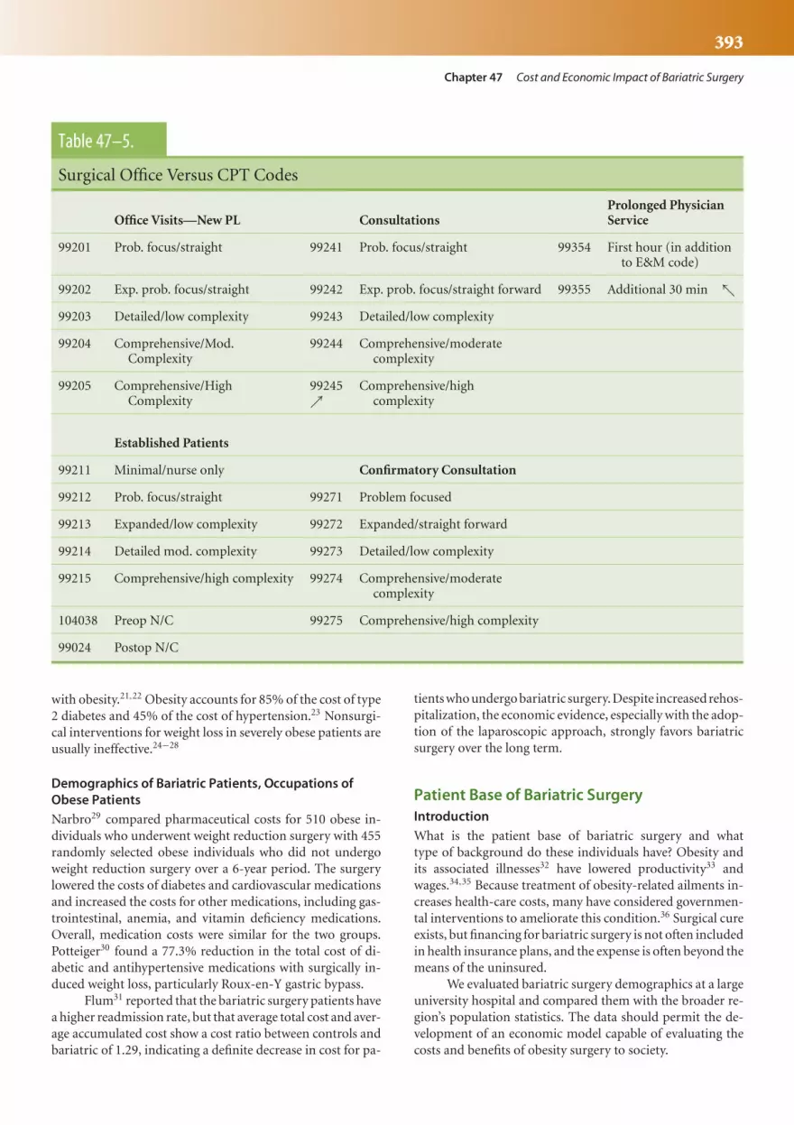
393
Chapter 47 Cost and Economic Impact of Bariatric Surgery
Table 47–5.
Surgical Office Versus CPT Codes
Prolonged PhysicianOffice Visits—New PL Consultations Service
99201 Prob. focus/straight 99241 Prob. focus/straight 99354 First hour (in additionto E&M code)
99202 Exp. prob. focus/straight 99242 Exp. prob. focus/straight forward 99355 Additional 30 min ↖99203 Detailed/low complexity 99243 Detailed/low complexity
99204 Comprehensive/Mod. 99244 Comprehensive/moderateComplexity complexity
99205 Comprehensive/High 99245 Comprehensive/highComplexity ↗ complexity
Established Patients
99211 Minimal/nurse only Confirmatory Consultation
99212 Prob. focus/straight 99271 Problem focused
99213 Expanded/low complexity 99272 Expanded/straight forward
99214 Detailed mod. complexity 99273 Detailed/low complexity
99215 Comprehensive/high complexity 99274 Comprehensive/moderatecomplexity
104038 Preop N/C 99275 Comprehensive/high complexity
99024 Postop N/C
with obesity.21,22 Obesity accounts for 85% of the cost of type2 diabetes and 45% of the cost of hypertension.23 Nonsurgi-cal interventions for weight loss in severely obese patients areusually ineffective.24−28
Demographics of Bariatric Patients, Occupations ofObese Patients
Narbro29 compared pharmaceutical costs for 510 obese in-dividuals who underwent weight reduction surgery with 455randomly selected obese individuals who did not undergoweight reduction surgery over a 6-year period. The surgerylowered the costs of diabetes and cardiovascular medicationsand increased the costs for other medications, including gas-trointestinal, anemia, and vitamin deficiency medications.Overall, medication costs were similar for the two groups.Potteiger30 found a 77.3% reduction in the total cost of di-abetic and antihypertensive medications with surgically in-duced weight loss, particularly Roux-en-Y gastric bypass.
Flum31 reported that the bariatric surgery patients havea higher readmission rate, but that average total cost and aver-age accumulated cost show a cost ratio between controls andbariatric of 1.29, indicating a definite decrease in cost for pa-
tients who undergo bariatric surgery. Despite increased rehos-pitalization, the economic evidence, especially with the adop-tion of the laparoscopic approach, strongly favors bariatricsurgery over the long term.
Patient Base of Bariatric SurgeryIntroduction
What is the patient base of bariatric surgery and whattype of background do these individuals have? Obesity andits associated illnesses32 have lowered productivity33 andwages.34,35 Because treatment of obesity-related ailments in-creases health-care costs, many have considered governmen-tal interventions to ameliorate this condition.36 Surgical cureexists, but financing for bariatric surgery is not often includedin health insurance plans, and the expense is often beyond themeans of the uninsured.
We evaluated bariatric surgery demographics at a largeuniversity hospital and compared them with the broader re-gion’s population statistics. The data should permit the de-velopment of an economic model capable of evaluating thecosts and benefits of obesity surgery to society.

394
Part VIII Special Topics in Bariatric Surgery
Costs of Obesity, Impact on Local and State Economy
Description of Sample
The age, race, education level, employment status, occu-pation, and bariatric surgical choice of 150 patients wascollected and compared with the state of New Mexico. Ofthe patients reviewed, 87.5% (130/150) were female, 62%(93/150) were Caucasian, 28% (42/150) were Hispanic, 4.5%(7/150) were African American, and 0.5% (1/150) were Na-tive American. Half (75/150) had a high school degree,20% (30/150) had a Bachelor’s degree, 6% (9/150) had anAssociate’s degree, 13 (19/150) had a Master’s degree, 1%(2/150) had a PhD, 0.5% (1/150) were physicians, 0.5%(1/150) were veterinary assistants, and 1.5% (3/150) had aneducation up to the eleventh grade. Of this sample, 82%(123/150) were employed, 10% (15/150) were homemakers,32% (48/150) were nurses, secretaries, or managers, 4.5%(7/150) were supervisors in companies, 3% (5/150) were stu-dents, 4% (6/150) were teachers, and 2.5% (4/150) were incustomer service. The remainder had a wide array of oc-cupations. In terms of age, 13% (8/150) were 40 years orolder, 4.5% (7/150) were 30–39 years old, and 7% were 20–29years old. Our results showed that morbid obesity affects all“classes” of the population. Bariatric surgery was particularlysought out by women with high school diplomas and higherdegrees.
Statistical Analysis of Surgery Choice
In the future, we will need to use a series of appropriate para-metric and nonparametric tests (e.g., t-statistics,χ2-statistics,etc.) to compare the patient sample to the population at large.From this information, an economic choice model can be de-rived to predict individuals who will opt to have this surgery.Identifying a set of predictor variables may be important forthe efficient allocation of scarce health-care state and federalresources.
The Impact of Obesity on the Local and StateEconomyA number of attempts have been made to quantify the eco-nomic costs of obesity and/or the economic or societal ben-efits associated with treatment and prevention.8,37,38
The costs are usually broken down into three cost cat-egories: direct, indirect, and personal.37 Much debate existsas to the appropriate cost–benefit measurement and, more-over, on whether or not economic methodology should befocused on evaluation of policy under the constraint of scarceresources instead of on the traditional role of costing.38
State Losses from Obesity, Illness and Obesity
The economic evaluation of solutions pays particular atten-tion to direct costs of obesity that accrue to a community, ashealth-care resources must be diverted from one activity toanother in order to deal with obesity-related illnesses. In termsof personal costs, some studies show that persons deemed
overweight or obese have lower wages, though other studieshave not found this to be true.10,11 Besides the “wage penalty”(in the jargon of economists) assessed for being obese, soci-ety suffers productivity losses from illnesses associated withobesity9; this is an indirect cost of obesity.
Currently underway is a study in our division that willestimate the economic impact of obesity on a specific localeconomy in terms of lost business output, lost employment,and lost income. The study uses information gathered onbariatric patients at a large university hospital. The region ofanalysis, dictated by patient residence and employment loca-tion, includes the state of New Mexico. Our goal is to quan-tify the state’s loss in jobs, income, and output attributable toobesity-related illnesses.
Absenteeism and Obesity, Job Cost Loss
Obesity is hypothesized to increase absenteeism. The asso-ciated lost productivity would yield lost output, jobs, andincome through a mechanism known as the economic mul-tiplier. Economic Impact Analysis studies the direct benefitsand costs of an event (or, in our case, the existence of obesity)as it affects local, regional, or national economies (EconomicAnalysis Primer, USDOT, FHWA 2003). Economic impactsarise from payroll spending, business/organizational operat-ing expenses, and construction projects; these factors in turngenerate jobs and income through a process known as the“multiplier effect.”
MATERIALS AND METHODS
Input–output (I–O) analysis, which estimates margins and re-gional purchase coefficients (RPC), was used to model the re-gional economy. Margins refer to the prices of purchases madeby final consumers (or households) of goods and services.RPC measures the percent of spending from local suppliers.Collected patient data used in conjunction with IMPLANmodel data sufficed to estimate a complete set of economicaccounts for local areas,39 which are converted to an industry-by-industry formulation of I–O accounts and then into a setof multipliers.
From each data set were derived final demands, valueadded, output, and employment. Actual local employment byindustry was used to partly validate the model. As a generalrule, the quantity of goods and services purchased correlatedwith the economic activity; these factors in turn correlate withthe magnitude of the multiplier.
Specific patient observations included informationabout socioeconomic status and demographics, occupation,education, and place of residence, which along with per-sonal income and absenteeism data (as compared with nor-mal weight persons) estimates lost work days attributableto obesity-related problems. The lost work days were thenconverted to full-time job equivalents, i.e., the jobs lostdue to obesity, which are then entered into the IMPLANmodel. Estimates are computed for over 500 subsectors (based

395
Chapter 47 Cost and Economic Impact of Bariatric Surgery
on the North American Industrial Classification System,NAICS).
EXPECTED RESULTS AND REMARKS
If an obese worker had one more day of absence or otherdowntime per month than a normal weight worker, 2002economic data suggest the region experienced a loss in totaloutput of $9.65 million per year. This loss estimates the eco-nomic impact of obesity on the regional economy and takesinto account both direct and secondary impacts. The loss rep-resents how much more the economy would produce if theseworkers followed the average absence/downtime patterns ofnormal weight workers. To put the loss in perspective, theloss in potential output from obesity is equivalent to slightlymore than $500 per person and, in total, is about 1.8% of thetotal gross state product.
To date, no study has specifically estimated the impactof obesity on a regional economy.40 The study now underway will estimate lost jobs, income (in dollars), and output(in dollars) attributable to obesity-related illnesses. The find-ings may aid public policy decision making in regard to thedesigning and implementation of health-care policy. For ex-ample, finding substantial economic impacts from obesitymight suggest that employers implement work-site healthpromotion programs, as some evidence indicates such treat-ment programs reduce health-related absenteeism.27,29 Morepatients are looking for surgery and some of them are readyto pay out of their own pocket,41 decreasing the health-carecosts.
CONCLUSION
The cost of surgery and the impact of the expenses on sur-geons, the hospital, health care, and the government are de-lineated in this chapter. More research and papers need tobe developed to obtain a better idea of our progress in halt-ing the spread of morbid obesity nationally and worldwideand to determine the cost to the world economy. As we haveseen in this chapter, obesity is a pervasive problem that hasa profound impact on the health-care system. Collection ofeconomic data will permit the development of an economicmodel capable of evaluating the cost effectiveness of obesitysurgery. Evaluating the costs and benefits of obesity surgerywill aid policymakers, health-care professionals, and organi-zations in making decisions about the allocation of resourcesfor the treatment of morbid obesity and the role of bariatricsurgery.
AcknowledgmentsWe would like to thank Brad Ewing, PhD, for his economicinsights and help, June Wagner for her editing job, and LisaBruster for her typographical assistance.
References1. Albert M: Morbid obesity: The value of surgical intervention. Clin
Fam Pract 4:447–451, 2002.2. Angus LD, Cottam DR, Gorecki PJ, Mourello R, Ortega RE,
Adamski J: DRG, costs and reimbursement following Roux en-Ygastric bypass: An economic appraisal. Obes Surg 13(4):591–595,2003.
3. Baum C, Ford W: The wage effects of obesity: A longitudinal study.Health Econ 13:885–899, 2004.
4. Berke EM, Morden NE: Medical management of obesity. Am FamPhysician 62:419–426, 2000.
5. Birmingham CL, Muller JL, Palepu A, et al.: The cost of obesity inCanada. CMAJ 160:483–488, 1999.
6. Cawley J: The impact of obesity on wages. J Hum Resour 34:451–474,2004.
7. Colditz GA: Economic costs of obesity. Am J Clin Nutr 55:503S–507S,1992.
8. Colditz GA: Economic costs of obesity and inactivity. Med Sci SportsExerc 31(Suppl 11):S663–S667, 1999.
9. Finkelstein MM: Obesity, cigarette smoking and the cost ofphysicians’ services in Ontario. Can J Public Health 92:437–440,2001.
10. Fisher BL, Schauer P: Medical and surgical options in the treatmentof severe obesity. Am J Surg 184:S9, 2002.
11. Flum DR, Salem L, Elrod JA, Dellinger EP, Cheadle A,Chan L: Early mortality among Medicare beneficiaries under-going bariatric surgical procedures. JAMA 294(15):1903–1908,2005.
12. Frezza EE: A plan to establish a private-pay bariatric surgery in theU.S.A. Obes Surg 16(1):110–111, 2006.
13. Frezza EE, Wachtel MS, Ewing BT: Bariatric surgery costs and impli-cations for hospital margins: Comparing laparoscopic gastric bypassand laparoscopic gastric banding. Surg Laparosc Endosc PercutanTech, 2006.
14. Frezza EE: How to improve office collection in a bariatric practice.Obes Surg 15:1352–1354, 2005.
15. Goldblatt PB, Moore ME, Stunkard AJ: Social factors in obesity.JAMA 192:1039–1044, 1965.
16. Gorstein J, Grosse R: The indirect costs of obesity to society. Phar-macoeconomics 5(Suppl 1):58–61, 1994.
17. Jeffrey RW, Forster JL, Dunn BV, French SA, McGovern PG,Lando HA: Effects of work-site health promotion on illness-relatedabsenteeism, Am College Occup Environ Med 35(11):1142–1146,1993.
18. Krott MA, Langley PC, Cox ER: A review of cost-of-illness studieson obesity. Clin Ther 40:772–779, 1998.
19. Levy E, Levy P, Le Pen C, et al.: The economic cost of obesity: TheFrench situation. Int J Obes 19:788–792, 1995.
20. Narbro K, Agren G, Jonsson E, et al.: Pharmaceutical costs in obeseindividuals. Arch Intern Med 162:2061–2069, 2002.
21. National Institutes of Health. Gastrointestinal surgery for severeObesity: National Institutes of Health Consensus DevelopmentConference Statement. Am J Clin Nutr 55(Suppl 2):S582–S585,1992.
22. Olson D, Lindall S: IMPLAN User’s Guide. Minnesota: MinnesotaIMPLAN Group 2002, published by State of Minnesota.
23. Oster G, Thompson D, Edelsberg J, et al.: Lifetime health and eco-nomic benefits of weight loss among obese persons. Am J PublicHealth 89:1536–1542, 1999.
24. Philipson T: The world-wide growth in obesity: An economic re-search agenda, Health Econ 10:1–7, 2001.
25. Potteiger CE, Prokash RP, Inverso NA, et al.: Bariatric surgery: Shed-ding the monetary weight of prescription cost in the managed carearena. Obes Surg 14:725–730, 2004.
26. Rous L, Donaldson C: Economics and obesity: Costing theproblem or evaluating the solutions? Obes Res 12(2):173–179,2004.

396
Part VIII Special Topics in Bariatric Surgery
27. Sampalis JS, Liberman M, Auger S, Christou NV: The impact ofweight reduction surgery on health-care costs in morbidly obesepatients. Obes Surg 14:939–947, 2004.
28. Seidell JC: Societal and personal costs of obesity. Exp Clin EndocrinolDiabetes 106(Suppl 2):7–9, 1998.
29. Sorensen TI: Socio-economic aspects of obesity: Causes or effects?Int J Obes 19(Suppl 6):S6–S8, 1995.
30. Oster G, O’Sullivan A, Edelsberg J, et al.: The clinical and economiccost of obesity in a managed care setting. Am J Manag Care 6:681–689, 2000.
31. Sturm R: The effects of obesity, smoking, and drinking on medicalproblems and costs. Health Aff 21:245–253, 2002.
32. Swinburn B, Ashton T, Gillespie J, et al.: Health care costs of obesityin New Zealand. Int J Obes 21:891–896, 1997.
33. Thompson D, Edelsberg J, Colditz GA, et al.: Lifetime health andeconomic consequences of obesity. Arch Intern Med 159:2177–2183,1999.
34. Thompson D, Wolf AM: The medical-care cost burden of obesity.Obes Rev 2:189–197, 2001.
35. Wang G, Zheng ZJ, Heath G, et al.: Economic burden of cardiovas-cular disease associated with excess body weight in U.S. adults. Am JPrev Med 23:1–6, 2002.
36. Weintraub M: Long-term weight control study conclusions. ClinPharmocol Ther 51:642–646, 1992.
37. Wittgrove AC, Clark GW: Laparoscopic gastric bypass, Roux-en-Y—500 patients: Technique and results, with 3–60 month follow up. ObesSurg 10:233–239, 2000.
38. Wolf AM: What is the economic case for treating obesity? Obes Res6(Suppl 1):2S–7S, 1998.
39. Wolf AM, Colditz GA: Social and economic effects of body weightin the United States. Am J Clin Nutr 63:466S, 1996.
40. Wolf AM, Colditz GA: Current estimates of the economiccost of obesity in the United States. Obes Res 6:97–106, 1998.www.cureyourpractice.com

48Adolescent Bariatric SurgeryVenita Chandra, MD � Sanjeev Dutta, MD, MA, FRCSC
I. BACKGROUNDEpidemiologyDefinition of ObesityRisk FactorsLong-Term Effects
II. CONSEQUENCES AND COMORBIDITIES OFADOLESCENT OBESITYMedical Impact
III. NONSURGICAL THERAPIES
IV. SURGICAL THERAPIESSurgical Options
V. GUIDELINES FOR BARIATRIC SURGERY INADOLESCENTSPreoperative EvaluationPostoperative ManagementNutritional ConsequencesComplianceOutcomes
VI. SUMMARY
BACKGROUND
Obesity is rapidly becoming a problem of epic proportionsin the adolescent patient population. The implicationsof this profound health crisis are clearly recognizedas serious and immediate, and long-term physicaland psychological consequences are being identified.Childhood and adolescent obesity are now consideredindependent risk factors for adult morbidity and prematuremortality.1,2
Resorting to bariatric surgery to address these issuesin morbidly obese adolescents who have life-threatening
comorbidities is a radical concept demanding increasingattention, particularly given the growing amount of datademonstrating positive outcomes after bariatric surgery inthe adult population. Adolescence represents a period of sig-nificant growth and maturation both physically and emotion-ally, thus issues unique to this patient population exist, whichmust be addressed when considering bariatric surgery. Thischapter focuses on aspects of bariatric surgery unique to theadolescent population, including the health consequences ofobesity in this age group, the indications for bariatric surgery,preoperative and postoperative management, as well as a re-view of current outcomes.
Copyright © 2008 by The McGraw-Hill Companies, Inc. Click here for terms of use.

398
Part VIII Special Topics in Bariatric Surgery
EpidemiologyIn the United States, obesity has far surpassed malnutritionas the most pressing nutritional disorder.3 Children are thefastest growing segment of the population afflicted by thisdisorder. The proportion of children and adolescents whoare obese tripled between 1980 and 2002.4,5 Recent preva-lence studies in the United States estimate 17.1% of childrenand adolescents to be overweight.6 Clear ethnic and racialdisparities are also noted with especially high rates of obesityin the Hispanic and African American communities.6−9 Arecent study in a New York City elementary school reviewedover 3,000 students’ demographic data and demonstrated theprevalence of overweight in young children to be 43% (morethan half of whom were obese) with significantly higher levelsamong Hispanic and Black children.10
This problem of obesity is not isolated to the UnitedStates, rising trends in overweight and obesity both in adultsand children are apparent in both developed and developingcountries worldwide.11−15 In China, for example, the preva-lence of obesity in school children increased from 1.5% in thelate eighties to 12.6% in 1997.16
Definition of ObesityThroughout childhood, dramatic changes in adiposity,height, and weight occur; thus, the exact definition of obe-sity in children and adolescents is subject to much debate. Ingeneral, it can be defined as an excess of body fat. Accurateassessment of body fat requires underwater weighing, dualenergy x-ray absorptiometery, or bioelectric impedance, allof which are not practical for everyday use.17
Body mass index (BMI, kg/m2) is widely used as a sur-rogate measure of adiposity, particularly in the adult pop-ulations. An adult with a BMI ≥ 30 kg/m2 is consideredobese.18 In children and adolescents who are still growingand have changing body shapes, BMI can be less accurate,particularly as it fails to distinguish between fat and fat-freemass.17,19 Thus, the application of growth charts and multiplepercentiles is necessary to determine overweight and obesityfor age and sex in this group.18,20,21 Pediatric obesity is nowgenerally defined as a BMI greater than the ninety-fifth per-centile and pediatric overweight with a BMI greater than theeighty-fifth percentile.22
Risk FactorsThe extent and impact of today’s obesity epidemic both onchildren and adults demands the question “why?” The answerto this is not straightforward. Overweight and obesity arecomplex multifactorial phenotypes influenced by an inter-action of genetic, behavioral, and environmental factors. Anin-depth discussion of this is beyond the scope of this chapter;however, some basic observations can be made.
Risk factors for the development of obesity in childhoodor adolescence are multifactorial, although the overall risk ac-cumulates with age. There are robust data demonstrating anassociation between higher birth weight and higher attained
BMI in childhood and early adulthood.23−28 Interestingly,lower birth weight has also been associated with increasedrisk of later central obesity,26,29−32 insulin resistance, and themetabolic syndrome.26,33−37 Several studies have assessed in-trauterine environment and determined maternal obesity andgestational diabetes to be strong predictors of childhood andadolescent obesity.23,27,38 These risk factors have also beenshown, when paired, to be associated with an increased riskof metabolic syndrome during childhood.27
Parental BMI is also a significant risk factor for obesity.Parental obesity more than doubles the risk of adult obesityamong both obese and nonobese children under the age of10.39 Other variables that are shown to be risk factors in-clude duration of breastfeeding, socioeconomic status, ma-ternal smoking status, and hours spent watching television.25
Long-Term EffectsMultiple studies suggest that long-term health is significantlycompromised due to persistence of adolescent overweightinto adulthood.40−44 This persistence of obesity results in abroad range of adverse health effects.41 In addition to physicalhealth, overweight in adolescents has harmful effects on theirsubsequent self-esteem and social and economic character-istics. For example, Gortmaker and colleagues45 looked at acohort of 370 overweight adolescents and found that obeseyoung women were less likely to marry, had lower incomes,and had completed less schooling than did nonobese womenand that obese young men were less likely to marry than werenonobese men.
CONSEQUENCES AND COMORBIDITIES OFADOLESCENT OBESITY
With the epidemic of obesity in children, we are seeing anemergence of traditionally adult diseases in this youngerpopulation. In addition to medical issues, the psychologicalsequelae of obesity during some of the most formative yearsof life have significant impact on how these children and ado-lescents adapt as effective members of society (Table 48–1).The potential ramifications of these issues on our country’shealth-care system and on the future health of our nation arequite alarming.
Medical ImpactAs in adults, obesity in adolescents is associated with a largenumber of health consequences. Although these obesity-related comorbidities occur more frequently in adults,significant consequences of obesity as well as the antecedentsof adult disease occur in obese children and adolescents.
Metabolic Syndrome and Cardiac Risk Factors
The metabolic syndrome is a cluster of risk factors that links in-sulin resistance, hypertension, dyslipidemia, type 2 diabetes,and other metabolic abnormalities with an increased risk of

399
Chapter 48 Adolescent Bariatric Surgery
Table 48–1.
Consequences and Comorbidities ofAdolescent Obesity
Psychosocial Gastrointestinal
Low self-esteem Gallstones
Depression Steatohepatitis
Eating disorders Gastroesophageal refluxdisease
Discrimination, prejudice,social marginalization
Endocrine
Poor quality of life,impairment of activitiesof daily living
Type 2 diabetes mellitus
Neurological Insulin resistance
Pseudotumor cerebri Metabolic syndrome
Pulmonary Precocious puberty
Obstructive sleep apnea Polycystic ovary syndrome
Asthma and exerciseintolerance
Musculoskeletal
Cardiovascular Slipped capital femoralepiphysis
Dyslipidemia Blount’s disease
Hypertension Forearm fractures
Coagulopathy Flat feet
Chronic inflammation Osteoarthritis
Endothelial dysfunction Scoliosis
Venous stasis disease Spondylolisthesis
Renal Dermatologic
Glomerulosclerosis Intertriginous soft-tissueinfections
Acanthosis nigricans
later cardiovascular disease.46 This syndrome, at first thoughtto be only an adult phenomenon, is unfortunately becom-ing more prominent in adolescents and children. Cook andcolleagues47 studied the prevalence of metabolic syndromein a sample of adolescents from the third National Healthand Nutrition Examination Survey (NHANES III) and foundthe syndrome to be present in 28.7% of overweight adoles-cents (BMI ≥ 95th percentile). More recent findings, using
modified criteria, suggest that the metabolic syndrome is farmore common (nearly 50%) among overweight children andadolescents and that its prevalence increases directly with thedegree of obesity.48
Obese children and adolescents have increased bloodlipids as well as glucose intolerance and diabetes.49−51 Al-though thought to be a traditionally “adult” disease, type 2diabetes is becoming shockingly more common in obese chil-dren and adolescents. The incidence among adolescents hasincreased 10-fold since 1982 and is felt to account for one-third of all new cases of diabetes in children,52 these numbersbeing even higher in women and Hispanics.53
Hypertension is yet another example of a predomi-nantly adult disease, with a rising incidence in the pedi-atric population.54−56 The long-term consequences are asyet unknown; however, there is some evidence that child-hood hypertension can lead to adult hypertension, and thusthe associated development of left ventricular hypertrophy,atherosclerosis, and even early development of coronaryartery disease.44,57−62
Obstructive Sleep Apnea
Sleep apnea is another significant health consequence ofchildhood obesity. Symptoms which may include snoring,poor school performance, enuresis, and hyperactivity canhave significant effects on quality of life.63,64 The prevalenceof obstructive sleep apnea (OSA) in obese children and ado-lescents is quoted anywhere from 24% to 59%65−68; however,a direct correlation between reduction in OSA indices andreduction in BMI has not been demonstrated.69
Pseudotumor Cerebri
Pseudotumor cerebri (PTC) is a rare disorder character-ized by increased intracranial pressure. Symptoms includeheadache, visual field disturbances (the most severe is blind-ness), and pulsatile tinnitus.70,71 PTC in adolescents has adirect correlation with obesity.72,73 Progressive visual impair-ment indicates the need for aggressive treatment of obesityin patients with this disease.44 Weight loss is associated withdramatic improvement in symptoms and improvements inobjective measures of the disease including visual field ab-normalities and lumbar pressures.74−76
Musculoskeletal Effects
Overweight children are susceptible to developing orthope-dic complications because the tensile strength of bone andcartilage is not evolved to carry substantial quantities ofexcess weight. As a result, the excess weight carried by obesechildren is associated with bowing of long bones, which maycause injury to the growth plate and result in slipped capitalfemoral epiphysis, genu valga, tibia vara (Blount’s disease),scoliosis, osteoarthritis, and other injuries.44,66 Both slippedcapital femoral epiphysis and Blount’s disease are pediatricorthopedic injuries that are highly associated with obesityin children, and prompt and sustained weight reduction isessential in their treatment.77,78

400
Part VIII Special Topics in Bariatric Surgery
Psychosocial Impact
Few problems in childhood have as significant an impact onemotional development as being overweight. Adolescence inand of itself is a tumultuous period of emotional and interper-sonal growth and development for children. It is not surpris-ing therefore that the psychosocial consequences of obesitymay in fact be the most prevalent of associated comorbidi-ties. Obesity is one of the most stigmatized and least sociallyacceptable conditions in childhood.79 Health related qual-ity of life studies document marked impairments in all do-mains including physical functioning, emotional well-being,social relations, and school functioning in adolescents withsevere obesity (BMI > 40 kg/m2). This level of impairmentis even worse than for those youth with chronic disease orcancer.64,80,81
Relative to average-weight peers, obese adolescents,particularly females, are socially marginalized and are lesslikely to be nominated by their peers as a friend.79 Given theimportance of peer acceptance, body image, and physical fit-ness to social and emotional development, overweight mayhave lasting implications on child development and adoles-cent well-being. These adolescents are clearly at considerablerisk of continued and mounting psychosocial impairmentand poor developmental adaptation.82 Among severely obeseadolescents, there is a high incidence of low self-esteem, sad-ness, and moderate to severe depressive symptoms. Extremeobesity is associated with an increased risk of suicide and sui-cidal ideation among adolescents.66,83,84 The impact of thispsychosocial impairment is grand, and some studies even sug-gest that obesity may be the worst socioeconomic handicapthat women who were obese adolescents can suffer.44,45
NONSURGICAL THERAPIES
It is evident that the health and psychosocial consequencesof child and adolescent obesity are profound, resultingin significant morbidity and increased long-term adultmortality.41,42,85 Evaluation and treatment of obesity in ado-lescents and children is imperative in order to avoid latersequelae and requires early multidisciplinary intervention bypediatric care providers.
Obesity results from an imbalance in energy intake andexpenditure, thus the ideal treatment for obesity involvesdecreasing caloric intake while increasing caloric expen-diture. Although no evidence-based overweight treatmentguidelines exist for adolescent overweight, traditional clinicalapproaches include combinations of caloric restriction, exer-cise promotion, and behavior therapy. Considerable evidenceshows that behavioral family-based treatment approaches arethe most effective nonsurgical methods for the managementof childhood obesity.86−88
Experience with medication for use in adolescentweight loss is limited. Currently, two medications, orlistat(Xenical, Roche Pharmaceuticals, Nutley, NJ) and sibu-tramine (Meridia, Knoll Pharmaceutical Company, Bray,
GA), have received FDA approval for long-term use inadolescents. Orlistat inhibits pancreatic lipase and increasesfecal losses of triglycerides. Although early evidence showedorlistat use to result in slight improvement in weightcontrol,89 a recent randomized controlled trial of adolescentsusing orlistat did not show significant reduction in BMI ascompared to placebo at 6 months.90 In addition, orlistat is as-sociated with high study-dropout rates because of unaccept-able flatulence and diarrhea as side effects of the drug.66,91
Sibutramine is a nonselective inhibitor of serotonin, nor-epinephrine, and dopamine which acts as an anorectic bystimulating satiety. A randomized controlled trial showeda statistically significant amount of weight loss with sibu-tramine (10.3 ± 6.6 kg) plus diet and exercise as compared toplacebo plus diet and exercise (2.4 ± 2.5 kg)92; however, thebenefits appear to be only short term.93
Successful weight loss and maintenance require greateffort and commitment. Although success is possible, moststudies show that behavior modification and dieting are as-sociated with poor weight loss, high attrition rates, and highprobability of weight regain.66,94−97 Bariatric surgery appearsto be the only intervention with evidence suggesting success-ful long-lasting (>1 year) effects on body weight in severelyobese adolescents.95 As a result, more and more pediatricproviders are considering bariatric surgery for the treatmentof obese adolescents.
SURGICAL THERAPIES
Evidence demonstrating the benefit of surgical weight lossin adults is extensive including not only dramatic reduc-tions in BMI but resolution of associated comorbidities, aswell as reduced mortality.98−108 Preliminary experience withbariatric surgery in adolescents suggests efficacy in amelio-rating obesity-related morbidities as well.76,109,110 Thus, forseverely obese adolescents who have failed organized attemptsat achieving and maintaining weight loss through conven-tional nonoperative approaches and who have serious orlife-threatening conditions, bariatric surgery may providethe only practical alternative for achieving a healthy weightand for escaping the devastating physical and psychologicalconsequences of obesity.111
Surgical OptionsThe goals of surgical therapy for obese adolescents, as foradults, are reduction in excess body weight in addition toreduction or resolution of obesity-related comorbidities andreduction in the risk of early mortality. The National Insti-tutes of Health Consensus Conference has endorsed bariatricsurgery as effective in achieving significant and sustainedweight reduction as well as ameliorating or resolving mostobesity-related comorbidities.112 The two main bariatric sur-gical procedures used in the adolescent population todayare Roux-en-Y gastric bypass (RYGBP) and adjustable gas-tric banding. Primarily, malabsorptive procedures such as

401
Chapter 48 Adolescent Bariatric Surgery
biliopancreatic bypass with duodenal switch which are oc-casionally used in adults are not recommended for usein adolescents, given their relative high rate of nutritionalsequelae.21,76,109 No studies to date have compared the effi-cacy and safety of various bariatric procedures among adoles-cents; however, both RYGBP and adjustable gastric bandinghave been effective in treating the medical consequences ofsevere obesity in adolescence.76,113−118
Roux-en-Y Gastric Bypass
The gold standard bariatric surgical procedure in the UnitedStates is the RYGBP procedure.111 The basic principles ofthis procedure are the same in adolescents as for adults:(1) Creation of a restrictive component (i.e. gastric pouch)and (2) reestablishment of intestinal continuity and creationof a malabsorptive component (i.e., Roux limb bypass ofstomach, duodenum, and portion of jejunum). Though itcan be done via laparotomy, recent data suggest that thelaparoscopic approach has advantages over the open whendone by experienced surgeons.119,120
The advantages of gastric bypass include excellentlargely sustainable excess weight loss, inherent deterrence tothe ingestion of sweets, and enhanced satiety. Disadvantagesinclude a greater risk of perioperative mortality demonstratedamong adults with gastric bypass (0.5%) compared to thosewith adjustable gastric banding (0.05%) as well as numberof complications such as intestinal leakage, thromboembolicdisease, marginal ulcers, dumping syndrome, gastrojejunalstomal stenosis, gastric distension, small bowel obstruction,incisional hernia, symptomatic cholelithiasis, protein calo-rie malnutrition, and micronutrient deficiencies, especiallyof iron, calcium, and vitamin B12.102,109,111,116
Restrictive Procedures
This category includes both vertical banded gastroplasty andlaparoscopic adjustable gastric banding (LAGB). Althoughthe vertical banded gastroplasty has been used in the past inboth adult and adolescent populations, it has been largely re-placed by the adjustable balloon gastric band and is now onlyof historic significance.21 LAGB consists of a laparoscopicallyplaced adjustable balloon band encircling the most proximalstomach creating a very small proximal gastric reservoir. Themain advantage of LAGB is its ease and safety of placement,and the lack of an anastomosis results in less morbidity andmortality associated with anastomoses. In addition, weightloss tends to be more gradual and the adjustable band reducesthe potential for adverse nutritional consequences.21,121 Dis-advantages include device malfunctions or malposition, slip-page, foreign-body infection, and band erosion into the stom-ach or esophagus.122 Furthermore, the mechanical deviceshave a finite lifetime, and thus adolescent patients may needto undergo replacement of the device during their lifetimes.111
LAGB is not currently approved by the FDA in theUnited States for use in adolescents. Studies from Europeand Australia, where the technique has gained signifi-cant popularity, show excellent outcomes in adults, with
lower complication rates as compared to those resultingfrom RYGBP, although preliminary US studies show mixedresults.102,121,123−129 Some data suggest less total excess weightloss in LAGB patients as compared to that in RYGBPpatients, although this does not appear to affect resolu-tion of comorbidities.127,130 In addition, current literaturedemonstrates similar effectiveness of LAGB in adolescentsas for adults in terms of weight loss and resolution ofcomorbidities,113,118,131,132 including the most recent pub-lication by Horgan and colleagues118 reviewing four ado-lescents (age, 17–19) who underwent the procedure in theUnited States.
Compared to RYGBP, the LAGB is a particularlyattractive option in adolescents as it does not require anatomicrearrangement and is reversed with much greater ease. How-ever, the placement of a foreign body at such an early ageraises concerns over the potential for long-term morbidity,such as erosion, and the eventual need to replace the prosthe-sis due to its limited lifespan. Long-term studies are necessaryto better understand these issues.
GUIDELINES FOR BARIATRIC SURGERY INADOLESCENTS
In 1991, the National Institutes of Health published consensusguidelines for bariatric surgery in adults. On the basis of theserecommendations bariatric surgery is commonly performedin adults with BMI values of ≥35 kg/m2 with comorbiditiesand for BMI values of ≥40 kg/m2 with or without comorbidi-ties. However, simple adoption of these guidelines for use inyounger age groups is generally not felt to be appropriate.133
Recommendations specific for bariatric surgery inadolescents have since been proposed by a panel of ex-perts in the field of pediatric obesity.111 Given the uniquemetabolic, developmental, and psychological needs of ado-lescents, bariatric surgery in this population should onlybe performed at specialized centers under the supervi-sion of multidisciplinary teams of experts experienced inmeeting these distinct needs. These teams should includespecialists in adolescent obesity evaluation and manage-ment, psychology, nutrition, physical activity instruction,and bariatric surgery.111,134 Depending on the needs of theindividual patients, other pediatric experts in fields such asgynecology, endocrinology, pulmonology, gastroenterology,cardiology, anesthesiology, or orthopedics may be consulted.
It is imperative that pediatric surgical specialistsperforming minimally invasive bariatric procedures havespecialized training. There is substantial data showing thatminimally invasive bariatric surgery is one of the more tech-nically difficult operations to perform, with a steep learningcurve and demonstrated differences in outcome dependingon training background of the surgeons.135 Pediatric surgeonsinvolved in bariatric surgery should pursue advanced trainingand credentialing as recommended by the American Societyfor Bariatric Surgery. In addition, initially they should beginto work with an experienced bariatric surgical specialist.136

402
Part VIII Special Topics in Bariatric Surgery
Table 48–2.
Criteria for Adolescent Bariatric Surgery
Failure of ≥6 months of organized weight loss attempts
Attainment of physiologic maturity
BMI ≥ 40 kg/m2 with serious obesity-relatedcomorbiditiesa
BMI ≥ 50 kg/m2 with less serious obesity-relatedcomorbiditiesb
Commitment to comprehensive medical and psychologicevaluations before and after surgery
Commitment to medical and nutritional requirementspostoperatively
Ability to provide informed assent for surgical treatment
Demonstrate cognitive maturity and decisional capacity
Supportive family environment
a Serious comorbidities: Type 2 diabetes mellitus, obstructive sleep apnea,or pseudotumor cerebri.bLess serious comorbidities: Hypertension, dyslipidemia, nonalcoholicsteatohepatitis, venous stasis disease, significant impairment in activities ofdaily living, intertriginous soft-tissue infections, stress urinaryincontinence, gastroesophageal reflux disease, weight-related arthropathiesthat impair physical activity, and psychosocial distress.
Adolescents who have failed multiple nonsurgicalweight loss attempts may be considered candidates for sur-gical intervention. Table 48–2 lists the criteria recommendedby Inge and colleagues111 for patients being considered forbariatric surgery. In general, bariatric surgery is consideredan appropriate option for those adolescents with very severeobesity (BMI ≥ 40 kg/m2) along with the presence of seriouscomorbidities as well as for patients with higher BMI val-ues (≥50 kg/m2) and less serious obesity-related comorbidconditions (Table 48–2).111 Contraindications to surgery areoutlined in Table 48–3.
Preoperative EvaluationIntensive patient and family preoperative education and eval-uation is imperative for the success of bariatric surgery pro-grams. Medical evaluations should include investigation intopossible endogenous causes of obesity that may be amenableto treatment as well as identification of any obesity-relatedhealth complications, as is done in the adult population. Pre-operative evaluation at our institution involves some or all ofthe procedures listed below as indicated for each individualpatient:
� A consistent primary care provider (≥1-year relationship)� evaluation by pediatric weight clinic
Table 48–3.
Contraindications to Adolescent BariatricSurgery
Medically correctable cause of obesity
Substance abuse problem within preceding year
Medical, psychiatric, or cognitive condition that wouldimpair the patient’s ability to adhere to dietary ormedication regimen
Current lactation
Current pregnancy or planned pregnancy within 2 yearsafter surgery
Inability to comprehend or refusal to participate inlifelong medical surveillance
� evaluation by dietician� evaluation by psychiatrist� evaluation by physical therapist� completion of a detailed health questionnaire� electrocardiogram� echocardiogram� ultrasound (to assess for steatohepatitis and cholelithiasis)
◦ patients with symptomatic gallstones should have acholecystectomy before the bariatric procedure.
� bone age study (when indicated)� polysomnography (sleep study)� pulmonary function test and/or indirect calorimetry� endoscopy� attendance of support groups and education session� blood tests:
◦ complete blood count◦ serum electrolytes, renal and liver function tests◦ calcium and phosphate levels and uric acid levels◦ fasting lipids◦ insulin, blood sugar, hemoglobin A1 C◦ thyroid function tests◦ blood coagulation studies (PT, PTT, INR)
Physiologic maturity must be established prior to con-sideration of bariatric surgery, as the dietary complexitiesand potential nutritional consequences following surgery canstunt completion of growth. Physiologic maturation usuallyoccurs along with sexual maturation (Tanner) stage 3 or 4.Most often skeletal maturation (>95% of adult stature) is at-tained by age 14 in girls and 16 in boys.137 Obesity is usuallyassociated with precocious puberty, and thus overweight chil-dren are often taller and have advanced bone age comparedwith age-matched nonoverweight children.138−140 If there isuncertainty about whether adult stature has been attained,
403
Chapter 48 Adolescent Bariatric Surgery
skeletal maturation (bone age) can be assessed objectivelyusing a radiograph of the hand and wrist.137,141
In addition to undergoing medical assessment, com-prehensive psychological evaluations of both potential can-didates for bariatric surgery and their parents should beperformed.111,142 This involves assessment of the family unit;determination of coping skills; assessment of the severity ofpsychosocial comorbidities; review of past/present psychi-atric, emotional, behavioral, or eating disorders; and eval-uation of cognitive development, including determination ofdecisional capacity.
These evaluations serve to define potential supports andbarriers to patient adherence as well as patient and family pre-paredness for surgery and the required postoperative lifestylechanges. An in-depth understanding, by the team, of the pa-tients’ psychological makeup and their family strengths orfamily dysfunction could have significant effects on the over-all success of bariatric surgery. In addition, although legallyconsent for the surgical intervention must be obtained by theparents, ethically, the decisional capacity of the adolescentand their ability to give assent for surgery are of utmost im-portance and need to be clarified prior to proceeding withbariatric surgery.
Postoperative ManagementPostoperative follow-up is generally quite intensive inorder to verify adherence to diet and vitamin guidelines, en-sure optimal postoperative weight loss, and avoid nutritionalcomplications. Often programs establish follow-up visits inthe following manner: Weekly for 1 month, monthly until6 months, quarterly until first 12 months, biannually until24 months, and yearly therafter.140 It is strongly encouragedthat all patients have lifelong follow-up in order to ensureoptimal weight maintenance and overall health. In addition,the long-term affects of bariatric surgery in this patient pop-ulation are still unknown, thus ongoing rigorous clinical datacollection will allow for structured clinical trials and observa-tions to assist in our understanding and improve patient care.
Adolescents, like adults, are at increased risk of gall-stone formation and peptic ulcer development following gas-tric bypass; therefore, usually ursodiol and proton-pumpinhibitors are prescribed. Nonsteroidal anti-inflammatorymedications should also be avoided to reduce the risk of in-testinal ulceration and bleeding. Expected weight loss afterlaparoscopic gastric bypass surgery is 20–30 lb in the firstmonth and approximately 10 lb/month until the weight lossplateaus after 12–18 months for a total excess weight loss ofapproximately 60–70%.66
RYGBP necessitates very low calorie, low-carbohydratedietary intake; thus, attention to ensuring adequate daily pro-tein intake (0.5–1.0 g/kg per day) is important to maintainlean body mass. Vitamin and mineral supplementation is re-quired in order to optimize bone growth in growing adoles-cents and to avoid nutritional consequences discussed morein detail below. This usually includes chewable or solublemultivitamins, calcium citrate (1200–1500 mg/day), as well
as B-complex supplements to reduce the risk of beriberi,which has been documented in adolescents after gastric by-pass surgery.143 Menstruating females should also take ironsupplements. Bariatric surgery does not appear to affect theoutcome of subsequent pregnancies as long as the period ofrapid weight loss has passed; thus, pregnancy is contraindi-cated for 2 years after surgery.144
Nutritional ConsequencesBoth gastric restrictive and bypass procedures carry an as-sociated risk of macronutrient as well as micronutrientdeficiency.145 In adolescents, the long-term risk of mineral ormicronutrient deficiency, though not well defined, is a legit-imate concern. Vitamin and mineral deficiencies, includinghypovitaminosis A, B12, C, D, E, folic acid, and iron, have allbeen reported.143,146,147 The presumed mechanism for thesedeficiencies is thought to be malabsorption along with de-creased oral intake, including poor compliance with medica-tions postoperatively. Given the exponential skeletal growththat occurs during adolescence, it is unclear whether or notthe current recommended amount of calcium supplementswill be adequate to optimize bone growth. The decrease inbone density that occurs after surgical weight loss, combinedwith potential vitamin D and calcium deficiencies, may putadolescent bariatric patients at greater risk for fractures laterin life.148
ComplianceAdolescence is often regarded as a period of experimenta-tion, rebellion, risk taking, and egocentrism. All of thesemake surgical preparation and adolescent compliance to rec-ommended health behaviors uniquely challenging, partic-ularly when pursuing life-altering procedures like bariatricsurgery. Compliance with health-care recommendations inadolescents is shown to be disappointingly low. Adoles-cents with chronic medical conditions such as cystic fi-brosis, cancer, diabetes, and asthma have compliance ratesof approximately 40–50%.149−152 Rand and Macgregor116
reported poor compliance by adolescents following gastricbypass surgery, with less than 15% of adolescents demon-strating compliance to postoperative dietary multivitaminand nutrient supplementation.
Noncompliance is often associated with such psychoso-cial factors as low self-esteem, poor cognitive abilities, andinsufficient family support.148 Several studies suggest that be-havioral therapy can improve adolescent adherence to strictmedical and dietary regimens.21,153−155 Continued supportby behavioral psychologists with expertise in enhancing ad-herence is essential for success in dietary compliance andlong-term follow-up after bariatric procedures in adolescents.With the alterations in eating patterns that are required af-ter bariatric surgery, repetitive reinforcement with structuredfamily involvement and continued support is needed to facil-itate the formation of lifelong health-promoting habits.109

404
Part VIII Special Topics in Bariatric Surgery
OutcomesThere is currently a paucity of literature on long-term out-comes of bariatric surgery in adolescents. The majority ofrecent studies do, however, demonstrate the safety and ef-ficacy of bariatric surgery in adolescent patients in achiev-ing durable weight loss and amelioration of obesity-relatedcomorbidities.19,66,109,111,114,115,127,132,140,156,157
In the largest long-term series to date, Sugerman andcolleagues158 reviewed their 20-year experience with RYGBPsurgery in adolescents. The vast majority of their procedureswere done open. They demonstrated results similar to adultpatients, including sustained weight loss, correction of co-morbidities, and improved self-image. More recent experi-ence with laparoscopic RYGBP also demonstrates excellentexcess weight loss and resolution of comorbidities, althoughlong-term data is still pending.109,114,157
Perhaps even more reassuring than the amount of ex-cess weight loss is the dramatic improvement in most obesity-related comorbidities after bariatric surgery. These have beenvery clearly demonstrated in adults and early reports inadolescents are also showing this to be true.69,74,76,100,106
Lawson and colleagues114 recently published the first out-come report from the Pediatric Bariatric Study Group, withtheir retrospective review of 12 morbidly obese adolescentswho underwent RYGBP. In addition to a mean reductionin BMI of 37%, they demonstrated significant improve-ments in triglycerides, total cholesterol, fasting blood glu-cose, and fasting insulin after 1 year. Rand and Macgregor116
reported excellent psychosocial adjustment 6 years afterbariatric surgery with patients reporting increased self-esteem, improved social relationships, and feeling moreattractive.
Late weight gain remains a potential concern and isstill unclear given the limited long-term data. Recidivism inthe form of weight gain is estimated at 20–30% in adults.Durable weight loss occurs in most adolescents, yet up to 15%of these patients appear to have late weight regain.106 This oc-currence may be avoided with a behavioral management pro-gram specifically designed for adolescents.87,109 More long-term studies are necessary to further clarify this risk.
SUMMARY
Adolescent obesity is quickly becoming an epidemic world-wide. The impact of its attendant comorbidities will undoubt-edly prove devastating to the health of populations across theglobe unless practical solutions can be achieved. Programsaimed at prevention clearly must form the cornerstone ofthis effort. However, effective treatment for those adolescentsalready suffering from obesity cannot be delayed. There is sub-stantial evidence supporting the benefits of bariatric surgeryin this group in terms of excess weight loss and resolution ofcomorbidities.
The indications for bariatric surgery in adolescents arecurrently more conservative than those for adults. It may soon
become clear, however, that surgical intervention earlier inthe course of extreme obesity provides the best opportunityto reverse the debilitating cardiovascular, metabolic, and psy-chosocial sequelae of this disease.159,160 The development ofmultidisciplinary adolescent bariatric teams, both at individ-ual hospitals and on a national and international level, willaid in further clarifying the best treatment strategies.
References1. Reilly JJ, Methven E, McDowell ZC, et al.: Health consequences of
obesity. Arch Dis Child 88(9):748, 2003.2. Freedman DS, Khan LK, Dietz WH, et al.: Relationship of childhood
obesity to coronary heart disease risk factors in adulthood: TheBogalusa Heart Study. Pediatrics 108(3):712, 2001.
3. Baron M: Trends in pediatric nutrition. Health Care Food Nutr Focus23(4):9, 2006.
4. Hedley AA, Ogden CL, Johnson CL, et al.: Prevalence of overweightand obesity among US children, adolescents, and adults, 1999–2002.JAMA 291(23):2847, 2004.
5. Ogden CL, Flegal KM, Carroll MD, Johnson CL: Prevalence andtrends in overweight among US children and adolescents, 1999–2000. JAMA 288(14):1728, 2002.
6. Ogden CL, Carroll MD, Curtin LR, et al.: Prevalence of overweightand obesity in the United States, 1999–2004. JAMA 295(13):1549,2006.
7. Dwyer JT, Stone EJ, Yang M, et al.: Prevalence of marked overweightand obesity in a multiethnic pediatric population: Findings fromthe Child and Adolescent Trial for Cardiovascular Health (CATCH)study. J Am Diet Assoc 100(10):1149, 2000.
8. Kimm SYS, Barton BA, Obarzanek E, et al.: Obesity developmentduring adolescence in a biracial cohort: The NHLBI Growth andHealth Study. Pediatrics 110(5):e54, 2002.
9. Freedman DS, Khan LK, Serdula MK, et al.: Racial and ethnic dif-ferences in secular trends for childhood BMI, weight, and height.Obesity 14(2):301, 2006.
10. Thorpe LE, List DG, Marx T, et al.: Childhood obesity in New YorkCity elementary school students. Am J Public Health 94(9):1496,2004.
11. Wang Y, Monteiro C, Popkin BM: Trends of obesity and under-weight in older children and adolescents in the United States, Brazil,China, and Russia. Am J Clin Nutr 75(6):971, 2002.
12. Silventoinen K, Sans S, Tolonen H, et al.: Trends in obesity andenergy supply in the WHO MONICA Project. Int J Obes 28(5):710,2004.
13. Salazar-Martinez E, Allen B, Fernandez-Ortega C, et al.: Overweightand obesity status among adolescents from Mexico and Egypt. ArchMed Res 37(4):535, 2006.
14. Musaiger AO: Overweight and obesity in the Eastern Mediter-ranean Region: Can we control it? East Mediterr Health J 10(6):789,2004.
15. Wong JPS, Ho SY, Lai MK, et al.: Overweight, obesity, weight-relatedconcerns and behaviours in Hong Kong Chinese children and ado-lescents. Acta Paediatr 94(5):595, 2005.
16. Luo J, Hu FB: Time trends of obesity in pre-school children in Chinafrom 1989 to 1997. Int J Obes 26(4):553, 2002.
17. Dehghan M, Akhtar-Danesh N, Merchant AT: Childhood obesity,prevalence and prevention. Nutr J 4(1):24, 2005.
18. Cole TJ, Bellizzi MC, Flegal KM, Dietz WH: Establishing a stan-dard definition for child overweight and obesity worldwide: Inter-national survey. BMJ 320(7244):1240, 2000.
19. Inge TH, Zeller MH, Lawson ML, Daniels SR: A critical appraisal ofevidence supporting a bariatric surgical approach to weight man-agement for adolescents. J Pediatr 147(1):10, 2005.
20. Himes JH, Dietz WH. Guidelines for overweight in adolescentpreventive services: Recommendations from an expert committee.

405
Chapter 48 Adolescent Bariatric Surgery
The Expert Committee on Clinical Guidelines for Overweight inAdolescent Preventive Services. Am J Clin Nutr 59(2):307, 1994.
21. Nadler EP, Kane TD: Bariatric surgery. In: Langer JC, AlbaneseCT, (eds.): Pediatric Minimal Access Surgery. Boca Raton, Taylor &Francis, 2005; pp. 319–330.
22. Barlow SE, Dietz WH: Obesity evaluation and treatment: ExpertCommittee recommendations. The Maternal and Child Health Bu-reau, Health Resources and Services Administration and the De-partment of Health and Human Services. Pediatrics 102(3):E29,1998.
23. Gillman MW, Rifas-Shiman S, Berkey CS, et al.: Maternal ges-tational diabetes, birth weight, and adolescent obesity. Pediatrics111(3):e221, 2003.
24. Singhal A, Wells J, Cole TJ, et al.: Programming of lean body mass:A link between birth weight, obesity, and cardiovascular disease?Am J Clin Nutr 77(3):726, 2003.
25. Burke V, Beilin LJ, Simmer K, et al.: Predictors of body mass in-dex and associations with cardiovascular risk factors in Australianchildren: A prospective cohort study. Int J Obes 29(1):15, 2005.
26. Oken E, Gillman MW: Fetal origins of obesity. Obes Res 11(4):496,2003.
27. Boney CM, Verma A, Tucker R, Vohr BR: Metabolic syndrome inchildhood: Association with birth weight, maternal obesity, andgestational diabetes mellitus. Pediatrics 115(3):e290, 2005.
28. Malina RM, Katzmarzyk PT, Beunen G: Birth weight and its rela-tionship to size attained and relative fat distribution at 7 to 12 yearsof age. Obes Res 4(4):385, 1996.
29. Law CM, Barker DJ, Osmond C, et al.: Early growth and abdominalfatness in adult life. J Epidemiol Community Health 46(3):184, 1992.
30. Barker M, Robinson S, Osmond C, Barker DJ: Birth weight andbody fat distribution in adolescent girls. Arch Dis Child 77(5):381,1997.
31. Loos RJ, Beunen G, Fagard R, et al.: Birth weight and body com-position in young adult men—a prospective twin study. Int J Obes25(10):1537, 2001.
32. Loos RJF, Beunen G, Fagard R, et al.: Birth weight and body com-position in young women: A prospective twin study. Am J Clin Nutr75(4):676, 2002.
33. Stern MP, Bartley M, Duggirala R, Bradshaw B: Birth weight andthe metabolic syndrome: Thrifty phenotype or thrifty genotype?Diabetes Metab Res Rev 16(2):88, 2000.
34. Rich-Edwards JW, Colditz GA, Stampfer MJ, et al.: Birthweight andthe risk for type 2 diabetes mellitus in adult women. Ann InternMed 130(4 Pt 1):278, 1999.
35. Poulsen P, Vaag A: The intrauterine environment as reflected bybirth size and twin and zygosity status influences insulin actionand intracellular glucose metabolism in an age- or time-dependentmanner. Diabetes 55(6):1819, 2006.
36. Bavdekar A, Yajnik CS, Fall CH, et al.: Insulin resistance syndromein 8-year-old Indian children: Small at birth, big at 8 years, or both?Diabetes 48(12):2422, 1999.
37. Damm P: Gestational diabetes mellitus and subsequent develop-ment of overt diabetes mellitus. Dan Med Bull 45(5):495, 1998.
38. Vohr BR, McGarvey ST, Tucker R: Effects of maternal gestationaldiabetes on offspring adiposity at 4–7 years of age. Diabetes Care22(8):1284, 1999.
39. Whitaker RC, Wright JA, Pepe MS, et al.: Predicting obesity inyoung adulthood from childhood and parental obesity. N Engl JMed 337(13):869, 1997.
40. Freedman DS, Shear CL, Burke GL, et al.: Persistence of juvenile-onset obesity over eight years: The Bogalusa Heart Study. Am JPublic Health 77(5):588, 1987.
41. Must A, Jacques PF, Dallal GE, et al.: Long-term morbidity andmortality of overweight adolescents. A follow-up of the HarvardGrowth Study of 1922 to 1935. N Engl J Med 327(19):1350, 1992.
42. Must A: Morbidity and mortality associated with elevated bodyweight in children and adolescents. Am J Clin Nutr 63(Suppl3):445S, 1996.
43. Srinivasan SR, Bao W, Wattigney WA, Berenson GS: Adolescentoverweight is associated with adult overweight and related multiplecardiovascular risk factors: The Bogalusa Heart Study. Metabolism45(2):235, 1996.
44. Dietz WH: Health consequences of obesity in youth: Childhoodpredictors of adult disease. Pediatrics 101(3 Pt 2):518, 1998.
45. Gortmaker SL, Must A, Perrin JM, et al.: Social and economic con-sequences of overweight in adolescence and young adulthood. NEngl J Med 329(14):1008, 1993.
46. Reaven GM: Banting lecture 1988. Role of insulin resistance in hu-man disease. Diabetes 37(12):1595, 1988.
47. Cook S, Weitzman M, Auinger P, et al.: Prevalence of a metabolicsyndrome phenotype in adolescents: Findings from the third Na-tional Health and Nutrition Examination Survey, 1988–1994. ArchPediatr Adolesc Med 157(8):821, 2003.
48. Weiss R, Dziura J, Burgert TS, et al.: Obesity and the metabolicsyndrome in children and adolescents. N Engl J Med 350(23):2362,2004.
49. Sinha R, Fisch G, Teague B, et al.: Prevalence of impaired glucosetolerance among children and adolescents with marked obesity. NEngl J Med 346(11):802, 2002.
50. Duncan GE: Prevalence of diabetes and impaired fasting glucoselevels among US adolescents: National Health and Nutrition Ex-amination Survey, 1999–2002. Arch Pediatr Adolesc Med 160(5):523,2006.
51. Ehtisham S, Barrett TG: The emergence of type 2 diabetes in child-hood. Ann Clin Biochem 41(Pt 1):10, 2004.
52. Pinhas-Hamiel O, Dolan LM, Daniels SR, et al.: Increased incidenceof non-insulin-dependent diabetes mellitus among adolescents. JPediatr 128(5 Pt 1):608, 1996.
53. Narayan KMV, Boyle JP, Thompson TJ, et al.: Lifetime risk for dia-betes mellitus in the United States. JAMA 290(14):1884, 2003.
54. Luma GB, Spiotta RT: Hypertension in children and adolescents.Am Fam Physician 73(9):1558, 2006.
55. Stabouli S, Kotsis V, Papamichael C, et al.: Adolescent obesity is as-sociated with high ambulatory blood pressure and increased carotidintimal-medial thickness. J Pediatr 147(5):651, 2005.
56. Sorof JM, Lai D, Turner J, et al.: Overweight, ethnicity, and theprevalence of hypertension in school-aged children. Pediatrics113(3 Pt 1):475, 2004.
57. Berenson GS, Srinivasan SR, Bao W, et al.: Association between mul-tiple cardiovascular risk factors and atherosclerosis in children andyoung adults. The Bogalusa Heart Study. N Engl J Med 338(23):1650,1998.
58. Lauer RM, Clarke WR, Mahoney LT, Witt J: Childhood predictorsfor high adult blood pressure. The Muscatine Study. Pediatr ClinNorth Am 40(1):23, 1993.
59. Hanevold C, Waller J, Daniels S, et al.: The effects of obesity, gender,and ethnic group on left ventricular hypertrophy and geometry inhypertensive children: A collaborative study of the InternationalPediatric Hypertension Association. Pediatrics 113(2):328, 2004.
60. McGill HC, Herderick EE, McMahan CA, et al.: Atherosclerosis inyouth. Minerva Pediatr 54(5):437, 2002.
61. McGill HC, McMahan CA, Herderick EE, et al.: Obesity acceleratesthe progression of coronary atherosclerosis in young men. Circula-tion 105(23):2712, 2002.
62. Gidding SS, Nehgme R, Heise C, et al.: Severe obesity associatedwith cardiovascular deconditioning, high prevalence of cardiovas-cular risk factors, diabetes mellitus/hyperinsulinemia, and respira-tory compromise. J Pediatr 144(6):766, 2004.
63. Styne DM: Childhood and adolescent obesity. Prevalence and sig-nificance. Pediatr Clin North Am 48(4):823, 2001.
64. Schwimmer JB, Burwinkle TM, Varni JW: Health-related quality oflife of severely obese children and adolescents. JAMA 289(14):1813,2003.
65. Marcus CL, Curtis S, Koerner CB, et al.: Evaluation of pulmonaryfunction and polysomnography in obese children and adolescents.Pediatr Pulmonol 21(3):176, 1996.

406
Part VIII Special Topics in Bariatric Surgery
66. Helmrath MA, Brandt ML, Inge TH: Adolescent obesity andbariatric surgery. Surg Clin North Am 86(2):441, 2006.
67. Silvestri JM, Weese-Mayer DE, Bass MT, et al.: Polysomnographyin obese children with a history of sleep-associated breathing dis-orders. Pediatr Pulmonol 16(2):124, 1993.
68. Mallory GB, Fiser DH, Jackson R: Sleep-associated breathingdisorders in morbidly obese children and adolescents. J Pediatr115(6):892, 1989.
69. Kalra M, Inge T, Garcia V, et al.: Obstructive sleep apnea in ex-tremely overweight adolescents undergoing bariatric surgery. ObesRes 13(7):1175, 2005.
70. Friedman DI: Pseudotumor cerebri. Neurol Clin 22(1):99, 2004.71. Friedman DI, Jacobson DM: Idiopathic intracranial hypertension.
J Neuroophthalmol 24(2):138, 2004.72. Cinciripini GGS, Donahue SS, Borchert MMS: Idiopathic intracra-
nial hypertension in prepubertal pediatric patients: Characteristics,treatment, and outcome. Am J Ophthalmol 127(2):178, 1999.
73. Baker RRS, Baumann RRJ, Buncic JJR: Idiopathic intracranial hy-pertension (pseudotumor cerebri) in pediatric patients. PediatrNeurol 5(1):5, 1989.
74. Chandra V, Dutta S, Albanese CT, et al.: Clinical resolution ofseverely symptomatic pseudotumor cerebri after gastric bypass inan adolescent: A case report. Surg. Obes Relat Dis, 2007 Feb 24.
75. Nadkarni TT, Rekate HL, Wallace DD: Resolution of pseudotumorcerebri after bariatric surgery for related obesity. Case report. J Neu-rosurg 101(5):878, 2004.
76. Sugerman HJ, Sugerman EL, DeMaria EJ, et al.: Bariatric surgeryfor severely obese adolescents. J Gastrointest Surg 7(1):102, 2003.
77. Dietz WH, Gross WL, Kirkpatrick JA: Blount disease (tibia vara):Another skeletal disorder associated with childhood obesity. J Pe-diatr 101(5):735, 1982.
78. Kelsey JL, Acheson RM, Keggi KJ: The body build of patients withslipped capital femoral epiphysis. Am J Dis Child 124(2):276, 1972.
79. Strauss RS, Pollack HA: Social marginalization of overweight chil-dren. Arch Pediatr Adolesc Med 157(8):746, 2003.
80. Zeller MH, Roehrig HR, Modi AC, et al.: Health-related qual-ity of life and depressive symptoms in adolescents with extremeobesity presenting for bariatric surgery. Pediatrics 117(4):1155,2006.
81. Swallen KC, Reither EN, Haas SA, Meier AM: Overweight, obesity,and health-related quality of life among adolescents: The NationalLongitudinal Study of Adolescent Health. Pediatrics 115(2):340,2005.
82. Bagwell CL, Newcomb AF, Bukowski WM: Preadolescent friendshipand peer rejection as predictors of adult adjustment. Child Dev69(1):140, 1998.
83. Eisenberg ME, Neumark-Sztainer D, Story M: Associations ofweight-based teasing and emotional well-being among adolescents.Arch Pediatr Adolesc Med 157(8):733, 2003.
84. Dong C, Li WD, Li D, Price RA: Extreme obesity is associated withattempted suicides: Results from a family study. Int J Obes 30(2):388,2006.
85. Fontaine KR, Redden DT, Wang C, et al.: Years of life lost due toobesity. JAMA 289(2):187, 2003.
86. Jelalian E, Saelens BE: Empirically supported treatments in pediatricpsychology: Pediatric obesity. J Pediatr Psychol 24(3):223, 1999.
87. Epstein LH, Myers MD, Raynor HA, Saelens BE: Treatment of pe-diatric obesity. Pediatrics 101(3 Pt 2):554, 1998.
88. Kirk S, Zeller M, Claytor R, et al.: The relationship of health out-comes to improvement in BMI in children and adolescents. ObesRes 13(5):876, 2005.
89. Chanoine J-P, Hampl S, Jensen C, et al.: Effect of orlistat on weightand body composition in obese adolescents: A randomized con-trolled trial. JAMA 293(23):2873, 2005.
90. Maahs D, de Serna DG, Kolotkin RL, et al.: Randomized, double-blind, placebo-controlled trial of orlistat for weight loss in adoles-cents. Endocr Pract 12(1):18, 2006.
91. Ozkan B, Bereket A, Turan S, Keskin S. Addition of orlistat to con-ventional treatment in adolescents with severe obesity. Eur J Pediatr163(12):738, 2004.
92. Godoy-Matos A, Carraro L, Vieira A, et al.: Treatment of obese ado-lescents with sibutramine: A randomized, double-blind, controlledstudy. J Clin Endocrinol Metab 90(3):1460, 2005.
93. Reisler G, Tauber T, Afriat R, et al.: Sibutramine as an adjuvanttherapy in adolescents suffering from morbid obesity. Isr Med AssocJ 8(1):30, 2006.
94. Tsai AG, Wadden TA: Systematic review: An evaluation of majorcommercial weight loss programs in the United States. Ann InternMed 142(1):56, 2005.
95. Yanovski JA: Intensive therapies for pediatric obesity. Pediatr ClinNorth Am 48(4):1041, 2001.
96. Figueroa-Colon R, Franklin FA, Lee JY, et al.: Feasibility of a clinic-based hypocaloric dietary intervention implemented in a schoolsetting for obese children. Obes Res 4(5):419, 1996.
97. Methods for voluntary weight loss and control. NIH Technology As-sessment Conference Panel. Consensus Development Conference,30 March to 1 April 1992. Ann Intern Med 119(7 Pt 2):764, 1993.
98. McTigue KM, Harris R, Hemphill B, et al.: Screening and inter-ventions for obesity in adults: Summary of the evidence for theU.S. Preventive Services Task Force. Ann Intern Med 139(11):933,2003.
99. Sjostrom CD, Lissner L, Wedel H, Sjostrom L: Reduction in inci-dence of diabetes, hypertension and lipid disturbances after inten-tional weight loss induced by bariatric surgery: The SOS Interven-tion Study. Obes Res 7(5):477, 1999.
100. Buchwald H, Avidor Y, Braunwald E, et al.: Bariatric surgery: Asystematic review and meta-analysis. JAMA 292(14):1724, 2004.
101. Buchwald H, Buchwald JN: Evolution of operative proceduresfor the management of morbid obesity 1950–2000. Obes Surg12(5):705, 2002.
102. Chapman AE, Kiroff G, Game P, et al.: Laparoscopic adjustablegastric banding in the treatment of obesity: A systematic literaturereview. Surgery 135(3):326, 2004.
103. MacDonald GJ, Long GJ, Swanson GJ, et al.: The gastric bypassoperation reduces the progression and mortality of non-insulin-dependent diabetes mellitus. J Gastrointest Surg 1(3):213, 1997.
104. Flum DR, Dellinger EP: Impact of gastric bypass operation on sur-vival: A population-based analysis. J Am Coll Surg 199(4):543, 2004.
105. Dixon JB, Dixon AF, O’Brien PE: Improvements in insulin sensitiv-ity and beta-cell function (HOMA) with weight loss in the severelyobese. Homeostatic model assessment. Diabet Med 20(2):127, 2003.
106. Sugerman HJ, Fairman RP, Sood RK, et al.: Long-term effects ofgastric surgery for treating respiratory insufficiency of obesity. AmJ Clin Nutr 55(Suppl 2):597S, 1992.
107. Dymek MP, Le Grange D, Neven K, Alverdy J: Quality of life aftergastric bypass surgery: A cross-sectional study. Obes Res 10(11):1135, 2002.
108. Christou NV, Sampalis JS, Liberman M, et al.: Surgery decreaseslong-term mortality, morbidity, and health care use in morbidlyobese patients. Ann Surg 240(3):416, 2004.
109. Garcia VF, Langford L, Inge TH: Application of laparoscopy forbariatric surgery in adolescents. Curr Opin Pediatr 15(3):248,2003.
110. Strauss RS, Bradley LJ, Brolin RE: Gastric bypass surgery in adoles-cents with morbid obesity. J Pediatr 138(4):499, 2001.
111. Inge TH, Krebs NF, Garcia VF, et al.: Bariatric surgery for severelyoverweight adolescents: Concerns and recommendations. Pedi-atrics 114(1):217, 2004.
112. Health implications of obesity: National Institutes of Health Con-sensus Development Conference Statement. National Institutes ofHealth Consensus Development Conference Consensus Statement5(9):1, 1985.
113. Dolan K, Creighton L, Hopkins G, Fielding G: Laparoscopic gas-tric banding in morbidly obese adolescents. Obes Surg 13(1):101,2003.

407
Chapter 48 Adolescent Bariatric Surgery
114. Lawson ML, Kirk S, Mitchell T, et al.: One-year outcomes of Roux-en-Y gastric bypass for morbidly obese adolescents: A multicen-ter study from the Pediatric Bariatric Study Group. J Pediatr Surg41(1):137, 2006.
115. Anderson AE, Soper RT, Scott DH: Gastric bypass for morbid obe-sity in children and adolescents. J Pediatr Surg 15(6):876, 1980.
116. Rand CS, Macgregor AM: Adolescents having obesity surgery: A6-year follow-up. South Med J 87(12):1208, 1994.
117. Soper RT, Mason EE, Printen KJ, Zellweger H: Gastric bypass formorbid obesity in children and adolescents. J Pediatr Surg 10(1):51,1975.
118. Horgan S, Holterman MJ, Jacobsen GR, et al.: Laparoscopic ad-justable gastric banding for the treatment of adolescent morbidobesity in the United States: A safe alternative to gastric bypass. JPediatr Surg 40(1):86, 2005.
119. Nguyen NT, Ho HS, Palmer LS, Wolfe BM: A comparison study oflaparoscopic versus open gastric bypass for morbid obesity. J AmColl Surg 191(2):149, 2000.
120. Podnos YD, Jimenez JC, Wilson SE, et al.: Complications afterlaparoscopic gastric bypass: A review of 3464 cases. Arch Surg138(9):957, 2003.
121. O’Brien PE, Dixon JB, Brown W, et al.: The laparoscopic adjustablegastric band (Lap-Band): A prospective study of medium-term ef-fects on weight, health and quality of life. Obes Surg 12(5):652,2002.
122. Chevallier J-M, Zinzindohoue F, Douard R, et al.: Complicationsafter laparoscopic adjustable gastric banding for morbid obesity:Experience with 1000 patients over 7 years. Obes Surg 14(3):407,2004.
123. Nguyen NT, Silver M, Robinson M, et al.: Result of a national auditof bariatric surgery performed at academic centers: A 2004 Univer-sity HealthSystem Consortium Benchmarking Project. Arch Surg141(5):445, 2006.
124. DeMaria EJ, Jamal MK: Laparoscopic adjustable gastric banding:Evolving clinical experience. Surg Clin North Am 85(4):773, 2005.
125. O’Brien PE, Dixon JB, Laurie C, et al.: Treatment of mild to mod-erate obesity with laparoscopic adjustable gastric banding or anintensive medical program: A randomized trial. Ann Intern Med144(9):625, 2006.
126. Ren CJ, Horgan S, Ponce J: US experience with the LAP-BANDsystem. Am J Surg 184(6B):46S, 2002.
127. Angrisani L, Furbetta F, Doldi SB, et al.: Results of the Italian mul-ticenter study on 239 super-obese patients treated by adjustablegastric banding. Obes Surg 12(6):846, 2002.
128. Weiner R, Blanco-Engert R, Weiner S, et al.: Outcome after laparo-scopic adjustable gastric banding—8 years experience. Obes Surg13(3):427, 2003.
129. Parikh MS, Laker S, Weiner M, et al.: Objective comparison ofcomplications resulting from laparoscopic bariatric procedures. JAm Coll Surg 202(2):252, 2006.
130. Mognol P, Chosidow D, Marmuse J-P: Laparoscopic gastric bypassversus laparoscopic adjustable gastric banding in the super-obese:A comparative study of 290 patients. Obes Surg 15(1):76, 2005.
131. Dolan K, Fielding G: A comparison of laparoscopic adjustable gas-tric banding in adolescents and adults. Surg Endosc 18(1):45, 2004.
132. Abu-Abeid S, Gavert N, Klausner JM, Szold A: Bariatric surgery inadolescence. J Pediatr Surg 38(9):1379, 2003.
133. Gastrointestinal surgery for severe obesity. Proceedings of a Na-tional Institutes of Health Consensus Development Conference.March 25–27, 1991, Bethesda, MD. Am J Clin Nutr 55(Suppl2):487S, 1992.
134. Recommendations for facilities performing bariatric surgery. BullAm Coll Surg 85(9):20, 2000.
135. Hsu GP, Morton JM, Jin L, et al.: Laparoscopic Roux-en-Y gastricbypass: Differences in outcome between attendings and assistantsof different training backgrounds. Obes Surg 15(8):1104, 2005.
136. Lublin M, Lyass S, Lahmann B, et al.: Leveling the learningcurve for laparoscopic bariatric surgery. Surg Endosc 19(6):845,2005.
137. Tanner JM: Normal growth and techniques of growth assessment.Clin Endocrinol Metab 15(3):411, 1986.
138. Leonard MB, Shults J, Wilson BA, et al.: Obesity during childhoodand adolescence augments bone mass and bone dimensions. Am JClin Nutr 80(2):514, 2004.
139. Wang Y: Is obesity associated with early sexual maturation? A com-parison of the association in American boys versus girls. Pediatrics110(5):903, 2002.
140. Inge TH, Zeller M, Garcia VF, Daniels SR: Surgical approach toadolescent obesity. Adolesc Med Clin 15(3):429, 2004.
141. Gilli G: The assessment of skeletal maturation. Horm Res 45(Suppl2):49, 1996.
142. Dziurowicz-Kozlowska AH, Wierzbicki Z, Lisik W, et al.: Theobjective of psychological evaluation in the process of qual-ifying candidates for bariatric surgery. Obes Surg 16(2):196,2006.
143. Towbin A, Inge TH, Garcia VF, et al.: Beriberi after gastric bypasssurgery in adolescence. J Pediatr 145(2):263, 2004.
144. Sheiner E, Levy A, Silverberg D, et al.: Pregnancy after bariatricsurgery is not associated with adverse perinatal outcome. Am J Ob-stet Gynecol 190(5):1335, 2004.
145. Xanthakos, I: Nutritional consequences of bariatric surgery. CurrOpin Clin Nutr Metab Care 9(4):489, 2006.
146. Boylan LM, Sugerman HJ, Driskell JA: Vitamin E, vitamin B-6,vitamin B-12, and folate status of gastric bypass surgery patients. JAm Diet Assoc 88(5):579, 1988.
147. Halverson JD: Vitamin and mineral deficiencies following obesitysurgery. Gastroenterol Clin North Am 16(2):307, 1987.
148. Garcia V, Inge T: Bariatric surgery in adolescents. In: Grosfeld JL(ed.): Pediatric Surgery. Nashville, TN, Mosby, 2006.
149. Inge TH, Garcia V, Daniels S, et al.: A multidisciplinary approachto the adolescent bariatric surgical patient. J Pediatr Surg 39(3):442,2004.
150. Phipps S, DeCuir-Whalley S: Adherence issues in pediatric bonemarrow transplantation. J Pediatr Psychol 15(4):459, 1990.
151. Buston KM, Wood SF: Non-compliance amongst adolescents withasthma: Listening to what they tell us about self-management. FamPract 17(2):134, 2000.
152. Tamaroff MH, Festa RS, Adesman AR, Walco GA: Therapeuticadherence to oral medication regimens by adolescents with can-cer, II: Clinical and psychologic correlates. J Pediatr 120(5):812,1992.
153. Wysocki T, Harris MA, Greco P, et al.: Randomized, controlledtrial of behavior therapy for families of adolescents with insulin-dependent diabetes mellitus. J Pediatr Psychol 25(1):23, 2000.
154. Lemanek KL, Kamps J, Chung NB: Empirically supported treat-ments in pediatric psychology: Regimen adherence. J Pediatr Psy-chol 26(5):253, 2001.
155. Fielding D, Duff A: Compliance with treatment protocols: Inter-ventions for children with chronic illness. Arch Dis Child 80(2):196,1999.
156. Breaux. Obesity surgery in children. Obes Surg 5(3):279, 1995.157. Stanford A, Glascock JM, Eid GM, et al.: Laparoscopic Roux-en-
Y gastric bypass in morbidly obese adolescents. J Pediatr Surg38(3):430, 2003.
158. Sugerman HJHJ, Sugerman ELEL, DeMaria EJEJ, et al.: Bariatricsurgery for severely obese adolescents. J Gastrointest Surg 7(1):102,2003.
159. Capella JF, Capella RF: Bariatric surgery in adolescence. Is this thebest age to operate? Obes Surg 13(6):826, 2003.
160. Garcia VF, DeMaria EJ: Adolescent bariatric surgery: Treatmentdelayed, treatment denied, a crisis invited. Obes Surg 16(1):1,2006.

Index
abdominal wall complications, 350–351acne, from BPD, 127ad hoc stomach BPD (AHS BPD), 112,
126, 277adaptations
preoperativefor accommodation/hospital
admittance, 59for clinic evaluations, 57–59examining table, 58 (fig.)special size arterial pressure cuffs, 58
(fig.)in surgical center, 59–60
defibrillator availability, 60instrumentation availability, 60measurement equipment, 59reinforced toilets, 58 (fig.)scale for 500 kg, 58 (fig.)surgical tables, 59–60
adiponectin, 43, 46, 47, 106, 142adjustable gastric band, restrictive surgery,
253–261. See also LAP-BANDgastric band
advantages, 254–256complications, 254disadvantages, 256
adjustable Silastic gastric banding, 11(fig.)
adolescent obesityconsequences/comorbidities
metabolic syndrome/cardiac riskfactors, 398–399
musculoskeletal effects, 399obstructive sleep apnea, 399pseudomotor cerebri, 399psychosocial impact, 400
defined, 398epidemiology of, 398long-term effects, 398nonsurgical therapies, 400risk factors, 398surgical guidelines
compliance, 403
nutritional consequences, 403outcomes, 404postoperative management, 403preoperative evaluation, 402
surgical therapiesrestrictive procedures, 401Roux-en-Y gastric bypass, 401
afferent signals, of digestive tract, 42(table), 44 (fig.)
alkaline gastritis, 189American Association of Clinical
Endocrinologists (AACE), 150American Psychiatric Association, 76American Society for Bariatric Surgery
(ASBS), 33AMI Band r© gastric band, 170 (table)anal complications, BPD, 289–291anal rhagades, from BPD, 127anastomotic leaks, 29, 192, 215,
344–350as early complication, 358and fluid collections, 355–356in stapled gastrojejunostomy,
11–12anastomotic stricture, 30, 192, 314,
315anastomotic ulcers, 355, 359anesthesia, general
hemodynamic changes, 89mechanical ventilation, 89preoxygenation, 88tracheal intubation, 88
anesthesiology support, 62anesthetic technique, 89apM1 gene, 47apolipoprotein A-IV, 45apple vs. pear fat distribution, 16archetype, for morbid/super obesity, 78Ardelt-Moorehead-Weger Addiction
Scale, 76, 77arrhythmias, gastric, 368, 369asthma, 21atelectasis bronchospasm, 63
atherosclerosis, accelerated, and metabolicsyndrome, 155
Australia, obesity statistics, 55
Baker jejunostomy tube, 190balloon catheter, 190, 191banded gastric bypass (BGBP)
efficacy of, 226evolution, 221–222preformed silk loop introduction, 223
(fig.), 224 (fig.)safety issues
food intolerance/chronicregurgitation, 225–226
ring migration/erosion, 224techniques
Marema BGBP surgery, 223pouch formation/ring placement,
222–223Bariatric Analysis and Reporting Outcome
System (BAROS), 80, 173bariatric surgery. See also biliopancreatic
diversion; high risk, two-stageapproach; jejunoileal bypass;restrictive surgery; revisionalsurgery
for adolescents, 397–404anesthesia, general
hemodynamic changes, 89mechanical ventilation, 89preoxygenation, 88tracheal intubation, 88
anesthetic technique, 89bariatric surgery practice
impact of obesity on local/stateeconomy, 394
local/state health-care economy,392–393
office reimbursement, 391–392operative costs, 389–390patient base, 393–394surgical reimbursement, 390–391
Centers of Excellence in, 170
Copyright © 2008 by The McGraw-Hill Companies, Inc. Click here for terms of use.

410
Index
bariatric surgery (Cont.)complications, early, 307–310
bowel obstruction/acute gastricdistension, 309–310
leaks, 308–309venous thromboembolism, 309
complications, late, 313–317gastrogastric fistula, 315–317stenosis, 314–315ulcers, 313–314
expected results, 39514 + 14 operation, 6gastric bypass, 7–8genealogical tree (fig.), 5and hindgut satiety peptides, 141historical background, 4, 6–8, 6–13materials/methods, 394and metabolic syndrome
cardiovascular mortality, 157diabetes prevention, 157
methods, 161–162minimally invasive, rationale for, 27–30and natural history of metabolic
syndrome, 157nutrition, consequences/management,
319–325perioperative considerations
monitoring, 86–87patient position, 86pharmacologic
induction agents, 87inhalation agents, 87intravenous fluids, 88muscle relaxants, 87opioids, 88
premedication, 86postoperative considerations
analgesia, 90antithrombosis, 90nausea/vomiting, 90position/oxygenation, 89–90
postoperative managementinfection in obesity surgery,
295–305preoperative evaluation
cardiovascular risk factors, 62, 85clinical examination, 62, 84–86diabetes mellitus, 64endocrine function, 64gastrointestinal function, 63, 86hematologic abnormalities, 62–63hepatic function, 63medical history, 84obesity hyperventilation syndrome,
86obstructive sleep apnea syndrome, 85psychologic evaluation, 64pulmonary function, 62–63, 85renal function, 63urinary function, 86
results, 162–164role of imaging studies, 335–336
temperature maintenance, 89types of, 138weight loss failure distribution, 331
(table)BAROS evaluation, BPD, 127Bayliss, William M., 42BGBP. See banded gastric bypass (BGBP)BIB. See BioEnterics intragastric balloon
(BIB)biliopancreatic diversion (BPD), 8, 8
(fig.), 10, 44 (fig.). see alsolaparoscopic biliopancreaticdiversion, with duodenal switch
ad hoc, 112 (fig.)approach
patient position, 234postoperative control, 238trocar positioning, 113 (fig.),
234–235BAROS evaluation, 127beneficial effects (other), 119–121bowel habits, 114–115case material, 112–113common limb elongation, 277, 278
(fig.)complications
minor/rare late, 126–127nonspecific, 121nutritional/medical, 248specific/late
anemia, 121–122bone demineralization, 123–124neurological, 124protein malnutrition, 124–126stomal ulcer, 122–123
componentsalimentary canal, 127common channel, 127gastric remnant, 127
duodenal switch, 132 (fig.), 233–234,241–243
eating habits, 114half-half diversion, 115late mortality, 127modifications, 11nutrition appraisal, 121overfeeding study, post BPD, 116
(table)primum movens in onset of, 120rationale for, 111–112results/complications of, 112revisional surgery, 277–283
alimentary limb to duodenumrestoration, 280 (fig.)
common limb elongation, 277, 278(fig.)
follow-up data, 281 (table)intestinal continuity restoration, 279
(fig.)shortening alimentary/common
limb, 280 (fig.)weight changes, 282 (table)
and stomal ulcers, 122–123, 277, 289surgical stages
first stagecholecystectomy, 235gastrectomy/duodenal section,
235–236second stage
enteroanastomosis, 236–237loop measurement, 236–237
third stagegastroenteroanastomosis, 238opening mesocolon, 238
surgical techniquelaparoscopic, 113–114laparotomic, 113
vs, JJB, 112weight loss, 115weight maintenance, 115–119
binge eating disorder (BED), 327–328BioEnterics intragastric balloon (BIB),
170, 180, 246, 249background/evolution of, 376clinical studies
Brazilian multicentric study,377–378, 379
Europe, 377U.S., 376–377
dietary reduction/15 days afterplacement, 383–384
MEDLINE indexed studies, 381(table)
nutritional orientation, 383physical activity orientation, 384protocol
complications, 382contraindications, 380, 382follow-up, 382indications, 380placement/removal, 383previous routine, 382procedure, 382
psychological support, 384body mass index (BMI)
comparative analysis, 329definition, 84and insulin resistance, 155and morbidity/comorbidity, 84
bone demineralization, BPD, 123–124bowel obstruction, 7, 29–30, 34, 36, 161,
309–310and banded gastric bypass, 224and circular stapler technique, 190and linear technique, 209LRYGBP vs. ORYGBP, 34, 36
BPD. See biliopancreatic diversion (BPD)BPL-CC anastomosis, 242Brazil, obesity statistics, 55Brazilian Metabolic Syndrome Study
(BRAMS), 151, 152Brown, John C., 42bulimia Nervosa, 78bypass arthritis, from BPD, 127

411
Index
camerascamera body, 69microcamera, 69
“Cancun IFSO Statement on BariatricSurgeon Qualifications:04-10-1997,” 56
Capella procedures, 222, 223, 224cardiac arrhythmias, 62, 85, 368cardiac dysfunction, 17, 19cardiovascular risk factors
comorbidities, 84 (table)and metabolic syndrome, 157preoperative evaluation, 62
catecholamines, 152Centers of Excellence (COE) in bariatric
surgery, 170central nervous system (CNS), regulation
of, 43–44cerebrovascular accidents, 62cholecystectomy criteria, 34, 233cholecystitis, 16, 63cholecystokinin (CKK), 43, 45, 103, 139,
321, 368, 686cholelithiasis, 6, 16, 63, 248, 336, 401chronic obstructive pulmonary disease,
63chronic venous stasis ulcers, 16, 20, 62circular stapler technique, 189–193.
See also transgastric circularstapler technique
alternative techniques, 193complications of, 193modifications, 191–192results, 192–193
circular staplers, 71 (fig.), 184, 189cirrhosis, 16, 22, 63, 127, 134, 278, 382clinical evaluation. See preoperative
evaluationclip appliers, 72colectomy, 17, 22, 28colostomy, end-to-side jejunotransverse,
4, 7(fig.), 8comorbidity conditions, 18 (table), 57
(table), 62, 84 (table), 173complications
of abdominal wall, 350–351adjustable gastric band surgery, 254,
336, 338, 339 (fig.)anastomotic leaks, 344–350
intestinal obstruction, 345,347–350
stoma obstruction, 345bariatric surgery, early, 307–310
bowel obstruction/acute gastricdistension, 309–310
leaks, 308–309venous thromboembolism, 309
bariatric surgery, late, 313–317gastrogastric fistula, 315–317stenosis, 314–315ulcers, 313–314
of BPD, 112, 122 (table), 124
of circular stapler technique, 193gastric bypass, laparoscopic vs. open
anastomotic stricture, 30bowel obstruction, 29–30gastrointestinal hemorrhage, 30leak, 29retained foreign bodies, 29wound complications, 29
LAP-BANDearly complications
acute obstruction, 258bleeding/other reasons, 257–258perforation/sepsis, 258
late complicationserosion, 258–259slippage/dilation, 258
port–related complicationssite infection, 259tubing disconnection/
dislodgement, 259tubing leaks, 259
laparoscopic gastric bypass, 163 (fig.),163 (table)
of malabsorptive/restrictive gastricbypass, 7
of pulmonary function, 56, 234.336,338
congestive heart failure, 62conversions. See revisional surgerycoronary artery disease, 16, 17CRT monitors, 70Cushing’s disease, 64cytokines, 47
deep venous thrombosis (DVP), 20, 30,68, 309
prophylaxis against, 60, 62–63, 86degenerative osteoarthritis, 16diabetes mellitus (adult onset, type 2), 16,
20, 37, 62Greenville study, 155and metabolic syndrome, 154–155post-RYGB changes, 106–107post-surgery resolution, 47 (fig.),
48–49, 141–142role of ghrelin, 142role of hindgut hormones, 142–143role of intestinal foregut, 143–144
postsurgical improvement/resolution,155–156
preoperative evaluation, 64surgical treatment advances, 49–50
DIGEST. See Dual-Lead ImplantableGastric Electrical StimulationTrial (DIGEST)
digestive tract, afferent signals, 42 (table)digital television, 70distal gastrectomy, 113, 122, 127, 131, 132,
177, 285distal gastric bypass, 10, 102, 113, 249
with duodenal stump closure, 113divided gastroplasty, 9
Dual-Lead Implantable Gastric ElectricalStimulation Trial (DIGEST),370
dumping syndrome, 102, 114, 127, 139,177, 226, 260, 271, 290, 369, 401
duodenal switch (DS), 131–136, 241–243.See also laparoscopicbiliopancreatic diversion, withduodenal switch
hand-assisted laparoscopic, 383–386intestinal bypass alone vs. gastrectomy
alone, 135 (table)and revisions experience, 133–134and stapled duodenum failure, 132–133and stomach removal experience, 134two-stage choice experience, 134–135
duodenoileal anastomosis (DIA), 242,285, 288, 354
dyslipidemia, 17, 19, 62, 156dysmenorrhea, 16dysmetabolic syndrome, 150
end-to-side jejunotransverse colostomy, 4,7(fig.), 8
endocrine functioncomorbidities, 84 (table)preoperative evaluation, 64
Endopath Stealth endoscopic/conventional circular stapler,189
endoscopic stapler, 71 (fig.)endothelial dysfunction, postsurgical
improvement/resolution, 157energy instruments, 72enteroenterostomy, side-to-side, 191esophagitis, 189excess body weight loss (EBWL), 28, 178,
192, 246, 247, 248
fat distributioncentral (android) vs. peripheral
(gynoid), 16–17visceral, 47–48, 151
fat hormones, post-RYGB changes, 106fetal malnutrition, 152Fobi procedures, 11, 222food addictions, 76, 77food intolerance, 224, 225–226, 255
(table), 258, 269, 290 (fig.)France, obesity statistics, 55
gallstone ileus, from BPD, 127gastric anastomosis, 190gastric banding. See also laparoscopic
adjustable gastric bandingadjustable Silastic, 11 (fig.), 12laparoscopic adjustable, 12nonadjustable, 10–11post-surgical imaging studies
normal study, 336.338postoperative complications, 338
vertical, 10, 10 (fig.)

412
Index
gastric bypass, 7–8distal, 10laparoscopic, 11laparoscopic Roux-en-Y, 9 (fig.), 33laparoscopic vs. open
postoperative pain, 28postoperative pulmonary function,
28weight loss, 28–29
vertical banding revision to, 269–275gastric bypass, laparoscopic vs. open
complicationsanastomotic stricture, 30bowel obstruction, 29–30gastrointestinal hemorrhage, 30leak, 29retained foreign bodies, 29wound complications, 29
outcomespostoperative pain, 28postoperative pulmonary function,
28weight loss, 28–29
gastric inhibitory polypeptide (GIP),42
gastric-jejunal anastomosis, 183gastric leptin, 43, 46–48, 106, 139, 146
adipose tissueadiponectin, 46–47resistin, 47
genetic possibilities, 48visceral fat comorbidities, 47–48
gastric outlet obstruction, 10, 270, 356gastric pouch, 185
anastomosis of, 183BGP formation, 222–223catheterization of, 60CPAP distension of, 90division creation, 101, 102flexible endoscopy of, 191formation of, 218 (fig.)as ghrelin source, 140, 179proximal, 37and RYGB, 102silastic ring bypass, 230–231sizing of, 89upper, 9“virtual,” 12weight control role of, 179
gastric restriction, and post-RYGB weightloss, 138–139
gastric staplingstaple-line failures, 34stapled duodenum failure, 132–133stapled gastrojejunostomy, 11–12, 209stapled side-to-side jejunojejunostomy,
190staplers
circular staplers, 71, 71 (fig.), 184endoscopic stapler, 71 (fig.)linear staplers, 70–71loading units, 71
stapling to LAGB conversion, 266–267stapling to stapling revision, 265–266
gastric stoma ulceration, 353gastroenterostomy, 112 (fig.), 113, 189,
190, 191, 291anastomosis, 222banded, 162complications, 164and LRYGBP, 208and malabsorption, 319stenosis of, 192
gastroesophageal reflux disease (GERD),16, 21, 63, 86
gastrogastric fistula, 37, 224, 271, 289, 359foreign boy erosion, 316gastric tissue migration, 316iatrogenic, 316leaks, 316marginal ulceration/perforation, 316mechanical/device related, 316radiographic evaluation/treatment,
352–353gastrointestinal function
comorbidities, 84 (table)preoperative evaluation, 63
gastrointestinal hemorrhage, 30, 193gastrointestinal hormones
control of digestive system, 41effects of surgery
diabetes resolution, 48incretins, 48–49
and food intakeapolipoprotein A-IV, 45cholecystokinin, 45ghrelin, 45glucose-dependent insulinotropic
peptid, 45–46oxytomodulin, 45peptide tyrosine-tyrosine3−36, 45
imbalances of, 16, 21as mediators, 42
gastrojejunostomy, 30, 34, 71, 219construction techniques, 192end-to-end, 101formation debate, 190hand sewn bypass technique, 217, 219linear bypass technique, 209loop, 7 (fig.)Roux, with horizontal stapling, 9 (fig.)stapled, 11–12, 209trans-oral circular stapling, 184transgastric circular stapler technique,
203–204gastroplasty, 8
divided, horizontal staplingmodification, 9
vertical-banded, 10, 10 (fig.), 17, 138GBPs. See laparoscopic gastric bypass
(GBPs)gender, and distribution of patients, 330
(table)Germany, obesity statistics, 55
ghrelin, 45and LSG, 179orexigenic, RYGBP influence on,
139–140post-surgical changes, 105 (table)postsurgical diabetes resolution, 142
The Gift and The Tool cd, 77–78GIP. See glucose-dependent insulinotropic
peptide (GIP)glucagons-like peptide 1 (GLP-1)
functions of, 48–49influence of gastric bypass on, 141
glucagons-like peptide 2 (GLP-2), 49glucocorticoids, 152gluconeogenic enzymes, 47glucose-dependent insulinotropic peptide
(GIP, 45–46, 45–46)glucose tolerance test, 152graspers, 60, 67, 70, 196, 197growth hormone, 152gut hormones, post-RYGB changes,
104–106
H2 blockers, 123Habener, Joel, 42half-half BPD, 115halitosis, from BPD, 127hand-assisted laparoscopic duodenal
switch, 383–386hand sewn bypass technique
postoperative management, 219results, 219surgery preparation/positioning, 216surgical procedure, 216–219
harmonic shears, 184, 185, 186harmonic ultrasound (HUS), 242Helicobacter pylori, 14, 63, 314, 359, 382Heliogast r© gastric band, 170 (table), 254hematologic abnormalities, preoperative
evaluation, 62–63hemorrhage, gastrointestinal, 193hemorrhoids, from BPD, 127heparin, low-molecular weight, 60, 62–63hepatic function, preoperative evaluation,
63hepatic lacerations, 353hepatogastric omentum, 191hernia risk, 22high risk, two-stage approach, 245–249hindgut satiety peptides, 141, 142–143hirsutism, 16homeostasis model assessment (HOMA)
model, 47, 152horizontal partial gastrectomy, 8horizontal stapling, with Roux
gastrojejunostomy, 9 (fig.)hypercapnia, postoperative, 63hypercoagulability, 16hyperglycemia, 15, 20, 42, 48, 64, 119, 142,
150, 155, 282hyperinsulinemia, 120, 124, 154hyperlipidemia, 8, 84, 156, 173

413
Index
hypertension, 17, 62, 156hypophagia, 42hypopharyngeal perforation, 193hypoxemic pulmonary artery
vasoconstriction, 17, 19hypoxia, postoperative, 60, 63, 85, 86, 303,
309
ideal body weight (IBW), 84, 87, 88, 89, 90idiopathic intracranial hypertension
(pseudotumor cerebri), 16, 17,21
IFSO. See International Federation for theSurgery of Obesity (IFSO)
ileal pouch anal anastomosis (IPAA), 17implantable gastric stimulator (IGS)
development/evolution, 369international experience
DIGEST, 370European multicentric study, 370U.S. 0-01 trial, 370
physiologic basisgastric emptying/obesity, 368gastric stimulation, 368–369stomach myoelectrical activity, 368
surgical technique, 369–370incisional hernias, 22, 28, 29, 34, 36, 56,
121, 164, 189, 224, 274incretins, 43, 48–49, 142, 153infections, 22, 295–305
antibiotic prophylaxy, 296–298, 301gut dysfunction on infection
genesis/maintenance, 301–302immunomodulation, 303intra-abdominal
anti-microbial therapy, 299–300basic disease control, 301hemodynamic/
immunological/metabolicsupport, 298–299
latent infections, 303–305postoperative, severe sepsis, 302–303risk factors, 295–296
infertility (polycystic ovary syndrome),15, 16, 21, 154, 399
inguino-perineal furunculosis, from BPD,127
insulinbiological effects of, 153 (fig.)discovery of, 42
insulin resistance, 47assessment methods, 152definition, 154HOMA model, 47, 152molecular mechanisms of, 154
insulin tolerance test (ITT), 152Interleukin 6 (IL-6), 46, 47Interleukin 1α (IL-1α)p, 47International Diabetes Federation, 152International Federation for the Surgery
of Obesity (IFSO), 56, 75, 81intestinal foregut, 105, 143–144
intragastric balloon, 180, 246, 249, 285,357, 375–384
intraoperative issueslaparoscopic kit
camera body, 69insufflation, 69, 69 (fig.)light cable, 68–69light source, 68, 68 (fig.)microcamera, 69monitors, 69–70
leak test, linear bypass, 209open surgery, 68operating room, 67–68, 68 (fig.)patient contact
laparoscopic instruments, 70–72optic scopes, 70peritoneal cavity access, 70
robotic surgery, 72IPAA. See ileal pouch anal anastomosis
(IPAA)
jejunoileal bypass (JJB), 4, 6, 7, 8, 10, 42,138, 266
complications, 233early weight loss results, 111–112side effects, 285therapeutic interval problem, 112vs. BPD, 112
jejunojejunal anastomosis, 140, 184, 186(fig.), 212, 216, 338, 344, 356
jejunojejunostomy, 29, 30, 71, 101, 186,231
formation of, 217 (fig.), 218 (fig.)and leaks, 308stapled side-to-side, 190
JGA. See juxta-glomerular apparatus(JGA)
joint disease, degenerative, 20Jun N-terminal kinase (JNK), 47juxta-glomerular apparatus (JGA), 17
LAP-BAND gastric bandadvantages, 254–256complication avoidance
good patient selection, 256good technique, 257life-long follow-up, 257multidisciplinary approach,
256–257stoma adjustment guidelines, 257
complications, earlyacute obstruction, 258bleeding/other reasons, 257–258perforation/sepsis, 258
complications, lateerosion, 258–259slippage/dilation, 258
complications, port–relatedsite infection, 259tubing disconnection/dislodgement,
259tubing leaks, 259
disadvantages, 256recommendations (Ten
Commandments), 255 (table)reoperation techniques
band removal, 260band replacement, 260conversions to other procedures,
260–261laparoscopic adjustable gastric banding
(LAGB), 12brands/features, 170 (table)clinical pathway, 171 (fig.)education/future aspects, 174with gastropexy, 170 (fig.)low pressure/high pressure bands, 172patient selection, 170–171postoperative follow up, 172quality of life issues, 173–174stapling conversion to, 266–267surgical technique
pars flaccida-perigastriccombination, 172
pars flaccida procedure, 12, 171, 172,254, 257, 258, 267
pars flaccida technique, 171perigastric technique, 171
weight loss/comorbidity issues, 173laparoscopic biliopancreatic diversion,
with duodenal switch(LBPD-DS), 245–250
excess body weight loss, 246, 247,248
operative technique, first stagelaparoscopic sleeve gastrectomy,
246operative technique, second stage
laparoscopic duodenoileostomy/ileoileostomy, 246–247
patient results, 247–248revisional surgery
for anal complications, 289–291for insufficient weight loss/weight
regain, 286–288for intestinal obstruction, 289for malnutrition, 288–289for persistent symptoms, 289
laparoscopic duodenoileostomy, 178,246–247
laparoscopic gastric bypass (GBP), 11, 27.See also banded gastric bypass;Roux-en-Y gastric bypass; Rouxgastrojejunostomy, withhorizontal stapling; silastic ringbypass; transgastric circularstapler technique
average OR time, 162 (fig.)circular stapler technique, 189–193
alternative techniques, 193complications of, 193modifications, 191–192results, 192–193
complications, 163 (fig.), 163 (table)

414
Index
laparoscopic gastric bypass (Cont.)hand sewn bypass technique
postoperative management, 219results, 219surgery preparation/positioning,
216surgical procedure, 216–219
linear bypass techniquedrain, 209–210enteroenterostomy formation,
208–209gastrojejunostomy formation, 209intraoperative leak test, 209pouch construction, 208
methods, 161–162program recommendations, 165results, 162–164and stenosis, 164 (fig.)technique evolution, 190trans-oral circular stapling, 183–187
anesthesia/special care, 183–184technique, 184–185
transgastric circular stapler technique,195–206
laparoscopic instrumentsclip appliers, 72energy instruments, 72graspers, 70liver retractors, 70staple-line reinforcements, 71–72staplers
circular staplers, 71, 184endoscopic, 71 (fig.)loading units, 71
suction/irrigation, 72suturing, 72
laparoscopic kitcameral body, 69insufflation, 69light cable, 68–69light source, 68, 68 (fig.)microcamera, 69monitors, 69–70
laparoscopic restrictive procedures. Seelaparoscopic adjustable gastricbanding; laparoscopic sleevegastrectomy
laparoscopic Roux-en-Y gastric bypass(LRYGBP), 9 (fig.), 33
operating room time, 36OR time/length of stay, 3pros/cons, 37–38technical demands of, 37–38vs. ORYGBP, 29, 35 (table)
laparoscopic sleeve gastrectomy (LSG),131, 245
operative technique, 177–178results, 178–179successfulness of, 179, 248vs. intragastric balloon, 247 (table)
laparoscopic technique, BPD, 3, 12,113–114, 190, 223, 383, 390
LBW. See lean body weight (LBW)leaks
anastomotic, 90, 192, 344–350, 355–356as complication
early, 308late, 316
costs of, 35–36duodenal, 242from gastrojejunostomy, 190LRYGBP vs. ORYGBP, 29, 35 (table)from revisional surgery, 274RYGBP rate, 34from tubing, 259
lean body weight (LBW), 84, 87left ventricular hypertrophy, 62leptin, 46, 47ligament of Treitz
biliopancreatic diversion, 238, 278, 279(fig.)
biliopancreatic diversion-Scopinaroprocedure, 113, 114
circular stapling technique, 191hand sewn bypass, 216, 217Roux-en-Y gastric bypass, 102trans-oral circular stapling, 184transgastric circular stapling technique,
196, 197 (fig.)vertical banding to gastric bypass
revision, 274linear bypass technique
drain, 209–210enteroenterostomy formation,
208–209gastrojejunostomy formation, 209intraoperative leak test, 209pouch construction, 208
linear stapler, 70–71, 162, 185, 230for anastomosis of gastric pouch, 183for bowel obstruction, 29for enteroenterostomy construction,
208for gastrogastric fistula, 315–316for gastrojejunostomy construction,
207placement, 113for Roux limb formation, 217in sleeve gastrectomy, 177usage technique, 30
lipothymia, from BPD, 127liquid crystal displays (LCDs), 70liver retractors, 70, 71 (fig.)loop gastrojejunostomy, 7 (fig.), 9lower esophageal sphincter (LES)
hypotonia, 225
macrovascular disease, 62, 64, 149, 154,155, 156
malabsorption, 44, 62, 96, 99, 320absorptive loop reduction of, 354calcium/Vitamin D, 325from gastric bypass, 7and IGS, 370
and post-RYGB weight loss, 102–103,138–139
and revisional surgery, 133starch/fat selectivity, 233
malabsorptive proceduresbiliopancreatic diversion-Scoparino
procedure, 111–128duodenal switch, 131–136
male-type obesity, 150malignancy risk, with obesity, 21–22malnutrition, selective, 4Marema BGBP surgery, 193, 223marginal ulcers, 358, 401Mason procedure, 10“The mechanism of pancreatic secretion”
(Bayliss/Starling), 42metabolic syndrome, 63. See also
dysmetabolic syndrome; insulinresistance
associated abnormalitieselevated blood pressure, 150elevated fasting/post-load glucose,
150elevated triglycerides, 150reduced HDL-cholesterol, 150
BRAMS epidemiological study, 151clinical presentations of
accelerated atherosclerosis, 155macrovascular disease, 155type 2 diabetes, 154–155
definition/new definition of, 150–152and ethnicity, 151etiology of, 152–153historical background, 150postsurgical improvements/resolution
dyslipidemia, 156endothelial dysfunction, 157procoagulant state, 156–157type 2 diabetes, 155–156
and visceral fat distribution, 151metabolic system, comorbidities, 84
(table)microcameras, 69Mid-Band r© gastric band, 170 (table)Minimal Model of Bergman, 152minimally invasive bariatric surgery,
27–30monitors, 68, 69–70, 86–87Moorehead-Ardelt Quality of Life
Questionnaire, 173mortality rates, 30, 36, 157, 245, 246, 286,
307, 348musculoskeletal system, comorbidities, 84
(table)myocardial infarction, 62, 85, 149
NALD. See nonalcoholic liver disease(NALD)
National Cholesterol Education Program,150
National Institutes of Health (NIH), 43nephrotic syndrome, 15, 16, 17, 278

415
Index
neurohormonal changes, post RYGBPfat hormones, 106gut hormones, 104–106
neurological complications, BPD, 124neuropeptides, 43, 45, 104night blindness, from BPD, 127NIH. See National Institutes of Health
(NIH)Nissen fundoplication, 21, 27, 189, 216,
237nonadjustable gastric banding, 10–11nonalcoholic liver disease (NALD), 15, 16,
18, 22nonalcoholic steatotic hepatitis (NASH),
22, 63NPY/AGRPergic neurons, 46nutrition, consequences/management,
319–325clinical applications, 325deficiencies, prevention/management
calcium, 324–325carbohydrates, 322fatty acids, 322folate, 323iron, 324protein, 322Vitamin A, 324Vitamin B1, 323–324Vitamin B12, 323zinc, 325
physiology of digestion/absorptioncarbohydrates, 320–321lipids, 321–322proteins, 321vitamin/minerals, 322
obesitycentral v. peripheral, 16–17co-morbidity, 18 (table)complications, 16international statistics, 55and physical/psychological disability,
16severe, morbidity of, 16 (table)
obesity hypoventilation syndrome (OHS),15, 16, 17, 19, 84 (table), 86
ObRb (leptin receptor), 46obstructive sleep apnea. See sleep apnea
syndromesOca-Torres procedure, 37OHS. See obesity hyperventilation
syndrome (OHS)omentum
adjustable gastric banding, 171circular staple technique, 191, 192duodenal switch, 242hand sewn gastric bypass, 217linear technique, 208, 209Silastic ring bypass, 230trans-oral circular stapling, 183transgastric circular stapler, 196
omentum, hepatogastric, 191
open bariatric surgeons, ORYGBP studydata, 34 (table)
open Roux-en-Y gastric bypass(ORYGBP), 33
operating room time, 36pros/cons, 37–38
operating roomnecessary adaptations
defibrillator availability, 60indispensable equipment, 60instrumentation, 60instrumentation availability, 60measurement equipment, 59surgical tables, 59–60
time spent, LRYGBP vs. ORYGBP, 36Orlistat, 84outcomes
gastric bypass, laparoscopic vs. openpostoperative pain, 28postoperative pulmonary function,
28weight loss, 28–29
oxalate hyperabsorption, from BPD, 127oxytomodulin, 45, 141, 143
p38-MAP kinase, 47pain control, 36–37pancreatic β-cells, 46pancreatic polypeptides (PPs), 45pars flaccida LAGB technique, 12, 171,
172, 254, 257, 258, 267Penrose drain, 191PEPKC gluconeogenic enzyme, 47peptic ulcers, 289, 358peptide tyrosine-tyrosine3−36, 45peptide YY, 141percutaneous gastrostomy, 356perianal abscess, from BPD, 127perigastric LAGB technique, 171, 172perioperative considerations
monitoring, 86–87patient position, 86pharmacologic
induction agents, 87inhalation agents, 87intravenous fluids, 88muscle relaxants, 87opioids, 88
premedication, 86phentermine-fenfluramine (phen-fen),
84phosphatidylinositide 3-kinase
(P13-kinase) pathway, 154physical/psychological disabilities, 16pneumatic compression stockings, 60
(fig.)pneumonia, 63polycystic ovary syndrome, 16polycystic ovary syndrome (PCOS), 154POMC/CARTergic neurons, 46PPS. See pancreatic polypeptides (PPs)preoperative adaptations
for accommodation/hospitaladmittance, 59
for clinic evaluations, 57–59, 62, 84–86preoperative evaluation
cardiovascular risk factors, 62, 85clinical examination, 57–59, 62, 84–86diabetes mellitus, 64endocrine function, 64gastrointestinal function, 63, 86hematologic abnormalities, 62–63hepatic function, 63medical history, 84obesity hyperventilation syndrome, 86obstructive sleep apnea syndrome, 85psychologic evaluation, 64pulmonary function, 62–63, 85renal function, 63urinary function, 86
protein kinase B (Akt) pathway, 154protein malnutrition, 99, 103, 124–126,
224, 226, 233, 239, 249, 270,277, 286, 288, 302
protocol compliance issues, 78pseudotumor cerebri (idiopathic
intracranial hypertension), 16,21
psychologic evaluationpreoperative, 64
psychology, of bariatric surgery. See alsoArdelt-Moorehead-WegerAddiction Scale; Three FactorEating Inventory
mental health services, 77–78patient profiles, 76–77service provider specialists, 76what/where/when of, 77–80
pulmonary functioncomplications of, 56, 234.336, 338dysfunction
embolism, 16, 19, 211obesity hypoventilation syndrome,
19sleep apnea syndrome, 19–20
preoperative evaluation, 62–63, 85
quality of life (QoL) issues, 22, 62,173–174
radiographic evaluation/treatment,335–355
abdominal wall complications, 350–351acute gastric distention, 352anastomotic leaks, 344–350
intervention, 355–356intestinal obstruction, 345, 347–350stoma obstruction, 345
gastric bandingnormal study, 336.338postoperative complications, 338,
339 (fig.)gastric stoma ulceration, 353gastrogastric fistula, 352–353

416
Index
radiographic evaluation/treatment (Cont.)intervention
anastomotic leaks/fluid collection,355–356
gastric outlet obstruction, 356percutaneous gastrotomy, 356
intracavity bleeding, 351Roux-en-Y gastric bypass
normal study, 338–339, 343–344postoperative complications, 344
Scopinaro/duodenal switchnormal study, 353–355postoperative complications, 355
splenic/hepatic lacerations, 353regurgitation, 59, 88, 96, 225–226, 384renal function, 63, 156, 335renin-angiotensin-aldosterone system, 17reoperative surgery
restrictive proceduresadjustable gastric banding for failed
stapled operations, 265–268gastric band, 253–261VGP to gastric bypass, 269–275
of RYGBP, 34resistin, 43, 46, 47, 106respiratory system. See pulmonary
functionrestrictive surgery. See also laparoscopic
adjustable gastric banding;sleeve gastrectomy
adjustable gastric band, 253–261advantages, 254–256disadvantages, 256failed stapled operations utilization,
265–268design/operative technique
outlet length, 98outlet size, 98pouch configuration, 97pouch volume, 97–98reoperations, 98–99
historical background, 95–96longevity/follow-up, 96–97patient education, 96success requirements, 96
reverse Trendelenburg position (RTP), 86,88, 113, 177, 196, 216, 234, 242,246
revisional surgerybiliopancreatic diversion, 277–283LBPD-DS
for anal complications, 289–291for insufficient weight loss/weight
regain, 286–288for intestinal obstruction, 289for malnutrition, 288–289for persistent symptoms, 289
patient selection/assessment, 266–267stapling to LAGB, 266–267techniques, 267–268vertical banding to gastric bypass,
269–275
ringformation, BGP, 222–223migration/erosion, BGP, 222–223, 232
(fig.)robotic surgery, 72Roux-en-Y gastric bypass (RYGBP), 33, 43
(fig.), 211–212. See alsolaparoscopic Roux-en-Y gastricbypass; open Roux-en-Y gastricbypass
effects on levels of orexigenic ghrelin,139–140
experimental training in dog, 57 (fig.)gastric bypass with ring and, 231 (fig.)as gold standard treatment, 183leak rate, 34literature comparison, 165 (table)post-surgical changes
anatomical, 101–102diabetes control mechanism,
106–107ghrelin concentration changes, 105
(table)physiological
malabsorption, 102–103neurohormonal, 103–106restriction/dumping, 101, 102
post-surgical imaging studiesnormal study, 338–339, 343–344postoperative complications, 344
post surgical weight loss mechanismsgastric restriction/malabsorption/
dumping, 138–139reoperations, 34technique evolution, 190
Roux gastrojejunostomy, with horizontalstapling, 9 (fig.), 10
Rubino procedure, 50 (fig.)
SAGB/SAGB Quick Close gastric band,170 (table)
SASs. See sleep apnea syndromes (SASs)Scandinavian Obesity Study, 151–152Schwarz-Drew philosophy, 37, 38Scopinaro, Nicola, 8, 10, 38, 56, 131, 156,
233. See also biliopancreaticdiversion
selective malnutrition, 4serine kinases, 47sex hormone imbalance, 16, 21shame, 76, 78, 80, 261sibutramine, 84side-to-side enteroenterostomy, 191,
278silastic ring bypass
operative techniquegastric pouch/enteroenteric
anastomoses, 230–231patient care, 229trocar/team position, 229–230
sleep apnea syndromes (SASs), 16, 19–20,63, 84 (table), 85, 173, 358, 399
sleeve gastrectomy. See laparoscopic sleevegastrectomy
Society of American GastrointestinalEndoscopic Surgeons (SAGES),33
splenic/hepatic lacerations, 353staple-line failures, 34staple-line reinforcements, 71–72stapled duodenum, failure of, 132–133stapled gastrojejunostomy, 11–12, 209stapled side-to-side jejunojejunostomy,
190staplers
circular staplers, 71, 71 (fig.), 184endoscopic stapler, 71 (fig.)linear staplers (See linear staplers)loading units, 71
Starling, Ernest H., 42stereotaxic
stimulation/electrocoagulation,in lateral hypothalamus, 9
stoma construction, 169stomal ulcers, 127, 289, 345, 353, 355, 359
and BPD, 122–123, 277, 289stress overflow urinary incontinence, 16subcostal incision, left, LRYGBP vs.
ORYGBP, 36Suggestions for the Pre-Surgical Assessment
of Bariatric Surgery Candidates(ASBS), 78
Swedish Obesity Study (SOS), 17Syndrome X. See metabolic syndrome
technological advanceshand-assisted laparoscopic duodenal
switch, 383–386implantable gastric stimulator, 367–371intragastric balloon, 375–384
temperature maintenance, 89three-dimensional (3D) vision, 70Three Factor Eating Inventory, 76trans-oral circular stapling, 183–187
anesthesia/special care, 183–184technique, 184–185trocar positioning, 184
transgastric circular stapler technique,195–206
postoperative care, 205–206procedure
air insufflation leak check, 204–205angle of His dissection, 199anvil of circular stapler placement,
200–202gastrojejunostomy creation, 203–204gastrojejunostomy oversight, 204intermesenteric space closure,
198–199jejunojejunostomy, 196–198liver retractor placement, 199omentum division, 196perioperative preparation, 196pouch sizing, 199–200

417
Index
stomach division, 202–203trocar placement, 196
Trendelenburg position, reverse (RTP),86, 88, 113, 177, 196, 216, 234,242, 246
triglyceride reduction, 17trocar placement/position
biliopancreatic diversion, 113 (fig.)silastic ring bypass, 229–230trans-oral circular stapling, 184transgastric circular stapler technique,
196tumor necrosis factor (TNF-αp, 46, 47
ulcerative colitis, 17ulcerogenic procedures, 131ulcers, 313–317
anastomotic, 355, 359chronic venous stasis, 16, 17, 20, 62marginal, 358, 401peptic, 289, 358prevention of, 131stomal, 122, 127, 289, 345, 355, 359
United States, obesity statistics, 55urinary incontinence, 16, 21
venous stasis disease, 16, 20ventral hernias, 63Veress’s needle, 70vertical-banded gastroplasty (VGP), 10,
138, 222revision to gastric bypass
indicationsband erosion, 270
reflux symptoms/complications,270
stomal stenosis/dysfunction, 270unsatisfactory weight loss, 270
operative technique, 272–274preoperative evaluation, 270–272results/outcomes, 275
viral hepatitis, 63visceral fat
apple vs. pear distribution, 16relationship with comorbidities, 47–48
weight maintenance, BPD, 113–119weight recidivism, 327–332Wernicke’s encephalopathy, 127World Health Organization (WHO),
150