nutritional and physiological constraints contributing ... - mdpi
TRANSCRIPT
�����������������
Citation: Trotta, R.J.; Harmon, D.L.;
Matthews, J.C.; Swanson, K.C.
Nutritional and Physiological
Constraints Contributing to
Limitations in Small Intestinal Starch
Digestion and Glucose Absorption in
Ruminants. Ruminants 2022, 2, 1–26.
https://doi.org/10.3390/
ruminants2010001
Academic Editor: Brian J. Leury
Received: 25 October 2021
Accepted: 20 December 2021
Published: 23 December 2021
Publisher’s Note: MDPI stays neutral
with regard to jurisdictional claims in
published maps and institutional affil-
iations.
Copyright: © 2021 by the authors.
Licensee MDPI, Basel, Switzerland.
This article is an open access article
distributed under the terms and
conditions of the Creative Commons
Attribution (CC BY) license (https://
creativecommons.org/licenses/by/
4.0/).
Review
Nutritional and Physiological Constraints Contributing toLimitations in Small Intestinal Starch Digestion andGlucose Absorption in Ruminants
Ronald J. Trotta 1,*, David L. Harmon 1 , James C. Matthews 1 and Kendall C. Swanson 2
1 Department of Animal and Food Sciences, University of Kentucky, Lexington, KY 40546, USA;[email protected] (D.L.H.); [email protected] (J.C.M.)
2 Department of Animal Sciences, North Dakota State University, Fargo, ND 58108, USA;[email protected]
* Correspondence: [email protected]
Abstract: Increased efficiency of nutrient utilization can potentially be gained with increased starchdigestion in the small intestine in ruminants. However, ruminants have quantitative limits in theextent of starch disappearance in the small intestine. The objective is to explore the nutritionaland physiological constraints that contribute to limitations of carbohydrate assimilation in theruminant small intestine. Altered digesta composition and passage rate in the small intestine,insufficient pancreatic α-amylase and/or small intestinal carbohydrase activity, and reduced glucoseabsorption could all be potentially limiting factors of intestinal starch assimilation. The absenceof intestinal sucrase activity in ruminants may be related to quantitative limits in small intestinalstarch hydrolysis. Multiple sequence alignment of the sucrase-isomaltase complex gives insight intopotential molecular mechanisms that may be associated with the absence of intestinal sucrase activity,reduced capacity for intestinal starch digestion, and limitations in the efficiency of feed utilizationin cattle and sheep. Future research efforts in these areas will aid in our understanding of smallintestinal starch digestion and glucose absorption to optimize feeding strategies for increased meatand milk production efficiency.
Keywords: amylase; cattle; congenital sucrase-isomaltase deficiency; GLUT2; maltase-glucoamylase;pancreas; SGLT1; sheep; sucrase-isomaltase; transporter
1. Introduction
Ruminants consume various types of carbohydrates at different stages of their produc-tion life cycle. This includes fructose (during fetal development) [1], lactose (from milk),galactose (from digestion of lactose), glucose (dietary origin) [2], sucrose (from high sugarfeedstuffs), starch and starch-digestion products (maltose, isomaltose, and limit dextrins),and cellulose and hemicellulose (fiber). A common finding is that ruminants readily utilizeglucose, galactose, and lactose but not sucrose, maltose, or starch [3,4]. Understandingthe limitations in carbohydrate assimilation will identify biological processes that can bemanipulated to improve nutrient utilization and the efficiency of meat and milk production.
Grain-based diets containing moderate to large proportions of starch are typically fedto increase the net energy concentrations of the diet allowing for more efficient growthand improved product quality. When grain-based diets are fed, up to 40% of dietarystarch intake can escape ruminal fermentation and flow to the small intestine for potentialenzymatic digestion [5]. Shifting the site of carbohydrate digestion and absorption fromthe rumen to the small intestine can provide energetic advantages because postruminalglucose can be used more efficiently and provide more net ATP production than ruminalglucose fermentation to short-chain fatty acids [6–9].
Several studies have demonstrated that small intestinal carbohydrate assimilation isfunctionally different in ruminants compared with nonruminant animals. The objective
Ruminants 2022, 2, 1–26. https://doi.org/10.3390/ruminants2010001 https://www.mdpi.com/journal/ruminants
Ruminants 2022, 2 2
of this review is to explore the nutritional and physiological constraints that contribute tolimitations of carbohydrate assimilation in the ruminant small intestine. This includes di-gesta composition and passage rate, the extent of small intestinal disappearance, regulationof digestive enzymes and absence of sucrase activity, and possible limitations in glucoseabsorption. Nutrient composition from dietary origin can be substantially altered throughthe process of ruminal fermentation. Small intestinal starch disappearance as a percentageof duodenal flow decreases with increasing intestinal starch flow but can be improvedwith increased postruminal protein or non-essential amino acid supply [10]. Nearly allof the carbohydrases present in the intestinal mucosa or pancreas of ruminants containless activity compared with nonruminants [11–16]. It remains unknown why sucrase andpalatinase activities are not present in the intestinal mucosa of ruminants [14]. The lack ofan adaptive response of glucose transporters to dietary substrates [17,18] and the dispro-portional relationship between intestinal carbohydrate disappearance and portal glucoseappearance [19] raise concerns about the capacity for glucose absorption in ruminants. Toachieve energetic advantages with increased small intestinal starch digestion and glucoseabsorption [20], factors limiting these processes must be understood to identify potentialstrategies or solutions to increase the efficiency of starch utilization.
2. Small Intestinal Digesta Composition and Passage Rate2.1. Small Intestinal Digesta Composition
Duodenal nutrient flows in ruminants can drastically differ in response to the compo-sition of the diet because of pregastric fermentation of dietary components [21]. Therefore,duodenal digesta composition of ruminants can be substantially different from the compo-sition of duodenal digesta in nonruminants [22]. Obvious factors such as dietary intake,diet composition, foregut retention time, and foregut anatomy and digestive functiondifferentiate the ruminant from nonruminants. These factors contribute to the differences indigesta flow to the small intestine, which in turn, could affect how small intestinal functionis coordinated.
The microbes of the reticulorumen contribute substantially to the animal’s metaboliz-able protein supply because large amounts of microbial protein flow to the small intestinefor hydrolysis and absorption. With intestinal microbial protein flow, there are simultane-ous flows of nucleic acids, as well as α-linked glucose from microbial polysaccharides [23].
Early comparative studies demonstrated that short-chain fatty acid concentrationsin the small intestine were small across both ruminant and nonruminant species [24].Dietary lipid composition is altered by ruminal microbial populations via lipolysis andbiohydrogenation [25], which alters duodenal digesta by increasing the concentration ofnon-esterified saturated fatty acids. Indeed, approximately 90% of dietary lipids reachthe duodenum as saturated fatty acids in ruminants [26]. Therefore, concentrations ofunsaturated fatty acids in duodenal digesta are typically much greater in nonruminantspecies because they do not undergo ruminal biohydrogenation as in ruminants.
Ruminal fermentation of starch is largely affected by level of intake, grain processing,and rate of passage [27] and this can lead to large proportions of starch flowing to thesmall intestine for potential enzymatic digestion. Ruminal starch fermentation likelyincreases the amount of partially hydrolyzed starch and starch-digestion products thatare present in duodenal digesta compared with nonruminants. Differences in duodenalcarbohydrate, nitrogen, and lipid composition between ruminants and nonruminants couldpotentially contribute to changes in digestive and absorptive functions in the small intestinebecause regulation of digestion and absorption can be coordinated through luminal nutrientflows [28].
2.2. Small Intestinal Passage Rate
In general, the rate of ruminal passage typically increases with increasing dry matter orenergy intake [29,30]. Other factors influencing ruminal passage rates include particle size(within a given diet) [31], rumination and ingestive behavior, specific gravity [32], forage
Ruminants 2022, 2 3
quality [33] or treatment [34,35], forage:concentrate [36], protein supplementation [37],animal species and breed [38–40], physiological state [41,42], ambient temperature [43],and reticular motility [44,45]. Increased rates of ruminal passage can be associated withincreased ruminal microbial protein synthesis and efficiency and flow of microbial proteinto the small intestine [46,47]. Therefore, some factors influencing ruminal passage can alsopotentially influence passage to the small intestine.
However, the flow of digesta through the duodenum is essentially continuous inruminants [21] while ileal flow is intermittent [48]. In mature cattle, continuous in- andoutflow of digesta leads to a relatively constant and small abomasal capacity [49]. However,it should be recognized that abomasal emptying occurs in the milk-fed calf and has similar-ities to nonruminants [49]. Continuous fermentation and ruminal passage rate have a largeinfluence on digesta flow to the abomasum and subsequently influences abomasal passagerate. Because of the nearly continuous duodenal flow, several other physiological processesseem relatively continuous in the ruminant, as well. For example, the near-continuous flowof digesta in the intestine has been thought to minimize diurnal variations in pancreaticexocrine secretion [21,50]. Furthermore, this may affect buffering capacity and in turn,digestive enzyme activity because digestive enzymes are pH-dependent. Although theoptimal pH ranges for digestive enzymes are similar between ruminants and nonruminants,it should be noted that the intestinal contents of the ruminant small intestine remain acidicfor an appreciable length (approximately 7 m in sheep) [51]. Subsequently, the lack of apostprandial glucose increase in response to feeding could also potentially be attributed torelatively constant metabolic processes in nutrient assimilation in ruminants. Because theabomasal and intestinal flow of digesta are essentially constant, it is unclear if regulatorymechanisms between splanchnic tissues, such as neural or hormonal signaling, are altered.More definitive studies are needed in ruminants to evaluate how near continuous abomasalflow affects autonomic control of digestion in the small intestine and if the lack of abomasalemptying could contribute to limitations in digestive enzyme production or secretion.
Owens et al. [7] suggested that intestinal retention time could potentially limit theextent of small intestinal starch disappearance. Digesta typically spends less than 3 h in thesmall intestine of steers [52] which is comparable with intestinal retention time in pigs [53].Luminal nutrient composition in the distal intestine can influence hormonal secretionwhich may act to slow digesta passage to increase digestion in more proximal locations [54].However, postruminal casein supply did not influence small intestinal transit time in steersduodenally infused with raw corn starch [55]. In milk-fed calves, casein did not influencethe rate of abomasal emptying or intestinal transit time [56]. Even if passage rate limitedthe extent of intestinal carbohydrate disappearance, it is thought that this factor is notindependent of the activity or amount of carbohydrases [10].
3. Small Intestinal Starch Disappearance3.1. Small Intestinal Starch Disappearance: Linear Relationships
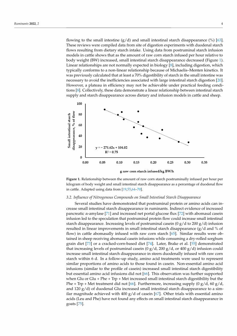
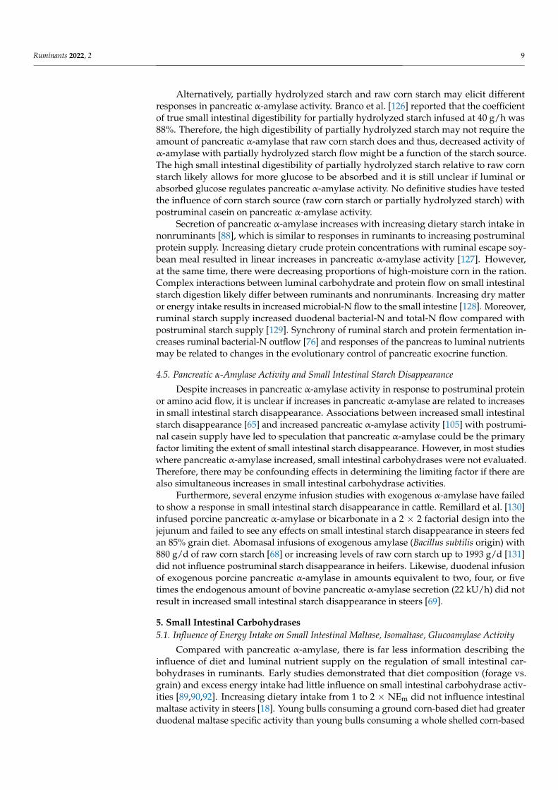
Early work using dietary [57,58] or abomasal infusion models [59] demonstrated thatthe extent of postruminal starch disappearance was much lower than in nonruminants.These authors concluded that the extent of starch disappearance in the small intestine wasinadequate for optimum utilization. Indeed, summaries have indicated that the extents ofsmall intestinal starch disappearance in beef cattle (55%) [7] and dairy cows (60%) [60] are in-adequate to achieve potential energetic advantages over ruminal fermentation of starch [20].Interestingly, the limitation of small intestinal starch disappearance is proportional to smallintestinal starch flow instead of an absolute maximal value (i.e., plateau) [27,61]. Linear re-lationships between intestinal starch appearance and small intestinal starch disappearancewere first suggested by Ørskov et al. [62], and a linear regression model was developed topredict small intestinal starch digestibility in lambs [61]. When Owens et al. [7] reviewedthe literature, they found that there was a positive linear relationship between the amountof starch flowing to the small intestine (g/d) and small intestinal starch disappearance(g/d). Furthermore, there is a negative linear relationship between the amount of starch
Ruminants 2022, 2 4
flowing to the small intestine (g/d) and small intestinal starch disappearance (%) [63].These reviews were compiled data from site of digestion experiments with duodenal starchflows resulting from dietary starch intake. Using data from postruminal starch infusionmodels in cattle shows that as the amount of raw corn starch infused per hour relative tobody weight (BW) increased, small intestinal starch disappearance decreased (Figure 1).Linear relationships are not normally expected in biology [8], including digestion, whichtypically conforms to a non-linear relationship because of Michaelis–Menten kinetics. Itwas previously calculated that at least a 70% digestibility of starch in the small intestine wasnecessary to avoid the inefficiencies associated with large intestinal starch digestion [20].However, a plateau in efficiency may not be achievable under practical feeding condi-tions [8]. Collectively, these data demonstrate a linear relationship between intestinal starchsupply and starch disappearance across dietary and infusion models in cattle and sheep.
Ruminants 2022, 1, FOR PEER REVIEW 4
small intestinal starch disappearance in beef cattle (55%) [7] and dairy cows (60%) [60] are
inadequate to achieve potential energetic advantages over ruminal fermentation of starch
[20]. Interestingly, the limitation of small intestinal starch disappearance is proportional
to small intestinal starch flow instead of an absolute maximal value (i.e., plateau) [27,61].
Linear relationships between intestinal starch appearance and small intestinal starch dis‐
appearance were first suggested by Ørskov et al. [62], and a linear regression model was
developed to predict small intestinal starch digestibility in lambs [61]. When Owens et al.
[7] reviewed the literature, they found that there was a positive linear relationship be‐
tween the amount of starch flowing to the small intestine (g/d) and small intestinal starch
disappearance (g/d). Furthermore, there is a negative linear relationship between the
amount of starch flowing to the small intestine (g/d) and small intestinal starch disappear‐
ance (%) [63]. These reviews were compiled data from site of digestion experiments with
duodenal starch flows resulting from dietary starch intake. Using data from postruminal
starch infusion models in cattle shows that as the amount of raw corn starch infused per
hour relative to body weight (BW) increased, small intestinal starch disappearance de‐
creased (Figure 1). Linear relationships are not normally expected in biology [8], including
digestion, which typically conforms to a non‐linear relationship because of Michaelis–
Menten kinetics. It was previously calculated that at least a 70% digestibility of starch in
the small intestine was necessary to avoid the inefficiencies associated with large intestinal
starch digestion [20]. However, a plateau in efficiency may not be achievable under prac‐
tical feeding conditions [8]. Collectively, these data demonstrate a linear relationship be‐
tween intestinal starch supply and starch disappearance across dietary and infusion mod‐
els in cattle and sheep.
Figure 1. Relationship between the amount of raw corn starch postruminally infused per hour per
kilogram of body weight and small intestinal starch disappearance as a percentage of duodenal flow
in cattle. Adapted using data from [19,55,64–70].
3.2. Influence of Nitrogenous Compounds on Small Intestinal Starch Disappearance
Several studies have demonstrated that postruminal protein or amino acids can in‐
crease small intestinal starch disappearance in ruminants. Indirect evidence of increased
pancreatic α‐amylase [71] and increased net portal glucose flux [72] with abomasal casein
infusion led to the speculation that postruminal protein flow could increase small intesti‐
nal starch disappearance. Increasing levels of postruminal casein (0 g/d to 200 g/d) infu‐
sion resulted in linear improvements in small intestinal starch disappearance (g/d and %
of flow) in cattle abomasally infused with raw corn starch [65]. Similar results were ob‐
tained in sheep receiving abomasal casein infusions while consuming a dry‐rolled sor‐
ghum grain diet [73] or a cracked‐corn‐based diet [74]. Later, Brake et al. [55] demon‐
strated that increasing levels of postruminal casein (0 g/d, 200 g/d, or 400 g/d) infusion
Figure 1. Relationship between the amount of raw corn starch postruminally infused per hour perkilogram of body weight and small intestinal starch disappearance as a percentage of duodenal flowin cattle. Adapted using data from [19,55,64–70].
3.2. Influence of Nitrogenous Compounds on Small Intestinal Starch Disappearance
Several studies have demonstrated that postruminal protein or amino acids can in-crease small intestinal starch disappearance in ruminants. Indirect evidence of increasedpancreatic α-amylase [71] and increased net portal glucose flux [72] with abomasal caseininfusion led to the speculation that postruminal protein flow could increase small intestinalstarch disappearance. Increasing levels of postruminal casein (0 g/d to 200 g/d) infusionresulted in linear improvements in small intestinal starch disappearance (g/d and % offlow) in cattle abomasally infused with raw corn starch [65]. Similar results were ob-tained in sheep receiving abomasal casein infusions while consuming a dry-rolled sorghumgrain diet [73] or a cracked-corn-based diet [74]. Later, Brake et al. [55] demonstratedthat increasing levels of postruminal casein (0 g/d, 200 g/d, or 400 g/d) infusion couldincrease small intestinal starch disappearance in steers duodenally infused with raw cornstarch within 6 d. In a follow-up study, amino acid treatments were used to representsimilar proportions of amino acids to those found in casein. Non-essential amino acidinfusions (similar to the profile of casein) increased small intestinal starch digestibilitybut essential amino acid infusions did not [66]. This observation was further supportedwhen Glu or Glu + Phe + Trp + Met increased small intestinal starch digestibility but thePhe + Trp + Met treatment did not [66]. Furthermore, increasing supply (0 g/d, 60 g/d,and 120 g/d) of duodenal Glu increased small intestinal starch disappearance to a sim-ilar magnitude achieved with 400 g/d of casein [67]. Other trials with essential aminoacids (Leu and Phe) have not found any effects on small intestinal starch disappearance ingoats [75].
Ruminants 2022, 2 5
3.3. Influence of Grain Processing on Small Intestinal Starch Disappearance
The effects of grain processing on small intestinal or postruminal starch disappearancehave been reviewed extensively [5,7,20,27,76,77]. The current nutrient requirements of beefcattle [78] use data from Sniffen et al. [79] and Owens and Zinn [80] to show the effects ofgrain type and degree of processing on postruminal starch disappearance. The postrumi-nal starch disappearance coefficients reported are 30–40%, 65–70%, 80–90%, 85–95%, and92–97% for whole, dry-rolled or cracked, meal, high-moisture, and steam-flaked methodsof corn processing, respectively [78]. Owens et al. [77] developed linear models to predictpostruminal starch disappearance in dairy and finishing diets. In general, starch digestibil-ity of diets containing whole or rolled corn typically decreases linearly with increasingintestinal flow, as described previously. However, extensive methods of corn processing,such as high-moisture and steam-flaking, are typically not affected by increasing intestinalflow because they are highly digestible postruminally. It is not clear whether or not en-hanced postruminal starch digestibility with extensive corn processing methods are directlyrelated to increased small intestinal starch digestibility. The issues of variable duodenalflow, maintenance of ileal cannulas, and maintaining production levels of intake in cattlewith ileal cannulas are largely why measurements of small intestinal disappearance withdifferent grain processing methods have not been evaluated. A complicating factor is thatextensive grain processing methods that increase postruminal digestibility also increaseruminal digestibility [81]. Therefore, steam-flaking and high-moisture processing methodsresult in greater ruminal starch disappearance and decreased intestinal starch flows rela-tive to whole-shelled or dry-rolled processing methods. Because of this, comparisons ofpostruminal starch digestibility coefficients that were obtained using dietary models haddiffering intestinal starch flows. More studies in which duodenal flow is controlled areneeded to clarify how grain processing methods affect the extent of small intestinal starchdisappearance in ruminants. This will aid in understanding the limits of the extent of smallintestinal starch disappearance in ruminants.
4. Pancreatic α-Amylase4.1. Influence of Dry Matter and Energy Intake on Pancreatic α-Amylase Activity
The effects of nutrition on pancreatic exocrine function in ruminants have been re-viewed previously [16,54,82–87]. In nonruminants, carbohydrase activities typically in-crease proportional to luminal substrate flow [88]. However, in ruminants, postruminaldigestive enzymes respond differently to diet and luminal nutrient flows [16]. Russellet al. [89] evaluated the effects of diet and energy intake on pancreatic α-amylase activity insteers. They fed either an alfalfa hay diet (hay) or a corn and corn-silage-based diet (grain)at 1 × net energy of maintenance (NEm) or the grain diet at 2 × or 3 × NEm. At 1 × NEmintake, they found that steers consuming the grain diet had lower pancreatic α-amylaseactivity per gram protein than steers consuming the hay diet. Furthermore, increasing theenergy intake of the grain diet from 1 to 2 × NEm increased pancreatic α-amylase activityper gram protein by two-fold, without any additional increases at 3 × NEm.
To further evaluate the effects of diet and energy intake on carbohydrase activities,Kreikemeier et al. [90] fed either a 90% forage (alfalfa hay) or 90% grain (sorghum andwheat) diet at 1 or 2 × the NEm requirement. In steers consuming the grain diet, pancreaticα-amylase concentration and total content was lower than steers consuming forage. Addi-tionally, when energy intake increased from 1 to 2 × NEm, pancreatic α-amylase activityand total content increased with an increase in pancreatic mass. In contrast, previousstudies demonstrated that increasing starch intake could increase pancreatic α-amylaseactivity [91,92]. However, these studies were confounded with energy intake. Results fromRussell et al. [89] and Kreikemeier et al. [90] demonstrated that increasing energy intake upto 2×maintenance can increase pancreatic α-amylase activity. In addition, steers consum-ing starch-based diets had lower activity of pancreatic α-amylase. However, the diet effectson pancreatic α-amylase were less clear, as the alfalfa hay-based diets had greater crude
Ruminants 2022, 2 6
protein levels. This led to the hypothesis that changes in luminal carbohydrate and proteinflow could influence pancreatic α-amylase activity.
More recent studies have evaluated the effects of dietary intake restriction on pan-creatic α-amylase activity in ruminants. Dietary intake restriction decreased pancreaticα-amylase activity in nonpregnant ewes [93], pregnant ewes [94,95], and pregnant beefcows [96,97]. Changes in pancreatic α-amylase activity in response to changes in drymatter or energy intake may be related to the abundance and activity of pancreatic proteinsinvolved in energy metabolism. Increasing dry matter intake increased the abundanceof ATP synthase, Na+/K+-ATPase, proliferating cell nuclear antigen, and ubiquitin in thepancreas of steers [98]. Dietary intake restriction of pregnant beef cows decreased ATPsynthase abundance in the pancreas [99]. Proteomic analyses suggest that intracellularactivity and abundance of proteins related to energy metabolism in the pancreas may beassociated with pancreatic α-amylase activity [100].
4.2. Influence of Dietary or Luminal Carbohydrate on Pancreatic α-Amylase Activity
While pancreatic α-amylase activity in nonruminants increases in response to lumi-nal starch flows [88], the response is opposite in ruminants. High levels of postruminalcarbohydrate supply as starch, partially hydrolyzed starch, or glucose decreases pancre-atic α-amylase activity when energy intake is controlled. Abomasal infusions of partiallyhydrolyzed starch decreased pancreatic α-amylase concentration, specific activity, andsecretion in steers compared with steers ruminally infused with partially hydrolyzed starchor steers infused with water [50]. The same decrease in pancreatic α-amylase activity inresponse to abomasal partially hydrolyzed starch was observed with pancreatic tissue sam-ples [101]. Similarly in wethers, abomasal infusions of raw corn starch decreased pancreaticα-amylase concentration and secretion compared with control wethers receiving abomasalinfusion of water [71]. These studies demonstrated that luminal complex carbohydrateflow decreases pancreatic α-amylase activity in cattle. In a study by Swanson et al. [102],abomasal infusions of either glucose or partially hydrolyzed starch decreased pancreaticα-amylase concentration, specific activity, and secretion in steers. This study demonstratedthat downregulation of pancreatic α-amylase is not due solely to luminal complex carbohy-drate flow. However, it remains unclear whether luminal glucose concentration, absorbedglucose, or both regulate pancreatic α-amylase activity in ruminants. Increasing levels ofruminal glucose infusions did not affect plasma amylase concentrations in lambs fed a 50%concentrate diet [103]. In neonatal dairy calves, supplementing fructose at 2.2 g/kg of BWdid not statistically increase pancreatic α-amylase activity; however, pancreatic α-amylaseactivity was 42% greater in fructose-fed calves [104]. This could partially result from anincrease in metabolizable energy intake.
4.3. Influence of Dietary or Luminal Nitrogenous Compounds on Pancreatic α-Amylase Activity
As stated previously, studies by Russell et al. [89] and Kreikemeier et al. [90] demon-strated that pancreatic α-amylase activity was greater in steers fed an alfalfa hay dietcompared with a grain-based diet. These authors speculated that differences in dietarycrude protein (and therefore, rumen undegradable protein and metabolizable protein) con-tribute to differences in pancreatic α-amylase activity. In sheep, Wang and Taniguchi [71]abomasally infused water (control), raw corn starch, or raw corn starch + casein and mea-sured pancreatic exocrine secretion. Pancreatic α-amylase activity was depressed withabomasal starch infusion; however, abomasal infusion of starch with casein restored α-amylase activity to the same level as the control. Similarly, increasing levels of abomasalcasein supply (0 g/d, 60 g/d, 120 g/d, or 180 g/d) linearly increased pancreatic α-amylaseconcentration, specific activity, and secretion in steers postruminally infused with rawcorn starch [105]. Feeding a 68.7% concentrate diet with supplemental casein to steers pro-duced increases in duodenal α-amylase concentrations and serum cholecystokinin (CCK)concentrations [106].
Ruminants 2022, 2 7
More information is needed to understand how the association between luminalnutrient supply, hormones and neuropeptides, and enzyme activities are coordinated toinfluence intestinal starch disappearance in ruminants. The effects of individual aminoacids on pancreatic exocrine function have been studied predominantly with preruminantcalves and lambs [86]. Several amino acids including Arg, Leu, Ile, and Phe have beenshown to influence pancreatic α-amylase activity in ruminants. Similarly, rumen-protectedTrp supplementation to steers consuming a high-concentrate diet was associated withgreater postruminal starch disappearance, increased luminal amylase activity in the duo-denum, and increased serum CCK and melatonin [107]. We have found that melatoninsupplementation to gestating ewes increased maternal pancreatic α-amylase activity [94]and small intestinal maltase, isomaltase, and glucoamylase activities [108]. Tryptophanand its metabolites are precursors to the synthesis of biogenic amines such as serotoninand melatonin.
Responses in pancreatic α-amylase activity to individual amino acids have varied withthe type of amino acid, length of infusion, and animal species. Arginine administrationthrough jugular blood did not influence pancreatic α-amylase activity in non-pregnantewes [93]. Similarly, dietary rumen-protected Arg supplementation to ewes during mid- tolate-gestation did not influence pancreatic α-amylase activity of lamb offspring at 54 d ofage [109]. After 14 d of duodenally infusing increasing levels of Phe, Yu et al. [110] observedlinear increases in pancreatic α-amylase specific activity, and a cubic response in α-amylasesecretion in goats. In the short-term experiment (10 h), they found a quadratic response inpancreatic α-amylase secretion to increasing levels of Phe. Moreover, increasing levels ofLeu linearly increased α-amylase concentration in pancreatic juice after 14 d of duodenalinfusion [111]. In dairy heifers, duodenal infusions of 10 g/d Leu increased total pancreaticsecretion, α-amylase concentration, and α-amylase secretion [112]. Increases in pancreaticα-amylase activity were observed with duodenal infusions or Leu (3 g/d or 9 g/d) andPhe (2 g/d) in goats [113]. However, when Leu (1.435 g/L milk), Phe (0.725 g/L milk), ora combination of Leu and Phe (1.435 g Leu/L milk and 0.725 g Phe/L milk) were fed tomilk-fed calves, pancreatic α-amylase specific activity was not influenced [114]. Similarly,increasing levels of Leu supplementation to neonatal calves in milk replacer did not affectpancreatic α-amylase activity [115]. These data suggest that Leu can increase pancreatic α-amylase activity in post-weaning ruminants but not in milk-fed calves. Duodenal infusionsof 20 g/d or 30 g/d of Ile have been shown to increase pancreatic α-amylase activity indairy heifers after 12 h or 10 d of infusion [116]. In cell culture models using pancreaticacinar cells, amino acids such as Phe [117], Leu [118–120], and Ile [121] increased α-amylaserelease. Despite increases in small intestinal starch disappearance with Glu [66,67], it isunclear if these increases are related to increases in pancreatic α-amylase activity, as ourrecent experiment found duodenal glutamic acid infusion did not influence pancreaticα-amylase in steers [122].
Indeed, a few studies have begun to explore cellular and molecular mechanismsdriving associations between increased pancreatic α-amylase activity in ruminants andamino acid supply. Phenylalanine increases α-amylase activity in dairy calves and theinitiation of messenger ribonucleic acid (mRNA) translation through phosphorylationof ribosomal protein S6 kinase 1 (S6K1) and eukaryotic translation initiation factor 4Ebinding protein 1 (4EBP1) [118]. Leucine and Ile have been shown to increase α-amylasesynthesis and phosphorylation of the mammalian target of rapamycin (mTOR) signalingpathway [118,121]. Proteomic analysis has suggested that Leu modulates increases inpancreatic α-amylase activity in dairy calves by increasing citrate synthase activity in thetricarboxylic acid cycle, ATPase activity and oxidative phosphorylation, and stimulating thegeneral secretory signaling pathway in pancreatic acinar cells [123]. In these studies, Phe,Leu, and Ile were the only amino acids studied and future research is needed to evaluatehow other amino acids could affect pancreatic exocrine function in ruminants.
Ruminants 2022, 2 8
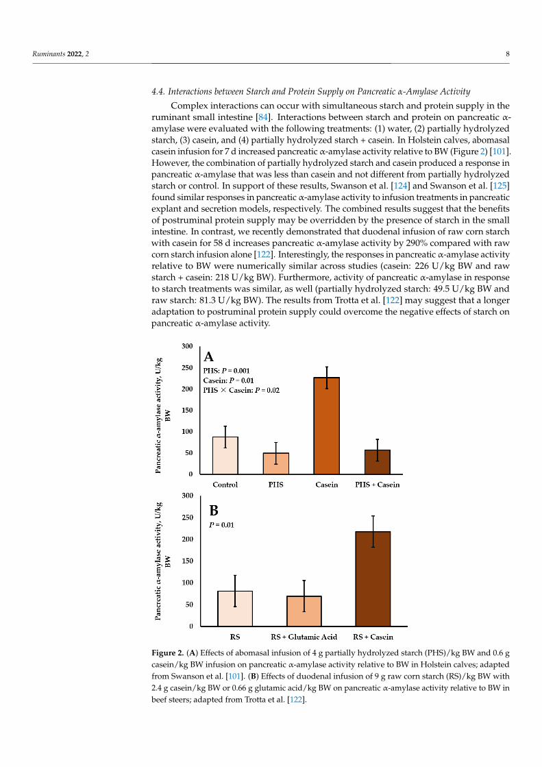
4.4. Interactions between Starch and Protein Supply on Pancreatic α-Amylase Activity
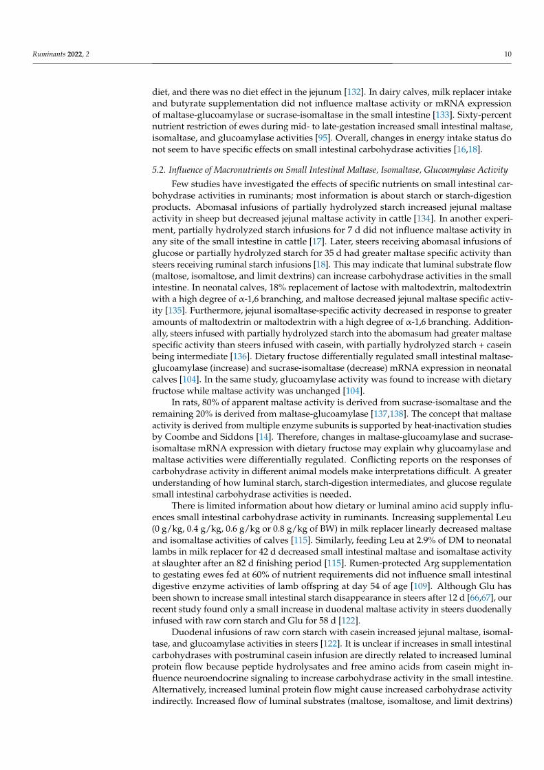
Complex interactions can occur with simultaneous starch and protein supply in theruminant small intestine [84]. Interactions between starch and protein on pancreatic α-amylase were evaluated with the following treatments: (1) water, (2) partially hydrolyzedstarch, (3) casein, and (4) partially hydrolyzed starch + casein. In Holstein calves, abomasalcasein infusion for 7 d increased pancreatic α-amylase activity relative to BW (Figure 2) [101].However, the combination of partially hydrolyzed starch and casein produced a response inpancreatic α-amylase that was less than casein and not different from partially hydrolyzedstarch or control. In support of these results, Swanson et al. [124] and Swanson et al. [125]found similar responses in pancreatic α-amylase activity to infusion treatments in pancreaticexplant and secretion models, respectively. The combined results suggest that the benefitsof postruminal protein supply may be overridden by the presence of starch in the smallintestine. In contrast, we recently demonstrated that duodenal infusion of raw corn starchwith casein for 58 d increases pancreatic α-amylase activity by 290% compared with rawcorn starch infusion alone [122]. Interestingly, the responses in pancreatic α-amylase activityrelative to BW were numerically similar across studies (casein: 226 U/kg BW and rawstarch + casein: 218 U/kg BW). Furthermore, activity of pancreatic α-amylase in responseto starch treatments was similar, as well (partially hydrolyzed starch: 49.5 U/kg BW andraw starch: 81.3 U/kg BW). The results from Trotta et al. [122] may suggest that a longeradaptation to postruminal protein supply could overcome the negative effects of starch onpancreatic α-amylase activity.
Ruminants 2022, 1, FOR PEER REVIEW 8
Indeed, a few studies have begun to explore cellular and molecular mechanisms driv‐
ing associations between increased pancreatic α‐amylase activity in ruminants and amino
acid supply. Phenylalanine increases α‐amylase activity in dairy calves and the initiation
of messenger ribonucleic acid (mRNA) translation through phosphorylation of ribosomal
protein S6 kinase 1 (S6K1) and eukaryotic translation initiation factor 4E binding protein
1 (4EBP1) [118]. Leucine and Ile have been shown to increase α‐amylase synthesis and
phosphorylation of the mammalian target of rapamycin (mTOR) signaling pathway
[118,121]. Proteomic analysis has suggested that Leu modulates increases in pancreatic α‐
amylase activity in dairy calves by increasing citrate synthase activity in the tricarboxylic
acid cycle, ATPase activity and oxidative phosphorylation, and stimulating the general
secretory signaling pathway in pancreatic acinar cells [123]. In these studies, Phe, Leu, and
Ile were the only amino acids studied and future research is needed to evaluate how other
amino acids could affect pancreatic exocrine function in ruminants.
4.4. Interactions between Starch and Protein Supply on Pancreatic α‐Amylase Activity
Complex interactions can occur with simultaneous starch and protein supply in the
ruminant small intestine [84]. Interactions between starch and protein on pancreatic α‐
amylase were evaluated with the following treatments: (1) water, (2) partially hydrolyzed
starch, (3) casein, and (4) partially hydrolyzed starch + casein. In Holstein calves, abomasal
casein infusion for 7 d increased pancreatic α‐amylase activity relative to BW (Figure 2)
[101]. However, the combination of partially hydrolyzed starch and casein produced a
response in pancreatic α‐amylase that was less than casein and not different from partially
hydrolyzed starch or control. In support of these results, Swanson et al. [124] and Swanson
et al. [125] found similar responses in pancreatic α‐amylase activity to infusion treatments
in pancreatic explant and secretion models, respectively. The combined results suggest
that the benefits of postruminal protein supply may be overridden by the presence of
starch in the small intestine. In contrast, we recently demonstrated that duodenal infusion
of raw corn starch with casein for 58 d increases pancreatic α‐amylase activity by 290%
compared with raw corn starch infusion alone [122]. Interestingly, the responses in pan‐
creatic α‐amylase activity relative to BW were numerically similar across studies (casein:
226 U/kg BW and raw starch + casein: 218 U/kg BW). Furthermore, activity of pancreatic
α‐amylase in response to starch treatments was similar, as well (partially hydrolyzed
starch: 49.5 U/kg BW and raw starch: 81.3 U/kg BW). The results from Trotta et al. [122]
may suggest that a longer adaptation to postruminal protein supply could overcome the
negative effects of starch on pancreatic α‐amylase activity.
Ruminants 2022, 1, FOR PEER REVIEW 9
Figure 2. (A) Effects of abomasal infusion of 4 g partially hydrolyzed starch (PHS)/kg BW and 0.6 g
casein/kg BW infusion on pancreatic α‐amylase activity relative to BW in Holstein calves; adapted
from Swanson et al. [101]. (B) Effects of duodenal infusion of 9 g raw corn starch (RS)/kg BW with
2.4 g casein/kg BW or 0.66 g glutamic acid/kg BW on pancreatic α‐amylase activity relative to BW
in beef steers; adapted from Trotta et al. [122].
Alternatively, partially hydrolyzed starch and raw corn starch may elicit different
responses in pancreatic α‐amylase activity. Branco et al. [126] reported that the coefficient
of true small intestinal digestibility for partially hydrolyzed starch infused at 40 g/h was
88%. Therefore, the high digestibility of partially hydrolyzed starch may not require the
amount of pancreatic α‐amylase that raw corn starch does and thus, decreased activity of
α‐amylase with partially hydrolyzed starch flow might be a function of the starch source.
The high small intestinal digestibility of partially hydrolyzed starch relative to raw corn
starch likely allows for more glucose to be absorbed and it is still unclear if luminal or
absorbed glucose regulates pancreatic α‐amylase activity. No definitive studies have
tested the influence of corn starch source (raw corn starch or partially hydrolyzed starch)
with postruminal casein on pancreatic α‐amylase activity.
Secretion of pancreatic α‐amylase increases with increasing dietary starch intake in
nonruminants [88], which is similar to responses in ruminants to increasing postruminal
protein supply. Increasing dietary crude protein concentrations with ruminal escape soy‐
bean meal resulted in linear increases in pancreatic α‐amylase activity [127]. However, at
the same time, there were decreasing proportions of high‐moisture corn in the ration.
Complex interactions between luminal carbohydrate and protein flow on small intestinal
starch digestion likely differ between ruminants and nonruminants. Increasing dry matter
or energy intake results in increased microbial‐N flow to the small intestine [128]. Moreo‐
ver, ruminal starch supply increased duodenal bacterial‐N and total‐N flow compared
with postruminal starch supply [129]. Synchrony of ruminal starch and protein fermenta‐
tion increases ruminal bacterial‐N outflow [76] and responses of the pancreas to luminal
nutrients may be related to changes in the evolutionary control of pancreatic exocrine
function.
4.5. Pancreatic α‐Amylase Activity and Small Intestinal Starch Disappearance
Despite increases in pancreatic α‐amylase activity in response to postruminal protein
or amino acid flow, it is unclear if increases in pancreatic α‐amylase are related to in‐
creases in small intestinal starch disappearance. Associations between increased small in‐
testinal starch disappearance [65] and increased pancreatic α‐amylase activity [105] with
postruminal casein supply have led to speculation that pancreatic α‐amylase could be the
Figure 2. (A) Effects of abomasal infusion of 4 g partially hydrolyzed starch (PHS)/kg BW and 0.6 gcasein/kg BW infusion on pancreatic α-amylase activity relative to BW in Holstein calves; adaptedfrom Swanson et al. [101]. (B) Effects of duodenal infusion of 9 g raw corn starch (RS)/kg BW with2.4 g casein/kg BW or 0.66 g glutamic acid/kg BW on pancreatic α-amylase activity relative to BW inbeef steers; adapted from Trotta et al. [122].
Ruminants 2022, 2 9
Alternatively, partially hydrolyzed starch and raw corn starch may elicit differentresponses in pancreatic α-amylase activity. Branco et al. [126] reported that the coefficientof true small intestinal digestibility for partially hydrolyzed starch infused at 40 g/h was88%. Therefore, the high digestibility of partially hydrolyzed starch may not require theamount of pancreatic α-amylase that raw corn starch does and thus, decreased activity ofα-amylase with partially hydrolyzed starch flow might be a function of the starch source.The high small intestinal digestibility of partially hydrolyzed starch relative to raw cornstarch likely allows for more glucose to be absorbed and it is still unclear if luminal orabsorbed glucose regulates pancreatic α-amylase activity. No definitive studies have testedthe influence of corn starch source (raw corn starch or partially hydrolyzed starch) withpostruminal casein on pancreatic α-amylase activity.
Secretion of pancreatic α-amylase increases with increasing dietary starch intake innonruminants [88], which is similar to responses in ruminants to increasing postruminalprotein supply. Increasing dietary crude protein concentrations with ruminal escape soy-bean meal resulted in linear increases in pancreatic α-amylase activity [127]. However,at the same time, there were decreasing proportions of high-moisture corn in the ration.Complex interactions between luminal carbohydrate and protein flow on small intestinalstarch digestion likely differ between ruminants and nonruminants. Increasing dry matteror energy intake results in increased microbial-N flow to the small intestine [128]. Moreover,ruminal starch supply increased duodenal bacterial-N and total-N flow compared withpostruminal starch supply [129]. Synchrony of ruminal starch and protein fermentation in-creases ruminal bacterial-N outflow [76] and responses of the pancreas to luminal nutrientsmay be related to changes in the evolutionary control of pancreatic exocrine function.
4.5. Pancreatic α-Amylase Activity and Small Intestinal Starch Disappearance
Despite increases in pancreatic α-amylase activity in response to postruminal proteinor amino acid flow, it is unclear if increases in pancreatic α-amylase are related to increasesin small intestinal starch disappearance. Associations between increased small intestinalstarch disappearance [65] and increased pancreatic α-amylase activity [105] with postrumi-nal casein supply have led to speculation that pancreatic α-amylase could be the primaryfactor limiting the extent of small intestinal starch disappearance. However, in most studieswhere pancreatic α-amylase increased, small intestinal carbohydrases were not evaluated.Therefore, there may be confounding effects in determining the limiting factor if there arealso simultaneous increases in small intestinal carbohydrase activities.
Furthermore, several enzyme infusion studies with exogenous α-amylase have failedto show a response in small intestinal starch disappearance in cattle. Remillard et al. [130]infused porcine pancreatic α-amylase or bicarbonate in a 2 × 2 factorial design into thejejunum and failed to see any effects on small intestinal starch disappearance in steers fedan 85% grain diet. Abomasal infusions of exogenous amylase (Bacillus subtilis origin) with880 g/d of raw corn starch [68] or increasing levels of raw corn starch up to 1993 g/d [131]did not influence postruminal starch disappearance in heifers. Likewise, duodenal infusionof exogenous porcine pancreatic α-amylase in amounts equivalent to two, four, or fivetimes the endogenous amount of bovine pancreatic α-amylase secretion (22 kU/h) did notresult in increased small intestinal starch disappearance in steers [69].
5. Small Intestinal Carbohydrases5.1. Influence of Energy Intake on Small Intestinal Maltase, Isomaltase, Glucoamylase Activity
Compared with pancreatic α-amylase, there is far less information describing theinfluence of diet and luminal nutrient supply on the regulation of small intestinal car-bohydrases in ruminants. Early studies demonstrated that diet composition (forage vs.grain) and excess energy intake had little influence on small intestinal carbohydrase activ-ities [89,90,92]. Increasing dietary intake from 1 to 2 × NEm did not influence intestinalmaltase activity in steers [18]. Young bulls consuming a ground corn-based diet had greaterduodenal maltase specific activity than young bulls consuming a whole shelled corn-based
Ruminants 2022, 2 10
diet, and there was no diet effect in the jejunum [132]. In dairy calves, milk replacer intakeand butyrate supplementation did not influence maltase activity or mRNA expressionof maltase-glucoamylase or sucrase-isomaltase in the small intestine [133]. Sixty-percentnutrient restriction of ewes during mid- to late-gestation increased small intestinal maltase,isomaltase, and glucoamylase activities [95]. Overall, changes in energy intake status donot seem to have specific effects on small intestinal carbohydrase activities [16,18].
5.2. Influence of Macronutrients on Small Intestinal Maltase, Isomaltase, Glucoamylase Activity
Few studies have investigated the effects of specific nutrients on small intestinal car-bohydrase activities in ruminants; most information is about starch or starch-digestionproducts. Abomasal infusions of partially hydrolyzed starch increased jejunal maltaseactivity in sheep but decreased jejunal maltase activity in cattle [134]. In another experi-ment, partially hydrolyzed starch infusions for 7 d did not influence maltase activity inany site of the small intestine in cattle [17]. Later, steers receiving abomasal infusions ofglucose or partially hydrolyzed starch for 35 d had greater maltase specific activity thansteers receiving ruminal starch infusions [18]. This may indicate that luminal substrate flow(maltose, isomaltose, and limit dextrins) can increase carbohydrase activities in the smallintestine. In neonatal calves, 18% replacement of lactose with maltodextrin, maltodextrinwith a high degree of α-1,6 branching, and maltose decreased jejunal maltase specific activ-ity [135]. Furthermore, jejunal isomaltase-specific activity decreased in response to greateramounts of maltodextrin or maltodextrin with a high degree of α-1,6 branching. Addition-ally, steers infused with partially hydrolyzed starch into the abomasum had greater maltasespecific activity than steers infused with casein, with partially hydrolyzed starch + caseinbeing intermediate [136]. Dietary fructose differentially regulated small intestinal maltase-glucoamylase (increase) and sucrase-isomaltase (decrease) mRNA expression in neonatalcalves [104]. In the same study, glucoamylase activity was found to increase with dietaryfructose while maltase activity was unchanged [104].
In rats, 80% of apparent maltase activity is derived from sucrase-isomaltase and theremaining 20% is derived from maltase-glucoamylase [137,138]. The concept that maltaseactivity is derived from multiple enzyme subunits is supported by heat-inactivation studiesby Coombe and Siddons [14]. Therefore, changes in maltase-glucoamylase and sucrase-isomaltase mRNA expression with dietary fructose may explain why glucoamylase andmaltase activities were differentially regulated. Conflicting reports on the responses ofcarbohydrase activity in different animal models make interpretations difficult. A greaterunderstanding of how luminal starch, starch-digestion intermediates, and glucose regulatesmall intestinal carbohydrase activities is needed.
There is limited information about how dietary or luminal amino acid supply influ-ences small intestinal carbohydrase activity in ruminants. Increasing supplemental Leu(0 g/kg, 0.4 g/kg, 0.6 g/kg or 0.8 g/kg of BW) in milk replacer linearly decreased maltaseand isomaltase activities of calves [115]. Similarly, feeding Leu at 2.9% of DM to neonatallambs in milk replacer for 42 d decreased small intestinal maltase and isomaltase activityat slaughter after an 82 d finishing period [115]. Rumen-protected Arg supplementationto gestating ewes fed at 60% of nutrient requirements did not influence small intestinaldigestive enzyme activities of lamb offspring at day 54 of age [109]. Although Glu hasbeen shown to increase small intestinal starch disappearance in steers after 12 d [66,67], ourrecent study found only a small increase in duodenal maltase activity in steers duodenallyinfused with raw corn starch and Glu for 58 d [122].
Duodenal infusions of raw corn starch with casein increased jejunal maltase, isomal-tase, and glucoamylase activities in steers [122]. It is unclear if increases in small intestinalcarbohydrases with postruminal casein infusion are directly related to increased luminalprotein flow because peptide hydrolysates and free amino acids from casein might in-fluence neuroendocrine signaling to increase carbohydrase activity in the small intestine.Alternatively, increased luminal protein flow might cause increased carbohydrase activityindirectly. Increased flow of luminal substrates (maltose, isomaltose, and limit dextrins)
Ruminants 2022, 2 11
as a result of greater hydrolysis of amylose and amylopectin in response to increases inpancreatic α-amylase activity might modulate increases in small intestinal carbohydraseactivities. In Caco-2 cells, supply of maltose induced synthesis of a higher molecularweight sucrase-isomaltase immunoblot band compared with glucose, fructose, isomal-tose, and fructose [139]. Using Caco-2 cells, Chegeni et al. [140] suggested there was aluminal maltose-sensing mechanism that increases apparent maltase activity by enhanc-ing intracellular trafficking of sucrase-isomaltase to the apical membrane. These effectswere associated with an increased mRNA expression of TAS1R2 and TAS1R3, the genesencoding the dimeric sweet taste receptor subunits T1R2-T1R3. These authors suggestedthat T1R2-T1R3 could potentially mediate effects of luminal maltose on sucrase-isomaltaseactivity [140].
6. Sucrase6.1. Intestinal Sucrase Activity Is Absent in Multiple Ruminant Species
A remaining enigma of ruminant digestive physiology is the absence of sucrase activityin the small intestine. Several studies have investigated and characterized digestive enzymeactivity along the small intestine with various ruminant species, ages, and diets. Yet, therehas been a failure to detect active sucrase in the small intestine. This is in contrast tononruminant species including the pig and human. Dollar and Porter [3] were the firstto report the absence of sucrase activity in young calves. Furthermore, no measurablesucrase activity was detected in mucosa or small intestinal digesta contents from lambs [11].Later reports by Huber et al. [141] and Siddons [13] corroborated the findings that sucraseactivity is absent from the digestive tract of the young calf. With cattle ranging from4 days of age up to 6 years of age, no detectable amounts of sucrase were found in thesmall intestine [13,90]. Shirazi-Beechey et al. [142] attempted to measure sucrase activityin isolated brush-border membrane vesicles (BBMV) from lamb intestine and also did notdetect any sucrase activity. More recently, we have been unable to detect sucrase activityin intestinal mucosa samples from neonatal calves [104], growing steers [122], or fetal,neonatal, or gestating sheep [95,109].
The lack of sucrase activity seems to expand to a wider range of ruminants otherthan sheep and cattle and even some nonruminant foregut fermenters. Marine mammalssuch as whales and dolphins have similarities in the sucrase-isomaltase protein sequenceto even-toed ungulates. Although it is unknown if dolphins or whales possess intestinalsucrase activity [143], the sea lion does not [144]. A comparative study demonstratedthat sucrase activity was not detected in any ruminant species including sheep, goat, roedeer, and moose [145]. Although not considered a ruminant, the kangaroo is a foregutfermenter and does not possess intestinal sucrase activity [146]. In the pseudoruminantcamel intestine, glucoamylase and maltase activities were two- and three-fold greater thansucrase activity [147].
6.2. Congenital Sucrase-Isomaltase Deficiency and Multiple Sequence Alignment
Interestingly, the absence of sucrase activity in the small intestine of ruminants appearsto be similar to congenital sucrase-isomaltase deficiency (CSID) in infants [14]. There areseven distinct phenotypes of CSID in humans with phenotypes I, II, and III resulting incompletely inactive sucrase and isomaltase activities [148]. It would seem unlikely thatmechanisms driving phenotypes I, II, or III of CSID would explain the inactivity of sucrasein ruminants because ruminants do have active isomaltase activity [14]. However, inphenotype V of CSID, it is characterized by the presence of isomaltase activity and absenceof sucrase activity.
The primary structure of the sucrase-isomaltase complex was deduced in the rabbitintestine and a “stalk” region (amino acids 33–70) was identified as the connection betweenthe transmembrane domain and N-terminal sucrase-isomaltase (isomaltase subunit) [149].In phenotype V of CSID, pro-sucrase-isomaltase is cleaved intracellularly in the trans-Golginetwork, and the sucrase subunit is subsequently degraded while the isomaltase subunit
Ruminants 2022, 2 12
is transported to the apical membrane [148,150]. Later experiments determined that thesignals for apical sorting were located in the O-glycosylated “stalk” region and membraneanchoring domain of the isomaltase subunit [151,152].
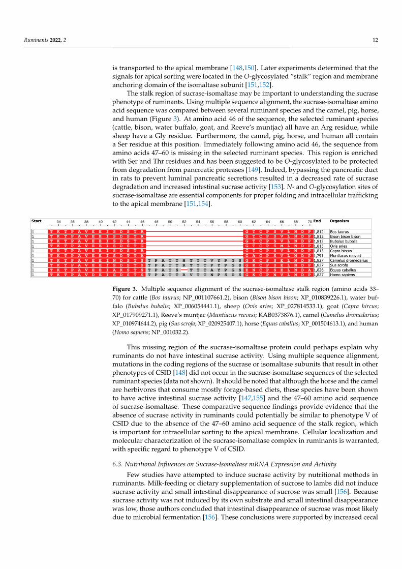
The stalk region of sucrase-isomaltase may be important to understanding the sucrasephenotype of ruminants. Using multiple sequence alignment, the sucrase-isomaltase aminoacid sequence was compared between several ruminant species and the camel, pig, horse,and human (Figure 3). At amino acid 46 of the sequence, the selected ruminant species(cattle, bison, water buffalo, goat, and Reeve’s muntjac) all have an Arg residue, whilesheep have a Gly residue. Furthermore, the camel, pig, horse, and human all containa Ser residue at this position. Immediately following amino acid 46, the sequence fromamino acids 47–60 is missing in the selected ruminant species. This region is enrichedwith Ser and Thr residues and has been suggested to be O-glycosylated to be protectedfrom degradation from pancreatic proteases [149]. Indeed, bypassing the pancreatic ductin rats to prevent luminal pancreatic secretions resulted in a decreased rate of sucrasedegradation and increased intestinal sucrase activity [153]. N- and O-glycosylation sites ofsucrase-isomaltase are essential components for proper folding and intracellular traffickingto the apical membrane [151,154].
Ruminants 2022, 1, FOR PEER REVIEW 13
Figure 3. Multiple sequence alignment of the sucrase‐isomaltase stalk region (amino acids 33–70)
for cattle (Bos taurus; NP_001107661.2), bison (Bison bison bison; XP_010839226.1), water buffalo (Bu‐
balus bubalis; XP_006054441.1), sheep (Ovis aries; XP_027814533.1), goat (Capra hircus;
XP_017909271.1), Reeve’s muntjac (Muntiacus reevesi; KAB0373876.1), camel (Camelus dromedarius;
XP_010974644.2), pig (Sus scrofa; XP_020925407.1), horse (Equus caballus; XP_001504613.1), and hu‐
man (Homo sapiens; NP_001032.2).
This missing region of the sucrase‐isomaltase protein could perhaps explain why ru‐
minants do not have intestinal sucrase activity. Using multiple sequence alignment, mu‐
tations in the coding regions of the sucrase or isomaltase subunits that result in other phe‐
notypes of CSID [148] did not occur in the sucrase‐isomaltase sequences of the selected
ruminant species (data not shown). It should be noted that although the horse and the
camel are herbivores that consume mostly forage‐based diets, these species have been
shown to have active intestinal sucrase activity [147,155] and the 47–60 amino acid se‐
quence of sucrase‐isomaltase. These comparative sequence findings provide evidence that
the absence of sucrase activity in ruminants could potentially be similar to phenotype V
of CSID due to the absence of the 47–60 amino acid sequence of the stalk region, which is
important for intracellular sorting to the apical membrane. Cellular localization and mo‐
lecular characterization of the sucrase‐isomaltase complex in ruminants is warranted,
with specific regard to phenotype V of CSID.
6.3. Nutritional Influences on Sucrase‐Isomaltase mRNA Expression and Activity
Few studies have attempted to induce sucrase activity by nutritional methods in ru‐
minants. Milk‐feeding or dietary supplementation of sucrose to lambs did not induce su‐
crase activity and small intestinal disappearance of sucrose was small [156]. Because su‐
crase activity was not induced by its own substrate and small intestinal disappearance
was low, those authors concluded that intestinal disappearance of sucrose was most likely
due to microbial fermentation [156]. These conclusions were supported by increased cecal
microbial counts and increased fecal N excretion with sucrose inclusion [156]. Likewise,
abomasal infusions of sucrose did not induce sucrase activity in lambs [157].
In one case report in humans, dietary fructose supplementation increased sucrase
activity by nearly four‐fold in a patient with CSID [158]. Although the phenotype of the
patient was not revealed, the patient had deficient, but not absent, activity of sucrase and
isomaltase before beginning fructose treatment [158]. After treatment, sucrase activity lev‐
els were still approximately 18.5% of the amount of sucrase activity from the patient’s
family members [158]. Fructose supplementation at 2.2 g/kg of BW did not induce sucrase
activity in neonatal calves fed milk replacer [104]. However, dietary fructose decreased
sucrase‐isomaltase mRNA expression, suggesting that sucrase‐isomaltase may be tran‐
scriptionally regulated by dietary fructose in the ruminant small intestine. These studies
indicate that fructose supplementation is not effective at inducing or increasing sucrase
activity in patients with CSID or in ruminants.
6.4. Impacts on Carbohydrate Digestion
Recent evidence from nonruminant studies suggests that the absence of sucrase can
have other physiological consequences on carbohydrate digestion. Nichols et al. [159]
demonstrated that the absence of sucrase activity leads to a reduction in starch digestion
and postprandial glucose response with a sucrase‐deficient shrew model. Furthermore,
Figure 3. Multiple sequence alignment of the sucrase-isomaltase stalk region (amino acids 33–70) for cattle (Bos taurus; NP_001107661.2), bison (Bison bison bison; XP_010839226.1), water buf-falo (Bubalus bubalis; XP_006054441.1), sheep (Ovis aries; XP_027814533.1), goat (Capra hircus;XP_017909271.1), Reeve’s muntjac (Muntiacus reevesi; KAB0373876.1), camel (Camelus dromedarius;XP_010974644.2), pig (Sus scrofa; XP_020925407.1), horse (Equus caballus; XP_001504613.1), and human(Homo sapiens; NP_001032.2).
This missing region of the sucrase-isomaltase protein could perhaps explain whyruminants do not have intestinal sucrase activity. Using multiple sequence alignment,mutations in the coding regions of the sucrase or isomaltase subunits that result in otherphenotypes of CSID [148] did not occur in the sucrase-isomaltase sequences of the selectedruminant species (data not shown). It should be noted that although the horse and the camelare herbivores that consume mostly forage-based diets, these species have been shownto have active intestinal sucrase activity [147,155] and the 47–60 amino acid sequenceof sucrase-isomaltase. These comparative sequence findings provide evidence that theabsence of sucrase activity in ruminants could potentially be similar to phenotype V ofCSID due to the absence of the 47–60 amino acid sequence of the stalk region, whichis important for intracellular sorting to the apical membrane. Cellular localization andmolecular characterization of the sucrase-isomaltase complex in ruminants is warranted,with specific regard to phenotype V of CSID.
6.3. Nutritional Influences on Sucrase-Isomaltase mRNA Expression and Activity
Few studies have attempted to induce sucrase activity by nutritional methods inruminants. Milk-feeding or dietary supplementation of sucrose to lambs did not inducesucrase activity and small intestinal disappearance of sucrose was small [156]. Becausesucrase activity was not induced by its own substrate and small intestinal disappearancewas low, those authors concluded that intestinal disappearance of sucrose was most likelydue to microbial fermentation [156]. These conclusions were supported by increased cecal
Ruminants 2022, 2 13
microbial counts and increased fecal N excretion with sucrose inclusion [156]. Likewise,abomasal infusions of sucrose did not induce sucrase activity in lambs [157].
In one case report in humans, dietary fructose supplementation increased sucraseactivity by nearly four-fold in a patient with CSID [158]. Although the phenotype of thepatient was not revealed, the patient had deficient, but not absent, activity of sucrase andisomaltase before beginning fructose treatment [158]. After treatment, sucrase activity levelswere still approximately 18.5% of the amount of sucrase activity from the patient’s familymembers [158]. Fructose supplementation at 2.2 g/kg of BW did not induce sucrase activityin neonatal calves fed milk replacer [104]. However, dietary fructose decreased sucrase-isomaltase mRNA expression, suggesting that sucrase-isomaltase may be transcriptionallyregulated by dietary fructose in the ruminant small intestine. These studies indicate thatfructose supplementation is not effective at inducing or increasing sucrase activity inpatients with CSID or in ruminants.
6.4. Impacts on Carbohydrate Digestion
Recent evidence from nonruminant studies suggests that the absence of sucrase canhave other physiological consequences on carbohydrate digestion. Nichols et al. [159]demonstrated that the absence of sucrase activity leads to a reduction in starch digestionand postprandial glucose response with a sucrase-deficient shrew model. Furthermore,when supplemented with an oral glucoamylase enzyme, sucrase-deficient shrews had bloodglucose concentrations that were similar to the control shrews (containing normal sucraseactivity). These authors concluded that sucrase was the predominant mucosal enzymeinvolved in starch digestion because of its affinity towards multiple starch substrates [160].In steers, duodenal infusions of exogenous glucoamylase increased small intestinal starchdisappearance [69]. Collectively, these data suggest that ruminants have quantitative limitsin carbohydrate digestion similar to humans with congenital sucrase-isomaltase deficiencyand supplemental enzymes may replace missing intestinal hydrolytic activity to improvesmall intestinal carbohydrate digestibility.
7. Glucose Absorption7.1. Sodium/Glucose Cotransporter-1 (SGLT1)
Sodium/glucose cotransporter-1 (SGLT1), glucose transporter 5 (GLUT5), and glucosetransporter 2 (GLUT2) are thought to be the predominant carbohydrate transporters inthe small intestine of ruminants [84]. Many studies in ruminants have concluded thatSGLT1 activity and SGLT1 abundance were greatest in milk-fed lambs and declines withage [142,161–163]. Shirazi-Beechey et al. [163] demonstrated that duodenal infusions of a30 mM glucose solution for 4 d in adult sheep increased the rate of glucose transport by40- to 80-fold which was also accompanied by an increase in SGLT1 abundance. Further-more, Dyer et al. [164] demonstrated that duodenal fructose infusions can increase jejunalSGLT1 activity in lambs. These authors concluded that luminal sugar is sensed in theintestine, independent of glucose metabolism, and that the inducing sugar does not need tobe a substrate of SGLT1. However, duodenal infusion of raw corn starch did not influenceSGLT1 activity in sheep [165].
Moreover, regulation of carbohydrate transport in ruminants has been suggestedto be influenced by the presence of sweet taste receptors in the bovine and ovine smallintestine (T1R2-T1R3) [166]. The sweet taste receptor signaling mechanism was proposedby Moran et al. [167], based on research with mice. Luminal sugar is sensed in the smallintestine by T1R2-T1R3 and its associated G-protein, gustducin, which induces a signalingcascade, leading to a subsequent increase in glucagon-like peptide-2 secretion. Glucagon-like peptide-2 binds to its receptor on the submucosal plexus, eliciting a neuronal responseto evoke the release of vasoactive intestinal peptide (VIP) or pituitary adenylate cyclase-activating peptide (PACAP) in absorptive enterocytes. Binding of either VIP or PACAP toits receptor on the basolateral membrane of absorptive enterocytes results in an increase in
Ruminants 2022, 2 14
intracellular cyclic adenosine monophosphate (cAMP) levels, leading to an upregulation ofSGLT1 [167].
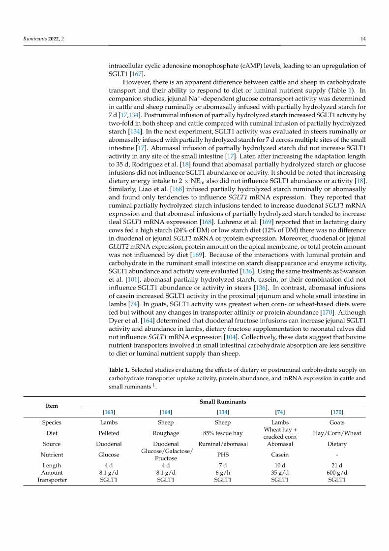
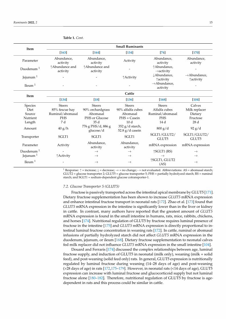
However, there is an apparent difference between cattle and sheep in carbohydratetransport and their ability to respond to diet or luminal nutrient supply (Table 1). Incompanion studies, jejunal Na+-dependent glucose cotransport activity was determinedin cattle and sheep ruminally or abomasally infused with partially hydrolyzed starch for7 d [17,134]. Postruminal infusion of partially hydrolyzed starch increased SGLT1 activity bytwo-fold in both sheep and cattle compared with ruminal infusion of partially hydrolyzedstarch [134]. In the next experiment, SGLT1 activity was evaluated in steers ruminally orabomasally infused with partially hydrolyzed starch for 7 d across multiple sites of the smallintestine [17]. Abomasal infusion of partially hydrolyzed starch did not increase SGLT1activity in any site of the small intestine [17]. Later, after increasing the adaptation lengthto 35 d, Rodriguez et al. [18] found that abomasal partially hydrolyzed starch or glucoseinfusions did not influence SGLT1 abundance or activity. It should be noted that increasingdietary energy intake to 2 × NEm also did not influence SGLT1 abundance or activity [18].Similarly, Liao et al. [168] infused partially hydrolyzed starch ruminally or abomasallyand found only tendencies to influence SGLT1 mRNA expression. They reported thatruminal partially hydrolyzed starch infusions tended to increase duodenal SGLT1 mRNAexpression and that abomasal infusions of partially hydrolyzed starch tended to increaseileal SGLT1 mRNA expression [168]. Lohrenz et al. [169] reported that in lactating dairycows fed a high starch (24% of DM) or low starch diet (12% of DM) there was no differencein duodenal or jejunal SGLT1 mRNA or protein expression. Moreover, duodenal or jejunalGLUT2 mRNA expression, protein amount on the apical membrane, or total protein amountwas not influenced by diet [169]. Because of the interactions with luminal protein andcarbohydrate in the ruminant small intestine on starch disappearance and enzyme activity,SGLT1 abundance and activity were evaluated [136]. Using the same treatments as Swansonet al. [101], abomasal partially hydrolyzed starch, casein, or their combination did notinfluence SGLT1 abundance or activity in steers [136]. In contrast, abomasal infusionsof casein increased SGLT1 activity in the proximal jejunum and whole small intestine inlambs [74]. In goats, SGLT1 activity was greatest when corn- or wheat-based diets werefed but without any changes in transporter affinity or protein abundance [170]. AlthoughDyer et al. [164] determined that duodenal fructose infusions can increase jejunal SGLT1activity and abundance in lambs, dietary fructose supplementation to neonatal calves didnot influence SGLT1 mRNA expression [104]. Collectively, these data suggest that bovinenutrient transporters involved in small intestinal carbohydrate absorption are less sensitiveto diet or luminal nutrient supply than sheep.
Table 1. Selected studies evaluating the effects of dietary or postruminal carbohydrate supply oncarbohydrate transporter uptake activity, protein abundance, and mRNA expression in cattle andsmall ruminants 1.
ItemSmall Ruminants
[163] [164] [134] [74] [170]
Species Lambs Sheep Sheep Lambs Goats
Diet Pelleted Roughage 85% fescue hay Wheat hay +cracked corn Hay/Corn/Wheat
Source Duodenal Duodenal Ruminal/abomasal Abomasal Dietary
Nutrient Glucose Glucose/Galactose/Fructose PHS Casein -
Length 4 d 4 d 7 d 10 d 21 dAmount 8.1 g/d 8.1 g/d 6 g/h 35 g/d 600 g/d
Transporter SGLT1 SGLT1 SGLT1 SGLT1 SGLT1
Ruminants 2022, 2 15
Table 1. Cont.
ItemSmall Ruminants
[163] [164] [134] [74] [170]
Parameter Abundance,activity
Abundance,activity Activity Abundance,
activityAbundance,
activity
Duodenum 1 ↑Abundance andactivity
↑Abundance andactivity - ↑Abundance,
→activity -
Jejunum 1 - - ↑Activity ↓Abundance,↑activity
→Abundance,↑activity
Ileum 1 - - - →Abundance,activity -
ItemCattle
[134] [18] [136] [168] [104]
Species Steers Steers Steers Steers CalvesDiet 85% fescue hay 90% orchardgrass 90% alfalfa cubes Alfalfa cubes Milk replacer
Source Ruminal/abomasal Abomasal Abomasal Ruminal/abomasal DietaryNutrient PHS PHS or Glucose PHS + Casein PHS FructoseLength 7 d 35 d 10 d 14 d 28 d
Amount 40 g/h 776 g PHS/d, 886 gglucose/d
352 g/d starch,52.8 g/d casein 800 g/d 92 g/d
Transporter SGLT1 SGLT1 SGLT1 SGLT1/GLUT2/GLUT5
SGLT1/GLUT2/GLUT5
Parameter Activity Abundance,activity
Abundance,activity mRNA expression mRNA expression
Duodenum 1 - → → ↑SGLT1 (RS) →Jejunum 1 ↑Activity → → → →
Ileum 1 - → → ↑SGLT1, GLUT2(AS) →
1 Response: ↑ = increase; ↓ = decrease;→ = no change; - = not evaluated. Abbreviations: AS = abomasal starch;GLUT2 = glucose transporter 2; GLUT5 = glucose transporter 5; PHS = partially hydrolyzed starch; RS = ruminalstarch; and SGLT1 = sodium-dependent glucose cotransporter-1.
7.2. Glucose Transporter 5 (GLUT5)
Fructose is passively transported across the intestinal apical membrane by GLUT5 [171].Dietary fructose supplementation has been shown to increase GLUT5 mRNA expressionand enhance intestinal fructose transport in neonatal rats [172]. Zhao et al. [173] found thatGLUT5 mRNA expression in the intestine is significantly lower than in the liver or kidneyin cattle. In contrast, many authors have reported that the greatest amount of GLUT5mRNA expression is found in the small intestine in humans, rats, mice, rabbits, chickens,and horses [174]. Nutritional regulation of GLUT5 by fructose requires luminal presence offructose in the intestine [175] and GLUT5 mRNA expression is directly proportional to in-testinal luminal fructose concentration in weaning rats [172]. In cattle, ruminal or abomasalinfusions of partially hydrolyzed starch did not affect GLUT5 mRNA expression in theduodenum, jejunum, or ileum [168]. Dietary fructose supplementation to neonatal calvesfed milk replacer did not influence GLUT5 mRNA expression in the small intestine [104].
Douard and Ferraris [174] discussed the complex relationships between age, luminalfructose supply, and induction of GLUT5 in neonatal (milk only), weaning (milk + solidfeed), and post-weaning (solid feed only) rats. In general, GLUT5 expression is nutritionallyregulated by luminal fructose during weaning (14–28 days of age) and post-weaning(>28 days of age) in rats [172,175–179]. However, in neonatal rats (<14 days of age), GLUT5expression can increase with luminal fructose and glucocorticoid supply but not luminalfructose alone [180–182]. Therefore, nutritional regulation of GLUT5 by fructose is age-dependent in rats and this process could be similar in cattle.
Ruminants 2022, 2 16
7.3. Glucose Transporter 2 (GLUT2)
Glucose transporter 2 is thought to be the primary basolateral transporter of monosac-charides from intestinal enterocytes. The apical GLUT2 hypothesis [183] in which GLUT2translocates to the apical membrane and contributes to apical (luminal) sugar transporthas been controversial [171]. Whether or not GLUT2 translocation occurs in ruminantsor contributes to apical sugar uptake under physiological substrate concentrations havenot been adequately evaluated. Abundance of GLUT2 in the small intestine has beenspecifically evaluated in BBMV from lactating dairy cows [169], newborn calves [184],and lactating ewes [185]. Lohrenz et al. [169] quantified GLUT2 abundance in BBMV, aswell as, crude cell membrane extracts (CCM) from duodenal and jejunal mucosal tissue oflactating dairy cows. They found that GLUT2 was present in duodenal and jejunal BBMV.Steinhoff-Wagner et al. [184] demonstrated that GLUT2 was present in BBMV preparedfrom mid-duodenal and proximal-, mid-, and distal-jejunal mucosa of newborn calves.Additionally, they used immunofluorescence to show localization of GLUT2 on the apicaland basolateral membranes [184]. Their data showed that the apical:basolateral distributionof GLUT2 was positive [184], indicating greater abundance of GLUT2 on the apical mem-brane. A follow-up study demonstrated that feeding colostrum for 4 d after birth decreasedbasolateral GLUT2 fluorescence and increased apical GLUT2 fluorescence, suggesting anincrease in GLUT2 translocation in small intestinal enterocytes of calves [186].
However, in sheep, jejunal BBMV did not express GLUT2, whereas jejunal CCMdid [185]. Brush-border membrane vesicles can potentially be contaminated with increasedbasolateral enrichment, indicated by increased Na+/K+-ATPase activity or abundance com-pared with the homogenate. Contamination of BBMV with the basolateral membrane couldartificially increase GLUT2 abundance estimates in the apical membrane [187]. Activity ofSGLT1 is typically assessed using BBMV preparations and measuring glucose uptake in thepresence or absence of Na+. Bauer et al. [17] measured Na+-independent glucose uptake inBBMV from cattle and found that at 200 µM luminal glucose, Na+-independent glucoseuptake only contributed to 3% of total glucose uptake by BBMV. These data indicate thatNa+-independent uptake activity at 200 µM luminal glucose in the apical membrane isunlikely to be a major route of glucose absorption in growing beef steers. Solvent drag [188],a phenomena where glucose is paracellularly absorbed across intracellular junctions, isalso not thought to be a major route of glucose absorption under physiological conditionsbecause passive diffusion of glucose is small in cattle [189]. Further evaluation is neededacross different physiological states, intakes, and luminal glucose concentrations to betterunderstand the contribution of GLUT2 to apical glucose transport in ruminants.
Studies evaluating effects of nutrition on GLUT2 mRNA expression or protein abun-dance have not been consistent in ruminants. Abomasal infusion of partially hydrolyzedstarch tended to increase ileal GLUT2 mRNA expression in steers [168]. Duodenal orjejunal GLUT2 mRNA expression and protein abundance in BBMV or CCM were not influ-enced by feeding diets with differing starch concentrations to lactating dairy cows [169].Klinger et al. [185] found that jejunal GLUT2 abundance was greater for lactating ewescompared with dried-off ewes. Dietary fructose supplementation to neonatal calves didnot influence GLUT2 mRNA expression in the duodenum, jejunum, or ileum [104].
7.4. Portal Appearance of Glucose
There is a disproportional relationship between intestinal carbohydrate disappearanceand portal glucose appearance in cattle. In mature ruminants, limited amounts of glucoseappear in portal blood [190] which indicate that microbial fermentation and/or visceralmetabolism of glucose are substantial. Short-chain fatty acid concentrations in digesta aretypically used to evaluate the fermentability of a given diet or nutrient. Reductions inileal pH and increased short-chain fatty acid concentrations in ileal digesta could suggestmicrobial activity in the small intestine [19,156,191]. In general, small intestinal short-chain fatty acid concentrations are far less than large intestinal concentrations in cattle [19]and pigs [192]. Huntington and Reynolds [193] abomasally infused glucose or raw corn
Ruminants 2022, 2 17
starch in lactating dairy cows and beef heifers and measured net nutrient flux across theportal-drained viscera (PDV). They reported that approximately 65% of the infused glucoseappeared in portal blood and this was similar between lactating dairy cows and beef heifers.However, only 35% and 8% (26% average) of the infused corn starch appeared in portalblood as glucose for the beef heifer and lactating cow, respectively. It should be noted thatthese calculations were based on the amount of carbohydrate infused, not disappearance ofthe carbohydrate.
Kreikemeier et al. [19] were the first to quantify small intestinal carbohydrate dis-appearance and net portal glucose absorption in cattle simultaneously. Holstein steersabomasally infused with glucose, corn dextrin, or corn starch at 60 g/h had 94% of glucose,38% of corn starch, and 29% of corn dextrin disappearance in the small intestine that couldbe accounted for in portal blood [19]. These authors suggested that glucose could poten-tially be used as a substrate within the small intestine or that small intestine carbohydratedisappearance could be partially due to microbial fermentation. They concluded thatapproximately 35% of raw corn starch that disappears in the small intestine resulted innet portal glucose absorption. In a similar study, Holstein steers were abomasally infusedwith water, glucose, corn dextrin, or corn starch at 66 g/h [64]. A total of 73% of glucose,60% of corn dextrin, and 57% of corn starch that disappeared in the small intestine couldbe accounted for as net portal glucose flux. Shifting the site of starch digestion from therumen to the small intestine increased glucose utilization by PDV tissues (132%), PDVglucose flux (310%), and irreversible loss of glucose (59%) in growing beef steers infusedwith partially hydrolyzed starch [9]. In general, these studies collectively demonstrate thatintestinal starch flow does result in an increase in net portal glucose flux; however, there isa large amount of glucose that is utilized by splanchnic tissues. When corrected for visceralmetabolism, glucose uptake by the PDV was 77% of supply [9].
In beef steers, abomasal raw corn starch infusions with casein increased portal glu-cose appearance by 0.38 g per gram of casein infused [72]. However, when corn starchwas infused ruminally and casein was infused abomasally, net portal glucose flux didnot differ [72]. The amino acids Gln, Glu, and Pro are found in the largest abundancein casein [194] and Glu is the primary substrate used by duodenal enterocytes for en-ergy metabolism in beef cattle [195]. Because abomasal casein infusion with ruminal rawcorn starch infusion did not increase net portal glucose flux, these data suggest that in-creases in net portal glucose flux in response to casein are because of increased intestinalstarch hydrolysis and/or greater intestinal glucose transport rather than shifts in PDVmetabolism [196].
Compared with nonruminants, the amount of glucose appearing in portal blood islow and raises many questions about the fate of glucose that disappears in the intestine. Infact, most authors agree that the disproportional relationship between intestinal carbohy-drate disappearance and net portal glucose absorption is partially due to both microbialfermentation and visceral metabolism [19,135,191,197]. Gilbert et al. [135] concluded thatfermentation is the primary contributor to starch disappearance in the small intestinerather than enzymatic hydrolysis to glucose in milk-fed calves. However, cellulose andhemicellulose digestibility in the small intestine is small, indicating that microbial activityon carbohydrates in the small intestinal lumen is probably not a major contributor tointestinal carbohydrate disappearance in functional ruminants [79,198,199]. Whether ornot starch is digested hydrolytically or microbially in the small intestine, research hasshown that energetic advantages can be gained with increased starch digestion in the smallintestine [8]. Despite limitations in intestinal glucose absorption resulting from insufficienttransport, microbial fermentation, or visceral metabolism, adequate amounts of glucosecan be supplied to peripheral tissues via hepatic gluconeogenesis, even in high-producingdairy cows [200].
Ruminants 2022, 2 18
8. Conclusions
The interface between nutrient supply and gastrointestinal function are importantbecause digestive and absorptive function in the small intestine are coordinated by lu-minal nutrient flows. Partially hydrolyzed carbohydrates, microbial-N and nucleosides,and biohydrogenated lipids flowing to the small intestine may alter responses of the pan-creas and intestine to luminal nutrient flows. Practical solutions to increase the extent ofsmall intestinal starch disappearance are challenging, yet improvements in small intestinalstarch digestibility with extensive grain processing warrant further attention. Ruminantcarbohydrase activities of the pancreas and small intestine appear to respond differentlyto diet and luminal nutrient supply compared with nonruminants. Alignment of thesucrase-isomaltase primary sequence of multiple species suggests that the absent region inruminant species is related to the absence of intestinal sucrase activity in ruminants and,thus, constitutes a major limitation in ruminant intestinal assimilation of starch comparedwith nonruminant species. Mechanisms of adaptation of glucose transporters to substrateare apparently less sensitive in cattle compared with small ruminants. Future researchefforts in these areas will aid our efforts to optimize feeding strategies that increase theefficiency of meat and milk production by increasing our understanding of how starch isdigested, and glucose absorbed, in the ruminant small intestine.
Author Contributions: Conceptualization, R.J.T., D.L.H., J.C.M. and K.C.S.; formal analysis, R.J.T.;writing—original draft preparation, R.J.T.; writing—review and editing, R.J.T., D.L.H., J.C.M. andK.C.S.; visualization, R.J.T.; and supervision, D.L.H. and K.C.S. All authors have read and agreed tothe published version of the manuscript.
Funding: This research received no external funding.
Institutional Review Board Statement: Not applicable.
Informed Consent Statement: Not applicable.
Data Availability Statement: Not applicable.
Conflicts of Interest: The authors declare no conflict of interest.
References1. Crouse, M.S.; Greseth, N.P.; McLean, K.J.; Crosswhite, M.R.; Pereira, N.N.; Ward, A.K.; Reynolds, L.P.; Dahlen, C.R.; Neville, B.W.;
Borowicz, P.P.; et al. Maternal nutrition and stage of early pregnancy in beef heifers: Impacts on hexose and AA concentrations inmaternal and fetal fluids. J. Anim. Sci. 2019, 97, 1296–1316. [CrossRef]
2. Brockman, R.P.; Laarveld, B. Hormonal regulation of metabolism in ruminants: A review. Livest. Prod. Sci. 1986, 14, 313–334.[CrossRef]
3. Dollar, A.M.; Porter, J.W. Utilization of carbohydrates by the young calf. Nature 1957, 179, 1299–1300. [CrossRef] [PubMed]4. Siddons, R.C.; Smith, R.H.; Henschel, M.J.; Hill, W.B.; Porter, J.W. Carbohydrate utilization in the pre-ruminant calf. Br. J. Nutr.
1969, 23, 333–341. [CrossRef] [PubMed]5. Ørskov, E.R. Starch digestion and utilization in ruminants. J. Anim. Sci. 1986, 63, 1624–1633. [CrossRef] [PubMed]6. Black, J.L. A theoretical consideration of the effect of preventing rumen fermentation on the efficiency of utilization of dietary
energy and protein in lambs. Br. J. Nutr. 1971, 25, 31–55. [CrossRef]7. Owens, F.N.; Zinn, R.A.; Kim, Y.K. Limits to starch digestion in the ruminant small intestine. J. Anim. Sci. 1986, 63, 1634–1648.
[CrossRef]8. Harmon, D.L.; McLeod, K.R. Glucose uptake and regulation by intestinal tissues: Implications and whole-body energetics. J.
Anim. Sci. 2001, 79, E59–E72. [CrossRef]9. Harmon, D.L.; Richards, C.J.; Swanson, K.C.; Howell, J.A.; Matthews, J.C.; True, A.D.; Huntington, G.B.; Gahr, S.A.; Russell, R.W.
Influence of ruminal or postruminal starch on visceral glucose metabolism in steers. In Proceedings of the 15th Symposium onEnergy Metabolism in Animals, Snekkersten, Denmark, 11–16 September 2001; pp. 273–276.
10. Brake, D.W.; Swanson, K.C. RUMINANT NUTRITION SYMPOSIUM: Effects of postruminal flows of protein and amino acids onsmall intestinal starch digestion in beef cattle. J. Anim. Sci. 2018, 96, 739–750. [CrossRef]
11. Walker, D.M. The development of the digestive system of the young animal III. Carbohydrase enzyme development in the younglamb. J. Agric. Sci. 1959, 53, 374–380. [CrossRef]
12. Hembry, F.G.; Bell, M.C.; Hall, R.F. Intestinal carbohydrase activity and carbohydrate utilization in mature sheep. J. Nutr. 1967, 93,175–181. [CrossRef]
Ruminants 2022, 2 19
13. Siddons, R.C. Carbohydrase activities in the bovine digestive tract. Biochem. J. 1968, 108, 839–844. [CrossRef]14. Coombe, N.B.; Siddons, R.C. Carbohydrases of the bovine small intestine. Br. J. Nutr. 1973, 30, 269–276. [CrossRef]15. Sir Elkhatim, M.M.; Osman, A.M. The level and distribution of disaccharidases in the camel (C. dromedarius) intestine. Comp.
Biochem. Physiol. A Comp. Physiol. 1982, 71, 199–204. [CrossRef]16. Harmon, D.L. Nutritional regulation of postruminal digestive enzymes in ruminants. J. Dairy Sci. 1993, 76, 2102–2111. [CrossRef]17. Bauer, M.L.; Harmon, D.L.; Bohnert, D.W.; Branco, A.F.; Huntington, G.B. Influence of α-linked glucose on sodium-glucose
cotransport activity along the small intestine in cattle. J. Anim. Sci. 2001, 79, 1917–1924. [CrossRef] [PubMed]18. Rodriguez, S.M.; Guimaraes, K.C.; Matthews, J.C.; McLeod, K.R.; Baldwin, R.L.; Harmon, D.L. Influence of abomasal carbo-
hydrates on small intestinal sodium-dependent glucose cotransporter activity and abundance in steers. J. Anim. Sci. 2004, 82,3015–3023. [CrossRef] [PubMed]
19. Kreikemeier, K.K.; Harmon, D.L.; Brandt Jr, R.T.; Avery, T.B.; Johnson, D.E. Small intestinal starch digestion in steers: Effect ofvarious levels of abomasal glucose, corn starch and corn dextrin infusion on small intestinal disappearance and net glucoseabsorption. J. Anim. Sci. 1991, 69, 328–338. [CrossRef]
20. Huntington, G.B.; Harmon, D.L.; Richards, C.J. Sites, rates, and limits of starch digestion and glucose metabolism in growingcattle. J. Anim. Sci. 2006, 84, E14–E24. [CrossRef] [PubMed]
21. Merchen, N.R. Digestion, absorption, and excretion in ruminants. In The Ruminant Animal—Digestive Physiology and Nutrition;Church, D.C., Ed.; Prentice Hall: Englewood Cliffs, NJ, USA, 1988; p. 172.
22. Swanson, K.C. Small Intestinal Anatomy, Physiology, and Digestion in Ruminants. In Reference Module in Food Science; Smithers,G., Knoerzer, K., Eds.; Elsevier: Amsterdam, The Netherlands, 2019.
23. McAllan, A.B.; Smith, R.H. Carbohydrate metabolism in the ruminant. Bacterial carbohydrates formed in the rumen and theircontribution to digesta entering the duodenum. Br. J. Nutr. 1974, 31, 77–88. [CrossRef]
24. Elsden, S.R.; Hitchcock, M.W.; Marshall, R.A.; Phillipson, A.T. Volatile acid in the digesta of ruminants and other animals. J. Exp.Biol. 1946, 22, 191–202. [CrossRef]
25. Jenkins, T.C. Lipid metabolism in the rumen. J. Dairy Sci. 1993, 76, 3851–3863. [CrossRef]26. Doreau, M.; Ferlay, A. Digestion and utilisation of fatty acids by ruminants. Anim. Feed Sci. Technol. 1994, 45, 379–396. [CrossRef]27. Theurer, C.B. Grain processing effects on starch utilization by ruminants. J. Anim. Sci. 1986, 63, 1649–1662. [CrossRef]28. Ferraris, R.P.; Diamond, J.M. Specific regulation of intestinal nutrient transporters by their dietary substrates. Annu. Rev. Physiol.
1989, 51, 125–141. [CrossRef]29. Kreikemeier, K.K.; Harmon, D.L.; Brandt Jr, R.T.; Nagaraja, T.G.; Cochran, R.C. Steam-rolled wheat diets for finishing cattle:
Effects of dietary roughage and feed intake on finishing steer performance and ruminal metabolism. J. Anim. Sci. 1990, 68,2130–2141. [CrossRef]
30. Staples, C.R.; Fernando, R.L.; Fahey Jr, G.C.; Berger, L.L.; Jaster, E.H. Effects of intake of a mixed diet by dairy steers on digestionevents. J. Dairy Sci. 1984, 67, 995–1006. [CrossRef]
31. Pearce, G.R.; Moir, R.J. Rumination in sheep. I. The influence of rumination and grinding upon the passage and digestion of food.Aust. J. Agric. Res. 1964, 15, 635–644. [CrossRef]
32. desBordes, C.K.; Welch, J.G. Influence of specific gravity on rumination and passage of indigestible particles. J. Anim. Sci. 1984,59, 470–475. [CrossRef]
33. Cherney, D.J.R.; Mertens, D.R.; Moore, J.E. Fluid and particulate retention times in sheep as influenced by intake level and foragemorphological composition. J. Anim. Sci. 1991, 69, 413–422. [CrossRef]
34. Berger, L.L.; Klopfenstein, T.J.; Britton, R.A. Effect of sodium hydroxide treatment on rate of passage and rate of ruminal fiberdigestion. J. Anim. Sci. 1980, 50, 745–749. [CrossRef]
35. Zorrilla-Rios, J.; Owens, F.N.; Horn, G.W.; McNew, R.W. Effect of ammoniation of wheat straw on performance and digestionkinetics in cattle. J. Anim. Sci. 1985, 60, 814–821. [CrossRef]
36. Colucci, P.E.; MacLeod, G.K.; Grovum, W.L.; McMillan, I.; Barney, D.J. Digesta kinetics in sheep and cattle fed diets with differentforage to concentrate ratios at high and low intakes. J. Dairy Sci. 1990, 73, 2143–2156. [CrossRef]
37. McCollum, F.T.; Galyean, M.L. Influence of cottonseed meal supplementation on voluntary intake, rumen fermentation and rateof passage of prairie hay in beef steers. J. Anim. Sci. 1985, 60, 570–577. [CrossRef]
38. Prigge, E.C.; Baker, M.J.; Varga, G.A. Comparative digestion, rumen fermentation and kinetics of forage diets by steers andwethers. J. Anim. Sci. 1984, 59, 237–245. [CrossRef] [PubMed]
39. Reid, R.L.; Jung, G.A.; Cox-Ganser, J.M.; Rybeck, B.F.; Townsend, E.C. Comparative utilization of warm-and cool-season foragesby cattle, sheep and goats. J. Anim. Sci. 1990, 68, 2986–2994. [CrossRef]
40. Mann, D.L.; Goode, L.; Pond, K.R. Voluntary intake, gain, digestibility, rate of passage and gastrointestinal tract fill in tropicaland temperate breeds of sheep. J. Anim. Sci. 1987, 64, 880–886. [CrossRef]
41. Gunter, S.A.; Judkins, M.B.; Krysl, L.J.; Broesder, J.T.; Barton, R.K.; Rueda, B.R.; Hallford, D.M.; Holcombe, D.W. Digesta kinetics,ruminal fermentation characteristics and serum metabolites of pregnant and lactating ewes fed chopped alfalfa hay. J. Anim. Sci.1990, 68, 3821–3831. [CrossRef] [PubMed]
42. Coffey, K.P.; Paterson, J.A.; Saul, C.S.; Coffey, L.S.; Turner, K.E.; Bowman, J.G. The influence of pregnancy and source ofsupplemental protein on intake, digestive kinetics and amino acid absorption by ewes. J. Anim. Sci. 1989, 67, 1805–1814.[CrossRef] [PubMed]
Ruminants 2022, 2 20
43. Westra, R.; Christopherson, R.J. Effects of cold on digestibility, retention time of digesta, reticulum motility and thyroid hormonesin sheep. Can. J. Anim. Sci. 1976, 56, 699–708. [CrossRef]
44. Sissons, J.W.; Thurston, S.M.; Smith, R.H. Reticular myoelectric activity and turnover of rumen digesta in the growing steer. Can.J. Anim. Sci. 1984, 64, 70–71. [CrossRef]
45. Okine, E.K.; Mathison, G.W.; Hardin, R.T. Effects of changes in frequency of reticular contractions on fluid and particulate passagerates in cattle. J. Anim. Sci. 1989, 67, 3388–3396. [CrossRef] [PubMed]
46. Isaacson, H.R.; Hinds, F.C.; Bryant, M.P.; Owens, F.N. Efficiency of energy utilization by mixed rumen bacteria in continuousculture. J. Dairy Sci. 1975, 58, 1645–1659. [CrossRef]
47. Prigge, E.C.; Galyean, M.L.; Owens, F.N.; Wagner, D.G.; Johnson, R.R. Microbial protein synthesis in steers fed processed cornrations. J. Anim. Sci. 1978, 46, 249–254. [CrossRef]
48. Goodall, E.D.; Kay, R.N.B. Digestion and absorption in the large intestine of the sheep. J. Physiol. 1965, 176, 12–23. [CrossRef][PubMed]
49. Burgstaller, J.; Wittek, T.; Smith, G.W. Invited review: Abomasal emptying in calves and its potential influence on gastrointestinaldisease. J. Dairy Sci. 2017, 100, 17–35. [CrossRef]
50. Walker, J.A.; Harmon, D.L. Influence of ruminal or abomasal starch hydrolysate infusion on pancreatic exocrine secretion andblood glucose and insulin concentrations in steers. J. Anim. Sci. 1995, 73, 3766–3774. [CrossRef] [PubMed]
51. Ben-Ghedalia, D.; Tagari, H.; Bondi, A. Protein digestion in the intestine of sheep. Br. J. Nutr. 1974, 31, 125–142. [CrossRef]52. Zinn, R.A.; Owens, F.N. Expanding our concept of starch digestion in the ruminant. In Proceedings of the Minnesota Nutrition
Conference, Bloomington, IN, USA, 22–23 September 1980; 1980; p. 69.53. Wilfart, A.; Montagne, L.; Simmins, H.; Noblet, J.; van Milgen, J. Effect of fibre content in the diet on the mean retention time in
different segments of the digestive tract in growing pigs. Livest. Sci. 2007, 109, 27–29. [CrossRef]54. Croom, W.J., Jr.; Bull, L.S.; Taylor, I.L. Regulation of pancreatic exocrine secretion in ruminants: A review. J. Nutr. 1992, 122,
191–202. [CrossRef]55. Brake, D.W.; Titgemeyer, E.C.; Bailey, E.A.; Anderson, D.E. Small intestinal digestion of raw cornstarch in cattle consuming a
soybean hull-based diet is improved by duodenal casein infusion. J. Anim. Sci. 2014, 92, 4047–4056. [CrossRef] [PubMed]56. Smith, R.H. Passage of digesta through the calf abomasum and small intestine. J. Physiol. 1964, 172, 305. [CrossRef] [PubMed]57. Karr, M.R.; Little, C.O.; Mitchell, G.E., Jr. Starch disappearance from different segments of the digestive tract of steers. J. Anim. Sci.
1966, 25, 652–654. [CrossRef] [PubMed]58. Tucker, R.E.; Mitchell, G.E., Jr.; Little, C.O. Ruminal and postruminal starch digestion in sheep. J. Anim. Sci. 1968, 27, 824–826.
[CrossRef] [PubMed]59. Little, C.O.; Mitchell, G.E.; Reitnour, C.M. Postruminal digestion of corn starch in steers. J. Anim. Sci. 1968, 27, 790–792. [CrossRef]60. Moharrery, A.; Larsen, M.; Weisbjerg, M.R. Starch digestion in the rumen, small intestine, and hind gut of dairy cows–A
meta-analysis. Anim. Feed Sci. Technol. 2014, 192, 1–14. [CrossRef]61. Ørskov, E.R. The effect of processing on digestion and utilization of cereals by ruminants. Proc. Nutr. Soc. 1976, 35, 245–252.
[CrossRef] [PubMed]62. Ørskov, E.R.; Fraser, C.; Kay, R.N.B. Dietary factors influencing the digestion of starch in the rumen and small and large intestine
of early weaned lambs. Br. J. Nutr. 1969, 23, 217–226. [CrossRef]63. Harmon, D.L.; Yamka, R.M.; Elam, N.A. Factors affecting intestinal starch digestion in ruminants: A review. Can. J. Anim. Sci.
2004, 84, 309–318. [CrossRef]64. Kreikemeier, K.K.; Harmon, D.L. Abomasal glucose, maize starch and maize dextrin infusions in cattle: Small-intestinal
disappearance, net portal glucose flux and ileal oligosaccharide flow. Br. J. Nutr. 1995, 73, 763–772. [CrossRef]65. Richards, C.J.; Branco, A.F.; Bohnert, D.W.; Huntington, G.B.; Macari, M.; Harmon, D.L. Intestinal starch disappearance increased
in steers abomasally infused with starch and protein. J. Anim. Sci. 2002, 80, 3361–3368. [CrossRef]66. Brake, D.W.; Titgemeyer, E.C.; Anderson, D.E. Duodenal supply of glutamate and casein both improve intestinal starch digestion
in cattle but by apparently different mechanisms. J. Anim. Sci. 2014, 92, 4057–4067. [CrossRef]67. Blom, E.J.; Anderson, D.E.; Brake, D.W. Increases in duodenal glutamic acid supply linearly increase small intestinal starch
digestion but not nitrogen balance in cattle. J. Anim. Sci. 2016, 94, 5332–5340. [CrossRef]68. Westreicher-Kristen, E.; Robbers, K.; Blank, R.; Tröscher, A.; Dickhoefer, U.; Wolffram, S.; Susenbeth, A. Postruminal digestion of
starch infused into the abomasum of heifers with or without exogenous amylase administration. J. Anim. Sci. 2018, 96, 1939–1951.[CrossRef]
69. Trotta, R.J.; Loe, E.R.; Swanson, K.C.; Bauer, M.L. Duodenal infusion of exogenous glucoamylase increases small intestinal starchdisappearance in cattle. In Proceedings of the Plains Nutrition Conference, San Antonio, TX, USA, 1–3 April 2020.
70. Acharya, S.; Petzel, E.A.; Bailey, E.A.; Brake, D.W. 182 Effects of duodenal casein and glutamic acid infusion on small intestinalstarch digestion and energy and nitrogen balance in cattle. J. Dairy Sci. 2020, 103, 70.
71. Wang, X.; Taniguchi, K. Activity of pancreatic digestive enzyme in sheep given abomasal infusion of starch and casein. Anim.Feed Sci. Technol. 1998, 69, 870–874.
72. Taniguchi, K.; Huntington, G.B.; Glenn, B.P. Net nutrient flux by visceral tissues of beef steers given abomasal and ruminalinfusions of casein and starch. J. Anim. Sci. 1995, 73, 236–249. [CrossRef]
Ruminants 2022, 2 21
73. Mendoza, G.D.; Britton, R.A. Response of intestinal starch digestion to duodenal infusion of casein. J. Appl. Anim. Res. 2003, 24,123–128. [CrossRef]
74. Mabjeesh, S.J.; Guy, D.; Sklan, D. Na+/glucose co-transporter abundance and activity in the small intestine of lambs: Enhancementby abomasal infusion of casein. Br. J. Nutr. 2003, 89, 573–580. [CrossRef]
75. Yu, Z.P.; Xu, M.; Wang, F.; Liu, K.; Yao, J.H.; Wu, Z.; Qin, D.K.; Sun, F.F. Effect of duodenal infusion of leucine and phenylalanineon intestinal enzyme activities and starch digestibility in goats. Livest. Sci. 2014, 162, 134–140. [CrossRef]
76. Huntington, G.B. Starch utilization by ruminants: From basics to the bunk. J. Anim. Sci. 1997, 75, 852–867. [CrossRef] [PubMed]77. Owens, C.E.; Zinn, R.A.; Hassen, A.; Owens, F.N. Mathematical linkage of total-tract digestion of starch and neutral detergent
fiber to their fecal concentrations and the effect of site of starch digestion on extent of digestion and energetic efficiency of cattle.Prof. Anim. Sci. 2016, 32, 531–549. [CrossRef]
78. National Academies of Sciences, Engineering, and Medicine. Nutrient Requirements of Beef Cattle: Eighth Revised Edition; TheNational Academies Press: Washington, DC, USA, 2016; p. 494.
79. Sniffen, C.J.; O’Connor, J.D.; Van Soest, P.J.; Fox, D.G.; Russell, J.B. A net carbohydrate and protein system for evaluating cattlediets: II. Carbohydrate and protein availability. J. Anim. Sci. 1992, 70, 3562–3577. [CrossRef] [PubMed]
80. Owens, F.N.; Zinn, R.A. Corn grain for cattle: Influence of processing on site and extent of digestion. In Proceedings of the 20thSouthwest Nutrition and Management Conference, Tempe, AZ, USA, 24–25 February 2005; pp. 86–112.
81. Zinn, R.A.; Owens, F.N.; Ware, R.A. Flaking corn: Processing mechanics, quality standards, and impacts on energy availabilityand performance of feedlot cattle. J. Anim. Sci. 2002, 80, 1145–1156. [CrossRef]
82. Harmon, D.L. Impact of nutrition on pancreatic exocrine and endocrine secretion in ruminants: A review. J. Anim. Sci. 1992, 70,1290–1301. [CrossRef]
83. Harmon, D.L. Dietary influences on carbohydrases and small intestinal starch hydrolysis capacity in ruminants. J. Nutr. 1992, 122,203–210. [CrossRef] [PubMed]
84. Harmon, D.L. Understanding starch utilization in the small intestine of cattle. Asian-Australas. J. Anim. Sci. 2009, 22, 915–922.[CrossRef]
85. Harmon, D.L.; Swanson, K.C. Review: Nutritional regulation of intestinal starch and protein assimilation in ruminants. Animal2020, 14, s17–s28. [CrossRef]
86. Trotta, R.J.; Swanson, K.C. Prenatal and Postnatal Nutrition Influence Pancreatic and Intestinal Carbohydrase Activities ofRuminants. Animals 2021, 11. [CrossRef]
87. Guo, L.; Yao, J.; Cao, Y. Regulation of pancreatic exocrine in ruminants and the related mechanism: The signal transduction andmore. Anim. Nutr. 2021. [CrossRef]
88. Brannon, P.M. Adaptation of the exocrine pancreas to diet. Annu. Rev. Nutr. 1990, 10, 85–105. [CrossRef] [PubMed]89. Russell, J.R.; Young, A.W.; Jorgensen, N.A. Effect of dietary corn starch intake on pancreatic amylase and intestinal maltase and
pH in cattle. J. Anim. Sci. 1981, 52, 1177–1182. [CrossRef]90. Kreikemeier, K.K.; Harmon, D.L.; Peters, J.P.; Gross, K.L.; Armendariz, C.K.; Krehbiel, C.R. Influence of dietary forage and feed
intake on carbohydrase activities and small intestinal morphology of calves. J. Anim. Sci. 1990, 68, 2916–2929. [CrossRef]91. Clary, J.J.; Mitchell, G.E., Jr.; Little, C.O. Action of bovine and ovine alpha-amylases on various starches. J. Nutr. 1968, 95, 469–473.
[CrossRef] [PubMed]92. Janes, A.N.; Weekes, T.E.C.; Armstrong, D.G. Carbohydrase activity in the pancreatic tissue and small intestine mucosa of sheep
fed dried-grass or ground maize-based diets. J. Agric. Sci. 1985, 104, 435–443. [CrossRef]93. Keomanivong, F.E.; Grazul-Bilska, A.T.; Redmer, D.A.; Bass, C.S.; Kaminski, S.L.; Borowicz, P.P.; Kirsch, J.D.; Swanson, K.C. The
impact of diet and arginine supplementation on pancreatic mass, digestive enzyme activity, and insulin-containing cell clustermorphology during the estrous cycle in sheep. Domest. Anim. Endocrinol. 2017, 59, 23–29. [CrossRef] [PubMed]
94. Keomanivong, F.E.; Lemley, C.O.; Camacho, L.E.; Yunusova, R.; Borowicz, P.P.; Caton, J.S.; Meyer, A.M.; Vonnahme, K.A.;Swanson, K.C. Influence of nutrient restriction and melatonin supplementation of pregnant ewes on maternal and fetal pancreaticdigestive enzymes and insulin-containing clusters. Animal 2016, 10, 440–448. [CrossRef] [PubMed]
95. Trotta, R.J.; Vasquez-Hidalgo, M.A.; Vonnahme, K.A.; Swanson, K.C. Effects of nutrient restriction during midgestation to lategestation on maternal and fetal postruminal carbohydrase activities in sheep. J. Anim. Sci. 2020, 98, skz393. [CrossRef]
96. Awda, B.J.; Wood, K.M.; Keomanivong, F.E.; Borowicz, P.P.; McBride, B.W.; Miller, S.P.; Fitzsimmons, C.J.; Swanson, K.C. Theinfluence of pregnancy and plane of nutrition during pregnancy on pancreatic digestive enzymes and insulin-containing cellcluster morphology in beef cows. Can. J. Anim. Sci. 2016. [CrossRef]
97. Keomanivong, F.E.; Camacho, L.E.; Lemley, C.O.; Kuemper, E.A.; Yunusova, R.D.; Borowicz, P.P.; Kirsch, J.D.; Vonnahme, K.A.;Caton, J.S.; Swanson, K.C. Effects of realimentation after nutrient restriction during mid-to late gestation on pancreatic digestiveenzymes, serum insulin and glucose levels, and insulin-containing cell cluster morphology. J. Anim. Physiol. Anim. Nutr. 2017,101, 589–604. [CrossRef] [PubMed]
98. Wang, Y.J.; Ko, M.; Holligan, S.; McBride, B.W.; Fan, M.Z.; Swanson, K.C. Effect of dry matter intake on visceral organ mass,cellularity, and the protein expression of ATP synthase, Na+/K+-ATPase, proliferating cell nuclear antigen and ubiquitin infeedlot steers. Can. J. Anim. Sci. 2009, 89, 253–262. [CrossRef]
Ruminants 2022, 2 22
99. Wood, K.M.; Awda, B.J.; Fitzsimmons, C.; Miller, S.P.; McBride, B.W.; Swanson, K.C. Effect of moderate dietary restriction onvisceral organ weight, hepatic oxygen consumption, and metabolic proteins associated with energy balance in mature pregnantbeef cows. J. Anim. Sci. 2013, 91, 4245–4255. [CrossRef] [PubMed]
100. Holligan, S.; Wang, J.; Cant, J.P.; Swanson, K.C. A proteomics approach to detect tissue-wide adaptive changes in the pancreasassociated with increased pancreatic α-amylase activity in domestic cattle (Bos taurus). Comp. Biochem. Physiol. Part D Genom.Proteom. 2013, 8, 65–71. [CrossRef]
101. Swanson, K.C.; Matthews, J.C.; Woods, C.A.; Harmon, D.L. Postruminal administration of partially hydrolyzed starch and caseininfluences pancreatic α-amylase expression in calves. J. Nutr. 2002, 132, 376–381. [CrossRef] [PubMed]
102. Swanson, K.C.; Richards, C.J.; Harmon, D.L. Influence of abomasal infusion of glucose or partially hydrolyzed starch on pancreaticexocrine secretion in beef steers. J. Anim. Sci. 2002, 80, 1112–1116. [CrossRef]
103. Krehbiel, C.R.; Britton, R.A.; Harmon, D.L.; Wester, T.J.; Stock, R.A. The effects of ruminal acidosis on volatile fatty acid absorptionand plasma activities of pancreatic enzymes in lambs. J. Anim. Sci. 1995, 73, 3111–3121. [CrossRef]
104. Trotta, R.J.; Ward, A.K.; Swanson, K.C. Influence of dietary fructose supplementation on visceral organ mass, carbohydraseactivity, and mRNA expression of genes involved in small intestinal carbohydrate assimilation in neonatal calves. J. Dairy Sci.2020, 103, 10060–10073. [CrossRef]
105. Richards, C.J.; Swanson, K.C.; Paton, S.J.; Harmon, D.L.; Huntington, G.B. Pancreatic exocrine secretion in steers infusedpostruminally with casein and cornstarch. J. Anim. Sci. 2003, 81, 1051–1056. [CrossRef] [PubMed]
106. Lee, S.B.; Choi, C.W.; Jin, Y.C.; Wang, T.; Lee, K.H.; Ku, M.B.; Hwang, J.H.; Kim, K.H.; Vega, R.S.A.; Lee, H.G. Effect of oraladministration of intact casein on gastrointestinal hormone secretion and pancreatic α-amylase activity in Korean native steer.Asian-Australas. J. Anim. Sci. 2013, 26, 654. [CrossRef]
107. Lee, S.B.; Lee, K.W.; Wang, T.; Lee, J.S.; Jung, U.S.; Nejad, J.G.; Oh, Y.K.; Baek, Y.C.; Kim, K.H.; Lee, H.G. Administration ofencapsulated L-tryptophan improves duodenal starch digestion and increases gastrointestinal hormones secretions in beef cattle.Asian-Australas. J. Anim. Sci. 2020, 33, 91–99. [CrossRef]
108. Trotta, R.J.; Lemley, C.O.; Vonnahme, K.A.; Swanson, K.C. Effects of nutrient restriction and melatonin supplementation frommid-to-late gestation on maternal and fetal small intestinal carbohydrase activities in sheep. Domest. Anim. Endocrinol. 2021, 74,106555. [CrossRef] [PubMed]
109. Trotta, R.J.; Keomanivong, F.E.; Peine, J.L.; Caton, J.S.; Swanson, K.C. Influence of maternal nutrient restriction and rumen-protected arginine supplementation on post-ruminal digestive enzyme activity of lamb offspring. Livest. Sci. 2020, 241. [CrossRef][PubMed]
110. Yu, Z.P.; Xu, M.; Yao, J.H.; Liu, K.; Li, F.; Liu, Y.; Wang, F.; Sun, F.F.; Liu, N.N. Regulation of pancreatic exocrine secretion in goats:Differential effects of short- and long-term duodenal phenylalanine treatment. J. Anim. Physiol. Anim. Nutr. 2013, 97, 431–438.[CrossRef]
111. Yu, Z.P.; Xu, M.; Liu, K.; Yao, J.H.; Yu, H.X.; Wang, F. Leucine markedly regulates pancreatic exocrine secretion in goats. J. Anim.Physiol. Anim. Nutr. 2014, 98, 169–177. [CrossRef]
112. Liu, K.; Liu, Y.; Liu, S.M.; Xu, M.; Yu, Z.P.; Wang, X.; Cao, Y.C.; Yao, J.H. Relationships between leucine and the pancreatic exocrinefunction for improving starch digestibility in ruminants. J. Dairy Sci. 2015, 98, 2576–2582. [CrossRef]
113. Cao, Y.C.; Yang, X.J.; Guo, L.; Zheng, C.; Wang, D.D.; Cai, C.J.; Yao, J.H. Regulation of pancreas development and enzymatic geneexpression by duodenal infusion of leucine and phenylalanine in dairy goats. Livest. Sci. 2018, 216, 9–15. [CrossRef]
114. Cao, Y.; Liu, S.; Yang, X.; Guo, L.; Cai, C.; Yao, J. Effects of dietary leucine and phenylalanine on gastrointestinal development andsmall intestinal enzyme activities in milk-fed Holstein dairy calves. Biosci. Rep. 2019, 39, BSR20181733. [CrossRef] [PubMed]
115. Reiners, J.N. Supplemental Leucine in Milk Replacer Fed to Neonatal Calves and Lambs: Effects on Growth, Development, andMetabolism. Ph.D. Thesis, North Dakota State University, Fargo, North Dakota, 2021.
116. Liu, K.; Shen, J.; Cao, Y.; Cai, C.; Yao, J. Duodenal infusions of isoleucine influence pancreatic exocrine function in dairy heifers.Arch Anim. Nutr. 2018, 72, 31–41. [CrossRef] [PubMed]
117. Guo, L.; Tian, H.; Shen, J.; Zheng, C.; Liu, S.; Cao, Y.; Cai, C.; Yao, J. Phenylalanine regulates initiation of digestive enzyme mRNAtranslation in pancreatic acinar cells and tissue segments in dairy calves. Biosci. Rep. 2018, 38, BSR20171189. [CrossRef] [PubMed]
118. Guo, L.; Liang, Z.; Zheng, C.; Liu, B.; Yin, Q.; Cao, Y.; Yao, J. Leucine affects α-amylase synthesis through PI3K/Akt-mTORsignaling pathways in pancreatic acinar cells of dairy calves. J. Agric. Food. Chem. 2018, 66, 5149–5156. [CrossRef]
119. Guo, L.; Yao, J.H.; Zheng, C.; Tian, H.B.; Liu, Y.L.; Liu, S.M.; Cai, C.J.; Xu, X.R.; Cao, Y.C. Leucine regulates α-amylase and trypsinsynthesis in dairy calf pancreatic tissue in vitro via the mammalian target of rapamycin signalling pathway. Animal 2019, 13,1899–1906. [CrossRef] [PubMed]
120. Cao, Y.; Liu, K.; Liu, S.; Guo, L.; Yao, J.; Cai, C. Leucine regulates the exocrine function in pancreatic tissue of dairy goats in vitro.BioMed Res. Int. 2019, 2019, 7521715. [CrossRef]
121. Cao, Y.; Liu, K.; Liu, S.; Guo, L.; Cai, C.; Yao, J. Isoleucine regulates the synthesis of pancreatic enzymes via the activation ofmRNA expression and phosphorylation in the mammalian target of rapamycin signalling pathways in pancreatic tissues. BioMedRes. Int. 2019, 2019, 6302950. [CrossRef]
122. Trotta, R.J.; Sitorski, L.G.; Acharya, S.; Brake, D.W.; Swanson, K.C. Duodenal infusions of starch with casein or glutamic acidinfluence pancreatic and small intestinal carbohydrase activities in cattle. J. Nutr. 2020, 150, 784–791. [CrossRef] [PubMed]
Ruminants 2022, 2 23
123. Guo, L.; Tian, H.; Yao, J.; Ren, H.; Yin, Q.; Cao, Y. Leucine improves alpha-amylase secretion through the general secretorysignaling pathway in pancreatic acinar cells of dairy calves. Am. J. Physiol. Cell Physiol. 2020, 318, C1284–C1293. [CrossRef][PubMed]
124. Swanson, K.C.; Matthews, J.C.; Woods, C.A.; Harmon, D.L. Influence of substrate and/or neurohormonal mimic on in vitropancreatic enzyme release from calves postruminally infused with partially hydrolyzed starch and/or casein. J. Anim. Sci. 2003,81, 1323–1331. [CrossRef]
125. Swanson, K.C.; Benson, J.A.; Matthews, J.C.; Harmon, D.L. Pancreatic exocrine secretion and plasma concentration of somegastrointestinal hormones in response to abomasal infusion of starch hydrolyzate and/or casein. J. Anim. Sci. 2004, 82, 1781–1787.[CrossRef] [PubMed]
126. Branco, A.F.; Harmon, D.L.; Bohnert, D.W.; Larson, B.T.; Bauer, M.L. Estimating true digestibility of nonstructural carbohydratesin the small intestine of steers. J. Anim. Sci. 1999, 77, 1889–1895. [CrossRef] [PubMed]
127. Swanson, K.C.; Kelly, N.; Salim, H.; Wang, Y.J.; Holligan, S.; Fan, M.Z.; McBride, B.W. Pancreatic mass, cellularity, and α-amylaseand trypsin activity in feedlot steers fed diets differing in crude protein concentration. J. Anim. Sci. 2008, 86, 909–915. [CrossRef][PubMed]
128. Clark, J.H.; Klusmeyer, T.H.; Cameron, M.R. Microbial protein synthesis and flows of nitrogen fractions to the duodenum of dairycows. J. Dairy Sci. 1992, 75, 2304–2323. [CrossRef]
129. Alman, M.S. Effect of site of starch infusion on nutrient utilization by growing beef steers consuming cubed alfalfa hay. Master’sThesis, University of Kentucky, Lexington, KY, USA, 2009.
130. Remillard, R.L.; Johnson, D.E.; Lewis, L.D.; Nockels, C.F. Starch digestion and digesta kinetics in the small intestine of steers fedon a maize grain and maize silage mixture. Anim. Feed Sci. Technol. 1990, 30, 79–89. [CrossRef]
131. Robbers, K.; Westreicher-Kristen, E.; Troescher, A.; Susenbeth, A. Effects of abomasally infused amylase and increasing amountsof corn starch on fecal excretion of starch, total and microbial nitrogen, and volatile fatty acids in heifers. J. Anim. Sci. 2019, 97,3984–3993. [CrossRef] [PubMed]
132. Carvalho, J.R.R.; Schoonmaker, J.P.; Chizzotti, M.L.; Teixeira, P.D.; Dias, J.C.O.; Gionbelli, T.R.S.; Rodrigues, A.C.; Costa, S.F.;Ladeira, M.M. Total nutrient digestibility and small intestine starch digestion in Nellore and Angus young bulls fed a wholeshelled corn diet. J. Anim. Physiol. Anim. Nutr. 2019, 103, 8–16. [CrossRef] [PubMed]
133. Koch, C.; Gerbert, C.; Frieten, D.; Dusel, G.; Eder, K.; Zitnan, R.; Hammon, H. Effects of ad libitum milk replacer feeding andbutyrate supplementation on the epithelial growth and development of the gastrointestinal tract in Holstein calves. J. Dairy Sci.2019, 102, 8513–8526. [CrossRef]
134. Bauer, M.L.; Harmon, D.L.; McLeod, K.R.; Huntington, G.B. Influence of α-linked glucose on jejunal sodium–glucose co-transportactivity in ruminants. Comp. Biochem. Physiol. A Mol. Integr. Physiol. 2001, 129, 577–583. [CrossRef]
135. Gilbert, M.S.; Pantophlet, A.J.; Berends, H.; Pluschke, A.M.; van den Borne, J.J.G.C.; Hendriks, W.H.; Schols, H.A.; Gerrits, W.J.J.Fermentation in the small intestine contributes substantially to intestinal starch disappearance in calves. J. Nutr. 2015, 145,1147–1155. [CrossRef]
136. Guimaraes, K.C.; Hazelton, S.R.; Matthews, J.C.; Swanson, K.C.; Harmon, D.L.; Branco, A.F. Influence of starch and caseinadministered postruminally on small intestinal sodium-glucose cotransport activity and expression. Braz. Arch. Biol. Technol.2007, 50, 963–970. [CrossRef]
137. Galand, G. Brush border membrane sucrase-isomaltase, maltase-glucoamylase and trehalase in mammals. Comparative develop-ment, effects of glucocorticoids, molecular mechanisms, and phylogenetic implications. Comp. Biochem. Physiol. B 1989, 94, 1–11.[CrossRef]
138. Lin, A.H.M.; Nichols, B.L.; Quezada-Calvillo, R.; Avery, S.E.; Sim, L.; Rose, D.R.; Naim, H.Y.; Hamaker, B.R. Unexpected highdigestion rate of cooked starch by the Ct-maltase-glucoamylase small intestine mucosal α-glucosidase subunit. PLoS ONE 2012, 7,e35473. [CrossRef]
139. Cheng, M.W.; Chegeni, M.; Kim, K.H.; Zhang, G.; Benmoussa, M.; Quezada-Calvillo, R.; Nichols, B.L.; Hamaker, B.R. Differentsucrose-isomaltase response of Caco-2 cells to glucose and maltose suggests dietary maltose sensing. J. Clin. Biochem. Nutr. 2014,54, 55–60. [CrossRef]
140. Chegeni, M.; Amiri, M.; Nichols, B.L.; Naim, H.Y.; Hamaker, B.R. Dietary starch breakdown product sensing mobilizes andapically activates alpha-glucosidases in small intestinal enterocytes. FASEB J. 2018, 32, 3903–3911. [CrossRef]
141. Huber, J.T.; Jacobson, N.L.; Allen, R.S.; Hartman, P.A. Digestive enzyme activities in the young calf. J. Dairy Sci. 1961, 44,1494–1501. [CrossRef]
142. Shirazi-Beechey, S.P.; Kemp, R.B.; Dyer, J.; Beechey, R.B. Changes in the functions of the intestinal brush border membrane duringthe development of the ruminant habit in lambs. Comp. Biochem. Physiol. B 1989, 94, 801–806. [CrossRef]
143. Ridgway, S. A mini review of dolphin carbohydrate metabolism and suggestions for future research using exhaled air. Front.Endocrinol. 2013, 4, 152. [CrossRef] [PubMed]
144. Kretchmer, N.; Sunshine, P. Intestinal disaccharidase deficiency in the sea lion. Gastroenterology 1967, 53, 123–129. [CrossRef]145. Rowell-Schäfer, A.; Lechner-Doll, M.; Hofmann, R.R.; Streich, W.J.; Güven, B.; Meyer, H.H.D. Metabolic evidence of a ‘rumen
bypass’ or a ‘ruminal escape’of nutrients in roe deer (Capreolus capreolus). Comp. Biochem. Physiol. A Mol. Integr. Physiol. 2001, 128,289–298. [CrossRef]
Ruminants 2022, 2 24
146. Kerry, K.R. Intestinal disaccharidase activity in a monotreme and eight species of marsupials (with an added note on thedisaccharidases of five species of sea birds). Comp. Biochem. Physiol. 1969, 29, 1015–1022. [CrossRef]
147. Mohamed, S.A.; Fahmy, A.S.; Salah, H.A. Disaccharidase activities in camel small intestine: Biochemical investigations ofmaltase–glucoamylase activity. Comp. Biochem. Physiol. B Biochem. Mol. Biol. 2007, 146, 124–130. [CrossRef]
148. Naim, H.Y.; Heine, M.; Zimmer, K.P. Congenital sucrase-isomaltase deficiency: Heterogeneity of inheritance, trafficking, andfunction of an intestinal enzyme complex. J. Pediatr. Gastroenterol. Nutr. 2012, 55 (Suppl. 2), S13–S20. [CrossRef]
149. Hunziker, W.; Spiess, M.; Semenza, G.; Lodish, H.F. The sucrase-isomaltase complex: Primary structure, membrane-orientation,and evolution of a stalked, intrinsic brush border protein. Cell 1986, 46, 227–234. [CrossRef]
150. Fransen, J.A.; Hauri, H.P.; Ginsel, L.A.; Naim, H.Y. Naturally occurring mutations in intestinal sucrase-isomaltase provideevidence for the existence of an intracellular sorting signal in the isomaltase subunit. J. Cell Biol. 1991, 115, 45–57. [CrossRef][PubMed]
151. Alfalah, M.; Jacob, R.; Preuss, U.; Zimmer, K.P.; Naim, H.; Naim, H.Y. O-linked glycans mediate apical sorting of human intestinalsucrase-isomaltase through association with lipid rafts. Curr. Biol. 1999, 9, 593–596. [CrossRef]
152. Jacob, R.; Alfalah, M.; Grünberg, J.; Obendorf, M.; Naim, H.Y. Structural determinants required for apical sorting of an intestinalbrush-border membrane protein. J. Biol. Chem. 2000, 275, 6566–6572. [CrossRef] [PubMed]
153. Riby, J.E.; Kretchmer, N. Participation of pancreatic enzymes in the degradation of intestinal sucrase-isomaltase. J. Pediatr.Gastroenterol. Nutr. 1985, 4, 971–979. [CrossRef] [PubMed]
154. Naim, H.Y.; Joberty, G.; Alfalah, M.; Jacob, R. Temporal association of the N- and O-linked glycosylation events and theirimplication in the polarized sorting of intestinal brush border sucrase-isomaltase, aminopeptidase N, and dipeptidyl peptidaseIV. J. Biol. Chem. 1999, 274, 17961–17967. [CrossRef]
155. Dyer, J.; Merediz, E.F.-C.; Salmon, K.S.H.; Proudman, C.J.; Edwards, G.B.; Shirazi-Beechey, S.P. Molecular characterisation ofcarbohydrate digestion and absorption in equine small intestine. Equine Vet. J. 2002, 34, 349–358. [CrossRef]
156. Ørskov, E.R.; Mayes, R.W.; Mann, S.O. Postruminal digestion of sucrose in sheep. Br. J. Nutr. 1972, 28, 425–432. [CrossRef]157. Swanson, K.; Harmon, D. 505 Influence of abomasal sucrose infusion on small intestinal disaccharidase activity in lambs. J. Anim.
Sci. 1997, 75, 263.158. Greene, H.L.; Stifel, F.B.; Herman, R.H. Dietary stimulation of sucrase in a patient with sucrase-isomaltase deficiency. Biochem.
Med. 1972, 6, 409–418. [CrossRef]159. Nichols, B.L.; Avery, S.E.; Quezada-Calvillo, R.; Kilani, S.B.; Lin, A.H.; Burrin, D.G.; Hodges, B.E.; Chacko, S.K.; Opekun, A.R.;
Hindawy, M.E.; et al. Improved starch digestion of sucrase-deficient shrews treated with oral glucoamylase enzyme supplements.J. Pediatr. Gastroenterol. Nutr. 2017, 65, e35–e42. [CrossRef]
160. Lee, B.H.; Rose, D.R.; Lin, A.H.; Quezada-Calvillo, R.; Nichols, B.L.; Hamaker, B.R. Contribution of the individual small intestinalα-glucosidases to digestion of unusual α-linked glycemic disaccharides. J. Agric. Food. Chem. 2016, 64, 6487–6494. [CrossRef][PubMed]
161. Scharrer, E.; Liebich, H.G.; Raab, W.; Promberger, N. Influence of age and rumen development on intestinal absorption ofgalactose and glucose in lambs. A functional and morphological study. Zentralbl. Veterinarmed. A 1979, 26, 95–105. [CrossRef][PubMed]
162. Scharrer, E.; Peter, W.; Raab, W. Reciprocal relationship between rumen development and intestinal sugar transport capacity insheep. Zentralbl. Veterinarmed. A 1979, 26, 513–520. [CrossRef] [PubMed]
163. Shirazi-Beechey, S.P.; Hirayama, B.A.; Wang, Y.; Scott, D.; Smith, M.W.; Wright, E.M. Ontogenic development of lamb intestinalsodium-glucose co-transporter is regulated by diet. J. Physiol. 1991, 437, 699–708. [CrossRef]
164. Dyer, J.; Scott, D.; Beechey, R.B.; Care, A.D.; Abbas, K.S.; Shirazi-Beechey, S.P. Dietary regulation of intestinal glucose transport. InMammalian Brush Border Membrane Proteins, Part II; Lentze, M.J., Grand, R.J., Naim, H.Y., Eds.; Thieme Verlag: Stuttgart, Germany,1994; pp. 65–72.
165. Shirazi-Beechey, S.P.; Wood, I.S.; Dyer, J.; Scott, D.; King, T.P. Intestinal sugar transport in ruminants. In Ruminant Physiology:Digestion, Metabolism, Growth and Reproduction; Engelhardt, W.V., Leonhard, S., Breves, G., Giesecke, D., Eds.; Ferdinand EnkeVerlag: Stuttgart, Germany, 1995; p. 117.
166. Moran, A.W.; Al-Rammahi, M.; Zhang, C.; Bravo, D.; Calsamiglia, S.; Shirazi-Beechey, S.P. Sweet taste receptor expression inruminant intestine and its activation by artificial sweeteners to regulate glucose absorption. J. Dairy Sci. 2014, 97, 4955–4972.[CrossRef]
167. Moran, A.W.; Al-Rammahi, M.A.; Batchelor, D.J.; Bravo, D.M.; Shirazi-Beechey, S.P. Glucagon-like peptide-2 and the entericnervous system are components of cell-cell communication pathway regulating intestinal Na +/glucose co-transport. Front. Nutr.2018, 5, 101. [CrossRef]
168. Liao, S.F.; Harmon, D.L.; Vanzant, E.S.; McLeod, K.R.; Boling, J.A.; Matthews, J.C. The small intestinal epithelia of beef steersdifferentially express sugar transporter messenger ribonucleic acid in response to abomasal versus ruminal infusion of starchhydrolysate. J. Anim. Sci. 2010, 88, 306–314. [CrossRef]
169. Lohrenz, A.-K.; Duske, K.; Schönhusen, U.; Losand, B.; Seyfert, H.M.; Metges, C.C.; Hammon, H.M. Glucose transporters andenzymes related to glucose synthesis in small intestinal mucosa of mid-lactation dairy cows fed 2 levels of starch. J. Dairy Sci.2011, 94, 4546–4555. [CrossRef]
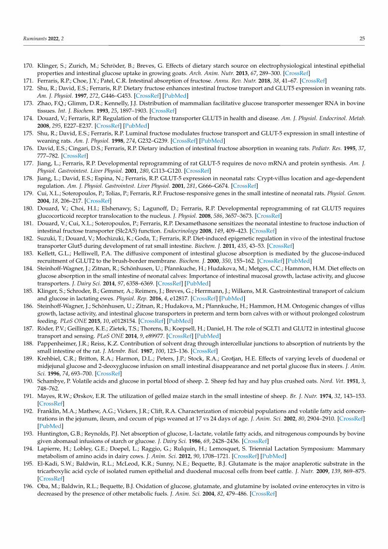
Ruminants 2022, 2 25
170. Klinger, S.; Zurich, M.; Schröder, B.; Breves, G. Effects of dietary starch source on electrophysiological intestinal epithelialproperties and intestinal glucose uptake in growing goats. Arch. Anim. Nutr. 2013, 67, 289–300. [CrossRef]
171. Ferraris, R.P.; Choe, J.Y.; Patel, C.R. Intestinal absorption of fructose. Annu. Rev. Nutr. 2018, 38, 41–67. [CrossRef]172. Shu, R.; David, E.S.; Ferraris, R.P. Dietary fructose enhances intestinal fructose transport and GLUT5 expression in weaning rats.
Am. J. Physiol. 1997, 272, G446–G453. [CrossRef] [PubMed]173. Zhao, F.Q.; Glimm, D.R.; Kennelly, J.J. Distribution of mammalian facilitative glucose transporter messenger RNA in bovine
tissues. Int. J. Biochem. 1993, 25, 1897–1903. [CrossRef]174. Douard, V.; Ferraris, R.P. Regulation of the fructose transporter GLUT5 in health and disease. Am. J. Physiol. Endocrinol. Metab.
2008, 295, E227–E237. [CrossRef] [PubMed]175. Shu, R.; David, E.S.; Ferraris, R.P. Luminal fructose modulates fructose transport and GLUT-5 expression in small intestine of
weaning rats. Am. J. Physiol. 1998, 274, G232–G239. [CrossRef] [PubMed]176. David, E.S.; Cingari, D.S.; Ferraris, R.P. Dietary induction of intestinal fructose absorption in weaning rats. Pediatr. Res. 1995, 37,
777–782. [CrossRef]177. Jiang, L.; Ferraris, R.P. Developmental reprogramming of rat GLUT-5 requires de novo mRNA and protein synthesis. Am. J.
Physiol. Gastrointest. Liver Physiol. 2001, 280, G113–G120. [CrossRef]178. Jiang, L.; David, E.S.; Espina, N.; Ferraris, R.P. GLUT-5 expression in neonatal rats: Crypt-villus location and age-dependent
regulation. Am. J. Physiol. Gastrointest. Liver Physiol. 2001, 281, G666–G674. [CrossRef]179. Cui, X.L.; Soteropoulos, P.; Tolias, P.; Ferraris, R.P. Fructose-responsive genes in the small intestine of neonatal rats. Physiol. Genom.
2004, 18, 206–217. [CrossRef]180. Douard, V.; Choi, H.I.; Elshenawy, S.; Lagunoff, D.; Ferraris, R.P. Developmental reprogramming of rat GLUT5 requires
glucocorticoid receptor translocation to the nucleus. J. Physiol. 2008, 586, 3657–3673. [CrossRef]181. Douard, V.; Cui, X.L.; Soteropoulos, P.; Ferraris, R.P. Dexamethasone sensitizes the neonatal intestine to fructose induction of
intestinal fructose transporter (Slc2A5) function. Endocrinology 2008, 149, 409–423. [CrossRef]182. Suzuki, T.; Douard, V.; Mochizuki, K.; Goda, T.; Ferraris, R.P. Diet-induced epigenetic regulation in vivo of the intestinal fructose
transporter Glut5 during development of rat small intestine. Biochem. J. 2011, 435, 43–53. [CrossRef]183. Kellett, G.L.; Helliwell, P.A. The diffusive component of intestinal glucose absorption is mediated by the glucose-induced
recruitment of GLUT2 to the brush-border membrane. Biochem. J. 2000, 350, 155–162. [CrossRef] [PubMed]184. Steinhoff-Wagner, J.; Zitnan, R.; Schönhusen, U.; Pfannkuche, H.; Hudakova, M.; Metges, C.C.; Hammon, H.M. Diet effects on
glucose absorption in the small intestine of neonatal calves: Importance of intestinal mucosal growth, lactase activity, and glucosetransporters. J. Dairy Sci. 2014, 97, 6358–6369. [CrossRef] [PubMed]
185. Klinger, S.; Schroder, B.; Gemmer, A.; Reimers, J.; Breves, G.; Herrmann, J.; Wilkens, M.R. Gastrointestinal transport of calciumand glucose in lactating ewes. Physiol. Rep. 2016, 4, e12817. [CrossRef] [PubMed]
186. Steinhoff-Wagner, J.; Schönhusen, U.; Zitnan, R.; Hudakova, M.; Pfannkuche, H.; Hammon, H.M. Ontogenic changes of villusgrowth, lactase activity, and intestinal glucose transporters in preterm and term born calves with or without prolonged colostrumfeeding. PLoS ONE 2015, 10, e0128154. [CrossRef] [PubMed]
187. Röder, P.V.; Geillinger, K.E.; Zietek, T.S.; Thorens, B.; Koepsell, H.; Daniel, H. The role of SGLT1 and GLUT2 in intestinal glucosetransport and sensing. PLoS ONE 2014, 9, e89977. [CrossRef] [PubMed]
188. Pappenheimer, J.R.; Reiss, K.Z. Contribution of solvent drag through intercellular junctions to absorption of nutrients by thesmall intestine of the rat. J. Membr. Biol. 1987, 100, 123–136. [CrossRef]
189. Krehbiel, C.R.; Britton, R.A.; Harmon, D.L.; Peters, J.P.; Stock, R.A.; Grotjan, H.E. Effects of varying levels of duodenal ormidjejunal glucose and 2-deoxyglucose infusion on small intestinal disappearance and net portal glucose flux in steers. J. Anim.Sci. 1996, 74, 693–700. [CrossRef]
190. Schambye, P. Volatile acids and glucose in portal blood of sheep. 2. Sheep fed hay and hay plus crushed oats. Nord. Vet. 1951, 3,748–762.
191. Mayes, R.W.; Ørskov, E.R. The utilization of gelled maize starch in the small intestine of sheep. Br. J. Nutr. 1974, 32, 143–153.[CrossRef]
192. Franklin, M.A.; Mathew, A.G.; Vickers, J.R.; Clift, R.A. Characterization of microbial populations and volatile fatty acid concen-trations in the jejunum, ileum, and cecum of pigs weaned at 17 vs 24 days of age. J. Anim. Sci. 2002, 80, 2904–2910. [CrossRef][PubMed]
193. Huntington, G.B.; Reynolds, P.J. Net absorption of glucose, L-lactate, volatile fatty acids, and nitrogenous compounds by bovinegiven abomasal infusions of starch or glucose. J. Dairy Sci. 1986, 69, 2428–2436. [CrossRef]
194. Lapierre, H.; Lobley, G.E.; Doepel, L.; Raggio, G.; Rulquin, H.; Lemosquet, S. Triennial Lactation Symposium: Mammarymetabolism of amino acids in dairy cows. J. Anim. Sci. 2012, 90, 1708–1721. [CrossRef] [PubMed]
195. El-Kadi, S.W.; Baldwin, R.L.; McLeod, K.R.; Sunny, N.E.; Bequette, B.J. Glutamate is the major anaplerotic substrate in thetricarboxylic acid cycle of isolated rumen epithelial and duodenal mucosal cells from beef cattle. J. Nutr. 2009, 139, 869–875.[CrossRef]
196. Oba, M.; Baldwin, R.L.; Bequette, B.J. Oxidation of glucose, glutamate, and glutamine by isolated ovine enterocytes in vitro isdecreased by the presence of other metabolic fuels. J. Anim. Sci. 2004, 82, 479–486. [CrossRef]
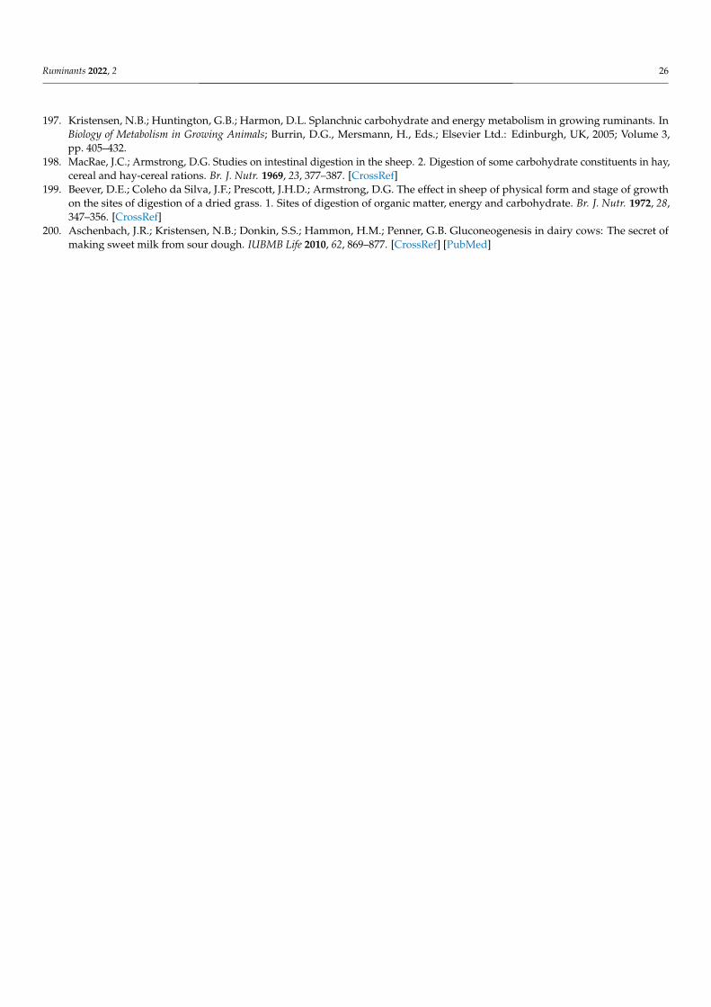
Ruminants 2022, 2 26
197. Kristensen, N.B.; Huntington, G.B.; Harmon, D.L. Splanchnic carbohydrate and energy metabolism in growing ruminants. InBiology of Metabolism in Growing Animals; Burrin, D.G., Mersmann, H., Eds.; Elsevier Ltd.: Edinburgh, UK, 2005; Volume 3,pp. 405–432.
198. MacRae, J.C.; Armstrong, D.G. Studies on intestinal digestion in the sheep. 2. Digestion of some carbohydrate constituents in hay,cereal and hay-cereal rations. Br. J. Nutr. 1969, 23, 377–387. [CrossRef]
199. Beever, D.E.; Coleho da Silva, J.F.; Prescott, J.H.D.; Armstrong, D.G. The effect in sheep of physical form and stage of growthon the sites of digestion of a dried grass. 1. Sites of digestion of organic matter, energy and carbohydrate. Br. J. Nutr. 1972, 28,347–356. [CrossRef]
200. Aschenbach, J.R.; Kristensen, N.B.; Donkin, S.S.; Hammon, H.M.; Penner, G.B. Gluconeogenesis in dairy cows: The secret ofmaking sweet milk from sour dough. IUBMB Life 2010, 62, 869–877. [CrossRef] [PubMed]