nss 404 course title: medical surgical nursing iii
TRANSCRIPT

1
NATIONAL OPEN UNIVERSITY OF NIGERIA
SCHOOL OF SCIENCE AND TECHNOLOGY
COURSE CODE: NSS 404
COURSE TITLE: MEDICAL SURGICAL NURSING III

2
COURSE GUIDE
NSS 404: MEDICAL SURGICAL NURSING III
Course Developer/Writer: Dr. Clara Agbedia
Department of Nursing sciences
Delta State University,
Abraka
Delta State
Programme Leader: Prof. Afolabi Adebanjo
School of Science and Technology.
National Open University of Nigeria,
Lagos.
Course Coordinator: Simeon Kayode Olubiyi
School of Science and Technology.
National Open University of Nigeria,
Lagos.
3
Table of Contents . Introduction …………………………………………………………………………………. Course Aims …………………………………………………………………………………. The Course ………………………………………………………………………………….. Course Aims …………………………………………………………………………………. Course Objectives ……………………………………………………………………………. Working through the Course ……………………………………………………………. … Course Material ………………………………………………………………………………. Study Units …………………………………………………………………………………… Text Books ……………………………………………………………………………………. Assessment …………………………………………………………………………………… Tutor Mark Assignment ………………………………………………………………………. End of Semester Examination ………………………………………………………………… Summary ……………………………………………………………………………………… 1.0 Introduction The course you are about to study is Medical-Surgical Nursing III. This course is a continuation of Medico-surgical nursing II. It deals with disordres of the cardiovascular, respiratory systems, infection process, cell proliferation, and disorders of the male and female pelves. The care of the older adults, skin and wound care are also discussed. At the end of Medical-Surgical III, you will be able to identify clients/patien suffering from specific medical or surgical conditions as outlined above, formulate and implement nursing care plans based on the needs of individuals. Demonstrate ability to perform a simple surgical wound dressing and skill in use of various nursing equipments. A nursing process is used in the discussion of the disorders. Diagnostic measures in medical-surgical condition are also discussed. 2.0 The Course This course, medico-surgical nursing III is divided into three modules Module I deals with the care of clients with the disorders of the cardiovascular system, shock management, and disorders of the red and white blood cells. Module 2 focuses on common lower and upper respiratory disorders, infection process and cell proliferation. Module 3 deals with the challenges of the care of the older adults, disorders of the male and female pelves. The final unit focuses on skin and wound care. Course Aim The goal of this course is to provide you with the necessary knowledge of the art and science of adult medico-surgical nursing and the therapeutic skills needed for effective management of systemic disorders in the body. Course Objectives
4
In addition to the aims above, this course set to achive some objectives. After going through this course, you should be able to: • Understand the basic concept and terminologies in associated with Medical Surgical
Nursing. • Use the nursing process in the care of client/patient with health conditions as identified
above. • Understand the concept of wound managment. • Know the diagnostic measures in medical surgical conditions. 3.0 Working through the Course This course involves that you would be required to spend lot of time to read. The contents of this material are very dense and require you spending great time to study it. This account for the great effort put into its development in the attempt to make it very readable and comprehensible. Nevertheless, the effort required of you is still tremendous. I would advice that you avail yourself the opportunity of attending the tutorial sessions where you would have the opportunity of comparing knowledge with your peers. 4.0 The Course Material You will be provided with the following materials; Course guide Study units. 5.0 Study Units The study units covered on this course are: MODULE 1 Unit I Caring For Clients with Disorders of Heart and Blood Vessels(CardioVascular
Disorders) Unit II Cardiac and Noncardiac Shock (Circulatory Failure) Unit III Disorders Of the Red Blood Unit IV Disorders of White Blood Cells MODULE II Unit I Caring For Clients with Upper Respiratory Disorders
Unit II Care Of Client with Lower Respiratory Disorders
Unit III Infection Process
MODULE III
Unit I Caring For Clients with Cancer, Disorders Of The Male And Female Pelves
Unit II Caring For Older Adults
Unit III Skin Disorders and Wound Management
APPENDIX I Different types of Cardiac Pain APPENDIX II Electrocardiograph Machine and Electrode
5
APPENDIX III Electrocardiograph APPENDIX IV Normal Values for Laboratory Test 6.0 Text Books In addition, the course comes with a list of recommended textbooks, which though are not compulsory for you to acquire or indeed read, are necessary as supplements to the course material. 1. Timby K. Barbara, Jeanne C. Scherer, & Nancy E. Smith (1999). Introductory Medical-Surgical
Nursing. (7th Edition) Lippincott. 2. Kozier Barbara, Glenora Erb. Fundamentals of Nursing. Concepts and procedures. (2nd ed.) 3. Brunner & Suddarth’s (2004) Medical Surgical Nursing. (10th ed) Lippincott Wilkins 4. Walsh M., Watson’s (1997). Clinical Nursing and Related Sciences. (5th Edition) 5. Suzanne C. Smeltzer, Brenda Bare (2004). Medical Surgical Nursing. Lippincott Williams & Wilkins 6. Ethelwynn L. Stellenbery, Juditt C. Bruce (2007). Nursing Practice: Medical-Surgical Nursing for
Hospital and Community. Elsevier Edinburgh. 7.0 Assessment There are two components of assessment for this course. The Tutor Marked Assignment (TMA), and the end of course examination. 8.0 Tutor Marked Assignment(TMA) The TMA is the continuous assessment component of your course. It accounts for 30% of the total score. You will be given 4 TMA’s to answer. Three of these must be answered before you are allowed to sit for the course examination. The TMAs would be given to you by your facilitator and returned after you have done the assignment. MODULE 1 Unit I Identify at least four causes of secondary hypertension. Discuss the nursing management of clients with hypertension. Discuss the cause and pathophysiology of heart failure. Distinguish between left- and right-sided heart failure Unit 1I Explain the pathophysiology of shock. Identify 4 causes of hypovolemic shock and how it can be prevented. Unit III Outline the nursing management of a client with leukemia MODULE II Unit I Explain epistaxis, and describe the nursing care of a client with the condition Unit II Discuss antitubercular pharmacotherapy Outline the medical and nursing management of a client with asthma. Unit III
6
Explain the difference between mechanical and chemical defense mechanisms Discuss the medical management of clients with infectious disorders. Name three nursing interventions that prevent or control infectious disorders. Discuss measures to take if a needle stick injury occurs. MODULE III Unit I Discuss methods of diagnosing cancer. Differentiate various treatments and methods for managing cancer. Unit II Identify nursing care measures that are especially important when providing physical care for an older adult. Unit III Describe the process of wound healing by first and second intention

7
NSS 404 : MEDICAL-SURGICAL NURSING III
Course Developer/Writer: Dr. Clara Agbedia
Department of Nursing sciences
Delta State University,
Abraka
Delta State
Programme Leader: Prof. Afolabi Adebanjo
School of Science and Technology.
National Open University of Nigeria,
Lagos.
Course Coordinator: Simeon Kayode Olubiyi
School of Science and Technology.
National Open University of Nigeria,
Lagos.
8
UNIT I: CARING FOR CLIENTS WITH DISORDERS OF HEART AND BLOOD VESSELS (CARDIOVASCULAR) 1.0 Introduction ………………………………………………………..........................
2.0 Objective ……………………………………………………….............................. 3.0 Main Content ………………………………………………………......................... 3.1 Arteriosclerosis and Atherosclerosis ………………………………………………… 3.2 Occlusive Disorders of Coronary Blood Vessels ………………............................. 3.2.1 Coronary Artery Disease ………………………………………………………......... 3.3 Myocardial Infarction ………………………………………………………............. 3.4 Angina Pectoris ………………………………………………………...................... 3.5 Thrombosis, Phlebothrombosis, and Embolism ……………………………………... 3.6 Disorders of Blood Vessel Walls ……………………………………………………… 3.7 Aneurysms ………………………………………………………............................... 3.8 Cardiac arrhythmia ……………………………………………………….................... 3.9 Pacemaker ………………………………………………………................................ 3.10 Hypertention …………………………………………………………………………… 3.11 Heart Failure …………………………………………………………………………… 3.12 Pulmonary Edema ……………………………………………………………………… 4.0 Conclusion ………………………………………………………………………………. 5.0 Summary ………………………………………………………………………………… 6.0 Tutor Marked Assignment ………………………………………………………………. 7.0 Further Reading and Other Resources …………………………………………………..
9
1.0 Introduction Cardiovascular disease is the leading cause of death all over the world. Occlusive disorders of the coronary arteries and resulting complications are largely responsible for this death. The most common causes of occlusive vascular diseases include atherosclerosis, arteriosclerosis, clot formation, or vascular spasm. 2.0 Objective By the end of this unit you will be able to: • Distinguish between arteriosclerosis and atherosclerosis. • List the risk factors associated with coronary artery disease and discuss which factors can
be modified. • Describe the nursing management of clients with an occlusive disorder of peripheral
blood vessels. • Discuss the symptoms, diagnosis, and treatment of varicose veins. • Describe the nursing management of clients undergoing surgery for varicose veins. • Name and describe six common arrhythmias • Explain the difference between essential and secondary hypertension. • Identify at least four causes of secondary hypertension. • Discuss the nursing management of clients with hypertension. • Discuss the cause and pathophysiology of heart failure. • Distinguish between left- and right-sided heart failure. 3.0 Main Content Arrhythmia Atrial flutter Pacemaker Atrial fibrillation Premature ventricular contractions Defibrillation Ventricular fibrillation Congestive heart failure Pulmonary hypertension Cor pulmonale Pulmonary vascular Digitalization Renin Exertional dyspnea Right-sided heart failure Ventricular assist device Hypertensive heart disease Systolic blood pressure Aneurysm Angina pectoris
3.1 Arteriosclerosis and Atherosclerosis Arteriosclerosis refers to the loss of elasticity or hardening of the arteries. Atherosclerosis is a condition in which the lumen of the artery fills with fatty deposits (chiefly composed of cholesterol) called plaque or atheroma. Arteriosclerosis and atherosclerosis affect may parts of the body (heart, brain, kidneys, and extremities) and cause a variety of disorders (myocardial infarction [MI], cerebrovascular accidents, renal failure). The rate at which arterial changes occur in various organs or structures varies. Etiology and Pathophysiology Arteriosclerosis and atherosclerosis accompany the aging process. Many factors affect the rate of onset and, overall severity of these conditions. As cells within the arterial tissue layers degenerate due to aging, calcium is deposited with the cytoplasm. The calcium causes the arteries to become less elastic. As the left ventricle contracts sending oxygenated blood from the
10
heart, the rigid arterial vessels fail to stretch. This potentially reduces the volume of oxygenated blood delivered to organs. Hyperlipidemia or high blood fat levels trigger atherosclerotic changes. Factors such as gender, heredity, diet, and level of activity individually or collectively influence hyperlipidemia. The body responds to the injury by activating the inflammatory response. Monocytes migrate to the site of injury and deposit themselves under the endothelial cells of the tunica intima. The monocytes then attract and accumulate lipid (fatty) material. The enlarging lesion elevates the endothelium of the artery wall and narrows the lumen. Atherosclerotic vessels are unable to produce endothelial derived relaxing factors and the ability of the artery to dilate is impaired. As the subendothelial atheroma enlarges, the intimal layer may spilt and expose the lesion. As blood flows through the vessel, platelets become trapped in the roughened wall and initiate the clotting mechanism. When this occurs in a coronary artery, the resulting mass is called coronary thrombosis. 3.2 Occlusive Disorders of Coronary Blood Vessels Coronary occlusion is the closing of a coronary artery, reducing or totally interrupting blood supply to the area distal to the occlusion. Coronary artery disease precedes coronary occlusion. If the occlusion is not treated, an MI occurs. Symptoms generally do not occur until at least 60% of the arterial lumen is occluded. 3.2.1 Coronary Artery Disease Coronary artery disease (CAD) refers to the arteriosclerotic and atherosclerotic changes taking place in the coronary arteries that supply the myocardium. The disease may not be diagnosed until individuals are late middle age or older, but the vascular changes most likely began occurring at a much younger age. Etiology and Pathophysiology Coronary artery disease is thought to be due to many factors, rather than a single cause. Men are affected at a younger age than women; however, the incidence rises in postmenopausal women and becomes similar to that in men. A progressively diminishing oxygen supply to cells may actually stimulate collateral circulation ––arterial channels that form to supply the ischemic area. Branches of the coronary artery below the narrowed segment may even dilate. The condition may go unrecognized, particularly among those with a sedentary lifestyle. However, during situations that increase myocardial oxygen demand (exercise or emotional stress), the compromised coronary arteries are unable to adequately oxygenate the myocardium. When the myocardial tissue becomes ischemic (deprived of oxygen), clinical manifestations of CAD, such as chest pain of cardiac origin (angina pectoris), occur. Coronary insufficiency describes a clinical condition in which cardiac pain is frequently more severe than that of typical angina, but death of the heart muscle does not occur. Diagnostic tests The serum cholesterol and triglyceride levels are elevated. Exercise electrocardiogram (ECG) or stress testing may reveal ST segment depression, arrhythmias, and exercise-induced hypertension. Narrowing of one or more coronary arteries is shown during coronary arteriography. Medical and Surgical Management Treatment of CAD includes drugs that produce arterial vasodilation, such as the nitrates (eg, nitroglycerin and isosorbide dinitrate). Beta-adrenergic blocking agents, which decrease myocardial oxygen consumption by reducing heart rate and increasing the diameter of peripheral
11
arteries, are also used in the treatment of CAD. Calcium channel blocking agents are used in the treatment of CAD, although research has shown that they may not be of much benefit. The physician selects the drug that produces the best results for the individual. Drugs such as angiotensin-converting enzyme (ACE) inhibitors and diuretics, as well as stress management, are used to control hypertension. Prevention of further plaque formation is attempted by lowering elevated cholesterol and triglyceride levels through diet, exercise, and, in extreme cases, drugs. Factors that contribute to arterial constriction, like nicotine from cigarettes, are eliminated. Some physicians advise taking one aspirin tablet daily to prevent thrombi from occurring. Nonstrenuous but active, can promote collateral circulation. Invasive but nonsurgical procedures are done to reopen narrowed coronary arteries. Nursing Management Assess the characteristics of chest pain and administer prescribed drugs that dilate the coronary arteries. If rest, drugs, and oxygen do not relieve the pain, notify the physician. Help clients learn how to reduce CAD risk factors that are modifiable and instruct them on the administration and side effects of antianginal drugs. Emphasize that severe, unrelieved chest pain indicates a need to be examined by a physician without delay. The basic preparation of clients who undergo invasive, nonsurgical procedures such PTCA and atherectomy procedures is similar to that for clients who have surgery Monitor all vascular sites for bleeding postprocedure and assess distal pulses. Observe the client’s mental status as cerebral emboli can occur. Measure urine output. Administer analgesics for discomfort. Report any of the following immediately: severe chest pain, abnormal heart rate or rhythm, mental confusion or loss of consciousness, hypotension, urine output less than 30 to 50 mL/h, or a cold, pulse less extremity. Client and Family Teaching Educating clients in ways they can modify their risk factors is a critical element in the care of clients with CAD. A low-fat diet and regular aerobic exercise can significantly reduce the risk of CAD, and refer them to a dietitian for assistance in meal planning. Encourage activity and inform clients that regular exercise such as walking and gardening are sufficient to obtain health benefits. 3.3 Myocardial Infarction An infarct is an area of tissue that dies (necrosis) due to inadequate oxygenation. An MI or heart attack occurs when there is prolonged, 100% occlusion of coronary arterial blood flow. The larger the necrotic area, the more serious the damage done to the heart. An infarct that extends through the full thickness of the myocardial wall is called a transmural infarction or Q wave MI. A partial thickness infarct is called a subendocardial infarction, or non-Q wave MI. Each coronary artery supplies a different area of the myocardium. The zone of necrosis is identified according to the area for myocardium supplied by the respective coronary artery. Diagnostic tests Laboratory tests include a series of serum enzyme and isoenzyme levels, which are elevated. The white blood cell count and the erythrocyte sedimentation rate increase about the third day due to the inflammatory response triggered by the injury to myocardial cells. The blood sugar may be elevated in clients with diabetes (and those without) because of their response to a major stressor. Following an MI, characteristic changes appear in the ECG within 2 to 12 hours after the infarction but may take as long as 3 days to develop. These changes include ST segment, T wave inversion, and the appearance of a Q wave.
12
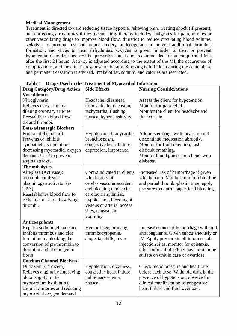
Medical Management Treatment is directed toward reducing tissue hypoxia, relieving pain, treating shock (if present), and correcting arrhythmias if they occur. Drug therapy includes analgesics for pain, nitrates or other vasodilating drugs to improve blood flow, diuretics to reduce circulating blood volume, sedatives to promote rest and reduce anxiety, anticoagulants to prevent additional thrombus formation, and drugs to treat arrhythmias. Oxygen is given in order to treat or prevent hypoxemia. Complete bed rest is prescribed but is not recommended for uncomplicated MIs after the first 24 hours. Activity is adjusted according to the extent of the MI, the occurrence of complications, and the client’s response to therapy. Smoking is forbidden during the acute phase and permanent cessation is advised. Intake of fat, sodium, and calories are restricted. Table 1 Drugs Used in the Treatment of Myocardial Infarction
Drug Category/Drug Action Side Effects Nursing Considerations. Vasodilators Nitroglycerin Relieves chest pain by dilating coronary arteries Reestablishes blood flow around thrombi.
Headache, dizziness, orthostatic hypotension, tachycardia, flushing, nausea, hypersensitivity
Assess the client for hypotension. Monitor for pain relief. Monitor the client for headache and flushed skin.
Beta-adrenergic Blockers Propranolol (Inderal) Prevents or inhibits sympathetic stimulation, decreasing myocardial oxygen demand. Used to prevent angina attacks.
Hypotension bradycardia, bronchospasm, congestive heart failure, depression, impotence.
Administer drugs with meals, do not discontinue medication abruptly. Monitor for fluid retention, rash, difficult breathing. Monitor blood glucose in clients with diabetes.
Thrombolytics Alteplase (Activase); recombinant tissue plasminogen activator (r-TPA). Reestablishes blood flow to ischemic areas by dissolving thrombi.
Contraindicated in clients with history of cerebrovascular accident and bleeding tendencies, cardiac arrhythmias, hypotension, bleeding at venous or arterial access sites, nausea and vomiting
Increased risk of hemorrhage if given with heparin. Monitor prothrombin time and partial thromboplastin time; apply pressure to control superficial bleeding.
Anticoagulants Heparin sodium (Hepalean) Inhibits thrombus and clot formation by blocking the conversion of prothrombin to thrombin and fibrinogen to fibrin.
Hemorrhage, bruising, thrombocytopenia, alopecia, chills, fever
Increase chance of hemorrhage with oral anticoagulants. Given subcutaneously or IV. Apply pressure to all intramuscular injection sites, monitor for epistaxis, other forms of bleeding, have protamine sulfate on unit in case of overdose.
Calcium Channel Blockers Diltiazem (Cardizem) Relieves angina by improving blood supply to the myocardium by dilating coronary arteries and reducing myocardial oxygen demand.
Hypotension, dizziness, congestive heart failure, pulmonary edema, nausea.
Check blood pressure and heart rate before each dose. Withhold drug in the presence of hypotension, observe for clinical manifestation of congestive heart failure and fluid overload.
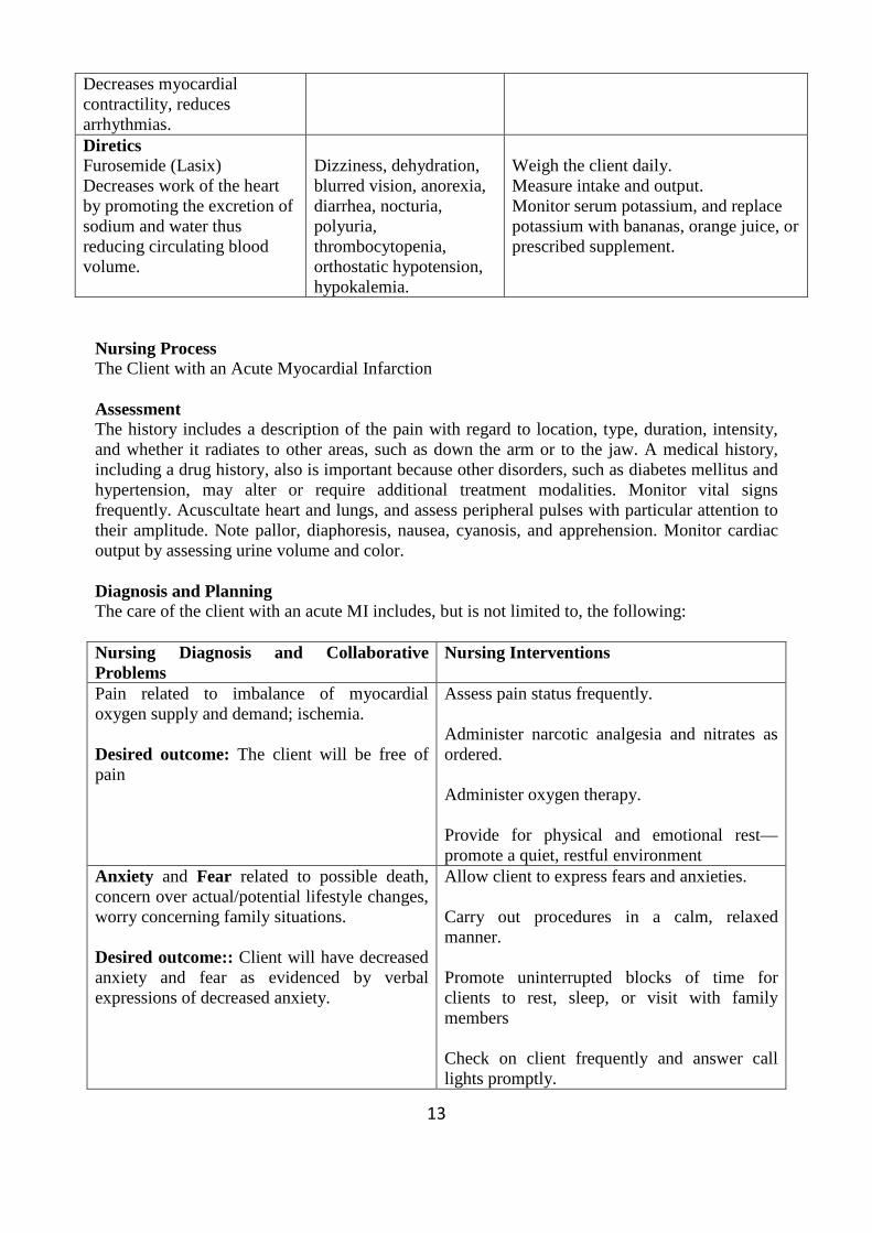
13
Decreases myocardial contractility, reduces arrhythmias. Diretics Furosemide (Lasix) Decreases work of the heart by promoting the excretion of sodium and water thus reducing circulating blood volume.
Dizziness, dehydration, blurred vision, anorexia, diarrhea, nocturia, polyuria, thrombocytopenia, orthostatic hypotension, hypokalemia.
Weigh the client daily. Measure intake and output. Monitor serum potassium, and replace potassium with bananas, orange juice, or prescribed supplement.
Nursing Process The Client with an Acute Myocardial Infarction Assessment The history includes a description of the pain with regard to location, type, duration, intensity, and whether it radiates to other areas, such as down the arm or to the jaw. A medical history, including a drug history, also is important because other disorders, such as diabetes mellitus and hypertension, may alter or require additional treatment modalities. Monitor vital signs frequently. Acuscultate heart and lungs, and assess peripheral pulses with particular attention to their amplitude. Note pallor, diaphoresis, nausea, cyanosis, and apprehension. Monitor cardiac output by assessing urine volume and color. Diagnosis and Planning The care of the client with an acute MI includes, but is not limited to, the following: Nursing Diagnosis and Collaborative Problems
Nursing Interventions
Pain related to imbalance of myocardial oxygen supply and demand; ischemia. Desired outcome: The client will be free of pain
Assess pain status frequently. Administer narcotic analgesia and nitrates as ordered. Administer oxygen therapy. Provide for physical and emotional rest––promote a quiet, restful environment
Anxiety and Fear related to possible death, concern over actual/potential lifestyle changes, worry concerning family situations. Desired outcome:: Client will have decreased anxiety and fear as evidenced by verbal expressions of decreased anxiety.
Allow client to express fears and anxieties. Carry out procedures in a calm, relaxed manner. Promote uninterrupted blocks of time for clients to rest, sleep, or visit with family members Check on client frequently and answer call lights promptly.
14
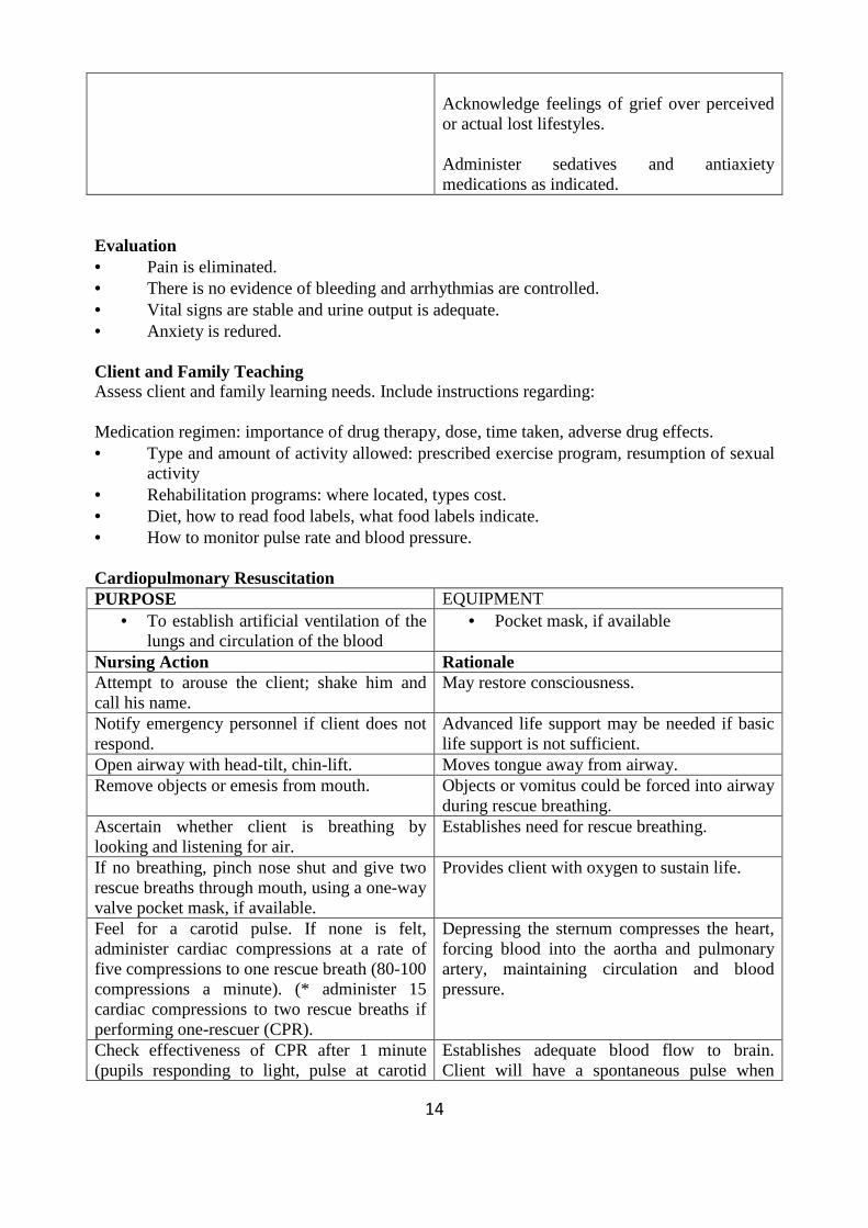
Acknowledge feelings of grief over perceived or actual lost lifestyles. Administer sedatives and antiaxiety medications as indicated.
Evaluation • Pain is eliminated. • There is no evidence of bleeding and arrhythmias are controlled. • Vital signs are stable and urine output is adequate. • Anxiety is redured. Client and Family Teaching Assess client and family learning needs. Include instructions regarding: Medication regimen: importance of drug therapy, dose, time taken, adverse drug effects. • Type and amount of activity allowed: prescribed exercise program, resumption of sexual
activity • Rehabilitation programs: where located, types cost. • Diet, how to read food labels, what food labels indicate. • How to monitor pulse rate and blood pressure. Cardiopulmonary Resuscitation PURPOSE EQUIPMENT
• To establish artificial ventilation of the lungs and circulation of the blood
• Pocket mask, if available
Nursing Action Rationale Attempt to arouse the client; shake him and call his name.
May restore consciousness.
Notify emergency personnel if client does not respond.
Advanced life support may be needed if basic life support is not sufficient.
Open airway with head-tilt, chin-lift. Moves tongue away from airway. Remove objects or emesis from mouth. Objects or vomitus could be forced into airway
during rescue breathing. Ascertain whether client is breathing by looking and listening for air.
Establishes need for rescue breathing.
If no breathing, pinch nose shut and give two rescue breaths through mouth, using a one-way valve pocket mask, if available.
Provides client with oxygen to sustain life.
Feel for a carotid pulse. If none is felt, administer cardiac compressions at a rate of five compressions to one rescue breath (80-100 compressions a minute). (* administer 15 cardiac compressions to two rescue breaths if performing one-rescuer (CPR).
Depressing the sternum compresses the heart, forcing blood into the aortha and pulmonary artery, maintaining circulation and blood pressure.
Check effectiveness of CPR after 1 minute (pupils responding to light, pulse at carotid
Establishes adequate blood flow to brain. Client will have a spontaneous pulse when
15
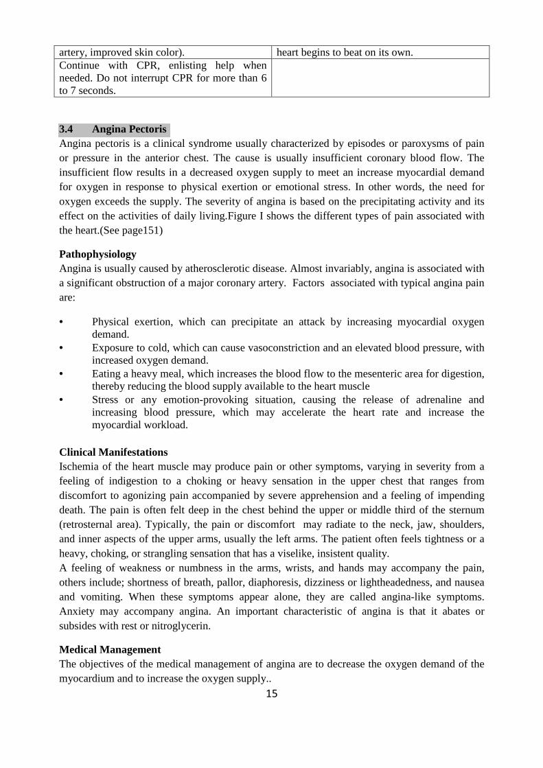
artery, improved skin color). heart begins to beat on its own. Continue with CPR, enlisting help when needed. Do not interrupt CPR for more than 6 to 7 seconds.
3.4 Angina Pectoris Angina pectoris is a clinical syndrome usually characterized by episodes or paroxysms of pain or pressure in the anterior chest. The cause is usually insufficient coronary blood flow. The insufficient flow results in a decreased oxygen supply to meet an increase myocardial demand for oxygen in response to physical exertion or emotional stress. In other words, the need for oxygen exceeds the supply. The severity of angina is based on the precipitating activity and its effect on the activities of daily living.Figure I shows the different types of pain associated with the heart.(See page151)
Pathophysiology Angina is usually caused by atherosclerotic disease. Almost invariably, angina is associated with a significant obstruction of a major coronary artery. Factors associated with typical angina pain are:
• Physical exertion, which can precipitate an attack by increasing myocardial oxygen demand.
• Exposure to cold, which can cause vasoconstriction and an elevated blood pressure, with increased oxygen demand.
• Eating a heavy meal, which increases the blood flow to the mesenteric area for digestion, thereby reducing the blood supply available to the heart muscle
• Stress or any emotion-provoking situation, causing the release of adrenaline and increasing blood pressure, which may accelerate the heart rate and increase the myocardial workload.
Clinical Manifestations Ischemia of the heart muscle may produce pain or other symptoms, varying in severity from a feeling of indigestion to a choking or heavy sensation in the upper chest that ranges from discomfort to agonizing pain accompanied by severe apprehension and a feeling of impending death. The pain is often felt deep in the chest behind the upper or middle third of the sternum (retrosternal area). Typically, the pain or discomfort may radiate to the neck, jaw, shoulders, and inner aspects of the upper arms, usually the left arms. The patient often feels tightness or a heavy, choking, or strangling sensation that has a viselike, insistent quality. A feeling of weakness or numbness in the arms, wrists, and hands may accompany the pain, others include; shortness of breath, pallor, diaphoresis, dizziness or lightheadedness, and nausea and vomiting. When these symptoms appear alone, they are called angina-like symptoms. Anxiety may accompany angina. An important characteristic of angina is that it abates or subsides with rest or nitroglycerin.
Medical Management The objectives of the medical management of angina are to decrease the oxygen demand of the myocardium and to increase the oxygen supply..
16
Pharmacologic Therapy Medications used to control angina are nitroglycerin, beta-adrenergic blocking agents, calcium channel blockers, and antiplatelet agents. Nitroglycerin. Nitrates remain the mainstay for treatment of angina pectoris. Nitroglycerin dilates primarily the veins and the arteries. It helps to increase coronary blood flow by preventing vasospasm and increasing perfusion through the collateral vessels. Dilation of the veins causes venous pooling of blood throughout the body. As a result, less blood returns to the heart, and filling pressure (preload) is reduced. Nitroglycerin may be given by several routes: sublingual tablet or spray, topical agent, and intravenous administration. Beta-Adrenergic Blocking Agents. Beta-blockers such as propranolol (Inderal), metoprolol (Lopressor, Toprol), and atenolol (Tenormin) appear to reduce myocardial oxygen consumption by blocking the beta-adrenergic sympathetic stimulation to the heart. The result is a reduction in heart rate, slowed conduction of an impulse through the heart, decreased blood pressure, and reduced myocardial contractility (force of contraction) that establishes a more favorable balance between myocardial oxygen needs (demands) and the amount of oxygen available (supply). This helps to control chest pain and delays the onset of ischemia during work or exercise. Cardiac side effects and possible contraindications include hypotension, bradycardia, advanced atrioventricular block, and decompensated heart failure.
Calcium Channel Blocking Agents. Calcium channel blockers increase myocardial oxygen supply by dilating the smooth muscle wall of the coronary arterioles; they decrease myocardial oxygen demand by reducing systemic arterial pressure and the workload of the left ventricle.
The calcium channel blockers most commonly used are amlodipine (Norvasc), verapamil (Calan, Isoptin, Verelan), and diltiazem (Cardizem, Dilacor, Tiazac).
Antiplatelet and Anticoagulat Medications. Antiplatelet medications are administered to prevent platelet aggregation, wich impedes blood flow.
Aspirin. Aspirin prevents platelet activation and reduces the incidence of MI and death in patient with CAD.
Clopidogrel and Ticlopidine. Clopidogrel (Plavix) or ticlopidine (Ticlid) is given to patients who are allergic to aspirin or given in addition to aspirin in patients at high risk for MI. They also cause gastrointestinal upset, including nausea, vomiting, and diarrhea, and they decrease the neutrophil level.
Heparin. Unfractionated heparin prevents the formation of new blood clots. Use of heparin alone in treating patients with unstable angina reduces the occurrence of MI.
3.5 Thrombosis, Phlebothrombosis, and Embolism A thrombus is a stationary clot. Thrombosis is a state in which a clot has formed within a blood vessel. Phlebothrombosis is the development of a clot within a vein without inflammation.
17
Phlebothrombosis and thrombophlebitis have similar symptoms and treatment An embolus is a moving mass (clot) of particles either solid or gas within the bloodstream. Etiology and Pathophysiology When a thrombus forms or an embolus reaches a blood vessel that is too small to permit its passage, there is partial or total occlusion of blood flow through the vessel.Thrombosis in the venous system most often occurs in the lower extremities and generally is associated with disorders or circumstances that cause venous stasis. For example, inactivity, immobility, or trauma to a blood vessel commonly predisposes an individual to clot formation. Other causes include; atherosclerosis, endocarditis, pooling of blood in a ventricular aneurysm, and arrhythmias. Assessment Clinical manifestation The clinical manifestations of deep vein thrombosis usually include pain, swelling, and tenderness of the affected extremity, and mild fever. A positive Homans’ sign, pain on dorsiflexion of the foot, may be present. A thrombus may become a mobile embolus and lodge in a distal blood vessel, like the pulmonary capillaries, causing symptoms related to the organ to which circulation has becomes impaired. Diagnostic tests Arteriography or venography (also called phlebography) using a contrast dye identifies the point of obstruction. Doppler ultrasonography is used to detect abnormalities in peripheral blood flow. Plethysmography measures volume changes within the venous or arterial system. Nursing Management Obtain a history of the symptoms and identify the characteristics of the client’s pain. Assess for Homan’s sign by having the client dorsiflex each foot. Examine the extremities and compare skin color, temperature, capillary refill time, and tissue integrity. Measure each calf. Palpate peripheral pulse; use a Doppler ultrasound device if pulses cannot be palpated. Monitor the client’s response to anticoagulation therapy. Monitor IV infusions of heparin hourly. Monitor prothrombin time when oral anticoagulation is prescribed. Therapeutic response and daily dosage are determined by these values. Be alert for signs of bleeding and keep protamine sulfate on hand for reversing heparin, and vitamin K for reversing oral anticoagulants. Additional nursing management is directed toward increasing arterial or venous blood flow, relieving pain, preventing complications, and providing thorough teaching before discharge. Client and Family Teaching To prevent a recurrence of thrombosis, embolisms, and phlebothrombosis, inform clients to avoid prolonged periods of inactivity (especially sitting), elevate the legs periodically, and walk or do isometric leg exercises at frequent intervals if sitting is unavoidable. Recommend wearing antiembolism stockings to prevent venous stasis. 3.6 Disorders of Blood Vessel Walls Varicose Veins Varicose veins are dilated, tortuous veins (varicosities). The saphenous veins of the legs are commonly affected because they lack support from surrounding leg muscles. Both sexes suffer
18
equally from this disorder. Varicose veins also may occur in other parts of the body, such as the rectum (hemorrhoids) and the esophagus (esophageal varices). Etiology and Pathophysiology There is a familial tendency for varicose veins. Normally, the action of leg muscles during movement and exercise aids venous return. When the valves within veins become incompetent blood accumulates rather than being propelled efficiently to the heart. The congestion stretches the veins. Over time, they are unable to recoil and remain in a chronically distended state. Venous hypertension then forces some fluid to move into the interstitial spaces of surrounding tissue. Venous congestion and local edema may diminish arterial blood flow, resulting in impaired cellular nutrition. Minor skin or soft tissue injuries easily become infected and ulcerated. The healing of such lesions is slow and uncertain. Prolonged standing compromises venous return as blood pools distally with gravity. Obesity and pressure on blood vessels from an enlarging fetus, liver, or abdominal tumor contribute to venous congestion. Assessment Clinical manifestation Often the condition first manifests itself when other factors impair venous return. The legs feel heavy and tired, particularly after prolonged standing and the discomfort is relieved with activity or elevation of the legs. The veins of the legs look distended and torturous and can be seen under the skin as dark blue or purple swellings. The feet, ankles, and legs may appear swollen. The skin may be slightly darker in the areas of impaired circulation. There may be signs of skin ulcerations in various stages of healing. Capillary refill may be abnormal. Diagnostic tests The Brodie-Trendelenberg test is performed for diagnostic purposes. The client lies flat and elevates the affected leg to empty the veins. A tourniquet is then applied to the upper thigh, and the client is asked to stand. If blood flows from the upper part of the leg into the superficial veins when the tourniquet is released, the valves of the superficial veins are considered incompetent. Ultrasonography and venography also are used to detect impaired blood flow. Medical Management Treatment of mild varicose veins includes exercise (walking, swimming), weight loss (if needed), the wearing of support stockings, and the avoidance of prolonged periods of sitting. The defective vein may be sclerosed or occluded by injecting a chemical that sets up an inflammation within the vein wall. Surgical Management Surgical treatment for severe or multiple varicose veins consists of vein ligation with or without vein stripping. The affected veins are ligated (tied off) above and below the area of incompetent valves. Nursing Management Assess the skin, distal circulation and peripheral edema. Ask the client to rate the level of discomfort and ability to do active and isometric leg exercises. Elevate the foot of the bed in the immediate postoperative period to aid venous return. Remind the client to alternately contract and relax the muscles in the lower legs. If the client’s active exercise is inadequate, consider using pneumatic venous compression stockings, which cover the leg from the foot to the thigh and periodically inflate and release air, stimulating isometric muscle contraction. Ambulate the
19
client as soon as possible to promote venous circulation, reduce edema, and prevent venous thrombosis. Client and Family Teaching Identity factors that impair venous circulation such as wearing elastic girdles, tight belts, using round garters or rolling and twisting nylon stockings, and standing or sitting for prolonged periods; suggest alternative measures that will promote venous return. Stress that the client should avoid sitting with the knees crossed. Describe appropriate foot and nail care. 3.7 Aneurysms An aneurysm is a stretching and bulging of an arterial wall. Aneurysms of the aorta (aortic arch, thoraci, abdominal) are the most common . Etiology and Pathophysiology Arteriosclerosis, hypertension, trauma, or a congenital weakness can affect the elasticity of the tunica media (middle layer of the artery wall), causing a portion of the vessel to bulge. Most aneurysms enlarge until they rupture leading to shock and death if not controlled. Assessment Clinical manifestation Many aneurysms go unnoticed by the individual until they are found during a physical examination or the client has a massive hemorrhage. Some cause pain and discomfort and symptoms related to pressure on nearby structures. For example, a thoracic aortic aneurysm can cause bronchial obstruction, dysphagia (difficulty swallowing), and dyspnea. An abdominal aortic aneurysm can produce nausea and vomiting from pressure exerted on the intestines, or it may cause back pain from pressure on the vertebrae or spinal nerves. Most individuals are hypertensive. Diagnostic tests Aortography identifies the size and exact location of the aneurysm. Medical and Surgical Management Medical treatment includes administering antihypertensive drugs to keep the blood pressure low. Aneurysms are repaired by bypass or replacement grafting. 3.8 Cardiac arrhythmia Cardiac rhythm refers to the pattern (or pace) of the heartbeat. This pattern is enabled by the conduction system of the heart and the inherent rhythmicity of cardiac muscle. An arrhythmia (also called dysrhythmia) is a conduction disorder that results in an abnormally slow or rapid regular heart rate or at an irregular pace. Common cause of arrhythmias is ischemic heart disease. Others are effects of drug therapy, electrolyte disturbances, metabolic acidosis, and hypothermia can also cause arrhythmias. Types of Arrhythmias Originating in the Sinoatrial (SA) Node Sinus Bradycardia Sinus bradycardia is a regular, but slow, (≤ 60 beats/min) rhythm. It is a pathologic sign in clients with heart disorders. The danger in bradycardia is that the slow rate may be insufficient to maintain cardiac output. Atropine sulfate is sometimes given intravenously (IV) to increase the heart rate.
20
Sinus Tachycardia Sinus tachycardia is a regular but fast (100-150 beats/min) rhythm Sinus tachycardia occurs in clients with healthy hearts as a physiologic response to strenuous exercise, anxiety and fear, pain, fever, hyperthyroidism, hemorrhage, shock, or hypoxemia. Supraventricular Tachycardia Supraventricular tachycardia (SVT) is an arrhythmia with a dangerously high heart rate (≥ 150 beats/min). At this rate, diastole is shortened and the heart does not have sufficient time to fill. Cardiac output drops dangerously low and heart failure can occur. Drugs, such as digitalis, adrenergic blockers, and calcium channel blockers, can be used to slow the heart rate. Atrial Flutter Atrial flutter is a disorder in which a single atrial impulse outside the SA node causes the atria to contract at an exceedingly rapid rate (200-400 times/min). The atrial waves in atrial flutter have a characteristic sawtooth pattern. Atrial Fibrillation This is a disorganized, rapid atrial activity. Atrial fibrillation is treated with digitalis if the ventricular rate is not too slow or cardioversion is used. Defibrillation The only treatment for a life-threatening arrhythmia such as ventricular tachycardia or fibrillation is immediate defibrillation.. It is used during cardiac arrest when there is no identifiable R wave present. 3.9 Pacemaker A pacemaker is a battery-operated device that provides an electrical stimulus to the muscle of the heart. It is inserted either temporarily or permanently to restore an effective cardiac rhythm. It can be a temporary or a permanent pacemaker Temporary Pacemakers A temporary pacemaker is indicated in clients with transient bradyarrhythmias (slow, abnormal rhythms) such as during an acute myocardial infarction or after coronary artery bypass graft surgery, and with some tachyarrhythmias. Permanent Pacemakers A permanent pacemaker is used for complete and some second-degree heart blocks. 3.10 Hypertention Blood pressure is the force produced by the volume of blood within the walls of arteries. It is represented by the formula: BP = CO (cardiac output) x PR (peripheral resistance). The measured pressure reflects the ability of the arteries to stretch and fill with blood, the efficiency of the heart as a pump, and the volume of circulating blood. Blood pressure is affected by age, body size, diet, activity, emotions, pain, position, gender, the time of day, and diseases states.
21
Arterial Blood Pressure Arterial pressure is regulated by the autonomic nervous system, the kidneys, and various endocrine glands. When measured, the pressure during systole and diastole is expressed as a fraction. The top number is the systolic pressure and the bottom number is the diastolic pressure. Normal blood pressure for adults ranges from 100/60 to 140/90 mm Hg. Blood pressure tends to increase with age, most likely from arteriosclerotic and atherosclerotic changes in the blood vessels, or other effects of chronic diseases. Systolic Blood Pressure Systolic blood pressure is determined by the force and volume of blood ejected from the left ventricle during systole and the ability of the arterial system to distend at the time of ventricular contraction. The walls of the arteries are normally elastic and yield to the force and volume of ventricular contraction. In older clients, systolic blood pressure may be elevated because of loss of arterial elasticity (arteriosclerosis). Narrowing of the arterioles, either by arteriosclerosis or some other mechanisms that causes vasoconstriction, increases peripheral resistance, which in turn increases systolic blood pressure. This resistance can be compared to the slight narrowing of a tube, such as a drinking straw or a garden hose. The narrower the lumen, the greater the pressure needed to move air or liquid through it. Diastolic Blood Pressure Diastolic blood pressure reflects arterial pressure during ventricular relaxation and depends on the resistance of the arterioles and the diastolic filling times. If arterioles are resistant (constricted), blood is under greater pressure. Hypertensive Defined The term hypertension refers to a sustained elevation of systolic arterial pressure of 140 mm Hg or higher, a sustained diastolic arterial pressure of 90 mm Hg or greater, or both. When a cardiac abnormality results from elevated blood pressure, the term hypertensive heart disease is used. When vascular damage is present without heart involvement, the term hypertensive vascular disease is used. When both heart disease and vascular damage accompany hypertension, the appropriate term is hypertensive cardiovascular disease. Essential and Secondary Hypertension Hypertension is divided into two main categories: essential (or primary) hypertension and secondary hypertension. Essential hypertension is a sustained elevation of blood pressure without any known cause. About 95% of those with hypertension have this type. Secondary hypertension is an elevation of blood pressure that results from, or is secondary to some other disorder. Etiology and Pathophysiology The exact cause of essential hypertension is unknown. Blood pressure often increases with age and may run in families. African Americans are affected at a higher rate than other ethnic groups. The risk for hypertension is increased by obesity, inactivity, smoking, excessive alcohol intake, and ineffective stress management. Research into specific factors that contribute to the development of essential hypertension continues. For instance, it is well documented that hypernatremia (elevated serum sodium level) increases blood volume, which raises blood pressure. However, a low serum potassium level may actually cause sodium retention because the kidneys try to maintain a balanced number of
22
cations (positively charged electrolytes) in body fluid. Scientists are also investigating the role of calcium in hypertension because serum calcium levels are low in some hypertensive clients. Essential hypertension may also develop from alterations in other body chemicals. Defects in blood pressure regulation may result from an impairment in the rennin-angiotensin-aldosterone mechanism. Renin is a chemical released by the kidneys to raise blood pressure and increase vascular fluid volume. For those who respond to stress at a heightened degree, hypertension may be correlated with a release of higher than usual catecholamines, such ase epinephrine and norepinephrine, which elevate blood pressure. Lastly, some feel that there may be a deficiency of natriuretic factor, a hormone produced by the heart, causing arteries and arterioles to remain in a state of sustained vasoconstriction. Secondary hypertension may accompany any primary condition that effects fluid volume or renal function or causes arterial vasoconstriction. Predisposing conditions include kidney disease, pheochromocytoma (a tumor of the adrenal medulla), hyperaldosteronism, atherosclerosis, the use of cocaine or other cardiac stimulants such as weight control drugs and caffeine, and the use of oral contraceptives. Regardless of whether a person has essential or secondary hypertension, the organ damage and complications that follow are the same. Hypertension causes the heart to work harder to pump against the increased resistance. Consequently, the size of the heart muscle increases. When the heart can no longer pump adequately to meet the body’s metabolic needs, heart failure occurs. The extra work and the greater mass increase the heart’s need for oxygen. If the myocardium does not receive sufficient oxygenated blood, myocardial ischemia occurs and the client experiences angina. In addition to the direct effects on the heart, high blood pressure damages the arterial vascular system. It accelerates atherosclerosis. Furthermore, the increased resistance of the arterioles to the flow of blood causes serious complications in other organs of the body, including the eyes, brain, heart, and kidneys. Hemorrhage of tiny arteries in the retina may cause marked visual disturbance or blindness. A cerebrovascular accident may result from hemorrhage or occlusion of a blood vessel in the brain. Myocardial infarction may result from occlusion of a branch of a coronary artery. Impaired circulation to the kidneys results in renal failure among some clients with hypertension. Accelerated and Malignant Hypertension Accelerated and malignant hypertension are more serious forms of elevated blood pressure that develop among individuals with either essential or primary hypertension. Accelerated hypertension describes markedly elevated blood pressure, accompanied by hemorrhages and exudates in the eye. If untreated, accelerated hypertension may progress to malignant hypertension. The term malignant hypertension describes dangerously elevated blood pressure accompanied by papilledema (swelling of the optic nerve at its point of entrance into the eye). Assessment Clinical manifestation Clients may be asymptomatic. The onset of hypertension, considered “the silent killer,” is often gradual. Hypertension can be present for years and discovered during a routine physical examination or when the client experiences a major complication. As the blood pressure becomes elevated, clients may identify symptoms such as a throbbing or pounding headache, dizziness, fatigue, insomnia, nervousness, nosebleeds, and blurred vision. Angina or shortness of breath may be the first clue to hypertensive heart disease. The most obvious finding during a physical assessment is a sustained elevation of one or both blood pressure measurements. The pulse may feel bounding from the force of ventricular
23
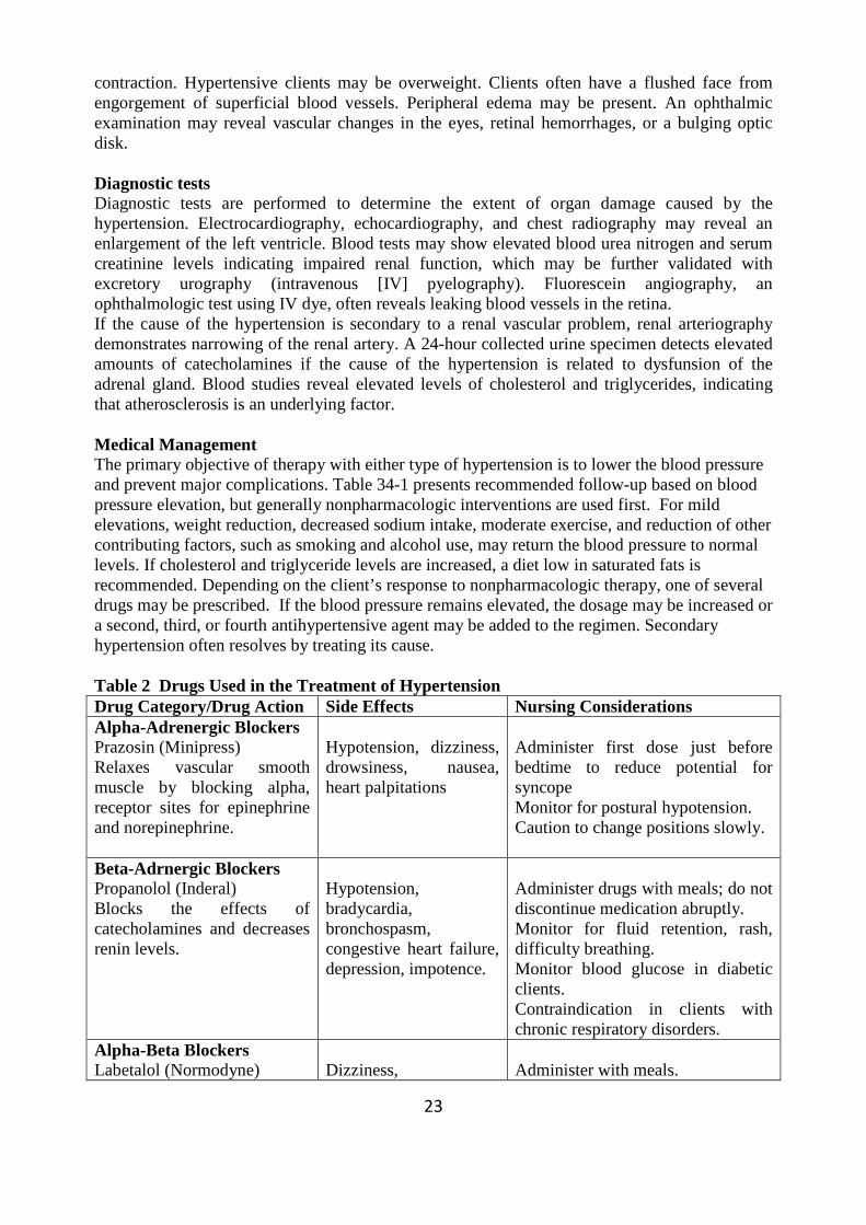
contraction. Hypertensive clients may be overweight. Clients often have a flushed face from engorgement of superficial blood vessels. Peripheral edema may be present. An ophthalmic examination may reveal vascular changes in the eyes, retinal hemorrhages, or a bulging optic disk. Diagnostic tests Diagnostic tests are performed to determine the extent of organ damage caused by the hypertension. Electrocardiography, echocardiography, and chest radiography may reveal an enlargement of the left ventricle. Blood tests may show elevated blood urea nitrogen and serum creatinine levels indicating impaired renal function, which may be further validated with excretory urography (intravenous [IV] pyelography). Fluorescein angiography, an ophthalmologic test using IV dye, often reveals leaking blood vessels in the retina. If the cause of the hypertension is secondary to a renal vascular problem, renal arteriography demonstrates narrowing of the renal artery. A 24-hour collected urine specimen detects elevated amounts of catecholamines if the cause of the hypertension is related to dysfunsion of the adrenal gland. Blood studies reveal elevated levels of cholesterol and triglycerides, indicating that atherosclerosis is an underlying factor. Medical Management The primary objective of therapy with either type of hypertension is to lower the blood pressure and prevent major complications. Table 34-1 presents recommended follow-up based on blood pressure elevation, but generally nonpharmacologic interventions are used first. For mild elevations, weight reduction, decreased sodium intake, moderate exercise, and reduction of other contributing factors, such as smoking and alcohol use, may return the blood pressure to normal levels. If cholesterol and triglyceride levels are increased, a diet low in saturated fats is recommended. Depending on the client’s response to nonpharmacologic therapy, one of several drugs may be prescribed. If the blood pressure remains elevated, the dosage may be increased or a second, third, or fourth antihypertensive agent may be added to the regimen. Secondary hypertension often resolves by treating its cause. Table 2 Drugs Used in the Treatment of Hypertension Drug Category/Drug Action Side Effects Nursing Considerations Alpha-Adrenergic Blockers Prazosin (Minipress) Relaxes vascular smooth muscle by blocking alpha, receptor sites for epinephrine and norepinephrine.
Hypotension, dizziness, drowsiness, nausea, heart palpitations
Administer first dose just before bedtime to reduce potential for syncope Monitor for postural hypotension. Caution to change positions slowly.
Beta-Adrnergic Blockers Propanolol (Inderal) Blocks the effects of catecholamines and decreases renin levels.
Hypotension, bradycardia, bronchospasm, congestive heart failure, depression, impotence.
Administer drugs with meals; do not discontinue medication abruptly. Monitor for fluid retention, rash, difficulty breathing. Monitor blood glucose in diabetic clients. Contraindication in clients with chronic respiratory disorders.
Alpha-Beta Blockers Labetalol (Normodyne)
Dizziness,
Administer with meals.
24
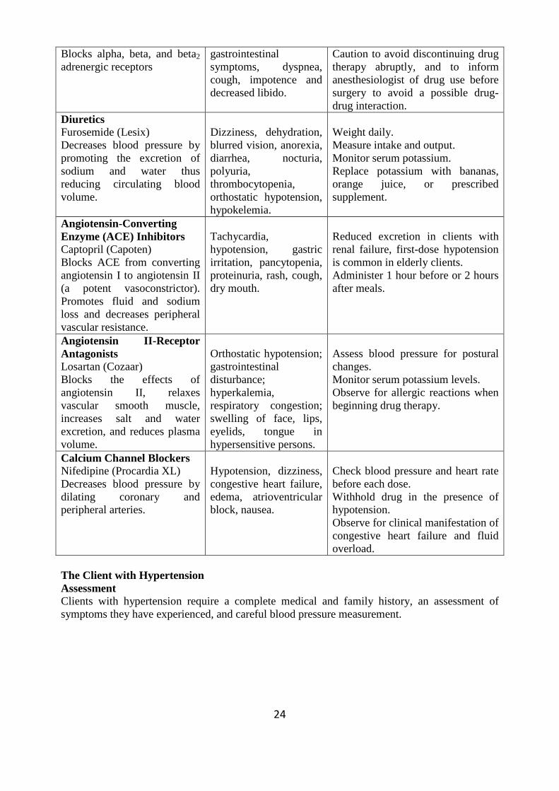
Blocks alpha, beta, and beta2 adrenergic receptors
gastrointestinal symptoms, dyspnea, cough, impotence and decreased libido.
Caution to avoid discontinuing drug therapy abruptly, and to inform anesthesiologist of drug use before surgery to avoid a possible drug-drug interaction.
Diuretics Furosemide (Lesix) Decreases blood pressure by promoting the excretion of sodium and water thus reducing circulating blood volume.
Dizziness, dehydration, blurred vision, anorexia, diarrhea, nocturia, polyuria, thrombocytopenia, orthostatic hypotension, hypokelemia.
Weight daily. Measure intake and output. Monitor serum potassium. Replace potassium with bananas, orange juice, or prescribed supplement.
Angiotensin-Converting Enzyme (ACE) Inhibitors Captopril (Capoten) Blocks ACE from converting angiotensin I to angiotensin II (a potent vasoconstrictor). Promotes fluid and sodium loss and decreases peripheral vascular resistance.
Tachycardia, hypotension, gastric irritation, pancytopenia, proteinuria, rash, cough, dry mouth.
Reduced excretion in clients with renal failure, first-dose hypotension is common in elderly clients. Administer 1 hour before or 2 hours after meals.
Angiotensin II-Receptor Antagonists Losartan (Cozaar) Blocks the effects of angiotensin II, relaxes vascular smooth muscle, increases salt and water excretion, and reduces plasma volume.
Orthostatic hypotension; gastrointestinal disturbance; hyperkalemia, respiratory congestion; swelling of face, lips, eyelids, tongue in hypersensitive persons.
Assess blood pressure for postural changes. Monitor serum potassium levels. Observe for allergic reactions when beginning drug therapy.
Calcium Channel Blockers Nifedipine (Procardia XL) Decreases blood pressure by dilating coronary and peripheral arteries.
Hypotension, dizziness, congestive heart failure, edema, atrioventricular block, nausea.
Check blood pressure and heart rate before each dose. Withhold drug in the presence of hypotension. Observe for clinical manifestation of congestive heart failure and fluid overload.
The Client with Hypertension Assessment Clients with hypertension require a complete medical and family history, an assessment of symptoms they have experienced, and careful blood pressure measurement.
25
Diagnosis and Planning The care of the client with hypertension includes, but is not limited to, the following: Administer oxygen, nitrates, diuretics, antihypertensives as ordered and indicatedand document their effects by assessing changes in blood pressure, heart rate and rhythm, urine output, and activity tolerance. Provide rest if tachycardia or dyspnea develops. Restrict nicotine and caffeine. Assess client for postural hypotension by taking blood pressure lying down and then sitting up. Instruct client to rise slowly from a lying or sitting position and to sit on the edge of the bed before rising from bed. Evaluation and Expected Outcomes • Adequate cardiac output is maintained. • No chest pain occurs. • Able to perform ADLs without fatigue. • No syncope episodes. Client and Family Teaching Many clients fail to adhere to their treatment regimen because they have a few, if any, symptoms and feel well. Stress that hypertension is a chronic condition that requires lifelong attention and treatment. In addition to discussing the dietary measures listed in Box 34-1 and in General Nutritional Considerations, teach clients or family members how to take a blood pressure reading or refer them to a community health service where this is available free or at low cost on a regular basis. Include the following points in the teaching plan: • Keep a log of the blood pressure measurements for follow-up visits. • Comply with the treatment regimen involving diet, exercise, and drug therapy. • Consult cookbooks published or endorsed by the American Heart Association, the
American Diabetes Association, or other reliable sources for “heart smart” recipes. • Follow the directions for medications; never increase, decrease, or omit a prescribed drug
unless first conferring with the physician. • Report adverse effects from medications to the prescribing physician. • Get medical approval before taking nonprescription drugs. • Inform all physicians and dentists of medications that are being taken. • Avoid tobacco and beverages containing caffeine or alcohol, unless permitted by the
physician. 3.11 Heart Failure Heart failure is the inability of the heart to pump a sufficient amount of blood to meet the body’s metabolic needs. The term congestive heart failure describes the accumulation of blood and fluid within the heart. Etiology and Pathophysiology Acute heart failure is a sudden change in the heart’s ability to contract. Regardless of the etiology, when one of the ventricles fails to pump effectively, the amount of blood entering the
26
atria remains the same but ventricular output is diminished. The net result is that the vascular system becomes overloaded with fluid and cardiac output is reduced. When cardiac output falls, certain mechanisms occur within the body that is designed to increase stroke volume and raise blood pressure. These compensatory mechanisms often do more harm than good. For example, when cardiac output is low, the sympathetic nervous system raises heart rate and increases the force of myocardial contraction in an effort to eject more blood into the circulation. The increased force and contraction of the heart increases myocardial oxygen demand (the amount of oxygen the heart itself needs to perform its work). Because oxygenated blood is unavailable, the client’s condition worsens. Blood vessels constrict in an effort to maintain blood pressure; however, this causes increased resistance against which the failing heart must pump. Another compensatory mechanism is the rennin-angiotensin-aldosterone mechanism that is initiated in response to decreased blood flow to the kidneys. Rennin is released by the kidneys and activates angiotensin. Angiotensin causes vasoconstriction and increased in blood pressure. Angiotensin also stimulates the adrenal gland to secrete aldosterone, which causes sodium and water to be retained, further compromising the client’s status by increasing the amount of blood volume the heart must pump. As cardiac output falls, cells now deprived of oxygen switch from aerobic metabolism to less efficient anaerobi metabolism. Anaerobic metabolism results in an accumulation of lactic acid, which lowers blood pH and can eventually cause metabolic acidosis. In an acidotic state, more oxygen is transferred to the cells, but the amount of oxygenated blood available is quickly exhausted. Figure 35-2 illustrates the compensatory mechanisms that occur in response to low cardiac output. Types of Heart Failure This describes the location of the pumping dysfunction.
• left-sided (left ventricular) heart failure • right-sided (right ventricular) heart failure
Left-Sided Heart Failure Left-sided heart failure produces respiratory distress. If uncontrolled, pulmonary edema develops. Some conditions predisposing to left-sided failure include: • High blood pressure secondary to arteriosclerosis and atherosclerosis. • Scar formation following a myocardial infarction • Prolonged cardiac infections or inflammatory heart conditions. Assessment Clinical manifestation Many clients notice unusual fatigue associated with activity. Some find that exertional dyspnea (effort at breathing when active) is the first symptom. Inability to breathe unless sitting upright (orthopnea) or being awakened by breathlessness (paroxysmal nocturnal dyspnea) may prompt the person to use several pillows when in bed or to sleep in a chair or recliner. The client may have a rapid or irregular pulse. Unless the cardiac output is extremely low, the blood pressure is elevated. A cough, hemoptysis (blood-streaked sputum), and moist crackles heard on auscultation are typical respiratory findings. Urine output is diminished. Restlessness and confusion accompany severe hypoxia. Right-Sided Heart Failure When the right pump fails, there is congestion of blood within the venous vascular system. When the right ventricle fails to empty completely, blood is trapped in the venous vascular
27
system. Eventually, the fluid is forced to move into the cells and interstitial spaces of other organs and tissues of the body. Subsequently, the right ventricle enlarges and weakens under the increased workload, leading to failure. Assessment Clinical manifestation The client may have a history of gradual unexplained weight gain due to fluid retention. Dependent pitting edema in the feet and the ankles can be observed. This type of edema may seem to disappear overnight but is temporarily redistributed by gravity to other tissues, such as the presacral area. The abdomen may be distended with fluid (ascites) and the liver may be enlarged (hepatomegaly). Judular veins are often distended due to increased central venous pressure. Enlarged abdominal organs often restrict ventilation, creating dyspnea. Clients may observe that rings, show, or clothing have become tight. Accumulation of blood in abdominal organs may cause anorexia, nausea, and flatulence. Diagnostic tests A chest radiograph, electrocardiogram, and echocardiography reveal right ventricular enlargement. Cor pulmonale is confirmed with a lung scan and pulmonary arteriography. Liver enzymes are elevated if the liver is impaired. Medical Management The medical management of both left- and right-sided heart failure is directed toward reducing the workload of the heart and improving ventricular output. This is achieved primarily with drug therapy. Activity is limited according to the severity of the client’s condition. A low-sodium diet is prescribed and fluids may be restricted. Sedatives or tranquilizers, reduce dyspnea and relieve anxiety. An intra-aortic balloon pump (IABP), left ventricular blood pump (Hemopump), or ventricular assist device (VAD) may be used to support left ventricular function until the heart can recover. Drug Therapy Drug therapy is aimed at improving cardia output. One or more drugs are prescribed (Table 3). Because poorly circulated blood leads to the formation of thrombi and emboli, a daily aspirin, dipyridamole (Persantine), or an oral anticoagulant is prescribed. Nursing Management Administer drugs carefully because most are quite powerful and an incorrect dose is dangerous. Digitalis is commonly prescribed in frequent and relatively large doses at the beginning of therapy to quickly achive a thereafter effect. This is called digitalization. Thereafter, a daily, smaller dose is administered to sustain therapeutic blood levels (maintenance dose). Always monitor the heart rate before digitalis administration. Report signs of digitalis toxicity (loss of appetite, nausea, or vomiting; rapid, slow, or irregular heart rate or sudden disturbance in color vision) to the physician. Monitor serum electrolyte values, especially if the client is receiving diuretics. Some diuretics deplete potassium as well as sodium. Hypokalemia (low serum potassium) is especially dangerous because is increases the potential for digitalis toxicity. Normal potassium levels can be maintained by increasing the intake of potassium-rich foods. Monitor serum magnesium level) predisposes clients to cardiac arrhythmias.
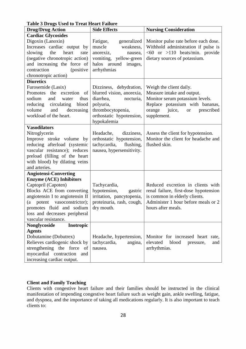
28
Table 3 Drugs Used to Treat Heart Failure Drug/Drug Action Side Effects Nursing Consideration Cardiac Glycosides Digoxin (Lanoxin) Increases cardiac output by slowing the heart rate (negative chronotropic action) and increasing the force of contraction (positive chronotropic action)
Fatigue, generalized muscle weakness, anorexiz, nausea, vomiting, yellow-green halos around images, arrhythmias
Monitor pulse rate before each dose. Withhold administration if pulse is <60 or >110 beats/min. provide dietary sources of potassium.
Diuretics Furosemide (Lasix) Promotes the excretion of sodium and water thus reducing circulating blood volume and decreasing workload of the heart.
Dizziness, dehydration, blurred vision, anorexia, diarrhea, nocturia, polyuria, thrombocytopenia, orthostatic hypotension, hypokalemia
Weigh the client daily. Measure intake and output. Monitor serum potassium levels. Replace potassium with bananas, orange juice, or prescribed supplement.
Vasodilators Nitroglycerin Improve stroke volume by reducing afterload (systemic vascular resistance); reduces preload (filling of the heart with blood) by dilating veins and arteries.
Headache, dizziness, orthostatic hypotension, tachycardia, flushing, nausea, hypersensitivity.
Assess the client for hypotension. Monitor the client for headache and flushed skin.
Angiotensi-Converting Enzyme (ACE) Inhibitors Captopril (Capoten) Blocks ACE from converting angiotensin I to angiotensin II (a potent vasoconstrictor); promotes fluid and sodium loss and decreases peripheral vascular resistance.
Tachycardia, hypotension, gastric irritation, pancytopenia, proteinuria, rash, cough, dry mouth.
Reduced excretion in clients with renal failure, first-dose hypotension is common in elderly clients. Administer 1 hour before meals or 2 hours after meals.
Nonglycoside Inotropic Agents Dobutamine (Dobutrex) Relieves cardiogenic shock by strengthening the force of myocardial contraction and increasing cardiac output.
Headache, hypertension, tachycardia, angina, nausea.
Monitor for increased heart rate, elevated blood pressure, and arrhythmias.
Client and Family Teaching Clients with congestive heart failure and their families should be instructed in the clinical manifestation of impending congestive heart failure such as weight gain, ankle swelling, fatigue, and dyspnea, and the importance of taking all medications regularly. It is also important to teach clients to:
29
• Measure pulse and blood pressure daily. • Identify and avoid occasions that produce stress. Elevate the legs while sitting. • Follow the diet prescribed by the physician. • Avoid extreme heat, cold, or humidity. • Report a heart rate less than 60 or more than 120 beats/min. before taking digitalis
preparations. • Maintain follow-up care. 3.12 Pulmonary Edema Pulmonary edema is an accumulation of fluid in the interstitium and alveoli of the lungs. Pulmonary congestion results when the right side of the heart delivers more blood to the pulmonary circulation than the left side of the heart can handle. The fluid escapes the capillary walls and fills the airways. A client with pulmonary edema experiences dyspnea, breathlessness, and a feeling of suffocation. In addition, the client exhibits cool, moist, and cyanotic extremities. The overall skin color is cyanotic and grays. The client has an incessant productive cough of blood-tinged, frothy fluid. This condition requires emergency treatment. Diagnostic tests Chest radiographs show pulmonary infiltration with fluid. ABGs indicate severe hypoxemia (low PaO2) and hypercapnia (high PaCO2) and a pH less than 7.35, Medical Management Every effort is made to relieve lung congestion as quickly as possible. Relief of symptoms is accomplished by the administration of medications that improve myocardial contractility and decrease preload. Inotropic agents, such as dobutamine or digitalis, are administered IV to improve the force of ventricular contraction. Intravenous morphine sulfate is often given to lessen anxiety. Morphine seems to help relieve respiratory symptoms by depressing higher cerebral centers, thus relieving anxiety and slowing the respiratory rate. Morphine also promotes muscle relaxation and reduces the work of breathing.To facilitate gas exchange, oxygen is administered. 4.0 Conclusion Nursing management of the client with heart disease and occlusive disorder of peripheral blood vessels is directed toward increasing blood flow, relieving pain, preventing complications, and providing thorough teaching and relieving anxiety and pain. 5.0 Summary • Arteriosclerosis is a hardening of the arteries; atherosclerosis is filling of the artery with
fatty plaque. Both interfere with the circulation of oxygenated blood to tissues and organs.
• Severe, unrelieved chest pain is the hallmark of an MI. The chest pain is accompanied by diaphoresis, pale skin, nausea, and vomiting. Treatment includes reestablishing coronary artery blood flow, managing the symptoms, and preventing additional complications.
• Blood vessels also may become occluded by the formation of clots, some of which may break free and travel in the circulation. Occlusion is accompanied by localized symptoms
30
like pain and swelling, and systemic symptoms when circulation to tissues or an organ becomes impaired.
• Varicose veins form when incompetent valves within the veins cause them to distend with engorged blood.
• An aneurysm is a ballooning of an arterial wall occurring commonly in the aorta. • Blood pressure is influenced by age, body size, diet, activity, emotions, pain, position,
gender, time of day, and disease states. • When the heart fails, ventricular output falls and circulatory pathways become
overloaded with fluid. Left- sided failure produces respiratory effects, whereas right-sided heart failure causes systemic effects.
• Pulmonary edema is a complication of left-sided heart failure in which the lung fill rapidly with fluid. Ventilation is extremely impaired because gases cannot diffuse through the fluid medium. The client hyperventilates to compensate, but as carbon dioxide is retained, respiratory acidosis and metabolic acidosis develop. Death is inevitable if the condition is not reversed.
• A person with pulmonary edema experiences sudden dyspnea with moist, gurgling respirations. The client is apprehensive due to the feeling of suffocation. ABG analysis indicates severe hypoxemia and hypercapnia. Potent diuretics and inotropic agents are administered IV. The client may require endotracheal intubation and mechanical ventilation.
6.0 Tutor Marked Assignment • Identify at least four causes of secondary hypertension. • Discuss the nursing management of clients with hypertension. • Discuss the cause and pathophysiology of heart failure. • Distinguish between left- and right-sided heart failure. 7.0 Further Reading and Other Resources • Timby K. Barbara, Jeanne C. Scherer, & Nancy E. Smith (1999). Introductory Medical-
Surgical Nursing. (7th Edition) Lippincott. • Kozier Barbara, Glenora Erb. Fundamentals of Nursing. Concepts and procedures. (2nd
ed.) • Brunner & Suddarth’s (2004) Medical Surgical Nursing. (10th ed) Lippincott Wilkins • Walsh M., Watson’s (1997). Clinical Nursing and Related Sciences. (5th Edition) • Suzanne C. Smeltzer, Brenda Bare (2004). Medical Surgical Nursing. Lippincott
Williams & Wilkins • Ethelwynn L. Stellenbery, Juditt C. Bruce (2007). Nursing Practice: Medical-Surgical
Nursing for Hospital and Community. Elsevier Edinburgh.
31
UNIT II: Cardiac and Noncardiac Shock (Circulatory Failure)
Content …………………………………………………………………………………… 1.0 Introduction ……………………………………………………………………….
2.0 Objective ………………………………………………………………………….
3.0 Main Content ………………………………………………………………………
3.1 Definition ………………………………………………………………………….
3.2 Causes of Shock ……………………………………………………………………
3.3 Pathophysiology of Shock …………………………………………………………
3.4 Common types of Shock ……………………………………………………………...
3.5 Clinical manifestations ……………………………………………………………..
3.6 Management of Shock ……………………………………………………………...
3.6.1 First Aid Management Of Shock …………………………………………………..
3.6.2 Medical and Nursing Managementof Shock ………………………………………
3.7 Complications of Shock …………………………………………………………….
4.0 Conclusion ……………………………………………………………………………
5.0 Summary ………………………........................................................................
6.0 Tutor Marked Assignment …………………………………………………………..
7.0 Further Reading and Other Resources ……………………………………………….
32
1.0 Introduction
The organs and tissues of the body are supposed to be adequately supplied with blood to enhance their effective functioning, apart from oxygen and nutrients derived by these structures from blood circulation. Certain pathophysiological conditions may bring about hypotension and subsequent reduction in blood supply to most vital structures in the body. This state is accompanied by serious reduction in the delivery of oxygen and other essential substances to a level below what is needed for normal and effective cellular activities. 2.0 Objective By the end of this unit you will be able to:
1. Define shock 2. Identify various types of conditions that can lead to shock 3. Identify the clinical manifestation of a patient with shock 4. Distinguish between the various types of shock. 5. Utilize nursing process to manage a patient with shock. 6. Discuss the role of the nurse in psychosocial support of both the patient experiencing
shock and the family.
3.0 Main Content
1. Anphylactic shock: This results from a severe allergic reaction producing an overwheming systemic vasodilation and relative hypovolemia.
2. Cardiogenic shock: This is due to impairment or failure of the myocardium 3. Circulatory shock: This results from displacement of blood volume creating a relative
hypovolemia and inadequate delivery of oxygen to the cells. This is also called distributive shock.
4. Neurogenic shock: Refer to a shock state resulting from loss of sympathetic tone causing relative hypovolemia.
5. Septic shock: This results from overwhelming infection causing relative hypovolemia. 6. Anoxia ; Refers to lack of oxygen in the body 7. Anoxemia : Refers to lack of oxygen in the blood. 8. Anuria : This is absence of urinary secretion 9. Thrombosis :Refers to possible emboli due to blood stasis. 3.1 Definition: Shock is an abnormal physiological state in which there is wide spread, serious reduction of tissue perfusion that if prolonged will lead to generalized impairment of cellular function. Shock has also been described as a clinical state of peripheral circulatory failure characterized by a fall in blood pressure. Cellular destruction, and deterioration in tissue and organ functions are possible out comes.
3.2 Causes of Shock 1. Loss of body fluid 2. Blood loss 3. Inadequate fluid intake 4. Congestive cardiac failure

33
5. Myocardial infarction 6. Pulmonary embolism 7. Cardiac arrhymias 8. Spinal anaesthesia 9. Infections with the release of endotoxins 10. Antigen – antibody reaction with release of histamine.
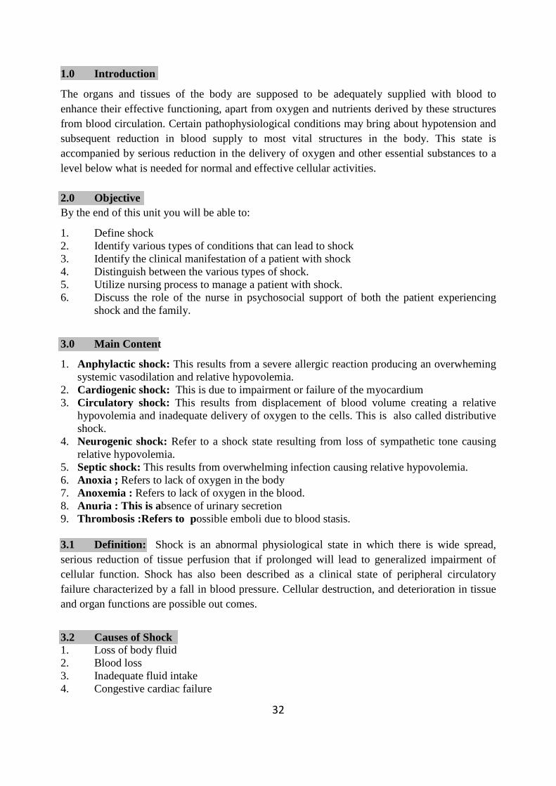
3.3 Pathophysiology of Shock The cardiac output and the peripheral vascular resistance normally maintain arterial blood pressure. When there is reduction in cardiac output and a subsequent decrease in arterial pressure sufficient to produce a wide spread reduction in tissue perfusion, the body attempts to compensate for the changes that follows in the body. The ultimate importance of this compensatory mechanism is to restore adequate circulation to the vital structure of the body. The response of these systems, varies from individual to individual. Vasoconstriction and increase in the heart rate with increase in both peripheral resistance and cardiac output causes additional blood circulation to the vital organs. Haemodilution occurs due to secretion of antidiuretic hormone, and subsequent retention of fluid and sodium helps to improve blood volume. Improved cardiac output and myocardial contractility occur due to increased production of carbon-dioxide occassioned by limited tissue oxygenation. The increased carbon dioxide causes the coronary arteries to dilate resulting in increased myocardial perfusion.
When the compensatory mechanism can not effectively sustain the body’s physiologic functioning, shock progresses and multiple physiological changes ensue. A progressive shock produces multiple systemic changes as a result of decreased cardiac output, hypovolemia, and limited cardiac perfusion. These changes produce alteration in oxygenation, fluid and electrolytes metabolism and the body’s defence against bacterial invasion. In the early stages cerebral hypoxia produces restlessness, apprehension, and anxiety, and may be replaced by apathy and confusion and verbal response becoming inappropriate as cerebral hypoxia increases. In the irreversible stage, unconsciousness manifests with no response to painful stimuli.
The skin is pale, cold and clammy reflecting poor perfusion of the superficial tissue and sympathetic activity to the sweat glands respectively. There is cyanosis, showing a reduction in cardiac output and decreased oxygen saturation. Initially the pulse is rapid and thready, but later becomes slower, irregular and imperceptible.
In response to hypoxia, respirations increase in rate and depth. In severe cases, there is depression of the respiratory centre resulting in shallow and irregular respiration. Severe respiratory dysfunction accounts for complications such as atelectasis, pulmonary emboli, interstitial congestion and oedema. The complication is referred to as ‘shock lung’ or adult respiratory distress syndrome (ARDS). Subnormal temperature is characteristic of shock. This is due to reduction in cellular metabolism and heat production caused by hypoxia. However exception is noticed in septic shock. Oliguria ensues due to decreased renal perfusion. Urinary output might be less than 30mls per hour resulting in the retention of urea, nitrogen and creatinine. Decreased bowel sounds indicating reduced peristalsis develops due to sympathetic innervation and vaso constriction. Reduction in tissue perfusion and the resulting hypoxia accounts for the anaerobic metabolism which causes accumulation of metabolic acids. This
34
eventually leads to acidosis. Myocardial failure and cardiac arrhythmia may develop if acidosis is prolonged.
Source: Jones, D.A. et al, Medical–Surgical Nursing, A conceptual approach. Megraw-Hill, 1978, P. 959.
3.4 Common types of Shock
1. Hypovolaemic shock (decrease in blood volume). This is due to a decrease in blood volume which may be caused by haemorrhage, dehydration due to vomiting and diarrhoea, loss of plasma in burns, inadequate fluid intake, excessive use of diuretics. When intravascular volume drops, there is decrease in tissue perfusion, decreased venous return, and low cardiac output and blood flow through the tissue becomes inadequate.
2. Cardiogenic shock (decreased cardiac output). This indicates a severe impairment in the efficiency of the heart as a pump. There is decreased ability of the heart to pump out blood into circulation. This results in decrease in stroke volume and cardiac output. Cardiogenic shock may be occassioned by congestive cardiac failure, pulmonary embolism, myocardial infaction, pneumothorax, cardiac arrhythmias, or pericardial tamponade.
3. Neurogenic shock: This develops as a response to autonomic nervous system activity resulting in reflex vasodilatation and loss of arteriolar tone with subsequent pooling of blood in the dilated vasculature. This results in decreased venous return to the heart. This type of shock is usually due to spinal anaesthesia, barbiturate injection, hyperrinsulinism, spinal cord injury, severe pain, accidental injury or extreme fright. Septic or bacteraemic shock, toxic shock, anaphylactic shock occurs essentially following the same phenomena as in neurogenic shock. It is characterized by dry, warm skin rather than the cool, moist skin seen in hypovolemic shock. Another characteristic is bradycardia, rather than the tachycardia that characterizes other forms of shock.
Effects of shock
Cellular effects of shock. The cell swells and the cell membrane becomes more permeable, and fluidsand electrolytes seep from and into the cell. Mitochondria and lysosomes are damaged, and the cell dies.
Lysosomal membranerupture
Mitochondrial damage(swelling)
Normal
Cellular edema
Efflux of K
Increased membranepermeability
Influx of Na+ and H2O
Cell damage and death
35
4. Circulatory Shock: Circulatory or distributive shock occurs when blood volume is abnormally displaced in the vasculature––for example, when blood volume pools in peripheral blood vessels. The displacement of blood volume causes a relative hypovolemia because not enough blood returns to the heart, which leads to subsequent inadequate tissue perfusion. The ability of the blood vessels to constrict helps return the blood to the heart. Thus, the vascular tone is determined both by central regulatory mechanisms, as in tissue demands for oxygen and nutrients. Therefore, circulatory shock can be caused either by a loss of sympathetic tone or by release of biochemical mediators from cells. Pooling of blood in the periphery results in decreased venous return. Decreased venous return results in decreased stroke volume and decreased cardiac output. Decreased cardiac output, in turn, causes decreased blood pressure and ultimately decreased tissue perfusion.
5. Septic Shock: Septic shock is the most common type of circulatory shock and is caused by widespread infection. The source of infection is an important determinant of the clinical outcome. The greatest risk of sepsis occurs in patients with bacteremia (bacteria in bloodstream) and pneumonia. Other infections that may progress to septic shock include intra-abdominal infections, wound infections, bacteremia associated with intravascular catheters.
6. Anaphylactic Shock: Anaphylactic shock occurs in patients already exposed to an antigen who have developed antibodies to it. An antigen––antibody reaction provokes mast cells to release potent vasoactive substances, such as histamine or bradykinin, that cause widespread vasodilation and capillary permeability. Therefore, patients with known allergies need to understand the consequences of subsequent exposure to the antigen and should wear medical identification that lists their sensitivities. This could prevent inadvertent administration of a medication that would lead to anaphylactic shock.
B. Diagnostic Tests Diagnosis is usually based on the presenting symptoms and clinical signs.
1. ABG values: Will reveal metabolic acidosis caused by anaerobic metabolism. 2. Serial measurement of urinary output: Less than 30 mL/h is indicative of decreased
perfusion and decreased renal function. For septic shock: 3. Serial creatinine and BUN levels: To assess for potential renal complications and
dysfunction. 4. Serum electrolyte levels: Identify renal complications and dysfunctions as evidenced by
hyperkalemia and hypernatremia. 5. Blood culture: To identify the causative organism. 6. WBC and ESR: Elevated in the presence of infection.
For hematogenic shock:
7. CBC: Hematocrit and hemoglobin will be decreased. For anaphylactic shock:
36
8. WBC count: Will reveal increased eosinophils. 3.5 Clinical manifestations
1. Restlessness 2. Apathy and confusion 3. Unconsciousness 4. Rapid thready pulse followed by weak pulse 5. Decreased blood pressure 6. Increased respiratory rate, shallow respirations 7. Subnormal temperature 8. Cold and clammy skin 9. Decreased urinary output (oliguria) 10. Cyanosis 11. Decreased bowel sounds or absence of bowel sounds 3.6 Management of Shock 3.6.1 First Aid Management Of Shock: In the presence of major external haemorrhage, - Stop the bleeding. - Apply firm pressure over the wound or artery involved. - Apply a firm pressure bandage. - Immobilize the extremity to control the bleeding. - Elevate the part. If heamorrage is internal: - Blood transfusion and surgery may be indicated. - Tourniquet is the last resort. Other actions include: - Keep the patient laid flat or place him on shock position (head lower than its feet) to
improve blood supply to the brain. - Give analgesics to reduce pain. - Take patient to hospital as fast as you can. - Keep crowd away from patient. - Give reassurance. - Keep patient warm. 3.6.2 Medical And Nursing Managementof Shock Management in all types and phase of shock should include the following:
• Fluid replacement to restore intravascular volume • Vasoactive medications to restore vasomotor tone and improve cardiac function • Nutritional support to address the metabolic requirements that are often dramatically
increase in shock.
Assessment: The management of shock should be rapid to prevent the condition from becoming irreversible. A good and careful assessment of the patient’s general/physical health
37
status is paramount. Blood pressure, respiration, pulse, urinary output, skin colour should be noted. The blood pressure and pulse rate should be monitored every 15 minutes. This reflects the cardiac functioning and cardiac output. An indwelling catheter is passed to facilitate the measurement of the urinary output hourly. In the adults, the urinary output is expected to range between 30-60ml/hour. A decrease in this value indicates poor renal perfusion. Oliguria may lead to anuria. The hourly urinary output is useful in assessing patients’ cardiovascular status. Except in septic shock in which the body temperature may be elevated in the early stages, patients with other types of shock usually record a subnormal temperature and remains same as shock progresses. The body temperature should be monitored continuously and recorded every 1-2 hours.
The rate and volume of respirations should be monitored and recorded 15-30 minutes. This is particularly important as hyperventilation occur in the early stages of shock while respiration may become slow, irregular and shallow as a result of ischaemia of the respiratory centre. Secretions in the respiratory tract should be removed promptly through suctioning. The skin should be observed for lessening of pallor, warmth and quick refilling of the capillaries and veins following compression which are signs of improvement. Conversely signs of subcutaneous bleeding may indicate disseminated intravascular coagulation in severe shock, and especially that associated with sepsis. The level of consciousness should be determined at regular intervals using Glasgow coma scale. This reflects blood and oxygen supply to the brain.
Treatment: Usually treatment is directed towards improving tissue perfusion and oxygenation as well as treating the specific cause accordingly. The contractile ability of the heart is strengthened to increase cardiac output in cardiogenic shock. Adrenalin is useful as a cardiac stimulant.
Fluid Replacement
Fluid replacement is administered in all types of shock. The type of fluids administered and the speed of delivery vary, but fluids are given to improve cardiac and tissue oxygenation. Fluid replacement is paramount in all types of shock especially in hypovolaemic shock. The fluids administered may include crystalloids (electrolyte solutions that move freely between intravascular spaces), colloids (large-molecule intravenous solutions), or blood components.
Initially a crystalloid solution e.g. Ringers lactate solution or Normal saline is used. Colloid solutions of whole blood or fresh plasma may be used in conjunction with the crystalloid solutions. The aim is to expand intravascular volume. If the cause is due to haemorrhage efforts should be made to arrest bleeding by giving whole blood or fresh plasma so that blood pressure is raised and tissue perfusion restored.
Close monitoring of the patient during fluid replacement is necessary to identify side effects and complications. The most common and serious side effects of fluid replacement are cardiovascular overload and pulmonary edema. Patients receiving fluid replacement must be monitored frequently for adequate urinary output, changes in mental status, skin perfusion, and changes in vital signs. Lung sounds are auscultated frequently to detect signs of fluid accumulation. Adventitious lung sounds, such as crackles, may indicate pulmonary edema.

38
Vasoactive Medication Therapy
Vasoactive medications are administered in all forms of shock to improve the patient’s hemodynamic stability when fluid therapy alone is inadequate. Specific vasoactive medications are prescribe to correct the particular hemodynamic alteration that is impeding cardiac output. These medications help to increase the strength of myocardial contractility, regulate the heart rate, reduce myocardial resistance, and initiate vasoconstriction.
Nutritional Support
Nutritional support is an important aspect of care for the patient with shock. Increased metabolic rates during shock increase energy requirements and therefore caloric requirements. The patient in shock requires more than 3,000 calories daily. The release of catecholamines early in the shock continuum causes glycogen stores to be depleted in about 8 to 10 hours. Nutritional energy requirements are then met by breaking down lean body mass. In this catabolic process, skeletal muscle mass is broken down even when the patient has large stores of fat or adipose tissue. Loss of skeletal muscle can greatly prolong the recovery time for the patient in shock. Parenteral or enteral nutritional support should be initiated as soon as possible, with some form of enteral nutrition always being administered.
Stress ulcers occurs frequently in acutely ill patients because of the compromised blood supply to the gastrointestinal tract. Therefore, antacids, histamine-2 blockers (eg, famotidine [Pepcid], ranitidine [Zantac]), and antipeptic agents (eg, sucralfate [Carafate]) are prescribed to prevent ulcer formation by inhibiting gastric acid secretion or increasing gastric pH.
D. Nursing Diagnoses and Interventions
Alteration in tissue perfusion: Peripheral, cardiopulmonary, cerebral, and renal related to impaired circulation secondary to decreased circulating blood volume.
Nursing objective: Patient’s VS, mentation, and physical findings are within acceptable limits.
1. Assess and document peripheral pulse. Report significant findings such as coolness and pallor of the extremities, decreased amplitude of pulses, and delayed capillary refill.
2. Monitor BP at frequent intervals; be alert to reading >20 mm Hg below patient’s normal or to other indicators of hypotension, such as dizziness, altered mentation, or decreased urinary output.
3. If hypotension is present, place patient in a supine position to promote venous return. Remember that BP must be at least 80/60 mm Hg for adequate coronary and renal artery perfusion.
4. Monitor CVP (if line is inserted) to determine adequacy of venous return and blood volume; 4–10 cm H20 and usually considered adequate levels. Values near zero can indicate hypovolemia, especially when associated with decreased urinary output, vasoconstriction, and increased heart rate, which are found with hypovolemia.
5. Observe for indicators of decreased cerebral perfusion such as restlessness, confusion, and decreased LOC. If positive indicators are present, protect patient from injury by raising side rails and placing bed in its lowest position. Reorient patient as indicated.
6. Monitor for indicators of decreased coronary artery perfusion such as chest pain and an irregular heart rate.
39
7. Monitor urinary output hourly. Notify MD if it is <30 mL/h in the presence of adequate intake. Check weight daily for evidence of gain.
8. Monitor lab results for elevated BUN and creatinine; report increases. 9. Monitor serum electolyte values for evidence of imbalances, particularly sodium and
potassium. Be alert to signs of hyperkalemia such as increased serum levels, muscle weakness, hyporeflexia, and irregular heart rate. Also monitor for signs of hypernatremia such as increased serum levels, fluid retention, and edema.
10. Administer fluids as prescribed to increase vascular volume. The type and amount of fluid will depend on the type of shock and the patient’s clinical situation. • Cardiogenic shock: Fluids probably will be limited to prevent overload, yet
dehydration must be avoided to ensure support of vascular space and cardiac muscle.
• Hypovolemic shock: The amount lost is replaced. As much as 1000 mL/h of Ringer’s solution may be administered if volume loss is severe. Most often, this includes blood replacement.
• Septic shock: Ringer’s solution, plasma, and blood are administered.
Impaired gas exchange related to tissue hypoxia secondary to decreased circulating blood volume.
Nursing objective: Patient exhibits ABG values and VS within acceptable limits.
1. Monitor respiration q30 min; note and report presence of tachypnea and/or dyspnea. 2. Teach patient to breathe slowly and deeply to promote oxygenation. 3. Ensure that the patient has a patent airway; suction secretions as needed to assist with gas
exchange. 4. Administer oxygen as prescribed. 5. Monitor ABG results. Be alert to and report presence of hypoxemia (decreased Pao2),
hypercapnia (increased Paco2), and acidosis (decreased pH, increased Paco2). 6. Report significant findings to MD. E. Patient – Family Teaching and Discharge Planning Nursing care
Possible nursing diagnosis include:
1. Altered nutrition, less than body requirements. 2. Impaired mobility 3. Fluid volume deficit 4. Altered comfort 5. Altered urinary output 6. Ineffective breathing pattern
Altered nutrition: The patient should not be given anything orally because of paralytic ileus, instead a nasogastric tube is inserted to drain the stomach contents and prevent abdominal distension. The nutritional status is maintained primarily by intravenous infusions. Oral feeding is commenced only in the presence of bowel sounds.
40
Impaired mobility: Complications such as thrombosis, circulatory stasis, decubitus, ulcer, flexion contractures and atelectasis may develop due to immobility. The patient’s position should be changed every 1-2 hours (where permitted). Skin should be inspected for redness and pressure areas treated accordingly. This promote circulation, relieves pressure and drainage of pulmonary secretions. Patient is positioned in good body alignment to prevent contractures and foot drop. Passive exercise especially of the lower limbs is also useful. A bed craddle will be useful in lifting the weight of the bed clothes thus preventing pressure.
Fluid volume deficit: Frequent observation of the patient’s reaction and flow of the infusion used should be carried out by the nurse. Intake and output record should be maintained. Oral fluids should be avoided because of poor gastro-intestinal absorption.
(Altered comfort) or Self care deficit (bath, and oral care and grooming). Physical care such as bathing and oral hygiene are necessary as they promote relaxation and ensure prevention of sores, ulcers and infection respectively. Analgestics is administered intravenously to relief pain. The patient should be observed for the respiratory depressive effect of analgesic used. Light linen should be used to keep patient comfortably warm but overheating and chilling should be avoided.
Altered urinary output: Impaired urinary elimination occurs due to inadequate renal perfusion. To maintain adequate urinary output, an indwelling catheter is passed using aseptic technique. This is to facilitate the measurement of hourly urinary output. Hourly urinary output less than 30mls (in adult) should be reported to the appropriate personnel.
Ineffective breathing pattern: A patient airway should be established immediately and oxygen given through nasal cannulae or mask. When the patient is able, he should be encouraged to carry out deep breathing and coughing exercises at frequent intervals. Change in positions hourly is beneficial as it decreases the possibility of consolidation developing thereby promoting effective gas exchange.
In order of priority in shock management, restoration of tissue perfusion by way of I.V. fluid administration takes precedence over other measures. This is followed by adequate tissue oxygenation, i.e. ensuring a patent airway and adequate gaseous exchange.
3.7 Complications of Shock
1. Metabolic acidosis 2. Cardiac failure 3. Cardiac arrhythmias 4. ‘Shock lung’ 5. Uraemia 6. Cerebral damage 7. Susceptibility to infection
41
4.0 Conclusion Any condition that prevents cells from receiving an adequate blood supply or interfere with this metabolism produces shock. 5.0 Summary
• Shock may be classified as hypovolemic, cardiogence or vasegenic. • Liver, heart, kidney and brain are major organism that can easily be damages by
shock. 6.0 Tutor Marked Assignment 1. Explain the pathophysiology of shock. 2. Identify 4 causes of hypovolemic shock and how it can be prevented. 7.0 Further Reading and Other Resources Brunner & Suddarth’s (2004) Medical Surgical Nursing. (10th ed) Lippincott Wilkins
42
UNIT III: DISORDERS OF THE RED BLOOD CELLS
1.0 Introduction …………………………………………………………………………….
2.0 Objective …………………………………………………………………………....... 3.0 Main Content…………………………………………………………………………... 3.1 Anaemia ………………………………………………………………………………. 3.2 Polycythemia …………………………………………………………………………... 3.3 Disorders of Coagulation ………………………………………………………………. 3.3.1 Thrombocytopenia ……………………………………………………………………… 3.3.2 Haemophilla ……………………………………………………………………………. 3.3.3 Disseminated Intravascular Coagulation ……………………………………………… 4.0 Conclusion ……………………………………………………….............................. 5.0 Summary ………………………………………………………................................ 6.0 Tutor Marked Assignment ………………………………………………………......... 7.0 Further Reading and Other Resources …………………………………………………
1.0 Introduction
The erythrocyte, or red blood cell (RBC), is the transport mechanism for hemoglobin, which carries oxygen from the heart and lungs to the tissues, exchanges it for carbon dioxide, and then returns to the heart and lungs. RBCs are very flexible and capable of bending, elongating, and squeezing through tiny capillaries. Normal RBCs can travel under high pressure and speed, are extremely active metabolically, and have an average life of 120 days. The bone marrow produces and replaces RBCs every day and can respond to the increased need for RBCs by increasing production. However, with increased need for RBCs (reticulocytes) are often released into the circulation; a high level of immature RBCs often aids in the diagnosis of RBC disorders.
43
2.0 Objective By the end of this unit you will be able to:
• Explain the meaning of anaemia
• Identify the causes and types of anaemia • Describe the disorders of coagulation
3.0 Main Content Anaemia Fibrin Plasma Prothrombin Time Disseminated intravascular coagulation 3.1 Anemia Is a common hematopoietic disorder.It is defined as a reduced number of RBCs and/or a reduced amount of hemoglobin. The general effects of anemia result from a deficiency in the oxygen-carrying mechanism, although some effects are related to varied etiologies and pathogenesis. Types of anemias • Pernicious Anaemia • Hemolytic Anaemia • Hypoplastic (Aplastic) Anaemia Pernicious Anaemia Vitamin B12 is supplied by diet and stored in the liver to be used for maturation of RBCs. Deficiency of this vitamin leads to the development of immature erythrocytes, a chronic condition known as pernicious anaemia. Decreased dietary intake of animal products, increased need for this vitamin with pregnancy or a tumor, presence of parasites, or surgery involving the small intestine where the vitamin is absorbed are conditions that can lead to vitamin B12 deficiency. The most common condition is the decrease in production by the gastric mucosa of intrinsic factor, a microprotein, which when combined with vitamin B12 facilitates absorption and use of the vitamin by body cells, particularly in the bone marrow, GI tract, and nervous system. A. Assessment Chronic indicators: Brittle nails, smooth tongue, numbness and tingling of the extremities, fatigue, and dysphagia. However, because of slow progression, many patients remain asymptomatic. Anorexia, weight loss, jaundice from destruction of malformed erythrocytes, and gingivitis from absence of vitamin B12 also can occur. Acute indicators: Dyspnea on exertion, irritability, palpitations, and dizziness in the presence of severe deficiency. In addition, because the nervous system is particularly sensitive to the lack

44
of vitamin B12, degenerative changes of the cerebral cortex and spinal cord can occur, seen mainly in the form of paresthesias. Physical examination: Presence of oral lesions and gingivitis, tachycardia, unsteady gait, and clumsiness. B. Diagnostic Tests 1. Schilling’s test: Patient is given radioactive-tagged vitamin B12, and urine concentration
of tagged B12 is then measured. In the presence of pernicious anemia, B12 is not absorbed and urine levels will be low (<3%).
2. Trial administration of vitamin B12: In the presence of pernicious anemia, symptoms will be decreased.
3. Serial hemoglobin and erythrocyte counts: Initially will be decreased. 4. Bone marrow aspiration: Will reveal hyperplasia with increased numbers of large-sized
megaloblasts. 5. Gastric analysis: Will reveal decreased volume of gastric secretions. Atrophic gastritis is
characteristic of perncious anaemia. C. Medical Management 1. Vitamin B 12 replacement: Dosage depends on the individual and the response to
treatment. For example, 100 mg cyanocobalamin may be given IM qd x 7 days. If improvement occurs, it is given qod x 7 days and then q3–4 days x 2–3 weeks.
2. Concurrent treatment of underlying disorder: If present, eg, gastric mucosal problem.
3. Serial measures of reticulocytes: To determine effectiveness of treatment. Hemolytic Anaemia Hemolytic anaemia is characterized by abnormal or premature destruction of RBCs. Hemolysis can be intrinsic or result from conditions such as infection or radiation.
Types of Hemolytic Anaemia • Sickle cell anaemia is a form of chronic haemolytic anaemia characterized by abnormal,
crescent-shaped, rigid, and elongated erythrocytes. These “sickle” RBCs interfere with circulation because they cannot get through the microcirculation and are destroyed in the process. Sickle cell anemia can affect almost every body system due to decreased oxygen delivery, decreased circulation caused by occlusion of the vessels by RBCs, and inflammatory process. This disorder occurs when the gene is inherited from both parents (homozygous); a carrier state exists when it is inherited from one parent (heterozygous). Medical treatment has improved the prognosis for this disorder, which is seen predominantly in blacks.
• Thalassemia is another type of chronic hemolytic anaemia. In this disorder, hemoglobin A is manufactured in less than-normal amounts, although the hemoglobin itself is of normal morphology. This is an inherited disorder passed on through an autosomal gene. Severity depends on whether the inheritance is heterozygous or homozygous. It occurs most often in individuals of Mediterranean descent, eg, Greeks and Italians. If the condition is severe the patient seldom survives to adulthood. Individuals with intermediate and minor forms develop normally and usually can expect a normal life span.
45
• Acquired haemolytic anemia is usually the result of an abnormal immune response that causes premature destruction of RBCs. Hemolysis can occur as a result of a foreign antigen such as from a transfusion reaction, or an automimmune reaction in which the hemolytic agent is intrinsic to the patient’s body.
• Other possible causes include exposure to radiaton and ingestion of drugs such as sulfisoxazole (eg, Gantrisin), phenacetin, and methyldopa (eg, Aldomet).
A. Assessment Chronic indicators: Pallor (eg, conjunctival), fatigue dyspnea on exertion, and intermittent dizziness, all of which depend on the severity of the anemia. Acute indicators: Fever, visual blurring, temporary blindness, abdominal pain, lymphadenopathy, splenomegaly, and decreased urinary output (clinical manifestations of hemolytic crisis). Peripheral nerve damage can result in paralysis or paresthesias, vomiting, and chills. B. Diagnostic Tests 1. Sickle cell test: To screen for sickle cell anemia. 2. Hemoglobin and hematocrit: Decreased. 3. Serum tests: LDH and bilirubin will be elevated. 4. Urine and fecal urobilinogen: Increased and are more sensitive indicators of RBC
destruction than serum bilirubin levels. 5. Bone marrow aspiration: Will reveal erythroid hyperplasia, especially with chronic
hemolytic anemia. 6. Hemoglobin electrophoresis: Will diagnose hemoglobin AS, a sickle cell trait.
C. Medical Management 1. Elimination or discontinuation of causative factor: If possible, eg, chemical, drug,
incompatible blood. 2. Supportive therapy of shock state: If it occurs. 3. Transfusion: If circulatory failure or severe anemic anoxia occurs. 4. Corticosteroids: Usually 50 –100 mg prednisone, given with antacids. 5. Folic acid: To help prevent hemolytic crisis in patients with chronic hemolytic anemias. 6. Splenectomy: To provide relief, depending on the cause of the anemia. The spleen is
the site of RBC destruction.
Hypoplastic (Aplastic) Anaemia This type of anaemia results from inability of erythrocyte-producing organs, specifically the bone marrow, to produce erythrocytes. The causes of hypoplastic anemia are varied but can include use of antineoplastic or antimicrobial agents, infectious process, pregnancy, hepatitis, and radiation. Approximately half of the patients with hypoplastic anemia have had exposure to drugs or chemical agents, while the remaining half have had immnologic disorders. Hypoplastic anaemia also can involve pancytopenia, the depression of production of all three bone marrow elements: erythrocytes, platelets, and granulocytes. Usually the onset of hypoplastic anaemia is insidious, but it can evolve quickly in some cases. Prognosis is usually poor for these patients.
46
A. Assessment Chronic indicators: Weakness, fatigue, pallor, dysphagia, and numbness and tingling of the extremities (indicators of anemia). Acute indicators: Fever and infection (because of decreased neutrophils); bleeding (because of thrombocytopenia); and dizziness, dyspnea on exertion, progressive weakness, and oral ulcerations. History of: Exposure to chemical toxins or radiation, use of antibiotics such as chloramphenicol.
B. Diagnostic Tests 1. CBC with differential: Low levels of hemoglobin, WBCs, and RBCs; however, RBCs
usually appear to be normal morphologically. 2. Platelet count: Low. 3. Bleeding time: Prolonged. 4. Bone marrow aspiration: Will reveal hypocellular or hypoplastic tissue with a fatty and
fibrous appearance and depression of erythroid elements. C. Medical Management 1. Determine cause of anaemia. 2. Transfuse packed RBCs or frozen plasma: 3. Transfuse concentrated platelets: To keep platelet count > 20,000/mm3. Granulocyte
transfusion: For life-threatening sepsis . 4. Cultures: If infection is found. 5. Antibiotic therapy: If infection is found. 6. Reverse isolation: If granulocytes count is <200/mm3. 7. Steroid therapy: To stimula granulocyte production, although results with adults are
not always successful. 8. Oxygen: If anemia is severe. 9. Bone marrow transplantation: In this procedure, 500–700 mL of bone marrow are
aspirated from the pelvic bones of the donor and then filtered and infused into the patient. The donated marrow must be antigen-compatible and therefore the donor is usually a twin or sibling.
3.2 Polycythemia Polycythemia is a chronic disorder characterized by excessive production of RBCs. As the number of RBCs increases, blood volume, blood viscosity, and hemoglobin concentration increase, causing excessive workload of the heart and congestion of organ systems such as the liver and kidney. Secondary polycythemia results from an abnormal increase in erythropoietin production, for example, secondary to hypoxia that occurs with chronic lung disease, or it can occur inappropriately, such as with renal tumors. A. Assessment Clinical manifestations: Headache, dizziness, visual disturbances, dyspnea, thrombophlebitis, joint pain, fatigue, chest pain, and a feeling of “fullness,” especially in the head.
47
Physical exam: Hypertension crackles (rales), cyanosis, ruddy complexion, hepatosplenomegaly. B. Diagnostic Tests 1. CBC: Increased RBC mass (8–12 million/mm3); increased hemoglobin; leukocytosis. 2. Platelet count: Increased. 3. Bone marrow aspiration: Will reveal RBC proliferation. 4. Uric acid levels: May be increased because of increased nucleoprotein, an end product
of RBC breakdown.
C. Medical Management 1. Phlebotomy: Blood withdrawn from the vein to decrease blood volume (and decrease
hematocrit to 45%). 2. Myelosuppressive agents such as radiophosphorus: To inhibit proliferation of RBCs. 3. Alkylating (myelosuppressive) agents such as busulfan and chlorambucli: To
decrease bone marrow function.
3.3 Disorders of Coagulation The formation of a visible fibrin clot is the conclusion of a complex series of reactions involving different clotting factors in the blood that are identified by Roman numerals 1–XIII. All are plasma proteins except factor III (thromboplastin) and factor IV (calcium ion). When a vessel injury occurs, these factors interact to form the end product, a clot. The clots that are formed are eventually dissolved by the fibrinolytic system. Platelets play a role in coagulation by releasing substances that activate the clotting factors. At the time of vascular injury, platelets migrate to the site and adhere to each other to form a temporary plug to stop the bleeding. 3.3.1 Thrombocytopenia Thrombocytopenia is a common coagulation disorder, which results from a decreased number of platelets. It can be congenital or acquired, and it is classified according to cause. Causes include; deficient formation of thrombocytes, which occurs with bone marrow disease or destruction; accelerated platelet destruction, loss, or increased utilization such as in hemolytic anemia, diffuse intravascular coagulation, or damage by prosthetic heart valves; and abnormal platelet distribution such as in hyperplenism and hypothermia. In addition, thrombocytopenia can occur as a side effect of certain chemotherapeutic agents and antibiotics. Regardless of the cause or trigger, the disorder affects coagulation and hemostasis. With chemical-induced thrombocytopenia, prognosis is good after withdrawal of the individual’s response to treatment. Thrombocytopenic purpura, also termed idiopathic thrombocytopenic purpura (ITP), occurs when blood extravasates into tissue and mucous membranes due to increased destruction or decreased formation of thrombocytes. A. Assessment Chronic indicators: Long history of mild bleeding or hemorrhagic episodes. Increased bruising, gum bleeding, and petechiae also may be noted.
48
Acute indicators: Fever, splenomegaly, acute and severe bleeding episodes, weakness, lethargy, malaise, hemorrhage into mucous membranes, gum bleeding, and GI bleeding. Prolonged bleeding can lead to a shock state with tachycardia, SOB, and decreased LOC. History of: Recent infection; vaccination; use of chlorothiazide, digitalis, quinidine, rifampin, sulfisoxazole, chloramphenicol, phenytoin. B. Diagnostic Tests 1. Platelet count: Can vary from only slightly decreased to nearly absent. Less than
100,000 is significantly decreased; <20,000 results in a serious risk of hemorrhage. 2. CBC: Low hemoglobin and hematocrit levels; WBC within normal range. 3. Template bleeding time: Increased. 4. Bone marrow aspiration: Will reveal increased number of megakaryocytes (platelet
precursors) in the presence of ITP, but may be decreased with certain causes of thrombocytopenia.
5. Platelet antibody screen: May be positive. The test is not generally available.
C. Medical Management and Surgical Interventions 1. Treatment of underlying cause or removal of precipitating agent. 2. Platelet transfusion: Corticosteroids: To enhance vascular integrity or diminish
platelet destruction. 3. Splenectomy: Removal of an organ responsible for platelet destruction. This is
considered viable treatment unless patient has acute bleeding, a severe deficiency of platelets, or a cardiac disorder that contraindicates surgery
3.3.2 Haemophilla Haemophilia is a sex-linked, inherited bleeding disorder characterized by a deficiency of one or more clotting factors. Classic hemophilia is caused by deficiency of factors VIII (hemophilia A) and IX (hemophilia B). Individuals affected are usually males, whereas their mothers and sisters are asymptomatic carriers. This disorder also can occur in females if it is due to X chromosome inactivation during embryologic development. An intracranial hemorrhage is the most common cause of death.
A. Assessment Chronic indicators: Bruising after minimal trauma, joint pain. Acute indicators: Acute bleeding episodes after minimal trauma. Hemarthrosis is the most common and debilitating symptom, causing painful and swollen joints. Large ecchymoses can occur, as well as bleeding from the gums, tongue, GI tract, urinary tract, or from cuts in the skin. Shock can result from severe bleeding. B. Diagnostic Tests 1. Partial thromboplastin time (PTT): Prolonged. 2. Bleeding time: Prolonged. 3. Platelet count: Usually normal.
49
4. Activated clotting time: Prolonged. 5. Assays of factors VII and IX: Will reveal low activity.
C. Medical Management 1. Factor transfusion: For hemophilia B 2. Transfusion of fresh frozen plasma: 3.3.3 Disseminated Intravascular Coagulation Disseminated intravascular coagulation (DIC) is an acute coagulation disorder characterized by paradoxical clotting and hemorrhage. The sequence usually progresses by massive clot formation, depletion of the clotting factors, and activation of diffuse fibrinolysis, followed by hemorrhage. DIC occurs secondary to widespread coagulation factors in the bloodstream caused by extensive surgery, burns, shock, neoplastic diseases, and abruptio placenta; extensive destruction of blood vessel walls caused by Eclampsia, anoxia, and heat stroke; and damage to blood cells caused by hemolysis, sickle cell disease, and transfusion reactions. Prompt assessment of the disorder can result in a good prognosis. Usually, affected patients are transferred to ICU for careful monitoring and aggressive therapy. A. Assessment Clinical indicators: Bleeding of abrupt onset, ozzing from venipuncures sites, bleeding from surgical sites. Symptoms of hypoperfusion can occur, including decreased urine output and abnormal behavior. Risk factors: Infection, burns, trauma, hepatic disease, hypovolemic shock, severe hemolytic reaction, obstetric complications, and hypoxia. B. Diagnostic Tests 1. Serum fibrinogen: Low because of abnormal consumption of clotting factors. 2. Platelet count: Will be <250,000 because of platelet’s role in clot formation. 3. Fibrin split products (FSP): Increased, indicating widespread dissolution of clots. 4. PT: Increased because of depletion of clotting factors. 5. PTT: High because of depletion of clotting factors. 6. Peripheral blood smear: Will show fragmented RBCs. 7. Bleeding time: Prolonged because of decreased platelets.
C. Medical Management 1. Identify and treat primary disorder. 2. Anticoagulant therapy: 3. Compartment replacement of platelets and clotting factors: Instituted after heparin
therapy has been initiated, because otherwise the transfused factors would enhance the clotting process.
4. Thrombolytic agents such as streptokinase: To increase circulation in thrombosed vessels after the acute phase has resolved.
Product Approximate
Volume Indications Precautions/Comments
Whole Blood 500–510 mL (450 WB; 50– 60
Acute, severe blood loss; hypovolemic shock
Must be ABO and Rh compatible. Do not mix with dextrose solutions; always prime
50
(WB) antico-agulants) increases both red cell mass and plasma
tubing with normal saline. Observe for dypnea, orthopnea, cyanosis, and anxiety as signs of circulatory overload; monitor VS. Hepatitis risk = 2*
Packed Red Blood Cells (RBCs)
250mL Increases RBC mass and oxygen-carrying capacity of the blood
Must be ABO and Rh compatible. Less immunologic risk than with WB because some donor antibodies are removed. Less volume reducing risk of fluid overload. Hepatitis risk = 2*
Fresh Frozen Plasma (FFP)
250 mL Treatment of choice for combined coagulation factor deficiencies and factor V and XI deficiencies; alternate treatment for factor VII, VIII, IX, and X deficiencies when concentrates are not available
Must be ABO compatible. Supplies clotting factors. Usual dose is 10 – 15 mL/kg body weight. Hepatitis risk 2**
Platelet Concentrate
25–50 mL (volumes may vary; usual adult dose is 5–6 U)
Treatment of choice for thrombocytopenia. Also used for leukemia and hypoplastic anemia
Usual doses is 0.1 U/kg body weight to increase platelet count to 25,000. Administer as rapidly as tolerated ABO compatibility is preferable, but is expensive and usually not practical. Effectiveness is decreased by fever, sepsis, and splenomegaly. Febrile reactions are common. Use special “platelet” tubing and filter. Hepatitis risk = 2**
Platelet Concentrate by Platelet Pheresis
200 mL but may vary
Treatment for thrombocytopenic patients who are refractory to random donor platelets
Involves removing donor’s venous blood 200 mL at a time, removing the platelets by centrifuge, and returning the blood to patient. This is performed approximately six times to yield 200 mL platelets. Uses special donors, who usually are human leukocyte antigen (HLA)
Cryoprecipitate (factor VIII)
10–25 mL Routine treatment for heophilia (factor VII deficiency) & fibrinogen deficiency (factor XIII deficiency)
Made from FFP. Infuse immediately upon thawing. Hepatitis risk = 2*
AHG (factor VIII) Concentrates
20 mL Alternative treatment for hemophilia A
Allergic and febrile reactions occur frequently. Administer by syringe or component drip set. Can store at refrigerator temperature, making it conventient for hemophiliacs during travel. Hepatitis risk = 3**
Factor II, VII, IX, X
20 mL Treatment of choice for hemophilia B & factor IX
Can precipitate clotting. Allergic and febrile reactions occur occasionally. Contraindicated in liver disease. Hepatitis
51
Concentrate deficiencies risk = 3**
Albumin 50 or 250 mL Hypovolemic shock, hypoalbuminemia, plasma replacement for burn patients
Osmotically equal to 5X its volume of plasma. Used as a volume expander or in hypoalbuminemic states. Commercially available. Hepatitis risk = 0*
Plasma Protein Fraction (PPF)
250 mL (83% albumin with some alpha and beta globulins)
Volume expansion Commercially available; expensive. Certain lots reported to have caused hypotension, possibly related to vasoactive amines used in preparation. Hepatitis risk = 0*
Granulocyte Transfusion (collected from a single pheresis donor)
200 mL, but may vary
Leukemia with granulocytopenia related to treatment
Not a common treatment. Febrile and allergic symptoms are frequent. Must be ABO compatible. Hepatitis risk = 3*
5. *Relative hepatitis risk: 0, no risk, 1 and 2, moderate to high risk, 3, maximum risk. 6. **Risk is greater because multiple donors are used
4.0 Conclusion The bone marrow produces and replaces RBCs every day.A high level of immature RBCs often aids in the diagnosis of RBC disorders. 5.0 Summary Aneamia is common hematopoietic disorder characterized by a reduced number of RBCs and/or a reduced amount of hemoglobin. Disorders of Coagulation include Thrombocytopenia and disseminated intravasculular 6.0 Tutor Marked Assignment 1. Identify the causes and types of aneamis 2. Describe the disorders of coagulation 7.0 Further Reading and Other Resources • Timby K. Barbara, Jeanne C. Scherer, & Nancy E. Smith (1999). Introductory Medical-Surgical
Nursing. (7th Edition) Lippincott. • Kozier Barbara, Glenora Erb. Fundamentals of Nursing. Concepts and procedures. (2nd
ed.) • Brunner & Suddarth’s (2004) Medical Surgical Nursing. (10th ed) Lippincott Wilkins
52
UNIT IV: DISORDERS OF WHITE BLOOD CELLS 1.0 Introduction …………………………………………………………………………….. 2.0 Objective ……………………………………………………………………………… 3.0 Main Content………………………………………………………………………
3.1 Hodgkin’s Disease ……………………………………………………………………... 3.2 Acute Leukemia ………………………………………………………………………… 3.3 Chronic Leukemia ……………………………………………………………………… 4.0 Conclusion ……………………………………………………………………………. 5.0 Summary ……………………………………………………………………………… 6.0 Tutor Marked Assignment ……………………………………………………………. 7.0 Further Reading and Other Resources ………………………………………………..
1.0 Introduction
White blood cells (WBCs), also called leukocytes, are the blood cells responsible for both immunity and the body’s response to infectious organisms. Different types of WBCs are classified according to structure, specialized function, and response to dye in the laboratory. The three classifications of WBCs are granulocytes, lymphocytes, and monocytes, all of which may undergo malignant transformations. The bone marrow has a reserve of approximately 10 times the number of circulating WBCs, which are released into the circulation during an infectious process.
2.0 Objective
By the end of this unit you will be able to:
• Identify the clinical manifestation of Hodgkin’s disease
• Identify the types of Leukemia • Identify the clinical manifestation of Leukemia
• Know the nusing management of clients with white blood disorders
53
3.0 Main Content
White blood cells (WBCs), Hodgkin’s disease, Leukemia, Granulocytes, Lymphocytes, and Monocytes,
3.1 Hodgkin’s Disease
Hodgkin’s disease is a tumor of the lymph tissue. It is distinguished from other lymphomas by the presence of large, variable cells called Reed-Sternberg cells, which proliferate and invade normal lymph tissue throughout the body. Lymph tissue is found in the spleen, liver, bone marrow, lymph nodes, and lymph channels, which connect virtually all tissues. Clinical presentation depends on the degree of malignant cell growth, extent of the invasion, and tissues that are affected. Hodgkin’s disease frequently affects young people, and it can be treated successfully, particularly with early diagnosis and intervention.
Causes The etiology of the disorder is unclear, although a hereditary component has been implicated. Although no infectious organism has been identified, infection has been suggested as a potential cause. A. Assessment Chronic indicators: Nonspecific symptoms such as persistent fever, night sweats, malaise, weight loss, and pruritis. Acute indicators: Worsening of the above symptoms, in addition to unexplained pain in the lymph nodes after drinking alcohol. Physical exam: Enlarged (painless) lymph nodes in the cervical area; possible splenomegaly and hepatomegaly. B. Diagnostic Tests 1. CBC: Decreased hemoglobin and hematocrit (confirming anemia); increased, decreased,
or normal levels of WBCs. 2. Platelet count: May be low. 3. Lymph node biopsy: May reveal presence of characteristic Reed-Sternberg cells. 4. Lymphangiogram: To determine extent of involvement. 5. Biopsy of the bone marrow, lung, liver, pleura, or bone: May be performed to determine
involvement. 6. Serum alkaline phosphatase: If elevated, will indicate liver or bone involvement. 7. Erythrocyte sedimentation rate (ESR): Will be elevated. 8. Chest x-ray and/or abdominal CT scan: To help determine presence of nodal
involvement.
C. Medical Management 1. Staging: To determine extent of the disease. A simplified description of staging follows
(based on Ann Arbor Staging Classification): • Stage 1: Limited to a single lymph node region or a single extralymphatic organ.
54
• Stage II: Involves two or more lymph node regions on the same side of the diaphragm or localized involvement of an extralymphatic organ.
• Stage III: Involves lymph node regions on both sides of the diaphragm, accompanied by involvement of an extralymphatic region and/or the spleen.
• Stage IV: Diffuse involvement of one or more extralymphatic region or tissue. 2. Radiation therapy to lymph node regions: For stages I and II. 3. Chemotherapy in combination with radiation: For stages III and IV. One common
combination of chemotherapeutic agents includes mechlorethamine hydrochloride (nitrogen mustard), vincristine, prednisone, and procarbazine. For non-Hodgkin’s lymphoma, a variety of antineoplastic drugs are currently being used, including cytoxan, vincristine, prednisone, procarbazine, doxorubicin, and bleomycin.
3.2 Acute Leukemia Acute leukemia is an abnormal, malignant proliferation of WBC precursors, also called “blasts.” These abnormal cells accumulate in bone marrow, body tissues, and blood vessels and eventually cause malfunction by encroachment, hemorrhage, or infection. In addition, they function inappropriately in response to infection and prevent normal WBC maturation. Acute leukemia is invariably fatal, and even with treatment the prognosis varies
Types of acute leukemia • Myelogenous arising from the myeloblast which matures into a neutrophil.
Lymphoblastic arising from the myeloblast, which matures into a lymphocyte. Untreated, A. Assessment Chronic indicators: Fever, pallor, chills, and weakness, which can be present for days, weeks, or months before acute crisis occur.
Acute indicators: High fever, diffuse petechiae, ecchymosis, epistaxis, anorexia, headaches, visual disturbances, weakness, feeling of abdominal fullness, and lethargy.
Physical exam: Sternal and bone tenderness on palpation, splenomegaly, hepatomegaly, palpable lymph nodes, pallor, and diffuse bleeding of mucous membranes.
B. Diagnostic Tests 1. CBC: Hemoglobin will be decreased; WBC value may be low, normal or elevated, and
will include many immature cells. 2. Bone marrow aspiration: Will reveal increased numbers of myeloblasts or lymphoblasts. 3. Platelet count: Will be decreased. 4. Uric acid: Increased secondary to rapid cell destruction.
C. Medical Management The goal is the complete remission or reduction in the number of malignant cells and increased number of normal leukocytes by normal hematopoiesis. Secondary management goals are to return the erythrocyte index and thrombocyte count to normal.
Chemotherapy/pharmacotherapy: Used in combination to produce remission Treatment may be continued for 1½ –2 years after remission occurs. For acute lymphoblastic leukemia: Vincristine sulfate and prednisone, asparaginase, and doxorubicin and methotrexate for CNS
55
prophylaxis.For acute myelogenous leukemia: Daunorubicin hydrochloride, cytarabine, and thioguanine.
Transfusion of packed RBCs: To restore erythrocytes. Leukcyte-poor packed RBCs is preferable to whole blood because febrile reactions to WBCs or platelet antibodies are prevented. Because of possible antibody formation and increased transfusion reactions over time, transfusions are given conservatively, especially in patients for whom long-term transfusions of platelets and granulocytes are anticipated. Therefore, patients may need to tolerate a certain degree of anemia.
Bone marrow transplantation
3.3 Chronic Leukemia Chronic leukemias are characterized by malignant proliferation of abnormal immature WBCs. The abnormal cells eventually infiltrate body tissues and organs and prevent maturation of normal WBCs, thus preventing usual and necessary WBC function. Chronic leukemic cells are a more mature form than is seen in acute leukemias and accumulate much more slowly. Types of chronic leukemia • Chronic myelogenous (involves the myelocyte, precursor of the neutrophil) and
• Chronic lymphatic (involves the lymphocyte). Causes of chronic leukemia are unclear, although chromosomal abnormality is suspected in many cases of myelogenous leukemia. Also implicated are hereditary factors and immunologic defects. A. Assessment Chronic indicators: Fatigue, anorexia, weight loss, feeling of heaviness in the spleen area, malaise, unexplained low-grade fever, and lymph node enlargement. Acute indicators: High fever, diffuse petechiac, ecchymosis, epistaxis, anorexia, headache, visual disturbances, weakness, feeling of abdominal fullness, and lethary. B. Diagnostic Tests 1. CBC with differential: Elevated WBC; decreased hemoglobin and neutrophils. 2. Platelet count (thrombocytes): Low. 3. Bone marrow aspiration: Usually identifies abnormal distribution and/or increased
number of cells. C. Medical Management and Surgical Interventions For chronic lymphocytic leukemia: 1. Chlorambucil and prednisone: To produce remission. 2. Total body or local irradiation of spleen and lymph nodes.
56
For chronic myelogenous leukemia:
1. Chemotherapy • Busulfan and hydroxyurea: During the stable, chronic phase. • Daunorubicin, cytarabine, vincristine, prednisone, and thioguanine: During the
acute phase. 2. Splenectomy: May be necessary if the spleen is destroying platelets.
4.0 Conclusion Leukemias are characterized by malignant proliferation of abnormal immature WBCs. The abnormal cells eventually infiltrate body tissues and organs and prevent maturation of normal WBCs, thus preventing usual and necessary WBC function. 5.0 Summary Hodgkin’s disease is a tumor of the lymph tissue characterized with the presence of large, variable cells called Reed-Sternberg cells, which proliferate and invade normal lymph tissue throughout the body. Hodgkin’s disease frequently affects young people, and it can be treated successfully, particularly with early diagnosis and intervention.
The goal is the complete remission or reduction in the number of malignant cells and increased number of normal leukocytes by normal hematopoiesis. 6.0 Tutor Marked Assignment
• Outline the nursing management of a client with leukemia 7.0 Further Reading and Other Resources • Walsh M., Watson’s (1997). Clinical Nursing and Related Sciences. (5th Edition)
• Suzanne C. Smeltzer, Brenda Bare (2004). Medical Surgical Nursing. Lippincott
Williams & Wilkins
• Ethelwynn L. Stellenbery, Juditt C. Bruce (2007). Nursing Practice: Medical-Surgical Nursing for Hospital and Community. Elsevier Edinburgh.
57
MODULE TWO
UNIT I: CARING FOR CLIENTS WITH RESPIRATORY DISO RDERS 1.0 Introduction ………………………………………………………………………... 2.0 Objective …………………………………………………………………………… 3.0 Main Content ……………………………………………………………………….. 3.1 Upper Respiratory Disorders ………………………………………………………. 3.1.1 Rhinitis ……………………………………………………………………………… 3.1.2 Sinustitis …………………………………………………………………………….. 3.1.3 Pharyngitis …………………………………………………………………………... 3.1.4 Tonsillitis and Adenoiditis .…………………………………………………………. 3.1.5 Laryngitis ……………………………………………………………………………. 3.2 Structural Disorders …………………………………………………………………. 3.2.1 Epistaxis ……………………………………………………………………………... 3.2.2 Nasal Obstruction ……………………………………………………………………. 4.0 Conclusion …………………………………………………………………………… 5.0 Summary …………………………………………………………………………….. 6.0 Tutor Marked Assignment …………………………………………………………… 7.0 Further Reading and Other Resources ……………………………………………….. 1.0 Introduction Respiratory disorders and diseases are common, ranging from mild to life-threatening. The respiratory system provides oxygen for cellular metabolic needs and removes carbon dioxide, a waste product of cellular metabolism. Disorders that interfere with breathing or the ability to obtain sufficient oxygen greatly affect a client’s respiratory status and overall health status.
58
2.0 Objective By the end of this unit you will be able to:
• Describe the nursing care of clients with structural disorders of the upper airway. • Describe the nursing management of clients experiencing infectious or inflammatory
respiratory disorders. • Discuss the nursing management of a client with a tracheostomy.
3.0 Main Content Rhinitis Epistaxis, Tonsillectomy Tonsillitis Laryngitis Nasal polyps Tracheostomy Pneumoconiosis Asthma, Pneumonia Atelectasis Pneumothorax Empyema Silicosis Hemoptysis Hemothorax Tuberculosis Pleural effusion Wedge resection Pleurisy 3.1 Upper Respiratory Disorders Disorders of the upper airway range from common colds to cancer. The severity depends on the nature of the disorder and the client’s physiologic responses. Most people experience common colds and sore throats and find them more inconvenient than serious. 3.1.1 Rhinitis Rhinitis is an inflammation of the mucous membranes of the nose. It is often called the common cold, or coryza. It may be called acute, chronic, or allergic, depending on its cause. Etiology and Pathophysiology The most common cause of rhinitis is the rhinovirus. It is spread by inhalation of droplets and by direct contact, such as hand contact with droplets. Allergic rhinitis is a hypersensitive reaction to allergens, such as pollen, dust, animal dander, or food. For most debilitated, immunosuppressed, or elderly clients, rhinitis may lead to pneumonia and other more serious illnesses. Clinical manifestations This include; sneezing, nasal congestion, rhinorrhea (clear nasal discharge), sore throat, watery eyes, cough, low-grade fever, headache, aching muscles, and malaise. These symptoms may last for about 5 to 14 days. A sustained temperature elevation suggests a bacterial infection or infection in the sinuses or ears. Symptoms associated with allergic rhinitis persist as long as the client is exposed to a specific allergen. Treatment for rhinitis is minimal. Antibiotics are not used unless a bacterial infection is identified. Clients are advised to use antipyretics for fever, decongestants for a prolonged cough, saline gargles for sore throat, or antihistamines for allergic rhinitis. Desensitization or suppression of the immune response also may be prescribed for allergic rhinitis. Teaching
59
clients about upper respiratory infections helps to prevent infections and minimizes other potential problems. Maintaining a healthy lifestyle is the best prevention. This includes adequate rest and sleep, a proper diet, and moderate exercise. Another important factor is frequent handwashing, which greatly reduces the spread of infection. 3.1.2 Sinustitis Sinusitis is an inflammation of the sinuses. The maxillary sinus is affected most often. Sinusitis can lead to serious complications, such as infection of the middle ear or brain. Etiology and Pathophysiology Sinusitis is caused principally by the spread of an infection from the nasal passages to the sinuses and by the blockage of normal sinus drainage Interference with the drainage of the sinuses predisposes a client to sinusitis because trapped secretions readily become infected. Allergies frequently cause edema of the nasal mucous membranes, leading to obstruction of sinus drainage and sinusitis. Nasal polyps and deviated septum may also cause impaired sinus drainage. Assessment Clinical manifestations Clinical manifestation depend on which sinus is infected and include one or more of the following: headache, fever, pain over the area of the affected sinus, nasal congestion and discharge, pain and pressure around the eyes, and malaise. Diagnostic Tests A thorough history, including an allergy history, usually confirms the diagnosis. A nasal smear is obtained and used for culture and sensitivity, it identifies the infectious microorganism and the appropriate antibiotic therapy. Transillumination and radiographs of the sinuses may show a change in the shape or the presence of fluid in the sinus cavity. Medical and Surgical Management Acute sinusitis frequently responds to conservative treatment designed to help overcome the infection. A saline irrigation of the maxillary sinus may be done to remove accumulated exudates and promote drainage. This is accomplished by inserting a catheter through the normal opening under the middle concha, three bones that project from the lateral wall of the chest cavity. Antibiotic therapy is necessary if the infection is severe. Vasoconstrictors, such as phenylephrine nose drops, may be recommended for short-term use to relieve nasal congestion and aid in sinus drainage. Surgery is often indicated in the treatment of chronic sinusitis. Endoscopic sinus surgery helps in providing an opening in the inferior meatus to promote drainage. Nursing Management • If the client is receiving medical treatment, inform him or her that humidification, an
increase in the fluid intake, and mouthwashes may loosen secretions and increase comfort.
60
• Tell the client to take nasal decongestants and antihistamines as ordered. • observe the client for repeated swallowing that suggests possible hemorrhage.
• Because one risk of sinus surgery is damage to the optic nerve, assess the client’s visual acuity by asking him or her to identify the number of fingers you are displaying. Monitor temperature at least every 4 hours.
• Assess client for pain over involved sinuses to determine if postoperative infection or impaired drainage is present.
• Administer analgesics as indicated and apply ice compresses to involved sinuses to reduce pain and edema.
• Because the nasal packing forces the client to mouth breathe, encourage oral hygiene and ice chips or small sips of fluids frequently. This will alleviate the dryness caused by mouth breathing.
Client and Family Teaching Tell the client not to blow the nose Tell the client to remain in a warm environment and to avoid smoky or poorly ventilated areas. Measures that help reduce the incidence or severity of sinusitis includes: • Eat a well balanced diet
• Get plenty of rest • Engage in moderate exercise
• Avoid allergens • Seek medical attention promptly if cold symptoms persist longer than 10 days or if nasal
discharge is green or dark yellow and foul smelling 3.1.3 Pharyngitis Pharyngitis is an inflammation of the throat. It is often associated with rhinitis and other upper respiratory infections, viruses and other upper respiratory infections. Etiology and Pathophysiology Viruses and bacteria cause pharyngitis. The most serious bacteria are the group A beta-hamolytic streptococci that can lead to dangerous cardiac complications, such as endocarditis and rheumatic fever, and harmful renal complications such as glomerulonephritis. Pharyngitis is highly contagious and spreads via inhalation of or direct contamination with droplets. Clinical manifestations The incubation period for pharyngitis is 2 to 4 days. The first symptom is a sore throat––sometimes severe. Dysphagia (difficulty swallowing) fever, chills, headache, and malaise accompany the sore throat. Some clients exhibit a white or exudates patch over the tonsillar area and swollen glands. Diagnostic Tests A throat culture and sensitivity tests are done to identify other organisms.
61
Medical Management Early antibiotic treatment is the best choice for pharyngitis. This treats the infection and helps to prevent potential complications. Penicillin or penicillin derivatives are generally the antibiotics of choice and are given for 7 to 14 days.. Erythromycin may be given to clients sensitive to penicillin. 3.1.4 Tonsillitis and Adenoiditis Tonsillitis is inflammation of the tonsils, and adenoiditis is inflammation of the adenoids. Although these disorders are more common in children, they also may be seen in adults. Etiology and Pathophysiology The causative organism is the A beta-hemolytic streptococcus.The tonsils and adenoids are lymphatic tissues and are common sites of infection. Infection can be chronic, primary or secondary. Primary infection may occur in the tonsils and adenoids, or the infection can be secondary to other upper airway infections. Chronic infection of the tonsils leads to enlargement and partial upper airway obstruction. Chronic infection of the adenoids can result in an acute or chronic infection in the middle ear (otitis media). Assessment Clinical manifestations Sore throat, difficulty or pain on swallowing, fever, and malaise are the most common symptoms of tonsillitis. Enlargement of the adenoids may produce nasal obstruction, noisy breathing, snoring, and a nasal quality to the voice. Diagnostic tests Visual examination reveals enlarged and reddened tonsils. White patches may be present on the tonsils if group a beta-hemolytic streptococcus is the cause of the infection. Throat culture and sensitivity tests determine the causative microorganism and appropriate antibiotic therapy. Medical and Surgical Management Antibiotic therapy, analgesics such as acetaminophen, and saline gargles are used to treat the infection and discomfort associated with tonsillitis. Tonsillectomy and adenoidectomy may be done for chronic tonsillitis and adenoiditis. The criteria for performing these procedures are: • Repeated episodes to tonsillitis
• Hypertrophy of the tonsils • Enlarged adenoids that are obstructive
• Repeated purulent otitis media • Hearing loss related to serous otitis media associated with enlarged tonsils and adenoids • Other conditions, such as asthma or rheumatic fever, exacerbated by tonsillitis
62
3.1.5 Laryngitis Laryngitis is an inflammation and swelling of the mucous membrane lining the larynx. Edema of the vocal cords frequently accompanies the laryngeal inflammation. Etiology and Pathophysiology Laryngitis may be caused by upper respiratory infection, excessive or improper uses of the voice, allergies, or smoking also cause laryngitis. Assessment Clinical manifestations Hoarseness, inability to speak above a whisper, or aphonia (complete loss of voice) is the usual symptoms of laryngitis. Clients may also complain of throat irritation and a dry, nonproductive cough. Diagnostic tests The diagnosis is made on the basis of the symptoms. Persistent hoarseness is a major sign. Medical and Surgical Management Treatment is the removal of the cause and voice rest. Antibiotic therapy may be used if laryngitis is caused by a bacterial infection. If smoking is the cause of disorder, encourage smoking cessation 3.2 Structural Disorders 3.2.1 Epistaxis Epistaxis or nosebleed is the rupture of tiny capillaries in the mucous membrane of the nose, most commonly in the anterior septum, referred to as Kiesselbach’s area. Etiology and Pathophysiology Causes include; trauma or disease, such as rheumatic fever, infection, hypertension, nasal tumors, cocaine abuse and blood dyscrasias. Other predisposion factors are foreign bodies in the nose, deviated septum, forceful nose blowing and frequent nose picking. Epistaxis that results from hypertension or blood dyscrasias is usually severe and difficult to control. Assessment Inspection of the nares using a nasal speculum and light reveals the area of bleeding. A tongue blade is used to examine the back of the throat and a laryngeal mirror is used to examine the back of the throat and a laryngeal mirror is used to examine the area above and behind the uvula. Medical and Surgical Management The severity and location of the bleeding determines the treatment. One or a combination of the following therapies is useful:
63
• Direct continuous pressure to the nares for 5 to 10 minutes with head tilted slightly forward.
• Ice packs to the nose • Cauterization with silver nitrate, electrocautery, or application of a topical
vasoconstrictor such as 1:1000 epinephrines.
• Nasal packing with a cotton tampon • Pressure with a balloon inflated catheter––inserted posteriorly for a minimum of 48
hours. Nursing Management Client and Family Teaching Before discharge, inform the client and family of the following: • If epistaxis recurs, apply pressure to nares with two fingers. Breathe through the mouth
and sit with head tipped forward slightly to prevent the blood from running down the throat.
• Do not swallow blood; spit out any blood that is oozing from the area. Do not blow the nose.
• If blood has been swallowed, diarrhea and black tarry stools may be seen for a few days. • Take pain medications as ordered. Do not use aspirin or ibuprofen products until
bleeding is controlled.
• Notify the physician if bleeding persists. 3.2.2 Nasal Obstruction The nasal obstruction often interferes with the passage of air. Etiology and Pathophysiology Nasal obstruction is usually caused by; : deviated septum, nasal polyps, and hypertrophied turbinates. Deviated septum is an irregularity in the septum that results in nasal obstruction. The deviation may occur as deflection from the midline in the form of lumps or sharp projections or as a cuvature in the shape of an “S.” marked deviation of the nasal septum can result in complete obstruction of one nostril and interference with sinus drainage. A deviated septum may be congenital, but often it is caused by trauma. Nasal polyps are grapelike swellings that arise from the mucous membranes of the nose. They probably occur as the result of chronic irritation from infection or allergic rhinitis. They obstruct nasal breathing and sinus drainage, which ultimately leads to sinusitis. Most nasal polyps are benign and tend to recur when removed. Hypertrophied turbinates are enlargements of the nasal concha. This occurs as a result of chronic rhinitis. The hypertrophy interferes with air passage and sinus drainage, and eventually leads to sinusitis.
64
Assessment Clinical manifestation Symptoms include a history of sinusitis, difficulty breathing out of one nostril, frequent nosebleeds, and nasal discharge. Diagnostic tests Inspection of the nose with a nasal speculum will reveal nasal septum deviation, the number and location of the polyps, or enlarged turbinates. Medical and Surgical Management For the client with a deviated septum, submucous surgical resection or septoplasty may be necessary to remove the portions of the septum that cause obstruction and restore normal breathing and to permit adequate sinus drainage Rhinoplasty, reconstruction of the nose, may also be done at the same time. This procedure enhances the client’s appearance cosmetically, but also corrects any structural nasal deformities that interfere with air passage. polypectomy, the removal of polyps may be done inconbination with the use of a steroidal nasal spray to reduce the inflammation, or direct injection of steroids into the polyps. The polyps are examined microscopically to rule out malignant disease. Hypertrophied trubinates are often treated with the application of astringents or aerosolized corticosteroids to shrink them close to the nose. Occasionally, one of the turbinates may be surgically removed (turbinectomy). Nursing Management The nurse provides adequate explanations throughout the procedures to alleviate anxiety. Nasal packing will ice will be used to reduce pain and swelling. Place the client in a semi-Fowler’s position to promote drainage, reduce edema, and enhance breathing. Inspect the nasal packing and dressings frequently for bleeding. Observe the client for excessive swallowing, which can indicate bleeding. Monitor vital signs frequently. Frequent oral hygiene and saline mouth rinses help keep mucous membranes moist. Client and Family Teaching Instruct the client in the following measures to prevent further bleeding:
• Do not bend over. • Do not blow. • If sneezing, keep mouth open.
• Avoid contact with nose or surrounding tissue. • Keep head elevated with an extra pillow when lying down.
• Avoid heavy lifting. • Do not use aspirin, ibuprofen, alcohol, or tobacco products.
65
4.0 Conclusion Disorders that interfere with breathing or the ability to obtain sufficient oxygen greatly affect a client’s respiratory status and overall health status. 5.0 Summary Disorders of the upper airway range from common colds to cancer. The severity depends on the nature of the disorder and the client’s physiologic responses. Most people experience common colds and sore throats and find them more inconvenient than serious. 6.0 Tutor Marked Assignment Explain epistaxis, and describe the nursing care of a client with the condition 7.0 Further Reading and Other Resources
• Timby K. Barbara, Jeanne C. Scherer, & Nancy E. Smith (1999). Introductory Medical-
Surgical Nursing. (7th Edition) Lippincott.
• Kozier Barbara, Glenora Erb. Fundamentals of Nursing. Concepts and procedures. (2nd
ed.)
• Brunner & Suddarth’s (2004) Medical Surgical Nursing. (10th ed) Lippincott Wilkins
66
UNIT II: CARE OF CLIENT WITH LOWER RESPIRATORY DI SORDERS
1.0 Introduction …………………………………………………………………………..
2.0 Objective ……………………………………………………………………………
3.0 Main Content…………………………………………………………………….
3.1 Acute Respiratory Disorders ……………………………………………………….
3.1.1 Pneumothorax ………………………………………………………………………..
3.1.2 Pneumonia ……………………………………………………………………………
3.1.3 Pathophysiology ……………………………………………………………………
3.1.4 Pleurisy ………………………………………………………………………………
3.1.5 Pulmonary Embolism ………………………………………………………………..
3.1.6 Hemothorax ………………………………………………………………………….
3.1.7 Atelectasis ……………………………………………………………………………
3.1.8. Pleural Effusion ……………………………………………………………………
3.1.9 Pulmonary Edema ………………………………………………………………….
3.1.10 Pulmonary emphysema …………………………………………………………….
3.2 Obstructive Pulmonary Disease …………………………………………………….
3.2.1 Bronchiectasis ……………………………………………………………………….
3.2.3 Chronic Bronchitis ………………………………………………………………….
3.3 Adult Respiratory Distress Syndrome ………………………………………………
3.5 Restrictive Respiratory Disorders …………………………………………………….
3.5.1 Pneumoconioses …………………………………………………………………….
3.5.2 Asbestosis …………………………………………………………………………..
3.5.3 Silicosis ……………………………………………………………………………..
3.5.4 Pulmonary Fibrosis ………………………………………………………………….
3.6 Pulmonary Tuberculosis ……………………………………………………………..
3.6.1 Etiology and Pathophysiology ………………………………………………………
3.6.2 Predisposing factors ………………………………………………………………….
4.0 Conclusion ……………………………………………………………………………
5.0 Summary ……………………………………………………………………………..
6.0 Tutor Marked Assignment ……………………………………………………………
7.0 Further Reading and Other Resources ……………………………………………….
67
1.0 Introduction Acute respiratory disorders are short-term or acute complications of underlying chronic conditions. They can occur once and respond to treatment or recur to further complicate an underlying condition.This include; Pneumothorax, Pneumonia, Pleurisy, Pulmonary Embolism, Hemothorax 2.0 Objective By the end of this unit you will be able to:
• List acute respiratory disorders • Define pleurisy, pleural effusion
• Identify the types of pneumothorax • Idenfy some disorders that are occupational in origin
• Discuss the medical and nursing management of asthma, pneumonia and tuberculosis
3.0 Main Content
3.1 Acute Respiratory Disorders
3.1.1 Pneumothorax
Pneumothorax is a collection of gas or air in the pleural space.
Causative factors include COPD, previous pneumothorax, chest injury, and positive pressure ventilation with high airway pressures.
Types of Pneumothorax
Spontaneous
Traumatic
Tension
Spontaneous Pneumothorax: This occurs when a small or large vesicle on the visceral pleura ruptures, usually from an unidentifiable cause. It can occur from severe coughing, rupture of blebs, lung cancer, tuberculosis, lung abscess, or pulmonary infarction. Spontaneous pneumothorax also can occur after the insertion of a central line into the subclavian v Nursing management focuses on educating clients in managing their disease. Help clients identify ways to eliminate environmental irritants. This includes smoking cessation, occupational counseling, monitoring air quality and pollution levels, and avoiding cold air and wind exposure that can cause bronchospasm. Preventing infection is another important aspect of care. Instruct clients to avoid others with respiratory tract infections and to receive pneumonia and influenza immunizations. Teach the client to monitor sputum for signs of infection. Instruct clients in the proper use of aerosolized bronchodilators and corticosteroids. Warn against overuse. Instruct the client in postural drainage techniques and measures for improving overall ein.
Traumatic Pneumothorax: This can occur from a traumatic injury to the chest wall such as a gunshot wound, stabbing, or artificially from a lung biopsy or thoracocentesis in which air enters the pleural space from the atmosphere (open pneumothorax).
68
Tension Pneumothorax: This occurs when air enters the pleura but cannot escape, usually froma tear in the pleura , resulting in a one-way valve effect. This produces a shift in the affected lung and mediastinum toward the opposite side. Tension pneumothorax is a very dangerous condition that can lead to death from cardiovascular collapse. A. Assessment The clinical indicators vary, somewhat depending on the type of pneumothorax.
Clinical manifestations: Sudden onset of sharp chest pain, dyspnea on exertion, coughing, restlessness, anxiety, feeling of chest tightness.absent or diminished breath sounds on the affected side, decreased tactile fremitus,cyanosis and/or pallor appear as late signs.. B. Diagnostic Tests 1. Chest x-ray: Reveals the presence of air in the pleural space, determines the extent of the
pneumothorax, and shows any shift in the media sternum. 2. Arterial blood gases: Abnormal if there is significant pneumothorax, in which case
respiratory acidosis with hypoxemia will be present, showing a decrease in pH, decrease in Pao2 and O2 saturation, and an increase in Paco2.
C. Medical Management Management is determined by the clinical manifestations in relationship to the extent of the pneumothorax.
1. Oxygen therapy if the pneumothorax is large and arterial blood gases results reveal a low Pao2.
2. Analgesics for management of pain. 3. Chest tube insertion into the second or third anterior intercostals space. The tube is then
connected to a closed chest drainage system to remove air from the pleural space and facilitate lung re-expansion.
4. Thoracocentesis for a tension pneumothorax to remove air from the pleural space. D. Possible Nursing Diagnoses and Outcomes 1. Potential alteration in respiratory function related to blockage, air leak, or dislodging
of chest tube.
Desired outcome: Patient does not exhibit signs of respiratory dysfunction
2. Impaired gas exchange related to tissue hypoxia secondary to decreased lung capacity. Desired outcome: Patient demonstrates VS and Arterial blood gases values within acceptable range. 3. Alteration in comfort: Pain related to pneumothorax and/or chest tube insertion
Desired outcome: Patient verbalizes a reduction in discomfort and does not exhibit signs of uncontrolled pain
69
3.1.2 Pneumonia
Pneumonia is an inflammatory process affecting the bronchioles and alveoli. Although generally associated with an acute infection, pneumonia can also result from chemical ingestion or inhalation, radiation therapy, and the aspiration of foreign bodies or gastric contents. Etiology and Pathophysilogy Pneumonia is classified according to the etiology and the presenting symptoms. Viral pneumonias are the most common, with type A virus as the usual causative organism. Causative organisms include: • Streptococcus pneumoniae
• Staphylococcus aureus • Klebsiella pneumonia
• Pseudomonas aeruginosa • Haemophilus influenza
Predisposing factors include; Recent Upper respiratory infection, poor nutrition, COPD, exposure to pollutants, low activity level, fluid aspiration, smoking, cardiac failure chronic illness, or mal-nutrition have a predisposition for the development of pneumonia. Types of pneumonias • Atypical pneumonias are those caused by mycoplasmas, viruses, and fungi. Pneumonia
caused by Mycobacterium tuberculosis is also classified as atypical.
• Radiation pneumonia is a result of damage to normal lung mucosa during radiation therapy for breast or lung cancer..
• Aspiration pneumonia results from the ingestion of kerosene or or gasoline,a foreign body or inhation of gastric contents during vomiting or regurgitation.
• Lobar pneumonia is an inflammation confined to one or more lobes of the lung. • Bronchopneumonia is patchy and diffuses infection scattered throughout both lungs.
• Hypostatic pneumonia results from the accumulation of bronchial secretions from hypoventilation of lung tissue over a prolonged period. This can occur when a client is bedridden and breathing with only part of the lungs.
3.1.3 Pathophysiology Organisms that cause pneumonia reaches the alveoli by inhalation of droplets, aspiration of organisms from the upper airway.When the organisms reach the alveoli, an intense inflammatory reaction occurs. This produces exudates, which impairs gas exchange. Capillaries surrounding the alveoli become engorged and cause the alveoli to collapse (atelectasis), further impairing gas exchange and interfering with ventilation. White blood cells move into the area to destroy the pathogenic organisms, filling the interstitial spaces. If untreated, consolidation occurs as the inflammation and exudates increase. Hypoxemia results from the inability of the lungs to oxygenate blood from the heart. Bronchitis, tracheitis (inflammation of the trachea), and spots of necrosis (death of tissue) in the lung may follow.
70
In atypical pneumonias, the exudates infiltrate the interstitial spaces, rather than the alveoli directly. The pneumonia is more scattered, as with bronchopneumonia. As the inflammatory process continues, interference with the exchange of gases between the bloodstream and lungs increases. With an increase in the carbon dioxide content of the blood, the respiratory center in the brain is stimulated, and breathing becomes more rapid and shallow. Without an interruption of the disease process of any type of pneumonia, the client becomes increasingly ill. If the circulatory system cannot compensate for the burden of decreased gas exchange, heart failure may occur. Death due to pneumonia can occur in elderly people and in those weakened by acute or chronic diseases or disorders such as acquired immunodeficiency syndrome (AIDS), cancer, and lung disease, or by prolonged periods of inactivity. Complications of pneumonia include: • Congestive heart failure, Empyema––collection of pus in the pleural cavity • Pleurisy––inflammation of the pleura
• Septicemia––infective microorganisms in the blood • Atelectasis
• Hypotension • Shock • otitis media
• bronchitis, or sinusitis.
A. Assessment The severity of clinical manifestations is in direct proportion to the extent of the disease process
Clinical manifestations: Fever and chills of sudden onset, a productive cough, and discomfort in the chest wall muscles from coughing., painful cough, malaise, diaphoresis, nausea and vomiting, diarrhea, herpes simplex, headache. The sputum may be rusty colored. Breathing causes pain, and the client tries to breathe as shallowly as possible.Flaring of nostrils and grunting respirations during the expiratory phase, shallow and rapid respiration, crackles (rales) and bronchial sounds over areas of consolidation, decreased breath sounds, dullness over affected lung fields with percussion, increased fremitus over the affected area with palpation,
B. Diagnostic Tests 1. Auscultation of the chest reveals wheezing, crackles, and decreased breath sounds. 2. The nail beds, lips, and oral mucosa may be cyanotic. 3. Chest x-ray: vague haziness to consolidation in the affected lung fields confirms the
presence of pneumonia,. 4. Sputum culture: Sputum culture and sensitivity studies identify the infectious
microorganism and effective antibiotics for treatment. 5. A chest film shows areas of infiltrates and consolidation. 6. WBC count: A complete blood count shows an elevated white blood cell count. Usually increased to 20,000–35,000.
71
7. Blood culture: Blood cultures also may be done to detect microorganisms in the blood identifies the presence of infection and extent of the disease. It is positive in a third of all cases and should be done before initiating antibiotic therapy.
C. Medical Management 1. Oral and/or IV fluids: To prevent dehydration. Electrolyte replacement is sometimes
necessary secondary to fever, dehydration, and inadequate nutritional intake. 2. Oxygen therapy: If indicated by ARTERIAL BLOOD GASES to alleviate hypoxemia, 3. Bed rest. 4. Pharmacotherapy 5. ● Antipyretics: To reduce temperature.
● Antibiotics: As determined by specific bacterial pathogen.
● Antitussives: In the absence of sputum production, if coughing is continuous and exhausting to patient.
6. Chest physical therapy by percussion and postural drainage. 7. Hyperinflation therapy (IPPB): For patients with inadequate inspiratory effort. 8. Ventilator therapy: If the client experiences severe respiratory difficulty, and thick,
copious secretions, intubation may be needed, along with mechanical ventilation. Management • Auscultate lung sounds and monitor the client for signs of respiratory difficulty. • The client’s oxygenation status is best assessed by pulse oximetry, arterial blood gas
(ABG) analysis, and quality of breathing. • Also assess the client’s cough and the nature of the sputum production. • Place the client in semi-Fowler’s position to aid breathing and increase the amount of air
taken with each breath. Encourage increase fluid intake to help loosen secretions and replace fluids lost through fever and an increased respiratory rate.
• Monitor fluid intake and output, skin turgor, vital signs, and serum electrolytes.
• Administer antipyretics as indicated and ordered. .
D. Nursing Diagnoses and Interventions 1. Ineffective airway clearance related to pain and fatigue secondary to lung
consolidation. Desired outcome: Patient demonstrates effective airway clearance.
2. Impaired gas exchange related to tissue hypoxia secondary to inflammatory process. Desired outcome: Patient demonstrates compliance with rest and exhibits VS and Arterial blood gases values within acceptable range.
3. Potential fluid volume deficit related to increased need secondary to infection and/or loss secondary to diaphoresis. Desired outcome: Patient does not exhibit signs of dehydration.
4. Activity intolerance related to fatigue secondary to inflammatory process Desired outcome: Patient relates the attainment of optimal amounts of rest.
72
5. Alteration in nutrition: Less than body requirements related to decreased intake secondary to anorexia. Desired outcome: Patient does not exhibit signs of malnutrition or weight loss
3.1.4 Pleurisy
Pleurisy is the inflammation of the visceral and parietal pleurae of the lung. It develops suddenly and occurs as a complication of inflammatory pulmonary conditions such as pneumonia, tuberculosis, viral infections, or pulmonary abscess.
A. Assessment
Respirations become shallow secondary to this excruciating pain. Pleural fluid accumulates as the inflammatory process worsens. The pain decreases as the fluid increases because it separates the pleura. The client develops a dry cough, fatigues easily, and experiences shortness of breath. A friction rub (coarse sounds heard during inspiration and early expiration) is heard during auscultation. Decreased ventilation may result in atelectasis. Clinical manifestations: Fever, malaise, pain with deep breath on inspiration or with coughing.In addition, patients have shallow and rapid respiration and use accessory muscles with respirations. Diagnostic Tests Diagnosis is confirmed by the presence emboli, heart friction rub with auscultation of the lung fields A chest x-ray shows changes in the affected area. Microscopic examination of sputum and a sputum culture may or may not be positive for pathogenic microorganisms. If a thoracentesis (removal of fluid from the chest) is performed, a pleural fluid specimen is sent to the laboratory for analysis.
C. Medical Management 1. The underlying condition dictates the treatment for pleurisy. 2. Bed rest. 3. Oxygen therapy: If required. 4. Analgesics: For pain relief. 5. Antitussives: For cough control. 6. Treatment of underlying condition such as pneumonia 7. A nonsteroidal anti-inflammatory drug such as indomethacin provides analgesia and
promotes more effective coughing by the client. 8. Severe cases may require a procaine intercostals nerve block.
D. Nursing Diagnoses and Interventions 1. Ineffective breathing patterns related to guarding secondary to pain at the affected lung
site. Desired outcome: Patient’s respiratory rate and depth are within acceptable limits.
2. Sleep pattern disturbance related to awakening secondary to pain and dyspnea Desired outcome: Patient verbalizes the attainment of adequate amounts of rest.
73
3.1.5 Pulmonary Embolism
A pulmonary embolus is a substance that plugs vessels in the pulmonary circulation, with or without damage to lung tissue. The embolus can be a blood clot, air, fat, and amniotic fluid. A pulmonary embolism occurs suddenly, typically in the lower lobes. Its severity is determined by the number of clots and the degree of compromise to pulmonary circulation.
Risk factor: Recent deep-vein thrombosis of lower legs, immobilization secondary to fracture, prolonged periods of sitting or bed rest, use of oral contraceptives, obesity.
A. Assessment Clinical manifestations: Dyspnea, sudden chest or shoulder pain, anxiety, nonproductive cough, restlessness, irritability, and syncope. Hypotension, tachycardia, rapid and shallow respirations, distended neck veins can also be seen. Auscultation over lung fields may reveal decreased breath sounds.
B. Diagnostic Tests 1. Chest x-ray: Usually normal but may show an elevated diaphragm, infiltrations, or
dilated pulmonary arteries in the presence of significant disease. 2. ARTERIAL BLOOD GASES results: May reveal an elevated pH, decreased Pao2,
decreased Paco2, respiratory alkalosis. 3. Lung scan (ventilation/perfusion scan): Visualizes pulmonary blood flow under
fluoroscopy by injection of an isotope, or distribution of air by the inhalation of an isotope.
C. Medical Management 1. Oxygen therapy: 2. Pharmacotherapy 3. IV heparin: Either continuous or intermittent, administered as soon as diagnosis is
made. After 5 days the patient is started on warfarin, which is maintained for at least 1 month. Heparin is continued for the first 3 – 5 days of the warfarin administration because it takes that amount of time for warfarin to reach an effective level.
4. Vasopressors: May be used if shock is present secondary to excessive blood loss.
5. Analgesics: For pain.
6. Vena cava ligation: To prevent clots from traveling to the heart and lungs.
7. Pulmonary embolectomy: may be done to remove clots from the pulmonary circulation
D. Nursing Diagnoses and Interventions Potential for injury related to increased risk of bleeding secondary to anticoagulant therapy
Desired outcome: Patient does not exhibit signs of bleeding. Patient can verbalize knowledge of the side effects of anticoagulant therapy and measures that can prevent bleeding.
74
3.1.6 Hemothorax
Hemothorax is an accumulation of blood in the pleural space caused by an injury to the chest wall and/or lungs. Mediastinal shift and lung collapse can occur, depending on the amount of accumulated blood. Causative factors include; penetrating chest wound, anticoagulant therapy, after the insertion of a central venous catheter.
A. Assessment The patient may be asymptomatic If the hemothorax is very small,.
Clinical manifestations:
Chest pain, dyspnea, anxiety, cyanosis, restlessness.Tachypnea, tachycardia, and shock if blood loss is significant. The affected side will show decreased or absent thoracic movement, dullness with percussion, and decreased or absent breath sounds with auscultation.
B. Diagnostic Tests 1. Chest x-ray: Will show presence of fluid in the pleurals space and confirm the presence
of a mediastinal shift, if present. 2. Arterial blood gases values: decreased pH, increased Paco2, 3. CBC: May reveal a decrease in hemoglobin, proportionate to the amount of blood loss.
C. Medical Management 1. Thoracocentesis: To remove blood from the pleural space. 2. Insertion of a chest tube To drain accumulated blood and assess for further bleeding. 3. Analgesics: For pain management. 4. IV replacement of fluids and blood products: If there is significant blood loss.
D. Nursing Diagnoses and Interventions Potential alteration in respiratory functions related to blockage or dislodging of chest tube.
Desired outcome: Patient does not exhibit signs of respiratory dysfunction.
3.1.7 Atelectasis
Atelectasis is a collapse of all or part of the lung, and it is most commonly seen immediately after major abdominal or thoracic surgery. It can be either an acute or chronic condition, and it occurs more frequently in patients with COPD.
Causative factors include;
Recent surgery of long duration, incision high on abdomen or thorax, lung disease, smoking, obesity, immobility, the effects of anesthesia, sedation, mucous plugs, foreign objects in the lung, aneurysm, pleural effusion, and bronchogenic carcinoma. Atelectasis can lead to respiratory infection.
75
A. Assessment
The clinical picture is determined by the site of collapse and rate of development. It can include all or some of the following:
Clinical manifestations:
Dyspnea, fever, cyanosis, weakness. and dullness over affected area with percussion.
B. Diagnostic Test Chest x-ray: Will reveal higher density in the affected area, and may show decreaed lung size.
C. Medical Management 1. Chest percussion and postural drainage To remove secretions if mucous plugs are the
cause of the conditoin. 2. Deep breathing and coughing exercises. 3. Hyperinflation therapy: To expand partially collapsed lung areas. 4. Analgesics: To facilitate coughing for patients who are guarding because of pain. 5. Bronchoscopy. 6. Oxygen therapy: As needed. D. Nursing Diagnoses and Interventions 1. Potential alteration in respiratory function related to prolonged inactivity and/or
omission of deep breathing (for all patients on bed rest and/or at risk for atelectasis). Desired outcome: Patient demonstrates deep breathing at frequent intervals and effective coughing (if needed) and does not exhibit signs of respiratory dysfunction.
2. Alteration in comfort: Pain related to surgical incision or disease process.
Desired outcome: Patient relates a reduction in discomfort and does not exhibit signs of uncontrolled pain
3.1.8 Pleural Effusion A pleural effusion is an accumulation of fluid in the pleural space between the visceral and parietal pleurae and may be a complication of pneumonia, lung cancer, tuberculosis, pulmonary embolism, and congestive heart failure The accumulated fluid may lead to partial collapse of the the lung. Causative factors Include; congestive heart failure, ascites, pneumonia, or pulmonary embolism. A. Assessment Clinical manifestations of pleural effusion are related to the underlying disease. Dyspnea is present when there is a large effusion; the patient may be asymptomatic. Other signs include; pleuritic pain, diaphoresis, cough, fever. With a large effusion, the trachea might deviate away from the affected side.
76
B. Diagnostic Tests 1. Chest x-ray: Will show evidence of effusion if there is > 300 mL of fluid present in the
pleural space. The costophrenic angle will be obliterated. 2. Thoracocentesis: Allows removal and examination of fluid from pleural space to
provide the definitive diagnosis and determine the type of effusion.
A. Medical Management 1. Insertion of chest tube attached to a closed drainage system: To remove fluid if the
effusion is large. Fluid should anot be drained to quickly. 2. Oxygen therapy: As needed.
B. Nursing Diagnoses and Interventions
Alteration in respiratory function related to decreased lung expansion secondary to fluid accumulation in the pleural space. Desired outcome: Patient complies with the therapeutic regimen and does not exhibit signs of respiratory dysfunction. If appropriate, the patient can demonstrate apical expansion breathing exercises. Medical Management The main goal of treatment is to eliminate the cause of pleural effusion. Treatment includes antibiotics, analgesics for pain, cardiotonic drugs to control congestive heart failure (when present), removal of excess pleural fluid by thoracentesis, and surgery for the cancer. Nursing Management If a thoracentesis is needed, the nurse prepares and provides support throughout the procedure. 3.19 Pulmonary emphysema is characterized y the destruction of pulmonary elastic tissue, reduction of elastic recoil of the lungs, and the formation of cystic areas in the lungs that diminish air exchange. There is enlargement of the air passages distal to the terminal bronchioles and airway collapse on expiration, reducing expiratory airflow. Emphysema is a progressive disease, and affected individuals can become totally disabled by using all available energy for breathing. In later stages, pulmonary hypertension develops, leading to cor pulmonale, which produces cardiac as well as respiratory problems. Types of emphysema. Centrilobular: The most common type, frequently associated with chronic bronchitis. The destruction is limited to the bronchioles, and it is found in the upper lung fields. It can progress to panlobular emphysema. Panlobular: Caused by a deficiency of alpha-antitrypsin and considered to be an inherited disease. There is uniform destruction of alveoli, usually in the lower lung fields. Parasptal: Caused by conditions that produce scarring or fibrosis of lung tissue. The alveolar sac is involved.
77
3.2 Obstructive Pulmonary Disease Obstructive lung disease describes conditions in which airflow within the lungs is impaired. There is decreased resistance to inspiration and increased resistance to expiration, so that the expiratory phase of respiration is prolonged (Bullock, 1996). Chronic obstructive pulmonary disease (COPD) is a broad, non-specific term that describes a group of pulmonary disorders with symptoms of chronic cough and expectoration, dyspnea, and an impaired expiratory airflow. Bronchiectasis, atelectasis, chronic bronchitis, and emphysema are categorized as COPDs. Asthma is also an obstructive disorder that is more episodic in nature and generally more acute than chronic. Sleep apnea syndrome is the cessation of airflow in and out of the lungs during sleep. It can also have obstructive causes. 3.2.1 Bronchiectasis Bronchiectasis is a chronic disease characterized by irreversible dilation of the bronchi and bronchioles and chronic infection. Etiology and Pathophysiology Causes of bronchiectasis include bronchial obstruction by tumor or foreign body, congenital abnormalities, exposure to toxic gases, and chronic pulmonary infections. When clearance of the airway is impeded, an infection can develop in the walls of the bronchus or bronchiole. This leads to changes in the structure of the wall tissue and results in the formation of saccular dilatations, which collect purulent material. Airway clearance is further impaired, and the purulent material remains, causing more dilatation, structural damage, and more infection. Assessment Clinical manifestation Clients with bronchiectasis experience a chronic cough with expectoration of copious amounts of purulent sputum and possible hemoptysis. The coughing becomes worse when the client changes position. The amount of sputum produced during one paroxysm varies with the stage of the disease. Clients may also experience fatigue, weight loss, anorexia, and dyspnea. Diagnostic tests Chest x-ray and bronchoscopy demonstrate the increased size of the bronchioles, possible areas of atelectasis, and changes in the pulmonary tissue. Sputum culture and sensitivity identify the causativie microorganism and effective antibiotics to control the infection. Medical Management Treatment of brnchiectasis includes drainage of purulent material from the bronchi, antibiotics, bronchodilators, and mucolytics to improve breathing and help raise secretions; humidification to loosen secretions; and surgical removal if bronchiectasis is confined to a small area. Nursing Management Nursing management focuses on instructing the client in postural drainage techniques. Postural drainage helps the client mobilize and expectorate secretions. The position or positions to be assumed depend on the site or lobe to be drained. Chest percussion and vibration may be performed during this time. This procedure may be repeated.
78
3.2.2 Asthma Asthma is a reversible obstructive disease of the lower airway in which there is obstruction of airflow in the bronchioles and smaller bronchi, producing bronchospasm, mucosal edema, and excessive mucus production. It can occur in any age group, and its symptoms are intermittent and usually alleviated with treatment. The incidence of asthma is increasing, particularly in children and adolescents. Etiology and Pathophysiology There are three types of asthma: • allergic asthma (extrinsic), which occurs in response to allergens, such as pollen, dust,
spores, and animal danders; • idiopathic asthma (intrinsic) associated with factors such as upper respiratory infections,
emotional upsets, and exercise; and • Mixed asthma, which has characteristics of allergic and idiopathic asthma. Mixed asthma
is the most common form. Acute asthma occurs as a result of increasing airway obstruction caused by bronchospasm and bronchoconstriction, inflammation and edema of the lining of the bronchi and bronchioles, and production of thick mucus that can plug the airway Allergic asthma causes the IgE inflammatory response. These antibodies attach to mast cells within the lungs. Re-exposure to the antigen causes the antigen to attach to the antibody, releasing mast cell products such as histamine. The manifestations of asthma become evident as this occurs. The alveoli hyperinflate and trap air within the lungs because are unable to expel air. The client breathes faster, blowing off excess carbon dioxide. Although the client tries to force the air out, the narrowed airway makes it difficult. Wheezing is usually audible with expiration, resulting from air being forced out of the narrowed airway and from the vibrating mucus. Other pathophysiologic changes include: • Interference with gas exchange • Poor perfusion
• Possible atelectasis Assessment A. Assessment Clinical manifestations: Dyspnea with a prolonged expiratory phase, productive cough with thick sputum. Wheezing, use of accessory muscles for respirations,flared nostrils, retraction of intercostals spaces, neck vein distention, and tachycardia.
B. Diagnostic Tests 1. Arterial blood gases values: To assess the severity of Respiratory acidosis (decreased
pH, increased Paco2) 2. Chest x-ray: May reveal hyperinflation of the lungs during an attack; 3. WBC and sputum specimen analysis: To determine the presence of infection. 4. Skin testing: To identify the allergen that produced the attack.
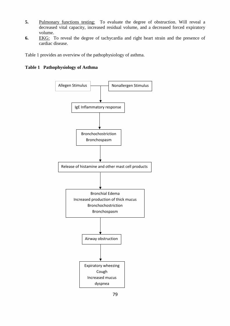
79
5. Pulmonary functions testing: To evaluate the degree of obstruction. Will reveal a decreased vital capacity, increased residual volume, and a decreased forced expiratory volume.
6. EKG: To reveal the degree of tachycardia and right heart strain and the presence of cardiac disease.
Table 1 provides an overview of the pathophysiology of asthma. Table 1 Pathophysiology of Asthma
Allegen Stimulus Nonallergen Stimulus
IgE Inflammatory response
Bronchochostriction
Bronchospasm
Bronchial Edema
Increased production of thick mucus
Bronchochostriction
Bronchospasm
Release of histamine and other mast cell products
Expiratory wheezing
Cough
Increased mucus
dyspnea
Airway obstruction
80
C. Medical Management: 1. Phamacotherapy: Parenteral or inhalant epinephrine, bronchodilators, and
corticosteroids. Antibiotics are given if there is evidence of infection. Nebulized bronchodilators and/or steroids
2. Oxygen therapy is given depending on the Arterial blood gases results) usually about 1 – 3 L/min is given via nasal cannula or via venturi mask.
3. Serial Arterial blood gases values: To evaluate patient’s condition and the effectiveness of treatment.
4. Chest physiotherapy: To enhance secretion expectoration if necessary. This treatment is used with caution, however, because it can exacerbate symptoms.
5. Identification of stressors that precipitate or exacerbate asthomatic attacks. D. Nursing Diagnoses and Interventions 1. Impaired gas exchange related to chronic tissue hypoxia secondary to obstructive
process in the bronchioles and bronchi. Desired outcome: Patient demonstrates vs and Arterial blood gases values within acceptable limits.
1. Assess VS s q1 – 2h or more frequently as necessary, report any significant changes in vs and respiratory pattern, such as increasing dyspnea or tachycardia.
2. Observe for presence of cyanosis of mucous membrance, 3. Review Arterial blood gases results, for changes in respiratory acidosis 4. Give low concentrations of oxygen via a venturi mask as prescribed. 5. Enhance air exchange by assisting patient into high fowler’s position.
2. Ineffective airway clearance related to excessive mucus production and ineffective
coughing. Desired outcome: Patient demonstrates appropriate coughing technique and the ability to excrete secretions
1. Percussion and postural drainage, are done to assist with clearing of airways. 2. Auscultate anterior, lateral, and posterior lung fields to evaluate breath sounds q2 –
4h after coughing. 3. Alteration in nutrition: Less than body requirements related to decreased intake secondary to fatigue and anorexia.
Desired outcome: Patient does not exhibit signs of malnutrition or weight loss.
1. Monitor I&O and record food intake daily; 2. Weigh patient at least weekly. 3. Encourage intake of foods high in protein and carbohydrates. 4. Provide small, frequent meals to minimize fatigue and enhance patient’s compliance
with eating. 5. Avoiding gas-producing foods and carbonated beverages to avoid abdominal
distention . 6. Provide oral care at frequent intervals to enhance appetite.
Anxiety related to perceived threat to biology integrity secondary to sensation of suffocation.
81
Desired outcome: Patient does not exhibit ineffective coping mechanisms. Patient relates the presence of psychologic comfort.
1. Keep patient and SOs well informed of activities, therapy, and procedures in which patient will participate. Involve patient in decision about care activities.
2. Remain with patient during acute episodes. Answer questions and provide care in a calm, reassuring manner.
3. Facilitate transition to home care by initiating discharge planning early in patient’s hospital stay.
4. Encourage patient to establish realistic goals for return to wellness. E. Patient – Family Teaching and Discharge Planning Provide patient and SOs with verbal and written information for the following:
1. Medications to be taken at home, including name, schedule, dosage, purpose, route, precautions, and potential side effects.
2. Use of at-home therapy, including hand-held nebulizers for administering bronchodilators and steroids.
3. Avoiding exposure to infections individuals, particularly those with URLs. 4. Potential need for change in environment such as the elimination of certain foods, pets
feather pillows, dust. 5. Maintenance of diet high in protein and carbohydrates. 3.2.3 Chronic Bronchitis Chronic bronchitis is the persistence of a chronic cough with excessive production of mucus for at least 3 months a year for 2 consecutive years. It is a serious health problem that develops gradually and may go untreated for many years until the disease is well established. Etiology and Pathophysiology Chronic bronchitis is characterized by hypersecretion of mucus by the bronchial glands and recurrent or chronic respiratory tract infections. As the infection progresses, there is a significant alteration in the ability of the cilia lining the airway to propel the secretions upward. Secretions remain in the lungs and form plugs within the smaller bronchi. These plugs become areas for bacterial growth and chronic infection, which increases mucous secretion, and eventually causes areas of focal necrosis and fibrosis. Airway obstruction results from the inflammation of the bronchi. Multiple factors are associated with chronic bronchitis. Its development may be insidious, or it may follow a long history of bronchial asthma or an acute respiratory tract infection, such as influenza or pneumonia. Air pollution and smoking are significant factors. Smoking causes hypertrophy and hypersecretion of the bronchial glands. The disease may occur at any age, but is most often seen in middle age following years of untreated, low-grade bronchitis. Diagnosis is based on evaluation of the duration of the symptoms, how it began, and the client’s occupational history, history of pulmonary disease, and smoking history.
82
Assessment Clinical manifestation The earliest symptom is a productive cough of thick white mucus, especially when rising in the morning and in the evening. Bronchospasm may occur during severe bouts of coughing. As the disease progresses, the sputum may become yellow, purulent, copious, and, after paroxysms of coughing, blood-streaked. Cyanosis secondary to hypoxemia may be noted, especially after severe coughing. Dyspnea begins with exertion, but progresses to occurring with minimal activity, and later occurs at rest. Right hearth failure results from the hypoxemia, which causes edema in the extremities. Diagnostic tests Diagnostic studies done are; physical examination, chest x-ray, and pulmonary function tests. The chest x-ray shows signs of fluid overload and consolidation. Pulmonary function tests demonstrate decreased vital capacity and forced expiratory volume, and increased residual volume and total lung capacity. Diagnostic studies such as bronchoscopy, microscopic examination of the sputum for malignant cells, and lung scan may be performed to rule out cancer, bronchiectasis, tuberculosis, or other diseases in which cough is a predominant feature. Medical Management The goals of treatment are to prevent recurrent irritation of the bronchial mucosa by infection or chemical agents, to maintain the function of the bronchioles, and to assist in the removal of secretions. Treatment includes: • Smoking cessation
• Bronchodilators to dilate bronchi, and reduce airway obstruction and bronchospasm • Increased fluid intake
• Maintenance of a well balanced diet • Postural drainage to remove secretions from the bronchi • Steroid therapy if other treatment is ineffective
• Change in occupation if work involves exposure to dust and chemical irritants • Antibiotic therapy Nursing Management Nursing management focuses on educating clients in managing their disease. Help clients identify ways to eliminate environmental irritants. This includes smoking cessation, occupational counseling, monitoring air quality and pollution levels, and avoiding cold air and wind exposure that can cause bronchospasm. Preventing infection is another important aspect of care. Instruct clients to avoid others with respiratory tract infections and to receive pneumonia and influenza immunizations. Teach the client to monitor sputum for signs of infection. Instruct clients in the proper use of aerosolized bronchodilators and corticosteroids. Warn against overuse. Instruct the client in postural drainage techniques and measures for improving overall health such as eating a well balanced diet, getting plenty of rest, and engaging in moderate aerobic activity should be determined by dyspnea, not heart rate.
83
3.2.4 Adult Respiratory Distress Syndrome Adult respiratory distress syndrome (ARDS) is a complication that occurs following other clinical conditions. It is not a primary disease. When it occurs, it can lead to respiratory failure and death. It is referred to as noncardiogenic pulmonary edema (pulmonary edema not caused by a cardiac disorder). ARDS is characterized by severe hypoxemia and progressive less of lung compliance.Causes of ARDS include chest trauma, shock, drug overdose, drowning, gram negative infections, emboli, and major surgery. Hematologic disorders, pancreatitis, and uremia may also be factors in the development of ARDS. The mortality rate with ARDS is high, particularly if the underlying cause cannot be treated or is inadequately treated. Severe respiratory distress develops within 8 to 48 hours after the onset of illness or injury. In the early stages there are few definitive symptoms. As the condition progresses, the following signs are seen: • Increased respiratory rate
• Shallow, labored respirations • Cyanosis • Use of accessory muscles
• Respiratory distress unrelieved with oxygen administration • Anxiety, restlessness
• Mental confusion, agitation, and drowsiness with cerebral anoxia The initial cause of ARDS must be diagnosed and treated. Nursing management focuses on the promotion of oxygenation and ventilation and prevention of complications. Assessing and monitoring a client’s respiratory status are essential. Potential complications include deteriorating respiratory status, infection, renal failure, and cardiac complications. The client is also anxious and requires explanations and support. Additionally, if the client is on a ventilator, verbal communication is impaired. The nurse provides alternative methods for the client to communicate
3.5 Restrictive Respiratory Disorders
3.5.1 Pneumoconioses The pneumoconiosis include a number of disorders that are occupational in origin. They are characterized by permanent retention of inhaled particles, which results in inflammation and fibrosis. Because these particles remain in the lung tissue for the life of the individual, damage to lung tissue is ongoing.
3.5.2 Asbestosis: Develops from exposure to asbestos fibers in occupations such as mining of asbestos, construction, and paint production. It also can occur in family members who are exposed to the asbestos fibres on the clothing of the asbestos worker. Inhalation of these fibers causes lung tissue fibrosis, usually in the lower lobes. In turn, the fibrosis decreases lung volume and elasticity of the tissues, which ultimately leads to pulmonary failure. When exposed to asbestos fiber, cigarette smokers and individuals with COPD are at a greater risk of getting this disease.
84
3.5.3 Silicosis: Occurs in individuals who inhale dust from quartz rock containing silica. This may occur during mining, tunneling in rock, or sandblasting. The inhaled particles adhere to the lung tissue and produce fibrotic nodules. Long-term exposure to silica dust is necessary before there is evidence of lung tissue changes. Many individuals who have silicosis also develop tuberculosis (TB). Cigarette smokers and individuals with COPD have a greater risk of developing this disease.
3.5.4 Pulmonary Fibrosis
The causes of pulmonary fibrosis are unknown, but it is theorized that it is a reaction to the inhalation of noxious gases or exposure to radiation. It is seen in patients with silicosis, tuberculosis, and collagen diseases. The fibrotic process is a continuous one and does not abate even when the causative agent is no longer present. The fibrosis primarily affects the alveoli and causes an increase in the bronchial diameter in relationship to lung volume. Often the patient has a viral illness at the time of initial diagnosis. Work of breathing is increased because of decreased lung compliance, and affected individuals adopt a rapid, shallow breathing pattern because it requires the least amount of energy. Cardiac complications can occur as a result of the pulmonary hypertension. Assessment Chronic indicators: Dyspnea, cyanosis, digital clubbing. Acute indicators: Increased dyspnea, tachycardia, dysrhythmias. Physical exam: Dullness with percussion over affected lung fields; decreased breath sounds with auscultation. Diagnostic Tests 1. Lung biopsy: To rule out other respiratory disorders such as infection of the lung tissue
and determine prognosis. 2. CBC: Will show elevated WBCs in the presence of infection. 3. Pulmonary function testing: Assesses extent of lung damage. 4. Arterial blood gases values: Help evaluate the level of hypoxemia and hypercapnia. 5. Chest x-ray: Reveals the extent of fibrosis. Medical Management Management is directed toward reversing or halting disease progression. 1. Corticosteriods: To decrease inflammation and fibrosis. 2. Chest physiotherapy: To assist with removal of secretions. 3. Antibiotics: If infection is present. 4. Oxygen therapy: To correct hypoxemia. Nursing Diagnoses and Interventions 1. Monitor I&O and weight daily. 2. Offer small, frequent meals to encourage adequate nutrition. 3. Provide encouragement during mealtimes to promote eating.
85
4. Review Arterial blood gases values and watch for a decrease in Pao2, which would indicate worsening hypoxemia, and in increase in Paco2, which is indicative of hypoventilation (co2 retention).
5. Assess VS and respiratory status q4h or more frequently, as necessary. 6. Monitor for indicators of increasing respiratory distress, including dyspnea, cyanosis,
and tachycaria. 7. Minimize activity level in the presence of increased work of breathing. 8. Administer prescribed oxygen as indicated. Patient – Family Teaching and Discharge Planning Provide patient and SOs with verbal and written information for the following:
1. Medications, including drug name, dosage, purpose, schedule, precautions, and potential
side effects.
3.6 Pulmonary Tuberculosis Tubercle bacilli are gram-positive, rod-shaped, acid-fast aerobic bacilli which can live in the dark for months in particles of dried sputum, exposure to direct sunlight, heat, and ultraviolet light destroys them in a few hours. Tubercle bacilli are killed by pasteurization, a process widely used in preventing the spread of tuberculosis by milk and milk products. Tuberculosis is most commonly transmitted by direct contact with a person who has the active disease through the inhalation of droplets produced by coughing, sneezing, and spitting. 3.6.1 Etiology and Pathophysiology Tuberculosis (TB) is caused by the acid-fast organism Mycobacterium tuberculosis, which causes the formation of tubercles on the lungs and then spreads through the lymphatic system into other body organs. The infection is spread via airborne droplets projected by sneezing or coughing. Typically, the primary lesion develops 6 weeks after the initial infection. When the immune response occurs, the organism circulating in the bloodstream becomes trapped in organs such as the kidney. The course of the disease is as follows: • Acute local inflammation with necrosis
• Ulceration of infected lung tissue • Tubercles cluster together––become surrounded by inflammation • Exudates fills the surrounding alveoli
• Bronchopneumonia develops • Tuberculosis tissue becomes caseous––ulcerates into the bronchus
• Cavities form • Ulcerations heal––scar tissue left around cavity
• Pleura thicken and retract The course of tuberculosis becomes a cyclical one of inflammation, bronchopneumonia, ulceration, cavitations, and scarring. The tuberculosis gradually spreads throughout the lung fields and into the rest of the respiratory structures, as well as other organs via the lymph system.
86
A client may experience periods of exacerbation, followed by remission. Table 28-3 presents the complications of tuberculosis. 3.6.2 Predisposing factors Individuals most prone to contract TB are those who are malnourished, alcoholic, have uncontrolled diabetes, or live in crowded conditions. Tuberculosis is characterized by stages of early infection, latency, and potential for recurrence after the primary disease. The bacilli may remain dormant for many years and reactivate and produce clinical symptoms of tuberculosis. Assessment Clinical manifestation Assessment Patient can be asymptomatic during the early stages of the disease. Clinical manifestations: Malaise, low-grade fever, fatigue, anorexia, weight loss, and a slight, nonproductive cough are the early symptoms of tuberculosis often attributed to overwork, excessive smoking, or poor eating habits Low-grade fever, particularly in the late afternoon, and night sweats are common as the disease progresses. The cough typically becomes productive of mucopurulent and blood-streaked sputum. Marked weakness, wasting, hemoptysis (expectoration of blood from the respiratory tract), and dyspnea are characteristics of later stages of the illness. Chest pain may result from the spread of infection to the pleura. Physical exam: Dullness with percussion over the affected lung fields; crackles (rales) or wheezes with auscultation. Diagnostic Tests Results can mimic those for pneumonia, lung abscess, or bronchogenic carcinoma. 1. Chest x-ray: Will show node enlargement and presence of calcification. The upper lung
fields are usually affected and cavitation might be present. In the early stages, patchy cavities are noted; in advanced stages calcium deposits in lymph nodes and lungs (coin lesions) are present.
2. Tuberculin skin test with purified protein derivative (PPD): Test is positive if it reveals 10 mm or more of induration around the site within 24–72 hours.
3. Sputum smear and culture: Will show presence of M tuberculosis. It should be obtained in the early morning and sent immediately to the lab. Three to five specimens are needed for accurate indentification.
4. Gastric culture: Will show presence of M tuberculosis if the patient swallows sputum. It should be obtained in the morning after patient has been NPO for 8 hours.
5. Lymph node biopsy: Performed when lymph nodes are enlarged and might show the presence of M tuberculosis.
Medical and Surgical Management Medical Management 1. Bedrest: If there is fever, hemoptysis, and/or cough. 2. Isolation: During contagious period. 3. Antitubercular pharmacotherapy: The following are used in combination: Isoniazid,
ethambutol hydrochloride, rifampin, and streptomycin sulfate. Note: When isoniazid is
87
used, close monitoring of SGOT and SGPT is necessary to evaluate hepatic integrity. Hepatic complications can be fatal in the elderly.
4. Corticosteroids: Sometimes given if there is evidence of TB in organs other than the lungs.
Nursing Diagnoses and Interventions Alteration in nutrition: Less than body requirements related to decreased intake secondary to fatigue and anorexia. Desired outcome: Patient does not exhibit signs of malnutrition or weight loss. 1. Monitor and record I&O and weight daily. 2. Give meals that are high in protein and carbohydrates; offer small, frequent meals and
between-meal snacks. 3. Evaluation of patient’s nutrition status with the use of anthropometric measurements of
body size and proportions, including height, weight, triceps skinfold measurement, and mid-arm circumference to evaluate the patient’s fat and protein reserves.
Ineffective breathing pattern related to hyperpnea secondary to infectious process. Desired outcome: Patient exhibits a respiratory rate and depth within acceptable range. 1. Monitor VS and assess patient’s respiratory status q2–4h. 2. Observe for circumoral cyanosis, use of accessory muscles for breathing, dyspnea, and
tachycardia. 3. Encourage patient to take slow, deep breaths. 4. Report any significant change in VS or respiratory pattern . Activity intolerance related to fatigue secondary to infectious process. Desired outcome: Patient relates the attainment of adequate rest. 1. Provide frequent rest periods between care activities. 2. Advise patient to perform activities to tolerance. 3. Reassure patient that normal activity tolerance will return with wellness. 4. Involve patient in decision making regarding care activities and need for rest periods. Potential for infection (for other patients and staff members) related to susceptibility secondary to communicable nature of the disease. Desired outcome: Other patients and staff members do not exhibit signs of infection. 1. Until patient is no longer communicable, maintain isolation by;
• Provide private room with special ventilation; keep door closed; patients infected with same organism can share a room.
• Use mask if patient is coughing • Wear gown, if needed, to prevent gross contamination of clothing. (Gloves are not
necessary.) • Wash hands after touching patient or potentially contaminated articles. • Articles used in patient care should be cleaned and disinfected or discarded,
2. Instruct patient to cough or sneeze into tissues and discard them into specially designated
bag. 3. Instruct patient and SOs regarding isolation precautions. 4. Consult with hospital’s infection control nurse for additional information. 5. Reinforce need for the patient to adhere to medication routine, both in the hospital and at
home.

88
Patient – Family Teaching and Discharge Planning Provide patient and SOs with verbal and written information for the following: 1. Medications, including drug name, dosage, purpose, schedule, precautions, and potential
side effects. 2. Importance of follow-up appointments 3. Explain the need for tuberculin skin tests for SOs. 4. Stress the importance of continuous therapy because lapses in taking the prescribed
drugs can result in reactivation of the infection. 5. Eat a balanced but light diet. If more than a few pounds of weight are lost, contact the
physician. 6. Recognizing causative factors for TB and altering lifestyle and/or home environment as
necessary, 4.0 Conclusion Nursing management of clients with acute respiratory disorders focuses on educating clients in managing their disease. Help clients identify ways to eliminate environmental irritants. This includes smoking cessation, occupational counseling, monitoring air quality and pollution levels, and avoiding cold air and wind exposure Adult respiratory distress syndrome (ARDS) is a complication that occurs following other clinical conditions. It is not a primary disease. 5.0 Summary The pneumoconiosis includes a number of disorders that are occupational in origin. Tuberculosis (TB) is caused by the acid-fast organism Mycobacterium tuberculosis, which causes the formation of tubercles on the lungs and then spreads through the lymphatic system into other body organs. Antitubercular pharmacotherapy include the combination of : Isoniazid, ethambutol hydrochloride, rifampin, and streptomycin sulfate. 6.0 Tutor Marked Assignment Discuss antitubercular pharmacotherapy Outline the medical and nursing management of a client with asthma. 7.0 Further Reading and Other Resources
• Timby K. Barbara, Jeanne C. Scherer, & Nancy E. Smith (1999). Introductory Medical-
Surgical Nursing. (7th Edition) Lippincott.
• Brunner & Suddarth’s (2004) Medical Surgical Nursing. (10th ed) Lippincott Wilkins
89
UNIT III: INFECTION PROCESS
1.0 Introduction …………………………………………………………………………… 2.0 Objective ………………………………………………………………………………. 3.0 Main Content …………………………………………………………………………... 3.1 Microorganisms and Infectious Disorders ……………………………………………. 3.2 Types of Microorganisms …………………………………………………………….. 3.3 Characteristics of Microorganisms …………………………………………………… 3.4 Infection Transmission ……………………………………………………………….. 3.5 Defense Mechanisms …………………………………………………………………. 3.6 Pathophysiology ……………………………………………………………………… 3.7 Nosocomial Infections ………………………………………………………………... 4.0 Conclusion ……………………………………………………………………………. 5.0 Summary ……………………………………………………………………………… 6.0 Tutor Marked Assignment ……………………………………………………………. 7.0 Further Reading and Other Resources ………………………………………………. 1.0 Introduction This unit deals with infection process with special emphasis on oncology 2.0 Objective By the end of this unit you will be able to:
• Describe microorganisms and list at least three examples. • List three factors that influence whether an infection develops. • Differentiate between pathogens and nonpathogens. • List at least five factors that increase susceptibility to infection. • Name six components of the infectious process cycle.
90
• Explain the difference between mechanical and chemical defense mechanisms. • Describe the events that occur during the inflammatory process. • Discuss the medical management of clients with infectious disorders. • Name three nursing interventions that prevent or control infectious disorders. • Explain the role of an infection control committee. • List at least four measures that have reduced community-acquired infections. • Discuss measures to take if a needle stick injury occurs.
3.0 Main Content Community-acquired infections Opportunistic infections Culture Pathogens Generalized infections Phagocytosis Host Portal of entry Immunizations Portal of exit, Reservoir Infectious process cycle Sensitivity Leukocytosis Sepsis Localized infections Septicemia Microorganisms Standard precautions Mode of transmission Susceptibility Multidrug resistance Transmission-based precautions Nonpathogens virulence Nosocomial Infections. 3.1 Microorganisms and Infectious Disorders Infectious disorders are conditions caused by microorganisms. Microorganisms, commonly called “germs,” are living plants and animals that are so small they can only be seen with a microscope. Factors that influence infection are the type of microorganism, its characteristics, and susceptibility of the host, the person on or in whom the microorganism resides. Once microorganisms invade, one of three events occurs: (1) The body’s immune defense mechanisms eliminate them, (2) They reside in the body without causing disease, or (3) They cause an infection or infectious disease. 3.2 Types of Microorganisms Microorganisms that infect living tissue include bacteria, viruses, fungi, rickettsiae, protozoans, mycoplasmas, and helminthes. Some cause communicable or contagious diseases, infectious diseases that are transmissible to other people. Examples of communicable diseases are measles, streptococcal sore throat, sexually transmitted diseases, and tuberculosis. Bacteria Bacteria are single-celled microorganisms. They appear in a variety of shapes: round (cocci), rod-shaped (bacilli), or spiral (spirochetes). Aerobic bacteria require oxygen for growth and multiplication, whereas anaerobic bacteria grow and multiply in an atmosphere that lacks oxygen. A growing number of bacteria such as Staphylococcus aureus, Streptococcus

91
pneumoniae, and the enterococcus (intestinal bacteria) Escherichia coli, are developing multidrug resistance, the ability to remain unaffected by antimicrobial drugs such as antibiotics. Viruses Viruses are the smallest agents known to cause disease and can only be seen with high-powered magnification using an electron microscope. They are also filterable, meaning they pass through very small barriers. Viruses are divided into two types: (1) those whose nucleic acid composition consists of deoxyribonucleic acid (DNA), or (2) those whose nucleic acid consists of ribonucleic acid (RNA). Viruses use the metabolic and reproductive materials of living cells or tissues to multiply. Some viral infections, such as the common cold, are minor and self-limiting; that is, they terminate with or without medical treatment. Others, such as rabies poliomyelitis, and viral hepatitis, are more serious and may be fatal. Fungi Fungi are divided into two basic groups: yeasts and molds. Only a small number of fungi appear to produce disease in humans. There are three types of fungus (mycotic) infections: superficial (dermatophytoses), that effect the skin, hair, and nails, intermediate, that chiefly affect subcutaneous tissues; and deep (systemic), that affect deep tissues and organs. Rickettsiae Rickettsiae are microorganisms that resemble but are different from bacteria. Like viruses, they invade living cells and cannot survive outside a living organism or a host. Rickettsial diseases are transmitted by arthropods (invertebrate animals with a segmented body, an external skeleton, and jointed, paired appendages), such as the flea, tick, louse, or mite. Protozoans Protozoans are single-celled animals that are classified according to their motility (ability to move). Some protozoans possess amoeboid motion; they extend their cell walls and their intracellular contents flow forward. Others movement. Mycoplasmas Mycoplasmas are single-celled microorganisms that lack a cell wall and, therefore, are pleomorphic (assume many shapes). They are similar but not related to bacteria. Mycoplasmas primarily infect the surface linings of the respiratory, genitourinary, and gastronintestinal tracts. Helminthes Helminthes are infectious worms, some of which are microscopic. They are divided into three major groups: nematodes, or roundworms; cestodes, or tapeworms; and trematodes, or flukes. Some helminthes enter the body in the egg stage, whereas others spend the larval stage in an intermediate host, then enter the human host. The organisms mate and reproduce in the definitive host and then are excreted, and the cycle begins again.
92
3.3 Characteristics of Microorganisms Not all microorganisms are dangerous. Some are nonpathogens because they are generally harmless to healthy humans. For example, the normal microbial flora (microscopic plants) present in the intestine helps synthesize vitamin B12, biotin, vitamin K, and folic acid. Pathogens, on the other hand, have a high potential for causing infectious diseases. The ability of a microorganism to cause infection depends on a number of factors: its ability to move or be moved from one place to another, its virulence (power to produce disease), the number present, the duration of exposure, its ability to invade the host, and the susceptibility (prone to disease) of the host. Given the right set of circumstances, both pathogens and nonpathogens can produce an infection. Unless and until the supporting host becomes weakened, normal flora remains in check. However, even benign microorganisms can produce opportunistic infections, those in which nonpathogenic or remotely pathogenic microorganisms take advantage of favorable situations and overwhelm the host More often than not, however, infections are caused by common pathogents. 3.4 Infection Transmission Like the links in a chain, components in the infectious process cycle must be present to transmit an infectious disease from one human or animal to a susceptible host (Fig. 18-2). Besides the infectious agent and host, an appropriate reservoir, exit route, mode of transmission, and portal of entry are necessary. Reservoir A reservoir is the environment in which the infectious agent is able to survive and reproduce. The reservoir may be human, animal, or nonliving, such as contaminated food and water. A human or animal that harbors (or is the reservoir of) an infectious microorganism but does not show active evidence of the infectious disease is a carrier. Nonliving reservoirs are fomites. Portal of Exit The portal of exit is the route by which the infectious agent escapes from the reservoir such as through the respiratory, gastrointestinal, or genitourinary tract, from the skin and mucous membranes, and blood and other body fluids. Mode of Transmission A microorganism’s mode of transmission refers to the manner in which it is transferred or moved from its reservoir to the susceptible host. There are five potential modes of transmission Portal of Entry An infectious agent gains entrance into a susceptible host through a portal of entry. Some infectious agents may have only one portal of entry; others may use several. Staphylococci, for example, can cause disease by entering through the respiratory tract (pneumonia), skin (boils), blood (internal abscesses), or gastrointestinal tract (food poisoning).
93
Although many microorganisms are present in reservoirs, they may be prevented from producing an infection because of human defense mechanisms. 3.5 Defense Mechanisms Humans and other animal species have developed both mechanical and chemical defense mechanisms. Mechanical defense are those that act as a physical barrier for preventing microorganisms from gaining entry or those designed to expel microorganisms before they multiply. Examples of mechanical defenses are the skin and mucous membranes, physiologic reflexes (eg, sneezing, coughing, and vomiting), and macrophages. Chemical defense mechanisms destroy or incapacitate microorganisms with naturally produced biologic substances. Examples of chemical defense mechanisms include enzymes, secretions, and antibody substances. Skin and Mucous Membranes The first line of defense against invading microorganisms is unbroken skin and mucous membranes. They separate underlying body tissues from microorganisms in the environment. The normal flora (eg, microorganisms) found on the skin compete with pathogenic microorganisms for nutrients, thereby retarding the growth of pathogens in these areas. In addition, the skin, which is acidic (because of the acetic acid in perspiration), creates an undesirable medium for the multiplication of pathogenic microorganisms. Mucus, a sticky substance secreted from mucous membranes, traps microorganisms and debris on its surface. For example, mucous membrane secretions of the vagina favor the growth of nonpathogenic acid-producing bacteria, known as Doederlein’s bacilli. The acid environment is unfavorable for multiplication of pathogenic bacteria and fungi. However, a change in vaginal pH or destruction of the normal flora can favor the development of a vaginal infection. Physiologic Reflexes If microorganisms gain entry, they can be forcefully expelled by sneezing, coughing, and vomiting. Coughing is promoted by the action of cilia, hairlike projections in the upper respiratory tract, that beat in an upward direction. Macrophages Macrophages are specialized cells that make up the mononuclear phagocyte system, formerly known as the reticuloendothelial system. These cells are located throughout tissues of the body, in the liver (Kupffer’s cells), the spleen, and lymphoid tissue (eg, the tonsils). Their primary function is to ingest dead cells and foreign material including microorganisms. Biologic Substances Lysozyme (muramidase), an enzyme capable of splitting (lysing) the cell wall of some gram-positive bacteria, is present in tears, saliva, mucus, skin secretions, and some internal body fluids such as gastric juices. This enzyme is bactericidal (destroys bacteria) and thus acts as a defense against some pathogenic bacteria. Antibodies, complex proteins also referred to as immunoglobulins, are formed when macrophages consume microorganisms and display their distinct cellular markers from surface.
94
Antibodies work with other white blood cells (WBCs) by rendering microorganisms more easily ingested, or phagocytized, in one of several ways: by lysing (dissolving or reducing the size of the foreign invader), by neutralizing their toxins (poisons released by some microorganisms), by opsonizing (coating) them, by agglutinating (clumping) them together, or by precipitating (solidifying) them. Interferon, another chemical protein, is produced by the WBCs and other body cells in response to viral infections and other factors. Interferon appears to trigger infected cells to manufacture an antiviral protein. Because interferon also appears to inhibit cell reproduction, it is being used in the adjunct treatment of some cancers and viral disorders with positive results. Regardless of the infectious process, all infections share some common pathophysiologic characteristics. 3.6 Pathophysiology Localized Infections The initial localized reaction to an invading microorganism activates the inflammatory process–the first event is a cellular response that results in leakage of fluid, colloids, and ions from the capillaries into the tissues between the cells, producing swelling. It is followed by a vascular response that produces redness and heat, and a chemical response that causes pain. WBCs ––neutrophils, macrophages, monocytes, and lymphocytes––move to the injury site to destroy the toxins produced by the pathogens and to remove debris from the area. More WBCs are manufactured as they are needed, a process referred to as leukocytosis. To prevent the spread of pathogenic microorganisms to adjacent tissues, a fibrin barrier forms around the injured area. Inside the barrier, thick, white exudates (pus) accumulates. This collection of pus is called an abscess may break through the skin and drain, or continue to enlarge internally. If the lymph nodes in the area become involved, they become enlarged and tender, an inflammatory condition referred to as lymphadenitis. If this defense mechanism is unable to contain the infection, the microorganisms begin to travel from node to node (see Chap. 39). Because the lymphatic system drains into the venous system, the microorganisms may eventually reach the bloodstream causing a condition called septicemia or sepsis, which leads to a generalized infection.
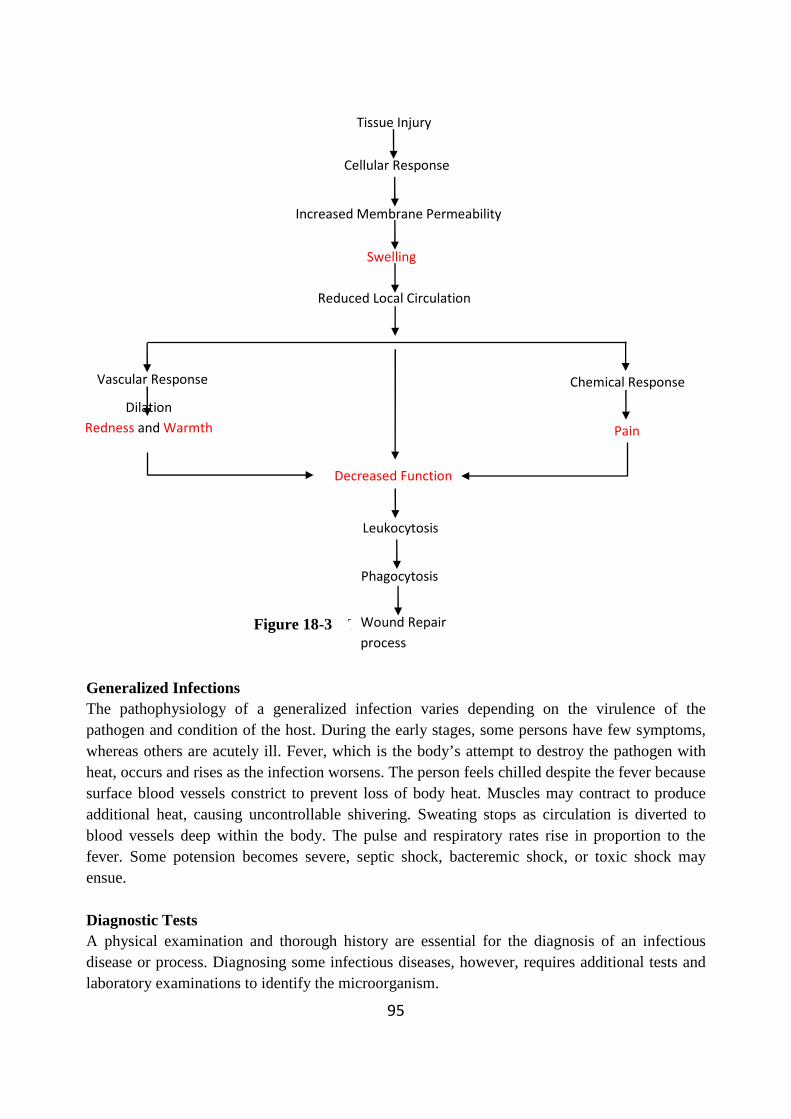
95
Figure 18-3 The inflame Generalized Infections The pathophysiology of a generalized infection varies depending on the virulence of the pathogen and condition of the host. During the early stages, some persons have few symptoms, whereas others are acutely ill. Fever, which is the body’s attempt to destroy the pathogen with heat, occurs and rises as the infection worsens. The person feels chilled despite the fever because surface blood vessels constrict to prevent loss of body heat. Muscles may contract to produce additional heat, causing uncontrollable shivering. Sweating stops as circulation is diverted to blood vessels deep within the body. The pulse and respiratory rates rise in proportion to the fever. Some potension becomes severe, septic shock, bacteremic shock, or toxic shock may ensue. Diagnostic Tests A physical examination and thorough history are essential for the diagnosis of an infectious disease or process. Diagnosing some infectious diseases, however, requires additional tests and laboratory examinations to identify the microorganism.
Tissue Injury
Dilation
Redness and Warmth
Cellular Response
Increased Membrane Permeability
Reduced Local Circulation
Swelling
Decreased Function
Leukocytosis
Phagocytosis
Wound Repair
process
Vascular Response Chemical Response
Pain
96
White Blood Cell Count and Differential An elevation in the number and type of WBCs (leukocytes), is indicative of an inflammatory and possibly infectious process. Although a total WBC count provides important information, a differential––one that indicates the percentage of WBC subtypes––is even more valuable. An elevation of neutrophils, the largest subtype of leukocytes, is an indication that the body is in the early stages of responding to an invading pathogen. As their numbers become depleted, bone marrow produces additional cells called “band cells” (bands) that eventually mature and take their place. An elevation of monocytes, the largest sized subtype of WBCs, is the body’s second line of defense. Culture and Sensitivity Test A culture is used to identify bacteria within a specimen taken from a person with symptoms of an infection. The source of the specimen of the specimen may be body fluids or wastes, such as blood, sputum, urine, or feces, or the prulent exudates, collection of pus, from an open wound The specimen is cultured. This involves placing a small amount of the specimen in or on a special growth medium. The specimen is incubated for a specific time period (usually 48-72 hours), then examined microscopically. To facilitate examination, the specimen is stained or dyed (colored). One stain is the Gram stain. Those bacteria that absorb the color are classified as gram-positive; those that do not, are gram-negative. The microorganisms may also be tested for pathogenicity or virulence with a coagulase test. When a culture is reported as coagulase-positive, it is more virulent than a culture of the same microorganism that produces a negative (coagulase-negative) response. Sensitivity studies are done to determine which antibiotic inhibits the growth of a nonviral microorganism and will be most effective in treating the infection. Examination for Ova and Parasites Most ova (eggs) and parasites (those that live at the expense of the host) are intestinal worms. Therefore, the stool is examined for evidence of any forms within the infecting microorganism’s life cycle. Generally, three random stools are collected from a bedpan, not the toilet. Urine and toilet paper may alter the specimen and therefore must be disposed of separately. Clients suspected of having intestinal ova and parasites are advised to perform scrupulous handwashing to avoid reinfecting themselves and others. Skin Tests Skin testing determines the presence of a specific active or inactive infection. Diseases for which skin testing may be done include histoplasmosis, mumps, tuberculosis, diphtheria, and coccidioidomycosis. The material for skin testing is injected intradermally The reaction is read after a specified time period (usually 48-72 hours). The size of the induration (hard elevated tissue), not including the surrounding area of erythema (redness) is measured in millimeters (mm). The measurement determines whether the reaction is significant. For example, a tuberculin skin test is considered positive if the induration is 15 mm or greater in persons with no known risk factors for tuberculosis; smaller measurements are significant in certain risk groups, such as those who are immune-compromised.
97
Immunologic Tests Immunologic tests may be used to determine the presence of antigen (substances that stimulate an immune response) and antibody reactions. Examples include agglutination tests, for example the cold agglutinins test, which may reveal the presence of high antibody titers confirming immunity to rubella (measles); precipitation tests, for example the C-reactive protein test and erythrocyte sedimentation rate, which are elevated in some inflammatory diseases; complement-fixation tests, which, when elevated, indicate an inflammatory process; and immunoflorescence tests, which identify immunoglobulins, antibodies formed by the immune system. Other Tests Depending on the disease, other diagnostic tests may be used. Radiography (plain films or contrast studies), computed tomography scanning, and magnetic resonance imaging may be used to locate abscesses, identify displacement of organs or structures that may indicate abscess formation, and detect changes in tissues in areas such as the bones or the lung. Medical Management In some cases, supportive therapy such as rest, fluids, adequate nutrition, and antipyretics such as aspirin or acetaminophen (Tylenol) for a significantly elevated fever, may be advised while the infectious disease runs its course. If the etiology is amenable to drug therapy, antimicrobials such as antibiotics, sulfonamides, and antiviral drugs are prescribed. Infected wounds may be debrided, a process of removing dead and damaged tissue. Wound irrigations, hydrotherapy (whirlpool), and the application of wet-to-dry dressings may accomplish the same objective. With secondary infections, treatment of the primary condition may relieve the infectious process. Bone marrow transplantation or administration of drugs that boost WBC production such as filgrastim (Neupogen) may help immunosuppressed clients. Nursing Management The nurse obtains a client’s history, gathers subjective and objective data paying particular attention to manifestations of an inflammatory response, evidence of unusual drainage, and generalized data like a fever, lassitude (feeling tired), and loss of appetite. The nurse also prepares the client for diagnostic tests and collects specimens that will be cultured. Handwashing remains the single most important measure for preventing the spread of infection. It is essential that everyone in a health care agency conscientiously follows handwashing guidelines (Box 18-3). Four elements are necessary to reduce the number of microorganisms on the hands: a cleansing agent, friction, running water, and time. The hands are washed with a cleansing agent such as soap. Friction and the lather from the cleansing agent lift the microorganisms from the skin’s surface. Running water removes the cleansing agent and many of the microorganisms. To be effective, time is essential: 30 to 60 seconds of washing is recommended for hands not grossly contaminated. Follow standard precautions, formerly called universal precautions, during the care of all clients. Standard Precautions are techniques that are used to prevent the potential for transmitting pathogens in blood or other body fluids when a person’s infectious status is unknown. When,
98
and if, a client is diagnosed with an infectious condition, transmission-based precautions those that interfere with the manner in which a particular pathogen is spread are implemented. The nurse administers drug therapy and observes for evidence of the client’s improvement such as reduction in temperature, heart rate, and WBC count. Perform wound care regularly; keep dressings dry and intact. The nurse also implements measures that promote the client’s comfort such as reducing fever and measures that prevent or restore impaired skin integrity and improve nutrition and fluid intake. Methods for preventing or controlling nosocomial infections and infectious communicable diseases, also known as community-acquired infections, are especially important. Nurses have valuable skills to contribute in these two aspects of health care. 3.7 Nosocomial Infections Nosocomial Infections are those acquired while being cared for in a health care agency and the infection was not present, as either an active, incubatory, or chronic infection, at the time of admission. There are many reasons why nosocomial infections occur. Hospitalized clients are more susceptible to infections than well people, because they are exposed to pathogenic microorganisms in the hospital’s environment, may have incisions or invasive equipment such as intravenous lines that compromise skin integrity, or may be immunosuppressed due to poor nutrition, their disease process, or its treatment. Also, because hospital personnel are in frequent and direct contact with many clients, there is a high risk for transmitting pathogenic microorganisms between and among clients. Visitors also carry a potential for introducing pathogens into the hospital environment as might equipment (eg, wheelchairs) and facilities used in common with others (eg, shared bathrooms. Prevention and Control To help prevent and control nosocomial infections, nurses apply principles of medical and surgical asepsis whenever they care for clients. They are further guided by recommendations from the health care agency’s infection control committee, which is usually composed of representatives from various areas and departments of the hospital, such as medical staff, nursing service, clinical laboratories, pathology, operating room, housekeeping, and dietary service. The responsibilities of the infection control committee include surveillance, the process of detecting, reporting, and recording nosocomial infections; educating hospital personnel about methods for reducing nosocomial infections; providing guidelines for the prevention of infectious diseases, and investigating and following up outbreaks of nosocomial infections. Infection control guidelines generally establish policies for pre- and postemployment health examinations, sterization procedures and methods, disposal of garbage and biologic wastes, and housekeeping techniques; designate precautions to be followed for a specific infection; and define the procedures for managing contaminated materials such as linen, equipment, and suppliers used in the care of infectious clients. Many community-acquired (communicable, contagious) infections have been contained or eliminated because of advances in the prevention and treatment of infectious diseases. These advances include the discovery and use of antibiotics, the development of immunizing agents, guidelines for the proper disposal of human wastes, legislation controlling the preparation and sale of foods, immunization programs, and public education.
99
To help prevent and control community-acquired infections, nurse must encourage childhood immunizations, vaccines that stimulate the body to produce antibodies against a specific disease organism, to reduce the incidence of some infectious diseases. However, parental apathy or the inability to afford health care can pose a potential problem in children obtaining immunizations. The immunization of children protects all people-children as well as adults who may not have development sufficient immunity. Local, state, and federal public health agencies and the World Health Organization cooperate in the detection and control of communicable diseases. Because of their combined efforts, the incidence of many infectious diseases has been reduced and some (eg, smallpox) have been virtually eliminated. The most personal avenue for preventing and controlling infectious diseases is education. Ideally, education about general health measures begins early in life. Parents must instruct their children in proper handwashing and in personal cleanliness and grooming and model similar behaviors. Client and Family Teaching To reduce the potential for infections, the nurse teaches the client and family members to: • Perform frequent handwashing, especially before eating, after using the toilet, and after
contact with nasal secretions. • Bathe daily and perform other forms of personal hygiene such as oral care.
• Keep the home environment clean; household bleach diluted 1:10 or 1:100 is an excellent disinfectant.
• Keep immunizations current. Tetanus vaccine is recommended every 10 years; the influenza vaccine is repeated yearly; and one dose of pneumococcal pneumonia vaccine lasts a lifetime.
• Investigate the need for vaccinations, water purification techniques, and foods to avoid when traveling outside the United States.
• Practice a healthy lifestyle such as eating the recommended servings from the food pyramid and using safe food handling practices.
• Use and immediately discard disposable paper tissues rather than reuse a cloth handkerchief.
• Avoid sharing personal care items such as washcloths and drinking cups.
• Use safe sex practices. • Stay home from work or school when ill rather than expose others to infectious
pathogens. • Avoid crowds and public places when there are local outbreaks of influenza. • Follow posted infection control instructions when visiting hospitalized family members
and friends. • Understand that antibiotic therapy is not always appropriate for every infectious disease,
but when it is, take the full doses for the prescribed period of time. Needle Stick Injuries One of the greatest threats to health care workers is the potential for acquiring blood-borne infectious diseases such as hepatitis B (HBV) and acquired immunodeficiency syndrome
100
(AIDS). Following Standard Precautions reduces this potential. However, gloves are not impervious to penetration by sharp objects such as needles that may contain blood. Despite following policies and precautions for avoiding blood-borne pathogens and using new needleless access devices on intravenous lines, needle stick injuries continue to occur. Should an accidental injury occur, health care workers are advised to follow postexposure recommendations, which include:
• Reporting the injury to one’s supervisor immediately. • Documenting the injury in writing.
• Identifying the person or source of blood, if possible. • Obtaining the human immunodeficiency virus (HIV) and hepatitis B status, if it is legal
to do so. Unless the client gives permission, testing and revealing HIV status is prohibited.
• Obtaining counseling on the potential for infection. • Receiving the most appropriate postexposure prophylaxis.
• Receiving instructions on monitoring potential symptoms and medical follow-up. 4.0 Conclusion • Nonpathogens are microorganisms that are generally harmless to healthy humans.
Pathogens are microorganisms that have a high potential for causing infectious diseases. • Factors that increase a person’s susceptibility to infection include inadequate nutrition,
poor hygiene, suppressed immune system, chronic illness, and insufficient leukocytes. • Nurses and other health care workers are at risk for infection from blood-borne
microorganisms. Therefore, if a penetrating injury occurs, such as a needle stick, it is important to report and document the incident>
5.0 Summary • Mechanical and chemical defense mechanisms prevent many infectious disorders from
occurring. Mechanical defenses, like the skin, or reflexes that expel microorganisms, like coughing, act as a physical barrier to microorganisms. Chemical defenses destroy or incapacitate microorganisms with naturally produced biologic substances like enzymes and antibodies.
• Handwashing is the best technique for limiting infections. To be effective, it must include a cleansing agent, friction, running water, and sufficient time.
6.0 Tutor Marked Assignment 1. Explain the difference between mechanical and chemical defense mechanisms. 2. Discuss the medical management of clients with infectious disorders. 3. Name three nursing interventions that prevent or control infectious disorders. 4. Discuss measures to take if a needle stick injury occurs.

101
7.0 Further Reading and Other Resources • Timby K. Barbara, Jeanne C. Scherer, & Nancy E. Smith (1999). Introductory Medical-
Surgical Nursing. (7th Edition) Lippincott.
• Brunner & Suddarth’s (2004) Medical Surgical Nursing. (10th ed) Lippincott Wilkins

102
MODULE THREE
UNIT I: DISORDERS OF THE MALE AND FEMALE PELVIS 1.0 Introduction ……………………………………………………………………….
2.0 Objective …………………………………………………………………………..
3.0 Main Content ……………………………………………………………………….
3.1 Pathophysiology ……………………………………………………………………
3.1.1 Etiology …………………………………………………………………………….
3.1.2 Assessment/ Clinical manifestation …………………………………………………
3.1.3 Diagnosis
3.1.4 Medical Management of Cancer ……………………………………………………
3.2 Surgeries and Disorders of the Breast ………………………………………………
3.2.1 Breast Augmentation ………………………………………………………………..
3.2.2 Breast Reconstruction ……………………………………………………………….
3.2.3 Benign/Malignant Breast Disease ……………………………………………………
3.2.4. Malignant Disorders of the Breast ………………………………………………….
3.3 Neoplasms of the Female Pelvis ……………………………………………………
3.3.1 Cancer of the Cervix …………………………………………………………………...
3.3.2 Ovarian Tumors ………………………………………………………………………
3.3.3 Endometrial Cancer ………………………………………………………………….
3.4 Disorders of the Female Pelvis ……………………………………………………….
3.4.1 Endometriosis ………………………………………………………………………..
3.4.2 Cytocele ……………………………………………………………………………….
3.4.3. Uterine Prolapse ………………………………………………………………………..
3.4.3. Uterine Prolapse ………………………………………………………………………...
3.5 Disorders and Surgeries of the Male Pelvis ……………………………………………..
3.5.1 Benign Prostatic Hypertrophy …………………………………………………………
3.5.2 Prostatitis ………………………………………………………………………………
3.5.3 Prostatic Neoplasm …………………………………………………………………….
4.0 Conclusion ……………………………………………………………………………..
5.0 Summary ……………………………………………………………………………….
6.0 Tutor Marked Assignment ……………………………………………………………...
7.0 Further Reading and Other Resources ………………………………………………….
103
1.0 Introduction Cancer is characterized by abnormal, unrestricted cell proliferation. Malignant tumors invade healthy tissues and compete with normal cells for oxygen, nutrients, and physical space. The nursing specialty related to caring for clients with cancer is called oncology nursing. The diagnosis of cancer is frightening for most people, although reactions of clients depend on the particular diagnosis, location, stage, treatment, effects on bodily functions, and prognosis. 2.0 Objective By the end of this unit you will be able to: 1. Discuss the pathology and etiology of cancer. 2. Differentiate between benign and malignant tumors. 3. Identify the warning signs of cancer. 4. Describe ways to reduce the risks of cancer. 5. Discuss the emotional impact associated with the diagnosis of cancer. 6. Use the nursing process as a framework for caring for clients with cancer. 3.0 Main Content Gene Therapy Antineoplastic Benign Malignant Metastasis Neoplasms Cancer Radiation Therapy Carcinogens endometriosis, cystocele, rectocele, uterine prolapse 3.1 Pathophysiology The cell is the basic structural functional unit in plant and animal life forms. Differentiated cells work together to perform specific functions. Cell regeneration occurs through cell division and reproduction. Abnormal changes in cells can occur for many reasons. These abnormal cells reproduce in the same way, but they do not have the regulatory mechanisms to control growth. As a result, the abnormal cell growth proliferates in an uncontrolled and unrestricted way. New growths of abnormal tissue are called neoplasms or tumors. They are classified according to their cell of origin and whether their manner of growth is benign, not invasive or spreading, or malignant, invasive and capable of spreading. The first part of the name indicates the particular cell or tissue. The suffix –oma indicates it is a tumor. Four main classifications of tumors according to tissue type are: • Carcinomas (cancers originating from epithelial cells).
• Lymphomas (cancers originating from organs that fight infection). • Leukemias (cancers originating from organs that form blood).
• Sarcomas (cancers originating from connective tissue, such as bone or muscle). Cancer can spread (metastasis) by direct extension to adjacent tissues, extension from lymph vessels into the tissues adjacent to lymphatic vessels, transportation by blood or lymph systems, and diffusion within a body cavity. The area in which malignant cells first seen is called the primary site. The regions to which cancer cells spread are secondary or metastatic sites.
104
Metastasis is one of the most discouraging characteristics of cancer because even one malignant cell can give rise to a metastatic lesion in a distant part of the body. Metastatic tumors are treated aggressively whenever possible to improve the quality of life and to lengthen survival time. Benign tumors remain at the original site of their development. They may grow large, but their rate of growth is slower than that of malignant tumors. Benign tumors usually do not cause death unless their location impairs the function of a vital organ, such as the brain. On the other hand, malignant tumors grow rapidly, and, unless completely removed before metastasis has occurred, they are likely to spread. 3.1.1 Etiology Certain factors and agents appear to be related to the development of many cancers. Factors believed to cause cancer are called carcinogens. These include chemical agents, environmental factors, dietary substances, viruses, defective genes, and hornomes. Effects of tobacco smoke and nicotine are related to cancers of the lung, mouth, throat, neck, esophagus, pancreas, cervix, and bladder. Prolonged exposure to certain chemicals such as asbestos and coal dust is associated with som cancers. Chemical substances in the workplace can cause cancer. Organs most affected are the lungs, liver, and kidneys because they are involved with biotransformation and excretion of chemicals. Environmental factors include prolonged exposures to sunlight, radiation, pollutants, and possibly electromagnetic fields from microwaves, power lines, and cellular phones. Diet is a risk factor in cancer development. What a person does not consume is as important as what he or she consumes. Food high in fat, smoked foods, foods preserved with salts or alcohol, and foods with nitrates are associated with an increased risk for cancer. Foods believed to reduce the risk of cancer are high in fiber, cruciferous, (cabbage, broccoli), and high in carotene (winter squash, carrots, cantaloupe). Vitamins A, C, and E also seem to have anticancer value. Viruses are implicated in many cancers. The cell changes incorporated into the genetic information by the virus may result in the formatio of cancerous cells. An example is Kaposi’s sarcoma associated with human immunodeficiency virus (HIV) (Bullock, 1996). Defective genes are responsible for diverse cancers. Some types of leukemia, retinoblastoma (eye tumor in children), and skin cancers are associated with genetic factors. Breast, prostate, and colorectal cancers are also associated with defective genes. Medically prescribed interventions such as immunosuppressive drugs, hormone replacements and anticancer drugs have been associated with increased incidence of cancer in individuals or their offspring when exposed in utero. 3.1.2 Assessment Clinical manifestation Cancer is an insidious disease. It initially may not cause any symptoms or the signs may be vague. This underscores the importance of educating the public about prevention and self-examination so that cancer can be diagnosed in its earliest stages. Seven warning signals of caner (CAUTION) published by the American Cancer Society are:
105
Change in bowel or bladder habits. A sore that does not heal. Unusual bleeding or discharge. Thickening or lump in breast or elsewhere. Indigestion or difficulty in swallowing. Obvious change in wart or mole. Nagging cough or hoarseness. Better education has produced an awareness of the warning signals and factors that influence the development of cancer. Public education stresses the importance of periodic physical examinations and cancer screening programs. Self-examination of the breasts, testicles, and skin is emphasized and taught. Avoidance of factors that predispose a person to cancer and early detection increase the chances of curing the disease. 3.1.3 Diagnosis Cancer is diagnosed by history, physical examination, and diagnostic studies. In some cases the physical examination is unremarkable, but the client’s history is suspicious for cancer. In addition, the client is evaluated for risk factors. Many diagnostic studies are used to establish a diagnoses of cancer. The physician, using information obtained during the history and physical examination, selects tests that help establish a diagnosis. Laboratory Tests The chemical composition of blood and other body fluids is altered by the presence of specific cancers. Specialized tests have been developed for tumor markers, which are specific proteins, antigens, hormones, genes, or enzymes released by cancer cells. These include: • Acid phosphatase: indicates that prostate cancer has metastasized to other parts of the
body if blood levels of this enzyme are elevated. • CA-19-9: detects an antigen that indicates pancreatic cancer.
• CA-125: measures the antigen that is diagnostic for ovarian cancer. • Prostate specific antigen (PSA): detects prostate cancer when levels are elevated; this test
is more specific than acid phosphatase. Other laboratory tests may be useful in establishing a diagnosis. Although abnormal values do not directly indicate a malignant process, they may help in formulating a total clinical picture. For example, a complete blood count may detect anemia in a client with possible colon cancer. Occult blood in the stool may indicate colorectal cancer. Radiologic Tests • X-ray studies: These studies, such as plain films or radiographs using contrast media or
specialized equipment, detect tumors in specific organs. A contrast medium is a substances that highlights, outlines, or provides more detail than shown in a plain film. A barium enema is an example of a study done with contrast medium.

106
• Computed tomography (CT): The CT scan provides three-dimensional cross-sectional views of tissues to determine tumor density, size, and location.
• Magnetic resonance imaging(MRI): Similar to a CT scan, the MRI uses magnetic fields for sectional images. It helps visualize tumors hidden by bones.
• Radioisotope studies: These studies use radioactive materials (given orally or intravenously) to help diagnose various malignancies. A scanner then identifies areas of increased, decreased, or normal distribution.
• Ultrasound: Ultrasound uses high frequency sound waves to detect abnormal variations of a body organ or structure. The sound wave reflections are projected on a screen and may be recorded on film. These studies help differentiate solid and cystic tumors of the abdomen, breasts, pelvis, and heart.
Other Studies • Biopsy: Tissue samples excised from the body are directly examined microscopically for
malignant or premalignant processes. Tissue samples may be obtained during a surgical procedure, through insertion of a biopsy needle under local anesthesia, or during endoscopic procedures. A biopsy provides the most definitive method for diagnosing cancer.
• Endoscopy: Fiberoptic instruments are flexible tubes that contain optic fibers that enable light to travel in a straight line or at various angles and illuminate the area being examined. Specific areas of the body are examined with endoscopy. Tissue biopsies are done if a malignancy is suspected.
• Cytology: Microscopic examination of cells from various areas of the body is used to diagnose malignant or premalignant disorders. Cells are obtained via needle aspiration, scraping, brushing, or sputum. An example of a cytologic test is the Papanicolaou’s (Pap) smear used in diagnosing changes in the endometrium, cervix, and vagina.
Staging of Tumors Before a client is treated for cancer, the tumor is staged and graded. The American Joint Commission on Cancer developed a grading system referred to as the TNM classification system: T indicates the size of the tumor; N stands for the involvement of regional lymph nodes; and M refers to the presence of metastasis. A second classification system also stages tumors according to size, evidence of metastasis, and the presence or absence of lymph node involvement:
• Stage I: malignant cells are confined to the tissue of origin, no signs of metastasis. • Stage II: limited spread of cancer in the local area, generally to area lymphy nodes. • Stage III: tumor larger or probably has invaded surrounding tissues or both.
• Stage IV: cancer has invaded or metastasized to other parts of the body. 3.1.4 Medical Management of Cancer Three basic methods are used in the treatment of cancer: (1) surgery, (2) radiation therapy, and (3) chemotherapy. Another method, bone marrow transplantation, is also used for treating

107
selected cancers. Biotherapy, gene therapy, and other alternative therapies may also be used. Cancer frequently is treated with a combination of therapies using established protocols. Surgery Surgery may range from excision of the tumor alone to extensive excision, including removal of the tumor and adjacent structures such as bone, muscle, and lymph nodes. The type and extent of the surgery depends on the extent of disease, actual pathology, age and physical condition of the client, and anticipated results. When tumors are confined and have not invaded vital organs, the surgery is more likely to be curative. Surgery that helps to relieve uncomfortable symptoms or prolong life is considered palliative. Reconstructive or plastic surgery may be done after extensive surgery to correct defects caused by the original surgery. Some surgeries are disfiguring or are so profound that the client may have difficulty adjusting to body changes and disfigurement. In these cases, radiation therapy may be a better option. Radiation Therapy Radiation therapy uses high-energy ionizing radiation, such as high-energy x-rays, gamma rays, and radioactive particles (alpha and beta particles, neutrons, and protons), to destroy cancer cells. The goal of radiation therapy is to destroy malignant, rapidly dividing cells without permanently damaging the surrounding healthy tissues. Normal and malignant cells can both be destroyed. However, the more rapidly reproducing malignant cells are more sensitive to radiation because radiation affects cells undergoing mitosis (cancer cells) more than cells in slower growth cycles (normal tissue). Radiation therapy is applied externally or internally, both with curative and palliative intent. External radiation therapy makes use of high energy x-rays aimed at a specific location in the body. Expected side effects occurs as a result of the destruction of normal cells in the area being irradiated and are specific to the anatomic site treated. These include: alopecia (hair loss); erythema (local redness and inflammation of the skin); desquamation (shedding of epidermis; can be dry or moist); alterations in oral mucosa including stomatitis (inflammation of the mouth); xerostomia (dryness of the mouth); change or loss in taste, and decreased salivation; anorexia (loss of appetite); nausea; vomiting; diarrhea; cystitis (inflammation of the bladder); pneumonitis (inflammation of the lungs); fatigue; and bone marrow suppression if marrow-producing sites are irradiated resulting in anemia (decreased red blood cells, decreased hemoglobin or volume of packed red blood cells), leucopenia (decreased white blood cell count), or thrombocytopenia (decreased platelet count). Effects of radiation are cumulative and often the client experiences chronic or long-term side effects after therapy is completed. Time Time refers to the length of exposure. The less time spent in the vicinity of a radioactive substance, the less radiation received. Nurses must plan carefully and work quickly and efficiently so minimal time is spent at the bedside. Careful psychological preparation helps the client accept the limited amount of nursing time.
108
Distance Distance refers to the length in feet between the person entering the room and the radioactive source (the client). The rate of exposure varies inversely to the square of the distance from the source (client). For example, nurses standing 4 feet away from the source of radiation receive 25% of the radiation they would receive if they were standing 2 feet away from the source Shielding Shielding is the use of any type of material to lessen the amount of radiation that reaches an area. The material usually used is lead, such as lead-lined gloves and lead aprons. Other materials, such as concrete walls, have the capability of shielding. Pregnant women should avoid exposure to radioactive substances. When providing information, clients need to know: the type and duration of treatment; what is required of the client; possible side effects; skin and mouth care; nutritional and dietary concerns; and precautions needed. Client and Family Teaching For clients who will receive radiation therapy on an outpatient basis, it is important for the nurse to instruct the client to: • Avoid the use of ointments or creams on the area receiving radiation therapy unless their
use has been prescribed or recommended by a physician or radiation therapist. • Avoid extremes of heat or cold: heating pads; ultraviolet light; diathermy; whirlpool,
sauna, or steam baths; direct sunlight. If receiving radiation to the head or scalp, avoid shampooing with harsh shampoos (baby or mild shampoo is okay), tinting, permanent waving, hair dryers, curling irons, and hair products or treatments of any kind unless approved by the physician or radiation therapist.
• Bathe carefully. Avoid using soap and friction over the area being radiated. Skin markings must not be washed off because they are used as guides for setting and adjusting the treatment machine over the area to be radiated.
• Wear loose clothing to avoid irritation of the irradiated areas. Chemotherapy Chemotherapy uses antineoplastic drugs to destroy tumor cells, usually by interfering with cellular function and reproduction. Antineoplastic drugs are classified according to their relationship to cell division and reproduction. Cell Cycle-Specific Drugs Antineoplastic drugs are effective during any phase of the cell cycle, whether reproducing or resting. These drugs are used for larger tumors that are not as fast growing. The amount of drug given is more important than the frequency it is given. Examples of cell cycle-nonspecific agents are alkylating agents, antitumor antibiotics, nitrosureas and hormones. Many drugs are given in combination with or following radiation therapy.
109
Routes and Devices for Administration of chemotherapy Chemotherapeutic drugs are administered by a number of routes. The most common are the oral and intravenous (IV) routes, but they are also given intramuscularly, intraperitoneally, intra-arterially, intrapleurally, topically, intrathecally, or directly into a cavity. Intravenous administration is monitored closely to prevent leakage of the drug into the surrounding tissues. This is referred to as extravasation. Chemotherapy infusion pumps are used for some cancers. These devices provide constant infusion of an antineoplastic drug directly into the cancerous organ. A small pump (similar in size to a hockey puck) is surgically implanted subcutaneously in the abdomen or applied externally. Vascular access devices are particularly beneficial for long term chemotherapy. The client does not have repeated venipunctures. In addition, they are advantageous when a client has poor veins, especially from damage caused by antineoplastics. Adverse Effects of Chemotherapy Adverse effects associated with chemotherapy include: • Nausea and vomiting are the most common adverse effects to occur during the first 24
hours after chemotherapy administration; use of concurrent antiemetics helps to reduce the incidence and severity.
• Stomatitis (inflammation of the mouth) and mouth soreness or ulceration: may occur as a result of destruction to the epithelial layer.
• Alopecia (loss of hair): occurs when rapidly growing cells of the hair follicles are affected by the chemotherapy.
• Bone marrow depression: inhibits the manufacture of red and white blood cells and platelets. Severe anemia, bleeding tendencies, leucopenia (abnormal decrease in white blood cell count), and thrombocytopenia (abnormal decrease in the number of platelets) may occur if bone marrow depression is profound. Blood transfusions may be necessary, as well as protection of the client from infections.
• Fatigue: results from the above effects and from chemotherapy and the increased metabolic rate that occurs with cell destruction.
Client and Family Teaching For clients who will be receiving chemotherapy on an outpatient basis, it is important for the nurse to instruct the client to: • Keep all appointments of chemotherapy treatments.
• Follow recommendations of the physician or health care personnel regarding diet, oral fluids, and what adverse effects to report.
• Purchase a wig, cap, or scarf before therapy begins, if hair loss is anticipated. Regrowth of hair usually occurs within 4 to 6 months after therapy; new growth of hair may be a slightly different color and texture.
• Have periodic evaluations and examinations as recommended.

110
3.2 Surgeries and Disorders of the Breast
3.2.1 Breast Augmentation
Breast augmentation is the implantation of suitable material into the breast to increase breast size. The decision for surgery is made by the patient, who then seeks a qualified practitioner, usually a plastic surgeon, to perform the surgery. Because the procedure is elective, there are no specific indicators. Frequently, the patient has had a decrease in breast size following weight loss, or she may never have been satisfied with the size of her breasts. The patient should not have unrealistic expectations for the surgery such as saving a marriage or offsetting a problem such as obesity.
3.2.2 Breast Reconstruction
After a mastectomy, a woman may elect to have breast reconstruction in an attempt to create a breast “mound” in place of the lost breast. Although there is no medical indication for breast reconstruction, psychologic benefits may result. Breast reconstruction can usually be performed at the time of mastectomy. However, when positive axillary nodes are discovered, reconstruction is delayed at least 3 months to avoid poorer cosmetic results secondary to radiation or chemotherapy. This surgery has become more popular in recent years because the technique continues to improve and patients are become more aware of the procedure and its benefits. 3.2.3 Benign Breast Disease
The most common breast masses are those caused by fibrocystic disease, fibroadenomas, and intraductal papilloma; all are evaluated for potential malignancy. Fibrocystic disease: Can be either a simple cyst(s) caused by normal changes in the lining of the duct and the secretion of fluid or a premalignant condition caused by hyperplasia of the cells. Fibrocystic disease is the most common breast lesion in women. It usually develops at about age 20–25 and often regresses with menopause.
Fibroadenomas: Solid masses that may occur at age 15–60, with peak occurrence at 21–25 years.
Intraductal papilloma: A benign condition of the ductal system of the breast. It occurs infrequently and is found most often in women 35–45 years of age.
A. Assessment Clinical manifestations: With fibrocystic disease there often are bilateral, multiple masses that are painful and tender. They change in size relative to the menstrual cycle and are most evident just before menstruation. The masses are firm, mobile, and smooth or regular in shape. Fibroadenomas are painless masses and are usually spherical, but it can lobulated or dumbbell-shaped. The most frequent symptom of intraductal papilloma is serosanguineous or serous nipple discharge. A masss is usually not present.
111
Physical exam: With fibrocystic disease, masses usually can be palpated in the upper outer quandrants of the breasts. Fibroadenomas are often discovered by the patient and are usually 2 – 2.5 cm in diameter. An intraductal papilloma often involves a 1 cm area that circumvents the nipple; discharge also may be found on physical exam. History of: Benign breast masses. There is potential for recurrence of both fibrocystic disease and fibroadenomas.
B. Diagnostic Tests 1. Thermography: Presents a picture of normal and abnormal temperatures in the breast.
Malignant masses are warmer because of increased vascularity in the area. Again, there is the risk of false-positive and false-negative results. Thermography is not as accurate as mammography and cannot be substituted for it.
2. Ultrasound mammography: Uses sound waves to delineate the internal pattern of the breast. It is used when cysts and enlarged ducts are suspected. It is 98% accurate in diagnosing cysts and enlarged ducts.
3. Magnetic resonance (MR): Experimental technique that uses the interation between magnetism and ratio waves (without ionizing radiation) to show the structure of the breasts. It can image breast cancers that are large and palpable.
4. Incisional biopsy: Involves the surgical removal of part of the mass for histologic evaluation.
5. Excisional biopsy: Involves the removal of the entire mass as well as marginal breast tissue. This procedure results in the most accurate diagnosis.
C. Medical Management and Surgical Interventions 1. Diet: Vitamin E therapy may be helpful in reducing the incidence of recurrent cysts. It is
theorized that a diet low in fat and high in fiber helps prevent breast cancer. 2. Excisional biopsy: Not only useful in diagnosis, it removes the breast mass as well. 3. Wedge resection of the breast: Resection of the lobe that is involved in the intraductal
papilloma. D. Nursing Diagnoses and Interventions Fear related to the possibility of cancer, change in body image, surgical or diagnostic procedure, and pain.
Desired outcome: Patient is able to discuss fears and demonstrates increasing psychologic comfort.
1. Assess and document the location, quality, and duration of the pain. 2. Medicate with the prescribed analgesics; evaluate and document the patient’s response. 3. To help decrease swelling, apply a wrapped ice pack to the involved breast. 4. Ensure that patient has a comfortable bra that supports the breast adequately.
See other nursing diagnoses and interventions for the care of preoperative and postoperative patients,
112
3.2.4. Malignant Disorders of the Breast
Breast cancer is one of the three most common types of breast disease, second only to fibrocystic disease in occurrence. it is the most frequently occurring type of reproductive cancer in females. Breast cancer is usually diagnosed between 40 and 70 years, with 54 the median age. The histopathology of breast tumors involves the progression of the tumor from a local preinvasive disease state to invasive malignancy. The changes that occur in the breast are due primarily to hyperplasia of the epiothelium. A. Assessment Clinical manifestations: The earliest indicator is the presence of a palpable mass. Signs of advanced disease include nipple retraction, change in breast contour, nipple discharge, redness or heat of the breast, palpable lymph glands, dimpling of the skin of the breast, and peau d’orange or orange peel appearance of the breast. Ulceration also may be a sign of advanced disease. Physical exam: Palpable mass, which is usually located in the upper outer quadrant of the breast. The mass is usually not painful, and it is unilateral, irregular in shape, poorly delineated, and usually nonmobile. There may be signs of edema, venous engorgement, and abnormal contours. Risk factors: Previous breast cancer in the contralateral breast; family history of cancer and breast cancer, especially a mother or sister, particularly if the family member postmenopausal weight gain; early age at menarche (11 or younger); late age of menopause (after 52 years); nulliparity or late age at first full-term delivery (over 30). In addition, it is theorized that exposure to carcinogens, use of estrogens, and a high-fat diet are other factors in the development of breast cancer. B. Diagnostic Tests The most specific test for detection of breast disease is the excisional breast biopsy. Mammography can be used as a screening method in women who are at high risk, and it may detect breast masses in patients who do not have a palpable mass. However, there is the risk of false-negative and false-positive results.
C. Medical Management and Surgical Interventions 1. Staging of the tumor: Provides a means for formulating the prognosis and treatment
plan. The size of the tumor, the appearance of the cancer in the axillary nodes, the histopathologic examination, and the presence of distant metastases determine the degree of staging (see Table 1).
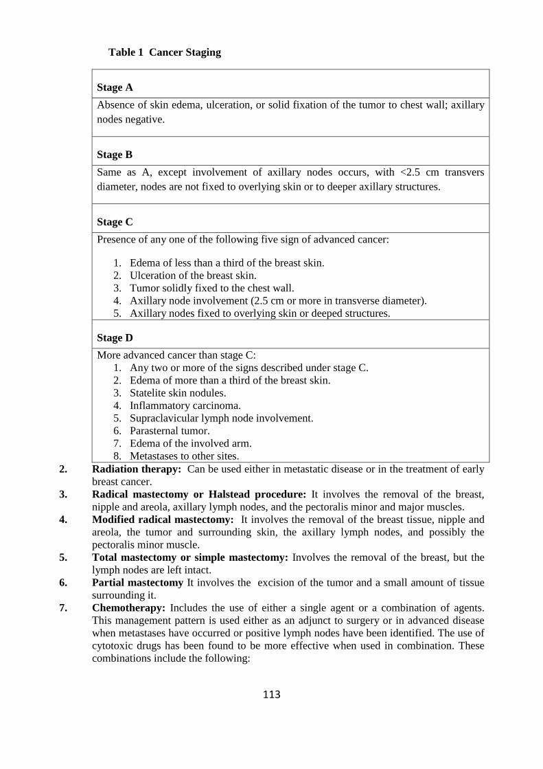
113
Table 1 Cancer Staging
Stage A
Absence of skin edema, ulceration, or solid fixation of the tumor to chest wall; axillary nodes negative.
Stage B
Same as A, except involvement of axillary nodes occurs, with <2.5 cm transvers diameter, nodes are not fixed to overlying skin or to deeper axillary structures.
Stage C
Presence of any one of the following five sign of advanced cancer:
1. Edema of less than a third of the breast skin. 2. Ulceration of the breast skin. 3. Tumor solidly fixed to the chest wall. 4. Axillary node involvement (2.5 cm or more in transverse diameter). 5. Axillary nodes fixed to overlying skin or deeped structures.
Stage D
More advanced cancer than stage C: 1. Any two or more of the signs described under stage C. 2. Edema of more than a third of the breast skin. 3. Statelite skin nodules. 4. Inflammatory carcinoma. 5. Supraclavicular lymph node involvement. 6. Parasternal tumor. 7. Edema of the involved arm. 8. Metastases to other sites.
2. Radiation therapy: Can be used either in metastatic disease or in the treatment of early breast cancer.
3. Radical mastectomy or Halstead procedure: It involves the removal of the breast, nipple and areola, axillary lymph nodes, and the pectoralis minor and major muscles.
4. Modified radical mastectomy: It involves the removal of the breast tissue, nipple and areola, the tumor and surrounding skin, the axillary lymph nodes, and possibly the pectoralis minor muscle.
5. Total mastectomy or simple mastectomy: Involves the removal of the breast, but the lymph nodes are left intact.
6. Partial mastectomy It involves the excision of the tumor and a small amount of tissue surrounding it.
7. Chemotherapy: Includes the use of either a single agent or a combination of agents. This management pattern is used either as an adjunct to surgery or in advanced disease when metastases have occurred or positive lymph nodes have been identified. The use of cytotoxic drugs has been found to be more effective when used in combination. These combinations include the following:
114
Cyclophosphamide, methotrexate, and fluorouracil: Used in metastatic or ٱrecurrent disease.
.Cyclophosphamide, doxorubicin, and fluorouracil: Used in metastatic disease ٱ8. Hormonal therapy: The hormone used depends on whether the tumor is estrogen-
receptor-positive or estrogen-receptor-poor, and it is also used in advanced disease as adjuvant therapy for stage II breast. Some of the hormonal agents used include: .Tamoxifen citrate: An anti-estrogen used in estrogen-receptor-positive tumors ٱ Diethylstilbestrol: Used in women at least 5 years postmenopausal with ٱ
estrogen-receptor-poor. Fluoxymesterone: Used in women at least 5 years postmenopausal with ٱ
estrogen-receptor-poor tumors who also have bone metastasis and estrogen failure.
D. Nursing Diagnoses and Interventions
Fear related to the possibility of cancer and its treatment.
Desired outcome: Patient freely expresses fears and anxieties and relates increasing psychologic comfort.
1. Assess the patient’s understanding of the potential diagnosis and treatment plan; clarify and explain as appropriate.
2. Provide time for patient to express feelings and fears. 3. Evaluate the patient’s emotional status, and explore with the patient what her breasts
mean to her. The breast may represent nurturance, sexuality, femininity, and desirably. 4. Assess your own feelings about the diagnosis of cancer and the psychologic meaning of
the breast. Your attitude may be reflected in the patient’s care, and therefore a positive attitude is essential for optimal patient support.
5. Provide a nonthreatening, relaxed atmosphere for the patient and SOs by using therapeutic communication techniques such as open-ended questions and reflection.
Ineffective individual coping related to depression secondary to diagnosis of breast cancer.
Desired outcomes: Patient demonstrates comfort with expressing her feelings, identifies positive coping patterns, and accepts the support of others.
1. Assist patient with developing a support system. 2. If an extensive mastectomy was performed, recognize the signs of grief, such as denial,
anger, withdrawal, or inappropriate affect. Provide emotional support, and describe the stages of grief to the patient and SOs.
Potential impairment of physical mobility related to risk of alterations in the upper extremity secondary to postmastectomy complication.
Desired outcomes: Patient complies with the therapeutic regiment to prevent joint contractures, lymphedema, and infection, and does not experience impaired mobility in the involved upper extremity.
115
1. Develop an individualized exercise program specific to the patient’s needs that can be implemented as soon as the patient returns from the recovery room.
2. Passive ROM can be initiated in the recovery room, and you can teach the patient assisted ROM on the involved shoulder as soon as she returns to her room. Encourage finger, wrist, and elbow movement to aid circulation and help minimize edema.
3. Encourage progressive exercise by having the patient use the affected arm for personal hygiene and ADLs the morning after surgery. Other exercises (clasping the hands behind the head and “walking” the fingers up the wall) should be added as soon as patient is ready. Once the sutures have been removed (usually around the 2nd postoperative week), patient should begin exercises that enhance external rotation and abduction of the shoulder. Before hospital discharge, the patient should be able to achieve maximum shoulder flexion by touching her fingertips together behind her back.
4. Assist patient with ambulation until her gait is normal. Encourage correct posture, with the back straight and shoulders back.
5. To minimize the risk of lymphedema and/or infection, avoid giving injections, measuring BP, or taking blood samples from the affected arm. Remind the patient about her lowered resistance to infection
6. Advise patient to wear a Medic-Alert bracelet that cautions against injections and tests in the involved arm.
7. To pretect the hand and arm from injury, advise the patient to wear a protective glove when gardening or doing chores the require exposure to harsh chemicals such as cleaning fluids.
Disturbance in self-concept related to altered body image secondary to loss of a breast.
Desired outcome: Patient demonstrates movement toward acceptance of the loss of a breast. 1. Recognize that loss of a breast is perceived in different ways by different women. It is
frequently more traumatic for the young adult. 2. Provide emotional support by being with the patient when the surgical dressing is
removed. 3. As appropriate, explain that sexual dysfunction often occurs after mastectomy and that it
can be minimized with resumption of sexual relations as soon as the pain has decreased. 4. provide the patient with a breast prosthesis after surgery to help her feel “normal.” A
temporary prosthesis, made of nylon and filled with Dacron fluff, can be worn until the incision heals.
5. Be aware that use of touch often enhances the patient’s self-concept. 6. Provide information and answer questions about breast reconstruction. Alteration in comfort: Pain related to the surgical procedure Desired outcome: Patient expresses a reduction in discomfort and does not exhibit signs of uncontrolled pain.
1. Assess and document the location, quality, and duration of the pain. 2. Medicate patient with the prescribed analgesics before the pain becomes too severe.
Evaluate and document the response. 3. Reassure patient that phantom breast sensations are normal. 4. Provide a comfortable in-bed position, and support the affected arm with pillows.
116
5. Encourage movement of the fingers on the affected arm to increase circulation. Inform the patient that although progressive exercise will cause some discomfort, it will aid in the mobility of the affected arm and enhance recovery.
6. Reassure the patient that exercise movements will be adapted to her level of tolerance. 7. If appropriate, instruct the patient in relaxation techniques and use of guided imagery. 8. Provide distraction, such as television, radio, or books. 9. Use touch to help relieve tension, for example, by giving a gentle massage. E. Patient–Family Teaching and Discharge Planning Provide patient and SOs with verbal and written information for the following:
1. Medications, including drug name, purpose, dosage, schedule, precautions, and potential side effects.
2. Type and dates of follow-up treatment. 3. Resumption of sexual activity, which usually can occur as soon as pain is diminished. 4. Care of the incision site, including cleansing. Explain the components of good hygiene. 5. Progressive exercise regimen, which should be continued at home. 6. Informing health care professionals to avoid measuring BP or giving injections in the
affected arm. 7. Indicators of infection and the importance of reporting them to MD. 8. Performing monthly BSE. 3.3 Neoplasms of the Female Pelvis
3.3.1 Cancer of the Cervix
Cervical cancer is one of the most frequently occurring cancers in women. Although the cause is unknown, the following risk factors have been associated with this disease: early age of first coitus, early age of first pregnancy, multiple pregnancies, and intercourse with males who practice poor hygiene, multiple sexual partners, herpes-virus type II, and family history. The two types of cervical cancer are squamous cell, which is the most common, and adenocarcinoma. Preinvasive describes cancerous cells that are limited to the cervix; invasive refers to cancer that is present in the cervix, in other pelvic structures, and possibly in the lymphatic system as well. Preinvasive cancer of the cervix typically is found in women age 30 – 40; invasive cancer usually appears between the ages of 40 and 50. A. Assessment Preinvasive: Patient asymptomatic; Pap smear abnormal.
Invasive: Abnormal vaginal bleeding; persistent, watery vaginal discharge; post-coital pain and bleeding; abnormal Pap smear.
B. Diagnostic Tests 1. Pap smear: Cells are collected from the endocervix, cervix, and upper vagina with an
applicator, placed on a slide, and sent to the lab for analysis. Pap smear results are reported as follows: .Stage I: Normal; negative for malignant cells ٱ Stage II: Negative for malignant cells but containing atypical elements ٱ
(infection).
117
.Stage III: Markedly atypical cells suggestive of malignancy ٱ .Stage IV: Malignant cells probably present ٱ .Stage V: Malignancy present ٱ
2. Colposcopy: Procedure providing a three-dimensional view of the cervix and allowing for cervical staining with an iodine solution (Schiller’s test). Cells that do not absorb the stain are considered abnormal and are sent to the lab for further examination. This procedure takes approximately 20 minutes.
3. Conization biopsy: Surgical procedure performed under general anesthesia in which a cone-shaped area of the cervix is biopsied for lab analysis to determine the extent of the malignancy.
4. Lymphography: Radiologic exam, which grades the stages of cervical cancer. Chest x-ray: May reveal presence of metastasis to the lungs.
5. Staging of the disease: The following parameters are used (based on clinical classification of the International Federation of Gynecology and Obstetrics): .Stage O: Carcinoma in situ; preinvasive ٱ Stage I: Cancer cells in the cervix only ٱ .Stage IA: Cervical cancer with <3 mm spread ٱ .Stage IB: Cervical cancer with definite invasive areas ٱ .Stage II: Cancer involving cervix and vagina but not the pelvic wall ٱ .Stage IIA: No involvement of uterine tissue ٱ .Stage IIB: Involvement of uterine tissue ٱ .Stage III: Involvement of the pelvic wall or lower third of the vagina ٱ Stage IIIA: No extension onto the pelvic wall ٱ Stage IIIB: Extention onto pelvic wall and/or kidney secondary to hydronephrosis ٱ
(obstructed flow of urine to kidney producing kidney atrophy). .Stage IV: Cancer in bladder, rectum, and other organs of the pelvis ٱ .Stage IVA: Metastasis to recturm, bladder ٱ .Stage IVB: Metastasis to distant organs ٱ
C. Medical Management and Surgical Interventions 1. External radiation therapy: all stages of cancer are treated with this therapy, 2. 2. Radium implants: Used to destroy cervical cancer that is graded stage II – IV. A combination of external radiation and implants gives the best therapeutic results. While the implant is in place, the patient is kept on strict bed rest, has an indwelling catheter, is on a low-residue diet, and is given analgesia (usually non-narcotic) and diphenoxylate hydrochloride with atropine sulfate (Lomotil) or paregoric to control diarrhea, which often occurs. After the implant has been removed, the patient is allowed to ambulate.
3. Hysterectomy: Often the treatment of choice. The uterus and cervix are removed surgically via an abdominal or vaginal approach
D. Nursing Diagnoses and Interventions Alteration in comfort: Pain related to surgery or radiation implant.
Potential fluid volume deficit related to abnormal loss secondary to postoperative or postimplant bleeding.
Potential alteration in pattern of urinary eliminat ion related to inadequate intake or obstruction of indwelling catheter.

118
Disturbance in self-concept related to altered body image and role performance expectations secondary to diagnosis of cancer.
For other interventions, see similar nursing diagnosis with patient with breast cancer
3.3.2 Ovarian Tumors
The etiology of ovarian tumors is unknown, but there is a strong relationship between their development and familial incidence. They can occur in females of all ages, but are most common between the ages of 20 and 45. If left untreated, the disease can spread through the lymphatic system and into the bloodstream, or it can proliferate by peritoneal seeding, in which malignant cells spread to peritoneal lymph nodes. Benign ovarian tumors seen most frequently include fibromas, which are found most often in postmenopausal women, and cystadenomas, which are the most common ovarian tumor, occurring in 70% of cases.
Malignant solid ovarian tumors are rarely diagnosed early because the patient tends to be asymptomatic. Survival rate is low because the tumor is usually not detected until an advanced stage. These tumors are the most lethal type of gynecologic cancer.
A. Assessment Benign ovarian tumors: Abdominal enlargement and complaints of abdominal fullness.
Solid ovarian tumors: Abdominal enlargementand pressure; pelvic pressure and discomfort.
Clinical manifestations for both classifications: Amenorrhea, postmenopausal vaginal bleeding, and other menstrual irregularities; urinary frequency and urgency; constipation.
Physical exam: Abdominal distention; an enlarged ovary, which is highly suspicious in postmenopausal women.
B. Diagnostic Tests 1. Abdominal x-ray: Will reveal presence of an ovarian tumor. 2. Ultrasound of the abdomen: Will reveal an ovarian mass. 3. Culdoscopy: 4. Laparoscopy: 5. Cytologic examination of the pelvic washings/ascites: May show presence of malignant
cancerous cells. 6. Staging of ovarian tumors (dependent on surgical exploration): The following
parameters are used (based on clinical classification of the International Federation of Gynecology and Obstetrics):
.Stage I: Tumors limited to the ovaries ٱ .Stage IA: Growth limited to one ovary; no ascites ٱ .Stage IB: Tumors limited to both ovaries; no ascites ٱ Stage II: Tumors involving one or both ovaries with presence of malignant cells ٱ
in pelvic organs. Stage IIA: Involvement of uterus and/or fallopian tubes with presence of ٱ
malignant cells. .Stage IIB: Presence of malignant cells in other pelvic tissues ٱ
119
Stages III: Tumors involving one or both ovaries with metastasis outside the ٱpelvis and/or positive retroperitioneal nodes. Tumor limited to retroperitoneal lymph nodes. Malignant cells found in small bowel omentum.
Stage IV: Tumors involving one or both ovaries with metastasis to distant ٱorgans.
C. Medical Management and Surgical Interventions 1. Wedge resection: Surgical procedure in which a benign tumor is removed, leaving
normal ovarian tissue. It is done under general anesthesia using an abdominal vaginal approach.
2. Salpingo-oophorectomy: Removal of the ovary and fallopian tube on the affected side. It is performed under general anesthesia using an abdominal approach. Solid tumors of the ovary are treated with this procedure.
3. Total hysterectomy and bilateral salpingo-oophorectomy: Performed if both ovaries are affected. If the disease has invaded other abdominal organs,
4. radiation therapy 5. Chemotherapy: Used when the disease has spread to distant organs. Chemotherapeutic
medications include doxorubicin, platinol, and cyclophosphamide. Chemotherapy is a palliative rather than curative measure.
D. Nursing Diagnoses and Interventions Alteration in comfort, , Potential fluid volume deficit , Potential alteration in pattern of urinary elimination, and Disturbance in self-concept, .
3.3.3 Endometrial Cancer
Endometrial (uterine) cancer typically occurs in postmenopausal women between the age of 50 and 70. It is a slow-growing cancer that can take up to 10 years to metastasize. Risk factors for developing uterine cancer include obesity, hypertension, diabetes mellitus, history of uterine polyps, sterility, menopausal estrogen therapy, and nulliparity; it is more common in Caucasian women. Adenocarcinama is the most common endomentrial cancer, and its growth is estrogen-dependent in the early stages. The tumor can be found in any location within the uterus and it is considered cancer in situ. The invasive stages of uterine cancer are not estrogen-dependent and can spread to the vagina, pelvic lymph nodes, ovaries, lungs, brain, and bones. Recurrence most frequently is seen in the vagina. When an early diagnosis is made the prognosis is good, with successful treatment occurring in 80 – 90% of the cases.
A. Assessment Clinical manifestations: Abnormal bleeding, including spotting; watery, unpleasant serosanguineous discharge.
Physical exam: Presence of a palpable uterine mass, uterine polyps; obvious increase in uterine size in advanced disease.
B. Diagnostic Tests 1. Dilation and curettage (D&C): Surgical procedure in which the cervical opening is
widened by a dilating instrument and the uterine lining is scraped with a currette to obtain a specimen for examination.
120
2. Endometrial biopsy: Procedure in which a specimen is obtained from the endometrial surface for biopsy. In premenopausal women, it is obtained 12 hours after the onset of the menses because the cervix is easier to enter at that time.
3. Chest x-ray: To detect metastatasis to the lungs. 4. Proctosigmoidoscopy: Visualizes the distal sigmoid colon, rectum, and anal canal with
an endoscope, allowing biopsy of tissue for detecting metastasis to pelvic structures. 5. Cystoscopy: Visualizes the bladder via the insertion of a cystoscope through the urethra.
A biopsy is done to detect cancerous cells in the bladder. 6. IVP: To rule out spread of disease to other organs. 7. Staging of the disease: The following parameters are used (based on clinical
classification of the International Federation of Gynecology and Obstetrics): .Stage O: Carcinoma in situ: Histologic findings suggestive of malignancy ٱ .Stage I: Carcinoma confined to the corpus of the uterus ٱ Stage IA: Cancer measuring 8 cm in length from the external os to the upper ٱ
point of the uterus. Stage IB: Cancer measuring >8 cm in length from the external os to the upper ٱ
point of the uterus. Stage II: Cancer involving the corpus and cervix but not extending beyond the ٱ
uterus. .Stage III: Cancer extending beyond the uterus but confined to the true pelvis ٱ Stage IV: Cancer extending outside the true pelvis or obviously involving the ٱ
mucosa of the bladder or rectum.
C. Medical Management and Surgical Interventions 1. Total hysterectomy with bilateral salpingo-oophorectomy: Performed in patients
who have a well-defined stage I tumor without cervical involvement. The uterus, cervix, fallopian tubes, ovaries, and a part of the vaginal cuff are removed.
2. Radical hysterectomy with bilateral pelvic lymph node dissection: Performed for patients with stage II uterine cancer.
3. Radiation therapy: Used if the cancer stage is difficult to determine. Chemotherapy: Used for advanced and recurrent disease. Chemotherapeutic drugs include doxorubicin, vincristine sulfate, cyclophosphamide, and d-actnomycin. Combinations of drugs and the dosage and length of treatment are determined by the patient’s response to treatment and the severity of resurence.
4. Progestin therapy: Used for in situ endometrial cancers (stage I and II) that are estrogen dependent, and it productces a remission in 35% of the cases. It is used as palliative treatment for stage IV cancers. Modroxyprogesterone (Depo-Provera) is the hormone that is usually used.
3.4 Disorders of the Female Pelvis
This includes disorders such as endometriosis, cystocele, rectocele, and uterine prolapse. These conditions occur when there is misplacement of structures or tissue within the female pelvis.
3.4.1 Endometriosis
Endometriosis is a condition in which endometrial tissue is present outside of the uterus. Typically it is found on the ovaries or in the peritoneal cul-de-sac. It might also be found in the vagina, vulva, uterosacral ligaments, or bowel. In extreme cases it is found in the vagina, vulva, uterosacral ligaments, or bowel. In extreme cases it is found in the lungs, bones, and other organs of the body. Endometriosis is considered a benign disease; it most often occurs in
121
nulliparous women 30–40 years of age and in those who have had their first child at a later age. Its cause is unknown
A. Assessment
Clinical manifestations: Dysmenorrhea 5–7 days before and 2–3 days after menses, hypermenorrhea (prolonged, excessive, and/or frequent menses), infertility, painful defecation during menses, sacral backache, and dyspareunia. Patient may be asymptomatic.
Physical exam: Palpation of the peritoneal cul-de-sac and ovaries may reveal presence of nodules or masses with extensive disease. Nodularity and tenderness of the uterosacral ligaments are common. The pelvic exam is performed several days before the menstrual cycle.
B. Diagnostic Tests 1. Laparoscopy: Confirms presence of endometriosis at pelvic organs by passing a lighted
instrument through an incision made near the umbilicus.
2. Culdoscopy: Also confirms the presence of endometriosis by passing a lighted instrument through the vagina to visualize the pelvic organs.
C. Medical Management and Surgical Interventions 1. Encourage pregnancy in women wishing to have children: Pregnancy softens and
atrophies the diseased areas as a result of hormone production. Pregnancy (or pseudopregnancy) stops the spread of endometriosis
2. Pharmacotherapy: Estrogens are not used because of their side effects. Danazol (400 mg bid is an androgen given to suppress ovulation and, hence, endometriosis. It is the current treatment of choice.
3. Surgical procedures: Determined by the patient’s age and desire to have children, and by extent of the disease. They are performed if medical treatment is unsuccessful.
● For women without extensive disease Cauterization is done ● For women who are not menopausal but do not wish to have children: A
hysterectomy is performed ● When there is extensive disease a total hysterectomy with bilateral salpingo-
oophorectomy is performed. The ovaries are removed because they are the hormone-carrying organs, which influence the development and progression of the disease.
3.4.2 Cytocele
A cystocele is the bulging of the posterior bladder wall into the vagina. It is caused by the stretching and tearing of the pelvic connective tissue during childbirth. Most often it occurs as a result of the delivery of a very large baby or after several deliveries. Symptoms usually do not appear until middle age.
A Assessment Clinical manifestations: Sensation of vaginal fullness, inability to empty bladder after voiding, urinary frequency, dysuria, stress incontinence, incontinence resulting from urgency, and recurrent cystitis.
122
Physical exam: Manual pelvic exam will reveal prolapsed cervix and a soft mass that bulges into the anterior vagina. The mass increases in size with straining.
B. Diagnostic Tests 1. Cystogram: May show the presence of bladder herniation.
2. Measurement of residula urine via intermittent urinary catheterization: May reveal >60 mL.
3. Urine culture and sensitivity: May reveal presence of bladder infection caused by retention.
C. Medical Management and Surgical Interventions
1. Urinary catheterization done to empty a distended bladder.
2. Antibiotics: Given if urinary retention results in an infection.
3. Estrogen therapy: Conjugated estrogen (Premarin) is sometimes given in small doses daily for 3 weeks each month in postmenopausal women to maintain hormonal levels. A lack of hormones results in weakness of the anterior vaginal wall, which allows the development of a cytocele.
4. Kegel isometric exercises: To help with bladder control. .
5. Weight control: Overweight women are encouraged to lose weight.
6. Anterior colporrhaphy: done to suspend the bladder.
7. Pessary: Used as an internal support for some patients
3.4.3. Uterine Prolapse
A uterine prolapse is a bulging of the uterus through the pelvic floor into the vagina. It results from an injury to the cervical and uterosacral ligaments, which can occur with childbirth, surgical trauma, or atrophy of the supportive tissue during menopause. A prolapse also can develop as a result of uterine tumors, diabetic neuropathy, and neurologic injury to the sacral nerves, obesity, or ascites. A prolapse will progress unless surgically repaired.
A prolapse is graded in the following way: • Grade I: Cervix remains within the vagina; the uterus partially descends into the vagina.
This is a slight prolapse. • Grade II: Cervix protrudes through the entrance to the vagina. This is a moderate
prolapse. • Grade III: Entire uterus protrudes through the entrance of the vagina with vaginal
inversion. This a severe prolapse, occurring most frequently in postmenopausal, multiparous women and often along with a rectocele, cystocele, and enterocele (a hernia containing a loop of small intestine or the sigmoid colon that bulges into the upper posterior vagina).
123
A. Assessment Clinical manifestations: Patient complaints of heaviness in the pelvis, low bachache, “dragging” sensation in the inguinal region, and involuntary loss of urine with coughing/sneezing.
Physical exam: Pelvic examination is performed with the patient either standing or supine. As patient bears down, a firm mass can be palpated in the lowers vagina. This exam also can confirm diagnosis of a rectocele and cystocele, if present.
B. Medical Management and Surgical Interventions 1. Vaginal pessary: A rubber device that is inserted into the vagina to support the pelvic
structures. It is used if there is a slight or moderate prolapse and/or if surgery is contraindicated or unwanted by patient.
2. Weight loss: If patient is obese.
3. Estrogen suppositories: To maintain tone of pelvic floor.
4. Antibiotics: If patient has a urinary tract infection.
5. High-fiber diet: To aid in bowel elimination.
6. Vaginal hysterectomy: To correct uterine prolapse. For severe prolapse with rectocele and cyscelel, a hysterectomy with an anterior/posterior colporrhaphy is performed.
3.5 Disorders and Surgeries of the Male Pelvis
3.5.1 Benign Prostatic Hypertrophy
The prostate is an encapsulated gland that surrounds the male urethra below the bladder neck and produces a thin, milky fluid during ejaculation. As a man ages, the prostate gland grows larger. Although the exact cause of the enlargement is unknown, one theory is that hormonal changes affect the estrogren–androgen balance. This noncancerous enlargement is common in men over age 50, and as many as 75% of men over the age of 70 are believed to have symptoms of prostatic enlargement.
A. Assessment Chronic indicators: Urinary frequency, hesitancy, and dribbling; decreased force of stream; nocturia; hematuria.
Acute indicators: Anuria, nausea, vomiting, abdominal tenderness, pain that is sharp and intense.
Physical exam: Bladder distention, “kettle-drum” sound with percussion over the distended bladder. Rectal exam will reveal a smooth, firm, and elastic enlargement of the prostate.
B. Diagnostic Tests 1. Urinalysis tests the integrity of urinary function and urine culture and sensitivity verifies
presence of infection; the results will specify the type of organism and determine the most effective antibiotic.
124
2. Phenolsulfonphthalein (PSP): Tests adequacy of renal blood flow and tubular function. PSP is injected into a vein and urine is collected via indwelling catheterization at least three times post-injection. Note: Because high fluid intake is necessary for patients undergoing this test, closely monitor patients who have cardiac or renal insufficiencies.
3. CBC, other blood tests: Results might indicate mild anemia from local bleeding, elevated creatine if renal function has been affected, and elevated blood urea nitrogen (BUN) if renal function is compromised.
4. Cystoscopy: To visualize the prostate gland, estimate its size, and ascertain the presence of any damage to the bladder wall secondary to an enlarged prostate.
5. Intravenous pyelography (IVP)/excretory urogram: Evaluates the structure and function of the kidney, ureters, and bladder, and reveals calculi if they are present.
C. Medical Management and Surgical Interventions 1. Catheterization: To relieve urinary retention. 2. Antibiotics and antimicrobial agents: To treat infection, if one is present. 3. Antiandrogen (estrogen) therapy: May be initiated to lower the levels of testosterone
if this is the cause of the prostate’s enlargement. Occasionally, and orchiectomy is performed for the same purpose. The patient may become impotent while on estrogen therapy; however, an orchiectomy will not affect the patient’s ability to have intercourse.
4. Reducing prostatic congestion via rectal massage of the prostate gland: This is performed only if there is substantial congestion. Hot sitz baths are also prescribed to relieve congestion.
5. Restricting rapid intake of fluids: Particularly alcohol, which can result in episodes of acute urinary retention from loss of bladder tone secondary to rapid distention.
6. Prostatectomy: Removal of enlarged prostatic tissue. Transurethral resection of the prostate (TURP): Prostatic tissue is scraped away ٱ
via cystoscopy. This is the most common approach, especially in patients who are poor surgical risks. It is done under spinal anesthesia.
Suprapubic transvesical prostatectomy: Prostatic tissue is removed via incision ٱhigh in the bladder (abdominal approach). This is indicated for a large prostate that cannot be removed transurethrally.
Retropubic extravesical prostatectomy: Prostatic tissue is removed via low ٱabdominal incision without entry into he bladder.
D. Nursing Diagnoses and Interventions Knowledge deficit: Potential for infection/shock after cystoscopy or TURP.
Desired outcome: Patient and/or SOs can verbalize knowledge of the indicators of shock/infection and are aware of the need to alert staff immediately should they occur.
1. Patients who have had a cystoscopy and/or TURP are at increased risk for septic shock because of the surgical instrumentation. Teach the following indicators to the patient and explain the importance of alerting the staff immediately should they occur: low-grade temperature (100 – 101F), warm extremities, rapid pulse and respiration rates, restlessness, unusual anxiety, irritability, and disorientation.
125
2. Teach the following indicators of UTI: chills, fever, flank pain, diaphoresis, and cloudy and/or foul-smelling urine.
3. Explain to patient that with the first stages of septic shock the skin will remain warm, dry, and pink, but will become cool and clammy with reduced cardiac output. In addition, as shock progresses, urinary output decreases.
Potential fluid volume deficit related to abnormal loss secondary to bleeding/hemorrhage due to surgical procedure and/or pressure on the prostatic capsule.
Desired outcomes: Patient does not exhibit signs of excessive bleeding. Patient can verbalize knowledge of actions that might result in hemorrhage of the prostatic capsule.
1. Upon patient’s return from the recovery room, monitor VS q15min for first hour; if stable, check q30min for an hour and then q4h for 24 hours. Be alert to increasing pulse, decreasing BP, diaphoresis, pallor, and increasing respirations, which can occur with hemorrhage and impending shock.
2. Monitor catheter drainage closely for the first 24 hours. Watch for dark red drainage that does not lighten to reddish-pink or drainage that remains thick in consistency after irrigation, which can signal venous bleeding within the operative site.
3. Be alert to bright red, thick drainage at any time, which can occur with arterial bleeding within the operative site.
4. Do not measure temperature rectally or insert rectal tubes or enemas into the rectum. Instruct patient not to strain with bowel movements or sit for long periods of time. Any of these actions can result can result in pressure on the prostatic capsule and potentially lead to hemorrhage. Obtain prescription for and provide stool softeners or cathartics as necessary.
5. The surgeon may establish traction on the indwelling urethral catheter in the operating room to help prevent bleeding. Maintain the traction for 4 – 8 hours after surgery, or as directed.
Potential fluid volume excess: Edema related to retention secondary to administration of high volumes of irrigating fluid.
Desired outcomes: Patient does not exhibit signs of fluid overload or electrolyte imbalance, or, if they occur, they are detected and reported promptly. Patient and SOs can verbalize the indicators of fluid overload and are aware of the need to alert the staff immediately should they occur.
1. Monitor and record I&O. To determine the true amount of urinary output, subtract the amount of irrigant from the total output. Report discrepancies that indicate fluid retention.
2. Monitor the patient’s mental and motor status. Assess for the presence of muscle twitching, convulsions, and changes in mentation. These are signs of water intoxification and/or electrolyte imbalance, which can occur within 24 hours of surgery because of the high volumes of fluid that are used in irrigation. Alert patient and SOs to the potential for these indicators.
3. Promptly report indications of fluid overload and/or electrolyte imbalance to the MD.
126
Potential impairment of skin integrity related to irritation secondary to wound drainage.
Desired outcome: Patient does not exhibit evidence of skin impairment at the wound site.
1. Monitor incisional dressings frequently during the first 24 hours and change or reinforce as needed. If the incision has been made into the bladder, excoriation can result from prolonged contact of urine with the skin.
2. Use Montagomery straps rather than tape to secure the dressing. 3. If the drainage is copious after drain removal, apply a wound drainage or ostomy pouch
with a skin barrier over the incision. Use a pouch with an anti-reflux valve to prevent contamination from reflux.
Alteration in comfort: Pain related to bladder spasms.
Desired outcome: Patient relates a reduction in discomfort and does not exhibit evidence of uncontrolled pain.
1. Assess and document the quality, location, and duration of pain. 2. Medicate the patient with prescribed analgesics, narcotics, and antispasmodics as
appropriate; evaluate and document the patient’s response. 3. Provide warm blankets, heating pad to affected area, or warm baths to increase regional
circulation and relax tense muscles. 4. Teach technique for slow, diaphragmatic breathing to relax patient and help ease pain. 5. Provide backrubs and encourage use of other nonpharmacologic methods of pain relief
such as guided imagery, distraction, relaxation tapes, and soothing music. 6. Monitor for leakage around the catheter, which can signal the presence of bladder
spasms. 7. If the patient has spasms, assure him they are normal and can occur because of irritation
of the bladder mucosa by the catheter balloon or from a clot that results in backup of urine into the bladder with concomitant irritation of the mucosa. Encourage fluid intake, as this will help prevent spasms. If the MD has prescribed catheter irrigation for the removal of clots, follow instructions carefully to prevent discomfort and injury to patient.
Potential for sexual dysfunction related to fear of impotence secondary to lack of knowledge of postsurgical sexual function.
Desired outcome : Patient can verbalize knowledge of accurate information regarding sexual function.
1. Assess patient’s level of readiness to discuss sexual function; provide opportunities for patient to discuss fears and anxieties.
2. Assure patient who has had a simple prostatectomy that his ability to attain and sustain an erection is unaltered.
3. Retrograde ejaculation may occur but this ends after a few months. It does not, however, affect the ability to achieve orgasm.
4. Encourage communication between patient and his SO. 5. Be aware of your own feelings regarding sexuality.
E. Patient–Family Teaching and Discharge Planning
Provide patient and SOs with verbal and written information for the following:
127
1. Medications, including drug name, purpose, dosage, schedule, precautions, and potential side effect.
2. Care of catheters or drains if patient is discharged with them. 3. Importance of increasing dietary fiber or taking stool softeners to minimize the risk of
damage to the prostatic capsule by preventing straining with bowel movement. 4. Avoiding the following activities for the stipulated period of time, sitting for long
periods of time, heavy lifting (>5 1b), and sexual intercourse. 5. Perineal exercises to help regain urinary sphincter control.
3.5.2 Prostatitis
Prostatitis is inflammation of the prostate gland. Acute bacterial prostatitis is the form most frequently seen in the hospital setting. It is caused by the introduction of bacteria into the prostate via the bloodstream, urethra or the kidneys. The urethra is the most common avenue for introduction of bacteria, and patients undergoing uretral instrumentation such as cystoscopy or catherization are at increased risk.
A. Assessment Clinical manifestations: Chills, fever (moderate to high), urinary urgency and frequency, dysuria, perineal and low back pain, and purulent urethral discharge. Mental confusion might be seen in older patients because of decreased oxygenation in the brain due to infection.
Physical exam: Presence of tender, enlarged, boggy prostate gland palpated on rectal exam.
B. Diagnostic Tests 1. WBC count: To reveal presence of infection. 2. Urinalysis and urine culture: To detect the presence of urinary tract infection (UTI) and
identify the offending bacteria. 3. BUN and creatinine: Show evidence of renal involvement when the test results are
elevated. 4. Culture and sensitivity of the prostatic and urethral exudates: Identifies the causative
bacteria and determines the most appropriate antibiotic. C. Medical Management and Surgical Interventions 1. Pharmacotherapy: may include the following:
.Antispasmodics: Such as propantheline bromide or oxybutynin chloride ٱ .Stool softeners: To prevent pressure on the prostatic capsule from straining ٱ .Antipyretics: For fever associated with infections ٱ Urinary analgesics: Such as phenazopyridine hydrochloride to alleviate burning ٱ
with urination. Antibiotics: Often given in combination (eg, trimethoprim-sulfamethoxazole) to ٱ
control gram-positive bacteria, and gentamicin or tobramycin sulfate is used for more severe infections. If sepsis is suspected, ampicillin or amoxicillin is given.
2. Bed rest: Either strict or with bathroom privileges during the first 24 – 48 hours to relieve perineal and suprapubic pain.
3. Intravenous fluids: For hydration. 4. Serial urine cultures, urinalysis, and WBC: To monitor the infection and determine
bacterial level. 5. Sitz baths: To relax perineal muscles and reduce risk of urinary retention. 6. Suprapubic drainage system: For relief of continued urinary retention.
128
7. Restriction of sexual intercourse: During the acute phase.
3.5.3 Prostatic Neoplasm Cancer of the prostate is the second most common cancer in men over age 50. Because most prostatic neoplasms develop in the posterior portion of the gland, they can be detected in the early stages of development; therefore, rectal examinations should be a part of every man’s regular health checks after the age of 40.
A. Assessment Clinical manifestations (in the later stages of development): Dysuria, dribbling, anuria, hematuria, nocturia, burning with urination, urgency, chills, fever, and cloudy and foul-smelling urine.
Physical exam: Bladder distention; “kettle-drum” sound with percussion over distended bladder. Rectal exam may reveal a large, hard, fixed prostate with irregular nodules.
B. Diagnostic Tests 1. Urinalysis and urine culture: Verify presence of infection. 2. CBC: Results may reveal presence of marked anemia if bone marrow is being replaced
by tumor growth. 3. Creatinine and BUN: Will be elevated if renal function is compromised. 4. Serum acid phosphatase: Will be elevated if metastasis has occurred. Serum alkaline
phosphatase: Will be elevated if matastasis has spread to the bones. 5. CT scan: May show presence of metastasis to the bones. Patient might experience a
flushed or burning sensation if contrast medium is administered. C. Medical Management and Surgical Interventions 1. Staging of the disease [based on International Union Against Cancer: Tumor, Nodes,
and Metastasis (TNM) System]: .Stage I: Cancer is diagnosed by biopsy; no clinical signs or symptoms ٱ Stage II: Cancer is confined to prostatic capsule; can be felt on rectal exam as a ٱ
hard, stony mass; tumor confined to one lobe of prostate. Stage III: Cancer passes into tissues surrounding the prostate; might reach the ٱ
lymph nodes or be confined within prostatic capsule; high serum phosphatase levels indicating increased tumor activity; patient might be symptomatic, with dribbling and difficulty starting stream, and/or have recurrent cystitis and urinary retention.
;Stage IV: Cancer has spread to the bone or into lymph nodes and beyond ٱpresence of high serum phosphatase levels. Patient exhibits signs of advanced disease: uremian, anemia, anorexia, urinary retention, and possibly severe bone pain if bone metastasis has occurred.
2. External radiation therapy: Performed both for curative and palliative therapy, depending on the stage of the neoplasm. Treatment occurs overs a 6-week period, and patients can expert to remain sexually potent after treatment. This therapy also is used to shrink a tumor, thereby relieving obstruction in the urinary tract.
3. Interstitial irradiation of the prostate: Uses gold, chromium, or iodine implantation to destroy the prostate tumor at its origin.
4. Estrogen therapy: Might be initiated to reduce plasma testosterone levels, since it is believed that testosterone is involved in the development of prostate cancer. Chemotherapy: Might be used either as a curative or palliative measure.
129
5. Surgical procedures might include the following: :Radial prostatectomy with or without pelvic node dissection ٱ
D. Nursing Diagnoses and Interventions
See “Benign Prostatic Hypertrophy,”
4.0 Conclusion Factors believed to cause cancer are called carcinogens. These include chemical agents, environmental factors, dietary substances, viruses, defective genes, and hornomes. Three basic methods are used in the treatment of cancer: (1) surgery, (2) radiation therapy, and (3) chemotherapy. 5.0 Summary Cancer is characterized by abnormal, unrestricted cell proliferation. Malignant tumors invade healthy tissues and compete with normal cells for oxygen, nutrients, and physical space. The prodnosis of cancer depends on the reactions of clients, the particular diagnosis, location, stage, treatment, and effects on bodily functions.. 6.0 Tutor Marked Assignment 1. Discuss methods for diagnosing cancer. 2. Describe systems for staging and grading malignant tumors. 3. Differentiate various treatments and methods for managing cancer. 7.0 Further Reading and Other Resources • Timby K. Barbara, Jeanne C. Scherer, & Nancy E. Smith (1999). Introductory Medical-
Surgical Nursing. (7th Edition) Lippincott.
• Brunner & Suddarth’s (2004) Medical Surgical Nursing. (10th ed) Lippincott Wilkins
130
UNIT II: CARING FOR OLDER ADULTS 1.0 Introduction ………………………………………………………………………… 2.0 Objective ……………………………………………………………………………. 3.0 Main Content ………………………………………………………………………… 3.1 The Old Adult ……………………………………………………………………… 3.2 Gerontology …………………………………………………………………………. 3.3 Theories of Aging …………………………………………………………………… 3.4.1 Late Adulthood ……………………………………………………………………… 3.4.2 Social Characteristics ……………………………………………………………….. 3.4.3 Emotional Characteristics …………………………………………………………… 3.4.4 Physical Characteristics ……………………………………………………………… 3.5 The Cardiovascular System ………………………………………………………….. 3.5.1 Reproductive Changes ……………………………………………………………….. 3.5.2 The Urinary System ………………………………………………………………….. 3.5.3 The Gastrointestinal System …………………………………………………………. 3.6 Implications for Nursing …………………………………………………………….. 3.7 Physical Care ………………………………………………………………………… 4.0 Conclusion …………………………………………………………………………… 5.0 Summary …………………………………………………………………………….. 6.0 Tutor Marked Assignment …………………………………………………………… 7.0 Further Reading and Other Resources ………………………………………………...
131
1.0 Introduction Older adults play a vital role in society, the home, the workplace, and the community. They offer others with less maturity the wisdom of life experiences. Health care workers play an important role in helping older adults maintain their independence, health, and productivity. 2.0 Objective By the end of this unit you will be able to: • List five trends for which society should prepare in view of the growing population of
aging adults. • Define gerontology. • Discuss three theories of aging. • Discuss physical characteristics that are unique to the adult older than age 65. • Identify social characteristics of older adults. • Discuss factors that affect the emotional health of older adults. • Discuss ways that a nurse’s attitude concerning the aged can affect their care. 3.0 Main Content Ageism Kyphosis Antioxidants Life review Autophagocytosis Pet therapy Despair Reality orientation Ego integrity Reminiscence therapy Elder abuse Senescence Free radicals Validation therapy Gerontology 3.1 The Old Adult Senescence is the last stage in the life cycle. During senescence there is a gradual degeneration of body processes. Diet, exercise, stress reduction, and health promotion activities play a role in slowing the rate of senescence. This developmental period is subdivided into three categories: the young-old, ages 65 to 74, who are still active and vibrant; the middle-old, ages 75 to 84; and the old-old, over age of 85, who are physically frail (Fig. 8-1). 3.2 Gerontology Gerontology is the study of aging, including physiologic, psychological, and social aspects. Scientists are conducting much research to determine the actual causes of the aging processes. The goal of this extensive research is to find ways to extend both the quantity and the quality of life. 3.3 Theories of Aging It is an established fact that physical characteristics change throughout the life cycle. As one age, the hair turns gray, skin becomes wrinkled, bones demineralize, and blood vessels become hard and inelastic. What biologic mechanism triggers these changes remains a mystery. Some theories that attempt to explain the aging process include autophagocytosis, the stress response, impairment of the immune response, alterations in DNA replication, and cellular damage by free radicals. No one theory fully explains the aging process and it is likely that aging is a combination of physiologic phenomena.
132
Autophagocytosis Autophagocytosis literally means “to eat self.” This does not mean total consumption of the cell, but portions of it are consumed to reduce its size. This process is used when the cell senses that adverse conditions exist. Cell shrinkage caused by autophagocytosis contributes to the decrease in weight and height of older adults and to the atrophy of tissues and organs. Stress Response Hans Selye, proposed that physical, psychological, and social changes produce biologic stress. Regardless of the stress-producing stimulus, Selye maintained that the physiologic response, which he called general adaptation syndrome, is always the same Gerontologists hypothesize that the defense mechanisms associated with the stress response eventually weaken, leading to death. Decline of the Immune System One of the most important mechanisms for preserving health is the immune system. Scientists have discovered that as individuals age, the immune system becomes impaired and the body’s defenses are weakened and vulnerable to overwhelming infection and cancer. The autoimmune theory of aging is that the body’s defenses attack the healthy cells, advancing the aging process. Faulty DNA Replication DNA is susceptible to damaging agents such as free radicals The dysfunctional DNA is then unable to program the continued, orderly synthesis of proteins. When this happens repeatedly, fewer cells are capable of replication. Because billions of cells are damaged in the aging process, tissue disorganization occurs and leads to organ failure. Free Radicals Free radicals are unstable atoms with excessive energy capable of damaging DNA molecules. It is possible that free radicals are responsible for changes associated with aging. Chronic exposure to substances such as radiation and environmental toxins damage DNA, causing cellular changes. Healthy cells resist the damaging effects of free radicals using chemicals called antioxidants that block the chemical reactions that cause free radicals. Scientists are investigating the antioxidant properties of vitamins C and E, and provitamin A, known as beta-carotene and found in the pigments of deep yellow and dark green fruits and vegetables. 3.4 Ageism Ageism describes behaviours and beliefs that depict older adults in a negative, inaccurate, or stereotypical manner. Ageism results in the older adult being seen as a sick, feeble, rigid, disagreeable, opinionated, or demented individual who lives in the past. This view undermines the dignity of older people who sense that they are not expected to remain physically or mentally healthy. In some situations older adults are avoided, treated disrespectfully, or ignored. 3.4.1 Late Adulthood Late adulthood is the period beginning at the age of 65 and continuing until death. During late adulthood there are developmental tasks to complete. Older adults must discover activities to fill the time previously spent at work and raising families. In addition, older adults must adjust to many losses such as the loss of friends or a spouse, loss of income, loss of health and agility, and loss of independence. Aging adults must face their own death.
133
3.4.2 Social Characteristics Working and raising a family previously provided many social contacts for the older adult. Some older adults feel bored and lonely when they retire, with a loss of self-esteem because their former activities and contacts are curtailed. Dependence Many older adults find dependence on others difficult to accept, even though they recognize the need for it. Some deny that they need assisted living and insist on remaining at home even when it jeopardizes their safety. Economic Concerns Older adults face economic concerns related to decreased income. Compulsory retirement or serious and chronic illnesses are major factors contributing to financial worries. Although studies show that older workers tend to be conscientious, careful, accurate, and dependable, many employers are reluctant to hire them. Medical expenses increase for older adults, adding to economic concerns. Consequently, as older adults live longer, their money does not stretch as far. They are less able to buy material goods as costs continue to rise. 3.4.3 Emotional Characteristics Although personality continues to evolve throughout life, basic temperament is stable into old age. Thus, if an individual’s temperament was cheerful and optimistic during youth, that is usually the demeanor in old age. A bitter, complaining young person will often carry that attitude into old age. The ability to cope with the crises of diminished health, dependence, leisure time, fixed income, and alternative housing depends on the coping skills established much earlier in life. Loneliness All people have emotional needs for love, companionship, and acceptance. Hobbies or other special interests and social contacts help older adults cope. Some become involved in community volunteer work or take advantage of activities and meeting places specifically for older adults.The problem of loneliness is acute for many older people. Besides being faced with a lack of contact with former coworkers, the older adult also is faced with separation from family and friends and the death of a spouse. Depression and Suicide Many older adults experience depression. Symptoms such as fatigue, irritability, loss of interest in surroundings, decreased ability to concentrate, or feeling of worthlessness are often viewed as natural consequences of aging, and many older adults with depression receive no treatment. This is unfortunate because older adults respond well to antidepression The risk for suicide is greater in those who live alone, have health problems, or abuse alcohol. Men are more likely to actually commit suicide, but more women attempt it. Death of a spouse is also associated with an increased risk of suicide. Fear for Safety Many older adults who live alone, particularly those living in urban settings, are vulnerable to crimes against their property and person. Older adults are also the victims of fraud or con games. Older adults may become victims of elder abuse when they depend on others for care and support. Abuse may be physical, in the form of beatings; psychological, in the form of threats; social, in the form of abandonment or unreasonable confinement; and material, in the form of theft or mismanagement of money.
134
3.4.4 Physical Characteristics The nurse can expect to see a functional decline in the absence of any specific illness in the older adult. In caring for the gerontologic client, it is essential to recognize the normal physiologic variations that occur with aging. In addition, older adults often manifest diseases with milder or atypical symptoms. Musculoskeletal System The older adult continues to experience a decrease in height due to compression of the disks between the vertebrae. This, combined with a weakening of the chest muscles, causes the spine to take on an obvious thoracic curve known as kyphosis or humpback. With a change in the center of gravity, the older adult assumes a wider stance when standing and walking. The hips and the knees tend to be flexed at all times. There may be stiffness in the weight-bearing joints and limitation in their range of motion. Muscles atrophy, yet they are more clearly defined because of the loss of subcutaneous fat. There is a general, progressive loss of muscle fibers causing flabby thin muscles particularly of the arms and legs. The physical limitations seen in the elderly are the result of inactivity, not degenerative changes. Remaining as active a possible helps slow the loss of physical ability. Arthritis is the leading chronic condition that affects the musculoskeletal system among people over the age of 65. Arthritis results in joint stiffness, limitation of movement, and deformity. Osteoporosis causes a loss of bone mass, particularly in older women, and places the older adult at increased risk for spontaneous fractures. Cognition The incidence of Alzheimer’s disease increases with age. Older adults have better long-term memory than short-term memory. The older adult does not respond quickly to questions in an interview. One should not assume, however, that the individual is cognitively impaired. Diminished hearing or anxiety slows a client’s responses. Clinical depression, common among older adults, is often mistaken for dementia. Sensory-Perception Older adults wear glasses. The presbyopia (age-related loss of visual acuity for near vision) that began in the middle years becomes more pronounced. Visual acuity declines at an accelerated rate as structures of the eye degenerate. Most people over age 70 have some degree of cataract formation. Many avoid driving at night because of the difficulty in adjusting to the glare of oncoming headlights. Hearing loss also becomes more pronounced. Presbycusis (age-related hearing loss) that began in the middle years progresses in the older adult. The nurse must look for signs of hearing loss because adults do not always report hearing difficulties. Signs of hearing loss include: • Cupping the hand behind the ear. • Asking the person to repeat what was said. • Agreeing without waiting for the completion of the sentence. • Speaking in an excessively loud or quiet voice or acting indifferent. The deterioration of these two faculties, together with diminished vision, is a common cause of accidents. Consequently, many older adults are injured in falls.
135
The Integument Gradual changes in the skin and in the body’s ability to adjust to heat and cold occur with age. The skin is drier, is prone to wrinkling, and becomes thin, flaky, and susceptible to irritation. Small brown pigmented freckles called lentigines, or liver spots appear about the hands, arms, and face or other areas exposed to sun and weather. Older adults bruise easily due to capillary fragility. Diabetes and vascular disease predispose older adults to poor healing. Facial features change as the result of loss of subcutaneous tissue. Wrinkles appear and the face seems to sag. The hair becomes drier, thinner, and gray. Nails, particularly toenails, often thicken and become brittle from poor circulation to the extremities. Teeth are lost due to neglected oral hygiene. The gums recede and teeth loosen as the bones in the jaw shrink in size. The body gradually loses its ability to adjust to extremes of temperature. It is harder for older people to keep warm because their metabolism is lower. In hot weather, they do not lose heat as well as younger people because the blood vessels in their skin dilate slowly and their sweating mechanism does not function as effectively. 3.5 The Cardiovascular System Common changes in vital signs include increased blood pressure, irregular pulse, and shortness of breath on exertion. Older adults also experience postural hypotension (rapid drop in blood pressure when rising or standing from a lying or sitting position). Impaired arterial circulation makes the skin feel cool and causes the feet to appear purplish or bluish when in a sitting or standing position. Varicose veins (bulging, twisted veins in the legs) cause the lower extremities to swell and fatigue easily. 3.5.1 Reproductive Changes The breasts of older women lose their suppleness and hang flat against the chest wall. Men appear to have more prominent breasts due to a slight decrease in testosterone. Both genders remain sexually active; both require more stimulation to become aroused. The penis and testes decrease in size, whereas the prostate gland enlarges. In women, the vagina becomes shorter and narrower. Discomfort and bleeding may occur during intercourse due to diminished lubrication and thinning of the epithelium. 3.5.2 The Urinary System Both men and women develop urinary problems as they age. The bladder capacity decreases in both sexes. Men awaken at night to void because the enlarged prostate gland blocks complete emptying of urine. It becomes necessary to urinate more frequently. For women, ligaments and muscles stretched during pregnancy fail to keep the bladder suspended. As elastic tissue and pelvic floor muscles weaken, stress incontinence, leakage of urine with increased abdominal pressure during coughing, sneezing, laughing, or lifting, occurs. 3.5.3 The Gastrointestinal System Older adults have many gastrointestinal problems such as constipation, indigestion, or increased flatulence (gas). In the oral cavity, taste buds decrease in number and saliva production diminishes. The older adult is at increased risk for aspiration because food remains in the esophagus for a longer period of time and the gag reflex is weaker. The cells of the colon atrophy and peristalsis slows, resulting in constipation and flatulence.
136
3.6 Implications for Nursing Nurses will care for greater numbers of older adults in a variety of health care settings as this population continues to increase. The plan of care includes early discharge planning and appropriate use of community resources. Teaching the Older Adult Older adults retain the ability to learn new information but require more time during the teaching process. Speak slowly and distinctly if the client has a hearing deficit and ensure that the older client is wearing glasses (if necessary) and that the lighting is adequate. Provide ample opportunity for questions to clarify misunderstood information. If visual aides such as graph, pictures, or printed material are used, use materials that are clear, uncluttered, and large enough to be seen. If a hearing aid is worn make certain that the client is wearing it and that it is working properly. Depression and Suicide It is extremely important for nurses to be aware of the high incidence of depression and the potential for suicide in the older adult. Assess the older adult for signs of increased alcohol consumption, decreased interest in friends and social activities, complaints of fatigue, anger, and feelings of hopelessness. If an older adult confides suicidal thoughts, this must be reported and immediately remove any potential articles that could be used to commit suicide from the environment such as guns, razors, knives, scissors, and medications (both prescription and nonprescription). Nutrition and Dietary Modifications Nutritional deficiencies are a serious problem for the older adult. Dietary insufficiencies are related loss of or changes in teeth, boredom at eating alone, fatigue, or lack of money. Older adults benefit from a diet lower in calories but high in nutrients because absorption of the nutrients is diminished. Some older clients require a soft diet and foods that are easy to digest. Chewing is difficult for some because of diminished production of saliva or improperly fitted dentures. Many benefit from supplemental vitamins and minerals or nourishing between-meal snacks. 3.7 Physical Care The nurse helps older adults maintain their dignity and self-respect by promoting hygiene and grooming. Clients feel better about themselves when their hair is neatly arranged, their skin is clean and healthy and they are dressed attractively. Older adults must learn to adjust to their increasing physical limitations, but self-care is always encouraged. Older adults quickly lose their ability to care for themselves when someone does it for them. If the nurse performs activities of daily living for clients when they are capable of performing these activities without help, they may think they are no longer considered capable of managing such tasks. Surrendering self-care functions that help maintain their independence and contact with reality leads to unnecessary dependence on others, as well as a loss of self-esteem. Skin and Nail Care Decreased circulation to the extremities results in prolonged healing time for injuries or infections. If the skin on the legs and feet is very dry, cream or lotion is necessary after bathing. Bathing and Hygiene A daily partial bath and a biweekly tub bath are adequate because frequent bathing dries the skin. The older client must be helped getting in and out a tub. Whatever method of bathing is
137
used, care is taken to ensure privacy. Some older women have a vaginal discharge or urinary incontinence. The client is kept clean and comfortable with good perineal care and the use of disposable pads. Elimination Bowel and urinary elimination may pose problems for older adults. Frequency of urination is common. Care is taken to prevent falls when the client gets up during the night to use toilet. The call button is nearby so that the client can obtain assistance. Help the client to maintain adequate fiber and fluid . Enemas, mild laxatives, or stool softeners are ordered by the physician if other methods of relieving constipation are ineffective. Mobility Confinement in bed causes adverse effects among older people. Pressure ulcers (bedsores) are common among older adults because they have diminished subcutaneous fat. Frequent position changes and pressure – relieving devices are necessary for clients who are inactive. Muscle tone is readily lost during prolonged bed rest to prevent these complications, active or passive exercises must be promoted. Physical Coordination Some older adults are slower in their movements and responses. The thoughtful nurse prepares everything clients need for self-care and then lets them proceed at their own pace to complete those aspects of care that they can perform. Safety The nurse ensures that that the environment is safe for the older client. Beds that are left in a high position are a potential danger because older adults can misjudge the distance to the floor and fall when getting out of bed. The bed is left in the low position except when direct nursing care is administered. A dim night light helps the older person become oriented to the surroundings and prevents failing or tripping over objects when the client gets out of bed. The call button is placed with reach. Restraints increase the individual’s frustration and distress. The nurse must determine the cause of the client’s confusion and then attempts to reduce or eliminate it. Sleep Older adults usually require less sleep than younger adults. Keeping older clients awake and active during the day facilitates sleep at night. Sedatives and hypnotics, even when administered in low doses, can result in wakefulness, excitement, and confusion. The nurse can provide a warm, caffeine-free beverage, an extra blanket, or soft music to promote restful sleep. Orientation Measures to help older clients maintain contact with reality can prevent episodes of confusion. The older adult benefits from reality orientation Reality orientation involves using various techniques that reinforce the client’s awareness of the date, time, place, names, or roles of individuals involved in their care, and current events. Communication An important aspect of encouraging communication involves stimulating older adults to talk about past experiences and events. This is referred to as reminiscence therapy and is a good technique for reinforcing self-esteem. Older adults have a need to talk about past events,
138
achievements, and losses. Asking them to recall their personal history encourages communication between the older adult, health care personnel, and the family. Validation therapy, developed by Naomi Feil, is a method of communicating with the elderly who are confused, disoriented, and act out in inappropriate ways because of a permanent and progressive loss of cognitive ability. In validation therapy the nurse seeks to reassure the client and gain understanding of the client’s behavior or words spoken. For example, the women who cares for and nurtures a doll may need validation of her role as a By seeking to validate feeling, the nurse provides comfort and affirming feelings. 4.0 Conclusion • Because more adults are living longer, society must prepare for a larger population of
people who will enjoy a longer period of retirement, need alternative living arrangements, be dependent on limited incomes, have more chronic disease, be at risk for injury, and require more assistance with their physical care.
5.0 Summary • Older adults tend to lose height, experience problems with weight gain or loss, lose
short-term memory, have a worsening of visual and hearing deficits, develop gray hair and dry skin, have impaired circulation in their extremities, and experience urinary incontinence or retention.
• Social problems among older adults include loneliness and isolation from living a distance from their children, losing a spouse and friends through death, living alone, becoming economically impoverished, and being unable to afford increased health care costs.
• Social problems among older adults include loneliness and isolation from living a distance from their children, losing a spouse and friends through death, living alone, becoming economically impoverished, and being unable to afford increased health care costs.
6.0 Tutor Marked Assignment • Identify nursing care measures that are especially important when providing physical
care for an older adult. • Describe pet therapy, reality orientation, and reminiscence therapy. 7.0 Further Reading and Other Resources
• Timby K. Barbara, Jeanne C. Scherer, & Nancy E. Smith (1999). Introductory Medical-
Surgical Nursing. (7th Edition) Lippincott.
• Brunner & Suddarth’s (2004) Medical Surgical Nursing. (10th ed) Lippincott Wilkins
139
UNIT III: SKIN DISORDERS AND WOUND MANAGEMENT 1.0 Introduction ………………………………………………………………………….. 2.0 Objective ……………………………………………………………………………… 3.0 Main Content …………………………………………………………………………
3.1 Wounds healing by Primary Intention …………………………………………………..
3.1.1 Causes …………………………………………………………………………………...
3.1.2 Classification ……………………………………………………………………………
3.2 Managing Wound Care ………………………………………………………………….
3.2.1 Wounds healing by Primary Intention ………………………………………………….
3.2.2 Surgical and/or Traumatic Wounds Healing by Secondary Intention ……………….. 3.3. Pressure (Decubitus) Ulcers …………………………………………………………... 3.4 Skin Disorders……………………………………………………………………………. 3.4.1 Dermatitis ……………………………………………………………………………… 3.4.2 Psoriasis ………………………………………………………………………………..
3.4.3 Scabies ………………………………………………………………………………… 4.0 Conclusion …………………………………………………………………………….. 5.0 Summary ……………………………………………………………………………… 6.0 Tutor Marked Assignment ……………………………………………………………. 7.0 Further Reading and Other Resources ………………………………………………..
140
1.0 Introduction The functions of the skin include protection from infection, protection of the organs from external elements, regulation of temperature, secretion, excretion, maintenance of fluid balance, and production of vitamin D. After a burns or injury to the skin any or all of these functions may be altered. 2.0 Objective On completion of this unit,you will be able to
1. Explain how the depth and percent of burns are determined.
2. Differentiate between open and closed methods of burn wound care
3.0 Main Content
3.1 Wound healing 3.1.1 Causes Burns can be caused by chemicals, contact with hot surface, electricity, flames, flammable liquids, flashes from explorisives, radioactive materials, hot liquids, and the sun. 3.1.2 Classification This classification is used to determine the appropriate medical facility for the burn injury victim.Burn can be categorized in the following way: Major burn injury: Partial-thickness burns >25% total body surface area (TBSA) for adults and >20% for children; full-thickness burns 10% TBSA or greater;
Moderate uncomplicated burn injury: Partial-thickness burns of 15 – 20% TBSA for adults and 10 – 20% for children; less than 10% full-thickness burns
Minor burn injury: Partial-thickness burns of less than 15% TBSA for adults and less than 10% for children; burns less than 2% full thickness. A. Assessment Severity of the burn is determined by the size and depth of the involved area, age of the patient, patient’s medical history, and burn site. In addition, added complications such as infection and shock can cause tissue damage beyond that of the initial injury. Size or extent of the burn area: May be measured according to the “rule of nines.” Each area is assigned a percentage of body surface area to establish the percentage of involvement. The head and neck area is assigned 9%, each upper extremity 9%, anterior chest 18%, posterior chest 18%, each lower extremity 18%, and the genitallia 1%.
141
Depth of the burn: Also significant (see Table 10-3). The depth is not always immediately evident, even to the experienced observer. Often it will take several days for the level of injury to become apparent.
The following body systems also should be evaluated for involvement: Cardiovascular: With a burn injury involving >20% TSBA, there is a systemic response characterized by increased capillary permeability and shifting of plasma into the interstitial space. This can result in hypovolemia and edema formation. Assess for presence of disorientation, decreasing BP, tachycardia, and oliguria. Sealing of the capillary leak followed by cellular fluid mobilization back into vascular spaces with diuresis occurs 24 – 48 hours after the injury. Circumferential burns to the extremities combined with edema formation can compromise peripheral circulation. Close monitoring of peripheral pulses, skin temperature and color, and capillary refill is essential. Respiratory: Carbon monoxide poisoning can result from inhalation of smoke. Laryngeal and/or oropharyngeal edema can be present; and any carbonaceous sputum, hoarseness, singed nasal hair, smoky breath, or stridor should be observed for and documented. Chemical pneumonitis and progressive adult respiratory distress syndrome (ARDS) can occur during the first 72 hours after inhalation of toxic particles and chemicals in smoke, and pneumonia can develop from prolonged bed rest and/or bacterial invasion of damaged lung tissue. Neurologic: Alteration in status can signal presence of head trauma at the time of the accident, deficiency in circulating blood volume, hypoxin form inadequate ventilation, or impending sepsis. LOC, orientation, and pupillary reactions should be monitored routinely. Circumferential burns of the extremities and resulting edema may compromise peripheral neurologic status. Closely monitor peripheral sensorimotor function. Gastrointestinal: Diversion of blood flow to vital organs can result in paralytic ileus during the first 24 hours after the injury. Sepsis and electrolyte (potassium) shifts also can cause ileus. Routine auscultation of the abdomen for bowel sounds is important. Renal-Urinary: Urinary output should be maintained at a minimum 30 – 50 mL/h. Hydration status should be assessed routinely.
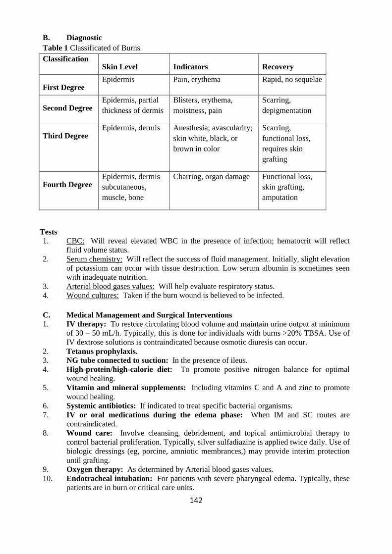
142
B. Diagnostic Table 1 Classificated of Burns Classification
Skin Level Indicators Recovery
First Degree Epidermis Pain, erythema Rapid, no sequelae
Second Degree Epidermis, partial thickness of dermis
Blisters, erythema, moistness, pain
Scarring, depigmentation
Third Degree Epidermis, dermis Anesthesia; avascularity;
skin white, black, or brown in color
Scarring, functional loss, requires skin grafting
Fourth Degree Epidermis, dermis subcutaneous, muscle, bone
Charring, organ damage Functional loss, skin grafting, amputation
Tests 1. CBC: Will reveal elevated WBC in the presence of infection; hematocrit will reflect
fluid volume status. 2. Serum chemistry: Will reflect the success of fluid management. Initially, slight elevation
of potassium can occur with tissue destruction. Low serum albumin is sometimes seen with inadequate nutrition.
3. Arterial blood gases values: Will help evaluate respiratory status. 4. Wound cultures: Taken if the burn wound is believed to be infected.
C. Medical Management and Surgical Interventions 1. IV therapy: To restore circulating blood volume and maintain urine output at minimum
of 30 – 50 mL/h. Typically, this is done for individuals with burns >20% TBSA. Use of IV dextrose solutions is contraindicated because osmotic diuresis can occur.
2. Tetanus prophylaxis. 3. NG tube connected to suction: In the presence of ileus. 4. High-protein/high-calorie diet: To promote positive nitrogen balance for optimal
wound healing. 5. Vitamin and mineral supplements: Including vitamins C and A and zinc to promote
wound healing. 6. Systemic antibiotics: If indicated to treat specific bacterial organisms. 7. IV or oral medications during the edema phase: When IM and SC routes are
contraindicated. 8. Wound care: Involve cleansing, debridement, and topical antimicrobial therapy to
control bacterial proliferation. Typically, silver sulfadiazine is applied twice daily. Use of biologic dressings (eg, porcine, amniotic membrances,) may provide interim protection until grafting.
9. Oxygen therapy: As determined by Arterial blood gases values. 10. Endotracheal intubation: For patients with severe pharyngeal edema. Typically, these
patients are in burn or critical care units.
143
D. Nursing Diagnoses and Interventions Potential alteration in respiratory function related to distress secondary to smoke inhalation and inactivity secondary to prolonged bed rest. Desired outcome: Patient does not exhibit signs of respiratory dysfunction.
1. Assess VS and auscultate lung fields for the presence of adventitious breath sounds at least q shift; notify significant findings, including crackles (rales) and rhonchi.
2. Monitor for evidence of inhalation injury: burns on the head and neck, soot in the oropharynx, singed nasal or facial hairs, carbonaceous sputum, hoarseness, and stridor.
3. Monitor arterial blood gases results, 4. Instruct patient to cough and breathe deeply q2h; assist with turning after each coughing
and deep-breathing exercise. 5. Encourage use of incentive spirometry q2h. 6. Perform tracheal suctioning as necessary. 7. Deliver oxygen as prescribed.
Alteration in tissue perfusion: Peripheral, related to edema secondary to burn injury or skin grafting.
Desired outcome: Patient does not exhibit signs of malnutrition or weight loss. 1. Monitor peripheral pulses and capillary refill, color, and skin temperature of extremities; 2. Elevate burned extremities above the level of the heart. 3. Observe for increasing edema. 4. Before patient ambulates, use elastic bandages to double wrap lower extremities that
have been burned, grafted, or used as donor sites. 5. Immobilize newly grafted areas for 3 days or as prescribed, to ensure optimum graft
adherence.
Alteration in nutrition: Less than body requirements related to increased need secondary to hypermetabolic state associated with burn injury. Desired outcome: Patient does not exhibit signs of malnutrition or weight loss.
1. Monitor weight daily; record daily calorie fluids and foods instead. 2. Provide high-protein/high-calorie diet; record daily calorie count. 3. Encourage high-calorie nutritional supplements between meals. 4. Limit intake of water and foods low in calories, promoting high-calories fluids and foods
instead. 5. In the presence of processive weight loss or slow wound healing, consult with MD and
dietitian regarding need for enteral nutritional supplement via NG tube. Potential for infection related to increased susceptibility secondary to loss of protective skin layer.
Desired outcome: Patient does not exhibit signs of infection.
144
1. Monitor and record patient’s temperature q4h. 2. If temperature is >102F, obtain prescription for culture of blood, sputum, urine, and/or
wound drainage. 3. Be alert to early signs of wound infection, including change in wound color, increased
pain, and redness at wound edges. 4. Monitor wound for evidence of cellulitis (tissue that is swollen, red, and warm to the
touch), purulent drainage, and increasing wound depth. Document and report changes 5. Use strict aseptic technique for all wound care. 6. Apply prescribed topical antimicrobial agents to wound. If silver sulfadiazide is used, it
is normal for the wound to have green-yellow drainage. 7. Shave hair from wounds and skin that is within 2 inches of wound margins. 8. Be alert to deterioration in mentation, an early indicator of sepsis. 9. Administer antibiotics as prescribed. Potential impairment of physical mobility: Contractures and/or muscles atrophy related to inactivity secondary to discomfort and prolonged bedrest.
Desired outcome: Patient does not exhibit signs of joint contractures or muscle atrophy.
1. Encourage self-care, to patient’s tolerance. 2. To prevent joint contractures and promote maximum function, ensure that patient’s joints
are extended while on bed rest. 3. Apply splints as indicated by OT/PT. 4. Provide assistive devices such as a walker and modified eating utensils as necessary for
performance of ADLs. 5. Encourage ambulation 6. Provide for active, passive, resistive, and/or assisted ROM exercises as indicated. 7. Offer prescribed pain medications one-half hour before ambulation and planned exercise
periods to enhance compliance.
Alteration in comfort: Pain related to burn injury.
Desired outcome: Patient expresses relief of discomfort and does not exhibit signs of uncontrolled pain.
1. Assist patient with using nonpharmacologic methods of pain control such as relaxation breathing, guided imagery, and soothing music.
2. Provide prescribed pain medication throughout the day, as indicated, and one-half hour before procedures that may cause discomfort.
3. Recognize that anxiety can increase the perception of pain; relieve stress by providing supportive atmosphere, informing patient of procedures and progress made, and administering prescribed antianxiety medications as indicated.
C. Patient – Family Teaching and Discharge Planning As appropriate, provide patient and SOs with verbal and written information for the following:
1. Wound care: Teach a simplified dressing procedure, or refer patient to an appropriate resource for outpatient care. Alert patient to the signs of infection, which necessitate prompt medical attention.
145
2. Skin care: Advise patient to expect itching and blistering of healed wounds. Instruct patient in the use of moisturizers sunscreen lotions that are recommended Explain the scarring process.
3. Medications: Teach patient the drug name, purpose, dosage, schedule, route, precautions, and potential side effects.
4. Use of pressure garments: Explain application and laundering procedure and importance of monitoring skin under the garments.
5. Exercise program: Stress the importance of following program outlined by PT. Remind patient that pain medications should be taken a half-hour before exercise and wound care.
6. Nutrition: Explain dietary requirements to promote healing. 7. Psychological adjustments after hospital discharge: Advise patient there is a potential
for boredom, being stared at by strangers, and dificulty with finding a role in the family again. Provide phone numbers of appropriate resource professionals.
3.2 Managing Wound Care
A wound is a disruption of tissue integrity caused by trauma, surgery, or an underlying medical disorder. Wound management is directed at preventing infection and/or deterioration in wound status and promoting healing. Wounds can heal by primary intention or by Secondary Intention
3.2.1 Wounds healing by Primary Intention
These are clean, surgical, or traumatic wounds whose edges are closed with sutures, clips or sterile tape strips Impairment of healing most frequently manifests as dehiscence, evisceration, or infection. Individuals at high risk for disruption of wound healing include those who are obese, diabetic, elderlly, malnourished, receiving steriods, or undergoing chemo-therapy or radiation therapy.
A. Assessment Optimal healing: Immediately after injury, the incision line is warm, reddened, indurated, and tender. After 1 or 2 days, wound fluid on the incision line dries, forming a scab that subsequently falls off and leaves a pink scar. After 5 – 9 days a healing ridge, a palpable accumulation of scar tissue, forms. In patients who undergo cosmetic surgery, scab formation and a healing ridge are purposely avoided to minimize scar formation.
Impaired healing: Lack of an adequate inflammatory response manifested by absence of initial redness warmth, and/or induration; continued drainage from the incision line 2 days after injury (when no drain is present); absence of a healing ridge by the 9th day after injury; and/or presence of purulent exudate.
3.2.2 Surgical and/or Traumatic Wounds Healing by Secondary Intention
Wounds healing by secondary intention are those with tissue loss and/or heavy contamination that form granulation tissue and contract in order to heal. Most often, impairment of healing is caused by infection, which results in a delay in the healing process. Individuals at risk for impaired healing include those who are obese, diabetic, malnourished, elderly, taking steroids, or undergoing radiation or chemotherapy
146
A. Assessment Optimal healing: Initially, the wound edges are inflamed, indurated, and tender. At first, granulation tissue on the floor and walls is pink, progressing to a deeper pink and then to a beefy red; it should be moist. Epithelial cells from the tissue surrounding the wound gradually migrate across the granulation tissue. As healing occurs, the wound edges become pink, the angle between surrounding tissue and the wound become less acute, and wound contraction occurs. Occasionally a wound has a tract or sinus that gradually decreases in size as healing occurs.
Impaired healing: Exudate appears on the floor and walls of the wound and does not abate as healing progresses. It is important to note the distribution, color, odor, volume, and adherence of the exudate. The skin surrounding the wound should be assessed for signs of tissue damage, including disruption, discoloration, and increasing pain. When a drain is in place, the volume, color, and odor of the drainage should be evaluated. It is also importance to note whether the drain is sutured in place.
B. Diagnostic Tests 1. WBC with differential: To assess for infection. 2. Gram stain of drainage: If infection is suspected, to identify the offending organism and
aid in the selection of preliminary antibiotics. 3. Culture and sensitivity of drainage: To determine optimal antibiotic. Infection is said to
be present when there are 105 organisms per gram of tissue and/or fever and drainage.
C. Medical Management and Surgical Interventions 1. Application of a sterile dressing in surgery: To protect wound from external
contamination, trauma, and/or provide pressure. Usually, surgeon changes the initial dressing.
2. High-calorie/high-protein diet: To promote positive nitrogen balances for optimal wound healing.
3. Multivitamins, especially C: To enhance tissue healing. 4. Minerals especially zinc and iron: May be prescribed, depending on patient’s serum
levels. 5. Insulin: As needed to control glucose levels in diabetics. 6. Local and/or systemic antibiotics: Given when infection is present and sometimes
used prophylactically as well. 7. Incision and drainage of the incision line: When infection is present and localized.
This allows healing by secondary intention. Often, the wound is irrigated with antiinfective agents
D. Medical Management and Surgical Interventions 1. Debriding enzymes: to soften and remove necrotic tissue. 2. Dressings: To provide mechanical debridement, 3. Hydrophylic agents: to remove contaminants and excess moisture. 4. Hydrotherapy: To soften and remove debris mechanically. 5. Wound irrigation with or without antiinfective agen ts: To dislodge and remove
bacteria and loosen necrotic tissue, foreign bodies, and exudate. 6. IV fluids: For patients unable to take adequate oral fluids. 7. Topical or systemic vitamin A: As needed to reverse adverse effects of steroids on
healing.
147
8. Drain(s): To remove excess tissue fluid or purulent drainage. 9. Surgical debridement: To remove dead tissue and reduce debris and fibrotic tissue. 10. Skin graft: To provide closure of wound if necessary. 11. Tissue flaps: To provide wound closure with its own blood supply. 12. Hign-protein/high-calorie diet,
3.3 Pressure (Decubitus) Ulcers
Pressure ulcers result from a disruption in tissue integrity and are most often caused by excessive tissue pressure or shearing of blood vessels. High-risk patients include the elderly and those who have decreased mobility, decreased LOC, impaired sensation, debilitation, incontinence, sepsis/elevated temperature, and/or malnutrition.
A. Assessment High-risk individuals should be identified upon admission assessment, with ongoing assessments during hospitalization. Assessment should include the patient’s LOC, ability to perform ADLs, degree of sensation and mobility, status of nutrition and continence, body temperature, and age.
When pressure ulcers are present, their severity can be graded on a scale of I to IV.
Grade I: Irregular area of soft tissue swelling, pain, erythema, and heat. Erythema is not relieved by alleviation of pressure or stimulation of local circulation. In dark-skinned individuals, heat may be the only indication of a grade I pressure ulcer.
Grade II: Full-thickness damage of the skin with heat, erythema, pain, and induration. The skin may be attached to or removed from the ulcer.
Grade III: Involves subcutaneous tissue down to fat; often infected and/or necrotic; muscle under the fat frequently inflamed; and skin surrounding the ulcer often affected. Induration and pain are present.
Grade IV: Involves extensive soft tissue damage, extending to the bone; often associated with osteomyelitis, profuse drainage, tissue necrosis, and pain. C. Medical Management and Surgical Interventions • Debriding enzymes: To soften and remove necrotic tissue. • Dressings: To provide mechanical debridement, keep healthy tissue moist, or apply an
antiinfective agent. • Hydrophylic agents: To remove contaminants and excess moisture.
• Wound irrigation with antiinfective agents: To reduce contamination. • Hydrotherapy: To soften and remove debris mechanically. • Diet: Adequate protein and calories to promote positive nitrogen balance for rapid
wound healing. • Supplemental vitamins and minerals: As needed. • Supplemental oxygen: Usually 2 – 4 L/min to promote wound healing for high-risk
patients or those with delayed wound healing.
148
• Surgical debridement: Removal of devitalized tissue with a scalpel to reduce the amount of debris and fibrotic tissue.
• Tissue flaps: Provide closure of wound as well as its own blood supply.
3.4 Skin Disorders
3.4.1 Dermatitis
Dermatitis is a general term that refers to an inflammation of the skin. It is a common sign of many skin disorders that are accompanied by a red rash. An associated symptom is pruritus, or itching. Dermatitis and pruritus may be localized or generalized. Because both are nonspecific to any one disease, it is essential that the cause is diagnosed and definitively treated. Two common conditions include allergic and irritant dermatitis.
Etiology and Pathophysiology
Allergic contact dermatitis develops in individuals who are sensitive to one or more substances, such as drugs, fibers in clothing, cosmetics, plants (eg, poison ivy), and dyes.
Medical Management
Treatment of both types of dermatitis is to remove the substances causing the reaction. This is done by flushing the skin with cool water. Topical lotions, such as calamine, or systemic drugs, such as diphenhydramine (Benadryl) or cyproheptadine (Periactin), are prescribed to relieve itching. Moisturizing creams with lanolin restore lubrication.
3.4.2 Psoriasis
Psoriasis is a chronic, noninfectious inflammatory disorder of the skin. Both men and women are affected by psoriasis. Its onset occurs during young adulthood and middle life.
Etiology and Pathophysiology
The cause of psoriasis is unknown
Assessment findings
Signs and Symptoms
Psoriasis is characterized by patches of erythema (redness) covered with silvery scales, usually on the extensor surfaces of the elbows, knees, trunk, and scalp. Itching is usually absent or slight, but occasionally it is severe. The lesions are obvious and unsightly and the scales tend to shed.
Medical management
Psoriasis has no cure. Symptomatic treatment to control the scaling and itching includes the use of topical agents such as coal tar extract, corticosteroids, or anthralin.
149
Photochemotherapy has also been used for severe, disabling psoriasis that does not respond to other methods of treatment.
3.4.3 Scabies
Scabies is a fairly common infectious skin disease.
Etiology and Pathophysiology
Scabies is caused by infestation with the itch mite (Sarcoptes scabiei). Anyone can acquire this infection; it is erroneous to assume that infected individuals have poor personal hygiene. Outbreaks are common where large groups of people are confined such as nursing homes, military barracks, prisons, boarding schools, and child care centers.
The mites are spread by skin-to-skin contact. In rare cases, scabies is acquired from handling clothing and linen in recent contact with an infected individual. Scabies mites do not survive off the body more than 2 days.
Assessment findings
Signs and Symptoms
There is intense itching especially at night. The areas that are commonly affected include the webs and sides of fingers and around the wrists, elbows, armpits, waist, thighs, genitalia, nipples, breasts, and lower buttocks. The itching is accompanied by excoriation from scratching. Skin burrows are caused by the female itch mite that invades the skin to lay her eggs.
Medical management
Scabicides, chemicals that destroy mites, such as lindane (cream or lotion), permethrin cream, and crotamiton cream or lotion, are prescribed. The medication is applied to the skin from the neck down in a thin layer, left on for 8 to 12 hours, and then removed by washing. Thorough bathing, clean clothing, and the avoidance of contact with others who have scabies are essential in preventing recurrence. 4.0 Conclusion Wound management is directed at preventing infection and/or deterioration in wound status and promoting healing. Wounds can heal by primary intention or by Secondary Intention
5.0 Summary Burns can be caused by chemicals, contact with hot surface, electricity, flames, flammable liquids, flashes from explorisives, radioactive materials, hot liquids, and the sun. A wound is a disruption of tissue integrity caused by trauma, surgery, or an underlying medical disorder. Wound management is directed at preventing infection and/or deterioration in wound status and promoting healing. Wounds can heal by primary intention or by Secondary Intention

150
6.0 Tutor Marked Assignment Describe the process of wound healing by first and second intention 7.0 Further Reading and Other Resources • Timby K. Barbara, Jeanne C. Scherer, & Nancy E. Smith (1999). Introductory Medical-
Surgical Nursing. (7th Edition) Lippincott.
• Brunner & Suddarth’s (2004) Medical Surgical Nursing. (10th ed) Lippincott Wilkins
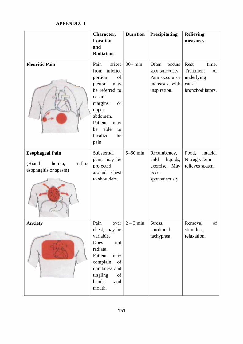
151
APPENDIX I
Character, Location, and Radiation
Duration Precipitating Relieving measures
Pleuritic Pain
Pain arises from inferior portion of pleura; may be referred to costal margins or upper abdomen. Patient may be able to localize the pain.
30+ min Often occurs spontaneously. Pain occurs or increases with inspiration.
Rest, time. Treatment of underlying cause bronchodilators.
Esophageal Pain
(Hiatal hernia, reflux esophagitis or spasm)
Substernal pain; may be projected around chest to shoulders.
5–60 min Recumbency, cold liquids, exercise. May occur spontaneously.
Food, antacid. Nitroglycerin relieves spasm.
Anxiety
Pain over chest; may be variable. Does not radiate. Patient may complain of numbness and tingling of hands and mouth.
2 – 3 min Stress, emotional tachypnea
Removal of stimulus, relaxation.
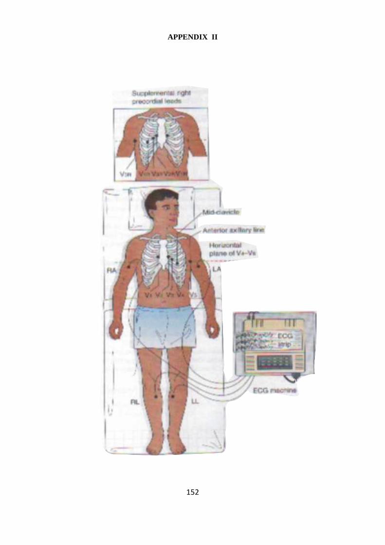
152
APPENDIX II
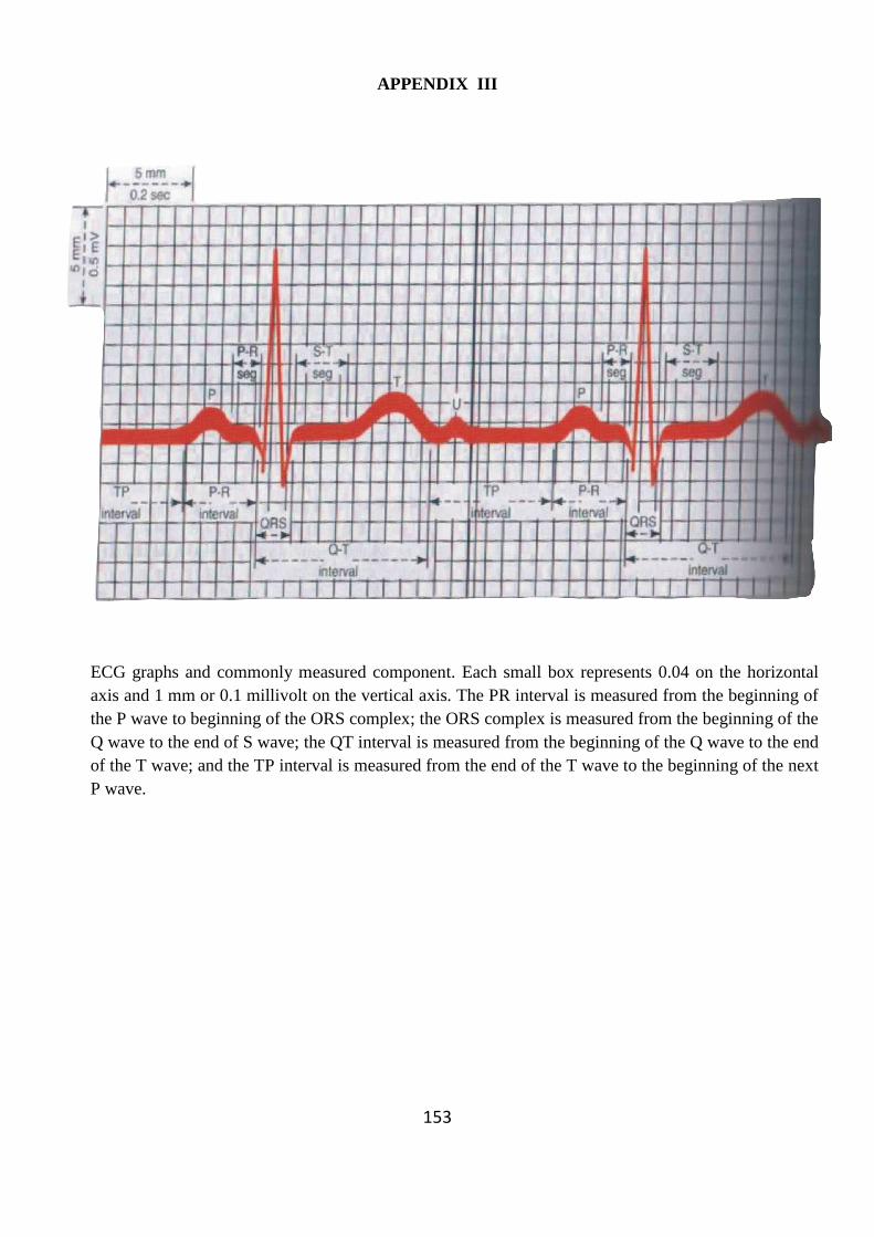
153
APPENDIX III
ECG graphs and commonly measured component. Each small box represents 0.04 on the horizontal axis and 1 mm or 0.1 millivolt on the vertical axis. The PR interval is measured from the beginning of the P wave to beginning of the ORS complex; the ORS complex is measured from the beginning of the Q wave to the end of S wave; the QT interval is measured from the beginning of the Q wave to the end of the T wave; and the TP interval is measured from the end of the T wave to the beginning of the next P wave.
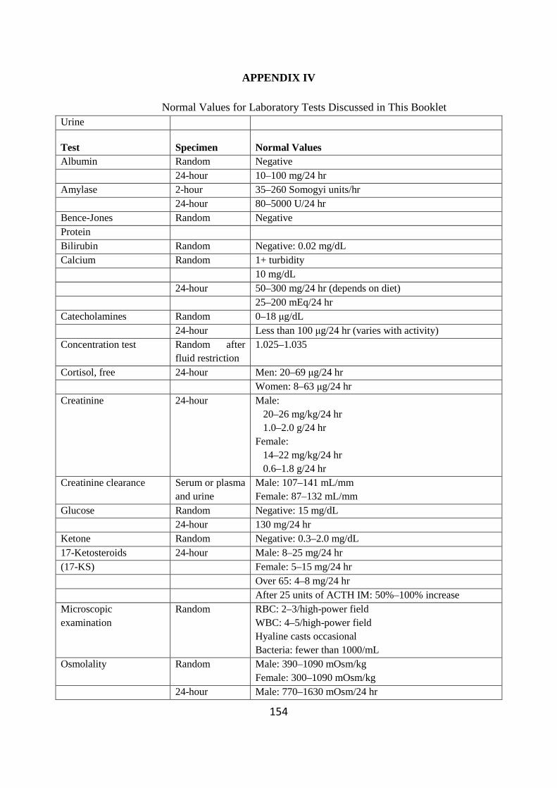
154
APPENDIX IV
Normal Values for Laboratory Tests Discussed in This Booklet Urine
Test Specimen Normal Values Albumin Random Negative 24-hour 10–100 mg/24 hr Amylase 2-hour 35–260 Somogyi units/hr 24-hour 80–5000 U/24 hr Bence-Jones Random Negative Protein Bilirubin Random Negative: 0.02 mg/dL Calcium Random 1+ turbidity 10 mg/dL 24-hour 50–300 mg/24 hr (depends on diet) 25–200 mEq/24 hr Catecholamines Random 0–18 µg/dL 24-hour Less than 100 µg/24 hr (varies with activity) Concentration test Random after
fluid restriction 1.025–1.035
Cortisol, free 24-hour Men: 20–69 µg/24 hr Women: 8–63 µg/24 hr Creatinine 24-hour Male:
20–26 mg/kg/24 hr 1.0–2.0 g/24 hr Female: 14–22 mg/kg/24 hr 0.6–1.8 g/24 hr
Creatinine clearance Serum or plasma and urine
Male: 107–141 mL/mm Female: 87–132 mL/mm
Glucose Random Negative: 15 mg/dL 24-hour 130 mg/24 hr Ketone Random Negative: 0.3–2.0 mg/dL 17-Ketosteroids 24-hour Male: 8–25 mg/24 hr (17-KS) Female: 5–15 mg/24 hr Over 65: 4–8 mg/24 hr After 25 units of ACTH IM: 50%–100% increase Microscopic examination
Random RBC: 2–3/high-power field WBC: 4–5/high-power field Hyaline casts occasional Bacteria: fewer than 1000/mL
Osmolality Random Male: 390–1090 mOsm/kg Female: 300–1090 mOsm/kg
24-hour Male: 770–1630 mOsm/24 hr
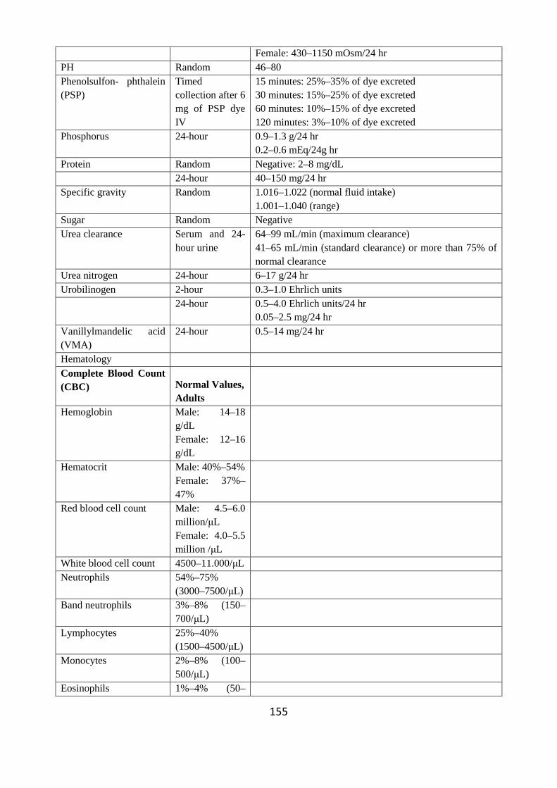
155
Female: 430–1150 mOsm/24 hr PH Random 46–80 Phenolsulfon- phthalein (PSP)
Timed collection after 6 mg of PSP dye IV
15 minutes: 25%–35% of dye excreted 30 minutes: 15%–25% of dye excreted 60 minutes: 10%–15% of dye excreted 120 minutes: 3%–10% of dye excreted
Phosphorus 24-hour 0.9–1.3 g/24 hr 0.2–0.6 mEq/24g hr
Protein Random Negative: 2–8 mg/dL 24-hour 40–150 mg/24 hr Specific gravity Random 1.016–1.022 (normal fluid intake)
1.001–1.040 (range) Sugar Random Negative Urea clearance Serum and 24-
hour urine 64–99 mL/min (maximum clearance) 41–65 mL/min (standard clearance) or more than 75% of normal clearance
Urea nitrogen 24-hour 6–17 g/24 hr Urobilinogen 2-hour 0.3–1.0 Ehrlich units 24-hour 0.5–4.0 Ehrlich units/24 hr
0.05–2.5 mg/24 hr Vanillylmandelic acid (VMA)
24-hour 0.5–14 mg/24 hr
Hematology Complete Blood Count (CBC) Normal Values,
Adults
Hemoglobin Male: 14–18 g/dL Female: 12–16 g/dL
Hematocrit Male: 40%–54% Female: 37%–47%
Red blood cell count Male: 4.5–6.0 million/µL Female: 4.0–5.5 million /µL
White blood cell count 4500–11.000/µL Neutrophils 54%–75%
(3000–7500/µL)
Band neutrophils 3%–8% (150–700/µL)
Lymphocytes 25%–40% (1500–4500/µL)
Monocytes 2%–8% (100–500/µL)
Eosinophils 1%–4% (50–
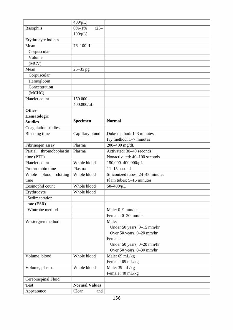
156
400/µL) Basophils 0%–1% (25–
100/µL)
Erythrocyte indices Mean 76–100 fL Corpuscular Volume (MCV) Mean 25–35 pg Corpuscular Hemoglobin Concentration (MCHC) Platelet count 150.000–
400.000/µL
Other Hematologic Studies
Specimen
Normal
Coagulation studies - Bleeding time Capillary blood Duke method: 1–3 minutes
Ivy method: 1–7 minutes Fibrinogen assay Plasma 200–400 mg/dL Partial thromoboplastin time (PTT)
Plasma Activated: 30–40 seconds Nonactivated: 40–100 seconds
Platelet count Whole blood 150,000–400,000/µL Prothrombin time Plasma 11–15 seconds Whole blood clotting time
Whole blood Siliconized tubes: 24–45 minutes Plain tubes: 5–15 minutes
Eosinophil count Whole blood 50–400/µL Erythrocyte Whole blood Sedimentation rate (ESR) Wintrobe method Male: 0–9 mm/hr Female: 0–20 mm/hr Westergren method Male:
Under 50 years, 0–15 mm/hr Over 50 years, 0–20 mm/hr Female: Under 50 years, 0–20 mm/hr Over 50 years, 0–30 mm/hr
Volume, blood Whole blood Male: 69 mL/kg Female: 65 mL/kg
Volume, plasma Whole blood Male: 39 mL/kg Female: 40 mL/kg
Cerebraspinal Fluid Test Normal Values Appearance Clear and
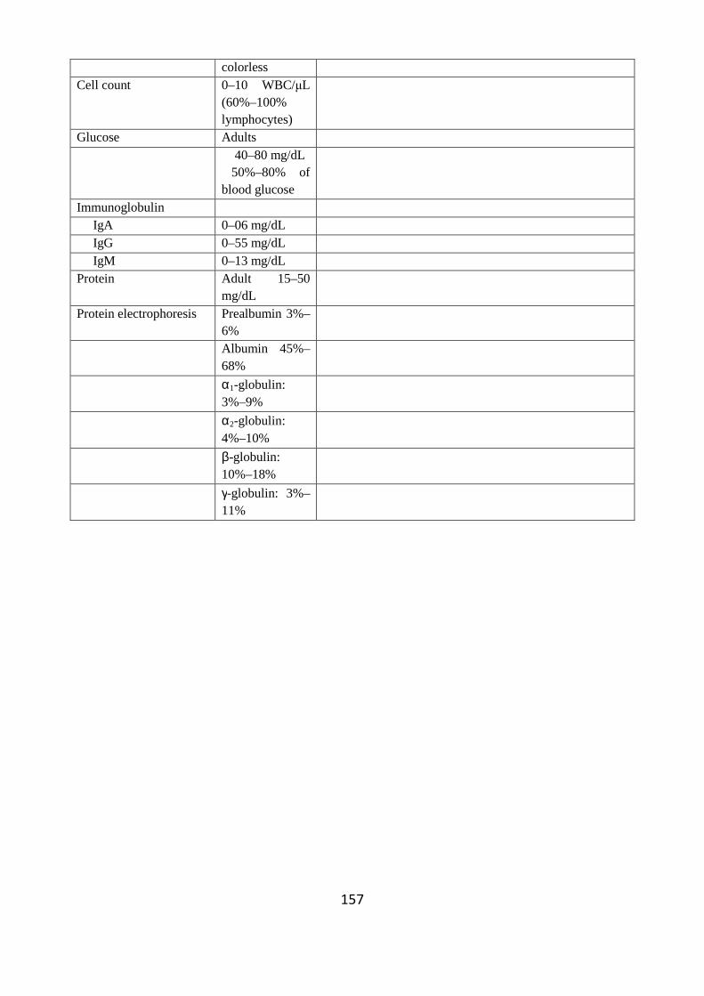
157
colorless Cell count 0–10 WBC/µL
(60%–100% lymphocytes)
Glucose Adults 40–80 mg/dL
50%–80% of blood glucose
Immunoglobulin IgA 0–06 mg/dL IgG 0–55 mg/dL IgM 0–13 mg/dL Protein Adult 15–50
mg/dL
Protein electrophoresis Prealbumin 3%–6%
Albumin 45%–68%
α1-globulin: 3%–9%
α2-globulin: 4%–10%
β-globulin: 10%–18%
γ-globulin: 3%–11%