normal sleep homeostasis and lack of epilepsy phenotype in gabaa receptor α3 subunit-knockout mice
TRANSCRIPT

NP
RDVJa
tb
vc
mU
AtlOcrnaniHrcmgc
nwfRmtasma�oorio
1
6*EAcNmsi4W
Neuroscience 154 (2008) 595–605
0d
ORMAL SLEEP HOMEOSTASIS AND LACK OF EPILEPSY
HENOTYPE IN GABAA RECEPTOR �3 SUBUNIT-KNOCKOUT MICEpll
Kat
Caemfiwtsrc2crnetu1
rilaltpanrltC
srs(ttm2
. WINSKY-SOMMERER,a A. KNAPMAN,a
. E. FEDELE,a C. M. SCHOFIELD,b
. V. VYAZOVSKIY,a1 U. RUDOLPH,c
. R. HUGUENARD,b J.-M. FRITSCHYa AND I. TOBLERa*
Institute of Pharmacology and Toxicology, University of Zurich, Win-erthurerstrasse 190, CH-8057 Zurich, Switzerland
Department of Neurology and Neurological Sciences, Stanford Uni-ersity Medical Center, Stanford, CA 94305-5122, USA
Laboratory of Genetic Neuropharmacology, McLean Hospital, Depart-ent of Psychiatry, Harvard Medical School, Belmont, MA 02478,SA
bstract—Thalamo-cortical networks generate specific pat-erns of oscillations during distinct vigilance states and epi-epsy, well characterized by electroencephalography (EEG).scillations depend on recurrent synaptic loops, which areontrolled by GABAergic transmission. In particular, GABAA
eceptors containing the �3 subunit are expressed predomi-antly in cortical layer VI and thalamic reticular nucleus (nRT)nd regulate the activity and firing pattern of neurons in relayuclei. Therefore, ablation of these receptors by gene target-
ng might profoundly affect thalamo-cortical oscillations.ere, we investigated the role of �3-GABAA receptors in
egulating vigilance states and seizure activity by analyzinghronic EEG recordings in �3 subunit-knockout (�3-KO)ice. The presence of postsynaptic �3-GABAA receptors/
ephyrin clusters in the nRT and GABAA-mediated synapticurrents in acute thalamic slices was also examined.
EEG spectral analysis showed no difference between ge-otypes during non rapid-eye movement (NREM) sleep or ataking–NREM sleep transitions. EEG power in the spindle
requency range (10 –15 Hz) was significantly lower at NREM–EM sleep transitions in mutant compared with wild-typeice. Enhancement of sleep pressure by 6 h sleep depriva-
ion did not reveal any differences in the regulation of EEGctivities between genotypes. Finally, the waking EEGhowed a slightly larger power in the 11–13-Hz band in �3-KOice. However, neither behavior nor the waking EEG showed
lterations suggestive of absence seizures. Furthermore,3-KO mice did not differ in seizure susceptibility in a modelf temporal lobe epilepsy. Strikingly, despite the disruptionf postsynaptic gephyrin clusters, whole-cell patch clampecordings revealed intact inhibitory synaptic transmissionn the nRT of �3-KO mice. These findings show that the lackf �3-GABAA receptors is extensively compensated for to
Present address: Department of Psychiatry, University of Wisconsin,001 Research Park Blvd, Madison, WI 53719, USA.Corresponding author. Tel: �41-44-635-59-57; fax: �41-44-635-57-07.-mail address: [email protected] (I. Tobler).bbreviations: ACSF, artificial cerebrospinal fluid; EEG, electroen-ephalogram; EMG, electromyogram; IR, infrared; KO, knockout;REM, non-rapid-eye movement; nRT, reticular nucleus of the thala-us; REM, rapid-eye movement; SD, sleep deprivation; sIPSCs,
pontaneous inhibitory postsynaptic currents; SWA, slow-wave activ-ty; TLE, temporal lobe epilepsy; TPMPA, 1,2,5,6-tetrahydropyridin-
i-yl)-methylphosphinic acid; VIAAT, vesicular amino acid transporter;T, wild-type.
306-4522/08$32.00�0.00 © 2008 IBRO. Published by Elsevier Ltd. All rights reseroi:10.1016/j.neuroscience.2008.03.081
595
reserve the integrity of thalamo-cortical function in physio-ogical and pathophysiological situations. © 2008 IBRO. Pub-ished by Elsevier Ltd. All rights reserved.
ey words: thalamo-cortical network, EEG rhythms, spectralnalysis, gephyrin, spike-wave discharges, sleep depriva-ion.
hanges in brain electrical activity during vigilance statesnd epilepsy are evidenced by complex rhythms in thelectroencephalogram (EEG). Frequency, amplitude andodulation of neuronal oscillations are determined by the
ring patterns and connectivity of specific neuronal net-orks (Llinas and Steriade, 2006). In particular, the func-
ional coupling of the thalamus and cerebral cortex plays atrategic role in the emergence of behaviorally relevanthythmic activities (Steriade et al., 1993) and hypersyn-hronization leading to seizures (Timofeev and Steriade,004). The function of thalamo-cortical circuits dependsritically on reciprocal synaptic loops between thalamicelay nuclei, the thalamic reticular nucleus (nRT) and theeocortex (Jones, 2002; Pinault, 2004). These reciprocalxcitatory and inhibitory connections, as well as inputs tohis network, give rise to specific oscillatory activities thatnderlie EEG rhythms (Domich et al., 1986; Steriade et al.,986; Steriade, 2003).
The nRT, exclusively composed of GABAergic neu-ons, plays a pivotal role in oscillatory activities by provid-ng a powerful and widespread inhibitory tone onto tha-amic relay nuclei (Huguenard and Prince, 1994b; Cox etl., 1996). The activity of nRT neurons is, in turn, modu-
ated by afferent fibers from several brain regions includinghalamic nuclei, brainstem and basal nuclei, and the mostowerful input connections arise from cortical layer VI (Liund Jones, 1999). Importantly, nRT neurons are intercon-ected via GABAergic synapses (Jones, 2002). GABAA
eceptor-mediated currents in both the nRT and relay tha-amic nuclei are critical in modulating neuronal firing pat-erns in thalamo-cortical circuits (von Krosigk et al., 1993;ox et al., 1997).
These integrated recurrent synaptic loops enable theynchronized neuronal activity underlying major EEGhythms, notably delta waves, spindles, and the corticallow oscillation, which define non-rapid-eye movementNREM) sleep (Steriade, 2006). In contrast, abnormal ac-ivity of the thalamo-cortical network can lead for example,o the onset of spike-wave discharges that are EEG hall-arks of absence seizure episodes (for review, Steriade,003, 2005). Numerous studies have demonstrated the
mportance of nRT neurons in network desynchronization,ved.

wsKmH
Gl�rtt1SaasdvpsM2
cet2noacttGptsa�md�zlhwtat
A
MWbtuw
lra2CIbu
S
u3w1ts2twta0ea
t6Smt(as
wa�2bEf20
stNbe7MU
gWlloc1Sfi
R. Winsky-Sommerer et al. / Neuroscience 154 (2008) 595–605596
hich prevents widespread activity and subsequent hyper-ynchrony leading to seizures (Steriade et al., 1993; vonrosigk et al., 1993; Huguenard and Prince, 1994a; Hunts-an et al., 1999; Sohal et al., 2000, 2003; Sohal anduguenard, 2003).
The functional and pharmacological properties ofABAA receptors depend on their subunit composition. A
arge family of constituent subunits (�1–6, �1–3, �1–3, �,1–3, �, �, �) allows the assembly of a variety of GABAA
eceptor subtypes (Rudolph and Mohler, 2006). Strikingly,he nRT mainly expresses GABAA receptors containinghe �3 subunit (�3-GABAA receptors) (Wisden et al., 1988,992; Fritschy and Mohler, 1995; Pirker et al., 2000;tuder et al., 2006) and this receptor subtype is alsobundant in layer VI of the neocortex. Although whenssayed in whole brain this subtype represents a minorubpopulation of GABAA receptors (10–15%), it is pre-ominant in several neuronal networks (i.e. arousal acti-ating systems as well as sleep-promoting circuitries) thatlay a key role in the generation and maintenance of theleep–wake cycle (Gao et al., 1993, 1995; Fritschy andohler, 1995; Rodriguez-Pallares et al., 2001; Jones,005).
In �3-knockout (KO) mice, there is no detectablehange in the expression of other � subunit variants (Yeet al., 2005) and no replacement of �3-GABAA receptors in
he nRT by another subtype is apparent (Studer et al.,006). Therefore, we hypothesized that changes in neuro-al activity in the nRT and neocortical layer VI due to lackf �3-GABAA receptors may alter thalamo-cortical activitynd thereby result in a sleep phenotype or enhanced sus-eptibility to epileptic seizures. A recent study has shownhat genetically epilepsy-prone rats, displaying abnormalhalamic synchronization, exhibit a specific loss of �3-ABAA receptors in the nRT (Liu et al., 2007). Here, weerformed chronic EEG recordings in freely moving wild-ype (WT) and �3-KO mice to investigate alterations inleep and wakefulness and test for the presence of spike-nd-wave discharges. Next, we studied the response of3-KO mice to sleep deprivation (SD), a well-establishedethod to enhance sleep pressure and thereby uncoverifferences in sleep regulation. Finally, the susceptibility of3-KO mice to experimentally-induced recurrent focal sei-ures was investigated in a model of temporal lobe epi-epsy (TLE). To further assess potential alterations of in-ibitory synaptic transmission in the nRT, we performedhole-cell patch clamp recordings on thalamic slices ob-
ained from juvenile �3-KO mice and investigated gephyrinnd �3-GABAA receptor clustering at postsynaptic sites in
he nRT using immunofluorescence staining.
EXPERIMENTAL PROCEDURES
nimals
ice lacking the GABAA receptor �3 subunit (�3-KO) and theirT controls were maintained on either 129X1/SvJ or C57Bl/6J
ackground (see Yee et al., 2005 for characterization) and geno-yped by PCR analysis of tail biopsies. Mice were housed individ-ally with ad libitum access to food and water. The animal facility
as maintained on a 12-h light/dark cycle (light on at 9 am; �30 eux), at a constant ambient temperature (22–24 °C) and 50%elative humidity. All experimental procedures were carried out inccordance with the European Communities’ Council Directive of4 November 1986 (86/609/EEC) and were approved by theantonal Veterinary Office of Zurich or the Stanford University
nstitutional Animal Care and Use Committee. The minimum num-er of animals necessary to obtain statistically reliable data wastilized. Every effort was made to minimize animal suffering.
leep and motor activity recordings
Surgery. A first group of adult 129X1/SvJ mice (male) wassed for surgery (11–13 weeks old at surgery; �3-KO: n�12,5.8�2.0 g; WT: n�11, 31.2�1.4 g). For EEG recording, miceere implanted epidurally under deep anesthesia (ketamine00 mg/kg–xylazine 20 mg/kg, 10 ml/kg, i.p.). Gold-plated minia-ure screws (diameter 0.9 mm) were positioned on the right hemi-phere above the frontal cortex (1.5 mm anterior to bregma andmm lateral to the midline) and the parietal cortex (2 mm posterior
o bregma and 3 mm lateral to the midline). A reference electrodeas placed above the cerebellum (2 mm posterior to lambda, on
he midline). Electrodes were connected to stainless steel wiresnd fixed to the skull with dental cement. Two gold wires (diameter.2 mm) were inserted bilaterally in the neck muscles to record thelectromyogram (EMG). After 3 weeks’ recovery, the mice weredapted for at least 3 days to the recording conditions.
EEG recording. Continuous EEG-EMG recordings were ob-ained throughout 48 h. A 24-h baseline recording was followed by
h SD starting at light onset, and the subsequent 18 h recovery.D was performed by introducing a variety of objects (e.g. nestingaterial, pieces of wood) into the cage, as well as by gently
apping on the cage whenever a mouse appeared to be drowsyTobler et al., 1997). The mice were under constant observationnd motor activity was continuously recorded by an infrared (IR)ensor placed above the cage during the two experimental days.
Data acquisition and analysis. The EEG and EMG signalsere amplified (amplification factor approx. 2000), conditioned bynalog filters (high-pass filter: �3 dB at 0.016 Hz; low-pass filter:3 dB at 40 Hz, less than �35 dB at 128 Hz.) sampled with56 Hz, digitally filtered (EEG: low-pass FIR filter 25 Hz; EMG:and-pass FIR 20–50 Hz) and stored with a resolution of 128 Hz.EG power spectra were computed for consecutive 4-s epochs by a
ast Fourier transform routine within the frequency range of 0.25–5 Hz. Between 0.25 and 5 Hz, the 0.25 Hz bins were added to yield.5 Hz bins, and between 5.25 and 25 Hz to yield 1 Hz bins.
Based on the raw parietal and frontal EEG, the correspondinglow-wave activity (SWA), as well as the raw and integrated EMG,hree vigilance states were visually scored for 4-s epochs asREM sleep, rapid-eye movement (REM) sleep and waking (To-ler et al., 1997). Epochs containing artifacts were identified andxcluded from spectral analysis (% of recording time: �3-KO:.5�1.7; WT: 6.4�2.3%). Data analysis was carried out using theATLAB software package (The Math Works, Inc., Natick, MA,SA).
Motor activity. Motor activity was also recorded in a secondroup of mice (�12 weeks old; male; n�9 �3-KO mice; n�10T). These mice did not undergo EEG-EMG surgery and were
ittermates of the mice included in the sleep experiment. After ateast 10 days’ adaptation, motor activity was recorded continu-usly for 10 days via an IR sensor placed above the cage. Activityounts were stored in 1-min epochs (Tobler et al., 1996) and0-day mean activity profiles were computed (Stanford Softwareystems, Chronobiology Kit, Stanford, CA, USA). Rest was de-ned as the amount of 1-min epochs where activity counts
qualed zero.
cpnTfabwlr6fcw0‘racurpN
I
We(and(
rbiede2aacI2fmcaoia
I
Wdtpppcat
i
ptmsSp2wajEGgmad
E
TmediNetwi(2ia
wliavueomSsiHmipg(pE(s(wwMtusdp
R. Winsky-Sommerer et al. / Neuroscience 154 (2008) 595–605 597
Statistics. One-way ANOVA factor ‘genotype’ was used toompare baseline spectra (0.25–25 Hz) during the 12-h lighteriod of the baseline between WT and �3-KO mice. When sig-ificance was reached, post hoc unpaired t-tests were performed.he EEG spectra at transitions were computed for a specific
requency band (i.e. spindle range: 10–15 Hz; SWA: 0.75–4 Hz)s a percentage of the corresponding mean power in the sameand in NREM sleep during the 12-h baseline light period. Two-ay ANOVA with factors ‘genotype’ and ‘epoch’ was used fol-
owed by a post hoc unpaired t-test when significance waseached. All analyses of sleep are based on the 24-h baseline and-h recovery after SD. The NREM sleep and REM sleep spectraor the 2 h following SD were expressed as a percentage of theorresponding baseline hour of each individual animal. Meansere thereafter computed for each genotype. Frequency bins from.25–25 Hz were compared using a one-way ANOVA with factor
genotype’ followed by unpaired t-tests when significance waseached. The time course of SWA following SD was subjected to
one-way ANOVA factor ‘genotype’ to compare genotypes. Toompare the amount of activity and rest in the two genotypes, annpaired t-test was used. Vigilance states during baseline andecovery from SD were compared between genotypes by un-aired t-test. For statistical analysis SAS (SAS Institute, Inc., Cary,C, USA) was used. Statistical significance was set at P�0.05.
nduction of TLE model
Kainic acid injection. Under isoflurane general anesthesia,T (n�13) and �3-KO (n�14) C57BL/6J mice received a unilat-
ral stereotaxic injection of 50 nl of a 20 mM solution of kainic acidCalbiochem; San Diego, CA, USA) in saline (i.e. 1 nmol kainiccid) into the right CA1 area of the dorsal hippocampus (coordi-ates with bregma as reference: anteroposterior��1.7 mm, me-iolateral��1.6 mm, dorsoventral��1.9 mm) as describedKralic et al., 2005).
Electrode implantation and EEG recording. Spontaneousecurrent focal seizures in kainic acid–treated mice were recordedy EEG at 14 and 28 days post-injection. Sixteen mice were
mplanted immediately following kainic acid injection with a bipolarlectrode inserted into the right hippocampus at the same coor-inates as for kainic acid injection and a monopolar referencelectrode placed over the cerebellum, as described (Kralic et al.,005). The electrodes were fixed to the skull with cyanoacrylatend dental cement. EEG activities were recorded in freely movingnimals placed in a Faraday cage using a digital acquisitionomputer-based system (MP100WSW System; Biopac Systems,nc., Santa Barbara, CA, USA; six channels, sampling rate00 Hz). Before beginning EEG recordings, mice were habituatedor 1 h to the test cage. At the end of the last recording session,ice were killed by perfusion–fixation and brain sections pro-
essed for Cresyl Violet staining to assess the effects of kainiccid on the cytoarchitecture of the hippocampus and the locationf electrodes. A separate group of WT and �3-KO mice was not
mplanted with electrodes and analyzed histologically at 10, 14,nd 28 days post-kainate injection.
mmunofluorescence staining
T and �3-KO C57BL/6J male mice aged P14–P15 (n�11) wereeeply anesthetized with Nembutal (50 mg/kg, i.p.) and perfusedhrough the ascending aorta with 4% paraformaldehyde in 0.15 Mhosphate buffer (pH 7.4). Brains were postfixed overnight, cryo-rotected in sucrose, frozen and cut either transversally orarasagitally at 40 m with a sliding microtome. Sections wereollected in PBS and stored in an antifreeze solution (15% glucosend 30% ethylene glycol in 50 mM phosphate buffer, pH 7.4) prioro use.
To enhance the detection of postsynaptic proteins, free-float-
ng sections were incubated for 15 min at 37 °C in 0.15 mg/ml eepsin diluted in 0.2 M HCl (Watanabe et al., 1998). They werehen rinsed with Tris buffer and incubated overnight at 4 °C with aixture of primary antibodies against the GABAA receptor �3
ubunit (Fritschy and Mohler, 1995), gephyrin (mAb7a, Synapticystems, Göttingen, Germany), and vesicular amino acid trans-orter (VIAAT, Synaptic Systems) diluted in Tris buffer containing% normal goat serum and 0.2% Triton X-100. Sections were thenashed and incubated for 30 min at room temperature in second-ry antibodies (anti–guinea pig, anti-mouse, and anti-rabbit) con-
ugated with different fluorochromes (Alexa488, Molecular Probes,ugene, OR, USA; Cy3 and Cy5, Jackson Immunoresearch, Westrove, PA, USA). Sections were then washed again, mounted onelatin-coated slides, air-dried and coverslipped with fluorescenceounting medium (Dako, Glostrup, Denmark). Sections from WTnd mutant mice were processed in parallel under identical con-itions to minimize variability in staining intensity.
x vivo electrophysiology
halamic slices were obtained from WT and �3-KO C57BL/6Jice (n�8 and n�6 mice respectively; postnatal days P12–P15;ither gender). Mice were anesthetized with pentobarbital andecapitated. The brain was rapidly removed and transferred into
ce-cold solution containing (in mM): 234 sucrose, 11 glucose, 24aHCO3, 2.5 KCl, 1.25 NaH2PO4, 10 MgSO4, and 0.5 CaCl2,quilibrated with 95% O2/5% CO2. Horizontal slices (275–300 mhickness) containing the ventrobasal thalamus and adjacent nRTere obtained with a vibratome. Thalamic slices were transferred
nto a chamber with artificial cerebrospinal fluid (ACSF) containingin mM): 126 NaCl, 26 NaHCO3, 2.5 KCl, 1.25 NaH2PO4, 2 MgCl2,
CaCl2, and 10 glucose, equilibrated with 95% O2/5% CO2 andncubated at 33 °C for at least 1 h, then brought to room temper-ture before recording.
Recordings were made at room temperature (22–24 °C) usinghole-cell patch-clamp methodology. Following incubation, tha-
amic slices were transferred to the recording chamber, and heldn place by a nylon grid while continuously superfused with ACSFt a flow rate of 2 ml/min. Thalamic neurons in the nRT wereisually identified (Huntsman et al., 1999) using a fixed-stagepright microscope (Axioskop, Zeiss, Thornwood, NY, USA)quipped with an insulated 63� objective and Nomarski DICptics. Recordings were performed under voltage-clamp at �60V using a MultiClamp 700A amplifier (Molecular Devices,unnyvale, CA, USA). Recording electrodes were made of boro-ilicate glass and had a resistance of 1.8–2.8 M� when filled withntracellular solution, which contained (in mM): 135 CsCl, 10epes, 10 EGTA, 5 QX-314, 5 ATP-Mg2�; pH was 7.3 and os-olarity was adjusted to 300 mOsm with sucrose. During record-
ngs, spontaneous inhibitory postsynaptic currents (sIPSCs) wereharmacologically isolated by bath application of the ionotropiclutamate receptor blockers 6,7-dinitro-quinoxaline-2,3-dioneDNQX, 20 m, Sigma, St. Louis, MO, USA) and 2-amino-5-hosphonopentanoic acid (AP-5, 100 m, Tocris Biosciences,llisville, MO, USA). The GABAA receptor antagonists SR-95531
20 M) and bicuculline (50 M) (Sigma) were applied to abolishIPSCs. The effect of the high affinity GABAC antagonist TPMPA(1,2,5,6-tetrahydropyridin-4-yl)-methylphosphinic acid; 50 M)as also assessed. Access resistance was monitored and cellsere included for analysis only if the series resistance was �15� and the change of resistance was �25% over the course of
he experiment. Data were acquired in gap free mode at 10 kHzsing pClamp 9 (Molecular Devices) and filtered at 2 kHz. Customoftware (Detector, WinScanSelect, J. R. Huguenard) was used toetect, sort, and measure sIPSCs. At least n�50 isolated IPSCser cell were aligned and averaged to give the mean response for
ach cell.
Ui
avdIvc2Psstdf
ep
sEaTrWo‘
staa21ssrtspaS(ag
teEso(aoetf
aNshnTftiAdtbn
Fbaapit‘
R. Winsky-Sommerer et al. / Neuroscience 154 (2008) 595–605598
RESULTS
naltered EEG rhythms and sleep regulationn adult �3-KO mice
Baseline EEG power spectra. We performed spectralnalysis and compared EEG power density in the threeigilance states (i.e. waking, NREM sleep and REM sleep)uring the baseline 12-h light period between genotypes.n �3-KO mice, the waking EEG spectra exhibited higheralues compared with the WT controls in frequencies en-ompassing the spindle range (11–13 Hz) as well as at2–23 Hz (Fig. 1; one-way ANOVA factor ‘genotype’,�0.05 post hoc unpaired t-test). These changes werepecific to the frontal derivation. The NREM sleep EEGhowed no difference between genotypes in either deriva-ion (Fig. 1). Total EEG power (0.75–25 Hz) in NREM sleepid not differ significantly between the two genotypes (V2,
ig. 1. EEG power density in the three vigilance states during the 12-haseline light period. Curves represent logarithmic mean values ofbsolute power densities (n�11 WT; n�12 �3-KO mice) for the frontalnd parietal EEG in waking, NREM sleep and REM sleep. Values arelotted at the upper limit of each bin. Triangles below the curves
ndicate frequency bins differing significantly between genotypes andheir orientation the direction of the difference (one-way ANOVA factorgenotype’; P�0.05 post hoc unpaired t-test).
rontal derivation: �3-KO 4103�251, WT 4247�395; pari- ‘
tal derivation: �3-KO 6485�352, WT 7234�623; un-aired t-test P�0.76 and 0.32, respectively).
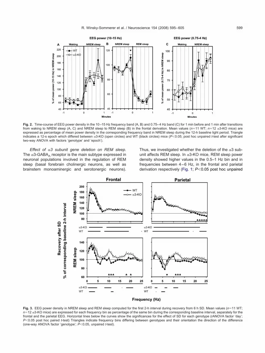
To further analyze the difference in the waking EEGpectra within the spindle frequency range, we computedEG power for the 10–15 Hz frequency band 1 min beforend after the waking-NREM sleep transitions (Fig. 2A).wo-way ANOVA (factors ‘genotype’ and ‘12-s epoch’)evealed a significant difference between �3-KO mice and
T (P�0.04), corresponding to the increase in EEG powerbserved above. However, the interaction ‘genotype’�
12-s epoch’ did not reach significance (P�0.35).Previous studies have shown that, in some mouse
trains, spindles are observed mainly in the frontal deriva-ion and are found predominantly in NREM sleep, as wells immediately before the transition between NREM sleepnd REM sleep (Valatx and Bugat, 1974; Vyazovskiy et al.,004). Therefore, we also computed EEG power (10–5 Hz) 1 min before and after transitions between NREMleep and REM sleep (Fig. 2B). The �3-KO mice did nothow the typical surge in power at the spindle frequencyange immediately before the transition from NREM sleepo REM sleep while WT mice showed a significant powerurge in the last 12-s epoch before the transition (P�0.05,ost hoc unpaired t-test when significance was reachedfter a two-way ANOVA factors ‘genotype’�‘epoch’).ince the nRT gates the transitions from wake to sleep
Steriade et al., 1993; Pinault, 2004), we investigated SWAt these transitions. No differences occurred between theenotypes (Fig. 2C).
Effects of SD on sleep EEG power density. To inves-igate whether the genotypes differ when challenged bynhancing their sleep pressure, we analyzed the sleepEG after 6 h SD. In the first 2 h recovery after SD, aignificant increase in EEG power in NREM sleep wasbserved for broad frequency ranges encompassing SWA0.75–4 Hz) and spindles (10–15 Hz) in both the frontalnd parietal derivations (Fig. 3). No significant differencesccurred between genotypes during these first 2 h recov-ry, except in high frequencies between 21 and 25 Hz inhe parietal derivation (one-way ANOVA factor ‘genotype’ollowed by unpaired t-test, P�0.05).
Time course of SWA during baseline and recoveryfter SD. To further investigate the effects of SD onREM sleep, we computed SWA, an index of sleep inten-ity (Borbély and Achermann, 2005). Previous reportsave shown that the slow and high SWA frequencies doot have an identical time course (Huber et al., 2000b).hus, we also subdivided SWA into a low and a high
requency band (0.75–2.5 Hz and 2.75–4 Hz, respec-ively). No differences were observed between genotypesn the time course of SWA in these two bands (not shown).s expected, SD induced a significant increase in SWAuring recovery in both frequency bands, in both geno-ypes and derivations, compared with the correspondingaseline values (P�0.05 post hoc paired t-test when sig-ificance was reached after rANOVA with factors ‘day’ and
interval’).
Tnsb
Tudfd
Ffei nd WT (t
FnfP(
R. Winsky-Sommerer et al. / Neuroscience 154 (2008) 595–605 599
Effect of �3 subunit gene deletion on REM sleep.he �3-GABAA receptor is the main subtype expressed ineuronal populations involved in the regulation of REMleep (basal forebrain cholinergic neurons, as well asrainstem monoaminergic and serotonergic neurons).
ig. 2. Time-course of EEG power density in the 10–15 Hz frequencyrom waking to NREM sleep (A, C) and NREM sleep to REM sleepxpressed as percentage of mean power density in the corresponding
ndicates a 12-s epoch which differed between �3-KO (open circles) awo-way ANOVA with factors ‘genotype’ and ‘epoch’).
ig. 3. EEG power density in NREM sleep and REM sleep computed�12 �3-KO mice) are expressed for each frequency bin as percentag
rontal and the parietal EEG. Horizontal lines below the curves show
�0.05 post hoc paired t-test) Triangles indicate frequency bins differing betone-way ANOVA factor ‘genotype’; P�0.05, unpaired t-test).
hus, we investigated whether the deletion of the �3 sub-nit affects REM sleep. In �3-KO mice, REM sleep powerensity showed higher values in the 0.5–1 Hz bin and inrequencies between 4–6 Hz, in the frontal and parietalerivation respectively (Fig. 1; P�0.05 post hoc unpaired
B) and 0.75–4 Hz band (C) for 1 min before and 1 min after transitionsfrontal derivation. Mean values (n�11 WT; n�12 �3-KO mice) are
y band in NREM sleep during the 12-h baseline light period. Triangleblack circles) mice (P�0.05; post hoc unpaired t-test after significant
rst 2-h interval during recovery from 6 h SD. Mean values (n�11 WT;ame bin during the corresponding baseline interval, separately for thecances for the effect of SD for each genotype (rANOVA factor ‘day’;
band (A,(B) in thefrequenc
for the fie of the sthe signifi
ween genotypes and their orientation the direction of the difference

tftfipvTa0wtsr(vsfWln(esW
awieust
wsotea�prW
mahmEtadeNae�d
Tcemmurfwwi
pkCdms1wvteslcco
i
Tr
W
N
R
evlsC(
R. Winsky-Sommerer et al. / Neuroscience 154 (2008) 595–605600
-test when significance was reached for one-way ANOVAactor ‘genotype’). Theta rhythms (4–9 Hz) characterizehe EEG during REM sleep and exploratory behavior. Aner analysis showed a faster theta-peak frequency in thearietal derivation in �3-KO mice (�3-KO: 6.31�0.03 Hzersus WT: 6.11�0.05 Hz; unpaired t-test P�0.0034).his difference was no longer observed during recoveryfter 6 h SD (�3-KO: 6.36�0.07 Hz versus WT: 6.46�.03 Hz; unpaired t-test P�0.2111). Next, we investigatedhether increased sleep pressure leads to differences in
he REM sleep EEG. We found that EEG power in REMleep showed several minor alterations in the first 2 h ofecovery following SD in both genotypes and derivationsFig. 3). The �3-KO mice displayed significantly higheralues in EEG power in the frequency bins including thepindle frequency range (10–12 Hz and 11–13 Hz in therontal and parietal derivation respectively) compared with
T control mice (Fig. 3; one-way ANOVA ‘genotype’ fol-owed by unpaired t-test P�0.05). These differences wereo longer present during the subsequent hours of recoverydata not shown). Furthermore, the number of REM sleeppisodes lasting between 28 and 60 s were slightly butignificantly more abundant in �3-KO mice compared withT mice (data not shown).
Baseline vigilance states and effect of SD. Themount of time spent in each of the three vigilance states,aking, NREM sleep, and REM sleep was compared dur-
ng baseline and during recovery after 6 h SD. No differ-nces were found between genotypes in the baseline val-es (Table 1), and the effects of SD on these vigilancetates were similar in both genotypes (Table 1; unpaired-test showed no significant difference).
able 1. Vigilance states in �3-KO and WT mice during baseline andecovery after 6 h sleep deprivation
WT �3-KO
Baseline Recovery Baseline Recovery
akingLight 328.6 (13.1) 329.4 (8.3)Dark 459.8 (10.9) 406.4 (10.6) 410.0 (9.8) 380.6 (8.4)24 h 758.4 (18.3) 739.4 (14.3)Rec 7–12 h 141.5 (6.4) 140.3 (2.9)REM sleepLight 304.3 (12.5) 299.1 (6.9)Dark 229.4 (10.4) 247.1 (10.0) 245.8 (7.0) 262.4 (7.8)24 h 533.7 (18.2) 544.9 (11.7)Rec 7–12 h 170.5 (5.0) 170.2 (3.2)EM sleepLight 87.1 (2.5) 91.5 (3.3)Dark 60.7 (2.6) 66.5 (3.4) 64.2 (3.5) 77.0 (2.6)24 h 147.9 (3.8) 155.7 (4.7)Rec 7–12 h 48.0 (2.6) 49.5 (2.1)
The amount of time spent in waking, NREM sleep and REM sleepxpressed in minutes. Mean values (�S.E.M.; n�12 �3-KO miceersus n�11 WT mice) are shown for the subdivided baseline (12-h
ight/dark phases), the entire 24 h baseline, and for recovery after 6 hleep deprivation (hours 7–12 of the recovery (Rec) 12 h light phase).omparisons between genotypes were not significant for any variable
punpaired t-test).
Motor activity. Since exploratory behavior duringakefulness affects the EEG spectrum during subsequentleep (Huber et al., 2007), we assessed whether deletionf the �3 subunit gene results in alterations of motor ac-
ivity in a familiar environment (i.e. home cage). No differ-nces were observed between genotypes in intensity ofctivity (24-h values; mean counts/active epoch�S.E.M.:3-KO: 8.9�0.3 (n�9) versus WT: 9.6�0.3 (n�10); un-aired t-test comparing 10-day means) or in the amount ofest (24-h values, min�S.E.M.: �3-KO: 1157.6�15.0 versus
T: 1205.3�17.5; unpaired t-test comparing 10-day means).
Lack of spontaneous spike-wave discharge patterns inutant mice. The lack of �3-GABAA receptors in the nRT
nd cortical layer VI may affect the tight control that preventsypersynchrony underlying spike-wave discharges, a hall-ark of epileptic seizures. Moreover, abnormalities in theEG patterns of mutant mice may in particular be detected at
he transitions between vigilance states when changes ofctivity take place in thalamic neurons. Finally, our SD para-igm may facilitate seizure episodes in mutant mice. How-ver, visual inspection of raw EEG traces during waking andREM sleep, under baseline conditions or during SD, as wells at transitions between vigilance states, did not provide anyvidence of abnormalities such as spike-wave discharges in3-KO mice. Furthermore, behavioral monitoring during SDid not reveal any abnormalities in mutant mice.
Unaltered epilepsy phenotype in adult �3-KO mice.o determine whether �3-GABAA receptors in thalamo-ortical circuits contribute to the regulation of seizures andpileptogenesis and to assess the vulnerability of �3-KOice to an excitotoxic insult, we investigated these mutantice and WT controls in a model of TLE induced by anilateral intrahippocampal injection of kainic acid. Chronicecurrent seizures were detected by EEG recordings per-ormed at 14 and 28 days post-kainate injection in 12 mice,hereas histological alterations induced by kainic acidere assessed by Nissl staining after 10, 14, and 28 days
n a total of 13 WT and 14 mutant mice.EEG recordings revealed abnormal neuronal activity
atterns, with frequent, irregular spikes at 14 days post-ainate injection, without difference between genotypes.hronic recurrent seizures recorded 2 weeks later (28ays post-kainate) also were similar in WT and mutantice (Fig. 4A), both in terms of frequency and duration of
eizures (defined here as ictal events lasting more than0 s). Likewise, the frequency of short ictal events (1–10 s)as unchanged. Examination of Nissl-stained sections re-ealed comparable histological alterations after kainatereatment in both genotypes, with extensive neurodegen-ration occurring between day 10 and day 28. At the lattertage, the injected side was characterized by extensive
oss of pyramidal cells in CA1 and CA3, as well as hilarells, and a prominent dispersion of dentate gyrus granuleells (Fig. 4B–C). These features are typical for this modelf TLE (Bouilleret et al., 2000).
Alteration of gephyrin and GABAA receptor clusteringn nRT neurons of juvenile �3-KO mice. We have re-
orted previously that gephyrin and GABAA receptor clus-
tawmwmdcrtcwssgp
hesG
F(itpsfpgfiam
Fa�prthpdcm
R. Winsky-Sommerer et al. / Neuroscience 154 (2008) 595–605 601
ering at postsynaptic sites of nRT neurons is altered indult �3-KO mice (Studer et al., 2006). To determinehether this deficit occurs secondarily due to a failure toaintain GABAergic synapses formed during ontogeny orhether synapse formation is impaired in these mutantice, the analysis was performed in juvenile mice (P15),uring the peak of synaptogenesis. Triple immunofluores-ence staining for the �3 subunit, gephyrin, and VGATevealed a normal distribution of GABAergic terminals inhe nRT from both genotypes. However, while gephyrinlusters colocalized with GABAA receptor subunit stainingere readily evident in WT mice, no such clusters wereeen in �3-KO mutants (Fig. 5A–B). Staining for the �3ubunit was not detectable, whereas a few gephyrin ag-regates were visible, but were not associated with VIAAT-
ig. 4. (A) Average (mean�SD) frequency and duration of seizuresnd average frequency of short ictal episodes in WT (n�4) and3-KO mice (n�6), as recorded by EEG 28 days after intrahip-ocampal kainic acid injection. Statistical analysis (unpaired t-test)evealed no significant differences among genotypes. (B, C) His-opathological changes induced by kainic acid injection in the dorsalippocampus, as seen by Cresyl Violet staining. Sections wererepared after completion of EEG recordings. The extent of neuro-egeneration in CA1, CA3, and hilus, and the dispersion of granuleells in the dentate gyrus, are similar in WT (B) and in mutant (C)ice. Scale bar�200 m.
ositive terminals. Similar aggregates, but of larger size,�a
ave been reported in nRT neurons of adult mice (Studert al., 2006). These results strongly suggest that the �3ubunit is required for the formation of postsynapticABAA receptor and gephyrin clusters, but does not affect
WT
α3-KO
A
BB
A’
’
α3-KO
C
WT
ED
WT WT
pA
α3-KO α3-KO
ig. 5. (A, B) Loss of GABAA receptor �3 subunit (red) and gephyringreen) clusters in �3-KO mice at the age of P15, as visualized bymmunofluorescence staining. In sections from WT mice, these clus-ers are extensively co-localized (yellow hue in A) and apposed toresynaptic GABAergic terminals (stained for VIAAT, blue, in A=). Inections from mutant mice, VIAAT-positive terminals appear unaf-ected (B=). The few remaining gephyrin clusters are not apposed toresynaptic terminals, suggesting that they are intracellular aggre-ates. Scale bar�10 m. (C–E). Whole-cell patch-clamp recordingsrom nRT neurons. (C) Representative electrophysiology trace show-ng 20 s of continuous recording from individual cells voltage-clampedt �60 mV from one WT and one �3-KO mouse. (D, E) Histograms ofean sIPSC amplitude and frequency (black bars, WT; white bars,
3-KO; n�10 nRT neurons per genotype; mean�S.E.M.). The meanmplitude was significantly larger in mutant mice (t-test; P�0.001).
ttgmg
vbiticraSc�rbTGlcns(tt�
Gteattacd(sanjpaartgpa�qp�
Pi
CiuptI(2raYGccisGatpGtnsBS�FsGsMsata2rToatepcet
sh(tpaT
R. Winsky-Sommerer et al. / Neuroscience 154 (2008) 595–605602
he morphology or distribution of GABAergic terminals inhe nRT. This effect of the mutation was cell-specific, asephyrin clustering was intact in neighboring regions thatainly express other GABAA receptor subtypes (striatum,lobus pallidus, hippocampal formation; not shown).
Ex vivo electrophysiology in juvenile �3-KO mice. Iniew of the surprisingly minor differences in the sleep EEGetween �3-KO mice and WT mice, we assessed the
mpact of �3 subunit gene deletion on inhibitory neuro-ransmission by performing whole-cell patch clamp record-ngs in nRT neurons of juvenile �3-KO mice and their WTontrols. Similar to WT mice, spontaneous IPSCs wereobustly detected in the nRT of �3-KO mice (Fig. 5C). Bathpplication of the specific GABAA receptor antagonistsR-95531 (eight cells from �3-KO mice), as well as bicu-ulline, completely abolished sIPSCs (five cells from3-KO mice), demonstrating that these events are GABAA
eceptor-mediated. Moreover, IPSCs remained intact afterath application of the high affinity GABAC antagonistPMPA, providing evidence that they are not mediated byABAC receptors (four cells from �3-KO mice). We ana-
yzed the mean IPSC response averaged from WT (10ells from eight mice) and �3-KO (10 cells from six mice)RT neurons (Fig. 5D–E). Events from �3-KO mice wereignificantly larger in amplitude compared with WT miceWT��20�2 pA versus �3-KO��55�5 pA, unpaired t-est), while the frequency of events was slightly increased,hough not significantly, in �3-KO mice (WT�2.0�0.2 Hz;3-KO�2.6�0.3 Hz).
DISCUSSION
iven the restricted expression of the �3-GABAA recep-ors in neuronal populations playing a key role in the gen-ration of brain rhythms, we expected to observe an un-mbiguous behavioral phenotype in �3-KO mice. Indeed,
he lack of �3-GABAA receptors should induce changes inhe intrinsic firing of the nRT and cortical layer VI neurons,nd subsequently alter the activity of the thalamo-corticalircuits. Paradoxically, �3-KO mice did not show any majoreficiencies in their sleep regulation or epilepsy phenotypeFigs. 1–4), suggesting potent ‘rescue’ mechanisms totabilize the activity of thalamo-cortical networks in thebsence of the main GABAA receptor expressed by nRTeurons. Furthermore, we confirmed that nRT neurons of
uvenile �3-KO mice have a deficit of gephyrin clustering atostsynaptic sites (Fig. 5A–B), as shown previously indult mice (Studer et al., 2006). Strikingly, despite thispparent disruption of the GABAergic postsynaptic appa-atus, GABAA receptor-mediated transmission was re-ained in the nRT (Fig. 5C). Altogether, our results sug-est that concerted rescue mechanisms are activated,robably during ontogeny, to ensure the stable neuronalnd network function of the thalamo-cortical circuits in3-KO mice. This homeostatic plasticity allows for ade-uate thalamo-cortical network function underlying com-lex behaviors and their associated EEG rhythms in
3-KO mice. creservation of GABAergic synaptic functionn the nRT
onsiderable evidence in vivo and in vitro indicates thatnteraction with GABAA receptors containing the �2 sub-nit is essential for proper clustering of gephyrin atostsynaptic sites. Thus, in vivo ablation of GABAA recep-ors by gene targeting can result in a complete loss ofPSCs and gephyrin clusters in various types of neuronsSchweizer et al., 2003; Fritschy et al., 2006; Kralic et al.,006; Peden et al., 2008). Likewise, suppression of gephy-in expression results in a loss of GABAA receptor clusters,s shown in vitro (Kneussel et al., 1999; Jacob et al., 2005;u et al., 2007). Morphologically, the absence of �3-ABAA receptors in nRT neurons of �3-KO mice is suffi-ient to prevent postsynaptic clustering of gephyrin, dis-losing a mandatory interaction between the receptor and
ts scaffolding protein. The preservation of bicuculline-sen-itive IPSCs in these cells most likely indicates that otherABAA receptor subtypes are present, possibly clusteredt postsynaptic sites without interacting with gephyrin. Lit-le information is available about the possible subunit com-osition of these ‘compensatory’ receptors. Reports onABAA receptor subunit expression in the nRT are par-
ially controversial; aside from the �3 and �2 subunit, theseeurons might also express the �2, �5, �1, �3 and �ubunit (Fritschy and Mohler, 1995; Pirker et al., 2000;rowne et al., 2001; Huntsman and Huguenard, 2006;tuder et al., 2006). So far, no compensation by �2, �5 orsubunit has been observed (Studer et al., 2006 and
ristchy et al., unpublished observations), but since an �ubunit variant is required for assembly of functionalABAA-receptors, the presence of low levels of these
ubunits in �3-KO nRT neurons cannot be excluded.oreover, since a targeted deletion of the �3 subunit
trongly impairs the function of nRT neurons (Huntsman etl., 1999), the ‘compensatory’ receptors are likely to con-ain this subunit. Finally, since the � and � subunit genesre frequently co-expressed with �3 (Moragues et al.,000), and are also localized on the X-chromosome, up-egulation of these subunits in �3-KO mice is conceivable.he � and � subunits represent the mammalian orthologuef the avian �4 and �4 subunit, respectively (Darlison etl., 2005). Interestingly, recombinant receptors expressinghese subunits (along with �3 and �1) have a markedlynhanced GABA sensitivity (Ranna et al., 2006). Thus, ifresent in nRT neurons of �3-KO mice, GABAA receptorsontaining �2/�5, �3, and �/� subunits might explain pres-rvation of large amplitude IPSCs in the absence of clus-ering with gephyrin.
Preservation of sIPSCs in nRT neurons of �3-KO miceuggests that a potential significance of GABAA receptoreterogeneity might be to preclude any major alterationsi.e. loss of function or ‘over-activity’) in neuronal systemshat are critical for proper brain function by allowing com-ensation by subtypes that are either absent or expressedt very low levels under normal, physiological conditions.he moderate phenotype of �3-KO mice stands in striking
ontrast with the effects of an acute pharmacological
biltbswwh
dwrfrs(tarvGptrG2
So
Tf�siNdwwmntTitaqredda�lpRit1
aatMsmba1atco
iomioatwbcipsfiwcW�isa
t�Gi2b�
Gssmrirdoc�K
R. Winsky-Sommerer et al. / Neuroscience 154 (2008) 595–605 603
lockade of GABAA receptors. Adaptive changes preserv-ng brain activity may be more efficient following a globaloss of function than subsequent to small disturbances inhe system. This aspect is illustrated by the discrepancyetween the �3-KO mice, displaying no susceptibility topike-wave seizures, and genetically epilepsy-prone rats,hich lack the �3-GABAA receptors selectively in the nRT,ithout alteration of their expression in the cortex, andave absence-like seizures (Liu et al., 2007).
It is important to note that �3-KO mice have a globaleficit of �3-GABAA receptors. The question ariseshether these receptors are also replaced in other brain
egions than the nRT, such as the cerebral cortex or basalorebrain in �3-KO mice. Results so far with other GABAA
eceptor mutant mice suggested that functional compen-ation can occur without replacement of GABAA receptorsFritschy and Panzanelli, 2006). Homeostatic plasticity of-en involves changes in intrinsic neuronal properties thatllow constant network excitability over a broad dynamicange, notably by adjusting the expression or function ofarious types of ion channels (reviewed in Marder andoaillard, 2006). For instance, an adaptive increase of theotassium ‘leak’ conductance has been shown to preservehe integrity of excitability in the cerebellum of GABAA
eceptor �6-null mice, which display a complete loss ofABAA-mediated tonic conductance (Brickley et al.,001).
ignificance of alterations in the sleep EEGf �3-KO mice
he retained GABAergic inhibition in the nRT may accountor the lack of a robust ‘sleep’ and ‘epilepsy’ phenotype in3-KO mice. With regard to sleep, we observed ratherubtle alterations in the �3-KO mice. The reduced increasen power in the spindle frequency band (10–15 Hz) at theREM–REM sleep transition in �3-KO mice is in accor-ance with a previous study showing that spindle activityas diminished in GABAA �3-KO mice (Wisor et al., 2002),hich have an impaired thalamo-cortical function (Hunts-an et al., 1999). Though spindles are produced in theRT (Fuentealba and Steriade, 2005), their synchroniza-ion is under tight control by the cortex (Steriade, 2003).hus, the lack of �3-GABAA receptors in cortical layer VI,
f not compensated for, may underlie the difference be-ween the genotypes. During waking, �3-KO mice displayn increase of EEG power density in the 11–13 Hz fre-uency band in the frontal derivation, which could haveeflected the occurrence of spike-wave discharges. How-ver, we did not observe any spontaneous spike-waveischarge episodes in the EEG of mutant mice, eitheruring baseline recordings or following a challenge, suchs SD. Several features of REM sleep were altered in3-KO mice, namely decreased parietal EEG power in the
ow theta range (4–9 Hz) associated with a faster theta-eak frequency, as well as an increased number of longerEM sleep episodes. These changes suggest alterations
n the brainstem–septo-hippocampal systems involved inhe generation of theta oscillations (Vertes and Kocsis,
997). Strikingly, basal forebrain and brainstem arousal- pctivating systems, which are involved in cortical activationssociated with REM sleep, show a predominant distribu-ion of �3-GABAA receptors (Gao et al., 1993; Fritschy andohler, 1995). Noticeably, these receptors are the main
ubtype expressed in several neuronal networks playing aajor role in REM sleep onset and maintenance, includingasal forebrain cholinergic neurons, serotonergic neurons,nd noradrenergic cells of the locus coeruleus (Gao et al.,995; Rodriguez-Pallares et al., 2001; Jones, 2005). Minorlterations in the excitability and thereby firing patterns ofhese neuronal populations, due to lack of �3-GABAA re-eptors, may underlie the subtle variations in REM sleepbserved in mutant mice.
Consistent with the lack of major difference observedn EEG spectra, we found no differences in vigilance statesr in locomotor activity in the home cage between WT andutant mice. Furthermore, SD did not uncover any prom-
nent differences between genotypes. The minor increasebserved in the NREM sleep spectrum (parietal derivation)nd in the REM sleep spectrum (both derivations) duringhe first 2 h of recovery in �3-KO mice were short lasting,hich is in contrast with the robust effect induced by SD inoth genotypes, waning only after 6 h recovery. The timeourse of recovery is in accordance with previous studies
n mice (Huber et al., 2000a,b). Moreover, in the intrahip-ocampal kainate model of TLE, �3-KO mice showed theame sensitivity as WT mice to this excitotoxic insult. Thisnding confirms that the absence of �3-GABAA receptorsas functionally compensated for in the affected neuronalircuits contributing to epileptogenesis and seizure control.e have shown previously a similar compensation in
1-KO mice, which lack a large subset of GABAA receptorsn hippocampal neurons, but do not exhibit any increasedusceptibility to kainic acid injection (Schneider Gasser etl., 2007).
The role of �3-GABAA receptors was previously inves-igated using point-mutated �3(H126R) mice in which3-GABAA receptors are diazepam-insensitive. The �3-ABAA receptors were shown not to be critical in mediat-
ng the effects of diazepam on the sleep EEG (Kopp et al.,003). In contrast, the anti-absence drug clonazepam haseen reported to suppress thalamic oscillations via the3-GABAA receptors (Sohal et al., 2003).
CONCLUSIONS
ABAA receptor subtypes are exquisitely tuned to regulatepecific brain function and behavioral states, as demon-trated by pharmacological analysis of mice carrying point-utations abrogating diazepam effects in selected GABAA
eceptors (reviewed in Rudolph and Mohler, 2004). In strik-ng contrast to these findings, selective elimination of theseeceptors by gene targeting fails, in some cases, to pro-uce a major phenotype, presumably due to the activationf compensatory mechanisms. The GABAA �1-KO miceonstitute a remarkable example. Despite the loss of1�2�2, the most abundant receptor subtype, GABAA-�1O mice were viable and only exhibited a mild behavioral
henotype (Sur et al., 2001; Kralic et al., 2002). Here, we
seurpstdtc
AdmTgFN
B
B
B
B
C
C
D
D
F
F
F
F
G
G
H
H
H
H
H
H
H
J
J
J
K
K
K
K
K
L
L
L
M
M
P
R. Winsky-Sommerer et al. / Neuroscience 154 (2008) 595–605604
how that these adaptive changes are sophisticatednough to sustain thalamo-cortical network performancenderlying complex behaviors and their associated EEGhythms in �3-KO mice. The preservation of the EEGower spectrum across the sleep–wake cycle, the lack ofpike-waves underlying absence seizures, and the unal-ered response to an excitotoxic brain lesion collectivelyocument the effectiveness of homeostatic brain plasticityo sustain complex behavioral functions under physiologi-al conditions and in response to a major challenge.
cknowledgments—We gratefully acknowledge Cornelia Schwer-el and Ruth Keist for support with breeding and genotyping ofutant mice and Corinne Sidler for excellent technical assistance.his study was supported by the European Union Marie Curierant MCRTN-CT-2004-512362, the Swiss National Scienceoundation grant 3100A0-108260 and NCCR-Neuro, and by theIH grant NS034774.
REFERENCES
orbély AA, Achermann P (2005) Sleep homeostasis and models ofsleep regulation. In: Principles and practice of sleep medicine(Kryger MH et al., eds), pp 405–417. Philadelphia: ElsevierSaunders.
ouilleret V, Loup F, Kiener T, Marescaux C, Fritschy JM (2000) Earlyloss of interneurons and delayed subunit-specific changes inGABAA-receptor expression in a mouse model of mesial temporallobe epilepsy. Hippocampus 10:305–324.
rickley SG, Revilla V, Cull-Candy SG, Wisden W, Farrant M (2001)Adaptive regulation of neuronal excitability by a voltage-indepen-dent potassium conductance. Nature 409:88–92.
rowne SH, Kang J, Akk G, Chiang LW, Schulman H, Huguenard JR,Prince DA (2001) Kinetic and pharmacological properties ofGABAA receptors in single thalamic neurons and GABAA subunitexpression. J Neurophysiol 86:2312–2322.
ox CL, Huguenard JR, Prince DA (1996) Heterogeneous axonalarborizations of rat thalamic reticular neurons in the ventrobasalnucleus. J Comp Neurol 366:416–430.
ox CL, Huguenard JR, Prince DA (1997) Nucleus reticularis neuronsmediate diverse inhibitory effects in thalamus. Proc Natl Acad SciU S A 94:8854–8859.
arlison MG, Pahal I, Thode C (2005) Consequences of the evolutionof the GABAA receptor gene family. Cell Mol Neurobiol 25:607–624.
omich L, Oakson G, Steriade M (1986) Thalamic burst patterns in thenaturally sleeping cat: a comparison between cortically projectingand reticularis neurones. J Physiol 379:429–449.
ritschy JM, Mohler H (1995) GABAA-receptor heterogeneity in theadult rat brain: differential regional and cellular distribution of sevenmajor subunits. J Comp Neurol 359:154–194.
ritschy JM, Panzanelli P (2006) Molecular and synaptic organizationof GABAA receptors in the cerebellum: Effects of targeted subunitgene deletions. Cerebellum 5:275–285.
ritschy JM, Panzanelli P, Kralic JE, Vogt KE, Sassoe-Pognetto M(2006) Differential dependence of axo-dendritic and axo-somaticGABAergic synapses on GABAA receptors containing the �1 sub-unit in Purkinje cells. J Neurosci 26:3245–3255.
uentealba P, Steriade M (2005) The reticular nucleus revisited: in-trinsic and network properties of a thalamic pacemaker. Prog Neu-robiol 75:125–141.
ao B, Fritschy JM, Benke D, Mohler H (1993) Neuron-specific ex-pression of GABAA-receptor subtypes: differential association ofthe �1- and �3-subunits with serotonergic and GABAergic neu-
rons. Neuroscience 54:881–892.ao B, Hornung JP, Fritschy JM (1995) Identification of distinctGABAA-receptor subtypes in cholinergic and parvalbumin-positiveneurons of the rat and marmoset medial septum-diagonal bandcomplex. Neuroscience 65:101–117.
uber R, Deboer T, Tobler I (2000a) Effects of sleep deprivation onsleep and sleep EEG in three mouse strains: empirical data andsimulations. Brain Res 857:8–19.
uber R, Deboer T, Tobler I (2000b) Topography of EEG dynamicsafter sleep deprivation in mice. J Neurophysiol 84:1888–1893.
uber R, Tononi G, Cirelli C (2007) Exploratory behavior, corticalBDNF expression, and sleep homeostasis. Sleep 30:129–139.
uguenard JR, Prince DA (1994a) Clonazepam suppresses GABAB-mediated inhibition in thalamic relay neurons through effects innucleus reticularis. J Neurophysiol 71:2576–2581.
uguenard JR, Prince DA (1994b) Intrathalamic rhythmicity studied invitro: nominal T-current modulation causes robust antioscillatoryeffects. J Neurosci 14:5485–5502.
untsman MM, Huguenard JR (2006) Fast IPSCs in rat thalamicreticular nucleus require the GABAA receptor �1 subunit. J Physiol572:459–475.
untsman MM, Porcello DM, Homanics GE, DeLorey TM, HuguenardJR (1999) Reciprocal inhibitory connections and network syn-chrony in the mammalian thalamus. Science 283:541–543.
acob TC, Bogdanov YD, Magnus C, Saliba RS, Kittler JT, HaydonPG, Moss SJ (2005) Gephyrin regulates the cell surface dynamicsof synaptic GABAA receptors. J Neurosci 25:10469–10478.
ones BE (2005) Basic mechanisms of sleep-wake states. In: Princi-ples and practice of sleep medicine (Kryger MH et al., eds), pp136–153. Philadelphia: Elsevier Saunders.
ones EG (2002) Thalamic circuitry and thalamocortical synchrony.Philos Trans R Soc Lond B Biol Sci 357:1659–1673.
neussel M, Brandstatter JH, Laube B, Stahl S, Muller U, Betz H(1999) Loss of postsynaptic GABAA receptor clustering in gephy-rin-deficient mice. J Neurosci 19:9289–9297.
opp C, Rudolph U, Keist R, Tobler I (2003) Diazepam-inducedchanges on sleep and the EEG spectrum in mice: role of thealpha3-GABAA receptor subtype. Eur J Neurosci 17:2226–2230.
ralic JE, Korpi ER, O’Buckley TK, Homanics GE, Morrow AL (2002)Molecular and pharmacological characterization of GABAA recep-tor �1 subunit knockout mice. J Pharmacol Exp Ther 302:1037–1045.
ralic JE, Ledergerber DA, Fritschy JM (2005) Disruption of the neu-rogenic potential of the dentate gyrus in a mouse model of tempo-ral lobe epilepsy with focal seizures. Eur J Neurosci 22:1916–1927.
ralic JE, Sidler C, Parpan F, Homanics GE, Morrow AL, Fritschy JM(2006) Compensatory alteration of inhibitory synaptic circuits incerebellum and thalamus of gamma-aminobutyric acid type A re-ceptor �1 subunit knockout mice. J Comp Neurol 495:408–421.
iu XB, Coble J, van Luijtelaar G, Jones EG (2007) Reticular nucleus-specific changes in �3 subunit protein at GABA synapses in ge-netically epilepsy-prone rats. Proc Natl Acad Sci U S A 104:12512–12517.
iu XB, Jones EG (1999) Predominance of corticothalamic synapticinputs to thalamic reticular nucleus neurons in the rat. J CompNeurol 414:67–79.
linas RR, Steriade M (2006) Bursting of thalamic neurons and statesof vigilance. J Neurophysiol 95:3297–3308.
arder E, Goaillard JM (2006) Variability, compensation and ho-meostasis in neuron and network function. Nat Rev Neurosci7:563–574.
oragues N, Ciofi P, Lafon P, Odessa MF, Tramu G, Garret M (2000)cDNA cloning and expression of a gamma-aminobutyric acid Areceptor �-subunit in rat brain. Eur J Neurosci 12:4318–4330.
eden DR, Petitjean CM, Herd MB, Durakoglugil M, Rosahl TW,Wafford K, Homanics GE, Belelli D, Fritschy JM, Lambert JJ (2008)
Developmental maturation of synaptic and extrasynaptic GABAA
P
P
R
R
R
R
S
S
S
S
S
S
S
S
S
S
S
S
T
T
T
V
V
v
V
W
W
W
W
Y
Y
R. Winsky-Sommerer et al. / Neuroscience 154 (2008) 595–605 605
receptors in mouse thalamic ventrobasal neurones. J Physiol586:965–987.
inault D (2004) The thalamic reticular nucleus: structure, function andconcept. Brain Res Brain Res Rev 46:1–31.
irker S, Schwarzer C, Wieselthaler A, Sieghart W, Sperk G (2000)GABAA receptors: immunocytochemical distribution of 13 subunitsin the adult rat brain. Neuroscience 101:815–850.
anna M, Sinkkonen ST, Moykkynen T, Uusi-Oukari M, Korpi ER(2006) Impact of � and � subunits on pharmacological properties of�3�1 GABAA receptors expressed in Xenopus oocytes. BMCPharmacol 6:1.
odriguez-Pallares J, Caruncho HJ, Lopez-Real A, Wojcik S, GuerraMJ, Labandeira-Garcia JL (2001) Rat brain cholinergic, dopami-nergic, noradrenergic and serotonergic neurons express GABAA
receptors derived from the �3 subunit. Receptors Channels7:471–478.
udolph U, Mohler H (2004) Analysis of GABAA receptor function anddissection of the pharmacology of benzodiazepines and generalanesthetics through mouse genetics. Annu Rev Pharmacol Toxicol44:475–498.
udolph U, Mohler H (2006) GABA-based therapeutic approaches:GABAA receptor subtype functions. Curr Opin Pharmacol 6:18–23.
chneider Gasser EM, Duveau V, Prenosil GA, Fritschy JM (2007)Reorganization of GABAergic circuits maintains GABAA receptor-mediated transmission onto CA1 interneurons in �1-subunit-nullmice. Eur J Neurosci 25:3287–3304.
chweizer C, Balsiger S, Bluethmann H, Mansuy IM, Fritschy JM,Mohler H, Luscher B (2003) The �2 subunit of GABAA receptors isrequired for maintenance of receptors at mature synapses. MolCell Neurosci 24:442–450.
ohal VS, Huguenard JR (2003) Inhibitory interconnections controlburst pattern and emergent network synchrony in reticular thala-mus. J Neurosci 23:8978–8988.
ohal VS, Huntsman MM, Huguenard JR (2000) Reciprocal inhibitoryconnections regulate the spatiotemporal properties of intrathalamicoscillations. J Neurosci 20:1735–1745.
ohal VS, Keist R, Rudolph U, Huguenard JR (2003) Dynamic GABAA
receptor subtype-specific modulation of the synchrony and dura-tion of thalamic oscillations. J Neurosci 23:3649–3657.
teriade M (2003) The corticothalamic system in sleep. Front Biosci8:d878–d899.
teriade M (2005) Sleep, epilepsy and thalamic reticular inhibitoryneurons. Trends Neurosci 28:317–324.
teriade M (2006) Grouping of brain rhythms in corticothalamic sys-tems. Neuroscience 137:1087–1106.
teriade M, Domich L, Oakson G (1986) Reticularis thalami neuronsrevisited: activity changes during shifts in states of vigilance.J Neurosci 6:68–81.
teriade M, McCormick DA, Sejnowski TJ (1993) Thalamocorticaloscillations in the sleeping and aroused brain. Science 262:
679–685.tuder R, von Boehmer L, Haenggi T, Schweizer C, Benke D, RudolphU, Fritschy JM (2006) Alteration of GABAergic synapses andgephyrin clusters in the thalamic reticular nucleus of GABAA re-ceptor �3 subunit-null mice. Eur J Neurosci 24:1307–1315.
ur C, Wafford KA, Reynolds DS, Hadingham KL, Bromidge F, Ma-caulay A, Collinson N, O’Meara G, Howell O, Newman R, Myers J,Atack JR, Dawson GR, McKernan RM, Whiting PJ, Rosahl TW(2001) Loss of the major GABAA receptor subtype in the brain isnot lethal in mice. J Neurosci 21:3409–3418.
imofeev I, Steriade M (2004) Neocortical seizures: initiation, devel-opment and cessation. Neuroscience 123:299–336.
obler I, Deboer T, Fischer M (1997) Sleep and sleep regulation innormal and prion protein-deficient mice. J Neurosci 17:1869–1879.
obler I, Gaus SE, Deboer T, Achermann P, Fischer M, Rulicke T,Moser M, Oesch B, McBride PA, Manson JC (1996) Altered circa-dian activity rhythms and sleep in mice devoid of prion protein.Nature 380:639–642.
alatx JL, Bugat R (1974) Genetic factors as determinants of thewaking-sleep cycle in the mouse. Brain Res 69:315–330.
ertes RP, Kocsis B (1997) Brainstem-diencephalo-septohippocam-pal systems controlling the theta rhythm of the hippocampus. Neu-roscience 81:893–926.
on Krosigk M, Bal T, McCormick DA (1993) Cellular mechanisms ofa synchronized oscillation in the thalamus. Science 261:361–364.
yazovskiy VV, Achermann P, Borbely AA, Tobler I (2004) The dy-namics of spindles and EEG slow-wave activity in NREM sleep inmice. Arch Ital Biol 142:511–523.
atanabe M, Fukaya M, Sakimura K, Manabe T, Mishina M, Inoue Y(1998) Selective scarcity of NMDA receptor channel subunits in thestratum lucidum (mossy fibre-recipient layer) of the mouse hip-pocampal CA3 subfield. Eur J Neurosci 10:478–487.
isden W, Laurie DJ, Monyer H, Seeburg PH (1992) The distribution of13 GABAA receptor subunit mRNAs in the rat brain. I. Telencephalon,diencephalon, mesencephalon. J Neurosci 12:1040–1062.
isden W, Morris BJ, Darlison MG, Hunt SP, Barnard EA (1988)Distinct GABAA receptor alpha subunit mRNAs show differentialpatterns of expression in bovine brain. Neuron 1:937–947.
isor JP, DeLorey TM, Homanics GE, Edgar DM (2002) Sleep statesand sleep electroencephalographic spectral power in mice lackingthe �3 subunit of the GABAA receptor. Brain Res 955:221–228.
ee BK, Keist R, von Boehmer L, Studer R, Benke D, Hagenbuch N,Dong Y, Malenka RC, Fritschy JM, Bluethmann H, Feldon J,Mohler H, Rudolph U (2005) A schizophrenia-related sensorimotordeficit links �3-containing GABAA receptors to a dopamine hyper-function. Proc Natl Acad Sci U S A 102:17154–17159.
u W, Jiang M, Miralles CP, Li RW, Chen G, de Blas AL (2007)Gephyrin clustering is required for the stability of GABAergic syn-
apses. Mol Cell Neurosci 36:484–500.(Accepted 27 March 2008)(Available online 11 April 2008)