characterization of immunoglobulin heavy chain knockout rats
TRANSCRIPT
Characterization of immunoglobulin heavy chainknockout rats
Severine Menoret1, Anne-L. Iscache1,2,3,4, Laurent Tesson1,2,3,4,
Severine Remy1,2,3,4, Claire Usal1,2,3,4, Michel J. Osborn5,6,
Gregory J. Cost7, Marianne Bruggemann5,6, Roland Buelow�5
and Ignacio Anegon�1,2,3,4
1 Platform Rat Transgenesis IBiSA-CNRS, Nantes, France2 INSERM, UMR 643, Nantes, France3 CHU Nantes, Nantes, France4 Universite de Nantes, Faculte de Medecine, Nantes, France5 Open Monoclonal Technologies, Palo Alto, CA, USA6 The Babraham Institute, Babraham, Cambridge, UK7 Sangamo BioSciences Inc., Richmond, CA, USA
The rat is a species frequently used in immunological studies but, until now, there were no
models with introduced gene-specific mutations. In a recent study, we described for the first
time the generation of novel rat lines with targeted mutations using zinc-finger nucleases. In
this study, we compare immune development in two Ig heavy-chain KO lines; one with
truncated Cl and a new line with removed JH segments. Rats homozygous for IgM mutation
generate truncated Cl mRNA with a de novo stop codon and no Cc mRNA. JH-deletion rats
showed undetectable mRNA for all H-chain transcripts. No serum IgM, IgG, IgA and IgE were
detected in these rat lines. In both lines, lymphoid B-cell numbers were reduced495% versus
WT animals. In rats homozygous for IgM mutation, no Ab-mediated hyperacute allograft
rejection was encountered. Similarities in B-cell differentiation seen in Ig KO rats and ES cell-
derived Ig KOmice are discussed. These Ig and B-cell-deficient rats obtained using zinc-finger
nucleases-technology should be useful as biomedical research models and a powerful plat-
form for transgenic animals expressing a human Ab repertoire.
Key words: B-cell deficient . Ig . Knockout rat . Transgenic rat . Zinc-finger nucleases
See accompanying Commentary by Holmdahl
Supporting Information available online
Introduction
The derivation of genetically engineered animals addresses basic
biological problems, generates disease models and helps to
develop new biotechnology tools [1, 2]. Although ES-cell-derived
mice carrying introduced gene mutations have provided invalu-
able information, the availability of other species with engineered
gene alterations is limited. For over 100 years, the rat has been an
experimental species of choice in many biomedical research areas
and in biotechnological applications [3, 4]. During the last 15
years, genetic engineering techniques have resulted in the
generation of many transgenic and non-targeted mutated rats
[1, 3, 4]. This has confirmed and complemented disease studies
�Both are senior and corresponding authors (Dr. Roland Buelow,
e-mail: [email protected])Correspondence: Dr. Ignacio Anegone-mail: [email protected]
& 2010 WILEY-VCH Verlag GmbH & Co. KGaA, Weinheim www.eji-journal.eu
DOI 10.1002/eji.201040939 Eur. J. Immunol. 2010. 40: 2932–2941Severine Menoret et al.2932

but, as well as presenting biotechnological alternatives, also
generated new paradigms. Nevertheless, the development of
gene-targeted mutated rats was hampered by the absence of rat
ES cells or robust cloning techniques. In 2008, rat ES cells were
described [5, 6] but as yet there have been no reports on the
generation of mutant rats from such cells. In 2009, we reported
for the first time the generation of IgM-specific alterations directly
in rats using zinc-finger nucleases (ZFN) [7–9].
ZFN are new versatile and efficient tools that have been used
to generate several genetically modified organisms such as plants,
Drosophila, zebra fish and rats as well as human ES cells [7]. ZFN
are hybrid molecules composed of a designed polymeric zinc
finger domain specific for a DNA target sequence and a FokI
nuclease cleavage domain [10]. Since FokI requires dimerization
to cut DNA, the binding of two heterodimers of designed ZFN-
FokI hybrid molecules to two contiguous target sequences in each
DNA strand separated by a 5–6 bp cleavage site results in FokI
dimerization and subsequent DNA cleavage [10]. Following the
DNA double strand break by a ZFN, the DNA can be repaired in
two different ways; non-homologous end joining that generates
short insertions or deletions at the cleavage site and therefore
DNA frame shifts resulting in high incidence of functional KO or
homologous recombination using a DNA template which results
in gene knockins that are either a perfect repair or, if a modified
template is introduced, a sequence replacement [7].
Since Ig membrane expression on B lymphocytes is required for
cell survival [11, 12], targeting IgM exons or the JH locus with ZFN
was expected to generate non-homologous end joining mutations
resulting in Ig-deficient rats and thus lacking mature B cells.
In this manuscript, we describe the phenotype of rats homo-
zygous for a truncation in Cm1 and, separately, deletion of the JH
locus. Both lines show no detectable Ig production and mature
B-cell development. The availability of B-cell-deficient rats will
permit to gain new insights of Ig function and development in
health and disease. In addition, ZFN technology paves the way for
simpler gene replacement and transgenic studies with the
immediate aim of expressing human Ab repertoires in the rat.
Results
DNA and RNA analysis of the IgM mutation and of Igtranscription
Among several rat lines with IgM CH1 domain mutations [8], rat
line 19 was breed to homozygocity. The mutation in this rat line
comprised a 64 bp deletion in both alleles of the IgM CH1 domain
gene (Fig. 1A, left) and no additional mutations in any of the ten
genomic sequences most homologous to the one targeted [8].
Analysis of IgM mRNA by RT-PCR of JH1-Cm transcripts showed a
shorter transcript in rats homozygous for IgM mutation (IgM KO
rats) compared with WT (Fig. 1B, left). Analysis of IgG transcripts
using RT-PCR of JH-Cg showed the absence of mRNA in IgM KO
rats and a strong signal of the expected size in WT rats (Fig. 1B,
left). Heterozygous IgM KO rats showed the presence of IgM
and IgG transcripts (data not shown). Digestion of the JH-Cm
amplicon with DdeI resulted in the generation of a smaller band
due to the 64bp deletion (Fig. 1B, right). Sequencing of JH-Cm
mRNA isolated from IgM KO rats showed a deletion of 64 bp and
the generation of a stop codon (Fig. 1C).
Microinjection of rat zygotes with ZFN mRNA specific for the
JH locus resulted in the generation of a mutant animal with a
2465 bp DNA deletion, spanning the entire locus (Supporting
Information Data 1). In homozygous JH locus, mutant rats’
analysis of mRNA using primers spanning several VH or JH
sequences to mCH2 (Fig. 1D) or Cg sequences (data not shown)
did not reveal detectable levels of transcripts. These results
indicate that IgM KO rats have a deletion in the Cm1 domain that
generated a stop codon, resulting in shorter IgM transcripts and
no IgG transcripts. Rats homozygous for J deletion (JH KO rats)
showed a large deletion and no detectable IgM or IgG transcripts.
Analysis of serum Ig
ELISA revealed undetectable levels for all Ig isotypes in IgM or JH KO
rats analyzed (Fig. 2A). Heterozygous IgM KO animals and WT rats
showed normal levels of IgM (1246781mg/mL), IgG (60607
1356mg/mL), IgA (6575mg/mL) and IgE (284571110ng/mL). In
mice, mutations in the IgM Cm1 exon have resulted in alternative
splicing of the mutated region and shorter m-chains were produced
[13]. This suggested the possibility that shorter Cm polypeptides
could be produced in mutant rats but failed to be recognized by the
anti-m mAb used for the ELISA. Although the absence of other Ig
isotypes was not in agreement with this hypothesis, we aimed to
formerly exclude the possibility by performing Western blot analysis
using a polyclonal anti-m Ab. Western blot analysis of different
amounts of purified IgM showed that we could detect down to
7.8ng/lane of m-chains. WT sera diluted 1/100 gave a signal
corresponding to 250ng/lane (Fig. 2B, upper). Since 20mL were
loaded per lane, this corresponded to a detection limit of 390ng/mL
and 12.5mg/mL m-chains for purified and 1/100 diluted serum,
respectively. Analysis of sera from three homozygous IgM (Fig. 2B,
middle) or two JH (Fig. 2B, lower) KO rats showed undetectable
levels of IgM (o7.8ng/lane) and thus below 12.5mg/mL in serum.
Sera from heterozygous IgM KO rats analyzed by Western blot
showed normal size and concentration of m-chains (data not shown).
These results indicated that both the IgM Cm1 and the JH
mutation resulted in a complete absence of the production of all
Ig isotypes.
Macro and microscopic analysis of lymphoid organs
The size of the spleens of IgM and JH KO rats was drastically
reduced, whereas only some, but not all lymph nodes appeared to
be slightly reduced. Thymus did not show obvious diminution
(Fig. 3A). JH KO rats displayed an identical lymphoid organs
macroscopic phenotype (data not shown).
Immunohistology showed that spleens of IgM KO rats were
completely devoid of CD45RA1 B (Fig. 3B) and IgM1 B cells
Eur. J. Immunol. 2010. 40: 2932–2941 Molecular immunology 2933
& 2010 WILEY-VCH Verlag GmbH & Co. KGaA, Weinheim www.eji-journal.eu
(data not shown). As compared with WT animals, the TCRab1
T-cell zones of IgM KO rats were well defined but reduced in size
and a matching reduction was also seen for CD41 and CD81
T cells (Fig. 3B). Lymph nodes also showed a complete absence of
CD45RA1 B (Supporting Information Data 3) and of IgM1 B cells
(data not shown) but normal areas of TCR1, CD41 and CD81
cells (Supporting Information Data 3). Thymus also showed the
absence of small clusters of CD45RA1 B cells and normal areas of
TCR1, CD41 and CD81 cells (Supporting Information Data 3).
JH KO rats showed identical lymphoid organ histology (data not
shown).
These results indicate that B cells were virtually absent from
secondary lymphoid organs in IgM and JH KO rats and as
previously described for mMT KO and JH KO mice the number of
T cells in spleen but not in lymph nodes or thymus was decreased
[12, 14, 15].
A
IgM IgM IgMWT
bp
800
603>539>
700600500400
WT WTKO KO KO
bp
500
400
300
200
100<117
<36
<386
<181
JH-Cµ JH-Cγ
B
Ddel (Cµ)
C
D
600
100JH1-µCH2 JH2-µCH2 JH3-µCH2
VH2-µCH2 VH5/6/11-µCH2 VH8-µCH2
GAPDH
595
539
JH4-µCH2
~900
VH1/7-µCH2
603
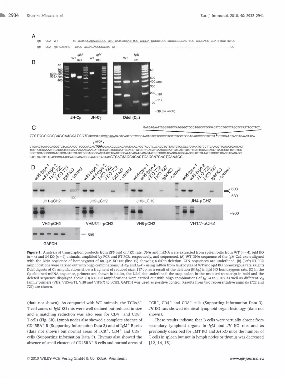
Figure 1. Analysis of transcription products from ZFN IgM or J KO rats. DNA and mRNAwere extracted from spleen cells from WT (n5 4), IgM KO(n5 4) and JH KO (n5 4) animals, amplified by PCR and RT-PCR, respectively, and sequenced. (A) WT DNA sequence of the IgM Cm1 exon alignedwith the DNA sequence of homozygous of an IgM KO rat (line 19) showing a 64bp deletion. ZFN sequences are underlined. (B) (Left) RT-PCRamplifications were carried out with oligo combinations JH1-Cm and JH-Cg using mRNA from leukocytes of WTand IgM KO homozygous rats. (Right)DdeI digests of Cm amplifications show a fragment of reduced size, 117bp, as a result of the deletion (64 bp) in IgM KO homozygous rats. (C) In theCm obtained mRNA sequence, primers are shown in italics, the DdeI site underlined, the stop codon in the mutated transcript in bold and thedeleted sequence displayed above. (D) RT-PCR amplifications were carried out with oligo combinations of JH1-4 to mCH2 as well as different VH
family primers (VH2, VH5/6/11, VH8 and VH1/7) to mCH2. GAPDH was used as positive control. Results from two representative animals (722 and727) are shown.
Eur. J. Immunol. 2010. 40: 2932–2941Severine Menoret et al.2934
& 2010 WILEY-VCH Verlag GmbH & Co. KGaA, Weinheim www.eji-journal.eu

wild type
(1/dilution)B purified IgM (ng/line)IgM IgGA
75KDa
500 250 125 62.5 31.2 15.6 7.8 1/10 1/100
A
1500 8000
6000
4000
2000
0
1000
500
0
IgM
(µ
g/m
l)
WT IgM KO J KOIgM KO (1/dilution)
-/- -/-
75
1/10 1/100 1/10 1/1001/10 1/100 1/10 1/100
-/-
wild type
(1/dilution)
IgAIgEIg
G (
µg
/ml)
WT IgM KO J KO
KDa
wild type
(1/dilution) -/- -/-
wild type
(1/dilution)
J KO (1/dilution)
IgE
(n
g/m
l)
IgA
(µ
g/m
l)
1/10 1/100 1/10 1/100 1/10 1/1001/10 1/100
75KDa
WT IgM KO J KO WT IgM KO J KO
75
50
25
0
5000
4000
3000
2000
1000
0
Figure 2. Quantitative ELISA of serum Ig isotypes. Sera were collected from WT (n5 5), IgM homozygous KO (IgM KO, n5 6) and JH homozygous KO(J KO, n5 4) rats 12–18wk old. (A) Isotype-specific and quantitative ELISA for the different rat isotypes. Each point represents the values of oneanimal and the horizontal bar the mean of each group of values. �po0.01; ��po0.001 WT versus IgM KO or JH KO using Mann–Whitney test.(B) Western blot analysis of serum IgM. Purified rat IgM and sera harvested fromWT, IgM homozygous KO and JH homozygous KO rats 12–18wk oldwere analyzed by Western blot using polyclonal anti-rat m heavy chain-specific Ab. (upper) Purified rat IgM was diluted and the indicated totalamount of Ab (ng) was loaded in each line. Serum from aWTrat was diluted as indicated and 20mL was loaded by line. (Medium) Serum from aWTrat and sera from three IgM KO rats were diluted as indicated. (Bottom) Serum from a WT rat and sera from two JH KO rats were diluted asindicated.
A spleen lymph nodes thymus
BTCR (T) CD4 CD8
CD68
(macrophages)
Spleen
WT
CD45RA
(OX33) (B)
Spleen
IgM KO
Figure 3. Macroscopic and immunohistology analysis of lymphoid organs. Lymphoid organs were harvested from WT (n5 4) and IgMhomozygous KO (IgM KO, n5 6) rats 12–18wk old. (A) Macroscopic analysis of spleen, lymph nodes and thymus. (B) Immunoperoxidasestaining of spleen cryostat sections using mAb directed to the defined cell markers and cell types between parentheses and counterstained withMayer’s hematoxylin.
Eur. J. Immunol. 2010. 40: 2932–2941 Molecular immunology 2935
& 2010 WILEY-VCH Verlag GmbH & Co. KGaA, Weinheim www.eji-journal.eu
Flow cytometry analysis for quantification of immunecells in bone marrow (BM) and lymphoid organs
To better define the blockade in B-cell differentiation and to
quantify the absolute numbers of different cell subsets, we
evaluated the single-cell composition in the various lymphoid
organs. Using CD45R (B220) and IgM as markers, several
B-cell populations could be identified in the rat [16]; pro–pre
B (IgM� CD45Rlow), immature (IgMlow CD45Rlow), transitional
(IgMhigh CD45Rlow), marginal zone (IgMhigh CD45R�) and
mature (IgMlow and high CD45Rhigh). The analysis of IgD
allowed a further subdivision of IgM1 B cells as IgDlow/�
marginal zone and IgD1 follicular B cells and IgMlow IgD� as
immature/transitional B cells [17]. Analysis of WT rats showed
mostly pro–pre B and some immature B cells in BM, mostly
immature and some mature in lymph nodes and similar
proportion of immature and transitional cells (Fig. 4A and
Table 1). Spleen and lymph nodes of IgM or JH KO rats showed
barely detectable IgM or IgD positive cells (Fig. 4A, Table 1 and
Supporting Information Data 4). The total number of cells in the
spleen and lymph nodes of IgM or JH KO rats were drastically
decreased versusWT rats (Table 1). IgM1 and CD45R1cells in the
spleen of IgM or JH KO rats were drastically decreased versus WT
rats (IgM1: 0.7 and 2.28%, respectively; CD45R1: 1.6 and 4.3%,
respectively) (Table 1). FACS analysis showed the presence of a
small population of CD45R1IgM� cells in spleen (Fig. 4A,
Table 1). Immunohistology revealed their location mainly in
the spleen red pulps (data not shown). Using several markers, we
confirmed that the phenotype of CD45R1 cells in IgM KO rats
corresponded to the previously described phenoype of rat pDC
[18] (data not shown).
In lymph nodes, absolute numbers of IgM1 or CD45R1 cells
were greatly reduced in IgM or JH KO rats versus WT controls
(�4 and �4.5%, respectively) (Table 1).
In BM of IgM or JH KO rats, we observed no immature or
mature B cells and greatly reduced proportion of pro–pre B cells
(IgM� CD45Rlow) (Fig. 4A). The absolute number of mono-
nuclear cells was significantly reduced in IgM and JH KO versus
WT rats (42.2 and 56.7%, respectively) (Table 1) and numbers of
pro–pre B cells (IgM� CD45Rlow) in IgM, JH KO and WT were
12.8 and 22.4%, respectively, versus WT (Table 1).
T cells in spleen, as defined by double staining using anti-TCRab
and anti-CD4 or anti-CD8 Ab, showed an increased proportion of
TCRab1 cells compared with WT rats (�85% in IgM and JH KO rats
versus �40% in WT animals), both of the CD41 and CD81 subtypes
(Fig. 4B). Despite this increase, the total numbers of spleen cells in
IgM and JH KO rats were only 13.6 and 16.6%, respectively,
compared with WT spleen cells and thus the total numbers of
TCRab1 cells in IgM and JH KO rats were 30 and 33.7%, respectively,
versus WT (p5o0.05 for both IgM or JH KO versus WT) (Table 1).
Despite the fact that cell numbers in the lymph nodes were
considerably decreased in IgM or JH KO versus WT rats (43 and
wild type
spleenIgM KO
spleenB J KO
spleen
IgM
wild type IgM KOA
J KO
TC
Rα
β
sp
lee
n
TC
Rα
β
CD8
lym
ph
sn
od
es
bo
ne
wm
arr
ow
CD45R
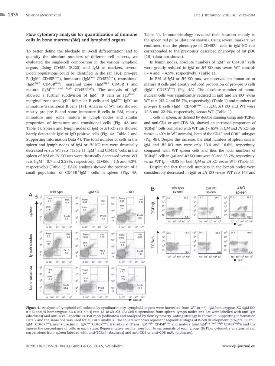
Figure 4. Analysis of lymphoid cell subsets by cytofluorimetry. Lymphoid organs were harvested from WT (n5 4), IgM homozygous KO (IgM KO,n5 6) and JH homozygous KO (J KO, n5 4) rats 12–18wk old. (A) Cell suspensions from spleen, lymph nodes and BM were labelled with anti-IgM(abscissas) and anti-B-cell-specific CD45R mAb (ordinates) and analyzed by flow cytometry. Gating strategy is shown in Supporting InformationData 2 and the same one was used for all FACS analysis. The square windows represent sequential stages of B-cell development (pro–pre B (Pro BIgM� CD45Rlow), immature (imm. IgMlow CD45Rlow), transitional (Trans. IgMhigh, CD45Rlow) and mature (mat IgMlow and high CD45Rhigh)) and thefigures the percentages of cells in each stage. Representative results from four to six animals of each group. (B) Flow cytometry analysis of cellsuspensions from spleen labelled with anti-TCRab (abscissas) and anti-CD4 or anti-CD8 mAb (ordinates).
Eur. J. Immunol. 2010. 40: 2932–2941Severine Menoret et al.2936
& 2010 WILEY-VCH Verlag GmbH & Co. KGaA, Weinheim www.eji-journal.eu
39%, respectively), T cells were not significantly reduced
(Table 1) due to a significantly increased proportion of TCRab1
cells (�95% for both KO versus �78%, respectively) with the
CD41 or CD81 surface marker (Supporting Information Data 2).
In BM, the proportion of TCR1 cells was increased in IgM or
JH KO versus WT rats (both �35 versus �10%, respectively) in
both compartments, TCR1CD41 and TCR1CD81 (Supporting
Information Data 2). The total number of T cells was also
significantly increased in IgM or JH KO versus WT (275 and
201%, respectively) (Table 1).
In thymus of IgM or J KO rats, the proportion of TCR1, TCR1
CD41 and TCR1CD81 cells (Supporting Information Data 3) as
well as the total number of T cells (Table 1) were comparable.
IgM KO rats, 1 year old showed the same cellular phenotype
and the absence of Ig as younger animals (data not shown).
Thus, both IgM and JH KO rats showed a blockade on B-cell
differentiation in the earliest stages of B-cell development in BM
with greatly reduced B cells in peripheral lymphoid organs. Total
T CD41 and T CD81 cells were also significantly decreased in
spleen but not in lymph nodes. T cells were increased in BM and
maintained in the thymus of IgM or J KO versus WT rats.
Heart allograft survival
To test in vivo for the absence of B cells, we used a model of
hyperacute heart allograft rejection in which increased anti-donor
Ab are the first rejection mechanism. In this model, recipients were
immunized against donor antigens by multiple skin transplants from
MHC-mismatched donor prior to heart transplantation from the
same donor. WT recipients without previous donor immunization
rejected donor hearts in 7 days (n54). Immunized recipients
exhibited accelerated rejection in hours (1h40, 5h00 and o8h00)
with high titers of anti-donor Ab (Fig. 5A and B). On the contrary,
IgM KO rats showed significantly prolonged survival of transplanted
hearts (144h (d6), 168h (d7), 456h (d19), 480 (d20); po0.05
versus WT) (Fig. 5A). Importantly, flow cytometric analysis showed
that IgM KO rats did not produce Ab binding to donor cells (Fig. 5B).
Thus, B-cell and Ab-deficient animals showed delayed allo-
graft rejection after repeated anti-donor stimulation in a model of
Ab-mediated rejection.
Discussion
Although the rat has been a major experimental species in
physiological studies for many years, the lack of robust genetic
engineering technologies to generate gene-specific mutations
hampered its use in many other models [1, 3, 4, 7]. The cloning
of the rat through nuclear transfer has been described several
years ago [19] but a source of suitable cells in which gene
targeting and selection of mutants is feasible without losing
cloning potency is lacking. Analogously, rat ES cells [5, 6]
and induced pluripotent stem cells [20] have been recently
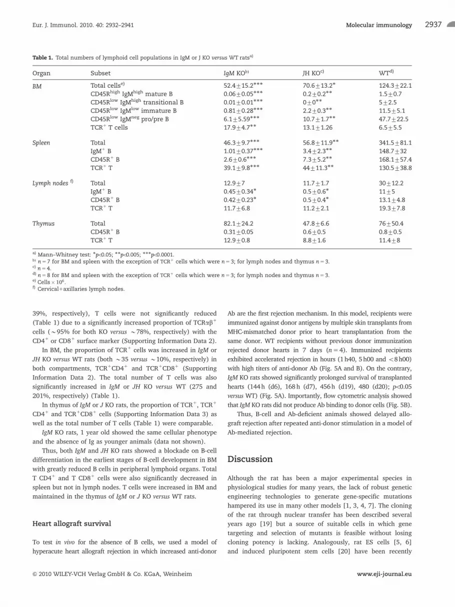
Table 1. Total numbers of lymphoid cell populations in IgM or J KO versus WT ratsa)
Organ Subset IgM KObÞ JH KOc) WTd)
BM Total cellse) 52.4715.2��� 70.6713.2� 124.3722.1
CD45Rhigh IgMhigh mature B 0.0670.05��� 0.270.2�� 1.570.7
CD45Rlow IgMhigh transitional B 0.0170.01��� 070�� 572.5
CD45Rlow IgMlow immature B 0.8170.28��� 2.270.3�� 11.575.1
CD45Rlow IgMneg pro/pre B 6.175.59��� 10.771.7�� 47.7722.5
TCR1 T cells 17.974.7�� 13.171.26 6.575.5
Spleen Total 46.379.7��� 56.8711.9�� 341.5781.1
IgM1 B 1.0170.37��� 3.472.3�� 148.7732
CD45R1 B 2.670.6��� 7.375.2�� 168.1757.4
TCR1 T 39.179.8��� 44711.3�� 130.5738.8
Lymph nodes f) Total 12.977 11.771.7 30712.2
IgM1 B 0.4570.34� 0.570.6� 1175
CD45R1 B 0.4270.23� 0.570.4� 13.174.8
TCR1 T 11.776.8 11.272.1 19.377.8
Thymus Total 82.1724.2 47.876.6 76750.4
CD45R1 B 0.3170.05 0.670.5 0.870.5
TCR1 T 12.970.8 8.871.6 11.478
a) Mann–Whitney test: �po0.05; ��po0.005; ���po0.0001.bÞ n5 7 for BM and spleen with the exception of TCR1 cells which were n5 3; for lymph nodes and thymus n5 3.c) n5 4.d) n5 8 for BM and spleen with the exception of TCR1 cells which were n5 3; for lymph nodes and thymus n5 3.e) Cells� 106.f) Cervical1axillaries lymph nodes.
Eur. J. Immunol. 2010. 40: 2932–2941 Molecular immunology 2937
& 2010 WILEY-VCH Verlag GmbH & Co. KGaA, Weinheim www.eji-journal.eu

described and may eventually allow generation of precise
gene modifications as obtained in mice. However, currently,
there are no reports of gene KO rats from such cells. KO rats have
been described using chemical mutagens [21] or transposons
[22] but these techniques, although very useful, generate random
non-controlled mutations and are thus labour intensive and
expensive. The first gene-specific KO rats with mutations in IgM
(phenotyped here) and Rab38 endogenous loci as well as a
transgenic GFP were generated using ZFN [7–9].
ZFN provide several advantages to generate novel rat lines
carrying mutations in specific genes. The most important ones are
the capacity to target specifically a given gene and the high effi-
ciency of the procedure. As far as specificity is concerned, we
showed that the most homologous non-related sequences in the
rat genome to the one targeted by the IgM ZFN did not show non-
specific mutations [8, 9]. Although it needs to be established
whether any given gene can be targeted by ZFN, our experience
with other ZFN, for example directed against rat l and k light-
chain genes (our unpublished results) as well as the IL-2 receptor
g locus gene [23], have all resulted in successful KO. The other
major advantage of ZFN is the speed of the procedure since KO
rats can be generated in about 4 months in both inbred and
outbred strains [8, 9, 23]. Finally, mutations are definitive and
transmitted to the progeny.
Our characterization of IgM KO and JH KO rats confirm the
previous findings in mMT or J KO mice [11, 12] and immuno-
deficient human patients [24, 25] that the absence of membrane
Ig expression results in the absence of B cells. On the contrary,
IgM deletion [26] or truncation [13] in mice permitted expression
of other heavy chains and allowed B-cell development and
maturation due to replacement of IgM by IgD. Similarly to
humans [24, 27], IgM KO rats showed only 5% of normal levels of
BM pro–pre B cells, whereas mMT mice showed normal levels of
BM pro–pre B cells [11]. In this regard, deletion in mice of the Ig
JH region resulted in a block of Ig gene expression and B-cell
development at the pro-B-cell stage [12] as for JH KO rats. Thus,
like mMT mice in which transcription and translation of m-chain
occurred but did not result in expression of membrane-bound
IgM and like JH KO mice, IgM KO rats showed a shortened m
transcript and the absence of Ig polypeptide production and
therefore a very early B-cell block. As for mice and human cells,
an enigma still persists on how B-cell levels can be suppressed
early or potentially, after rearrangement at the pre-BCR stage but
before a fully functional m polypeptide is expressed. An answer to
this may be dependent on the level of early control of the IgH
locus when chromatin is opening and antisense transcription will
be initiated before D to J recombination [28]. It is possible that
strain-specific parameters as well as size and position of the
removed or targeted region may determine the B-cell block.
Another difference with IgM KO mice [11] is that these mice
showed normal levels of IgA and absence of all other Ig isotypes
[29], whereas IgM KO rats showed complete deficiency of all
isotypes including IgA. Analogously to IgM KO rats, patients with
deletion of the m locus also result in the absence of Ig production
for all isotypes including IgA [25].
Since in contrast to mice, only 1% of cells recovered from the
peritoneal cavity of rats are B cells [17], we did not analyze this
compartment.
In IgM or JH KO rats’ T-cell numbers in spleen but not in
lymph nodes were decreased, as described for mMT mice [14, 15].
In mMT mice, this was due to the lack of production of lymphotoxin
a1b2 by B cells, required for CCL21 and stromal cell develop-
ment, and as yet to be defined mechanism(s) for the promotion
of T-cell numbers [14]. This decrease in spleen T cells in mMT
mice did not result in impairment of several immune responses,
but depletion of CD81 cells did impair heart allograft rejection
A controls n=3
IgM KO n=4
*
Bcontrols n=3
IgM KO n=4
IgM
IgG
controls n=3
IgM KO n=4
*
*
Figure 5. Hyperacute allograft rejection mediated by alloAb. RecipientWTor IgM KO rats (both MHC RT-1u haplotype) were grafted three timesat 1-wk intervals with skin from LEW.1A donors (MHC RT-1a haplotype)and 1wk later grafted with a LEW.1A donor heart. (A) Kaplan–Meiergraft survival curve showing graft rejection in some hours for WT ratsand in several days for IgM KO rats (�po0.05 using Kaplan–Meier test).(B) Analysis of IgM (upper panel) and IgG (lower panel) anti-LEW.1AalloAb levels in sera diluted 1/10 from day 0 before skin transplantationup to the moment of heart rejection (for WT and one IgM KO) or up today 43 in non-rejecting IgM KO. Results are shown in mean channelfluorescence after subtraction of mean channel fluorescence obtainedincubating sera with recipient T cells7SD. �po0.05 IgM KO versus WTfor all time points using Mann–Whitney test.
Eur. J. Immunol. 2010. 40: 2932–2941Severine Menoret et al.2938
& 2010 WILEY-VCH Verlag GmbH & Co. KGaA, Weinheim www.eji-journal.eu

by cellular immune responses when initiated in the spleen but
not in lymph nodes [15]. We also observed that T cells were
significantly increased in the BM of IgM KO rats and this vascular
compartment of T cells could replace at least in part the reduced
pool of spleen T cells for immune responses mainly taking place in
the blood and spleen. Therefore, care should be taken when
analyzing T-cell responses in B-cell-deficient animals, in particular
when immune responses are mediated in the vascular compartment
and spleen as compared with other tissues. Further experiments
are needed to analyze this point in IgM or JH KO rats. As far as
Ab-mediated hyperacute allograft rejection is concerned, IgM KO
rats showed a significantly delayed rejection which was associated
with undetectable levels of alloAb, as previously described in mMT
mice [30].
In conclusion, we generated a new rat KO line by ZFN-
targeted deletion of the J locus and we describe that both IgM KO
rats and JH KO rats are B cell and Ig deficient. These animals will
be useful models to explore the role of B cells and Ab in different
pathophysiological processes as organ rejection. They will also be
useful for the generation of rats expressing a human Ab reper-
toire, an important application of transgenic animals [2].
Materials and methods
Generation of IgM or J KO rats
Sprague–Dawley WT, IgM KO and JH KO rats analyzed were
10–18wk old. In addition, IgM KO over 1 year old were compared
with younger animals. Animals were bred at Charles River under
specific pathogen-free conditions. The generation of heterozygous
IgM KO rats using ZFN has been described previously [8, 9]. JH KO
rats, generated using ZFN (Sigma) targeting sequences upstream
and downstream of the rat JH-locus (Supporting Information
Data 1) (ZFN1: CAGGTGTGCCCATCCAGCTGAGTTAAGGTGGAG;
ZFN2: CAGGACCAGGACACCTGCAGCAGCTGGCAGGAAGCAGGT;
binding sites underlined) were designed and validated biochemi-
cally in vitro as described previously [31]. Pronuclear injections of in
vitro-transcribed mRNA-encoding ZFN were performed as described
previously [8, 9] using Sprague–Dawley rats. Offspring with large
deletions was identified by PCR using the primers GATTTACTGA-
GAGTACAGGG and AGGATTCAGTCGAAACTGGA (Supporting
Information Data 1) at an annealing temperature of 581C. The
experiments complied with the institutional ethical guidelines and,
both, the animal facility and the researchers performing the
experiments have been approved by national and local authorities
in accordance with the guidelines for animal experiments of the
French Veterinary Services.
Collection of cells
Spleen, lymph nodes and BM biopsies were collected under
anesthesia. Single-cell suspensions from spleen and lymph nodes
were prepared as described previously [32]. BM cells were
obtained by flushing one femur with PBS. Cell suspensions were
then pelleted and red blood cells were removed by erythrocyte
lysis. Cell suspensions were washed twice and passed through a
nylon gauze before counting the cells using an haemocytometer.
Part of the cell suspensions was used for mRNA and DNA
extraction and others for flow cytometry analysis.
Analysis of transcription products from KO rats
RNA was extracted from rat spleen cells using TRIzol (Invitrogen),
stored in RNAlater (Ambion) and reverse transcribed at 421C with
BioScript (Bioline, London, UK). PCR reactions were set up using rat
JH or VH forward primers with mCH2 or gCH2 reverse primers.
Sequences of primers from 50 to 30 were as follows: JH1:
TTCTGGGGCCCAGGAACCATGGTCA; JH2: TACTGGGGCCAAG-
GAGTCATGGTCA; JH3: TACTGGGGCCAAGGCACTCTGGTCA; JH4:
TGCCTGGGGTCAAGGAGCTTCAGTCA; VH2: CAGGTGCAGCT-
GAAGGAGWCAG; VH5_6_11: AGGTGCAGCTGGTGGAGWCWG;
VH8: CAGGTTACTCTGAAAGAGTCTGG; VH1_7: CAGGTC-
CAGCTGCWGSARTCTG; mCH2R GCTTTCAGTGATGGT-
CAGTGTGCTTATGAC; gCH2: GTTTGGAGATGCTTTTCTCG-
ATGGG; GAPDH F: CAGTGCCAGCCTCGTCTCAT; GAPDH R:
AGGGGCCATCCACAGTCTTC. GoTaqs Green Master mix
(Promega) was used as per the manufacturer’ instructions
(www.promega.com) with amounts of sample cDNA adjusted by
comparing GAPDH band strength. Annealing temperatures used for
the PCR were set at the lowest primer Tm – 51C (http://www.sigma-
genosys.com/calc/DNACalc.asp). The reaction conditions were 951C
for 2min, 34 cycles of 951C for 20 s and 701C for 40s, followed by
701C for 5min RT-PCR products were cleaned up using SureClean
(Bioline) digested with DdeI (NEB) or sequenced directly.
Identification of cell subsets by flow cytometry
Cell suspensions were washed and adjusted to 5�105 cells/well
in PBS-1% BSA-0.1% Azide. The different B-cell subsets were
identified using mouse anti-rat IgM FITC-labelled mAb (MARM 4,
Jackson Immunoresearch Laboratories) in combination with anti-
B cell CD45R (rat B220)-PE-conjugated mAb (His 24, BD
biosciences) or anti-IgD-PE-conjugated mAb (MARD-3, Abd
Serotec). The incubation period was 30min at 41C and for the
analysis an FACS CantoII flow cytometer and FlowJo software
(Becton Dickinson, Pont de Claix, France) were used. T cells were
detected using anti-CD3 and anti-abTCR mAb (G4.18 and R7.3,
both from BD biosciences) as described previously [32].
Identification of cell subsets by immunohistology
Tissue biopsies were embedded in optimal tissue compound
(Tissue-TEKs, Miles, Elkart, IN, USA), snap in liquid nitrogen
cooled isopentane and stored at �801C. Cryostat sections
Eur. J. Immunol. 2010. 40: 2932–2941 Molecular immunology 2939
& 2010 WILEY-VCH Verlag GmbH & Co. KGaA, Weinheim www.eji-journal.eu
(5mm) from tissues were thawed, fixed in acetone (10min at room
temperature) and incubated with mAb (1h at room temperature,
10mg/mL) recognizing CD45RA (OX33), abTCR, CD8 (OX8) and
CD4 (W3.25), followed by biotin-conjugated anti-mouse Ab
(Jackson ImmunoResearch Laboratories) as described previously
[31]. Ab binding was detected by incubation with HRP-conjugated
streptavidin using Vectors VIP (Vector Laboratories, Burlingame,
CA, USA) as a substrate. Tissue sections were counterstained with
Mayer’s hematoxylin and lithium carbonate.
Detection of Ig in rat sera by quantitative ELISA
Serum Ig concentrations were determined by a quantitative ELISA,
using plates coated with isotype-specific mouse mAb anti-rat Ab to
IgM (MARM-4), IgG (MARG), IgE (MARE) or IgA (MARA) (all
from Abd Serotec, Jackson ImmunoResearch, BD Biosciences) at
5mg/mL in PBS overnight at 41C. After washing with PBS-Tween
0.5%, plates were blocked for 2h at room temperature with PBS-
BSA 3%w/v. Sera were diluted in PBS starting at 1/10 and
incubated for 2h at 371C. After washing, a mixture of mouse mAb
anti-rat k and anti-l chain-specific peroxydase-conjugated Ab
(clone MARK-1/MARL-15, Abd Serotec) were added at a dilution
of 1:2000 in PBS for 1h. Purified rat mAb IgM, IgG, IgA and IgE
isotypes (Abd Serotec, Jackson ImmunoResearch, BD Biosciences)
were added at different known concentrations and used as
standard. After three washes, TMB substrate reagent (Becton
Dickinson) was added and the reaction was stopped after 10min
by the addition of 2N H2SO4. Absorbance was read at 490nm.
Serum Ig concentrations were determined by extrapolating
absorbance values of sera dilutions in the linear range of the
dilution curves to those of isotype standard controls.
Western blot analysis
Protein concentration of serum was measured (BCA Protein Assay
Kit, Pierce, Rockford, IL, USA). A standard curve dilution of
monoclonal purified rat IgM and dilutions of rat serum (1/10 and
1/100) (20mL/line) were electrophoresed in 12% SDS-polyacryla-
mide gels. After semi-dry transfer, the nitrocellulose membranes
were blocked in 5% dry milk in PBS with 0.05% Tween-20 for 1–2h
and incubated overnight at 41C with Peroxydase-conjugated goat
anti-rat IgM m-specific Ab (from Jackson Laboratories) at 1mg/mL.
The binding was visualized with enhanced chemiluminescence and
quantified using a Fuji LAS 4000 (Fujifilm) imaging system and
Multi Gauge V3.0 software (Fujifilm).
Heart allograft transplantation and anti-donor Ab
IgM KO rats (haplotype RT1u for MHC I and II) were immunized
against donor LEW.1A alloantigens (haplotype RT1a for MHC I and
II) by three successive skin grafts separated by 1wk each and
grafted with a LEW.1A heart 1wk after the last skin transplant.
Heart transplantation was performed heterotopically in the abdo-
men. Heart allograft survival was evaluated through abdominal
palpation and rejection was defined as total cessation of beating
and confirmed by visual inspection after laparotomy [32, 33]. Anti-
donor Ab were analyzed in the sera of WT or IgM KO rats harvested
at day 0 before skin transplantation, at days 20 and 30 after skin
transplantation and at rejection or later time points if hearts were
not rejected. Heat-inactivated sera (dilutions 1/10, 1/100 and 1/
1000 and 1/5000) were incubated with donor T cells, washed and
alloAb bound were detected using rat anti-IgG or IgM-specific Ab.
As negative control, sera were incubated with recipient T cells.
Results were reported as the mean channel fluorescence using
donor T cells – mean channel fluorescence using donor T cells7SD.
Statistical analysis
Results are presented as the means7SD. Statistical analyses were
performed by a Mann–Whitney test for laboratory analyses and
Kaplan–Meier log rank test for graft survival using GraphPad
Prism 4 software (GraphPad Software, La Jolla, CA, USA).
Differences associated with probability values of po0.05 were
considered statistically significant.
Acknowledgements: This work was in part supported by funding
from La Region Pays de la Loire through the IMBIO program and
Biogenouest, IBiSA program and Fondation Progreffe.
Conflict of interest: A.-L. I., M. J. O., M. B., R. B. and I. A. have
potential conflict of interests that include stock options, salaries or
consulting fees from OMT. G. J. C. has potential conflict of interest
that includes salary fees from Sangamo BioSciences.
References
1 Tesson, L., Cozzi, J., Menoret, S., Remy, S., Usal, C., Fraichard, A. and
Anegon, I., Transgenic modifications of the rat genome. Transgenic Res.
2005. 14: 531–546.
2 Lonberg, N., Human antibodies from transgenic animals. Nat. Biotechnol.
2005. 23: 1117–1125.
3 Aitman, T. J., Critser, J. K., Cuppen, E., Dominiczak, A., Fernandez-Suarez,
X. M., Flint, J., Gauguier, D. et al., Progress and prospects in rat genetics: a
community view. Nat. Genet. 2008. 40: 516–522.
4 Jacob, H. J., The rat: a model used in biomedical research. Methods Mol.
Biol. 2009. 597: 1–11.
5 Li, P., Tong, C., Mehrian-Shai, R., Jia, L., Wu, N., Yan, Y., Maxson, R. E.
et al., Germline competent embryonic stem cells derived from rat
blastocysts. Cell 2008. 135: 1299–1310.
6 Buehr, M., Meek, S., Blair, K., Yang, J., Ure, J., Silva, J., McLay, R. et al.,
Capture of authentic embryonic stem cells from rat blastocysts. Cell 2008.
135: 1287–1298.
Eur. J. Immunol. 2010. 40: 2932–2941Severine Menoret et al.2940
& 2010 WILEY-VCH Verlag GmbH & Co. KGaA, Weinheim www.eji-journal.eu
7 Remy, S., Tesson, L., Menoret, S., Usal, C., Scharenberg, A. M. and
Anegon, I., Zinc-finger nucleases: a powerful tool for genetic engineering
of animals. Transgenic Res. 2010. 19: 363–371.
8 Geurts, A. M., Cost, G. J., Miller, J. C., Freyvert, Y., Zeitler, B., Choi, V. M.,
Jenkins, S. S. et al., Knockout rats produced via embryo pronuclear
microinjection of designed zinc finger nucleases. Science 2009. 325: 433.
9 Geurts, A. M., Cost, G. J., Remy, S., Cui, X., Tesson, L., Usal, C., Menoret, S.
et al., Generation of gene-specific mutated rats using zinc-finger
nucleases. Methods Mol. Biol. 2009. 597: 211–225.
10 Kandavelou, K. and Chandrasegaran, S., Custom-designed molecular
scissors for site-specific manipulation of the plant and Mammalian
genomes. Methods Mol. Biol. 2009. 544: 617–636.
11 Kitamura, D., Roes, J., Kuhn, R. and Rajewsky, K., A B cell-deficient mouse
by targeted disruption of the membrane exon of the immunoglobulin mu
chain gene. Nature 1991. 350: 423–426.
12 Jakobovits, A., Vergara, G. J., Kennedy, J. L., Hales, J. F., McGuinness, R. P.,
Casentini-Borocz, D. E., Brenner, D. G. and Otten, G. R., Analysis of
homozygous mutant chimeric mice: deletion of the immunoglobulin
heavy-chain joining region blocks B-cell development and antibody
production. Proc. Natl. Acad. Sci. USA 1993. 90: 2551–2555.
13 Zou, X., Ayling, C., Xian, J., Piper, T. A., Barker, P. J. and Bruggemann, M.,
Truncation of the mu heavy chain alters BCR signalling and allows
recruitment of CD51 B cells. Int. Immunol. 2001. 13: 1489–1499.
14 Ngo, V. N., Cornall, R. J. and Cyster, J. G., Splenic T zone development is B
cell dependent. J. Exp. Med. 2001. 194: 1649–1660.
15 Nozaki, T., Rosenblum, J. M., Ishii, D., Tanabe, K. and Fairchild, R. L., CD4
T cell-mediated rejection of cardiac allografts in B cell-deficient mice. J.
Immunol. 2008. 181: 5257–5263.
16 Kroese, F. G., Wubbena, A. S., Opstelten, D., Deenen, G. J., Schwander, E.
H., De Leij, L., Vos, H. et al., B lymphocyte differentiation in the rat:
production and characterization of monoclonal antibodies to B lineage-
associated antigens. Eur. J. Immunol. 1987. 17: 921–928.
17 Kroese, F. G., Butcher, E. C., Lalor, P. A., Stall, A. M. and Herzenberg, L. A.,
The rat B cell system: the anatomical localization of flow cytometry-
defined B cell subpopulations. Eur. J. Immunol. 1990. 20: 1527–1534.
18 Hubert, F. X., Voisine, C., Louvet, C., Heslan, M. and Josien, R., Rat
plasmacytoid dendritic cells are an abundant subset of MHC class II1
CD41CD11b-OX62- and type I IFN-producing cells that exhibit selective
expression of Toll-like receptors 7 and 9 and strong responsiveness to
CpG. J. Immunol. 2004. 172: 7485–7494.
19 Zhou, Q., Renard, J. P., Le Friec, G., Brochard, V., Beaujean, N., Cherifi, Y.,
Fraichard, A. and Cozzi, J., Generation of fertile cloned rats by regulating
oocyte activation. Science 2003. 302: 1179.
20 Ueda, S., Kawamata, M., Teratani, T., Shimizu, T., Tamai, Y., Ogawa, H.,
Hayashi, K. et al., Establishment of rat embryonic stem cells and making
of chimera rats. PLoS One 2008. 3: e2800.
21 van Boxtel, R., Gould, M. N., Cuppen, E. and Smits, B. M., ENU
mutagenesis to generate genetically modified rat models. Methods Mol.
Biol. 2009. 597: 151–167.
22 Kitada, K., Ishishita, S., Tosaka, K., Takahashi, R., Ueda, M., Keng, V. W.,
Horie, K. and Takeda, J., Transposon-tagged mutagenesis in the rat. Nat.
Methods 2007. 4: 131–133.
23 Mashimo, T., Takizawa, A., Voigt, B., Yoshimi, K., Hiai, H., Kuramoto, T.
and Serikawa, T., Generation of knockout rats with X-linked severe
combined immunodeficiency (X-SCID) using zinc-finger nucleases. PLoS
One 2010. 5: e8870.
24 Meffre, E., Milili, M., Blanco-Betancourt, C., Antunes, H., Nussenzweig,
M. C. and Schiff, C., Immunoglobulin heavy chain expression shapes the
B cell receptor repertoire in human B cell development. J. Clin. Invest. 2001.
108: 879–886.
25 Milili, M., Antunes, H., Blanco-Betancourt, C., Nogueiras, A., Santos, E.,
Vasconcelos, J., Castro e Melo, J. and Schiff, C., A new case of autosomal
recessive agammaglobulinaemia with impaired pre-B cell differentiation
due to a large deletion of the IGH locus. Eur. J. Pediatr. 2002. 161: 479–484.
26 Lutz, C., Ledermann, B., Kosco-Vilbois, M. H., Ochsenbein, A. F.,
Zinkernagel, R. M., Kohler, G. and Brombacher, F., IgD can largely
substitute for loss of IgM function in B cells. Nature 1998. 393: 797–801.
27 Lopez Granados, E., Porpiglia, A. S., Hogan, M. B., Matamoros, N.,
Krasovec, S., Pignata, C., Smith, C. I. et al., Clinical andmolecular analysis
of patients with defects in micro heavy chain gene. J. Clin. Invest. 2002.
110: 1029–1035.
28 Featherstone, K., Wood, A. L., Bowen, A. J. and Corcoran, A. E., Themouse
immunoglobulin heavy chain V-D intergenic sequence contains insula-
tors that may regulate ordered V(D)J recombination. J. Biol. Chem. 2010.
285: 9327–9338.
29 Macpherson, A. J., Lamarre, A., McCoy, K., Harriman, G. R., Odermatt, B.,
Dougan, G., Hengartner, H. and Zinkernagel, R. M., IgA production
without mu or delta chain expression in developing B cells. Nat. Immunol.
2001. 2: 625–631.
30 Wasowska, B., Qian, Z., Behrens, E., Cangello, D., Sanfilippo, F. and
Baldwin, W. I., Elimination of antibody reponses inhibits acute and
chronic cardiac allograft rejection in mice. Transplantation 1998. 65: S90.
31 Doyon, Y., McCammon, J. M., Miller, J. C., Faraji, F., Ngo, C., Katibah, G. E.,
Amora, R. et al., Heritable targeted gene disruption in zebrafish using
designed zinc-finger nucleases. Nat. Biotechnol. 2008. 26: 702.
32 Guillonneau, C., Hill, M., Hubert, F. X., Chiffoleau, E., Herve, C., Li, X. L.,
Heslan, M. et al., CD40Ig treatment results in allograft acceptance
mediated by CD8CD45RC T cells, IFN-gamma, and indoleamine 2,3-
dioxygenase. J. Clin. Invest. 2007. 117: 1096–1106.
33 Ono, K. and Lyndsey, E. S., Improved technic of heart transplantation in
rats. J. Thorac. Cardiovasc. Surg. 1968. 57: 225–229.
Abbreviations: IgM KO rats: rats homozygous for IgM mutation � JH KO
rats: rats homozygous for J deletion � ZFN: zinc-finger nucleases
Full correspondence: Dr. Ignacio Anegon, INSERM-ITERT, UMR 643, 30,
boulevard Jean Monnet, Nantes F44093, France
Fax: 133-240087411
e-mail: [email protected]
Additional correspondence: Dr. Roland Buelow, Open Monoclonal
Technologies, Palo Alto, CA, USA
Fax: 133-240087411
e-mail: [email protected]
See accompanying Commentary:
http://dx.doi.org/10.1002/eji.201040980
Received: 11/8/2010
Accepted: 25/8/2010
Accepted article online: 9/9/2010
Note added in proof: We state in our article that there are
currently no reports of gene KO rats using ES cells; however, the
following article was published as our article was under review:
Tong, C., Li, P., Wu., N. L. Yan, Y. and Ying, Q. L., Production of
p53 gene knockout rats by homologous recombination in
embryonic stem cells. Nature 2010. 467: 211–213.
Eur. J. Immunol. 2010. 40: 2932–2941 Molecular immunology 2941
& 2010 WILEY-VCH Verlag GmbH & Co. KGaA, Weinheim www.eji-journal.eu