normal and abnormal bilirubin metabolism
TRANSCRIPT
University of Nebraska Medical Center University of Nebraska Medical Center
DigitalCommons@UNMC DigitalCommons@UNMC
MD Theses Special Collections
5-1-1939
Normal and abnormal bilirubin metabolism Normal and abnormal bilirubin metabolism
C. Robert Hankins University of Nebraska Medical Center
This manuscript is historical in nature and may not reflect current medical research and
practice. Search PubMed for current research.
Follow this and additional works at: https://digitalcommons.unmc.edu/mdtheses
Part of the Medical Education Commons
Recommended Citation Recommended Citation Hankins, C. Robert, "Normal and abnormal bilirubin metabolism" (1939). MD Theses. 748. https://digitalcommons.unmc.edu/mdtheses/748
This Thesis is brought to you for free and open access by the Special Collections at DigitalCommons@UNMC. It has been accepted for inclusion in MD Theses by an authorized administrator of DigitalCommons@UNMC. For more information, please contact [email protected].
NORMAL AND ABNORMAL BILIRUBIN METABOLISM
c. Robert Hankins
Presented to the College of Medicine
University of Nebraska, Omaha,
1939
NORMAL AND ABNORMAL BILIRUBIN METABOLISM
PART I
Normal Bilirubin Metabolism
I. Formation of Bilirubin PP• 1-16
A. Historical Work
1. Berzelius (1840), Study of Biliary O~nstituents. (1)
2. Virchow (1847), Discovery of Hema.toidin. (1)
3. Frerichs and Stadler (1850), First Experimental
Production of Bilirubinemia. (1)
4. Kuhne (1858), Confirmation of Frerichs Work (2)
5. Herman (1859), First conclusive experimental
Evidence of Relation of Hemoglobin to Bilirubin. (2)
6. Minkowsky and Naunyn (1886), Hepatic Formation
of Bilirubin. (3)
7. McNee (1913-14), Modern Concept of Extrahepatic
formation of Bilirubin. (3)
B. Short Survey of the Retioulo-endothelial System. (4)
C. Chemical Relation of Bilirubin to Hemoglobin. (4)
D. Physiological Studies of Bilirubin Derivation. (8)
1. Evidence of Formation of Bilirubin in the
Retioulo-endothelial System (Extrahepatic). (8)
2. Evidence of Hepato-oellular formation of
Bilirubin. (12)
3. Lepehne' s Dualistic Theory of Bilirubin Forma-
(13)
4. Extracellular Formation of Bilirubin (14)
5. Formation of Bilirubin Independent or Hemoglobin
Catabolism. (15)
E. Summary of Conception of Physiological Production
or Bilirubin. (15)
II. Methods of Determining Bilirubin and Normal Bilirubin
Concentrations.
A. Oxidation Method.
B. Icteric Index.
C. Van den Bergh Reaction.
D. Spectrophotometric Determination.
E. Normal Bilirubin Concentration in Man.
III. Excretion of Bilirubin and the Formation of
Urobilin.
A. Excreted by Liver.
B. Fate in Small Gut.
c. Urobilin Formation in Large Gut.
D. Urobilin Excretion.
E. Laboratory Determination of Urobilin.
PART II
Abnormal Bilirubin Metabolism
~. Jaundice.
A. Historical Survey.
B. Conditions Necessary for Jaundice to Occur.
PP• 16-19
(16)
(16)
(18)
(19)
(19)
pp.20-26
(20)
(21)
(22)
(22)
(23)
PP• 27-41
(27)
(28)
c. Hepatogenous or Regurgitation Jauntioe. (31)
D. Hemotogenous or Retention Jaundice. (32)
E. Discussion of van den Bergh Test in Relation to
Jaundice. (33)
F. Hypobilirubinemia. (41)
II. Effeot of Abnormal Bilirubin Metabolism on Urobilin
Physiology.
A. Survey of McMaster's Work.
B. Summary of Urobilin Metabolism in Generalized
Bilirubin Disorders.
III. Clinical Application of Discussed Factors in
PP• 41~44
(41)
(43)
Bilirubin Disorders. PP• 44-50
A. Clinical Recognition of Retention Jaundice. (45)
B. Clinical Recognition of Regurgitation Jaundice. (46)
c. Use of van den Bergh in Luetic Treatment. (47)
D. Bilirubin as a Liver Function Test. (48)
E. Differentiation between Primary and Secondary
anemias. (48)
F. Differentiation between Catarrhal Jaundice and
Mechanical Jaundice. (49)
G. ,Place of Bilirubin Studies in Diagnosis. (49)
H. New Evidence Concerning Possible Place of Bilirubin
in the Field of Immunology. (50)
PREFACE
The term bilirubin innnediately calls forth in the mind of the
physiologist the normal processes of biliary pigment formation and
the normal hepatic functions. Bilirubin to the pathologist usually
brings up pictures either of liver damage or of some hemolytio pro
cess with deposition of this pigment in the tissues of the body.
In the mind of the chemist the extensive chemical analyses of
hemoglobin, bilirubin and the related pyrroles done by Fischer ase
recalled. To the mind or the medical clinician, the term bilirubin
integrates the above pictures as a single working unit portrayed
to him by his patient.
It is the purpose or this paper to present the normal and ab
normal aspects.of bilirubin metabolism giving some or the major
contributions that have taken this subject out of the realm of
conjecture and have placed it upon a scientific basis. Some points
are still not explained, and these points of dispute will be
pointed out. It is not the purpose of this paper to discuss the
various pathological entities that may cause disruption in the
normal bilirubin metabolism, but there will be an attempt to show
that these entities may be classed into groups fundamentally the
same as far as the principle involving the disrupted bilirubin meta
bolism is concerned; and, that through simplified classification
of these processes, bilirubin disorderw are more readily understood.
Finally, an attempt will be made to show the various practical
applications that oan be made upon the basis of the known facts
of bilirubin metabolism.
c. Robert Hankins
3/24/39
PART I
NORMAL BILIRUBIN METABOLISM
1
Berzelius is credited with the first scientific studies of
the biliary constituents, or bile. He studied bile as a whole,
and gave a fair analysis of the various constituents as to their
percentage composition. To an orange-red pigment found in the
bile he attached the term cholepyrrhin. This work was done in
1840. (21)
Virchow in 1847, (13,39) made the first observations of
extrahepatic formation of bile pigment. He found a substance
which he called hematoidin occurring in tissues around old hem
orrhages discovered at autopsy. In his report he stressed the
close relation of hemoglobin to this substance, and also stated
its close relation to the biliary pigment termed cholepyrrhin by
Berzelius. However, Virchow could not prove conclusively that
the pigment formed under such circumstances is identical with
cholepyrrhin (or bilirubin) and he therefore gave it the name
hematoidin.
Frerichs and Stadler, 1850, (39) worked with bile and renamed
the orange-red pigment bilirubin. The name is derived from "bilis"
meaning bile, and "ruber" meaning red. They found that a pigment
resembling bilirubin could be produced in vitro by the action of
sulphuric acid upon bile salts, and more important, that the
injection of bile acids into the blood stream of an animal would
be followed by the appearance of undoubted bilirubin in the urine.
Their conclusion was that the body could transform bile acids into
bile pigment.
2
Kuhne, 1858, (39) repeated and confirmed a forgotten or
unnoticed observation of von Dusch that bile acids are powerful
hemolytic agentsJ he insisted that the experiments of Frerichs
and Stadler did not prove the origin of bilirubin from bile acids,
for those investigators had not taken into account the fact that
a large amount of hemoglobin is set free in the plasma by the
injection of bile acids. Kuhne was unable to satisfy himself
that the injection of hemoglobin alone, in the absence of bile
acids, would be followed by bilirubinuria, and he was .forced to
hold to the idea that the bile acids were necewsary in some way
for the formation of bile pigment.
Herrmann, 1859, (39) was able to produce bilirubinurea at
w'iil by inducing intravascular hemolysis with injections of
distilled water. This was the first clear demonstration that
simple liberation of hemoglobin into the blood stream may be
followed by an increased output of bile pigment in the urine.
Neiit;her Naunyn, 1868, nor Steiner, 1873, could con.firm. Herrman's
results, a fact that is now easily explained, since the absence
of bilirubinuria is by no means a proof that there has been
no increased formation of the pigment. Tarohanoff, 1874, on the
other hand not only confirmed Herrman's work, but, with the use
of bile fistula animals, carried the proof of the relation of
hemoglobin to bile pigment still .f.'urther by demonstrating, for
the first time, that the introduction of pure hemoglobin into
the circulation is followed by a marked increase in the amount of
3
bile pigment excreted by the liver.
As early as 1169, Morgagni taught that the liver excreted
the bile which was brought to it, preformed, by the blood. An
altogether different view was held for many years following the
experiments of Minkowski and Naunyn, 1886, (13) who found that
jaundice was produced in geese by poisoning with arseniuretted
hydrogen, whereas jaundice could not be so produced if the liver
was removed immediately after the administration of the substance.
From this they concluded that the liver was necessary for the
production of jaundice, and the belief arose that bile pigment
was formed by the hepatic cell.
McNee, 1913-14, (30) repeated the experiments of Minkowski and
Naunyn, and found small traces of bile in the urine and the
tissues of hepatectomized geese following the administration of
the arseniuretted hydrogen. McNee observed, in histologic studies
of the normal goose liver, that the von Kupffer cells contained
erythrocytes and gave a marked iron reaction. In the liver of
jaundiced geese, the Kupffer cells were seen to contain, besides
many erythrocytes, large quantities of yellowish green pigment,
which resembled biliverdin, although chemical proof was lacking.
MoNee concluded that in geese bile pigment was formed by the
endothelial (Kupffer) cells of the liver and by the small number
of endothelial cells found elsewhere (spleen and bone-marrow). In
geese, hepatectomy accomplishes removal of the greater part of
the retioulo-endothelial system, but in mammals it do.as not. Thus
- -------------------
4
the conception of bile pigment formation returns to the concept
of Morgagni, 1769, that bile constituents are produced in the
body and carried to the liver via the blood stream, at least
in relation to bilirubin.
Before reviewing further physiological experimentation, one
should consider the type of cell of which the Kupffer cell is
an example. During the last half century, certain fixed cells
and certain wandering cells having phagoeytic properties have
been •tudied by many workers. Ribbert, 1904, (13) showed the
interrelation of all these different cells in their col!Dilon a
bility to engulf particles of lithium carmine injected into the
circulation. To Aschoff belongs the credit for the conception
that these reticular and endothelial cells of the various tissues
possess col!Dilon properties and functions, and may be designated
together as the reticulo-endothelial system.
As these physiological principles were being studied, the
biochemists were doing much work in determining the composition
of the bile pigments. The discoveries of Virohow and Herrman
were the guiding lights in their studies. It was up to them to
show the chemical similarity of bilirubin to hemoglobin, or if
possible definitely prove the derivation of bilirubin from
hemoglobin. Fischer has probably been the greatest student of
this subject, and to him is attributed most of the advancement
made in this field. Without going through the long processes
involved in determining this technically difficult problem, it
5
is the purpose of this paper to present the modern concept of
this problem.
As stated before, the chemists started working with hemo-
globin and attempted to prove the derivation of bilLrubin from
this souroe. Hemoglobin is classified as a hematoprotein.
The hematoproteins are present in all protoplasm, but are found
more aooessable in the blood~ Their purpose is to carry oxygen,
and they are associated with the processes of oxidation in every
tissue of the body. Hemoglobin is insoluble in itself, and so
must be carried about physically in the red blood cells rather than
in solution. Hemoglobin is a sub-group of the conjugated proteins.
Conjugated proteins are made up of two radicals, one being the
protein radical and the· other being termed the prosthetic group.
In the ease of hemoglobin, the protein fraction is made up of a
globin and the prosthetic group is a porphyrin. The prosthetic
group is a relatively small part of the molecule, roughly making
up about 4% of the molecule. Porphyrins, chemically, are oon-
struoted on the porphine nucleus; this nucleus is made up of
4 pyrrole rings arranged in rosettes and called tetra-pyrroles.
The porphyrins are found widely distributed throughout nature
in both animal and plant kingdoms, their main characteristic
being their ability to produce color in reflected light, or,
in other words, they are pigments.
'When hemoglboin is treated, under appropriate conditions,
with glacial acetic acid and sodium chloride, and the mi.~ture
I !
6
~rmed gently, a substance is obtained which crystallizes readi17.r
as brown oyrstals--this substance has been called hemin. (See
the plates at the end of this paper f~r formula and reactions
mentioned in this chemical discussion of bilirubin) The other
product of this cleavage is globin. Hemin has been synthesized
by Fischer. (6) It contains four methyl-pyrrol radicals and a~
atom of iron, and may be represented as shown in the aforementioned
plates. If the crystals of hemin are treated with sodium
hydttoxide, the c.orrespoliding base, heme,, is liberated; this is
the original prosthetic radical of hemoglobin.
The porphyrin nucleus related to hemin is called protopor
phyrin. (see plate) With the introduction of an iron atom into
the molecule, neutral heme is formed, which may be isolated as
hemin, indistinguishable from the product derived from hemoglobin.
This is indirect evidence to the fact that hemoglobin is built up
from preformed protoporphyrin. It is asswmned by the chemist
that the body is capable of forming this nucleus as a necessity
to the making of hemoglobin. (Evidence will be given later to
substantiate this fact from the physiological viewpoint.)
Bilirubin has been studied extensively by Fischer and he
has given the substance a definite formula. (See plate) As
yet, he has not been able to synthesize this substance, so the
formula rests merely upon analysis of the substance. As shown
by his formula, Fischer believes that bilirubin arises by oxi-
dative scission of the porphyrin nucleus of hemoglobin, the iron
7
being lost in the process. The method by which the iron is lost,
and the nature of the compound in which it is lost is still a
vague subject. This iron containing radical is called hemo-
siderin1 the material can be demonstrated to contain iron; and
beyond that, there is little information. Lemberg (22,23)
believes that the process leads to biliverdin first, and then to
bilirubin. 'Biliverdin is an oxidized product of bilirubin, con-
taining two more oxygen atoms, but otherwise the same emperically.
In his experiments, Lemberg has been able to obtain bilirubin
from hemin as outlined in the plates previously mentioned, and has
identifi-ed :the intermediary pi"oducts. He suggests that the for-
ma~ion of bile pigments in the animal body proceeds in a similar
fashion yielding biliverdin which is afterwards r~duced to bili
rubin. He admits that from a physiological point of view much
remains to be done, but he believes that this model will provide
a good guide to further research. ·
Andrews (1) and Rich (41) have proven that the hematoidin
found by Virehow is the same substance as bilirubin.
In summarizing the modern chemical viewpoint, we must admit
that definite proof of the formation of bilirubin from hemoglobin
is still lacking. An important step in clearing up this matter
would be the synthesis of bilirubin, and the demonstration that
the formula as given is correct. But in all fairness, we must say,
the only chemical evidence of derivation of bilirubin from hemo-
globin is the chemical similarity of the bilirubin to the porphine
nucleus. Lemberg's work seems to be the closest approaoh to
definite proof of this derivation.
8
Knowing these chemical relationships of bilirubin to hemo
globin, we can now proceed more intelligently to follow the
physiological developments that have been made in the last few
years.
Interesting obserVa.tions were made by Peabody and Brown (13)
on the phagocytosis of erythrocytes in the bone marrow. These
workers employing the Berlin blue iron reaction, observed in the
bone marrow the presence of erythrooytes and granules of iron in
the same phagocytes. They concluded that the phagooytes of the
bone marrow ingest erythrocytes and that the hemosiderin (an iron
containing substance derived from the breaking down of hemoglobin)
is formed within the phagocytes from hemoglobin.
P. Corr (8) recently studied material from 71 necropsies
with respect to the distribution of hemosiderin and bilirubin.
Sections of liver and spleen and occasionally of bone-marrow,
kidney, lymph node, lung, suprarenal gland and pancreas were
stained for iron by the Berlin Blue method. Hemosiderin was
found in varying amounts in the splenic phagocytes, in the hepatic
and Kupffer cells of the liver and in the reticulo-endothelial
cells of the,bone marrow. Corr concluded that the reticulo
endothelial cells of the spleen and the bone marrow were most
active in breaking down the hemoglobin. He believed that the
Kupffer cells and large wandering phagocytes served as a
t.
9
reserve of the retioulo-endothelial system.
Haldeman (13) anesthatized a group of dogs and removed
pieces of liver, spleen, bone marrow, lymph nodes and various
other tissues. The hemolyzed corpuscles of 100 ocs of dog's
blood were injected into the juglar vein. At intervals of 15
minutes to 3 hours thereafter, additional pieces of the same
organs were excised. Samples of blood from the juglar vein,
corresponding in time to the various sets of tissues removed
were studied by spectrophotometric methods to determine the
quantity of bilirubin circulating in the blood at the various
stages of the experiment. Since it is known that iron is liber
ated in the breaking down of hemoglobin into bilirubin, it is
reasonable to conclude that the cells that contain iron are those
concerned in the conversion of hemoglobin into bilirubin. Further
more, when samples of various tissues are removed before the
intravenous injection of hemoglobin and at regular intervals of
time thereafter, it may be justifiably assumed that the tissues
showing the striking increase in iron content are those actively
concerned in the process of converting hemoglobin to bilirubin,
while those tissues containing a constant amount of iron (as the
spleen) probably have to do with the storage of the iron until it
is again built up into hemoglobin. Haldeman concludes that the
bone marrow plays the major role in the formation of bilirubin,
with the liver and spleen and possibly the lymph nodes having
lesser parts in the process.
10
Mann and Boliman (31) to afford conclusive proof of this
extrahepatic formation of bile pigment made a series of studies
on hepatectomized dogs. These demonstrated clearly that bilirubin
formation continued in the absence of the liver. By measuring
the arterial and venous flow to the various organs and then
comparing the bilirubin content indicated that the spleen and
the bone marrow. particularly the latter, are the chief sites
of formation of bilirubin. They also found bilirubin in the
splenic vein and in the veins returning from the bone marrow
after injections of hemoglobin solution into the arteries
afferent to these structures.
Rich (40) stated it was obvious that if red blood cells
could be kept under continuous observation after being ingested
by clasmatocytes, or while disintegrating in the tissue fluids
outside of living cells, a much clearer idea of the process might
be obtained. This Rich has tried to do by the use of the Lewis
tissue culture. Fresh red blood cells were added to cover glass
cultures containing wandering phagocytic cells of mesodermal
origin, and the fate of the red cells was followed under the
microscope. It was found that the phagocytes wandered among and
ingested the red cells in large numbers, and that, as the hemoglobin
was broken down within the phagocytio cells, beautiful rhomboid
or needle-shaped crystals typical of bilirubin, and often diffuse.
bright green biliverdin appeared within the cytoplasm of the
living phagocytes. These pigments gave a perfect Gmelin test for
~ I
11
bilirubin under the microscope and possessed every other chemical
and physical property of bile pigment for which they were tested.
The Prussian blue reaction often showed the presence of an iron-
containing residue within the phagocytic cells by the side of the
bile pigment. Up to the stage in which crystals of bile pigment
first appeared within the phagocytes, no crystals were ever found
outside of these cells, although there were often great numbers
of red cells disintegrating extracellularly. Later, as the
clasma.tocytes themselves began to die and to disintegrate, the
intracellular crystals and soluble pigment were, of course, set
free; so that in older cultures there might be numerous crystals
lying outside of cells some of which apparently formed extra-
cellularly from soluble bile pigment. It was certain, however,
from the study of hundreds of such cultures that the hematoidin
crystals can make their first appearance within phagooytic cells,
and that they were not phagocytized preformed. The experiments
are not yet entirely free from the possibility that bile pigment
may have been present invisibly in soluble £Dam in the tissue
fluids, and until this possibility of phagocytosis of the pigment
in soluble form from the surrounding media can be controlled,
it cannot be stated with any certainty that hemoglobin can be
converted into bilirubin within phagocytie cells of the reticule-
endothelial variety. However, this is one of the best proofs we
have of the extrahepatic formation of bilirubin in the reticulo-
endothelial system.
I I
12
Despite the extensive evidence of the formation of bilirubin
extre.hepatically, some men still cling to the idea that the
hepatic cell in itself is able to produce bilirubin. The main
evidence to date that the hepatic cell makes bilirubin is that
the jaundice produced by certain substances, notably toluylene-
diamine, is prevented by the removal of the liver. Toluylene-
diamine produces both increase in formation of bilirubin and
retardation of its excretion, together with demonstrable injury
to some of the hepatic cells. In order to explain the jaundice
produced by this substance, on the basis that the retained
bilirubin is formed by the hepatic cells, it is necessary to
postulate that toluylenediam.ine injures the hepatic cells in
such a manner that they form more bilirubin than normal and ex-
crete less than normal. Mann (33) believes that this consideration
must remain as a possibility until the mechanism of the jaundice
caused by the drug is conclusively determined, although it appears
rather unique that injury to a cell should cause it to increase
its activity in one respect and decrease it in another.
Among numerous data which have been assembled both in support
of and against hepatocellular bilirubin formation, one observation
of Lubarsch, 1921, (13) deserves mention. This is the appearance
of bilirubin in distant metastases from primary carcinomas of the
liver. Lubarsch states that since these lesions do not contain
von Kupffer cells, the presense of bile pigment is evidence of
formation by hepatic cells. On the other hand, Aschoff, 1922'.,
t
[
13
has pointed out that these cells might be serving simply in an
excretory capacity, removing bilirubin from the blood, the only
function which he attributes to the normal liver cell insofar
as bilirubin metabolism is concerned.
Lepehne (9) felt that the question of bile pigment formation
could best be explained as a dualistic role, one in which both
hepatic and reticulo-endothelial cells participate. This view of
Lepehne's is difficult to accept since it seems unlikely that
glandular epithelial cells, (the hepatic cells) and cells of
mesenchymal origin (the v. Kupffer cells) would both posses the
rather specific function of converting hemoglobin into bile pig-
ment. In the following it will be seen that epithelial cells of
ectodermal or entoderma.l origin have not been shown to have this
ability, while the evidence already discussed makes it clear that
the retioulo-endothelial cells generally are able to make this
conversion. In connection with this argument, van den Bergh and
Papendieck (9) have seen marked variation in the relative amount
of bilirubin taken from hemorrhagic fluids of different sources.
In an extensive series of tests, it was found that bilirubin was
regularly present in those fluids within mesothelial-lined cavi-
ties, but was present only twice in epithelial-lined cysts. It
also showed that epithelial specialization of cells which were
originally mesodermal is associated with & ~ihiiished production
of the ferment necessary to bilirubin formation. In this connection
it is noteworthy that bleeding into the gastro-intestinal canal does
not result in bilirubin formation. Cases of gastro-intestinal
14
cancer are not infrequently seen, having blood in the feces and
at the same time exclusion of bile from the duodenum. because of
neoplastic disease. Hemoglobin or hematin is readily demonstrable
in the feces of these patients, but bilirubin or its derivatives
are absent.
The evidence for the extracellular formation of bilirubin
is rather convincing; in fact, Leschke's study of bilirubin forma-
tion in the spinal fluid has proved that bilirubin may be formed
extracellularly, probably by means of a ferment. (9) This work was
based upon Ober-Mayer and Popper's demonstration, 1908, that the
yellow color of spinal fluid, as in the Froin syndrome and in
other blood containing fluids, was due to the presence of bilirubin.
Leschke injected washed erythro~ytes in the human lumbar canal
and noted bilirubin formation which did not occur with control
erythrocytes kept sterile in the incubator. After centrifuging,
the supernatant fluid was placed in the incubator with more washed
erythrocytes while the other half was incubated without addition
of cells. An increase of bilirubin/Mthe half containing erythro-
oytes was observed. ·This pointed strongly to a fermentative
mechanism as the basis for the transition of hemoglobin to bile
pigment, which was also suggested by the studies of van Czike,
the results of which indicated that small amounts of bilirubin
may form in vitro, when blood alone is allowed to stand.
Besides the possibilities of bilirubin being formed intra-
and extracellularly from hemoglobin., the possibility of the body
15
making this pigment independently of hemoglobin must be con
sidered. As mentioned under the chemical discussion of this
subject. it has been suggested that the body is capable of making
the pyrrole nucleus. and thus might be capable of manufacturing
bilirubin. Hawkins (20) found that anemic dogs formed new hemo
globin from the entire amount of foreign hemoglobin administered.
but at the same time formed additional bilirubin equivalent to the
entire amount of hemoglobin given. These observations indicated
that new hemoglobin molecules cannot be utilized in the formation
of new hemoglobin, although other portions of the hemoglobin
molecule may be incorporated in the new hemoglobin. Whipple
(49) also agrees that the body is capable of producing pyrrole
rings. Whether the body produces some of the pyrrole rings
directly for bile pigment formation is a question, but it does
produce them for the formation of hemoglobin, using the old
globin and iron fractions in the process.
Rich (39) in summarizing the evidence that hemoglobin furn
ishes a source of bile pigment, stat~s: •1. that hematin and
bilirubin are closely related chemically; 2. that hemoglobin
spilled into the tissues almost anywhere in the body may be trans
formed, locally into bile pigment; and 3. that the presence of an
excess of hemoglobin in the circulation. whether introduced in
pure form experimentally or liberated during hemolysis in ex
perimental or pathological conditions, is regularly followed by
an increased formation of bile pigment." llaJ1n and Bollman (32)
16
have shown that bile pigment continues to be formed in animals
after complete removal of the liver and there can be no doubt
that hemoglobin is the material from which it is derived.
Further summary to this point on the evidence presented, one may
conclude that the major site of bilirubin formation is located
within the reticulo-endothelial system, particularly within the
bone marrow and spleen. it is also reasonable to conclude that
the bilirubin that is formed within the liver is formed within
the von Kupffer cells and not the hepatic oells.
It is of some interest at this point to consider briefly the
methods used to determine bilirubin concentrations in the blood.
These methods can be grouped into four Dl8.lhn groups: oxidation
method; icteric indeXJ methods based on Ehrlich's reaction; and "
spectrophotometric methods.
The first method, that of oxidation of bilirubin has proven
to be rather inaccurate and has been replaced by other methods.
The principle involved was that th~ bilirubin of the sera wa.s
oxidized to biliverdin, the resulting color being compared with
previously made color charts. The results were obtained directly
from the color chart with which it compared.
The icteric index is a method of direct oomparismn of the
blood sera with a known standard. Assmning the yellow color of
blood sera is principally due to bilirubin, methods have been
17
evolved to determine the amount of bilirubin by comparing the
oolor of the serum with artificial standards of approximately
the same shade of yellow oolor--usually a solution of potassium
diohromate. If in orystalloid state, a given value of bilirubin
would impart a greater oolor intensity to a given volume of sera
than that same amount of bilirubin if it were in a eolloidal
state. The icterus index, then, may be regarded as a function.tel
as well of the physical state of bilirubin as of its total
quantity. (Barron, 2) Elton (10) in correlations from over 1700
sera examined found; •1. The icterus index fails to conform
consistently with any constant proportion of total bilirubin;
2. For a given icterus index the bilirubin content of the serum
is higher when it exists in the colloidal state than when it is
crystalloidalJ 3. Colloidal bilirubin accumulates in the blood
stream at the icterus index 16.6, but fails to impart a higher
color intensity to the serum in which it is suspended until it
undergoes a physical change, expressed by the development of a
direct type of van den Bergh." In further criticism of this
method, Barron (2) states, "As the chemistry, the amount and the
daily variation of the lypoohromes (lutein, carotin, etc.) of the
blood is unknown, the writer is of the opinion that these methods
in whioh bilirubin as well as all other yellow substances in the
blood serum are estimated, ought to be discarded. It is well known
that the yellow color of sera increases with a diet rich in veget
ables, producing a pseudo-jaundice." In s~y of the icteric index
18
we may say that it is useful in showing an increased bilirubin
content in the blood, but it must not be relied upon to give a
quantitative estimation of the bilirubin present.
The van den Bergh reaction is probably the best kno1'Il
method of testing for bilirubin. Erhlich, 1883, first made the
observation that when a dizaonium salt is added to a solution of
bilirubin, a new co:mpolim.d, azo-bilirubin, is formed, the solution
becomes colored, and the color depends upon the reaction of the
solution--red when neutral and blue when the solution is acid.
Van den Bergh applied it first to clinical work in 1913, thus
transferring the outlook on jaundice from the color of the skin
to quantitative changes in the blood. (MoNee,29)
The van den Bergh test consists in overlaying a small amount
of clear serum with the diazo reagent and observing for a purplish
or bluish red ring at the line of contact. The diazo reagent
consists of two parts, A and B, mixed together just before making
the test. Solution A is a 0.5% solution of sulphanilic acid in
distilled water. Solution B is a 0.5% solution of sodium nitrite
in distilled water. If a reddish violet color reaction occurs
immediately on adding the reagent to the serum and reaches a
maximum within about 30 seconds, the reaction is known as
"immediate direct." When with the same technique, a reddish color
is observed to develop only after some time has elapsed (1-10
minutes) and deepens to a violet hue, the reaction is known as
"delayed direct." When a reddish color appears immediately and,
only after some time deepens to a violet hue, the reaction is k:ilown

19
as a "biphasic direct." When no color reaction whatever occurs
after addition of the reagent to the blood directly, but does
occur after the blood proteins have been precipitated with alcohol
and the reagent has been applied to the clear alcoholic filtrate,
the reaction is known as the "indirect." The van den Bergh is
delicate enough to determine the presence of bilirubin in dil-
utions as great as 1:1,500,000 in alcohol. The significance
of the various reactions obtained in this test will be pointed
out later.
The spectrophotometric method for determination of bilirubin
calls for a costly instrument and careful technique, and is not
suitable for most laboratories. There .is no doubt that it is by
far the most accurate and sensitive way of measuring bilirubin.
It is possible to determine the character and the shape of the
spectrophotometric curve of bilirubin for a dilution as low as
one-fiftieth of the smallest amount measurable by the van den
Bergh technique.
By use of the above proceedures a number of facts have been
elicited. Normal blood serum. contains between O.l and 0.25 mg
of bilirubin per 100 cc; or in other words, it is present in
dilutions of 1:1,000,000 to 1:400,000. (37) As the reaction
is frequently spoken of in "units" of bilirubin, it may be here ~
observed that one unit corresponds to a dilution of 11200,000.
In other words, normally, blood serum contains 0.2 to 0.5 units.
Barron {2) believes the normal bilirubin content to vary
20
between O.l and 0.5 mg%. It is possible some of the higher normal
figures include some individuals classified among the group of
latent familial jaundice or those affected with slight liver
insufficiency. However, daily variations occur. 'l'he highest
reading is found in the morning when fasting; it falls after
breakfast, the lowest figure being obtained generally 3 to 6
hours after meals. A prolonged fast seems to increase the bili
rubin content of the blood. Sex seemingly plays no difference
in the bilirubin level.
Van Berghman (10) estimates that in the average 70 kilogram
human adult the daily bilirubin production is about 500 mg, which,
in terms of 100 cc units of blood serum volume, may be computed as
approximately 14.3 mg. The normal bilirubin level does not exceed
0.2 mg%. Visible jaundice may appear in the white race when this
level rises to 1.5 or 2.0 mg and may often be detectable in the
sclerae at much lower levels. So, the body must have mechanisms
for adequate excretion of bilirubin.
It is almost unnecessary to state the fact that the liver
is the main organ of excretion of bilirubin. The bilirubin is
removed from. the blood in the liver sinusoids and is excreted in
the bile. The normally functioning liver keeps the bilirubin
level down to figures just previously mentioned. The exact
threshold value of the liver in relation to bilirubin has not been
21
shown; but if it does exist. it is very low.
It appears that, as for sugar and a number of other blood
constituents. there is a renal threshold for bilirubin; the normal
threshold is about 2.0 mg% or 4 units. It is regularly found in
the urine when the concentration is 3.5 to 4 units. (Rabinowitch,
37) A curious exception must be made to this statement; bilirubin
giving the indirect van den Bergh reaction is not excreted by
the normal kidney regardless of how high the bilirubin sera
concentration is. This fact will be discussed later. So we can
say that the kidney plays a part in the excretion of bilirubin
only under pathological conditions.
The fate of bilirubin in the intestinal tract after leaving
the common duct brings up some rather interesting factors.
Sackey (43) has proven that bilirubin is not reabsorbed in the
small intestine. He placed bilirubin in isolated sterile jejunal
loops for a period of two hours and was able to quantitatively
obtain the bilirubin back from the loop contents. He has also
shown that no change takes place in the nature of the bilirubin
while in the small intestine. He has placed bilirubin in intest-
inal juice and incubated it for 2 hours with no change in the
bilirubin.
Blankenhorn(3) devised an ingenious means of obtaining
blood from the portal vein of a dog without opening the abdomen.
He made corresponding studies of the juglar and portal blood, and
the lymph from the thoracic duct. He found that "bilirubin is
I I
22
not reabsorbed from the intestine by way of the portal vein
in healthy animals. Bilirubin may be absorbed from the intestine
by the lymphatics but only in minute amounts."
With this evidence, it is reasonable to say that the bilirubin
passes through the small bowel with little quantitative loss due
to absorption, and with no chemical alteration. But in the large
bowel a large proportion of the bilirubin is changed into another
substance called urobilin1 or into urobilinogen. These substances
are reduced bilirubin. Their relation to bilirubin may be seen
on the plate showing chemical properties and formulae of the
bile pigments. This change in the bilirubin is supposedly due
to bacterial action in the colon. The urobilin formed in the
intestines is in part excreted in the stools, in part destroyed,
and in part resorbed by the portal blood stream and carried
back to the liver. Normally the liver removes this urobilin
and excretes it in the bile. If the liver does not excrete this
urobilin 1 it is then excreted by the kidney, the renal threshold
being practically nil for urobilin. Normally the liver removes
this urobilin from the blood to such an extent that urobilin does
not appear in the urine in readily demonstrable quantities.
To prove this transformation of bilirubin, Friedrich Muller
(41!) obtained urobilinogen by bringing bilirubin in contact
with peptone solution and putrefactive bacteria obtained from
fecal material. (1892)
Methods of estimating urobilinogen and urobilin are:
23
1. Fluorescence in the presence of zinc salts; 2. Spectroscopic
absorption bands; 3. Production of red color with Ehrlich's
aldehyde.
The fluorescence in solutions of zinc salts was one of the
first methods of determining the quantity of urobilin present.
Acriflavlne is used as a standard, being standardized against a
zinc acetate filtrate from a standard known solution of urobilin
with regulated pH (equivalent to a 0.05 N HCl solution). By
determining standards in this way, a measurement of the urobilin
content is effected by comparing its fluorescence at a great
dilution with a standard containing acriflavine, calibrated in
turn against pure urobilin. (24)
The spectroscopic method is the most accurate, but again
requires expensive apparatus and special technique.
For a simple test, we are again indebted to Ehrlich (lS86).
The reagent in the original form consisted of a 3 per cent
paradimethylamidobenzaldehyde in a 50% solution of HCl. Neubauer;
1903, was the first to use it clinically. When added to urobilino-
gen solutions a red color develops. The test is not specific
for urobilinogen, being conunon to a variety of chemically related
substances, but, for practical purposes as far as urine is con-
cerned, it may be assumed to be so. (37)
Wallace and Diamond, 1925, (47) devised a quantitative test.
The test is, essentially, one of serial dilution of urine and the
addition of a definite amount of.reagent to a definite quantity
24
of diluted urine. The reagent for this purpose consists of a
2% solution of the aldehyde compound in 20% HCl. The amount of
pigment is judged by the greatest dilution of the urine in which
the faintest pink color is disoernable on the addition of the
reagent. The normal figure obtained in this manner ranges from
a 1:10 to 1:20 dilution.
Urobilinogen is a normal constituent of urine in man. It
is present in smallest amount in the morning urine, or may be
absent altogether--rising somewhat in the afternoon and evening.
In constipated individuals with increased putrefactive changes in
the large bowel, the quantity of urobilogen rises above normal. (47)
Urobilin never appears in freshly voided urine, but appears upon
oxidation of the urobilogen.
MoMa.ster {24) has shown that the normal presence of urobilin
in the bile and feces of dogs depends upon the passage of bile
pigment to the intestine, either through the normal channels, or
by abnormal ones, as when it is fed by mouth. He devised a
method'wh•rby animals can be totally, partially or intermittently
deprived of their bile, without infection of this secretion or
of the duct system. By this means he was able to study urobilin
physiology and to make the above observations. He has shvwn
that complete loss of bile from the body resulted in the total
disappearance of urobilin and urobilinogen from the bile, feces
and urine. Partial loss of the bile resulted in a corresponding
reduction in the urobilin of the dejeota.
r---· i
25
Feedings of sterile urobilin-free dog bile to intubated
dogs losing all of their bile and having no urobilin in it, or
in the feces or urine, were followed by the appearance of the
pigment in the hepatic bile secreted shortly thereafter. When
the feedings were stopped, the urobilin soon disappeared.
Total obstruction of the bile flow caused disappearance of
the urobilin of the bile and stool. Later as the animals became
heavily jaundiced, the pigment appeared again in very small
quantity in the feces. Autopsy at this time showed that the
intestinal mucosa was deeply tinted with bilirubin, some of which
undoubtedly had passed into the lumen of the bowel and had there
been changed to urobilin.
Mo:Master (24) also employed intermittent diversion of the
bile stream of animals from the intestine to a collecting apparatus,
and showed that while bile pigment still reached the intestine
urobilin was present in the bile secreted by the liver, but that
almost at once after the bile had been diverted from the gut
urobilin disappeared from it. He has shown that the bacillus
putrificus and other intestinal flora were capable bf. changing
bilirubin to urobilin.
So in conclusion we can say that urobilin is an end product
of the action of bacterial flora of the large intestine on
bilirubin; that bilirubin in itself is not reabsorbed from the gut
in a~y quantitative manner, but this end-product is rather readily
absorbed; that for the production of urobilin, bilirubin must reach
I I
I !
- ----- - --------------
the large intestine; that final excretion of the urobilin
is via the feces in the normal individual. only a SlDB.11
proportion being eliminated by the kidneys.
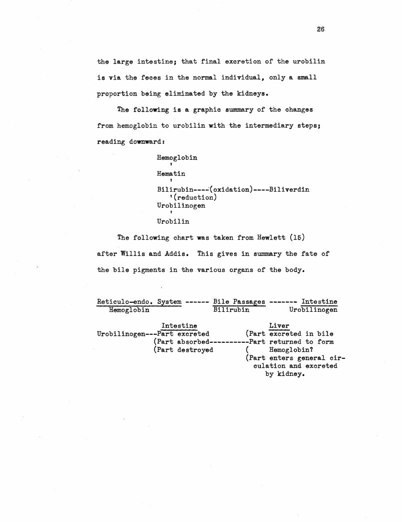
The following is a graphic summary of the changes
from hemoglobin to urobilin with the intermediary steps;
reading downward:
Hemoglobin
' Hema tin
Bilirubin---~(oxidation)----Biliverdin '(reduction)
Urobilinogen '
Urobilin
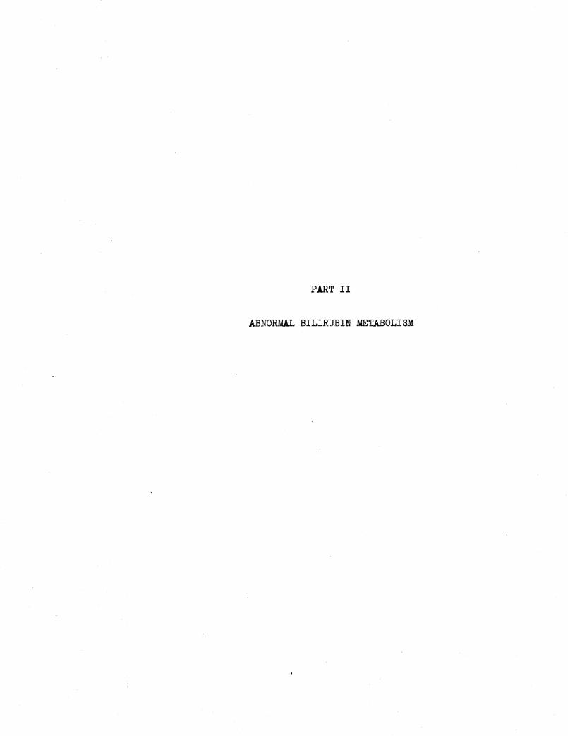
The following chart was taken from Hewlett (15)
after Willis and Addis. This gives in summary the fate of
the bile pigments in the various organs of the body.
Reticulo-endo. System ------ Bile Passages ------- Intestine Hemoglobin Bilirubin Urobilinogen
Intestine Liver Urobilinogen---Part excreted (Part excreted in bile
(Part absorbed----------Part returned to form (Part destroyed ( Hemoglobin?
(Part enters general oiroulation and excreted
by kidney.
PART II
.ABNORMAL BILIRUBIN METABOLISM
r
l F !
~-l'
l
- -- ---------------
27
Having considered the normal physiology of bilirubin,
one can now view the pathological disturbances of its metabo-
.lism with clearer insight. Knowing the normal physiology,
one can point to the process of the bilirubin metabolism in
which the fault lies in the pathological process.
The most objective sign of abnormal or pathological
bilirubin metabolism is jaundice or icterus. This phenomenon
is defined by Dorland as "a syndrome characterized by hyper-
bilirubinemia and deposition of bile pigment in the skin and
mucous membranes with resulting yellow appearance of the
patient." Jaundice comes from the French word "jaune", meaning
yellow.
Jaundice, being such an evident thing, has been described
in connection with disease from the times of the early Greeks.
The Greeks called the disease ioterus. This word is derived from
the Greek word applied to an animal resembling our oat. The
eyes of this animal were said to be of the yellow color char-
acteristic of jaundice. Others believe that the word is de-
rived from the name of a bird called icterus; the sight of
this bird was a cure to the jaundiced patient, but death to
the bird, as the belief goes. (21)
The Latins called jaundice "morbus regius", from the
yellow color of gold, the "Rex metallorum". For the same
reason, another Latin synonym is "aurigo" from "aurum, meaning
gold.
28
Jaundice oould hardly fail to be noticed early in the
history of mankind, as the change of complexion and color would
be a striking appearanoe. Acoordingly there is abundant mention
of it in the genuine Hipp.oeratic writings, whioh most often speak
of it as a complioation of other diseases rather than as a
disease in itself. In the nAphorisms" it is said to be a bad
sign if the liver becomes hard during jaundice; and in the third
book of the "Epidemicsn, that an habitual pain in the neighbor
hood of the liver, with other symptoms, preceeded a jaundice.
It is disputed if Hippocrates conceived any relation between the
bile secreted by the liver and the symptom of jaundice. (21)
Galen asserts that "the yellow bile, when it is carried all
over the body, still keeping its. own nature, causes a disease
called jaundice". He believed that "weakness of the gall bladder,
which does not draw to itself the bile out of the liver as it is
accustomed, leaving the blood impure, is the cause for jaundice".
Galen also states that jaundice may be found without any associated
liver damage. (21) In the light of our present day knowledge,
it is remarkable that such accurate conclusions could be dra11n
from pu:re observation.
Returning to the present-day conception of jaundice, Rice (38)
in view of the .known physiology of bilirubin, states that jaundice
will occur if any of the four following qualifications a.re met:
1. "If the threshold of the liver for bilirubin
excretion became greatly raised."
29
~. "If for any reason, bilirubin were produced faster
than the normal liver cells could excrete it."
3. "If the mechanism of the liver were so disturbed
that the amount of bilirubin normally produced could not be
satisfactorily removed from the blood."
4. "If there occurred any combination of the above."
According to Rice, the first is hypothetical, with no evi
dence of its existence, and which it is unnecessary to involve
in order to explain any form of jaundice.
The second classification, an uncomplicated excessive pro
duction of bilirubin, is rarely a cause of jaundice for two reasons,
according to Rice: "First, because the normal liver is able to
excrete much more bilirubin than is ordinarily delivered to it;
and second, because an excessive production of bilirubin is, as we
shall see, practically always associated with other conditions
which tend to impair the excretory power of the liver." There is
a tremendous reserve power in the liver in excreting bilirubin.
The excretory power of more than 95% of the liver substance of the
dog or monkey can be abolished, and yet the remaining 5% of the
liver tissue will suffice to prevent the development of jaundice.
In the human, atrophy of over half of the liver has been shown not
to produce any noticeable jaundice. The fact that the liver
possesses a much greater capacity for excreting bilirubin than it
is called upon to use normally makes it clear that up to a certain
limit an over-production of bilirubin will be taken care of without
30
the development of cutaneous jaundice. Just what this limit is
in the human being is not accurately known, but the numerous
pathological conditions in which the amount of pigment in the
stools is greatly increased above the normal in absence of jaundice
stand as evidence that marked increases in the amount of bilirubin
produced can be excreted by the normal liver well enough to
prevent visible jaundice. It is imaginable that with excessive
production over a long period of time the excretory power of the
liver may be impaired due to the strain put upon it. There is
no evidence that this occurs; •on the contrary one would believe
that an uncomplicated demand for increased work would, through the
process of work hypertrophy, increase the liver's efficiency,
as in the case of the other excretory organs, rather than depress
it. Overproduction of the pigment is ordinarily associated with
conditions other than mere overstrain which tend to depress the
excretory power of the liver (e.g. anoxemia, febrile disease,
immaturity of liver cells); thus it is believed that jaundice
rarely, if ever, results from simple overproduction of bilirubin."
(38)
The third possibility (liver damage to the extent that the
normal bilirubin production can not be excreted) undoubtedly
causes certain oases of jaundice. No jaundice is known to, result
from a functional depression of the excretory power of the liver
without actual loss of cells through necrosis. There is no patho
logical condition which is known to impair the function of the
3}
living hepatic cells to such a degree that they are unable to
prevent jaundice by excreting the amount of bilirubin normally
found. If. however, the total amount of functioning liver sub
stance is reduced by necrosis and cellular damage, jaundice will
result wherever the loss of liver tissue reaches a point at
which the activity of the cells remaining alive is insufficient
for the proper removal of the pigment from the blood (e.g. acute
yellow atrophy, yellow fever, chloroform poisoning). (38)
The fourth possibility (a combination of any of the preceeding
possibilities) is the most common. A liver with reduced capacity
for excreting bilirubin may be able to rid the blood of the amount
of pigment formed normally; if bilirubin is produced in excess,
such a liver may be quite unable to excrete it at all, and jaundice
will result from the retention of the excess pigment. (38)
In line with the preceeding discussion, the clinician
classifies jaundice as being hepatogenous or hemotogenous in origin.
The hepatogenous jaundice is caused by the inab~lity of the liver
to excrete via the bile any or all of the bilirubin brought to
it by the blood stream. The pathology of this process is found
entirely within the liver. Rice (38) has termed this regurgitation
jaundice. The cause of this jaundice is blockage of the bile
passages so that bile cannot be excreted (calculus, inflammatory
processes, or neoplastic disease). The comm.only accepted view on
the manner of origin of the jaundice is that after occlusion of
the comm.on bile duct the bile continues to be excreted by the liver
r---·· !
32
parenchyma.. and sinoe it is unable to follow its normal oourse
to the duodenum, it is damned back into the delicate bile capillaries.
The increase in tension in these very delicate structures causes
them to rupture; the extravasated bile is absorbed either by the
blood capillaries or by the lymphatics and is then distributed
throughout the body. being in part deposited in the tissues and
in part excreted by the urine. Bloom (4) has proven that the
removal of bilirubin early in obstructive jaundice is via the
lymphatics, then later by the blood. That is. before the bile
canaliculi rupture, the bilirubin is resorbed by the lymph in
the space immediately about the canaliculi and returned to the
systemic system before rupture has taken place.
The hemotogenous jaundice has been termed retention jaundice
by Rice (38). This implies merely an abnormal retention of the
bilirubin within the systemic circulation. Basically it is con-
earned with a minimal amount of liver dam.age and a pronounced
tendency for peripheral blood destruction (e.g. hemolytic icterus,
any poison causing intravascular hemolysis).
Besides hepatic dysfunction due to hepatic disease, there
is a type of constitutional nature. The constitutional type
shows a light yellow or sallow tint to the skin, however, the
conjunctiva is not colored. There is no demonstrable hepatic
or splenic damage. The urine contains appreciable amounts of
bilirubin and considerable amounts of urobilin. This condition
has not been adequately explained. but it has been found in j;he
I I
33
people falling within this group a tendency to hepatic disease,
especially cholecystitis. (42)
Before continuing further in the field of jaundice it is
necessary to reconsider some of the factors in the van den Bergh
test for bilirubin. As will be remembered, van den Bergh got
four different types of reactions with his reagent, namely the
immediate direct reaction, the delayed direct reaction, the bi-
phasic direct~reaction, and lastly the indirect reaction. Van
den Bergh was unable to discover the factor which underlies these
different reactions, nor has this problem been solved up to the
present time. A voluminous literature has accumulated which
relates to this subject.
Van den Bergh (45), in 1918, found a direct reaction occurred
with serum of a patient having obstructive jaundice (regurgitation
type). He found that an indirect reaction resulted in a patient
having hemolytic icterus (retention type of jaundice). Because of
this and other work, van den Bergh suggested that there was some
chemical difference between the two forms of pigment, or perhaps
the pigment was modified by its attachment to the protein of the
serum; the form present in the serum of the obstructive jaundice
was probably circulating in a free state, while that giving the
indirect reaction was bound with serum protein and, in order to
detect its presence, had to be liberated from the proteins by the
alcohol. Thus, van den Bergh believed that in the passage through
the hep~tic cells the bilirubin was changed chemically, thus
accounting for the difference in reaction in the obstructive and
hemolytic types of jaundice.
Blank:enhorn (10) found that.there was present in bile and in
many icteric sera a form of bilirubin which would pass through an
animal membrane, showing that since it acted as a diffusate on
dialysis, it must be in the crystalloid state. He found, however,
that this was a relatively unstable form., for on repeated dialysis
of .the diffusates less and less would pass through the membrane,
indicating that it readily reverted to a non-dialyzable or colloidal
form. Collinson and Fowweather (11) applied this observation to
the.van den Bergh reaction, and found that the crystalloid form
alone gave the direct positive reaction, while the non-dialyzable,
or colloidal, form was direct negative, reacting with the diazonium
salt only after the addition of alcohol and giving the indirect
reaction. They conclude that the differ~nce is largely in physical
state; the direct negative being free bilirubin, a suspensoid
colloid; the d;rect positive being a pure crystalloid.
Vaughan and Hubbard {46) after studying xanthochromie spinal
fluid, concluded that the type of reaction depends upon the
quantitative relationship between the pigment and protein. They
found with material of low protein concentration that when the
bilirubin exceeded 0.3 mg% the van den Bergh reaction became
direct. They believe, therefore, it is not necessary to consider
that passage through the liver cells is the determining factor
that changes the indirect to the direct type of bilirubin. Gregory

r !
35
and Andersch (l~) have shown through clinical studies that as
high as 26 mg% of bilirubin can be found in spinal fluid with an
indirect van den Bergh; therefore they do not believe that the
quantity of bilirubin present in the sera determines whether the
van den Bergh will be direct or indirect in contradistinction to
Vaughan and Hubbard's work.
Fowweather (11) has reviewed the literature well on the van
den Bergh reaction, and the following are only a few of the many
theories put forward.
Davies and Dodds, 1926, believed that bilirubin was
responsible for the direct reaction, whereas an oxidation product
of bilirubin was responsible for the indirect reaction.
Roberts, 1928, put forward the view that bilirubin in
the free colloidal condition was the pigment of indirect sera, while
in direct sera the bilirubin was in combination with some substance
the nature of which is as yet undetermined.
Newman, 1928, opposed the view that the pigment of
indirect sera was free bilirubin and claimed that the pigment of
direct sera was a sodium hydrogen bilirubinate.
McGowan, 1930, considered that the pigments of direct
and indirect sera were identical, the types of van den Bergh
reaction being dependent on the alkali reserve of the blood;
indirect reactions were obtained when the alkali reserve was
diminished.
Snider and Reinhold, 1930, claimed that the type of
36
van den Bergh reaction was dependent on the concentration of the
pigment which is present, direct reactions being associated with
high concentrations of bilirubin, indirect reactions with low
concentrations.
Kuster believes a keto-enol modification of bilirubin
exists explaining the varied reaction.
Fowweather, 1926 and 1931, put forth the idea that the
pigment of indirect sera was free bilirubin as an acid, while the
pigment of direct sera was a salt of bilirubin, probably the
ammonium salt.
Mann and Bollman (32) have shown that the van den Bergh
reaction for serum bilirubin is indirect in animals that have
become jaundiced following complete removal of the liver. The
amount of bile pigment that accumulates in the blood for the first
24 hours after the removal of the liver is comparable in amount to
that which is found after an equal interval of time following
ligation of the common bile duct and extirpation of the gall
bladder. In the latter case the bilirubin of the blood gives
a direct van den Bergh reaction. Removal of the liver when
obstructive jaundice is present produces serum bilirubin with a
true biphasic reaction obtained by the van den Bergh method. If
the liver is removed several hours after ligation of the comm.on
duct and rem.oval of the gall bladder, at a time when a definite
direct van den Bergh reaction is present in the serum, the amount
of bilirubin reacting directly will be subsequently unchanged.
I !
37
Additional bilirubin will accumulate in the blood after removal of
the liver, so that the total amount of bile pigment in the blood
will progressively increase at the same rate that it was increasing
following ligation of the common bile duct, but the bilirubin that
is added to the blood after removal of the liver gives only the
indirect reaction. Thus Mann and Bollman's experiments show the
relative importance of liver passage of bilirubin in relation to
the way it will react to the van den Bergh reaction, but it also
shows a possible mechanism for explaining the delayed direct and
the biphasic reactions. McNee, 1922, (29) believes blood from
obstructive jaundice would yield an immediate direct reaction,
hemolytic jaundice would account for the indirect reaction, and
jaundice due to a toxic agent and which, furthermore, may be due
to a combination of obstructive and hemolytic types would account
for a biphasic reaction.
Probably one of the most ingenious of the theories put out
thus far is that of Barron's. (2) Because of its apparent rationality
and the usefulness in explaining the various van den Bergh phenomena
it is given in more detail than the other theories. It must be
remembered, however, that this theory is not accepted by all.
Barron, in contradistinction to van den Bergh, does not believe
that the passage of bilirubin through the hepatic cells changes the
bilirubin in any manner; but to him it has seemed more probable
that there is some substance present in the bile which is responsible
for the difference in the reaction. In other words, he believes
38
that the type of reaction depends upon the meidium. in which the
pigment is contained rather than upon any alteration of the pigment
itself. Barron has shown that, while pU18bilirubin at the pH
of blood gives the direct or prompt action, if it be added to normal
plasma it then gives the indirect reaction. This Barron believes
to be the result of the adsorption of bilirubin by the plasma
proteins, the pigment being in ~hat way prevented from reacting
promptly with the reagent. If, however, substances which have
the property of lowering surface tension are first added to.the
plasma and then bilirubin is introduced, or if such substances
are added to the plasma at the same time as the bilirubin, the
r~aotion of the pigment remains direct. This occurs apparently
for the reason that substances which lower surface tension seem
to be adsorbed by proteins more readily than is bilirubin by
the plasma proteins, and it is significant that these substances
are present in whole bile and occur in increased a.mounts in the
plasma in various forms of jaundice in which the blood bilirubin
gives the direct reaction. Bile salts and acids have the faculty
of lowering surface tension and are the active principles in this
role in the case of bile. In obstructive or regurgitation jaundice,
whole bile, containing both bilirubin and bile salts, is regurgitated
into the systemic circulation. The bile salts collect at the
protein interface leaving the bilirubin free to react with the
diazo compound, giving a direct reaction.
When sodium bilirubinate solution givi~ a direct reaction is
39
added to normal serum in increasing amounts up to 12 mg%, a
typical indirect reaction takes place. When the concentration
is increased to 16 mg%, the reaction becomes of the biphasic type.
When the concentration is higher than 16 mg%, a direct reaction
is obtained. Barron believes that up to 12 mg% the bilirubin is
being adsorbed by the protein; from 12 to 16 mg%, small enough
amounts of bilirubin are left free from the protein interface, or
relatively free of this interface_ so that a biphasio re~ction may
take placeJ from 16 mg% on upwards the bilirubin remains free in
the plasma and thus gives the direct reaction. The direct reaction
can be obtained from the first by adding bile acids which have
the surface tension lowering effect in the case of bile.
Whatever be the ultimate explanation of the different forms
of the reaction, a great amount of experimental and clinical in
vestigation has made it perfectly clear that when the plasma
bilirubin in a case of jaundice is subjected to the van den Bergh
test, the direct reaction indicates that whole bile, containing
bile acids and cholestrin as well as bilirubin, has been regurgi
tated into the blood stream, and therefore that one is dealing
either with obstruction of the ducts or with necrosis of the liver
cells, for these are the two conditions which permit bile to
escaped from the canaliculi into the blood; the indirect reaction,
on the other hand, informs us that the bilirubin in the plasma
under examination has not been regurgitated into the blood from
the caniliculi, but that it represents pigment which the liver has
not yet been able to remove from the blood stream. Bloom. (4)
brings out a little recognized fact concerning obstructive jaundice
that gives very good proof of the above summarizing statement.
He found that bilirubin in early obstructive jaundice is removed
via the lymphatics surrounding the canaliculi before rupture of
these canaliculi ever takes place. During this period when the
bile pigment is thus being resorbed. the van den Bergh shows an
indirect reaction. He has shown histologioally that ruptured
bile canaliculi are not found in the early stages of obstructive
jaundice. But as soon as the bile canaliouli have ruptured.
pouring whole bile into the lymphatic and blood streams. the van
den Bergh becomes direct positive.
The biphasic reaction is due to a small amount of direct
reacting pigment in plasma containing a large amount of indirect
reacting pigment. According to Barron's view. a biphasio reaction
would occur. for example. if bilirubin continued to be added to
the plasma. the proteins of which had already adsorbed all of the
pigment they were capable of holding. This form of reaction does
not yield the clear out information which the pure direct or
indirect reaction yields. for it can occur as a result of a high
degree of pigment retention in the abscence of regurgitation. or
it may occur in certain stages of biliary regurgitation caused
by obstruction or by necrosis of liver cells. However considered
in relation to findings in the stools and urine in a given case.
it is frequently possible to determine whether the biphasio
41
reaction results from the retention or from the regurgitation
of bilirubin.
In contradistinction to the condition of jaundice, there
may exist a condition of hypobilirubinemia. This condition is
found in all secondary anemias where blood lost by hemorrhage is
not promptly replaced. It can also be associated with alterations
in the hematopoietic system (diminution of the number of red blood
cells thrown into the general circulation) which have as a conse
quence a diminished production of bilirubin. Hypobilirubinemia
is found in chronic nephritis with cardiac complication, secondary
anemias, aplastic anemias, and chlorosis. Murphy, in 190 cases of
secondary anemia'due to different conditions f~nds a persistant
hypobilirubinemia. This condition is due, then, to a general
lack of blood destruction in the body. This fact may be taken
as further proof of the derivation of bilirubin from hemoglobin.
The classical work on urobilin has been done by McM'asters
and his co-workers already mentioned before. Besides studying
the normal physiology involved in urobilin, they have also included
pathological study that explains very nicely the abnormal physiology
occurring in the human patient.
He has shown (24) that urobilin is not formed normally unless
bilirubin reaches the large intestine. In further experimentation,
(26) urobilinuria has never found after liver damage except when
bile pigment was present in the intestine. Thus, for example,
42
it appeared during the first days after ligation of the common
duct, but disappeared as the stools became aoholie. When this
had happened, a small amount of urobilin-free bile, given by mouth,
precipitated a prompt urobilinurea. After obstruction of the duet
from one-third of the liver, mild urobilinurea was found, but no
bilirubinurea. In animals intubated for the collection of a part
of the bile only, while the rest flowed to the duodenum through the
ordinary channels, liver injury caused urobilinurea, unless indeed
it was so severe as to lead to bile suppression, when almost at
onee the urobilinurea ceased, though the organism became jaundiced.
He ooneludes that "urobilinurea is an expression of the inability
of the liver cells to remove from the circulation the urobilin
brought by the portal stream with the result that the pigment
passes on to the kidney and urine. Urobilinurea occurs with far
less degree of liver injury than does bilirubinurea. 11
MoMasters (27) also found that animals rendered urobilin-free
by the collection of all the bile from the intubated, uninfected
common duot, remain urobilin free during and after extensive blood
destruction due to intravenous injections of hemolyzing agents
and also reinjections of the animals own hemolyzed blood. On the
other hand, when bile flow into the intestine is uninterrupted,
urobilinurea occurs during blood destruction caused in any of the
ways mentioned, and it parallels, both in severity and duration,
the destructive process. He concludes, "Urobilinurea, occurring
during blood destruction, is primarily the result of an increased
I I
43
excretion of bilirubin from which, in turn, an unusually large
quantity of urobilin is formed within the intestine. The liver
fails to remove from the portal blood all of the latter pigment
which is resorbed and consequently some of it reaches the kidneys
and urine."
And again, McMasters (28) shows that experimental infection
of the intubated and previously sterile biliary tract of the dog
with particles of the stools leads to a formation of urobilin from
the bilirubin of the bile as it flows through the ducts. No
urobilinurea occurs, however, unless temporary biliary obstruction
is produced, or the liver parenchyma is injured. Then urobilinurea
develops, despite the fact that no bile is reaching the intestine
and, by corollary, no urobilin being formed there. "Cholangitic
urobilinurea, as one may term the phenomenon just described, to
distinguish it from the urobilinurea having origin in the pigment
absorbed from within the intestine is far more pronounced in animals
possessing a healthy gall bladder than in those with a pathological
gall bladder or with one prevented from functioning by severance
of the cystic duct. These facts suggest that there may be an
active absorption of urobilin from the normal gall bladder. There
can be no doubt that the pigment is absorbed from within the
bile ducts." He further states that there is no justification
for the belief that urobilin is ever formed through the action of
the liver parenchyma..
In summarizing this work on urobilinurea, we may say;
44
urobilinurea occurs in excessive hem.olysis and whenever there is
an excessive excretion o~ bile pigments by the liver (as when a
stone suddenly becomes dislodged from the common duct allowing a
large amollll.t of bile to empty into the gut at once); no urobilinurea
occurs with obstruction of the common bile duct since the bilirubin
must reach the intestine to be changed into urobilin, but when
hepatic cells are affected by bacteria or toxins, urobilin may be
formed in the bile ducts themselves resulting in urobilin being
formed within the liver itself. The excretion of urobilin from
the portal blood stream is one of the most delicate functions of
the liver, and is one of the first functions to become impaired
ina pathological process; whereas., the liver may still be function
ing well in regards to bilirubin excretion, yet some increased
quantity of urobilin may be e:scaping the liver and be showing·,
in the urine. Thus, the test for urobilin in the urine is a good
functional test of the liver and rather delicate in nature, too,
and may be one of the first signs of liver dysfunction.
An attempt will now be made to correlate those factors brought
out in the normal and abnormal metabolism of bilirubin and show
their application in clinical medicine.
Being presented with a clinical case of jaundice, one may
usually come to an accurate diagnosis by studying the type and
amount of the biliary pigments within the blood., the urine, and
the stools. By studying these factors, one can determine whether
45
or not the jaundice is of the regurgitation type or of the retention
type. A summary of the -Oharacteristios of these two types of
jaundice will be given at this point.
Retention jaundice, in its pure form. wh~tever the etiology.
is recognizable clinically by the following characteristics:
1) The bilirubin of the plasma. never having passed through the
liver cells and being, therefore, unmixed with the constituents of
the bile, gives the indirect van den Bergh reaction; 2) The stools
contain an increased amount of urobilin because, in the face of the
increased production of bilirubin the liver is excreting more of
the pigment than normally, even though it cannot excrete enough
of the excess to prevent jaundice; and 3) The urine contains an
increased amount of urobilin but no bilirubin; (it is not yet
clearly understood why only the direct reacting bilirubin can
escape readily from the blood to the urine; but if it be true that
the indirect reacting bilirubin is bound to plasma protein while
the direct reacting bilirubin is not, it is clear that the latter
form could be excreted more readily by the kidneys than the former);
and again, the urine will be free from bile salts because bile
salts are formed in the liver and are absent from the urine unless
they have been poured into the blood by the escape of whole bile
from the caniliculi into the blood stream. Pathologically, the
liver may show cloudy swelling, or atrophy, according to the
etiological reason for their mal-function, but there is no wide
spread necrosis; and the bile ducts are patent.
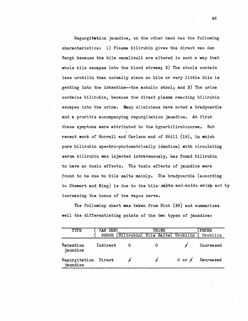
46
Regurgitation jaundice. on the other hand has the following
oharacteristios: i) Plasma bilirubin gives the direct van den
Bergh because the bile canaliculi are altered in such a way that
whole bile escapes into the blood stream.1 2) The stools contain
less urobilin than normally since no bile or very little bile is
getting into the intestine--the acholic stool; and 3) The urine
contains bilirubin. because the direct plasma reacting bilirubin
escapes into the urine. Many clinicians have noted a bradycardia
and a pruritis accompanying regurgitation jaundice. At first
these symptoms were attributed to the hyperbilirubinurea. But
recent work of Horrall and) Carlson and of Still (19). in which
pure bilirubin spectro-photometrically identical with circulating
serum bilirubin was injected intravenously. has found bilirubin
to have no toxic effects. The toxic effects of jaundice were
found to be due to bile salts mainly. The bradyoardia (according
to Stewart and King) is due to the bile~ ,aa~-11l;;SnUd'!ll!!.tirts,,whie!t act by
increasing the tonus of the vagus nerve.
The following chart was taken from Rich (38) and summarizes
well the differentiating points of the two types of jaundices
TYPE ( VAN DEN( URINE (FECES ( BERGH (Bilirubin( Bile Salts( Urobilin ( Urobilin
Retention Indirect 0 0 I Increased jaundice
Regurgitation Direct I I 0 or I Decreased jaundice
47
"Into these two main groups all various forms of jaundice can
be rationally and helpfully placed from the standpoint of patho
genesis, for the results of laboratory findings, as listed above,
will enable one to determine' the fundamentle mechanism responsible
for the jaundice in a given case and will permit one to form a
judgement regarding the general type of anatomical alteration
present in the liver." (Rice, 38)
We may now mention some of the other clinical applications
that are concerned with bilirubin metabolism.
Perkin (36) believes that a routine cheek of the van den
Bergh of patients taking arsenioals affords the earliest possible
indication of damage to the liver. If a record of the blood
bilirubin is made at the oonnnencement of the treatment and re
peated at regular intervals during the oourse, one may proceed
with safety as long as the reading remains normal. A rise of a
few mgs. is significant and the patient should be treated accordingly.
Any considerable rise calls for immediate cessation of treatment.
Once an arsenical hepatitis is established, determination of the
bilirubin offers a reliable evidence concerning the severity and
progress of the lesion.
Boehner-Mortensen (5), from his results and comparison with
the results of other liver function tests, concludes that in its
limited field of application, the bilirubin tolerance test is a
sensitive test of the liver function. While inapplicable in
hepatic disorders with bilirubinurea, it is useful in the diagnosis
48
of cirrhosis and in evaluation of the condition of the liver in
chronic hepatitis, cholangeitis, and in examination of possible
injury of the liver in infectious diseases and different disorders
such as diabetis, exophthalmic goiter and certain skin and brain
disturbances. It may prove to be especially suitable for the
investigation of possible lasting functional disorders of the liver
following acute hepatitis.
Rozendal (42) states that the mechanism for secretion of
bilirubin is a sensitive one. It is influenced by emotion and by
disturbances of the sympathetic nervous system, as well as by very
minor degrees of hepatic injury. An increased concentration of
bilirubin may be the one laboratory evidence of dysfunction and
it should be used more widely. 1t will in all probability
disclose many unsuspected cases of mild functional and organic
disturbances of the liver as well as a group of individuals of
a constitutional type especially susceptable to disease of the
gall bladder.
The diagnostic value of hypobilirubinemia in the differentia
tion of pernicious anemia from aplastio or secondary anemia is now
a well known procedure. (2) The increase of urobilogen in pernicious
anemia associated with an icterie tint of the solera and skin and
often a palpable liver presents difficulty at times in the differ
entiation between hepatic disease and primary anemia. Aside from
blood morphology, the examination of the feces indicates an increased
pigmentation to a dark brown color in contradistinction to the light
49
or aoholic feces accompanying liver disease. In the anemias,
bile is never found in the urine, while in hepatic diseases associa
ted with icterus, bile is always present.
The constant presence of urobilogen in oatarrhal jaundice,
and its total absence in mechanical icterus, such as encountered
in carcinoma of the head of the pancreas and of the biliary tract,
forms a marked distinguishing diagnostic feature between this
benign form of jaundice and those due to malignant causes. The
presence of urobilogen, even in traces, during the state of jaundice
promptly rules out an obstruction or mechanical icterus.
Bilirubin metabolism has been studied more carefully than
any other function concerned with the liver to the present date.
Knowledge of this has been a great aid in diagnosis of liver
dysfunction, but can not be relied upon too much. Quite a bit
of confusion is present regardless of the nice theories that have
been· concocted to explain the biphasic and delayed direct van
den Bergh tests. These results occur quite frequently in cases
of jaundice and are of little diagnostic value in single tests,
but when repeated over some time, they often change to a direct
or true indirect reaction. (47) However, great aid is obtained
by studying the constituents of the feces and urine. The tendency
has been to shy away from the use of the van den Bergh test ..and
to rely upon the lactose tolerance tests and such related tests.
These newer tests certainly have their place since they test
an altogether different function of the liver. But, in the light
50
of our present knowledge, the tests for bilirubin and urobilin
in the blood• urine and feces are still important steps in
diagnosing a given case of jaundice.
Recent experiments with bilirubin have offered what may be
a truely great advance in our knowledge of the purpose of bili
rubin. To date it has been considered only as a waste product
of hemoglobin metabolism. Najib-Farah (35) working on pneumococcal
infections in rabbits noted that the rabbit sera contained no
bilirubin. Knowing the lytic action of bile on the pneumocooous,
he added bilirubin to the rabbit sera. Whereas before the pneumo
oocoi grew rapidly in the sera, the growth of the virulent pneumo
cocci was inhibited by the addition of bilirubin. The organisms
undergo agglutination and disintegration (partial and complete
lysis). the capsules being denuded of their contents. The
possibility of bilirubin being an aid to body defense in certain
infections is indeed a new concept--the possibility that bilirubin
may be an agglutinin and a lysin in vivo is interesting, and
certainly is a line of thought that is worthy of further study.
* * * * * * * * * * * *
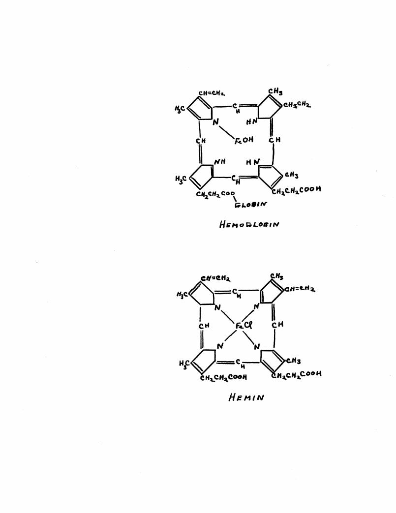
He111N
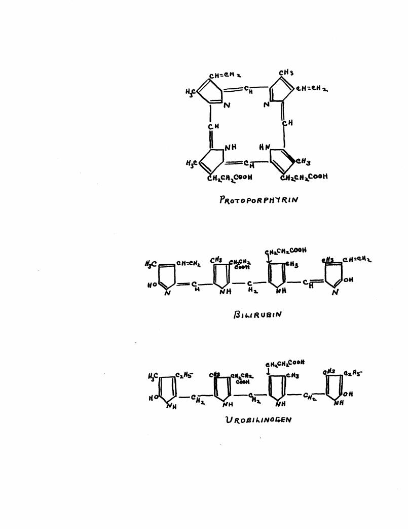
P1t0To PoR PH"t RI,.,
/3u .. 1RUB1tl
e'-CHa.Coolt
~C:QC.,.Ht»" cr.,r~ J.0_.... t:ralr tC -e-V_-c;.- -cc- oH
N Ha. llH i. NH e. H
lJ R.OlllklNOt;.EH
51
BIBLIOGRAPHY
1. Andrewes, c. H. The Nature of the Difference between the Bilirubin of Obstructive and Hemolytic Jaundice, Br. Jr. Exp. Path. 5:213-219, 1924
2. Barron, E. s. G. Bilirubinemia, Medicine, 10:77-133, 1931
3. Blankenhorn, M. A. On the Absorption of Bile Pigments from the Intestines, J. E:itp. Med. 45:195-303, 1927
f. Bloom, William. The Role of Lymphatics in the Absorption of Bile Pigment from the Liver in Early Obstructive Jaundice, Johns Hopkins Hosp. Bul. 34:316-320, 1923
5. Bochner-Mortensen. Bilirubin Tolerance Test, J.A.M.A. 103:1188, Oct., 1935
6. Bodansky, M. Introduction to Physiological Chemistry, a'34-245, New York, John Wiley & Sons Inc., 1934
7. Boyd, William. The Pathology of Internal Diseases, 353-368, Philadelphia, Lea & Febiger, 1935
8. Corr, Phillip. Histochemical Evidence Concerning the Site of Formation of Bile Pigment, Arch. Path., 7:84-93, 1928
9. Downey, Hal. Handbook of Hematology, Vol. IV, 2445-2580, New York, Paul B. Hoeber Inc. of Harper & Bros., 1938
10. Elton, N. w. Physiology, Correlations and Technic of the van den Bergh Reaction, Icterus Index, and Quantitative Serum Bilirubin, J. Lab. & Clinic. Med., 17:1-13, 1931
11. Fowweather, F. s. Bilirubin and the van den Bergh Reaction, Biochem. Journ., 26:165-182, 1932
12. Gregory, Raymond, & Andersch, Marie. The Influence of the Quantity of Serum Bilirubin on the Type of van den Bergh Reaction, J. Lab. & Clinic. Med., 23:572-74, 1938
13. Haldeman, K. o. A Histologic Study of the Formation of Bile Pigment, Archives of Path., 7:993-1011, 1919
14. Heilbrun, N., & Hubbard, R. An Experimental Study of the Biphasic van den Bergh Reaction. Am. Jour. of Clin. Path~, 8:273-280, 1938
52
15. Hewlett, Albion W. Pathological Physiology of Internal Diseases, 326-336, New York & London, D. Appleton & Co, 1928
16. Hoover, c. F., & Blankenhorn, M. A. Dissociated Jaundice, Arch. 1p.t. Med.,, 18:289-308, 1916
17. Horral, O. H., & Carlson,, A. J. The Toxic Factor in Bile, Am. Jour. Physiology, 85:691-606, 1925
18. Howell, William H.,, A Textbook of Physiology, 864-887, Philadelphia, W. B. Saunders Comp., 1936
19. Ivy, A. c., Physiologic Disturbances Incident to Obstructive Jaundice, J.A.M.A., 95:1068-1072, 1930
20. Kuhn,, A., Hawkins, W. B., & Whipple, G. H. I. Bile Pigment and Hemoglobin Interrelation in
Normal Dogs, Am. J. Physiol. 96:449-463 II. Bile Pigment and Hemoglobin Interrelation in
Anemic Dogs, Ibid. 96:463-476.
21. Legg, J. w. On the Bile, Jaundice, and Bilious Diseases, New York, D. Appleton & Co., 1880
22. Lemberg, R. Bile Pigments, Biochem. Journ. Vol XXVIII, No. 3:978-987, 1934
23. Lemberg, R. Transformation of Haemins into Bile Pigments, Biochem. Journ., Vol XXIX, No. 6:1322-1336, 1935
24. McMaster, P. D., & Elman, Robert., Studies on Urobilin Physiology and Pathology.
I. Quantitative Determination of Urobilin II. Derivation of Urobilin
J. Exp. Med., 41:503-534, 1925
25. McMaster, P. D., & Elman, R. Studies on Urobilin Physiology and Pathology.
III. Absorption of Pigments of Biliary Derivation from the Intestine, J. Exp. Med., 41:719-738, 1925
26. Ibid IV. Urobilin and The Damaged Liver, J. Exp. Med.,
42:99-122, 1926
27. Ibid v. The Relation between Urobilin and Conditions in
volving Red Blood Cell Destruction., J. Exp. Med., 42:619-640, 1926.
as. McMaster, P. D., & Elman, R. Studies on Urobilin Physiology and Pathology,
53
VI. The Relation of Biliary Infections to the Genesis and Excretion of Urobilin, J. Exp. Med., 43:753-783, 1926.
29. McNee, J. w., Discussion on Jaundice, Brit. Med. Journ., 2:495-498, 1924
30. McNee, J. w. Experiments on Hemolytic Icterus, J. Path. & Bact., 18:325-340, 1914
31. Mann, F. o., Sheard, c. H., & Bollman, J. L., The Extrahepatic Formation of Bilirubin, Am J of Physiology, 74:49-60, 1925
32. Mann, F. c., & Bollman, J. L., Jaundice--Review of Some Experimental Investigations, J.A.M.A. 104:371-374, 1935
33. Mann, F. c., & Bollman, J. L., Jaundice: A Review of Some Experimental Investigations, JA.M.A. 104:371-374, 1935
34. Mendel, L. B., & Underhill, F. P., On the Paths of Absorption from the Liver, Am. J. Phys. 14:252-259, 1905
35. Ne.jib-Farah. Defensive Role of Bilirubinemia in Pneumococcal Infections, Lancet, P• 505, 1937, Feb.
36. Perkin, F. s. Blood Bilirubin Estimation and Clinical Significance. Arch. Int. Med. 40:195-203, 112'7
37. Rabinowitch, I. M., Jaundice and the Application of the van den Bergh and Urobilinogen Tests, Canadian Med. Assoc. Journ., N.S.25:255-265, 1931
38. Rich, Arnold Rice, The Pathogenesis of the Forms of Jaundice, Johns Hopkins Hosp. Bull., 47:338-377, 1930
39. Rich, A. R., The Formation of Bile Pigment, Physio. Reviews, 5:182-224, 1925
40. Rich, A. R., The Formation of Bile Pigment from Hemoglobin in Tissue Culture, Bull. Johns Hopkins Hosp. 35:415-435, 1921!
41. Rich, A. R., Identity of Hematoiden and Bilirubin, Bull. Uohns Hopkins Hosp. 36:225-238, 1925
42. Rozendal, H. M., Comf'ort, M. w., & Snell, A. M. Slight and Latent Janudice, J.A.M.A. 104:374-382, 1935
54
43. Sackey, M. s., Johnston, c. G., & Ravdin, I. s., Fate of Bilirubin in the Small Intestine, J. Exp. Med., 60:189-194, 1934
44. Still, E. u., On the Toxicity of Purified Bile Preparations, Am. Jour. Phys., 88:729-7.37, 1929
45. Van den Bergh, Hijm.a.ns, Discussion on Jaundice, Brit. Med. Jour., 2:498-500, 1924
46. Vaughan, J. M., & Haslewood, G. A. D., The Normal Level of Plasma Bilirubin, Lancet, 1:133~135, Jan., 1938
47. Wallace, c. B., & Diamond, J. s., Significance of Urobilogen in the Urine as a Test for Liver Function., Arch. Int. Med., 35:689-725, 1925
48. Watson, c. J. Studies of Urobilinogen, Arch. Int. Med., 59:196-205, 1937
49. Whipple, G. H., Hemoglobin and Bile Pigment Studies in the Splenectomized Dog, Arch. Int. Med., 291711-720, 1923