nimesulide inhibits protein kinase c epsilon and substance p in sensory neurons \u0026ndash;...
TRANSCRIPT

© 2011 Vellani et al, publisher and licensee Dove Medical Press Ltd. This is an Open Access article which permits unrestricted noncommercial use, provided the original work is properly cited.
Journal of Pain Research 2011:4 177–187
Journal of Pain Research Dovepress
submit your manuscript | www.dovepress.com
Dovepress 177
O R i g i n A L R e s e A R c h
open access to scientific and medical research
Open Access Full Text Article
http://dx.doi.org/10.2147/JPR.S21931
nimesulide inhibits protein kinase c epsilon and substance P in sensory neurons – comparison with paracetamol
Vittorio Vellani1
silvia Franchi2
Massimiliano Prandini1
sarah Moretti2
giorgia Pavesi1
chiara giacomoni3
Paola sacerdote2
1Dipartimento di scienze Biomediche, Università di Modena e Reggio emilia, Modena, italy; 2Dipartimento di Farmacologia chemioterapia e Tossicologia Medica, Università degli studi di Milano, italy; 3Dipartimento di economia e Tecnologia, Università degli studi della Repubblica di san Marino, Montegiardino, Repubblica di san Marino
correspondence: Vittorio Vellani Dipartimento di scienze Biomediche, Università di Modena e Reggio emilia, Via campi 287, 41125 Modena, italy Tel +39 059 2055063 Fax +39 059 2055363 email [email protected]
Abstract: In this paper we describe new actions of nimesulide and paracetamol in cultured
peripheral neurons isolated from rat dorsal root ganglia (DRG). Both drugs were able to decrease
in a dose-dependent fashion the number of cultured DRG neurons showing translocation
of protein kinase C epsilon (PKCε) caused by exposure to 1 µM bradykinin or 100 nM
thrombin. In addition, the level of substance P (SP) released by DRG neurons and the level of
preprotachykinin mRNA expression were measured in basal conditions and after 70 minutes or
36 hours of stimulation with nerve growth factor (NGF) or with an inflammatory soup containing
bradykinin, thrombin, endothelin-1, and KCl. Nimesulide (10 µM) significantly decreased the
mRNA levels of the SP precursor preprotachykinin in basal and in stimulated conditions, and
decreased the amount of SP released in the medium during stimulation of neurons with NGF or
with the inflammatory soup. The effects of paracetamol (10 µM) on such response was lower.
Nimesulide completely inhibited the release of prostaglandin E2 (PGE
2) from DRG neurons,
either basal or induced by NGF and by inflammatory soup, while paracetamol decreased PGE2
release only partially. Our data demonstrate, for the first time, a direct effect of two drugs largely
used as analgesics on DRG neurons. The present results suggest that PKCε might be a target for
the effect of nimesulide and paracetamol, while inhibition of SP synthesis and release is clearly
more relevant for nimesulide than for paracetamol mechanism of action.
Keywords: nociceptors, analgesia, hyperalgesia, dorsal root ganglia, PKCε
IntroductionIn this paper we present a comparative study of mechanisms of action of nimesulide
and paracetamol, drugs largely used as analgesics, in cultured rat dorsal root ganglia
(DRG) neurons.
Nimesulide is a nonsteroidal anti-inflammatory drug (NSAID) with broad effects
on inflammation and other biochemical processes leading to a multifactorial mode
of action.1 Nimesulide is a preferential cyclo-oxygenase-2 (COX-2) inhibitor1–4 but
its anti-inflammatory and analgesic action also involves activity on a wide range of
inflammatory and pain mediators and intracellular pathways1,5 including an effect
on synovial concentration of substance P (SP) in patients with knee osteoarthritis.6
While some evidence of analgesic action of nimesulide has been linked to actions in
the central nervous system,7 to our knowledge direct effects of nimesulide in isolated
sensory neurons have not yet been reported.
Paracetamol is one of the most popular and commonly used drugs for the treatment
of moderate pain, which still presents a challenge in terms of a satisfactory explanation
of its well-established effects. Despite its antipyretic and analgesic activities, it has

Journal of Pain Research 2011:4submit your manuscript | www.dovepress.com
Dovepress
Dovepress
178
Vellani et al
almost no anti-inflammatory action and is a weak inhibitor of
either COX-1 or COX-2.8,9 Contribution of several possible
mechanisms has been proposed at a variety of levels in the
nociceptive pathway from the periaqueductal gray to the
periphery.10–16
The epsilon isoform of protein kinase C (PKCε) is a very
important peripheral effector of a number of inflammatory
mediators, including bradykinin, prostaglandins, proteases,
prokineticins, SP and others,17–21 which causes increased
activation of nociceptors via action on transient receptor
potential vanilloid member1 (TRPV1) and on tetrodotoxin
(TTX)-insensitive sodium channels.22
The neuropeptide SP has long been associated with
transmission of noxious stimuli.23 In the peripheral nervous
system it is expressed in a subset of unmyelinated nociceptive
sensory neurons and is transported to central and peripheral
axonal terminals. Centrally, SP is released in the superficial
lamina of the spinal cord dorsal horn, while in the periphery
it is implicated in neurogenic inflammation.24–26 Despite its
importance in pain transmission, little is known about SP
modulation by anti-inflammatory and analgesic drugs.
In this paper we investigate in cultured DRG neurons
the ability of nimesulide and paracetamol to decrease
translocation of PKCε, to reduce preprotachykinin synthesis
and the release of SP induced by inflammatory mediators, and
to reduce prostaglandin E2 (PGE
2) release from activated neu-
rons. We show that nimesulide and paracetamol share some
novel mechanisms of action potentially relevant for their
in vivo effects, although other effects are exclusively seen or
are quantitatively stronger with nimesulide treatment.
Material and methodsDorsal root ganglion primary culturesSprague Dawley rats (2–3 weeks old) were sacrificed
under total anesthesia according to Italian and European
legislation, with protocols in agreement with the guidelines
of the Committee for Research and Ethical Issues of IASP.27
Experimental work was also reviewed and approved by
local institutional animal care and use committee. DRGs
were collected, incubated for 1 hour at 37°C with 0.125%
collagenase (Worthington, Freehold, NJ), and mechanically
dissociated, plated onto coverslips or Petri dishes pretreated
with 10 µg/mL poly-L-lysine (Sigma-Aldrich, St Louis, MO)
and 20 µg/mL laminin (Sigma-Aldrich, Milan, Italy), and
cultured in DMEM containing 1% penicillin/streptomycin,
10% fetal bovine serum, 1% L-glutamine (Invitrogen,
San Diego, CA), 1.5 µg/mL cytosine 1-d-arabinofuranoside
(ARA-C, Sigma), as described previously.28 Neurons used
for immunocytochemistry experiments were cultured in the
presence of 100 ng/mL nerve growth factor (NGF) (Sigma)
in order to improve bradykinin and thrombin receptor
expression.29
immunocytochemistryPKCε was visualized as previously described.18,19 In brief,
rat DRG neurons cultured for 2–3 days in vitro were treated
with bradykinin (BK, at 1 µM concentration) or throm-
bin (THR, 100 nM) for 30 seconds, and rapidly fixed for
10 minutes at room temperature with paraformaldehyde
(4% formaldehyde and 4% sucrose, dissolved in phosphate-
buffered saline (PBS)/distilled water 2:1). Stimulation
solutions and then fixation solution were applied with an
automated system (FSC-1, CV Scientific, Modena, Italy). In
test experiments, nimesulide or paracetamol (from Sigma)
at different concentrations were preapplied for 120 minutes
or overnight (see Figure 1) and also added to BK and THR
applied to coverslips. Dimethyl sulfoxide (DMSO) was used
to prepare stock solutions of nimesulide and paracetamol, and
the final concentration of DMSO applied to cells was always
lower than 1:1000. Fixed cells were washed three times in
PBS (with 0.1% fish skin gelatin to block nonspecific sites),
permeabilized for 30 minutes at room temperature with Triton
X-100 (0.2% in PBS), and incubated overnight at 4°C with
a polyclonal anti-PKCε antibody17 diluted 1:1000 in PBS-T/
gelatin (PBS with 0.05% Triton X-100). Coverslips were then
incubated for 1 hour at room temperature with goat antirabbit
IgG conjugated to the fluorophore Alexa Fluor 488 (1:200;
Invitrogen), washed three times in PBS/gelatin, and visual-
ized using a confocal microscope (Leica SP2, Leica, Swit-
zerland). Activation of PKCε results in translocation from an
entirely cytoplasmic location to the neuronal cell membrane
(see Figure 1). Translocation was quantified by determining
fluorescence intensity along a line positioned across the cell
so as to avoid the nucleus (for details see Cesare et al17).
Neurones in which intensity at the cell membrane was at
least 1.5 times greater than the mean of cytoplasmic intensity
were counted as positive.
Dorsal root ganglia stimulation and drug treatmentAfter 2–3 days in vitro, DRG cultures were stimulated for
experimental procedures using either NGF (100 ng/mL) or a
cocktail of inflammatory/proalgesic mediators (inflammatory
soup, IS) of the following composition: 1 µM BK, 100 nM

Journal of Pain Research 2011:4
0Ctrl −5 −6 −7
Nimesulide (logM)
*** ***
*
*
Tra
nsl
oca
tio
n in
du
ced
by
1 µM
BK
(%
)
−8 −5OVN
10
20
30
0Ctrl −5 −6 −7
Paracetamol (logM)
*** ***T
ran
slo
cati
on
ind
uce
db
y 1
µM B
K (
%)
−8 −5OVN
10
20
30
0Ctrl −5 −6 −7
Nimesulide (logM)
*****
*
Tra
nsl
oca
tio
n in
du
ced
by
100
nM
TH
R (
%)
−8 −5OVN
5
10
15
20
0Ctrl −5 −6 −7
Paracetamol (logM)
* *
Tra
nsl
oca
tio
n in
du
ced
by
100
nM
TH
R (
%)
−8 −5OVN
10
5
15
20
25
A B
C D
E F
Figure 1 nimesulide and paracetamol inhibit protein kinase c epsilon (PKcε) translocation in dorsal root ganglia (DRg) sensory neurons. (A–B) confocal images of DRg neurons treated with bradykinin (BK, 1 µM) for 30 seconds and then fixed and stained for PCKε with a specific antibody. (A) typical behavior of an unresponsive neuron. (B) typical BK-responsive neuron showing translocation of PKcε to the plasma membrane. neurons treated with 100 nM thrombin (ThR) for 30 seconds would display similar behavior. Neurons fixed without prior treatment with BK or THR showed no sign of spontaneous translocation. Scale bar 10 µm. (C) The percentage of neurons showing PKcε translocation induced by 1 µM BK or (E) by 100 nM THR was significantly decreased by 2 hours preapplication of nimesulide at 1 or 10 µM concentration. Overnight treatment (OVn) did not result in larger inhibition (C–E). With paracetamol treatment only the largest concentration of drug tested (10 µM) significantly reduced translocation induced by BK (D) or by ThR (F). Further explanations in the text. Values are means ± seM obtained from 4–12 cultures. Notes: *P , 0.05, **P , 0.01 and ***P , 0.001 compared with control.
submit your manuscript | www.dovepress.com
Dovepress
Dovepress
179
nimesulide inhibits PKcε and substance P in sensory neurons

Journal of Pain Research 2011:4submit your manuscript | www.dovepress.com
Dovepress
Dovepress
180
Vellani et al
THR, 100 nM endothelin-1, 25 mM KCl, dissolved in normal
culture medium (DMEM + 10% FBS). Where appropriate,
a concentration of 10 µM of tested chemicals (nimesulide
or paracetamol, from Sigma) dissolved in DMSO was
preapplied for 30 minutes to cultures before treatment with
NGF and IS which was considered sufficient time to allow
full onset of their effect before application of inflammatory
mediators.
Cells were stimulated with either NGF or IS with or
without the drugs for 70 minutes and 36 hours. At the end
of these incubation periods, media and cells were collected
for further processing as described below.
Measurement of sP and Pge2 in culture mediaQuantitative determination of PGE
2 was performed by the
enzyme immunoassay using a commercially available EIA
kit (Cayman Chemical Company, Ann Arbor, MI). The
sensitivity of the PGE2 EIA kit was 15 pg/mL.
To measure SP, culture media were acidified with 1N
acetic acid and SP was measured by radioimmunoassay
(RIA) using antiserum and methods previously described
and validated.30,31 The antibody was raised in rabbit against
synthetic SP, and was directed towards the C terminal of the
peptide. I125-SP was purchased from Perkin Elmer (Monza,
Italy). Sensitivity of the RIA was 10 pg/tube and intra-assay
and inter-assay variation coefficients were 8% and 11%,
respectively.
RnA isolation and real-time RT-PcRTotal RNA from DRG cells was purified using TRIzol reagent
(Invitrogen, Life Technologies, San Giuliano Milanese, Italy).
Cells were lysed directly in the culture dish, according to the
manufacturer’s instructions and RNA resuspended in 8 µL
of water. After purification, total RNA concentrations were
determined from the sample absorbance value at 260 nm.
Total RNA (300 ng) was treated with DNase (DNA-free-
Ambion) to avoid false-positive results due to amplification
of contaminating genomic DNA. First-strand cDNA was
synthesized from 1000 ng of total RNA in a final volume of
20 µL using M-MLV RT (Moloney Murine Leukemia Virus
Reverse Transcriptase; Invitrogen, San Giuliano Milanese,
Italy). cDNA (2 µL) was subjected to real-time quantita-
tive PCR using ABI PRISM 7000 (Applied Biosystems,
Forster City, CA). TaqMan PCR was performed in 25 µL
volumes using Real Master Mix Probe ROX (Eppendorf,
Hamburg, Germany). Custom probes were prepared by
Applied Biosystems. The probes were the same as those
used in a previous study31 and were designed to span an
intron in order to avoid potential amplification of genomic
DNA in the analyzed samples. The probes were labelled at
the 5´ end with 6-carboxy fluorescein and at the 3´ end with
6-carboxy-tetramethyl rhodamine. The primers and probe
sequence for preprotachykinin (PPT, Genbank accession
number M15191) and GAPDH (Genbank accession number
AF106860) are shown in Bianchi et al.31 All PCR assays were
performed in duplicate. Before using the ∆∆CT method for
relative quantification, we performed a validation experiment
to demonstrate that the efficiencies of the two different probes
(target and reference) are equal. The reaction conditions
were as follows: 95°C for 2 minutes, followed by 40 cycles
at 95°C for 15 seconds (denaturation) and 60°C for 1 minute
(annealing and elongation). As controls, we used the reac-
tion mixture without the cDNA. Threshold cycle numbers
(CT) were determined with an ABI PRISM 7000 Sequence
Detection System (version 1.1 software) and transformed
using the ∆CT (2-∆∆CT) comparative method. Gene-specific
expression values were normalized to expression values of
GAPDH (endogenous control) within each sample. The levels
of preprotachykinin were expressed relative to the calibrator
value control cells. Relative quantification was performed
using the comparative method. The amount of target, normal-
ized to an endogenous reference and relative to a calibrator,
is given by 2-∆∆CT. Briefly, the ∆CT value is determined
by subtracting the average GAPDH CT value from the aver-
age PPT CT in the same sample. The calculation of ∆∆CT
involves subtraction of the ∆CT calibrator value.
statistical analysisData were analyzed by one way analysis of variance
(ANOVA), followed by Bonferroni’s t-test for multiple
comparison. An effect was determined to be significant if
the P value was less than 0.05.
Resultseffects on PKcε translocationActivation of PKCε by inflammatory mediators leads to its
translocation from the cytoplasm to the surface membrane
in DRG sensory neurons, which can be visualized directly
with immunocytochemistry and confocal microscopy
(Figure 1A and B).18,19,21 PKCε translocation can be quanti-
fied in terms of the number of neurons in which translocation
is observed – with this approach reliable dose-response and
time-course curves can be obtained.19,21 After application of
1 µM BK or 100 nM THR, which are saturating concentrations
for these agonists, maximum translocation was consistently
Journal of Pain Research 2011:4
PP
T m
RN
Are
lati
ve t
o c
on
tro
ls
0
1
2
3
4
*
*
#
A
B
PP
T m
RN
Are
lati
ve in
crea
se
0
1
2
3
4
*
##
#
##
Ctrl N ISIS
+ N
NGF
NGF + N
Ctrl P ISIS
+ P
NGF
NGF + P
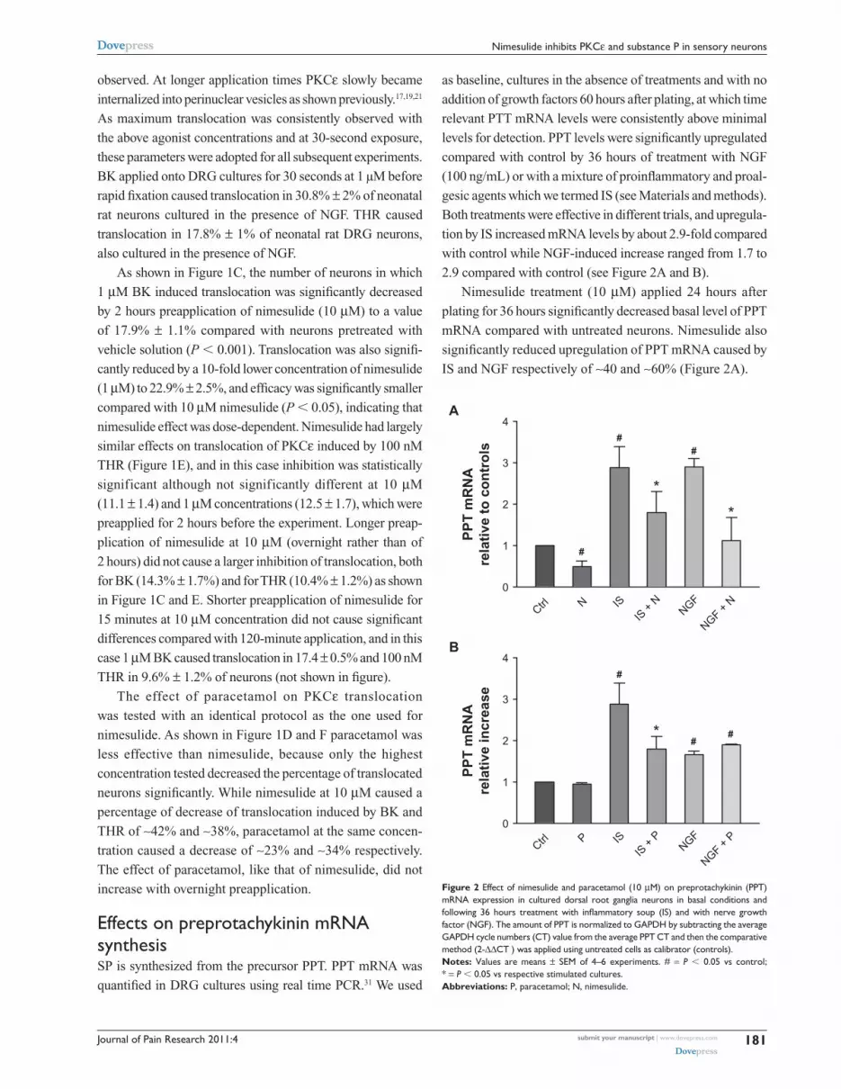
Figure 2 effect of nimesulide and paracetamol (10 µM) on preprotachykinin (PPT) mRnA expression in cultured dorsal root ganglia neurons in basal conditions and following 36 hours treatment with inflammatory soup (IS) and with nerve growth factor (ngF). The amount of PPT is normalized to gAPDh by subtracting the average gAPDh cycle numbers (cT) value from the average PPT cT and then the comparative method (2-∆∆cT ) was applied using untreated cells as calibrator (controls). Notes: Values are means ± seM of 4–6 experiments. # = P , 0.05 vs control; * = P , 0.05 vs respective stimulated cultures.Abbreviations: P, paracetamol; n, nimesulide.
submit your manuscript | www.dovepress.com
Dovepress
Dovepress
181
nimesulide inhibits PKcε and substance P in sensory neurons
observed. At longer application times PKCε slowly became
internalized into perinuclear vesicles as shown previously.17,19,21
As maximum translocation was consistently observed with
the above agonist concentrations and at 30-second exposure,
these parameters were adopted for all subsequent experiments.
BK applied onto DRG cultures for 30 seconds at 1 μM before
rapid fixation caused translocation in 30.8% ± 2% of neonatal
rat neurons cultured in the presence of NGF. THR caused
translocation in 17.8% ± 1% of neonatal rat DRG neurons,
also cultured in the presence of NGF.
As shown in Figure 1C, the number of neurons in which
1 µM BK induced translocation was significantly decreased
by 2 hours preapplication of nimesulide (10 µM) to a value
of 17.9% ± 1.1% compared with neurons pretreated with
vehicle solution (P , 0.001). Translocation was also signifi-
cantly reduced by a 10-fold lower concentration of nimesulide
(1 µM) to 22.9% ± 2.5%, and efficacy was significantly smaller
compared with 10 µM nimesulide (P , 0.05), indicating that
nimesulide effect was dose-dependent. Nimesulide had largely
similar effects on translocation of PKCε induced by 100 nM
THR (Figure 1E), and in this case inhibition was statistically
significant although not significantly different at 10 µM
(11.1 ± 1.4) and 1 µM concentrations (12.5 ± 1.7), which were
preapplied for 2 hours before the experiment. Longer preap-
plication of nimesulide at 10 µM (overnight rather than of
2 hours) did not cause a larger inhibition of translocation, both
for BK (14.3% ± 1.7%) and for THR (10.4% ± 1.2%) as shown
in Figure 1C and E. Shorter preapplication of nimesulide for
15 minutes at 10 µM concentration did not cause significant
differences compared with 120-minute application, and in this
case 1 µM BK caused translocation in 17.4 ± 0.5% and 100 nM
THR in 9.6% ± 1.2% of neurons (not shown in figure).
The effect of paracetamol on PKCε translocation
was tested with an identical protocol as the one used for
nimesulide. As shown in Figure 1D and F paracetamol was
less effective than nimesulide, because only the highest
concentration tested decreased the percentage of translocated
neurons significantly. While nimesulide at 10 µM caused a
percentage of decrease of translocation induced by BK and
THR of ∼42% and ∼38%, paracetamol at the same concen-
tration caused a decrease of ∼23% and ∼34% respectively.
The effect of paracetamol, like that of nimesulide, did not
increase with overnight preapplication.
effects on preprotachykinin mRnA synthesisSP is synthesized from the precursor PPT. PPT mRNA was
quantified in DRG cultures using real time PCR.31 We used
as baseline, cultures in the absence of treatments and with no
addition of growth factors 60 hours after plating, at which time
relevant PTT mRNA levels were consistently above minimal
levels for detection. PPT levels were significantly upregulated
compared with control by 36 hours of treatment with NGF
(100 ng/mL) or with a mixture of proinflammatory and proal-
gesic agents which we termed IS (see Materials and methods).
Both treatments were effective in different trials, and upregula-
tion by IS increased mRNA levels by about 2.9-fold compared
with control while NGF-induced increase ranged from 1.7 to
2.9 compared with control (see Figure 2A and B).
Nimesulide treatment (10 µM) applied 24 hours after
plating for 36 hours significantly decreased basal level of PPT
mRNA compared with untreated neurons. Nimesulide also
significantly reduced upregulation of PPT mRNA caused by
IS and NGF respectively of ∼40 and ∼60% (Figure 2A).

Journal of Pain Research 2011:4
Su
bst
ance
P r
elat
ive
to c
on
tro
ls (
70 m
inut
es)
0
50
100
150
200
250
300
A
B
Su
bst
ance
P r
elat
ive
to c
on
tro
ls (
70 m
inut
es)
0
50
100
150
200
250
Su
bst
ance
P r
elat
ive
to c
on
tro
ls (
36 h
ou
rs)
0
100
200
300
400
500
* *
C
D
Su
bst
ance
P r
elat
ive
to c
on
tro
ls (
36 h
ou
rs)
0
50
100
150
200
250
*
*
#
#
##
##
##
#
#
# #
Contro
l N IS
IS +
NNGF
NGF + N
Contro
l N IS
IS +
NNGF
NGF + N
Contro
l P IS
IS +
PNGF
NGF + P
Contro
l P IS
IS +
PNGF
NGF + P
Figure 3 effect of nimesulide and paracetamol (10 µM) on release of substance P (sP) measured in culture medium from DRg neurons in basal conditions and following 70 minutes (A, B) and 36 hours (C, D) treatment with inflammatory soup (IS) and nerve growth factor (NGF). Notes: Values are means ± seM of 4–6 experiments and are expressed as % of control cultures. # = P , 0.05 vs controls; * = P , 0.05 vs respective stimulated cultures.Abbreviations: P, paracetamol; n, nimesulide.
submit your manuscript | www.dovepress.com
Dovepress
Dovepress
182
Vellani et al
Effects of paracetamol (Figure 2B) were somewhat
different: at 10 µM after 36 hours of treatment it did not
cause any change in basal expression levels of PPT mRNA,
and significantly reduced upregulation by IS (by ∼40%),
while upregulation by NGF was not changed by treatment.
Both nimesulide and paracetamol were preapplied for
30 minutes to allow full onset of effect before treatment
with IS and NGF.
effects on basal and stimulated substance P release in mediumLevels of SP released in the medium by cultured DRG
neurons were assessed by radioimmunoassay (see Materials
and methods). Approximately 24 hours after plating, separate
coverslips from the same DRG cultures were treated with
either nimesulide, paracetamol (both 10 µM), with or
without NGF (100 ng/mL) or IS, or with vehicle, for a total
of 6 separate conditions (Figure 3A–D). Coverslips exposed
to treatments containing nimesulide or paracetamol were
pre-treated with the same concentration of these drugs
for 30 minutes, and control coverslips were treated with
vehicle medium for the same time. The treatments described
remained on coverslips for either 70 minutes (Figure 3A
and B) or 36 hours (Figure 3C and D), then the medium was
removed and stored at –80°C until SP measurement.
Neither nimesulide nor paracetamol altered basal levels
of release of SP, which were about 30 ± 2.19 (mean ± SEM)
pg/mL of medium at 70 minutes and 77 ± 11 (mean ± SEM)
pg/mL medium at 36 hours.
Treatment with both NGF and IS significantly increased
SP level in medium after 70 minutes and after 36 hours
(Figure 3A–D). Such release was significantly decreased
by nimesulide (Figure 3A and C) but was not changed
by paracetamol, which was completely ineffective
(Figure 3B and D). Nimesulide effect was significant after
70 minutes, and was particularly strong after 36 hours, as

Journal of Pain Research 2011:4
PGE 2 i
n m
ediu
mat
70
min
utes
(pg/
mL)
PGE 2 i
n m
ediu
mat
70
min
utes
(pg/
mL)
PGE 2 i
n m
ediu
mat
36
hour
s (n
g/m
L)PG
E 2 in
med
ium
at 3
6 ho
urs
(ng/
mL)
0
200
400
600
800A
B
0
200
400
600
800
1000
0
3
6
9
12
15C
D
0
2
4
6
8
10
*
*#
*#
*
## * *
##
#
#
#
##
#
*
Contro
l N IS
IS +
NNGF
NGF + N
Contro
l N IS
IS +
NNGF
NGF + N
Contro
l P IS
IS +
PNGF
NGF + P
Contro
l P IS
IS +
PNGF
NGF + P
Figure 4 effect of nimesulide and paracetamol (10 µM) on release of prostaglandin e2 (Pge2) measured in culture medium from dorsal root ganglia neurons in basal conditions and following 70 minutes (A, B) and 36 hours (C, D) treatment with inflammatory soup (IS) and nerve growth factor (NGF). Values are means ± seM of 4–6 experiments. Notes: # = P , 0.05 vs controls; * = P , 0.05 vs respective stimulated cultures.Abbreviations: P, paracetamol; n, nimesulide.
submit your manuscript | www.dovepress.com
Dovepress
Dovepress
183
nimesulide inhibits PKcε and substance P in sensory neurons
SP release caused by NGF and IS was totally inhibited and
reduced to values largely identical to basal levels.
effects on Pge2 release in mediumPGE
2 was quantified in DRG cultures using an EIA
(see Figure 4A).31,32 In culture medium there was little if
any PGE2 as only traces were contributed by the 10% fetal
bovine serum added to DMEM (see methods). Approximately
24 hours after plating, the medium was changed and cultures
treated either with vehicle or with IS, NGF, with or without
nimesulide or paracetamol (10 µM). After 70 minutes or
36 hours of treatment, the medium was collected and PGE2
released by cultures during this time measured. Basal release
by unstimulated DRG cultures in different sets of experiment
levels ranged between 199 ± 41 and 337 ± 37 pg/mL after
70 minutes and between 1.5 ± 0.5 and 1.7 ± 0.5 ng/mL after
36 hours. PGE2 levels were significantly increased compared
with control by 70 minutes or 36 hours of treatment with
NGF or IS. Both treatments were effective in different trials,
and IS augmented PGE2 levels by about 1.6- to 2.3-fold
compared with control, while NGF increase ranged between
1.5 and 2.0-fold after 70 minutes; the increase ranged between
4.8- and 7.4-fold (IS) and 4.6- and 4.2-fold (NGF) after
36 hours compared with control levels (see Figure 4) in dif-
ferent groups of experiments. Nimesulide treatment (10 µM)
applied 24 hours after plating for 70 minutes or 36 hours
(Figure 4A and C) completely inhibited PGE2 basal levels
released in medium compared with untreated neurons (reduc-
tion by almost 100%). Nimesulide treatment also completely
inhibited the release of PGE2 caused by IS and NGF. Both at
70 minutes and 36 hours nimesulide reduced PGE2 down to
levels similar to those present in the medium before it was
added to cultures (Figure 4A and C).
Paracetamol (Figure 4B and D) (10 µM) decreased basal
levels of PGE2 both after 70 minutes and 36 hours. Similarly,
release of PGE2 induced by IS was significantly reduced
by paracetamol both at 70 minutes and 36 hours. Differently,
PGE2 release induced by NGF was significantly decreased by
paracetamol after 70 minutes, while reduction after 36 hours
(about 30%) did not reach statistical significance.

Journal of Pain Research 2011:4submit your manuscript | www.dovepress.com
Dovepress
Dovepress
184
Vellani et al
DiscussionSpecific involvement of PKCε in nociceptor sensitization and
hyperalgesia has been described in many reports. Following
a first paper by Cesare et al,17 showing that sensitization of
heat-induced currents by bradykinin in nociceptive neurons
is mediated by PKCε, a number of studies confirmed the
importance, participation, and requirement of this enzyme
in inflammatory pain and nociception at the cellular and
whole-animal level.22,33,34 PKCε is preferentially expressed in
nociceptive neurons where it plays a crucial role in chronic
hyperexcitability, but its inhibition causes little disruption in
normal sensory function or in other functions.34 PKCε therefore
currently represents not only a well-validated target for both
inflammatory and neuropathic pain in the preclinical scientific
literature35,36 but also a novel and appealing target for therapeutic
intervention in human patients. In this light, our novel finding
of PKCε translocation inhibition by nimesulide and, although to
a lesser extent, by paracetamol, appears to be of high relevance
for a better understanding of the mechanisms of action of these
drugs. Work is currently in progress to investigate whether inter-
ference on PKCε translocation is a common feature of other
NSAIDs and analgesic drugs. PKCε inhibition by nimesulide,
paracetamol, or possibly other NSAIDs and analgesics may be
a relevant part of their pharmacological actions.
Prostaglandins work not only as mediators that directly
activate downstream cascades leading to sensitization of
nociceptor ion channels, but also function as paracrine media-
tors in nociceptor sensitization by other inflammatory agents
(glutamate, bradykinin, thrombin etc). Prostaglandins may be
produced by the inflamed tissues and by white bloods cells
participating in inflammation, but also by sensory neurons
and by peripheral glial cells. It is noteworthy that in DRG
cultures, as in DRGs and in peripheral nerve endings, there
is a significant presence of peripheral glial cells (satellite
cells and Schwann cells) whose number, despite the presence
of the cell replication inhibitor ARA-C (see Materials and
methods), is in at least a 10:1 ratio compared with neurons.
These cells express many of the receptors of inflammatory
mediators expressed by nociceptors, and release prostaglan-
dins as well as other mediators, including endocannabinoids37
or cytokines.38 Release of PGE2 from DRG cultures39 may
be due to release from these non-neuronal cells. In fact in
preliminary experiments (not shown here) we found a large
release of PGE2 by cultures of non-neuronal peripheral glial
cells after treatment with inflammatory mediators. Effects of
PGE2 release inhibition from DRG cultures by nimesulide
and paracetamol described in this paper may therefore be
due to inhibition of non-neuronal and neuronal COXs. Both
COX-1 and COX-2 are expressed in DRG neurons, with
COX-1 especially expressed in small-sized sensory neurons,40
while the identity of COXs expressed in peripheral glia is
less clear.39,41
DRG cultures released considerable amounts of PGE2
which increased significantly very quickly when IS and NGF
were added to the medium. The blocking effect of nimesulide
at the concentration used (10 µM) appeared to be maximum,
as nimesulide completely suppressed both basal and induced
PGE2 synthesis in DRG cultures. On the contrary, inhibition
of PGE2 release caused by paracetamol was significantly
smaller compared with the effect of nimesulide, consistent
with current knowledge of paracetamol pharmacology and
mechanisms of action.
Peripheral inflammatory pain is associated with a
complex pattern of local changes, and after tissue injury
many pronociceptive and proinflammatory mediators
become activated; they lower nociceptive thresholds
and increase neuronal membrane excitability, leading to
hypernociception.42,43 The neuropeptide SP is present in
C-fibers, is synthesized in DRG, and is transported to both
central and peripheral endings of primary afferent neurons.
In the central nervous system, SP plays a crucial role in spi-
nal neuron sensitization. At the periphery, SP induces vaso-
dilatation, increases the sensitivity to nociceptive stimuli,
and contributes to neurogenic inflammation.24 In addition, SP
can directly activate immune cells inducing the chemotaxis
of monocytes/macrophages, and the production of different
proinflammatory cytokines.30,44,45 These effects contribute to
the spreading of sensitization leading to secondary hyperal-
gesia. The control of both central and peripheral SP release
is therefore a critical step in determination of the threshold
of pain perception in hyperalgesia during inflammation.
Upon nociceptor stimulation, SP is released through a very
complex process involving several important intracellular
effectors such as extracellular calcium influx, 1-4-5 inositol
triphosphate induced calcium release, the activation of
ERK, PKA, COX, and prostaglandins. In order to induce
SP synthesis and release we have used two different stimuli:
NGF and a mix of inflammatory mediators (IS), which have
been used all together in order to mimic the inflammatory
soup that is present in the inflamed tissue. Both stimuli are
potent activators of SP, as we observed a significant increase
of SP release already after 70 minutes of stimulation while,
as expected, a longer time was needed to upregulate the
synthesis of preprotachykinin.
It appears that while the IS stimulus induced a repetitive
and consistent increase of both preprotakikinin mRNA and

Journal of Pain Research 2011:4 submit your manuscript | www.dovepress.com
Dovepress
Dovepress
185
nimesulide inhibits PKcε and substance P in sensory neurons
SP release across the different experiments, the modulation
of the SP system by NGF, although always present, varied in
the different cultures. It is likely that the IS, composed of 3
different proinflammatory agents, is more potent than NGF
since it activates several pathways and induces an almost
maximal stimulation of the system. On the other hand, the
NGF effect might be more sensitive to slight variations in
experimental conditions in different batches of experiments
such as the age of the animals and the different months of the
year in which experiments were performed, which might have
influenced the basal state of activation of the DRG cells and/
or the relative percentage of neurons and glial cells expressing
NGF receptors in the cultures. Despite these quantitative
variations in the amplitude of NGF response, the effects of
the drugs tested appeared largely similar and consistent with
other sets of experiments.
Our data show that the increase in synthesis and release
of SP caused by IS and NGF was significantly reduced by
nimesulide but not at all by paracetamol (Figure 3). Also
basal levels of PPT mRNA were reduced by nimesulide, but
again not by paracetamol. Given the in vitro system that we
used in our experiment, and the rapid onset of the effect, the
inhibition of SP release and production is likely to be due to
a direct effect of nimesulide on nociceptors.
Our present data therefore suggest that sensory nerve
fibers can be a target for nimesulide action and identify an
important involvement of SP in the effects of nimesulide,
indicating a further mechanism of action besides its well-
known inhibition of peripheral COX-2. On the contrary,
modulation of SP levels does not appear to be part of the
effects of paracetamol.
These observations add other elements to the growing
number of effectors targeted by nimesulide, confirming the
multifactorial basis for the actions of nimesulide. The details
of the mechanisms by which nimesulide causes a decrease
both in SP synthesis and release remains to be clarified. At
the moment we can only speculate about the possibility that
the inhibition of COX-1/2 present in DRG cells, as discussed
above, leading to a reduction of PGE2 production might
mediate, at least in part, the modulatory effect of nimesulide
on SP. Despite a contribution of nimesulide-induced
prostaglandin, a reduction in inhibition of SP release is
possible, but is unlikely to be the only mechanism responsible.
In fact paracetamol can also cause significant inhibition of
prostaglandin but this is not paralleled by any decrease in
SP release from DRGs (Figures 3 and 4). Interestingly, it has
been suggested recently that the activation of TRPV1 channel
is a strong stimulus for SP synthesis and release.46 As the
sensitization of TRPV1 is partly due to PKCε-dependent
phosphorylation, the possibility that nimesulide could
downregulate SP throughout the reduction of PKCε
translocation can be hypothesized. Moreover the binding of
SP to its NK-1 receptors in primary sensory neurons enhances
TRPV1 activity via PKCε.20 We can therefore envisage
a positive feedback mechanism where SP, prostaglandin,
TRPV1, and PKCε act in parallel and contribute to induce
and maintain inflammatory hyperalgesia.
On the other hand, considering that paracetamol is also a
weak inhibitor of PKCε but does not affect SP, we cannot rule
out the possibility that the inhibition of translocation and the
reduction of SP release are unrelated events. However, the
effects of nimesulide on several crucial interacting mediators
can be important in controlling not only acute hyperalgesia
but also in preventing progression of inflammatory hyper-
algesia to chronicity. In a previous study nimesulide plasma
concentrations were evaluated in patients after 2 weeks of
treatment with an active dose of the drug. Interestingly, the
measured nimesulide plasma levels were strictly in the range
of the concentrations used in the present in vitro study, further
indicating its relevance for in vivo effects of the drug.47
The effect of paracetamol and nimesulide investigated in
this paper takes place in the peripheral nervous system. We
need to remember that in vivo a large number of activities
on several different targets may be involved in the effect
of these drugs, and may be important in different, pain-
ful conditions. Supraspinal effects, which have been well
described for paracetamol,48 are probably responsible for a
large part of its analgesic activity. On the contrary, central
mechanisms of nimesulide involved in its analgesic and
antinflammatory effects are less recognized. We are also
aware that our in vitro model is particularly representative of
inflammatory hyperalgesia, and that further work is needed
in order to evaluate the role of the pathways that we studied
in other painful conditions, such as neuropathic pain.
ConclusionOur data helped to clarify the pharmacological profile of
nimesulide and paracetamol as analgesic drugs. From these
results, nimesulide emerges as an NSAID with a multifacto-
rial mode of action and with a growing number of targets
including the most recently identified and appealing ones.
AcknowledgmentsWork was supported by grants from Fondazione Cassa di
Risparmio di Modena and Fondazione Cassa di Risparmio
di Carpi and by an unrestricted research grant by Helsinn

Journal of Pain Research 2011:4submit your manuscript | www.dovepress.com
Dovepress
Dovepress
186
Vellani et al
Healthcare SA (Lugano, Switzerland). We thank Maurizia
Celario, Matteo Corradini, and Giuseppe Nespoli for
invaluable technical assistance. The automated fast solution
changer device used in immunocitochemistry experiments was
a gift from CV Scientific (http://www.cvscientific.com).
DisclosureThe authors report no conflicts of interest in this work.
References 1. Rainsford KD. Members of the Consensus Report Group on Nimesulide.
Nimesulide – a multifactorial approach to inflammation and pain: scientific and clinical consensus. Curr Med Res Opin. 2006;22: 1161–1170.
2. Warner TD, Giuliano F, Vojnovic I, et al. Nonsteroid drug selectivities for cyclo-oxygenase-1 rather than cyclo-oxygenase-2 are associated with human gastrointestinal toxicity: a full in vitro analysis. Proc Natl Acad Sci U S A. 1999;96:7563–7568.
3. Bennett A, Villa G. Nimesulide: an NSAID that preferentially inhibits COX-2, and has various unique pharmacological activities. Expert Opin Pharmacother. 2000;1:277–286.
4. Suleyman H, Cadirci E, Albayrak A, et al. Nimesulide is a selective COX-2 inhibitory, atypical non-steroidal anti-inflammatory drug. Curr Med Chem. 2008;15:278–283.
5. Dogan MD, Ataoglu H, Akarsu ES. Nimesulide and diclofenac inhibit lipopolysaccharide-induced hypothermia and tumour necrosis factor-alpha elevation in rats. Fundam Clin Pharmacol. 2002;16: 303–309.
6. Bianchi M, Broggini M, Balzarini P, et al. Effects of nimesulide on pain and on synovial fluid concentrations of substance P, interleukin-6 and interleukin-8 in patients with knee osteoarthritis: comparison with celecoxib. Int J Clin Pract. 2007;61:1270–1277.
7. Tassorelli C, Greco R, Sandrini G, et al. Central components of the analgesic/antihyperalgesic effect of nimesulide: studies in animal models of pain and hyperalgesia. Drugs. 2003;63:9–22.
8. Ohki S, Ogino N, Yamamoto S, et al. Prostaglandin hydroperoxidase, an integral part of prostaglandin endoperoxide synthetase from bovine vesicular gland microsomes. J Biol Chem. 1979;254:829–836.
9. Harvison PJ, Egan RW, Gale PH, et al. Acetaminophen as a cosubstrate and inhibitor of prostaglandin H synthase. Adv Exp Med Biol. 1986; 197:739–747.
10. Duarte ID, Ferreira SH. The molecular mechanism of central anal-gesia induced by morphine or carbachol and the L-arginine-nitric oxide-cGMP pathway. Eur J Pharmacol. 1992;221:171–174.
11. Duarte ID, dos S, Lorenzetti BB, et al. Analgesia by direct antagonism of nociceptor sensitization involves the arginine-nitric oxide-cGMP pathway. Eur J Pharmacol. 1992;217:225–227.
12. Pini LA, Vitale G, Ottani A, et al. Naloxone-reversible antinociception by paracetamol in the rat. J Pharmacol Exp Ther. 1997;280:934–940.
13. Vaughan CW, Ingram SL, Connor MA, et al. How opioids inhibit GABA-mediated neurotransmission. Nature. 1997;390:611–614.
14. Alloui A, Chassaing C, Schmidt J, et al. Paracetamol exerts a spinal, tropisetron-reversible, antinociceptive effect in an inflammatory pain model in rats. Eur J Pharmacol. 2002;443:71–77.
15. Vanegas H, Tortorici V. Opioidergic effects of nonopioid analgesics on the central nervous system. Cell Mol Neurobiol. 2002;22:655–661.
16. Mallet C, Daulhac L, Bonnefont J, et al. Endocannabinoid and serotonergic systems are needed for acetaminophen-induced analgesia. Pain. 2008;139:190–200.
17. Cesare P, Dekker LV, Sardini A, et al. Specific involvement of PKC-epsilon in sensitization of the neuronal response to painful heat. Neuron. 1999;23:617–624.
18. Vellani V, Zachrisson O, McNaughton PA. Functional bradykinin B1 receptors are expressed in nociceptive neurones and are upregulated by the neurotrophin GDNF. J Physiol. 2004;560:391–401.
19. Vellani V, Colucci M, Lattanzi R, et al. Sensitization of transient receptor potential vanilloid 1 by the prokineticin receptor agonist Bv8. J Neurosci. 2006;26:5109–5116.
20. Zhang H, Cang CL, Kawasaki Y, et al. Neurokinin-1 receptor enhances TRPV1 activity in primary sensory neurons via PKC epsilon: a novel pathway for heat hyperalgesia. J Neurosci. 2007;27:12067–12077.
21. Vellani V, Kinsey AM, Prandini M, et al. Protease activated receptors 1 and 4 sensitize TRPV1 in nociceptive neurons. Mol Pain. 2010;6:61.
22. Huang JJ, Zhang XM, McNaughton PA. Modulation of temperature-sensitive TRP channels. Semin Cell Dev Biol. 2006;17:638–645.
23. Cao YQ, Mantyh PW, Carlson EJ, et al. Primary afferent tachykinins are required to experience moderate to intense pain. Nature. 1998;392: 390–394.
24. Maggi CA. Tachykinins and calcitonin-gene-related peptide (CGRP) as cotransmitters released from peripheral endings of sensory nerves. Prog Neurobiol. 1995;45:1–98.
25. White DM. Release of substance P from peripheral sensory nerve terminals. J Peripher Nerv Syst. 1997;2:191–201.
26. Tang HB, Li YS, Arihiro K, Nakata Y. Activation of the neurokinin-1 receptor by substance P triggers the release of substance P from cultured adult rat dorsal root ganglion neurons. Mol Pain. 2007;3:42
27. Zimmermann M. Ethical guidelines for investigations of experimental pain in conscious animals. Pain. 1983;16:109–110.
28. Vellani V, Mapplebeck S, Moriondo A, et al. Protein kinase C activation potentiates gating of the vanilloid receptor VR1 by capsaicin, protons, heat and anandamide. J Physiol. 2001;534:813–825.
29. Lee YJ, Zachrisson O, Tonge DA, et al. Upregulation of bradykinin B2 receptor expression by neurotrophic factors and nerve injury in mouse sensory neurons. Mol Cell Neurosci. 2002;19:186–200.
30. Bianchi M, Martucci C, Biella G, et al. Increased substance P and tumor necrosis factor-alpha level in the paws following formalin injection in rat. Brain Res. 2004;1019:255–258.
31. Bianchi M, Franchi S, Ferrario P, et al. Effects of the bisphosphonate ibandronate on hyperalgesia, substance P, and cytokine levels in a rat model of persistent inflammatory pain. Eur J Pain. 2008;12: 284–292.
32. Bianchi M, Martucci C, Ferrario P, et al. Increased tumor necrosis factor-alpha and prostaglandin E2 concentrations in the cerebrospinal fluid of rats with inflammatory hyperalgesia: the effects of analgesic drugs. Anesth Analg. 2007;104:949–954.
33. Aley KO, Messing RO, Mochly-Rosen D, et al. Chronic hypersensitivity for inflammatory nociceptor sensitization mediated by the epsilon isozyme of protein kinase C. J Neurosci. 2000;20:4680–4685.
34. Reichling DB, Levine JD. Critical role of nociceptor plasticity in chronic pain. Trends Neurosci. 2009;32:611–618.
35. Souroujon MC, Mochly-Rosen D. Peptide modulators of protein-protein interactions in intracellular signaling. Nat Biotechnol. 1998;16: 919–924.
36. Brandman R, Disatnik MH, Churchill E, et al. Peptides derived from the C2 domain of protein kinase C epsilon (epsilon PKC) modulate epsilon PKC activity and identify potential protein-protein interaction surfaces. J Biol Chem. 2007;282:4113–4123.
37. Vellani V, Petrosino S, De Petrocellis L, et al. Functional lipidomics. Calcium-independent activation of endocannabinoid/endovanilloid lipid signalling in sensory neurons by protein kinases C and A and thrombin. Neuropharmacology. 2008;55:1274–1279.
38. Campana WM. Schwann cells: activated peripheral glia and their role in neuropathic pain. Brain Behav Immun. 2007;21:522–527.
39. Hanani M. Satellite glial cells in sensory ganglia: from form to function. Brain Res Rev. 2005;48:457–476.
40. Chopra B, Giblett S, Little JG, et al. Cyclooxygenase-1 is a marker for a subpopulation of putative nociceptive neurons in rat dorsal root ganglia. Eur J Neurosci. 2000;12:911–920.

Journal of Pain Research
Publish your work in this journal
Submit your manuscript here: http://www.dovepress.com/journal-of-pain-research-journal
The Journal of Pain Research is an international, peer-reviewed, open access, online journal that welcomes laboratory and clinical findings in the fields of pain research and the prevention and management of pain. Original research, reviews, symposium reports, hypoth-esis formation and commentaries are all considered for publication.
The manuscript management system is completely online and includes a very quick and fair peer-review system, which is all easy to use. Visit http://www.dovepress.com/testimonials.php to read real quotes from published authors.
Journal of Pain Research 2011:4 submit your manuscript | www.dovepress.com
Dovepress
Dovepress
Dovepress
187
nimesulide inhibits PKcε and substance P in sensory neurons
41. Capuano A, De Corato A, Lisi L, et al. Proinflammatory-activated trigeminal satellite cells promote neuronal sensitization: relevance for migraine pathology. Mol Pain. 2009;5:43.
42. Woolf CJ, Allchorne A, Safieh-Garabedian B, et al. Cytokines, nerve growth factor and inflammatory hyperalgesia: the contribution of tumour necrosis factor alpha. Br J Pharmacol. 1997;121:417–424.
43. Cunha TM, Verri WA Jr, Silva JS, et al. A cascade of cytokines mediates mechanical inflammatory hypernociception in mice. Proc Natl Acad Sci U S A. 2005;102:1755–1760.
44. Delgado AV, McManus AT, Chambers JP. Production of tumor necrosis factor-alpha, interleukin 1-beta, interleukin 2, and interleukin 6 by rat leukocyte subpopulations after exposure to substance P. Neuropeptides. 2003;37:355–361.
45. Bianchi M, Broggini M, Balzarini P, et al. Effects of tramadol on synovial fluid concentrations of substance P and interleukin-6 in patients with knee osteoarthritis: comparison with paracetamol. Int Immunopharmacol. 2003;3:1901–1908.
46. Tang HB, Nakata Y. The activation of transient receptor potential vanilloid receptor subtype 1 by capsaicin without extracellular Ca2+ is involved in the mechanism of distinct substance P release in cultured rat dorsal root ganglion neurons. Naunyn Schmiedebergs Arch Pharmacol. 2008;377:325–332.
47. Bianchi M, Ferrario P, Balzarini P, et al. Plasma and synovial fluid concentrations of nimesulide and its main metabolite after a single or repeated oral administration in patients with knee osteoarthritis. J Int Med Res. 2006;34:348–354.
48. Smith SH. Potential analgesic mechanisms of acetaminophen. Pain Physician. 2009;12:269–280.