new self-assembly nanoparticles and stealth liposomes for the delivery of zoledronic acid: a...
TRANSCRIPT

This article appeared in a journal published by Elsevier. The attachedcopy is furnished to the author for internal non-commercial researchand education use, including for instruction at the authors institution
and sharing with colleagues.
Other uses, including reproduction and distribution, or selling orlicensing copies, or posting to personal, institutional or third party
websites are prohibited.
In most cases authors are permitted to post their version of thearticle (e.g. in Word or Tex form) to their personal website orinstitutional repository. Authors requiring further information
regarding Elsevier’s archiving and manuscript policies areencouraged to visit:
http://www.elsevier.com/copyright
Author's personal copy
New self-assembly nanoparticles and stealth liposomes for the delivery of zoledronicacid: a comparative study
Monica Marra a,1, Giuseppina Salzano b,1, Carlo Leonetti c, Manuela Porru c, Renato Franco d,Silvia Zappavigna a, Giuseppina Liguori d, Gerardo Botti d, Paolo Chieffi e, Monica Lamberti f,Giovanni Vitale g,h, Alberto Abbruzzese a,⁎, Maria Immacolata La Rotonda b,Giuseppe De Rosa b, Michele Caraglia a,⁎a Department of Biochemistry and Biophysics, Second University of Naples, Via Costantinopoli, 16 80138 Naples, Italyb Department of Pharmaceutical and Toxicological Chemistry, University of Naples Federico II, Via Montesano, 49, 80131 Naples, Italyc Experimental Chemotherapy Laboratory, Regina Elena Cancer Institute, Via delle Messi d'oro, 156, 00158 Rome, Italyd Pathology Unit, INT Pascale, Naples, Italye Department of Experimental Medicine, Hygiene and Industrial Toxicology, Second University of Naples, Italyf Section of Occupational Medicine, Hygiene and Industrial Toxicology, Second University of Naples, Italyg Dipartimento di Scienze Mediche, Universita` degli Studi di Milano, Milan, Italyh Istituto Auxologico Italiano IRCCS Milan, Italy
a b s t r a c ta r t i c l e i n f o
Available online 30 June 2011
Keywords:Zoledronic acidProstate adenocarcinomaNeo-angiogenesisTumour-associated macrophagesLiposomesCalcium phosphate nanoparticlesNanotechnologySelf-assembly nanoparticlesSignal transduction pathwaysNecrosis
Zoledronic acid (ZOL) is a drug whose potent anti-cancer activity is limited by its short plasma half-life andrapid uptake and accumulation within bone. We have recently proposed new delivery systems to avoid ZOLaccumulation into the bone, thus improving extra-skeletal bioavailability. In this work, we have compared thetechnological and anti-cancer features of either ZOL-containing self-assembly PEGylated nanoparticles (NPs)or ZOL-encapsulating PEGylated liposomes (LIPO-ZOL). ZOL-containing NPs showed superior technologicalcharacteristics in terms of mean diameter, size distribution, and ZOL encapsulation efficiency, compared toLIPO-ZOL. Moreover, the anti-cancer activity of NPs in nude mice xenografted with prostate cancer PC3 cellswas higher than that one induced by LIPO-ZOL. In addition, NPs induced the complete remission of tumourxenografts and an increase of survival time higher than that one observed with LIPO-ZOL. It has also to beconsidered that PC3 tumour xenografts were almost completely resistant to the anti-cancer effects induced byfree ZOL. Both nanotechnological products did not induce toxic effects not affecting the mice weight norinducing deaths. Moreover, the histological examination of some vital organs such as liver, kidney and spleendid not find any changes in terms of necrotic effects or modifications in the inflammatory infiltrate. On theother hand, NPs but not LIPO-ZOL caused a statistically significant reduction of the tumour associatedmacrophages (TAM) in tumour xenografts. This effect was paralleled by a significant increase of both necroticand apoptotic indexes. The effects of the NPs were also higher in terms of neo-angiogenesis inhibition. Theseresults suggest the future preclinical development of ZOL-encapsulating NPs in the treatment of humancancer.
© 2011 Elsevier Inc. All rights reserved.
1. Introduction
Bisphosphonates (BPs) are the most potent inhibitor of boneresorption and represent the treatment of choice for different diseases,such as osteoporosis, Paget's disease and bone metastases. In cancertreatment, their role inmetastatic bone disease is well established, butthere is increasing interest in their potential role in preventing cancer-induced bone loss and their possible direct antitumor effects (Caraglia
et al., 2005). Increasing evidence is accumulating that BPs are able todirectly affect tumour cells, in addition to their effects uponosteoclasts. The potency of their antitumor effects in vitro generallymirrors that one of the anti-resorptive ability with amino-BPs (NBPs);in particular, zoledronic acid (ZOL) is the most potent in both respects(Brown et al., 2004). NBPs induce apoptosis of tumour cells and inhibittumour cell growth, in vitro, of a variety of tumour cell types, includingbreast (Senaratne et al., 2000), prostate (Lee et al., 2001), melanoma(Riebeling et al., 2002), osteosarcoma (Mackie et al., 2001; Sonnemannet al., 2001) andmyeloma (Shipman et al., 1997) cells. However, one ofthe most important limits of ZOL and NBPs in general, which makestheir direct anti-cancer activity difficult to demonstrate in vivo, is theirpharmacokinetic profile. In fact, ZOL is rapidly eliminated from plasma
Biotechnology Advances 30 (2012) 302–309
⁎ Corresponding authors. Tel.: +39 0815665871; fax: +39 0815665863.E-mail addresses: [email protected] (A. Abbruzzese),
[email protected] (M. Caraglia).1 The authors equally contributed to the work.
0734-9750/$ – see front matter © 2011 Elsevier Inc. All rights reserved.doi:10.1016/j.biotechadv.2011.06.018
Contents lists available at ScienceDirect
Biotechnology Advances
j ourna l homepage: www.e lsev ie r.com/ locate /b iotechadv
Author's personal copy
upon intravenous administration, due to renal excretion and rapiduptake and accumulation within bone (about 55% of the administereddose) (Caraglia et al., 2010). This disadvantageous pharmacokineticprofile strongly hampers the achievement of tumoricidal concentra-tions of ZOL in non-skeletal tissues in which tumor can be usuallylocalized. On the other hand, the use of high ZOL dose is certainlyquestionable, for the risks of side effects, such as osteonecrosis of thejaws, already observed at the concentrations used in the clinicalpractice (Caraglia et al., 2010; Scheper et al., 2009). In the light of theseconsiderations, a new formulation able to change ZOL pharmacoki-netic and pharmacodistribution, inducing a lower drug accumulationinto the bone and a longer half-life into the blood, would be of greatpotential usefulness to take advantage of the ZOL anti-apoptotic andanti-proliferative effect in peripheral tumours. Nanotechnologies offera very powerful tool to improve pharmacokinetic profile of drugs(Farokhzad and Langer, 2009). Previously, we developed stealthliposomes, a delivery system largely used for anticancer drugs,encapsulating ZOL (Marra et al., 2011). Alternatively, we designed anew delivery system for BPs, consisting in new self-assemblyPEGylated nanoparticles (NPs) based on calcium/phosphate NPs andcationic liposomes (Salzano et al., 2011). Both the developed deliverysystems showed promising anticancer activity in vitro and in vivo.
In the present manuscript, we have compared the two developednanotechnological strategies, namely LIPO-ZOL and ZOL-encapsulat-ing self-assembly NPs, especially concerning their anti-cancer activityin a xenograft model of prostate adenocarcinoma. Moreover, we havealso studied the biochemical effects of the two formulations on thetumour collected from animals after the treatment.
2. Materials and methods
2.1. Materials
Phosphatidylcholine from egg yolk (EPC), 1,2-dioleoyl-3-trimethylammonium-propane chloride (DOTAP) and 1,2-diacyl-sn-glycero-3-phosphoethanolamine-N-[methoxy(polyethylene glycol)-2000] (DSPE-PEG) were kindly offered from Lipoid GmbH (Cam,Switzerland). Cholesterol (chol), sephadex G-150, tetrabutylammo-nium bromide (TBA), calcium chloride, sodium phosphate dibasic,potassium chloride, sodium chloride, were obtained from SigmaChemical Co. (St. Louis Mo, USA). Zoledronic acid (ZOL) was kindlyprovided from Novartis (Novartis, Basel, Switzerland). Acetonitrile(HPLC grade) and analytical grade of methanol and chloroform werepurchased from Carlo Erba (Milano, Italy). Culture media, bovineserum albumin (BSA) and foetal bovine serum (FBS) were purchasedfrom Flow Laboratories (Milan, Italy). Lactose was obtained from NewFa.Dem (Naples, Italy). High pure water was prepared by usingMillipore (Molsheim, France) Milli Q plus filtration system.
2.2. Preparation of stealth liposomes encapsulating ZOL
The stealth liposomes encapsulating ZOL (LIPO-ZOL) were pre-pared by a modified reverse-phase evaporation technique (Szoka andPapahadjopoulos, 1978). The lipid mixture composed of EPC/Chol/DSPE-PEG (1:0.32:0.30 weight ratio) were dissolved in a mixturechloroform/methanol (2:1 v/v). The organic solution was added to a50 ml round-bottom flask, and the solvent was removed underreduced pressure by a rotary evaporator under nitrogen atmosphere.The resulting lipid film was dissolved in 3 ml diethyl ether and thesolution was emulsified, by sonication for 30 min in a bath-typesonicator (Branson 3510, Danbury, USA), with 1 ml of ammoniumchloride buffer at pH 9,5 containing 75 mM ZOL and 58 mM lactose inpresence of glass beads (Sigma). The resulting emulsion was thenplaced on the rotary evaporator (Laborota 4010 digital, Heidolph,Schwabach, Germany) and the organic solvent was removed underreduced pressure at 30 °C in nitrogen atmosphere. When viscous gel
was obtained, the vacuum was broken and the gel was vortexed forabout 1 min. Then, the dispersion was placed at rotary evaporatorunder vacuum for about 15 min. The liposome suspension was thenextruded using a thermobarrel extruder system (Northern Lipids Inc.,Vancouver, BC, Canada) passing repeatedly the suspension undernitrogen through polycarbonate membrane (Nucleopore Track Mem-brane 25 mm, Whatman, Brentford, UK) with 0.4 μm pore size. Theunencapsulated ZOL was removed by passing the liposome suspen-sion through Sephadex G-150 column and eluted in an aqueoussolution containing 58 mM lactose. The liposomes were quicklyfrozen with liquid nitrogen and freeze-dried for 24 h. Blank liposomeswere also prepared similarly. All liposome preparations were stored at−20 °C. Each formulation was prepared in triplicate.
2.3. Preparation of self-assembly NPs encapsulating ZOL
Self assembly NPs encapsulationg ZOLwere prepared starting froma ZOL aqueous solution, calcium/phosphate nanoparticles (CaP NPs)and cationic liposomes.
CaP NPs were prepared as follows. An aqueous solution of calciumchloridre (18 mM) was added, dropwise and under magnetic stirring,to an aqueous solution on dibasic hydrogen phosphate (10.8 mM).The pH of both solutions was adjusted before hand to 9.5 with NaOH.The resulting suspension was filtered through a 0.22 μm filter(0.22 μm pore size, polycarbonate filters, MF-Millipore, MicroglassHeim, Italy) and then stored at 4 °C before use. To complex ZOL withCaP NPs (CaPZ NPs), an aqueous solution containing the aminobi-sphosphonate (50 mg/ml) was mixed with the CaP NP dispersion, at avolume ratio of 50:1, respectively.
Cationic liposomes consisting of DOTAP/chol/DSPE-PEG (1:1:0.5)were prepared by hydration of a thin lipid film followed by extrusion.Briefly, the lipid mixture was dissolved in 1 ml of a mixturechloroform/methanol (2:1 v/v), the resulting solution was added toa 50 ml round-bottom flask, and the solvent was removed underreduced pressure by a rotary evaporator (Laborota 4010 digital,Heidolph, Schwabach, Germany) under nitrogen atmosphere. Then,the lipid film was hydrated with 1 ml of 0.22 μm filtered distilledwater and the resulting suspension was gently mixed in the presenceof glass beads until the lipid layer was removed from the glass wall,after that the flask was left at room temperature for still 2 h. Theliposome suspension was then extruded using a thermobarrelextruder system (Northern Lipids Inc., Vancouver, BC, Canada)passing repeatedly the suspension under nitrogen through polycar-bonate membranes with decreasing pore sizes from 400 to 100 nm(Nucleopore Track Membrane 25 mm, Whatman, Brentford, UK).After preparation, liposomes were stored at 4 °C. Each formulationwas prepared in triplicate.
Equal volumes of suspensions containing DOTAP/chol/DSPE-PEGliposomes and CaPZ NPs, respectively, were mixed in a glass tube andthe resulting dispersion was allowed to stand at room temperature for10 min (PLCaPZ NPs). In particular, 500 μl of CaPZ NPs were addeddropwise to 500 μl of DOTAP/chol/DSPE-PEG in a glass tube. Blank NPswere also prepared similarly. Each formulationwas prepared in triplicate.
2.4. Characterization of the nanocarriers
The mean diameter of stealth liposomes and PLCaPZ NPs, weredetermined at 20 °C by photon correlation spectroscopy (N5, BeckmanCoulter, Miami, USA). Each sample was diluted in deionizer/filtered(0.22 μm pore size, polycarbonate filters, MF-Millipore, MicroglassHeim, Italy) water and analyzed with detector at 90° angle. As measureof the particle size distribution, polydispersity index (P.I) was used. Foreach batch,mean diameter and size distributionwere themean of threemeasures. For each formulation, the mean diameter and P.I. werecalculated as themean of three different batches. The zeta potential (ζ)of the NPs surface wasmeasured in water by means of a Zetasizer Nano
303M. Marra et al. / Biotechnology Advances 30 (2012) 302–309

Author's personal copy
Z (Malvern, UK). Data of ζ were collected as the average of 20measurements.
2.5. ZOL encapsulated into the nanocarriers
ZOL dosage was carried out by a high performance liquidchromatographic (HPLC) method. The HPLC system consisted of anisocratic pump (LC-10A VP, Shimadzu, Kyoto, Japan) equipped with a7725i injection valve (Rheodyne, Cotati, USA), SPV-10AUV–Vis detector(Shimadzu) set at thewavelengthof 220 nm. The systemwas controlledby a SCL-10A VP System Controller (Shimadzu) connected with acomputer. Chromatograms were acquired and analysed by a Class VPClient/Server 7.2.1 program (Shimadzu). The quantitative analysis ofZOL was performed by reverse-phase chromatography (RP-HPLC) on aGemini 5 μm C18 column (250×4.60 mm, 110 Å, Phenomenex, Klwid,USA) equipped with a security guard. The mobile phase was a mixture20:80 (v/v) of acetonitrile and an aqueous solution (8 mMdi-potassiumhydrogen orthophosphate, 2 mM di-sodium hydrogen orthophosphateand 7 mM tetra-n-butyl ammonium hydrogen sulphate, adjusted at pHof 7.0 with sodium hydroxide). The ZOL analysis was performed inisocratic condition, at a flow rate of themobile phase of 1 ml/min and atroom temperature. In the case of LIPO-ZOL, the amount of ZOLencapsulated into the nanocarriers was determined as follows. Briefly,100 μl of the suspension was added to 400 μl of water and 500 μl ofchloroform. The mixture was centrifuged at 55×g for 10 min and thesupernatant was analyzed by RP-HPLC. The amount of ZOL loaded intothe nanocarrierswas expressed as ZOL actual loading and encapsulationefficiency.
ZOL actual loading was calculated as μg of ZOL/mg of lipids. In thecase of LIPO-ZOL, the encapsulation efficiency was calculated as[(ALzol−TLzol) /TLzol]×100, where ALzol is the ZOL actual loading intothe liposomes and TLzol is the ZOL theoretical loading (expressed as μgof ZOL added in the preparation/mg lipids). For PLCaPZ NPs theamount of un-encapsulated ZOL was determined as follows. Briefly,1 ml of NPs dispersion was ultracentrifuged (Optima Max E, BeckmanCoulter, USA) at 80,000 rpm at 4 °C for 40 min. Supernatants werecarefully removed and ZOL concentration was determined by RP-HPLC. The ZOL encapsulation efficiency into PLCaPZ NPs wascalculated as [(TSzol−ASzol) /TSzol]×100, where TSzol is the theoret-ical ZOL in the supernatant and ASzol is the actual ZOL concentration inthe supernatant. For each formulation, the results are the mean ofmeasures on three different batches.
2.6. Cell culture
The human prostate carcinoma PC3 cell line, obtained from theAmerican Type Tissue Culture Collection, Rockville, MD, was grown inRPMI supplemented with heat inactivated 10% FBS, 20 mM HEPES,100 U/ml penicillin, 100 μg/ml streptomycin, 1% L-glutamine and 1%sodium pyruvate. The cells were grown in a humidified atmosphere of95% air/5% CO2 at 37 °C.
2.7. In vivo experiments
CD-1 male nude (nu/nu) mice, 6–8 weeks old and weighing 22–24 g were purchased from Charles River Laboratories (Calco, Italy). Allprocedures involving animals and their care were conducted inconformity with the institutional guidelines, which are in compliancewith national (D.L. No. 116, G.U., Suppl. 40, Feb. 213 18, 1992;Circolare No. 8, G.U., July 1994) and international laws (EEC CouncilDirective 86/609, OJ L 358. 1, Dec 12, 1987; Guide for the Care and Useof Laboratory Animals, United States National Research Council,1996). To compare the antitumor efficacy of ZOL free with LIPO-ZOLand PLCaPZ NPs, immunosuppressed mice were injected intramus-cularly (i.m.) into the hind leg muscles of mice at 3×10⁶ cells/mouse.After 6 days (when a tumor mass of about 400 mg was evident)
mice were randomized, divided in four groups and treatment started.The following groups were evaluated: (1) untreated; (2) empty NPs;(3) empty Liposome; (4) free ZOL; (5) LIPO-ZOL; (6) PLCaPZ NPs.Mice were treated intravenously (i.v.) with empty Liposome or NPs orwith 20 μg of free ZOL or ZOL-encapsulating Liposome or NPs for threetimes a week for 3 consecutive weeks. Tumor sizes were measuredthree times a week in two dimensions by a caliper and tumor weightwas calculated using the following formula: a×b2/2, where a and bare the long and short diameter of the tumor, respectively. Thefollowing end-points were assessed: (a) percent change in tumorweight calculated as the ratio between tumorweights recorded beforeand after the start of treatment×100; (b) tumor growth delay,evaluated as T−C, where T and C are themedian times for treated andcontrol tumors, respectively, to achieve equivalent size. c) stabiliza-tion, regression or complete response as evidentiated by palpability.Body weight was measured two times a week as control for treatmenttoxicity. The Student's t-test (unpaired, two-tailed) was used forcomparing statistical differences.
2.8. Bioluminescence imaging analysis
PC3M-luc2 is a luciferase expressing cell line which was stablytransfected with the luc2 firefly luciferase gene (Caliper Life Sciences,Hopkinton, MA, USA). Human xenografts were imaged using the IVISimaging system 200 series (Caliper Life Sciences). Briefly, mice wereanesthetized with a combination of tiletamine–zolazepam (Telazol,Virbac, Carros, France) and xylazine (xylazine/ Rompun BAYER) givenintramuscularly at 2 mg/kg. Then mice were injected intraperitoneallywith 150 mg/kg D-luciferin (Caliper Life Sciences), and imaged in thesupine position 10–15 min after luciferin injection. Data were acquiredand analyzed using the living image software version 3.0 (Caliper LifeSciences).
2.9. Immunohistochemistry
Briefly, sections fromeach specimenwere cut at 3–5 μm,mounted onglass and dried overnight at 37 °C. All sections thenwere de-paraffinizedin xylene, rehydrated through a graded alcohol series andwashed in PBS.This buffer was used for all subsequent washes and for dilution of theantibodies. Tissue sections were heated twice in a microwave oven for5 min each at 700W in citrate buffer (pH 6) and then processedwith thestandard streptavidin-biotin-immunoperoxidase method (DAKO Uni-versal Kit, DAKOCorporation, Carpinteria, California, 93013, USA). Rabbitpolyclonal immune serum raised against pAkt, CD31 and CD68 (CellSignaling) at a 1:100 dilution, were the primary antibodies incubated for1 hour at room temperature.
Diaminobenzidinewasused as thefinal chromogen, andhematoxylinas the nuclear counterstain. Negative controls for each tissue sectionwere performed leaving out the primary antibody. The specificity ofstaining was also confirmed by competition of the primary antibodieswith the respective peptide to which they were generated (data notshown). All samples were processed under the same conditions. Briefly,tumor sections were scanned at lowmagnification (×40) to identify theregionof the sectionwith thehighestmicrovascular density (neovascular“hotspot”); this areawas then counted at amagnification of ×200 for themicrovasculaturehighlightedbyCD31or for TAMhighlightedwithCD68.For the evaluation of necrosis special care was taken in assessing onparaffin fixed, haematoxylin and eosin (H&E)-stained sections. Twopathologists (RF and GB) evaluated the staining pattern of the twoproteins separately and scored the protein expression in each specimenfor the percentage of positive neoplastic cells.
2.10. Statistical analysis
The statistical difference of tumorweight among thedifferent groupswas determined by two-tailed Student's t-test assuming unequal
304 M. Marra et al. / Biotechnology Advances 30 (2012) 302–309

Author's personal copy
variances. Differences were considered statistically significant when pwas less than 0.05.
3. Results
3.1. Characteristics of LIPO-ZOL and PLCaPZ NPs
The technological characteristic of the developed nanocarriers aresummarized in the Table 1. ZOL-containing stealth liposomes and self-assembly NPs, here named LIPO-ZOL and PLCaPZ NPs, respectively,were significantly different concerning their technological character-
istics. In particular, LIPO-ZOL were characterized by a mean diameterof about 270 nm, while PLCaPZ NPs showed a mean diametersignificantly lower (about 147 nm). Moreover, concerning the size, alower standard deviation was observed in the case of PLCaPZ NPs(Table 1), demonstrating the higher reproducibility of the self-assembling approach. In addition, the same formulation was charac-terized by narrower size distribution, as showed by the P.I. lower than0.2. Surface characteristics of two developed nanocarriers were alsovery different, as observed by the ζ measurement (Table 1). Inparticular, a ζ close to the neutrality was found in the case of LIPO-ZOL(about −1.9 mV), while PLCaPZ NPs showed a net positive charge, as
Table 1Technological characteristic of LIPO-ZOL and PLCaPZ NPs.
Liposome formulation Mean diameter(nm)±S.D.
P.I. ±S.D. ζ (mV)±SD ZOL actual loadingb
(μg ZOL/mg lipids)Encapsulationefficiency (%)
LIPO-ZOLa 265±64 0.36±0.30 −1.8±0.9 108.0±34.0 5.6±0.8PLCaPZ NPs 147±7 0.15±0.06 17.5±5.6 66.0±1.0 66.0±1.0
a Analysed after liposome freeze-drying and following reconstitution in water.b The theoretical loading was 1.9 and 0.1 mg ZOL/mg lipid in the case of LIPO-ZOL and PLCaPZ NPs, respectively.
0
50
100
150
200
250
300
350
2 4 6 8 10 12 14 16 18 20 22 24 26 28
Days after cell injection
Cha
nge
in tu
mor
wei
ght (
%)
NPs-ZOL
Untreated
13 19 272
Days after cells injection
56
B
A
2.0
1.5
0.5
Color ScaleMin = 3,02e5Max = 2,40e7
x107
1.0
p/sec/cm 2/sr
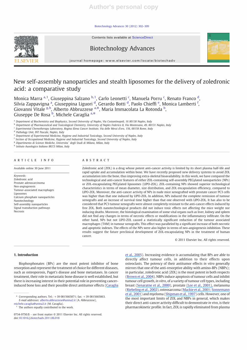
Fig. 1. In vivo anti-cancer effects of ZOL-encapsulating nanocarriers. A) Encapsulation in NPs increases antitumoral efficacy of ZOL against human prostate PC3M-luc2 cell linexenografts. Mice were injected i.m. with PC3M-luc2 cell line cells and starting from day 6 treated as follows: (♦) untreated; (◊) free ZOL; (▲) LIPO-ZOL; (Δ) PLCaPZ NPs. Points aremeans with SD (bars). B) Example of luminescence associated to injected tumour cells in an untreated mouse (upper panel) and in a mouse treated with PLCaPZ NPs (lower panel)achieving a complete regression of the tumour after 56 days from the initial tumour cell injection.
305M. Marra et al. / Biotechnology Advances 30 (2012) 302–309

Author's personal copy
suggested by the ζ value of about 17.5 mV. Finally, the actual loadingsof LIPO-ZOL and PLCaPZ NPs were about 108 and 66 μg/mg lipid,respectively. However, when considering that two totally differentprocedures were used for the preparation of two nanocarriers (with adifferent theoretical loading), the encapsulation efficiency obtained inthe case PLCaPZ NPs was about 66%, while this value dramaticallydropped (b6%) when encapsulating ZOL into LIPO-ZOL.
3.2. Effects of PLCaPZNPsand LIPO-ZOL on invivoprostate adenocarcinomagrowth
We evaluated the in vivo efficacy of these two formulationsencapsulating ZOL compared to free ZOL on human prostate cancerxenografts. To this purpose immunosuppressed mice were injectedwith PC-3 cells and starting from day 6 treated with ZOL-free orPLCaPZ NPs or LIPO-ZOL or with blank NPs or blank liposomes threetimes a week for 3 consecutive weeks, as in “Materials and methods”.As reported, in the Fig. 1A, the administration of free ZOL was noteffective in reducing the growth of tumors as at the end of treatment(day 27 after tumor cells injection) an about 250% increase of tumorweight was observed. At this time the tumor weight of ZOL-treatedmice were not significantly different (pN0.05) compared to micetreated with blank NPs or liposomes (data not shown) and untreatedgroups. Moreover, free ZOL induced a tumour growth delay (TGD) of3 days (Table 2). On the contrary, the treatment of mice with eitherLIPO-ZOL or PLCaPZ NPs elicited a marked anti-tumor activity; in fact,mice treated with the two different formulations elicited about 180%and 120% change in tumor weight respectively (Fig. 1A). Moreover, atumor growth delay of 7 and 12 days for LIPO-ZOL and PLCaPZ NPs,respectively was observed (Table 2). These increases were statisticallysignificant if compared to untreated group (p=0.000), to free ZOL(p=0.02 for LIPO-ZOL and pb0.001 for PLCaPZ NPs). The differencesin TGD between LIPO-ZOL and PLCaPZ NPs were also highly significant(p=0.016) demonstrating that PLCaPZ NPs had a higher anti-canceractivity if compared with that one of LIPO-ZOL. Moreover, PLCaPZ NPscaused tumour stabilization in all 6 mice treated with the formulationwhile LIPO-ZOL and free ZOL on 4/6 and in 0/6 mice, respectively. Atumour regression of at least 25% the initial volume was recorded in4/6 mice treated with NPs-ZOL and 1/6 and 0/6 mice of the LIPO-ZOLand control group, respectively (Table 2). Notably, only in the PLCaPZNP-treated group one complete response was observed. In fact, after4 months from tumor cell injection no tumour was evident with bothpalpability and at the analysis of luminescence with a dedicatedapparatus (Fig. 1B). In this mouse a progressive reduction of theluminescence associated to the tumour cells was observed with acomplete regression of the luminescence at 56 days from the tumourcell injection. Blank LIPO-ZOL and PLCaPZ NPs did not induce anysignificant effect on the tumour weight if compared to free ZOL anduntreated groups (data not shown). Finally, it is interesting to note
that all the treatments were well tolerated by animals since no bodyweight loss and toxic deaths have been observed.
3.3. Effects of PLCaPZ NPs and LIPO-ZOL on necrosis and neo-angiogenesis of tumour xenografts
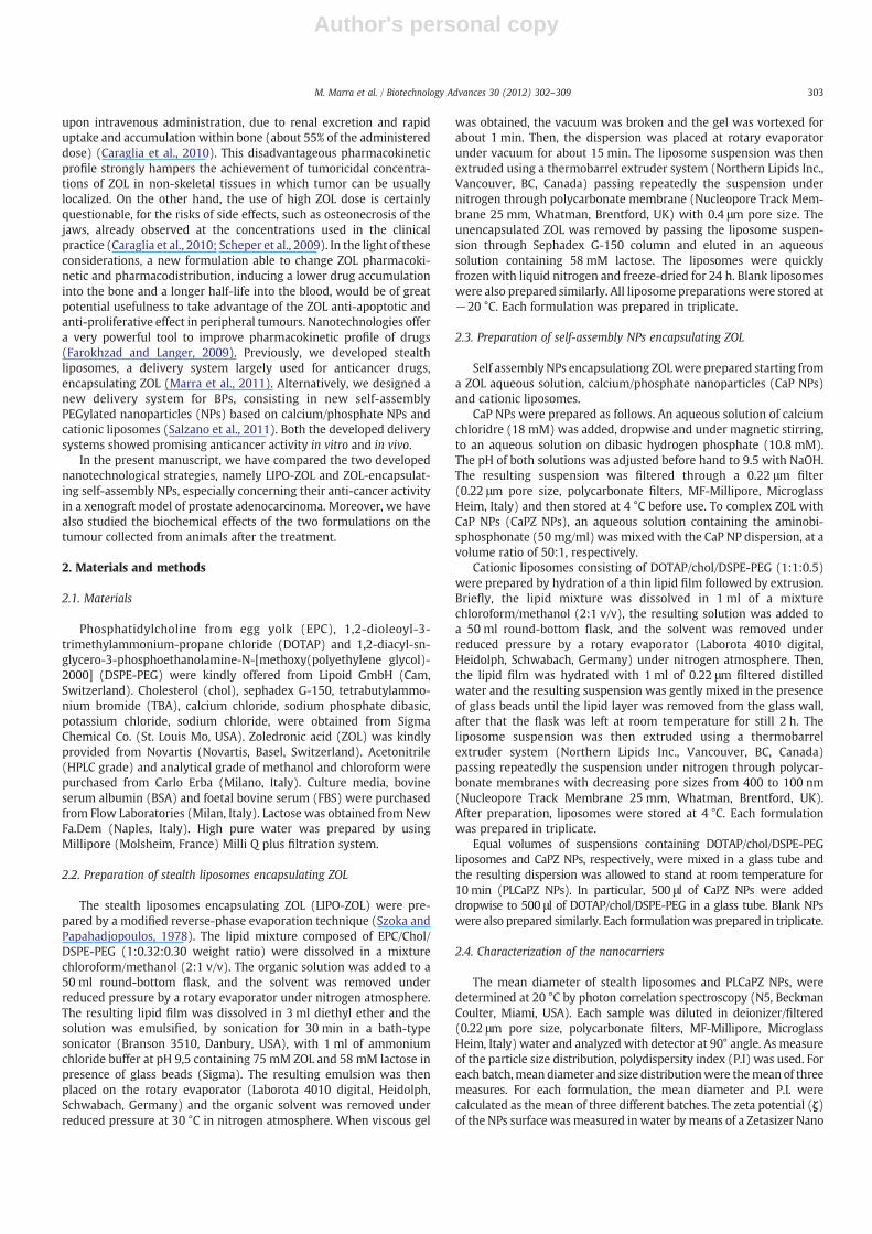
We have evaluated the effects of the two formulations on thenecrosis of tumour xenografts collected from mice after treatment.We have found that empty nanotech devices were ineffective ininducing any change in the necrotic index of the tumours (about 23%of necrotic index was recorded with both formulations vs 20% ofuntreated mice, data not shown). On the other hand, LIPO-ZOLinduced an about 45% and PLCaPZ NPs an about 65% necrotic indexthat was paralleled by a significant reduction of the tumour weight asindicated in the Table 2 (Fig. 2 and 3). The statistical analysis of theresults compared with that one of untreated tumours achieved a highstatistical significance (pb0.001 in both cases). The empty nanotechdevices were completely inactive in changing the vessel density of the
% o
f p
osi
tive
cel
ls
0
20
40
60
80
NecrosisCD31CD68
CTR LIPO-ZOLZOL PLCaPZ NPs
*
*
*
§
§
§
Fig. 2. Percentage of positive cells for staining specific for necrosis or immunostainingfor CD31 (endothelial cells) or CD68 (TAM). Briefly, tumor sections were scanned at lowmagnification (×40) to identify the region of the sectionwith the highest microvasculardensity (neovascular “hotspot”); this area was then counted at a magnification of ×200for the microvasculature highlighted by CD31 or for TAM highlighted with CD68. Forthe evaluation of necrosis special care was taken in assessing on paraffin fixed,haematoxylin and eosin (H&E)-stained sections. Two pathologists (RF and GB)evaluated the staining pattern of the two proteins separately and scored the proteinexpression in each specimen for the percentage of positive neoplastic cells. * pb0.05 ifcompared with both LIPO-ZOL and ZOL. § pb0.05 if compared with ZOL. Bars, SEs.
Table 2Comparison of antitumor efficacy of NPs-ZOL vs LIPO-ZOL on PC3 bearing mice.
Treatmentgroupsa
T-C (days)b Tumorstabilization/mice treated
Tumorregression/mice treated
Completeresponse/mice treatedc
ZOL 3 0/6 0/6 0/6LIPO-ZOL 7 4/6 1/6 0/6PLCaPZ NPs 12 6/6 4/6 1/6
a Mice were injected with PC3 cells at 3×10⁶ and treated with ZOL at 20 μg/mouse/dgiven i.v. following the different formulations on days 6, 8, 10, 13, 15, 17, 20, 22, 24 aftertumor cell injection. Each group included 6 mice.
b Tumor growth delay, evaluated as T - C , where T and C are the median times fortreated and control tumors, respectively, to achieve equivalent size. Statistical analysis:PLCaPZ NPs vs LIPO-ZOL, p=0.016 and vs ZOL, pb0.001; LIPO-ZOL vs ZOL, p=0.02.
c In this group a complete response was observed in 1 mouse. In fact, four monthsafter tumour cells injection, no tumour was evident by palpability.
A B
C D
Fig. 3. Effects of ZOL-encapsulating nanocarriers on necrosis. The necrotic evidence onslides derived from paraffin-embedded tissues was performed as described in“Materials and methods”. The area indicated by arrows are necrotic. The images are200× magnifications of representative results. A: untreated animal; B: 20 μg ZOL-treated animal; C: 20 μg LIPO-ZOL-treated animal; D: 20 μg PLCaPZ NPs -treated animal.
306 M. Marra et al. / Biotechnology Advances 30 (2012) 302–309
Author's personal copy
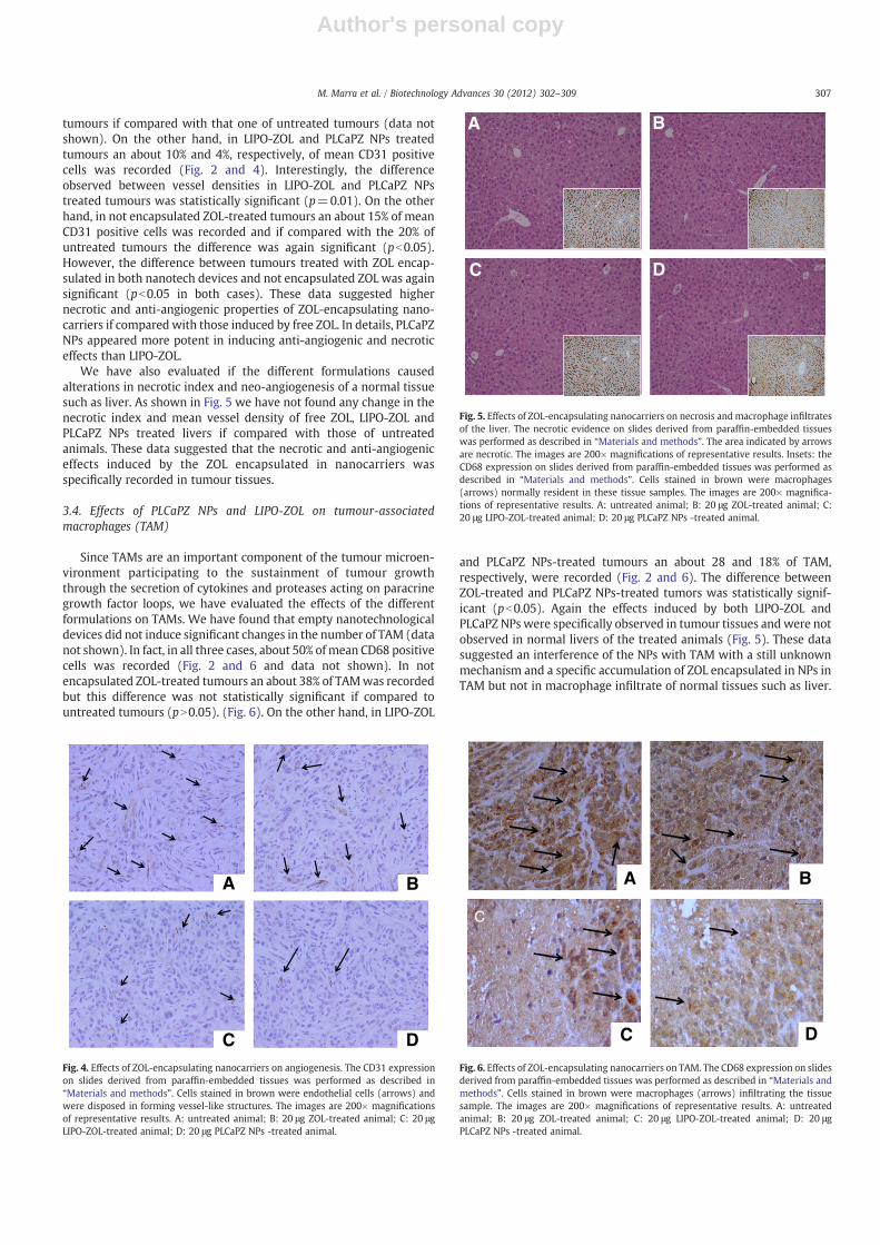
tumours if compared with that one of untreated tumours (data notshown). On the other hand, in LIPO-ZOL and PLCaPZ NPs treatedtumours an about 10% and 4%, respectively, of mean CD31 positivecells was recorded (Fig. 2 and 4). Interestingly, the differenceobserved between vessel densities in LIPO-ZOL and PLCaPZ NPstreated tumours was statistically significant (p=0.01). On the otherhand, in not encapsulated ZOL-treated tumours an about 15% of meanCD31 positive cells was recorded and if compared with the 20% ofuntreated tumours the difference was again significant (pb0.05).However, the difference between tumours treated with ZOL encap-sulated in both nanotech devices and not encapsulated ZOL was againsignificant (pb0.05 in both cases). These data suggested highernecrotic and anti-angiogenic properties of ZOL-encapsulating nano-carriers if compared with those induced by free ZOL. In details, PLCaPZNPs appeared more potent in inducing anti-angiogenic and necroticeffects than LIPO-ZOL.
We have also evaluated if the different formulations causedalterations in necrotic index and neo-angiogenesis of a normal tissuesuch as liver. As shown in Fig. 5 we have not found any change in thenecrotic index and mean vessel density of free ZOL, LIPO-ZOL andPLCaPZ NPs treated livers if compared with those of untreatedanimals. These data suggested that the necrotic and anti-angiogeniceffects induced by the ZOL encapsulated in nanocarriers wasspecifically recorded in tumour tissues.
3.4. Effects of PLCaPZ NPs and LIPO-ZOL on tumour-associatedmacrophages (TAM)
Since TAMs are an important component of the tumour microen-vironment participating to the sustainment of tumour growththrough the secretion of cytokines and proteases acting on paracrinegrowth factor loops, we have evaluated the effects of the differentformulations on TAMs. We have found that empty nanotechnologicaldevices did not induce significant changes in the number of TAM (datanot shown). In fact, in all three cases, about 50% ofmean CD68 positivecells was recorded (Fig. 2 and 6 and data not shown). In notencapsulated ZOL-treated tumours an about 38% of TAMwas recordedbut this difference was not statistically significant if compared tountreated tumours (pN0.05). (Fig. 6). On the other hand, in LIPO-ZOL
and PLCaPZ NPs-treated tumours an about 28 and 18% of TAM,respectively, were recorded (Fig. 2 and 6). The difference betweenZOL-treated and PLCaPZ NPs-treated tumors was statistically signif-icant (pb0.05). Again the effects induced by both LIPO-ZOL andPLCaPZ NPs were specifically observed in tumour tissues andwere notobserved in normal livers of the treated animals (Fig. 5). These datasuggested an interference of the NPs with TAM with a still unknownmechanism and a specific accumulation of ZOL encapsulated in NPs inTAM but not in macrophage infiltrate of normal tissues such as liver.
C
D
A
C
B
Fig. 6. Effects of ZOL-encapsulating nanocarriers on TAM. The CD68 expression on slidesderived from paraffin-embedded tissues was performed as described in “Materials andmethods”. Cells stained in brown were macrophages (arrows) infiltrating the tissuesample. The images are 200× magnifications of representative results. A: untreatedanimal; B: 20 μg ZOL-treated animal; C: 20 μg LIPO-ZOL-treated animal; D: 20 μgPLCaPZ NPs -treated animal.
D
BA
CFig. 4. Effects of ZOL-encapsulating nanocarriers on angiogenesis. The CD31 expressionon slides derived from paraffin-embedded tissues was performed as described in“Materials and methods”. Cells stained in brown were endothelial cells (arrows) andwere disposed in forming vessel-like structures. The images are 200× magnificationsof representative results. A: untreated animal; B: 20 μg ZOL-treated animal; C: 20 μgLIPO-ZOL-treated animal; D: 20 μg PLCaPZ NPs -treated animal.
A B
DC
Fig. 5. Effects of ZOL-encapsulating nanocarriers on necrosis andmacrophage infiltratesof the liver. The necrotic evidence on slides derived from paraffin-embedded tissueswas performed as described in “Materials and methods”. The area indicated by arrowsare necrotic. The images are 200× magnifications of representative results. Insets: theCD68 expression on slides derived from paraffin-embedded tissues was performed asdescribed in “Materials and methods”. Cells stained in brown were macrophages(arrows) normally resident in these tissue samples. The images are 200× magnifica-tions of representative results. A: untreated animal; B: 20 μg ZOL-treated animal; C:20 μg LIPO-ZOL-treated animal; D: 20 μg PLCaPZ NPs -treated animal.
307M. Marra et al. / Biotechnology Advances 30 (2012) 302–309

Author's personal copy
4. Discussion
In this work two different nanotechnology-based delivery systemsfor ZOL were compared. LIPO-ZOL and PLCaPZ NPs were prepared bycompletely different approaches. ZOL was physically entrapped intoLIPO-ZOL by a modified reverse-phase evaporation method, largelyused to improve the encapsulation efficiency of hydrophilic com-pounds into liposomes. Moreover, to improve ZOL solubility into theaqueous phase of liposomes, ZOL was solubilized in an alkaline buffer(pH 8.5). Only when combining reverse-phase evaporation methodand an alkaline buffer, an encapsulation efficiency higher than 5% wasobtained. On the contrary, encapsulation efficiency lower than 1% wasobtained when using other techniques (data not shown). However,the alkaline pH inside the liposomes can improve lipid degradationrate, limiting the chemical-physical stability of the formulation duringthe storage (Zhang and Pawelchak, 2000). On the other hand,liposome freeze-drying and following rehydration can result invesicles fusion and aggregation, generally requiring the addition ofcryoprotectants. In the case of LIPO-ZOL, the use of lactose beforefreeze-drying allowed to prevent vesicle alteration, although littlepercentage of liposome fusion/aggregation cannot be excluded. Onthe contrary, the new self-assembling method developed in our lab(Salzano et al., 2011), assured a ZOL encapsulation efficiency of about66%, about 12-fold greater than that obtained with LIPO-ZOL. Thiscould be explained because the encapsulation process of ZOL, in thecase of PLCaPZ NPs, is driven by ionic interactions, overcoming theencapsulation issues encountered with liposomes. It is worthy of notethat, in the case of LIPO-ZOL, a ZOL theoretical loading 20-times higherwas used in order to improve the actual loading that was about108 μg/mg lipid. This was possible by using an alkaline buffer thatallowed to improve the ZOL water solubility. The alkaline buffer wasnot compatible with the preparation method of PLCaPZ NPs, wherethe theoretical loading was limited by the ZOL solubility in water.Finally, the possibility to prepare PLCaPZ NPs before use, allows toovercome stability issues during storage. Further studies are neededto clarify how CaPZ NPs and PEGylated cationic liposomes rearrangeto form PLCaPZ NPs.
We have also compared the anticancer activity of the twoformulations in a model of hormone-independent prostate adeno-carcinoma cell xenografts finding that the two nanotechnology basedformulations had a higher anti-cancer activity than free ZOL. Theseresults confirmed our previous reports on the anti-cancer activity ofPLCaPZ NPs against prostate cancer (Salzano et al., 2011). Here wehave found, for the first time, that PLCaPZ NPs induced higher anti-cancer activity than LIPO-ZOL. Interestingly, PLCaPZ NPs induced alsotumour regression and completely cured 1/6 treated mice. Theseeffects were paralleled by a more potent induction of necrosis in thetumour treated with PLCaPZ NPs than those treated with LIPO-ZOLand the analysis of the statistical significance revealed that thedifference in the necrotic index induced by PLCaPZ NPs was highlysignificant if compared with both ZOL and LIPO-ZOL.
Prostate cancer remains a significant public health problem, withlimited therapeutic options in the setting of castrate-resistantmetastatic disease. Solid tumors can be thought of as multicellular‘organs’ that consist of a variety of cells as well as a scaffold of non-cellular matrix. Stromal–epithelial crosstalk is integral to prostatecancer progression and metastasis. Thus, various pathways areimportant for stromal–epithelial crosstalk and represent attractivecandidate therapeutic targets. In this view, fibroblast growth factorsand transforming growth factor β signaling regulate cell proliferation,apoptosis and angiogenesis in the prostate cancer microenvironment(Karlou et al., 2010). Angiogenesis inhibition is a relatively novelantineoplastic approach, which targets the reliance of tumour growthon the formation of new blood vessels that are necessary to supportthe growth of the tumours and the subsequent extravasation ofmetastatic cells (Hwang and Heath, 2010). Another important
component of prostate cancer microenvironment are TAMs thathave been recently reported to be involved in the growth of prostatecancer cells in the bone, themainmetastatic site of this cancer (Kim etal., 2011). Moreover, the infiltration of tumour-associated macro-phages in prostate biopsy specimens is predictive of diseaseprogression after hormonal therapy for prostate cancer (Nonomuraet al., 2010). TAMs represent a prominent component of themononuclear leukocyte population of solid tumours, which displaysan ambivalent relationship with tumours. They originate in thecirculation and are recruited to the tumour site by tumour-derivedattractants such as chemokines and interact with the tumour cells andpreferentially localize at the tumour-host tissue interface, in regionsoften associated with low oxygen tensions. The tumour microenvi-ronment, including cytokines and hypoxia, regulates the localizationand function of TAMs. Upon activated by cancer cells, the TAMs canrelease a vast diversity of growth factors, proteolytic enzymes,cytokines, and inflammatory mediators. Many of these factors arekey agents in cancer metastasis. Substantial evidence suggests thatTAMs can interact with cancer cells, modify the ECM, and promotecancer cell invasion and metastasis (Siveen and Kuttan, 2009). It wasreported that ZOL can inhibit neo-angiogenesis in prostate cancerpatients reducing the circulating levels of VEGF (Santini et al., 2007).Moreover, the reduction of the circulating levels of VEGF correlateswith the reduction of the incidence of skeletal related events (SREs) inbreast cancer patients treated with ZOL (Vincenzi et al., 2005). It hasalso been reported the property of ZOL to induce apoptosis of TAM inprostate cancer and to modulate the pro-inflammatory phenotype ofTAM especially in bone metastases from prostate cancer (Saylor andSmith, 2010). In our study, we have found that free ZOL was able toinhibit both neo-angiogenesis and macrophage infiltrates in prostatecancer tissues, but both ZOL-encapsulating nanotechnological formu-lations enhanced this effect being more potent when using PLCaPZNPs. These results are not surprising taking in consideration the recentdata about more potent anti-cancer effects of ZOL administered at lowand repeated doses inmice xenografted in the bonewith breast cancercells (Daubiné et al., 2007). Moreover, it was also demonstrated incancer patients that a single dose of low dose ZOL (1 mg instead of4 mg) caused a rapid and sustained decrease of circulating levels ofVEGF suggesting an anti-angiogenic effect that was maintained by therepeated administration of low doses ZOL (Santini et al., 2007). Thedelivery of ZOL by stealth nanovectors likely mimics the repeatedadministration of low dose ZOL prolonging the plasma half life of thedrug. In fact, the pharmacokinetic of ZOL administered as a singleintravenous bolus is limited being the plasma half life of the drug only15 min (Caraglia et al., 2010). Moreover, the encapsulation of ZOL inPEGylated nanovectors (able to escape the reticular endothelialsystem) could result in a reduction of the osteotropism of the drugwith the consequent increased perfusion in extra-bone sites of thedisease. In fact, it was demonstrated that therapy with NE-10790, anaminobisphosphonate with reduced affinity to bone, substantiallyreduced skeletal tumor growth at a dosage that did not inhibitosteolysis. On the other hand, analogue concentrations of risedronate,an aminobisphosphonatewith high affinity for bone, reduced osteolysisbut not tumour growth in bone (Fournier et al., 2008). Interestingly, theanti-angiogenic and anti-TAM effects of ZOLwere limited to the tumourtissues and were not present in normal liver. The latter effect could bedue to the specific accumulation of the nanovectors in tumour tissuesmediated by the so-called enhanced permeability and retention (EPR)effect that allows the accumulation of the nanocarriers in tissues withfenestrated vessels and altered lymphatic drainage, e.g. tumour tissues.The more potent anti-cancer biological and clinical effects mediated byPLCaPZ NPs could be explained by the improved cell uptake of theseNPs. Indeed, PLCaPZ NPs are based on CaP NPs and cationic lipids,largely used to promote cell uptake of negatively charged molecules,such as nucleic acids (De Rosa et al., 2010; Maitra, 2005). Thus, thecomplexes between ZOL and CaP NPs/cationic lipids could actively
308 M. Marra et al. / Biotechnology Advances 30 (2012) 302–309

Author's personal copy
enter into the cells by endocytotic mechanism, similarly to that of DNAcontaining lipoplexes. Moreover, the different surface characteristics ofthe two nanocarriers, close to the neutrality and positive for LIPO-ZOLand PLCaPZ NPs, respectively, could affect their uptake by TAM. Fromthis point of view, our findings agree with data from literature. Indeed,the higher value of zeta potential found in the case of PLCaPZ NPs couldbe explained with a lower shielding effect of PEG together with thepresence of polar head of cationic lipids onto NP surface. It has beenreported that both the degree of PEGylation and the presence of a netcharge affect nanocarrier uptake by macrophages (Chellat et al., 2005).In particular, a high degree of PEGylation should hamper the adsorptionof plasma proteins on the particles, thus hampering macrophageuptake. Moreover, nanocarriers with a zeta potential close to zero havebeen found to be less phagocytable in comparison with positivelycharged particles, probably due to the presence of negatively chargedsialic acid on their surface (Chellat et al., 2005).
Finally, we have recently reported that LIPO-ZOL induces anti-proliferative effects on human prostate adenocarcinoma PC3 cell linehigher than that one found in the present manuscript (Marra et al.,2011). This discrepancy could be due to the fact that the data reportedin Fig. 1 and Table 2 of the present manuscript were obtained by usingthe human prostate carcinoma PC3M-luc2 cell line, a luciferaseexpressing cell line which was stably transfected with the luc2 fireflyluciferase gene (Caliper Life Sciences, Hopkinton, MA, USA), while inthe previous experiments we have used the standard PC3 cells line.The PC3M-luc2 cell line showed a more aggressive phenotypecompared to the standard PC3. In fact, the injection of 3×10⁶ cells/mouse produced a tumor mass of about 400 mg at day 6 after tumorcell injection (the present manuscript); on the contrary, the PC3standard cell line elicited a slower growth rate as the injection of5×106 cells/mouse resulted in a tumor mass of 300 mg at day 6 aftertumor cells injection. These differences in tumor growth rate couldaccount for the discrepancy observed between the experiments.
In conclusion, PLCaPZ NPs have demonstrated a higher anticanceractivity if compared to LIPO-ZOL and this effect occurred togetherstrong modifications of the tumour micro-environment.
Acknowledgements
MC was supported by a grant of the Italian Association for CancerResearch (AIRC) for a project entitled “Liposome-encapsulatingzoledronic acid: a new experimental therapeutic for the treatmentof brain tumours and by Italian Ministry of Education and University(PRIN 2010)”. CL received a grant from Italian Association for CancerResearch (AIRC) and Ministero della Salute. Manuela Porru received afellowship by Italian Association for Cancer Research.
References
Brown JE, Webbe HN, Coleman RE. The role of bisphosphonates in breast and prostatecancers. Endocr Relat Cancer 2004;11:207–24.
Caraglia M, Santini D, Marra M, Vincenzi B, Tonini G, Budillon A. Emerging anti-cancermolecular mechanisms of aminobisphosphonates. Endocr Relat Cancer 2005;13:1–22.
Caraglia M, Marra M, Naviglio S, Botti G, Addeo R, Abbruzzese A. Zoledronic acid: anunending tale for an antiresorptive agent. Expert Opin Pharmacother 2010;11:141–54.
Chellat F, Merhi Y, Moreau A, Yahia L. Therapeutic potential of nanoparticulate systemsfor macrophage targeting. Biomaterials 2005;26:7260–75.
Daubiné F, Le Gall C, Gasser J, Green J, Clézardin P. Antitumor effects of clinical dosingregimens of bisphosphonates in experimental breast cancer bone metastasis. J NatlCancer Inst 2007;99:322–30.
De Rosa G, De Stefano D, Galeone A. Oligonucleotide delivery in cancer therapy. ExpertOpin Drug Deliv 2010;7:1263–78.
Farokhzad OC, Langer R. Impact of nanotechnology on drug delivery. ACS Nano 2009;3:16–20.
Fournier PG, Daubiné F, Lundy MW, Rogers MJ, Ebetino FH, Clézardin P. Lowering bonemineral affinity of bisphosphonates as a therapeutic strategy to optimize skeletaltumor growth inhibition in vivo. Cancer Res 2008;68(21):8945–53.
Hwang C, Heath EI. Angiogenesis inhibitors in the treatment of prostate cancer.J Hematol Oncol 2010;2(3):26.
Karlou M, Tzelepi V, Efstathiou E. Therapeutic targeting of the prostate cancermicroenvironment. Nat Rev Urol 2010;7:494–509.
Kim SW, Kim JS, Papadopoulos J, Choi HJ, He J, Maya M, et al. Consistent interactionsbetween tumor cell IL-6 and macrophage TNF-α enhance the growth of humanprostate cancer cells in the bone of nude mouse. Int Immunopharmacol 2011:17.(Epub ahead of print).
Lee MV, Fong EM, Singer FR, Guenette RS. Bisphosphonate treatment inhibits thegrowth of prostate cancer cells. Cancer Res 2001;6:2602–8.
Mackie PS, Fisher JL, Zhou H, Choong PF. Bisphosphonates regulate cell growth and geneexpression in the UMR 106–101 clonal rat osteosarcoma cell line. Br J Cancer2001;84:951–8.
Maitra A. Calcium phosphate nanoparticles: second-generation nonviral vectors ingene therapy. Expert Rev Mol Diagn 2005;5:893–905.
Marra M, Salzano G, Leonetti C, Tassone P, Scarsella M, Zappavigna S, et al.Nanotechnologies to use bisphosphonates as potent anticancer agents: the effectsof zoledronic acid encapsulated into liposomes. Nanomedicine 2011. Mar. 29.[Epub ahead of print].
Nonomura N, Takayama H, Nakayama M, Nakai Y, Kawashima A, Mukai M, et al.Infiltration of tumour-associated macrophages in prostate biopsy specimens ispredictive of disease progression after hormonal therapy for prostate cancer. BJUInt 2010. Nov. 2.
Riebeling C, Forsea AM, Raisova M, Orfanos CE, Geilen CC. The bisphosphonatepamidronate induces apoptosis in human melanoma cells in vitro. Br J Cancer2002;87:366–71.
Salzano G, Marra M, Porru M, Zappavigna S, Abbruzzese A, La Rotonda MI, et al. Self-assembly nanoparticles for the delivery of bisphosphonates into tumors. Int JPharm 2011;17:292–7.
Saylor PJ, Smith MR. Bone health and prostate cancer. Prostate Cancer Prostatic Dis2010;13:20–7.
Santini D, Vincenzi B, Galluzzo S, Battistoni F, Rocci L, Venditti O, et al. Repeatedintermittent low-dose therapy with zoledronic acid induces an early, sustained,and long-lasting decrease of peripheral vascular endothelial growth factor levels incancer patients. Clin Cancer Res 2007;13:4482–6.
Scheper MA, Badros A, Chaisuparat R, Cullen KJ, Meiller TF. Effect of zoledronic acid onoral fibroblasts and epithelial cells: a potential mechanism of bisphosphonate-associated osteonecrosis. Br J Haematol 2009;144:667–76.
Senaratne SG, Pirianov G, Mansi JL, Arnett TR, Colston KW. Bisphosphonates induceapoptosis in human breast cancer cell lines. Br J Cancer 2000;2:1459–68.
Shipman CM, Rogers MJ, Apperley JF, Russell RG, Croucher PI. Bisphosphonates induceapoptosis in human myeloma cell lines: a novel anti-tumour activity. Br J Haematol1997;98:665–72.
Siveen KS, Kuttan G. Role of macrophages in tumour progression. Immunol Lett2009;123:97–102.
Sonnemann J, Eckervogt V, Truckenbrod B, Boos J, Winkelmann W, Van Valen F. Thebisphosphonate pamidronate is a potent inhibitor of human osteosarcoma cellgrowth in vitro. Anticancer Drug 2001;12:459–65.
Szoka Jr F, Papahadjopoulos D. Procedure for preparation of liposomes with largeinternal aqueous space and high capture by reverse-phase evaporation. Proc NatlAcad Sci USA 1978;75:4194–8.
Vincenzi B, Santini D, Dicuonzo G, Battistoni F, Gavasci M, La Cesa A, et al. Zoledronicacid-related angiogenesis modifications and survival in advanced breast cancerpatients. J Interferon Cytokine Res 2005;25:144–51.
Zhang JA, Pawelchak J. Effect of pH, ionic strength and oxygen burden on the chemicalstability of EPC/cholesterol liposomes under accelerated conditions. Part 1: Lipidhydrolysis. Eur J Pharm Biopharm 2000;50:357–64.
309M. Marra et al. / Biotechnology Advances 30 (2012) 302–309