development and characterisation study of liposomes-encapsulated piroxicam
TRANSCRIPT
International Journal of Drug Delivery 3 (2011) 64-73 http://www.arjournals.org/ijdd.html
Research Article
ISSN: 0975-0215
Development and characterisation study of liposomes-encapsulated piroxicam
H.S. Chiong1, M. Nazrul Hakim1,2*, M.R. Sulaiman1, Z.A. Zakaria1, A. Zuraini1, S.G.M. Ong3 and
K.H. Yuen3 *Corresponding author:
M. Nazrul Hakim 1 Faculty of Medicine and Health
Sciences, Universiti Putra Malaysia,
43400 UPM Serdang, Selangor,
Malaysia.
2 Sports Academy, Universiti Putra
Malaysia, 43400 UPM Serdang,
Selangor, Malaysia.
3 School of Pharmaceutical Sciences,
Universiti Sains Malaysia, 11800
Minden, Penang, Malaysia.
Abstract The objective of present work was to develop a novel liposomes-based drug delivery system for a lipophilic non-steroidal anti-inflammatory drug, piroxicam. The system was prepared using proliposomes method and optimised for different preparation parameters including type of proliposomes, concentration of drug, duration of hydration and type of particle size reduction treatment used. All prepared liposomal samples were extensively characterized for their drug-entrapment and size profile using various in-vitro techniques. Present work showed that the most optimum formulation (Pro-lipoTM Duo; 12mg piroxicam per gram Pro-lipoTM; 10 hours hydration time) produced highest amount of actual drug been entrapped in liposomes (800.4 mg/g Pro-lipoTM) with a satisfactory entrapment efficiency of 15.36%. This formulation had also produced liposomal samples with a homogenous (polydispersity index = 0.45) and small particle size (359.95nm). Extrusion technique was found to cause significant reduction in drug-entrapment and size profile of drug-loaded liposomes. A 4-weeks storage study showed that drug-entrapment and size profile of liposomal samples were stable in both refrigerated and room temperature. Electron microscopy revealed that prepared liposomal samples were spherical-shaped and showed concentric lamellae. In conclusion, present work successfully demonstrated a simple, reproducible and practical method of preparation for liposomes-encapsulated piroxicam. Keywords: Proliposomes; Liposomes; Piroxicam; Encapsulation; Particle size; Transmission electron microscopy
Introduction Liposomes are microscopic spherical vesicles composed of lipid bilayers which formed by
phospholipids dispersed in water [1]. Due to their high degree of biocompatibility and effectiveness in the modulation of drug release properties [2], liposomes have been used in many drug delivery
doi:10.5138/ ijdd.2010.0975.0215.03055 ©arjournals.org, All rights reserved.
Chiong et al. International Journal of Drug Delivery 3 (2011) 64-73
65
research as vehicles to incorporate assortment of active agents [3]. Literature has also showed that liposomes-entrapped drugs exhibit improved therapeutic indices and superior pharmacological properties than those observed with conventional formulations [4]. An attempt has been made in present work to develop a practical liposomes-based drug delivery system for piroxicam (4-hydroxy-2-methyl-N-(pyridine-2-yl)-2H-1,2-benzothiazine-3-carboxamide-1,2-dioxide). Piroxicam, an oxicam derivative, is a well-known non-steroidal anti-inflammatory drug discovered by Pfizer Laboratories [5,6]. Pharmacokinetic study has demonstrated that the hydrophobic piroxicam has a slow absorption via oral route, which resulted in low bioavailability of the drug and delayed the onset of its therapeutics effects [7]. Due to the potential modification of drug’s bioavailability and therapeutic index using liposomal technology, the strategy to entrap piroxicam in liposomes is therefore deemed important in pharmaceutical field [8,9]. Over the years, numerous techniques and procedures have been described to prepare liposome-encapsulated drugs with different sizes and characteristics. Most of the preparation methods such as reverse-phase evaporation, ether injection and freeze-thaw method are tedious in nature and require long preparation time [10]. In current study, the proliposomes method, which based on the conversion of initial concentrated ethanolic solutions of phospholipids into liposomes dispersion by dilution under strictly controlled conditions [11], is used for rapid production of blank and piroxicam-loaded liposomes. This relatively simpler technique has been reported for its suitable to encapsulate drugs of various water and alcohol solubility with high entrapment efficacy [12]. A number of factors, especially parameters used in the preparation procedures, have also been known to affect the quality of the liposomal samples [10,13,14]. In this paper, we present the results on the entrapment of piroxicam into liposomes. The formulation method and preparation procedures were optimized for
parameters and properties that were deemed important in order to produce suitable drug-loaded liposomal samples with satisfactory qualities of drug loading and size profile as well as potential for in-vivo performance.
Materials and methods Materials Two commercial preparations of proliposomes, namely Pro-lipoTM Duo and Pro-lipoTM C (Lucas Meyer, France), were used. Pro-lipoTM Duo and C contained 50% and 40% unsaturated soybean phosphatidylcholine respectively which suspended in hydrophilic medium consisted of glycerol and ethanol. Piroxicam, dimethylsulfoxide and Triton X-100 were purchased from Sigma (USA). Acetonitrile and sodium dihydrogen phosphate 1-hydrate were originated from Merck (Germany). Hydrochloric acid was purchased from J.T. Baker (Thailand). All solvents and chemicals used were of analytical or HPLC grade. Water used in all present work was purified and deionized by Direct-QTM 3 water purification system (Millipore, France).
Proliposomes, drug concentration and hydration time studies Liposomal samples preparation Piroxicam-loaded and blank liposomes were prepared in accordance to the manufacturer’s instructions with some modifications. All formulations, distinctive by different type of proliposomes, drug concentration and/or duration of hydration time used during preparation process, were produced in three batches respectively. Briefly, a stock piroxicam solution (60 mg/ml) was prepared initially by completely dissolving pre-determined amount of the drug in dimethylsulfoxide. These stock drug solutions were prepared freshly daily and protected from direct light. Next, 400 µl drug solution with known concentration was added gradually into 2g proliposomes (Pro-lipoTM Duo or C) in a beaker with moderate stirring (±100 rpm) for 60 minutes. The blank liposomes were prepared according to the same procedure except
Chiong et al. International Journal of Drug Delivery 3 (2011) 64-73
dimethylsulfoxide was used instead of drug solution. The mixture was then hydrated with 3.6ml distilled water which was added gradually to form a concentrated liposomal suspension. These liposomal suspensions was stirred continuously for a pre-determined period of stirring (hydration) time before further diluted with 10ml distilled water with continuous stirring for another 30 minutes. Preparation of all liposomal samples was carried out at room temperature.
Drug entrapment analysis The amount of piroxicam successfully been entrapped into liposomes were quantified using the following previously validated in-vitro techniques. For the analysis of each liposomal sample’s formulations, duplicate samples were prepared from all of the three individual batches (n=6).
Instrumentation and chromatographic condition
The HPLC system used consisted of a Jasco PU-980 Intelligent HPLC pump, Rheodyne 7125 (Cotati) sample injector fitted with 50 µl sample loop, Jasco UV-975 Intelligent UV/VI Detector and Hitachi D-2500 Chromato-Integrator. A Jones Chromatography Genesis C18 column (4µm, 150 x 4.6 mm I.D.), fitted with a refillable guard column packed with Perisorb® RP-18 (30-40 µm, pellicular) powder, was used for the chromatographic separation. The mobile phase consisted of 0.02M sodium dihydrogen orthophosphate 1-hydrate and acetonitrile (53:47, v:v) adjusted to pH 3.2 using 5M hydrochloric acid, which delivered at a flow rate of 1ml/minute. The detection wavelength was set at 360 nm with a sensitivity range of 0.005 a.u.f.s. and the amount of piroxicam was quantified using peak area. Retention time for piroxicam was 5.3 min.
Total piroxicam determination To determine the concentration of the total piroxicam in prepared liposomal samples, Triton X-100 (0.2% in final concentration) was added into the liposomes suspensions in order to lyse
the liposomes structure and released all entrapped drug. The mixture was vortexed for 30 seconds (Barnstead/Thermolyne, USA) and diluted with mobile phase before quantified using the HPLC method described in previous section.
Free undissolved piroxicam determination To determine the concentration of free undissolved drug, liposomal samples were first been centrifuged at 12800 G for 10 minutes (MiniSpin® plus, Eppendorf, Germany) to precipitate undissolved and unentrapped hydrophobic drug. The supernatant was then discarded and the remaining sediment was reconstituted with mobile phase before subjected to HPLC analysis.
Free dissolved piroxicam determination Liposomal samples were ultracentrifugated at 300,000G and 20ºC for 90 minutes using a TLA-110 rotor in OptimaTM MAX-XP ultracentrifuge (Beckman Coulter, USA). The supernatant, which contained only dissolved drug (confirmed by the absence of appreciable liposomal particles using particle size analyzer), was then collected. This supernatant samples were diluted with mobile phase to appropriate concentration before subjected to HPLC analysis.
Calculation formulas Actual amount of piroxicam been entrapped in liposomal samples were calculated by subtracting the amount of free drug (undissolved and dissolved form) from the total drug input. The drug entrapment efficiency was evaluated as percent of the total input drug entrapped in the liposomes.
Total piroxicam – Free piroxicam Amount entrapped (µg/g Pro-lipoTM ) = ⎯⎯⎯⎯⎯⎯⎯⎯⎯⎯⎯⎯⎯⎯ Total Pro-lipoTM used Total piroxicam – Free piroxicam
Percent entrapped (%) = ⎯⎯⎯⎯⎯⎯⎯⎯⎯⎯⎯⎯⎯⎯ X 100 Total piroxicam
Particle size analysis The size profile for all of the liposomal samples was determined by photon correlation spectroscopy technique using Zetasizer Nano S (Malvern Instrument, UK). The procedure for particle size analysis entailed dispersing 10µl of
66
Chiong et al. International Journal of Drug Delivery 3 (2011) 64-73 liposomal samples with 500µl distilled water in a low volume disposable sizing cuvette. The particle size and size distribution were measured by the instrument as ZAve and polydispersity index (PDI) respectively. For each formulation, two measurements were taken on two separate samples from three individual bathes of liposomal samples.
Size reduction study The optimized formulation (Pro-lipoTM Duo; 12mg piroxicam per 1g Pro-lipoTM; 10 hours hydration time) which produced highest amount of entrapped drug in previous section was selected for this study. Three batches of liposomal samples were freshly prepared using the same formulation according to the same procedure described earlier. Each batch of liposomal samples were subjected to either extrusion (single pass through a double-stacked 0.1µm polycarbonate membrane filter) using LiposoFastTM mini-extruder (Avestin Inc, Canada) or ultra-sonication in 300W bath sonicator (Digital Pro heated ultrasonic cleaner, Huiyuan Int’l Commerce & Exhibition Co., China) for 60 minutes. The drug entrapment and size profile for all prepared samples were evaluated using same procedures outlined in previous sections. Storage study In this part of study, the physical stability of liposomal samples stored in different conditions was evaluated in respect to their drug entrapment and particle size profile using same in-vitro techniques described previously. Each of the three batches of freshly prepared liposomal samples (optimized formulation) was equally separated and placed in two different temperatures respectively, namely in refrigerated temperature (2�C to 8�C) and ambient room temperature (25�C±2�C). All samples were stored in air-tight test tubes away from direct light. Samples for analyses were withdrawn at definite time intervals throughout the 4-weeks storage study. Reproducibility testing
Data obtained from six different batches of liposomal samples which prepared using the same formulation (Pro-lipoTM Duo; 12mg piroxicam per 1g Pro-lipoTM; 10 hours hydration time; no size reduction treatment) were used to investigate the reproducibility of drug entrapment and particle size profile.
Transmission electron microscopy (TEM) Morphological and size examinations of freshly prepared blank, drug-loaded and extruded drug-loaded liposomes were done using negative staining electron microscopy. A droplet of the spontaneously formed liposomal samples to be examined was placed onto a carbon-coated copper grid (400 mesh) for about 3 minutes in order to reach absorption equilibrium. The droplet was then wicked to dryness using pieces of filter paper before leaving the grid for another 1 minute. Next, a droplet of the negative stain solution (2% uranyl acetate) was added to the surface of the grid. After 1 minute, the copper mesh grid was dried using pieces of filter paper and kept in a filter paper lined petri dish. The size, morphology and lamellarity of liposomes samples were then viewed on Philips CM12 transmission electron microscope and photographed at an accelerating voltage of 80 kV.
Statistical evaluations The results were presented as Mean ± S.E.M. All data were subjected to one-way analysis of variance (ANOVA) followed by Tukey’s multiple comparison test among groups, and Student’s t-test for comparison between two groups. P values less than 0.05 (P<0.05) were taken as the limit of significance. All statistical analyses were carried out using SPSS v16.0 (SPSS Inc., Chicago).
Results and discussion Proliposomes, drug concentration and hydration time studies Drug entrapment and particle size profile Lipophilic, bilayer-interacting NSAIDs such as piroxicam were naturally incorporated within the lipid bilayers of liposomes vesicle [15,16]. Reports in literature had showed that the
67
Chiong et al. International Journal of Drug Delivery 3 (2011) 64-73
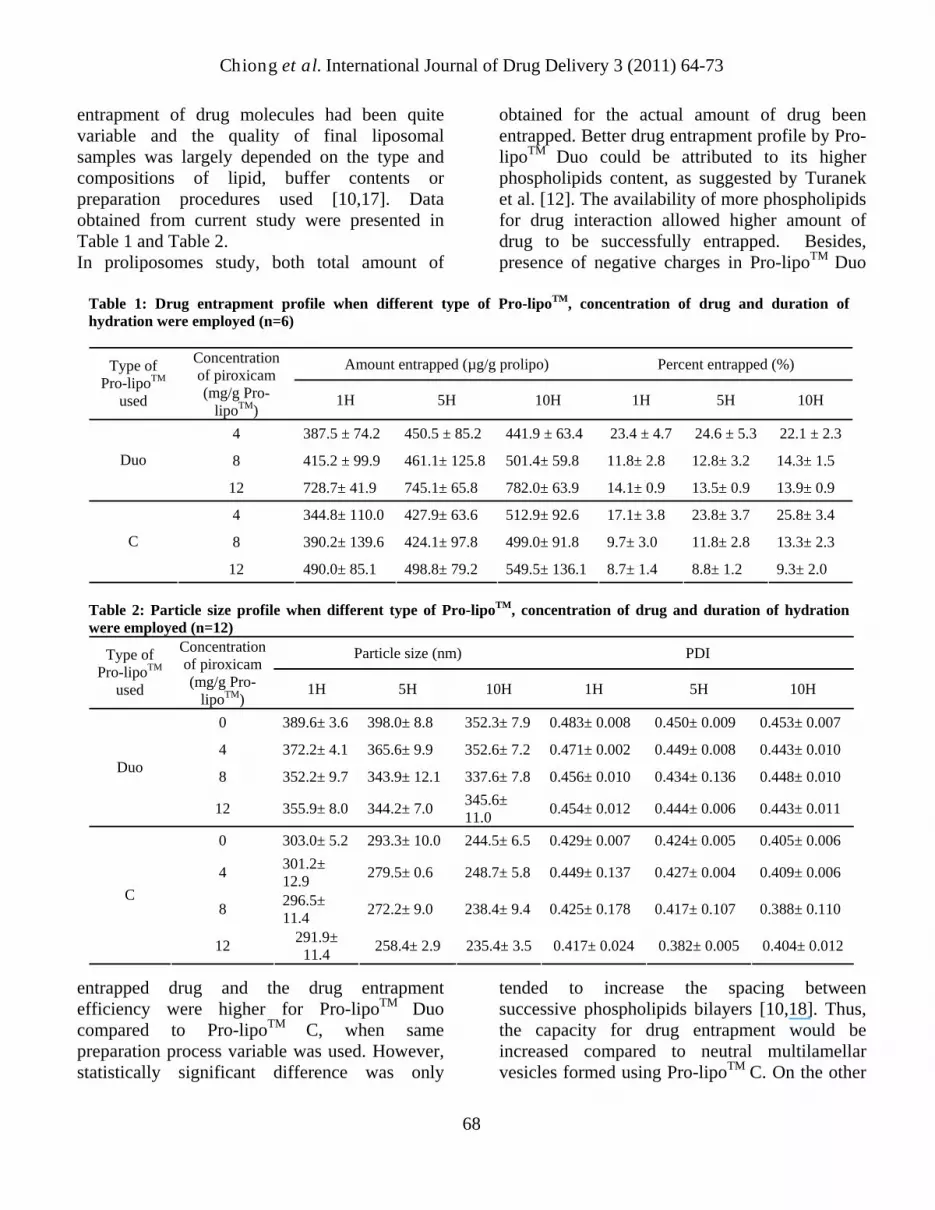
entrapment of drug molecules had been quite variable and the quality of final liposomal samples was largely depended on the type and compositions of lipid, buffer contents or preparation procedures used [10,17]. Data obtained from current study were presented in Table 1 and Table 2. In proliposomes study, both total amount of
entrapped drug and the drug entrapment efficiency were higher for Pro-lipoTM Duo compared to Pro-lipoTM C, when same preparation process variable was used. However, statistically significant difference was only
obtained for the actual amount of drug been entrapped. Better drug entrapment profile by Pro-lipoTM Duo could be attributed to its higher phospholipids content, as suggested by Turanek et al. [12]. The availability of more phospholipids for drug interaction allowed higher amount of drug to be successfully entrapped. Besides, presence of negative charges in Pro-lipoTM Duo
tended to increase the spacing between successive phospholipids bilayers [10,18]. Thus, the capacity for drug entrapment would be increased compared to neutral multilamellar vesicles formed using Pro-lipoTM C. On the other
Table 1: Drug entrapment profile when different type of Pro-lipoTM, concentration of drug and duration of hydration were employed (n=6)
Amount entrapped (µg/g prolipo) Percent entrapped (%) Type of Pro-lipoTM
Concentration of piroxicam (mg/g Pro-
lipoTM) 1H 5H 10H 1H 5H used 10H
4 387.5 ± 74.2 450.5 ± 85.2 441.9 ± 63.4 23.4 ± 4.7 24.6 ± 5.3 22.1 ± 2.3
Duo 8 415.2 ± 99.9 461.1± 125.8 501.4± 59.8 11.8± 2.8 12.8± 3.2 14.3± 1.5
12 728.7± 41.9 745.1± 65.8 782.0± 63.9 14.1± 0.9 13.5± 0.9 13.9± 0.9
4 344.8± 110.0 427.9± 63.6 512.9± 92.6 17.1± 3.8 23.8± 3.7 25.8± 3.4
8 390.2± 139.6 424.1± 97.8 499.0± 91.8 9.7± 3.0 11.8± 2.8 13.3± 2.3 C
12 490.0± 85.1 498.8± 79.2 549.5± 136.1 8.7± 1.4 8.8± 1.2 9.3± 2.0 Table 2: Particle size profile when different type of Pro-lipoTM, concentration of drug and duration of hydration were employed (n=12)
Particle size (nm) PDI Type of Pro-lipoTM
used
Concentration of piroxicam (mg/g Pro-
lipoTM) 1H 5H 10H 1H 5H 10H
0 389.6± 3.6 398.0± 8.8 352.3± 7.9 0.483± 0.008 0.450± 0.009 0.453± 0.007
4 372.2± 4.1 365.6± 9.9 352.6± 7.2 0.471± 0.002 0.449± 0.008 0.443± 0.010 Duo 8 352.2± 9.7 343.9± 12.1 337.6± 7.8 0.456± 0.010 0.434± 0.136 0.448± 0.010
12 355.9± 8.0 344.2± 7.0 345.6± 11.0 0.454± 0.012 0.444± 0.006 0.443± 0.011
0 303.0± 5.2 293.3± 10.0 244.5± 6.5 0.429± 0.007 0.424± 0.005 0.405± 0.006
4 301.2± 12.9 279.5± 0.6 248.7± 5.8 0.449± 0.137 0.427± 0.004 0.409± 0.006
8 296.5± 11.4 272.2± 9.0 238.4± 9.4 0.425± 0.178 0.417± 0.107 0.388± 0.110
C
12 291.9± 11.4 258.4± 2.9 235.4± 3.5 0.417± 0.024 0.382± 0.005 0.404± 0.012
68

Chiong et al. International Journal of Drug Delivery 3 (2011) 64-73 hand, liposomes formed using Pro-lipoTM Duo was significantly bigger in size and size distribution. This discrepancy could also be attributed by the differences of phospholipids and hydrophilic medium content in proliposomes. Pro-lipoTM Duo, which was more viscous than Pro-lipoTM C due its content, would had subjected to relatively less vigorous stirring force throughout the hydration period when similar stirring speed were used in preparation of both samples. As a result, the produced liposomes were comparatively less homogenous and bigger in size. In the drug concentration study, data obtained showed that the actual amount of drug successfully been entrapped increased significantly when higher concentration of piroxicam solution were used during liposomes preparation. On the contrary, increment of drug concentration produced significantly smaller and more homogenous liposomal samples. These phenomenons could be due to the increment of materials interaction and drug incorporation into vesicles when higher amount of lipophilic piroxicam was added into the lipidic proliposomes mixture. This was supported by the fact that Pro-lipoTM Duo, which contained higher amount of phospholipids, showed higher magnitude of increment in entrapment capacity compared to Pro-lipoTM C. As the amount of drug incorporated within the phospholipid bilayers increased, the bonds that holding successive bilayers decreased. In turn, the vesicle had looser packing which caused liposomes tend to have a smaller particle size [19]. Regretfully, drug entrapment efficiency decreased significantly as higher drug concentration was used. This trend suggested that the drug entrapment into lipid bilayers of liposomes were amount-limited, causing proportionally less increment on their successful drug entrapment rate compared to actual amount of drug used during sample preparations. Besides, decreased in the size of liposomes which in turn proportionally decreased the amount of lipid bilayers for entrapping drug could also resulted in reduction of their drug entrapment capacity.
In hydration study, an interesting trend of increment in the actual amount of piroxicam-loaded liposomes and percentage of entrapment efficiency were observed for both grade of Pro-lipoTM when longer stirring (hydration) time used. Regretfully, these observed increments were statistically not significant in present study. Fresta et al. [20] had claimed that slow rate of hydration and gentle mixing was beneficial for the entrapment efficiency due to slow annealing of multilamellar vesicle, which allowed for a longer period of contact between all the liposomes bilayers and the drug aqueous solution. On the other hand, data obtained showed that longer hydration time resulted in significantly smaller particle size and narrower size distribution in both type of Pro-lipoTM. A combination of impingement, cavitation and shear force from the continuously stirring mechanism during samples preparation could have been responsible to reduce the size of suspended liposomes and yield a more homogenous sample.
Applications for further studies One of the primary aims in current study was to determine the most suitable formulation which capable to use least amount of material (proliposomes) to encapsulate the largest amount of the model drug (piroxicam). Besides being cost effective, minimization of phospholipid content in liposomal drug formulation was important since lipids in high doses may be toxic and also cause non-linear/saturable pharmacokinetics [21]. On the other hand, smaller particle size had been reported to pose various advantageous characteristics including its effective interaction with cells, increased drug bioavailability in pathological area and longer circulation half life [21-23]. Thus, liposomal sample prepared using Pro-lipoTM Duo, 12mg piroxicam per gram Pro-lipoTM and 10 hours hydration time were deemed as the most optimum formulation which could produced highest amount of actual drug loaded into liposomes as well as exhibited homogenous and small particle size in nano range.
69

Chiong et al. International Journal of Drug Delivery 3 (2011) 64-73
Size reduction study This study was an attempt to search for a practical size reduction method which produced smaller and more homogenous liposomal samples, without compromising their initial drug entrapment capacity and efficiency. Data obtained were presented in Table 3. Bath sonication, though was a relatively more convenient method, did not cause a significant trend of decrement in the entrapment and size profile for treated liposomal samples. Only the extrusion method resulted in statistically significant loss in drug entrapment capacity and efficiency as well as significant decrement in size and size distribution of liposomes. From the data and trends observed in this study, it could be concluded that the extrusion technique through polycarbonate membrane filter was a far superior size reduction method than the bath sonication technique. The impingement and shear forces created when forcing the liposomes through small pores of membrane filter could destroy the lamellar surface, allowing the formation of monodisperse vesicles [24].
Regretfully, extrusion of liposomes resulted in significant loss of the originally entrapped piroxicam, which was also manifested by the decrease in the entrapment efficiency. These marked reductions in drug entrapment capacity and efficiency could attribute to the lower entrapment volume (lipid bilayers) available for entrapping drug molecules. Similar findings had also been previously reported [10, 21, 25]. On the other hand, results showed that liposomes had a size larger than the pores through which they were extruded. This finding suggested the elastic deformation of the liposomes. A similar interpretation had also been suggested by Elhissi et al. [26].
Storage study Liposomes were known for their physical instability during storage due to potential drug leakage from vesicles and changes in the size of liposomes [21]. In present study, data obtained were presented in Table 4. The drug entrapment capacity and efficiency of prepared liposomal samples were found stable in both refrigerated
Table 3: Drug entrapment and particle size profile before and after size reduction treatment
Entrapment profile (n=6) Size profile (n=12) Size reduction treatment Amount entrapped (µg/g Pro-
lipoTM) Percent entrapped
(%) Particle size (nm) PDI
Without treatment 818.8 ± 124.1 16.9 ± 2.0 382.5 ± 3.0 0.463 ± 0.005
Sonication 770.9 ± 95.9 14.6 ± 1.9 376.5 ± 2.0 0.451 ± 0.005
Extrusion 335.8 ± 40.4 6.7 ± 0.8 135.6 ± 2.6 0.120 ± 0.007
Table 4: Drug entrapment and particle size profile of liposomes kept under different storage conditions
Refrigerated temperature (2�C to 8�C) Room temperature (25�C±2�C).
Entrapment profile (n=6) Size profile (n=12) Entrapment profile (n=6) Size profile
(n=12) Week
Amount entrapped (µg/g Pro-
lipoTM)
Percent entrapped
(%)
Particle size (nm)
PDI
Amount entrapped (µg/g Pro-
lipoTM)
Percent entrapped
(%)
Particle size (nm) PDI
0 818.8± 124.1 16.9± 2.0 382.5± 3.0 0.463± 0.005 818.8± 124.1 16.9± 2.0 382.5± 3.0 0.463± 0.005
1 761.3± 86.2 14.8± 1.6 380.7± 2.8 0.454± 0.005 762.7± 104.4 14.6± 2.0 381.8± 3.3 0.458± 0.006
2 731.8± 100.4 14.6± 1.7 382.6± 2.1 0.463± 0.006 736.5± 115.9 14.9± 2.3 388.4± 3.2 0.443± 0.006
3 746.0± 83.2 15.5± 1.5 377.7± 3.1 0.472± 0.013 757.1± 122.3 15.6± 2.3 387.6± 5.1 0.460± 0.007
4 771.2± 79.7 15.3± 1.5 377.8± 1.9 0.457± 0.007 760.1± 97.4 14.9± 1.5 382.9± 3.0 0.473± 0.013
70

Chiong et al. International Journal of Drug Delivery 3 (2011) 64-73 and room temperature. Although slight decrement pattern was observed in both storage temperatures throughout the duration of study, no statistically differences were found. These fairly high retention of drug (>90%) within the liposomes could be explained by great affinity of phospholipid bilayers towards the drug molecules as well as good structural integrity of the formed vesicles [2]. On the other hand, particle size and PDI were also found to be stable in this study. No statistically differences were found on the effect of storage time to the size profile of these liposomal samples. However, liposomes kept in refrigerated temperature were found to have relatively slight decrement in particle size compared to those kept in the room temperature. This decrement of mean particle size could be due to the refrigerating and thawing mechanism when handling the samples, which resulted in the breaking up of larger sized liposomes. Damages could also caused by the crystallization of internal water, osmotic forces, dehydration and formation of amorphous material [25,27].
Reproducibility testing In the context of this study, the measure of reproducibility was defined as the closeness
between independent data obtained from each batch of samples which prepared at different time using identical materials, procedures and preparation conditions. A liposomal sample formulation, which was deemed to have the most desirable drug-entrapment and size profile for future works, was selected for current evaluation. The percent coefficient of variation (%CV) for all of the parameters tested was found to be less than 15% (Table 5). Hence, the preparation of liposomal samples at different time using a same formulation and methodology was deemed to pose satisfactory reproducible physico-chemical values.
Transmission electron microscope observations Direct visualization of liposomes in readily prepared liposomal samples using TEM technique revealed that different sizes of multilamellar vesicles were formed using current materials and preparation procedures (Figure 1). The blank and drug-loaded liposomes vesicles were observed to be spherical-shaped and showed concentric lamellae. Whereas, liposomal sample extruded through filter membrane produced clearly smaller oligolamellar liposomes,
(A) (B) (C) Figure 1. TEM photograph of (A) blank liposomes; (B) piroxicam-loaded liposomes; (C) extruded piroxicam-loaded liposomes. (Arrows: liposomes)
Table 5: Reproducibility of piroxicam-loaded liposomes (n=6)
Entrapment profile Size profile Batch name Amount entrapped
(µg/g Pro-lipoTM) Percent entrapped (%) Particle size (nm) PDI
Mean ± S.E.M 800.4 ± 48.9 15.4 ± 0.9 360.0 ± 14.4 0.454 ± 0.009 CV (%) 14.964 14.825 9.775 4.830
71
Chiong et al. International Journal of Drug Delivery 3 (2011) 64-73
indicating that lamellarity of liposomes was effectively reduced by the extrusion process.
Conclusions In summary, findings in current studies conclusively demonstrated an optimized formulation and reproducible preparation of piroxicam-loaded liposomes which prepared using a simple proliposomes method. As a rule of thumb, preparation of piroxicam-loaded liposomes using Pro-lipoTM Duo with higher drug concentration at prolonged hydration period would able to yield smaller, more homogenous liposomes with improved drug entrapment capacity and satisfactory entrapment efficiency. Liposomes prepared were stably stored for at least 4 weeks. Furthermore, extrusion technique was demonstrated to be an effective size reduction method, whereas electron microscopy revealed sizes and lamellarity of prepared liposomal samples. Acknowledgements This work was supported by Fundamental Research Grant Scheme from the Ministry of Higher Education, Malaysia (04-10-07-277FR). The authors thank Hovid Sdn. Bhd. and Renogenic Sdn. Bhd. for providing instrumental and technical assistances with the experiments.
References
1. Gulati M, Grover M, Singh S, Singh M, 1998. Lipophilic drug derivatives in liposomes. Int. J. Pharm., 165, 129–168.
2. Agarwal R, Katare OP, Vyas SP, 2001. Preparation and in vitro evaluation of liposomal/niosomal delivery systems for antipsoriatic drug dithranol. Int. J. Pharm., 228, 43–52.
3. Hens AG, Romero JMF. 2006. Analytical methods for the control of liposomal delivery systems. Trend Anal. Chem., 25(2), 167-178.
4. Vasudevan DM, Sreekumari S, 2005. Phospholipids. In: Textbook of Biochemistry For Medical Students, 4th
Ed., Jaypee Brothers Medical Publishers, New Delhi, pp. 76-77.
5. Baudrimonta I, Murnb M, Betheder AM, Guilcher J, Creppy EE, 1995. Effect of piroxicam on the nephrotoxicity induced by ochratoxin A in rats. Toxicology, 147, 1-4.
6. Starek M, Krzek J, 2009. A review of analytical techniques for determination of oxicams, nimesulide and nabumetone. Talanta, 77, 925–942.
7. Tagliati CA, Kimura E. Nothenberg MS., Santos S, Oga S, 1999. Pharmacokinetic profile and gastric effect of zinc-piroxicam in rats. Gen. Pharmacol., 33, 67–71.
8. Matos C, Lima JLC, Reis S, Lopes, A., Bastos, M., 2004. Interaction of anti-inflammatory drugs with EPC liposomes: calorimetric study in a broad concentration range. Biophys. J., 86, 946–954.
9. Lopes LB, Scarpa MV, Pereira NL, Oliveira LC, Oliveira AG. 2006. Interaction of sodium diclofenac with freeze-dried soya phosphatidylcholine and unilamellar liposomes. Braz. J. Pharm. Sci., 42(4), 497-504.
10. Vemuri S, Rhodes CT, 1995. Preparation and characterization of liposomes as therapeutic delivery systems: a review. Pharm. Acta Helv., 70, 95-111.
11. Perrett S, Golding M, Williams WP. 1991. A simple method for the preparation of liposomes for pharmaceutical applications: characterization of the liposomes. J. Pharm. Pharmacol., 43, 54–161.
12. Turanek J, Zaluska D, Neca J, 1997. Linkup of a fast protein liquid chromatography system with a stirred thermostated cell for sterile preparation of liposomes by the proliposome–liposome method: Application to encapsulation of antibiotics, synthetic peptide immunomodulators and a photosensitizer. Anal. Biochem., 249, 131–139.
13. Shahiwala A, Misra A. 2002. Studies in topical application of niosomally entrapped nimesulide. J. Pharm. Pharmaceut. Sci., 5(3), 220-225.
72

Chiong et al. International Journal of Drug Delivery 3 (2011) 64-73 14. Ling SSN. 2004. Investigation of liposomes
for oral delivery of peptidomimetic drugs. Ph.D Thesis. Universiti Sains Malaysia, Malaysia.
15. Lucchetti G, Assogna G, Bagnato P. 1996. Liposomal piroxicam formulations. US Patent 5505960, 9 Apr.
16. Singh A, Lawson GE, Shivakrupa R, Johnson BT, Blumenthal R, Puri A. 2007. Piroxicam entrapped in head-group polymerized liposomes inhibits proliferation of IC2 mast cells in vitro. Mater. Res. Soc. Symp. Proc., 1019, FF02-07.
17. Martin FJ. 1990. Pharmaceutical manufacturing of liposomes. In: Specialized Drug Delivery Systems: Manufacturing and Production Technology, Marcel Dekker, New York, pp. 267-316.
18. Alpar OH, Bamford JB, Walters V. 1981. The in vitro incorporation and release of hydroxocobalamin by liposomes. Int. J. Pharm., 7, 349-351.
19. Fa N, Lins L, Courtoy PJ, Dufrêne Y, Van Der Smissen P, Brasseur R, Tyteca D, Mingeot-Leclercq M.-P. 2007. Decrease of elastic moduli of DOPC bilayers induced by a macrolide antibiotic, azithromycin. Biochim Biophys Acta, 1768, 1830–1838.
20. Fresta M, Villari A, Puglisi G, Cavallaro G. 1993. 5-Fluorouracil: various kina of loaded liposomes: encapsulation efficiency, storage stability and fusogenic properties. Int. J. Pharm., 99, 145-156.
21. Sharma A, Sharma US. 1997. Liposomes in drug delivery: progress and limitations. Int. J. Pharm., 154, 123-140.
22. Elorza B, Elorza MA, Sainz MC, Chantres JR. 1993. Análysis of the particle size distribution and the internal volume of liposomal preparations. J. Pharm. Sci., 82(11), 1160-1163.
23. Chen HM, Langer R. 1998. Oral particle delivery: status and future trends. Adv. Drug Del. Rev., 34(2-3), 339-350.
24. Segota S, Tezak D. 2006. Spontaneous formation of vesicles. Adv. Colloid Interfac. 121, 51–75.
25. Betageri GV, Jenkins SA, Parsons DL. 1993. Preparation of liposomes. In: Liposome Drug Delivery Systems, Technomic Publishing Company, Lancaster, pp. 1-24.
26. Elhissi AMA, Faizi M, Naji WF, Gill HS, Taylor KMG. 2007. Physical stability and aerosol properties of liposomes delivered using an air-jet nebulizer and a novel micropump device with large mesh apertures. Int. J. Pharm., 334, 62–70.
27. Talsma H, Van Steebergen MJ, Salemink PJM, Crommelin JA. 1991. The cryopreservation of liposomes. 1. a differential scanning calorimetry study of the thermal behaviour of a liposomes dispersion containing mannitol during freezing/thawing. Pharm. Res., 8(8), 1021-1026.
73