new 3-cyano-2-substituted pyridines induce apoptosis in
TRANSCRIPT
molecules
Article
New 3-Cyano-2-Substituted Pyridines InduceApoptosis in MCF 7 Breast Cancer Cells
Ahmed Malki 1,*, Mona Mohsen 1, Hassan Aziz 1, Ola Rizk 2,3, Omima Shaban 2,3,Mohamed El-Sayed 4, Zaki A. Sherif 5 and Hayam Ashour 2,*
1 Biomedical Science Program, Department of Health Sciences, College of Art and Sciences, Qatar University,Doha 2713, Qatar; [email protected] (M.M.); [email protected] (H.A.)
2 Department of Pharmaceutical Chemistry, Faculty of Pharmacy, Alexandria University, Alexandria 21521,Egypt; [email protected] (O.R.); [email protected] (O.S.)
3 Department of Analytical and Pharmaceutical Chemistry, Faculty of Pharmacy & Drug Manufacturing,Pharos University, Alexandria, 21311, Egypt
4 Biochemistry Department, Faculty of Science, Alexandria University, Alexandria 21521, Egypt;[email protected]
5 Department of Biochemistry and Molecular Biology, Howard University, College of Medicine, Washington,DC 20059, USA; [email protected]
* Correspondence: [email protected] (A.M.); [email protected] (H.A.);Tel.: +974-4403-6557 (A.M.); +20-3-487-1317 (H.A.)
Academic Editor: Jean Jacques Vanden EyndeReceived: 3 January 2016 ; Accepted: 1 February 2016 ; Published: 18 February 2016
Abstract: The synthesis of new 3-cyano-2-substituted pyridines bearing various pharmacophoresand functionalities at position 2 is described. The synthesized compounds were evaluated fortheir in vitro anti-cancer activities on five cancer cell lines using 5-FU as reference compound.The results revealed that the benzohydrazide derivative 9a induced growth inhibition in humanbreast cancer cell line MCF-7 with an IC50 value of 2 µM and it showed lower cytotoxicity onMCF-12a normal breast epithelial cells. Additionally, 9a induced apoptotic morphological changesand induced apoptosis in MCF-7 in a dose and time-dependent manner according to an enzymelinked immunosorbent apoptosis assay which is further confirmed by a TUNEL assay. Flowcytometric analysis indicated that 9a arrested MCF-7 cells in the G1 phase, which was furtherconfirmed by increased expression of p21 and p27 and reduced expression of CDK2 and CDK4.Western blot data revealed significant upregulation of the expression of p53, Bax, caspase-3 anddown-regulation of Bcl-2, Mdm-2 and Akt. Additionally, 9a increased the release of cytochromec from mitochondria to cytoplasm which provokes the mitochondrial apoptotic pathway while itshowed no significant change on the expression of the death receptor proteins procaspase-8, caspase-8and FAS. Furthermore, 9a reduced the expression of phospho AKT and β-catenin in dose dependentmanner while inhibiting the expression of migration-related genes such as matrix metalloproteinase(MMP)-9 and vascular endothelial growth factor (VEGF). Our findings suggest that compound 9acould be considered as a lead structure for further development of more potent apoptosis inducingagents with anti-metastatic activities.
Keywords: 3-cyanopyridines; alkoxy substituents; MCF-7; apoptosis; p53; VEGF
1. Introduction
Breast cancer is a complex disease that comprises heterogenous tumors associated with distinctivehistological patterns and different clinical characteristics [1]. An estimated 1.7 million women will be
Molecules 2016, 21, 230; doi:10.3390/molecules21020230 www.mdpi.com/journal/molecules
brought to you by COREView metadata, citation and similar papers at core.ac.uk
provided by Qatar University Institutional Repository
Molecules 2016, 21, 230 2 of 25
diagnosed with breast cancer in 2020, a 26% increase from current levels [2]. Approximately 30% of thewomen diagnosed with early-stage disease subsequently progress to metastatic breast cancer (MBC),for which few therapeutic regimens exist [3]. Many factors contribute to the metastasis of tumorcells; these include molecular signaling pathways, and expression of metastasis-related genes such asmatrix metalloproteinases (MMPs) [4]. Despite the advances in therapeutic modalities, MBC remainsincurable, with an estimated survival rate of about 23% over 5 years [5]. Chemotherapy resistance isalso a consistent obstacle in the management of breast cancer, where many of the initially responsivetumors relapse and develop resistance to diverse chemotherapeutic agents [6]. Consequently, newagents with low susceptibility to common drug resistance mechanisms are urgently needed for themanagement and treatment of various forms of breast cancer.
The anticancer activity of most cytotoxic therapies which are currently used in the clinicalmanagement of cancer patients including radiotherapy is based on their ability to induce cell deathprograms such as apoptosis [7]. Apoptosis is an evolutionary conserved process characterized by aseries of morphological and biochemical changes, including cell shrinkage, nuclear DNA fragmentationand membrane blebbing [8].
Pyridine derivatives have always constituted a subject of great interest due to the ubiquityof pyridine in Nature and the extensive presence in the skeletal backbone of many therapeuticagents. These derivatives were reported to possess cytotoxic [9], and anticancer activities [10–18]in addition to topoisomerase I, II [19–21] and Src inhibitory activities [22–24]. Penclomedine (PEN1,3,5-dichloro-4,6-dimethoxy-2-trichloromethylpyridine), is an antitumor agent which was selected forclinical development by the NCI based on its selective antitumor activity against certain carcinomas [25].Moreover, triapine (3-aminopyridine-2-carboxaldehyde thiosemicarbazone) is a potent ribonucleotidereductase inhibitor that has shown potent antiproliferative activity against several cancer cell lines [26].Furthermore, particular interest has been focused on pyridine carbonitriles due to their remarkablein vitro anticancer activity against a wide range of cell lines (Figure 1) [27–30]. Consequently, pyridinecarbonitrile remains a promising template for the design of a new category of chemotherapeutic agents.
Inspired by the abovementioned findings and in continuation of our efforts linked to discoveringand exploring novel lead heterocyclic structures as potent chemotherapeutic agents [31–34], newderivatives of 3-cyano-2-substituted pyridines were synthesized for evaluation of their in vitroanticancer activity. A literature survey revealed that incorporation of alkoxy substituents (methoxyand/or aryloxy moieties) results in significant enhancement of antitumor activity due to magnificationof compounds’ lipophilicity [35,36]. Accordingly, the target compounds were designed so as tocomprise 3,4-dimethoxyphenyl groups at positions 4 and 6. Moreover to the best of our knowledge,2-substituted alkoxycyanopyridines are seldom reported in the literature. Therefore, it was planned toinclude variable substituents at position 2, linked to the cyanopyridine scaffold through a methyleneoxyor acetyloxy spacer (A and B, Figure 1). Such substituents were selected so as to offer variableelectronic, lipophilic and steric environment that could influence the targeted biological activity. Thesubstituents include either alkyl groups of different length or biologically active pharmacophores thatare believed to be responsible for the biological significance of some reported anticancer agents such asbenzohydrazides [37,38] benzosulfohydrazides [10], dithioates [39,40] and arylhydrazones [41–43]. Inaddition, incorporation of heterocyclic groups such as pyrazoles and 1,3,4-oxadiazoles (B, Figure 1) wasconsidered as an interesting structure variation that might impose an impact on the potential biologicalactivities owing to their documented chemotherapeutic activity [44–48].The in vitro antiproliferativeactivity of the newly synthesized compounds was investigated against five cancer cell lines and theeffect of the most promising compound on apoptosis and expression of proteins related to cell cyclepathways was also evaluated.

Molecules 2016, 21, 230 3 of 25Molecules 2016, 21, 230 3 of 24
N NHF3C
NH
N
O
Cl
Cl
ClN NHF3C
NH
Cl
O
OMe
OMe
OMe
N
CNNC
NH
HN C6H5C6H5
N NH
NH
HN
O
S
R
R1
O O
N
CN
NH2
NR3
RR1
R2
N
CN
O
OCH3
OCH3
H3CO
OCH3
N
CN
O
OCH3
OCH3
H3CO
OCH3
R
O
R = H, CH3, CH2CH3, COCH3, CH2COOCH2CH3, 1,3,4-oxadiazole-2-thione
(R= dithioates, aryl hydrazones, arylhydrazides, arylsulfohydrazides, pyrazole or pyrazolinone
A B
reported antitumor pyridine derivatives
reported antitumor cyanopyridines
isosteric replacement of NH by O
pharmacologically active pharmacophores
R
isosteric replacement of NH by O
dimethoxyphenyl groupincreases lipophilicity
dimethoxy
phenyl g
roup
increa
ses lip
ophilic
ity
dimethoxyphenyl groupincreases lipophilicity
dimethoxyphenyl groupincreases lipophilicity
Figure 1. Chemical structure of reported pyridines and cyanopyridines endowed with anticancer and apoptosis-inducing activities and the synthesized compounds (A,B).
2. Results and Discussion
2.1. Chemistry
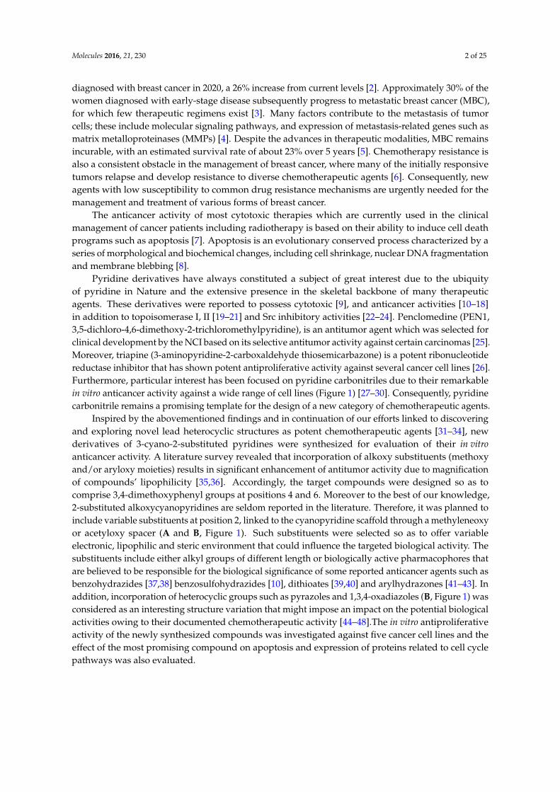
The synthetic strategies adopted for the synthesis of the intermediate and target compounds are depicted in Schemes 1–3. In Scheme 1, the cyanopyridinone 3 was prepared according to the Al-Saadi procedure [49] via a one-pot multicomponent reaction of 3,4-dimethoxybenzaldehyde (1), 3,4-dimethoxyacetophenone (2), an excess of ammonium acetate and ethyl cyanoacetate in boiling ethanol. Heating the cyanopyridinone 3 with different alkyl halides in absolute ethanol using sodium ethoxide as a basic catalyst according to the Kornblum procedure [50] failed to afford the target O-alkylated derivatives 4a–d. However, such compounds were successfully prepared by heating the cyanopyridinone 3 with the appropriate alkyl halide in acetone in the presence of anhydrous K2CO3. Similarly, refluxing 3 with ethyl bromoacetate in dry acetone containing anhydrous K2CO3 yielded the corresponding ethyl acetate ester 5. Reaction of the ester 5 with hydrazine hydrate in refluxing ethanol resulted in the formation of the corresponding acetohydrazide 6 which was employed as key intermediate for synthesis of the target compounds presented in Scheme 2.
Figure 1. Chemical structure of reported pyridines and cyanopyridines endowed with anticancer andapoptosis-inducing activities and the synthesized compounds (A,B).
2. Results and Discussion
2.1. Chemistry
The synthetic strategies adopted for the synthesis of the intermediate and target compoundsare depicted in Schemes 1–3. In Scheme 1, the cyanopyridinone 3 was prepared according to theAl-Saadi procedure [49] via a one-pot multicomponent reaction of 3,4-dimethoxybenzaldehyde (1),3,4-dimethoxyacetophenone (2), an excess of ammonium acetate and ethyl cyanoacetate in boilingethanol. Heating the cyanopyridinone 3 with different alkyl halides in absolute ethanol using sodiumethoxide as a basic catalyst according to the Kornblum procedure [50] failed to afford the targetO-alkylated derivatives 4a–d. However, such compounds were successfully prepared by heating thecyanopyridinone 3 with the appropriate alkyl halide in acetone in the presence of anhydrous K2CO3.Similarly, refluxing 3 with ethyl bromoacetate in dry acetone containing anhydrous K2CO3 yieldedthe corresponding ethyl acetate ester 5. Reaction of the ester 5 with hydrazine hydrate in refluxingethanol resulted in the formation of the corresponding acetohydrazide 6 which was employed as keyintermediate for synthesis of the target compounds presented in Scheme 2.
Molecules 2016, 21, 230 4 of 25Molecules 2016, 21, 230 4 of 24
N
CN
OH
+
N
CN
ON
CN
O
N
CN
OO
HN
NH2
O
OC2H5
3
4a-d5
6
H
O
CH3
O
i
ii
iv
H3CO
H3CO
H3CO
H3CO
OCH3
H3CO
H3CO
H3COOCH3
H3CO
H3CO
H3CO
OCH3
H3CO
H3CO
H3CO
1 2
iii
OCH3
H3CO
H3CO
H3CO
R
Scheme 1. Synthesis of the target compounds 4, 5 and the key intermediate 6. For 4a–d: a, R = H; b, R = CH3; c, R = CH2CH3; d, R = COCH3; Reagents and conditions: (i) CNCH2COOEt/anhydrous CH3COONH4/EtOH/reflux; (ii) the appropriate alkyl halide/anhydrous K2CO3/dry acetone/reflux; (iii) BrCH2COOEt/anhydrous K2CO3/dry acetone/reflux; (iv) N2H4 98%/EtOH/reflux.
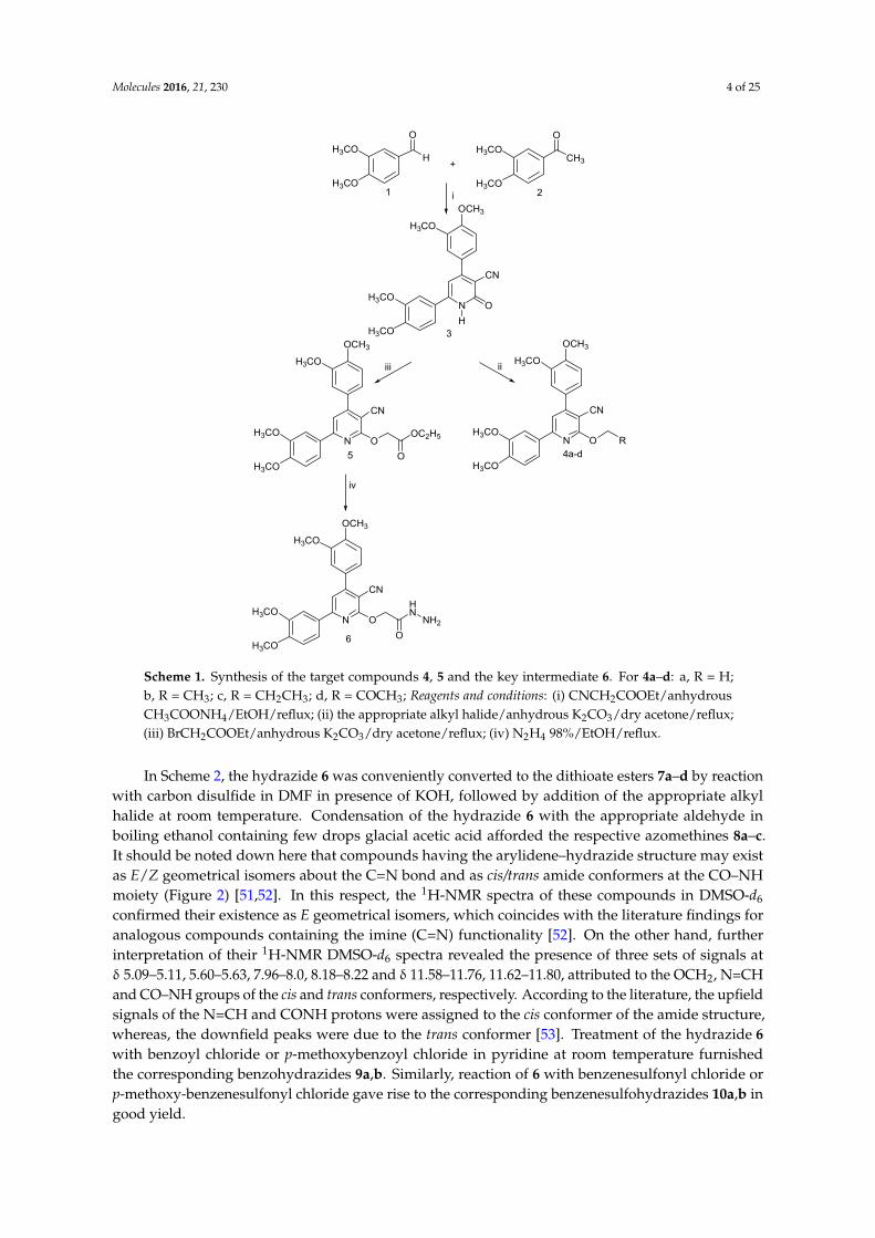
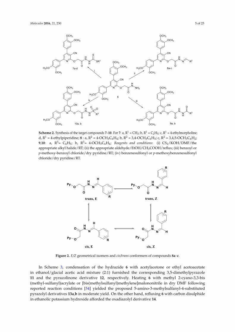
In Scheme 2, the hydrazide 6 was conveniently converted to the dithioate esters 7a–d by reaction with carbon disulfide in DMF in presence of KOH, followed by addition of the appropriate alkyl halide at room temperature. Condensation of the hydrazide 6 with the appropriate aldehyde in boiling ethanol containing few drops glacial acetic acid afforded the respective azomethines 8a–c. It should be noted down here that compounds having the arylidene–hydrazide structure may exist as E/Z geometrical isomers about the C=N bond and as cis/trans amide conformers at the CO–NH moiety (Figure 2) [51,52]. In this respect, the 1H-NMR spectra of these compounds in DMSO-d6 confirmed their existence as E geometrical isomers, which coincides with the literature findings for analogous compounds containing the imine (C=N) functionality [52]. On the other hand, further interpretation of their 1H-NMR DMSO-d6 spectra revealed the presence of three sets of signals at δ 5.09–5.11, 5.60–5.63, 7.96–8.0, 8.18–8.22 and δ 11.58–11.76, 11.62–11.80, attributed to the OCH2, N=CH and CO–NH groups of the cis and trans conformers, respectively. According to the literature, the upfield signals of the N=CH and CONH protons were assigned to the cis conformer of the amide structure, whereas, the downfield peaks were due to the trans conformer [53]. Treatment of the hydrazide 6 with benzoyl chloride or p-methoxybenzoyl chloride in pyridine at room temperature furnished the corresponding benzohydrazides 9a,b. Similarly, reaction of 6 with benzenesulfonyl chloride or p-methoxy-benzenesulfonyl chloride gave rise to the corresponding benzenesulfohydrazides 10a,b in good yield.
Scheme 1. Synthesis of the target compounds 4, 5 and the key intermediate 6. For 4a–d: a, R = H;b, R = CH3; c, R = CH2CH3; d, R = COCH3; Reagents and conditions: (i) CNCH2COOEt/anhydrousCH3COONH4/EtOH/reflux; (ii) the appropriate alkyl halide/anhydrous K2CO3/dry acetone/reflux;(iii) BrCH2COOEt/anhydrous K2CO3/dry acetone/reflux; (iv) N2H4 98%/EtOH/reflux.
In Scheme 2, the hydrazide 6 was conveniently converted to the dithioate esters 7a–d by reactionwith carbon disulfide in DMF in presence of KOH, followed by addition of the appropriate alkylhalide at room temperature. Condensation of the hydrazide 6 with the appropriate aldehyde inboiling ethanol containing few drops glacial acetic acid afforded the respective azomethines 8a–c.It should be noted down here that compounds having the arylidene–hydrazide structure may existas E/Z geometrical isomers about the C=N bond and as cis/trans amide conformers at the CO–NHmoiety (Figure 2) [51,52]. In this respect, the 1H-NMR spectra of these compounds in DMSO-d6
confirmed their existence as E geometrical isomers, which coincides with the literature findings foranalogous compounds containing the imine (C=N) functionality [52]. On the other hand, furtherinterpretation of their 1H-NMR DMSO-d6 spectra revealed the presence of three sets of signals atδ 5.09–5.11, 5.60–5.63, 7.96–8.0, 8.18–8.22 and δ 11.58–11.76, 11.62–11.80, attributed to the OCH2, N=CHand CO–NH groups of the cis and trans conformers, respectively. According to the literature, the upfieldsignals of the N=CH and CONH protons were assigned to the cis conformer of the amide structure,whereas, the downfield peaks were due to the trans conformer [53]. Treatment of the hydrazide 6with benzoyl chloride or p-methoxybenzoyl chloride in pyridine at room temperature furnishedthe corresponding benzohydrazides 9a,b. Similarly, reaction of 6 with benzenesulfonyl chloride orp-methoxy-benzenesulfonyl chloride gave rise to the corresponding benzenesulfohydrazides 10a,b ingood yield.
Molecules 2016, 21, 230 5 of 25Molecules 2016, 21, 230 5 of 24
N
CN
O
O
HN
NH2
6
N
CN
O
O
HN
N R2
8a-c
N
CN
O
O
HN
NH
O
R3N
CN
O
O
HN
NH
10a, b
SR3
O O
N
CN
O
O
HN
NH
7a-d
S
SR1
ii
OCH3
OCH3
H3CO
OCH3
OCH3
OCH3
H3CO
OCH3
OCH3
OCH3
OCH3
H3CO
OCH3
OCH3
H3CO
OCH3
OCH3
OCH3
OCH3
H3CO
9a, b
i
iiiiv
Scheme 2. Synthesis of the target compounds 7–10. For 7: a, R1 = CH3; b, R1 = C2H5; c, R1 = 4-ethylmorpholine; d, R1 = 4-ethylpiperidine; 8 : a, R2 = 4-OCH3C6H4; b, R2 = 3,4-OCH3C6H3; c, R2 = 3,4,5-OCH3C6H2; 9,10: a, R3= C6H5; b, R3= 4-OCH3C6H4; Reagents and conditions: (i) CS2/KOH/DMF/ the appropriate alkyl halide/RT; (ii) the appropriate aldehyde/EtOH/CH3COOH/reflux; (iii) benzoyl or p-methoxy-benzoyl chloride/dry pyridine/RT; (iv) benzenesulfonyl or p-methoxybenzenesulfonyl chloride/dry pyridine/RT.
O
HN
O
N
H
R
O
HN
O
N H
R
HN
N H
O
OHN
N
H
O
O
R
R
cis, E
trans, E trans, Z
cis, Z
Py =
Py Py
PyPy
3-cyano-4,6-bis(3,4-dimethoxyphenyl)pyridin-2-yl Figure 2. E/Z geometrical isomers and cis/trans conformers of compounds 8a–c.
In Scheme 3, condensation of the hydrazide 6 with acetylacetone or ethyl acetoacetate in ethanol/glacial acetic acid mixture (2:1) furnished the corresponding 3,5-dimethylpyrazole 11 and the pyrazolinone derivative 12, respectively. Heating 6 with methyl 2-cyano-3,3-bis(methyl-sulfanyl) acrylate or [bis(methylsulfanyl)methylene]malononitrile in dry DMF following reported reaction conditions [54] yielded the proposed 5-amino-3-methylsulfanyl-4-substituted pyrazolyl derivatives 13a,b in moderate yield. On the other hand, refluxing 6 with carbon disulphide in ethanolic potassium hydroxide afforded the oxadiazolyl derivative 14.
Scheme 2. Synthesis of the target compounds 7–10. For 7: a, R1 = CH3; b, R1 = C2H5; c, R1 = 4-ethylmorpholine;d, R1 = 4-ethylpiperidine; 8 : a, R2 = 4-OCH3C6H4; b, R2 = 3,4-OCH3C6H3; c, R2 = 3,4,5-OCH3C6H2;9,10: a, R3= C6H5; b, R3= 4-OCH3C6H4; Reagents and conditions: (i) CS2/KOH/DMF/theappropriate alkyl halide/RT; (ii) the appropriate aldehyde/EtOH/CH3COOH/reflux; (iii) benzoyl orp-methoxy-benzoyl chloride/dry pyridine/RT; (iv) benzenesulfonyl or p-methoxybenzenesulfonylchloride/dry pyridine/RT.
Molecules 2016, 21, 230 5 of 24
N
CN
O
O
HN
NH2
6
N
CN
O
O
HN
N R2
8a-c
N
CN
O
O
HN
NH
O
R3N
CN
O
O
HN
NH
10a, b
SR3
O O
N
CN
O
O
HN
NH
7a-d
S
SR1
ii
OCH3
OCH3
H3CO
OCH3
OCH3
OCH3
H3CO
OCH3
OCH3
OCH3
OCH3
H3CO
OCH3
OCH3
H3CO
OCH3
OCH3
OCH3
OCH3
H3CO
9a, b
i
iiiiv
Scheme 2. Synthesis of the target compounds 7–10. For 7: a, R1 = CH3; b, R1 = C2H5; c, R1 = 4-ethylmorpholine; d, R1 = 4-ethylpiperidine; 8 : a, R2 = 4-OCH3C6H4; b, R2 = 3,4-OCH3C6H3; c, R2 = 3,4,5-OCH3C6H2; 9,10: a, R3= C6H5; b, R3= 4-OCH3C6H4; Reagents and conditions: (i) CS2/KOH/DMF/ the appropriate alkyl halide/RT; (ii) the appropriate aldehyde/EtOH/CH3COOH/reflux; (iii) benzoyl or p-methoxy-benzoyl chloride/dry pyridine/RT; (iv) benzenesulfonyl or p-methoxybenzenesulfonyl chloride/dry pyridine/RT.
O
HN
O
N
H
R
O
HN
O
N H
R
HN
N H
O
OHN
N
H
O
O
R
R
cis, E
trans, E trans, Z
cis, Z
Py =
Py Py
PyPy
3-cyano-4,6-bis(3,4-dimethoxyphenyl)pyridin-2-yl Figure 2. E/Z geometrical isomers and cis/trans conformers of compounds 8a–c.
In Scheme 3, condensation of the hydrazide 6 with acetylacetone or ethyl acetoacetate in ethanol/glacial acetic acid mixture (2:1) furnished the corresponding 3,5-dimethylpyrazole 11 and the pyrazolinone derivative 12, respectively. Heating 6 with methyl 2-cyano-3,3-bis(methyl-sulfanyl) acrylate or [bis(methylsulfanyl)methylene]malononitrile in dry DMF following reported reaction conditions [54] yielded the proposed 5-amino-3-methylsulfanyl-4-substituted pyrazolyl derivatives 13a,b in moderate yield. On the other hand, refluxing 6 with carbon disulphide in ethanolic potassium hydroxide afforded the oxadiazolyl derivative 14.
Figure 2. E/Z geometrical isomers and cis/trans conformers of compounds 8a–c.
In Scheme 3, condensation of the hydrazide 6 with acetylacetone or ethyl acetoacetatein ethanol/glacial acetic acid mixture (2:1) furnished the corresponding 3,5-dimethylpyrazole11 and the pyrazolinone derivative 12, respectively. Heating 6 with methyl 2-cyano-3,3-bis(methyl-sulfanyl)acrylate or [bis(methylsulfanyl)methylene]malononitrile in dry DMF followingreported reaction conditions [54] yielded the proposed 5-amino-3-methylsulfanyl-4-substitutedpyrazolyl derivatives 13a,b in moderate yield. On the other hand, refluxing 6 with carbon disulphidein ethanolic potassium hydroxide afforded the oxadiazolyl derivative 14.
Molecules 2016, 21, 230 6 of 25Molecules 2016, 21, 230 6 of 24
N
CN
O
O
N
N
N
CN
O
O
HN
NH2
6
12
N
CN
O
O
N
N
13a, b
11
i
N
CN
O
O
N
N
OCH3
OCH3
OCH3
H3CO
OCH3
OCH3
H3CO
OCH3
OCH3
OCH3
H3CO
OCH3OCH3
OCH3
H3CO
OCH3
ii
iii
N
CN
O
OCH3
OCH3
H3CO
OCH3
CH3
CH3
SCH3
NH2
R
14
ONH
N
iv
CH3
O
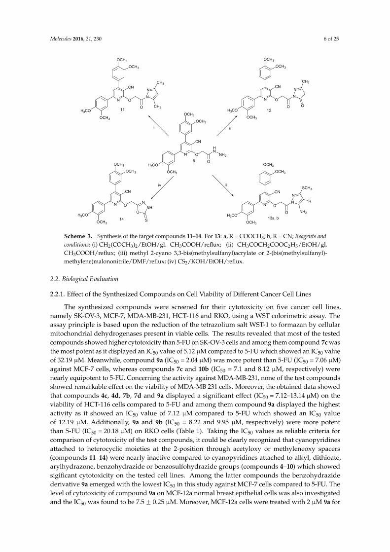
S Scheme 3. Synthesis of the target compounds 11–14. For 13: a, R = COOCH3; b, R = CN; Reagents and conditions: (i) CH2(COCH3)2/EtOH/gl. CH3COOH/reflux; (ii) CH3COCH2COOC2H5/EtOH/gl. CH3COOH/reflux; (iii) methyl 2-cyano 3,3-bis(methylsulfanyl)acrylate or 2-(bis(methylsulfanyl)-methylene)malononitrile/DMF/reflux; (iv) CS2/KOH/EtOH/reflux.
2.2. Biological Evaluation
2.2.1. Effect of the Synthesized Compounds on Cell Viability of Different Cancer Cell Lines
The synthesized compounds were screened for their cytotoxicity on five cancer cell lines, namely SK-OV-3, MCF-7, MDA-MB-231, HCT-116 and RKO, using a WST colorimetric assay. The assay principle is based upon the reduction of the tetrazolium salt WST-1 to formazan by cellular mitochondrial dehydrogenases present in viable cells. The results revealed that most of the tested compounds showed higher cytotoxicity than 5-FU on SK-OV-3 cells and among them compound 7c was the most potent as it displayed an IC50 value of 5.12 μM compared to 5-FU which showed an IC50
value of 32.19 μM. Meanwhile, compound 9a (IC50 = 2.04 μM) was more potent than 5-FU (IC50 = 7.06 μM) against MCF-7 cells, whereas compounds 7c and 10b (IC50 = 7.1 and 8.12 μM, respectively) were nearly equipotent to 5-FU. Concerning the activity against MDA-MB-231, none of the test compounds showed remarkable effect on the viability of MDA-MB 231 cells. Moreover, the obtained data showed that compounds 4c, 4d, 7b, 7d and 9a displayed a significant effect (IC50 = 7.12–13.14 μM) on the viability of HCT-116 cells compared to 5-FU and among them compound 9a displayed the highest activity as it showed an IC50 value of 7.12 μM compared to 5-FU which showed an IC50 value of 12.19 μM. Additionally, 9a and 9b (IC50 = 8.22 and 9.95 μM, respectively) were more potent than 5-FU (IC50 = 20.18 μM) on RKO cells (Table 1). Taking the IC50 values as reliable criteria for comparison of cytotoxicity of the test compounds, it could be clearly recognized that cyanopyridines attached to heterocyclic moieties at the 2-position through acetyloxy or methyleneoxy spacers (compounds 11–14) were nearly inactive compared to cyanopyridines attached to alkyl, dithioate, arylhydrazone, benzohydrazide or benzosulfohydrazide groups (compounds 4–10) which showed sigificant cytotoxicity on the tested cell lines. Among the latter compounds the benzohydrazide derivative 9a emerged with the lowest IC50 in this study against MCF-7 cells compared to 5-FU. The level of cytotoxicity of compound 9a on MCF-12a normal breast epithelial cells was also investigated and the IC50 was found to be 7.5 ± 0.25 μM. Moreover, MCF-12a cells were treated with 2 μM 9a for 24 h and the results (Figure 3) indicated that 2 μM of 9a reduced cell viability to approximately 50% in MCF-7 cells
Scheme 3. Synthesis of the target compounds 11–14. For 13: a, R = COOCH3; b, R = CN; Reagents andconditions: (i) CH2(COCH3)2/EtOH/gl. CH3COOH/reflux; (ii) CH3COCH2COOC2H5/EtOH/gl.CH3COOH/reflux; (iii) methyl 2-cyano 3,3-bis(methylsulfanyl)acrylate or 2-(bis(methylsulfanyl)-methylene)malononitrile/DMF/reflux; (iv) CS2/KOH/EtOH/reflux.
2.2. Biological Evaluation
2.2.1. Effect of the Synthesized Compounds on Cell Viability of Different Cancer Cell Lines
The synthesized compounds were screened for their cytotoxicity on five cancer cell lines,namely SK-OV-3, MCF-7, MDA-MB-231, HCT-116 and RKO, using a WST colorimetric assay. Theassay principle is based upon the reduction of the tetrazolium salt WST-1 to formazan by cellularmitochondrial dehydrogenases present in viable cells. The results revealed that most of the testedcompounds showed higher cytotoxicity than 5-FU on SK-OV-3 cells and among them compound 7c wasthe most potent as it displayed an IC50 value of 5.12 µM compared to 5-FU which showed an IC50 valueof 32.19 µM. Meanwhile, compound 9a (IC50 = 2.04 µM) was more potent than 5-FU (IC50 = 7.06 µM)against MCF-7 cells, whereas compounds 7c and 10b (IC50 = 7.1 and 8.12 µM, respectively) werenearly equipotent to 5-FU. Concerning the activity against MDA-MB-231, none of the test compoundsshowed remarkable effect on the viability of MDA-MB 231 cells. Moreover, the obtained data showedthat compounds 4c, 4d, 7b, 7d and 9a displayed a significant effect (IC50 = 7.12–13.14 µM) on theviability of HCT-116 cells compared to 5-FU and among them compound 9a displayed the highestactivity as it showed an IC50 value of 7.12 µM compared to 5-FU which showed an IC50 valueof 12.19 µM. Additionally, 9a and 9b (IC50 = 8.22 and 9.95 µM, respectively) were more potentthan 5-FU (IC50 = 20.18 µM) on RKO cells (Table 1). Taking the IC50 values as reliable criteria forcomparison of cytotoxicity of the test compounds, it could be clearly recognized that cyanopyridinesattached to heterocyclic moieties at the 2-position through acetyloxy or methyleneoxy spacers(compounds 11–14) were nearly inactive compared to cyanopyridines attached to alkyl, dithioate,arylhydrazone, benzohydrazide or benzosulfohydrazide groups (compounds 4–10) which showedsigificant cytotoxicity on the tested cell lines. Among the latter compounds the benzohydrazidederivative 9a emerged with the lowest IC50 in this study against MCF-7 cells compared to 5-FU. Thelevel of cytotoxicity of compound 9a on MCF-12a normal breast epithelial cells was also investigatedand the IC50 was found to be 7.5 ˘ 0.25 µM. Moreover, MCF-12a cells were treated with 2 µM 9a for
Molecules 2016, 21, 230 7 of 25
24 h and the results (Figure 3) indicated that 2 µM of 9a reduced cell viability to approximately 50% inMCF-7 cells compared to approximately 74% in MCF-12a cells. The significance of viability betweenMCF-7 and MCF-12a cells was calculated using SPSS non-parametric analysis of two independenttests and the results showed statistical significance of p value 0.001. Based on the fact that 9a had lesscytotoxic effects on normal breast epithelial cells it was subjected to further studies.
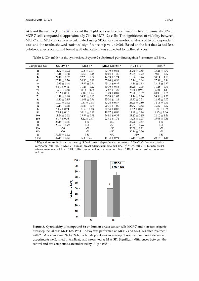
Table 1. IC50 (µM) a of the synthesized 3-cyano-2-substituted pyridines against five cancer cell lines.
Compound No. SK-OV-3 b MCF-7 c MDA-MB-231 d HCT-116 e RKO f
4a 11.37 ˘ 0.72 9.08 ˘ 0.37 32.10 ˘ 0.84 20.30 ˘ 0.85 13.21 ˘ 0.774b 18.16 ˘ 0.98 15.52 ˘ 0.46 40.04 ˘ 1.36 26.25 ˘ 1.22 19.80 ˘ 0.374c 22.12 ˘ 1.32 12.28 ˘ 0.77 44.93 ˘ 1.74 10.06 ˘ 0.76 18.14 ˘ 1.034d 25.19 ˘ 0.76 20.30 ˘ 0.98 35.80 ˘ 0.96 13.14 ˘ 0.84 17.39 ˘ 0.445 10.15 ˘ 0.44 15.42 ˘ 0.94 25.12 ˘ 0.87 14.88 ˘ 0.98 22.13 ˘ 0.977a 9.01 ˘ 0.42 11.23 ˘ 0.22 30.10 ˘ 0.88 23.20 ˘ 0.95 11.25 ˘ 0.917b 12.33 ˘ 0.88 18.14 ˘ 1.76 37.87 ˘ 1.25 9.10 ˘ 0.97 15.21 ˘ 1.137c 5.12 ˘ 0.11 7.10 ˘ 0.44 31.73 ˘ 0.89 26.90 ˘ 0.97 20.30 ˘ 0.767d 10.10 ˘ 0.98 14.30 ˘ 0.95 35.53 ˘ 1.03 11.16 ˘ 1.26 24.98 ˘ 1.338a 14.13 ˘ 0.95 12.01 ˘ 0.96 25.34 ˘ 1.24 28.82 ˘ 0.31 12.32 ˘ 0.028b 10.21 ˘ 0.92 9.31 ˘ 0.98 32.26 ˘ 0.87 25.20 ˘ 0.89 14.16 ˘ 0.918c 12.14 ˘ 0.98 15.27 ˘ 0.74 20.31 ˘ 1.06 25.87 ˘ 0.83 16.32 ˘ 0.379a 9.06 ˘ 0.24 2.04 ˘ 0.13 22.34 ˘ 0.88 7.12 ˘ 0.37 8.22 ˘ 0.999b 7.08 ˘ 0.16 10.18 ˘ 0.92 19.27 ˘ 0.86 17.90 ˘ 0.74 9.95 ˘ 1.0610a 11.34 ˘ 0.02 13.39 ˘ 0.98 26.82 ˘ 0.33 21.82 ˘ 0.85 12.10 ˘ 1.2610b 9.17 ˘ 0.38 8.12 ˘ 0.47 22.44 ˘ 1.71 16.09 ˘ 1.07 15.65 ˘ 0.8611 26.19 ˘ 0.95 >50 >50 33.90 ˘ 0.87 >5012 20.27 ˘ 1.72 >50 >50 40.35 ˘ 1.76 >5013a >50 >50 >50 36.30 ˘ 1.71 >5013b >50 >50 >50 30.14 ˘ 0.76 >5014 30.30 ˘ 1.12 >50 >50 >50 >50
5-FU 32.19 ˘ 1.03 7.06 ˘ 0.91 15.13 ˘ 0.94 12.19 ˘ 1.10 20.18 ˘ 1.16a IC50 values are indicated as mean ˘ S.D of three independent experiments. b SK-OV-3: human ovariancarcinoma cell line. c MCF-7: human breast adenocarcinoma cell line. d MDA-MB-231: human breastadenocarcinoma cell line. e HCT-116: human colon carcinoma cell line. f RKO: human colon carcinomacell line.
Molecules 2016, 21, 230 7 of 24
compared to approximately 74% in MCF-12a cells. The significance of viability between MCF-7 and MCF-12a cells was calculated using SPSS non-parametric analysis of two independent tests and the results showed statistical significance of p value 0.001. Based on the fact that 9a had less cytotoxic effects on normal breast epithelial cells it was subjected to further studies.
Table 1. IC50 (μM) a of the synthesized 3-cyano-2-substituted pyridines against five cancer cell lines.
Compound No. SK-OV-3 b MCF-7 c MDA-MB-231 d HCT-116 e RKO f 4a 11.37 ± 0.72 9.08 ± 0.37 32.10 ± 0.84 20.30 ± 0.85 13.21 ± 0.77 4b 18.16 ± 0.98 15.52 ± 0.46 40.04 ± 1.36 26.25 ± 1.22 19.80 ± 0.37 4c 22.12 ± 1.32 12.28 ± 0.77 44.93 ± 1.74 10.06 ± 0.76 18.14 ± 1.03 4d 25.19 ± 0.76 20.30 ± 0.98 35.80 ± 0.96 13.14 ± 0.84 17.39 ± 0.44 5 10.15 ± 0.44 15.42 ± 0.94 25.12 ± 0.87 14.88 ± 0.98 22.13 ± 0.97
7a 9.01 ± 0.42 11.23 ± 0.22 30.10 ± 0.88 23.20 ± 0.95 11.25 ± 0.91 7b 12.33 ± 0.88 18.14 ± 1.76 37.87 ± 1.25 9.10 ± 0.97 15.21 ± 1.13 7c 5.12 ± 0.11 7.10 ± 0.44 31.73 ± 0.89 26.90 ± 0.97 20.30 ± 0.76 7d 10.10 ± 0.98 14.30 ± 0.95 35.53 ± 1.03 11.16 ± 1.26 24.98 ± 1.33 8a 14.13 ± 0.95 12.01 ± 0.96 25.34 ± 1.24 28.82 ± 0.31 12.32 ± 0.02 8b 10.21 ± 0.92 9.31 ± 0.98 32.26 ± 0.87 25.20 ± 0.89 14.16 ± 0.91 8c 12.14 ± 0.98 15.27 ± 0.74 20.31 ± 1.06 25.87 ± 0.83 16.32 ± 0.37 9a 9.06 ± 0.24 2.04 ± 0.13 22.34 ± 0.88 7.12 ± 0.37 8.22 ± 0.99 9b 7.08 ± 0.16 10.18 ± 0.92 19.27 ± 0.86 17.90 ± 0.74 9.95 ± 1.06 10a 11.34 ± 0.02 13.39 ± 0.98 26.82 ± 0.33 21.82 ± 0.85 12.10 ± 1.26 10b 9.17 ± 0.38 8.12 ± 0.47 22.44 ± 1.71 16.09 ± 1.07 15.65 ± 0.86 11 26.19 ± 0.95 >50 >50 33.90 ± 0.87 >50 12 20.27 ± 1.72 >50 >50 40.35 ± 1.76 >50 13a >50 >50 >50 36.30 ± 1.71 >50 13b >50 >50 >50 30.14 ± 0.76 >50 14 30.30 ± 1.12 >50 >50 >50 >50
5-FU 32.19 ± 1.03 7.06 ± 0.91 15.13 ± 0.94 12.19 ± 1.10 20.18 ± 1.16 a IC50 values are indicated as mean ± S.D of three independent experiments. b SK-OV-3: human ovarian carcinoma cell line. c MCF-7: human breast adenocarcinoma cell line. d MDA-MB-231: human breast adenocarcinoma cell line. e HCT-116: human colon carcinoma cell line. f RKO: human colon carcinoma cell line.
Figure 3. Cytotoxicity of compound 9a on human breast cancer cells MCF-7 and non-tumorigenic breast epithelial cells MCF-12a. WST-1 Assay was performed on MCF-7 and MCF-12a after treatment with 2 μM of compound 9a for 24 h. Each data point was an average of results from three independent experiments performed in triplicate and presented as M ± SD. Significant differences between the control and test compounds are indicated by * (* p ˂ 0.05).
Figure 3. Cytotoxicity of compound 9a on human breast cancer cells MCF-7 and non-tumorigenicbreast epithelial cells MCF-12a. WST-1 Assay was performed on MCF-7 and MCF-12a after treatmentwith 2 µM of compound 9a for 24 h. Each data point was an average of results from three independentexperiments performed in triplicate and presented as M ˘ SD. Significant differences between thecontrol and test compounds are indicated by * (* p < 0.05).
Molecules 2016, 21, 230 8 of 25
2.2.2. Compound 9a induces apoptosis in MCF-7 cells
Morphological Changes
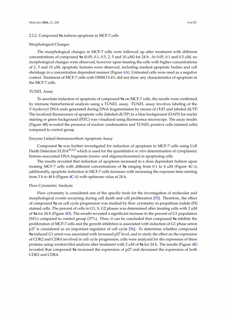
The morphological changes in MCF-7 cells were followed up after treatment with differentconcentrations of compound 9a (0.05, 0.1, 0.5, 2, 5 and 10 µM) for 24 h. At 0.05, 0.1 and 0.5 µM, nomorphological changes were observed, however upon treating the cells with higher concentrationsof 2, 5 and 10 µM, apoptotic features were observed, including marked apoptotic bodies and cellshrinkage in a concentration dependent manner (Figure 4A). Untreated cells were used as a negativecontrol. Treatment of MCF-7 cells with DMSO 0.4% did not show any characteristics of apoptosis inthe MCF-7 cells.
TUNEL Assay
To ascertain induction of apoptosis of compound 9a on MCF-7 cells, the results were confirmedby immune histochemical analysis using a TUNEL assay. TUNEL assay involves labeling of the3'-hydroxyl DNA ends generated during DNA fragmentation by means of rTdT and labeled dUTP.The localized fluorescence of apoptotic cells (labeled-dUTP) in a blue background (DAPI) for nucleistaining or green background (FITC) was visualized using fluorescence microscope. The assay results(Figure 4B) revealed the presence of nuclear condensation and TUNEL-positive cells (stained cells)compared to control group.
Enzyme Linked Immunosorbent Apoptosis Assay
Compound 9a was further investigated for induction of apoptosis in MCF-7 cells using CellDeath Detection ELISAPLUS which is used for the quantitative in vitro determination of cytoplasmichistone-associated DNA fragments (mono- and oligonucleosomes) in apoptosing cells.
The results revealed that induction of apoptosis increased in a dose dependent fashion upontreating MCF-7 cells with different concentrations of 9a ranging from 0.1 to 4 µM (Figure 4C-i),additionally, apoptotic induction in MCF-7 cells increases with increasing the exposure time startingfrom 3 h to 48 h (Figure 4C-ii) with optimum value at 24 h.
Flow Cytometric Analysis
Flow cytometry is considered one of the specific tools for the investigation of molecular andmorphological events occurring during cell death and cell proliferation [55]. Therefore, the effectof compound 9a on cell cycle progression was studied by flow cytometry in propidium iodide (PI)stained cells. The percent of cells in G1, S, G2 phases was determined after treating cells with 2 µMof 9a for 24 h (Figure 4D). The results revealed a significant increase in the percent of G1 population(54%) compared to control group (37%). Thus, it can be concluded that compound 9a inhibits theproliferation of MCF-7 cells and the growth inhibition is associated with induction of G1 phase arrest.p27 is considered as an important regulator of cell cycle [56]. To determine whether compound9a-induced G1 arrest was associated with increased p27 level, and to study the effect on the expressionof CDK2 and CDK4 involved in cell cycle progression, cells were analyzed for the expression of theseproteins using western blot analysis after treatment with 2 µM of 9a for 24 h. The results (Figure 4E)revealed that compound 9a increased the expression of p27 and decreased the expression of bothCDK2 and CDK4.
Molecules 2016, 21, 230 9 of 25
Molecules 2016, 21, 230 9 of 24
(A) (B)
3 6 12 18 24 480.0
0.5
1.0
1.5
2.0
2.5
3.0
3.5
4.0
Time in hours
Ind
uct
ion
of
apo
pto
sis
Ab
s at
405
nm
(C-i) (C-ii)
(D) (E)
Figure 4. Compound 9a-induced apoptosis in MCF-7 cells. (A) Morphological changes in MCF-7 cells were examined after treatment with 0.05, 0.1, 0.5, 2, 5 and 10 μM of compound 9a for 24 h. Compound 9a induces apoptotic death including marked apoptotic bodies and cell shrinkage in a concentration dependent manner. Untreated cells were used as a negative control. Treatment of MCF-7 cells with 0.4% DMSO did not show any cell death; (B) A TUNEL assay was used to confirm induction of apoptosis in MCF-7 cells. Cells were treated with 2 μM of 9a for 24 h. Untreated cells act as control. Lack of staining in control cells shows that the cells are actively proliferating, i.e., no apoptotic cell death and induction of apoptosis in treated cells was confirmed by the appearance of TUNEL-positive cells; (C) ELISA assay apoptotic cell detection. Effect of 9a on apoptosis induction in MCF-7 cells after treatment with different concentrations (i) and different time intervals (ii). Data are presented as mean ± SD (n = 3). Significant differences between the control and test compounds are indicated by * (* p ˂ 0.05); (D) Percent of distribution of cell cycle phases of MCF-7 breast cancer cells after treatment with 2 μM of 9a for 24 h. Each experiment was performed in triplicate. Data are presented as mean ± SD (n = 3); (E) Western blot analysis was carried out to quantify the expression levels of p27, CDK2 and CDK4 in treated and non-treated control cells. β-actin was used as a loading control.
Figure 4. Compound 9a-induced apoptosis in MCF-7 cells. (A) Morphological changes in MCF-7cells were examined after treatment with 0.05, 0.1, 0.5, 2, 5 and 10 µM of compound 9a for 24 h.Compound 9a induces apoptotic death including marked apoptotic bodies and cell shrinkage in aconcentration dependent manner. Untreated cells were used as a negative control. Treatment of MCF-7cells with 0.4% DMSO did not show any cell death; (B) A TUNEL assay was used to confirm inductionof apoptosis in MCF-7 cells. Cells were treated with 2 µM of 9a for 24 h. Untreated cells act as control.Lack of staining in control cells shows that the cells are actively proliferating, i.e., no apoptotic celldeath and induction of apoptosis in treated cells was confirmed by the appearance of TUNEL-positivecells; (C) ELISA assay apoptotic cell detection. Effect of 9a on apoptosis induction in MCF-7 cellsafter treatment with different concentrations (i) and different time intervals (ii). Data are presentedas mean ˘ SD (n = 3). Significant differences between the control and test compounds are indicatedby * (* p < 0.05); (D) Percent of distribution of cell cycle phases of MCF-7 breast cancer cells aftertreatment with 2 µM of 9a for 24 h. Each experiment was performed in triplicate. Data are presented asmean ˘ SD (n = 3); (E) Western blot analysis was carried out to quantify the expression levels of p27,CDK2 and CDK4 in treated and non-treated control cells. β-actin was used as a loading control.
Molecules 2016, 21, 230 10 of 25
Impact of 9a on Apoptotic Signaling Pathways
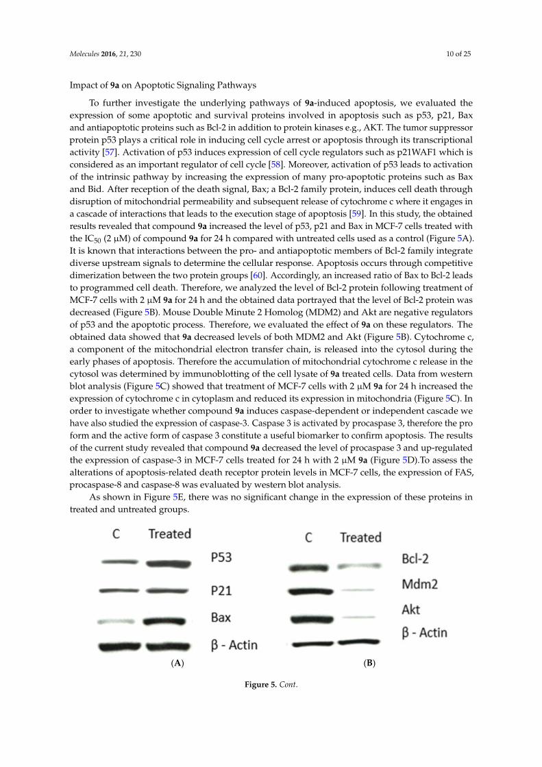
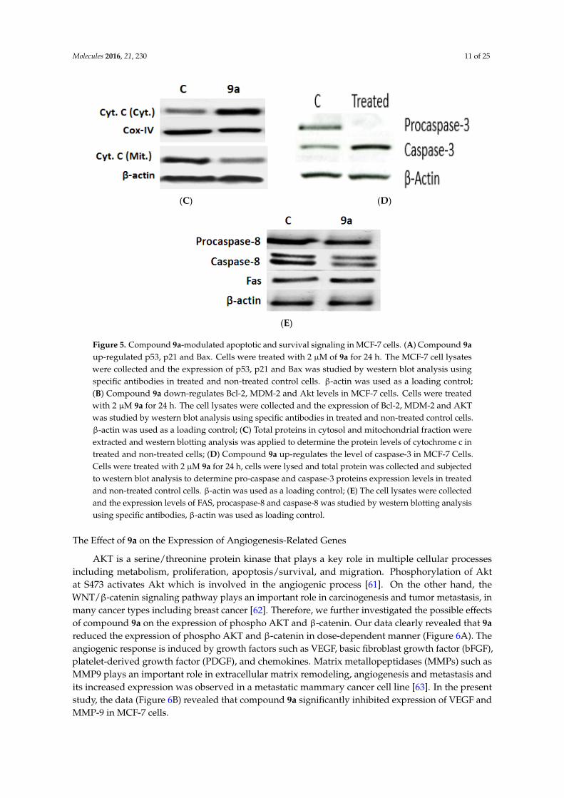
To further investigate the underlying pathways of 9a-induced apoptosis, we evaluated theexpression of some apoptotic and survival proteins involved in apoptosis such as p53, p21, Baxand antiapoptotic proteins such as Bcl-2 in addition to protein kinases e.g., AKT. The tumor suppressorprotein p53 plays a critical role in inducing cell cycle arrest or apoptosis through its transcriptionalactivity [57]. Activation of p53 induces expression of cell cycle regulators such as p21WAF1 which isconsidered as an important regulator of cell cycle [58]. Moreover, activation of p53 leads to activationof the intrinsic pathway by increasing the expression of many pro-apoptotic proteins such as Baxand Bid. After reception of the death signal, Bax; a Bcl-2 family protein, induces cell death throughdisruption of mitochondrial permeability and subsequent release of cytochrome c where it engages ina cascade of interactions that leads to the execution stage of apoptosis [59]. In this study, the obtainedresults revealed that compound 9a increased the level of p53, p21 and Bax in MCF-7 cells treated withthe IC50 (2 µM) of compound 9a for 24 h compared with untreated cells used as a control (Figure 5A).It is known that interactions between the pro- and antiapoptotic members of Bcl-2 family integratediverse upstream signals to determine the cellular response. Apoptosis occurs through competitivedimerization between the two protein groups [60]. Accordingly, an increased ratio of Bax to Bcl-2 leadsto programmed cell death. Therefore, we analyzed the level of Bcl-2 protein following treatment ofMCF-7 cells with 2 µM 9a for 24 h and the obtained data portrayed that the level of Bcl-2 protein wasdecreased (Figure 5B). Mouse Double Minute 2 Homolog (MDM2) and Akt are negative regulatorsof p53 and the apoptotic process. Therefore, we evaluated the effect of 9a on these regulators. Theobtained data showed that 9a decreased levels of both MDM2 and Akt (Figure 5B). Cytochrome c,a component of the mitochondrial electron transfer chain, is released into the cytosol during theearly phases of apoptosis. Therefore the accumulation of mitochondrial cytochrome c release in thecytosol was determined by immunoblotting of the cell lysate of 9a treated cells. Data from westernblot analysis (Figure 5C) showed that treatment of MCF-7 cells with 2 µM 9a for 24 h increased theexpression of cytochrome c in cytoplasm and reduced its expression in mitochondria (Figure 5C). Inorder to investigate whether compound 9a induces caspase-dependent or independent cascade wehave also studied the expression of caspase-3. Caspase 3 is activated by procaspase 3, therefore the proform and the active form of caspase 3 constitute a useful biomarker to confirm apoptosis. The resultsof the current study revealed that compound 9a decreased the level of procaspase 3 and up-regulatedthe expression of caspase-3 in MCF-7 cells treated for 24 h with 2 µM 9a (Figure 5D).To assess thealterations of apoptosis-related death receptor protein levels in MCF-7 cells, the expression of FAS,procaspase-8 and caspase-8 was evaluated by western blot analysis.
As shown in Figure 5E, there was no significant change in the expression of these proteins intreated and untreated groups.
Molecules 2016, 21, 230 10 of 24
Impact of 9a on Apoptotic Signaling Pathways
To further investigate the underlying pathways of 9a-induced apoptosis, we evaluated the expression of some apoptotic and survival proteins involved in apoptosis such as p53, p21, Bax and antiapoptotic proteins such as Bcl-2 in addition to protein kinases e.g., AKT. The tumor suppressor protein p53 plays a critical role in inducing cell cycle arrest or apoptosis through its transcriptional activity [57]. Activation of p53 induces expression of cell cycle regulators such as p21WAF1 which is considered as an important regulator of cell cycle [58]. Moreover, activation of p53 leads to activation of the intrinsic pathway by increasing the expression of many pro-apoptotic proteins such as Bax and Bid. After reception of the death signal, Bax; a Bcl-2 family protein, induces cell death through disruption of mitochondrial permeability and subsequent release of cytochrome c where it engages in a cascade of interactions that leads to the execution stage of apoptosis [59]. In this study, the obtained results revealed that compound 9a increased the level of p53, p21 and Bax in MCF-7 cells treated with the IC50 (2 μM) of compound 9a for 24 h compared with untreated cells used as a control (Figure 5A). It is known that interactions between the pro- and antiapoptotic members of Bcl-2 family integrate diverse upstream signals to determine the cellular response. Apoptosis occurs through competitive dimerization between the two protein groups [60]. Accordingly, an increased ratio of Bax to Bcl-2 leads to programmed cell death. Therefore, we analyzed the level of Bcl-2 protein following treatment of MCF-7 cells with 2 μM 9a for 24 h and the obtained data portrayed that the level of Bcl-2 protein was decreased (Figure 5B). Mouse Double Minute 2 Homolog (MDM2) and Akt are negative regulators of p53 and the apoptotic process. Therefore, we evaluated the effect of 9a on these regulators. The obtained data showed that 9a decreased levels of both MDM2 and Akt (Figure 5B). Cytochrome c, a component of the mitochondrial electron transfer chain, is released into the cytosol during the early phases of apoptosis. Therefore the accumulation of mitochondrial cytochrome c release in the cytosol was determined by immunoblotting of the cell lysate of 9a treated cells. Data from western blot analysis (Figure 5C) showed that treatment of MCF-7 cells with 2 μM 9a for 24 h increased the expression of cytochrome c in cytoplasm and reduced its expression in mitochondria (Figure 5C). In order to investigate whether compound 9a induces caspase-dependent or independent cascade we have also studied the expression of caspase-3. Caspase 3 is activated by procaspase 3, therefore the pro form and the active form of caspase 3 constitute a useful biomarker to confirm apoptosis. The results of the current study revealed that compound 9a decreased the level of procaspase 3 and up-regulated the expression of caspase-3 in MCF-7 cells treated for 24 h with 2 μM 9a (Figure 5D).To assess the alterations of apoptosis-related death receptor protein levels in MCF-7 cells, the expression of FAS, procaspase-8 and caspase-8 was evaluated by western blot analysis.
As shown in Figure 5E, there was no significant change in the expression of these proteins in treated and untreated groups.
(A) (B)
Figure 5. Cont.
Figure 5. Cont.
Molecules 2016, 21, 230 11 of 25Molecules 2016, 21, 230 11 of 24
(C) (D)
(E)
Figure 5. Compound 9a-modulated apoptotic and survival signaling in MCF-7 cells. (A) Compound 9a up-regulated p53, p21 and Bax. Cells were treated with 2 μM of 9a for 24 h. The MCF-7 cell lysates were collected and the expression of p53, p21 and Bax was studied by western blot analysis using specific antibodies in treated and non-treated control cells. β-actin was used as a loading control; (B) Compound 9a down-regulates Bcl-2, MDM-2 and Akt levels in MCF-7 cells. Cells were treated with 2 μM 9a for 24 h. The cell lysates were collected and the expression of Bcl-2, MDM-2 and AKT was studied by western blot analysis using specific antibodies in treated and non-treated control cells. β-actin was used as a loading control; (C) Total proteins in cytosol and mitochondrial fraction were extracted and western blotting analysis was applied to determine the protein levels of cytochrome c in treated and non-treated cells; (D) Compound 9a up-regulates the level of caspase-3 in MCF-7 Cells. Cells were treated with 2 μM 9a for 24 h, cells were lysed and total protein was collected and subjected to western blot analysis to determine pro-caspase and caspase-3 proteins expression levels in treated and non-treated control cells. β-actin was used as a loading control; (E) The cell lysates were collected and the expression levels of FAS, procaspase-8 and caspase-8 was studied by western blotting analysis using specific antibodies, β-actin was used as loading control.
The Effect of 9a on the Expression of Angiogenesis-Related Genes
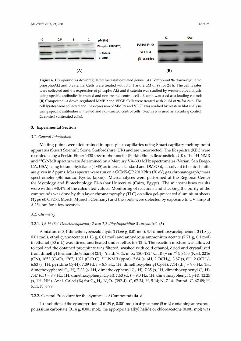
AKT is a serine/threonine protein kinase that plays a key role in multiple cellular processes including metabolism, proliferation, apoptosis/survival, and migration. Phosphorylation of Akt at S473 activates Akt which is involved in the angiogenic process [61]. On the other hand, the WNT/β-catenin signaling pathway plays an important role in carcinogenesis and tumor metastasis, in many cancer types including breast cancer [62]. Therefore, we further investigated the possible effects of compound 9a on the expression of phospho AKT and β-catenin. Our data clearly revealed that 9a reduced the expression of phospho AKT and β-catenin in dose-dependent manner (Figure 6A). The angiogenic response is induced by growth factors such as VEGF, basic fibroblast growth factor (bFGF), platelet-derived growth factor (PDGF), and chemokines. Matrix metallopeptidases (MMPs) such as MMP9 plays an important role in extracellular matrix remodeling, angiogenesis and metastasis and its increased expression was observed in a metastatic mammary cancer cell line [63]. In the present study, the data (Figure 6B) revealed that compound 9a significantly inhibited expression of VEGF and MMP-9 in MCF-7 cells.
Figure 5. Compound 9a-modulated apoptotic and survival signaling in MCF-7 cells. (A) Compound 9aup-regulated p53, p21 and Bax. Cells were treated with 2 µM of 9a for 24 h. The MCF-7 cell lysateswere collected and the expression of p53, p21 and Bax was studied by western blot analysis usingspecific antibodies in treated and non-treated control cells. β-actin was used as a loading control;(B) Compound 9a down-regulates Bcl-2, MDM-2 and Akt levels in MCF-7 cells. Cells were treatedwith 2 µM 9a for 24 h. The cell lysates were collected and the expression of Bcl-2, MDM-2 and AKTwas studied by western blot analysis using specific antibodies in treated and non-treated control cells.β-actin was used as a loading control; (C) Total proteins in cytosol and mitochondrial fraction wereextracted and western blotting analysis was applied to determine the protein levels of cytochrome c intreated and non-treated cells; (D) Compound 9a up-regulates the level of caspase-3 in MCF-7 Cells.Cells were treated with 2 µM 9a for 24 h, cells were lysed and total protein was collected and subjectedto western blot analysis to determine pro-caspase and caspase-3 proteins expression levels in treatedand non-treated control cells. β-actin was used as a loading control; (E) The cell lysates were collectedand the expression levels of FAS, procaspase-8 and caspase-8 was studied by western blotting analysisusing specific antibodies, β-actin was used as loading control.
The Effect of 9a on the Expression of Angiogenesis-Related Genes
AKT is a serine/threonine protein kinase that plays a key role in multiple cellular processesincluding metabolism, proliferation, apoptosis/survival, and migration. Phosphorylation of Aktat S473 activates Akt which is involved in the angiogenic process [61]. On the other hand, theWNT/β-catenin signaling pathway plays an important role in carcinogenesis and tumor metastasis, inmany cancer types including breast cancer [62]. Therefore, we further investigated the possible effectsof compound 9a on the expression of phospho AKT and β-catenin. Our data clearly revealed that 9areduced the expression of phospho AKT and β-catenin in dose-dependent manner (Figure 6A). Theangiogenic response is induced by growth factors such as VEGF, basic fibroblast growth factor (bFGF),platelet-derived growth factor (PDGF), and chemokines. Matrix metallopeptidases (MMPs) such asMMP9 plays an important role in extracellular matrix remodeling, angiogenesis and metastasis andits increased expression was observed in a metastatic mammary cancer cell line [63]. In the presentstudy, the data (Figure 6B) revealed that compound 9a significantly inhibited expression of VEGF andMMP-9 in MCF-7 cells.
Molecules 2016, 21, 230 12 of 25Molecules 2016, 21, 230 12 of 24
(A) (B)
Figure 6. Compound 9a downregulated metastatic-related genes. (A) Compound 9a down-regulated phosphoAkt and β catenin. Cells were treated with 0.5, 1 and 2 μM of 9a for 24 h. The cell lysates were collected and the expression of phospho Akt and β catenin was studied by western blot analysis using specific antibodies in treated and non-treated control cells. β-actin was used as a loading control; (B) Compound 9a down-regulated MMP 9 and VEGF. Cells were treated with 2 μM of 9a for 24 h. The cell lysates were collected and the expression of MMP 9 and VEGF was studied by western blot analysis using specific antibodies in treated and non-treated control cells. β-actin was used as a loading control. C: control (untreated cells).
3. Experimental Section
3.1. General Informstion
Melting points were determined in open-glass capillaries using Stuart capillary melting point apparatus (Stuart Scientific Stone, Staffordshire, UK) and are uncorrected. The IR spectra (KBr) were recorded using a Perkin-Elmer 1430 spectrophotometer (Perkin Elmer, Beaconsfield, UK). The 1H-NMR and 13C-NMR spectra were determined on a Mercury VX-300 MHz spectrometer (Varian, San Diego, CA, USA) using tetramethylsilane (TMS) as internal standard and DMSO-d6 as solvent (chemical shifts are given in δ ppm). Mass spectra were run on a GCMS-QP 2010 Plus (70 eV) gas chromatograph/mass spectrometer (Shimadzu, Kyoto, Japan). Microanalyses were performed at the Regional Center for Mycology and Biotechnology, El-Azhar University (Cairo, Egypt). The microanalyses results were within ±0.4% of the calculated values. Monitoring of reactions and checking the purity of the compounds was done by thin layer chromatography (TLC) on silica gel-precoated aluminium sheets (Type 60 GF254; Merck, Munich, Germany) and the spots were detected by exposure to UV lamp at λ 254 nm for a few seconds.
3.2. Chemistry
3.2.1. 4,6-bis(3,4-Dimethoxyphenyl)-2-oxo-1,2-dihydropyridine-3-carbonitrile (3)
A mixture of 3,4-dimethoxybenzaldehyde 1 (1.66 g, 0.01 mol), 3,4-dimethoxyacetophenone 2 (1.8 g, 0.01 mol), ethyl cyanoacetate (1.13 g, 0.01 mol) and anhydrous ammonium acetate (7.71 g, 0.1 mol) in ethanol (50 mL) was stirred and heated under reflux for 12 h. The reaction mixture was allowed to cool and the obtained precipitate was filtered, washed with cold ethanol, dried and crystallized from dimethyl formamide/ethanol (2:1). Yield: 70%, m.p.: 180–182 °C. IR (ν cm−1): 3455 (NH), 2216 (CN), 1653 (C=O), 1267, 1021 (C-O-C). 1H-NMR (ppm): 3.84 (s, 6H, 2 OCH3), 3.87 (s, 6H, 2 OCH3), 6.83 (s, 1H, pyridine C5-H), 7.09 (d, J = 8.7 Hz, 1H, dimethoxyphenyl C5-H), 7.14 (d, J = 9.0 Hz, 1H, dimethoxyphenyl C5-H), 7.33 (s, 1H, dimethoxyphenyl C2-H), 7.35 (s, 1H, dimethoxyphenyl C2-H), 7.47 (d, J = 8.7 Hz, 1H, dimethoxyphenyl C6-H), 7.53 (d, J = 9.0 Hz, 1H, dimethoxyphenyl C6-H), 12.25 (s, 1H, NH). Anal. Calcd (%) for C22H20N2O5 (392.4): C, 67.34; H, 5.14; N, 7.14. Found: C, 67.09; H, 5.11; N, 6.99.
3.2.2. General Procedure for the Synthesis of Compounds 4a–d
To a solution of the cyanopyridone 3 (0.39 g, 0.001 mol) in dry acetone (5 mL) containing anhydrous potassium carbonate (0.14 g, 0.001 mol), the appropriate alkyl halide or chloroacetone (0.001 mol) was added dropwise. The reaction mixture was stirred and heated under reflux for 2 h then allowed
Figure 6. Compound 9a downregulated metastatic-related genes. (A) Compound 9a down-regulatedphosphoAkt and β catenin. Cells were treated with 0.5, 1 and 2 µM of 9a for 24 h. The cell lysateswere collected and the expression of phospho Akt and β catenin was studied by western blot analysisusing specific antibodies in treated and non-treated control cells. β-actin was used as a loading control;(B) Compound 9a down-regulated MMP 9 and VEGF. Cells were treated with 2 µM of 9a for 24 h. Thecell lysates were collected and the expression of MMP 9 and VEGF was studied by western blot analysisusing specific antibodies in treated and non-treated control cells. β-actin was used as a loading control.C: control (untreated cells).
3. Experimental Section
3.1. General Informstion
Melting points were determined in open-glass capillaries using Stuart capillary melting pointapparatus (Stuart Scientific Stone, Staffordshire, UK) and are uncorrected. The IR spectra (KBr) wererecorded using a Perkin-Elmer 1430 spectrophotometer (Perkin Elmer, Beaconsfield, UK). The 1H-NMRand 13C-NMR spectra were determined on a Mercury VX-300 MHz spectrometer (Varian, San Diego,CA, USA) using tetramethylsilane (TMS) as internal standard and DMSO-d6 as solvent (chemical shiftsare given in δ ppm). Mass spectra were run on a GCMS-QP 2010 Plus (70 eV) gas chromatograph/massspectrometer (Shimadzu, Kyoto, Japan). Microanalyses were performed at the Regional Centerfor Mycology and Biotechnology, El-Azhar University (Cairo, Egypt). The microanalyses resultswere within ˘0.4% of the calculated values. Monitoring of reactions and checking the purity of thecompounds was done by thin layer chromatography (TLC) on silica gel-precoated aluminium sheets(Type 60 GF254; Merck, Munich, Germany) and the spots were detected by exposure to UV lamp atλ 254 nm for a few seconds.
3.2. Chemistry
3.2.1. 4,6-bis(3,4-Dimethoxyphenyl)-2-oxo-1,2-dihydropyridine-3-carbonitrile (3)
A mixture of 3,4-dimethoxybenzaldehyde 1 (1.66 g, 0.01 mol), 3,4-dimethoxyacetophenone 2 (1.8 g,0.01 mol), ethyl cyanoacetate (1.13 g, 0.01 mol) and anhydrous ammonium acetate (7.71 g, 0.1 mol)in ethanol (50 mL) was stirred and heated under reflux for 12 h. The reaction mixture was allowedto cool and the obtained precipitate was filtered, washed with cold ethanol, dried and crystallizedfrom dimethyl formamide/ethanol (2:1). Yield: 70%, m.p.: 180–182 ˝C. IR (ν cm´1): 3455 (NH), 2216(CN), 1653 (C=O), 1267, 1021 (C-O-C). 1H-NMR (ppm): 3.84 (s, 6H, 2 OCH3), 3.87 (s, 6H, 2 OCH3),6.83 (s, 1H, pyridine C5-H), 7.09 (d, J = 8.7 Hz, 1H, dimethoxyphenyl C5-H), 7.14 (d, J = 9.0 Hz, 1H,dimethoxyphenyl C5-H), 7.33 (s, 1H, dimethoxyphenyl C2-H), 7.35 (s, 1H, dimethoxyphenyl C2-H),7.47 (d, J = 8.7 Hz, 1H, dimethoxyphenyl C6-H), 7.53 (d, J = 9.0 Hz, 1H, dimethoxyphenyl C6-H), 12.25(s, 1H, NH). Anal. Calcd (%) for C22H20N2O5 (392.4): C, 67.34; H, 5.14; N, 7.14. Found: C, 67.09; H,5.11; N, 6.99.
3.2.2. General Procedure for the Synthesis of Compounds 4a–d
To a solution of the cyanopyridone 3 (0.39 g, 0.001 mol) in dry acetone (5 mL) containing anhydrouspotassium carbonate (0.14 g, 0.001 mol), the appropriate alkyl halide or chloroacetone (0.001 mol) was
Molecules 2016, 21, 230 13 of 25
added dropwise. The reaction mixture was stirred and heated under reflux for 2 h then allowed toattain room temperature. It was then poured onto crushed ice and the separated solid product wasfiltered, washed with water, dried and crystallized from the proper solvent.
2-Methoxy-4,6-bis(3,4-dimethoxyphenyl)pyridine-3-carbonitrile (4a). Yield: 72%, m.p.: 155–156 ˝C (ethanol).IR (ν cm´1): 2213 (CN), 1586 (C=N), 1263, 1223, 1024 (C-O-C). 1H-NMR (ppm): 3.71 (s, 3H, OCH3),3.73 (s, 3H, OCH3), 3.79 (s, 3H, OCH3), 3.81 (s, 3H, OCH3), 3.83 (s, 3H, OCH3), 6.98 (d, J = 8.7 Hz,1H, dimethoxyphenyl C5-H), 7.07 (d, J = 8.4 Hz, 1H, dimethoxyphenyl C5-H), 7.31–7.37 (m, 2H,dimethoxyphenyl C2,6-H), 7.75 (s, 1H, pyridine C5-H), 7.78 (d, J = 2.1 Hz, 1H, dimethoxyphenyl C2-H),7.79 (d, J =8.4, 2.1 Hz, 1H, dimethoxyphenyl C6-H). 13C-NMR (DMSO-d6, ppm): 55.36, 55.42, 55.56,55.60, 55.64, 90.46, 110.12, 111.74, 111.79, 111.89, 113.13, 115.69, 120.64, 121.56, 127.83, 128.91, 148.59,148.81, 150.23, 151.12, 156.10, 156.69, 163.08. Anal. Calcd (%) for C23H22N2O5 (406.43): C, 67.97; H, 5.46;N, 6.89. Found: C, 68.13; H, 5.44; N, 7.03.
2-Ethoxy-4,6-bis(3,4-dimethoxyphenyl)pyridine-3-carbonitrile (4b). Yield: 73%, m.p.: 152–153 ˝C (ethanol).IR (ν, cm´1): 2220 (CN), 1582 (C=N), 1265, 1226, 1027 (C-O-C). 1H-NMR (ppm): 1.44 (t, J = 6.9 Hz, 3H,CH3), 3.84 (s, 3H, OCH3), 3.85 (s, 3H, OCH3), 3.86 (s, 3H, OCH3), 3.87 (s, 3H, OCH3), 4.62 (q, J = 6.9 Hz,2H, OCH2), 7.10 (d, J = 8.9 Hz, 1H, dimethoxyphenyl C5-H), 7.15 (d, J = 8.3 Hz, 1H, dimethoxyphenylC5-H), 7.29–7.35 (m, 2H, dimethoxyphenyl C2,6-H), 7.75 (s, 1H, pyridine C5-H), 7.78 (d, J = 2.1 Hz, 1H,dimethoxyphenyl C2-H), 7.80 (dd, J = 8.3, 2.1 Hz, 1H, dimethoxyphenyl C6-H). 13C-NMR (ppm): 14.8,55.45, 55.58, 55.59, 55.66, 63.88, 90.49, 110.14, 111.73, 111.77, 111.94, 113.18, 115.72, 120.73, 121.64, 127.94,129.01, 148.66, 148.84, 150.19, 151.18, 156.21, 156.82, 163.22. EI-MS m/z (%): 420.17 (M+) (77 %), 111(100%). Anal. Calcd (%) for C24H24N2O5 (420.46): C, 68.56; H, 5.75; N, 6.66. Found: C, 68.72; H, 5.78;N, 6.79.
4,6-bis(3,4-Dimethoxyphenyl)-2-propoxypyridine-3-carbonitrile (4c). Yield: 64%, m.p.: 156–158 ˝C(dimethylformamide/ethanol 2:1). IR (ν cm´1): 2214 (CN), 1583 (C=N), 1264, 1228, 1026 (C-O-C).1H-NMR (ppm): 0.93 (t, J = 6.9 Hz, 3H, CH2CH3), 1.65–1.70 (m, 2H, CH2CH3), 3.77 (s, 3H, OCH3), 3.80(s, 3H, OCH3), 3.82 (s, 3H, OCH3), 3.84 (s, 3H, OCH3), 4.83 (t, J = 6.9 Hz, 2H, OCH2), 6.96 (d, J = 8.3 Hz,1H, dimethoxyphenyl C5-H), 7.08 (d, J = 8.5 Hz, 1H, dimethoxyphenyl C5-H), 7.22 (dd, J = 8.3, 1.8 Hz,1H, dimethoxyphenyl C6-H), 7.26 (d, J = 1.8 Hz, 1H, dimethoxyphenyl C2-H), 7.67 (s, 1H, pyridineC5-H), 7.70 (d, J = 1.8 Hz, 1H, dimethoxyphenyl C2-H), 7.77 (dd, J = 8.5, 1.8 Hz, 1H, dimethoxyphenylC6-H). 13C-NMR (ppm): 11.76, 23.84, 55.49, 55.55, 55.61, 55.68, 65.23, 90.56, 110.22, 111.65, 111.82,112.12, 113.33, 115.68, 120.80, 121.66, 128.01, 129.14, 148.73, 148.69, 150.26, 151.34, 156.27, 156.94, 163.25.Anal. Calcd (%) for C25H26N2O5 (434.48): C, 69.11; H, 6.03; N, 6.45. Found: C, 68.77; H, 5.73; N, 6.59.
4,6-bis(3,4-Dimethoxyphenyl)-2-(2-oxopropoxy)pyridine-3-carbonitrile (4d). Yield: 75%, m.p.: 110–112 ˝C(ethanol). IR (ν cm´1): 2214 (CN), 1731 (C=O), 1593 (C=N), 1262, 1023 (C-O-C). 1H-NMR (ppm): 2.23 (s,3H, COCH3), 3.72 (s, 3H, OCH3), 3.78 (s, 3H, OCH3), 3.80 (s, 3H, OCH3), 3.83 (s, 3H, OCH3), 5.25 (s, 2H,OCH2), 7.08 (d, J = 8.7 Hz, 1H, dimethoxyphenyl C5-H), 7.17 (d, J = 8.4 Hz, 1H, dimethoxyphenyl C5-H),7.33 (dd, J = 8.7 Hz, 2.1 Hz, 1H, dimethoxyphenyl C6-H), 7.37 (d, J = 2.1 Hz, 1H, dimethoxyphenylC2-H), 7.66 (s, 1H, pyridine C5-H), 7.77 (d, J = 8.4 Hz, 1H, dimethoxyphenyl C6-H), 7.78 (d, J = 2.1 Hz,1H, dimethoxyphenyl C2-H). 13C-NMR (ppm):25.73, 55.47, 55.54, 55.57, 55.62, 70.62, 90.42, 110.07,111.63, 111.71, 111.92, 113.05, 115.66, 120.75, 121.50, 127.87, 128.84, 148.54, 148.76, 150.29, 151.06, 156.07,156.64, 162.96, 203.32. Anal. Calcd (%) for C25H24N2O6 (448.47): C, 66.95; H, 5.39; N, 6.25. Found: C,67.15; H, 5.44; N, 6.21.
3.2.3. Ethyl 2-(3-Cyano-4,6-bis(3,4-dimethoxyphenyl)pyridin-2-yloxy)acetate (5)
A mixture of 3 (3.92 g, 0.01 mol), ethyl bromoacetate (1.67 g, 0.01 mol) and anhydrous potassiumcarbonate (5.53 g, 0.04 mol) in dry acetone (30 mL) was heated under reflux for 20 h. After cooling,water was added and the reaction mixture was left in a refrigerator for an overnight. The separatedsolid product was filtered, washed with water, dried and recrystallized from ethanol. Yield: 72%, m.p.:
Molecules 2016, 21, 230 14 of 25
130–132 ˝C. IR (ν cm´1): 2220 (CN), 1760 (C=O), 1595 (C=N), 1258, 1231, 1067, 1028 (C-O-C). 1H-NMR(ppm): 1.15 (t, J = 7.1 Hz, 3H, CH2CH3), 3.84 (s, 3H, OCH3), 3.85 (s, 3H, OCH3), 3.86 (s, 6H, 2OCH3),4.17 (q, J = 7.1 Hz, 2H, CH2CH3), 5.14 (s, 2H, OCH2), 7.08 (d, J = 8.4 Hz, 1H, dimethoxyphenyl C5-H),7.16 (d, J = 8.3 Hz, 1H, dimethoxyphenyl C5-H), 7.32–7.38 (m, 2H, dimethoxyphenyl C2,6-H), 7.73 (d,J = 1.9 Hz, 1H, dimethoxyphenyl C2-H), 7.81 (dd, J = 8.3, 1.9 Hz, 1H, C6-H), 7.84 (s, 1H, pyridine C5-H).13C-NMR (ppm):13.88, 55.51, 55.63, 55.59, 55.63, 60.81, 63.61, 90.45, 110.34, 111.57, 111.72, 111.99, 113.12,115.52, 120.79, 121.53, 127.82, 128.66, 148.58, 148.78, 150.35, 151.22, 156.21, 156.47, 162.82, 168.51. Anal.Calcd (%) for C26H26N2O7 (478.49): C, 65.26; H, 5.48; N, 5.85. Found: C, 65.38; H, 5.53; N, 5.97.
3.2.4. 2-{[3-Cyano-4,6-bis(3,4-dimethoxyphenyl)pyridin-2-yl]oxy}acetohydrazide (6)
To a suspension of 5 (4.8 g, 0.01 mol) in absolute ethanol (50 mL), hydrazine hydrate 98% (3.2 g,0.1 mol) was added. The reaction mixture was heated under reflux for 8 h then allowed to cool to roomtemperature. The obtained product was filtered, washed with ethanol, dried and recrystallized fromethyl alcohol/dimethylformamide mixture (3:1). Yield: 68%, m.p.: 216–218˝C. IR (ν cm´1): 3437, 3303(NH), 2209 (CN), 1690 (C=O), 1645 (C=N), 1268, 1019 (C-O-C). 1H-NMR (ppm): 3.83 (s, 3H, OCH3),3.85 (s, 3H, OCH3), 3.86 (s, 3H, OCH3), 3.88 (s, 3H, OCH3), 4.27 (s, 2H, NH2, D2O exchangeable), 4.97 (s,2H, OCH2), 7.07 (d, J = 8.7 Hz, 1H, dimethoxyphenyl C5-H), 7.17 (d, J = 8.8 Hz, 1H, dimethoxy-phenylC5-H), 7.29–7.35 (m, 2H, dimethoxyphenyl C2,6-H), 7.75 (d, J = 2.0 Hz, 1H, dimethoxyphenyl C2-H),7.80 (s, 1H, pyridine C5-H), 7.82 (dd, J = 8.8, 2.0 Hz, 1H, dimethoxyphenyl C6-H), 9.36 (s, 1H, NH).13C-NMR (ppm): 55.54, 55.66, 55.56, 55.67, 63.65, 90.44, 110.38, 111.63, 111.70, 112.22, 113.16, 115.56,120.77, 121.55, 127.88, 128.74, 148.68, 148.82, 150.42, 151.26, 156.18, 156.51, 162.85, 168.46. Anal. Calcd(%) for C24H24N4O6 (464.47): C, 62.06; H, 5.21; N, 12.06. Found: C, 62.29; H, 5.28; N, 12.12.
3.2.5. General Procedure for the Synthesis of Compounds 7a–d
To a well stirred cold suspension of fine powdered potassium hydroxide (0.11 g, 0.002 mol) indimethylformamide (4 mL) the acid hydrazide 6 (0.46 g, 0.001 mol) was added followed by carbondisulphide (0.076 g, 0.001 mol). The reaction mixture was stirred at room temperature overnightthen the appropriate alkyl halide (0.001 mol) was added and the mixture was further stirred at roomtemperature for 12 h. The reaction mixture was concentrated under reduced pressure then poured intoice-cold water and the obtained precipitate was filtered, washed with water, dried and crystallizedfrom ethanol.
Methyl 2-[2-(3-cyano-4,6-bis(3,4-dimethoxyphenyl)pyridin-2-yloxy)acetyl]hydrazinecarbodithioate (7a). Yield:63%, m.p.:151–153 ˝C. IR (ν cm´1): 3433 (NH), 2214 (CN), 1675 (C=O), 1589 (C=N), 1516, 1314, 1145, 977(NCS), 1261, 1023 (C-O-C). 1H-NMR (ppm): 2.69 (s, 3H, SCH3), 3.84 (s, 3H, OCH3), 3.85 (s, 3H, OCH3),3.86 (s, 3H, OCH3), 3.91 (s, 3H, OCH3), 5.78 (s, 2H, OCH2), 7.07 (d, J = 8.7 Hz, 1H, dimethoxyphenylC5-H), 7.16 (d, J = 8.4 Hz, 1H, dimethoxyphenyl C5-H), 7.32–7.38 (m, 2H, dimethoxyphenyl C2,6-H),7.80 (d, J = 1.8 Hz, 1H, dimethoxyphenyl C2-H), 7.84–7.88 (m, 2H, dimethoxyphenyl C6-H and pyridineC5-H), 10.40 (s, 1H, NH), 10.59 (s, 1H, NH). 13C-NMR (ppm):16.28, 55.67, 58.14, 64.21, 91.25, 110.37,111.59, 111.77, 112.22, 113.62, 115.39, 120.84, 121.56, 127.85, 128.72, 148.63, 148.90, 150.42, 151.31, 156.29,156.74, 162.44, 164.60, 172.26. EI-MS m/z (%): 554 (M+) (19.5 %), 94 (100%). Anal. Calcd (%) forC26H26N4O6S2 (554.64): C, 56.30; H, 4.72; N, 10.10. Found: 56.38; H, 4.75; N, 10.21.
Ethyl 2-[2-(3-cyano-4,6-bis(3,4-dimethoxyphenyl)pyridin-2-yloxy)acetyl]hydrazinecarbodithioate (7b). Yield:64%, m.p.: 144–146 ˝C. IR (ν cm´1): 3437 (NH), 2214 (CN), 1672 (C=O), 1591 (C=N), 1516, 1307, 1144,979 (NCS), 1261, 1025 (C-O-C). 1H-NMR (ppm): 1.28 (t, J = 7.1 Hz, 3H, CH3), 3.18 (q, J = 7.1 Hz, 2H,SCH2), 3.76 (s, 3H, OCH3), 3.82 (s, 3H, OCH3), 3.83 (s, 3H, OCH3), 3.88 (s, 3H, OCH3), 5.83 (s, 2H,OCH2), 7.07 (d, J = 9.0 Hz, 1H, dimethoxyphenyl C5-H), 7.16 (d, J = 8.1 Hz, 1H, dimethoxyphenylC5-H), 7.28-7.32 (m, 2H, dimethoxyphenyl C2,6-H), 7.73–7.86 (m, 3H, dimethoxyphenyl C2,6-H andpyridine C5-H), 10.42 (s, 1H, NH), 10.63 (s, 1H, NH). 13C-NMR (ppm): 14.54, 26.61, 55.64, 58.07, 63.98,90.78, 110.49, 111.56, 111.74, 112.14, 113.58, 115.37, 120.87, 121.54, 127.80, 128.68, 148.61, 148.89, 150.40,
Molecules 2016, 21, 230 15 of 25
151.24, 156.33, 156.70, 162.41, 163.78, 171.98. Anal. Calcd (%) for C27H28N4O6S2 (568.66): C, 57.03; H,4.96; N, 9.85. Found: C, 57.17; H, 4.92; N, 9.91.
2-Morpholinoethyl-2-[2-(3-cyano-4,6-bis(3,4-dimethoxyphenyl)pyridin-2-yloxy)acetyl]hydrazinecarbo-dithioate(7c). Yield: 66 %, m.p.: 158–160 ˝C. IR (ν, cm´1): 3435 (NH), 2217 (CN), 1656 (C=O), 1586 (C=N), 1516,1322, 1138, 921 (NCS), 1262, 1022 (C-O-C). 1H-NMR (ppm): 2.60–2.64 (m, 4H, morpholine C3,5-H2),2.74 (t, J = 6.9 Hz, 2H, SCH2), 2.94 (t, J = 6.9 Hz, 2H, NCH2), 3.59–3.64 (m, 4H, morpholine C2,6-H2),3.78 (s, 3H, OCH3), 3.83 (s, 3H, OCH3), 3.84 (s, 3H, OCH3), 3.88 (s, 3H, OCH3), 5.72 (s, 2H, OCH2), 7.06(d, J = 8.1 Hz, 1H, dimethoxyphenyl C5-H), 7.14 (d, J = 8.4 Hz, 1H, dimethoxyphenyl C5-H), 7.28–7.33(m, 2H, dimethoxyphenyl C2,6-H), 7.75–7.81 (m, 3H, dimethoxyphenyl C2,6-H and pyridine C5-H),10.49 (s, 1H, NH), 10.67 (s, 1H, NH). 13C-NMR (ppm): 32.83, 52.10, 55.65, 55.67, 56.13, 58.07, 64.69,90.73, 110.41, 111.57, 111.72, 113.11, 113.58, 115.36, 120.86, 121.55, 127.80, 128.67, 148.59, 148.89, 150.38,151.21, 156.37, 156.72, 162.35, 164.30, 172.0. Anal. Calcd (%) for C31H35N5O7S2 (653.77): C, 56.95; H,5.40; N, 10.71. Found: 56.61; H, 5.18; N, 11.04.
2-(Piperidin-1-yl)-ethyl-2-[2-(3-cyano-4,6-bis(3,4-dimethoxyphenyl)pyridin-2-yloxy)acetyl]hydrazinecarbo-dithioate (7d). Yield: 66 %, m.p.: 160–162 ˝C. IR (ν, cm´1): 3434 (NH), 2218 (CN), 1645 (C=O), 1587(C=N), 1516, 1322, 1134, 921 (NCS), 1262, 1022 (C-O-C). 1H-NMR (ppm): 1.44–1.48 (m, 2H, piperidineC4-H2), 1.60–1.64 (m, 4H, piperidine C3,5-H2), 2.83–3.07 (m, 8H, piperidine C2,6-H, SCH2 and NCH2),3.86 (s, 3H, OCH3), 3.87 (s, 3H, OCH3), 3.90 (s, 3H, OCH3), 3.92 (s, 3H, OCH3), 5.64 (s, 2H, OCH2), 7.08(d, J = 8.1 Hz, 1H, dimethoxyphenyl C5-H), 7.20 (d, J = 8.4 Hz, 1H, dimethoxyphenyl C5-H), 7.33–7.38(m, 2H, dimethoxyphenyl C2,6-H), 7.71–7.85 (m, 3H, dimethoxyphenyl C2,6-H and pyridine C5-H),10.51 (s, 1H, NH), 10.69 (s, 1H, NH). 13C-NMR (ppm): 23.76, 33.14, 52.26, 53.82, 55.65, 55.67, 63.99,91.33, 110.42, 111.55, 111.76, 113.14, 113.59, 115.54, 120.88, 121.61, 127.84, 128.69, 148.55, 148.93, 150.27,151.33, 156.44, 156.81, 162.46, 164.42, 174.20. Anal. Calcd (%) for C32H37N5O6S2 (651.8): C, 58.97; H,5.72; N, 10.74. Found: 58.99; H, 5.83; N, 10.92.
3.2.6. General Procedure for the Synthesis of Compounds 8a–c
A mixture of 6 (0.464 g, 0.001 mol) and the appropriate aldehyde (0.001 mol) in absolute ethanol(20 mL) containing 2 drops of glacial acetic acid was heated under reflux for 8 h. The reaction mixturewas allowed to attain room temperature and the separated solid product was filtered, washed withethanol, dried and recrystallized from dimethylformamide.
2-[3-Cyano-4,6-bis(3,4-dimethoxyphenyl)pyridin-2-yl]oxy-N'-(4-methoxybenzylidene)acetohydrazide (8a).Yield: 84%, m.p.: 163–164 ˝C. IR (ν, cm´1): 3189 (NH), 2224 (CN), 1673 (C=O), 1601 (C=N), 1257, 1026(C-O-C). 1H-NMR (ppm):3.64 (s, 3H, OCH3), 3.78 (s, 3H, OCH3), 3.80 (s, 3H, OCH3), 3.86 (s, 3H, OCH3),3.88 (s, 3H, OCH3), 5.09 (s, 2H, OCH2, cis conformer), 5.60 (s, 2H, OCH2, trans conformer), 6.95–7.03(m, 3H, dimethoxyphenyl C5-H and methoxyphenyl C3,5-H), 7.18 (d, J = 8.4 Hz, 1H, dimethoxyphenylC5-H), 7.33–7.37 (m, 2H, dimethoxyphenyl C2,6-H), 7.62–7.72 (m, 4H, dimethoxyphenyl C2,6-H andmethoxyphenyl C2,6-H), 7.79 (s, 1H, pyridine C5-H), 8.0 (s, 1H, N=CH, cis conformer), 8.22 (s, 1H,N=CH, trans conformer), 11.58 (s, 1H, NH, cis conformer), 11.62 (s, 1H, NH, trans conformer). 13C-NMR(ppm): 55.22, 55.51, 55.64, 70.15, 90.45, 109.94, 111.50, 111.76, 112.30, 113.59, 114.29, 115.66, 120.73,121.48, 127.40, 128.10, 128.33, 128.43, 142.50, 148.55, 148.85, 150.38, 151.19, 155.80, 156.04, 160.75, 163.50,164.34. Anal. Calcd (%) for C32H30N4O7 (582.6): C, 65.97; H, 5.19; N, 9.62. Found: C, 66.12; H, 5.43;N, 9.91.
2-[3-Cyano-4,6-bis(3,4-dimethoxyphenyl)pyridin-2-yl]oxy-N'-(3,4-dimethoxybenzylidene)acetohydrazide (8b).Yield: 82%, m.p.:190–192 ˝C. IR (ν, cm´1): 3200, 3133 (NH), 2210 (CN), 1696 (C=O), 1581 (C=N), 1264,1023 (C-O-C). 1H-NMR (DMSO-d6, ppm): 3.66 (s, 3H, OCH3), 3.77 (s, 3H, OCH3), 3.80 (s, 3H, OCH3),3.82 (s, 3H, OCH3), 3.86 (s, 3H, OCH3), 3.87 (s, 3H, OCH3), 5.11 (s, 2H, OCH2, cis conformer), 5.62 (s,2H, OCH2, trans conformer), 6.96 (d, J = 8.7 Hz, 1H, dimethoxyphenyl C5-H), 7.16–7.20 (m, 2H, twodimethoxyphenyl C5-H), 7.33–7.79 (m, 6H, three dimethoxyphenyl C2,6-H), 7.98 (s, 1H, pyridine C5-H),
Molecules 2016, 21, 230 16 of 25
8.0 (s, 1H, N=CH, cis conformer), 8.18 (s, 1H, N=CH, trans conformer), 11.68 (s, 1H, NH, cis conformer),11.72 (s, 1H, NH, trans conformer). 13C-NMR (ppm): 55.20, 55.45, 55.56, 55.62, 70.26, 90.26, 110.10,110.85, 111.53, 111.77, 111.96, 113.20, 113.83, 115.74, 120.98, 121.82, 123.50, 127.74, 128.44, 129.12, 144.36,148.58, 148.80, 149.05, 149.85, 150.32, 151.18, 155.93, 156.34, 163.88, 164.42.EI-MS m/z (%): 612 (M+)(14.34%), 69.0 (100%). Anal. Calcd (%) for C33H32N4O8 (612.63): C, 64.70; H, 5.26; N, 9.15. Found: C,64.82; H, 5.22; N, 9.23.
2-[3-Cyano-4,6-bis(3,4-dimethoxyphenyl)pyridin-2-yl]oxy-N'-(3,4,5-trimethoxybenzylidene)acetohydrazide(8c). Yield: 79 %, m.p.: 232–234 ˝C. IR (ν, cm´1): 3204 (NH), 2217 (CN), 1670 (C=O), 1586 (C=N),1263, 1022 (C-O-C). 1H-NMR (ppm): 3.67 (s, 3H, OCH3), 3.70 (s, 3H, OCH3), 3.78 (s, 3H, OCH3),3.81 (s, 3H, OCH3), 3.83 (s, 3H, OCH3), 3.86 (s, 3H, OCH3), 3.87 (s, 3H, OCH3), 5.10 (s, 2H, OCH2,cis conformer), 5.63 (s, 2H, OCH2, trans conformer), 6.98 (d, J = 8.7 Hz, 1H, dimethoxyphenyl C5-H),7.06 (s, 1H, trimethoxyphenyl-H), 7.18 (d, J = 8.1 Hz, 1H, dimethoxyphenyl C5-H), 7.32–7.37 (m, 3H,dimethoxyphenyl-C2,6-H and trimethoxyphenyl-H), 7.71 (d, J = 1.8 Hz, 1H, dimethoxyphenyl C2-H),7.78–7.81 (m, 2H, pyridine C5-H and dimethoxyphenyl C6-H), 7.96 (s, 1H, N=CH, cis conformer), 8.19(s, 1H, N=CH, trans conformer), 11.76 (s, 1H, N=H, cis conformer), 11.80 (s, 1H, NH, trans conformer).13C-NMR (ppm): 55.53, 55.68, 56.33, 56.46, 60.45, 70.37, 90.48, 104.60, 110.08, 110.79, 111.62, 111.88,112.47, 115.58, 120.98, 121.82, 127.78, 128.52, 130.37, 139.64, 144.64, 148.58, 149.05, 149.85, 150.32, 153.76,155.88, 157.11, 164.29, 165.12. Anal. Calcd (%) for C34H34N4O9 (642.66): C, 63.54; H, 5.33; N, 8.72.Found: C, 63.59; H, 5.36; N, 8.81.
3.2.7. General Procedure for the Synthesis of Compounds 9a,b and 10a,b
To a suspension of 6 (0.464 g, 0.001 mol) in dry pyridine (5 mL), the appropriate aroyl chlorideor sulfonyl chloride (0.001 mol) was slowly added at 0 ˝C. The reaction mixture was stirred at roomtemperature overnight then poured into ice-cold water. The obtained precipitate was filtered, washedwith water, dried and crystallized from dimethylformamide/ethanol mixture (3:1).
N'-[2-(3-cyano-4,6-bis(3,4-dimethoxyphenyl)pyridin-2-yloxy)acetyl]benzohydrazide (9a). Yield: 67 %, m.p.:222–224 ˝C. IR (ν, cm´1): 3416, 3168 (NH), 2216 (CN), 1680 (C=O), 1605 (C=N), 1261, 1025 (C-O-C).1H-NMR (ppm): 3.84 (s, 3H, OCH3), 3.86 (s, 3H, OCH3), 3.87 (s, 3H, OCH3), 3.89 (s, 3H, OCH3), 5.19 (s,2H, OCH2), 7.08 (d, J = 8.7 Hz, 1H, dimethoxyphenyl C5-H), 7.18 (d, J = 9.0 Hz, 1H, dimethoxyphenylC5-H), 7.33–7.76 (m, 2H, dimethoxyphenyl C2,6-H), 7.46–7.57 (m, 3H, phenyl C3,4,5-H), 7.83–7.92 (m,5H, phenyl C2,6-H, dimethoxyphenyl C2,6-H and pyridine C5-H), 10.41 (s, 1H, NH), 10.50 (s, 1H,NH). 13C-NMR (ppm): 55.56, 55.59, 55.64, 55.67, 64.68, 90.23, 110.27, 111.65, 111.72, 112.31, 113.16,115.66, 120.73, 121.46, 127.67, 127.82, 128.43, 128.79, 132.45, 133.89, 148.75, 148.69, 150.38, 151.42, 156.18,156.51, 162.85, 166.12, 168.46. EI-MS m/z (%): 568.0 (M+) (16.36 %), 153.90 (100%). Anal. Calcd (%) forC31H28N4O7 (568.58): C, 65.48; H, 4.96; N, 9.85. Found: C, 65.56; H, 4.98; N, 9.97.
N'-[2-(3-cyano-4,6-bis(3,4-dimethoxyphenyl)pyridin-2-yloxy)acetyl]-4-methoxybenzo hydrazide (9b). Yield:69%, m.p.: 170–172 ˝C. IR (ν, cm´1): 3440, 3173 (NH), 2218 (CN), 1667 (C=O), 1604 (C=N), 1261,1023 (C-O-C). 1H-NMR (ppm): 3.81 (s, 3H, OCH3), 3.84 (s, 3H, OCH3), 3.85 (s, 3H, OCH3), 3.86 (s,3H, OCH3), 3.88 (s, 3H, OCH3),5.16 (s, 2H, OCH2), 7.01 (d, J = 9.0 Hz, 1H, dimethoxyphenyl C5-H),7.08 (d, J = 8.4 Hz, 1H, dimethoxyphenyl C5-H), 7.17 (d, J = 8.7 , 2.1 Hz, 2H, methoxyphenyl C3,5-H),7.32–7.35 (m 2H, dimethoxyphenyl C2,6-H), 7.81–7.90 (m, 5H, dimethoxyphenyl C2,6-H, pyridine C5-Hand methoxyphenyl C2,6-H), 10.33 (s, 2H, 2NH). 13C-NMR (ppm): 55.59, 55.62, 55.64, 55.68, 55.77,63.69, 90.17, 110.14, 111.63, 111.74, 112.13, 113.25, 114.23, 115.69, 120.74, 121.56, 125.81, 127.77, 128.43,128.72, 148.63, 148.79, 150.44, 151.21, 156.43, 156.58, 163.12, 165.40, 165.90, 168.46. Anal. Calcd (%) forC32H30N4O8 (598.6): C, 64.21; H, 5.05; N, 9.36. Found: C, 64.37; H, 5.03; N, 9.44.
N'-[2-(3-cyano-4,6-bis(3,4-dimethoxyphenyl)pyridin-2-yloxy)acety]benzenesulfohydrazide (10a). Yield: 65%,m.p.: 120–122 ˝C. IR (ν, cm´1): 3454, 3233 (NH), 1711 (C=O), 1587 (C=N), 1330, 1165 (SO2), 1261,1021 (C-O-C). 1H-NMR (ppm): 3.83 (s, 3H, OCH3), 3.85 (s, 3H, OCH3), 3.86 (s, 3H, OCH3), 3.90 (s,
Molecules 2016, 21, 230 17 of 25
3H, OCH3), 4.96 (s, 2H, OCH2), 7.06 (d, J = 9.0 Hz, 1H, dimethoxyphenyl C5-H), 7.16 (d, J = 8.9 Hz,1H, dimethoxyphenyl C5-H), 7.23–7.85 (m, 10H, two dimethoxyphenyl C2,6-H, pyridine C5-H andphenyl-H), 10.06 (s, 1H, NH), 10.49 (s, 1H, NH). 13C-NMR (ppm): 55.48, 55.56, 55.62, 55.68, 64.66, 90.29,110.25, 111.66, 111.74, 112.36, 113.14, 115.60, 120.69, 121.50, 127.64, 128.08, 128.47, 128.89, 132.39, 138.91,148.68, 148.73, 150.42, 151.44, 156.22, 156.54, 162.93, 166.48. Anal. Calcd (%) for C30H28N4O8S (604.63):C, 59.59; H, 4.67; N, 9.27. Found: C, 59.78; H, 4.60; N, 9.39.
N'-[2-(3-cyano-4,6-bis(3,4-dimethoxyphenyl)pyridin-2-yloxy)acetyl]-4-methoxybenzenesulfohydrazide (10b).Yield: 65 %, m.p.: 190–192 ˝C. IR (ν, cm´1): 3446, 3238 (NH), 1708 (C=O), 1588 (C=N), 1333, 1166 (SO2),1258, 1030 (C-O-C). 1H-NMR (ppm): 3.80 (s, 3H, OCH3), 3.82 (s, 3H, OCH3), 3.86 (s, 3H, OCH3), 3.87 (s,3H, OCH3), 3.89 (s, 3H, OCH3), 4.97 (s, 2H, OCH2), 7.0 (d, J = 9.0 Hz, 1H, dimethoxyphenyl C5-H),7.09 (d, J = 8.6 Hz, 1H, dimethoxyphenyl C5-H), 7.16 (d, J = 8.8 , 2.1 Hz, 2H, methoxyphenyl C3,5-H),7.30–7.34 (m, 2H, dimethoxyphenyl C2,6-H), 7.82–7.88 (m, 5H, dimethoxyphenyl C2,6-H, pyridine C5-Hand methoxyphenyl C2,6-H), 10.10 (s, 1H, NH), 10.48 (s, 1H, NH). 13C-NMR (ppm): 55.47, 55.58, 55.62,55.65, 55.7), 63.6), 90.2), 110.1), 111.58, 111.76, 112.16, 113.22, 114.27, 115.73, 120.77, 121.63, 127.77,128.43, 128.72, 130.21, 148.68, 148.81, 150.39, 151.26, 156.38, 156.62, 163.14, 165.44, 166.76. Anal. Calcd(%) for C31H30N4O9S (634.66): C, 58.67; H, 4.76; N, 8.83. Found: C, 58.78; H, 4.60; N, 9.19.
3.2.8. General Procedure for the Synthesis of Compounds 11 and 12
A mixture of 6 (0.464 g, 0.001 mol) and acetylacetone or ethyl acetoacetate (0.001 mol) in absoluteethanol/glacial acetic acid mixture (2:1) (15 mL) was heated under reflux for 8 h then allowed to coolto room temperature. The separated solid product was filtered, thoroughly washed with cold ethanol,dried and recrystallized from dimethylformamide/water mixture (4:1).
4,6-Bis(3,4-dimethoxyphenyl)-2-(2-(3,5-dimethyl-1H-pyrazol-1-yl)-2-oxoethoxy)pyridine-3-carbonitrile (11).Yield: 67%, m.p.: 161–163 ˝C. IR (ν, cm´1): 2215 (CN), 1705 (C=O), 1591 (C=N), 1255, 1022 (C-O-C).1H-NMR (ppm):2.19 (s, 3H, CH3), 2.38 (s, 3H, CH3), 3.74 (s, 3H, OCH3), 3.76 (s, 3H, OCH3), 3.78(s, 3H, OCH3), 3.80 (s, 3H, OCH3), 5.15 (s, 2H, OCH2), 6.21 (s, 1H, pyrazole C4-H), 7.10 (d, J = 8.6Hz, 1H, dimethoxyphenyl C5-H), 7.16 (d, J = 8.3 Hz, 1H, dimethoxyphenyl C5-H), 7.33-7.38 (m, 2H,dimethoxyphenyl C2,6-H), 7.73 (d, J = 2.2 Hz, 1H, dimethoxyphenyl C2-H), 7.79 (dd, J = 8.3, 2.2 Hz,1H, dimethoxyphenyl C6-H), 7.85 (s, 1H, pyridine C5-H). 13C-NMR (ppm):11.23, 12.54, 55.49, 55.64,55.68, 55.71, 63.72, 90.28, 96.36, 110.41, 111.48, 111.73, 112.28, 113.36, 115.65, 120.76, 121.66, 127.79,128.92, 142.89, 148.77, 148.85, 150.43, 151.33, 152.53, 156.28, 156.62, 162.89, 168.63. Anal. Calcd (%) forC29H28N4O6 (528.56): C, 65.90; H, 5.34; N, 10.60. Found: C, 66.08; H, 5.38; N, 10.81.
4,6-bis(3,4-Dimethoxyphenyl)-2-(2-(3-methyl-5-oxo-4,5-dihydro-1H-pyrazol-1-yl)-2-oxoethoxy)pyridine-3-carbonitrile (12). Yield: 65 %, m.p.: 189–190 ˝C. IR (ν, cm´1): 2216 (CN), 1708 (C=O), 1593 (C=N), 1261,1023 (C-O-C). 1H-NMR (ppm): 2.49 (s, 3H, CH3), 3.67 (s, 2H, pyrazole C4-H2), 3.76 (s, 3H, OCH3),3.78 (s, 3H, OCH3),3.81(s, 3H, OCH3), 3.83 (s, 3H, OCH3), 5.16 (s, 2H, OCH2), 7.10 (d, J = 8.7 Hz,1H, dimethoxyphenyl C5-H), 7.17 (d, J = 8.3 Hz, 1H, dimethoxyphenyl C5-H), 7.33–7.38 (m, 2H,dimethoxyphenyl C2,6-H), 7.72 (d, J = 2.2 Hz, 1H, dimethoxyphenyl C2-H), 7.80 (dd, J = 8.3, 2.2 Hz, 1H,dimethoxyphenyl C6-H), 7.85 (s, 1H, pyridine C5-H). 13C-NMR (ppm): 14.95, 55.53, 55.65, 55.69, 55.96,59.60, 63.68, 90.14, 110.29, 111.38, 111.77, 112.32, 113.44, 115.71, 120.77, 121.69, 127.88, 128.97, 142.98,148.77, 148.85, 150.43, 151.33, 156.33, 156.67, 162.82, 162.88, 168.63. Anal. Calcd (%) for C28H26N4O7
(530.53): C, 63.39; H, 4.94; N, 10.56. Found: C, 63.47; H, 4.98; N, 10.73.
3.2.9. General Procedure for the Synthesis of Compounds 13a,b
A mixture of 6 (0.464 g, 0.001 mol) and methyl 2-cyano-3,3-bis(methylthio)acrylate or [bis(methylthio)methylene]malononitrile (0.001 mol) in dry dimethylformamide (8 mL) was heated underreflux for 5 h. The solvent was removed under vacuum and the residue was treated with ice cold water.The formed precipitate was filtered, washed with ethanol, dried and crystallized from dioxane.
Molecules 2016, 21, 230 18 of 25
Methyl 5-amino-1-{[(3-cyano-4,6-bis(3,4-dimethoxyphenyl)pyridin-2-yl)oxy]acetyl}-3-(methylsulfanyl)-1H-pyrazole-4-carboxylate (13a). Yield: 64 %, m.p.: 184–186 ˝C. IR (ν cm´1): 3448, 3323 (NH2), 2221 (CN),1739, 1675 (C=O), 1622 (C=N), 1261, 1019 (C-O-C). 1H-NMR (ppm): 2.49 (s, 3H, SCH3), 3.72 (s, 3H,OCH3), 3.80 (s, 3H, OCH3), 3.86 (s, 3H, OCH3), 3.87 (s, 3H, OCH3), 3.88 (s, 3H, OCH3), 5.85 (s, 2H,OCH2), 6.99 (d, J = 8.7 Hz, 1H, dimethoxyphenyl C5-H), 7.18 (d, J = 8.4 Hz, 1H, dimethoxyphenylC5-H), 7.34–7.38 (m, 2H, dimethoxyphenyl C2,6-H), 7.49 (s, 2H, NH2, D2O exchangeable), 7.56 (s, 1H,dimethoxyphenyl C2-H), 7.71 (d, J = 8.4 Hz, 1H, dimethoxyphenyl C6-H), 7.84 (s, 1H, pyridine C5-H).13C-NMR (ppm): 13.15, 51.28, 55.49, 55.64, 55.68, 55.71, 63.72, 90.28, 96.36, 110.41, 111.48, 111.73, 112.28,113.36, 115.65, 120.76, 121.66, 127.79, 128.92, 142.89, 148.77, 148.85, 150.43, 151.33, 152.53, 156.28, 156.62,162.89, 168.63. EI-MS m/z: 619.0 (M+) (5.14%), 64.0 (100%). Anal. Calcd (%) for C30H29N5O8S (619.64):C, 58.15; H, 4.72; N, 11.30. Found: C, 58.32; H, 4.48; N, 11.13.
2-{2-[5-Amino-4-cyano-3-(methylsulfanyl)-1H-pyrazol-1-yl]-2-oxoethoxy}-4,6-bis(3,4-dimethoxyphenyl)-pyridine-3-carbonitrile (13b). Yield 59 %, m.p. 202–204 ˝C. IR (ν, cm´1): 3397, 3168 (NH), 2215 (CN), 1670(C=O), 1618 (C=N), 1267, 1020 (C-O-C). 1H-NMR (ppm): 2.49 (s, 3H, SCH3), 3.80 (s, 3H, OCH3),3.86 (s, 3H, OCH3), 3.87 (s, 3H, OCH3), 3.89 (s, 3H, OCH3), 5.11 (s, 2H, OCH2), 6.96 (d, J = 8.7 Hz,1H, dimethoxyphenyl C5-H), 7.14–7.20 (m, 3H, dimethoxyphenyl C5-H and NH2), 7.28–7.36 (m, 2H,dimethoxyphenyl C2,6-H), 7.70 (s, 1H, dimethoxyphenyl C2-H), 7.75 (s, 1H, pyridine C5-H), 7.83 (d,J = 8.4 Hz, 1H, dimethoxyphenyl C6-H). 13C-NMR (ppm):13.26, 55.52, 55.59, 55.64, 55.76, 63.78, 90.26,91.44, 110.41, 111.55, 111.78, 112.36, 113.39, 113.56, 115.68, 120.84, 121.73, 128.11, 128.96, 143.77, 148.78,148.89, 150.44, 151.36, 153.59, 156.34, 156.65, 162.84, 168.63. EI-MS m/z (%): 619.0 (M+) (5.14%), 64.0(100%). Anal. Calcd (%) for C29H26N6O6S (586.62): C, 59.38; H, 4.47; N, 14.33. Found: C, 59.69; H, 4.51;N, 14.68.
2-((5-Mercapto-1,3,4-oxadiazol-2-yl)methoxy)-4,6-bis(3,4-dimethoxyphenyl)pyridine-3-carbonitrile (14).A mixture of 6 (0.464 g, 0.001 mol), carbon disulfide (0.076 g, 0.001 mol) and potassium hydroxide(0.056 g, 0.001 mol) in ethanol (20 mL) was heated under reflux for 8 h. The reaction mixture was leftto cool then poured onto crushed ice and acidified with 4N hydrochloric acid. The separated solid wasfiltered, washed with water, dried and crystalized from dioxane. Yield: 62%, m.p. 180–182 ˝C. IR(ν, cm´1): 3219 (NH), 2218 (CN), 1629 (C=N), 1262,1022(C-O-C). 1H-NMR (ppm): 3.80 (s, 3H, OCH3),3.81 (s, 3H, OCH3), 3.82 (s, 3H, OCH3), 3.86 (s, 3H, OCH3), 5.69 (s, 2H, OCH2), 7.03 (d, J = 8.9 Hz,1H, dimethoxyphenyl C5-H), 7.12 (d, J = 8.6 Hz, 1H, dimethoxyphenyl C5-H), 7.29–7.80 (m, 4H, twodimethoxyphenyl C2,6-H), 7.82 (s, 1H, pyridine C5-H), 12.53 (s, 1H, NH). 13C-NMR (ppm): 55.64, 55.69,55.73, 55.76, 63.88, 90.33, 110.43, 111.54, 111.80, 112.41, 113.42, 115.70, 120.87, 121.75, 128.13, 129.11,148.81, 148.84, 150.39, 151.45, 158.35, 156.29, 156.71, 162.88,178.30.EI-MS m/z: 507.0 (M+ /11.40%), 149(100%). Anal. Calcd (%) for C25H22N4O6S (506.53): C, 59.28; H, 4.38; N, 11.06. Found: 59.39; H, 4.35;N, 11.22.
3.3. Biological Evaluation Methodology
3.3.1. Cell Lines and Cell Culture
The following cell lines were used for the screening stage, obtained from the American TypeCulture Collection (ATCC, Manassas, VA, USA); SK-OV-3 (HTB-77), MCF-12A (ATCC CRL-10782),MDA-MB-231 (ATCC HTB-26), HCT-116 (CCL-247), RKO (CRL-2577) and sub-culturing of the abovecell lines was conducted in 75 cm2 flasks. The old culture media was removed and discarded afterassessing the confluency of the cells and the cells were rinsed with 2–3 mL diluted trypsin (1ˆ)(trypsin/EDTA solution consists of 0.025% trypsin and 0.01% EDTA in PBS), for about 5 minutes forcomplete detachment. The cells were observed under the inverted microscope until the cell layer iscompletely dispersed. 75 cm2 flasks were prepared and 16 mL of DMEM media for SK-OV-3, MCF-7,MDA-MB-231, (DMEM 4500 mg glucose) and RPMI media for HCT-116 were added to each flask. Thecells were aspirated gently and added to the flasks. DMEM 4500 mg/L high glucose with 10% FBS,
Molecules 2016, 21, 230 19 of 25
5% streptomycin and penicillin, RPMI 1640 (prepared by adding 5% heat-inactivated FCS and 2 mmglutamine) were used. DMEM was also used for MCF-12A cells with the addition of human epidermalgrowth factor 20 ng/mL, insulin 0.01 mg/mL and hydrocortisone 500 ng/mL.
3.3.2. Drug Stock Preparation
The synthesized compounds were screened for their anti-proliferative activity using 5-FU(F6627-1G, Sigma-Aldrich, St. Louis, MO, USA) as reference compound. A concentration of 0.4%DMSO was used in all experiment after ensuring 100% viability of the cell lines with this concentration.A 25 mM stock concentration of all compounds was prepared for further studies and analysis.
3.3.3. Cell Viability Assay (WST-1 Assay)
Cells were trypsinized and placed in a 15 mL Falcon tube, the cells were centrifuged for 3 min at1000 rpm. Trypsin is decanted using a sterile pipette and then the pellet is loosened by flicking thetube few times then 5–10 mL warm DMEM or RPMI were added to the Falcon tube and the pelletis resuspended. Cell count was done next by taking 10 µL of the suspension into the cell countingslide and read under 10ˆ cells/mL. A cell stock of 50,000 cells/mL was prepared for the 96 wells.Cells (100 µL) were added to each well using a sterile pipette each time to ensure consistency. Theexperiment was planned ahead of time using excel sheet, 3–6 wells were reserved as blank media while3–6 wells for vehicle (DMSO). The plate was incubated for 24 h for compound treatment step. DMEMmedia was used to prepare different dilutions of the compounds. The old DMEM media was removedfrom the wells using sterile pipette in the hood and the media was replaced with 100 µL media witheach compound. The plate was incubated for 24 h. After 24 h incubation, WST-1 reagent (Sigma) wasadded to each well and then the plate was incubated for 1 h before measuring the absorbance usingVICTOR™ Multilabel Plate Reader (Perkin Elmer, Beaconsfield, UK). The absorbance of the coloredsolution can be quantified by measuring at a certain wavelength (usually between 500–600 nm), inthis experiment the first reading was conducted at 540 nm and the second reading at 690 nm for thereference wavelength. The percentage of cell viability was determined as the ratio of the absorbance ofthe sample vs the control. Three independent experiments were performed. The IC50 of each derivativewas calculated as the concentration showing 50% cell growth inhibition during 24 h of compoundtreatment. 5-Florouracil (Sigma-Aldrich, F6627-1G) was used as reference standard.
3.3.4. Morphological Examination
Morphological examination was performed as described previously; MCF-7 cells were plated on96 well plates using 5 ˆ 104 cells/well and allowed to reach 75%–85% confluence. The compounds wereadded to the medium, untreated cells were used as negative control, in addition to blank cells (medium)and vehicle which consist of (DMSO 0.4% + cells). After treatment, all the cells were incubated for 24 hin an atmosphere containing 5% CO2. After 2 h of treatment, the morphological changes of the cellswere examined and photographs were taken using an inverted microscope at 400ˆ magnification.
3.3.5. TUNEL Assay
For in situ detection of apoptotic cells, a terminal-deoxynucleotidyl transferase meditated nickend labeling (TUNEL) assay was performed using DeadEndTM fluorimetric tunnel system (Promega,Madison, WI, USA). Cells were cultured on 4-chamber slides (VWR, Radnor, PA, USA) at a densityof 2 ˆ 104 cells/ chamber. After treatment with compound 9a, cells were washed with phosphatebuffer saline (PBS) and fixed by incubation in 4% paraformaldehyde (PFA) for 20 min at 4 ˝C, thenpermeabilized with 0.05% Triton X-100 for 5 min at 4 ˝C. The fixed cells were then incubated withdigoxigenin-conjugated dUTP in terminal deoxynucleotide transferase recombinant (rTdT)-catalyzedreaction and nucleotide mixture for 60 min at 37 ˝C in a humidified atmosphere and then immersedin stop/wash buffer for 15 min at room temperature. Cells were then washed with PBS to removeunincorporated fluorescein-12-dUTP. After washing with PBS, cells were incubated in 1 µg/mL
Molecules 2016, 21, 230 20 of 25
2-(4-amidinophenyl)-6-indole carbamidine dihydrochloride (DAPI) and fluorescein isothiocyanate(FITC) solution for 15 min in dark. Cells were observed with fluorescence microscopy (RT slider Spot,Diagnostic Instruments, Inc., Sterling Heights, MI, USA) and photographed at 100ˆ magnification.
3.3.6. Enzyme Linked Immunosorbent Apoptosis Assay
Cells were seeded at a density of 2 ˆ 104/ well in a 96- well plate and incubated for 24 h. Mediawere changed to media containing compound 9a (2 µM). Cells were then incubated for 24 h. AnELISA assay was performed using Cell Death Detection ELISA PLUS kit (Roche-Applied Science,Indianapolis, IN, USA) for the quantitative in vitro determination of cytoplasmic histone-associatedDNA fragments (mono- and oligonucleosomes) in apoptosing cells. Briefly, cells were lysed with200-µL lysis buffer for 30 min at room temperature. The lysate was centrifuged at 200 g for 10 min.150 µL of supernatant was collected, of which 20 µL was incubated with anti-histone biotin andanti-DNA peroxidase at room temperature for 2 h. After washing with incubation buffer three times,100 µL of substrate solution (2,21-azino-di(3-ethylbenzthiazolinsulphonic acid) was added to each welland incubated for 15–20 min at room temperature. The absorbance was measured using an ELISAreader (Spectra Max Plus) at 405 nm. The control group was cells without compounds treatment. Eachassay was done in triplicate and the standard deviation was determined.
3.3.7. Flow Cytometry
Cell Cycle Analysis
MCF-7 cells were seeded in 25 cm2 flasks until reaching the desirable confluence of 1 millioncells and media was changed to media containing compound 9a (2 µM). After 24 h incubation, thecells were harvested by trypsinization to detach the cells without affecting the integrity of the cellmembrane. The cells were incubated with PI for 5 min at room temperature, washed twice with PBSand fixed with ice-cold 70% ethanol while vortexing. The cells were then washed and re-suspended inPBS with 5 g/mL RNase A (Sigma) and 50 g/mL PI (Sigma) for analysis by flow cytometry. Cell cycleanalysis was performed using FACScan Flow Cytometer (Becton Dickson, Franklin Lakes, NJ, USA)according to the manufacturer’s protocol.
3.3.8. Western Blot
Total protein was extracted from treated and untreated cells using lysis buffer (10 mM Tris-HCl(pH 7.5), 1 mM EDTA, 1% Triton X-100, 150 mM NaCl, 1 mM dithiothretol, 10% glycerol, 0.2 mMphenylmethysulphonyl fluoride and protease inhibitors) for 30–50 min on ice. The extracts werecentrifuged at 13,000ˆ g for 15 min at 4 ˝C to remove cell debris. Folin Lowry (Pierce, Grand Island,NY, USA) protein assay was used to figure the protein concentration in the cell lysates. Proteinswere resolved by electrophoresis on 8%–10% sodium dodecyl sulphate-polycrylamide gel loadingequal amount of proteins per lane. The resolved proteins were transferred onto polyvinylidenedifluoride (PVDF) membrane and then probed with primary antibody against the protein of interestprepared in 5% milk/PBS-T. The membrane was washed using phosphate buffer saline with Tween20 (PBS-T) and then appropriate secondary antibody conjugated to horseradish peroxidase (HRP)was used for visualization of the bands using ECL chemiluminescence kit (GE, Fairfield, CT, USA).Anti-p27, anti-CDK2, anti-CDK4, anti-(p21), anti-(p53), anti-Bax, anti-Akt, anti-Bcl-2, anti-MDM2,anti-cytochrome C, anti-caspase-8, anti-procaspase-8, anti-FAS, anti-VEGF, anti-MMP-9, anti-β-cateninwere purchased from SantaCruz (Dallas, TX, USA). Pixel density of the proteins studied was calculatedusing Image J, version 1.41o, NIH (Bethesda, MD, USA). The values obtained were first normalized toloading control β-actin and the fold change was measured by normalizing to that of the control (0 h)value. At least two independent experiments were performed.
Molecules 2016, 21, 230 21 of 25
3.3.9. Statistical Analysis
The quantitative ratios of different groups were compared using Student’s t-test. Probabilityvalues of p < 0.05 were regarded as statistically significant. All statistical tests were two sided.
4. Conclusions
In the present study, new 3-cyano-4,6-bis (3,4-dimethoxyphenyl)-2-substituted pyridines weresynthesized and investigated for their cytotoxicity on five cancer cell lines. The results demonstratedthat the benzohydrazide derivative 9a reduced viability and induced apoptosis in MCF 7 breast cancercells at an IC50 value of 2 µM and was less cytotoxic to normal breast epithelial cells (MCF-12a) thanMCF 7 cells. The results of apoptosis assays and cell cycle analysis demonstrated that compound 9aobviously inhibits the proliferation of MCF-7 cancer cells by inducing apoptosis and arresting the cellcycle at G1 phase via inhibition of CDK2 and CDK4. In addition, 9a modified apoptotic response andincreased the expression levels of p53, p21, p27, Bax and caspase-3, while it reduced expression levelsof Bcl-2, Mdm-2 and Akt. Additionally, 9a induced the release of cytochrome c from mitochondriato cytoplasm while it has no significant effect on death-receptor related proteins FAS, procaspase-8and caspase-8. Collectively, these results suggest that intrinsic (mitochondria) dependent pathwayscontributed to 9a-provoked apoptotic death in MCF-7 cells. Moreover, the expression of β-catenin andphospho AKT was downregulated by 9a. Additionally, compound 9a was found to significantly inhibitthe expression of MMP-9 and VEGF. Finally, the observed data suggest that the 3-cyano-2-substitutedalkoxypyridine moiety might be a potential scaffold for further development of more potent anticanceragents and compound 9a might be a promising novel therapeutic agent with anti-metastatic activitiesin breast cancer.
Acknowledgments: We would like to express our gratitude to Khaled Machaca, Associate Dean of Research atWeil Cornell Medical College, for supporting part of this research in his lab and for all his valuable advices.
Author Contributions: Ahmed Malki designed all molecular and cell based experiments, performed all westernblot experiments, flow cytomtery and migrations assays, provided reagents/materials, interpreted all data andwrote the manuscript. Mona Mohsen performed the initial screening, cell and apoptotic assays. Zaki A. Sherifanalyzed data, critically reviewed and edited the manuscript. Hassan Aziz and Mohamed El-Sayed participatedin data analysis. Hayam Ashour, Ola Rizk and Omima Shaban designed and synthesized the target compoundsand wrote the manuscript.
Conflicts of Interest: The authors declare no conflict of interest.
References
1. Weigelt, B.; Reis-Filho, J.S. Histological and molecular types of breast cancer: Is there a unifying taxonomy?Nat. Rev. Clin. Oncol. 2009, 6, 718–730. [CrossRef] [PubMed]
2. Forouzanfar, M.H.; Foreman, K.J.; Delossantos, A.M.; Lozano, R.; Lopez, A.D.; Murray, C.J.L.; Naghavi, M.Breast and cervical cancer in 187 countries between 1980 and 2010: A systematic analysis. Lancet 2011, 378,1461–1484. [CrossRef]
3. O’Shaughnessy, J. Extending survival with chemotherapy in metastatic breast cancer. Oncologist 2005, 10(Suppl. S3), 20–29. [CrossRef] [PubMed]
4. Vandooren, J.; Van den Steen, P.E.; Opdenakker, G. Biochemistry and molecular biology of gelatinase B ormatrix metalloproteinase-9 (MMP-9): The next decade. Crit. Rev. Biochem. Mol. Biol. 2013, 48, 222–272.[CrossRef] [PubMed]
5. Tinoco, G.; Warsch, S.; Glück, S.; Avancha, K.; Montero, A.J. Treating breast cancer in the 21st century:Emerging biological therapies. J. Cancer 2013, 4, 117–132. [CrossRef] [PubMed]
6. Wind, N.S.; Holen, I. Multidrug resistance in breast cancer: From in vitro models to clinical studies. Int. J.Breast Cancer 2011, 2011, 1–12. [CrossRef] [PubMed]
7. Fulda, S. Inhibitor of apoptosis (IAP) proteins as therapeutic targets for radiosensitization of human cancers.Cancer Treat. Rev. 2012, 38, 760–766. [CrossRef] [PubMed]
8. Hengartner, M.O. The biochemistry of apoptosis. Nature 2000, 407, 770–776. [CrossRef] [PubMed]
Molecules 2016, 21, 230 22 of 25
9. Perez-Rebolledo, A.; Ayala, J.D.; de Lima, G.M.; Marchini, N.; Bombieri, G.; Zani, C.L.; Souza-Fagundes, E.M.;Beraldo, H. Structural studies and cytotoxic activity of N(4)-phenyl-2-benzoylpyridine thiosemicarbazoneSn(IV) complexes. Eur. J. Med. Chem. 2005, 40, 467–472. [CrossRef] [PubMed]
10. Kamal, A.; Khan, M.N.A.; Reddy, K.S.; Rohini, K. Synthesis of a new class of 2-anilino substituted nicotinylarylsulfonylhydrazides as potential anticancer and antibacterial agents. Bioorg. Med. Chem. 2007, 15,1004–1013. [CrossRef] [PubMed]
11. Onnis, V.; Cocco, M.T.; Lilliu, V.; Congiu, C. Synthesis and evaluation of antitumoral activity of ester andamide derivatives of 2-arylamino-6-trifluoromethyl-3-pyridinecarboxylic acids. Bioorg. Med. Chem. 2008, 16,2367–2378. [CrossRef] [PubMed]
12. Abadi, A.H.; Ibrahim, T.M.; Abouzid, K.M.; Lehmann, J.; Tinsley, H.N.; Gary, B.D.; Piazza, G.A. Design,synthesis and biological evaluation of novel pyridine derivatives as anticancer agents and phosphodiesterase3 inhibitors. Bioorg. Med. Chem. 2009, 17, 5974–5982. [CrossRef] [PubMed]
13. El-Sayed, H.A.; Moustafa, A.H.; Haikal, A.E.-F.Z.; Abu-El-Halawa, R.; el Ashry, E.-S.H. Synthesis, antitumorand antimicrobial activities of 4-(4-chlorophenyl)-3-cyano-2-(β-O-glycosyloxy)-6-(thien-2-yl)-nicotinonitrile.Eur. J. Med. Chem. 2011, 46, 2948–2954. [CrossRef] [PubMed]
14. Onnis, V.; Cocco, M.T.; Fadda, R.; Congiu, C. Synthesis and evaluation of anticancer activity of2-arylamino-6-trifluoromethyl-3-(hydrazonocarbonyl)pyridines. Bioorg. Med. Chem. 2009, 17, 6158–6165.[CrossRef] [PubMed]
15. Carbone, A.; Pennati, M.; Barraja, P.; Montalbano, A.; Parrino, B.; Spano, V.; Lopergolo, A.; Sbarra, S.; Doldi, V.;Zaffaroni, N.; et al. Synthesis and antiproliferative activity of substituted 3[2-(1H-indol-3-yl)-1,3-thiazol-4-yl]-1H-pyrrolo[3,2-b]pyridines, marine alkaloid Nortopsentin analogues. Curr. Med. Chem. 2014, 21, 1654–1666.[CrossRef] [PubMed]
16. Carbone, A.; Pennati, M.; Parrino, B.; Lopergolo, A.; Barraja, P.; Montalbano, A.; Spanò, V.; Sbarra, S.; Doldi, V.;de Cesare, M.; et al. Novel 1H-Pyrrolo[2,3-b]pyridine Derivative Nortopsentin Analogues: Synthesis andAntitumor Activity in Peritoneal Mesothelioma Experimental Models. J. Med. Chem. 2013, 56, 7060–7072.[CrossRef] [PubMed]
17. Carbone, A.; Parrino, B.; di Vita, G.; Attanzio, A.; Spanò, V.; Montalbano, A.; Barraja, P.; Tesoriere, L.;Antonia Livrea, M.; Diana, P.; et al. Synthesis and antiproliferative activity of thiazolyl-bis-pyrrolo[2,3-b]pyridines and indolyl-thiazolyl-pyrrolo[2,3-c]pyridines, Nortopsentin analogues. Mar. Drugs 2015, 13,460–492. [CrossRef] [PubMed]
18. Parrino, B.; Carbone, A.; Di Vita, G.; Ciancimino, C.; Attanzio, A.; Spanò, V.; Montalbano, A.; Barraja, P.;Tesoriere, L.; Antonia Livrea, M.; et al. 3-[4-(1H-Indol-3-yl)-1,3-thiazol-2-yl]-1H-pyrrolo[2,3-b]pyridines,Nortopsentin analogues with antiproliferative activity. Mar. Drugs 2015, 13, 1901–1924. [CrossRef] [PubMed]
19. Thapa, P.; Karki, R.; Thapa, U.; Jahng, Y.; Jung, M.-J.; Nam, J.M.; Na, Y.; Kwon, Y.; Lee, E.-S.2-Thienyl-4-furyl-6-aryl pyridine derivatives: Synthesis, topoisomerase I and II inhibitory activity,cytotoxicity, and structure–activity relationship study. Bioorg. Med. Chem. 2010, 18, 377–386. [CrossRef][PubMed]
20. Jun, K.-Y.; Kwon, H.; Park, S.-E.; Lee, E.; Karki, R.; Thapa, P.; Lee, J.-H.; Lee, E.-S.; Kwon, Y. Discovery ofdihydroxylated 2,4-diphenyl-6-thiophen-2-yl pyridine as a non-intercalative DNA-binding topoisomeraseII-specific catalytic inhibitor. Eur. J. Med. Chem. 2014, 80, 428–438. [CrossRef] [PubMed]
21. Karki, R.; Song, C.; Kadayat, T.M.; Magar, T.B.T.; Bist, G.; Shrestha, A.; Na, Y.; Kwon, Y.; Lee, E.-S.Topoisomerase I and II inhibitory activity, cytotoxicity, and structure-activity relationship study ofdihydroxylated 2,6-diphenyl-4-aryl pyridines. Bioorg.Med.Chem. 2015, 23, 3638–3654. [CrossRef] [PubMed]
22. Kraker, A.J.; Hartl, B.G.; Amar, A.M.; Barvian, M.R.; Showalter, H.D.; Moore, C.W. Biochemical and cellulareffects of c-Src-kinase-selective pyrido[2,3-d]pyrimidine tyrosine kinase inhibitors. Biochem. Pharmacol. 2000,60, 885–898. [CrossRef]
23. Moasser, M.M.; Srethapakdi, M.; Sachar, K.S.; Kraker, A.J.; Rosen, N. Inhibition of Src-kinases by a selectivetyrosine kinase inhibitor causes mitotic arrest. Cancer Res. 1999, 59, 6145–6152. [PubMed]
24. Chand, K.; Prasad, S.; Tiwari, R.K.; Shirazi, A.N.; Kumar, S.; Parang, K.; Sharma, S.K. Synthesis andevaluation of c-Src-kinase inhibitory activity of pyridine 2-(1H)-one derivatives. Bioorg. Chem. 2014, 53,75–82. [CrossRef] [PubMed]
25. Tiwari, A.; Waud, W.R.; Struck, R.F. Determination of the phamacophore of penclomedine, a clinicallyevaluated antitumor pyridine derivative. Bioorg. Med. Chem. 2002, 10, 3593–3598. [CrossRef]
Molecules 2016, 21, 230 23 of 25
26. Finch, R.A.; Liu, M.; Grill, S.P.; Rose, W.C.; Loomis, R.; Vasquez, K.M.; Cheng, Y.; Sartorelli, A.C. Triapine(3-aminopyridine-2-carboxaldehyde-thiosemicarbazone): A potent inhibitor of ribonucleotide reductaseactivitywith broad spectrum antitumor activity. Biochem. Pharmacol. 2000, 59, 983–991. [CrossRef]
27. Wang, G.T.; Wang, X.; Wang, W.; Hasvold, L.A.; Sullivan, G.; Hutchins, C.W.; O’Conner, S.;Gentiles, R.; Sowin, T.; Cohen, J.; et al. Design and synthesis of O-trifluoromethylbiphenyl substituted2-amino-nicotinonitriles as inhibitors of farnesyltransferase. Bioorg. Med. Chem. Lett. 2005, 15, 153–158.[CrossRef] [PubMed]
28. Cocco, M.T.; Congiu, C.; Lilliu, V.; Onnis, V. Synthesis and antiproliferative activity of 2,6-dibenzylamino-3,5-dicyanopyridines on human cancer cell lines. Eur. J. Med. Chem. 2005, 40, 1365–1372. [CrossRef] [PubMed]
29. Cocco, M.T.; Congiu, C.; Lilliu, V.; Onnis, V. Synthesis and in vitro antitumoral activity of new3,5-dicyanopyridine derivatives. Bioorg. Med. Chem. 2007, 15, 1859–1867. [CrossRef] [PubMed]
30. Rostom, S.A.F.; Faidallah, H.M.; Al-Saadi, M.S. A facile synthesis of some 3-cyano-1,4,6-trisubstituted-2(1H)pyridinones and their biological evaluation as anticancer agents. Med. Chem. Res. 2011, 20, 1260–1272.[CrossRef]
31. Rostom, S.A.F.; Ashour, H.M.A.; Abd ElRazik, H.A. Synthesis and biological evaluation of some novelpolysubstituted pyrimidine derivatives as potential antimicrobial and anticancer agents. Arch. Pharm. Chem.Life Sci. 2009, 342, 299–310. [CrossRef] [PubMed]
32. Ashour, H.M.A.; Abdel Wahab, A.E. Synthesis and biological evaluation of novel pyrazoles andpyrazolo[3,4-d]pyrimidines incorporating a benzenesulfonamide moiety. Arch. Pharm. Chem. Life Sci.2009, 342, 238–252. [CrossRef] [PubMed]
33. Rostom, S.A.F.; Badr, M.H.; Abd ElRazik, H.A.; Ashour, H.M.A.; Abdel Wahab, A.E. Synthesis of somepyrazolines and pyrimidines derived from polymethoxy chalcones as anticancer and antimicrobial agents.Arch. Pharm. Chem. Life Sci. 2011, 344, 572–587. [CrossRef] [PubMed]
34. Malki, A.; Elbayaa, R.Y.; Ashour, H.M.A.; Loffredo, C.A.; Youssef, A.M. Novel thiosemicarbazides inducedapoptosis in human MCF-7 breast cancer cells via JNK signaling. J. Enzyme Inhib. Med. Chem. 2015, 30,786–795. [CrossRef] [PubMed]
35. Lin, C.-F.; Yang, J.-S.; Chang, C.-Y.; Kuo, S.-C.; Lee, M.-R.; Huang, L.-J. Synthesis and anticancer activity ofbenzyloxybenzaldehyde derivatives against HL-60 cells. Bioorg. Med. Chem. 2005, 13, 1537–1544. [CrossRef][PubMed]
36. Barboni, L.; Giarlo, G.; Ballini, R.; Fontana, G. 14β-hydroxy-10-deacetylbaccatin III as a convenient,alternative substrate for the improved synthesis of methoxylated second-generation taxanes. Bioorg. Med.Chem. Lett. 2006, 16, 5389–5391. [CrossRef] [PubMed]
37. Zhou, Y.; Li, Y.; Wang, W.-J.; Xiang, P.; Luo, X.-M.; Yang, L.; Yang, S.-Y.; Zhao, Y.-L. Synthesis and biologicalevaluation of novel (E)-N1-(2,3-dihydro-1H-inden-1-ylidene) benzohydrazides as potent LSD1 inhibitors.Bioorg. Med. Chem. Lett. 2016. in press.
38. Saini, M.; Kumar, P.; Kumar, M.; Ramasamy, K.; Mani, V.; Mishra, R.K.; Abdul Majeed, A.B.;Narasimhan, B. Synthesis, in vitro antimicrobial, anticancer evaluationand QSAR studies of N-(substituted)-4-(butan-2-ylideneamino)benzohydrazides. Arab. J. Chem. 2014, 7, 448–460. [CrossRef]
39. Li, R.-D.; Wang, H.-L.; Li, Y.-B.; Wang, Z.-Q.; Wang, X.; Wang, Y.-T.; Ge, Z.-M.; Li, R.-T. Discovery andoptimization of novel dual dithiocarbamates as potent anticancer agents. Eur. J. Med. Chem. 2015, 93,381–391. [CrossRef] [PubMed]
40. Duan, Y.-C.; Ma, Y.-C.; Zhang, E.; Shi, X.-J.; Wang, M.-M.; Ye, X.-W.; Liu, H.-M. Design and synthesis of novel1,2,3-triazole-dithiocarbamate hybrids as potential anticancer agents. Eur. J. Med. Chem. 2013, 62, 11–19.[CrossRef] [PubMed]
41. Nasr, T.; Bondock, S.; Youns, M. Anticancer activity of new coumarin substitutedhydrazide-hydrazonederivatives. Eur. J. Med. Chem. 2014, 76, 539–548. [CrossRef] [PubMed]
42. Baviskar, A.T.; Banerjee, U.C.; Gupta, M.; Singh, R.; Kumar, S.; Gupta, M.K.; Kumar, S.; Raut, S.K.; Khullar, M.;Singh, S.; et al. Synthesis of imine-pyrazolopyrimidinones and their mechanistic interventions on anti-canceractivity. Bioorg. Med. Chem. 2013, 21, 5782–5793. [CrossRef] [PubMed]
43. Yu, X.; Shi, L.; Ke, S. Acylhydrazone derivatives as potential anticancer agents: Synthesis, bioevaluation andmechanism of action. Bioorg. Med. Chem. Lett. 2015, 25, 5772–5776. [CrossRef] [PubMed]
Molecules 2016, 21, 230 24 of 25
44. Zheng, L.W.; Li, Y.; Ge, D.; Zhao, B.X.; Liu, Y.R.; Lv, H.S.; Ding, J.; Miao, J.Y. Synthesis of noveloxime-containing pyrazole derivatives and discovery of regulators for apoptosis and autophagy in A549lung cancer cells. Bioorg. Med. Chem. Lett. 2010, 20, 4766–4770. [CrossRef] [PubMed]
45. Zheng, L.-W.; Zhu, J.; Zhao, B.-X.; Huang, Y.-H.; Ding, J.; Miao, J.-Y. Synthesis, crystal structure and biologicalevaluation of novel 2-[(5-hydroxymethyl)-3-phenyl-1H-pyrazol-1-yl)-1-phenylethanol derivatives. Eur. J.Med. Chem. 2010, 45, 5792–5799. [CrossRef] [PubMed]
46. Puthiyapurayil, P.; Poojary, B.; Chikkanna, C.; Buridipad, S.K. Design, synthesis and biological evaluation ofa novel series of 1,3,4-oxadiazole bearing N-methyl-4-(trifluoromethyl)phenyl pyrazole moiety as cytotoxicagents. Eur. J. Med. Chem. 2012, 53, 203–210. [CrossRef] [PubMed]
47. Shahzad, S.A.; Yar, M.; Bajda, M.; Jadoon, B.; Khan, Z.A.; Naqvi, S.A.R.; Shaikh, A.J.; Hayat, K.; Mahmmod, A.;Mahmood, N.; et al. Synthesis and biological evaluation of novel oxadiazole derivatives: A new class ofthymidine phosphorylase inhibitors as potential anti-tumor agents. Bioorg. Med. Chem. 2014, 22, 1008–1015.[CrossRef] [PubMed]
48. Desai, N.C.; Bhatt, N.; Somani, H.; Trivedi, A. Synthesis, antimicrobial and cytotoxic activities of some novelthiazole-clubbed 1,3,4-oxadiazoles. Eur. J. Med. Chem. 2013, 67, 54–59. [CrossRef] [PubMed]
49. Al-Saadi, M.S.; Rostom, S.A.F.; Faidallah, H.M. Synthesis and biological evaluation of some3-cyano-4-(1-methyl-1H-pyrrol-2-yl)-6-substituted-2(1H)-pyridinones and their 2-imino isosters. Alex. J.Pharm. Sci. 2005, 19, 15–21.
50. Kornblum, N.; Berrigan, P.J.; le Noble, W.J. Solvation as a factor in alkylation of ambident anions: Theimportance of hydrogen bonding capacity of the solvent. J. Am. Chem. Soc. 1963, 85, 1141–1147. [CrossRef]
51. Galic, N.; Peric, B.; Joji-Prodic, B.; Cimeriman, Z. Structural and spectroscopic characteristics ofaroylhydrazones derived from nicotinic acid hydrazide. J. Mol. Struct. 2001, 559, 187–194. [CrossRef]
52. Wyrzykiewicz, E.; Prukah, D. New isomeric N-substituted hydrazones of 2-, 3- and 4-pyridinecarboxaldehydes.J. Heterocycl. Chem. 1998, 35, 381–387. [CrossRef]
53. Rando, D.G.; Sats, D.N.; Siqueira, L.; Malvezzi, A.; Leite, C.O.F.; Amaral, A.T.; Ferreira, F.I.; Tavares, L.C.Potential tuberculostatic agents. Topliss application on benzoic acid [(5-Nitrothiophen-2-yl)methylene]hydrazide series. Bioorg. Med. Chem. 2002, 10, 557–560. [CrossRef]
54. Mohan, S.; Ananthan, S. A new approach for the synthesis of some novel sulphur bridged pyrazoles andtheir characterization. J. Chem. Pharm. Res. 2011, 3, 402–413.
55. Shrivastava, H.Y.; Ravikumar, T.; Shanmugasundaram, N.; Babu, M.; Nair, B.U. Cytotoxicity studies ofchromium (III) complexes on human dermal fibroblasts. Free Radic. Biol. Med. 2005, 38, 58–69. [CrossRef][PubMed]
56. Chu, I.M.; Hengst, L.; Slingerland, J.M. The Cdk inhibitor p27 in human cancer: Prognostic potential andrelevance to anticancer therapy. Nat. Rev. Cancer 2008, 8, 253–267. [CrossRef] [PubMed]
57. Haupt, S.; Berger, M.; Goldberg, Z.; Haupt, Y. Apoptosis–the p 53 network. J. Cell Sci. 2003, 116, 4077–4085.[CrossRef] [PubMed]
58. Taguchi, T.; Kato, Y.; Baba, Y.; Nishimura, G.; Tanigaki, Y.; Horiuchi, C.; Mochimatsu, I.; Tsukuda, M. Proteinlevels of p21, p27, cyclin E and Bax predict sensitivity to cisplatin and paclitaxel in head and neck squamouscell carcinomas. Oncol. Rep. 2004, 11, 421–446.
59. Riedl, S.J.; Shi, Y.G. Molecular mechanisms of caspase regulation during apoptosis. Nat. Rev. Mol. Cell Biol.2004, 5, 897–907. [CrossRef] [PubMed]
60. Pellecchia, M.; Reed, J.C. Inhibition of anti-apoptotic Bcl-2 family proteins by natural polyphenols newavenues for cancer chemoprevention and chemotherapy. Curr. Pharm. Des. 2004, 10, 1387–1398. [CrossRef][PubMed]
61. Shiojima, I.; Walsh, K. Role of Akt signaling in vascular homeostasis and angiogenesis. Circ. Res. 2002, 90,1243–1250. [CrossRef] [PubMed]
62. Dakeng, S.; Duangmano, S.; Jiratchariyahul, W.; U-Pratya, Y.; Bogler, O.; Patmasriwat, P. Inhibition ofWnt signaling by cucurbitacin B in breast cancer cells: Reduction of Wnt-associated proteins and reducedtranslocation of galectin-3 mediated β-catenin to the nucleus. J. Cell Biochem. 2012, 113, 49–60. [CrossRef][PubMed]

Molecules 2016, 21, 230 25 of 25
63. Morini, M.; Mottolese, M.; Ferrari, N.; Ghiorzo, F.; Buglioni, S.; Mortarini, R.; Noonan, D.M.; Natal, P.G.;Albini, A. The α3β1 integrin is associated with mammary carcinoma cell metastasis, invasion and gelatinaseB (mmp-9) activity. Int. J. Cancer 2000, 87, 336–342. [CrossRef]
Sample Availability: Samples of the compounds 1–14 are available from the authors.
© 2016 by the authors; licensee MDPI, Basel, Switzerland. This article is an open accessarticle distributed under the terms and conditions of the Creative Commons by Attribution(CC-BY) license (http://creativecommons.org/licenses/by/4.0/).