neuropeptide secreted from a pacemaker activates neurons to control a rhythmic behavior
TRANSCRIPT
Please cite this article in press as: Wang et al., Neuropeptide Secreted from a Pacemaker Activates Neurons to Control a RhythmicBehavior, Current Biology (2013), http://dx.doi.org/10.1016/j.cub.2013.03.049
Neuropeptide Secreted from
Current Biology 23, 1–9, May 6, 2013 ª2013 Elsevier Ltd All rights reserved http://dx.doi.org/10.1016/j.cub.2013.03.049
Articlea Pacemaker
Activates Neurons to Controla Rhythmic Behavior
Han Wang,1,2 Kelly Girskis,2 Tom Janssen,3 Jason P. Chan,2
Krishnakali Dasgupta,1,2 James A. Knowles,2
Liliane Schoofs,3 and Derek Sieburth2,*1Graduate Program in Genetic, Molecular and Cellular Biology,Keck School of Medicine2Zilkha Neurogenetic Institute, Keck School of MedicineUniversity of Southern California, Los Angeles, CA 90033, USA3Functional Genomics and Proteomics Unit, Department ofBiology, Katholieke Universiteit Leuven, 3000 Leuven, Belgium
Summary
Background: Rhythmic behaviors are driven by endogenousbiological clocks in pacemakers, which must reliably transmittiming information to target tissues that execute rhythmicoutputs. During the defecation motor program in C. elegans,calcium oscillations in the pacemaker (intestine), which occurabout every 50 s, trigger rhythmic enteric muscle contractionsthrough downstream GABAergic neurons that innervateenteric muscles. However, the identity of the timing signalreleased by the pacemaker and the mechanism underlyingthe delivery of timing information to the GABAergic neuronsare unknown.Results: Here, we show that a neuropeptide-like protein(NLP-40) released by the pacemaker triggers a single rapidcalcium transient in the GABAergic neurons during each defe-cation cycle. We find that mutants lacking nlp-40 have normalpacemaker function, but lack enteric muscle contractions.NLP-40 undergoes calcium-dependent release that is medi-ated by the calcium sensor, SNT-2/synaptotagmin.We identifyAEX-2, the G-protein-coupled receptor on the GABAergic neu-rons, as the receptor for NLP-40. Functional calcium imagingreveals that NLP-40 and AEX-2/GPCR are both necessary forrhythmic activation of these neurons. Furthermore, acuteapplication of synthetic NLP-40-derived peptide depolarizesthe GABAergic neurons in vivo.Conclusions: Our results show that NLP-40 carries the timinginformation from the pacemaker via calcium-dependentrelease and delivers it to the GABAergic neurons by instructingtheir activation. Thus, we propose that rhythmic releaseof neuropeptides can deliver temporal information frompacemakers to downstream neurons to execute rhythmicbehaviors.
Introduction
Rhythmic behaviors are widely observed in multicellularorganisms. The periods of these rhythms are determined byendogenous biological clocks in pacemakers and range fromseconds to even years [1, 2]. Genetic, biochemical, andelectrophysiological studies have shed light on how pace-makers generate biological clocks with different periods[3–5]. However, how pacemakers impact the physiology oftarget tissues to generate rhythmic behaviors is largely
*Correspondence: [email protected]
unknown. Perturbations in the communication between pace-makers and downstream targets can disrupt the orchestratedrhythmic behavioral outputs and can lead to disorders suchas insomnia and arrhythmia [6, 7].The C. elegans defecation motor program is a very simple
rhythmic behavior with a period of about 50 s [8]. It is com-posed of three stereotypical, sequential muscle contractions:first, the posterior body wall muscles contract (pBoc); threeseconds later, the anterior body wall muscles contract(aBoc); and next, the enteric muscles contract which leads tothe expulsion (Exp) of digested food from the intestine (Fig-ure 1A). Previous studies have shown that the intestine func-tions as the pacemaker and that the period is set by calciumoscillations in the intestine that peak every 50 s [9–12]. It hasbeen proposed that the intestine might secrete differentsignals that act on different circuits to coordinate these threemuscle contractions [13].Among the candidate signals are neuropeptides. Neuropep-
tides are derived from larger neuropeptide precursors, whichare packaged into dense-core vesicles (DCVs), where theyare cleaved and processed to produce small bioactive pep-tides [14]. Neuropeptides are released when DCVs undergocalcium-dependent exocytosis upon stimulus, which is medi-ated by the synaptotagmin family of calcium sensors [15]. Aftersecretion, neuropeptides activate G-protein-coupled recep-tors (GPCRs) on target cells to regulate diverse biological pro-cesses [16]. While it has been well known that neuropeptidesin pacemakers are critical for rhythmic behavioral outputs[17–19], it is still unclear how neuropeptide signaling estab-lishes rhythmicity in target tissues to generate rhythmic behav-iors [20].The Exp step in the defecation motor program is controlled
by a pair of GABAergic neurons, AVL and DVB [21]. Thesetwo neurons release the neurotransmitter g-aminobutyricacid (GABA), which activates the excitatory GABA receptor,EXP-1, on enteric muscles to cause muscle contraction [22].It has been suggested that a secreted signal from the intestinemight act through AEX-2, a GPCR, on the GABAergic neuronsto control the Exp step [23]. However, the identity of the signaland how it conveys the temporal information from the intestineto the downstream GABAergic neurons are unknown.Here, we report that a conserved peptide (neuropeptide-like
protein 40, NLP-40) is required for the Exp step in C. elegans.We show that the calcium oscillations in the intestine drive therelease of NLP-40, which is mediated by the calcium sensor,SNT-2/synaptotagmin. In vivo calcium imaging shows thatNLP-40 is the instructive cue for rhythmic calcium influx inthe GABAergic neurons by activating its receptor AEX-2/GPCR. We propose a model whereby rhythmic release ofneuropeptides encodes temporal information that couplespacemakers to downstream neurons to coordinate rhythmicbehaviors.
Results
nlp-40 Is Required for the Exp Step
The gene nlp-40 was identified in a forward genetic screenfor genes that regulate synaptic transmission (see the
A B
C D
E
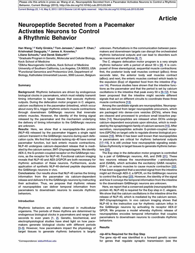
Figure 1. nlp-40 Mutants Lack the Exp Step
(A) Diagram of the defecation cycle in C. elegans,
which repeats every 50 s (adapted from [23]).
Each cycle is initiated with posterior body wall
muscle contraction (pBoc), and 3 s later, anterior
body wall muscles contract (aBoc), which
is immediately followed by enteric muscle con-
traction (the expulsion [Exp] step). The two
GABAergic neurons, AVL and DVB, which control
the Exp step, are indicated by arrowheads.
(B) Representative differential interference con-
trast (DIC) images of the posterior intestines
of young adult worms with the indicated geno-
types. The enlarged space within the intestinal
lumen (black lines) indicates that the lumen is
distended in nlp-40 mutants.
(C) Quantification of the Exp frequency in young
adult worms of the indicated genotypes. nlp-40
gDNA rescue denotes nlp-40(tm4085) mutant
animals expressing genomic nlp-40 transgenes.
‘‘Exp per cycle’’ is defined as the ratio of Exp
over pBoc.
(D) The gene structure of nlp-40 and the positions
of the vj3 and tm4085 deletions are indicated. The
region labeled nlp-40 represents the genomic
DNA fragment (Supplemental Experimental Pro-
cedures) used for rescue.
(E) Schematic of the NLP-40 protein, which is a
neuropeptide precursor with 123 amino acids.
The signal sequence is shown and arrowheads
indicate the three consensus cleavage sites by
proprotein convertases. NLP-40 is predicted to
yield four small peptides (P1 to P4).
The mean and standard errors are shown.
Asterisks (***) indicate a significant difference
of p < 0.0005 in a Student’s t test. See also
Figures S1 and S2.
Current Biology Vol 23 No 92
Please cite this article in press as: Wang et al., Neuropeptide Secreted from a Pacemaker Activates Neurons to Control a RhythmicBehavior, Current Biology (2013), http://dx.doi.org/10.1016/j.cub.2013.03.049
Experimental Procedures). Two independently isolated nlp-40mutants, vj3 and tm4085, both of which delete significantportions of the nlp-40 coding region (Figure 1D and theSupplemental Experimental Procedures available online),displayed distended intestinal lumens, nearly complete elimi-nation of Exp and reductions in aBoc (Figures 1B, 1C, andS1B). However, both pBoc frequency and calcium oscillationsin the intestine were normal (Figure S1A and data not shown).The constipated phenotype and Exp defects of nlp-40mutantscould be fully rescued by a transgene containing nlp-40genomic DNA (gDNA; Figures 1B and 1C). In addition, knock-down of nlp-40 expression by RNA interference (RNAi) alsoproduced similar Exp defects (Figure 1C). Thus, nlp-40 isnecessary for the execution of the Exp step. nlp-40 is pre-dicted to encode a 123-amino-acid neuropeptide precursorprotein, which is highly conserved in nematodes. The NLP-40 precursor contains a predicted signal sequence andthree dibasic consensus cleavage sites predicted to serve asprocessing sites to generate four small peptides (P1 to P4)(Figures 1E and S2).
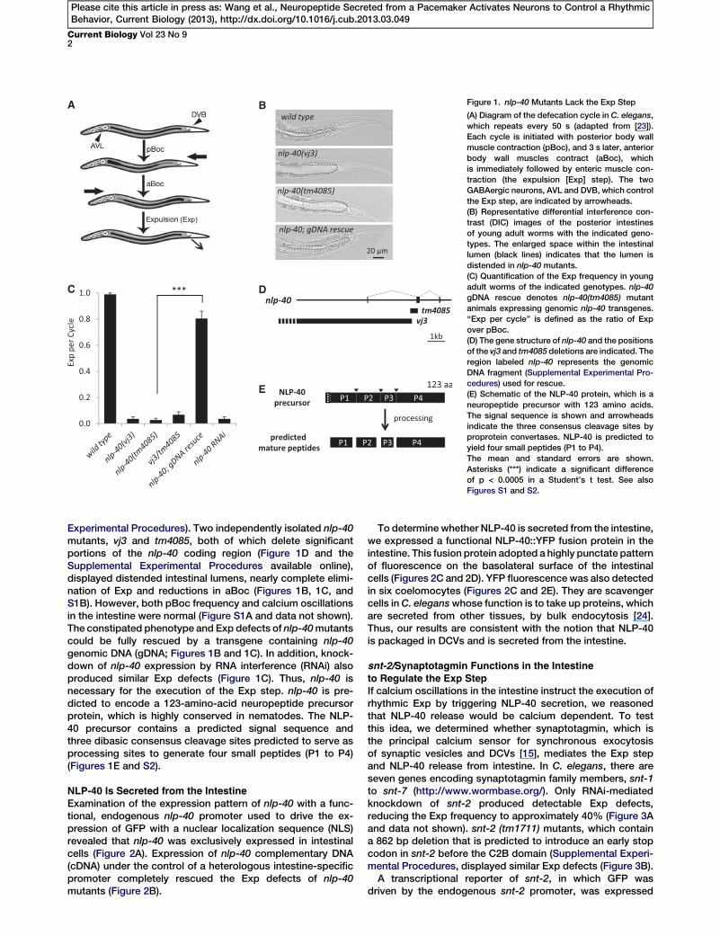
NLP-40 Is Secreted from the Intestine
Examination of the expression pattern of nlp-40 with a func-tional, endogenous nlp-40 promoter used to drive the ex-pression of GFP with a nuclear localization sequence (NLS)revealed that nlp-40 was exclusively expressed in intestinalcells (Figure 2A). Expression of nlp-40 complementary DNA(cDNA) under the control of a heterologous intestine-specificpromoter completely rescued the Exp defects of nlp-40mutants (Figure 2B).
To determine whether NLP-40 is secreted from the intestine,we expressed a functional NLP-40::YFP fusion protein in theintestine. This fusion protein adopted a highly punctate patternof fluorescence on the basolateral surface of the intestinalcells (Figures 2C and 2D). YFP fluorescence was also detectedin six coelomocytes (Figures 2C and 2E). They are scavengercells inC. eleganswhose function is to take up proteins, whichare secreted from other tissues, by bulk endocytosis [24].Thus, our results are consistent with the notion that NLP-40is packaged in DCVs and is secreted from the intestine.
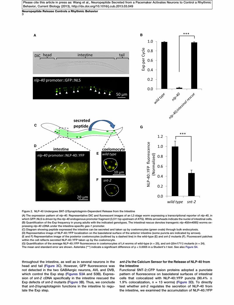
snt-2/Synaptotagmin Functions in the Intestine
to Regulate the Exp StepIf calcium oscillations in the intestine instruct the execution ofrhythmic Exp by triggering NLP-40 secretion, we reasonedthat NLP-40 release would be calcium dependent. To testthis idea, we determined whether synaptotagmin, which isthe principal calcium sensor for synchronous exocytosisof synaptic vesicles and DCVs [15], mediates the Exp stepand NLP-40 release from intestine. In C. elegans, there areseven genes encoding synaptotagmin family members, snt-1to snt-7 (http://www.wormbase.org/). Only RNAi-mediatedknockdown of snt-2 produced detectable Exp defects,reducing the Exp frequency to approximately 40% (Figure 3Aand data not shown). snt-2 (tm1711) mutants, which containa 862 bp deletion that is predicted to introduce an early stopcodon in snt-2 before the C2B domain (Supplemental Experi-mental Procedures, displayed similar Exp defects (Figure 3B).A transcriptional reporter of snt-2, in which GFP was
driven by the endogenous snt-2 promoter, was expressed
A B
0.0
0.2
0.4
0.6
0.8
1.0
Exp
per C
ycle
***
G
0.0
0.2
0.4
0.6
0.8
1.0
1.2
wild type snt-2
***
NLP
-40:
:YFP
fluo
resc
ence
(Nor
mal
ized)
10 μm
secreted peptC
nlp-40 promoter::NLP-40::YFP
20 μm
intest coelomocytewild type
snt-2
D
E
F
nlp-40 promoter::GFP::NLS
50 μm
DIC head intest tail
ine
ide
ine
Figure 2. NLP-40 Undergoes SNT-2/Synaptotagmin-Dependent Release from the Intestine
(A) The expression pattern of nlp-40. Representative DIC and fluorescent images of an L3 stage worm expressing a transcriptional reporter of nlp-40, in
which GFP::NLS is driven by the nlp-40 endogenous promoter fragment (3,511 bp upstream of ATG). White arrowheads indicate the nuclei of intestinal cells.
(B) Quantification of the Exp frequency in young adults with the indicated genotypes. The intestinal rescue denotes transgenic nlp-40(tm4085) worms ex-
pressing nlp-40 cDNA under the intestine-specific ges-1 promoter.
(C) Diagram showing peptide expressed the intestine can be secreted and taken up by coelomocytes (green ovals) through bulk endocytosis.
(D) Representative image of NLP-40::YFP localization on the basolateral surface of the anterior intestine (some puncta are indicated by arrows).
(E and F) Representative image of the posterior coelomocytes (outlined by a dashed line) in the wild-type (E) and snt-2 mutants (F). Fluorescent patches
within the cell reflects secreted NLP-40::YFP taken up by the coelomocyte.
(G) Quantification of the average NLP-40::YFP fluorescence in coelomocytes of L4 worms of wild-type (n = 25), and snt-2(tm1711) mutants (n = 24).
The mean and standard error are shown. Asterisks (***) indicate a significant difference of p < 0.0005 in a Student’s t test. See also Figure S4.
Neuropeptide Release Controls a Rhythmic Behavior3
Please cite this article in press as: Wang et al., Neuropeptide Secreted from a Pacemaker Activates Neurons to Control a RhythmicBehavior, Current Biology (2013), http://dx.doi.org/10.1016/j.cub.2013.03.049
throughout the intestine, as well as in several neurons in thehead and tail (Figure 3C). However, GFP fluorescence wasnot detected in the two GABAergic neurons, AVL and DVB,which control the Exp step (Figures S3A and S3B). Expres-sion of snt-2 cDNA specifically in the intestine rescued theExp defects of snt-2 mutants (Figure 3B). Thus, we concludethat snt-2/synaptotagmin functions in the intestine to regu-late the Exp step.
snt-2 Is the Calcium Sensor for the Release of NLP-40 fromthe Intestine
Functional SNT-2::CFP fusion proteins adopted a punctatepattern of fluorescence on basolateral surfaces of intestinalcells that colocalized with NLP-40::YFP puncta (90.4% 61.9% colocalization, n = 13 worms) (Figure 3D). To directlytest whether snt-2 regulates the secretion of NLP-40 fromthe intestine, we examined the accumulation of NLP-40::YFP
A B
C
0.0
0.2
0.4
0.6
0.8
1.0 *** *
n.s.
Exp
per C
ycle
0.0
0.2
0.4
0.6
0.8
1.0***
Exp
per C
ycle
100μm
snt-2 promoter::GFPhead tailintes�ne
D
SNT-2::CFP
NLP-40::YFP
20μm
Merge
coelomocyte
Figure 3. snt-2/Synaptotagmin Functions in the
Intestine to Regulate the Exp Step
(A and B) Quantification of the Exp frequency of
RNAi-treated young adults (A) and in young
adults with the indicated genotypes (B).
(C) The expression pattern of snt-2. Representa-
tive fluorescent image of an adult expressing
a snt-2 transcriptional reporter, in which GFP
is driven by the snt-2 promoter fragment
(4,177 bp, from 792 bp upstream of ATG to
33 bp of the second exon of snt-2 gene). Arrow-
heads indicate fluorescence in head and tail.
(D) Colocalization of NLP-40::YFP (false colored
in green) and SNT-2::CFP (false colored in red)
in the intestine (with the nlp-40 promoter). The
arrow indicates the coelomocyte, which only
takes up NLP-40::YFP. Right panels: 53 magnifi-
cation of the indicated regions (dashed rectan-
gles). Some colocalized puncta are indicated by
arrowheads.
Asterisks indicate significant differences of *p <
0.05 and ***p < 0.0005 in Student’s t tests. See
also Figures S1, S3, and S4.
Current Biology Vol 23 No 94
Please cite this article in press as: Wang et al., Neuropeptide Secreted from a Pacemaker Activates Neurons to Control a RhythmicBehavior, Current Biology (2013), http://dx.doi.org/10.1016/j.cub.2013.03.049
in coelomocytes. The intensity of YFP-tagged neuropeptidefluorescence in coelomocytes is a measure of the efficacyof neuropeptide secretion in C. elegans [25, 26]. NLP-40::YFPfluorescence in coelomocytes was reduced by approximately50% in snt-2 mutants, compared to wild-type controls (Fig-ure 2E–2G). Thus, SNT-2 is associated with the DCVs contain-ing NLP-40 and snt-2 is required for normal secretion ofNLP-40 from the intestine.
SNT-2 contains two calciumbinding domains, C2A andC2B.The C2B domain contains all five conserved aspartic acid res-idues (Figure S3C), which are the key residues that coordinatecalcium [27]. To determine whether calcium binding is criticalfor SNT-2 function, we mutated two aspartic acid residues inthe C2B domain of SNT-2 corresponding to the residues inDrosophila synaptotagmin 1 that are required for calcium-dependent synchronous transmitter release [28]. Expressionof this calcium-binding defective SNT-2 (referred to as snt-2[D247, 253N], Figure S3C) in the intestine failed to rescue theExp defects of snt-2 mutants in three independent transgeniclines (Figure 3B). Thus, SNT-2 is likely to be an important cal-cium sensor for NLP-40 release from the intestine. The residualNLP-40 secretion observed in snt-2 mutants might be medi-ated by other calcium sensors or by calcium-independentNLP-40 release.
NLP-40 Regulates Rhythmic Calcium Influx in theGABAergic Neurons
Once released from the intestine, NLP-40 could control Expby either activating the GABAergic neurons or by directly
activating the entericmuscles. To distin-guish between these two possibilities,we tested whether optogenetic activa-tion of the GABAergic neurons couldbypass the requirement of nlp-40. Itwas previously shown that activation ofchannelrhodopsin-2 (ChR-2) in theGABAergic neurons with blue light inthe presence of all trans-retinal but notin its absence could restore Exp tomutants lacking aex-2, the GPCR onGABAergic neurons [23]. We found that
activation of ChR-2 in the GABAergic neurons rescued theExp defects of nlp-40 mutants to a similar extent as aex-2mutants but failed to rescue the Exp defects of mutants lack-ing exp-1, the excitatory GABA receptor on enteric muscles(Figure 4A). Therefore, NLP-40 does not function to activatethe muscles directly, but instead is likely to participate in theactivation of the GABAergic neurons during the Exp step.To directly test whether nlp-40 is essential for the activation
of the GABAergic neurons, we performed calcium imagingof GABAergic neurons during the defecation cycle usingGCaMP3.0, a genetically encoded calcium indicator [29].Expression of GCaMP3.0 in the DVB neuron allows the visual-ization of the DVB cell body and the synaptic region, wheretheDVBaxon enters the ventral cord and innervates the entericmuscles (Figure 4B) [30]. In wild-type worms, we observed asingle robust calcium spike in the synaptic region of the DVBneuron (and also often in the cell body) that began approxi-mately 3 s after each pBoc step, peaked immediately beforeeach Exp step, and returned to baseline about 2 to 3 s afterExp. Calcium spikes were not observed at any other time inthe cycle (51 out of 51 cycles in 14 worms; Figures 4B and4C, Movie S1, and data not shown). These results show thatDVB neurons undergo rhythmic activation that correlates withthe Exp step. nlp-40 mutants displayed no calcium spikes inDVBneurons (0 out 46 cycles in 14worms; Figure 4DandMovieS2), except for two calcium spikes that were associated withtwo passive Exp steps (see the Supplemental ExperimentalProcedures). Expression of nlp-40 cDNA in the intestinecompletely restored rhythmic calcium transients to nlp-40
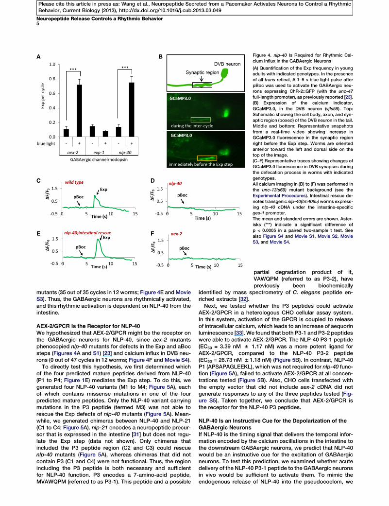
-0.5
0.5
1.5
0 5 10 15
A B
C
ΔF/F
0
GABAergic channelrhodopsin
0.0
0.2
0.4
0.6
0.8
1.0
- + - + - +
aex-2 exp-1 nlp-40
*** ***
blue light
Exp
per c
ycle
immediately before the Exp step
during the inter-cycle
GCaMP3.0
Synaptic region
DVB neuron
GCaMP3.0
nlp-40;intes�nal rescue
pBoc
Exp
-0.5
0.5
1.5
0 5 10 15
pBoc
aex-2
ΔF/F
0
Time (s)
-0.5
0.5
1.5
0 5 10 15
pBoc
wild typeExp
-0.5
0.5
1.5
0 5 10 15
nlp-40
pBocΔF/F
0
ΔF/F
0
Time (s)
Time (s)
Time (s)
D
E F
Figure 4. nlp-40 Is Required for Rhythmic Cal-
cium Influx in the GABAergic Neurons
(A) Quantification of the Exp frequency in young
adults with indicated genotypes. In the presence
of all-trans retinal, A 1–5 s blue light pulse after
pBoc was used to activate the GABAergic neu-
rons expressing ChR-2::GFP (with the unc-47
full-length promoter), as previously reported [23].
(B) Expression of the calcium indicator,
GCaMP3.0, in the DVB neuron (vjIs58). Top:
Schematic showing the cell body, axon, and syn-
aptic region (boxed) of the DVB neuron in the tail.
Middle and bottom: Representative snapshots
from a real-time video showing increase in
GCaMP3.0 fluorescence in the synaptic region
right before the Exp step. Worms are oriented
anterior toward the left and dorsal side on the
top of the image.
(C–F) Representative traces showing changes of
GCaMP3.0 fluorescence in DVB synapses during
the defecation process in worms with indicated
genotypes.
All calcium imaging in (B) to (F) was performed in
the unc-13(s69) mutant background (see the
Experimental Procedures). Intestinal rescue de-
notes transgenic nlp-40(tm4085)worms express-
ing nlp-40 cDNA under the intestine-specific
ges-1 promoter.
The mean and standard errors are shown. Aster-
isks (***) indicate a significant difference of
p < 0.0005 in a paired two-sample t test. See
also Figure S4 and Movie S1, Movie S2, Movie
S3, and Movie S4.
Neuropeptide Release Controls a Rhythmic Behavior5
Please cite this article in press as: Wang et al., Neuropeptide Secreted from a Pacemaker Activates Neurons to Control a RhythmicBehavior, Current Biology (2013), http://dx.doi.org/10.1016/j.cub.2013.03.049
mutants (35 out of 35 cycles in 12 worms; Figure 4E and MovieS3). Thus, the GABAergic neurons are rhythmically activated,and this rhythmic activation is dependent on NLP-40 from theintestine.
AEX-2/GPCR Is the Receptor for NLP-40
We hypothesized that AEX-2/GPCR might be the receptor onthe GABAergic neurons for NLP-40, since aex-2 mutantsphenocopied nlp-40 mutants for defects in the Exp and aBocsteps (Figures 4A and S1) [23] and calcium influx in DVB neu-rons (0 out of 47 cycles in 12 worms; Figure 4F and Movie S4).
To directly test this hypothesis, we first determined whichof the four predicted mature peptides derived from NLP-40(P1 to P4; Figure 1E) mediates the Exp step. To do this, wegenerated four NLP-40 variants (M1 to M4; Figure 5A), eachof which contains missense mutations in one of the fourpredicted mature peptides. Only the NLP-40 variant carryingmutations in the P3 peptide (termed M3) was not able torescue the Exp defects of nlp-40 mutants (Figure 5A). Mean-while, we generated chimeras between NLP-40 and NLP-21(C1 to C4; Figure 5A). nlp-21 encodes a neuropeptide precur-sor that is expressed in the intestine [31] but does not regu-late the Exp step (data not shown). Only chimeras thatincluded the P3 peptide region (C2 and C3) could rescuenlp-40 mutants (Figure 5A), whereas chimeras that did notcontain P3 (C1 and C4) were not functional. Thus, the regionincluding the P3 peptide is both necessary and sufficientfor NLP-40 function. P3 encodes a 7-amino-acid peptide,MVAWQPM (referred to as P3-1). This peptide and a possible
partial degradation product of it,VAWQPM (referred to as P3-2), havepreviously been biochemically
identified by mass spectrometry of C. elegans peptide en-riched extracts [32].Next, we tested whether the P3 peptides could activate
AEX-2/GPCR in a heterologous CHO cellular assay system.In this system, activation of the GPCR is coupled to releaseof intracellular calcium, which leads to an increase of aequorinluminescence [33]. We found that both P3-1 and P3-2 peptideswere able to activate AEX-2/GPCR. The NLP-40 P3-1 peptide(EC50 = 3.39 nM 6 1.17 nM) was a more potent ligand forAEX-2/GPCR, compared to the NLP-40 P3-2 peptide(EC50 = 26.73 nM 6 1.18 nM) (Figure 5B). In contrast, NLP-40P1 (APSAPAGLEEKL), which was not required for nlp-40 func-tion (Figure 5A), failed to activate AEX-2/GPCR at all concen-trations tested (Figure 5B). Also, CHO cells transfected withthe empty vector that did not include aex-2 cDNA did notgenerate responses to any of the three peptides tested (Fig-ure S5). Taken together, we conclude that AEX-2/GPCR isthe receptor for the NLP-40 P3 peptides.
NLP-40 Is an Instructive Cue for the Depolarization of theGABAergic Neurons
If NLP-40 is the timing signal that delivers the temporal infor-mation encoded by the calcium oscillations in the intestine tothe downstream GABAergic neurons, we predict that NLP-40would be an instructive cue for the excitation of GABAergicneurons. To test this prediction, we examined whether acutedelivery of the NLP-40 P3-1 peptide to the GABAergic neuronsin vivo would be sufficient to activate them. To mimic theendogenous release of NLP-40 into the pseudocoelom, we
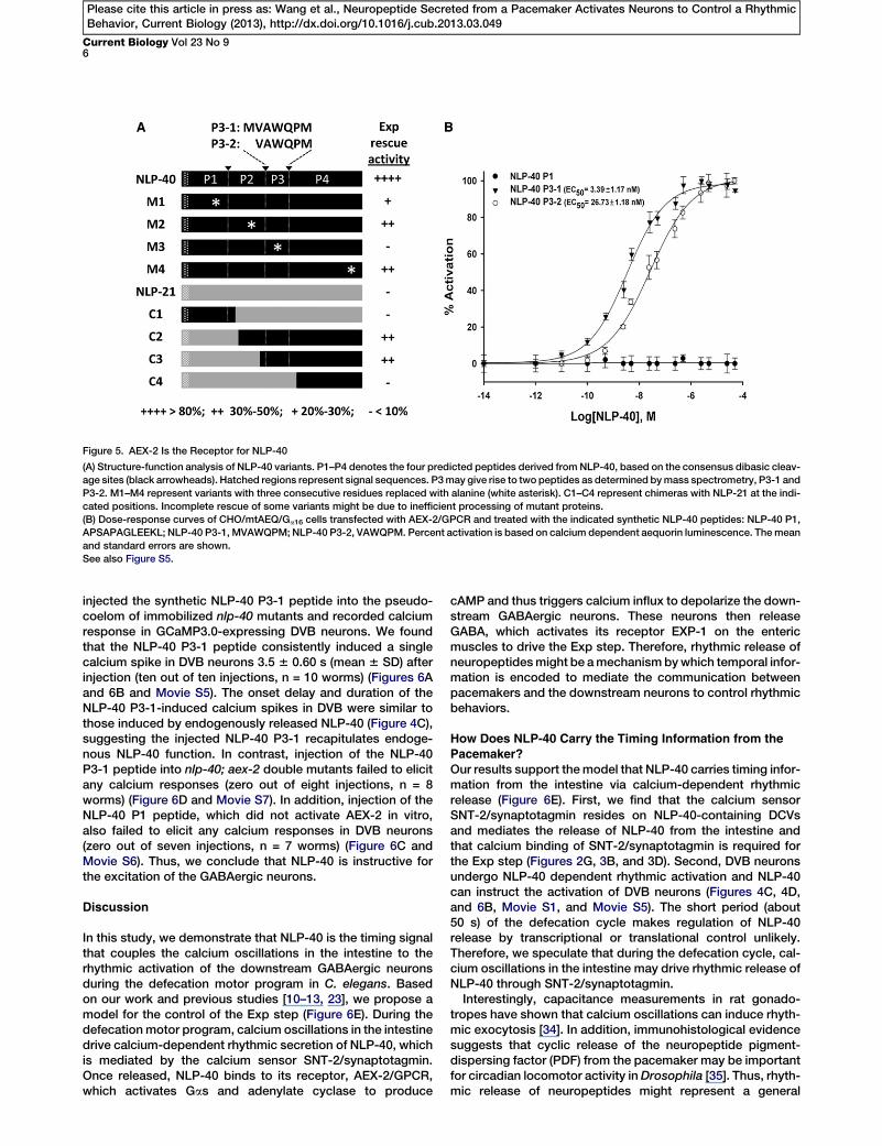
Figure 5. AEX-2 Is the Receptor for NLP-40
(A) Structure-function analysis of NLP-40 variants. P1–P4 denotes the four predicted peptides derived from NLP-40, based on the consensus dibasic cleav-
age sites (black arrowheads). Hatched regions represent signal sequences. P3may give rise to two peptides as determined bymass spectrometry, P3-1 and
P3-2. M1–M4 represent variants with three consecutive residues replaced with alanine (white asterisk). C1–C4 represent chimeras with NLP-21 at the indi-
cated positions. Incomplete rescue of some variants might be due to inefficient processing of mutant proteins.
(B) Dose-response curves of CHO/mtAEQ/Ga16 cells transfected with AEX-2/GPCR and treated with the indicated synthetic NLP-40 peptides: NLP-40 P1,
APSAPAGLEEKL; NLP-40 P3-1, MVAWQPM; NLP-40 P3-2, VAWQPM. Percent activation is based on calcium dependent aequorin luminescence. Themean
and standard errors are shown.
See also Figure S5.
Current Biology Vol 23 No 96
Please cite this article in press as: Wang et al., Neuropeptide Secreted from a Pacemaker Activates Neurons to Control a RhythmicBehavior, Current Biology (2013), http://dx.doi.org/10.1016/j.cub.2013.03.049
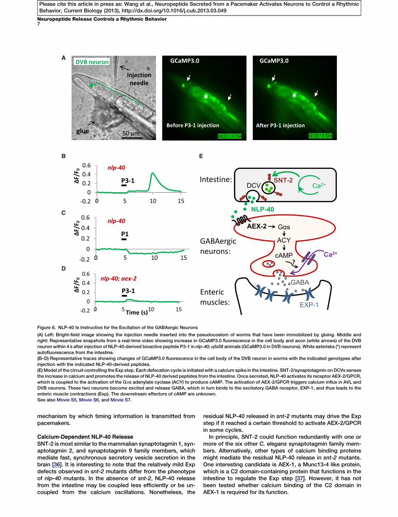
injected the synthetic NLP-40 P3-1 peptide into the pseudo-coelom of immobilized nlp-40 mutants and recorded calciumresponse in GCaMP3.0-expressing DVB neurons. We foundthat the NLP-40 P3-1 peptide consistently induced a singlecalcium spike in DVB neurons 3.5 6 0.60 s (mean 6 SD) afterinjection (ten out of ten injections, n = 10 worms) (Figures 6Aand 6B and Movie S5). The onset delay and duration of theNLP-40 P3-1-induced calcium spikes in DVB were similar tothose induced by endogenously released NLP-40 (Figure 4C),suggesting the injected NLP-40 P3-1 recapitulates endoge-nous NLP-40 function. In contrast, injection of the NLP-40P3-1 peptide into nlp-40; aex-2 double mutants failed to elicitany calcium responses (zero out of eight injections, n = 8worms) (Figure 6D and Movie S7). In addition, injection of theNLP-40 P1 peptide, which did not activate AEX-2 in vitro,also failed to elicit any calcium responses in DVB neurons(zero out of seven injections, n = 7 worms) (Figure 6C andMovie S6). Thus, we conclude that NLP-40 is instructive forthe excitation of the GABAergic neurons.
Discussion
In this study, we demonstrate that NLP-40 is the timing signalthat couples the calcium oscillations in the intestine to therhythmic activation of the downstream GABAergic neuronsduring the defecation motor program in C. elegans. Basedon our work and previous studies [10–13, 23], we propose amodel for the control of the Exp step (Figure 6E). During thedefecation motor program, calcium oscillations in the intestinedrive calcium-dependent rhythmic secretion of NLP-40, whichis mediated by the calcium sensor SNT-2/synaptotagmin.Once released, NLP-40 binds to its receptor, AEX-2/GPCR,which activates Gas and adenylate cyclase to produce
cAMP and thus triggers calcium influx to depolarize the down-stream GABAergic neurons. These neurons then releaseGABA, which activates its receptor EXP-1 on the entericmuscles to drive the Exp step. Therefore, rhythmic release ofneuropeptidesmight be amechanismbywhich temporal infor-mation is encoded to mediate the communication betweenpacemakers and the downstream neurons to control rhythmicbehaviors.
How Does NLP-40 Carry the Timing Information from the
Pacemaker?
Our results support themodel that NLP-40 carries timing infor-mation from the intestine via calcium-dependent rhythmicrelease (Figure 6E). First, we find that the calcium sensorSNT-2/synaptotagmin resides on NLP-40-containing DCVsand mediates the release of NLP-40 from the intestine andthat calcium binding of SNT-2/synaptotagmin is required forthe Exp step (Figures 2G, 3B, and 3D). Second, DVB neuronsundergo NLP-40 dependent rhythmic activation and NLP-40can instruct the activation of DVB neurons (Figures 4C, 4D,and 6B, Movie S1, and Movie S5). The short period (about50 s) of the defecation cycle makes regulation of NLP-40release by transcriptional or translational control unlikely.Therefore, we speculate that during the defecation cycle, cal-cium oscillations in the intestine may drive rhythmic release ofNLP-40 through SNT-2/synaptotagmin.Interestingly, capacitance measurements in rat gonado-
tropes have shown that calcium oscillations can induce rhyth-mic exocytosis [34]. In addition, immunohistological evidencesuggests that cyclic release of the neuropeptide pigment-dispersing factor (PDF) from the pacemaker may be importantfor circadian locomotor activity inDrosophila [35]. Thus, rhyth-mic release of neuropeptides might represent a general
A
B E
C
D
Figure 6. NLP-40 Is Instructive for the Excitation of the GABAergic Neurons
(A) Left: Bright-field image showing the injection needle inserted into the pseudocoelom of worms that have been immobilized by gluing. Middle and
right: Representative snapshots from a real-time video showing increase in GCaMP3.0 fluorescence in the cell body and axon (white arrows) of the DVB
neuron within 4 s after injection of NLP-40-derived bioactive peptide P3-1 in nlp-40; vjIs58 animals (GCaMP3.0 in DVB neurons). White asterisks (*) represent
autofluorescence from the intestine.
(B–D) Representative traces showing changes of GCaMP3.0 fluorescence in the cell body of the DVB neuron in worms with the indicated genotypes after
injection with the indicated NLP-40-derived peptides.
(E)Model of the circuit controlling the Exp step. Each defecation cycle is initiatedwith a calcium spike in the intestine. SNT-2/synaptotagmin onDCVs senses
the increase in calcium and promotes the release of NLP-40 derived peptides from the intestine. Once secreted, NLP-40 activates its receptor AEX-2/GPCR,
which is coupled to the activation of the Gas adenylate cyclase (ACY) to produce cAMP. The activation of AEX-2/GPCR triggers calcium influx in AVL and
DVB neurons. These two neurons become excited and release GABA, which in turn binds to the excitatory GABA receptor, EXP-1, and thus leads to the
enteric muscle contractions (Exp). The downstream effectors of cAMP are unknown.
See also Movie S5, Movie S6, and Movie S7.
Neuropeptide Release Controls a Rhythmic Behavior7
Please cite this article in press as: Wang et al., Neuropeptide Secreted from a Pacemaker Activates Neurons to Control a RhythmicBehavior, Current Biology (2013), http://dx.doi.org/10.1016/j.cub.2013.03.049
mechanism by which timing information is transmitted frompacemakers.
Calcium-Dependent NLP-40 Release
SNT-2 ismost similar to themammalian synaptotagmin 1, syn-aptotagmin 2, and synaptotagmin 9 family members, whichmediate fast, synchronous secretory vesicle secretion in thebrain [36]. It is interesting to note that the relatively mild Expdefects observed in snt-2 mutants differ from the phenotypeof nlp-40 mutants. In the absence of snt-2, NLP-40 releasefrom the intestine may be coupled less efficiently or be un-coupled from the calcium oscillations. Nonetheless, the
residual NLP-40 released in snt-2 mutants may drive the Expstep if it reached a certain threshold to activate AEX-2/GPCRin some cycles.In principle, SNT-2 could function redundantly with one or
more of the six other C. elegans synaptotagmin family mem-bers. Alternatively, other types of calcium binding proteinsmight mediate the residual NLP-40 release in snt-2 mutants.One interesting candidate is AEX-1, a Munc13-4 like protein,which is a C2 domain-containing protein that functions in theintestine to regulate the Exp step [37]. However, it has notbeen tested whether calcium binding of the C2 domain inAEX-1 is required for its function.
Current Biology Vol 23 No 98
Please cite this article in press as: Wang et al., Neuropeptide Secreted from a Pacemaker Activates Neurons to Control a RhythmicBehavior, Current Biology (2013), http://dx.doi.org/10.1016/j.cub.2013.03.049
How Does NLP-40 Deliver the Temporal Information to theGABAergic Neurons?
Many studies have established a classic neuromodulatory roleof neuropeptides in which neuropeptides fine-tune the excit-ability of neurons and circuits [38–41]. However, our resultssuggest that NLP-40 can act more like a classic neurotrans-mitter by depolarizing the GABAergic neurons through its re-ceptor AEX-2/GPCR. First, the rhythmic calcium spikes inDVB neurons are abolished in nlp-40 and aex-2 mutants (Fig-ures 4C, 4D, and 4F). Second, injection of the NLP-40-derivedP3-1 peptide into the pseudocoelom is sufficient to reliablyelicit a calcium transient in the DVB neuron within a fewseconds, and this response is dependent on AEX-2/GPCR(Figures 6B and 6D). Third, it is unlikely that classic neurotrans-mitters are involved in activating DVB neurons during the defe-cation cycle, since mutants defective in the biosynthesis,transport, or release of classical neurotransmitters (such asunc-13/Munc13, eat-4 for glutamate, cat-2 for dopamine,tph-1 for serotonin, tdc-1 for tyramine and octopamine,unc-17 for acetylcholine) have grossly normal Exp frequency(Figure S4) [42]. Therefore, NLP-40 delivers timing informationby serving as an instructive cue for the excitation of theGABAergic neurons via its receptor AEX-2/GPCR.
NLP-40 P3-1 injection caused consistent calcium responsesin DVB but failed to induce robust enteric muscle contractions(Exp; data not shown). This is likely due to the additional un-known permissive signal proposed by Mahoney et al., whichrestricts the Exp step from occurring outside of a small win-dow within a few seconds of the pBoc step [23]. Our resultsshow that this permissive signal most likely controls the refrac-tory property of enteric muscles, since peptide injection (pre-sumably at any time in the cycle) always elicited a calciumresponse in DVB. Thus the refractory period might be a mech-anism by which the robust rhythmicity of Exp, instructed byNLP-40/AEX-2 signaling in theGABAergic neurons, is ensured.
That neuropeptides are involved in controlling the Exp stephas been implied by the identification of a well-conserved setof proteins involved in the processing and secretion of neuro-peptides, including AEX-5/proprotein convertase, AEX-4/SNAP25, AEX-1/Munc13-like protein, and SYN-1/syntaxin,which are necessary for the Exp step [23, 37, 43]. Our resultsextend these studies by identifying the peptide and providingthe mechanisms by which the peptide is released from thepacemaker and activates its downstream targets. The identifi-cation of additional downstream components in the NLP-40/AEX-2 signaling pathway will promote further understandingof how neuropeptides can instruct rhythmic behaviors.
Experimental Procedures
Strains, Plasmids, and Transgenes
All C. elegans strains were maintained as described at 20�C on NGM plates
seeded with OP50 E. coli [44]. The vj3 allele was originally isolated from an
EMS screen for aldicarb resistant mutants. The construction of plasmids
was performed with standard protocols. Transgenic worms were generated
by microinjection [45]. A detailed list of all the strains, plasmids and oligos is
described in the Supplemental Experimental Procedures.
Behavioral Assays
The defecation motor program was scored as previously described [8, 46].
Each animal was scored for ten consecutive defecation cycles. Eight to ten
worms were examined for each genotype. An unpaired two-tail Student’s
t test with unequal variance was used to determine statistical significance
between two samples. The assay with channelrhdopsin-2 (ChR2) in
GABAergic neurons was performed as previously described [23]. See the
Supplemental Experimental Procedures for details.
Fluorescence Imaging and Analysis
Fluorescence imaging of NLP-40::YFP and SNT-2::CFP in the intestine was
performed in glo-1(zu391)mutant background, which has normal Exp steps
(Figure S4), to reduce intestinal autofluorescence [47]. The coelomocyte
assay was performed and quantified as previously described [25]. See the
Supplemental Experimental Procedures for details.
In Vivo Calcium Imaging
The transgenic animal vjIs58[pmyo-2::NLS::mCherry, Punc-47mini::
GCaMP3.0] was used for calcium imaging in DVB neurons. The strains,
used in Figures 4B–4F, contained the unc-13(s69) mutation to immobilize
animals, because unc-13(s69) mutants are almost completely paralyzed
[48]. Both vjIs58 and unc-13(s69) had normal Exp steps (Figure S4). Live im-
aging of young adults was performed onNGM-agarose plateswith food top-
ped with a coverslip using a Nikon eclipse 90i microscope equipped with a
Plan Apo 403 oil objective (numerical aperture [NA] = 1.0) and a standard
GFP filter set. The GCaMP3.0 fluorescence in the synaptic region of the
DVB neuron was quantified. For imaging and quantification details, see
the Supplemental Experimental Procedures.
In Vitro Cellular Assay for AEX-2/GPCR Activation by NLP-40-Derived
Peptides
Chinese hamster ovary cells (CHO-K1), stably expressing the mitochondri-
ally targeted apo-aequorin (mtAEQ) and the human Ga16 subunit, were used
for the Ca2+ measurements to detect the activation of GPCR [33]. The cells
were transiently transfected with the aex-2 cDNA construct (pHW149) or the
empty vector (pHW148) and were then treated with one of the synthetic
NLP-40-derived peptides (NLP-40 P1, P3-1 or P3-2). See the Supplemental
Experimental Procedures for details.
Injection of NLP-40-Derived Peptides and Calcium Imaging
Injections were performed with the Eppendorf Femtojet express under an
inverted Olympus IX70 microscope equipped with an Olympus UPlan FLN
403 oil objective (NA = 1.30) and a Photometrics cascade 512B camera.
Adult worms were glued on sylgard-coated cover slides with histoacryl
(B. Braun, Germany) as previously described [48]. Worms were superfused
with external solution (150 mM NaCl, 5 mM KCl, 4 mM MgCl2, 1 mM CaCl2,
10 mM glucose, and 15 mM HEPES [pH 7.3], 330 mOsm). Real-time calcium
imaging was performed with Metamorph 6.0 (Universal Imaging), at four
frames per second for 1 min (15–100 ms exposure time, 2 3 2 binning,
with injection at around the 15th second). The injection solution was pre-
pared as follows: 10 mM synthetic peptide (NLP-40 P1 or P3-1), 0.5 mg/ml
Dextran, and rhodamine B (10,000 MW, Molecular Probes) in the external
solution. Successful injections were verified with the red coinjection marker
in pseudocoelom after injection. Quantification of GCaMP3.0 signal in syn-
aptic region or cell body of the DVB neuron was measured as described in
the ‘‘in vivo calcium imaging’’ section of the Supplemental Experimental
Procedures.
Accession Numbers
The GenBank accession number for the nlp-40 sequence reported in this
paper is NM058259. TheGenBank accession number for the snt-2 sequence
reported in this paper is NM064860.
Supplemental Information
Supplemental Information includes five figures, Supplemental Experimental
Procedures, and seven movies and can be found with this article online at
http://dx.doi.org/10.1016/j.cub.2013.03.049.
Acknowledgments
We thankMichael Ailion andmembers of the Sieburth lab for critical reading
of the manuscript. We thank Stephen Nurrish for his suggestion to analyze
the defecation motor program in vj3 mutants. We thank Andrew Weitz,
Robert H. Chow, Emily R. Liman, Kang Shen, Kim Schuske, Oliver Hobert,
Alexander Gottschalk, and Michael Nonet for reagents. We thank Oleg
Evgrafov, Andrew Clark, Zack Ramjan, and Benjamin Berman for helping
us analyze the whole-genome sequencing data. Some strains were pro-
vided by the CGC, which is funded by NIH Office of Research Infrastructure
Programs (P40 OD010440), and the Japanese National Bioresource Project.
The work is funded by grants from the American Heart Association (to D.S.),
NIH NINDS NS071085-02 (to D.S.), and fellowship from the NIH training

Neuropeptide Release Controls a Rhythmic Behavior9
Please cite this article in press as: Wang et al., Neuropeptide Secreted from a Pacemaker Activates Neurons to Control a RhythmicBehavior, Current Biology (2013), http://dx.doi.org/10.1016/j.cub.2013.03.049
program in Cellular, Biochemical, and Molecular Sciences at University of
Southern California (to H.W.).
Received: March 15, 2013
Revised: March 18, 2013
Accepted: March 20, 2013
Published: April 11, 2013
References
1. Iwasaki, K., and Thomas, J.H. (1997). Genetics in rhythm. Trends Genet.
13, 111–115.
2. Schibler, U., and Naef, F. (2005). Cellular oscillators: rhythmic gene
expression and metabolism. Curr. Opin. Cell Biol. 17, 223–229.
3. Stanewsky, R. (2003). Genetic analysis of the circadian system in
Drosophila melanogaster and mammals. J. Neurobiol. 54, 111–147.
4. Reppert, S.M., and Weaver, D.R. (2002). Coordination of circadian
timing in mammals. Nature 418, 935–941.
5. Marder, E., and Calabrese, R.L. (1996). Principles of rhythmic motor
pattern generation. Physiol. Rev. 76, 687–717.
6. Sehgal, A., andMignot, E. (2011). Genetics of sleep and sleep disorders.
Cell 146, 194–207.
7. Qu, Z. (2011). Chaos in the genesis and maintenance of cardiac arrhyth-
mias. Prog. Biophys. Mol. Biol. 105, 247–257.
8. Thomas, J.H. (1990). Genetic analysis of defecation in Caenorhabditis
elegans. Genetics 124, 855–872.
9. Dal Santo, P., Logan, M.A., Chisholm, A.D., and Jorgensen, E.M. (1999).
The inositol trisphosphate receptor regulates a 50-second behavioral
rhythm in C. elegans. Cell 98, 757–767.
10. Teramoto, T., and Iwasaki, K. (2006). Intestinal calciumwavescoordinate
a behavioral motor program in C. elegans. Cell Calcium 40, 319–327.
11. Nehrke, K., Denton, J., andMowrey, W. (2008). Intestinal Ca2+ wave dy-
namics in freely moving C. elegans coordinate execution of a rhythmic
motor program. Am. J. Physiol. Cell Physiol. 294, C333–C344.
12. Peters,M.A., Teramoto, T.,White, J.Q., Iwasaki, K., and Jorgensen, E.M.
(2007). A calcium wave mediated by gap junctions coordinates a rhyth-
mic behavior in C. elegans. Curr. Biol. 17, 1601–1608.
13. Branicky, R., and Hekimi, S. (2006). What keeps C. elegans regular: the
genetics of defecation. Trends Genet. 22, 571–579.
14. Li, C., and Kim, K. (2008). Neuropeptides. WormBook, 1–36.
15. Pang, Z.P., and Sudhof, T.C. (2010). Cell biology of Ca2+-triggered
exocytosis. Curr. Opin. Cell Biol. 22, 496–505.
16. Salio, C., Lossi, L., Ferrini, F., and Merighi, A. (2006). Neuropeptides as
synaptic transmitters. Cell Tissue Res. 326, 583–598.
17. Taghert, P.H. (2001). How does the circadian clock send timing informa-
tion to the brain? Semin. Cell Dev. Biol. 12, 329–341.
18. Mertens, I., Husson, S.J., Janssen, T., Lindemans, M., and Schoofs, L.
(2007). PACAP and PDF signaling in the regulation ofmammalian and in-
sect circadian rhythms. Peptides 28, 1775–1783.
19. Vosko, A.M., Schroeder, A., Loh, D.H., and Colwell, C.S. (2007).
Vasoactive intestinal peptide and the mammalian circadian system.
Gen. Comp. Endocrinol. 152, 165–175.
20. Taghert, P.H. (2009). Circadian biology: a neuropeptide is bound to acti-
vate its receptor. Curr. Biol. 19, R696–R697.
21. McIntire, S.L., Jorgensen, E., Kaplan, J., and Horvitz, H.R. (1993). The
GABAergic nervous system of Caenorhabditis elegans. Nature 364,
337–341.
22. Beg, A.A., and Jorgensen, E.M. (2003). EXP-1 is an excitatory GABA-
gated cation channel. Nat. Neurosci. 6, 1145–1152.
23. Mahoney, T.R., Luo, S., Round, E.K., Brauner, M., Gottschalk, A.,
Thomas, J.H., and Nonet, M.L. (2008). Intestinal signaling to
GABAergic neurons regulates a rhythmic behavior in Caenorhabditis el-
egans. Proc. Natl. Acad. Sci. USA 105, 16350–16355.
24. Fares, H., and Grant, B. (2002). Deciphering endocytosis in
Caenorhabditis elegans. Traffic 3, 11–19.
25. Sieburth, D., Madison, J.M., and Kaplan, J.M. (2007). PKC-1 regulates
secretion of neuropeptides. Nat. Neurosci. 10, 49–57.
26. Speese, S., Petrie, M., Schuske, K., Ailion, M., Ann, K., Iwasaki, K.,
Jorgensen, E.M., and Martin, T.F. (2007). UNC-31 (CAPS) is required
for dense-core vesicle but not synaptic vesicle exocytosis in
Caenorhabditis elegans. J. Neurosci. 27, 6150–6162.
27. Bai, J., and Chapman, E.R. (2004). The C2 domains of synaptotagmin—
partners in exocytosis. Trends Biochem. Sci. 29, 143–151.
28. Mackler, J.M., Drummond, J.A., Loewen, C.A., Robinson, I.M., and
Reist, N.E. (2002). The C(2)B Ca(2+)-binding motif of synaptotagmin is
required for synaptic transmission in vivo. Nature 418, 340–344.
29. Tian, L., Hires, S.A., Mao, T., Huber, D., Chiappe, M.E., Chalasani, S.H.,
Petreanu, L., Akerboom, J., McKinney, S.A., Schreiter, E.R., et al. (2009).
Imaging neural activity in worms, flies and mice with improved GCaMP
calcium indicators. Nat. Methods 6, 875–881.
30. White, J.G., Southgate, E., Thomson, J.N., and Brenner, S. (1986). The
structure of the nervous system of the nematode Caenorhabditis ele-
gans. Philos. Trans. R. Soc. Lond. B Biol. Sci. 314, 1–340.
31. Nathoo, A.N., Moeller, R.A., Westlund, B.A., and Hart, A.C. (2001).
Identification of neuropeptide-like protein gene families in
Caenorhabditiselegans and other species. Proc. Natl. Acad. Sci. USA
98, 14000–14005.
32. Husson, S.J., Janssen, T., Baggerman, G., Bogert, B., Kahn-Kirby, A.H.,
Ashrafi, K., and Schoofs, L. (2007). Impaired processing of FLP and NLP
peptides in carboxypeptidase E (EGL-21)-deficient Caenorhabditis ele-
gans as analyzed by mass spectrometry. J. Neurochem. 102, 246–260.
33. Mertens, I., Meeusen, T., Janssen, T., Nachman, R., and Schoofs, L.
(2005). Molecular characterization of two G protein-coupled receptor
splice variants as FLP2 receptors in Caenorhabditis elegans.
Biochem. Biophys. Res. Commun. 330, 967–974.
34. Tse, A., Tse, F.W., Almers, W., and Hille, B. (1993). Rhythmic exocytosis
stimulated by GnRH-induced calcium oscillations in rat gonadotropes.
Science 260, 82–84.
35. Park, J.H., Helfrich-Forster, C., Lee, G., Liu, L., Rosbash, M., and Hall,
J.C. (2000). Differential regulation of circadian pacemaker output by
separate clock genes in Drosophila. Proc. Natl. Acad. Sci. USA 97,
3608–3613.
36. Gustavsson, N., and Han, W. (2009). Calcium-sensing beyond neuro-
transmitters: functions of synaptotagmins in neuroendocrine and endo-
crine secretion. Biosci. Rep. 29, 245–259.
37. Doi, M., and Iwasaki, K. (2002). Regulation of retrograde signaling at
neuromuscular junctions by the novel C2 domain protein AEX-1.
Neuron 33, 249–259.
38. Root, C.M., Ko, K.I., Jafari, A., and Wang, J.W. (2011). Presynaptic facil-
itation by neuropeptide signaling mediates odor-driven food search.
Cell 145, 133–144.
39. Hu, Z., Pym, E.C., Babu, K., Vashlishan Murray, A.B., and Kaplan, J.M.
(2011). A neuropeptide-mediated stretch response links muscle
contraction to changes in neurotransmitter release. Neuron 71, 92–102.
40. Bargmann, C.I. (2012). Beyond the connectome: how neuromodulators
shape neural circuits. Bioessays 34, 458–465.
41. Chalasani, S.H., Kato, S., Albrecht, D.R., Nakagawa, T., Abbott, L.F., and
Bargmann, C.I. (2010). Neuropeptide feedback modifies odor-evoked
dynamics in Caenorhabditis elegans olfactory neurons. Nat. Neurosci.
13, 615–621.
42. Miller, K.G., Alfonso, A., Nguyen, M., Crowell, J.A., Johnson, C.D., and
Rand, J.B. (1996). A genetic selection for Caenorhabditis elegans syn-
aptic transmission mutants. Proc. Natl. Acad. Sci. USA 93, 12593–
12598.
43. Yamashita, M., Iwasaki, K., and Doi, M. (2009). The non-neuronal syn-
taxin SYN-1 regulates defecation behavior and neural activity in C. ele-
gans through interaction with the Munc13-like protein AEX-1. Biochem.
Biophys. Res. Commun. 378, 404–408.
44. Brenner, S. (1974). The genetics of Caenorhabditis elegans. Genetics
77, 71–94.
45. Mello, C.C., Kramer, J.M., Stinchcomb, D., and Ambros, V. (1991).
Efficient gene transfer in C.elegans: extrachromosomal maintenance
and integration of transforming sequences. EMBO J. 10, 3959–3970.
46. Liu, D.W., and Thomas, J.H. (1994). Regulation of a periodic motor pro-
gram in C. elegans. J. Neurosci. 14, 1953–1962.
47. Hermann, G.J., Schroeder, L.K., Hieb, C.A., Kershner, A.M., Rabbitts,
B.M., Fonarev, P., Grant, B.D., and Priess, J.R. (2005). Genetic analysis
of lysosomal trafficking in Caenorhabditis elegans. Mol. Biol. Cell 16,
3273–3288.
48. Richmond, J.E., Davis, W.S., and Jorgensen, E.M. (1999). UNC-13 is
required for synaptic vesicle fusion in C. elegans. Nat. Neurosci. 2,
959–964.