neuropathology of central nervous system infections
TRANSCRIPT
Neuropathology of Central Nervous System Infections
Leila Chimelli, MD, PhD.
Professor of PathologyCoordinator of the Laboratory of Neuropathology
State Institute of BrainRio de Janeiro, Brazil.

Disclosures
• I have no relevant financial relationships to disclose
Learning Objectives
• Identify the morphological appearances of the various infectious agents that affect the Central Nervous System (CNS).
• Formulate diagnostic hypotheses of CNS infections according to the topography and morphological presentations of the lesions.
• Propose diagnoses of CNS infections taking into account the immunological state of the patient.
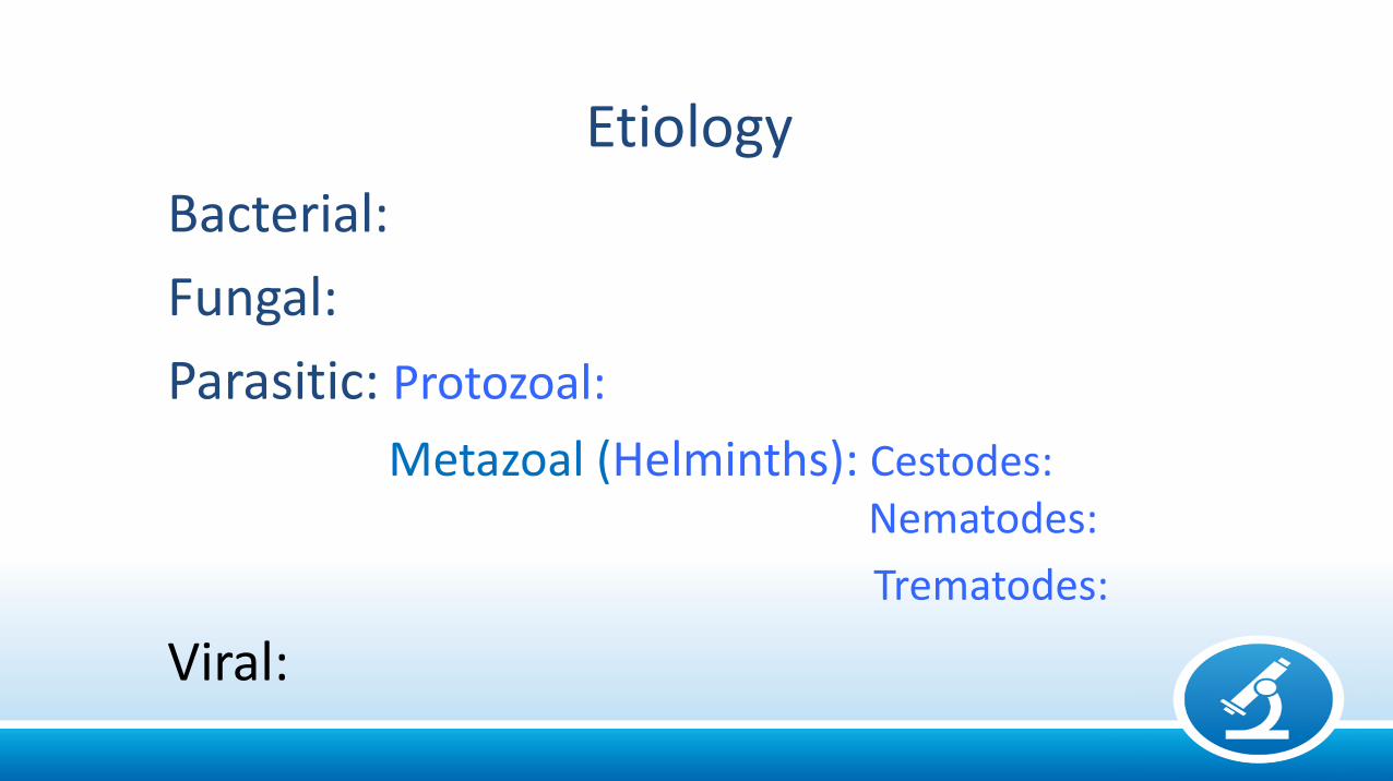
Etiology
Bacterial:
Fungal:
Parasitic: Protozoal:
Metazoal (Helminths): Cestodes:Nematodes:
Trematodes:
Viral:
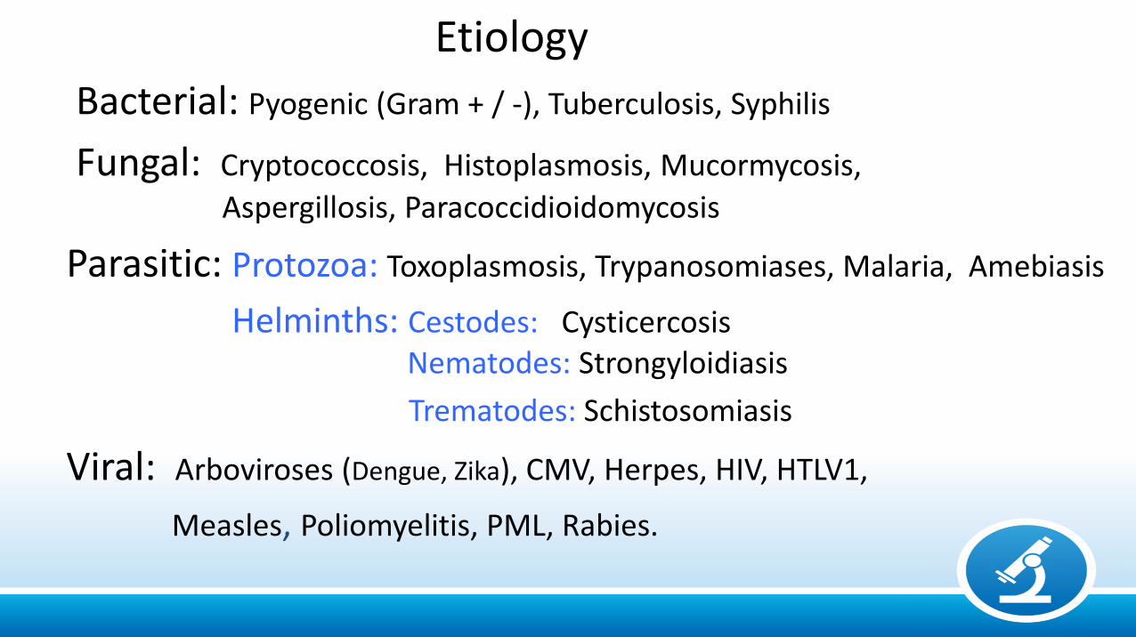
Etiology
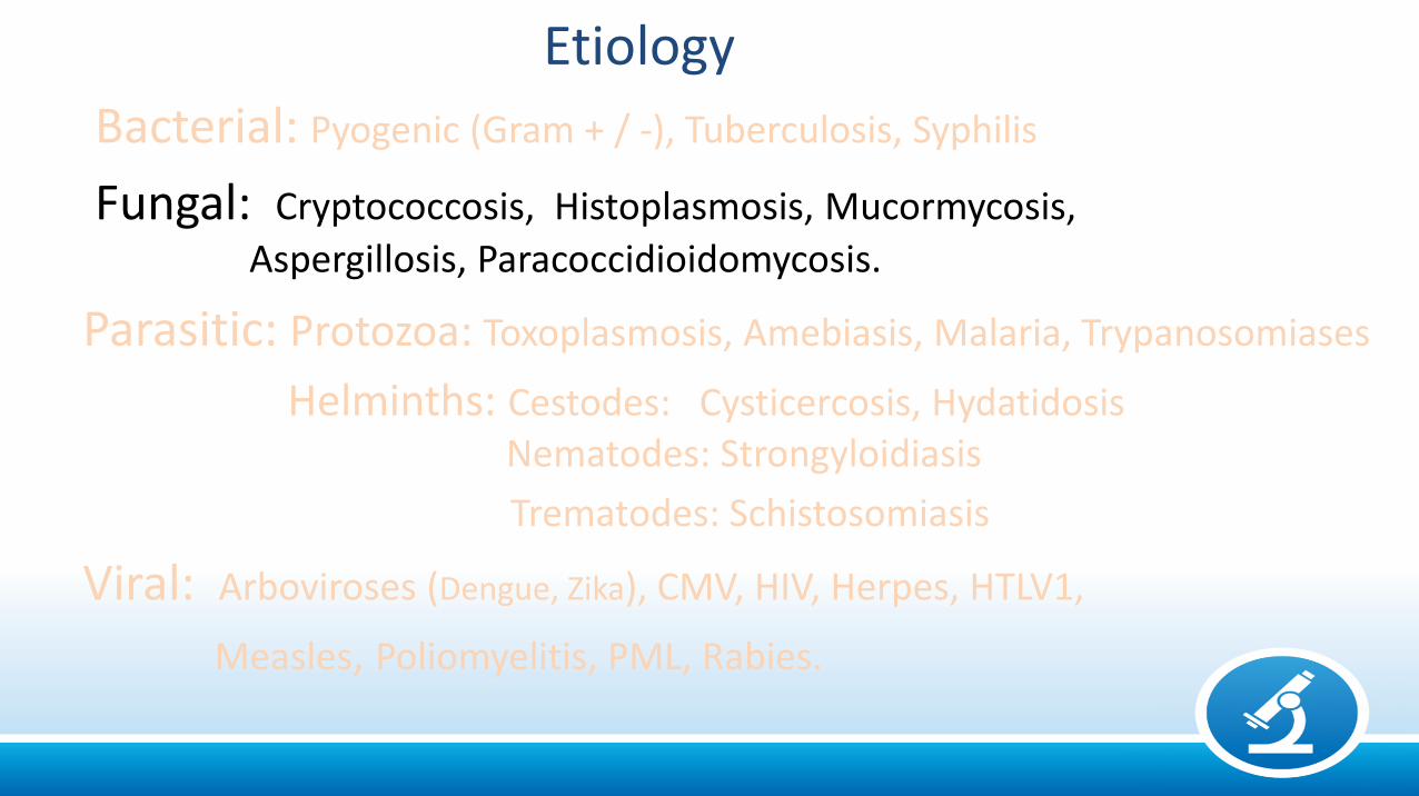
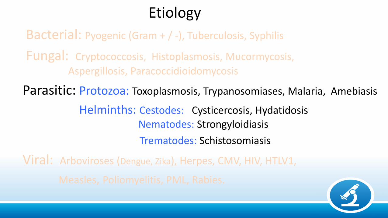
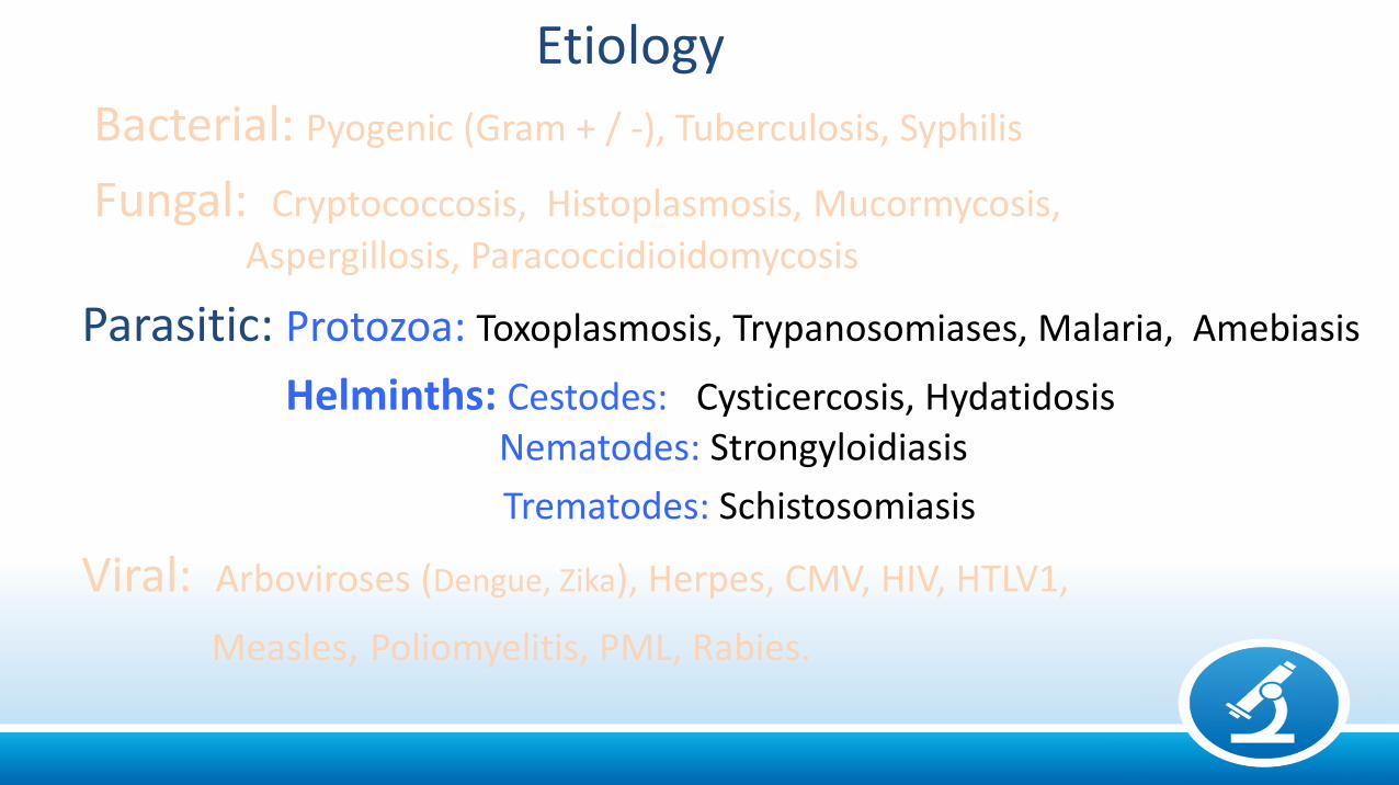
Bacterial: Pyogenic (Gram + / -), Tuberculosis, Syphilis
Fungal: Cryptococcosis, Histoplasmosis, Mucormycosis,
Aspergillosis, Paracoccidioidomycosis
Parasitic: Protozoa: Toxoplasmosis, Trypanosomiases, Malaria, Amebiasis
Helminths: Cestodes: CysticercosisNematodes: Strongyloidiasis
Trematodes: Schistosomiasis
Viral: Arboviroses (Dengue, Zika), CMV, Herpes, HIV, HTLV1,
Measles, Poliomyelitis, PML, Rabies.
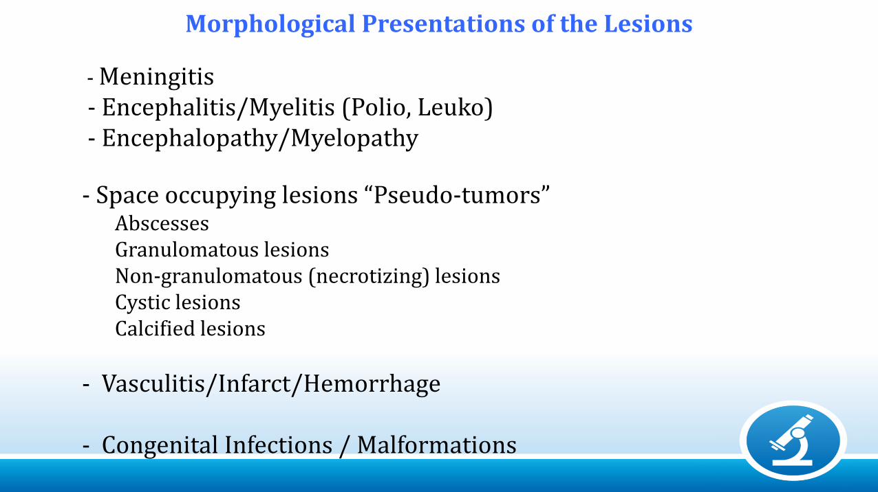
Morphological Presentations of the Lesions
- Meningitis- Encephalitis/Myelitis (Polio, Leuko)- Encephalopathy/Myelopathy
- Space occupying lesions “Pseudo-tumors”AbscessesGranulomatous lesionsNon-granulomatous (necrotizing) lesionsCystic lesionsCalcified lesions
- Vasculitis/Infarct/Hemorrhage
- Congenital Infections / Malformations
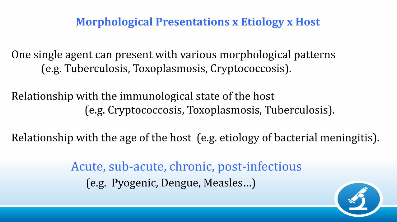
Morphological Presentations x Etiology x Host
One single agent can present with various morphological patterns (e.g. Tuberculosis, Toxoplasmosis, Cryptococcosis).
Relationship with the immunological state of the host (e.g. Cryptococcosis, Toxoplasmosis, Tuberculosis).
Relationship with the age of the host (e.g. etiology of bacterial meningitis).
Acute, sub-acute, chronic, post-infectious(e.g. Pyogenic, Dengue, Measles…)
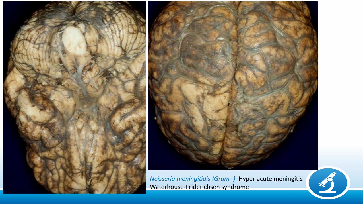
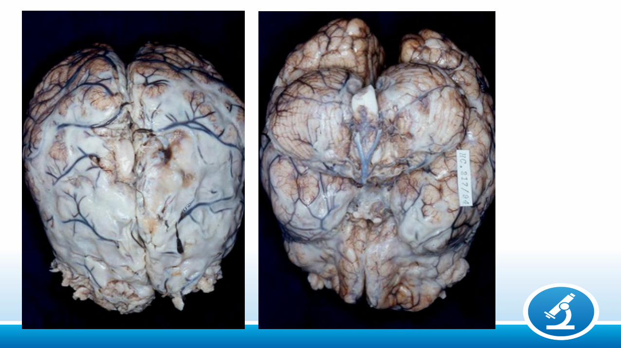
Neisseria meningitidis (Gram -) Hyper acute meningitisWaterhouse-Friderichsen syndrome
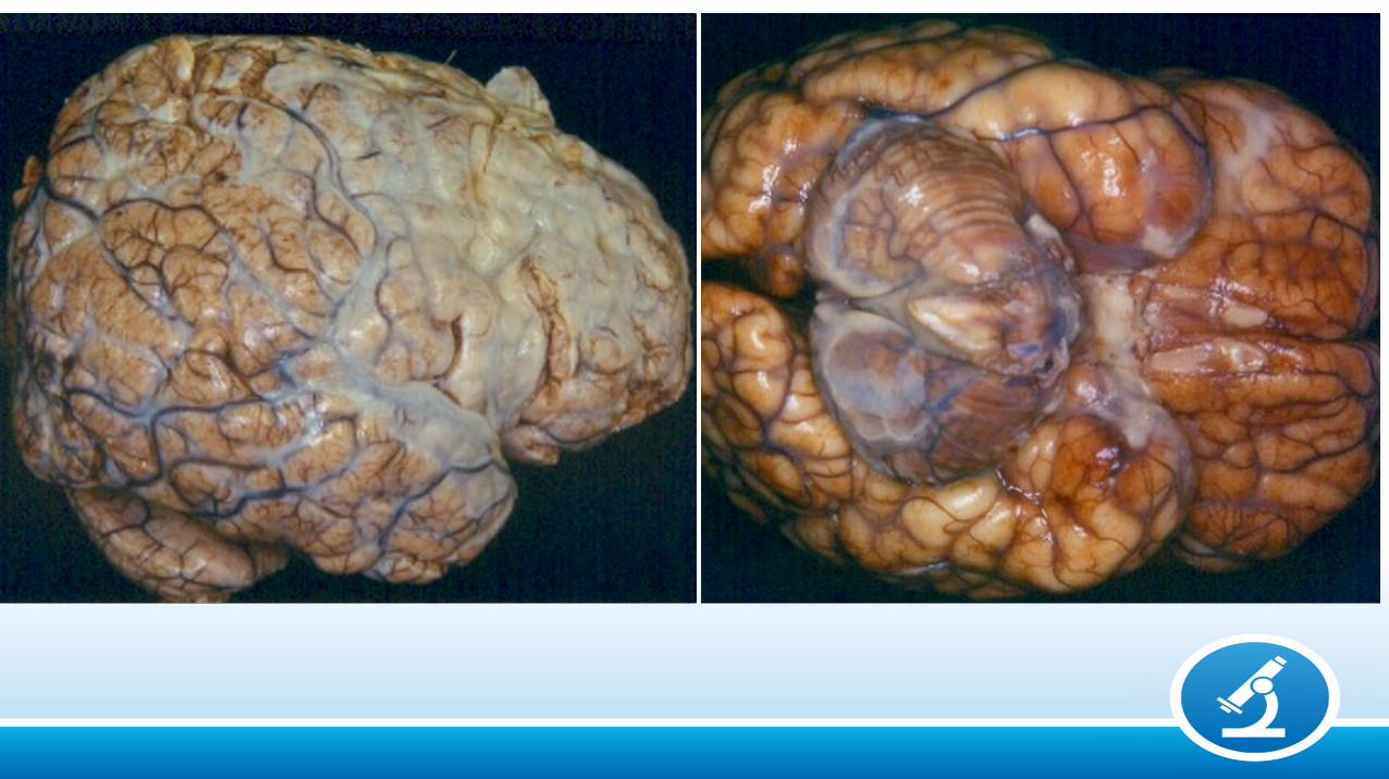
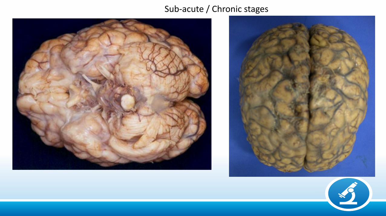
Sub-acute / Chronic stages
COMPLICATIONS
• Ventriculitis
• Hydrocephalus
• Infarct (vasculitis, thrombosis)
• Abscess (micro-abscesses)
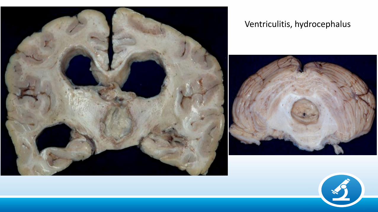
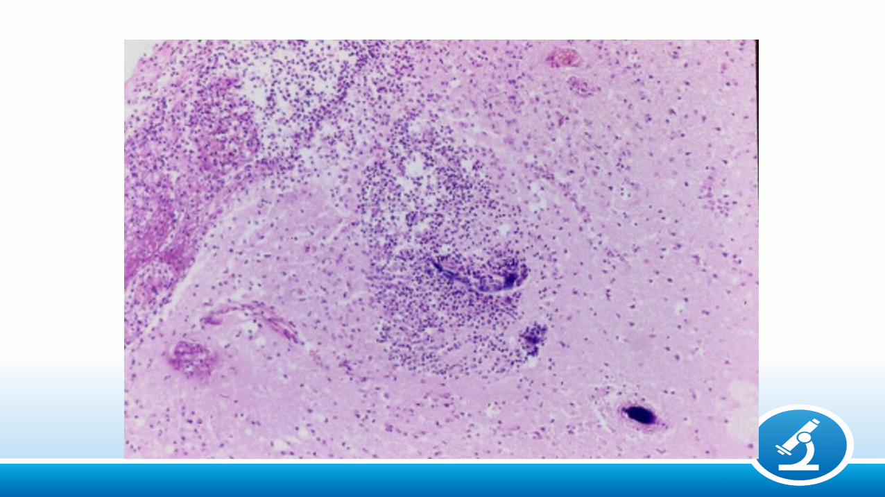
Ventriculitis, hydrocephalus
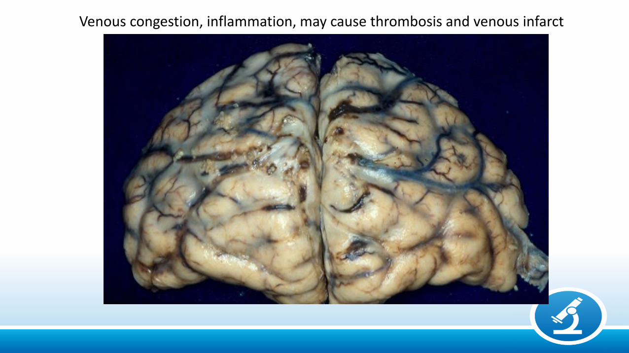
Venous congestion, inflammation, may cause thrombosis and venous infarct
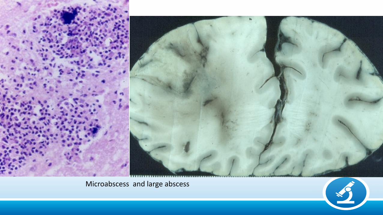
Microabscess and large abscess
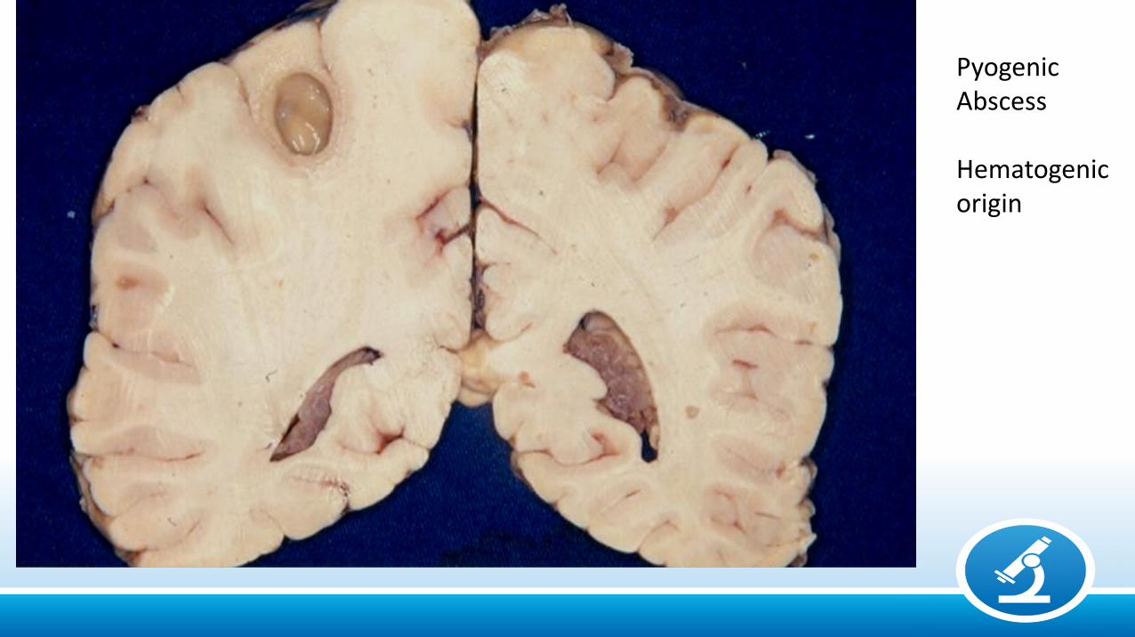
PyogenicAbscess
Hematogenicorigin
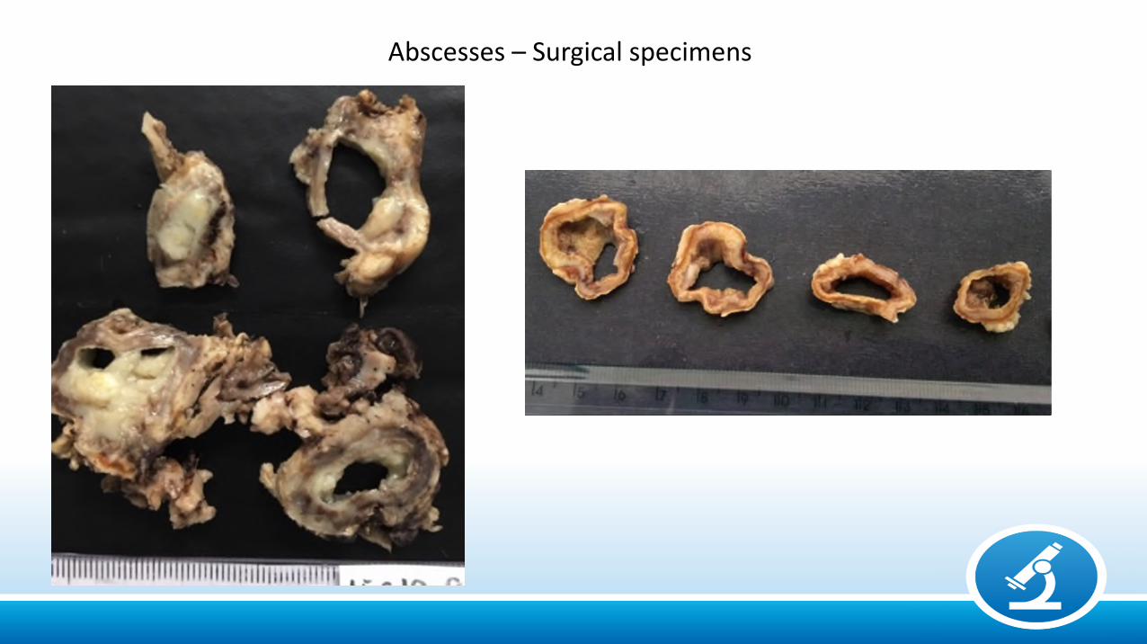
Abscesses – Surgical specimens
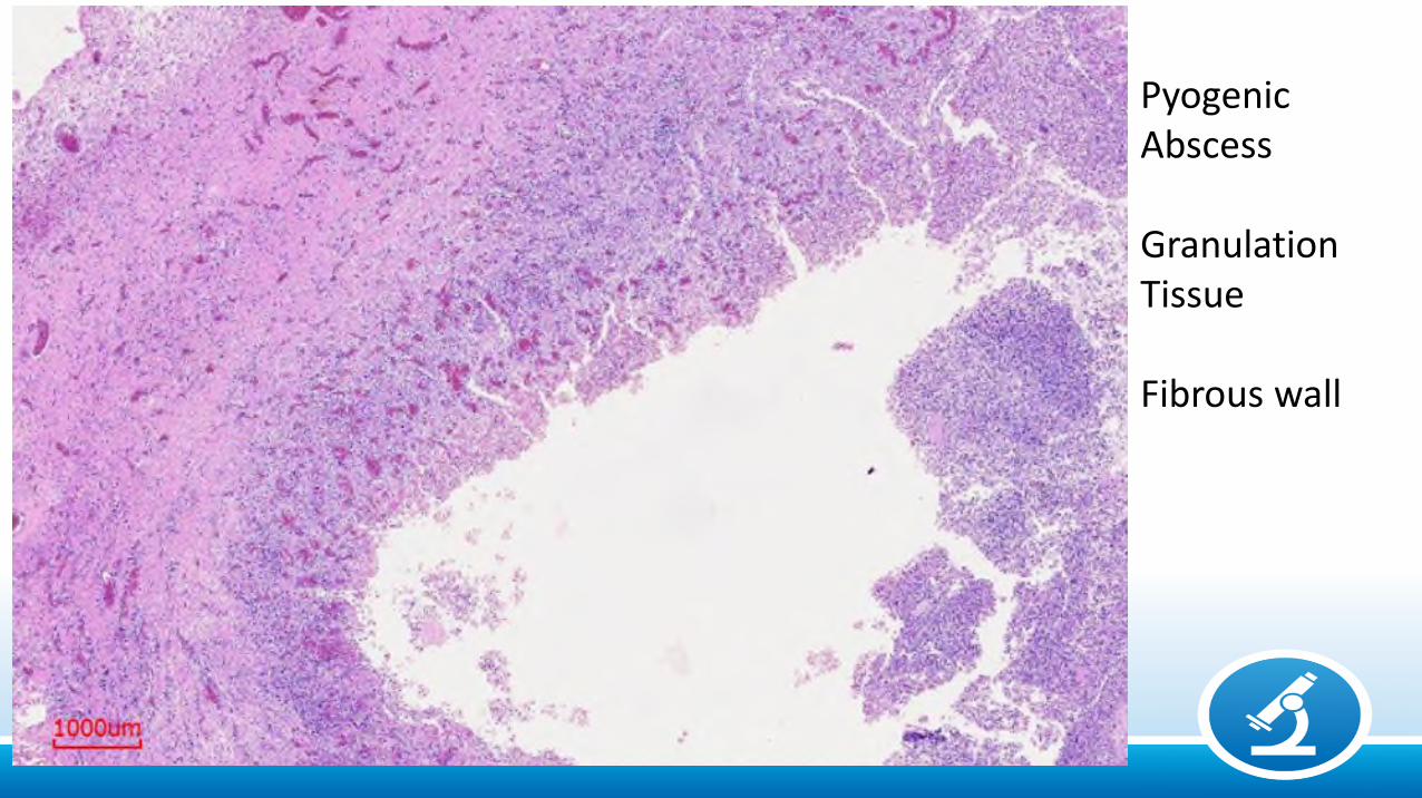
Pyogenic Abscess
Granulation Tissue
Fibrous wall
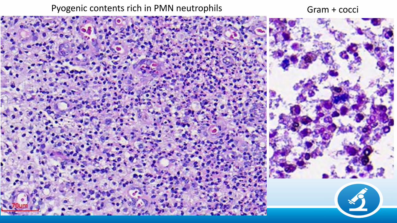
Gram + cocciPyogenic contents rich in PMN neutrophils
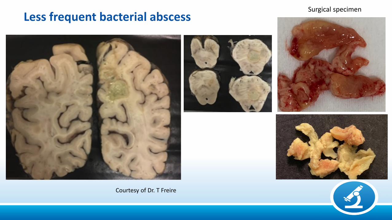
Surgical specimen
Less frequent bacterial abscess
Courtesy of Dr. T Freire
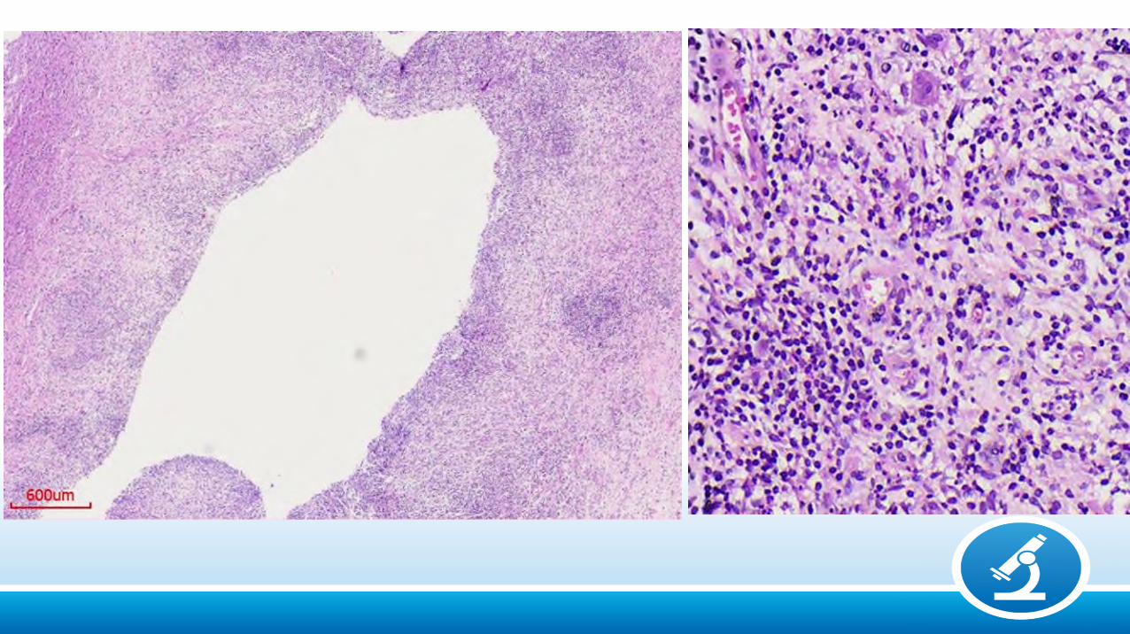
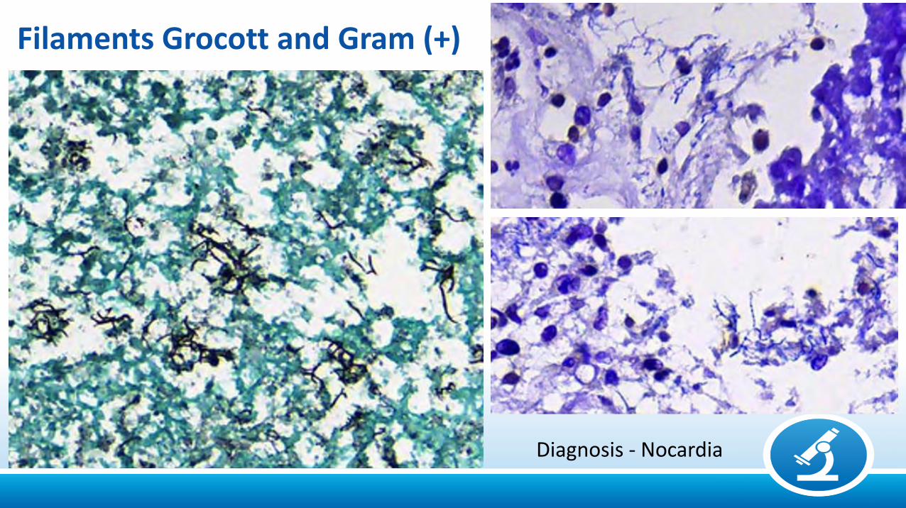
Filaments Grocott and Gram (+)
Diagnosis - Nocardia
TUBERCULOSIS• Still a major health problem
• Endemic in developing countries with a largepopulation, crowding and poverty
• Cerebral Tuberculosis• Tuberculous meningitis
• Tuberculoma
• Tuberculous abscess
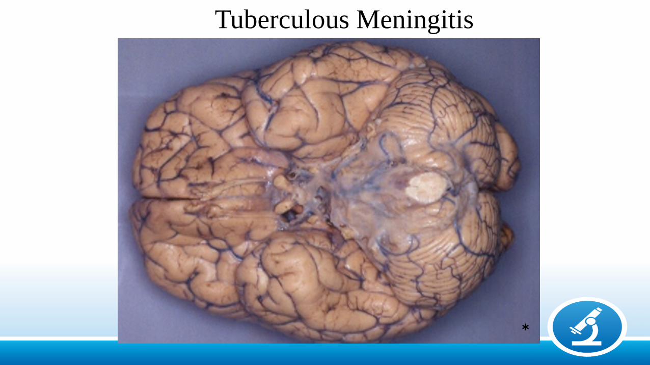
Tuberculous Meningitis
*
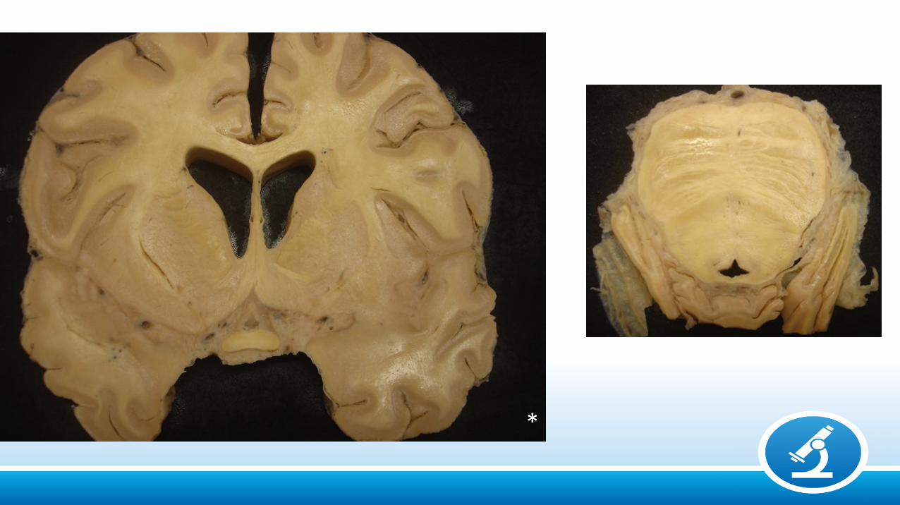
*
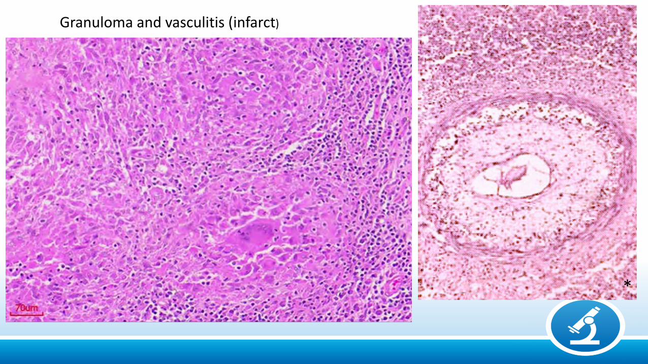
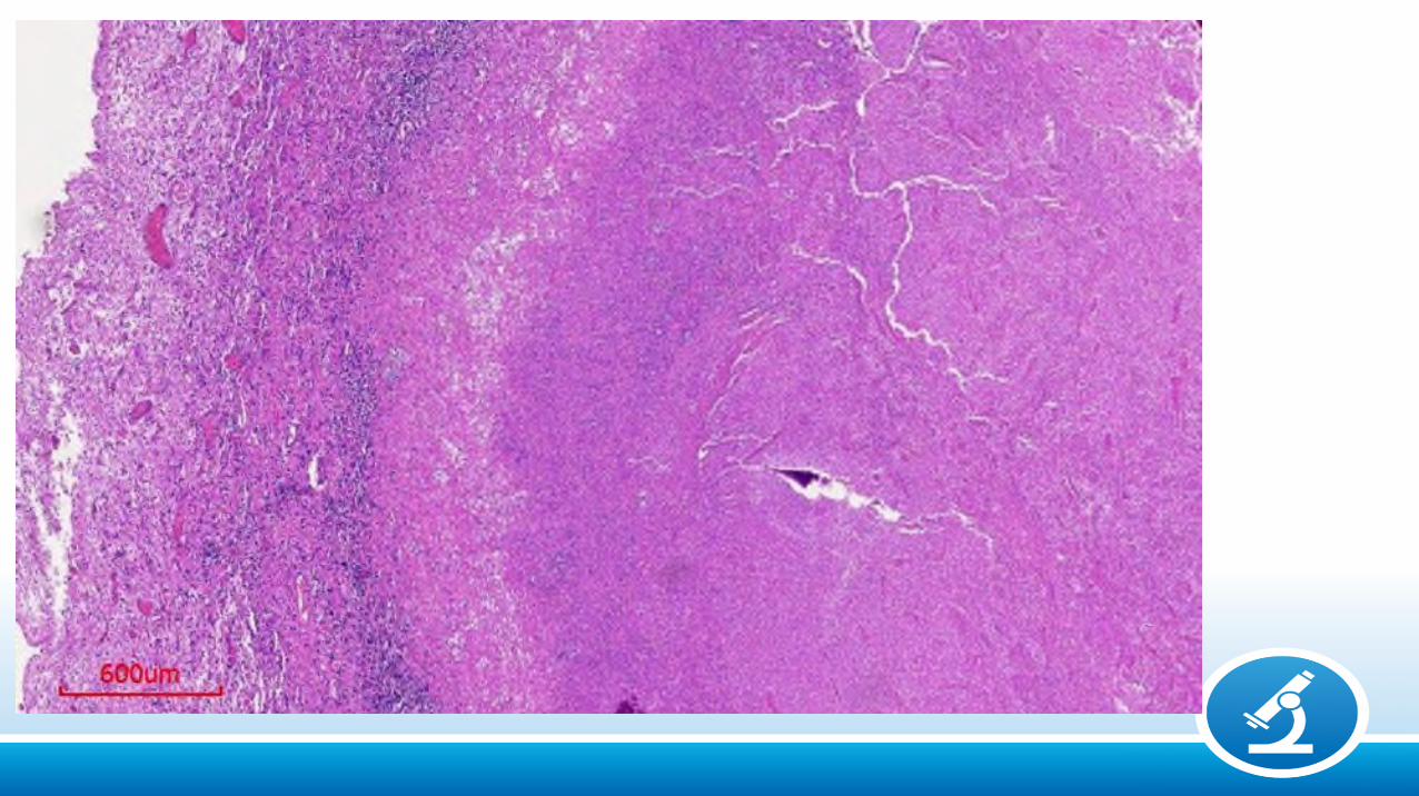
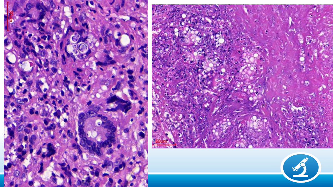
Granuloma and vasculitis (infarct)
*
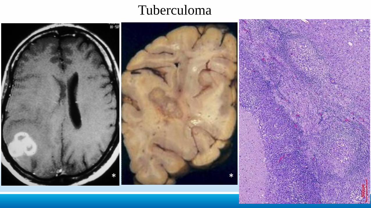
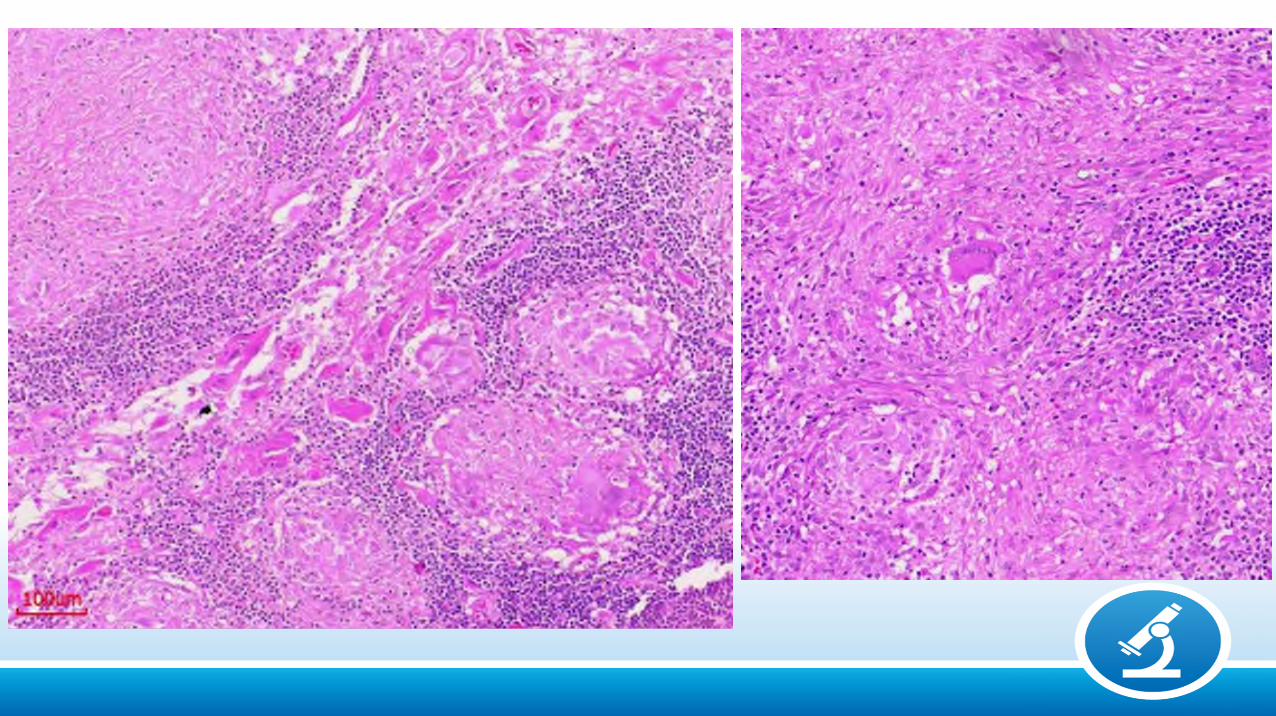
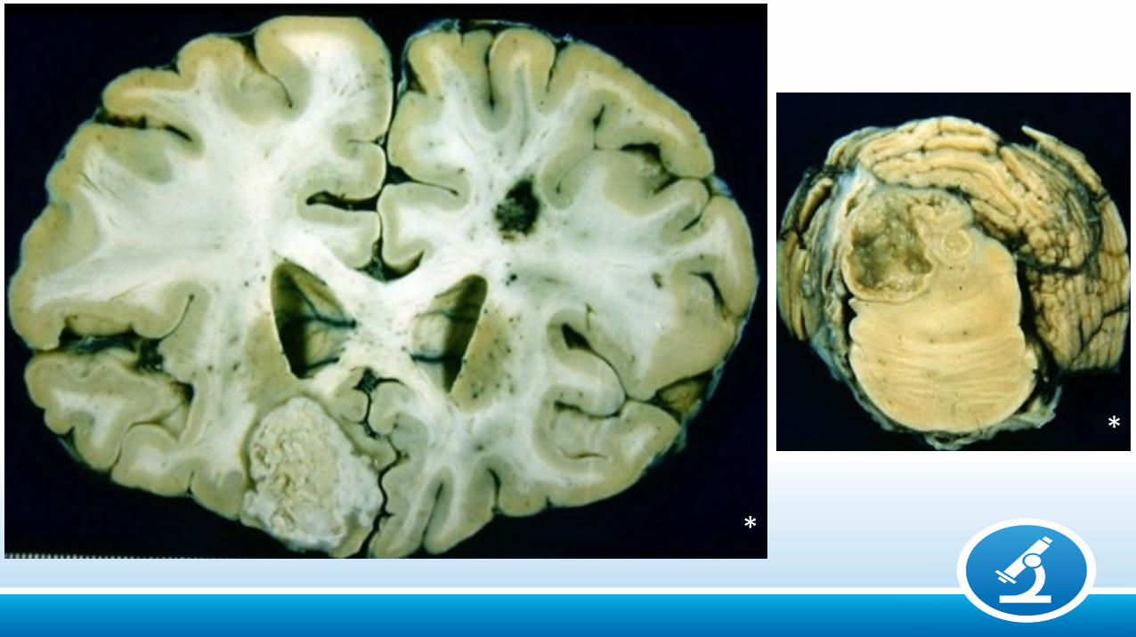
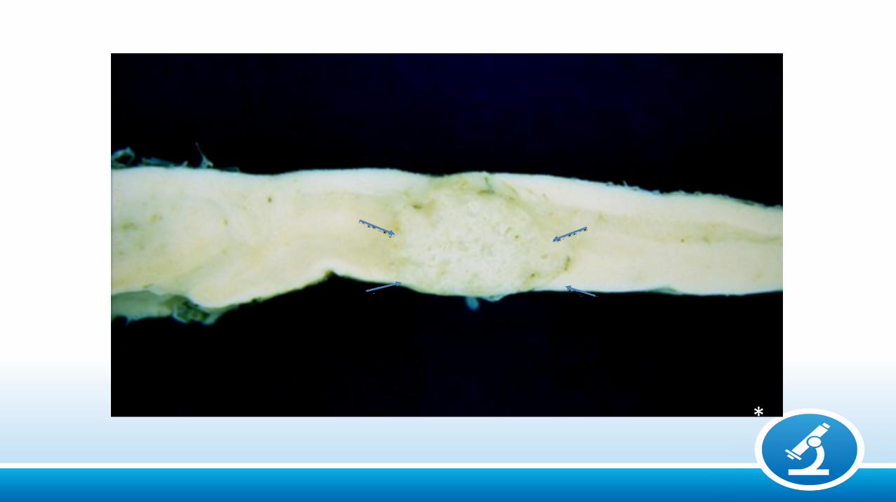
Tuberculoma
***
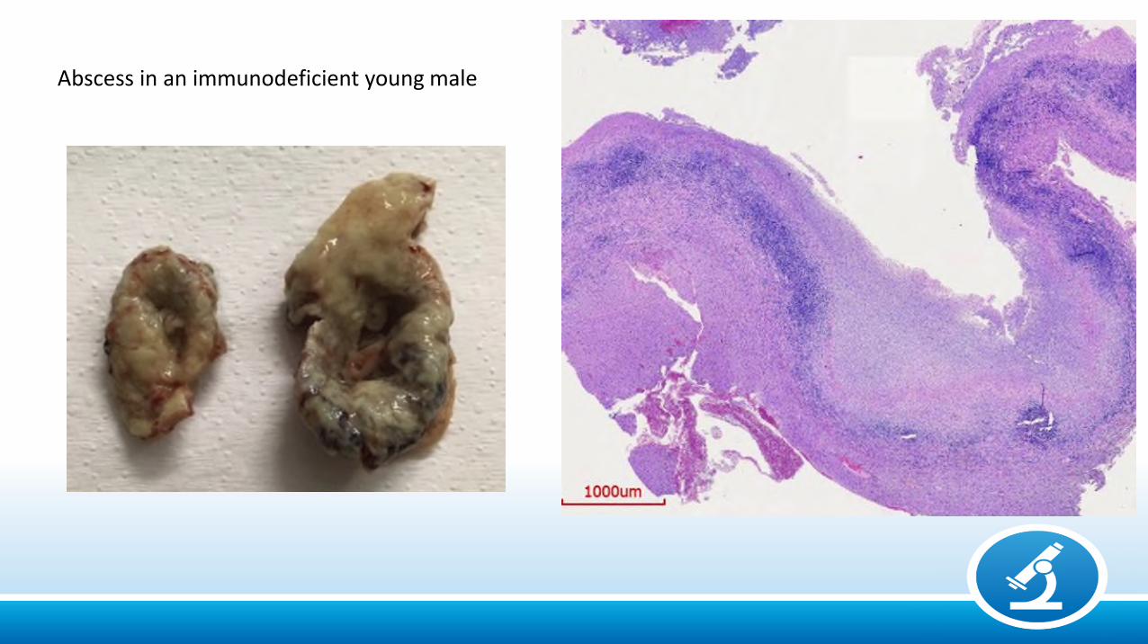
Abscess in an immunodeficient young male
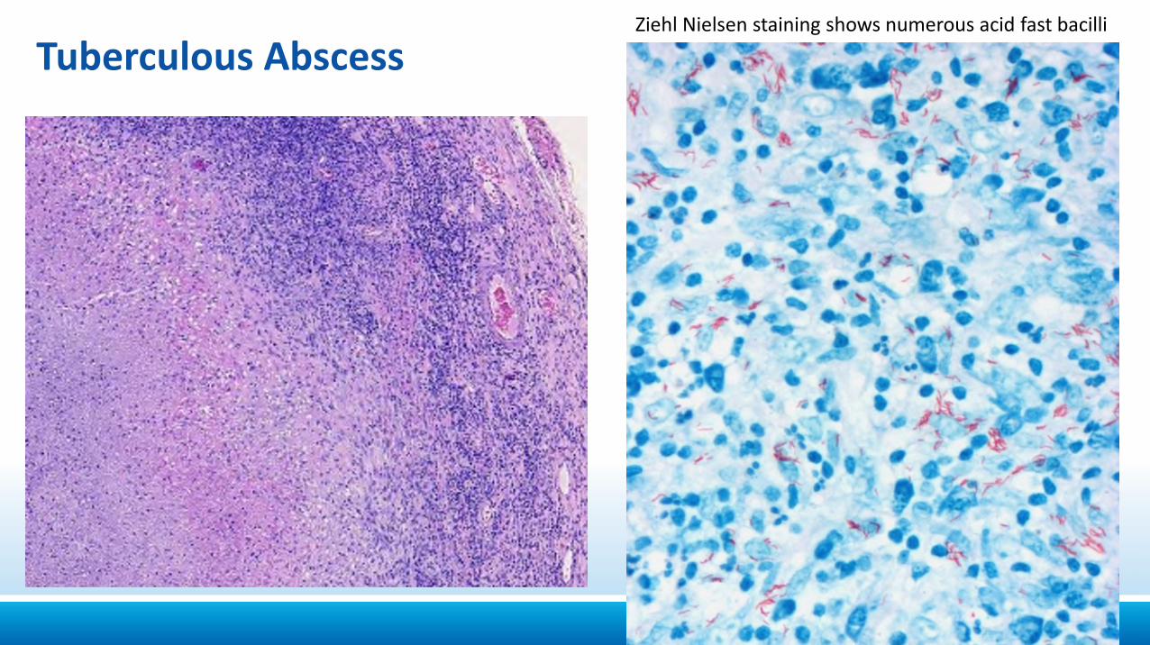
Tuberculous AbscessZiehl Nielsen staining shows numerous acid fast bacilli
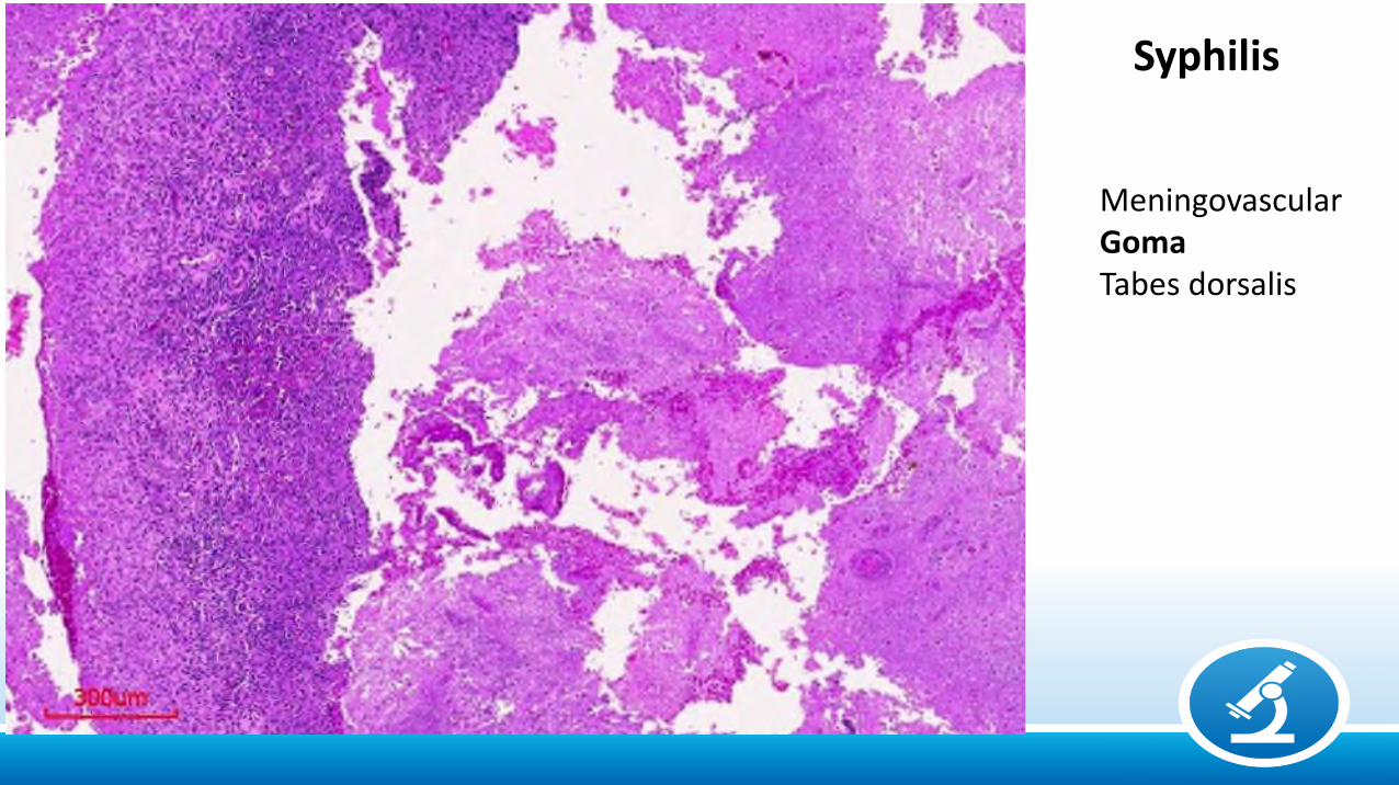
Syphilis
MeningovascularGomaTabes dorsalis

Etiology
Bacterial: Pyogenic (Gram + / -), Tuberculosis, Syphilis
Fungal: Cryptococcosis, Histoplasmosis, Mucormycosis,
Aspergillosis, Paracoccidioidomycosis.
Parasitic: Protozoa: Toxoplasmosis, Amebiasis, Malaria, Trypanosomiases
Helminths: Cestodes: Cysticercosis, HydatidosisNematodes: Strongyloidiasis
Trematodes: Schistosomiasis
Viral: Arboviroses (Dengue, Zika), CMV, HIV, Herpes, HTLV1,
Measles, Poliomyelitis, PML, Rabies.

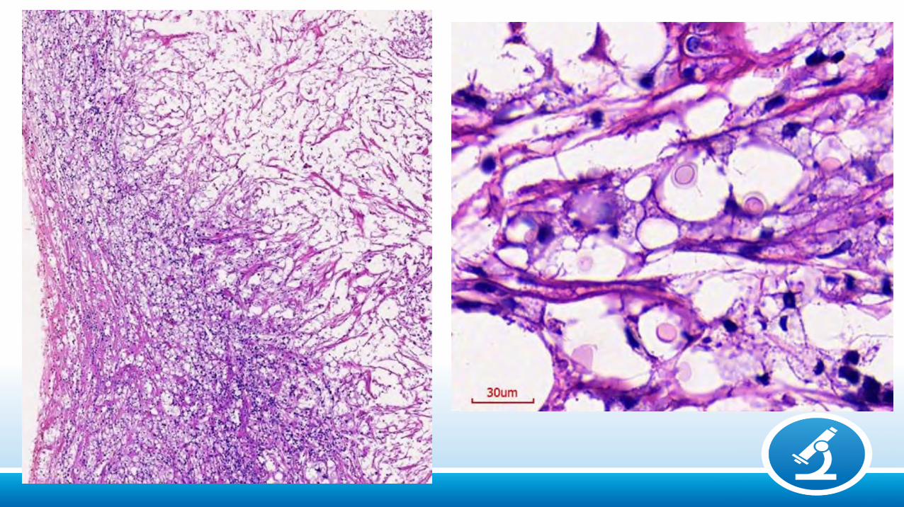
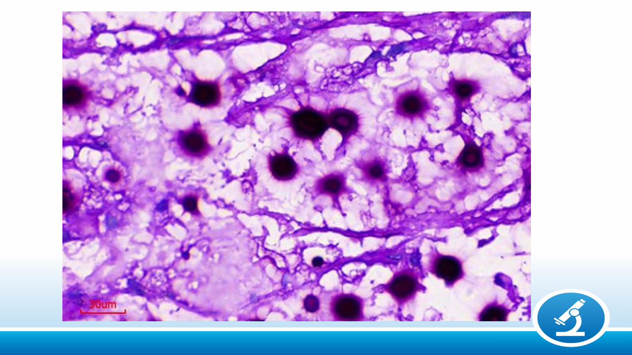
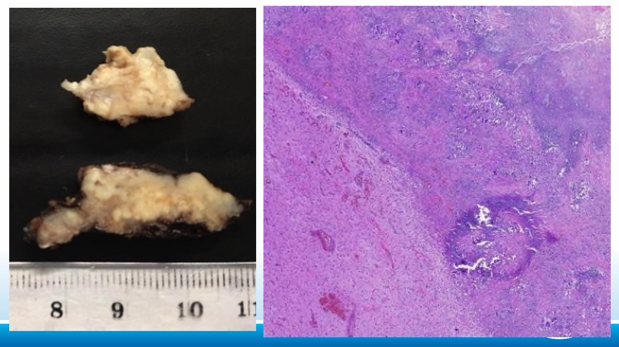
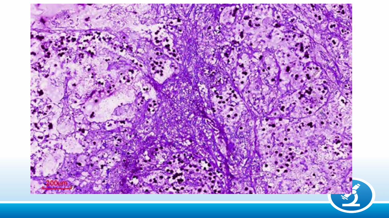
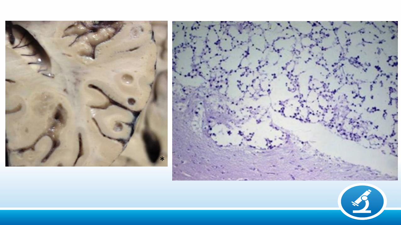
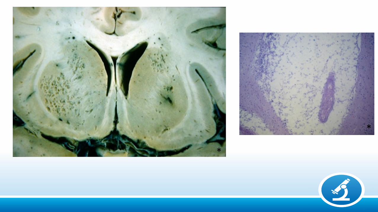
Cryptococcosis
Meningitis (Immunodeficient)
*
*
*
*
*
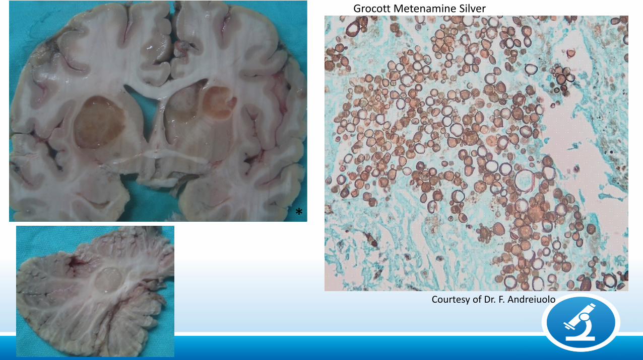
Courtesy of Dr. F. Andreiuolo
Grocott Metenamine Silver
*
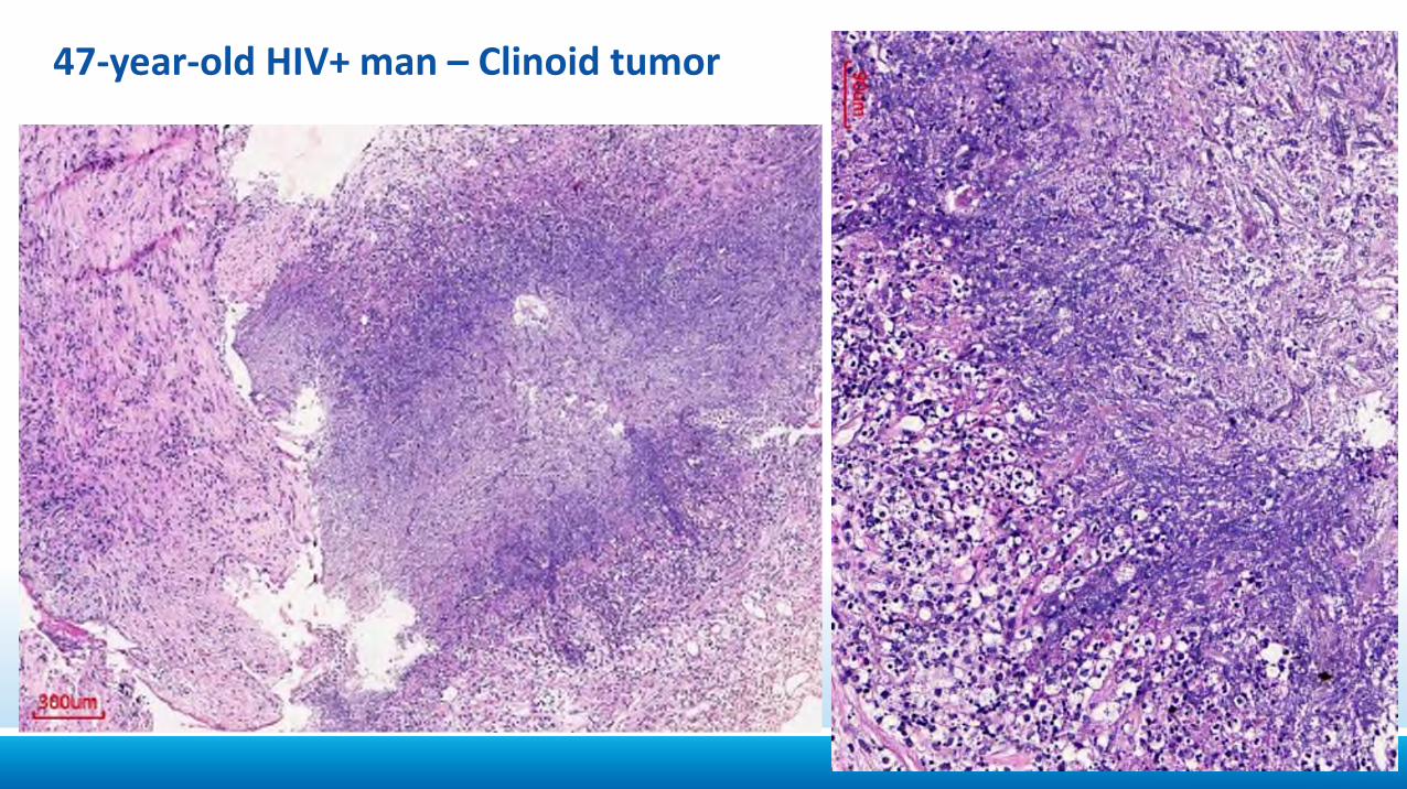
47-year-old HIV+ man – Clinoid tumor
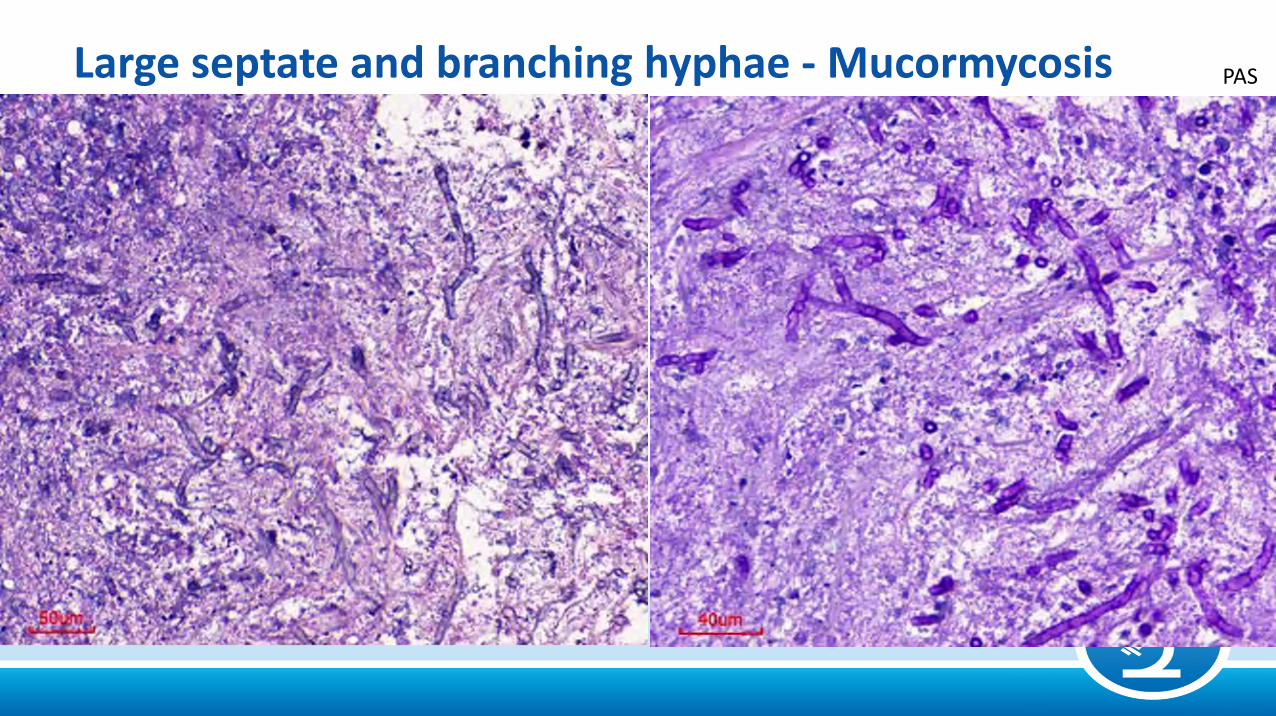
Large septate and branching hyphae - Mucormycosis PAS

Histoplasmosis• Histoplasma capsulatum• Inhaled in infected dust contaminated by
chicken, bird or bat excreta • Lungs are primarily infected, but also
mouth, digestive tract and skin• Frequent involvement of lymphnodes,
spleen, liver• CNS infection is rare
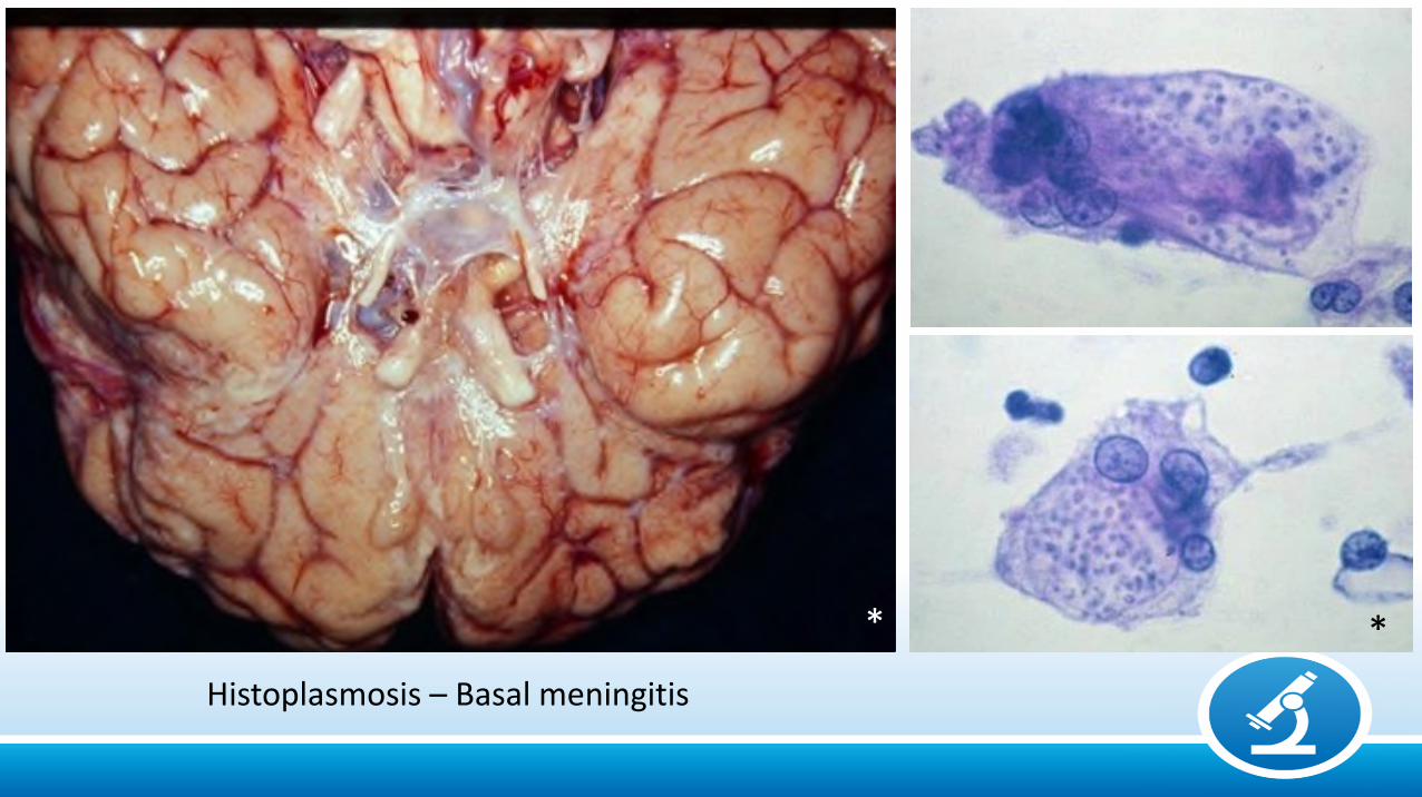
Histoplasmosis – Basal meningitis
**
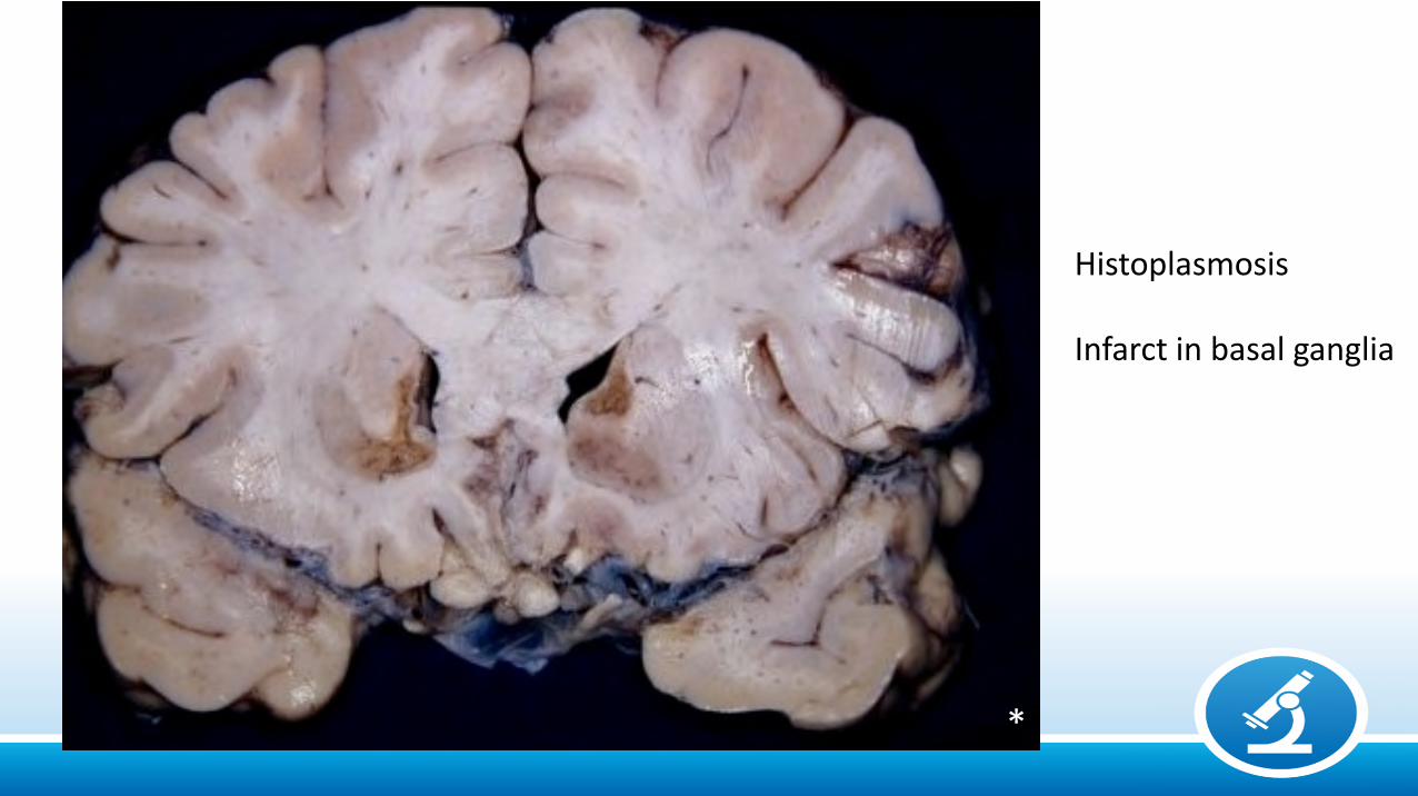
Histoplasmosis
Infarct in basal ganglia
*
Paracoccidioidomycosis(South American blastomycosis)
• Paracoccidioidis brasiliensis
• Organisms found in soil and vegetation
• Frequent in Brazil, Venezuela and Colombia
• Preferential sites: lungs, oral and nasal mucosae, lymphnodes
• CNS involvement is not frequent
*
*
*
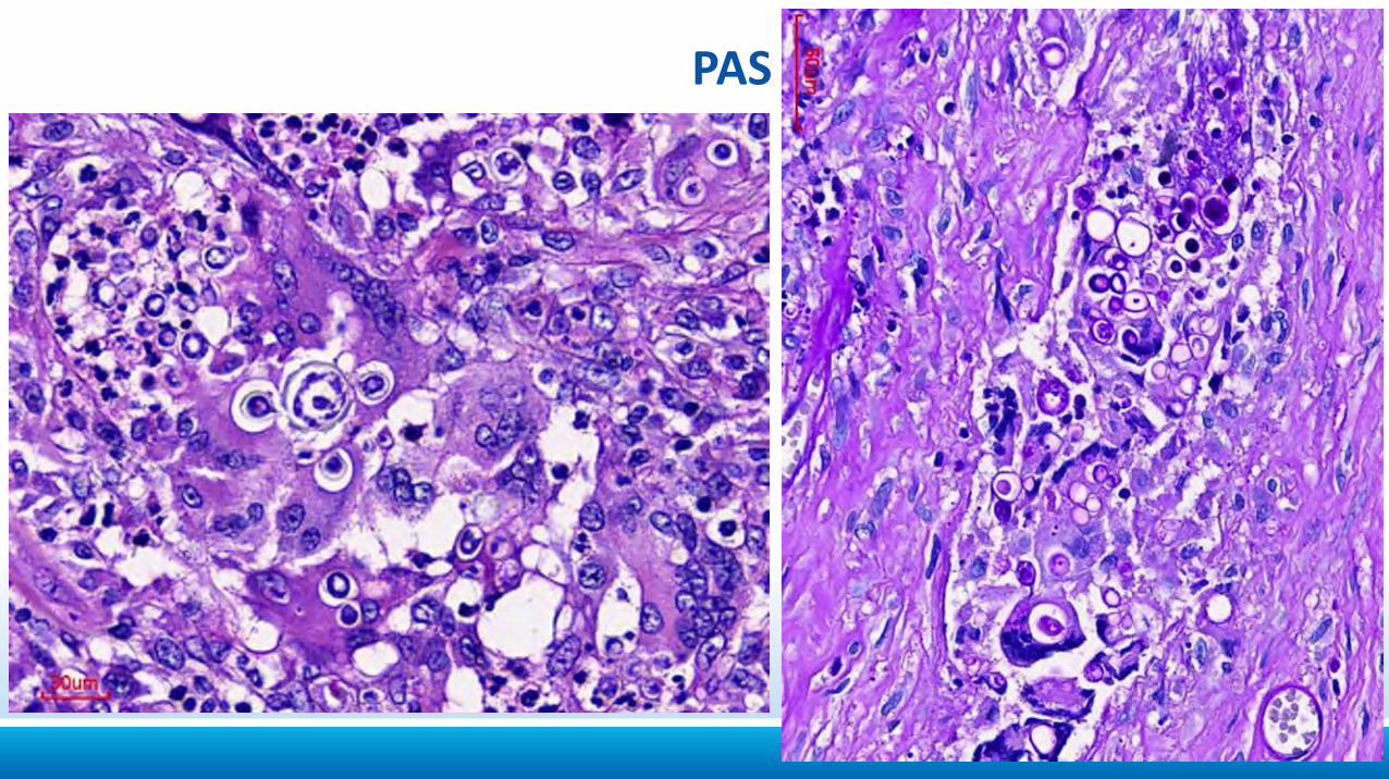
PAS
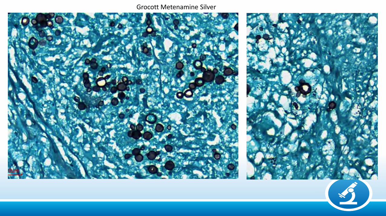
Grocott Metenamine Silver
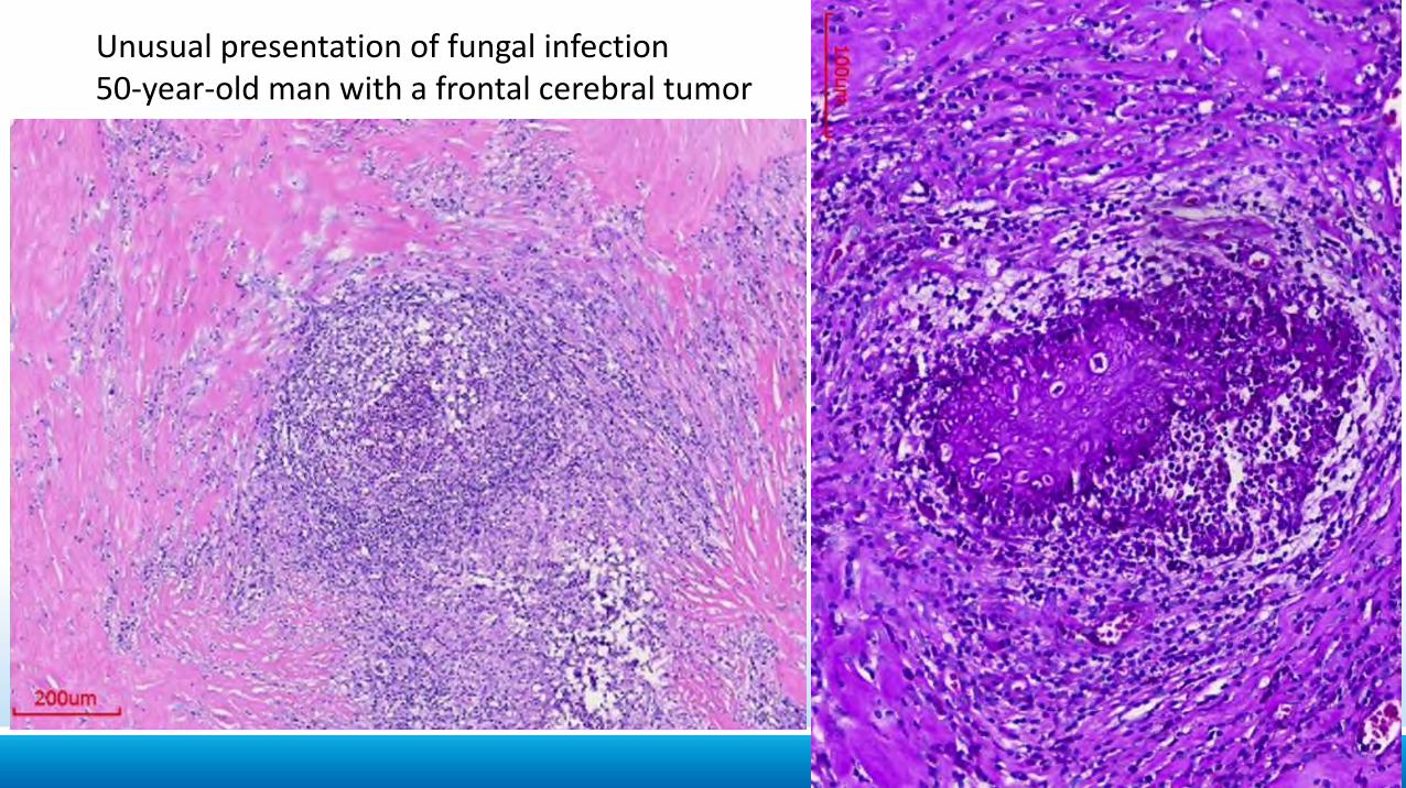
Unusual presentation of fungal infection50-year-old man with a frontal cerebral tumor
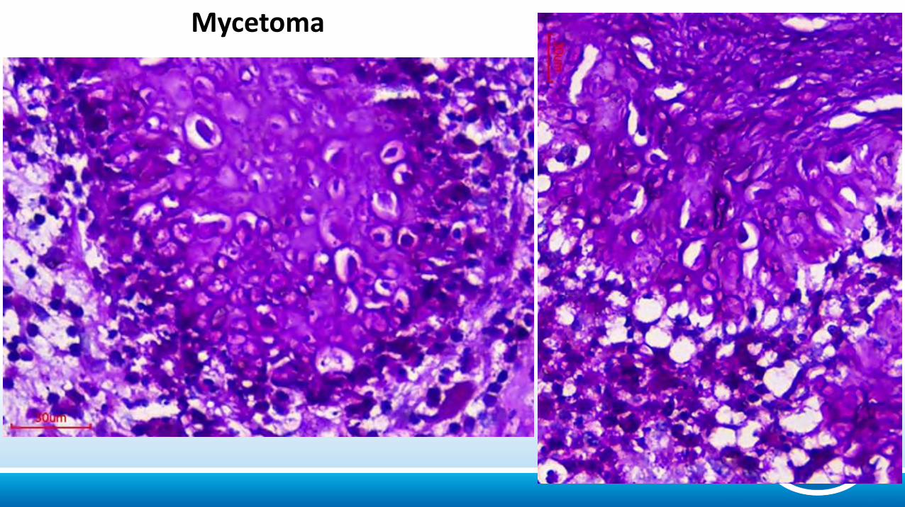
Mycetoma
Etiology
Bacterial: Pyogenic (Gram + / -), Tuberculosis, Syphilis
Fungal: Cryptococcosis, Histoplasmosis, Mucormycosis,
Aspergillosis, Paracoccidioidomycosis
Parasitic: Protozoa: Toxoplasmosis, Trypanosomiases, Malaria, Amebiasis
Helminths: Cestodes: Cysticercosis, HydatidosisNematodes: Strongyloidiasis
Trematodes: Schistosomiasis
Viral: Arboviroses (Dengue, Zika), Herpes, CMV, HIV, HTLV1,
Measles, Poliomyelitis, PML, Rabies.
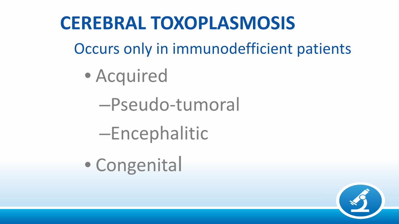
CEREBRAL TOXOPLASMOSISOccurs only in immunodefficient patients
• Acquired
–Pseudo-tumoral
–Encephalitic
• Congenital
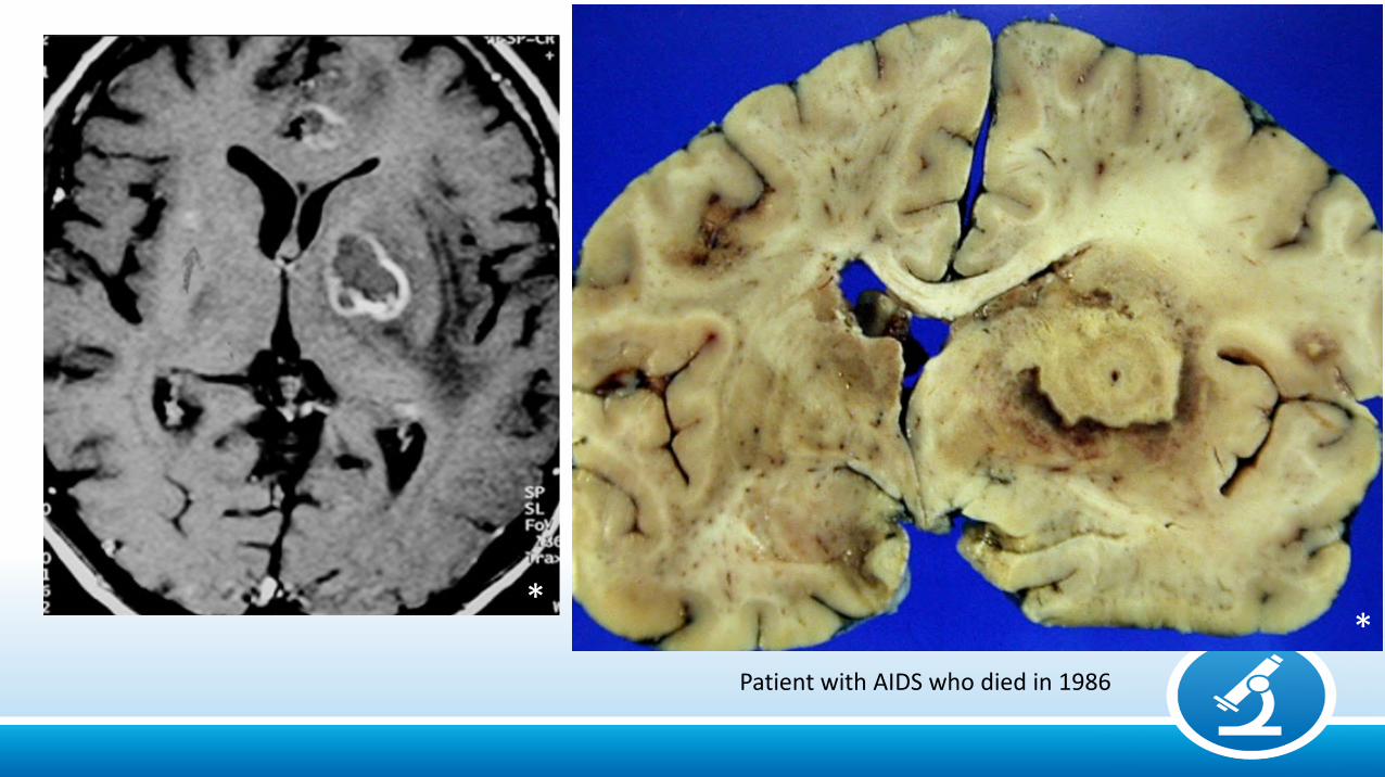
Patient with AIDS who died in 1986
**
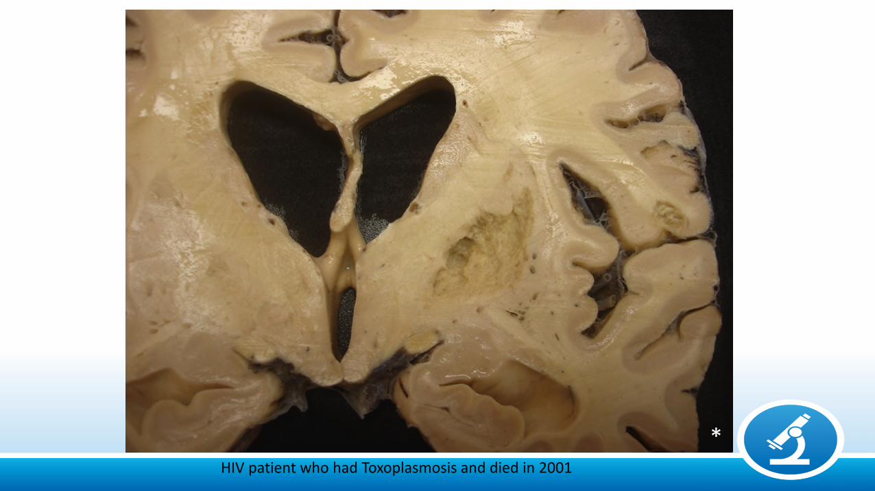
HIV patient who had Toxoplasmosis and died in 2001
*
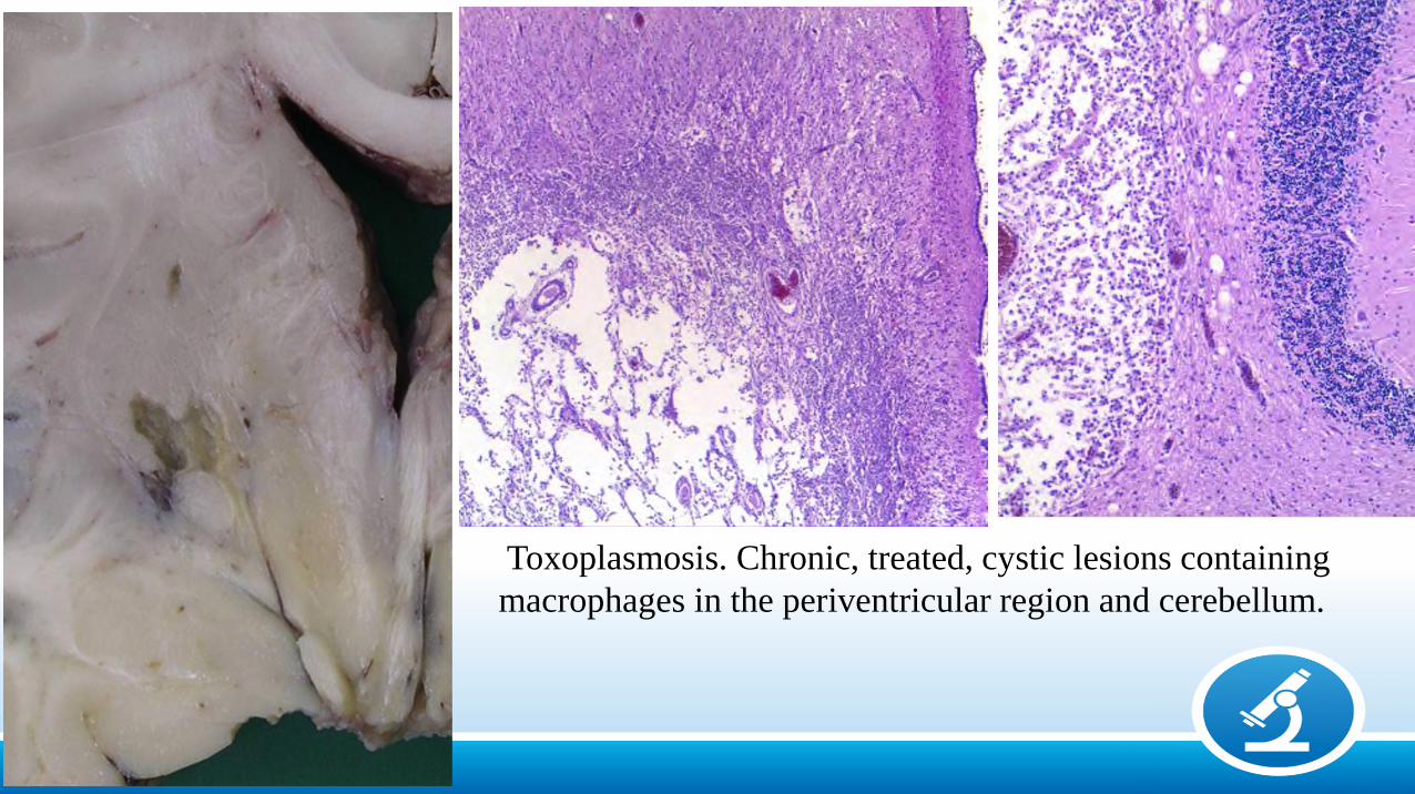
Toxoplasmosis. Chronic, treated, cystic lesions containing macrophages in the periventricular region and cerebellum.
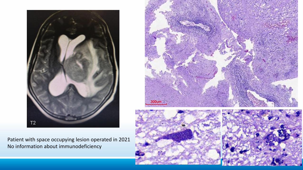
Patient with space occupying lesion operated in 2021No information about immunodeficiency
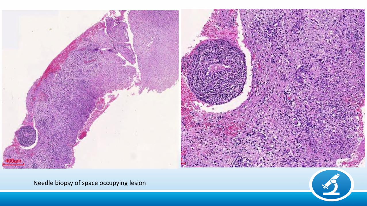
Needle biopsy of space occupying lesion
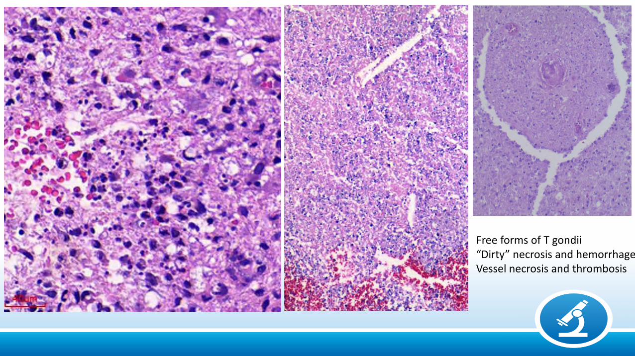
Free forms of T gondii“Dirty” necrosis and hemorrhageVessel necrosis and thrombosis
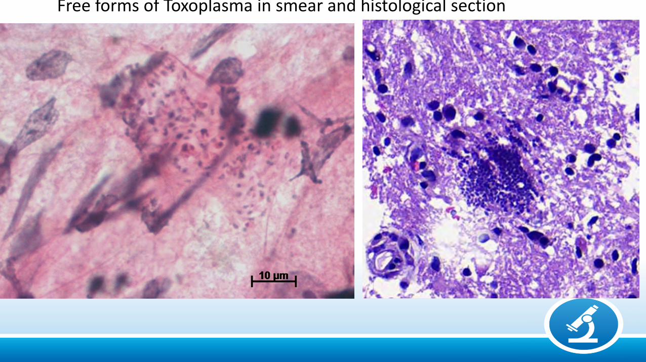
Free forms of Toxoplasma in smear and histological section
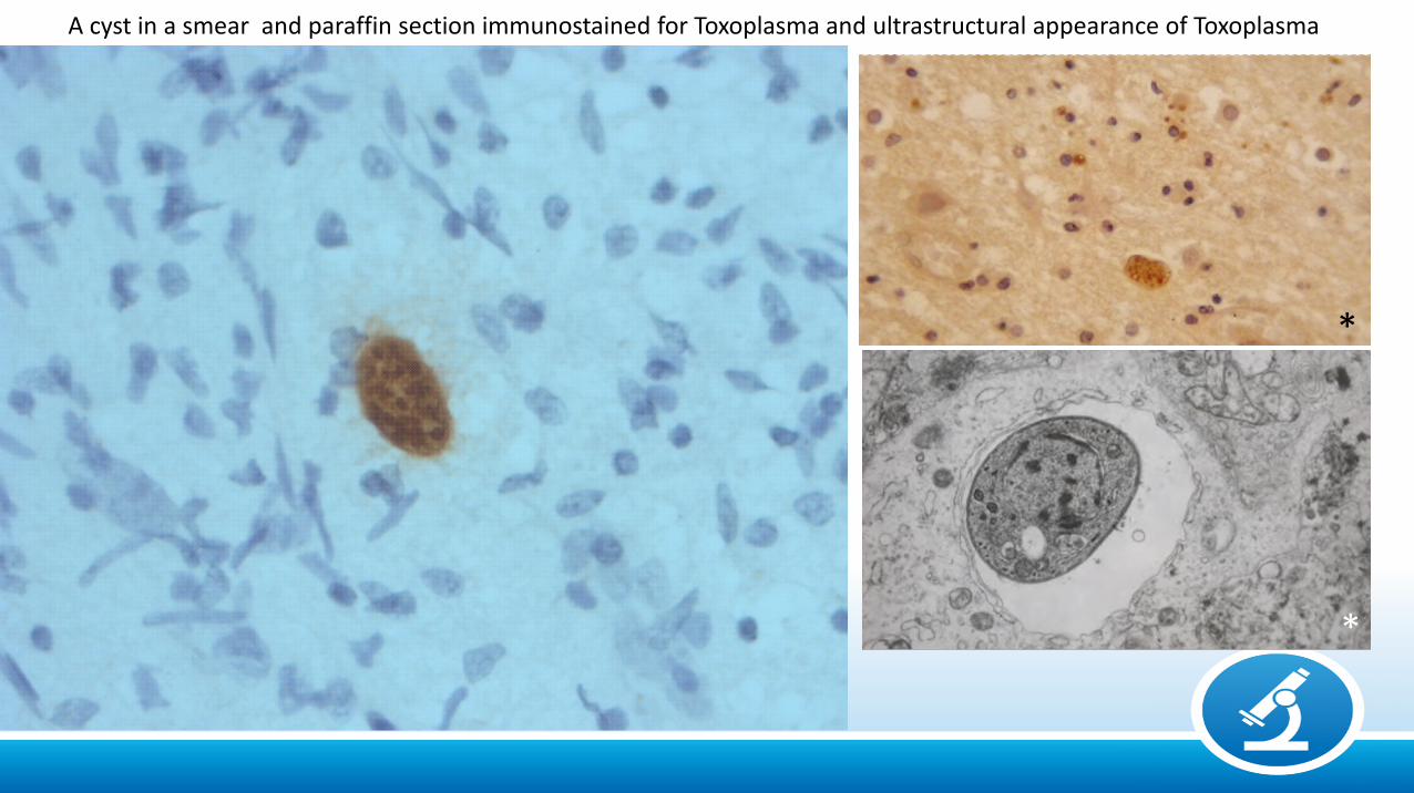
A cyst in a smear and paraffin section immunostained for Toxoplasma and ultrastructural appearance of Toxoplasma
*
*
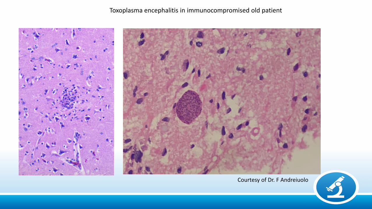
Toxoplasma encephalitis in immunocompromised old patient
Courtesy of Dr. F Andreiuolo
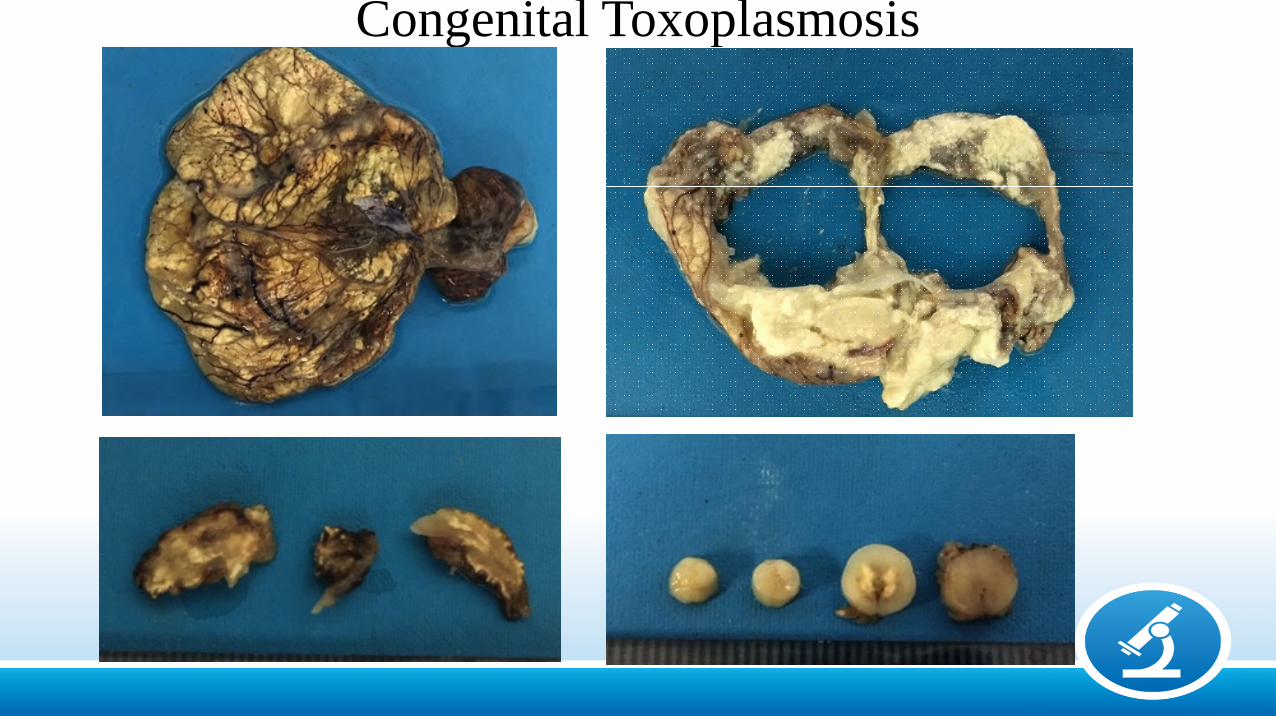
Congenital Toxoplasmosis
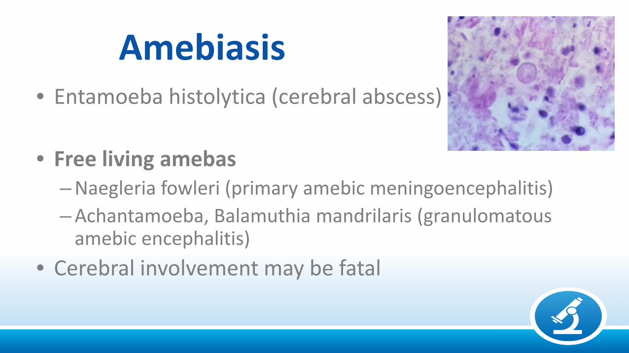
Amebiasis• Entamoeba histolytica (cerebral abscess)
• Free living amebas– Naegleria fowleri (primary amebic meningoencephalitis)
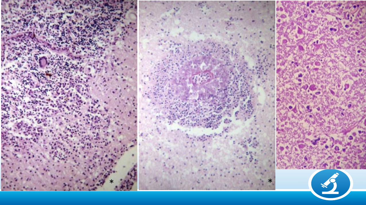
– Achantamoeba, Balamuthia mandrilaris (granulomatousamebic encephalitis)
• Cerebral involvement may be fatal
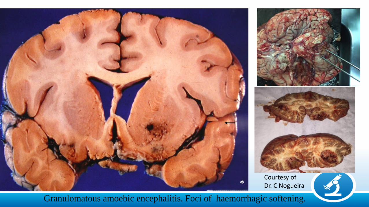
Granulomatous amoebic encephalitis. Foci of haemorrhagic softening.
Courtesy ofDr. C Nogueira*
* *
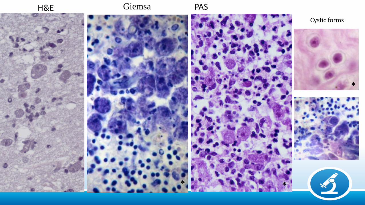
Giemsa PAS
Cystic forms
H&E
* *
*
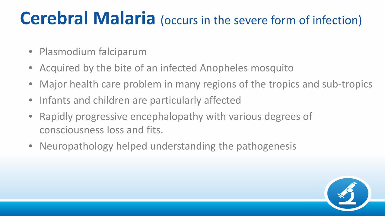
Cerebral Malaria (occurs in the severe form of infection)
• Plasmodium falciparum
• Acquired by the bite of an infected Anopheles mosquito
• Major health care problem in many regions of the tropics and sub-tropics
• Infants and children are particularly affected
• Rapidly progressive encephalopathy with various degrees ofconsciousness loss and fits.
• Neuropathology helped understanding the pathogenesis
AM
RJ

MALARIA
*
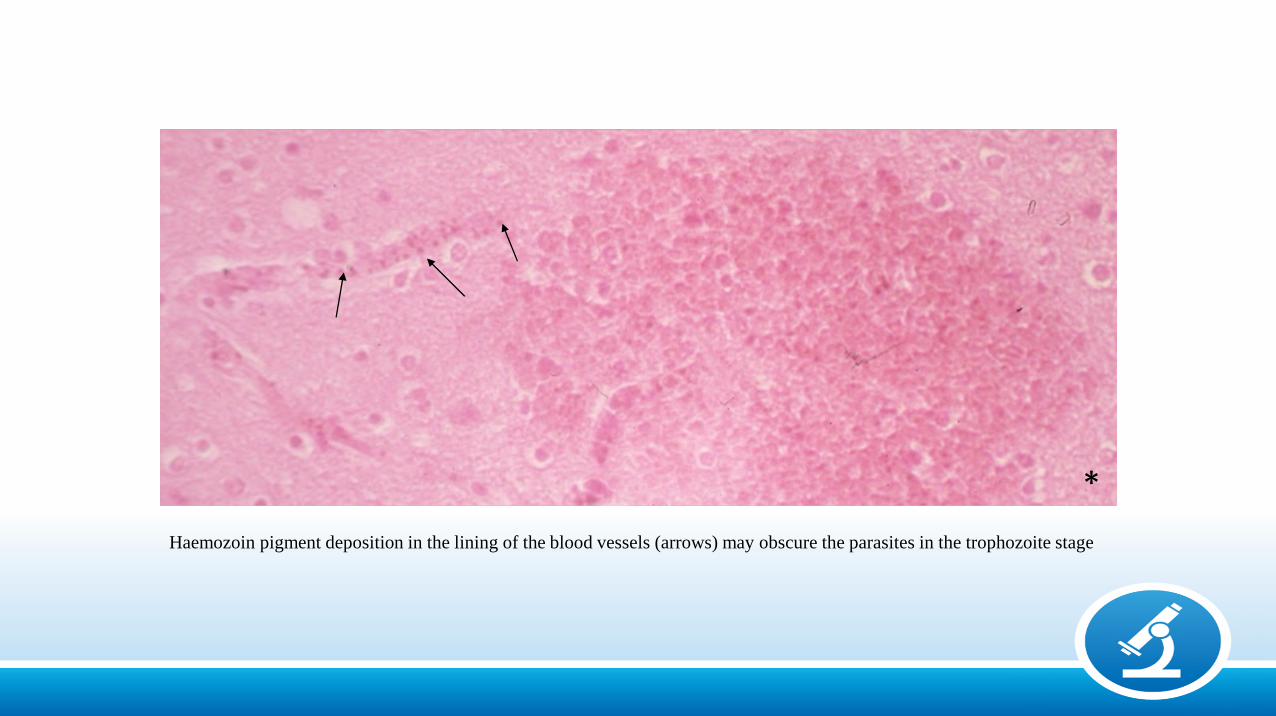
Haemozoin pigment deposition in the lining of the blood vessels (arrows) may obscure the parasites in the trophozoite stage
*
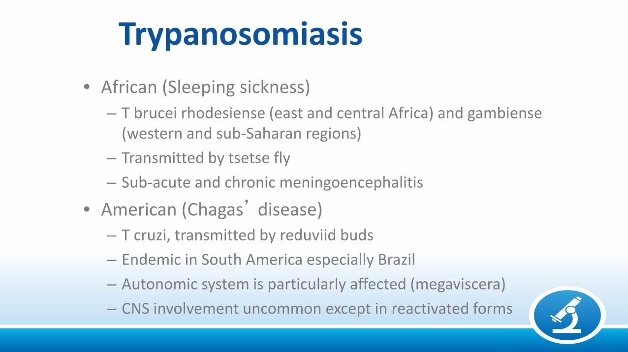
Trypanosomiasis
• African (Sleeping sickness)
– T brucei rhodesiense (east and central Africa) and gambiense (western and sub-Saharan regions)
– Transmitted by tsetse fly
– Sub-acute and chronic meningoencephalitis
• American (Chagas’ disease)
– T cruzi, transmitted by reduviid buds
– Endemic in South America especially Brazil
– Autonomic system is particularly affected (megaviscera)
– CNS involvement uncommon except in reactivated forms
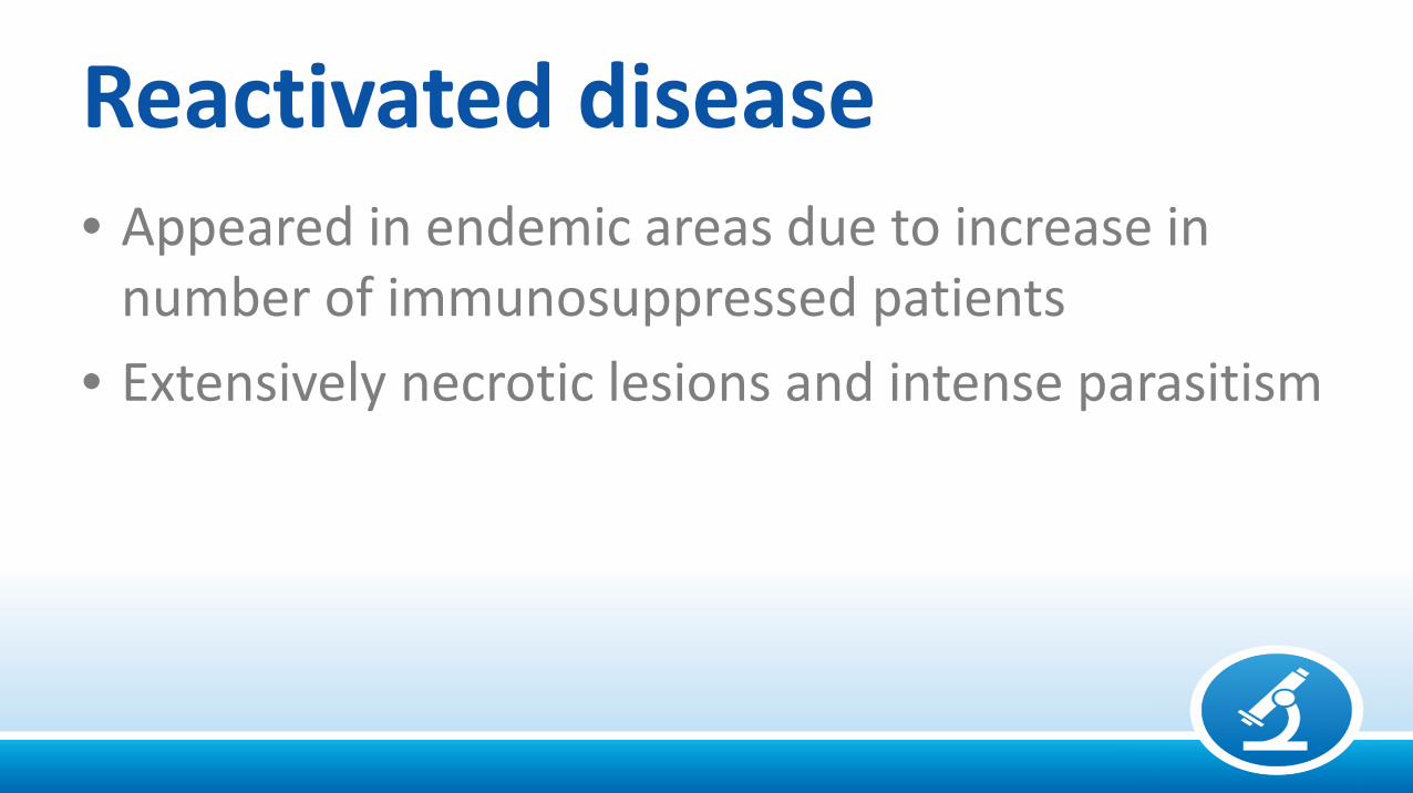
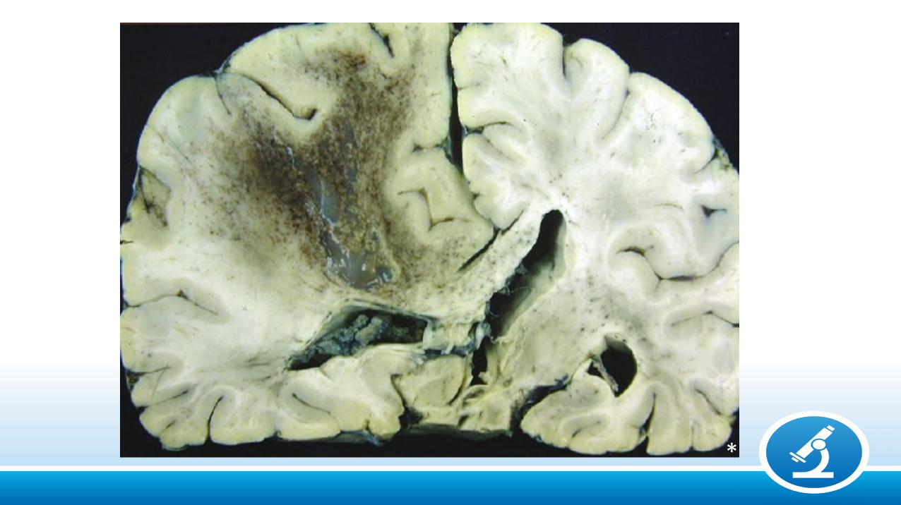
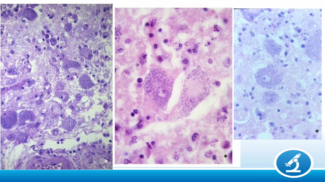
Reactivated disease• Appeared in endemic areas due to increase in
number of immunosuppressed patients
• Extensively necrotic lesions and intense parasitism
*
*
**
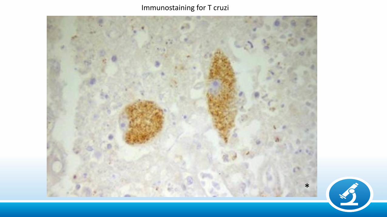
Immunostaining for T cruzi
*
Etiology
Bacterial: Pyogenic (Gram + / -), Tuberculosis, Syphilis
Fungal: Cryptococcosis, Histoplasmosis, Mucormycosis,
Aspergillosis, Paracoccidioidomycosis
Parasitic: Protozoa: Toxoplasmosis, Trypanosomiases, Malaria, Amebiasis
Helminths: Cestodes: Cysticercosis, HydatidosisNematodes: Strongyloidiasis
Trematodes: Schistosomiasis
Viral: Arboviroses (Dengue, Zika), Herpes, CMV, HIV, HTLV1,
Measles, Poliomyelitis, PML, Rabies.
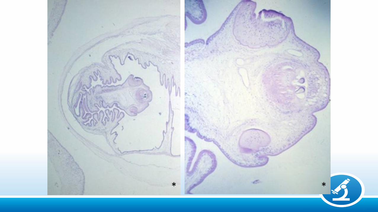
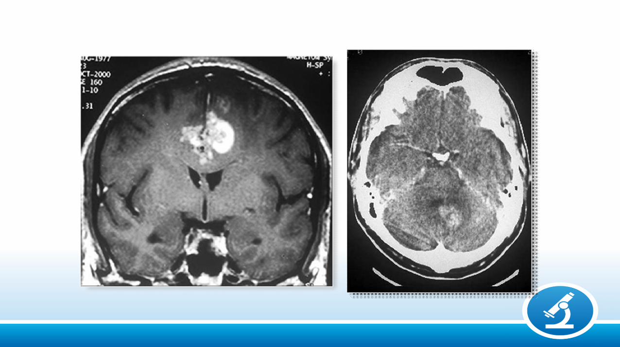
Neurocysticercosis• Cysticercus cellulosae, the larval form of Taenia solium.
• Usually found in pork (the intermediate host)
• Humans are intermediate hosts after ingesting the ova of T. solium (usually in vegetables).
• Ova develop into larvae that penetrate intestinal wall, invade lymphatic and veins, disseminate to skeletal muscle and CNS.
• Clinical features depend on number and location of the cysts
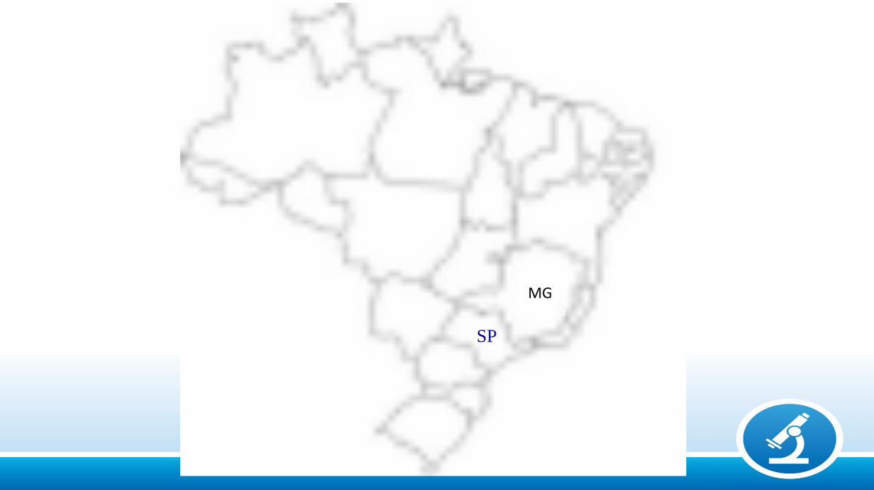
MGSP
MG
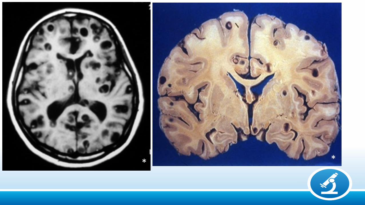
* *
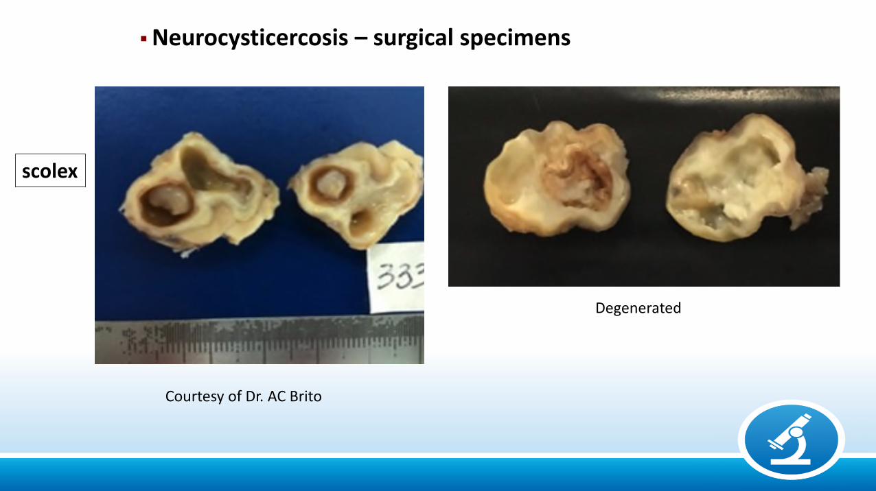
scolex
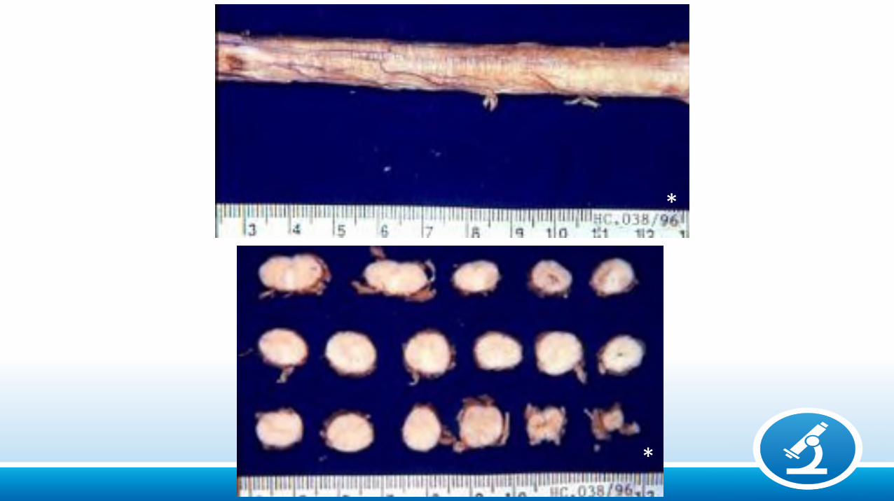
Neurocysticercosis – surgical specimens
Degenerated
Courtesy of Dr. AC Brito
**

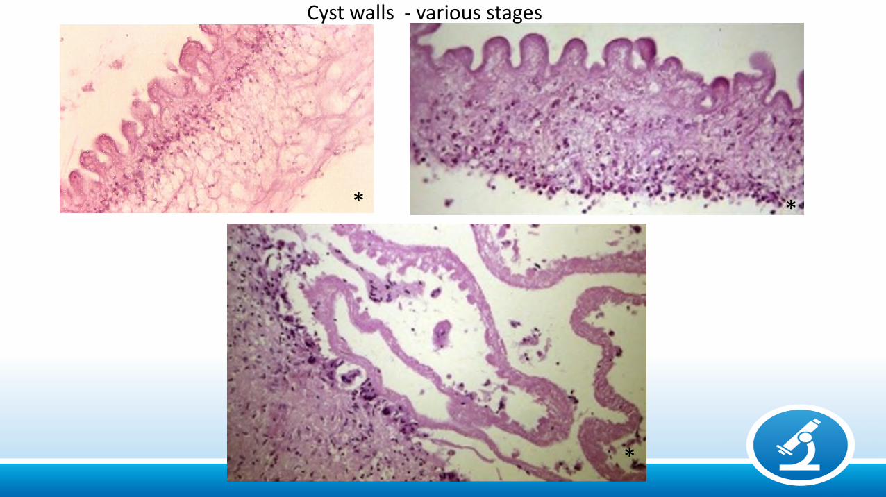
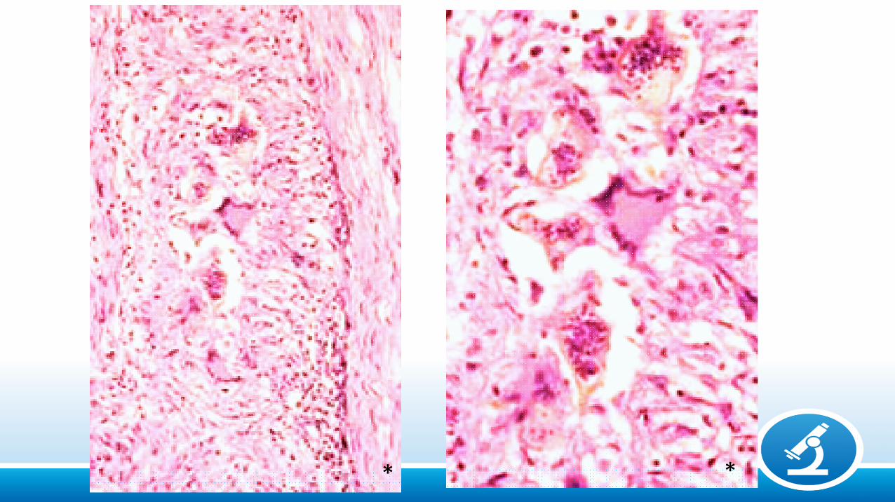
Cyst walls - various stages
**
*
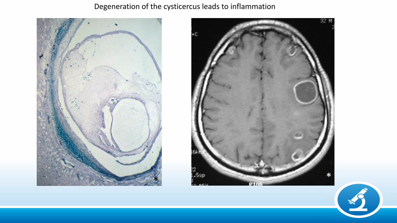
Degeneration of the cysticercus leads to inflammation
* *
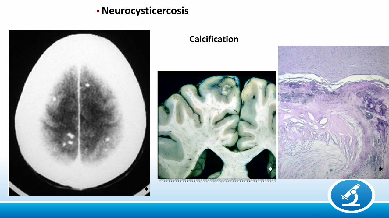
Calcification
Neurocysticercosis
**
*

Racemose Cysticerci
*
*
**
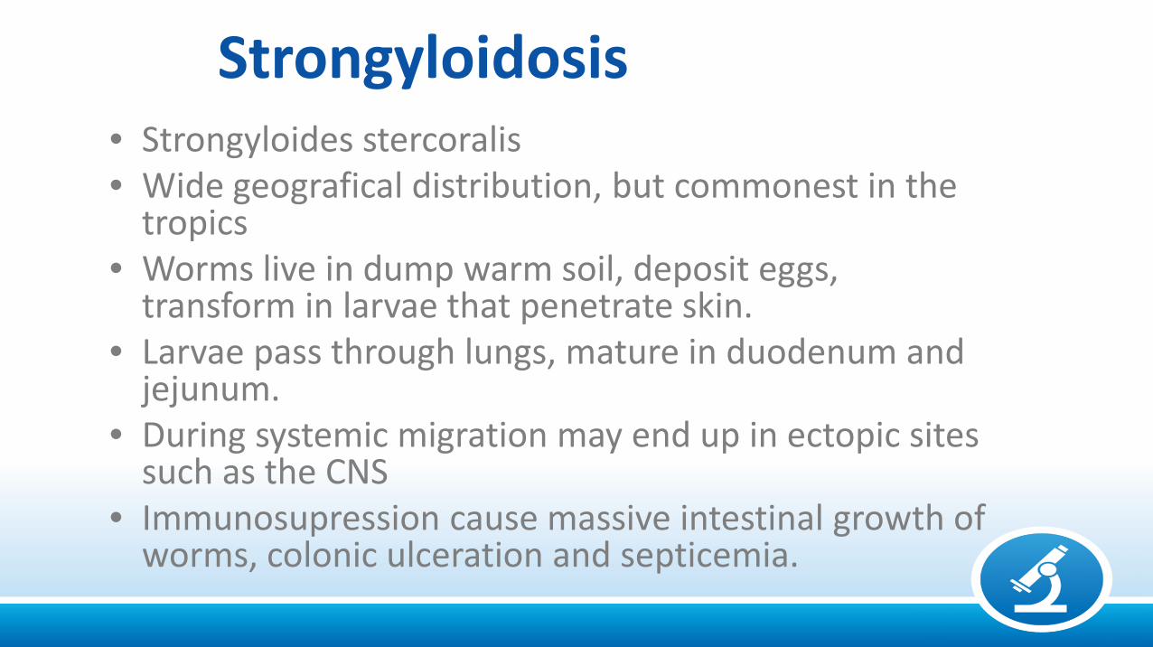
Strongyloidosis• Strongyloides stercoralis• Wide geografical distribution, but commonest in the
tropics• Worms live in dump warm soil, deposit eggs,
transform in larvae that penetrate skin.• Larvae pass through lungs, mature in duodenum and
jejunum.• During systemic migration may end up in ectopic sites
such as the CNS• Immunosupression cause massive intestinal growth of
worms, colonic ulceration and septicemia.
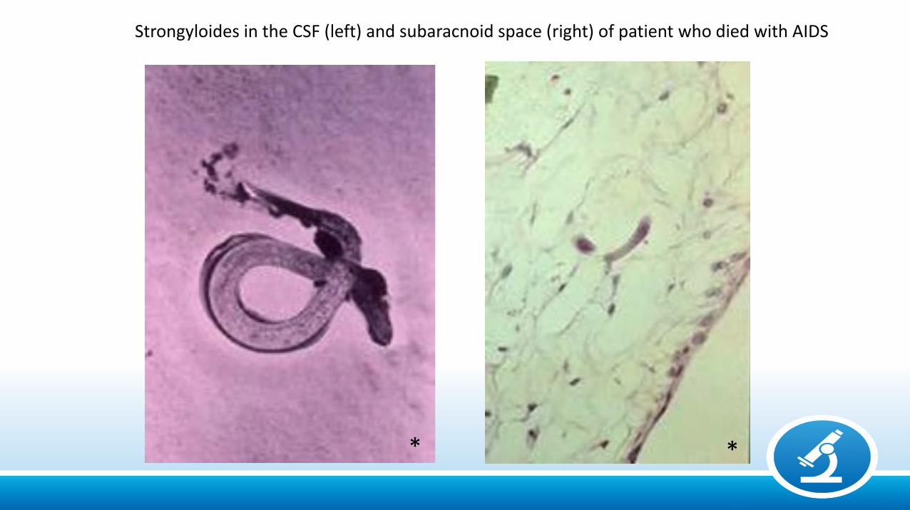
Strongyloides in the CSF (left) and subaracnoid space (right) of patient who died with AIDS
**
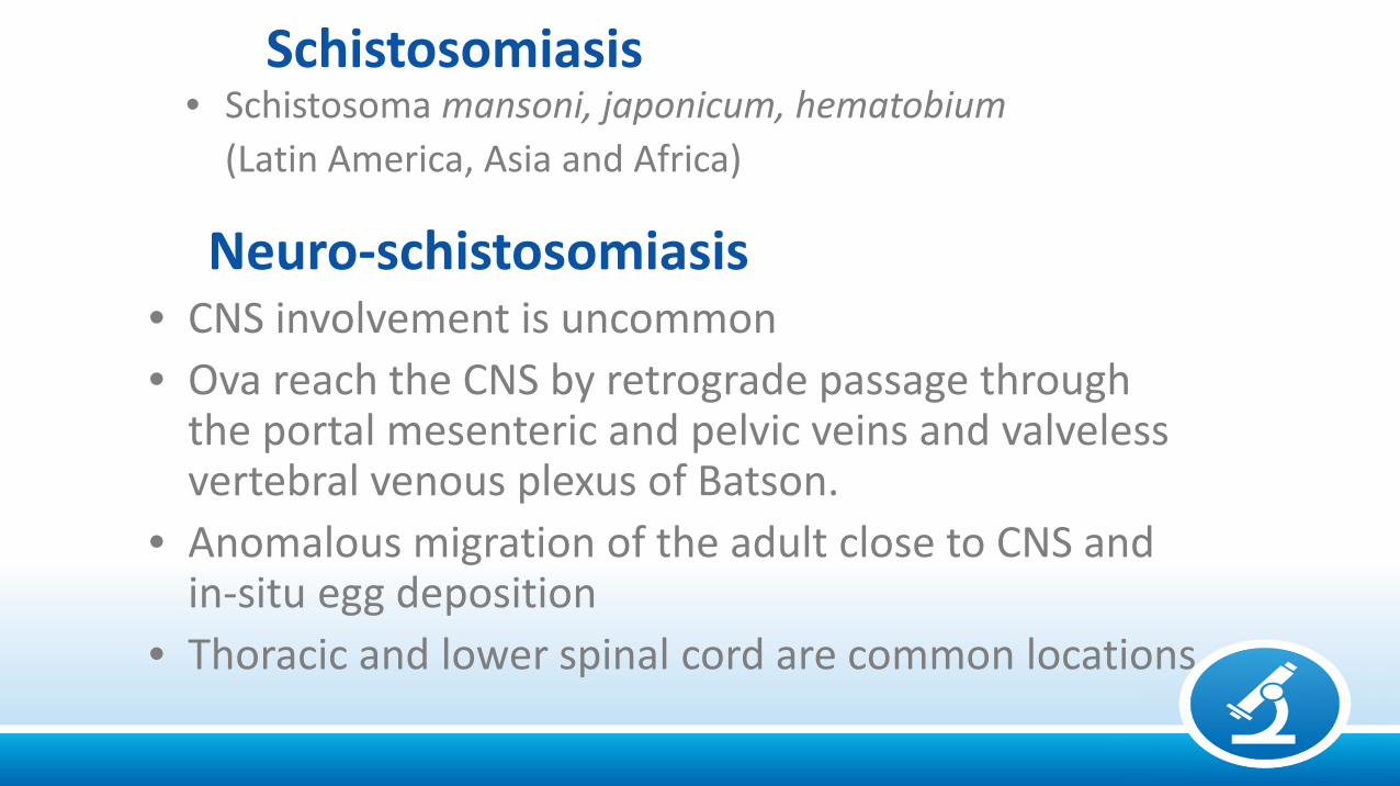
Schistosomiasis• Schistosoma mansoni, japonicum, hematobium
(Latin America, Asia and Africa)
• CNS involvement is uncommon
• Ova reach the CNS by retrograde passage throughthe portal mesenteric and pelvic veins and valvelessvertebral venous plexus of Batson.
• Anomalous migration of the adult close to CNS andin-situ egg deposition
• Thoracic and lower spinal cord are common locations
Neuro-schistosomiasis
*
*
**
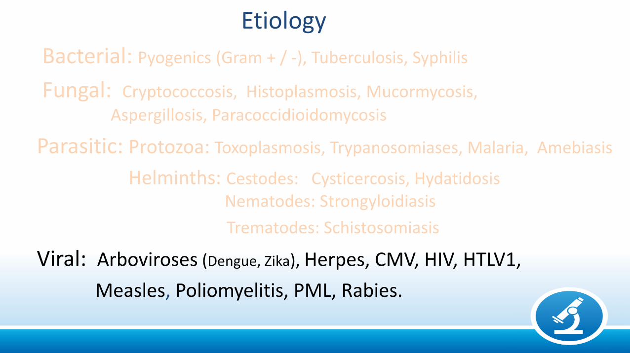
Etiology
Bacterial: Pyogenics (Gram + / -), Tuberculosis, Syphilis
Fungal: Cryptococcosis, Histoplasmosis, Mucormycosis,
Aspergillosis, Paracoccidioidomycosis
Parasitic: Protozoa: Toxoplasmosis, Trypanosomiases, Malaria, Amebiasis
Helminths: Cestodes: Cysticercosis, HydatidosisNematodes: Strongyloidiasis
Trematodes: Schistosomiasis
Viral: Arboviroses (Dengue, Zika), Herpes, CMV, HIV, HTLV1,
Measles, Poliomyelitis, PML, Rabies.
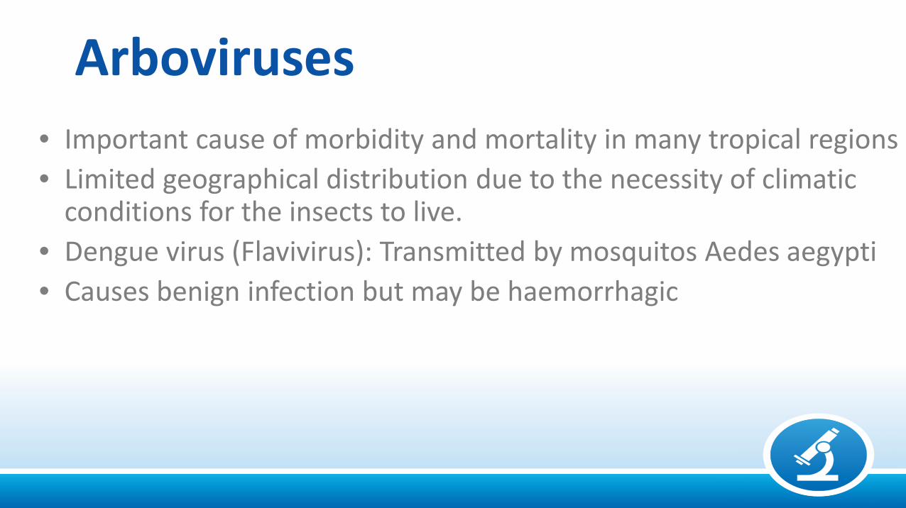
Arboviruses
• Important cause of morbidity and mortality in many tropical regions
• Limited geographical distribution due to the necessity of climaticconditions for the insects to live.
• Dengue virus (Flavivirus): Transmitted by mosquitos Aedes aegypti
• Causes benign infection but may be haemorrhagic
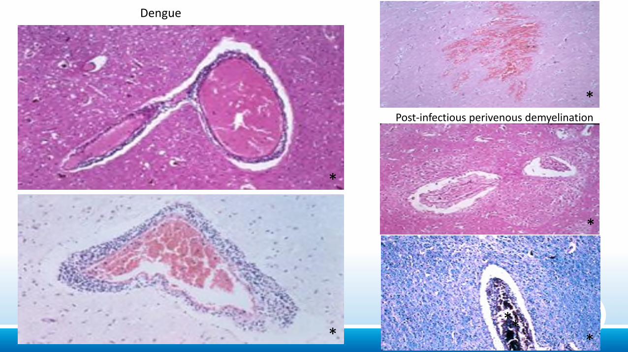
Dengue
Post-infectious perivenous demyelination
**
*
*
*
**
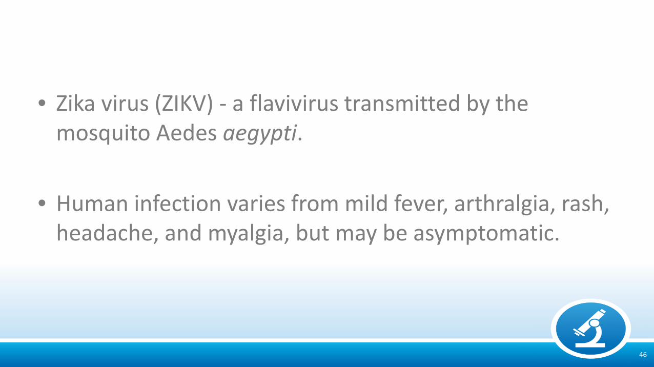
• Zika virus (ZIKV) - a flavivirus transmitted by the mosquito Aedes aegypti.
• Human infection varies from mild fever, arthralgia, rash, headache, and myalgia, but may be asymptomatic.
46
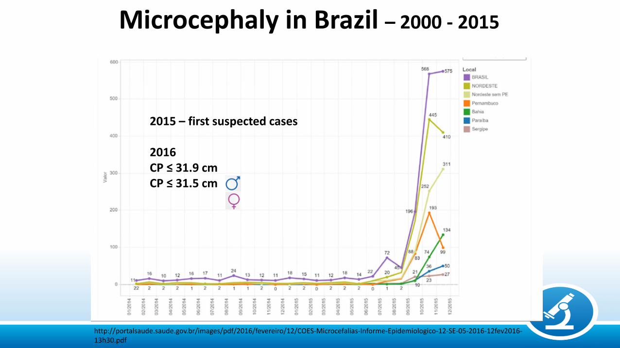
Microcephaly in Brazil – 2000 - 2015
2015 – first suspected cases
2016CP ≤ 31.9 cm CP ≤ 31.5 cm
http://portalsaude.saude.gov.br/images/pdf/2016/fevereiro/12/COES-Microcefalias-Informe-Epidemiologico-12-SE-05-2016-12fev2016-13h30.pdf
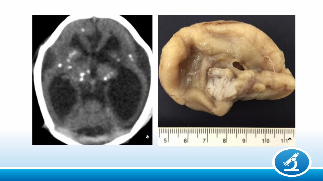
D
**

Ex vacuo ventriculomegaly and calcifications
*
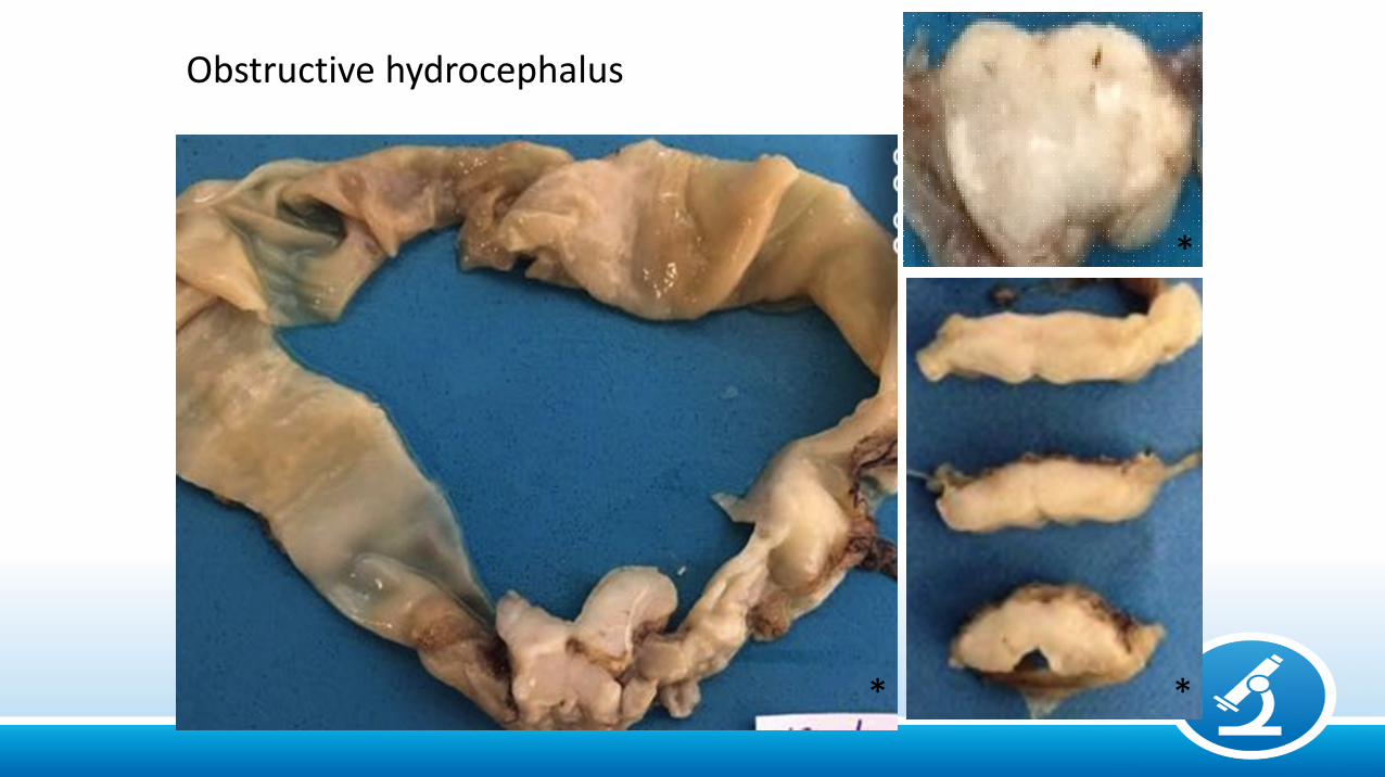
Obstructive hydrocephalus
* *
*
51
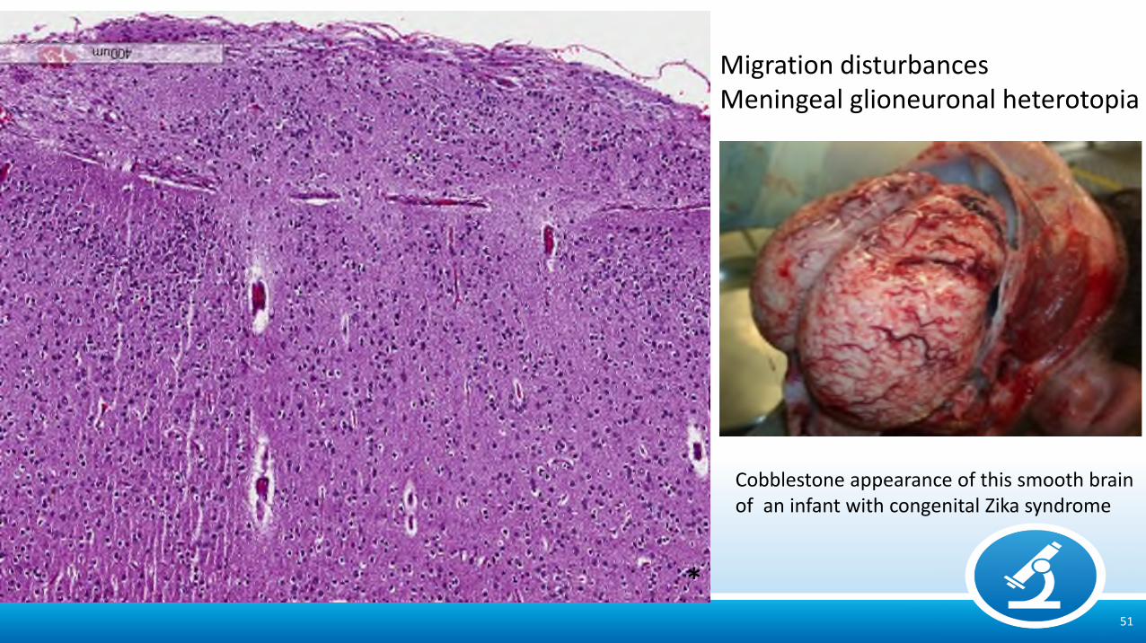
Migration disturbancesMeningeal glioneuronal heterotopia
Cobblestone appearance of this smooth brainof an infant with congenital Zika syndrome
*
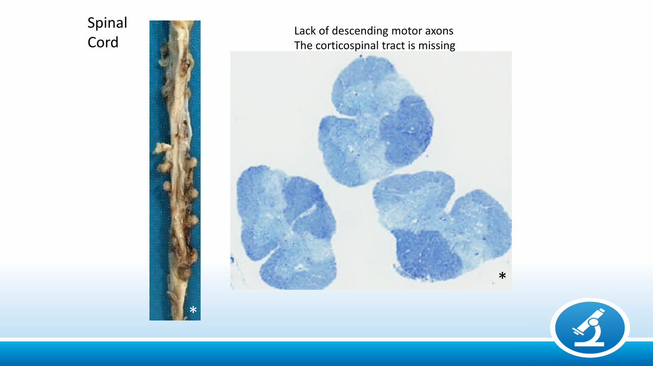
SpinalCord
Lack of descending motor axonsThe corticospinal tract is missing
*
*
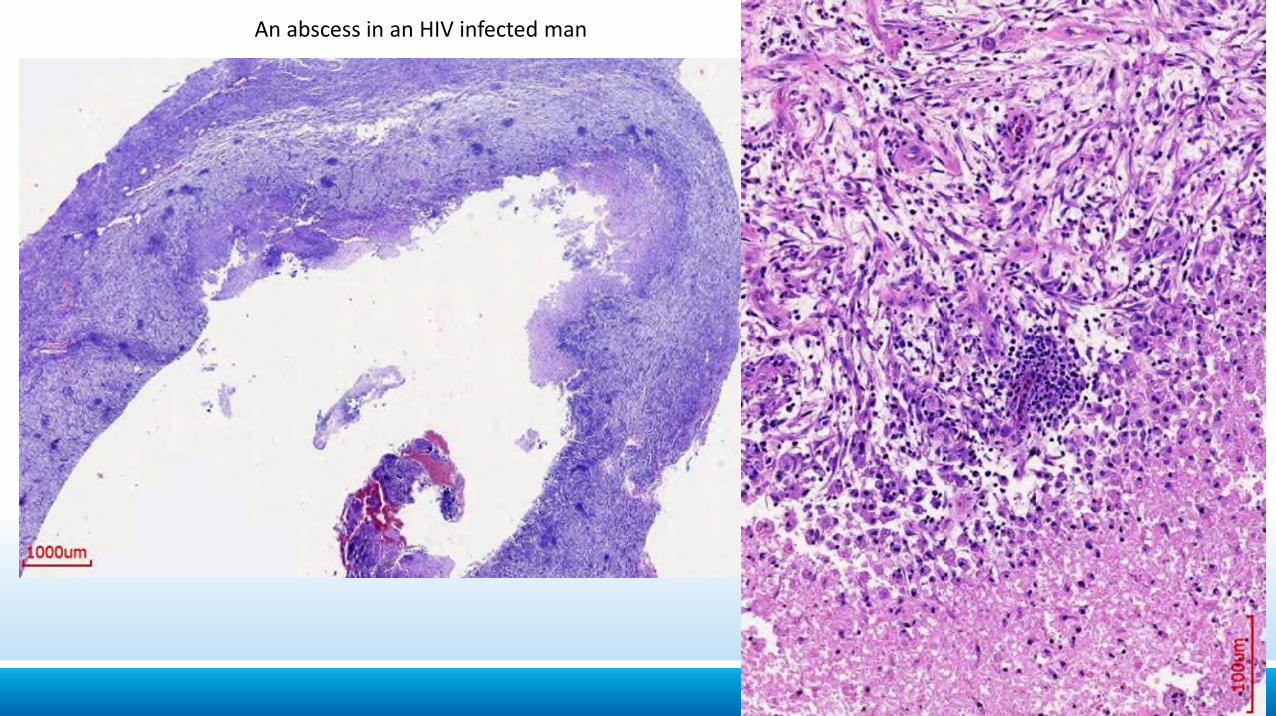
An abscess in an HIV infected man
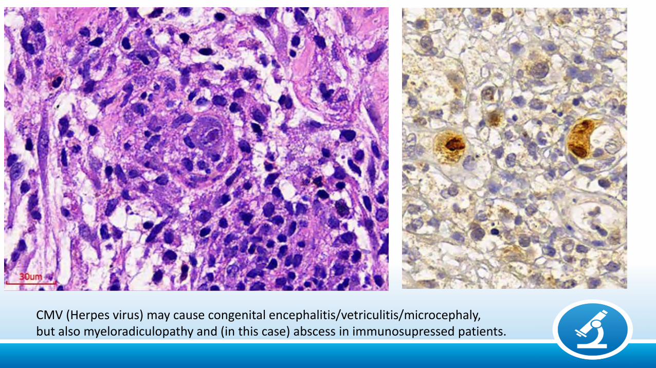
CMV (Herpes virus) may cause congenital encephalitis/vetriculitis/microcephaly, but also myeloradiculopathy and (in this case) abscess in immunosupressed patients.
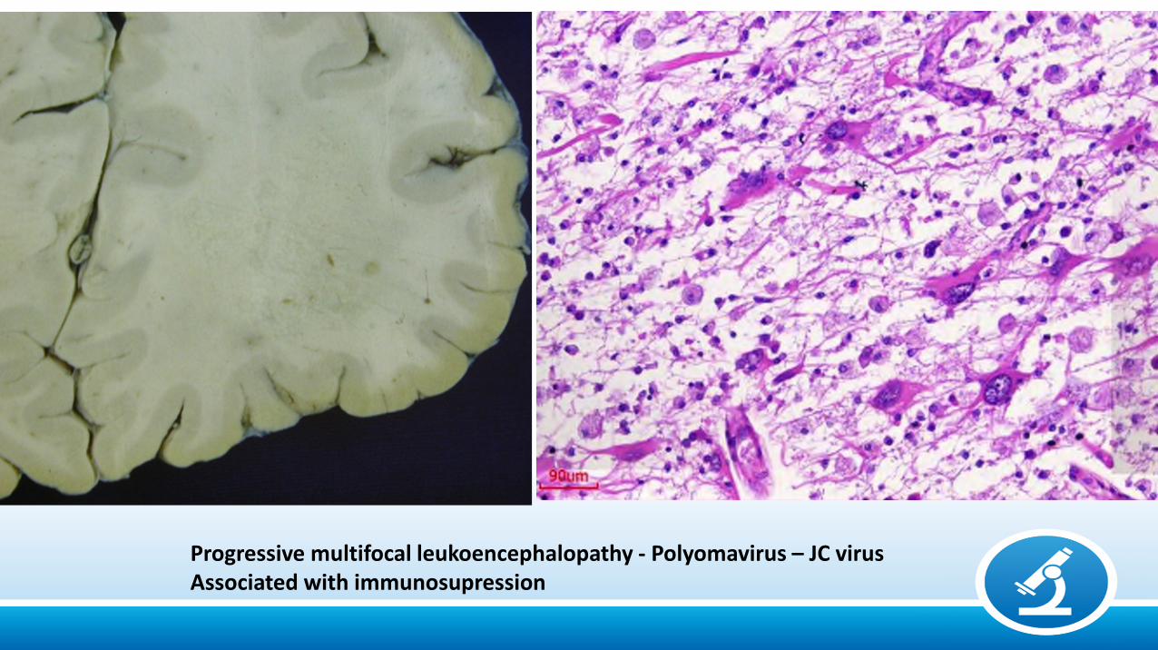
Progressive multifocal leukoencephalopathy - Polyomavirus – JC virusAssociated with immunosupression
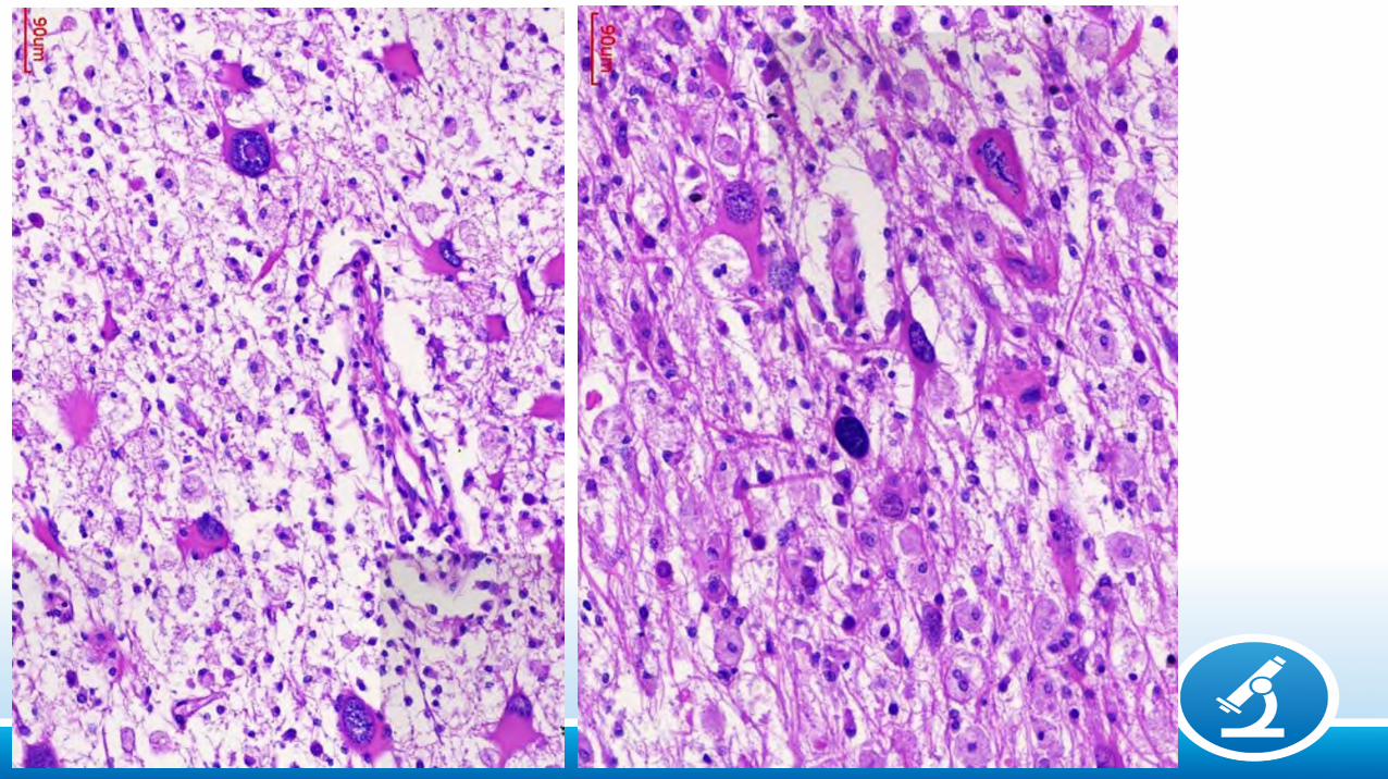

34-year-old man who coursed with progressive dementia and myoclonusClinical hypothesis was prion disease
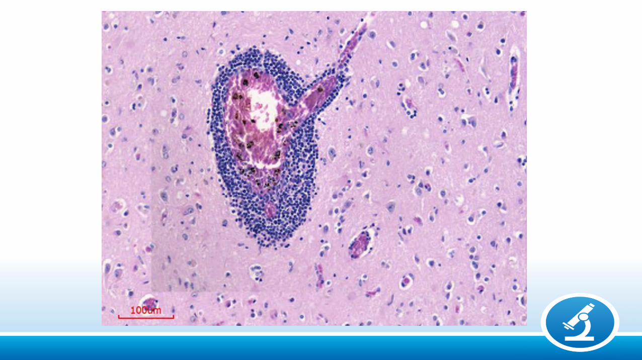
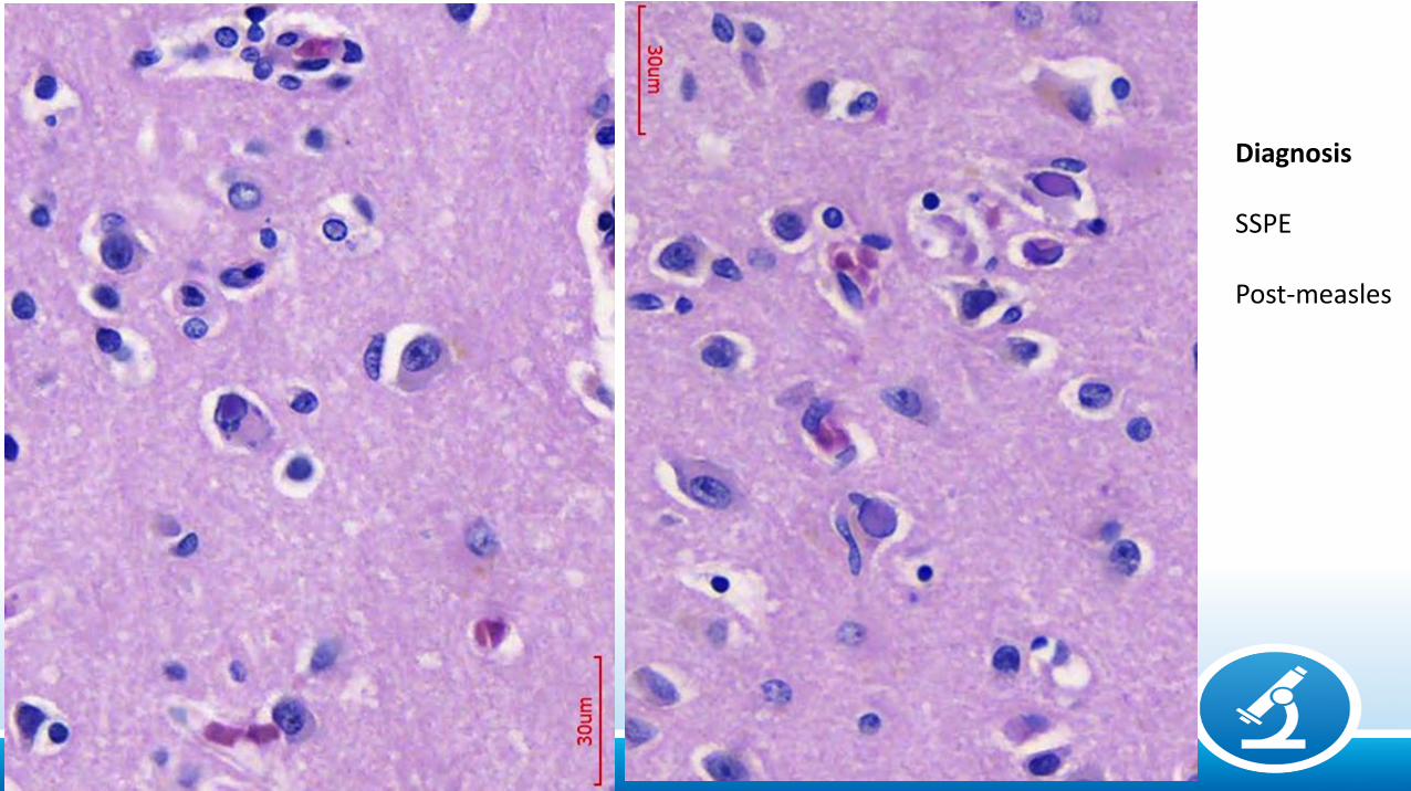
Diagnosis
SSPE
Post-measles
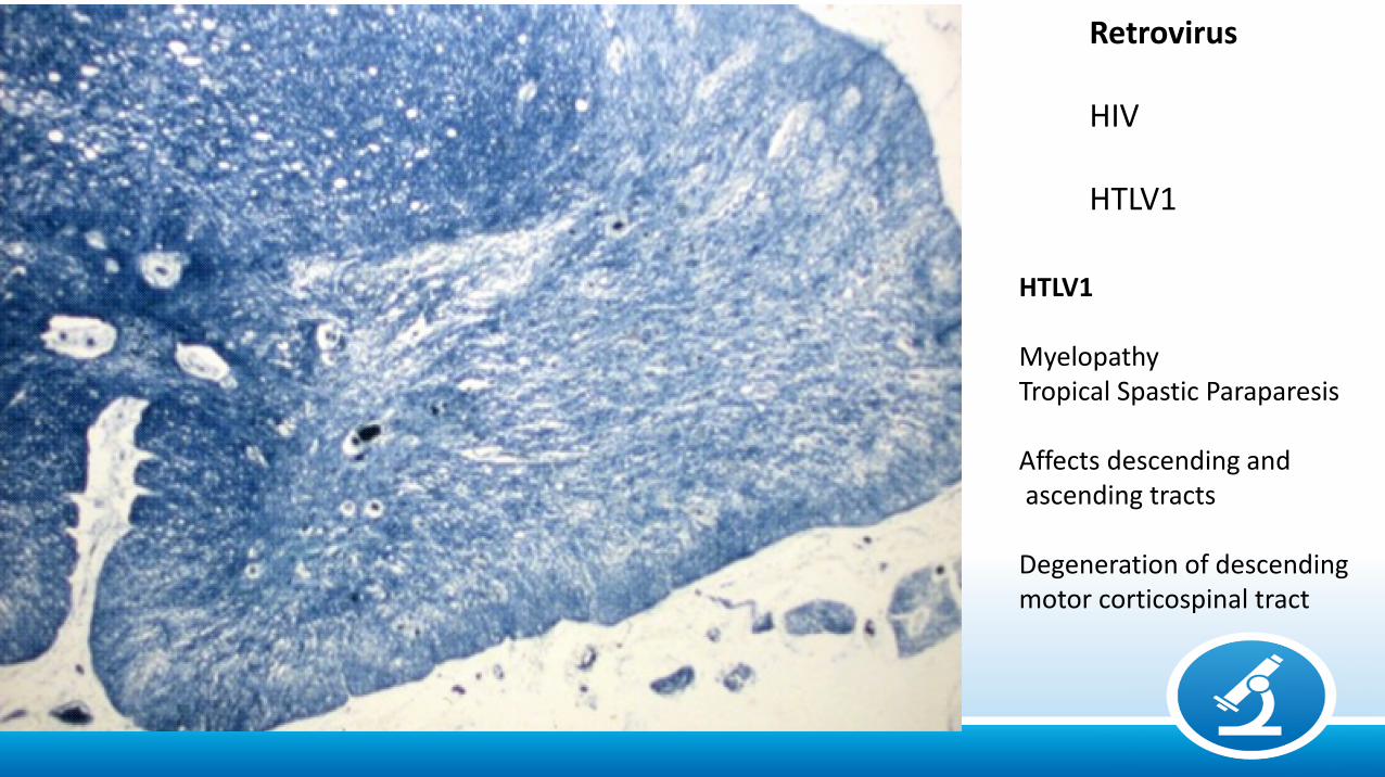
Retrovirus
HIV
HTLV1
HTLV1
MyelopathyTropical Spastic Paraparesis
Affects descending andascending tracts
Degeneration of descendingmotor corticospinal tract
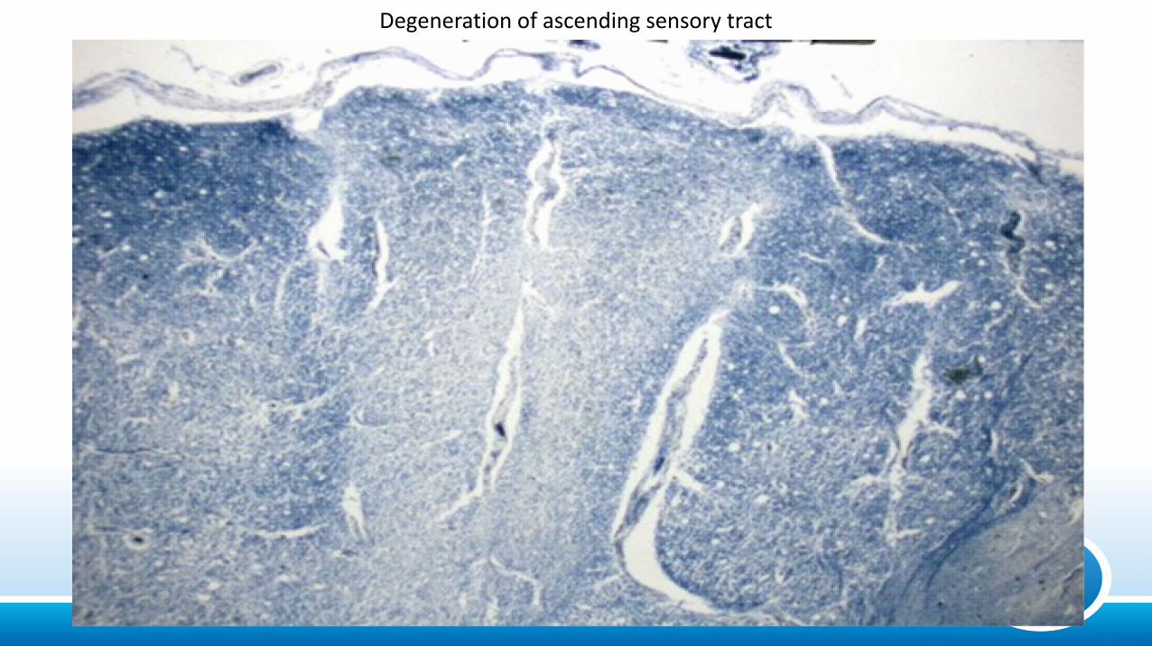
Degeneration of ascending sensory tract
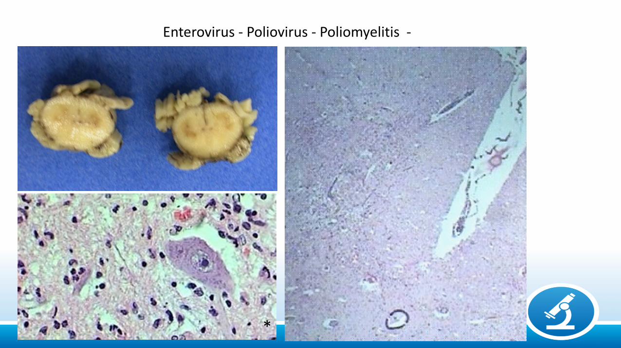
Enterovirus - Poliovirus - Poliomyelitis -
*
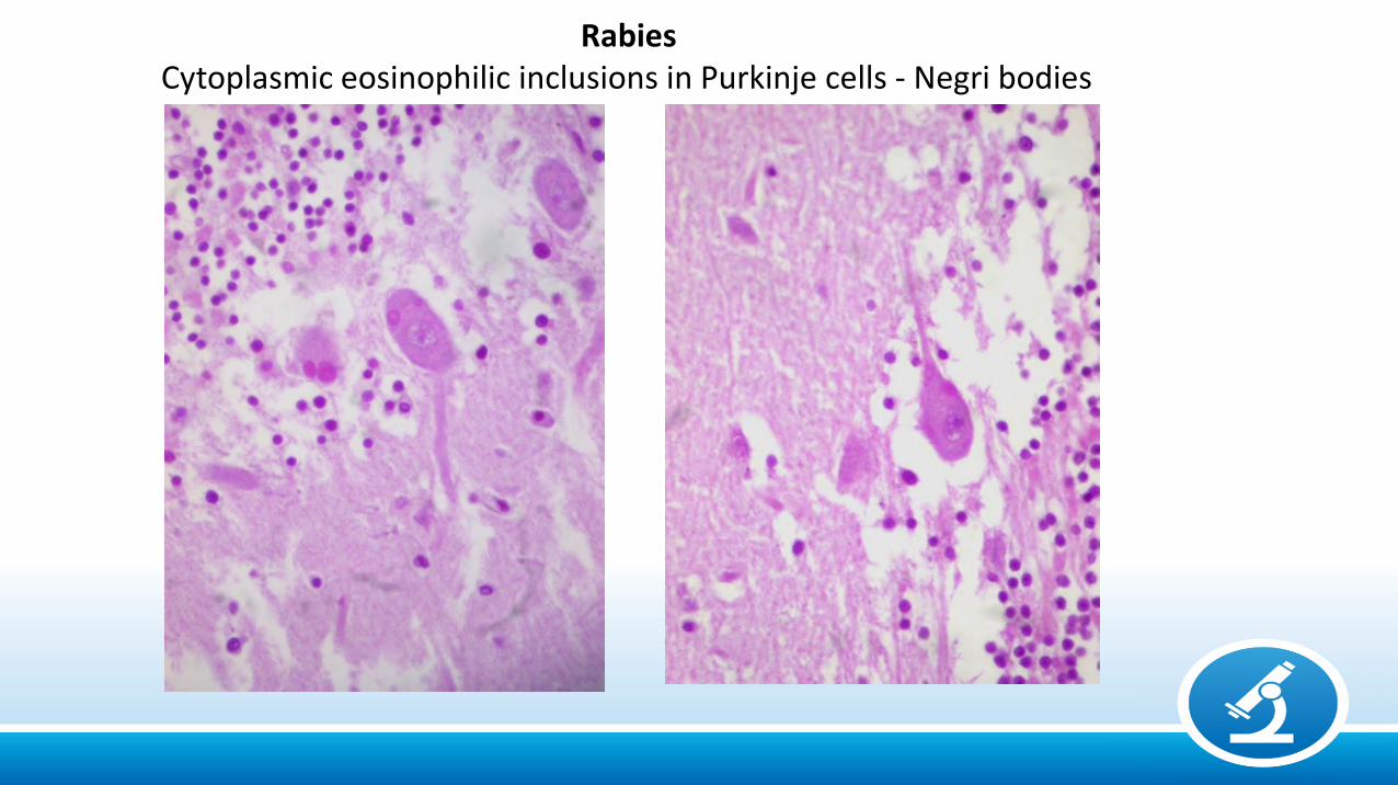
RabiesCytoplasmic eosinophilic inclusions in Purkinje cells - Negri bodies
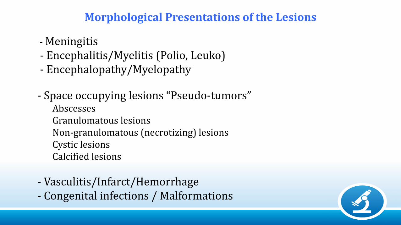
Morphological Presentations of the Lesions
- Meningitis- Encephalitis/Myelitis (Polio, Leuko)- Encephalopathy/Myelopathy
- Space occupying lesions “Pseudo-tumors”AbscessesGranulomatous lesionsNon-granulomatous (necrotizing) lesionsCystic lesionsCalcified lesions
- Vasculitis/Infarct/Hemorrhage- Congenital infections / Malformations
* References containing illustrations included in this presentation
1- Chimelli L, Hahn MD, Netto MB, Ramos RG, Dias M, Gray F. Dengue: neuropathological findings in 5 fatal cases. ClinNeuropathol. 9(3):157-62, 1990.2 – Chimelli L, Mahler-Araujo MB. Fungal Infections. Brain Pathol 7: 613-627, 1997.3 – Chimelli L, Scaravilli F. Trypanosomyasis. Brain Pathol 7: 599-611, 1997. 4- Chimelli L. A Morphological Approach to the Diagnosis of Protozoal Infections of the Central Nervous System. Pathology Research International, Volume 2011, Article ID 290853, 15 pages doi:10.4061/2011/290853 5- Chimelli L et al, The spectrum of neuropathological changes associated with congenital Zika virus infection. ActaNeuropathol 133:983–999, 2017.6- Chimelli, L.; Avvad-Portari, E. Congenital Zika virus infection: a neuropathological review. Childs Nervous System. , v.34, p.95 - 99, 2018.7- Chrétien F, Jouvion G, Wong KT, Sharer LR. Infections of The Central Nervous System. In: F Gray, C Duyckaertz, U De Girolami (eds), Sixth edition of Escourolle & Poirier’s, Manual of Basic Neuropathology. Oxford University Press, New York, 122-158, 2019.8- Chrétien F, Wong KT, Sharer L (eds.), ISN Series Editors, Catherine Keohane and Françoise Gray. Infections of the Central Nervous System. Wiley Blackwell, Inst. Pasteur, International Society of Neuropathology, Oxford/Singapore, pp. 520, 2020. 9- Ellison, D; Love, S. (eds); Chimelli L; Harding, B. N.; Lowe, J. S.; Vinters, H. V.; Brandner, S.; Yong, W. H. Neuropathology. A Reference Text of CNS Pathology. Edinburgh: Elsevier Mosby, Third ed. 2013, pp.879.10- Lucas, S; Bell, J; Chimelli L. Parasitic and fungal infections In: Greenfield's Neuropathology. 8th ed.London: HodderArnold, 2008, v.1, p. 1447-1512.
References
1. Lucas, S; Bell, J; Chimelli L. Parasitic and fungal infections In: Greenfield's Neuropathology. 8th ed.London: Hodder Arnold, 2008, v.1, p. 1447-1512.
2. Chimelli L. A Morphological Approach to the Diagnosis of Protozoal Infections of the Central Nervous System. Pathology Research International, Volume 2011, Article ID 290853, 15 pages doi:10.4061/2011/290853
3. Ellison, D; Love, S.; Chimelli L; Harding, B. N.; Lowe, J. S.; Vinters, H. V.; Brandner, S.; Yong, W. H. Neuropathology. A Reference Text of CNS Pathology. Edinburgh: Elsevier Mosby, Third ed. 2013, pp.879.
4. Love S, Wiley CA, Lucas S. Viral Infections. In: Seth Love, Arie Perry, James Ironside, Herbert Budka (eds), Greenfields’ Neuropathology, Ninth ed. 1087-1191, CRC. Press, Taylor and Francis. Boca Raton, FL, USA, 2015.
5. Deckert M. Bacterial Infections. In: Seth Love, Arie Perry, James Ironside, Herbert Budka (eds), Greenfields’ Neuropathology, Ninth ed. 1192-1290, CRC. Press, Taylor and Francis. Boca Raton, FL, USA, 2015.
6. Chimelli, L.; Avvad-Portari, E. Congenital Zika virus infection: a neuropathological review. Childs Nervous System. , v.34, p.95 - 99, 2018.
7. Chrétien F, Jouvion G, Wong KT, Sharer LR. Infections of The Central Nervous System. In: F Gray, C Duyckaertz, U De Girolami (eds), Sixth edition of Escourolle & Poirier’s, Manual of Basic Neuropathology. Oxford University Press, New York, 122-158, 2019.
8. Chrétien F, Wong KT, Sharer L (eds.), ISN Series Editors, Catherine Keohane and Françoise Gray. Infections of the Central Nervous System. Wiley Blackwell, Inst. Pasteur, International Society of Neuropathology, Oxford/Singapore, pp. 520, 2020.