neural correlates of action attribution in schizophrenia
TRANSCRIPT

Psychiatry Research: Neuroimaging 131(2004) 31–44
0925-4927/04/$ - see front matter� 2004 Elsevier Ireland Ltd. All rights reserved.doi:10.1016/j.psychresns.2004.02.004
Neural correlates of action attribution in schizophrenia
Chloe Farrer *, Nicolas Franck , Chris D. Frith , Jean Decety , Nicolas Georgieff ,a, a b c a,d
Thierry d’Amato , Marc Jeannerodd a
Institut des Sciences Cognitives, 67 boulevard Pinel, 69675 Bron Cedex, Francea
Wellcome Department of Cognitive Neurology, 12 Queen Square, London WC1 N 3BG, UKb
University of Washington Center for Mind, Brain and Learning, Seattle, WA 98195, USAc
Centre Hospitalier Le Vinatier and EA 3092 (IFNL) Universite Claude Bernard, Lyon, Franced ´
Received 9 December 2002; received in revised form 19 November 2003; accepted 24 February 2004
Abstract
Patients with first-rank symptoms(FRS) of schizophrenia do not experience all of their actions and personal statesas their own. FRS may be associated with an impaired ability to correctly attribute an action to its origin. In thepresent study, we examined regional cerebral blood flow(rCBF) with positron emission tomography during an action-attribution task in a group of patients with FRS. We used a device previously used with healthy subjects that allowsthe experimenter to modulate the subject’s degree of movement control(and thus action attribution) of a virtual handpresented on a screen. In healthy subjects, the activity of the right angular gyrus and the insula cortex appeared tobe modulated by the subject’s degree of movement control of the virtual hand. In the present study, the schizophrenicpatients did not show this pattern. We found an aberrant relationship between the subject’s degree of control of themovements and rCBF in the right angular gyrus and no modulation in the insular cortex. The implications of theseresults for understanding pathological conditions such as schizophrenia are discussed.� 2004 Elsevier Ireland Ltd. All rights reserved.
Keywords: Agency; Schneiderian symptomatology; Positron emission tomography; Volition
1. Introduction
One of the most disconcerting sensationsencountered by patients with schizophrenia is thefeeling that their actions and personal states are nolonger under their own control. Symptoms such asauditory hallucinations, thought insertion, thoughtbroadcasting and the influence of others on thepatient’s thoughts, actions or emotions are very
*Corresponding author. Tel.:q33-4-37-91-12-46; fax:q33-4-37-91-12-10.
E-mail address: [email protected](C. Farrer).
frequent in schizophrenia. Kurt Schneider(1959)classified these experiences as first-rank symptoms(FRS) and considered them as the consequencesof a loss of boundaries between the self and others.Indeed, he stated that major FRS had to beinterpreted as the consequence of an impairedselfness, leading to the sensation of being con-trolled by the others. It has been proposed thatthese symptoms may reflect a problem with therecognition of the person’s own actions or thoughtsand to a perturbed sense of agency(the sense thatI am the one who is causing an action; Gallagher,

32 C. Farrer et al. / Psychiatry Research: Neuroimaging 131 (2004) 31–44
2000), leading patients to misattribute their ownactions to another agent. Experimental evidencefor this hypothesis comes from studies that testedsource monitoring(i.e. the ability to attribute anaction or an event to its correct agent) (Bentall etal., 1991; Daprati et al., 1997; Morrison andHaddock, 1997; Baker and Morrison, 1998; Johnsand McGuire, 1999; Brebion et al., 2000; Johns et´al., 2001) and other studies that distorted thefeedback of patients’ actions to evaluate theircapacity to detect the distortion(Cahill et al.,1996; Johns and McGuire, 1999; Blakemore et al.,2000; Franck et al., 2001). The results from thesestudies showed that patients currently experiencinghallucinations andyor delusions of control werenot only impaired at detecting the distortion oftheir own movements, but also tended to misattri-bute their own actions to another agent.The presence of FRS is associated with abnor-
mal over-activation in specific brain regions. Somestudies have found an association between hallu-cinatory phenomenon and activation in primarysensory areas of the auditory cortex and in brainareas involved in the generation and understandingof speech(McGuire et al., 1993; Silbersweig etal., 1995; David et al., 1996; Woodruff et al., 1997Dierks et al., 1999; Bentaleb et al., 2002). It hasbeen proposed that the hyperactivation that occursduring hallucinations arises from a failure in theneural processes subserving self-generated action(Frith, 1996; Frith and Dolan, 1996). Studies onwilled action in normal subjects have shown thatself-generated movements involved an inhibitoryprocess(from prefrontal and cingular cortices), inthe areas involved in the processing of the sensoryconsequences of the action(Muller-Preuss and¨Jurgens, 1976; Muller-Preuss and Ploog, 1981;¨ ¨Frith et al., 1991a,b; Blakemore et al., 1998).Auditory hallucinations may be explained by adeficit in inhibitory processes leading to an over-activation of those areas normally involved in thereception of verbal messages(Silbersweig et al.,1995). Increased activity in those regions mightlead to incorrect agency judgments. The sameexplanation can be applied to delusions of control.Patients experiencing this symptom feel that someoutside force is creating their own actions. Spenceet al. (1997) found hyperactivity in the right
inferior parietal lobule when patients with schizo-phrenia experienced alien control during a move-ment selection task. Delusions of control may arisebecause of a disconnection between frontal brainregions, where actions are initiated, and parietalregions, where the current and predicted states oflimbs are represented(Frith, 1996; Frith et al.,2000).In normal subjects, attribution of action judg-
ment has been shown to involve different brainareas. When subjects feel in control of an actionand thus attribute it to themselves, activation inthe insular cortex is observed(Farrer and Frith,2002). However, when they do not feel in controland attribute the action to another agent, the rightinferior parietal lobule is activated(Ruby andDecety 2001; Decety et al., 2002; Farrer and Frith,2002). Furthermore, a study by Farrer et al.(2003)suggests that the feeling of being in control of anaction is not an all or none state. It varies contin-uously on the basis of the various action-relatedsignals concerned with sensation(kinesthetic, visu-al) and motor control(motor commands). In thisstudy visual feedback was distorted by varyingdegrees up to the point where the movements seenwere completely unrelated to those executed. Theresults showed that the level of activity in theareas known to be activated during attributionjudgments(e.g. posterior parietal cortex and insu-la) co-varied continuously with the feeling ofbeing in control of the action.The studies reviewed above indicate that
patients with FRS are impaired in attributingactions to their respective authors and that thesesymptoms are associated with neurofunctionalabnormalities. Studies with normal subjects haveassociated attribution of action judgments withactivations in the right inferior parietal cortex andthe insula. In the present study we went one stepfurther and examined the neural correlates of thesame attribution judgments in a group of patientssuffering from FRS. We aimed at determiningwhether the attribution deficit observed in suchpatients is linked to abnormal neurofunctionalprocesses. We considered FRS as a whole andpatients were included according to the presenceof at least one such symptom. Each of them wasconsidered equivalent in the present study, since

33C. Farrer et al. / Psychiatry Research: Neuroimaging 131 (2004) 31–44
our hypothesis was that all of them would berelated to the same impairment of action attribu-tion. We did not attempt to evaluate the neuralcorrelates of these symptoms while they occurred,but rather mechanisms that favor their production.We expected that the weakening of action attribu-tion processes could produce conditions necessaryfor FRS production. Thus we sought to findevidence of trait markers rather than state markers.We selected patients who experienced FRS fre-quently, but we did not try to record cerebral bloodflow during the presence of FRS.We used a device previously used in a group of
normal subjects(Farrer et al., 2003) that allowsmodulation of the feeling of being in control ofthe movements(and thus action attribution) of avirtual hand presented on a screen. This feelingwas modulated across the different experimentalconditions by introducing a distortion between theexecuted movements and their indirect visualiza-tion. The higher the distortion, the more the sub-jects felt that they were not controlling themovements on the screen. The maximal distortionwas obtained when they executed movements andsaw the movements of another agent(the experi-menter). In normal subjects the activity of twomain brain areas(the right angular gyrus and theinsula cortex) appeared to be modulated by thesubject’s degree of control of the movements ofthe virtual hand(Farrer et al., 2003). The presentstudy aimed at evaluating whether the feeling ofbeing in control of one’s action was correlatedwith activation in the angular and insular areas ina group of patients with Schneiderian symptoms.
2. Methods
We studied eight healthy right-handed malesubjects(mean age 34"6.71 years, mean years ofeducation 12"3.26) and eight right-handed malemedicated patients with DSM-IV diagnoses(estab-lished according to clinical consensus) of schizo-phrenia(mean age 36.25"9.57 years; mean yearseducation 10.25"2.96). Exclusion criteria werevisual and auditory disorders, history of neurolog-ical illness or trauma, alcohol and drug dependenceaccording to the DSM-IV criteria, and age above70 and below 18. The two groups were demo-
graphically similar in terms of age and level ofeducation. None of the patients had localizedcerebral lesions on magnetic resonance imaging(MRI) scans. The patients underwent clinicalassessment with the Scale for the Assessment ofPositive Symptoms(SAPS; Andreasen, 1984) andthe Scale for the Assessment of Negative Symp-toms (SANS; Andreasen, 1983). Mean scoreswere: 40.88"16.14 for the SAPS and27.63"16.16 for the SANS. The mean durationof illness was 10.12"11.87 years. Patients wereselected, for the presence of FRS in the weeksbefore the experiment(i.e. verbal hallucinations,impressions that another agent was controllingactions and thoughts, that thoughts have beenstolen or introduced in the patient’s mind or thatsomeone else knows his thoughts). However, thepatients did not manifest these symptoms duringthe scanning sessions. A Schneiderian score wascalculated for each patient according to sevenitems from the SAPS(item 2: voices commenting;item 3: voices conversing; item 15: delusions ofcontrol; item 16: delusions of mind reading; item17: thought broadcasting; item 18: though inser-tion; item 19: thought withdrawal). The meanSchneiderian score was 6.88"5.67. All patientswere under antipsychotic treatment(risperidone,olanzapine, clozapine, haloperidol or levomeprom-azine), but one of them also received prazepam(benzodiazepine) and another one valproate(moodstabilizer). All were clinically stable at the time oftesting and gave written informed consent to par-ticipate in the study, which had been approved bythe local Ethics Committee(CCPPRB, CentreLeon Berard, Lyon).´ ´The subjects underwent 12 perfusion scans with
positron emission tomography(PET) in a singlesession. Subjects lay in the scanner with headmovements minimised by a mask. Radioactivitywas administered as an intravenous injection ofH O by a plastic canula placed in the left cubital152
vein. The subjects held a joystick with their righthand and were instructed to execute random move-ments at a constant rate throughout the 70-s block.This task is very similar to that used by Spence etal. (1997) to demonstrate parietal overactivity inpatients with delusions of control. They wererequested to move the joystick back to the center
34 C. Farrer et al. / Psychiatry Research: Neuroimaging 131 (2004) 31–44
after each excursion in the chosen direction. Thisprocedure ensured that the movements were similaracross conditions and subjects. The movements ofthe joystick controlled an image of a virtual handholding a joystick. This system provided a dynam-ic representation of the movements of the joystickheld by the subjects with an intrinsic delay of lessthan 30 ms(Franck et al., 2001).The joystick was attached to a table above the
bed of the scanner. The image of the virtual handholding the joystick was projected onto a mirrorplaced in front of the subject. The angle of visual-isation of the image in the mirror was adjusted soas to coincide with the real position of the joystickactually held by the subjects. The position of thesubject’s forearm was adjusted so as to coincidewith the direction of the virtual forearm seen inthe mirror. A black cloth was then hung above thesubject’s forearm so as to prevent him from seeinghis forearm and the device controlled by the ex-perimenter.Angular distortions could be introduced into this
system, modifying the direction of the movementactually performed by the subjects with respect tothe movement displayed on the computer screen.The experiment involved four experimental con-ditions. In the first condition(‘08’ condition) thesubjects could see the movements of the virtualhand in perfect concordance with their movementsmade with the joystick. In the second condition(‘258’ condition), they saw the movements of thevirtual hand deviating by 258 from their hand’sactual trajectory. In the third condition(‘508’condition) the value of the deviation was 508. Inthe latter two conditions, the deviation was to theright on half of the sessions and to the left on theother half. In the fourth condition(‘Other’ condi-tion), the subjects saw the movements of thejoystick controlled by another agent(the experi-menter).During each session subjects were asked to
direct their attention to the origin of the movementthey saw. They had to give a verbal response toindicate whether it was their own movement(‘Self’ response), their own movement distorted(‘Distorted’ response) or the movement of anotheragent(‘Other’ response).
Two low-level control conditions(‘C1’ and‘C2’) were also included. In condition ‘C1’ thesubjects had to execute random movements with-out seeing anything on the screen. In condition‘C2’ they had to watch the virtual hand movingby itself without doing anything.At the beginning of the experiment subjects
undertook a practice session in order to getacquainted with the device and to experiencegenerating random movements. They sat in frontof the monitor and executed random movementsin three different conditions: ‘08’ condition, ‘Dis-torted’ condition (the deviation was 358) and‘Other’ condition. When the subjects were lyingin the scanner, they had a second practice sessionso that they could familiarize themselves with thenew angle of visualization caused by their supineposition. Only the ‘08’ and the ‘Distorted’ condi-tions were performed in this final practice session.The PET scanning comprised two blocks of
each of the six conditions of 70 s each. Theinterval between each start time was 8 min. Theorder of the conditions was randomised andreversed within and between subjects. Each trialwas initiated by an auditory stimulus.
2.1. Image acquisition
The PET images were acquired using a SiemensCTI HRq (63 slices, 15.2-cm axial field of view)PET tomograph with collimating septa retractedoperating in 3D mode. Relative rCBF was meas-ured by recording the regional distribution ofcerebral radioactivity using H O as a tracer. After15
2
a 9-mCi bolus injection of H O , scanning started152
when the brain radioactive count rate reached athreshold value and continued for 60 s. Integratedradioactivity accumulated in 60 s of scanning wasused as an index of rCBF. A transmission scancollected before the first emission scan permittedcorrecting for radiation attenuation.
2.2. Data analysis
2.2.1. Image analysis: pre-processingThe data were analysed with SPM99(Wellcome
Department of Cognitive Neurology, London, UK).
35C. Farrer et al. / Psychiatry Research: Neuroimaging 131 (2004) 31–44
Each subject’s PET data were realigned to thefirst scan of the time series. The estimates extract-ed from the rigid body transformation(describedas three translations(x, y, z) and three rotationsabout the axes) were used to realign the imagesand to perform a mathematical adjustment(min-imising the sum of the squares of differences inintensity between each image and the reference)to remove movement-related components. Theimages were then spatially normalised into thesystem of reference of Talairach and Tournoux(1988) using as the template a representative brainfrom the Montreal Neurological Institute(MNI)series(Evans et al., 1994). The first step of spatialnormalisation was to determine the optimal affinetransformation(correction for variation in positionand size) that mapped the brain image onto thetemplate (minimisation of first the sum of thesquares of the differences between those two imag-es and also the squared distance between theparameters and their known expectation). Residualdifferences between each pair of images werecorrected using nonlinear basis functions(Fristonet al., 1995). The normalization parameters weresubsequently applied to the PET images. Finally,PET images were filtered with the use of a low-pass Gaussian filter(FWHMs11.1, 13.2, and 14.5mm) to reduce noise and maximise signal. Thesmoothness was achieved by forcing the deforma-tions to consist of a linear combination of pre-defined smooth spatial basis functions.
2.2.2. Statistical model and inferenceThe data were modeled so as to partition the
rCBF of each voxel into components of interest,confounds of no interest and an error term. Thedata were first adjusted for the effect of globalimage signal with the proportional scaling method.The fraction of mean signal over the whole brainwas specified for thresholding signal intensitiesabove the grey matter value. Since the subjectswere requested to move the joystick freely, therewere some differences in the amount of movementbetween subjects and between conditions. To elim-inate this bias, the co-ordinates of the position ofthe joystick were recorded so as to obtain ameasure of the distance covered by the joystick.This reflected the amount of movement made by
each subject in each condition. These measureswere modeled as confounds in the analysis so asto remove variations in blood flow that wererelated to the amount of movement.The analysis of regionally specific effects was
realized using the general linear model. We spec-ified the following six effects of interest: ‘08’,‘258’, ‘508’, ‘Other’, ‘C1’ and ‘C2’. The ‘258’ and‘508’ effects of interest grouped together the trialsin the two directions of deviation(to the left andto the right). We were interested in finding brainareas showing significant variations(i.e, increasedor decreased rCBF) as a function of the discrep-ancy between the movements made by the subjectsand the movements of the virtual hand on thescreen. To achieve this aim, the four experimentalconditions (‘08’, ‘258’, ‘508’ and ‘Other’) weremodeled as independent covariates and two con-junction analyses(Price and Friston, 1997) ofthree contrasts each were performed. The first onewas designed to identify brain areas showingincreased rCBF with increasing distortion from the‘08’ condition to the ‘Other’ condition((‘258’–‘08’) in conjunction with(‘508’–‘258’) and(‘Oth-er’–508)). The second analysis allowed us todetermine brain areas showing decreased rCBFwith increasing distortion from the ‘08’ conditionto the ‘Other’ condition((‘08’–‘258’) in conjunc-tion with (‘258’–‘508’) and (‘508’–‘Other’)). Anexclusive masking procedure with the contrast((‘08’q‘258’q‘508’q‘Other’) – ‘C1’) wasapplied to each conjunction analysis to eliminatehemodynamic activity related to visual feedbackand eye movements. In the ‘C1’ condition thepatients were requested to move the joystick whilefixating the center of the blank screen. Thus thecontrast ((‘08’q‘258’q‘508’q‘Other’) – ‘C1’)allowed us to obtain brain activity related to visualfeedback and eye movements.Complementary simple contrast analyses within
the patient group and comparisons between thepatients and the normal subjects were realized toassess, respectively, differences in brain activationbetween the effects of interest and differences inthe magnitude of activation effects betweengroups.Finally, to examine whether brain activations
were related to symptomatology, we computed the

36 C. Farrer et al. / Psychiatry Research: Neuroimaging 131 (2004) 31–44
correlations between effects of interest and theSchneiderian score and between these effects andthe SANS score.The different models were framed in terms of a
statistical parametric map of at value SPM{T }3
for the conjunction analysis and of a SPM{ t}transformed into SPM(Z) for the simple contrasts.Since we were interested in brain activity in theright inferior parietal cortex and in the insula andwe had strong a priori hypotheses, we first definedsearch volume corrections in a region involvingthe inferior parietal lobule and the intraparietalsulcus and in a second region involving the insulaand the circular insular sulcus. These regions weredelimited with MRICRO, a software package. Tolook for other brain activations that were notpredicted, we analyzed the SPMs thresholded atP-0.0001 uncorrected for multiple comparisonsat the voxel level or thresholded atP-0.05 cor-rected at the cluster level. Only activations with aZG3.70 were taken into account. Finally, brainactivity localization was identified using the atlasof neuroanatomy by Duvernoy(1992).
3. Results
3.1. Behavioural results
After each experimental condition the patientswere first requested to indicate whether the move-ments they saw on the screen exactly correspondedto their executed movements(‘Self’ response),whether they had deviated(‘Distorted’ response)or whether they were controlled by the experi-menter(‘Other’ response). Secondly patients werequestioned about their impressions and feelingsduring the trial.This debriefing showed that patients did not
manifest any passivity phenomena, such as feelingthat they were being controlled by another agent,or hallucinating. One patient reported during a ‘08’trial that another person was controlling the move-ments of the virtual hand at the end of the session.This same patient also reported during a ‘258’ trialthat another person was controlling, but that hewas able to get back the control very rapidly.Another patient reported during a ‘08’ trial that hewas controlling the movements, but at the same
time, another person was doing exactly the samemovement. Further questioning of the patientsrevealed that these impressions only occurred onceand were very brief. Indeed these patients reportedthat they knew they controlled the virtual hand.These patients were included in the analyses.Analyses of the behavioral responses showed
that patients tended to perform well; at-test forindependent comparisons on the percentage ofcorrect responses for each condition did not revealany significant differences between the patientsand the controls. There was, however, a trend forthe patients to perform worse on the ‘258’ and onthe ‘508’ trials. Patients gave 100% correctresponses for the ‘08’ and the ‘Other’ trials. How-ever, errors were observed in the Distortion con-ditions. Errors were found in 31% of cases(‘Self’response) for the ‘258’ trials and in 9% of cases(‘Self’ response) and 6% of cases(‘Other’response) for the ‘508’ trials. T-tests for pairwisecomparisons revealed significant differencesbetween the different types of responses for the‘08’, ‘508’ and ‘Other’ trials. However, there wereno significant differences between ‘Self’ and ‘Dis-torted’ responses for the ‘258’ trials. This resultindicates that patients did not easily recognize aperturbation of 258.
3.2. Functional imaging data
3.2.1. All experimental conditions vs. observationcontrol condition (‘C2’)Contrasting all experimental conditions with the
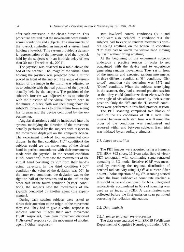
observation control condition(‘C2’) revealed sig-nificant activation(P-0.001 corrected for multi-ple comparisons at the voxel level) in the rightventral premotor cortex(PMv) and the left poste-rior insula. At the cluster level, significant activa-tions were observed in the left sensorimotor cortex.Additional activations that did not survive correc-tion for multiple comparisons were found in theleft PMv, the right sensorimotor cortex, the rightdorsolateral prefrontal cortex and the left cerebel-lum. The co-ordinates of these areas, theZ-valuesand the probability scores are shown in Table 1.
37C. Farrer et al. / Psychiatry Research: Neuroimaging 131 (2004) 31–44
Table 1Brain areas activated during all the experimental conditions contrasted to the ‘Observation’ control condition(‘C2’)
Area x y z Z P
R ventral premotor cortex* 56 8 8 4.79 0.031L posterior insula* y32 16 8 4.71 0.044L sensorimotor cortex** y38 y32 52 4.57 -0.0001L ventral premotor cortex y42 6 24 4.24 -0.0001R dorsolateral prefrontal cortex 44 34 28 4.18 -0.0001R sensorimotor cortex 52 y28 52 3.78 -0.0001L cerebellum y30 y54 y34 3.73 -0.0001
*P-0.05 corrected at the voxel level.** P-0.05 corrected at the cluster level.All other areas are reported for aP-0.0001 uncorrected at the voxel level; voxel extent threshold 10,ZG3.70.
Fig. 1. Brain activations for the contrast(‘Other’–‘08’). TheSPM is thresholded atP-0.0001(uncorrected) and superim-posed on axial sections(from zs10 to 70).
3.2.2. All experimental conditions vs. executioncontrol condition (‘C1’)Contrasting all experimental conditions with the
execution control condition revealed significantactivation (P-0.001 corrected for multiple com-parisons) in brain areas associated with visualperception. Activation was observed with a peakin the right striate cortex extending into the pre-cuneus, the inferior occipital cortex, the medialoccipital cortex and the superior occipital cortexbilaterally.
3.2.3. Brain areas increasing their activity as afunction of the degree of discordance between theexecuted and the seen movements in controlssubjects and patientsThe main interest of the present study was to
test whether patients with FRS present a modula-tion of brain activation as a function of the degreeof the discordance and thus of the sense of agency.Increased brain activity as a function of theincreased discordance was tested with a conjunc-tion analysis between the contrasts((‘258’–‘0 8’);(‘508’–‘258’) and(‘Other’–‘508’)). In the normalsubjects, increased discordance was associatedwith increased brain activity in the right angulargyrus (Farrer et al., 2003). However, in schizo-phrenic patients, we did not find any co-variationbetween the subject’s degree of control of themovements and rCBF, in either the right angulargyrus or in other brain areas.Although we did not find any co-variation
between brain activity and the degree of discor-dance between executed and seen movements,
contrasting the two extreme experimental condi-tions (‘Other’–‘08’) revealed significant activationin the right inferior parietal lobe(see Fig. 1), witha peak activation in the right angular gyrus(P-0.001 corrected for multiple comparisons at thevoxel level). This angular activation was also
38 C. Farrer et al. / Psychiatry Research: Neuroimaging 131 (2004) 31–44
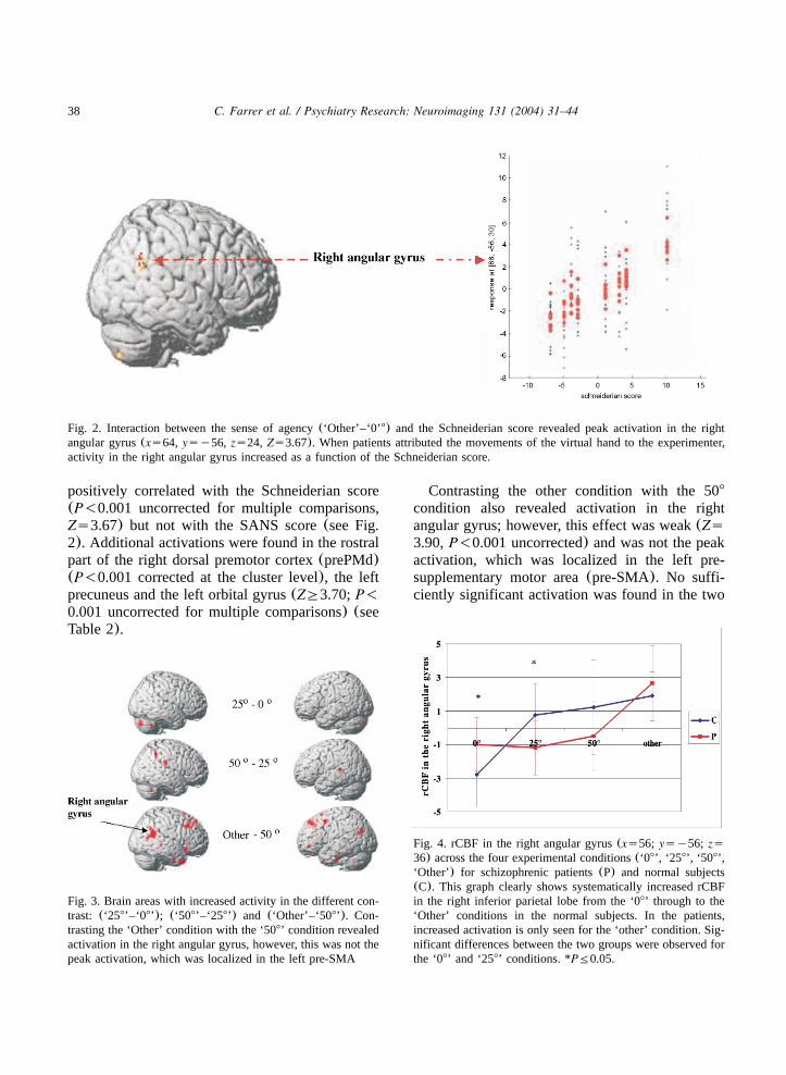
Fig. 2. Interaction between the sense of agency(‘Other’–‘0’8) and the Schneiderian score revealed peak activation in the rightangular gyrus(xs64, ysy56, zs24, Zs3.67). When patients attributed the movements of the virtual hand to the experimenter,activity in the right angular gyrus increased as a function of the Schneiderian score.
Fig. 3. Brain areas with increased activity in the different con-trast: (‘258’–‘08’); (‘508’–‘258’) and (‘Other’–‘508’). Con-trasting the ‘Other’ condition with the ‘508’ condition revealedactivation in the right angular gyrus, however, this was not thepeak activation, which was localized in the left pre-SMA
Fig. 4. rCBF in the right angular gyrus(xs56; ysy56; zs36) across the four experimental conditions(‘08’, ‘258’, ‘508’,‘Other’) for schizophrenic patients(P) and normal subjects(C). This graph clearly shows systematically increased rCBFin the right inferior parietal lobe from the ‘08’ through to the‘Other’ conditions in the normal subjects. In the patients,increased activation is only seen for the ‘other’ condition. Sig-nificant differences between the two groups were observed forthe ‘08’ and ‘258’ conditions. *PF0.05.
positively correlated with the Schneiderian score(P-0.001 uncorrected for multiple comparisons,Zs3.67) but not with the SANS score(see Fig.2). Additional activations were found in the rostralpart of the right dorsal premotor cortex(prePMd)(P-0.001 corrected at the cluster level), the leftprecuneus and the left orbital gyrus(ZG3.70;P-0.001 uncorrected for multiple comparisons) (seeTable 2).
Contrasting the other condition with the 508
condition also revealed activation in the rightangular gyrus; however, this effect was weak(Zs3.90,P-0.001 uncorrected) and was not the peakactivation, which was localized in the left pre-supplementary motor area(pre-SMA). No suffi-ciently significant activation was found in the two

39C. Farrer et al. / Psychiatry Research: Neuroimaging 131 (2004) 31–44
Table 2Brain areas activated during ‘Other’ condition compared with ‘08’ condition
Area x y z Z P
R angular gyrus* 54 y52 22 4.59 0.02R rostral dorsal premotor cortex 44 10 38 4.43 -0.0001L precuneus y4 y46 56 4.07 -0.0001L orbital gyrus y44 42 y2 3.94 -0.0001
*P-0.05 corrected at the voxel level.All other areas are reported forP-0.0001 uncorrected at the voxel level; voxel extent threshold 10,ZG3.70.
other contrasts:(‘258’–‘08’) and (‘508’–‘258’)(see Fig. 3).
3.2.4. Comparison of schizophrenic patients withcontrolsSince schizophrenic patients only showed sig-
nificant activation in the right angular gyrus incontrasts of the two extreme conditions, we com-pared rCBF in the right angular gyrus for eachcondition between the two groups. At-test forindependent variables revealed significantly greateractivity in the patients for the ‘08’ condition (tsy2.08, d.f.s14, Ps0.05). Interestingly, the ten-dency reversed at ‘258’ (ts2.16, d.f.s14, Ps0.05), with a significantly lower activity in theschizophrenic group. For the ‘508’ and ‘Other’conditions there were no significant differencesbetween the groups(see Fig. 4). This result showsthat the patients’ lack of increase in activationwith increasing distortion was associated with anabnormally high level of activation in the zerodistortion condition, with an increase in activityonly appearing for the ‘Other’ condition.
3.2.5. Brain areas decreasing their activity as afunction of the degree of discordance between theexecuted and the seen movements in controlssubjects and patientsDecreased brain activity in the right posterior
insula ((‘08’–‘258’) in conjunction with (‘258’–‘508’) and (‘508’–‘Other’)) was observed in thecontrols (Farrer et al., 2003) but not in schizo-phrenics, in either the insular cortex or in otherbrain areas. Furthermore, we did not obtain signif-icant activation in the insular cortex when contrast-ing the ‘08’ condition with the ‘Other’ condition,even when using a search volume correction in a
region involving the insula and the circular insularsulcus. Only the right cingulate gyrus was foundactivated(ZG3.80,P-0.001 uncorrected for mul-tiple comparisons); however, since this activationwas not predicted and was not significant atP-0.001 corrected, we will not consider it in furtherdiscussion. The other contrasts between the differ-ent experimental conditions did not reveal activa-tions significant enough to be taken into account.
4. Discussion
This study aimed at evaluating whether thefeeling of being in control of one’s action wascorrelated with brain activity in a group of patientswith FRS. This feeling was modulated across thedifferent experimental conditions by introducing adistortion between the executed movements andtheir indirect visualization. The higher the distor-tion, the more the subjects felt that they were notcontrolling the movements on the screen. Themaximal distortion was obtained when they exe-cuted movements and saw the movements ofanother agent(the experimenter). In this case, theydid not feel in control of the movements andattributed them to the experimenter. Our resultsshowed that, contrary to the results of our studyin normal subjects(Farrer et al., 2003), we didnot find any co-variation between the degree ofdistortion and rCBF in either of the brain areasthat were predicted(right inferior parietal lobuleand the insular cortex), nor in other brain areas.However, contrasting the two extreme conditions(‘Other’ with ‘08’ condition) revealed activationin the right angular gyrus.Before going into the interpretation of these
main results, we will briefly interpret the motor
40 C. Farrer et al. / Psychiatry Research: Neuroimaging 131 (2004) 31–44
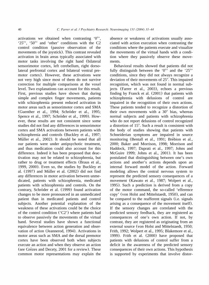
activations we obtained when contrasting ‘08’,‘258’, ‘508’ and ‘other’ conditions with the C2control condition (passive observation of themovements of the joystick). This contrast revealedactivation in brain areas typically associated withmotor tasks involving the right hand(bilateralsensorimotor cortex, left cerebellum, right dorso-lateral prefrontal cortex and bilateral ventral pre-motor cortex). However, these activations werenot very high since most of them do not survivecorrection for multiple comparisons at the voxellevel. Two explanations can account for this result.First, previous studies have shown that duringsimple and complex finger movements, patientswith schizophrenia present reduced activation inmotor areas such as sensorimotor cortex and SMA(Guenther et al., 1994; Schroder et al., 1995;¨Spence et al., 1997; Schroder et al., 1999). How-¨ever, these results are not consistent since somestudies did not find any differences in sensorimotorcortex and SMA activations between patients withschizophrenia and controls(Buckley et al., 1997;Muller et al., 2002). It should be noted that all¨our patients were under antipsychotic treatment,and thus medication could also account for thisdifference. Indeed it has been proposed that deac-tivation may not be related to schizophrenia, butrather to drug or treatment effects(Braus et al.,1999, 2000). Even so, the studies by Buckley etal. (1997) and Muller et al.(2002) did not find¨any differences in motor activation between unme-dicated, patients with schizophrenia, medicatedpatients with schizophrenia and controls. On thecontrary, Schroder et al.(1999) found activation¨changes to be more pronounced in an unmedicatedpatient than in medicated patients and controlsubjects. Another potential explanation of theweakness of these activations could be the choiceof the control condition(‘C2’) where patients hadto observe passively the movements of the virtualhand. Several studies have shown a functionalequivalence between action generation and obser-vation of action(Jeannerod, 1994). Activations inmotor areas such as SMA and the dorsal premotorcortex have been observed both when subjectsexecute an action and when they observe an action(see Grezes and Decety, 2001 for a review). These`common motor representations may explain the
absence or weakness of activations usually asso-ciated with action execution when contrasting theconditions where the patients execute and visualizethe movements of the virtual hands with a condi-tion where they passively observe these move-ments.Behavioral results showed that patients did not
fully distinguish between the ‘08’ and the ‘258’conditions, since they did not always recognize adeviation of their movements of 258. This impairedrecognition, which was not found in normal sub-jects (Farrer et al., 2003), echoes a previousfinding by Franck et al.(2001) that patients withschizophrenia with delusions of control areimpaired in the recognition of their own actions.These patients tended to recognize a distortion oftheir own movements with a 308 bias, whereasnormal subjects and patients with schizophreniawho do not report delusions of control recognizeda distortion of 158. Such a result is consistent withthe body of studies showing that patients withSchneiderian symptoms are impaired in sourcemonitoring (Bentall et al., 1991; Brebion et al.,´2000; Baker and Morrison, 1998; Morrison andHaddock, 1997; Daprati et al., 1997; Johns andMcGuire 1999; Johns et al., 2001). It has beenpostulated that distinguishing between one’s ownactions and another’s actions depends upon aninternal forward model of the action. Forwardmodeling allows the central nervous system torepresent the predicted sensory consequences of amovement(Kawato et al., 1987; Wolpert et al.,1995). Such a prediction is derived from a copyof the motor command, the so-called ‘efferencecopy’ (von Holst and Mittelstaedt, 1950), and canbe compared to the reafferent signals(i.e. signalsarising as a consequence of the movement itself).If the sensory changes are correlated with thepredicted sensory feedback, they are registered asconsequences of one’s own action. If not, bycontrast, they are registered as originating from anexternal source(von Holst and Mittelstaedt, 1950;Frith, 1992; Wolpert et al., 1995; Blakemore et al.,1999). Frith et al. (2000) have proposed thatpatients with delusions of control suffer from adeficit in the awareness of the predicted sensoryconsequences of their own actions. This hypothesisis supported by experiments that involve distor-

41C. Farrer et al. / Psychiatry Research: Neuroimaging 131 (2004) 31–44
tions of the sensory feedback of the patient’sactions. Studies show that patients with hallucina-tions tend to attribute their own distorted speechto another agent more than non-hallucinatedpatients and normal subjects(Cahill et al., 1996;Johns and McGuire, 1999). Recently, Blakemoreet al.(2000) have shown that patients with Schnei-derian symptoms failed to show a differencebetween the perception of self-produced and exter-nally produced tactile sensations. This result dem-onstrates that these patients might be abnormallyaware of the sensory consequences of their ownmovements. Anomalous integration of the differentaction-related signals might explain inaccurate rec-ognition of their own actions and misattributionsof their actions to others. At the physiologicallevel it has been shown that patients with delusionsof control show over-activity in right inferiorparietal cortex when making voluntary movements(Spence et al., 1997). Similar over-activity is alsoseen in normal volunteers who, through hypnosis,believe that they are not the authors of their armmovements(Blakemore et al., 2003). Both psy-chiatric and neurological patients with abnormalexperience of agency show abnormal hyperactivityin right inferior parietal cortex(Franck et al.,2002; Simeon et al., 2000). On the other hand,patients with hallucinations show over-activity intemporal cortex when speaking(Ford et al., 2001).In the present study, we also observed over-
activity in parietal cortex in the condition withzero distortion in patients who had reported recentexperiences of FRS. In addition these patientsfailed to show the normal increased parietal activ-ity with increasing distortion between made andobserved movements(Farrer et al., 2003). Thislack of change at a physiological level couldexplain the patients’ relative difficulty in distin-guishing between ‘08’ trials and ‘258’ trials.Only in the case of extreme discrepancies where
the movements seen on the screen were actuallycontrolled by another agent(the ‘other’ condition)did the patients show an increase in parietal activ-ity significantly above that seen in the ‘08’ condi-tion. This increase was significantly greater inthose patients who were most prone to experienceFRS. In the normal case high activity in this regionof parietal cortex indicates that another agent is
acting (Ruby and Decety, 2001; Decety et al.,2002; Farrer and Frith, 2002; Farrer et al., 2003).In patients with schizophrenia, activity in thisregion is high when the action is clearly self-generated, but this is accompanied by a lack ofmodulation by discrepancies between expected andobserved consequences of self-action. In thosepatients for whom such modulation still occurs,activity is more likely to go above the thresholdwhich signals that someone else is acting. Henceit is these patients who are most likely to attributetheir own actions to another.It has been hypothesized that hallucinations and
delusions are best understood in terms of abnormalinteractions or integration between different corti-cal areas. This dysfunctional integration isexpressed at a physiological level as abnormalconnectivity and at a cognitive level as a failureto integrate perception and action(Friston andFrith, 1995). The failed integration of perceptionand action proposed by Friston and Frith(1995)at the cognitive level is very similar to the mech-anism we have discussed above, that is, the com-parison process between sensory feedback of anaction and its predicted sensory consequencesderived from the motor command. At the physio-logical level, several studies have revealed a dis-connection between frontal regions and moreposterior regions in patients with schizophrenia(Dolan et al., 1999; Fletcher et al., 1999, forreview). Specifically, this disconnection will dis-rupt the modulation by frontal regions of thosemore posterior brain areas involved in the process-ing of the sensory consequences of an action(Frithand Dolan, 1996; Frith et al., 2000). This willmake it difficult to identify the source of theperceptions as internal(self) or external(other)(Frith and Dolan, 1996).The absence of modulation obtained in the
present study can be explained in this framework.Attributing an action to its correct origin requiresa comparison process between the different action-related signals. In a previous study we showedthat brain areas involved in such attribution judg-ments show a modulation of their activation as afunction of the mismatch between these differentsignals. If this process is impaired, it will lead atthe cognitive level to misattribution judgments and

42 C. Farrer et al. / Psychiatry Research: Neuroimaging 131 (2004) 31–44
at the physiological level to abnormal activations,e.g. an aberrant modulation of brain activity as wefound in the present study.It is worth noting that this abnormal neural
integration can also account for the hypothesis byGeorgieff and Jeannerod(1998) explaining mis-attributions judgments. This hypothesis relies onthe observation that the generation of an actionand the observation of an action performed byanother agent, respectively, are subserved by dis-tinct neural networks, which partially overlap.Monitoring the activation of these respective neu-ral networks would be the basis for correctlyattributing the corresponding action to its properagent. However, changes in the pattern of corticalconnectivity could alter the form of the networkcorresponding to different representations, or therelative intensity of activation in the areas com-posing these networks. The degree of overlapbetween these representations may increase in sucha way that the representations would becomeundistinguishable from each other, leading to mis-attribution judgments(Jeannerod et al., 2003).Activation in the right inferior parietal lobule in
the ‘other’ condition compared with the ‘08’ con-dition showed that when these patients did notmanifest symptomatology, there was not anabsence of modulation but rather an abnormalneurofunctional process that underlies the feelingof being in control of an action. In addition,behavioral results showed that patients perfectlydistinguished the ‘08’ and the ‘other’ conditionssince they gave 100% correct responses(‘Self’responses for ‘08’ condition and ‘Other’ responsesfor the ‘Other’ condition).The aberrant modulation can hardly be attribut-
able to antipsychotic or other medication, since wefound an activation of the angular gyrus in the‘Other’ condition compared with the ‘08’ conditionthat correlated with the Schneiderian score. Thisresult shows that patients with Schneiderian symp-toms, even when their symptoms are not currentlymanifest, differ from controls for subtle modula-tions of brain activity, but not for greater differ-ences. What needs to be discovered is how theseabnormalities become exaggerated so that theylead to the manifestation of symptoms.
Acknowledgments
This work was supported by Europe Union(Fifth Framework Programme Quality of life andmanagement of living resources RTD activities ofa generic nature 9 Neurosciences Proposal No.QLRT-2001-00746). We thank Nicolas Costes,Franck Lavenne and Christian Pierre for theirtechnical support at the CERMEP where the PETwas conducted. We thank Rick Henson for hishelp concerning the statistical analyses. We thankHalima Zeroug-Vial for her help in the selectionof the patients. Chloe Farrer was supported by theMedical Research Foundation(FRM), and ChrisD. Frith is supported by the Wellcome Trust.
References
Andreasen, N.C., 1983. Scale for the Assessment of NegativeSymptoms(SANS). University of Iowa, Iowa City.
Andreasen, N.C., 1984. Scale for the Assessment of PositiveSymptoms(SAPS). University of Iowa, Iowa City.
Baker, C.A, Morrison, A.P., 1998. Cognitive processes inauditory hallucinations: attributional biases and metacogni-tion. Psychological Medicine 28, 1199–1208.
Bentaleb, L.A, Beauregard, M, Liddle, P, Stip, E., 2002.Cerebral activity associated with auditory verbal hallucina-tions: a functional magnetic resonance imaging case study.Journal of Psychiatry and Neuroscience 27 (2), 110–115.
Bentall, R.P., Baker, G.A., Havers, S., 1991. Reality monitoringand psychotic hallucinations. British Journal of ClinicalPsychology 30 (Pt 3), 213–222.
Blakemore, S.J., Wolpert, D., Frith, C.D., 1998. Central can-cellation of self-produced tickle sensation. Nature Neuro-science 1, 635–640.
Blakemore, S.J., Frith, C.D., Wolpert, D.M., 1999. Spatio-temporal prediction modulates the perception of self-pro-duced stimuli. Journal of Cognitive Neuroscience 11 (5),551–559.
Blakemore, S.J., Smith, J., Steel, R., Johnstone, E.C., Frith,C.D., 2000. The perception of self-produced sensory stimuliin patients with auditory hallucinations and passivity expe-riences: evidence for a breakdown in self-monitoring. Psy-chological Medicine 30, 1131–1139.
Blakemore, S.J., Oakley, D.A., Frith, C.D., 2003. Delusions ofalien control in the normal brain. Neuropsychologia 41 (8),1058–1067.
Braus, D.F., Ende, G., Weber-Fahr, W., Sartorius, A., Krier, A.,Hubrich-Ungureanu, P., Ruf, M., Stuck, S., Henn, F.A.,1999. Antipsychotic drug effects on motor activation meas-ured by functional magnetic resonance imaging in schizo-phrenic patients. Schizophrenia Research 39 (1), 19–29.
Braus, D.F., Ende, G., Hubrich-Ungureanu, P., Henn, F.A.,

43C. Farrer et al. / Psychiatry Research: Neuroimaging 131 (2004) 31–44
2000. Cortical response to motor stimulation in neuroleptic-naive first episode schizophrenics. Psychiatry Research:Neuroimaging 98 (3), 145–154.
Brebion, G., Amador, X., David, A., Malaspina, D., Sharif, Z.,´Gorman, J.M., 2000. Positive symptomatology and sourcemonitoring failure in schizophrenia: an analysis of symptomspecific effects. Psychiatry Research 95 (2), 119–131.
Buckley, P.F., Friedman, L., Wu, D., Lai, S., Meltzer, H.Y.,Haacke, E.M., Miller, D., Lewin, J.S., 1997. Functionalmagnetic resonance imaging in schizophrenia: initial meth-odology and evaluation of the motor cortex. PsychiatryResearch: Neuroimaging 74 (1), 13–23.
Cahill, C., Silbersweig, D., Frith, C.D., 1996. Psychotic expe-riences induced in deluded patients using distorted auditoryfeedback. Cognitive Neuropsychiatry 1 (3), 201–211.
Daprati, E., Franck, N., Georgieff, N., Proust, J., Pacherie, E.,Dalery, J., Jeannerod, M., 1997. Looking for agent: aninvestigation into consciousness of action and self-con-sciousness in schizophrenic patients. Cognition 65, 71–86.
David, A.S., Woodruff, P.W., Howard, R., Mellers, J.D., Bram-mer, M., Bullmore, E., Wright, I., Andrew, C., Williams,S.C., 1996. Auditory hallucinations inhibit exogenous acti-vation of auditory association cortex. Neuroreport 7 (4),932–936.
Decety, J., Chaminade, T., Grezes, J., Meltzoff, A.N., 2002. A`PET exploration of the neural mechanisms involved inreciprocal imitation. Neuroimage 15 (1), 265–272.
Dierks, T., Linden, D.E., Jandl, M., Formisano, E., Goebel,R., Lanfermann, H., Singer, W., 1999. Activation of Heschl’sgyrus during auditory hallucinations. Neuron 22 (3),615–621.
Dolan, R.J., Fletcher, P.C., McKenna, P., Friston, K.J, Frith,C.D., 1999. Abnormal neural integration related to cognitionin schizophrenia. Acta Psychiatrica Scandinavica Supple-ment 395, 58–67.
Duvernoy, M., 1992. Le Cerveau Humain. Spinger and Verlag,Paris.
Evans, A.C., Kamber, M, Collins, D.L., MacDonald, D., 1994.An MRI-based probabilistic atlas of neuroanatomy. In:Shorvon, S., Fish, D., Andermann, F., Bydder, G.M., Stefan,H. (Eds.), Magnetic Resonance Scanning and Epilepsy, Vol.264. NATO ASI series A, Life Sciences, Plenum, NewYork, pp. 263–274.
Farrer, C., Frith, C.D., 2002. Experiencing oneself vs. anotherperson as being the cause of an action: the neural correlatesof the experience of agency. Neuroimage 15 (3), 596–603.
Farrer, C., Franck, N., Georgieff, N., Frith, C.D., Decety, J.,Jeannerod, M., 2003. Modulating the experience of agency.Neuroimage 18 (2), 324–333.
Fletcher, P., McKenna, P.J., Friston, K.J., Frith, C.D., Dolan,R.J., 1999. Abnormal cingulate modulation of fronto-tem-poral connectivity in schizophrenia. Neuroimage 9 (3),337–342.
Ford, J.M., Mathalon, D.H., Kalba, S., Whitfield, S., Faustman,W.O., Roth, W.T., 2001. Cortical responsiveness duringtalking and listening in schizophrenia: an event-related brainpotential study. Biological Psychiatry 50 (7), 540–549.
Franck, N., Farrer, C., Georgieff, N., Marie-Cardine, M.,Dalery, J., d’Amato, T., Jeannerod, M., 2001. Defective´recognition of one’s own actions in patients with schizo-phrenia. American Journal of Psychiatry 158, 454–459.
Franck, N., O’Leary, D.S., Flaum, M., Hichwa, R.D, Andreas-en, N.C., 2002. Cerebral blood flow changes associated withSchneiderian first-rank symptoms in schizophrenia. Journalof Neuropsychiatry and Clinical Neuroscience 14 (3),277–282.
Friston, K.J., Frith, C.D., 1995. Schizophrenia: a disconnectionsyndrome? Clinical Neurosciences 3 (2), 89–97.
Friston, K.J., Ashburner, J., Frith, C.D., Poline, J.B., Heather,J.D., Frackowiak, R.S.J., 1995. Spatial registration andnormalisation of images. Human Brain Mapping 2, 165–189.
Frith, C.D, Friston, K., Liddle, P.F, Frackowiak, R.S., 1991a.Willed action and the prefrontal cortex in man: a study withPET. Proceedings of the Royal Society of London BBiological Sciences 244 (1311), 241–246.
Frith, C.D., Friston, K.J., Liddle, P.F., Frackowiak, R.S.J.,1991b. A PET study of word finding. Neuropsychologia 29(12), 1137–1148.
Frith, C.D., 1992. The Cognitive Neuropsychology of Schiz-ophrenia. Lawrence Erlbaum Associates, Hove, UK.
Frith, C.D., 1996. The prefrontal cortex in self-consciousness.Philosophical Transactions of the Royal Society of LondonB 351, 1505–1512.
Frith, C.D., Dolan, R., 1996. The role of the prefrontal cortexin higher cognitive functions. Brain Research: CognitiveBrain Research 5 (1–2), 175–181.
Frith, C.D., Blakemore, S., Wolpert, D., 2000. Explaining thesymptoms of schizophrenia: abnormalities in the awarenessof action. Brain Research Reviews 31, 357–363.
Gallagher, S., 2000. Philosophical conceptions of the self:implications for cognitive science. Trends in CognitiveSciences 4 (1), 14–21.
Georgieff, N., Jeannerod, M., 1998. Beyond consciousness ofexternal reality. A ‘Who’ system for consciousness of actionand self-consciousness. Consciousness and Cognition 7,465–477.
Grezes, J., Decety, J., 2001. Functional anatomy of execution,`mental simulation, observation, and verb generation ofactions: a meta-analysis. Human Brain Mapping 12 (1),1–19.
Guenther, W., Brodie, J.D., Bartlett, E.J., Dewey, S.L., Henn,F.A., Volkow, N.D., Alper, K., Wolkin, A., Cancro, R., Wolf,A.P., 1994. Diminished cerebral metabolic response to motorstimulation in schizophrenics: a PET study. EuropeanArchives of Psychiatry and Clinical Neuroscience 244 (3),115–125.
von Holst, E., Mittelstaedt, H., 1950. Das reafferenzprinzip.Wechselwirkungen zwischen zentralnervensystem und peri-pherie. Naturwissenschaften 37, 464–476.
Jeannerod, M., 1994. The representing brain. Neural correlatesof motor intention and imagery. Behavioral Brain Sciences17, 187–245.
Jeannerod, M., Farrer, C., Franck, N., Fourneret, P., Daprati,E., Georgieff, N., 2003. Action recognition in normal and

44 C. Farrer et al. / Psychiatry Research: Neuroimaging 131 (2004) 31–44
schizophrenic subjects. In: David, A.(Ed.), Self and Schiz-ophrenia. Cambridge University Press, London.
Johns, L.C., McGuire, P.K., 1999. Verbal self-monitoring andauditory hallucinations in schizophrenia. Lancet 353 (9151),469–470.
Johns, L.C., Rossell, S., Frith, C., Ahmad, F., Hemsley, D.,Kuipers, E., McGuire, P.K., 2001. Verbal self-monitoringand auditory verbal hallucinations in patients with schizo-phrenia. Psychological Medecine 31 (4), 705–715.
Kawato, M., Furakawa, K., Suzuki, R., 1987. A hierarchicalneural-network model for control and learning of voluntarymovement. Biological Cybernetics 57 (3), 169–185.
McGuire, P.K., Shah, G.M., Murray, R.M., 1993. Increasedblood flow in Broca’s area during auditory hallucinations inschizophrenia. Lancet 342, 703–706.
Morrison, A.P, Haddock, G., 1997. Cognitive factors in sourcemonitoring and auditory hallucinations. PsychologicalMedecine 27 (3), 669–679.
Muller, J.L., Roder, C., Schuierer, G., Klein, H.E., 2002.¨¨Subcortical overactivation in untreated schizophrenicpatients: a functional magnetic resonance image finger-tapping study. Psychiatry and Clinical Neurosciences 56 (1),77–84.
Muller-Preuss, P., Jurgens, U., 1976. Projections from the¨ ¨‘cingular’ vocalization area in the squirrel monkey. BrainResearch 103 (1), 29–43.
Muller-Preuss, P., Ploog, D., 1981. Inhibition of auditory¨cortical neurons during phonation. Brain Research 215,61–76.
Price, C.J., Friston, K.J., 1997. Cognitive conjunction: a newapproach to brain activation experiments. Neuroimage 5 (4),261–270.
Ruby, P., Decety, J., 2001. Effect of subjective perspectivetaking during simulation of action: a PET investigation ofagency. Nature Neuroscience 4 (5), 546–550.
Schneider, K., 1959. Clinical Psychopathology. Grune andStratton, New York.
Schroder, J., Wenz, F., Schad, L.R., Baudendistel, K., Knopp,¨M.V., 1995. Sensorimotor cortex and supplementary motorarea changes in schizophrenia. A study with functionalmagnetic resonance imaging. British Journal of Psychiatry167 (2), 197–201.
Schroder, J., Essig, M., Baudendistel, K., Jahn, T., Gerdsen,¨I., Stockert, A., Schad, L.R, Knopp, M.V., 1999. Motordysfunction and sensorimotor cortex activation changes inschizophrenia: a study with functional magnetic resonanceimaging. Neuroimage 9 (1), 81–87.
Silbersweig, D.A., Stern, E., Frith, C.D., Cahill, C., Holmes,A., Grootoonk, S., Seaward, J., McKenna, P., Chua, S.E.,Schnorr, L., Jones, T., Frackowiak, R.S.J., 1995. A function-al neuroanatomy of hallucinations in schizophrenia. Nature378 (6553), 176–179.
Simeon, D., Guralnik, O., Hazlett, E.A., Spiegel-Cohen, J.,Hollander, E., Buchsbaum, M.S., 2000. Feeling unreal: aPET study of depersonalisation disorder. American Journalof Psychiatry 157 (11), 1782–1788.
Spence, S.A., Brooks, D.J., Hirsch, S.R., Liddle, P.F., Meehan,J., Grasby, P.M., 1997. A PET study of voluntary movementin schizophrenic patients experiencing passivity phenomena(delusions of alien control). Brain 120@, 1997–2011.
Talairach, J., Tournoux, P., 1988. Coplanar Stereotaxic Atlasof the Human Brain. Thieme Medical, New York.
Wolpert, D.M., Ghahramani, Z., Jordan, M.I., 1995. An internalmodel for sensorimotor integration. Science 269, 1880–1882.
Woodruff, P.W., Wright, I.C., Bullmore, E.T., Brammer, M.,Howard, R.J., Williams, S.C., Shapleske, J., Rossell, S.,David, A.S., McGuire, P.K., Murray, R.M., 1997. Auditoryhallucinations and the temporal cortical response to speechin schizophrenia: a functional magnetic resonance imagingstudy. American Journal of Psychiatry 154 (12), 1676–1682.