natural production and functional effects of alternatively spliced interleukin-4 protein in asthma
TRANSCRIPT
Natural Production and Functional Effects of AlternativelySpliced Interleukin-4 Protein in Asthma
Irina G. Luzina1,2, Virginia Lockatell1, Sachin Lavania1, Edward M. Pickering1,2, Phillip H.Kang1, Yulia N. Bashkatova3, Sergey M. Andreev3, and Sergei P. Atamas1,2,*
1Department of Medicine, University of Maryland School of Medicine, Baltimore, MD 20201,U.S.A2Research Service, Baltimore VA Medical Center, Baltimore, MD 20201, U.S.A3Department of Nanobiotechnology, NRC Institute of Immunology, Moscow, Russia
AbstractWe have previously described an alternatively spliced isoform of IL-4 mRNA that omits exon 2and is termed IL-4δ2. However, the natural production of IL-4δ2 protein and its association withdisease have not been previously assessed due to unavailability of an antibody that interacts withIL-4δ2 without cross-reactivity with full length IL-4. We used a unique monoclonal antibody(mAb) that reacts with IL-4δ2, but not with IL-4, and observed that IL-4δ2 is naturally producedby T cells from patients with asthma, but not from healthy controls. The kinetics of IL-4δ2 andIL-4 production by phorbol myristate acetate (PMA)/ionomycin-activated cells differed, withIL-4δ2 increasing at 48 – 72 h and IL-4 peaking at 24 h. The steady-state levels of IL-4δ2 mRNAvaried significantly among the donors and were discordant with the corresponding protein levels,suggesting post-transcriptional regulation of protein production. Polarized Th1 or Th2lymphocytes were not a major source of IL-4δ2. Stimulation of cultured T lymphocytes with IL-4δ2 caused elevated production of IFN-γ, IL-10, IL-6, MCP-1, and TNF-α, with notabledifferences between patients and controls in the production of IFN-γ, IL-10, and IL-6. Thus, IL-4δ2 is natively produced not only as mRNA but also as a protein by cells other than Th1 or Th2. Itis regulated post-transcriptionally, is associated with allergic asthma, and regulates production ofother cytokines by primary T lymphocytes. Alternatively spliced interleukin-4 may be a newbiomarker, a pathophysiological player, and possibly a molecular target for future therapies inasthma.
Keywordsinterleukin-4; cytokines; alternative splicing; lymphocytes; asthma
IntroductionInterleukin-4 (IL-4), a pleiotropic cytokine produced predominantly by Th2 lymphocytes,regulates a plethora of functions in hematopoietic and non-hematopoietic cells. The IL-4
*Submit correspondence to Sergei P. Atamas, MD, PhD, University of Maryland School of Medicine, 10 S. Pine St., MSTF 8-34,Baltimore, MD 21201, telephone 410-706-6474, [email protected]'s Disclaimer: This is a PDF file of an unedited manuscript that has been accepted for publication. As a service to ourcustomers we are providing this early version of the manuscript. The manuscript will undergo copyediting, typesetting, and review ofthe resulting proof before it is published in its final citable form. Please note that during the production process errors may bediscovered which could affect the content, and all legal disclaimers that apply to the journal pertain.
NIH Public AccessAuthor ManuscriptCytokine. Author manuscript; available in PMC 2013 April 1.
Published in final edited form as:Cytokine. 2012 April ; 58(1): 20–26. doi:10.1016/j.cyto.2011.12.017.
NIH
-PA Author Manuscript
NIH
-PA Author Manuscript
NIH
-PA Author Manuscript
gene is expressed in two mRNA isoforms, a full-length variant containing four exons and analternatively spliced variant, known as interleukin-4delta2 (IL-4δ2), in which exon 2 isomitted (1–5). Of these two isoforms, IL-4δ2 has been much less studied. Expression ofIL-4δ2 mRNA in peripheral blood mononuclear cells (PBMC), thymocytes, andbronchoalveolar lavage cells was described previously (1–15). The combined expressionlevels of IL-4 and IL-4δ2 mRNAs, as well as the IL-4/IL-4δ2 mRNA ratio, have beenmeasured in healthy volunteers and in patients with systemic sclerosis (6,7), asthma (8,9),acute cardiac transplant rejection (10), pulmonary tuberculosis (11,12), severe sepsis (13),Helicobacter pylori infection (14), and in calves experimentally infected with Fasciolahepatica (15). However, it remains unknown whether IL- 4δ2 is expressed as a protein invivo.
The studies of IL-4δ2 protein thus far have been limited to testing of recombinant human(rh) IL-4δ2 in vitro and in vivo (1–5). In cell culture, rhIL-4δ2 had no independent effect onthe proliferation of T cells, B cells, or Mϕ and competed with rhIL-4 effects on T cellproliferation (2,3). Similarly, rhIL-4δ2 acted as an antagonist of the rhIL-4-inducedsynthesis of IgE and expression of CD23 in B cells and blocked the inhibitory action ofhIL-4 on LPS-induced cyclooxygenase-2 expression and subsequent prostaglandin E2secretion in monocytes (3). These observations in vitro suggested that IL-4δ2 may be anatural functional antagonist of IL-4. However, in contrast to these in vitro results, recent invivo observations revealed potent proinflammatory regulation by IL-4δ2 in a differentfashion than regulation by IL-4 (4,5). The reason for this discrepancy between observationsin cell culture and in vivo remains unclear. Nevertheless, the extensive body of literaturesummarized above suggests that IL-4δ2, should it exist naturally as a protein, is centrallyinvolved in regulation of immunity and inflammation.
Until now, the natural production of IL-4δ2 protein was difficult to assess, due tounavailability of an antibody that recognizes IL-4δ2 without cross-reacting with IL-4. Thesetwo splice isoforms are 100% homologous and differ only in 16 amino acids that are absentin the alternatively spliced isoform. Therefore, commercial anti-IL-4 antibodies either reactwith both isoforms indiscriminately or recognize only full length IL-4. The purpose of thisstudy was to investigate, using a unique anti-IL-4δ2 antibody that does not react with fulllength IL-4 (16–19), whether natural IL-4δ2 protein is produced and secreted by primaryhuman T cells, whether the IL-4δ2 mRNA steady-state levels correlate with thecorresponding protein levels, whether Th1 or Th2 cells are a source of IL-4δ2 protein, andwhether IL-4δ2 has a regulatory effect on cultured primary T cells from patients with asthmaand healthy control volunteers.
Patients and MethodsPatients and controls
This study was approved by the University of Maryland Institutional Review Board, andwritten informed consent was obtained from all participants. Patients with documentedallergic asthma and healthy volunteers donated blood for the assays. The diagnosis ofasthma was made following The National Asthma Education and Prevention Program(NAEPP) guidelines. Patients were recruited from the University of Maryland MedicalCenter through patient care visits. None of the patients received systemic steroids at the timeof blood draw. Healthy controls were defined as current non-smokers who had not smokedin the past 3 years, had no known allergies or asthma, and were older than 21 years.Peripheral blood from patients and controls was obtained by venipuncture. A total of 36adult patients with asthma and 21 healthy volunteers were included in the entire study;subsets of these volunteers were included in separate experiments as described in Results.
Luzina et al. Page 2
Cytokine. Author manuscript; available in PMC 2013 April 1.
NIH
-PA Author Manuscript
NIH
-PA Author Manuscript
NIH
-PA Author Manuscript
Cell cultureT lymphocytes were isolated from whole blood by negative selection using RosetteSep®
Human T Cell Enrichment Cocktail according to the protocol of the manufacturer (StemCellTechnologies Inc., Vancouver, British Columbia, Canada). Purified T cells were cultured inRPMI1640 culture medium supplemented with 10% dialyzed fetal bovine serum, 2 mMglutamine, 2 mM sodium pyruvate, and antibiotic-antimycotic solution (all from Invitrogen,Carlsbad, CA). Some primary T cell cultures were activated with 50 ng/ml phorbol 12-myristate 13-acetate (PMA) with 1 μM ionomycin (both from Sigma, St. Louis, MO). Toinduce Th1 and Th2 polarization in cell culture, naïve primary CD4+CD45RA+ cells werepurified using Human Naïve CD4+ T Cell Isolation Kit from Miltenyi Biotec (Auburn, CA).Cells were then suspended in AIM-V serum-free cell expansion medium (Gibco Invitrogen)combined with anti-CD3/anti- CD28 coated beads (Dynal Invitrogen) and 3 ng/mlrecombinant human (rh) IL-2. For Th1 polarization, the medium also contained 5 ng/mlrhIL-12 and 10 μg/ml anti-hIL-4 antibody (Ab), whereas for Th2 polarization, the mediumcontained 10 ng/ml rhIL-4, 5 μg/ml anti-IL-12 Ab, and 5 μg/ml anti-hIFN-γ Ab. Allcytokines were from R&D Systems (Minneapolis, MN), whereas all neutralizing anti-cytokine Abs were from eBioscience (San Diego, CA). Cultures were expanded for 7 days,cells were then washed three times with fresh medium, cultured for an additional 48 hwithout beads, cytokines, or Abs, and production of IFN-γ, IL-4, and IL-4δ2 proteins wasmeasured by ELISA.
Recombinant human IL-4δ2 and IL-4Recombinant human (rh) IL-4δ2, rhIL-4, and the corresponding NULL control preparationwere expressed and purified as previously described (4,5). Briefly, adenoviral constructsencoding IL- 4δ2, IL-4, or not encoding a cytokine (NULL) were used to infect monolayersof HEK293 cells cultured in Hyclone (Logan, UT) non-serum medium. The supernates werecleared by centrifugation, passed through a 300-kDa-cutoff Macrosep filter (PALL LifeSciences, East Hills, NY) to remove any possibility of contamination with viral particles andconcentrated tenfold using a 3-kDa-cutoff Macrosep filter. The concentrates were filter-sterilized using a low-protein-binding, 0.2-μm syringe filter, followed by ELISA assays (seebelow) to measure the concentration of the recombinant proteins. The concentrated stockswere stored at −80° C. For experiments, these preparations were diluted with cell culturemedium to the required final concentration of IL-4 or IL-4δ2. Similarly processedsupernates of HEK293 cells infected with the NULL adenoviral construct were used as anegative control.
ELISAMonoclonal Ab against hIL-4d2 with no cross-reactivity with hIL-4 was produced,validated, and used in ELISA assays as previously described (16–19). An ELISA for hIL-4with no cross-reactivity with hIL-4δ2, as well as an ELISA for hIFN-γ, were obtained fromR&D Systems (Minneapolis, MN).
Multiplex analyses of cytokinesPrimary T cells from 11 asthma patients (8 mild and 3 severe) and 9 healthy donors werepurified and activated with 300 ng/ml rhIL-4 or rhIL-4δ2 for 48 h. Then, the supernateswere harvested and eotaxin, IFN-γ, IL-10, IL-13, IL-17, IL-5, IL-6, MCP-1, and TNF-αwere measured using the Luminex 100 System (Austin, TX). Fold changes were calculatedfor each donor by dividing the cytokine concentration from rhIL-4- or rhIL-4δ2-treatedgroups by the cytokine concentration from control NULL-treated groups.
Luzina et al. Page 3
Cytokine. Author manuscript; available in PMC 2013 April 1.
NIH
-PA Author Manuscript
NIH
-PA Author Manuscript
NIH
-PA Author Manuscript
Reverse transcriptase-quantitative polymerase chain reaction (RT-QPCR)Peripheral blood mononuclear cells (PBMC) were isolated, and total RNA purified andreverse transcribed as previously described (2). The RT² qPCR Primer Assays(SABiosciences, Frederick, MD) were used to measure expression of human IL-4, IL-4δ2,and refernce 18S rRNA, following the manufacturer’s recommendations. All RT-Q-PCRexperiments were performed in duplicate using Applied Biosystems Step-One Plus system(Foster City, CA).
Statistical analysesPatients with asthma were compared with healthy controls using the two-tailed Student’s t-test, and the effects of stimulation with IL-4δ2 and IL-4 were assessed with the Wilcoxonsigned rank test. The differences between asthma patient and healthy control groups in thefraction of subjects responding to stimulation with IL-4δ2 and IL-4 were evaluated using thechi-square test. For all statistical analysis, the level of significance was set at p<0.05.
ResultsIL-4 and IL-4d2 mRNAs are expressed by PBMC obtained from asthma patients andhealthy controls
Initial experiments assessed the steady-state levels of IL-4 and IL-4δ2 mRNAs in freshlypurified non-stimulated peripheral blood mononuclear cells (PBMC) from 14 patients withasthma (6 mild, 5 moderate, and 3 severe) and 9 healthy age- and gender-matchedindividuals (Figure 1), by Q-PCR. After RNA isolation and reverse transcription to cDNA,IL-4 and IL-4δ2 mRNA expression levels were measured by Q-PCR. The expression levelsof these mRNAs in asthma patients and healthy controls varied significantly and did not, inthis small cohort, correlate with disease or its severity, therapies, levels of IgE, or clinicalstatus at the time of blood draw. The levels of IL-4 mRNA were 3.5- to 11-fold higher thanthose of IL-4δ2 mRNA. Consistent with a previous report by others (compare with Figure 1in ref. 9), there was a notable correlation between the levels of IL-4 and IL-4δ2, in bothpatient and control groups. The relative levels of IL-4δ2 mRNA were lower than those ofIL-4 mRNA in each of the tested volunteers with one exception (the outlying data point inFigure 1). However, expression of IL-4δ2 mRNA in some individuals was as high or evenhigher than expression of IL-4 mRNA in other individuals across the cohort. For example,the levels of IL-4δ2 in patients represented by the data points in the upper right corner of theplot in Figure 1 are comparable with the levels of IL-4 in volunteers represented by thepoints in the middle of the plot. These observations confirm that IL-4δ2 mRNA is expressedin health and disease and that, although expressed in lower concentration than IL-4 mRNAin most individuals, the levels of IL-4δ2 mRNA are comparable to those of IL- 4 mRNA inother individuals, suggesting a likely contribution to immune regulation by IL-4δ2.
Activation of purified T cells variably affects the levels of IL-4 and IL- 4δ2 mRNAsTo address the possibility that steady-state IL-4δ2 mRNA levels are affected by cellstimulation, T lymphocytes were freshly purified from blood samples of 5 patients (1 mild, 3moderate, and 1 severe) and 2 controls and activated with PMA/ionomycin as described inMethods. The cells were then harvested at various times and analyzed for IL-4 and IL-4δ2mRNA levels. Significant variability in response to such stimulation was observed (Figure2). For example, the levels of IL-4δ2 mRNA were not affected by activation in T cellsobtained from 2 patients with asthma and a healthy control volunteer (Asthma 1, Asthma 3,and Control 1 in Figure 2), whereas IL-4δ2 mRNA levels were affected in other patientswith asthma or in a different control volunteer (Asthma 2, Asthma 4, Asthma 5, and Control2 in Figure 2). Combined with the data shown in Figure 1, the results presented in Figure 2
Luzina et al. Page 4
Cytokine. Author manuscript; available in PMC 2013 April 1.
NIH
-PA Author Manuscript
NIH
-PA Author Manuscript
NIH
-PA Author Manuscript
suggest that significant variability exists in IL-4 and IL-4δ2 mRNA levels in asthma patientsand controls under basal conditions and upon cell activation.
Primary human T lymphocytes from asthma patients but not healthy controls secreteIL-4δ2 protein
Whether IL-4δ2 protein is naturally expressed is unknown. We previously reported thatHEK293 cells infected with IL-4δ2-encoding adenoviral constructs or transfected with acorresponding plasmid construct secrete IL-4δ2 in cell culture (4,5). Similar experimentsrevealed that primary human T cells transfected with IL-4δ2-encoding plasmid under thecontrol of the CMV promoter also secrete IL-4δ2 into the cell culture supernate at ~150 –250 pg/ml. Although these observations confirm that IL-4δ2 protein can be secreted by cellsexpressing IL-4δ2 mRNA, the expression of IL-4δ2 was forced by delivery of artificialgenetic constructs. In the present study, we hypothesized that primary T cells naturallyproduce IL-4δ2 protein and tested whether such production takes place in health and disease.To determine whether primary T cells secreted IL- 4δ2, an ELISA approach was utilized todetect either IL-4 with no cross-reactivity to IL-4δ2 or IL-4δ2 with no cross-reactivity toIL-4, as described in Methods.
Following activation with PMA/ionomycin, T lymphocytes from patients with asthma, butnot from healthy controls, secreted IL-4δ2 into the culture supernate (Figure 3A) in a time-dependent fashion, which was different from the kinetics of the IL-4 level in the samesupernates (Figure 3B). Levels of IL-4 tended to peak after 12 – 24 h of cell stimulation,declined by 48 h, and declined further at 72 h. In contrast, levels of IL-4δ2 becamedetectable only at 12 – 24 h, increased at 48 h, and, in some samples, further increased at 72and 96 h. The supernates tested in this experiment were from the cultured cells whosemRNA levels for IL-4 and IL-4δ2 had been tested in Figure 2. Despite the variable mRNApatterns in Figure 2, IL-4 and IL-4δ2 proteins showed consistent patterns of upregulation,with notable difference in IL-4δ2 protein production by T cells from patients with asthma,but not from control volunteers.
To confirm that IL-4 levels indeed decrease, while IL-4δ2 levels increase, between 48 h and72 h, additional similarly purified T cell samples from 6 patients with asthma (5 moderateand 1 severe) and healthy controls were tested at these times, validating this observation(Figure 3C). Again, T cells from patients with asthma, but not from healthy controls,produced IL-4δ2 protein, the levels of which increased later during stimulation, whereas thelevels of IL-4 simultaneously declined.
These results show, for the first time, that IL-4δ2 is naturally produced as a protein,particularly in T cells from patients with asthma, but not in T cells from healthy individuals,and that the kinetics of IL-4δ2 production are different from that of IL-4.
Assessment of the production of IL-4δ2 by Th1, Th2, and Th17 lymphocytesProduction of IL-4 defines the Th2 pattern of lymphocyte differentiation, which is typical ofatopic asthma. Whether IL-4δ2 is produced by Th2 or perhaps Th1 cells is not known. Thisquestion is particularly important in light of our previous observation that, in vivo, IL-4δ2expression skews the pulmonary milieu toward a pro-inflammatory Th1-like pattern (4,5).To address this issue, naïve primary human T cells from a healthy volunteer were purifiedand expanded by CD3 stimulation and CD28 co-stimulation using pro-Th1 or pro-Th2polarizing cocktails of cytokines and neutralizing antibodies, as described in Methods.ELISA assays confirmed successful polarization of cultures toward Th1 cells, producingIFN-γ but not IL-4, or toward Th2 cells, producing IL-4 but not IFN-γ (left and middlepanels in Figure 4). ELISA assays of the supernates from the polarized Th1 and Th2 cultures
Luzina et al. Page 5
Cytokine. Author manuscript; available in PMC 2013 April 1.
NIH
-PA Author Manuscript
NIH
-PA Author Manuscript
NIH
-PA Author Manuscript
revealed that IL-4δ2 protein is produced in very small, if any, amounts by either Th1 or Th2lymphocytes (compare the scales for IL-4 and IL-4δ2 levels in Figure 4). Additionalexperiments assessed secretion of IL-4δ2 by Th17 lymphocytes. Differentiation of primarylymphocytes toward Th17 was driven as described in (20), resulting in 9.5 – 12 % of IL-17+
T cells by intracellular staining with subsequent flowcytometric analyses. These was nodetectable IL-4δ2 in the supernates of these cultures. Thus, it appears that Th1, Th2, or Th17cells are unlikely to be the main source of IL-4δ2 protein observed in primary cell culturesderived from patients with asthma (Figure 3).
In a separate experiment, Th2-polarized T cell cultures were washed three times and allowedto rest for 24 h in fresh medium with no additives. Then, additional medium replacementwas performed with no additives, with 100 ng/ml rhIL-4δ2, or with a similarly processedNULL control. After 48 h, ELISA assays revealed that IL-4δ2 inhibited production of IL-4in these Th2- polarized cultures (lower right panel in Figure 4).
Effect of rhIL-4δ2 on production of cytokines by primary human T cellsPrevious studies in cell culture have suggested that rhIL-4δ2 has no independent effect onproliferation of lymphocytes and that it competes with IL-4 for binding to a shared receptorand mildly inhibits functional effects of IL-4 (2,3). In contrast, previous in vivo studiesshowed that IL-4δ2 induces lymphocytic inflammation independently of IL-4 (4,5). Wetherefore hypothesized that IL-4δ2 activates lymphocytes not by inducing proliferation butthrough activation of cytokine production. Multiplex assays were used to determine whetherstimulation with rhIL-4δ2 or rhIL-4 changes the expression levels of cytokines in purifiedprimary T cell cultures compared with cells treated with NULL control. T lymphocytes werepurified from 11 patients with allergic asthma (4 mild, 5 moderate, and 2 severe) and 9healthy controls. Stimulation with IL-4δ2 did not have significant effects on the expressionof IL-5, IL-13, IL-17, or eotaxin in cultures from healthy controls or asthma patients.Stimulation with IL-4δ2, compared to treatment with NULL control, affected production ofIFN-γ in T cell cultures from healthy controls (p = 0.023, Wilcoxon paired signed rank test)and from asthma patients (p = 0.016). Although the difference in basal (upon treatment withNULL control) levels of IFN-γ production was not significant (p = 0.153, two-tailedunpaired t-test), the differences in the expression of IFN-γ were significant between patientsand controls following stimulation with IL-4δ2 (p = 0.042) or IL-4 (p = 0.050). In theseassays, some primary T cell cultures did and some did not respond to stimulation withIL-4δ2 by increasing IFN-γ production in both control and patients groups, with nodifference in frequency of responders and non-responders between these groups according tothe chi-square test. Similarly, responders and non-responders were observed in tests forIL-10, IL-6, MCP-1, and TNF-α production, with responders defined as those cultures inwhich the levels of the tested cytokine increased two fold or more in response to stimulationwith IL-4δ2 compared to NULL-treated cultures. There was a tendency to a higher numberof healthy volunteer responders compared to asthmatics, who produced increased IL-10 andIL-6 following stimulation with IL-4δ2 (p = 0.07, chi-square test, in both cases, Figure 5),although the mean production levels of these cytokines did not differ between asthmapatients and control and did not change significantly following stimulation with IL-4δ2 orIL-4 (Figure 5). Responder and non-responder T cell cultures were also observed thatresponded by increasing production of MCP-1 and TNF-α, although there were nostatistically significant differences between cultures from patients and controls with orwithout stimulation with IL-4δ2 (Figure 5). These observations suggest that IL-4δ2 regulatescytokine production by primary T lymphocytes, with particularly pronounced effects onexpression of IFN-γ.
Luzina et al. Page 6
Cytokine. Author manuscript; available in PMC 2013 April 1.
NIH
-PA Author Manuscript
NIH
-PA Author Manuscript
NIH
-PA Author Manuscript
DiscussionThe goal of this study was to determine whether alternatively spliced Interleukin-4 (IL-4δ2)is naturally produced in humans, whether its production is associated with asthma, andwhether it has functional effects on T lymphocytes. The number of volunteers studied wassufficient to clarify these issues, but substantially larger populations will be required todetermine with statistical certainty whether an association exists between the levels ofIL-4δ2 protein production and severity of asthma and whether therapeutic interventionsaffect such an association.
Various levels for IL-4δ2 and IL-4 mRNAs were observed in PBMC from asthma patientsand healthy controls (Figure 1). The steady-state expression levels of IL-4δ2 mRNA werecomparable with, although consistently lower than, those of IL-4 mRNA. However,expression of IL-4δ2 mRNA in some individuals was as high or even higher than expressionof IL-4 mRNA in other individuals across the cohort (Figure 1), suggesting that IL-4δ2, ifproduced as a protein, may be a viable functional member of the cytokine network.
Further experiments revealed that stimulation of purified primary T cells induces theexpression of IL-4δ2 and IL-4 mRNAs, but there was a significant variability amongpatients with asthma and healthy controls (Figure 2). Considering the substantial variabilityin IL-4δ2 mRNA profiles in non-stimulated (Figure 1) and stimulated (Figure 2) cells, thepossibility exists that there is a post-transcriptional regulation, and that the expression ofIL-4δ2 protein may be more consistent than that of IL-4δ2 mRNA. While the existence ofIL-4δ2 protein has never been demonstrated in the past, we measured, for the first time, theproduction of IL-4δ2 protein. Following activation with PMA/ionomycin, T lymphocytesfrom patients with asthma, but not from healthy controls, secreted IL-4δ2 into the culturesupernate in a time-dependent fashion, which was different from the kinetics of the IL-4levels in the same supernates (Figure 3). Despite the variable mRNA levels (Figures 1 and2), production of IL-4δ2 and IL-4 proteins was consistently observed in T cells from patientswith asthma, but not in healthy controls (Figure 3). This is the first demonstration of naturalproduction of IL-4δ2 protein in a disease context.
The possibility was considered that Th2 or perhaps Th1 or Th17 lymphocytes may be themain source of IL-4δ2 protein observed in cell cultures from patients with asthma. PolarizedTh2, Th1, or Th17 lymphocytes did not produced substantial levels of IL-4δ2 (Figure 4). Aconclusion was made that T cell subpopulations different from classical Th2, Th1, or Th17are likely responsible for most of the IL-4δ2 observed in T cell cultures in Figure 3.
Previous data suggested that IL-4δ2 induces immune inflammation in vivo (4,5), althoughIL- 4δ2 did not activate proliferation of cultured lymphocytes (2,3). The possibility wasconsidered that IL-4δ2 regulates lymphocytes not by inducing proliferation but throughactivation of cytokine production. Many T cell cultures from patients with asthma andhealthy controls responded to IL-4δ2 stimulation (Figure 5). Production of IFN-γ wassignificantly increased by IL-4δ2 stimulation in T cells from healthy controls and frompatients with asthma, consistent with the previous findings of elevated IFN-γ upon IL-4δ2gene delivery in vivo (4,5). This consistency of findings between cell culture and in vivostudies suggests that IL-4δ2 is a pro-Th1 cytokine. There was no difference in the fraction ofIFN-γ responders to IL-4δ2 stimulation between healthy controls and patients with asthma,although IL-4δ2-stimulated cell cultures from healthy controls produced higher levels ofIFN-γ (Figure 5). There was a tendency to a higher proportion of responders to IL-4δ2stimulation among healthy controls that produced increased levels of IL-10 and IL-6 (Figure5). Thus, IL-4δ2 stimulates cytokine production by primary T lymphocytes, with particularly
Luzina et al. Page 7
Cytokine. Author manuscript; available in PMC 2013 April 1.
NIH
-PA Author Manuscript
NIH
-PA Author Manuscript
NIH
-PA Author Manuscript
pronounced effects on the expression of IFN-γ and notable differences between patients withasthma and healthy controls in the production of IFN-γ, IL-10, and IL-6.
In light of our data, the question of the potential biomedical relevance of IL-4δ2 must beasked. Its mRNA (Figures 1 and 2) and protein (Figure 3) expression levels appear to becomparable with, but overall lower than, those of IL-4. Moreover, IL-4 and IL-4δ2 likelyshare the same cell surface receptor (2–5), and the receptor affinity of IL-4 appears to bemuch higher than that of IL-4δ2, suggesting that IL-4 will easily outcompete IL-4δ2 forbinding to the receptor (2,5). These considerations suggest that a lesser role for IL-4δ2 inimmune regulation. However, it is important to consider that cytokines generally act overshort distances, usually in the immediate vicinity of the producing cells where theconcentration remains high. It is therefore conceivable that an IL-4δ2-secreting cell wouldregulate its neighboring cells irrespective of IL-4 secreted by other cells elsewhere. Thismechanism is speculative and needs to be validated by demonstrating exclusive secretion ofIL-4δ2 and IL-4 by individual cells. The second indication of the potential role of IL-4δ2stems from the observation that the kinetics of IL-4 and IL-4δ2 secretion upon cellactivation differ (Figure 3), and the two isoforms likely do not occur together at the sametime. Secretion of IL-4δ2 occurs later, when the levels of IL-4 have declined (Figure 3).Considering that the physiological effects of IL-4δ2 are consistent with the Th1 activationpattern, it is possible that the switch from IL-4 to IL-4δ2 production represents the earlystages of a regulatory mechanism by which the Th2 effects of IL-4 are inhibited throughIL-4δ2/pro-Th1 regulation at the end of a physiological Th2 immune reaction. The purposeof such regulation would be to prevent an unnecessarily prolonged or exaggerated Th2response (Figure 3, lower right panel). Finally, the results presented above suggest that IL-4δ2 protein may at least be used as a molecular marker associated with the asthmaphenotype.
In summary, IL-4δ2 is natively produced not only as mRNA but also as a protein by cellsother than Th1 or Th2, its production is associated with allergic asthma, and it regulatesproduction of other cytokines by primary T lymphocytes. Further studies are necessary toassess a possible association between severity of asthma and the levels of IL-4δ2 proteinexpression, which would identify IL-4δ2 as a biomarker of asthma. A more detailedmechanistic understanding of IL-4δ2-induced inflammation needs to be developed, whichmight identify IL- 4δ2 as a new molecular target for future asthma therapies.
AcknowledgmentsThis study was funded by VA Merit Review Awards (IGL and SPA), and NIH R21HL106196 (SPA). We thank Dr.Igor Dubinkin for technical help in production of anti-IL-4δ2 mAb, Dr. Alexander Bocharov for technical help withsome of the initial experiments, and Dr. Paul Todd for his expert editorial assistance with manuscript preparation.
References1. Alms WJ, Atamas SP, Yurovsky VV, White B. Generation of a variant of human interleukin-4 by
alternative splicing. Mol Immunol. 1996; 33:361–70. [PubMed: 8676887]2. Atamas SP, Choi J, Yurovsky VV, White B. An alternative splice variant of human IL-4, IL-4 delta
2, inhibits IL-4-stimulated T cell proliferation. J Immunol. 1996; 156:435–41. [PubMed: 8543791]3. Arinobu Y, Atamas SP, Otsuka T, Niiro H, Yamaoka K, Mitsuyasu H, Niho Y, Hamasaki N, White
B, Izuhara K. Antagonistic effects of an alternative splice variant of human IL-4, IL- 4delta2, onIL-4 activities in human monocytes and B cells. Cell Immunol. 1999; 191:161–7. [PubMed:9973539]
4. Luzina IG, Lockatell V, Todd NW, Keegan AD, Hasday JD, Atamas SP. Splice isoforms of humaninterleukin-4 are functionally active in mice in vivo. Immunology. 2011; 132:385–93. [PubMed:21219317]
Luzina et al. Page 8
Cytokine. Author manuscript; available in PMC 2013 April 1.
NIH
-PA Author Manuscript
NIH
-PA Author Manuscript
NIH
-PA Author Manuscript
5. Luzina IG, Lockatell V, Todd NW, Highsmith K, Keegan AD, Hasday JD, Atamas SP.Alternatively spliced variants of interleukin-4 promote inflammation differentially. J Leukoc Biol.2011; 89:763–70. [PubMed: 21285395]
6. Atamas SP, Yurovsky VV, Wise R, Wigley FM, Goter Robinson CJ, Henry P, Alms WJ, White B.Production of type 2 cytokines by CD8+ lung cells is associated with greater decline in pulmonaryfunction in patients with systemic sclerosis. Arthritis Rheum. 1999; 42:1168–78. [PubMed:10366109]
7. Sakkas LI, Tourtellotte C, Berney S, Myers AR, Platsoucas CD. Increased levels of alternativelyspliced interleukin 4 (IL-4delta2) transcripts in peripheral blood mononuclear cells from patientswith systemic sclerosis. Clin Diagn Lab Immunol. 1999; 6:660–4. [PubMed: 10473513]
8. Glare EM, Divjak M, Rolland JM, Walters EH. Asthmatic airway biopsy specimens are more likelyto express the IL-4 alternative splice variant IL-4delta2. J Allergy Clin Immunol. 1999; 104:978–82.[PubMed: 10550742]
9. Seah GT, Gao PS, Hopkin JM, Rook GA. Interleukin-4 and its alternatively spliced variant(IL-4delta2) in patients with atopic asthma. Am J Respir Crit Care Med. 2001; 164:1016–8.[PubMed: 11587989]
10. Bijlsma FJ, van Kuik J, van Hoffen E, de Jonge N, Tilanus MG, Gmelig-Meyling FH, de WegerRA. Acute cardiac transplant rejection is associated with low frequencies of interleukin-4producing helper T-lymphocytes rather than with interleukin-4 promoter or splice variants. HumImmunol. 2002; 63:317–23. [PubMed: 12039414]
11. Fletcher HA, Owiafe P, Jeffries D, Hill P, Rook GA, Zumla A, Doherty TM, Brookes RH. VacselStudy Group. Increased expression of mRNA encoding interleukin (IL)-4 and its splice variantIL-4delta2 in cells from contacts of Mycobacterium tuberculosis, in the absence of in vitrostimulation. Immunology. 2004; 112:669–73. [PubMed: 15270739]
12. Dheda K, Chang JS, Breen RA, Kim LU, Haddock JA, Huggett JF, Johnson MA, Rook GA, ZumlaA. In vivo and in vitro studies of a novel cytokine, interleukin 4delta2, in pulmonary tuberculosis.Am J Respir Crit Care Med. 2005; 172:501–8. [PubMed: 15901609]
13. Wu HP, Wu CL, Chen CK, Chung K, Tseng JC, Liu YC, Chuang DY. The interleukin-4expression in patients with severe sepsis. J Crit Care. 2008; 23:519–24. [PubMed: 19056016]
14. Orsini B, Vivas JR, Ottanelli B, Amedei A, Surrenti E, Galli A, Milani S, Pinzani P, Del Prete G,Surrenti C, Baldari CT, Touati E, D’ Elios MM. Human gastric epithelium produces IL- 4 andIL-4delta2 isoform only upon Helicobacter pylori infection. Int J Immunopathol Pharmacol. 2007;20:809–18. [PubMed: 18179754]
15. Waldvogel AS, Lepage MF, Zakher A, Reichel MP, Eicher R, Heussler VT. Expression ofinterleukin 4, interleukin 4 splice variants and interferon gamma mRNA in calves experimentallyinfected with Fasciola hepatica. Vet Immunol Immunopathol. 2004; 97:53–63. [PubMed:14700537]
16. Vasiliev AM, Vasilenko RN, Kulikova NL, Andreev SM, Chikileva IO, Puchkova GY, KosarevIV, Khodyakova AV, Khlebnikov VS, Ptitsyn LR, Shcherbakov GY, Uversky VN, DuBuske LM,Abramov VM. Structural and functional properties of IL-4delta2, an alternative splice variant ofhuman IL-4. J Proteome Res. 2003; 2:273–81. [PubMed: 12814267]
17. Andreev SM, Dubinkin IV, Petrukhina AO, Vasiliev AM, Kosarev IV, Tokhtamysheva NV,Puchkova GY, Babakhin AA, DuBuske LM. B Epitope assay of hIL-4 delta 2, an alternativesplicing variant of hIL-4. Clin Immunol. 2005; 115:S57.
18. Andreev S, Petrukhina A, Bashkatova Y, Dubinkin I, Vasiliev A, Babakhin A, DuBuske L.Development of an Assay to Detect IL-4δ2. Clin Immunol. 2006; 119:124.
19. Andreev SM, Bashkatova YN, Petrukhina AO, Dubinkin IV, Vasiliev AM, Abramov VM,Khlebnikov VS, Kulikova NL, Khaitov MR. Human IL-4delta2: structure and Quantitative assay.Immunologiya (Moscow, Russia). 2010; (1):18–24.
20. Klunker S, Chong MM, Mantel PY, Palomares O, Bassin C, Ziegler M, Rückert B, Meiler F, AkdisM, Littman DR, Akdis CA. Transcription factors RUNX1 and RUNX3 in the induction andsuppressive function of Foxp3+ inducible regulatory T cells. J Exp Med. 2009; 206:2701–15.[PubMed: 19917773]
Luzina et al. Page 9
Cytokine. Author manuscript; available in PMC 2013 April 1.
NIH
-PA Author Manuscript
NIH
-PA Author Manuscript
NIH
-PA Author Manuscript
Highlights
• An alternative splice variant of IL-4 protein termed IL-4δ2 is naturallyproduced.
• IL-4δ2 protein is produced by T cells from asthma patients but not fromcontrols.
• Kinetics of induced production IL-4δ2 and IL-4 proteins differ.
• IL-4δ2 mRNA and protein are discordant suggesting post-transcriptionalregulation.
• Recombinant IL-4δ2 regulates production of cytokines by T cells in culture.
Luzina et al. Page 10
Cytokine. Author manuscript; available in PMC 2013 April 1.
NIH
-PA Author Manuscript
NIH
-PA Author Manuscript
NIH
-PA Author Manuscript
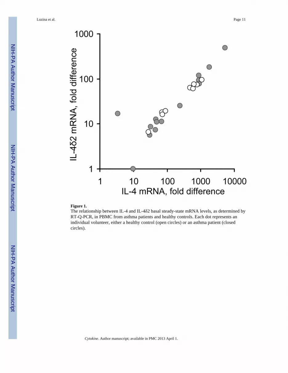
Figure 1.The relationship between IL-4 and IL-4δ2 basal steady-state mRNA levels, as determined byRT-Q-PCR, in PBMC from asthma patients and healthy controls. Each dot represents anindividual volunteer, either a healthy control (open circles) or an asthma patient (closedcircles).
Luzina et al. Page 11
Cytokine. Author manuscript; available in PMC 2013 April 1.
NIH
-PA Author Manuscript
NIH
-PA Author Manuscript
NIH
-PA Author Manuscript
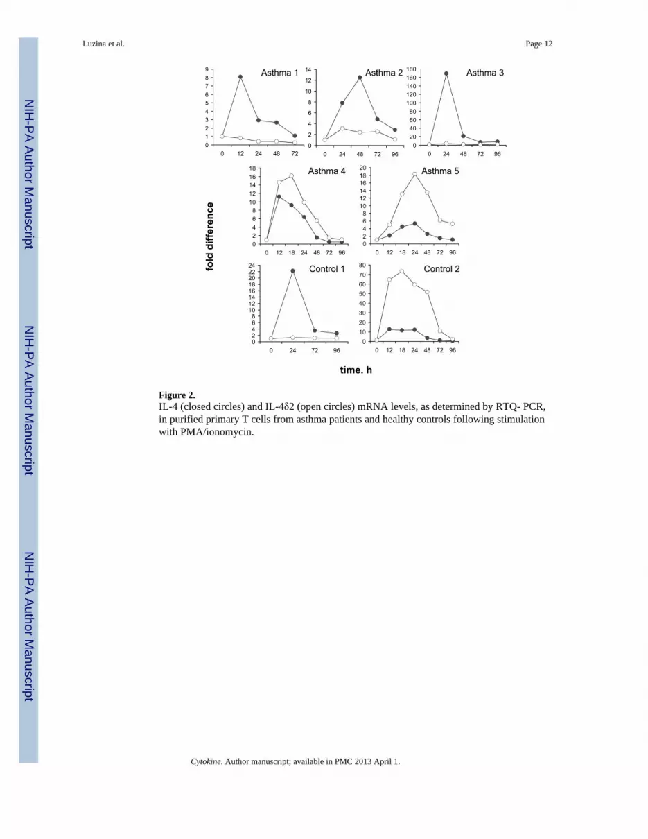
Figure 2.IL-4 (closed circles) and IL-4δ2 (open circles) mRNA levels, as determined by RTQ- PCR,in purified primary T cells from asthma patients and healthy controls following stimulationwith PMA/ionomycin.
Luzina et al. Page 12
Cytokine. Author manuscript; available in PMC 2013 April 1.
NIH
-PA Author Manuscript
NIH
-PA Author Manuscript
NIH
-PA Author Manuscript
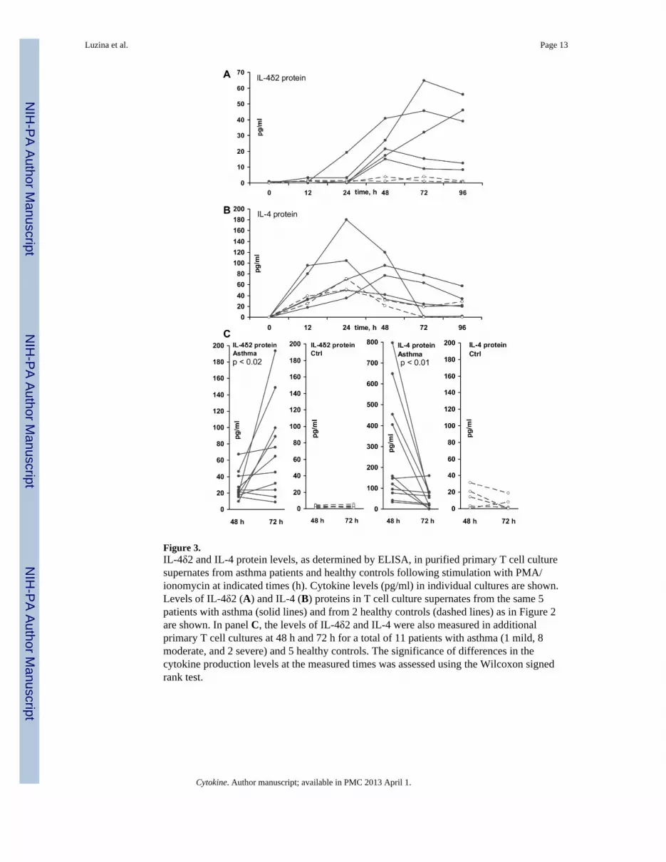
Figure 3.IL-4δ2 and IL-4 protein levels, as determined by ELISA, in purified primary T cell culturesupernates from asthma patients and healthy controls following stimulation with PMA/ionomycin at indicated times (h). Cytokine levels (pg/ml) in individual cultures are shown.Levels of IL-4δ2 (A) and IL-4 (B) proteins in T cell culture supernates from the same 5patients with asthma (solid lines) and from 2 healthy controls (dashed lines) as in Figure 2are shown. In panel C, the levels of IL-4δ2 and IL-4 were also measured in additionalprimary T cell cultures at 48 h and 72 h for a total of 11 patients with asthma (1 mild, 8moderate, and 2 severe) and 5 healthy controls. The significance of differences in thecytokine production levels at the measured times was assessed using the Wilcoxon signedrank test.
Luzina et al. Page 13
Cytokine. Author manuscript; available in PMC 2013 April 1.
NIH
-PA Author Manuscript
NIH
-PA Author Manuscript
NIH
-PA Author Manuscript
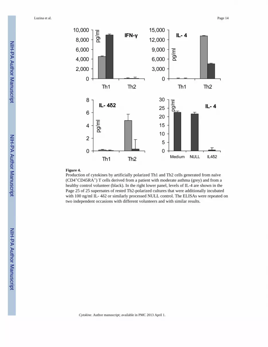
Figure 4.Production of cytokines by artificially polarized Th1 and Th2 cells generated from naïve(CD4+CD45RA+) T cells derived from a patient with moderate asthma (grey) and from ahealthy control volunteer (black). In the right lower panel, levels of IL-4 are shown in thePage 25 of 25 supernates of rested Th2-polarized cultures that were additionally incubatedwith 100 ng/ml IL- 4δ2 or similarly processed NULL control. The ELISAs were repeated ontwo independent occasions with different volunteers and with similar results.
Luzina et al. Page 14
Cytokine. Author manuscript; available in PMC 2013 April 1.
NIH
-PA Author Manuscript
NIH
-PA Author Manuscript
NIH
-PA Author Manuscript
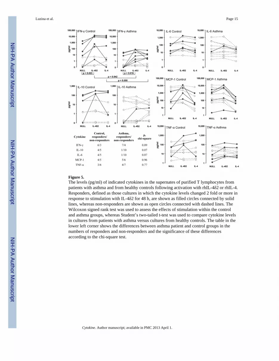
Figure 5.The levels (pg/ml) of indicated cytokines in the supernates of purified T lymphocytes frompatients with asthma and from healthy controls following activation with rhIL-4δ2 or rhIL-4.Responders, defined as those cultures in which the cytokine levels changed 2 fold or more inresponse to stimulation with IL-4δ2 for 48 h, are shown as filled circles connected by solidlines, whereas non-responders are shown as open circles connected with dashed lines. TheWilcoxon signed rank test was used to assess the effects of stimulation within the controland asthma groups, whereas Student’s two-tailed t-test was used to compare cytokine levelsin cultures from patients with asthma versus cultures from healthy controls. The table in thelower left corner shows the differences between asthma patient and control groups in thenumbers of responders and non-responders and the significance of these differencesaccording to the chi-square test.
Luzina et al. Page 15
Cytokine. Author manuscript; available in PMC 2013 April 1.
NIH
-PA Author Manuscript
NIH
-PA Author Manuscript
NIH
-PA Author Manuscript