nasolacrimal system injuries
TRANSCRIPT
Nasolacrimal System Injuries Chapter 9 91Chapter 9
Nasolacrimal System InjuriesD.A. Della Rocca, S. Ahmad, P. Preechawi, S.D. Schaefer and R.C. Della Rocca
9
Core Messages!
Damage to the lacrimal drainage system can cause telecanthus, globe displacement, epiphora, or obstructive dacryocystitis. It can affect patients both functionally and cosmetically.
It is essential to carefully check for any associated injuries such as neurological, thoracic, and abdominal trauma when significant facial trauma occurs.
An appropriate ocular examination and visual assessment should be performed.
A facial CT scan is required in any patients suspected of having nasoethmoid injuries.
Early one-stage repair is recommended. It consists of exposure of all fracture frag-ments and precise anatomic rigid fixation, immediate bone grafting, if needed, and definitive soft tissue management.
9.1 Introduction
This chapter describes the evaluation and treatment of injuries to nasolacrimal system. Isolated nasoeth-moid-orbital injuries are common. The most frequent causes are high-energy traumas, such as motor vehi-cle accidents [1, 2], by an impact force applied to the upper portion of the bridge of nose [3]. Often, a closed fracture is transformed into a compound one by lac-eration, avulsion, or bursting of the soft tissue of the naso-orbital area [4].
The patient with nasolacrimal injuries may present with significant concurrent facial wounds, and mul-tiple system injuries. Such patients must be rapidly evaluated and stabilized. The American College of Surgeons (ACS) has developed an Advanced Trauma Life Support (ATLS) protocol to guide one through the four steps of primary survey, resuscitation, sec-ondary survey, and definitive treatment. The primary
Contents
9.1 Introduction . . . . . . . . . . . . . . . . . . . . . . . . . . . . . . . . . . 919.2 Anatomical Considerations . . . . . . . . . . . . . . . . . . . . . 929.2.1 Osteology . . . . . . . . . . . . . . . . . . . . . . . . . . . . . . . . . . . . . 929.2.2 Soft Tissue . . . . . . . . . . . . . . . . . . . . . . . . . . . . . . . . . . . . 929.2.3 Lacrimal Excretory System . . . . . . . . . . . . . . . . . . . . . . 939.3 Case History . . . . . . . . . . . . . . . . . . . . . . . . . . . . . . . . . . 949.4 Diagnosis and Clinical Assessment . . . . . . . . . . . . . . 94
9.5 General Principles . . . . . . . . . . . . . . . . . . . . . . . . . . . . 959.6 Management . . . . . . . . . . . . . . . . . . . . . . . . . . . . . . . . . 969.7 Anesthesia . . . . . . . . . . . . . . . . . . . . . . . . . . . . . . . . . . . 969.8 Canalicular Laceration . . . . . . . . . . . . . . . . . . . . . . . . 979.9 Operative Technique . . . . . . . . . . . . . . . . . . . . . . . . . . 979.10 Postoperative Care . . . . . . . . . . . . . . . . . . . . . . . . . . . . 1009.10.1 Complications of Silicone Tube Intubation . . . . . . . 1009.10.2 Secondary Repair of Traumatic Canalicular
Stenosis . . . . . . . . . . . . . . . . . . . . . . . . . . . . . . . . . . . . . . 1009.10.3 Lacrimal Sac and Nasolacrimal Duct Injuries . . . . 1019.10.4 Different DCR Technique
in Post-traumatic Patients . . . . . . . . . . . . . . . . . . . . . 1019.10.5 Conjunctivodacryocystorhinostomy
with Jones Tube Intubation . . . . . . . . . . . . . . . . . . . . 101Reference . . . . . . . . . . . . . . . . . . . . . . . . . . . . . . . . . . . . . . . . . . . 102
D.A. Della Rocca, S. Ahmad, P. Preechawi et al.
9
92
survey focuses on the ABCs, i.e., airway, breathing, and circulation. In the facial injury patient, airway evaluation must include assessment of the patency of the upper aerodigestive track followed by assessing breathing effort. Possible etiologies of impaired air-way in these patients are facial fractures, foreign bod-ies, laryngeal injuries, and expanding hematomas. Impaired breathing may reflect neurological injuries sustained during the facial injury to direct trauma to the thorax. Poor perfusion of the head and neck, or more distal sites, suggests shock. Shock may be di-vided into neurological, cardiac, septic, and hypovo-lemic etiologies. Neurological and hypovolemic shock are the most common forms of shock in the patient with trauma confined to the head. Resuscitation be-gins with establishing a patent airway. This may be as simple as removing foreign bodies, blood, or mucus to intubation or surgical establishment of an airway. As the patient with head injuries may also have cervi-cal spine trauma, the neck should be stabilized by sand bags, and manipulation of the head may be min-imized until this site is further evaluated. Following the critical steps of the primary survey, the secondary survey should be performed. This part of the ATLS protocol recapitulates the ABC of the primary survey, and also includes the detailing the disability (D) and exposure or examination (E) to better define the in-jury. Depending on the findings in the primary sur-vey, attention is now focused on such areas as the de-finitive neurological examination; cervical spine, chest, or abdominal radiography; and careful exami-nation of the imaging and angiography. Following stabilization of the patient, and appropriate definitive management of more severe injuries, the patient can now enter the stage of definitive treatment of the nasolacrimal injury.
Anatomical landmarks in this area are helpful. They include the medial orbital wall, the medial canthal tendon, the eyelids, and the lacrimal drainage system. Damage to the structures can cause tele-canthus, globe displacement, epiphora, or obstructive dacryocystitis [1–18]. It can affect patients both functionally and cosmetically. A systemic treatment approach towards diagnosis and treatment is neces-sary.
9.2 Anatomical Considerations
9.2.1 Osteology
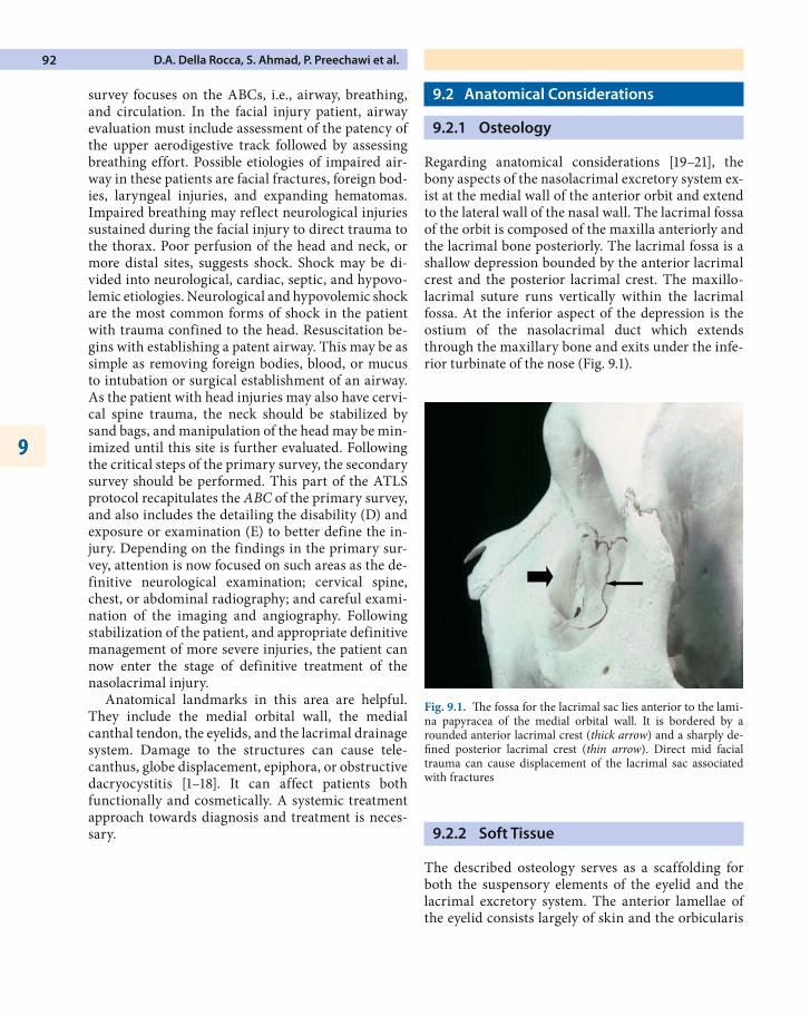
Regarding anatomical considerations [19–21], the bony aspects of the nasolacrimal excretory system ex-ist at the medial wall of the anterior orbit and extend to the lateral wall of the nasal wall. The lacrimal fossa of the orbit is composed of the maxilla anteriorly and the lacrimal bone posteriorly. The lacrimal fossa is a shallow depression bounded by the anterior lacrimal crest and the posterior lacrimal crest. The maxillo-lacrimal suture runs vertically within the lacrimal fossa. At the inferior aspect of the depression is the ostium of the nasolacrimal duct which extends through the maxillary bone and exits under the infe-rior turbinate of the nose (Fig. 9.1).
9.2.2 Soft Tissue
The described osteology serves as a scaffolding for both the suspensory elements of the eyelid and the lacrimal excretory system. The anterior lamellae of the eyelid consists largely of skin and the orbicularis
Fig. 9.1. The fossa for the lacrimal sac lies anterior to the lami-na papyracea of the medial orbital wall. It is bordered by a rounded anterior lacrimal crest (thick arrow) and a sharply de-fined posterior lacrimal crest (thin arrow). Direct mid facial trauma can cause displacement of the lacrimal sac associated with fractures
Nasolacrimal System Injuries Chapter 9 93
muscle. The orbicularis, which acts as the protractor of the eyelids, has a complex arrangement to where it originates at the medial wall. At this origin, the prese-ptal orbicularis is divided into a superficial head and deep (Jones muscle) head. The superficial head ex-tends from the anterior rim of the medial canthal ten-don which itself originates from the anterior lacrimal crest. The deep head of the preseptal orbicularis orig-inates at the lacrimal sac and its connective tissues.
The pretarsal orbicularis is adherent to the tarsus of the upper and lower eyelids. This is also split into superficial and deep (Horner’s tensor tarsi muscle) segments. The deep head extends from 4 mm poste-rior to the posterior lacrimal crest. This muscle’s pos-terior orientation allows for proper contour of the medial canthus and appropriate apposition of the eye-lids to the medial aspect of the eye globe. The superfi-cial horns of the pretarsal orbicularis inserts on the anterior edge of medial canthal tendon.
The complex arrangements of the muscles allows for a lacrimal pump of positive and negative pressures which helps move the tears within the palpebral fis-sures through the lacrimal excretory system. Bony and soft tissue traumatic injury to these structures may eliminate “lacrimal pump” physiology.
9.2.3 Lacrimal Excretory System
The lacrimal system begins at the lacrimal punctum which starts at the myocutaneous junction of the me-dial aspect of the lid margin of upper and lower eye-lids. The punctum are surrounded by a fibrous ring called the lacrimal papilla which is in turn surround-ed by the pretarsal orbicularis.
The canaliculi extends form the punctum to the lacrimal sac. The caniliculus initially has a vertical path of 2 mm followed by a medial extension toward the lacrimal sac. The medial extension (8–10 mm) fol-lows a horizontal pathway hugging the contour of the eyelid margin. As the canaliculi approach the lacri-mal sac, they tend to combine to form the common canaliculus. This final pathway enters the lateral wall of the lacrimal sac slightly above the vertical midpoint of the sac.
The lacrimal sac lies within the bony depression of the medial orbital wall, called the fossa of the lacrimal sac. The sack measures 12 mm in height, 4–6 mm in depth, and 2 mm in width. The shape is pisciform with a narrower top and wider lower portion. The
sack is bound by the fossa medially, the medial can-thal tendon superiorly, and muscle and orbital septum inferomedially.
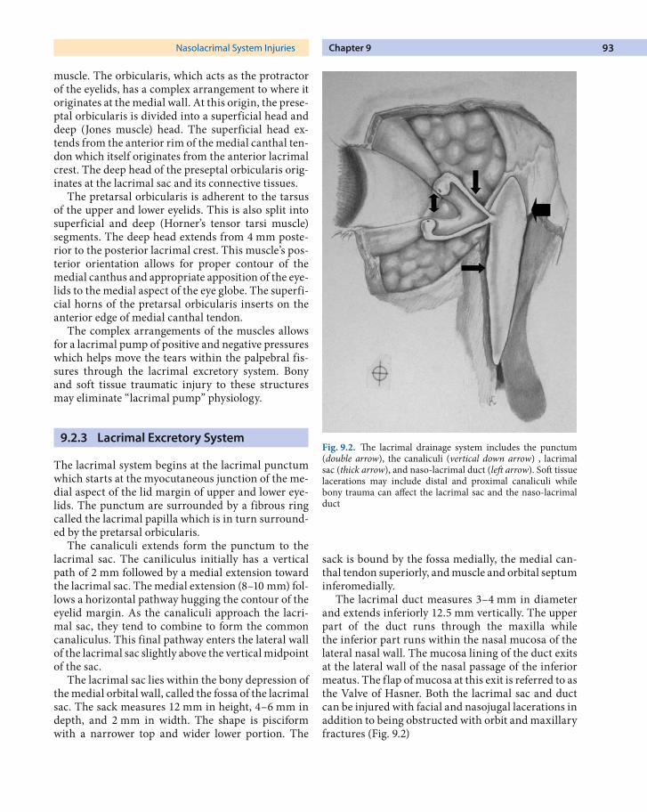
The lacrimal duct measures 3–4 mm in diameter and extends inferiorly 12.5 mm vertically. The upper part of the duct runs through the maxilla while the inferior part runs within the nasal mucosa of the lateral nasal wall. The mucosa lining of the duct exits at the lateral wall of the nasal passage of the inferior meatus. The flap of mucosa at this exit is referred to as the Valve of Hasner. Both the lacrimal sac and duct can be injured with facial and nasojugal lacerations in addition to being obstructed with orbit and maxillary fractures (Fig. 9.2)
Fig. 9.2. The lacrimal drainage system includes the punctum (double arrow), the canaliculi (vertical down arrow) , lacrimal sac (thick arrow), and naso-lacrimal duct (left arrow). Soft tissue lacerations may include distal and proximal canaliculi while bony trauma can affect the lacrimal sac and the naso-lacrimal duct
D.A. Della Rocca, S. Ahmad, P. Preechawi et al.
9
94
9.3 Case History
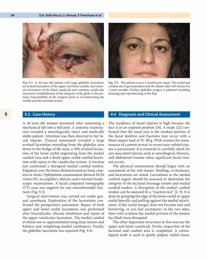
A 46-year-old woman presented after sustaining a mechanical fall into a bed post. A systemic examina-tion revealed a neurologically intact and medically stable patient. Attention was then directed to her fa-cial injuries. Clinical assessment revealed a large avulsed laceration extending from the glabellar area down to the bridge of the nose, a 50% avulsed lacera-tion of the lower eyelid originating from the medial canthal area and a distal upper eyelid canthal lacera-tion with injury to the canalicular system. A traction test confirmed a disrupted medial canthal tendon. Palpation over the bones demonstrated no bony crep-itus or clicks. Ophthalmic examination showed 20/20 vision OU, no pupillary deficits, and a normal fundo-scopic examination. A facial computed tomography (CT) scan was negative for any nasoethmoidal frac-tures (Fig. 9.3).
Surgical intervention was carried out under gen-eral anesthesia. Exploration of the lacerations con-firmed the preoperative assessment. Repair of both upper and lower eyelid lacerations was performed after bicanalicular silicone intubation and repair of the upper canalicular laceration. The medial canthal avulsion was re-approximated using deep sutures and bolsters and completing medial canthopexy. Finally, the glabellar laceration was repaired (Fig. 9.4).
9.4 Diagnosis and Clinical Assessment
The incidence of facial injuries is high because the face is in an exposed position [14]. A study [22] con-firmed that the nasal area is the weakest portion of the facial skeleton and fractures may occur with a blunt impact load of 35–80 g. With trauma the main-tenance of a patent airway in severe naso-orbital trau-ma is paramount. It is essential to carefully check for any associated injuries such as neurological, thoracic, and abdominal trauma when significant facial trau-ma occurs.
The physical examination should begin with an assessment of the soft tissues. Swelling, ecchymoses, and lacerations are noted. Lacerations in the medial canthal region should be assessed to determine the integrity of the lacrimal drainage system and medial canthal tendon. A disruption of the medial canthal tendon can be assessed by a “traction test” [2, 9]. It is done by grasping the edge of the lower eyelid or upper eyelid laterally and pulling against the medial attach-ment. If the eyelid margin does not become taut and bowstring, or you feel asymmetry in the two sides, then with avulsion the medial portion of the tendon has likely been disrupted.
The other important structures in this area are the upper and lower canaliculi. Firstly, inspection of the lacrimal and canthal area is completed. A cotton-tipped swab is used to gently palpate eyelid tissue.
Fig. 9.3. A 46-year-old patient with large glabellar laceration, an avulsed laceration of the upper and lower eyelids, and exten-sive laceration of the distal canaliculi and common canalicular structures. Establishment of the integrity of the globe is the pri-mary responsibility of the surgeon prior to reconstructing the eyelids and the lacrimal system
Fig. 9.4. The patient is seen 3 month post-repair. The eyelid and canthus are in good position and the silastic tube will remain for 2 more months. Further glabellar surgery is planned including thinning and repositioning of the flap
Nasolacrimal System Injuries Chapter 9 95
This can help define the location and extent of the in-jury. In addition, an accurate evaluation of the lacri-mal drainage effectiveness, irrigation, and probing should be performed. The presence of canalicular lac-eration requires silicone intubation and repair of the laceration. The distal lacrimal system, including the lacrimal sac and the nasolacrimal duct, tends not to be affected by trauma because they are well protected by the bony structures.
Inspection and physical examination of the pa-tients with nasoethmoid–orbital injuries can help predict the sites and extent of fractures prior to radio-graphical studies. The palpation over the bones onto the medial canthal tendon attachment will give good information [23]. This palpation may demonstrate bony crepitus or clicks depending on the degree of instability. The width and the symmetry of the me-dial canthi should be assessed for telecanthus. The normal intercanthal width ranges from 30 to 35 mm in whites [9, 24, 25], or half of interpupillary distance [9, 26], which is a more reliable guide. The third guide which might be used for the intercanthal width is equal to the alar–alar width at the base of the nose. The other obvious sign is saddle nose deformity which means loss of nasal skeletal support. Furthermore, typically, the medial aspect of the palpebral fissure may lose its sharpness and become rounded and slack with varying degrees of downward and outward dis-placement.
An ocular examination should be performed. Inju-ries in this area may be associated with ophthalmic emergency and problems such as ruptured globe or traumatic optic neuropathy especially when the prin-ciple fracture or displacement involves bones of the apex of the orbit [27–30]. There is no accurate inci-dence of ocular injuries associated with nasolacrimal injuries, because many studies vary in the level of ophthalmic evaluation; however, a study by Holt et al. [31] found 59% of nasal fractures showed concomi-tant eye injuries and 76% of midfacial fractures were associated with eye injuries. For the severity of ocular injuries, 79% were temporary or minor, and 18% were serious, defined as sustained visual loss or adnexal sequelae requiring subsequent reconstructive mea-sures; 3% resulted in blindness. Therefore, an initial ocular evaluation in mid-facial fractures is necessary [32–34]. Useful guidelines are as follows:
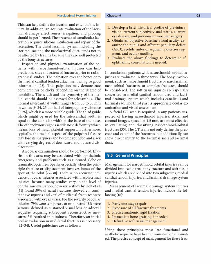
1. Develop a brief historical profile of pre-injury vision, current subjective visual status, current eye disease, and previous intraocular surgery.
2. Obtain an objective baseline visual acuity, ex-amine the pupils and afferent papillary defect (APD), eyelids, anterior segment, posterior seg-ment, and ocular motility.
3. Evaluate the above findings to determine if ophthalmic consultation is needed.
In conclusion, patients with nasoethmoid–orbital in-juries are evaluated in three ways. The bony involve-ment, such as nasoethmoid fracture or nasolacrimal, naso-orbital fractures, or complex fractures, should be considered. The soft tissue injuries are especially concerned in medial canthal tendon area and lacri-mal drainage system which includes canaliculi and lacrimal sac. The third part is appropriate ocular ex-amination and visual assessment.
A facial CT scan is required in any patients sus-pected of having nasoethmoid injuries. Axial and coronal images, spaced at 1.5 mm, are most effective in evaluating and classifying nasoethmoid–orbital fractures [35]. The CT scans not only define the pres-ence and extent of the fractures, but additionally can show direct injury to the lacrimal sac and lacrimal duct.
9.5 General Principles
Management for nasoethmoid orbital injuries can be divided into two parts, bony fracture and soft tissue injuries which are divided into two subgroups, medial canthal tendon injuries, and lacrimal drainage system injuries.
Management of lacrimal drainage system injuries and medial canthal tendon injuries include the fol-lowing [16]:
1. Early one-stage repair2. Exposure of all fracture fragments3. Precise anatomic rigid fixation4. Immediate bone grafting, if needed5. Definitive soft tissue management
Using these principles most late functional and aesthetic sequelae have been diminished or eliminat-ed. The precise concept of management for these frac-
D.A. Della Rocca, S. Ahmad, P. Preechawi et al.
9
96
tures is to do as much as possible at the first time [36]. It is unusual for the medial canthal tendon to be di-rectly injured in this type of trauma (blunt trauma) [9] and, because of that repositioning of the bony complex, the proper intercanthal relationship should be adequately restored [37–40].
The management of fractures in this area, when extensive, is completed utilizing open reduction, rigid osteological fixation, and plate implants as required [3, 5, 9, 11, 16, 36].
The lacrimal system is not frequently injured in nasolacrimal injuries in the absence of medial canthal avulsion or obvious lacrimal system transection [11, 13]. The incidence of late lacrimal obstruction requir-ing dacryocystorhinostomy was 5–10% following acute fracture management [43].
The indications for surgery in a nasolacrimal trau-ma are those outlined above. Restoration of pre-injury facial aesthetics and function is the goal of treatment. Since these injuries are usually associated with sig-nificant cosmetic and functional sequelae, expedi-tious restoration of injuries and function prevents latent cosmetic and functional deficits. Longer-term follow-up allows the surgeon to assess for both early and late sequelae of injuries.
Definitive treatment of nasolacrimal injuries should be deferred until the patient has been stabi-lized regarding any concomitant, compromising, or life-threatening trauma. During this time, systemic deficits can be corrected while giving the surgeon time for an accurate assessment prior to the operative procedure. As with any operative procedure, the risks of general anesthesia and the stresses of surgery must be weighed against medical contraindications. Ocu-lar contraindications include optic nerve injury and globe injury (e.g., hyphema, rupture, laceration). These injuries should be addressed and stabilized prior to surgical intervention, since osseous manipu-lation may exacerbate damage to the eye. Some inju-ries may not need correction, provided that the pa-tient is satisfied with the appearance and function.
9.6 Management
While proper instrumentation is an essential element to the successful undertaking of surgical repair of nasolacrimal injuries, strict adherence to several ba-sic surgical principles is more important. Intimate knowledge of anatomy, adequate anesthesia, excellent
exposure of operative site, sufficient hemostasis, and proper wound closure will ultimately impact greater on the results than choice of manufacturer of instru-ments. One must also pay special attention to light-ing, suction, and instruments.
Surgical vision aids include telescopic loops with magnification from 2.5–3.5 times with an adequate field of view and comfortable working distance. The operating microscope may also have a role in certain procedures. Illumination of the surgical field is key; preferably, the operating room should have two ceiling-mounted lamps to minimize shadowing of the field. Head-mounted fiberoptic lamps also very useful.
The basic lacrimal irrigation set should include punctal dilators, Bowman probes, a lacrimal irriga-tion cannula, and a syringe filled with balanced salt solution (BSS).
Prior to probing, nasal packing should be per-formed. A basic nasal packing set should include Codman sponges (cotton strips 1 7.5 cm) soaked in 0.25 or 0.5% phenylephrine, nasal specula, and Gruenwald (Jansen) forceps. A fiberoptic headlight may be used to illuminate and aid in the insertion the cotton pledgets into the nasal cavity. An endoscope may also aid in visualization of the nasal cavity.
A number of manufacturers produce lacrimal in-tubation systems (Jed-Med, Ritleng, FCI company).The authors prefer the Crawford intubation system manufactured by Jed-Med. This consists of a pair of flexible stainless steel olive-tipped intubation rods that are approximately #000 to #0000 in size and are attached to a silastic tube. The small tip size allows for easy passage through the upper and or lower puncta. The system also has a retrieval hook that is used to engage the olive tips when the tubes are externalized in the nares.
9.7 Anesthesia
The choice of anesthesia depends on several factors. Treatment in children must be performed under gen-eral anesthesia. For most adults, treatment can be performed with monitored anesthesia with intrave-nous sedation. Because of the difficult anatomy of the nose, especially in the setting of extensive nasoeth-moidal orbital trauma or more extensive injuries, general anesthesia may be the preferred anesthetic approach. The surgeon’s familiarity with the tech-
Nasolacrimal System Injuries Chapter 9 97
nique is also important, so that abundant bleeding is avoided under local anesthesia. Otherwise, in experi-enced hands, monitored anesthesia care (MAC) can be offered to adults and surgery can be performed on a day-surgery outpatient basis.
Local anesthesia has some advantages in prefer-ence to regional anesthesia for its hemostatic proper-ties. The area of the lacrimal sac and both the superior and inferior medial aspects of the injured eyelid are first infiltrated with dilute anesthetic solution con-sisting of nine parts saline and one part 2% lidocaine (Xylocaine) with epinephrine 1:100,000 (in adults) or 0.5% lidocaine with 1:200,000 epinephrine (in chil-dren). The dilute anesthetic is less painful. Alterna-tively, 2% lidocaine with epinephrine can be mixed with bicarbonate in a 9-to-1 ratio. For maximum he-mostasis, the anesthetic solution should be prepared “fresh” by adding 0.3 ml of epinephrine 1:1000 to a 30-ml bottle of 2% lidocaine and administered 10 min prior to the procedure. Epinephrine is not used if the patient has a history of coronary artery disease.
The sensation to the nose derives from the in-fratrochlear, infraorbital, supratrochlear, and anterior ethmoidal nerves. The base of the nose at the anterior septum, the nasal root, dorsum, lateral nasal walls, along with the middle turbinate, is infiltrated with 1–2% lidocaine with 1:100,000 epinephrine. This field block is more effective than targeted nerve blocks. Local hemostasis and anesthesia are augmented with nasal vasoconstrictors, such as phenylephrine soaked cottonoids. This may be preceded by topical decon-gestion and anesthesia (e.g., oxymetazoline, Ceta-caine) to aid in more comfortable introduction of the pledgets. The physician must wait an adequate period (approximately 15–20 min) to allow the anesthesia and vasoconstriction to be effective.
The benefits of general anesthesia in orbital sur-gery and more complicated procedures are many: Firstly, it provides deep orbital anesthesia, especially during osteotomy procedures and other bone work, which is difficult to achieve with regional blockade. Secondly, it allows for monitoring of blood pressure and heart rate. It provides a relative systemic hypo-tension, which is helpful in reducing bleeding during orbital surgery. Thirdly, the necessary volume of in-jectable anesthetic agent is reduced, limiting the risk of systemic toxicity.
General anesthesia can be safely administered to the ambulatory patient; however, because of the great effect on cardiovascular and respiratory systems, em-
phasis is placed on the preoperative evaluation. Pre-existing medical conditions need to be treated preop-eratively to ensure that the patient is in the best possible health prior to surgery. Of particular impor-tance is a recent history of myocardial infarction, hypertension, cardiac arrhythmia, and chronic ob-structive pulmonary disease, and diabetes. General anesthesia may be inappropriate for patients with poor systemic health, particularly those with ad-vanced cardiovascular or pulmonary disease.
9.8 Canalicular Laceration
Lacerations to the canaliculus should be treated pri-marily while injury to the lacrimal sac or nasolacri-mal duct can be operated on later [44], because there is a chance of spontaneous improvement. Studies of canalicular lacerations by experiments and retrospec-tive analysis [45] suggest that canalicular portion of the eyelid is particularly vulnerable to shearing, avul-sion, and stretching forces. Canalicular lacerations may occur by direct lacerations of the canaliculus or from diffuse or indirect injury. If the inferior cana-liculus is lacerated, bicanalicular silicone intubation is preferred over monocanalicular intubation. In ad-dition to being more stable within the lacrimal sys-tem, bicanalicular intubation of the lacrimal system is particularly effective in defining the medial canthus and commissure when treating medial canthal and eyelid avulsions.
9.9 Operative Technique
Repair of the canalicular system is optimally done under general anesthesia. Microsurgical repair by surgical loops or operating microscope is necessary [6, 46]. Before starting repair, it is important to con-strict the nasal mucosa with oxymethazoline or 0.25% phenylephrine on cotton pledgets placed inferiorly to the inferior turbinate. This will shrink the inferior turbinate and improve visualization. Following this, injection with 2% lidocaine with epinephrine 1:100,000 is done followed by repacking of the inferior meatus with the soaked cotton pledgets.
The punctal dilator is used to enlarge punctum and the lacrimal probe is used to navigate the proxi-mal canaliculus until the cut canaliculus is identified laterally. Canalicular injuries should be repaired
D.A. Della Rocca, S. Ahmad, P. Preechawi et al.
9
98
within 24–48 h after injuries [6, 46], because the me-dial cut edge of canaliculus becomes progressively more difficult to identify as fibrin and granulation deposition occurs. The medial cut edge of canaliculus is identified successfully by direct inspection. The cut canaliculus is identified as white mucosal tissue with wall and lumens. Deliberate inspection with gentle traction of the crowded tissue is often necessary. If discovery of the lumen remains difficult, injection of air into the uncut canaliculus while observing the medial cut area submerged in saline may uncover its location with the appearance of air bubbles [47–49]. Also, skin hooks and silk traction sutures can be used to retract the medial eyelid tissue as necessary.
After identification of the medial cut edge of the canaliculus, a Crawford tube is used to intubate the distal canaliculus and the lacrimal sac and duct. Fol-lowing this, a metallic probe attached to silicone tub-ing is insinuated into the proximal canaliculus, distal canaliculus, and then the lacrimal sack and duct. It is necessary to orient the probe to follow the anatomical course of the lacrimal system. Because visualization of the distal canaliculus is easily lost, it is useful to keep the Crawford tube in place until the moment of intubation with the silicone tubing. The hook or grooved dissector is used to deliver the probe from beneath the inferior turbinate and out the nostril. When a bicanilicular system is used, the opposite ca-nilicular system is insinuated in a similar way and retrieved through the nares.
The canaliculus can be approximated by two to three absorbable 8-0 sutures placed in the mucosal wall of cut canaliculus in order to achieve an end-to-end anastomosis of the tube [50, 51]. Some authors [52, 53] used single stitch repairs with 7-0 vicryl hori-zontal mattress sutures, which passed in the plane directly anterior to the canaliculus. The results [52] are excellent, although 4% still have epiphora and 13% still have delay outflow with dye disappearance test.
With compete avulsion of the medial canthus from its origin at the anterior lacrimal crest, reapproxima-tion can be done with a double armed 4-0 silk suture. This suture should be placed through the lateral wound edge with a substantial bite followed by a deep medial bite which would ideally include periostium of the anterior lacrimal crest. The sutures should be tied over the skin using bolsters of foam or rubber.
With bicanilicular intubation, the distal tube ends are joined with five single throws of the silicone su-ture. The silicone should have enough tension on it to
recess at least 1.5 cm into the nares after they are tied and released.
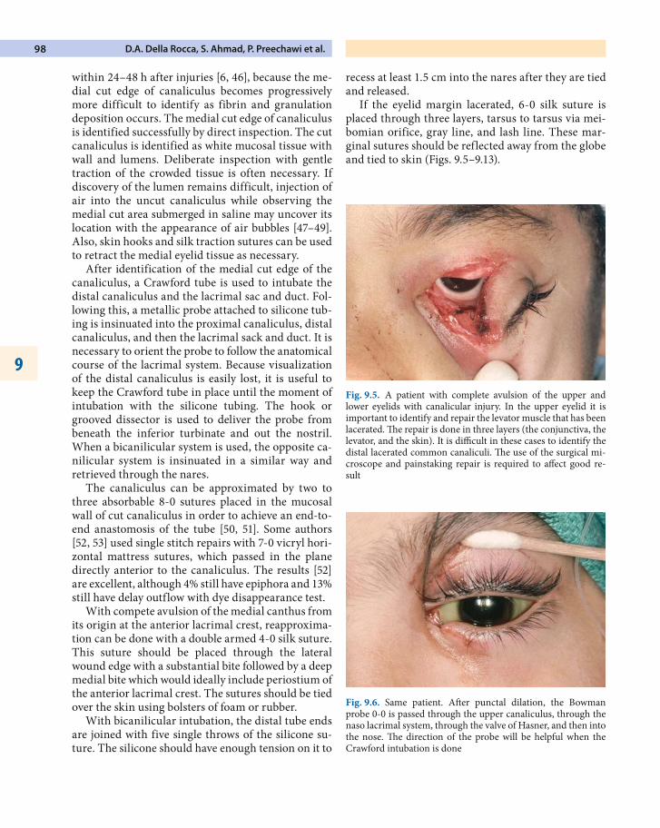
If the eyelid margin lacerated, 6-0 silk suture is placed through three layers, tarsus to tarsus via mei-bomian orifice, gray line, and lash line. These mar-ginal sutures should be reflected away from the globe and tied to skin (Figs. 9.5–9.13).
Fig. 9.5. A patient with complete avulsion of the upper and lower eyelids with canalicular injury. In the upper eyelid it is important to identify and repair the levator muscle that has been lacerated. The repair is done in three layers (the conjunctiva, the levator, and the skin). It is difficult in these cases to identify the distal lacerated common canaliculi. The use of the surgical mi-croscope and painstaking repair is required to affect good re-sult
Fig. 9.6. Same patient. After punctal dilation, the Bowman probe 0-0 is passed through the upper canaliculus, through the naso lacrimal system, through the valve of Hasner, and then into the nose. The direction of the probe will be helpful when the Crawford intubation is done
Nasolacrimal System Injuries Chapter 9 99
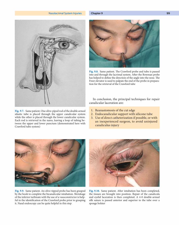
In conclusion, the principal techniques for repair canalicular laceration are:
1. Reanastomosis of the cut edge2. Endocanalicular support with silicone tube3. Use of direct catheterization if possible, or with
an inexperienced surgeon, to avoid uninjured canaliculus injury
Fig. 9.7. Same patient. One olive-piped rod of the double armed silastic tube is placed through the upper canalicular system while the other is placed through the lower canalicular system. Each rod is retrieved in the nares, leaving a loop of tubing be-tween the upper and lower punctum (demonstrated here with Crawford tube system)
Fig. 9.8. Same patient. The Crawford probe and tube is passed into and through the lacrimal system. After the Bowman probe has helped to define the direction of the angle into the nose. The Freer elevator is used to palpate the end of the probe in prepara-tion for the retrieval of the Crawford tube
Fig. 9.9. Same patient. An olive-tipped probe has been grasped by the hook to complete the bicanalicular intubation. Shrinkage of the inferior turbinate with the use of a vasoconstrictor is help-ful in the identification of the Crawford probe prior to grasping it. Nasal endoscopy can be quite helpful in this step
Fig. 9.10. Same patient. After intubation has been completed, the tissues are brought into position. Repair of the canalicula and eyelid laceration is then completed. A 4-0 double-armed silk suture is passed anterior and superior to the tube over a sponge bolster
D.A. Della Rocca, S. Ahmad, P. Preechawi et al.
9
100
In some very severe and extensive injuries, however, repair cannot be achieved the first time. Surgery should be obtained for the best possible repair of frac-ture and eyelids [54]. Then a bypass tube procedure may be considered in the next step of management.
9.10 Postoperative Care
Following silicone tube intubation, the patient is giv-en steroid–antibiotic eye drops four times a day for 1 week. The patient is reevaluated at 1 week after sur-
gery. If the patient has no stent-related problem, the stent is kept in place for 3–6 months.
9.10.1 Complications of Silicone Tube Intubation
Generally, the silicone rods are well tolerated; how-ever, if the tube is tied too tightly, or the lacrimal pa-pilla is compromised, “cheese wiring” through the puncta and canaliculi may occur, necessitating the re-moval of the tube. A pyogenic granuloma may devel-op near the punctum in some cases and should be excised with cautery to its base.
The tube can irritate the cornea and conjunctiva with adduction of the eyes. Tear supplements should be used; however, if keratoconjunctivitis is persistent, the tube should be removed earlier. Commonly, the tube can prolapse and extrude. Often the tube can be replaced into proper position with forceps through an intranasal approach with the aid of an endoscope. If the surgeon cannot replace the tube, it can be re-moved.
9.10.2 Secondary Repair of Traumatic Canalicular Stenosis
If epiphora exists in patients with eyelid lacerations, one should suspect unidentified or undetected cana-licular involvement. It is possible to repair at a later
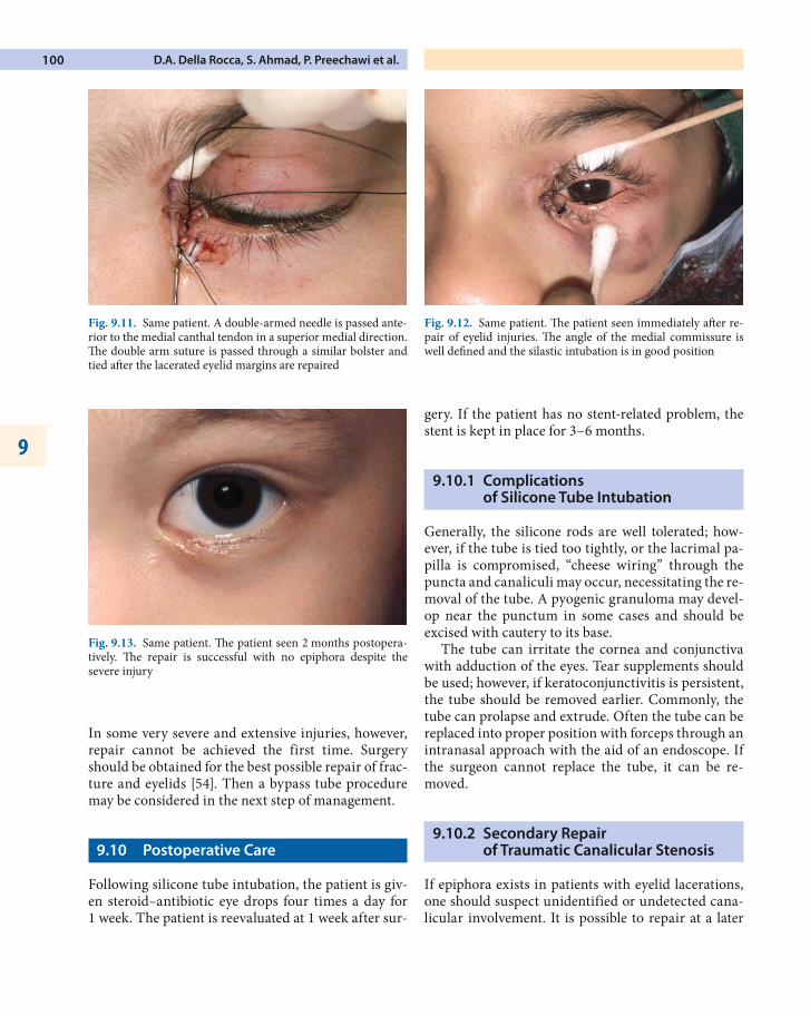
Fig. 9.11. Same patient. A double-armed needle is passed ante-rior to the medial canthal tendon in a superior medial direction. The double arm suture is passed through a similar bolster and tied after the lacerated eyelid margins are repaired
Fig. 9.12. Same patient. The patient seen immediately after re-pair of eyelid injuries. The angle of the medial commissure is well defined and the silastic intubation is in good position
Fig. 9.13. Same patient. The patient seen 2 months postopera-tively. The repair is successful with no epiphora despite the severe injury
Nasolacrimal System Injuries Chapter 9 101
date by excising the scar and reanastomoses the nor-mal lumen, followed by bicanalicular silicone intuba-tion [50, 55]. A nasolacrimal probe is passed into the punctum and passed to the site of obstruction. The eyelid is incised at this location. The purpose is to identify the proximal and distal ends of the lacerated canaliculus. At this time bicanilicular intubation is completed, if possible.
9.10.3 Lacrimal Sac and Nasolacrimal Duct Injuries
The incidence of persistent nasolacrimal system ob-struction requiring DCR ranged from 5 to 21% [13, 41, 42]. As previously mentioned, the management of lac-rimal sac and nasolacrimal duct injuries should not be explored at the initial surgery if there is no obvious laceration [11, 14, 41–43]. The rationale is that it is dif-ficult in adequate assessment and repair especially of the severe injury because of soft tissue edema and hemorrhage. Trauma to lacrimal pathways can pro-duce temporary or permanent dysfunction. Tempo-rary dysfunction is caused by lacrimal compression by posttraumatic edema. There were studies [41, 42] that showed spontaneous resolution of traumatic epiphora within 6 months after primary fracture re-pair. Persistent dysfunction is a result of direct causes such as detachment of the medial canthal ligament with subsequent sac compression and pumping fail-ure.
Irrigation of the system during primary fracture reconstruction or early postoperative period is not helpful due to edema and inflammation of the naso-lacrimal duct. We recommend better assessment 1–3 months after trauma when resolution of edema and soft tissue injuries permit the definitive evalua-tion. Using fluorescein dye instilled into inferior cul-de-sac then waiting 5 min to reevaluate, if dye still persists in cul-de-sac, it means that there is nasolacri-mal duct obstruction. The other investigation that is useful for evaluating post-traumatic nasolacrimal duct obstruction is CT scan and dacryocystography (DCG) [56–60] or combined CT and DCG. The com-bination of CT and DCG will give the useful informa-tion of complexity of anatomical change after trauma and repair, identify location of the lacrimal sac, bony structure, plate and screw implantation, and nasal septum which help in planning surgery.
For obvious laceration of the lacrimal sac or naso-lacrimal duct there are different techniques to per-form. Some authors advise applying silicone tube from punctum through the lacrimal sac and nasolac-rimal duct [14, 42], but only if this can be done easily. If any difficulty is encountered, the attempts should be curtailed to avoid damage to the canalicular sys-tem. Subsequent DCR surgery may be done, if neces-sary, when the healing process is complete, usually 6 months after injury [41].
9.10.4 Different DCR Technique in Post-traumatic Patients
In traumatic cases the surgical technique for DCR differs in several aspects from routine DCR [41, 42]:
1. The skin incision is somewhat lengthened.2. An attempt to avoid cutting of orbicularis oculi
muscle fibers to maintain lacrimal pump func-tion previously compromised by injury.
3. Bone removal may require bone drilling be-cause bone can be thicker from the inflamma-tion or impacted from the trauma. Care must be taken to avoid perforating the nasal muco-sa.
4. Delay secondary repair at least 5–6 months from primary repair and the subsequent DCR decreases friability of the sac and nasal muco-sa.
Silicone intubation is utilized in all cases because of the presumed predisposition to inflammation after trauma.
9.10.5 Conjunctivodacryocystorhinostomy with Jones Tube Intubation
In some instances, common punctum reconstruction cannot be accomplished because of extensive scarring involving the lacrimal sac or displaced lacrimal bone. Conjunctivodacryocysorhinostomy (CDCR) requires the placement of a Jones tube or similar bypass stent from the caruncle region directly through the lateral nasal mucosal wall following a large osteotomy. Prior to surgery, the patient requires counseling on the need for personal care of the tube and possible revision of the tube position.
D.A. Della Rocca, S. Ahmad, P. Preechawi et al.
9
102
Reference
1. Epker BN. Open surgical management of naso-orbit-al-ethmoid facial fractures. Trans Int Conf Oral Surg 1973;4:323–329
2. Fedok FG. Comprehensive management of nasoethmoid-orbital injuries. J Craniomaxillofac Trauma 1995;1:36–48
3. Converse JM, Smith B. Naso-orbital fractures and trau-matic deformities of the medial canthus. Plast Reconstr Surg 1966;38:147–162
4. Smith BC, Barr DR, Langham EJ. Complication of orbital fractures. N Y S J Med 1971;71:2407–2411
5. Stranc MF. Primary treatment of naso-ethmoid inju-ries with increased intercanthal distance.Br J Plast Surg 1970;23:8–25
6. Lindsey JT. Lacrimal duct injuries revisited: a retrospective review of six patients. Ann Plast Surg 2000;44:167–172
7. Ramselaar JM, van der Meulen JC, Bloem JJ. Naso-orbital fractures. Mod Probl Ophthal 1975;14:607–610
8. Smith B. Late bilateral naso-orbital fracture and dac-ryostenosis. Trans Am Acad Ophthalmol Otolaryngol 1972;76:1378–1379
9. Holt GR, Holt JE. Nasoethmoid complex injuries. Otolar-yngol Clin N Am 1985;18:87–98
10. Harris GJ, Fuerste FH. Lacrimal intubation in the primary repair of midfacial fractures. Ophthalmology 1987;94:242–247
11. Leipziger LS, Manson PN. Nasoethmoid orbital fractures. Clin Plast Surg 1992;19:167–193
12. White MJ, Johnson PC, Heckler FR. Management of max-illofacial and neck soft tissue injuries. Clin Sports Med 1989;8:11–23
13. Stranc MF. The pattern of lacrimal injuries in naso-eth-moid fractures. Br J Plast Surg 1970;23:339–346
14. Gruss JS. Fronto-naso-orbital trauma. Clin Plast Surg 1982;9:577–589
15. Katowitz JA, Diamond G. Ophthalmic consideration in cranio-orbital surgery. Clin Plast Surg 1987;14:155–162
16. Rohrich RJ, Shewmake KB. Evolving concepts of cra-niomaxillofacial fracture management. Clin Plast Surg 1992;19:1–10
17. Spinelli HM, Forman DL. Current treatment of post-trau-matic deformities. Clin Plast Surg 1997;24:519–530
18. Merville LC, Real JP. Fronto-orbital nasal dislocation. Scand J Plast Reconstr Surg 1980;15:287–297
19. Lemke BN, Della Rocca RC. The eyelids; medial canthal tendon: anatomy and surgery. Norwalk: Appleton and Lange, 1990:199–202
20. Shovlin JP, Lemke BN. Clinical eyelid anatomy. In: Bos-niak S, ed. Principle and practice of ophthalmic plas-tic and reconstructive surgery, Philadelphia: Saunders, 1996:261–280
21. Nerad JA. Clinical anatomy. In: Nerad JA, ed. Oculoplastic surgery: the requisites in ophthalmology. St. Louis: Mosby, 2001:25–70
22. Swearingen JJ. Tolerances of the human face to crash im-pact. Federal Aviation Agency, Oklahoma City, 1965
23. Paskert JP, Manson PN. The bimanual examination for as-sessing instability in naso-orbitoethmoidal injuries. Plast Reconstr Surg 1989;83:165–167
24. Hansman CF. Growth of interorbital distance and skull thickness as observed in roentgenographic measurements. Radiology 1966;86:87–96
25. Freihofer HP. Inner intercanthal and interorbital distanc-es. J Maxillofac Surg 1980;8:324–326
26. Collin JRO, Tyers AG. Preoperative evaluation. In: Collin JRO, Tyers AG, eds. Colour atlas of ophthalmic plastic sur-gery. Oxford: Butterworth-Heinemann, 2001:49–58
27. Hooper RS. Orbital complication of head injury. Br J Surg 1951;39:126
28. King AB, Walsh FB. Trauma to the head with particular reference to the ocular signs. Part I injuries involving the cranial nerves. Am J Ophthalmol 1949;32:191–205
29. Milauskas AT, Fueger GF. Serious ocular complication as-sociated with blow-out fractures of the orbit. Am J Oph-thalmol 1966;62:670–672
30. Miller GR, Tenzel RR. Ocular complication of mid-facial fractures. Plast Reconstr Surg 1967;39:37–42
31. Holt JE, Holt R, Blodgett JM. Ocular injuries sustained during blunt facial trauma. Ophthalmology 1983;90:14–18
32. Gossman MD, Roberts DM, Barr CC. Ophthalmic aspects of orbital injury: a comprehensive diagnostic and manage-ment approach. Clin Plast Surg 1992;19:71–85
33. Wessberg GA, Wolford LM, Zerdecki JW, et al. Ophthal-mic consideration in maxillofacial trauma. Int J Oral Surg 1981;10:236–246
34. Pelletier CR, Jordan DR, Braga R, et al. Assessment of ocular trauma associated with head and neck injuries. J Trauma Injury Infect Crit Care 1998;44:350–354
35. Manson PN, Markowitz B, Mirvis S et al. Toward CT-based facial fracture treatment. Plast Reconstr Surg 1990;85:202
36. Lauritzen C, Lilja J, Vallfors B. The craniofacial approach to trauma. Ann Plast Surg 1986;17:503–512
37. Beyer CK, Fabian RL, Smith B. Naso-orbital fractures, complication and treatment. Ophthalmology 1982;89:456–463
38. Converse JM, Hogan VM. Open sky approach for reduc-tion of naso-orbital fractures: case report. Plast Reconstr Surg 1970;46:396–398
39. Converse JM, Smith B. Naso-orbital fractures. Trans Am Acad Ophthalmol Otolaryngol 1976;80:622–623
40. Gross CW, Teague PF, Nakamura T. Reconstruction fol-lowing severe nasofrontal injuries. Otolaryngol Clin North Am 1972;5:653–665
41. Becelli R, Renzi G, Mannino G, et al. Posttraumatic ob-struction of lacrimal pathways: a retrospective analysis of 58 consecutive naso-orbitalethmoid fractures. J Craniofac Surg 2004;15:29–33
42. Gruss JS, Hurwitz JJ, Nik NA et al. The pattern and in-cidence of nasolacrimal injury in naso-orbital-ethmoid fractures: the role of delayed assessment and dacryocysto-rhinostomy. Br J Plast Surg 1985;38:116–121
43. Markowitz BL, Manson PN, Sargent LA et al. Management of the medial cantal tendon in nasoethmoid orbital frac-tures: the importance of the central fragment in classifica-tion and treatment. Plast Reconstr Surg 1991;87:843–853

Nasolacrimal System Injuries Chapter 9 103
44. Duvall AJ, Foster DA, Lyons DP et al. Medial canthoplas-ty: early and delayed repair. Laryngoscope 1981;91:173–183
45. Wulc AE, Arterberry JF. The pathogenesis of canalicular laceration. Ophthalmology 1991;98:1243–1249
46. Adenis JP. Management of canalicular trauma. Eye 1988; 2:223–225
47. Keith CG. Intubation of the lacrimal passages. Am J Oph-thalmol 1968;70–74
48. Lauring L. Silicone intubation of the lacrimal system: pitfalls, problems and complications. Ann Ophthalmol 1976;8:489–498
49. MacGillivray RF, Stevens MR. Primary surgical repair of traumatic lacerations of the lacrimal canaliculi. Oral Surg Oral Med Oral Pathol 1996;81:157–163
50. Leone CR. Periorbital trauma. Int Ophthalmol Clin 1995;35:1–24
51. Hawes MJ, Segrest DR. Effectiveness of bicanalicular sili-cone intubation in the repair of canalicular lacerations. Ophthalmic Plast Surg 1985;1:185–190
52. Kersten RC, Kulwin DR. “One stitch” canalicular repair: a simplified approach for repair of canalicular laceration. Ophthalmology 1996;103:785–789
53. Reed S, Lissner G. Cinical study on the effectiveness of tear drainage with a single canalicular system under environ-mental stress. Ophthal Plast Reconstr Surg 1993;9:17–31
54. Welham RA. The immediate management of injuries to the lacrimal drainage apparatus. Trans Ophthalmol Soc UK 1982;102:216–217
55. Drnovsek-Olup B, Beltram M. Trauma of the lacrimal drainage system: retrospective study of 32 patients. Croa-tian Med J 2004;45:292–294
56. Ellis E III. Sequencing treatment of naso-orbitoethmoid fractures. J Oral Maxillofac Surg 1993;51:543–558
57. Stranc MF, Bunce AH. Dacryo-cystography in mid-facial fractures. Br J Plast Surg 1972;25:269–275
58. Unger JM. Fractures of the nasolacrimal fossa and canal: a CT study of appearance, associated injuries, and signifi-cance in 25 patients. AJR 1992;158:1321–1324
59. Glatt HJ. Evaluation of lacrimal obstruction secondary to facial fractures using computed tomography or computed tomographic dacryocystography. Ophthalmic Plast Re-constr Surg 1996;12:284–293
60. Ashenhurst M, Jaffer N, Hurwitz JJ et al. Combined com-puted tomography and dacryocystography for complex lacrimal problems. Can J Ophthalmol 1991;26:27–31