nanomolar simultaneous determination of levodopa and melatonin at
TRANSCRIPT
Elsevier Editorial System(tm) for Sensors & Actuators: B. Chemical Manuscript Draft Manuscript Number: Title: Nanomolar simultaneous determination of levodopa and melatonin at a new cobalt hydroxide nanoparticles and multi-walled carbon nanotubes composite modified carbon ionic liquid electrode Article Type: Research Paper Keywords: Levodopa; Melatonin; Carbon Ionic Liquid Electrode; Electrochemical Sensor; Cobalt Hydroxide nanoparticles Corresponding Author: Dr Ali Babaei, PhD Corresponding Author's Institution: Arak University First Author: Ali Babaei, PhD Order of Authors: Ali Babaei, PhD; Ali Reza Taheri; Iman Khani Farahani Abstract: A novel modified carbon ionic liquid electrode (CILE) is prepared as an electrochemical sensor for simultaneous determination of Levodopa (L-dopa) and Melatonin (Mel). The experimental results suggest that a carbon ionic liquid electrode modified with multi-walled carbon nanotubes and cobalt hydroxide nanoparticles accelerates the electron transfer reactions of L-Dopa and Mel. The fabricated sensor revealed some advantages such as convenient preparation, good stability and high sensitivity. The DPV data in 0.1 M phosphate buffer soloution (PBS) (pH 7.5) allowed a method to be developed for the determination of L-Dopa and Mel concentrations in the ranges 0.1 to 300 and 0.01 to 50 μM, with the detection limits of 0.075 and 0.004 μM, respectively. The proposed method was successfully applied to determinations of these compounds in some pharmaceutical and human urine samples. Suggested Reviewers: Sheng Shui Hu [email protected] Yuzhong Z. Zhang [email protected] Hamid Reza Zare [email protected] Codrura Cofan [email protected] Jiannong Ye [email protected]
1
Nanomolar simultaneous determination of levodopa and melatonin at
a new cobalt hydroxide nanoparticles and multi-walled carbon
nanotubes composite modified carbon ionic liquid electrode
Ali Babaeia,b,
, Ali Reza Taheria, Iman Khani Farahani
c
a Department of Chemistry, Faculty of Science, Arak University, Arak, 38156-8-8349,
Iran
b Research Center for Nanotechnology, Arak University, Arak, 38156-8-8349, Iran
c Department of Biology, Faculty of Science, Yasouj University, Yasouj, 75918-74831,
Iran
Corresponding author. Tel.: +98 861 4173401; Fax: +98 861 4173406.
E-mail address: [email protected], [email protected]
*ManuscriptClick here to view linked References
2
Abstract
A novel modified carbon ionic liquid electrode (CILE) is prepared as an electrochemical
sensor for simultaneous determination of Levodopa (L-dopa) and Melatonin (Mel). The
experimental results suggest that a carbon ionic liquid electrode modified with multi-
walled carbon nanotubes and cobalt hydroxide nanoparticles accelerates the electron
transfer reactions of L-Dopa and Mel. The fabricated sensor revealed some advantages
such as convenient preparation, good stability and high sensitivity. The DPV data in 0.1
M phosphate buffer soloution (PBS) (pH 7.5) allowed a method to be developed for the
determination of L-Dopa and Mel concentrations in the ranges 0.1 to 300 and 0.01 to
50 μM, with the detection limits of 0.075 and 0.004 μM, respectively. The proposed
method was successfully applied to determinations of these compounds in some
pharmaceutical and human urine samples.
Keywords: Levodopa; Melatonin; Carbon Ionic Liquid Electrode; Electrochemical
Sensor; Cobalt Hydroxide nanoparticles.
1. Introduction
Melatonin (Mel, N-acetyl-5-methoxytryptamine) is a lipophilic hormone, mainly
produced and secreted at night by the pineal gland. The mechanisms that control its
synthesis within the pineal gland have been well characterized [1] and the retinal and
biological clock processes that modulate the circadian production of Mel in the pineal
gland are rapidly being unraveled [2]. Main and best-known effect of Mel is restoring
the natural cycle of organism functions[3]. It is safe and non-addictive sleep-inducing
3
drug, which can eliminate disruptions in our circadian rhythm, in such situations as shift
working, changing of time zones (during intercontinental air travelling) or insomnia.
However, researchers also have shown that Mel has chronobiotic activities to
resynchronize sleep and circadian rhythms disturbances and it is also involved in the
regulation of seasonal reproduction, body weight and energy balance [4]. Mel can be
detected in biological samples by several methods, such as HPLC [5],
spectrofluorimetric and colorimetric methods[6, 7], electrochemical methods [8, 9], GC-
MS methods [10], radioimmunoassay (RIA) [11], flow injection analysis system
(FIAED)[12] and capillary electrophoresis (CE) [13]. Furthermore, Mel content in
pharmaceutical products has also been determined by capillary electrophoretic methods
with UV [14] or electrochemical detection[15]. However, some drawbacks of these
methods include complicated pretreatment of sample, high cost or low sensitivity.
Levodopa (L-dopa) is a naturally occurring dietary supplement and psychoactive
drug found in certain kinds of herbs and food and is synthesized from the essential
amino acids L-phenylalanine and L-tyrosine in the brain and mammalian body. L-Dopa
is currently the therapeutic drug in the treatment of Parkinson’s disease and required by
the brain to produce dopamine which compensates the deficiency of dopamine in the
organism and decreases the symptoms of Parkinson’s disease[16]. Various analytical
methods have been developed for L-Dopa determination, such as
spectrophotometry[17], liquid chromatography[18], and capillary zone
electrophoresis[19].
Carbon nanotubes (CNTs) are carbon materials that have a new kind of porous
nanostructure, have been found to possess properties such as high surface area, high
electrical conductivity, significant mechanical strength and chemical stability [20].
4
Multiwalled carbon nanotubes (MWCNTs) can be used to promote electron transfer
reactions when used as electrode materials in electrochemical Sensors [21].
Room temperature ionic liquids (RTILs) such as 1-butyl-3-methylimidazolium
hexafluorophosphate, (BMIM)(PF6), have been proposed to be very interesting and
efficient pasting binder in place of non conductive organic binders such as Nojul or
paraffin oil for the preparation of carbon ionic liquid electrodes (CILEs)[22]. This new
composite has several advantages compared to other traditional carbon paste (CPEs)
such as high electrical conductivity, high stability and very low vapor pressure [23],
where it offers improved sensitivities toward a number of compounds, and at the same
time lower detection potentials using a very small amount of MWCNTs[24].
Transition-metal nanoparticles, in different forms, have emerged as a novel
family of catalysts able to promote more efficiently a variety of organic transformations
because of their extremely large surface-to-volume ratio and small size [25, 26]. Many
nanoparticles have been successfully introduced onto CNTs, such as TiO2[27],
CdTe[28], Au [29], Cu[30] and Ag [31]. Some electrodes such as platinum gauze[32] ,
glassy carbon electrode [33], and carbon paste electrode [34, 35] have been modified by
Co and Co(OH)2 particles and nanoparticles. Cobalt hydroxide nanoparticles (CHNPs)
with a low crystallinity and nano-flake network structure show a high proton diffusion
coefficient, giving excellent electrochemical performance. Various methods of
preparation of cobalt hydroxide nanoparticles, ranging from spray pyrolysis [36],
sonication[37], sputtering [38] and electrodeposition[33] to precipitate them at various
pH values, have been considered. The method of precipitation is new and facile,
needing no expensive raw materials or equipment. It is also easy for mass production
and can be extended to synthesize other hydroxide or oxide nanocrystals[32].
5
Simultaneous determination of L-Dopa and Mel is important, since numerous
reports demonstrated that L-Dopa and Mel influence each other in their respective
releasing [39-43] and also they coexist in a biological system. Lynch et al. reported L-
Dopa administration also causes profound increases in the pineal Mel content and its
biosynthesis [39]. This response is also potentiated by sympathetic denervation of the
pineal. Srinivasan et al. studied Mel secretion patterns in patients suffering from
Parkinson disease [40]. A phase advance of the nocturnal Mel maximum was noted in
L-Dopa-treated but not in untreated patients. Under medication with L-Dopa, daytime
Mel was additionally increased, a finding discussed in terms of an adaptive mechanism
in response to the neurodegenerative process and possibly reflecting a neuroprotective
property of Mel. However, to the best of our knowledge no study has reported yet about
the simultaneous determination of L-Dopa and Mel.
In the present work an RTIL, (BMIM)(PF6), is used as the binder for fabrication
of a CILE and modified with a nanocomposite film which contains MWCNTs and
CHNPs, based on the idea that the MWCNTs with CHNPs could enhance the electron
transfer rate for L-Dopa and Mel, due to synergistic electrocatalysis which leads to
increasing the sensitivity. The fabricated electrode was used as a new sensor for
simultaneous determination of Mel and L-Dopa in some real samples.
2. Experimental
2.1 Reagents and solutions
L-Dopa and Mel were obtained from Acros and Sigma chemical companies,
respectively. (BMIM)(PF6) was obtained from Hangzhou Kemer Chemical Limited
6
Company. Spectrally pure graphite powder (average particle size 50 μm) from Merck
was used as received. Multiwalled carbon nanotubes (MWCNTs) (>95 wt%, 5-20 nm)
were purchased from PlasmaChem GmbH company. Phosphate buffer 0.1 M solution
(PBS) was prepared by dissolving appropriate amounts of sodium hydrogen phosphate
and sodium dihydrogen phosphate in a 250 mL volumetric flask. The solution pH was
adjusted to appropriate value by addition of 7.5 M sodium hydroxide solution. All
electrochemical experiments were carried out in 0.1 M PBS at pH 7.5. The other
chemicals were of analytical reagent grade purchased from Merck and used without
further purification.
2.2 Synthesis of nanoscale Co (OH)2
CHNPs were synthesized according to a literature method[32]. Briefly, Co(OH)2
nanoparticles were prepared by a simple precipitation method. The first step was the
dissolving of cobalt chloride as aqueous solution (1 M, 25 ml) in a glass beaker, using a
magnetic stir bar. The cobalt chloride solution was slowly adjusted to pH 9 by addition
of 5 wt. % NH3·H2O (30 ml) at a temperature around 10 C. The NH3·H2O was added
drop wise with a constant time interval of 5 s. The resulting suspension was stirred at
this temperature for an additional 3 h. Then the solid was filtered, washed with a
copious amount of distilled water several times. The obtained CHNPs product was dried
at 100C.
2.3 Instrumentation
7
All the voltammetric measurements were carried out using the MWCNTs-
CHNPs/CILE electrode as the working electrode, Ag/AgCl 3 M KCl as the reference
electrode and platinum wire as an auxiliary electrode at room temperature. A magnetic
stirrer was used for the convective transport of the analyte. Cyclic voltammetry was
scanned between -0.2 and 1 V at the scan rate of 0.1 V s-1
. Amperometric measurement
was conducted under forced convection (stirring) by applying the appropriate potentials
and allowing the transient currents to decay to a steady-state value. All experiments
were done under a nitrogen atmosphere at room temperature by using an Autolab
PGSTAT 30 Potentiostat Galvanostat (EcoChemie, The Netherlands) coupled with a
663 VA stand (Metrohm Switzerland). The pH measurements were performed with a
Metrohm 744 pH meter using a combination glass electrode. X-ray diffraction (XRD)
measurements were performed at a speed of 0.01 s
−1 by a Bruker Axs diffractometer
(Germany) with Cu Kα (λ=1.5418 nm) operating at 40 kV, 30 mA. The morphology of
the nanoscale CHNPs was investigated by scanning electron microscopy (SEM, Leica
Cambridge, model S 360) and transmission electron microscopy (TEM, Philips CM10).
2.4. Electrode modification
The carbon ionic liquid electrode (CILE) was prepared by mixing graphite
powder and (BMIM)(PF6) (w/w 4:1) thoroughly in a mortar to form a carbon paste. A
portion of the carbon paste was firmly filled into one end of a glass tube (ca. 1.8 mm i.d.
and 10 cm long) and a copper wire was inserted through the opposite end to establish an
electrical contact. The surface of the CILE was smoothened on a piece of weighing
paper. The fabricated CILE was used as the basic solid electrode. A stock solution of
8
MWCNTs–CHNPs in DMF was prepared by dispersing weighed amounts of MWCNTs
and CHNPs (94/6% : w/w) in 1 mL DMF using ultrasonic bath until a homogeneous
solution resulted, and 20µL of the prepared suspension was casted on the CILE surface
with a microsyring and dried at room temperature. During these procedures a small
bottle was fitted tightly over the electrode so that the solvent could evaporate slowly
and a uniform film was formed. The fabricated electrode was stored at 4 C when not in
use. For comparison, MWCNTs/CILE and CHNPs/CILE were prepared with similar
procedures and used for further investigation.
2.5 General procedure
The electrode was first activated in PBS by cyclic voltammetric sweeps between
-0.1 and 1.1V until stable cyclic voltammograms were obtained. Each sample solution
(10 mL) containing 0.1 M PBS (pH 7.5) and appropriate amounts of L-Dopa and Mel
were pipetted into a voltammetric cell. The open-circuit accumulation time was 90 s.
Upon using the differential pulse voltammetric technique the oxidation peaks for L-
Dopa and Mel appeared at 0.075 and 0.70 V respectively. After every measurement, the
electrode was regenerated by soaking then rinsing thoroughly with triply distilled water
and then 0.5% sodium hydroxide solution for few seconds to remove adsorbed
substances. The electrode was finally rinsed carefully with distilled water to remove all
adsorbate from the electrode surface and to provide a fresh surface before running the
next experiments. All sample solutions were deoxygenated by purging with N2 gas
before each experiment.
9
3. Results and Discussion
3.1. Characterization of the CHNPs
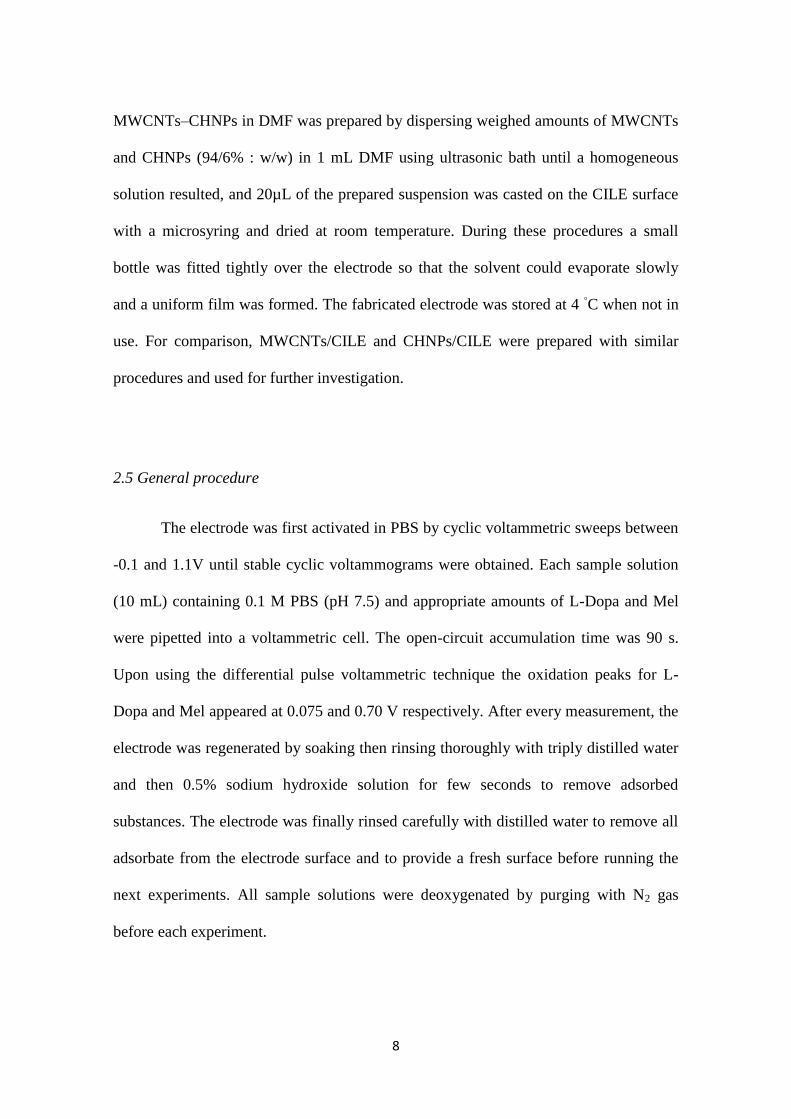
The response of a sensor is related to physical morphologyof its surface.
Scanning electron microscopy (SEM) was performed to the CHNPs synthesized through
the precipitation method (Fig. 1a). The morphology of CHNPs shows a network-like
structure which consists of interconnected nano-flakes. The SEM image shows the
agglomerated Co(OH)2 particles with an average size of less than 100 nm. It can be seen
that the CHNPs are very homogeneous in size. Fig. 1b displays TEM image of
Nanoscale CHNPs. The result shows the nanoparticles are in the same sizes as it is
shown in the SEM image.
Figure 1
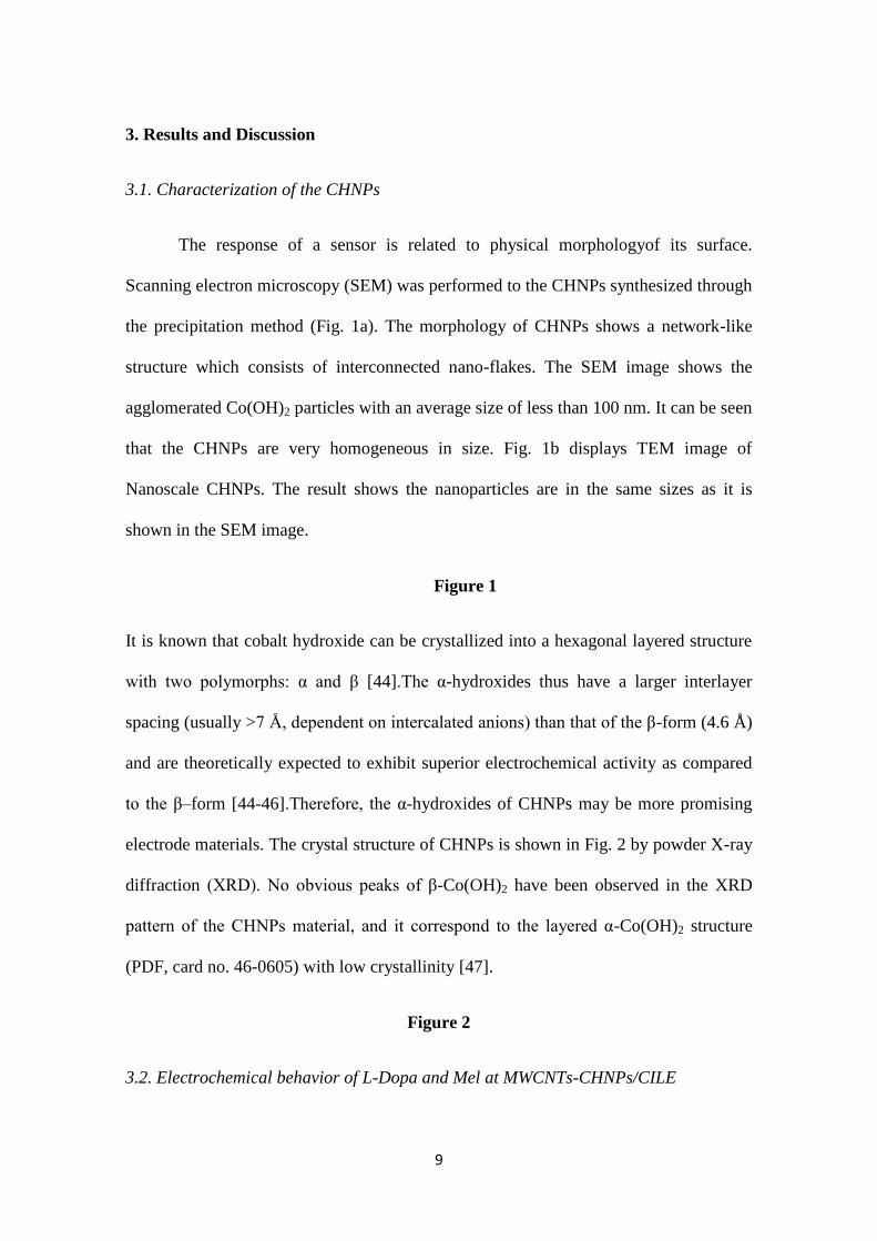
It is known that cobalt hydroxide can be crystallized into a hexagonal layered structure
with two polymorphs: α and β [44].The α-hydroxides thus have a larger interlayer
spacing (usually >7 Å, dependent on intercalated anions) than that of the β-form (4.6 Å)
and are theoretically expected to exhibit superior electrochemical activity as compared
to the β–form [44-46].Therefore, the α-hydroxides of CHNPs may be more promising
electrode materials. The crystal structure of CHNPs is shown in Fig. 2 by powder X-ray
diffraction (XRD). No obvious peaks of β-Co(OH)2 have been observed in the XRD
pattern of the CHNPs material, and it correspond to the layered α-Co(OH)2 structure
(PDF, card no. 46-0605) with low crystallinity [47].
Figure 2
3.2. Electrochemical behavior of L-Dopa and Mel at MWCNTs-CHNPs/CILE
10
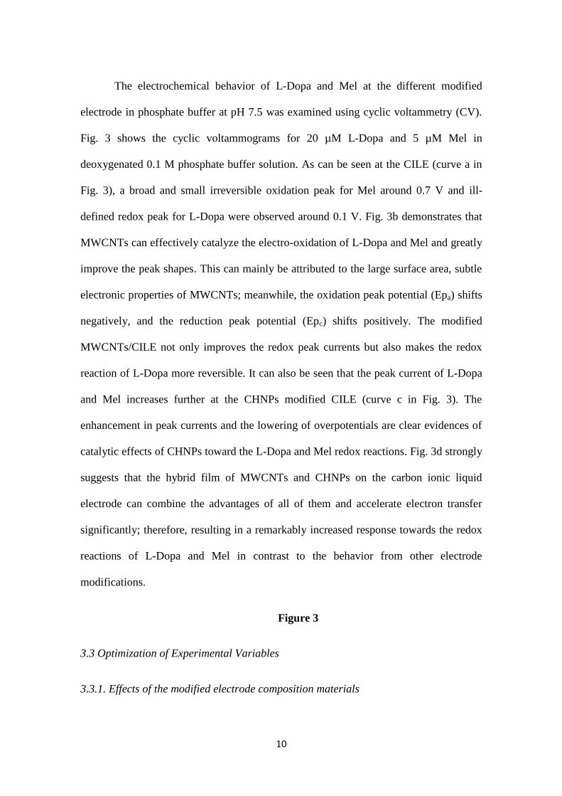
The electrochemical behavior of L-Dopa and Mel at the different modified
electrode in phosphate buffer at pH 7.5 was examined using cyclic voltammetry (CV).
Fig. 3 shows the cyclic voltammograms for 20 µM L-Dopa and 5 µM Mel in
deoxygenated 0.1 M phosphate buffer solution. As can be seen at the CILE (curve a in
Fig. 3), a broad and small irreversible oxidation peak for Mel around 0.7 V and ill-
defined redox peak for L-Dopa were observed around 0.1 V. Fig. 3b demonstrates that
MWCNTs can effectively catalyze the electro-oxidation of L-Dopa and Mel and greatly
improve the peak shapes. This can mainly be attributed to the large surface area, subtle
electronic properties of MWCNTs; meanwhile, the oxidation peak potential (Epa) shifts
negatively, and the reduction peak potential (Epc) shifts positively. The modified
MWCNTs/CILE not only improves the redox peak currents but also makes the redox
reaction of L-Dopa more reversible. It can also be seen that the peak current of L-Dopa
and Mel increases further at the CHNPs modified CILE (curve c in Fig. 3). The
enhancement in peak currents and the lowering of overpotentials are clear evidences of
catalytic effects of CHNPs toward the L-Dopa and Mel redox reactions. Fig. 3d strongly
suggests that the hybrid film of MWCNTs and CHNPs on the carbon ionic liquid
electrode can combine the advantages of all of them and accelerate electron transfer
significantly; therefore, resulting in a remarkably increased response towards the redox
reactions of L-Dopa and Mel in contrast to the behavior from other electrode
modifications.
Figure 3
3.3 Optimization of Experimental Variables
3.3.1. Effects of the modified electrode composition materials
11
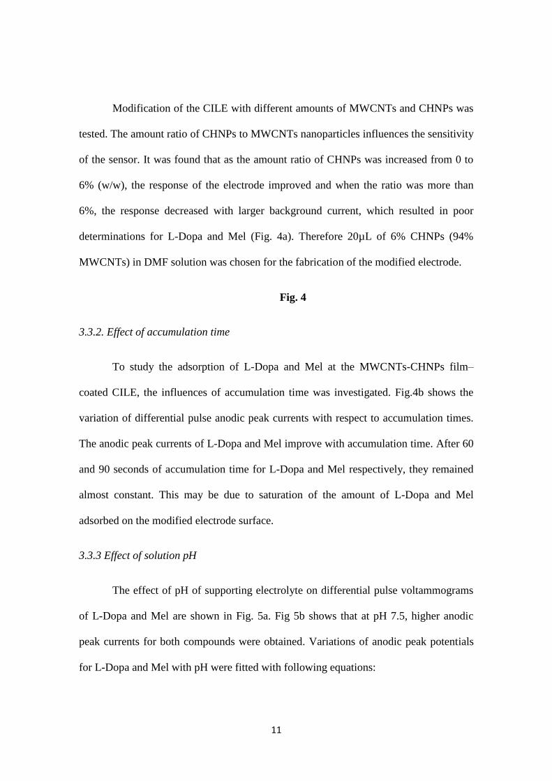
Modification of the CILE with different amounts of MWCNTs and CHNPs was
tested. The amount ratio of CHNPs to MWCNTs nanoparticles influences the sensitivity
of the sensor. It was found that as the amount ratio of CHNPs was increased from 0 to
6% (w/w), the response of the electrode improved and when the ratio was more than
6%, the response decreased with larger background current, which resulted in poor
determinations for L-Dopa and Mel (Fig. 4a). Therefore 20µL of 6% CHNPs (94%
MWCNTs) in DMF solution was chosen for the fabrication of the modified electrode.
Fig. 4
3.3.2. Effect of accumulation time
To study the adsorption of L-Dopa and Mel at the MWCNTs-CHNPs film–
coated CILE, the influences of accumulation time was investigated. Fig.4b shows the
variation of differential pulse anodic peak currents with respect to accumulation times.
The anodic peak currents of L-Dopa and Mel improve with accumulation time. After 60
and 90 seconds of accumulation time for L-Dopa and Mel respectively, they remained
almost constant. This may be due to saturation of the amount of L-Dopa and Mel
adsorbed on the modified electrode surface.
3.3.3 Effect of solution pH
The effect of pH of supporting electrolyte on differential pulse voltammograms
of L-Dopa and Mel are shown in Fig. 5a. Fig 5b shows that at pH 7.5, higher anodic
peak currents for both compounds were obtained. Variations of anodic peak potentials
for L-Dopa and Mel with pH were fitted with following equations:
12
L-Dopa: Epa (V) = −0.057 pH + 0.490 (R2 = 0.996) (1)
Mel: Epa (V) = −0.036 pH + 0.971 (R2 = 0.997) (2)
The results showed that the oxidation potentials of L-Dopa and Mel shift to less
positive potential with increasing solution pH which is a consequence of the
deprotonation involved in the oxidation process that is facilitated at higher pH values.
The slope of 0.057 and 0.036 VpH-1
for L-Dopa and Mel respectively, suggests that the
oxidations of L-Dopa involve the same number of transferred electrons and protons and
the number of electrons transferred in the oxidation of Mel is twice of protons, which is
in agreement with previous reports. [9, 48, 49]
Fig. 5
3.4 Effect of the scan rate
In order to investigate the effect of scan rate, cyclic voltammetry of solutions of
20 µM L-Dopa and 5 µM Mel were obtained in the range 0.01–0.8 Vs−1
. The effect of
scan rate on the oxidative peak potential (Epa) and peak current (Ipa) of L-Dopa and Mel
are shown in Fig. 6. The results showed that the peak currents vary linearly with the
scan rate over the range 0.01-0.15 Vs-1
(Figure 6b,d) for both compounds which
confirm the adsorption-controlled process for electro-oxidation of L-Dopa and Mel on
the surface of the electrode as following equations:
L-Dopa : Ipa = 0.251ν + 5.20 (R2 = 0.997) (3)
Mel : Ipa = 0.269ν + 7.59 (R2 = 0.998) (4)
13
At sweep rates between 0.2 V s−1
and 0.4 V s−1
, the plot of peak currents vs.
scan rate plot deviates from linearity and the peak current becomes proportional to the
square root of the scan rate which confirm diffusion controls of the systems(Figure
6c,e).
Fig. 6
The plot of Ep versus the logarithm of scan rate (log(ν)) was not linear for L-
Dopa, but showed a linear behavior for Mel according to Laviron theory [50]. The
charge transfer coefficient (α) can be determined by measuring the variation of Ep vs.
log(v). The slope of the Ep vs. log(ν), was about, 0.056 V. Using the equation of:
Ep = K − 2.3030 (RT/αnF) log(ν)
By considering two electrons transferred for Mel, charge transfer coefficient (α) of
0.530 was obtained which is in agreement with the results explained for the oxidation
process of Mel at the other electrodes[9, 49, 51]
3.5 Linear dynamic range and detection limit of the method
To obtain the linear dynamic range of the modified electrode under optimum
conditions for simultaneous determination of L-Dopa and Mel, the differential pulse
voltammetric behaviors of the mixed analytes were obtained (Fig 7 and 8). The
electrochemical response of additions of L-Dopa from 0.1 to 300 µM in the co-
existence of 5 µM Mel under the optimized conditions is depicted in Fig.7. By
application of the DPV method two linear ranges were obtained. The first linear
dynamic range was from 0.1 to 7.5 μM, with a calibration equation of Ip(µA) = 1.59c
14
(M) + 0.21 (R2=0.9993) and the second linear dynamic range was between 10 to 300
μM with a calibration equation of Ip(µA) = 1.55c (M) + 0.52 (R2=0.9996). A detection
limit of 0.075 µM (S/N = 3) was obtained.
Fig. 7
Figure 8 shows differential pulse voltammograms and the corresponding calibration
curves obtained from 0.01 to 50 µM of Mel in presence of 20 µM L-Dopa. The first
linear dynamic range was from 0.01 to 0.5 μM, with a calibration equation of Ip(µA) =
7.491 (M) + 0.329 (R2=0.9998) and the second linear dynamic range was between 1
μM to 50 μM with a calibration equation of Ip(µA) = 6.71c (M) + 2.44 (R2=0.9995). A
detection limit of 0.004 µM (S/N = 3) was obtained.
Fig. 8
Figure 9 displays a hydrodynamic chronoamperogram of oxidations of various
concentrations of L-Dopa and Mel at applied potential of 0.85 V in PBS (pH 7.5) using
the rotated modified electrode (2500 rpm). For Mel the linear dynamic range was from
0.3 to 49.2 µM, with a calibration equation of Ip(µA) = 3.45c (mol L-1
) + 1.21
(R2=0.9998) (Fig 9B) . A detection limit of 0.042 µM (S/N = 3) was obtained. For L-
Dopa the linear relationship was in the range of 0.8 to 98.2 µM with a calibration
equation of Ip(µA) = 1.49c (mol L-1
) – 0.22 (R2=0.9995) (Fig 9C). In addition a
detection limit of 0.10 µM was obtained.
Fig. 9
15
3.6 Repeatability and stability of the modified electrode
To evaluate the repeatability of the MWCNTs-CHNPs/CILE, the peak currents
of 20 successive measurements by DPV in a mixture solution of 20 µM L-Dopa and 5
µM Mel were determined. The relative standard deviation (R.S.D.) of 2.60% and 2.75%
were obtained for L-Dopa and Mel, respectively; indicating that the MWCNTs-
CHNPs/CILE is not subject to surface fouling by the oxidation products. The stability
of the proposed sensor was investigated. After 100 cyclic runs, the voltammetric
response to 20 µM L-Dopa and 5 µM Mel almost remained 88% and 87% of the initial
response, respectively (data not shown). The storage stability of the proposed sensor
was also studied. When not in use, the electrode was suspended in PBS at 4 C in a
refrigerator. The response to determination of 20 µM L-Dopa and 5 µM Mel were tested
intermittently. After 7 and 15 days of storage, the sensor retained 93% and 91% of its
initial response current in 20 µM L-Dopa, respectively. In addition the response to 5 µM
Mel was also tested and the sensor retained 94% and 92% of its initial response current
after 7 and 15 days respectively. The results indicate that the modified electrode has a
good stability.
3.7 Effects of interferences on the behaviors of L-Dopa and Mel
The effects of the common interfering species in solutions of 10 µM L-Dopa and
Mel were investigated in the optimum measurement conditions using differential pulse
voltammetric method. The tolerance limit was defined as the maximum concentration of
the interfering substance that causes an error less than 5% for determination of Mel and
L-Dopa. It was found that a 450-fold excess NaCl and KCl, 350-fold excess Ca(NO3)2,

16
300-fold excess MgCl2, and Cu(NO3)2, 220-fold excess of folic acid and oxalic acid,
200-fold excess of Ascorbic acid and Uric acid, 100-fold excess citric acid and
glutamic acid, 50-fold excess of glucose and L-histidine did not interfere with the
measurement of L-Dopa and Mel.
3.8 Determination of L-Dopa and Mel in real samples
The proposed method was applied to simultaneously determination of L-Dopa
and Mel in several commercially available pharmaceutical formulations and human
urine samples in order to demonstrate the capability of the modified electrode. Ten
tablets of each sample were weighed and pulverized by gentle grinding. An accurate
weight of the powder was dissolved in PBS (pH=7.4) in a water bath (450C) and
sonicated for 10 min. Then all sample solutions and running buffer were filtered
through a filter paper (Whatman No. 1) prior their use. Solutions obtained by dissolving
of L-Dopa and Mel tablets were subsequently diluted by PBS so that concentration lies
in the linear ranges. The prepared solutions were analyzed and the results obtained with
the MWCNTs-CHNPs/CILE were compared to the spectrophotometric method, as the
official method (Table 1). The analysis of L-Dopa and Mel for each sample was realized
in triplicate (n = 3). According to the t-test [52], there were no significant differences
between the calculated and comparative values at the 95% confidence level and within
an acceptable range of error, indicating that the modified electrode can be used for
voltammetric determinations of L-Dopa and Mel in the real samples.
Table 1
17
4. Conclusion
In the current study, a CHNPs and MWCNTs modified carbon ionic liquid
electrode (CILE) was fabricated. The combination of CHNPs nanoparticles and
MWCNTs show the characteristics of large surface area, good dispersing properties and
fast electron transfer. Due to the co-contribution of CILE and modifiers on the electrode
surface, the resulting electrode exhibited a good electrocatalytic performance to
simultaneous trace determination of L-Dopa and Mel. The analytical parameters of the
proposed electrode for determination of L-Dopa or Mel were compared with the earlier
reports and the results were listed in Table 2. A wide linear range, low detection limit,
high stability and good reproducibility suggest that this electrode will be an attractive
candidate for practical applications.
Table 2
Acknowledgments
The authors gratefully acknowledge the research council of Arak University for
providing financial support (grant number 89.13326) for this work.
18
References
[1] J. Barrenetxe, P. Delagrange, J. Martínez, Physiological and metabolic functions of
melatonin, J. Physiol. Biochem. , 60 (2004) 61-72.
[2] M. Mor, P. Plazzi, G. Spadoni, G. Tarzia, Melatonin, Curr. Med. Chem., 6 (1999)
501-518.
[3] CA. Medeiros, PF. Carvalhedo de Bruin, LA. Lopes, MC. Magalhães, M. de
Lourdes Seabra, V. d. Bruin, Effect of exogenous melatonin on sleep and motor
dysfunction in Parkinson's disease. A randomized, double blind, placebo-controlled
study., J. Neurol., 254 (2007) 459-464.
[4] MM. Macchi, JN. Bruce, Human pineal physiology and functional significance of
melatonin, Front Neuroendocrinol., 25 (2004) 177-195.
[5] A. Arabi, H. Najafzadeh, Determination Melatonin in Serum of Kurdish Horses by
HPLC in Kermanshah Region at Breeding Season, Int. J. Anim. Veter. Adv., 4 (2012)
170-172.
[6] A. S. Amin, M. Zaky, A. M. El-Beshbeshy, Colorimetric Estimation of Melatonin in
Pharmaceutical Formulations, Microchim. Acta, , 135 (2000) 81-85.
[7] A. S. Amin, M. Zaky, H. M. Khater, A. M. El-Beshbeshy, New Colorimetric
Methods for Microdetermination of Melatonin in Pure and in Dosage Forms, Anal.
Lett., 32 (1999) 1421-1434.
[8] B. Uslu, B. Demircigil, S. Ozkan, Z. Sentürk, H. Aboul-Enein, Simultaneous
determination of melatonin and pyridoxine in tablet formulations by differential pulse
voltammetry, Pharmazie, , 56 (2001) 938-942.
[9] A. Radi, G. E. Bekhiet, Voltammetry of melatonin at carbon electrodes and
determination in capsules, Bioelectrochem. Bioenerg., 45 (1998) 275-279.
[10] H. Y. Aboul-Enein, C. Doneanu, A. Covaci, Capillary gc–ms determination of
melatonin in several pharmaceutical tablet formulations, Biomed. Chromatogr., 13
(1999) 24-26.
[11] A. Miles, D. Philbrick, S. F. Tidmarsh, D. M. Shaw, Direct radioimmunoassay of
melatonin in saliva, Clin. Chem., 31 (1985) 1412-1413.
- - - -
, Voltammetric and flow amperometric methods for the determination of
melatonin in pharmaceuticals, J. Pharm. Biomed. Anal. , 31 (2003) 421-429.
[13] E. Poboży, A. Michalski, J. Sotowska-Brochocka, M. Trojanowicz, Determination
of melatonin and its precursors and metabolites using capillary electrophoresis with UV
and fluorometric detection, J. Separation Science, , 28 (2005) 2165-2172.
[14] G. P. Cartoni, F. Coccioli, R. Jasionowska, M. Masci, Rapid analysis of melatonin
in pharmaceutical tablets by capillary electrophoresis with UV detection,
Chromatographia, , 52 (2000) 603-606.
[15] G. Chen, X. Ding, Z. Cao, J. Ye, Determination of melatonin and pyridoxine in
pharmaceutical preparations for health-caring purposes by capillary electrophoresis with
electrochemical detection, Anal. Chim. Acta. , 408 (2000) 249-256.
[16] P. Damier, E. C. Hirsch, Y. Agid, A. M. Graybiel, The substantia nigra of the
human brain: II. Patterns of loss of dopamine-containing neurons in Parkinson's disease,
Brain, , 122 (1999) 1437-1448.
[17] M. R. Hormozi Nezhad, J. Tashkhourian, J. Khodaveisi, Sensitive
Spectrophotometric Detection of Dopamine, Levodopa and Adrenaline Using Surface
19
Plasmon Resonance Band of Silver Nanoparticles, J. Iran. Chem. Soc., 7 (2010) S83-
S91.
[18] C. Saxer, M. Niina, A. Nakashima, Y. Nagae, N. Masuda, Simultaneous
determination of levodopa and 3-O-methyldopa in human plasma by liquid
chromatography with electrochemical detection, J. Chromatogr. B, , 802 (2004) 299-
305.
[19] L. Zhang, G. Chen, Q. Hu, Y. Fang, Separation and determination of levodopa and
carbidopa in composite tablets by capillary zone electrophoresis with amperometric
detection, Anal. Chim. Acta, , 431 (2001) 287-292.
[20] M. Inagaki, K. Kaneko, T. Nishizawa, Nanocarbons––recent research in Japan,
Carbon, 42 (2004) 1401-1417.
[21] Y. Yang, S. Chen, Q. Xue, A. Biris, W. Zhao, Electron transfer chemistry of
octadecylamine-functionalized single-walled carbon nanotubes, Electrochim. Acta, 50
(2005) 3061-3067.
[22] M. J. Earle, K. R. Seddon, Ionic liquids. Green solvents for the future, Pure Appl.
Chem., 72 (2000) 1391-1398.
[23] J. G. Huddleston, A. E. Visser, W. M. Reichert, H. D. Willauer, G. A. Broker, R.
D. Rogers, Characterization and comparison of hydrophilic and hydrophobic room
temperature ionic liquids incorporating the imidazolium cation, Green Chem., 3 (2001)
156–164.
[24] Q. Wang, H. Tang, Q. Xie, L. Tan, Y. Zhang, B. Li, S. Yao, Room-temperature
ionic liquids/multi-walled carbon nanotubes/chitosan composite electrode for
electrochemical analysis of NADH, Electrochim. Acta, , 52 (2007) 6630-6637.
[25] M. Moreno-Manas, R. Pleixats, Formation of carbon-carbon bonds under catalysis
by transition-metal nanoparticles, Acc. Chem. Res., 36 (2003) 638-643.
[26] A. Roucoux, J. Schulz, H. Patin, Reduced transition metal colloids: a novel family
of reusable catalysts? , Chem. Rev., 102 (2002) 3757-3778.
[27] P. H. Lo, S. A. Kumar, S. M. Chen, Amperometric determination of H2O2 at nano-
TiO2/DNA/thionin nanocomposite modified electrode, Colloids Surf. B, , 66 (2008)
266-273.
[28] Q. Xu, J. H. Wang, Z. Wang, H. Q. Wang, Q. Yang, Y. D. Zhao, Biosensor for
Hydrogen Peroxide Based on Chitosan and Nanoparticle Complex Film–Modified
Glassy-Carbon Electrodes, Anal. Lett., 42 (2009) 2496 - 2508.
[29] T. J. Zhang, W. Wang, D. Y. Zhang, X. X. Zhang, Y. R. Ma, Y. L. Zhou, L. M. Qi,
Biotemplated Synthesis of Gold Nanoparticle-Bacteria Cellulose Nanofiber
Nanocomposites and Their Application in Biosensing, Adv. Funct. Mater., 20 (2010)
1152-1160.
[30] X. Kang, Z. Mai, X. Zou, P. Cai, J. Mo, A sensitive nonenzymatic glucose sensor
in alkaline media with a copper nanocluster/multiwall carbon nanotube-modified glassy
carbon electrode, Anal. Biochem., 363 (2007) 143-150.
[31] C. Y. Liu, J. M. Hu, Hydrogen peroxide biosensor based on the direct
electrochemistry of myoglobin immobilized on silver nanoparticles doped carbon
nanotubes film, Biosens. Bioelectron., 24 (2009) 2149-2154.
[32] L. B. Kong, J. W. Langa, M. Liua, Y. C. Luob, L. Kang, Facile approach to prepare
loose-packed cobalt hydroxide nano-flakes materials for electrochemical capacitors,
Journal of Power Sources, 194 (2009) 1194-1201.
20
[33] A. Salimi, R. Hallaj, S. Soltanian, Immobilization of hemoglobin on
electrodeposited cobalt-oxide nanoparticles: Direct voltammetry and electrocatalytic
activity, Biophys. Chem., 130 (2007) 122-131.
[34] S. shahrokhian, L. Fotouhi, Carbon paste electrode incorporating multi-walled
carbon nanotube/cobalt salophen for sensitive voltammetric determination of tryptophan
Sens. Actuators, B, 123 (2007) 942-949.
[35] S. shahrokhian, H. R. Zare, Cobalt salophen-modified carbon-paste electrode
incorporating a cationic surfactant for simultaneous voltammetric detection of ascorbic
acid and dopamine, Sens. Actuators, B, 121 (2007) 530-537.
[36] P. Nkeng, J. F. Koening, J. L. Gautier, P. Chartier, G. Poillerat, Enhancement of
surface areas of Co3O4 and NiCo2O4 electrocatalysts prepared by spray pyrolysis, J.
Electroanal. Chem., 402 (1996) 81-89.
[37] Y. Zhu, H. Li, Y. Koltypin, A. Gedanken, Preparation of nanosized cobalt
hydroxides and oxyhydroxide assisted by sonication, J. Mater. Chem., 12 (2002) 729-
733.
[38] L. C. Schumacher, I. B. Holzhueter, I. R. Hill, M. J. Dignam, Semiconducting and
electrocatalytic properties of sputtered cobalt oxide films, Electrochim. Acta, , 35
(1990) 975-984.
[39] H. J. Lynch, P. Wang, R. J. Wurtman, Increase in rat pineal melatonin content
following L-Dopa administration, Life Sci., 12 (1973) 145-151.
[40] V. Srinivasan, S. Pandi-Perumal, D. Cardinali, B. Poeggeler, R. Hardeland,
Melatonin in Alzheimer's disease and other neurodegenerative disorders, Behavioral
and Brain Functions, 2 (2006) 15-38.
[41] E. Fertl, E. Auff, A. Doppelbauer, F. Waldhauser, Circadian secretion pattern of
melatonin in de novo parkinsonian patients: evidence for phase-shifting properties of l-
dopa., J. Neural. Transm. Park. Dis. Dement. Sect., 5 (1993) 227-234.
[42] G. C. Cotzias, L. C. Tang, S. T. Miller, J. Z. Ginos, Melatonin and Abnormal
Movements Induced by L-Dopa in Mice, Science, , 173 (1971) 450-452.
[43] G. Rocchitta, R. Migheli, G. Esposito, B. Marchetti, M. S. Desole, E. Miele, P. A.
Serra, Endogenous melatonin protects L-DOPA from autoxidation in the striatal
extracellular compartment of the freely moving rat: potential implication for long-term
L-DOPA therapy in Parkinson's disease, J. Pineal Res., 40 (2006) 204-213.
[44] P. Oliva, J. Leonardi, J. F. Laurent, C. Delmas, J. J. Braconnier, M. Figlarz, F.
Fievet, A. d. Guibert, Review of the structure and the electrochemistry of nickel
hydroxides and oxy-hydroxides, J. Power Sources, , 8 (1982) 229-255.
[45] P. V. Kamath, M. Dixit, L. Indira, A. K. Shukla, V. G. Kumar, N. Munichandraiah,
Stabilized α-Ni(OH)2 as Electrode Material for Alkaline Secondary Cells J.
Electrochem. Soc., 141 (1994) 2956-2959.
[46] D. Singh, Characteristics and Effects of γ‐NiOOH on Cell Performance and a
Method to Quantify It in Nickel Electrodes J. Electrochem. Soc., 145 (1998) 116-120.
[47] M. Rajamathi, P. V. Kamath, R. Seshadri, Chemical synthesis of α-cobalt
hydroxide, Mater. Res. Bull. , 35 (2000) 271-278.
[48] M. F. S. Teixeira, L. H. Marcolino-J´unior, O. Fatibello-Filho, E. R. Dockal, M. F.
Bergamini, An electrochemical sensor for l-dopa based on oxovanadium-salen thin film
electrode applied flow injection system, Sens. Actuators, B, 122 (2007) 549-555.
[49] W. Qu, F. Wang, S. Hu, D. Cui, Electrocatalytic Properties and Voltammetric
Determination of Melatonin at a Nanostructured Film Electrode, Microchim. Acta, ,
150 (2005) 109-114.
21
[50] E. Laviron, General expression of the linear potential sweep voltammogram in the
case of diffusionless electrochemical systems., J. Electroanal. Chem. Int. Electrochem.,
101 (1979) 19-28.
[51] W. Xiao-Ping, Z. Lan, L. Wen-Rong, D. Jian-Ping, C. Hong-Qing, C. Guo-Nan,
Study on the Electrochemical Behavior of Melatonin with an Activated Electrode,
Electroanalysis, , 14 (2002) 1654-1660.
[52] J. C. Miller, J. N. Miller Statistics for analytical chemistry; 3 reprint ed.; Ellis
Horwood PTR Prentice Hall: Chichester, New York, 1993.
[53] P. Nagaraja, K. C. S. Murthy, K. S. Rangappa, N. M. M. Gowda,
Spectrophotometric methods for the determination of certain catecholamine derivatives
in pharmaceutical preparations, Talanta, , 46 (1998) 39-44.
[54] M. F. S. Teixeira, M. F. Bergamini, C. M. P. Marques, N. Bocchi, Voltammetric
determination of L-dopa using an electrode modified with trinuclear ruthenium ammine
complex (Ru-red) supported on Y-type zeolite, Talanta, , 63 (2004) 1083-1088.
[55] M. F. Bergamini, A. L. Santos, N. R. Stradiotto, M. V. B. Zanoni, A disposable
electrochemical sensor for the rapid determination of levodopa, J. Pharm. Biomed.
Anal., 39 (2005) 54-59.
[56] H. Yaghoubian, H. Karimi-Maleh, M. A. Khalilzadeh, F. Karimi, Electrocatalytic
Oxidation of Levodopa at a Ferrocene Modified Carbon Nanotube Paste Electrode, Int.
J. Electrochem. Sci., 4 (2009) 993 - 1003.
[57] H. d. Melo, A. P. D. Seleghim, W. L. Polito, O. Fatibello-Filho, I. C. Vieira,
Simultaneous Differential Pulse Voltammetric Determination of L-Dopa and Carbidopa
in Pharmaceuticals Using a Carbon Paste Electrode Modified with Lead Dioxide
Immobilized in a Polyester Resin, J. Braz. Chem. Soc., 18 (2007) 797-803.
[58] A. Babaei, M. Babazadeh, A Selective Simultaneous Determination of Levodopa
and Serotonin Using a Glassy Carbon Electrode Modified with Multiwalled Carbon
Nanotube/Chitosan Composite, Electroanalysis, , 23 (2011) 1726-1735.
[59] A. Radi, Electroanalysis of melatonin using castor oil–graphite paste electrode,
Anal. Commun., 36 (1999) 43-44.
22
Biographies
Ali Babaei received his BS degree in 1989 from Shahid Beheshti University, Tehran,
Iran; MS degree in 1991 from Mazandarn University, Babolsar, Iran and PhD degree
from Otago University, Dunedin, New Zealand. At present, he is associate professor of
chemistry at Arak University , Arak, Iran. His main area of interest at present is
electroanalytical chemistry.
Ali Reza Taheri received his BS degree in 2003 from Isfahan University, Isfahan, Iran;
MS degree in 2007 from Arak University, Arak, Iran. At present, he is a PhD student in
chemistry department of Arak University, Arak, Iran.
Iman Khani Farahani received his BS degree in 2008 from Arak University, Arak,
Iran;. At present, he is a MS student in biology department of Yasouj University,
Yasouj, Iran.
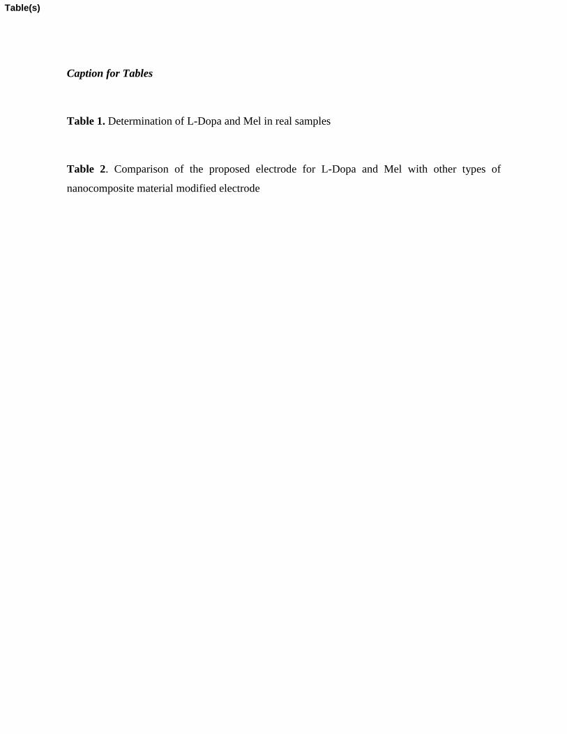
Caption for Tables
Table 1. Determination of L-Dopa and Mel in real samples
Table 2. Comparison of the proposed electrode for L-Dopa and Mel with other types of
nanocomposite material modified electrode
Table(s)
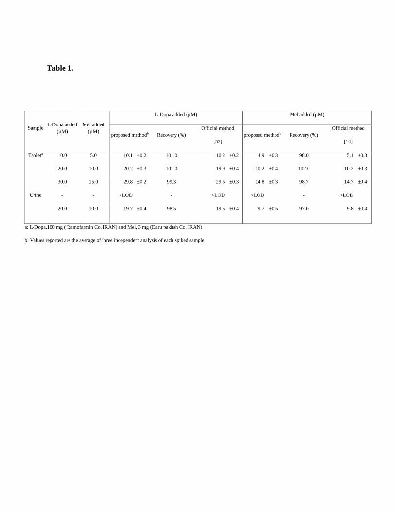
Table 1.
Sample L-Dopa added
(µM)
Mel added
(µM)
L-Dopa added (µM) Mel added (µM)
proposed methodb Recovery (%)
Official method
[53]
proposed methodb Recovery (%)
Official method
[14]
Tableta 10.0 5.0 10.1 ±0.2 101.0 10.2 ±0.2 4.9 ±0.3 98.0 5.1 ±0.3
20.0 10.0 20.2 ±0.3 101.0 19.9 ±0.4 10.2 ±0.4 102.0 10.2 ±0.3
30.0 15.0 29.8 ±0.2 99.3 29.5 ±0.3 14.8 ±0.3 98.7 14.7 ±0.4
Urine - - <LOD
- <LOD <LOD
- <LOD
20.0 10.0 19.7 ±0.4 98.5 19.5 ±0.4 9.7 ±0.5 97.0 9.8 ±0.4
a: L-Dopa,100 mg ( Ramofarmin Co. IRAN) and Mel, 3 mg (Daru pakhsh Co. IRAN)
b: Values reported are the average of three independent analysis of each spiked sample.
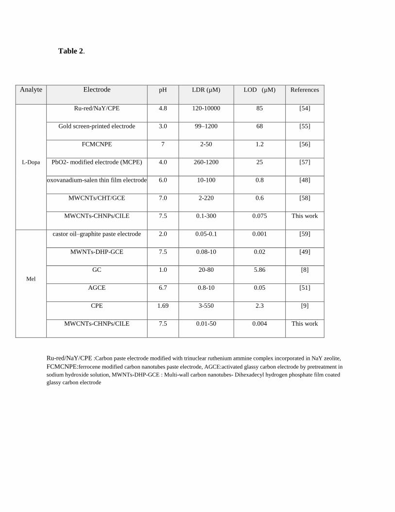
Table 2.
Analyte Electrode pH LDR (µM) LOD (µM) References
L-Dopa
Ru-red/NaY/CPE 4.8 120-10000 85 [54]
Gold screen-printed electrode 3.0 99–1200 68 [55]
FCMCNPE 7 2-50 1.2 [56]
PbO2- modified electrode (MCPE) 4.0 260-1200 25 [57]
oxovanadium-salen thin film electrode 6.0 10-100 0.8 [48]
MWCNTs/CHT/GCE 7.0 2-220 0.6 [58]
MWCNTs-CHNPs/CILE 7.5 0.1-300 0.075 This work
Mel
castor oil–graphite paste electrode 2.0 0.05-0.1 0.001 [59]
MWNTs-DHP-GCE 7.5 0.08-10 0.02 [49]
GC 1.0 20-80 5.86 [8]
AGCE 6.7 0.8-10 0.05 [51]
CPE 1.69 3-550 2.3 [9]
MWCNTs-CHNPs/CILE 7.5 0.01-50 0.004 This work
Ru-red/NaY/CPE :Carbon paste electrode modified with trinuclear ruthenium ammine complex incorporated in NaY zeolite,
FCMCNPE:ferrocene modified carbon nanotubes paste electrode, AGCE:activated glassy carbon electrode by pretreatment in
sodium hydroxide solution, MWNTs-DHP-GCE : Multi-wall carbon nanotubes- Dihexadecyl hydrogen phosphate film coated
glassy carbon electrode
Legends for Figures:
Figure 1. SEM (a) and TEM (b) image of the CHNPs.
Figure 2. Typical X-ray diffraction patterns ofCHNPs.
Figure 3.CVs of 20 µM L-Dopa and 5 µM Mel in 0.1 M PBS at CILE(a), MWCNTs/CILE (b),
CHNPs/CILE(c) and MWCNTs-CHNPs/CILE(e).
Figure 4.Effect of the amount ratio of CHNPs (a) and accumulation time (b), on themodified
electrodes using differential pulse anodic peak currents of 20 μM L-Dopa and 5 μM Mel in 0.1
M PBS (pH 7.5) atscan rate of 100mVs−1
.
Figure 5. DPVs of MWCNTs-CHNPs/CILE in 0.1 M PBs at different pHs.(curves A–F: 4.5, 5.5,
6.5, 7.5, 8.5, 9.5)in the presence of 20 µM of L-Dopa and 5 µM of Mel(a) and plot of peak
current vs. pH values (b).
Figure 6.CVs of 20 µM of L-Dopa and 5 µM of Mel at different scan rates (from A to L) 0.01,
0.025, 0.075, 0.1, 0.125, 0.150, 0.200, 0.250, 0.300, 0.350 and 0.4V s-1
(a).Plot of peak currents
vs. scan rate for L-Dopa (b), plot of peak currents vs. square root of scan rate for L-Dopa (c) ,(d)
as (b) for Mel, (e) as (c) for Mel.
Figure 7.DPVs of L-Dopa at MWCNTs-CHPs/CILE in the presence of 5 μM Mel. L-Dopa
concentrations (from A to L) are: 0.1, 0.5, 1, 3, 7.5, 12.5, 20, 45, 75, 130, 200, 300 μM. Insets:
Figure(s)
Plot of peak currents as a function of L-dopa concentration at low concentrations (a) and at high
concentrations (b).
Figure 8.DPVs of Mel at MWCNTs-CHNPs/CILE in the presence of 20 μM L-Dopa. Mel
concentrations (from A to L) are: 0.01, 0.05, 0.1, 0.15, 0.2, 0.5, 1.5, 5, 10, 15, 25, 50 μM. Insets:
Plot of peak currents as a function of Mel concentration at low concentrations (a)and at high
concentrations (b).Other conditions are the same as in Fig. 4.
Figure 9. Hydrodynamic amperometric response (rotating speed 2500 rpm) held at 0.85 V in
PBS (pH 7.5) for simultaneous determination of L-Dopa and Mel by successive additions of (a)
10 µM L-Dopa and (b) 5 µM Mel. Insets: (A) successive additions of (c) 0.8 µM L-Dopa and (d)
0.3 µM Mel; (B) Plot of currents as a function of Mel concentration and (C) Plot of the currents
as a function of L-Dopa concentration.
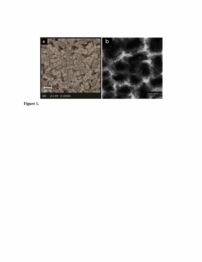
Figure 1.
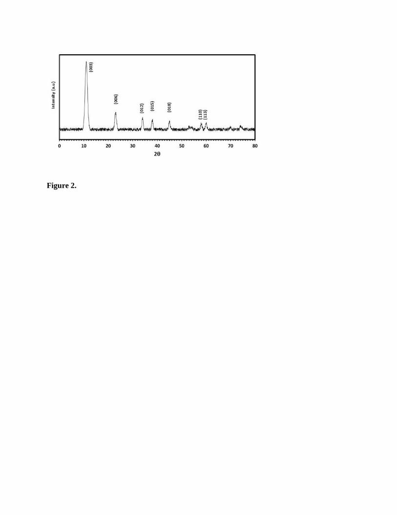
Figure 2.
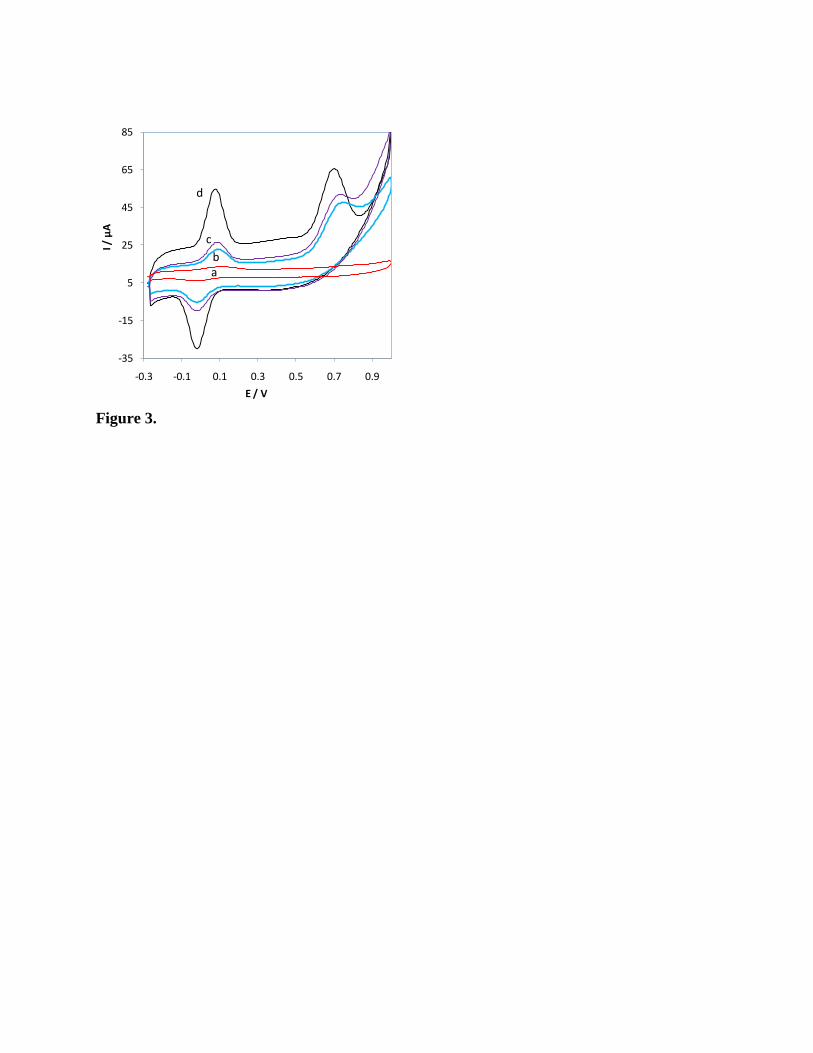
Figure 3.
-35
-15
5
25
45
65
85
-0.3 -0.1 0.1 0.3 0.5 0.7 0.9
I / µ
A
E / V
d
c
ba
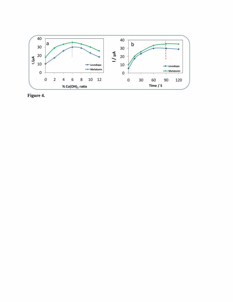
Figure 4.
0
10
20
30
40
0 30 60 90 120
I / µ
A
Time / S
Levodopa
Melatonin
b
0
10
20
30
40
0 2 4 6 8 10 12
I /µ
A
% Co(OH)2 ratio
Levodopa
Melatonin
a
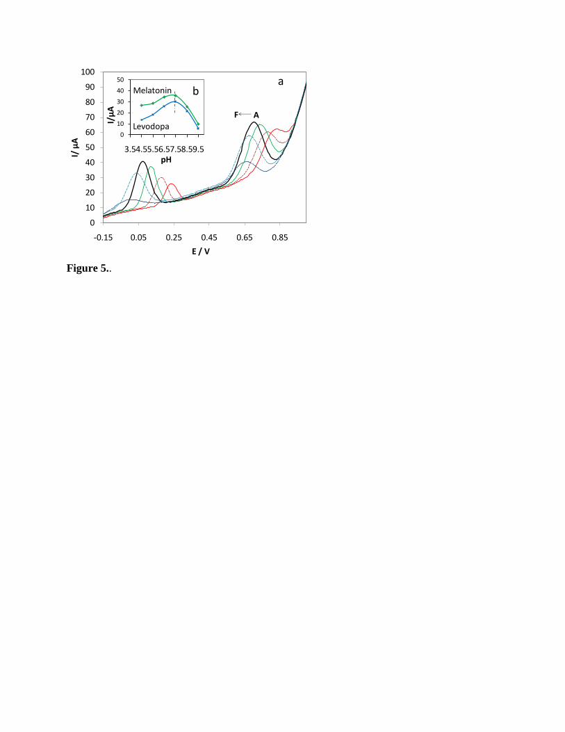
Figure 5..
0
10
20
30
40
50
60
70
80
90
100
-0.15 0.05 0.25 0.45 0.65 0.85
I/ µ
A
E / V
a
AF
0
10
20
30
40
50
3.54.55.56.57.58.59.5
I/µ
A
pH
Melatonin
Levodopa
b
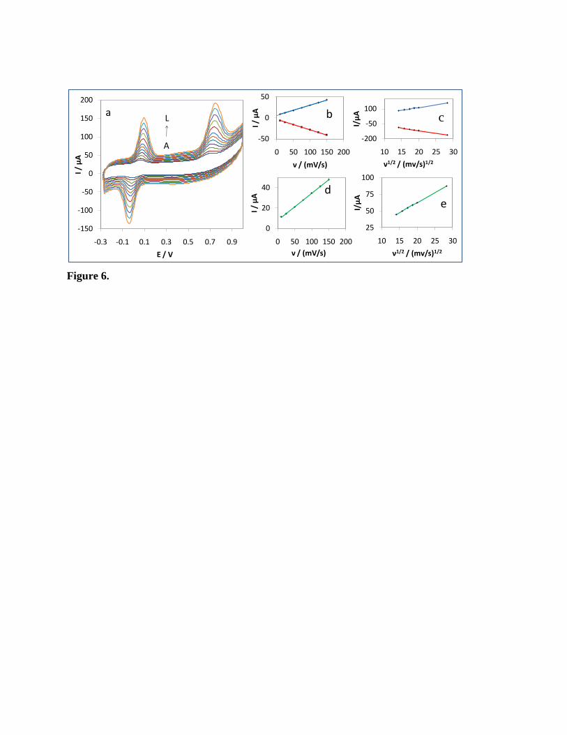
Figure 6.
-150
-100
-50
0
50
100
150
200
-0.3 -0.1 0.1 0.3 0.5 0.7 0.9
I / µ
A
E / V
A
La
-50
0
50
0 50 100 150 200
I / µ
A
ν / (mV/s)
0
20
40
0 50 100 150 200
I / µ
Aν / (mV/s)
b
d
-200
-50
100
10 15 20 25 30
I/µ
A
ν1/2 / (mv/s)1/2
25
50
75
100
10 15 20 25 30
I/µ
A
ν1/2 / (mv/s)1/2
e
c
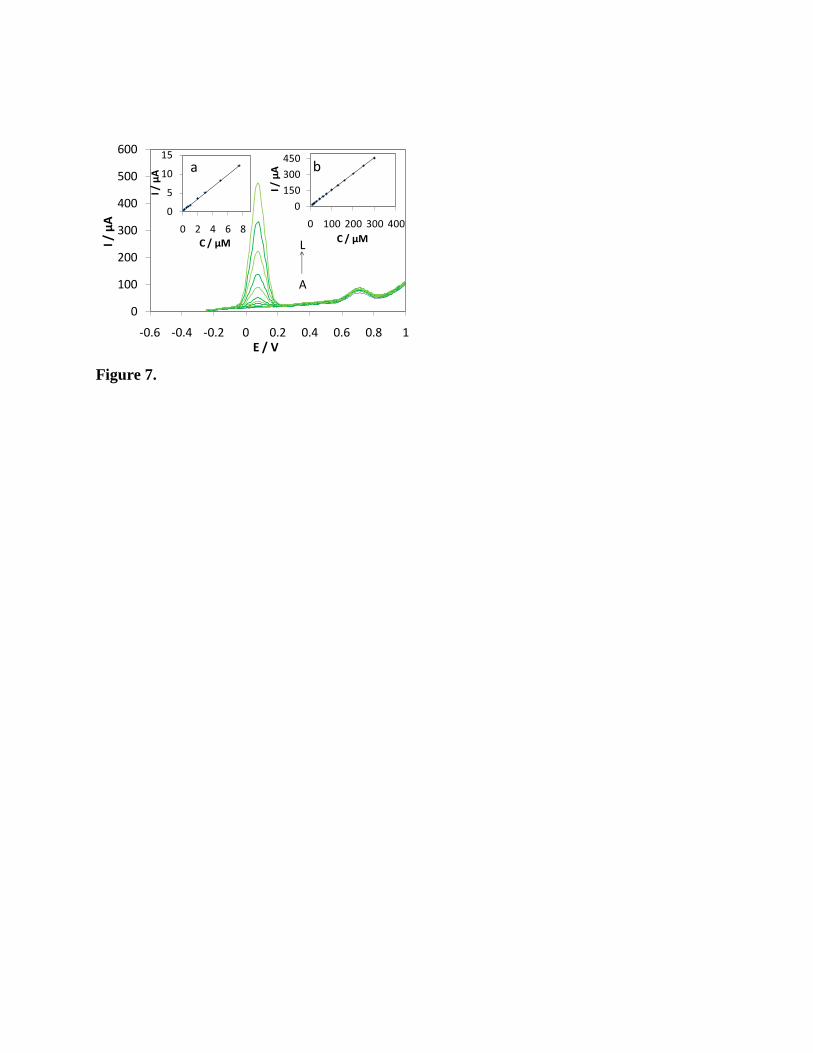
Figure 7.
0
100
200
300
400
500
600
-0.6 -0.4 -0.2 0 0.2 0.4 0.6 0.8 1
I / µ
A
E / V
L
A
0
5
10
15
0 2 4 6 8
I / µ
A
C / µM
a
0
150
300
450
0 100 200 300 400
I / µ
A
C / µM
b
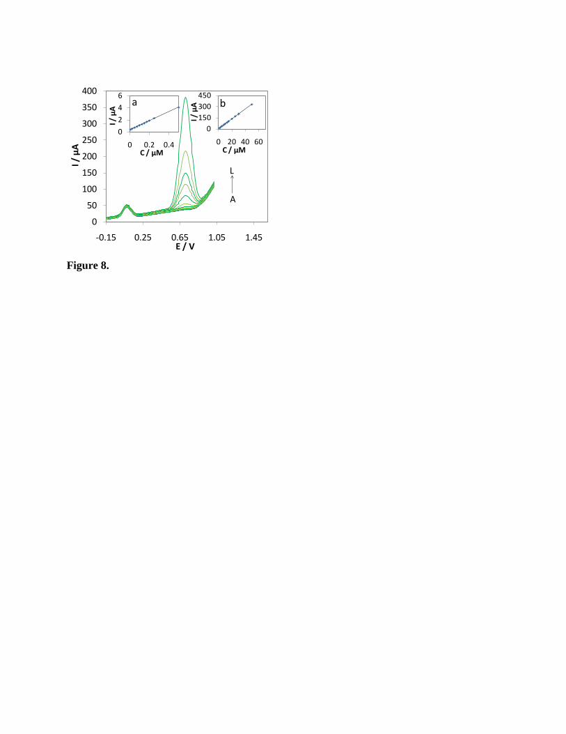
Figure 8.
0
50
100
150
200
250
300
350
400
-0.15 0.25 0.65 1.05 1.45
I / µ
A
E / V
L
A
0
2
4
6
0 0.2 0.4
I / µ
A
C / µM
a
0
150
300
450
0 20 40 60
I / µ
A
C / µM
b
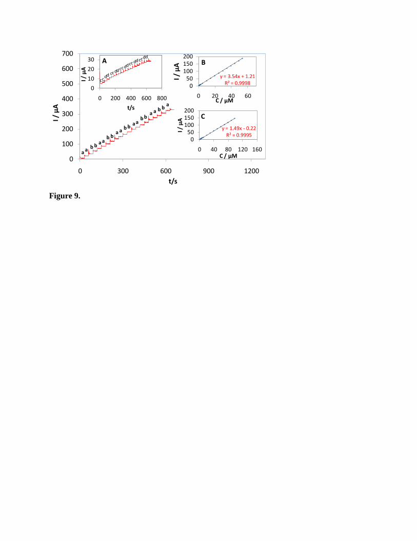
Figure 9.
0
100
200
300
400
500
600
700
0 300 600 900 1200
I / µ
A
t/s
a
ab
ba
ab
ba
a
aa
a aa
bb
b b
b b
0
10
20
30
0 200 400 600 800
I / µ
A
t/s
cc
cccc
cccc
dd
dddd
dddd
A
y = 3.54x + 1.21R² = 0.99980
50100150200
0 20 40 60
I / µ
A
C / µM
B
y = 1.49x - 0.22R² = 0.9995
050
100150200
0 40 80 120 160
I / µ
A
C / µM
C