mutations in dnmt1 cause hereditary sensory neuropathy with dementia and hearing loss
TRANSCRIPT

Mutations in DNMT1 cause hereditary sensory neuropathy withdementia and hearing loss
Christopher J. Klein1, Maria-Victoria Botuyan2,*, Yanhong Wu3,*, Christopher J. Ward4,*,Garth A. Nicholson5, Simon Hammans6, Kaori Hojo7, Hiromitch Yamanishi7, Adam R.Karpf8, Douglas C. Wallace9, Mariella Simon9, Cecilie Lander10, Lisa A. Boardman11, JulieM. Cunningham3, Glenn E. Smith12, William J. Litchy1, Benjamin Boes13, Elizabeth J.Atkinson14, Sumit Middha14, P. James Dyck1, Joseph E. Parisi15, Georges Mer2, David I.Smith3, and Peter J. Dyck1
1Mayo Clinic, Department of Neurology, Division of Peripheral Nerve Diseases, Rochester, MN,USA2Mayo Clinic, Biochemistry and Molecular Biology, Rochester MN, USA3Mayo Clinic, Laboratory Medicine and Pathology, Rochester, MN, USA4Mayo Clinic, Nephrology and Hypertension Research, Rochester MN, USA5 University of Sydney, Molecular Medicine Laboratory & ANZAC Research Institute, Australia6Southampton University Hospitals NHS Trust, Department of Neurology, Southampton, UnitedKingdom7Harima Sanatorium, Division of Neuropsychiatry, Hyogo, Japan8Roswell Park Cancer Institute, Department of Pharmacology and Therapeutics, Buffalo, NY,USA9Center for Molecular & Mitochondrial Medicine and Genetics, University of California, Irvine,USA10Queensland Health, Royal Brisbane Hospital, Herston, Australia11Mayo Clinic, Division of Gastroenterology, Rochester, MN, USA12Mayo Clinic, Division of Psychology, Rochester, MN13Roche Applied Science Genomic Sequencing, Indianapolis, IN, USA
Christopher J. Klein, [email protected].*Authors contributed equally
Authors Contributions: C.J.K., D.I.S. and P.J.D. directed the study. C.J.K. wrote the paper. C.J.K., P.J.D., G.A.N., S.H., K.H., H.Y.,D.C.W., M.S., C.L., L.A.B., G.E.S., W.J.L., evaluated or collated patient data. E.J.A. did the linkage and haplotype analysis. S.M. andB.B. did next generation sequencing analysis. C.J.K., C.J.W. and Y.W. did the cell culture and protein expression studies, genesequencing and southern blot analysis. J.M.C. and A.R.K. did the methylation analysis. M.V.B. and G.M. did the mutagenesis,bacterial protein expression and structural analysis. J.E.P. provided pathologic analysis of autopsy material.
Competing Financial Interests: The authors declare no competing financial interests.
Accession number:Protein Data Bank accession number for DNMT1 targeting sequence domain, 3EPZ.Exome sequencing data is available upon request through FTP site (NCBI Sequence Read Archive repositories has been discontinuedfor next generation sequencing data).
URLs:Human Genome Mutation Database (HGMD), http://www.hgmd.cf.ac.uk/.The 1000 Genome Browser, http://browser.1000genomes.org/index.html.Environmental Genome Project, http://www.niehs.nih.gov/research/supported/programs/egp/.
NIH Public AccessAuthor ManuscriptNat Genet. Author manuscript; available in PMC 2011 December 01.
Published in final edited form as:Nat Genet. 2011 June ; 43(6): 595–600. doi:10.1038/ng.830.
NIH
-PA Author Manuscript
NIH
-PA Author Manuscript
NIH
-PA Author Manuscript

14Mayo Clinic, Biomedical informatics and Statistics, Rochester, MN, USA15 Mayo Clinic, Division of Neuropathology Rochester, MN, USA
AbstractDNA methyltransferase 1 (DNMT1) is crucial for maintenance of methylation, gene regulationand chromatin stability1-3. DNA mismatch repair, cell cycle regulation in post-mitotic neurons4,5
and neurogenesis6 are influenced by DNA methylation. Here we show mutations in DNMT1 causeboth central and peripheral neurodegeneration in one form of hereditary sensory and autonomicneuropathy (HSAN1) with dementia and hearing loss7,8. Exome sequencing led to theidentification of DNMT1 mutation c.A1484G (p.Tyr495Cys) in two American and one Japanesekindreds and a triple nucleotide change c.1470TCC-1472ATA (p.Asp490Glu-Pro491Tyr) in oneEuropean kindred. All mutations are within the targeting sequence (TS) domain of DNMT1. Thesemutations cause premature degradation of mutant proteins, reduced methyltransferase activity andimpaired heterochromatin binding during the G2 cell cycle phase, leading to globalhypomethylation and site specific hypermethylation. Our study demonstrates DNMT1 mutationscause aberrant methylation implicated in complex pathogenesis. The discovered DNMT1mutations provide a new framework for the study of neurodegenerative diseases.
Neurodegenerative diseases are hallmarked by an increased rate of neuronal axonal loss.Neural development, neural survival and connectivity have been linked to DNA methylationand chromatin stability1. Here we studied one form of neurodegeneration with bothperipheral and central involvement, namely hereditary sensory and autonomic neuropathytype 1 (HSAN1) with dementia and hearing loss. As a group, HSAN1 has autosomaldominant inheritance with neurodegeneration that clinically manifests with loss of sensationleading to painless extremity injuries, infections and resultant amputations9. Serinepalmitoyltransferase (SPTLC1 and SPTLC2)10,11, atlastin-1 (ATL1)12 and Ras-relatedGTPase 7 (RAB7)13 have been linked to some HSAN1 kindreds, but the majority ofcausative genes remains unknown. The specific form of HSAN1 investigated here has earlyonset dementia and sensorineural hearing loss that tracks with sensory neuropathy7,8.Pathologic studies of affected persons' nerve7, brain14 and spinal cord (Fig. 1) show axonalloss without interstitial infiltrative inclusions. Affected persons typically have earlymortality and often require total care due to dementia, hearing loss, and lose of ambulationfrom predominant sensory ataxia. Four kindreds (Fig. 2a-d) were investigated, two of themwere previously reported in detail7,8. Affected persons had very similar phenotypes (Table1) and were normal in youth, but developed worsening sensorineural deafness and sensoryneuropathy by ages 20-35 years. Progressive cognitive and behavioral declines occurred bythe fourth decade. Brain imaging of affected persons showed global atrophy and reducedweight of autopsied brains. Among the Japanese kindred PET and SPECT imagingsuggested medial frontal and thalamic hypo metabolism8. Quantitative sensory testing, nerveconductions, and nerve biopsy were indicative of length dependent progressive sensoryaxonal loss.
We performed genome-wide linkage analysis in Kindred-1 and mapped to19p13.2 with amaximal LOD score of 6.38. Fine mapping analysis with haplotype construction narrowedthe region to 3.4 MB between D19S583 and D19S221. To increase the sequencing coverageand depth, we performed both Illumina GAII and Roche454 exome sequencing(Supplementary Table 1). All identified base alterations in the region were filtered againstthe dbSNP131 database. Bioinformatics analysis revealed a novel non-synonymousheterozygous mutation in DNMT1 at c.A1484G resulting in tyrosine495 changing tocysteine. Sequencing results confirmed this alteration segregated with disease status. Wesubsequently sequenced all 41 exons of DNMT1 and found kindred-3 (American kindred)
Klein et al. Page 2
Nat Genet. Author manuscript; available in PMC 2011 December 01.
NIH
-PA Author Manuscript
NIH
-PA Author Manuscript
NIH
-PA Author Manuscript

and kindred-4 (Japanese kindred) have the same heterozygous mutation c.A1484G(p.Tyr495Cys) and kindred-2 (European Kindred) contain three consecutive nucleotidesheterozygous change c.1470TCC-1472 ATA (p.Asp490Glu-Pro491Tyr) (SupplementaryFig. 1). All heterozygous mutations were absent in over 1500 unaffected controls and notreported in HGMD, the 1000 Genome and 10 exomes sequenced as part of theEnvironmental Genome Project.
DNMT1 has a large N-terminal regulatory region and a smaller C-terminal catalytic region(Fig. 2e). The highly conserved catalytic region of DNMT1 alone is not sufficient forenzymatic activity and an allosteric activation through direct interaction of the N-terminalregulatory and C-terminal catalytic regions is required for its enzymatic function15. Theregulatory region contains many protein-protein interaction domains including PCNAbinding, targeting sequence, zinc finger, bromo-adjacent homology domains. The identifiedmutations occurred in the TS domain. TS domain regulates DNMT1 binding to chromatinduring late S phase and is responsible for persistent association during G2 and M phase. Theproper folding of TS domain is crucial for DNMT1 function16. To probe the impact of thetwo mutations on the TS domain folding in vitro, we conducted the expression of wild typeTS domain (aa 351-600) and corresponding p.Tyr495Cys and p.Asp490Glu-Pro491Tyr TSdomain mutants in E. coli. While the three proteins are well produced in E. coli, only wt-TSis soluble (Supplementary Fig. 2). In contrast, the TS mutants form inclusion bodies, astrong indication that they are misfolded (Supplementary Fig. 2). Misfolding is notsurprising for p.Tyr495Cys mutant, considering that Tyr495 is part of the proteinhydrophobic core with an accessible solvent surface area (ASA) of only 0.55 Å2 in thecrystal structure of wt-TS. Asp490 is solvent accessible (ASA is 75.3 Å2) and is thereforeunlikely to contribute to the disease phenotype (Fig. 2f). In agreement, Asp490 is not aconserved residue and is replaced by a glutamate in other species (Fig. 2f). On the otherhand, Pro491 has limited solvent accessibility with an ASA of 25.7 Å2 and together withAsp490 is part of the linker region between the β-sheet-based and α-helical subdomains ofTS, suggesting that the constrained conformation of the trans-proline residue is needed forproper folding.
The TS domain has been expressed independently and shown as an important factor for thestructure, function and localization of DNMT117,18,19, 16. Partial deletion of TS domainabolishes enzymatic activity of DNMT116. To investigate the functional implication of themutant TS domain, we transfected GFP-tagged constructs encompassing wt-TS (aa 306-620)and the two TS mutants (p.Tyr495Cys-TS and p.Asp490Glu-Pro491Tyr-TS) into HeLacells. While the wt-TS domain entered the nucleus and bound to the replication foci duringS-phase and to heterochromatin during G2 phase, the two TS mutants localized to thecytoplasm and were not able to enter the nucleus (Fig. 3a-c). One of DNMT1's multiplenuclear localization signals is just upstream of the targeting-sequence domain(323KRRK326)19, the mutations likely disrupt the proper fold of dNMT1, preventing themutant proteins from entering the nuclei. Mutations p.Tyr495Cys and p.Asp490Glu-Pro491Tyr also affect the stability and the enzymatic activity of full length DNMT1. Wetransfected GFP-tagged full length wild type and mutant DNMT1 into HeLa cells andtreated cells with cycloheximide for different time lengths. Western blot analysis showed themutant proteins were quickly degraded compared to wild type DNMT1 (Supplementary Fig.3). The enzymatic activity assay showed that p.Tyr495Cys-DNMT1 and p.Asp490Glu-Pro491Tyr-DNMT1 have significantly decreased activity comparing to wt-DNMT1,(Supplementary Fig. 4 and Supplementary notes). These findings demonstrate thedeleterious effects of these mutations in the TS domain on the structure, methyltransferaseactivity and stability of DNMT1.
Klein et al. Page 3
Nat Genet. Author manuscript; available in PMC 2011 December 01.
NIH
-PA Author Manuscript
NIH
-PA Author Manuscript
NIH
-PA Author Manuscript

DNMT1 is one of the components of the DNA replication machinery during S phase of thecell cycle17. Studies have shown that maintenance of methylation could last past thereplication stage with DNMT1 continuously loaded on heterochromatin after S phase. TheTS domain was identified responsible for this persistent association20. Partial deletion of TSdomain has been shown to abolish DNMT1 association with heterochromatin after Sphase20. To evaluate the heterochromatin binding of mutant DNMT1, we co-transfectedGFP-tagged wild type and p.Tyr495Cys-DNMT1 and p.Asp490Glu-Pro491Tyr-DNMT1along with RFP-tagged proliferating cell nuclear antigen (PCNA) as cell cycle marker intoHeLa cells. Our results show that the mutant DNMT1 only showed normal localization atreplication foci during S phase as previously described20, but did not show binding ofheterochromatin during G2 phase in contrast to wt-DNMT1 (Fig. 4a-c), indicating that themutations abolished DNMT1 heterochromatin binding ability after S phase. Previous studiesshowed the replication machinery loading of DNMT1 is not required for the genomemethylation maintenance21, suggesting DNMT1 binding to heterochromatin after S phasecould be involved in the prolonged methylation maintenance, recruitment of histonedeacetylases and chromatin stability preservation in the transcriptionally repressed genomeregion20. The impaired heterochromatin binding after S phase is likely to impair theseimportant cellular functions.
Next, we examined the effect of mutant DNMT1 on the global methylation level usingliquid chromatography-electrospray ionization tandem mass spectrometry (LC-ESI-MS/MS)to measure 5-methyl-2′-deoxycytidine (5-mdC) levels following enzymatic hydrolysis ofgenomic DNA22. Total 24 samples including 12 affected and 12 genetically related andunaffected samples from Kindred-1 were quantified. Our results showed 8% reduction in 5-mdC content in affected group comparing to the unaffected group (p<0.001) (SupplementaryFig. 5). This is predicted meaningful considering in DNMT1 knockout HCT116 cells only10-20% of genomic DNA hypomethylation was observed, but the deleterious impact led topronounced chromosomal defects and apoptosis23,24. Since methylation varies with age andsex, we specifically compared the content of m5dC between three pairs of same sex siblingsand between two pairs of same sex cousins with similar age (<5 years difference). Theresults showed consistent 7-15% reduction of 5-mdC content in all affected sibling andcousins (Supplementary Table 2). Centromeric and pericentromeric regions have beenshown to contribute to the majority of global methylation. We used Pyrosequencing assay ofbisulfite treated DNA to investigate the CpG sites in the repetitive elements of Satellite-2,Line-1 and Satellite-α and Alu region. The results suggested reduced methylation at certainCpG sites in Satellite-2 region among affected group (Supplementary Table 3 andSupplementary notes). The Satellite-2 methylation reduction was further confirmed bySouthern blot using genomic DNA digested by methylation sensitive enzyme BstB1(Supplementary Fig. 6a-b). These results support global hypomethylation demonstrated byreduced 5-mdC content, suggesting the hypomethylation likely occurs at the Satellite-2repetitive elements. The hypomethylation in repetitive and transposable elements has beenlinked to the loss of methyl-specific protein complexes that stabilize a high order ofheterochromatin structure25. The reduction of total 5-mdC content and decreasedmethylation in Satellite-2 resulting from DNMT1 mutations may impair the heterochromatinstability.
Since variation in methylation is usually linked to the transcriptional regulation of specificgenes, we investigate the methylation of CpG sites in gene promoter regions. IlluminaHumanMethylation27 BeadChip was used to survey genome-wide promoter methylationprofiles. The 24 samples from kindred-1 were studied. Based on the moving average ofmethylation difference between affected and unaffected group, moderate reduction ofmethylation was observed in the affected samples (Fig. 5). Interestingly, the 10 promoterCpG sites with the most significant p value (p<0.00007) showed increased methylation (Fig.
Klein et al. Page 4
Nat Genet. Author manuscript; available in PMC 2011 December 01.
NIH
-PA Author Manuscript
NIH
-PA Author Manuscript
NIH
-PA Author Manuscript

5 and Supplementary Fig. 7). Global hypomethylation of cytosines and localhypermethylation of CpG islands are characteristics of the neoplastic epigenome26 but theinvestigated kindreds did not have any cancer histories. One of the characteristics of tumorcells is uncontrolled proliferation. In postmitotic neurons, DNA methylation undergoesturnover process in response to synaptic activity27 and failure of mitotic arrest may result incell death4. Re-expression of various cell cycle proteins has been reported in patients withAlzheimer's dementia and other neurodegenerative disorders4, suggesting the initiation ofcell cycle by abnormal expression of cell cycle proteins impairs neuronal health. It isintriguing to see that the methylation pattern in the affected persons mimics the methylationpattern of tumor cells, leading us to speculate that mutated DNMT1 could be linked to theloss of cell cycle arrest in postmitotic neuron. Defects of methylation have been shown tocause developmental and progressive human neurodegenerative diseases such as Rett(MeCP2)28 and ICF(DNMT3b)29 syndromes. Epigenetic dysregulation has also beensuggested in Alzheimer's dementia through reduced histone acetylation30. Ourchromosomally linked region at 19p13.2 also associates with late onset familial Alzheimer's(LOFAD)31, possible association of DNMT1 in LOFAD needs further investigation.
In this report, we identified mutations in DNMT1 that cause HSAN1 with dementia andhearing loss. These mutations affect DNMT1's proper folding, decrease enzymatic activity,impair heterochromatin binding ability after S phase and lead to the occurrence of globalhypomethylation with local hypermethylation. Our results provide a direct link betweenDNMT1 alteration and a neurodegenerative disorder of both central and peripheral nervoussystem. DNMT1 is highly expressed in postmitotic neurons and adult central nervoussystem32, it interacts with a series of important cell cycle regulating proteins33 and isinvolved in neuronal differentiation, migration and central neural connection1,34,35,suggesting DNMT1 participates in a precise mechanism of dynamic regulation of thenervous system. The affected individuals in our kindreds had apparent normal intelligenceand nerve function until early adulthood, then the typical symptoms began and worsenedwith age, implicating DNMT1's influence is beyond embryogenesis and suggesting aprogressive course with gradual accumulating effect. Our study emphasizes that evenmoderate global hypomethylation in association with local hypermethylation can have asevere impact on neuronal survival and function. The pathogenic mechanism of mutantDNMT1 is potentially complex and provides a new direction for the study ofneurodegeneration.
MethodsSubjects and Samples
This study was approved by the Mayo Institutional Review Boards. Blood and other sampleswere taken after obtaining written informed consent. This study included total 4 HSAN1kindreds with sensorineural deafness and dementia - two American families, one of Germanorigin and the other of Northern European descent described previously 7, one Japanesefamily with no known European ancestry also described previously8 and one Australianfamily of English descent.
Linkage AnalysisWe performed a 8-cM genome wide linkage scan on 63 individuals (12 affected, 51unaffected including 14 unaffected spouses) from kindred-1 using 500 deCODEmicrosatellite markers spaced at approximately 8-cM intervals. To allow for potentialambiguities of the relationship between the underlying genotype and affection status,penetrance was assumed to be 0.1 for noncarriers and 0.9 for carriers. Two-point logarithmof the odds (LOD) scores was calculated using the MLINK program of Linkage, assuming a
Klein et al. Page 5
Nat Genet. Author manuscript; available in PMC 2011 December 01.
NIH
-PA Author Manuscript
NIH
-PA Author Manuscript
NIH
-PA Author Manuscript

rare susceptibility allele (frequency 0.001) and an autosomal dominant mode of inheritance.Multipoint analysis and haplotypes were performed by SIMWALK2.17. To confirm andrefine the localized region at chromosome 19q, we identified additional ten microsatellitemarkers for haplotype analysis between D19S884 and D19S226, spanning ∼6.5-cM distancewith an average 0.8-cM density.
Exome capture and Next-Generation sequencinga. Illumina GAII Paired End Exome Sequencing—Exome capture is carried out usingAgilent's SureSelect Human All Exon kit-v1. The flow cells are sequenced as 76×2 pairedend reads on an Illumina GAIIx using SBS sequencing kit-v4 and SCS-v2.5 software. Theimage processing and base calling was performed using the Illumina Pipeline Software. Thereads with phred-like consensus quality ≥ 40 were aligned to human genome-36 (hg18) andthe single nucletoide variants (SNVs) and Indels (insertion-deletions) were called usingMAQ (http://maq.sourceforge.net/) and BWA (http://bio-bwa.sourceforge.net/bwa.shtml).SNVs and Indels were then filtered using Agilent's ‘on-target bed file’ and annotated usingSIFT and SeattleSEQ annotation servers. The run statistics is summarized in SupplementaryTable 1.
b. Roche 454 GS-FLX sequencing with NimbleGen 2.1 M Human Exomecapture—The sample was prepared using the GS-FLX Titanium optimized sequencecapture protocol using the Roche-NimbleGen 2.1 M Human Exome array. Exome-capturedproband DNA sample was sequenced on 454 Genome Sequencer FLX instrument with GS-FLX Titanium Kits. Two full 454 GS-FLX (Titanium) runs were performed. High qualitydata were aligned to the hg18 human genome reference. Variants were identified using theGSMapper software 2.0.01 and were included in AllDiff (all differences) and HCDiff (highconfidence differences) reports. The average read length is 382bp with mode of 490bp. Wescreened novel mutations in both AllDiff and HCDiff files within the identified 3.4 MBlinked region. The run statistics is summarized in Supplementary Table 1.
Sanger Sequencing AnalysisGenomic DNA was extracted from whole peripheral blood, transformed lymphoblastoid celllines or available tissues using standard methods (Qiagen). Resequencing the region of theinterest was carried out using dye termination chemistry (Big Dye Terminator with themodel 3730xl sequencer; Applied Biosystems, CA). Primer sets for PCR were designedusing the web-based design tool Primer3 (Supplementary Table 4). PCR were carried outusing the standard protocol. After PCR reactions, the amplicons were treated with theExoSAP-IT (USB) to degrade unincorporated PCR primers and deoxynucleotidetriphosphates. The cleaned products were mixed with 10 picomoles of the forward or reversePCR primers for sequencing. DNA sequence variants were identified using MutationSurveyor Analysis Software (SoftGenetics).
Expression Vectors for TS domain, Full length DNMT1 and PCNAThe nucleotide sequence encompassing wild type DNMT1 TS domain (aa 306-620) forHeLa cell transfection was custom synthesized (Origene) and this cDNA was cloned into thepCMV6-N-GFP vector. The plasmid pEX-N-His-TS containing wild type TS domain (aa351-600) for bacterial expression and purification was also custom synthesized (Origene).The expression constructs pEGFP-human DNMT1 and pmRFP-human PCNA were kindlyprovided by Dr. H. Leonhardt (Ludwig Maximilians University Munich, Germany). ThepEGFP-hDNMT1 contains a CMV promoter and enhanced GFP fused to N-terminus of fulllength human DNMT1 (NM_001379). Similarly pmRFP-human PCNA contains a CMVpromoter and mRFP cDNA fused to the N-terminus of human PCNA. Mutants were
Klein et al. Page 6
Nat Genet. Author manuscript; available in PMC 2011 December 01.
NIH
-PA Author Manuscript
NIH
-PA Author Manuscript
NIH
-PA Author Manuscript

generated using QuikChange mutagenesis kit (Stratagene). For primer sequences, seeSupplementary Table 5 online. All constructs were confirmed by direct sequencing.
DNMT1 TS Domain Expression and Purification in E. coliPlasmids of wild type, p.Tyr495Cys and p.Asp490Glu-Pro491Tyr TS domain of DNMT1were transformed in BL21(DE3) E. coli cells and grown in LB broth supplemented with 100mg/mL ampicillin at 37°C up to an OD600 of ∼0.8, after which the temperature wasdecreased to 15°C for about 1h and then 1mM IPTG (final concentration) was added. After16 h, the cells were harvested by centrifugation, resuspended in 50mM sodium phosphate,pH 7.5, 300 mM NaCl (buffer A) and 20 mM PMSF and then lyzed with an Emulsiflex C-5high-pressure homogenizer (Avestin). After centrifugation, the soluble fraction was purifiedby passing through a Ni-NTA column pre-equilibrated with buffer A; washing the columnwith buffer A containing 5 mM imidazole (buffer B); and eluting the protein with buffer Acontaining 500 mM imidazole (buffer C). Fractions containing the TS domain were pooledand passed through a Superdex 200 FPLC column (GE Healthcare) using buffer A as therunning buffer. The cell debris were resuspended in buffer A containing 8 M urea, andcentrifuged. The resulting supernate (referred to as insoluble fraction) was purified similarlyas above but using buffers B and C with 6 M urea. At various stages during the purification,sample aliquots were obtained and later assessed by SDS-PAGE. Protein purification wascarried out at 4°C.
Cell Culture and Western Blot AnalysisHeLa Cells were cultured in RPMI supplemented with 10% fetal Bovine serum. Cells weretransfected using FugeneHD (Roche). For Cycloheximide treatment, fresh media contain0.05 mg/mL l of cycloheximide (Sigma) were added after 24 h of transfection. Cell lysatewere collected using NP-40 lysis buffer (Invitrogen) at different time points for westernblotting analysis. BCA assay was used to ensure the equal loading. Cell lysate wereseparated on 4-12% gradient SDS–PAGE and transferred to a PVDF-membrane (Bio-Rad).The membrane was blocked with 1×Casein (Vector) in TBST and incubated overnight at4°C with an anti-GFP mouse monoclonal antibody (Allele Biotechnology). After washing,the blots were incubated with the appropriate secondary antibody conjugated withhorseradish peroxidase at room temperature for 1 h. Immunoreactive bands were washedand visualized with SuperSignal Pico Western Blot Detection Kit (Thermo Scientific).
Confocal MicroscopyHeLa cells were seeded on Lab Tek II chambered cover glass (Nunc) 24 h beforetransfection. Cells were transfected using Fugene HD (Roche). Twenty-four hours aftertransfection, live cell microscopy was performed with a LSM510 (Carl Zeiss) confocal setupmounted on an Axiovert 200 M inverted microscope using a 100× phase contrast Plan-Neofluar oil, NA 1.3 heated to 37°C. Chambered cover glass was maintained at 37°C usinga live-cell chamber and temperature controller (PeCon GmbH, Erbach, Germany). EnhancedGFP and mRFP were excited sequentially at 488nm, 543nm to minimize the crosstalk.
Global Genomic DNA Methylation AnalysesGenomic DNA methylation was assessed by direct measurement of the level of 5-methyl-2′-deoxycytidine (5-mdC) using liquid chromatography-electrospray ionization massspectrometry (LC-ESI-MS/MS), as described previously22. Briefly, 1 μg genomic DNAsamples were enzymatically hydrolyzed with Nuclease P1 or Nuclease S1 into componentnucleosides, and loaded, along with standards, onto an LC-ESI/MS/MS system. All sampleswere analyzed in duplicate and proper controls were tested before and after samplemeasurement to ensure the accuracy.
Klein et al. Page 7
Nat Genet. Author manuscript; available in PMC 2011 December 01.
NIH
-PA Author Manuscript
NIH
-PA Author Manuscript
NIH
-PA Author Manuscript

Illumina Infinium HumanMethylation27 BeadChip AnalysisOne microgram DNA from each selected sample was bisulfite-treated using ZymoMethylation kit (Zymo Research). After bisulfite conversion, 250 ng of each sampleundergo a whole-genome amplification (WGA) followed by hybridization. Allele-specificprimer annealing is followed by single-base extension with labeled nucleotides. Afterextension, the array is scanned and the intensities of the unmethylated and methylated beadtypes are measured. The methylation level at each CpG site was determined by measuringthe methylation fraction (beta), defined as the fraction of methylated signal over the totalsignal (unmethylated + methylated). The beta values are continuous variables between 0(unmethylated) and 1 (completely methylated). The Infinium Assay includes redundant,built-in, bisulfite conversion quality controls that measure the conversion rate of non-CpGcytosines and background signal. Using within-assay controls evaluated with IlluminaGenomeStudio software, we assessed background signal and bisulfite conversion rate andensured that non-methylated cytosines in all 24 samples were efficiently bisulfite converted.∼8% CpG sites that did not reveal reliable signals, as determined by their detection p-value,were excluded from further analysis. Differential methylation analysis and construction of avolcano plot was performed using the Partek Statistical Package (Partek). We employedtwo-way analysis of variance (ANOVA) to account for the potential batch effects. The type Ierror was controlled by the use of Benjamini and Hochberg false discovery rate (FDR)controlling procedure.
Supplementary MaterialRefer to Web version on PubMed Central for supplementary material.
AcknowledgmentsThis study was supported by the National Institute of Neurologic Disorders and Strokes (NINDS) K08 (NS065007),NINDS R01 (NS36797) and a previous grant from the Muscular Dystrophy Association. The authors wish to thankMayo Clinic Bioinformatics Core and Sequencing Core for their excellent assistance. We would also like to thankthe support from the Clinical Core of the Mayo Clinic Center for Cell Signaling in Gastroenterology(P30DK084567). DCW and MS would like to acknowledge the outstanding clinical contributions of Ms. Julia Platt.Their portion of this work was supported by NIH grants NS21328 and NS070298.
References1. Feng J, Fan G. The role of DNA methylation in the central nervous system and neuropsychiatric
disorders. Int Rev Neurobiol. 2009; 89:67–84. [PubMed: 19900616]
2. Chen WG, et al. Derepression of BDNF transcription involves calcium-dependent phosphorylationof MeCP2. Science. 2003; 302:885–9. [PubMed: 14593183]
3. Tohgi H, et al. Reduction with age in methylcytosine in the promoter region -224 approximately-101 of the amyloid precursor protein gene in autopsy human cortex. Brain Res Mol Brain Res.1999; 70:288–92. [PubMed: 10407177]
4. Herrup K, Yang Y. Cell cycle regulation in the postmitotic neuron: oxymoron or new biology? NatRev Neurosci. 2007; 8:368–78. [PubMed: 17453017]
5. Brooks PJ, Marietta C, Goldman D. DNA mismatch repair and DNA methylation in adult brainneurons. J Neurosci. 1996; 16:939–45. [PubMed: 8558262]
6. Fan G, et al. DNA hypomethylation perturbs the function and survival of CNS neurons in postnatalanimals. J Neurosci. 2001; 21:788–97. [PubMed: 11157065]
7. Wright A, Dyck PJ. Hereditary sensory neuropathy with sensorineural deafness and early- onsetdementia. Neurology. 1995; 45:560–2. [PubMed: 7898717]
8. Hojo K, et al. Hereditary sensory neuropathy with deafness and dementia: a clinical andneuroimaging study. Eur J Neurol. 1999; 6:357–61. [PubMed: 10210919]
Klein et al. Page 8
Nat Genet. Author manuscript; available in PMC 2011 December 01.
NIH
-PA Author Manuscript
NIH
-PA Author Manuscript
NIH
-PA Author Manuscript
9. Klein, CJ.; Dyck, P. Hereditary Sensory and Autonomic Neuropathies (HSAN): Clinical FeaturesPathologic Classification, and Molecular Genetics. In: Dyck, PJ.; T, P., editors. PeripheralNeuropathy. Fourth. Vol. 2. Elsevier; 2005. p. 1809-1845.
10. Dawkins JL, Hulme DJ, Brahmbhatt SB, Auer-Grumbach M, Nicholson GA. Mutations inSPTLC1, encoding serine palmitoyltransferase, long chain base subunit-1, cause hereditarysensory neuropathy type I. Nat Genet. 2001; 27:309–12. [PubMed: 11242114]
11. Rotthier A, et al. Mutations in the SPTLC2 subunit of serine palmitoyltransferase cause hereditarysensory and autonomic neuropathy type I. Am J Hum Genet. 2010; 87:513–22. [PubMed:20920666]
12. Guelly C, et al. Targeted high-throughput sequencing identifies mutations in atlastin-1 as a causeof hereditary sensory neuropathy type I. Am J Hum Genet. 2011; 88:99–105. [PubMed: 21194679]
13. Verhoeven K, et al. Mutations in the small GTP-ase late endosomal protein RAB7 cause Charcot-Marie-Tooth type 2B neuropathy. American Journal of Human Genetics. 2003; 72:722–7.[PubMed: 12545426]
14. Hojo K, Kawamata T, Tanaka C, Maeda K. Inflammatory glial activation in the brain of a patientwith hereditary sensory neuropathy type 1 with deafness and dementia. Neurosci Lett. 2004;367:340–3. [PubMed: 15337262]
15. Fatemi M, Hermann A, Pradhan S, Jeltsch A. The activity of the murine DNA methyltransferaseDnmt1 is controlled by interaction of the catalytic domain with the N-terminal part of the enzymeleading to an allosteric activation of the enzyme after binding to methylated DNA. J Mol Biol.2001; 309:1189–99. [PubMed: 11399088]
16. Margot JB, et al. Structure and function of the mouse DNA methyltransferase gene: Dnmt1 showsa tripartite structure. J Mol Biol. 2000; 297:293–300. [PubMed: 10715201]
17. Leonhardt H, Page AW, Weier HU, Bestor TH. A targeting sequence directs DNAmethyltransferase to sites of DNA replication in mammalian nuclei. Cell. 1992; 71:865–73.[PubMed: 1423634]
18. Fellinger K, Rothbauer U, Felle M, Langst G, Leonhardt H. Dimerization of DNAmethyltransferase 1 is mediated by its regulatory domain. J Cell Biochem. 2009; 106:521–8.[PubMed: 19173286]
19. Cardoso MC, Leonhardt H. DNA methyltransferase is actively retained in the cytoplasm duringearly development. J Cell Biol. 1999; 147:25–32. [PubMed: 10508852]
20. Easwaran HP, Schermelleh L, Leonhardt H, Cardoso MC. Replication-independent chromatinloading of Dnmt1 during G2 and M phases. EMBO Rep. 2004; 5:1181–6. [PubMed: 15550930]
21. Spada F, et al. DNMT1 but not its interaction with the replication machinery is required formaintenance of DNA methylation in human cells. J Cell Biol. 2007; 176:565–71. [PubMed:17312023]
22. Song L, James SR, Kazim L, Karpf AR. Specific method for the determination of genomic DNAmethylation by liquid chromatography-electrospray ionization tandem mass spectrometry. AnalChem. 2005; 77:504–10. [PubMed: 15649046]
23. Chen T, et al. Complete inactivation of DNMT1 leads to mitotic catastrophe in human cancer cells.Nat Genet. 2007; 39:391–6. [PubMed: 17322882]
24. Brown KD, Robertson KD. DNMT1 knockout delivers a strong blow to genome stability and cellviability. Nat Genet. 2007; 39:289–90. [PubMed: 17325677]
25. Eden A, Gaudet F, Waghmare A, Jaenisch R. Chromosomal instability and tumors promoted byDNA hypomethylation. Science. 2003; 300:455. [PubMed: 12702868]
26. Jones PA, Baylin SB. The fundamental role of epigenetic events in cancer. Nat Rev Genet. 2002;3:415–28. [PubMed: 12042769]
27. Nelson ED, Kavalali ET, Monteggia LM. Activity-dependent suppression of miniatureneurotransmission through the regulation of DNA methylation. J Neurosci. 2008; 28:395–406.[PubMed: 18184782]
28. Amir RE, et al. Rett syndrome is caused by mutations in X-linked MECP2, encoding methyl-CpG-binding protein 2. Nat Genet. 1999; 23:185–8. [PubMed: 10508514]
29. Xu GL, et al. Chromosome instability and immunodeficiency syndrome caused by mutations in aDNA methyltransferase gene. Nature. 1999; 402:187–91. [PubMed: 10647011]
Klein et al. Page 9
Nat Genet. Author manuscript; available in PMC 2011 December 01.
NIH
-PA Author Manuscript
NIH
-PA Author Manuscript
NIH
-PA Author Manuscript
30. Cao X, Sudhof TC. A transcriptionally [correction of transcriptively] active complex of APP withFe65 and histone acetyltransferase Tip60. Science. 2001; 293:115–20. [PubMed: 11441186]
31. Wijsman EM, et al. Evidence for a novel late-onset Alzheimer disease locus on chromosome19p13.2. Am J Hum Genet. 2004; 75:398–409. [PubMed: 15248153]
32. Tawa R, Ono T, Kurishita A, Okada S, Hirose S. Changes of DNA methylation level during pre-and postnatal periods in mice. Differentiation. 1990; 45:44–8. [PubMed: 2292362]
33. Spada F, Rothbauer U, Zolghadr K, Schermelleh L, Leonhardt H. Regulation of DNAmethyltransferase 1. Adv Enzyme Regul. 2006; 46:224–34. [PubMed: 16859735]
34. Golshani P, Hutnick L, Schweizer F, Fan G. Conditional Dnmt1 deletion in dorsal forebraindisrupts development of somatosensory barrel cortex and thalamocortical long-term potentiation.Thalamus Relat Syst. 2005; 3:227–233. [PubMed: 17710197]
35. Rampon C, et al. Enrichment induces structural changes and recovery from nonspatial memorydeficits in CA1 NMDAR1-knockout mice. Nat Neurosci. 2000; 3:238–44. [PubMed: 10700255]
Klein et al. Page 10
Nat Genet. Author manuscript; available in PMC 2011 December 01.
NIH
-PA Author Manuscript
NIH
-PA Author Manuscript
NIH
-PA Author Manuscript

Figure 1.Autopsy (a-c) and nerve biopsy (d) results of an affected person (VI-36, deceased at 48 y.o.)from Kindred 1. Similar results were also seen in two other affected and deceased personsfrom kindred 1. (a) Ascending spinal sensory tract degeneration with profound myelin andaxonal loss (lighter staining, between arrowheads), involving the gracile fasciculus (medialposterior columns) at all spinal levels; (b). The neuronal loss described in panel a is shownat higher magnification; (c) Chronic cerebellar Purkinje cell swelling and axonal loss(arrowheads) with associated hyperplasia of the Bergman glia. There was also severeneuronal loss and gliosis of inferior olivary nucleus as well as generalized cerebral atrophywith brain weight of 1085 (normal 1300-1400) grams without distinct histopathologicalterations or inclusions which was determined by Bielschowsky silver andimmunohistochemical stains for detection of beta-amyloid, tau, TDP-43, and alpha-synuclein; (d) Epoxy embedded sural nerve tissue showing severe loss of large and smallmyelinated fibers with only few fibers remaining (arrows) without distinctive interstitialinfiltrative change.
Klein et al. Page 11
Nat Genet. Author manuscript; available in PMC 2011 December 01.
NIH
-PA Author Manuscript
NIH
-PA Author Manuscript
NIH
-PA Author Manuscript

Klein et al. Page 12
Nat Genet. Author manuscript; available in PMC 2011 December 01.
NIH
-PA Author Manuscript
NIH
-PA Author Manuscript
NIH
-PA Author Manuscript
Klein et al. Page 13
Nat Genet. Author manuscript; available in PMC 2011 December 01.
NIH
-PA Author Manuscript
NIH
-PA Author Manuscript
NIH
-PA Author Manuscript

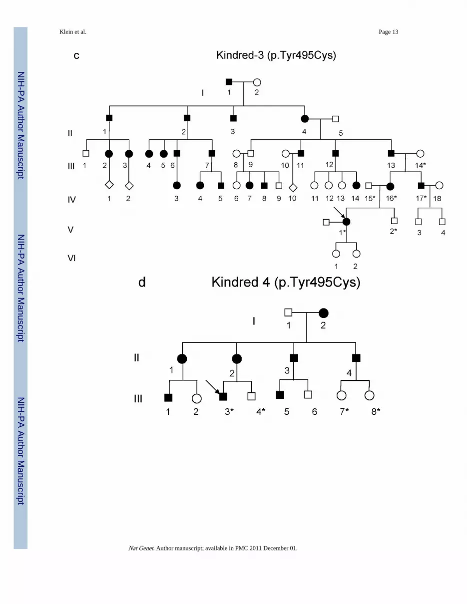
Figure 2.(a-d) Pedigrees of four kindreds are shown. A heterozygous mutation c.A1484G, resultingin p.Tyr495Cys, was identified in exon 20 of DNMT1 in the HSAN kindred 1(a), 3(c) and
Klein et al. Page 14
Nat Genet. Author manuscript; available in PMC 2011 December 01.
NIH
-PA Author Manuscript
NIH
-PA Author Manuscript
NIH
-PA Author Manuscript

4(d). Three consecutive heterozygous mutations c.1470TCC-1472ATA, resulting inp.Asp490Glu-Pro491Tyr substitution, also in exon 20 of DNMT1 was identified in theHSAN kindred 2(b). We sequenced all available samples (with asterisk) from these fourkindreds to confirm the mutation is segregated with the disease. (e) Schematic overview ofDNMT1 and its multiple domains in the N-terminal region (PBD, PCNA binding domain;TS, targeting sequence; ZnF, zinc finger; BAH1 & 2, bromo adjacent homology domains1& 2). Below is the ClustalW alignment of the part of TS domain where mutations occurfrom multiple DNMT1 homologs. Comparison of human DNMT1 (P26358) and itsorthologs in mouse (P13864), rat (Q9Z330), cow (Q24K09), sheep (Q865V5), zebrafish(Q8QGB8), frog (Q6GQH0), opossum (Q8MJ28), chicken (Q92072), silkworm (Q5W7N6),arebiopsis (Q9SEG3), carrot (O48867), corn (Q8LPU6) and rice (A2XMY1). Conservedamino acids are colored in blue. Red arrow points to the mutations. (f). Location of mutatedresidues in the TS domain of human DNMT1. Shown in red are the side chains of Asp490,Pro491 and Tyr495 in the crystal structure of the TS domain (Protein Data Bank accessionnumber 3EPZ). The image was generated using PyMOL (http://www.pymol.org/).
Klein et al. Page 15
Nat Genet. Author manuscript; available in PMC 2011 December 01.
NIH
-PA Author Manuscript
NIH
-PA Author Manuscript
NIH
-PA Author Manuscript

Figure 3.Confocal microscopy was performed using HeLa cells co-transfected with plasmidscontaining RFP-PCNA and GFP-DNMT1 wild type TS domain (panel a), p.Tyr495Cys-TSdomain (panel b) or p.Asp490Glu-Pro491Tyr-TS domain (panel c). Wild type and mutantTS domains appear green in the right panels, PCNA appears red in the middle panels andmerged images are shown in the left panels. Scale bar, 5um. In panel a, wild type TSdomain enters nucleus during S phase when PCNA localizes to the toroidal structures ofreplication foci, wild type TS also binds to heterochromatin during G2 phase when PCNAshowed diffused pattern and toroidal structures are no longer visible. Panel b and panel cshowed mutant TS domains was unable to enter into the nucleus and remained in thecytoplasm.
Klein et al. Page 16
Nat Genet. Author manuscript; available in PMC 2011 December 01.
NIH
-PA Author Manuscript
NIH
-PA Author Manuscript
NIH
-PA Author Manuscript

Klein et al. Page 17
Nat Genet. Author manuscript; available in PMC 2011 December 01.
NIH
-PA Author Manuscript
NIH
-PA Author Manuscript
NIH
-PA Author Manuscript

Figure 4.Confocal microscopy was performed using HeLa cells co-transfected with plasmidscontaining RFP-PCNA and full length (a) GFP-wild type DNMT1, (b) GFP-p.Tyr495Cys-DNMT1 or (c) GFP-p.Asp490Glu-Pro491Tyr-DNMT1. Wild type and mutant DNMT1appear green in the right panels, PCNA appears red in the middle panels and merged imagesare shown in the left panels. Scale bar, 5um. Cell cycles are deciphered from the pattern ofRFP-PCNA. In S phase, PCNA is present at the toroidal structures of the replication foci; inG2 phase, PCNA shows diffused pattern in the nucleus. In panel a, wild type DNMT1 co-localizes with PCNA at replication foci during both S phase and binds to heterochromatinduring G2 phase. In panel b and c, p.Tyr495Cys-DNMT1 and p.Asp490GLu-Pro491Tyr-DNMT1 localize along with PCNA at the replication foci during S phase but did not showbinding of heterochromatin during G2 phase.
Klein et al. Page 18
Nat Genet. Author manuscript; available in PMC 2011 December 01.
NIH
-PA Author Manuscript
NIH
-PA Author Manuscript
NIH
-PA Author Manuscript

Figure 5.Moving average of methylation difference of ∼25000 CpG sites between affected andunaffected groups from kindred-1. Kindred 1 was chosen to optimize same number ofgender and the first degree relation between affected and unaffected group. The methylationprofile of affected and unaffected groups was compared. Y-axis represents the methylationdifference between the two groups. X-axis represents p-value. Red colored line representsthe moving average of methylation difference between affected vs. unaffected group. Eachblue dot represents methylation difference for a CpG site. Blue dots below the 0 linerepresents reduced methylation in the affected group while blue dots above 0 line representsincreased methylation in the affected group. The red moving average line suggests localhypermethylation and moderate global hypomethylation in the affected group, consistentwith the 5-mdC content measurement by LC-ESI-MS/MS (supplementary fig. 5).
Klein et al. Page 19
Nat Genet. Author manuscript; available in PMC 2011 December 01.
NIH
-PA Author Manuscript
NIH
-PA Author Manuscript
NIH
-PA Author Manuscript

NIH
-PA Author Manuscript
NIH
-PA Author Manuscript
NIH
-PA Author Manuscript
Klein et al. Page 20
Tabl
e 1
Phe
noty
pic
Cha
ract
eris
tics
of
HSA
N 1
Sub
ject
s w
ith
Dem
enti
a an
d Se
nsor
ineu
ral H
eari
ng L
oss
Pat
ient
Sym
ptom
Ons
et (
yrs)
Age
(yr
)F
irst
Exa
min
edG
ende
rF
irst
Sym
ptom
(s)
Ext
rem
ity
Sens
ory
Los
sD
ista
l Lim
b W
eakn
ess
Cer
ebel
lar
atax
iaD
emen
tia
Ons
et(y
rs)
Dea
th (
yrs)
K1
V7
3059
FH
eari
ng lo
ssY
es*
No
No
40s
61
K1
V-1
235
48M
Los
s of
sen
satio
nH
eari
ng lo
ssY
esN
oN
o42
Aliv
e
K1
V-1
430
44F
Hea
ring
loss
Yes
No
No
41-
K1
V-1
632
38F
Los
s of
sen
satio
nH
eari
ng lo
ssY
esY
esN
o30
sA
live
K1
VI-
1120
s30
FH
eari
ng lo
ssY
esN
oN
o30
sA
live
K1
VI-
2335
45M
Hea
ring
loss
Yes
*N
oN
o40
51
K1
VI-
3020
s30
ML
oss
of s
ensa
tion
Yes
No
No
30s
Aliv
e
K1
V1-
3335
43M
Hea
ring
loss
Yes
*Y
esN
o30
s46
K1
VI-
3635
42M
Hea
ring
loss
Yes
*N
oN
o40
s48
K1
VII
13
20s
18F
Hea
ring
loss
Yes
No
No
30s
Aliv
e
K1
VII
15
20s
20F
Los
s of
sen
satio
nY
esN
oN
o40
sA
live
K2
II-2
2832
FH
eari
ng lo
ssY
es*
No
No
30s
46
K2
III-
127
38M
Hea
ring
loss
Yes
*N
oY
es30
sA
live
K2
III-
316
39F
Hea
ring
loss
Yes
*N
oY
es30
sA
live
K3
IV-1
635
47F
Hea
ring
loss
Yes
*N
oN
o40
s55
K3
IV-1
730
s40
ML
oss
of s
ensa
tion
Hea
ring
loss
Yes
No
Yes
40s
47
K3
V-1
30s
40F
Los
s of
sen
satio
nH
eari
ng lo
ssY
es*
No
Yes
40s
44
K4
III-
330
s40
sM
Los
s of
sen
satio
nH
eari
ng lo
ssY
esN
oN
o30
sA
live
* Foot
ulc
ers;
--
Una
vaila
ble;
K k
indr
ed
Nat Genet. Author manuscript; available in PMC 2011 December 01.