transit defect of potassium-chloride co-transporter 3 is a major pathogenic mechanism in hereditary...
TRANSCRIPT

1
TTRANSIT DEFECT OF THE POTASSIUM-CLHORIDE CO-TRANSPORTER 3 IS A
MAJOR PATHOGENIC MECHANISM IN HEREDITARY MOTOR AND SENSORY
NEUROPATHY WITH AGENESIS OF THE CORPUS CALLOSUM
Adèle Salin-Cantegrel1, Jean-Baptiste Rivière
1, Masoud Shekarabi
1, Sarah Rasheed
1, Sandra
DaCal2, Janet Laganière
1, Rébecca Gaudet
1, Daniel Rochefort
1, Gaëtan Lesca
3, Claudia Gaspar
1,
Patrick A. Dion1, Jean-Yves Lapointe
2, Guy A. Rouleau
1, 4*
From Centre of Excellence in Neuromics of University of Montreal, CHUM Research Center, Montreal 1,
Department of Medicine 4 and Department of Physics 2, University of Montreal, Quebec, Canada, Service
de génétique moléculaire et clinique, hôpital Edouard-Herriot, Lyon, France 3.
Running head: Transit defect of KCC3 in HMSN/ACC
Address correspondence to: Guy A. Rouleau MD PhD FRCP(C). 2099, rue Alexandre de Sève
Office Y-3633, Montréal, Qc H2L 2W5. Fax: (514) 345 4698 or (514) 412-7602. Email:
Missense and protein truncating
mutations of the human potassium-chloride
co-transporter 3 gene (KCC3) cause
hereditary motor and sensory neuropathy
with agenesis of the corpus callosum
(HMSN/ACC), which is a severe
neurodegenerative disease characterized by
axonal dysfunction and neurodevelopmental
defects. We previously reported that KCC3
truncating mutations disrupt brain-type
creatine kinase (CK-B)-dependent activation
of the co-transporter through the loss of its
last 140 amino acids. Here, we report a novel
and more distal HMSN/ACC truncating
mutation (3402C�T; R1134X) that
eliminates only the last 17 residues of the
protein. This small truncation disrupts the
interaction with CK-B in mammalian cells
but also affects plasma membrane localization
of the mutant transporter. Although it is not
truncated, the previously reported
HMSN/ACC-causing 619C�T; R207C
missense mutation also leads to KCC3 loss of
function in Xenopus oocytes flux assay.
Immunodetection in Xenopus oocytes and in
mammalian cultured cells revealed a
decreased amount of R207C at the plasma
membrane, with significant retention of the
mutant proteins in the endoplasmic reticulum
(ER). In mammalian cells, curcumin partially
corrected these mutant protein
mislocalizations, with more protein reaching
the plasma membrane. These findings suggest
that mistrafficking of mutant protein is an
important pathophysiological feature of
HMSN/ACC causative KCC3 mutations.
Hereditary motor and sensory
neuropathy with agenesis of the corpus callosum
(HMSN/ACC; OMIM 218000) is a severe and
progressive early onset motor and sensory
neuropathy that is characterized by axonal
swelling and is associated with
neurodevelopmental defects in the central
nervous system (CNS) (1). This autosomal
recessive neuropathy is caused by mutations in
the potassium-chloride co-transporter 3
(SLC12A6/KCC3) gene (2). However, cation-
chloride co-transporter (CCC)-related diseases
are mainly metabolic disorders that include
Gitelman’s disease, which is caused by
mutations in the SLC12A3/NCC gene (OMIM
#2638000), and Bartter’s disease, which is
caused by mutations in the SLC12A1/NKCC2
gene (OMIM #601678).
CCCs share a structure of twelve
membrane-spanning segments, which constitute
a hydrophobic and glycosylated core, flanked by
cytosolic amino- and carboxy-terminal domains.
Many pathomechanisms have already been
proposed to explain the inactivation of NCC and
NKCC2 in Gitelman’s and Batter’s diseases,
respectively; these include anomalies in the
biosynthesis, processing, trafficking,
conductance, regulation, and degradation of the
mutant proteins (see Gamba 2005, for review)
(3); however, little is known about the
pathomechanisms underlying KCC3 dysfunction
in HMSN/ACC.
Ten HMSN/ACC-causing KCC3
mutations have been reported, which include six
randomly distributed frameshift mutations, two
carboxy-terminal-truncating nonsense mutations
and two missense mutations (2, 4, 5); thus, the
http://www.jbc.org/cgi/doi/10.1074/jbc.M111.226894The latest version is at JBC Papers in Press. Published on June 6, 2011 as Manuscript M111.226894
Copyright 2011 by The American Society for Biochemistry and Molecular Biology, Inc.

2
most frequent alterations in HMSN/ACC are the
total or partial elimination of KCC3 carboxy-
terminal regions. Data collected from
heterologous expression studies in Xenopus
oocytes using truncated KCC3 mutants revealed
that the loss of the last 140 amino acids is
sufficient to abolish the KCC3-driven flux of 86Rb+ (2, 4). We have recently demonstrated that
this loss of function includes the failure of the
transporter to interact with brain-type creatine
kinase (CK-B) (6). However, the R207C
missense mutation is predicted to result in a full-
length protein bearing an amino acid exchange
that should not impact the interaction of KCC3
with CK-B, which suggests that other
mechanisms may be involved in KCC3
dysfunction in HMSN/ACC (5).
In the present study, we report a novel
and very distal KCC3 truncation mutation in an
Algerian HMSN/ACC family. We found that
the targeting of this and other mutant KCC3
proteins to the plasma membrane is perturbed.
These results provide evidence that a trafficking
defect may in part explain the KCC3 dysfunction
found in HMSN/ACC.
Experimental procedures
Mutations screening - Blood samples were
collected with informed consent from Algerian
and Turkish families (Fig. 1). DNA was
extracted from peripheral blood lymphocytes
using standard methods. A clinical HMSN/ACC
diagnosis was established by a neurologist
according to the basic features of the disease
(Table 1). The 26 exons of the KCC3 gene were
amplified by PCR using intronic primers (4) and
products were directly sequenced on an ABI
3700 sequencer, according to the manufacturer’s
recommended protocol (Applied Biosystems,
Foster City, CA).
RT-PCR - Total RNA was extracted, from a
lymphoblast cell line established from the blood
of a Turkish patient bearing the exon 5 missense
mutation (619C�T; R207C), using the RNeasy
kit (Qiagen) according to the manufacturer’s
protocol. The extracted RNA was reverse
transcribed (RT) using a classic protocol (7).
Different primers were design to flank the
mutation site: primers “4” hybridized at the
beginning of exon 4 (5�–
GAAATGGACACCAGACCGAA–3�), primers
“4/5” hybridized at the junction of exons 4 and 5
(5�–GCCCACCAAGACCCCCCAAA–3�),
primers “5/6” hybridized at the junction of exons
5 and 6 (5�–TCAACATTGTACAGCAGCAG–
3�), and primers “6” hybridized at the end of
exon 6 (5�–TGGCACCACTCCATTAGTGG–
3�). The primers flanking exon 5 of the KCC3
gene were used in PCR reactions following a
classic protocol (2) and products were loaded on
agarose gel for observation. A DNA construct
containing the wild-type KCC3 cDNA was used
as a positive control in the PCR reaction.
Plasmid constructs and mutagenesis - The wild-
type KCC3 cDNA (in the pGEM vector), which
was kindly provided by Dr Mount (Harvard
Medical School, Boston, MA, 02114, USA), was
subcloned in pcDNA3 vector (Invitrogen) to
allow expression in mammalian cells. To
generate the R207C and R1134X mutant
transporters, the 619C�T transition mutation in
exon 5 and the 3402C�T transition mutation in
exon 25 of KCC3 were introduced in the pGEM
and pcDNA constructs using the QuikChange
site-directed mutagenesis kit (Stratagene, La
Jolla, CA) with the lower primers: 5�–
TTCTTCAGGTTTTTGCAATT–3� and 5�–
GAAGTGATCACTATTTATTC–3�. DNA
sequence accuracy was verified by sequencing.
Xenopus laevis flux assay - The activity of the
co-transporter was determined by assessing 86Rb+ uptake, according to a previously
described protocol (6). Briefly, the pGEM
templates were digested with NotI (for
linearization) and the cRNAs were transcribed in vitro using T7 RNA polymerase. Groups of 20 or
more Xenopus oocytes were injected with 50 nL
of water with or without wild-type or mutant
cRNA. The uptake experiment consisted of a 45
min incubation period in a hypotonic medium (in
mM: 52 Na-cyclamate, 3.3 KCl, 0.74 CaCl2,
0.82 MgCl2, 10 HEPES/Tris, pH 7.4, with 10
μM ouabain), followed by a 45 min incubation
in an uptake medium (mM: 49 NaCl, 15 Na-
cyclamate, 0.74 CaCl2, 0.82 MgCl2, 30 RbCl, 10
HEPES/Tris, pH 7.4, and 10 �M ouabain
supplemented with 50 �Ci of 86Rb-Cl). When
experiments were performed without chloride,

3
Cl- was substituted by gluconate. The KCC3-
dependent uptake of 86Rb+ was deduced by
exposing groups of cRNA-injected oocytes to 1
mM of the loop diuretic agent furosemide.
Dependency of the function of KCC3 on CK-B
activity was determined by exposure to 200 �M
1-Fluoro-2,4-dinitrobenzene (DNFB). The
uptake experiment was stopped after 45 min by
five washes in ice-cold uptake solution without
the isotope, to remove extracellular fluid tracer.
The oocytes were lysed in 10% sodium dodecyl
sulfate and tracer activity was measured for 2
min in a liquid scintillation counter.
Plasma membrane fraction preparation of
oocytes - The oocytes were rinsed in MES-
buffered saline solution (MBSS; 80 mM NaCl,
20 mM MES pH 6.0) and incubated for 10 min
at room temperature in the same solution with
0.005% subtilisin A (Sigma-Aldrich) to partially
digest the vitelline membranes. The
polymerizing steps were performed for 60 min
with 1% ludox/MBSS and then 45 min with
0.1% polyacrylic acid (Sigma-Aldrich) at 4°C
under mild agitation. Between each step, oocytes
were rinsed 3 times in MBSS. The oocytes were
then homogenized by pipetting in 1ml cold HbA
(in mM: 5 MgCl2, 5 NaH2PO4, 1 EDTA, 80
sucrose, and 20 Tris pH 7.4) and then
centrifuged at 16 g, 25 g and 35 g for 30 sec at
4°C. Between each centrifugation, the
supernatants were removed and the pellet was re-
suspended in 1 ml of cold HbA. A final
centrifugation at 16,000 g for 20 min pelleted the
purified plasma membranes, which were
prepared for immunoblotting (8).
Cell culture and transfection - HeLa cells
(ATCC) were grown in Dulbecco’s modified
Eagle’s medium (DMEM) supplemented with
10% fetal bovine serum (FBS) and antibiotics
(50 units/ml penicillin and 50 mg/ml
streptomycin) at 37°C with 5% CO2 (all supplied
by Gibco). PC12 cells were grown on poly-D-
lysine-coated coverslips placed in DMEM/F12
containing 10% heat-inactivated horse serum
(iHs), 5% FBS, and antibiotics (50 units/ml
penicillin and 50 mg/ml streptomycin) at 37°C
with 5% CO2.
The plasmids used in the transfection
studies were prepared using the Qiagen Plasmid
Maxi Kit, according to the manufacturer’s
instructions. Transient transfections were
performed on cells using the Lipofectamine
transfection reagent (Invitrogen), according to
the manufacturer’s protocol. Cells were
incubated for 24 hours with lipofectamine before
replacing the medium, and transgene expression
was assayed at least 36 hours post-transfection.
Typically, a transfection efficiency of the
transgene of 20–50% was achieved; equivalent
transfection efficiency among the various
transfection experiments was confirmed by
Western blot.
PC12 differentiation was induced by
treating the cells with 50 ng/ml nerve growth
factor (NGF) in a reduced serum environment
(2,5% iHS) for 3 days.
Curcumin treatment was performed prior
to the immunofluorescence experiment. The
cells were incubated for 3 hours in curcumin at a
final concentration ranging from 5 to 40 �M.
The cells were rinced several times in PBS to get
rid of the curcumin before the immunostaining
experiment.
Immunofluorescence study - Cells grown on
coverslips were induced to differentiate if
necessary (in the case of PC12 cells) and were
then transiently transfected with the pcDNA 3.0
wild-type or mutant KCC3 constructs, fixed in
4% paraformaldehyde solution in PBS at room
temperature for 10 min, and finally processed for
immunofluorescence staining. For this, cells
were permeabilized in very mild conditions
(0,01% Triton X-100 for 3 min) and nonspecific
site blocking was performed in 10% normal goat
serum (NGS) solution in PBS for 1 hour at room
temperature, which was directly followed by
incubation with anti-KCC3 (1:500; Abnova),
anti-CK-B (1:100; OEM concept), anti-pan
cadherin (1:500; abcam), or anti-calreticulin
(1:250; Abcam) primary antibodies in 5%
NGS/PBS overnight at 4°C. Coverslips were
then washed twice in PBS, incubated with the
appropriate secondary antibodies (Jackson Labs),
and mounted on slides for confocal microscopy
(Leica).
Paraffin-embedded fragments of brains
from an HMSN/ACC patient were sectioned (5
�m) and placed on glass slides. The sections
were processed for immunohistochemistry using
the following protocol: after deparaffinization
and rehydration, the slide-mounted tissue
sections were incubated in an antigen retrieval

4
solution (DAKO S1699) preheated to 85˚C for 1
hour. Sections were cooled to room temperature
and were then rinsed three times with PBS. After
a 30 min permeabilization step in 0.2% Triton X-
100/PBS, the sections were blocked in a 10%
solution of NGS in 0.02% Triton X-100/PBS at
room temperature for 1 hour. Incubation of
primary antibodies at suggested concentrations
(diluted in 2% NGS in 0.02% Triton X-
100/PBS) was then carried out overnight at room
temperature. Sections were washed three times
in PBS the following day. Secondary antibody
incubation was carried out using the appropriate
Alexa antibodies (1:1000 in 2% NGS/0.02%
Triton X-100/PBS) for 1 hour at room
temperature. Sections were again washed three
times in PBS. The slides were then coverslip-
mounted using Mowiol (Fluka, Polyvinyl
alcohol 4-88) and were visualized using
appropriate filters on a Leica DM6000
microscope.
Immunoblot - HeLa cells were plated onto 10 cm
petri dishes, grown to confluence, and then
transfected with pcDNA-KCC3 wild-type or
mutant constructs, as described previously (6).
After transfection, cells were placed in fresh
DMEM/10% FBS for 48 hours. Whole-cell
lysates were harvested by washing cells in cold
PBS and by scraping them into 0.1% triton in
PBS or SUB lysis buffer supplemented with
Complete protease inhibitors (Roche). The
extracted proteins were separated by 8% SDS-
PAGE, transferred to nitrocellulose membranes,
and immunoblotted with antisera to KCC3
(1:1000) or CK-B (1:100). The blots were
stripped and re-probed with an anti-actin
antibody (Chemicon) to verify equivalent
loading of protein.
Statistics - Protein co-localization rates were
evaluated using Pearson’s coefficient on
confocal microscopy, using the LAS AF
software (Leica). Significance was set at a two-
tailed Student’s t test with a P value < 0.01.
Results were presented as means ± the standard
error (SE). The significance of the differences
between Xenopus oocyte groups in the flux
assays was tested by one-way analysis of
variance (ANOVA).
RESULTS
R1134X is a novel and most distal KCC3
truncating mutation. Sequencing of the
SLC12A6/KCC3 gene in two HMSN/ACC cases
from an Algerian consanguineous family
revealed a homozygous cytosine-to-thymine
transition (3402C�T) in exon 25 (Fig. 1a). The
mutation creates a premature stop codon that
leads to the loss of the last 17 amino acids of
KCC3, which corresponds to only ~2% of the
total protein sequence (R1134X mutant); thus,
R1134X represents the least truncated KCC3
mutant. In comparison, the exon 22 mutation,
which was the smallest previously identified
KCC3 truncation, causes the elimination of the
last 140 amino acids of the protein, which
correspond to ~12% of the whole protein. In all,
nine of the eleven HMSN/ACC mutations result
in a net loss of the amino acids within the CTD
of KCC3.
Truncated R1134X fails to interact with
CK-B. The brain-type creatine kinase (CK-B),
which is an ATP-generating protein, is a potent
activator of KCC3 via a direct interaction with
the co-transporter’s carboxy-terminal domain
(CTD) (6). We have previously used a yeast-two
hybrid system to demonstrate that the sequence
encompassing the last 18 amino acids of KCC3
is necessary for its interaction with human CK-B
(6), which suggests that the physical interaction
between CK-B and R1134X (which lacks the last
17 amino acids) may be impaired in vivo (Fig.
1b). To confirm this hypothesis, we performed
dual immunofluorescence studies with CK-B-
and KCC3-specific antibodies using human
HeLa cells that were transfected with R1134X
construct and which express CK-B endogenously
(Fig. 2a). Although we observed the interaction
between wild-type KCC3 and CK-B as
previously reported, we failed to observe a co-
localization between CK-B and R1134X (Fig.
2b).
To confirm that the disruption of
R1134X and CK-B interaction has an impact on
the co-transport activity, wild-type KCC3 and
the nonsense mutant R1134X were expressed in
Xenopus oocytes and 86Rb+ uptake upon
swelling-activation of KCC3 was measured. As
expected, the wild-type transporter induced a
significant flux (P = 0.027 ANOVA compared
5
with the control), which was abolished by
chloride substitution or by treatment with 200
�M of DNFB, a specific creatine kinase
inhibitor. In contrast, R1134X did not induce a
significant flux in Xenopus oocytes (Fig. 2c).
This further associates the loss of interaction
with CK-B with the loss of function of truncated
KCC3.
It is noteworthy that the last 17 amino
acids missing in R1134X contain two highly
conserved sequences probably involved in the
protein trafficking: a hydrophobic tetrad that
participates in the transit of other proteins of the
SLC12 family (9) and a C-terminal tyrosine
sorting motif maybe controlling the co-
transporter trafficking (Fig. 1b). When we
performed immunoblots of plasma membrane
fractions from Xenopus oocytes expressing
R1134X, we observed only small amounts of the
mutant protein in the purified extract (Fig. 2d).
In addition co-staining of R1134X and CK-B in
mammalian cells revealed that the most
abundant staining of the truncated protein was
intracellular and not at the cell periphery. These
data suggest that defect of mutant KCC3
trafficking could be a pathomechanism leading
to KCC3 dysfunction even without a truncation.
The R207C mutation does not result in
KCC3 truncation. The screening of the KCC3
gene for mutations in a Turkish patient and his
parents revealed the presence of a 619C�T
missense mutation in exon 5 (Fig. 3a). This
mutation was previously reported and contrasted
with the contention that KCC3 truncation
mutations alone can lead to disease (5). Given
that the vast majority of known HMSN/ACC
mutations truncate the co-transporter, we
investigated whether the nucleotide transition in
exon 5 might lead to aberrant RNA splicing and
result in a "classic" case of HMSN/ACC
truncation. To do so, total RNA extracted from
lymphoblasts of the patient bearing the 619C�T
missense mutation was reverse transcribed to the
corresponding cDNA. Three primer sets that
flank the mutated exon 5 were used in a PCR
reaction to assess exon splicing. We observed
that all predicted PCR fragments from the patient
were present and were equal in size to those of
the control sample, which implies a normal exon
splicing of the mutant RNA (Fig. 3b). This
overall suggests that a full-length KCC3 mutant
protein (R207C) can be produced from this
mutated KCC3 mRNA.
The R207C mutation affects KCC3
function through transit defect to the cell
surface. So far, KCC3 loss of function was
associated with disruption of truncated KCC3
interaction with CK-B. However the missense
KCC3 mutant R207C is not truncated but is a
full-length protein that still co-localizes with
CK-B in mammalian cells (supplementary data:
Fig. S1). This suggests that disruption of KCC3
interaction with CK-B is not the mechanism of
dysfunction of the R207C mutation. Given that
the KCC3 R207C mutant interacts with CK-B
and is likely translated into an intact protein, we
wondered if it may in fact be a benign variant,
and not a causative mutation. To assess its
activity, R207C was expressed in Xenopus
oocytes and 86Rb+ uptake upon swelling-
activation of KCC3 was measured. Similarly to
the truncated mutants, full-length R207C
expression did not result in a significant flux in
Xenopus oocytes (Fig. 4a), confirming that this
mutation impairs KCC3 function and is likely
causative in HMSN/ACC. Although R207C co-
localized with CK-B in HeLa cells, we observed
a strong perinuclear localization of the mutant
protein in these cells, suggesting a deficiency in
the transit of the R207C protein to the plasma
membrane in this system (supplementary data:
Fig. S1). KCC3 loss of function could therefore
be caused by the absence of the mutant co-
transporter at the plasma membrane. To
investigate this hypothesis, we first performed
Western blot analysis of plasma membrane
proteins isolated from Xenopus oocytes
expressing the wild-type and the mutant
transporters. Similarly to R1134X, little R207C
protein was observed in purified plasma
membrane extracts of Xenopus oocytes (Fig. 4b),
suggesting defective KCC3 transport to the cell
membrane. To further support this hypothesis,
we sought to measure how much of the mutant
forms of KCC3 reached the plasma membrane in
mammalian cells. Wild-type and mutant KCC3
were expressed in HeLa cells (Fig. 5a) and cell
surface localization was quantified by measuring
the rate of co-localization between KCC3 and
pan-cadherin, which is a plasma-membrane
marker, using Pearson’s coefficient (Fig. 5b).

6
The wild-type protein was mainly found at the
plasma membrane of transfected HeLa cells
(Pearson’s coefficient = 0.84). In contrast, most
of the mutant R207C did not reach the plasma
membrane (Pearson’s coefficient = 0.285; P <
0.0001 when compared with wild-type-
transfected cells). For the R1134X mutation,
plasma membrane localization was also impaired
(Pearson’s coefficient = 0.57; P = 0.003 when
compared with wild-type-transfected cells).
Instead, R207C and R1134X accumulated inside
the cytosol. This shows that the transit of these
mutant cotransporters is deficient.
The R207C amino acid exchange
stabilizes the homodimeric structure of KCC3.
It was reported that cation-chloride
cotransporters form transient homodimers and
heterodimeric structures in Xenopus oocytes
(10). As enhanced dimerization can alter the
membrane targeting process through organelle
retention (11), we investigated whether this
mechanism was at the origin of the deficient
transit of the mutant proteins to the plasma
membrane. Wild-type and mutant KCC3
proteins were transiently expressed in
mammalian cells and total protein lysates
prepared for immunodetection by Western blots.
The immunoblots for wild-type KCC3 revealed
an abundant band below 150 kDa (KCC3
expected size) but also a faint band at ~300 kDa,
which corresponds to double the size expected
for KCC3 monomers. In the R207C protein
extract, immunoblots revealed an abundant band
at ~300 kDa (Fig 5c). Increased stringency or
additional sonication was required to disrupt
some of these structures and restore a migration
profile closer to the wild-type protein (Fig 5d-e).
This suggests that the arginine to cysteine
exchange in R207C favors the stabilization of
homodimeric structures. This may affect the
transit of R207C to the plasma membrane, which
results in the intracellular accumulation of the
mutant protein
Curcumin treatment partially rescues the
transit defect of mutant KCC3. To determine the
predominant site of mutant protein
accumulation, we used a panel of organelle
markers, which included markers for the golgi,
proteasome, clathrin-coated vesicules (data not
shown), lamellipodia (e.g. cortactin), ER (e.g.,
calreticulin) and plasma membrane (e.g., pan-
cadherin) in co-immunofluorescence labeling
experiments. No specific localization of R1134X
was detected. However, we observed a strong
co-localization signal between R207C and the
ER markers (Fig. 6b). Aberrant distribution of
the mutant proteins was also observed in PC-12
cells that were induced to differentiate by NGF
treatment (Fig. 6c). This overall suggests that
R207C dimers may be retained in the ER and
thus be kept from reaching the plasma
membrane.
Chemical compounds have been shown
to favor the release of mutant transmembrane
proteins that are aberrantly retained in the ER.
Curcumin is one of the most promising
compounds that help to relieve this retention, as
it is harmless, eatable, and works in mouse
models (12, 13). We sought to determine
whether R207C retention in cultured cells could
be lessened by curcumin treatment by incubating
cells expressing the mutant protein with 5–40
�M of curcumin at 37°C. The localization of the
proteins at the plasma membrane was evaluated
using pan-cadherin as a specific plasma-
membrane marker in immunofluorescence
studies. A concentration of curcumin of 10 �M
was sufficient to rescue some of the transit to the
plasma membrane (data not shown); however,
the best results were observed with 20 �M
curcumin (Fig 6 d-e). This result further supports
the hypothesis that R207C dimers are retained
within the ER.
Aberrant mutant KCC3 localization in
HMSN/ACC brain. HMSN/ACC is most
frequent in the French-Canadian population and
is mainly caused by the T813X truncation
mutation. We have previously shown that the
T813X mutation leads to the loss of function of
the co-transporter using a Xenopus oocyte flux
assay. Although T813X was detected at the
oocyte membrane (2) it has been reported that
mutant channels or transporters can behave
differently in the Xenopus oocyte system, where
mutant proteins are less prone to traffic defects
compared with mammalian cells and in vivo
scenarios (11). In addition, when we expressed
mutant proteins R1011X, L808X (4) or R1134X
in mammalian cells, we observed aberrant
intracellular accumulation of the mutant
proteins. Finally, all truncated transporters,
including the French-Canadian mutant protein

7
T813X, lack a C-terminal hydrophobic tetrade
involved in the trafficking of other SLC12 co-
transporters. All these data suggest that T813X
and the other mutant transporters might have
some traffic defects in vivo.
To test this hypothesis, we focused on
KCC3 localization in brain. First, both the wild-
type KCC3 and the mutant T813X could be
detected by Western-blot using protein extracted
from brain tissue of a control individual and a
French Canadian HMSN/ACC patient (Fig. 7a).
Then, to evaluate if defective transit participates
in T813X pathogenesis, we observed the
distribution of T813X truncated protein in brain
histological sections by immunofluorescent
labeling of KCC3. Using confocal microscopy,
we found particularly abundant staining of
T813X around the nucleus whereas KCC3
distribution in unaffected neurons was more
peripheral (Fig. 7b). In addition, swollen axons
of the HMSN/ACC patient lacked KCC3
staining (data not shown). This suggests that a
transit defect of T813X may occur in
HMSN/ACC brains as well.
DDISCUSSION
Potassium/chloride co-transporters participate in
cell volume regulation but also control neuronal
activity by transporting K+ and Cl- ions across
the plasma membrane.
Herein, we confirmed that the loss of
KCC3 activity is systematic in HMSN/ACC but
that different pathogenic mechanisms can lead to
the inactivation of the co-transporter. To
participate in the electroneutral transport of ions,
KCC3 needs to be translated and targeted to the
plasma membrane, where it has to be properly
activated. Some mutations have been associated
with abrogated or defective protein biosynthesis.
Because of their altered structure, the mutant
mRNAs tend to be unstable and are efficiently
cleared from the cell. As a result, virtually no
protein is expressed. However, we did not
observe anomalies in mutant KCC3 biosynthesis
in this study (supplementary data: Fig. S2).
Many variants, including those expressed in
heterologous systems, fail to be properly
processed to a mature glycosylated form and/or
to be transported to the plasma membrane. In
Gitelman’s disease, the majority of the missense
mutations affect the glycosylation of NCC,
however non-glycosylated forms can still be
processed to the cell surface. Alternatively, an
apparently normal processing of a mutant protein
with normal functional and kinetic properties can
be associated with an impaired insertion into the
plasma membrane. The most common mutation
in cystic fibrosis (i.e., deltaF508) belongs in this
category and, if correctly processed to the
membrane, possesses residual activity and leads
to a sustained normal or only mildly affected
phenotype. For this reason, mutations in this
group are promising targets for therapies aimed
at correcting the processing and delivery of the
mutant protein to the membrane. Mutations in
channels/co-transporters, which produce
abnormal protein variants, can also affect the
regulation of the function of the protein
eventually by preventing physical binding to
partners that are required for its activation. In
type I Bartter’s disease, the mutated co-
transporters are normally synthetized,
glycosylated and inserted into the plasma
membrane, which implies a defect in the
functional properties or in the intrinsic activity of
the protein. Finally, some mutations accelerate
the removal from the plasma membrane and/or
degradation of the mutant protein. Truncation of
the C-terminal domain of CFTR reportedly leads
to a marked instability of an otherwise fully
processed and functional variant.
Our biochemical and cellular data
demonstrate the presence of at least two distinct
mechanisms that lead to KCC3 dysfunction in
HMSN/ACC. We confirmed that the functional
interaction between CK-B and truncated KCC3
is systematically disrupted. We also provide
evidence that indicates that defective transit to
the plasma membrane is involved in the loss of
function of mutant KCC3 in HMSN/ACC. In
addition, we showed that enhanced dimerization
of cation-chloride co-transporters seems to
disturb their transit and activity. The R207C
missense mutation results in the replacement of a
highly conserved arginine residue with a
cysteine residue, within or close to the first
transmembrane domains of R207C (Fig. 3c).
Moreover, cysteines are prone to form
intramolecular and intermolecular disulfide
bonds in proteins to promote the formation of
tertiary and quaternary structures; thus the
8
introduction of novel cysteines might be at the
origin of these abnormal structures. Interestingly,
several similar arginine/glycine to cysteine
substitutions in NCC (R399C, G496C, R642C,
R852C, and R919C) have been reported as being
causative in Gitelman’s disease (3).
Cation-chloride co-transporters show
self-interacting properties when expressed in
Xenopus oocytes (10). Here, we provide
evidence that the R207C mutation induced the
stabilization of dimeric structures, which
demonstrates the occurrence of KCC3
dimerization in mammalian cells. To allow the
formation of intermolecular disulfide bonds, the
cysteine residues involved need to be physically
close in the quaternary structure, which suggests
that the first transmembrane domains directly
allow the association between the KCC3
subunits. However, the failure of the aberrant
dimers to reach the plasma membrane suggests
that, in normal physiological conditions, the
dimerization is a regulated process that is not
constitutive but occurs transiently. A similar
mechanism leads to the mislocalization of the
G480C form of CFTR in cystic fibrosis. In this
case, the substitution of glycine with cysteine
results in a protein that is not fully glycosylated
and fails to reach the plasma membrane in
mammalian cells. However, the mutant CFTR
protein retained normal chloride channel activity
in Xenopus oocytes, as channels and transporters
are less likely to experience intracellular
processing and trafficking disturbance in this
system (11). Unfortunately, we were not able to
detect a significant activity of R207C in Xenopus
oocytes, maybe because the number of mutant
proteins that reached the oocyte plasma
membrane was not sufficient to allow the
detection of co-transport activity.
The R1134X protein was
underrepresented on Western blot from
mammalian cells when compared with the wild-
type protein (Fig. 2c, 5c-e and supplemental
data: Fig. S1). This underrepresentation of
R1134X on Western blot may indicate a failure
of cultured cells transfected with the mutant
construct to proliferate at the rate of the cells
expressing the wild-type construct. This
hypothesis could be supported by the fact that
KCC3 has been implicated in cell proliferation in
normal and pathological instances (14).
However, no obvious differences in cell density
were observed in the nervous system of this
HMSN/ACC patient when compared with the
control sample (unpublished data). It is worth
noting that the mutant co-transporter T813X also
seemed to be under-expressed in the brain of a
HMSN/ACC patient (Fig. 7a). Therefore, these
observations may indicate an increased
instability of the truncated proteins, which could
either be due to the aberrant structure of the
mutants and recognition by the cellular
misfolding/degradation pathway or to their
accelerated recycling from the plasma membrane
to the cytoplasmic degradation machinery.
HMSN/ACC is a severe and progressive
neurodegenerative disease that exhibits an early
onset of symptoms. Signs of HMSN/ACC, such
as hypotonia and delays in motor development
skills, are noticed before one year of age.
However, the motor abilities of patients progress
slowly to 4–6 years of age and these children are
able to stand and walk with some help. This is
followed by a motor deterioration that generally
renders affected subjects wheelchair-dependent
by adolescence. The patients generally die at the
average age of 33. Interestingly, the mutant
protein that exhibits a truncation of the last 17
amino acids is associated with one of the worst
clinical manifestations of HMSN/ACC, whereas
the patient bearing the missense mutation
presents with one of the milder phenotype
reported to date (5) (Table 1). Further
investigations will be required to allow the clear
establishment of genotype–phenotype
correlations, as nonfunctional truncated
transporters worsen the HMSN/ACC phenotype
whereas potentially functional but aberrant
KCC3 dimers may attenuate some aspects of the
disease presentation.
The distinct pathomechanisms
uncovered here suggest novel therapeutic
approaches for the management of the disorder
or for the modulation of the progression of the
disease. The structural similarities between
KCC3 and CFTR suggest that efficient therapies
identified for cystic fibrosis may be applicable to
HMSN/ACC, at least at the molecular and
cellular levels. For example, incubation with
curcumin restores the transit of CFTR units that

9
are functional but are trapped in the ER by
altering the function of resident ER chaperones
as well as other mechanisms (15-17). This
treatment yielded promising results in mouse
models of cystic fibrosis (11). Accordingly, we
found that curcumin relieved the ER retention of
dimerized R207C in mammalian cultured cells.
A diet enriched in curcumin may therefore be
beneficial for the relief or delay of some of the
HMSN/ACC symptoms in patients bearing the
R207C mutation, including the Turkish patient
described in the present study (as he has not yet
reached puberty).
REFERENCES
1 Dupre, N., Howard, H.C., Mathieu, J., Karpati, G., Vanasse, M., Bouchard, J.P., Carpenter, S. and
Rouleau, G.A. (2003) Hereditary motor and sensory neuropathy with agenesis of the corpus callosum. Ann
Neurol, 54, 9-18.
2 Howard, H.C., Mount, D.B., Rochefort, D., Byun, N., Dupre, N., Lu, J., Fan, X., Song, L.,
Riviere, J.B., Prevost, C. et al. (2002) The K-Cl cotransporter KCC3 is mutant in a severe peripheral
neuropathy associated with agenesis of the corpus callosum. Nat Genet, 32, 384-392.
3 Gamba, G. (2005) Molecular physiology and pathophysiology of electroneutral cation-chloride
cotransporters. Physiol Rev, 85, 423-493.
4 Salin-Cantegrel, A., Riviere, J.B., Dupre, N., Charron, F.M., Shekarabi, M., Karemera, L., Gaspar,
C., Horst, J., Tekin, M., Deda, G. et al. (2007) Distal truncation of KCC3 in non-French Canadian
HMSN/ACC families. Neurology, 69, 1350-1355.
5 Uyanik, G., Elcioglu, N., Penzien, J., Gross, C., Yilmaz, Y., Olmez, A., Demir, E., Wahl, D.,
Scheglmann, K., Winner, B. et al. (2006) Novel truncating and missense mutations of the KCC3 gene
associated with Andermann syndrome. Neurology, 66, 1044-1048.
6 Salin-Cantegrel, A., Shekarabi, M., Holbert, S., Dion, P., Rochefort, D., Laganiere, J., Dacal, S.,
Hince, P., Karemera, L., Gaspar, C. et al. (2008) HMSN/ACC truncation mutations disrupt brain-type
creatine kinase-dependant activation of K+/Cl– co-transporter 3. Hum Mol Genet, 17, 2703-2711.
7 Xiong, L., Catoire, H., Dion, P., Gaspar, C., Lafreniere, R.G., Girard, S.L., Levchenko, A.,
Riviere, J.B., Fiori, L., St-Onge, J. et al. (2009) MEIS1 intronic risk haplotype associated with restless
legs syndrome affects its mRNA and protein expression levels. Hum Mol Genet, 18, 1065-1074.
8 Leduc-Nadeau, A., Lahjouji, K., Bissonnette, P., Lapointe, J.Y. and Bichet, D.G. (2007)
Elaboration of a novel technique for purification of plasma membranes from Xenopus laevis oocytes. Am
J Physiol Cell Physiol, 292, C1132-1136.
9 Nezu, A., Parvin, M.N. and Turner, R.J. (2009) A conserved hydrophobic tetrad near the C
terminus of the secretory Na+-K+-2Cl– cotransporter (NKCC1) is required for its correct intracellular
processing. J Biol Chem, 284, 6869-6876.
10 Simard, C.F., Bergeron, M.J., Frenette-Cotton, R., Carpentier, G.A., Pelchat, M.E., Caron, L. and
Isenring, P. (2007) Homooligomeric and heterooligomeric associations between K+-Cl– cotransporter
isoforms and between K+-Cl– and Na+-K+-Cl– cotransporters. J Biol Chem, 282, 18083-18093.
11 Smit, L.S., Strong, T.V., Wilkinson, D.J., Macek, M., Jr., Mansoura, M.K., Wood, D.L., Cole,
J.L., Cutting, G.R., Cohn, J.A., Dawson, D.C. et al. (1995) Missense mutation (G480C) in the CFTR gene
associated with protein mislocalization but normal chloride channel activity. Hum Mol Genet, 4, 269-273.
12 Egan, M.E., Pearson, M., Weiner, S.A., Rajendran, V., Rubin, D., Glockner-Pagel, J., Canny, S.,
Du, K., Lukacs, G.L. and Caplan, M.J. (2004) Curcumin, a major constituent of turmeric, corrects cystic
fibrosis defects. Science, 304, 600-602.
13 Lu, M., Leng, Q., Egan, M.E., Caplan, M.J., Boulpaep, E.L., Giebisch, G.H. and Hebert, S.C.
(2006) CFTR is required for PKA-regulated ATP sensitivity of Kir1.1 potassium channels in mouse
kidney. J Clin Invest, 116, 797-807.
14 Shen, M.R., Chou, C.Y., Hsu, K.F., Hsu, Y.M., Chiu, W.T., Tang, M.J., Alper, S.L. and Ellory,
J.C. (2003) KCl cotransport is an important modulator of human cervical cancer growth and invasion. J
Biol Chem, 278, 39941-39950.

10
15 Berger, A.L., Randak, C.O., Ostedgaard, L.S., Karp, P.H., Vermeer, D.W. and Welsh, M.J. (2005)
Curcumin stimulates cystic fibrosis transmembrane conductance regulator Cl– channel activity. J Biol
Chem, 280, 5221-5226.
16 Harada, K., Okiyoneda, T., Hashimoto, Y., Oyokawa, K., Nakamura, K., Suico, M.A., Shuto, T.
and Kai, H. (2007) Curcumin enhances cystic fibrosis transmembrane regulator expression by down-
regulating calreticulin. Biochem Biophys Res Commun, 353, 351-356.
17 Lipecka, J., Norez, C., Bensalem, N., Baudouin-Legros, M., Planelles, G., Becq, F., Edelman, A.
and Davezac, N. (2006) Rescue of DeltaF508-CFTR (cystic fibrosis transmembrane conductance
regulator) by curcumin: involvement of the keratin 18 network. J Pharmacol Exp Ther, 317, 500-505.
FOOTNOTES
We want to thank the families for participating in this research. This work was supported by a grant from
the Canadian Institutes of Health Research and a grant by the Fondation des Jumelles Coudé. The authors
have reported no conflicts of interest.
The abbreviations used are: ANOVA: analysis of variance; CCC: cation chloride co-transporter; CFTR:
cystic fibrosis transmembrane conductance regulator; CK-B: creatine kinase brain-specific; CNS: central
nervous system; CTD: carboxy-terminal domain; DMEM: Dulbecco’s modified Eagle’s medium; DNFB:
1-Fluoro-2,4-dinitrobenzene; ER: endoplasmic reticulum; FBS: fetal bovine serum; HMSN/ACC:
hereditary motor and sensory neuropathy; iHs: heat-inactivated horse serum; NCC: sodium-chloride
cotransporter; NKCC2: sodium-potassium-chloride cotransporter 2; KCC3: potassium-chloride
cotransporter 3; RT: reverse transcription; Pan-cadh: pan-cadherin; PCR: polymerase chain reaction; PM:
plasma membrane; SD: standard deviation; SE: standard error.
FFIGURE LEGENDS
Fig. 1. Identification of a novel HMSN/ACC truncating mutation. (a) Mutated sequence of one of the
affected individuals bearing the SLC12A6/KCC3 homozygous 3402C�T transition, with exchange of
Arg1134 for a stop codon (R1134X). (b) Representation of the protein sequence encompassing the R1134X
mutation and amino acid conservation in various SLC12 family members. The hydrophobic tetrade
suggested to be involved in cation-chloride co-transporters trafficking and an amino-terminal tyrosine
residue predicted in silico to be involved in membrane protein sorting are shown.
Fig. 2. R1134X is non-functional and fails to interact with CK-B. (a) Assessment of the interaction
between CK-B and wild-type KCC3 (top) or R1134X (bottom). The loss of interaction between R1134X
and CK-B was inferred (using confocal microscopy) from the absence of co-localization of these proteins
in HeLa cells transiently transfected with wild-type and mutant KCC3 constructs. KCC3 immunostaining
is shown in red, CK-B immunostaining is shown in green and co-localization is indicated by a yellow
signal in the overlay image. (b) The inhibition of CK-B by incubation with DNFB for 4 hours had no
dramatic effect on the transit of wild-type KCC3 (top) to the plasma membrane nor on the abnormal
subcellular localization of R207C (middle) and R1134X (bottom). (c) Western blot analysis of wild-type,
of R1134X and of endogenous CK-B in HeLa cells. (d) Assessment of wild-type and mutant KCC3 co-
transport activity in Xenopus oocytes. Measurements of 86Rb+ uptake invoked under hypotonic conditions
in a representative experiment in Xenopus oocytes injected with the wild-type and mutant KCC3 cRNA.
The indicated SEs were calculated for experimental groups of 20 or more oocytes. Wild-type KCC3
activation mediated a significant 86Rb+ transport, which was fully abolished without Cl– and by 250 �M
DNFB (a CK-B inhibitor). Significance was set at P < 0.05 (one-way ANOVA). (e) Immunoblot analysis
of wild-type and mutant KCC3 proteins at the plasma membrane of Xenopus oocytes. Total plasma
membrane proteins of Xenopus oocytes expressing wild-type and mutant KCC3 were purified using a

11
protocol previously described (8). Total and plasma membrane protein extracts were loaded on a
denaturing gel and probed with a KCC3 N-terminal-specific antibody.
Fig. 3. R207C is not a truncated protein. (a) Sequencing results for exon 5 of SLC12A6/KCC3 in a Turkish
patient bearing the 619C�T transition; the mutation is expected to exchange Arg207 for Cys207 (R207C).
(b) Total RNA extracted from lymphoblasts of the patient bearing the R207C mutation was analyzed by
RT–PCR to assess the structure of the mutant RNA. PCR primers encompassing the junction of exons 4
and 5 (referred to as “4/5”) and exons 5 and 6 (referred to as “5/6”) and at the beginning of exon 4
(referred to as “4”) and at the end of exon 6 (referred to as “6”) were used to assess the structure of KCC3
RNA. The control corresponds to the wild-type cDNA. (c) Representation of the protein sequence
encompassing the R207C mutation and amino acid conservation in various species. Highly conserved
residues are indicated by a star.
Fig. 4. R207C and R1134X are non-functional proteins. (a) Assessment of wild-type and mutant KCC3
co-transport activity. Measurements of 86Rb+ uptake invoked under hypotonic conditions in a
representative experiment in Xenopus oocytes injected with the wild-type and mutant KCC3 constructs.
The indicated SEs were calculated for experimental groups of 20 or more oocytes. Wild-type KCC3
activation mediated a significant 86Rb+ transport, which was fully abolished by 1 mM furosemide and by
250 μM DNFB, which is a specific inhibitor of creatine kinases. Significance was set at P < 0.05 (one-
way ANOVA). (b) Immunoblot analysis of wild-type and mutant KCC3 at the plasma membrane of
Xenopus oocytes. Total protein and plasma membranes of Xenopus oocytes were purified using a protocol
previously described (8). The protein extracts were loaded on a denaturing gel and probed with a KCC3
N-terminal-specific antibody.
Fig. 5. R207C and R1134X processing to the plasma membrane is impaired. (a) Assessment of the
membrane localization of wild-type KCC3, mutant R207C, and R1134X by confocal microscopy. The use
of an anti-pan-cadherin antibody as a specific plasma membrane marker revealed the impaired targeting of
R207C to the plasma membrane. KCC3 immunostaining is shown in red and pan-cadherin
immunostaining is shown in green. Co-localization is indicated by a yellow signal in the overlay image.
(b) Quantification of the co-localization rate between KCC3 and pan-cadherin in cultured cells using
Pearson’s coefficient. (c-e) Western blot of proteins extracted from cultured cells expressing wild-type,
R207C, or R1134X KCC3 proteins. Proteins were extracted using the less stringent PBS-triton buffer (c)
or using the more stringent SUB lysis buffer (d) or subjected to additional sonication (e). KCC3 proteins
were detected using a specific amino-terminal antibody that recognizes full-length and truncated KCC3.
The control corresponded to mock-transfected cells (first lane). The size of the strong band corresponding
to the R207C protein extract (third lane) was double that of the wild-type monomers, when extracted in
less stringent buffer. Additional sonication successfully dissociated the heavier structures. As reported
previously, KCC3 bands most likely corresponding to glycosylated and phosphorylated forms of the co-
transporter were also observed on western-blot between 150 and 250kDa (3).
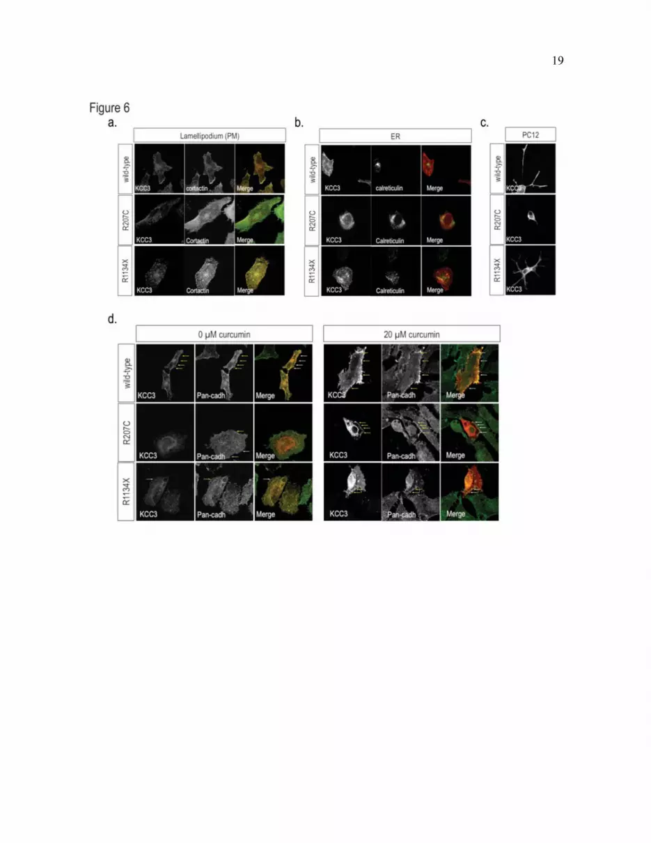
Fig. 6. Mislocalization of mutant KCC3 in HeLa and differentiated PC12 cells. (a-b) Assessment of the
subcellular co-localization of wild-type KCC3 (top), R207C (middle), or R1134X (bottom) with specific
marker in HeLa cells. KCC3 immunostaining is shown in red and cortactin (a) and calreticulin (b)
immunostainings are shown in green. Co-localization of red and green staining is evidenced by the
generation of yellow signal in overlay images. The R207C mutant protein strongly co-localized with the
ER resident chaperon protein calreticulin. (c) Assessment of the localization of wild-type KCC3 (top) or
mutant R207C (middle) and R1134X (bottom) proteins in PC12 cells differentiated using 50 ng/ml of
NGF. Similarly to what was observed in other mammalian cell types, R207C exhibited an aberrant
distribution in differentiated PC12 cells. (d-e) Curcumin relieved R207C perinuclear retention. (d)
Assessment of wild-type KCC3 (top), R207C (middle), or R1134X (bottom) at the plasma membrane in
HeLa cells. The R207C mutant protein strongly co-localized with the plasma membrane marker pan-

12
cadherin. (e) Treatment with 20 �M curcumin restored the localization of R207C at the plasma membrane,
to some extent (as assessed by co-localization with pan-cadherin). Similar results were obtained with 40
�M curcumin. KCC3 immunostaining is shown in red and pan-cadherin immunostaining is shown in
green. Co-localization of red and green stainings is evidenced by the generation of yellow signal in
overlay images.
Fig. 7. Mislocalization of mutant KCC3 in the brain cells of a HMSN/ACC patient. (a) Proteins extracted
from different brain regions (cortex and pons) of a HMSN/ACC patient and a control individual were
analyzed for the expression of KCC3 using Western blotting. The presence of the truncation mutation in
T813X allowed the distinction between the human wild-type KCC3 and the mutant form, as assessed by
the shift in the migration profile on Western blot. (b) The cellular localization of wild-type (lower left
panel) and truncated KCC3 (lower right panel) was assessed by immunofluorescence labeling of KCC3
on sections of the pons region from a control unaffected individual and from a HMSN/ACC patient.
Mutant KCC3 tended to aberrantly accumulate in vivo as seen by an intense perinuclear signal. Control
stainings without KCC3 primary antibody are shown on the upper panels to confirm the signal specificity;
KCC3 immunostaining is shown in the lower panels. The nucleus is in blue, KCC3 staining is shown in
white.
13
TTABLES
Table 1. Clinical features
Observations Case 1 (5) Case 2 Case 3
Mutation R207C R1134X R1134X
Reflexes Present tendon reflex Absent Absent
Tonicity Normal tonus in upper
extremities
Absent Absent
Motor milestones Walk with support at 5
y/o
Never achieved walking Never achieved walking
Mental retardation Mild Moderate Moderate
Seizures None Partial and Generalized Generalized
ACC Complete (MRI) Complete (MRI) Complete (MRI)
ACC: agenesis of the corpus callosum ; MRI: Magnetic Resonance Imaging ; y/o: years old

14

15
16

17

18
19

20