munich october 16 - 18, 2019 - premc
TRANSCRIPT

Book
of
Abstracts
Munich
October 16 - 18, 2019

International Conference On Nanomedicine And Nanobiotechnology 2019
Table of Contents
My 27 Years of Failure (Trying to Cure Lung and LIver Metastases) 1
Dr. mauro ferrari
A cancer survivor’s journey; A patient’s perspective 2
Mrs. Lora Kelly
Polypeptide-based Conjugates as Versatile Therapeutics 3
Dr. María J. Vicent
Squalene lipid prodrugs: a unique biomimetic approach exploiting endogenous lipoproteins for drugdelivery 4
Dr. Simona Mura
Heat mediated drug delivery with temperature sensitive liposomes – a synergistic approach 6
Prof. Lars Lindner
Synthesis and Characterization of Photoresponsive Amphiphilic Star Copolymers as Drug Carriers 7
Mr. Fatih Genç, Dr. Binnur Aydogan Temel
The new types of carbosilane dendrimers as non-viral transfection vectors for siRNA cell delivery 8
Mrs. Regina Herma, Dr. Michaela Liegertova, Dr. Jan Maly, Dr. Dominika Wrobel, Dr. Marcel Stofik, Dr. MarekMaly, Dr. Dietmar Appelhans, Dr. Tomas Strasak
Using Liposomal Encapsulated Thymoquinone in Treating Cervical Cancers 9
Mr. Poona Matin, Dr. Lucy Ghali, Dr. Xuesong Wen, Prof. Hemda Garelick
Halloysite nanotubes-mediated delivery of therapeutics in cancer therapy 10
Ms. Katarzyna Fidecka, Dr. Jessica Giacoboni, Dr. Riccardo Vago, Dr. Frederic Jamme, Prof. Emanuela Licandro
Pulmonary delivery and fate of nanomedicine in whole-murine lungs: novel insights into cellular local-ization and interaction 12
Mr. Lin Yang, Ms. Regine Gradl, Dr. Annette Feuchtinger, Dr. Kaye Susannah Morgan, Dr. Martin Dierolf, Mr.David Kutschke, Dr. Tobias Stoeger, Prof. Franz Pfeiffer, Prof. Daniel Razansky, Dr. Otmar Schmid
Variations in Surface Chemistry Influences Immune Response of Tumour Lysate-coated Gold Nanorods 14
Mr. Kang Wei Cherng Malvin, Prof. Liu Haiyan, Dr. Kah Chen Yong James
Evaluation of the Stability and Drug Release Behavior of mPEG5k-b-p(HPMA-Bz) Polymeric MicellesLoaded with Paclitaxel 16
Ms. Maryam Sheybanifard, Dr. Nataliia Beztsinna, Ms. Mahsa Bagheri, Prof. Gert Storm, Dr. Josbert Metselaar
Development of Bioanalytical Assays based on Protein-stabilized Catalytic Bimetallic Nanoclusters 17
Dr. Laura Saa, Mrs. Verónica Mora Sanz, Dr. Nerea Briz, Dr. Valery Pavlov
The Uptake of Poly Lactic-co-glycolic acid (PLGA) Particles by Alveolar macrophages and epithelial cellsdepends on the Exposure scenario 19
Ms. Mehwish Ishaque, Dr. Carola Voss, Dr. Otmar Schmid, Dr. Markus Rehberg, Dr. Tobias Stoeger
ii

International Conference On Nanomedicine And Nanobiotechnology 2019
Bio-nanocarrier system : Self assembling peptide hydrogel for dual delivery of Virus Like Particles anddrug to treat Brain cancer 20
Ms. Mrunal Wanjale, Dr. Amy Barker, Prof. Peter Stockley, Dr. GS Vinod Kumar
Synthesis of multivalent glyconanogels and evaluation of their interaction with lectins 22
Dr. Ana Sousa, Mrs. Noelia de la Cruz, Dr. Javier Rojo
Carbon based 0D and 2D nanomaterials for biomedical application 24
Dr. Katerina Polakova, Mr. Tomáš Malina, Mr. Jan Belza, Dr. Sergii Kalytchuk, Dr. Aristides Bakandritsos, Prof.Radek Zboril
Nanomedicine Based in Digital Molecular Communications for Delivery of Ions Through the Beta Cell forArtificial Segregation of Insulin Granules 25
Dr. Huber Nieto-Chaupis
Natural Killer Cell Derived Biomimetic Nanoconstructs for Tumor Targeted Bioimaging 27
Dr. Arunkumar Pitchaimani, Dr. Tuyen Nguyen, Mr. Ramesh Marasini, Ms. Achini Eliyapura, Ms. TahminehAzizi, Prof. Majid Jaberi-Douraki, Prof. Paolo Decuzzi, Prof. Santosh Aryal
The impact of multidrug resistance on tumor microenvironment remodeling and nanodrug distributionin murine breast cancer models 29
Mr. Okan Tezcan, Ms. Elena Rama, Prof. Fabian Kiessling, Prof. Twan Lammers
Lipid-conjugated compounds dissociate from drug delivery liposomes in biological environments 31
Mr. Rasmus Münter, Dr. Kasper Kristensen, Mr. Dennis Pedersbæk, Dr. Jannik Bruun Larsen, Dr. Jens BækSimonsen, Prof. Thomas Lars Andresen
Drug-loaded PBCA-based polymeric microbubbles for ultrasound-mediated drug delivery across biolog-ical barriers 33
Mr. Anshuman Dasgupta, Ms. Mengjiao Liu, Prof. Fabian Kiessling, Prof. Twan Lammers
Assemblies of highly efficient iron oxide nanocubes for magnetic (fluid) hyperthermia to treat tumors 35
Mr. sahitya kumar avugadda, Dr. DINANiculaes, Dr. Maria ElenaMateria, Dr. Aidin Lak, Dr. Rinat Nigmatullin,Dr. Pooja Basnett, Ms. Elena Marcello, Dr. Francisco Jose Teran, Dr. Ipsita Roy, Dr. Teresa Pellegrino
Nanoparticles in Phototherapies 37
Prof. Havva Yagci Acar, Dr. Kubra Bilici, Mr. ABDULLAH MUTI, Prof. Alphan Sennaroğlu, Ms. NAZLI ATAC,Prof. FUSUN CAN
Supramolecular dynamic non-viral vectors: synergistic effect of lowmolecular weight polyethylenimineand polyethylene glycol on transfection efficiency and cytotoxicity 38
Dr. Lilia Clima, Mr. Bogdan Florin Craciun, Dr. Mariana Pinteala
DNAmediates the release of nanoparticles from a hydrogel environment in a cascade-like fashion 39
Ms. Ceren Kimna, Prof. Oliver Lieleg
Carbon nanotube active-by-design nanocarriers for cancer therapy 41
Prof. Mónica L. Fanarraga, Dr. Eloisa González-Lavado, Ms. Esperanza Padín, Ms. Nerea Iturroz, Ms. LourdesValdivia, Dr. Lorena García-Hevia, Prof. Rafael Valiente, Dr. Jesús González
Fullerene, siloxane, cyclodextrin and polyrotaxane : a non-viral approach in gene delivery 43
Dr. Dragos Peptanariu, Dr. Mariana Pinteala
iii

International Conference On Nanomedicine And Nanobiotechnology 2019
Forward genetic screening as a tool to study endocytosis of nanocarriers 44
Ms. Daphne Montizaan, Dr. Diana Spierings, Dr. Victor Guryev, Dr. Anna Salvati
Aqueous stable gold nanostar/ZIF-8 nanocomposites for light triggered release of active cargo inside liv-ing cells 45
Dr. Carolina Carrillo-Carrion
Reactive oxygen species (ROS)-responsive polymersomes with site-specific chemotherapeutic deliveryinto ROS-rich tumor in vivo 47
Mr. Vladimir Sincari, Dr. Eliezer Jager, Dr. Martin Hruby, Dr. Petr Stepanek
CriPec® nanomedicines: academic to industry and preclinical to clinical translation 49
Dr. Rachel Hu, Dr. Rob Hanssen, Dr. cristianne rijcken
Translating pre-clinical drug delivery research into products for clinical use by starting a company 50
Dr. Josbert Metselaar
Long-acting drug delivery: are we witnessing a paradigm shift in treatment of chronic diseases? 51
Prof. Andrew Owen
Phenotypic targeting via multiplexed and chemotactic polymersomes 52
Prof. Giuseppe Battaglia
Prodrugs and associated opportunities in localized drug synthesis 53
Dr. Alexander Zelikin
Drug Delivery with Multifunctional Mesoporous Nanoparticles 54
Prof. Thomas Bein
Optimizing Nucleic Acid Nanomedicines by Chemical Evolution of Carrier Sequences 55
Prof. Ernst Wagner
Increased in vivo biocompatibility of PASylated nanocarriers in targeted therapy of breast carcinoma 56
Ms. Barbora Tesarova, Dr. Simona Dostalova, Mrs. Veronika Smidova, Ms. Zita Goliasova, Mrs. HanaMichalkova, Dr. Petr Michalek, Dr. Hana Polanska, Prof. Marie Stiborova, Prof. Tomas Eckschlager, Dr. ZbynekHeger
Positively charged human recombinant ferritin as a potent tool for gene silencing 58
Ms. Markéta Charousová, Dr. Simona Dostalova, Mr. Michal Mokry, Dr. Zbynek Splichal, Dr. Vladimir Pekarik,Dr. Zbynek Heger
Protein nanoparticle-based inhibition of DNA methylation for the treatment of atherosclerosis 59
Mrs. Ana C. Marquez-Sanchez, Mr. Alejandro Manzanares-Guzman, Dr. Dannia Colin-Castelan, Ms. DaliaRodriguez-Rios, Mr. Enrique Ramirez-Chavez, Dr. Jorge Molina Torres, Dr. Gloria Barbosa-Sabanero, Dr.Agustino Martinez-Antonio, Dr. Yolanda Alvarado-Caudillo, Dr. Ramon Carriles-Jaimes, Dr. Gertrud Lund, Dr.Lino Sanchez-Segura, Dr. Silvio Zaina
Protein-based nanocarriers for photodynamic therapy 60
Dr. Eduardo Guisasola, Prof. Aitziber L. Cortajarena, Dr. Sergio Moya
pH driven enhancement of drug loading on iron oxide nanoparticles for drug delivery in macrophages. 62
Ms. Karishma Cotta, Prof. Rajdip Bandyopadhyaya, Prof. Sarika Mehra
iv

International Conference On Nanomedicine And Nanobiotechnology 2019
Use of antibodies modified with catalytic Au/Pt nanoclusters in immunoassays 64
Mrs. Verónica Mora Sanz, Dr. Laura Saa, Dr. Nerea Briz, Dr. Valery Pavlov
Characterization and evaluation of graphene in cellular interactions and infection prevention as futureapplication 66
Ms. Lucila Menacho, Mrs. Jacqueline Zarria, Dr. Angel Bustamante
Supramolecular Amphiphiles of Beta-Cyclodextrin and Oleyl Amine for enhancement of Vancomycin de-livery 67
Mr. Mohammed Salih, Dr. Calvin Omolo, Dr. Nikhil Agrawal, Dr. Pavan Walvekar, Dr. Ayman Waddad, Dr.chunderika Mocktar, Dr. Charlotte Ramdhin, Prof. Thirumala Govender
Transcriptomic response of primary human hematopoietic stem cells to graphene quantum dots 68
Mr. Stefan Fasbender, Ms. Lisa Zimmermann, Dr. Ron-Patrick Cadeddu, Dr. Martina Luysberg, Mr. BastianMoll, Prof. Christoph Janiak, Prof. Thomas Heinzel, Prof. Rainer Haas
Liposomes Decorated with G-Quadruplex Decoy Oligonucleotides: Their Nanoparticle Delivery and Effi-cient Bioactivity in Pancreatic Cancer Cells 69
Prof. Erik Pedersen, Prof. Stefan Vogel, Prof. Luigi E. Xodo, Dr. Ulla Jacobsen
Prospective Procedure for detaining Angiogenesis by using Quantum-Mechanics-theory-based electri-cally charged Nanodevices 71
Dr. Huber Nieto-Chaupis
Biosynthesis of gold nanoparticles using extract of Xylopia aethiopica and evaluation of its antioxidantand anticancer properties 73
Ms. Scholastica O. Anadozie, Prof. Saartjie Roux, Dr. Hajierah Davids
ShRNA-mediated knock-down of CD200 using the self-assembled nanoparticle-forming derivative ofpolyethylenimine 75
Dr. Ali Dehshahri, Dr. Bahman Khalvati
Green synthesized gold nanoparticles antibacterial and anti-inflammatory agents 76
Dr. Abdulrahman Elbagory, Prof. Ahmed Hussein, Prof. Mervin Meyer
Successful preparation of siRNA polyplexes into inhalable dry powder 78
Mr. Tobias Keil, Prof. Olivia Merkel
Nanoparticles for in vivo genome editing mediated by crispr-cas9 delivery for undruggable KRAS drivenlung cancers 80
Dr. Aditi Mehta, Prof. Olivia M. Merkel
Enhancing cancer immunotherapy by a tumor targeted chemotherapeutic nanomedicine 81
Dr. Yang Shi, Ms. Qingxue Sun, Ms. Diana Möckel, Prof. Fabian Kiessling, Prof. Twan Lammers
Effects of antineoplastic agents delivery by plasma membrane-derived nanoparticle on cancer cells andimmunoregulatory properties on monocytes. 83
Mr. Edson Comparetti, Prof. Valtencir Zucolotto
Au@DTDTPA NPs endocytosis alters glioblastoma cell lines behavior 85
Ms. Elodie Lelievre, Ms. Alicia Chateau, Dr. Alexandre Berquand, Mr. Maxime Durand, Dr. gautier laurent, Dr.Rana Bazzi, Prof. Stéphane Roux, Dr. Sophie Pinel, Dr. Jerome DEVY
v

International Conference On Nanomedicine And Nanobiotechnology 2019
Quantitative analysis of nanoparticle composition using open microcavities 86
Ms. Kiana Malmir, Dr. Aurelien Trichet, Dr. Robert Nyman, Dr. Benjamin Ash, Dr. Matteo Contino, Dr. DeanJames, Prof. Claire Vallance, Mr. Jeremy Warren, Prof. Jason Smith
Tumor vascular heterogeneity and the impact of sub-tumoral nanoemulsion in vivo biodistribution 88
Dr. JOAO PAULO LONGO, Mrs. Jaqueline Vaz Oliveira
Effect of the antimicrobial silver nano-particles on microbes at different replicating or non replicatingstages. 89
Ms. Archi Ghosh, Dr. Mahua Ghosh Chaudhuri, Prof. Prasanta kumar Maiti
Nanoplatforms for the design of engineered biopolymer nanostructures for therapy and multimodalimaging applications 91
Dr. Enza Torino, Prof. Paolo Antonio Netti
Drug delivery to solid tumors using nano-sized carriers: the mistakes we make. 93
Dr. Timo L.M. ten Hagen
Superparamagnetic iron oxide nanoparticles for MRI - new developments for clinical applications 94
Dr. Rainer Tietze, Mrs. Eveline Schreiber, Ms. Marina Mühlberger, Dr. Harald Unterweger, Prof. ChristophAlexiou
Enhancement of Tumor Penetration and Efficacy of Liposomal Doxorubicin: What We Have Learned 95
Dr. Leila Arabi, Prof. Mahmoud Reza Jaafari
Yttriumoxide nanoparticles resolve acute pancreatitis bymodulation ofmitochondrial and endoplasmicreticulum stress 96
Mr. Amit Khurana, Ms. Pratibha Anchi, Dr. Chandraiah Godugu
My MSCA-funded postdoc project: A modular nanocarrier platform for siRNA and prodrug delivery 98
Dr. Roy van der Meel
Securing a prestigious personal grant 100
Prof. Twan Lammers
From encoded combinatorial libraries to targeted therapeutics 101
Prof. Dario Neri
Nano-Ghosts: A cancer delivery platform or an inflammatory therapeutic? 102
Prof. Marcelle Machluf
RNA delivery: between extracellular vesicles and lipid nanoparticles 103
Prof. raymond schiffelers
Developing Nanomedicines for Innovative Therapies - Industrial Perspective 104
Dr. Marianne Ashford
Tuning Particle Deformability in Drug Delivery Systems 105
Prof. Paolo Decuzzi
Inhibition of hyperactive protein kinases using targeted therapy of solid breast cancer 106
Ms. Zuzana Skubalova, Dr. Simona Dostalova, Ms. Marketa Charousova, Ms. Zuzana Bytesnikova, Dr. JanPribyl, Dr. Akila Weerasekera, Prof. Tomas Eckschlager, Prof. Marie Stiborova, Dr. Zbynek Heger
vi

International Conference On Nanomedicine And Nanobiotechnology 2019
Gold nanocages as drug and gene delivery systems 108
Mr. Jamal Almowalad, Dr. Christine Dufes
In vitro evaluation of siRNA loaded hNPs for the treatment of cystic fibrosis 109
Ms. Domizia Baldassi, Dr. Gabriella Costabile, Ms. Gemma Conte, Dr. Ivana D‘Angelo, Prof. Francesca Ungaro,Prof. Olivia M. Merkel
Preparation of water insoluble hyaluronic acid nanofiber layers for potential use in regenerativemedicine 110
Ms. Kristýna Skuhrovcová, Ms. Adela Kotzianova, Mr. Ondřej Židek, Mr. Marek Pokorný, Dr. Vladimír Velebný
Utilizing Tumor Microenvironment pH to Enhance Drug Delivery by Hydrazone Containing PolymericMicelles 111
Mr. Xiangyang Bai, Ms. Qingxue Sun, Prof. Fabian Kiessling, Prof. Twan lammers, Dr. Yang Shi
Nanoencapsulation of chemotherapeutics in smart Polyurethane/Polyurea Nanoparticles as a novel toolto enhance cancer targeting and improve safety profile 112
Dr. Cristina Cuscó, Dr. Marta Pérez, Mr. Joaquin Bonelli, Prof. Roberto Quesada, Prof. Vanessa Soto, Prof.Ricardo Pérez, Dr. Josep Rocas
Scalable flame synthesis of superparamagnetic iron oxide nanoparticles for triggered drug release fromcolloidal capsules 114
Mrs. Shno Asad, Dr. Jamal Khan, Prof. Christel Bergström, Prof. Alexandra Teleki
Multi-responsive nanogels for biomedical applications 116
Dr. Sebastian Spain, Ms. Emma Owens, Ms. Marissa Morales-Moctezuma
Nanoparticle synthesized from themain components of cancer cell membrane functionalizedwith c-mycsiRNA modulate the antitumor mechanisms of macrophages. 117
Mr. Edson Comparetti, Prof. Valtencir Zucolotto
A Full Surveillance Internet of Bio-Nano Things Based on Feynman-Path-Integral-Based Cognitive RadioTheory and Machine Learning 118
Dr. Huber Nieto-Chaupis
Nose-to-Brain Delivery of Biologics 120
Ms. Bettina Schwarz, Dr. Aditi Mehta, Dr. Friederike Adams, Dr. Christian L. Ried, Dr. Thomas Merdan, Prof.Olivia Merkel
Pattern-generating fluorescent molecular probes for chemical biology 122
Dr. leila Motiei
All Organic Ultrabright Red to Near-Infrared Nanoparticles for Single Particle Tracking and Bioimaging 123
Dr. Paolo Pagano, Dr. Morgane Rosendale, Ms. Jessica Flores, Dr. Chiara Paviolo, Dr. Jonathan Daniel, Dr.Laurent Cognet, Dr. Jean-Baptiste Verlhac, Dr. Mireille Blanchard-Desce
The density of cell targeting antibodies on sarcosine-based peptobrushes correlates with the extent ofunwanted liver accumulation associated with diminished cell targeting efficiency 125
Ms. Cinja Kappel, Dr. Meike Schinnerer, Mr. Gabor Kuhn, Mrs. Ingrid Tubbe, Dr. David Paßlick, Ms. Do-minika Hobernik, Dr. Rebekka Bent, Dr. Katharina Haas, Ms. Evelyn Montermann, Dr. Kerstin Walzer,Dr. Mustafa Diken, Dr. Matthias Barz, Prof. Manfred Schmidt, Prof. Volker Mailänder, Dr. Matthias Bros,Prof. Stephan Grabbe
vii

International Conference On Nanomedicine And Nanobiotechnology 2019
Multi-Detector Field-Flow Fractionation - A powerful analytical tool in the field of Nanomedicine 126
Dr. Gerhard Heinzmann
Microfluidic assembly of siRNA embedded nanoparticles 127
Mr. Christoph Zimmermann, Dr. Daniel P. Feldmann, Prof. Paola Luciani, Prof. Olivia M. Merkel
Selective self-assembly of bioelectrocatalysts for functional biofuel cells. 129
Mr. Alexander Trifonov, Dr. Ran Tel-Vered, Prof. Andreas Stemmer
Miniaturized Nanopore Reader for single Nanoparticle detection and analysis 130
Mr. Marcus Pollard, Dr. Angelika Holzinger, Dr. Micheal Scanlon, Dr. Federico Thei, Prof. Mark Platt
Complex macromolecular architectures for promoting drug penetration across biological barriers 132
Prof. Francesco Cellesi
Effect of protein source on nanoparticle-protein corona and cellular uptake 133
Ms. Keni Yang, Dr. Anna Salvati
The impact of Nylon-3 copolymer composition on the efficiency in siRNA delivery 134
Dr. Friederike Adams, Mrs. Natascha Hartl, Prof. Runhui Liu, Prof. Olivia M. Merkel
Bacteria-derived vesicles show low cytotoxicity but inherent antimicrobial activity against gram-negative and gram-positive pathogens 136
Dr. Gregor Fuhrmann, Ms. Eilien Schulz, Ms. Adriely Goes
Imaging the stability and extravasation of micelles using a microfluidic platform mimicking tumor mi-croenvironment 137
Ms. Natalia Feiner Gracia, Ms. Adrianna Glinkowska Mares, Ms. Marina Buzhor, Dr. Sílvia Pujals, Dr. RoeyAmir, Dr. Josep Samitier, Dr. Lorenzo Albertazzi
A Nanotoxicology Investigation: Exploring the size effects of silver and gold nanoparticles on lung cells 138
Ms. Hanouf Bafhaid, Dr. Hanene Ali-Boucetta, Dr. Zubair Ahmed, Dr. Youcef Mehellou
Cell penetrating liposomes enable the oral delivery of vancomycin 139
Dr. Philipp Uhl, Dr. Max Sauter, Dr. Tobias Hertlein, Dr. Dominik Witzigmann, Dr. Knut Ohlsen, Prof. GertFricker, Prof. Walter Mier
Multimodal Image-Guided folic acid targeted Ag2S quantum dots for photothermal therapy and selectivemethotrexate delivery 141
Ms. Mahshid Hashemkhani, Prof. Havva Yagci Acar, Prof. Alphan Sennaroğlu, Mr. ABDULLAH MUTI
The CRC 1066: From a center for nanoparticle based tumor immunotherapy to novel approaches in on-cology 143
Dr. Matthias Barz
MINDED – Multiscale Technologies for NeuroDevelopmental Disorders 144
Prof. Paolo Decuzzi
viii

International Conference On Nanomedicine And Nanobiotechnology 2019
My 27 Years of Failure (Trying to Cure Lung and LIverMetastases)
Wednesday, 16th October - 09:05: Plenary Speeches (Amphitheatre N02.040) - Plenary Speech - Abstract ID: 266
Dr. mauro ferrari 1
1. University of St Thomas
Nanomedicine has been a great success, in research laboratories, and in the clinic, since its emergence in
medicine about 25 years ago. Yet, it has not been able to yield a general cure for metastatic disease to lungs
and liver - which unfortunately are responsible for the vast majority of cancer deaths. nenomedicine is not
alone in this “failure” - all other approaches have similarly failed, to date, including chemotherapy, molecu-
larly targeted biotherapeutics, and immunotherapy. In my talk, I will give an overview of my long and rather
colorful list of personal failures in attempting to do just that: To cure visceral metastases, regardless of their
primary cancer site of origin. Along the way, I foundmyself in the early, formative stages of nanomedicine - and
we have continued to share the journey until now. Post-nanomedicine I also developed a number of different
approaches, which employed nanotechnology as one of the components of the attempted solutions. This gave
rise to other fields, such as multi-stage vectors (MSV), transport oncophysics (TOP), and injectable nanoparticle
generators (iNPG). A combination of these is now giving me new hope that a cure for metastatic disease to lung
and liver for many may actually be reachable, soon. We have developed a new drug, regulatory codename
ML-016, (scientific name iNPG-pDox), which has shown unprecedented curative results in preclinical models,
and now we are taking it to the clinic. We have developed and scaled up good manufacturing techniques, build
a specialized facility, progressed through toxicity studies with exemplary results, and had independent veri-
fication of of efficacy results. We established a company (BrYet, LLC) and secured the portfolio of issued and
pending patents supporting ML-016. Independent clinical trials on triple-negative breast cancer with visceral
metastases are scheduled to start in June at Houston Methodist hospital, with support from the Department of
Defense of the USA, under the clinical leadership of Dr Jenny Chang. BrYet is looking at starting its all-comer
visceral metastases Phase I/Ib later in 2020. in this talk, I will focus on recent scientific developments of impor-
tance for for ML-016, and namely novel validations for its postulated, transport-based MOA (primary authors:
Shreya Goel, Haifa Shen); and new discoveries on the modalities of uptake of particulate drugs (more in gen-
eral than ML-016) by the liver and other biological barriers (primary author: Sara Nizzero). Looking forward to
discussing it all with you! My most cordial regards - Mauro
1

International Conference On Nanomedicine And Nanobiotechnology 2019
A cancer survivor’s journey; A patient’s perspective
Wednesday, 16th October - 09:33: Plenary Speeches (Amphitheatre N02.040) - Plenary Speech - Abstract ID: 277
Mrs. Lora Kelly 1
1. Director of Clinical Nursing Education (HACC) & Chair of Central Pennsylvania National Pancreas Foundation
Abstract (Summary of nonacademic Lecture). Lora Kelly is a healthy pancreatic cancer survivor who shares
her journey of diagnosis and treatment over the past several years in a truthful, vulnerable manner to compel
the listener to rightly begin to understand what cancer patients endure. Lora’s story is one of hope, resilience,
and positivity. Lora demonstrates great appreciation for the scientific community while equally urging cancer
research scientists to gain a sense of immediacy to develop better therapies for cancer patients. Lora shares
her story in such a personal way to both honor any person touched by cancer as well as reallyhelp the scientific
community understand the great value and necessity of their work through the eyes of a cancer survivor.
2

International Conference On Nanomedicine And Nanobiotechnology 2019
Polypeptide-based Conjugates as Versatile Therapeutics
Wednesday, 16th October - 10:30: Plenary Speeches (Amphitheatre N02.040) - Plenary Speech - Abstract ID: 83
Dr. María J. Vicent 1
1. Polymer Therapeutics Lab. Prince Felipe Research Center
Polypeptides are already playing a major role on a number of different relevant areas such as nanomedicine
[1]. The physico-chemical parameters of a polypeptide-conjugate, and hence its biological performance, are
defined by an intricate interplay of multiple structural factors. This highlights the need for detailed structure-
activity relationship studies to develop the hierarchical strategies of polypeptide conjugate design. However,
structural complexity also represents a unique opportunity, since small changes at the structural level might
endow nanomedicines with outstanding and unexpected biological performance [2].
In our group, we have overcome the main classical limitations for the synthesis of defined polypeptides using
precise controlled reactions followed by an adequate characterization yielding to well-defined polypeptidic ar-
chitectures (including stars, graft and block-copolymers) by NCA polymerization techniques [3]. In addition,
post-polymerization techniques allow us the introduction of a variety of functionalities yielding a set of orthog-
onal reactive attachment sides [4]. Using these techniques and following a bottom-up strategy we have been
able to obtain star-based polypeptide architectures with the capacity to self-assemble yielding supramolecular
nanostructures with interesting properties [5]. This strategy together with an adequate polymer-drug linker
design [6] enabled in vitro and in vivoevaluation, revealing a lack of toxicity, an enhanced in vitro cell inter-
nalization rate and significantly greater terminal and accumulation half-life in vivo together with a significant
lymph node accumulation [5]. These results allow us to envisage these systems as promising nanocarriers for
therapeutic or diagnostic applications, especially in anti-cancer treatments including lymph node metastasis
and cancer immunotherapy. Proof of Concept for metastatic breast cancer [6] and for immunotherapy design
in melanoma will be also shown as well as the use of this self-assembled architectures in applications such us
neurodegenerative disorders or acute kidney injury.
References[1] a) Duro-Castano A., Conejos-Sánchez I., Vicent M.J. Polymers 2014, 6, 515-551; b) Duro-Castaño A,Movellan J,
Vicent MJ Biomater. Sci., 2015,3, 1321-1334[2] Zagorodko O., Arroyo-Crespo, J.J., Nebot, V.J., and Vicent, M.J.Macromolecular Bioscience 2017, 17, 1600316-
n/a.
[3] a) Duro-Castano A., England, R.M., Razola, D., Romero, E., Oteo-Vives, M., Morcillo, M.A., and Vicent, M.J.
Molecular Pharmaceutics 2015, 12, 3639-3649; b) Conejos-Sánchez I. Duro-Castano, A., Birke, A., Barz, M., and
Vicent, M.J. Polymer chemistry 2013, 4, 3182-3186.
[4] Barz M., Duro-Castano A., Vicent M.J. Polymer Chemistry 2013, 4, 2989-2994
[5] Duro-Casaño Nebot, V. J., Niño-Pariente, A., Armiñán, A., Arroyo-Crespo, J. J., Paul, A., Feiner-Gracia, N., Al-
bertazzi, L. and Vicent, M. J. Advanced Materials 2017, doi. 10.1002/adma.201702888
[6] a) Arroyo-Crespo, J.J, Armiñán A., Charbonnier D:, Balzano-Nogueira L., Huertas-López F., Martí C., Tarazona
S., Forteza J., Conesa A., Vicent M.J. Biomaterials 2018, 186, 8-21; b) Arroyo�Crespo, J.J., Deladriere, C. et al. Adv.
Func. Mat. 2018. 28(22): p. 1800931
AcknowledgmentsSpanish Ministry of Economy and Competitiveness (SAF2016-80427-R) and the European Research Council
(Grant ERC-CoG-2014-648831 MyNano, Grant ERC-PoC-2018-825798 Polymmune) for financial support. Part of
the equipment employed in this work has been funded by Generalitat Valenciana and co-financed with FEDER
funds (PO FEDER of Comunitat Valenciana 2014-2020).
3

International Conference On Nanomedicine And Nanobiotechnology 2019
Squalene lipid prodrugs: a unique biomimetic approachexploiting endogenous lipoproteins for drug delivery
Wednesday, 16th October - 11:00: Plenary Speeches (Amphitheatre N02.040) - Plenary Speech - Abstract ID: 10
Dr. Simona Mura 1
1. University of Paris-Sud
Introduction. An amphiphilic prodrug of gemcitabine (Gem) has been synthesized by its covalent conjugation
to the squalene (SQ), a natural lipid, precursor in the cholesterol biosynthesis. Compared to the free drug,
the nanoparticles made of this bioconjugate (SQGemNPs) revealed higher anticancer activity in various animal
tumormodels.[1] However, in absence of any specific ligand targeted towards cancer cells, the exactmechanism
behind this improved anticancer activity appeared quite puzzling.
It is well known that, once introduced in the organism, NPs interact with a complex biological environment and
acquire a complex signature, which can significantly affect their in vivo fate. While a great deal of attention
has been focused to identify the proteins adsorbed at the nanoparticle surface, herein, the lipid nature of SQ
and the capacity of circulating lipoproteins (LP) to transport hydrophobic molecules led us to believe that the
interaction between the SQGem NPs and lipoproteins deserved to be deeply explored.
Materials andMethods. Formulation. A radiolabeled bioconjugate (3H-SQGem) has been synthesized and triti-
ated NPs were prepared by nanoprecipitation. Size and polydispersity index were determined by DLS. Interac-
tion with lipoproteins: Radiolabeled NPs and free 3H-Gem were (i) incubated in vitro with human blood or (ii)
administered intravenously to healthy Sprague Dawley rats. 5 minutes after, blood was collected, centrifuged
and obtained plasma was separated into lipoprotein and LP deficient fraction (LPDF). The radioactivity found
in each fraction was measured using a β-scintillation counter.
Results and Discussion. We clearly showed that SQGem bioconjugates spontaneously interact with the plasma
lipoproteins, and in particular with the cholesterol-rich ones, both in vitro in human blood and in vivo in
rodents, whereas the free drug does not interact with LPs. [2] (Figure 1) To be noted that in rodents, due to
their specific lipid metabolism, the cholesterol transport is mediated by the HDL, which play the same role as
LDL in Humans. Thanks to this interaction, the cholesterol-rich particles behaved as endogenous carriers of
the bioconjugates and allowed an “indirect” transport of the gemcitabine to cancer cells with high LP receptor
expression. [3]
Conclusions. We have demonstrated that is possible to exploit the lipoproteins as indirect carriers by simply
taking advantage of the spontaneous intravascular events that occur in the circulation post administration.
Moreover, not only SQGem NPs but also other squalene derivatives can interact similarly with lipoproteins
thus opening an entirely new perspective, which may significantly advance the application of LDL as drug
delivery systems.
Acknowledgments: This work was supported by the ERC under the FP7/2007-2013 Grant Agreement
No. 249835.
[1]D. Desmaele, R. Gref et al., J. Controlled Release 2012, 161, 609[2]D. Sobot, S. Mura et al., Nature Comm.2017, 8, 15678[3]D. Sobot, S. Mura. et al., Mol.Ther. 2017, 25, 1596
Figure 1. Radioactivity (magenta lines) and cholesterol (blue line) distribution among the collected fractions of
plasma obtained from rats treated with 3H-SQGem or free 3H-Gem, 5 min post administration.
4

International Conference On Nanomedicine And Nanobiotechnology 2019
Fig 1 drug distribution in vivo.png
5

International Conference On Nanomedicine And Nanobiotechnology 2019
Heat mediated drug delivery with temperature sensitiveliposomes – a synergistic approach
Wednesday, 16th October - 11:30: Plenary Speeches (Amphitheatre N02.040) - Plenary Speech - Abstract ID: 281
Prof. Lars Lindner 1
1. University Hospital of the Ludwig-Maximilians-Universitaet Munich, Germany
Coming soon
6

International Conference On Nanomedicine And Nanobiotechnology 2019
Synthesis and Characterization of PhotoresponsiveAmphiphilic Star Copolymers as Drug Carriers
Wednesday, 16th October - 13:30: Poster Presentations (Foyer N02.083) - Poster - Abstract ID: 2
Mr. Fatih Genç 1, Dr. Binnur Aydogan Temel 1
1. Bezmialem Vakif University
Introduction:Star polymers are the structures consisted by many linear polymers fused at a central point with
a large number of chain end functionalities [1]. Through this special structure, star polymers exhibit some
important properties and characteristics that cannot be reached by simple linear polymers. Compared to lin-
ear polymers, the three-dimensional spherical structures and special hydrodynamic volumes provide a very
good advantage for the encapsulation of drugs [2,3]. In general, these particulate encapsulation vehicles have a
core structure that allows the water-insoluble molecules to be entrapped in a hydrophobic environment and a
hydrophilic shell structure that surrounds the core providing water solubility of the formed micelle structure.
Methods:Photoresponsive amphiphilic star copolymers bearing anthracene moieties at side chains were syn-
thesized by Atom Transfer Radical Polymerization (ATRP). Final amphiphilic copolymers were used for prepa-
ration of polymeric micelles which were further loaded with DOX in order to examine their potential as drug
carriers. All polymers were characterized by 1H NMR, FT-IR, DSC, UV-Vis spectroscopy. Particle sizes of poly-
meric micelles were characterized using DLS measurement.
Results and Discussion: Anthracene bearing amphiphilic star copolymers were synthesized and their micelles
were formed in water. 1H NMR, FT-IR, UV-Vis data clearly revealed the successful synthesis of all structures. A
hydrophobic drug DOX was used to study encapsulation of guest molecules inside the hydrophobic core of star
copolymer micelle. Photodimerization characteristics of anthracene pendants in polymer micelles were inves-
tigated by UV-Vis spectroscopy. Polymer micelles were irradiated at 365 nm and obtained UV spectra showed a
clear decrease with time. The micelles showed good loading capacity for DOX was clearly demonstrated.
Acknowledgements: This work was financially supported by Bezmialem Vakif University Scientific Research
Projects Unit (Project No: 2.2019/19).
References:[1] Wang, M., Wang, Y., Zhao, S., Zhang, X., and Wei, H.,Macromolecular Chemistry and Physics, 2018, 219(12),1800061.
[2] Zhu, W., Nese, A., and Matyjaszewski, K., Journal of Polymer Science Part A: Polymer Chemistry, 2011, 49(9),1942-1952.
[3] París, R. and De la Fuente, J. L., Reactive and Functional Polymers, 2008, 68(5), 1004-1012.
Figure 1. self-assembly of amphiphilic star copolymers bearing anthracene groups..jpg
7

International Conference On Nanomedicine And Nanobiotechnology 2019
The new types of carbosilane dendrimers as non-viraltransfection vectors for siRNA cell delivery
Wednesday, 16th October - 13:30: Poster Presentations (Foyer N02.083) - Poster - Abstract ID: 32
Mrs. Regina Herma 1, Dr. Michaela Liegertova 1, Dr. Jan Maly 1, Dr. Dominika Wrobel 1, Dr. MarcelStofik 1, Dr. Marek Maly 1, Dr. Dietmar Appelhans 2, Dr. Tomas Strasak 3
1. Jan Evangelista Purkyne University, 2. The Leibniz Institute of Polymer Research Dresden (, 3. Institute of Chemical Process
Fundamentals of the CAS, v. v. i.
Rapidly developing concepts of gene therapy bring great expectations in potential treatment of several fatal
genetic-based diseases as are cystic fibrosis, haemophilia, various types of neurodegenerative diseases, HIV
infections and cancers. [1] The core of the approach lies in the specific local delivery of nucleic acids (DNA,
small interfering RNA (siRNA)) in to the targeted cells to mediate the therapeutic effect on selected genes. Based
on the type of nucleic acid, the genetic material must be transported either into the nucleus (DNA) or into the
cytosol of the cells (siRNA). The indispensable part of the functional gene therapy concept is the availability of
suitable nucleic acid carriers.
Non-viral gene delivery vectors studied in the gene therapy applications are often designed with the cationic
nitrogen containing groups necessary for binding and cell release of nucleic acids. Disadvantage is a relatively
high toxicity which restricts the in vivo use of such nanoparticles. We shown, that the 3rd generation carbosi-
lane dendrimers possessing (trimethyl)phosphonium (PMe3) groups on their periphery were able to effectively
deliver the functional siRNA into the cells (B14, Cricetulus griseus), release it into the cytosol and finally to
achieve up to 40% gene silencing of targeted gene (glyceraldehyde-3-phosphate dehydrogenase (GAPDH)) with
the comparable or, in some cases, even better effectivity as their ammonium counterparts. Moreover, such
cationic dendrimers show relatively low in vivo toxicity as compared to their ammonium analogues when an-
alyzed by standard Fish Embryo Test (FET) on Danio rerio in vivo model, with LD50 = 6.26 µM after 48 hours
of incubation. This is more than 10-fold improvement as compared to published values for various other types
of cationic dendrimers. We discuss the potential of further increase of the transfection efficiency, endosomal
escape and decrease of toxicity of such non-viral vectors, based on the systematic screening of different types
of substituents on central phosphonium atom.
Researchwas supported by project [UJEP-SGS-2017-53-002-3] and partially by project 173-07-04 of Internal Grant
Agency UJEP.
Reference[1] Miele, E. et al. 2012. Nanoparticle-based delivery of small interfering RNA: challenges for cancer therapy.
Int J Nanomedicine 7: 3637-3657.
Confocal fluorescence microscopy images of transfected cells.png
8

International Conference On Nanomedicine And Nanobiotechnology 2019
Using Liposomal Encapsulated Thymoquinone in TreatingCervical Cancers
Wednesday, 16th October - 13:30: Poster Presentations (Foyer N02.083) - Poster - Abstract ID: 74
Mr. Poona Matin 1, Dr. Lucy Ghali 1, Dr. Xuesong Wen 1, Prof. Hemda Garelick 1
1. Middlesex University
Using Liposomal Encapsulated Thymoquinone in Treating Cervical CancersPoona Matin, Lucy Ghali, Hemda Garelick, Xuesong Wen*Department of Natural Sciences, Faculty of Science & Technology, Middlesex University, London, UK
Corresponding author email address: [email protected]
Cervical cancer is one of the most common female malignancies worldwide of which over 99% cases are asso-
ciated with a high-risk human papillomavirus (HPV) infection. Current treatments of cervical cancer involve
surgery, radiotherapy and chemotherapy depending on the stage of disease. Cisplatin is themain drug of choice
despite its cytotoxicity. Thymoquinone (TQ), a component of the Nigella sativaplant has been shown to have
anti-inflammatory, antioxidant and anticancer properties. Although it has been shown to induce cell apoptosis
in cervical cancer cells, its anti-HPV effect has not been investigated. Liposomes as drug delivery vesicles have
been used to deliver drugs enabling reduced cellular toxicity and a controlled drug release. In this study, liposo-
mal encapsulated TQ were prepared and used to treat HPV-16 infected cervical cancer cell line (CasKi) in order
to determine whether it can reduce the HPV oncogenes level of expression of E6 and E7. C33A (HPV-negative
cervical cancer cells) was used as a control cell line.
The MTT assay was used to determine the IC50 value of TQ and the control drug cisplatin following 48 hours
drug exposure to the tested cell lines (CasKi and C33A). IC50 values were determined as 24µM and 15µM for TQ
and 13µM and 18.5µM for cisplatin. These concentrations were used in subsequent experiments.
Immunocytochemical staining and western blotting analyses were carried out to determine the drug effect on
the expression of HPV oncoproteins following the treatment. Results showed that both free and liposomal TQ
decreased E6 and E7 expression levels which were similar to the results obtained when CasKi cells were treated
by cisplatin. However, the expression of tumour suppressor protein p53 for CasKi cells was higher following
liposomal TQ treatment than free drugs. For C33A cells, increasing levels of pRb but not p53 were observed
from all three drug treated groups which might indicate a different mechanism involved in the drug action for
non-HPV associated cervical cancers.
Current results have shown some promising anti-HPV effect from liposomal delivered TQ on cervical cancer
cells. Further investigations are warranted to confirm the findings.
9

International Conference On Nanomedicine And Nanobiotechnology 2019
Halloysite nanotubes-mediated delivery of therapeutics incancer therapy
Wednesday, 16th October - 13:30: Poster Presentations (Foyer N02.083) - Poster - Abstract ID: 113
Ms. Katarzyna Fidecka 1, Dr. Jessica Giacoboni 1, Dr. Riccardo Vago 2, Dr. Frederic Jamme 3, Prof.Emanuela Licandro 1
1. University of Milan, 2. San Raffaele Hospital, 3. SOLEIL Synchrotron
I. IntroductionTraditional drug delivery presents limitations such as inability of therapeutics to reach target sites and pass
cell membrane, short drugs lifetime, undesirable side effects as well as the existence of a dose threshold, which
drive to develop new strategies to treat patients, overcoming the above mentioned drawbacks.
Application of nanotechnology is one of the approaches currently investigated that could bring a new insight in
this field of study. On account of a wide diversity of available nanoparticles and their properties, nanomedicine
offers various possibilities of engineered nanocarrier for delivering therapeutics to target organs. Herein, we
present the possibility to apply the unique natural nanoparticle, namely halloysite nanotube (HNT) and the ex-
amination of its potential in the field of nanomedicine. This up to date quite unknown, nevertheless triggering
attention, nanoparticle has been noticed by the scientific community as a promising high-performance nano-
material with peculiar physico-chemical and biological properties, such as tubular hollow morphology, large
aspect ratio, surfacemultifunctionality, ability to release incorporatedmolecules in a sustain way as well as bio-
compatibility. Moreover, halloysite is a low-cost clay mineral, available in huge quantities. Advantages of HNT
make it stand out as a potential candidate for drugs immobilization by various methodologies and consequent
tumor-specific drugs release. Therefore, we aim to present our research dedicated to the novel halloysite-based
nanomaterials development for more efficient and less toxic anticancer therapeutic strategies.
II. MethodsNovel halloysite-based nanoconstructs were made and examined on their physico-chemical properties by stan-
dard techniques, such as FTIR, TGA, ζ – potential, XRD, nitrogen BET method and SEM/TEM microscopy. Bio-
logical in vitro characterization was also performed and consisted of toxicity assays and internalization studies
using human cells. Furthermore, we have implemented novel characterization techniques for their investiga-
tion as well, such as Multiphoton microscopy.
III. Results and discussionThe primary focus of our research was to extend the knowledge in the newly developing halloysite field for the
purpose of the nanoparticle application in biological and medical fields. We have discovered novel properties
of halloysite nanoparticle and developed label-free characterization techniques for HNT-based nanoconstructs
bioimaging. We have focused on the nanocylinder length size and inner lumen diameter modifications in order
to overcome its limiting factors and create a highly competitive nanoparticle for the efficient intracellular drug
delivery. We have generated nanoarchitectures with targeted affinity through specific incorporation of various
therapeutics through halloysite outer surface functionalization or incorporationwithin its inner lumen. Finally,
wewish to present strategies for drug loading improvement aswell as control of kinetics of drug release through
formation of the natural end-stoppers on the drug filled HNT.
10

International Conference On Nanomedicine And Nanobiotechnology 2019
Iconan k.fidecka image.jpeg
11
International Conference On Nanomedicine And Nanobiotechnology 2019
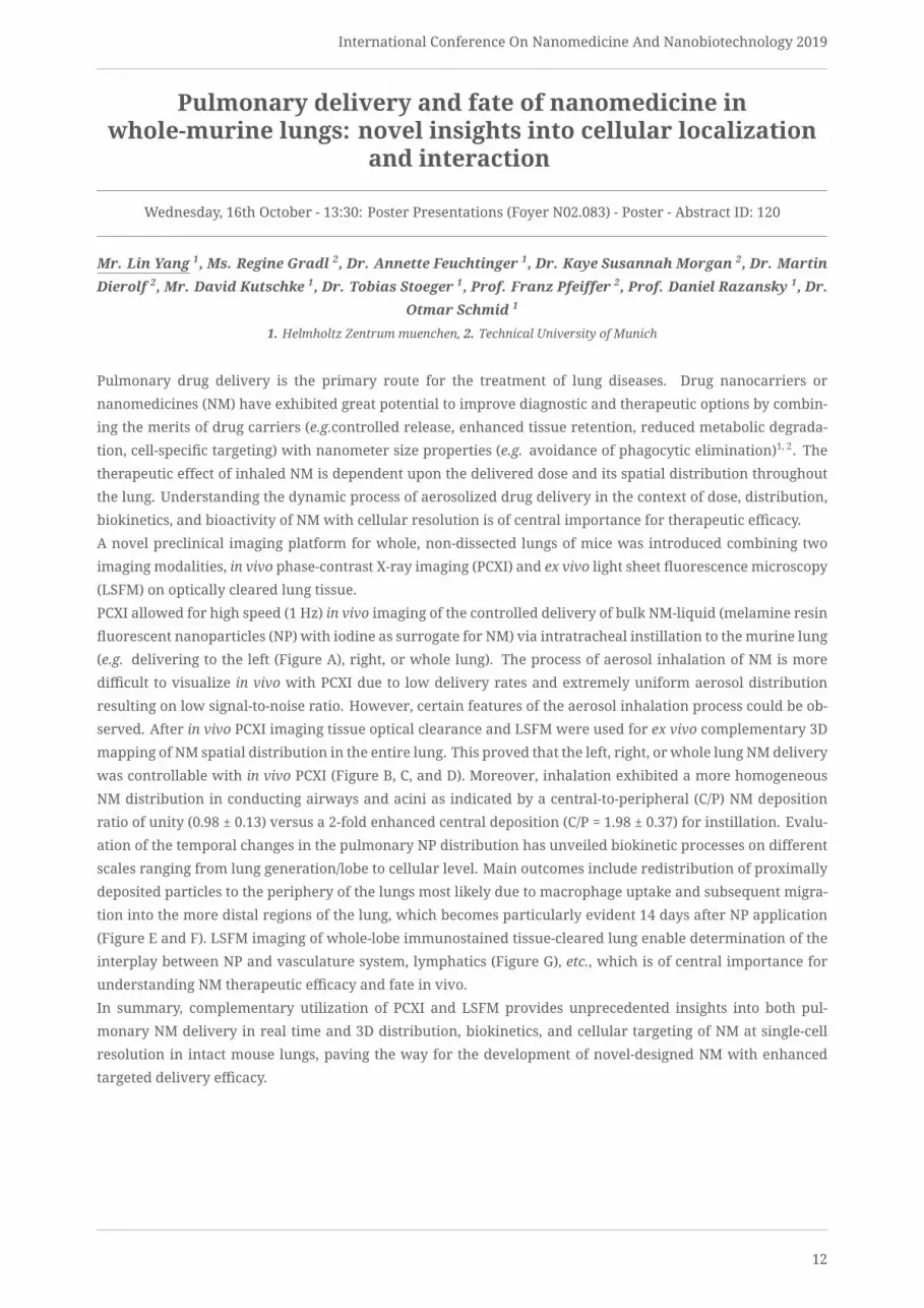
Pulmonary delivery and fate of nanomedicine inwhole-murine lungs: novel insights into cellular localization
and interaction
Wednesday, 16th October - 13:30: Poster Presentations (Foyer N02.083) - Poster - Abstract ID: 120
Mr. Lin Yang 1, Ms. Regine Gradl 2, Dr. Annette Feuchtinger 1, Dr. Kaye Susannah Morgan 2, Dr. MartinDierolf 2, Mr. David Kutschke 1, Dr. Tobias Stoeger 1, Prof. Franz Pfeiffer 2, Prof. Daniel Razansky 1, Dr.
Otmar Schmid 1
1. Helmholtz Zentrum muenchen, 2. Technical University of Munich
Pulmonary drug delivery is the primary route for the treatment of lung diseases. Drug nanocarriers or
nanomedicines (NM) have exhibited great potential to improve diagnostic and therapeutic options by combin-
ing the merits of drug carriers (e.g.controlled release, enhanced tissue retention, reduced metabolic degrada-
tion, cell-specific targeting) with nanometer size properties (e.g. avoidance of phagocytic elimination)1, 2. The
therapeutic effect of inhaled NM is dependent upon the delivered dose and its spatial distribution throughout
the lung. Understanding the dynamic process of aerosolized drug delivery in the context of dose, distribution,
biokinetics, and bioactivity of NM with cellular resolution is of central importance for therapeutic efficacy.
A novel preclinical imaging platform for whole, non-dissected lungs of mice was introduced combining two
imaging modalities, in vivo phase-contrast X-ray imaging (PCXI) and ex vivo light sheet fluorescence microscopy
(LSFM) on optically cleared lung tissue.
PCXI allowed for high speed (1 Hz) in vivo imaging of the controlled delivery of bulk NM-liquid (melamine resin
fluorescent nanoparticles (NP) with iodine as surrogate for NM) via intratracheal instillation to themurine lung
(e.g. delivering to the left (Figure A), right, or whole lung). The process of aerosol inhalation of NM is more
difficult to visualize in vivo with PCXI due to low delivery rates and extremely uniform aerosol distribution
resulting on low signal-to-noise ratio. However, certain features of the aerosol inhalation process could be ob-
served. After in vivo PCXI imaging tissue optical clearance and LSFM were used for ex vivo complementary 3D
mapping of NM spatial distribution in the entire lung. This proved that the left, right, or whole lung NMdelivery
was controllable with in vivo PCXI (Figure B, C, and D). Moreover, inhalation exhibited a more homogeneous
NM distribution in conducting airways and acini as indicated by a central-to-peripheral (C/P) NM deposition
ratio of unity (0.98 ± 0.13) versus a 2-fold enhanced central deposition (C/P = 1.98 ± 0.37) for instillation. Evalu-
ation of the temporal changes in the pulmonary NP distribution has unveiled biokinetic processes on different
scales ranging from lung generation/lobe to cellular level. Main outcomes include redistribution of proximally
deposited particles to the periphery of the lungs most likely due to macrophage uptake and subsequent migra-
tion into the more distal regions of the lung, which becomes particularly evident 14 days after NP application
(Figure E and F). LSFM imaging of whole-lobe immunostained tissue-cleared lung enable determination of the
interplay between NP and vasculature system, lymphatics (Figure G), etc., which is of central importance for
understanding NM therapeutic efficacy and fate in vivo.
In summary, complementary utilization of PCXI and LSFM provides unprecedented insights into both pul-
monary NM delivery in real time and 3D distribution, biokinetics, and cellular targeting of NM at single-cell
resolution in intact mouse lungs, paving the way for the development of novel-designed NM with enhanced
targeted delivery efficacy.
12

International Conference On Nanomedicine And Nanobiotechnology 2019
Figure 1 yang.png
13

International Conference On Nanomedicine And Nanobiotechnology 2019
Variations in Surface Chemistry Influences Immune Responseof Tumour Lysate-coated Gold Nanorods
Wednesday, 16th October - 13:30: Poster Presentations (Foyer N02.083) - Poster - Abstract ID: 155
Mr. Kang Wei Cherng Malvin 1, Prof. Liu Haiyan 2, Dr. Kah Chen Yong James 2
1. NUS Graduate School for Integrative Sciences and Engineering, 2. National University of Singapore
The ability for surface modification of gold nanoparticles provides a promising and versatile platform
for biomedical applications. Given the ability of nanoparticles to enhance cellular uptake of drugs and
biomolecules, it serves as an interesting platform for the development of vaccines, particularly cancer vaccines.
Tumour lysate serves as an attractive vaccination platform given the plethora of antigens present. However,
current clinical application for tumour lysate in cancer vaccination is limited to ex vivo dendritic cell (DC) puls-
ing due to rapid renal clearance upon direct vaccination, which often leads to sub-optimal clinical responses.
Also, to date there has been no consensus on how variations in surface chemistry influences the immune re-
sponse of vaccines. This project seeks to exploit the concept of protein corona to coat tumour lysate (TL) proteins
on a variety of surface-modified gold nanorods (AuNRs) and perform in vitro studies using J774A.1 macrophage
cell line to test for vaccine efficacy. Experimental results shows that NR-TL can generate immune activation of
J774A.1 cells independent of surface charge of the original AuNR prior to coating. It was interesting to note how-
ever that hydrophobic NR-PAAwhen coatedwith TL does not generate any immune response, which contradicts
existing literature on the direct correlation between hydrophobicity and immune response. When tested with
controls such as mouse serum-coated NRs (NR-MS) and free TL, we observe superiority in immune response by
both NR-PSS-TL and NR-Cit-TL, highlighting the efficacy of the vaccine construct due to increase antigen uptake
and adjuvant effects. Future works include studies with primary immune cells such as bone marrow-derived
dendritic cells to validate the efficacy of the NR-TL construct.
Nr-pe-tl schematic.pngNr-tl figure 1.png
14

International Conference On Nanomedicine And Nanobiotechnology 2019
Nr-pe-tl varying surface j774a.1.png Nr-tl vaccine with controls j774a.1.png
15

International Conference On Nanomedicine And Nanobiotechnology 2019
Evaluation of the Stability and Drug Release Behavior ofmPEG5k-b-p(HPMA-Bz) Polymeric Micelles Loaded with
Paclitaxel
Wednesday, 16th October - 13:30: Poster Presentations (Foyer N02.083) - Poster - Abstract ID: 172
Ms. Maryam Sheybanifard 1, Dr. Nataliia Beztsinna 2, Ms. Mahsa Bagheri 2, Prof. Gert Storm 2, Dr.Josbert Metselaar 1
1. University Hospital RWTH Aachen, 2. Utrecht University
The poor water solubility of paclitaxel (PTX) contributes to its suboptimal in vivo behavior and efficacy. To im-
prove the delivery of this drug, polymeric micelles of mPEG-b-p(HPMA-Bz) appeared promising. Nevertheless,
variations in this polymer’s hydrophobicity might alter its capacity to hold, carry and deliver the drug. In the
current study, we aimed to systematically evaluate themicellar formulations of PTX by changing the ratio of the
drug to polymer as well as the chain length of the hydrophobic block of the polymer. To reach our aim, differ-
ent hydrophobic variants of mPEG5k-b-p(HPMA-Bz) were made and loaded with various quantities of PTX and
assessed regarding micelle stability and drug retention. Four variants of mPEG5k-b-p(HPMA-Bz) polymer were
used with an identical hydrophilic part and different hydrophobic units, namely mPEG5k-b-p(HPMA-Bz)18.5k,
mPEG5k-b-p(HPMA-Bz)9.6k, mPEG5k-b-p(HPMA-Bz)4.7k, and mPEG5k-b-p(HPMA-Bz)2.2k (from the most hydropho-
bic to the least hydrophobic variants of the polymer respectively). Micelles were formulated using ethanol as
a biocompatible and industrially feasible solvent. Our results suggest that among 5%, 10%, and 15% w/w of
PTX loading, samples with 15% w/w PTX are the most unstable and that 10% and 5% w/w PTX loading is most
desirable. Meanwhile, investigating the different variants of the polymer revealed that micelles of polymers
mPEG5k-b-p(HPMA-Bz)18.5k and mPEG5k-b-p(HPMA-Bz)9.6k have a similar hydrodynamic diameter of around 52
nm while the polymer with the longest hydrophobic blocks showed slightly better PTX retention and release.
At the same time, micelles of polymers with shorter hydrophobic blocks (i.e. mPEG5k-b-p(HPMA-Bz)4.7k and
mPEG5k-b-p(HPMA-Bz)2.2k) both demonstrated a smaller hydrodynamic diameter of around 39 nm and resulted
in a more premature PTX leakage and release compared to the micelles of polymers with longer hydrophobic
blocks. Furthermore, mPEG5k-b-p(HPMA-Bz)4.7k presented a slightly better PTX retention and release compared
to mPEG5k-b-p(HPMA-Bz)2.2k. In conclusion, we argue that the better stability of mPEG5k-b-p(HPMA-Bz)18.5k mi-
celles and the smaller size of mPEG5k-b-p(HPMA-Bz)4.7k micelles are both desirable features. However, each
polymeric micelle fails to deliver both features simultaneously. Therefore, in vivo studies should be performed
to help find the optimally performing lead candidate.
16

International Conference On Nanomedicine And Nanobiotechnology 2019
Development of Bioanalytical Assays based onProtein-stabilized Catalytic Bimetallic Nanoclusters
Wednesday, 16th October - 13:30: Poster Presentations (Foyer N02.083) - Poster - Abstract ID: 182
Dr. Laura Saa 1, Mrs. Verónica Mora Sanz 2, Dr. Nerea Briz 3, Dr. Valery Pavlov 1
1. CIC Biomagune, 2. CIC biomaGUNE/ Tecnalia, 3. Tecnalia
Protein-stabilized metallic nanoclusters (NCs) have attracted enormous interest due to their unique electronic
structures, physical and chemical properties, and their potential applications in imaging and optical biosens-
ing (1, 2). Most of the reported protocols for the synthesis of NCs using proteins include very harsh conditions
that lead to a loss in the structure and the complete denaturation of the protein. On the other hand, metal-
lic nanoparticles (NPs) have been widely employed in bioanalysis. These NPs should be pre-synthesized and
purified from the components of reaction mixtures, and afterwards, tethered to biorecognition elements. In
addition, the reproducibility of the assays depends significantly on the quality of NP preparation.
In this work, we report the synthesis of bimetallic Au/Pt NCs using proteins as scaffolds under non-denaturing
conditions. The resulting protein-Au/Pt NCs exhibit peroxidase-like catalytic activity andmore interestingly, the
proteins used for the synthesis are not denatured during the process, maintaining their native structure and
functionality. These protein-stabilized Au/Pt NCs have been applied for the development of sensitive bioanalyt-
ical assays.
First, we used streptavidin, widely used in biotechnology due to their highly selective and stable interaction
with biotin (3), to produce streptavidin-Au/Pt NCs that can bind biotin with high affinity. This interaction can be
monitored by measuring the peroxidase activity of the streptavidin-Au/Pt NCs using the chromogenic substrate
TMBwhich is oxidized in presence of H2O2. The streptavidin-Au/Pt NCs have been successfully applied for signal
amplification in immunoassays employing biotinylated antibodies for the detection of anti-BSA IgG (Figure 1).
In a second approach, we employed fibrinogen to produce fibrinogen stabilized -Au/Pt bimetallic NCs with
peroxidase-like activity. Thesemolecules have been applied for colorimetric detection of thrombin activity. The
assay is based on the activation of fibrinogen immobilized on the surface of a microplate and fibrinogen-Au/Pt
NCs in solution in the presence of thrombin. The activated molecules can bind each other forming a catalytic
fibrin fiber that remains bound on the surface. Since the generation of fibrin is correlated with the activity of
thrombin, an increase in thrombin concentration is related with an increase in the readout peroxidase signal
due to the polymerization of fibrin derived from fibrinogen-Au/Pt NCs. This strategy allows the development of
a sensitive and simple bioassay for quantification of thrombin in the picomolar range (Figure 2).
In conclusion, we propose the synthesis of bimetallic nanoclusters by their incorporation inside of proteins, that
requires mild physiological conditions that do not denature either streptavidin or fibrinogen during incorpora-
tion of the resulting atomic clusters. This strategy may address the drawbacks related with the tethering of NPs
to biopolymers and result in development of new efficient strategies for biosensing.
References.
1. Chem. Soc. Rev., 2012, 41, 3594.
2. J. Nanophoton., 2012, 6, 064504.
3. Appl. Microbiol. Biotechnol., 2013, 97, 9343.
17

International Conference On Nanomedicine And Nanobiotechnology 2019
Fig1 bioanalytical assay for the detection of
antibsa igg using streptavidin aupt ncs.jpg
Fig 2 bioanalytical assay for the detection of
thrombin using fibrinogen aupt ncs.jpg
18

International Conference On Nanomedicine And Nanobiotechnology 2019
The Uptake of Poly Lactic-co-glycolic acid (PLGA) Particles byAlveolar macrophages and epithelial cells depends on the
Exposure scenario
Wednesday, 16th October - 13:30: Poster Presentations (Foyer N02.083) - Poster - Abstract ID: 192
Ms. Mehwish Ishaque 1, Dr. Carola Voss 1, Dr. Otmar Schmid 1, Dr. Markus Rehberg 1, Dr. TobiasStoeger 1
1. Comprehensive Pneumology Center, Helmholtz Zentrum München, Germany; Member of the German Centre for Lung
Research
Due to its large surface area, the alveolar epithelium represents a major target for drug interaction and ab-
sorption. In this context nanoparticle-mediated drug delivery is an emerging therapeutic technique to deferent
respiratory diseases, and predictive in vitro models are accordingly needed.
The aim of this study was to investigate the cell specific uptake of different sized fluorescent labelled poly lactic-
co-glycolic acid (PLGA) particles as carrier surrogates in different models. To this end we exposed cocultures
of alveolar epithelial and alveolar macrophage cell lines, either at conventional submerged or at the air liquid
interface (ALI) setup, to different sized PLGA particles, and compared the uptake efficacy to that observed in
mice. Submerged exposures are experimentally simpler andmore convenient to perform, butALI exposures are
even if costlier, physiologicallymore realistic and hence potentially biologicallymoremeaningful. Additionally,
while the cell delivered nanocarrier dose is usually rather obscure for submerged conditions, then on the cell
surface deposited dose can be determined using the ALICE Cloud setup featuring a quartz crystal microbalance.
0.1µm, 0.5µm and 1µm PLGA particles were studied. For in-vitro experiments, the murine cell lines LA-4 (ep-
ithelial, type II-likemouse lung adenoma cells) andMH-S (immortalizedmouse alveolarmacrophages) asmono-
cultures, were exposed to PLGAparticles for 24 hours in submerged andALI conditions. For in-vivo experiments,
C57BL/6J mice were exposed to PLGA particles (0.1µm & 1µm) by intratracheal instillation and analysed after
24 hours. The uptake was monitored and quantified by confocal microscopy and flow cytometry.
Under submerged conditions, flow cytometry revealed for epithelial cells the highest uptake for the largest
particles, whereas macrophages showedmore effective uptake of 0.5µm followed by 1.0µm particles. Uptake of
0.1µm particles was barely detectable at all. Under ALI conditions, significant particle uptake was only detected
in macrophage and independent of particle size. Flow cytometry of particle exposed mouse lungs showed a
significant uptake only for 0.1µm particles by alveolar macrophages, compared to little uptake by epithelial,
type II cells, independent of size.
Finally, our current results support the idea that the more elaborate ALI exposure setup represents a more
realistic model when studying particle uptake at the air tissue interface. Currently performed histological in-
vestigations shall confirm the flow cytometry based findings.
19

International Conference On Nanomedicine And Nanobiotechnology 2019
Bio-nanocarrier system : Self assembling peptide hydrogel fordual delivery of Virus Like Particles and drug to treat Brain
cancer
Wednesday, 16th October - 13:30: Poster Presentations (Foyer N02.083) - Poster - Abstract ID: 221
Ms. Mrunal Wanjale 1, Dr. Amy Barker 2, Prof. Peter Stockley 2, Dr. GS Vinod Kumar 1
1. Rajiv Gandhi Centre for Biotechnology and University of Kerala, 2. University of Leeds
Among the central nervous system neoplasms, gliomas are the most common anaplastic tumors that are ob-
served in children, adults as well as elderly people. Even after intensive therapy many of Glioma patients
suffer from aggressive recurrence of these tumors. To address this problem, we have designed a biodegrad-
able peptide hydrogel implant systems for sustained release of therapeutic agents . This system can bypass the
BBB and make the drug available in appropriate amount to decrease the non specific cytotoxicity and systemic
myelodysplasia . We have designed a 24 amino acid peptide which is an amalgamation of self assembling unit
(KFEFKFEF) and Matrix metalloproteinase sensitive motif : VPLSLYSG selectively cleavable by MMP-7, MMP2
and MMP9 which are reported to be over expressed in glioma. Via this self assembling hydrogel we propose
to deliver Temozlomide, an oral prodrug that acts by methylating DNA. But glioma cells develop resistance to
this drug by over expressing an enzyme MGMT (O6-methylguanine methyl transferases). To tackle this issue in
treatment of glioma chemotherapy we propose use of Virus Like Particles(VLP) the bio nano carriers of RNA( in
this system-siRNA), to silence the gene expression of MGMT and sensitise glioma cells to Temozolomide.
Methods:
Peptide KFEFKEEF-VPLSLYSG-KFEFKEEF was in-silico modelled to check aggregation using the program GRO-
MACSv5.1.4. It was docked with the known structure of MMP7 enzyme using PATCHDOCK 3.1 software. Mod-
elled peptide was synthesised by solid phase peptide synthesis and mass confirmed by MALDI-Toff analysis. A
complex of siRNA targeting the MGMT gene and TR sequence required for VLP assembly with fluorescent tag
was designed. VLP derived from the MS2 bacteriophage coat protein were assembled using this siRNA as ge-
nomic material. VLP were purified by sucrose density gradient (15-45%) ultracentrifugation. 1% agarose gel
run was done along with a wild type MS2 bacteriophage to ensure similar assembly.
Results and discussion:
MD simulation for 20ns gave a steady conformations of peptide. It was observed to be stabilized by pi-pi-
stacking, H bonds and Hydrophobic interactions. The Root Mean Square Fluctuation graph analysis shows
that loop (VPLSLY) region is packed well inside the inhibitor binding pocket of MATRILYSIN. Peptide synthesis
yielded correct mass of peptide upon cleavage and MALDI analysis. Transmission electron microscopy con-
firmed size ~28 nm and structure of the reassembled VLP which was similar to the wild type MS2. The nanocar-
rier VLP were tested for cell internalisation in U251 and U138 cells and showed positive uptake with 2 hours
incubation. MTT assay has shown increase in cytotoxicity when Temozolomide was supplemented with VLP.
Thus glioma cells are sensitized to Temozolomide treatment effectively
We are currently evaluating and quantitating the silencing potential of these VLP based siRNA nanocarriers and
targeting of the VLP to glioma cells.The interaction of coat protein to the TR region of the RNA is very specific,
ensuring specific and higher entrapment efficiency. These nanocarriers have better prospects on account of
biocompatibility, cost of production. Together, this system can deliver drug and tackle resistance problem in
tumor environment sparing systemic circulation of chemotherapy drugs.
20

International Conference On Nanomedicine And Nanobiotechnology 2019
Cell uptake vlp by facs.jpgPeptide maldi.jpg
Sap in silico.jpgVlp assembly.jpg
21

International Conference On Nanomedicine And Nanobiotechnology 2019
Synthesis of multivalent glyconanogels and evaluation oftheir interaction with lectins
Wednesday, 16th October - 13:30: Poster Presentations (Foyer N02.083) - Poster - Abstract ID: 244
Dr. Ana Sousa 1, Mrs. Noelia de la Cruz 1, Dr. Javier Rojo 1
1. Spanish National Research Council
IntroductionCarbohydrate-protein interactionsmediatemanybiological processes including tumour progression, inflamma-
tion, and viral infection. These interactions are typically characterized by a high selectivity and a low affinity,
which is compensated in Nature by multivalency.
In this context, a variety of multivalent nanosized materials (dendrimers, polymers, nanoparticles, etc.) modi-
fied with several copies of carbohydrates have been designed to competitively interfere with those recognition
processes. In particular, our research group is interested in the interaction of carbohydrates with C-type lectins,
because these lectins play a key role in the initial stages of many viral infections, including human immunode-
ficiency virus (HIV) and Ebola virus (EBOV), thus becoming an interesting therapeutic target.
MethodsWith the aim of obtaining a multivalent carbohydrate presentation, we have developed a simple and straight-
forward approach to prepare polymeric glyconanogels designed to interact with cellular receptors.
Mannosylated nanogels were synthesized from biocompatible and FDA-approved Poly(ethylene glycol) (PEG)
by free radical polymerization in miniemulsion. This strategy allowed the preparation of NGs modified with
simple monosacharides and with more complex structures such as glycodendrons of mannose (Figure 1).
After purification and characterization by different techniques, the ability of these nanogels to interact with the
model lectin Concanavalin A (Con A) has been studied in solution.
Results and DiscussionIn a first step, different PEGylated building blocks containing simple mannose or mannosylated dendrimers
were synthesized. To this aim, an acrylate group was introduced on one end of the PEG chain, while the other
was functionalized with terminal azides that allow the straightforward introduction of carbohydrates or gly-
codendrons by Click Chemistry. Afterwards, a series of mannosylated NGs were synthesized by free radical
polymerization in miniemulsion. The NGs were characterized by dynamic light scattering (DLS), transmission
electron microscopy (TEM) and scanning electron microscopy (SEM).
Interaction studies of the different NGs with the model lectin ConA by UV and DLS showed that the NGs could
effectively interact with the lectin forming aggregates, and that the aggregation ability increased with the mul-
tivalency of the mannosylated ligand.
Importantly, control nanogels (displaying galactose groups on their surface) showed no activity at all.
In conclusion, biocompatible, PEG-based, glyconanogels modified with several mannose copies have been pre-
pared and their multivalent ability to interact with a model lectin has been demonstrated.
22

International Conference On Nanomedicine And Nanobiotechnology 2019
Figure 1. glyconanogels.jpg
23
International Conference On Nanomedicine And Nanobiotechnology 2019
Carbon based 0D and 2D nanomaterials for biomedicalapplication
Wednesday, 16th October - 13:30: Poster Presentations (Foyer N02.083) - Poster - Abstract ID: 258
Dr. Katerina Polakova 1, Mr. Tomáš Malina 1, Mr. Jan Belza 1, Dr. Sergii Kalytchuk 1, Dr. AristidesBakandritsos 1, Prof. Radek Zboril 1
1. Palacký University in Olomouc
Carbon dots (0D) and graphene (2D) have attracted significant attention in last decades due to their unique
mechanical, optical, electronic, thermal and chemical properties. The properties stated above implicate broad
range of applications in physics, chemistry, biology and medicine.
Here, the new application of carbon dots as nanothermometers of cells based on photoluminiscence lifetime
thermal sensing together with optical imaging of carbon dots labeled stem cell migration will be presented as a
first.
Concerning 2D graphene, it is well known that limited reactivity of graphene has so far hampered many ap-
plications due to its low density of functionalization with limited number of carboxyl groups. We employed
the pronounced and controllable reactivity of fluorographene where a densely functionalized and hydrophilic
nitrile-functionalized graphene derivative was obtained (cyanographene, GCN). Promising biomedical applica-
tion of this new graphene derivate will be introduced as well.
24

International Conference On Nanomedicine And Nanobiotechnology 2019
Nanomedicine Based in Digital Molecular Communications forDelivery of Ions Through the Beta Cell for Artificial
Segregation of Insulin Granules
Wednesday, 16th October - 13:30: Poster Presentations (Foyer N02.083) - Poster - Abstract ID: 251
Dr. Huber Nieto-Chaupis 1
1. Universidad Privada del Norte
The so-called Nanomedicine [1] will use all compatible nano technologies encompassing biological systems in
order to restore the homeostasis or delay the apparition of diseases such as type-2 diabetes for example [2].In this paper we focus in the theory of Digital Molecular Communications by using the path integral theory
that allows us tomake predicions about the performance of nano devices working as cargo of ions. Because the
prospective nano devices can among varios roles, work as antena [3], fact that allows us to regulate the releasing
of ions thatmight be required to restore funcionalities of cells and tissues. Once the theory is developed, we pass
to estimate the different paths and their respective probabilities by which ions are targeted to arrive inside the
beta cell. Thus, advanced nano devices would be part of an advanced network such as the Internet of Bio-Nano
Things. The proposed theory that emphasizes the transportation from a nano-particles generator is resummed
as follows (FIG-1):
P<A|D> =| <A|B> +<B|C> +<C | D >|ˆ2
Where P<A|D>= Probability to accomplish the path between the nano particles generator (A) and (D) inside the
beta cell.
<A|B>=Probability between the surface of the nano device.
<B|C>=Probability between the Surface of nano device and the voltaje-dependent gate located on the beta cell.
<C | D >=Probability between the voltaje-dependent gate and the insulin granules inside the beta cell.
For each path its correspondent mathematical structure for example <A|B> is given by:
<A|B> = ∫ Φ(x_A) Exp[ -i (P+qA)2 /2M +eU ] Φ(x_A)where the electromagnetic wave denoted byA depends on the frequency that modules the digital informa-
tion that the nano device should receive to release a certain amount of ions. We asume that an external bio-
cyber interface is the responsable of the emission of the waves so that it is connected to the nano device pema-
nently.Concretely, this paper focuses on the scenario of artificial releasing of granules of insulin in beta cells
(FIG-2) to avoid hyperglycemia in those type-2 diabetes patients. In order to have an efficient emission-reception
system and the releasing of insulin granules is successful, we estimate the full efficiency: E =(N_CD)/(N_AB +
N_BC) with
N_CD=ions that have arrived inside the beta cell
N_AB=released ions fom the nano particle generator inside the nano device
N_BC=ions travelling in the spatial área between the cell and nano device.
Through Monte Carlo simulations based on the proposed theoretical model we have estimate that the average
efficiency for a suitable improving on the glucose values of a type-2 diabetes patients would be of order of 79%
with an systematic error of order of 9%.
[1] Anders H, The utility of DNA nanostructures for drug delivery in vivoExpert Opinion on Drug Delivery, Vol-
ume 14, 2017 - Issue 2.
[2] Nieto-Chaupis, Huber, Cheating the Beta Cells to Delay the Beginning of Type-2 Diabetes Through Artificial
Segregation of Insulin, BICT 2019, Pittsburgh, PA, USA, March 13–14, 2019.
[3] Nieto-Chaupis, Huber, Modified Friis Equation for Biological Internet, 2019 IEEE iWAT Miami.
25

International Conference On Nanomedicine And Nanobiotechnology 2019
Fig-1.pngFig-2.png
26

International Conference On Nanomedicine And Nanobiotechnology 2019
Natural Killer Cell Derived Biomimetic Nanoconstructs forTumor Targeted Bioimaging
Wednesday, 16th October - 14:15: Nano-Imaging for diagnosis, therapy and delivery in preclinical and clinicalfields (Amphitheatre N02.040) - Oral - Abstract ID: 52
Dr. Arunkumar Pitchaimani 1, Dr. Tuyen Nguyen 2, Mr. Ramesh Marasini 2, Ms. Achini Eliyapura 2, Ms.Tahmineh Azizi 2, Prof. Majid Jaberi-Douraki 2, Prof. Paolo Decuzzi 1, Prof. Santosh Aryal 2
1. Nanotechnology for Precision Medicine, Italian Institute of Technology, Genova, Italy., 2. Nanotechnology Innovation Center
of Kansas State (NICKS), Kansas State University, USA
Introduction: Targeted magnetic resonance imaging (MRI) of deep solid tumors using clinical contrast agents
is highly challenging due to its short-half life and rapid systemic clearance from the blood. The recent ad-
vent of Biomimetic Nanoconstructs (BNc) system shows tremendous progress in the field of nanomedicine. The
advantages of incorporating biological materials with synthetic materials include biocompatibility, resistivity,
cellular interaction, enhanced circulation half-life and cellular retention. Among various blood pool agents,
Natural killer (NK) cells are a predominant member of the innate immune system. The major function of the
NK cell includes host defense against microbial infections and tumor cells by immunosurveillance of cell sur-
face abnormalities in major histocompatibility class I markers and cell stress markers. In the present study,
biomimetic nanoconstructs was designed using tumor homing Natural Killer cell membrane, and MR imaging
responsive Gd-lipid camouflaged onto the surface of polymeric nanoparticles for targeted bioimaging.
Methods: Natural Killer cell membrane was isolated using sucrose gradient ultracentrifugation method from
NK-92 cells. Further, Biomimetic NanoconstructswithMagnetic Resonance Imaging (MRI) contrast agent (DSPE-
DOTA-Gd) and NIR fluorescent (DiR) molecules were fabricated using simple nanoprecipitation method. The
magnetic properties of the BNc with different Gd-lipid was tested in vitro and investigated its ex-vivo tumor
imaging under MR imaging, along with its pharmacokinetics and bio-distribution pattern in human breast can-
cer cell line MCF-7 induced solid tumor model in Nu/Nu mice. For in vivo NIR bio-imaging studies, DiR labeled
BNc (10mg/kg) were injected intravenously into the MCF-7 tumor bearing mice along with bare control PLGA
nanoparticles and analyzed using Pearl® Trilogy Small animal imaging system (LI-COR®).
Results: The prepared BNc are nanometer in size and shows successful retention of NK cellmembrane proteins.
The maximummagnetic relaxivity of BNc was found to be 7.2mM-1S-1 and was tunable by adjusting the concen-
tration of Gd-lipid. The cellular uptake efficiency of BNc inMCF-7 cells was found to be significantly higher than
the bare PLGA NPs. Confocal analysis demonstrated the uniform intracellular distribution of BNc inMCF-7 cells
and found be highly biocompatible. BNc also exhibits longer circulation half-life (~9.5h) and higher biodistri-
bution in tumor tissues (~10% injected dose) suggesting its tumor targeting potential. The ex-vivo MR images of
tumor mice show clear T1-contrast in tumors tissues under 14.1 T.
Conclusion: Overall, the results demonstrated the bio imaging ability of natural Killer cell membrane camou-
flaged BNc for targeted Magnetic Resonance and NIR Fluorescent bioimaging of tumor. Owing to the proven
immunosurveillance potential of NK�cell in the field of immunotherapy, the BNc engineered herein would hold
promises in the design consideration of targeted nanomedicine.
b
27

International Conference On Nanomedicine And Nanobiotechnology 2019
Biomimetic nanoconstructs-bioimaging.jpg
28

International Conference On Nanomedicine And Nanobiotechnology 2019
The impact of multidrug resistance on tumormicroenvironment remodeling and nanodrug distribution in
murine breast cancer models
Wednesday, 16th October - 14:30: Nano-Imaging for diagnosis, therapy and delivery in preclinical and clinicalfields (Amphitheatre N02.040) - Oral - Abstract ID: 94
Mr. Okan Tezcan 1, Ms. Elena Rama 1, Prof. Fabian Kiessling 1, Prof. Twan Lammers 2
1. RWTH Aachen University Clinic, 2. ExMI – Experimental Molecular Imaging
Introduction:Multidrug resistance (MDR) is a major limiting event in chemotherapy. Development of MDR
results from changes in signaling networks and several cellular mechanisms, such as altered gene expression
profiles. The cellular MDR events were further evaluated in the direction of tumor microenvironment (TME)
remodeling aspects driven by the differences in regulatory mechanisms. To examine this, we developed mul-
tidrug sensitive and resistant breast tumor-bearing mouse models. To evaluate differences in TME, some of the
main components of the extracellular matrix (ECM) were monitored. Additionally, differences in angiogenic
vessel density and the vessel functionality as well as the vessel maturity were analysed. Lastly, to evaluate
nanodrug delivery, liposome distribution was investigated in both tumor models.
Methods:Multidrug resistant 4T1 cells (4T1DOXR)were developed by stepwise selection using doxorubicin dose
increments. Two groups of mice (n = 5 per group): one group was orthotopically injected with 4T1DOXR cells
and the other one with sensitive 4T1 cells (4T1S). After two weeks, both groups of mice were rhodamine lectin
injected and tumors were taken. Thereafter, ex vivo analyses, for ECM materials and vessels, were performed
by immunofluorescence staining and fluorescence microscopy. As a final step, the intravenously injected lipo-
somes were monitored by fluorescence microscopy and further penetration/distribution analysis was applied
to both sensitive and resistant tumor models.
Results: Histological analysis revealed that P glycoprotein (Pgp) was overexpressed in MDR tumor (Fig.1A).
Additionally, the over-deposition of ECM components, such as collagen type-I (COL-I) (Fig.1B), hyaluronic acid
(HA) (Fig.1C), fibronectin (FN) (Fig.1D) and the basal lamina component, collagen type-IV (COL-IV) (Fig.1E) was
also significantly higher in the microenvironment of the MDR tumor. Moreover, the angiogenic vessel density
was higher inMDR tumor (Fig.1F). As for functionality andmaturity of the vessels, no significant difference was
noted. Nanodrug penetration analysis showed a poor liposome distribution in MDR tumor (Fig.2).
Conclusion:Our findings show an increased ECM deposition in multidrug resistant 4T1 tumors. We also supply
evidence that the amount of angiogenic vessels inMDR tumor is higher. No significant difference, however, was
observed in functionality and maturity of the angiogenic vessels in sensitive vs. resistant 4T1 breast tumors.
The over-deposition of ECMmaterials as well as more COL-IV, locating at the outside of microvessels, negatively
affected the liposome distribution. Together, these results provide a basis for further investigations, especially
for patients with a poor response to chemotherapy as well as nanodrug delivery, such as liposome.
Acknowledgements: This work is supported by DAAD (57048249).
29

International Conference On Nanomedicine And Nanobiotechnology 2019
Figure. 1 okan tezcan.png
Figure. 2 okan tezcan.png
30

International Conference On Nanomedicine And Nanobiotechnology 2019
Lipid-conjugated compounds dissociate from drug deliveryliposomes in biological environments
Wednesday, 16th October - 14:45: Nano-Imaging for diagnosis, therapy and delivery in preclinical and clinicalfields (Amphitheatre N02.040) - Oral - Abstract ID: 143
Mr. Rasmus Münter 1, Dr. Kasper Kristensen 1, Mr. Dennis Pedersbæk 1, Dr. Jannik Bruun Larsen 1, Dr.Jens Bæk Simonsen 1, Prof. Thomas Lars Andresen 1
1. Technical University of Denmark (DTU)
Liposomes are the most widespread used nanoparticles for medical applications such as drug delivery, diag-
nostics and imaging, and some liposomal formulations have even entered the clinic. Liposomes are often in-
vestigated for their ability to enter tissues and deliver drugs into subcellular compartments of the target cells.
Studies on cellular uptake of liposomes are usually based on microscopy or flow cytometry, where researchers
track the liposomes using fluorescently labeled lipids (FLLs) incorporated into the liposomal membrane. Such
studies rely on the assumption that the FLL stays associated with the liposome for the entire duration of the
experiment. However, this fundamental assumption remains largely unverified. We have applied size exclu-
sion chromatography and a single liposome imaging assay to investigate the validity of the assumption.1 We
quantified the propensity of a range of commercially available FLLs to dissociate from PEGylated liposomes
when incubated in human plasma. The FLLs displayed large variations in their dissociation propensities, vary-
ing from 10% to 75% over a 24 hour incubation period. The dissociation did not occur in buffer, demonstrating
that incubation in a true biological environment is critical in order to mimic the liposomal stability in a drug
delivery context.
The dissociation of the FLLs could have severe implications on studies where FLLs are used to track the cellu-
lar uptake of liposomes. To address this, we prepared liposomes with different FLLs with varying dissociation
propensities, incubated them in whole human blood and investigated their uptake in peripheral blood leuko-
cytes using flowcytometry. We observed significant differences in the apparent cell uptake, dependent onwhich
FLLwe used to track the liposomes. This uptake correlatedwith the dissociation propensities of the FLLs, which
strongly implies that the dissociated FLLs are taken up by the cells independent on the liposomes (see figure).
Consequently, the results and conclusions drawn from an uptake study can be dramatically influenced by the
choice of FLL.
The dissociation of lipid-conjugated compounds fromdrug delivery liposomes is presumably not limited to FLLs,
but could also be relevant for lipid-anchored drugs, targeting ligands, polymer coatings etc. It is thus crucial
for researchers working with self-assembled nanoparticles such as liposomes, micelles and vesicles, to consider
the dynamics of their systems in biological settings. We here present a simple assay that researchers can ap-
ply to study stability and dynamics of their lipid-based nanomedicine platforms in a biological environment.2
The assay is based on fluorescence detection of FLLs, but can easily be extended to analyze other compounds.
Based on our results, we present guidelines on how a stable anchoring of lipid-conjugated compounds might be
achieved.
1. Münter, R. et al. Dissociation of fluorescently labeled lipids from liposomes in biological environments chal-
lenges the interpretation of uptake studies. Nanoscale (2018). doi:10.1039/c8nr07755j
2. Münter, R. et al. Quantitative Methods for Investigating Dissociation of Fluorescently Labeled Lipid Fluo-
rophores fromDrugDelivery Liposomes. in Characterization Tools for Nanotechnology-based Tissue Engineering
and Medical Therapy (Vol 9) in Press
31

International Conference On Nanomedicine And Nanobiotechnology 2019
Graphical abstract - lipids dissociate from liposomes in plasma and are taken up independently by peripheral
blood leukocytes.png
32

International Conference On Nanomedicine And Nanobiotechnology 2019
Drug-loaded PBCA-based polymeric microbubbles forultrasound-mediated drug delivery across biological barriers
Wednesday, 16th October - 15:00: Nano-Imaging for diagnosis, therapy and delivery in preclinical and clinicalfields (Amphitheatre N02.040) - Oral - Abstract ID: 152
Mr. Anshuman Dasgupta 1, Ms. Mengjiao Liu 1, Prof. Fabian Kiessling 1, Prof. Twan Lammers 1
1. RWTH Aachen University Clinic
Introduction:Drug delivery across vascular barriers is often impaired largely due to inefficient vessel perme-
ability and poor fenestrations between endothelial cells. Sonoporation, which refers to temporary permeabi-
lization of cell membranes or blood vessels induced by oscillating microbubbles in the presence of ultrasound,
is an emerging strategy for enhancing drug delivery across vascular barriers. In this context, most of the studies
have focused on ultrasound-induced drug delivery via indirect approach, i.e., enhancing the delivery of drugs
and drug delivery systems upon co-administration with microbubbles and very few studies have focused on
direct approach, i.e., enhancing the delivery of drugs by encapsulating them into the shell of microbubble. In
this study, we aim to encapsulate drugs (corticosteroids and dyes) into the polymeric shell of microbubble and
perform in-depth characterization for applications in drug delivery across biological barriers such as tumors
and brain.
Methods:PBCA-based polymeric microbubbles were synthesized by homogenizing a 300 mL aqueous solution
of pH 2.5, containing 1%Triton and 3mLbutyl cyanoacrylate, at 10000 rpm for 1 h. Aftermultiplewashing steps,
the drugs (corticosteroids and dyes) were incubated with the microbubbles for 19 h at room temperature. Sub-
sequently, the drug loaded polymeric microbubbles were characterized by HPLC, plate reader and ultrasound
imaging to determine the loading capacity, encapsulation efficiency, drug release and acoustic characteristics
(figure 1).
Results and discussions: Corticosteroids used in this study had similar chemical structure and only varied
in few functional groups depending on hydrophobicity and molecular weight. Upon loading of corticosteroids
into the microbubbles, loading capacity, encapsulation efficiency and release characteristics were evaluated
and these characteristics showed a linear trend with the degree of hydrophobicity (log P) as well as the molec-
ular weight of the corticosteroids (figure 2 and 3). On the other hand, all the dyes had very different chemical
structures with large variations in functional groups, and quite obviously the dye loaded microbubbles failed
to show any dependency on the physicochemical characteristics of the dyes (figure 2 and 3). Moreover, in the
context of ultrasound, both corticosteroid and dye loaded microbubbles responded quite similarly to acoustics,
without showing any dependency on the characteristics of the loaded drug molecules (figure 4).
Conclusion: This is the first systematic analysis and characterization of PBCA-based polymeric microbubbles
upon loading of corticosteroids and dyes. These data highlight the significance of physicochemical characteris-
tics of the drug such as chemical structure, functional group, hydrophobicity and molecular weight on loading
capacity, release and acoustic properties of drug loaded microbubbles. These principles and notions may pro-
vide useful guidelines while optimizing treatment protocols in ultrasound-mediated drug delivery to tumors
and brain.
33

International Conference On Nanomedicine And Nanobiotechnology 2019
Figure 1. schematic representation for
synthesizing and characterizing drug and
dye-loaded pbca mb..jpg
Figure 2. properties of drugs and dyes used in the
study..jpg
Figure 3. quantitative and qualitative analysis of
corticosteroids and dye loaded pbca mb.
encapsulation efficiency and loading capacity of
a-b corticosteroids and c-d dyes..jpg
Figure 4. acoustic analysis of corticosteroid and
dye loaded pbca mb. a b-mode and b contrast
mode image of corticosteroiddye-loaded pbca mb
before and after us treatment. .jpg
34

International Conference On Nanomedicine And Nanobiotechnology 2019
Assemblies of highly efficient iron oxide nanocubes formagnetic (fluid) hyperthermia to treat tumors
Wednesday, 16th October - 15:15: Nano-Imaging for diagnosis, therapy and delivery in preclinical and clinicalfields (Amphitheatre N02.040) - Oral - Abstract ID: 234
Mr. sahitya kumar avugadda 1, Dr. DINA Niculaes 2, Dr. Maria Elena Materia 2, Dr. Aidin Lak 3, Dr.Rinat Nigmatullin 4, Dr. Pooja Basnett 4, Ms. Elena Marcello 4, Dr. Francisco Jose Teran 5, Dr. Ipsita
Roy 6, Dr. Teresa Pellegrino 7
1. Istituto Italiano di Tecnologia, Via Morego 30, 16163 Genova. Dipartimento di Chimica e Chimica Industriale, Università di
Genova, Via Dodecaneso 31, 16146 Genova, 2. Istituto Italiano di Tecnologia, Via Morego 30, 16163 Genova, 3. Istituto Italiano
di Tecnologia, 4. School of Life Sciences, College of Liberal Arts and Sciences, University of Westminster, New Cavendish Street,
London, 5. iMdea Nanociencia, Campus Universitario de Cantoblanco, C/ Faraday 9, 28049, Madrid, Spain. ”Nanomedicina
(iMdea Nanociencia), Unidad Asociada al Centro Nacional de Biotecnologia (CSIC), 28049 Madrid, Spain, 6. Department of
Material Science and Engineering, Faculty of Engineering, University of Sheffield, 7. 1Istituto Italiano di Tecnologia, Via Morego
30, 16163 Genova
Magnetic fluid hyperthermia (MFH) is a non-invasive method, now in clinical practice, to treat solid tumors,
by exposing cancerous tissues to an abnormal temperature of 40-45°C generated by magnetic nanoparticles
when excited under an alternative magnetic field. In our group we have developed, cubic shaped iron oxide
nanocubes (IONC) with optimized magnetic properties, that have 10 to 20 times better heating performance
than commercially available spherical iron oxide nanoparticles (SPIONs), as typically measured in terms of
specific absorption rates (SAR) (Guardia, P. et al, J. Mater. Chem. B 2014) . On the other hand, it is known
that chains of magnetic nanoparticles produced by magnetosome bacteria have enhanced SAR properties com-
pared to isolated nanoparticles (Alphandéry ACS Nano,2011). Since then, much attention has been dedicated
to the assembly of nanoparticles from current available magnetic nanoparticles with better heat performance,
in different geometrical configurations and to the study of their SAR performance. Recently, we have devel-
oped colloidally stable, water soluble short chains of core-shell IONC arranged in dimeric (two nanoparticles)
and trimeric (three nanoparticles) clusters, using an amphiphilic polymer, poly(styrene-co-maleic anhydride)
cumene terminated.3 By increasing the amount of polymer to nanoparticles surface ratio, wewere able to fabri-
cate the morphology of cluster from an isolated to centrosymmetric form (fig.1a-c). Among the three developed
assemblies, dimer/trimeric forms possessed enhanced SAR properties (fig.1d) due to dipolar interactions be-
tween particles in chains and improved magnetic anisotropy (Niculaes, D. et al,ACS Nano,2017)
.The assembling strategy of using the same building blocks arranged in different configurations is crucial in
MFH in order to reduce the doses of magnetic materials, thus avoiding toxicity concerns associated to injection
of higher doses. We have also developed nanoparticle assemblies arranged in well-defined geometries that can
be easily disassembled upon exposure to intra-tumoral stimuli. This assembly/disassembly process is accom-
panied by a significant increase in SAR that favors a more effective heat development under the alternating
magnetic field (Avugadda, S. K. et al, Chem. Mater,2019). At the same time, the assembled particles, such as
bi-dimensional assemblies of nanoparticles demonstrated a rapid response to an external magnet (0.3 T) com-
pared to isolated particles, which is a crucial feature for external magnetic guided physical accumulation of
particles at the target site. This indeed would favor the accumulation of nanoparticles at the tumor site.
Figure 1. a-c) TEM images of water-soluble structures produced from core-shell iron oxide nanocubes by tun-
ing the amount of amphiphilic polymer (poly(styrene-co-maleic anhydride) cumene terminated). d) Scheme of
nanoclusters production, with respective SAR properties.3
35

International Conference On Nanomedicine And Nanobiotechnology 2019
Fig 1.jpg
36

International Conference On Nanomedicine And Nanobiotechnology 2019
Nanoparticles in Phototherapies
Wednesday, 16th October - 15:30: Nano-Imaging for diagnosis, therapy and delivery in preclinical and clinicalfields (Amphitheatre N02.040) - Oral - Abstract ID: 278
Prof. Havva Yagci Acar 1, Dr. Kubra Bilici 1, Mr. ABDULLAH MUTI 1, Prof. Alphan Sennaroğlu 1, Ms.NAZLI ATAC 1, Prof. FUSUN CAN 1
1. Koc university
Superpramagnetic iron oxide has been around for decades formagnetic resonance imaging, magnetic hyperthe-
ria, magnetic separation, and drug/gene delivery. It is now being investigated for photothermal therapy (PTT).
Small SPIONs with 3-aminopropyltrimethoxysilane (APTMS) coating are popular due to their surface amine
groups allowing conjugation of drugs, ligands etc for delivery of therapeutic cargo, yet, they have never been
investigated for PTT. We will discuss the nanoparticle dose and laser intensity dependent temperature increase
in the aqueous NP solutions upon irradiation at 795 nm. Indocyanine green (ICG), an FDA approved fluorescent
dye, has been recently used for photodynamic therapy (PDT). We investigated the PTT and PDT (dual therapy)
potential of ICG loaded to APTMS@SPIONs. Therapeutic efficiency of PTT, PDT and dual therapy was tested on
colorectal and breast cancer cells at safe nanoparticle doses and laser irradiation power. Dual therapy was
able to kill tumor cells almost totally on both cell lines showing higher efficiency compared to the application
of either therapy scheme alone.
This strategy is also utilized for the eradication of biofilms of Pseudomonas aeruginosa, Klebsiella pneumo-
niae, Escherichia coli and Staphylococcus epidermidis. The efficiency of PTT, PDT and dual therapy on growth
inhibition in either planktonic cells or biofilms will be discussed.
Fig 1 iconan19.png
Fig 2 iconan 2019.png
37

International Conference On Nanomedicine And Nanobiotechnology 2019
Supramolecular dynamic non-viral vectors: synergistic effectof low molecular weight polyethylenimine and polyethylene
glycol on transfection efficiency and cytotoxicity
Wednesday, 16th October - 14:15: Targeted drug delivery, organ biodistribution and nanocarriers (RoomN02.011) - Oral - Abstract ID: 21
Dr. Lilia Clima 1, Mr. Bogdan Florin Craciun 2, Dr. Mariana Pinteala 3
1. “Petru Poni” Institute of Macromolecular Chemistry, 2. ”P. Poni” Institute of Macromolecular Chemistry, 3. Centre of
Advanced Research in Bionanoconjugates and Biopolymers, “Petru Poni” Institute of Macromolecular Chemistry, Iasi
Using polyethylenimine (PEI) as a component of non-viral vectors for delivery of nucleic acids, often rise issues
relatedwith high toxicity, low transfection efficiency and nucleic acid polyplex formation. The shielding of poly-
plexes charge by hydrophilic polymers, in particular polyethylene glycols, human serum albumin and dextran
also knownas “stealth technology”, has been intensively applied. In some cases, however, they are also responsi-
ble for the non-specific ionic interactions between the complex and target cells, thus decreasing the transfection
efficiency. Themolecular weight of PEI, PEGylation, biocompatibility and supramolecular structure of potential
carrier can each influence the nucleic acid condensation behavior, polyplex size, and transfection efficiency and
cytotoxicity. Therefore, it is important to find a correlation between the constituent components, as well as the
possible synergy between them, to efficiently transport and to release in specific manner different molecules of
interest. Ourmain interest was to synthesize new compounds from commercially available or “easy to prepare”
units, which will further self-assemble in a complex, tuneable and multifunctional non-viral vectors, suitable
for specific targeted gene delivery.
In this communication, the preparation of libraries of polymeric micellar supramolecular assemblies based on
Pegylated squalene derivative, linked with linear polyethylene glycol and low molecular weight branched PEI
through a multifunctional core connector, and testing as non-viral vectors for plasmid DNA delivery were re-
ported. Dynamic Combinatorial Chemistry approach was used to produce libraries of compounds bymodifying
the components and their ratios in the composition of vectors. PEGylated squalene, poly-(ethyleneglycol)-bis(3-
aminopropyl) (dPEG, 1500 Da or 2000 Da or 3000 Da) and branched PEI (800 Da or 2000 Da), were reversibly con-
nected in a hyperbranched structure to a carbonyl functionalized core via reversible imine bond. Driven by self-
assembly properties of squalene derivatives, obtained amphiphilic architectures formhydrophobic-hydrophilic
particles spontaneously in water. The prepared compounds were capable of interaction with nucleic acids,
forming stable polyplexes and to deliver plasmid DNA to cells. It has been shown that studied vectors exhibited
distinctly higher efficiency in transfection of pDNA toHeLa cells as comparedwith starting PEIs and transfection
efficiency was closely related to ratio and size of PEI and dPEG.
This research was funded by H2020 WIDESPREAD 2-2014: ERA Chairs Project no 667387: SupraChem Lab Lab-
oratory of Supramolecular Chemistry for Adaptive Delivery Systems ERA Chair initiative and from a grant of
Ministry of Research and Innovation, CNCS-UEFISCDI, project number PN-III-P1-1.1-TE-2016-1180, within PNCDI
III.
38

International Conference On Nanomedicine And Nanobiotechnology 2019
DNAmediates the release of nanoparticles from a hydrogelenvironment in a cascade-like fashion
Wednesday, 16th October - 14:30: Targeted drug delivery, organ biodistribution and nanocarriers (RoomN02.011) - Oral - Abstract ID: 27
Ms. Ceren Kimna 1, Prof. Oliver Lieleg 1
1. Department of Mechanical Engineering and Munich School of Bioengineering, Technical University of Munich,
Boltzmannstraße 11, 85748 Garching, Germany
IntroductionCurrent medication treatments require the patient to be in charge of ensuring correct dosing, timing and order
of the drugs to be taken. This becomes an important problem in the case of patient non-compliance, e.g., among
elderly people who are prone to forgetting individual doses. Therefore, a smart delivery mechanism providing
a programmable release order of agents is needed to achieve medication at distinct, pre-defined time points for
delivery of multiple molecules at correct dosages.
MethodsSelf-complementaryDNA sequenceswere designed to achieve a cascade-style release of three different nanopar-
ticles (Figure 1). For initiating the cascade, a displacement DNA (dDNA) was used to generate electrostatically
stable silver clusters. Two DNA sequences (pDNA) linked to Fe2O3 NPs were combined with a symmetric bridge-
like DNA (brDNA) sequence to create a second cluster type, which could be separated again in the presence of
the displacement DNA (dDNA). Later in the release cascade, this bridge-like DNA was used to break down gold-
NP clusters that were crosslinked via partially self-complementary DNA sequences (crDNA). All three NP cluster
variants were placed separately in agarose hydrogel layers. The release was monitored spectrophotometrically
after addition of 150 mM NaCl solution, which triggered the release cascade.
ResultsHere, we showed that the release of three different nanoparticles can be initiated by subjecting the hydrogel
matrix to a physiological salt solution. Electrostatically stable silver/DNA clusters placed in the bottom layer
of the hydrogel were disaggregated after exposure to 150 mM NaCl – which gave rise to Debye screening ef-
fects. This triggered the release avalanche by liberating the displacement DNA that can fully bind to pDNA by
forming complementary, thermodynamically favorable DNA-base pairs (Figure 2). Therefore, the Fe2O3clusters
were disaggregated and brDNA strands were liberated. Finally, these liberated brDNA sequences induced the
disaggregation of gold NP clusters and caused the release of gold nanoparticles.
Depending on whether the NP clusters were deposited into three different layers of the hydrogel matrix or
mixed together into a single layer, the release cascade takes ~4 weeks or ~6 days to complete. In any case, we
achieved a high cumulative efficiency of NPs between 50% and 75% and very high precision and reproducibility.
DiscussionOur results show that the DNA-mediated release system can provide a precise control over the release of three
different NPs that can be used as drug carriers. In addition, the release time can be altered to achieve different
treatment durations without affecting the efficiency of the release mechanism. Thus, the DNA-based mecha-
nism developed here can be useful for developing smart delivery systems to minimize a patient’s medication
management duty by providing an automated, multicomponent release cascade.
39

International Conference On Nanomedicine And Nanobiotechnology 2019
Molecular design of the dna sequences for
three-stage np-release.png
Schematic illustration and cumulative release
profiles of np in 3-layer agarose gel.png
40

International Conference On Nanomedicine And Nanobiotechnology 2019
Carbon nanotube active-by-design nanocarriers for cancertherapy
Wednesday, 16th October - 14:45: Targeted drug delivery, organ biodistribution and nanocarriers (RoomN02.011) - Oral - Abstract ID: 35
Prof. Mónica L. Fanarraga 1, Dr. Eloisa González-Lavado 1, Ms. Esperanza Padín 1, Ms. Nerea Iturroz1, Ms. Lourdes Valdivia 1, Dr. Lorena García-Hevia 2, Prof. Rafael Valiente 1, Dr. Jesús González 1
1. Universidad Cantabria-IDIVAL, 2. International Iberian Laboratory
In the treatment of human cancer, multiple-drug resistance is amajor problem. To circumvent this issue, clinicians
combine several drugs. However, this strategy could backfire resulting in more toxic or ineffective treatments in
themed-term.
Carbon nanotubes(CNTs) are nanomaterials with a high interest in the industry that recently have drawn special
attention in the field of nanobiotechnology. As other carbon allotropes, CNTs display an extraordinary capacity to
capture biomolecules from the environment, acquiring different biological identities. Their unidimensional nature
endows CNTs with unique properties in vivo. These nanofilaments can cross many different types of membranes
and penetrate inside cells or tissues. More interestingly, their unique morphology and surface properties prompts
their biomimetic interaction with the intracellular biological nanofilaments, namely microtubules,[1]actin[2]and
DNA[3], triggering interesting effects at the cellular level that deserve special attention when developing nano-
vectors for anticancer therapies. The multi-walled nanotube (MWCNT) biomimetic properties result in intrin-
sic anti-proliferative,[4,5]anti-migratory[6]and cytotoxic[7]properties in vitro, resulting in anti-tumoral properties in
vivo[8–10]that are maintained (and enhanced) in tumours produced cell resistant to traditional microtubule-binding
chemotherapies such as Taxol®.[8]
Here, we propose the use of MWCNTs, in the design of active-by-design nanocarriers, attempting to enhance the
effect of broadly used chemotherapies. Our results demonstrate how the total effect of the drug 5-Fluorouracil (5-
FU) is remarkably improved (50%more effective) when delivered intratumorally coupled toMWCNTs both, in vitro
and in vivo in solid tumoral models.[10]Our results demonstrate how using MWCNTs as anti-cancer drug delivery
platforms is a promising approach to boost the efficacy of traditional chemotherapies, while considerably reducing
the chances of resistance in cancer cells.
AcknowledgementsThis work was founded by Spanish MINECO, Instituto de Salud Carlos III-European Regional Development Funds
(ERDF) under Projects ref. PI16/00496, NanoBioApp Network (MINECO-17-MAT2016-81955-REDT), IDIVAL and
the University of Cantabria.
References:1. Rodríguez-Fernández, L., et al. Multiwalled carbon nanotubes display microtubule biomimetic properties in
vivo, enhancing microtubule assembly and stabilization. ACS Nano 6, 6614 (2012).2. Holt, B. D. et al. Carbon nanotubes reorganize actin structures in cells and ex Vivo. ACS Nano 4, 4872 (2010).3. Li, X., Peng, Y. & Qu, X. Carbon nanotubes selective destabilization of duplex and triplex DNA and inducing
B-A transition in solution. Nucleic Acids Res. 34, 3670 (2006).4. Sargent, L. M. et al. Single-walled carbon nanotube-induced mitotic disruption. Mutat. Res. - Genet. Toxicol.
Environ. Mutagen. 745, 28 (2012).5. García-Hevia, L. et al. Nanotube interactions with microtubules: implications for cancer medicine.
Nanomedicine 9, 1581 (2014).6. Garcia-Hevia, L. et al. Inhibition of Cancer Cell Migration by Multiwalled Carbon Nanotubes. Adv. Healthc.
Mater. 4, 1640 (2015).7. García-Hevia, L. et al. Anti-cancer cytotoxic effects of multiwalled carbon nanotubes. Curr. Pharm. Des. 21,
41

International Conference On Nanomedicine And Nanobiotechnology 2019
(2015).
8. García-Hevia, L. et al. Multiwalled Carbon Nanotubes Inhibit Tumor Progression in a Mouse Model. Adv.
Healthc. Mater. 5, 1 (2016).9. González-Lavado, E. et al. Biodegradable multi-walled carbon nanotubes trigger anti-tumoral effects.
Nanoscale 10, 11013 (2018).10. González-Lavado, E. et al. MWCNTs complement the anti-tumoral effect of 5-Fluorouracil. Oncotarget 10,2022 (2019).
Fanarraga iconan2019.jpg
42

International Conference On Nanomedicine And Nanobiotechnology 2019
Fullerene, siloxane, cyclodextrin and polyrotaxane : anon-viral approach in gene delivery
Wednesday, 16th October - 15:00: Targeted drug delivery, organ biodistribution and nanocarriers (RoomN02.011) - Oral - Abstract ID: 38
Dr. Dragos Peptanariu 1, Dr. Mariana Pinteala 1
1. Centre of Advanced Research in Bionanoconjugates and Biopolymers, “Petru Poni” Institute of Macromolecular Chemistry,
Iasi
The development of scientific fields such as biochemistry and molecular biology have led to a better under-
standing of disease mechanisms and their genetic basis. This has increased the desire to find new and more
effective treatments and gene therapy has begun to claim an increasing importance.
Gene therapy promises the possibility of curative treatment of genetic diseases by introducing genes into pa-
tients’ cells instead of using common drugs that only provide symptomatic treatment.
Gene therapy might use viruses or non-viral agents like liposomes and cationic polymers. Viral vectors are
much more common, but they raise many issues such as the potential for rejection by the patient’s body, the
possibility of unwanted genome integration and the inability to repeat their administration.
In the design of non-viral vectors, our group has applied various strategies based on molecules such as
fullerene, siloxane, beta-cyclodextrin, which functions as the core to which several polyethyleneimine
molecules are attached. This combination is versatile because it offers the possibility of attaching other groups
such as peptides for targeted delivery. Also, using several cyclodextrin molecules with polyethyleneimine and
polyethylene glycol, a polyrotaxane has been constructed that has proven to be very effective as a transfectant.
The synthesis of the vectors proceeded in two steps: in the first step the functionalization of the core
molecule took place, and in the second step the addition of the polyethylenimine arms was done.
After chemical characterization, the synthesized vectorswere tested as transfection agents on cell culture. These
vectors have demonstrated good plasmid binding and transport capacity as well as efficient in vitro transfection
for reporter genes.
This work has received funding from the European Union’s Horizon 2020 research and innovation program
under grant agreement No 667387 WIDESPREAD 2-2014 SupraChem Lab and from a grant of the Romanian
National Authority for Scientific Research and Innovation, CNCS/CCCDI – UEFISCDI, project number PN-III-P3-
3.6-H2020- 2016-0011, within PNCDI III.
Vector synthesis.png
43

International Conference On Nanomedicine And Nanobiotechnology 2019
Forward genetic screening as a tool to study endocytosis ofnanocarriers
Wednesday, 16th October - 15:15: Targeted drug delivery, organ biodistribution and nanocarriers (RoomN02.011) - Oral - Abstract ID: 51
Ms. Daphne Montizaan 1, Dr. Diana Spierings 2, Dr. Victor Guryev 2, Dr. Anna Salvati 1
1. University of Groningen, 2. European Research Institute for the Biology of Ageing
Nanocarriers offer great potential for targeted drug delivery. For efficient delivery, the nanocarriers do not
only need to reach the target tissue, but they also need to cross the cell membrane and locate the drug to the
appropriate intracellular location. To be able to achieve this, it is crucial to understand the nanocarrier-cell in-
teractions. Most research studying these nanocarrier-cell interactions have usedmethods like RNA interference,
overexpression of proteins, or chemical inhibitors, in which only known targets are usually studied. However,
the mechanism by which nanocarriers enter cells is still ambiguous and it might be that novel mechanisms are
involved.
In this work, to prevent exclusion of novel targets which have no clear relation to known endocytic pathways as
yet, a forward genetic screeningmethod originally developed by Carette et al. (2009) was used to study the inter-
nalization of nanocarriers. A hundred million human haploid HAP1 cells were mutagenized with a retrovirus,
and the library of mutagenized cells was subsequently exposed to fluorescently labelled nanoparticles. Silica
nanoparticles (50 nm) were used as a model to test this forward genetic screening method. Cells with a reduced
uptake were enriched by several selections using fluorescence-activated cell sorting. The enriched population
was sequenced to determine the integration site of the retrovirus causing the mutation. Different from previ-
ous studies, multiple sorting procedures were necessary to enrich for cells with a reduced nanoparticle uptake.
Nonetheless, enrichment was achieved and sequencing allowed to identify a high number of targets involved in
the uptake of nanoparticles. Among them, one of the identified targets was the low-density lipoprotein receptor,
which is known to be involved in the uptake of the silica nanoparticles under the particular conditions used.
Multiple other novel targets involved in nanoparticle internalization were discovered and are currently being
verified. Overall, these results show that forward genetic screening can be used to study the internalization of
nanocarriers and discover novel targets not yet associated the uptake of nano-sized objects.
44

International Conference On Nanomedicine And Nanobiotechnology 2019
Aqueous stable gold nanostar/ZIF-8 nanocomposites for lighttriggered release of active cargo inside living cells
Wednesday, 16th October - 15:30: Targeted drug delivery, organ biodistribution and nanocarriers (RoomN02.011) - Oral - Abstract ID: 175
Dr. Carolina Carrillo-Carrion 1
1. Centro Singular de Investigación en Química Biolóxica e Materiais Moleculares (CiQUS), Universidade de Santiago de
Compostela
IntroductionIn recent years, the interest in nanoscale materials for theranostic applications, for instance in diagnosis and
treatment of cancer, has increased. In this direction, multifunctional hybrid nanostructures with improved
performance in physiological scenarios are required. The combination of plasmonic nanoparticles such as
gold nanoparticles with metal–organic frameworks (MOFs) offers unique opportunities for the development
of nanoplatforms with theranostic potential thanks to the versatile drug encapsulation properties of MOFs [1].
MethodsHere, a plasmonic core-shell nanostar@ZIF-8 nanocomposite (NC) was developed for thermoplasmonic-driven
release of encapsulated molecules inside living cells [2]. The NCs were loaded with Hoechst molecules as func-
tional cargo (DNA staining), and further functionalized with an amphiphilic polymer that prevents ZIF-8 degra-
dation and Hoechst leaking in aqueous media and inside living cells. Confocal fluorescence microscopy and
surface enhanced Raman spectroscopy (SERS) were used to evaluate the performance of the system.
Results and discussionThe newly developed NC has a similar capacity for drug loading than conventional ZIF-8 based nanosystems,
but importantly it is stable in aqueous solution, and it can be activated to release the drug upon illumination
with near–IR (NIR) light. This concept is demonstrated both in aqueous solution and inside living cells (HeLa
cells). The proposed drug-release mechanism relies on the use of NIR light coupled to the plasmonic absorption
of the core nanostars, which creates local temperature gradients, thereby leading to thermodiffusion of cargo
molecules.
The intracellular monitoring of cargo delivery upon illumination with a NIR light (785 nm) was first carried
out by using SERS imaging in cells. A clear signal of Hoechst was recorded in certain regions of the cell (very
likely the lysosomes) previously to the light illumination. The release kinetics showed that around the 75% of
the Hoechst was released during the first 2 h of illumination. These results contrast with those obtainedwithout
illumination, where SERS spectra showed clear Hoechst signals along the time (during 8 h) indicating that the
integrity of the drug carrier was not compromised. SERS data fully agree with equivalent studies by confocal
microscopy (Figure 1). Accumulation of the NCs (blue fluorescent dots) was found in the vicinity (very likely
in lysosomes) of the nuclei before illumination. Then, cells containing the NCs were irradiated with the NIR
illumination setup (5 min, 7W/cm2) leading to the Hoechst release as can be concluded from the nuclei staining
observed in the confocal images.
ConclusionAn aqueous stable (even when stored in cells) ZIF-8 based nanomaterial with combined thermoplasmonic and
high drug-loading capabilities has been developed. This approach is an important step forward in the develop-
ment of MOF-based intracellular vehicles for the controlled release of drugs.
References[1] Zheng, G. et al., Small 2016, 12(29), 3935.
[2] Carrillo–Carrión, et al., Angew. Chem. Int. Ed. 2018, 58(21), 7078.
45

International Conference On Nanomedicine And Nanobiotechnology 2019
Figure 1. confocal microscopy images before and after nir irradiation.png
46

International Conference On Nanomedicine And Nanobiotechnology 2019
Reactive oxygen species (ROS)-responsive polymersomes withsite-specific chemotherapeutic delivery into ROS-rich tumor
in vivo
Wednesday, 16th October - 15:45: Targeted drug delivery, organ biodistribution and nanocarriers (RoomN02.011) - Oral - Abstract ID: 245
Mr. Vladimir Sincari 1, Dr. Eliezer Jager 1, Dr. Martin Hruby 1, Dr. Petr Stepanek 1
1. Institute of Macromolecular Chemistry, Academy of Sciences of the Czech Republic
Introduction: Polymersomes (PS) made from block copolymers are spherical self-assembled structures which
resemble cell membranes of living organisms. In comparison to liposomes the PS have improved stability and
higher loading capacity for drug encapsulation. The release of loaded drugs fromPS can be triggered by external
or internal physical or chemical stimuli such as pH, redox potential, light, magnetic field, temperature or the
presence of enzymes. Some of these proposed “smart” PS were able to release their payloads, or show reduced
sizes or converted charges under conditions (e.g., reduced pH, hypoxia, tumor-specific enzymes) within the
tumor microenvironment (TME). Among the aforementioned stimuli, one straightforward targeting approach
is exploiting the presence of reactive oxygen species (ROS) in cancer cells. ROS such as, hydrogen peroxide
(H2O2), is a component of cell signalling pathways that are necessary for the growth, development, and fitness
of living organisms. However, imbalances in H2O2 production lead to oxidative stress and inflammation events,
which damage tissue and organ systems and are correlatedwith the onset and advancement of various diseases,
including cancer. Hence, the design of PS able to be responsive to these inherent feature of the TME has been
proposed as a promising approach for the cancer treatment.
Methods: A new oxidative responsive amphiphilic diblock copolymer based on a novel methacrylamide
monomer bearing pinacol-type boronic ester functional moieties and N-(2-hydroxypropyl)methacrylamide
(HPMA)was synthesized by a reversible addition-fragmentation chain transfer (RAFT) polymerization and char-
acterized by standard techniques (1HNMR and SEC). Monodisperse polymersomes (PS1 and PS2) ~ 100 nm in di-
ameter loadedwith the chemotherapeutic drug doxorubicin (Dox) were produced bymicrofluidic flow-focusing
method. Characterization of the PS and their degradation under presence of H2O2was probed by 1H NMR, SEC,
dynamic and static light scattering techniques. The therapeutic effect of PS was tested on in vitro and in in vivo
biological models and was compared with the marketed free chemotherapeutic Dox.
Results and discussion: The developed PS platform allows the specific delivery of Dox at the inherent ROS levels
typically found in the TME (Figure 1, top). The in vivo results clearly show efficient suppression of tumor cell
growth in mice bearing EL4 T cell lymphoma (Figure 1, bottom right). The survival time of the animals was also
extended if comparedwith DOX-free (Figure 1, bottom left). Along, the reduced side-effects of the chemotherapy
were substantially improved with the PS such as balance of the body weight, less cardiotoxicity of DOX, one of
the main drawbacks of this current chemotherapeutic drug in the clinic, as observed after the decreasing of
serum creatine kinase levels monitored in blood. The results from this pioneer work strongly suggest that ROS-
responsive PS has potential for tumor-targeting DOX delivery based on the ROS-triggered release mechanism in
vivo.
ACKNOWLEDGEMENTS:
This research was supported by the Czech Science Foundation (grant # 17-09998S). P.Š acknowledges financial
support fromMinistry of Education, Youth and Sports (grant # LM2015064 ERIC; grant POLYMAT # LO1507). M.H
acknowledges the Ministry of Health of the Czech Republic (grant # 16-30544A).
47

International Conference On Nanomedicine And Nanobiotechnology 2019
Figure1.jpg
48

International Conference On Nanomedicine And Nanobiotechnology 2019
CriPec® nanomedicines: academic to industry and preclinicalto clinical translation
Wednesday, 16th October - 16:45: Start-up session: How to successfully start a bio-nano-pharmaceuticalcompany? (Amphitheatre N02.040) - Workshop - Abstract ID: 259
Dr. Rachel Hu 1, Dr. Rob Hanssen 1, Dr. cristianne rijcken 1
1. Cristal Therapeutics
IntroductionCristal Therapeutics is a clinical stage pharmaceutical company developing next generation nanomedicines
based on its proprietary CriPec® platform to treat various diseases, including cancer. CriPec® is perfectly
suited for the rational design of (targeted) nanomedicines with superior efficacy and safety profiles. The most
advanced product in development is CPC634 (CriPec® docetaxel) for treatment of solid tumours, while other
CriPec® products are in preclinical development.
MethodsCriPec® is a pioneering approach to transform a broad range of therapeutic compounds into rationally designed
nanomedicines to assure optimal treatment of various diseases.
ResultsCriPec® has been successfully combinedwith small molecules, peptides or oligonucleotides, thereby generating
monodisperse CriPec® nanoparticles with tuneable sizes between 30 and 100 nm with high drug entrapment
efficiencies and predetermined drug release kinetics. The surface can bemodified by targeting ligands. CriPec®
products are customisable and biocompatible, with a robust manufacturability at clinical scale.
CPC634 is a 65 nm sized nanoparticle entrapping docetaxel designedwhich improves tumour accumulation and
tolerability compared to conventional docetaxel by taking advantage of the presumed enhanced permeability
and retention (EPR) effect. In preclinicalmodels, an enhanced therapeutic index is observed due to an improved
pharmacokinetic profile, increased tumour uptake and superior tolerability.
The first-in-human study demonstrated a favourable plasma PK profile of CPC634. Safety evaluation specifically
demonstrated less neutropenia. One partial response and sixteen cases of stable disease (RECIST 1.1) were
confirmed. The RP2D was set at 60 mg/m2 with dexamethasone premedication.
In a randomized cross-over study, tumour biopsies were taken after intravenous administration of CPC634 and
conventional docetaxel (or vice versa). CPC634 generated higher intratumoural total docetaxel (323%, p < 0.001)
and comparable released docetaxel levels relative to conventional docetaxel.
Next, CPC634 was labelled with zirconium-89 to facilitate non-invasive PET imaging of its biological fate. Tu-
mour retention showed intra- and interpatient heterogeneity with the highest intensity at 96h post injection
and with a mean %ID/kg of 3.43 [1.14-9.32], confirming the EPR effect in patients.
ConclusionThe clinical results of CPC634 illustrate the improved safety profile and increased tumour uptake. A phase II
efficacy study of CPC634 in patients with platinum resistant ovarian cancer (NCT03742713) is currently ongoing.
The innovative CriPec® platform allows for the rational design of nanomedicines of a variety of drugmolecules
with an anticipated superior therapeutic performance.
Acknowledgements: H2020-SMEInst-2016-2017 – INTACT #784127
49

International Conference On Nanomedicine And Nanobiotechnology 2019
Translating pre-clinical drug delivery research into productsfor clinical use by starting a company
Wednesday, 16th October - 17:10: Start-up session: How to successfully start a bio-nano-pharmaceuticalcompany? (Amphitheatre N02.040) - Plenary Speech - Abstract ID: 275
Dr. Josbert Metselaar 1
1. Department of Nanomedicine and Theranostics Institute for Experimental Molecular Imaging RWTH Aachen University
Clinic; Enceladus Pharmaceuticals BV
In my talk I will present practical experience as to the trajectory of commercializing breakthrough scientific
results into products for clinical use. The following items are highlighted: setting out a clinical development
strategy, building a patent portfolio, entering into collaborations, and last but not least: how to acquire financ-
ing. A personal view is taken with my drug development company Enceladus Pharmaceuticals as an ‘example
case’.
Enceladus started at the time as a spin-off from the Utrecht University. Enceladus focused on the development of
Nanocort andOncocort, two liposomal pharmaceuticals that contain corticosteroids and selectively target actual
sites of pathology in inflammation and cancer respectively. After a range of preclinical studies that showed that
these i.v. liposomal targeted corticosteroid products can achieve a dramatic increase of the efficacy and safety
of corticosteroids, clinical trials were undertaken in several diseases including a recently concluded successful
phase 3 trial.
50

International Conference On Nanomedicine And Nanobiotechnology 2019
Long-acting drug delivery: are we witnessing a paradigm shiftin treatment of chronic diseases?
Thursday, 17th October - 09:00: Plenary Speeches (Amphitheatre N02.040) - Plenary Speech - Abstract ID: 263
Prof. Andrew Owen 1
1. University of Liverpool
Long-acting drug delivery has emerged as an effective option for indications such as contraception, schizophre-
nia, androgen ablation and opioid substitution therapy. The approach encompasses implant and injectable
technologies with a growing interest also evident inmicroarray patches and devices for extending half-life from
oral administration. Many opportunities exist for therapy and prevention of disease, and long-acting injecta-
bles for HIV drugs have spurred recent interest across infectious diseases. such as tuberculosis, malaria and
viral hepatitis. Despite success, considerable knowledge gaps exist in terms of the physiological and molecu-
lar processes that govern drug absorption for some technologies. Preclinical studies have highlighted potential
roles for macrophage infiltration, granuloma formation and/or angiogenesis at the depot site for long-acting in-
jectables, but the quantitative importance of these mechanisms in humans is uncertain. Moreover, the clinical
application of long-acting medicines requires consideration of several factors relating to posology and clinical
pharmacology of the administered drug. For example, a long sub-therapeutic pharmacokinetic tail may be a
more important consideration for infectious diseases with a resistance liability than for non-communicable in-
dications. The implications for drug-drug interactions with the long-acting agent as a victim or perpetrator is
also not well characterised and may be different for some drugs when the route of administration is changed,
avoiding the gastrointestinal tract and first pass metabolism. Other questions relate to whether there will be
newDDIsmediated throughdifferent absorptionmechanisms and/orwhether an oral lead-in tomitigate toxicity
will be necessary for all long-acting medicines. This presentation will summarise existing data for long-acting
medicine, with a focus upon long-acting injectable formulations.
51

International Conference On Nanomedicine And Nanobiotechnology 2019
Phenotypic targeting via multiplexed and chemotacticpolymersomes
Thursday, 17th October - 09:30: Plenary Speeches (Amphitheatre N02.040) - Plenary Speech - Abstract ID: 273
Prof. Giuseppe Battaglia 1
1. University College London
We have advanced our ability to deliver drugs combining the high selectivity of active molecules with molecu-
larly engineered carriers equipped with the necessary attributes to navigate biological environments. A critical
element of such a nanomedicinal effort is the introduction of ligands that enable targeting and selectivity to
guide carriers across biological barriers. Such an approach is now allowing to extend drug discovery to target
biological macromolecules that are not accessible via simple diffusion such as the inside of cells or the central
nervous system. However, tight control on the selectivity of drugs and nanomedicines’ interaction with biolog-
ical systems is paramount for the development of targeted therapies. A large number of synthetically tune-
able parameters makes it difficult to identify optimal design “sweet spots” without rational guiding principles.
Here I address this problem combining super-selectivity theory (SST) with analytical models from soft matter
and polymer physics into a unified theoretical framework. Starting from an archetypal system, a polymer-
stabilized nanoparticle functionalized with targeting ligands, we use our model to identify the most selective
combina- tion of parameters in terms of particle size, brush polymerization degree and grafting density, as well
as tether length, binding affinity and ligands number. I further discuss how to combine multivalent interac-
tions into mul- tiplexed systems which act holistically as a function of the density of more than one receptor
type, to achieve binding onlywhenmultiple receptors are expressed above a threshold density. I christen this as
”phenotypic” targeting, and I propose its use for drugging different cell populations enabling personalized ther-
apies. I will show few examples of phenotypic targeting in both blood-brain barrier crossing, cancer targeting
and immune cell recognition.
Finally, I will combine phenotypic targeting with chemotaxis in all effect amplifying molecular interaction to
create organotropic diffusion profile. I will show this with a fully synthetic, organic, nanoscopic system that
exhibits attractive chemotaxis driven by enzymatic conversion of glucose. This is achieved by encapsulating
glucose oxidase alone or in combination with catalase into nanoscopic and biocompatible asymmetric polymer
vesicles (known as polymersomes). I will show that these vesicles self-propel in response to an external gradient
of glucose by inducing a slip velocity on their surface, which makes them move in an extremely sensitive way
toward higher-concentration regions. I will finally demonstrate that the chemotactic behaviour of these nano-
swimmers, in combination with LRP-1 (low-density lipoprotein receptor-related protein 1) targeting, enables a
fourfold increase in penetration to the brain compared to non-chemotactic systems
52

International Conference On Nanomedicine And Nanobiotechnology 2019
Prodrugs and associated opportunities in localized drugsynthesis
Thursday, 17th October - 10:30: Plenary Speeches (Amphitheatre N02.040) - Plenary Speech - Abstract ID: 261
Dr. Alexander Zelikin 1
1. Aarhus University
Biocatalytic impants and nanozymes, antiviral macromolecular prodrugs, remote activation of biomaterials
�these areas of research are in the focus ofmy lab and during this presentation, I will discuss our recent findings
on these avenues. I will present the development of biocatalytic implants as novel tools for site�specific drug
delivery. We will also discuss the design of nanozymes as fundamentally important materials for biomedicine.
Finally, I will outline our recent successes in the design of macromolecular prodrugs as long-acting tools of drug
delivery, as broad�spectrum antiviral agents, and latency reversing agents against HIV. Presentation aims to be
of interest for diverse audience, specifically scientists in the fields of medicinal chemistry and virology, polymer
science and engineering, and biomaterials.
53

International Conference On Nanomedicine And Nanobiotechnology 2019
Drug Delivery with Multifunctional MesoporousNanoparticles
Thursday, 17th October - 11:00: Plenary Speeches (Amphitheatre N02.040) - Plenary Speech - Abstract ID: 269
Prof. Thomas Bein 1
1. Ludwig-Maximilians-Universität München
Over the years, numerous intriguing types of multifunctional mesoporous nanoparticles have been developed
as carriers for targeted drug delivery, for example in the context of cancer therapy. In our recent work, we have
addressed several key challenges related to the development of multifunctional mesoporous silica and other
mesoporous nanoparticles, including stimuli-responsive release systems, targeting ligands for specific cellular
receptors, biodistribution and biocompatibility.
For example, folate and epidermal growth factor (EGF) have been used for successful cell targeting with meso-
porous silica nanoparticles (MSNs). Considering triggered release in the endosome, a novel pH-responsive sys-
tem has been created based on genetically modified carbonic anhydrase (CA) gatekeepers. A pH-dependent
CA inhibitor was covalently attached to the surface of the MSNs, resulting in the desired opening mechanism
caused by the endosomal pH change. Addressing endosomal escape, we have covalently attached a red-light
sensitive phthalocyanine photosensitizer to the MSN, surrounded by a lipid bilayer, which releases cargo upon
illumination. Moreover, we have exploited the proton sponge effect during acidification of the endosome with
polymer-functionalized MSNs to achieve endosomal release. Taking advantage of overexpressed matrix met-
alloproteinases in cancer tissue, we have also achieved spatially selective extracellular release of bioactive
molecules.
Organically functionalized MSNs were developed to reversibly adsorb oligonucleotides such as siRNA at high
loading and with high cellular activity upon release. Moreover, cellular uptake of proteins such as small anti-
body fragments was successfully achieved using a novel release mechanism from MSNs.
An important feature of theMSNs regards their biodistribution and their fate after successful drug delivery. Our
recent quantitative analysis of the dissolution kinetics of different types of multifunctional MSNs demonstrates
that their dissolution rate can be tuned over a wide range, leading to lifetimes of under one hour to several
weeks in aqueous media. In contrast, these MSNs are stable in ethanol for long periods exceeding two years.
Recently, we have expanded the scope of mesoporous materials to mesoporous metal phosphates with very
high surface area and the ability to dissolve in the acidic cytosol. Their intracellular behavior opens new vistas
in targeted drug release with porous nanoparticles. Specifically, we will discuss a novel synthesis method for
spherical, amorphous mesoporous calcium phosphate-citrate nanoparticles (CPCs) with an average size of 50
nm. Internalized CPCs dissolve once the endosomal pH turns acidic. This leads to a rapid release of Ca2+ ions
into the cytosol. Remarkably, mesenchymal cancer cell lines are much more strongly affected by the Ca2+ shock
and induce apoptosis at lower IC50 values than epithelial cancer cells, while healthy cells are not affected. These
nanoparticles show promising therapeutic activity in a malignant pleural effusion (MPE) mouse model. Our re-
sults strongly suggest the possibility to efficiently treat certain lung tumors while at the same time dramatically
reducing toxic side effects.
54

International Conference On Nanomedicine And Nanobiotechnology 2019
Optimizing Nucleic Acid Nanomedicines by ChemicalEvolution of Carrier Sequences
Thursday, 17th October - 11:30: Plenary Speeches (Amphitheatre N02.040) - Plenary Speech - Abstract ID: 225
Prof. Ernst Wagner 1
1. Pharmaceutical Biotechnology, Department of Pharmacy, Ludwig-Maximilians-Universität, Munich
It took five decades from first gene transfections to approval of gene therapies as medical drugs. Up to date,
>2930 clinical gene therapy trials have been initiated, nine gene therapy products, one siRNA drug, and eight
oligonucleotide drugs received market authorization. Intracellular delivery has been critical for the success of
these therapeutic macromolecules. A further refinement of delivery carriers will have a tremendous impact
for efficacy of future nanomedicines. Different chemical evolution approaches are pursued toward synthetic
nucleic acid or protein nanocarriers. Our strategy focuses on a bioinspired sequence-defined process including
(i) artificial amino acids active in specific delivery steps, (ii) precise assembly into defined sequences by solid
phase-assisted synthesis, (iii) screening for a pre-defined delivery task and selection of top candidates, followed
by random or educated variation for a next round of selection. The selected type of cargo (pDNA, siRNA,miRNA,
PMOs, mRNA, or Cas9/sgRNA) directs the optimal motifs and sequences of nanocarriers.
55

International Conference On Nanomedicine And Nanobiotechnology 2019
Increased in vivo biocompatibility of PASylated nanocarriersin targeted therapy of breast carcinoma
Thursday, 17th October - 13:30: Poster Presentations (Foyer N02.083) - Poster - Abstract ID: 12
Ms. Barbora Tesarova 1, Dr. Simona Dostalova 1, Mrs. Veronika Smidova 1, Ms. Zita Goliasova 1, Mrs.Hana Michalkova 1, Dr. Petr Michalek 1, Dr. Hana Polanska 2, Prof. Marie Stiborova 3, Prof. Tomas
Eckschlager 4, Dr. Zbynek Heger 1
1. 1) Department of Chemistry and Biochemistry, Mendel University in Brno, Zemedelska 1, Brno, CZ-613 00, 2) Central
European Institute of Technology, Brno University of Technology, Purkynova 123, Brno, CZ-612 00, 2. 3) Department of
Pathological Physiology and Department of Physiology, Faculty of Medicine, Masaryk University, Kamenice 753/5, Brno, CZ-625
00, 3. 4) Department of Paediatric Haematology and Oncology, 2nd Faculty of Medicine, Charles University, and University
Hospital Motol, V Uvalu 84, Prague 5, CZ-150 06, 4. 5) Department of Biochemistry, Faculty of Science, Charles University,
Albertov 2030, Prague 2, CZ-128 40
IntroductionThere is an increasing trend of studying the biological identity of nanoparticles in order to improve the ability to
predict their biological outcomes. Surface modifications of nanoparticles could be used to potentially speed up
of their translation to the clinic. PASylation represents one of the most promising modifications of nanocarrier
surface. Modification is based on PAS sequences, which are abundant on small residues of amino acids Proline,
Alanine and Serine (PAS). PASylation is overcoming drawbacks of PEGylation, e.q. decreased biological activity
of a drug after PEGylation, lack of biodegradability or inherent polydispersity, even though PEGylation is the
only one clinically approved surface modification of drug by FDA for human oral, intravenous and dermal
pharmaceutical use. PEGylation can be generally defined as modification of proteins, peptides or small organic
molecules by covalent bindingwith one ormore poly-ethylene glycol (PEG) chains. As a nanocarrier was chosen
ubiquitous protein 22L/2H-ferritin (FRT), which is naturally found in human body. For the purpose of this study
was used experimental drug ellipticine (Elli).
MethodsEncapsulation of Elli into FRT was realized via changing the pH of FRT solution, because the structure of FRT
is dependent on the pH values (Fig. 1). Formed FRTElli nanoparticles were modified with PAS-10 sequences
leading to formation of PAS-10-FRTElli. For in vivo testing was used MDA-MB-231 cell line, which represents
triple negative breast carcinoma
derived from metastatic site. Female Nu/Nu mice were treated with 250 µg of Elli in the form of FRTElli, PAS-
10-FRTElli and nonencapsulated Elli. Extracted organs (tumor, liver, kidney, heart and spleen) were evaluated
in terms of Elli uptake, morphology (hematoxylin and eosin staining), Fe detection (Prussian blue staining),
complement C3b activation and protein corona formation.
ResultsDue to the prolonged circulation of PAS-10-FRTElli, higher transport efficiency in the tissue of an ectopic triple
negative breast carcinoma (73% higher than in FRTElli) with a significant reduction in Elli transport to non-
target tissues was achieved (Fig. 2). No morphological changes were detected after treatment with PAS-10-
FRTElli nanoparticles, while unmodified FRTElli caused significant changes in tissue morphology. The use of
FRT as a nanocarrier did not lead to iron loss in tissues. PASylated FRT also reduced protein corona formation
and did not activate complement C3b in mice.
DiscussionOverall, in vivo experiments of PAS-10-FRTElli showed promising results in the anticancer therapy. Modification
of FRT with PAS-10 sequences led to higher internalization of Elli into tumor while protecting non-target tissue.
The authors gratefully acknowledge financial support from the Grant Agency of the Czech Republic (GACR 17-
56

International Conference On Nanomedicine And Nanobiotechnology 2019
12816S), League Against Cancer Prague and Brno Ph.D. Talent.
Fig 1 schematic representation of preparation
and layout of modified frtelli nanoparticles.png Fig 2 internalization of elli into target and
non-target tissue.png
57

International Conference On Nanomedicine And Nanobiotechnology 2019
Positively charged human recombinant ferritin as a potenttool for gene silencing
Thursday, 17th October - 13:30: Poster Presentations (Foyer N02.083) - Poster - Abstract ID: 39
Ms. Markéta Charousová 1, Dr. Simona Dostalova 2, Mr. Michal Mokry 3, Dr. Zbynek Splichal 1, Dr.Vladimir Pekarik 4, Dr. Zbynek Heger 2
1. 1) Department of Chemistry and Biochemistry, Mendel University in Brno, Zemedelska 1, Brno, CZ-613 00, 2) Central
European Institute of Technology, Brno University of Technology, Purkynova 123, Brno, CZ-612 00„ 2. 1) Department of
Chemistry and Biochemistry, Mendel University in Brno, Zemedelska 1, Brno, CZ-613 00, 2) Central European Institute of
Technology, Brno University of Technology, Purkynova 123, Brno, CZ-612 00, 3. Department of Biomedical Engineering,
University of Technology, Technicka, Brno, Czech Republic, 4. Department of Physiology, Faculty of Medicine, Masaryk
University, Kamenice, Brno, Czech Republic
IntroductionUse of horse spleen ferritin (EcaFtLH) as a suitable nanocarrier is an often studied approach in nanomedicine.
Amongmany benefits belongs small size, ability to disassemble and reassemble itself in pH dependent manner,
and ability to encapsulate various molecules. However, it has a low encapsulation efficiency for negatively
charged molecules, such as siRNA. Therefore, we produced recombinant human H-chain ferritin (HsaFtH), and
its derivative HsaFtH-RK, which has a positively charged sequence on E-helix at the C-end formed by arginine
and lysine. The experimental results confirmed its ability to encapsulate negatively charged molecules.
MethodsStructure of ferritins was analysed by transmission electron microscopy (TEM) and by dynamic light scatter-
ing (DLS). The amount of encapsulated non-coding fluorescently labelled siRNA was detected by agarose elec-
trophoresis. Cell experiments were performed using HEK293 cell line. Internalization of ferritins with encap-
sulated siRNA was detected by flow cytometry and fluorescence microscopy. Finally, silencing of Bcl-2 using
encapsulated anti-Bcl-2 siRNA was evaluated using western blotting.
ResultsSpherical structures with size of approximately 12 nmwere detected by TEM after encapsulation of non-coding
siRNA. The encapsulation efficiency was in both ferritins above 85 %. However, agarose electrophoresis re-
vealed a different mode of association between non-coding fluorescently labelled siRNA and ferritins. Whereas
siRNAwas encapsulated inside HsaFtH-RK, it is mostly linked to the external surface of EcaFtLH. Long-term sta-
bility test was performed in two solutions – in water and in solution that simulates human plasma. The size was
constant for at least 24 hours (measured by DLS) and percentage of lost siRNAwas under 20% for both ferritins.
Themain difference in ferritins was in their ability to internalize encapsulated siRNA inside HEK293 cells. Both
ferritins were able to internalize inside cells (HsaFtH-RK with higher efficiency), but only Hsa-FtH-RK was able
to deliver siRNA inside cells. Functional analysis with anti-Bcl-2 siRNA revealed a successful, 54 % silencing of
Bcl-2 using HsaFtH-RK.
DiscussionWe were able to create and produce recombinant human ferritin with a suitable size and ability to encapsu-
late negatively charged siRNA inside its cavity. The encapsulation efficiency and further stability are more than
sufficient for gene silencing. The intensity of internalization of HsaFtH-RK inside cells, as well as the Bcl-2 silenc-
ing efficiency confirm our expectation that this ferritin could be a potent nanocarrier for negatively charged
molecules.
The authors gratefully acknowledge financial support from the GAČR project 17-12816S and AF-IGA2019-IP044
and League Against Cancer Prague. M. Ch. is a Brno Ph.D. Talent Scholarship Holder – Funded by the Brno City
Municipality.
58

International Conference On Nanomedicine And Nanobiotechnology 2019
Protein nanoparticle-based inhibition of DNA methylation forthe treatment of atherosclerosis
Thursday, 17th October - 13:30: Poster Presentations (Foyer N02.083) - Poster - Abstract ID: 115
Mrs. Ana C. Marquez-Sanchez 1, Mr. Alejandro Manzanares-Guzman 2, Dr. Dannia Colin-Castelan 1,Ms. Dalia Rodriguez-Rios 2, Mr. Enrique Ramirez-Chavez 2, Dr. Jorge Molina Torres 2, Dr. Gloria
Barbosa-Sabanero 1, Dr. Agustino Martinez-Antonio 2, Dr. Yolanda Alvarado-Caudillo 1, Dr. RamonCarriles-Jaimes 3, Dr. Gertrud Lund 2, Dr. Lino Sanchez-Segura 2, Dr. Silvio Zaina 1
1. University of Guanajuato, 2. CINVESTAV-IPN Irapuato, 3. Center for Research in Optics
Introduction. We omit technical details in order to protect a patent application. Epigenetic mechanisms partic-
ipate in the etiology of atherosclerosis. Particularly, the DNA of the atherosclerotic lesion is hypermethylated in
the initial and intermediate phases of the disease in comparison with the histologically normal vascular tissue
of the same patient. Therefore, we aim to design nanoparticles to deliver a DNAmethylation inhibitor (DMI) se-
lectively to macrophages, a critic cell type in atherogenesis. For this purpose, the DMI is encapsulated in human
protein nanoparticles (HP-NPs) functionalized with an exposed peptide that specifically binds to a macrophage
internalization receptor (PP1). HP-NPs were tested in human THP-1 macrophages and in APOE-null mice, a
model of hyperlipidemia-induced atherosclerosis.
Methodology. HP-NPs were synthesized by the desolvation method. Morphometric characteristics were eval-
uated by transmission electronic microscopy, and the Z potential and PDI by Dynamic Light Scattering. The
efficiency of drug encapsulation (EE) was determined by HPLC. Functionalization with PP1 was verified by dot
blot and immunogold.
The internalization kinetics were analyzed by multiphotonic microscopy and flow cytometry in THP-1
macrophages, stimulated with HP-NPs with and without DMI and the peptide for 15 min, 30 min and 4h. HP-
NPs were detected by eosin-Y staining, and SynaptoRed C2 and DAPI were used for the detection of cellular
membranes and nuclei, respectively.
Five-month-old APOE-null mice fed with high fat diet were treated with HP-NPs corresponding to 0.19µg/g DMI,
or vector (deionized water, pH 7.4) by subdermal injection for 8 weeks. Whole mount aortas were stained with
oil red O to identify lipid-rich lesions. The lesion relative area (% of the luminal surface occupied by lesions) was
measured with the image J software. Groups were compared with a Mann-Whitney U test. Glucose, cholesterol
and triglycerides were measured at the end of the treatment.
Results. HP-NPs had an average diameter of 320.6+149.61 nm, a PDI of 0.9+0.1 and a Z potential of -44.6+0.8 mV.
The EE was 22.6%. We observed a fastest HP-NPs uptake functionalized with PP1, relative to not functionalized
counterparts. The relative lesion area in HP-NPs-treated mice (n=10) was 8.7+0.16%, corresponding to a ~44%
reduction in comparison with controls (15.5+0.4%; n=10; p<0.001). HP-NPs did not exert any effect on glucose
and blood lipids, nor caused any macroscopic adverse effects.
Discussion. Functionalization with the PP1 peptide accelerated HP-NP uptake by THP-1 macrophages, already
after 15 a minute-stimulation. The data suggest an interaction between the DMI and PP1 that further promotes
internalization. In vivo, the effectiveness of HP-NPs in reducing atherosclerotic lesion area, confirms that the
DNA methylome is a promising target in cardiovascular prevention and therapy.
59

International Conference On Nanomedicine And Nanobiotechnology 2019
Protein-based nanocarriers for photodynamic therapy
Thursday, 17th October - 13:30: Poster Presentations (Foyer N02.083) - Poster - Abstract ID: 127
Dr. Eduardo Guisasola 1, Prof. Aitziber L. Cortajarena 1, Dr. Sergio Moya 1
1. CIC Biomagune
Introduction:
The ability of nanocarriers to target specific tissues offers a great potential in the development of advance ther-
apies in medicine, helping to solve drawbacks from the current standard medical approaches. Nanocarries
combine cargo ability with targeting capacities that are certainly helpful to find efficient and selective thera-
pies. Photodynamic therapy (PDT) has been showed to be an effective treatment for earlier stages cancer and
can selectively destroy tumor tissues with a minimal invasive procedure. Clinical studies demonstrate that PDT
can be applied in cycles and as cotreatment of surgery, radio and chemotherapies. Besides, no mechanisms of
resistance to PDT have been described. The fundamental elements involved in PDT are a light source, a photo-
sensitizer moiety, and the oxygen availability.
The mechanism of action of PDT is driven by the light irradiation of the photosensitizer that generates an ox-
idative stress by reactive oxygen species (ROS) generation that provokes a significant damage to cell structures.
Unfortunately, the lack of oxygen within hypoxic tumor regions can decrease the efficacy of this therapy. In
addition, the perfusion of small molecules into necrotic tumors is hindered by the internal pressure of these
tissues. Taking into account these issues, we have designed protein-based hybrid nanomaterial based on the
stabilization of photoactivatable nanoclusters (NCs) on engineered proteins as photosensitizers, in combination
with hemoglobin as oxygen transporter.
Methods:
Different approaches are applied for the protein encapsulation. Layer by layer technique with biocompati-
ble polyelectrolites ensures the hemoglobin encapsulation free of covalent bonding and therefore unrestricted
movement for oxygen release. Also a radical polymerization was explored to keep the protein free of cova-
lent bonding by encapsulation within nanogel particles. Recombinant protein expression technology is used
to produce and isolate engineered tetratricopeptide repeat proteins (TPR) with specific mutations to create a
preferential metal coordination sites. NCs are grown on the protein scaffold by reduction different metal salts.
Results:
Polymer-hemoglobin capsules with different sizes were generated and their protein entrapment efficiency and
oxygen release performance were evaluated. The recombinant proteins were designed via incorporation of a
cluster coordination site. The results support the stabilization of metal NCs within the protein scaffold, which
showed tunable fluorescence and biocompatible properties besides that promoted the internalization in cells.
Moreover, the NCs have photocatalytic properties which make them able to generate ROS as common photo-
sensitizers.
Discussion:
In the oxygen carrier module a polymeric capsule protects the hemoglobin structure from release in blood-
stream, to ensure the delivery of the oxygen captured by the protein once the nanostructure targets the tumor
tissue. The photosensitizer module is based on repeat protein modules as scaffolds for the synthesis of metal-
lic nanoclusters. The main role of this module is to generate ROS species inside the cancer cells through its
metallic NCs catalytic activity. The fluorescent properties of metallic nanoclusters allow the protein tracking.
Furthermore, these recombinant proteins allow the expression of homing peptides to address specific targets.
60

International Conference On Nanomedicine And Nanobiotechnology 2019
General scheme-1.png
61

International Conference On Nanomedicine And Nanobiotechnology 2019
pH driven enhancement of drug loading on iron oxidenanoparticles for drug delivery in macrophages.
Thursday, 17th October - 13:30: Poster Presentations (Foyer N02.083) - Poster - Abstract ID: 163
Ms. Karishma Cotta 1, Prof. Rajdip Bandyopadhyaya 1, Prof. Sarika Mehra 1
1. Indian Institute of Technology Bombay (IITB)
IntroductionNanoparticles play an important role in modern pharmaceutics, wherein their small size and large specific sur-
face area have been found to greatly improve drug delivery. Understanding the drug-nanoparticle interaction
can help increase drug coating, in turn overcoming limitations of toxicity, while facilitating drug delivery. In
our work, we explore the interaction of norfloxacin (NOR) and iron oxide nanoparticles (IONPs) at different
pH and also the effect of norfloxacin coated iron oxide nanoparticle (NOR@IONP) systems on drug uptake in
macrophages.
MethodsSynthesis and Characterization:
IONPs were synthesized by co-precipitation of FeCl2.4H2O and FeCl3.6H2O, in presence of ammonia, followed by
magnetic separation and washing. Norfloxacin, a class of anti-tubercular drug, was coated onto the nanoparti-
cles at either pH 5 (NOR@IONPpH5) or at pH 10 (NOR@IONPpH10) with subsequent centrifugation. Finally, pellets
were dried and stored as powders. Nanoparticles were characterized for their physical properties and drug
coating.
Drug uptake:
Differentiated THP 1 cells were treated with either free drug or drug coated nanoparticle, at same drug concen-
trations, and incubated for 48 hrs. Post treatment, cells werewashed and lysed. InternalizedNOR concentration
was estimated through fluorescence spectroscopy (excitation/emission wavelengths: 280/420 nm).
ResultsNOR@IONPpH5 and NOR@IONPpH10consisted of 50 to 100 nm aggregates of 8 to 12 nm sized individual nanopar-
ticle (Figure 1a, 1b). These exhibited a zeta potential of + 29 and – 16.5 mV, respectively.
Absence of COO- and OH vibrational spectra in FTIR data indicates that drug-nanoparticle interaction could be
via COO- of NOR (Figure 1c, 1d). A shift in the fluorescence spectra of NOR on NOR@IONPs, further confirms this
observation. A pH of 5 facilitated this interaction; we observed a higher drug coating of 50.2 ± 7.3 µg/mg of the
nanoparticle, as opposed to a coating of only 6.5 ± 2.1 µg/mg of the nanoparticle, achieved at pH 10. As desired,
a slow drug release profile was achieved in NOR@IONPpH5, which was not affected by external pH (Figure 2a,
2b).
The uptake of NOR in differentiated THP1 cells is a low amount of 0.62 ± 0.19 pg/cell, on treatment with 32 µg/mL
of extracellular NOR. Coating of NOR onto IONPs enhances uptake by 20 or 28 fold when coated at pH 5 or pH
10, respectively (Figure 2c). However, due to very low drug loading at pH 10, the Fe concentrations required to
achieve such a drug concentration is itself high (Figure 2d).
DiscussionAlteration of pH during drug coating is a possible route, whereby zwitterionic drug loading can be increased on
IONPs; e. g. norfloxacin coating being facilitated at lower pH. Interactions occur between the positive charge
on the nanoparticle surface and the negative COO- group present in the drug. Such drug-nanoparticle inter-
action slows down drug release in time, which is independent of the release medium pH. This system in turn
enhances the delivery of antibiotics to macrophages, while also allowing us to overcome the limitation due to
nanoparticle toxicity. Application of such a system can be realized in the treatment of intracellular pathogens
as in phagocytized Mtb.
62

International Conference On Nanomedicine And Nanobiotechnology 2019
Figure 1. characterization of drug loaded ionps
synthesized at ph 5 and ph 10.jpg
Figure 2. drug release and drug or nanoparticle
uptake.jpg
Figure 3. overall process schematic.jpg
63

International Conference On Nanomedicine And Nanobiotechnology 2019
Use of antibodies modified with catalytic Au/Pt nanoclustersin immunoassays
Thursday, 17th October - 13:30: Poster Presentations (Foyer N02.083) - Poster - Abstract ID: 177
Mrs. Verónica Mora Sanz 1, Dr. Laura Saa 2, Dr. Nerea Briz 3, Dr. Valery Pavlov 2
1. CIC biomaGUNE/ Tecnalia, 2. CIC Biomagune, 3. Tecnalia
Nanoclusters (NCs) have attained great attention in the last few years due to their size dependent optical and
chemical properties1. Nowadays a large number of methods using proteins as scaffold have been developed for
NCs synthesis. Up to now any synthetic method has been described using an antibody as scaffold. Its structure,
highly like that of proteins, makes antibodies suitable biomolecules to act as templates for the incorporation of
NCs. Usually the synthetic conditions for the synthesis of NCs stabilized with proteins require extreme condi-
tions of pH or temperature. These conditions cause the denaturalization of the proteins.
In this work we present the first method for the synthesis of catalytic NCs using antibodies as scaffold and the
first immunoassay carried out using antibodies modified with NCs. The synthesis of antibodies modified with
NCs is performed under physiological conditions, which do not affect the antibody structure. The resulting
antibodies still maintain the affinity for target antigens.
Bimetallic nanoclusters composed by Au and Pt were chosen as candidates for the modification of an antibody
due to its catalytic properties2. The peroxidase-like activity of theNCs can bemeasured bymonitoring the colour
change of the chromogenic substrate TMB in presence of H2O2(Figure 1.).
The NCs have been characterized by TEM, MALDI-TOF and circular dichroism. The results indicate that the NCs
have amean diameter less than 2 nm (Figure 2.), the NCs are bound to the antibody and the antibody secondary
structure stays unalterable after the synthesis.
The Au/Pt NCs-IgG can be used as detection antibody in a direct sandwich ELISA, the concentration of the target
analyte can be related with the reaction rate of TMB oxidation (Figure 3.A.). This immunoassay was compared
with a conventional method used in sandwich ELISA using the primary antibody labeled with HRP (Figure 3.B.).
In Figure 4. the calibration curve for both methods is shown. The limit of detection (LOD) for the ELISA that
uses Au/Pt NCs IgG is 1.84 ng/mL and for the conventional method 52.03 ng/mL.
In conclusion, this study provides the first method for the synthesis of bimetallic NCs using antibodies as scaf-
folds. The physiological conditions using during the synthesis make that the antibody structure remains unal-
terable after themodification, thus antibody still have affinity for its target analyte. The Au/Pt NCs have catalytic
properties with the chromogenic substrate TMB. The Au/Pt NCs-IgG can be used as detection antibody in a di-
rect sandwich ELISA and the concentration of the target analyte can be related with the reaction rate of TMB
oxidation. In comparison with the conventional method using an IgG labelled with HRP the LOD is improved
28 times.
1. Li-Yi Chen, Chia-Wei Wang, Zhiqin Yuan, and H.-T. C. Fluorescent Gold Nanoclusters: Recent advances in
Sensing and Imaging. Anal. Chem. 87, 216–229 (2015).2. Feng, J., Huang, P. & Wu, F. Gold–platinum bimetallic nanoclusters with enhanced peroxidase-like activity
and their integrated agarose hydrogel-based sensing platform for the colorimetric analysis of glucose levels in
serum. 142, 4106–4115 (2017).
64

International Conference On Nanomedicine And Nanobiotechnology 2019
Figure 1. intensity of absorbance peak 655 nm at
different a tmb and b h2o2 concentration..png
Figure 2. a typical stem image and b size
distribution of aupt ncs igg..png
Figure 3. scheme of elisa sandwich performed
with detection antibodies labelled with different
catalytic elements. a igg labelled with hrp. b igg
labelled with aupt ncs..png
Figure 4. calibration curve of the direct sandwich
elisa system based on igg-hrp and aupt ncs
igg..png
65

International Conference On Nanomedicine And Nanobiotechnology 2019
Characterization and evaluation of graphene in cellularinteractions and infection prevention as future application
Thursday, 17th October - 13:30: Poster Presentations (Foyer N02.083) - Poster - Abstract ID: 188
Ms. Lucila Menacho 1, Mrs. Jacqueline Zarria 2, Dr. Angel Bustamante 2
1. Universidad Nacional de Ingenieria, 2. Universidad Nacional Mayor de San Marcos
In response to the wide spread of microbial contamination induced by bacterial pathogens, the development
of novel material as graphene with remarkable bactericidal activity on a wide range of bacteria activity is of
great interest [1,2]. In addition to this, graphene has been already reported as a preventive of infections in open
wounds as new proposals to treat patients immediately, avoiding bleeding and septicaemia conditions [3]. In
this study, E. coli (ATCC 13706) bacteria were used to check the growth inhibiting activity of Graphene particles
in DMF. Graphene particles concentrations of 0,2x10⁻² mg/ml and 0,1x10⁻² mg/mlwas tested in a bacterial culture
medium. DMF has shown inhibiting activity of 33%whilst for two concentrations of Graphene particles diluted
in DMFwas obtained bacterial growth inhibiting of 65%. It shows that at low concentrations, graphenemanages
to inhibit bacterial growth by itself, demonstrating its potential use in gauze sponges or surgical tapes to protect
open wounds from infections. We have already investigated the graphene by X-Ray diffraction (XRD), Fourier
transform infrared (FTIR), field emission scanning electron microscopy (FE-SEM), thermogravimetric analysis
(TGA) and Raman spectroscopy (for more details see Figure 1).
Figure 1.png
66

International Conference On Nanomedicine And Nanobiotechnology 2019
Supramolecular Amphiphiles of Beta-Cyclodextrin and OleylAmine for enhancement of Vancomycin delivery
Thursday, 17th October - 13:30: Poster Presentations (Foyer N02.083) - Poster - Abstract ID: 205
Mr. Mohammed Salih 1, Dr. Calvin Omolo 2, Dr. Nikhil Agrawal 1, Dr. Pavan Walvekar 1, Dr. AymanWaddad 1, Dr. chunderika Mocktar 1, Dr. Charlotte Ramdhin 1, Prof. Thirumala Govender 1
1. University of Kwazulu Natal, 2. United States International University
The global threat of antimicrobial resistant strains calls for innovative strategies to utilize nano drug delivery
systems to enhance the delivery of antibiotics, thus reducing the development of resistance. Supramolecular
amphiphiles that can self-assemble into nanostructures are one such nano delivery system, that are showing
potential for effective drug delivery. The aim of this study was to synthesize and formulate a novel sugar-based
cationic amphiphile (BCD-OLA) derivative from a Beta-cyclodextrin (BCD) head and long C18 carbon chain with
a terminal amine; oleyl amine (OLA), using inclusion complexation for application in antibiotic delivery. A
suspension method was used for preparation of the BCD-OLA amphiphile, which was then utilized for the for-
mulation of nanovesicles. The complexation of BCD-OLA was confirmed by FTIR, NMR and molecular dynamic
(MD) simulations. Thereafter, biosafety was evaluated using the in vitro MTT cytotoxicity assay. Size, zeta po-
tential (ZP), polydispersity index (PDI), entrapment efficiency, in vitro drug release and antimicrobial activity
of BCD-OLA-loaded nanovesicles was also evaluated. MD of the BCD-OLA simulation showed that the mech-
anism responsible for amphiphile formation was through hydrophobic inclusion of OLA in BCD. MTT results
showed cell viability of 75-100%, thus affirming biosafety of BCD-OLA complex. TEM images showed the self-
assembled structures to be vesicles. The formulated nanovesicles sizewas shown to be 119.8±1.12 nmwith a PDI
of 0.220±3.98, and ZP of 25.8±6.96 mv. The encapsulation efficiency of vancomycin was 40.2±4.5%. Vancomycin
release from the nanovesicles was found to be sustained, with an 80% release over a 48h period. The in vitro
antibacterial test showed that the BCD-OLA had a 2- and 4-fold lower minimum inhibitory concentration (MIC)
against Staphylococcus aureus (SA) and Methicillin-resistant Staphylococcus aureus (MRSA), compared to bare
vancomycin. Further, intracellular andmacrophage studies showed that the system had a 459-fold reduction of
intracellular bacteria using infected human embryotic kidney cells (HEK), and an 8-fold reduction in infected
macrophages, in contrast with the bare vancomycin. These discoveries affirmed the potential of the BCD-OLA
complex as a promising biosafe effective nanocarrier for antibiotic delivery
67

International Conference On Nanomedicine And Nanobiotechnology 2019
Transcriptomic response of primary human hematopoieticstem cells to graphene quantum dots
Thursday, 17th October - 13:30: Poster Presentations (Foyer N02.083) - Poster - Abstract ID: 235
Mr. Stefan Fasbender 1, Ms. Lisa Zimmermann 1, Dr. Ron-Patrick Cadeddu 2, Dr. Martina Luysberg 3,Mr. Bastian Moll 1, Prof. Christoph Janiak 1, Prof. Thomas Heinzel 1, Prof. Rainer Haas 2
1. Heinrich-Heine-Universität Düsseldorf, 2. Universitätsklinikum Düsseldorf, 3. Forschungszentrum Jülich
Introduction:Graphene quantum dots (GQDs) consist of one or a few layers of graphene. They are used in
biomedical research not only for drug delivery applications, but also for long term and deep tissue imaging, can-
cer diagnostics and intracellular sensing [1,2]. All of these applications require profound knowledge about the
response of the cells to GQDs. Here we report the cellular gene expression of primary human CD34+ hematopoi-
etic stem cells after exposure to GQDs [3].
Methods:Fluorescent GQDs were synthesized using the recipe of Qu et al. [4] with slight modifications. They
were characterized with fluorescence spectroscopy, UV-vis, XPS, AFM, TEM and Raman spectroscopy. Primary
human hematopoietic stem cells were collected from leukapheresis products of in total seven healthy individu-
als. The cells were exposed in vitro to a high concentration (500 µg/ml) of GQDs for 36 hours. The uptake of GQDs
into the cells was measured by flow cytometry and validated by confocal fluorescence microscopy. Total RNA
was extracted using the Qiagen RNeasy Mini Kit and gene expression analysis was performed using Affymetrix
Clariom S assays.
Results:According to XPS, the GQDs consist of 62 % C, 19 % O and 19%N atoms. They have an average diameter
of 3.3 nmandpresentwith a graphene-like, hexagonal crystal structure. At 405 nmexcitation, they showabroad
fluorescence emission around 460 nm. Confocal fluorescencemicroscopy shows an accumulation of GQDs near
the nucleus of hematopoietic stem cells after an incubation period of 36 hours. Gene expression analysis reveals
that only one, namely the selenoproteinW, 1 of 20 800 recorded gene expressions is significantly changed by the
GQDs and intracellular selenoproteinWmeasurements show a decrease of intracellular selenopreinW content.
Ameta-analysis, which takes into account themore prominent effects by cell culture, reveals that the expression
of 1171 genes with 8 corresponding signaling pathways is weakly affected. [3]
Discussion:We conclude that our GQDs show only marginal effects on the transcriptome as well as low toxicity.
These results are in linewith recent observations that theGQDare encapsulated in the endosomal and lysosomal
system of the cells [1], which might protect the cells from possible toxic effects of the GQDs. This situation
may be useful for some diagnostic or therapeutic applications, while for others, endosomal release, and the
corresponding toxicity studies thereafter, would be required.
[1] Kersting et al., Nanotechnology, 2019, 30, 395101 https://doi.org/10.1088/1361-6528/ab2cb4[2] Shen et al., Chem.Commun., 2012, 48, 3686-3699 https://doi.org/10.1039%2FC2CC00110A
[3] Fasbender et al., Scientific Reports, 2019, 9, 12028 https://doi.org/10.1038/s41598-019-48567-6[4] Qu et al., Light: Science and Applications, 2015, 4, e364 https://doi.org/10.1038/lsa.2015.137
68
International Conference On Nanomedicine And Nanobiotechnology 2019
Liposomes Decorated with G-Quadruplex DecoyOligonucleotides: Their Nanoparticle Delivery and Efficient
Bioactivity in Pancreatic Cancer Cells
Thursday, 17th October - 13:30: Poster Presentations (Foyer N02.083) - Poster - Abstract ID: 249
Prof. Erik Pedersen 1, Prof. Stefan Vogel 1, Prof. Luigi E. Xodo 2, Dr. Ulla Jacobsen 1
1. University of Southern Denmark, 2. University of Udine
KRAS is mutated in >90% of pancreatic ductal adenocarcinomas (PDAC). As its inactivation leads to tumour re-
gression, mutant KRAS is considered an attractive target for anticancer drugs. In this study we report a delivery
strategy for a G4-decoy oligonucleotide (ON) that sequesters MAZ (myc-associated zinc-finger), a transcription
factor essential for KRAS transcription. However, the challenge is delivery of G4-decoy oligonucleotides across
cell membranes. We present a liposomal delivery platform and demonstrate the potency of the strategy in pan-
creatic cancer cells by clonogenic assays.
POPC liposomes were formulated with lipid-modified G4-ONs and TAT/R8-peptides that spontaneously anchor
to the liposome surface. The molecules can move freely on the lipid surface and the G4-decoy can efficiently
interactwith the target proteins.[1-2]Themembrane anchor of theG4-decoy consists of twopalmityl chains (mem-
brane anchor Y).[3]ODN-1 and ODN-2 are designed with: (i) the sequence of truncated G4-proximal comprising
G-runs 2-3-4-5, (ii) two para-TINA (P) units to stabilize the unimolecular folding of the G4-ON and a membrane
anchor Y followed by thymidines at the 3’ end to prevent ON self-aggregation of the lipid-modified G4-Ons (Fig-
ure 1).
G4-decoy oligonucleotides inhibit KRAS transcription by a decoymechanism (MAZ scavenger) and consequently
the proliferation of cancer cells. Panc-1 cells were treated with the liposomes functionalized with cell penetrat-
ing peptides and ODN-1 and the G4-decoys ODN-1 and ODN-2 strongly reduced colony formation to ~10 % of the
control.
The POCP liposome strategy has been extended to downregulation of miR-216b in pancreatic ductal adenocarci-
noma (PDAC) using apalmitoylated amino-LNAas the lipid anchor of the oligonucleotide.[4] Togetherwith the re-
sults from the previous work[1] this demonstrates the potential of usingmany different types of oligonucleotides
in this delivery strategy. Also, the two very different anchors presented in these two publications shows that a
large freedom is possible when selecting the lipid anchors.
1. S. Cogoi, U. Jakobsen, E. B. Pedersen, S. Vogel, and L. E. Xodo, Sci. Rep. 2016, 6, 1-13.
2. S. Cogoi, M. Paramasivam, B. Spolaore, L.E. Xodo. Nucleic Acids Res. 2008, 36, 3765.
3. O. Ries, P. M. G. Löffler, S. Vogel, Org.Biomol. Chem. 2015, 13, 9673.
4. A. Ferino, G. Miglietta, R. Picco, S. Vogel, J. Wengel, L. E. Xodo, RNA Biology 2018, DOI:
10.1080/15476286.2018.1526536.
69

International Conference On Nanomedicine And Nanobiotechnology 2019
Oligonucleotides and peptides anchored to
liposomes.jpg
Sequences and structures of oligos with lipid
anchors.jpg
70

International Conference On Nanomedicine And Nanobiotechnology 2019
Prospective Procedure for detaining Angiogenesis by usingQuantum-Mechanics-theory-based electrically charged
Nanodevices
Thursday, 17th October - 13:30: Poster Presentations (Foyer N02.083) - Poster - Abstract ID: 255
Dr. Huber Nieto-Chaupis 1
1. Universidad Privada del Norte
From the fact that tumorization is achieved by the so-called angiogenesis, we present a hybrid physics-based
scheme by which a nano device can perform the tasks of (i) ions anomalous concentration identification, and
(ii) exerting electric forces in order to create instability to the electric mechanisms that would lead to tumor
formation (FIG-1). In this manner, we employ the conjunction of the concept of probability as defined in
Quantum Mechanics as well as those equations derived from Classical Electrodynamics that emerges as an
suitable theory that would be used to identify events where attraction and repulsión take place as previous to
angiogenesis that would end in the formation ofanomalous tissue or tumor [1].
Essentially, we simulate the action of an engineered nano device [2] that through electric forces either
repulsion or attraction might draw asymmetriesin contrast in clean zones, thus it would reveal the presence of
apossible spatial region that is perceived as an early phase of anomalous vascularity formation. Particularly
we have used the Jackson’s electric potential that iswell adjusted to the morphology and spatial distribution of
new vessels. We propose a physical scenario where nano particles plays the role ofin-situ sensor that can be
able to recognize the presence of an anomalous formationof tissue.
In this paper we paid attention to the blocking probability that measures the capability of nano device to avoid
ions accumulation along the sprouting process that depends on the surface ionic charge density. By knowing the
blocking probability and its corresponding spatial location, we turn to achieve an approximate reconstructionof
the spatial cavity of sprouting.
Essentially we use Quantum Mechanics to estimate probabilities as to block the so callled Vascular Endhotelial
Growth into a theoretical approach that allows us to determine from the obtained probabilities a spatial recon-
struction of the sprouting (FIG-2 and FIG-3). For this end we focus on the propagator that is derived form the
usage of the evolution operator as commonly done in in Quantum Mechanic. Thus, the choice of the Hamilto-
nian becomes crucial to calculate the different forms of probabilities from the fact that
P = |U|x> |2>P = | Exp−iH(t−t′) |x> |ˆ2with H the hamiltonian in its elementary versión andwritten asH = P2/2M+Φ(r,θ,z) being the electric potential
the source of the electric field and force so that
E =−�U(r,θ,z),F = −Q�Φ(r,θ,z).
Once the probability of the event is established, we pass toinsert inside of a Metropolis algorithm in order to
estimate the best values of the free parameters of model.
In addition for a certain tissue and its bio-physical intrinsic parameters, the probability to detain angiogénesis
reads
P= ΣN Sqrt[ 2M h/ ΔT ] Exp[ M Δz2 /ΔT ] JN(ΔT )
with JN the integer-order Bessel function.
[1] Domenico Ribatti and Silvia Baiguera, Phase II angiogenesis stimulators, Expert Opinion on Investigational
Drugs, Volume 22, 2013 - Issue 9.
71

International Conference On Nanomedicine And Nanobiotechnology 2019
[2]Huber Nieto-Chaupis, Macrophage-Like Nanorobots To Anticipate Bacterial Dynamics, 2019 IEEE 9th Annual
Computing and Communication Workshop and Conference (CCWC).
Figure-1.pngFigure-2.png
Figure-3.png
72

International Conference On Nanomedicine And Nanobiotechnology 2019
Biosynthesis of gold nanoparticles using extract of Xylopiaaethiopica and evaluation of its antioxidant and anticancer
properties
Thursday, 17th October - 13:30: Poster Presentations (Foyer N02.083) - Poster - Abstract ID: 189
Ms. Scholastica O. Anadozie 1, Prof. Saartjie Roux 1, Dr. Hajierah Davids 1
1. Nelson Mandela University
Biosynthesis of gold nanoparticles using extract of Xylopia aethiopica and evaluation of its antioxidant and
anticancer properties.
Scholastica O. Anadozie1,2, Hajierah Davids1, Saartjie Roux1
1Department of Biochemistry, Microbiology and Physiology, Nelson Mandela University, Port Elizabeth, South
Africa.2Department of Biochemistry, Afe Babalola University, P.M.B 5454, Ado-Ekiti, Nigeria.
Background: Over the last few decades, the development of gold nanoparticles (AuNPs) using green approach
has drawn a considerable interest in the field of nanomedicine. Its wide application in clinical diagnosis, imag-
ing and as therapeutics portrays its importance for human existence. Antioxidant and anticancer pontential of
aqueous fruit extract of Xylopia aethiopica (XA) biosynthesised AuNPs was investigated in this study.
Methods: The biosynthesised AuNPs were characterised using ultraviolet (UV-Vis) visible spectroscopy, dy-
namic light scattering (DLS), high-resolution transmission electronmicroscopy (HRTEM) and fourier transform-
infrared spectroscopy (FTIR). In vitro antioxidant activities of the extract and biosynthesised AuNPs were eval-
uated by phosphomolybdeum, 1,1-diphenyl-2-picrylhydrazyl (DPPH) radical scavenging ability and ferric re-
ducing antioxidant power (FRAP) assays. The 3,−4, 5 dimethylthiazol-2,5 diphenyl tetrazolium bromide (MTT)
viability test was used to evaluate the effect of the AuNPs on a human breast carcinoma cells (MCF-7), human
colorectal adenocarcinoma cells (HT-29), human epithelial colorectal adenocarcinoma cells (Caco-2) and normal
human fibroblast cells (KMST-6).
Results: The synthesised AuNPs were mostly spherical in shape with an average size of 10.61± 3.33 nm, exhibit-
ing surface plasmon absorption maximum at 517 nm. The in vitro antioxidant studies showed that the biosyn-
thesised AuNPs exhibited DPPH radical scavenging activity and ferric reducing antioxidant power potential in
a concentration-dependent manner. The cytotoxicity result except for the KMST-6 cells showed a viability less
than 70% at the maximum treatment concentration of 200 µg/ml.
Conclusion: The biosynthesised AuNPs showed good antioxidant and anticancer activities, thus, can be sug-
gested as a potential therapeutic agent for breast and colorectal cancers.
73

International Conference On Nanomedicine And Nanobiotechnology 2019
So anadozie xylopia aethiopica.png
74

International Conference On Nanomedicine And Nanobiotechnology 2019
ShRNA-mediated knock-down of CD200 using theself-assembled nanoparticle-forming derivative of
polyethylenimine
Thursday, 17th October - 14:15: Nanomedicine for immune system and cancer diagnosis & therapy(Amphitheatre N02.040) - Oral - Abstract ID: 48
Dr. Ali Dehshahri 1, Dr. Bahman Khalvati 2
1. Shiraz University of Medical Sciences, 2. Yasuj University of Medical Sciences
Introduction:ShRNA-mediated silencing strategy is considered to be a potent therapeutic approach. The
present study aimed to assess the ability of the cross-linked LMW polyethylenimine (LMW PEI) derivative for
the shRNA knock-down of the CD200 gene on the cells obtained from the patients with chronic lymphocytic
leukemia (CLL).
Methods: Since there are several investigations regarding the role of CD200 over-expression in the progression
of several malignancies (e.g., CLL), polyplexes were prepared using succinylated cross-linked PEI and the plas-
mid encoding anti-CD200 shRNA. The ability of the nanoparticles for CD200 silencing at the levels of protein and
mRNA, as well as the apoptotic effects induced by unmodified PEI and its derivative, were evaluated.
Results: The results of apoptosis assay demonstrated that 92.1% of the cells remained alive after treatment
with the nanoparticles based onmodified PEI. In addition, CD200 knock-down evaluations demonstrated a 50%
reduction in the expression of the gene in the samples obtained from patients with CLL, while using the same
formulation on the cells obtained from healthy donors decreased the CD200+ cells up to 10%. The results of
CD200 silencing at the mRNA level revealed that the shRNA formulation could reduce the CD200 level in the
cells of the patients by 3.2-6.06-fold relative to the cells transfected with non-effective, scrambled shRNA.
Discussion: Our findings supported the application of succinylated cross-linked PEI for the down-regulation of
the CD200 gene in the upcoming attempts to develop nano-carriers for gene therapy.
75

International Conference On Nanomedicine And Nanobiotechnology 2019
Green synthesized gold nanoparticles antibacterial andanti-inflammatory agents
Thursday, 17th October - 14:30: Nanomedicine for immune system and cancer diagnosis & therapy(Amphitheatre N02.040) - Oral - Abstract ID: 50
Dr. Abdulrahman Elbagory 1, Prof. Ahmed Hussein 2, Prof. Mervin Meyer 1
1. University of the Western Cape, 2. Cape Peninsula University of Technology
Introduction:Gold nanoparticles (AuNPs) have been showing great potentials inmany biomedical applications.
The antibacterial activity of the AuNPs presents a therapeutic option against bacterial infections.
Macrophages and Natural Killer (NK) cells are integral part of the innate immune system. These cells produce
pro-inflammatory cytokines to defend against bacterial infections. However, prolonged inflammation can be
an etiology of several conditions such as rheumatoid arthritis and inflammatory bowel disease. Therefore,
reducing the secretion of pro-inflammatory cytokines is an effective treatment strategy for these conditions.
The immunomodulatory activity of AuNPs canmake themuseful in themanagement of prolonged inflammation
caused by bacterial infections.
The synthesis of AuNPs can be achieved by a variety of physical and chemicalmethods. However, the drawbacks
of these “conventional” methods in terms of high cost, adverse health side effects and incompatibility with the
ecosystem cannot be undermined. Thus, safer andmore cost-effective protocols have been reported for the syn-
thesis of AuNPs. Plants have provided alternative synthesis methods to chemically synthesize AuNPs. Hypoxis
hemerocallidea is an important medicinal plant that is indigenous to South Africa and is used in traditional
medicine as an immunophytotherapy.
Methods: The biosynthesis and characterization of AuNPs fromH. hemerocallidea and itsmajormetabolite, hy-
poxoside, were investigated in this study. The study investigated the antibacterial activity of the biosynthesized
AuNPs against several Gram-positive and Gram-negative bacterial strains using Alamar blue assay. Addition-
ally, the immunomodulatory effect of the biosynthesized AuNPs on the pro-inflammatory cytokines production
from the lipopolysaccharides (LPS) stimulatedmacrophages (Differentiated fromTHP1 cells) andNK cells (NK92
cell line) was examined using solid phase sandwich ELISA assay. Also, the study investigated the toxicity of the
biosynthesized AuNPs to non-cancerous human fibroblast cells (KMST-6) using MTT assay.
Results: The study was successful to produce AuNPs from H. hemerocallidea (Hypoxis-AuNPs) and hypoxoside
(Hy-AuNPs) with similar particle size of 25 nm. The Hypoxis-AuNPs exhibited antibacterial activity. The AuNPs
were also found to have anti-inflammatory responses as shown by the reduction of pro-inflammatory cytokines
(Fig 1). All the biosynthesized AuNPs showed no toxicity against KMST-6.
Conclusion: The data suggest that H. hemerocallidea and hypoxoside produce biologically safe AuNPs with
antibacterial and anti-inflammatory activities that can be exploited in the treatment of bacterial infections and
in the management of chronic inflammation.
76

International Conference On Nanomedicine And Nanobiotechnology 2019
Figure 1 abstract.png
77
International Conference On Nanomedicine And Nanobiotechnology 2019
Successful preparation of siRNA polyplexes into inhalable drypowder
Thursday, 17th October - 14:45: Nanomedicine for immune system and cancer diagnosis & therapy(Amphitheatre N02.040) - Oral - Abstract ID: 78
Mr. Tobias Keil 1, Prof. Olivia Merkel 1
1. Ludwig-Maximilians University Munich
Introduction:Enormous potential and high versatility are major characteristics of siRNA based therapies. Local adminis-
tration e.g. into the lung, is ideal to minimize drug dose and side effects. To achieve lung deposition, siRNA
containing nanoparticles can be embedded via spray drying into inhalable microparticles. Here, we investigate
the effect of different excipients and inlet-temperatures (T-In) on polyplex size distribution, siRNA and polymer
content as well as integrity and biological activity.
Methods:For this proof-of-concept study, we used a model system for siRNA therapeutics made of siGFP and a PEG-PCL-
PEI (PPP) block copolymer. Polyplexes are prepared at nitrogen to phosphate ratio of 10 in either 5 or 10%wt/wt
mannitol or trehalose solutions. After dilution to 5 ml, the suspensions were spray dried at 120° or 145°C T-In.
Size distribution is assessed before and after spray drying by DLS. Results are reported as peak size, particle
size distribution (PSD) (distribution width) and intensity. For nucleic acid and polymer quantification, a hep-
arin competition assay followed by SYBRgold™ dye exclusion assay and a TNBS assay were used, respectively.
To confirm siRNA integrity, a 4-20% TBE gel was loaded with redispersed polyplexes upon siGFP release. Cell
culture experiments were performed with H1299 expressing eGFP. For 72 h under standard conditions, cells
were transfected with 100 nM siGFP and analyzed via flow cytometry.
Results & Discussion:Comparisons of size peak, distribution width and intensity of fresh and redispersed polyplexes showed no sig-
nificant difference. Due to mannitol monomer peaks appearing at 1 nm, Z average and PDI results for these
experiments do not represent meaningful parameters. Polyplexes prepared with trehalose showed no signifi-
cant losses in siGFP and PPP content. However, polyplexes preparedwithmannitol underwent significant losses
of ~20% and 53-65% for siGFP and PPP, respectively. The different T-In had no effect on trehalose formulations.
Also, the siGFP losses of mannitol formulations did not differ with increased T-In. However, PPP losses were
greater when mannitol formulated polyplexes were spray dried at higher temperatures. These findings sug-
gest that trehalose through its amorphous structure is able to stabilize polyplexes to a greater extent than the
recrystallized mannitol. This is underlined by DSC measurements, showing a glass transition of formulations
spray-dried in trehalose whereas the mannitol formulations showed the typical crystalline melting point. Fur-
ther on, gel integrity testing revealed unaffected base pair length of siRNA released from spray dried polyplexes
in both formulations at both temperatures. First preliminary in vitro data suggest that the ability of redispersed
polyplexes applied in cell culture to downregulate eGFP expression is not affected by spray drying in compari-
son to freshly prepared polyplexes confirming their biological activity.
78

International Conference On Nanomedicine And Nanobiotechnology 2019
Fig-2 quantification.jpg
Fig-1 polyplex size analysis.jpg
79

International Conference On Nanomedicine And Nanobiotechnology 2019
Nanoparticles for in vivo genome editing mediated bycrispr-cas9 delivery for undruggable KRAS driven lung
cancers
Thursday, 17th October - 15:00: Nanomedicine for immune system and cancer diagnosis & therapy(Amphitheatre N02.040) - Oral - Abstract ID: 107
Dr. Aditi Mehta 1, Prof. Olivia M. Merkel 2
1. Faculty for Chemistry and Pharmacy, LMU Munich, 2. Faculty for Chemistry and Pharmacy LMU
Introduction:KRAS is the most frequently mutated gene in human cancers, with most mutations occurring in
codon 12 or 13. However, due to its important role in cell signaling, picomolar affinity between KRAS and GTP,
and the relatively smooth protein surface, KRAS has proved to be an impossible target for novel small molecule
drugs, and KRAS tumors are often regarded untreatable, making it a promising candidate for nucleic acid based
therapies, especially genome editing. The greatest hurdle facing nucleic acid-based therapy is its safe and ef-
ficient delivery. Since nucleic acids are negatively charged hydrophilic macromolecules, they are incapable of
crossing the biological membrane on their own to reach target cells. Therefore, we aimed to engineer nanocar-
riers to specifically deliver nucleic acids to lung tumor cells.
Methods: KRAS G12S mutation specific guide RNAs (sgRNAs) were designed using crispr.mit.edu and cloned
into the pCas9-2A-GFP plasmid (Addgene). ERBB3 affibody was overexpressed in E.coli BL21and purified using
affinity chromatography. Low molecular weight Polyethylenimine (PEI) was coupled to the ERBB3 affibody to
yield a lung cancer specific plasmid DNA (pDNA) delivery system. Characterization of ERBB3-Aff-PEI/pDNA
polyplexes included measurements of size and zeta potential by dynamic light scattering and determination
of pDNA encapsulation efficiency and stability in lung fluids via SYBR gold assays. Uptake and genome editing
were tested in vitro.A549 cells and cancer hallmarks, such asmigration, apoptosis, cell proliferationwere tested.
Results:To achieve targeted delivery of lung cancer cells, we used affibodies against ERBB3, a member of the
EGFR family. Affibodies are small, single domain proteins with high affinities to their targets and are internal-
ized by the cells.We found high levels of ERBB3 at the membrane of lung cancer cells while little to no expres-
sion of ERBB3 in the healthy lung epithelial cells. Polyplexes of the ERBB3-Aff-PEI conjugate and pDNA showed
favourable sizes smaller than 200 nm and slightly negative zeta potentials, displaying optimal characteristics
for in vivo application.
A549 cells, known to carry a KRAS G12S mutation, were transfected with ERBB3-Aff-PEI/pDNA polyplexes,
thereby deleting the mutant allele. Minimal off target effects were observed, as confirmed by targeted gene
sequencing. Transfected cells also demonstrated decreased viability, proliferation and migration coupled with
an increase in apoptosis, confirming the therapeutic benefit of KRAS G12S deletion.
Discussion: Our data demonstrate the successful conjugation of ERBB3 affibody and PEI resulting in an effi-
cient lung cancer-targeted nucleic acid delivery system. ERBB3-Aff-PEI/pDNA polyplexes are successfully and
preferentially taken up A549 cells, resulting in efficient deletion of the mutant allele. Using genome editing, we
interfere with disease progression early-on, resulting in the direct and heritable deletion of the tumour driving
mutation.
In comparison to systemic chemotherapeutic anti-cancer drugs, the direct localized administration of nucleic
acids via the pulmonary route allows higher retention in lung tissues and reduces systemic toxicity for bet-
ter treatments of lung cancer. Using the efficient and precise CRISPR-Cas9 gene editing approach, mutant
KRASalleles can be deleted resulting in sudden cessation of signaling and ultimate cell death, in accordance
with the theory of Oncogene Addiction.
80

International Conference On Nanomedicine And Nanobiotechnology 2019
Enhancing cancer immunotherapy by a tumor targetedchemotherapeutic nanomedicine
Thursday, 17th October - 15:15: Nanomedicine for immune system and cancer diagnosis & therapy(Amphitheatre N02.040) - Oral - Abstract ID: 157
Dr. Yang Shi 1, Ms. Qingxue Sun 1, Ms. Diana Möckel 1, Prof. Fabian Kiessling 1, Prof. Twan Lammers 2
1. RWTH Aachen University Clinic, 2. ExMI – Experimental Molecular Imaging
Combining nanomedicines with immunotherapy has great potential to fully harness the capability of cancer im-
munity. Although cancer immunotherapy has demonstrated unprecedented therapeutic outcomes, this is only
achieved in a fraction of patients. The low response rate of immunotherapy can be improved by nanomedicines
which induce, e.g., immunogenic cell death (ICD). By such ICD effect, dying cancer cells release antigens and
danger-associated molecular patterns, which improve antigen presentation and cytotoxic T cell generation.1,2
The immunomodulatory treatment turns “cold tumors” with low immunogenicity and poor immune infiltra-
tion into “hot tumors”, in which immunotherapy (e.g., checkpoint blockade therapy) exhibits the most opti-
mal effectiveness.3 The ICD effect of small molecule chemotherapeutics (anthracyclines, cyclophosphamide,
and oxaliplatin) is often compromised due to their very low tumor disposition, which can be improved by
nanomedicine-based tumor targeting.1 The immunomodulation effect of nanomedicines can be further im-
proved by pharmacological microenvironment modulators increasing tumor penetration of the nano-drugs,
as well as infiltrating immune cells and immunotherapeutics such as checkpoint blockade antibodies. In this
study, polymeric micelles stabilized by π-π stacking with enhanced loading capacities for multiple drugs and
tumor targeting efficacy4 are engineered with varying size to achieve optimal tumor penetration, which de-
liver anthracycline ICD inducers and pharmacological modulators to prime tumors for potentiating checkpoint
immunotherapy.
Methods:The polymeric micelles are prepared from amphiphilic polymers synthesized by free radical or re-
versible addition-fragmentation chain transfer polymerization with different molecular weight via the nano-
precipitation method. Various hydrophobic ICD-inducers and pharmacological tumor penetration enhancers
are loaded in the micelles. The ICD induction and cytotoxicity are studied in vitro, and optimized formulations
are injected in 4T1 tumor-bearing syngeneic mice to assess the immunomodulation effects and therapeutic ef-
ficacy in combination with anti-PD1 checkpoint blockade antibodies.
Results:Polymers with different molecular weights are synthesized, which form micelles with varying sizes
between 30 and 100 nm. Hydrophobic compounds, (ICD-inducing) chemotherapeutics and pharmacological
modulators, are efficiently loaded (loading capacity >20 wt%) in the micelles due to π–π stacking interactions.
The PM induce significant ICD in vitro as characterized by calreticulin translocation and show high targeting
efficiency in vivo. The micellar formulations induced complete tumor regression, which have the potential to
synergize with immunotherapy.
Conclusions:Polymeric micelles with enhanced loading capacity, stability, and efficient tumor penetration
are prepared, in which ICD inducers and pharmacological modulators are efficiently loaded. These micelles
that prime the tumormicroenvironment are potential candidates in the setting of combination immunotherapy.
References:[1] Shi Y, Lammers T. Combining Nanomedicine and Immunotherapy. Acc. Chem. Res. 2019, 1543-1554
[2] Sun Q, Barz M, De Geest BG, Diken M, Hennink WE, Kiessling F, Lammers T, Shi Y. Nanomedicine and
macroscale materials in immuno-oncology. Chem. Soc. Rev. 2019, 351-381
[4] Shi Y, van der Meel R, Theek B, Blenke EO, Pieters EHE, Fens MHAM, Ehling J, Schiffelers RM, Storm G, van
81

International Conference On Nanomedicine And Nanobiotechnology 2019
Nostrum CF, Lammers T, Hennink WE. Complete regression of xenograft tumors upon targeted delivery of pa-
clitaxel via π-π stacking stabilized polymeric micelles. ACS Nano. 2015, 3740–3752
Picture1.jpg
82

International Conference On Nanomedicine And Nanobiotechnology 2019
Effects of antineoplastic agents delivery by plasmamembrane-derived nanoparticle on cancer cells and
immunoregulatory properties on monocytes.
Thursday, 17th October - 15:30: Nanomedicine for immune system and cancer diagnosis & therapy(Amphitheatre N02.040) - Oral - Abstract ID: 161
Mr. Edson Comparetti 1, Prof. Valtencir Zucolotto 1
1. Physics Institute of São Carlos, USP - University of São Paulo
A recent advance in nanoengineering uses themain components from cell membrane to camouflage the nanos-
tructures in the organisms. Lipids and proteins are isolated from the organelles by ultracentrifugation to create
nanovesicles that can be load with antitumor drugs in order to guide drug release within the tumor cells. The
present study aims the development of nanocarriers to transport antineoplastic agents using the main compo-
nents from plasma membrane (MNPs). Currently, studies investigate the simultaneous application of different
chemotherapeutic molecules in order to improve treatments efficiency. Considering that, we synthesized lipid
nanoparticles with the major components from pancreatic tumor cell membrane (PANC-1) conjugated with
two first-line drugs used in clinical treatment, such as gemcitabine (GEM) and paclitaxel (PTX). First, we inves-
tigated its stability by dynamic light scattering (DLS) and nanoparticle tracking analysis (NTA). Atomic force
microscopy was used to study the structure and morphological changes induced by incorporated material. In
cancer cell line and healthy cell, MNPs were tested in vitro for particle internalization, and cytotoxic activ-
ity of Gemcitabine/Paclitaxel incorporated on nanocarriers. Moreover, particles were labeled with fluorescein
isothiocyanate, a fluorescent substance used to evidence internalization by target cells. In order to observe par-
ticle specificity, nanocarriers were exposed to PANC-1 cells (human pancreatic tumor), HEPA-RG cells (healthy
phenotype liver cell line) and peripheral blood monocytes from healthy donors. Adhesion/internalization was
investigated by flow cytometry and the cytotoxic activity of immobilized drug was analyzed by methyl tetra-
zolium (MTT) reduction and cell viability by Annexin V (apoptosis) and 7AAD (cell death) after 24h and 48h
of incubation. The main factor that favors neoplastic cells growth and expansion is their ability to modulate
tumor microenvironment and evade immune surveillance. Nanocomposites can prevent the production of im-
munosuppressive cytokines and increase the activity of the main mechanisms of cellular immunity. We also
study the nanocarriers ability to carrier immunomodulatory agents, both inside tumor cells and immunocom-
petent cells. Therefore, MNPs were used to deliver antineoplastic agents to cancer cells, antigenic material to
antigen presenting cells to modulate the activity of immune system, establishing a pro-inflammatory response
in tumor sites. Our preliminary results indicate excellent nanovesicle stability and higher cytotoxic effects of
MNPs-GEM-PTX than pure GEM-PTX and MNP-GEM, indicating that membrane nanoparticle favors the inter-
action with tumor cells. The most interesting finding was that in monocytes and dendritic cells membrane
nanoparticle increases the expression of CD80, CD83, CD86, and HLA-DR, indicating that drug-carrier property
of MNPs has a low toxic effect on normal monocytes.
83

International Conference On Nanomedicine And Nanobiotechnology 2019
Size and surface charge.jpgNanoparticles stability in culture media..jpg
Cell death.jpg
84

International Conference On Nanomedicine And Nanobiotechnology 2019
Au@DTDTPA NPs endocytosis alters glioblastoma cell linesbehavior
Thursday, 17th October - 15:45: Nanomedicine for immune system and cancer diagnosis & therapy(Amphitheatre N02.040) - Oral - Abstract ID: 176
Ms. Elodie Lelievre 1, Ms. Alicia Chateau 2, Dr. Alexandre Berquand 3, Mr. Maxime Durand 2, Dr.gautier laurent 4, Dr. Rana Bazzi 4, Prof. Stéphane Roux 4, Dr. Sophie Pinel 2, Dr. Jerome DEVY 1
1. URCA / UMR 7369 CNRS, 2. UL / UMR 7039 CNRS, 3. URCA / LRN, 4. UTINAM / UMR 6213 CNRS
Glioblastoma (GBM) represents themost commonand aggressive primary brain tumorwith a poor survival rate.
Providing new therapies and new diagnostic solutions is still worldwide challenging. The standard treatment
of GBM is multimodal including surgery, radiotherapy and chemotherapy. In the past twenty years, the use
of nanoparticles (NPs) in cancer therapy and imaging has attracted an increased interest. In this context, we
developed Au@DTDTPA(Gd) NPs. These multifunctional NPs are composed of Au core for X-ray radiotherapy
coated by gadolinium chelates forMRI imaging. Considering that few data exist concerning the effects of NPs on
cell behavior after endocytosis, wewere interested by studying the biological effects of Au@DTDTPA(Gd) NPs on
two GBM cell lines after their endocytosis. Different in vitro approaches such as videomicroscopy, atomic force
microscopy and 3D spheroids tumor invasion assays have been applied to characterize U251 and U87 cell lines
behavior as well as NPs effects. AFM experiments revealed a young’s modulus (YM) of 2.23 kPa and 1.31 kPa for
U87 and U251 respectively. After NPs endocytosis the YM increased in these two cell lines to reach 6.05 kPa and
4.48kPa in U87 and U251 respectively (Figure 1). Interestingly, videomicroscopy experiments demonstrated a
significant effect of NPs on cell migration only on U251 cell line. In fact, NPs endocytosis induced a decrease
of velocity, Euclidian distance and accumulated distance by a factor 1.8, 2.7 and 1.8 respectively. These results
were confirmed by using a wound healing model as well as by a 3D spheroid tumor invasion assay (Figure
2). Indeed, forU251 cells, migration in wound healing assays was 2-fold reduced after exposition to NPs (16h
post-scratching, recovery of the injury reached 16% for NPs-treated cells vs 32% for control cells) and invasive
capacities were decreased of about 21%.
Collectively, our results indicate that NPs induced a stronger effect in term of the cell elasticity on U251 and
U87 (an increase of YM by a factor of 3.4 and 2.7 respectively) and corroborate with a decrease of cell migra-
tion/invasion capabilities.
Above all, these results pointed out the beneficial effect of theseNPs onGBMcancer cellmigration independently
of their potential in radiotherapy enhancement.
Figure1.png Figure2.png
85

International Conference On Nanomedicine And Nanobiotechnology 2019
Quantitative analysis of nanoparticle composition using openmicrocavities
Thursday, 17th October - 16:00: Nanomedicine for immune system and cancer diagnosis & therapy(Amphitheatre N02.040) - Oral - Abstract ID: 203
Ms. Kiana Malmir 1, Dr. Aurelien Trichet 2, Dr. Robert Nyman 2, Dr. Benjamin Ash 2, Dr. MatteoContino 2, Dr. Dean James 2, Prof. Claire Vallance 1, Mr. JeremyWarren 2, Prof. Jason Smith 1
1. University of Oxford, 2. Oxford HighQ Ltd
Introduction:
Improved techniques for the characterisation, identification and analysis of nanoparticles in fluids are impor-
tant in nanomedicine, with a particular challenge related to themeasurement of drug load and variation of drug
load on nanoparticles for targeted delivery. Here we present a new approach to the analysis of single nanopar-
ticles using optical microcavities through which minute quantities of fluid can be flowed, allowing accurate
quantitative measurements of size and composition at the single particle level.
Methods:
The microcavities are plano-concave Fabry Pérot resonators with Q factors exceeding 10,000 supporting well-
characterised Gaussian modes of volume ~1 mm3 (Figure 1). The mode is monitored via laser transmission
relative to a reference cavity on the same chip to suppress noise. A nanoparticle entering the trap causes a
red-shift and broadening of the mode that is dependent on the position of the nanoparticle within the confined
field distribution. Monitoring these parameters at 200 microsecond intervals captures the diffusive motion of
the nanoparticles revealing size information [1].
Results:
Figure 2 shows a trace of the mode displacement for a 200 nm diameter polystyrene particle diffusing in the
optical trap. The magnitude of the resonance shift reveals the particle polarisability while the time autocorre-
lation function reveals the hydrodynamic radius, providing a useful fingerprint that is sensitive to the material
composition of the nanoparticle. The technique therefore allows the differentiation of particles of similar size,
as shown by the distinction of polystyrene and PMMA particle in figure 3.
Discussion:
The sensing method provides a new means for speciation of nanoparticles in fluids, or for detection of small
changes in nanoparticles, such as those resulting from drug loading in nano-medicine applications. Measure-
ment of the distribution of polarisabilities from a monodispersed reference sample of 200 nm PMMA nanopar-
ticles in water reveals an instrument resolution of order 8% (Figure 4). Since the polarisability is proportional
to the difference in density between the particle and the surrounding water, this is equivalent to a resolution
of around 2% in the density of the particle.
The same device can provide enhancements to the sensing of chemicals via optical absorption, fluorescence
and Raman spectroscopy. They are well-suited to integration into microfluidic systems, with the optical mode
spanning the channel and thus sampling across the flowprofile, offering potential for flexible chip-scale sensing
at the nanoscale.
[1] A. A. P. Trichet et al, Nanoparticle trapping and characterisation using open microcavities, Nano Letters 16,
6172 (2016).
86

International Conference On Nanomedicine And Nanobiotechnology 2019
Fig1.jpg
Fig2.jpg
Fig3.jpgFig4.jpg
87

International Conference On Nanomedicine And Nanobiotechnology 2019
Tumor vascular heterogeneity and the impact of sub-tumoralnanoemulsion in vivo biodistribution
Thursday, 17th October - 14:15: Nanotechnology and translational medicine: from the bench to the bedside(Room N02.011) - Oral - Abstract ID: 123
Dr. JOAO PAULO LONGO 1, Mrs. Jaqueline Vaz Oliveira 1
1. University of Brasília
Tumor growth and dissemination leads to increased expression of angiogenic factors, inducing the formation of
an altered vascular network, which supports its survival and expansion, as well as providing a transport path-
way to metastasize tumor cells. The idea of nanomedicine is to offer effective therapies to help compounds stay
in circulation longer and accumulate in tumors instead of healthy tissue. Much of these therapies are based on
the idea that particles accumulate in tumors due to a phenomenon in which blood vessels present fenestrations
that facilitate the preferential passage to the tumor tissue and remain in place longer due to inefficient lym-
phatic activity, event known as Permeability Effect and Enhanced Retention (EPR). However, many researchers
have questioned this premise because of the difficulty of drug-loaded nanoparticles to demonstrate greater effi-
cacy in clinical trials. Thus, the study of how tumor vascular structures affect the accumulation of nanoparticles
in tumor tissues is a topic of interest to the scientific community. The aim of this study was to study the vascular
vascular structure of two experimental carcinoma models (Ehrlich carcinoma and murine mammary adeno-
carcinoma) and to analyze their influence on the biodistribution of nanoemulsion containing the fluorescent
marker DiR (1,1’-dioctadecyltetramethyl indotricarbocyanine Iodide). To this end, imaging and morphological
analysis techniques such as scanning electron microscopy, computerized microtomography, molecular fluores-
cence tomography, vascular permeability (evans blue), histology and immunohistochemistry (CD31 and KI67)
were used to study how blood vessels behave in the fourthweek of tumor development. Using these techniques,
it has been observed that even the two carcinoma lines being at a similar stage of development and with the
same anatomical location, exhibit distinct vascular behaviorwithin and between tumor types so that this affects
the perfusion dynamics and vascular permeability and in the passive biodistribution of the nanoemulsion into
tumor tissue. Thus, it is recommended that for a better clinical application in therapies using nanostructured
materials, the biological behavior of the tumor should be evaluated individually so that the most appropriate
strategy for the treatment can be selected and, consequently, the clinical success in nanomedicine is improved.
Mev.jpg In vivo.jpg
88

International Conference On Nanomedicine And Nanobiotechnology 2019
Effect of the antimicrobial silver nano-particles on microbesat different replicating or non replicating stages.
Thursday, 17th October - 14:30: Nanotechnology and translational medicine: from the bench to the bedside(Room N02.011) - Oral - Abstract ID: 186
Ms. Archi Ghosh 1, Dr. Mahua Ghosh Chaudhuri 2, Prof. Prasanta kumar Maiti 1
1. Dept. of Microbiology,Institute of Post Graduate Medical Education and Research , Kolkata., 2. School of Materials Science
and Nano Technology, Jadavpur University,Kolkata
Introduction:
Conventional antibiotics have selective target of action during stages of cell replication. But in some clinical
situations, the dormant & non-replicating microbes poses a serious therapeutic problem either in treatment
refractoriness in case of biofilms or longer requirement of treatment during conventional antibiotic therapy.
Silver nanoparticles (AgNPs) havingmultiple targets of action andhigher permeabilitywith efficacy irrespective
of cell replication may overcome these limitations.
Methods:
I) Three different types of colloidal AgNPs had been prepared capped with tri -sodium citrate , Carboxy Methyl
Cellulose (CMC), poly vinyl pyrrolidone (PVP) and characterized for physical properties alongwith antimicrobial
properties against E.coli, S. aureus, P.aeruginosa, C.albicans(reference strains & clinical isolates).
II) MIC values of E.coli and P.aeruginosa for amikacin ; S. aureus for penicillin and C. albicans for fluconazole
were determined. Similarly respective MIC values of all organisms for different tested AgNPs were determined.
III) Microbial suspention was stored at refrigeration temperature (0-4 °C) to arrest replication.
IV) At respective 4 MIC concentrations of AgNPs and relevant antibiotics were added to broth cultures of each
organism adjusted at o.5 McFarland standard in two sets .One was incubated for 24 hours at 37 ° C and another
at 0-4 ° C . After 24 hours, both the refrigerated samples and incubated samples were washed thrice with sterile
phosphate buffer saline, by passing through membrane filter, then reconstituted in respective culture medium
and adjusted to 0.5 McFarland standard .All tubes were incubated at 37 ° C for 24 hours. Viability of organisms
was checked by subculture on blood agar medium.
V) Transmission electron microscopic analysis was also performed to note presence of AgNPs after washing.
VI) Dead and viable cell assaywere done by flow cytometry both for directly incubated andwashed refrigerated
microbes.
Results:
Prepared Citrate, CMC and PVP capped silver nanoparticles showed average size of 10 nm , 9.5 nm, 20nm re-
spectively with triangular shape.
Total killing of microbial cells with all antimicrobials including AgNPs were noted at 37 ° C incubation, whereas
almost all organisms were intact in refrigerated cultures after washing antimicrobials. Subsequent incubation
showed active growth of antibioticwashed refrigerated organismswhile no growth for AgNP treated organisms.
In flow cytometric analysis, viability of washed out antimicrobial cells was nearly 100% and there was total
inhibition of cells incubated after exposure to AgNP at refrigeration temperature, whereas similarly treated
mictobes with antibiotics at refrigeration shows about 95% viability.
Discussion:
Due to no replication of microbes at refrigeration temperature, conventional antimicrobial agents could not
act whereas silver nanoparticles could attach, invade negatively charged microbial cells and on subsequent
incubation completed cascade of reactions. Suchmicrobial damagemight occur either through ROS production
or enzymatic inhibition resulting in microbial death. So, in treatment refractory biofilm colonizations, silver
89

International Conference On Nanomedicine And Nanobiotechnology 2019
nanoparticlemay be effective. Unlike antibiotics as nanoparticles work on all active or dormant cell population,
their prolong course with repetitive dosages may not be required.
Pvp figure final-1.jpg Citrate figure-1.jpg
Cmc physical data final-1.jpg Table.jpg
90

International Conference On Nanomedicine And Nanobiotechnology 2019
Nanoplatforms for the design of engineered biopolymernanostructures for therapy and multimodal imaging
applications
Thursday, 17th October - 14:45: Nanotechnology and translational medicine: from the bench to the bedside(Room N02.011) - Oral - Abstract ID: 265
Dr. Enza Torino 1, Prof. Paolo Antonio Netti 2
1. Fondazione Istituto Italiano di Tecnologia - Center for Advanced Biomaterials for HealthCare - Napoli, Italy, 2. University of
Napoli Federico II
Recently, rational design of a new class of contrast agents (CAs), based on biopolymers (hydrogels), have re-
ceived considerable attention in Magnetic Resonance Imaging (MRI) diagnostic field. Several strategies have
been adopted to improve relaxivity without chemical modification of the commercial CAs, however, under-
standing the MRI enhancement mechanism remains a challenge. Furthermore, the combination of more Imag-
ing Modalities(Multimodal Imaging)1, 2 and its integration with the therapy (Theranostics)2 is acquiring more
attention because it allows integration of the strengths of individual modalities while overcoming their limita-
tions and provides more accuracy in the response. A multidisciplinary approach is used to highlight the basic
principles ruling biopolymer-CA interactions in the perspective of their influence on the relaxometric properties
of the CA. Changes in polymer conformation and thermodynamic interactions of CAs and polymers in aqueous
solutions are detected by Isothermal Titration Calorimetry (ITC) and later, these interactions are investigated at
the molecular level using NMR to better understand the involved phenomena3. Furthermore, different strate-
gies based on microfluidic, high-pressure homogenization and calorimetry are presented enabling the produc-
tion of different complex architectures4-6 with multimodal imaging and theranostic properties.The effect of the
hydration of the hydrogel structure on the relaxometric properties and its application to the nanomedicine field,
is exploited. The effect that results from the complex equilibrium established by the elastic stretches of polymer
chains, water osmotic pressure and hydration degree of GdCAs has been called Hydrodenticity. A comparison in
vitro and in vivo among different techniques and architectures is possible regarding impact of the interaction
between the biopolymers and the tracers to design rationally further nanovectors with improved properties
and able to overcome many biological barriers6-11.
1. Key, J.; Leary, J. F. International Journal of Nanomedicine2014, 9, 711-726.2. Xie, J.; Lee, S.; Chen, X. Y. Advanced Drug Delivery Reviews2010, 62, (11), 1064-1079.3. De Sarno, F.; Ponsiglione, A. M.; Russo, M.; Grimaldi, A. M.; Forte, E.; Netti, P. A.; Torino, E. Theranostics2019,9, (6), 1809-1824.
4. Ponsiglione, A. M.; Russo, M.; Netti, P. A.; Torino, E. Interface Focus2016, 6, (6).5. Russo, M.; Bevilacqua, P.; Netti, P. A.; Torino, E. Scientific Reports2016, 6.6. Torino, E.; Aruta, R.; Sibillano, T.; Giannini, C.; Netti, P. A. Scientific Reports2016, 6.7. Torino, E.; Auletta, L.; Vecchione, D.; Orlandella, F. M.; Salvatore, G.; Iaccino, E.; Fiorenza, D.; Grimaldi, A.
M.; Sandomenico, A.; Albanese, S.; Sarnataro, D.; Gramanzini, M.; Palmieri, C.; Scala, G.; Quinto, I.; Netti, P. A.;
Salvatore, M.; Greco, A. Nanomedicine: Nanotechnology, Biology, and Medicine2018, 14, (2), 483-491.8. Vecchione, D.; Grimaldi, A. M.; Forte, E.; Bevilacqua, P.; Netti, P. A.; Torino, E. Scientific reports2017, 7, 45121-45121.
9. Russo, M.; Ponsiglione, A. M.; Forte, E.; Netti, P. A.; Torino, E. 2017.10. Russo, M.; Grimaldi, A. M.; Bevilacqua, P.; Tammaro, O.; Netti, P. A.; Torino, E. 2017.11. Vecchione, D.; Aiello, M.; Cavaliere, C.; Nicolai, E.; Netti, P. A.; Torino, E. Nanomedicine2017, 12, (18), 2223-2231.
91
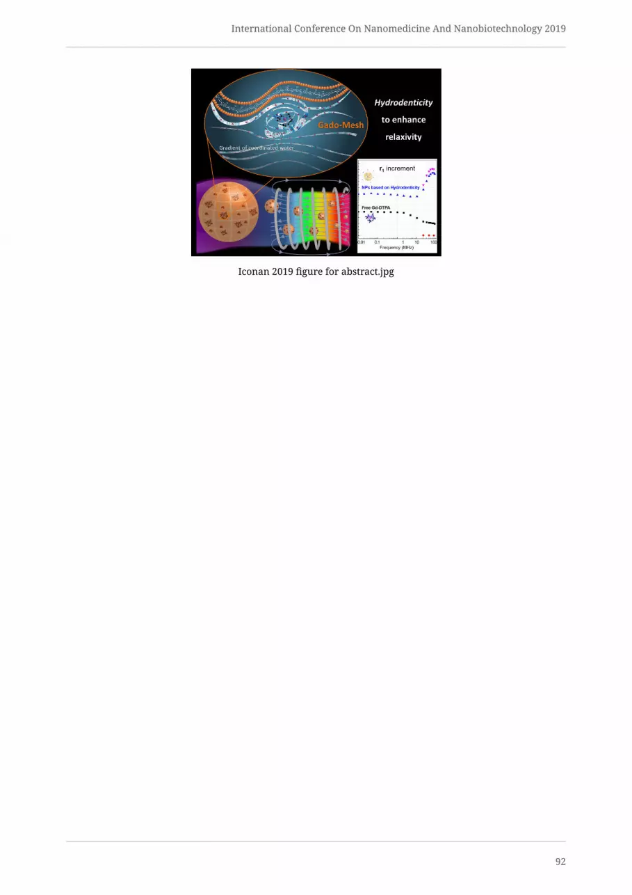
International Conference On Nanomedicine And Nanobiotechnology 2019
Iconan 2019 figure for abstract.jpg
92

International Conference On Nanomedicine And Nanobiotechnology 2019
Drug delivery to solid tumors using nano-sized carriers: themistakes we make.
Thursday, 17th October - 15:00: Nanotechnology and translational medicine: from the bench to the bedside(Room N02.011) - Oral - Abstract ID: 243
Dr. Timo L.M. ten Hagen 1
1. Erasmus MC
Introduction
Delivery of active compounds to tumors is met by a number of hurdles, which are important reasons for failure
of therapy. Chemotherapy is intrinsically toxic, not only to tumor cells but also healthy tissue. Additionally,
most therapeutics are not tumor specific, distribute throughout the body and only a marginal amount arrives
at the target. Increasing the dose is not an option because of the dose-limiting toxicity. Encapsulating drugs
in nanoparticles shields toxicity, impairs distribution and clearance, and may improve accumulation at the
tumor site. Lipid-based nanoparticles have thus far been the most successful, however, clinical outcome is less
favorable. There are a number of reasons which affect nanoparticle-chemotherapeutic performance which
need to be understood before we can start to formulate better nano-therapeutics.
Methods
Here we constructed several nanoparticles (NPs) of 90 nm, made of lipids in a composition specific for the pro-
posed treatment: 1) standard, stable lipid-basedNPs, pegylated andneutral of charge, 2) standardNPs, pegylated
with a positive charge, 3) standard NPs pegylated with a targeting moiety, 4) trigger-sensitive NPs loaded with
doxorubicin, and 5) trigger-sensitive NPs loaded with a drug with a higher uptake profile. Fate of these NPs was
studied in vitro by standard assays and confocal microscopy, and intravitally in tumor-bearing mice by high
resolution confocal microscopy.
Results
Administration of pegylated NPs is thought to result in higher tumor levels of the encapsulated drug due to
the enhanced permeability and retention (EPR) effect. Also, it is thought that the drug is readily released from
NPs outside of tumor cells. Here we show that this is not happening: NPs only distribute to tumors in low
percentages and extravasation is lowandheterogeneous. More so, drugs are only slowly released from standard
NPs and after being taken up by tumor cells. Inside tumor cells NPs end up in lysosomes together with the
drug and remain entrapped for days. Taken together, in vitro these NPs are a log-fold less active compared to
free drug and in vivo, while delivery of drug is higher, availability of free drug is lower than free doxorubicin
infused. Trigger-sensitive NPs have therefore beenmade to release contents when exposed to a trigger; here we
use mild hyperthermia (41°C). When trigger-sensitive NPs (TSNPs) are used loaded with doxorubicin and mild
hyperthermia is applied properly, massive intratumoral drug release can be achieved. However, we found that
doxorubicin is not the best drug in this setting as a considerable amount does not enter tumor cells and diffuses
back to the circulation.
Discussion
Nanotechnology and nanoparticle-mediated drug delivery to cancer has developed fast and delivered a num-
ber of clinical products. In spite of massive research results are however lagging. Results on performance of
lipid-based nanoparticles indicate that better understanding of the interaction between NP, encapsulated drug,
microenvironment and, if applicable, applied trigger is needed. Currently, TSNPs with doxorubicin are used
in combination with radiofrequency ablation for primary liver cancer. In light of above mentioned aspects of
drug delivery with NPs treatment options will be discussed.
93

International Conference On Nanomedicine And Nanobiotechnology 2019
Superparamagnetic iron oxide nanoparticles for MRI - newdevelopments for clinical applications
Thursday, 17th October - 15:15: Nanotechnology and translational medicine: from the bench to the bedside(Room N02.011) - Oral - Abstract ID: 253
Dr. Rainer Tietze 1, Mrs. Eveline Schreiber 1, Ms. Marina Mühlberger 1, Dr. Harald Unterweger 1, Prof.Christoph Alexiou 1
1. Universitätsklinikum Erlangen, Department of Otorhinolaryngology, Head and Neck Surgery, Section of Experimental
Oncology and Nanomedicine (SEON), Else-Kröner-Fresenius Stiftung-Professorship, Erlangen, Germany
Superparamagnetic iron oxide nanoparticles (SPIONs) are of great importance in medical research due to their
wide range of applications in diagnostics and therapy. The use of SPIONs asMRI contrast agents in particular has
great potential since gadolinium containing contrast agents have been controversially discussed and a ban on
the use ofmany of these substances by the EuropeanMedicines Agencywas passed in 20181. Wehave developed
a system of dextran-coated SPIONs (SEONDex) that is ideally suited for use as an MRI contrast agent.
Based on an alkaline precipitation reaction, SPIONs are in-situ coated with dextran and in a subsequent step,
cross-linkedwith epichlorohydrin. By varying the educts, the hydrodynamic size of the particles can bewell con-
trolled. Direct correlations between particle size and relaxivity, as a quality feature for contrast enhancement
in MRI, can be established. The SEONDex particles were comprehensively characterized. After the laboratory
synthesis, a manufacturing process for the 2 litre scale was developed.
The particles have a size of 30 nm and a narrow size distribution, a relaxivity of 106 mM-1s-1 in T2* mode,
excellent biocompatibility in vitro and in vivo and good signalling in animal experiments (Fig. 1). In addition,
SEONDex is very stable and even after 2 years of storage there is no sign of agglomeration or sedimentation.
The current situation concerning gadolinium containing contrast media urgently requires the de-velopment
of alternatives. Various paramagnetic complexes such as manganese or gallium are currently being tested,
but cannot compensate for many disadvantages of the gadolinium that is still in use. Particle-based contrast
agents made of iron oxide as developed by us can set new standards due to their convincing properties. They
can be produced in large quantities in con-stant quality and offer the development perspective by selective
modification of the surface with selective ligands to perform functional imaging in MRI.
Fig. 1:Wildtype mice treated with SEONDex (2.6 mg/ml Fe). T2 weighted images. From left side to the right:
before, 1h after and 24h after application.
Fig. 1.png
94

International Conference On Nanomedicine And Nanobiotechnology 2019
Enhancement of Tumor Penetration and Efficacy of LiposomalDoxorubicin: What We Have Learned
Thursday, 17th October - 15:30: Nanotechnology and translational medicine: from the bench to the bedside(Room N02.011) - Oral - Abstract ID: 254
Dr. Leila Arabi 1, Prof. Mahmoud Reza Jaafari 1
1. Department of Pharmaceutical Nanotechnology, School of Pharmacy, Mashhad University of Medical Sciences, Mashhad
IntroductionSeveral liposomes targeting tumor have been developed to enhance the efficacy of chemotherapeutics. Despite
reducing side effects and success in preclinical studies, most formulations show poor anti-tumor activity due
to insufficient drug bioavailability at the tumor site. Inefficient tumor penetration caused by tumor microen-
vironment may be one of the main reasons. Hence, different approaches have been applied in our group to
enhance the tumor penetration and therapeutic efficacy of liposomes, using penetration-promoting ligands,
and modulating the tumor microenvironment.
MethodsDuring our research project aimed at improving of liposomal formulations, a number of liposomes were pre-
pared to enhance either doxorubicin release or liposome uptake by the tumor cells. lipidswith differentmelting
points were used for preparation of liposomes by lipid film method to optimize the release of drug in vitro and
in vivo in C26 mice model The measurement of the particles size and polydispersity index and zeta potential
of liposomes was performed by dynamic light scattering (Nano-ZS;Malvern, UK). Flow cytometry (FCM) and
confocal laser scanning microscopy (CLSM, Nikon Inc., Switzerland), were used to assess the cellular binding
and uptake of targeted liposomal Doxorubicin. Therapeutic efficacy, biodistribution and pharmacokinetics of
targeted- liposomal Doxorubicin were evaluated in mice bearing tumor. Survival data was analyzed by the
log-rank (Mantel–Cox) test.
ResultsBased on the FCM analyses and CSFM, liposomal formulation conjugated with monoclonal antibodies targeting
the cancer stem cell markers, and liposomal formulation targeted with the matrix-modifying agents exhibited
significantly higher concentration of doxorubicin inside the tumor cells and resulted in superior tumor growth
inhibition in mice model.
ConclusionOverall, using targeting ligand on the surface of liposome and optimizing lipid components, are valuable ap-
proaches resulting in a significant uptake and release of drug from liposome. These efforts have resulted in
liposomes with enhanced therapeutic efficacy with ability to reach the tumor site in the more efficient manner.
These results encouraged us to initiate new projects with different ligands against tumor associated antigens
for development of promising liposomal Doxorubicin.
Keywords: Doxorubicin,Liposome, Targeting, Therapeutic efficacy, Tumor Penetration, tumor microenviron-
ment
95

International Conference On Nanomedicine And Nanobiotechnology 2019
Yttrium oxide nanoparticles resolve acute pancreatitis bymodulation of mitochondrial and endoplasmic reticulum
stress
Thursday, 17th October - 15:45: Nanotechnology and translational medicine: from the bench to the bedside(Room N02.011) - Oral - Abstract ID: 241
Mr. Amit Khurana 1, Ms. Pratibha Anchi 1, Dr. Chandraiah Godugu 1
1. National Institue of Pharmaceutical Education and Research (NIPER), Hyderabad
Introduction: Oxidative stress plays a major role in the pathology of acute pancreatitis (AP), leading to massive
macrophage infiltration. Nanoyttria (NY), possesses potent free radical scavenging activity. As reactive oxygen
species (ROS) and inflammation play major role in AP, we hypothesized that NY may alleviate cerulein induced
AP by modulation of mitochondrial and endoplasmic reticulum stress.
Methods: The nanoparticles were characterized by dynamic light scattering, FTIR, pXRD, XPS, SEM and TEM.
The anti-inflammatory potential of NY was evaluated by Griess assay and cytokines estimation whereas antiox-
idant effect was assessed by DCFDA, MitoSox and JC-1 staining in Raw 264.7 macrophages. The anti-pancreatitic
potential of NYwas evaluated at two dose levels (1 and 3mg/kg) against cerulein challengedmurinemodel of AP.
Results: The nanoparticles were in the size range of 159.25±7.45 nm. NY ameliorated LPS induced oxidative
stress in vitro. It reduced ROS, superoxide radical generation and restored themitochondrial membrane poten-
tial in macrophages (Figure 1). The TEM and ICP-OES based investigation indicated efficient internalization of
the nanoparticles inside the cells. Interestingly, NY reduced plasma amylase and lipase levels and attenuated
the mitochondrial stress and inflammatory markers. Further, NY reduced plasma amylase and lipase levels
and significantly attenuated the levels of oxidative stress markers (MDA, GSH, SOD, catalase, nitrite) and
significantly reduced the levels of inflammatory cytokines
(Figure 2). NY suppressed the recruitment of inflammatory cells around the damaged pancreatic acinar
cells as evident from reduced expression of ICAM1 and PECAM1. Furthermore, NY intervention perturbed
the course of AP via reduction of endoplasmic reticulum (ER) stress markers (BiP, IRE1 and Ero1-Lα), and
molecular chaperones (Hsp27 and Hsp70) (Figure 3). Surprisingly, NY intervention showed promising effects
by modulation of epigenetic modifications by H3 acetylation.
Discussion: We, to the best of our knowledge, report for first time that NY can attenuate experimental AP by
restoration of mitochondrial and ER homeostasis through Nrf2/NFκB pathway modulation. The figure 4 shows
the graphical representation of our novel findings.
96

International Conference On Nanomedicine And Nanobiotechnology 2019
Fig 1.jpg
Fig 2.jpg
Fig 3.jpg
Fig 4.jpg
97

International Conference On Nanomedicine And Nanobiotechnology 2019
My MSCA-funded postdoc project: A modular nanocarrierplatform for siRNA and prodrug delivery
Thursday, 17th October - 16:45: ERC + Marie Curie session: How to successfully apply for a grant(Amphitheatre N02.040) - Plenary Speech - Abstract ID: 226
Dr. Roy van der Meel 1
1. Eindhoven University of Technology
In 2015, I obtained Marie Skłodowska-Curie Action (MSCA) funding from the European Commission and a Veni
grant from the Netherlands Organization for Scientific Research (NWO) to conduct a postdoctoral research
project in Pieter Cullis’ lab at the University of British Columbia, Vancouver, BC, Canada and Raymond Schiffel-
ers’ lab at the University Medical Center Utrecht, Utrecht, The Netherlands.
Technologies developed in the Cullis lab have enabled the clinical translation of thefirst siRNA therapeutic called
Onpattro®, recently approved for treatment of hereditary amyloidogenic transthyretin amyloidosis1. Therapeu-
tically applying siRNA is crucially dependent on delivery systems to prevent degradation and ensure cytosolic
delivery in target cells. In the case of Onpattro®, lipid nanoparticle (LNP) technology facilitates siRNA delivery
to hepatocytes where it inhibits the production of disease-causing mutant transthyretin protein2.
During my postdoc project, I leveraged LNP and drug derivatization technology to develop a modular deliv-
ery platform for stable co-loading siRNA and lipophilic prodrugs3 (Figure 1). As a proof-of-concept, I designed
LNPs containing siRNA against the androgen receptor (AR), a fundamental prostate cancer driver, and taxane
chemotherapeutic prodrugs as a combination treatment (Figure 2).
LNPs were composed of DSPC, cholesterol, PEG-lipid and the ionizable cationic lipid DLin-MC3-DMA for effi-
cient siRNA encapsulation. To formulate LNPs, lipids and prodrugs were dissolved in ethanol and nucleic acids
in buffer and rapidly mixed using a T-junction mixer4 (Figure 3). Following physicochemical analysis, LNPs’
therapeutic effects in vitrowere determined by cell viability assays and AR target gene knockdown. Formula-
tions with radiolabeled lipids and prodrugs were designed to determine the LNPs’ pharmacokinetic parameters
and biodistribution in mice.
In this interactive session, I will share my experience with grant applications, how successfully obtaining re-
search funding has shaped my research career and how it has positively impacted my personal life.
FundingThis research was supported by funding from the European Union’s Horizon 2020 research and innovation
programme under the Marie Sklodowska-Curie grant agreement No. 660426 and a Veni STW Grant (#14385)
from the Netherlands Organization for Scientific Research (NWO).
References1. Adams, D. et al. Patisiran, an RNAi Therapeutic, for Hereditary Transthyretin Amyloidosis. N Engl J Med 379,
11–21 (2018).
2. Kulkarni, J. A., Witzigmann, D., Chen, S., Cullis, P. R. & van der Meel, R. Lipid nanoparticle tech-
nology for clinical translation of siRNA therapeutics. Acc. Chem. Res. acs.accounts.9b00368 (2019).
doi:10.1021/acs.accounts.9b00368
3. Chen, S. et al. Dexamethasone prodrugs as potent suppressors of the immunostimulatory effects of lipid
nanoparticle formulations of nucleic acids. J. Control. Release 286, 46–54 (2018).
4. Kulkarni, J. A. et al. On the Formation and Morphology of Lipid Nanoparticles Containing Ionizable Cationic
Lipids and siRNA. ACS Nano 12, 4787–4795 (2018).
98

International Conference On Nanomedicine And Nanobiotechnology 2019
Figure 1.jpgPicture2.png
Picture3.png
99

International Conference On Nanomedicine And Nanobiotechnology 2019
Securing a prestigious personal grant
Thursday, 17th October - 17:45: ERC + Marie Curie session: How to successfully apply for a grant(Amphitheatre N02.040) - Workshop - Abstract ID: 283
Prof. Twan Lammers 1
1. ExMI – Experimental Molecular Imaging
Obtaining a prestigious personal grant, such aMarie Curie Individual Fellowship (MSCA) or a Starting or Consol-
idator Grant from the European Research Council (ERC), is becoming increasingly important in academic career
development. In the present lecture, I’ll discuss several key considerations when intending to apply for such a
prestigious personal grant, and I’ll share some personal thoughts, tips, tricks, and experiences with regard to
grant writing and panel interviews. The lecture is intended to be interactive and there will be ample time in
the end for comments and questions.
100

International Conference On Nanomedicine And Nanobiotechnology 2019
From encoded combinatorial libraries to targetedtherapeutics
Friday, 18th October - 09:00: Plenary Speeches (Amphitheatre N02.040) - Plenary Speech - Abstract ID: 22
Prof. Dario Neri 1
1. ETH Zürich
Most pharmaceutical agents are either smallmolecules or proteins, whichmay display a beneficial action for pa-
tients by binding to one ormore protein targets of biomedical interest. Thus, the discovery of bindingmolecules
(be them large or small) of fundamental importance for pharmaceutical applications.
Over the past 20 years, my laboratory has mainly used two technologies for ligand discovery:
- antibody phage-display libraries (pioneered by the laboratories of Sir GregoryWinter in the UK and of Richard
Lerner in California) for the isolation of fully-human monoclonal antibodies
- DNA-encoded chemical libraries for the isolation of small organic molecules, capable of binding to specific
protein targets of interest
Both technologies have common feature, as they require the construction of large encoded sets of molecules
(be them antibodies or small organic molecules), followed by the identification of specific ligands by affinity
capture procedures on immobilized protein targets. In both cases, DNA serves as an amplifiable identification
barcode.
In this lecture, I will present:
(i) how we have practiced, developed and applied antibody phage technology and DNA-encoded chemistry for
the discovery of ligands to tumor-associated antigens
(ii) howwe have converted ligands, capable of selective localization at the site of disease, into therapeutic agents
(iii) emerging preclinical and clinical results, obtained with targeted therapeutics developed in my laboratory,
in collaboration with the Philogen group (www.philogen.com)
101

International Conference On Nanomedicine And Nanobiotechnology 2019
Nano-Ghosts: A cancer delivery platform or an inflammatorytherapeutic?
Friday, 18th October - 09:30: Plenary Speeches (Amphitheatre N02.040) - Plenary Speech - Abstract ID: 282
Prof. Marcelle Machluf 1
1. Technion
Mesenchymal stem cells (MSC), have been extensively investigated as cell carriers or cell therapies for treat-
ing a wide range of malignant and inflammatory diseases. Despite promising preclinical results, MSCs have
largely failed to translate into broadly-applicable clinical application due toMSCs’ susceptibility to host-induced
changes, limiting their ability to deliver a long-lasting effect. Recent studies show that MSCs’ preferential ac-
cumulation in malignant and inflamed tissues as well as their immunomodulatory capacity, both are largely
governed by direct cell-cell interactions facilitated by membrane-bound proteins. Building on this knowledge’
we have developed a scalable and cGMP compliant technology for the production of nano-vesicles from MSC
membranes, and their loading with diverse bioactive compounds including proteins, small drugs, and nucleic
acids. These nano-vesicles, are termedNano-Ghosts (NGs). Our data highlight theNGs as a delivery platform that
can be effectively loaded and used to selectively deliver diverse therapeutics including biological drugs, small
molecules, and nucleic acids. Never the less’ surprising data demonstrate that the NG by themselves can mod-
ulate inflammation via cell-cell interaction. Their abundance of natural targeting mechanisms allows the NGs
to penetrate the entire tumor bulk and rapidly deploy their payload directly into the target cells’ cytoplasm and
nucleus led to unprecedented tumor growth inhibition and increased animals’ survival in establishedmetastatic
lung cancer, pancreatic and prostate tumor models. The NG also interact with the cancer stem cells modulating
the response of the tumor to chemotherapy. On the other hand, our studies in a mice model for Multiple Scle-
rosis show that the NGs can bypass the BBB and retain MSCs’ tropism towards inflamed tissues and function as
immunomodulators. The question arises whether these NG are acting as a smart delivery platform, which can
change the inflammatory process in the tumor niche as well as in other pathologies such as MS manifested by
sever inflammatory process. Our results, so far, clearly demonstrate the translational potential of NG, both as
targeted carriers and as a novel immunomodulatory biologic, for oncological and immunological applications.
102

International Conference On Nanomedicine And Nanobiotechnology 2019
RNA delivery: between extracellular vesicles and lipidnanoparticles
Friday, 18th October - 10:30: Plenary Speeches (Amphitheatre N02.040) - Plenary Speech - Abstract ID: 270
Prof. raymond schiffelers 1
1. University Medical Center Utrecht
Nucleic acids can be considered the holy grail of drug development as the active ingredient is essentially always
the same, from a physicochemical point of view, only the nucleotide sequence differs. As a result the same for-
mulation principlesmay be applied acrossmany applications. In the past, electrostatic interactions between the
negatively charged nucleic acids and positively charged carrier materials was used to drive complexation into
nanoparticles thatwould aid tissue distribution and cellular entrance. The recent clinical success of OnPattro(R)
has put the ionizable lipid-based lipid nanoparticles to the forefront. Although much better than many of the
systems explored in the past, even these particles leave room for improvement. An exciting new development is
the use of endogenous cell-derived extracellular vesicles. These nanoparticles are released by cells and contain
many biomolecules, including RNA. It is tempting to speculate that these vesicles are Nature’s way of delivering
complex cargo between cells. In this presentation, I will discuss our experiences with both systems.
103
International Conference On Nanomedicine And Nanobiotechnology 2019
Developing Nanomedicines for Innovative Therapies -Industrial Perspective
Friday, 18th October - 11:00: Plenary Speeches (Amphitheatre N02.040) - Plenary Speech - Abstract ID: 246
Dr. Marianne Ashford 1
1. AstraZeneca
There is a huge array of nanomedicines in pre-clinical research for a breadth of applications, yet few have
progressed to late stage clinical development and commercialisation. This talk will focus on the drivers for
nanomedicines and some of the challenges with the translation of nanomedicines into clinical development
It will share some of the learnings and experiences in exploring three very different nanoparticle systems with
different drug modalities.
A case history showing the factors considered in the design& translation of a polymeric nanoparticle into clinical
development will be shared.
In addition, it will highlight some of the additional challenges with intracellular delivery systems which are
critical for nucleic acid-based drugs and share some of the work done exploring LNP as a delivery system for
mRNA
104

International Conference On Nanomedicine And Nanobiotechnology 2019
Tuning Particle Deformability in Drug Delivery Systems
Friday, 18th October - 11:30: Plenary Speeches (Amphitheatre N02.040) - Plenary Speech - Abstract ID: 285
Prof. Paolo Decuzzi 1
1. Nanotechnology for Precision Medicine, Italian Institute of Technology, Genova, Italy.
Multifunctional nanoconstructs are particle-based nano-scale systems designed for the ‘smart’ delivery of ther-
apeutic and imaging agents. The Laboratory of Nanotechnology for Precision Medicine at IIT-GE synthesizes
multifunctional polymeric nanoconstructs with different sizes, ranging from a few tens of nanometers to a few
microns; shapes, including spherical, cubical and discoidal; surface properties, with positive, negative, neutral
coatings; and mechanical stiffness, varying from that of cells to rigid, inorganic materials, such as iron oxide.
These are the 4S parameters – size, shape, surface, stiffness – which can be precisely tuned in the synthesis pro-
cess enabling disease- and patient-specific designs of multifunctional nanoconstructs. In this lecture, the role of
manipulating these 4S parameters over different temporal and length scales will be elucidated in the context of
future nanomedicines with applications in cancer, cardiovascular and anti-inflammatory diseases. Particular
attention will be given to the role of particle deformability in modulating US-triggered drug release, vascular
transport, mechanical support and the therapeutic efficacy of implantable drug delivery systems.
105

International Conference On Nanomedicine And Nanobiotechnology 2019
Inhibition of hyperactive protein kinases using targetedtherapy of solid breast cancer
Friday, 18th October - 13:30: Poster Session (Foyer N02.083) - Poster - Abstract ID: 19
Ms. Zuzana Skubalova 1, Dr. Simona Dostalova 2, Ms. Marketa Charousova 3, Ms. Zuzana Bytesnikova4, Dr. Jan Pribyl 5, Dr. Akila Weerasekera 6, Prof. Tomas Eckschlager 7, Prof. Marie Stiborova 8, Dr.
Zbynek Heger 9
1. Department of Chemistry and Biochemistry, Mendel University in Brno,Zemedelska 1, Brno,CZ-61200, 2. (1)Department of
Chemistry and Biochemistry, Mendel University in Brno, Zemedelska 1, Brno, CZ-613 00, (2) Central European Institute of
Technology, Brno University of Technology, Purkynova 123, Brno, CZ-612 00, 3. (1)Department of Chemistry and Biochemistry,
Mendel University in Brno, Zemedelska 1, Brno, CZ-613 00, (2) Central European Institute of Technology, Brno University of
Technology, Purkynova 123, Brno, CZ-612 00„ 4. (1)Department of Chemistry and Biochemistry, Mendel University in Brno,
Zemedelska 1, Brno, CZ-613 00, Czech Republic, (2)Central European Institute of Technology, Brno University of Technology,
Purkynova 123, Brno, CZ-612 00, Czech Republic, 5. Central European Institute of Technology, Masaryk University, Kamenice
5/A35, CZ-62500 Brno, 6. Biomedical MRI unit/MoSAIC, Department of Imaging and Pathology, KU Leuven, Leuven, Belgium, 7.
Department of Biochemistry, Faculty of Science, Charles University, Albertov 2030, Prague 2, CZ-128 40, 8. Department of
Biochemistry, Faculty of Science, Charles University, Hlavova 2030/8, CZ-128 43 Prague 2, Czech Republic, 9. 1) Department of
Chemistry and Biochemistry, Mendel University in Brno, Zemedelska 1, Brno, CZ-613 00, 2) Central European Institute of
Technology, Brno University of Technology, Purkynova 123, Brno, CZ-612 00
IntroductionCurrent cancer treatment is for patients really painful, tormenting and lengthy; above all the therapy is not too
specific. Due to this nonspecific treatment, patients are exposed to various serious side effects.
In our project, we focus on a targeted therapy of solid breast carcinoma by tyrosine kinase inhibitors. For
these purposes, three different types of tyrosine kinase inhibitors were selected, as approved chemotherapeutic
agents used in clinic. These include lenvatinib, vandetanib and cabozantinib. In spite of the fact that these
agents are very effective in cancer cure, they cause a number of serious side effects, such as bleeding to the
gastrointestinal tract, fistula formation or vomiting. Therefore, we encapsulated the inhibitors into protein
cage ferritin and modified the protein surface by folic acid (FA) to target folate receptor-overexpressing cancer
cells.
MethodsPrepared nanoconstructs with FA-surface modification were characterized using AFM, TEM, DLS, NMR and
native PAGE. We tested their toxic propertiesin vitroconditions in following malignant breast cell lines: MCF-7
(PR+, ER+,HER2-, FR-), T-47D (PR+, ER+, HER2-,FR+) and also normal breast cell lineHBL-100. The uptake kinetics
and efficacy was assessed via flow cytometer and CLSM.
ResultsIt was found that formed nanoconstructs have almost spherical shape and size around 12 nm. The structure of
ferritin was successfully reassociated after process of encapsulation with yield of encapsulation from 20 % to
60 %, as well as an acceptable long-term stability of 3 weeks. FA-mediated targeting fruitfully promoted uptake
to the target cell lines and increased its efficacy.
DiscussionObtained results provide a promising basis for gaining a folate receptor-targeted nanotherapy of solid breast
malignancies, with pronounced safety for surrounding healthy tissue.
The authors gratefully acknowledge financial support from the GACR project 18-10251S and AF-IGA2019-IP073,
League Against Cancer Prague.
106

International Conference On Nanomedicine And Nanobiotechnology 2019
Characterization of nanoconstrut by afm atomic
force microscopy .png
Scheme of preparation of folate receptor-targeted
nanoconstruct carrying tyrosin kinase
inhibitor.png
107

International Conference On Nanomedicine And Nanobiotechnology 2019
Gold nanocages as drug and gene delivery systems
Friday, 18th October - 13:30: Poster Session (Foyer N02.083) - Poster - Abstract ID: 47
Mr. Jamal Almowalad 1, Dr. Christine Dufes 1
1. Strathclyde Institute of Pharmacy and Biomedical Sciences, University of Strathclyde
Gold nanocages are a novel class of gold nanomaterial with cage shape, hollow interiors and porous wall. Their
unique optical and physiochemical properties make them an appropriate candidate for nanomedicine applica-
tions. Their hollow interiors and porous wall allow them to load and release drugs or biomolecules. In addition,
gold nanocages have the ability to convert the near-infrared light to heat that can be applied into the desired
tissue (photothermal therapy). They can be synthesized by the galvanic replacement reaction between silver
nanocubes and cholorauric acid solution. However, only small amounts of gold nanocages (2–4 mL) can be
isolated through this method. The aim of this project is therefore to optimize the synthesis of gold nanocages
and to modify themwith tumour-targeted ligands for increasing their drug and gene delivery efficacy to cancer
cells.
We have successfully synthesized gold nanocages in uniform shape, large quantities and controlled size. All
gold nanocages samples (blue, grey and teal) had particles size lower than 60 nm, which is ideal for drug deliv-
ery applications. These samples also exhibited UV-Vis spectra in the near-infrared region, which makes them
appropriate for photothermal therapy. Grey and blue gold nanocages showed colloidal stability in water, phos-
phate buffer solution and cell culture medium. Moreover, two concentrations of grey gold nanocages (1 and 4
nM) showed minimum cytotoxicity on the melanoma cell line B16F10 s. In addition, we have modified the sur-
face of the gold nanocages with thiol-PEG-amine to obtain positively charged PEGylated nanocages, which can
be utilised to carry DNA and can be functionalized with tumour-targeting moieties. In conclusion, PEGylated
gold nanocages could be a promising drug and gene nano-carrier for cancer therapy.
108

International Conference On Nanomedicine And Nanobiotechnology 2019
In vitro evaluation of siRNA loaded hNPs for the treatment ofcystic fibrosis
Friday, 18th October - 13:30: Poster Session (Foyer N02.083) - Poster - Abstract ID: 105
Ms. Domizia Baldassi 1, Dr. Gabriella Costabile 1, Ms. Gemma Conte 2, Dr. Ivana D‘Angelo 2, Prof.Francesca Ungaro 3, Prof. Olivia M. Merkel 1
1. Ludwig-Maximilians University Munich, 2. University of Campania, 3. University of Napoli Federico II
IntroductionNowadays, the downregulation of genes involved in the pathogenesis of severe lung diseases through local
siRNA delivery appears an interesting therapeutic approach. In the case of Cystic Fibrosis (CF), it can be used
to downregulate the NF-kβ gene, one of the most critical signals in evoking the inflammatory response in this
disease. In this context, hybrid lipid/polymer nanoparticles (hNPs) can be exploited as a safe and effective
pulmonary delivery system for siRNA [1].
MethodsIn this project, we investigated the biological behavior and the in vitro effects of siRNA-loaded hNPs on human
bronchial epithelial cells. The hNPs studied here consist of an inner core of poly(lactic-co-glycolic) acid (PLGA)
combined with an outer lipidic corona of 1,2-dipalmitoyl-sn-glycero-3-phosphocholine (DPPC) or 1,2-distearoyl-
sn-glycero-3-phosphoethanolamine-poly(ethylene glycol) (DPSE-PEG), with or without polyethylenimine (PEI)
as third component to assist siRNA encapsulation in the hNPs [2].
The efficacy of the hNPs was evaluated in vitroin terms of cytotoxicity, intracellular delivery and gene silenc-
ing ability. The cytotoxicity was tested in terms of cell viability, LDH release in the cell supernatant and pro-
inflammatory effect after incubation with different hNPs concentrations and different exposure times. The
intracellular delivery was measured by flow cytometry and confirmed via confocal microscopy, while the gene
downregulation was determined via western blot analysis.
ResultsThe cytotoxicity studies consisted of MTT and LDH assays at different time points: the results of both experi-
ments agreed that no relevant cytotoxicity was detectable for any tested formulation. We also performed an
ELISA assay to check if TNF-α, a main mediator of inflammation, was released after incubation with hNPs.
The ability of hNPs to deliver siRNA was investigated by flow cytometry using hNPs encapsulated with Alex-
aFluor647 labelled siRNA and was then confirmed by confocal microscopy. From this study, we observed that
PEI plays a key role: in fact, only the formulations containing PEI as third constituent showed a significant
uptake without affecting the cellular viability. Finally, the ability to downregulate NF-kβ gene was assessed
via western blot analysis. From this experiment, we observed that PEI-containing formulations were able to
achieve significant silencing efficacy.
DiscussionThe formulations showed an overall safe profile even after long exposure times and at high concentrations.
Among them, the ones containing PEI retained the best parameters in terms of cellular uptake and gene down-
regulation.
In conclusion, we were able to characterize in vitro a formulation that could represent an interesting new ther-
apeutic strategy for the treatment of inflammation in cystic fibrosis patients, suitable for a following in vivo
study.
[1] I. d’Angelo et al., J. Aerosol Med. Pulm. Drug Deliv., 31, 170-181 (2018)
[2] D. Pandita, Drug Discovery Today, 20 (2015)
109

International Conference On Nanomedicine And Nanobiotechnology 2019
Preparation of water insoluble hyaluronic acid nanofiberlayers for potential use in regenerative medicine
Friday, 18th October - 13:30: Poster Session (Foyer N02.083) - Poster - Abstract ID: 119
Ms. Kristýna Skuhrovcová 1, Ms. Adela Kotzianova 1, Mr. Ondřej Židek 1, Mr. Marek Pokorný 1, Dr.Vladimír Velebný 1
1. Contipro a.s.
Introduction
Hyaluronic acid (HA) is for its great biocompatibility and ability to bind water often used in tissue engineering
and regenerative medicine. Because of its specific behavior in water solution, its transformation into nanofiber
layers or scaffolds is challenging. Uniform and homogeneous layers of oriented or non-oriented nanofibers can
be produced by the electrospinning process.
A drawback of nanofibrous layers made of HA is their high hydrophilicity. As HA nanofibers instantly dissolve
in water-based solutions, these materials are not suitable for longer use during medical treatment. Therefore,
we optimized the electrospinning process to prepare nanofibrous layers using two different types of HA with
chemically modified structure. These modifications increased its lipophilicity by acylation of UV crosslinkable
(1) or fatty acid (2) groups and thus we were able to prepare water stable nanofiber layers.
Methods
Nanofiber layers made of two different HA derivatives (dHA) – furanyl HA and lauroyl HA were prepared by
the electrospinning process. HA derivatives were spun along with polyethylene oxide (PEO) to enhance the
electrospinning process. Various ratios between dHA and PEO as well as different degree of substitution of dHA
were used, so we were able to prepare materials with various properties (e.g. swelling index). All layers were
tested for their swelling index and solubility in phosphate buffer solution up to 72 hours and were analyzed
for their possible structural changes during the electrospinning process and swelling tests by SEM and Raman
spectroscopy.
Results and discussion
Using the UV crosslinkable dHA, we were able to prepare nanofibrous layers solely from water solution, thus
there is no need to use potentially hazard solvents. The swelling behavior of such layers could be controlled by
the time of exposure, the crosslinking time increase fiber diameter. In case of lauroyl HA, there is a necessity
to use also other solvents, but there is no need to post process layers to make them hydrophobic. Swelling is
related mainly to ratio between dHA and PEO and thus there are no other changes in fiber morphology. In
both cases, our results showed the possibility to prepare homogeneous hydrophobized nanofibrous layers from
both hyaluronan derivative. It was shown, that concentration of the solution, degree of substitution and ratio
between hyaluronan and PEO significantly affect both the microscopic quality and application properties of
nanofibrous layers.
References
1. Marcela Dušánková, Gloria Huerta-Angeles, Kristina Nešporová. Self-Supporting, Biodegradable Film Based
on Hydrophobized Hyaluronic Acid, Method of Preparation and Use Thereof. US20180105610A1 Contipro a.s.,
3. 9. 2016.
2. Gloria Huerta-Angeles, Martin Bobek, Eva Příkopová. Nanofibers Containing Photocurable Ester Derivative
of Hyaluronic Acid or Its Salt, Photocured Nanofibers, Method of Synthesis Thereof, Preparation Containing
Photocured Nanofibers and Use Thereof. US20160312385A1 Contipro a.s., 21. 11. 2014.
110

International Conference On Nanomedicine And Nanobiotechnology 2019
Utilizing Tumor Microenvironment pH to Enhance DrugDelivery by Hydrazone Containing Polymeric Micelles
Friday, 18th October - 13:30: Poster Session (Foyer N02.083) - Poster - Abstract ID: 141
Mr. Xiangyang Bai 1, Ms. Qingxue Sun 1, Prof. Fabian Kiessling 1, Prof. Twan lammers 1, Dr. Yang Shi 1
1. RWTH Aachen University Clinic
Nanomedicines have shown their promise for cancer treatment, which can significantly improve drug accumu-
lation in tumors due to the enhanced permeability and retention effect (EPR). However, after tumor accumu-
lation, most of nanomedicines are trapped by extracellular matrix (ECM) and/or taken up by macrophage, and
only a minimal portion can enter cancer cells. We attempt to tackle this issue of nanomedicine by pH sensitive
nanocarriers which are rapidly degraded in the tumor tissues due to the slightly acidic pH. This strategy has
been exploited previously by other pH sensitive materials, however, they mostly have incomplete or slow re-
lease at the tumor microenvironment pH or premature release at neutral pH. We design a super pH sensitive
polymer with hydrazone bond linked side groups to formmicelles with triggered drug release at the tumor mi-
croenvironment pH. The polymer was synthesized by reversible addition fragmentation chain-transfer (FAFT)
polymerization and formed micelles in PBS. Docetaxel (DTX) could be efficiently loaded in the micelles (load-
ing capacity>20 wt%) and the size was around 60 nm. Micellar degradation was analyzed by GPC, HPLC and
DLS. Empty and DTX-loaded micelles showed complete degradation and drug release in 8-12 hours at pH 6.5
and 37 OC. Meanwhile, the micelles were stable at pH 7.4 and 37 OC for >24 hours. Furthermore, the micelles
loaded with DTX or a photosensitizer induced significant cytotoxicity in 4T1 cells in 6-8 hours at pH 6.5 due to
the rapid drug release rate, however, the cytotoxicity was negligible at neutral pH. In conclusion, the designed
super pH sensitive polymeric micelles containing hydrazone groups are promising carriers to improve tumor
drug delivery by exploiting acid-triggered drug release.
Picture1.jpg
Picture2.jpg
Picture3.jpgPicture4.jpg
111

International Conference On Nanomedicine And Nanobiotechnology 2019
Nanoencapsulation of chemotherapeutics in smartPolyurethane/Polyurea Nanoparticles as a novel tool toenhance cancer targeting and improve safety profile
Friday, 18th October - 13:30: Poster Session (Foyer N02.083) - Poster - Abstract ID: 166
Dr. Cristina Cuscó 1, Dr. Marta Pérez 2, Mr. Joaquin Bonelli 1, Prof. Roberto Quesada 3, Prof. VanessaSoto 2, Prof. Ricardo Pérez 2, Dr. Josep Rocas 1
1. EcopolTech, 2. University of Barcelona, 3. University of Burgos
Most of the current Cancer therapies derives in several problems for patients, leading to death in some cases.
Novel targeted nano-therapies against different types of Cancer have been a strategic pillar in last years for
Nanomedicine Community.
FromEcopol Tech S.L., in collaborationwithDepartment of Pathology and Experimental Therapeutics of Univer-
sity of Barcelona, we are developing new one-pot and industrially scalable process for the synthesis of targeted-
Nanoparticles against Lung Cancer.
These type of nanoparticles have been performed to accumulate and internalize preferentially in the acidic
media of Tumor Micro-Environment (TME), as well as to release the drug inside cancer cells due to the high
concentration of Glutathione, which promotes the degradation of the shell in Nanoparticles. In terms of surface
charge, these Nanocapsules exhibit different behavior depending on the pH value of the media because of the
presence of amphoteric moieties in the pre-designed polymer: in TME media, with slightly acidic values about
6.8-7.0 compared with physiological media value of 7.4, Nanoparticles becomes positively charged thus facili-
tating its penetration through cell membrane. This fact has been proved in vitro by internalization experiments
at different pH values.
Thus, we are able to decrease the accumulation in other locations of human body and preventing the undesired
release in these locations. We have tested the preferential release in different media by FRET analisys with
Nanoparticles loaded with different fluorophores.
This study has been carried out with Nanocapsules loaded with Tambjamine analog. This type of drug is highly
hydrophobic, hampering its administration by intravenous via. This new method of nanoencapsulation allows
to disperse it and stabilize it in a crosslinked oil in water dispersion between 15-20 nm.
In vivo tests show a promising safety profile compared with the same doses administration program for free
drug, demonstrating that encapsulation of Tambjamine analog decreases the systemic toxicity caused by the
administration of free drug (which has to be administered intraperitoneally due to its poor solubility).
Fret-release.jpg
Synthetic-scheme.jpg
112

International Conference On Nanomedicine And Nanobiotechnology 2019
Ncs-uptake-different-ph.tif.jpgSafety-assay.tif.jpg
113

International Conference On Nanomedicine And Nanobiotechnology 2019
Scalable flame synthesis of superparamagnetic iron oxidenanoparticles for triggered drug release from colloidal
capsules
Friday, 18th October - 13:30: Poster Session (Foyer N02.083) - Poster - Abstract ID: 178
Mrs. Shno Asad 1, Dr. Jamal Khan 1, Prof. Christel Bergström 1, Prof. Alexandra Teleki 1
1. Uppsala University
Introduction: Microcapsules self-assembled from colloidal particles are promising oral drug delivery vehicles
for therapeutics of low gastrointestinal stability.1 Capsule functionality can be tuned by incorporating nanopar-
ticles with various physicochemical properties in their shell.1 An example of such a functional nanomaterial
are superparamagnetic iron oxide nanoparticles (SPION) that act as MRI contrast agents or trigger hyperther-
mic drug release. However, the lack of scalable and reproducible synthesismethods of SPIONhave hindered the
successful translation into clinics. Flame spray pyrolysis (FSP) is an industrially established synthesis process
that allows large scale manufacture of SPION and close control over particle properties. We have developed a
single-step flame process for encapsulating SPIONwith a biocompatible SiO2 coating2,3 that facilitates their sub-
sequent assembly into colloidal capsules. This study aims to develop colloidal capsules loaded with biological
drugs for oral delivery. We coat the capsules with biopolymer layers to improve drug encapsulation efficiency
and to control release properties. Finally, we explore the incorporation of functional, flame-made nanoparticles
in the shell of these capsules.
Methods: SiO2-coated SPION were made by FSP.2,3 Magnetic hyperthermia performance of particles was mea-
sured by recording the temperature evolution of an aqueous suspension (10 g/L) exposed to an alternatingmag-
netic field. Colloidal capsules were synthesized using a water-in-oil (W/O) emulsion stabilized by commercially
available SiO2 nanoparticles or flame-made SiO2-coated SPION. Capsuleswere assembled by ultrasonication, fol-
lowed by transfer of capsules to awater phase through centrifugation.4 Hydrodynamic size and zeta potential of
nanoparticle suspension and colloidal capsules were measured by dynamic light scattering and electrophoretic
light scattering, respectively. Capsule morphology was visualized by scanning electron microscopy (SEM). SiO2
capsules were coated with alginate and chitosan using the layer-by-layer (LBL) technique.5 The encapsulation
efficiency of a model compound (fluorescein) in the colloidal capsules was measured by microplate reading.
Results: Superparamagnetic iron oxide nanoparticles with a primary particle size of about 20 nm and encap-
sulated with a nanothin (2-3 nm) silica coating were produced by scalable flame spray pyrolysis and exhibited
hyperthermia in an alternatingmagnetic field. Silica colloidal capsuleswith an average hydrodynamic diameter
of 335 nmwere successfully synthesized using an emulsion-based method. SEM images showed homogenously
assembled nanoparticles in the capsule shell with a pore size between particles of about 7 nm. The capsules
were successfully coated with alternating layers of alginate and chitosan and each coating layer reversed the
capsule surface charge. Coated capsules exhibited a lower variability in the encapsulation efficiency (about
60%) of a model compound (fluorescein).
Conclusions: Colloidal capsules made of SiO2 nanoparticles were synthesized using Pickering emulsion-based
method. The SiO2-capsules were coated with alginate and chitosan through the LBL technique, which proved
to enhance the encapsulation efficiency of the model drug. Our ongoing work focuses on assembling colloidal
capsules using flame-made SiO2-coated SPION for controlled oral delivery of biological drugs.
[1] Bollhorst et.al.Chem. Soc. Rev. (46) 2017[2] Teleki et.al. RCS. Adv. (6) 2016[3] Teleki et.al. Chem. Mater. (21) 2009[4] Bollhorst et.al. Chem. Soc. Rev. (25) 2013
114

International Conference On Nanomedicine And Nanobiotechnology 2019
[5] Yang et.al.Acta. Biomater. (6) 2010
Colloidal capsules pre data.png
115

International Conference On Nanomedicine And Nanobiotechnology 2019
Multi-responsive nanogels for biomedical applications
Friday, 18th October - 13:30: Poster Session (Foyer N02.083) - Poster - Abstract ID: 239
Dr. Sebastian Spain 1, Ms. Emma Owens 1, Ms. Marissa Morales-Moctezuma 1
1. Department of Chemistry, University of Sheffield
The ability to control a material’s properties in response to external stimuli potential applications applications
from recycling of plastics to biomedical sensors. We are currently developing a range of nanogels that
respond to multiple stimuli (e.g. pH, redox, salts) separately or synergistically. Here, I will present our
recent work in two areas: oxidation-responsive nanogels for the detection or treatment of inflammation; and
calcium-responsive nanogels for wound care.
Nanogels have been synthesis via polymerisation-induced self-assembly (PISA) using reversible
addition-fragmentation chain transfer polymerisation. For oxidation-responsive nanogels, a poly(N-
hydroxyethyl)acrylamide stabiliser block is chain extended with N-isopropylacrylamide (NIPAM), an
oxidation-responsive acrylate and bisacrylamide (BIS). Nanogel size and morphology can be tuned by various
the relative block lengths, and swell and shrink in response to temperature. Oxidation-responsive nanogels
also display a dose-dependent swelling in response to hydrogen peroxide as a model oxidant.
pH-responsive and thermoresponsive nanogels have synthesised using poly(acrylic acid) or poly(2-
(dimethylamino)ethylmethacrylate) as a stabiliser block with crosslinked polyNIPAM as the core-forming
block. Nanogels behave synergistic responses to temperature and pH or the presence of metal ions.
Figure 2 - oxidation response of nanogels.png
Figure 1 - synthesis of oxidation reponsive
nanogels.png
116

International Conference On Nanomedicine And Nanobiotechnology 2019
Nanoparticle synthesized from the main components ofcancer cell membrane functionalized with c-myc siRNAmodulate the antitumor mechanisms of macrophages.
Friday, 18th October - 13:30: Poster Session (Foyer N02.083) - Poster - Abstract ID: 252
Mr. Edson Comparetti 1, Prof. Valtencir Zucolotto 2
1. Physics Institute of São Carlos, University of São Paulo, 2. Physics Institute of São Carlos, USP - University of São Paulo
Cancer is the most recurrent chronic disease in the world. Human hepatocellular carcinoma (HCC) is the sec-
ond type of cancer with highest number of deaths in the world and their frequency of relapses and metastasis
require the development of new diagnostic and therapeutic approaches. Neoplastic cells have the ability to
evade immune surveillance, modulating tumor microenvironment, to favor tumor progression in the body.
Nanocomposites can be used to prevent the expression of immunosuppressive proteins and cytokines, increas-
ing the activity of themainmechanisms of cellular immunity. This study aims to develop a nanocarrier to trans-
port immunomodulatory agents into cancer and immunocompetent cells. Nanoparticles were synthesized with
lipids and proteins from plasma membrane of neoplastic cells to deliver a large amount of antigenic material
to professional antigen-presenting cells (APC). To establish a pro-inflammatory response, lipid nanoparticles
were incorporated with monophosphoryl lipid A and oligonucleotide sequence (siRNA) to silence c-Myc onco-
gene. Following, nanoparticle was exposed to HCC cells, peripheral blood monocytes from healthy donors and
macrophages generated in vitro. The nanocarriers were tested for: a) nanoparticle internalization into cancer
and immunocompetent cells, b) immunomodulatory activity, observing the expression of cell surface markers
and cytokine production, and c) cytotoxicity of nanocarriers. Uptake was analyzed by fluorescence microscopy
and cytotoxicity by flow cytometry. We aim at increasing the immunogenicity of the tumor cell, upon positively
modulating the action of the antigen-presenting cells (APCs).
Nanoparticle size and charge. .jpg
Monocytes.jpg
117

International Conference On Nanomedicine And Nanobiotechnology 2019
A Full Surveillance Internet of Bio-Nano Things Based onFeynman-Path-Integral-Based Cognitive Radio Theory and
Machine Learning
Friday, 18th October - 13:30: Poster Session (Foyer N02.083) - Poster - Abstract ID: 267
Dr. Huber Nieto-Chaupis 1
1. Universidad Privada del Norte
We present a full surveillance Internet of Bio-Nano Things that is based on the combination of the theories of
Feynman path integral and cognitive radio [1]. In addition we add the Mitchell’s criteria in order to guarantee
the continuous learning of the system and to improve its performance along the time where the measurements
are carried out [2]. We focus on the clinic variables: (i) cardiac pule, (ii) blood pressure, (iii) glucose, and (iv)
cholesterol. When all these variables are fully interconnected among them the probability to anticipate strokes
might be substantial.
Usage of the Cognitive Radio (CR) Model:Consider top panel in FIG-1 where for $q$ nano devices labeled by $h_q$ acquire a signal from $x_m$. It should
be noted that e assume that all signal are electromagnetic waves in the range of allowed frequencies. Once the
sensing and measurement has been performed, then all devices send their signal to the fusion center $y$. In
turn, this has an intrinsic perturbation that alters the output in $y$. With respect to the $h_q$ we can associate
each of the them a specific function as for example given from (i) to (iv) above. Thus CR model this as:
y = x_m \beta_q h_q + n (1)
Usage of the Path Integral Feynman TheoryEq (1) can be written as a probability amplitude that demands of an well-defined initial and final state by
allowing to compute the probability of the transitions among the allowed states. Clearly the concept of CR
applies in the sense that the best path is the one where the best scenario is the one by which the sensing of a
certain signal is n such manner that it does not find any obstruction during its trajectory to the fusion center.
Thus we Eq (1) in terms of the Feynman theory gives
< y_f | y_i > = ∑m ∑n ∫ ∫ dx1 dx2 Φ(x1) Exp[ -i H1 ΔT/ h ] Exp[ -i H2 ΔT/ h] Φ(x2)(2)
With $H1$ and $H2$ the Hamiltonian functions. It is noteworthy that these Hamiltonians are depending on the
instantaneous expended energy of system, electromagnetic energy and batteries of nano-devices.
Usage of the Mitchell’s CriteriaBoth CR and Feynman path have yielded a compact expression as given in Eq (2) where the sub index m and
n indicates us the number of available nano devices that are expected to perform the measurement, that
means that n runs from 1 to 4 for the present study. Once the replacement of initial and final states is done, the
full integral Eq (2) is computationally operated. The resulting surveillance is shown in Fig-2. The apparition of
peaks is counted as alarms.
[1] Huber Nieto-Chaupis, Theory of Cognitive Ratio Through the Feynman Path Integral, 2019 International
Workshop on Antenna Technology (iWAT).
[2] Huber Nieto-Chaupis , The Feynman Path Integral and Machine Learning Algorithms to Characterize and
Anticipate Bacteria Chemotaxis in a Host Healthy Body 2019 IEEE 9th Annual Computing and Communication
Workshop and Conference (CCWC).
118

International Conference On Nanomedicine And Nanobiotechnology 2019
Fig-1.png Fig-2.png
119
International Conference On Nanomedicine And Nanobiotechnology 2019
Nose-to-Brain Delivery of Biologics
Friday, 18th October - 13:30: Poster Session (Foyer N02.083) - Poster - Abstract ID: 79
Ms. Bettina Schwarz 1, Dr. Aditi Mehta 1, Dr. Friederike Adams 1, Dr. Christian L. Ried 2, Dr. ThomasMerdan 3, Prof. Olivia Merkel 4
1. Ludwig-Maximilians-University Munich, 2. AbbVie Deutschland GmbH & Co. KG, 3. AbbVie Deutschland GmbH, 4.
Ludwig-Maximilians University Munich
Introduction:The selective permeability of the blood-brain-barrier (BBB) prevents the utilization of many therapeutic agents,
especially proteins and peptides, for treating CNS disorders. A very promising option to deliver particularly
macromolecular drugs to the CNS is bypassing the BBB by using the nose-to-brain route. In the last few decades,
the nasal route has attracted wide attention as a reliable, safe and non-invasive route to achieve faster and
higher levels of drug absorption in the brain. Despite various benefits, such as rapid onset and avoiding first-
pass metabolism, it presents certain limitations such as short drug residence time and insufficient drug trans-
port due to mucociliary clearance.
This project aims to develop a novel drug delivery system for the intranasal delivery of macromolecules to the
brain with a controlled drug release rate for sufficient therapeutic CNS concentrations. Therefore, we use low
molecular weight chitosan that is known to be biodegradable and non-toxic facilitating paracellular transport
of peptides and proteins across the nasal mucosal barrier.
Methods:Chitosan nanoparticles (NP) were prepared by ionotropic gelationmethod using pentasodium tripolyphosphate
as crosslinker. (Fig.1) Physicochemical characterization included measurements of size using dynamic light
scattering technique, nanoparticle tracking analysis and atomic force microscopy. Furthermore, the overall
charge was assessed by laser Doppler anemometry and shape and morphology by transmission and scanning
electron microscopy. The encapsulation efficiency was determined using the model protein BSA coupled to
fluorescein isothiocyanate (FITC). TheNP cytotoxicitywas evaluated viaMTTassay in the humannasal epithelial
cell line RPMI2650. For the evaluation of NP cellular uptake, free proteinwas compared to protein encapsulated
in NPs at 4°C and 37°C. To specifically target nasal epithelial cells and enhance the cellular uptake, a conjugate
of chitosan with transferrin as targeting ligand was synthesized using click chemistry.
Results:The NPs have a mean size of approx. 100 nm together with a positive surface charge of +40 mV. Scanning
electron microscopy showed that the particles are spherical in shape with a smooth surface. (Fig.2) The NPs
were not found to inhibit cellular proliferation and demonstrated an encapsulation efficiency of FITC-BSA of
about 20-45% depending on chitosan and FITC-BSA concentration.(Fig. 3) The uptake in human nasal epithelial
cells was significantly higher for protein encapsulated in NP compared to free protein. (Fig. 4) The two different
temperatures show that the NPs were internalized (37°C) and not just adsorbed to the cellular surface (4°C).
Discussion:The prepared chitosan NPs have a size of about 100 nm, hence, favoring uptake into olfactory neurons in the
nasal cavity together with potential paracellular transport through the tight junctions of the nasal epithelial
cells. The positive surface charge supports a mucoadhesive effect based on electrostatic interactions with nega-
tively charged mucins in the nasal cavity. We were able to show that the NPs did not affect cell proliferation of
nasal epithelial cells but mediated enhanced cellular uptake of a model protein, which makes them a promis-
ing delivery system for macromolecules. We are currently developing chitosan-transferrin to enhance cellular
uptake by active targeting.
120

International Conference On Nanomedicine And Nanobiotechnology 2019
Figure 1- chitosan nanoparticle preparation by
ionotropic gelation.png
Figure 2- scanning electron microscopy of
chitosan nanoparticles.png
Figure 3- encapsulation efficiency of model
protein fitc-bsa in chitosan nanoparticles.png
Figure 4- cellular uptake of chitosan
nanoparticles in rpmi2650 cell line at 4 c and 37
c.png
121

International Conference On Nanomedicine And Nanobiotechnology 2019
Pattern-generating fluorescent molecular probes for chemicalbiology
Friday, 18th October - 14:15: Biological & medical nanodevices and biosensors (Amphitheatre N02.040) - Oral -Abstract ID: 4
Dr. leila Motiei 1
1. Weizmann Institute of Science
Fluorescent molecular probes have become a powerful tool in protein research. However, these probes are
less suitable for analyzing specific populations of proteins in their native environment. In this talk I will give
an overview of a new class of fluorescent molecular probes1-5 recently developed in our group, and show
how they can be used to detect individual proteins, protein combinations, as well as binding interactions and
dynamic changes that occur on their surfaces. In the second part of this talk, I will present a new class of
fluorescent molecular sensors that combines the properties of small molecule-based probes and cross-reactive
sensor arrays (the so-called chemical nose/tongue’) and explain how these pattern-generating probes could
expand the fluorescent toolbox currently used to detect and image proteins.6 Specifically, I will show how
such systems can be used to identify combinations of specific protein families within complex mixtures and to
discriminate among protein isoforms in living cells, where macroscopic arrays cannot access.6
References
• Rout, B.; Milko, P.; Iron, M. A.; Motiei, L.; Margulies, D. J. Am. Chem. Soc.2013, 135, 15330.
• Unger-Angel, L.; Rout, B.; Ilani, T.; Eisenstein, M.; Motiei, L.; Margulies, D. Chem. Sci. 2015, 6, 5419.
• Nissinkorn, Y.; Lahav-Mankovski, N.; Rabinkov, A.; Albeck, S.; Motiei, L.; Margulies, D. Chem. Eur. J.2015,21, 15981.
• Motiei, L.; Pode, Z.; Koganitsky, A.; Margulies, D. Angew. Chem. Int. Ed. 2014, 53, 9289.
• Hatai, J.; Motiei, L.; Margulies D. J. Am. Chem. Soc. 2017, 139, 2136.
• Pode, Z.; Peri-Naor, R.; Georgeson, J. M.; Ilani, T.; Kiss, V.; Unger, L.; Markus, B.; HM, B.; Motiei, L.; Mar-
gulies, D. Nature Nanotechnol. 2017, 12, 1161.
122

International Conference On Nanomedicine And Nanobiotechnology 2019
All Organic Ultrabright Red to Near-Infrared Nanoparticlesfor Single Particle Tracking and Bioimaging
Friday, 18th October - 14:30: Biological & medical nanodevices and biosensors (Amphitheatre N02.040) - Oral -Abstract ID: 14
Dr. Paolo Pagano 1, Dr. Morgane Rosendale 2, Ms. Jessica Flores 1, Dr. Chiara Paviolo 3, Dr. JonathanDaniel 2, Dr. Laurent Cognet 3, Dr. Jean-Baptiste Verlhac 2, Dr. Mireille Blanchard-Desce 2
1. (formerly) Institute of Molecular Sciences - University of Bordeaux, 2. Institute of Molecular Sciences - University of
Bordeaux, 3. Institut d’Optique Graduate School - University of Bordeaux
Nanotechnologies have the potential to revolutionise our understanding of devastating diseases such as cancer
or neuropathologies through the design of nano-carriers for drug delivery and of fluorescent nano-emitters
for bioimaging. In this latter field, we have now reached the era of nanoscopy where one can dynamically
follow the fate of a single molecule inside a living cell thanks to a class of techniques known as single particle
tracking (SPT). To date, quantum-dots are the most widely used nanoparticles for such applications thanks
to their unparalleled brightness and photostability. Moreover, the size of these semiconductor nanocrystals
can be modulated to finely tune their spectral properties such that their emission can cover the entire visible
range and more. However, their inorganic core is inherently water insoluble. They thus and have to be
passivated with a shell of metal chalcogenides and further coated, typically with silica or polyethylene-glycol,
to increase their solubility. Moreover, most contain toxic heavy metals such as arsenic or cadmium, which
raises environmental concerns on the long-term and drastically limits their potential of ever being used in the
clinic.
To circumvent these limitations, our lab develops Fluorescent Organic Nanoparticles (FONs). Prepared by
self-aggregation of rationally designed hydrophobic dyes in water, FONs show remarkable colloidal and optical
stability, making them a promising, all organic, spontaneously water soluble alternative to quantum-dots.
Their bottom-up molecular design makes it possible to rationally tune their properties for specific applications.
Through engineering of excitonic coupling effects, we have previously reported on green ultrabright FONs
(Daniel et al., J Phys D Appl Phys., 2016). However, biological tissues scatter light and exhibit some level of
auto-fluorescence. These observations led to the definition of an imaging window in the red to near infra-red
(NIR) spectral region, i.e. 600-1000 nm, that is most favourable for deep tissue imaging (Weissleder, Nat.
Biotechnol., 2001). Our previous work in this direction, based on push-pull conjugated systems optimising in-
tramolecular charge transfer by electron delocalisation, led to the development of near-infra-red emitting FONs
of gigantic brigtness (Genin et al., Adv Mat., 2014). These FONs had the property of non-specifically interacting
and entering living cells such that they are potential candidates for drug delivery systems. However, stealth
emitters can be of interest for single particle tracking of cell-surface receptors, a class of biomolecules suspected
to be dysregulated in disease states including neuropsychiatric disorders(Jézéquel et al., TrendsNeurosci., 2018).
In this work, we describe stealth, ultrabright, red to near-infrared emitting FONsmade from novel quadrupolar
dyes. We report on the characteristics and properties of these FONs and show that they have no unspecific
interactions with living cells. Moreover, we demonstrate that some candidates have sufficient brightness and
photostability to achieve single particle tracking in solution. From these combined properties, we conclude that
these novel FONs are promising candidates for the next generation of tools for super resolution bioimaging.
123

International Conference On Nanomedicine And Nanobiotechnology 2019
Figure 1 from dye design to fons preparation and characterisation.png
124

International Conference On Nanomedicine And Nanobiotechnology 2019
The density of cell targeting antibodies on sarcosine-basedpeptobrushes correlates with the extent of unwanted liveraccumulation associated with diminished cell targeting
efficiency
Friday, 18th October - 14:45: Biological & medical nanodevices and biosensors (Amphitheatre N02.040) - Oral -Abstract ID: 56
Ms. Cinja Kappel 1, Dr. Meike Schinnerer 2, Mr. Gabor Kuhn 1, Mrs. Ingrid Tubbe 1, Dr. David Paßlick 1,Ms. Dominika Hobernik 1, Dr. Rebekka Bent 1, Dr. Katharina Haas 1, Ms. Evelyn Montermann 1, Dr.Kerstin Walzer 2, Dr. Mustafa Diken 3, Dr. Matthias Barz 4, Prof. Manfred Schmidt 4, Prof. Volker
Mailänder 5, Dr. Matthias Bros 1, Prof. Stephan Grabbe 1
1. Medical University Mainz, 2. Biontech AG, 3. TRON–Translational Oncology at the University Medical Center of the Johannes
Gutenberg University GmbH, 4. Johannes Gutenberg University, 5. Medic
The development of nanocarriers that deliver cargo specifically to leukocyte populations is considered an inno-
vative immunotherapeutic strategy. The suitability of antibody-decorated nanocarriers to target immune cell
populations has been assessed in numerous studies. So far, most types of functionalized nanocarriers accu-
mulate in the liver after systemic application, which constitutes an obstacle for the perspective of nanocarrier
based therapy. The exact mechanisms that contribute to unwanted liver accumulation of nanocarriers have
not been fully elucidated yet. Here we asked for the role of antibody density per nanocarrier as a determinant
of liver trapping in vivo.
We employed sarcosine-based peptobrushes which are characterized by a long circulation time, and low organ
accumulation as well as little unspecific cell binding. This nanocarrier was conjugated with defined numbers
(2, 6 and 12) of an antibody specific for the surface receptor DEC205 that is specifically expressed by conven-
tional dendritic cells type 1 (cDC1). In parallel assays, we tested the time-dependent biodistribution (in vivo& ex
vivo imaging) of antibody-functionalized peptobrushes and their cell binding (flow cytometry) and cell uptake
(cLSM) properties.
Our data indicate a direct correlation between the number of antibodies per peptobrush and the extent of pep-
tobrush accumulation in the liver. We demonstrate that peptobrushes with intermediate and high antibody
density are efficiently engaged and internalized by liver sinusoidal endothelial cells (LSEC), whereas Kupffer
cells as themain liver macrophage population play aminor role. Blocking experiments revealed that LSEC bind
antibody-decorated peptobrushes via their Fc receptors. Consequently, peptobrushes with intermediate/high
antibody density engaged cDC1 only at low extent. In contrast, conjugation of peptobrushes with 2 antibodies
resulted in pronounced binding to cDC1 target cells in the spleen and only low liver accumulation.
Altogether, our results suggest that lowdensities of antibodies for specific cell targeting are preferable regarding
the design of nanocarriers for immune-therapeutic purposes to prevent unwanted liver accumulation.
125

International Conference On Nanomedicine And Nanobiotechnology 2019
Multi-Detector Field-Flow Fractionation - A powerfulanalytical tool in the field of Nanomedicine
Friday, 18th October - 15:00: Biological & medical nanodevices and biosensors (Amphitheatre N02.040) - Oral -Abstract ID: 103
Dr. Gerhard Heinzmann 1
1. Postnova Analytics GmbH
Field-Flow Fractionation (FFF) belongs to the flow-based separation techniques, where separation of dissolved,
suspended and dispersed sample constituents in the size range of 1 nm to approx. 50 µm is achieved within a
thin, ribbon-like channel without stationary phase (Fig. 1) [1].
In FFF, separation of different sample constituents is induced by a force field that is applied perpendicular to
the channel flow, which transports the sample toward the channel outlet and further to the respective detec-
tors. Common FFF detectors such as UV/Vis, RI and ICP-MS thereby enable access to the mass and concentration
of the fractionated sample, but also size-sensitive detectors based on Multi-Angle- (MALS) and Dynamic Light
Scattering (DLS) are frequently applied turning FFF into a multi-detector analytical platform that facilitates the
comprehensive physico-chemical characterization of the sample. Depending on the applied force field, FFF can
be divided into different subtechniques. In Asymmetrical-Flow FFF (AF4), a second flow, the so-called cross
flow, enables separation according to hydrodynamic size. By superimposing the cross flow field with an elec-
trical field, Electrical Asymmetrical-Flow FFF (EAF4) additionally enables the separation according to charge
thus gaining access to electrophoretic mobility and Zeta potential of the sample. In Centrifugal FFF (CF3), a
centrifugal force field induces separation according to mass respectively density of the sample. Due to its wide
applicability and gentle separation conditions, FFF has become a powerful characterization tool for nano-sized
samples particularly in the field of bio- [2] and nanomedicine [3].
This presentation shall display the benefits ofmulti-detector FFF to propel thefield of nanomedicinehighlighting
its application toward the characterization of liposomal drug formulations, polymeric drug delivery systems
and nano-sized antimicrobial agents.
Fig. 1: Separation principle of Field-Flow Fractionation.
References:
[1] J.C. Giddings, Separation Science, 1(1), 123-125 (1966).
[2] X. Zhang, Y. Li, S. Shen, S. Lee, H. Dou, TrAC Trends in Analytical Chemistry, 108, 231-238 (2018).
[3] M. Wagner, S. Holzschuh, A. Traeger, A. Fahr, U.S. Schubert, Analytical Chemistry, 86(11), 5201-5210 (2014).
Figure 1.jpg
126

International Conference On Nanomedicine And Nanobiotechnology 2019
Microfluidic assembly of siRNA embedded nanoparticles
Friday, 18th October - 15:15: Biological & medical nanodevices and biosensors (Amphitheatre N02.040) - Oral -Abstract ID: 111
Mr. Christoph Zimmermann 1, Dr. Daniel P. Feldmann 2, Prof. Paola Luciani 3, Prof. Olivia M. Merkel 1
1. Faculty for Chemistry and Pharmacy LMU, 2. Wayne State University, 3. University of Bern
Introduction
Nanoparticles have been shown to be successful drug delivery systems and are useful for encapsulating drugs,
enabling more precise targeting with a controlled release. Different approaches can be used to form these
nanoparticles. The classical procedures comprise top-down or bottom-up techniques. However, if electrostatic
interactions are involved in particle formation, the self-assembly of nanoparticles can be achieved via simple
bulk mixing of both fluids. Microfluidic mixing techniques, bringing cationic polymers and nucleic acids to-
gether at a constant ratio during the entire mixing process, have the potential for a more homogeneous and
smoother complexation, leading to a more uniform charge distribution and increased colloidal stability. Mi-
crofluidic mixing can be conducted based on two techniques: active micromixers use applied forces to create a
higher diffusion rate and turbulent flow, whereas passive micromixers change the fluid flow within the chan-
nels to reduce the diffusion time or increase the contact area of the fluids. In this work, siRNA containing
polyplexes were formed by electrostatic interactions using the Micromixer Chip from Dolomite as a passive
micromixer system.
Method
The particle preparation was performed by pumping positively charged br-PEI-g-PCL-b-mPEG triblock copoly-
mer solution and siRNA solution through the Dolomite Micromixer Chip. The flow rate was set by a syringe
pump. Fixed parameters were siRNA concentration, triblock copolymer concentration and buffer (5% glucose,
pH 7.4). The N/P ratio was set at 6. Variable parameters to be tested were tube length (10 cm, 20 cm) and flow
rates (0.1 – 1 mL/min). The preparation and implementation of the mixing process was kept the same for ev-
ery run. The resulting polyplexes were characterised using dynamic light scattering (DLS) and dye exclusion
assays. Selection criteria for subsequent experiments were particle size, PDI, and zeta potential. Furthermore,
parameters were compared with polyplex formulations prepared by simple batch reaction.
Results
The results show that independently of tubing length, no difference could be found in size or PDI. However,
both parameters were affected by the flow rate resulting in smaller sizes (<130 nm) and more monodisperse
particles (PDI < 0.3) obtained with higher flow rates (0.5 mL/min). In comparison, the batch reaction resulted
in sizes of 260 nm and a PDI of 0.38. The zeta potential of the polyplexes assembled by both mixing techniques
were positive and below +10 mV.
Discussion
Microfluidic mixing was found to promote conducive characteristics for cellular transfection. The freedom in
design of the different mixing devices offers almost unlimited possibilities to obtain the most suitable settings.
As shown in Figure 1, even preparation of functionalised or hybrid nanoparticles, consisting of siRNA mixed
with both polymers and lipids or different types of polymers or lipids, is possible. Additional proof-of-concept
experiments will be performed to optimise siRNA formulations made from biodegradable and biocompatible
materials for further in vitroand in vivoscreening.
127

International Conference On Nanomedicine And Nanobiotechnology 2019
Figure 1 - microfluidic assembly of sirna embedded nanoparticles.jpg
128

International Conference On Nanomedicine And Nanobiotechnology 2019
Selective self-assembly of bioelectrocatalysts for functionalbiofuel cells.
Friday, 18th October - 15:30: Biological & medical nanodevices and biosensors (Amphitheatre N02.040) - Oral -Abstract ID: 171
Mr. Alexander Trifonov 1, Dr. Ran Tel-Vered 1, Prof. Andreas Stemmer 1
1. ETH Zürich
Introduction
Self-assembly considered one of the foremost ways of nature to promote the ordering of chemical structures
and the controlled growth of biological systems. Through a set of local interactions, structures of growing di-
mensions are assembled, showing in many cases an emergence of functionality. The self-assembly process has
consequently become a prominent nanofabrication tool inmany fields such as switchable catalytic surfaces and
synthesis of complex materials exhibiting improved electrical or structural properties.
Enzymatic biofuel cells are well recognized as clean energy biogenerators, possessing intrinsic selectivity to-
wards the substrate molecules, and avoiding the use of toxic and/or expensive catalysts. In recent years, a new
class of enzymatic biofuel cells was developed, enabling direct electron transfer (DET) between redox enzymes
and their binding electrodes and showing inherent advantages in terms of design and bioelectrocatalytic effi-
ciencies.
In this work, we describe a design of an all-DET, membrane-less biofuel cell whose active components self-
assemble on stationary electrode surfaces following infusion of mixed catalysts-modified nanoparticles into
the electrolyte.
Methods
The general concept behind the construction of the biofuel cell employing self-assembly is introduced in Figure
1. Two conductive surfaces were chemically modified with receptor units, R1 and R2, showing high affinity to
ligands L1 and L2, respectively. The latter were individually functionalized on carbon coatedmagnetic nanopar-
ticles (ccMNPs), co-modified with fructose dehydrogenase (FDH) or bilirubin oxidase (BOD) as an oxidation or
reduction processes-catalyzing enzymes, respectively. Starting from a mixed suspension, the specific enzyme-
coated ccMNP hybrids showed selective binding to the receptor-modified electrodes within few minutes, thus
self-assembling the anodic and cathodic elements required for the functioning of a biofuel cell.
The self-assembling anodic layer relies on the strong host-guest complexation between the R1 and L1 , whereas
the cathodic compartments interact through electrostatic forces between R2 and L2 termini.
Results and discussion
The anodic and cathodic compartments were constructed and investigated separately, including monitoring of
self-assembly kinetics, which was found to occur within the first 5 minutes of active components interaction;
biocatalytic activities of both compartments, revealing high turnover rates at DET communication of both en-
zymes in presented configurations; and the detachment of the cathodic and anodic catalytic carriers from the
electrodes using external magnetic field.
The resulted two electrodes (the anode and the cathode) were combined into a self-assembling all-DET biofuel
cell. The self-assembled biofuel cell was discharged versus decreasing resistor loads, showing very impressive
results, reflected in characteristic polarization curve. Very impressive power densitywas obtained at amaximal
current density, obtained at 80 mM of fructose. The cell demonstrated good power generation reproducibility
upon cyclicmagnetic removal/self-assembled restoration of fresh hybrids on the electrode surface. The resulted
biofuel cell was further operated under harsh biological conditions, namely viscous biomassmixture, revealing
yet lower, but stable power output.
129

International Conference On Nanomedicine And Nanobiotechnology 2019
Miniaturized Nanopore Reader for single Nanoparticledetection and analysis
Friday, 18th October - 15:45: Biological & medical nanodevices and biosensors (Amphitheatre N02.040) - Oral -Abstract ID: 193
Mr. Marcus Pollard 1, Dr. Angelika Holzinger 2, Dr. Micheal Scanlon 2, Dr. Federico Thei 3, Prof. MarkPlatt 1
1. University of Loughborough, 2. University of Limerick, 3. Elements srl
Resistive pulse sensing (RPS) is a technique which can provide a full characterization of an analyte. In an
RPS system, a nanopore is sandwiched between two fluid cells, one cell contains a ground electrode and the
other contains a working electrode. A potential is applied across the nanopore which causes ions to migrate
resulting in a current which is measured when an analyte passes through the pore it causes exclusion of these
ions reducing the current which is termed a pulse. Using these pulses, we can characterize the analyte’s size,
concentration and charge. Herein we present using a simple approach to characterize nanoparticle size and
concentration using a Nanopore Reader developed by Elements SRL (Italy), a miniaturized ultra-low noise
current amplifier and Nanopore flowcell, a small cartridge which hosts the nanopore chip. Measurements
were done using a solid-state Silicon Nitride nanopore of a fixed size to identify the limits of detection for the
pores. Possible applications of this sensing technology are in the early detection of marker disease.
The figure shows a current trace of a run containing 400 and 800 nm particles, one peak of each size has been
highlighted, 800 nm (Purple) and 400 nm (Orange). The different sized particles can be observed by the differ-
ences in blockademagnitude, caused by an increase in the volume excludedwithin the pore by a larger particle.
By analyzing these blockades a distribution of the blockade magnitudes for a particle can be characterized and
by using a known calibrant the size can then be determined.
Nanopore reader.png Nanopore flowcell.png
130

International Conference On Nanomedicine And Nanobiotechnology 2019
Particle translocations.png
131

International Conference On Nanomedicine And Nanobiotechnology 2019
Complex macromolecular architectures for promoting drugpenetration across biological barriers
Friday, 18th October - 14:15: Targeted drug delivery, organ biodistribution and nanocarriers (Room N02.011) -Oral - Abstract ID: 43
Prof. Francesco Cellesi 1
1. Politecnico di Milano
Recent advances in polymer chemistry have led to a fine control of key properties of polymer nanocarriers, such
as size, drug loading and functionality, with the purpose of an efficient site-specific drug delivery1. However, a
major challenge in nanomedicine is to maximise drug transport across biological barriers,which are necessary
to prevent undesired access of molecules to sensitive organs, tissues and cells in healthy conditions.
In this study, synthetic polymers of controlled molecular architecture were designed to obtain size-tuneable,
amphiphilic and multifunctional drug-loaded nanocarriers with therapeutic and diagnostic properties (Figure
1). Controlled living polymerisations (ROP and ATRP 2) of polyester-, PEG-, glycerol- based monomers/initiators,
and possible peptide conjugation by Michael-type addition3, allowed the development of bioactive and trace-
able nanomaterials which selectively cross specific barriers, including the glomerular filtration barrier to treat
chronic kidney diseases, the blood brain barrier to target brain tumours, and specific cell membranes to obtain
intracellular drug delivery and/or controlled immunostimulation. When required, fluorinated repeating units
were introduced to the macromolecules in order to obtain nanoscopic 19F magnetic resonance imaging agents,
which were also capable of in situ drug release.
AcknowledgmentsThe financial support from Regione Lombardia (POR FESR 2014 – 2020) within the framework of the NeOn
project (ID 239047) is gratefully acknowledged.
References1. Ordanini, S.; Cellesi, F. Complex polymeric architectures self-assembling in unimolecular micelles: Prepara-
tion, characterization and drug nanoencapsulation. Pharmaceutics 2018, 10, (4).
2. Bruni, R.; Possenti, P.; Bordignon, C.; Li, M.; Ordanini, S.; Messa, P.; Rastaldi, M. P.; Cellesi, F. Ultrasmall
polymeric nanocarriers for drug delivery to podocytes in kidney glomerulus. Journal of Controlled Release
2017, 255, 94-107.
3. Celentano, W.; Battistella, J.; Silvestri, I. P.; Bruni, R.; Huang, X.; Li, M.; Messa, P.; Ordanini, S.; Cellesi, F. Engi-
neered polyester-PEG nanoparticles prepared through a “grafting through” strategy and post-functionalization
via Michael type addition. Reactive and Functional Polymers 2018, 131, 164-173.
Figure1 final.jpg
132

International Conference On Nanomedicine And Nanobiotechnology 2019
Effect of protein source on nanoparticle-protein corona andcellular uptake
Friday, 18th October - 14:30: Targeted drug delivery, organ biodistribution and nanocarriers (Room N02.011) -Oral - Abstract ID: 69
Ms. Keni Yang 1, Dr. Anna Salvati 1
1. University of Groningen
The application of nanotechnology formedical purposes has attracted extensive research over the last decades,
but relatively few nano-formulations have reached the market so far. One of the main obstacles is the lim-
ited understanding of the interactions between nanomaterials, cells/tissues and the biological environment in
which they are applied. Generally, in order to evaluate nanomedicine efficiency, in vitro experiments are per-
formed using human cell lines cultured in FBS supplemented medium. However, little attention has been paid
to the effects of the protein source in the medium on the uptake behavior of nanomaterials. To this aim, in
this work, liposomes composed of DOPG and DC-cholesterol lipids (DOPG-DC) have been chosen as a model
nanomedicine. DOPG-DC was prepared by rehydrating a mixed lipid film and extrusion through a 100 nm pore
membrane, followed by physicochemical characterization by DLS and zeta potential measurements. The cell
uptake of DOPG-DC in medium supplemented with FBS and HS was studied by flow cytometry and confocal
microscopy. Additionally, the liposome-corona complexes formed in FBS and HS were separated from free pro-
teins by size exclusion chromatography, followed by qualitative and quantitative analysis of corona proteins by
SDS-PAGE and LC-MS/MS. To further study the effects of proteins of different source on the uptake behavior, the
corona-coated DOPG-DC formed in FBS and HS were isolated and incubated with cells in serum free medium
and medium supplemented with either FBS or HS. This allowed us to discriminate effects due to the source of
the adsorbed corona proteins from those due to the source of free proteins in solution.
The results showed that the uptake of DOPG-DC liposomes incubated in FBS and HS supplemented medium
differed strongly, and the proteomics results confirmed that a different corona was formed in the respective
media. However, when the different hard corona-coated liposomeswere exposed to cells in serum freemedium,
they showed similar internalization efficiency. Similar results were observedwhen liposome-corona complexes
were re-introduced in FBS or HS medium. This suggested that the protein source can affect nanomaterial-cell
interactions not only because of the absorbed corona proteins but also via effects related to the presence of free
proteins in the medium. Similar effects should be taken into account when testing nanomedicines in order to
reduce the differences betweenin vitro and in vivo studies.
133

International Conference On Nanomedicine And Nanobiotechnology 2019
The impact of Nylon-3 copolymer composition on theefficiency in siRNA delivery
Friday, 18th October - 14:45: Targeted drug delivery, organ biodistribution and nanocarriers (Room N02.011) -Oral - Abstract ID: 100
Dr. Friederike Adams 1, Mrs. Natascha Hartl 1, Prof. Runhui Liu 2, Prof. Olivia M. Merkel 1
1. Faculty for Chemistry and Pharmacy, LMU Munich, 2. East China University of Science and Technology
IntroductionRNA interference by small interfering RNA (siRNA) is an efficient strategy to silence genes and thus to prevent
translation. However, the application of siRNA faces challenges, since siRNA is a negatively charged macro-
molecule. It cannot cross cell membranes, the cellular uptake is limited and siRNA is unstable in blood due
to rapid enzymatic degradation. Consequently, several carriers for siRNA have been developed in which poly-
meric vectors showed advantages over viral vectors regarding safety, immunogenicity, production costs and
risk of mutagenicity. In these systems, siRNA is condensed with polycationic polymers through electrostatic in-
teractions to so-called polyplexes. Amine-containing polymers, such as poly(L-lysine) (PLL), linear or branched
poly(ethylenimine) (PEI) or poly(2-dimethylaminoethyl methacrylate) are positively charged at physiological
pH, form ion pairs with nucleic acids, and mediate transfection in a variety of cell lines. Despite their com-
mon use, however, high molecular weight PEI and PLL are significantly cytotoxic. For use in vivo, new cationic
polymers have to be designed incorporating hydrolyzable or biodegradable moieties such that the polymers
readily degrade into nontoxic byproducts. In addition, hydrophobic modification of cationic polymers, e.g. in
phospholipid-modified PEI and in PEI-poly(caprolactone)-poly(ethylene glycol) polymerswas shown to improve
the performance in siRNA delivery coupled with considerably decreased cytotoxicity. Our group developed
polymeric vectors for siRNA encapsulation and delivery based on Nylon-3 polymers, which have a similar back-
bone to biocompatible and biodegradable peptides and a cationic side chain. These cationic subunits derived
either from β-lactam monomer DM or NM. In addition, hydrophobic subunits were introduced to the polymer
which derived from β-lactam monomer CP. DM0.4/CP0.6, NM0.4/CP0.6and NM0.2/CP0.8polymers were designed to
study the impact of the ratio between the hydrophobic and cationic subunit as well as of the use of different
cationic monomers related to the in vitro siRNA delivery into glioblastoma cells.
MethodsNylon-3 polymers were synthesized via anionic ring-opening polymerization (ROP) through statistical copoly-
merization of various β-lactams using lithium bis(trimethylsilylamide) (LiHMDS) as initiator. The obtained
siRNA-polymer polyplexes were characterized regarding physicochemical characteristics such as siRNA encap-
sulation ability (SYBR gold assay), polyplex stability (Heparin assay), particle size and surface charge (DLS and
zeta potential measurements). Furthermore, cell internalization capability, route of uptake and gene knock-
down efficiency were assessed by flow cytometry and cell tolerability was examined by MTT and LDH assays.
Results and DiscussionEfficient siRNA condensation was demonstrated for all tested polymers even with low polymer concentra-
tions. These led to particles with hydrodynamic diameters of < 250 nm and slightly positive surface charges.
NM0.4/CP0.6 built up the most stable polyplexes under physiological conditions leading to the assumption that
NM subunits interact more strongly with siRNAmolecules than DM subunits. Nevertheless, all polyplexes were
able to release acceptable amounts of siRNA under acidic conditions, mimicking endosomal compartment con-
ditions. Furthermore, polyplexes with the highest hydrophobic content displayed significantly higher cellular
internalization in comparison to more cationic formulations and successful knockdown capabilities. Taken
together with convenient toxicity profiles; hydrophobic Nylon-3 polymers provide a potential siRNA delivery
agent for therapeutic approaches.
134

International Conference On Nanomedicine And Nanobiotechnology 2019
Nylon-3 polymers derived from monomers dm nm and cp.jpg
135

International Conference On Nanomedicine And Nanobiotechnology 2019
Bacteria-derived vesicles show low cytotoxicity but inherentantimicrobial activity against gram-negative and
gram-positive pathogens
Friday, 18th October - 15:00: Targeted drug delivery, organ biodistribution and nanocarriers (Room N02.011) -Oral - Abstract ID: 106
Dr. Gregor Fuhrmann 1, Ms. Eilien Schulz 1, Ms. Adriely Goes 1
1. Helmholtz Institute for Pharmaceutical Research
Bacterial infections with antibiotic-resistant pathogens are becoming a major healthcare issue. Indeed, the
WHO has declared these infections as one of the top three threads to humanity, which amplifies the need to de-
velop new therapeutic avenues. Encapsulation of clinically established antibiotics into nanoparticles is promis-
ing to enhance their targeted delivery to the site of infection and could reduce potential side-effects, such as
kidney toxicity.
In this work, we characterised a new group of naturally occurring nanoparticles, so-called outer membrane
vesicles (OMVs) shed by bacteria, as carriers for antibiotics. Bacteria use OMVs for intercellular communication
and as defence strategy. We assessed OMVs frommyxobacteria, a group of non-pathogenic, soil-living bacteria.
In recent years, myxobacteria have been explored for their capability of providing natural compounds that
have a great potential for many uses, especially antimicrobial therapy
We studiedmyxobacterial strains Cystobacter velatus (Cbv34) and Cystobacter ferrugineus (Cbfe23). Cbv34 and
Cbfe23 are producers of antimicrobial products that we hypothesised to be shed into their OMVs. OMVs were
isolated by differential centrifugation and purified using size-exclusion chromatography. The myxobacterial
OMVs showed a mean size of 150-200 nm which we visualised by both nanoparticle tracking analysis and cryo-
electronmicroscopy. Interestingly, whenOMVswere incubatedwith human epithelial A549 lung cells or human
macrophage THP-1 cells we did not see any influence on cell viability or cytotoxicity, even at concentrations of
10,000 OMVs/cell. OMVs also showed low endotoxin concentrations and comparable to buffer controls. We
further studied the interaction of OMVs with Escherichia coli model bacteria by confocal microscopy and saw
that OMVs co-localise with bacteria better than plain liposomes. When assessing their antimicrobial activity,
we found that OMVswere inherently antibiotic against gram-positive Staphylococcus aureus and gram-negative
Escherichia colimodel pathogens. This antibiotic effect was elicited by a cystobactamid, a potent inhibitor of the
bacterial topoisomerase, which is packaged into the bacterial OMVs. The antibacterial effect of Cbv34-derived
OMVs remains potent upon storage in 4° C up to 4 weeks, indicating cargo protection by vesicles.
In ongoing work, we are studying how OMVs interact with bacterial membranes and whether their uptake into
prokaryotes is selective over mammalian cells. In this work, we report a new type of natural vesicle shed by
non-pathogenic bacteria. These OMVswere not toxic towards human cells in our simple assays but they showed
promising activity against gram-positive and gram-negative bacteria.
136

International Conference On Nanomedicine And Nanobiotechnology 2019
Imaging the stability and extravasation of micelles using amicrofluidic platformmimicking tumor microenvironment
Friday, 18th October - 15:15: Targeted drug delivery, organ biodistribution and nanocarriers (Room N02.011) -Oral - Abstract ID: 146
Ms. Natalia Feiner Gracia 1, Ms. Adrianna Glinkowska Mares 2, Ms. Marina Buzhor 3, Dr. Sílvia Pujals2, Dr. Roey Amir 4, Dr. Josep Samitier 2, Dr. Lorenzo Albertazzi 1
1. Eindhoven University of Technology, 2. Institute for Bioengineering of Catalonia, 3. Center for Nanoscience and
Nanotechnology. Tel-Aviv University, 4. University Center for Nanoscience and Nanotechnology, Tel-Aviv University
Supramolecular structures potential use as drug delivery systems is closely related to their stability in complex
biological media. Due to their dynamic behavior, it is necessary to understand the state of the self-assembled
nanocarriers in conditions mimicking the in vivo environment and barriers they will interact with. Current
2D in vitromodels do not provide exhaustive information due to the lack of complex environments such as
flow-induced shear stress, endothelial barrier, 3D tumormicroenvironment (TME) and cancer cells distribution
in extracellular matrix (ECM)1–3. These deficiencies in pre-clinical complexity are the main reason of the low
clinical translation of nanocarriers. Therefore, a comprehensive study of supramolecular systems stability in
complex and dynamic environment requires a reliable, developed and versatile method.
Herein, we present a real-time monitoring study of stability and extravasation of self-assembled micelles, com-
bining spectral confocal microscopy and tumor-on-a-chip. We developed a 3D microfluidic chip recapitulating
the TME. It consists of a perfusable blood vessel channel lined with endothelial cells forming a wall which sep-
arates it from the cancer cells distributed in the ECM in spheroids. In the present work we investigate in the
tumor-on-a-chip model three amphiphilic PEG-dendron hybrids that change their fluorescent properties upon
assembly into micelles4. This property allows us to resolve the stability state of the hybrids in space and time,
in correlation with the barrier they encountered, by monitoring changes in their fluorescence emission.
We observed a time dependent extravasation of monomer followed bymicelles when cancer cells were present
in the ECM. Moreover, no micelle extravasation occurred if cancer cells were not co-cultured in the chip within
the time of the experiment. Interestingly, we observed that small differences in number of spheroids or the
proximity between spheroid and endothelial monolayer impairs their retention capacity. These observations
reflect the heterogeneity of the EPR effect which has been extensively discussed recently and is related not
only to the type of cancer but also to the stage of the diseases and varies from patient to patient1. Overall, we
demonstrated the high stability of our micelles against flow shear stress, and interactions with the ECM. How-
ever, we observed a low stability in contact with cancer cells forming spheroids which impedes the assembled
state to penetrate the solid tumor, in contrast to micelle internalization verified in previous studies in 2D cancer
monolayers.4 Moreover, we observed big differences in stability and extravasation within hybrids with minor
structural modifications.
Our results highlight the interplay betweenmicelle stability in biologicalmedia and their pathway to extravasate
to the target cell, a key step towards drug delivery, and demonstrate the importance of molecular control on
nanostructures for their successful clinical translation.
1. Björnmalm, M. et. al., ACS Nano 11, 9594–9613 (2017).2. Shi, J. et. al., Nat.Rev. Cancer 17, 20–37 (2017).3. Lazzari, G., Couvreur, P. & Mura, S. Polym.Chem. 8, 4947–4969 (2017).4. Feiner-Gracia, N. et al., J. Am. Chem. Soc. 139, 46, 16677-16687 (2017).
137

International Conference On Nanomedicine And Nanobiotechnology 2019
A Nanotoxicology Investigation: Exploring the size effects ofsilver and gold nanoparticles on lung cells
Friday, 18th October - 15:30: Targeted drug delivery, organ biodistribution and nanocarriers (Room N02.011) -Oral - Abstract ID: 158
Ms. Hanouf Bafhaid 1, Dr. Hanene Ali-Boucetta 1, Dr. Zubair Ahmed 2, Dr. Youcef Mehellou 3
1. University of Birmingham, 2. University of Birminhgam, 3. Cardiff University
Unfortunately, nanotoxicology literature does not present a homogenous finding in regards to the cytotoxicity
of nanoparticles and discrepancies are still present between the different conclusions. While the biological
responses and cellular-uptake of nanoparticles are predominately influenced by their different physiochemical
properties, a systematic nanotoxicology framework is still lacking.
This study aims to systematically compare the impact of different composition and different sizes (10, 40 and
80nm) of spherical silver (AgNP) and gold (AuNP) nanoparticles on cellular toxicity and assess the changesmedi-
ated by these nanoparticles following their exposure to human adenocarcinomic alveolar basal epithelial cells
(A549) and normal lung fibroblasts (MRC-5). The size and charge of the nanoparticles were characterised using
dynamic light scattering and transmission electron microscopy. Their cytotoxicity effects were then studied
using MTT, mLDH assays and the mechanism(s) of cell death investigated using Annexin-V/PI, reactive oxygen
species (ROS) and glutathione depletion. On A549 cells, we found that CC50 (50% cytotoxic concentration) of
Ag10 and Au10 after 24hrs are 11.7 and 42.2µg/ml respectively which suggests that AuNP are safer than AgNP.
It was interesting to see that the smaller sizes (10,40nm) of AgNP and AuNP demonstrate more cytotoxicity in
comparison to their bigger counterparts (80nm). This could be due to their enhanced cellular uptake due to
their small size, as we found the amount of silver -taken-up by A549 cells after 24hrs exposure to Ag10 and Ag80
measured by ICP-MS are 4 and 3µg/ml, respectively. Necrotic cell death was observed in a size, concentration
and time dependent manner following exposure to AgNP for 24 and 48hrs (more than 15% Annexin (+)/PI (+))
in both cell lines. Induction of oxidative stress was also found after 24hrs post- exposure to all sizes of AgNP on
both cell lines, while AuNP showed an increase in ROS generation only at the highest concentration (40µg/ml).
After 24hrs exposure to AgNP, the amount of reduced glutathione was significantly depleted in a size and dose-
dependent manner with up to 80% decrease in reducing glutathione levels after A594 exposure to Ag10. AuNP
illustrated a reduction effect on reduced glutathione in A549 andMRC-5 cells at the highest concentrationwhich
correlates with the detected ROS levels.
In summary, our findings highlight that the size of nanoparticles can influence their interactionwith cells. AgNP
of smaller sizes (10 and 40nm) showed detrimental effects on lung cells after 24 and 48hrs of exposure. AuNP
demonstrated a good safety profile comparing to AgNP of the same size, emphasising that nanoparticle com-
position has also an impact on their cellular toxicity. However, MRC-5 and A549 cells display similar trend of
toxicity toward AgNP, which is associated with the release of silver ions.
While size and chemical composition were the key determinant factors in defining the toxicity of our silver &
gold nanoparticles, the ultimate goal of this work is to create a systematic nanotoxicology framework in order
to validate current literature and to pave the development nanoparticles for biomedical applications.
138

International Conference On Nanomedicine And Nanobiotechnology 2019
Cell penetrating liposomes enable the oral delivery ofvancomycin
Friday, 18th October - 15:45: Targeted drug delivery, organ biodistribution and nanocarriers (Room N02.011) -Oral - Abstract ID: 264
Dr. Philipp Uhl 1, Dr. Max Sauter 1, Dr. Tobias Hertlein 2, Dr. Dominik Witzigmann 3, Dr. Knut Ohlsen 2,Prof. Gert Fricker 4, Prof. Walter Mier 1
1. Department of Nuclear Medicine, Heidelberg University Hospital, Im Neuenheimer Feld 400, 69120 Heidelberg, Germany, 2.
Institute for Molecular Infection Biology, University of Würzburg, Josef-Schneider-Straße 2/D15, 97080 Würzburg, Germany, 3.
Nanomedicines Research Group, Life Sciences Institute, University of British Columbia, Vancouver BC, V6T 1Z3, Canada, 4.
Ruprecht-Karls-University, Institute of Pharmacy and Molecular Biotechnology, Im Neuenheimer Feld 329, 69120 Heidelberg,
Germany
Introduction:
Oral delivery of liposomally encapsulated peptide drugs is limited due to their instability in the gastrointestinal
tract and their low mucosa penetration. Tetraether lipids are considered to stabilize liposomes under harsh
conditions while cell penetrating peptides (CPPs) can enhance the mucosa penetration. Therefore, the oral
availability of peptide drugs such as vancomycin incorporated into novel liposomes containing cell penetrating
peptides and tetraether lipids was examined.
Methods:
Tetraether lipid isolation of S. acidocaldariuswas performed by Soxhlet extraction. The cyclic cell penetrating
peptide (CPP) R9C was synthesized by solid-phase synthesis and coupled to maleimide-functionalized phospho-
lipids. Purification of the novel conjugates was achieved by preparative HPLC and confirmed by HPLC/MS anal-
ysis. Preparation of the liposomal formulations was performed by dual assymetric centrifugation (DAC). For in
vivo studies, the oral uptake of the peptide antibiotic vancomycin was determined by radiolabelling. Therapeu-
tic trials in a mouse ‘(MRSA) systemic infection model’ were performed according to Hertlein et al [1].
Results:
The novel liposomes were characterized by Zetasizer measurements. The increase in the zeta potential indi-
cated the successful incorporation of the CPP-phospholipid-conjugates into the liposomes. TEM and cryo-TEM
micrographs confirmed the appropriate liposomal size and morphology. The preparation of the liposomes by
DAC enabled high encapsulation efficiencies (up to 60%) as verified by HPLC and FCS measurements [2, 3]. Mi-
croscopy studies using Caco-2 cells showed that a strongly enhanced binding of the CPP-liposomes in contrast
to standard liposomes was obtained. Furthermore, a high increase in the transport of vancomycin over rat
mucosal tissue in ussing chamber studies in contrast to the free peptide could be shown. Oral application in a
rat model revealed that the novel liposomal formulation led to a fivefold increase in the bioavailability of van-
comycin. The functional efficiency of this uptake enhancementwas proven by a strong increase of the antibiotic
efficiency in a mouse ‘(MRSA) systemic infection model’.
Discussion:
The promising results clearly raise the hope that this novel liposomal formulation can be used as platform-
technology for oral application of a variety of peptide drugs. Bioavailability studies in dogs are ongoing to in-
vestigate the transferability from rodents to highermammals. Additionally, the applicability for further peptide
drugs is currently examined.
[1] Hertlein, T., et al. “Bioluminescence and 19F magnetic resonance imaging visualize the efficacy of
lysostaphin alone and in combination with oxacillin against Staphylococcus aureus in murine thigh and
catheter-associated infection models.” Antimicrobial agents and chemotherapy 58.3 (2014): 1630-1638.
[2] Uhl, P., et al. “A liposomal formulation for the oral application of the investigational hepatitis B drug Myr-
139

International Conference On Nanomedicine And Nanobiotechnology 2019
cludex B.” European Journal of Pharmaceutics and Biopharmaceutics 103 (2016): 159-166.
[3] Uhl, P., et al. “Oral delivery of vancomycin by tetraether lipid liposomes.” European Journal of Pharmaceu-
tical Sciences 108 (2017): 111-118.
Figure 1. cpp-liposomes enable the oral delivery
of vancomycin.jpgFigure 2. animal study.jpg
140

International Conference On Nanomedicine And Nanobiotechnology 2019
Multimodal Image-Guided folic acid targeted Ag2S quantumdots for photothermal therapy and selective methotrexate
delivery
Friday, 18th October - 16:00: Targeted drug delivery, organ biodistribution and nanocarriers (Room N02.011) -Oral - Abstract ID: 45
Ms. Mahshid Hashemkhani 1, Prof. Havva Yagci Acar 1, Prof. Alphan Sennaroğlu 1, Mr. ABDULLAHMUTI 1
1. Koc university
Introduction:The use of Ag2S near-infrared quantum dots (AS NIRQDs) as a sensitizer in photothermal therapy (PTT) is rela-
tively new. Emission of AS QDs in near infrared I (NIR-I) allows better signal/noise ratio and deeper penetration
depth for optical imaging of biological tissues. Here, we developed a multifunctional AS NIRQDs tagged with
folic acid (FA) to deliver methotrexate (MTX) which is an anticancer drug to folate receptor overexpressing
cancer cell lines for image guided combination therapy.
Method: AS QDs with glutathione (GSH) coating was prepared in aqueous solutions. It was conjugated with FA
using a PEG spacer via amidation reactions. Then, MTX was covalently conjugated to AS-GSH-PEG-FA to form
a multifunctional nanocarrier for NIR imaging, PTT and targeted chemotherapy. Hydrodynamic size and sur-
face charge of nanoparticles were measured by Malvern Zetasizer. In the in vitro studies, FR-positive human
cervical carcinoma cells (HeLa) cells and FR-negative A549 and colorectal adenocarcinoma (HT29) cells were
used. Particle internalization was determined by ICP. MTT was used for determination of cytotoxicity (with and
without laser irradiation). The PTT effect of AS-GSH and AS-GSH-PEG-FA QDs were first tested in aqueous solu-
tions as a function of QD concentration, irradiation time and input power. To evaluate PTT efficiency of AS-GSH,
AS-GSH-PEG-FA and AS-GSH-PEG-FA-MTX QDs, HeLa and HT29 cells were treated with different concentrations,
which was determined as safe, and then irradiated with 795 nm laser at 500 mW for 10 minutes. Cell viability
after laser irradiation was determined using MTT assay.
Results and discussions:Aqueous colloidal AS-GSH QD with excellent stability was achieved with emission maxima between 800-900
nm. The absorption peak around 370 nm with a slight red shift compared to free FA clearly confirmed the
FA and MTX conjugation (Figure 1). Based on a calibration curve prepared from the concentration dependent
absorption of FA and MTX separately, binding efficiency of FA and MTX to AS-GSH were obtained 93.6% and
76%, respectively. QDs showed no cytotoxicity up to 200 µg/ml Ag concentration in either cell lines but induced
significant dark toxicity when conjugated with FA and MTX in HeLa due to the presence of folate receptors.
QDs were well internalized by HeLa cells and provided strong optical signal in the NIR. Using 795 nm laser
irradiation induced significant cell death in AS-GSH QDs (Figure 2).
141

International Conference On Nanomedicine And Nanobiotechnology 2019
Figure 1- a the uv-vis b pl spectra of different
conjugates of as qds and c in vitro release profiles
of as-gsh-mtx in pbs solution at ph 7.4 and 5.5 37
.png
Figure 2-time dependent change in the
temperature of as-gsh a at different qd
concentrations b at different powers c stability d
e cell viability after laser.png
142

International Conference On Nanomedicine And Nanobiotechnology 2019
The CRC 1066: From a center for nanoparticle based tumorimmunotherapy to novel approaches in oncology
Friday, 18th October - 16:45: Large projects session: How to run a successful bio-nano-pharmaceuticalconsortial project? (Amphitheatre N02.040) - Plenary Speech - Abstract ID: 262
Dr. Matthias Barz 1
1. Johannes Gutenberg University
In 2013 the center of nanoparticle based tumor immunotherapy (CRC 1066: Nanodimensional polymer thera-
peutics for tumor therapy) was established at the Johannes Gutenberg University and the Medical Center of the
JGU in Mainz with the goalm to develop novel therapeutic interventions for melanoma therapy. After its elon-
gation in 2017 the center hosts 32 groups from chemistry, physics, biology and medicine. The interdisciplinary
character of this CRC is also reflected by the steering committee, which includes Rudolf Zentel (Organic/Polymer
chemistry, JGU), Stephan Grabbe (Department of Dermatology, University Medical Center), Katharina Land-
fester (Max-Planck-Institute for Polymer Research), Detlef Schuppan (University Medical Center) and Matthias
Barz (Organic/Polymer chemistry, JGU).
Therefore, the presentation will include a general introduction to the funding scheme of CRC (Collaborative Re-
search Center, SFB), an overview on the development of the CRC1066 and a personal overview on the related
research from our group.
143

International Conference On Nanomedicine And Nanobiotechnology 2019
MINDED – Multiscale Technologies for NeuroDevelopmentalDisorders
Friday, 18th October - 17:07: Large projects session: How to run a successful bio-nano-pharmaceuticalconsortial project? (Amphitheatre N02.040) - Workshop - Abstract ID: 287
Prof. Paolo Decuzzi 1
1. Nanotechnology for Precision Medicine, Italian Institute of Technology, Genova, Italy.
The societal and economic impact, the growing incidence, and the lack of any cure call for radical changes in
the way Neurodevelopmental Disorders (NDDs) are studied and treated. This lecture will elaborate on syner-
gistic and highly interdisciplinary approaches aiming to revolutionize the life of NDD patients. MINDED uses a
multiscale technology approach integrating acrossNanomedicine,MolecularNeurosciences, andCognitiveNeu-
roscience Robotics, for the development of novel diagnostic, imaging and therapeutic interventions for NDDs.
Nanomedicine for the precise delivery of therapeutic and imaging agents; RNA-technologies for gene silenc-
ing, modulation and repair; laboratories-on-chip for high throughput screening of novel therapeutic strategies;
molecular imaging for the early detection and localization of diseased cells; and advanced cognitive robotics for
patient stratification and rehabilitation are integrated together to improve the classification, diagnosis, imaging
and treatment of NDDs. Training in these three advanced technological areas is expected to foster the establish-
ment of a new class of scientists, technologists, administrators and entrepreneurs.
144

International Conference On Nanomedicine And Nanobiotechnology 2019
Authors Index
Adams, F. 120, 134
Agrawal, N. 67
Ahmed, Z. 138
Albertazzi, L. 137
Alexiou, C. 94
Ali-Boucetta, H. 138
Almowalad, J. 108
Alvarado-Caudillo, Y. 59
Amir, R. 137
Anadozie, S. 73
Anchi, P. 96
Andresen, T. 31
Appelhans, D. 8
Arabi, L. 95
Aryal, S. 27
Asad, S. 114
Ash, B. 86
Ashford, M. 104
ATAC, N. 37
avugadda, s. 35
Aydogan Temel, B. 7
Azizi, T. 27
Bafhaid, H. 138
Bagheri, M. 16
Bai, X. 111
Bakandritsos, A. 24
Baldassi, D. 109
Bandyopadhyaya, R. 62
Barbosa-Sabanero, G. 59
Barker, A. 20
Barz, M. 125, 143
Basnett, P. 35
Battaglia, G. 52
Bazzi, R. 85
Bein, T. 54
Belza, J. 24
Bent, R. 125
Bergström, C. 114
Berquand, A. 85
Beztsinna, N. 16
Bilici, K. 37
Blanchard-Desce, M. 123
Bonelli, J. 112
Briz, N. 17, 64
Bros, M. 125
Bustamante, A. 66
Buzhor, M. 137
Bytesnikova, Z. 106
Cadeddu, R. 68
CAN, F. 37
Carriles-Jaimes, R. 59
Carrillo-Carrion, C. 45
Cellesi, F. 132
Charousova, M. 106
Charousová, M. 58
Chateau, A. 85
Chen Yong James, K. 14
Clima, L. 38
Cognet, L. 123
Colin-Castelan, D. 59
Comparetti, E. 83, 117
Conte, G. 109
Contino, M. 86
Costabile, G. 109
Cotta, K. 62
Craciun, B. 38
Cuscó, C. 112
D‘Angelo, I. 109
Daniel, J. 123
Dasgupta, A. 33
Davids, H. 73
de la Cruz, N. 22
Decuzzi, P. 27, 105, 144
Dehshahri, A. 75
DEVY, J. 85
Dierolf, M. 12
Diken, M. 125
Dostalova, S. 56, 58, 106
Dufes, C. 108
Durand, M. 85
Eckschlager, T. 56, 106
145

International Conference On Nanomedicine And Nanobiotechnology 2019
Elbagory, A. 76
Eliyapura, A. 27
Fanarraga, M. 41
Fasbender, S. 68
Feiner Gracia, N. 137
Feldmann, D. 127
ferrari, m. 1
Feuchtinger, A. 12
Fidecka, K. 10
Flores, J. 123
Fricker, G. 139
Fuhrmann, G. 136
García-Hevia, L. 41
Garelick, H. 9
Genç, F. 7
Ghali, L. 9
Ghosh Chaudhuri, M. 89
Ghosh, A. 89
Giacoboni, J. 10
Glinkowska Mares, A. 137
Godugu, C. 96
Goes, A. 136
Goliasova, Z. 56
González, J. 41
González-Lavado, E. 41
Govender, T. 67
Grabbe, S. 125
Gradl, R. 12
Guisasola, E. 60
Guryev, V. 44
Haas, K. 125
Haas, R. 68
Haiyan, L. 14
Hanssen, R. 49
Hartl, N. 134
Hashemkhani, M. 141
Heger, Z. 56, 58, 106
Heinzel, T. 68
Heinzmann, G. 126
Herma, R. 8
Hertlein, T. 139
Hobernik, D. 125
Holzinger, A. 130
Hruby, M. 47
Hu, R. 49
Hussein, A. 76
Ishaque, M. 19
Iturroz, N. 41
Jaafari, M. 95
Jaberi-Douraki, M. 27
Jacobsen, U. 69
Jager, E. 47
James, D. 86
Jamme, F. 10
Janiak, C. 68
Kalytchuk, S. 24
Kappel, C. 125
Keil, T. 78
Kelly, L. 2
Khalvati, B. 75
Khan, J. 114
Khurana, A. 96
Kiessling, F. 29, 33, 81, 111
Kimna, C. 39
Kotzianova, A. 110
Kristensen, K. 31
Kuhn, G. 125
Kumar, G. 20
Kutschke, D. 12
L. Cortajarena, A. 60
Lak, A. 35
Lammers, T. 29, 33, 81, 100
lammers, T. 111
Larsen, J. 31
laurent, g. 85
Lelievre, E. 85
Licandro, E. 10
Liegertova, M. 8
Lieleg, O. 39
Lindner, L. 6
Liu, M. 33
Liu, R. 134
LONGO, J. 88
Luciani, P. 127
Lund, G. 59
Luysberg, M. 68
Machluf, M. 102
Mailänder, V. 125
Maiti, P. 89
Malina, T. 24
Malmir, K. 86
146

International Conference On Nanomedicine And Nanobiotechnology 2019
Maly, J. 8
Maly, M. 8
Manzanares-Guzman, A. 59
Marasini, R. 27
Marcello, E. 35
Marquez-Sanchez, A. 59
Martinez-Antonio, A. 59
Materia, M. 35
Matin, P. 9
Mehellou, Y. 138
Mehra, S. 62
Mehta, A. 80, 120
Menacho, L. 66
Merdan, T. 120
Merkel, O. 78, 80, 109, 120, 127, 134
Metselaar, J. 16, 50
Meyer, M. 76
Michalek, P. 56
Michalkova, H. 56
Mier, W. 139
Mocktar, c. 67
Mokry, M. 58
Molina Torres, J. 59
Moll, B. 68
Montermann, E. 125
Montizaan, D. 44
Mora Sanz, V. 17, 64
Morales-Moctezuma, M. 116
Morgan, K. 12
Motiei, l. 122
Moya, S. 60
Mura, S. 4
MUTI, A. 37, 141
Möckel, D. 81
Mühlberger, M. 94
Münter, R. 31
Neri, D. 101
Netti, P. 91
Nguyen, T. 27
Niculaes, D. 35
Nieto-Chaupis, H. 25, 71, 118
Nigmatullin, R. 35
Nyman, R. 86
Ohlsen, K. 139
Omolo, C. 67
Owen, A. 51
Owens, E. 116
Padín, E. 41
Pagano, P. 123
Paviolo, C. 123
Pavlov, V. 17, 64
Paßlick, D. 125
Pedersbæk, D. 31
Pedersen, E. 69
Pekarik, V. 58
Pellegrino, T. 35
Peptanariu, D. 43
Pfeiffer, F. 12
Pinel, S. 85
Pinteala, M. 38, 43
Pitchaimani, A. 27
Platt, M. 130
Pokorný, M. 110
Polakova, K. 24
Polanska, H. 56
Pollard, M. 130
Pribyl, J. 106
Pujals, S. 137
Pérez, M. 112
Pérez, R. 112
Quesada, R. 112
Rama, E. 29
Ramdhin, C. 67
Ramirez-Chavez, E. 59
Razansky, D. 12
Rehberg, M. 19
Ried, C. 120
rijcken, c. 49
Rocas, J. 112
Rodriguez-Rios, D. 59
Rojo, J. 22
Rosendale, M. 123
Roux, S. 73, 85
Roy, I. 35
Saa, L. 17, 64
Salih, M. 67
Salvati, A. 44, 133
Samitier, J. 137
Sanchez-Segura, L. 59
Sauter, M. 139
Scanlon, M. 130
147
International Conference On Nanomedicine And Nanobiotechnology 2019
schiffelers, r. 103
Schinnerer, M. 125
Schmid, O. 12, 19
Schmidt, M. 125
Schreiber, E. 94
Schulz, E. 136
Schwarz, B. 120
Sennaroğlu, A. 37, 141
Sheybanifard, M. 16
Shi, Y. 81, 111
Simonsen, J. 31
Sincari, V. 47
Skubalova, Z. 106
Skuhrovcová, K. 110
Smidova, V. 56
Smith, J. 86
Soto, V. 112
Sousa, A. 22
Spain, S. 116
Spierings, D. 44
Splichal, Z. 58
Stemmer, A. 129
Stepanek, P. 47
Stiborova, M. 56, 106
Stockley, P. 20
Stoeger, T. 12, 19
Stofik, M. 8
Storm, G. 16
Strasak, T. 8
Sun, Q. 81, 111
Tel-Vered, R. 129
Teleki, A. 114
ten Hagen, T. 93
Teran, F. 35
Tesarova, B. 56
Tezcan, O. 29
Thei, F. 130
Tietze, R. 94
Torino, E. 91
Trichet, A. 86
Trifonov, A. 129
Tubbe, I. 125
Uhl, P. 139
Ungaro, F. 109
Unterweger, H. 94
Vago, R. 10
Valdivia, L. 41
Valiente, R. 41
Vallance, C. 86
van der Meel, R. 98
Vaz Oliveira, J. 88
Velebný, V. 110
Verlhac, J. 123
Vicent, M. 3
Vogel, S. 69
Voss, C. 19
Waddad, A. 67
Wagner, E. 55
Walvekar, P. 67
Walzer, K. 125
Wanjale, M. 20
Warren, J. 86
Weerasekera, A. 106
Wei Cherng Malvin, K. 14
Wen, X. 9
Witzigmann, D. 139
Wrobel, D. 8
Xodo, L. 69
Yagci Acar, H. 37, 141
Yang, K. 133
Yang, L. 12
Zaina, S. 59
Zarria, J. 66
Zboril, R. 24
Zelikin, A. 53
Zimmermann, C. 127
Zimmermann, L. 68
Zucolotto, V. 83, 117
Židek, O. 110
148

Please consider environment before printing this book