multiwall carbon nanotubes mediate macrophage activation and promote pulmonary fibrosis through...
TRANSCRIPT

Carbon Nanotubes
Multiwall Carbon Nanotubes Mediate Macrophage Activation and Promote Pulmonary Fibrosis Through TGF- β /Smad Signaling Pathway
Peng Wang , Xin Nie , Yue Wang , Yang Li , Cuicui Ge , Lili Zhang , Liming Wang , Ru Bai , Zhiyun Chen , Yuliang Zhao , and Chunying Chen*
1© 2013 Wiley-VCH Verlag GmbH & Co. KGaA, Weinheim wileyonlinelibrary.com
DOI: 10.1002/smll.201300607
Multiwall carbon nanotubes (MWCNTs) have been widely used in many disciplines due to their unique physical and chemical properties, but have also raised great concerns about their possible negative health impacts, especially through occupational exposure. Although recent studies have demonstrated that MWCNTs induce granuloma formation and/or fi brotic responses in the lungs of rats or mice, their cellular and molecular mechanisms remain largely unaddressed. Here, it is reported that the TGF- β /Smad signaling pathway can be activated by MWCNTs and play a critical role in MWCNT-induced pulmonary fi brosis. Firstly, in vivo data show that spontaneously hypertensive (SH) rats administered long MWCNTs (20–50 μ m) but not short MWCNTs (0.5–2 μ m) exhibit increased fi broblast proliferation, collagen deposition and granuloma formation in lung tissue. Secondly, the in vivo experiments also indicate that only long MWCNTs can signifi cantly activate macrophages and increase the production of transforming growth factor (TGF)- β 1, which induces the phosphorylation of Smad2 and then the expression of collagen I/III and extracellular matrix (ECM) protease inhibitors in lung tissues. Finally, the present in vitro studies further demonstrate that the TGF- β /Smad signaling pathway is indeed necessary for the expression of collagen III in fi broblast cells. Together, these data demonstrate that MWCNTs stimulate pulmonary fi brotic responses such as fi broblast proliferation and collagen deposition in a TGF- β /Smad-dependent manner. These observations also suggest that tube length acts as an important factor in MWCNT-induced macrophage activation and subsequent TGF- β 1 secretion. These in vivo and in vitro studies further highlight the potential adverse health effects that may occur following MWCNT exposure and provide a better understanding of the cellular and molecular mechanisms by which MWCNTs induce pulmonary fi brotic reactions.
Dr. P. Wang, X. Nie, Y. Wang, Y. Li, Dr. C. Ge, Dr. L. Zhang, R. Bai, Z. Chen, Prof. Y. Zhao, Prof. C. ChenCAS Key Laboratory for Biomedical Effects of Nanomaterials & NanosafetyNational Center for Nanoscience and TechnologyBeijing 100190, ChinaE-mail: [email protected]
Dr. L. Wang, Prof. Y. ZhaoCAS Key Laboratory for Biomedical Effects of Nanomaterials & NanosafetyInstitute of High Energy PhysicsChinese Academy of SciencesBeijing 100049, China
small 2013, DOI: 10.1002/smll.201300607
P. Wang et al.full papers
1. Introduction
Carbon nanotubes (CNTs) are recently developed, prom-
ising nanomaterials due to their remarkable mechanical,
electrical, and magnetic properties. They are anticipated to
revolutionize the fi elds of electronics, structural engineering,
and medicine. [ 1–3 ] With the increasing production of single-
wall CNTs (SWCNTs) and multiwall CNTs (MWCNTs),
there is an accompanying increase in the potential for
human exposure, especially in occupational settings. [ 4–6 ] It
is rather remarkable that some of the physical properties of
MWCNTs that make them desirable for electronics, engi-
neering, and medicine may also augment their toxic potential
relative to larger particles or even other types of nanoparti-
cles. Some of these properties include altered surface chem-
istry compared with larger particles of the same material,
a high surface area to volume ratio, a high length-to-width
aspect ratio, and a high degree of biopersistence. Indeed, the
biosafety issues of CNTs have attracted enormous atten-
tion in recent years. Wang et al. have reported that chronic
exposure to SWCNTs causes malignant transformation of
human lung epithelial cells. [ 7 ] Meng et al. have studied the
effect of different MWCNTs on the neuron growth factor
(NGF)-induced differentiation of PC12 cells, which is cor-
related to the metal impurities or tube length of CNTs. [ 8 , 9 ]
Some reviews have also raised many concerns about the
nanosafety of CNTs and the possible mechanism of carbon
nanotube-induced toxicity. [ 10 , 11 ] Therefore, it will be neces-
sary to systematically assess the potential adverse effects of
CNTs on human health and the environment during their
applications.
A number of studies have addressed the potential of
CNTs to cause pulmonary fi brosis, the scarring of lung
tissue caused by an increase in fi broblasts and their col-
lagen deposits. [ 12 ] SWCNTs or MWCNTs administered by
intratracheal instillation or pharyngeal aspiration cause
infl ammation in the lungs of rats and mice, as well as
formation of granulomas and/or fi brotic responses. [ 13–17 ]
Recent studies showed that fi brotic reactions in the
lungs of mice exposed to MWCNTs were associated with
increased levels of transforming growth factor (TGF)- β 1
in bronchoalveolar lavage (BAL) fl uid. [ 18 , 19 ] However,
the detailed cellular and molecular mechanisms by which
MWCNTs cause infl ammation and fi brosis in the lung
remain largely unaddressed.
Because CNTs differ in their physical and chemical
properties such as diameter, length and impurities, there
are disparities in the experimental results for nanotoxicity
that could render the mechanism of CNT-induced fi bro-
genesis elusive. Several studies have demonstrated that dif-
ferent dispersions of MWCNTs were associated with their
pulmonary toxicity. [ 19–21 ] Our previous studies suggest that
metal impurities of CNTs or interactions between CNTs
and protein play important roles in CNT-caused cytotox-
icity. [ 22–24 ] Importantly, a recent discovery showed that the
diameter and rigidity of MWCNTs are critical factors in
mesothelial injury and carcinogenesis. [ 25 ] Palomaki et al.
also reported that long, needle-like but not short or long-
tangled MWCNTs can activate the NLRP3 infl ammasome
2 www.small-journal.com © 2013 Wiley-VCH V
in macrophages through a mechanism similar to that of
asbestos. [ 26 ] Indeed, long MWCNTs induce frustrated
phagocytosis and granuloma formation in the mesothe-
lium. [ 27 ] The “length-dependent theory” is useful to explain
CNT-caused infl ammation and mesothelioma in the pari-
etal pleura. [ 28 ] However, to date, few studies have examined
whether the tube length of CNTs contributes to CNT-
caused pulmonary fi brosis. Therefore, to determine whether
fi ber length can play a critical role in MWCNT-induced
fi brogenesis, more in vivo and in vitro experiments are still
urgently needed.
Due to increasing longevity and the prevalence of con-
tributing factors such as obesity, hypertension is already a
highly prevalent cardiovascular risk factor worldwide. We
previously reported that an acute exposure of the lungs
of spontaneously hypertensive (SH) rats to SWCNTs by a
non-surgical intratracheal instillation induced acute pulmo-
nary and cardiovascular responses, which were associated
with an increased level of tumor necrosis factor- α (TNF-
α ) in the BAL fl uid of SH rats. [ 29 ] However, the long-term
effects are not clear. Given that pulmonary fi brosis is a
chronic disease associated with multiple cellular origins
involving complex and interrelated signaling pathways, it is
important to determine whether pulmonary fi brosis has the
potential to crosstalk with other diseases, especially some
chronic diseases. Hypertension is associated with multiple
functional and structural cardiovascular alterations, which
include myocardiac fi brosis induced by local production of
TGF- β 1. Therefore, it seems that pulmonary fi brosis would
be easier to induce in an experimental model of hyperten-
sion. However, to date, there has been no report analyzing
the risk of pulmonary fi brosis generated by CNTs in SH
rats.
Therefore, in the current work, we focus on molecular
and cellular mechanisms by which MWCNT may induce
lung fibrosis after long-term exposure. We chose SH rats
as a susceptible model in our in vivo studies and used
two types of MWCNTs (see Table 1 ), short MWCNTs
(0.5–2 μ m) and long MWCNTs (20–50 μ m), to study
the role of tube length in MWCNT-induced pulmonary
fibrosis. Importantly, our in vivo studies showed that
long MWCNTs, but not short MWCNTs, caused fibrotic
responses including granuloma formation, collagen depo-
sition and fibroblast proliferation. Moreover, we analyzed
the role of MWCNTs in activating the TGF- β /Smad sign-
aling pathway by measuring the production of TGF- β 1
from MWCNT-stimulated macrophages and by detecting
the mRNA expression of TGF- β 1, T β RII and Smad2/3/7
using real-time polymerase chain reaction (PCR) and
phosphorylation of Smad2 by immunoblotting and immu-
nohistochemistry. Finally, we checked whether MWCNTs
could promote the expression of collagen III in vitro
through activating the TGF- β /Smad signaling pathway.
Such a detailed molecular characterization of MWCNT-
induced TGF- β /Smad signaling activation, to our knowl-
edge, has not previously been achieved, and this study
provides an important insight into the cellular and mole-
cular mechanisms involved in MWCNT-induced granu-
loma formation and fibrotic responses.
erlag GmbH & Co. KGaA, Weinheim small 2013, DOI: 10.1002/smll.201300607

Multiwall CNTs Promote Pulmunary Fibrosis
Table 1. Specifi cations of MWCNTs.
Short MWCNTs Long MWCNTs
Outside Diameter
(OD, nm)
≈ 50 ≈ 50
Length ( μ m)
0.5–2 20–50
CNT Purity (wt%)
> 95 > 95
Specifi c surface areas (SSA, m 2 g − 1 )
> 40 > 50
TEM Morphology (Measure bar 100 nm)
Note: Representative transmission electron microscopy (TEM) images of short and long MWCNTs were taken with a Tecnai
G220 S-TWIN transmission electron microscope.
2. Results and Discussion
2.1. Long but Not Short MWCNTs Signifi cantly Increase Fibrosis in the Lungs of MWCNT-Exposed SH Rats
It has been reported that long MWCNTs induce frustrated
phagocytosis and granuloma formation in the mesothe-
lium. [ 27 ] Here, we asked whether fi ber length could also play
a critical role in MWCNT-induced fi brogenesis in the lung.
We answered this question by administering short or long
MWCNTs to the lungs of SH rats by a non-surgical intratra-
cheal instillation and comparing their fi brotic effects 1 day,
7 days or 30 days after exposure.
Granulomas were strongly induced in the lung tissues of
SH rats exposed to Long MWCNTs 30 days after exposure.
In contrast, we could not fi nd any granuloma in the group
treated with short MWCNTs ( Figure 1 A). Similarly, 30 days
post-exposure to the long MWCNTs, the deposition of col-
lagen was easily observed in both granulomatous regions
and the areas distant from granulomas, as established by
immunohistochemistry examination (Figure 1 B) and Sirius
Red staining (Figure 1 C, D). However, there is no such
phenomenon 30 days after exposure to short MWCNTs
(Figure 1 B–D). It is well known that fi broblasts are respon-
sible for the production of collagen. Thus, we analyzed the
expression of fi broblast-specifi c protein-1 (FSP-1), a fi bro-
blast marker, by immunohistochemistry, and found that long
MWCNTs clearly increased the number of FSP-1 positive
cells even at only one day post-exposure. However, short
MWCNTs caused only a weak elevation at 30 days post-expo-
sure (Figure 1 E,F). Moreover, it appears that the granuloma
formation, collagen deposition and fi broblast proliferation in
© 2013 Wiley-VCH Verlag GmbH & Co. KGaA, Weinheimsmall 2013, DOI: 10.1002/smll.201300607
the lung tissues caused by long MWCNT
exposure progressed with time.
Taken together, our data show that
long MWCNTs but not short MWCNTs
cause obvious granuloma formation
and fi brotic effects, which include an
increase in fi broblasts and their col-
lagen deposits in lung tissues of SH rats.
These results suggest that the fi brotic
effect of MWCNTs is length-dependent.
It has been reported that the effect of
MWCNTs on mesothelial injury was
length- and diameter-dependent. [ 25 , 27 ]
Yamashita et al. also showed that long
and thick MWCNTs, but not short and
thin MWCNTs, can cause DNA damage
and severe infl ammatory effects. [ 30 ] How-
ever, there are still some uncertainties
about whether the length of MWCNTs
could play an important role in MWCNT-
caused lung toxicity. Muller et al. reported
that intratracheal instillation of 0.5–5 mg
of long (6 μ m) and short (0.7 μ m) non-
functionalized MWCNTs led to long
persistence of infl ammation and fi brosis
of lung tissue, without signifi cant length-
dependent differences. [ 17 ] Perhaps these various results are
due to the different lengths and diameters of the MWCNTs
they chose. [ 31 ] Given that MWCNTs with a diameter of
50 nm caused the most signifi cant mesothelial injury, [ 25 ] in
our study we chose short (0.5–2 μ m) and long (20–50 μ m)
MWCNTs whose diameters were all about 50 nm. Our fi nd-
ings suggest that short MWCNTs cause less lung injury than
long MWCNTs. Herein, “fi ber-like” pathogenic behavior is
expected based on the available data. In this case, our data
suggest that long and rigid MWCNTs should be avoided for
in vivo applications not only for their mesothelial potential
but also for their promotion of pulmonary fi brosis.
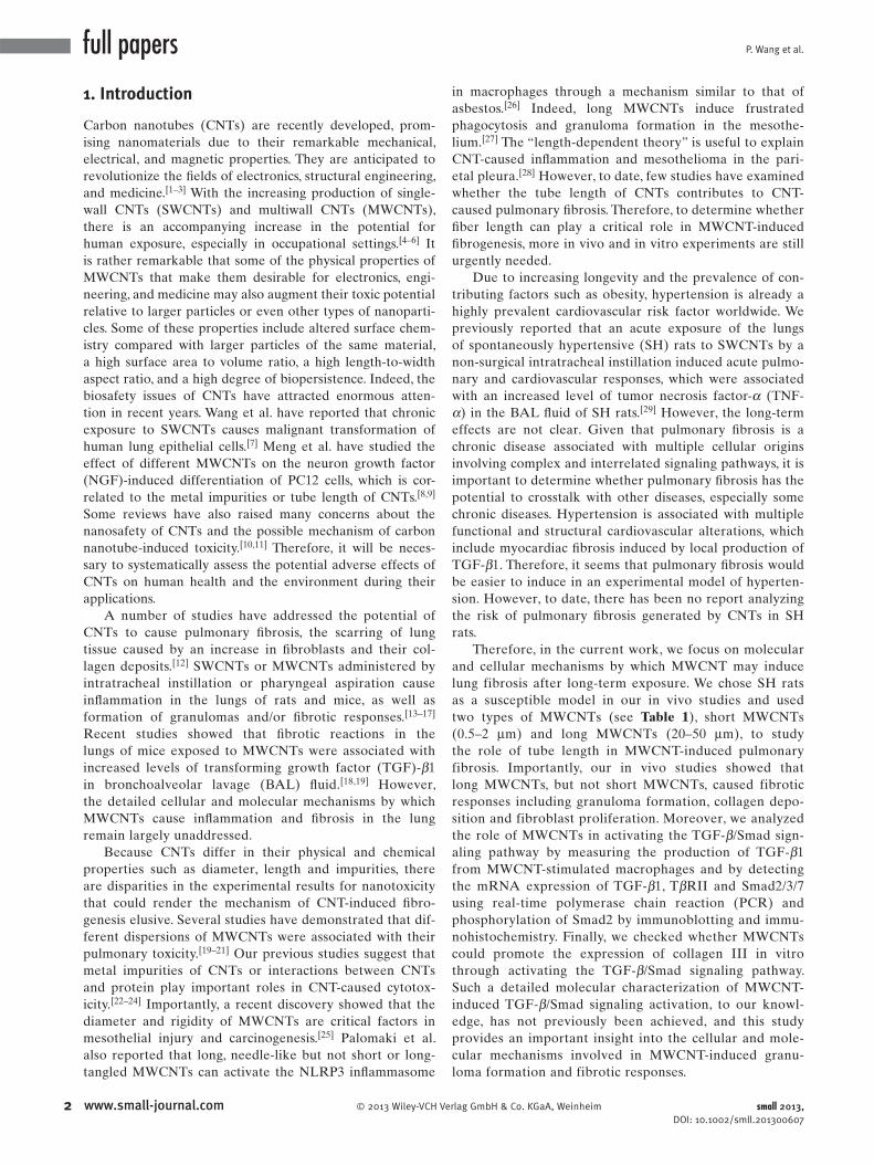
2.2. Long MWCNTs Cause Collagen Deposition by Upregulating Both ECM Protease Inhibitors and Collagen Type I/III mRNA Transcription in the Lungs of MWCNT-Exposed SH Rats
As is known, pulmonary fi brosis is characterized by exces-
sive accumulation of extracellular matrix (ECM) in the lung
tissue. Accumulation of ECM in fi brotic diseases usually
results from elevated mRNA levels of fi brillar collagens due
to increased transcriptional activation. Moreover, inhibitors
of ECM degradation may also play important roles. ECM is
mainly degraded via two distinct pathways: the matrix met-
alloproteinases (MMP) degrading pathway and the plasmi-
nogen activators (PA)/plasmin proteolytic axis. [ 32 , 33 ] Both
pathways have specifi c inhibitors: tissue inhibitors of metal-
loproteinases (TIMP) and plasminogen activator inhibitors
(PAI), respectively. [ 34 ] A previous report has described PAI-1
induction in the lungs of mice exposed to CNTs as a potential
cardiovascular risk factor. [ 35 ] However, how TIMP-1 or PAI-1
3www.small-journal.com

P. Wang et al.full papers
Figure 1 . Comparison of the fi brotic effect of different MWCNTs in the lungs of spontaneously hypertensive rats. SH rats were exposed by intratracheal instillation to short or long MWCNTs (0.6 mg/rat) and lung tissues were obtained 1, 7, or 30 days after exposure. Granuloma was detected by A) HE-staining, and B) production of collagen type III in lung tissue was determined by immunohistochemistry at the indicated time post-exposure. C) The content of collagen type I/III in lung tissues was determined by Sirius Red staining and D) the optical density was integrated by Leica's Microscope Software. E) FSP-1 (a marker of fi broblasts) positive cells in lung tissue were observed by immunohistochemistry and F) the optical density was integrated by Leica's Microscope Software. All values are means ± SD from three independent analyses. Scale bar as follows: 100 μ m (A and C), 50 μ m (B and E).
is regulated in the lungs of SH rats after long-term exposure
to MWCNTs is still unknown.
Thus, to elucidate the possible mechanisms of MWCNT-
caused collagen deposition, we analyzed the mRNA expres-
sion levels of ECM protease inhibitors and collagen I/III in
the lung tissue of SH rats exposed by intratracheal instilla-
tion to short or long MWCNTs for 1, 7, or 30 days. Real-time
polymerase chain reaction (PCR) results showed that the
mRNA expression of both TIMP-1 and PAI-1 was signifi -
cantly increased by long MWCNT exposure for 7 or 30 days,
but not 1 day ( Figure 2 A). However, it seems that there is
4 www.small-journal.com © 2013 Wiley-VCH Ve
selectivity for short MWCNTs in the regulation of TIMP-1
and PAI-1. Short MWCNTs clearly promoted TIMP-1 tran-
scription but had a limited effect on PAI-1 mRNA expression
(Figure 2 A). Except for detecting these two ECM protease
inhibitor genes, the mRNA transcriptions of collagen I/III
were also checked using real-time PCR. In accordance with
our data from immunohistochemistry examination (Figure 1 B)
and Sirius Red staining (Figure 1 C, D), collagen I/III mRNA
transcription was time-dependently upregulated by long but
not short MWCNT treatment (Figure 2 B). Taken together,
our data suggest that MWCNTs cause collagen deposition
rlag GmbH & Co. KGaA, Weinheim small 2013, DOI: 10.1002/smll.201300607
Multiwall CNTs Promote Pulmunary Fibrosis
Figure 2 . Effects of MWCNT exposure on expression of ECM protease inhibitors and collagen type I/III in vivo. SH rats were exposed by intratracheal instillation to short or long MWCNTs (0.6 mg/rat) and lung tissues were prepared for RNA isolation at 1, 7, or 30 days post-exposure. Real time quantitative PCR was performed using 2 μ g of total RNA for A) two ECM protease inhibitor genes, TIMP and PAI-1 or B) collagen type I/III. All values are means ± SD from three independent analyses.
by upregulating both ECM protease inhibitors and collagen
type I/III mRNA transcription in the lungs of MWCNT-
exposed SH rats. Moreover, the fi ber length of MWCNTs
might play a critical role in MWCNT-induced transcriptional
activation.
Several studies have reported that MWCNTs upregu-
late the expression of collagens. [ 18 , 36 , 37 ] However, they only
detected the protein level of collagen; whether collagen
mRNA transcription could be affected by MWCNT treat-
ment is unknown. Moreover, the effect of MWCNTs on
the expression of the ECM protease inhibitor gene has not
been systematically studied. Importantly, our study indi-
cated that long MWCNTs contributed to fi brosis not only by
enhancing collagen transcription but also by inhibiting ECM
degradation, by inducing transcription of protease inhibitors
© 2013 Wiley-VCH Verlag Gmbsmall 2013, DOI: 10.1002/smll.201300607
such as TIMP-1 and PAI-1. As is known, the TGF- β /Smad
signaling pathway is responsible for the transcriptional acti-
vation of collagen I/III, TIMP-1 and PAI-1. Thus exploration
of whether MWCNTs cause fi brosis through activating the
TGF- β /Smad signaling pathway in lung tissues is urgently
needed.
2.3. Long MWCNTs Stimulate the Production of TGF- β 1 by Mediating Activation of Macrophages and Causing More Damage to the Bronchiolar Epithelium
Fibrosis is a disease of multiple cellular origins involving
complex and interrelated signaling pathways. The produc-
tion of infl ammatory cytokines and chemokines from acti-
vated macrophages is a hallmark of the pulmonary response
to fi brogenic fi bers. [ 12 ] Thus, in order to fi gure out the inter-
action between MWCNTs and macrophages and the effect
of MWCNTs on macrophage activation, we fi rst examined
the cellular uptake of different MWCNTs by alveolar mac-
rophages in BAL fl uid or lung tissues. From Figure 3 A, both
short and long MWCNTs can be observed in alveolar mac-
rophages from BAL fl uid. Interestingly, images from alveolar
macrophages of SH rats exposed to long MWCNTs show
that incomplete or frustrated phagocytosis occurred. We also
detected the distribution of MWCNTs in the lung tissue,
and the results showed that both MWCNTs could be taken
up mainly by alveolar macrophages localized in the pulmo-
nary alveoli (Figure 3 B). Together, our data suggested that
alveolar macrophages were responsible for the clearance of
administered MWCNTs from the lung.
Among the mediators involved in tissue fi brosis, TGF- β is considered a key molecule in the activation of the fi brotic
program. Meanwhile, TNF- α is considered a central mediator
of chronic infl ammatory diseases. It has been reported that
both TGF- β 1 and TNF- α can be stimulated by infl ammatory
stimuli in macrophages. [ 38 , 39 ] Therefore, we next detected the
production of TGF- β 1 and TNF- α from MWCNT-stimulated
macrophages. As shown in Figure 4 A, the production of TGF-
β 1 was signifi cantly stimulated by long MWCNT treatment
for 1 day. However, the short treatment group had no effect
on the level of TGF- β 1 in BAL fl uid. By contrast, TNF- α levels increased at 7 days after but not 1 day or 30 days after
exposure to short or long MWCNTs (Figure S1, Supporting
Information). Interestingly, we also used real-time PCR to
detect the mRNA expression of TGF- β 1 in lung tissues, and
found out that TGF- β 1 transcription was upregulated by
MWCNTs only after 7 days of exposure, but not after 1 day
or 30 days, and that long MWCNTs enhanced TGF- β 1 tran-
scription more signifi cantly than short MWCNTs (Figure 4 B).
In addition, we looked at whether TGF- β 1 could be
secreted by MWCNT-stimulated macrophages by immu-
nohistochemistry. As shown in Figure 4 C, TGF- β 1 could
be clearly observed in MWCNT-stimulated alveolar
macro phages. Moreover, it seems that TGF- β 1 produced
by MWCNT-stimulated alveolar macrophages could be
observed only after 7 days of exposure, but not after 1 day or
30 days, and that more TGF- β 1 could be generated by alve-
olar macrophages exposed to long MWCNTs (Figure 4 D).
5www.small-journal.comH & Co. KGaA, Weinheim

P. Wang et al.full papers
Figure 3 . Representative optical microscope images to show the cellular uptake of MWCNTs by alveolar macrophages in vivo. A) SH rats were exposed by intratracheal instillation to short or long MWCNTs (0.6 mg/rat) and BAL fl uid was obtained 7 days after exposure. Uptake of MWCNTs by alveolar macrophages in BAL fl uid was observed by optical microscopy (100 × ). B) Representative regions in lung tissues of short or long MWCNT-exposed SH rats. Both short and long MWCNTs could be phagocytosed by alveolar macrophages 7 days after exposure. The white arrows point to CNTs extending outside the macrophage in the process of incomplete or frustrated phagocytosis. Scale bar as follows: 5 μ m (A), 30 μ m (B).
Together, these results suggest that MWCNT-stimulated alve-
olar macrophages are responsible for TGF- β 1 production in
the lung tissue of SH rats exposed to MWCNTs for 7 days.
In fact, several studies have already reported that
TGF- β 1 could be stimulated in the BAL fl uid of MWCNT-
administered mice. [ 18–23 ] However, the mechanisms of
MWCNT-caused TGF- β 1 secretion into BAL fl uid are still
unknown. Therefore we used real-time PCR and immuno-
histochemistry to detect TGF- β 1 production and found that
MWCNTs increased TGF- β 1 production only after 7 days of
exposure (Figure 4 B–D); however, upregulation of TGF- β 1
secretion into BAL fl uid could only be detected after 1 day of
exposure to long MWCNTs (Figure 4 A).
It has been previously reported that protein expres-
sion of TGF- β 1 is confi ned to the bronchiolar epithe-
lium. [ 40 ] Consistent with this study, we also observed
constitutively expressed TGF- β 1 near the bronchiolar epi-
thelium ( Figure 5 A). This constitutive TGF- β 1 expression
near the bronchiolar epithelium might be signifi cant for the
constitutive expression of collagen III (Figure 5 C), which
6 www.small-journal.com © 2013 Wiley-VCH Verlag GmbH & Co. KGaA
is mediated by phosphorylated Smad2
(Figure 5 B), and it might also be necessary
for the physiological function of the bron-
chiolar epithelium. Moreover, as shown
in Figure 5 , we observed that some cell
fl akes appeared in the bronchus of SH rats
exposed to long MWCNTs but not short
MWCNTs for 1 day. We speculate that
long MWCNTs might cause more damage
to the bronchiolar epithelium than short
MWCNTs, and this damage might induce
the release of TGF- β 1 from the bron-
chiolar epithelium, which may recruit
fi broblast cells and cause some pulmo-
nary infl ammations. Therefore, MWCNT-
caused release of TGF- β 1 from the
bronchiolar epithelium may be one of the
mechanisms of MWCNT-caused TGF- β 1
secretion into BAL fl uid. Taken together,
these results suggest that TGF- β 1 could
be induced in alveolar macrophages or
released from the bronchiolar epithelium
by MWCNT exposure and play an impor-
tant role in pulmonary fi brosis.
2.4. Activation of the TGF- β /Smad Signaling Pathway in the Lung Tissues of MWCNT-Exposed SH Rats
TGF- β , a primary mediator of collagen
deposition during fi brogenesis, signals
through two types of membrane-bound
serine/threonine kinase receptors and
intracellular Smad proteins. [ 41 , 42 ] Ligand
binding results in the formation of the
receptor heteromeric complex con-
sisting of TGF- β type I and TGF- β type
II receptors, phosphorylation of R-Smad
(receptor-regulated Smads: Smad2/3), accumulation of the
Smad2/3-Smad4 complex in the nucleus, and (ultimately)
modulation of gene expression. Our above studies have
shown that MWCNTs could stimulate TGF- β 1 secretion in
the lung by activation of alveolar macrophages, and that the
fi ber length of MWCNTs plays a critical role in MWCNT-
stimulated production of TGF- β 1. However, to date, the
effect of MWCNTs on Smad2 phosphorylation has not been
directly demonstrated. Thus, to determine whether MWCNTs
could activate the TGF- β /Smad signaling pathway in the lung
tissue of SH rats, we examined Smad phosphorylation in
fi broblast cells.
As shown in Figure 6 A, TGF- β 1 signifi cantly induced
Smad2 phosphorylation in a time-dependent manner in
fi broblast cells in vitro. We showed above that MWCNTs
could stimulate TGF- β 1 secretion in the lung by activation
of alveolar macrophages. To determine whether the TGF-
β 1 secreted by alveolar macrophages could stimulate Smad2
phosphorylation in the lung, we used immunoblotting and
immunohistochemistry to detect Smad2 phosphorylation
, Weinheim small 2013, DOI: 10.1002/smll.201300607

Multiwall CNTs Promote Pulmunary Fibrosis
Figure 4 . Effects of MWCNTs on the production of TGF- β 1 in lung tissues of SH rats. SH rats were exposed by intratracheal instillation to short or long MWCNTs (0.6 mg/rat) and BAL fl uids or lung tissues were obtained at 1, 7 and 30 days post-exposure, respectively. A) ELISA analysis was used to analyze the contents of TGF- β 1 in BAL fl uid. B) Real-time PCR was performed to determine the mRNA expression level of TGF- β 1 in lung tissue. C) Immunohistochemistry was employed to detect TGF- β 1 in lung tissues of SH rats exposed to short or long MWCNTs for 7 days. Scale bar 50 μ m. D) Immunohistochemistry was employed to detect TGF- β 1 in lung tissues of SH rats exposed to short or long MWCNTs for the indicated time. Scale bar 100 μ m. All values were normalized according to the PBS control. All values are means ± SD from three independent analyses.
in lung tissues of SH rats exposed to MWCNTs for 7 days.
Both immunoblotting and immunohistochemistry results
showed that Smad2 phosphorylation was signifi cantly
enhanced by MWCNTs in lung tissues of SH rats 7 days after
intratracheal instillation exposure (Figure 6 B,C). As shown
in Figure 6 C, long MWCNTs caused a much higher Smad2
© 2013 Wiley-VCH Verlag Gmsmall 2013, DOI: 10.1002/smll.201300607
phosphorylation level than short MWCNTs. Moreover, our
in vitro experiments also indicated that long but not short
MWCNT pre-treatment signifi cantly promoted TGF- β -
induced Smad2 phosphorylation in NIH 3T3 cells ( Figure 7 ).
Furthermore, we examined the transcription of sev-
eral critical genes in this pathway, such as T β RII, R-Smads
7www.small-journal.combH & Co. KGaA, Weinheim

P. Wang et al.
8
full papers
Figure 5 . High level of TGF- β 1 expression, Smad2 phosphorylation and Collagen type III production near the bronchus in lung tissues of SH rats . SH rats were exposed by intratracheal instillation to short or long MWCNTs (0.6 mg/rat) and lung tissues were obtained 1 day after exposure. Immunohistochemistry was employed to detect A) TGF- β 1 expression, B) Smad2 phosphorylation and C) Collagen type III production near the bronchus in lung tissues of SH rats. The black arrows point to cell fl akes appeared in the bronchus. Scale bar as follows: 100 μ m (A and C), 50 μ m (B).
(Smad2/3) and I-Smad (inhibitory Smad, Smad7). The
results showed that both short and long MWCNTs increased
the mRNA expression of T β RII (Figure 6 D) and Smad3
(Figure 6 E), which all contribute to activation of the TGF-
β /Smad signaling pathway. However, for Smad2 and Smad7
mRNA expression, these two kinds of MWCNTs have dif-
ferent effects. As shown in Figure 6 E, short MWCNTs have
no effect on the Smad2 mRNA level, but increase expres-
sion of Smad7, while long MWCNTs inhibit both Smad2
and Smad7 mRNA transcription. Smad7 has been well
established to be a key negative regulator of TGF- β signal-
ling. [ 43 , 44 ] Therefore, downregulation of Smad7 caused by
long MWCNTs also contributes to the stimulation of the
TGF- β /Smad signaling pathway.
Taken together, our data suggest that MWCNTs could
cause TGF- β /Smad2 signaling activation in fi broblasts not
only by stimulating TGF- β 1 secretion from macrophages
(indirectly), but also by interacting with fi broblasts
(directly). A recent study showed that MWCNT attenuates
bone morphogenetic protein (BMP)/Smad1 signaling by
www.small-journal.com © 2013 Wiley-VCH Verlag GmbH & Co. KGaA
binding to BMP receptor 2. [ 45 ] It would
be interesting for us to study whether
various MWCNTs could regulate TGF- β
signaling through their interactions with
TGF- β receptors. However, in this study,
we focus on the interaction between
MWCNTs and macrophages, which pro-
moted secretion of TGF- β from mac-
rophages serving as a critical paracrine
stimulus to fi broblasts. To further explore
the necessary role of paracrine TGF- β in
MWCNTs-induced collagen III expres-
sion, we next employed in vitro co-cul-
ture experiments.
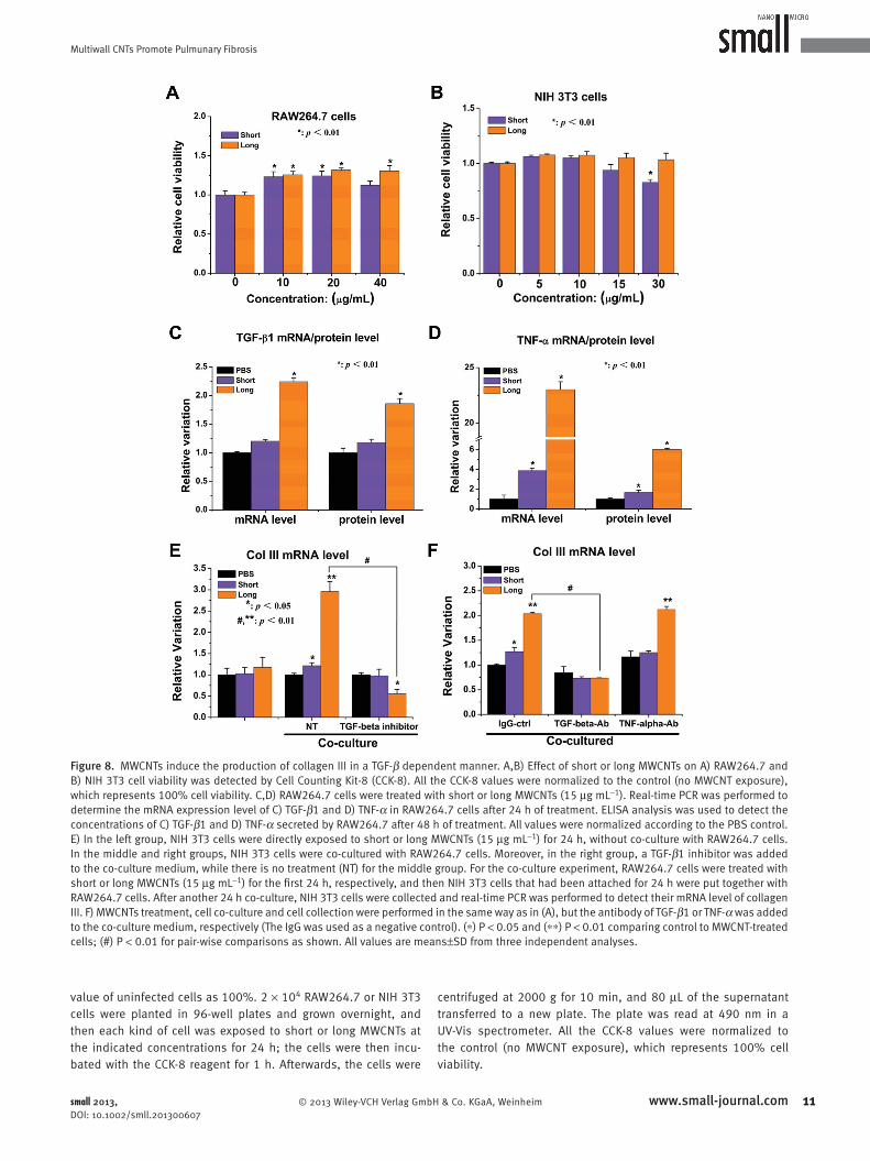
2.5. Long MWCNTs Induce Production of Collagen III in a TGF- β /Smad Dependent Manner In Vitro
It is believed that TGF- β secreted from
macrophages serves as a critical parac-
rine stimulus to fi broblasts to promote the
transformation of fi broblasts into myofi -
broblasts. Myofi broblasts secrete colla-
gens to deposit in the matrix, resulting in
fi brosis. Indeed, we detected the effect of
TGF- β 1 on NIH 3T3 cell proliferation.
As shown in Figure S2, Supporting Infor-
mation, TGF- β 1 increases the prolifera-
tion of NIH 3T3 cells dose-dependently.
Therefore, in order to further illustrate
the critical role that TGF- β secreted
from MWCNT-stimulated macrophages
could play in the promotion of fi broblast
proliferation and collagen deposition,
we similarly investigated whether TGF-
β 1 secreted from MWCNT-stimulated
RAW264.7 cells could affect the function
of NIH 3T3 fi broblast cells by co-culturing NIH 3T3 cells
with RAW264.7 cells in vitro.
We fi rst examined the effect of MWCNTs on RAW264.7
and NIH 3T3 cell viability. We found that long MWCNTs
caused no cytotoxicity to either RAW264.7 or NIH 3T3.
Moreover, it seems that long MWCNTs can promote prolif-
eration of RAW264.7. However, short MWCNTs downregu-
late cell viability of both RAW264.7 and NIH 3T3 at high
concentration ( Figure 8 A, B).
Next, we analyzed MWCNT-stimulated production of
TGF- β 1 and TNF- α in RAW264.7 cells by monitoring the
mRNA expression of TGF- β 1 and TNF- α after treatment
by short or long MWCNTs for 24 h. Real-time PCR results
showed that the mRNA levels of both TGF- β 1 and TNF- α
were more obviously upregulated by long MWCNT treat-
ments than by short MWCNTs (Figure 8 C,D). We also used
the ELISA assay to determine the effect of MWCNTs on the
levels of secreted TGF- β 1 and TNF- α in the culture medium
after 48 h of treatment. In agreement with the results from
the mRNA analysis, we found that RAW264.7 cells treated
, Weinheim small 2013, DOI: 10.1002/smll.201300607

Multiwall CNTs Promote Pulmunary Fibrosis
Figure 6 . MWCNTs enhanced TGF- β 1-stimulated Smad2 phosphorylation in vivo. A) Immunoblotting was used to explore the time-course of Smad2 phosphorylation in NIH 3T3 cells, which were stimulated by 10 ng mL − 1 TGF- β 1 in vitro for the indicated time. B) SH rats were exposed by intratracheal instillation to short or long MWCNTs (0.6 mg/rat) for 1, 7 and 30 days. Immunoblotting was also used to examine the Smad2 phosphorylation level in lung tissues of SH rats exposed to short or long MWCNTs for 7 days. C) Immunohistochemistry was used to determine Smad2 phosphorylation in lung tissues of SH rats exposed to short or long MWCNTs for the indicated time. Scale bar 50 μ m. D,E) Real-time PCR was performed for detecting the relative mRNA expressions of D) TGF- β 1/T β RII and E) Smad2/3/7 at 7 days postexposure.
with long MWCNTs produced more TGF- β 1 and TNF- α
than those treated with short MWCNTs (Figure 8 C,D).
Finally, we co-cultured NIH 3T3 cells with RAW264.7
cells that had been pre-treated with a nontoxic concentration
of MWCNTs for 24 h. After 24 h of co-culture, the mRNA
© 2013 Wiley-VCH Verlag Gmsmall 2013, DOI: 10.1002/smll.201300607
expression level of collagen III in NIH 3T3 fi broblast cells
was detected by real-time PCR. As shown in Figure 8 E, co-
culture with MWCNT-treated RAW264.7 cells signifi cantly
increased the expression of collagen III in NIH/3T3 cells.
Long MWCNTs caused a more signifi cant upregulation of
9www.small-journal.combH & Co. KGaA, Weinheim

P. Wang et al.
1
full papers
Figure 7 . MWCNTs enhanced TGF- β 1-stimulated Smad2 phosphorylation in vitro. NIH 3T3 cells were fi rst treated with short or long MWCNTs (15 μ g mL − 1 ) for 24 h, and subsequently stimulated by 20 ng mL − 1 TGF- β 1 for the indicated times. The time course of Smad2 phosphorylation was determined by immunoblotting.
collagen III than short MWCNTs. Moreover, we found that
the addition of TGF- β 1 inhibitor into the co-culture medium
signifi cantly inhibited the effect of long MWCNTs on col-
lagen III expression.
To further investigate the role of TGF- β 1 or TNF- α in
upregulating collagen III expression, we used a specifi c anti-
body to neutralize RAW264.7-secreted TGF- β 1 or TNF- α .
TGF- β 1 antibody inhibited the elevation of collagen III
expression when it was added to the co-culture medium, but
TNF- α antibody did not (Figure 8 F). Together, these in vitro
data suggest that TGF- β 1 secreted from MWCNT-stimulated
RAW264.7 cells plays a central role in regulating collagen III
expression.
Therefore, we propose the following framework for the
cellular and molecular mechanisms of MWCNT-caused pul-
monary fi brosis. As shown in Figure 9 , MWCNTs fi rst stimu-
late TGF- β 1 secretion in the lung by activation of alveolar
macrophages and subsequntly activate the TGF- β /Smad sign-
aling pathway in fi broblasts, which can then upregulate the
mRNA transcription of both ECM protease inhibitors and
collagen type I/III. Moreover, our studies suggest that tube
length acts as an important factor in MWCNT-induced pul-
monary fi brosis.
3. Conclusion
It is crucial to systematically understand how these inherent
and external factors infl uence the toxicity of carbon nano-
tubes so that their undesirable toxicity can be avoided. In
summary, this study complements previous knowledge of
the cellular and molecular mechanisms by which MWCNTs
induce fi brosis, as well as the “length-dependent theory” used
to explain CNT-caused harmful health effects. The fi brotic
effect of MWCNTs clearly depends on the length of the
MWCNTs. These data suggest that the activation of TGF- β /
Smad2/collagen III signal transduction is an important step
in the pulmonary fi brogenesis caused by long MWCNTs
(Figure 9 ). Nevertheless, further investigation is still required
to answer the question of whether MWCNT-activated TGF-
β 1 production will further mediate the EMT process of
epithelial cells in the lung. It will be interesting to fi nd out
whether MWCNTs could regulate TGF- β /Smad signaling
through their interactions with TGF- β receptors. Further-
more, it will be useful to have identifi ed the TGF- β /Smad2
signaling pathway as a source of potential and novel targets
0 www.small-journal.com © 2013 Wiley-VCH V
that can be exploited for the development of therapeutic and
preventive approaches to CNT-induced pulmonary fi brosis.
4. Experimental Section
Preparation and Characterization of MWCNTs : Two MWCNTs were purchased from Nanotech Port (Chengdu, China), which we called short or long MWCNTs based on their length. The length of short MWCNTs was 0.5–2 μ m, but for long MWCNTs, the length was 20–50 μ m. Their size and morphology was characterized by high-resolution transmission electron microscopy (HRTEM, Tecnai G2 F20 U-TWIN). Nanomaterial suspensions for experiments were prepared by weighing the materials into glass tubes and diluting them to 1.5 mg mL − 1 stock solution with 1% Pluronic F108 solu-tion in phosphate buffered saline (PBS), which was then soni-cated for 1 h (500 W) at 30 ° C. For optimal suspension, the stock solutions were further sonicated for 30 min just before adminis-tration to cells. The suspensions of 1.5 mg mL − 1 MWCNTs were further diluted into fresh medium to the fi nal concentrations as required, and then old media were carefully removed and replaced with new media containing the fi nal concentrations of MWCNTs.
Animals : Male spontaneously hypertensive (SH) rats (derived from Wistar Kyoto rats by segregation of the hypertensive trait and inbreeding), 11–12 weeks old and 220–250 g body weight, were obtained from Vital River Laboratory Animal Technology Co., Ltd, Beijing. Animals were housed as previously described. [ 29 ] In our present study, SH rats were randomized by body weight into groups of PBS, short MWCNTs, and long MWCNTs (6 rats/group). SH rats were exposed to PBS or PBS-suspended short or long MWCNT particles (0.6 mg/rat) using a non-surgical intratracheal instillation once a day for two consecutive days. Then, the rats were killed 1 day, 7 days and 30 days following the last exposure. All animal studies, animal care, and use were approved according to local guidelines. All experiments with rats followed the guide-lines for experimental animals and were approved by the animal welfare committee of Peking University.
Cell Culture and Co-Culture Experiments : The mouse leukemic monocyte macrophage cell line (RAW264.7) and mouse embryonic fi broblast cell line (NIH 3T3) were purchased from American Type Culture Collection (ATCC, Manassas, VA). RAW264.7 cells were cul-tured in Dulbecco's modifi ed Eagle's medium (DMEM) with 10% heat-inactivated fetal bovine serum. NIH 3T3 cells were main-tained in DMEM with 10% fetal bovine serum. Both media were supplemented with 2 m M L-glutamine, 100 U mL − 1 penicillin and 100 μ g mL − 1 streptomycin. Cells were grown at 37 ° C in a humidi-fi ed 95% air/5% CO 2 incubator. For co-culture experiments, we used 24 mm Transwell® with a 0.4 μ m Pore Polyester Membrane Insert from Corning Company. RAW264.7 cells seeded in the well below were fi rst treated with short or long MWCNTs (15 μ g mL − 1 ) for 24 h, and then NIH 3T3 cells that had attached on the top of the insert for 24 h were co-cultured with RAW264.7 cells in the Transwell 6-well plate system for another 24 h.
Cell Viability Assay : Cell viability was determined using a cell counting kit-8 (Dojindo), in which 2-(2-methoxy-4-nitrophenyl)-3-(4-nitrophenyl)-5-(2,4-disulfophenyl)-2H-tetrazolium monoso-dium salt (WST-8) was used as a substrate. The relative number of surviving cells was determined in duplicate by estimating the
erlag GmbH & Co. KGaA, Weinheim small 2013, DOI: 10.1002/smll.201300607
Multiwall CNTs Promote Pulmunary Fibrosis
Figure 8 . MWCNTs induce the production of collagen III in a TGF- β dependent manner. A,B) Effect of short or long MWCNTs on A) RAW264.7 and B) NIH 3T3 cell viability was detected by Cell Counting Kit-8 (CCK-8). All the CCK-8 values were normalized to the control (no MWCNT exposure), which represents 100% cell viability. C,D) RAW264.7 cells were treated with short or long MWCNTs (15 μ g mL − 1 ). Real-time PCR was performed to determine the mRNA expression level of C) TGF- β 1 and D) TNF- α in RAW264.7 cells after 24 h of treatment. ELISA analysis was used to detect the concentrations of C) TGF- β 1 and D) TNF- α secreted by RAW264.7 after 48 h of treatment. All values were normalized according to the PBS control. E) In the left group, NIH 3T3 cells were directly exposed to short or long MWCNTs (15 μ g mL − 1 ) for 24 h, without co-culture with RAW264.7 cells. In the middle and right groups, NIH 3T3 cells were co-cultured with RAW264.7 cells. Moreover, in the right group, a TGF- β 1 inhibitor was added to the co-culture medium, while there is no treatment (NT) for the middle group. For the co-culture experiment, RAW264.7 cells were treated with short or long MWCNTs (15 μ g mL − 1 ) for the fi rst 24 h, respectively, and then NIH 3T3 cells that had been attached for 24 h were put together with RAW264.7 cells. After another 24 h co-culture, NIH 3T3 cells were collected and real-time PCR was performed to detect their mRNA level of collagen III. F) MWCNTs treatment, cell co-culture and cell collection were performed in the same way as in (A), but the antibody of TGF- β 1 or TNF- α was added to the co-culture medium, respectively (The IgG was used as a negative control). ( ∗ ) P < 0.05 and ( ∗ ∗ ) P < 0.01 comparing control to MWCNT-treated cells; (#) P < 0.01 for pair-wise comparisons as shown. All values are means ± SD from three independent analyses.
value of uninfected cells as 100%. 2 × 10 4 RAW264.7 or NIH 3T3 cells were planted in 96-well plates and grown overnight, and then each kind of cell was exposed to short or long MWCNTs at the indicated concentrations for 24 h; the cells were then incu-bated with the CCK-8 reagent for 1 h. Afterwards, the cells were
© 2013 Wiley-VCH Verlag Gmsmall 2013, DOI: 10.1002/smll.201300607
centrifuged at 2000 g for 10 min, and 80 μ L of the supernatant transferred to a new plate. The plate was read at 490 nm in a UV-Vis spectrometer. All the CCK-8 values were normalized to the control (no MWCNT exposure), which represents 100% cell viability.
11www.small-journal.combH & Co. KGaA, Weinheim

P. Wang et al.full papers
Figure 9 . MWCNTs promote pulmonary fi brosis through the TGF- β /Smad signaling pathway.
Preparation of Bronchoalveolar Lavage Fluid : The right lung was used to obtain BAL fl uid after ligation of the left bronchus. The right lung was lavaged three times with a specifi c volume (27 mL kg − 1 body weight) of ice-cold phosphate-buffered saline (PBS, pH 7.4). The recovered BAL fl uid was placed on ice. One ali-quot of the recovered lavage fl uid was centrifuged (400 g, 10 min, 4 ° C), and then the supernatant fl uid was collected and imme-diately analyzed for TGF- β 1 and TNF- α using the corresponding ELISA kits, and the pellets were collected for observation of MWCNT uptake in alveolar macrophages, which were stained with hematoxylin.
Histopathological and Immunohistochemistry Analysis : Unlav-aged left lungs of each group were dissected and immediately fi xed with 10% phosphate buffered formalin for 24–72 h. Lung tissues were then dehydrated, embedded in paraffi n, and cut into 4 μ m-thick slices. Sections were stained with hematoxylin and eosin (H&E) or Sirius Red to detect morphological changes and collagen deposition. For immunohistochemistry, slices were depar-affi nized with xylene. Endogenous peroxidase was blocked with 0.3% H 2 O 2 for 15 min. After treatment with blocking goat serum for 15 min, sections were incubated overnight with a collagen III anti-body (1:200), an FSP-1 antibody (1:200) and a phospho-Smad2 antibody (1:200) and then with a biotinylated-link secondary anti-body and peroxidase-labeled streptavidin followed by a revelation step with diaminobenzidine (substrate of peroxidase) and counter-staining with Mayer's hematoxylin. Slices were analyzed under a microscope (Leica DM4000M, Germany).
Real-Time Quantitative PCR Analysis : Total RNA was isolated from cells or lung tissues using TRIzol (Invitrogen, Carlslab, CA, USA) and converted to cDNA by M-MLV reverse transcriptase (Promega, Madison, WI, USA) according to the manufacturer's instructions. The resulting cDNA samples were analyzed by quan-titative real-time PCR (Eppendorf, Hauppauge, New York, USA), using the SYBR Green assay. Relative levels of mRNA expression were normalized to β -actin expression for each gene. The following genes were measured: TNF- α , TGF- β 1, TGF- β receptor II, Smad2, Smad3, Smad 7, TIMP-1, PAI-1, Col I, Col III, and β -actin.
ELISA and Immunoblotting : TGF- β 1 and TNF- α levels in BALF or cell culture medium were quantifi ed using DuoSet ELISA kits (R&D Systems, Minneapolis, MN) in accordance with the manufacturer's instructions. For immunoblotting, the cells were lysed in radioim-munoprecipitation assay (RIPA) buffer (50 m M Tris-HCl, pH 8.0,
12 www.small-journal.com © 2013 Wiley-VCH Verlag GmbH & Co. KGaA
150 m M NaCl, 1% Nonidet P-40, 0.5% sodium deoxycholate, 0.1% SDS, 2 m M EDTA, 1 m M NaVO 4 , 10 m M NaF, and protease inhibitors), and the protein concentration in the lysates was determined by a spectrophotometer. Equal amounts of the lysates were subjected to SDS-polyacrylamide gel electrophoresis (PAGE) and the immunoblotting was per-formed with various primary antibodies and secondary antibodies conjugated to horse-radish peroxidase. Proteins were visualized by chemiluminescence.
Statistical Analysis : Data are represented as mean and SEM. Statistical analysis of data was carried out using Student's t test or the χ 2 test. A P value of less than 0.05 was consid-ered to be a signifi cant difference.
Supporting Information
Supporting Information is available from the Wiley Online Library or from the author.
Acknowledgements
Authors P. Wang and X. Nie contributed equally to this work. This work was supported by grants from the National Basic Research Program of China (973 Programs 2011CB933401, 2012CB934003 and 2010CB934004), National Major Scientifi c Instruments Devel-opment Project (2011YQ03013406), the National Natural Science Foundation of China (10975040 and 31000337), German Federal Ministry of Education and Research (BMBF 0315773A) and the European Commission through the Seventh Framework Programme for Research and Technological Development (FP7-MARINA; Grant agreement 263215).
[ 1 ] R. H. Baughman , A. A. Zakhidov , W. A. de Heer , Science 2002 , 297 , 787 .
[ 2 ] P. Avouris , Z. Chen , V. Perebeinos , Nat. Nanotechnol. 2007 , 2 , 605 .
[ 3 ] A. Bianco , K. Kostarelos , M. Prato , Curr. Opin. Chem. Biol. 2005 , 9 , 674 .
[ 4 ] K. Donaldson , R. Aitken , L. Tran , V. Stone , R. Duffi n , G. Forrest , A. Alexander , Toxicol. Sci. 2006 , 92 , 5 .
[ 5 ] J. W. Card , D. C. Zeldin , J. C. Bonner , E. R. Nestmann , Am. J. Physiol. Lung Cell Mol. Physiol. 2008 , 295 , L400 .
[ 6 ] Y. Zhao , G. Xing , Z. Chai , Nat. Nanotechnol. 2008 , 3 , 191 . [ 7 ] L. Wang , S. Luanpitpong , V. Castranova , W. Tse , Y. Lu ,
V. Pongrakhananon , Y. Rojanasakul , Nano Lett . 2011 , 11 , 2796 . [ 8 ] L. Meng , A. Jiang , R. Chen , C. Z. Li , L. Wang , Y. Qu , P. Wang , Y. Zhao ,
C. Chen , Toxicology 2012 , DOI: 10.1016/j.tox.2012.11.011. [ 9 ] L. Meng , R. Chen , A. Jiang , L. Wang , P. Wang , C. Z. Li , R. Bai ,
Y. Zhao , H. Autrup , C. Chen , Small 2012 , DOI: 10.1002/smll.201201388.
[ 10 ] B. Fubini , I. Fenoglio , M. Tomatis , F. Turci , Nanomedicine (London, U.K.) 2011 , 6 , 899 .
, Weinheim small 2013, DOI: 10.1002/smll.201300607

Multiwall CNTs Promote Pulmunary Fibrosis
[ 11 ] A. A. Shvedova , A. Pietroiusti , B. Fadeel , V. E. Kagan , Toxicol. Appl. Pharmacol. 2012 , 261 , 121 .
[ 12 ] T. A. Wynn , J. Pathol. 2008 , 214 , 199 . [ 13 ] E. J. Park , J. Roh , S. N. Kim , M. S. Kang , Y. A. Han , Y. Kim , J. T. Hong ,
K. Choi , Arch. Toxicol. 2011 , 85 , 1121 . [ 14 ] A. A. Shvedova , E. R. Kisin , R. Mercer , A. R. Murray ,
V. J. Johnson , A. I. Potapovich , Y. Y. Tyurina , O. Gorelik , S. Arepalli , D. Schwegler-Berry , A. F. Hubbs , J. Antonini , D. E. Evans , B. K. Ku , D. Ramsey , A. Maynard , V. E. Kagan , V. Castranova , P. Baron , Am. J. Physiol. Lung Cell Mol. Physiol. 2005 , 289 , L698 .
[ 15 ] C. W. Lam , J. T. James , R. McCluskey , R. L. Hunter , Toxicol. Sci. 2004 , 77 , 126 .
[ 16 ] D. B. Warheit , B. R. Laurence , K. L. Reed , D. H. Roach , G. A. Reynolds , T. R. Webb , Toxicol. Sci. 2004 , 77 , 117 .
[ 17 ] J. Muller , F. Huaux , N. Moreau , P. Misson , J. F. Heilier , M. Delos , M. Arras , A. Fonseca , J. B. Nagy , D. Lison , Toxicol. Appl. Phar-macol. 2005 , 207 , 221 .
[ 18 ] C. Ronzani , C. Spiegelhalter , J. L. Vonesch , L. Lebeau , F. Pons , Arch. Toxicol. 2012 , 86 , 137 .
[ 19 ] X. Wang , T. Xia , S. A. Ntim , Z. Ji , S. Lin , H. Meng , C. H. Chung , S. George , H. Zhang , M. Wang , N. Li , Y. Yang , V. Castranova , S. Mitra , J. C. Bonner , A. E. Nel , ACS Nano 2011 , 5 , 9772 .
[ 20 ] X. Wang , T. Xia , M. C. Duch , Z. Ji , H. Zhang , R. Li , B. Sun , S. Lin , H. Meng , Y. P. Liao , M. Wang , T. B. Song , Y. Yang , M. C. Hersam , A. E. Nel , Nano Lett. 2012 , 12 , 3050 .
[ 21 ] X. Wang , T. Xia , S. A. Ntim , Z. Ji , S. George , H. Meng , H. Zhang , V. Castranova , S. Mitra , A. E. Nel , ACS Nano 2010 , 4 , 7241 .
[ 22 ] J. F. Du , C. C. Ge , Y. Lu , R. Bai , D. H. Li , Y. L. Yang , L. F. Liao , C. Y. Chen , J. Nanosci. Nanotechnol. 2011 , 11 , 10102 .
[ 23 ] C. C. Ge , J. F. Du , L. N. Zhao , L. M. Wang , Y. Liu , D. H. Li , Y. L. Yang , R. H. Zhou , Y. L. Zhao , Z. F. Chai , C. Y. Chen , Proc. Natl. Acad. Sci. USA 2011 , 108 , 16968 .
[ 24 ] C. Ge , Y. Li , J. J. Yin , Y. Liu , L. Wang , Y. Zhao , C. Chen , NPG Asia Mater. 2012 , 4 , e32 .
[ 25 ] H. Nagai , Y. Okazaki , S. H. Chew , N. Misawa , Y. Yamashita , S. Akatsuka , T. Ishihara , K. Yamashita , Y. Yoshikawa , H. Yasui , L. Jiang , H. Ohara , T. Takahashi , G. Ichihara , K. Kostarelos , Y. Miyata , H. Shinohara , S. Toyokuni , Proc. Natl. Acad. Sci. USA 2011 , 108 , E1330 .
[ 26 ] J. Palomaki , E. Valimaki , J. Sund , M. Vippola , P. A. Clausen , K. A. Jensen , K. Savolainen , S. Matikainen , H. Alenius , ACS Nano 2011 , 5 , 6861 .
© 2013 Wiley-VCH Verlag Gmbsmall 2013, DOI: 10.1002/smll.201300607
[ 27 ] C. A. Poland , R. Duffi n , I. Kinloch , A. Maynard , W. A. H. Wallace , A. Seaton , V. Stone , S. Brown , W. MacNee , K. Donaldson , Nat. Nanotechnol. 2008 , 3 , 423 .
[ 28 ] K. Donaldson , F. A. Murphy , R. Duffi n , C. A. Poland , Part. Fibre. Toxicol. 2010 , 7 , 5 .
[ 29 ] C. Ge , L. Meng , L. Xu , R. Bai , J. Du , L. Zhang , Y. Li , Y. Chang , Y. Zhao , C. Chen , Nanotoxicology 2011 , 6 , 526 .
[ 30 ] K. Yamashita , Y. Yoshioka , K. Higashisaka , Y. Morishita , T. Yoshida , M. Fujimura , H. Kayamuro , H. Nabeshi , T. Yamashita , K. Nagano , Y. Abe , H. Kamada , Y. Kawai , T. Mayumi , T. Yoshikawa , N. Itoh , S. Tsunoda , Y. Tsutsumi , Infl ammation 2010 , 33 , 276 .
[ 31 ] Y. Liu , Y. Zhao , B. Sun , C. Chen , Acc. Chem. Res. 2013 , 46 , 702 . [ 32 ] R. Visse , H. Nagase , Circ. Res. 2003 , 92 , 827 . [ 33 ] J. P. Irigoyen , P. Munoz-Canoves , L. Montero , M. Koziczak ,
Y. Nagamine , Cell Mol. Life Sci. 1999 , 56 , 104 . [ 34 ] J. Harslund , O. L. Nielsen , N. Brunner , H. Offenberg , Am. J.
Physiol. Regul. Integr. Comp. Physiol. 2007 , 293 , R1630 . [ 35 ] A. Erdely , T. Hulderman , R. Salmen , A. Liston , P. C. Zeidler-Erdely ,
D. Schwegler-Berry , V. Castranova , S. Koyama , Y. A. Kim , M. Endo , P. P. Simeonova , Nano Lett. 2009 , 9 , 36 .
[ 36 ] X. Wang , P. Katwa , R. Podila , P. Chen , P. C. Ke , A. M. Rao , D. M. Walters , C. J. Wingard , J. M. Brown , Part. Fibre. Toxicol. 2011 , 8 , 24 .
[ 37 ] P. Ravichandran , S. Baluchamy , R. Gopikrishnan , S. Biradar , V. Ramesh , V. Goornavar , R. Thomas , B. L. Wilson , R. Jeffers , J. C. Hall , G. T. Ramesh , J. Biol. Chem. 2011 , 286 , 29725 .
[ 38 ] X. Li , Y. Hu , Z. Jin , H. Jiang , J. Wen , Toxicol. Mech. Methods 2009 , 19 , 51 .
[ 39 ] Y. Li , Y. Liu , Y. J. Fu , T. T. Wei , L. Le Guyader , G. Gao , R. S. Liu , Y. Z. Chang , C. Y. Chen , Biomaterials 2012 , 33 , 402 .
[ 40 ] R. W. Pelton , M. D. Johnson , E. A. Perkett , L. I. Gold , H. L. Moses , Am. J. Respir. Cell Mol. Biol. 1991 , 5 , 522 .
[ 41 ] X. H. Feng , R. Derynck , Annu. Rev. Cell Dev. Biol. 2005 , 21 , 659 . [ 42 ] J. Massague , Cell 2008 , 134 , 215 . [ 43 ] P. Lonn , A. Moren , E. Raja , M. Dahl , A. Moustakas , Cell Res. 2009 ,
19 , 21 . [ 44 ] S. H. Park , J. Biochem. Mol. Biol. 2005 , 38 , 9 . [ 45 ] Y. Zhang , Q. Mu , H. Zhou , K. Vrijens , M. F. Roussel , G. Jiang ,
B. Yan , Cell Death Dis. 2012 , 3 , e308 .
Received: February 26, 2013Published online:
13www.small-journal.comH & Co. KGaA, Weinheim