morphological characterization of the blood cells in the endangered sicilian endemic pond turtle,...
TRANSCRIPT
This article was downloaded by: [Universita di Palermo], [Federico Marrone]On: 25 July 2014, At: 08:08Publisher: Taylor & FrancisInforma Ltd Registered in England and Wales Registered Number: 1072954 Registered office: Mortimer House,37-41 Mortimer Street, London W1T 3JH, UK
Italian Journal of ZoologyPublication details, including instructions for authors and subscription information:http://www.tandfonline.com/loi/tizo20
Morphological characterization of the blood cells inthe endangered Sicilian endemic pond turtle, Emystrinacris (Testudines: Emydidae)V. Arizzaabc, D. Russoac, F. Marronea, F. Saccoa & M. Arculeoa
a Dipartimento STEBICEF, Università degli Studi di Palermo, Palermo, Italyb Dipartimento Key-SET, Istituto Euro Mediterraneo di Scienza e Tecnologia, Palermo, Italyc Applied Biodiversity Laboratory, Dipartimento STEBICEF, Università di Palermo, Palermo,ItalyPublished online: 21 Jul 2014.
To cite this article: V. Arizza, D. Russo, F. Marrone, F. Sacco & M. Arculeo (2014): Morphological characterization of the bloodcells in the endangered Sicilian endemic pond turtle, Emys trinacris (Testudines: Emydidae), Italian Journal of Zoology, DOI:10.1080/11250003.2014.938371
To link to this article: http://dx.doi.org/10.1080/11250003.2014.938371
PLEASE SCROLL DOWN FOR ARTICLE
Taylor & Francis makes every effort to ensure the accuracy of all the information (the “Content”) containedin the publications on our platform. However, Taylor & Francis, our agents, and our licensors make norepresentations or warranties whatsoever as to the accuracy, completeness, or suitability for any purpose of theContent. Any opinions and views expressed in this publication are the opinions and views of the authors, andare not the views of or endorsed by Taylor & Francis. The accuracy of the Content should not be relied upon andshould be independently verified with primary sources of information. Taylor and Francis shall not be liable forany losses, actions, claims, proceedings, demands, costs, expenses, damages, and other liabilities whatsoeveror howsoever caused arising directly or indirectly in connection with, in relation to or arising out of the use ofthe Content.
This article may be used for research, teaching, and private study purposes. Any substantial or systematicreproduction, redistribution, reselling, loan, sub-licensing, systematic supply, or distribution in anyform to anyone is expressly forbidden. Terms & Conditions of access and use can be found at http://www.tandfonline.com/page/terms-and-conditions
Morphological characterization of the blood cells in the endangeredSicilian endemic pond turtle, Emys trinacris (Testudines: Emydidae)
V. ARIZZA1,2,3, D. RUSSO1,3, F. MARRONE1, F. SACCO1, & M. ARCULEO1
1Dipartimento STEBICEF, Università degli Studi di Palermo, Palermo, Italy, 2Dipartimento Key-SET, Istituto EuroMediterraneo di Scienza e Tecnologia, Palermo, Italy, and 3Applied Biodiversity Laboratory, Dipartimento STEBICEF,Università di Palermo, Palermo, Italy
(Received 27 March 2014; accepted 13 June 2014)
AbstractIn this study, measurements of morphological parameters, sizes and frequencies of peripheral blood cells (erythrocytes,leukocytes, thrombocytes) on blood smear preparation devices stained with May-Grünwald stain were evaluated for bothsexes in 20 Emys trinacris (Testudines: Emydidae) specimens. Erythrocytes were higher in male than in female specimens.The leukocyte of E. trinacris contains eosinophil, basophil, monocyte, heterophil and lymphocyte. The eosinophil was higherin males than in females whereas lymphocytes were higher in females than in males. The erythrocyte morphologicalparameters (EL [erythrocyte length], EW [erythrocyte width], L/W [length/width], ES [erythrocyte size]) were comparedwith the same data from Emys orbicularis s.l, and from species belonging to other chelonian genera. The erythrocyte size didnot vary within the studied Palearctic Emys taxa, whereas it proved to differ from that observed in other chelonians.
Keywords: Emys trinacris, blood smear, blood cell morphology, Trachemys scripta elegans
Introduction
Emys trinacris (Fritz et al. 2005) (Testudines:Emydidae) is a Sicilian endemic pond turtle (Fritzet al. 2005); although it is morphologically close toEmys orbicularis s.l. (Fritz et al. 2006), molecular taxo-nomic studies have unambiguously revealed the pre-sence of significant differences between the twospecies, which are adelphotaxa (Fritz et al. 2007;Pedall et al. 2011; Stuckas et al. 2014). Consideringthe biogeographical and evolutionary importance ofthis species and the drastic reduction of its populationscaused by habitat destruction, pollution, and patho-gens, in 2013,E. trinacriswas listed in the InternationalUnion for Conservation of Nature (IUCN) Red List as“Endangered” (EN) (Rhodin et al. 2009).
To date, few studies have focused on the biologyof this endemic species, and nothing is known aboutthe hematologic blood characterization (HBC) of thespecies and on the health status of the wild popula-tions of E. trinacris.
In the literature, HBC has been used successfullyto diagnose chelonian diseases and to assess the
physiological status of wild turtle populations(Duguy 1967; Dessauer 1970; Frye 1991;Campbell 1996; Stein 1996). This approach iswidely used because the HBC is a minimally invasivetool that allows health evaluations, especially in rela-tion to determining potential effects associated withstress factors such as pollution, disease, invasion byexotic species, etc. In order to be soundly usable, thereference evaluations have to be performed onhealthy animals (Nagy & Medica 1986; Deem et al.2006).Blood cell parameters of reptiles may be influ-
enced by several factors, such as age, sex, seasonal-ity, reproduction, nutritional status andenvironmental parameters such as temperature, sali-nity, oxygen and light (Dessauer 1970; Duguy 1970;Frye 1991; Wilkinson 2003; Tavares-Dias et al.2009; Yilmaz & Tosunoglu 2010; Gu et al. 2011;Scheelings & Jessop 2011; Tosunoglu et al. 2011;Scheelings & Rafferty 2012); these parameters canvary through the annual cycle and throughout the lifeof the individuals. Studies that describe chelonian
Correspondence: V. Arizza, Applied Biodiversity Laboratory, Dipartimento STEBICEF, Università di Palermo, Via Archirafi, 18, 90123 Palermo, Italy. Tel:+39 91 23891804. Fax: +39 91 23860855. Email: [email protected]
Italian Journal of Zoology, 2014, 1–10http://dx.doi.org/10.1080/11250003.2014.938371
© 2014 Unione Zoologica Italiana
Dow
nloa
ded
by [
Uni
vers
ita d
i Pal
erm
o], [
Fede
rico
Mar
rone
] at
08:
08 2
5 Ju
ly 2
014
blood cells are rare and the data are often in contra-diction because of the lack of standard criteria usedto categorize blood cells (Work et al. 1998). Variousauthors described circulating blood cells of differentamphibian and reptile species; moreover, there aremany chelonian species for which blood cell mor-phology and reference values are still unknown orimprecise. Descriptions of the morphologic charac-teristics of blood cells of pond turtles are limited andfail to standardize the parameters of the HBC (Metinet al. 2006; Rossini et al. 2012).
In order to perform conservation activities for thepopulations of E. trinacris, reference studies on bio-logical parameters are necessary. Moreover, as agood practice, any action taken for the protectionand the conservation of species in danger of extinc-tion cannot ignore the full knowledge of their biol-ogy. For this reason, the blood cell parameters of theSicilian pond turtle E. trinacris have been documen-ted by analysing blood samples from free-livingmales and females. Moreover, blood cell parameterdata obtained from E. trinacris were compared with
those available from E. orbicularis s.l. Other compar-isons were performed with those of the Americanemydid Trachemys scripta elegans (Wied, 1839).The present study describes for the first time the
blood cell parameters in E. trinacris obtained fromthe turtles under natural conditions, and aims toestablish the blood cell parameter reference valuesnecessary for the evaluation of the health status ofindividuals from wild populations.
Materials and methods
Sampling area
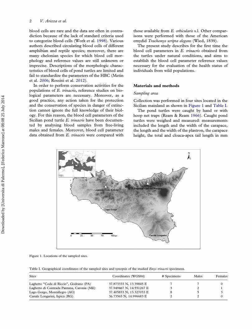
Collection was performed in four sites located in theSicilian mainland as shown in Figure 1 and Table I.The pond turtles were caught by hand or with
hoop net traps (Ream & Ream 1966). Caught pondturtles were weighed and measured: measurementsincluded the length and the width of the carapace,the length and the width of the plastron, the carapaceheight, the total and cloaca-apex tail length in mm
Figure 1. Locations of the sampled sites.
Table I. Geographical coordinates of the sampled sites and synopsis of the studied Emys trinacris specimens.
Sites Coordinates (WGS84) # Specimens Males Females
Laghetto “Coda di Riccio”, Godrano (PA) 37.873333 N, 13.39845 E 7 7 0Laghetto di Contrada Pantana, Caronia (ME) 37.949467 N, 14.551267 E 3 2 1Lago Gorgo, Montallegro (AG) 37.405833 N, 13.327033 E 8 5 3Canale Longarini, Ispica (RG) 36.73565 N, 14.996683 E 2 2 0
2 V. Arizza et al.
Dow
nloa
ded
by [
Uni
vers
ita d
i Pal
erm
o], [
Fede
rico
Mar
rone
] at
08:
08 2
5 Ju
ly 2
014
(with a caliper, to the nearest 0.5 mm) (Fritz et al.2005), and the body mass in grams for each speci-men (Zuffi & Gariboldi 1995; Zuffi et al. 2006). Tenblood samples of Trachemys scripta elegans wereobtained in September 2013 from specimens housedin the Botanical Garden of the department ofSTEBICEF, University of Palermo.
Blood sampling, cell morphology and counts
Blood samples were obtained from the dorsal coccy-geal vessel via heparinized glass capillaries (Hutchison& Szarski 1965; Szarski 1968). After obtaining bloodsamples, the animals were immediately released totheir natural environments. For each individual, oneto three capillaries were collected.
Blood smears were prepared in situ. A blood dropwas smeared on a glass slide and air-dried. Thesample was then fixed in methanol and stained bythe Pappenheim method (May-Grünwald + Giemsa-Romanowsky staining diluted 1:10 in buffered water,pH 7) for 20 min, and washed in running tap waterfor 2 minutes. One hundred erythrocytes and 30each of thrombocytes, eosinophils, basophils, lym-phocytes and monocytes were measured under amicroscope (Leica DMRE) equipped with a digitalcamera (Leica DCF420 C). In each smear wererecorded lengths (L) and widths (W) of 100 ran-domly chosen erythrocytes as well as nuclear lengths(NL) and nuclear widths (NW). Erythrocyte sizes(ES) and their nucleus sizes (NS) were computedfrom the following equations (Arikan & Cicek 2010):
ES ¼ ðEL � EW� πÞ=4 (1)
NS ¼ ðNL �NW� πÞ=4 (2)
Cells and nuclear shapes were compared with EL/EW and NL/NW ratios, and nucleus/cytoplasmwith NS/ES ratio. In addition, from the bloodsmears of each species, measurements of leuco-cytes (lymphocytes, monocytes, heterophils, eosi-nophils, basophils) and thrombocytes (TL, TW)
were also taken to determine their sizes and com-puted from the following formula:
A ¼πr2 (3)
Statistical analyses
Hematological variables (number of cells or dimen-sions) were summarized as mean, standard deviation(SD), standard error of the mean (SE) and range.We used analysis of the t test for a comparison of thesexes.
Results
Sampling and measurements
A total of 20 wild pond turtles Emys trinacris, 16 maleand four female, were sampled in Sicily from 2012 to2013 (Table I). The average body weights for themale and female turtles used for the study were355.31 ± 154.78 g and 504.33 ± 75.5 g, respec-tively. The measured carapace lengths were12.77 ± 2.15 cm and 14.08 ± 1.11 cm for maleand female specimens, respectively.Those samples which showed wounds or epiphytes
or possessed parasites in the blood were not includedin the analyses.
Emys trinacris blood cells
Differential blood cell count of the peripheral bloodof E. trinacris was carried out using blood smearsstained with May-Grünwald Giemsa observedunder a light microscope equipped with a digitalcamera. The leukocyte types recognized in Emystrinacris correspond with those found in other speciesof the family Emydidae (Table II). In particular,seven cell types were identified: (a) nucleated ery-throcytes, (b) eosinophils, (c) basophils, (d) mono-cytes, (e) thrombocytes, (f) heterophils and (g)lymphocytes (Figure 2). Significant differences incell size were not observed in either sex. Males pos-sessed significantly higher numbers of red blood cells
Table II. Leukocytes in turtle species of the Emydidae family.
Species Leukocyte type Reference
Lymphocyte Heterophils Eosinophils Basophils Monocyte
Emys orbicularis galloitalica + + + + + (Metin et al. 2006)+ + + + + (Yilmaz & Tosunoglu 2010)
Graptemys gibbonsi + + + + + (Perpiñán et al. 2008)Pseudemys rubriventris + + + + + (Innis et al. 2007)Clemmys muhlenbergii + + + + + (Brenner et al. 2002)Chrysemys picta + + + + + (Schwanz et al. 2011)
Hematological blood characterization of Emys trinacris 3
Dow
nloa
ded
by [
Uni
vers
ita d
i Pal
erm
o], [
Fede
rico
Mar
rone
] at
08:
08 2
5 Ju
ly 2
014
(p < 0.05) and eosinophils (p < 0.01), while femalesshowed a significantly (p < 0.05) higher percentageof lymphocytes (Table I). For other cell types,although their number varied between the genders,no significant differences were found.
Erythrocyte morphology
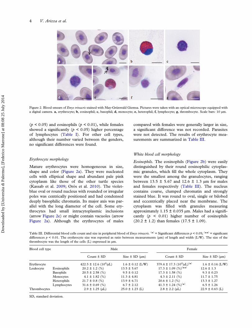
Mature erythrocytes were homogeneous in size,shape and color (Figure 2a). They were nucleatedcells with elliptical shape and abundant pale pinkcytoplasm like those of the other turtle species(Kassab et al. 2009; Orós et al. 2010). The violet-blue oval or round nucleus with rounded or irregularpoles was centrically positioned and had condenseddeeply basophilic chromatin. Its major axis was par-allel with the long diameter of the cell. Some ery-throcytes had small intracytoplasmic inclusions(arrow Figure 2e) or might contain vacuoles (arrowFigure 2a). Although the erythrocytes of males
compared with females were generally larger in size,a significant difference was not recorded. Parasiteswere not detected. The results of erythrocyte mea-surements are summarized in Table III.
White blood cell morphology
Eosinophils. The eosinophils (Figure 2b) were easilydistinguished by their round eosinophilic cytoplas-mic granules, which fill the whole cytoplasm. Theywere the smallest among the granulocytes, rangingbetween 13.5 ± 5.67 and 12.6 ± 1.3 µm for malesand females respectively (Table III). The nucleuscontains coarse, clumped chromatin and stronglystained blue. It was round to oval, single or bilobedand eccentrically placed near the membrane. Thecytoplasm was filled with granules measuringapproximately 1.15 ± 0.035 µm. Males had a signifi-cantly (p < 0.01) higher number of eosinophils(20.2 ± 1.2) than females (17.5 ± 1.09).
Figure 2. Blood smears of Emys trinacris stained with May-Grünwald Giemsa. Pictures were taken with an optical microscope equipped witha digital camera. a, erythrocyte; b, eosinophil; c, basophil; d, monocyte; e, heterophil; f, lymphocyte; g, thrombocyte. Scale bars: 10 µm.
Table III. Differential blood cells count and size in peripheral blood of Emys trinacris. (*) = Significant differences p < 0.05; (**) = significantdifferences p < 0.01. The erythrocyte size was reported as ratio between measurements (µm) of length and width (L/W). The size of thethrombocyte was the length of the cells (L) expressed in µm.
Blood cell type Male Female
Count ± SD Size ± SD (µm) Count ± SD Size ± SD (µm)
Erythrocyte 422.5 ± 12.6 (104/µL) 1.6 ± 0.12 (L/W) 379.4 ± 17.3 (104/µL)(*) 1.6 ± 0.14 (L/W)Leukocyte Eosinophils 20.2 ± 1.2 (%) 13.5 ± 5.67 17.3 ± 1.09 (%)(**) 12.6 ± 1.3
Basophils 20.5 ± 2.58 (%) 9.5 ± 0.12 17.3 ± 1.58 (%) 9.3 ± 0.23Monocytes 4.1 ± 1.82 (%) 11.3 ± 4.81 4.3 ± 2.11 (%) 11.7 ± 1.75Heterophils 21.7 ± 0.8 (%) 13.9 ± 6.71 20.6 ± 1.2 (%) 13.3 ± 1.27Lymphocytes 31.6 ± 0.69 (%) 6.7 ± 2.12 41.3 ± 1.24 (%)(*) 6.5 ± 1.26
Thrombocytes 2.9 ± 1.25 (µL) 25.0 ± 1.23 (L) 2.8 ± 2.2 (µL) 22.9 ± 0.63 (L)
SD, standard deviation.
4 V. Arizza et al.
Dow
nloa
ded
by [
Uni
vers
ita d
i Pal
erm
o], [
Fede
rico
Mar
rone
] at
08:
08 2
5 Ju
ly 2
014
Basophils. The basophils (Figure 2c) were presentwith a percentage of 5.8 ± 0.45 and 5.4 ± 0.77respectively for male and female turtles. They weresmall cells, about 9.49 µm, without significant differ-ences between the genders. They are easily identifiedby their deeply stained cytoplasm filled with verydense, dark purple granules. Their large nuclei(7.09 ± 0.25) were round and centrically placed(Figure 2c).
Monocytes. The monocyte (Figure 2d) contained alarge amount of light blue-gray, finely granular orvacuolated cytoplasm and an oval or kidney-shapednucleus with a dense chromatin pattern near themembrane. The mean diameter in observed mono-cytes ranged between 11.3 ± 4.81 and 11.7 ± 1.75 µmand did not differ significantly between males andfemales (Table III). The presence of this cell in bothmales and females was the same.
Heterophils. Heterophils contained large, eosinophilicand fusiform cytoplasmic granules. The cytoplasm,which can be difficult to visualize, was light blue orclear (Figure 2e). The nucleus is segmented andfrequently displaced toward the edge of the cell andappeared basophilic with dense chromatin. No sig-nificant differences were found between males andfemales for size; the diameter ranged from13.9 ± 6.71 in males to 13.3 ± 1.27 in females,and the frequency was 15.4 ± 0.8% in males and15.7 ± 1.2% in females.
Lymphocytes. The lymphocytes of E. trinacris wereeasily recognizable because they differed greatly fromthrombocytes (Figure 2f). They were round cells with adiameter of 6.7 ± 2.12 µm in males and 6.5 ± 1.26 µmin females. They contained a small amount of blue-stained cytoplasm and a round nucleus with a finereticular pattern. The lymphocyte showed a nuclearto cytoplasmic ratio greater than one.
Thrombocytes. The thrombocytes (Figure 2g)were observed as spindle-shaped cells (25.0 ±1.23 × 5.0 ± 0.89 µm for males and22.9 ± 0.63 × 4.8 ± 0.54 for females) that con-tained a central, ellipsoidal, densely stainednucleus of about 12.9 ± 1.3 × 4.11 ± 0.74 µmfor both males and females. The cytoplasm washyaline and had no granules.
Discussion
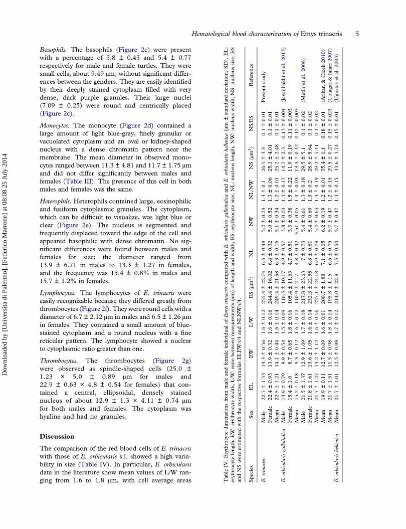
The comparison of the red blood cells of E. trinacriswith those of E. orbicularis s.l. showed a high varia-bility in size (Table IV). In particular, E. orbicularisdata in the literature show mean values of L/W ran-ging from 1.6 to 1.8 µm, with cell average areas T
able
IV.Erythrocyte
dimen
sion
sfrom
malean
dfemaleindividu
alof
Emys
trinacrisco
mpa
redwithE.orbicularisgalloita
licaan
dE.orbicularishellenica
(µm
±stan
dard
deviation,
SD).
EL:
erythroc
yteleng
th,EW:erythroc
ytewidth,L/W
:ratiobe
tweenmeasuremen
ts(µm)of
leng
than
dwidth,ES:erythroc
ytesize,NL:nu
cleu
sleng
th,NW:nu
cleu
swidth,NS:nu
cleu
ssize.ES
andNSwereestimated
withtherespective
form
ulas
ELEWπ/4
andNLNWπ/4.
Spe
cies
Sex
EL
EW
L/W
ES(µm
2)
NL
NW
NL/N
WNS(µm
2)
NS/ES
Referen
ce
E.trinacris
Male
22.7
±1.53
14.3
±0.56
1.6±0.12
255.4±22
.74
6.5±0.48
5.2±0.24
1.3±0.1
26.5
±3.3
0.1±0.01
Present
stud
yFem
ale
22.4
±0.93
13.9
±0.32
1.6±0.16
244.4±16
.02
6.4±0.32
5.0±0.52
1.3±0.06
25.1
±4.01
0.1±0.01
Mean
22.5
±1.21
14.1
±0.44
1.6±0.14
249.4±21
.58
6.3±0.16
5.1±0.34
1.2±0.01
25.2
±2.48
0.1±0.01
E.orbicularisgalloita
lica
Male
14.9
±0.79
9.9±0.54
1.5±0.09
116.5±10
.17
4.9±0.57
3.8±0.03
1.3±0.17
14.7
±2.3
0.13
±0.00
4(Javan
bakh
tet
al.20
13)
Fem
ale
15.4
±1.0
8.7±0.65
1.8±0.16
105.4±11
.63
4.7±0.51
3.3±0.38
1.5±0.22
11.9
±0.19
0.11
±0.00
3M
ean
15.2
±0.18
9.3±0.12
1.6±0.12
110.9±2.17
4.8±0.42
3.51
±0.05
1.4±0.03
13.3
±0.42
0.12
±0.00
3M
ale
21.5
±1.37
12.9
±1.09
1.7±0.18
217.8±23
.63
7±0.73
5.4±0.61
1.3±0.18
29.5
±5.1
0.1±0.02
(Metin
etal.20
06)
Fem
ale
21.8
±1.61
13.6
±1.05
1.6±0.14
232.3±22
.55
6.8±0.81
5.4±0.69
1.3±0.2
28.9
±5.64
0.1±0.02
Mean
21.7
±1.27
13.2
±1.12
1.6±0.16
225.1±24
.18
6.9±0.78
5.4±0.65
1.3±0.19
29.2
±5.41
0.1±0.02
Mean
19.9
±0.11
12.7
±0.09
1.6±0.01
200.7±1.88
7.1±0.05
6.2±0.19
1.2±0.01
35.4
±1.1
0.18
±0.01
(Arikan&
Cicek
2010
)M
ean
21.7
±1.51
11.5
±0.98
1.8±0.14
195.8±1.16
6.6±0.75
5.7±0.47
1.2±0.15
29.5
±0.27
0.15
±0.02
3(C
olagar
&Jafari20
07)
E.orbicularishellenica
Mean
21.7
±1.01
12.5
±0.98
1.7±0.12
214.0±22
.87.5±0.54
5.7±0.47
1.3±0.15
33.6
±3.74
0.15
±0.01
(Ugu
rtas
etal.20
03)
Hematological blood characterization of Emys trinacris 5
Dow
nloa
ded
by [
Uni
vers
ita d
i Pal
erm
o], [
Fede
rico
Mar
rone
] at
08:
08 2
5 Ju
ly 2
014
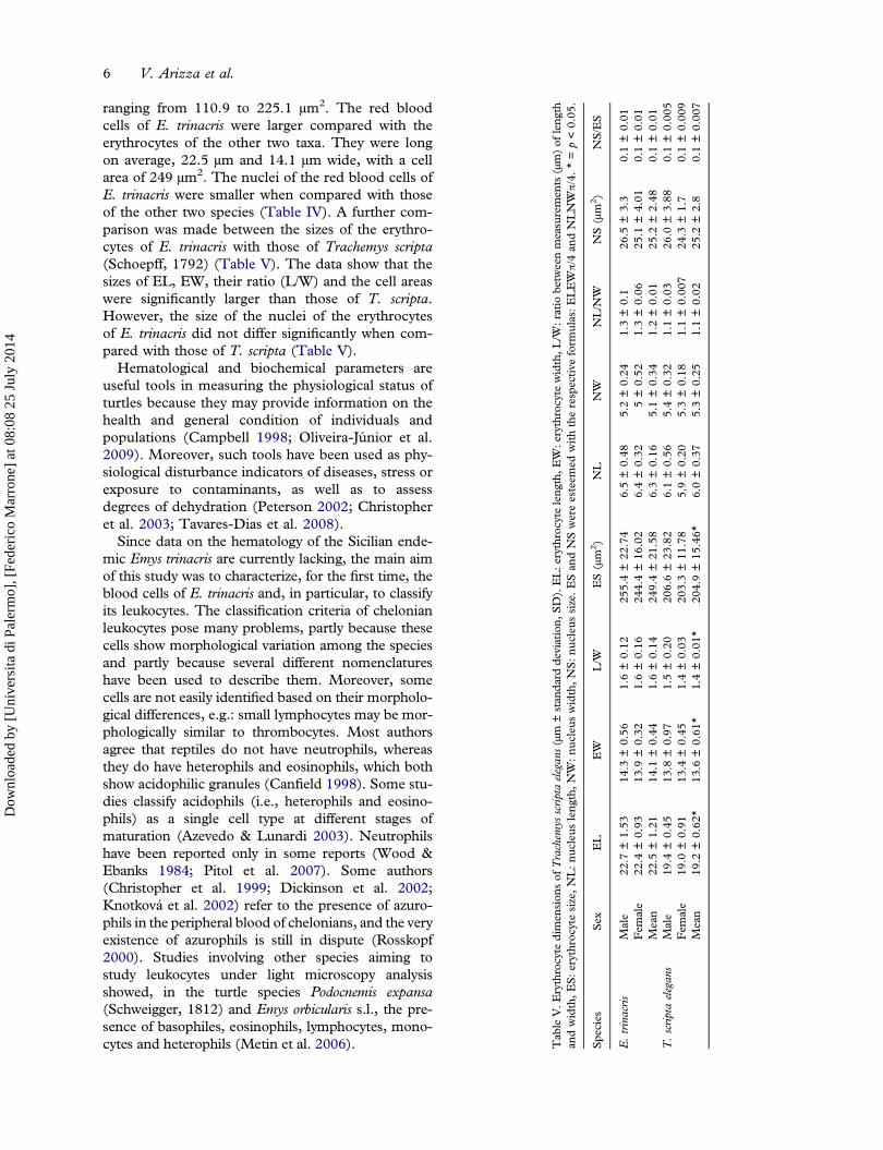
ranging from 110.9 to 225.1 μm2. The red bloodcells of E. trinacris were larger compared with theerythrocytes of the other two taxa. They were longon average, 22.5 µm and 14.1 µm wide, with a cellarea of 249 μm2. The nuclei of the red blood cells ofE. trinacris were smaller when compared with thoseof the other two species (Table IV). A further com-parison was made between the sizes of the erythro-cytes of E. trinacris with those of Trachemys scripta(Schoepff, 1792) (Table V). The data show that thesizes of EL, EW, their ratio (L/W) and the cell areaswere significantly larger than those of T. scripta.However, the size of the nuclei of the erythrocytesof E. trinacris did not differ significantly when com-pared with those of T. scripta (Table V).
Hematological and biochemical parameters areuseful tools in measuring the physiological status ofturtles because they may provide information on thehealth and general condition of individuals andpopulations (Campbell 1998; Oliveira-Júnior et al.2009). Moreover, such tools have been used as phy-siological disturbance indicators of diseases, stress orexposure to contaminants, as well as to assessdegrees of dehydration (Peterson 2002; Christopheret al. 2003; Tavares-Dias et al. 2008).
Since data on the hematology of the Sicilian ende-mic Emys trinacris are currently lacking, the main aimof this study was to characterize, for the first time, theblood cells of E. trinacris and, in particular, to classifyits leukocytes. The classification criteria of chelonianleukocytes pose many problems, partly because thesecells show morphological variation among the speciesand partly because several different nomenclatureshave been used to describe them. Moreover, somecells are not easily identified based on their morpholo-gical differences, e.g.: small lymphocytes may be mor-phologically similar to thrombocytes. Most authorsagree that reptiles do not have neutrophils, whereasthey do have heterophils and eosinophils, which bothshow acidophilic granules (Canfield 1998). Some stu-dies classify acidophils (i.e., heterophils and eosino-phils) as a single cell type at different stages ofmaturation (Azevedo & Lunardi 2003). Neutrophilshave been reported only in some reports (Wood &Ebanks 1984; Pitol et al. 2007). Some authors(Christopher et al. 1999; Dickinson et al. 2002;Knotková et al. 2002) refer to the presence of azuro-phils in the peripheral blood of chelonians, and the veryexistence of azurophils is still in dispute (Rosskopf2000). Studies involving other species aiming tostudy leukocytes under light microscopy analysisshowed, in the turtle species Podocnemis expansa(Schweigger, 1812) and Emys orbicularis s.l., the pre-sence of basophiles, eosinophils, lymphocytes, mono-cytes and heterophils (Metin et al. 2006). Tab
leV.E
rythrocyte
dimen
sion
sof
Trachem
ysscriptaelegan
s(µm
±stan
dard
deviation,
SD).EL:e
rythrocyte
leng
th,E
W:e
rythrocyte
width,L
/W:ratio
betw
eenmeasuremen
ts(µm)of
leng
than
dwidth,ES:erythroc
ytesize,NL:nu
cleu
sleng
th,NW:nu
cleu
swidth,NS:nu
cleu
ssize.ESan
dNSwereesteem
edwiththerespective
form
ulas:ELEWπ/4
andNLNWπ/4.*=p<0.05
.
Spe
cies
Sex
EL
EW
L/W
ES(µm
2)
NL
NW
NL/N
WNS(µm
2)
NS/ES
E.trinacris
Male
22.7
±1.53
14.3
±0.56
1.6±0.12
255.4±22
.74
6.5±0.48
5.2±0.24
1.3±0.1
26.5
±3.3
0.1±0.01
Fem
ale
22.4
±0.93
13.9
±0.32
1.6±0.16
244.4±16
.02
6.4±0.32
5±0.52
1.3±0.06
25.1
±4.01
0.1±0.01
Mean
22.5
±1.21
14.1
±0.44
1.6±0.14
249.4±21
.58
6.3±0.16
5.1±0.34
1.2±0.01
25.2
±2.48
0.1±0.01
T.scriptaelegan
sM
ale
19.4
±0.45
13.8
±0.97
1.5±0.20
206.6±23
.82
6.1±0.56
5.4±0.32
1.1±0.03
26.0
±3.88
0.1±0.00
5Fem
ale
19.0
±0.91
13.4
±0.45
1.4±0.03
203.3±11
.78
5.9±0.20
5.3±0.18
1.1±0.00
724
.3±1.7
0.1±0.00
9M
ean
19.2
±0.62
*13
.6±0.61
*1.4±0.01
*20
4.9±15
.46*
6.0±0.37
5.3±0.25
1.1±0.02
25.2
±2.8
0.1±0.00
7
6 V. Arizza et al.
Dow
nloa
ded
by [
Uni
vers
ita d
i Pal
erm
o], [
Fede
rico
Mar
rone
] at
08:
08 2
5 Ju
ly 2
014
Furthermore, another difficulty lies in determiningthe proportions of these cells within a particularspecies since these vary with individual physiologicstatus and the method of investigation (Campbell &Ellis 2007).
Erythrocytes
Mature erythrocytes of E. trinacris proved to be mor-phologically similar to those of various species ofturtles and tortoises and in particular to those of E.orbicularis s.l. (Ugurtas et al. 2003; Metin et al.2006). They were ellipsoidal cells with a centrallypositioned, ovoid nucleus and cytoplasmic inclu-sions, observed in over 30% of erythrocytes. Forother reptiles, these inclusions have been reportedto be degenerated organelles (Alleman et al. 1992;Clark et al. 2001; Chung et al. 2009) and may berelated to the aging of erythrocytes (Heard et al.2004). Others have postulated that the basophilicinclusions could be micronuclei and, therefore, theycould be biomarkers for chromosomal damage fromgenotoxic environmental pollutants (Matson et al.2005; Metin et al. 2006). The erythrocyte meansizes did not differ significantly from those of E.orbicularis s.l. (p > 0.05), failing in an attempt touse the erythrocyte size as a discriminator betweenspecies of the same Emydinae subfamily. The highernumber of red blood cells observed in males of E.trinacris than in females is similar to the findings inother turtles such as E. orbicularis (Duguy 1967) andKinixys erosa (Schweigger, 1812) (Oyewale et al.1998). The higher number of red blood cells foundin males may depend on testosterone hormonelevels. In fact, testosterone, when present in chelo-nians (Paitz & Bowden 2013), is able to increase thenumber of erythrocytes (Fried & Gurney 1965; Pati& Thapliyal 1984; Oyewale et al. 1998).
White blood cell morphology
Our findings conform to the basic morphologicaldescription for other Emydidae turtle species such asEmys orbicularis,Graptemys gibbonsi (Lovich &McCoy,1992), Pseudemys rubriventris (LeConte, 1830) andClemmys muhlenbergii (Schoepff, 1801) (Table III).
Eosinophils. In turtles, the same authors showed twotypes of eosinophils distinguishable by the shape ofcytoplasmic granules. Azevedo and Lunardi (2003)observed in the blood of Chrysemys dorbigni (Duméril& Bibron, 1835) two types of granulocytes that exhi-bit eosinophilia, one of them with round cytoplasmicgranules and the other with elongated cytoplasmicgranules. It has been suggested that these cells may
be eosinophils in different stages of maturation, butthey also may be distinct cell types, i.e. eosinophilsand heterophils (Azevedo & Lunardi 2003). In E.trinacris, most leukocytes were heterophils, basophilsand eosinophils. Similar findings have also beenreported by (Oliveira-Júnior et al. 2009) forPodocnemis expansa. This result was not confirmedfor other species of turtles. In fact, only captivefemale Clemmys muhlenbergii had a higher absoluteeosinophil count and a higher percentage of eosino-phils compared with captive males; conversely, wildfemales were not significantly different from wildmales (Brenner et al. 2002).
Basophils. The numbers of basophils in turtles varygreatly. In Graptemys gibbonsi, basophils were foundto be the most abundant leukocyte type, about 40%of total leukocytes (Perpiñán et al. 2008). Higherpercentages of basophils (50–63%) have been foundin other chelonians, such as Chelydra serpentina(Linnaeus, 1758) (Mead et al. 1983). In contrast,moderate basophil percentages have been found inother species, such as 5.7% in Gopherus polyphemus(Daudin, 1802) (Taylor & Jacobson 1982) and 8%in Geochelone radiata (Shaw, 1802) (Marks & Citino1990). Basophil numbers were almost nonexistent(~0.8 for both sexes) in Clemmys muhlenbergii(Brenner et al. 2002). However, care must be takenwhen analyzing published works; as an example,basophil counts in E. orbicularis varied widely: 0–4% was reported by Duguy (1970) and about 34%was reported by Javanbakht et al. (2013). This varia-tion of basophil density in various species of turtles isdifficult to explain. Many factors can affect the num-ber of basophils and the leukocytic formula such asage, health status, ecological factors and the seasons(for a review see Duguy 1970). We found in E.trinacris that the percentage of basophils did notvary significantly between sexes, ranging between20.5 ± 2.58 for males and 17.3 ± 1.58 for females.
Monocytes. This leukocyte type is not present in allspecies of turtles. Indeed, in Chelonia mydas(Linnaeus, 1758), authors did not identify mono-cytes (Wood & Ebanks 1984; Aguirre et al. 1995).Often, monocytes are not visible if the blood smearsare performed with blood that was taken eight ormore hours before (Work et al. 1998). Monocytesfrom E. trinacris were similar to the monocytes fromE. orbicularis described by Metin et al. (2006) orOcadia sinensis (Gray, 1870) described by Chunget al. (2009). In E. trinacris, monocytes were roundcells and had a similar size in both males andfemales, (11.3 and 11.7 µm respectively). Also,their frequency was similar for both sexes (~4.2%).
Hematological blood characterization of Emys trinacris 7
Dow
nloa
ded
by [
Uni
vers
ita d
i Pal
erm
o], [
Fede
rico
Mar
rone
] at
08:
08 2
5 Ju
ly 2
014
Heterophils. The heterophils of chelonians are analo-gous to mammalian neutrophils (Montali 1988) andcan be easily distinguished by the fusiform red gran-ules contained in the cytoplasm. They had the samepercentage for both sexes (~15%). These frequenciescorrespond with those found in Graptemys gibbonsi(Perpiñán et al. 2008), but differ from those inPseudemys rubriventris (~26.9%) (Innis et al. 2007)and Clemmys muhlenbergii and Chrysemys picta(Schneider, 1783) (both about 9.3% (Brenner et al.2002; Schwanz et al. 2011). Furthermore, the percen-tage value is included within the range indicated forE. orbicularis (Duguy 1970). The number and size ofheterophils have been observed to be influenced byindividual and seasonal factors (Duguy 1970).
Lymphocytes. The lymphocytes were the smallestcells, with a diameter on average about 6.6 µm forboth sexes. Female had a significantly (p < 0.05)higher percentage of lymphocytes (27.3 ± 1.24)compared with males (22.5 ± 0.69). The same resultwas reported by Brenner et al. (2002) for C. muhlen-bergii, where female and male lymphocyte percen-tages were 1.8 and 1.5%, respectively. Thepercentages of lymphocytes found in both gendersof E. trinacris were coherent with values reported forother Emydidae turtles such as G. gibbonsi and C.muhlenbergii (Brenner et al. 2002; Perpiñán et al.2008) but differ from those for P. rubriventris, inwhich these cells represent about 50% of the whiteblood cell differential count (Innis et al. 2007).
Thrombocytes. Although the similarity of thrombocytesand leukocytes in reptiles is known (Frye 1991), in thecase of E. trinacris, thrombocytes differ greatly fromthose of other pond turtles. In E. orbicularis, thethrombocytes are round cells with a nucleus roundto oval and dark (Metin et al. 2006), whereas inPseudemys rubriventris these were elliptical, with cen-tral ovoid basophilic nuclei, lightly basophilic cyto-plasm, and were often noted in small clusters (Inniset al. 2007). The thrombocytes of E. trinacris have anelongated cell shape with a central ovoid nucleus. Thesize and number do not differ between the sexes.
The comparison of the erythrocyte size parameterswith those of Emys orbicularis s.l. showed no importantdifferences even in comparison between the two sexes(Table IV). The data reported by Javanbakht et al.(2013) had the lowest values, which probably derivedfromdifferent environmental conditions (e.g. tempera-ture, air pressure) (Ruiz et al. 1983, 1989) or differentactivity levels (e.g. healthy, breeding, hibernating, fora-ging and daily activity) (Sykes & Klaphake 2008;Tosunoglu et al. 2011; Yu et al. 2013).
However, significant differences were found whenthe erythrocyte parameters (EL, EW, L/W, ES) of E.
trinacris were compared with those of T. scripta ele-gans, a tortoise belonging to the same family but to adifferent genus (Table V). The morphology of E.trinacris erythrocytes was similar to that of T. scriptaelegans, but the size was greater. The L/W ratio wasabout 1.6 for E. trinacris and about 1.4 for T. scriptaelegans; consequently, erythrocyte shape was moreellipsoidal in E. trinacris. No significant differenceswere found in the nucleo-cytoplasmic ratio.The results of our analysis show that the morphol-
ogy of erythrocytes within the family of Emydidaedoes not change greatly and, in a comparisonbetween species from different genera, only a fewdifferences in size can be found.The findings of this study present for the first time
data on the cytomorphological structure and num-bers of peripheral blood cells in both sexes of wild-caught, healthy E. trinacris. Since dates were derivedfrom specimens in good health, the hematologicalprofile here reported could be used as referencevalues for studies on E. trinacris, and could be ben-eficial to future clinical and conservation work on theendangered Sicilian pond turtle.
Acknowledgements
We thank the Director of Orto botanico of theUniversity of Palermo for permission to sample theTrachemys scripta elegans specimens, the President ofParco dei Nebrodi and the Italian Ministry of theEnvironment and Protection of Land and Sea forgranting the authorization U. prot. PNM-2011-0022035 25/10/2011 to sample Emys.Research partially funded by the “Fondi di
Ateneo” (60%) of the University of Palermo.
References
Aguirre AA, Balazs GH, Spraker TR, Gross TS. 1995. Adrenaland hematological responses to stress in juvenile green turtles(Chelonia mydas) with and without fibropapillomas.Physiological Zoology 68:831–854.
Alleman AR, Jacobson ER, Raskin RE. 1992. Morphologic andcytochemical characteristics of blood cells from the deserttortoise (Gopherus agassizii). American Journal of VeterinaryResearch 53:1645–1651.
Arikan H, Cicek K. 2010. Morphology of peripheral blood cells fromvarious species of Turkish herpetofauna. Acta Herpetologica5:179–198.
Azevedo A, Lunardi LO. 2003. Cytochemical characterization ofeosinophilic leukocytes circulating in the blood of the turtle(Chrysemys dorbignih). Acta Histochemica 105:99–105.doi:10.1078/0065-1281-00693.
Brenner D, Lewbart G, Stebbins M, Herman DW. 2002. Healthsurvey of wild and captive bog turtles (Clemmys muhlenbergii) inNorth Carolina and Virginia. Journal of Zoo and WildlifeMedicine 33:311–316.
8 V. Arizza et al.
Dow
nloa
ded
by [
Uni
vers
ita d
i Pal
erm
o], [
Fede
rico
Mar
rone
] at
08:
08 2
5 Ju
ly 2
014
Campbell TW. 1996. Clinical pathology. In: Mader DR, editor.Reptile medicine surgery. Philadelphia, Pennsylvania, USA:Saunders Company Ltd. pp. 248–257.
Campbell TW. 1998. Interpretation of the reptilian blood profile.Exotic Pet Practice 3:33–36.
Campbell TW, Ellis C. 2007. Avian and exotic animal hematologyand cytology. 3rd ed. Ames, Iowa, USA: Blackwell Pub.
Canfield PJ. 1998. Comparative cell morphology in the peripheralblood film from exotic and native animals. Australian VeterinaryJournal 76:793–800. doi:10.1111/j.1751-0813.1998.tb12328.x.
Christopher MM, Berry KH, Henen BT, Nagy KA. 2003. Clinicaldisease and laboratory abnormalities in free-ranging deserttortoises in California (1990–1995). Journal of WildlifeDiseases 39:35–56. doi:10.7589/0090-3558-39.1.35.
Christopher MM, Berry KH, Wallis IR, Nagy KA, Henen BT,Peterson CC. 1999. Reference intervals and physiologic altera-tions in hematologic and biochemical values of free-rangingdesert tortoises in the Mojave Desert. Journal of WildlifeDiseases 35:212–238. doi:10.7589/0090-3558-35.2.212.
Chung CS, Cheng CH, Chin SC, Lee AH, Chi CH. 2009.Morphologic and cytochemical characteristics of Asian yellowpond turtle (Ocadia sinensis) blood cells and their hematologicand plasma biochemical reference values. Journal of Zoo andWildlife Medicine 40:76–85. doi:10.1638/2008-0023.1.
Clark P, Johnstone AC, Ellison R, Goold M. 2001. Inclusions in theerythrocytes of eastern water dragons (Physignathus lesueurii).Australian Veterinary Journal 79:61–62. doi:10.1111/j.1751-0813.2001.tb10643.x.
Colagar H, Jafari N. 2007. Red blood cell morphology and plasmaproteins electrophoresis of the European pond terrapin Emysorbicularis. African Journal of Biotechnology 6:1578–1581.
Deem SL, Dierenfeld ES, Sounguet GP, Alleman AR, Cray C,Poppenga RH, Norton TM, Karesh WB. 2006. Blood values infree-ranging nesting leatherback sea turtles (Dermochelys coriacea)on the coast of the Republic of Gabon. Journal of Zoo andWildlife Medicine 37:464–471. doi:10.1638/05-102.1.
Dessauer HC. 1970. Blood chemistry of reptiles: Physiologicaland evolutionary aspects. In: Gans C, Parson T, editors.Biology of the reptilia. London, UK: Academy Press. pp. 1–72.
Dickinson VM, Jarchow JL, Trueblood MH. 2002. Hematologyand plasma biochemistry reference range values for free-ran-ging desert tortoises in Arizona. Journal of Wildlife Diseases38:143–153. doi:10.7589/0090-3558-38.1.143.
Duguy R. 1967. Le cycle annuel des elements figures dus sangchez Emys orbicularis L., Lacerta muralis Laur., et Natrix mauraL. Bulletin de la Societe Zoologique de France 92:15.
Duguy R. 1970. Numbers of blood cells and their variation. In:Gans C, Parson T, editors. Biology of the reptilia. Vol. 3.London, UK: Academy Press. pp. 93–110.
Fried W, Gurney CW. 1965. Use of mild plethora to demonstratean erythropoietic effect from small amounts of androgens.Experimental Biology and Medicine 120:519–521.doi:10.3181/00379727-120-30577.
Fritz U, d’Angelo S, Pennisi MG, Lo Valvo M. 2006. Variationof Sicilian pond turtles, Emys trinacris - What makes a spe-cies cryptic? Amphibia-Reptilia 27:513–529. doi:10.1163/156853806778877095.
Fritz U, Fattizzo T, Guicking D, Tripepi S, Pennisi MG, Lenk P,Joger U, Wink M. 2005. A new cryptic species of pond turtlefrom southern Italy, the hottest spot in the range of the genusEmys (Reptilia, Testudines, Emydidae). Zoologica Scripta34:351–371. doi:10.1111/j.1463-6409.2005.00188.x.
Fritz U, Guicking D, Kami H, Arakelyan M, Auer M, Ayaz D,Fernandez CA, Bakiev AG, Celani A, Dzukic G, Fahd S,Havaš P, Joger U, Khabibullin VF, Mazanaeva LF, Široky' P,
Tripepi S, Vélez AV, Antón GV, Wink M. 2007.Mitochondrial phylogeography of European pond turtles(Emys orbicularis, Emys trinacris) - an update. Amphibia-Reptilia 28:418–426. doi:10.1163/156853807781374737.
Frye FL. 1991. Hematology as applied to clinical reptile medicine.In: Frye FL, editor. Biomedical surgical aspects of captivereptile husbandry. Vol. 1. Malabar, Florida, USA: KriegerPublishing Co. pp. 209–280.
Gu HX, Zhang FY, Li PP. 2011. A Review of chelonian hematol-ogy. Asian Herpetological Research 2:12–20. doi:10.3724/SP.J.1245.2011.00012.
Heard D, Harr K, Wellehan J. 2004. Diagnostic sampling andlaboratory tests. In: Girling SJ, Raiti P, editors. Manual ofreptiles. 2nd ed. Quedgeley, Gloucester, UK: BSAVAPublisher. pp. 70–86.
Hutchison HV, Szarski H. 1965. Number of erythrocytes in someamphibians and reptiles. Copeia 1965:373–376. doi:10.2307/1440807.
Innis CJ, Tlusty M, Wunn D. 2007. Hematologic and plasmabiochemical analysis of juvenile head-started northern red-bel-lied cooters (Pseudemys rubriventris). Journal of Zoo andWildlife Medicine 38:425–432. doi:10.1638/1042-7260(2007)38[425:HAPBAO]2.0.CO;2.
Javanbakht H, Vaissi S, Parto P. 2013. The morphological char-acterization of the blood cells in the three species of turtle andtortoise in Iran. Research in Zoology 3:38–44.
Kassab A, Shousha S, Fargani A. 2009. Morphology of blood cells,liver and spleen of the Desert tortoise (Testudo graeca). The OpenAnatomy Journal 1:1–10. doi:10.2174/1877609400901010001.
Knotková Z, Doubek J, Knotek Z, Hájková P. 2002. Blood cellmorphology and plasma biochemistry in Russian tortoises(Agrionemys horsfieldi). Acta Veterinaria Brno 71:191–198.doi:10.2754/avb200271020191.
Marks SK, Citino SB. 1990. Hematology and serum chemistry ofthe radiated tortoise (Testudo radiata). Journal of Zoo andWildlife Medicine 21:342–344.
Matson CW, Palatnikov G, Islamzadeh A, McDonald TJ, AutenriethRL, Donnelly KC, Bickham JW. 2005. Chromosomal damage intwo species of aquatic turtles (Emys orbicularis and Mauremyscaspica) inhabiting contaminated sites in Azerbaijan.Ecotoxicology 14:513–525. doi:10.1007/s10646-005-0001-0.
Mead KF, Borysenko M, Findlay SR. 1983. Naturally abundantbasophils in the snapping turtle, Chelydra serpentina, possesscytophilic surface antibody with reaginic function. Journal ofImmunology 130:334–340.
Metin K, Türkozan O, Kargin F, Koca YB, Taskavak E, Koca S.2006. Blood cell morphology and plasma biochemistry of thecaptive European pond turtle Emys orbicularis. Acta VeterinariaBrno 75:49–55. doi:10.2754/avb200675010049.
Montali RJ. 1988. Comparative pathology of inflammation in thehigher vertebrates (reptiles, birds and mammals). Journal ofComparative Pathology 99:1–26. doi:10.1016/0021-9975(88)90101-6.
Nagy KA, Medica PA. 1986. Physiological ecology of desert tor-toises in southern Nevada. Herpetologica 42:73–92.
Oliveira-Júnior AA, Tavares-Dias M, Marcon JL. 2009. Biochemicaland hematological reference ranges for Amazon freshwater turtle,Podocnemis expansa (Reptilia: Pelomedusidae), with morphologicassessment of blood cells. Research in Veterinary Science 86:146–151. doi:10.1016/j.rvsc.2008.05.015.
Orós J, Casal A, Arencibia A. 2010. Microscopic studies on char-acterization of blood cells of endangered sea turtles. In:Méndez-Vilas A, Díaz J, editors. Microscopy: Science, tech-nology, applications and education. Vol. 1. Badajoz, Spain:Formatex Research Center. pp. 75–84.
Hematological blood characterization of Emys trinacris 9
Dow
nloa
ded
by [
Uni
vers
ita d
i Pal
erm
o], [
Fede
rico
Mar
rone
] at
08:
08 2
5 Ju
ly 2
014
Oyewale J, Ebute C, Ogunsanmi A, Olayemi F, Durotoye L. 1998.Weights and blood profiles of the West African hinge backedtortoise, Kinixys erosa and the desert tortoise, Gopherus agassi-zii. Journal of Veterinary Medicine Series A 45:599–605.doi:10.1111/j.1439-0442.1998.tb00864.x.
Paitz RT, Bowden RM. 2013. Sulfonation of maternal steroids is aconserved metabolic pathway in vertebrates. Integrative andComparative Biology 53:895–901. doi:10.1093/icb/ict027.
Pati A, Thapliyal J. 1984. Erythropoietin, testosterone, and thyr-oxine in the erythropoietic response of the snake, Xenochrophispiscator. General and Comparative Endocrinology 53:370–374.doi:10.1016/0016-6480(84)90264-8.
Pedall I, Fritz U, Stuckas H, Valdeón A, Wink M. 2011. Geneflow across secondary contact zones of the Emys orbiculariscomplex in the Western Mediterranean and evidence forextinction and re-introduction of pond turtles on Corsicaand Sardinia (Testudines: Emydidae). Journal of ZoologicalSystematics and Evolutionary Research 49:44–57.doi:10.1111/j.1439-0469.2010.00572.x.
Perpiñán D, Hernandez-Divers SM, Latimer KS, Akre T, HagenC, Buhlmann KA, Hernandez-Divers SJ. 2008. Hematology ofthe Pascagoula map turtle (Graptemys Gibbonsi) and the south-east Asian box turtle (Cuora amboinensis). Journal of Zoo andWildlife Medicine 39:460–463. doi:10.1638/2007-0044.1.
Peterson CC. 2002. Temporal, population, and sexual variation inhematocrit of free-living desert tortoises: Correlational tests ofcausal hypotheses. Canadian Journal of Zoology-RevueCanadienne De Zoologie 80:461–470. doi:10.1139/z02-021.
Pitol DL, Issa JPM, Caetano FH, Lunardi LO. 2007.Morphological characterization of the leukocytes in circulatingblood of the turtle (Phrynops hilarii). International Journal ofMorphology 25:6. doi:10.4067/S0717-95022007000400002.
Ream C, Ream R. 1966. The Influence of sampling methods on theestimation of population structure in painted turtles. AmericanMidland Naturalist 75:325–338. doi:10.2307/2423395.
Rhodin AGJ, Parham JF, van Dijk PP, Iverson JB. 2009. Turtles ofthe world: annotated checklist of taxonomy and synonymy,2009 update, with conservation status summary. Conservationbiology of freshwater turtles and tortoises: A compilation projectof the IUCN/SSC Tortoise and Freshwater Turtle SpecialistGroup. Chelonian Research Monographs 5:000.39–000.84.
Rossini M, Blanco PA, Marín E, Comerma-Steffensen S, ZerpaH. 2012. Haematological values of post-laying Arrau turtle(Podocnemis expansa) in the Orinoco River, Venezuela.Research in Veterinary Science 92:128–131. doi:10.1016/j.rvsc.2010.10.026.
Rosskopf WJ. 2000. Disorders of reptilian leucocytes and erythro-cytes. In: Fudge AM, editor. Laboratory medicine: Avian andexotic pets. Philadelphia, USA: Saunders, W.B. pp. 198–204.
Ruiz G, Rosenmann M, Veloso A. 1983. Respiratory and hematolo-gical adaptations to high altitude in Telmatobius frogs from theChilean Andes. Comparative Biochemistry and Physiology PartA: Physiology 76:109–113. doi:10.1016/0300-9629(83)90300-6.
Ruiz G, Rosenmann M, Veloso A. 1989. Altitudinal distributionand blood values in the toad, Bufo spinulosus Wiegmann.Comparative Biochemistry and Physiology Part A: Physiology94:643–646. doi:10.1016/0300-9629(89)90609-9.
Scheelings TF, Jessop TS. 2011. Influence of capture method,habitat quality and individual traits on blood parameters of freeranging lace monitors (Varanus varius). Australian VeterinaryJournal 89:360–365.
Scheelings TF, Rafferty AR. 2012. Hematologic and serum bio-chemical values of gravid freshwater Australian chelonians.Journal of Wildlife Diseases 48:314–321. doi:10.7589/0090-3558-48.2.314.
Schwanz L, Warner DA, McGaugh S, Di Terlizzi R, Bronikowski A.2011. State-dependent physiological maintenance in a long-livedectotherm, the painted turtle (Chrysemys picta). The Journal ofExperimental Biology 214:88–97. doi:10.1242/jeb.046813.
Stein G. 1996. Hematologic and blood chemistry values in rep-tiles. In: Mader DR, editor. Reptile medicine and surgery.Philadelphia, Pennsylvania, USA: Saunders, W B. CompanyLtd. pp. 473–483.
Stuckas H, Velo-Antón G, Fahd S, Kalboussi M, Rouag R, ArculeoM,Marrone F, Sacco F, Vamberger M, Fritz U. 2014. Where areyou from, stranger? The enigmatic biogeography of North Africanpond turtles (Emys orbicularis). Organisms Diversity andEvolution. doi:10.1007/s13127-014-0168-4.
Sykes JM, Klaphake E. 2008. Reptile hematology. VeterinaryClinics of North America: Exotic Animal Practice 11:481–500. doi:10.1016/j.cvex.2008.03.005.
Szarski H. 1968. Evolution of cell size in lower vertebrates. In:Orvig T, editor. Current problems of lower vertebrate phylo-geny. Stockhol, SE: Almqvist and Wiksell. pp. 445–453.
Tavares-Dias M, Oliveira-Júnior AA, Marcon JL. 2008.Methodological limitations of counting total leukocytes andthrombocytes in reptiles (Amazon turtle, Podocnemis expansa):An analysis and discussion. Acta Amazonica 38:351–356.doi:10.1590/S0044-59672008000200020.
Tavares-Dias M, Oliveira-Junior AA, Silva MG, Marcon JL,Barcellos JFM. 2009. Comparative hematological and biochem-ical analysis of giant turtles from the Amazon farmed in poor andnormal nutritional conditions. Veterinarski Arhiv 79:601–610.
Taylor RW, Jacobson ER. 1982. Hematology and serum chemis-try of the gopher tortoise, Gopherus polyphemus. ComparativeBiochemistry and Physiology Part A: Physiology 72:425–428.doi:10.1016/0300-9629(82)90241-9.
Tosunoglu M, Yilmaz N, Gul C. 2011. Effects of varying ecolo-gical conditions on the blood parameters of freshwater turtlesin Canakkale (Turkey). Ekoloji 20:7–12. doi:10.5053/ekoloji.2011.782.
Ugurtas IH, Sevinc M, Yildirimhan HS. 2003. Erythrocyte sizeand morphology of some tortoises and turtles from Turkey.Zoological Studies 42:173–178.
Wilkinson R. 2003. Clinical pathology. In: McArthur S, Wilkinson R,Meyer J, editors. Medicine and surgery of tortoises and turtles.Oxford, UK: Blackwell Publishing. pp. 141–186.
Wood FE, Ebanks GK. 1984. Blood cytology and hematology ofthe green sea turtle, Chelonia mydas. Herpetologica 40:6.
Work TM, Raskin RE, Balazs GH, Whittaker SD. 1998.Morphologic and cytochemical characteristics of blood cellsfrom Hawaiian green turtles. American Journal of VeterinaryResearch 59:1252–1257.
Yilmaz N, Tosunoglu M. 2010. Hematology and some plasmabiochemistry values of free living freshwater turtles (Emys orbi-cularis and Mauremys rivulata) from Turkey. North-WesternJournal of Zoology 6:109–117.
Yu PH, Yang PY, Chiu YS, Chi CH. 2013. Hematologic andplasma biochemical reference values of the yellow pond turtleMauremys mutica and the effects of sex and season. ZoologicalStudies 52:24–29. doi:10.1186/1810-522X-52-24.
Zuffi MAL, Gariboldi A. 1995. Geographical patterns of ItalianEmys orbicularis: A biometrical analysis. In: Llorente GA,Montori A, Santox X, Carretero MA, editors. ScientiaHerpetologica. Barcelona: Agal. pp. 120–123.
Zuffi MAL, Odetti F, Batistoni R, Mancino G. 2006. Geographicvariation of sexual size dimorphism and genetics in theEuropean pond turtle, Emys orbicularis and Emys trinacris, ofItaly. Italian Journal of Zoology 73:363–372. doi:10.1080/11250000600971323.
10 V. Arizza et al.
Dow
nloa
ded
by [
Uni
vers
ita d
i Pal
erm
o], [
Fede
rico
Mar
rone
] at
08:
08 2
5 Ju
ly 2
014