molecular detection of eukaryotes in a single human stool sample from senegal
TRANSCRIPT

Molecular Detection of Eukaryotes in a Single HumanStool Sample from SenegalIbrahim Hamad1, Cheikh Sokhna2, Didier Raoult1, Fadi Bittar1*
1 URMITE UMR-IRD 198, IHU Mediterranee Infection, Aix-Marseille Universite, Marseille, France, 2 IRD, UMR-IRD 198, Dakar, Senegal
Abstract
Background: Microbial eukaryotes represent an important component of the human gut microbiome, with differentbeneficial or harmful roles; some species are commensal or mutualistic, whereas others are opportunistic or parasitic. Thediversity of eukaryotes inhabiting humans remains relatively unexplored because of either the low abundance of theseorganisms in human gut or because they have received limited attention from a whole-community perspective.
Methodology/Principal Finding: In this study, a single fecal sample from a healthy African male was studied using bothculture-dependent methods and extended molecular methods targeting the 18S rRNA and ITS sequences. Our resultsrevealed that very few fungi, including Candida spp., Galactomyces spp., and Trichosporon asahii, could be isolated usingculture-based methods. In contrast, a relatively a high number of eukaryotic species could be identified in this fecal samplewhen culture-independent methods based on various primer sets were used. A total of 27 species from one sample werefound among the 977 analyzed clones. The clone libraries were dominated by fungi (716 clones/977, 73.3%), correspondingto 16 different species. In addition, 187 sequences out of 977 (19.2%) corresponded to 9 different species of plants; 59sequences (6%) belonged to other micro-eukaryotes in the gut, including Entamoeba hartmanni and Blastocystis sp; andonly 15 clones/977 (1.5%) were related to human 18S rRNA sequences.
Conclusion: Our results revealed a complex eukaryotic community in the volunteer’s gut, with fungi being the mostabundant species in the stool sample. Larger investigations are needed to assess the generality of these results and tounderstand their roles in human health and disease.
Citation: Hamad I, Sokhna C, Raoult D, Bittar F (2012) Molecular Detection of Eukaryotes in a Single Human Stool Sample from Senegal. PLoS ONE 7(7): e40888.doi:10.1371/journal.pone.0040888
Editor: Jonathan H. Badger, J. Craig Venter Institute, United States of America
Received February 17, 2012; Accepted June 14, 2012; Published July 13, 2012
Copyright: � 2012 Hamad et al. This is an open-access article distributed under the terms of the Creative Commons Attribution License, which permitsunrestricted use, distribution, and reproduction in any medium, provided the original author and source are credited.
Funding: FB was supported by a Chair of Excellence IRD provided by the Institut de Recherche pour le Developpement. The funders had no role in study design,data collection and analysis, decision to publish, or preparation of the manuscript.
Competing Interests: The authors have declared that no competing interests exist.
* E-mail: [email protected]
Introduction
The human body is home to vast and complex communities of
microorganisms. It has been estimated that microbes in human
bodies collectively make up approximately 100 trillion cells, ten
times the number of human cells [1]. The microbial ecosystem
plays important role in human metabolic activities, protection
against pathogens, nutrient processing, the stimulation of angio-
genesis, and the regulation of host fat storage [2,3].
The human gut is dominated by bacteria, especially species of
the phyla Firmicutes and Bacteroidetes. These two phyla are
spread throughout the intestinal tract and play crucial roles in
human health [4,5]. In addition to bacteria, organism belonging to
other domains of life, Archaea and Eukarya, are present in the
human intestine [4,6].
Microbial eukaryotes represent an important component of the
human gut microbiome, with different beneficial or harmful roles;
some species are commensal or mutualistic, whereas others are
opportunistic or parasitic [7]. This eukaryotic component of the
human gut microbiome remains relatively unexplored because
these organisms have a low abundance in human gut or because
they received a limited attention from molecular analyses [4,8].
Thus, studying the eukaryotic diversity in the human gut can
provide more complete picture of the natural communities
inhabiting this niche.
The microbial eukaryote communities in the human gut have
been studied primarily using selective culture techniques and
microscopy-based approaches [4,9,10]. Identification was based
on morphological and physiological traits. However, only a small
fraction of the microorganisms present has been detected using
this approach because the growth requirements for many of these
organisms remain unknown [4]. Recently, molecular-based
approaches, such as polymerase chain reaction (PCR) amplifica-
tion of the small subunit ribosomal RNA, have been established to
explore the microbial diversity in the human body [4,11,12]. In
2006, Scupham and his colleagues undertook a culture-indepen-
dent analysis of fungi in mouse feces, and they identified a wide
variety of fungi belonging to the phyla Ascomycota, Basidiomy-
cota, Chytridiomycota and Zygomycota using oligonucleotide
fingerprinting of rRNA genes (OFRG) [13].
More recently, in the human distal gut, culture-independent
methods have shown that the diversity and abundance of
eukaryotes is quite low relative to that of bacteria, and members
of the genera Gloeotinia/Paecilomyces and Galactomyces were the most
abundant [14] (Table 1). A more diverse fungal community was
PLoS ONE | www.plosone.org 1 July 2012 | Volume 7 | Issue 7 | e40888

observed in the study of Ott et al., 2008, in which they investigated
the mucosa-associated fungal microbiota in 47 controls and 57
subjects with inflammatory bowel disease. That study showed that
the majority of fungi retrieved from the fecal sample belonged to
Ascomycota [15] (Table 1). Only four types of fungi (Candida
vinaria, Candida edaphicus, Saccharomyces cerevisiae and Saccharomyces
servazzii) and one stramenopile (Blastocystis hominis) were detected in
fecal samples from ten Korean people using the PCR-fingerprint-
ing method [8] (Table 1). Finally, the ileal effluent and feces from 2
intestinal transplant patients were analyzed by Li et al. [16], who
reported temporal alterations in the fungal communities in these
patients (Table 1). An increase in the size of the fungal community
early after intestinal transplantation, followed by a decrease in this
community over time, was observed. Moreover, sequence analysis
of the 18S ribosomal DNA revealed that S. cerevisiae and
Kluyveromyces waltii were the dominant fungal species in both
patients [16].
The aim of this study is to carry out a comprehensive extended
molecular analysis of the diversity of eukaryotes in one fecal
sample from a young Senegalese man using amplification with
various universal primers followed by cloning and sequencing.
Results
Culture-dependent MethodsUsing different media, five strains of fungi were isolated from
one fecal sample. The results of MALDI-TOF identification were
as follows: Candida albicans, Candida krusei, Galactomyces geotrichum,
Trichosporon asahii and Geotrichum silvicola (Table 2). Direct ITS
sequencing of these strains confirmed the results of the MALDI-
TOF MS analysis for C. krusei, G. geotrichum and T. asahii. C. albicans
and G. silvicola were misidentified using MALDI-TOF MS, and the
correct identities of these strains were Candida rugosa and G.
geotrichum, respectively (Table 2). Finally, taking into account the
ITS results, only four species of fungi were recovered through
culture-based methods.
Culture-independent MethodsAn important part of this study was to choose suitable PCR
primers for the amplification of eukaryote sequences in the human
gastrointestinal tract. One major difficulty was the tendency to
amplify background bacterial, plant/animal and human DNA,
which are all potentially found in DNA extracted from human
feces. Twenty-five different published eukaryotic PCR primer sets
were tested on fecal DNA extracts from young, healthy Senegalese
man (see Table S1). For all primer sets, with the exception of the
primers listed in Table 3, a negative PCR signal was obtained. The
negative results with some primers could be explained by either
their low sensitivities for the eukaryotic communities in the human
gut or the absence of the target microorganisms in the studied
sample.
Cloning was performed prior to sequencing only when direct
sequencing was problematic or when the obtained sequences were
not clean sequences, indicating the amplification of sequences
from more than one microorganism. Finally, all PCR products
were cloned and then sequenced with the exception of primers
E528F/Univ1391 and FunF/FunR, which amplified Malassezia
restricta and G. geotrichum, respectively (Table 3). A total of 977
clones were collected from different clone libraries that were
generated using various primers and a fecal sample from a healthy
male. All but 9 of the resulting sequences have sequence similarity
scores of $98% when compared with existing sequences in the
GenBank database. The remaining 9 sequences have similarity
scores of 92% with Malassezia pachydermatis, and these 9 sequences
may be from putative new species.
Taking the different clone libraries together, one-quarter of the
obtained sequences were distributed among Viridiplantae (187
clones, 9 plant species), Stramenopile (38 clones, Blastocystis sp.),
Amoebozoa (21 clones, Entamoeba hartmanni), and human 18S
rDNA sequences (15 clones) (Table 3). Three-quarters of the
remaining sequences (716 clones) were identified as fungi, with 16
different fungal species belonging to Ascomycota (48.4%) and
Basidiomycota (24.9%) (Table 3). Seven fungal species of
Ascomycete yeasts were detected: C. rugosa, G. geotrichum, S.
cerevisiae, Arxiozyma telluris, Kluyveromyces hubeiensis, Torulaspora pre-
toriensis, and Sterigmatomyces elviae (Table 3). Nine basidiomycetous
yeasts were also recovered from the different clone libraries,
including Trichosporon caseorum, T. asahii, Trichosporon cutaneum, M.
restricta, Malassezia globosa, M. pachydermatis, Asterophora parasitica,
Bjerkandera adusta, and Phanerochaete steroids (Table 3).
Discussion
The primary objective of this study was to evaluate the broad
diversity of eukaryotes in a single fecal sample from an African
male using both culture-dependent and extensive culture-inde-
pendent methods.
Culture-dependent versus Culture-independent Methodsin this Study
Our results revealed that when using culture-dependent
methods, very few fungi, including C. rugosa, C. krusei, G. geotrichum,
and T. asahii, could be isolated. Among these fungi, C. rugosa and C.
krusei have been previously detected by culture-based methods
[12,17]. Many studies using culture-dependent approaches have
found fungi as the sole eukaryotes in the human microbiome of
healthy individuals and immunocompromised patients
[12,14,18,19].
The culture-independent methods revealed a more vast
diversity of eukaryotes, especially fungi, in our fecal sample
compared with the eukaryotes that were obtained by culture-
dependent methods. Among the 977 clones that were generated
using different primers, 716 clones (73.3%) were belonged to fungi
and corresponded to 16 species (Table 3). Among these 16 species,
only 3, C. Rugosa, G. candidum and T. asahii, have been cultured.
This discrepancy is due to the fact that some species require both
special growth media and special conditions to be cultured.
Conversely, C. krusei, a fungus isolated by culturing, was not
detected by culture-independent methods. This result could be
explained by biases in the PCR amplification [20] and/or cloning
bias [21]. Interestingly, in Chen’s study, molecular methods did
not identify C. krusei from fecal samples, but this fungus was found
by culturing techniques [12].
Culture-independent MethodsDifferent PCR primers were used to evaluate the diversity of
eukaryotes in the fecal sample, and these primers were adopted
from previously published studies. Some of these primers were
used previously to analyze eukaryotes in the human gut (Table S1),
whereas other primers were used previously to analyze eukaryotes
from aquatic environments rather than the human gut (Table S1).
Among the 18 micro-eukaryotic species found in our study, seven
species were detected by two or more primer sets, including T.
Caseorum, S. cerevisiae, Blastocystis sp., G. geotrichum, C. rugosa, M.
restricta, and M. globosa (Table 3 and Figure 1). The remaining 11
species (Table 3 and Figure 1) were amplified using only one
primer set.
Eukaryotic Diversity in African Human Gut
PLoS ONE | www.plosone.org 2 July 2012 | Volume 7 | Issue 7 | e40888

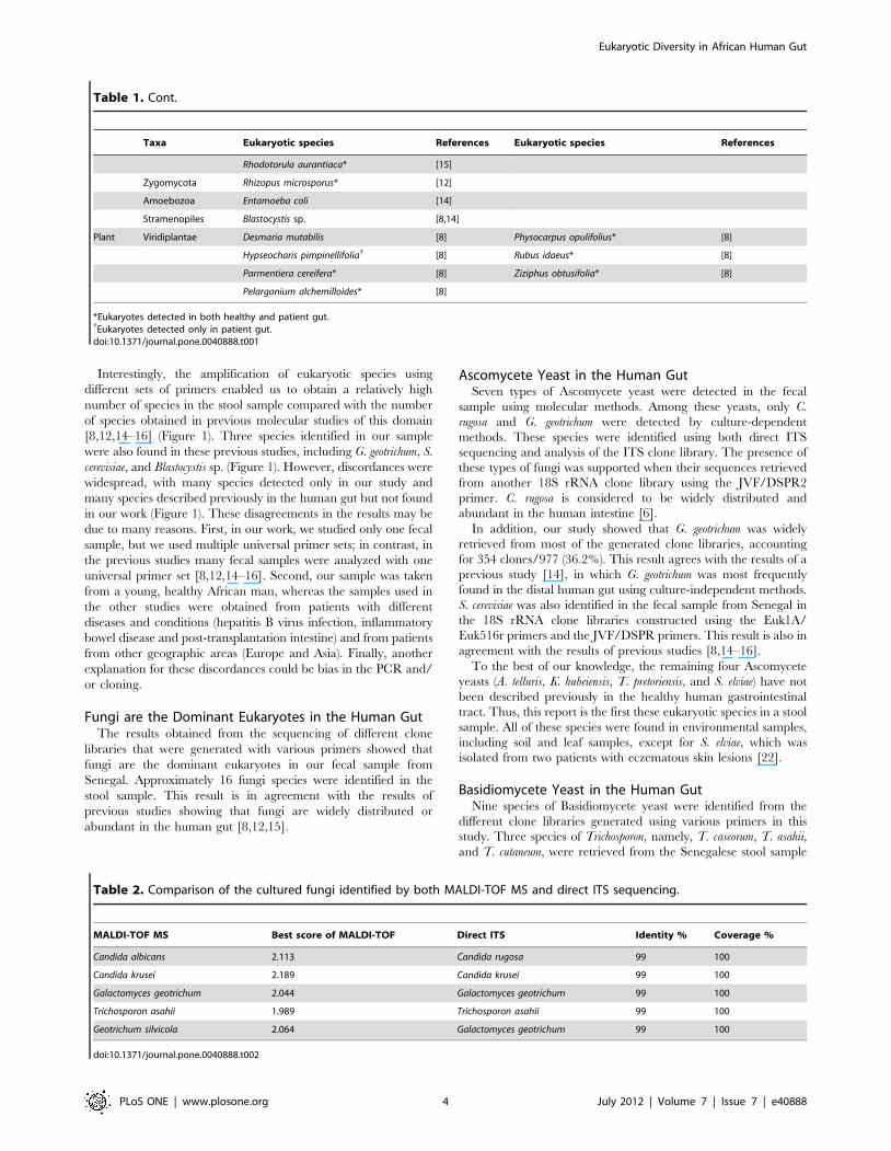
Table 1. The different eukaryotes previously detected by molecular methods in the human gut using universal 18S rDNA or ITSprimers.
Taxa Eukaryotic species References Eukaryotic species References
Fungi Ascomycota Acremonium sp. [14] Iodophanus carneus* [12]
Ajellomyces capsulatus{ [16] Kluyveromyces waltii{ [16]
Ajellomyces dermatitidis{ [16] Madurella mycetomatis* [15]
Aspergillus clavatus{ [16] Ophiocordyceps caloceroides* [12]
Aspergillus penicillioides* [12] Paraphaeosphaeria filamentosa [15]
Aspergillus versicolor{ [12] Penicillium chrysogenum* [14,15]
Aureobasidium pullulans* [12,15] Penicillium freii{ [12]
Botryotinia fuckeliana{ [15,16] Penicillium glabrum* [15]
Candida albicans* [12,14–16] Penicillium italicum* [15]
Candida austromarina* [12,15] Penicillium marneffei{ [16]
Candida dubliensis{ [15,16] Penicillium sp.* [12]
Candida edaphicus [8] Penicillium verruculosum* [14]
Candida glabrata [15] Penicillum sacculum* [15]
Candida intermedia* [12] Pleospora herbarum* [15]
Candida krissii{ [12] Raciborskiomyces longisetosum [15]
Candida milleri* [12] Saccharomyces bayanus [15]
Candida parapsilosis{ [16] Saccharomyces cariocanus [15]
Candida solani* [12] Saccharomyces castellii{ [16]
Candida sp. [12] Saccharomyces cerevisia* [8,12,14–16]
Candida tropicalis* [12,16] Saccharomyces paradoxus{ [12]
Candida vinaria* [8] Saccharomyces servazzii* [8]
Cephalosporium sp. [14] Saccharomyces sp.{ [12]
Chaetomium globosum* [12,15] Sclerotinia sclerotiorum* [15,16]
Chaetomium sp.* [12] Sclerotium sp.{ [15]
Cladosporium cladosporioides{ [15] Septoria epambrosiae [15]
Coccidioides immitis{ [16] Simplicillium lanosoniveum* [12]
Coccidioides posadasii{ [16] Simplicillium obclavatum* [12]
Doratomyces stemonitis* [12] Sirococcus conigenus* [15]
Dothideomycete sp.{ [15] Trichophyton verrucosum{ [16]
Fusarium oxysporum* [15,16] Uncultivatable Pezizomycotina{ [12]
Fusarium sp.* [12] Uncultured ascomycete{ [15]
Galactomyces geotrichum* [12,14–16] Verticillium leptobactrum [14]
Gloeotinia temulenta [14] Yarrowia lipolytica* [15]
Hyphozyma variabilis* [12]
Basidiomycota Asterotremella albida* [12] Rhodotorula mucilaginosa{ [15]
Bullera crocea{ [15] Sporolobomyces yunnanensis* [15]
Cryptococcus carnescens [15] Trametes versicolor* [15]
Cryptococcus fragicola* [12] Tricholoma saponaceum* [15]
Cryptococcus neoformans{ [16] Trichosporon dermatis* [15]
Cystofilobasidium capitatum* [15] Uncultivatable Agaricomycotina* [12]
Dacrymyces sp.* [15] Uncultivatable Pucciniomycotina{ [12]
Exidiopsis calcea{ [15] Unculture basidiomycete [15]
Filobasidium globisporum* [15] Uncultured basidiomycete{ [15]
Flammulina velutipes* [15] Uncultured ustilaginomycete{ [15]
Fomitopsis pinicola* [15] Ustilago maydis{ [15]
Graphiola phoenicis* [15] Ustilago sp.{ [15]
Malassezia globosa{ [16] Wallemia muriae* [12]
Malassezia pachydermatis{ [12] Wallemia sebi* [12]
Eukaryotic Diversity in African Human Gut
PLoS ONE | www.plosone.org 3 July 2012 | Volume 7 | Issue 7 | e40888
Interestingly, the amplification of eukaryotic species using
different sets of primers enabled us to obtain a relatively high
number of species in the stool sample compared with the number
of species obtained in previous molecular studies of this domain
[8,12,14–16] (Figure 1). Three species identified in our sample
were also found in these previous studies, including G. geotrichum, S.
cerevisiae, and Blastocystis sp. (Figure 1). However, discordances were
widespread, with many species detected only in our study and
many species described previously in the human gut but not found
in our work (Figure 1). These disagreements in the results may be
due to many reasons. First, in our work, we studied only one fecal
sample, but we used multiple universal primer sets; in contrast, in
the previous studies many fecal samples were analyzed with one
universal primer set [8,12,14–16]. Second, our sample was taken
from a young, healthy African man, whereas the samples used in
the other studies were obtained from patients with different
diseases and conditions (hepatitis B virus infection, inflammatory
bowel disease and post-transplantation intestine) and from patients
from other geographic areas (Europe and Asia). Finally, another
explanation for these discordances could be bias in the PCR and/
or cloning.
Fungi are the Dominant Eukaryotes in the Human GutThe results obtained from the sequencing of different clone
libraries that were generated with various primers showed that
fungi are the dominant eukaryotes in our fecal sample from
Senegal. Approximately 16 fungi species were identified in the
stool sample. This result is in agreement with the results of
previous studies showing that fungi are widely distributed or
abundant in the human gut [8,12,15].
Ascomycete Yeast in the Human GutSeven types of Ascomycete yeast were detected in the fecal
sample using molecular methods. Among these yeasts, only C.
rugosa and G. geotrichum were detected by culture-dependent
methods. These species were identified using both direct ITS
sequencing and analysis of the ITS clone library. The presence of
these types of fungi was supported when their sequences retrieved
from another 18S rRNA clone library using the JVF/DSPR2
primer. C. rugosa is considered to be widely distributed and
abundant in the human intestine [6].
In addition, our study showed that G. geotrichum was widely
retrieved from most of the generated clone libraries, accounting
for 354 clones/977 (36.2%). This result agrees with the results of a
previous study [14], in which G. geotrichum was most frequently
found in the distal human gut using culture-independent methods.
S. cerevisiae was also identified in the fecal sample from Senegal in
the 18S rRNA clone libraries constructed using the Euk1A/
Euk516r primers and the JVF/DSPR primers. This result is also in
agreement with the results of previous studies [8,14–16].
To the best of our knowledge, the remaining four Ascomycete
yeasts (A. telluris, K. hubeiensis, T. pretoriensis, and S. elviae) have not
been described previously in the healthy human gastrointestinal
tract. Thus, this report is the first these eukaryotic species in a stool
sample. All of these species were found in environmental samples,
including soil and leaf samples, except for S. elviae, which was
isolated from two patients with eczematous skin lesions [22].
Basidiomycete Yeast in the Human GutNine species of Basidiomycete yeast were identified from the
different clone libraries generated using various primers in this
study. Three species of Trichosporon, namely, T. caseorum, T. asahii,
and T. cutaneum, were retrieved from the Senegalese stool sample
Table 1. Cont.
Taxa Eukaryotic species References Eukaryotic species References
Rhodotorula aurantiaca* [15]
Zygomycota Rhizopus microsporus* [12]
Amoebozoa Entamoeba coli [14]
Stramenopiles Blastocystis sp. [8,14]
Plant Viridiplantae Desmaria mutabilis [8] Physocarpus opulifolius* [8]
Hypseocharis pimpinellifolia{ [8] Rubus idaeus* [8]
Parmentiera cereifera* [8] Ziziphus obtusifolia* [8]
Pelargonium alchemilloides* [8]
*Eukaryotes detected in both healthy and patient gut.{Eukaryotes detected only in patient gut.doi:10.1371/journal.pone.0040888.t001
Table 2. Comparison of the cultured fungi identified by both MALDI-TOF MS and direct ITS sequencing.
MALDI-TOF MS Best score of MALDI-TOF Direct ITS Identity % Coverage %
Candida albicans 2.113 Candida rugosa 99 100
Candida krusei 2.189 Candida krusei 99 100
Galactomyces geotrichum 2.044 Galactomyces geotrichum 99 100
Trichosporon asahii 1.989 Trichosporon asahii 99 100
Geotrichum silvicola 2.064 Galactomyces geotrichum 99 100
doi:10.1371/journal.pone.0040888.t002
Eukaryotic Diversity in African Human Gut
PLoS ONE | www.plosone.org 4 July 2012 | Volume 7 | Issue 7 | e40888

(Table 3). The genus Trichosporon is widely found in the
environment, but it can occasionally be found in the gastrointes-
tinal microbiota and can colonize human skin and the respiratory
tract [23]. The three Trichosporon species found in our sample were
not detected by previous molecular studies (Figure 1). Only T.
asahii, which was detected by both culture-dependent and culture-
independent methods in our study, has been isolated from a stool
sample of 22 month-old boy with acute myeloid leukemia [24].
Malassezia, a fastidious basidiomycetous yeast, was also found in
the Senegal stool sample represented by three species M. restricta,
M. globosa, and M. pachydermatis. Malassezia could be found naturally
on human skin but it is also able to cause cutaneous and systematic
diseases [25]. Among these three species of Malassezia, M.
pachydermatis and M. globosa have been detected previously in stool
sample from health volunteers and intestinal transplant patients,
respectively [12,16]. Thus, this study is the first report of molecular
detection of M. restricta in human fecal sample (Figure 1).
The remaining three fungi belonged to Basidiomycota including A.
parasitica, B. adusta, and P. steroids were not reported previously in
human stool sample. Among these environmental species only B.
adusta was previously isolated from human samples including
sputa, bronchial washing and skin [26].
Other Eukaryotes in Human GutAs well as the 16 fungal species discovered among the clone
sequences, two micro-eukaryotic species were also detected
(Entamoeba hartmanni and Blastocystis sp). E. hartmanni, which resides
in the large intestine of man, is now considered to be a distinct
Table 3. Summary of resulting clone libraries in our study.
Name of primer Fungal/Micro-eukaryotes species No. of Clone/Total Plant/human No. of Clone/Total
Euk1A/Euk516r Trichosporon caseorum 08/115 Humulus lupulus 64/115
Saccharomyces cerevisiaei 06/115 Artemisia annua 23/115
Blastocystis sp. 01/115 Triticum aestivum 01/115
Trichosporon cutaneum 11/115 Cupressus gigantea 01/115
ITS F/ITS-4R Trichosporon asahii 29/144
Galactomyces geotrichum 99/144
Candida rugosa 16/144
E528F/Univ1391 Malassezia restricta Direct sequencing
E528F/Univ1492 Malassezia globosa 34/98 Cupressus gigantea 03/98
Malassezia restricta 44/98 Pinus luchuensis 02/98
Human 18s rRNA 15/98
JVF/DSPR2 Saccharomyces cerevisiae 17/132 Humulus lupulus 87/132
Galactomyces geotrichum 02/132 Solanum lycopersicum 02/132
Candida rugosa 03/132 Triticum aestivum 01/132
Arxiozyma telluris 01/132 Schinus molle 01/132
Trichosporon caseorum 14/132 Phoenix canariensis 01/132
Torulaspora pretoriensis 01/132
Kluyveromyces hubeiensis 01/132
Asterophora parasitica 01/132
NSI/FR1 Galactomyces geotrichum 52/96
Geotrichum candidum 44/96
MF/MR Malassezia globosa 79/96
Malassezia restricta 04/96
Malassezia pachydermatis 09/96
Sterigmatomyce elviae 02/96
Bjerkandera adusta 01/96
Phanerochaete stereoides 01/96
EK1F/EK-1520 Galactomyces geotrichum 59/96
Blastocystis sp. 37/96
121F/1147R Entamoeba hartmanni 21/104 Bomax ceiba 1/104
Galactomyces geotrichum 74/104
Trichosporon sp. 8/104
FunF/FunR Galactomyces geotrichum Direct sequencing
EUKA/EUKB Galactomyces geotrichum 24/96
Candida rugosa 72/96
11 primer sets 18 micro-eukaryotic species 775/977 10 species 202/977
doi:10.1371/journal.pone.0040888.t003
Eukaryotic Diversity in African Human Gut
PLoS ONE | www.plosone.org 5 July 2012 | Volume 7 | Issue 7 | e40888
strain or species that is nonpathogenic and smaller than E.
histolytica but otherwise indistinguishable from it [27]. Blastocystis sp
also retrieved from the stool sample from Senegal, and this result
concur with the study of Scanlan [14] in which Blastocystis was the
dominant eukaryote in healthy human distal colon. Thus it may
not cause disease. However, this unicellular, obligatory anaerobic
protist could be common and prevalent in human with gastroin-
testinal illness like diarrhea and irritable bowel syndrome [28].
Finally, 9 different species of plants were detected in our sample
(19.2% of the sequenced clones), with H. lupulus accounting for
15.5% of total sequenced clones (Table 3). The co-amplification of
plant sequences with micro-eukaryote sequences has been
reported previously in the literature [8] and may be due to the
use of nonspecific and universal primers targeting the 18S rRNA
gene. Some of these plants are consumed either as part of the diet
or as a dietary supplement, including T. aestivum, S. lycopersicum and
P. canariensis, and others plants are used as traditional medicines
(e.g., A. annua as an anti-malarial [29], H. lupulus as an anxiolytic
calming agent [30], and B. ceiba as an antioxidant [31] ). The
consumption of plants could explain the presence of these
sequences in the stool sample.
In conclusion, we studied the eukaryotic diversity in one fecal
sample from a healthy African man using extensive molecular
methods with different sets of universal primers. Fungi largely
dominated the clone libraries. The application of our molecular
strategy in larger studies with a greater sample size, including
people living in various geographic regions, is currently needed to
better evaluate the occurrence/diversity of eukaryotes inhabiting
the human gut and to correlate the presence of eukaryotes with
human metabolism or disease. Moreover, expanded sequencing
analysis using high-throughput pyrosequencing will expand the
known diversity of eukaryotes in the healthy gut in the future.
Materials and Methods
Culturing and Identification of EukaryotesThe fecal sample was obtained from a healthy 16-year-old
Senegalese man living in Dielmo (a rural village in the Guinean-
Sudanian zone in Senegal). Written assent was obtained from this
individual; no written informed consent was needed from his
guardians for this study because he was older than 15 years old (in
accordance with the previous project approved by the Ministry of
Health of Senegal and the assembled village population and as
published elsewhere [32] ). Both this study and the assent
procedure were approved by the National Ethics Committee of
Senegal (CNERS) and the Ethics Committee of the Institute
Federatif de Recherche IFR 48, Faculty of Medecine, Marseille,
France (agreement number 09-022). The sample was serially
diluted, and fivefold dilutions were spread-plated in triplicate on
different media, including Sabouraud dextrose agar (BD Diag-
nostics, Heidelberg, Germany), Columbia culture media (BD
Diagnostics, Heidelberg, Germany) and glycine-vancomycin-
polymyxin B (GVPC) culture media (Biomerieux, Marcy l’ Etoile,
France). The plates were incubated aerobically for 48–72 h at 30
and 37uC. Colonies exhibiting different morphologies were
restreaked to obtain pure cultures. The fungi were identified
using matrix-assisted laser desorption ionization time-of-flight
(MALDI-TOF) mass spectrometry (MS) (Microflex, Bruker
Daltonics). The spectra obtained were compared with the Bruker
Taxonomy database. Finally, direct internal transcribed spacer
(ITS) analysis was performed for the fungal isolates as described
previously to confirm the results of MALDI-TOF MS.
DNA ExtractionDNA was extracted from the frozen sample using a modified
method for the Qiagen stool procedure (QIAamp DNA Tissue
Kit, Qiagen Inc, Germany) [14]. Aliquots of 200 mg of fecal
Figure 1. Comparison of our molecular results and thoseobtained previously for the human stool samples [8,12,14–16]using various universal primers. The universal primers usedpreviously for the analysis of the human gut are indicated by thenumber of the reference. * indicates primers used for the first time todescribe the human eukaryotic microbiota. Green color box = speciespositive in our sample and negative in the former studies; blue colorbox = species positive in both our sample and in the former studies; redcolor box = species negative in our sample and positive in the formerstudies.doi:10.1371/journal.pone.0040888.g001
Eukaryotic Diversity in African Human Gut
PLoS ONE | www.plosone.org 6 July 2012 | Volume 7 | Issue 7 | e40888

sample were placed in 2 ml tubes containing a 200 mg mixture of
0.1, 0.5, and 22 m mm zirconium beads and 1.5 ml of ASL buffer
(Qiagen). The sample was bead beaten at 3200 rpm for 90
seconds, followed by heating at 95uC for 10 minutes. The final
pellet was suspended in 180 ml of tissue lysis buffer and incubated
with proteinase K for 2 hours at 55uC. Then, the manufacturer’s
recommendations were followed for the purification and elution of
the DNA.
Primer SelectionTwenty-two different published universal eukaryotic or fungal-
specific PCR primer sets targeting the 18S rDNA and ITS
sequences were used, as shown in Table S1. In addition, three
specific primers for Malassezia, Rhodophyta, and Chlorophyta
targeting the 28S rDNA, RUBISCO, and rps11-rp12 sequences,
respectively, were also used (Table S1).
Genomic AmplificationAmplifications of sections of approximately 250–1,700 bp were
carried out with the primers listed in Table S1. The PCR reaction
mixture (final volume, 50 ml) contained 5 ml of dNTPs (2 mM of
each nucleotide), 5 ml of 10x DNA polymerase buffer (QIAGEN,
Courtaboeuf, France), 1 ml of MgCl2 (25 mM), 0.25 ml of
HotStarTaq DNA polymerase (5 U) (QIAGEN, Courtaboeuf,
France), 1 ml of each primer (Table S1) (10 pmol/ml), and 5 ml of
extracted DNA. PCR was performed with a preliminary step at
95uC for 15 minutes; 40 cycles of 95uC for 45 seconds, annealing
at the appropriate temperature for the primers used (see Table S1)
for 30 seconds, and 72uC for 1 to 2 minutes; and a final extension
step at 72uC for 5 minutes. The PCR products were analyzed
using agarose gel electrophoresis and visualized by ethidium
bromide staining. Then, the positive PCR products were purified
using the NucleoFastH 96 PCR Kit (MACHEREY-NAGEL,
Hoerdt, France) according to the manufacturer’s instructions.
Cloning Procedures and Insert AmplificationCloning of the purified PCR products was performed using the
pGEMH -T Easy Vector System 2 Kit (Promega, Madison, USA)
as recommended by the manufacturer. All white colonies were
collected and then analyzed by PCR M13 as described previously
[33].
Sequencing and Informative Data AnalysisPurified PCR-M13 inserts were sequenced in both directions
using the Big DyeH Terminator V1,1 Cycle Sequencing Kit
(Applied Biosystems, Courtaboeuf, France). The primers used for
sequencing were M13d and M13r. The sequencing products were
then run on an ABI PRISM 3130 automated sequencer (Applied
Biosystems, Foster City, CA, USA). Finally, the eukaryotes were
identified by comparing the obtained sequences with existing
sequences in the GenBank database using the BLAST program
available at the National Center for Biotechnology Information
Web site (http://www.ncbi.nlm.nih.gov/, BLAST).
Nucleotide Sequence Accession NumbersAll sequences obtained in this work have been deposited in
GenBank database with the accession numbers JX131688 to
JX132666.
Supporting Information
Table S1 Primers used in this study.
(DOCX)
Author Contributions
Conceived and designed the experiments: DR FB. Performed the
experiments: IH. Analyzed the data: IH CS DR FB. Contributed
reagents/materials/analysis tools: IH CS. Wrote the paper: IH DR FB.
References
1. Ley RE, Peterson DA, Gordon JI (2006) Ecological and evolutionary forces
shaping microbial diversity in the human intestine. Cell 124: 837–848.
2. Backhed F, Ley RE, Sonnenburg JL, Peterson DA, Gordon JI (2005) Host-
bacterial mutualism in the human intestine. Science 307: 1915–1920.
3. Hooper LV, Midtvedt T, Gordon JI (2002) How host-microbial interactions
shape the nutrient environment of the mammalian intestine. Annu Rev Nutr 22:
283–307.
4. Marchesi JR (2010) Prokaryotic and eukaryotic diversity of the human gut. Adv
Appl Microbiol 72: 43–62.
5. Eckburg PB, Bik EM, Bernstein CN, Purdom E, Dethlefsen L, et al. (2005)
Diversity of the human intestinal microbial flora. Science 308: 1635–1638.
6. Rajilic-Stojanovic M, Smidt H, de Vos WM (2007) Diversity of the human
gastrointestinal tract microbiota revisited. Environ Microbiol 9: 2125–2136.
7. Parfrey LW, Walters WA, Knight R (2011) Microbial eukaryotes in the human
microbiome: ecology, evolution, and future directions. Frontiers in Microbiology
2: 153.
8. Nam YD, Chang HW, Kim KH, Roh SW, Kim MS, et al. (2008) Bacterial,
archaeal, and eukaryal diversity in the intestines of Korean people. J Microbiol
46: 491–501.
9. Church C, Neill A, Schotthoefer AM (2010) Intestinal infections in humans in
the Rocky Mountain region, United States. J Parasitol 96: 194–196.
10. Macura AB, Witalis J (2010) [Fungi isolated from the stool in patients with
gastrointestinal disorders in 2005–2009]. Przegl Epidemiol 64: 313–317.
11. Stensvold CR, Lebbad M, Verweij JJ (2011) The impact of genetic diversity in
protozoa on molecular diagnostics. Trends Parasitol 27: 53–58.
12. Chen Y, Chen Z, Guo R, Chen N, Lu H, et al. (2010) Correlation between
gastrointestinal fungi and varying degrees of chronic hepatitis B virus infection.
Diagn Microbiol Infect Dis 70: 492–498.
13. Scupham AJ, Presley LL, Wei B, Bent E, Griffith N, et al. (2006) Abundant and
diverse fungal microbiota in the murine intestine. Appl Environ Microbiol 72:
793–801.
14. Scanlan PD, Marchesi JR (2008) Micro-eukaryotic diversity of the human distal
gut microbiota: qualitative assessment using culture-dependent and -indepen-
dent analysis of faeces. ISME J 2: 1183–1193.
15. Ott SJ, Kuhbacher T, Musfeldt M, Rosenstiel P, Hellmig S, et al. (2008) Fungi
and inflammatory bowel diseases: Alterations of composition and diversity.
Scand J Gastroenterol 43: 831–841.
16. Li Q, Wang C, Zhang Q, Tang C, Li N, et al. (2012) Use of 18S ribosomal DNA
polymerase chain reaction-denaturing gradient gel electrophoresis to study
composition of fungal community in 2 patients with intestinal transplants. Hum
Pathol. In press.
17. Waldman A, Gilhar A, Duek L, Berdicevsky I (2001) Incidence of Candida in
psoriasis–a study on the fungal flora of psoriatic patients. Mycoses 44: 77–81.
18. Agirbasli H, Ozcan SA, Gedikoglu G (2005) Fecal fungal flora of pediatric
healthy volunteers and immunosuppressed patients. Mycopathologia 159: 515–
520.
19. Khatib R, Riederer KM, Ramanathan J, Baran J (2001) Faecal fungal flora in
healthy volunteers and inpatients. Mycoses 44: 151–156.
20. de LA, Ashley FP, Palmer RM, Munson MA, Kyriacou L, et al. (2006) Novel
subgingival bacterial phylotypes detected using multiple universal polymerase
chain reaction primer sets. Oral Microbiol Immunol 21: 61–68.
21. Ghai R, Martin-Cuadrado AB, Molto AG, Heredia IG, Cabrera R, et al. (2010)
Metagenome of the Mediterranean deep chlorophyll maximum studied by direct
and fosmid library 454 pyrosequencing. ISME J 4: 1154–1166.
22. Sonck CE (1969) A new yeast species, Sterigmatomyces elviae, isolated from
eczematous skin lesions in two patients. Antonie van Leeuwenhoek 35: Suppl:
E25–6.
23. Chagas-Neto TC, Chaves GM, Colombo AL (2008) Update on the genus
Trichosporon. Mycopathologia 166: 121–132.
24. Agirbasli H, Bilgen H, Ozcan SK, Otlu B, Sinik G, et al. (2008) Two possible
cases of Trichosporon infections in bone-marrow-transplanted children: the first
case of T. japonicum isolated from clinical specimens. Jpn J Infect Dis 61: 130–
132.
25. Ashbee HR, Evans EG (2002) Immunology of diseases associated with
Malassezia species. Clin Microbiol Rev 15: 21–57.
26. Gonzalez GM, Sutton DA, Thompson E, Tijerina R, Rinaldi MG (2001) In
vitro activities of approved and investigational antifungal agents against 44
clinical isolates of basidiomycetous fungi. Antimicrob Agents Chemother 45:
633–635.
Eukaryotic Diversity in African Human Gut
PLoS ONE | www.plosone.org 7 July 2012 | Volume 7 | Issue 7 | e40888
27. Fotedar R, Stark D, Beebe N, Marriott D, Ellis J, et al. (2007) Laboratory
diagnostic techniques for Entamoeba species. Clin Microbiol Rev 20: 511–32.28. Yakoob J, Jafri W, Beg MA, Abbas Z, Naz S, et al. (2010) Blastocystis hominis
and Dientamoeba fragilis in patients fulfilling irritable bowel syndrome criteria.
Parasitol Res 107: 679–684.29. Mueller MS, Runyambo N, Wagner I, Borrmann S, Dietz K, et al. (2004)
Randomized controlled trial of a traditional preparation of Artemisia annua L.(Annual Wormwood) in the treatment of malaria. Trans R Soc Trop Med Hyg
98: 318–321.
30. Weeks BS (2009) Formulations of dietary supplements and herbal extracts forrelaxation and anxiolytic action: Relarian. Med Sci Monit 15: RA256-RA262.
31. Vieira TO, Said A, Aboutabl E, Azzam M, Creczynski-Pasa TB (2009)
Antioxidant activity of methanolic extract of Bombax ceiba. Redox Rep 14: 41–
46.
32. Trape JF, Tall A, Diagne N, Ndiath O, Ly AB, et al. (2011) Malaria morbidity
and pyrethroid resistance after the introduction of insecticide-treated bednets
and artemisinin-based combination therapies: a longitudinal study. Lancet Infect
Dis 11: 925–932.
33. Bittar F, Richet H, Dubus JC, Reynaud-Gaubert M, Stremler N, et al. (2008)
Molecular detection of multiple emerging pathogens in sputa from cystic fibrosis
patients. PLoS One 3: e2908.
Eukaryotic Diversity in African Human Gut
PLoS ONE | www.plosone.org 8 July 2012 | Volume 7 | Issue 7 | e40888