modulation of anticipatory emotion and perception processing by cognitive control
TRANSCRIPT
www.elsevier.com/locate/ynimg
NeuroImage 37 (2007) 652–662Modulation of anticipatory emotion and perception processing bycognitive control
Uwe Herwig,a,b,⁎ Thomas Baumgartner,c,d Tina Kaffenberger,a,b Annette Brühl,a
Mara Kottlow,c Ursula Schreiter-Gasser,a Birgit Abler,b Lutz Jäncke,c and Michael Rufere
aPsychiatric University Hospital Zürich, SwitzerlandbDepartment of Psychiatry, University of Ulm, GermanycInstitute of Psychology, Division Neuropsychology, University of Zürich, SwitzerlanddInstitute for Empirical Research in Economics, University of Zürich, SwitzerlandeDepartment of Psychiatry, University Hospital Zürich, Switzerland
Received 7 February 2007; revised 3 May 2007; accepted 5 May 2007Available online 24 May 2007
Strategies of cognitive control are helpful in reducing anxiety ex-perienced during anticipation of unpleasant or potentially unpleasantevents. We investigated the associated cerebral information processingunderlying the use of a specific cognitive control strategy during theanticipation of affect-laden events. Using functional magnetic resonanceimaging, we examined differential brain activity during anticipation ofevents of unknown and negative emotional valence in a group of eighteenhealthy subjects that used a cognitive control strategy, similar to “realitychecking” as used in psychotherapy, compared with a group of sixteensubjects that did not exert cognitive control. While expecting unpleasantstimuli, the “cognitive control” group showed higher activity in leftmedial and dorsolateral prefrontal cortex areas but reduced activity inthe left extended amygdala, pulvinar/lateral geniculate nucleus andfusiform gyrus. Cognitive control during the “unknown” expectationwas associated with reduced amygdalar activity as well and further withreduced insular and thalamic activity. The amygdala activationsassociated with cognitive control correlated negatively with thereappraisal scores of an emotion regulation questionnaire. The resultsindicate that cognitive control of particularly unpleasant emotions isassociated with elevated prefrontal cortex activity that may serve toattenuate emotion processing in for instance amygdala, and, notably, inperception related brain areas.© 2007 Published by Elsevier Inc.
Introduction
In everyday life we are often faced with approaching events thatwe know to be unpleasant or expect to be potentially unpleasant.Prior to their actual occurrence we may experience and have to copewith the uneasy feeling or even anxiety associated with the anti-
⁎ Corresponding author. Psychiatric University Hospital, Lenggstr. 31,CH-8032 Zürich, Switzerland. Fax: +41 44 383 4456.
E-mail address: [email protected] (U. Herwig).Available online on ScienceDirect (www.sciencedirect.com).
1053-8119/$ - see front matter © 2007 Published by Elsevier Inc.doi:10.1016/j.neuroimage.2007.05.023
cipation of such events. In coping effectively, we may applycognitive strategies to control the concomitant emotions. Anefficient method for cognitive emotion regulation is the strategy of“reappraisal” (Gross and John, 2003). Cognitive reappraisal isdefined as a form of an active cognitive process that involvesconstruing an emotion-eliciting situation in a way that changes itsemotional impact (Lazarus and Alfert, 1964). It is considered anantecedent strategy apt to successfully reduce the behavioral andexperiential component of negative future emotions. A specific kindof reappraisal that is used as a psychotherapeutic anxiety manage-ment strategy is the “reality checking”. Performing a “reality check”entails a cognitive shift from the awareness of the unpleasantemotional impact of a situation to a realistic cognitive evaluation ofthe objective context of the actual situation during exposure, but notto anticipate future and possibly unrealistic scenarios. Thus, as thereappraisal technique used for instance by Ochsner et al. (2002)consisted of directly reappraising a presented visual stimulus,“reality checking” consists of reappraising the meaning of an actualsituation for instance while expecting an emotional event. Thistechnique of self-regulation or cognitive control helps patients tolearn that they can successfully cope with feared situations. It istherefore commonly used in cognitive–behavioral therapies in thetreatment of for example phobic anxiety disorders with exposure-response prevention in order to reduce avoidance behaviors (e.g.,Hand, 2000; Otto et al., 2004).
Concerning the associated neural correlates, models of top-downcognitive control of emotion processing areas have been proposed(Mathews and MacLeod, 2005). These models draw attention to theinvolvement of prefrontal areas in modulating for exampleamygdala activity (Ochsner et al., 2002; Ochsner, 2001; Ochsneret al., 2004; Roffman et al., 2005; Harenski and Hamann, 2006;Kalisch et al., 2006; Ohira et al., 2006; Urry et al., 2006). They aresupported by animal studies that demonstrate the inhibiting in-fluence of medial prefrontal projections on centromedial amygdalaefferents to basolateral amygdala, which in turn modulates output to
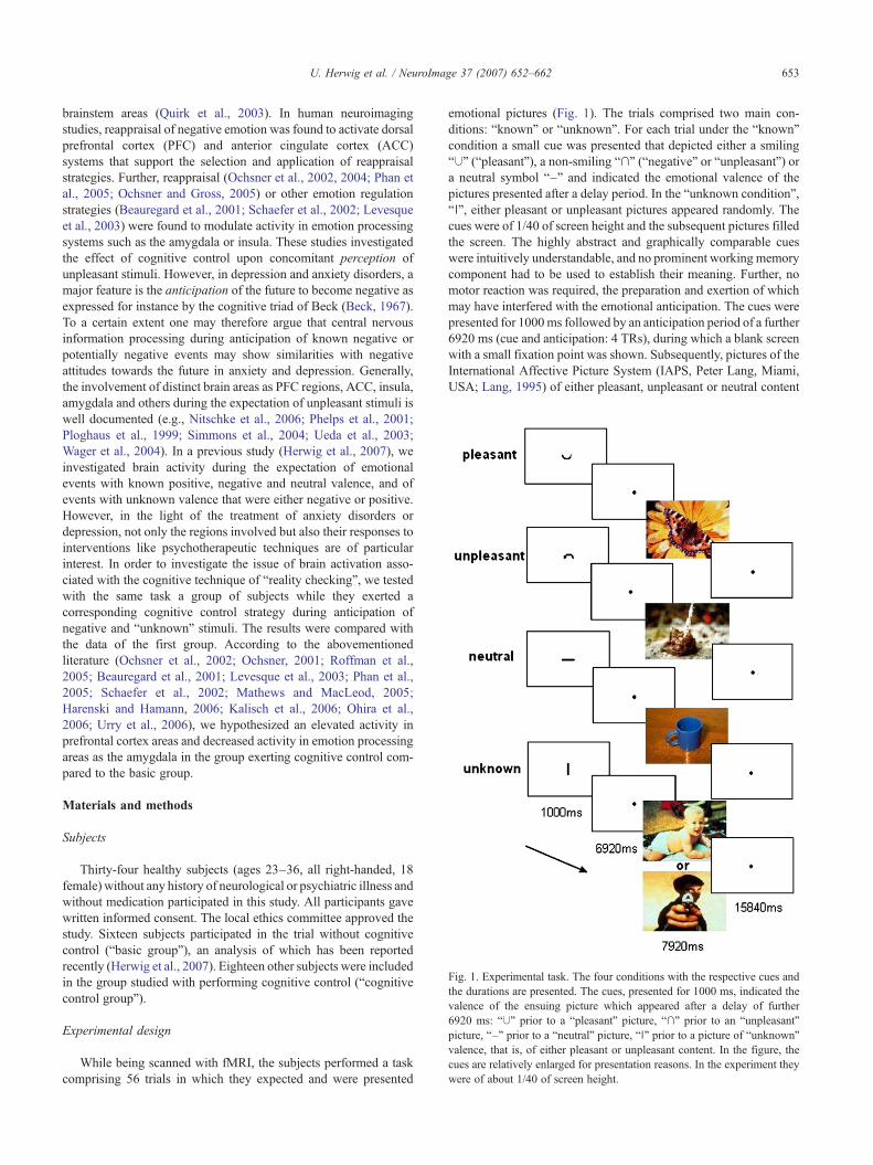
Fig. 1. Experimental task. The four conditions with the respective cues andthe durations are presented. The cues, presented for 1000 ms, indicated thevalence of the ensuing picture which appeared after a delay of further6920 ms: “∪” prior to a “pleasant” picture, “∩” prior to an “unpleasant”picture, “–” prior to a “neutral” picture, “∣” prior to a picture of “unknown”valence, that is, of either pleasant or unpleasant content. In the figure, thecues are relatively enlarged for presentation reasons. In the experiment theywere of about 1/40 of screen height.
653U. Herwig et al. / NeuroImage 37 (2007) 652–662
brainstem areas (Quirk et al., 2003). In human neuroimagingstudies, reappraisal of negative emotion was found to activate dorsalprefrontal cortex (PFC) and anterior cingulate cortex (ACC)systems that support the selection and application of reappraisalstrategies. Further, reappraisal (Ochsner et al., 2002, 2004; Phan etal., 2005; Ochsner and Gross, 2005) or other emotion regulationstrategies (Beauregard et al., 2001; Schaefer et al., 2002; Levesqueet al., 2003) were found to modulate activity in emotion processingsystems such as the amygdala or insula. These studies investigatedthe effect of cognitive control upon concomitant perception ofunpleasant stimuli. However, in depression and anxiety disorders, amajor feature is the anticipation of the future to become negative asexpressed for instance by the cognitive triad of Beck (Beck, 1967).To a certain extent one may therefore argue that central nervousinformation processing during anticipation of known negative orpotentially negative events may show similarities with negativeattitudes towards the future in anxiety and depression. Generally,the involvement of distinct brain areas as PFC regions, ACC, insula,amygdala and others during the expectation of unpleasant stimuli iswell documented (e.g., Nitschke et al., 2006; Phelps et al., 2001;Ploghaus et al., 1999; Simmons et al., 2004; Ueda et al., 2003;Wager et al., 2004). In a previous study (Herwig et al., 2007), weinvestigated brain activity during the expectation of emotionalevents with known positive, negative and neutral valence, and ofevents with unknown valence that were either negative or positive.However, in the light of the treatment of anxiety disorders ordepression, not only the regions involved but also their responses tointerventions like psychotherapeutic techniques are of particularinterest. In order to investigate the issue of brain activation asso-ciated with the cognitive technique of “reality checking”, we testedwith the same task a group of subjects while they exerted acorresponding cognitive control strategy during anticipation ofnegative and “unknown” stimuli. The results were compared withthe data of the first group. According to the abovementionedliterature (Ochsner et al., 2002; Ochsner, 2001; Roffman et al.,2005; Beauregard et al., 2001; Levesque et al., 2003; Phan et al.,2005; Schaefer et al., 2002; Mathews and MacLeod, 2005;Harenski and Hamann, 2006; Kalisch et al., 2006; Ohira et al.,2006; Urry et al., 2006), we hypothesized an elevated activity inprefrontal cortex areas and decreased activity in emotion processingareas as the amygdala in the group exerting cognitive control com-pared to the basic group.
Materials and methods
Subjects
Thirty-four healthy subjects (ages 23–36, all right-handed, 18female) without any history of neurological or psychiatric illness andwithout medication participated in this study. All participants gavewritten informed consent. The local ethics committee approved thestudy. Sixteen subjects participated in the trial without cognitivecontrol (“basic group”), an analysis of which has been reportedrecently (Herwig et al., 2007). Eighteen other subjects were includedin the group studied with performing cognitive control (“cognitivecontrol group”).
Experimental design
While being scanned with fMRI, the subjects performed a taskcomprising 56 trials in which they expected and were presented
emotional pictures (Fig. 1). The trials comprised two main con-ditions: “known” or “unknown”. For each trial under the “known”condition a small cue was presented that depicted either a smiling“∪” (“pleasant”), a non-smiling “∩” (“negative” or “unpleasant”) ora neutral symbol “–” and indicated the emotional valence of thepictures presented after a delay period. In the “unknown condition”,“∣”, either pleasant or unpleasant pictures appeared randomly. Thecues were of 1/40 of screen height and the subsequent pictures filledthe screen. The highly abstract and graphically comparable cueswere intuitively understandable, and no prominent working memorycomponent had to be used to establish their meaning. Further, nomotor reaction was required, the preparation and exertion of whichmay have interfered with the emotional anticipation. The cues werepresented for 1000ms followed by an anticipation period of a further6920 ms (cue and anticipation: 4 TRs), during which a blank screenwith a small fixation point was shown. Subsequently, pictures of theInternational Affective Picture System (IAPS, Peter Lang, Miami,USA; Lang, 1995) of either pleasant, unpleasant or neutral content
654 U. Herwig et al. / NeuroImage 37 (2007) 652–662
were presented for 7920 ms (4 TRs), followed by a baseline of15840ms (8 TRs) to allow the BOLD signal to level off before a newtrial started. Altogether, 56 pre-cued pictures were shown, 14 foreach of the conditions known positive (ps), negative (ng) and neutral(nt) valence and unknown (uk, comprising 7 ps and 7 ng) valence.The trials and correspondingly the different conditions appeared in arandomized order. The stimuli were matched for equal difference inthe valence rating from neutral (IAPS picture rating; Lang, 1995;mean ps 2.53, mean ng 2.20, t-tests for continuous variables:p=0.14), for complexity (two or more categories as faces andscenery; ps n=19 of 21 ps pictures in total, ng n=17/21; Chi squaretests for categorical variables: p=0.22) and contents (sums morethan the total of each 21 ps and ng pictures because of complexity) offaces (ps n=11, ng n=11; p=1.0), scenery (ps n=13, ng n=14;p=0.61) and food and nature (ps n=8, ng n=6; p=0.74). Withpriority to the matching of valence, content and complexity, arousalwas matched by selecting positive and negative pictures withdescriptively comparable IAPS arousal ratings (IAPS picture rating:ng mean 5.71, mean IAPS standard deviation 2.22; ps mean 4.86,mean IAPS standard deviation 2.36). However, the samples differstatistically concerning this measure (pb0.01). This resulted as aconsequence from the effort on a good matching of valence, contentand complexity on the one hand, and facing the fact that negativepictures are commonly more arousing than positive ones, despitecomparable differences of valence values from neutral. This hard-to-avoid issue was not considered a problem because we analyzed theexpectation period, and because the fMRI contrast analyzes werebased on the “negative” and the “unknown” condition versusneutral, not on the “positive” condition. After the scanning, thepresented pictures were rated for valence by the subjects of the“basic group” after the scanning on a 9-step visual analogue scalewith 1 being most negative, 5 being neutral and 9 being mostpositive. Both positive and negative valence ratings were equallydifferent from the neutral ratings: The mean ratings (and standarddeviations) for the negative pictures over all subjects were 2.96(1.35), for the neutral pictures 5.10 (0.67) and for the positivepictures 7.20 (1.25). The valence differences of the negative andthe positive pictures from neutral were similar (p=0.87). The taskwas programmed with PresentationTM (Neurobehavioral Systems,USA).
In the “basic group”, subjects were simply instructed to expectthe emotional stimuli after the cue and to be aware of theemotional valence indicated and to subsequently look at thefollowing picture. The subjects of the “cognitive control group”were instructed to perform “reality checking” derived as astandard intervention from cognitive–behavioral therapy (Hand,2000; Otto et al., 2004) during the unpleasant and unknownexpectation conditions, not during the pleasant and neutralexpectation conditions, in order to reduce anticipatory emotionalarousal after cue presentation: They were instructed to repeatedlyevaluate the realistic context of their actual situation by, e.g.,thinking: “I am lying in a scanner”, “They will show me apicture, this is part of the study”, while waiting during theanticipation period for the emotional picture. Both groupsperformed training sessions with examples of their respectivetasks prior to the experiment in the scanner. After scanning, bothgroups were asked in an unstructured non-quantitative interviewabout their experience with the task and how they were able toperform the task. The “cognitive control group” was further askedexplicitly about the subjective ability to perform the realitychecking.
fMRI acquisition
Imaging was performed with a 1.5-T Siemens Sonata whole-body scanner (Erlangen, Germany) equipped with a head coil.Initially, for each subject three-dimensional T1*-weighted anatomi-cal volumes were acquired (TR/TE 1880/3.22 ms; matrix size256×256; slice thickness 1 mm) for later coregistration with thefMRI. T2*-weighted functional MR images were obtained usingechoplanar imaging in an axial orientation. Image size was 64×64pixels, with a field of view of 220 mm, flip angle was 90°. Onevolume covering the whole brain consisted of 22 slices. Slicethickness was 4 mm with 1 mm gap resulting in a voxel size of3.4×3.4×5mm. Volumes were obtained every 1980ms (TE 40ms),16 volumes per scanning session and altogether 908 volumes. Thesubjects watched the stimuli in a mirror attached to the head coil anddirected to a screen onto which the stimuli were projected with avideo beamer.
fMRI data analysis and statistics
fMRI data were analyzed using BrainVoyager™ QX 1.8 (BrainInnovation, Maastricht, The Netherlands). The first four images ofeach functional scan were discarded to allow for T2* equilibrationeffects. Preprocessing of the functional scans included motion cor-rection, slice scan time correction, high frequency temporal filteringand removal of linear trends. Functional images were superimposedon the 2D anatomical images and incorporated into 3D data sets. Theindividual 3D data sets were then transformed into Talairach andTournoux space (Talairach and Tournoux, 1988) resulting in a voxelsize of 3×3×3 mm and then spatially smoothed with an 8-mmGaussian kernel for subsequent group analysis. Eight predictors,defined to represent the anticipation conditions (negative, pleasant,neutral, unknown) and the presentation conditions (negative, plea-sant, neutral, ex ante unknown—either pleasant or unpleasant) wereused to build the design matrix of the experiment. Expectation pe-riod and picture presentation periods were modeled as epochs usingthe standard two-gamma hemodynamic response function (HRF;peak after 5 s, undershoot peak 15 s) provided by BrainVoyager.
The fMRI data analysis based on the general linear modelcomprised the following steps: First, fixed effects analyses werecalculated separately for each subject for the three contrastscomparing the emotion expectation conditions “negative versusneutral”, “positive versus neutral” and “unknown versus neutral”and resulting in summary images. We focused on the contrasts ofthe emotion expectation conditions versus the “neutral” condition inorder to reveal areas selectively involved in emotion processing andnot just in expectation of a visual stimulus. These summary imageswere subjected to second level random effects analyses (rfx),separately for both groups, with and without cognitive control. Forthese random effects analyses we set a threshold of pb0.005 andused a cluster threshold of 135 voxels of 1×1×1 mm as providedby BrainVoyager corresponding to 5 voxels à 3×3×3 mm. Thesethresholds were chosen because most studies in this field ofaffective neuroscience have used statistical thresholds of similarsizes in order to avoid type-2 errors (e.g., Phelps et al., 2001; Phanet al., 2005; Wittmann et al., 2005). The application of more lenientthresholds in this research field is justified because the hemody-namic responses in the emotional network are weaker than inperception and motor studies due to methodological constraints.
The next step addressed our main question and consisted ofrandom effects group comparisons of the “basic” and the “cognitive
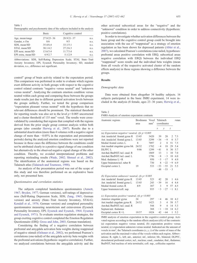
Table 1Demographic and psychometric data of the subjects included in the analysis
Basic Cognitive control
Age, mean/range 27.8/23–36 28.9/21–37 n.s.Gender, n f/m 8/6 9/5 n.s.SDS, mean/SD 35.4/6.4 33.1/5.1 n.s.STAI, mean/SD 30.1/4.2 27.2/6.3 n.s.EPI neur, mean/SD 13.6/3.4 11.7/3.4 n.s.EPI extra, mean/SD 4.9/2.7 5.1/2.9 n.s.
Abbreviations: SDS, Self-Rating Depression Scale; STAI, State–TraitAnxiety Inventory; EPI, Eysenck Personality Inventory; SD, standarddeviation, n.s., difference not significant.
Table 2FMRI-Analysis of emotion expectation contrasts
Anatomic regions Brodmannarea
Voxelmm3
Talairachcoordinates
t-max
x y z
(a) Expectation negativeNneutral, rfx pb0.005Ant. insula/inf. frontal gyrus R 13/45 3420 39 20 2 7.3Ant. insula/inf. frontal gyrus L 13/45 3299 −34 −9 0 5.7Medial frontal cortex L 8 7097 2 8 51 7.5Ant./medial cingulate gyrus blt. 24/32 1782 −4 18 29 5.4DLPFC L 9/46 1299 −41 8 30 5.7Ant.thal./BedNST./ncl. caud. R 1869 13 1 10 7.0Ant.thal./BedNST./ncl. caud. L 1796 −8 −3 10 6.0Med. thalamus LNR 950 −5 −17 9 4.9Upper brainstem/ncl. ruber R 730 8 −22 −9 6.9Occipital cortex L 19 533 −53 −55 21 5.5
−48 −53 −3
(b) Expectation unknownNneutral, rfx pb0.005Ant. insula/inf. frontal gyrus R 13/45 523 42 20 1 4.6Ant. insula/inf. frontal gyrus L 13/45 243 −39 16 4 4.4Medial frontal cortex R 8/9 387 3 9 57 4.5Upper brainstem/coll. sup. 935 −3 −27 −1 8.1
(c) Expectation positiveNneutral, rfx pb0.005Anterior cingulate gyrus 24 297 −4 46 10 4.2Ant./medial cingulate gyrus R. 24/32 1423 3 4 58 5.7Ant.thal./BedNST./ncl. caud. R 544 9 4 14 5.3Septum/ant. thalamus 368 −1 −10 14 6.2Occipital cortex RNL 19/37 1024 42 −64 1 5.2
fMRI analysis of emotion expectation in the cognitive control group. Acti-vated regions according to the random effects analyses (rfx) of the contrasts:(a) expectation negative versus neutral, (b) expectation positive versusneutral, (c) expectation unknown versus neutral. Indicated are the amount ofvoxels in mm3, the Talairach coordinates (x, y, z) of the centre of mass of theactivation and the maximal t-value of the voxels within each region. Abbrev-iations: R, right; L, left; ant., anterior; inf., inferior; blt., bilateral; DLPFC,dorsolateral prefrontal cortex; ncl., nucleus; caud., caudatus; thal., thalamus;BedNST, bed nucleus of stria terminalis; coll. sup., colliculus superior.
655U. Herwig et al. / NeuroImage 37 (2007) 652–662
control” group of brain activity related to the expectation period.This comparison was performed in order to evaluate which regionsexert different activity in both groups with respect to the cognitivecontrol related contrasts “negative versus neutral” and “unknownversus neutral”. Analyzing the contrasts emotion condition versusneutral within each group and comparing these between the groupsmade results just due to different general activation levels betweenthe groups unlikely. Further, we tested the group comparison“expectation pleasant versus neutral” with the hypothesis that norelevant differences should be prominent. The statistical thresholdfor reporting results was also set to the level pb0.005 uncorrectedand a cluster threshold of 135 mm3 voxel. The results were cross-validated by considering that regions that complied with the regionsderived from the prior single group contrast analysis within bothgroups (also consider Herwig et al., 2007). Results due to asubstantial deactivation (more than 4 volumes with a negative signalchange of more than −0.05% in the expectation and presentationperiods) during the analyzed emotion conditions were not reportedbecause in those cases the difference between the conditions couldnot be attributed clearly to a positive signal change of one conditionbut alternatively to the observed negative signal change of the othercondition. Thereby, we avoided interpretational problems andreporting misleading results (Wade, 2002; Shmuel et al., 2002).The identification of the anatomical regions was based on theTalairach atlas (Talairach and Tournoux, 1988).
An analysis of the presentation period was out of the scope ofthis study and was therefore performed on an explorative basisonly, not presented here.
Questionnaires and correlation statistics
The subjects completed handedness questionnaires (Annett,1967; Bryden, 1977; German versions), self-ratings of depressive-ness (Self-Rating Depression Scale, SDS; Zung, 1965; Germanversion) and anxiety (State–Trait Anxiety Inventory, STAI-G;Kendall et al., 1976; German version) and completed personalityquestionnaires measuring neuroticism and extraversion (EysenckPersonality Inventory, EPI; Eysenck and Eysenck, 1964; Eysenckand Eysenck, 1971). To evaluate emotion regulation strategies, thegroup exerting cognitive control completed the Emotion RegulationQuestionnaire (ERQ; Gross and John, 2003; German translation).
Considering the finding of a negative correlation betweenprefrontal and amygdala activation beta weights during reappraisalof negative stimuli (Ochsner et al., 2002), we performed Pearson'scorrelations (one-tailed) of the amygdala activity beta weights withthe prefrontal activations (hypothesis: negative correlation). Further,we analyzed correlations between the amygdala activity and the
other activated subcortical areas for the “negative” and the“unknown” condition in order to address connectivity (hypothesis:positive correlations).
In order to investigate whether activation differences between thebasic group and the cognitive control group could be brought intoassociation with the use of “reappraisal” as a strategy of emotionregulation as has been shown for depressed patients (Abler et al.,2007), we calculated Pearson's correlations (one-tailed, hypotheses:prefrontal areas positive correlation with ERQ, subcortical areasnegative correlation with ERQ) between the individual ERQ“reappraisal” score results and the individual beta weights (meanfrom all voxels of the respective activated cluster of the randomeffects analysis) in those regions showing a difference between thegroups.
Results
Demographic data
Data were obtained from altogether 34 healthy subjects. 16subjects participated in the basic fMRI experiment, 14 were in-cluded in the analysis (8 female, ages 23–36 years; Herwig et al.,
656 U. Herwig et al. / NeuroImage 37 (2007) 652–662
2007). Eighteen subjects participated in the “cognitive control”experiment; the data of 14 could be included in the group analysis.Four subjects had to be excluded due to fMRI signal artifacts causedby head movements (sudden head movements of more than 3 mm intranslation or rotation or more than three movements of more thanone mm) or by the beamer lamp. All subjects were right-handed andwere healthy with subthreshold ratings of depressiveness (self-rating depression scale, SDS, range 27.5–47.5), anxiety (STAI-G,range 20–43), “neuroticism” (EPI, range 0–10) and “extraversion”(EPI, range 1–18). The two groups did not differ concerning thesefactors (Table 1). In the interview after the scanning, all includedsubjects stated that they were able to perform the task, and thesubjects of the “cognitive control group” explicitly stated that theywere able to perform the reality check as instructed. None of thesubjects of the basic group reported anything comparable to anapplied cognitive control strategy when asked about theirexperiences during the task.
Brain activation during the expectation of emotional stimuli
In a first step, we compared the emotion expectation conditionsnegative (ng), unknown (uk) and positive (ps) with the neutral (nt)expectation condition. The results of the basic group are describedin detail in the previous report (Herwig et al., 2007). In the follow-ing, the results of the “cognitive control” group and the comparisonswith the basic group are presented.
The analysis of the single contrast expectation negative versusneutral under condition of cognitive control (exp-c ngNnt, Table2a) revealed activity within bilateral inferior frontal gyrus (IFG)and insula, anterior and medial cingulate cortex (ACC), medialprefrontal cortex (MPFC), dorsolateral prefrontal cortex (DLPFC),anterior thalamus and bed nucleus of stria terminalis (BedNST),medial thalamus, midbrain nuclei (nucleus ruber) and left lateraloccipital cortex.
The analysis of expectation unknown versus expectation neutralunder condition of cognitive control (exp-c ukNnt, Table 2b)
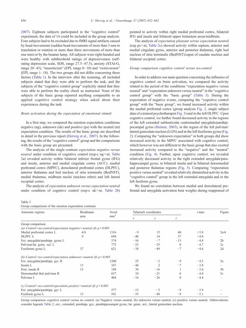
Table 3Group comparisons of the emotion expectation contrasts
Anatomic regions Brodmannarea
Voxelmm3
Group comparison(a) ControlNno-control/expectation negativeNneutral rfx pb0.005Medial prefrontal cortex L 6/8 1316DLPFC L 9 694Ext. amygdala/parahipp. gyrus L 374Pulvinar/lat. genic. ncl. L 775Fusiforme gyrus L 37 621
(b) ControlNno-control/expectation unknownNneutral rfx pb0.005Ext. amygdala/parahipp. gyr. R 1200Insula L 13 167Post. insula R 13 188Dorsomedial thal./pulvinar R 637Pulvinar L 404
(c) ControlNno-control/expectation positiveNneutral rfx pb0.005Ext. amygdala/parahipp. gyr. L 577Fusiform gyrus L 13 541
Group comparison cognitive control versus no control. (a) Negative versus neutrconsider legends Table 2; ext., extended; parahipp. gyr., parahippocampal gyrus; l
pointed to activity within right medial prefrontal cortex, bilateralIFG and insula and bilateral upper brainstem areas/midbrain.
The analysis of expectation pleasant versus expectation neutral(exp psNnt, Table 2c) showed activity within septum, anterior andmedial cingulate gyrus, anterior and posterior thalamus, right bednucleus of stria terminalis (BedNST)/caput of caudate nucleus andbilateral occipital cortex.
Group comparison cognitive control versus no-control
In order to address our main question concerning the influence ofcognitive control on brain activation, we compared the activityrelated to the period of the conditions “expectation negative versusneutral” and “expectation unknown versus neutral” in the “cognitivecontrol group” with the “basic group” (Table 3). During theexpectation of negative events, comparing the “cognitive controlgroup” with the “basic group”, we found increased activity withinthe medial prefrontal cortex (group analysis Fig. 2, single subjectdata of a trained psychotherapist Fig. 3) and in the left DLPFC. Uponcognitive control, we further found decreased activity in the regionsof the left extended sublenticular centromedial amygdala/parahip-pocampal gyrus (Heimer, 2003), in the region of the left pulvinar/lateral geniculate nucleus (LGN) and in the left fusiforme gyrus (Fig.2). Comparing the “unknown expectation” in both groups did showincreased activity in the MPFC associated with cognitive control,which however was not different to the basic group that also exertedincreased activity compared to the “negative” and the “neutral”condition (Fig. 4). Further, upon cognitive control, we revealedrelatively decreased activity in the right extended amygdala/para-hippocampal gyrus, in bilateral insula and in bilateral dorsomedialand posterior thalamus regions (Fig. 5). Comparing “expectationpositive versus neutral” revealed relatively diminished activity in the“cognitive control” group in the left extended amygdala and in theleft fusiform gyrus.
We found no correlation between medial and dorsolateral pre-frontal and amygdala activation beta weights during reappraisal of
Talairach coordinates t-max Figure
x y z
−9 15 60 +5.8 2a/4−40 14 37 +4.0 –−16 −7 −13 −4.8 2b−25 −25 0 −4.7 2c−38 −49 −7 −4.6 2d
25 −2 −9 −4.5 5a−44 2 −7 −3.8 –36 −16 1 −3.6 5b10 −21 6 −4.4 5c
−16 −26 0 −4.4 –
−15 −5 −9 −3.9 –−35 −48 −9 −5.1 –
al, (b) unknown versus neutral, (c) positive versus neutral. Abbreviations:at. genic. ncl., lateral geniculate nucleus.
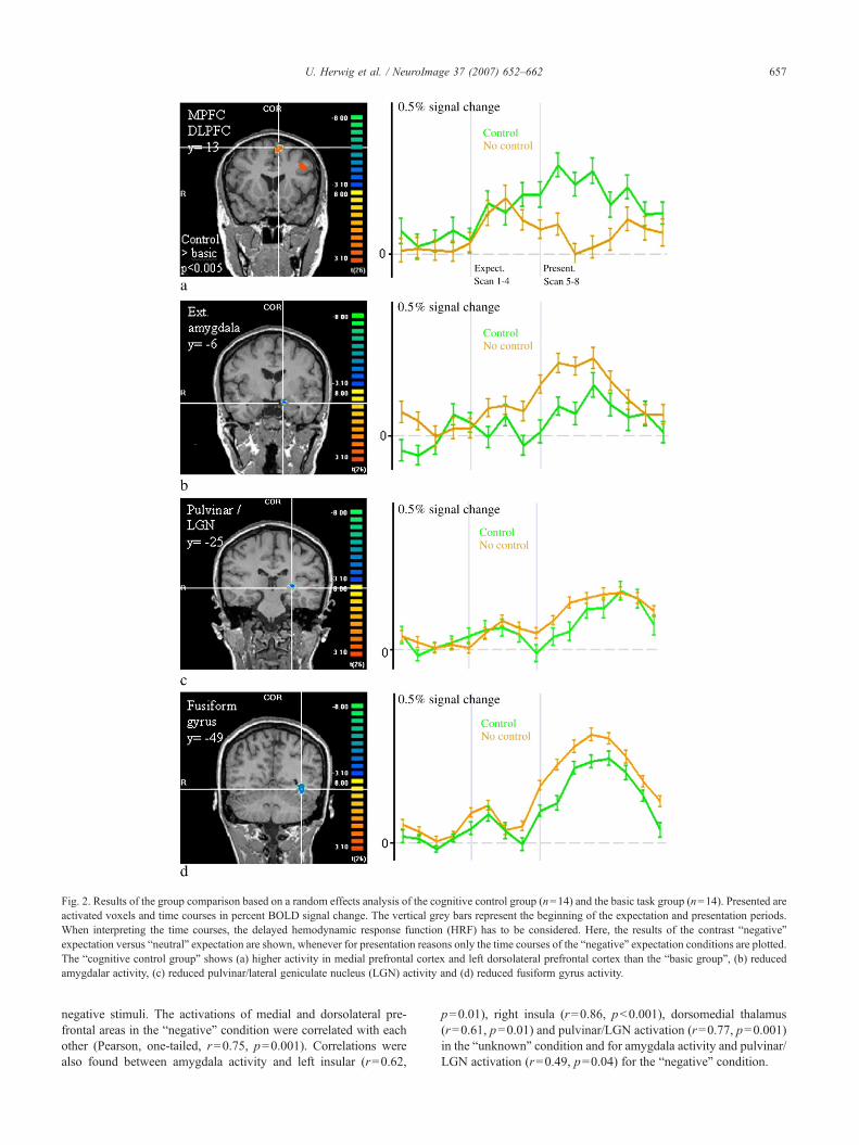
Fig. 2. Results of the group comparison based on a random effects analysis of the cognitive control group (n=14) and the basic task group (n=14). Presented areactivated voxels and time courses in percent BOLD signal change. The vertical grey bars represent the beginning of the expectation and presentation periods.When interpreting the time courses, the delayed hemodynamic response function (HRF) has to be considered. Here, the results of the contrast “negative”expectation versus “neutral” expectation are shown, whenever for presentation reasons only the time courses of the “negative” expectation conditions are plotted.The “cognitive control group” shows (a) higher activity in medial prefrontal cortex and left dorsolateral prefrontal cortex than the “basic group”, (b) reducedamygdalar activity, (c) reduced pulvinar/lateral geniculate nucleus (LGN) activity and (d) reduced fusiform gyrus activity.
657U. Herwig et al. / NeuroImage 37 (2007) 652–662
negative stimuli. The activations of medial and dorsolateral pre-frontal areas in the “negative” condition were correlated with eachother (Pearson, one-tailed, r=0.75, p=0.001). Correlations werealso found between amygdala activity and left insular (r=0.62,
p=0.01), right insula (r=0.86, pb0.001), dorsomedial thalamus(r=0.61, p=0.01) and pulvinar/LGN activation (r=0.77, p=0.001)in the “unknown” condition and for amygdala activity and pulvinar/LGN activation (r=0.49, p=0.04) for the “negative” condition.
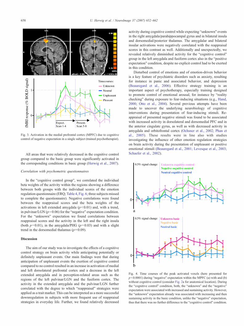
Fig. 3. Activation in the medial prefrontal cortex (MPFC) due to cognitivecontrol of negative expectation in a single subject (trained psychotherapist).
Fig. 4. Time courses of the peak activated voxels (here presented forpb0.0001) during “negative” expectation within the MPFC (a) with and (b)without cognitive control (consider Fig. 2a for anatomical location). Duringthe “cognitive control” condition, both, the “unknown” and the “negative”expectation were associated with increased and sustaining activity. However,the “unknown” expectation already was associated with increasing and thensustaining activity in the basic condition, unlike the “negative” expectation,thus that there was no further difference to the “cognitive control” condition.
658 U. Herwig et al. / NeuroImage 37 (2007) 652–662
All areas that were relatively decreased in the cognitive controlgroup compared to the basic group were significantly activated inthe corresponding conditions in basic group (Herwig et al., 2007).
Correlation with psychometric questionnaires
In the “cognitive control group”, we correlated the individualbeta weights of the activity within the regions showing a differencebetween both groups with the individual scores of the emotionregulation questionnaire (ERQ; Table 4, Fig. 6; three subjects missedto complete the questionnaire). Negative correlations were foundbetween the reappraisal scores and the beta weights of theactivations in left extended amygdala (pb0.01) and, with a trend,in pulvinar/LGN (p=0.06) for the “negative” expectation condition.For the “unknown” expectation we found correlations betweenreappraisal scores and the activity in the left and the right insula(both p=0.01), in the amygdala/PHG (p=0.03) and with a slighttrend in the dorsomedial thalamus (p=0.09).
Discussion
The aim of our study was to investigate the effects of a cognitivecontrol strategy on brain activity while anticipating potentially ordefinitely unpleasant events. Our main findings were that duringanticipation of unpleasant events the exertion of cognitive controlcompared to no control resulted in an increase in activation of medialand left dorsolateral prefrontal cortex and a decrease in the leftextended amygdala and in perception-related areas such as theregions of the left pulvinar/LGN and the fusiform cortex. Theactivity in the extended amygdala and the pulvinar/LGN furthercorrelated with the degree to which “reappraisal” strategies wereapplied as a trait marker. This can be interpreted as a more successfuldownregulation in subjects with more frequent use of reappraisalstrategies in everyday life. Further, we found relatively decreased
activity during cognitive control while expecting “unknown” eventsin the right amygdala/parahippocampal gyrus and in bilateral insulaand dorsomedial/posterior thalamus. The amygdalar and bilateralinsular activations were negatively correlated with the reappraisalscores in this contrast as well. Additionally and unexpectedly, werevealed relatively diminished activity for the “cognitive control”group in the left amygdala and fusiform cortex also in the “positiveexpectation” condition, despite no explicit control had to be exertedin this condition.
Disturbed control of emotions and of emotion-driven behavioris a key feature of psychiatric disorders such as anxiety, resultingfor instance in panic and associated behavior, and depression(Beauregard et al., 2006). Effective strategy training is animportant aspect of psychotherapy, especially training designedto promote control of emotional arousal, for instance by “realitychecking” during exposure to fear-inducing situations (e.g., Hand,2000; Otto et al., 2004). Several previous attempts have beenmade to uncover the underlying neurobiology of cognitiveinterventions during presentation of fear-inducing stimuli. Re-appraisal of presented negative stimuli was found to be associatedwith increased activity in dorsolateral and dorsomedial PFC and inthe anterior cingulate gyrus, as well as with decreased activity inamygdala and orbitofrontal cortex (Ochsner et al., 2002; Phan etal., 2005). These results were in line also with studiesinvestigating the influence of other emotion regulation strategieson brain activity during the presentation of unpleasant or positiveemotional stimuli (Beauregard et al., 2001; Levesque et al., 2003;Schaefer et al., 2002).
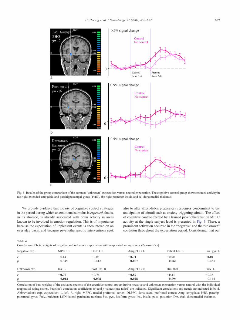
Fig. 5. Results of the group comparison of the contrast “unknown” expectation versus neutral expectation. The cognitive control group shows reduced activity in(a) right extended amygdala and parahippocampal gyrus (PHG), (b) right posterior insula and (c) dorsomedial thalamus.
659U. Herwig et al. / NeuroImage 37 (2007) 652–662
We provide evidence that the use of cognitive control strategiesin the period during which an emotional stimulus is expected, that is,in its absence, is already associated with brain activity in areasknown to be involved in emotion regulation. This is of importancebecause the expectation of unpleasant events is encountered on aneveryday basis, and because psychotherapeutic interventions seek
Table 4Correlation of beta weights of negative and unknown expectation with reappraisal
Negative exp. MPFC L DLPFC L
r 0.14 −0.08p 0.345 0.412
Unknown exp. Ins. L Post. ins. R
r −0.70 −0.74p 0.012 0.008
Correlation of beta weights of the activated regions of the cognitive control groupreappraisal rating scores. Pearson's correlation coefficients (r) and p-values (one-taAbbreviations: exp, expectation; L, left; R, right; MPFC, medial prefrontal cortepocampal gyrus; Pulv., pulvinar; LGN, lateral geniculate nucleus; Fus. gyr., fusifo
also to alter affect-laden preparatory responses concomitant to theanticipation of stimuli such as anxiety-triggering stimuli. The effectof cognitive control exerted by a trained psychotherapist on MPFCactivity at the single subject level is presented in Fig. 3. There, aprominent activation occurred in the “negative” and the “unknown”condition throughout the expectation period. Considering, that our
rating scores (Pearsons’s r)
Amg/PHG L Pulv./LGN L Fus. gyr. L
−0.71 −0.50 0.040.007 0.060 0.453
Amg/PHG R Dm. thal. Pulv. L
−0.59 −0.41 −0.380.028 0.094 0.144
during negative and unknown expectation versus neutral with the individualiled) are indicated. Significant correlations and trends are indicated in bold.x; DLPFC, dorsolateral prefrontal cortex; Amg, amygdala; PHG, parahip-rm gyrus; Ins., insula; post., posterior; Dm. thal., dorsomedial thalamus.
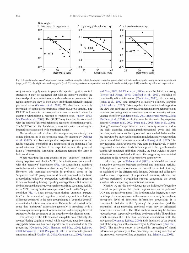
Fig. 6. Correlation between “reappraisal” scores and beta weights within the cognitive control group of (a) left extended amygdala during negative expectation(exp.; pb0.01), (b) right extended amygdala (p=0.03) during unknown expectation and (c) left insular activity (p=0.01) also during unknown expectation.
660 U. Herwig et al. / NeuroImage 37 (2007) 652–662
subjects were largely naive to psychotherapeutic cognitive controlstrategies, it may be suggested that with an intensive training theincreased prefrontal activations would even be more prominent. Ourresults support the view of a top-down inhibitionmediated bymedialprefrontal areas (Ochsner et al., 2002). We also found relativelyincreased left dorsolateral prefrontal cortex (DLPFC) activity. TheDLPFC is known to be involved in executive control when forexample withholding a reaction is required (e.g., Fuster, 2000;MacDonald et al., 2000). The DLPFC may therefore be associatedwith the control of external behavioral reactions to emotional events.The MPFC on the other hand may be associated with controlling theinternal state associated with emotional events.
Our results provide evidence that reappraising an actually pre-sented stimulus, as in the technique used for instance by Ochsneret al. (2002), involves comparable cognitive processes as thereality checking, consisting of a reappraisal of the meaning of anactual situation. This had to be expected because the principalissue of reappraising something unpleasant remains the same inboth conditions.
When regarding the time courses of the “unknown” conditionduring cognitive control in theMPFC, the activationwas comparablewith the “negative” expectation (Fig. 4a) suggesting a cognitivecontrol-associated activation also during “unknown” expectation.However, this increased activation in prefrontal areas in the“cognitive control” group was not different compared to the basicgroup during “unknown” expectation. At the first look, this appearedto be a confounding finding regarding our hypothesis. But in fact, inthe basic group there alreadywas an increased and sustaining activityin theMPFC during “unknown expectation” unlike in the “negative"condition (Fig. 4). Thus, the activation during “unknown expecta-tion” in the context of cognitive control may not have led to adifference compared to the basic group despite a “cognitive control”associated activation was prominent. This can be interpreted in thesense that “unknown” expectation generally is associated with acertain cognitive component for instance due to developing differentstrategies for the occurrence of the negative or the pleasant event.
The activity of the left extended amygdala was relatively de-creased during cognitive control while expecting negative stimuli.Amygdala activation has been reported to be associated with fearprocessing (Compton, 2003; Hamann and Mao, 2002; LeDoux,2000; Morris et al., 1998; Phelps et al., 2001), but also with pleasantemotional stimuli (Canli et al., 2002; Garavan et al., 2001; Hamann
and Mao, 2002; McClure et al., 2004), reward-related processing(Breiter and Rosen, 1999; Gottfried et al., 2003), encoding ofemotionally salient information (Canli et al., 2000), risk processing(Ernst et al., 2002) and appetitive or aversive olfactory learning(Gottfried et al., 2002). Taken together, these studies lend support tothe view that attributes to amygdalar function a more general role inemotion processing such as emotional arousal or intensity withoutvalence specificity (Anderson et al., 2003; Baxter andMurray, 2002;McClure et al., 2004), a role that may be attenuated by cognitivecontrol (Ochsner et al., 2002; Phan et al., 2005; Urry et al., 2006).During “unknown” expectation decreased activity was observed inthe right extended amygdala/parahippocampal gyrus and leftpulvinar, and also in insular regions and dorsomedial thalamus thatare known to be involved in emotion regulation and visceroception(for a more detailed discussion, consider Herwig et al., 2007). Theamygdala and insular activations were correlated negatively with thereappraisal scores which lends further support to the hypothesis of acognitively mediated inhibition. Finally, the beta weights of theseactivations were correlated with each other suggesting an associatedactivation in the network with respective connectivity.
Unlike the report of Ochsner et al. (2002), our data did not reveala negative correlation between prefrontal and amygdala activity.Although such correlations seemed expectable in our task, this maybe explained by the different task designs. Ochsner and colleaguesused a direct reappraisal of a presented stimulus, whereas oursubjects performed a regulation strategy concerning the actualsituation while expecting an emotional stimulus.
Notably, we provide new evidence for the influence of cognitivecontrol on perception-related brain regions such as the pulvinar/LGN and the fusiform gyrus. This is of particular interest, as it maybe interpreted as a top-down influence on a very early hierarchicalperception level of emotional information processing. It isconceivable that due to this “priming” the perception (and theevaluation) of an upcoming emotional event may be influencedbefore one is aware of it. The effect on these areas may be due to areduced arousal supposedlymediated by the amygdala: The pulvinarwhich includes the LGN has reciprocal connections with theamygdala (Doron and Ledoux, 2000) and stimulation of the centralpart of the amygdala influences activity in LGN neurons (Cain et al.,2002). The fusiform cortex is involved in processing of visualinformation particularly in face processing, including detection ofthe emotional valence of face expression (e.g., Ishai et al., 2005).
661U. Herwig et al. / NeuroImage 37 (2007) 652–662
From a certain perspective conflicting with our hypothesis, wefound diminished activity in amygdala and fusiform gyrus alsowith the “positive” expectation condition. Thus, one may argue thatthe decreases of activity observed during cognitive control may besolely due to less attention being drawn to the experiment in thesense that the subjects were non-specifically distracted by the fact ofperforming the cognitive control task. However, this would notexplain the increase of medial and dorsolateral prefrontal activity inthe “negative expectation” condition, and further supposedly shouldhave resulted in amore general relative deactivation for instance alsoin the visual cortex, but this was not the case. Further, if thatargument would be valid it nevertheless would be an indication thatperforming such a cognitive task is associated with deactivation inemotion processing areas during affect-laden events and maytherefore be suitable to attenuate concomitant emotions. Impor-tantly, we analyzed the contrasts of the emotion conditions versus“neutral” within each group and compared the differences betweenthe groups in order to focus specifically on emotion processingareas. Thus, if general emotion unspecific or solely expectationrelated differences had been present between the groups, theseshould have been valid for the neutral condition as well and wouldnot have led to the reported results. However, the fact of adeactivation during “positive” expectation despite no explicitcognitive control had to be exerted remains to be interpreted. Thismay have been caused by a generally increased awareness of thecognitive control group about the “real” experimental setting due tothe reality checking, independent of the emotion conditions andindependent of actively applying cognitive control. This howeverwould not be an argument against the presented results but add theinterpretation that reality checking in a certain context may lead to ageneral context-related awareness of the “real”, emotion-indepen-dent aspects of the situation.
Finally, our findings are supported by a negative correlation ofthe amygdalar activations during both, cognitive control of “nega-tive” and “unknown” expectation, with the degree to which“reappraisal” strategies are applied, according to an emotionregulation questionnaire used as a trait marker (Gross and John,2003). This indicates a direct influence of the applied reappraisalonto emotional arousal as processed by the amygdala.
Generally, our findings support models of cognitive control thatpropose the engagement of prefrontal circuitry by for instancereappraisal techniques in decreasing activity in limbic structuressuch as the amygdala (Ochsner, 2001; Phan et al., 2005; Roffman etal., 2005). This view is underlined by findings from animal studies aswell: Electrical stimulation of medial prefrontal cortex neurons incats inhibited output neurons from centromedial amygdala, whichwas suggested to implicate a cortical control of fear (Quirk et al.,2003). Sustained or repeated cognitive control in the course ofbehavioral therapy may unfold its impact by potentiating PFCactivity and/or attenuating amygdalar activity by neuroplasticsynaptic adaptations resulting in desensitization or extinction oflearned dysfunctional emotional responses (Roffman et al., 2005).
Findings within this field of research may help to understand theneurobiological correlates of psychotherapeutic interventions. Theyfurther may be drawn on for the purpose of treatment responseprediction and treatment effect monitoring, for instance, bymonitoring MPFC activity over the course of psychotherapy. As aperspective, one may even contemplate the application ofreappraisal in fMRI settings with real-time feedback of respectiveMPFC or amygdalar activity as a method of cognitive training in thetreatment of affective disorders (Linden, 2006).
In conclusion, our findings indicate that cognitive control isexerted particularly during the anticipation of negative events byprefrontal cortical areas associated with the inhibition of regionsrecruited in emotion and perception processing.
Acknowledgments
We are grateful to Dr. Huber and C. Sauerwald, HirslandenClinic Zürich, for providing the fMRI scanning.
References
Abler, B., Erk, S., Herwig, U., Walter, H., 2007. Anticipation of aversivestimuli activates extended amygdala in unipolar depression. J. Psychiatr.Res. 41, 511–522.
Anderson, A.K., Christoff, K., Stappen, I., Panitz, D., Ghahremani, D.G.,Glover, G., Gabrieli, J.D., Sobel, N., 2003. Dissociated neural repre-sentations of intensity and valence in human olfaction. Nat. Neurosci. 6,196–202.
Annett, M., 1967. The binomial distribution of right, mixed and left handed-ness. Q. J. Exp. Psychol. 19, 327–333.
Baxter, M.G., Murray, E.A., 2002. The amygdala and reward. Nat. Rev.,Neurosci. 3, 563–573.
Beauregard, M., Levesque, J., Bourgouin, P., 2001. Neural correlates ofconscious self-regulation of emotion. J. Neurosci. 21, RC165.
Beauregard, M., Paquette, V., Levesque, J., 2006. Dysfunction in the neuralcircuitry of emotional self-regulation in major depressive disorder.NeuroReport 17, 843–846.
Beck, A.T., 1967. Depression: Clinical, Experimental and TheoreticalAspects. Harper and Row, New York.
Breiter, H.C., Rosen, B.R., 1999. Functional magnetic resonance imagingof brain reward circuitry in the human. Ann. N. Y. Acad. Sci. 877,523–547.
Bryden, M.P., 1977. Measuring handedness with questionnaires. Neuro-psychologia 15, 617–624.
Cain, M.E., Kapp, B.S., Puryear, C.B., 2002. The contribution of the amyg-dala to conditioned thalamic arousal. J. Neurosci. 22, 11026–11034.
Canli, T., Zhao, Z., Brewer, J., Gabrieli, J.D., Cahill, L., 2000. Event-relatedactivation in the human amygdala associates with later memory forindividual emotional experience. J. Neurosci. 20, RC99.
Canli, T., Sivers, H., Whitfield, S.L., Gotlib, I.H., Gabrieli, J.D., 2002.Amygdala response to happy faces as a function of extraversion. Science296, 2191.
Compton, R.J., 2003. The interface between emotion and attention: a reviewof evidence from psychology and neuroscience. Behav. Cogn. Neurosci.Rev. 2, 115–129.
Doron, N.N., Ledoux, J.E., 2000. Cells in the posterior thalamus project toboth amygdala and temporal cortex: a quantitative retrograde double-labeling study in the rat. J. Comp. Neurol. 425, 257–274.
Ernst, M., Bolla, K., Mouratidis, M., Contoreggi, C., Matochik, J.A., Kurian,V., Cadet, J.L., Kimes, A.S., London, E.D., 2002. Decision-making in arisk-taking task: a PET study. Neuropsychopharmacology 26, 682–691.
Eysenck, S.B., Eysenck, H.J., 1964. An improved short questionnaire for themeasurement of extraversion and neuroticism. Life Sci. 3, 1103–1109.
Eysenck, H.J., Eysenck, S.B., 1971. The orthogonality of psychoticism andneuroticism: a factorial study. Percept. Mot. Skills 33, 461–462.
Fuster, J.M., 2000. Executive frontal functions. Exp. Brain Res. 133, 66–70.Garavan, H., Pendergrass, J.C., Ross, T.J., Stein, E.A., Risinger, R.C., 2001.
Amygdala response to both positively and negatively valenced stimuli.NeuroReport 12, 2779–2783.
Gottfried, J.A., O'Doherty, J., Dolan, R.J., 2002. Appetitive and aversiveolfactory learning in humans studied using event-related functionalmagnetic resonance imaging. J. Neurosci. 22, 10829–10837.
Gottfried, J.A., O'Doherty, J., Dolan, R.J., 2003. Encoding predictivereward value in human amygdala and orbitofrontal cortex. Science 301,1104–1107.
662 U. Herwig et al. / NeuroImage 37 (2007) 652–662
Gross, J.J., John, O.P., 2003. Individual differences in two emotionregulation processes: implications for affect, relationships, and well-being. J. Pers. Soc. Psychol. 85, 348–362.
Hamann, S., Mao, H., 2002. Positive and negative emotional verbal stimulielicit activity in the left amygdala. NeuroReport 13, 15–19.
Hand, I., 2000. Group exposure in vivo for agoraphobics (1974): amultifaceted pilot study and its impact on subsequent agoraphobiaresearch. Behav. Cogn. Psychother. 28, 335–351.
Harenski, C.L., Hamann, S., 2006. Neural correlates of regulating negativeemotions related to moral violations. NeuroImage 30, 313–324.
Heimer, L., 2003. A new anatomical framework for neuropsychiatric dis-orders and drug abuse. Am. J. Psychiatry 160, 1726–1739.
Herwig, U., Kaffenberger, T., Baumgartner, T., Jancke, L., 2007. Neuralcorrelates of a ‘pessimistic’ attitude when anticipating events of un-known emotional valence. NeuroImage 34, 848–858.
Ishai, A., Schmidt, C.F., Boesiger, P., 2005. Face perception is mediated by adistributed cortical network. Brain Res. Bull. 67, 87–93.
Kalisch, R., Wiech, K., Critchley, H.D., Dolan, R.J., 2006. Levels ofappraisal: a medial prefrontal role in high-level appraisal of emotionalmaterial. NeuroImage 30, 1458–1466.
Kendall, P.C., Finch Jr., A.J., Auerbach, S.M., Hooke, J.F., Mikulka, P.J.,1976. The State–Trait Anxiety Inventory: a systematic evaluation.J. Consult. Clin. Psychol. 44, 406–412.
Lang, P.J., 1995. The emotion probe. Studies of motivation and attention.Am. Psychol. 50, 372–385.
Lazarus, R.S., Alfert, E., 1964. Short-circuiting of threat by experimentallyaltering cognitive appraisal. J. Abnorm. Psychology 69, 195–205.
LeDoux, J.E., 2000. Emotion circuits in the brain. Annu. Rev. Neurosci. 23,155–184.
Levesque, J., Eugene, F., Joanette, Y., Paquette, V., Mensour, B., Beaudoin,G., Leroux, J.M., Bourgouin, P., Beauregard, M., 2003. Neural circuitryunderlying voluntary suppression of sadness. Biol. Psychiatry 53,502–510.
Linden, D.E., 2006. How psychotherapy changes the brain–the contributionof functional neuroimaging. Mol. Psychiatry 11, 528–538.
MacDonald 3rd, A.W., Cohen, J.D., Stenger, V.A., Carter, C.S., 2000.Dissociating the role of the dorsolateral prefrontal and anterior cingulatecortex in cognitive control. Science 288, 1835–1838.
Mathews, A., MacLeod, C., 2005. Cognitive vulnerability to emotionaldisorders. Annu. Rev. Clin. Psychol. 1, 167–195.
McClure, S.M., York, M.K., Montague, P.R., 2004. The neural substrates ofreward processing in humans: the modern role of fMRI. Neuroscientist10, 260–268.
Morris, J.S., Friston, K.J., Buchel, C., Frith, C.D., Young, A.W., Calder, A.J.,Dolan, R.J., 1998. A neuromodulatory role for the human amygdala inprocessing emotional facial expressions. Brain 121 (Pt 1), 47–57.
Nitschke, J.B., Sarinopoulos, I., Mackiewicz, K.L., Schaefer, H.S.,Davidson, R.J., 2006. Functional neuroanatomy of aversion and itsanticipation. NeuroImage 29, 106–116.
Ochsner, K.N.F.B.L., 2001. Amulti-process perspective on the neuroscience ofemotion. In: Davidson, R.J., Goldsmith, H., Scherer, K.R. (Eds.), Emotion:Current Issues and Future Directions. Oxford Univ. Press, New York.
Ochsner, K.N., Gross, J.J., 2005. The cognitive control of emotion. TrendsCogn. Sci. 9, 242–249.
Ochsner, K.N., Bunge, S.A., Gross, J.J., Gabrieli, J.D., 2002. Rethinkingfeelings: an fMRI study of the cognitive regulation of emotion. J. Cogn.Neurosci. 14, 1215–1229.
Ochsner, K.N., Ray, R.D., Cooper, J.C., Robertson, E.R., Chopra, S.,Gabrieli, J.D.E., Gross, J.J., 2004. For better or for worse: neural systems
supporting the cognitive down- and up-regulation of negative emotion.NeuroImage 23, 483–499.
Ohira, H., Nomura, M., Ichikawa, N., Isowa, T., Iidaja, T., Sato, A.,Fukuyama, S., Nakajima, T., Yamada, J., 2006. Association of neuraland physiological responses during voluntary emotion suppression.NeuroImage 29, 721–733.
Otto, M.W., Smits, J.A., Reese, H.E., 2004. Cognitive–behavioral therapyfor the treatment of anxiety disorders. J. Clin. Psychiatry 65 (Suppl. 5),34–41.
Phan, K.L., Fitzgerald, D.A., Nathan, P.J., Moore, G.J., Uhde, T.W., Tancer,M.E., 2005. Neural substrates for voluntary suppression of negativeaffect: a functional magnetic resonance imaging study. Biol. Psychiatry57, 210–219.
Phelps, E.A., O'Connor, K.J., Gatenby, J.C., Gore, J.C., Grillon, C., Davis,M., 2001. Activation of the left amygdala to a cognitive representation offear. Nat. Neurosci. 4, 437–441.
Ploghaus, A., Tracey, I., Gati, J.S., Clare, S., Menon, R.S., Matthews, P.M.,Rawlins, J.N., 1999. Dissociating pain from its anticipation in the humanbrain. Science 284, 1979–1981.
Quirk, G.J., Likhtik, E., Pelletier, J.G., Pare, D., 2003. Stimulation of medialprefrontal cortex decreases the responsiveness of central amygdalaoutput neurons. J. Neurosci. 23, 8800–8807.
Roffman, J.L., Marci, C.D., Glick, D.M., Dougherty, D.D., Rauch, S.L.,2005. Neuroimaging and the functional neuroanatomy of psychotherapy.Psychol. Med. 35, 1385–1398.
Schaefer, S.M., Jackson, D.C., Davidson, R.J., Aguirre, G.K., Kimberg,D.Y., Thompson-Schill, S.L., 2002. Modulation of amygdalar activityby the conscious regulation of negative emotion. J. Cogn. Neurosci. 14,913–921.
Shmuel, A., Yacoub, E., Pfeuffer, J., Van de Moortele, P.F., Adriany, G., Hu,X., Ugurbil, K., 2002. Sustained negative BOLD, blood flow andoxygen consumption response and its coupling to the positive responsein the human brain. Neuron 36 (6), 1195–1210.
Simmons, A., Matthews, S.C., Stein, M.B., Paulus, M.P., 2004. Anticipationof emotionally aversive visual stimuli activates right insula. Neuro-Report 15, 2261–2265.
Talairach, J., Tournoux, P., 1988. Co-Planar Stereotaxic Atlas of the HumanBrain. Thieme, Stuttgart.
Ueda, K., Okamoto, Y., Okada, G., Yamashita, H., Hori, T., Yamawaki, S.,2003. Brain activity during expectancy of emotional stimuli: an fMRIstudy. NeuroReport 14, 51–55.
Urry, H.L., van Reekum, C.M., Johnstone, T., Kalin, N.H., Thurow, M.E.,Schaefer, H.S., Jackson, C.A., Frye, C.J., Greischar, L.L., Alexander,A.L., Davidson, R.J., 2006. Amygdala and ventromedial prefrontalcortex are inversely coupled during regulation of negative affect andpredict the diurnal pattern of cortisol secretion among older adults.J. Neurosci. 26, 4415–4425.
Wade, A.R., 2002. The negative BOLD signal unmasked. Neuron 36 (6),993–995.
Wager, T.D., Rilling, J.K., Smith, E.E., Sokolik, A., Casey, K.L., Davidson,R.J., Kosslyn, S.M., Rose, R.M., Cohen, J.D., 2004. Placebo-inducedchanges in fMRI in the anticipation and experience of pain. Science 303,1162–1167.
Wittmann, B.C., Schott, B.H., Guderian, S., Frey, J.U., Heinze, H.-J., Düzel,E., 2005. Reward-related fMRI activation of dopaminergic midbrain isassociated with enhanced hippocampus-dependent long-term memoryformation. Neuron 45, 459–467.
Zung, W.W., 1965. A self-rating depression scale. Arch. Gen. Psychiatry 12,63–70.