modulating fatty acid oxidation in heart failure
TRANSCRIPT
Acc
epte
d M
anus
crip
t
Published on behalf of the European Society of Cardiology. All rights reserved. © The Author 2011. For permissions please email: [email protected]
Modulating Fatty Acid Oxidation in Heart Failure
Vincenzo Lionetti
1,2, William C. Stanley
3, Fabio A. Recchia
1,2,4
1 Gruppo Intini-SMA Laboratory of Experimental Cardiology, Scuola Superiore Sant’Anna, Pisa,
Italy;
2Fondazione CNR-Regione Toscana “G. Monasterio” Pisa, Italy;
3Division of Cardiology and Department of Medicine, University of Maryland, Baltimore, MD,
USA;
4Department of Physiology, New York Medical College, Valhalla, NY, USA;
Address for correspondence:
Fabio A. Recchia, MD, PhD, FAHA
Department of Physiology
New York Medical College
Valhalla, NY, 10595, USA
Phone: 001-914-594-4095
Fax: 001-914-594-4018
e-mail: [email protected]
Cardiovascular Research Advance Access published February 2, 2011by guest on S
eptember 13, 2016
Dow
nloaded from
Acc
epte
d M
anus
crip
t2
Abstract
In the advanced stages of heart failure, many key enzymes involved in myocardial energy substrate
metabolism display various degrees of downregulation. The net effect of the altered metabolic
phenotype consists of reduced cardiac fatty oxidation, increased glycolysis and glucose oxidation
and rigidity of the metabolic response to changes in workload. Is this metabolic shift an adaptive
mechanism that protects the heart or a maladaptive process that accelerates structural and functional
derangement? The question remains open, however the metabolic remodeling of the failing heart
has induced a number of investigators to test the hypothesis that pharmacological modulation of
myocardial substrate utilization might prove therapeutically advantageous. The present review
addresses the effects of indirect and direct modulators of fatty acid oxidation, which are the best
pharmacological agents available to date for “metabolic therapy” of failing hearts. Evidence for the
efficacy of therapeutic strategies based on modulators of fatty acid metabolism is mixed, pointing to
the possibility that the molecular/biochemical alterations induced by these pharmacological agents
are more complex than originally thought. Much remains to be understood, however the beneficial
effects of molecules such as perhexilline and trimetazadine in small clinical trials indicate that this
promising therapeutic strategy is worthy of further pursuit.
This article is part of the Spotlight Issue on: Metabolic Remodelling in Heart Failure
by guest on Septem
ber 13, 2016D
ownloaded from
Acc
epte
d M
anus
crip
t3
1. Introduction.
A long known feature of the failing heart phenotype is the profoundly altered energy metabolism1.
ATP concentration is approximately 30% lower in failing compared to normal human myocardium2
and its flux through creatine kinase is also reduced3. Such depletion of chemical energy stores is
associated with marked changes in substrate utilization4. Cardiac muscle satisfies its high energy
requirements by oxidizing fatty acids (FA) and carbohydrates and, to a lesser extent, amino acids.
Despite some inconsistent reports, there is growing consensus that the failing heart looses its
metabolic flexibility and relies more on glucose as its preferential substrate4. Is this metabolic shift
an adaptive mechanism that protects the heart or a maladaptive process that accelerates cardiac
pathology? The question remains open, however the metabolic remodeling of the failing heart has
induced a number of investigators to test the hypothesis that pharmacological modulation of
myocardial substrate utilization might prove therapeutically advantageous. The present review will
go over data that do or do not support the efficacy of therapeutic strategies for heart failure (HF)
based on modulators of FA metabolism. For more detailed descriptions of the cardiac metabolic
pathways and of their fine regulation we refer the reader to the numerous reviews on this topic4-9.
2. Metabolic changes in the failing myocardium
2.1 Regulation of myocardial fatty acid oxidation
The healthy cardiac muscle can oxidize various energy substrates, although under post-
absorptive conditions it preferentially utilizes FA10 (Figure 1). Myocardial FA oxidation (FAO) is a
complex process that provides almost 70% of cardiac ATP in the fasting state, while the remaining
portion yields mostly from the oxidation of the competing substrates lactate, glucose and
pyruvate4,10. FAO is less energy efficient than glucose oxidation, theoretically requiring 11% to
by guest on Septem
ber 13, 2016D
ownloaded from
Acc
epte
d M
anus
crip
t4
12% more oxygen for a given amount of ATP produced4. The amount of ATP synthesized per mole
of oxygen consumed is dependent on the coupling of oxygen consumption to ATP production in the
mitochondria. Classical studies in humans and dogs show that very high plasma levels of free FA
have a profound oxygen wasting effect on myocardium, resulting in a fall in the rate of left
ventricular power by up to 30% relative to the rate of myocardial oxygen consumption4, 11-12. The
mechanism for this effect is not clear, but may involve mitochondrial uncoupling and stimulation of
futile substrate cycles that waste ATP4. The normo-perfused, healthy heart readily extracts and
oxidizes circulating FA in proportion to their arterial concentration4-5,13. Once in the cytosol, FA are
first converted into long-chain acyl-CoA esters by fatty acyl-coA synthase: 75% of them are
transferred into mitochondria through the CPT-1/carnitine system and immediately oxidized5,13,
while the remaining are stored into the triglyceride pool for later oxidation. FAO requires the
conversion of acyl-CoA into acyl-carnitine14 by CPT-1, a key limiting step enzyme whose activity
is inhibited by malonyl CoA. This latter is formed by acetyl-CoA carboxylase (ACC) and degraded
by malonyl-CoA decarboxylase (MCD)15. The activities of MCD and ACC, which are highly
expressed in myocardium, determine the cytosolic levels of malonyl-CoA, and play a pivotal role in
regulating FAO during physiological and pathological conditions, such as fasting, diabetes and
ischemia5.
Long-chain acyl-CoA metabolism in the mitochondrial matrix occurs via the β-oxidation
pathway involving the activity of acyl-CoA dehydrogenase, enoyl-CoA hydratase, L-3-
hydroxyacyl-CoA dehydrogenase and 3-ketoacyl-CoA thiolase5. Each cycle of fatty acid oxidation
results in the production of acetyl CoA, FADH2, and NADH, which can also modulate the activity
of the above enzymes through an inhibitory feedback. An acute increase in workload of healthy
hearts, for instance during exercise or β-adrenergic stimulation, increases myocardial FAO16.
by guest on Septem
ber 13, 2016D
ownloaded from
Acc
epte
d M
anus
crip
t5
Interestingly, the increase of FAO following acute increase of workload does not depend on the
activity of MCD-malonyl CoA pathway17-18.
The transcriptional control of genes encoding for FAO enzymes is in large part mediated by
peroxisome proliferator-activated receptors (PPARs) -α, -β, -δ, and -γ, the retinoid X receptor-α
(RXRα) and the peroxisome proliferator-activated receptor co-activator gamma (PGC-1α)5, 19.
PPARs, RXRα and PGC-1α form heterotrimers binding to responsive elements of various
promoters, thus activating the transcription process. The rate of FA oxidation is partially dependent
on the expression level of PPAR-regulated genes that encode FAO enzymes5.
2.2 Myocardial FAO in the failing heart
A number of clinical studies have clearly documented a reduced utilization of FA and an
increased utilization of glucose as energy substrates by the failing heart20-23. When FAO was
directly measured using radiolabeled tracers and normalized to myocardial oxygen consumption, it
was found reduced by 70% in patients with dilated cardiomyopathy compared to control subjects,
even during pacing stress22. These results are consistent with previous ones showing a significant
reduction of FAO in a dog model of severe non-ischemic dilated cardiomyopathy24-26. Various
mechanisms have been proposed to explain the switch in substrate preference, from FA to
carbohydrate, observed in the failing heart4,27. One hypothesis is that the altered fuel selection
reflects a reversal from the adult to the fetal metabolic phenotype, which may protect the failing
heart from further irreversible structural and functional impairment27. The fetal heart, endowed with
fewer, immature mitochondria compared to the adult heart28, has a limited ability to oxidize long
chain FA29-30, therefore lactate and glucose oxidation constitute the major sources of energy for
ATP synthesis31. During the transition from fetal to neonatal life the heart is exposed to an increased
by guest on Septem
ber 13, 2016D
ownloaded from
Acc
epte
d M
anus
crip
t6
hemodynamic load and oxygen tension which, in turn, drive its metabolic transformation. A reverse
change would occur during advanced HF as a direct consequence of reduced expression of the FA-
handling enzymes in response to changes in hemodynamic load, myocardial blood flow, and
metabolic milieu. The exact sequence of events involved in this phenomenon is not yet well
defined.
Other studies failed to observe abnormalities in cardiac FA uptake and utilization in heart
failure32-33. For instance, HF patients with elevated plasma FA displayed even an increased uptake
of this substrate34. Another study based on indirect measures of the rate of FAO from FA extraction
and transmyocardial respiratory quotient showed an elevated arterial FA concentration and a higher
oxidation rate in HF patients compared to controls, despite no differences in coronary blood flow or
cardiac energy expenditure33. In this regard, it is important to consider that HF patients have
approximately a 20% to 50% increase in the circulating levels of FA35-36. Moreover, dogs with
moderately/severe HF showed normal myocardial FA oxidation37, which supports the hypothesis
that FAO is not downregulated during early HF, but it is significantly depressed in the more
advanced stages (Figure 1).
Several studies documented a decreased expression and activity of enzymes involved in
mitochondrial FAO in failing heart. Myocardial expression of enzymes of the first and third step of
FA β-oxidation were first found downregulated in a study on patients with terminal HF and
undergoing cardiac transplantation38. A reduced expression and activity of the key enzyme CPT-1
was found in failing hearts of dogs25 and patients39. Similar to patients, the expression of medium
chain acyl-CoA dehydrogenase, one of the enzymes of the FA β-oxidation, is downregulated in the
dog model of pacing-induced HF25. In the light of data collected from several experimental models,
by guest on Septem
ber 13, 2016D
ownloaded from
Acc
epte
d M
anus
crip
t7
it has become clear that the HF-induced mRNA downregulation of enzymes involved in FAO is far
more pronounced than the actual changes in enzymatic activity or protein levels4-5, 40-41.
The mechanisms responsible for the downregulation of FAO in failing myocardium are not
well defined, but appear to be in part the result of reduced transcriptional activation of genes
regulated by PPARα/RXRα/PGC-1α heterotrimer. In fact the transcriptional activation complex,
when activated by long chain FA, is able to bind specific responsive elements that regulate the
expression of genes that encode enzymes involved in FAO42. PPARα protein levels were found to
be significantly decreased in myocardium of human end-stage HF compared with control donor
hearts43, but unchanged in the myocardium of dogs with pacing-induced HF, despite a significant
decrease in RXRα protein levels25. PPARα protein levels were also unchanged in rodent models of
heart failure40, 44-45. While it is conceivable that impaired formation of the nuclear heterotrimer or
the peroxisome responsive element's affinity for PPARα contributes to down regulate essential
enzymes for FAO in failing myocardium, this has not been clearly demonstrated. It is also
noteworthy that the downregulation of FAO genes in failing human heart is consistent with lower
levels of estrogen-related receptor-α (ERRα) and PGC-1α46-47. On the other hand, PGC-1α
expression was found not significantly different from control in our canine model of heart failure48.
The decrease in FAO is accompanied by enhancement of glucose uptake and glycolysis in the
failing heart, which can have both beneficial and toxic effects49. Genetic strategies have been used
to test whether enhancing glucose utilization can render hearts more tolerant to chronic injury.
Transgenic mice with cardiac specific overexpression of glucose transporter type 1 (GLUT-1),
which increases basal myocardial glucose uptake, are more resistant to HF progression compared to
wild-type50. This suggests that accelerating glucose metabolism in HF above what is induced by the
normal switch in metabolism is beneficial. However it has been noted that accelerated glucose
by guest on Septem
ber 13, 2016D
ownloaded from
Acc
epte
d M
anus
crip
t8
uptake and metabolism can result in “cardiac glucotoxicity” which is further exacerbated by
elevated circulating FA51. Studies in experimental models and in patients suggest that glucose
utilization is enhanced in the failing heart mostly due to the downergulation of the competing FAO
rather than to an upregulation of the oxidative glycolytic pathway22, 41. Enzymes of both
carbohydrate and FA metabolism are downergulated in the severely failing heart51. In fact, during
pacing stress the failing heart produces more lactate compared to control, even in the absence of
coronary pathology22. Moreover, we found that the increase in myocardial glucose uptake in HF is
associated with higher NADPH levels due to upregulated glucose-6-phosphate dehydrogenase
(G6PD), a key enzyme of the oxidative pentose phosphate pathway, which can fuel myocardial
superoxide generation by NADPH oxidase. We interpret these phenomena as the consequence of a
limited capacity of the failing heart to channel the higher amount of extracted glucose into the
glycolytic/oxidative pathway, with consequent re-direction towards the pentose phosphate pathway
which, in turn, would worsen oxidative stress52-53.
Finally, in dogs with pacing-induced HF, we have tested the hypothesis that the recovery of
cardiac substrate oxidation capacity might match with functional recovery, but we found that the
basal FA and glucose oxidation is normalized at an early stage of post-failure recovery, when
reverse morphological remodeling is not complete and contractile function is still partially
impaired26.
3. Pharmacological Modulation of Myocardial Fatty Acid Metabolism
The results of the above cited studies indicate that the primary goal of metabolic therapies for HF
should be the maintenance of myocardial capacity for oxidative metabolism and the flexibility in
substrate use. Several studies investigated the possibility of improving the function of failing
by guest on Septem
ber 13, 2016D
ownloaded from
Acc
epte
d M
anus
crip
t9
myocardium without affecting oxygen consumption and hemodynamics, using agents aimed at
enhancing myocardial energy efficiency. Shifting energy substrate preference away from the use of
FA towards glucose as an oxidative fuel is a promising therapeutic approach to better preserve or
improve the mechanical function and limit the progression of HF54. Broadly speaking, myocardial
FA metabolism can be modulated by indirect and direct approaches. Indirect approaches are aimed
at decreasing circulating FA levels, such as by the administration of glucose–insulin-potassium
solutions55, nicotinic acid or related analogues56 or β-adrenergic blocking drugs57. Direct
approaches include activation of PPARs, inhibition of FA mitochondrial uptake via suppression of
CPT-1, or the inhibition of 3-ketoacyl coenzyme A thiolase (3-KAT), the last enzyme involved in
β-oxidation58 (Figure 2). To date, the most effective metabolic modulation of β-oxidation in failing
myocardium is seen with pharmacological agents such as trimetazidine and perhexiline, which
directly inhibit fatty acid oxidation and improve regional and global myocardial function.
3.1 Indirect FAO modulators
The excessive mobilization of FA from adipose tissue and chronic elevation of their
circulating levels in HF36, at least in part induced by beta-adrenergic overdrive, inhibits myocardial
uptake of glucose, promotes the onset of insulin resistance59, and can be pro-arrhythmic57,60.
Moreover, the excess of plasma FA can cause abnormalities of myocardial function, including the
formation of reactive oxygen species (ROS)61 and oxygen wastage4-5. It has been suggested that a
decreased capacity for FAO may result in the accumulation of long chain fatty acyl intermediates
and triglyceride, which would lead to generation of toxic lipid metabolites and accelerate the
progression of HF62. Interestingly, the pathological role of triglyceride accumulation in
cardiomyocytes has been recently challenged by a clinical study showing that myocardial lipid
by guest on Septem
ber 13, 2016D
ownloaded from
Acc
epte
d M
anus
crip
t10
accumulation is not related to the severity of cardiac dysfunction in patients with HF of various
etiologies63. On the other hand, the toxic effects of long chain fatty acyl metabolites are supported
by more solid evidence64. Therefore, an indirect therapeutic approach to modulate FAO in failing
heart is to reduce the circulating levels of FA. The best known pharmacological agents utilized for
this strategy are nicotinic acid and its derivatives, which can decrease myocardial FAO through a
progressive decrease of plasma levels of FA56. Acute treatment with acipimox, a nicotinic acid
derivative with profound anti-lipolytic effects, caused a decrease in myocardial FAO and enhanced
glucose uptake in patients with dilated cardiomyopathy65. Surprisingly, however, these metabolic
effects were associated with a significant fall in cardiac work and efficiency. One possible
explanation is the fact that, although FAO is reduced in the failing heart, it still represents a critical
source of energy and its further inhibition by an aggressive pharmacological treatment would
necessarily cause a functional derangement. Unfortunately, this study did not include a control
group or placebo employed. A very recent study in patients with ischemic HF treated with either
acipimox or placebo for 28 days, demonstrated no beneficial effect on cardiac function despite a
significant decrease in plasma FA levels66. Taken together, the available evidence suggests that FA
lowering by suppression of lipolysis in adipose tissue does not improve cardiac function in HF.
Long-term therapy with beta-adrenergic receptor antagonists (metoprolol, carvedilol) is
known to improve cardiac performance and survival in patients with HF through an energy-sparing
effect, in part due to a switch in myocardial substrate preference away from FAO toward
carbohydrate oxidation67-69. Little is known about the effects of β-adrenergic receptor activation in
HF on myocardial substrate metabolism. Studies in the canine tachypacing model of HF found that
β2-adrenergic receptor stimulation shifts substrate preference toward FFA oxidation associated with
greater and deleterious myocardial oxygen requirement70. Several studies showed that long-term
by guest on Septem
ber 13, 2016D
ownloaded from
Acc
epte
d M
anus
crip
t11
therapy with β-adrenergic receptor antagonists decrease oxidative metabolism and improve
myocardial performance in patients with HF, which is associated with a switch in myocardial
substrate preference away from FAO toward carbohydrate oxidation68-69,71-72. There are clear
differences in the pharmacological effects and clinical efficacy among various β-adrenergic receptor
antagonists, as seen in clinical studies showing that chronic administration of carvedilol rather than
metoprolol increased the survival in HF patients73. Studies in a canine HF model found a more
pronounced shift in substrate preference from FFA to glucose, increased plasma insulin levels,
suppressed glucagon levels, leading to increased myocardial glucose uptake with carvediolol than
metoprolol74. At present, the precise effects of antagonism of β1 and β2 adrenergic receptors on
substrate metabolism of failing cardiomyocytes in vivo are not clear, as the pharmacological effect
of these drugs can profoundly alter heart rate, preload, afterload, and circulating substrates and
hormones.
3.2 Direct FAO Modulators
3.2.1 PPAR Agonists
A key question that remains open is whether the reduced FAO is an adaptive or a
maladaptive process in the failing heart. If it is adaptive, then enhancing FAO should accelerate the
progression of failure towards decompensation. We found that chronic pharmacological activation
of PPARα with the agonist fenofibrate upregulates medium chain acyl-coA activity and expression
and prevented the HF-induced reduction in myocardial FAO, but did not without affecting LV
function or chamber volume in dogs with pacing-induced HF75. These data are in agreement with
another study performed by our group in rats with infarct-induced HF44. Conversely, other authors
tested fenofibrate treatment in pigs with pacing-induced HF and found increased expression of
by guest on Septem
ber 13, 2016D
ownloaded from
Acc
epte
d M
anus
crip
t12
PPARα regulated genes, prevention of LV hypertrophy and delayed development of LV dilation
and dysfunction, however the effects on FAO were not assessed76. Taken together, these findings
support the important regulatory role of the PPAR/RXRα/PGC-1α heterodimeric complex on FAO,
but leave open the question on whether the selective pharmacological modulation of FAO at the
transcriptional level has a positive impact on the progression of HF.
3.2.2 CPT-1 Inhibitors
Several findings suggest that direct inhibition of mitochondrial FA uptake is a helpful approach to
increase glucose oxidation at the expense of myocardial FAO, and to limit the progression of HF.
Three CPT-1 inhibitors have been evaluated for this purpose: oxfenicine, etomoxir and perhexiline.
Oxfenicine is an effective inhibitor of cardiac CPT-I and fatty acid oxidation that was initially
developed for the treatment of chronic stable angina77. Oxfenicine is not available for human use,
however we evaluated it in the canine tachypacing model of HF, comparing normal dogs to
untreated and oxfencine treated dogs with HF48. Oxfenicine extended the development of terminal
failure and attenuated hemodynamic alterations and left ventricular chamber dilation. Interestingly,
oxfenicine also prevented the HF-induced transcriptional down-regulation of metabolic enzymes
(CPT-I, MCAD, GAPDH and citrate synthase). These results were the first to show in a large
animal experimental model that CPT-I inhibition might be effective for slowing the progression of
clinical HF.
Etomoxir is an irreversible inhibitor of CPT-I that efficaciously inhibits myocardial FAO
and causes reciprocal activation of pyruvate dehydrogenase and glucose oxidation78-79. Rupp and
colleagues showed that etomoxir prevented initiation of pathological gene expression and
development of HF in rats with pressure overload induced cardiac hypertrophy79. On the other
by guest on Septem
ber 13, 2016D
ownloaded from
Acc
epte
d M
anus
crip
t13
hand, treatment with etomoxir failed to reverse contractile dysfunction in rats with established HF
induced by chronic pressure overload78. The initial open label clinical trial in HF patients with
etomoxir showed promising results80-81, however the subsequent placebo controlled trial was
stopped due to hepatotoxicity82. Nevertheless, this latter trial showed trends for improved cardiac
function in treated patients82, and suggested that CPT-I inhibition could be exerting a positive effect
on the myocardium.
Perhexeline is a drug for the treatment of chronic stable angina that is used in Australia and
some parts of Asia, but is not clinically available in America or Europe77. It is more effective at
inhibiting the cardiac isoform of CPT-1 than the liver isoform83. Small placebo controlled clinical
studies report that perhexiline enhances the quality of life, and increases the LV ejection fraction in
patients with contractile dysfunction84-85. Abozguia et al.85 demonstrated in symptomatic patients
with hypertrophic cardiomyopathy that perhexiline increased the myocardial ratio of
phosphocreatine to ATP as measured by nuclear magnetic resonance, consistent with a metabolic
mechanism of action. While data from these small clinical studies suggest that perhexilline should
improve clinical outcome in HF patients, a large scale pivotal trial has not be conducted in patients
with HF or hypertrophic cardiomyopathy.
3.2.1 Inhibitors of Fatty Acid β-oxidation
Direct partial inhibition of mitochondrial fatty acid β-oxidation have been shown to be
effective anti-ischemic agents, and have shown clinical efficacy in patients with chronic stable
angina, as seen in improved exercise time to onset of symptoms and a decrease in the frequency in
episodes of angina77, 5. Trimetazidine and ranolazine are anti-anginal drugs that have been shown to
inhibit myocardial FAO in vitro and result in reciprocal activation of glucose oxidation86-88, and
by guest on Septem
ber 13, 2016D
ownloaded from
Acc
epte
d M
anus
crip
t14
both compounds inhibit myocardial fatty acid uptake in humans65, 89. Trimetazidine is a partial
inhibitor of the terminal enzyme in β-oxidation long chain 3-ketoacylthiolase90. Few studies have
investigated the effects of trimetazidine in animal models of HF. Studies in the rat infarct model of
HF showed no benefit on LV function or chamber remodeling91-92, but it prolonged survival in
cardiomyopathic hamsters93.
Trimetazidine is widely used in Europe and Asia for treating chronic stable angina, where it
improves exercise tolerance and decreases the frequency of anginal episodes with lowering blood
pressure or heart rate like traditional drugs for this indication94. Several small studies in HF patients
evaluated the effects of trimetazidine on left ventricular function and clinical indices of HF severity.
In general, these studies have shown that trimetazidine is well tolerated in HF patients and has no
direct effect on heart rate or blood pressure65, 95-104. Duration of treatment in these studies has been
relatively short (3 to 6 months), and endpoints have focused on left ventricular function. Results
generally support either a trend or significant improvement in left ventricular ejection fraction
concomitant with a reduction in systolic and diastolic volumes. Exercise performance and left
ventricular wall motion during dobutamine stress has been found to improve relative to placebo
treatment95. In addition, plasma markers of HF severity improve with treatment (brain natriuretic
peptide and inflammatory cytokines)99, 103-104. All together, evidence from these short-term studies
with surrogate endpoints of HF outcome suggest that long term treatment with trimetazidine could
improve hard clinical endpoints (i.e. survival and hospitalization). As with perhexilline, large
controlled trials have not be conducted, thus at this point there is no evidence that trimetazidine
improved clinical outcomes, thus it is not approved by regulatory agencies for the treatment of HF.
Ranolazine is an anti-angina drug with a clinical pharmacology similar to trimetazidine105.
It is approved in the United States and some European countries for the treatment of chronic stable
by guest on Septem
ber 13, 2016D
ownloaded from
Acc
epte
d M
anus
crip
t15
angina in patients which have not achieved an adequate response with other antianginal drugs106.
The mechanism responsible for the anti-anginal actions of ranolazine are not known, however in
vitro studies found it inhibits fatty acid oxidation in skeletal muscle and increases pyruvate
dehydrogenase activity and glucose oxidation in myocardium. There have been few reports on the
effects of ranolazine on human metabolism, though it has been shown to lower hemoglobin A1c in
diabetic patients and to decrease cardiac fatty acid uptake in humans. Ranolazine also inhibits the
late Na+ current and prevent ventricular repolarization abnormalities107. There are no reports on the
effect of ranolazine on cardiac function or clinical outcome in HF patients. Studies with acute
intravenous treatment in dogs with micorembolization-induced chronic HF showed rapid
improvement in left ventricular function (greater ejection fraction, stroke volume and cardiac
output) despite no increase in myocardial oxygen consumption, and thus improve mechanical
efficiency108. There was no change in net myocardial extraction of glucose, lactate or free fatty
acids from the blood, suggesting ranolazine was not acting through effects on myocardial
metabolism. Beneficial effects were observed in left ventricular systolic function and chamber size
with three months of treatment with ranolazine compared to placebo109. While the current evidence
suggests that ranolazine could be effective for treating HF, clinical studies have not been performed,
and its use in HF might be limited because it increases serum digoxin levels and has effects of
cardiac depolarization with potential rhythm disturbances. Importantly, the mechanism of action in
HF does not appear to be through inhibition of myocardial fatty acid oxidation108.
by guest on Septem
ber 13, 2016D
ownloaded from
Acc
epte
d M
anus
crip
t16
5. Conclusions
We started the present review with the question on whether the altered energy substrate utilization
characterizing the failing heart is an adaptive/protective mechanism or a maladaptive process that
accelerates structural and functional derangement. After reviewing a number of studies on the
pharmacological modulators of FAO a new question arises, namely whether metabolic therapies
should be really considered a valid option for the treatment of HF. It is too early to draw
conclusions, however it is undeniable that pharmacological agents such as perhexiline and
trimetazidine proved surprisingly efficacious in small clinical trials. The real problem is perhaps
that our interpretative paradigm based on the equation FAO inhibition = enhanced carbohydrate
oxidation is too simplistic. Depending on the site of inhibition in the FAO pathway, different
inhibitors may induce different metabolic changes, ranging from reduced accumulation of toxic
intermediates of FA metabolism to re-channeling of glucose in the glycolytic oxidative pathway.
Moreover, since cardiac metabolic alterations depend on the severity of HF, a fine modulation of β-
oxidation should be finely matched to the stage of the disease. Finally, a new chapter might be
opened in the next future: the potential use of metabolic modulators for the treatment of the
diastolic HF with preserved systolic function, diagnosed in a growing portion of HF patients,
particularly among the elderly and in women110-111.
Conflict of Interest
Conflict of Interest: none declared.
Funding
This work was supported by the NIH grant P01-HL-74237 (FA Recchia e WC Stanley). FA Recchia
is an Established Investigator of the AHA.
by guest on Septem
ber 13, 2016D
ownloaded from
Acc
epte
d M
anus
crip
t17
References
1. Herrmann G, Decherd GM. The chemical nature of heart failure. Ann Intern Med
1939;12:1233–1244.
2. Neubauer S. The failing heart—an engine out of fuel. N Engl J Med 2007; 356:1140–1151.
3. Weiss RG, Gerstenblith G, Bottomley PA. ATP flux through creatine kinase in the normal,
stressed, and failing human heart. Proc Natl Acad Sci U S A 2005;102:808–813.
4. Stanley WC, Recchia FA, Lopaschuk GD. Myocardial substrate metabolism in the normal
and failing heart. Physiol Rev 2005;85:1093-1129.
5. Lopaschuk GD, Ussher JR, Folmes CD, Jaswal JS, Stanley WC. Myocardial fatty acid
metabolism in health and disease. Physiol Rev 2010;90:207-258.
6. Ashrafian H, Frenneaux MP, Opie LH. Metabolic mechanisms in heart failure. Circulation
2007;116:434-448.
7. Madrazo JA, Kelly DP. The PPAR trio: regulators of myocardial energy metabolism in
health and disease. J Mol Cell Cardiol 2008;44:968-975.
8. Hue L and Taegtmeyer H. The Randle cycle revisited: a new head for an old hat. Am J
Physiol Endocrinol Metab 2009;297: E578–E591.
9. Ingwall JS. Energy metabolism in heart failure and remodelling. Cardiovasc Res
2009;81:412-419.
10. Bing R. The metabolism of the heart. Harvey Lect 1955; 50:27-70.
11. Mjos OD. Effect of free fatty acids on myocardial function and oxygen consumption in
intact dogs. J Clin Invest 1971; 50:1386 –1389.
12. Simonsen S, Kjekshus JK. The effect of free fatty acids on myocardial oxygen consumption
during atrial pacing and catecholamine infusion in man. Circulation 1978; 58:484-491.
by guest on Septem
ber 13, 2016D
ownloaded from
Acc
epte
d M
anus
crip
t18
13. Wisneski JA, Gertz EW, Neese RA, Mayr M. Myocardial metabolism of free fatty acids.
Studies with 14C-labeled substrates in humans. J Clin Invest 1987;79:359-366.
14. McGarry JD, Brown NF. The mitochondrial carnitine palmitoyltransferase system. From
concept to molecular analysis. Eur J Biochem 1997;244: 1–14.
15. Dyck JR, Barr AJ, Barr RL, Kolattukudy PE, Lopaschuk GD. Characterization of cardiac
malonyl-CoA decarboxylase and its putative role in regulating fatty acid oxidation. Am J
Physiol Heart Circ Physiol 1998; 275: H2122–H2129.
16. Zhou L, Huang H, Yuan CL, Keung W, Lopaschuk GD, Stanley WC. Metabolic response to
an acute jump in cardiac workload: effects on malonyl-CoA, mechanical efficiency, and
fatty acid oxidation. Am J Physiol Heart Circ Physiol 2008; 294: H954–H960.
17. Hall JL, Lopaschuk GD, Barr A, Bringas J, Pizzurro RD, Stanley WC. Increased cardiac
fatty acid uptake with dobutamine infusion in swine is accompanied by a decrease in
malonyl CoA levels. Cardiovasc Res 1996; 32: 879–885.
18. King KL, Okere IC, Sharma N, Dyck JR, Reszko AE, Mc-Elfresh TA, et al. Regulation of
cardiac malonyl-CoA content and fatty acid oxidation during increased cardiac power. Am J
Physiol Heart Circ Physiol 2005; 289: H1033–H1037.
19. Finck BN, Kelly DP. Peroxisome proliferator-activated receptor gamma coactivator-1
(PGC-1) regulatory cascade in cardiac physiology and disease. Circulation 2007;115: 2540–
2548.
20. Davila-Roman VG, Vedala G, Herrero P, de las Fuentes L, Rogers JG, Kelly DP, et al.
Altered myocardial fatty acid and glucose metabolism in idiopathic dilated cardiomyopathy.
J Am Coll Cardiol 2002;40:271-277.
by guest on Septem
ber 13, 2016D
ownloaded from
Acc
epte
d M
anus
crip
t19
21. Sochor H, Schelbert HR, Schwaiger M, Henze E, Phelps ME. Studies of fatty acid
metabolism with positron emission tomography in patients with cardiomyopathy. Eur J Nucl
Med 1986;12:S66-S69.
22. Neglia D, De Caterina A, Marraccini P, Natali A, Ciardetti M, Vecoli C, et al. Impaired
myocardial metabolic reserve and substrate selection flexibility during stress in patients with
idiopathic dilated cardiomyopathy. Am J Physiol Heart Circ Physiol 2007;293:H3270–
H3278.
23. Yazaki Y, Isobe M, Takahashi W, Kitabayashi H, Nishiyama O, Sekiguchi M, et al.
Assessment of myocardial fatty acid metabolic abnormalities in patients with idiopathic
dilated cardiomyopathy using 123I BMIPP SPECT: correlation with clinico-pathological
findings and clinical course. Heart 1999; 81: 153–159.
24. Nikolaidis LA, Sturzu A, Stolarski C, Elahi D, Shen YT, Shannon RP. The development of
myocardial insulin resistance in conscious dogs with advanced dilated cardiomyopathy.
Cardiovasc Res 2004; 61: 297–306.
25. Osorio JC, Stanley WC, Linke A, Castellari M, Diep QN, Panchal AR, et al. Impaired
myocardial fatty acid oxidation and reduced protein expression of retinoid X receptor-alpha
in pacing-induced heart failure. Circulation 2002;106:606–612.
26. Qanud K, Mamdani M, Pepe M, Khairallah RJ, Gravel J, Lei B, et al. Reverse changes in
cardiac substrate oxidation in dogs recovering from heart failure. Am J Physiol Heart Circ
Physiol 2008;295:H2098-H2105.
27. Taegtmeyer H, Sen S, Vela D. Return to the fetal gene program: a suggested metabolic link
to gene expression in the heart. Ann N Y Acad Sci 2010;1188:191-198.
by guest on Septem
ber 13, 2016D
ownloaded from

Acc
epte
d M
anus
crip
t20
28. Ventura-Clapier R, Garnier A, Veksler V. Transcriptional control of mitochondrial
biogenesis: the central role of PGC-1α. Cardiovasc Res 2008; 79:208–217.
29. Bartelds B, Gratama JW, Knoester H, Takens J, Smid GB, Aarnoudse JG, et al. Perinatal
changes in myocardial supply and flux of fatty acids, carbohydrates, and ketone bodies in
lambs. Am J Physiol 1998; 274:H1962–H1969.
30. Fisher D, Heymann M, Rudolph A. Myocardial consumption of oxygen and carbohydrates
in newborn sheep. Pediatr Res 1981;15:843–846.
31. Goodwin CW, Mela L, Deutsch C, Forster RE, Miller LD, Kelivoria-Papadopoulos M.
Development and adaptation of heart mitochondrial respiratory chain function in fetus and in
newborn. Adv Exp Med Biol 1976;75:13–19.
32. Funada J, Betts TR, Hodson L, Humphreys SM, Timperley J, Frayn KN, et al. Substrate
utilization by the failing human heart by direct quantification using arterio-venous blood
sampling. PLoS One 2009;4:e7533.
33. Paolisso G, Gambardella A, Galzerano D, D’Amore A, Rubino P, Verza M, et al. Total-
body and myocardial substrate oxidation in congestive heart failure. Metabolism 1994; 43:
174–179.
34. Taylor M, Wallhaus TR, Degrado TR, Russell DC, Stanko P, Nickles RJ, et al. An
Evaluation of Myocardial Fatty Acid and Glucose Uptake Using PET with [18F]Fluoro-6-
Thia-Heptadecanoic Acid and [18F]FDG in Patients with Congestive Heart Failure. J Nucl
Med 2001;42:55-62.
35. Lommi J, Kupari M, Koskinen P, Näveri H, Leinonen H, Pulkki K, et al. Blood ketone
bodies in congestive heart failure. J Am Coll Cardiol 1996;28:665-672.
by guest on Septem
ber 13, 2016D
ownloaded from
Acc
epte
d M
anus
crip
t21
36. Lommi J, Kupari M, Yki-Järvinen H. Free fatty acid kinetics and oxidation in congestive
heart failure. Am J Cardiol 1998;81:45-50.
37. Chandler MP, Kerner J, Huang H, Vazquez E, Reszko A, Martini WZ, et al. Moderate
severity heart failure does not involve a downregulation of myocardial fatty acid oxidation.
Am J Physiol Heart Circ Physiol 2004; 287: H1538–H1543.
38. Sack MN, Rader TA, Park S, Bastin J, McCune SA, Kelly DP. Fatty acid oxidation enzyme
gene expression is downregulated in the failing heart. Circulation 1996; 94: 2837–2842.
39. Martin MA, Gomez MA, Guillen F, Bornstein B, Campos Y, Rubio JC, et al. Myocardial
carnitine and carnitine palmitoyltransferase deficiencies in patients with severe heart failure.
Biochim Biophys Acta 2000;1502: 330–336.
40. Morgan EE, Chandler MP, Young ME, McElfresh TA, Kung TA, Rennison JH, et al.
Dissociation between gene and protein expression of metabolic enzymes in a rodent model
of heart failure. Eur J Heart Fail 2006;8:687-693.
41. Lei B, Lionetti V, Young ME, Chandler MP, d’Agostino C, Kang E, et al. Paradoxical
downregulation of the glucose oxidation pathway despite enhanced flux in severe heart
failure. J Mol Cell Cardiol 2004; 36: 567–576.
42. Huss JM, Kelly DP. Nuclear receptor signaling and cardiac energetics. Circ Res 2004; 95:
568–578.
43. Karbowska J, Kochan Z, Smolenski RT. Peroxisome proliferator-activated receptor alpha is
downregulated in the failing human heart. Cell Mol Biol Lett 2003; 8: 49–53.
44. Morgan EE, Rennison JH, Young ME, McElfresh TA, Kung TA, Tserng KY, et al. Effects
of chronic activation of peroxisome proliferator-activated receptor alpha or high-fat feeding
by guest on Septem
ber 13, 2016D
ownloaded from
Acc
epte
d M
anus
crip
t22
in a rat infarct model of heart failure. Am J Physiol Heart Circ Physiol 2006;290: H1899–
H1904.
45. Okere IC, Young ME, McElfresh TA, Chess DJ, Sharov VG, Sabbah HN, et al. Low
carbohydrate/high-fat diet attenuates cardiac hypertrophy, remodeling, and altered gene
expression in hypertension. Hypertension 2006;48:1116-1123.
46. Karamanlidis G, Nascimben L, Couper GS, Shekar PS, del Monte F, Tian R. Defective
DNA replication impairs mitochondrial biogenesis in human failing hearts. Circ Res
2010;106:1541-1548.
47. Sihag S, Cresci S, Li AY, Sucharov CC, Lehman JJ. PGC-1alpha and ERRalpha target gene
downregulation is a signature of the failing human heart. J Mol Cell Cardiol 2009; 46: 201–
212.
48. Lionetti V, Linke A, Chandler MP, Young ME, Penn MS, Gupte S, et al. Carnitine
palmitoyl transferase-I inhibition prevents ventricular remodeling and delays
decompensation in pacing-induced heart failure. Cardiovasc Res 2005; 66:454-461.
49. Taegtmeyer H, Stanley WC. Too much or not enough of a good thing? Cardiac
glucolipotoxicity versus lipoprotection. J Mol Cell Cardiol. 2010 Sep 24. [Epub ahead of
print].
50. Liao R, Jain M, Cui L, D’Agostino J, Aiello F, Luptak I, et al. Cardiac specific
overexpression of GLUT1 prevents the development of heart failure due to pressure-
overload in mice. Circulation 2002;106:2125–2131.
51. Razeghi P, Young ME, Ying J, Depre C, Uray IP, Kolesar J, et al. Downregulation of
metabolic gene expression in failing human heart before and after mechanical unloading.
Cardiology 2002;97:203-209.
by guest on Septem
ber 13, 2016D
ownloaded from
Acc
epte
d M
anus
crip
t23
52. Gupte SA, Levine RJ, Gupte RS, Young ME, Lionetti V, Labinskyy V, et al. Glucose-6-
phosphate dehydrogenase-derived NADPH fuels superoxide production in the failing heart.
J Mol Cell Cardiol 2006;41:340-349.
53. Gupte RS, Vijay V, Marks B, Levine RJ, Sabbah HN, Wolin MS, et al. Upregulation of
glucose-6-phosphate dehydrogenase and NAD(P)H oxidase activity increases oxidative
stress in failing human heart. J Card Fail 2007;13:497-506.
54. Fragasso G, Salerno A, Spoladore R, Bassanelli G, Arioli F, Margonato A. Metabolic
therapy of heart failure. Curr Pharm Des 2008;14:2582-2591.
55. Kalay N, Ozdogru I, Gul A, Yucel Y, Cetinkaya Y, Inanc MT, et al. Effects of intermittent
and long-term glucose-insulin-potassium infusion in patients with systolic heart failure. Exp
Clin Cardiol 2008;13:85-88.
56. Datta S, Das DK, Engelman RM, Otani H, Rousou JA, Breyer RH, et al. Enhanced
myocardial preservation by nicotinic acid, an antilipolytic compound: mechanism of action.
Basic Res Cardiol 1989;84:63-76.
57. Opie LH, Knuuti J. The adrenergic-fatty acid load in heart failure. J Am Coll Cardiol 2009;
54:1637-1646.
58. Fragasso G, Spoladore R, Cuko A, Palloshi A. Modulation of fatty acids oxidation in heart
failure by selective pharmacological inhibition of 3-ketoacyl coenzyme-A thiolase. Curr
Clin Pharmacol 2007;2:190-196.
59. Murray AJ, Lygate CA, Cole MA, Carr CA, Radda GK, Neubauer S, et al. Insulin
resistance, abnormal energy metabolism and increased ischemic damage in the chronically
infarcted rat heart. Cardiovasc Res 2006;71:149-157.
by guest on Septem
ber 13, 2016D
ownloaded from
Acc
epte
d M
anus
crip
t24
60. Oliver MF, Opie LH. Effects of glucose and fatty acids on myocardial ischaemia and
arrhythmias. Lancet 1994;343:155-158.
61. Stojiljkovic MP, Lopes HF, Zhang D, Morrow JD, Goodfriend TL, Egan BM. Increasing
plasma fatty acids elevates F2-isoprostanes in humans: implications for the cardiovascular
risk factor cluster. J Hypertens 2002;20:1215-1221.
62. Sharma S, Adrogue JV, Golfman L, Uray I, Lemm J, Youker K, et al. Intramyocardial lipid
accumulation in the failing human heart resemble the lipotoxic rat heart. FASEB J
2004;18:1692-1700.
63. Nakae I, Mitsunami K, Yoshino T, Omura T, Tsutamoto T, Matsumoto T, et al. Clinical
features of myocardial triglyceride in different types of cardiomyopathy assessed by proton
magnetic resonance spectroscopy: comparison with myocardial creatine. J Card Fail
2010;16:812-822.
64. Wende AR, Abel ED. Lipotoxicity in the heart. Biochim Biophys Acta 2010;1801:311-319.
65. Tuunanen H, Engblom E, Naum A, Någren K, Hesse B, Airaksinen KE, et al. Free fatty acid
depletion acutely decreases cardiac work and efficiency in cardiomyopathic heart failure.
Circulation 2006;114:2130–2137.
66. Halbirk M, Nørrelund H, Møller N, Schmitz O, Gøtzsche L, Nielsen R, et al. Suppression of
circulating free fatty acids with acipimox in chronic heart failure patients changes whole
body metabolism but does not affect cardiac function. Am J Physiol Heart Circ Physiol
2010;299:H1220-H1225.
67. Beanlands RS, Nahmias C, Gordon E, Coates G, deKemp R, Firnau G, et al. The effects of
β1-blockade on oxidative metabolism and the metabolic cost of ventricular work in patients
with left ventricular dysfunction. Circulation 2000;102:2070 –2075.
by guest on Septem
ber 13, 2016D
ownloaded from
Acc
epte
d M
anus
crip
t25
68. Wallhaus TR, Taylor M, DeGrado TR, Russell DC, Stanko P, Nickles RJ, et al. Myocardial
free fatty acid and glucose use after carvedilol treatment in patients with congestive heart
failure. Circulation 2001;103:2441-2446.
69. Eichhorn EJ, Heesch CM, Barnett JH, Alvarez LG, Fass SM, Grayburn PA, et al. Effect of
metoprolol on myocardial function and energetics in patients with nonischemic dilated
cardiomyopathy: a randomized, double-blind, placebo-controlled study. J Am Coll
Cardiol.1994;24:1310-1320.
70. Nikolaidis LA, Hentosz T, Doverspike A, Huerbin R, Stolarski C, Shen YT, et al.
Catecholamine stimulation is associated with impaired myocardial O2 utilization in heart
failure. Cardiovasc Res 2002;53:392-404.
71. Eichhorn EJ, Bedotto JB, Malloy CR, Hatfield BA, Deitchman D, Brown M, et al. Effect of
beta adrenergic blockade on myocardial function and energetics in congestive heart failure.
Improvements in hemodynamic, contractile, and diastolic performance with bucindolol.
Circulation 1990; 82: 473–483.
72. Beanlands RSB, Nahmias C, Gordon E, Coates G, deKemp R, Firnau G, et al. The effects of
beta1-blockade on oxidative metabolism and the metabolic cost of ventricular work in
patients with left ventricular dysfunction. Circulation 2000;102:2070 –2075.
73. Poole-Wilson PA, Swedberg K, Cleland JG, Di Lenarda A, Hanrath P, Komajda M, et al.
Comparison of carvedilol and metoprolol on clinical outcomes in patients with chronic heart
failure in the Carvedilol Or Metoprolol European Trial (COMET): randomised controlled
trial. Lancet 2003;362:7–13.
74. Nikolaidis LA, Poornima I, Parikh P, Magovern M, Shen YT, Shannon RP. The effects of
combined versus selective adrenergic blockade on left ventricular and systemic
by guest on Septem
ber 13, 2016D
ownloaded from
Acc
epte
d M
anus
crip
t26
hemodynamics, myocardial substrate preference, and regional perfusion in conscious dogs
with dilated cardiomyopathy. J Am Coll Cardiol 2006;47:1871-1881.
75. Labinskyy V, Bellomo M, Chandler MP, Young ME, Lionetti V, Qanud K, et al. Chronic
activation of peroxisome proliferator-activated receptor alpha with fenofibrate prevents
alterations in cardiac metabolic phenotype without changing the onset of decompensation in
pacing- induced heart failure. J Pharmacol Exp Ther 2007;321:165–171.
76. Brigadeau F, Gelé P, Wibaux M, Marquié C, Martin-Nizard F, Torpier G, et al. The
PPARalpha activator fenofibrate slows down the progression of the left ventricular
dysfunction in porcine tachycardia-induced cardiomyopathy. J Cardiovasc Pharmacol
2007;49:408-415.
77. Wolff AA, Rotmensch HH, Stanley WC, Ferrari R. Metabolic approaches to the treatment
of ischemic heart disease: the clinicians' perspective. Heart Fail Rev 2002;7:187-203.
78. Schwarzer M, Faerber G, Rueckauer T, Blum D, Pytel G, Mohr FW, et al. The metabolic
modulators, Etomoxir and NVP-LAB121, fail to reverse pressure overload induced heart
failure in vivo. Basic Res Cardiol 2009;104:547-557.
79. Rupp H, Zarain-Herzberg A, Maisch B. The use of partial fatty acid oxidation inhibitors for
metabolic therapy of angina pectoris and heart failure. Herz 2002;27:621-636.
80. Schmidt-Schweda S, Holubarsch C. First clinical trial with etomoxir in patients with chronic
congestive heart failure. Clin Sci (Lond) 2000;99:27-35.
81. Bristow M. Etomoxir: a new approach to treatment of chronic heart failure. Lancet
2000;356:1621-1622.
82. Holubarsch CJ, Rohrbach M, Karrasch M, Boehm E, Polonski L, Ponikowski P, et al. A
double-blind randomized multicentre clinical trial to evaluate the efficacy and safety of two
by guest on Septem
ber 13, 2016D
ownloaded from
Acc
epte
d M
anus
crip
t27
doses of etomoxir in comparison with placebo in patients with moderate congestive heart
failure: the ERGO (etomoxir for the recovery of glucose oxidation) study. Clin Sci (Lond)
2007;113:205-212.
83. Kennedy JA, Unger SA, Horowitz JD. Inhibition of carnitine palmitoyltransferase-1 in rat
heart and liver by perhexiline and amiodarone. Biochem Pharmacol 1996; 52: 273–280.
84. Lee L, Campbell R, Scheuermann-Freestone M, Taylor R, Gunaruwan P, Williams L, et al.
Metabolic modulation with perhexiline in chronic heart failure: a randomized, controlled
trial of short-term use of a novel treatment. Circulation 2005;112:3280-3288.
85. Abozguia K, Elliott P, McKenna W, Phan TT, Nallur-Shivu G, Ahmed I, et al. Metabolic
Modulator Perhexiline Corrects Energy Deficiency and Improves Exercise Capacity in
Symptomatic Hypertrophic Cardiomyopathy. Circulation 2010 Oct 4. [Epub ahead of print].
86. McCormack JG, Baracos VE, Barr R, Lopaschuk GD. Effects of ranolazine on oxidative
substrate preference in epitrochlearis muscle. J Appl Physiol 1996;81: 905–910.
87. McCormack JG, Barr RL, Wolff AA, Lopaschuk GD. Ranolazine stimulates glucose
oxidation in normoxic, ischemic, and reperfused ischemic rat hearts. Circulation 1996; 93:
135–142.
88. Fantini E, Demaison L, Sentex E, Grynberg A, Athias P. Some biochemical aspects of the
protective effect of trimetazidine on rat cardiomyocytes during hypoxia and reoxygenation. J
Mol Cell Cardiol 1994;26:949–958.
89. Bagger JP, Bøtker HE, Thomassen A, Nielsen TT. Effects of Ranolazine on Ischemic
Threshold, Coronary Sinus Blood Flow, and Myocardial Metabolism in Coronary Artery
Disease. Cardiovasc Drugs Ther 1997;11:479-484.
by guest on Septem
ber 13, 2016D
ownloaded from
Acc
epte
d M
anus
crip
t28
90. Kantor PF, Lucien A, Kozak R, Lopaschuk GD. The antianginal drug trimetazidine shifts
cardiac energy metabolism from fatty acid oxidation to glucose oxidation by inhibiting
mitochondrial long chain 3-ketoacyl coenzyme A thiolase. Circ Res 2000; 86: 580–588.
91. Morgan EE, Young ME, McElfresh TA, Kung TA, Hoit BD, Chandler MP, et al. Chronic
treatment with trimetazidine reduces the upregulation of atrial natriuretic peptide in heart
failure. Fundam Clin Pharmacol 2006; 20: 503–505.
92. Mouqueta F, Rousseau D, Domergue-Dupont V, Grynberg A, Liao R. Effects of
trimetazidine, a partial inhibitor of fatty acid oxidation, on ventricular function and survival
after myocardial infarction and reperfusion in the rat. Fundam Clin Pharmacol 2010; 24:
469-476.
93. D’Hahan N, Taouil K, Dassouli A, Morel JE. Long-term therapy with trimetazidine in
cardiomyopathic Syrian hamster BIO 14:6. Eur J Pharmacol 1997; 328: 163–174.
94. Stanley WC, Marzilli M. Metabolic therapy in the treatment of ischaemic heart disease: the
pharmacology of trimetazidine. Fundam Clin Pharmacol 2003;17: 133–145.
95. Belardinelli R, Purcaro A. Effects of trimetazidine on the contractile response of chronically
dysfunctional myocardium to low-dose dobutamine in ischaemic cardiomyopathy. Eur
Heart J 2001;22:2164-2170.
96. Fragasso G, Piatti Md PM, Monti L, Palloshi A, Setola E, Puccetti P, et al. Short- and long-
term beneficial effects of trimetazidine in patients with diabetes and ischemic cardiomyo-
pathy. Am Heart J 2003;146:E18.
97. Rosano GM, Vitale C, Sposato B, Mercuro G, Fini M. Trimetazidine improves left
ventricular function in diabetic patients with coronary artery disease: a double-blind
placebo-controlled study. Cardiovasc Diabetol 2003;2:16.
by guest on Septem
ber 13, 2016D
ownloaded from
Acc
epte
d M
anus
crip
t29
98. Vitale C, Wajngaten M, Sposato B, Gebara O, Rossini P, Fini M, et al. Trimetazidine
improves left ventricular function and quality of life in elderly patients with coronary artery
disease. Eur Heart J 2004;25:1814-1821.
99. Fragasso G, Palloshi A, Puccetti P, Silipigni C, Rossodivita A, Pala M, et al. A randomized
clinical trial of trimetazidine, a partial free fatty acid oxidation inhibitor, in patients with
heart failure. J Am Coll Cardiol 2006;48:992-998.
100. Fragasso G, Perseghin G, De CF, Esposito A, Palloshi A, Lattuada G, et al. Effects
of metabolic modulation by trimetazidine on left ventricular function and
phosphocreatine/adenosine triphosphate ratio in patients with heart failure. Eur Heart J
2006;27:942-948.
101. Sisakian H, Torgomyan A, Barkhudaryan A. The effect of trimetazidine on left
ventricular systolic function and physical tolerance in patients with ischaemic
cardiomyopathy. Acta Cardiol 2007;62:493-499.
102. Belardinelli R, Cianci G, Gigli M, Mazzanti M, Lacalaprice F. Effects of
trimetazidine on myocardial perfusion and left ventricular systolic function in type 2 diabetic
patients with ischemic cardiomyopathy. J Cardiovasc Pharmacol 2008;51:611-615.
103. Di Napoli P, Taccardi AA, Barsotti A. Long term cardioprotective action of
trimetazidine and potential effect on the inflammatory process in patients with ischaemic
dilated cardiomyopathy. Heart 2005;91:161-165.
104. Di Napoli P, Di GP, Gaeta MA, D'Apolito G, Barsotti A. Beneficial effects of
trimetazidine treatment on exercise tolerance and B-type natriuretic peptide and troponin T
plasma levels in patients with stable ischemic cardiomyopathy. Am Heart J 2007;154:602-
605.
by guest on Septem
ber 13, 2016D
ownloaded from

Acc
epte
d M
anus
crip
t30
105. Nash DT, Nash SD. Ranolazine for chronic stable angina. Lancet 2008;372:1335-
1341.
106. Vadnais DS, Wenger NK. Emerging clinical role of ranolazine in the management of
angina. Ther Clin Risk Manag 2010;6:517-530.
107. Hale SL, Shryock JC, Belardinelli L, Sweeney M, Kloner RA. Late sodium current
inhibition as a new cardioprotective approach. J Mol Cell Cardiol 2008;44:954-967.
108. Chandler MP, Stanley WC, Morita H, Suzuki G, Roth BA, Blackburn B, et al. Short-
term treatment with ranolazine improves mechanical efficiency in dogs with chronic heart
failure. Circ Res 2002; 91: 278–280.
109. Rastogi S, Sharov VG, Mishra S, Gupta RC, Blackburn B, Belardinelli L, et al.
Ranolazine combined with enalapril or metoprolol prevents progressive LV dysfunction and
remodeling in dogs with moderate heart failure. Am J Physiol Heart Circ Physiol 2008; 295:
H2149–H2155.
110. Kass DA, Bronzwaer JG, Paulus WJ. What mechanisms underlie diastolic
dysfunction in heart failure? Circ Res 2004;94:1533-1542.
111. Owan TE, Hodge DO, Herges RM, Jacobsen SJ, Roger VL, Redfield MM. Trends
in prevalence and outcome of heart failure with preserved ejection fraction. N Engl J Med
2006;355:251-259.
by guest on Septem
ber 13, 2016D
ownloaded from
Acc
epte
d M
anus
crip
t31
Figure legends
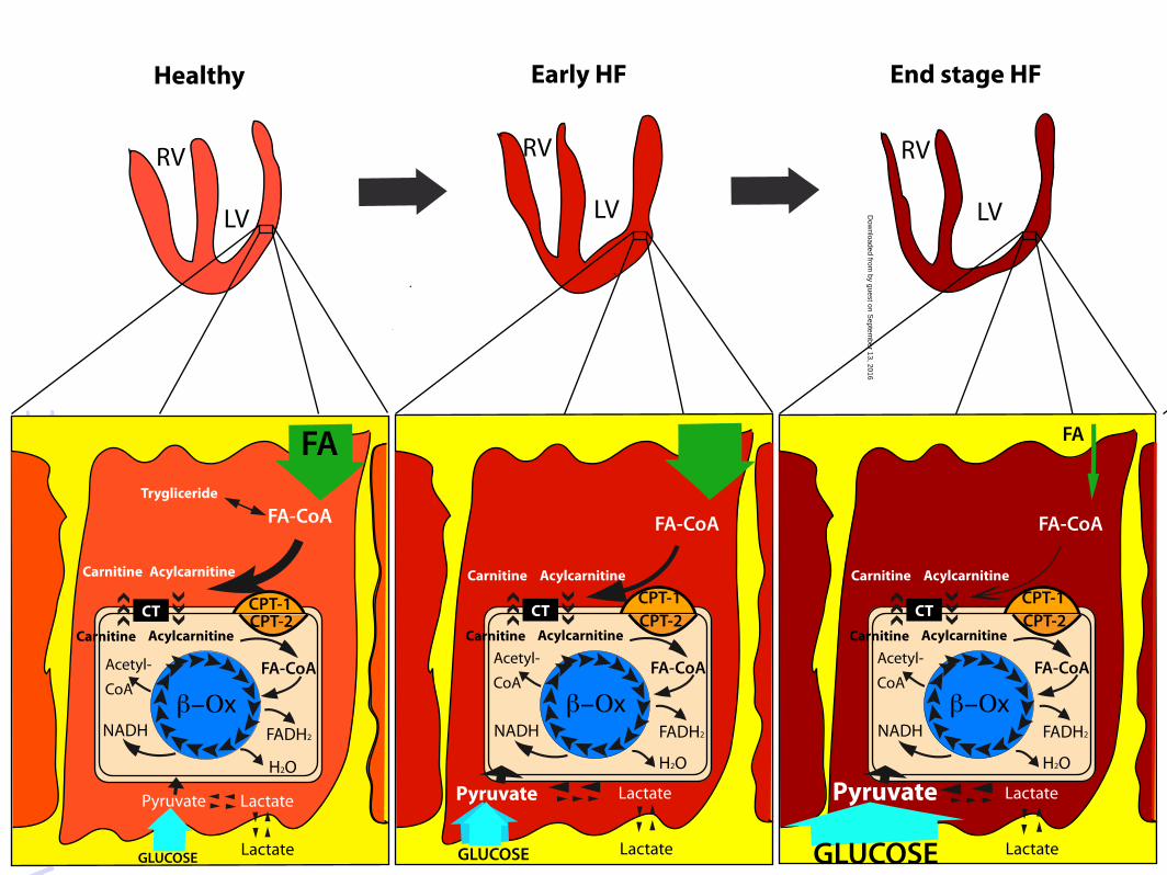
Figure 1. Cardiomyocyte substrates utilization. (A) Healthy cardiomyocyte. Cardiomyocyte mainly
uses fatty acids (FA) that enter into the cell and are converted in the mitochondria through the
carnitine palmitoyl transferase type 1 and type 2 (CPT-1 and CPT-2), and the carnitine acylcarnitine
translocase (CT) before being used by β-oxidation (β-Ox) to produce FADH2, H2O, NADH and
acetyl Co-A. Glucose and lactate enter into the cells and are transformed into pyruvate by glycolysis
and lactate dehydrogenase, respectively. (B) Early heart failure (HF) cardiomyocyte. In early HF,
FA utilization is blunted, mitochondrial FA β-Ox decreases, while uptake and metabolism of the
competing substrate glucose increase. (C) End stage HF cardiomyocyte. In overt HF, FA uptake and
metabolism are significantly depressed; although, the glucose uptake and metabolism is
significantly enhanced. RV, right ventricle; LV, left ventricle; FA-CoA, fatty acyl-coenzyme A;
FADH2, reduced form of flavine adenine dinucleotide; NADH, reduced form of nicotinamide
adenite dinucleotide.
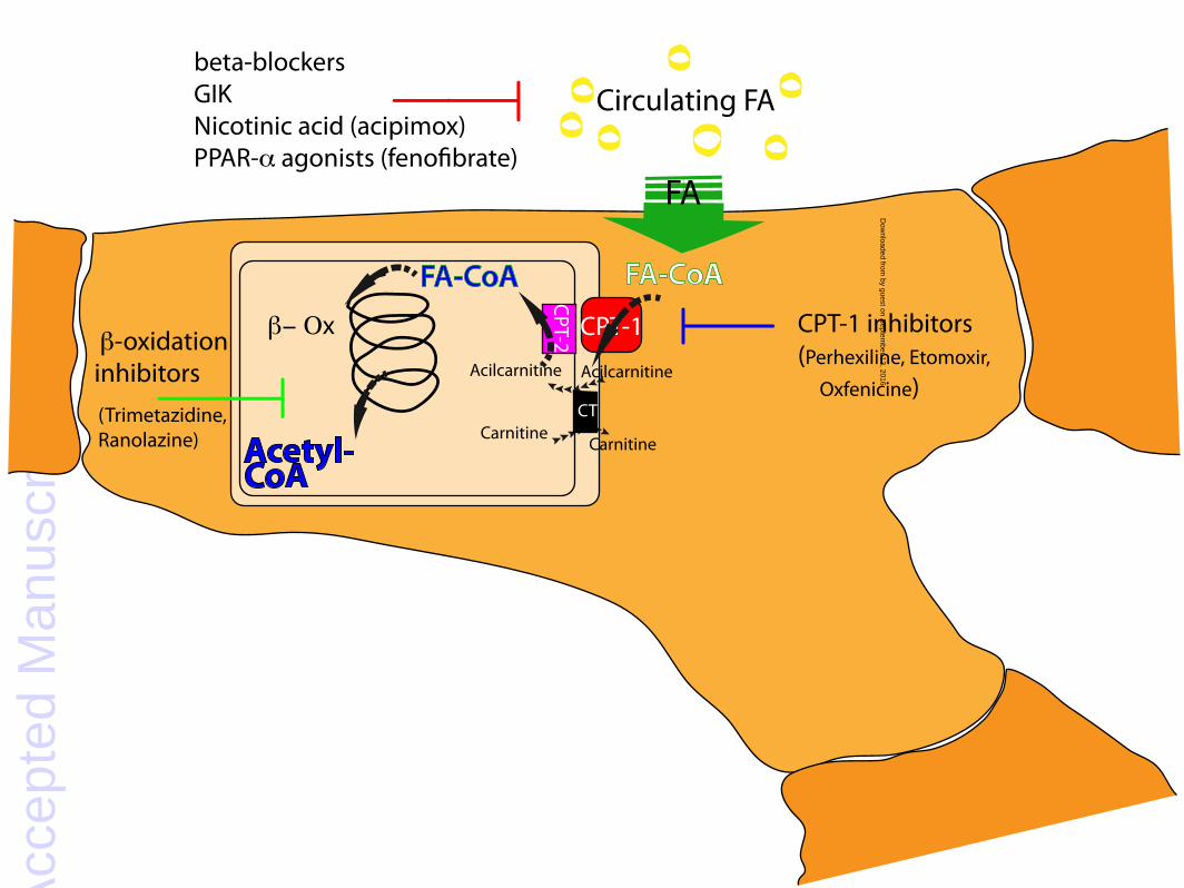
Figure 2. Targets for modulation of myocardial fatty acid metabolism. Indirect modulators of fatty
acids (FA) oxidation: beta blockers, glucose-insulin-potassium solution (GIK), nicotinic acid or
related analogues (acipimox). Direct modulators of FA oxidation: PPARα agonists, carnitine
palmitoyl transferase type 1 (CPT-1) inhibitors, β-oxidation (β-Ox) inhibitors. FA-CoA, fatty acyl-
coenzyme A; CPT-2, carnitine palmitoyl transferase type 2; CT, carnitine acylcarnitine translocase.
by guest on Septem
ber 13, 2016D
ownloaded from
Acc
epte
d M
anus
crip
tHealthy Early HF End stage HF
LV
RV
LV
RV
LV
RV
FA
GLUCOSE
FA-CoA
CPT-1CPT-2CT
Acylcarnitine
Acylcarnitine
Carnitine
Carnitine
Pyruvate
FA-CoA
β−Οx
Acetyl-
CoA
FA
GLUCOSE
FA-CoA
CPT-1CPT-2CT
Acylcarnitine
Acylcarnitine
Carnitine
Carnitine
Pyruvate
FA-CoA
β−Οx
Acetyl-
CoA
NADH FADH2
H2O
NADH FADH2
H2O
Lactate
Lactate Lactate
FA
GLUCOSE
FA-CoA
CPT-1CPT-2CT
Acylcarnitine
Acylcarnitine
Carnitine
Carnitine
Pyruvate
FA-CoA
β−Οx
Acetyl-
CoA
NADH FADH2
H2O
Lactate
Lactate
Lactate
Trygliceride
by guest on Septem
ber 13, 2016D
ownloaded from
Acc
epte
d M
anus
crip
t
FA
FA-CoA
Circulating FAbeta-blockersGIKNicotinic acid (acipimox)PPAR-α agonists (feno�brate)
CPT-1 CPT-1 inhibitors(Perhexiline, Etomoxir,
Oxfenicine) Acilcarnitine
β-oxidationinhibitors
(Trimetazidine, Ranolazine) CarnitineAcetyl-
CoA
FA-CoA
Acilcarnitine
Carnitine
CPT-2
CT
β− Οxby guest on S
eptember 13, 2016
Dow
nloaded from