minicell compositions and methods - ep 2272946 a2
TRANSCRIPT

Printed by Jouve, 75001 PARIS (FR)
(19)E
P2
272
946
A2
TEPZZ 7 946A T(11) EP 2 272 946 A2
(12) EUROPEAN PATENT APPLICATION
(43) Date of publication:12.01.2011 Bulletin 2011/02
(21) Application number: 10008869.9
(22) Date of filing: 28.05.2002
(51) Int Cl.:C12N 1/00 (2006.01)
(84) Designated Contracting States:AT BE CH CY DE DK ES FI FR GB GR IE IT LI LUMC NL PT SE TR
(30) Priority: 25.02.2002 US 359843 P24.05.2002 US 154951
(62) Document number(s) of the earlier application(s) inaccordance with Art. 76 EPC:02747872.6 / 1 487 965
(71) Applicant: Vaxiion Therapeutics IncSan Diego, CA 92182-4614 (US)
(72) Inventors:• Sabbadini, Roger A.
Lakeside, CA 92040 (US)
• Surber, MarkCoronado, CA 92118 (US)
• Berkley, NeilSan Diego, CA 92117 (US)
• Giacolone, MatthewSan Diego, CA 92117 (US)
(74) Representative: PolypatentBraunsberger Feld 2951429 Bergisch Gladbach (DE)
Remarks:This application was filed on 26-08-2010 as adivisional application to the application mentionedunder INID code 62.
(54) Minicell compositions and methods
(57) The invention provides compositions and methods for the production of achromosomal and anucleate cellsuseful for applications such as diagnositic and therapeutic uses, as well as research tools and agents for drug discovery.

EP 2 272 946 A2
2
5
10
15
20
25
30
35
40
45
50
55
Description
[0001] The invention is drawn to compositions and methods for the production of achromosomal archeabacterial,eubacterial and anucleate eukaryotic cells that are used as, e.g., therapeutics and/or diagnostics, reagents in drugdiscovery and functional proteomics, research tools, and in other applications as well.[0002] The following description of the background of the invention is provided to aid in understanding the invention,but is not admitted to describe or constitute prior art to the invention. The contents of the articles, patents, and patentapplications, and all other documents and electronically available information mentioned or cited in this application, arehereby incorporated by reference in their entirety to the same extent as if each individual publication was specificallyand individually indicated to be incorporated by reference. Applicants reserve the right to physically incorporate into thisapplication any and all materials and information from any such articles, patents, patent applications, or other documents.[0003] Minicells are achromosomal cells that are products of aberrant cell division, and contain RNA and protein, butlittleor no chromosomal DNA. Clark-Curtiss andCurtiss III, Analysis of Recombinant DNA Using Escherichia coliMinicells,101 Methods in Enzymology 347 (1983); Reeve and Mendelson, Minicells of Bacillus subtilis. A new system for transportstudies in absence of macromolecular biosynthesis, 352 Biochim. Biophys. Acta 298-305 (1974). Minicells are capableof plasmid-directed synthesis of discrete polypeptides in the absence of synthesis directed by mRNA from the bacterialchromosome. Meagher et al., Protein Expression in E. coli Minicells by Recombinant Plasmids, 10 Cell 521, 523 (1977);Roozen et al., Synthesis of Ribonucleic Acid and Protein in Plasmid-Containing Minicells of Escherichia coli K-12, 107(1) J. of Bacteriology 21 (1971); and Curtiss III, Research on bacterial conjugation with minicells and minicell-producingE. coli strains, In: Microbial Drug Resistance, Editors Susumu Mitsuhashi & Hajime Hashimoto, p. 169 (Baltimore:University Park Press 1976). Early descriptions of minicells include those of Adler et al., Genetic control of cell divisionin bacteria, 154 Science 417 (1966), and Adler et al. (Miniature Escherichia coli cells deficient in DNA, 57 Proc. Nat.Acad. Sci (Wash.) 321 (1967)). However, discovery of the production of minicells can arguably be traced to the 1930’s(Frazer and Curtiss III, Production, Properties and Utility of Bacterial Minicells, 69 Curr. Top. Microbiol. Immunol. 1-3(1975)).[0004] Prokaryotic (a.k.a. eubacterial) minicells have been used to produce various eubacterial proteins. See, e.g.,Michael Gaâel, et al., The kdpF Subunit Is Part of the K+-translocating Kdp Complex of Escherichia coli and Is Responsiblefor Stabilization of the Complex in vitro, 274(53) Jn. of Biological Chemistry 37901 (1999); Harlow, et al., Cloning andCharacterization of the gsk Gene Encoding Guanosine Kinase of Escherichia coli, 177(8) J. of Bacteriology 2236 (1995);Carol L. Pickett, et al., Cloning, Sequencing, and Expression of the Escherichia coli Cytolethal Distinding Toxin Genes,62(3) Infection & Immunity 1046 (1994); Raimund Eck & Jörn Better, Cloning and characterization of a gene coding forthe catechol 1,2 dioxygenase of Arthrobacter sp. mA3, 123 Gene 87 (1993); Andreas Schlössser, et al, Subcloning,Nucleotide Sequence, and Expression of trkG, a Gene That Encodes an Integral Membrane Protein Involved in PotassiumUptake via the Trk System of Escherichia coli, 173(10) J. of Bacteriology 3170 (1991); Mehrdad Jannatipour, et al.,Translocation of Vibrio harveyi N, N’-Diacetylchitobiase to the Outer Membrane of Escherichia coli 169(8) J. of Bacte-riology 3785 (1987); and Jacobs et al., Expression of Mycobacterium leprae genes from a Streptococcus mutans promoterin Escherichia coli K-12, 83(6) Proc. Natl. Acad. Sci. USA 1926 (1986);[0005] Various bacteria have been used, or proposed to be used, as gene delivery vectors to mammalian cells. Forreviews, see Grillot-Courvalin et al., Bacteria as gene delivery vectors for mammalian cells, 10 Current Opinion inBiotechnology 477 (1999); Johnsen et al., Transfer of DNA from Genetically Modified Organisms (GMOs), Biotechno-logical Institute, 1-70 (2000); Sizemore et al., Attenuated Shigella as a DNA delivery vehicle for DNA-mediated immu-nization, 270(5234) Science 299 (1995); Patrice Courvalin, et al., Gene transfer from bacteria to mammalian cells, 318C. R. Acad. Sci.1207 (1995); Sizemore, et al. Attenuated bacteria as a DNA delivery vehicle for DNA-mediated immu-nization, 15(8) Vaccine 804 (1997).[0006] U.S. Patent No. 4,190,495, which issued February 26, 1980, to Curtiss is drawn to minicell producing strainsof E. coli that are stated to be useful for the recombinant expression of proteins.[0007] U.S. Patent No. 4,311,797, which issued January 19, 1982 to Khachatourians is stated to be drawn to a minicellbased vaccine. The vaccine is stated to induce the production of antibodies against enteropathogenic E. coli cells incattle and is stated to be effective against coliform enteritis.[0008] Eubacterial minicells expressing immunogens from other prokaryotes have been described. Purcell et al.,Molecular cloning and characterization of the 15-kilodalton major immunogen of Treponema pallidum, Infect. Immun.57:3708, 1989.[0009] In "Biotechnology: Promise ... and Peril" (IDRC Reports 9:4-7, 1980) authors Fleury and Shirkie aver thatGeorge Khachatourians at the Unveristy of Saskatchewan, Canada, "is working on a vaccine against cholera using’minicells.’" The minciells are said to contain "genes from the pathogenic agent," and the "pathogen antigens are carriedon the surface of the minicells" (p. 5, paragraph brigding the central and right columns).[0010] Lundstrom et al., Secretion of Semliki Forest virus membrane glycoprotein E1 from Bacillus subtilis, Virus Res.2:69-83, 1985, describe the expression of the E1 protein of the eukaryotic virus, Semliki Forest virus (SFV), in Bacillus
EP 2 272 946 A2
3
5
10
15
20
25
30
35
40
45
50
55
minicells. The SFV E1 protein used in these studies is not the native E1 protein. Rather, it is a fusion protein in whichthe N-terminal signal sequence and C-terminal transmembrane domain have been removed and replaced with signalsequences from a gene from Bacillus amyloliquefaciens. The authors aver that "E1 is properly translocated through thecell membrane and secreted" (p. 81, I.I. 19-20), and note that "it has been difficult to express viral membrane proteinsin prokaryotes" (p. 81, I.27).[0011] U.S. Patent No. 4,237,224, which issued December 2, 1980, to Cohen and Boyer, describes the expressionof X. Laevis DNA in E. coli minicells.[0012] U.S. patent application Serial No. 60/293,566 (attorney docket Nos. 078853-0401 and 089608-0201 is entitled"Minicell Compositions and Methods," and was filed May 24, 2001, by Sabbadini, Roger A., Berkley, Neil L., and Klepper,Robert E., and is hereby incorporated in its entirety by reference.[0013] Jespersen et al. describes the use of "proteoliposomes" to generate antibodies to the AMPA receptor. JespersenLK, Kuusinen A, Orellana A, Keinanen K, Engberg J. Use of proteoliposomes to generate phage antibodies againstnative AMPA receptor. Eur J Biochem. 2000 Mar;267(5):1382-9[0014] The invention is drawn to compositions and methods for the production and use of minicells, including but notlimited to eubacterial minicells, in applications such as diagnostics, therapeutics, research, compound screening anddrug discovery, as well as agents for the delivery of nucleic acids and other bioactive compounds to cells.[0015] Minicells are derivatives of cells that lack chromosomal DNA and which are sometimes referred to as anucleatecells. Because eubacterial and archeabacterial cells, unlike eukaryotic cells, do not have a nucleus (a distinct organellethat contains chromosomes), these non-eukaryotic minicells are more accurately described as being "without chromo-somes" or "achromosomal," as opposed to "anucleate." Nonetheless, those skilled in the art often use the term "anucleate"when referring to bacterial minicells in addition to other minicells. Accordingly, in the present disclosure, the term "min-icells" encompasses derivatives of eubacterial cells that lack a chromosome; derivatives of archeabacterial cells thatlack their chromosome(s), and anucleate derivatives of eukaryotic cells. It is understood, however, that some of therelevant art may use the terms "anucleate minicells" or anucleate cells" loosely to refer to any of the preceeding typesof minicells.[0016] In one aspect, the invention is drawn to a eubacterial minicell comprising a membrane protein that is notnaturally found in a prokaryote, i.e., a membrane protein from a eukaryote or an archeabacterium. Such minicells may,but need not, comprise an expression element that encodes and expresses the membrane protein that it comprises.The membrane protein may be one found in any non-eubacterial membrane, including, by way of non-limiting example,a cellular membrane, a nuclear membrane, a nucleolar membrane, a membrane of the endoplasmic reticulum (ER), amembrane of a Golgi body, a membrane of a lysosome a membrane of a peroxisome, a caveolar membrane, an outermembrane of a mitochondrion or a chloroplast, and an inner membrane of a mitochondrion or a chloroplast. By way ofnon-limiting example, a membrane protein may be a receptor, such as a G-protein coupled receptor; an enzyme, suchas ATPase or adenylate cyclase, a cytochrome; a channel; a transporter; or a membrane-bound nucleic acid bindingfactor, such as a transcription and/or translation factor; signaling components; components of the electon transport chain(ETC); or cellular antigens. A membrane fusion protein, which is generated in vitro using molecular cloning techniques,does not occur in nature and is thus a membrane protein that is not naturally found in a prokaryote, even if the fusionprotein is prepared using amino acid sequences derived from eubacterial proteins.[0017] Minicells that have segregated from parent cells lack chromosomal and/or nuclear components, but retain thecytoplasm and its contents, including the cellular machinery required for protein expression. Although chromosomes donot segregate into minicells, extrachromosomal and/or episomal genetic expression elements will segregate, or may beintroduced into mincells after segregation from parent cells. Thus, in one aspect, the invention is drawn to minicellscomprising an expression element, which may be an inducible expression element, that comprises expression sequencesoperably linked to an open reading frame (ORF) that encodes the non-eubacterial membrane protein. In a related aspect,the invention is drawn to minicell-producing host cells having an expression element, which may be an inducible ex-pression element, that comprises expression sequences operably linked to an ORF that encodes a non-eubacterialmembrane protein. In a related aspect, the invention is drawn to a method of making a eubacterial minicell comprisinga membrane protein that is not naturally found in a prokaryote, the method comprising growing minicell-producing hostcells, the host cells having an expression element, which may be an inducible expression element, that comprisesexpression sequences operably linked to an ORF that encodes a non-eubacterial membrane protein; and preparingminicells from the host cells. Optionally, at any point in the method, an inducing agent is provided in order to induceexpression of an ORF that encodes a non-eubacterial membrane protein.[0018] In one aspect, the invention is drawn to display produced membrane-associated protein(s) on the surface ofthe minicell. For purposes of this document, the term "display" is defined as exposure of the structure of interest on theouter surface of the minicell. By way of non-limiting example, this structure may be an internally expressed membraneprotein or chimeric construct to be inserted in or associated with the minicell membrane such that the extracellulardomain or domain of interest is exposed on the outer surface of the minicell (expressed and displayed on the surfaceof the minicell or expressed in the parental cell to be displayed on the surface of the segregated minicell). In any scenario,

EP 2 272 946 A2
4
5
10
15
20
25
30
35
40
45
50
55
the "displayed" protein or protein domain is available for interaction with extracellular components. A membrane-asso-ciated protein may have more than one extracellular domain, and a minicell of the invention may display more than onemembrane-associated protein.[0019] The membrane protein displayed by minicells may be a fusion protein, i.e., a protein that comprises a firstpolypeptide having a first amino acid sequence and a second polypeptide having a second amino acid sequence, whereinthe first and second amino acid sequences are not naturally present in the same polypeptide. At least one polypeptidein a membrane fusion protein is a "transmembrane domain" or "membrane-anchoring domain". The transmembraneand membrane-anchoring domains of a membrane fusion protein may be selected from membrane proteins that naturallyoccur in a eucaryote, such as a fungus, a unicellular eucaryote, a plant and an animal, such as a mammal including ahuman. Such domains may be from a viral membrane protein naturally found in a virus such as a bacteriophage or aeucaryotic virus, e.g., an adenovirus or a retrovirus. Such domains may be from a membrane protein naturally found inan archaebacterium such as a thermophile.[0020] The displayed domain of a membrane fusion protein may be a binding moiety. By way of non-limiting example,binding moieties used for particular purposes may be a binding moiety directed to a compound or moiety displayed bya specific cell type or cells found predominantly in one type of tissue, which may be used to target minicells and theircontents to specific cell types or tissues; or a binding moiety that is directed to a compound or moiety displayed by apathogen, which may be used in diagnostic or therapeutic methods; a binding moiety that is directed to an undesirablecompound, such as a toxin, which may be used to bind and preferably internalize and/or neutralize the undesirablecompound; a diseased cell; or the binding moiety may be a domain that allows for the minicells to be covalently or non-covalently attached to a support material, which may be used in compositions and methods for compound screeningand drug discovery. By "diseased cell" it is meant pathogen-infected cells, malfunctioning cells, and dysfunctional cells,e.g., cancer cells.[0021] In various aspects, the minicells of the invention comprise one or more biologically active compounds. Theterm "biologically active" (synonymous with "bioactive") indicates that a composition or compound itself has a biologicaleffect, or that it modifies, causes, promotes, enhances, blocks, reduces, limits the production or activity of, or reactswith or binds to an endogenous molecule that has a biological effect. A "biological effect" may be but is not limited toone that stimulates or causes an immunoreactive response; one that impacts a biological process in an animal; one thatimpacts a biological process in a pathogen or parasite; one that generates or causes to be generated a detectable signal;and the like. Biologically active compositions, complexes or compounds may be used in therapeutic, prophylactic anddiagnostic methods and compositions. Biologically active compositions, complexes or compounds act to cause or stim-ulate a desired effect upon an animal. Non-limiting examples of desired effects include, for example, preventing, treatingor curing a disease or condition in an animal suffering therefrom; limiting the growth of or killing a pathogen in an animalinfected thereby; augmenting the phenotype or genotype of an animal; stimulating a prophylactic immunoreactive re-sponse in an animal; or diagnosing a disease or disorder in an animal.[0022] In the context of therapeutic applications of the invention, the term "biologically active" indicates that the com-position, complex or compound has an activity that impacts an animal suffering from a disease or disorder in a positivesense and/or impacts a pathogen or parasite in a negative sense. Thus, a biologically active composition, complex orcompound may cause or promote a biological or biochemical activity within an animal that is detrimental to the growthand/or maintenance of a pathogen or parasite; or of cells, tissues or organs of an animal that have abnormal growth orbiochemical characteristics, such as cancer cells.[0023] In the context of diagnostic applications of the invention, the term "biologically active" indicates that the com-position, complex or compound can be used for in vivo or ex vivo diagnostic methods and in diagnostic compositionsand kits. For diagnostic purposes, a preferred biologically active composition or compound is one that can be detected,typically (but not necessarily) by virtue of comprising a detectable polypeptide. Antibodies to an epitope found on com-position or compound may also be used for its detection.[0024] In the context of prophylactic applications of the invention, the term "biologically active" indicates that thecomposition or compound induces or stimluates an immunoreactive response. In some preferred embodiments, theimmunoreactive response is designed to be prophylactic, i.e., prevents infection by a pathogen. In other preferredembodiments, the immunoreactive response is designed to cause the immune system of an animal to react to thedetriment of cells of an animal, such as cancer cells, that have abnormal growth or biochemical characteristics. In thisapplication of the invention, compositions, complexes or compounds comprising antigens are formulated as a vaccine.[0025] It will be understood by those skilled in the art that a given composition, complex or compound may be biologicallyactive in therapeutic, diagnostic and prophylactic applications. A composition, complex or compound that is describedas being "biologically active in a cell" is one that has biological activity in vitro (i.e., in a cell culture) or in vivo (i.e., inthe cells of an animal). A "biologically active component" of a composition or compound is a portion thereof that isbiologically active once it is liberated from the composition or compound. It should be noted, however, that such acomponent may also be biologically active in the context of the composition or compound.[0026] In one aspect, the minicells of the invention comprise a therapeutic agent. Such minicells may be used to

EP 2 272 946 A2
5
5
10
15
20
25
30
35
40
45
50
55
deliver therapeutic agents. In a preferred embodiment, a minicell comprising a therapeutic agent displays a bindingmoiety that specifically binds a ligand present on the surface of a cell, so that the minicells may be "targeted" to the cell.The therapeutic agent may be any type of compound or moiety, including without limitation small molecules, polypeptides,antibodies and antibody derivatives and nucleic acids. The therapeutic agent may be a drug; a prodrug, i.e., a compoundthat becomes biologically active in vivo after being introduced into a subject in need of treatment; or an immunogen.[0027] In one aspect, the minicells of the invention comprise a detectable compound or moiety. As is understood bythose of skill in the art, a compound or moiety that is "detectable" produces a signal that can detected by spectroscopic,photochemical, biochemical, immunochemical, electromagnetic, radiochemical, or chemical means such as fluores-cence, chemifluoresence, or chemiluminescence, electrochemilumenscence, or any other appropriate means. A detect-able compound may be a detectable polypeptide, and such polypeptides may, but need not, be incorporated into fusionmembrane proteins of the minicell. Detectable polypeptides or amino acid sequences, includes, by way of non-limitingexample, a green fluorescent protein (GFP), a luciferase, a beta-galactosidase, a His tag, an epitope, or a biotin-bindingprotein such as streptavidin or avidin. The detectable compound or moiety may be a radiolabeled compound or aradioisotope. A detectable compound or moiety may be a small molecule such as, by way of non-limiting example, afluorescent dye; a radioactive iostope; or a compound that may be detected by x-rays or electromagnetic radiation.Image enhancers as those used for CAT and PET scans (e.g., calcium, gallidium) may be used. In another non-limitingexample, detectable labels may also include loss of catalytic substrate or gain of catalytic product following catalysis bya minicell displayed, solule cytoplasmic, or secreted enzyme.[0028] In one aspect, the invention is drawn to a minicell comprising one or more bioactive nucleic acids or templatesthereof. By way of non-limiting example, a bioactive nucleic acid may be an antisense oligonucleotide, an aptamer, anantisense transcript, a ribosomal RNA (rRNA), a transfer RNA (tRNA), a molecular decoy, or an enzymatically activenucleic acid, such as a ribozyme. Such minicells can, but need not, comprise a displayed polypeptide or protein on thesurface of the minicell. The displayed polypeptide or protein may be a binding moiety directed to a compound or moietydisplayed by a particular type of cell, or to a compound or moiety displayed by a pathogen. Such minicells can further,but need not, comprise an expression element having eubacterial, archael, eucaryotic, or viral expression sequencesoperably linked to a nucleotide sequence that serves as a template for a bioactive nucleic acid.[0029] In one aspect, the invention is drawn to immunogenic minicells, i.e., minicells that display an immunogen,vaccines comprising immunogenic minicells, antibodies and antibody derivatives directed to immunogens displayed onimmunogenic minicells, and method of making and using immunogenic minicells and antibodies and antibody derivativesproduced therefrom in prophylactic, diagnostic, therapeutic and research applications. A preferred immunogen displayedby a minicell is an immunogenic polypeptide, which is preferably expressed from an expression element contained withinthe minicell in order to maximize the amount of immunogen displayed by the immunogenic minicells. The immunogenicpolypeptide can be derived from any organism, obligate intracelluar parasite, organelle or virus with the provisio that,in prophylactic applications, the immunogenic polypeptide is not derived from a prokaryote, including a eubacterial virus.The source organism for the immunogen may be a pathogen. A minicell displaying an immunogen derived from apathogen is formulated into a vaccine and, in a prophylactic application, used to treat or prevent diseases and disorderscaused by or related to the eukaryotic or archeabacterial pathogen.[0030] In a separate aspect, the invention is drawn to minicells that display an immunogen derived from a nonfunctional,dysfunctional and/or diseased cell. By way of non-limiting example, the minicells display an immunogenic polypeptidederived from a hyperproliferative cell, i.e., a cell that is tumorigenic, or part of a tumor or cancer. As another non-limitingexample, a cell that is infected with a virus or an obligate intracellular parasite (e.g., Rickettsiae) displays an immunogenicpolypeptide that is encoded by the genome of the infected cell but is aberrenatly expressed in an infected cell. A vaccinecomprising a minicell displaying an immunogen derived from a nonfunctional, dysfunctional and/or diseased cell is usedin methods of treating or preventing hyperproliferative diseases or disorders, including without limitation a cell comprisingan intracellular pathogen.[0031] In one aspect, the invention is drawn to a minicell comprising a membrane protein that is linked to a conjugatablecompound (a.k.a. "attachable compound"). The conjugatable compound may be of any chemical nature and have oneor more therapeutic or detectable moities. By way of non-limiting example, a protein having a transmembrane or mem-brane anchoring domain is displayed and has the capacity to be specifically cross-linked on its extracellular domain.Through this approach, any conjugatable compound of interest may be quickly and easily attached to the outer surfaceof minicells containing this expressed membrane-spanning domain. In aspects of the invention wherein minicells areused for drug delivery in vivo, a preferred conjugatable compound is polyethylene glycol (PEG), which provides for"stealth" minicells that are not taken as well and/or as quickly by the reticuloendothelial system (RES). Other conjugatablecompounds include polysaccharides, polynucleotides, lipopolysaccharides, lipoproteins, glycosylated proteins, syntheticchemical compounds, and/or chimeric combinations of these examples listed.[0032] In various aspects of the invention, the minicell displays a polypeptide or other compound or moiety on itssurface. By way of non-limiting example, a non-eubacterial membrane protein displayed by eubacterial minicells maybe a receptor. Minicells displaying a receptor may, but need not, bind ligands of the receptor. In therapeutic applications

EP 2 272 946 A2
6
5
10
15
20
25
30
35
40
45
50
55
of this aspect of the invention, the ligand is an undesirable compound that is bound to its receptor and, in some aspects,is internalized by the minicells. The non-eubacterial membrane protein displayed by minicells may be a fusion protein,i.e., a protein that comprises a first polypeptide having a first amino acid sequence and a second polypeptide having asecond amino acid sequence, wherein the first and second amino acid sequences are not naturally present in the samepolypeptide. At least one polypeptide in a membrane fusion protein is a "transmembrane domain" or "membrane-an-choring domain". The transmembrane and membrane-anchoring domains of a membrane fusion protein may be selectedfrom membrane proteins that naturally occur in a eukaryote, such as a fungus, a unicellular eukaryote, a plant and ananimal, such as a mammal including a human. Such domains may be from a viral membrane protein naturally found ina virus such as a bacteriophage or a eukaryotic virus, e.g., an adenovirus or a retrovirus. Such domains may be froma membrane protein naturally found in an archaebacterium such as a thermophile.[0033] The displayed domain of a membrane fusion protein may be a binding moiety. By way of non-limiting example,binding moieties used for particular purposes may be a binding moiety directed to a compound or moiety displayed bya specific cell type or cells found predominantly in one type of tissue, which may be used to target minicells and theircontents to specific cell types or tissues; or a binding moiety that is directed to a compound or moiety displayed by apathogen, which may be used in diagnostic or therapeutic methods; a binding moiety that is directed to an undesirablecompound, such as a toxin, which may be used to bind and preferably internalize and/or neutralize the undesirablecompound; a diseased cell; or the binding moiety may be a domain that allows for the minicells to be covalently or non-covalently attached to a support material, which may be used in compositions and methods for compound screeningand drug discovery.[0034] In one aspect, the invention provides minicells that may be used as research tools and/or kits comprising suchresearch tools. The minicells of the invention may be used as is, or incorporated into research tools useful for scientificresearch regarding all amino acid comprising compounds including, but not limited to membrane-associated proteins,chimeric membrane fusion proteins, and soluble proteins. Such scientific research includes, by way of non-limitingexample, basic research, as well as pharmacological, diagnostic, and pharmacogenetic studies. Such studies may becarried out in vivo or in vitro.[0035] In other aspects, the invention is drawn to methods of preparing the minicells, protoplasts, and poroplasts™of the invention for various applications including but not limited to diagnostic, therapeutic, research and screeningapplications. In a related aspect, the invention is drawn to pharmaceutical compositions, reagents and kits comprisingminicells.[0036] In each aspect and embodiment of the invention, unless stated otherwise, embodiments wherein the minicellis a eubacterial minicell, a poroplast, a spheroplast or a protoplast exist.[0037] In a first aspect, the invention provides a minicell comprising a membrane protein selected from the groupconsisting of a eukaryotic membrane protein, an archeabacterial membrane protein and an organellar membrane protein.In another embodiment, wherein the minicell comprises a biologically active compound. By way of non-limiting example,the biologically active compound is a radioisotope, a polypeptide, a nucleic acid or a small molecule.[0038] In another embodiment, the minicell comprises a expression construct, wherein the first expression constructcomprises expression sequences operably linked to an ORF that encodes a protein. In another embodiment, the ORFencodes the membrane protein. In another embodiment, the expression sequences that are operably linked to an ORFare inducible and/or repressible.[0039] In another aspect, the minicell comprises a second expression construct, wherein the second expressionconstruct comprises expression sequences operably linked to a gene. In another embodiment, the expression sequencesthat are operably linked to a gene are inducible and/or repressible. In a related embodiment, the gene product of thegene regulates the expression of the ORF that encodes the protein. A factor that "regulates" the expression of a geneor a gene product directly or indirectly initiates, enhances, quickens, slows, terminates, limits or completely blocksexpression of a gene. In different embodiments, the gene product of the gene is a nucleic acid or a polypeptide. Thepolypeptide can be of any type, including but not limited to a membrane protein, a soluble protein or a secreted protein.A membrane protein can be a membrane fusion protein comprising a first polypeptide, which comprises at least onetransmembrane domain or at least one membrane anchoring domain; and a second polypeptide.[0040] In one aspect, the invention provides a minicell comprising a membrane fusion protein, the fusion proteincomprising a first polypeptide, the first polypeptide comprising at least one transmembrane domain or at least onemembrane anchoring domain; and a second polypeptide, wherein the second polypeptide is not derived from a eubacterialprotein and is neither a His tag nor a glutathione-S-transferase polypeptide. In various embodiments, the minicell is aeubacterial minicell, a poroplast, a spheroplast or a protoplast. In one embodiment, the minicell comprises a biologicallyactive compound.[0041] In one aspect, the invention provides a minicell comprising a membrane conjugate, wherein the membraneconjugate comprises a membrane protein chemically linked to a conjugated compound. In one embodiment, the conju-gated compound is selected from the group consisting of a nucleic acid, a polypeptide, a lipid and a small molecule.[0042] In one aspect, the invention provides a method for making minicells, comprising (a) culturing a minicell-producing

EP 2 272 946 A2
7
5
10
15
20
25
30
35
40
45
50
55
parent cell, wherein the parent cell comprises an expression construct, wherein the expression construct comprises agene operably linked to expression sequences that are inducible and/or repressible, and wherein induction or repressionof the gene causes or enhances the production of minicells; and (b) separating the minicells from the parent cell, therebygenerating a composition comprising minicells, wherein an inducer or repressor is present within the parent cells duringone or more steps and/or between two or more steps of the method. In one embodiment, the method further comprises(c) purifying the minicells from the composition.[0043] Relevant gene products are factors involved in or modulating DNA replication, cellular division, cellular parti-tioning, septation, transcription, translation, or protein folding. The minicells are separated from parent cells by processessuch as centrifugation, ultracentrifugation, density gradation, immunoaffinity, immunoprecipitation and other techniquesdescribed herein.[0044] In one embodiment, the minicell is a poroplast, and the method further comprises (d) treating the minicells withan agent, or incubating the minicells under a set of conditions, that degrades the outer membrane of the minicell. Theouter membrane is degraded by treatment with an agent selected from the group consisting of EDTA, EGTA, lactic acid,citric acid, gluconic acid, tartaric acid, polyethyleneimine, polycationic peptides, cationic leukocyte peptides, aminogly-cosides, aminoglycosides, protamine, insect cecropins, reptilian magainins, polymers of basic amino acids, polymixinB, chloroform, nitrilotriacetic acid and sodium hexametaphosphate; by exposure to conditions selected from the groupconsisting of osmotic shock and insonation; and by other methods described herein.[0045] In one embodiment, further comprising removing one or more contaminants from the composition. Represent-ative contaminants are LPS and peptidoglycan. In a representative embodiment, LPS is removed by contacting thecomposition to an agent that binds or degrades LPS. At least about 50%, preferably about 65% to about 75%, morepreferably 95%, most preferably 99% or >99% of LPS is removed from an initial preparation of minicells. In a relatedembodiment, the minicell-producing parent cell comprises a mutation in a gene required for lipopolysaccharide synthesis.[0046] In on embodiment, the minicell is a spheroplast, and the method further comprises (d) treating the minicellswith an agent, or incubating the minicells under a set of conditions, that disrupts or degrades the outer membrane; and(e) treating the minicells with an agent, or incubating the minicells under a set of conditions, that disrupts or degradesthe cell wall. The agent that disrupts or degrades the cell wall can be. e.g., a lysozyme, and the set of conditions thatdisrupts or degrades the cell wall can be, e.g., incubation in a hypertonic solution.[0047] In one embodiment, the minicell is a protoplast, and the method further comprises (d treating the minicells withan agent, or incubating the minicells under a set of conditions, that disrupt or degrade the outer membrane; (e) treatingthe minicells with an agent, or incubating the minicells under a set of conditions, that disrupts or degrades the cell wall,in order to generate a composition that comprises protoplasts; and (f) purifying protoplasts from the composition. In oneembodiment, the method further comprises preparing a denuded minicell from the minicell. In one embodiment, themethod further comprises covalently or non-covalently linking one or more components of the minicell to a conjugatedmoiety.[0048] In one aspect, the invention provides a L-form minicell comprising (a) culturing an L-form eubacterium, whereinthe eubacterium comprises one or more of the following: (i) an expression element that comprises a gene operablylinked to expression sequences that are inducible and/or repressible, wherein induction or repression of the gene reg-ulates the copy number of an episomal expression construct; (ii) a mutation in an endogenous gene, wherein the mutationregulates the copy number of an episomal expression construct; (iii) an expression element that comprises a geneoperably linked to expression sequences that are inducible and/or repressible, wherein induction or repression of thegene causes or enhances the production of minicells; and (iv) a mutation in an endogenous gene, wherein the mutationcauses or enhances minicell production; (b) culturing the L-form minicell-producing parent cell in media under conditionswherein minicells are produced; and (c) separating the minicells from the parent cell, thereby generating a compositioncomprising L-form minicells, wherein an inducer or repressor is present within the minicells during one or more stepsand/or between two or more steps of the method. In one embodiment, the method further comprises (d) purifying theL-form minicells from the composition.[0049] In one aspect, the invention provides a solid support comprising a minicell, wherein the minicell displays afusion protein, the fusion protein comprising a first polypeptide that comprises at least one transmembrane domain orat least one membrane anchoring domain, and a second polypeptide. In various embodiments, the second polypeptidecomprises a binding moiety or an enzyme moiety.[0050] In one aspect, the invention provides a eubacterial minicell comprising a biologically active compound, whereinthe minicell displays a binding moiety, wherein the binding moiety is selected from the group consisting of (a) a eukaryoticmembrane protein; (b) an archeabacterial membrane protein; (c) an organellar membrane protein; and (d) a fusionprotein, the fusion protein comprising a first polypeptide, the first polypeptide comprising at least one transmembranedomain or at least one membrane anchoring domain; and a second polypeptide, wherein the second polypeptide is notderived from a eubacterial protein and is neither a His tag nor a glutathione-S-transferase polypeptide, and wherein thepolypeptide comprises a binding moiety.[0051] In one embodiment, the binding moiety is selected from the group consisting of an antibody, an antibody

EP 2 272 946 A2
8
5
10
15
20
25
30
35
40
45
50
55
derivative, a receptor and an active site of a non-catalytic derivative of an enzyme. In a preferred embodiment, thebinding moiety is a single-chain antibody. In one embodiment, one of the ORFs encodes a protein that comprises thebinding moiety.[0052] In one embodiment, the binding moiety is directed to a ligand selected from the group consisting of an epitopedisplayed on a pathogen, an epitope displayed on an infected cell and an epitope displayed on a hyperproliferative cell.[0053] In one embodiment, the invention further comprises a first and second nucleic acid, wherein the first nucleicacid comprises eukaryotic expression sequences operably linked to a first ORF, and a second nucleic acid, wherein thesecond nucleic acid comprises eubacterial expression sequences operably linked to a second ORF.[0054] In one embodiment, the eubacterial expression sequences are induced and/or derepressed when the bindingmoiety is in contact with a target cell. In a variant embodiment, the eukaryotic expression sequences are induced and/orderepressed when the nucleic acid is in the cytoplasm of a eukaryotic cell. In related embodiments, the protein encodedby the first ORF comprises eukaryotic secretion sequences and/or the protein encoded by the second ORF compriseseubacterial secretion sequences.[0055] In one aspect, the invention provides a method of associating a radioactive compound with a cell, wherein thecell displays a ligand specifically recognized by a binding moiety, comprising contacting the cell with a minicell thatcomprises the radioactive compound and displays the binding moiety. In a diagnostic embodiment, the amount ofradiation emitted by the radioactive isotope is sufficient to be detectable. In a therapeutic embodiment, the amount ofradiation emitted by the radioactive isotope is sufficient to be cytotoxic. In one embodiment, the ligand displayed by thecell is selected from the group consisting of an epitope displayed on a pathogen, an epitope displayed on an infectedcell and an epitope displayed on a hyperproliferative cell. In one embodiment, the binding moiety is selected from thegroup consisting of an antibody, an antibody derivative, a channel protein and a receptor, and is preferably a single-chain antibody. In other embodiments, the binding moiety is an aptamer or a small molecule. In one embodiment, theligand is selected from the group consisting of a cytokine, hormone, and a small molecule.[0056] In one aspect, the invention provides a method of delivering a biologically active compound to a cell, whereinthe cell displays a ligand specifically recognized by a binding moiety, comprising contacting the cell with a minicell thatdisplays the binding moiety, wherein the minicell comprises the biologically active compound, and wherein the contentsof the minicell are delivered into the cell from a minicell bound to the cell. In one embodiment, the biologically activecompound is selected from the group consisting of a nucleic acid, a lipid, a polypeptide, a radioactive compound, an ionand a small molecule.[0057] In one embodiment, the membrane of the minicell comprises a system for transferring a molecule from theinterior of a minicell into the cytoplasm of the cell. A representative system for transferring a molecule from the interiorof a minicell into the cytoplasm of.the cell is a Type III secretion system.[0058] In one embodiment, the minicell further comprises a first and second nucleic acid, wherein the first nucleic acidcomprises eukaryotic expression sequences operably linked to a first ORF, and a second nucleic acid, wherein thesecond nucleic acid comprises eubacterial expression sequences operably linked to a second ORF. In one embodiment,one of the ORFs encodes a protein that comprises the binding moiety. In one embodiment, the eubacterial expressionsequences are induced and/or derepressed when the binding moiety is in contact with a target cell. In one embodiment,the eukaryotic expression sequences are induced and/or derepressed when the nucleic acid is in the cytoplasm of aeukaryotic cell. In one embodiment, the protein encoded by the first ORF comprises eukaryotic secretion sequencesand/or the protein encoded by the second ORF comprises eubacterial secretion sequences. In one embodiment, theligand is selected from the group consisting of a cytokine, hormone, and a small molecule.[0059] In one aspect, the invention provides a minicell displaying a synthetic linking moiety, wherein the syntheticlinking moiety is covalenty or non-covalently attached to a membrane component of the mincell.[0060] In one aspect, the invention provides a sterically stabilized minicell comprising a displayed moiety that has alonger half-life in vivo than a wild-type minicell, wherein the displayed moiety is a hydrophilic polymer that comprises aPEG moiety, a carboxylic group of a polyalkylene glycol or PEG stearate.[0061] In one aspect, the invention provides a minicell having a membrane comprising an exogenous lipid, whereina minicell comprising the exogenous lipid has a longer half-life in vivo than a minicell lacking the exogenous lipid, andwherein the minicell is selected from the group consisting of a eubacterial minicell, a poroplast, a spheroplast and aprotoplast. In one embodiment, the exogenous lipid is a derivitized lipid which may, by way of non-limiting example, bephosphatidylethanolamine derivatized with PEG, DSPE-PEG, PEG stearate; PEG-derivatized phospholipids, a PEGceramide or DSPE-PEG.[0062] In one embodiment, the exogenous lipid is not present in a wild-type membrane, or is present in a differentproportion than is found in minicells comprising a wild-type membrane. The exogenous lipid can be a ganglioside,sphingomyelin, monosialoganglioside GM1, galactocerebroside sulfate, 1,2-sn-dimyristoylphosphatidylcholine, phos-phatidylinositol and cardiolipin.[0063] In one embodiment, the linking moiety is non-covalently attached to the minicell. In one embodiment, one ofthe linking moiety and the membrane component comprises biotin, and the other comprises avidin or streptavidin. In

EP 2 272 946 A2
9
5
10
15
20
25
30
35
40
45
50
55
one embodiment, the synthetic linking moiety is a cross-linker. In one embodiment, the cross-linker is a bifunctionalcross-linker.[0064] In one aspect, the invention provides a method of transferring a membrane protein from a minicell membraneto a biological membrane comprising contacting a minicell to the biological membrane, wherein the minicell membranecomprises the membrane protein, and allowing the mincell and the biological membrane to remain in contact for a periodof time sufficient for the transfer to occur.[0065] In one embodiment, the biological membrane is a cytoplasmic membrane or an organellar membrane. In oneembodiment, the biological membrane is a membrane selected from the group consisting of a membrane of a pathogen,a membrane of an infected cell and a membrane of a hyperproliferative cell. In one embodiment, the biological membraneis the cytoplasmic membrane of a recipient cell, which may be a cultured cell and a cell within an organism. In oneembodiment, the biological membrane is present on a cell that has been removed from an animal, the contacting occursin vitro, after which the cell is returned to the organism.[0066] In one embodiment, the membrane protein is an enzyme. In this embodiment, the membrane protein havingenzymatic activity is selected from the group consisting of a cytochrome P450 and a fusion protein, the fusion proteincomprising a first polypeptide, the first polypeptide comprising at least one polypeptide, wherein the second polypeptidehas enzymatic acitivity.[0067] In one embodiment, the membrane protein is a membrane fusion protein, the membrane fusion protein com-prising a first polypeptide, the first polypeptide comprising at least one transmembrane domain or at least one membraneanchoring domain; and a second polypeptide.[0068] In one embodiment, the second polypeptide is a biologically active polypeptide. In one embodiment, the minicelldisplays ligand or a binding moiety.[0069] In one aspect, the invention provides a minicell that comprises an expression construct comprising an ORFencoding a membrane protein operably linked to expression sequences, wherein the expression sequences are inducedand/or derepressed when the minicell is in contact with a target cell.[0070] In one embodiment, the biological membrane is a cytoplasmic membrane or an organellar membrane. In oneembodiment, the biological membrane is a membrane selected from the group consisting of a membrane of a pathogen,a membrane of an infected cell and a membrane of a hyperproliferative cell. In one embodiment, the minicell displaysa ligand or a binding moiety selected from the group consisting of an antibody, an antibody derivative, an aptamer anda small molecule. In one embodiment, the membrane protein is a membrane fusion protein, the membrane fusion proteincomprising a first polypeptide, the first polypeptide comprising at least one transmembrane domain or at least onemembrane anchoring domain; and a second polypeptide. In one embodiment, the ligand is selected from the groupconsisting of a cytokine, hormone, and a small molecule.[0071] In one aspect, the invention provides a pharmaceutical composition comprising a minicell, wherein the minicelldisplays a membrane protein, wherein the membrane protein is selected from the group consisting of a eukaryoticmembrane protein, an archeabacterial membrane protein and an organellar membrane protein. In one embodiment, themembrane protein is selected from the group consisting of a receptor, a channel protein, a cellular adhesion factor andan integrin. In one embodiment, the pharmaceutical formulation further comprises an adjuvant. In one embodiment, themembrane protein comprises a polypeptide epitope displayed by a hyperproliferative cell. In one embodiment, themembrane protein comprises an epitope displayed by a eukaryotic pathogen, an archeabacterial pathogen, a virus oran infected cell.[0072] In one aspect, the invention provides a pharmaceutical composition comprising a minicell, wherein the minicelldisplays a membrane protein that is a fusion protein, the fusion protein comprising (i) a first polypeptide, the first polypep-tide comprising at least one transmembrane domain or at least one membrane anchoring domain; and (ii) a secondpolypeptide, wherein the second polypeptide is not derived from a eubacterial protein. In one embodiment, the pharma-ceutical formulation further comprises an adjuvant. In one embodiment, the second polypeptide comprises a polypeptideepitope displayed by a hyperproliferative cell. In one embodiment, the membrane protein comprises an epitope displayedby a eukaryotic pathogen, an archeabacterial pathogen, a virus or an infected cell.[0073] In one aspect, the invention provides a pharmaceutical composition comprising a minicell, wherein the minicelldisplays a membrane conjugate, wherein the membrane conjugate comprises a membrane component chemically linkedto a conjugated compound. In one embodiment, the membrane protein is selected from the group consisting of a receptor,a channel protein, a cellular adhesion factor and an integrin. In one embodiment, the pharmaceutical further comprisesan adjuvant. In one embodiment, the membrane component is a polypeptide comprising at least one transmembranedomain or at least one membrane anchoring domain, or a lipid that is part of a membrane. In one embodiment, theconjugated compound is a polypeptide, and the chemical linkage between the membrane compound and the conjugatedcompound is not a peptide bond. In one embodiment, the conjugated compound is a nucleic acid. In one embodiment,the conjugated compound is an organic compound. In one embodiment, the organic compound is selected from thegroup consisting of a narcotic, a toxin, a venom, a sphingolipid and a soluble protein.[0074] In one aspect, the invention provides a method of making a pharmaceutical composition comprising a minicell,

EP 2 272 946 A2
10
5
10
15
20
25
30
35
40
45
50
55
wherein the minicell displays a membrane protein, wherein the membrane protein is selected from the group consistingof a eukaryotic membrane protein, an archeabacterial membrane protein and an organellar membrane protein. In oneembodiment, the method further comprises adding an adjuvant to the pharmaceutical formulation. In one embodiment,the method further comprises desiccating the formulation. In one embodiment, the method further comprises adding asuspension buffer to the formulation. In one embodiment, the method further comprises making a chemical modificationof the membrane protein. In one embodiment, the chemical modification is selected from the group consisting of glyc-osylation, deglycosylation, phosphorylation, dephosphorylation and proteolysis. In one aspect, the invention provides amethod of making a pharmaceutical composition comprising a minicell, wherein the minicell displays a membrane proteinthat is a fusion protein, the fusion protein comprising (i) a first polypeptide, the first polypeptide comprising at least onetransmembrane domain or at least one membrane anchoring domain; and (ii) a second polypeptide, wherein the secondpolypeptide is not derived from a eubacterial protein.[0075] In one aspect, the invention provides a method of making a pharmaceutical formulation comprising a minicell,wherein the minicell displays a membrane conjugate, wherein the membrane conjugate comprises a membrane com-ponent chemically linked to a conjugated compound. In one embodiment, the method further comprises adding anadjuvant to the pharmaceutical formulation. In one embodiment, the membrane component is a polypeptide comprisingat least one transmembrane domain or at least one membrane anchoring domain, or a lipid that is part of a membrane.In one embodiment, the conjugated compound is a polypeptide, and the chemical linkage between the membranecompound and the conjugated compound is not a peptide bond. In one embodiment, the conjugated compound is anucleic acid. In one embodiment, the conjugated compound is an organic compound. In one embodiment, the organiccompound is selected from the group consisting of a narcotic, a toxin, a venom, and a sphingolipid.[0076] In one aspect, the invention provides a method of detecting an agent that is specifically bound by a bindingmoiety, comprising contacting a minicell displaying the binding moiety with a composition known or suspected to containthe agent, and detecting a signal that is modulated by the binding of the agent to the binding moiety. In one embodiment,the agent is associated with a disease. In one embodiment, the minicell comprises a detectable compound. In oneembodiment, the binding moiety is antibody or antibody derivative. In one embodiment, the composition is an environ-mental sample. In one embodiment, the composition is a biological sample. In one embodiment, the biological sampleis selected from the group consisting of blood, serum, plasma, urine, saliva, a biopsy sample, feces and a skin patch.[0077] In one aspect, the invention provides a method of in situ imaging of a tissue or organ, comprising administeringto an organism a minicell comprising an imaging agent and a binding moiety and detecting the imaging agent in theorganism.[0078] In one embodiment, the minicell is a eubacterial minicell, a poroplast, a spheroplast or a protoplast. In oneembodiment, the binding moiety is an antibody or antibody derivative. In one embodiment, the binding moiety specificallybinds a cell surface antigen. In one embodiment, the cell surface antigen is an antigen displayed by a tumorigenic cell,a cancer cell, and an infected cell. In one embodiment, the cell surface antigen is a tissue-specific antigen. In oneembodiment, the method of imaging is selected from the group consisting of magnetic resonance imaging, ultrasoundimaging; and computer axaial tomography (CAT). In one aspect, the invention provides a device comprising a microchipoperatively associated with a biosensor comprising a minicell, wherein the microchip comprises or contacts the minicell,and wherein the minicell displays a binding moiety.[0079] In one aspect, the invention provides a method of determining the rate of transfer of nucleic acid from a minicellto a cell, comprising (a) contacting the cell to the minicell, wherein the minicell comprises the nucleic acid, for a measuredperiod of time; (b) separating minicells from the cells; (c) measuring the amount of nucleic acid in the cells,wherein theamount of nucleic acid in the cells over the set period of time is the rate of transfer of a nucleic acid from a minicell.[0080] In one aspect, the invention provides a method of determining the amount of a nucleic acid transferred to acell from a minicell, comprising (a) contacting the cell to the minicell, wherein the minicell comprises an expressionelement having eukaryotic expression sequences operably linked to an ORF encoding a detectable polypeptide, whereinthe minicell displays a binding moiety, and wherein the binding moiety binds an epitope of the cell; and (b) detecting asignal from the detectable polypeptide, wherein a change in the signal corresponds to an increase in the amount of anucleic acid transferred to a cell.[0081] In one embodiment, the cell is a eukaryotic cell. By way of non-limiting example, a eukaryotic cell can be aplant cell, a fungal cell, a unicellular eukaryote, an animal cell, a mammalian cell, a rat cell, a mouse cell, a primate cellor a human cell.[0082] In one embodiment, the binding moiety is an antibody or antibody derivative. In one embodiment, the bindingmoiety is a single-chain antibody. In one embodiment, the binding moiety is an aptamer. In one embodiment, the bindingmoiety is an organic compound. In one embodiment, the detectable polypeptide is a fluorescent polypeptide.[0083] In one aspect, the invention provides a method of detecting the expression of an expression element in a cell,comprising (a) contacting the cell to a minicell, wherein the minicell comprises an expression element having cellularexpression sequences operably linked to an ORF encoding a detectable polypeptide, wherein the minicell displays abinding moiety, and wherein the binding moiety binds an epitope of the cell; (b) incubating the cell and the minicell for

EP 2 272 946 A2
11
5
10
15
20
25
30
35
40
45
50
55
a period of time effective for transfer of nucleic acid from the minicell to the cell; and (c) detecting a signal from thedetectable polypeptide, wherein an increase in the signal corresponds to an increase in the expression of the expressionelement.[0084] In one embodiment, the cell is a eukaryotic cell and the expression sequences are eukaryotic expressionsequences. In one embodiment, the eukaryotic cell is a mammalian cell. In one embodiment, the binding moiety is anantibody or antibody derivative. In one embodiment, the binding moiety is a single-chain antibody. In one embodiment,the binding moiety is an aptamer. In one embodiment, the binding moiety is an organic compound.[0085] In one aspect, the invention provides a minicell comprising at least one nucleic acid, wherein the minicelldisplays a binding moiety directed to a target compound, wherein the binding moiety is selected from the group consistingof (i) a eukaryotic membrane protein; (ii) an archeabacterial membrane protein; (iii) an organellar membrane protein;and (iv) a fusion protein, the fusion protein comprising a first polypeptide, the first polypeptide comprising at least onetransmembrane domain or at least one membrane anchoring domain; and a second polypeptide, wherein the secondpolypeptide is not derived from a eubacterial protein and is neither a His tag nor a glutathione-S-transferase polypeptide,and wherein the polypeptide comprises a binding moiety.[0086] In one embodiment, the nucleic acid comprises an expression construct comprising expression sequencesoperably linked to an ORF encoding a protein selected from the group consisting of (i) the eukaryotic membrane protein,(ii) the archeabacterial membrane protein, (iii) the organellar membrane protein; and (iv) the fusion protein.[0087] In one embodiment, the nucleic acid comprises an expression construct comprising expression sequencesoperably linked to an ORF, wherein the ORF encodes a therapeutic polypeptide. In one embodiment, the therapeuticpolypeptide is a membrane polypeptide. In one embodiment, the therapeutic polypeptide is a soluble polypeptide. Inone embodiment, the soluble polypeptide comprises a cellular secretion sequence. In one embodiment, the expressionsequences are inducible and/or repressible.[0088] In one embodiment, the expression sequences are induced and/or derepressed when the binding moietydisplayed by the minicell binds to its target compound. In one embodiment, the nucleic acid comprises an expressionconstruct comprising expression sequences operably linked to an ORF, wherein the ORF encodes a polypeptide havingan amino acid sequence that facilitates cellular transfer of a biologically active compound contained within or displayedby the minicell. In one embodiment, the membrane of the minicell comprises a system for transferring a molecule fromthe interior of a minicell into the cytoplasm of the cell. In one embodiment, the system for transferring a molecule fromthe interior of a minicell into the cytoplasm of the cell is a Type III secretion system.[0089] In one aspect, the invention provides a method of introducing a nucleic acid into a cell, comprising contactingthe cell with a minicell that comprises the nucleic acid, wherein the minicell displays a binding moiety, wherein the bindingmoiety is selected from the group consisting of (i) a eukaryotic membrane protein; (ii) an archeabacterial membraneprotein; (iii) an organellar membrane protein; and (iv) a fusion protein, the fusion protein comprising a first polypeptide,the first polypeptide comprising at least one transmembrane domain or at least one membrane anchoring domain; anda second polypeptide, wherein the second polypeptide is not derived from a eubacterial protein and is neither a His tagnor a glutathione-S-transferase polypeptide, and wherein the polypeptide comprises a binding moiety; and wherein thebinding moiety binds an epitope of the cell.[0090] In one embodiment, the nucleic acid comprises an expression construct comprising expression sequencesoperably linked to an ORF encoding a protein selected from the group consisting of (i) the eukaryotic membrane protein,(ii) the archeabacterial membrane protein, (iii) the organellar membrane protein; and (iv) a fusion protein.[0091] In one embodiment, the nucleic acid comprises an expression construct comprising expression sequencesoperably linked to an ORF, wherein the ORF encodes a therapeutic polypeptide. In one embodiment, the expressionsequences are inducible and/or derepressible. In one embodiment, the expression sequences are induced or dere-pressed when the binding moiety displayed by the minicell binds its target compound. In one embodiment, the expressionsequences are induced or derepressed by a transactivation or transrepression event. In one embodiment, the nucleicacid comprises an expression construct comprising expression sequences operably linked to an ORF, wherein the ORFencodes a polypeptide having an amino acid sequence that facilitates cellular transfer of a biologically active compoundcontained within or displayed by the minicell.[0092] In one aspect, the invention provides a minicell comprising a nucleic acid, wherein the nucleic acid compriseseukaryotic expression sequences and eubacterial expression sequences, each of which is independently operably linkedto an ORF.[0093] In one embodiment, the minicell displays a binding moiety. In one embodiment, the eubacterial expressionsequences are induced and/or derepressed when the binding moiety is in contact with a target cell. In one embodiment,the eukaryotic expression sequences are induced and/or derepressed when the nucleic acid is in the cytoplasm of aeukaryotic cell. In one embodiment, the protein encoded by the ORF comprises eubacterial or eukaryotic secretionsequences.[0094] In one aspect, the invention provides a minicell comprising a first and second nucleic acid, wherein the firstnucleic acid comprises eukaryotic expression sequences operably linked to a first ORF, and a second nucleic acid,

EP 2 272 946 A2
12
5
10
15
20
25
30
35
40
45
50
55
wherein the second nucleic acid comprises eubacterial expression sequences operably linked to a second ORF.[0095] In one embodiment, the minicell displays a binding moiety. In one embodiment, the eubacterial expressionsequences are induced and/or derepressed when the binding moiety is in contact with a target cell. In one embodiment,the eukaryotic expression sequences are induced and/or derepressed when the nucleic acid is in the cytoplasm of aeukaryotic cell. In one embodiment, the protein encoded by the first ORF comprises eukaryotic secretion sequencesand/or the protein encoded by the second ORF comprises eubacterial secretion sequences.[0096] In one aspect, the invention provides a method of introducing into and expressing a nucleic acid in an organism,comprising contacting a minicell to a cell of the organism, wherein the minicell comprises the nucleic acid.[0097] In one embodiment, the minicell displays a binding moiety. In one embodiment, the nucleic acid comprises aeukaryotic expression construct, wherein the eukaryotic expression construct comprises eukaryotic expression sequenc-es operably linked to an ORF. In one embodiment, the ORF encodes a protein selected from the group consisting of amembrane protein, a soluble protein and a protein comprising eukaryotic secretion signal sequences. In one embodiment,the nucleic acid comprises a eubacterial expression construct, wherein the eubacterial expression construct compriseseubacterial expression sequences operably linked to an ORF. In one embodiment, the minicell displays a binding moiety,wherein the eubacterial expression sequences are induced and/or derepressed when the binding moiety is in contactwith a target cell. In one embodiment, the protein encoded by the ORF comprises eubacterial secretion sequences.[0098] In one embodiment, the ligand is a protein that forms a multimer with the target protein, and the ligand interactingatoms are atoms in the defined three-dimensional structure are atoms that are involved in protein-protein interactions.In one embodiment, the ligand is a compound that induces a conformational change in the target protein, and the definedthree-dimensional structure is the site of the conformational change. In one embodiment, the method for identifyingligands of a target protein, further comprising identifying the chemical differences in the variant proteins as comparedto the target protein. In one embodiment, the invention further comprises mapping the chemical differences onto thedefined three-dimensional structure, and correlating the effect of the chemical differences on the defined three-dimen-sional structure. In one embodiment, the target protein is a wild-type protein. In one aspect, the invention provides aminicell library, comprising two or more minicells, wherein each minicell comprises a different exogenous protein. In oneembodiment, the minicell is a eubacterial minicell, a poroplast, a spheroplast or a protoplast. In one embodiment, theexogenous protein is a displayed protein. In one embodiment, the exogenous protein is a membrane protein. In oneembodiment, the membrane protein is a receptor. In one embodiment, the protein is a soluble protein that is containedwithin or secreted from the minicell. In one embodiment, minicells within the library comprise an expression elementthat comprises expression sequences operably linked to a nucleic acid having an ORF that encodes the exogenousprotein. In one embodiment, the nucleic acid has been mutagenized; the mutagenesis can be site-directed or random.In one embodiment, an active site of the exogenous protein has a known or predicted three-dimensional structure, andthe a portion of the ORF encoding the active site has been mutagenized. In one embodiment, each of the minicellscomprises an exogenous protein that is a variant of a protein having a known or predicted three-dimensional structure.[0099] In one aspect, the invention provides a minicell-producing parent cell, wherein the parent cell comprises oneor more of the following (a) an expression element that comprises a gene operably linked to expression sequences thatare inducible and/or repressible, wherein induction or repression of the gene regulates the copy number of an episomalexpression construct; (b) a mutation in an endogenous gene, wherein the mutation regulates the copy number of anepisomal expression construct; (c) an expression element that comprises a gene operably linked to expression sequencesthat are inducible and/or repressible, wherein induction or repression of the gene causes or enhances the productionof minicells; and (d) a mutation in an endogenous gene, wherein the mutation causes or enhances minicell production.[0100] In one embodiment, the invention comprises an episomal expression construct. In one embodiment, the in-vention further comprises a chromosomal expression construct. In one embodiment, the expression sequences of theexpression construct are inducible and/or repressible. In one embodiment, the minicell-producing parent cell comprisesa biologically active compound. In one embodiment, the gene that causes or enhances the production of minicells hasa gene product that is involved in or regulates DNA replication, cellular division, cellular partitioning, septation, transcrip-tion, translation, or protein folding.[0101] In one aspect, the invention provides a minicell-producing parent cell, wherein the parent cell comprises anexpression construct, wherein the expression construct comprises expression sequences operably linked to an ORFthat encodes a protein, and a regulatory expression element, wherein the regulatory expression element comprisesexpression sequences operably linked to a regulatory gene that encodes a factor that regulates the expression of theORF. In one embodiment, the expression sequences of the expression construct are inducible and/or repressible. Inone embodiment, the expression sequences of the regulatory expression construct are inducible and/or repressible. Inone embodiment, one or more of the expression element or the regulatory expression element is located on a chromosomeof the parent cell. In one embodiment, one or more of the expression element or the regulatory expression element islocated on an episomal expression construct. In one embodiment, both of the expression element and the regulatoryexpression element are located on an episomal expression construct, and one or both of the expression element andthe regulatory expression element segregates into minicells produced from the parent cell. In one embodiment, the

EP 2 272 946 A2
13
5
10
15
20
25
30
35
40
45
50
55
minicell-producing parent cell comprises a biologically active compound. In one embodiment, the biologically activecompound segregates into minicells produced from the parent cell. In one embodiment, the ORF encodes a membraneprotein or a soluble protein. In one embodiment, the protein comprises secretion sequences. In one embodiment, thegene product of the gene regulates the expression of the ORF. In one embodiment, the gene product is a transcriptionfactor. In one embodiment, the gene product is a RNA polymerase. In one embodiment, the parent cell is MC-T7.[0102] In one aspect, the invention provides a minicell comprising a biologically active compound, wherein the minicelldisplays a binding moiety, wherein the minicell selectively absorbs and/or internalizes an undesirable compound, andthe minicell is a poroplast, spheroplast or protoplast. In one embodiment, the binding moiety is selected from the groupconsisting of an antibody, an antibody derivative, a receptor and an active site of a non-catalytic derivative of an enzyme.In one embodiment, the binding moiety is a single-chain antibody. In one embodiment, the binding moiety is directed toa ligand selected from the group consisting of an epitope displayed on a pathogen, an epitope displayed on an infectedcell and an epitope displayed on a hyperproliferative cell. In one embodiment, the biologically active compound is selectedfrom the group consisting of a radioisotope, a polypeptide, a nucleic acid and a small molecule. In one embodiment, aligand binds to and is internalized by the minicell or is otherwise inactivated by selective absorption. In one embodiment,the invention provides a pharmaceutical composition comprising the minicell. In one aspect, the invention provides amethod of reducing the free concentration of a substance in a composition, wherein the substance displays a ligandspecifically recognized by a binding moiety, comprising contacting the composition with a minicell that displays thebinding moiety, wherein the binding moiety binds the substance, thereby reducing the free concentration of the substancein the composition. In one embodiment, the substance is selected from the group consisting of a nucleic acid, a lipid, apolypeptide, a radioactive compound, an ion and a small molecule. In one embodiment, the binding moiety is selectedfrom the group consisting of an antibody, an antibody derivative, a channel protein and a receptor.[0103] In one embodiment, the composition is present in an environment including but not limited to water, air or soil.In one embodiment, the composition is a biological sample from an organism, including but not limited to blood, serum,plasma, urine, saliva, a biopsy sample, feces, tissue and a skin patch. In one embodiment, the substance binds to andis internalized by the minicell or is otherwise inactivated by selective absorption. In one embodiment, the biologicalsample is returned to the organism after being contacting to the minicell.[0104] For a better understanding of the present invention, reference is made to the accompanying detailed descriptionand its scope will be pointed out in the appended claims. All references cited herein are hereby incorporated by reference.[0105] For brevity’s sake, the single-letter amino acid abbreviations are used in some instances herein. Table 1describes the correspondence between the 1- and 3-letter amino acid abbreviations.
TABLE 1: THREE- AND ONE- LETTER ABBREVIATIONS FOR AMINO ACIDS
Amino acid Three-letter abbreviation One-letter symbol
Alanine Ala A
Arginine Arg R
Asparagine Asn N
Aspartic Acid Asp D
Cysteine Cys C
Glutamine Gln Q
Glutamic acid Glu E
Glycine Gly G
Histidine His H
Isoleucine Ile I
Leucine Leu L
Lysine Lys K
Methionine Met M
Phenylalanine Phe F
Proline Pro P
Serine Ser S

EP 2 272 946 A2
14
5
10
15
20
25
30
35
40
45
50
55
[0106] A "conjugatable compound" or "attachable compound" is capable of being attached to another compound. Theterms "conjugated to" and "cross-linked with" indicate that the conjugatable compound is in the state of being attachedto another compound.A "conjugate" is the compound formed by the attachment of a conjugatable compound or conju-gatable moiety to another compound.[0107] "Culturing" signifies incubating a cell or organism under conditions wherein the cell or organism can carry outsome, if not all, biological processes. For example, a cell that is cultured may be growing or reproducing, or it may benon-viable but still capable of carrying out biological and/or biochemical processes such as replication, transcription,translation, etc.[0108] An agent is said to have been "purified" if its concentration is increased, and/or the concentration of one ormore undesirable contaminants is decreased, in a composition relative to the composition from which the agent hasbeen purified. Purification thus encompasses enrichment of an agent in a composition and/or isolation of an agenttherefrom.[0109] A "solid support" is any solid or semisolid composition to which an agent can be attached or contained within.Common forms of solid support include, but are not limited to, plates, tubes, and beads, all of which could be made ofglass or another suitable material, e.g., polystyrene, nylon, cellulose acetate, nitrocellulose, and other polymers. Sem-isolids and gels that minicells are suspended within are also considered to be solid supports. A solid support can be inthe form of a dipstick, flow-through device, or other suitable configuration.[0110] A "mutation" is a change in the nucleotide sequence of a gene relative to the sequence of the "wild-type" gene.Reference wild-type eubacterial strains are those that have been cultured in vitro by scientists for decades; for example,a wild-type strain of Escherichia coli iss E. coli K-12. Mutations include, but are not limited to, point mutations, deletions,insertions and translocations.[0111] A "trans-acting regulatory domain" is a regulatory part of a protein that is expressed from a gene that is notadjacent to the site of regulatory effect. Trans-acting domains can activate or stimulate (transactivate), or limit or block(transrepress) the gene in question.[0112] A "reporter gene" refers to a gene that is operably linked to expression sequences, and which expresses agene product, typically a detectable polypeptide, the production and detection of which is used as a measure of therobustness and/or control of expression.[0113] A "detectable compound" or "detectable moiety" produces a signal that can be detected by spectroscopic,photochemical, biochemical, immunochemical, electromagnetic, radiochemical, or chemical means such as fluores-cence, chemifluoresence, or chemiluminescence, or any other appropriate means. A "radioactive compound" or "radi-oactive composition" has more than the natural (environmental) amount of one or more radioisotopes.[0114] By "displayed" it is meant that a portion of the membrane protein is present on the surface of a cell or minicell,and is thus in contact with the external environment of the cell or minicell. The external, displayed portion of a membraneprotein is an "extracellular domain" or a "displayed domain." A membrane protein may have more than one displayeddomain, and a minicell of the invention may display more than one membrane protein.[0115] A "domain" or "protein domain" is a region of a molecule or structure that shares common physical and/orchemical features. Non-limiting examples of protein domains include hydrophobic transmembrane or peripheral mem-brane binding regions, globular enzymatic or receptor regions, and/or nucleic acid binding domains.[0116] A "transmembrane domain" spans a membrane, a "membrane anchoring domain" is positioned within, butdoes not traverse, a membrane. An "extracellular" or "displayed" domain is present on the exterior of a cell, or minicell,and is thus in contact with the external environment of the cell or minicell.[0117] A "eukaryote" is as the term is used in the art. A eukaryote may, by way of non-limiting example, be a fungus,a unicellular eukaryote, a plant or an animal. An animal may be a mammal, such as a rat, a mouse, a rabbit, a dog, acat, a horse, a cow, a pig, a simian or a human.[0118] A "eukaryotic membrane" is a membrane found in a eukaryote. A eukaryotic membrane may, by way of non-limiting example, a cytoplasmic membrane, a nuclear membrane, a nucleolar membrane, a membrane of the endoplasmicreticulum (ER), a membrane of a Golgi body, a membrane of a lysosome a membrane of a peroxisome, a caveolarmembrane, or an inner or outer membrane of a mitochondrion, chloroplast or plastid.
(continued)
Amino acid Three-letter abbreviation One-letter symbol
Threonine Thr T
Tryptophan Trp W
Tyrosine Tyr Y
Valine Val V

EP 2 272 946 A2
15
5
10
15
20
25
30
35
40
45
50
55
[0119] The term "endogenous" refers to something that is normally found in a cell as that cell exists in nature.[0120] The term "exogenous" refers to something that is not normally found in a cell as that cell exists in nature.[0121] A "gene" comprises (a) nucleotide sequences that either (i) act as a template for a nucleic acid gene product,or (ii) that encode one or more open reading frames (ORFs); and (b) expression sequences operably linked to (1) or(2). When a gene comprises an ORF, it is a "structural gene."[0122] By "immunogenic," it is meant that a compound elicits production of antibodies or antibody derivatives and,additionally or alternatively, a T-cell mediated response, directed to the compound or a portion thereof. The compoundis an "immunogen."[0123] A "ligand" is a compound, composition or moiety that is capable of specifically bound by a binding moiety,including without limitation, a receptor and an antibody or antibody derivative.[0124] A "membrane protein" is a protein found in whole or in part in a membrane. Typically, a membrane protein has(1) at least one membrane anchoring domain, (2) at least one transmembrane domain, or (3) at least one domain thatinteracts with a protein having (1) or (2).[0125] An "ORF" or "open reading frame" is a nucleotide sequence that encodes an amino acid sequence of a known,predicted or hypothetical polypeptide. An ORF is bounded on its 5’ end by a start codon (usually ATG) and on its 3’ endby a stop codon (i.e., TAA or TGA). An ORF encoding a 10 amino acid sequence comprises 33 nucleotides (3 for eachof 10 amino acids and 3 for a stop codon). ORFs can encode amino acid sequences that comprise from 10, 25, 50, 125,150, 175, 200, 250, 300, 350, 400, 450, 500, 600, 700, 800, 900 or more amino acids[0126] The terms "Eubacteria" and"prokaryote" are used herein as these terms are used by those in the art. The terms"eubacterial" and "prokaryotic" encompasses Eubacteria, including both gram-negative and gram-positive bacteria,prokaryotic viruses (e.g., bacteriophage), and obligate intracellular parasites (e.g., Rickettsia, Chlamydia, etc.).[0127] An "active site" is any portion or region of a molecule required for, or that regulates, an activity of the molecule.In the case of a protein, an active site can be a binding site for a ligand or a substrate, an active site of enzyme, a sitethat directs or undergoes conformational change in response to a signal, or a site of post-translational modification ofa protein.[0128] In a poroplast, the eubacterial outer membrane (OM) and LPS have been removed. In a spheroplast, portionsof a disrupted eubacterial OM and/or disrupted cell wall either may remain associated with the inner membrane of theminicell, but the membrane is nonetheless porous because the permeability of the disrupted OM has been increased.A membrane is the to be "disrupted" when the membrane’s structure has been treated with an agent, or incubated underconditions, that leads to the partial degradation of the membrane, thereby increasing the permeability thereof. In contrast,a membrane that has been "degraded" is essentially, for the applicable intents and purposes, removed. In preferredembodiments, irrespective of the condition of the OM and cell wall, the eubacterial inner membrane is not disrupted,and membrane proteins displayed on the inner membrane are accessible to compounds that are brought into contactwith the minicell, poroplast, spheroplast, protoplast or cellular poroplast, as the case may be.[0129] Host cells (and/or minicells) harboring an expression construct are components of expression systems.[0130] An "expression vector" is an artificial nucleic acid molecule into which an exogenous ORF encoding a protein,or a template of a bioactive nucleic acid can be inserted in such a manner so as to be operably linked to appropriateexpression sequences that direct the expression of the exogenous gene. Preferred expression vectors are episomalvectors that can replicate independently of chromosomal replication.[0131] By the term "operably linked" it is meant that the gene products encoded by the non-vector nucleic acid se-quences are produced from an expression element in vivo.[0132] The term "gene product" refers to either a nucleic acid (the product of transcription, reverse transcription, orreplication) or a polypeptide (the product of translation) that is produced using the non-vector nucleic acid sequencesas a template.[0133] An "expression construct" is an expression vector into which a nucleotide sequence of interest has been insertedin a manner so as to be positioned to be operably linked to the expression sequences present in the expression vector.Preferred expression constructs are episomal.[0134] An "expression element" is a nucleic acid having nucleotide sequences that are present in an expressionconstruct but not its cognate expression vector. That is, an expression element for a polypeptide is a nucleic acid thatcomprises an ORF operably linked to appropriate expression sequences. An expression element can be removed fromits expression construct and placed in other expression vectors or into chromosomal DNA.[0135] "Expression sequences" are nucleic acid sequences that bind factors necessary for the expression of genesthat have been inserted into an expression vector. An example of an expression sequence is a promoter, a sequencethat binds RNA polymerase, which is the enzyme that produces RNA molecules using DNA as a template. An exampleof an expression sequence that is both inducible and repressible is L-arabinose operon (araC). See Schleif R. Regulationof the L-arabinose operon of Escherichia coli. Trends Genet. 2000 Dec;16(12):559-65.[0136] In the present disclosure, "a nucleic acid" or "the nucleic acid" refers to a specific nucleic acid molecule. Incontrast, the term "nucleic acid" refers to any collection of diverse nucleic acid molecules, and thus signifies that any

EP 2 272 946 A2
16
5
10
15
20
25
30
35
40
45
50
55
number of different types of nucleic acids are present. By way of non-limiting example, a nucleic acid may be a DNA, adsRNA, a tRNA (including a rare codon usage tRNA), an mRNA, a ribosomal RNA (rRNA), a peptide nucleic acid (PNA),a DNA:RNA hybrid, an antisense oligonucleotide, a ribozyme, or an aptamer.[0137] The invention described herein is drawn to compositions and methods for the production of achromosomalarcheabacterial, eubacterial and anucleate eukaryotic cells that are used for diagnostic and therapeutic applications,for drug discovery, and as research tools.[0138] The general advantage of minicells over cell-based expression systems (e.g., eucaryotic cells or bacterialexpression systems) is that one may express heterologous membrane bound proteins or over express endogenousmembrane bound proteins , cytoplasmic or secreted soluble proteins,or small molecules on the cytoplasmic or extra-cellular surfaces of the minicells that would otherwise be toxic to live cells. Minicells are also advantageous for proteinsthat require a particular lipid environment for proper functioning because it is very manipulatable in nature. Other ad-vantages include the stability of the minicells due to the lack of toxicity, the high level of expression that can be achievedin the minicell, and the efficient flexible nature of the minicell expression system. Such minicells could be used for invivo targeting or for selective absorption (i.e., molecular "sponges") and that these molecules can be expressed and"displayed" at high levels. Minicells can also be used to display proteins for low, medium, high, and ultra high throughputscreening, crystal formation for structure determination, and for in vitro research use only applications such as trans-fection. Minicells expressing proteins or small molecules, radioisotopes, image-enhancing reagents can be used for invivo diagnostics and for in vitro diagnostic and assay platforms. Also, soluble and/or membrane associated signalingcascade elements may be reconstituted in minicells producing encapsulated divices to follow extracellular stimulationevents using cytoplasmic reporter events, e.g. transactivation resulting from dimerization of dimerization dependanttranscriptional activation or repression of said reporter.[0139] Regarding protein expression, minicells can be engineered to express one or more recombinant proteins inorder to produce more protein per surface area of the particle (at least 10X more protein per unit surface area of protein).The proteins or small molecules that are "displayed" on the minicell surfaces can have therapeutic, discovery or diagnosticbenefit either when injected into a patient or used in a selective absorption mode during dialysis Proteins that are normallysoluble can be tethered to membrane anchoring domains or membrane proteins can be expressed for the purpose ofdisplaying these proteins on the surfaces of the minicell particle in therapeutic, discovery, and diagnostic modes. Thetypes of proteins that can be displayed include but are not limited to receptors (e.g., GPCRs, sphingolipid receptors,neurotransmitter receptors, sensory receptors, growth factor receptors, hormone receptors, chemokine receptors, cy-tokine receptors, immunological receptors, and complement receptors, FC receptors), channels (e.g., potassium chan-nels, sodium channels, calcium channels.), pores (e.g., nuclear pore proteins, water channels), ion and other pumps(e.g., calcium pumps, proton pumps), exchangers (e.g., sodium/potassium exchangers, sodium/hydrogen exchangers,potassium/hydrogen exchangers), electron transport proteins (e.g., cytochrome oxidase), enzymes and kinases (e.g.,protein kinases, ATPases, GTPases, phosphatases, proteases.), structural/linker proteins (e.g., Caveolins, clathrin),adapter proteins (e.g., TRAD, TRAP, FAN), chemotactic/adhesion proteins (e.g., ICAM11, selectins, CD34, VCAM-1,LFA-1,VLA-1), and chimeric/fussion proteins (e.g., proteins in which a normally soluble protein is attached to a trans-membrane region of another protein). As a non-limiting example, the small molecules that can be tethered and displayedon the surfaces of the minicells can be carbohydrates (e.g., monosaccharides), bioactive lipids (e.g., lysosphingolipids,PAF, lysophospholipids), drugs (e.g., antibiotics, ion channel activators/inhibitors, ligands for receptors and/or enzymes),nucleic acids (e.g., synthetic oligonucleotides), fluorophores, metals, or inorganic and organic small molecules typicallyfound in combinatorial chemistry libraries. Minicells may either contain (encapsulate) or display on their surfaces radi-onuclides or image-enhancing reagents both of which could be used for therapeutic and/or diagnostic benefit in vivo orfor in vitro assays and diagnostic platforms.[0140] For in vivo therapeutic uses, minicells can express proteins and/or display small molecules on their surfacesthat would either promote an immune response and passage through the RES system (e.g., to eliminate the minicelland its target quickly), or to evade the RES (e.g., to increase the bioavailability of the minicell). Toxicity is reduced oreliminated because the therapeutic agent is not excreted or processed by the liver and thus does not damage the kidneysor liver, because the minicell-based therapeutic is not activated until entry into the target cell (e.g., in the case of cancertherapeutics or gene therapy). Minicells are of the appropriate size (from about 0.005, 0.1, 0.15 or 0.2 micrometers toabout 0.25, 0.3, 0.35, 0.4, 0.45 or 0.5 micrometers) to facilitate deep penetration into the lungs in the cases whereadministration of the minicell-based therapeutic or diagnostic is via an inhalant (Strong, A. A., et al. 1987. An aerosolgenerator system for inhalation delivery of pharmacological agents. Med. Instrum. 21:189-194). This is due to the factthat minicells can be aerosolized. Without being limited to the following examples, inhalant therapeutic uses of minicellscould be applied to the treatment of anaphylactic shock, viral infection, inflammatory reactions, gene therapy for cysticfibrosis, treatment of lung cancers, and fetal distress syndrome.[0141] Minicells can also display expressed proteins that are enzymes that may have therapeutic and/or diagnosticuses. The enzymes that are displayed may be soluble enzymes that are expressed as fusion proteins with a transmem-brane domain of another protein. Display of such enzymes could be used for in vitro assays or for therapeutic benefit.

EP 2 272 946 A2
17
5
10
15
20
25
30
35
40
45
50
55
[0142] Gene therapy applications afforded by minicells generally involve the ability of minicells to deliver DNA to targetcells (either for replacement therapy, modifation of cell function or to kill cells). Expression plasmids can be deliveredto target cells that would encode proteins that could be cytoplasmic or could have intracellular signal sequences thatwould target the protein to a particular organelle (e.g., mitochondria, nuclei, endoplasmic reticulum, etc.). In the casewhere minicells are engulfed by the taget cell, the minicells themselves could have these intracellular targeting sequencesexpressed on their surfaces so that the minicells could be ’delivered’ to intracellular targets.[0143] Minicells used for the following therapeutic, discovery, and diagnostic applications can be prepared as describedin this application and then stored and/or packaged by a variety of ways, including but not limited to lyophilization,freezing, mixing with preservatives (e.g., antioxidants, glycerol), or otherwise stored and packaged in a fashion similarto methods used for liposome and proteoliposome formulations.[0144] The small size of minicells (from about 0.005, 0.1, 0.15 or 0.2 micrometers to about 0.25, 0.3, 0.35, 0.4, 0.45or 0.5 micrometers) makes them suitable for many in vitro diagnostic platforms, including the non-limiting examples oflateral flow, ELISA, HTS, especially those applications requiring microspheres or nanospheres that display many targetproteins or other molecules. The use of protoplast or poroplast minicells may be especially useful in this regard. Assaytechniques are dependent on cell or particle size, protein (or molecule to be tested) amount displayed on the surfaceof the cell or particle, and the sensitivity of the assay being measured. In current whole-cell systems, the expression ofthe protein of interest is limiting, resulting in the higher cell number requirement to satisfy the sensitivity of most assays.However, the relatively large size of cells prevents the incorporation of large numbers of cells in these assays, e.g. 96,384,and smaller well formats. In contrast, minicells, protoplasts, and poroplasts are smaller in sizeand can be manipulatedto express high levels of the preselected protein, and can be incorporated into small well assay formats.
I. TYPES OF MINICELLS
[0145] Minicells are derivatives of cells that lack chromosomal DNA and which are sometimes referred to as anucleatecells. Because eubacterial and achreabacterial cells, unlike eukaryotic cells, do not have a nucleus (a distinct organellethat contains chromosomes), these non-eukaryotic minicells are more accurately described as being "without chromo-somes" or "achromosomal," as opposed to "anucleate." Nonetheless, those skilled in the art often use the term "anucleate"when referring to bacterial minicells in addition to other minicells. Accordingly, in the present disclosure, the term "min-icells" encompasses derivatives of eubacterial cells that lack a chromosome; derivatives of archeabacterial cells thatlack their chromosome(s) (Laurence et al., Nucleoid Structure and Partition in Methanococcus jannaschii: An ArchaeonWith Multiple Copies of the Chromosome, Genetics 152:1315-1323, 1999); and anucleate derivatives of eukaryotic cells.It is understood, however, that some of the relevant art may use the terms "anucleate minicells" or anucleate cells" looselyto refer to any of the preceeding types of minicells.
I.A. Eubacterial Minicells
[0146] One type of minicell is a eubacterial minicell. For reviews of eubacterial cell cycle and division processes, seeRothfield et al., Bacterial Cell Division, Annu. Rev. Genet., 33:423-48, 1999; Jacobs et al., Bacterial cell division: Amoveable feast, Proc. Natl. Acad. Sci. USA, 96:5891-5893, May, 1999; Koch, The Bacterium’s Way for Safe Enlargementand Division, Appl. and Envir. Microb., Vol. 66, No. 9, pp. 3657-3663; Bouche and Pichoff, On the birth and fate ofbacterial division sites. Mol Microbiol, 1998. 29: 19-26; Khachatourians et al., Cell growth and division in Escherichiacoli: a common genetic control involved in cell division and minicell formation. J Bacteriol, 1973. 116: 226-229; Cooper,The Escherichia coli cell cycle. Res Microbiol, 1990. 141: 17-29; and Danachie and Robinson, "Cell Division: ParameterValues and the Process," in: Escherichia Coli and Salmonella Typhimurium: Cellular and Molecular Biology, Neidhardt,Frederick C., Editor in Chief, American Society for Microbiology, Washington, DC., 1987, Volume 2, pages 1578-1592,and references cited therein; and Lutkenhaus et al., "Cell Division," Chapter 101 in: Escherichia coli and Salmonellatyphimurium: Cellular and Molecular Biology, 2nd Ed., Neidhardt, Frederick C., Editor in Chief, American Society forMicrobiology, Washington, D.C., 1996, Volume 2, pages 1615-1626, and references cited therein. When DNA replicationand/or chromosomal partitioning is altered, membrane-bounded vesicles "pinch off" from parent cells before transfer ofchromosomal DNA is completed. As a result of this type of dysfunctional division, minicells are produced which containan intact outer membrane, inner membrane, cell wall, and all of the cytoplasm components but do not contain chromo-somal DNA. See Table 2.
I.B. Eukaryotic Minicells
[0147] The term "eukaryote" is defined as is used in the art, and includes any organism classified as Eucarya that areusually classified into four kingdoms: plants, animals, fungi and protists. The first three of these correspond to phyloge-netically coherent groups. However, the eucaryotic protists do not form a group, but rather are comprised of many

EP 2 272 946 A2
18
5
10
15
20
25
30
35
40
45
50
55
phylogenetically disparate groups (including slime molds, multiple groups of algae, and many distinct groups of protozoa).See, e.g., Olsen, G., http://www.bact.wisc.edu/microtextbook/. A type of animal of particular interest is a mammal,including, by way of non-limiting example a rat, a mouse, a rabbit, a dog, a cat, a horse, a cow, a pig, a simian and a human.[0148] Chromosomeless eukaryotic minicells (i.e., anucleate cells) are within the scope of the invention. Platelets area non-limiting example of eukaryotic minicells. Platelets are anucleate cells with little or no capacity for de novo proteinsynthesis. The tight regulation of protein synthesis in platelets (Smith et al., Platelets and stroke, Vasc Med 4:165-72,1999) may allow for the over-production of exogenous proteins and, at the same time, under-production of endogenousproteins. Thrombin-activated expression elements such as those that are associated with Bcl-3 (Weyrich et al., Signal-dependent translation of a regulatory protein, Bcl-3, in activated human platelets, Cel Biology 95:5556-5561, 1998) maybe used to modulate the expresion of exogneous genes in platelets.[0149] As another non-limiting example, eukaryotic minicells are generated from tumor cell lines (Gyongyossy-Issaand Khachatourians, Tumour minicells: single, large vesicles released from cultured mastocytoma cells (1985) TissueCell 17:801-809; Melton, Cell fusion-induced mouse neuroblastomas HPRT revertants with variant enzyme and elevatedHPRT protein levels (1981) Somatic Cell Genet 7: 331-344).[0150] Yeast cells are used to generate fungal minicells. See, e.g., Lee et al., Ibd1p, a possible spindle pole bodyassociated protein, regulates nuclear division and bud separation in Saccharomyces cerevisiae, Biochim Biophys Acta3:239-253, 1999; Kopecka et al., A method of isolating anucleated yeast protoplasts unable to synthesize the glucanfibrillar component of the wall J Gen Microbiol 81:111-120, 1974; and Yoo et al., Fission yeast Hrp1, a chromodomainATPase, is required for proper chromosome segregation and its overexpression interferes with chromatin condensation,Nucl Acids Res 28:2004-2011, 2000. Cell division in yeast is reviewed by Gould and Simanis, The control of septumformation in fission yeast, Genes & Dev 11:2939-51,1997).
I.C. Archeabacterial Minicells
[0151] The term "archeabacterium" is defined as is used in the art and includes extreme thermophiles and otherArchaea. Woese, C.R., L. Magrum. G. Fox. 1978. Archeabacteria. Journal of Molecular Evolution. 11:245-252. Threetypes of Archeabacteria are halophiles, thermophiles and methanogens. By physiological definition, the Archaea (infor-mally, archaes) are single-cell extreme thermophiles (including thermoacidophiles), sulfate reducers, methanogens, andextreme halophiles. The thermophilic members of the Archaea include the most thermophilic organisms cultivated inthe laboratory. The aerobic thermophiles are also acidophilic; they oxidize sulfur in their environment to sulfuric acid.The extreme halophiles are aerobic or microaerophilic and include the most salt tolerant organisms known. The sulfate-reducing Archaea reduce sulfate to sulfide in extreme environment. Methanogens are strict anaerobes, yet they gaverise to at least two separate aerobic groups: the halophiles and a thermoacidophilic lineage (Olsen, G., http://www.bact.wisc.edu/microtextbook/). Non-limiting examples of halophiles include Halobacterium cutirubrum and Hal-ogerax mediterranei. Non-limiting examples of methanogens include Methanococcus voltae; Methanococcus vanniela;Methanobacterium thermoautotrophicum; Methanococcus voltae; Methanothermus fervidus; and Methanosarcina bark-eri. Non-limiting examples of thermophiles include Azotobacter vinelandii; Thermoplasma acidophilum; Pyrococcushorikoshii; Pyrococcus furiosus; and Crenarchaeota (extremely thermophilic archaebacteria) species such as Sulfolobussolfataricus and Sulfolobus acidocaldarius.[0152] Archeabacterial minicells are within the scope of the invention. Archeabacteria have homologs of eubacterialminicell genes and proteins, such as the MinD polypeptide from Pyrococcus furiosus (Hayashi et al., EMBO J 2001 20:1819-28, Structural and functional studies of MinD ATPase: implications for the molecular recognition of the bacterialcell division apparatus). It is thus possible to create Archeabacterial minicells by methods such as, by way of non-limitingexample, overexpressing the product of a min gene isolated from a prokaryote or an archeabacterium; or by disruptingexpression of a min gene in an archeabacterium of interest by, e.g., the introduction of mutations thereof or antisensemolecules thereto. See, e.g., Laurence et al., Nucleoid Structure and Partition in Methanococcus jannaschii: An ArchaeonWith Multiple Copies of the Chromosome, Genetics 152:1315-1323,1999.[0153] In one aspect, the invention is drawn to archael minicells. By physiological definition, the Archaea (informally,archaes) are single-cell extreme thermophiles (including thermoacidophiles), sulfate reducers, methanogens, and ex-treme halophiles. The thermophilic members of the Archaea include the most thermophilic organisms cultivated in thelaboratory. The aerobic thermophiles are also acidophilic; they oxidize sulfur in their environment to sulfuric acid. Theextreme halophiles are aerobic or microaerophilic and include the most salt tolerant organisms known. The sulfate-reducing Archaea reduce sulfate to sulfide in extreme environment. Methanogens are strict anaerobes, yet they gaverise to at least two separate aerobic groups: the halophiles and a thermoacidophilic lineage (Olsen, G., http:/lw-ww.bact.wisc.edu/microtextbook/).

EP 2 272 946 A2
19
5
10
15
20
25
30
35
40
45
50
55
II. PRODUCTION OF MINICELLS
[0154] Eubacterial minicells are produced by parent cells having a mutation in, and/or overexpressing, or underexpressing a gene involved in cell division and/or chromosomal partitioning, or from parent cells that have been exposedto certain conditions, that result in abberant fission of bacterial cells and/or partitioning in abnormal chromosomal seg-regation during cellular fission (division). The term "parent cells" or "parental cells" refers to the cells from which minicellsare produced. Minicells, most of which lack chromosomal DNA (Mulder et al., The Escherichia coli minB mutationresembles gyrB in Defective nucleoid segregation and decreased negative supercoiling of plasmids. Mol Gen Genet,1990, 221: 87-93), are generally, but need not be, smaller than their parent cells. Typically, minicells produced from E.coli cells are generally spherical in shape and are about 0.1 to about 0.3 um in diameter, whereas whole E. coli cellsare about from about 1 to about 3 um in diameter and from about about 2 to about 10 um in length. Micrographs of E.coli cells and minicells that have been stained with DAPI (4:6-diamidino-z-phenylindole), a compound that binds to DNA,show that the minicells do not stain while the parent E. coli are brightly stained. Such micrographs demonstrate the lackof chromosomal DNA in minicells. (Mulder et al., Mol. Gen. Genet. 221:87-93, 1990).[0155] As shown in Table 2, minicells are produced by several different mechanisms such as, by way of non-limitingexample, the over expression of genes involved in chromosomal replication and partitioning, mutations in such genes,and exposure to various environmental conditions. "Overexpression" refers to the expression of a polypeptide or proteinencoded by a DNA introduced into a host cell, wherein the polypeptide or protein is either not normally present in thehost cell, or wherein the polypeptide or protein is present in the host cell at a higher level than that normally expressedfrom the endogenous gene encoding the polypeptide or protein. For example, in E. coli cells that overexpress the geneproduct FtsZ (The FtsZ gene encodes a protein that is involved in regulation of divisions; see Cook and Rothfield, Earlystages in development of the Escherichia coli cell-division site. Mol Microbiol, 1994. 14: p. 485-495; and Lutkenhaus,Regulation of cell division in E. coli. Trends Genet, 1990. 6: p. 22-25), there is an increase in the formation of minicells(Begg et al., Roles of FtsA and FtsZ in the activation of division sites. J. Bacteriology, 1997. 180: 881-884). Minicellsare also produced by E. coli cells having a mutation in one or more genes of the min locus, which is a group of genesthat encode proteins that are involved in cell division (de Boer et al., Central role for the Escherichia coli minC geneproduct in two different cell division-inhibition systems. Proc. Natl. Acad. Sci. USA, 1990. 87: 1129-33; Akerlund et al.,Cell division in Escherichia coli minB mutants. Mol Microbiol, 1992. 6: 2073-2083).[0156] Prokaryotes that have been shown to produce minicells include species of Escherichia, Shigella, Bacillus,Lactobacillus, and Campylobacter. Bacterial minicell-producing species of particular interest are E. coli and Bacillussubtilis. E. coli is amenable to manipulation by a variety of molecular genetic methods, with a variety of well-characterizedexpression systems, including many episomal expression systems, factors and elements useful in the present invention.B. subtilis, also amenable to genetic manipulation using episomal expression elements, is an important industrial or-ganism involved in the production of many of the world’s industrial enzymes (proteases, amylases, etc.), which it efficientlyproduces and secretes.[0157] In the case of other eubacterial species, homologs of E. coli or B. subilis genes that cause minicell productiontherein are known or can be identified and characterized as is known in the art. For example, the min regions of thechromosome of[0158] Strepococcus pneumoniae and Neisseria gonorrhoeae have been characterized (Massidda et al., Unconven-tional organization of the division and cell wall gene cluster of Streptococcus pneumoniae, Microbiology 144:3069-78,1998; and Ramirez-Arcos et al., Microbiology 147:225-237, 2001 and Szeto et al., Journal of Bacteria 183(21):6253,2001, respectively). Those skilled in the art are able to isolate minicell producing (min) mutants, or prepare compoundsinhibitory to genes that induce a minicell production (e.g., antisense to min transcripts).
TABLE 2: Eubacterial Strains, Mutations and Conditions that Promote Minicell Formation
Species Strain Notes References
Campylobacter jejuni may occur naturally late in growthcycle
Brock et al., 1987

EP 2 272 946 A2
20
5
10
15
20
25
30
35
40
45
50
55
(continued)
Species Strain Notes References
Bacillus subtilis Mutations in divIVB locus (inc.minC, minD
Barak et al., 1999
ripX mutations Sciochetti et al., 1999; Lemon etal., 2001
smc mutations Moriya et al., 1998; Britton et al.,1998
oriC deletions Moriya et al., 1997; Hassan et al.,1997
prfA mutations Pederson and Setlow, 2001
Mutations in divIVA locus Cha et al., 1997
B.s. 168 ts initiation mutation TsB143 Sargent, 1975
Bacillus cereus WSBC 10030 Induced by exposure to long-chainpolyphosphate
Maier et al., 1999
Shigella tlexneri (2a) MC-1 Gemski et al., 1980
S. dysenteriae (1) MC-V Gemski et al., 1980
Lactobacillus spp. Variant minicell-producing strainsisolated from grains
Pidoux et al., 1990
Neisseria gonorrhoeae deletion or overepression of minhomologues
Ramirez-Arcos et al., 2001; Szetoet al., 2001
Escherichia coli MinA mutations Frazer et al., 1975; Cohen et al.1976
MinB mutations and deletions Adler et al., 1967; Davie et al.,1984; Schaumberg et al.; 1983;Jaffe et al., 1988; Akerlund et al.,1992
CA8000 cya, crp mutations Kumar et al.; 1979
MukA1 mutation Hiraga et al., 1996
MukE, mukF mutations Yamanaka et al., 1996
hns mutation Kaidow et al., 1995
DS410 Heighway et al., 1989
χ1972, χ 1776 and χ 2076 Curtiss, 1980
P678-54 Temperature-sensitive cell divisionmutations
Adler et al. 1967; Allen et al., 1972;Hollenberg et al., 1976
Induced by overexpression of minBprotein
De Boer et al., 1988
Induced by overexpression of minEprotein or derivatives
Pichoff et al., 1995
Induced by oveproduction of ftsZgene
Ward et al., 1985
Induced by overexpression of sdiAgene
Wang et al., 1991

EP 2 272 946 A2
21
5
10
15
20
25
30
35
40
45
50
55
[0159] Citations for Table 2:
Adler et al., Proc. Natl. Acad. Sci. 57:321-326 (1967)Akerlund et al., Mol. Microbiol. 6:2073-2083 (1992)Allen et al., Biochem. Biophys. Res. Communi. 47:1074-1079 (1972)Barak et al., J. Bacteriol. 180:5237-5333 (1998)Britton et al., Genes Dev. 12:1254-9 (1998)Brock et al., Can. J. Microbiol. 33:465-470 (1987)Cha et al., J. Bacteriol. 179:1671-1683 (1997)Cohen et al., Genetics 56:550-551 (1967)Curtiss, Roy III, U.S. Patent No. 4,190,495; Issued February 26, 1980Davie et al., J. Bacteriol. 170:2106-2112 (1988)Elliott et al., J. Med. Microbiol, 19:383-390 (1985)Frazer et al., Curr. Top. Immuno/. 69:1-84 (1975)Gemski et al., Infect. Immun. 30:297-302 (1980)Hassan et al., J. Bacteriol. 179:2494-502 (1997)Heighway et al., Nucleic Acids Res. 17:6893-6901 (1989)Hiraga et al., J. Bacteriol. 177:3589-3592 (1995)Hollenberg et al., Gene 1:33-47 (1976)Kumar et al., Mol. Gen. Genet. 176:449-450 (1979)Lemon et a/., Proc. Natl. Acad. Sci. USA 98:212-7 (2001)Maier et al., Appl. Environ. Microbiol. 65:3942-3949 (1999)Moriya et al., DNA Res 4:115-26 (1997)Moriya et al., Mol. Microbiol. 29:179-87 (1998)Markiewicz et al., FEMS Microbiol. Lett. 70:119-123 (1992)Pederson and Setlow, J. Bacteriol. 182:1650-8 (2001)Pichoff et al., Mol. Microbiol. 18:321-329 (1995)Pidoux et al., J. App. Bacteriol. 69:311-320 (1990)Ramirez-Arcos et al. Microbiol. 147:225-237 (2001)Sargent M.G., J. Bacteriol. 123:1218-1234 (1975)Sciochetti et al., J. Bacteriol. 181:6053-62 (1999)Schaumberg et al., J. Bacteriol. 153:1063-1065 (1983)Szeto et al., Jour. of Bacter. 183 (21):6253 (2001)Wachi et al., Biochimie 81:909-913 (1999)Wang et al., Cell 42:941-949 (1985)Yamanaka et al., Mol. Gen. Genet. 250:241-251 (1996)
II.A. Optimized Minicell Construction
[0160] Minicells are produced by several different eubacterial strains and mechanisms including the overexpressionof endogenous or exogenous genes involved in cell division, chromosomal replication and partitioning, mutations insuch genes, and exposure to various chemical and/or physical conditions. For example, in E. coli cells that overexpressthe gene product FtsZ (the ftsZ gene encodes a protein that is involved in regulation of cell division; see Cook andRothfield, Early stages in development of the Escherichia coli cell-division site. Mol Microbiol, 1994. 14: p. 485-495; andLutkenhaus, Regulation of cell division in E. coli. Trends Genet, 1990. 6: p. 22-25), there is an increase in the formationof minicells (Begg et al., Roles of FtsA and FtsZ in the activation of division sites. J. Bacteriology, 1997. 180: 881-884).Minicells are also produced by E. coli cells having a mutation in one or more genes of the min locus, which is a groupof genes that encode proteins that are involved in cell division (de Boer et al., Central role for the Escherichia coli minC
(continued)
Species Strain Notes References
Induced by overexpression of mingenes from Neisseria gonorrhoeae
Ramirez-Arcos et al., 2001; Szetoet al., 2001
Induced by exposure to EGTA Wachi et al., 1999
Legionella Pneumophila Induced by exposure to ampicillin Elliot et al., 1985

EP 2 272 946 A2
22
5
10
15
20
25
30
35
40
45
50
55
gene product in two different cell division-inhibition systems. Proc. Natl. Acad. Sci. USA, 1990. 87: 1129-33; Akerlundet al., Cell division in Escherichia coli minB mutants. Mol Microbiol, 1992. 6: 2073-2083).[0161] Eubacterial cells that have been shown to produce minicells include, but are not limited to species of Escherichia,Shigella, Bacillus, Lactobacillus, Legionella and Campylobacter. Bacterial minicell-producing species of particular in-terest are E. coli and Bacillus subtilis. These organisms are amenable to manipulation by a variety of molecular andgenetic methods, with a variety of well-characterized expression systems, including many episomal and chromosomalexpression systems, as well as other factors and elements useful in the present invention.[0162] The following sections describe genes that may be manipulated so as to stimulate the production of minicells.The invention may include any of these non-limiting examples for the purpose of preparing minicells. Furthermore, thesegenes and gene products and conditions, may be used in methodologies to identify other gene(s), gene products,biological events, biochemical events, or physiological events that induce or promote the production of minicells. Thesemethodologies include, but are not limited to genetic selection, protein, nucleic acid, or combinatorial chemical libraryscreen, one- or two-hybrid analysis, display selection technologies, e.g. phage or yeast display, hybridization approaches,e.g. array technology, and other high- or low-throughput approaches.
II.A.1. Homologs
[0163] Homologs of these genes and gene products from other organisms may also be used. As used herein, a"homolog" is defined is a nucleic acid or protein having a nucleotide sequence or amino acid sequence, respectively,that is "identical," "essentially identical," "substantially identical," "homologous" or "similar" (as described below) to areference sequence which may, by way of non-limiting example, be the sequence of an isolated nucleic acid or protein,or a consensus sequence derived by comparison of two or more related nucleic acids or proteins, or a group of isoformsof a given nucleic acid or protein. Non-limiting examples of types of isoforms include isoforms of differing molecularweight that result from, e.g., alternate RNA splicing or proteolytic cleavage; and isoforms having different post-transla-tional modifications, such as glycosylation; and the like.
II.A.2. Escherichia coli Genes
[0164] Exemplary genes and gene products from E. coli the expression and/or sequence of which can be manipulatedso as to stimulate minicell production in E. coli or any other organism, as can homologs thereof from any species, includewithout limitation, the bolA gene (Aldea, M., et al. 1988. Identification, cloning, and expression of bolA, an ftsZ-dependentmorphogene of Escherichia coli. J. Bacteriol. 170:5196-5176; Aldea, M., et al. 1990. Division genes in Escherichia coliare expressed coordinately to cell septum requirements by gearbox promoters. EMBO J. 9:3787-3794); the chpA gene(Masuda, Y., et al. 1993. chpA and chpB, Escherichia coli chromosomal homologs of the pem locus responsible forstable maintenance of plasmid R100. J. Bacteriol. 175:6850-6856); the chpB gene (Masuda, Y., et al. 1993. chpA andchpB, Escherichia coli chromosomal homologs of the pem locus responsible for stable maintenance of plasmid R100.J. Bacteriol. 175:6850-6856); the chpR (chpAl) gene (Masuda, Y., et al. 1993. chpA and chpB, Escherichia coli chro-mosomal homologs of the pem locus responsible for stable maintenance of plasmid R100. J. Bacteriol. 175:6850-6856);the chpS (chpBl)gene (Masuda, Y., et al. 1993. chpA and chpB, Escherichia coli chromosomal homologs of the pemlocus responsible for stable maintenance of plasmid R100. J. Bacteriol. 175:6850-6856); the crg gene (Redfield, R. J.,and A. M. Campbell. 1987. Structurae of cryptic lambda prophages. J. Mol. Biol. 198:393-404); the crp gene (Kumar,S., et al. 1979. Control of minicell producing cell division by cAMP-receptor protein complex in Escherichia coli. Mol.Gen. Genet. 176:449-450); the cya gene (Kumar, S., et al. 1979. Control of minicell producing cell division by cAMP-receptor protein complex in Escherichia coli. Mol. Gen. Genet. 176:449-450); the dicA gene (Labie, C., et al. 1989.Isolation and mapping of Escherichia coli mutations conferring resistance to division inhibition protein DicB. J. Bacteriol.171:4315-4319); the dicB gene (Labie, C., et al. 1989. Isolation and mapping of Escherichia coli mutations conferringresistance to division inhibition protein DicB. J. Bacteriol. 171:4315-4319; Labie, C., et al. 1990. Minicell-forming mutantsof Escherichia coli: suppression of both DicB- and MinD-dependent division inhibition by inactivation of the minC geneproduct. J. Bacteriol. 1990. 172:5852-5858); the dicC gene (Bejar, S., et al. 1988. Cell division inhibition gene dicB isregulated by a locus similar to lambdoid bacteriophage immunity loci. Mol. Gen. Genet. 212:11-19); the dicF gene (Tetart,F., and J. P. Bouche. 1992. Regulation of the expression of the cell-cycle gene ftsZ by DicF antisense RNA. Divisiondoes not require a fixed number of FtsZ molecules. Mol. Microbiol. 6:615-620); the dif gene (Kuempel, P. L., et al. 1991.dif, a recA-independent recombination site in the terminus region of the chromosome of Escherichia coli. New Biol. 3:799-811 the dksA gene (Yamanaka, K., et al. 1994. Cloning, sequencing, and characterization of multicopy suppressorsof a mukB mutation in Escherichia coli. Mol. Microbiol. 13:301-312); the dnaK gene (Paek, K. H., and G. C. Walker.1987. Escherichia coli dnaK null mutants are inviable at high temperature. J. Bacteriol. 169:283-290); the dnaJ gene(Hoffman, H. J., et al. 1992. Activity of the Hsp70 chaperone complex--DnaK, DnaJ, and GrpE--in initiating phage lambdaDNA replication by sequestering and releasing lambda P protein. Proc. Natl. Acad. Sci. 89:12108-12111); the fcsA gene

EP 2 272 946 A2
23
5
10
15
20
25
30
35
40
45
50
55
(Kudo, T., et al. 1977. Characteristics of a cold-sensitive cell division mutant Escherichia coli K-12. Agric. Biol. Chem.41:97-107); the fic gene (Utsumi, R., et al. 1982. Involvement of cyclic AMP and its receptor protein in filamentation ofan Escherichia coli fic mutant. J. Bacteriol. 151:807-812; Komano, T., et al. 1991. Functional analysis of the fic geneinvolved in regulation of cell division. Res. Microbiol. 142:269-277); the fis gene (Spaeny-Dekking, L. et al. 1995. Effectsof N-terminal deletions of the Escherichia coli protein Fis on the growth rate, tRNA (2Ser) expression and cell morphology.Mol. Gen. Genet. 246:259-265); the ftsA gene (Bi, E., and J. Lutkenhaus. 1990. Analysis of ftsZ ,mutations that conferresistance to the cell division inhibitor SulA (SfiA). J. Bacterial. 172:5602-5609; Dai, K, and J. Lutkenhaus. 1992. Theproper ration of FtsZ to FtsA is required for cell division to occur in Escherichia coli. J. Bacteriol. 174:6145-6151); theftsE gene (Taschner, P.E. et al. 1988. Division behavior and shape changes in isogenic ftsZ, ftsQ, ftsA, pbpB, and ftsEcell division mutants of Escherichia coli during temperature shift experiments. J. Bacteriol. 170:1533-1540); the ftsHgene (Ogura, T. et al. 1991. Structure and function of the ftsH gene in Escherichia coli. Res. Microbiol. 142:279-282);the ftsI gene (Begg, K. J., and W. D. Donachie. 1985. Cell shape and division in Escherichia coli: experiments with shapeand division mutants. J. Bacteriol. 163:615-622); the ftsJ gene (Ogura, T. et al. 1991. Structure and function of the ftsHgene in Escherichia coli. Res. Microbiol. 142:279-282); the ftsL gene (Guzman, et al. 1992. FtsL, an essential cytoplasmicmembrane protein involved in cell division in Escherichia coli. J. Bacteriol. 174:7716-7728); the ftsN gene (Dai, K. et al.1993. Cloning and characterization of ftsN, an essential cell division gene in Escherichia coli isolated as a multicopysuppressor of ftsA12(Ts). J. Bacteriol. 175:3790-3797); the ftsQ gene (Wang, X. D. et al. 1991. A factor that positivelyregulates cell division by activating transcription of the major cluster of essential cell division genes of Escherichia coli.EMBO J. 10:3362-3372); the ftsW gene (Khattar, M. M. et al. 1994. Identification of FtsW and characterization of a newftsW division mutant of Escherichia coli. J. Bacteriol. 176:7140-7147); the ftsX (ftsS) gene (Salmond, G. P. and S.Plakidou. 1984. Genetic analysis of essential genes in the ftsE region of the Escherichia coli genetic map and identificationof a new cell division gene, ftsS. Mol. Gen. Genet. 197:304-308); the ftsY gene (Gill, D. R. and G. P. Salmond. 1990.The identification of the Escherichia coli ftsY gene product: an unusual protein. Mol. Microbiol. 4:575-583); the ftsZ gene(Ward, J. E., and J. Lutkenhaus. 1985. Overproduction of FtsZ induces minicell formation. Cell. 42:941-949; Bi, E., andJ. Lutkenhaus. 1993. Cell division inhibitors SulA and MinCD prevent formation of the FtsZ ring. J. Bacteriol. 175:1118-1125); the gyrB gene (Mulder, E., et al. 1990. The Escherichia coli minB mutation resembles gyrB in defectivenucleoid segregation and decreased negative supercoiling of plasmids. Mol. Gen. Genet. 221:87-93); the hlfB (ftsH)gene (Herman, C., et al. 1993. Cell growth and lambda phage development controlled by the same essential Escherichiacoli gene, ftsH/hfIB. Proc. Natl. Acad. Sci. 90:10861-10865); the hfq gene (Takada, A., et al. 1999. Negative regulatoryrole of the Escherichia coli hfq gene in cell division. Biochem. Biophys. Res. Commun. 266:579-583; the hipA gene(Scherrer, R., and H. S. Moyed. 1988. Conditional impairment of cell division and altered lethality in hipA mutants ofEscherichia coli K-12. J. Bacteriol. 170:3321-3326); the hipB gene (Hendricks, E. C., et al. 2000. Cell division, guillotiningof dimer chromosomes and SOS induction in resolution mutants (dif, xerC and xerD) of Escherichia coli. Mol. Microbiol.36:973-981); the hns gene (Kaidow, A., et al. 1995. Anucleate cell production by Escherichia coli delta hns mutantlacking a histone-like protein, H-NS. J. Bacteriol. 177:3589-3592); the htrB gene (Karow, M., et al. 1991. Complexphenotypes of null mutations in the htr genes, whole products are essential for Escherichia coli growth at elevatedtemperatures. Res. Microbiol. 142:289-294); the lpxC (envA)gene (Beall, B., and J. Lutkenhaus. 1987. Sequence anal-ysis, transcriptional organization, and insertional mutagenesis of the envA gene of Escherichia coli. J. Bacteriol. 169:5408-5415; Young, K., et al. 1995. The envA permeability/cell division gene of Escherichia coli encodes the secondenzyme of lipid A biosynthesis. UDP-3-0-(R-3-hydroxymyristoyl)-N-acetylglucosamine deacetylase. J. Biol. Chem. 270:30384-30391); the malE gene (Pichoff, S., et al. 1997. MinCD-independent inhibition of cell division by a protein thatfuses MalE to the topological specificity factor MinE. J. Bacteriol. 179:4616-4619); the minA gene (Davie, E., et al. 1984.Genetic basis of minicell formation in Escherichia coli K-12. J. Bacteriol. 158:1202-1203); the minB gene (Davie, E., etal. 1984. Genetic basis of minicell formation in Escherichia coli K-12. J. Bacteriol. 158:1202-1203); the minC gene (deBoer, P. A., et al. 1990. Central role for the Escherichia coli minC gene product in two different cell division-inhibitionsystems. Proc. Natl. Acad. Sci. 87:1129-1133); the minD gene (Labie, C., et al. 1990. Minicell-forming mutants ofEscherichia coli: suppression of both DicB- and MinD-dependent division inhibition by inactivation of the minC geneproduct. J. Bacteriol. 172:5852-5855; Hayashi, I., et al. 2001. Structural and functional studies of MinD ATPase: impli-cations for the molecular recognition of the bacterial cell division apparatus. EMBO J. 20:1819-1828); the minE gene(de Boer, P. A., et al. 1989. A division inhibitor and a topological specificity factor coded for by the minicell locus determineproper placement of the division septum in E. coli. Cell. 56:641-649); the mreB gene (Doi, M., et al. 1988. Determinationsof the DNA sequence of the mreB gene and of the gene products of the mre region that function in formation of the rodshape of Escherichia coli cells. J. Bacteriol. 170:4619-4624); the mreC gene (Wachi, M., et al. 1989. New mre genesmreC and mreD, responsible for formation of the rod shape of Escherichia coli cells. J. Bacteriol. 171:6511-6516); themreD gene (Wachi, M., et al. 1989. New mre genes mreC and mreD, responsible for formation of the rod shape ofEscherichia coli cells. J. Bacteriol. 171:6511-6516); the mukA gene (Hiraga, S., et al. 1989. Chromosome partitioningin Escherichia coli: novel mutants producing anucleate cells. J. Bacteriol. 171:1496-1505); the mukB gene (Hiraga, S.,et al. 1991. Mutants defective in chromosome partitioning in E. coli. Res. Microbiol. 142:189-194); the mukE gene

EP 2 272 946 A2
24
5
10
15
20
25
30
35
40
45
50
55
(Yamanaka, K., et al. 1996. Identification of two new genes, mukE and mukF, involved in chromosome partitioning inEscherichia coli. Mol. Gen. Genet. 250:241-251; Yamazoe, M., et al. 1999. Complex formation of MukB, MukE andMukF proteins involved in chromosome partitioning in Escherichia coli. EMBO J. 18:5873-5884); the mukF gene (Ya-manaka, K., et al. 1996. Identification of two new genes, mukE and mukF, involved in chromosome partitioning inEscherichia coli. Mol. Gen. Genet. 250:241-251; Yamazoe, M., et al. 1999. Complex formation of MukB, MukE andMukF proteins involved in chromosome partitioning in Escherichia coli. EMBO J. 18:5873-5884); the parC gene (Kato,J., et al. 1988. Gene organization in the region containing a new gene involved in chromosome partition in Escherichiacoli. J. Bacteriol. 170:3967-3977); the parE gene (Roberts, R. C., et al. 1994. The parDE operon of the broad-host-rangeplasmid RK2 specifies growth inhibition associated with plasmid loss. J. Mol. BioI. 237:35-51); the pbpA gene (Rodriguez,M. C., and M. A. de Pedro. 1990. Initiation of growth in pbpAts and rodAts mutants of Escherichia coli. FEMS Microbiol.Lett. 60:235-239); the pcnB gene (Makise, M., et al. 1999. Identification of a high-copy-number plasmid suppressor ofa lethal phenotype caused by mutant DnaA protein which has decreased intrinsic ATPase activity. BioI. Pharm. Bull.22:904-909); the parF (plsC in E. coli) gene product from Salmonella (Luttinger, A. L., et al. 1991. A cluster of genesthat affects nucleoid segregation in Salmonella typhimurium. New BioI. 3:687-697); the rpoS gene (Cam, K., et al. 1995.Sigma S-dependent overexpression of ftsZ in an Escherichia coli K-12 rpoB mutant that is resistant to the divisioninhibitors DicB and DicF RNA. Mol. Gen. Genet. 248:190-194); the rcsB gene (Gervais, F. G., et al. 1992. The rcsBgene, a positive regulator of colanic acid biosynthesis in Escherichia coli, is also an activator of ftsZ expression. J.Bacteriol. 174:3964-3971 the rcsF gene (Gervais, F. G., and G. R. Drapeau. 1992. Identification, cloning, and charac-terization of rcsF, a new regulator gene for exopolysaccharide synthesis that suppresses the division mutation ftsZ84in Escherichia coli K-12. J. Bacteriol. 174:8016-8022); the rodA gene (Rodriguez, M. C., and M. A. de Pedro. 1990.Initiation of growth in pbpAts and rodAts mutants of Escherichia coli. FEMS Microbiol. Lett. 60:235-239); the sdiA (sulB,sfiB) gene (Wang, X. D., et al. 1991. A factor that positively regulates cell division by activating transcription of the majorcluster of essential cell division genes of Escherichia coli. EMBO J. 10:3363-3372); the sefA (fabZ) gene (Mohan, S.,et al. 1994. An Escherichia coli gene (FabZ) encoding (3R)-hydroxymyristoyl acyl carrier protein dehydrase. Relationto fabA and suppression of mutations in lipid A biosynthesis. J. BioI Chem. 269:32896-32903); the sfiC gene (D’ Ari, R.,and O. Huisman. 1983. Novel mechanism of cell division inhibition associated with the SOS response in Escherichiacoli. J. Bacteriol. 156:243-250); the sulA gene (Bi, E., and J. Lutkenhaus. 1990. Interaction between the min locus andftsZ. J. Bacteriol. 172:5610-5616; Bi, E., and J. Lutkenhaus. 1993. Cell division inhibitors SulA and MinCD preventformation of the FtsZ ring. J. Bacteriol. 175:1118-1125); the stfZ gene (Dewar, S. J., and W. D. Donachie. 1993. Antisensetranscription of the ftsZ-ftsA gene junction inhibits cell division in Escherichia coli. J. Bacteriol. 175:7097-7101); the tolCgene (Hiraga, S., et al. 1989. Chromosome partitioning in Escherichia coli: novel mutants producing anucleate cells. J.Bacteriol. 171:1496-1505; Hiraga, S., et al. 1991. Mutants defective in chromosome partitioning in E. coli. Res. Microbiol.142:189-194); and the zipA gene (Hale, C. A., and P. A. de Boer. 1997. Direct binding of FtsZ to ZipA, an essentialcomponent of the septal ring structure that mediates cell division in E. coli. Cell. 88:175-185).
II.A.3. Bacillus subtilis Genes
[0165] Exemplary genes and gene products from B. subtilis, the expression and/or sequence of which can be manip-ulated so as to stimulate minicell production in B. subtilis or any other organism, as can homologs thereof from anyspecies, include without limitation, the divl (divD)gene (Van Alstyne, D., and M. I. Simon. 1971. Division mutants ofBacillus subtilis: isolation of PBS1 transduction of division-specific markers. J. Bacteriol. 108:1366-1379); the divlB (dds,ftsQ) gene (Harry, E. J., et al. 1993. Characterization of mutations in divlB of Bacillus subtilis and cellular localization ofthe DivIB protein. Mol. Microbiol. 7:611-621; Harry E. J., et al. 1994. Expression of divIB of Bacillus subtilis duringvegetative growth. J. Bacteriol. 176:1172-1179); the divIC gene product from B. subtilis or homologues of this gene orgene product found in other members of the Eubacteria, Eucarya or Archaea may be employed to produce minicells(Levin, P. A., and R. Losick. 1994. Characterization of a cell division gene from Bacillus subtilis that is required forvegetative and sporulation septum formation. J. Bacteriol. 176:1451-1459; Katis, V. L. , et al. 1997. The Bacillus subtilisdivision protein DivIC is a highly abundant membrane-bound protein that localizes to the division site; the divll (divC)gene (Van Alstyne, D., and M. I. Simon. 1971. Division mutations of Bacillus subtilis: isolation and PBS1 transductionof division-specific markers. J. Bacteriol. 108:1366-1379); the divIVA (divD) gene (Cha, J.-H., and G. C. Stewart. 1997.The divIVA minicell locus of Bacillus subtilis. J. Bacteriol. 179:1671-1683); the divIVC (divA) gene (Van Alstyne, D., andM. I. Simon. 1971. Division mutations of Bacillus subtilis: isolation and PBS1 transduction of division-specific markers.J. Bacteriol. 108:1366-1379); the divV (divB) gene (Van Alstyne, D., and M. I. Simon. 1971. Division mutations of Bacillussubtilis: isolation and PBS1 transduction of division-specific markers. J. Bacteriol. 108:1366-1379); the erzA (ytwP) gene(Levin, P. A., et al. 1999. Identification and regulation of a negative regulator of FtsZ ring formation in Bacillus subtilis.Proc. Natl. Acad. Sci. 96:9642-9647); the ftsA (spoIIN) gene (Feucht, A., et al. 2001. Cytological and biochemicalcharacterization of the FtsA cell division protein of Bacillus subtilis. Mol. Microbiol. 40:115-125); the ftsE gene (Yoshida,K., et al. 1994. Cloning and nucleotide sequencing of a 15 kb region of the Bacillus subtilis genome containing the iol

EP 2 272 946 A2
25
5
10
15
20
25
30
35
40
45
50
55
operon. Microbiology. 140:2289-2298); the ftsH gene (Deuerling. E., et al. 1995. The ftsH gene of Bacillus subtilis istransiently induced after osmotic and temperature upshift. J. Bacteriol. 177:4105-4112; Wehrl, W., et al. 2000. The FtsHprotein accumulates at the septum of Bacillus subtilis during cell division and sporulation. J. Bacteriol. 182:3870-3873);the ftsK gene (Sciochetti, S. A., et al. 2001. Identification and characterization of the dif Site from Bacillus subtilis. J.Bacteriol. 183:1058-1068); the ftsL (yIID)gene (Daniel, R. A., et al. 1998. Characterization of the essential cell divisiongene ftsL (yIID) of Bacillus subtilis and its role in the assembly of the division apparatus. Mol. Microbiol. 29:593-604);the ftsW gene (Ikeda, M., et al. 1989. Structural similarity among Escherichia coli FtsW and RodA proteins and Bacillussubtilis SpoVE protein, which function in cell division, cell elongation, and spore formation, respectively. J. Bacteriol.171:6375-6378); the ftsX gene (Reizer, J., et al. 1998. A novel protein kinase that controls carbon catabolite repressionin bacteria. Mol. Microbiol. 27:1157-1169); the ftsZ gene (Beall, B., and J. Lutkenhaus). FtsZ in Bacillus subtilis is requiredfor vegetative septation and for asymmetric septation during sporulation. Genes and Dev. 5:447-45); the gcaD gene(Hove-Jensen, B. 1992. Identification of tms-26 as an allele of the gcaD gene, which encodes N-acetylglucosamine 1-phosphate uridyltransferase in Bacillus subtilis. J. Bacteriol. 174:6852-6856); the gid (ylyC) gene (Kunst, F., et al. 1997.The complete genome sequence of the gram-positive bacterium Bacillus subtilis. Nature. 390:237-238); the gidA gene(Ogasawara, N., and H. Yoshikawa. 1992. Genes and their organization in the replication origin region of the bacterialchromosome. Mol. Microbiol. 6:629-634; Nakayashiki, T., and H. Inokuchi. 1998. Novel temperature-sensitive mutantsof Escherichia coli that are unable to grow in the absence of wild-type tRNA6Leu. J. Bacteriol. 180:2931-2935); the gidBgene (Ogasawara, N., and H. Yoshikawa. 1992. Genes and their organization in the replication origin region of thebacterial chromosome. Mol. Microbiol. 6:629-634; Nakayashiki, T., and H. Inokuchi. 1998. Novel temperature-sensitivemutants of Escherichia coli that are unable to grow in the absence of wild-type tRNA6Leu. J. Bacteriol. 180:2931-2935);the lytC (cwlB) gene (Blackman, S. A., et al. 1998. The role of autolysins during vegetative growth of Bacillus subtilis168. Microbiology. 144:73-82); the lytD (cwlG) gene (Blackman, S. A., et al. 1998. The role of autolysins during vegetativegrowth of Bacillus subtilis 168. Microbiology. 144:73-82); the lytE (cwlF) gene (Ishikawa, S., et al. 1998. Regulation ofa new cell wall hydrolase gene, cwlF, which affects cell separation in Bacillus subtilis. J. Bacteriol. 180:23549-2555);the lytf (cwlE, yhdD) gene (Ohnishi, R., et al. 1999. Peptidoglycan hydrolase lytF plays a role in cell separation withCwlF during vegetative growth of Bacillus subtilis. J. Bacteriol. 181:3178-I384); the maf gene (Butler, Y. X., et al. 1993.Amplification of the Bacillus subtilis maf gene results in arrested septum formation. J. Bacteriol. 175:3139-3145); theminC gene (Varley, A. W., and G. C. Stewart. 1992. The divIVB region of the Bacillus subtilis chromosome encodeshomologs of Escherichia coli septum placement (minCD) and cell shape (mreBCD) determinants. J. Bacteriol. 174:6729-6742; Barak, I, et al. 1998. MinCD proteins control the septation process during sporulation of Bacillus subtilis. J.Bacteriol. 180:5327-5333); the minD gene (Varley, A. W., and G. C. Stewart. 1992. The divIVB region of the Bacillussubtilis chromosome encodes homologs of Escherichia coli septum placement (minCD) and cell shape (mreBCD) de-terminants. J. Bacteriol. 174:6729-6742; Barak, I, et al. 1998. MinCD proteins control the septation process duringsporulation of Bacillus subtilis. J. Bacteriol. 180:5327-5333); the pbpB gene (Daniel, R. A., and J. Errington. 2000.Intrinsic instability of the essential cell division protein FtsL of Bacillus subtilis and a role for DivIB protinein FtsL turnover.Mol. Microbiol. 35:278-289); the ponA gene (Pederson, L. B., et al. Septal localization of penicillin-binding protein 1 inBacillus subtilis. J. Bacteriol. 181:3201-3211 the prfA gene (Popham, D. L., and P. Setlow. 1995. Cloning, nucleotidesequence, and mutagenesis of the Bacillus subtilis ponA operon, which codes for penicillin-binding protein (PBP) 1 anda PBP-related factor. J. Bacteriol. 177:326-335); the rodB gene (Burdett, I. D. 1979. Electron microscope study of therod-to-coccus shape change in a temperature-sensitive rod- mutant of Bacillus subtilis. J. Bacteriol. 137:1395-1405;Burdett, I. D. 1980. Quantitative studies of rod--coccus morphogenesis in a temperature-sensitive rod- mutant of Bacillussubtilis. J. Gen. Microbil. 121:93-103); the secA gene (Sadaie, Y., et al. 1991. Sequencing reveals similarity of the wild-type div+ gene of Bacillus subtilis to the Escherichia coli secA gene. Gene. 98:101-105); the smc gene (Britton, R. A.,et al. 1998. Characterization of a prokaryotic SMC protein involved in chromosome partitioning. Genes Dev. 12:1254-1259; Moriya, S., et al. 1998. A Bacillus subtilis gene-encoding protein homologous to eukaryotic SMC motorprotein is necessary for chromosome partition. Mol. Microbiol. 29:179-187; Hirano, M., and T. Hirano. 1998. ATP-dependent aggregation of single-stranded DNA by a bacterial SMC homodimer. EMBO J. 17:7139-7148); the spoIIEgene (Feucht, a., et al. 1996. Bifunctional protein required for asymmetric cell division and cell-specific transcription inBacillus subtilis. Genes Dev. 10:794-803; Khvorova, A., et al. 1998. The spoIIE locus is involved in the Spo0A-dependentswitch in the localization of FtsZ rings in Bacillus subtilis. J. Bacteriol. 180:1256-1260; Lucet, I, et al. 2000. Directinteraction between the cell division protein FtsZ and the cell differentiation protein SpollE. EMBO J. 19:1467-1475);the spo0A gene (Ireton, K., et al. 1994. spo0J is required for normal chromosome segregation as well as the initiationof sporulation in Bacillus subtilis. J. Bacteriol. 176:5320-5329); the spoIVF gene (Lee, S., and C. W. Price. 1993. TheminCD locus of Bacillus subtilis lacks the minE determinant that provides topological specificity to cell division. Mol.Microbiol. 7:601-610); the spo0J gene (Lin, D. C., et el. 1997. Bipolar localization of a chromosome partition protein inBacillus subtilis. Proc. Natl. Acad. Sci. 94:4721-4726; Yamaichi, Y., and H. Niki. 2000. Active segregation by the Bacillussubtilis partitioning system in Escherichia coli. Proc. Natl. Acad. Sci. 97:14656-14661); the smc gene (Moriya, S., et al.1998. A Bacillus subtilis gene-encoding protein homologous to eukaryotic SMC motor protein is necessary for chromo-

EP 2 272 946 A2
26
5
10
15
20
25
30
35
40
45
50
55
some partition. Mol. Microbiol. 29:179-187); the ripX gene (ciochetti, S. A. et al. 1999. The ripX locus of Bacillus subtilisencodes a site-specific recombinase involved in proper chromosome partitioning. J. Bacteriol. 181:6053-6062); and thespollIE gene (Wu, L. J., and J. Errington. 1994. Bacillus subtilis spollIE protein required for DNA segregation duringasymmetric cell division. Science. 264:572-575); the gene corresponding to the B. subtilis mutant alleal ts-31 (Errington,J., and A. D. Richard. Cell division during growth and sporulation. In A. L. Sonenshein, J. A. Hoch., and R. Losick (eds.).Bacillus subtilis and its closest relatives: from genes to cells. American Society for Microbiology, Washington D. C.); thegene corresponding to the B. subtilis mutant alleal ts-526 (Id.); the yacA gene (Kunst, F., et al. 1997. The completegenome sequence of the gram-positive bacterium Bacillus subtilis. Nature. 390:237-238); the yfhF gene (Kunst, F., etal. 1997. The complete genome sequence of the gram-positive bacterium Bacillus subtilis. Nature. 390:237-238); theyfhK gene (Kunst, F., et al. 1997. The complete genome sequence of the gram-positive bacterium Bacillus subtilis.Nature. 390:237-238); the yjoB gene (Kunst, F., et al. 1997. The complete genome sequence of the gram-positivebacterium Bacillus subtilis. Nature. 390:237-238); and the ywbG gene (Smith, T. J., et al. 2000. Autolysins of Bacillussubtilis: multiple enzymes with multiple functions. Microbiology. 146:249-262).
II.B. Gene Expression in Minicells
II.B.1. In General
[0166] In some aspects of the invention, it may be desirable to alter the expression of a gene and the production ofthe corresponding gene product. As is known in the art, and is used herein, a "gene product" may be a protein (polypeptide)or nucleic acid. Gene products that are proteins include without limitation enzymes, receptors, transcription factors,termination factors, expression factors, DNA-binding proteins, proteins that effect nucleic acid structure, or subunits ofany of the preceding. Gene products that are nucleic acids include, but are not limited to, ribosomal RNAs (rRNAs),transfer RNAs (tRNAs), antisense RNAs, nucleases (including but not limited to catalytic RNAs, ribonucleases, and thelike).[0167] Depending on the function of a gene product, and on the type of application of the invention, it may be desirableto increase protein production, decrease protein production, increase protein nucleic acid production and/or increasenucleic acid production. Provided herein are non-limiting examples of genes and gene products that may be manipulated,individually or in combination, in order to modulate the expression of gene products to be included into minicells or parentstrains from which minicells are derived. The expression elements so modulated may be chromosomal and/or episomal,and may be expressed constitutively or in a regulated fashion, i.e., repressible and/or inducible. Furthermore, geneproducts under the regulation may be either monocistronic or polycistronic with other genes or with themselves.
II.B.2. Protein Production
[0168] By way of non-limiting example, increased protein production may occur through increased gene dosage(increased copy number of a given gene under the control of the native or artificial promotor where this gene may beon a plasmid or in more than one copy on the chromosome), modification of the native regulatory elements, including,but not limited to the promotor or operator region(s) of DNA, or ribosomal binding sites on RNA, relevant repressors/silencers, relevant activators/enhancers, or relevant antisense nucleic acid or nucleic acid analog, cloning on a plasmidunder the control of the native or artificial promotor, and increased or decreased production of native or artificial promotorregulatory element(s) controlling production of the gene or gene product[0169] By way of non-limiting example, decreased protein production may occur through modification of the nativeregulatory elements, including, but not limited to the promotor or operator region(s) of DNA, or ribosomal binding siteson RNA, relevant repressors/silencers, relevant activators/enhancers, or relevant antisense nucleic acid or nucleic acidanalog, through cloning on a plasmid under the control of the native regulatory region containing mutations or an artificialpromotor, either or both of which resulting in decreased protein production, and through increased or decreased pro-duction of native or artificial promotor regulatory element(s) controlling production of the gene or gene product.[0170] As used herein with regards to proteins, "intramolecular activity" refers to the enzymatic function or structure-dependent function. By way of non-limiting example, alteration of intramolecular activity may be accomplished by mutationof the gene, in vivo or in vitro chemical modification of the protein, inhibitor molecules against the function of the protein,e.g. competitive, non-competitive, or uncompetitive enzymatic inhibitors, inhibitors that prevent protein-protein, protein-nucleic acid, or protein-lipid interactions, e.g. expression or introduction of dominant-negative or dominant-positiveprotein or other protein fragment(s), carbohydrate(s), fatty acid(s), lipid(s), and nucleic acid(s) that may act directly orallosterically upon the protein, and/or modification of protein, carbohydrate, fatty acid, lipid, or nucleic acid moieties thatmodify the gene or gene product to create the functional protein.[0171] As used herein with regards to proteins, "intermolecular function" refers to the effects resulting from an inter-molecular interaction between the protein or nucleic acid and another protein, carbohydrate, fatty acid, lipid, nucleic

EP 2 272 946 A2
27
5
10
15
20
25
30
35
40
45
50
55
acid, or other molecule(s) in or on the cell or the action of a product or products resulting from such an interaction. Byway of non-limiting example, intermolecular or intramolecular function may be the act or result of intermolecular phos-phorylation, biotinylation, methylation, acylation, glycosylation, and/or other signaling event; this function may be theresult of a protein-protein, protein-nucleic acid, or protein-lipid complex, and/or carrier function, e.g. the capacity to bind,either covalently or non-covalently small organic or inorganic molecules, protein(s), carbohydrate(s), fatty acid(s), lipid(s), and nucleic acid(s); this function may be to interact with the membrane to recruit other molecules to this compartmentof the cell; this function may be to regulate the transcription and/or translation of the gene, other protein, or nucleic acid;and this function may be to stimulate the function of another process that is not yet described or understood.
II.B.3. Nucleic Acid Production
[0172] By way of non-limiting example, increased nucleic acid production may occur through increased gene dosage(increased copy number of a given gene under the control of the native or artificial promotor where this gene may beon a plasmid or in more than one copy on the chromosome), modification of the native regulatory elements, including,but not limited to the promotor or operator region(s) of DNA, or ribosomal binding sites on RNA, relevant repressors/silencers, relevant activators/enhancers, or relevant antisense nucleic acid or nucleic acid analog, cloning on a plasmidunder the control of the native or artificial promotor, and increased or decreased production of native or artificial promotorregulatory element(s) controlling production of the gene or gene product.[0173] By way of non-limiting example, decreased nucleic acid production may occur through modification of the nativeregulatory elements, including, but not limited to the promotor or operator region(s) of DNA, or ribosomal binding siteson RNA, relevant repressors/silencers, relevant activators/enhancers, or relevant antisense nucleic acid or nucleic acidanalog, through cloning on a plasmid under the control of the native regulatory region containing mutations or an artificialpromotor, either or both of which resulting in decreased protein production, and through increased or decreased pro-duction of native or artificial promotor regulatory element(s) controlling production of the gene or gene product.[0174] As used herein with regards to nucleic acids, "intramolecular activity" refers to a structure-dependent function.By way of non-limiting example, alteration of intramolecular activity may be accomplished by mutation of the gene, invivo or in vitro chemical modification of the nucleic acid, inhibitor molecules against the function of the nucleic acid, e.g.competitive, non-competitive, or uncompetitive enzymatic inhibitors, inhibitors that prevent protein-nucleic acid interac-tions, e.g. expression or introduction of dominant-negative or dominant-positive protein or other nucleic acid fragment(s), or other carbohydrate(s), fatty acid(s), and lipid(s) that may act directly or allosterically upon the nucleic acid ornucleic acid-protein complex, and/or modification of nucleic acid moieties that modify the gene or gene product to createthe functional nucleic acid.[0175] As used herein with regards to nucleic acids, "intermolecular function" refers to the effects resulting from anintermolecular interaction between the nucleic acid and another nucleic acid, protein, carbohydrate, fatty acid, lipid, orother molecule(s) in or on the cell or the action of a product or products resulting from such an interaction. By way ofnon-limiting example, intermolecular function may be the act or result of intermolecular or intramolecular phosphorylation,biotinylation, methylation, acylation, glycosylation, and/or other signaling event; this function may be the result of aprotein-nucleic acid, and/or carrier function, e.g. the capacity to bind, either covalently or non-covalently small organicor inorganic molecules, protein(s), carbohydrate(s), fatty acid(s), lipid(s), and other nucleic acid(s); this function may beto interact with the membrane to recruit other molecules to this compartment of the cell; this function may be to regulatethe transcription and/or translation of the gene, other nucleic acid, or protein; and this function may be to stimulate thefunction of another process that is not yet described or understood.
II.C. Genes and Gene Products for Regulation of Expression
[0176] As is known in the art, a variety of genes, gene products and expression elements may be manipulated,individually or in combination, in order to modulate the expression of genes and/or production gene products. Theseinclude, by way of non-limiting example, RNA polymerases, ribosomes (ribosomal proteins and ribosomal RNAs), transferRNAs (tRNAs), amino transferases, regulatory elements and promoter regions, transportation of inducible and inhibitorycompounds, catabolite repression, general deletions and modifications, cytoplasmic redox state, transcriptional termi-nators, mechanisms for ribosomal targeting, proteases, chaperones, export apparatus and membrane targeting, andmechanisms for increasing stability and solubility. Each of these is discussed in more detail in the following sections.
II.C.1 Regulatory Elements and Promoter Regions
[0177] Included in the design of the invention are techniques that increase the efficiency of gene expression andprotein production in minicells. By way of non-limiting example, these techniques may include utilization and/or modifi-cation of regulatory elements and promoter regions. Such manipulations may result in increased or decreased production,

EP 2 272 946 A2
28
5
10
15
20
25
30
35
40
45
50
55
and/or changes in the intramolecular and intermolecular functions, of a protein in a segregated minicell or its parent cellprior to minicell formation; in the latter instance, the protein may be one that is desirably retained in segregated minicells.[0178] The production or activity of a desired gene product may be increased by increasing the level and/or activityof a promoter or other regulatory region that acts to stimulate or enhance the production of the desired gene product.The production or activity of a desired gene product may be increased by decreasing the level and/or activity of apromoter or other regulatory region that acts to stimulate or enhance the production of a gene product that acts to reduceor eliminate the level and/or activity of the desired gene product.
II.C.1.a. Escherichia coli
[0179] Regulatory elements, promoters and other expression elements and expression factors from E. coli includebut are not limited to acrR (Ma, D., et al. 1996. The local repressor AcrR plays a modulating role in the regulation ofacrAB genes of Escherichia coli by global stress signals. Mol. Microbiol. 19:101-112); ampD (Lindquist, S., et al. 1989.Signalling proteins in enterobacterial AmpC beta-lactamase regulation. Mol. Microbiol. 3:1091-1102; Holtje, J. V., et al.1994. The negative regulator of beta-lactamase induction AmpD is a N-acetyl-anhydromuramyl-L-alanine amidase.FEMS Microbiol. Lett. 122:159-164); appR (Diaz-Guerra, L., et al. 1989. appR gene product activates transcription ofmicrocin C7 plasmid genes. J. Bacteriol. 171:2906-2908; Touati, E., et al. 1991. Are appR and katF the same Escherichiacoli gene encoding a new sigma transcription initiation factor? Res. Microbiol. 142:29-36); appY (Atlung, T., et al. 1989.Isolation, characterization, and nucleotide sequence of appY, a regulatory gene for growth-phase-dependent geneexpression in Escherichia coli. J. Bacteriol. 171:1683-1691); araC (Casadaban, M. J., et al. 1976. Regulation of theregulatory gene for the arabinose pathway, araC. J. Mol. Biol. 104:557-566); arcA (luchi, S., and E. C. Lin. 1988. arcA(dye), a global regulatory gene in Escherichia.coli mediating repression of enzymes in aerobic pathways. Proc. Natl.Acad. Sci. 85:1888-1892; luchi, S., et al. 1989. Differentiation of arcA, arcB, and cpxA mutant phenotypes of Escherichiacoli by sex pilus formation and enzyme regulation. J. Bacteriol. 171:2889-2893); argR (xerA, Rarg) (Kelln, R. A., and V.L. Zak. 1978. Arginine regulon control in a Salmonella typhimurium--Escherichia coli hybrid merodiploid. Mol. Gen Genet.161:333-335; Vogel, R. H., et al. 1978. Evidence for translational repression of arginine biosynthetic enzymes in Es-cherichia coli: altered regulation in a streptomycin-resistant mutant. Mol. Gen. Genet. 162:157-162); ascG (Hall, B. G.,and L. Xu. Nucleotide sequence, function, activation, and evolution of the cryptic asc operon of Escherichia coli K12.Mol. BioI. Evol. 9:688-706); asIB (Bennik, M. H., et al. 2000. Defining a rob regulon in Escherichia coli by using transposonmutagenesis. J. Bacteriol. 182:3794-3801); asnC (Kolling, R., and H. Lother. 1985. AsnC: an autogenously regulatedactivator of asparagine synthetase A transcription in Escherichia coli. J. Bacteriol. 164:310-315); atoC (Jenkins, L. S.,and W. D. Nunn. 1987. Regulation of the ato operon by the atoC gene in Escherichia coli. J. Bacteriol. 169:2096-2102);baeR (Nagasawa, S., et al. 1993. Novel members of the two-component signal transduction genes in Escherichia coli.J. Biochem. (Tokyo). 114:350-357); baeS (Id.Id.); barA (Nagasawa, S., et al. 1992. A novel sensor-regulator proteinthat belongs to the homologous family of signal-transduction proteins involved in adaptive responses in Escherichia coli.Mol. Microbiol. 6:799-807; Ishige, K., et al. 1994. A novel device of bacterial signal transducers. EMBO J. 13:5195-5202);basS (Nagasawa, S., et al. 1993. Novel members of the two-component signal transduction genes in Escherichia coli.J. Biochem. (Tokyo). 114:350-357); betl (Lamark, T., et al. 1996. The complex bet promoters of Escherichia coli: regulationby oxygen (ArcA), choline (Betl), and osmotic stress. J. Bacteriol. 178:1655-1662); bglG (bglC, bglS) (Schnetz, K., andB. Rak. 1988. Regulation of the bgl operon of Escherichia coli by transcriptional antitermination. EMBO J. 7:3271-3277;Schnetz, K., and B. Rak. 1990. Beta-glucoside permease represses the bgl operon of Escherichia coli by phosphorylationof the antiterminator protein and also interacts with glucose-specific enzyme III, the key element in catabolite control.Proc. Natl. Acad. Sci. 87:5074-5078); birA (bioR, dhbB) (Barker, D. F., and A. M. Campbell. 1981. Genetic and biochemicalcharacterization of the birA gene and its product: evidence for a direct role of biotin holoenzyme synthetase in repressionof the biotin operon in Escherichia coli. J. Mol. Biol. 146:469-492; Barker, D. F., and A. M. Campbell. 1981. The birAgene of Escherichia coli encodes a biotin holoenzyme synthetase. J. Mol. BioI. 146:451-467; Howard, P. K., et al. 1985.Nucleotide sequence of the birA gene encoding the biotin operon repressor and biotin holoenzyme synthetase functionsof Escherichia coli. Gene. 35:321-331); btuR (Lundrigan, M. D., et al. 1987. Separate regulatory systems for the repressionof metE and btuB by vitamin B12 in Escherichia coli. Mol. Gen. Genet. 206:401-407; Lundrigan, M. D., and R. J. Kadner.1989. Altered cobalamin metabolism in Escherichia coli btuR mutants affects btuB gene regulation. J. Bacteriol. 171:154-161); cadC (Watson, N., et al. 1992. Identification of elements involved in transcriptional regulation of the Escherichiacoli cad operon by external pH. J. Bacteriol. 174:530-540); ceID (Parker, L. L., and B. G. Hall. 1990. Characterizationand nucleotide sequence of the cryptic cel operon of Escherichia coli K12. Genetics. 124:455-471); chaB (Berlyn, M.K. B., et al. 1996. Linkage map of Escherichia coli K-12, Edition 9. In F. C. Neidhardt, R. Curtiss, J. L. Ingraham, E. C.C. Lin, K. B. Low, B. Magasanik, W. S. Reznikoff, M. Riley, M. Schaechter, and H. E. Umbarger (eds.). Escherichia coliand Salmonella typhimurium: cellular and molecular biology, 2nd ed. American Society for Microbiology, WashingtonD. C.); chaC (Berlyn, M. K. B., et al. 1996. Linkage map of Escherichia coli K-12, Edition 9. In F. C. Neidhardt, R. Curtiss,J. L. Ingraham, E. C. C. Lin, K. B. Low, B. Magasanik, W. S. Reznikoff, M. Riley, M. Schaechter, and H. E. Umbarger

EP 2 272 946 A2
29
5
10
15
20
25
30
35
40
45
50
55
(eds.). Escherichia coli and Salmonella typhimurium: cellular and molecular biology, 2nd ed. American Society forMicrobiology, Washington D. C.); cpxR (Danese, P. N., et al. 1995. The Cpx two-component signal transduction pathwayof Escherichia coli regulates transcription of the gene specifying the stress-inducible periplasmic protease, DegP. GenesDev. 9:387-398); crl (Arnqvist, A., et al. 1992. The Crl protein activates cryptic genes for curli formation and fibronectinbinding in Escherichia coli HB101. Mol. Microbiol. 6:2443-2452); cspA (Bae, W., et al. 1999. Characterization of Es-cherichia coli cspE, whose product negatively regulates transcription of cspA, the gene for the major cold shock protein.Mol. Microbiol. 31:1429-1441); cspE (Id.); csrA (Liu, M. Y., et al. 1995. The product of the pleiotropic Escherichia coligene csrA modulates glycogen biosynthesis via effects on mRNA stability. J. Bacteriol. 177:2663-2672); cynR (Anderson,P. M., et al. 1990. The cyanase operon and cyanate metabolism. FEMS Microbiol. Rev. 7:247-252; Sung, Y. C., and J.A. Fuchs. 1992. The Escherichia coli K-12 cyn operon is positively regulated by a member of the lysR family. J. Bacteriol.174:3645-3650); cysB (Jagura-Burdzy, G., and D. Hulanicka. 1981. Use of gene fusions to study expression of cysB,the regulatory gene of the cysteine regulon. J. Bacteriol. 147:744-751); cytR (Hammer-Jespersen, K., and A. Munch-Ptersen. 1975. Multiple regulation of nucleoside catabolizing enzymes: regulation of the deo operon by the cytR anddeoR gene products. Mol. Gen. Genet. 137:327-335); dadQ (a/nR) (Wild, J., and B. Obrepalska. 1982. Regulation ofexpression of the dadA gene encoding D-amino acid dehydrogenase in Escherichia coli: analysis of dadA-lac fusionsand direction of dadA transcription. Mol. Gen. Genet. 186:405-410); dadR (alnR) (Wild, J., et al. 1985. Identification ofthe dadX gene coding for the predominant isozyme of alanine racemase in Escherichia coli K12. Mol. Gen. Genet. 198:315-322); deoR (nucR, tsc, nupG) (Hammer-Jespersen, K., and A. Munch-Ptersen. 1975. Multiple regulation of nucle-oside catabolizing enzymes: regulation of the deo operon by the cytR and deoR gene products. Mol. Gen. Genet. 137:327-335); dgoR (Berlyn, M. K. B., et al. 1996. Linkage map of Escherichia coli K-12, Edition 9. In F. C. Neidhardt, R.Curtiss, J. L. Ingraham, E. C. C. Lin, K. B. Low, B. Magasanik, W. S. Reznikoff, M. Riley, M. Schaechter, and H. E.Umbarger (eds.). Escherichia coli and Salmonella typhimurium: cellular and molecular biology, 2nd ed. American Societyfor Microbiology, Washington D. C.); dicA (Bejar, S., et al. 1988. Cell division inhibition gene dicB is regulated by a locussimilar to lambdoid bacteriophage immunity loci. Mol. Gen. Genet. 212:11-19); dnaK (gro, groP, groPAB, groPC, groPF,grpC, grpF, seg) (Bochner, B. R., et al. 1986. Escherichia coli DnaK protein possesses a 5’-nucleotidase activity that isinhibited by AppppA. J. Bacteriol. 168:931-935); dniR (Kajie, S., et al. 1991. Molecular cloning and DNA sequence ofdniR, a gene affecting anaerobic expression of the Escherichia coli hexaheme nitrite reductase. FEMS Microbiol. Lett.67:205-211); dsdC (Heincz, M. C., and E. McFall. 1978. Role of the dsdC activator in regulation of D-serine deaminasesynthesis. J. Bacteriol. 136:96-103); ebgR (Hall, B. G., and N. D. Clarke. 1977. Regulation of newly evolved enzymes.III Evolution of the ebg repressor during selection for enhanced lactase activity. Genetics. 85:193-201); envY (Lundrigan,M. D., and C. F. Earhart. 1984. Gene envY of Escherichia coli K-12 affects thermoregulation of major porin expression.J. Bacteriol. 157:262-268); envZ (ompB, perA, tpo) (Russo, F. D, and T. J. Silhavy. 1991. EnvZ controls the concentrationof phosphorylated OmpR to mediate osmoregulation of the porin genes. J. Mol. BioI. 222:567-580); evgA (Nishino, K.,and A. Yamaguichi. 2001. Overexpression of the response regulator evgA of the two-component signal transductionsystem modulates multidrug resistance conferred by multidrug resistance transporters. J. Bacteriol. 183:1455-1458);evgS (Id.); exuR (Portalier, R., et al. 1980. Regulation of Escherichia coli K-12 hexuronate system genes: exu regulon.J. Bacteriol. 143:1095-1107); fadR (dec, ole, thdB) (Simons, R. W., et al. 1980. Regulation of fatty acid degradation inEscherichia coli: isolation and characterization of strains bearing insertion and temperature-sensitive mutations in genefadR. J. Bacteriol. 142:621-632); fecI (Van Hove, B., et al. 1990. Novel two-component transmembrane transcriptioncontrol: regulation of iron dicitrate transport in Escherichia coli K-12. J. Bacteriol. 172:6749-6758); fecR (Id.); thIA (Maupin,J. A., and K. T. Shanmugam. 1990. Genetic regulation of formate hydrogenlyase of Escherichia coli: role of the fhlAgene product as a transcriptional activator for a new regulatory gene, fhlB. J. Bacteriol. 172:4798-4806; Rossmann, R.,et al. 1991. Mechanism of regulation of the formate-hydrogenlyase pathway by oxygen, nitrate, and pH: definition of theformate regulon. Mol. Microbiol. 5:2807-2814); fhlB (Maupin, J. A., and K. T. Shanmugam. 1990. Genetic regulation offormate hydrogenlyase of Escherichia coli: role of the fhlA gene product as a transcriptional activator for a new regulatorygene, fhlB. J. Bacteriol. 172:4798-4806); fimb (pil) (Pallesen, L., et al. 1989. Regulation of the phase switch controllingexpression of type 1 fimbriae in Escherichia coli. Mol. Microbiol. 3:925-931); fimE (pilH) (Id.); flhC (flal) (Liu, X., and P.Matsumura. 1994. The FlhD/FlhC complex, a transcriptional activator of the Escherichia coli flagellar class II operons.J. Bacteriol. 176:7345-7351); flhD (flhB) (Id.); f/iA (flaD, rpoF) (Komeda, Y., et al. 1986. Transcriptional control of flagellargenes in Escherichia coli K-12. J. Bacteriol. 168:1315-1318); fnr (frdB, nirA, nirR) (Jones, H. M., and R. P. Gunsalus.1987. Regulation of Escherichia coli fumarate reductase (frdABCD) operon expression by respiratory electron acceptorsand the fnr gene product. J. Bacteriol. 169:3340-3349); fruR (fruC, shl) (Geerse, R. H., at al. The PEP: fructose phos-photransferase system in Salmonella typhimurium: FPr combines enzyme IIIFru and pseudo-HPr activities. Mol. Gen.Genet. 216:517-525); fucR (Zhu, Y., and E. C. Lin. 1986. An evolvant of Escherichia coli that employs the L-fucosepathway also for growth on L-galactose and D-arabinose. J. Mol. Evol. 23:259-266); fur (Bagg, A., and J. B. Neilands.1987. Ferric uptake regulation protein acts as a repressor, employing iron (II) as a cofactor to bind the operator of aniron transport operon in Escherichia coli. Biochemistry 26:5471-5477); gadR gene product from Lactococcus lactis(Sanders, J. W., et al. 1997. A chloride-inducible gene expression cassette and its use in induced lysis of Lactococcus

EP 2 272 946 A2
30
5
10
15
20
25
30
35
40
45
50
55
lactis. Appl. Environ. Microbiol. 63:4877-4882); galR (von Wilcken-Bergmann, B., and B. Muller-Hill. 1982. Sequenceof galR gene indicates a common evolutionary origin of lac and gal repressor in Escherichia coli. Proc. Natl. Acad. Sci.79:2427-2431); gals (mgID) (Weickert., M. J., and S. Adhya. 1992. Isorepressor of the gal reguton in Escherichia coli.J. Mol. BioI. 226:69-83); galU (Berlyn, M. K. B., et al. 1996. Linkage map of Escherichia coli K-12, Edition 9. In F. C.Neidhardt, R. Curtiss, J. L. Ingraham, E. C. C. Lin, K. B. Low, B. Magasanik, W. S. Reznikoff, M. Riley, M. Schaechter,and H. E. Umbarger (eds.). Escherichia coli and Salmonella typhimurium: cellular and molecular biology, 2nd ed. Amer-ican Society for Microbiology, Washington D. C.); gatR (Nobelmann, B., and J. W. Lengeler. 1996. Molecular analysisof the gat genes from Escherichia coli and of their roles in galactitol transport and metabolism. J. Bacteriol. 178:6790-6795); gcvA (Wilson, R. L., et al. 1993. Positive regulation of the Escherichia coli glycine cleavage enzyme system.J. Bacteriol. 175:902-904); glgS (Hengge-Aronis, R., et al. 1993. Osmotic regulation of rpoS-dependent genes in Es-cherichia coli. J. Bacteriol. 175:259-265; Yang, H., et al. 1996. Coordinate genetic regulation of glycogen catabolismand biosynthesis in Escherichia coli via the CsrA gene product. J. Bactgeriol. 178:1012-1017); glnB (Bueno, R., et al.1985. Role of glnB and glnD gene products in regulation of the glnALG operon of Escherichia coli. J. Bacteriol. 164:816-822); glnG (gln, ntrC) (Pahel, G., and B. Tyler. 1979. A new glnA-linked regulatory gene for glutamine synthetasein Escherichia coli. Proc. Natl. Acad. Sci. 76:4544-4548); glnL (glnR, ntrB) (MacNeil, T., et al. The products of glnL andglnG are bifunctional regulatory proteins. Mol. Gen. Genet. 188:325-333); glpR (Silhavy, T. J., et al. 1976. Periplasmicprotein related to the sn-glycerol-3-phosphate transport system of Escherichia coli. J. Bacteriol. 126:951-958); gltF(Castano, I., et al. gltF, a member of the gltBDF operon of Escherichia coli, is involved in nitrogen-regulated geneexpression. Mol. Microbiol. 6:2733-2741); gntR (Peekhaus, N., and T. Conway. 1998. Positive and negative transcrip-tional regulation of the Escherichia coli gluconate regulon gene gntT by GntR and the cyclic AMP (cAMP)-cAMP receptorprotein complex. J. Bacteriol. 180:1777-1785); hha (Neito, J. M., et al. The hha gene modulates haemolysin expressionin Escherichia coli. Mol. Microbiol. 5:1285-1293); himD (hip) (Goosen, N., et al. 1984. Regulation of Mu transposition.II. The Escherichia coli HimD protein positively controls two repressor promoters and the early promoter of bacteriophageMu. Gene. 32:419-426); hrpB gene product from Pseudomonas solanacearum (Van Gijsegem, F., et al. 1995. The hrpgene locus of Pseudomonas solanacearum, which controls the production of a type III secretion system, encodes eightproteins related to components of the bacterial flagellar biogenesis complex. Mol. Microbiol. 15:1095-1114); hybF (Berlyn,M. K. B., et al. 1996. Linkage map of Escherichia coli K-12, Edition 9. In F. C. Neidhardt, R. Curtiss, J. L. Ingraham, E.C. C. Lin, K. B. Low, B. Magasanik, W. S. Reznikoff, M. Riley, M. Schaechter, and H. E. Umbarger (eds.). Escherichiacoli and Salmonella typhimurium: cellular and molecular biology, 2nd ed. American Society for Microbiology, WashingtonD. C.); hycA (Hopper, S., et al. 1994. Regulated expression in vitro of genes coding for formate hydrogenlyase componentsof Escherichia coli. J. BioI. Chem. 269:19597-19604); hydG (Leonhartsberger, S. et al. 2001. The hydH/G genes fromEscherichia coli code for a zinc and lead responsive two-component regulatory system. J. Mol. BioI. 307:93-105); hydH(Id.); iciA (Thony, B., et al. 1991. iciA, an Escherichia coli gene encoding a specific inhibitor of chromosomal initiationof replication in vitro. Proc. Natl. Acad. Sci. 88:4066-4070); iclR (Maloy, S. R., and W. D. Nunn. 1982. Genetic regulationof the glyoxylate shunt in Escherichia coli K-12. J. Bacteriol. 149:173-180); ileR (avr, flrA) (Johnson, D. I., and R. L.Somerville. 1984. New regulatory genes involved in the control of transcription initiation at the thr and ilv promoters ofEscherichia coli K-12. Mol. Gen. Genet. 195:70-76); ilvR (Id.); ilvU (Fayerman, J. T., et al. 1979. ilvU, a locus in Escherichiacoli affecting the derepression of isoleucyl-tRNA synthetase and the RPC-5 chromatographic profiles of tRNAlle andtRNAVal. J. Bio. Chem. 254:9429-9440); ilvY (Wek, R. C., and G. W. Hatfield. 1988. Transcriptional activation at adjacentoperators in the divergent-overlapping ilvY and ilvC promoters of Escherichia coli. J. Mol. Biol. 203:643-663); inaA(White, S., et al. 1992. pH dependence and gene structure of inaA in Escherichia coli. J. Bacteriol. 174:1537-1543);inaR (Id.); kdgR (Nemoz, G., et al. 1976. Physiological and genetic regulation of the aldohexuronate transport systemin Escherichia coli. J. Bacteriol. 127:706-718); lacl (Riggs, A. D, and S. Bourgeois. 1968. On the assay, isolation andcharacterization of the lac repressor. J. Mol. Biol. 34:361-364); leuO (Shi, X., and G. N. Bennett. 1995. Effects of multicopyLeuO on the expression of the acid-inducible lysine decarboxylase gene in Escherichia coli. J. Bacteriol. 177:810-814;Klauck, E., et al. 1997. The LysR-like regulator LeuO in Escherichia coli is involved in the translational regulation of rpoSby affecting the expression of the small regulatory DsrA-RNA. Mol. Microbiol. 25:559-569); leuR (Theall, G., et al. 1979.Regulation of the biosynthesis of aminoacyl-tRNA synthetases and of tRNA in Escherichia coli. IV. Mutants with increasedlevels of leucyl- or seryl-tRNA synthetase. Mol. Gen. Genet. 169:205-211); leuY (Morgan, S., et al. 1979. Regulation ofbiosynthesis of aminoacyl-transfer RNA synthetases and of transfer-RNA in Escherichia coli. Arch. Biol. Med. Exp.(Santiago) 12:415-426); lexA (Mount, D. W. 1977. A mutant of Escherichia coli showing constitutive expression of thelysogenic induction and error-prone DNA repair pathways. Proc. Natl. Acad. Sci. 74:300-304; Little, J. W., et al. 1980.Cleavage of the Escherichia coli lexA protein by the recA protease. Proc. Natl. Acad. Sci. 77:3225-3229); lldR (lctR)(Dong, J. M., et al. 1993. Three overlapping lct genes involved in L-lactate utilization by Escherichia coli. J. Bacteriol.175:6671-6678); lpp (Brosius, J. Expression vectors employing lambda-, trp-, lac-, and Ipp-derived promoters. 1988.Biotechnology. 10:205-225); lrhA (genR) (Bongaerts, J., et al. 1995. Transcriptional regulation of the proton translocatingNADH dehydrogenase genes (nuoA-N) of Escherichia coli by electron acceptors, electron donors and gene regulators.Mol. Microbiol. 16:521-534); lrp (ihb, livR, lss, lstR, oppl, rblA, mbf) (lto, K., et al. Multiple control of Escherichia coli

EP 2 272 946 A2
31
5
10
15
20
25
30
35
40
45
50
55
lysyl-tRNA synthetase expression involves a transcriptional repressor and a translational enhancer element. Proc. Natl.Acad. Sci. 90:302-306); /ysR (Gicquel-Sanzey, B. and P. Cossart. 1982. Homologies between different procaryotic DNA-binding regulatory proteins and between their sites of action. EMBO J. 1:591-595; Stragier, P., et al. 1983. Regulationof diaminopimelate decarboxylase synthesis in Escherichia coli. II. Nucleotide sequence of the lysA gene and its regu-latory region. J. Mol. Biol. 168:321-331); mall (Reidl, J., et al. 1989. Mall, a novel protein involved in regulation of themaltose system of Escherichia coli, is highly homologous to the repressor proteins GalR, CytR, and Lacl. J. Bacteriol.171:4888-4499); malT (malA) (Bebarbouille, M., and M. Schwartz. Mutants which make more malT product, the activatorof the maltose regulon in Escherichia coli. Mol. Gen. Genet. 178:589-595); marA (cpxB, soxQ) (Ariza, R. R., et al.Repressor mutations in the marRAB operon that activate oxidative stress genes and multiple antibiotic resistance inEscherichia coli. J. Bacteriol. 176:143-148); marB (Berlyn, M. K. B., et al. 1996. Linkage map of Escherichia coli K-12,Edition 9. In F. C. Neidhardt, R. Curtiss, J. L. Ingraham, E. C. C. Lin, K. B. Low, B. Magasanik, W. S. Reznikoff, M. Riley,M. Schaechter, and H. E. Umbarger (eds.). Escherichia coli and Salmonella typhimurium: cellular and molecular biology,2nd ed. American Society for Microbiology, Washington D. C.); marR (Ariza, R. R., et al. Repressor mutations in themarRAB operon that activate oxidative stress genes and multiple antibiotic resistance in Escherichia coli. J. Bacteriol.176:143-148); melR (Williams, J., et al. 1994. Interactions between the Escherichia coli MelR transcription activatorprotein and operator sequences at the melAB promoter. Biochem. J. 300:757-763); metJ (Smith, A. A., et al. 1985.Isolation and characterization of the product of the methionine-regulatory gene metJ of Escherichia coli K-12. Proc. Natl.Acad. Sci. 82:6104-6108; Shoeman, R., et al. 1985. Regulation of methionine synthesis in Escherichia coli: effect ofmetJ gene product and S-adenosylmethionine on the in vitro expression of the metB, metL and metJ genes. Biochem.Biophys. Res. Commun. 133:731-739); metR (Maxon, M. E., et al. 1989. Regulation of methionine synthesis in Es-cherichia coli: effect of the MetR protein on the expression of the metE and metR genes. Proc. Natl. Acad. Sci. 86:85-89); mglR (R-MG) (Berlyn, M. K. B., et al. 1996. Linkage map of Escherichia coli K-12, Edition 9. In F. C. Neidhardt,R. Curtiss, J. L. Ingraham, E. C. C. Lin, K. B. Low, B. Magasanik, W. S. Reznikoff, M. Riley, M. Schaechter, and H. E.Umbarger (eds.). Escherichia coli and Salmonella typhimurium: cellular and molecular biology, 2nd ed. American Societyfor Microbiology, Washington D. C.); mhpR (Ferrandez, A., et al. 1997. Genetic characterization and expression inheterologous hosts of the 3-(3-hydroxyphenyl)propionate catabolic pathway of Escherichia coli K-12. J. Bacteriol. 179:2573-2581); mhpS (Berlyn, M. K. B., et al. 1996. Linkage map of Escherichia coli K-12, Edition 9. In F. C. Neidhardt, R.Curtiss, J. L. Ingraham, E. C. C. Lin, K. B. Low, B. Magasanik, W. S. Reznikoff, M. Riley, M. Schaechter, and H. E.Umbarger (eds.). Escherichia coli and Salmonella typhimurium: cellular and molecular biology, 2nd ed. American Societyfor Microbiology, Washington D. C.); micF (stc) (Aiba, H., et al. 1987. Function of micF as an antisense RNA in osmoreg-ulatory expression of the ompF gene in Escherichia coli. J. Bacteriol. 169:3007-3012); mprA (emrR) (del Castillo, I, etal. 1990. mprA, an Escherichia coli gene that reduces growth-phase-dependent synthesis of microcins B17 and C7 andblocks osmoinduction of proU when cloned on a high-copy-number plasmid. J. Bacteriol. 172:437-445); mtlR (Figge, R.M., et al. 1994. The mannitol repressor (MtlR) of Escherichia coli. J. Bacteriol. 176:840-847); nagC (nagR) (Plumbridge,J. A. 1991. Repression and induction of the nag regulon of Escherichia coli K-12: the roles of nagC and nagA inmaintenance of the uninduced state. Mol. Microbiol. 5:2053-3062); narL (frdR, narR) (Stewart, V. 1982. Requirementof Fnr and NarL functions for nitrate reductase expression in Escherichia coli K-12. J. Bacteriol. 151:1320-1325; Miller,J. B., et al. 1987. Molybdenum-sensitive transcriptional regulation of the chlD locus of Escherichia coli. J. Bacteriol. 169:1853-1860; luchi, S., and E. C. Lin. 1987. Molybdenum effector of fumarate reductase repression and nitrate reductaseinduction in Escherichia coli. J. Bacteriol. 169:3720-3725); narP (Rabin, R. S., and V. Stewart. 1993. Dual responseregulators (NarL and NarP) interact with dual sensors (NarX and NarQ) to control nitrate- and nitrite-regulated geneexpression in Escherichia coli K-12. J. Bacteriol. 175:3259-3268); nhaR (gene product from E. coli (Rahav-Manor, O.,et al. 1992. NhaR, a protein homologous to a family of bacterial regulatory proteins (LysR), regulates nhaA, the sodiumproton antiporter gene in Escherichia coli. J. Biol. Chem. 267:10433-10438); ompR (cry, envZ, ompB) (Taylor, R. K., etal. Identification of OmpR: a positive regulatory protein controlling expression of the major outer membrane matrix porinproteins of Escherichia coli K-12. J. Bacteriol. 147:255-258); oxyR (mor, momR) (VanBogelen, R. A, et al. 1987. Differ-ential induction of heat shock, SOS, and oxidation stress regulons and accumulation of nucleotides in Escherichia coli.J. Bacteriol. 169:26-32); pdhR (Haydon, D. J., et al. A mutation causing constitutive synthesis of the pyruvate dehydro-genase complex in Escherichia coli is located within the pdhR gene. FEBS Lett. 336:43-47); phnF (Wanner, B. L., andW. W. Metcalf. 1992. Molecular genetic studies of a 10.9-kb operon in Escherichia coli for phosphonate uptake andbiodegradation. FEMS Microbiol. Lett. 79:133-139); phoB (phoRc, phoT) (Pratt, C. 1980. Kinetics and regulation of cell-free alkaline phosphatase synthesis. J. Bacteriol. 143:1265-1274); phoP (Kasahara, M., et al. 1992. Molecular analysisof the Escherichia coli phoP-phoQ operon. J. Bacteriol. 174:492-498); phoQ (Id.); phoR (R1pho, nmpB, phoR1) (Bracha,M., and E. Yagil. 1969. Genetic mapping of the phoR regulator gene of alkaline phosphatase in Escherichia coli. J. Gen.Microbiol. 59:77-81) phoU (phoT) (Nakata, A., et al. 1984. Regulation of the phosphate regulon in Escherichia coli K-12: regulation of the negative regulatory gene phoU and identification of the gene product. J. Bacteriol. 159:979-985);poaR (Berlyn, M. K. B., et al. 1996. Linkage map of Escherichia coli K-12, Edition 9. In F. C. Neidhardt, R. Curtiss, J. L.Ingraham, E. C. C. Lin, K. B. Low, B. Magasanik, W. S. Reznikoff, M. Riley, M. Schaechter, and H. E. Umbarger (eds.).

EP 2 272 946 A2
32
5
10
15
20
25
30
35
40
45
50
55
Escherichia coli and Salmonella typhimurium: cellular and molecular biology, 2nd ed. American Society for Microbiology,Washington D. C.); poxA (Chang, Y. Y., and J. E. Cronan Jr. 1982. Mapping nonselectable genes of Escherichia coliby using transposon Tn10: location of a gene affecting pyruvate oxidase. J. Bacteriol. 151:1279-1289); proQ (Milner, J.L., and J. M. Wood. 1989. Insertion proQ220::Tn5 alters regulation of proline porter II, a transporter of proline and glycinebetaine in Escherichia coli. J. Bacteriol. 171:947-951); pspA (Weiner, L., et al. 1991. Stress-induced expression of theEscherichia coli phage shock protein operon is dependent on sigma 54 and modulated by positive and negative feedbackmechanisms. Genes Dev. 5:1912-1923); pspB (Weiner, L., et al. 1991. Stress-induced expression of the Escherichiacoli phage shock protein operon is dependent on sigma 54 and modulated by positive and negative feedback mechanisms.Genes Dev. 5:1912-1923); pspC (Weiner, L., et al. 1991. Stress-induced expression of the Escherichia coli phage shockprotein operon is dependent on sigma 54 and modulated by positive and negative feedback mechanisms. Genes Dev.5:1912-1923); pssR (Sparrow, C. P., and C. R. Raetz. 1983. A trans-acting regulatory mutation that causes overpro-duction of phosphatidylserine synthase in Escherichia coli. J. Biol. Chem. 258:9963-9967); purR (Meng, L. M., et al.1990. Autoregulation of PurR repressor synthesis and involvement of purR in the regulation of purB, purC, purL, purMNand guaBA expression in Escherichia coli. Eur. J. Biochem. 187:373-379); putA (poaA) gene product from Salmonellaenterica serotype Typhimurium (Menzel, R., and J. Roth. 1981. Regulation of the genes for proline utilization in Salmonellatyphimurium: autogenous repression by the putA gene product. J. Mol. Biol. 148:21-44); pyrl (Cunin, R., et al. 1985.Structure-function relationship in allosteric aspartate carbamoyltransferase from Escherichia coli. I. Primary structure ofa pyrl gene encoding a modified regulatory subunit. J. Mol. Biol. 186:707-713); rbsR (Lopilato, J. E., et al. 1984. D-ribosemetabolism in Escherichia coli K-12: genetics, regulation, and transport. J. Bacteriol. 158:665-673); rcsA (Gottesman,S., et al. 1985. Regulation of capsular polysaccharide synthesis in Escherichia coli K-12: characterization of threeregulatory genes. J. Bacteriol. 162:1111-1119); rcsB (Id.); rcsC (Id.); rcsF (Grevais, F. G., and G. R. Drapeau. 1992.Identification, cloning, and characterization of rcsF, a new regulator gene for exopolysaccharide synthesis that sup-presses the division mutation ftsZ84 in Escherichia coli K-12. J. Bacteriol. 174:8016-8022); re/B (Christensen, S. K., etal. 2001. RelE, a global inhibitor of translation, is activated during nutritional stress. Proc Natl. Acad. Sci. 98:14328-14333);rfaH (sfrB) (Pradel, E., and C. A. Schnaitman. 1991. Effect of rfaH (sfrB) and temperature on expression of rfa genesof Escherichia coli K-12. J. Bacteriol. 173:6428-6431); rhaR (Tobin, J. F., and R. F. Schleif. 1987. Positive regulation ofthe Escherichia coli L-rhamnose operon is mediated by the products of tandemly repeated regulatory genes. J. Mol.Biol. 196:789-799); rhaS (Id.); mk (Berlyn, M. K. B., et al. 1996. Linkage map of Escherichia coli K-12, Edition 9. In F.C. Neidhardt, R. Curtiss, J. L. Ingraham, E. C. C. Lin, K. B. Low, B. Magasanik, W. S. Reznikoff, M. Riley, M. Schaechter,and H. E. Umbarger (eds.). Escherichia coli and Salmonella typhimurium: cellular and molecular biology, 2nd ed. Amer-ican Society for Microbiology, Washington D. C.); rob (Skarstad, K., et al. A novel binding protein of the origin of theEscherichia coli chromosome. J. Biol. Chem. 268:535-5370); rseA (mclA) (Missiakas, D., et al. 1997. Modulation of theEscherichia coli sigmaE (RpoE) heat-shock transcription-factor activity by the RseA, RseB and RseC proteins. Mol.Microbiol. 24:355-371; De Las Penas, A. 1997. The sigmaE-mediated response to extracytoplasmic stress in Escherichiacoli is transduced by RseA and RseB, two negative regulators of sigmaE. Mol. Microbiol. 24:373-385); rseB (Id.); rseC(Id.); rspA (Huisman, G. W., and T. Kolter. 1994. Sensing starvation: a homoserine lactone--dependent signaling pathwayin Escherichia coli. Science. 265:537-539); rspB (Shafqat, J., et al. An ethanol-inducible MDR ethanol dehydrogenase/acetaldehyde reductase in Escherichia coli: structural and enzymatic relationships to the eukaryotic protein forms. Eur.J. Biochem. 263:305-311); rssA (Berlyn, M. K. B., et al. 1996. Linkage map of Escherichia coli K-12, Edition 9. In F. C.Neidhardt, R. Curtiss, J. L. Ingraham, E. C. C. Lin, K. B. Low, B. Magasanik, W. S. Reznikoff, M. Riley, M. Schaechter,and H. E. Umbarger (eds.). Escherichia coli and Salmonella typhimurium: cellular and molecular biology, 2nd ed. Amer-ican Society for Microbiology, Washington D. C.); rssB (Muffler, A., et al. 1996. The response regulator RssB controlsstability of the sigma(S) subunit of RNA polymerase in Escherichia coli. EMBO J. 15:1333-1339); sbaA (Berlyn, M. K.B., et al. 1996. Linkage map of Escherichia coli K-12, Edition 9. In F. C. Neidhardt, R. Curtiss, J. L. Ingraham, E. C. C.Lin, K. B. Low, B. Magasanik, W. S. Reznikoff, M. Riley, M. Schaechter, and H. E. Umbarger (eds.). Escherichia coliand Salmonella typhimurium: cellular and molecular biology, 2nd ed. American Society for Microbiology, WashingtonD. C.); sdaC (Id.); sdiA (Sitnikov, D. M., et al. 1996. Control of cell division in Escherichia coli: regulation of transcriptionof I involves both rpoS and SdiA-mediated autoinduction. Proc. Natl. Acad. Sci. 93:336-341); serR (Theall, G., et al.1979. Regulation of the biosynthesis of aminoacyl-tRNA synthetases and of tRNA in Escherichia coli. IV. Mutants withincreased levels of leucyl- or seryl-tRNA synthetase. Mol. Gen. Genet. 169:205-211); sfsA (Takeda, K., et al. 2001.Effects of the Escherichia coli sfsA gene on mal genes expression and a DNA binding activity of SfsA. Biosci. Biotechnol.Biochem. 65:213-217); sfsB (nlp, sfs1) (Berlyn, M. K. B., et al. 1996. Linkage map of Escherichia coli K-12, Edition 9.In F. C. Neidhardt, R. Curtiss, J. L. Ingraham, E. C. C. Lin, K. B. Low, B. Magasanik, W. S. Reznikoff, M. Riley, M.Schaechter, and H. E. Umbarger (eds.). Escherichia coli and Salmonella typhimurium: cellular and molecular biology,2nd ed. American Society for Microbiology, Washington D. C.); soxR (Tsaneva, I. R., and B. Weiss. 1990. soxR, a locusgoverning a superoxide response regulon in Escherichia coli K-12. J. Bacteriol. 172:4197-4205); soxS (Wu, J., and B.Weiss. 1991. Two divergently transcribed genes, soxR and soxS, control a superoxide response regulon of Escherichiacoli. J. Bacteriol. 173:2864-2871); srlR (gutR) (Csonka, L. N., and A. J. Clark. 1979. Deletions generated by the transposon

EP 2 272 946 A2
33
5
10
15
20
25
30
35
40
45
50
55
Tn10 in the srl recA region of the Escherichia coli K-12 chromosome. Genetics. 93:321-343); tdcA (Ganduri, Y. L., etal. 1993. TdcA, a transcriptional activator of the tdcABC operon of Escherichia coli, is a member of the LysR family ofproteins. Mol. Gen. Genet. 240:395-402); tdcR (Hagewood, B. T., et al. 1994. Functional analysis of the tdcABC promoterof Escherichia coli: roles of TdcA and TdcR. J. Bacteriol. 176:6241-6220); thrS (Springer, M., et al. 1985. Autogenouscontrol of Escherichia coli threonyl-tRNA synthetase expression in vivo. J. Mol. Biol. 185:93-104); torR (Simon, G., etal. 1994. The torR gene of Escherichia coli encodes a response regulator protein involved in the expression of thetrimethylamine N-oxide reductase genes. J. Bacteriol. 176:5601-5606); treR (Horlacher, R., and W. Boos. 1997. Char-acterization of TreR, the major regulator of the Escherichia coli trehalose system. J. Biol. Chem. 272:13026-13032);trpR (Gunsalus, R. P., and C. Yanofsky. 1980. Nucleotide sequence and expression of Escherichia coli trpR, the structuralgene for the trp aporepressor. Proc. Natl. Acad. Sci. 77:7117-7121); tyrR (Camakaris, H., and J. Pittard. 1973. Regulationof tyrosine and phenylalanine biosynthesis in Escherichia coli K-12: properties of the tyrR gene product. J. Bacteriol.115:1135-1144); uhpA (Kadner, R. J., and D. M. Shattuck-Eidens. 1983. Genetic control of the hexose phosphatetransport system of Escherichia coli: mapping of deletion and insertion mutations in the uhp region. J. Bacteriol. 155:1052-1061); uidR (gusR) (Novel, M., and G. Novel. 1976. Regulation of beta-glucuronidase synthesis in Escherichiacoli K-12: pleiotropic constitutive mutations affecting uxu and uidA expression. J. Bacteriol. 127:418-432); uspA (Berlyn,M. K. B., et al. 1996. Linkage map of Escherichia coli K-12, Edition 9. In F. C. Neidhardt, R. Curtiss, J. L. Ingraham, E.C. C. Lin, K. B. Low, B. Magasanik, W. S. Reznikoff, M. Riley, M. Schaechter, and H. E. Umbarger (eds.). Escherichiacoli and Salmonella typhimurium: cellular and molecular biology, 2nd ed. American Society for Microbiology, WashingtonD. C.); uxuR (Novel, M., and G. Novel. 1976. Regulation of beta-glucuronidase synthesis in Escherichia coli K-12:pleiotropic constitutive mutations affecting uxu and uidA expression. J. Bacteriol. 127:418-432); wrbA (Yang, W., et al.1993. A stationary-phase protein of Escherichia coli that affects the mode of association between the trp repressorprotein and operator-bearing DNA. Proc. Natl. Acad. Sci. 90:5796-5800); xapR (pndR) (Seeger, C., et al. 1995. Identi-fication and characterization of genes (xapA, xapB, and xapR) involved in xanthosine catabolism in Escherichia coli. J.Bacteriol. 177:5506-5516); and xylR (Inouye, S., et al. 1987. Expression of the regulatory gene xylS on the TOL plasmidis positively controlled by the xylR gene product. Proc. Natl. Acad. Sci. 84:5182-5186).
II.C.1.b.Bacil/us subtilis
[0180] Regulatory elements, promoters and other expression elements and expression elements from B subtilis includebut are not limited to abrB (Perego, M., et al. 1988. Structure of the gene for the transition state regulator, abrB: regulatorsynthesis is controlled by the spo0A sporulation gene in Bacillus subtilis. Mol. Microbiol. 2:698-699); acoR (Ali, N. O.,et al. 2001. Regulation of the acetoin catabolic pathway is controlled by sigma L in Bacillus subtilis. J. Bacteriol. 183:2497-2504); ahrC (Klinger, U., et al. 1995. A binding site for activation by the Bacillus subtilis AhrC protein, a repressor/activator of arginine metabolism. Mol. Gen. Genet. 248:329-340); alaR (Sohenshein, A. L., J. A. Hoch, and R. Losick(eds.) 2002. Bacillus subtilis and its closest relatives: from genes to cells. American Society for Microbiology, WashingtonD. C.); alsR (Renna, M. C., et al. 1993. Regulation of the Bacillus subtilis alsS, alsD, and alsR genes involved in post-exponential-phase production of acetoin. J. Bacteriol. 175:3863-3875); ansR (Sun, D., and P. Setlow. 1993. Cloningand nucleotide sequence of the Bacillus subtilis ansR gene, which encodes a repressor of the ans operon coding for L-asparaginase and L-aspartase. J. Bacteriol. 175:2501-2506); araR (Sa-Nogueira, I, and L. J. Mota. 1997. Negativeregulation of L-arabinose metabolism in Bacillus subtilis: characterization of the araR (araC) gene. J. Bacteriol. 179:1598-1608); arfM (Marino, M., et al. 2001. Modulation of anaerobic energy metabolism of Bacillus subtilis by arfM (ywiD).J. Bacteriol. 183:6815-6821); arsR (Rosenstein, R., et al. 1992. Expression and regulation of the antimonite, arsenite,and arsenate resistance operon of Staphylococcus xylosus plasmid pSX267. J. Bacteriol. 174:3676-3683); azlB (Belitsky,B. R., et al. 1997. An Irp-like gene of Bacillus subtilis involved in branched-chain amino acid transport. J. Bacteriol. 179:54485457); birA (Bower, S., et al. 1995. Cloning and characterization of the Bacillus subtilis birA gene encoding arepressor of the biotin operon. J. Bacteriol. 177:2572-2575); bkdR (Bebarbouille, M., et al. 1999. Role of bkdR, atranscriptional activator of the sigL-dependent isoleucine and valine degradation pathway in Bacillus subtilis. J. Bacteriol.181:2059-2066); bltR (Ahmed, M., et al. 1995. Two highly similar multidrug transporters of Bacillus subtilis whoseexpression is differentially regulated. J. Bacteriol. 177:3904-3910); bmrR (Ahmed, M., et al. 1994. A protein that activatesexpression of a multidrug efflux transporter upon binding the transporter substrates. J. Biol. Chem. 269:28506-28513);ccpA (Henkin, T. M., et al. 1991. Catabolite repression of alpha-amylase gene expression in Bacillus subtilis involves atrans-acting gene product homologous to the Escherichia coli lacl and galR repressors. Mol. Microbiol. 5:575-584); ccpB(Chauvaux, S., et al. 1998. CcpB, a novel transcription factor implicated in catabolite repression in Bacillus subtilis. J.Bacteriol. 180:491-497); ccpC (Jourlin-Castelli, C., et al. 2000. CcpC, a novel regulator of the LysR family required forglucose repression of the citB gene in Bacillus subtilis. J. Mol. Biol. 295:865-878); cggR (Fillinger, S., et al. 2000. Twoglyceraldehyde-3-phosphate dehydrogenases with opposite physiological roles in a nonphotosynthetic bacterium. J.Biol. Chem. 275:14031-14037); cheB (Bischoff, D. S., and G. W. Ordal. 1991. Sequence and characterization of Bacillussubtilis CheB, a homolog of Escherichia coli CheY, and its role in a different mechanism of chemotaxis. J. Biol. Chem.

EP 2 272 946 A2
34
5
10
15
20
25
30
35
40
45
50
55
266:12301-12305); cheY (Bischoff, D. S., et al. 1993. Purification and characterization of Bacillus subtilis CheY. Bio-chemistry 32:9256-9261); citR (Jin, S., and A. L. Sonenshein. 1994. Transcriptional regulation of Bacillus subtilis citratesynthase genes. J. Bacteriol. 176:4680-4690); citT (Yamamoto, H., et al. 2000. The CitST two-component systemregulates the expression of the Mg-citrate transporter in Bacillus subtilis. Mol. Microbiol. 37:898-912); codY (Slack, F.J., et al. 1995. A gene required for nutritional repression of the Bacillus subtilis dipeptide permease operon. Mol. Microbiol.15:689-702); comA (Nakano, M. M., and P. Zuber. 1989. Cloning and characterization of srfB, a regulatory gene involvedin surfactin production and competence in Bacillus subtilis. J. Bacteriol. 171:5347-5353); comK (Msadek, T., et al. 1994.MecB of Bacillus subtilis, a member of the ClpC ATPase family, is a pleiotropic regulator controlling competence geneexpression and growth at high temperature. Proc. Natl. Acad. Sci. 91:5788-5792); comQ (Weinrauch, Y., et al. 1991.Sequence and properties of comQ, a new competence regulatory gene of Bacillus subtilis. J. Bacteriol. 173:5685-5693);cssR (Hyyrylainen, H. L., et al. 2001. A novel two-component regulatory system in Bacillus subtilis for the survival ofsevere secretion stress. Mol. Microbiol. 41:1159-1172); ctsR (Kruger, E., and M. Hecker. 1998. The first gene of theBacillus subtilis clpC operon, ctsR, encodes a negative regulator of its own operon and other class III heat shock genes.J. Bacteriol. 180:6681-6688); dctR (Sohenshein, A. L., J. A. Hoch, and R. Losick (eds.) 2002. Bacillus subtilis and itsclosest relatives: from genes to cells. American Society for Microbiology, Washington D. C.); degA (Bussey, L. B., andR. L. Switzer. 1993. The degA gene product accelerates degradation of Bacillus subtilis phosphoribosylpyrophosphateamidotransferase in Escherichia coli. J. Bacteriol. 175:6348-6353); degU (Msadek, T., et al. 1990. Signal transductionpathway controlling synthesis of a class of degradative enzymes in Bacillus subtilis: expression of the regulatory genesand analysis of mutations in degS and degU. J. Bacteriol. 172:824-834); deoR (Saxild, H. H., et al. 1996. Dra-nupC-pdp operon of Bacillus subtilis: nucleotide sequence, induction by deoxyribonucleosides, and transcriptional regulationby the deoR-encoded DeoR repressor protein. J. Bacteriol. 178:424-434); exuR (Sohenshein, A. L., J. A. Hoch, and R.Losick (eds.) 2002. Bacillus subtilis and its closest relatives: from genes to cells. American Society for Microbiology,Washington D. C.); fm (Cruz Ramos, H., et al. 1995. Anaerobic transcription activation in Bacillus subtilis: identificationof distinct FNR-dependent and -independent regulatory mechanisms. EMBO J. 14:5984-5994); fruR (Saier, M. H. Jr.1996. Cyclic AMP-independent catabolite repression in bacteria. FEMS Microbiol. Lett. 138:97-103); fur (Chen, L., etal. 1993. Metalloregulation in Bacillus subtilis: isolation and characterization of two genes differentially repressed bymetal ions. J. Bacteriol. 175:5428-5437); gabR (Sohenshein, A. L., J. A. Hoch, and R. Losick (eds.) 2002. Bacillus subtilisand its closest relatives: from genes to cells. American Society for Microbiology, Washington D. C.); gerE (Holand, S.K., et al. 1987. The possible DNA-binding nature of the regulatory proteins, encoded by spoIID and gerE, involved inthe sporulation of Bacillus subtilis. J. Gen. Microbiol. 133:2381-2391); glcR (Stulke, J., et al. 2001. Characterization ofglucose-repression-resistant mutants of Bacillus subtilis: identification of the glcR gene. Arch. Microbiol. 175:441-449);glcT (Paulsen, I. T., et al. 1998. Characterization of glucose-specific catabolite repression-resistant mutants of Bacillussubtilis: identification of a novel hexose:H+ symporter. J. Bacteriol. 180:498-504); glnR (Schreier, H. J., et al. 1989.Regulation of Bacillus subtilis glutamine synthetase gene expression by the product of the glnR gene. J. Mol. Biol. 210:51-63); glpP (Holmberg, C., and B. Rutberg. 1991. Expression of the gene encoding glycerol-3-phosphate dehydroge-nase (glpD) in Bacillus subtilis is controlled by antitermination. Mol. Microbiol. 5:2891-2900); gltC (Bohannon, D. E. andA. L. Sonenshein. 1989. Positive regulation of glutamate biosynthesis in Bacillus subtilis. J. Bacteriol. 171:4718-4727);gltR (Belitsky, B. R., and A. L. Sonenshein. 1997. Altered transcription activation specificity of a mutant form of Bacillussubtilis GltR, a LysR family member. J. Bacteriol. 179:1035-1043); gntR (Fujita, Y., and T. Fujita. 1987. The gluconateoperon gnt of Bacillus subtilis encodes its own transcriptional negative regulator. Proc. Natl. Acad. Sci. 84:4524-4528);gutR (Ye, R., et al. 1994. Glucitol induction in Bacillus subtilis is mediated by a regulatory factor, GutR. J. Bacteriol. 176:3321-3327); hpr (Perego, M., and J. A. Hoch. 1988. Sequence analysis and regulation of the hpr locus, a regulatorygene for protease production and sporulation in Bacillus subtilis. J. Bacteriol. 170:2560-2567); hrcA (Schulz, A., and W.Schumann. 1996. hrcA, the first gene of the Bacillus subtilis dnaK operon encodes a negative regulator of class I heatshock genes. J. Bacteriol. 178:1088-1093); hutP (Oda, M., et al. 1992. Analysis of the transcriptional activity of the hutpromoter in Bacillus subtilis and identification of a cisacting regulatory region associated with catabolite repressiondownstream from the site of transcription. Mol. Microbiol. 6:2573-2582); hxlR (Sohenshein, A. L., J. A. Hoch, and R.Losick (eds.) 2002. Bacillus subtilis and its closest relatives: from genes to cells. American Society for Microbiology,Washington D. C.); iolR (Yoshida, K. I., et al. 1999. Interaction of a repressor and its binding sites for regulation of theBacillus subtilis iol divergon. J. Mol. Biol. 285:917-929); kdgR (Pujic, P., et al. 1998. The kdgRKAT operon of Bacillussubtilis: detection of the transcript and regulation by the kdgR and ccpA genes. Microbiology. 144:3111-3118); kipR(Sohenshein, A. L., J. A. Hoch, and R. Losick (eds.) 2002. Bacillus subtilis and its closest relatives: from genes to cells.American Society for Microbiology, Washington D. C.); lacR (Errington, J., and C. H. Vogt. 1990. Isolation and charac-terization of mutations in the gene encoding an endogenous Bacillus subtilis beta-galactosidase and its regulator. J.Bacteriol. 172:488-490); levR (Bebarbouille, M., et al. 1991. The transcriptional regulator LevR of Bacillus subtilis hasdomains homologous to both sigma 54- and phosphotransferase system-dependent regulators. Proc. natl. Acad. Sci.88:2212-2216); lexA (Lovett, C. M. Jr., and J. W. Roberts. 1985. Purification of a RecA protein analogue from Bacillussubtilis. J. Biol. Chem. 260:3305-3313); licR (Tobisch, S., et a. 1997. Identification and characterization of a new beta-

EP 2 272 946 A2
35
5
10
15
20
25
30
35
40
45
50
55
glucoside utilization system in Bacillus subtilis. J. Bacteriol. 179:496-506); licT (Le Coq, D., et al. 1995. New beta-glucoside (bgl) genes in Bacillus subtilis: the bgIP gene product has both transport and regulatory functions similar tothose of BgIF, its Escherichia coli homolog. J. Bacteriol. 177:1527-1535); lmrA (Kumano, M., et al. 1997. A 32 kbnucleotide sequence from the region of the lincomycin-resistance gene (22 degrees-25 degrees) of the Bacillus subtilischromosome and identification of the site of the lin-2 mutation. Microbiology. 143:2775-2782); lrpA gene product fromPyrococcus furiosus (Brinkman, A. B., et al. 2000. An Lrp-like transcriptional regulator from the archaeon Pyrococcusfuriosus is negatively autoregulated. J. Biol. Chem. 275:38160-38169); lrpB (Sohenshein, A. L., J. A. Hoch, and R.Losick (eds.) 2002. Bacillus subtilis and its closest relatives: from genes to cells. American Society for Microbiology,Washington D. C.); /rpC (Beloin, C., et al. 1997. Characterization of an lrp-like (lrpC) gene from Bacillus subtilis. Mol.Gen. Genet. 256:63-71); lytR (Huang, X., and J. D. Helmann. 1998. Identification of target promoters for the Bacillussubtilis sigma X factor using a consensus-directed search. J. Mol. Biol. 279:165-173); lytT (Sohenshein, A. L., J. A.Hoch, and R. Losick (eds.) 2002. Bacillus subtilis and its closest relatives: from genes to cells. American Society forMicrobiology, Washington D. C.); manR gene product from Listeria monocytogenes (Dalet, K., et al. 2001. A sigma(54)-dependent PTS permease of the mannose family is responsible for sensitivity of Listeria monocytogenes to me-sentericin Y105. Microbiology. 147:3263-3269); mntR (Que, Q., and J. D. Helmann. 2000. Manganese homeostasis inBacillus subtilis is regulated by MntR, a bifunctional regulator related to the diphtheria toxin repressor family of proteins.Mol. Microbiol. 35:1454-1468); msmR gene product from Streptococcus mutans (Russell, R. R., et al. 1992. A bindingprotein-dependent transport system in Streptococcus mutans responsible for multiple sugar metabolism. J. Biol. Chem.267:4631-4637); mta (Baranova, N. N., et al. 1999. Mta, a global MerR-type regulator of the Bacillus subtilis multidrug-efflux transporters. Mol. Microbiol. 31:1549-1559); mtlR (Henstra, S. A., et al. 1999. The Bacillus stearothermophilusmannitol regulator, MtlR, of the phosphotransferase system. A DNA-binding protein, regulated by HPr and iicbmtl-dependent phosphorylation. J. Biol. Chem. 274:4754-4763); mtrB (Gollnick, P., et al. 1990. The mtr locus is a two-geneoperon required for transcription attenuation in the trp operon of Bacillus subtilis. Proc. Natl. Acad. Sci. 87:8726-8730);nhaX (Sohenshein, A. L., J. A. Hoch, and R. Losick (eds.) 2002. Bacillus subtilis and its closest relatives: from genesto cells. American Society for Microbiology, Washington D. C.); toxR gene product from Vibrio cholerae (Miller, V. L.,and J. J. Mekalanos. 1984. Synthesis of cholera toxin is positively regulated at the transcriptional level by toxR. Proc.Natl. Acad. Sci. 81:3471-3475); padR gene product from Pediococcus pentosaceus (Barthelmebs, L., et al. 2000.Inducible metabolism of phenolic acids in Pediococcus pentosaceus is encoded by an autoregulated operon whichinvolves a new class of negative transcriptional regulator. J. Bacteriol. 182:6724-6731); paiA (Sohenshein, A. L., J. A.Hoch, and R. Losick (eds.) 2002. Bacillus subtilis and its closest relatives: from genes to cells. American Society forMicrobiology, Washington D. C.); paiB (Id.); perA (Id.); phoP (Birkey, S. M., et al. 1994. A pho regulon promoter inducedunder sporulation conditions. Gene. 147:95-100); pksA (Sohenshein, A. L., J. A. Hoch, and R. Losick (eds.) 2002.Bacillus subtilis and its closest relatives: from genes to cells. American Society for Microbiology, Washington D. C.);pucR (Schultz, A. C., et al. 2001. Functional analysis of 14 genes that constitute the purine catabolic pathway in Bacillussubtilis and evidence for a novel regulon controlled by the PucR transcription activator. J. Bacteriol. 183:3293-3302);purR (Weng, M., et al. 1995. Identification of the Bacillus subtilis pur operon repressor. Proc. Natl. Acad. Sci. 92:7455-7459); pyrR (Martinussen, J., et al. 1995. Two genes encoding uracil phosphoribosyltransferase are present inBacillus subtilis. J. Bacteriol. 177:271-274); rbsR (Rodionov, D. A., et al. 2001. Transcriptional regulation of pentoseutilisation systems in the Bacillus/Clostridium group of bacteria. FEMS Microbiol. Lett. 205:305-314); resD (Suin, G., etal. 1996. Regulators of aerobic and anaerobic respiration in Bacillus subtilis. J. Bacteriol. 178:1374-1385); rocR (Gardan,R., et al. 1997. Role of the transcriptional activator RocR in the arginine-degradation pathway of Bacillus subtilis. Mol.Microbiol. 24:825-837); rsiX (Tortosa, P., et al. 2000. Characterization of ylbF, a new gene involved in competencedevelopment and sporulation in Bacillus subtilis. Mol. Microbiol. 35:1110-1119); sacT (Debarbouille, M., et al. 1990. ThesacT gene regulating the sacPA operon in Bacillus subtilis shares strong homology with transcriptional antiterminators.J. Bacteriol. 172:3966-3973); sacV (Wong, S. L., et al. 1988. Cloning and nucleotide sequence of senN, a novel ’Bacillusnatto’ (B. subtilis) gene that regulates expression of extracellular protein genes. J. Gen. Microbiol. 134:3269-3276); sacY(Steinmetz, M., et al 1989. Induction of saccharolytic enzymes by sucrose in Bacillus subtilis: evidence for two partiallyinterchangeable regulatory pathways. J. Bacteriol. 171:1519-1523); senS (Wang, L. F., and R. H. Dori. 1990. Complexcharacter of senS, a novel gene regulating expression of extracellular-protein genes of Bacillus subtilis. J. Bacteriol.172:1939-1947); sinR (Bai, U., et al. 1993. SinI modulates the activity of SinR, a developmental switch protein of Bacillussubtilis, by protein-protein interaction. Genes Dev. 7:139-148); slr (Asayama, M., et al. 1998. Translational attenuationof the Bacillus subtilis spo0B cistron by an RNA structure encompassing the initiation region. Nucleic Acids Res. 26:824-830); splA (Fajardo-Cavazos, P., and W. L. Nicholson. 2000. The TRAP-like SplA protein is a trans-acting negativeregulator of spore photoproduct lyase synthesis during Bacillus subtilis sporulation. J. Bacteriol. 182:555-560); spo0A(Smith, I., et al. 1991. The role of negative control in sporulation. Res. Microbiol. 142:831-839); spo0F (Lewandoski, M.,et al. 1986. Transcriptional regulation of the spo0F gene of Bacillus subtilis. J. Bacteriol. 168:870-877); spollID (Kunkel,B., et al. 1989. Temporal and spatial control of the mother-cell regulatory gene spollID of Bacillus subtilis. Genes. Dev.3:1735-1744); spoVT (Bagyan, I, et al. 1996. A compartmentalized regulator of developmental gene expression in

EP 2 272 946 A2
36
5
10
15
20
25
30
35
40
45
50
55
Bacillus subtilis. J. Bacteriol. 178:4500-4507); tenA (Pang, A. S., et al. 1991. Cloning and characterization of a pair ofnovel genes that regulate production of extracellular enzymes in Bacillus subtilis. J. Bacteriol. 173:46-54); ten1 (Sohen-shein, A. L., J. A. Hoch, and R. Losick (eds.) 2002. Bacillus subtilis and its closest relatives: from genes to cells. AmericanSociety for Microbiology, Washington D. C.); tnrA (Wray, L. V., Jr., et al. 1996. TnrA, a transcription factor required forglobal nitrogen regulation in Bacillus subtilis. Proc. Natl. Acad. Sci. 93:8841-8845); treR (Schock, F., and M. K. Dahl.1996. Expression of the tre operon of Bacillus subtilis 168 is regulated by the repressor TreR. J. Bacteriol. 178:4576-4581);xre (McDonnell, G. E., et al. 1994. Genetic control of bacterial suicide: regulation of the induction of PBSX in Bacillussubtilis. J. Bacteriol. 176:5820-5830); xylR gene product from Bacillus megaterium (Rygus, T., et al. 1991. Molecularcloning, structure, promoters and regulatory elements for transcription of the Bacillus megaterium encoded regulon forxylose utilization. Arch. Microbiol. 155:535:542); yacF (Sohenshein, A. L., J. A. Hoch, and R. Losick (eds.) 2002. Bacillussubtilis and its closest relatives: from genes to cells. American Society for Microbiology, Washington D. C.); and zur(Gaballa, A., and J. D. Helmann. 1998. Identification of a zinc-specific metalloregulatory protein, Zur, controlling zinctransport operons in Bacillus subtilis. J. Bacteriol. 180:5815-5821).
II.C.1.c. Bacteriophage and Transposable Elements
[0181] Regulatory elements, promoters and other expression elements from bacteriophage and transposable elementsinclude without limitation cl gene product from bacteriophage lambda mation and/or segregated minicells (Reichardt, L.F. 1975. Control of bacteriophage lambda repressor synthesis: regulation of the maintenance pathway of the cro andcl products. J. Mol. Biol. 93:289-309); (Love, C. A., et al. 1996. Stable high-copy-number bacteriophage lambda promotervectors for overproduction of proteins in Escherichia coli. Gene. 176:49-53); the c2 gene product from bacteriophageP22 (Gough, M., and S. Tokuno. 1975. Further structural and functional analogies between the repressor regions ofphages P22 and lambda. Mol. Gen. Genet. 138:71-79); the cro gene from bacteriophage lambda (Reichardt, L. F. 1975.Control of bacteriophage lambda repressor synthesis: regulation of the maintenance pathway of the cro and cl products.J. Mol. Biol. 93:289-309); the ant gene from bacteriophage P22 (Youderian, P. et al. 1982. Sequence determinants ofpromotor activity. Cell. 30:843-853); the mnt gene from bacteriophage P22 (Gough, M. 1970. Requirement for a functionalint product in temperature inductions of prophage P22 ts mnt. J. Virol. 6:320-325; Prell, H. H. 1978. Ant-mediatedtransactivation of early genes in Salmonella prophage P22 by superinfecting virulent P22 mutants. Mol. Gen. Genet.164:331-334); the tetR gene product from the TetR family of bacterial regulators or homologues of this gene or geneproduct found in Tn10 and other members of the bacteriophage, animal virus, Eubacteria, Eucarya or Archaea may beemployed to increase the efficiency of gene expression and protein production in parent cells prior to minicell formationand/or segregated minicells (Moyed, H. S., and K. P. Bertrand. 1983. Mutations in multicopy Tn10 tet plasmids thatconfer resistance to inhibitory effects of inducers of tet gene expression. J. Bacteriol. 155:557-564); the mnt gene productfrom bacteriophage SP6 mation and/or segregated minicells (Mead, D. A., et al. 1985. Single stranded DNA SP6 promoterplasmids for engineering mutant RNAs and proteins: synthesis of a ’stretched’ preproparathyroid hormone. NucleicAcids Res. 13:1103-1118); and the mnt gene product from bacteriophage T7 mation and/or segregated minicells (Steen,R., et al. 1986. T7 RNA polymerase directed expression of the Escherichia coli rrnB operon. EMBO J. 5:1099-1103).
II.C.1.d. Use of Site-Specific Recombination in Expression Systems
[0182] Included in the design of the invention are techniques that increase the efficiency of gene expression andprotein production in minicells. By way of non-limiting example, these techniques may include modification of endogenousand/or exogenous regulatory elements responsible for activation and/or repression of proteins to be expressed fromchromosomal and/or plasmid expression vectors. By way of non-limiting example, this system may be applied to any ofthe above regulatory elements/systems. Specifically, each of the above mentioned regulatory systems may be con-structed such that the promotor regions are oriented in a direction away from the gene to be expressed, or each of theabove mentioned gene(s) to be expressed may be constructed such that the gene(s) to be expressed is oriented in adirection away from the regulatory region promotor. Constructed in this system is a methodology dependent upon site-specific genetic recombination for inversion and induction of the gene of interest (Backman, K., et al. 1984. Use ofsynchronous site-specific recombination in vivo to regulate gene expression. Bio/Technology 2:1045-1049; Balakrishnan,R., et al. 1994. A gene cassette for adapting Escherichia coli strains as hosts for att-Int-mediated rearrangement andpL expression vectors. Gene 138:101-104; Hasan, N., and W. Szybalaki. 1987. Control of cloned gene expression bypromoter inversion in vivo: construction of improved vectors with a multiple cloning site and the Ptac promotor. Gene56:145-151; Wulfing, C., and A. Pluckthun. 1993. A versatile and highly repressible Escherichia coli expression systembased on invertible promoters: expression of a gene encoding a toxic gene product. Gene 136:199-203). These invertiblepromoters and/or gene regions will allow tight regulation of potentially toxic protein products. By way of non-limitingexample, these systems may be derived from bacteriophage lambda, bacteriophage Mu, and/or bacteriophage P22. Inany of these potential systems, regulation of the recombinase may be regulated by any of the regulatory systems

EP 2 272 946 A2
37
5
10
15
20
25
30
35
40
45
50
55
discussed in section II.C.5 and elsewhere herein.
II.C.1.e. Use of Copy Number Control Switches
[0183] A method that can be used to increase the efficiency of gene expression and protein production in minicellsinvolves the modification of endogenous and/or introduction of exogenous genetic expression systems such that thenumber of copies of a gene encoding a protein to be expressed can be modulated. Copy number control systemscomprise elements designed to modulate copy number in a controlled fashion.[0184] In an exemplary mode, copy number is controlled to decrease the effects of "leaky" (uninduced) expression oftoxic gene products. This allows one to maintain the integrity of a potentially toxic gene product during processes suchas cloning, culture maintenance, and periods of growth prior to minicell-induction. That is, decreasing the copy numberof a gene is expected to decrease the opportunity for mutations affecting protein expression and/or function to arise.Immediately prior to, during and/or after minicell formation, the copy number may be increased to optimize the genedosage in minicells as desired.[0185] The replication of eubacterial plasmids is regulated by a number of factors, some of which are contained withinthe plasmid, others of which are located on the chromosome. For reviews, see del Solar, G., et al. 2000. Plasmid copynumber control: an ever-growing story. Mol Microbiol. 37:492-500; del Solar, G., et al. 1998. Replication and control ofcircular bacterial plasmids. Microbiol Mol Biol Rev. 62:434-64; and Filutowicz, M., et al. 1987. DNA and protein interactionsin the regulation of plasmid replication. J Cell Sci Suppl. 7:15-31.[0186] By way of non-limiting example, the pcnB gene product, the wildtype form of which promotes increased ColE1plasmid copy number (Soderbom, F., et al. 1997. Regulation of plasmid R1 replication: PcnB and RNase E expeditethe decay of the antisense RNA, CopA. Mol. Microbiol. 26:493-504), is modulated; and/or mutant forms of the pcnBgene are introduced into a cell. In an exemplary cell type that may be used in the methods of the invention, the wildtypepcnB chromosomal gene is replaced with a mutant pcnB80 allele (Lopilato, J., et al. 1986. Mutations in a new chromosomalgene of Escherichia coli K-12, pcnB, reduce plasmid copy number of pBR322 and its derivatives. Mol. Gen. Genet. 205:285-290). In such cells the copy number of a ColE1-derived plasmid is decreased. The cell may further comprise anexpression element comprising an inducible promoter operably linked to an ORF encoding the wild-type pcnB. Becausethe wild-type pcnB gene is dominant to the mutant pcnB80 gene, and because the wild-type pcnB gene product promotesincreased ColE1 plasmid copy number, induction of a wild-type pcnB in the pcnB80 background will increase the plasmidcopy number of ColE1-derived plasmids. Such copy number control systems may be expressed from the chromosomeand/or plasmid to maintain either low or high plasmid copy number in the absence of induction. Other non-limitingexamples of gene and/or gene products that may be employed in copy number control systems for ColE1-based repliconsinclude genes or homologs of genes encoding RNA I, RNA II, rop, RNAse H, enzymes involved in the process ofpolyadenylation, RNAse E, DNA polymerase I, and DNA polymerase III.[0187] In the case of IncFll-derived replicons, non-limiting examples of gene and/or gene products that may be em-ployed in copy number control systems to control plasmid copy include genes or homologs of the copA, copB, repA,and repB genes. Copy number control systems may additionally or alternatively include manipulation of repC, trfA, dnaA,dnaB, dnaC, seqA, genes protein Pi, genes encoding HU protein subunits (hupA, hupB) and genes encoding IHF subunits.[0188] Other elements may also be included to optimize these plasmid copy number control systems. Such additionalelements may include the addition or deletion of iteron nucleic acid sequences (Chattoraj, D. K. 2000. Control of plasmidDNA replication by iterons: no longer paradoxical. Mol. Microbiol. 37:467-476), and modification of chaperone proteinsinvolved in plasmid replication (Konieczny, I., et al. 1997. The replication initiation protein of the broad-host-range plasmidRK2 is activated by the ClpX chaperone. Proc Natl Acad Sci USA 94:14378-14382).
II.C.2 Catabolite Repression
[0189] Included in the design of the invention are techniques that increase the efficiency of gene expression andprotein production in minicells. By way of non-limiting example, these techniques may include utilization and/or modifi-cation of factors and systems involved in the synthesis, degradation or transport of catabolites that modulate the geneticexpression of a preselected protein. Such manipulations may result in increased or decreased production, and/or changesin the intramolecular and intermolecular functions, of a protein in a minicell or its parent cell; in the latter instance, theprotein may be one that is desirably retained in segregated minicells.[0190] By way of non-limiting example, it is known in the art to use promoters from the trp, cst-1, and IIp operons ofE. coli, which are induced by, respectively, reduced tryptophan levels, glucose starvation, and lactose. Manipulation ofthe catabolites tryptophan, glucose and lactose, respectively, will influence the degree of expression of genes operablylinked to these promoters. (Makrides, Sawas C., Strategies for Achieving High-Level Expression of Genes in Escherichiacoli. Microbiological Reviews. 1996. 60:512-538.)[0191] As another non-limiting example, expression elements from the E. coli L-arabinose (ara) operon are used in

EP 2 272 946 A2
38
5
10
15
20
25
30
35
40
45
50
55
expression systems. AraC is a protein that acts as a repressor of ara genes in the absence of arabinose, and also asan activator of ara genes when arabinose is present. Induction of ara genes also involves cAMP, which modulates theactivity of CRP (cAMP receptor protein), which in turn is required for full induction of ara genes (Schleif, Robert, Regulationof the L-arabinose operon of Escherichia coli. 2000. TIG 16:559-564. Thus, maximum expression from an ara-basedexpression system is achieved by adding cAMP and arabinose to host cells, and optimizing the expression of CRP inhostcells.[0192] As one example, manipulation of the acpS gene or gene product of E. coli (Pollacco M.L., and J.E. Cronan Jr.1981. A mutant of Escherichia coli conditionally defective in the synthesis of holo-[acyl carrier protein]. J. Biol.Chem.256:5750-5754); or homologs of this gene or its gene product found in other prokaryotes, eukaryotes, archaebacteriaor organelles (mitochondria, chloroplasts, plastids and the like) may be employed to increase the efficiency of geneexpression and protein production in parent cells prior to minicell formation and/or in segregated minicells.[0193] In addition to acpS, other exemplary E. coli genes include the b2383 gene (Berlyn et al., "Linkage Map ofEscherichia coli K-12, Edition 9," Chapter 109 in: Escherichia coli and Salmonella typhimurium: Cellular and MolecularBiology, 2nd Ed., Neidhardt, Frederick C., Editor in Chief, American Society for Microbiology, Washington, DC., 1996,Volume 2, pages 1715-1902, and references cited therein. b2387 gene; the celA gene (Parker L.L., and B.G. Hall. 1990.Characterization and nucleotide sequence of the cryptic cel operon of Escherichia coli K12. Genetics. 124:455-471);the ce/B gene (Cole S.T., and B. Saint-Joanis, and A.P. Pugsley. 1985. Molecular characterisation of the colicin E2operon and identification of its products. Mol Gen Genet. 198:465-472); the ce/C gene (Parker L.L., and B.G. Hall. 1990.Characterization and nucleotide sequence of the cryptic cel operon of Escherichia coli K12. Genetics. 124:455-471);the cmtB gene (Ezhova N.M., Zaikina, N.A, Shataeva,L.K., Dubinina, N.I., Ovechkina, T.P. and J.V. Kopylova. [Sorptionproperties of carboxyl cation exchangers with a bacteriostatic effect]. 1980. Prikl Bioikhim Mikrobiol. 16:395-398); thecreB gene (Berlyn et al., "Linkage Map of Escherichia coli K-12, Edition 9," Chapter 109 in: Escherichia coli and Salmonellatyphimurium: Cellular and Molecular Biology, 2nd Ed., Neidhardt, Frederick C., Editor in Chief, American Society forMicrobiology, Washington, DC., 1996, Volume 2, pages 1715-1902, and references cited therein; the creCgene (WannerB.L. Gene regulation by phosphate in enteric bacteria. 1993. J Cell Biochem. 51:47-54); the crp gene (Sabourn D., andJ. Beckwith. Deletion of the Escherichia coli crp gene. 1975. J Bacteriology. 122:338-340); the crr (gsr, iex, tgs, treD)gene (Jones-Mortimer M.C., and H.L. Kornberg, and r. Maltby, and P.D. Watts. Role of the crr-gene in glucose uptakeby Escherichia coli. 1977. FEBS Lett. 74:17-19); the cya gene (Bachi B., and H.L. Kornberg. Utilization of gluconate byEscherichia coli. A role of adenosine 3’:5’-cyclic monophosphate in the induction of gluconate catabolism. 1975. BiochemJ. 150:123-128); the fruA gene (Prior T.I., and H.L. Kornberg. Nucleotide sequence of fruA, the gene specifying enzymelifru of the phosphoenopyruvate-dependent sugar phosphotranssferase system in Escherichia coli K12. 1988. J GenMicrobiol. 134:2757-2768); the fruB gene (Bol’shakova T.N. and R.S. Erlagaeva, and Dobrynina Oiu, and V.N. Gersh-anovich. [Mutation fruB in the fructose regulon affeting beta-galactosidase synthesis and adenylate cyclase activity ofE. coli K12]. 1988. Mol Gen Mikrobiol virusol. 3:33-39); the fruR gene (Jahreis K., and P.W. Postma, and J.W. Lengeler.Nucleotide sequence of the ilvH-frR gene region of Escherichia coli K12 and Salmonella typhimurium LT2. 1991. MolGen Genet. 226:332-336); the frvA gene (Berlyn et al., "Linkage Map of Escherichia coli K-12, Edition 9," Chapter 109in: Escherichia coli and Salmonella typhimurium: Cellular and Molecular Biology, 2nd Ed., Neidhardt, Frederick C., Editorin Chief, American Society for Microbiology, Washington, DC., 1996, Volume 2, pages 1715-1902, and references citedtherein); the frwB gene (Id.); the frvD gene (Id.); the gatB gene (Nobelmann B., and J.W. Lengeler. Molecular analysisof the gat genes from Escherichia coli and of their roles in galactitol transport and metabolism. 1996. J Bacteriol. 178:6790-6795); the gatC gene (Id.); the malX gene (Reidel J., W. Boos. The malX malY operon of Escherichia coli encodesa novel enzyme II of the photophotransferase system recognizing glucose and maltose and an enzyme abolishing theendogenous induction of the maltose system. 1991. J Bacteriol. 173:4862-4876); the manX (gptB, mpt, ptsL, ptsM,ptsX, manIII) gene (Plumbridge J., and A. Kolb. CAP and Nag repressor binding to the regulatory regions of the nagE-B and manX genes of Escherichia coli. 1991. J Mol Biol. 217:661-679); the manY (pel, ptsM, ptsP, manPII) gene(Henderson P.J., and R.A. Giddens, and M.C. Jones-Mortimer. Transport of galactose, glucose and their molecularanalogues by Escherichia coli K12. 1977. Biochem J. 162:309-320); the manZ (gptB, mpt, ptsM, ptsX) gene (WilliamsN., and D.K. Fox, and C. Shea and S. Roseman. Pel, the protein that permits lambda DNA penetration of Escherichiacoli, is encoded by a gene in ptsM and is required for mannose utilization by the phosphotransferase system. 1986.Proc Natl Acad Sci USA. 83:8934-8938); the mtlA gene (Lengeler J. Mutations affecting transport of the hexitols D-mannitol, D-glucitol, and galactitol in Escherichia coli K-12: isolation and mapping. 1975. J Bacteriol. 124:26-38.); thenagE (pstN) gene (Rogers M.J., and T. Ohgi, and J. Plumbridge, and D. Soil. Nucleotide sequences of the Escherichiacoli nagE and nagB genes: the structural genes for the N-acetylglucosamine transport protein of the bacterial phos-phoenolpyruvate: sugar phosphotransferase system and for glucosamine-6-phosphate deaminase. 1988. Gene. 62:197-207); the pstA gene (Cox G.B., H. Rosenberg, and J.A. Downie, and S. Silver. Genetic analysis of mutants affectedin the Pst inorganic phosphate transport system. 1981. J Bacteriol. 148:1-9); the pstB (gutB) gene (Id.); the pstG gene(Cox G.B., H. Rosenberg, and J.A. Downie, and S. Silver. Genetic analysis of mutants affected in the Pst inorganicphosphate transport system. 1981. J Bacteriol. 148:1-9); the pstH gene (Id.); the pstI gene (Id.); the pstN gene (Id.); the

EP 2 272 946 A2
39
5
10
15
20
25
30
35
40
45
50
55
pstO gene (Id.); the ptxA (yifU) gene (Berlyn et al., "Linkage Map of Escherichia coli K-12, Edition 9," Chapter 109 in:Escherichia coli and Salmonella typhimurium: Cellular and Molecular Biology, 2nd Ed., Neidhardt, Frederick C., Editorin Chief, American Society for Microbiology, Washington, DC., 1996, Volume 2, pages 1715-1902, and references citedtherein); the sgcA (yjhL) gene (Id.); the sgcC (yjhN) gene (Id.); the treB gene (Boos W., U. Ehmann, H. Forkl, W. Klein,M. Rimmele, and P. Postma. Trehalose transport and metabolism in Escherichia coli. 1990. J. Bacteriol. 172:3450-3461);the usg gene (Arps P.J., and M.E. Winkler ME. Structural analysis of the Escherichia coli K-12 hisT operon by using akanamycin resistance cassette. 1987. J Bacteriol. 169:1061-1070); the wcaD gene (Mao Y., and M.P. Doyle, and J.Chen. Insertion mutagenisis of wca reduces acide and heat tolerance of enterohemorrhagic Escherichia coli_O157:H7.2001. J Bacteriol. 183:3811-3815); the yadl gene (Berlyn et al., "Linkage Map of Escherichia coli K-12, Edition 9," Chapter109 in: Escherichia coli and Salmonella typhimurium: Cellular and Molecular Biology, 2nd Ed., Neidhardt, Frederick C.,Editor in Chief, American Society for Microbiology, Washington, DC., 1996, Volume 2, pages 1715-1902, and referencescited therein); and the ycgC gene (Gutknecht R., and R. Beutler, and L.F. Garcia-Alles, and U. Baumann, and B. Erni.The dihydroxyacetone kinase of Escherichia coli utilizes a phosphoprotein instead of ATP as phosphoryl donor. 2001.EMBO J. 20:2480-2486).
II.C.3. General Deletions and Modifications
[0194] Included in the design of the invention are techniques that increase the efficiency of gene expression andprotein production in minicells. By way of non-limiting example, these techniques may include modification or deletionof endogenous gene(s) from which their respective gene product decreases the induction and expression efficiency ofa desired protein in the parent cell prior to minicell formation and/or the segregated minicell. By way of non-limitingexample, these protein components may be the enzymes that degrade chemical inducers of expression, proteins thathave a dominant negative affect upon a positive regulatory elements, proteins that have proteolytic activity against theprotein to be expressed, proteins that have a negative affect against a chaperone that is required for proper activity ofthe expressed protein, and/or this protein may have a positive effect upon a protein that either degrades or preventsthe proper function of the expressed protein. These gene products that require deletion or modification for optimal proteinexpression and/or function may also be antisense nucleic acids that have a negative affect upon gene expression.
II.C.4. Transcriptional Terminators
[0195] Included in the design of the invention are techniques that increase the efficiency of gene expression andprotein production in parental cell cytoplasm prior to minicell formation and/or the segregated minicell cytoplasm. Byway of non-limiting example, these techniques may include modification of terminator regions of DNA templates or RNAtranscripts so that transcription and/or translation of these nucleic acid regions will terminate at greater efficiency. Byway of non-limiting example, these techniques may include stem-loop structures, consecutive translational terminators,polyadenylation sequences, or increasing the efficiency of rho-dependent termination. Stem loop structures may include,but are not limited to, inverted repeats containing any combination of deoxyribonucleic acid or ribonucleic acid molecule,more than one such inverted repeat, or variable inverted repeats such that the rate of transcriptional/translational ter-mination may be moderated dependent on nucleic acid and/or amino acid concentration, e.g. the mechanism of regulatoryattenuation (Oxdender et al., Attenuation in the Escherichia coli tryptophan operon: role of RNA secondary structureinvolving the tryptophan codon region, Proc. Natl. Acad. Sci. 76:5524-5528, 1979). See also, Yager and von Hippel,"Transcript Elongation and Termination in e. Col. And Landick and Yanofsky, "Transcriptional Attenuation," Chapters76 and 77, respectively in: Escherichia Coli and Salmonella Typhimurium: Cellular and Molecular Biology, Neidhardt,Frederick C., Editor in Chief, American Society for Microbiology, Washington, DC., 1987, Volume 1, pages 1241-1275and 1276-1301, respectively, and references cited therein.
II.C.5. Proteases
[0196] Included in the design of the invention are techniques that increase the efficiency of gene expression andprotein production in minicells. By way of non-limiting example, these techniques may include utilization and/or modifi-cation of endogenous and/or exogenous proteases. Such manipulations may result in increased or decreased production,and/or changes in the intramolecular and intermolecular functions, of a protein in a minicell or its parent cell; in the latterinstance, the protein may be one that is desirably retained in segregated minicells.[0197] The production or activity of a desired protein gene product may be increased by decreasing the level and/oractivity of a protease that acts upon the desired protein. The production or activity of a desired protein gene productmay be increased by increasing the level and/or activity of a protease that acts upon a protein that inhibits the productionor function of the desired protein.[0198] The production or activity of a desired nucleic acid gene product may be increased be increasing the level

EP 2 272 946 A2
40
5
10
15
20
25
30
35
40
45
50
55
and/or activity of a protease that acts upon a protein that that inhibits the production or function of the nucleic acid geneproduct. The production or activity of a desired nucleic acid gene product may be increased by decreasing the leveland/or activity of a protease that acts upon a protein that stimulates or enhances the production or function of the desirednucleic acid gene product.[0199] As one example, manipulation of the alpA gene or gene product from E. coli (Kirby J.E., and J.E. Trempy, andS. Gottesman. Excision of a P4-like cryptic prophage leads to Alp protease expression in Escherichia coli. 1994. JBacteriol. 176:2068-2081), or homologs of this gene or gene product found in other members of the Prokaryotes,Eukaryotes or Archaebacteria, may be employed to increase the efficiency of gene expression and protein productionin parent cells prior to minicell formation and/or segregated minicells postpartum.[0200] In addition to alpA, other exemplary E. coli genes and gene products include the clpA gene and gene productfrom E. coli (Katayama Y., and S. Gottesman, and J. Pumphrey, and S. Rudikoff, and W.P. Clark, and M.R. Maurizi.The two-component, ATP-dependent Clp protease of Escherichia coli. Purification, cloning, and mutational analysis ofthe ATP-binding component. 1988, J Biol Chem. 263-15226-15236); the clpB gene product from E. coli (Kitagawa M.,and C. Wada, and S. Yoshioka, and T. Yura. Expression of ClpB, an analog of the ATP-dependent proteas regulatorysubunit in Escherichia coli, is controlled by a heat shock sigma factor (sigma 32). J Bacteriol. 173:4247-4253); the clpCgene product from E. coli (Msadek T., and F. Kunst, and G. Rapoport. MecB of Bacillus subtilis, a member of the ClpCATPase family, is a pleiotropic regulator controlling competence gene expression and growth at high temperature. 1994.Proc Natl Acad Sci USA 91:5788-5792); the clpP gene product from E. coli (Maurizi M.R., and W.P. Clark, and Y.Katayama, and S. Rudikoff, and J. Pumphrey, and B. Bowers, and S. Gottesman. Sequence and structure of ClpP, theproteolytic component of the ATP-dependent Clp protease of Escherichia coli. 1990. J biol Chem. 265:12536-12545);the clpX gene product from E. coli (Gottesman S., and W.P. Clark, and V. de Crecy-Lagard, and M.R. Maurizi. ClpX,an alternative subunit for the ATP-dependent Clp protease of Escherichia coli. Sequence and in vivo activities. 1993. JBiol Chem. 268:22618-22626); the clpY gene product from E. coli (Missiakas D., and F. Schwager, J.M. Betton, and C.Georgopoulos, S. Raina. Identification and characterization of HslV HslU (ClpQ ClpY) proteins involved in overall pro-teolysis of misfolded proteins in Escherichia coli. 1996. EMBO J. 15:6899-6909); the dcp gene product from E. coli(Becker S., and Plapp R. Location of the dcp gene on the physical map of Escherichia coli. 1992. J Bacteriol. 174:1698-1699); the degP (htrA) gene product from E. coli (Lipinska B., and M. Zylicz, and C. Georgopoulos. The HtrA(DegP) protein, essential for Escherichia coli survival at high temperatures, is an endopeptidase. 1990. J Bacteriol. 172:1791-1797); the ggt gene product from E. coli (Finidori J., and Y. Laperche, and R. Haguenauer-Tsapis, and R. Barouki,and G. Guellaen, and J. Hanoune. In vitro biosynthesis and membrane insertion of gamma-glutamyl transpeptidase.1984. J Biol Chem. 259:4687-4690); the hfl gene product from E. coli (Cheng H. H., and H. Echols. A class of Escherichiacoli proteins controlled by the hflA locus. 1987. J Mol Biol. 196:737-740); the hflB gene product from E. coli (Banuett F.,and M.A. Hoyt, and L. McFarlane, and H. Echols, and I. Herskowitz. HflB, a new Escherichia coli locus regulating lysogenyand the level of bacteriophage lambda c11 protein. 1986. J Mol Biol. 187:213-224); the hflC gene product from E. coli(Noble J.A., and M.A. Innis, and E.V. Koonin, and K.E. Rudd, and F. Banuett, and I. Herskowitz, The Escherichia colihflA locus encodes a putative GTP-binding protein and two membrane proteins, one of which contains a protease-likedomain. 1993. Proc Natl Acad Sci U S A. 90:10866-10870); the hflK gene product from E. coli (Id.); the hftX gene productfrom E. coli (Noble J.A., and M.A. Innis, and E.V. Koonin, and K.E. Rudd, and F. Banuett, and I. Hertzskowitz. TheEscherichia coli hflA locus encodes a putative GTP-binding protein and two membrane proteins, one of which containsa protease-like domain. 1993. Proc Natl Acad Sci U S A. 90:10866-10870); the hopD gene product from E. coli (WhitchurchC.B., and J.S. Mattick Escherichia coli contains a set of genes homologous to those involved in protein secretion, DNAuptake and the assembly of type-4 fimbriae in other bacteria. 1994. Gene. 150:9-15); the htrA gene product from E. coli(Lipinska B., and S. Sharma, and C. Georgopoulos. Sequence analysis and regulation of the htrA gene of Escherichiacoli: a sigma 32-independent mechanism of heat-inducible transcription. 1988. Nucleicc Acids Res. 16:10053-10067);the hycl gene product from E. coli (Rossmann R., and T. Maier, and F. Lottspeich, and A. Bock. Characterisation of aproteas from Escherichia coli involved in hydrogenase maturation. 1995. Eur J Biochem. 227:545-550); the iap geneproduct from E. coli (Nakata A., and M. Yamaguchi, and K. Isutani, and M. Amemura. Escherichia coli mutants deficientin the production of alkaline phosphatase isoszymes. 1978. J Bacteriol. 134:287-294); the lep gene product from E. coli(Silver P., and W. Wickner. Genetic mapping of the Escherichia coli leader (signal) peptidase gene (lep): a new approachfor determining the map position of a cloned gene. 1983. J Bacteriol. 54:659-572); the Ion gene product from E. coli(Donch J., and J. Greenberg. Genetic analysis of lon mutants of strain K-12 of Escherichia coli. 1968. Mol Gen Genet.103:105-115); the lsp gene product from E. coli (Regue M., and J. Remenick, and M. tokunaga, and G.A. Mackie, andH.C. Wu. Mapping of the lipoprotein signal peptidase gene (lsp). 1984. J Bacteriol. 1984158:632-635); the ompT geneproduct from E. coli (Akiyama Y., and K. SecY protein, a membrane-embedded secretion factor of E. coli, is cleaved bythe ompT proteas in vitro. 1990. Biochem Biophys Res Commun. 167:711-715); the opdA gene product from E. coli(Conllin C.A., and C.G. Miller. Location of the prlC (opdA) gene on the physical map of Escherichia coli. 1993. J Bacteriiol.175:5731-5732); the orfX gene product from E. coli (Berlyn, M.K.B., et al. 1996. Linkage map of Escherichia coli K-12,Edition 9. In F.C. Neidhardt, R. Curtiss, J.L. Ingraham, E.C.C. Lin, K.B. Low, B. Magasanik, W.S. Reznikoff, M. Riley,

EP 2 272 946 A2
41
5
10
15
20
25
30
35
40
45
50
55
M. Schaechter, and H.E. Umbarger (eds.). Escherichia coli and Salmonella typhimurium: Cellular and Molecular Biology,2nd ed. American Society for Microbiology, Washington D.C.); the pepA gene product from E. coli (Stirling C.J., andS.D. Colloms, and J.F. Collins, and G. Szatmari, and D.J. Sherratt. XerB, an Escherichia coli gene required for plasmidColE1 site-specific recombination, is identical to pepA, encoding aminopeptidaseA, a protein with substantial similarityto bovine lens leucine aminopeptidase. 1989. EMBO J. 8:1623-1627); the pepD gene product from E. coli (Henrich B.,and U. Schroeder, and R.W. Frank, and R. Plapp. Accurate mapping of the Escherichia coli pepD gene by sequenceanalysis of its 5’ flanking region. 1989. Mol Gen Genet. 215:369-373); the pepE gene product from E. coli (Conlin C.A.,and T.M. Knox, and C.G. Miller. Cloning and physical map position of an alpha-aspartyl depeptidase gene, pepE, fromEscherichia coli. 1994. J Bacteriol. 176:1552-1553); the pepN gene product from E. coli (Miller C.G., and G. Schwartz.Peptidase-deficient mutants of Escherichia coli. 1978. J Bacteriiol. 135:603-611); the pepP gene product from E. coli(Id.); the pepQ gene product from E. coli (Id.); the pepT gene product from E. coli (Miller G.G., and G. Schwartz. Peptidase-deficient mutants of Escherichia coli. 1978. J Bacteriiol. 135:603-611); the pilD gene product from E. coli (Francetic O.,and S. Lory, and A.P. Pugsley. A second prepilin peptidase gene in Escherichia coli K-12. 1998, Mol Microbiol. 27:763-775); the pinA gene product from E. coli (Hilliard J.J., and L.D. Simon, and L. Van Melderen, and M.R. Maurizi.PinA inhibits ATP hydrolysis and energy-dependent protein degradation by Lon protease. 1998. J Biol Chem. 273:524-527); the prc(tsp) gene product from E. coli (Nagasawa H., and Y. Sakagami, and A. Suzuki, and H. Suzuki, andH. Hara, and Y. Hirota. Determination of the cleavage site involved in C-terminal processing of penicillin-binding proein3 of Escherichia coli. 1989. J Bacteriol. 171:5890-5893); the prlC gene product from E. coli (Jiang X., and M. Zhang,and Y. Ding, and J. Yao, and H. Chen, and D. Zhu, and M. Muramatu. Escherichia coli prlC gene encodes a trypsin-likeproteinase regulating the cell cycle. 1998. J Biochem (Tokyo) 128:980-985); the protease V gene product from E. coli(Berlyn, M.K.B. et al. 1996. Linkage map of Escherichia coli K-12, Edition 9, In F.C. Neidhardt, R. Curtiss, J.L. Ingraham,E.C.C. Lin, K.B. Low, B. Magasanik, W.S. Reznikoff, M. Riley, M. Schaechter, and H.E. Umbarger (eds.). Escherichiacoli and Salmonella typhimurium: Cellular and Molecular Biology, 2nd ed. American Society for Microbiology, Washington,D.C.); the protease VI gene product from E. coli (Id.); the protease In gene product from E. coli (Id.); the protease Fagene product from E. coli or homologues (Id.); the protease Mi gene product from E. coli (Id.); the protease So geneproduct from E. coli (Id.); the ptrA gene product from E. coli (Id.); the ptrB gene product from E. coli (Id.); the sypB geneproduct from E. coli (Barends S., and A.W. Karzai, and R.T.Sauer, and J. Wower, and B. Kraal. Simultaneous anfunctional binding of SmpB and EF-Tu-TP to the analyl acceptor arm of tmRNA. 2001. J Mol Biol. 314:9-21); the sohBgene product from E. coli (Baird L., and B. Lipinska, and S. Raina, and C. Georgopoulos. Identification of the Escherichiacoli sohB gene, a multicopy suppressor of the HtrA (DegP) null phenotype. 1991. J Bacteriol. 173-5763-5770); the sspAgene product from E. coli (Ichihara S., and T. Suzuki, and M. Suzuki, and C. Mizushima. Molecular cloning and sequencingof the sppA gene and characterization of the encoded proteas IV, a signal peptide peptidase of Escherichia coli. 1986.J Biol Chem. 261;9405-9411); the tesA gene product from E. coli (Cho H., and J.E. Cronan Jr. Escherichia coli thioesteraseI, molecular cloning and sequencing of the structural gene and identification as a periplasmic enzyme. 1993 J Biol Chem.268:9238-9245); the tufA gene product from E. coli (Ang., and J.S. Lee, and J.D. Friesen. Evidence for an internalpromoter preceding tufA in the str operon of Escherichia coli. J Bacteriol. 149:548-553); the tufB gene product from E.coli (Mihajima A., and M.Shibuya, and Y. Kaziro. Construction and characterization of the two hybrid Col1E1 plasmidscarrying Escherichia coli tufB gene. 1979. FEBS Lett. 102:207-210); the ybaU gene product from E. coli (Berlyn, M.K.B.,et al. 1996. Linkage map of Escherichia coli K-12, Edition 9. In F.C. Neidhardt, R. Curtiss, J.L. Ingraham, E.C.C. Lin,K.B. Low, B. Magasanik, W.S. Resnikoff, M. Riley, M. Schaechter, and H.E. Umbarger (eds.). Escherichia coli andSalmonella typhimurium: Cellular and Molecular Biology, 2nd ed. American Society for Microbiology, Washington, D.C.);the ssrA gene (tmRNA, 10sA RNA) product from E. coli (Oh B.K., and A.K. Chauhan, and K. Isono, and D. Apirion.Location of a gene (ssrA) for a small, stable RNA 910Sa RNA) in the Escherichia coli chromosome. 1990. J Bacteriol.172:4708-4709); and the ssrB gene from E. coli (Berlyn, M.K.B., et al. 1996. Linkage map of Escherichia coli K-12,Edition 9. In F.C. Neidhardt, R. Curtiss, J.L. Ingraham, E.C.C. Lin, K.B. Low, B. Magasanik, W.S. Reznikoff, M. Riley,M. Schaechter, and H.E. Ummbarger 9eds.). Escherichia coli and Salmonella typhimurium: Cellular and MolecularBiology, 2nd ed. American Society for Microbiology, Washington, D.C.).
II.C.6. Chaperones
[0201] Included in the design of the invention are techniques that increase the efficiency of gene expression andfunctional protein production in minicells. By way of non-limiting example, these techniques may include modificationof chaperones and chaperonins, i.e., endogenous and/or exogenous protein components that monitor the unfolded stateof translated proteins allowing proper folding and/or secretion, membrane insertion, or soluble multimeric assembly ofexpressed proteins in the parental cell prior to minicell formation and/or the segregated minicell cytoplasm, membrane,periplasm, and/or extracellular environment. See Gottesman et al., Protein folding and unfolding by Escherichia colichaperones and chaperonins, Current Op. Microbiol. 3:197-202, 2000; and Mayhew et al., "Molecular Chaperone Pro-teins," Chapter 61 in: Escherichia coli and Salmonella typhimurium: Cellular and Molecular Biology, 2nd Ed., Neidhardt,

EP 2 272 946 A2
42
5
10
15
20
25
30
35
40
45
50
55
Frederick C., Editor in Chief, American Society for Microbiology, Washington, DC., 1996, Volume 1, pages 922-937,and references cited therein.[0202] These applications may, but are not limited to increased or decreased chaperone production, increased ordecreased intramolecular activity of a chaperone, increased or decreased physiological function of a chaperone, ordeletion, substitution, inversion, translocation or insertion into, or mutation of, a gene encoding a chaperone. By way ofnon-limiting example, increased production of a chaperone may occur through increased chaperone gene dosage(increased copy number of a given gene under the control of the native or artificial promotor where this gene may beon a plasmid or in more than one copy on the chromosome), modification of the native regulatory elements, including,but not limited to the promotor or operator region(s) of DNA, or ribosomal binding sites on RNA, relevant repressors/silencers, relevant activators/enhancers, or relevant antisense nucleic acid or nucleic acid analog, cloning on a plasmidunder the control of the native or artificial promotor, and increased or decreased production of native or artificial promotorregulatory element(s) controlling production of the chaperone gene or gene product. By way of non-limiting example,decreased production of a chaperone may occur through modification of the native regulatory elements, including, butnot limited to the promotor or operator region(s) of DNA, or ribosomal binding sites on RNA, relevant repressors/silencers,relevant activators/enhancers, or relevant antisense nucleic acid or nucleic acid analog, through cloning on a plasmidunder the control of the native regulatory region containing mutations or an artificial promotor, either or both of whichresulting in decreased chaperone production, and through increased or decreased production of native or artificialpromotor regulatory element(s) controlling production of the chaperone gene or gene product. By definition, intramo-lecular activity refers to the enzymatic function, structure-dependent function, e.g. the capacity of chaperone to interactin a protein-protein, protein-nucleic acid, or protein-lipid complex, and/or carrier function, e.g. the capacity to bind, eithercovalently or non-covalently small organic or inorganic molecules, protein(s), carbohydrate(s), fatty acid(s), lipid(s), andnucleic acid(s). By way of non-limiting example, alteration of intramolecular activity may be accomplished by mutationof the chaperone gene, in vivo or in vitro chemical modification of Chaperone, inhibitor molecules against the functionof chaperone, e.g. competitive, non-competitive, or uncompetitive enzymatic inhibitors, inhibitors that prevent protein-protein, protein-nucleic acid, or protein-lipid interactions, e.g. expression or introduction of dominant-negative or domi-nant-positive chaperone or other protein fragment(s), or other carbohydrate(s), fatty acid(s), lipid(s), and nucleic acid(s)that may act directly or allosterically upon Chaperone, and/or modification of protein, carbohydrate, fatty acid, lipid, ornucleic acid moieties that modify the chaperone gene or gene product to create the functional protein. By definition,physiological function refers to the effects resulting from an intramolecular interaction between Chaperone and otherprotein, carbohydrate, fatty acid, lipid, nucleic acid, or other molecule(s) in or on the cell or the action of a product orproducts resulting from such an interaction. By way of non-limiting example, physiological function may be the act orresult of intermolecular phosphorylation, biotinylation, methylation, acylation, glycosylation, and/or other signaling event;this function may be the result of a protein-protein, protein-nucleic acid, or protein-lipid interaction resulting in a functionalmoiety; this function may be to interact with the membrane to recruit other molecules to this compartment of the cell;this function may be to regulate the transcription and/or translation of chaperone, other protein, or nucleic acid; and thisfunction may be to stimulate the function of another process that is not yet described or understood.[0203] By way of non-limiting example, chaperone genes may be any of the E. coli genes listed below, as well as anyhomologs thereof from prokaryotes, exukariutes, arcahebacteria, or organelles (mitochondria, chloroplasts, plastids,etc.). Exemplary E. coli genes encoding chaperones include, by way of non-limiting example, the cbpA gene (ShiozawaT., and C. Ueguchi, and T. Mizuno. The rpoD gene functions as a multicopy suppressor for mutations in the chaperones,CbpA, DnaJ and DnaK, in Escherichia coli. 1996 FEMS Microbiol Lett. 138:245-250): the clpB gene (Squires C. L., andS. Pedersen, and B. M. Ross, and C. Squires. ClpB is the Escherichia coli heat shock protein F84.1. 1991. J Bacteriol.173:4254-4262); the dnaK gene (Kroczynska B., and S. Y. Blond. Cloning and characterization of a new soluble murineJ-domain protein that stimulates BiP, Hsc70 and DnaK ATPase activity with different efficiencies. 2001. Gene. 273:267-274); the dnaJ gene (Kedzierska S., and E. Matuszewska. The effect of co-overproduction of DnaK/DnaJ/GrpE andClpB proteins on the removal of heat-aggregated proteins from Escherichia coli Delta clpB mutant cells--new insightinto the role of Hsp70 in a functional cooperation with Hsp100. 2001. FEMS Microbiol Lett. 204:355-360); the ecpD gene(Raina S., and D. Missiakas, and L. Baird, and S. Kumar, and C. Georgopoulos. Identification and transcriptional analysisof the Escherichia coli htrE operon which is homologous to pap and related pilin operons. 1993. J Bacteriol. 175:5009-5021); the ffh gene (Muller, M., et al. 1002. Protein traffic in bacteria: multiple routes from the ribosome to andacross the membrane. Prog. Nucleic Acid Res. Mol. Biol. 66:107-157); 4.5S RNA (Muller, M., et al. 1002. Protein trafficin bacteria: multiple routes from the ribosome to and across the membrane. Prog. Nucleic Acid Res. Mol. Biol. 66:107-157); the FtsY gene (Muller, M., et al. 1002. Protein traffic in bacteria: multiple routes from the ribosome to andacross the membrane. Prog. Nucleic Acid Res. Mol. Biol. 66:107-157);the fimC gene (Klemm P., and B. J. Jorgensen,and I. van Die, and H. de Ree, and H. Bergmans. The fim genes responsible for synthesis of type 1 fimbriae in Escherichiacoli, cloning and genetic organization. 1985. Mol Gen Genet. 199:410-414); the groE gene (Burton Z. F., and D. Eisenberg.A procedure for rapid isolation of both groE protein and glutamine synthetase from E coli. 1980. Arch Biochem Biophys.205:478-488); the groL gene (Berlyn, M. K. B., et al. 1996. Linkage map of Escherichia coli K-12, Edition 9. In F. C.

EP 2 272 946 A2
43
5
10
15
20
25
30
35
40
45
50
55
Neidhardt, R. Curtiss, J. L. Ingraham, E. C. C. Lin, K. B. Low, B. Magasanik, W. S. Reznikoff, M. Riley, M. Schaechter,and H. E. Umbarger (eds.). Escherichia coli and Salmonella typhimurium: cellular and molecular biology, 2nd ed. Amer-ican Society for Microbiology, Washington D. C.); the groS gene (Berlyn, M. K. B., et al. 1996. Linkage map of Escherichiacoli K-12, Edition 9. In F. C. Neidhardt, R. Curtiss, J. L. Ingraham, E. C. C. Lin, K. B. Low, B. Magasanik, W. S. Reznikoff,M. Riley, M. Schaechter, and H. E. Umbarger (eds.). Escherichia coli and Salmonella typhimurium: cellular and molecularbiology, 2nd ed. American Society for Microbiology, Washington D. C.); the hptG gene (Berlyn, M. K. B., et al. 1996.Linkage map of Escherichia coli K-12, Edition 9. In F. C. Neidhardt, R. Curtiss, J. L. Ingraham, E. C. C. Lin, K. B. Low,B. Magasanik, W. S. Reznikoff, M. Riley, M. Schaechter, and H. E. Umbarger (eds.). Escherichia coli and Salmonellatyphimurium: cellular and molecular biology, 2nd ed. American Society for Microbiology, Washington D. C.); the hscAgene (Takahashi Y., and M. Nakamura. Functional assignment of the ORF2-iscS-iscU-iscA-hscB-hscA-fdx-ORF3 genecluster involved in the assembly of Fe-S clusters in Escherichia coli. 1999. J Biochem (Tokyo). 126:917-926); the ibpAgene (Lund P. A. Microbial molecular chaperones. 2001. Adv Microb Physiol. 44:93-140); the papJ gene (Tennent, J.M., et al. 1990. Integrity of Escherichia coli P pili during biogenesis: properties and role of PapJ. Mol. Microbiol. 4:747-758); the secB gene (Lecker, S., et al. 1989. Three pure chaperone proteins of Escherichia coli--SecB, trigger factorand GroEL--form soluble complexes with precursor proteins in vitro. EMBO J. 8:2703-2709); and the tig gene (Lecker,S., et al. 1989. Three pure chaperone proteins of Escherichia coli--SecB, trigger factor and GroEL--form soluble com-plexes with precursor proteins in vitro. EMBO J. 8:2703-2709); the secE gene (Muller, M., et al. 1002. Protein traffic inbacteria: multiple routes from the ribosome to and across the membrane. Prog. Nucleic Acid Res. Mol. Biol. 66:107-157);and the secY gene (Muller, M., et al. 1002. Protein traffic in bacteria: multiple routes from the ribosome to and acrossthe membrane. Prog. Nucleic Acid Res. Mol. Biol. 66:107-157).
II.C.7. Export Apparatus and Membrane Targeting
[0204] Included in the design of the invention are techniques that increase the efficiency of gene expression andprotein production in parental cells prior to minicell formation and/or in the segregated minicells. By way of non-limitingexample, these techniques may include construction of chimeric proteins including, but not limited to, coupling theexpressed protein of interest with native Eubacterial, Eukaryotic, Archeabacterial or organellar leader sequences todrive membrane insertion or secretion of the protein of interest to the periplasm or extracellular environment. In additionto using native leader sequences, these minicell expression constructs may also include proteolytic cleavage sites toremove the leader sequence following insertion into the membrane or secretion. These proteolytic cleavage sites maybe native to the organism from which the minicell is derived or non-native. In the latter example, also included in thesystem are the non-native protease that recognizes the non-native proteolytic cleavage site.[0205] Non-limiting examples of these leader sequences may be the leader from the STII protein (Voss, T., et al.1994. Periplasmic expression of human interferon-alpha 2c in Escherichia coli results in a correctly folded molecule.Biochem. J. 298:719-725), maltose binding protein (malE) (Ito, K. 1982. Purification of the precursor form of maltose-binding protein, a periplasmic protein of Escherichia coli. J. Biol. Chem. 257:9895-9897), phoA (Jobling, M. G., et al.1997. Construction and characterization of versatile cloning vectors for efficient delivery of native foreign proteins to theperiplasm of Escherichia coli. Plasmid. 38:158-173), lamB (Wong, E. Y., et al. 1988. Expression of secreted insulin-likegrowth factor-1 in Escherichia coli. Gene. 68:193-203), ompA (Loo, T., et al. 2002. Using secretion to solve a solubilityproblem: high-yield expression in Escherichia coli and purification of the bacterial glycoamidase PNGase F. ProteinExpr. Purif. 24:90-98), or pelB (Molloy, P. E., et al. 1998. Production of soluble single-chain T-cell receptor fragmentsin Escherichia coli trxB mutants. Mol. Immunol. 35:73-81).[0206] In addition to these leader sequences, mutations in the cellular export machinery may be employed to increasethe promiscuity of export to display or export sequences with non-optimized leader sequences. Non-limiting examplesof genes that may be altered to increase export promiscuity are mutations in secY (prlA4) (Derman, A. I., et al. 1993. Asignal sequence is not required for protein export in prlA mutants of Escherichia coli. EMBO J. 12:879-888), and secE(Harris, C. R., and T. J. Silhavy. 1999. Mapping an interface of SecY (PrlA) and SecE (PrlG) by using synthetic phenotypesand in vivo cross-linking. J. Bacteriol. 181:3438-3444).
II.C.8. Increasing Stability and Solubility
[0207] Included in the design of the invention are techniques that increase the efficiency of gene expression andprotein production in parental cells prior to minicell formation and/or in the segregated minicells. By way of non-limitingexample, these techniques may include construction of chimeric/fusion proteins including, but not limited to, couplingthe expressed protein of interest with native Eubacterial, Eukaryotic, Archeabacterial or organellar solublizing sequences.As used herein, "solublizing sequences" are complete or truncated amino acid sequences that increase the solubility ofthe expressed membrane protein of interest. This increased solubility may be used to increase the lifetime of the solublestate until proper membrane insertion may take place. By way of non-limiting example, these soluble chimeric fusion

EP 2 272 946 A2
44
5
10
15
20
25
30
35
40
45
50
55
proteins may be ubiquitin (Power, R. F., et al. 1990. High level expression of a truncated chicken progesterone receptorin Escherichia coli. J. Biol. Chem. 265:1419-1424), thioredoxin (LaVallie, E. R., et al. 1993. A thioredoxin gene fusionexpression system that circumvents inclusion body formation in the E. coli cytoplasm. Biotechnology (N. Y.) 11:187-193;Kapust, R. B., and D. S. Waugh. 1999. Escherichia coli maltose-binding protein is uncommonly effective at promotingthe solubility of polypeptides to which it is fused. Protein Sci. 8:1668-1674), the dsbA gene product (Winter, J., et al.2001. Increased production of human proinsulin in the periplasmic space of Escherichia coli by fusion to DsbA. J.Biotechnol. 84:175-185), the SPG protein (Murphy, J. P., et al. 1992. Amplified expression and large-scale purificationof protein G’. Bioseparation 3:63-71), the malE gene product (maltose-binding protein) (Hampe, W., et al. 2000. Engi-neering of a proteolytically stable human beta 2-adrenergic receptor/maltose-binding protein fusion and production ofthe chimeric protein in Escherichia coli and baculovirus-infected insect cells. J. Biotechnol. 77:219-234; Kapust et al.,Escherichia coli maltose-binding protein is uncommonly effective at promoting the solubility of polypeptides to which itis fused, Protein Sci. 8:1668-1674, 1999), glutathione-s-transferase (GST); and/or nuclease A (Meeker et al., A fusionprotein between serum amyloid A and staphylococcal nuclease--synthesis, purification, and structural studies, Proteins30:381-387,1998). In addition to these proteins, Staphylococcal protein A, beta-galactosidase, S-peptide, myosin heavychain, dihydrofolate reductase, T4 p55, growth hormone N terminus, E. coli Hemolysin A, bacteriophage lambda cllprotein, TrpE, and TrpLE proteins may also be used as fusion proteins to increase protein expression and/or solubility(Makrides, Stratagies for Achieving High-Level Expression of Genes in Escherichia coli, Microbiol. Rev. 60:512-538).
III. PREPARATION OF MINICELLS
III.A. Parent Cell Mutations
[0208] Although it has been reported that relatively few molecules of endogenous RNA polymerase segregate intominicells (Shepherd et al., Cytoplasmic RNA Polymerase in Escherichia coli, J Bacteriol 183:2527-34, 2001), otherreports and results indicate that many RNA Polymerase molecules follow plasmids into minicells (Funnell and Gagnier,Partition of P1 plasmids in Escherichia cole mukB chromosomal partition mutants, J Bacteriol 177:2381-6, 1995). In anyevent, applicants have discovered that the introduction of an exogenous RNA polymerase to minicell-producing cellsenhances expression of episomal elements in minicells. Such enhanced expression may allow for the successful ex-pression of proteins in minicells, wherein such proteins are expressed poorly or not at all in unmodified minicells. Inorder to maximize the amount of RNA transcription from episomal elements in minicells, minicell-producing cell linesthat express an RNA polymerase specific for certain episomal expression elements may be used. An example of an E.coli strain of this type, designated MC-T7, was created and used as is described in the Examples. Those skilled in theart will be able to make and use equivalent strains based on the present disclosure and their knowledge of the art.[0209] Minicell-producing cells may comprise mutations that augment preparative steps. For example, lipopolysac-charide (LPS) synthesis in E. coli includes the lipid A biosynthetic pathway. Four of the genes in this pathway have nowbeen identified and sequenced, and three of them are located in a complex operon that also contains genes involvedin DNA and phospholipid synthesis. The rfa gene cluster, which contains many of the genes for LPS core synthesis,includes at least 17 genes. The rfb gene cluster encodes protein involved in O-antigen synthesis, and rfb genes havebeen sequenced from a number of serotypes and exhibit the genetic polymorphism anticipated on the basis of thechemical complexity of the O antigens. See Schnaitman and Klena, Genetics of lipopolysaccharide biosynthesis inenteric bacteria, Microbiol. Rev. 57:655-82, 1993. When present, alone, or in combination, the rfb and oms mutationscause alterations in the eubacterial membrane that make it more sensitive to lysozyme and other agents used to processminicells. Similarly, the rfa (Chen, L., and W. G. Coleman Jr. 1993. Cloning and characterization of the Escherichia coliK-12 rfa-2 (rfaC) gene, a gene required for lipopolysaccharide inner core synthesis. J. Bacteriol. 175:2534-2540), lpcA(Brooke, J. S., and M. A. Valvano. 1996. Biosynthesis of inner core lipopolysaccharide in enteric bacteria identificationand characterization of a conserved phosphoheptose isomerase. J. Biol. Chem. 271:3608-3614), and lpcB (Kadrman,J. L., et al. 1998. Cloning and overexpression of glycosyltransferases that generate the lipopolysaccharide core ofRhizobium leguminosarum. J. Biol. Chem. 273:26432-26440) mutations, when present alone or in combination, causealterations in lipopolysaccharides in the outer membrane causing cells to be more sensitive to lysozyme and agentsused to process minicells. In addition, such mutations can be used to reduce the potential antigenicity and/or toxicity ofminicells.
III.B. Culturing Conditions
[0210] Included in the design of the invention are the conditions to grow parental cells from which minicells will beproduced. Non-limiting examples herein are drawn to conditions for growing E. coli parental cells to produce minicellsderived from E. coli parental cells. Non-limiting examples for growth media may include rich media, e.g. Luria broth (LB),defined minimal media, e.g. M63 salts with defined carbon, nitrogen, phosphate, magnesium, and sulfate levels, and

EP 2 272 946 A2
45
5
10
15
20
25
30
35
40
45
50
55
complex minimal media, e.g. defined minimal media with casamino acid supplement. This growth may be performed inculture tubes, shake flasks (using a standard air incubator, or modified bioreactor shake flask attachment), or bioreactor.Growth of parental cells may include supplemented additions to assist regulation of expression constructs listed in thesections above. These supplements may include, but are not limited to dextrose, phosphate, inorganic salts, ribonucleicacids, deoxyribonucleic acids, buffering agents, thiamine, or other chemical that stimulates growth, stabilizes growth,serves as an osmo-protectant, regulates gene expression, and/or applies selective pressure to mutation, and/or markerselection. These mutations may include an amino acid or nucleotide auxotrophy, while the selectable marker may includetransposable elements, plasmids, bacteriophage, and/or auxotrophic or antibiotic resistance marker. Growth conditionsmay also require temperature adjustments, carbon alternations, and/or oxygen-level modifications to stimulate temper-ature sensitive mutations found in designed gene products for a given desired phenotype and optimize culture conditions.[0211] By way of non-limiting example, production of minicells and protein production may occur by using either oftwo general approaches or any combination of each. First, minicells may be formed, purified, and then contained ex-pression elements may be stimulated to produce their encoded gene products. Second, parental cells, from which theminicells are derived, may be stimulated to express the protein of interest and segregate minicells simultaneously.Finally, any timing variable of minicell formation and protein production may be used to optimize protein and minicellproduction to best serve the desired application. The two general approaches are shown in the sections below.
III.C. Manipulation of Genetic Expression in Minicell Production
[0212] Included in the design of the invention are methods that increase the efficiency, rate and/or level of geneexpression and protein production in parent cells and/or minicells. Such methods include, but are not limited to, thefollowing.[0213] By way of non-limiting example, parental cells are grown overnight in the appropriate media. From this culture,the cells are subcultured into the same media and monitored for growth. At the appropriate cell density, the cultures areinduced for minicell production using any of the switching mechanisms discussed in section II.B. regulating any constructdiscussed in section II.A. Non-limiting examples of this minicell-inducing switching mechanism may be the ileR geneproduct regulating the production of the hns minicell-inducing gene product or the me/R gene product regulating theproduction of the minB minicell-inducing gene product. Following minicell induction, the culture is allowed to continuegrowth until the desired concentration of minicells is obtained. At this point, the mincells are separated from the parentalcells as described in section II.E. Purified minicells are induced for protein production by triggering the genetic switchingmechanism that segregated into the minicell upon separation from the parental cell. By way of non-limiting example,this genetic switching mechanism may be any of those discussed in section I.B. regulating the production of any proteinof interest. Furthermore, at this point or during the production of minicells the peripheral gene expression, production,and assembly machinery discussed in section II.C. may be triggered to assist in this process. By way of non-limitingexample, the groEL complex may be triggered using the temperature sensitive lambda cl inducible system from a co-segregant plasmid to assist in the proper assembly of the expressed protein of interest.
III.D. Separation of Minicells From Parent Cells
[0214] A variety of methods are used to separate minicells from parent cells (i.e., the cells from which the minicellsare produced) in a mixture of parent cells and minicells. In general, such methods are physical, biochemical and genetic,and can be used in combination.
III.D.1. Physical Separation of Minicells from Parent Cells
[0215] By way of non-limiting example, minicells are separated from parent cells glass-fiber filtration (Christen et al.,Gene 23:195-198, 1983), and differential and zonal centrifugation (Barker et al., J. Gen. Microbiol. 111:387-396, 1979),size-exclusion chromatography, e.g. gel-filtration, differential sonication (Reeve, J. N., and N. H. Mendelson. 1973.Pronase digestion of amino acid binding components on the surface of Bacillus subtilis cells and minicells. Biochem.Biophys. Res. Commun. 53:1325-1330), and UV-irradiation (Tankersley, W. G., and J. M. Woodward. 1973. Inductionand isoloation of non-replicative minicells of Salmonella typhimuium and their use as immunogens in mice. Bacteriol.Proc. 97).[0216] Some techniques involve different centrifugation techniques, e.g., differential and zonal centrifugation. By wayof non-limiting example, minicells may be purified by the double sucrose gradient purification technique described byFrazer and Curtiss, Curr. Topics Microbiol. Immunol. 69:1-84, 1975. The first centrifugation involves differential centrif-ugation, which separates parent cells from minicells based on differences in size and/or density. The percent of sucrosein the gradient (graduated from about 5 to about 20%), Ficol or glycerol is designed to allow only parent cells to passthrough the gradient.

EP 2 272 946 A2
46
5
10
15
20
25
30
35
40
45
50
55
[0217] The supernatant, which is enriched for minicells, is then separated from the pellet and is spun at a much higherrate (e.g., ≥11,000 g). This pellets the minicells and any parent cells that did not pellet out in the first spin. The pellet isthen resuspended and layered on a sucrose gradient.[0218] The band containing minicells is collected, pelleted by centrifugation, and loaded on another gradient. Thisprocedure is repeated until the minicell preparation is essentially depleted of parent cells, or has a concentration ofparent cells that is low enough so as to not interfere with a chosen minicell application or activity. By way of non-limitingexample, buffers and media used in these gradient and resuspension steps may be LB, defined minimal media, e.g.M63 salts with defined carbon, nitrogen, phosphate, magnesium, and sulfate levels, complex minimal media, e.g. definedminimal media with casamino acid supplement, and/or other buffer or media that serves as an osmo-protectant, stabilizingagent, and/or energy source, or may contain agents that limit the growth of contaminating parental cells, e.g azide,antibiotic, or lack an auxotrophic supplemental requirement, e.g. thiamine.[0219] Other physical methods may also be used to remove parent cells from minicell preparations. By way of non-limiting example, mixtures of parent cells and minicells are frozen to -20˚C and then thawed slowly (Frazer and Curtiss,Curr. Topics Microbiol. Immunol. 69:1-84, 1975).
III.D.2. Biochemical Separation of Minicells From Parent Cells
[0220] Contaminating parental cells may be eliminated from minicell preparations by incubation in the presence of anagent, or under a set of conditions, that selectively kills dividing cells. Because minicells can neither grow nor divide,they are resistant to such treatments.[0221] Examples of biochemical conditions that prevent or kill dividing parental cells is treatment with a antibacterialagent, such as penicillin or derivatives of penicillin. Penicillin has two potential affects. First, penicillin prevent cell wallformation and leads to lysis of dividing cells. Second, prior to lysis dividing cells form filaments that may assist in thephysical separation steps described in section III.E.1. In addition to penicillin and its derivatives, other agents may beused to prevent division of parental cells. Such agents may include azide. Azide is a reversible inhibitor of electrontransport, and thus prevents cell division. As another example, D-cycloserine or phage MS2 lysis protein may also serveas a biochemical approach to eliminate or inhibit dividing parental cells. (Markiewicz et al., FEMS Microbiol. Lett. 70:119-123,1992). Khachatourians (U.S. Patent No. 4,311,797) states that it may be desirable to incubate minicell/parentcell mixtures in brain heart infusion broth at 36˚C to 38˚C prior to the addition of penicillin G and further incubations.
III.D.3. Genetic Separation of Minicells From Parent Cells
[0222] Alternatively or additionally, various techniques may be used to selectively kill, preferably lyse, parent cells.For example, although minicells can internally retain M13 phage in the plasmid stage of the M13 life cycle, they arerefractory to infection and lysis by M13 phage (Staudenbauer et al., Mol. Gen. Genet. 138:203-212, 1975). In contrast,parent cells are infected and lysed by M13 and are thus are selectively removed from a mixture comprising parent cellsand minicells. A mixture comprising parent cells and minicells is treated with M13 phage at an M.O.I. = 5 (phage:cells).The infection is allowed to continue to a point where ≥50% of the parent cells are lysed, preferably ≥75%, more preferably≥95% most preferably ≥99%; and ≤25% of the minicells are lysed or killed, preferably ≤15%, most preferably ≤1 %.[0223] As another non-limiting example of a method by which parent cells can be selectively killed, and preferablylysed, a chromosome of a parent cell may include a conditionally lethal gene. The induction of the chromosomal lethalgene will result in the destruction of parent cells, but will not affect minicells as they lack the chromosome harboring theconditionally lethal gene. As one example, a parent cell may contain a chromosomal integrated bacteriophage comprisinga conditionally lethal gene. One example of such a bacteriophage is an integrated lambda phage that has a temperaturesensitive repressor gene (e.g., lambda cl857). Induction of this phage, which results in the destruction of the parent cellsbut not of the achromosomal minicells, is achieved by simply raising the temperature of the growth media. A preferredbacteriophage to be used in this method is one that kills and/or lyses the parent cells but does not produce infectiveparticles. One non-limiting example of this type of phage is one that lyses a cell but which has been engineered to asto not produce capsid proteins that are surround and protect phage DNA in infective particles. That is, capsid proteinsare required for the production of infective particles.[0224] As another non-limiting example of a method by which parent cells can be selectively killed or lysed, toxicproteins may be expressed that lead to parental cell lysis. By way of non-limiting example, these inducible constructsmay employ a system described in section II.B. to control the expression of a phage holing gene. Holin genes fall within at least 35 different families with no detectable orthologous relationships (Grundling, A., et al. 2001. Holins kill withoutwarning. Proc. Natl. Acad. Sci. 98:9348-9352) of which each and any may be used to lyse parental cells to improve thepurity of minicell preparations.[0225] Gram negative eubacterial cells and minicells are bounded by an inner membrane, which is surrounded by acell wall, wherein the cell wall is itself enclosed within an outer membrane. That is, proceeding from the external envi-

EP 2 272 946 A2
47
5
10
15
20
25
30
35
40
45
50
55
ronment to the cytoplasm of a minicell, a molecule first encounters the outer membrane (OM), then the cell wall andfinally, the inner membrane (IM). In different aspects of the invention, it is preferred to disrupt or degrade the OM, cellwall or IM of a eubacterial minicell. Such treatments are used, by way of non-limiting example, in order to increase ordecrease the immunogenicity, and/or to alter the permeability characteristics, of a minicell.[0226] Eubacterial cells and minicells with altered membranes and/or cell walls are called "poroplasts™" "sphero-plasts," and "protoplasts." Herein, the terms "spheroplast" and "protoplast" refer to spheroplasts and protoplasts preparedfrom minicells. In contrast, "cellular spheroplasts" and "cellular protoplasts" refer to spheroplasts and protoplasts preparedfrom cells. Also, as used herein, the term "minicell" encompasses not only minicells per se but also encompassesporoplasts™, spheroplasts and protoplasts.[0227] In a poroplast, the eubacterial outer membrane (OM) and LPS have been removed. In a spheroplast, portionsof a disrupted eubacterial OM and/or disrupted cell wall either may remain associated with the inner membrane of theminicell, but the membrane and cell wall is nonetheless porous because the permeability of the disrupted OM and cellwall has been increased. A membrane is said to be "disrupted" when the membrane’s structure has been treated withan agent, or incubated under conditions, that leads to the partial degradation of the membrane, thereby increasing thepermeability thereof. In contrast, a membrane that has been "degraded" is essentially, for the applicable intents andpurposes, removed. In preferred embodiments, irrespective of the condition of the OM and cell wall, the eubacterialinner membrane is not disrupted, and membrane proteins displayed on the inner membrane are accessible to compoundsthat are brought into contact with the minicell, poroplast, spheroplast, protoplast or cellular poroplast, as the case may be.
IV. ASSAYING MINICELLS
IV.A. Efficiency of Minicell Production
[0228] The level of minicell production will vary and may be evaluated using methods described herein. Relativelyhigh levels of minicell production are generally preferred. Conditions in which about 40% of cells are achromosomalhave been reported (see, e.g., Hassan et al., Suppression of initiation defects of chromosome replication in Bacillussubtilis dnaA and oriC-deleted mutants by integration of a plasmid replicon into the chromosomes, J Bacteriol 179:2494-502, 1997). Procedures for identifying strains that give high yields of minicells are known in the art; see, e.g., Clark-Curtiss and Curtiss III, Analysis of Recombinant DNA Using Escherichira coli Minicells, Meth. Enzol. 101:347-362, 1983.[0229] Minicell production can be assessed by microscopic examination of late log-phase cultures. The ratio of minicellsto normal cells and the frequency of cells actively producing minicells are parameters that increase with increasingminicell production.
IV.B. Detecting Protein Synthesis in Minicells
[0230] Methods for detecting and assaying protein production are known in the art. See, e.g., Clark-Curtiss and CurtissIII, Meth Enzol 101:347-362, 1983. As an exemplary procedure, transformed E. coli minicell-producing cells are grownin LB broth with the appropriate antibiotic overnight. The following day the overnight cultures are diluted 1:50 in freshmedia, and grown at 37˚C to mid-log phase. If it is desired to eliminate whole cells, an antibiotic that kills growing (whole)cells but not quiescent cells (minicells) may be used. For example, in the case of cells that are not ampicillin resistant,ampicillin (100 mg per ml is added), and incubation is allowed to continue for about 2 more hrs. Cultures are thencentrifuged twice at low speed to pellet most of the large cells. Minicells are pelleted by spinning 10 min at 10,000 rpm,and are then resuspended in M63 minimal media supplemented with 0.5% casamino acids, and 0.5 mM cAMP, or M9minimal medium supplemented with 1 mM MgSO4, 0.1 mM CaCl2, 0.05% NaCl, 0.2% glucose, and 1 ng per ml thiamine.Labeled (35S) methonine is added to the minicells for about 15 to about 90 minutes, and minicells are immediatelycollected afterwards by centrifugation for 10 min at 4˚C and 14,000 rpm. Cells are resuspended in 50 to 100mg Laemmeli-buffer, and disrupted by boiling and vortexing (2 min for each step). Incorporation of 35S-methionine was determined bymeasuring the amount of radioactivity contained in 1 ml of the lysate after precipitation of proteins with trichloroaceticacid (TCA). Minicell lysates (50,000 to 100,000 cpm per lane) are subjected to PAGE on, e.g., 10% polyacrylamide gelsin which proteins of known size are also run as molecular weight standards. Gels are fixed and images there of aregenerated by, e.g., autoradiography or any other suitable detection systems.
IV.C. Evaluating the Therapeutic Potential of Minicells
[0231] Various methods are used at various stages of development of a therapeutic minicell composition to estimatetheir therapeutic potential. As a non-limiting example, the therapeutic potential of minicells displaying a receptor isexamined as follows.

EP 2 272 946 A2
48
5
10
15
20
25
30
35
40
45
50
55
V. GENETIC EXPRESSION IN MINICELLS
[0232] Various minicells of the invention use recombinant DNA expression systems to produce a non-eubacterialprotein, which may be a membrane protein that is preferably "displayed" on the surface of minicells, a membrane proteinthat projects portions not associtiated with a membrane towards the interior of a minicell, or a soluble protein presentin the exterior of the minicells. By "displayed" it is meant that a protein is present on the surface of a cell (or minicell)and is thus in contact with the external environment of the cell. Non-limiting examples of displayed exogenous proteinsof the invention include mammalian receptors and fusion proteins comprising one or more transmembrane domains. Inother aspects of the invention, minicells use expression elements to produce bioactive nucleic acids from templatestherefor.
V.A. Expression Systems
[0233] In vivo and in vitro protein expression systems provide a variety of techniques that allow scientists to transcribeand translate amino acid polypeptides proteins from recombinant DNA templates (Kaufman, Overview of vector designfor mammalian gene expression. Mol Biotechnol, 2001. 16: 151-160; and Kozak, Initiation of translation in prokaryotesand eukaryotes. Gene, 1999. 234:187-208).[0234] Although minicells are virtually depleted of chromosomal DNA (Tudor et al., Presence of nuclear bodies insome minicells of Escherichia coli. J Bacteriol, 1969. 98: 298-299), it has been reported that minicells have all theelements required to express nucleotide sequences that are present in episomal expression elements therein (Levy,Very stable prokaryote messenger RNA in chromosomeless Escherichia coli minicells. Proc Natl Acad Sci USA, 1975.72: 2900-2904; Hollenberg et al., Synthesis of high molecular weight polypeptides in Escherichia coli minicells directedby cloned Saccharomyces cerevisiae 2-micron DNA. Gene, 1976. 1: 33-47; Crooks et al., Transcription of plasmid DNAin Escherichia coli minicells. Plasmid, 1983.10: 66-72; Clark-Curtiss, Analysis of recombinant DNA using Escherichiacoli minicells. Methods Enzymol, 1983. 101: 347-362).[0235] Preferred expression vectors and constructs according to the invention are episomal genetic elements. By"episomal" it is meant that the expression construct is not always linked to a cell’s chromosome but may instead beretained or maintained in host cells as a distinct molecule entity. Minicells can retain, maintain and express episomalexpression constructs such as, e.g., plasmids, bacteriophage, viruses and the like (Crooks et al., Plasmin 10:66-72,1983; Clark-Curtiss, Methods Enzymology 101:347-62, 1983; Witkiewicz et al., Acta. Microbiol. Pol. A 7:21-24, 1975;Ponta et al., Nature 269:440-2, 1977). By "retained" it is meant that the episomal expression construct is at leasttemporarily present and expressed in a host parent cell and/or minicell; by "maintained" it is meant that the episomalexpression construct is capable of autonomous replication within a host parent cell and/or minicell. In the context ofepisomal elements, the term "contained" encompasses both "retained" and "maintained." A preferred type of an episomalelement according to the invention is one that is always an extrachromocomal element, or which is part of a chromosomebut becomes an extrachromosomal element before or during minicell production.[0236] The fact that minicells do not contain chromosomal DNA but do contain episomal expression elements, suchas plasmids, that can be used as templates for RNA synthesis means that the only proteins that are actively producedin minicells are those that are encoded by the expression elements that they contain. Minicell-producing E. coli cellscan be made competent and transformed with expression elements that direct the expression of proteins encoded bythe expression elements. An expression element segregates into minicells as they are produced. In isolated minicellsthat contain expression elements, there is a single DNA template RNA for transcription. Therefore, the only nucleic acidsand proteins that are actively produced (expressed) by minicells are those that are encoded by sequences on theexpression vector. In the context of the invention, sequences that encode amino acid sequences are designated "openreading frames" or "ORFs." One feature of minicell expression systems of interest as regards the present invention isthat endogenous (i.e., chromosomally located) genes are not present and are thus not expressed, whereas genespresent on the episomal element are expressed (preferably over-expressed) in the minicells. As a result, the amount ofendogenous proteins, including membrane proteins, decreases as the minicells continue to express genes located onepisomal expression constructs.[0237] The minicell system can reduce or eliminate undesirable features associated with the transcription and trans-lation of endogenous proteins from the E. coli chromosome. For example, expression of proteins in minicell systemsresults in low background signal ("noise") when radiolabeled proteins produced using recombinant DNA technology(Jannatipour et al., Translocation of Vibrio Harveyi N,N’-Dlacetylchitobiase to the outer membrane of Escherichia coli.J. Bacteriol, 1987. 169: 3785-3791). A high background signal can make it difficult to detect a protein of interest. Inwhole cell E. coli systems, endogenous proteins (encoded by the bacterial chromosome) are labeled as well as theprotein(s) encoded by the expression element; whereas, in minicell systems, only the proteins encoded by the expressionelement in the minicells are labeled.

EP 2 272 946 A2
49
5
10
15
20
25
30
35
40
45
50
55
V.B. Modulating Genetic Expression in Minicells
[0238] Gene expression in minicells, and/or in minicell-producing (parent) cells, involves the coordinated activity of avariety of expression factors,regulatory elements and expression sequences. Any of these may be modified to alter theextent, timing or regulation of expression of a gene of interest in minicells and/or their parent cells. Often, the goal ofthe manipulations is to increase the efficiency of protein production in minicells. However, increased expression may,in some instances, desirably include increased or "tight" negative regulation. This may reduce or eliminate selectivepressure created by toxic gene products, and allow for functional expression in a controlled fashion by removing thenegative regulation and/or inducing expression of the gene product at a preselected time. By way of non-limiting example,these techniques may include modification or deletion of endogenous gene(s) from which their respective gene productdecreases the induction and expression efficiency of a desired protein in the parent cell prior to minicell formation and/orthe segregated minicell. By way of non-limiting example, these protein components may be the enzymes that degradechemical inducers of expression, proteins that have a dominant negative affect upon a positive regulatory elements,proteins that have proteolytic activity against the protein to be expressed, proteins that have a negative affect againsta chaperone that is required for proper activity of the expressed protein, and/or this protein may have a positive effectupon a protein that either degrades or prevents the proper function of the expressed protein. These gene products thatrequire deletion or modification for optimal protein expression and/or function may also be antisense nucleic acids thathave a negative affect upon gene expression.
VI. FUSION (CHIMERIC) PROTEINS
[0239] In certain aspects of the invention, a fusion protein is expressed and displayed by minicells. One class of fusionproteins of particular interest are those that are displayed on the surface of minicells, e.g., fusion proteins comprisingone or more transmembrane domains. Types of displayed fusion proteins of particular interest are, by way of non-limitingexample, those that have an extracellular domain that is a binding moiety or an enzymatic moiety. By way of non-limitingexample, the fusion protein ToxR-PhoA has been expressed in and displayed on the surface of minicells. The ToxR-PhoA fusion protein comprises a polypeptide corresponding to the normally soluble enzyme, alkaline phosphatase,anchored to the minicell membrane by the single transmembrane domain of ToxR (see the Examples). The fusion proteinretains the activity of the enzyme in the context of the minicell membrane in which it is bound. Nearly all of the fusionprotein is oriented so that the enzyme’s catalytic domain is displayed on the outer surface of the minicell.
VI.A Generation of Fusion Proteins
[0240] Polypeptides, which are polymers of amino acids, are encoded by another class of molecules, known as nucleicacids, which are polymers of structural units known as nucleotides. In particular, proteins are encoded by nucleic acidsknown as DNA and RNA (deoxyribonucleic acid and ribonucleic acid, respectively).[0241] The nucleotide sequence of a nucleic acid contains the "blueprints" for a protein. Nucleic acids are polymersof nucleotides, four types of which are present in a given nucleic acid. The nucleotides in DNA are adenine, cytosineand guanine and thymine, (represented by A, C, G, and T respectively); in RNA, thymine (T) is replaced by uracil (U).The structures of nucleic acids are represented by the sequence of its nucleotides arranged in a 5’ ("5 prime") to 3’ ("3prime") direction, e.g.,5’-A-T-G-C-C-T-A-A-A-G-C-C-G-C-T-C-C-C-T-C-A-3’[0242] In biological systems, proteins are typically produced in the following manner. A DNA molecule that has anucleotide sequence that encodes the amino acid sequence of a protein is used as a template to guide the productionof a messenger RNA (mRNA) that also encodes the protein; this process is known as transcription. In a subsequentprocess called translation, the mRNA is "read" and directs the synthesis of a protein having a particular amino acidsequence.[0243] Each amino acid in a protein is encoded by a series of three contiguous nucleotides, each of which is knownas a codon. In the "genetic code," some amino acids are encoded by several codons, each codon having a differentsequence; whereas other amino acids are encoded by only one codon sequence. An entire protein (i.e., a completeamino acid sequence) is encoded by a nucleic acid sequence called a reading frame. A reading frame is a continuousnucleotide sequence that encodes the amino acid sequence of a protein; the boundaries of a reading frame are definedby its initiation (start) and termination (stop) codons.[0244] The process by which a protein is produced from a nucleic acid can be diagrammed as follows:
DNA (A-T-G)-(A-A-G)-(C-C-G)-(C-T-C)-(C-C-T)- ... (etc.)↓ Transcription

EP 2 272 946 A2
50
5
10
15
20
25
30
35
40
45
50
55
[0245] A chimeric reading frame encoding a fusion protein is prepared as follows. A "chimeric reading frame" is agenetically engineered reading frame that results from the fusion of two or more normally distinct reading frames, orfragments thereof, each of which normally encodes a separate polypeptide. Using recombinant DNA techniques, a firstreading frame that encodes a first amino acid sequence is linked to a second reading frame that encodes a secondamino acid sequence in order to generate a chimeric reading frame. Chimeric reading frames may also include nucleotidesequences that encode optional fusion protein elements (see below).[0246] A hypothetical example of a chimeric reading frame created from two normally separate reading frames isdepicted in the following flowchart.
[0247] In order for a chimeric reading frame to be functional, each normally distinct reading frame therein must befused to all of the other normally distinct reading frames in a manner such that all of the reading frames are in framewith each other. By "in frame with each other" it is meant that, in a chimeric reading frame, a first nucleic acid having afirst reading frame is covalently linked to a second nucleic acid having a second reading frame in such a manner thatthe two reading frames are "read" (translated) in register with each other. As a result, the chimeric reading frame encodesone extended amino acid sequence that includes the amino acid sequences encoded by each of the normally separatereading frames. A fusion protein is thus encoded by a chimeric reading frame.[0248] The fusion proteins of the invention are used to display polypeptides on minicells. The fusion proteins comprise(1) at least one polypeptide that is desired to be displayed by minicells (a "displayed polypeptide") and (2) at least onemembrane polypeptide, e.g., a transmembrane or a membrane anchoring domain. For various aspects of the invention,optional fusion protein elements, as defined herein, may also be included if required or desired.
VI.B. Optional Fusion Protein Elements
[0249] The fusion proteins of the invention may optionally comprise one or more non-biologically active amino acidsequences, i.e., optional fusion protein elements. Such elements include, but are not limited to, the following optionalfusion protein elements. It is understood that a chimeric reading frame will include nucleotide sequences that encodesuch optional fusion protein elements, and that these nucleotide sequences will be positioned so as to be in frame withthe reading frame encoding the fusion protein. Optional fusion protein elements may be inserted between the displayedpolypeptide and the membrane polypeptide, upstream or downstream (amino proximal and carboxy proximal, respec-tively) of these and other elements, or within the displayed polypeptide and the membrane polypeptide. A person skilled
(continued)
RNA (A-U-G)-(A-A-G)-(C-C-G)-(C-U-C)-(C-C-U)- ... (etc.)↓ Translation
Protein Met - Pro - Lys - Ala - Ala - ... (etc.)
First Open Reading Frame and "Protein-1":DNA-1 (A-T-G)-(A-A-G)-(C-C-G)-(C-T-C)-(C-C-T)- ... (etc.)
↓ TranscriptionRNA-1 (A-U-G)-(A-A-G)-(C-C-G)-(C-U-C)-(C-C-U)- ... (etc.)
↓ TranslationProtein-1 Met - Pro - Lys - Ala - Ala - ... (etc.)Second Open Reading Frame and "Protein-2":DNA-2 (T-G-G)-(G-T-7)-(A-C-T)-(C-A-C)-(T-C-A)- ... (etc.)
↓ TranscriptionRNA-2 (U-G-G)-(G-U-U)-(A-C-U)-(C-A-C)-(U-C-A)- ... (etc.)
↓ TranslationProtein-2 Trp - Val - Thr - His - Ser - ... (etc.)Chimeric Reading Frame that encodes a Fusion Protein having sequences from Protein-1 and Protein-2:DNA-Chimera (A-T-G)-(A-A-G)-(C-C-G)-(C-A-C)-(T C-A)-(etc.)
↓ TranscriptionRNA-Chimera (A-U-G)-(A-A-G)-(C-C-G)-(C-A-C)-(U-C-A)-(etc.)
↓ TranslationFusion Protein Met - Pro - Lys - His - Ser - (etc.)

EP 2 272 946 A2
51
5
10
15
20
25
30
35
40
45
50
55
in the art will be able to determine which optional element(s) should be included in a fusion protein of the invention, andin what order, based on the desired method of production or intended use of the fusion protein.[0250] Detectable polypeptides are optional fusion protein elements that either generate a detectable signal or arespecifically recognized by a detectably labeled agent. An example of the former class of detectable polypeptide is greenfluorescent protein (GFP). Examples of the latter class include epitopes such as a "His tag" (6 contiguous His residues,a.k.a. 6xHis), the "FLAG tag" and the c-myc epitope. These and other epitopes can be detected using labeled antibodiesthat are specific for the epitope. Several such antibodies are commercially available.[0251] Attachment (support-binding) elements are optionally included in fusion proteins and can be used to attachminicells displaying a fusion protein to a preselected surface or support. Examples of such elements include a "His tag,"which binds to surfaces that have been coated with nickel; streptavidin or avidin, which bind to surfaces that have beencoated with biotin or "biotinylated" (see U.S. Patent 4,839,293 and Airenne et al., Protein Expr. Purif. 17:139-145,1999);and glutathione-s-transferase (GST), which binds to surfaces coated with glutathione (Kaplan et al., Protein Sci. 6:399-406, 1997; U.S. Patent 5,654,176). Polypeptides that bind to lead ions have also been described (U.S. Patent6,111,079).[0252] Spacers (a.k.a. linkers) are amino acid sequences that are optionally included in a fusion protein in betweenother portions of a fusion protein (e.g., between the membrane polypeptide and the displayed polypeptide, or betweenan optional fusion protein element and the remainder of the fusion protein). Spacers can be included for a variety ofreasons. For example, a spacer can provide some physical separation between two parts of a protein that might otherwiseinterfere with each other via, e.g., steric hindrance. The ability to manipulate the distance between the membranepolypeptide and the displayed polypeptide allows one to extend the displayed polypeptide to various distances from thesurface of minicells.
VI.C. Interactions with Receipient Cells
[0253] Many Gram-negative pathogens use a type III secretion machine to translocate protein toxins across thebacterial cell envelope (for a review, see Cheng LW, Schneewind O. Type III machines of Gram-negative bacteria:delivering the goods. Trends Microbiol 2000 May;8(5):214-20). For example, pathogenic Yersinia spp. export over adozen Yop proteins via a type III mechanism, which recognizes secretion substrates by signals encoded in yop mRNAor chaperones bound to unfolded Yop proteins. A 70-kb virulence plasmid found in pathogenic Yersinia spp. to surviveand multiply in the lymphoid tissues of the host. The virulence plasmid encodes the Yop virulon, an integrated systemallowing extracellular bacteria to inject bacterial proteins into cells. The Yop virulon comprises a variety of Yop proteinsand a dedicated type III secretion apparatus, called Ysc (for a review, see Cornelis GR, Boland A, Boyd AP, Geuijen C,Iriarte M, Neyt C, Sory MP, Stainier I. The virulence plasmid of Yersinia, an antihost genome. Microbiol Mol Biol Rev1998 62(4):1315-52).[0254] In a related, non-limiting embodiment of the invention, minicells expressing a particular antigen (e.g., protein,carbohydrate, small molecule, lipid) on their surfaces (described elsewhere in this application) are used to generate animmunogenic response. The advantages of presenting an antigen on the surfaces of minicells are that the minicellsthemselves may be an adjuvant that stimulates the immune response, particularly if administered subcutaneously (SC)or intramuscularly (IM). Moreover, the minicells are not readily eliminated by the renal system and are present in thecirculatory system of an immunized animal for a longer time. In addition, small molecules could be tethered to the minicellin a way that presents the desired moiety of the molecule. Animals are presented with minicell-based immunogens, andthe antibodies produced in the animals are prepared and used in therapeutic, diagnostic, research and screening ap-plications. Although this aspect of the invention may be used to make antibodies to any molecule displayed on theirsurface, the extracellular domains of membrane proteins are of particular interest.
VII. APTAMERS
[0255] Traditionally, techniques for detecting and purifying target molecules have used polypeptides, such as anti-bodies, that specifically bind such targets. While nucleic acids have long been known to specifically bind other nucleicacids (e.g., ones having complementary sequences), aptamers (i.e., nucleic acids that bind non-nucleic target molecules)have been disclosed. See, e.g., Blackwell et al., Science (1990) 250:1104-1110; Blackwell et al., Science (1990) 250:1149-1152; Tuerk et al., Science (1990) 249:505-510; Joyce, Gene (1989) 82:83-87; and U.S. Patent 5,840,867 entitled"Aptamer analogs specific for biomolecules".[0256] As applied to aptamers, the term "binding" specifically excludes the "Watson-Crick"-type binding interactions(i.e., A:T and G:C base-pairing) traditionally associated with the DNA double helix. The term "aptamer" thus refers to anucleic acid or a nucleic acid derivative that specifically binds to a target molecule, wherein the target molecule is either(i) not a nucleic acid, or (ii) a nucleic acid or structural element thereof that is bound through mechanisms other thanduplex- or triplex-type base pairing. Such a molecule is called a "non-nucleic molecule" herein.

EP 2 272 946 A2
52
5
10
15
20
25
30
35
40
45
50
55
VII.A. Structures of Nucleic Acids
[0257] "Nucleic acids," as used herein, refers to nucleic acids that are isolated a natural source; prepared in vitro,using techniques such as PCR amplification or chemical synthesis; prepared in vivo, e.g., via recombinant DNA tech-nology; or by any appropriate method. Nucleic acids may be of any shape (linear, circular, etc.) or topology (single-stranded, double-stranded, supercoiled, etc.). The term "nucleic acids" also includes without limitation nucleic acidderivatives such as peptide nucleic acids (PNA’s) and polypeptide-nucleic acid conjugates; nucleic acids having at leastone chemically modified sugar residue, backbone, internucleotide linkage, base, nucleoside, or nucleotide analog; aswell as nucleic acids having chemically modified 5’ or 3’ ends; and nucleic acids having two or more of such modifications.Not all linkages in a nucleic acid need to be identical.[0258] Nucleic acids that are aptamers are often, but need not be, prepared as oligonucleotides. Oligonucleotidesinclude without limitation RNA, DNA and mixed RNA-DNA molecules having sequences of lengths that have minimumlengths of 2, 4, 6, 8, 10, 11, 12, 13, 14, 15, 17, 18, 19, 20, 21, 22, 23, 24 or 25 nucleotides, and maximum lengths ofabout 100, 75, 50, 40, 25, 20 or 15 or more nucleotides, irrespectively. In general, a minimum of 6 nucleotides, preferably10 nucelotides, more preferably 14 to 20 nucleotides, is necessary to effect specific binding.[0259] In general, the oligonucleotides may be single-stranded (ss) or double-stranded (ds) DNA or RNA, or conjugates(e.g., RNA molecules having 5’ and 3’ DNA "clamps") or hybrids (e.g., RNA:DNA paired molecules), or derivatives(chemically modified forms thereof). However, single-stranded DNA is preferred, as DNA is often less labile than RNA.Similarly, chemical modifications that enhance an aptamer’s specificity or stability are preferred.
VII.B. Chemical Modifications of Nucleic Acids
[0260] Chemical modifications that may be incorporated into aptamers and other nucleic acids include, with neitherlimitation nor exclusivity, base modifications, sugar modifications, and backbone modifications.[0261] Base modifications: The base residues in aptamers may be other than naturally occurring bases (e.g., A, G,C, T, U, 5MC, and the like). Derivatives of purines and pyrimidines are known in the art; an exemplary but not exhaustivelist includes aziridinylcytosine, 4-acetylcytosine, 5-fluorouracil, 5-bromouracil, 5-carboxymethylaminomethyl-2-thiour-acil, 5-carboxymethylaminomethyluracil, inosine, N6-isopentenyladenine, 1-methyladenine, 1-methylpseudouracil, 1-methylguanine, 1-methylinosine, 2,2-dimethylguanine, 2-methyladenine, 2-methylguanine, 3-methylcytosine, 5-methyl-cytosine (5MC), N6-methyladenine, 7-methylguanine, 5-methylaminomethyluracil, 5-methoxyaminomethyl-2-thiouracil,beta-D-mannosylqueosine, 5-methoxyuracil, 2-methylthio-N-6-isopentenyladenine, uracil-5-oxyacetic acid methylester,pseudouracil, queosine, 2-thiocytosine, 5-methyl-2-thiouracil, 2-thiouracil, 4-thiouracil, 5-methyluracil, uracil-5-oxyaceticacid, and 2,6-diaminopurine. In addition to nucleic acids that incorporate one or more of such base derivatives, nucleicacids having nucleotide residues that are devoid of a purine or a pyrimidine base may also be included in aptamers.[0262] Sugar modifications: The sugar residues in aptamers may be other than conventional ribose and deoxyriboseresidues. By way of non-limiting example, substitution at the 2’-position of the furanose residue enhances nucleasestability. An exemplary, but not exhaustive list, of modified sugar residues includes 2’ substituted sugars such as 2’-O-methyl-, 2’-O-alkyl, 2’-O-allyl, 2’-S-alkyl, 2’-S-allyl, 2’-fluoro-, 2’-halo, or 2’-azido-ribose, carbocyclic sugar analogs, alpha-anomeric sugars, epimeric sugars such as arabinose, xyloses or lyxoses, pyranose sugars, furanose sugars, sedohep-tuloses, acyclic analogs and abasic nucleoside analogs such as methyl riboside, ethyl riboside or propylriboside.[0263] Backbone modifications: Chemically modified backbones include, by way of non-limiting example, phospho-rothioates, chiral phosphorothioates, phosphorodithioates, phosphotriesters, aminoalkylphosphotriesters, methyl andother alkyl phosphonates including 3’-alkylene phosphonates and chiral phosphonates, phosphinates, phosphorami-dates including 3’-amino phosphoramidate and aminoalkylphosphoramidates, thionophosphoramidates, thionoalkyl-phosphonates, thionoalkylphosphotriesters, and boranophosphates having normal 3’-5’ linkages, 2’-5’ linked analogsof these, and those having inverted polarity wherein the adjacent pairs of nucleoside units are linked 3’-5’ to 5’-3’ or 2’-5’ to 5’-2’. Chemically modified backbones that do not contain a phosphorus atom have backbones that are formed byshort chain alkyl or cycloalkyl internucleoside linkages, mixed heteroatom and alkyl or cycloalkyl internucleoside linkages,or one or more short chain heteroatomic or heterocyclic internucleoside linkages, including without limitation morpholinolinkages; siloxane backbones; sulfide, sulfoxide and sulfone backbones; formacetyl and thioformacetyl backbones;methylene formacetyl and thioformacetyl backbones; alkene containing backbones; sulfamate backbones; methylene-imino and methylenehydrazino backbones; sulfonate and sulfonamide backbones; and amide backbones.
VIII. POLYPEPTIDIC BINDING MOIETIES
[0264] A variety of binding moities can be attached to a minicell of the invention for a variety of purposes. In a preferredembodiment, the binding moiety is directed to a ligand that is displayed by a cell into which it is desired to deliver thetherapeutic content of a minicell.

EP 2 272 946 A2
53
5
10
15
20
25
30
35
40
45
50
55
VIII.A. Antibodies and Antibody Derivatives
[0265] The term "antibody" is meant to encompass an immunoglobulin molecule obtained by in vitro or in vivo generationof an immunogenic response, and includes polyclonal, monospecific and monoclonal antibodies, as well as antibodyderivatives, e.g single-chain antibody fragments (scFv). An "immunogenic response" is one that results in the productionof antibodies directed to one or more proteins after the appropriate cells have been contacted with such proteins, orpolypeptide derivatives thereof, in a manner such that one or more portions of the protein function as epitopes. Anepitope is a single antigenic determinant in a molecule. In proteins, particularly denatured proteins, an epitope is typicallydefined and represented by a contiguous amino acid sequence. However, in the case of nondenatured proteins, epitopesalso include structures, such as active sites, that are formed by the three-dimensional folding of a protein in a mannersuch that amino acids from separate portions of the amino acid sequence of the protein are brought into close physicalcontact with each other.[0266] Wildtype antibodies have four polypeptide chains, two identical heavy chains and two identical light chains.Both types of polypeptide chains have constant regions, which do not vary or vary minimally among antibodies of thesame class (i.e., IgA, IgM, etc.), and variable regions. Variable regions are unique to a particular antibody and comprisean "antigen binding domain" that recognizes a specific epitope. Thus, an antibody’s specificity is determined by thevariable regions located in the amino terminal regions of the light and heavy chains.[0267] As used herein, the term "antibody" encompasses derivatives of antibodies such as antibody fragments thatretain the ability to specifically bind to antigens. Such antibody fragments include Fab fragments (i.e., an antibodyfragment that contains the antigen-binding domain and comprises a light chain and part of a heavy chain bridged by adisulfide bond); Fab’ (an antibody fragment containing a single anti-binding domain comprising an Fab and an additionalportion of the heavy chain through the hinge region); F(ab’)2 (two Fab’ molecules joined by interchain disulfide bondsin the hinge regions of the heavy chains; the Fab’ molecules may be directed toward the same or different epitopes); abispecific Fab (an Fab molecule having two antigen binding domains, each of which may be directed to a differentepitope); a single chain Fab chain comprising a variable region, a.k.a., a sFv (the variable, antigen-binding determinativeregion of a single light and heavy chain of an antibody linked together by a chain of about 10 to about 25 amino acids).[0268] The term "antibody" includes antibodies and antibody derivatives that are produced by recombinant DNAtechniques and "humanized" antibodies. Humanized antibodies have been modified, by genetic manipulation and/or invitro treatment to be more human, in terms of amino acid sequence, glycosylation pattern, etc., in order to reduce theantigenicity of the antibody or antibody fragment in an animal to which the antibody is intended to be administered(Gussow et al., Methods Enz. 203:99-121, 1991).[0269] A single-chain antibody (scFv) is a non-limiting example of a binding moiety that may be displayed on minicells.Single-chain antibodies are produced by recombinant DNA technology and may be incorporated into fusion proteins.The term "single chain" denotes the fact that scFv’s are found in a single polypeptide. In contrast, wildtype antibodieshave four polypeptide chains, two identical heavy chains and two identical light chains. Both types of polypeptide chainshave constant regions, which do not vary or vary minimally among antibodies of the same class (i.e., IgA, IgM, etc.),and variable regions. An antibody’s specificity is determined by the variable regions located in the amino terminal regionsof the light and heavy chains. The variable regions of a light chain and associated heavy chain form an "antigen bindingdomain" that recognizes a specific epitope. In a single chain antibody, the amino acid sequences of the variable lightand variable heavy regions of an antibody are present in one comtiguous polypeptide. Methods of producing singlechain antibodies are known in the art. See, for example, U.S. Patents 4,946,778; 5,260,203; 5,455,030; 5,518,889;5,534,621; 5,869,620; 6,025,165; 6,027,725 and 6,121,424.[0270] Antibody derivatives and other polypeptides that are binding moieties can be isolated from protein displaylibraries, in which a library of candidate binding agents is displayed on a phage or other agent that comprises a nucleicacid encoding the protein it displays. Thus, an agent that binds to the target compound can be isolated, and nucleic acidprepared therefrom, providing for the rapid isolation of binding moieties and nucleic acids that can be used to producethem. For reviews, see Benhar I. Biotechnological applications of phage and cell display. Biotechnology Adv. 2001 (19):1-33; FitzGerald K. In vitro display technologies - new tools for drug discovery. Drug Discov Today. 2000 5(6):253-258;and Hoogenboom HR, Chames P. Natural and designer binding sites made by phage display technology. ImmunolToday. 2000 Aug;21 (8):371-8.[0271] A variety of protein display systems are known in the art and include various phage display systems such asthose described in Jung S, Arndt K, Müller K, Plückthyn A. Selectively infective phage (SIP) technology: scope andlimitations. J Immunol Methods. 1999 (231):93-104; Katz B. Structural and mechanistic determinants of affinity andspecificity of ligands discovered or engineered by phage display. Annu Rev Biophys Biomol Struct. 1997 (26):27-45;Forrer P, Jung S, Pluckthun A. Beyond binding: using phage display to select for structure, folding and enzymatic activityin proteins. Curr Opin Struct Biol. 1999 Aug;9(4):514-20; Rondot S, Koch J, Breitling F, Dubel S. A helper phage toimprove single-chain antibody presentation in phage display. Nat Biotechnol. 2001 Jan;19(1):75-8. Giebel LB, Cass RT,Milligan DL, Young DC, Arze R, Johnson CR. Screening of cyclic peptide phage libraries identifies ligands that bind

EP 2 272 946 A2
54
5
10
15
20
25
30
35
40
45
50
55
streptavidin with high affinities. Biochemistry. 1995 Nov 28;34(47):15430-5; de Kruif J, Logtenberg T. Leucine zipperdimerized bivalent and bispecific scFv antibodies from a semisynthetic antibody phage display library. J Biol Chem.1996 Mar 29;271(13):7630-4; Hoogenboom HR, Henderikx P, de Haard H. Creating and engineering human antibodiesfor immunotherapy. Adv Drug Deliv Rev. 1998 Apr 6;31(1-2):5-31; Helfrich W, Haisma HJ, Magdolen V, Luther T, BomVJ, Westra J, van der Hoeven R, Kroesen BJ, Molema G, de Leij L. A rapid and versatile method for harnessing scFvantibody fragments with various biological effector functions. J Immunol Methods. 2000 Apr 3;237(1-2):131-45; HoessRH. Bacteriophage lambda as a vehicle for peptide and protein display. Curr Pharm Biotechnol 2002 Mar;3(1):23-8:Baek H, Suk KH, Kim YH, Cha S. An improved helper phage system for efficient isolation of specific antibody moleculesin phage display. Nucleic Acids Res. 2002 Mar 1;30(5):e18; and Rondot S, Koch J, Breitling F, Dubel S. A helper phageto improve single-chain antibody presentation in phage display. Nat Biotechnol. 2001 Jan;19(1):75-8.[0272] Other display systems include without limitation "Yeast Display" (Curr Opin Biotechnol 1999 Oct;10(5):422-7.Applications of yeast in biotechnology: protein production and genetic analysis. Cereghino GP, Cregg JM.); "BaculovirusDisplay" (Kost TA, Condreay JP. Recombinant baculoviruses as expression vectors for insect and mammalian cells.Curr Opin Biotechnol.1999 Oct;10(5):428-33; and Liang M, Dubel S, Li D, Queitsch I, Li W, Bautz EK. Baculovirusexpression cassette vectors for rapid production of complete human IgG from phage display selected antibody fragments.J Immunol Methods. 2001 Jan 1;247(1-2):119-30); "Ribosome Display" (Hanes J, Schaffitzel C, Knappik A, PluckthunA. Picomolar affinity antibodies from a fully synthetic naive library selected and evolved by ribosome display. Nat Bio-technol. 2000 Dec;18(12):1287-92; Hanes J, Jermutus L, Pluckthun A. Selecting and evolving functional proteins in vitroby ribosome display. Methods Enzymol. 2000;328:404-30; Schaffitzel C, Hanes J, Jermutus L, Pluckthun A. Ribosomedisplay: an in vitro method for selection and evolution of antibodies from libraries. J Immunol Methods. 1999 Dec 10;231 (1-2):119-35; Hanes J, Jermutus L, Weber-Bornhauser S, Bosshard HR, Pluckthun A. Ribosome display efficientlyselects and evolves high-affinity antibodies in vitro from immune libraries. Proc Natl Acad Sci U S A. 1998 Nov 24;95(24):14130-5; Hanes J, Pluckthun A. In vitro selection and evolution of functional proteins by using ribosome display.Proc Natl Acad Sci U S A. 1997 May 13;94(10):4937-42; Coia G, Pontes-Braz L, Nuttall SD, Hudson PJ, Irving RA.Panning and selection of proteins using ribosome display. J Immunol Methods. 2001 Aug 1;254(1-2):191-7.; Irving RA,Coia G, Roberts A, Nuttall SD, Hudson PJ. Ribosome display and affinity maturation: from antibodies to single V-domainsand steps towards cancer therapeutics. J Immunol Methods. 2001 Feb 1;248(1-2):31-45); and "Bacterial Display"(Hoischen C, Fritsche C, Gumpert J, Westermann M, Gura K, Fahnert B. Novel bacterial membrane surface displaysystem using cell wall-less L-forms of Proteus mirabilis and Escherichia coli. Appl Environ Microbiol. 2002 Feb;68(2):525-31; Etz H, Minh DB, Schellack C, Nagy E, Meinke A. Bacterial phage receptors, versatile tools for display of polypep-tides on the cell surface. J Bacteriol. 2001 Dec;183(23):6924-35; Patel D, Vitovski S, Senior HJ, Edge MD, HockneyRC, Dempsey MJ, Sayers JR. Continuous affinity-based selection: rapid screening and simultaneous amplification ofbacterial surface-display libraries. Biochem J. 2001 Aug 1;357(Pt 3):779-85; Lang H. Outer membrane proteins assurface display systems. Int J Med Microbiol. 2000 Dec;290(7):579-85; Earhart CF. Use of an Lpp-OmpA fusion vehiclefor bacterial surface display. Methods Enzymol. 2000;326:506-16; Benhar I, Azriel R, Nahary L, Shaky S, BerdichevskyY, Tamarkin A, Wels W. Highly efficient selection of phage antibodies mediated by display of antigen as Lpp-OmpA’fusions on live bacteria. J Mol Biol. 2000 Aug 25;301(4):893-904; Xu Z, Lee SY. Display of polyhistidine peptides on theEscherichia coli cell surface by using outer membrane protein C as an anchoring motif. Appl Environ Microbiol. 1999Nov;65(11):5142-7; Daugherty PS, Olsen MJ, Iverson BL, Georgiou G. Development of an optimized expression systemfor the screening of antibody libraries displayed on the Escherichia coli surface. Protein Eng. 1999 Jul;12(7):613-21;Chang HJ, Sheu SY, Lo SJ. Expression of foreign antigens on the surface of Escherichia coli by fusion to the outermembrane protein traT. J Biomed Sci. 1999 Jan;6(1):64-70; Maurer J, Jose J, Meyer TF. Autodisplay: one-componentsystem for efficient surface display and release of soluble recombinant proteins from Escherichia coli. J Bacteriol. 1997Feb;179(3):794-804.[0273] Antibodies, particularly single-chain antibodies, directed to surface antigens specific for a particular cell typemay also be used as cell- or tissue-specific targeting elements. Single-chain antibody amino acid sequences have beenincorporated into a variety of fusion proteins, including those with transmembrane domains and/or membrane-anchoringdomains. See, for example, Kuroki et al., "Specific Targeting Strategies of Cancer Gene Therapy Using a Single-ChainVariable Fragment (scFv) with a High Affinity for CEA," Anticancer Res., pp. 4067-71, 2000; U.S. Patent 6,146,885, toDornburg, entitled "Cell-Type Specific Gene Transfer Using Retroviral Vectors Containing Antibody-Envelope FusionProteins"; Jiang et al., "In Vivo Cell Type-Specific Gene Delivery With Retroviral Vectors That Display Single ChainAntibodies," Gene Ther. 1999, 6:1982-7; Engelstadter et al., "Targeting Human T Cells By Retroviral Vectors DisplayingAntibody Domains Selected From A Phage Display Library," Hum. Gene Ther. 2000, 11:293-303; Jiang et al., "Cell-Type-Specific Gene Transfer Into Human Cells With Retroviral Vectors That Display Single-Chain Antibodies," J. Virol1998,72:10148-56; Chu et al., "Toward Highly Efficient Cell-Type-Specific Gene Transfer With Retroviral Vectors Dis-playing Single-Chain Antibodies," J. Virol 1997, 71:720-5; Chu et al., "Retroviral Vector Particles Displaying The Antigen-Binding Site Of An Antibody Enable Cell-Type-Specific Gene Transfer," J. Virol 1995, 69:2659-63; Chu et al., "CellTargeting With Retroviral Vector Particles Containing Antibody-Envelope Fusion Proteins," Gene Ther. 1994, 1:292-9;

EP 2 272 946 A2
55
5
10
15
20
25
30
35
40
45
50
55
Esshar et al., "Specific activation and targeting of cytotoxic lymphocytes through chimeric single chains consisting of
antibody-binding domains and the or subunits of the immunoglobulin and T-cell receptors," Proc. Natl. Acad. Sci.USA, 1993, Vol. 90:720-724; Einfeld et al., "Construction of a Pseudoreceptor That Mediates Transduction by Adeno-viruses Expressing a Ligand in Fiber or Penton Base," J. Virol. 1999, 73:9130-9136; Marin et al., "Targeted Infection ofHuman Cells via Major Histocompatibility Complex Class I Molecules by Moloney Murine Leukemia Virus-Derived VirusesDisplaying Single-Chain Antibody Fragment-Envelope Fusion Proteins," J. Virol., 1996, 70:2957-2962; Somia et al.,"Generation of targeted retroviral vectors by using single-chain variable fragment: An approach to in vivo gene delivery,"Proc. Natl. Acad. Sci. USA, 1995, 92:7570-7574; Liu et al., "Treatment of B-Cell Lymphoma With Chimeric IgG andSingle-Chain Fv Antibody-Interleukin-2 Fusion Proteins," Blood, 1998, 92:2103-2112; Martin et al., "Retrovirus Targetingby Tropism Restriction to Melanoma Cells," J. Virol., 1999, 73:6923-6929; Ramjiawan et al., "Noninvasive Localizationof Tumors by Immunofluorescence Imaging Using a Single Chain Fv Fragment of a Human Monoclonal Antibody withBroad Cancer Specificity," Amer. Cancer Society, 2000, 89:1134-1144; Snitkovsky et al., "A TVA-Single-Chain AntibodyFusion Protein Mediates Specific Targeting of a Subgroup A Avian Leukosis Virus Vector to Cells Expressing a Tumor-Specific Form of Epidermal Growth Factor Receptor," J. Virol., 2000, 74:9540-9545; Chu et al., "Toward Highly EfficientCell-Type-Specific Gene Transfer with Retroviral Vectors Displaying Single-Chain Antibodies," J. Virol., 1997, 71:720-725; Kulkarni et al.,Programmed cell death signaling via cell-surface expression of a single-chain antibody transgene,Transplantation 2000 Mar 27;69(6):1209-17.
VIII.C. Nucleic Acid Binding Domains
[0274] Nucleic acid binding polypeptide domains may bind nucleic acids in a sequence-dependent or sequence-independent fashion and/or in a manner that is specific for various nucleic acids having different chemical structures(e.g., single- or double-stranded DNA or RNA, RNA:DNA hybrid molecules, etc.). Non-limiting examples of membrane-based transcription factors and DNA-binding protein include Smad proteins (Miyazono et al., TGF-beta signaling bySmad proteins (Review), Adv Immunol 75:115-57, 2000); SREBPs (sterol regulatory element binding proteins) (Ye etal., Asparagine-proline sequence within membrane-spanning segment of SREBP triggers intramembrane cleavage bysite-2 protease, Proc Natl Acad Sci USA 97:5123-8, 2000; Shimomura et al., Cholesterol feeding reduces nuclear formsof sterol regulatory element binding proteins in hamster liver, Proc Natl Acad Sci USA 94:12354-9, 1997; Brown andGoldstein, The SREBP pathway: regulation of cholesterol metabolism by proteolysis of a membrane-bound transcriptionfactor (Review), Cell 89:331-40, 1997; Scheek et al., Sphingomyelin depletion in cultured cells blocks proteolysis ofsterol regulatory element binding proteins at site 1, Proc Natl Acad Sci USA 94:11179-83, 1997); mitochondrial DNA-binding membrane proteins, e.g., Abf2p and YhmZp (Cho et al., A novel DNA-binding protein bound to the mitochondrialinner membrane restores the null mutation of mitochondrial histone Abf2p in Saccharomyces cerevisiae, Mol Cell Biol18:5712-23, 1998); and bacterial DNA-binding membrane proteins (Smith et al., Transformation in Bacillus subtilis:purification and partial characterization of a membrane-bound DNA-binding protein., J Bacteriol 156:101-8, 1983).
VIII.D. Attaching Binding Moities, or Other Compounds, to Minicells
[0275] Binding compounds or moieties can be chemically attached (conjugated) to minicells via membrane proteinsthat are displayed on the minicells. The compound to be conjugated to minicells (the "attachable compound") may ofany chemical composition, i.e., a small molecule, a nucleic acid, a radioisotope, a lipid or a polypeptide. One type ofattachable compound that can be covalently attached to minicells is a binding moitiety, e.g., an antibody or antibodyderivative. Another non-limiting example of attachable compounds is polyethylene glycol ("PEG"), which lowers theuptake in vivo of minicells by the reticuloendothelical system (RES). Another non-limiting example of creating stealthminicells to avoid the RES is to express proteins or other molecules on the surfaces of minicells whose lipid compositionshave been modified, such as anionic lipid-rich minicells.[0276] By way of non-limiting example, it is possible to prepare minicells that express transmembrane proteins withcysteine moieties on extracellular domains. Linkage of the membrane protein may be achieved through surface cysteinylgroups by, e.g., reduction with cysteinyl residues on other compounds to form disulfide bridges (S=S). If appropriatecysteinyl residues are not present on the membrane protein they may be introduced by genetic manipulation. Thesubstitution of cysteine for another amino acid may be achieved by methods well-known to those skilled in the art, forexample, by using methods described in Maniatis, Sambrook, and Fritsch (Molecular Cloning: A Laboratory Manual,Cold Spring Harbor Laboratory Press, 1989). As a non-limiting example, bioactive lysosphingolipids (e.g., sphingosine,sphingosine-1-phosphate, sphingosylphosphoryl choline) are covalently linked to proteins expressed on the surfacesof minicells such that these bioactive lipids are on the surface of the minicells and accessible for therapeutic or diagnosticuses in vivo or in vitro.[0277] When the attachable moiety and the membrane protein both have a reduced sulfhydryl group, a homobifunc-

EP 2 272 946 A2
56
5
10
15
20
25
30
35
40
45
50
55
tional cross-linker that contains maleimide, pyridyl disulfide, or beta-alpha-haloacetyl groups may be used for cross-linking. Examples of such cross-linking reagents include, but are not limited to, bismaleimidohexane (BMH) or 1,4-Di-[3’-(2’-pyridyldithio)propionamido]butane (DPDPB). Alternatively, a heterobifunctional cross-linker that contains acombination of maleimide, pyridyl disulfide, or beta-alpha-haloacetyl groups can be used for cross-linking.[0278] As another non-limiting example, attachable moieties may be chemically conjugated using primary amines. Inthese instances, a homobifunctional cross-linker that contains succiminide ester, imidoester, acylazide, or isocyanategroups may be used for cross-linking. Examples of such cross-linking reagents include, but are not limited to: Bis[2-(succinimidooxycarbonyloxy)ethyl]sulfone (BSOCOES); Bis[2-(sulfosuccinimidooxycarbonyloxy)ethyl]sulfone (sulfo-BSCOCOES); Disuccinimidyl suberate (DSS); Bis-(Sulfosuccinimidyl) Suberate (BS3); Disuccinimidyl glutarate (DSG);Dithiobis(succinimidylpropionate) (DSP); Dithiobois(sulfosuccinimidylpropionate) (DTSSP); Disulfosuccinimidyl tartrate(sulfo-DST); Dithio-bis-maleimidoethane (DTME); Disuccinimidyl tartrate (DST); Ethylene glycolbis(sulfosuccinimidyl-succinate) (sulfo-EGS); Dimethyl malonimidate-2 HCl (DMM); Ethylene glycolbis(succinimidylsuccinate) (EGS); Dime-thyl succinimidate•2 HCl (DMSC); Dimethyl adipimidate•2 HCl (DMA); Dimethyl pimelimidate•2 HCl (DMP); and Dimethylsuberimidate•2•HCl (DMS), and Dimethyl 3,3’ -dithiobispropionimidate•2 HCl (DTBP). Heterobifunctional cross-linkersthat contains a combination of imidoester or succinimide ester groups may also be used for cross-linking.[0279] As another non-limiting example, attachable moieties may be chemically conjugated using sulfhydryl andprimary amine groups. In these instances, heterobifunctional cross-linking reagents are preferable used. Examples ofsuch cross-linking reagents include, but are not limited to: N-succinimidyl 3-(2- pyridyldithio)propionate (DPDP); N-succinimidyl 6-[3’-(2-pyridyldithio)-propionamido] hexanoate (sulfo-LC-SPDP); m-maleimidobenzoyl-N-hydoxysuccin-imide ester (MBS); m-maleimidobenzoyl-N-hydoxysulfosuccinimide ester (sulfo-MBS); succinimidyl 4-[P-maleimidophe-nyl] butyrate (SMPB); sulfosuccinimidyl 4-[p-maleimidophenyl] butyrate (sulfo-SMPB); N-[y-Maleimidobutyryloxy] suc-cinimide ester (GMBS), N-[ε-maleimidobutyryloxy] sulfosuccinimide ester (sulfo-GMBS); N-[γ-maleimidocaproyloxy] suc-cinimide ester (EMCS); N-[ε-maleimidocaproyloxy] sulfosuccinimide ester (sulfo-EMCS); N-succinimidyl(4-iodoacetyl)aminobenzoate (SIAB); sulfosuccinimidyl(4-iodacetyl)aminobenzoate (sulfo-SIAB); succinimidyl 4-(N-maleimidomethyl)cyclohexane-1-carboxylate (SMCC); sulfosuccinimidyl 4-(N-maleimidomethyl)cyclohexane-1-carboxylate (sulfo-SM-CC); succiminidyl-4-(N-maleimidomethyl)cyclohexane-1-carboxy-(6-amido-caproate) (LC-SMCC); 4-succinimidyloxy-carbonyl-methyl-(2-pyridyldithio) toluene (SMPT); and sulfo-LC-SMPT.[0280] As an exemplary protocol, a minicell suspension is made 5 mM EDTA/PBS, and a reducing solution of 2-mercaptoethylamine in 5 mM EDTA/PBS is added to the minicells. The mixture is incubated for 90 minutes at 37˚C. Theminicells are washed with EDTA/PBS to remove excess 2-mercaptoethylamine. The attachable moiety is dissolved inPBS, pH 7.2. A maleimide crosslinker is added to the solution, which is then incubated for 1 hour at room temperature.Excess maleimide is removed by column chromatography.[0281] The minicells with reduced sulfhydryl groups are mixed with the derivatized compounds having an attachablemoiety. The mixture is allowed to incubate at 4˚C for 2 hours or overnight to allow maximum coupling. The conjugatedminicells are washed to remove unreacted (unattached) compounds having the attachable moiety. Similar protocols areused for expressed membrane proteins with other reactive groups (e.g., carboxyl, amine) that can be conjugated to anattachable moiety.
VIII.E. Non-Genetic Methods for Directing Compounds to Membranes
[0282] Included within the scope of the invention are compounds that can be inserted into the membrane of segregatedminicells. Such compounds include attachable moieties that are chemically conjugated to the surface of a minicell, andcompounds that associate with and/or insert into a membrane "spontaneously," i.e., by virtue of their chemical nature.By way of non-limiting example, proteins that "spontaneously" insert into membranes include but are not limited toThykaloid membrane proteins (Woolhead et al., J. Biol. Chem. 276:14607-14613, 2001), the mitochondrial adeninenucleotide translocator (Jacotot et al., J. Exp. Med. 193:509-519, 2001), and polypeptides obtained using the methodsof Hunt et al. (Spontaneous, pH-dependent membrane insertion of a transbilayer alpha-helix, Biochem 36:15177-15192,1997). Lipids, gangliosides, sphingomyelins, plasmalogens glycosyl diacylglycerols, and sterols can also be incorporatedinto the membranes of segregated minicells.
IX. MEMBRANE PROTEINS
[0283] In certain aspects of the invention, membrane proteins from non-eubacterial organisms are expressed anddisplayed by minicells. The cellular membrane (a.k.a. the "plasma membrane") is a lipid bilayer that forms the boundarybetween the interior of a cell and its external environment. The term "membrane proteins" refers to proteins that arefound in membranes including without limitation cellular and organellar membranes.

EP 2 272 946 A2
57
5
10
15
20
25
30
35
40
45
50
55
IX.A. Types of Membrane Proteins
IX.A.1. In General
[0284] Membrane proteins consist, in general, of two types, peripheral membrane proteins and integral membraneproteins.[0285] Integral membrane proteins can span both layers (or "leaflets") of a lipid bilayer. Thus, such proteins may haveextracellular, transmembrane, and intracellular domains. Extracellular domains are exposed to the external environmentof the cell, whereas intracellular domains face the cytosol of the cell. The portion of an integral membrane protein thattraverses the membrane is the "transmembrane domain." Transmembrane domains traverse the cell membrane oftenby one or more regions comprising 15 to 25 hydrophobic amino acids which are predicted to adopt an alpha-helicalconformation.[0286] Intergral membrane proteins are classified as bitopic or polytopic (Singer, (1990) Annu. Rev. Cell Biol. 6:247-96). Bitopic proteins span the membrane once while polytopic proteins contain multiple membrane-spanning seg-ments.[0287] A peripheral membrane protein is a membrane protein that is bound to the surface of the membrane and isnot integrated into the hydrophobic layer of a membrane region. Peripheral membrane proteins do not span the membranebut instead are bound to the surface of a membrane, one layer of the lipid bilayer that forms a membrane, or theextracellular domain of an integral membrane protein.[0288] The invention can be applied to any membrane protein, including but not limited to the following exemplaryreceptors and membrane proteins. The proteins include but are not limited to are receptors (e.g., GPCRs, sphingolipidreceptors, neurotransmitter receptors, sensory receptors, growth factor receptors, hormone receptors, chemokine re-ceptors, cytokine receptors, immunological receptors, and compliment receptors, FC receptors), channels (e.g., potas-sium channels, sodium channels, calcium channels.), pores (e.g., nuclear pore proteins, water channels), ion and otherpumps (e.g., calcium pumps, proton pumps), exchangers (e.g., sodium/potassium exchangers, sodium/hydrogen ex-changers, potassium/hydrogen exchangers), electron transport proteins (e.g., cytochrome oxidase), enzymes and ki-nases (e.g., protein kinases, ATPases, GTPases, phosphatases, proteases.), structural/linker proteins (e.g., Caveolins,clathrin), adapter proteins (e.g., TRAD, TRAP, FAN), chemotactic/adhesion proteins (e.g., ICAM11, selectins, CD34,VCAM-1, LFA-1,VLA-1), and phospholipases such as PI-specific PLC and other phospholipiases.
IX.A.2. Receptors
[0289] Within the scope of the invention are any receptor, including without limitation:
The nuclear receptors, e.g the nuclear export receptor;
[0290] The peripheral (mitochondrial) benzodiazephine receptor (Gavish et al., "Enigma of the Peripheral Benzodi-azephine Receptor," Pharmacological Reviews, Vol. 51, No. 4);[0291] Adrenergic and muscarinic receptors (Brodde et al., "Adrenergic and Muscarinic Receptors in the HumanHeart", Pharmacological Review, Vol. 51, No. 4);[0292] Gamma-aminobutyric acidA receptors (Barnard et al., "International Union of Pharmacology. IV. Subtypes ofy-Aminobutyric AcidA Receptors: Classification on the Basis of Submit Structure and Receptor Function," Pharmaco-logical Reviews, Vol. 50, No. 2);[0293] Kinin B1 receptors (Marceau et al., "The B1 Receptors for Kinins," Pharmacological Reviews, Vol. 50, No. 3);[0294] Chemokine receptors (Murphy et al., "International Union of Pharmacology. XXII. Nomenclature for ChemokineReceptors" Pharmacological Reviewa, Vol. 52, No. 1);[0295] Glycine and NMDA Receptors (Danysz et al., "Glycine and N-Methyl-D-Aspartate Receptors: PhysiologicalSignificance and Possible Therapeutic Applications," Pharmacological Reviews, Vol. 50, No. 4);[0296] Glutamate receptor ion channels (Dingledine et al., "The Glutamate Receptor Ion Channels", PharmacologicalReviews, Vol. 51, No. 1);[0297] Purine and pyrimidine receptors including purinergic (e.g., P2) receptors (Ralevic et al., "Receptors for Purinesand Pyrimidines", Pharmacological Reviews, Vol. 50, No. 3); CNS receptors and membrane transporters (E. SylvesterVizi, "Role of High-Affinity Receptors and Membrane Transporters in Nonsynaptic Communication and Drug Action inthe Central Nervous System," Pharmacological Reviews, Vol. 52, No. 1);
[0298] Opoid receptors, including but not limited to the -opioid receptor (Quock et al., "The -Opioid Receptor:Molecular Pharmacology, Signal Transduction and the Determination of Drug Efficacy", Pharmacological Review, Col.51, No. 3);

EP 2 272 946 A2
58
5
10
15
20
25
30
35
40
45
50
55
[0299] Angiotensin II receptors (Gasparo et al., "International Union of Pharmacology. XXIII. The Angiotensin II Re-ceptors" Pharmalogical Review, Vol. 52, No. 3);[0300] Cholecystokinin receptors (Noble et al., "International Union of Pharmacology. XXI. Structure, Distribution, andFunctions of Cholecystokinin Receptors", Pharmacological Reviews, Vol. 51, No. 4)[0301] Hormone receptors, including but not limited to, the estrogen receptor; the glucocorticoid receptor; and theinsulin receptor;[0302] Receptors found predominantly in the central nervous system, including but not limited to, neuronal nicotinicacetylcholine receptors; the dopamine D2/D3 receptor; GABA receptors; central cannabinoid receptor CB1; opoid re-ceptors, e.g., the kappa opioid receptor, and the methadone-specific opioid receptor; nicotinic acetylcholine receptors;serotonin receptors, e.g., the serotonin 5-HT3 receptor, the serotonin 5-HT4 receptor, and the serotonin-2 receptor; anddopamine receptors, e.g., the dopamine D2/D3 receptor; and the neurotensin receptor;[0303] Receptors for growth factors, including but not limited to, the erythropoietin receptor; the FGF receptor; theEGF receptor; the VEGF receptor; VEGF receptor-2 protein; VEGF-receptor protein (KDR); fibroblast growth factorreceptor; the p75 nerve growth factor receptor; epidermal growth factor receptor; IGF-1 receptor; platelet factor-4 receptor;alpha platelet-derived growth factor receptor; hepatocyte growth factor receptor; and human fibroblast growth factorreceptor;[0304] Receptors for sphingolipids and lysophospholipids such as the Edg family of GPCRs;[0305] Receptors for interleukins, e.g., receptors for interleukin-1 (IL-1), IL-2, IL-3, IL-4, IL-5, IL-6, IL-7, IL-8, IL-9, IL-10, IL-11, IL-12, IL-13, et seq.; and[0306] Various receptors, including by way of non-limiting example, receptors described in U.S. patents 6,210,967(DNA encoding a mammalian LPA receptor and uses thereof); 6,210,921 (CAR: a novel coxsackievirus and adenovirusreceptor; 6,211,343 (Lactoferrin receptor protein; 6,218,509 (LH/CG receptor, DNA and use thereof; 6,214,972 (DNAencoding prostaglandin receptor DP); 6,221,613 (DNA encoding a human melanin concentrating hormone receptor(MCH1) and uses thereof); 6,221,660 (DNA encoding SNORF25 receptor); 6,225,080 (Mu-subtype opioid receptor);6,222,015 (Estrogen receptor); 6,228,610 (Human metabotropic glutamate receptor subtypes (hmR4, hmR6, hmR7)and related DNA compounds); 6,235,496 (Nucleic acid encoding mammalian mu opioid receptor); 6,258,556 (cDNAand genomic clones encoding human .mu. opiate receptor and the purified gene product); 6,245,531 (Polynucleotideencoding insect ecdysone receptor); 6,225,531 Glucan elicitor receptor, DNA molecule coding therefor, fungus-resistantplants transformed with the DNA molecule and method for creating the plants); 6,245,893 (Receptor that binds anti-convulsant compounds); 6,248,712 (Urokinase-type plasminogen activator receptor; 6,248,554 (DNA sequence codingfor a BMP receptor); 6,248,520 (Nucleic acid molecules encoding nuclear hormone receptor coactivators and usesthereof); 6,242,251 (Rhesus neuropeptide Y5 receptor); 6,252,056 (Human lysophosphatidic acid receptor and usethereof); 6,255,472 (Isolated nucleic acid molecule encoding a human skeletal muscle-specific receptor); 6,291,207(Herpes virus entry receptor protein); 6,291,206 (BMP receptor proteins); 6,291,195 (DNA encoding a human melaninconcentrating hormone receptor (MCH1) and uses thereof); 6,344,200 (Lactoferrin receptor protein); 6,335,180 (Nucleicacid sequences encoding capsaicin receptor and uses thereof); 6,265,184 (Polynucleotides encoding chemokine re-ceptor 88C); 6,207,799 (Neuropeptide Y receptor Y5 and nucleic acid sequences); 6,290,970 (Transferrin receptorprotein of Moraxella); 6,326,350 (Transferrin receptor subunit proteins of Neisseria meningitidis); 6,313,279 (Humanglutamate receptor and related DNA compounds); 6,313,276 (Human endothelin receptor); 6,307,030 (Androgen re-ceptor proteins, recombinant DNA molecules coding for such, and use of such compositions); 6,306,622 (cDNA encodinga BMP type II receptor); 6,300,087 (DNA encoding a human serotonin receptor (5-HT4B) and uses thereof); 6,297,026(Nucleic acids encoding the C140 receptor); 6,277,976 (Or-1, an orphan receptor belonging to the nuclear receptorfamily); 6,274,708 (Mouse interleukin-11 receptor); 6,271,347 (Eosinophil eotaxin receptor); 6,262,016 (Transferrinreceptor genes); 6,261,838 (Rat melanocortin receptor MC3-R); 6,258,943 (Human neurokinin-3 receptor); 6,284,870(Gamma retinoic acid receptor); 6,258,944 (OB receptor isoforms and nucleic acids encoding them); 6,261,801 (Nucleicacids encoding tumor necrosis factor receptor 5); 6,261,800 (Luteinizing hormone/choriogonadotropin (LH/CG) receptor);6,265,563 (Opioid receptor genes); 6,268,477 (Chemokine receptor 88-C); 6,316,611 (Human N-methyl-D-aspartatereceptor subunits, nucleic acids encoding same and uses therefor); 6,316,604 (Human C3b/C4b receptor (CR1));6,287,855 (Nucleic acid encoding rat galanin receptor (GALR2)); 6,268,221 (Melanocyte stimulating hormone receptorand uses); and 6,268,214 (Vectors encoding a modified low affinity nerve growth factor receptor).
IX.B. Membrane Anchoring Domains
[0307] A membrane-anchoring domain can be incorporated into a fusion protein of the invention. Non-limiting examplesof membrane anchoring domains include those derived from Prostaglandin H2 synthases (PGHS-1 and -2) (Nina et al.,Anchoring of a monotopic membrane protein: the binding of prostaglandin H2 synthase-1 to the surface of a phospholipidbilayer, Eur. Biophys. J. 29:439-54, 2000; Otto and Smith, Photolabeling of prostaglandin endoperoxide H synthase-1with 3-trifluoro-3-(m-[1251]iodophenyl)diazirine as a probe of membrane association and the cyclooxygenase active

EP 2 272 946 A2
59
5
10
15
20
25
30
35
40
45
50
55
site, J Biol Chem 271:9906-10, 1996; and Otto and Smith, The orientation of prostaglandin endoperoxide synthases-1and -2 in the endoplasmic reticulum, J Biol Chem 269:19868-75, 1994; those derived from carboxypeptidase E (EC3.4.17.10) (Fricker et al., Identification of the pH-dependent membrane anchor of carboxypeptidase E (EC 3.4.17.10),J. Biol. Chem., 265, 2476-2482, 1990); and peptide convertase 3 (PC3) (Smeekens et al., Identification of a cDNAencoding a second putative prohormone convertase related to PC2 in AtT20 cells and islets of Langerhans, Proc NatlAcad Sci USA 88, 340-344, 1990).
IX.C. Transmembrane Domains
[0308] A variety of types and examples of transmembrane domain are known. Proteins with up to 12 transmembranedomains are known (Fujiwara et al., Identification of thyroid hormone transporters in humans: different molecules areinvolved in a tissue-specific manner, Endocrinology 2001 142:2005-12; Sharina et al., Mutational analysis of the functionalrole of conserved arginine and lysine residues in transmembrane domains of the murine reduced folate carrier, MolPharmacol 2001 59:1022-8). However, the invention is not limited to any particular number of transmembrane domains.[0309] Monotropic ("single pass") domains, which traverse a membrane once, include by way of non-limiting example,those found in receptors for epidermal growth factor (EGF), receptors for tumor necrosis factor (TNF) and the like.Polytropic ("multipass") proteins traverse a membrane two or more times. Non-limiting examples of polytropic proteinsare as follows.[0310] Biotropic ("2 passes") membrane proteins include, but are not limited to: EnvZ of E. coli; the peroxisomalmembrane protein Pex11-1p (Anton et al., ARF- and coatomer-mediated peroxisomal vesiculation, Cell Biochem Biophys2000;32 Spring:27-36); pleitropic drug ABC transporters of S. cervisiae (Rogers et al., The pleitropic drug ABC trans-porters from Saccharomyces cerevisiae, J Mol Microbiol Biotechnol 2001 3:207-14); and human and rate urate trans-porters hUAT and rUAT (Lipkowitz et al., Functional reconstitution, membrane targeting, genomic structure, and chro-mosomal localization of a human urate transporter, J Clin Invest 2001 107:1103-15).[0311] Tritropic ("3 pass") membrane proteins include, but are not limited to: the ethylene receptor ETR1 of Arabidopsis;the Cauliflower Card Expression protein CC1 (Palmer et al., A Brassica oleracea Gene Expressed in a Variety-SpecificManner May Encode a Novel Plant Transmembrane Receptor, Plant Cell Physiol 2001 42:404-413); and a splice variantof the mitochondrial membrane protein hMRS3/4 (Li et al., Characterization of a novel human putative mitochondrialtransporter homologous to the yeast mitochondrial RNA splicing proteins 3 and 4, FEBS Lett 2001 494:79-84).[0312] Tetraspanins or tetraspans are non-limiting examples of membrane proteins with four transmembrane domains.(Levy et al., J. Biol. Chem, 226:14597-14602, 1991; Tomlinson et al., J. Immol. 23:136-40, 1993; and Barclay et al., (In)The Leucocyte antigen factbooks, Academic press, London, 1993). These proteins are collectively known as the ’trans-membrane 4 superfamily’ (TM4) because they span the plasma membrane four times. The proteins known to belong tothis family include, but are not limited to: mammalian antigen CD9 (MIC3), a protein involved in platelet activation andaggregation; mammalian leukocyte antigen CD37, expressed on B lymphocytes; mammalian leukocyte antigen CD53(OX-44), which may be involved in growth regulation in hematopoietic cells; mammalian lysosomal membrane proteinCD63 (Melanoma-associated antigen ME491; antigen AD1); mammalian antigen CD81 (cell surface protein TAPA-1),which may play an important role in the regulation of lymphoma cell growth; mammalian antigen CD82 (Protein R2;Antigen C33; Kangai 1 (KAI1)), which associates with CD4 or CD8 and delivers costimulatory signals for the TCR/CD3pathway; mammalian antigen CD151 (SFA-1); Platelet-endothelial tetraspan antigen 3 (PETA-3); mammalian TM4SF2(Cell surface glycoprotein A15; TALLA-1; MXS1); mammalian TM4SF3 (Tumor-associated antigen CO-029); mammalianTM4SF6 (Tspan-6; TM4-D); mammalian TM4SF7 (Novel antigen 2 (NAG-2); Tspan-4); mammalian Tspan-2; MammalianTspan-3 (TM4-A); mammalian Tetraspan NET-5; and Schistosoma mansoni and japonicum 23 Kd surface antigen(SM23 / SJ23).[0313] Non-limiting examples of membrane proteins with six transmembrane domains include the EBV integral mem-brane protein LMP-1, and a splice variant of the mitochondrial protein hMRS3/4 (Li et al., Characterization of a novelhuman putative mitochondrial transporter homologous to the yeast mitochondrial RNA splicing proteins 3 and 4, FEBSLett 2001 Apr 6;494(1-2):79-84). Proteins with six transmembrane domains also include STEAP (six transmembraneepithelial antigens of the prostate) proteins (Afar et al., U.S. Patent 6,329,503). The prototype member of the STEAPfamily, STEAP-1, appears to be a type IIIa membrane protein expressed predominantly in prostate cells in normal humantissues. Structurally, STEAP-1 is a 339 amino acid protein characterized by a molecular topology of six transmembranedomains and intracellular N- and C-termini, suggesting that it folds in a "serpentine" manner into three extracellular andtwo intracellular loops.[0314] Literally hundreds of 7-pass membrane proteins are known. G-protein coupled receptors (GPCRs), includingwithout limitation beta-adreno receptors, adrenergic receptors, EDG receptors, adenosine receptors, B receptors forkinins, angiotensin receptors, and opiod receptors are of particular interest. GPCRs are described in more detail else-where herein.[0315] A non-limiting example of a protein with 9 transmembrane domains is Lipocalin-1 interacting membrane receptor

EP 2 272 946 A2
60
5
10
15
20
25
30
35
40
45
50
55
(Wojnar et al., Molecular cloning of a novel Lipocalin-1 interacting human cell membrane receptor (LIMR) using phage-display, J Biol Chem 2001 3; [epub ahead of print]).[0316] Proteins with both transmembrane and anchoring domains are known. For example, AMPA receptor subunitshave transmembrane domains and one membrane-anchoring domain.[0317] A variety of databases that describe known, and software programs that predict, membrane anchoring andtransmembrane domains are available to those skilled in the art. See, for example Gcrdb.dba GCRDb [G Protein CoupledReceptor database], Tmbase.dba Tmbase [database of transmembrane domains], Prodom.srv ProDom [Protein do-mains], Tmap.srv TMAP [Protein transmembrane segments prediction], Tm7.srv TM7 [Retrieval of data on G protein-coupled receptors], and Memsat.sof MEMSAT [transmembrane structure prediction program].[0318] Quentin and Fichant (J Mol Microbiol Biotechnol 2000 2:501-4, ABCdb: an ABC transporter database) havedescribed a database devoted to the ATP-binding cassette (ABC) protein domains (ABCdb), the majority of whichenergize the transport of compounds across membranes. In bacteria, ABC transporters are involved in the uptake of awide range of molecules and in mechanisms of virulence and antibiotic resistance. In eukaryotes, most ABC transportersare involved in drug resistance, and many are associated with diseases. ABCdb can be accessed via the World WideWeb (http://ir2lcb.cnrs-mrs.fr/ABCdb/). See also Sanchez-Fernandez et al., The Arabidopsis thaliana ABC protein su-perfamily: a complete inventory, J Biol Chem 2001 May 9; [epub ahead of print], and Rogers et al., The pleitropic drugABC transporters from Saccharomyces cerevisiae, J Mol Microbiol Biotechnol 2001 Apr;3(2):207-14.
X. RECOMBINANT DNA EXPRESSION
[0319] In order to achieve recombinant expression of a fusion protein, an expression cassette or construct capableof expressing a chimeric reading frame is introduced into an appropriate host cell to generate an expression system.The expression cassettes and constructs of the invention may be introduced into a recipient eubacterial or eukaryoticcell either as a nonreplicating DNA or RNA molecule, which may be a linear molecule or, more preferably, a closedcovalent circular molecule. Since such molecules are incapable of autonomous replication, the expression of the genemay occur through the transient expression of the introduced sequence. Alternatively, permanent expression may occurthrough the integration of the introduced DNA sequence into the host chromosome.
X.A. Recombinant DNA Expression Systems
[0320] A variety of eubacterial recombinant DNA expression systems may be used to produce the fusion proteins ofthe invention. Host cells that may be used in the expression systems of the present invention are not strictly limited,provided that they are suitable for use in the expression of the fusion protein of interest and can produce minicells. Non-limiting examples of recognized eubacterial hosts that may be used in the present invention include bacteria such asE. coli, Bacillus, Streptomyces, Pseudomonas, Salmonella, Serratia, and the like.[0321] Eubacterial expression systems utilize plasmid and viral (bacteriophage) expression vectors that contain rep-lication sites and control sequences derived from a species compatible with the host may be used. Suitable phage orbacteriophage vectors include λgt10, λgt11 and the like. Suitable virus vectors may include pMAM-neo, pKRC and thelike. Appropriate eubacterial plasmid vectors include those capable of replication in E. coli (such as, by way of non-limiting example, pBR322, pUC118, pUC119, ColEl, pSC101, pACYC 184, πVX. See "Molecular Cloning: A LaboratoryManual" 1989). Bacillus plasmids include pC194, pC221, pT127, and the like (Gryczan, In: The Molecular Biology ofthe Bacilli, Academic Press, NY, pp. 307-329, 1982). Suitable Streptomyces plasmids include p1J101 (Kendall et al.,J. Bacteriol. 169:4177-4183, 1987), and Streptomyces bacteriophages such as C31 (Chater et al., In: Sixth InternationalSymposium on Actinomycetales Biology, Akademiai Kaido, Budapest, Hungary, pp. 45-54, 1986). Pseudomonas plas-mids are reviewed by John et al. (Rev. Infect. Dis. 8:693-704, 1986), and Izaki (Jpn. J. Bacteriol. 33:729-742, 1978).See also Brent et al., "Vectors Derived From Plasmids," Section II, and Lech et al. "Vectors derived from Lambda andRelated Bacteriophages" Section III, in Chapter 1 of Short Protocols in Molecular Biology, 2nd Ed., Ausubel et al., eds.,John Wiley and Sons, New York, 1992, pages 1-13 to 1-27; Lech et al. "Vectors derived from Lambda and RelatedBacteriophages" Section III and Id. pages 1-28 to page 1-52.[0322] To express a protein, including but not limited to a fusion protein, in a eubacterial cell, it is necessary to operablylink the ORF encoding the protein to a functional eubacterial or viral promoter. Such promoters may be either constitutiveor, more preferably, regulatable (i.e., inducible or derepressible). Examples of constitutive promoters include the intpromoter of bacteriophage lambda, the bla promoter of the beta-lactamase gene sequence of pBR322, and the catpromoter of the chloramphenicol acetyl transferase gene sequence of pPR325, and the like. Examples of inducibleeubacterial promoters include the major right and left promoters of bacteriophage lambda (PL and PR), the trp, recA,lacZ, lacl, and gal promoters of E. coli, the alpha-amylase (Ulmanen et al., J. Bacteriol. 162:176-182, 1985) and thesigma-28-specific promoters of B. subtilis (Gilman et al., Gene Sequence 32:11-20, 1984), the promoters of the bacte-riophages of Bacillus (Gryczan, in: The Molecular Biology of the Bacilli, Academic Press, Inc., NY, 1982), and Strepto-

EP 2 272 946 A2
61
5
10
15
20
25
30
35
40
45
50
55
myces promoters (Ward et al., Mol. Gen. Genet. 203:468-478, 1986). Eubacterial promoters are reviewed by Glick (Ind.Microbiot. 1:277-282, 1987), Cenatiempo (Biochimie 68:505-516, 1986), and Gottesman (Ann. Rev. Genet. 18:415-442,1984).[0323] Proper expression also requires the presence of a ribosome-binding site upstream of the gene sequence-encoding sequence. Such ribosome-binding sites are disclosed, for example, by Gold et al. (Ann. Rev. Microbiol. 35:365-404,1981). The selection of control sequences, expression vectors, transformation methods, and the like, are de-pendent on the type of host cell used to express the gene. As used herein, "cell", "cell line", and "cell culture" may beused interchangeably and all such designations include progeny. Thus, the words "transformants" or "transformed cells"include the primary subject cell and cultures derived therefrom, without regard to the number of transfers. It is alsounderstood that all progeny may not be precisely identical in DNA content, due to deliberate or inadvertent mutations.However, as defined, mutant progeny have the same functionality as that of the originally transformed cell.[0324] Mammalian expression systems utilize host cells such as HeLa cells, cells of fibroblast origin such as VEROor CHO-K1, or cells of lymphoid origin and their derivatives. Preferred mammalian host cells include SP2/0 and J558L,as well as neuroblastoma cell lines such as IMR 332, which may provide better capacities for correct post-translationalprocessing. Non-limiting examples of mammalian extrachromosomal expression vectors include pCR3.1 and pcDNA3.1,and derivatives thereof including but not limited to those that are described by and are commercially available fromInvitrogen (Carlsbad, CA).[0325] Several expression vectors are available for the expression of polypeptides in mammalian host cells. A widevariety of transcriptional and translational regulatory sequences may be employed, depending upon the nature of thehost. The transcriptional and translational regulatory signals may be derived from viral sources, such as adenovirus,bovine papilloma virus, cytomegalovirus (CMV), simian virus, or the like, where the regulatory signals are associatedwith a particular gene sequence which has a high level of expression. Alternatively, promoters from mammalian expres-sion products, such as actin, collagen, myosin, and the like, may be employed. Transcriptional initiation regulatory signalsmay be selected which allow for repression or activation, so that expression of the gene sequences can be modulated.Of interest are regulatory signals that are temperature-sensitive since, by varying the temperature, expression can berepressed or initiated, or are subject to chemical (such as metabolite) regulation.[0326] Preferred eukaryotic plasmids include, for example, BPV, vaccinia, SV40, 2-micron circle, and the like, or theirderivatives. Such plasmids are well known in the art (Botstein et al., Miami Wntr. Symp. 19:265-274, 1982; Broach, in:The Molecular Biology of the Yeast Saccharomyces: Life Cycle and Inheritance, Cold Spring Harbor Laboratory, ColdSpring Harbor, NY, p. 445-470, 1981; Broach, Cell 28:203-204, 1982; Bollon et al., J. Clin. Hematol. Oncol. 10:39-48,1980; Maniatis, In: Cell Biology: A Comprehensive Treatise, Vol. 3, Gene Sequence Expression, Academic Press, NY,pp. 563-608, 1980).[0327] Expression of polypeptides in eukaryotic hosts generally involves the use of eukaryotic regulatory regions.Such regions will, in general, include a promoter region sufficient to direct the initiation of RNA synthesis. Preferredeukaryotic promoters include, for example, the promoter of the mouse metallothionein I gene sequence (Hamer et al.,J. Mol. Appl. Gen. 1:273-288, 1982); the TK promoter of Herpes virus (McKnight, Cell 31:355-365, 1982); the SV40early promoter (Benoist et al., Nature (London) 290:304-31, 1981); and the yeast gal4 gene sequence promoter (Johnstonet al., Proc. Natl. Acad. Sci. (USA) 79:6971-6975, 1982; Silver et al., Proc. Natl. Acad. Sci. (USA) 81:5951-5955,1984).[0328] Expression sequences and elements are also required for efficient expression. Non-limiting examples includeKozak and IRES elements in eukaryotes, and Shine-Delgarno sequences in prokaryotes, which direct the initiation oftranslation (Kozak, Initiation of translation in prokaryotes and eukaryotes. Gene, 1999. 234: 187-208; Martinez-Salaset al., Functional interactions in internal translation initiation directed by viral and cellular IRES elements, Jour. of Gen.Virol. 82:973-984, 2001); enhancer sequences; optioanl sites for repressor and inducers to bind; and recognition sitesfor enaymes that cleave DNA or RNA in a site-specific manner. Translation of mRNA is generally initiated at the codonwhich encodes the first methionine; if so, it is preferable to ensure that the linkage between a eukaryotic promoter anda preselected ORF does not contain any intervening codons that encode a methionine (i.e., AUG). The presence ofsuch codons results either in the formation of a fusion protein with an uncharacterized N-terminal extension (if the AUGcodon is in the same reading frame as the ORF) or a frameshift mutation (if the AUG codon is not in the same readingframe as the ORF).
X.B. Expression of Membrane Proteins
[0329] Presently, the most commonly used expression systems for the expression of integral membrane proteins areeukaryotic and eubacterial whole cell expression systems. Although minicells have been used to express several eu-bacterial membrane proteins, the production of non-eubacterial membrane proteins has not been reported. One aspectof the invention is the discovery that the minicell expression system can be made to express and preferably displayintegral membrane proteins from non-eubacterial organisms.[0330] A "recombinant expression system" (or simply "expression system") is one that directs the production of ex-

EP 2 272 946 A2
62
5
10
15
20
25
30
35
40
45
50
55
ogenous gene products in a host cell or minicell of choice. By "expressed" it is meant that a gene product of interest(which can be a protein or nucleic acid) is produced in the expression system of choice.[0331] Host cells (and/or minicells) harboring an expression construct are components of expression systems. An"expression vector" is an artificial nucleic acid molecule into which an exogenous ORF encoding a protein, or a templateof a bioactive nucleic acid can be inserted in such a manner so as to be operably linked to appropriate expressionsequences that direct the expression of the exogenous gene. By the term "operably linked" it is meant that the part ofa gene that is transcribed is correctly aligned and positioned with respect to expression sequences that promote, areneeded for and/or regulate this transcription. The term "gene product" refers to either a nucleic acid (the product oftranscription, reverse transcription, or replication) or a polypeptide (the product of translation) that is produced using thenon-vector nucleic acid sequences as a template.[0332] In some applications, it is preferable to use an expression construct that is an episomal element. If the episomalexpression construct expresses (or, preferably in some applications, over-expresses) a an ORF that has been incorpo-rated into the episomal expression construct, the minicells will direct the production of the polypeptide encoded by theORF. At the same time, any mRNA molecules transcribed from a chromosomal gene prior to minicell formation thathave been transferred to the minicell are degraded by endogenous RNases without being replaced by new transcriptionfrom the (absent) bacterial chromosome.[0333] Chromosomally-encoded mRNAs will not be produced in minicells and will be "diluted" as increasing amountsof mRNAs transcribed from the episomal element are generated. A similar dilution effect is expected to increase therelative amount of episomally-generated proteins relative to any chromosomally-encoded proteins present in the mini-cells. It is thus possible to generate minicells that are enriched for proteins encoded by and expressed from episomalexpression constructs.[0334] Although by no means exhaustive, a list of episomal expression vectors that have been expressed in eubacterialminicells is presented in Table 3.[0335] It is also possible to transform minicells with exogenous DNA after they have been prepared or separated fromtheir parent cells. For example, phage RNA is produced in minicells after infection by lambda phage (Witkiewicz andTaylor, Ribonucleic acid synthesis after adsorption of the bacteriophage lambda on Escherichia coli minicells, ActaMicrobiol Pol A 7:21-4, 1975), even though replication of lambda phage may not occur in minicells (Witkiewicz andTaylor, The fate of phage lambda DNA in lambda-infected minicells, Biochim Biophys Acta 564:31-6, 1979).[0336] Because it is the most characterized minicell-producing species, many of these episomal elements have beenexamined in minicells derived from E. coli. It is understood by practitioners of the art, however, that many episomalelements that are expressed in E. coli also function in other eubacterial species, and that episomal expression elementsfor minicell systems in other species are available for use in the invention disclosed herein.[0337] Alternatively, in one aspect of the invention, bacterial minicells are prepared that contain expression elementsthat are prepared from shuttle vectors. A "shuttle vector" has sequences required for its replication and maintenance incells from two different species of organisms, as well as expression elements, at least one of which is functional inbacterial cells, and at least one of which is functional in yeast cells. For example, E. coli-yeast shuttle vectors are knownin the art and include, by way of non-limiting example, those derived from Yip, Yrp, Ycp and Yep. Preferred E. coli-yeastshuttle vectors are episomal elements that can segregrate into yeast minicells (i.e., Yrp, Ycp and Yep. Particularlypreferred are expression vectors of the Yep (yeast episomal plasmid) class, and other derivatives of the naturallyoccurring yeast plasmid known as the 2mm circle. The latter vectors have relatively high transformation frequencies andare stably maintained through mitosis and meiosis in high copy number.
Table 3: Episomal Elements That Segregate Into Escherichia coli Minicells
EPISOMAL ELEMENT REFERENCES
Plasmids
R6K, R1DRD19 Nesvera et al., Folia Microbiol. (Praha) 23:278-285 (1978)
PSC101 Fox et al., Blood 69:1394-1400 (1987)
PBR322 Fox et al., Blood 69:1394-1400 (1987)
F element Cohen et al., Proc. Natl. Acad. Sci. 61:61-68 (1968); Khachatourians G.G.,Biochim. Biophys. Acta. 561:294-300 (1979)
NR1 Hochmannova et al., Folia Microbiol. (Praha) 26:270-276
R6δ1 Hochmannova et al., Folia Microbiol. (Praha) 26:270-276
PTTQ18 Rigg et al., Arch. Oral. Biol. 45:41-52 (2000)

EP 2 272 946 A2
63
5
10
15
20
25
30
35
40
45
50
55
[0338] For expression of membrane proteins, and/or other proteins of interest in the recipient cell, ORFs encodingsuch proteins are operably linked to eukaryotic expression sequences that are appropriate for the recipient cell. Forexample, in the case of E. coli-yeast shuttle vectors, the ORFs are operably linked to expression sequences that functionin yeast cells and/or minicells. In order to assess the effectiveness of a gene delivery vehicle, or a gene therapy expressionelement, an ORF encoding a detectable polypeptide (e.g., GFP, beta-galactosidase) is used. Because the detectablepolypeptide is operably linked to eukaryotic expression elements, it is not expressed unless it has been transferred toits recipient (eukaryotic) cell. The signal from the detectable polypeptide thus correlates with the efficiency of genetransfer by a gene delivery agent, or the degree of expression of a eukaryotic expression element.[0339] Gyuris and Duda (High-efficiency transformation of Saccharoymces cells by bacterial minicell protoplast fusion,Mol Cel Biol 6:329507,1986) allegedly demonstrated the transfer of plasmid molecular by fusing minicell protoplastswith yeast protoplasts. Gyuris and Duda state that 10% of Saccharomyces cerevisiae cells were found to contain trans-forming DNA sequences. However, the plasmids did not contain eukaryotic expression elements, were not shuttlevectors, and genetic expression of the plasmids in yeast cells was not examined.
XI. USES OF MINICELLS IN RESEARCH
[0340] Research products are designed for any specific type of application. These products may be packaged anddistributed as, by way of non-limiting example, kits, chemicals, solutions, buffers, powders, solids, filters, columns, gels,matrixes, emulsions, pellets, capsules, and aerosols. Kits and reagents for certain research applications may be requiredby regulatory agency to be labeled "research use only" in order to indicate that the reagents are not intended for use inhumans.
XI.B. Transfection
[0341] Transfection is the process of introducing genetic material into eukaryotic and archaebacterial cells usingbiological, biochemical or physical methods. This process allows researchers to express and study target proteins incultured cells (research use) as well as to deliver genetic material to cells in vivo or ex vivo systems (gene therapy).
(continued)
EPISOMAL ELEMENT REFERENCES
PGPR2.1 Rigg et al., Arch. Oral. Biol. 45:41-52 (2000); expresses cell surface antigenof P. gingivalis
"mini-plasmid" derivative of RK2 Firshein et al., J. Bacteriol. 150:1234-1243 (1982)
ColE1 Rashtchian et al., J. Bacteriol. 165:82-87 (1986); Witkiewicz et al., Acta.Microbiol. Pol. A 7:21-24 (1975)
PSC101 Rashtchian et al., J. Bacteriol. 165:82-87 (1986); Curtiss, Roy, III, U.S. PatentNo. 4,190,495; Issued February 26, 1980
pACYC184 Chang et al., J. Bacteriol. 134:1141-1156 (1978); Rose, Nucleic Acids Res16:355 (1988)
Co1Ib, Co1Ib7 DRD& Skorupska et al., Acta. Microbiol. Pol.A 8:17-26 (1976)
pUC19 Heighway et al., Nucleic Acids Res. 17:6893-6901 (1989)
R-plasmid Hochmannova et al., Folia Microbiol. (Praha) 25:11-15 (1980)
PCR1 Hollenberg et al., Gene 1:33-47 (1976); yeast shuttle vector
Bacteriophage
Lambda Witkiewicz et al., Acta. Microbiol. Pol. A 7:21-24 (1975)
M13 Staudenbauer et al., Mol. Gen. Genet. 138:203-212 (1975)
T7 Libby, Mech Ageing Dev. 27:197-206 (1984)
P1 Curtiss, Roy, III, U.S. Patent No. 4,190,495; Issued 2/26/80; J Bacteriol 1995;177:2381-6, Partition of P1 plasmids in Escherichia coli mukB chromosomalpartition mutants, Funnell and Gagnier.

EP 2 272 946 A2
64
5
10
15
20
25
30
35
40
45
50
55
There are a variety of techniques which allow for the introduction and expression of proteins into target cells. Theseinclude mechanical transfection (Biolistic particles and Electroporation), calcium phosphate, DEAE-dextranlpolybrene,viral based techniques and lipid based techniques.[0342] The genetic material and/or nucleic acid to be delivered can be, by way of non-limiting example, nucleic acidsthat repair damaged or missing genes, nucleic acids for research applications, nucleic acids that kill a dysfunctional cellsuch as a cancer cell, antisense oligonucleotides to reduce or inhibit expression of a gene product, genetic material thatincreases expression of another gene, nucleotides and nucleotide analogs, peptide nucleic acids (PNAs), tRNAs, rRNAs,catalytic RNAs, RNA:DNA hybrid molecules, and combinations thereof.[0343] The genetic material may comprise a gene expressing a protein. exemplary proteins include, but are not limitedto, receptors (e.g., GPCRs, sphingolipid receptors, neurotransmitter receptors, sensory receptors, growth factor recep-tors, hormone receptors, chemokine receptors, cytokine receptors, immunological receptors, and compliment receptors,FC receptors), channels (e.g., potassium channels, sodium channels, calcium channels.), pores (e.g., nuclear poreproteins, water channels), ion and other pumps (e.g., calcium pumps, proton pumps), exchangers (e.g., sodium/potas-sium exchangers, sodium/hydrogen exchangers, potassium/hydrogen exchangers), electron transport proteins (e.g.,cytochrome oxidase), enzymes and kinases (e.g., protein kinases, ATPases, GTPases, phosphatases, proteases),structural/linker proteins (e.g., Caveolins, clathrin), adapter proteins (e.g., TRAD, TRAP, FAN), chemotactic/adhesionproteins (e.g., ICAM11, selectins, CD34, VCAM-1, LFA-1,VLA-1), and chimeric/fusion proteins (e.g., proteins in whicha normally soluble protein is attached to a transmembrane region of another protein).[0344] A minicell that is used to deliver therapeutic agents may comprise and display a binding moiety. By way ofnon-limiting example, binding moieties used for particular purposes may be a binding moiety directed to a compoundor moiety displayed by a specific cell type or cells found predominantly in one type of tissue, which may be used, amongother things, to target minicells and their contents to specific cell types or tissues. A preferred binding moiety is anantibody or antibody derivative. Other binding moieties include, but are not limited to, receptors, enzymes, ligands,binding peptides, fusion proteins, small molecules conjugated to transmembrane proteins, ligands conjugated to trans-membrane proteins, viral fusion proteins, and fusion/chimeric proteins.[0345] A minicell containing genetic material may be to a target cell by methods including, but not limited to, receptormediated endocytosis, cell fusion, or phagocytosis (Aderem et al., Mechanism of Phagocytosis in Macrophages, Annu.Rev. Immunol. 17:593-623, 1999). The minicell gene delivery system is used to deliver genetic material in culture forresearch applications as well as to cells in vivo as part of gene therapy or other therapeutic applications.[0346] By way of non-limiting example, a minicell may express a protein such as invasin to induce receptor mediatedendocytosis (Pepe et al., "Yersinia enterocolitica invasin: A primary role in the initiation of infection," Proc. Natl. Acad.Sci. U.S.A. 90:6473-6477, 1993; Alrutz et al., "Involvement of focal adhesion kinase in invasin-mediated uptake," Proc.Natl. Acad. Sci. U.S.A. 95:13658-13663, 1998). Invasin interacts with the Beta2 Integrin protein and causes it to dimerize.Upon dimerization the Beta2 Integrin signals for an endocytotic event. Thus a minicell expressing the invasin proteinwill be taken up by cells expressing Beta2 Integrin via endocytosis.[0347] Another non-limiting example of the minicell gene delivery and transfection system using invasin involves theexpression of invasin following a targeting event. In this example, a minicell expresses a targeting protein that is capableof bringing the minicell in contact with a specific target cell. Upon contact with the target cell, the minicell will be inducedto transcribe and translate invasin. The induction is accomplished via signaling events or with a transcription factordimerization event. The minicells can be engineered to contain targeting proteins that induce protein expression onlyupon contact with a specific target cell. By way of non-limiting example, the invasin is expressed only at the target cellwhere it induces endocytosis, thus preventing the minicell from entering any cell but the target cell.[0348] Proteins can be induced and expressed post contact with target cells include but are not limited to antibodiesand antibody derivatives, receptors, enzymes, ligands, binding peptides, fusion proteins, small molecules conjugatedto transmembrane proteins, ligands conjugated to transmembrane proteins, viral fusion proteins, antibiotics, apoptoticproteins, hormones, toxins, poisons, and fusion/chimeric proteins.[0349] Another non-limiting example of gene delivery or transfection using the minicell involves the use of the type IIIsecretion apparatus of bacteria. The type III secretion apparatus is expressed in the minicell and used to transfer geneticmaterial to a target cell.[0350] Another non-limiting example of gene delivery and transfection using minicells involves minicells that havebeen engineered to contain anionic lipids or cationic lipids (Axel et al., "Toxicity, Uptake Kinetics and Efficacy of NewTransfection Reagents: Increase of Oligonucleotide Uptake," Jour. of Vasc. Res. 040:1-14, 2000). Many types of lipidshave been shown to induce or enhance transfection and gene delivery in a variety of cell types. Minicells containingsuch lipids could be used to transfer genetic material to specific cell types. Minicells can also be engineered to expresstargeting proteins that would allow the minicell to associate tightly with a target cell, which will facilitate the lipid interactionsand gene transfer.[0351] Another non-limiting example of gene delivery or transfection using minicells involves the use of ligands toinduce receptor mediated endocytosis. By way of non-limiting example, the ligand is expressed on the surface of the

EP 2 272 946 A2
65
5
10
15
20
25
30
35
40
45
50
55
minicell, or is attached to the surface of the minicell. A minicell containing genetic material is then able to associate witha target cell expressing the target receptor for the ligand. The receptor/ligand interaction will result in the endocytosisof the minicell into the target cell where the minicell would release and deliver the genetic material.[0352] Another non-limiting example of gene delivery or transfection using minicells involves the use of fusion proteins,such as but not limited to viral capsid proteins. In this example the fusion protein would be expressed or attached to theoutside of the minicell. The fusion protein would then induce fusion of a target cell with the minicell upon contact. Thecontact could be initiated via random non-targeting events or via the use of specific targeting proteins. In both casesthe end result would be the fusion of the minicell with a target cell and the delivery of the genetic material.
XII. MINICELL-BASED DELIVERY OF BIOLOGICALLY ACTIVE AGENTS
XII.A. General Considerations
[0353] The minicells of the invention are capable of encapsulating and/or loading into a membrane a variety of sub-stances, including but not limited to biologically active agents, including but not limited to diagnostic and therapeuticagents. Biologically active agents include, but are not limited to, nucleic acids, e.g., DNA, RNA, gene therapy constructs,ribozymes, antisense and other synthetic oligonucleotides including those with chemical modifications; peptide nucleicacids (PNAs); proteins; synthetic oligopeptides; peptomimetics; small molecules; radioisotopes; antibiotics; antibodiesand antibody derivatives; and combinations and/or prodrugs of any of the preceding.[0354] The surface of a minicell may be chemically altered in order to have certain properties that are desirable fortheir use as drug delivery agents. By way of non-limiting example, minicells may be chemically conjugated to polyethyleneglycol (PEG), which provides for "stealth" minicells that are not taken as well and/or as quickly by the reticuloendothelialsystem (RES). Other ccompounds that may be attached to minicells include without limitation polysaccharides, polynu-cleotides, lipopolysaccharides, lipoproteins, glycosylated proteins, synthetic chemical compounds, and/or combinationsof any of the preceding.[0355] A minicell that is used to deliver therapeutic agents may comprise and display a binding moiety. By way ofnon-limiting example, binding moieties used for particular purposes may be a binding moiety directed to a compoundor moiety displayed by a specific cell type or cells found predominantly in one type of tissue, which may be used, amongother things, to target minicells and their contents to specific cell types or tissues. A preferred binding moiety is anantibody or antibody derivative, which are described in deatil elsewhere herein. Other binding moieties include, but arenot limited to, receptors, enzymes, ligands, binding peptides, fusion proteins, small molecules conjugated to transmem-brane proteins, ligands conjugated to transmembrane proteins, viral fusion proteins, and fusion/chimeric proteins.
XII.B. Cellular Uptake
[0356] In addition to binding moieties, proteins and other compounds that induce or enhance the uptake or fusion ofthe minicell with the target gene can be displayed on the surface of a minicell for applications involving the delivery oftherapeutic agents, gene therapy, and/or transfection or other research applications. See, generally, Adhesion ProteinProtocols, Vol. 96, Dejana, E. and Corada, M., eds., Humana Press, 1999.
XII.B.1. Cellular Uptake Sequences from Eukaryotic Cells
[0357] Eukaryotic adhesion receptors, which mediate intercellular adhesion , can be used as agents or targets forcellular uptake. There are at least three distinct classes of adhesive molecules that leukocytes employ during theiradhesive interactions (a) integrins, including but not limited to LEC-CAMS/Selectins (ELAM-1, U\M-1/Leu8/TQ1, andGMP140/PADGEM); (b) those belonging to the immunoglobulin superfamily including but not limited to CD2(LFA-2),CD3/TCR, CD4, CD8, CD28, CD44,CD54 (ICAM-1), ICAM-2, CD58 (LFA-3), VCAM-1,B7; and (c) Class I and II MajorHistocompatability Antigens (MHC).[0358] The adhesion receptors that belong to the integrin family and control intercellular interactions are of partciularinterest. At least ten different structurally related cell surface heterodimeric (alpha and beta complexes) molecules havebeen defined as integrins and further classified into subfamilies (Springer T. A., 1990, Nature 346:425-434; Hynes, R.O., 1987, Cell 48:549-554; Moller, G. Editor, 1990, Immunol. Rev. 114.:1-217). Each subfamily has a unique beta subunit,designated integrin beta1 (CD29), integrin beta2 (CD18), and integrin beta3 (CD61), each of which can associate withmultiple alpha subunits, each with at least one di-valent cation binding site. The integrin family includes receptors forextracellular matrix components such as fibronectin, laminin, vitronectin, and collagen which recognize Arg-Gly-Asp intheir ligands and utilize the beta1 or beta3 subunits (Springer T. A., 1990, Nature 346:425-434; Hynes, R. O., 1987, Cell48:549-554; Hemler, M. E., 1988, Immunol. Today 9:109-113; Patarroyo, M., and Makgoba, M. W., 1989, Scand. J.Immunol. 30:129-164; Moller, G. Editor, 1990, Immunol. Rev. 11.4:1-217). There are at least six distinct alpha subunits

EP 2 272 946 A2
66
5
10
15
20
25
30
35
40
45
50
55
alpha1 (CD49a), alpha2 (CD49b), alpha3 (CD49c), alpha4 (CD49d), alpha5[0359] (CD49e), and alpha6 (CD49f) capable of associating with beta1 (CD29). The beta1 integrins are expressedon many nonhematopoietic and leukocyte cell types and are thought to play an active role in tissue organization bybinding to extracellular matrix components found in many tissues and in the basement membranes underlying muscles,nervous system, epithelium and endothelium. While the expression of many beta1 integrins on leukocytes requiresconsistent activation, their expression on nonhematopoietic cells does not (Hemler, M. E., 1988, Immunol. Today 9:109-113; Patarroyo, M., and Makgoba, M. W., 1989, Scand. J. Immunol. 30:129-164). The complexity of the integrinfamily has been increased by the discovery of novel beta subunits beta3 (CD61), beta4 and beta5 that can associatewith alpha 4, alpha 6, and alpha V subunits (Springer T. A., 1990, Nature 346:425-434; Hemler, M. E., 1988, Immunol.Today 9:109-113). This combinatorial use of alpha and beta subunits confers considerable diversity in ligand recognitionand also helps regulate communications between the inside and outside of the cell.[0360] By way of non-limiting example, a minicell display an adhesion receptor, or a fusion protein that has a trans-membrane domain linked to a functional portion of an adhesion receptor. Such minicells will bind to cells displaying theligand for the adhesion receptor.
XII.B.2. Cellular Uptake Sequences from Prokaryotes
[0361] Bacterial adhesion proteins are another source of polypetides that are used to stimulate uptake of minicells.See, generally, Handbook of Bacterial Adhesion: Priniciples, Methods, and Applications, Yuehuei H. An; Richard J.Friedman, eds., Humana Press, 2000; and Hultgren et al., "Bacterial Adhesions and Their Assembly," Chapter 150 in:Eschericia coli and Salmonella typhimurium: Cellular and Molecular Biology, 2nd Ed., Neidhardt, Frederick C., Editor inChief, American Society for Microbiology, Washington, D.C., 1996, Volume 2, pages 1903-1999, and references citedtherein.[0362] By way of non-limiting example, a minicell may express a protein such as invasin to induce receptor mediatedendocytosis (Pepe et al., Yersinia enterocolitica invasin: A primary role in the initiation of infection, Proc. Natl. Acad.Sci. U.S.A. 90:6473-6477, 1993; Alrutz et al., Involvement of focal adhesion kinase in invasin-mediated uptake, Proc.Natl. Acad. Sci. U.S.A. 95:13658-13663, 1998). Invasin interacts with the Beta2 Integrin protein and causes it to dimerize.Upon dimerization the Beta2 Integrin signals for an endocytotic event. Thus a minicell expressing the invasin proteinwill be taken up by cells expressing Beta2 Integrin via endocytosis.[0363] As another non-limiting example, the pneumococcal adhesin protein CpbA interacts with the human polyim-munoglobulin receptor (hplgR) as either a part of the outer surface of a bacterial cell or as a free molecule Zhang et al.(Cell 102:827-837, 2000). The regions of CpbA:hplgR interaction were mapped using a series of large peptide fragmentsderived from CpbA. CpbA (Swiss-Prot Accession No. 030874) contains a choline binding domain containing residues454-663 and two N-terminal repetitive regions called R1 and R2 that are contained in residues 97-203 and 259-365,respectively. Polypeptides containing R1 and R2 interact with hplgR, whereas polypeptides containing other sequencesfrom CpbA do not bind to hplgR. The R1 and/or R2 sequences of the CpbA polypeptide, and/or essentially identical,substantially identical, or homologous amino acid sequences, are used to facilitate the uptake of minicells by cells.[0364] Another non-limiting example of gene delivery or transfection using the minicell involves the use of the type IIIsecretion apparatus of bacteria. The type III secretion apparatus is expressed in the minicell and used to transfer geneticmaterial to a target cell.[0365] Other non-limiting examples of a minicell gene delivery and transfection targeting moiety are ETA (detoxifiedexotoxin a) protein delivery element described in U.S. Patent No. 6,086,900 to Draper; Interalin and related proteinsfrom Listeria species (Galan, Alternative Strategies for Becoming an Insider: Lessons from the Bacterial World, Cell103:363-366,2000); Intimin from pathogenic E. coli strains (Frankel et al., Intimin and the host cell - is it bound to endin Tir(s)? Trends in Microbiology 9:214-218); and SpeB, streptococcal pyrogenic exotoxin B (Stockbauer et al., A naturalvariant of the cysteine protease virulence factor of group A Streptococcus with an arginine-glycine-aspartic acid (RGD)motif preferentially binds human integrins αvβ3 and αIIbβ3 Proc. Natl. Acad. Sci. U.S.A. 96:242-247, 1999).
XII.B.3. Cellular Uptake Sequences from Viruses
[0366] Cellular uptake sequences derived from viruses include, but are not limited to, the VP22 protein delivery elementderived from herpes simplex virus-1 and vectors containing sequences encoding the VP22 protein delivery element arecommercially available from invitrogen (Carlsbad, CA; see also U.S. Patent No. 6,017,735 to Ohare et al.); and the Tatprotein delivery element derived from the amino acid sequence of the Tat protein of human immunodeficiency virus(HIV). See U.S. Patents 5,804,604; 5,747,641; and 5,674,980.

EP 2 272 946 A2
67
5
10
15
20
25
30
35
40
45
50
55
XII.B.4. Lipids
[0367] Another non-limiting example of gene delivery and transfection using minicells involves minicells that havebeen engineered to contain anionic lipids or cationic lipids (Axel et al., Toxicity, Uptake Kinetics and Efficacy of NewTransfection Reagents: Increase of Oligonucleotide Uptake, Jour. of Vasc. Res. 040:1-14, 2000). Many types of lipidshave been shown to induce or enhance transfection and gene delivery in a variety of cell types. Minicells containingsuch lipids could be used to transfer genetic material to specific cell types. Minicells can also be engineered to expresstargeting proteins that would allow the minicell to associate tightly with a target cell, which will facilitate the lipid interactionsand gene transfer.[0368] Another non-limiting example of gene delivery or transfection using minicells involves the use of ligands toinduce receptor mediated endocytosis. By way of non-limiting example, the ligand is expressed on the surface of theminicell, or is attached to the surface of the minicell. A minicell containing genetic material is then able to associate witha target cell expressing the target receptor for the ligand. The receptor/ligand interaction will result in the endocytosisof the minicell into the target cell where the minicell would release and deliver the genetic material.[0369] Another non-limiting example of gene delivery or transfection using minicells involves the use of fusion proteins,such as but not limited to viral capsid proteins. In this example the fusion protein would be expressed or attached to theoutside of the minicell. The fusion protein would then induce fusion of a target cell with the minicell upon contact. Thecontact could be initiated via random non-targeting events or via the use of specific targeting proteins. In both casesthe end result would be the fusion of the minicell with a target cell and the delivery of the genetic material.
XII.C. Post-Targeting Expression of Cellular Uptake Sequences
[0370] Another non-limiting example of the minicell gene delivery and transfection system using invasin involves theexpression of invasin following a targeting event. In this example, a minicell expresses a targeting protein that is capableof bringing the minicell in contact with a specific target cell. Upon contact with the target cell, the minicell will be inducedto transcribe and translate invasin. The induction is accomplished via signaling events or with a transcription factordimerization event. The minicells can be engineered to contain targeting proteins that induce protein expression onlyupon contact with a specific target cell. By way of non-limiting example, the invasin is expressed only at the target cellwhere it induces endocytosis, thus preventing the minicell from entering any cell but the target cell.[0371] Proteins can be induced and expressed post contact with target cells include but are not limited to antibodiesand antibody derivatives, receptors, enzymes, ligands, binding peptides, fusion proteins, small molecules conjugatedto transmembrane proteins, ligands conjugated to transmembrane proteins, viral fusion proteins, antibiotics, apoptoticproteins, hormones, toxins, poisons, and fusion/chimeric proteins.
XII.D. Intracellular Targeting and Organellar Delivery
[0372] After delivery to and entry into a targeted cell, a minicell may be designed so as to be degraded, therebyreleasing the therapeutic agent it encapsulates into the cytoplasm of the cell. The minicell and/or therapeutic agent mayinclude one or more organellar delivery elements, which targets a protein into or out of a specific organelle or organelles.For example, the ricin A chain can be included in a fusion protein to mediate its delivery from the endosome into thecytosol. Additionally or alternatively, delivery elements for other organelles or subcellular spaces such as the nucleus,nucleolus, mitochondria, the Golgi apparatus, the endoplasmic reticulum (ER), the cytoplasm, etc. are included Mam-malian expression constructs that incorporate organellar delivery elements are commercially available from Invitrogen(Carlsbad, CA: pShooter™ vectors). An H/KDEL (i.e., His /Lys-Asp-Glu-Leu sequence) may be incorporated into a fusionprotein of the invention, preferably at the carboxy-terminus, in order to direct a fusion protein to the ER (see Andres etal., J. Biol. Chem. 266:14277-142782, 1991; and Pelham, Trends Bio. Sci. 15:483-486, 1990).[0373] Another type of organellar delivery element is one which directs the fusion protein to the cell membrane andwhich may include a membrane-anchoring element. Depending on the nature of the anchoring element, it can be cleavedon the internal or external leaflet of the membrane, thereby delivering the fusion protein to the intracellular or extracellularcompartment, respectively. For example, it has been demonstrated that mammalian proteins can be linked to i) myristicacid by an amidelinkage to an N-terminal glycine residue, to ii) a fatty acid or diacylglycerol through an amide- or thioether-linkage of an N-terminal cysteine, respectively, or covalently to iii) a phophotidylinositol (PI) molecule through a C-terminal amino acid of a protein (for review, see Low, Biochem. J. 244:1-13,1987). In the latter case, the PI molecule islinked to the C-terminus of the protein through an intervening glycan structure, and the PI then embeds itself into thephopholipid bilayer; hence the term "GPI" anchor. Specific examples of proteins know to have GPI anchors and theirC-terminal amino acid sequences have been reported (see Table 1 and Figure 4 in Low, Biochemica et Biophysica Acta,988:427-454, 1989; and Table 3 in Ferguson, Ann. Rev. Biochem., 57:285-320, 1988). Incorporation of GPI anchorsand other membrane-targeting elements into the amino- or carboxy-terminus of a fusion protein can direct the chimeric

EP 2 272 946 A2
68
5
10
15
20
25
30
35
40
45
50
55
molecule to the cell surface.
XII.E. Minicell-Based Gene Therapy
[0374] The delivery of nucleic acids to treat diseases or disorders is known as gene therapy (Kay et al., Gene Therapy,Proc. Natl. Acad. Sci. USA 94:12744-12746, 1997). It has been proposed to use gene therapy to treat genetic disordersas well as pathogenic diseases. For reviews, see Desnick et al., Gene Therapy for Genetic Diseases, Acta Paediatr.Jpn. 40:191-203, 1998; and Bunnell et al., Gene Therapy for Infectious Diseases, Clinical Microbiology Reviews 11:42-56, 1998).[0375] Gene delivery systems use vectors that contain or are attached to therapeutic nucleic acids. These vectorsfacilitate the uptake of the nucleic acid into the cell and may additionally help direct the nucleic acid to a preferred siteof action, e.g., the nucleus or cytoplasm (Wu et al., "Delivery Systems for Gene Therapy," Biotherapy 3:87-95, 1991).Different gene delivery vectors vary with regards to various properties, and different properties are desirable dependingon the intended use of such vectors. However, certain properties (for example, safety, ease of preparation, etc.) aregenerally desirable in most circumstances.[0376] The minicells of the invention may be used as delivery agents for any therapeutic or diagnostic agent, includingwithout limitation gene therapy constructs. Minicells that are used as delivery agents for gene therepay constructs may,but need not be, targeted to specific cells, tissues, organs or systems of an organism, of a pathogen thereof, usingbinding moieties as described in detail elsewhere herein.[0377] In order to enhance the effectiveness of gene delivery vectors in, by way of non-limiting example, gene therapyand transfection, it is desirable in some applications of the invention to target specific cells or tissues of interest (targetedcells or tissues, respectively). This increases the effective dose (the amount of therapeutic nucleic acid present in thetargeted cells or tissues) and minimizes side effects due to distribution of the therapeutic nucleic acid to other cells. Forreviews, see Peng et al., "Viral Vector Targeting," Curr. Opin. Biotechnol. 10:454-457, 1999; Gunzburg et al., "RetroviralVector Targeting for Gene Therapy," Cytokines Mol. Ther. 2:177-184, 1996.; Wickham, "Targeting Adenovirus," GeneTher. 7:110-114, 2000; Dachs et al., "Targeting Gene Therapy to Cancer: A Review," Oncol. Res. 9:313-325, 1997;Curiel, "Strategies to Adapt Adenoviral Vectors for Targeted Delivery," Ann NY Acad. Sci. 886:158-171, 1999; Findeiset al., "Targeted Delivery of DNA for Gene Therapy via Receptors," Trends Biotechnol. 11:202-205, 1993.[0378] Some targeting strategies make use of cellular receptors and their natural ligands in whole or in part. See, forexample, Cristiano et al., "Strategies to Accomplish Gene Delivery Via the Receptor-Mediated Endocytosis Pathway,"Cancer Gene Ther., Vol. 3, No. 1, pp. 49-57, Jan. - Feb. 1996.; S.C. Philips, "Receptor-Mediated DNA Delivery Ap-proaches to Human Gene Therapy," Biologicals, Vol. 23, No. 1, pp. 13-6, March 1995; Michael et al., "Strategies toAchieve Targeted Gene Delivery Via the Receptor-Mediated Endocytosis Pathway," Gene Ther., Vol. 1, No. 4, pp.223-32, July 1994; Lin et al., "Antiangiogenic Gene Therapy Targeting The Endothelium-Specific Receptor TyrosineKinase Tie2," Proc. Natl. Acad. Sci., USA, Vol. 95, pp. 8829-8834, 1998. Sudimack et al., "Targeted Drug Delivery Viathe Folate Receptor," Adv. Drug Deliv., pp. 147-62, March 2000; Fan et al., "Therapeutic Application of Anti-GrowthFactor Receptor Antibodies," Curr. Opin. Oncol., Vol. 10, No. 1, pp. 67-73, January 1998; Wadhwa et al., "ReceptorMediated Glycotargeting," J. Drug Target, Vol. 3, No. 2, pp. 111-27,1995; Perales et al., "An Evaluation of Receptor-Mediated Gene Transfer Using Synthetic DNA-Ligand Complexes," Eur. J. Biochem, Vol. 1, No 2, pp. 226, 255-66,December 1994; Smith et al., "Hepatocyte-Directed Gene Delivery by Receptor-Mediated Endocytosis," Semin LiverDis., Vol. 19, No. 1, pp. 83-92, 1999.[0379] Antibodies, particularly single-chain antibodies, to surface antigens specific for a particular cell type may alsobe used as targeting elements. See, for example, Kuroki et al., "Specific Targeting Strategies of Cancer Gene TherapyUsing a Single-Chain Variable Fragment (scFv) with a High Affinity for CEA," Anticancer Res., pp. 4067-71, 2000; U.S.Patent 6,146,885, to Dornburg, entitled "Cell-Type Specific Gene Transfer Using Retroviral Vectors Containing Antibody-Envelope Fusion Proteins"; Jiang et al., "In Vivo Cell Type-Specific Gene Delivery With Retroviral Vectors That DisplaySingle Chain Antibodies," Gene Ther. 1999, 6:1982-7; Engelstadter et al., "Targeting Human T Cells By RetroviralVectors Displaying Antibody Domains Selected From A Phage Display Library," Hum. Gene Ther. 2000, 11:293-303;Jiang et al., "Cell-Type-Specific Gene Transfer Into Human Cells With Retroviral Vectors That Display Single-ChainAntibodies," J. Virol 1998,72:10148-56; Chu et al., "Toward Highly Efficient Cell-Type-Specific Gene Transfer WithRetroviral Vectors Displaying Single-Chain Antibodies," J. Virol 1997, 71:720-5; Chu et al., "Retroviral Vector ParticlesDisplaying The Antigen-Binding Site Of An Antibody Enable Cell-Type-Specific Gene Transfer," J. Virol 1995, 69:2659-63;and Chu et al., "Cell Targeting With Retroviral Vector Particles Containing Antibody-Envelope Fusion Proteins," GeneTher. 1994, 1:292-9.[0380] Minicells are used to deliver DNA-based gene therapy to targeted cells and tissues. Double minicell transform-ants are used not only to target a particular cell/tissue type (e.g. HIV-infected T-cells) but are also engineered to fusewith and enter targeted cells and deliver a protein-based toxin (e.g., antibiotic, or pro-apoptotic gene like Bax), anantisense expression construct (e.g., antisense to a transcription factor), or antisense oligonucleotides (e.g., antisense

EP 2 272 946 A2
69
5
10
15
20
25
30
35
40
45
50
55
to an anti-apoptotic gene such as Bcl-2. The doubly-transformed minicells express not only these cell death promoters,but also only target particular cells/tissues, thus minimizing toxicity and lack of specificity of gene therapy vectors. By"doubly-transformed" it is meant that the minicell comprises 2 expression elements, one eubacterial, the other eukaryotic.Alternaively, shuttle vectors, which comprise eubacterial and eukaryotic expression elementsin one vector, may be used.[0381] Minicell-based gene therapy is used to deliver expression plasmids that could correct protein expression de-ficiencies or abnormalities. As a non-limiting example, minicell inhalants are targeted to pulmonary alveolar cells andare used to deliver chloride transporters that are deficient or otherwise material in cystic fibrosis. Protein hormonedeficiencies (e.g., dwarfism) are corrected by minicell expression systems (e.g., growth hormone expression in pituitarycells). Duchene’s muscular dystrophy is characterized by a mutation in the dystrophin gene; this condition is correctedby minicell-based gene therapy. Angiogenesis treatment for heart patients is made effective by FGF or VGEF-producingminicells targeted to the heart. In this case, plasmid-driven over-expression of these grown factors is preferred.
XIII. THERAPEUTIC USES OF MINICELLS
[0382] In addition to minicell-based gene therapy, minicells can be used in a variety of therapeutic modalities. Non-limiting examples of these modalities include the following applications.
XIII.A. Diseases and Disorders
[0383] Diseases and disorders to which the invention can be applied include, by way of non-limiting example, thefollowing.[0384] Diseases and disorders that involve the respiratory system, such as cystic fibrosis, lung cancer and tumors,asthma, pathogenic infections, allergy-related diseases and disorders, , such as asthma; allergic bronchopulmonaryaspergillosis; hypersensitivity pneumonia, eosinophilic pneumonia; emphysema; bronchitis; allergic bronchitis bron-chiectasis; cystic fibrosis; hypersensitivity pneumotitis; occupational asthma; sarcoid, reactive airway disease syndrome,interstitial lung disease, hypereosinophilic syndrome, parasitic lung disease and lung cancer, asthma, adult respiratorydistress syndrome, and the like;[0385] Diseases and disorders of the digestive system, such as those of the gastrointestinal tract, including cancers,tumors, pathogenic infections, colitis; ulcerative colitis, diverticulitis, Crohn’s disease, gastroenteritis, inflammatory boweldisease, bowel surgery ulceration of the duodenum, a mucosal villous disease including but not limited to coeliac disease,past infective villous atrophy and short gut syndromes, pancreatitis, disorders relating to gastroinstestinal hormones,Crohn’s disease, and the like;[0386] Diseases and disorders of the skeletal system, such as spinal muscular atrophy, rheumatoid arthritis, osteoar-thritis, osteoporosis, multiple myeloma-related bone disorder, cortical-striatal-spinal degeneration, and the like;[0387] Autoimmune diseases, such as Rheumatoid arthritis (RA), multiple sclerosis (MS), Sjogren’s syndrome, sar-coidosis, insulin dependent diabetes mellitus (IDDM), autoimmune thyroiditis, reactive arthritis, ankylosing spondylitis,scleroderma, polymyositis, dermatomyositis, psoriasis, vasculitis, Wegener’s granulomatosis, Crohn’s disease and ul-cerative colitis amyotrophic lateral sclerosis, multiple sclerosis, autoimmune gastritis, systemic lupus erythematosus,autoimmune hemolytic anemia, autoimmune neutropenia, systemic lupus erythematosus, graft vs. host disease, bonemarrow engraftment, some cases of Type I diabetes, and the like;[0388] Neurological diseases and disorders, such as depression, bipolar disorder, schizophrenia, Alzheimer’s disease,Parkinson’s disease, familial tremors, Gilles de la Tourette syndrome, eating disorders, Lewy-body dementia, chronicpain and the like;[0389] Pathological diseases and resultant disorders such as bacterial infections such as infection by Escherichia,Shigella, Salmonella; sepsis, septic shock, and bacteremia; infections by a virus such as HIV, adenovirus, smallpoxvirus, hepatovirus, and the like; and AIDS-related encephalitis, HIV-related encephalitis, chronic active hepatitis, andthe like;[0390] Proliferative disease and disorders, such as acute lymphoblastic leukemia, acute myelogenous leukemia,chronic myelogenous leukemia, metastatic melanoma, Kaposi’s sarcoma, multiple myeloma, breast cancer, anal cancer,vulvar cancer, and the like; and[0391] Various diseases, disorders and traumas including, but not limited to, apoptosis mediated diseases, inflam-mation, cerebral ischemia, myocardial ischemia, aging, sarcoidosis, granulomatous colitis, scleroderma, degenerativediseases, necrotic diseases, alopecia, neurological damage due to stroke, diffuse cerebral cortical atrophy, Pick disease,mesolimbocortical dementia, thalamic degeneration, Huntington chorea, cortical-basal ganglionic degeneration, cere-brocerebellar degeneration, familial dementia with spastic paraparesis, polyglucosan body disease, Shy-Drager syn-drome, olivopontocerebellar atrophy, progressive supranuclear palsy, dystonia musculorum deformans, Hallervorden-Spatz disease, Meige syndrome, acanthocytic chorea, Friedreich ataxia, Holmes familial cortical cerebellar atrophy,Gerstmann-Straussler-Scheinker disease, progressive spinal muscular atrophy, progressive balbar palsy, primary lateral

EP 2 272 946 A2
70
5
10
15
20
25
30
35
40
45
50
55
sclerosis, hereditary muscular atrophy, spastic paraplegia, glomeralonephritis, chronic thyroiditis, Grave’s disease,thrombocytopenia, myasthenia gravis, psoriasis, peroneal muscular atrophy, hypertrophic interstitial polyneuropathy,heredopathia atactica polyneuritiformis, optic neuropathy, and ophthalmoplegia.[0392] A variety of diseases and disorders caused or exacerbated by pathogens may be treated using the invention.For a comprehensive description of pathogens and associated diseases and disorders, see Zinsser Microbiology, 20thEd., Joklik, ed., Appelton-Century-Crofts, Norwalk, CT, 1992, and references cited therein.[0393] Minicells could also be used for replacement therapy (via gene therapy) in a variety of conditions known to becaused by protein or proteins that are either absent (e.g. Duchene’s Muscular Dystrophy), reduced in level (Dwarfism)or abberant (Sickle-cell anemia).[0394] For a comprehensive description of diseases and disorders that may be treated using the invention, see TheMerck Manual of Diagnosis and Therapy, 17th Ed., Beers et al., eds.; published edition, Merck and Co., Rahway, N.J.,1999; on-line edition, Medical Services, Usmedsa, USHH, http://www.merck.com/pubs/mmanual/, and references citedtherein.
XIII.B. Antibacterial and Antiparasitic Applications
[0395] Minicells may be used to kill pathogenic bacteria, protozoans, yeast and other fungi, parasitic worms, virusesand other pathogens by mechanisms that either do or do not rely on selective absorption. Antibiotics can be deliveredto pathogenic organisms after first being targeted by the proteins or small molecules on the surfaces of the minicellsthat promote binding of the minicells to the surfaces of the pathogen. Fusion or injection of minicell contents into thepathogenic cell can result in the death or disablement of the pathogen and thus lower the effective dose of an antibioticor gene therapeutic agent. Delivery of antibiotics tethered to or encapsulated by the minicells will reduce the effectivedose of an antibiotic and will reduce its elimination by the renal system. In the case of delivering encapsulated molecules(e.g., antibiotics), purified/isolated minicells expressing membrane-bound proteins for targeting can be incubated withthe molecules in vitro prior to administration. This would be particularly applicable to the use of protoplast minicells orporoplast minicells that have their outer membrane and cell wall or outer membrane only removed, respectively, thusfacilitating the diffusion of the small molecule into the intact minicell.[0396] Without being limited by the following example, minicells can be use as antibacterial agents by expressing onthe surfaces of the minicells antigens, receptors, antibodies, or other targeting elements that will target the minicell tothe pathogenic organism and facilitate the entry of plasmids, proteins, small molecules in order to gain access to orentry into the organism. Antibiotics may be encapsulated by minicells post isolation from the parent strain so that theantibiotic will not be effective against the minicell-producing bacteria itself. Since minicells are not able to reproduce,the antibiotic will not be lethal to the minicell delivery vehicle, but only to the targeted pathogen. In another non-limitingexample, lyosgenic factors e.g., complement may be expressed on the surfaces of the minicells or encapsulated bysame as to promote lysis of the pathogen.[0397] Minicells can also be engineered to express toxic proteins or other elements upon binding to the pathogen.Induction of minicell protein expression can be an event that is coincident with targeting or triggered by minicell bindingto the target pathogen, thus making minicells toxic only when contact is made with the pathogenic organism. Minicellscan be engineered to express fusion/chimeric proteins that are tethered to the membrane by transmembrane domainsthat have signaling moieties on the cytoplasmic surfaces of the minicells, such as kinases or transcription factors. Inone non-limiting example, a minicell fusion membrane-bound protein could be expressed containing an extracellulardomain with a receptor, scFv, or other targeting protein that binds to the pathogen. The second segment of the chimeracould be a transmembrane domain of a protein such as the EGF receptor or ToxR (that would tether the fusion proteinto the membrane). Importantly, the cytoplasmic domain of the fusion protein could be a kinase that phosphorylates abacterial transcription factor present in the minicell or could be fused to a transcription factor that would be expressedon the cytoplasmic surface of the minicell. The expression plasmid that was previously introduced into the minicellswould then be activated by promoters utilizing the activated bacterial transcription factor pre-existing in the minicells orthat which may be introduced by the minicell. Without being limited to the following example, the binding event couldbe signaled by a fusion protein containing elements of a receptor (e.g., EGF) or by an adhesion protein (e.g., an integrin)that require oligomerization. In the example of the use of integrins, bacterial or other transcription factors that also requiredimerization could be cloned as fusion proteins such that the binding event would be signaled by a dimerization of twoor more identical recombinant chimeric proteins that have association-dependent transcription factors tagged to the C-terminus of the fusion protein. The minicells would only be toxic when contact is made with the pathogen.[0398] The proposed mechanism of induction coincident with targeting is not limited to the antiparasitic uses of minicellsbut can be used in other therapeutic situations where minicells are used to express proteins of therapeutic benefit whendirected against eucaryotic cells of the organism (e.g., kill cancer cells with protein toxins expressed only after bindingof the minicell to the cancer cell).[0399] Transfer of DNA-containing plasmids or other expression element, antisense DNA, etc. may be used to express

EP 2 272 946 A2
71
5
10
15
20
25
30
35
40
45
50
55
toxic proteins in the target cells or otherwise inhibit transcription and/or translation in the pathogenic organism or wouldotherwise be toxic to the cell. Without being limited by the following example, minicells can be used to transfer plasmidsexpressing growth repressors, DNAses, or other proteins or peptides (e.g., pro-apoptotic) that would be toxic to thepathogen.
XIII.C. Cancer Therapy
[0400] Fusion proteins expressed in minicells are used for cancer therapy. In a non-limiting example, phage displayantibody libraries are used to clone single chain antibodies against tumor-associated (tumor-specific) antigens, such asMUCH-1 or EGFvIII. Fusion proteins expressing these antibodies, and further comprising a single-pass transmembranedomain of an integral membrane protein, are used to "present" the antibody to the surface of the minicells. Injectedminicells coated with anti-tumor antibodies target the tumor and deliver pro-apoptotic genes or other toxic substancesto the tumor. The minicells are engulfed by the tumor cells by processes such receptor-mediated endocytosis (by, e.g.,macrophages). By way of non-limiting example, toxR-invasin could be expressed on the surfaces of the minicells topromote endocytosis through the interaction between invasin and beta2-integrins on the surfaces of the target cells.[0401] Fusion proteins possessing viral fusion-promoting proteins facilitate entry of the minicell to the tumor cell forgene therapy or for delivery of chemotherapy bioactive proteins and nucleic acids. In these and similar applications, theminicell may contain separate eukaryotic and eubacterial expression elements, or the expression elements may becombined into a single "shuttle vector."
XIV. DIAGNOSTIC USES OF MINICELLS
[0402] Minicells are transformed with plasmids expressing membrane-bound proteins, such as receptors, that bindto specific molecules in a particular biological sample such as blood, urine, feces, sweat, saliva or a tissue such as liveror heart. Minicells can also be used for delivery of therapeutic agents across the blood-brain barrier to the brain. Thismodality is used, by way of non-limiting example, for imaging purposes, and for the delivery of therapeutic agents, e.g.,anti-depressants, and agents for the treatment of cancer, obesity, insomnia, schizophrenia, compulsive disorders andthe like. Recombinant expression systems are incorporated into minicells where the plasmid-driven protein expressionconstruct could be the produce a single gene product or a fusion protein, such as a soluble protein for the particularligand fused with a transmembrane domain of a different gene. The fusion protein then acts as a membrane boundreceptor for a particular ligand or molecule in the sample. Conventional cloning techniques (e.g., PCR) are used toidentify genes for binding proteins, or phage display is used to identify a gene for a single-stranded variable antibodygene expressing binding protein for a particular ligand. The protein product is preferably a soluble protein. By constructinga plasmid containing this gene plus the transmembrane domain of a known single-pass membrane protein such as thatof the EGF receptor, a fusion protein may be expressed on the surfaces of the minicells as an integral membrane proteinwith an extracellular domain that is preferably capable of binding ligand.[0403] In another type of fusion protein, the transmembrane domain of the EGF receptor is fused to a known solublereceptor for a particular ligand, such as the LBP (lipopolysaccharide binding protein) or the extracellular domain of CD14receptor protein, both of which bind the bacterial endotoxin, LPS (lipopolysaccharide). The LBP/EGF or CD14/EGFfusion protein is used to measure LPS in the serum of patients suspected of sepsis.[0404] The minicell system is used to express receptors such as those of the EDG (endothelial cell differentiationgene) family (e.g., EDG 1-9) that recognize sphingolipids such as sphingosine-1-phosphate (S1P), sphingosylphosphorylcholine (SPC) and the lysophospholipid, lysophosphatidic acid (LPA). Since these proteins are 7-pass integral membraneproteins, no additional transmembrane domains of another protein are needed, and the receptor protein is thus not afusion protein.[0405] Truncated or mutant forms of a protein of interest are useful in a diagnostic assay. For example, a protein thatis an ligand-binding enzyme can be altered so as to bind its substrate of interest but can no longer convert substrateinto product. One example of this application of minicell technology is the expression of a truncated or mutant lacticdehydrogenase which is able to bind lactic acid, but is not able to convert lactic acid to pyruvate. Similarly, hexokinasederiviatives are used in minicells for glucose monitoring.[0406] Minicells as diagnostic tools can be used either in vitro or in vivo. In the in vitro context, the minicells are usedin an ELISA format or in a lateral flow diagnostic platform to detect the presence and level of a desired analyte. A sample(tissue, cell or body fluid sample) is taken and then tested in vitro. One advantage of the minicell system in detectingsubstances in tissue, cells or in body fluids is that the minicells can be used in vitro assays where the minicell is labeledwith either a radioactive or fluorescent compound to aid in its detection in a an ELISA format or lateral flow platform.Thus, the use of secondary antibody detection systems is obviated.[0407] As an in vivo diagnostic, minicells can be radiolabeled. One method of labeling is to incubate minicells for ashort time (about 8 hr) with a T1/2 tracer (e.g., Tn99M) that is useful for detecting tumor metastases. The Tn99M

EP 2 272 946 A2
72
5
10
15
20
25
30
35
40
45
50
55
accumulates in cells and loads into minicells after isolation or into the parent bacteria during growth phase. As Tn99Mis oxidized by either the parent E. coli strain or by the minicells after isolation, the Tn99M is retained by the cell. lodine-labled proteins may also be used (Krown et al., TNF-alpha receptor expression in rat cardiac myocytes: TNF-alphainhibition of L-type Ca2+ transiets, FEBS Letters 376:24-30, 1995).[0408] One non-limiting example of in vivo detection of cancer making use of radiolabeled minicells is the use of theminicells to express chimeric membrane-bound single-chain antibodies against tumor-specific antigens (TSA) expressedon malignant melanoma or other transformed cells. Such TSAs include, but are not limited to, the breast cancer associatedMUC1 antigen and variant forms of the EGFR (EGFvIII). By way of non-limiting example, minicells expressing antibodiesto melanoma cells can be injected (IV) into a patient and then subjected to CAT scan of the lymphatic drainage in orderto determine if a metastasis has occurred. This diagnostic technique obviates the need for lymph node dissection.[0409] Another example of an in vivo diagnostic is to use the minicell system to express antibodies against oxidizedlow-density lipoproteins (LDL). Oxidized LDLs are associated with atherogenic plaques. Radiolabeled minicells (preparedas above) are injected IV into a person prior to nuclear imaging for image enhancement. MRI image contrast enhancementis performed by preparing minicells complexed (loaded) with contrast enhancers such as paramagnetic relaxivity agentsand magnetic susceptibility agents.[0410] In diagnostic as well as other applications, minicells preferentially detect a diagnostic marker, i.e., a markerassociated with a disease or disorder. A diagnostic marker is statistically more like to occur in individuals suffereningfrom a disease than in those who are not diseased. Preferably, a diagnostic marker directly causes or is produced duringa disease; however, the association may be no more than a correlation.
XV. PHARMACEUTICAL COMPOSITIONS
[0411] Another aspect of the invention is drawn to compositions, including but not limited to pharmaceutical compo-sitions. According to the invention, a "composition" refers to a mixture comprising at least one carrier, preferably aphysiologically acceptable carrier, and one or more minicell compositions. The term "carrier" defines a chemical com-pound that does not inhibit or prevent the incorporation of the biologically active peptide(s) into cells or tissues. A carriertypically is an inert substance that allows an active ingredient to be formulated or compounded into a suitable dosageform (e.g., a pill, a capsule, a gel, a film, a tablet, a microparticle (e.g., a microsphere), a solution; an ointment; a paste,an aerosol, a droplet, a colloid or an emulsion etc.). A "physiologically acceptable carrier" is a carrier suitable for useunder physiological conditions that does not abrogate (reduce, inhibit, or prevent) the biological activity and propertiesof the compound. For example, dimethyl sulfoxide (DMSO) is a carrier as it facilitates the uptake of many organiccompounds into the cells or tissues of an organism. Preferably, the carrier is a physiologically acceptable carrier, pref-erably a pharmaceutically or veterinarily acceptable carrier, in which the minicell composition is disposed.[0412] A "pharmaceutical composition" refers to a composition wherein the carrier is a pharmaceutically acceptablecarrier, while a "veterinary composition" is one wherein the carrier is a veterinarily acceptable carrier. The term "phar-maceutically acceptable carrier" or "veterinarily acceptable carrier" includes any medium or material that is not biologicallyor otherwise undesirable, i.e., the carrier may be administered to an organism along with a minicell composition withoutcausing any undesirable biological effects or interacting in a deleterious manner with the complex or any of its componentsor the organism. Examples of pharmaceutically acceptable reagents are provided in The United States Pharmacopeia,The National Formulary, United States Pharmacopeial Convention, Inc., Rockville, Md. 1990, hereby incorporated byreference herein into the present application. The terms "therapeutically effective amount" or "pharmaceutically effectiveamount" mean an amount sufficient to induce or effectuate a measurable response in the target cell, tissue, or body ofan organism. What constitutes a therapeutically effective amount will depend on a variety of factors, which the knowl-edgeable practitioner will take into account in arriving at the desired dosage regimen.[0413] The compositions of the invention can further comprise other chemical components, such as diluents andexcipients. A "diluent" is a chemical compound diluted in a solvent, preferably an aqueous solvent, that facilitatesdissolution of the composition in the solvent, and it may also serve to stabilize the biologically active form of the compositionor one or more of its components. Salts dissolved in buffered solutions are utilized as diluents in the art. For example,preferred diluents are buffered solutions containing one or more different salts. A preferred buffered solution is phosphatebuffered saline (particularly in conjunction with compositions intended for pharmaceutical administration), as it mimicsthe salt conditions of human blood. Since buffer salts can control the pH of a solution at low concentrations, a buffereddiluent rarely modifies the biological activity of a biologically active peptide.[0414] An "excipient" is any more or less inert substance that can be added to a composition in order to confer asuitable property, for example, a suitable consistency or to form a drug. Suitable excipients and carriers include, inparticular, fillers such as sugars, including lactose, sucrose, mannitol, or sorbitol cellulose preparations such as, forexample, maize starch, wheat starch, rice starch, agar, pectin, xanthan gum, guar gum, locust bean gum, hyaluronicacid, casein potato starch, gelatin, gum tragacanth, methyl cellulose, hydroxypropylmethyl-cellulose, polyacrylate, so-dium carboxymethylcellulose, and/or polyvinylpyrrolidone (PVP). If desired, disintegrating agents can also be included,

EP 2 272 946 A2
73
5
10
15
20
25
30
35
40
45
50
55
such as cross-linked polyvinyl pyrrolidone, agar, or alginic acid or a salt thereof such as sodium alginate. Other suitableexcipients and carriers include hydrogels, gellable hydrocolloids, and chitosan. Chitosan microspheres and microcap-sules can be used as carriers. See WO 98/52547 (which describes microsphere formulations for targeting compoundsto the stomach, the formulations comprising an inner core (optionally including a gelled hydrocolloid) containing one ormore active ingredients, a membrane comprised of a water insoluble polymer (e.g., ethylcellulose) to control the releaserate of the active ingredient(s), and an outer layer comprised of a bioadhesive cationic polymer, for example, a cationicpolysaccharide, a cationic protein, and/or a synthetic cationic polymer; U.S. patent no. 4,895,724. Typically, chitosan iscross-linked using a suitable agent, for example, glutaraldehyde, glyoxal, epichlorohydrin, and succinaldehyde. Com-positions employing chitosan as a carrier can be formulated into a variety of dosage forms, including pills, tablets,microparticles, and microspheres, including those providing for controlled release of the active ingredient(s). Othersuitable bioadhesive cationic polymers include acidic gelatin, polygalactosamine, polyamino acids such as polylysine,polyhistidine, polyornithine, polyquaternary compounds, prolamine, polyimine, diethylaminoethyldextran (DEAE), DEAE-imine, DEAE-methacrylate, DEAE-acrylamide, DEAE-dextran, DEAE-cellulose, poly-p-aminostyrene, polyoxethane, co-polymethacrylates, polyamidoamines, cationic starches, polyvinylpyridine, and polythiodiethylaminomethylethylene.[0415] The compositions of the invention can be formulated in any suitable manner. Minicell compositions may beuniformly (homogeneously) or non-uniformly (heterogenously) dispersed in the carrier. Suitable formulations include dryand liquid formulations. Dry formulations include freeze dried and lyophilized powders, which are particularly well suitedfor aerosol delivery to the sinuses or lung, or for long term storage followed by reconstitution in a suitable diluent priorto administration. Other preferred dry formulations include those wherein a composition according to the invention iscompressed into tablet or pill form suitable for oral administration or compounded into a sustained release formulation.When the composition is intended for oral administration but is to be delivered to epithelium in the intestines, it is preferredthat the formulation be encapsulated with an enteric coating to protect the formulation and prevent premature releaseof the minicell compositions included therein. As those in the art will appreciate, the compositions of the invention canbe placed into any suitable dosage form. Pills and tablets represent some of such dosage forms. The compositions canalso be encapsulated into any suitable capsule or other coating material, for example, by compression, dipping, pancoating, spray drying, etc. Suitable capsules include those made from gelatin and starch. In turn, such capsules can becoated with one or more additional materials, for example, and enteric coating, if desired. Liquid formulations includeaqueous formulations, gels, and emulsions.[0416] Some preferred embodiments concern compositions that comprise a bioadhesive, preferably a mucoadhesive,coating. A "bioadhesive coating" is a coating that allows a substance (e.g., a minicellcomposition) to adhere to a biologicalsurface or substance better than occurs absent the coating. A "mucoadhesive coating" is a preferred bioadhesive coatingthat allows a substance, for example, a composition according to the invention, to adhere better to mucosa occurs absentthe coating. For example, micronized particles (e.g., particles having a mean diameter of about 5, 10, 25, 50, or 100wm) can be coated with a mucoadhesive. The coated particles can then be assembled into a dosage form suitable fordelivery to an organism. Preferably, and depending upon the location where the cell surface transport moiety to betargeted is expressed, the dosage form is then coated with another coating to protect the formulation until it reachesthe desired location, where the mucoadhesive enables the formulation to be retained while the composition interactswith the target cell surface transport moiety.[0417] The compositions of the invention may be administered to any organism, preferably an animal, preferably amammal, bird, fish, insect, or arachnid. Preferred mammals include bovine, canine, equine, feline, ovine, and porcineanimals, and non-human primates. Humans are particularly preferred. Multiple techniques of administering or deliveringa compound exist in the art including, but not limited to, oral, rectal (e.g. an enema or suppository) aerosol (e.g., fornasal or pulmonary delivery), parenteral, and topical administration. Preferably, sufficient quantities of the biologicallyactive peptide are delivered to achieve the intended effect. The particular amount of composition to be delivered willdepend on many factors, including the effect to be achieved, the type of organism to which the composition is delivered,delivery route, dosage regimen, and the age, health, and sex of the organism. As such, the particular dosage of acomposition incorporated into a given formulation is left to the ordinarily skilled artisan’s discretion.[0418] Those skilled in the art will appreciate that when the compositions of the present invention are administeredas agents to achieve a particular desired biological result, which may include a therapeutic or protective effect(s) (includingvaccination), it may be necessary to combine the fusion proteins of the invention with a suitable pharmaceutical carrier.The choice of pharmaceutical carrier and the preparation of the fusion protein as a therapeutic or protective agent willdepend on the intended use and mode of administration. Suitable formulations and methods of administration of ther-apeutic agents include those for oral, pulmonary, nasal, buccal, ocular, dermal, rectal, or vaginal delivery.[0419] Depending on the mode of delivery employed, the context-dependent functional entity can be delivered in avariety of pharmaceutically acceptable forms. For example, the context-dependent functional entity can be delivered inthe form of a solid, solution, emulsion, dispersion, micelle, liposome, and the like, incorporated into a pill, capsule, tablet,suppository, areosol, droplet, or spray. Pills, tablets, suppositories, areosols, powders, droplets, and sprays may havecomplex, multilayer structures and have a large range of sizes. Aerosols, powders, droplets, and sprays may range from

EP 2 272 946 A2
74
5
10
15
20
25
30
35
40
45
50
55
small (1 micron) to large (200 micron) in size.[0420] Pharmaceutical compositions of the present invention can be used in the form of a solid, a lyophilized powder,a solution, an emulsion, a dispersion, a micelle, a liposome, and the like, wherein the resulting composition containsone or more of the compounds of the present invention, as an active ingredient, in admixture with an organic or inorganiccarrier or excipient suitable for enteral or parenteral applications. The active ingredient may be compounded, for example,with the usual non-toxic, pharmaceutically acceptable carriers for tablets, pellets, capsules, suppositories, solutions,emulsions, suspensions, and any other form suitable for use. The carriers which can be used include glucose, lactose,mannose, gum acacia, gelatin, mannitol, starch paste, magnesium trisilicate, talc, corn starch, keratin, colloidal silica,potato starch, urea, medium chain length triglycerides, dextrans,and other carriers suitable for use in manufacturingpreparations, in solid, semisolid, or liquid form. In addition auxiliary, stabilizing, thickening and coloring agents andperfumes may be used. Examples of a stabilizing dry agent includes triulose, preferably at concentrations of 0.1 % orgreater (See, e.g., U.S. Patent No. 5,314,695). The active compound is included in the pharmaceutical composition inan amount sufficient to produce the desired effect upon the process or condition of diseases.
XVI. SMALL MOLECULES
[0421] The term "small molecule" includes any chemical or other moiety that can act to affect biological processes.Small molecules can include any number of therapeutic agents presently known and used, or can be small moleculessynthesized in a library of such molecules for the purpose of screening for biological function(s). Small molecules aredistinguished from macromolecules by size. The small molecules of this invention usually have molecular weight lessthan about 5,000 daltons (Da), preferably less than about 2,500 Da, more preferably less than 1,000 Da, most preferablyless than about 500 Da.[0422] Small molecules include without limitation organic compounds, peptidomimetics and conjugates thereof. Asused herein, the term "organic compound" refers to any carbon-based compound other than macromolecules suchnucleic acids and polypeptides. In addition to carbon, organic compounds may contain calcium, chlorine, fluorine, copper,hydrogen, iron, potassium, nitrogen, oxygen, sulfur and other elements. An organic compound may be in an aromaticor aliphatic form. Non-limiting examples of organic compounds include acetones, alcohols, anilines, carbohydrates,monosaccharides, oligosaccharides, polysaccharides, amino acids, nucleosides, nucleotides, lipids, retinoids, steroids,proteoglycans, ketones, aldehydes, saturated, unsaturated and polyunsaturated fats, oils and waxes, alkenes, esters,ethers, thiols, sulfides, cyclic compounds, heterocylcic compounds, imidizoles and phenols. An organic compound asused herein also includes nitrated organic compounds and halogenated (e.g., chlorinated) organic compounds. Methodsfor preparing peptidomimetics are described below. Collections of small molecules, and small molecules identifiedaccording to the invention are characterized by techniques such as accelerator mass spectrometry (AMS; see Turteltaubet al., Curr Pharm Des 2000 6(10):991-1007, Bioanalytical applications of accelerator mass spectrometry for pharma-ceutical research; and Enjalbal et al., Mass Spectrom Rev 200019(3):139-61, Mass spectrometry in combinatorial chem-istry.)[0423] Preferred small molecules are relatively easier and less expensively manufactured, formulated or otherwiseprepared. Preferred small molecules are stable under a variety of storage conditions. Preferred small molecules maybe placed in tight association with macromolecules to form molecules that are biologically active and that have improvedpharmaceutical properties. Improved pharmaceutical properties include changes in circulation time, distribution, metab-olism, modification, excretion, secretion, elimination, and stability that are favorable to the desired biological activity.Improved pharmaceutical properties include changes in the toxicological and efficacy characteristics of the chemicalentity.
XVII. POLYPEPTIDES AND DERIVATIVES
XVILA. Polypeptides
[0424] As used herein, the term "polypeptide" includes proteins, fusion proteins, oligopeptides and polypeptide deriv-atives, with the exception that peptidomimetics are considered to be small molecules herein. Although they are polypep-tides, antibodies and their derivatives are described in a separate section. Antibodies and antibody derivatives aredescribed in a separate section, but antibodies and antibody derivatives are, for purposes of the invention, treated asa subclass of the polypeptides and derivatives.[0425] A "protein" is a molecule having a sequence of amino acids that are linked to each other in a linear moleculeby peptide bonds. The term protein refers to a polypeptide that is isolated from a natural source, or produced from anisolated cDNA using recombinant DNA technology; and has a sequence of amino acids having a length of at least about200 amino acids.[0426] A "fusion protein" is a type of recombinant protein that has an amino acid sequence that results from the linkage

EP 2 272 946 A2
75
5
10
15
20
25
30
35
40
45
50
55
of the amino acid sequences of two or more normally separate polypeptides.[0427] A "protein fragment" is a proteolytic fragment of a larger polypeptide, which may be a protein or a fusion protein.A proteolytic fragment may be prepared by in vivo or in vitro proteolytic cleavage of a larger polypeptide, and is generallytoo large to be prepared by chemical synthesis. Proteolytic fragments have amino acid sequences having a length fromabout 200 to about 1,000 amino acids.[0428] An "oligopeptide" is a polypeptide having a short amino acid sequence (i.e., 2 to about 200 amino acids). Anoligopeptide is generally prepared by chemical synthesis.[0429] Although oligopeptides and protein fragments may be otherwise prepared, it is possible to use recombinantDNA technology and/or in vitro biochemical manipulations. For example, a nucleic acid encoding an amino acid sequencemay be prepared and used as a template for in vitro transcription/translation reactions. In such reactions, an exogenousnucleic acid encoding a preselected polypeptide is introduced into a mixture that is essentially depleted of exogenousnucleic acids that contains all of the cellular components required for transcription and translation. One or more radiola-beled amino acids are added before or with the exogenous DNA, and transcription and translation are allowed to proceed.Because the only nucleic acid present in the reaction mix is the exogenous nucleic acid added to the reaction, onlypolypeptides encoded thereby are produced, and incorporate the radiolabelled amino acid(s). In this manner, polypep-tides encoded by a preselected exogenous nucleic acid are radiolabeled. Although other proteins are present in thereaction mix, the preselected polypeptide is the only one that is produced in the presence of the radiolabeled aminoacids and is thus uniquely labeled.[0430] As is explained in detail below, "polypeptide derivatives" include without limitation mutant polypeptides, chem-ically modified polypeptides, and peptidomimetics.[0431] The polypeptides of this invention, including the analogs and other modified variants, may generally be preparedfollowing known techniques. Preferably, synthetic production of the polypeptide of the invention may be according tothe solid phase synthetic method. For example, the solid phase synthesis is well understood and is a common methodfor preparation of polypeptides, as are a variety of modifications of that technique [Merrifield (1964), J. Am. Chem. Soc.,85: 2149; Stewart and Young (1984), Solid Phase polypeptide Synthesis, Pierce Chemical Company, Rockford, lll.;Bodansky and Bodanszky (1984), The Practice of polypeptide Synthesis, Springer-Verlag, New York; Atherton andSheppard (1989), Solid Phase polypeptide Synthesis: A Practical Approach, IRL Press, New York]. See, also, the specificmethod described in Example 1 below.[0432] Alternatively, polypeptides of this invention may be prepared in recombinant systems using polynucleotidesequences encoding the polypeptides. For example, fusion proteins are typically prepared using recombinant DNAtechnology.
XVII.B. Polypeptide Derivatives
[0433] A "derivative" of a polypeptide is a compound that is not, by definition, a polypeptide, i.e., it contains at leastone chemical linkage that is not a peptide bond. Thus, polypeptide derivatives include without limitation proteins thatnaturally undergo post-translational modifications such as, e.g., glycosylation. It is understood that a polypeptide of theinvention may contain more than one of the following modifications within the same polypeptide. Preferred polypeptidederivatives retain a desirable attribute, which may be biological activity; more preferably, a polypeptide derivative isenhanced with regard to one or more desirable attributes, or has one or more desirable attributes not found in the parentpolypeptide. Although they are described in this section, peptidomimetics are taken as small molecules in the presentdisclosure.
XVII.C. Mutant Polypeptide Derivatives
[0434] A polypeptide having an amino acid sequence identical to that found in a protein prepared from a natural sourceis a "wildtype" polypeptide. Mutant oligopeptides can be prepared by chemical synthesis, including without limitationcombinatorial synthesis.[0435] Mutant polypeptides larger than oligopeptides can be prepared using recombinant DNA technology by alteringthe nucleotide sequence of a nucleic acid encoding a polypeptide. Although some alterations in the nucleotide sequencewill not alter the amino acid sequence of the polypeptide encoded thereby ("silent" mutations), many will result in apolypeptide having an altered amino acid sequence that is altered relative to the parent sequence. Such altered aminoacid sequences may comprise substitutions, deletions and additions of amino acids, with the proviso that such aminoacids are naturally occurring amino acids.[0436] Thus, subjecting a nucleic acid that encodes a polypeptide to mutagenesis is one technique that can be usedto prepare mutant polypeptides, particularly ones having substitutions of amino acids but no deletions or insertionsthereof. A variety of mutagenic techniques are known that can be used in vitro or in vivo including without limitationchemical mutagenesis and PCR-mediated mutagenesis. Such mutagenesis may be randomly targeted (i.e., mutations

EP 2 272 946 A2
76
5
10
15
20
25
30
35
40
45
50
55
may occur anywhere within the nucleic acid) or directed to a section of the nucleic acid that encodes a stretch of aminoacids of particular interest. Using such techniques, it is possible to prepare randomized, combinatorial or focused com-pound libraries, pools and mixtures.[0437] Polypeptides having deletions or insertions of naturally occurring amino acids may be synthetic oligopeptidesthat result from the chemical synthesis of amino acid sequences that are based on the amino acid sequence of a parentpolypeptide but which have one or more amino acids inserted or deleted relative to the sequence of the parent polypeptide.Insertions and deletions of amino acid residues in polypeptides having longer amino acid sequences may be preparedby directed mutagenesis.
XVILD. Chemically Modified Polypeptides
[0438] As contemplated by this invention, the term "polypeptide" includes those having one or more chemical modi-fication relative to another polypeptide, i.e., chemically modified polypeptides. The polypeptide from which a chemicallymodified polypeptide is derived may be a wildtype protein, a mutant protein or a mutant polypeptide, or polypeptidefragments thereof; an antibody or other polypeptide ligand according to the invention including without limitation single-chain antibodies, bacterial proteins and polypeptide derivatives thereof; or polypeptide ligands prepared according tothe disclosure. Preferably, the chemical modification(s) confer(s) or improve(s) desirable attributes of the polypeptidebut does not substantially alter or compromise the biological activity thereof. Desirable attributes include but are limitedto increased shelf-life; enhanced serum or other in vivo stability; resistance to proteases; and the like. Such modificationsinclude by way of non-limiting example N-terminal acetylation, glycosylation, and biotinylation.
XVILD.1. Polypeptides with N-Terminal or C-Terminal Chemical Groups
[0439] An effective approach to confer resistance to peptidases acting on the N-terminal or C-terminal residues of apolypeptide is to add chemical groups at the polypeptide termini, such that the modified polypeptide is no longer asubstrate for the peptidase. One such chemical modification is glycosylation of the polypeptides at either or both termini.Certain chemical modifications, in particular N-terminal glycosylation, have been shown to increase the stability ofpolypeptides in human serum (Powell et al. (1993), Pharma. Res. 10: 1268-1273). Other chemical modifications whichenhance serum stability include, but are not limited to, the addition of an N-terminal alkyl group, consisting of a loweralkyl of from 1 to 20 carbons, such as an acetyl group, and/or the addition of a C-terminal amide or substituted amide group.
XVILD.2. Polypeptides with a Terminal D-Amino Acid
[0440] The presence of an N-terminal D-amino acid increases the serum stability of a polypeptide that otherwisecontains L-amino acids, because exopeptidases acting on the N-terminal residue cannot utilize a D-amino acid as asubstrate. Similarly, the presence of a C-terminal D-amino acid also stabilizes a polypeptide, because serum exopepti-dases acting on the C-terminal residue cannot utilize a D-amino acid as a substrate. With the exception of these terminalmodifications, the amino acid sequences of polypeptides with N-terminal and/or C-terminal D-amino acids are usuallyidentical to the sequences of the parent L-amino acid polypeptide.
XVILD.3. Polypeptides With Substitution of Natural Amino Acids By Unnatural Amino Acids
[0441] Substitution of unnatural amino acids for natural amino acids in a subsequence of a polypeptide can confer orenhance desirable attributes including biological activity. Such a substitution can, for example, confer resistance toproteolysis by exopeptidases acting on the N-terminus. The synthesis of polypeptides with unnatural amino acids isroutine and known in the art (see, for example, Coller, et al. (1993), cited above).
XVILE. Peptidomimetics
[0442] In general, a polypeptide mimetic ("peptidomimetic") is a molecule that mimics the biological activity of apolypeptide but is no longer peptidic in chemical nature. By strict definition, a peptidomimetic is a molecule that containsno peptide bonds (that is, amide bonds between amino acids). However, the term peptidomimetic is sometimes usedto describe molecules that are no longer completely peptidic in nature, such as pseudo-peptides, semi-peptides andpeptoids. Examples of some peptidomimetics by the broader definition (where part of a polypeptide is replaced by astructure lacking peptide bonds) are described below. Whether completely or partially non-peptide, peptidomimeticsaccording to this invention provide a spatial arrangement of reactive chemical moieties that closely resembles the three-dimensional arrangement of active groups in the polypeptide on which the peptidomimetic is based. As a result of thissimilar active-site geometry, the peptidomimetic has effects on biological systems that are similar to the biological activity

EP 2 272 946 A2
77
5
10
15
20
25
30
35
40
45
50
55
of the polypeptide.[0443] There are several potential advantages for using a mimetic of a given polypeptide rather than the polypeptideitself. For example, polypeptides may exhibit two undesirable attributes, i.e., poor bioavailability and short duration ofaction. Peptidomimetics are often small enough to be both orally active and to have a long duration of action. There arealso problems associated with stability, storage and immunoreactivity for polypeptides that are not experienced withpeptidomimetics.[0444] Candidate, lead and other polypeptides having a desired biological activity can be used in the development ofpeptidomimetics with similar biological activities. Techniques of developing peptidomimetics from polypeptides areknown. Peptide bonds can be replaced by non-peptide bonds that allow the peptidomimetic to adopt a similar structure,and therefore biological activity, to the original polypeptide. Further modifications can also be made by replacing chemicalgroups of the amino acids with other chemical groups of similar structure. The development of peptidomimetics can beaided by determining the tertiary structure of the original polypeptide, either free or bound to a ligand, by NMR spec-troscopy, crystallography and/or computer-aided molecular modeling. These techniques aid in the development of novelcompositions of higher potency and/or greater bioavailability and/or greater stability than the original polypeptide (Dean(1994), BioEssays, 16: 683-687; Cohen and Shatzmiller (1993), J. Mol. Graph., 11: 166-173; Wiley and Rich (1993),Med. Res. Rev., 13: 327-384; Moore (1994), Trends Pharmacol. Sci., 15: 124-129; Hruby (1993), Biopolymers, 33:1073-1082; Bugg et al. (1993), Sci. Am., 269: 92-98, all incorporated herein by reference].[0445] Thus, through use of the methods described above, the present invention provides compounds exhibitingenhanced therapeutic activity in comparison to the polypeptides described above. The peptidomimetic compoundsobtained by the above methods, having the biological activity of the above named polypeptides and similar three-dimensional structure, are encompassed by this invention. It will be readily apparent to one skilled in the art that apeptidomimetic can be generated from any of the modified polypeptides described in the previous section or from apolypeptide bearing more than one of the modifications described from the previous section. It will furthermore beapparent that the peptidomimetics of this invention can be further used for the development of even more potent non-peptidic compounds, in addition to their utility as therapeutic compounds.[0446] Specific examples of peptidomimetics derived from the polypeptides described in the previous section arepresented below. These examples are illustrative and not limiting in terms of the other or additional modifications.
XVILE.1. Peptides With A Reduced Isostere Pseudopeptide Bond
[0447] Proteases act on peptide bonds. It therefore follows that substitution of peptide bonds by pseudopeptide bondsconfers resistance to proteolysis. A number of pseudopeptide bonds have been described that in general do not affectpolypeptide structure and biological activity. The reduced isostere pseudopeptide bond is a suitable pseudopeptide bondthat is known to enhance stability to enzymatic cleavage with no or little loss of biological activity (Couder, et al. (1993),Int. J. Polypeptide Protein Res. 41:181-184, incorporated herein by reference). Thus, the amino acid sequences of thesecompounds may be identical to the sequences of their parent L-amino acid polypeptides, except that one or more ofthe peptide bonds are replaced by an isostere pseudopeptide bond. Preferably the most N-terminal peptide bond issubstituted, since such a substitution would confer resistance to proteolysis by exopeptidases acting on the N-terminus.
XVII.E.2. Peptides With A Retro-Inverso Pseudopeptide Bond
[0448] To confer resistance to proteolysis, peptide bonds may also be substituted by retro-inverso pseudopeptidebonds (Dalpozzo, et al. (1993), Int. J. Polypeptide Protein Res. 41:561-566, incorporated herein by reference). Accordingto this modification, the amino acid sequences of the compounds may be identical to the sequences of their L-aminoacid parent polypeptides, except that one or more of the peptide bonds are replaced by a retro-inverso pseudopeptidebond. Preferably the most N-terminal peptide bond is substituted, since such a substitution will confer resistance toproteolysis by exopeptidases acting on the N-terminus.
XVILE.3. Peptoid Derivatives
[0449] Peptoid derivatives of polypeptides represent another form of modified polypeptides that retain the importantstructural determinants for biological activity, yet eliminate the peptide bonds, thereby conferring resistance to proteolysis(Simon, et al., 1992, Proc. Natl. Acad. Sci. USA, 89:9367-9371 and incorporated herein by reference). Peptoids areoligomers of N-substituted glycines. A number of N-alkyl groups have been described, each corresponding to the sidechain of a natural amino acid.

EP 2 272 946 A2
78
5
10
15
20
25
30
35
40
45
50
55
XVIII. KITS
[0450] The invention provides for diagnostic and therapeutic kits related useful for therapeutic, diagnostic, and researchapplications. Exemplary kits are disclosed in U.S. Patents 5,773,024; 6,017,721; and 6,232,127 B1. The kits of theinvention incorporate minicells, and/or include methods of using minicells described herein.
XVIILA. Diagnostic and Research Use Kit Components
[0451] In one embodiment, the invention relates to kits for determining the diagnosis or prognosis of a patient. Thesekits preferably comprise devices and reagents for measuring one or more marker levels in a test sample from a patient,and instructions for performing the assay. Optionally, the kits may contain one or more means for converting markerlevel(s) to a prognosis. Such kits preferably contain sufficient reagents to perform one or more such determinations.[0452] More specifically, a diagnostic kit of the invention comprises any of the following reagents and/or componentsin any combination.
(1) A detectable or detectably labeled first reagent that binds a ligand of interest. The binding reagent can, but neednot, be an antibody or an antibody derivative comprising a detectable moiety. The sphingolipid-binding reagent isstored in an openable container in the kit, or is bound to a surface of a substrate such that it is accessible to otherreagents. Examples of the latter include test strips.(2) If the first reagent in neither detectable nor detectably labeled, the kit may comprise a detectable or detectablylabeled second reagent that binds to the first reagent (e.g., a secondary antibody) or which produces a detectablesignal when in close proximity to the first reagent (e.g., as results from fluorescent resonance energy transfer FRET).In either case, the signal produced from the second reagent correlates with the amount of ligand in the sample.(3) One or more positive control reagents. Typically, these reagents comprise a compound that is known to producea signal in the assay. In one embodiment, the positive control reagents are standards, i.e., comprise a known amountof a detectable or detectably labeled compound, the signal from which may be compared to the signal from a testsample. In addition to serving as positive control reagents, they may be used to develop calibration curves thatrelate the amount of signal to the known concentration of a detectable or detectably labeled compound. The signalfrom a test sample is compared to the calibration curve in order to determine what concentration of the detectableor detectably labeled compound corresponds to the signal from the test sample. In this embodiment, the kit providesquantitative measurements of the amount of a ligand in a test sample.(4) One or more negative control reagents. Typically, these control reagents may comprise buffer or another solutionthat does not contain any of the detectable or detectably labeled first or second reagents and should thus not produceany detectable signal. Any signal that is detected reflects the background level of "noise" in the assay. Another typeof negative control reagent contains most of the components necessary for the signal of the assay to be produced,but lacks at least one such component and therefor should not produce a signal. Yet another type of negative controlreagent contains all of the components necessary for the signal of the assay to be produced, but also contains aninhibitor of the process that produced the signal.(5) One or more auxiliary reagents for use in the diagnostic assays of the kit, e.g., buffers, alcohols, acid solutions,etc. These reagents are generally available in medical facilities and thus are optional components of the kit. However,these reagents preferably are included in the kit to ensure that reagents of sufficient purity and sterility are used,since the resulting protein conjugates are to be administered to mammals, including humans, for medical purposes,and to provide kits that can be used in situations where medical facilities are not readily available, e.g., when hikingin places located far from medical facilities, or in situations where the presence of these auxiliary reagents allowsfor the immediate treatment of a patient outside of a medical facility as opposed to treatment that arrives at somelater time).(6) Instructions to a person using a kit for its use. The instructions can be present on one or more of the kit components,the kit packaging and/or a kit package insert.
XVIILB. Therapeutic Kit Components
[0453] A therapeutic kit of the invention comprises any of the following reagents and/or components in any combination.
(1) One or more therapeutic agents.(2) If the therapeutic agent(s) are not formulated for delivery via the alimentary canal, which includes but is notlimited to sublingual delivery, a device capable of delivering the therapeutic agent through some other routes. Onetype of device for parenteral delivery is a syringe that is used to inject the therapeutic agent into the body of ananimal in need of the therapeutic agent. Inhalation devices may also be used.

EP 2 272 946 A2
79
5
10
15
20
25
30
35
40
45
50
55
(3) Separate containers, each of which comprises one or more reagents of the kit. In a preferred embodiment, thecontainers are vials contain sterile, lyophilized formulations of a therapeutic composition that are suitable for recon-stitution. Other containers include, but are not limited to, a pouch, tray, box, tube, or the like. Kit components maybe packaged and maintained sterilely within the containers.(4) Instructions to a person using a kit for its use. The instructions can be present on one or more of the kit components,the kit packaging and/or a kit package insert. Such instructions include, by way of non-limiting example, instructionsfor use of the kit and its reagents, for reconstituting lyophilized reagents or otherwise preparing reagents.
[0454] A preferred kit of the present invention comprises the elements useful for performing an immunoassay. A kitof the present invention can comprise one or more experimental samples (i.e., formulations of the present invention)and one or more control samples bound to at least one pre-packed dipstick or ELISA plate, and the necessary meansfor detecting immunocomplex formation (e.g., labelled secondary antibodies or other binding compounds and any nec-essary solutions needed to resolve such labels, as described in detail above) between antibodies contained in the bodilyfluid of the animal being tested and the proteins bound to the dipstick or ELISA plate. It is within the scope of the inventionthat the kit can comprise simply a formulation of the present invention and that the detecting means can be provided inanother way.[0455] An alternative preferred kit of the present invention comprises elements useful for performing a skin test. A kitof the present invention can comprise at least one pre-packed syringe and needle apparatus containing one or moreexperimental samples and/or one or more control samples. A kit according to the invention may be designed for bothdiagnostic and therapeutiuc applications. Any combination of the above elements XVIII.A.(1)-(6) and XVIII.B.(1)-(4) maybe used in a kit, optionally with additional reagents, standards, sample containers, an the like.
XIX IMMUNOGENIC MINICELLS
XIX.A. In General
[0456] Minicells are used to immunize subjects. An organism is said to be "immunized" when, after contact with animmunogen, the organism produces antibodies directed to the immunogen, or has increased proliferation or activity ofcytotoxic and/or helper T cells, or both. Increased proliferation or activity of T cells may be particularly desirable in thecase of parasites that cause a decrease in T cell proliferation.[0457] The use of minicells to present antigens has several potential advantages. An intact membrane protein canbe presented in its native form on the surface of an immunogenic minicell, rather than as a denatured protein or asoligopeptides derived from the amino acid sequence of a membrane protein, which allows for antibodies to be developedthat are directed to epitopes which, due to protein folding, occur only in the native protein. The minicell surface maynaturally be, or may be modified to be, an adjuvant. Moreover, pharmacokinetic properties of minicells, as discussedelsewhere herein, may be improved relative to other forms of administration.[0458] The applications of immunogenic minicells include, but are not limited to, research, prophylactic, diagnosticand therapeutic applications.[0459] In research applications, immunogenic minicells are used to generate antibodies to an antigen displayed on aminicell. Such antibodies are used to detect an antigen, which may be a chemical moiety, molecule, virus, organelle,cell, tissue, organ, or organism that one wishes to study. Classically, such antibodies have been prepared by immunizingan animal, often a rat or a rabbit, and collecting antisera therefrom. Molecular biology techniques can be used to prepareantibodies and antibody fragments, as is described elsewhere herein. Single-chain antibody fragments (scFv) may alsobe identified, purified, and characterized using minicells displaying a membrane protein or membrane bound chimericsoluble protein.[0460] In prophylactic applications, immunogenic minicells are used to stimulate a subject to produce antibodies and/oractivate T cells, so that the subject is "pre-immunized" before contact with a pathogen or hyperproliferative cell. Thus,in the case of a pathogens, the subject is protected by antibodies and/or T cells that are specifically directed to thepathogen before infection.[0461] In therapeutic applications, immunogenic minicells are used in immunotherapy.[0462] Certain aspects of the invention involve active immunotherapy, in which treatment relies on the in vivo stimulationof the endogenous host immune system to react against pathogens or tumors due to the administration of agents thatcause, enhance or modulate an immune response. Such agents include, but are not limited to, immunogens, adjuvants,cytokines and chemokines.[0463] Other therapeutic applications involve passive immunotherapy, in which treatment involves the delivery ofagents (such as antibodies or effector cells) that are specifically directed to an immunogen of a pathogen or a hyper-proliferative cell, and does not necessarily depend on an intact host immune system. Examples of effector cells includeT cells; T lymphocytes, such as CD8+ cytotoxic T lymphocytes and CD4+ T-helper tumor-infiltrating lymphocytes; killer

EP 2 272 946 A2
80
5
10
15
20
25
30
35
40
45
50
55
cells, such as Natural Killer (NK) cells and lymphokine-activated killer cells.
XIX.B. Hyperproliferative Disorders
[0464] The immunogenic minicells of the invention can be used to treat hyperproliferative disorders by inducing animmune response to an antigen associated therewith. The term "hyperproliferative disorder" refers to disorders charac-terized by an abnormal or pathological proliferation of cells, for example, cancer, psoriasis, hyperplasia and the like.[0465] For reviews of immunotherapy as applied to hyperproliferative disorders, see Armstrong et al., Cellular immu-notherapy for cancer, BMJ 323:1289-1293, 2001; Evans, Vaccine therapy for cancer - fact or fiction?, Proc R CellPhysicians Edinb 31:9-16, 2001; Ravindranath and Morton, "Active Specific Immunotherapy with Vaccines," Chapter61 in: Holland-Frei Cancer Medicine, Fifth Edition, Bast, Robert C., et al., editors, B.C. Decker, Inc., Hamilton, 2000,pages 800-814.[0466] Types of cancers include without limitation fibrosarcoma, myxosarcoma, liposarcoma, chondrosarcoma, oste-ogenic sarcoma, chordoma, angiosarcoma, endotheliosarcoma, lymphangiosarcoma, lymphangioendotheliosarcoma,synovioma, mesothelioma, Ewing’s tumor, leiomyosarcoma, rhabdomyosarcoma, colon carcinoma, pancreatic cancer,breast cancer, ovarian cancer, prostate cancer, squamous cell carcinoma, basal cell carcinoma, adenocarcinoma, sweatgland carcinoma, sebaceous gland carcinoma, papillary carcinoma, papillary adenocarcinomas, cystadenocarcinoma,medullary carcinoma, bronchogenic carcinoma, renal cell carcinoma, hepatoma, bile duct carcinoma, choriocarcinoma,seminoma, embryonal carcinoma, Wilms’ tumor, cervical cancer, testicular tumor, lung carcinoma, small cell lung car-cinoma, bladder carcinoma, epithelial carcinoma, glioma, astrocytoma, medulloblastoma, craniopharyngioma, epend-ymoma, pinealoma, hemangioblastoma, acoustic neuroma, oligodendroglioma, meningioma, melanoma, neuroblasto-ma, retinoblastoma, leukemia, lymphoma, multiple myeloma, Waldenstrom’s macroglobulinemia, and heavy chain dis-ease.[0467] Tumor specific antigens (TSAs), tumor-associated differentiation antigens (TADAs) and other antigens asso-ciated with cancers and other hyperproliferative disorders include, but are not limited to, C1 IAC, a human cancerassociated protein (Osther, U.S. Patent 4,132,769); the CA125 antigen, an antigen associated with cystadenocarcinomaof the ovary, (Hanisch et al., Carbohydr. Res. 178:29-47, 1988; O’Brien, U.S. Patent No. 4,921,790); CEA, an antigenpresent on many adenocarcinomas (Horig et al., Strategies for cancer therapy using carcinembryonic antigen vaccines,Expert Reviews in Molecular Medicine, http://www-ermm.cbcu.cam.ac.uk: 1, 2000); CORA (carcinoma or orosomucoid-related antigen) described by Toth et al. (U.S. Patent No. 4,914,021); DF3 antigen from human breast carcinoma (Kufe,in U.S. Patent Nos. 4,963,484 and 5,053,489); DU-PAN-2, a pancreatic carcinoma antigen (Lan et al., Cancer Res. 45:305-310, 1985); HCA, a human carcinoma antigen (Codington et al., U.S. Patent 5,693,763); Her2, a breast cancerantigen (Fendly et al., The Extracellular Domain ofHER2/neu Is aPotential Immunogen for Active Specific Immunotherapyof Breast Cancer, Journal of Biological Response Modifiers 9:449-455, 1990); MSA, a breast carcinoma glycoprotein(Tjandra et al., Br. J. Surg. 75:811-817, 1988); MFGM, a breast carcinoma antigen (Ishida et al., Tumor Biol. 10:12-22,1989); PSA, prostrate specific antigen (Nadji et al., Prostatic-specific-antigen, Cancer 48:1229-1232, 1981); STEAP (sixtransmembrane epithelial antigens of the prostate) proteins (Afar et al., U.S. Patent 6,329,503); TAG-72, a breastcarcinoma glycoprotein (Kjeldsen et al., Cancer Res. 48:2214-2220, 1988); YH206, a lung carcinoma antigen (Hinodaet al., Cancer J. 42:653-658,1988); the p97 antigen of human melanoma (Estin et al., Recombinant Vaccinia VirusVaccine Against the Human Melanoma Antigen p97 for Use in Immunotherapy, Proc. Natl Acad. Sci. USA, 85:1052-1056,1988); and the melanoma specific antigen described by Pfreundschuh in U.S. Patent 6,025,191);
XIX.C. Intracellular Pathogens
[0468] In certain aspects of the invention, vaccines comprising immunogenic minicells are used to prevent or treatdiseases caused by intracellular pathogens. Vaccines may be prepared that stimulate cytotoxic T cell responses againstcells infected with viruses including, but not limited to, hepatitis type A, hepatitis type B, hepatitis type C, influenza,varicella, adenovirus, herpes simplex type I (HSV-I), herpes simplex type II (HSV-II), rinderpest, rhinovirous, echovirus,rotavirus, respiratory syncytial virus, papilloma virus, papova virus, cytomegalovirus, echinovirus, arbovirus, huntavirus,coxsackie virus, mumps virus, measles virus, rubella virus, polio virus, human immunodeficiency virus type I (HIV-I),and human immunodeficiency virus type II (HIV-II). Vaccines also may be prepared that stimulate cytotoxic T cellresponses against cells infected with intracellular obligates, including but not limited to Chlamydia, Mycobacteria andRickettsia. Vaccines also may be prepared that stimulate cytotoxic T cell responses against cells infected with intracellularprotozoa, including, but not limited to, leishmania, kokzidioa, and trypanosoma.[0469] The causative agent of Lyme disease, the spirochete Borrelia burgdorfei, is also of interest. The outer surfaceproteins (Osps) A, B and C of B. burgdorfei are known antigens that are lipoproteins that associate with membranes.Amino-terminal cysteine residues in Osp proteins are the sites of triacyl lipid modifications that serve as membrane-anchoring moities. The N-terminal portions of the Osp proteins are highly conserved and are preferred portions for

EP 2 272 946 A2
81
5
10
15
20
25
30
35
40
45
50
55
display on immunogenic minicells.
XIX.D. Eukaryotic Pathogens
[0470] In addition to intracellular pathogens, other eukaryotic pathogens exist and may also be treated using immu-nogenic minicells displayed antigens therefrom. A number of antigens have been used to develop anti-parasitic vaccines,e.g. the recombinant 45w protein of Taenia ovis; EG95 oncosphere proteins of Echinococcus granulosis; cathepsin Lantigen of the liver fluke, Fasciola hepatica; and the H11 antigen of Haemonchus contortus (Dalton et al., Parasitevaccines--a reality?, Vet Parasitol 98:149-167, 2001 Other eukaryotic pathogens include, but are not limited to:[0471] Protozoans, including but not limited to, Entamoeba histolytica, a pathogenic amoeba that causes amoebicdysentery and occasionally digests its way through the intestinal wall to invades other organs, which may cause morbidity;Balantinium coli, a ciliate that causes diarrhea in humans; Giardia lamblia, a flagellate that causes diarrhea and abdominalpain, along with a chronic fatigue syndrome that is otherwise asymptomatic and difficult to diagnose; Trypanosomabrucei, a hemoflagellate causing sleeping sickness; and Trypanosoma cruzi, the cause of Chagas disease);[0472] Plasmodia, sporozoan obligate intracellular parasites of liver and red blood cells, including but not limited toP. falciparum, the causative agent of malaria. Dozens of P. falciparum antigens have been identified, e.g., CSP-1,STARP, SALSA, SSP-2, LSA-1, EXP-1, LSA-3, RAP-1, RAP-2, SERA-1, MSP-1, MSP-2, MSP-3, MSP-4, MSP-5, AMA-1, EBA-175, RESA, GLURP, EMP-1, Pfs25, Pfg27, Pf35, Pf55, Pfs230, Pfg27, Pfs16, Pfs28 and Pfs45/48.[0473] Helminthes including but not limited to Ascaris lumbricoides (roundworm); Enterobius vermicularis (pinworm);Trichuris trichiuria (whipworm); and Fasciola hepatica (liver fluke);Taenia sp. (tapeworms and cestodes);[0474] Schistosoma (trematodes), such as Schistoma mansoni, which comprises the Sm32 antigen (asparaginylendopeptidase), which can induce antibody formation in mice (Chlichlia et al., DNA vaccination with asparaginyl en-dopeptidase (Sm32) from the parasite Schistosoma mansoni: anti-fecundity effect induced in mice, Vaccine 20:439-447,2001); and acetylcholinesterase (Arnon et al., Acetylcholinesterase of Schistoma mansoni-Functional correlates, ProteinScience 8:2553-2561, 1999); and[0475] Ticks and other invertebrates, including but not limited to insects, arachnids, etc. For example, a descriptionof a vaccine against the cattle tick Boophilus microplus has been described (Valle et al., The evaluation of yeast derivativesas adjuvants for the immune response to the Bm86 antigen in cattle, BMC Biotechnol. 1:2, 2001)
XIX.E. Formulation and Adminstration of Immunogenic Minicells
[0476] Vaccine formulations of immunogenic minicells include a suitable carrier. Because minicells may be destroyedby digestion, or prevented from acting due to antibody secretion in the gvut, they are preferably administered parenterally,including, for example, administration that is subcutaneous, intramuscular, intravenous, or intradermal. Formulationssuitable for parenteral administration include aqueous and non-aqueous sterile injection solutions which may containantioxidanits, buffers, and solutes which render the formulation isotonic with the bodily fluid, preferably the blood, of theindividual; and aqueous and non-aqueous sterile suspensions which may include suspending agents or thickeningagents. The formulations may be presented in unit-dose or multi-dose containers, for example, sealed ampules andvials and may be stored in a freeze-dried condition requiring only the addition of the sterile liquid carrier immediatelyprior to use. The vaccine formulation may also include adjuvant systems for enhancing the immunogenicity of theformulation. Adjuvants are substances that can be used to augment a specific immune response. Normally, the adjuvantand the composition are mixed prior to presentation to the immune system, or presented separately, but into the samesite of the mammal being immunized. Examples of materials suitable for use in vaccine compositions are provided inOsol, A., ed., Remington’s Pharmaceutical Sciences, Mack Publishing Co, Easton, Pa. (1980), pp. 1324-1341, whichreference is entirely incorporated herein by reference.[0477] Compositions comprising immunogenic minicells are injected into a human or animal at a dosage of 1-1000ug per kg body weight. Antibody titers against growth factor are determined by ELISA, using the recombinant proteinand horseradish peroxidase-conjugated goat anti-human or animal immunoglobulins or other serologic techniques (e.g.,sandwich ELISA). Booster injections are administered as needed to achieve the desired levels of protective antibodiesand/or T cells.[0478] Routes and frequency of administration, as well as dosage, will vary from individual to individual. Between 1and 10 doses may be administered for a 52-week period. Preferably, 6 doses are administered, at intervals of 1 month,and booster vaccinations may be given periodically thereafter. Alternate protocols may be appropriate for individualpatients. In immunotherapy of hyperproliferative disorders, a suitable dose is an amount of a compound that, whenadministered as described above, is capable of promoting an anti-tumor immune response. Such response can bemonitored by measuring the anti-tumor antibodies in a patient or by vaccine-dependent generation of cytolytic effectorcells capable of killing the patient’s tumor cells in vitro. Such vaccines should also be capable of causing an immune
EP 2 272 946 A2
82
5
10
15
20
25
30
35
40
45
50
55
response that leads to an improved clinical outcome (e.g., more frequent remissions, complete or partial or longerdisease-free survival) in vaccinated patients as compared to non-vaccinated patients.[0479] The vaccine according to the invention may contain a single species of immunogenic minicells according tothe invention or a variety of immunogenic minicells, each of which displays a different immunogen. Additionally oralternatively, immunogenic minicells may each display and/or express more than one iummunogen.[0480] The summary of the invention described above is non-limiting and other features and advantages of the inventionwill be apparent from the following detailed description of the invention, and from the claims.
BRIEF DESCRIPTION OF THE DRAWINGS
[0481]
Figure 1 is a Western blot in which Edg-1-6xHis and Edg-3-6xHis proteins expressed in minicells produced fromMC-T7 cells.Figure 2 shows induction of MalE(L)-NTR in isolated minicells.
EXAMPLES
EXAMPLE 1: CREATION OF A MINICELL-PRODUCING BACTERIAL CELL LINE (MC-T7) THAT EXPRESSES ANEXOGENOUS RNA POLYMERASE
[0482] In order to maximize the amount of RNA transcription from episomal elements in minicells, a minicell-producingcell line that expresses an RNA polymerase specific for certain episomal expression elements was created. This E. colistrain, designated MC-T7, was created as follows.[0483] The P678-54 E. coli strain contains mutations that influence cell division and induce the production of minicells(Adler et al., Proc. Natl. Acad. Sci. 57:321-326 (1967), Allen et al., Biochem. Biophys. Res. Communi. 47:1074-1079(1972), Hollenberg et al., Gene 1:33-47 (1976)). The P678-54 strain is resistant to Lambda phage due to a mutation inthe malT gene (Gottesman, Bacteriophage Lambda: The Untold Story. J. Mol. Biol. 293:177-180, 1999; Friedman,Interactions Between Bacteriophage Lambda and its Escherichia Coli Host. Curr. Opin. Genet. Dev. 2:727-738, 1992).Thus, as an initial step, the P678-54 strain was altered so as to be sensitive to Lambda phage so that it could formlysogens of Lambda-DE3 (see below). Wildtype MalT-encoding sequences were restored via a HFR (high frequencyrecombination) conjugation protocol using the G43 E. coli strain (CGSC stain 4928).[0484] Recipient (P678-54) and donor (G43:BW6169) strains were grown overnight in 10 mL of LB media (10 g NaCl,10 g select peptone 140, and 5 g yeast extract in one liter ddH20). The samples were centrifuged and then concentratedin about 0.2 mL of LB media. The concentrated samples were combined and incubated with slow rotation for 30 minutesat 30˚C, and were then plated on LB agar plates that contained streptomycin (50 mg/mL) and tetracycline (50 mg/mL).(Ampicillin, streptomycin, tetracycline, and all other chemicals were purchased from Sigma Chemical (St. Louis, MO)unless otherwise indicated.) Recipient cells were resistant to streptomycin and donor cells were resistant to tetracycline;only conjugates, which contained both resistance genes, were able to grow on the LB agar plates that contained strep-tomycin (50 mg/mL) and tetracycline (50 mg/mL).[0485] Putative conjugates were screened for Lambda phage sensitivity using a cross streak technique, in whichputative colonies were cross-streaked on an LB agarose plate (streptomycin, 50 mg/mL, and tetracycline, 50 mg/mL)that had been streaked with live Lambda phage. The streaked conjugate colonies were streaked perpendicular to theLambda phage streak; if a conjugate was sensitive to Lambda phage infection then, upon contact with the Lambdaphage streak, there was cell lysis and thus less or no bacterial growth. Thus, in the case of conjugates that were sensitiveto Lambda phage, there was deceased bacterial growth "downstreak" from the phage streak.[0486] The conjugate E. coli that were found to be sensitive to Lambda phage infection were then used to createLambda lysogens. Lysogenization is a process during which Lambda phage incorporates its genome, including exog-enous genes added thereto, into a specific site on the chromosome of its E. coli host cell.[0487] The DE3 gene, which is present in the genome of the Lamda phage used to create lysogens, encodes RNApolymerase from bacteriophage T7. Lysogenation was carried out using the DE3-Lysogenation kit (Novagen, Madison,WI) essentially according to the manufacturer’s instructions. A T7 polymerase dependent tester phage was used toconfirm the presence and expression of the DE3 gene on the bacterial chromosome. The T7-dependent tester phagecan only form plaques on a baterial known in the presence of T7 polymerase. The phage uses a T7 promoter forexpression of its essential genes. Therefore in a plaque-forming assay only cells which express T7 polymerase can belysed by the tester phage and only these cells will allow for the formation of plaques. As is described in more detailherein, episomal expression elements that are used in minicells may be designed such that transcription and translationof a cloned gene is driven by T7 RNA polymerase by utilizing expression sequences specific for the T7 RNA polymerase.

EP 2 272 946 A2
83
5
10
15
20
25
30
35
40
45
50
55
EXAMPLE 2: CLONING OF RAT EDG-1 INTO THE PCAL-C EXPRESSION VECTOR
Materials
[0488] Taq Polymerase, PCR Buffers, and PCR reagents were purchased from Roche Molecular Biochemicals (In-dianapolis, IN). All restriction enzymes were purchased from Gibco BRL (Grand Island, NY) and Stratagene (La Jolla,CA). QIAprep mini and maxi kits, PCR purification Kits, RNeasy miniprep kits, and the One Step RT-PCR Kit werepurchased from QIAGEN (Valencia, CA). The Geneclean Kit was purchased from BIO 101 (Carlsbad, CA). IPTG (iso-propy-beta-D-thiogalactopyranoside), T4 DNA Ligase, LB Media components and agarose were purchased from GibcoBRL. The pCAL-c prokaryote expression vector and competent cells were purchased from Stratagene.[0489] The pCAL-c expression vector has a structure in which an ORF may be operably linked to a high-level (but T7RNA polymerase dependent) promoter, sequences that bind the E. coli Lac repressor, and the strong T7 gene 10ribosome-binding site (RBS). The Lacl repressor is also encoded by an expressed from te pCAL-c vector. As long as itis bound to its recognition sequences in the pCAL-c expression element, the lac repressor blocks transcription from theT7 promoter. When an inducing agent, such as IPTG is added, the lac repressor is released from its binding sites andtranscription proceeds from the T7 protmoter, provided the T7 RNA polymerase is present. After induction, the clonedand expressed protein may constitute the majority of newley expressed cellular proteins due to the efficient transcriptionand translation processes of the system.
Amplification
[0490] The first step in cloning rat Edg-1 (rEDG-1) into an expression vector was to design primers for amplificationvia PCR (polymerase chain reaction). PCR primers were designed using the rat Edg-1 sequence (Nakajima et al.,Biophy, J. 78:319A, 2000) in such a manner that they contained either sites for Nhel (GCTAGC) or BamHl (GGATCC)on their five prime ends. The upstream primer had the sequence of SEQ ID NO:31. The three prime downstream primer(SEQ ID NO:32) also contained a stop codon, as the pCAL-c vector contains a Calmodulin Binding Protein (CBP) "tag"at its carboxyl terminus which was not intended to be incorporated into the rat Edg-1 polypeptide in this expressionconstruct. The primer and resulting PCR products were designed so that the five prime end of the rat Edg-1 ORF wasin frame with the methionine start codon found in the pCAL-c vector.
OLIGONUCLEOTIDE PRIMER SEQUENCES FOR CLONING INTO PCAL-C:
[0491]
Edg1/pCAL-c construct primers:
Upstream primer (SEQ ID NO:31)5’-AATTGCTAGCTCCACCAGCATCCCAGTGGTTA-3’Downstream primer (SEQ ID NO:32)5’-AATTGGATCCTTAAGAAGAAGAATTGACGTTT-3’
Edg1/CBP fusion construct primers:
Upstream primer (SEQ ID NO:31)5’-AATTGCTAGCTCCACCAGCATCCCAGTGGTTA-3’Downstream primer (SEQ ID NO:33)5’-AATTGGATCCAGAAGAAGAATTGACGTTTCCA-3’
Edg1/His6 construct primers:
Upstream primer (SEQ ID NO:31)5’-AATTGCTAGCTCCACCAGCATCCCAGTGGTTA-3’Downstream primer (SEQ ID NO:34)5’-AATTGGATCCIIAATGATGATGATGATGATGAGAAGAAGAATTGACGTTTCC-3’
Edg3/rtPCR primers:
Upstream primer (SEQ ID NO:35)

EP 2 272 946 A2
84
5
10
15
20
25
30
35
40
45
50
55
5’-TTATGGCAACCACGCACGCGCAGG-3’Downstream primer (SEQ ID NO:36)5’-AGACCGTCACTTGCAGAGGAC-3’
Edg3/pCAL-c construct primers:
Upstream primer (SEQ ID NO:37)5’-AATTGCTAGCACGCACGCGCAGGGGCACCCGC-3’Downstream primer (SEQ ID NO:38)5’-AATTGGTACCTCACTTGCAGAGGACCCCATTCTG-3’
Edg3/His6 construct primers:
Upstream primer (SEQ ID NO:39)5’-AATTGCTAGCACGCACGCGCAGGGGCACCCGC-3’Downstream primer (SEQ ID NO:16)5’-AATTGGTACCTCAATGATGATGATGATGATGCTTGCAGAGGACCCCATTCTG-3’
GFP/pCAL-c construct primers:
Upstream primer (SEQ ID NO:40)5’-GGTCGCCACCATGGTGAGCAA-3’Downstream primer (SEQ ID NO:41)5’-TTAAGGATCCTTACTTGTACAGCTCGTCCAT-3’
GFP/CBP construct primers:
Upstream primer (SEQ ID NO:42)5’-GGTCGCCACCATGGTGAGCAA-3’Downstream primer (SEQ ID NO:43)5’-TTAAGGATCCCTTGTACAGCTCGTCCATGCC-3’
[0492] Notes:
Restriction endonuclease sites are underlinedStop codons are double underlined
[0493] The primers were used to amplify the rEdg-1 DNA ORF using the polymerase chain reaction (PCR). Thetemplate used for amplification was mRNA isolated from rat muscle tissue using the RNeasy Miniprep Kit (Qiagen) andwas carried out essentially according to the manufacturer’s protocol. Both the rtPCR and PCR amplification steps werecarried out in a single reaction using the One Step RT-PCR Kit (Qiagen). The resulting rat Edg-1 PCR fragment waspurified using the PCR Purification Kit (Qiagen). The amplified double stranded rEdg-1 DNA sequence contained theNhel site at the 5-prime end and the BamHl site at the 3-prime end. This amplified rEdg-1 fragment was used for cloninginto the pCAL-c expression vector.[0494] The pCAL-c expression vector contains Ncol, Nhel, and BamHl restriction sites in its multiple cloning site. Inorder to insert rEdg-1-encoding sequence into the expression vector, the rEdg-1 PCR fragment and the pCAL-c expres-sion vector were digested with Nhel and BamHl restriction enzymes for one hour at 37˚C. The reaction mixture for thedigestion step consisted of 1 mg of DNA, 1x restriction buffer, and 1 mL of each enzyme. The reaction mixture wasbrought to a final volume of 20 mL with ddH2O (dd, double distilled). After 45 minutes, 1 mL of Calf Intestine AlkalinePhosphatase (CIAP) was added to the pCAL-c reaction mixture in order to remove the terminal phosphates from thedigested plasmid DNA. The reactions were incubated for an additional 15 minutes at 37˚C. The digested DNA sampleswere then run on a 1% TAE (Tris-acetate/EDTA electrophoresis buffer) agarose gel at 130 volts for 45 minutes. Thebands were visualized with UV light after the gel was stained with ethidium bromide.[0495] The appropriate bands were cut out of the gel for purification using the Geneclean Kit (BIO101). The PurifiedDNA fragments were then quantified on a 1 % TAE agarose gel. For the ligation reaction, ratios of insert to vector of 6:1 and 3:1 were used. A negative control comprising vector only was also included in the ligation reactions. The reactionmixtures contained insert and vector DNA, 4 mL Ligase buffer, and 2 mL Ligase. The reaction was brought up to a finalvolume of 20 mL with ddH2O. The ligation was carried out at room temperature for about 2 hours. Ten (10) mL of the

EP 2 272 946 A2
85
5
10
15
20
25
30
35
40
45
50
55
ligation reaction mixture was used for subsequent transformation steps.[0496] Ligated DNA was introduced into Epicurian Coli XL1-Blue competent cells using the heat shock transformationtechnique as follows. The ligation mixture was added to 100 mL of competent cells, placed on ice, and was incubatedfor about 30 minutes. The cells were then heat shocked at 37˚C for 1 minute and put back on ice for 2 minutes. Followingheat shock, 950 mL of room temperature LB media was added to the cells and the cells were shaken at 37˚C for 1 hour.Following the 1-hour agitation the cells were pelleted for one minute at 12000 rpm in a Eppendorf 5417C microcentrifuge.The supernatant was carefully poured off so that about 200 mL remained. The cells were then resuspended in theremaining LB media and spread on 100x15 mm LB agarose plates containing 50 mg/mL ampicilin. The plates wereincubated overnight at 37˚C. Colonies were counted the following day, and the ratio of colonies between the negativecontrol and the ligated samples was determined. A high ratio of the number of colonies when the ligation mixture wasused to transform cells, as contrasted to the number of negative control colonies indicated that the cloning was successful.Transformed colonies were identified, isolated, and grown overnight in LB media in the presence of ampicillin. Theresulting bacterial populations were screened for the presence of the Edg-1-pCAL-c expression construct.[0497] Plasmid DNA was isolated from the cells using the QlAprep Spin Miniprep Kit (Qiagen). Isolated Edg-1-pCAL-c constructs were screened using the restriction enzyme Apal, which digests the Edg-1-pCAL-c construct at two differentsites: one in the Edg-1 coding sequence and one in the pCAL-c vector itself. The plasmid preparations were digestedwith Apal electrophoresed on a 1% TAE agarose gel and visualized using uv light and ethidium bromide staining. Thepredicted sizes of the expected DNA fragments were 2065 bp and 4913 bp. As shown in Figure 3, bands of the predictedsize were present on the gel. The entire Edg-1-pCAL-c construct was sequenced in order to confirm its structure. Thisexpression construct, a pCAL-c derivative that contains the rat Edg-1 ORF operably linked to a T7 promoter and lacrepressor binding sites, is designated "prEDG-1" herein.
EXAMPLE 3: CONSTRUCTION OF RAT EDG-1-CBP FUSION PROTEIN
[0498] In order to detect rat Edg-1 protein expression, rEdg-1 coding sequences were cloned into the pCAL-c vectorin frame with a CBP fusion tag. The cloning strategy for the rEdg-1-CBP construct was performed essentially as describedfor the Edg-1-pCAL-c construct with the following differences. The PCR primers (SEQ ID NOS:3 and 5) were as describedfor the Edg-1-pCAL-c cloning except for the omission of the stop codon in the downstream primer (SEQ ID NO:33). Theremoval of the stop codon is required for the construction of the Edg-1-CBP fusion protein. The pCAL-c vector is designedso that, when the BamHl site is used for insertional cloning, and no stop codon is present in an ORF inserted into thepCAL-c expression vector the cloned ORF will be in-frame with the CBP fusion tag. Because the three prime downstreamprimer did not contain a stop codon, a CBP fusion tag could be cloned in-frame with the Edg-1 ORF. Other cloning stepswere performed essentially as described before. The resulting plasmid, a pCAL-c derivative that comprises an ORFencoding a rat Edg-1-CBP fusion protein operably linked to a T7 promoter and lac repressor binding sites, is designated"prEDG-1-CBP" herein.
EXAMPLE 4: CLONING OF A HIS-TAGGED RAT EDG-1 INTO PCAL-C EXPRESSION VECTOR
[0499] The rEdg-1 protein was manipulated to generate a fusion protein having a 6xHis tag at its carboxyl terminus.A "6xHis tag" or "His tag" is an amino acid sequence consisting of six contiguous histidine residues that can be usedas an epitope for the binding of anti-6xHis antibodies, or as ligand for binding nickel atoms. The His-tagged rEdg-1 fusionprotein is used to detect rEdg-1 protein expression in the minicell expression system environment.[0500] The rEdg-1-6xHis construct was cloned using the strategy described above for the construction of the rEdg-1-pCAL-c expression construct (prEDG-1), with the upstream primer having the sequence of SEQ ID NO:3, but with theexception that the three prime downstream primer (SEQ ID NO:34) was designed to contain six histidine codons followedby a stop codon. The 18 base pair 6xHis tag was incorporated into the carboxyl terminus of the Edg-1 protein asexpressed from the pCAL-c vector. Subsequent cloning procedures (PCR, restriction digest, gel purification, ligation,transformation, etc.) were performed as described previously for the Edg-1-pCAL-c construct (prEDG-1). The resultingplasmid, a pCAL-c derivative that comprises an ORF encoding a carboxy-terminal His-tagged rat Edg-1-CBP fusionprotein operably linked to a T7 promoter and lac repressor binding sites, is designated "prEDG-1-6xHis" herein.
EXAMPLE 5: AMPLIFICATION AND CLONING OF RAT EDG-3 SEQUENCES
[0501] The Edg-3 full length coding sequence was amplified via PCR from rat skeletal muscle mRNA using primers(SEQ ID NOS:35 and 36) designed from the known mouse sequence (Genbank accession NM_010101). The mRNAused as a template for the amplification reaction was isolated using the RNeasy Miniprep Kit (Qiagen). Both the rtPCRand PCR amplification steps were carried out in a single reaction using the One Step RT-PCR Kit (Qiagen). The rEdg-3 PCR products were visualized with UV after electrophoresis in 1% TAE agarose gels and ethidium bromide staining.

EP 2 272 946 A2
86
5
10
15
20
25
30
35
40
45
50
55
[0502] The predicted size of the amplified PCR products is 1145 base pairs. An appropriately-sized DNA band wasisolated from the TAE gel and purified using the Geneclean Kit (BIO101). The purified band was ligated to the pCR3.1vector using the TA-cloning kit (Invitrogen). Other cloning steps were carried out as described previously for the cloningof the rEdg-1-pCAL-c construct (prEDG-1)with the exception that the samples were screened using the EcoRI restrictionenzyme. The expected sizes of the digested bands were 1145 base pairs and 5060 base pairs. Positive clones wereanalyzed by automated sequencing. The nucleotide sequences were analyzed using BLAST searches from the NCBIweb site (www.ncbi.nlm.nih.gov/). The predicted full length rat Edg-3 amino acid sequence was assembled from thenucleotide sequencing data using in silico translation. The pCR3.1 vector comprising the rat Edg-3 ORF is designated"pCR-rEDG-3" herein.
EXAMPLE 6: CLONING OF RAT EDG-3 CODING SEQUENCES INTO THE PCAL-C EXPRESSION VECTOR
[0503] In order to express it in the minicell expression system, the rat Edg-3 ORF was cloned into the pCAL-c expressionvector. The cloning strategy used was as described above for the cloning of the rat Edg-1 gene into the pCAL-c vectorwith the following exceptions. The primers used for PCR amplification were designed from the rat Edg-3 sequence andcontained sites for the restriction enzymes Nhel and Kpnl (GGTACC). The Nhel site was added to the five prime upstreamprimer (SEQ ID NO:37) and the Kpnl site was added to the three prime downstream primer; SEQ ID NO:38). The Nheland Kpnl restriction enzymes were used for the digestion reaction. The reaction mixture for the digestion step consistedof 1 mg of DNA, 1x restriction buffer (provided with the enzyme), and 1 mL of each enzyme. Plasmid preparations werescreened by digestion with Nhel and Kpnl. The digested plasmid DNA was electrophosesed on a TAE agarose gel andvisualized by UV after staining with ethidium bromide. The resultant band sizes were predicted to be 1145 base pairsand 5782 base pairs. The positive plasmid clones were analyzed with automated sequencing. The resulting plasmid, apCAL-c derivative that comprises an ORF encoding a rat Edg-3 protein operably linked to a T7 promoter and lac repressorbinding sites, is designated "pEDG-3" herein.
EXAMPLE 7: CLONING OF A HIS-TAGGED RAT EDG-3 INTO THE PCAL-C EXPRESSION VECTOR
[0504] In order to detect expression of the rat Edg-3 protein in the minicell expression system, the rat Edg-3 codingsequence was manipulated so as to contain a 6xHis tag at the carboxyl terminus of the protein. The cloning strategyused to create this construct was essentially the same as described above for the rEdg-3-pCAL-c (prEDG-3) constructcloning, with the upstream primer having the sequence of SEQ ID NO:37, with the exception that the three-primedownstream primer (SEQ ID NO:18) was designed to contain a 6xHis coding sequence followed by a stop codon, whichallowed for the incorporation of the 6xHis amino acid sequence onto the carboxyl terminus of the Edg-3 receptor protein.Other cloning and screening steps were performed as described above. The resulting plasmid, a pCAL-c derivative thatcomprises an ORF encoding a carboxy-terminal His-tagged rat Edg-3 fusion protein operably linked to a T7 promoterand lac repressor binding sites, is designated "prEDG-3-6xHis" herein.
EXAMPLE 8: GFP CLONING INTO PCAL-C EXPRESSION CONSTRUCT
[0505] Cloning of GFP-encoding nucleotide sequences into the pCAL-c vector was performed in order to produce anexpression construct having a reporter gene that can be used to detect protein expression (GFP, green flourescentprotein). The cloning strategy used was essentially the same as the cloning strategy described above with the followingexceptions. The template used for PCR amplification was the peGFP plasmid "construct" (GFP construct sold by Clon-tech). The primers used for amplification were designed from the GFP coding sequence and contained sites for therestriction enzymes Ncol and BamHl. The Ncol site was added to the five prime upstream primer (SEQ ID NO:40) andthe BamHl site was added to the three prime downstream primer; see SEQ ID NO:41) The Ncol and BamHl restrictionenzymes were used for the digestion reaction. The reaction mixture for the digestion step consisted of 1 mg of DNA, 1xrestriction buffer (provided with the enzyme), and 1 mL of each enzyme. The screening of the plasmid preparations wascarried out using Ncol and BamHl. Digested plasmid preparations were electrophoresed and visualized on TAE agarosegels with UV after staining with ethidium bromide. Restriction products having the predicted sizes of 797 and 5782 basepairs were seen. Positive plasmid clones were sequenced using an automated sequencer. The resulting plasmid, apCAL-c derivative that comprises an ORF encoding a rEdg-3-GFP fusion protein operably linked to a T7 promoter andlac repressor binding sites, is designated "prEDG-3-GFP" herein.
EXAMPLE 9: DESIGN CONSTRUCTION OF CONTROL EXPRESSION ELEMENTS
[0506] Control expression elements used to detect and quantify expression of proteins in minicells were preposed.These controls direct the expression of detectable proteins. An expression element used as positive control is pPTC12,

EP 2 272 946 A2
87
5
10
15
20
25
30
35
40
45
50
55
which is supplied with the pCAL-c expression vector from Stratagene. This construct contains an ORF encoding a fusionprotein comprising beta-galactosidase linked to CBP. Induction of expression of pTC12 should result in the productionof a protein of about 120 kD, and this protein is detected via its enzymatic activity or by using antibodies directed toepitopes on the beta-galactosidase or CBP polypeptide.[0507] A GFP fusion construct was created and used as a positive control for the CBP detection kit. This constructwas a positive control for induction of protein expression in the minicell expression system. The cloning strategy usedto create the construct was essentially the same as that used for the cloning of the GFP into the pCAL-c expressionvector, with the exception that the three prime downstream primer did not contain a stop codon; this allowed for the inframe incorporation of the CBP fusion tag to the GFP protein. The upstream primer had the sequence of SEQ ID NO:42, and the downstream primer had the sequence SEQ ID NO:43. The nucleotide sequence of the expression elementwas confirmed using an automated sequencer. The resulting plasmid, a pCAL-c derivative that comprises an ORFencoding GFP operably linked to a T7 promoter and lac repressor binding sites, is designated "pGFP-CBP" herein.
EXAMPLE 10: INTRODUCTION OF PCAL-C EXPRESSION CONSTRUCTS INTO THE MC-T7 ESCHERICHIA COLISTRAIN
[0508] The MC-T7 E. coli strain was made competent using the CaCl2 technique. In brief, cells were grown in 40 mLLB medium to an OD600 of 0.6 to 0.8, and then centrifuged at 8000 rpm (7,700 g) for 5 min at 4˚C. The pellet wasresuspended in 20 mL of cold CaCl2 and left on ice for five minutes. The cells were then centrifuged at 8000 rpm (7,700g) for 5 min at 4˚C. The cell pellet was resuspended in 1 mL of cold CaCl2 and incubated on ice for 30 min. Followingthis incubation 1 mL of 25% glycerol was added to the cells and they were distributed and frozen in 200 mL aliquots.Liquid nitrogen was used to freeze the cells. These cells subsequently then used for the transformation of expressionconstructs.
EXAMPLE 11: PREPARATION OF MINICELLS
[0509] To some degree, the preparation of minicells varied according to the type of expression approach that is used.In general, there are two such approaches, although it should be noted from the outset that these approaches are neitherlimiting nor mutually exclusive. One approach is designed to isolate minicells that already contain an expressed thera-peutic protein or nucleic acid. Another approach is designed to isolate minicells that will express the protein or nucleicacid in the minicell following isolation.[0510] E. coli are inoculated into bacterial growth media (e.g., Luria broth) and grown overnight. After this, the overallprotocol varies with regards to methods of induction of expression. The minicell producing cultures used to expressprotein post isolation are diluted and grown to the desired OD600 or OD450, typically in the log growth phase of bacterialcultures. The cultures are then induced with IPTG and then isolated. The IPTG concentration and exposure dependedon which construct was being used, but was usually about 500 mM final for a short time, typically about 4 hours. Thistreatment results in the production of the T7 polymerase, which is under control of the LacUVR5 promoter, which isrepressed by the Lacl repressor protein. IPTG relieves the Lacl repression and thus induces expression from the LacUVR5promoter which controls expression of the T7 polymerase from the chromosome. This promoter is "leaky" that is, thereis always a basal level of T7 polymerase which can be selected for or against so that the induction before isolation isnot required. (This induction step is not required if a non-T7 expression system is used, as the reason for this step is toexpress the T7 RNA polymerase in the minicell-producing cells so that the polymerase and molecules segregate withthe minicell.)[0511] The E. coli cultures that produce minicells containing a therapeutic protein or nucleic acid have different inductionprotocols. The overnight cultures are diluted as described above; however, in the case of proteins that are not toxic tothe parent cells, this time the media used for dilution already contains IPTG. The cultures are then grown to mid-loggrowth and minicells are isolated. These cultures produce the therapeutic protein or nucleic acid as they grow, and theminicells derived therefrom contain the therapeutic protein or nucleic acid.[0512] Altenatively or additionally, IPTG is added and expression is induced after the isolation of minicells. In the caseof non-toxic proteins or nucleic acids that are expressed from expression elements in minicells, this treatment enhancesproduction of the eposimally encoded gene product. In the case of toxic gene products induction post-isolation is preferred.
EXAMPLE 12: MINICELL ISOLATION
[0513] Minicells were isolated from the minicell producing MC-T7 strain of E. coli using centrifugation techniques. Theprotocol that was used is essentially that of Jannatipour et al. (Translocation of Vibrio Harveyi N,N’-Diacetylchitobiaseto the Outer Membrane of Escherichia Coli, J. Bacteriol. 169: 3785-3791, 1987) and Matsumura et al. (Synthesis of Motand Che Products of Escherichia coli Programmed by Hybrid ColE1 Plasmids in Minicells, J. Bacteriol. 132:996-1002,

EP 2 272 946 A2
88
5
10
15
20
25
30
35
40
45
50
55
1977).[0514] In brief, MC-T7 cells were grown overnight at 37˚C in 2 to 3 mL of LB media containing ampicillin (50 mg/mL),streptomycin (50 mg/mL), and tetracycline (50 mg/mL) (ampicillin was used only when growing MC-T7 cells containinga pCAL-c expression construct). The cells were diluted 1:100 in a total volume of 100 to 200 mL LB media with antibiotics,and grown at 37˚C until they reached an OD600 of 0.4 to 0.6, which is roughly beginning of the log growth phase for theMC-T7 E. coli. During this incubation the remainder of the overnight culture was screened for the presence of the correctexpression construct using the techniques described above. When the cultures reached the appropriate OD600 theywere transferred to 250 mL GS3 centrifuge bottles and centrifuged (Beckman centrifuge) at 4500 rpm (3,500 g) for 5min. At this point the supernatant contains mostly minicells, although a few relatively small whole cells may be present.[0515] The supernatant was transferred to a clean 250 mL GS3 centrifuge bottle and centrifuged at 8000 rpm (11,300g) for 10 min. The pellet was resuspended in 2 mL of 1x BSG (10x BSG: 85 g NaCl, 3 g KH2PO4, 6 g Na2HPO4, and 1g gelatin in 1 L ddH2O) and layered onto a 32 mL 5 to 20% continuous sucrose gradient. The sucrose gradient wasmade with sucrose dissolved in 1x BSG.[0516] The sucrose gradient was then loaded in a Beckman SW24 rotor and centrifuged in a Beckman Ultracentrifugeat 4500 rpm (9,000 g) for 14 min. Following ultracentrifugation a single diffuse band of minicells was present. The toptwo thirds of this band was aspirated using a 10 mL pipette and transferred to a 30 mL Oakridge tube containing 10 mLof 1x BSG. The sample was then centrifuged at 13,000 rpm (20,400 g) for 8 min. Following centrifugation, the pelletwas resuspended in 2 mL 1x BSG, and the resuspended cells were loaded onto another 5 to 20% sucrose gradient.This sucrose gradient was centrifuged and the minicells were collected as described above. The sucrose gradientprocedure was repeated a total of three times.[0517] Following the final sucrose gradient step the entire minicell band was collected from the sucrose gradient andadded to a 30 mL Oakridge tube that contained 10 mL of MMM buffer (200 mL 1x M9 salts, 2 mL 20% glucose, and 2.4mL DIFCO Methionine Assay Medium). This minicell solution was centrifuged at 13,000 rpm (20,400 g) for 8 min. Thepellet was resuspended in 1 mL of MMM Buffer.[0518] The concentration of minicells was determined using a spectrophotometer. The OD450 was obtained by readinga sample of minicells that was diluted 1:100.
EXAMPLE 13: OTHER METHODS TO PREPARE AND ISOLATE MINICELLS
[0519] By way of non-limiting example, induction of E. coli parental cells to form minicells may occur by overexpressionof the E. coli ftsZ gene. To accomplish this both plasmid-based and chromosomal overexpression constructs werecreated that place the ftsZ gene under the control of various regulatory elements (Table 4).
Oligonucleotide names and PCR reactions use the following format:
[0520]
• "gene-1" is N-terminal, 100% homology oligo for chromosomal or cDNA amplification• "gene-2" is C-terminal,100% homology oligo for chromosomal or cDNA amplification• "gene-1-RE site" is same sequence as gene-1 with additional residues for remainder of sequence, RE sites, and/or
chimeric fusions.• "gene-2-RE site" is same sequence as gene-1 with additional residues for remainder of sequence, RE sites, and/or
chimeric fusions.
[0521] Use "gene-1, 2" combo for chromosomal/cDNA amplification and "gene-1 RE site, gene-2-RE site" to amplifythe mature sequence from the "gene-1, 2" gel-purified product.
Table 4. Regulatory constructs controlling ftsZ expression.
Regulatory region inducer [inducer] SEQ ID NO.:
Para::ftsZ Arabinose 10 mM 1, 3
Prha::ftsZ Rhamnose 1 mM 2, 4
Ptac::ftsZ IPTG 30 mM 5, Garrido et al.a
a. Garrido, T. et al. 1993. Transcription of ftsZ oscillates during thecell cycle of Escherichia coli.

EP 2 272 946 A2
89
5
10
15
20
25
30
35
40
45
50
55
[0522] Table 5 oligonucleotide sequences are for use in cloning ftsZ into SEQ ID NO.:1 and 2 (insertions of ftsZ behindthe arabinose promotor (SEQ ID NO.: 1) and the rhamnose promotor (SEQ ID NO.: 2).
[0523] In like fashion, the ftsZ gene was amplified from SEQ ID NO.: 1, 2 and Ptac::ftsZ (Garrido, T. et al. 1993.Transcription of ftsZ oscillates during the cell cycle of Escherichia coli. EMBO J. 12:3957-3965) plasmid and chromosomalconstructs, respectively using the following oligonucleotides:
For amplification of araC through ftsZ of SEQ ID NO.: 1 use oligonucleotides:
AraC-1FtsZ-2
For amplification of rhaR through ftsZ of SEQ ID NO.: 2 use oligonucleotides:
RhaR-1FtsZ-2
TABLE 5: OLIGONUCLEOTIDE PRIMER SEQUENCES FOR TABLE 6 CONSTRUCTS
SEQ ID NO.: Primer name 5’ to 3’ sequence
44 FtsZ-1 CCAATGGAACTTACCAATGACGCGG
45 FtsZ-2 GCTTGCTTACGCAGGAATGCTGGG
46 FtsZ-1-PstI CGCGGCTGCAGATGTTTGAACCAATGGAACTTACCAATGACGCGG
47 FtsZ-2-XbaI GCGCCTCTAGATTATTAATCAGCTTGCTTACGCAGGAATGCTGGG
TABLE 6: OLIGONUCLEOTIDE PRIMER SEQUENCES FOR FTSZ CHROMOSOMAL DUPLICATIONCONSTRUCTS
SEQ ID NO.: Primer name 5’ to 3’ sequence
48 Kan-1 GCTAGACTGGGCGGTTTTATGGACAGCAAGC
49 Kan-2 GCGTTAATAATTCAGAAGAACTCGTCAAGAAGGCG
50 Kan-1-X-frt
51 Kan-2-intD-frt
52 AraC-1 CGTTACCAATTATGACAACTTGACGG
53 RhaR-1 TTAATCTTTCTGCGAATTGAGATGACGCC
54 Laclq-1 GTGAGTCGATATTGTCTTTGTTGACCAG
55 Ara-1-intD
56 RhaR-1-intD
57 Laclq-1-intD
58 FtsZ-1-X

EP 2 272 946 A2
90
5
10
15
20
25
30
35
40
45
50
55
For amplification of laclq through ftsZ of Ptac::ftsZ (Garrido, T., et al.) use oligonucleotides:
laclq-1ftsZ-2
[0524] The above amplified DNA regions were gel-purified and used as template for the second round of PCR usingoligonucleotides containing homology with the E. coli chromosomal gene intD and on the other end with random sequencetermed "X". Oligonucleotides used in this round of PCR are shown below:
For amplification of araC through ftsZ from SEQ ID NO.: 1 to contain homology to intD and the random X useoligonucleotides:
AraC-1-intDFtsZ-1-X
For amplification of rhaR through ftsZ from SEQ ID NO.: 2 to contain homology to intD and the random X useoligonucleotides: RhaR-1-intD
FtsZ-1-X
For amplification of laclq through ftsZ from Ptac::ftsZ to contain homology to intD and the random X use oligonu-cleotides: Laclq-1-intD
FtsZ-1-X
The PCR products from these PCR reactions are as shown below:
intD - araC - Ara promotor - ftsZ - "X"intD - rhaRS - Rha promotor - ftsZ - "X"intD - laclq - Ptac promotor - ftsZ - "X"
[0525] To amplify the mature complexes, the following regions were mixed and amplified with the coupled oligonu-cleotide sequence primers:
SEQ ID NO.: 3 was produced using:
SEQ ID NO.: 4 was produced using:

EP 2 272 946 A2
91
5
10
15
20
25
30
35
40
45
50
55
SEQ ID NO.: 5 was produced using:
[0526] These expression constructs may be expressed from the plasmid, placed in single copy, replacing the nativeftsZ copy on the E. coli chromosome (Garrido, T., et al. 1993. Transcription of ftsZ oscillates during the cell cycle ofEscherichia coli. EMBO J. 12:3957-3965), or in duplicate copy retaining the native ftsZ copy while inserting one of theexpression constructs in Table 4 into the intD gene on the same chromosome. Chromosomal duplications were con-structed using the RED recombinase system (Katsenko, K. A., and B. L. Wanner. One-Step Inactivation of ChromosomalGenes in Escherichia coli K-12 Using PCR Products. Proc. Natl. Acad. Sci. 97:6640-6645. 2000) and are shown in SEQID NO 3-5. The later constructs allow native replication during non-minicell producing conditions, thus avoiding selectivepressure during strain construction and maintenance. Furthermore, these strains provide defined points of minicellinduction that improve minicell purification while creating conditions that allow strain manipulation prior to, during, andfollowing minicell production. By way of non-limiting example these manipulations may be protein production that thecytoplasmic redox state, modify plasmid copy number, and/or produce chaperone proteins.[0527] For minicell production, a minicell producing strain described in the previous section is grown overnight in Luriabroth (LB) supplemented with 0.1 % dextrose, 100 mg/ml ampicillin, and when using the single-copy ftsZ construct, 15mM IPTG. All incubations were performed at 37˚C. For minicell induction only, overnight strains are subcultured 1/1000into the same media. If minicell induction is to be coupled with co-expression of other proteins that are controlled by acatabolite repression-sensitive regulator, dextrose was excluded. Minicell induction is sensitive to aeration and mechan-ical forces. Therefore, flask size, media volume and shake speed is critical for optimal yields. Likewise, bioreactorconditions must be properly regulated to optimize these production conditions.[0528] In shake-flask cultures, strains are grown to early exponential (log) phase as monitored by optical density (OD)at 600 nm (OD600 0.05-0.20). (Bioreactor conditions may differ significantly depending on the application and yielddesired). For minicell induction alone, early log phase cultures are induced with the appropriate inducer concentrationshown in Table 4. For coupled co-expression, these cultures are induced as shown in Table 4 for the appropriate minicellregulator, while the coupled protein(s) is induced with the inducer appropriate for the regulator controlling the synthesisof that protein. Cultures are grown under the appropriate conditions and harvested during late log (OD600 0.8-1.2).Depending on the application, minicell induced cultures may be immediately chilled on ice prior to purification, or main-tained at room temperature during the harvesting process.[0529] To separate minicells from viable, parental cells, cultures are subjected to differential centrifugation (Voros, J.,and R. N. Goodman. 1965. Filamentous forms of Erwinia amylovora. Phytopathol. 55:876-879). Briefly, cultures arecentrifuged at 4,500 rpm in a GSA rotor for 5 min. Supernatants are removed to a fresh bottle and centrifuged at 8,000rpm for an additional 10 min to pellet minicells. Pelleted minicells (containing contaminating parental cells) are resus-pended in 2 ml LB, LBD (LB supplemented with 0.1% dextrose), Min (minimal M63 salt media) (Roozen, K. J., et al.1971. Synthesis of ribonucleic acid and protein in plasmid-containing minicells of Escherichia coli K-12. J. Bacteriol.107:21-23), supplemented with 0.5% casamino acids) or MDT (minimal M63 salt media, supplemented with 0.5% casami-no acids, 0.1 % dextrose, and thiamine). Resuspended minicells are next separated using linear density gradients. Byway of non-limiting example, these gradients may contain sucrose (Cohen A., et al. 1968. The properties of DNAtransferred to minicells during conjugation. Cold Spring Harb. Symp. Quant. Biol. 33:635-641), ficol, or glycerol. Forexample, linear sucrose gradients range from 5-20% and are poured in LB, LBD , Minor MDT. Using a SW28 swingingbucket rotor, gradients are centrifuged at 4,500 rpm for 14 min. Banded minicells are removed, mixed with LB, LBD ,Minor MDT, and using a JA-20 rotor are centrifuged at 13,000 rpm for 12 min. Following centrifugation, pellets areresuspended in 2 ml LB, LBD , Minor MDT and subjected to a second density gradient. Following the second densityseparation, banded minicells are removed from the gradient, pelleted as described, and resuspended in LB, LBD , MinorMDT for use and/or storage.[0530] Purified minicells are quantitated using an OD600 measurement as compared to a standard curve incorporatingLPS quantity, minicell size, and minicell volume. Quantitated minicells mixtures are analyzed for contaminating, viableparental cells by plating on the appropriate growth media (Table 7).

EP 2 272 946 A2
92
5
10
15
20
25
30
35
40
45
50
55
EXAMPLE 14: T7-DEPENDENT INDUCTION OF EXPRESSION
[0531] Expression from the pCAL-c expression vector is driven from a T7 bacteriophage promoter that is repressedby the Lacl gene product. Transcription of the DNA into mRNA, and subsequent translation of mRNA into proteins, doesnot occur as long as the Lacl repressor is bound to the T7 promoter. However, in the presence of IPTG, the Lacl repressordoes not bind the T7 promoter. Thus, induction of expression from pCAL-c sequences is dependent on the presenceof IPTG. Slightly different protocols were used for the induction of Escherichia coli whole and for the induction of minicells.Slight differences are also present in the protocols for induction of minicells for 35S-methionine labeling of proteins incontrast to those for the induction of minicells for Western blot analysis. These induction protocols are described bellow.[0532] For expression in E. coli whole cells, the cells were first grown overnight in 3 mL of LB and antibiotics. Thecultures were screened for the presence of the desired expression element as previously described. Cultures containingthe desired expression elements were diluted 1:100 and grown to an OD600 of between 0.4 to 0.6. The culture sizevaried depending on the intended use of the cells. IPTG was then added to a final concentration of 200 mg/mL, and thecells were shaken at 30˚C for 4 hours. Following the induction, cells were harvested for analysis.[0533] The induction of minicells was carried out as follows. The minicells were diluted in MMM buffer to 1 mL totalvolume according to the concentration obtained from the isolation procedure (OD450 of about 0.5). The cells were thentreated with 50mg/mL of cycloserine for 30 minutes at 37˚C to stop whole cell growth. Following the cycloserine treatmentthe cells were provided with an amino acid, methionine, which the MMM buffer does not contain. For 35S-labeled proteininduction 35S-methionine was added to the minicell sample whereas, for unlabeled protein induction unlabeled methio-nine was added. Fifteen (15) mCi of 35S-methionine (Amersham Pharmacia Biotech, Piscataway, NJ) was added to thesamples for radiolabeling and 5 mmol of methionine was added to the non-labeled minicell samples. Two hundred (200)mg/mL IPTG was also added to the minicell samples, which were then shaken at 30˚C for about 4 hours. Followinginduction, the minicells were harvested for further preparation or analysis.
EXAMPLE 15: METHODS TO INDUCE EXPRESSION
[0534] Expression in minicells may proceed following purification of minicells and/or minicell protoplasts from parentalcells and LPS constituents, repectively. However, for some applications it is suitable to co-express proteins of interestwith minicell induction. For these approaches, one may use the protocol described in EXAMPLE 13 for expression ofthe phoA constructs. By way of non-limiting example, either of these approaches may be accomplished using one ormore of the following expression constructs (Table 8).
TABLE 7: MINICELL PURIFICATION AND PARENTAL CELL QUANTITATION
Purification Total cells Total parental cells MC / PC ratio Fold-purification
Before 4.76 X 10¹¹ 3.14 X 10¹¹ 0.25/1 -
After 1.49 X 10¹¹ 6.01 X 10⁴ 2.48 X 10⁶/1 5.23 X 10⁶
TABLE 8: EXPRESSION CONSTRUCTS
Plasmid Regulatory element(s) inducer Plasmid SEO ID NO.:
pMPX-5 rhaRS Rhamnose pUC-18 6
pMPX-7 uidR β-glucuronate pUC-18 10
pMPX-8 melR Melibiose pUC-18 11
pMPX-18 araC Arabinose pUC-18 12
pMPX-6 araC Arabinose pUC-18 13
TABLE 14: OLIGONUCLEOTIDE PRIMER SEQUENCES FOR TABLE 13 CONSTRUCTS
ID NO.: name 5’ to 3’ sequence
SEQ 69 Primer Rha-1 GCGAATTGAGATGACGCCACTGGC
70 Rha-2 CCTGCTGAATTTCATTAACGACCAG

EP 2 272 946 A2
93
5
10
15
20
25
30
35
40
45
50
55
[0535] Oligonucleotides SEQ ID NOS.:69, 70, 71 and 72 were used to amplify the rhaRS genes and their divergentcontrol region from the E. coli chromosome. Once amplified, this region was inserted into pUC18 using HindIII and PstIto create SEQ ID NO.: 6.[0536] Oligonucleotides SEQ ID NOS.:73, 74, 75 and 76 were used to amplify the uidR control region, the uidR geneand the control region for expression from the E. coli chromosome. Once amplified, this region was inserted into pUC18using HindIII and PstI to create SEQ ID NO.: 10.[0537] Oligonucleotides SEQ ID NOS.:77, 78, 79 and 80 were used to amplify the meIR gene and its divergent controlregion from the E. coli chromosome. Once amplified, this region was inserted into pUC18 using HindIII and SaII to createSEQ ID NO.: 11.[0538] Oligonucleotides SEQ ID NOS.:81, 82, 83 and 84 were used to amplify the araC gene and its divergent controlregion from the E. coli chromosome. Once amplified, this region was inserted into pUC18 using HindIII and PstI to createSEQ ID NO.: 12.[0539] Oligonucleotides SEQ ID NOS.:81, 82, 85 and 86 were used to amplify the araC gene and its divergent controlregion was PCR amplified from pBAD-24. Once amplified, this region was inserted into pEGFP (Clontech) using Xholand Sstl to create SEQ ID NO.: 13.[0540] Except of pMPX-6, these expression constructs contain the same multiple cloning site. Therefore, any proteinof interested may be inserted in each modular expression construct for simple expression screening and optimization.[0541] By way of non-limiting example, other proteins that may be expressed are listed in Table 9.
(continued)
ID NO.: name 5’ to 3’ sequence
71 Rha-1-HindIII
72 Rha-2-PstI
73 Uid-1 CGCAGCGCTGTTCCTTTGCTCG
74 Uid-2 CCTCATTAAGATAATAATACTGG
75 Uid-1-HindIII GCCGCAAGCTTCGCAGCGCTGTTCCTTTGCTCG
76 Uid-2-PstI
77 Mel-1 CGTCTTTAGCCGGGAAACG
78 Mel-2 GCAGATCTCCTGGCTTGC
79 Mel-1-HindIII GCCGCAAGCTTCGTCTTTAGCCGGGAAACG
80 Mel-2-SaII CGGTCGACGCAGATCTCCTGGCTTGC
81 Ara-1 CAAGCCGTCAATTGTCTGATTCG
82 Ara-2 GGTGAATTCCTCCTGCTAGCCC
83 Ara-1-HindIII GCGCCAAGCTTCAAGCCGTCAATTGTCTGATTCG
84 Ara-2-PstI CTGCAGGGTGAATTCCTCCTGCTAGCCC
85 Ara-1-XhoI GCTTAACTCGAGCTTAATAACAAGCCGTCAATTGTCTGATTC
86 Ara-2-SstI GCTTAACCGCGGGCCAAGCTTGCATGCCTGCTCC
TABLE 9: OTHER EXPRESSED PROTEINS
Protein Origin Construct Purpose SEQ ID NO.:
Edg3 Rat native GPCR 14
β2AR Human native GPCR 15
TNFR-1a (human) Human residues 29-455 Receptor 18
TNFR-1b (human) Human residues 41-455 Receptor 17

EP 2 272 946 A2
94
5
10
15
20
25
30
35
40
45
50
55
(continued)
Protein Origin Construct Purpose SEQ ID NO.:
TNF (human) Human native Gene transfer 19
T-EGF Human chimera Gene transfer 20
T-Invasin Y. pseudotuberculosis chimera Gene transfer 21
TABLE 10: OLIGONUCLEOTIDE PRIMER SEQUENCES FOR TABLE 9
SEQ ID NO.: Primer name 5’ to 3’ sequence
87 Edg-1 GGCAACCACGCACGCGCAGGGCCACC
88 Edg-2 CAATGGTGATGGTGATGATGACCGG
89 Edg-1-SaII CGCGGTCGACATGGCAACCACGCACGCGCAGGGCCACC
90 Edg-2-KpnI GCGCCGGTACCTTATCAATGGTGATGGTGATGATGACCGG
91 β2AR-1 GGGGCAACCCGGGAACGGCAGCGCC
92 β2AR-2 GCAGTGAGTCATTTGTACTACAATTCCTCC
93 β2AR-1-SaII CGCGGTCGACATGGGGCAACCCGGGAACGGCAGCGCC
94 β2AR-2-BamHI
95 TNFR(29)-1 GGACTGGTCCCTCACCTAGGGGACAGGG
96 TNFR(29)-2 CTGAGAAGACTGGGCGCGGGCGGGAGG
97 TNFR(29)-1-SaII
98 TNFR(29)-2-KpnI
99 TNFR(41)-1 GATAGTGTGTGTCCCC
100 TNFR(41)-2 CTGAGAAGACTGGGCGC
101 TNFR(41)-1-NcoI GGGAGACCATGGATAGTGTGTGTCCCC
102 TNFR(41)-2-XbaI GCCTCATCTAGATTACTGAGAAGACTGGGCGC
103 TNF-1 GAGCACTGAAAGCATGATCCGGGACG
104 TNF-2 CAGGGCAATGATCCCAAAGTAGACCTGC
105 TNF-1-EcoRI CCGCGGAATTCATGAGCACTGAAAGCATGATCCGGGACG
106 TNF-2-HindIII GGCGCAAGCTTATCACAGGGCAATGATCCCAAAGTAGACCTGC
107 T-EGF-1
108 T-EGF-2
109 T-EGF-3
110 T-EGF-4

EP 2 272 946 A2
95
5
10
15
20
25
30
35
40
45
50
55
[0542] Oligonucleotides SEQ ID NOS.:87, 88, 89 and 90 were used to amplify rat Edg3 from rat cDNA. Once amplified,this region was inserted into SEQ ID NO.: 6 (pMPX-5) using SaII and Kpnl to create SEQ ID NO.:14.[0543] Oligonucleotides SEQ ID NOS.:91, 92, 93 and 94 were used to amplify human β2 adrenergic receptor (β2AR)from human heart cDNA. Once amplified, this region was inserted into SEQ ID NO.: 6 (pMPX-5) using SaII and BamHIto create SEQ ID NO.:15.[0544] Oligonucleotides SEQ ID NOS.:95, 96, 97 and 98 were used to amplify human tumor necrosis factor receptor(TNFR residues 29-455) from human Jurkat CL71 cDNA. Once amplified, this region was inserted into SEQ ID NO.: 12(pMPX-18) using SaII and Kpnl to create SEQ ID NO.:18.[0545] Oligonucleotides SEQ ID NOS.:99, 100, 101 and 102 were used to amplify human tumor necrosis factor receptor(TNFR residues 41-455) from human Jurkat CL71 cDNA. Once amplified, this region was inserted into pBAD24 usingNcol and XbaI to create SEQ ID NO.: 17.[0546] Oligonucleotides SEQ ID NOS.:103, 104, 105 and 106 were used to amplify human tumor necrosis factor(TNF) from human Jurkat CL71 cDNA. Once amplified, this region was inserted into SEQ ID NO.: 13 (pMPX-6) usingEcoRI and HindIII to create SEQ ID NO.:19.
(continued)
SEQ ID NO.: Primer name 5’ to 3’ sequence
111 Inv-1 TCATTCACATTGAGCGTCACCG
112 Inv-2 TTATATTGACAGCGCACAGAGCGG
113 Inv-1-ToxR-EcoRI
114 Inv-2-PstI
TABLE 11: PROGRAM TO ANNEAL GRADIENT PCR WITH PFX POLYMERASE
Step Temp (˚C) Time (min)
1 95 2.0
2 95 0.5
3 64 0.5
4 68 2.5
5 Goto 2, 2X
6 95 0.5
7 62 0.5
8 68 2.5
9 Goto 6, 4X
10 95 0.5
11 60 0.5
12 68 2.5
13 Goto 10, 6X
14 95 0.5
15 58 0.5
16 68 2.5
17 Goto 14, 24X
18 4 hold

EP 2 272 946 A2
96
5
10
15
20
25
30
35
40
45
50
55
[0547] Oligonucleotides SEQ ID NOS.:107, 108, 109 and 110 were mixed and PCR amplified using anneal gradientPCR (Table 11) to form mature human epidermal growth factor (EGF) (residues 971-1023) translationally fused to thetransmembrane domain of toxR from Vibrio cholerae. Once amplified, this region was inserted into SEQ ID NO.: 13(pMPX-6) using Kpnl and HindIII to create SEQ ID NO.:20.[0548] Using PFX polymerase (Invitrogen) oligonucleotide SEQ ID NO.:111, 112, 113 and 114 were used to amplifyinvasin residues 490-986 (inv) from Yersinia pseudotuberculosis chromosomal DNA and form a translational fusionbetween the transmembrane domain of toxR from Vbrio cholerae. Once amplified, this region was inserted into SEQ IDNO.:13 (pMPX-6) using EcoRI and PstI to create SEQ ID NO.:21.[0549] These proteins were proof-of-principle constructs used to evaluate the minicell platform. For purposes of thisinitial evaluation, all proteins except TNF, T-EGF and T-Invasin were cloned into pMPX-5, with these later proteinscloned into pMPX-6 for gene transfer experiments.[0550] Whether the approach for protein expression is co-expression with minicell induction or expression followingminicell and/or protoplast isolation, the procedure to transform the expression constructs is the same. To accomplishthis, protein constructs were initially cloned into E. coli MG1655 and then into the minicell producing strain of interest.Transformation events were selected prior to minicell induction. For co-induction of protein and minicells, see the protocolfor phoA expression above. For post-minicell and/or protoplast purification induction experiments, following minicellpurification and/or protoplast preparation and purification, these cellular bodies were induced for protein production ineither LBD or MDT at a minicell or protoplast / volume ratio of 1 X 10⁹ minicells or protoplasts / 1 ml media. Media wassupplemented with the appropriate inducer concentration (see Table 4). Protein induction is sensitive to a variety offactors including, but not limited to aeration and temperature, thus reaction volume to surface area ratio is important, asis the method of shaking and temperature of induction. Therefore, each protein must be treated as required to optimizeexpression. In addition to expression parameters, protoplasted minicells are sensitive to osmotic and mechanical forces.Therefore, protoplast protein induction reactions must also contain 10% sucrose with greater volume to surface arearatios than required for intact minicells to achieve similar aeration at lower revolutions.[0551] Using the T-PhoA as a non-limiting example, protein expression was performed during and following minicellisolation. To accomplish this task, t-phoA co-expressed with minicell induction was compared to t-phoA expressed afterminicell isolation. In both cases, overnight minicell-producing parental strains containing pMPX-5::t-phoA were subcul-tured into LBD supplemented with the appropriate antibiotic. Cultures were grown to OD600 0.1 and induced for minicellproduction alone or for both minicell and protein production. Both cultures were harvested at OD600 1.0 and minicellsproduced were harvested as described above. Minicells to be induced for T-phoA production following purification wereinduced by introducing 1 X 10⁹ purified minicells into a 15 ml culture tube containing 1 ml MDT with 1 mM L-rhamnose.Minicell protein induction was allowed to proceed for up to 14 hours and compared to protein production obtained usingthe co-expression approach. For each approach, minicells were fractionated and analyzed for membrane association,total protein, and membrane association-dependent enzymatic activity. These observations were compared to post-induction, pre-isolation parental cell/minicell (PC/MC) mixtures from the co-expressed reactions. The first observationwas that co-expression of minicell and protein induction was superior to post-minicell purification induction (Table 12).However, although the kinetics are slower for the post-minicell purification induction protocol, the end result is equivalent.
[0552] Using the co-expression induction procedure, the amount of membrane-associated T-PhoA was measuredand compared for both parental cells and minicells. Briefly, following co-expression induction of T-PhoA and minicells,
(continued)
Step Temp (˚C) Time (min)
19 end
TABLE 12. COMPARATIVE EXPRESSION: CO-EXPRESSION VERSUS POST MINICELL PURIFICATIONINDUCTION
Time of induction Purified minicell induction a Co-expression induction a
1.0 8.0 -
2.0 - 812.2
4.0 70.0 -
14.0 445.0 -
a. Nanogram expressed T-PhoA per 1 X 10⁹ minicells.

EP 2 272 946 A2
97
5
10
15
20
25
30
35
40
45
50
55
minicells were purified and their membranes isolated. For membrane isolation, minicells containing expressed T-PhoAwere subjected to three rounds of freeze-thaw lysis in the presence of 10mg/ml lysozyme. Following freeze-thaw cycling,the reaction was subjected to sonication. Sonicated material was centrifuged at 6,000 rpm in a microcentrifuge for 5 minat room temperature. Supernatants were transferred to a fresh 1.5 ml Eppendorf tube and centrifuged at 70,000 rpmusing a TLA-100 rotor. Following centrifugation, the pellet was resuspended in buffer and analyzed for total T-PhoAprotein (Table 13) and T-PhoA enzyme activity (Table 14).
[0553] These results suggest that co-expression induction of T-PhoA and minicells together results in minicells con-taining an equivalent amount of T-PhoA produced in both parental cells and minicells. However, the percent of T-PhoAcompared to total protein is 3.5X greater in minicells than in parental cells. Furthermore, of the protein made, T-PhoAconstitutes 50% of the total membrane protein in minicells, whereas it is only 29% in parental cells. It should be notedthat the T-PhoA protein associated with the membrane can be easily removed by treatment with mild, non-ionic detergentsuggesting that the T-PhoA present in the membrane pellet is indeed associated with the membrane and not an insoluble,co-sedimenting precipitate (data not shown). Finally, PhoA is a periplasmic enzyme that requires export to the periplasmicspace for proper folding and disulfide bond formation. Both of which are required for enzymatic activity. In the timecourse of this experiment, expression of ∆PhoA lacking a leader sequence does not demonstrate enzymatic activity.Furthermore, there is no difference between unlysed and lysed minicells containing expressed T-PhoA (Table 14) alsodemonstrating that the PhoA enzyme domain of the T-PhoA chimera must be present in the periplasmic space. Therefore,the T-PhoA construct must membrane associate and the PhoA domain must orient into the periplasmic space forenzymatic activity. Thus, when comparing equivalent amounts of membrane lipid between parental cells and minicellsin Table 20, membrane association-dependent T-PhoA activity is almost 5X greater than in parental cells. Taking intoaccount the data in Table 13 where 50% of T-PhoA is in the membrane compared to 29% in parental cells, the differencein T-PhoA membrane association is not sufficient to explain the almost 5X increase in minicell activity. These observationssuggest that minicells contain a capacity to support more expressed membrane protein than parental cells and that theprotein that associates with the membrane is more active. This activity may be simply result from minicells allowinggreater efficiency of folding and disulfide bond formation for this particular protein. However, do to the fact that minicellsdo not contain chromosome, it is also possible that the overexpression of this protein is readily finding membrane-bindingsites in the absence of chromosomally produced competitors present in parental cells. Furthermore, overexpression of
TABLE 13: MEMBRANE ASSOCIATED T-PHOA: PARENTAL CELLS VERSUS MINICELLS
Cell type aProteintotal a
T-PhoAtotal b
T-PhoA %total
Proteinmembrane
associated a
T-PhoAmembrane
associated b
T-PhoA %membraneprotein total
Parentalcells
107.5 5.3 4.9 10.7 3.1 29.0
Minicells 4.6 0.8 17.5 1.0 0.5 50.0
MinicellsEQ b 25.2 4.4 - 5.5 2.7 -
a. Total protein as determined by BCA assay (Pierce)b. Microgram expressed T-PhoA per 1 X 10⁹ minicells as determined via Western using an anti-PhoA antibody(Sigma) versus a PhoA standard curve (BCA determined).c. Equivalent membrane lipid to parental cell
TABLE 14: PHOA ENZYMATIC ACTIVITYa (RELATIVE UNITS): PARENTAL CELLS VERSUS MINICELLS.
Cell type b Unlysed Lysed, total Lysed, membrane
Parent cell - 358 240
Minicell 275 265 211
Minicell EQc 1,504 1,447 1,154
a. Activity determined colorimetrically using PNPP measuring optical density at 405 nmb. Based on 1X 10⁹ parental cells or minicells per reactionc. Equivalent membrane lipid to parental cell

EP 2 272 946 A2
98
5
10
15
20
25
30
35
40
45
50
55
proteins often leads to increased protease expression. Because minicells do not contain chromosome, these otherwisedegraded surplus T-PhoA is allowed the continued opportunity to insert and properly fold in the membrane, an attributethat could lend favor to overexpression of more complex membrane proteins.
EXAMPLE 16: EXEMPLARY METHODS TO INDUCE AND STUDY COMPLEX MEMBRANE PROTEINS
[0554] Expression of non-native (exogenous) complex membrane proteins in bacterial systems can be difficult. Usingthe minicell system, we are able to eliminate toxicity issues. However, issues still remain with proper translation, com-partmentalization at the membrane, insertion in the membrane and proper folding for native activity. To account for thesepotential problems we have constructed a modular chimeric system that incorporates leader sequences and chaperone-recognized soluble domains that are native to our bacterial minicell system. In addition, we created modular constructsthat overexpress the native chaperones groESL and trigger factor (tig). Finally, we have constructed minicell-producingstrains that contain mutations that effect protein export and disulfide bond formation. For non-limiting examples of theseconstructs see Table 15.
[0555] References to Table 15.
1. Grisshammer, R., et al. 1993. Expression of a rat neurotensin receptor in Escherichia coli. Biochem. J. 295:571-576.2. Tucker, J., and R. Grisshammer. 1996. Purification of a rat neurotensin receptor expressed in Escherichia coli.Biochem. J. 317:891-899.
TABLE 15: NON-LIMITING TOOLS FOR EXOGENOUS COMPLEX PROTEIN SYNTHESIS AND FUNCTION
Tool Ref Residues of sequence Purpose SEO ID NO
pMPX-5::phoA leader - 1-48 Membrane targeting 22
pMPX-5::phoA leader - 1-494 Membrane targeting 23
pMPX-5::malE leader 1 1-28 Membrane targeting 24
pMPX-5::malE leader 1 1-370 Membrane targeting 25
pMPX-17 (groESL, tig) - - Chaperone 26
pMPX-5::trxA::FLAG 2 2-109 a Solubility 27
a. Residues do not include FLAG sequence.
TABLE 16: OLIGONUCLEOTIDE PRIMER SEQUENCES FOR TABLE 15 CONSTRUCTS
SEQ ID NO.: Primer name 5’ to 3’ sequence
115 PhoA lead-1 GTCACGGCCGAGACTTATAGTCGC
116 PhoA lead-2 GGTGTCCGGGCTTTTGTCACAGG
117 PhoA lead-1-PstI CGCGGCTGCAGATGTCACGGCCGAGACTTATAGTCGC
118 PhoA lead-2-XbaI CGCGGTCTAGATTCTGGTGTCCGGGCTTTTGTCACAGG
119 PhoA complete CAGCCCCAGAGCGGCTTTCATGG
120 PhoA complete-2-XbaI CGCGGTCTAGATTTCAGCCCCAGAGCGGCTTTCATGG
121 MalE lead-1
122 MalE lead-2
123 MalE-1 GGTGCACGCATCCTCGCATTATCCGC
EP 2 272 946 A2
99
5
10
15
20
25
30
35
40
45
50
55
[0556] Oligonucleotides SEQ ID NOS.:115, 116, 117 and 118 were used to amplify the phoA leader (residues 1-49)from E. coli chromosomal DNA. Once amplified, this region was inserted into SEQ ID NO.: 6 (pMPX-5) using PstI andXbaI to create SEQ ID NO.:22.[0557] Oligonucleotides SEQ ID NOS.:115, 117, 119 and 120 were used to amplify the complete phoA gene from E.coli chromosomal DNA. Once amplified, this region was inserted into SEQ ID NO.: 6 (pMPX-5) using PstI and XbaI tocreate SEQ ID NO.23.[0558] Oligonucleotides SEQ ID NOS.:121 and 122 were used to construct the malE leader (residues 1-28) sequence.Once annealed, this construct was inserted into SEQ ID NO.: 6 (pMPX-5) using PstI and XbaI to create SEQ ID NO.:24.[0559] Oligonucleotides SEQ ID NOS.:123, 124, 125 and 126 were used to amplify the malE expanded leader (residues1-370) from E. coli chromosomal DNA. Once amplified, this region was inserted into SEQ ID NO.: 6 (pMPX-5) usingPstI and XbaI to create SEQ ID NO.:25.[0560] Oligonucleotides SEQ ID NOS.:127, 128, 129 and 130 were used to amplify the tig control and gene regionfrom E. coli chromosomal DNA. Once amplified, this region was ligated to the groESL amplified region below using XbaIprior to insertion into SEQ ID NO.: 6 (pMPX-5) using Narl (from the tig region) and HindIII (from the groESL region) tocreate SEQ ID NO.:26.[0561] Oligonucleotides SEQ ID NOS.:131, 132, 133 and 134 were used to amplify the groESL control and generegion from E. coli chromosomal DNA. Once amplified, this region was ligated to the tig amplified region above usingXbaI prior to insertion into SEQ ID NO.: 6 (pMPX-5) using Narl (from the tig region) and HindIII (from the groESL region)to create SEQ ID NO.:26.[0562] Oligonucleotides SEQ ID NOS.:135, 136, 137 and 138 were used to amplify trxA (residues 2-109) from E. colichromosomal DNA and insert FLAG and Factor Xa sequences. Once amplified, this region was inserted into SEQ IDNO.: 6 (pMPX-5) using PstI and BamHI to create SEQ ID NO.:27.[0563] By way of non-limiting example, the pMPX-5::phoA leader (residues 1-48), pMPX-5::phoA leader (residues
(continued)
SEQ ID NO.: Primer name 5’ to 3’ sequence
124 MalE-2 CGGCATACCAGAAAGCGGACATCTGC
125 MalE-1-PstI
126 MalE-2-XbaI CGCGGTCTAGAACGCACGGCATACCAGAAAGCGGACATCTGC
127 Tig-1 CGCGACAGCGCGCAATAACCGTTCTCG
128 Tig-2 GCTGGTTCATCAGCTCGTTGAAAGTGG
129 Tig-1-NarI GCGCCGGCGCCATACGCGACAGCGCGCAATAACCGTTCTCG
130 Tig-2-XbaI
131 Gro-1 GGTAGCACAATCAGATTCGCTTATGACGG
132 Gro-2 GCCGCCCATGCCACCCATGCCGCCC
133 Gro-1-XbaI GCGTCTAGAGGTAGCACAATCAGATTCGCTTATGACGG
134 Gro-2-HindIII
135 TrxA-1 GCGATAAAATTATTCACCTGACTGACG
136 TrxA-2 GCGTCGAGGAACTCTTTCAACTGACC
137 TrxA-1-Fxa-PstI
138 TrxA-2-FLAG-BamHI

EP 2 272 946 A2
100
5
10
15
20
25
30
35
40
45
50
55
1-494), pMPX-5::malE leader (residues 1-28), and pMPX-5::malE leader (residues 1-370) constructs are designed todirect expressed exogenous membrane proteins to the minicell cytoplasmic membrane. In addition to these constructs,By way of non-limiting example, mutations in E. coli genes secA and secY, specifically mutation prIA4 (Strader, J., etal. 1986. Kinetic analysis of IamB mutants suggests the signal sequence plays multiple roles in protein export. J. Biol.Chem. 261:15075-15080), permit promiscuous targeting to the membrane. These mutations, like the above constructsare integrated into the minicell expression system. To complement these mutations, the chaperone complex groESLand trigger factor have also been incorporated into the expression system. By way of non-limiting example, pMPX-5::trxA::FLAG will be used to create a carboxy-terminal fusion to the protein of interest to increase the membrane insertionefficiency of the membrane protein of interest (Tucker, J., and R. Grisshammer. 1996. Purification of a rat neurotensinreceptor expressed in Escherichia coli. Biochem. J. 317:891-899). Also By way of non-limiting example, pMPX-5::FLAG::toxR and pMPX-5::FLAG::λcl constructs will be prepared to create a carboxy-terminal fusion to the protein of interestfor use in a reporter-based assay for protein-protein interactions. By way of non-limiting example, the protein of interestfor this system is a GPCR. Also By way of non-limiting example, this GPCR may be the neurotensin receptor from rat(Grisshammer, R., et al. 1993. Expression of a rat neurotensin receptor in Escherichia coli. Biochem. J. 295:571-576.),or the β2 adrenergic receptor from humans (Freissmuth, M., et al. 1991. Expression of two β-adrenergic receptors inEscherichia coli: functional interaction with two forms of the stimulatory G protein. Proc. Natl. Acad. Sci. 88:8548-8552).Insertion of a GPCR into one of these reporter constructs creates a carboxy-terminal fusion between the GPCR ofinterest and the DNA-binding regulatory domain of the ToxR positive activator, the λcl repressor, or the AraC positiveactivator. To complete this reporter system, By way of non-limiting example pMPX-5::(X)::toxR or pMPX-5::(X)::λcl willbe used to create a carboxy-terminal fusion to the protein of interest for use in a reporter-based assay for protein-proteininteractions, where (X) may be any protein or molecule involved in an intermolecular or intramolecular interaction. Byway of non-limiting example, this molecule of interest may be a G-protein. This G-protein may be the Gαi1-protein fromrat (Grisshammer, R., and E. Hermans. 2001. Functional coupling with Gαq and Gαi1 protein subunits promotes high-affinity agonist binding to the neurotensin receptor NTS-1 expressed in Escherichia coli. FEBS Lett. 493:101-105), orthe Gsα-protein from human (Freissmuth, M., et al. 1991. Expression of two β-adrenergic receptors in Escherichia coli:functional interaction with two forms of the stimulatory G protein. Proc. Natl. Acad. Sci. 88:8548-8552). Like the GPCR,insertion of a G-protein into one of these reporter constructs creates a carboxy-terminal fusion between the G-proteinof interest and the DNA-binding regulatory domain of the ToxR positive activator, the λcl repressor, or other regulatoryprotein. Finally, these plasmid constructs contain the DNA-binding domain of each regulator; the ctx regulatory regionfrom Vibrio cholerae (Russ, W. P., and D. M. Engelman. 1999. TOXCAT: a measure of transmembrane helix associationin a biological membrane. 96:863-868), or the PR1OR1 region of bacteriophage lambda (Hu, J. C., et al. 1990. Sequencerequirements for coiled-coils: analysis with lambda repressor-GCN4 leucine zipper fusions. Science. 250:1400-1403),respectively. By way of non-limiting example, each binding domain is coupled to a reporter sequence encoding luciferase(Dunlap, P. V., and E. P. Greenberg. 1988. Control of Vibrio fischeri lux gene transcription by a cyclic AMP receptorprotein-luxR protein regulatory circuit. J. Bacteriol. 170:4040-4046), green fluorescent protein (GFP) (Yang, T. T., et al.1996. Dual color microscopic imagery of cells expressing the green fluorescent protein and a red-shifted variant. Gene.173:19-23; Matthysse, A. G., et al. 1996. Construction of GFP vectors for use in gram-negative bacteria other thanEscherichia coli. FEMS Microbiol. Lett. 145:87-94), or other reporter. Co-expression of these GPCR and G-proteinchimeras will create a system measuring the interaction between a GPCR and G-protein within an intact minicell. Thissystem is designed to be used as a positive or negative read-out assay and may be used to detect loss or gain of GPCRfunction. Although the GPCR-G-protein interaction is provided as an example, this modular system may be employedwith any soluble or membrane protein system measuring protein-protein or other intermolecular interaction.
EXAMPLE 17: EXEMPLARY METHODS FOR GENE TRANSFER USING MINICELLS OR MINICELL PROTO-PLASTS
[0564] Included in the design of the invention is the use of minicells to transfer genetic information to a recipient cell.By way of non-limiting example, this gene transfer may occur between a minicell and a mammalian cell in vitro, or invivo, and this gene transfer may occur through cell-specific interactions, through general interactions, or a combinationof each. To accomplish this task three basic constructs were created. Each of these constructs is created in pMPX-6which contains a CMV promotor controlling the synthesis of GFP. The plasmid pMPX-6 was constructed by cloning thearaC through the multiple cloning site of pBAD24 into pEGFP (Clontech). This construct provided a bacterial regulatoras well as a method to monitor the success of gene transfer using GFP expression form the CMV promotor. In design,the protein expressed using the bacterial promotor will drive the cell-cell interaction, while the successful transfer ofDNA from the minicell to the recipient cell will initiate the production of GFP. By way of non-limiting example, proteinsthat will drive the cell-cell interaction may be the invasin protein from Yersinia pseudotuberculosis, which stimulates β1integrin-dependent endocytic events. To properly display the invasin protein on the surface of minicells, the domain ofinvasin that stimulates these events (residues 490-986) (Dersch, P., and R. R. Isberg. 1999. A region of the Yersinia

EP 2 272 946 A2
101
5
10
15
20
25
30
35
40
45
50
55
pseudotuberculosis invasin protein enhances integrin-mediated uptake into mammalian cells and promotes self-asso-ciation. EMBO J. 18:1199-1213) was fused to the transmembrane domain of ToxR. Expression of this construct frompMPX-6 will display T-Inv on the surface of the minicell and stimulate endocytosis with any cell displaying a β1 integrin.Thus, T-Inv display will provide a general mechanism of gene transfer from minicells. To provide specificity, By way ofnon-limiting example, the ligand portion of epidermal growth factor (EGF) may be fused to the transmembrane domainof ToxR, thus creating a protein that will interact with cells displaying the EGF receptor (EGFR). Likewise, tumor nucrosisfactor (TNF) may also serve this purpose by stimulating cell-cell interactions between minicells displaying TNF and cellsdisplaying TNF receptor (TNFR). Although EGF-EGFR and TNF-TNFR interactions may stimulate cell-cell fusion be-tween minicells and recipient cells, or minicell uptake, this alone may not be sufficient to efficiently transfer geneticinformation from minicells. Therefore, a genetic approach to increasing the cell-cell genetic transfer may be the devel-opment of a genetic switch that senses the specificity interaction, e.g. EGF-EGFR interaction, and turns on the productionof a second gene product, e.g. invasin, that stimulates the endocytic event. By way of non-limiting example, this geneticswitch may be similar to the GPCR-G-protein interaction reporter system above, in that an extracellular event stimulatesthe dimerization of a transcriptional active regulator, thus turning on the production of invasin or invasin-like protein. Ineither approach, the display system to stimulate transfer of genetic information from minicells to recipient cells may alsobe applicable to the transfer of substances other than genetic information, e.g. pre-synthesized therapeutic drugs.[0565] To test this targeting methodology, different pMPX-6 constructs containing each of these general or specificcell-cell interaction proteins will be transformed into a minicell producing strain and either by co-expression induction ofminicells, by post-minicell purification induction, or by post-protoplasting induction, minicells displaying the targetingprotein of interest will be produced. When using the co-expression induction and post-minicell purification induction ofthe targeting protein approaches, it is necessary to protoplast the purified minicells after protein induction. Once thetargeting protein has been displayed on the surface of a minicell protoplast, these protoplasts are ready to be exposedto target cells. For preliminaryexperiments these interactions will be monitored using cell culture of Cos cells in comparisonto lipofectamine (Invitrogen), electroporation, and other transfection techniques. Initial experiments will expose proto-plasts displaying T-Inv to Cos cells and compare the transfection efficiency to protoplast containing pMPX-6::t-inv in theabsence of t-inv expression, naked pMPX-6::t-inv alone, and naked pMPX-6::t-inv with lipofectamine. Each of theseevents will be monitored using fluorescent microscopy and/or flow cytometry. From these results the specific targetingapparatus proteins will be tested. Using A-431 (display EGFR) and K-562 (no EGFR) cell lines, the pMPX-6::t-egfconstructs will be tested. Using the same approaches as for the t-inv study, the level of transfection between A-431 andK-562 cell lines will be measured and compared to those achieved using lipofectamine. Similarly, the ability of TNF tostimulate gene transfer will be studied using L-929 cells. In all cases, the ability of these general and specific targetingprotein constructs will be compared to standard transfection techniques. Upon positive results, these methodologies willbe tested on difficult to transfect cell lines, e.g. adult cardiomyocytes. The basis of these results will create a foundationfor which applications into in vivo gene transfer may occur.
EXAMPLE 18: ADDITIONAL AND OPTIMIZED METHODS FOR GENETIC EXPRESSION
[0566] Expression in minicells may occur following purification of minicells and/or minicell protoplasts from parentalcells and LPS constituents, respectively. However, for some applications it is preferred to co-express proteins of interestwith minicell induction. For these approaches, one may use the protocol described in Example 13 for expression of thephoA constructs. Either of these approaches may be accomplished using one or more of the following expressionconstructs (Table 17) and/or optimized expression constructs (Table 18).[0567] Expression plasmid pCGV1 contains a temperature sensitive lambda cl repressor (c1857) and both lambdaPR and PL promoters (Guzman, C. A., et al. 1994. A novel Escherichia coli expression-export vector containing alkalinephosphatase as an insertional inactivation screening system. Gene. 148:171-172) with an atpE initiation region (Schaud-er, B., et al. 1987. Inducible expression vectors incorporating the Escherichia coli atpE translational initiation region.Gene. 52:279-283). Included in the design of the invention is the modification of this expression vector to best align therequired Shine-Delgarno ribosomal binding site with cloning sites. In addition, the pCGVI expression vector was modifiedto incorporate a stem-loop structure at the 3-prime end of the transcript in order to provide a strong transcriptional stopsequence (Table 23).[0568] Expression plasmid pCL478 contains a temperature sensitive lambda cl repressor (cl857) and both lambdaPR and PL promoters (Love, C. A., et al. 1996. Stable high-copy bacteriophage promoter vectors for overproduction ofproteins in Escherichia coli. Gene. 176:49-53). Included in the design of the invention is the modification of this expressionvector to best align the required Shine-Delgarno ribosomal binding site with cloning sites. In addition, the pCL478expression vector was modified to incorporate a stem-loop structure at the 3-prime end of the transcript in order toprovide a strong transcriptional stop sequence (Table 23).

EP 2 272 946 A2
102
5
10
15
20
25
30
35
40
45
50
55
[0569] Oligonucleoides SEQ ID NOS.: 143 and 144 were annealed to each other to generate a DNA molecule with a5’ overhang at both ends. The overhangs are designed so that the DNA can be directly cloned into pCGVI cut with Ndel(5’ overhang is TA) and BamHI (5’ overhang is GATC). Insertion of the annealed DNA into pCGVI creates SEQ ID NO.:139, pMPX-84.[0570] Oligonucleoides SEQ ID NOS.:145 and 146 were annealed to each other to generate a DNA molecule with a5’ overhang at both ends. The overhangs are designed so that the DNA can be directly cloned into pCGVI cut with Ndel(5’ overhang is TA) and BamHI (5’ overhang is GATC). Insertion of the annealed DNA into pCGVI creates SEQ ID NO.:140, pMPX-85.[0571] Oligonucleoides SEQ ID NOS.: 147 and 148 were annealed to each other to generate a DNA molecule with a5’ overhang at both ends. The overhangs are designed so that the DNA can be directly cloned into pCL478 cut withBamHI (5’ overlap is GATC) and Xhol (overhang is TCGA). Insertion of the annealed DNA into pCL578 cut with BamHIand Xhol creates SEQ ID NO.: 141, pMPX-86.[0572] Oligonucleoides SEQ ID NOS.: 149 and 150 were annealed to were annealed to each other to generate a DNAmolecule with a 5’ overhang at both ends. The overhangs are designed so that the DNA can be directly cloned intopCL578 cut with BamHI (5’ overlap is GATC) and Xhol (overhang is TCGA). Insertion of the annealed DNA into pCL478cut with BamHI and Xhol creates SEQ ID NO.: 142, pMPX-87.[0573] The optimized expression constructs in Table 19 were created from SEQ ID NOS.: 6, 11, and 12 (see Table
TABLE 17. LAMBDA CI857 EXPRESSION VECTOR MODIFICATIONS
New Plasmid Parent plasmid Region removed Region added a SEQ ID NO
pMPX-84 pCGV1 Ndel - BamHI Ndel, SD - Pstl, Xbal, Kpnl, Stem-loop, BamHl 139
pMPX-85 pCGV1 Ndel - BamHI Ndel, SD - Sall, Xbal, Kpnl, Stem-loop, BamHl 140
pMPX-86 pCL478 BamHI - XhoI BamHl, SD - Pstl, Xbal, Kpnl, Stem-loop, Xhol 141
pMPX-87 pCL478 BamHI - XhoI BamHl, SD - Sall, Xbal, Kpnl, Stem-loop, Xhol 142
a. "SD" refers to a Shine-Delgarno ribosome-binding sequence; "Stem-loop" refers to a stem-loop structure thatfunctions as a transcriptional stop site.
TABLE 18. OLIGONUCLEOTIDE PRIMER SEQUENCES FOR TABLE 17
SEQ ID NO Primer name 5’ to 3’ sequence
143 CGV1-1-SalI
144 CGV1-2-SalI
145 CGV1-1-PstI
146 CGV1-2-PstI
147 CL478-1-Sall
148 CL478-2-SalI
149 CL478-1-PstI
150 CL478-2-Pstl

EP 2 272 946 A2
103
5
10
15
20
25
30
35
40
45
50
55
8). Modifications were made to optimize the alignment of the SalI or Pstl cloning sites with the Shine-Delgarno ribosome-binding site. In addition, stem-loop transcriptional termination sequences were added on the 3’ end of the cloning region.
[0574] Oligonucleotides SEQ ID NOS.: 69, 156, 72, and 157 were used to amplify the rhaRS genes and their divergentcontrol region from the E. coli chromosome and insertion of an optimized SalI-Shine-Delgarno ribosome binding alignmentand a stem-loop transcriptional termination sequence. Once amplified, this region was inserted into pUC18 using Hindllland Kpnl to create pMPX67, SEQ ID NO.: 151.[0575] Oligonucleotides SEQ ID NOS.: 69, 156, 72, and 158 were used to amplify the rhaRS genes and their divergentcontrol region from the E. coli chromosome and insertion of an optimized PstI-Shine-Delgarno ribosome-binding align-
TABLE 19: EXPRESSION CONSTRUCTS
Plasmid Regulatory element(s) inducer Plasmid SEQ ID NO.:
pMPX-67 RhaRS Rhamnose PUC-18 151
pMPX-72 RhaRS Rhamnose PUC-18 152
pMPX-66 AraC Arabinose PUC-18 153
pMPX-71 AraC Arabinose PUC-18 154
pMPX-68 MelR Melibiose PUC-18 155
TABLE 20. OLIGONUCLEOTIDE PRIMER SEQUENCES FOR TABLE 25 CONSTRUCTS
SEQ ID NO.: Primer name 5’ to 3’ sequence
69 Rha-1 GCGAATTGAGATGACGCCACTGGC
156 Rha-SD GCAGAACCTCCTGAATTTCATTACGACC
71 Rha-1-HindIII
157 Rha-SD SalI KpnI
158 Rha-SD KpnI KpnI
81 Ara-1 CAAGCCGTCAATTGTCTGATTGG
159 Ara-SD CTGCAGGGCCTCCTGCTAGCCCAAAAAAACGGGTATGG
83 Ara-1-HindIII GCGCCAAGCTTCAAGCCGTCAATTGTCTGATTCG
160 Ara-SD SalI KpnI
161 Ara-SD PstI KpnI
77 Mel-1 CGTCTTTAGCCGGGAAACG
162 Mel-SD CCTCCTGGCTTGCTTGAATAACTTCATCATGG
79 Mel-1-HindIII GCCGCAAGCTTCGTCTTTAGCCGGGAAACG
163 Mel-SD-SalI KpnI

EP 2 272 946 A2
104
5
10
15
20
25
30
35
40
45
50
55
ment and a stem-loop transcriptional termination sequence. Once amplified, this region was inserted into pUC18 usingHindlll and KpnI to create, pMPX-72, SEQ ID NO.: 152.[0576] Oligonucleotides SEQ ID NOS.: 81, 159, 81, 160 were used to amplify the araC genes and their divergentcontrol region from the E. coli chromosome and insertion of an optimized Sall-Shine-Delgarno ribosome-binding align-ment and a stem-loop transcriptional termination sequence. Once amplified, this region was inserted into pUC18 usingHindlll and KpnI to create, pMPX-66, SEQ ID NO.: 153.[0577] Oligonucleotides SEQ ID NOS.: 81, 159, 81, 161 were used to amplify the araC genes and their divergentcontrol region from the E. coli chromosome and insertion of an optimized PstI-Shine-Delgarno ribosome-binding align-ment and a stem-loop transcriptional termination sequence. Once amplified, this region was inserted into pUC18 usingHindlll and Kpnl to createm pMPX-71, SEQ ID NO.: 154.[0578] Oligonucleotides SEQ ID NOS.: 77, 162, 79, 163 were used to amplify the melR genes and their divergentcontrol region from the E. coli chromosome and insertion of an optimized Sall-Shine-Delgarno ribosome-binding align-ment and a stem-loop transcriptional termination sequence. Once amplified, this region was inserted into pUC18 usingHindlll and Kpnl to create, pMPX-68, SEQ ID NO.: 155.
EXAMPLE 19: OPTIMIZATION OF RAT NEUROTENSIN RECEPTOR (NTR) EXPRESSION
[0579] Expression of specific GPCR proteins in minicells may require chimeric domain fusions_to stabilize the ex-pressed protein and/or direct the synthesized protein to the membrane. The NTR protein from rat was cloned into severalchimeric combinations to assist in NTR expression and membrane association (Grisshammer, R., et al. 1993. Expressionof a rat neurotensin receptor in Escherichia coli. Biochem. J. 295:571-576; Tucker, J., and Grisshammer, R. 1996.Purification of a rat neurotensin receptor expressed in Escherichia coli. Biochem. J. 317:891-899). Methods for con-struction are shown the Tables below.
TABLE 21. NEUROTENSIN RECEPTOR EXPRESSION FACILITATING CONSTRUCTS
Protein a Construct b SEQ ID NO
MalE(L) Sall-MalE (1-370)-Factor Xa-NTR homology 164
NTR Factor Xa-NTR (43-424)-Notl-FLAG-KpnI 165
MalE(L)-NTR SalI-MalE(1-370)-Factor Xa-NTR(43-424)-Notl-FLAG-KpnI 166
MalE(S)-NTR SalI-MalE(1-28)-Factor Xa-NTR(43-424)-Notl-FLAG-KpnI 167
TrxA Notl-TrxA(2-109)-Notl 168
MalE(L)-NTR-TrxA SalI-MalE(1-370)-Factor Xa-NTR(43-424)-Notl-TrxA(2-109)-FLAG-KpnI
169
MalE(S)-NTR-TrxA SalI-MalE(1-28)-Factor Xa-NTR(43-424)-Notl-TrxA(2-109)-FLAG-KpnI
170
a. (L) refers to MalE residues 1-370, and (S) refers to MalE residues 1-28.b. All mature constructs were cloned into SalI and Kpnl sites of SEQ ID NOS.: 140, 142, 151 and 153.
TABLE 22. OLIGONUCLEOTIDE PRIMER SEQUENCES FOR TABLE 27
SEQ ID NO.: Primer name 5’ to 3’ sequence
171 MalE-1 GGTGCACGCATCCTCGCATTATCCGC
172 MalE-2 CGCACGGCATACCAGAAAGCGGACATCTGCG
173 MalE-1-SaII CCGCGGTCGACATGAAAATAAAAACAGGTGCACGCATCCTCGC
174 MalE-2-XaNTR
175 NTR-1 CCTCGGAATCCGACACGGCAGGGC
176 NTR-2 GTACAGGGTCTCCCGGGTGGCGCTGG
177 NTR-1-Xa CCGCGATCGAAGGCCGCACCTCGGAATCCGACACGGCAGGGCC

EP 2 272 946 A2
105
5
10
15
20
25
30
35
40
45
50
55
[0580] Oligonucleotides SEQ ID NOS.: 171, 172, 173 and 174 were used to amplify malE residues 1-370 from me E.coli chromosome to create SEQ ID NO.: 164. Using overlap PCR with the extended NTR homology, a chimeric trans-lational fusion was made between MalE (1-370) and NTR residues 43-424 (SEQ ID NO.: 165) to create a SEQ ID NO.:166. SEQ ID NO.: 166 was cloned into plasmids pMPX-85, pMPX-87, pMPX-66 and pMPX-67 (respectively, SEQ IDNOS.: 140, 142, 151 and 153) using SalI and KpnI.[0581] Three-step PCR with oligonucleotides, SEQ ID NOS.: 175 and 176 as primers was used to amplify NTR residues43-424 from rat brain cDNA. SEQ ID NOS.: 177 and 178 were then used with the NTR (43-424) template to add factorXa and FLAG sequence. Finally, SEQ ID NOS.: 177 and 179 were used to add a Kpnl site to create SEQ ID NO.: 165.Using overlap PCR with malE(1-370) containing extended NTR homology, a chimeric translational fusion was madebetween NTR (43-424) and MalE (1-370) (SEQ ID NO.: 164) to create a SEQ ID NO.: 166. SEQ ID NO.: 166 was clonedinto SEQ ID NOS.: 140, 142, 151 and 153 using SalI and KpnI.[0582] Using three-step PCR oligonucleotides SEQ ID NOS.: 175 and 176 were first used to amplify NTR residues43-424 from rat brain cDNA. SEQ ID NOS.: 178 and 180 were then used with the NTR (43-424) template to add factorXa and FLAG sequence. Finally, SEQ ID NOS.: 179 and 181 were used to add Kpnl to create SEQ ID NO.: 167. SEQID NO.: 167 was cloned into SEQ ID NOS.: 140, 142, 151 and 153 using SalI and KpnI.[0583] Oligonucleotides SEQ ID NOS.: 182,183,184 and 185 were used to amplify TrxA residues 2-109 from the E.coli chromosome to create SEQ ID NO.: 168. Using Notl, TrxA residues 2-109 was cloned into SEQ ID NOS.: 166 and167 to create SEQ ID NOS.: 169 and 170, respectively. SEQ ID NO.: 169 and 170 were cloned into SEQ ID NOS.: 140,142, 151 and 153 using SalI and KpnI.
EXAMPLE 20: METHODS FOR FUNCTIONAL GPCR ASSAY
[0584] Functional G-protein-coupled receptor (GPCR) binding assays in minicells requires expression of a GPCR ofinterest into the minicell membrane bilayer and cytoplasmic expression of the required G-protein. For these purposes,constructs were created to co-express both a GPCR and a G-protein. To regulate the ratio of GPCR to G-protein,transcriptional fusions were created. In these constructs, the GPCR and G-protein are co-transcribed as a bi-cistronicmRNA. To measure the GPCR-G-protein interaction in the intact minicell, each protein was created as a chimera witha transactivation domain. For these studies the N-terminal DNA-binding, activation domain of the ToxR protein from V.cholerae was fused to the C-terminus of both the GPCR and G-protein. Finally, to measure the interaction GPCR-G-protein interaction, the ToxR-activated ctx promoter region was cloned in front of lacZ. Dimerization of the ToxR DNA-binding region will bind and activate the ctx promoter. In this construct, heterodimerization of the GPCR and G-proteinwill promote dimerization of ToxR that will be monitored by LacZ expression. Details of these constructs are shown inTable 23.
(continued)
SEQ ID NO.: Primer name 5’ to 3’ sequence
178 NTR-2-Flag
179 NTR-2-Stop KpnI
180 NTR-1-Xa Lead
181 NTR-2-Lead2 SalI
182 TrxA-1 CCGCGAGCGATAAAATTATTCACCTGACTGACG
183 TrxA-2 GCCCGCCAGGTTAGCGTCGAGGAACTCTTTCAACTGACC
184 TrxA-1-Notl GCGGCCGCAAGCGATAAAATTATTCACCTGACTGACG
185 TrxA-2-Notl GGCGCTGCGGCCGCATCATCATGATCTTTATAATCGCC
EP 2 272 946 A2
106
5
10
15
20
25
30
35
40
45
50
55
TABLE 23. FUNCTIONAL HUMAN GPCR CONSTRUCTS
Protein a, b Construct a, b SEQ ID NO.:
2AR SalI-2AR-PstI, XhoI 186
GS1 XhoI-GS1XbaI 187
2AR-GS1 fusion SalI-2AR-PstI, XhoI-GS1XbaI 188
2AR-stop SalI-2AR-PstI-Stop-SD-XhoI 189
2AR-stop-GS1 SalI-h2AR-PstI-Stop-SD-XhoI-GS1XbaI 190
ToxR ClaI-ToxR-XbaI 191
GS1 XhoI-GS1-ClaI 192
GS2 XhoI-GS2-ClaI 193
Gq XhoI-Gq-ClaI 194
Gi XhoI-Gi-ClaI 195
G12/13 XhoI-G12/13-ClaI 196
GS1-ToxR XhoI-GS1-ClaI-ToxR-XbaI 197
GS2-ToxR XhoI-GS2-ClaI-ToxR-XbaI 198
Gq-ToxR Xhol- Gq -Clal-ToxR-Xbal 199
Gi-ToxR XhoI-G-ClaI-ToxR-XbaI 200
G12/13-ToxR Xhol- G12/13-ClaI-ToxR-XbaI 201
ToxR PstI-ToxR-XhoI 202
2AR SalI-2AR-PstI 203
2AR-ToxR SalI-2AR-PstI-ToxR-Stop-SD-XhoI 204
2AR-ToxR-stop-GS1h-ToxR
SalI-2AR-PstI-ToxR-Stop-SD-XhoI-GS1h-ClaI-ToxR-XbaI
205
Pctx XbaI-Pctx-lacZ homology 206
lacZ Pctx homology-lacZ-XbaI 207
Pctx::lacZ XbaI-Pctx-lacZ-XbaI 208
a. "SD" refers to the Shine-Delgarno ribosome-binding sequence and "ToxR" refers to thetransactivation, DNA-binding domain of the ToxR protein (residues 5-141).b. All mature constructs were cloned into Sall and Xbal sites of SEQ ID NOS.: 140, 142, 151 and 153.
TABLE 24. OLIGONUCLEOTIDE PRIMER SEQUENCES FOR TABLE 29.
SEQ ID NO.: Primer name 5’ to 3’ sequence
209 2AR-1 GGGGCAACCCGGGAACGGCAGCGCC
210 2AR-2 GCAGTGAGTCATTTGTACTACAATTCCTCC
211 2AR-1-SalI CGCGGTCGACATGGGGCAACCCGGGAACGGCAGCGCC
212 2AR-2-Link- GGCTCGAGCTGCAGGTTGGTGACCGTCTGGCCACGCTC
Xhol TAGCAGTGAGTCATTTGTACTACAATTCC
213 GS1-1 GGGCTGCCTCGGGAACAGTAAGACCGAGG
214 GS1-2 GAGCAGCTCGTACTGACGAAGGTGCATGC
215 GS1-1-XhoI

EP 2 272 946 A2
107
5
10
15
20
25
30
35
40
45
50
55
(continued)
SEQ ID NO.: Primer name 5’ to 3’ sequence
216 GS1-2-XbaI
217 GS1-2-ClaI CCATCGATGAGCAGCTCGTACTGACGAAGGTGCATGC
218 G12-1 CCGGGGTGGTGCGGACCCTCAGCCGC
219 G12-2 CTGCAGCATGATGTCCTTCAGGTTCTCC
220 G12-1-XhoI
221 G12-2-ClaI GCGCCATCGATCTGCAGCATGATGTCCTTCAGGTTCTCC
222 Gq-1 GACTCTGGAGTCCATCATGGCGTGCTGC
223 Gq-2 CCAGATTGTACTCCTTCAGGTTCAACTGG
224 Gq-1-XhoI ATGACTCTGGAGTCCATCATGGCGTGCTGC
225 Gq-2-ClaI GCGCCATCGATGACCAGATTGTACTCCTTCAGGTTCAACTGG
226 Gi-1 GGGCTGCACCGTGAGCGCCGAGGACAAGG
227 Gi-2 CCTTCAGGTTGTTCTTGATGATGACATCGG
228 Gi-1-XhoI ATGGGCTGCACCGTGAGCGCCGAGGACAAGG
229 Gi-2-ClaI
230 GS2-1 GGGCTGCCTCGGGAACAGTAAGACCGAGG
231 GS2-2 GAGCAGCTCGTACTGACGAAGGTGCATGC
232 GS2-1-XhoI ATGGGCTGCCTCGGGAACAGTAAGACCGAGG
233 GS2-2-ClaI GCGCCATCGATGAGCAGCTCGTACTGACGAAGGTGCATGC
234 2AR-2-Link-Stop-XhoI
235 2AR-2-Link
236 Tox(5-141)-1B
237 Tox(5-141)-2 GAGATGTCATGAGCAGCTTCGTTTTCGCG
238 Tox (5-141)-1-Link
239 Tox (5-141)-2-XhoI
240 Ctx-1 GGCTGTGGGTAGAAGTGAAACGGGGTTTACCG
241 Ctx-2 CTTTACCATATAATGCTCCCTTTGTTTAACAG
242 Ctx-2-XbaI
243 Ctx-2-LacZ

EP 2 272 946 A2
108
5
10
15
20
25
30
35
40
45
50
55
[0585] Oligonucleotides SEQ ID NOS.: 209, 210, 211 and 212 were used to amplity human 2AR from human cDNA
to create SEQ ID NO.: 186. Using Sall and Xhol a translational fusion was made between 02AR and human GS1 (SEQID NO.: 187) to create a SEQ ID NO.: 188. SEQ ID NO.: 188 was cloned into SEQ ID NOS.: 140, 142, 151 and 153using SalI and XbaI.
[0586] Oligonucleotides SEQ ID NOS.: 213, 214, 215 and 216 were used to amplify human GS1 from human cDNA
to create SEQ ID NO.: 187. Using XhoI and XbaI a translational fusion was made between GS1 and human 2AR(SEQ ID NO.: 186) create SEQ ID NO.: 188. SEQ ID NO.: 188 was cloned into SEQ ID NOS.: 140, 142, 151and 153using SalI and XbaI.
[0587] Oligonucleotides SEQ ID NOS.: 213, 214, 215 and 217 were used to amplify human GS1 from human cDNAto create SEQ ID NO.: 192. Using Xhol and Xbal a translational fusion was made with ToxR residues 5-141 from Vibrio
cholerae (SEQ ID NO.: 191) to create SEQ ID NO.: 197. To be used to create a transcriptional fusion with 2AR-ToxRchimeras as shown in SEQ ID NO.: 205 and future GPCR-ToxR chimeras.
[0588] Oligonucleotides SEQ ID NOS.: 218, 219, 220 and 221 were used to amplify human G 12/13 from humancDNA to create SEQ ID NO.: 196. Using Xhol and Xbal a translational fusion was made with ToxR residues 5-141 fromVibrio cholerae (SEQ ID NO.: 191) to create SEQ ID NO.: 201. To be used to create future transcriptional fusions withGPCR-ToxR chimeras as shown in SEQ ID NO.: 205.
[0589] Oligonucleotides SEQ ID NOS.: 222, 223, 224 and 225 were used to amplify human G q from human cDNAto create SEQ ID NO.: 194. Using Xhol and Xbal a translational fusion was made with ToxR residues 5-141 from Vibriocholerae (SEQ ID NO.: 191) to create SEQ ID NO.: 199. To be used to create future transcriptional fusions with GPCR-ToxR chimeras as shown in SEQ ID NO.: 205.
[0590] Oligonucleotides SEQ ID NOS.: 226, 227, 228 and 229 were used to amplify human Gi from human cDNAto create SEQ ID NO.: 195. Using Xhol and Xbal a translational fusion was made with ToxR residues 5-141 from Vibriocholerae (SEQ ID NO.: 191) to create SEQ ID NO.: 200. To be used to create future transcriptional fusions with GPCR-ToxR chimeras as shown in SEQ ID NO.: 205.[0591] Oligonucleotides SEQ ID NOS.: 230, 231, 232 and 233 were used to amplify human GS2 from human cDNAto create SEQ ID NO.: 193. Using Xhol and Xbal a translational fusion was made with ToxR residues 5-141 from Vibriocholerae (SEQ ID NO.: 191) to create SEQ ID NO.: 198. To be used to create future transcriptional fusions with GPCR-ToxR chimeras as shown in SEQ ID NO.: 205.
[0592] Oligonucleotides SEQ ID NOS.: 209, 210, 211 and 234 were used to amplify human 2AR from human cDNA
to create SEQ ID NO.: 189. Using SalI and Xhol a transcriptional fusion was made between 2AR and human GS1(SEQ ID NO.: 187) to create a SEQ ID NO.: 190. SEQ ID NO.: 190 was cloned into SEQ ID NOS.: 140, 142, 151 and153 using SalI and Xbal.[0593] Oligonucleotides SEQ ID NOS.: 236, 237, 238 and 239 were used to amplify bases coinciding with ToxRresidues 5-141 from Vibrio Cholerae to create SEQ ID NO.: 202. Using Pstl and Xhol a translational fusion was made
between ToxR and human 2AR (SEQ ID NO.: 203) to create SEQ ID NO.: 204.
[0594] Oligonucleotides SEQ ID NOS.: 209, 210, 211 and 235 were used to amplify human 2AR from human cDNA
to create SEQ ID NO.: 203. Using SalI and PstI a translational fusion was made between 2AR and ToxR (SEQ IDNO.: 202) to create SEQ ID NO.: 204.
[0595] Using oligonucleotides SEQ ID NOS.: 197 and 204 transcriptional fusions were created between the 2AR-
(continued)
SEQ ID NO.: Primer name 5’ to 3’ sequence
244 LacZ-1 CCATGATTACGGATTCACTGGCCGTCG
245 LacZ-2 CCAGACCAACTGGTAATGGTAGCGACC
246 LacZ-1-Ctx GGTAAAGACCATGATTACGGATTCACTGGCCGTCG
247 LacZ-2-Xbal
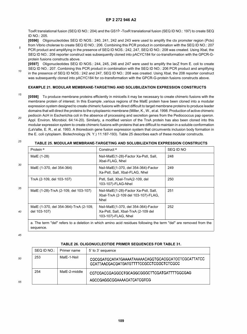
EP 2 272 946 A2
109
5
10
15
20
25
30
35
40
45
50
55
ToxR translational fusion (SEQ ID NO.: 204) and the GS1 -ToxR translational fusion (SEQ ID NO.: 197) to create SEQID NO.: 205.[0596] Oligonucleotides SEQ ID NOS.: 240, 241, 242 and 243 were used to amplify the ctx promoter region (Pctx)from Vibrio cholerae to create SEQ ID NO.: 206. Combining this PCR product in combination with the SEQ ID NO.: 207PCR product and amplifying in the presence of SEQ ID NOS.: 242, 247, SEQ ID NO.: 208 was created. Using Xbal, theSEQ ID NO.: 208 reporter construct was subsequently cloned into pACYC184 for co-transformation with the GPCR-G-protein fusions constructs above.[0597] Oligonucleotides SEQ ID NOS.: 244, 245, 246 and 247 were used to amplify the lacZ from E. coli to createSEQ ID NO.: 207. Combining this PCR product in combination with the SEQ ID NO.: 206 PCR product and amplifyingin the presence of SEQ ID NOS.: 242 and 247, SEQ ID NO.: 208 was created. Using Xbal, the 208 reporter constructwas subsequently cloned into pACYC184 for co-transformation with the GPCR-G-protein fusions constructs above.
EXAMPLE 21. MODULAR MEMBRANE-TARGETING AND SOLUBILIZATION EXPRESSION CONSTRUCTS
[0598] To produce membrane proteins efficiently in minicells it may be necessary to create chimeric fusions with themembrane protein of interest. In this Example ,various regions of the MalE protein have been cloned into a modularexpression system designed to create chimeric fusions with direct difficult to target membrane proteins to produce leaderdomains that will direct the proteins to the cytoplasmic membrane (Miller, K., W., et al. 1998. Production of active chimericpediocin AcH in Escherichia coli in the absence of processing and secretion genes from the Pediococcus pap operon.Appl. Environ. Microbiol. 64:14-20). Similarly, a modified version of the TrxA protein has also been cloned into thismodular expression system to create chimeric fusions with proteins that are difficult to maintain in a soluble conformation(LaVallie, E. R., et al. 1993. A thioredoxin gene fusion expression system that circumvents inclusion body formation inthe E. coli cytoplasm. Biotechnology (N. Y.) 11:187-193). Table 25 describes each of these modular constructs.
TABLE 25. MODULAR MEMBRANE-TARGETING AND SOLUBILIZATION EXPRESSION CONSTRUCTS
Protein a Construct a SEQ ID NO
MalE (1-28) NsiI-MalE(1-28)-Factor Xa-PstI, SalI,XbaI-FLAG, NheI
248
MalE (1-370, del 354-364) NsiI-MalE(1-370, del 354-364)-FactorXa-PstI, SalI, XbaI-FLAG, Nhel
249
TrxA (2-109, del 103-107) PstI, SalI, XbaI-TrxA(2-109, del103-107)-FLAG-NheI
250
MalE (1-28)-TrxA (2-109, del 103-107) NsiI-MalE(1-28)-Factor Xa-Pstl, SalI,XbaI-TrxA (2-109 del 103-107)-FLAG,Nhel
251
MalE (1-370, del 354-364)-TrxA (2-109,del 103-107)
NsiI-MalE(1-370, del 354-364)-FactorXa-Pstl, SalI, XbaI-TrxA (2-109 del103-107)-FLAG, Nhel
252
a. The term "del" refers to a deletion in which amino acid residues following the term "del" are removed from thesequence.
TABLE 26. OLIGONUCLEOTIDE PRIMER SEQUENCES FOR TABLE 31.
SEQ ID NO.: Primer name 5’ to 3’ sequence
253 MalE-1-NsiI
254 MalE-2-middle

EP 2 272 946 A2
110
5
10
15
20
25
30
35
40
45
50
55
[0599] Oligonucleotides SEQ ID NOS.: 253, 254, 255 and 256 overlap with each other to form a scaffold template toPCR amplify malE (1-28) to create a SEQ ID NO.: 248. Following PCR amplification, SEQ ID NO.: 248 was digestedwith Nsil and Nhel and cloned into SEQ ID NOS.: 152, 154, 139 and 141 digested with Pstl and Xbal. The resultantproducts create SEQ ID NOS.: 266, 267, 268 and 269, respectively, that lose both the 5-prime PstI and 3-prime Xbalrestriction site and retain the PstI, SalI, and Xbal restriction sites between MalE (1-28) and the FLAG sequence. Insertionof a protein in alignment with these sites results in a chimeric protein containing amino-terminal MalE (1-28) and carboxy-terminal FLAG.[0600] Oligonucleotides SEQ ID NOS.: 257, 258, 259 and 260 were used to amplify malE (1-370 with a deletionremoving residues 354-364) to create SEQ ID NO.: 249. Following PCR amplification, SEQ ID NO.: 249 was digestedwith Nsil and Nhel and cloned into SEQ ID NOS.: 152, 154, 139 and 141 digested with PstI and Xbal. The resultantproducts create SEQ ID NOS.: 270, 271, 272 and 273, respectively, that lose both the 5-prime PstI and 3-prime Xbalrestriction site and retain the Pstl, SalI, and Xbal restriction sites between MalE (1-370, del 354-364) and the FLAGsequence. Insertion of a protein in alignment with these sites results in a chimeric protein containing amino-terminalMalE (1-370, del 354-364) and carboxy-terminal FLAG.[0601] Oligonucleotides SEQ ID NOS.: 262, 263, 264 and 265 were used to amplify trxA (2-109 with a deletion removingresidues 103-107) to create SEQ ID NO.: 250. Following PCR amplification, SEQ ID NO.: 250 was digested with PstIand Nhel and cloned into SEQ ID NOS.: 152, 154, 139 and 141 digested with PstI and Xbal. to create SEQ ID NOS.:274, 275, 276 and 277, respectively. Using these restriction digestion combinations results in loss of the Xbal SEQ IDNO.: 249 insertion site.[0602] The resultant products create SEQ ID NOS.: 274, 275, 276 and 277, respectively, that lose the 3-prime Xbalrestriction site and retain the Pstl, SalI, and Xbal restriction sites on the 3-prime end of the TrxA (1-109, del 103-107)sequence. Insertion of a protein in alignment with these sites results in a chimeric protein containing Carboxy-terminal
(continued)
SEQ ID NO.: Primer name 5’ to 3’ sequence
255 MalE-3s-NheI
256 MalE-4-NheI GCGCCGCTAGCTTATTATTTGTCATCG
257 MalE-1a GGTGCACGCATCCTCGCATTATCCGC
258 MalE-2a GGCGTTTTCCATGGTGGCGGCAATACGTGG
259 MalE-1-NsiI
260 MalE-2-NheI
261 MalE-3L-NheI
262 TrxA-1a CCTGACTGACGACAGTTTTGACACGG
263 TrxA-2a CCTTTAGACAGTGCACCCACTTTGGTTGCCGC
264 TrxA-1a-PstI
265 TrxA-2-NheI GCGCCGCTAGCTTATTATTTGTCATCGTCATCTTTATAATCCG

EP 2 272 946 A2
111
5
10
15
20
25
30
35
40
45
50
55
TrxA (1-109, del 103-107)-FLAG.[0603] SEQ ID NO.: 248 was digested with Nsil and Xbal and cloned into SEQ ID NOS.: 274, 275, 276 and 277 thatwere digested with PstI and Xbal. The resultant products create SEQ ID NOS.: 278, 279, 280 and 281, respectively,that lose the 5 prime Pstl restriction site and retain the PstI, SalI, and Xbal restriction sites between MalE (1-28) andTrxA (1-109, del 103-107). Insertion of a protein in alignment with these sites results in a chimeric protein containingamino-terminal MalE (1-28) and carboxy-terminal TrxA (1-109, del 103-107)-FLAG.[0604] SEQ ID NO.: 249 was digested with Nsil and Xbal and cloned into SEQ ID NOS.: 274, 275, 276 and 277 thatwere digested with PstI and Xbal. The resultant products create SEQ ID NOS.: 282, 283, 284 and 285, respectively,that lose the 5 prime Pstl restriction site and retain the Pstl, SalI, and Xbal restriction sites between MalE (1-370, del354-364) and TrxA (1-109, del 103-107). Insertion of a protein in alignment with these sites results in a chimeric proteincontaining amino-terminal MalE (1-370, del 354-364) and carboxy-terminal TrxA (1-109, del 103-107)-FLAG.[0605] The contents of the articles, patents, and patent applications, and all other documents and electronically avail-able information mentioned or cited herein, are hereby incorporated by reference in their entirety to the same extent asif each individual publication was specifically and individually indicated to be incorporated by reference. Applicantsreserve the right to physically incorporate into this application any and all materials and information from any sucharticles, patents, patent applications, or other documents.[0606] The inventions illustratively described herein may suitably be practiced in the absence of any element orelements, limitation or limitations, not specifically disclosed herein. Thus, for example, the terms "comprising", "including,"containing", etc. shall be read expansively and without limitation. Additionally, the terms and expressions employedherein have been used as terms of description and not of limitation, and there is no intention in the use of such termsand expressions of excluding any equivalents of the features shown and described or portions thereof, but it is recognizedthat various modifications are possible within the scope of the invention claimed. Thus, it should be understood thatalthough the present invention has been specifically disclosed by preferred embodiments and optional features, modi-fication and variation of the inventions embodied therein herein disclosed may be resorted to by those skilled in the art,and that such modifications and variations are considered to be within the scope of this invention.[0607] The invention has been described broadly and generically herein. Each of the narrower species and subgenericgroupings falling within the generic disclosure also form part of the invention. This includes the generic description ofthe invention with a proviso or negative limitation removing any subject matter from the genus, regardless of whetheror not the excised material is specifically recited herein.[0608] Other embodiments are within the following claims. In addition, where features or aspects of the invention aredescribed in terms of Markush groups, those skilled in the art will recognize that the invention is also thereby describedin terms of any individual member or subgroup of members of the Markush group.The aspects of the invention are
1. A minicell comprising a membrane protein selected from the group consisting of a eukaryotic membrane protein,an archeabacterial membrane protein and an organellar membrane protein.
2. The minicell according to aspect 1, wherein said minicell is selected from the group consisting of a eubacterialminicell, a poroplast, a spheroplast and a protoplast.
3. The minicell according to aspect 1, wherein said minicell comprises a biologically active compound.
4. The minicell according to aspect 1, wherein said minicell comprises a expression construct, wherein said firstexpression construct comprises expression sequences operably linked to an ORF that encodes a protein.
5. The minicell according to aspect 4, wherein said ORF encodes said membrane protein.
6. The minicell according to aspect 4, wherein said expression sequences that are operably linked to an ORF areinducible and/or repressible.
7. The minicell according to aspect 4, wherein said minicell comprises a second expression construct, wherein saidsecond expression construct comprises expression sequences operably linked to a gene.
8. The minicell according to aspect 7, wherein said expression sequences that are operably linked to a gene areinducible and/or repressible.
9. The minicell according to aspect 7, wherein the gene product of said gene regulates the expression of the ORFthat encodes said protein.

EP 2 272 946 A2
112
5
10
15
20
25
30
35
40
45
50
55
10. The minicell according to aspect 7, wherein the gene product of said gene is a nucleic acid.
11. The minicell according to aspect 7, wherein the gene product of said gene is a polypeptide.
12. The minicell according to aspect 11, wherein said polypeptide is a membrane protein, a soluble protein or asecreted protein.
13. The minicell according to aspect 12, wherein said membrane protein is a membrane fusion protein, said mem-brane fusion protein comprising a first polypeptide, said first polypeptide comprising at least one transmembranedomain or at least one membrane anchoring domain; and a second polypeptide.
14. A minicell comprising a membrane fusion protein, said fusion protein comprising a first polypeptide, said firstpolypeptide comprising at least one transmembrane domain or at least one membrane anchoring domain; and asecond polypeptide, wherein said second polypeptide is not derived from a eubacterial protein and is neither a Histag nor a glutathione-S-transferase polypeptide.
15. The minicell according to aspect 14, wherein said minicell is selected from the group consisting of a eubacterialminicell, a poroplast, a spheroplast and a protoplast.
16. The minicell according to aspect 14, wherein said minicell comprises a biologically active compound.
17. A minicell comprising a membrane conjugate, wherein said membrane conjugate comprises a membrane proteinchemically linked to a conjugated compound.
18. The minicell according to aspect 17, wherein said minicell is selected from the group consisting of a eubacterialminicell, a poroplast, a spheroplast and a protoplast.
19. The minicell according to aspect 17, wherein said minicell comprises a biologically active compound.
20. The minicell according to aspect 17, wherein said conjugated compound is selected from the group consistingof a nucleic acid, a polypeptide, a lipid and a small molecule.
21. A method for making minicells, comprising (a) culturing a minicell-producing parent cell, wherein said parentcell comprises an expression construct, wherein said expression construct comprises a gene operably linked toexpression sequences that are inducible and/or repressible, and wherein induction or repression of said gene causesor enhances the production of minicells ; and (b) separating said minicells from said parent cell, thereby generatinga composition comprising minicells, wherein an inducer or repressor is present within said parent cells during oneor more steps and/or between two or more steps of said method.
22. The method according to aspect 21, further comprising (c) purifying said minicells from said composition.
23. The method according to aspect 21, wherein said minicell is selected from the group consisting of a eubacterialminicell, a poroplast, a spheroplast and a protoplast.
24. The method according to aspect 21, wherein said gene expresses a gene product that is a factor that is involvedin or modulates DNA replication, cellular division, cellular partitioning, septation, transcription, translation, or proteinfolding.
25. The method according to aspect 21, wherein said minicells are separated from said parent cells by a processselected from the group consisting of centrifugation, ultracentrifugation, density gradation, immunoaffinity and im-munoprecipitation.
26. The method according to aspect 22, wherein said minicell is a poroplast, said method further comprising (d)treating said minicells with an agent, or incubating said minicells under a set of conditions, that degrades the outermembrane of said minicell.
27. The method according to aspect 26, wherein said outer membrane is degraded by treatment with an agentselected from the group consisting of EDTA, EGTA, lactic acid, citric acid, gluconic acid, tartaric acid, polyethyle-

EP 2 272 946 A2
113
5
10
15
20
25
30
35
40
45
50
55
neimine, polycationic peptides, cationic leukocyte peptides, aminoglycosides, aminoglycosides, protamine, insectcecropins, reptilian magainins, polymers of basic amino acids, polymixin B, chloroform, nitrilotriacetic acid andsodium hexametaphosphate and/or by exposure to conditions selected from the group consisting of osmotic shockand insonation.
28. The method according to aspect 26, further comprising removing one or more contaminants from said compo-sition.
29. The method according to aspect 28, wherein said contaminant is LPS or peptidoglycan.
30. The method according to aspect 29, wherein said LPS is removed by contacting said composition to an agentthat binds or degrades LPS.
31. The method according to aspect 21, wherein said minicell-producing parent cell comprises a mutation in a generequired for lipopolysaccharide synthesis.
32. The method according to aspect 22, wherein said minicell is a spheroplast, said method further comprising (d)treating said minicells with an agent, or incubating said minicells under a set of conditions, that disrupts or degradesthe outer membrane; and
(e) treating said minicells with an agent, or incubating said minicells under a set of conditions, that disrupts ordegrades the cell wall.
33. The method according to aspect 32, wherein said agent that disrupts or degrades the cell wall is a lysozyme,and said set of conditions that disrupts or degrades the cell wall is incubation in a hypertonic solution.
34. The method according to aspect 22, wherein said minicell is a protoplast, said method further comprising (d)treating said minicells with an agent, or incubating said minicells under a set of conditions, that disrupt or degradethe outer membrane; (e) treating said minicells with an agent, or incubating said minicells under a set of conditions,that disrupts or degrades the cell wall, in order to generate a composition that comprises protoplasts; and purifyingprotoplasts from said composition.
35. The method according to aspect 22, further comprising preparing a denuded minicell from said minicell.
36. The method according to aspect 22, further comprising covalently or non-covalently linking one or more com-ponents of said minicell to a conjugated moiety.
37. A method of preparing a L-form minicell comprising: (a) culturing an L-form eubacterium, wherein said eubac-terium comprises one or more of the following: (i) an expression element that comprises a gene operably linked toexpression sequences that are inducible and/or repressible, wherein induction or repression of said gene regulatesthe copy number of an episomal expression construct; (ii) a mutation in an endogenous gene, wherein said mutationregulates the copy number of an episomal expression construct.
(iii) an expression element that comprises a gene operably linked to expression sequences that are inducibleand/or repressible, wherein induction or repression of said gene causes or enhances the production of minicells;and
(iv) a mutation in an endogenous gene, wherein said mutation causes or enhances minicell production.
(b) culturing said L-form minicell-producing parent cell in media under conditions wherein minicells areproduced; and (c) separating said minicells from said parent cell, thereby generating a composition com-prising L-form minicells, wherein an inducer or repressor is present within said minicells during one or moresteps and/or between two or more steps of said method.
38. The method according to aspect 37, further comprising (d) purifying said L-form minicells from said composition.
39. A method of producing a protein, comprising: (a) transforming a minicell-producing parent cell with an expressionelement that comprises expression sequences operably linked to a nucleic acid having an ORF that encodes said

EP 2 272 946 A2
114
5
10
15
20
25
30
35
40
45
50
55
protein; (b) culturing said minicell-producing parent cell under conditions wherein minicells are produced; and (c)purifying minicells from said parent cell, (d) purifying said protein from said minicells. wherein said ORF is expressedduring step (b), between steps (b) and (c), and during step (c).
40. The method according to aspect 39, wherein said expression elements segregate into said minicells, and saidORF is expressed between steps (c) and (d).
41. The method according to aspect 39, wherein said protein is a membrane protein.
42. The method according to aspect 39, wherein said protein is a soluble protein contained within said minicells,further comprising: (e) at least partially lysing said minicells.
43. The method according to aspect 39, wherein said protein is a secreted protein, wherein said method furthercomprises (e) collecting a composition in which said minicells are suspended or with which said minicells are incontact.
44. The method according to aspect 39, wherein the expression sequences to which said ORF is operably linkedare inducible, wherein said method further comprises adding an inducing agent between steps (a) and (b); duringstep (b); and between steps (b) and (c).
45. The method according to aspect 39, wherein the expression sequences to which said ORF is operably linkedare inducible, wherein said expression elements segregate into said minicells, said method further comprises addingan inducing agent after step (c).
46. The method according to aspect 39, further comprising: (e) preparing poroplasts from said minicells, whereinsaid ORF is expressed during step (b); between steps (b) and (c); during step (c); between step (c) and step (d)when said expression elements segregate into said minicells; and/or after step (d) when said expression elementssegregate into said minicells.
47. The method according to aspect 46, further comprising: purifying said protein from said poroplasts.
48. The method according to aspect 39, further comprising (e) preparing spheroplasts from said minicells, whereinsaid ORF is expressed during step (b), between steps (b) and (c), during step (c), between steps (c) and (d) and/orafter step (d).
49. The method according to aspect 48, further comprising: purifying said protein from said spheroplasts.
50. The method according to aspect 39, further comprising (e) preparing protoplasts from said minicells, whereinsaid ORF is expressed during step (b), between steps (b) and (c), during step (c), between steps (c) and (d) and/orafter step (d).
51. The method according to aspect 50, further comprising: (f) purifying said protein from said protoplasts.
52. The method according to aspect 39, further comprising (e) preparing membrane preparations from said minicells,wherein said ORF is expressed during step (b), between steps (b) and (c), during step (c), between steps (c) and(d) and/or after step (d).
53. The method according to aspect 48, further comprising: purifying said protein from said membrane preparations.
54. The method according to aspect 39, wherein said minicell-producing parent cell is an L-form bacterium.
55. A method of producing a protein, comprising: (a) transforming a minicell with an expression element. thatcomprises expression sequences operably linked to a nucleic acid having an ORF that encodes said protein; and(b) incubating said minicells under conditions wherein said ORF is expressed.
56. The method according to aspect 55, further comprising: (c) purifying said protein from said minicells.
57. The method according to aspect 55, wherein said minicell is selected from the group consisting of a eubacterial

EP 2 272 946 A2
115
5
10
15
20
25
30
35
40
45
50
55
minicell, a poroplast, a spheroplast and a protoplast.
58. A method of producing a protein, comprising: (a) transforming a minicell-producing parent cell with an expressionelement that comprises expression sequences operably linked to a nucleic acid having an ORF that encodes afusion protein comprising said protein and a polypeptide, wherein a protease-sensitive amino acid sequence ispositioned between said protein and said polypeptide; (b) culturing said minicell-producing parent cell under con-ditions wherein minicells are produced; (c) purifying minicells from said parent cell, wherein said ORF is expressedduring step (b); between steps (b) and (c); and/or after step (c) when said expression elements segregate into saidminicells; and (d) treating said minicells with a protease that cleaves said sensitive amino acid sequence, therebyseparating said protein from said polypeptide.
59. A poroplast, said poroplast comprising a vesicle, bonded by a membrane, wherein said membrane is an eubac-terial inner membrane, wherein said vesicle is surrounded by a eubacterial cell wall, and wherein said eubacterialinner membrane is accessible to a compound in solution with said poroplast.
60. The poroplast according to aspect 59, wherein said poroplast is a cellular poroplast.
61. The poroplast according to aspect 59, wherein said compound has a molecular weight of at least 1 kD.
62. The poroplast according to aspect 59, wherein said poroplast comprises an exogenous nucleic acid.
63. The poroplast according to aspect 62, wherein said exogenous nucleic acid is an expression construct.
64. The poroplast according to aspect 63, wherein said expression construct comprises an ORF that encodes anexogenous protein, wherein said ORF is operably linked to expression sequences.
65. The poroplast according to aspect 64, wherein said poroplast comprises an exogenous protein.
66. The poroplast according to aspect 59, wherein said poroplast comprises an exogenous protein.
67. The poroplast according to aspect 66, wherein said exogenous protein is a fusion protein, a soluble protein ora secreted protein.
68. The poroplast according to aspect 66, wherein said exogenous protein is a membrane protein.
69. The poroplast according to aspect 68, wherein said membrane protein is accessible to compounds in solutionwith said poroplast.
70. The poroplast according to aspect 68, wherein said membrane protein is selected from the group consisting ofa eukaryotic membrane protein, an archeabacterial membrane protein, and an organellar membrane protein.
71. The poroplast according to aspect 68, wherein said membrane protein is a fusion protein, said fusion proteincomprising a first polypeptide, said first polypeptide comprising at least one transmembrane domain or at least onemembrane anchoring domain; and a second polypeptide, wherein said second polypeptide is displayed by saidporoplast.
72. The poroplast according to aspect 71, wherein said second polypeptide is displayed on the external side of saideubacterial inner membrane.
73. The poroplast according to aspect 59, wherein said poroplast comprises a membrane component that is chem-ically linked to a conjugated compound.
74. The poroplast according to aspect 64, wherein said expression construct comprises one or more DNA fragmentsfrom a genome or cDNA.
75. The poroplast according to aspect 64, wherein said exogenous protein has a primary amino acid sequence thatis predicted from in silico translation of a nucleic acid sequence.

EP 2 272 946 A2
116
5
10
15
20
25
30
35
40
45
50
55
76. A method of making poroplasts or cellular poroplasts, comprising treating eubacterial minicells or cells with anagent, or incubating said minicells or cells under a set of conditions, that degrades the outer membrane of saidminicells or cells.
77. The method according to aspect 76, further comprising purifying said poroplasts or cellular poroplasts in orderto remove contaminants.
78. The method according to aspect 76, further comprising placing said poroplasts in a hypertonic solution, wherein90 % or more of said cells or minicells used to prepare said poroplasts would lyse in said solution under the sameconditions.
79. A solid support comprising a minicell.
80. The solid support according to aspect 79, wherein said minicell is selected from the group consisting of aeubacterial minicell, a poroplast, a spheroplast and a protoplast.
81. The solid support according to aspect 79, wherein said solid support is a dipstick.
82. The solid support according to aspect 79, wherein said solid support is a bead.
83. The solid support according to aspect 79, wherein said solid support is a mictrotiter multiwell plate.
84. The solid support according to aspect 79, wherein said minicell comprises a detectable compound.
85. The solid support according to aspect 84, wherein said detectable compound is a fluorescent compound.
86. The solid support according to aspect 79, wherein said minicell displays a membrane component.
87. The solid support according to aspect 86, wherein said membrane component is selected from the groupconsisting of (i) a eukaryotic membrane protein, (ii) an archeabacterial membrane protein, (iii) an organellar mem-brane protein, (iv) a fusion protein comprising at least one transmembrane domain or at least one membraneanchoring domain, and (v) a membrane conjugate comprising a membrane component chemically linked to a con-jugated compound.
88. The solid support according to aspect 86, wherein said membrane component is a receptor.
89. The solid support according to aspect 87, wherein said solid support further comprises a co- receptor.
90. The solid support according to aspect 79, wherein said minicell displays a binding moiety.
91. A solid support comprising a minicell, wherein said minicell displays a fusion protein, said fusion protein comprisinga first polypeptide that comprises at least one transmembrane domain or at least one membrane anchoring domain,and a second polypeptide.
92. The solid support according to aspect 91, wherein said minicell is selected from the group consisting of aeubacterial minicell, a poroplast, a spheroplast and a protoplast.
93. The solid support according to aspect 91, wherein said second polypeptide comprises a binding moiety.
94. The solid support according to aspect 91, wherein said second polypeptide comprises an enzyme moiety.
95. A solid support comprising a minicell, wherein said minicell comprises a membrane conjugate comprising amembrane component chemically linked to a conjugated compound.
96. The solid support according to aspect 95, wherein said conjugated compound is a spacer.
97. The solid support according to aspect 96, wherein said spacer is covalently linked to said solid support.

EP 2 272 946 A2
117
5
10
15
20
25
30
35
40
45
50
55
98. The solid support according to aspect 95, wherein said conjugated compound is covalently linked to said solidsupport.
99. A minicell comprising a biologically active compound, wherein said minicell displays a binding moiety, whereinsaid binding moiety is part of a fusion protein comprising a first polypeptide that comprises at least one transmembranedomain or at least one membrane anchoring domain and a second polypeptide that comprises a binding moiety,and said minicell is a poroplast, spheroplast or protoplast.
100. A eubacterial minicell comprising a biologically active compound, wherein said minicell displays a bindingmoiety, wherein said binding moiety is selected from the group consisting of (a) a eukaryotic membrane protein; (b)an archeabacterial membrane protein; (c) an organellar membrane protein; and (d) a fusion protein, said fusionprotein comprising a first polypeptide, said first polypeptide comprising at least one transmembrane domain or atleast one membrane anchoring domain; and a second polypeptide, wherein said second polypeptide is not derivedfrom a eubacterial protein and is neither a His tag nor a glutathione-S-transferase polypeptide, and wherein saidpolypeptide comprises a binding moiety.
101. The minicell according to aspect 99, wherein said binding moiety is selected from the group consisting of anantibody, an antibody derivative, a receptor and an active site of a non-catalytic derivative of an enzyme.
102. The minicell according to aspect 99, wherein said binding moiety is a single-chain antibody.
103. The minicell according to aspect 99, wherein said binding moiety is directed to a ligand selected from the groupconsisting of an epitope displayed on a pathogen, an epitope displayed on an infected cell and an epitope displayedon a hyperproliferative cell.
104. The minicell according to aspect 99, wherein said biologically active compound is selected from the groupconsisting of a radioisotope, a polypeptide, a nucleic acid and a small molecule.
105. The minicell according to aspect 99, further comprising a first and second nucleic acid, wherein said first nucleicacid comprises eukaryotic expression sequences operably linked to a first ORF, and a second nucleic acid, whereinsaid second nucleic acid comprises eubacterial expression sequences operably linked to a second ORF.
106. The minicell according to aspect 105, wherein one of said ORFs encodes a protein that comprises said bindingmoiety.
107. The minicell according to aspect 105, wherein said eubacterial expression sequences are induced and/orderepressed when said binding moiety is in contact with a target cell.
108. The minicell according to aspect 105, wherein said eukaryotic expression sequences are induced and/orderepressed when said nucleic acid is in the cytoplasm of a eukaryotic cell.
109. The minicell according to aspect 105, wherein the protein encoded by said first ORF comprises eukaryoticsecretion sequences and/or the protein encoded by said second ORF comprises eubacterial secretion sequences.
110. A method of associating a radioactive compound with a cell, wherein said cell displays a ligand specificallyrecognized by a binding moiety, comprising contacting said cell with a minicell that comprises said radioactivecompound and displays said binding moiety.
111. The method according to aspect 110, wherein said minicell is selected from the group consisting of a eubacterialminicell, a poroplast, a spheroplast and a protoplast.
112. The method according to aspect 110, wherein the amount of radiation emitted by said radioactive isotope issufficient to be detectable.
113. The method according to aspect 110, wherein the amount of radiation emitted by said radioactive isotope issufficient to be cytotoxic.
114. The method according to aspect 110, wherein said ligand displayed by said cell is selected from the group

EP 2 272 946 A2
118
5
10
15
20
25
30
35
40
45
50
55
consisting of an epitope displayed on a pathogen, an epitope displayed on an infected cell and an epitope displayedon a hyperproliferative cell.
115. The method according to aspect 110, wherein said binding moiety is selected from the group consisting of anantibody, an antibody derivative, a channel protein protein and a receptor.
116. The method according to aspect 110, wherein said binding moiety is a single-chain antibody.
117. The method according to aspect 110, wherein said binding moiety is selected from the group consisting of anaptamer and a small molecule.
118. A method of delivering a biologically active compound to a cell, wherein said cell displays a ligand specificallyrecognized by a binding moiety, comprising contacting said cell with a minicell that displays said binding moiety,wherein said minicell comprises said biologically active compound, and wherein the contents of said minicell aredelivered into said cell from a minicell bound to said cell.
119. The method according to aspect 118, wherein said minicell is selected from the group consisting of a eubacterialminicell, a poroplast, a spheroplast and a protoplast.
120. The method according to aspect 118, wherein said biologically active compound is selected from the groupconsisting of a nucleic acid, a lipid, a polypeptide, a radioactive compound, an ion and a small molecule.
121. The method according to aspect 118, wherein the membrane of said minicell comprises a system for transferringa molecule from the interior of a minicell into the cytoplasm of said cell.
122. The method according to aspect 121, wherein said system for transferring a molecule from the interior of aminicell into the cytoplasm of said cell is a Type III secretion system.
123. The method according to aspect 118, wherein said minicell further comprises a first and second nucleic acid,wherein said first nucleic acid comprises eukaryotic expression sequences operably linked to a first ORF, and asecond nucleic acid, wherein said second nucleic acid comprises eubacterial expression sequences operably linkedto a second ORF.
124. The method according to aspect 123, wherein one of said ORFs encodes a protein that comprises said bindingmoiety.
125. The method according to aspect 123, wherein said eubacterial expression sequences are induced and/orderepressed when said binding moiety is in contact with a target cell.
126. The method according to aspect 123, wherein said eukaryotic expression sequences are induced and/orderepressed when said nucleic acid is in the cytoplasm of a eukaryotic cell.
127. The method according to aspect 123, wherein the protein encoded by said first ORF comprises eukaryoticsecretion sequences and/or the protein encoded by said second ORF comprises eubacterial secretion sequences.
128. A minicell displaying a synthetic linking moiety, wherein said synthetic linking moiety is covalenty or non-covalently attached to a membrane component of said mincell.
129. The minicell according to aspect 128, wherein said minicell is selected from the group consisting of a eubacterialminicell, a poroplast, a spheroplast and a protoplast.
130. A sterically stabilized minicell comprising a displayed moiety that has a longer half- life in vivo than a wild-typeminicell, wherein said displayed moiety is a hydrophilic polymer that comprises a PEG moiety, a carboxylic groupof a polyalkylene glycol or PEG stearate.
131. A minicell having a membrane comprising an exogenous lipid, wherein a minicell comprising said exogenouslipid has a longer half-life in vivo than a minicell lacking said exogenous lipid, and wherein said minicell is selectedfrom the group consisting of a eubacterial minicell, a poroplast, a spheroplast and a protoplast.

EP 2 272 946 A2
119
5
10
15
20
25
30
35
40
45
50
55
132. The minicell according to aspect 131, wherein said exogenous lipid is a derivitized lipid.
133. The minicell according to aspect 132, wherein said derivitized lipid is selected from the group consisting ofphosphatidylethanolamine derivatized with PEG, DSPE-PEG, PEG stearate; PEG-derivatized phospholipids, andPEG ceramides is DSPE-PEG.
134. The minicell according to aspect 131, wherein said exogenous lipid is not present in a wild-type membrane,or is present in a different proportion than is found in minicells comprising a wild-type membrane, 135. The minicellaccording to aspect 134, wherein said exogenous lipid is selected from the group consisting of ganglioside, sphin-gomyelin, monosialoganglioside GM1, galactocerebroside sulfate, 1, 2-sn-dimyristoylphosphatidylcholine, phos-phatidylinositol and cardiolipin.
136. The minicell according to aspect 128, wherein said linking moiety is non-covalently attached to said minicell.
137. The minicell according to aspect 136, wherein one of said linking moiety and said membrane componentcomprises biotin, and the other comprises avidin or streptavidin.
138. The minicell according to aspect 128, wherein said synthetic linking moiety is a cross-linker.
139. The minicell according to aspect 128, wherein said cross-linker is a bifunctional cross-linker.
140. A method of transferring a membrane protein from a minicell membrane to a biological membrane comprisingcontacting a minicell to said biological membrane, wherein said minicell membrane comprises said membraneprotein, and allowing said mincell and said biological membrane to remain in contact for a period of time sufficientfor said transfer to occur.
141. The method according to aspect 140, wherein said minicell is selected from the group consisting of a eubacterialminicell, a poroplast, a spheroplast and a protoplast.
142. The method according to aspect 140, wherein biological membrane is a cytoplasmic membrane or an organellarmembrane.
143. The method according to aspect 140, wherein said biological membrane is a membrane selected from thegroup consisting of a membrane of a pathogen, a membrane of an infected cell and a membrane of a hyperproliferativecell.
144. The method according to aspect 140, wherein said biological membrane is the cytoplasmic membrane of arecipient cell.
145. The method according to aspect 144, wherein said recipient cell is selected from the group consisting of acultured cell and a cell within an organism.
146. The method according to aspect 140, wherein biological membrane is present on a cell that has been removedfrom an animal, said contacting occurs in vitro, after which said cell is returned to said organism.
147. The method according to aspect 144, wherein said membrane protein is an enzyme.
148. The method according to aspect 147, wherein said membrane protein having enzymatic activity is selectedfrom the group consisting of a cytochrome P450 and a fusion protein, said fusion protein comprising a first polypeptide,said first polypeptide comprising at least one polypeptide, wherein said second polypeptide has enzymatic acitivity.
149. The method according to aspect 140, wherein said membrane protein is a membrane fusion protein, saidmembrane fusion protein comprising a first polypeptide, said first polypeptide comprising at least one transmembranedomain or at least one membrane anchoring domain; and a second polypeptide.
150. The method according to aspect 149, wherein said second polypeptide is a biologically active polypeptide.
151. The method according to aspect 140, wherein said minicell displays a binding moiety.

EP 2 272 946 A2
120
5
10
15
20
25
30
35
40
45
50
55
152. A minicell that comprises an expression construct comprising an ORF encoding a membrane protein operablylinked to expression sequences, wherein said expression sequences are induced and/or derepressed when saidminicell is in contact with a target cell.
153. The minicell according to aspect 152, wherein said minicell is selected from the group consisting of a eubacterialminicell, a poroplast, a spheroplast and a protoplast.
154. The minicell according to aspect 152, wherein biological membrane is a cytoplasmic membrane or an organellarmembrane.
155. The minicell according to aspect 152, wherein said biological membrane is a membrane selected from thegroup consisting of a membrane of a pathogen, a membrane of an infected cell and a membrane of a hyperproliferativecell.
156. The minicell according to aspect 152, wherein said minicell displays a binding moiety.
157. The minicell according to aspect 156, wherein said binding moiety is selected from the group consisting of anantibody, an antibody derivative, an aptamer and a small molecule.
158. The minicell according to aspect 152, wherein said membrane protein is a membrane fusion protein, saidmembrane fusion protein comprising a first polypeptide, said first polypeptide comprising at least one transmembranedomain or at least one membrane anchoring domain; and a second polypeptide.
159. The minicell according to aspect. 152, wherein said membrane protein having enzymatic activity is selectedfrom the group consisting of a cytochrome P450 and a fusion protein, said fusion protein comprising a first polypeptide,said first polypeptide comprising at least one polypeptide, wherein said second polypeptide has enzymatic activity.
160. A pharmaceutical composition comprising a minicell, wherein said minicell displays a membrane protein,wherein said membrane protein is selected from the group consisting of a eukaryotic membrane protein, an archea-bacterial membrane protein and an organellar membrane protein.
161. The pharmaceutical composition according to aspect 160, wherein said minicell is selected from the groupconsisting of a eubacterial minicell, a poroplast, a spheroplast and a protoplast.
162. The pharmaceutical composition according to aspect 160, wherein said membrane protein is selected fromthe group consisting of a receptor, a channel protein, a cellular adhesion factor and an integrin.
163. The pharmaceutical formulation according to aspect 162, wherein said pharmaceutical formulation furthercomprises an adjuvant.
164. The pharmaceutical formulation according to aspect 162, wherein said membrane protein comprises a polypep-tide epitope displayed by a hyperproliferative cell.
165. The pharmaceutical formulation according to aspect 162, wherein said membrane protein comprises an epitopedisplayed by a eukaryotic pathogen, an archeabacterial pathogen, a virus or an infected cell.
166. A pharmaceutical composition comprising a minicell, wherein said minicell displays a membrane protein thatis a fusion protein, said fusion protein comprising (i) a first polypeptide, said first polypeptide comprising at leastone transmembrane domain or at least one membrane anchoring domain ; and (ii) a second polypeptide, whereinsaid second polypeptide is not derived from’a eubacterial protein.
167. The pharmaceutical composition according to aspect 166, wherein said minicell is selected from the groupconsisting of a eubacterial minicell, a poroplast, a spheroplast and a protoplast.
168. The pharmaceutical formulation according to aspect 167, wherein said pharmaceutical formulation furthercomprises an adjuvant.
169. The pharmaceutical formulation according to aspect 167, wherein said second polypeptide comprises a polypep-

EP 2 272 946 A2
121
5
10
15
20
25
30
35
40
45
50
55
tide epitope displayed by a hyperproliferative cell.
170. The pharmaceutical formulation according to aspect 169, wherein said membrane protein comprises an epitopedisplayed by a eukaryotic pathogen, an archeabacterial pathogen, a virus or an infected cell.
171. A pharmaceutical composition comprising a minicell, wherein said minicell displays a membrane conjugate,wherein said membrane conjugate comprises a membrane component chemically linked to a conjugated compound.
172. The pharmaceutical composition according to aspect 171, wherein said minicell is selected from the groupconsisting of a eubacterial minicell, a poroplast, a spheroplast and a protoplast.
173. The pharmaceutical composition according to aspect 171, wherein said membrane protein is selected fromthe group consisting of a receptor, a channel protein, a cellular adhesion factor and an integrin.
174. The pharmaceutical composition according to aspect 171, wherein said pharmaceutical further comprises anadjuvant.
175. The pharmaceutical composition according to aspect 171, wherein said membrane component is a polypeptidecomprising at least one transmembrane domain or at least one membrane anchoring domain, or a lipid that is partof a membrane.
176. The pharmaceutical composition according to aspect 171, wherein said conjugated compound is a polypeptide,and the chemical linkage between said membrane compound and said conjugated compound is not a peptide bond.
177. The pharmaceutical composition according to aspect 171, wherein said conjugated compound is a nucleic acid.
178. The pharmaceutical composition according to aspect 171, wherein said conjugated compound is an organiccompound.
179. The pharmaceutical composition according to aspect 176, wherein said organic compound is selected fromthe group consisting of a narcotic, a toxin, a venom, a sphingolipid and a soluble protein.
180. A method of making a pharmaceutical composition comprising a minicell, wherein said minicell displays amembrane protein, wherein said membrane protein is selected from the group consisting of a eukaryotic membraneprotein, an archeabacterial membrane protein and an organellar membrane protein.
181. The method according to aspect 1, wherein said minicell is selected from the group consisting of a eubacterialminicell, a poroplast, a spheroplast and a protoplast.
182. The method according to aspect 180, wherein said method further comprises adding an adjuvant to saidpharmaceutical formulation.
183. The method according to aspect 180, wherein said method further comprises desiccating said formulation.
184. The method according to aspect 183, wherein said method further comprises adding a suspension buffer tosaid formulation.
185. The method according to aspect 180, wherein said method further comprises making a chemical modificationof said membrane protein.
186. The method according to aspect 185, wherein said chemical modification is selected from the group consistingof glycosylation, deglycosylation, phosphorylation, dephosphorylation and proteolysis.
187.A method of making a pharmaceutical composition comprising a minicell, wherein said minicell displays amembrane protein that is a fusion protein, said fusion protein comprising (i) a first polypeptide, said first polypeptidecomprising at least one transmembrane domain or at least one membrane anchoring domain; and (ii) a secondpolypeptide, wherein said second polypeptide is not derived from a eubacterial protein.

EP 2 272 946 A2
122
5
10
15
20
25
30
35
40
45
50
55
188. The method according to aspect 187, wherein said method further comprises adding an adjuvant to saidpharmaceutical formulation.
189. The method according to aspect 187, wherein said method further comprises desiccating said pharmaceuticalformulation.
190. The method according to aspect 189 wherein said method further comprises adding a suspension buffer tosaid pharmaceutical formulation.
191. The method according to aspect 187, wherein said method further comprises making a chemical modificationof said membrane protein.
192. The method according to aspect 191, wherein said chemical modification is selected from the group consistingof glycosylation, deglycosylation, phosphorylation, dephosphorylation and proteolysis.
193. A method of making a pharmaceutical formulation comprising a minicell, wherein said minicell displays amembrane conjugate, wherein said membrane conjugate comprises a membrane component chemically linked toa conjugated compound.
194. The method according to aspect 193, wherein said method further comprises adding an adjuvant to saidpharmaceutical formulation.
195. The method according to aspect 193, wherein said membrane component is a polypeptide comprising at leastone transmembrane domain or at least one membrane anchoring domain, or a lipid that is part of a membrane.
196. The method according to aspect 193, wherein said conjugated compound is a polypeptide, and the chemicallinkage between said membrane compound and said conjugated compound is not a peptide bond.
197. The method according to aspect 193, wherein said conjugated compound is a nucleic acid.
198. The method according to aspect 193, wherein said conjugated compound is an organic compound.
199. The method according to aspect 186, wherein said organic compound is selected from the group consistingof a narcotic, a toxin, a venom, and a sphingolipid.
200. A method of detecting an agent that is specifically bound by a binding moiety, comprising contacting a minicelldisplaying said binding moiety with a composition known or suspected to contain said agent, and detecting a signalthat is modulated by the binding of said agent to said binding moiety.
201. The method according to aspect 200, wherein said minicell is a eubacterial minicell, a poroplast, a spheroplastor a protoplast.
202. The method according to aspect 200, wherein said agent is associated with a disease.
203. The method according to aspect 200, wherein said minicell comprises a detectable compound.
204. The method according to aspect 200, wherein said binding moiety is antibody or antibody derivative.
205. The method according to aspect 200, wherein said composition is an environmental sample.
206. The method according to aspect 200, wherein said composition is a biological sample.
207. The method according to aspect 206, wherein said biological sample is selected from the group consisting ofblood, serum, plasma, urine, saliva, a biopsy sample, feces and a skin patch.
208. A method of in situ imaging of a tissue or organ, comprising administering to an organism a minicell comprisingan imaging agent and a binding moiety and detecting said imaging agent in said organism.

EP 2 272 946 A2
123
5
10
15
20
25
30
35
40
45
50
55
209. The method according to aspect 208, wherein said minicell is a eubacterial minicell, a poroplast, a spheroplastor a protoplast.
210. The method according to aspect 208, wherein said binding moiety is an antibody or antibody derivative.
211. The method according to aspect 208, wherein said binding moiety specifically binds a cell surface antigen.
212. The method according to aspect 211, wherein said cell surface antigen is an antigen displayed by a tumorigeniccell, a cancer cell, and an infected cell.
213. The method according to aspect 211, wherein said cell surface antigen is a tissue-specific antigen.
214. The method according to aspect208, wherein said method of imaging is selected from the group consisting ofmagnetic resonance imaging, ultrasound imaging; and computer axaial tomography (CAT).
215. A device comprising a microchip operatively associated with a biosensor comprising a minicell, wherein saidmicrochip comprises or contacts said minicell, and wherein said minicell displays a binding moiety.
216. The device according to aspect 215, wherein said minicell is a eubacterial minicell, a poroplast, a spheroplastor a protoplast.
217. A method of detecting a substance that is specifically bound by a binding moiety, comprising contacting thedevice according to aspect 215 with a composition known or suspected to contain said substance, and detecting asignal from said device, wherein said signal changes as a function of the amount of said substance present in saidcomposition.
218. The method according to aspect 217, wherein said composition is a biological sample or an environmentalsample.
219.A method of identifying an agent that specifically binds a target compound, comprising contacting a minicelldisplaying said target compound with a library of compounds, and identifying an agent in said library that binds saidtarget compound.
220. The method according to aspect 219, wherein said minicell is a eubacterial minicell, a poroplast, a spheroplastor a protoplast.
221. The method according to aspect 219, wherein said library of compounds is a protein library.
222. The method according to aspect 221, wherein said protein library is selected from the group consisting of aphage display library, a phagemid display library, a baculovirus library, a yeast display library, and a ribosomaldisplay library.
223. The method according to aspect 219, wherein said library of compounds is selected from the group consistingof a library of aptamers, a library of synthetic peptides and a library of small molecules. q 224. The method accordingto aspect 219, wherein said target compound is a target polypeptide.
225. The method according to aspect 224, wherein said minicell comprises an expression construct comprisingexpression sequences operably linked to an ORF encoding said target polypeptide.
226. The method according to aspect 224, wherein said target polypeptide is a membrane protein.
227. The method according to aspect 226, wherein said membrane protein is a receptor or a channel protein.
228. The method according to aspect 226, wherein said membrane protein is an enzyme.
229. The method according to aspect 219, wherein said target compound is a membrane fusion protein, saidmembrane fusion protein comprising a first polypeptide, wherein said first polypeptide comprises at least one trans-membrane domain or at least one membrane anchoring domain; and a second polypeptide, wherein said second

EP 2 272 946 A2
124
5
10
15
20
25
30
35
40
45
50
55
polypeptide comprises amino acid sequences derived from a target polypeptide.
230. The method according to aspect 219, wherein said method further comprises comparing the activity of saidtarget compound in the presence of said agent to the activity of said target compound in the absence of said agent.
231. The method according to aspect 230, wherein said activity of said target compound is an enzyme activity.
232. The method according to aspect 231, wherein said enzyme is selected from the group consisting of an oxi-doreductase, a transferase, a hydrolase, a lyase, an isomerase, a ligase and a synthetase.
233. The method according to aspect 230, wherein said activity of said target compound is a binding activity.
234. The method according to aspect 233, further comprising comparing the binding of said agent to said targetcompound to the binding of a known ligand of said target compound.
235. The method according to aspect 234, wherein a competition assay is used for said comparing.
236. A device comprising microchips operatively associated with a biosensor comprising a set of minicells in aprearranged pattern, wherein said each of said microchips comprise or contact a minicell, wherein each of saidminicell displays a different target compound, and wherein binding of a ligand to a target compound results in anincreased or decreased signal.
237. A method of identifying an agent that specifically binds a target compound, comprising contacting the deviceaccording to aspect 236 with a library of compounds, and detecting a signal from said device, wherein said signalchanges as a function of the binding of an agent to said target compound.
238. A method of identifying an agent that specifically blocks the binding of a target compound to its ligand, comprisingcontacting the device according to aspect 236 with a library of compounds, and detecting a signal from said device,wherein said signal changes as a function of the binding of an agent to said target compound.
239. A method of making a antibody that specifically binds a protein domain, wherein said domain is in its nativeconformation, wherein said domain is contained within a protein displayed on a minicell, comprising contacting saidminicell with a cell, wherein said cell is competent for producing antibodies to an antigen contacted with said cell,in order to generate an immunogenic response in which said cell produces said antibody.
240. The method according to aspect 239, wherein said minicell is a eubacterial minicell, a poroplast, a spheroplastor a protoplast.
241. The method according to aspect 239, wherein said protein displayed on a minicell is a membrane protein.
242. The method according to aspect 241, wherein said membrane protein is a receptor or a channel protein.
243. The method according to aspect 239, wherein said domain is found within the second polypeptide of a membranefusion protein, wherein said membrane, fusion protein comprises a first polypeptide, wherein said first polypeptidecomprises at least one transmembrane domain or at least one membrane anchoring domain.
244. The method according to aspect 239, wherein said contacting occurs in vivo.
245. The method according to aspect 244, wherein said antibody is a polyclonal antibody or a monoclonal antibody.
246. The method according to aspect 244, wherein said contacting occurs in an animal that comprises an adjuvant.
247. The method of making an antibody derivative that specifically binds a protein domain, wherein said domain isin its native conformation, wherein said domain is displayed on a minicell, comprising contacting said minicell witha protein library, and identifying an antibody derivative from said protein library that specifically binds said proteindomain.
248. The method according to aspect 247, wherein said protein library is selected from the group consisting of a

EP 2 272 946 A2
125
5
10
15
20
25
30
35
40
45
50
55
phage display library, a phagemid display library, and a ribosomal display library.
249. The method according to aspect 247 wherein said antibody derivative is a single-chain antibody.
250. A method of making an antibody or antibody derivative that specifically binds an epitope, wherein said epitopeis selected from the group consisting of (i) an epitope composed of amino acids found within a membrane protein,(ii) an epitope present in an interface between a membrane protein and a membrane component, (iii) an epitopepresent in an interface between a membrane protein and one or more other proteins and (iv) an epitope in a fusionprotein, said fusion protein comprising a first polypeptide, said first polypeptide comprising at least one transmem-brane domain or at least one membrane anchoring domain, and a second polypeptide, said second polypeptidecomprising said epitope; comprising contacting a minicell displaying said epitope with a protein library, or to a cell,wherein said cell is competent for producing antibodies to an antigen contacted with said cell, in order to generatean immunogenic response in which said cell produces said antibody.
251. The method according to aspect 250, wherein said minicell is a eubacterial minicell, a poroplast, a spheroplastor a protoplast.
252. The method according to aspect 250, wherein said cell is contacted in vivo.
253. The method according to aspect 252, wherein said antibody is a polyclonal antibody.
254. The method according to aspect 252, wherein said antibody is a monoclonal antibody.
255. The method according to aspect 250, wherein said protein library is contacted in vitro.
256. The method according to aspect 255, wherein said protein library is selected from the group consisting of aphage display library, a phagemid display library, and a ribosomal display library.
257. The method according to aspect 256, wherein said antibody derivative is a single-chain antibody.
258. A method of determining the rate of transfer of nucleic acid from a minicell to a cell, comprising (a) contactingsaid cell to said minicell, wherein said minicell comprises said nucleic acid, for a set period of time; (b) separatingminicells from said cells;
(c) measuring the amount of nucleic acid in said cells, wherein the amount of nucleic acid in said cells over saidset period of time is the rate of transfer of a nucleic acid from a minicell.
259. A method of determining the amount of a nucleic acid transferred to a cell from a minicell, comprising (a)contacting said cell to said minicell, wherein said minicell comprises an expression element having eukaryoticexpression sequences operably linked to an ORF encoding a detectable polypeptide, wherein said minicell displaysa binding moiety, and wherein said binding moiety binds an epitope of said cell; and (b) detecting a signal from saiddetectable polypeptide, wherein a change in said signal corresponds to an increase in the amount of a nucleic acidtransferred to a cell.
260. The method according to aspect 258, wherein said minicell is selected from the group consisting of a eubacterialminicell, a poroplast, a spheroplast and a protoplast.
261. The method according to aspect 258, wherein said cell is a eukaryotic cell.
262. The method according to aspect 258, wherein said binding moiety is an antibody or antibody derivative.
263. The method according to aspect 258, wherein said binding moiety is a single-chain antibody.
264. The method according to aspect 258, wherein said binding moiety is an aptamer.
265. The method according to aspect 258, wherein said binding moiety is an organic compound.
266. The method according to aspect 258, wherein said detectable polypeptide is a fluorescent polypeptide.

EP 2 272 946 A2
126
5
10
15
20
25
30
35
40
45
50
55
267. A method of detecting the expression of an expression element in a cell, comprising (a) contacting said cell toa minicell, wherein said minicell comprises an expression element having cellular expression sequences operablylinked to an ORF encoding a detectable polypeptide, wherein said minicell displays a binding moiety, and whereinsaid binding moiety binds an epitope of said cell;
(b) incubating said cell and said minicell for a period of time effective for transfer of nucleic acid from said minicellto said cell; and (c) detecting a signal from said detectable polypeptide, wherein an increase in said signalcorresponds to an increase in the expression of said expression element.
268. The method according to aspect 267, wherein said minicell is selected from the group consisting of a eubacterialminicell, a poroplast, a spheroplast and a protoplast.
269. The method according to aspect 267, wherein said cell is a eukaryotic cell and said expression sequences areeukaryotic expression sequences.
270. The method according to aspect 269, wherein said eukaryotic cell is a mammalian cell.
271. The method according to aspect267, wherein said binding moiety is an antibody or antibody derivative.
272. The method according to aspect 267, wherein said binding moiety is a single-chain antibody.
273. The method according to aspect 267, wherein said binding moiety is an aptamer.
274. The method according to aspect 267, wherein said binding moiety is an organic compound.
275. The method according to aspect 267, wherein said detectable polypeptide is a fluorescent polypeptide.
276. A method for detecting the transfer of a fusion protein from the cytosol to an organelle of a eukaryotic cell,comprising (a) contacting said cell to a minicell, wherein (i) said minicell comprises an expression element havingeukaryotic expression sequences operably linked to an ORF encoding a fusion protein, wherein said fusion proteincomprises a first polypeptide that comprises organellar delivery sequences, and a second polypeptide that comprisesa detectable polypeptide; and (ii) said minicell displays a binding moiety that binds an epitope of said cell, or anepitope of an organelle;
(b) incubating said cell and said minicell for a period of time effective for transfer of nucleic acid from said minicellto said cell and production of said fusion protein ; and (c) detecting a signal from the detectable polypeptide,wherein a change in the signal corresponds to an increase in the amount of the fusion protein transferred tosaid organelle.
277. The method according to aspect 276, wherein said organelle is a mitochondrion, a chloroplast or a kinetoplast.
278. A minicell comprising at least one nucleic acid, wherein said minicell displays a binding moiety directed to atarget compound, wherein said binding moiety is selected from the group consisting of (i) a eukaryotic membraneprotein; (ii) an archeabacterial membrane protein; (iii) an organellar membrane protein; and (iv) a fusion protein,said fusion protein comprising a first polypeptide, said first polypeptide comprising at least one transmembranedomain or at least one membrane anchoring domain; and a second polypeptide, wherein said second polypeptideis not derived from a eubacterial protein and is neither a His tag nor a glutathione-S- transferase polypeptide, andwherein said polypeptide comprises a binding moiety.
279. The minicell according to aspect 278, wherein said minicell is selected from the group consisting of a eubacterialminicell, a poroplast, a spheroplast and a protoplast.
280. The minicell according to aspect 278, wherein said nucleic acid comprises an expression construct comprisingexpression sequences operably linked to an ORF encoding a protein selected from the group consisting of (i) saideukaryotic membrane protein, (ii) said archeabacterial membrane protein, (iii) said organellar membrane protein;and (iv) said fusion protein.
281. The minicell according to aspect 280, wherein said nucleic acid comprises an expression construct comprising

EP 2 272 946 A2
127
5
10
15
20
25
30
35
40
45
50
55
expression sequences operably linked to an ORF, wherein said ORF encodes a therapeutic polypeptide.
282. The minicell according to aspect 281, wherein said therapeutic polypeptide is a membrane polypeptide.
283. The minicell according to aspect 281, wherein said therapeutic polypeptide is a soluble polypeptide.
284. The minicell according to aspect 283, wherein said soluble polypeptide comprises a cellular secretion sequence.
285. The minicell according to aspect 281, wherein said expression sequences are inducible and/or repressible.
286. The minicell according to aspect2858, wherein said expression sequences are induced and/or derepressedwhen the binding moiety displayed by said minicell binds to its target compound.
287. The minicell according to aspect 1278herein said nucleic acid comprises an expression construct comprisingexpression sequences operably linked to an ORF, wherein said ORF encodes a polypeptide having an amino acidsequence that facilitates cellular transfer of a biologically active compound contained within or displayed by saidminicell.
288.The minicell according to aspect 278 wherein the membrane of said minicell comprises a system for transferringa molecule from the interior of a minicell into the cytoplasm of said cell.
289.The minicell according to aspect 288 wherein said system for transferring a molecule from the interior of aminicell into the cytoplasm of said cell is a Type III secretion system.
290. A method of introducing a nucleic acid into a cell, comprising contacting said cell with a minicell that comprisessaid nucleic acid, wherein said minicell displays a binding moiety, wherein said binding moiety is selected from thegroup consisting of (i) a eukaryotic membrane protein; (ii) an archeabacterial membrane protein; (iii) an organellarmembrane protein; and (iv) a fusion protein, said fusion protein comprising a first polypeptide, said first polypeptidecomprising at least one transmembrane domain or at least one membrane anchoring domain; and a second polypep-tide, wherein said second polypeptide is not derived from a eubacterial protein and is neither a His tag nor aglutathione-S-transferase polypeptide, and wherein said polypeptide comprises a binding moiety; and wherein saidbinding moiety binds an epitope of said cell.
291. The method according to aspect 290, wherein said minicell is selected from the group consisting of a eubacterialminicell, a poroplast, a spheroplast and a protoplast.
292. The method according to aspect 290, wherein said nucleic acid comprises an expression construct comprisingexpression sequences operably linked to an ORF encoding a protein selected from the group consisting of (i) saideukaryotic membrane protein,
(ii) said archeabacterial membrane protein, (iii) said organellar membrane protein; and (iv) said fusion protein.
293. The method according to aspect 290, wherein said nucleic acid comprises an expression construct comprisingexpression sequences operably linked to an ORF, wherein saidORF encodes a therapeutic polypeptide.
294. The method according to aspect 293, wherein said expression sequences are inducible and/or derepressible.
295. The method according to aspect 294, wherein said expression sequences are induced or derepressed whenthe binding moiety displayed by said minicell binds its target compound.
296. The method according to aspect 294, wherein said expression sequences are induced or derepressed by atransactivation or transrepression event.
297. The method according to aspect 292, wherein said nucleic acid comprises an expression construct comprisingexpression sequences operably linked to an ORF, wherein saidORF encodes a polypeptide having an amino acid sequence that facilitates cellular transfer of a biologically activecompound contained within or displayed by said minicell.

EP 2 272 946 A2
128
5
10
15
20
25
30
35
40
45
50
55
298. A minicell comprising a nucleic acid, wherein said nucleic acid comprises eukaryotic expression sequencesand eubacterial expression sequences, each of which is independently operably linked to an ORF.
299. The minicell according to aspect 298, wherein said minicell is selected from the group consisting of a eubacterialminicell, a poroplast, a spheroplast and a protoplast.
300. The minicell according to aspect 298, wherein said minicell displays a binding moiety.
301. The minicell according to aspect 300, wherein said eubacterial expression sequences are induced and/orderepressed when said binding moiety is in contact with a target cell.
302. The minicell according to aspect 300, wherein said eukaryotic expression sequences are induced and/orderepressed when said nucleic acid is in the cytoplasm of a eukaryotic cell.
303. The minicell according to aspect 301, wherein the protein encoded by said ORF comprises eubacterial oreukaryotic secretion sequences.
304. A minicell comprising a first and second nucleic acid, wherein said first nucleic acid comprises eukaryoticexpression sequences operably linked to a first ORF, and a second nucleic acid, wherein said second nucleic acidcomprises eubacterial expression sequences operably linked to a second ORF.
305. The minicell according to aspect 304, wherein said minicell is selected from the group consisting of a eubacterialminicell, a poroplast, a spheroplast and a protoplast.
306. The minicell according to aspect 304, wherein said minicell displays a binding moiety.
307. The minicell according to aspect 306, wherein said eubacterial expression sequences are induced and/orderepressed when said binding moiety is in contact with a target cell.
308. The minicell according to aspect 306, wherein said eukaryotic expression sequences are induced and/orderepressed when said nucleic acid is in the cytoplasm of a eukaryotic cell.
309. The minicell according to aspect 304, wherein the protein encoded by said first ORF comprises eukaryoticsecretion sequences and/or the protein encoded by said second ORF comprises eubacterial secretion sequences.
310. A method of introducing into and expressing a nucleic acid in an organism, comprising contacting a minicellto a cell of said organism, wherein said minicell comprises said nucleic acid.
311. The method according to aspect 310, wherein said minicell is selected from the group consisting of a eubacterialminicell, a poroplast, a spheroplast and a protoplast.
312. The method according to aspect 310, wherein said minicell displays a binding moiety.
313. The method according to aspect 310, wherein said nucleic acid comprises a eukaryotic expression construct,wherein said eukaryotic expression construct comprises eukaryotic expression sequences operably linked to anORF.
314. The method according to aspect 310, wherein said ORF encodes a protein selected from the group consistingof a membrane protein, a soluble protein and a protein comprising eukaryotic secretion signal sequences.
315. The method according to aspect 310, wherein said nucleic acid comprises a eubacterial expression construct,wherein said eubacterial expression construct comprises eubacterial expression sequences operably linked to anORF.
316. The method according to aspect 315, wherein said minicell displays a binding moiety, wherein said eubacterialexpression sequences are induced and/or derepressed when said binding moiety is in contact with a target cell.
317. The method according to aspect 316, wherein the protein encoded by said ORF comprises eubacterial secretion
EP 2 272 946 A2
129
5
10
15
20
25
30
35
40
45
50
55
sequences.
318. A minicell comprising a crystal of a membrane protein.
319. The minicell according to aspect 318, wherein said minicell is a eubacterial minicell, a poroplast, a spheroplastor a protoplast.
320. The minicell according to aspect 318, wherein said membrane protein is a receptor.
321. The minicell according to aspect 320, wherein said receptor is a G-protein coupled receptor.
322. The minicell according to aspect 318, wherein said crystal is displayed.
323. The minicell according to aspect 318, wherein said membrane protein is a fusion protein, said fusion proteincomprising a first polypeptide, said first polypeptide comprising at least one transmembrane domain or at least onemembrane anchoring domain, and a second polypeptide.
324. The minicell according to aspect 323, wherein said crystal is a crystal of said second polypeptide.
325. The minicell according to aspect 323, wherein said crystal is displayed.
326. A method of determining the three-dimensional structure of a membrane protein, comprising preparing a crystalof said membrane protein in a minicell, and determining the three-dimensional structure of said crystal.
327. A method for identifying ligand-interacting atoms in a defined three-dimensional structure of a target protein,comprising (a) preparing one or more variant proteins of a target protein having a known or predicted three-dimen-sional structure, wherein said target protein binds a preselected ligand; (b) expressing and displaying a variantprotein in a minicell; and (c) determining if a minicell displaying said variant protein binds said preselected ligandwith increased or decreased affinity as compared to the binding of said preselected ligand to said target protein.
328. The method according to aspect 327, wherein said ligand is a protein that forms a multimer with said targetprotein, and said ligand interacting atoms are atoms in said-defined three- dimensional structure are atoms that areinvolved in protein-protein interactions.
329. The method according to aspect 327, wherein said ligand is a compound that induces a conformational changein said target protein, and said defined three-dimensional structure is the site of said conformational change.
330. The method according to aspect 327, adopted to a method, said method for identifying ligands of a targetprotein, further comprising identifying the chemical differences in said variant proteins as compared to said targetprotein.
331. The method according to aspect 330, further comprising mapping said chemical differences onto said definedthree-dimensional structure, and correlating the effect of said chemical differences on said defined three-dimensionalstructure.
332. The method according to aspect 331, wherein said target protein is a wild-type protein.
333. A minicell library, comprising two or more minicells, wherein each minicell comprises a different exogenousprotein.
334. The minicell library according to aspect 333, wherein said minicell is a eubacterial minicell, a poroplast, aspheroplast or a protoplast.
335. The minicell library according to aspect 333, wherein said exogenous protein is a displayed protein.
336. The minicell library according to aspect 333, wherein said exogenous protein is a membrane protein.
337. The minicell library according to aspect 336, wherein said membrane protein is a receptor.

EP 2 272 946 A2
130
5
10
15
20
25
30
35
40
45
50
55
338. The minicell library according to aspect 333, wherein said protein is a soluble protein that is contained withinor secreted from said minicell.
339. The minicell library according to aspect 333, wherein minicells within said library comprise an expressionelement that comprises expression sequences operably linked to a nucleic acid having an ORF that encodes saidexogenous protein.
340. The minicell library according to aspect 339, wherein said nucleic acid has been mutagenized.
341. The minicell library according to aspect 339, wherein an active site of said exogenous protein has a known orpredicted three-dimensional structure, and said a portion of said ORF encoding said active site has been muta-genized.
342. The minicell library according to aspect 333, wherein each of said minicells comprises an exogenous proteinthat is a variant of a protein having a known or predicted three- dimensional structure.
343. A minicell library, comprising two or more minicells, wherein each minicell comprises a different fusion protein,each of said fusion protein comprising a first polypeptide that is a constant polypeptide, wherein said constantpolypeptide comprises at least one transmembrane domain or at least one membrane anchoring domain, and asecond polypeptide, wherein said second polypeptide is a variable amino acid sequence that is different in eachfusion proteins.
344. The minicell library according to aspect 343, wherein minicells within said library comprise an expressionelement that comprises expression sequences operably linked to a nucleic acid having an ORF that encodes saidfusion protein.
345. The minicell library according to aspect 344, wherein said second polypeptide of said fusion protein is encodedby a nucleic acid that has been cloned.
346. The minicell library according to aspect 344, wherein each of said second polypeptide of each of said fusionproteins comprises a variant of an amino acid sequence from a protein having a known or predicted three-dimensionalstructure.
347. A minicell library, comprising two or more minicells, wherein each minicell comprises a constant protein thatis present in each minicell and a variable protein that differs from minicell to minicell.
348. The minicell library according to aspect 347, wherein one of said constant and variable proteins is a receptor,and the other of said constant and variable proteins is a co-receptor.
349. The minicell library according to aspect 347, wherein each of said constant and variable proteins is differentfrom each other and is a factor in a signal transduction pathway.
350. The minicell library according to aspect 347, wherein one of said constant and variable proteins is a G-protein,and the other of said constant and variable proteins is a G-protein coupled receptor.
351. The minicell library according to aspect 347, wherein one of said constant and variable proteins comprises afirst transrepression domain, and the other of said constant and variable comprises a second transrepression domain,wherein said transrepression domains limit or block expression of a reporter gene when said constant and variableproteins associate with each other.
352. The minicell library according to aspect 347, wherein one of said constant and variable proteins comprises afirst transactivation domain, and the other of said constant and variable comprises a second transactivation domain,wherein said transactivation domains stimulate expression of a reporter gene when said constant and variableproteins associate with each other.
353. A method of identifying a nucleic acid that encodes a protein that binds to or chemically alters a preselectedligand, comprising: (a) separately contacting said ligand with individual members of a minicell library, whereinminicells in said library comprise expression elements, wherein said expression elements comprise DNA inserts,

EP 2 272 946 A2
131
5
10
15
20
25
30
35
40
45
50
55
wherein an ORF in said DNA insert is operably linked to expression sequences, in order to generate a series ofreaction mixes, each reaction mix comprising a different member of said minicell library; (b) incubating said reactionmixes, thereby allowing a protein that binds to or chemically alters said preselected ligand to bind or chemicallyalter said preselected ligand; (c) detecting a change in a signal from reaction mixes in which said ligand has beenbound or chemically altered; (d) preparing DNA from reaction mixes in which said ligand has been bound or chemicallyaltered; wherein said DNA is a nucleic acid that encodes a protein that binds to or chemically alters said preselectedligand.
354. The method according to aspect 353, wherein said minicell is a eubacterial minicell, a poroplast, a spheroplastor a protoplast.
355. The method according to aspect 353, wherein said preselected ligand is a biologically active compound.
356. The method according to aspect 353, wherein said preselected ligand is a therapeutic drug.
357. The method according to aspect 353, wherein a protein that binds or chemically alters said preselected ligandis a target protein for compounds that are therapeutic for a disease that is treated by administering said drug to anorganism in need thereof.
358. The method according to aspect 353, wherein said preselected ligand is detectably labeled, said mincellcomprises a detectable compound, and/or a chemically altered derivative of said protein is detectably labeled.
359. A method of determining the amino acid sequence of a protein that binds or chemically alters a preselectedligand, comprising:
(a) contacting said ligand with a minicell library, wherein minicells in said library comprise expression elements,wherein said expression elements comprise DNA inserts, wherein an ORF in said DNA insert is operably linkedto expression sequences; (b) incubating said mixture of ligand and minicells, under conditions which allowcomplexes comprising ligands and minicells to form and/or chemical reactions to occur; (c) isolating or identifyingsaid complexes from said ligand and said mixture of ligand and minicells; (d) preparing DNA from an expressionelement found in one or more of said complexes, or in a minicell thereof; (e) determining the nucleotide sequenceof said ORF in said DNA; and (f) generating an amino sequence by in silico translation, wherein said aminoacid sequence is or is derived from a protein that binds or chemically alters a preselected ligand.
360. The method according to aspect 359, wherein said minicell is a eubacterial minicell, a poroplast, a spheroplastor a protoplast.
361. The method according to aspect 359, wherein said DNA is prepared by isolating DNA from said complexes,or in a minicell thereof.
362. The method according to aspect 359, wherein said DNA is prepared by amplifying DNA from said complexes,or in a minicell thereof.
363. The method according to aspect 359, wherein said protein is a fusion protein.
364. The method according to aspect 359, wherein said protein is a membrane or a soluble protein.
365. The method according to aspect 364, wherein said protein comprises secretion sequences.
366. The method according to aspect 359, wherein said preselected ligand is a biologically active compound.
367. The method according to aspect 359, wherein said preselected ligand is a therapeutic drug.
368. The method according to aspect 359, wherein said preselected ligand is a therapeutic drug, and said proteinthat binds said preselected ligand is a target protein for compounds that are therapeutic for a disease that is treatedby administering said drug to an organism in need thereof.
369. A method of identifying a nucleic acid that encodes a protein that inhibits or blocks an agent from binding to
EP 2 272 946 A2
132
5
10
15
20
25
30
35
40
45
50
55
or chemically altering a preselected ligand, comprising: (a) separately contacting said ligand with individual membersof a minicell library, wherein minicells in said library comprise expression elements, wherein said expression elementscomprise DNA inserts, wherein an ORF in said DNA insert is operably linked to expression sequences, in order togenerate a series of reaction mixes, each reaction mix comprising a different member of said minicell library; (b)incubating said reaction mixes, thereby allowing a protein that binds to or chemically alters said preselected ligandto bind or chemically alter said preselected ligand; (c) detecting a change in a signal from reaction mixes in whichsaid ligand has been bound or chemically altered; (d) preparing DNA from reaction mixes in which said change insignal ligand has been bound or chemically altered; wherein said DNA is a nucleic acid that encodes a protein thatinhibits or blocks said agent from binding to or chemically altering said preselected ligand 370. The method accordingto aspect 369, wherein said minicell is a eubacterial minicell, a poroplast, a spheroplast or a protoplast.
371. The method according to aspect 369, wherein said DNA has a nucleotide sequence that encodes the aminoacid sequence of said protein that inhibits or blocks said agent from binding to or chemically altering said preselectedligand.
372. The method according to aspect 369, wherein a protein that binds or chemically alters said preselected ligandis a target protein for compounds that are therapeutic for a disease that is treated by administering said drug to anorganism in need thereof.
373. A method of identifying an agent that effects the activity of a protein, comprising contacting a library of two ormore candidate agents with a minicell comprising said protein or a polypeptide derived from said protein, assayingthe effect of candidate agents on the activity of said protein, and identifying agents that effect the activity of saidprotein.
374. The method according to aspect 373, wherein said minicell is selected from the group consisting of a eubacterialminicell, a poroplast, a spheroplast and a protoplast.
375. The method according to aspect 373, wherein said protein or said polypeptide derived from said protein isdisplayed on the surface of said minicell.
376. The method according to aspect 373, wherein said protein is a membrane protein.
377. The method according to aspect 376, wherein said membrane protein is selected from the group consistingof a receptor, a channel protein and an enzyme.
378. The methodaccording to aspect 373,wherein said activityof a protein is a binding activityoran enzymatic activity.
379. The method according to aspect 373, wherein said library of compounds is a protein library.
380. The method according to aspect 379, wherein said protein library is selected from the group consisting of aphage display library, a phagemid display library, and a ribosomal display library.
381. The method according to aspect 373, wherein said library of compounds is a library of aptamers.
382. The method according to aspect 373, wherein said library of compounds is a library of small molecules.
383. A method of identifying an agent that effects the activity of a protein domain containing a library of two or morecandidate agents with a minicell displaying a membrane fusion protein, said fusion protein comprising a first polypep-tide, said first polypeptide comprising at least one transmembrane domain or at least one membrane anchoringdomain, and a second polypeptide, wherein said second polypeptide comprises said protein domain.
384. A method of identifying undesirable side-effects of a biologically active compound that occur as a result ofbinding of said compound to a protein, wherein binding a compound to said protein is known to result in undesirableside effects, comprising contacting a minicell that comprises said protein to said biologically active compound.
385. The method according to aspect 384, wherein said minicell is selected from the group consisting of a eubacterialminicell, a poroplast, a spheroplast and a protoplast.

EP 2 272 946 A2
133
5
10
15
20
25
30
35
40
45
50
55
386. The method according to aspect 384, further comprising characterizing the binding of said biologically activecompound to said protein.
387. The method according to aspect 384, further comprising characterizing the effect of said biologically activecompound on the activity of said protein.
388. A method for identifying an agent that effects the interaction of a first signaling protein with a second signalingprotein, comprising (a) contacting a library of compounds with a minicell, wherein said minicell comprises: (i) a firstprotein comprising said first signaling protein and a first trans- acting regulatory domain; (ii) a second protein com-prising said second signaling protein and a second trans-acting regulatory domain ; and (iii) a reporter gene, theexpression of which is modulated by the interaction between said first trans-acting regulatory domain and saidsecond trans-acting regulatory domain; and (b) detecting the gene product of said reporter gene.
389. The method according to aspect 388, wherein said minicell is selected from the group consisting of a eubacterialminicell, a poroplast, a spheroplast and a protoplast.
390. The method according to aspect 388, wherein said trans-acting regulatory domains are transactivation domains.
391. The method according to aspect 388, wherein said trans-acting regulatory domains are transrepression do-mains.
392. The method according to aspect 388, wherein said reporter gene is induced by the interaction of said firsttrans-acting regulatory domain and said second trans-acting regulatory domain.
393. The method according to aspect 388, wherein said agent that effects the interaction of said first signalingprotein with said second signaling protein is an agent that causes or promotes said interaction.
394. The method according to aspect 388, wherein said reporter gene is repressed by the interaction of said firsttrans-acting regulatory domain and said second trans-acting regulatory domain.
395. The method according to aspect 394, wherein said agent that effects the interaction of said first signalingprotein with said second signaling protein is an agent that inhibits or blocks said interaction.
396. The method according to aspect 388, wherein said first signaling protein is a GPCR.
397. The method according to aspect 396, wherein said GPCR is an Edg receptor or a SCAMPER.
398. The method according to aspect 396, wherein said second signalling protein is a G-protein..
399. The method according to aspect 398, wherein said G-protein is selected from the group consisting of G-alpha-i, G-alpha-s, G-alpha-q, G-alpha-12/13 and Go.
400. The method according to aspect 388, wherein said library of compounds is a protein library.
401. The method according to aspect 400, wherein said protein library is selected from the group consisting of aphage display library, a phagemid display library, and a ribosomal display library.
402. The method according to aspect 388, wherein said library of compounds is a library of aptamers.
403. The method according to aspect 388, wherein said library of compounds is a library of small molecules.
404. A method for identifying an agent that effects the interaction of a first signaling protein with a second signalingprotein, comprising contacting a library of two or more candidate agents with a minicell, wherein said minicellcomprises: (a) a first fusion protein comprising said first signaling protein and a first detectable domain; and (b) asecond fusion protein comprising said second signaling protein and a second detectable domain, wherein a signalis generated when said first and second signaling proteins are in close proximity to each other, and detecting saidsignal.

EP 2 272 946 A2
134
5
10
15
20
25
30
35
40
45
50
55
405. The method according to aspect 404, wherein said signal is fluorescence.
406. The method according to aspect 404, wherein said first detectable domain and said second detectable domainare fluorescent and said signal is generated by FRET.
407. The method according to aspect 406, wherein said first and second detectable domains are independentlyselected from the group consisting of a green fluorescent protein, a blue-shifted green fluorescent protein, a cyan-shifted green fluorescent protein; a red- shifted green fluorescent protein; a yellow-shifted green fluorescent protein,and a red fluorescent protein, wherein said first fluorescent domain and said second fluorescent domain are notidentical.
408. A method of bioremediation, said method comprising contacting a composition that comprises an undesirablesubstance with a minicell, wherein said minicell alters the chemical structure and/or binds said undesirable substance.
409. A method of bioremediation, said method comprising contacting a composition that comprises an undesirablesubstance with a minicell, wherein said mincell comprises an agent that alters the chemical structure of said unde-sirable substance.
410. The method according to aspect 409, wherein said agent that alters the chemical structure of said undesirablesubstance is an inorganic catalyst.
411. The method according to aspect 409, wherein said agent that alters the chemical structure of said undesirablesubstance is an enzyme.
412. The method according to aspect 411, wherein said enzyme is a soluble protein contained within said minicell.
413. The method according to aspect 412, wherein said soluble protein is selected from the group consisting of anoxidoreductase, a transferase, a hydrolase, a lyase, an isomerase, a ligase and a synthetase.
414. The method according to aspect 411, wherein said enzyme is a secreted protein.
415. The method according to aspect 414, wherein said secreted protein is selected from the group consisting ofan oxidoreductase, a transferase, a hydrolase, a lyase, an isomerase, a ligase and a synthetase.
416. The method according to aspect 411, wherein said enzyme is a membrane protein.
417. The method according to aspect 416, wherein said membrane enzyme is selected from the group consistingof a cytochrome P450, an oxidoreductase, a transferase, a hydrolase, a lyase, an isomerase, a ligase and a syn-thetase.
418. The method according to aspect 409, wherein said agent that alters the chemical structure of said undesirablesubstance is a fusion protein comprising a first polypeptide that comprises a transmembrane domain or at least onemembrane-anchoring domain, and a second polypeptide, wherein said second polypeptide is an enzyme moiety.
419. The method according to aspect 418, wherein said second polypeptide is a polypeptide derived from a proteinselected from the group consisting of an oxidoreductase, a transferase, a hydrolase, a lyase, an isomerase, a ligaseand a synthetase.
420. A method of bioremediation, said method comprising contacting a composition that comprises an undesirable,substance with a minicell, wherein said mincell comprises an agent that binds an undesirable substance.
421. The method according to aspect 420, wherein said undesirable substance binds to and is internalized by saidminicell or is otherwise inactivated by selective absorption.
422. The method according to aspect 420, wherein said agent that binds said undesirable substance is a secretedsoluble protein.
423. The method according to aspect 422, wherein said secreted protein is a transport accessory protein.

EP 2 272 946 A2
135
5
10
15
20
25
30
35
40
45
50
55
424. The method according to aspect 420, wherein said agent that binds said undesirable substance is a membraneprotein.
425. The method according to aspect 420, wherein said undesirable substance is selected from the group consistingof a toxin, a pollutant and a pathogen.
426. The method according to aspect 420, wherein said agent that binds said undesirable substance is a fusionprotein comprising a first polypeptide that comprises a transmembrane domain or at least one membrane-anchoringdomain, and a second polypeptide, wherein said second polypeptide is a binding moiety.
427. The method according to aspect 426, wherein said binding moiety is selected from the group consisting of anantibody, an antibody derivative, the active site of a non- enzymatically active mutant enzyme, a single-chain antibodyand an aptamer.
428. A minicell-producing parent cell, wherein said parent cell comprises one or more of the following : (a) anexpression element that comprises a gene operably linked to expression sequences that are inducible and/orrepressible, wherein induction or repression of said gene regulates the copy number of an episomal expressionconstruct; (b) a mutation in an endogenous gene, wherein said mutation regulates the copy number of an episomalexpression construct; (c) an expression element that comprises a gene operably linked to expression sequencesthat are inducible and/or repressible, wherein induction or repression of said gene causes or enhances the productionof minicells; and (d) a mutation in an endogenous gene, wherein said mutation causes or enhances minicell pro-duction.
429. The minicell-producing parent cell according to aspect428, further comprisingan episomal expression construct.
430. The minicell-producing parent cell according to aspect 428, further comprising a chromosomal expressionconstruct.
431. The minicell-producing parent cell according to aspect 429, wherein said expression sequences of said ex-pression construct are inducible and/or repressible.
432. The minicell-producing parent cell according to aspect 428, wherein said minicell-producing parent cell com-prises a biologically active compound.
433. The minicell according to aspect 428 wherein said gene that causes or enhances the production of minicellshas a gene product that is involved in or regulates DNA replication, cellular division, cellular partitioning, septation,transcription, translation, or protein folding.
434. A minicell-producing parent cell, wherein said parent cell comprises an expression construct, wherein saidexpression construct comprises expression sequences operably linked to an ORF that encodes a protein, and aregulatory expression element, wherein said regulatory expression element comprises expression sequences op-erably linked to a regulatory gene that encodes a factor that regulates the expression of said ORF.
435. The minicell-producing parent cell according to aspect 434, wherein said expression sequences of said ex-pression construct are inducible and/or repressible.
436. The minicell-producing parent cell according to aspect 434, wherein said expression sequences of said regu-latory expression construct are inducible and/or repressible.
437. The minicell-producing parent cell according to aspect 434, wherein one or more of said expression elementor said regulatory expression element is located on a chromosome of said parent cell.
438. The minicell-producing parent cell according to aspect 434, wherein one or more of said expression elementor said regulatory expression element is located on an episomal expression construct.
439. The minicell-producing parent cell according to aspect 438, wherein both of said expression element and saidregulatory expression element are located on an episomal expression construct, and one or both of said expressionelement and said regulatory expression element segregates into minicells produced from said parent cell.

EP 2 272 946 A2
136
5
10
15
20
25
30
35
40
45
50
55
440. The minicell-producing parent cell according to aspect 434, wherein said minicell-producing parent cell com-prises a biologically active compound.
441. The minicell-producing parent cell according to aspect 440, wherein said biologically active compound segre-gates into minicells produced from said parent cell.
442. The minicell-producing parent cell according to aspect 434, wherein said ORF encodes a membrane proteinor a soluble protein.
443. The minicell-producing parent cell according to aspect 434, wherein said protein comprises secretion sequenc-es.
444. The minicell-producing parent cell according to aspect 434, wherein the gene product of said gene regulatesthe expression of said ORF.
445. The minicell-producing parent cell according to aspect 444, wherein said gene product is a transcription factor.
446. The minicell-producing parent cell according to aspect 440, wherein said gene product is a RNA polymerase.
447. The minicell-producing parent cell according to aspect 446, wherein said parent cell is MC-T7.
448. A minicell comprising a biologically active compound, wherein said minicell displays a binding moiety, whereinsaid minicell selectively absorbs and/or internalizes an undesirable compound, and said minicell is a poroplast,spheroplast or protoplast.
449. The minicell according to aspect 448, wherein said binding moiety is selected from the group consisting of anantibody, an antibody derivative, a receptor and an active site of a non-catalytic derivative of an enzyme.
450. The minicell according to aspect 458, wherein said binding moiety is a single-chain antibody.
451. The minicell according to aspect 458, wherein said binding moiety is directed to a ligand selected from thegroup consisting of an epitope displayed on a pathogen, an epitope displayed on an infected cell and an epitopedisplayed on a hyperproliferative cell.
452. The minicell according to aspect 458, wherein said biologically active compound is selected from the groupconsisting of a radioisotope, a polypeptide, a nucleic acid and a small molecule.
453. The minicell according to aspect 448, wherein a ligand binds to and is internalized by said minicell or is otherwiseinactivated by selective absorption.
454. A pharmaceutical composition comprising the minicell according to aspect 448.
455. A method of reducing the free concentration of a substance in a composition, wherein said substance displaysa ligand specifically recognized by a binding moiety, comprising contacting said composition with a minicell thatdisplays said binding moiety, wherein said binding moiety binds said substance, thereby reducing the free concen-tration of said substance in said composition.
456. The method according to aspect 455, wherein said minicell is selected from the group consisting of a eubacterialminicell, a poroplast, a spheroplast and a protoplast.
457. The method according to aspect 455, wherein said substance is selected from the group consisting of a nucleicacid, a lipid, a polypeptide, a radioactive compound, an ion and a small molecule.
458. The method according to aspect 455, wherein said binding moiety is selected from the group consisting of anantibody, an antibody derivative, a channel protein and a receptor.
459. The method according to aspect 455, wherein said composition is present in an environment.

EP 2 272 946 A2
137
5
10
15
20
25
30
35
40
45
50
55
460. The method according to aspect 459, wherein said environment is water, air or soil.
461. The method according to aspect455, wherein said composition is a biological sample from an organism.
462. The method according to aspect 461, wherein said biological sample is selected from the group consisting ofblood, serum, plasma, urine, saliva, a biopsy sample, feces, tissue and a skin patch.
463. The method according to aspect 461, wherein said substance binds to and is internalized by said minicell oris otherwise inactivated by selective absorption.
464. The method according to aspect 463, wherein said biological sample is returned to said organism after beingcontacting to said minicell.

EP 2 272 946 A2
138
5
10
15
20
25
30
35
40
45
50
55

EP 2 272 946 A2
139
5
10
15
20
25
30
35
40
45
50
55

EP 2 272 946 A2
140
5
10
15
20
25
30
35
40
45
50
55

EP 2 272 946 A2
141
5
10
15
20
25
30
35
40
45
50
55

EP 2 272 946 A2
142
5
10
15
20
25
30
35
40
45
50
55

EP 2 272 946 A2
143
5
10
15
20
25
30
35
40
45
50
55
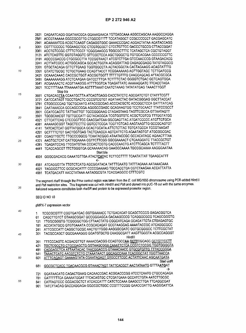
EP 2 272 946 A2
144
5
10
15
20
25
30
35
40
45
50
55

EP 2 272 946 A2
145
5
10
15
20
25
30
35
40
45
50
55

EP 2 272 946 A2
146
5
10
15
20
25
30
35
40
45
50
55

EP 2 272 946 A2
147
5
10
15
20
25
30
35
40
45
50
55
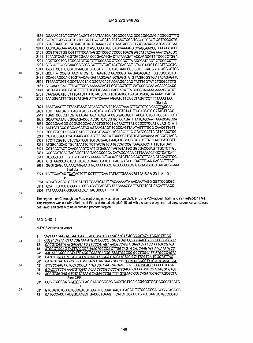
EP 2 272 946 A2
148
5
10
15
20
25
30
35
40
45
50
55

EP 2 272 946 A2
149
5
10
15
20
25
30
35
40
45
50
55

EP 2 272 946 A2
150
5
10
15
20
25
30
35
40
45
50
55

EP 2 272 946 A2
151
5
10
15
20
25
30
35
40
45
50
55

EP 2 272 946 A2
152
5
10
15
20
25
30
35
40
45
50
55

EP 2 272 946 A2
153
5
10
15
20
25
30
35
40
45
50
55

EP 2 272 946 A2
154
5
10
15
20
25
30
35
40
45
50
55

EP 2 272 946 A2
155
5
10
15
20
25
30
35
40
45
50
55

EP 2 272 946 A2
156
5
10
15
20
25
30
35
40
45
50
55

EP 2 272 946 A2
157
5
10
15
20
25
30
35
40
45
50
55

EP 2 272 946 A2
158
5
10
15
20
25
30
35
40
45
50
55

EP 2 272 946 A2
159
5
10
15
20
25
30
35
40
45
50
55
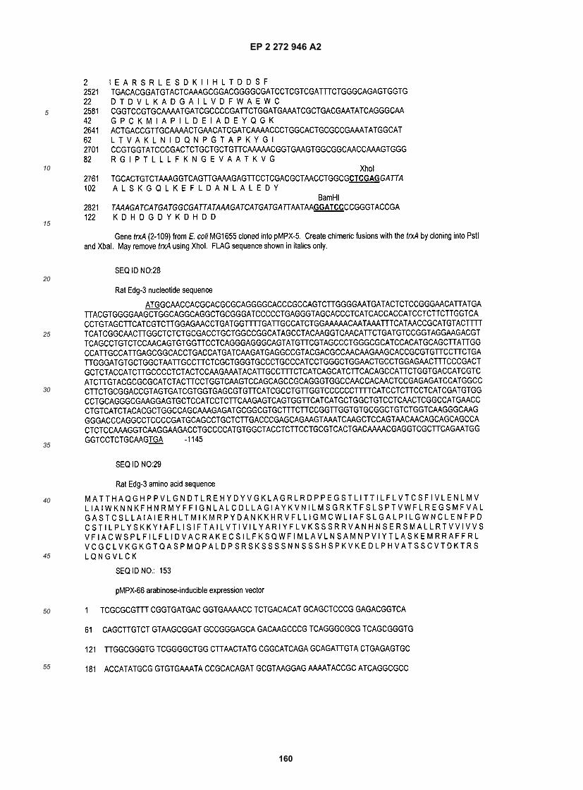
EP 2 272 946 A2
160
5
10
15
20
25
30
35
40
45
50
55

EP 2 272 946 A2
161
5
10
15
20
25
30
35
40
45
50
55

EP 2 272 946 A2
162
5
10
15
20
25
30
35
40
45
50
55

EP 2 272 946 A2
163
5
10
15
20
25
30
35
40
45
50
55

EP 2 272 946 A2
164
5
10
15
20
25
30
35
40
45
50
55

EP 2 272 946 A2
165
5
10
15
20
25
30
35
40
45
50
55

EP 2 272 946 A2
166
5
10
15
20
25
30
35
40
45
50
55

EP 2 272 946 A2
167
5
10
15
20
25
30
35
40
45
50
55

EP 2 272 946 A2
168
5
10
15
20
25
30
35
40
45
50
55

EP 2 272 946 A2
169
5
10
15
20
25
30
35
40
45
50
55

EP 2 272 946 A2
170
5
10
15
20
25
30
35
40
45
50
55

EP 2 272 946 A2
171
5
10
15
20
25
30
35
40
45
50
55

EP 2 272 946 A2
172
5
10
15
20
25
30
35
40
45
50
55

EP 2 272 946 A2
173
5
10
15
20
25
30
35
40
45
50
55

EP 2 272 946 A2
174
5
10
15
20
25
30
35
40
45
50
55

EP 2 272 946 A2
175
5
10
15
20
25
30
35
40
45
50
55
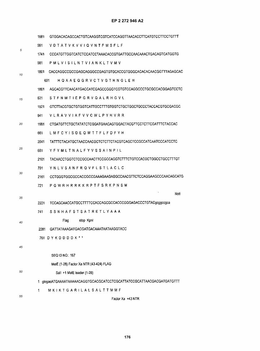
EP 2 272 946 A2
176
5
10
15
20
25
30
35
40
45
50
55

EP 2 272 946 A2
177
5
10
15
20
25
30
35
40
45
50
55

EP 2 272 946 A2
178
5
10
15
20
25
30
35
40
45
50
55

EP 2 272 946 A2
179
5
10
15
20
25
30
35
40
45
50
55

EP 2 272 946 A2
180
5
10
15
20
25
30
35
40
45
50
55

EP 2 272 946 A2
181
5
10
15
20
25
30
35
40
45
50
55

EP 2 272 946 A2
182
5
10
15
20
25
30
35
40
45
50
55

EP 2 272 946 A2
183
5
10
15
20
25
30
35
40
45
50
55

EP 2 272 946 A2
184
5
10
15
20
25
30
35
40
45
50
55

EP 2 272 946 A2
185
5
10
15
20
25
30
35
40
45
50
55

EP 2 272 946 A2
186
5
10
15
20
25
30
35
40
45
50
55

EP 2 272 946 A2
187
5
10
15
20
25
30
35
40
45
50
55

EP 2 272 946 A2
188
5
10
15
20
25
30
35
40
45
50
55

EP 2 272 946 A2
189
5
10
15
20
25
30
35
40
45
50
55

EP 2 272 946 A2
190
5
10
15
20
25
30
35
40
45
50
55

EP 2 272 946 A2
191
5
10
15
20
25
30
35
40
45
50
55

EP 2 272 946 A2
192
5
10
15
20
25
30
35
40
45
50
55

EP 2 272 946 A2
193
5
10
15
20
25
30
35
40
45
50
55

EP 2 272 946 A2
194
5
10
15
20
25
30
35
40
45
50
55

EP 2 272 946 A2
195
5
10
15
20
25
30
35
40
45
50
55

EP 2 272 946 A2
196
5
10
15
20
25
30
35
40
45
50
55

EP 2 272 946 A2
197
5
10
15
20
25
30
35
40
45
50
55

EP 2 272 946 A2
198
5
10
15
20
25
30
35
40
45
50
55

EP 2 272 946 A2
199
5
10
15
20
25
30
35
40
45
50
55

EP 2 272 946 A2
200
5
10
15
20
25
30
35
40
45
50
55

EP 2 272 946 A2
201
5
10
15
20
25
30
35
40
45
50
55

EP 2 272 946 A2
202
5
10
15
20
25
30
35
40
45
50
55

EP 2 272 946 A2
203
5
10
15
20
25
30
35
40
45
50
55
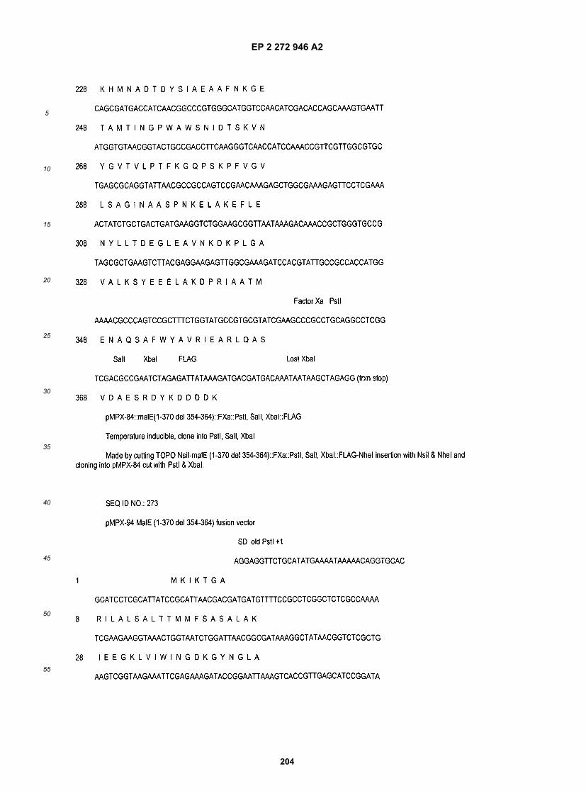
EP 2 272 946 A2
204
5
10
15
20
25
30
35
40
45
50
55

EP 2 272 946 A2
205
5
10
15
20
25
30
35
40
45
50
55

EP 2 272 946 A2
206
5
10
15
20
25
30
35
40
45
50
55

EP 2 272 946 A2
207
5
10
15
20
25
30
35
40
45
50
55

EP 2 272 946 A2
208
5
10
15
20
25
30
35
40
45
50
55

EP 2 272 946 A2
209
5
10
15
20
25
30
35
40
45
50
55

EP 2 272 946 A2
210
5
10
15
20
25
30
35
40
45
50
55
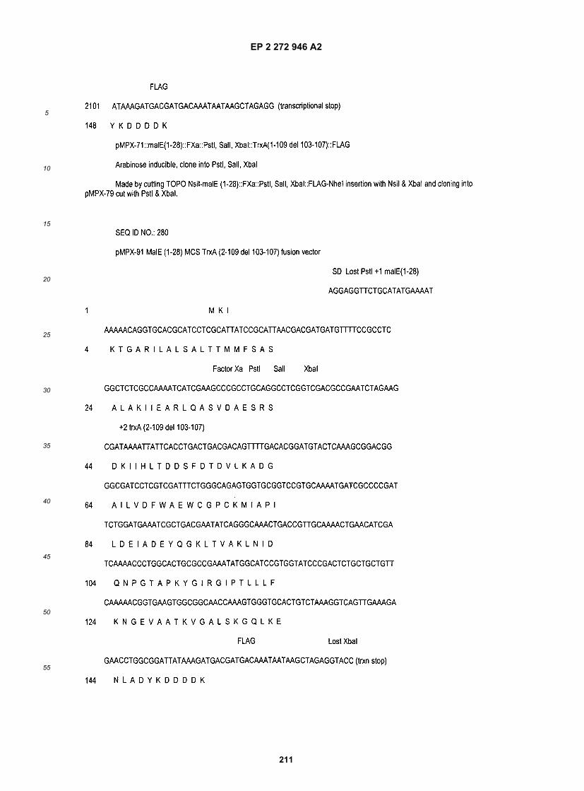
EP 2 272 946 A2
211
5
10
15
20
25
30
35
40
45
50
55

EP 2 272 946 A2
212
5
10
15
20
25
30
35
40
45
50
55

EP 2 272 946 A2
213
5
10
15
20
25
30
35
40
45
50
55

EP 2 272 946 A2
214
5
10
15
20
25
30
35
40
45
50
55
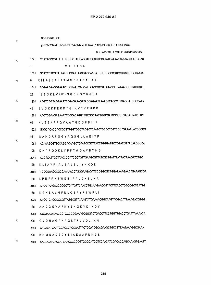
EP 2 272 946 A2
215
5
10
15
20
25
30
35
40
45
50
55

EP 2 272 946 A2
216
5
10
15
20
25
30
35
40
45
50
55

EP 2 272 946 A2
217
5
10
15
20
25
30
35
40
45
50
55

EP 2 272 946 A2
218
5
10
15
20
25
30
35
40
45
50
55

EP 2 272 946 A2
219
5
10
15
20
25
30
35
40
45
50
55
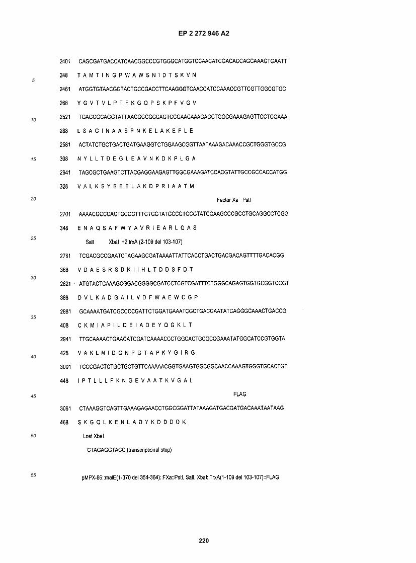
EP 2 272 946 A2
220
5
10
15
20
25
30
35
40
45
50
55

EP 2 272 946 A2
221
5
10
15
20
25
30
35
40
45
50
55

EP 2 272 946 A2
222
5
10
15
20
25
30
35
40
45
50
55

EP 2 272 946 A2
223
5
10
15
20
25
30
35
40
45
50
55

EP 2 272 946 A2
224
5
10
15
20
25
30
35
40
45
50
55

EP 2 272 946 A2
225
5
10
15
20
25
30
35
40
45
50
55

EP 2 272 946 A2
226
5
10
15
20
25
30
35
40
45
50
55

EP 2 272 946 A2
227
5
10
15
20
25
30
35
40
45
50
55

EP 2 272 946 A2
228
5
10
15
20
25
30
35
40
45
50
55

EP 2 272 946 A2
229
5
10
15
20
25
30
35
40
45
50
55

EP 2 272 946 A2
230
5
10
15
20
25
30
35
40
45
50
55

EP 2 272 946 A2
231
5
10
15
20
25
30
35
40
45
50
55

EP 2 272 946 A2
232
5
10
15
20
25
30
35
40
45
50
55

EP 2 272 946 A2
233
5
10
15
20
25
30
35
40
45
50
55

EP 2 272 946 A2
234
5
10
15
20
25
30
35
40
45
50
55

EP 2 272 946 A2
235
5
10
15
20
25
30
35
40
45
50
55

EP 2 272 946 A2
236
5
10
15
20
25
30
35
40
45
50
55

EP 2 272 946 A2
237
5
10
15
20
25
30
35
40
45
50
55

EP 2 272 946 A2
238
5
10
15
20
25
30
35
40
45
50
55

EP 2 272 946 A2
239
5
10
15
20
25
30
35
40
45
50
55

EP 2 272 946 A2
240
5
10
15
20
25
30
35
40
45
50
55

EP 2 272 946 A2
241
5
10
15
20
25
30
35
40
45
50
55

EP 2 272 946 A2
242
5
10
15
20
25
30
35
40
45
50
55

EP 2 272 946 A2
243
5
10
15
20
25
30
35
40
45
50
55

EP 2 272 946 A2
244
5
10
15
20
25
30
35
40
45
50
55

EP 2 272 946 A2
245
5
10
15
20
25
30
35
40
45
50
55

EP 2 272 946 A2
246
5
10
15
20
25
30
35
40
45
50
55

EP 2 272 946 A2
247
5
10
15
20
25
30
35
40
45
50
55

EP 2 272 946 A2
248
5
10
15
20
25
30
35
40
45
50
55

EP 2 272 946 A2
249
5
10
15
20
25
30
35
40
45
50
55

EP 2 272 946 A2
250
5
10
15
20
25
30
35
40
45
50
55

EP 2 272 946 A2
251
5
10
15
20
25
30
35
40
45
50
55

EP 2 272 946 A2
252
5
10
15
20
25
30
35
40
45
50
55
EP 2 272 946 A2
253
5
10
15
20
25
30
35
40
45
50
55
Claims
1. A fully intact eubacterial minicell derived from a eubacterial parent cell, wherein the minicell comprises a biologicallyactive compound and displays a ligand or binding moiety, wherein the biologically active compound and ligand orbinding moiety are exogenous to the parent cell and the biologically active compound is a compound distinct fromthe ligand or binding moiety.
2. The eubacterial minicell of claim 1, wherein said ligand or binding moiety comprises or is a part of: (a) an antibodyor antibody derivative; (b) a bacterial membrane protein; (c) a carbohydrate; (d) a polypeptide; (e) a nucleic acid;or (f) a fusion protein, said fusion protein comprising a first polypeptide, said first polypeptide comprising at leastone transmembrane domain or at least one membrane anchoring domain, and a second polypeptide comprisingsaid binding moiety.

EP 2 272 946 A2
254
5
10
15
20
25
30
35
40
45
50
55
3. The minicell of claim 1, wherein said binding moiety is directed to an antigen present on the surface of a eukaryoticcell, a receptor present on the surface of a eukaryotic cell, a tumor-associated antigen present on the surface ofthe cancer cell, or a tumor-associated receptor present on the surface of the cancer cell.
4. The minicell of claim 1, wherein said binding moiety is specific for an EGF receptor.
5. The minicell of any of claims 1 - 4, wherein said biologically active compound is a radioisotope, a polypeptide, anucleic acid, a therapeutic small molecule, a therapeutic small molecule drug, a chemotherapeutic agent, a thera-peutic single stranded RNA molecule, a therapeutic antisense RNA molecule, a ribozyme, a therapeutic doublestranded RNA molecule, a DNA molecule, a double stranded DNA molecule, a eukaryotic expression plasmidencoding for a therapeutic polypeptide, or a eukaryotic expression plasmid encoding for a therapeutic RNA.
6. The minicell of any of claims 1 - 5, wherein any combination of said biologically active compounds is present in thesame minicell.
7. The minicell of claim 1, further comprising a first and second nucleic acid, wherein said first nucleic acid compriseseukaryotic expression sequences operably linked to a first open reading frame, wherein said second nucleic acidcomprises eubacterial expression sequences operably linked to a second open reading frame.
8. The minicell of claim 7, wherein said eukaryotic expression sequences operably linked to a first open reading frameand said eubacterial sequences operably linked to a second open reading frame are present on the same nucleicacid molecule.
9. The minicell of claim 7, wherein one of said open reading frames encodes a protein that comprises said binding moiety.
10. The minicell of claim 7, wherein one of said open reading frames encodes a soluble protein, a membrane protein,a secreted protein, or a therapeutic polypeptide.
11. The minicell of claim 7, wherein said eukaryotic expression sequences are induced and/or derepressed when saidnucleic acids are introduced into a eukaryotic cell by said minicell.
12. A method of producing the minicell of claim 1, comprising exogenously introducing said ligand or binding moietyonto the surface of a eubacterial minicell.
13. A fully intact eubacterial minicell derived from a eubacterial parent cell, wherein the eubacterial minicell comprisesa biologically active compound for the purpose of delivering the biologically active compound to a eukaryotic cell,wherein said minicell displays a ligand or binding moiety and the biologically compound and ligand or binding moietyare exogenous to the parent cell and distinct from each other.
14. The minicell of claim 13, wherein the ligand or binding moiety targets the minicell to a specific eukaryotic cell type.
15. The minicell of claim 13, wherein the biologically active compound is delivered to the eukaryotic cell via: (a) receptormediated endocytosis; (b) cell fusion; or (c) phagocytosis.
16. The minicell of claim 15, wherein the receptor mediated endocytosis is induced by the ligand or binding moiety.
17. The minicell of claim 13, wherein the biologically active compound comprises genetic material.
18. The minicell of claim 13, wherein biologically active compound is transferred from a minicell to the interior of aeukaryotic cell.

EP 2 272 946 A2
255

EP 2 272 946 A2
256

EP 2 272 946 A2
257
REFERENCES CITED IN THE DESCRIPTION
This list of references cited by the applicant is for the reader’s convenience only. It does not form part of the Europeanpatent document. Even though great care has been taken in compiling the references, errors or omissions cannot beexcluded and the EPO disclaims all liability in this regard.
Patent documents cited in the description
• US 4190495 A, Curtiss [0006] [0159] [0337]• US 4311797 A, Khachatourians [0007] [0221]• US 4237224 A, Cohen and Boyer [0011]• US 29356601 P [0012]• US 4839293 A [0251]• US 5654176 A [0251]• US 6111079 A [0251]• US 5840867 A [0255]• US 4946778 A [0269]• US 5260203 A [0269]• US 5455030 A [0269]• US 5518889 A [0269]• US 5534621 A [0269]• US 5869620 A [0269]• US 6025165 A [0269]• US 6027725 A [0269]• US 6121424 A [0269]• US 6146885 A [0273] [0379]• US 6210967 B [0306]• US 6210921 B [0306]• US 6211343 B [0306]• US 6218509 B [0306]• US 6214972 B [0306]• US 6221613 B [0306]• US 6221660 B [0306]• US 6225080 B [0306]• US 6228610 B [0306]• US 6235496 B [0306]• US 6258556 B [0306]• US 6245531 B [0306]• US 6225531 B [0306]• US 6245893 B [0306]• US 6248712 B [0306]• US 6248554 B [0306]• US 6248520 B [0306]• US 6242251 B [0306]• US 6252056 B [0306]• US 6255472 B [0306]• US 6291207 B [0306]• US 6291206 B [0306]• US 6291195 B [0306]• US 6344200 B [0306]• US 6335180 B [0306]• US 6265184 B [0306]• US 6207799 B [0306]
• US 6290970 B [0306]• US 6326350 B [0306]• US 6313279 B [0306]• US 6313276 B [0306]• US 6307030 B [0306]• US 6306622 B [0306]• US 6300087 B [0306]• US 6297026 B [0306]• US 6277976 B [0306]• US 6274708 B [0306]• US 6271347 B [0306]• US 6262016 B [0306]• US 6261838 B [0306]• US 6258943 B [0306]• US 6284870 B [0306]• US 6258944 B [0306]• US 6261801 B [0306]• US 6261800 B [0306]• US 6265563 B [0306]• US 6268477 B [0306]• US 6316611 B [0306]• US 6316604 B [0306]• US 6287855 B [0306]• US 6268221 B [0306]• US 6268214 B [0306]• US 6329503 B [0313] [0467]• US 6086900 A, Draper [0365]• US 6017735 A [0366]• US 5804604 A [0366]• US 5747641 A [0366]• US 5674980 A [0366]• WO 9852547 A [0414]• US 4895724 A [0414]• US 5314695 A [0420]• US 5773024 A [0450]• US 6017721 A [0450]• US 6232127 B1 [0450]• US 4132769 A [0467]• US 4921790 A, O’Brien [0467]• US 4914021 A, Toth [0467]• US 4963484 A, Kufe [0467]• US 5053489 A [0467]• US 5693763 A, Codington [0467]• US 6025191 A, Pfreundschuh [0467]

EP 2 272 946 A2
258
Non-patent literature cited in the description
• Clark-Curtiss ; Curtiss III. Analysis of RecombinantDNA Using Escherichia coli Minicells. Methods in En-zymology, 1983, vol. 101, 347 [0003]
• Reeve ; Mendelson. Minicells of Bacillus subtilis. Anew system for transport studies in absence of mac-romolecular biosynthesis. Biochim. Biophys. Acta,1974, vol. 352, 298-305 [0003]
• Meagher et al. Protein Expression in E. coli Minicellsby Recombinant Plasmids. Cell, 1977, vol. 10, 521,523 [0003]
• Roozen et al. Synthesis of Ribonucleic Acid and Pro-tein in Plasmid-Containing Minicells of Escherichiacoli K-12. J. of Bacteriology, 1971, vol. 107 (1), 21[0003]
• Research on bacterial conjugation with minicells andminicell-producing E. coli strains. Curtiss III. Micro-bial Drug Resistance. University Park Press, 1976,169 [0003]
• Adler et al. Genetic control of cell division in bacteria.Science, 1966, vol. 154, 417 [0003]
• Adler et al. Miniature Escherichia coli cells deficientin DNA. Proc. Nat. Acad. Sci, 1967, vol. 57, 321[0003]
• Frazer ; Curtiss III. Production, Properties and Utilityof Bacterial Minicells. Curr. Top. Microbiol. Immunol.,1975, vol. 69, 1-3 [0003]
• Michael Gaâel et al. The kdpF Subunit Is Part of theK+-translocating Kdp Complex of Escherichia coliand Is Responsible for Stabilization of the Complexin vitro. Jn. of Biological Chemistry, 1999, vol. 274(53), 37901 [0004]
• Harlow et al. Cloning and Characterization of the gskGene Encoding Guanosine Kinase of Escherichia co-li. J. of Bacteriology, 1995, vol. 177 (8), 2236 [0004]
• Carol L. Pickett et al. Cloning, Sequencing, and Ex-pression of the Escherichia coli Cytolethal DistindingToxin Genes. Infection & Immunity, 1994, vol. 62 (3),1046 [0004]
• Raimund Eck ; Jörn Better. Cloning and character-ization of a gene coding for the catechol 1,2 dioxy-genase of Arthrobacter sp. mA3. Gene, 1993, vol.123, 87 [0004]
• Andreas Schlössser et al. Subcloning, NucleotideSequence, and Expression of trkG, a Gene That En-codes an Integral Membrane Protein Involved in Po-tassium Uptake via the Trk System of Escherichiacoli. J. of Bacteriology, 1991, vol. 173 (10), 3170[0004]
• Mehrdad Jannatipour et al. Translocation of Vibrioharveyi N, N’-Diacetylchitobiase to the Outer Mem-brane of Escherichia coli. J. of Bacteriology, 1987,vol. 169 (8), 3785 [0004]
• Jacobs et al. Expression of Mycobacterium lepraegenes from a Streptococcus mutans promoter in Es-cherichia coli K-12. Proc. Natl. Acad. Sci. USA, 1986,1926 [0004]
• Grillot-Courvalin et al. Bacteria as gene deliveryvectors for mammalian cells. Current Opinion in Bi-otechnology, 1999, vol. 10, 477 [0005]
• Johnsen et al. Transfer of DNA from GeneticallyModified Organisms (GMOs). Biotechnological Insti-tute, 2000, 1-70 [0005]
• Sizemore et al. Attenuated Shigella as a DNA deliv-ery vehicle for DNA-mediated immunization. Sci-ence, 1995, vol. 270 (5234), 299 [0005]
• Patrice Courvalin et al. Gene transfer from bacteriato mammalian cells. C. R. Acad. Sci., 1995, vol. 318,1207 [0005]
• Sizemore et al. Attenuated bacteria as a DNA deliv-ery vehicle for DNA-mediated immunization. Vac-cine, 1997, vol. 15 (8), 804 [0005]
• Lundstrom et al. Secretion of Semliki Forest virusmembrane glycoprotein E1 from Bacillus subtilis. Vi-rus Res., 1985, vol. 2, 69-83 [0010]
• Jespersen LK ; Kuusinen A ; Orellana A ; Kein-anen K ; Engberg J. Use of proteoliposomes to gen-erate phage antibodies against native AMPA recep-tor. Eur J Biochem., March 2000, vol. 267 (5), 1382-9[0013]
• Schleif R. Regulation of the L-arabinose operon ofEscherichia coli. Trends Genet., December 2000,vol. 16 (12), 559-65 [0135]
• Strong, A. A. et al. An aerosol generator system forinhalation delivery of pharmacological agents. Med.Instrum., 1987, vol. 21, 189-194 [0140]
• Laurence et al. Nucleoid Structure and Partition inMethanococcus jannaschii: An Archaeon With Multi-ple Copies of the Chromosome. Genetics, 1999, vol.152, 1315-1323 [0145] [0152]
• Rothfield et al. Bacterial Cell Division. Annu. Rev.Genet., 1999, vol. 33, 423-48 [0146]
• Jacobs et al. Bacterial cell division: A moveablefeast. Proc. Natl. Acad. Sci. USA, May 1999, vol. 96,5891-5893 [0146]
• Koch. The Bacterium’s Way for Safe Enlargementand Division. Appl. and Envir. Microb., vol. 66 (9),3657-3663 [0146]
• Bouche ; Pichoff. On the birth and fate of bacterialdivision sites. Mol Microbiol, 1998, vol. 29, 19-26[0146]
• Khachatourians et al. Cell growth and division inEscherichia coli: a common genetic control involvedin cell division and minicell formation. J Bacteriol,1973, vol. 116, 226-229 [0146]
• Cooper. The Escherichia coli cell cycle. Res Micro-biol, 1990, vol. 141, 17-29 [0146]
• Cell Division: Parameter Values and the Process.Danachie ; Robinson. Escherichia Coli and Salmo-nella Typhimurium: Cellular and Molecular Biology.American Society for Microbiology, 1987, vol. 2,1578-1592 [0146]

EP 2 272 946 A2
259
• Cell Division. Lutkenhaus et al. Escherichia coli andSalmonella typhimurium: Cellular and Molecular Bi-ology. American Society for Microbiology, 1996, vol.2, 1615-1626 [0146]
• Smith et al. Platelets and stroke, Vasc Med, 1999,vol. 4, 165-72 [0148]
• Weyrich et al. Signal-dependent translation of a reg-ulatory protein, Bcl-3, in activated human platelets.Cel Biology, 1998, vol. 95, 5556-5561 [0148]
• Gyongyossy-Issa ; Khachatourians. Tumour min-icells: single, large vesicles released from culturedmastocytoma cells. Tissue Cell, 1985, vol. 17,801-809 [0149]
• Melton. Cell fusion-induced mouse neuroblastomasHPRT revertants with variant enzyme and elevatedHPRT protein levels. Somatic Cell Genet, 1981, vol.7, 331-344 [0149]
• Lee et al. Ibd1p, a possible spindle pole body asso-ciated protein, regulates nuclear division and budseparation in Saccharomyces cerevisiae. BiochimBiophys Acta, 1999, vol. 3, 239-253 [0150]
• Kopecka et al. A method of isolating anucleatedyeast protoplasts unable to synthesize the glucan fi-brillar component of the wall. J Gen Microbiol, 1974,vol. 81, 111-120 [0150]
• Yoo et al. Fission yeast Hrp1, a chromodomain AT-Pase, is required for proper chromosome segrega-tion and its overexpression interferes with chromatincondensation. Nucl Acids Res, 2000, vol. 28,2004-2011 [0150]
• Gould ; Simanis. The control of septum formationin fission yeast. Genes & Dev, 1997, vol. 11, 2939-51[0150]
• Archaea. Woese, C.R. ; L. Magrum ; G. Fox.Archeabacteria. Journal of Molecular Evolution,1978, vol. 11, 245-252 [0151]
• Hayashi et al. EMBO J, 2001, vol. 20, 1819-28[0152]
• Mulder et al. The Escherichia coli minB mutation re-sembles gyrB in Defective nucleoid segregation anddecreased negative supercoiling of plasmids. MolGen Genet, 1990, vol. 221, 87-93 [0154]
• Mulder et al. Mol. Gen. Genet., 1990, vol. 221, 87-93[0154]
• Cook ; Rothfield. Early stages in development ofthe Escherichia coli cell-division site. Mol Microbiol,1994, vol. 14, 485-495 [0155] [0160]
• Lutkenhaus. Regulation of cell division in E. coli.Trends Genet, 1990, vol. 6, 22-25 [0155] [0160]
• Begg et al. Roles of FtsA and FtsZ in the activationof division sites. J. Bacteriology, 1997, vol. 180,881-884 [0155] [0160]
• de Boer et al. Central role for the Escherichia coliminC gene product in two different cell division-inhi-bition systems. Proc. Natl. Acad. Sci. USA, 1990, vol.87, 1129-33 [0155] [0160]
• Akerlund et al. Cell division in Escherichia coli minBmutants. Mol Microbiol, 1992, vol. 6, 2073-2083[0155] [0160]
• Massidda et al. Unconventional organization of thedivision and cell wall gene cluster of Streptococcuspneumoniae. Microbiology, 1998, vol. 144, 3069-78[0158]
• Ramirez-Arcos et al. Microbiology, 2001, vol. 147,225-237 [0158]
• Szeto et al. Journal of Bacteria, 2001, vol. 183 (21),6253 [0158]
• Adler et al. Proc. Natl. Acad. Sci., 1967, vol. 57,321-326 [0159] [0483]
• Akerlund et al. Mol. Microbiol., 1992, vol. 6,2073-2083 [0159]
• Allen et al. Biochem. Biophys. Res. Communi, 1972,vol. 47, 1074-1079 [0159]
• Barak et al. J. Bacteriol., 1998, vol. 180, 5237-5333[0159]
• Britton et al. Genes Dev., 1998, vol. 12, 1254-9[0159]
• Brock et al. Can. J. Microbiol., 1987, vol. 33, 465-470[0159]
• Cha et al. J. Bacteriol., 1997, vol. 179, 1671-1683[0159]
• Cohen et al. Genetics, 1967, vol. 56, 550-551 [0159]• Davie et al. J. Bacteriol., 1988, vol. 170, 2106-2112
[0159]• Elliott et al. J. Med. Microbiol, 1985, vol. 19, 383-390
[0159]• Frazer et al. Curr. Top. Immuno/., 1975, vol. 69, 1-84
[0159]• Gemski et al. Infect. Immun., 1980, vol. 30, 297-302
[0159]• Hassan et al. J. Bacteriol., 1997, vol. 179, 2494-502
[0159]• Heighway et al. Nucleic Acids Res., 1989, vol. 17,
6893-6901 [0159] [0337]• Hiraga et al. J. Bacteriol., 1995, vol. 177, 3589-3592
[0159]• Hollenberg et al. Gene, 1976, vol. 1, 33-47 [0159]
[0337] [0483]• Kumar et al. Mol. Gen. Genet., 1979, vol. 176,
449-450 [0159]• Lemon. Proc. Natl. Acad. Sci. USA, 2001, vol. 98,
212-7 [0159]• Maier et al. Appl. Environ. Microbiol., 1999, vol. 65,
3942-3949 [0159]• Moriya et al. DNA Res, 1997, vol. 4, 115-26 [0159]• Moriya et al. Mol. Microbiol., 1998, vol. 29, 179-87
[0159]• Markiewicz et al. FEMS Microbiol. Lett., 1992, vol.
70, 119-123 [0159] [0221]• Pederson ; Setlow. J. Bacteriol., 2001, vol. 182,
1650-8 [0159]• Pichoff et al. Mol. Microbiol., 1995, vol. 18, 321-329
[0159]

EP 2 272 946 A2
260
• Pidoux et al. J. App. Bacteriol., 1990, vol. 69,311-320 [0159]
• Ramirez-Arcos et al. Microbiol., 2001, vol. 147,225-237 [0159]
• Sargent M.G. J. Bacteriol., 1975, vol. 123,1218-1234 [0159]
• Sciochetti et al. J. Bacteriol., 1999, vol. 181,6053-62 [0159]
• Schaumberg et al. J. Bacteriol., 1983, vol. 153,1063-1065 [0159]
• Szeto et al. Jour. of Bacter., 2001, vol. 183 (21),6253 [0159]
• Wachi et al. Biochimie, 1999, vol. 81, 909-913 [0159]• Wang et al. Cell, 1985, vol. 42, 941-949 [0159]• Yamanaka et al. Mol. Gen. Genet., 1996, vol. 250,
241-251 [0159]• Aldea, M. et al. Identification, cloning, and expres-
sion of bolA, an ftsZ-dependent morphogene of Es-cherichia coli. J. Bacteriol., 1988, vol. 170, 5196-5176[0164]
• Aldea, M. et al. Division genes in Escherichia coliare expressed coordinately to cell septum require-ments by gearbox promoters. EMBO J., 1990, vol. 9,3787-3794 [0164]
• Masuda, Y. et al. chpA and chpB, Escherichia colichromosomal homologs of the pem locus responsiblefor stable maintenance of plasmid R100. J. Bacteriol.,1993, vol. 175, 6850-6856 [0164]
• Redfield, R. J. ; A. M. Campbell. Structurae of cryp-tic lambda prophages. J. Mol. Biol., 1987, vol. 198,393-404 [0164]
• Kumar, S. et al. Control of minicell producing celldivision by cAMP-receptor protein complex in Es-cherichia coli. Mol. Gen. Genet., 1979, vol. 176,449-450 [0164]
• Labie, C. et al. Isolation and mapping of Escherichiacoli mutations conferring resistance to division inhi-bition protein DicB. J. Bacteriol., 1989, vol. 171,4315-4319 [0164]
• Labie, C. et al. Minicell-forming mutants of Es-cherichia coli: suppression of both DicB- andMinD-dependent division inhibition by inactivation ofthe minC gene product. J. Bacteriol., 1990, vol. 172,5852-5858 [0164]
• Bejar, S. et al. Cell division inhibition gene dicB isregulated by a locus similar to lambdoid bacteri-ophage immunity loci. Mol. Gen. Genet., 1988, vol.212, 11-19 [0164] [0179]
• Tetart, F. ; J. P. Bouche. Regulation of the expres-sion of the cell-cycle gene ftsZ by DicF antisenseRNA. Division does not require a fixed number of FtsZmolecules. Mol. Microbiol., 1992, vol. 6, 615-620[0164]
• Kuempel, P. L. et al. dif, a recA-independent recom-bination site in the terminus region of the chromo-some of Escherichia coli. New Biol., 1991, vol. 3,799-811 [0164]
• Yamanaka, K. et al. Cloning, sequencing, and char-acterization of multicopy suppressors of a mukB mu-tation in Escherichia coli. Mol. Microbiol., 1994, vol.13, 301-312 [0164]
• Paek, K. H. ; G. C. Walker. Escherichia coli dnaKnull mutants are inviable at high temperature. J. Bac-teriol., 1987, vol. 169, 283-290 [0164]
• Hoffman, H. J. et al. Activity of the Hsp70 chaperonecomplex--DnaK, DnaJ, and GrpE--in initiating phagelambda DNA replication by sequestering and releas-ing lambda P protein. Proc. Natl. Acad. Sci., 1992,vol. 89, 12108-12111 [0164]
• Kudo, T. et al. Characteristics of a cold-sensitive celldivision mutant Escherichia coli K-12. Agric. Biol.Chem., 1977, vol. 41, 97-107 [0164]
• Utsumi, R. et al. Involvement of cyclic AMP and itsreceptor protein in filamentation of an Escherichia colific mutant. J. Bacteriol., 1982, vol. 151, 807-812[0164]
• Komano, T. et al. Functional analysis of the fic geneinvolved in regulation of cell division. Res. Microbiol.,1991, vol. 142, 269-277 [0164]
• Spaeny-Dekking, L. et al. Effects of N-terminal de-letions of the Escherichia coli protein Fis on thegrowth rate, tRNA (2Ser) expression and cell mor-phology. Mol. Gen. Genet., 1995, vol. 246, 259-265[0164]
• Bi, E. ; J. Lutkenhaus. Analysis of ftsZ ,mutationsthat confer resistance to the cell division inhibitor SulA(SfiA). J. Bacterial., 1990, vol. 172, 5602-5609 [0164]
• Dai, K ; J. Lutkenhaus. The proper ration of FtsZ toFtsA is required for cell division to occur in Es-cherichia coli. J. Bacteriol., 1992, vol. 174, 6145-6151[0164]
• Taschner, P.E. et al. Division behavior and shapechanges in isogenic ftsZ, ftsQ, ftsA, pbpB, and ftsEcell division mutants of Escherichia coli during tem-perature shift experiments. J. Bacteriol., 1988, vol.170, 1533-1540 [0164]
• Ogura, T. et al. Structure and function of the ftsHgene in Escherichia coli. Res. Microbiol., 1991, vol.142, 279-282 [0164]
• Begg, K. J. ; W. D. Donachie. Cell shape and divi-sion in Escherichia coli: experiments with shape anddivision mutants. J. Bacteriol., 1985, vol. 163,615-622 [0164]
• Guzman et al. FtsL, an essential cytoplasmic mem-brane protein involved in cell division in Escherichiacoli. J. Bacteriol., 1992, vol. 174, 7716-7728 [0164]
• Dai, K. et al. Cloning and characterization of ftsN, anessential cell division gene in Escherichia coli isolat-ed as a multicopy suppressor of ftsA12(Ts). J. Bac-teriol., 1993, vol. 175, 3790-3797 [0164]
• Wang, X. D. et al. A factor that positively regulatescell division by activating transcription of the majorcluster of essential cell division genes of Escherichiacoli. EMBO J., 1991, vol. 10, 3362-3372 [0164]

EP 2 272 946 A2
261
• Khattar, M. M. et al. Identification of FtsW and char-acterization of a new ftsW division mutant of Es-cherichia coli. J. Bacteriol., 1994, vol. 176, 7140-7147[0164]
• Salmond, G. P. ; S. Plakidou. Genetic analysis ofessential genes in the ftsE region of the Escherichiacoli genetic map and identification of a new cell divi-sion gene, ftsS. Mol. Gen. Genet., 1984, vol. 197,304-308 [0164]
• Gill, D. R. ; G. P. Salmond. The identification of theEscherichia coli ftsY gene product: an unusual pro-tein. Mol. Microbiol., 1990, vol. 4, 575-583 [0164]
• Ward, J. E. ; J. Lutkenhaus. Overproduction of FtsZinduces minicell formation. Cell, 1985, vol. 42,941-949 [0164]
• Bi, E. ; J. Lutkenhaus. Cell division inhibitors SulAand MinCD prevent formation of the FtsZ ring. J. Bac-teriol., 1993, vol. 175, 1118-1125 [0164]
• Mulder, E. et al. The Escherichia coli minB mutationresembles gyrB in defective nucleoid segregationand decreased negative supercoiling of plasmids.Mol. Gen. Genet., 1990, vol. 221, 87-93 [0164]
• Herman, C. et al. Cell growth and lambda phagedevelopment controlled by the same essential Es-cherichia coli gene, ftsH/hfIB. Proc. Natl. Acad. Sci.,1993, vol. 90, 10861-10865 [0164]
• Takada, A. et al. Negative regulatory role of the Es-cherichia coli hfq gene in cell division. Biochem. Bi-ophys. Res. Commun., 1999, vol. 266, 579-583[0164]
• Scherrer, R. ; H. S. Moyed. Conditional impairmentof cell division and altered lethality in hipA mutantsof Escherichia coli K-12. J. Bacteriol., 1988, vol. 170,3321-3326 [0164]
• Hendricks, E. C. et al. Cell division, guillotining ofdimer chromosomes and SOS induction in resolutionmutants (dif, xerC and xerD) of Escherichia coli. Mol.Microbiol., 2000, vol. 36, 973-981 [0164]
• Kaidow, A. et al. Anucleate cell production by Es-cherichia coli delta hns mutant lacking a histone-likeprotein, H-NS. J. Bacteriol., 1995, vol. 177,3589-3592 [0164]
• Karow, M. et al. Complex phenotypes of null muta-tions in the htr genes, whole products are essentialfor Escherichia coli growth at elevated temperatures.Res. Microbiol., 1991, vol. 142, 289-294 [0164]
• Beall, B. ; J. Lutkenhaus. Sequence analysis, tran-scriptional organization, and insertional mutagenesisof the envA gene of Escherichia coli. J. Bacteriol.,1987, vol. 169, 5408-5415 [0164]
• Young, K. et al. The envA permeability/cell divisiongene of Escherichia coli encodes the second enzymeof lipid A biosynthesis. UDP-3-0-(R-3-hydroxymyris-toyl)-N-acetylglucosamine deacetylase. J. Biol.Chem., 1995, vol. 270, 30384-30391 [0164]
• Pichoff, S. et al. MinCD-independent inhibition ofcell division by a protein that fuses MalE to the top-ological specificity factor MinE. J. Bacteriol., 1997,vol. 179, 4616-4619 [0164]
• Davie, E. et al. Genetic basis of minicell formationin Escherichia coli K-12. J. Bacteriol., 1984, vol. 158,1202-1203 [0164]
• de Boer, P. A. et al. Central role for the Escherichiacoli minC gene product in two different cell division-in-hibition systems. Proc. Natl. Acad. Sci., 1990, vol.87, 1129-1133 [0164]
• Labie, C. et al. Minicell-forming mutants of Es-cherichia coli: suppression of both DicB- andMinD-dependent division inhibition by inactivation ofthe minC gene product. J. Bacteriol., 1990, vol. 172,5852-5855 [0164]
• Hayashi, I. et al. Structural and functional studies ofMinD ATPase: implications for the molecular recog-nition of the bacterial cell division apparatus. EMBOJ., 2001, vol. 20, 1819-1828 [0164]
• de Boer, P. A. et al. A division inhibitor and a topo-logical specificity factor coded for by the minicell locusdetermine proper placement of the division septumin E. coli. Cell, 1989, vol. 56, 641-649 [0164]
• Doi, M. et al. Determinations of the DNA sequenceof the mreB gene and of the gene products of the mreregion that function in formation of the rod shape ofEscherichia coli cells. J. Bacteriol., 1988, vol. 170,4619-4624 [0164]
• Wachi, M. et al. New mre genes mreC and mreD,responsible for formation of the rod shape of Es-cherichia coli cells. J. Bacteriol., 1989, vol. 171,6511-6516 [0164]
• Hiraga, S. et al. Chromosome partitioning in Es-cherichia coli: novel mutants producing anucleatecells. J. Bacteriol., 1989, vol. 171, 1496-1505 [0164]
• Hiraga, S. et al. Mutants defective in chromosomepartitioning in E. coli. Res. Microbiol., 1991, vol. 142,189-194 [0164]
• Yamanaka, K. et al. Identification of two new genes,mukE and mukF, involved in chromosome partition-ing in Escherichia coli. Mol. Gen. Genet., 1996, vol.250, 241-251 [0164]
• Yamazoe, M. et al. Complex formation of MukB,MukE and MukF proteins involved in chromosomepartitioning in Escherichia coli. EMBO J., 1999, vol.18, 5873-5884 [0164]
• Kato, J. et al. Gene organization in the region con-taining a new gene involved in chromosome partitionin Escherichia coli. J. Bacteriol., 1988, vol. 170,3967-3977 [0164]
• Roberts, R. C. et al. The parDE operon of thebroad-host-range plasmid RK2 specifies growth in-hibition associated with plasmid loss. J. Mol. BioI.,1994, vol. 237, 35-51 [0164]

EP 2 272 946 A2
262
• Rodriguez, M. C. ; M. A. de Pedro. Initiation ofgrowth in pbpAts and rodAts mutants of Escherichiacoli. FEMS Microbiol. Lett., 1990, vol. 60, 235-239[0164]
• Makise, M. et al. Identification of ahigh-copy-number plasmid suppressor of a lethalphenotype caused by mutant DnaA protein which hasdecreased intrinsic ATPase activity. BioI. Pharm.Bull., 1999, vol. 22, 904-909 [0164]
• Luttinger, A. L. et al. A cluster of genes that affectsnucleoid segregation in Salmonella typhimurium.New BioI., 1991, vol. 3, 687-697 [0164]
• Cam, K. et al. Sigma S-dependent overexpressionof ftsZ in an Escherichia coli K-12 rpoB mutant thatis resistant to the division inhibitors DicB and DicFRNA. Mol. Gen. Genet., 1995, vol. 248, 190-194[0164]
• Gervais, F. G. et al. The rcsB gene, a positive reg-ulator of colanic acid biosynthesis in Escherichia coli,is also an activator of ftsZ expression. J. Bacteriol.,1992, vol. 174, 3964-3971 [0164]
• Gervais, F. G. ; G. R. Drapeau. Identification, clon-ing, and characterization of rcsF, a new regulatorgene for exopolysaccharide synthesis that suppress-es the division mutation ftsZ84 in Escherichia coliK-12. J. Bacteriol., 1992, vol. 174, 8016-8022 [0164]
• Wang, X. D. et al. A factor that positively regulatescell division by activating transcription of the majorcluster of essential cell division genes of Escherichiacoli. EMBO J., 1991, vol. 10, 3363-3372 [0164]
• Mohan, S. et al. An Escherichia coli gene (FabZ)encoding (3R)-hydroxymyristoyl acyl carrier proteindehydrase. Relation to fabA and suppression of mu-tations in lipid A biosynthesis. J. BioI Chem., 1994,vol. 269, 32896-32903 [0164]
• D’ Ari, R. ; O. Huisman. Novel mechanism of celldivision inhibition associated with the SOS responsein Escherichia coli. J. Bacteriol., 1983, vol. 156,243-250 [0164]
• Bi, E. ; J. Lutkenhaus. Interaction between the minlocus and ftsZ. J. Bacteriol., 1990, vol. 172,5610-5616 [0164]
• Dewar, S. J. ; W. D. Donachie. Antisense transcrip-tion of the ftsZ-ftsA gene junction inhibits cell divisionin Escherichia coli. J. Bacteriol., 1993, vol. 175,7097-7101 [0164]
• Hale, C. A. ; P. A. de Boer. Direct binding of FtsZ toZipA, an essential component of the septal ring struc-ture that mediates cell division in E. coli. Cell, 1997,vol. 88, 175-185 [0164]
• Van Alstyne, D. ; M. I. Simon. Division mutants ofBacillus subtilis: isolation of PBS1 transduction of di-vision-specific markers. J. Bacteriol., 1971, vol. 108,1366-1379 [0165]
• Harry, E. J. et al. Characterization of mutations indivlB of Bacillus subtilis and cellular localization ofthe DivIB protein. Mol. Microbiol., 1993, vol. 7,611-621 [0165]
• Harry E. J. et al. Expression of divIB of Bacillus sub-tilis during vegetative growth. J. Bacteriol., 1994, vol.176, 1172-1179 [0165]
• Levin, P. A. ; R. Losick. Characterization of a celldivision gene from Bacillus subtilis that is required forvegetative and sporulation septum formation. J. Bac-teriol., 1994, vol. 176, 1451-1459 [0165]
• Van Alstyne, D. ; M. I. Simon. Division mutations ofBacillus subtilis: isolation and PBS1 transduction ofdivision-specific markers. J. Bacteriol., 1971, vol.108, 1366-1379 [0165]
• Cha, J.-H. ; G. C. Stewart. The divIVA minicell locusof Bacillus subtilis. J. Bacteriol., 1997, vol. 179,1671-1683 [0165]
• Levin, P. A. et al. Identification and regulation of anegative regulator of FtsZ ring formation in Bacillussubtilis. Proc. Natl. Acad. Sci., 1999, vol. 96,9642-9647 [0165]
• Feucht, A. et al. Cytological and biochemical char-acterization of the FtsA cell division protein of Bacillussubtilis. Mol. Microbiol., 2001, vol. 40, 115-125[0165]
• Yoshida, K. et al. Cloning and nucleotide sequenc-ing of a 15 kb region of the Bacillus subtilis genomecontaining the iol operon. Microbiology, 1994, vol.140, 2289-2298 [0165]
• Deuerling. E. et al. The ftsH gene of Bacillus subtilisis transiently induced after osmotic and temperatureupshift. J. Bacteriol., 1995, vol. 177, 4105-4112[0165]
• Wehrl, W. et al. The FtsH protein accumulates at theseptum of Bacillus subtilis during cell division andsporulation. J. Bacteriol., 2000, vol. 182, 3870-3873[0165]
• Sciochetti, S. A. et al. Identification and character-ization of the dif Site from Bacillus subtilis. J. Bacte-riol., 2001, vol. 183, 1058-1068 [0165]
• Daniel, R. A. et al. Characterization of the essentialcell division gene ftsL (yIID) of Bacillus subtilis andits role in the assembly of the division apparatus. Mol.Microbiol., 1998, vol. 29, 593-604 [0165]
• Ikeda, M. et al. Structural similarity among Es-cherichia coli FtsW and RodA proteins and Bacillussubtilis SpoVE protein, which function in cell division,cell elongation, and spore formation, respectively. J.Bacteriol., 1989, vol. 171, 6375-6378 [0165]
• Reizer, J. et al. A novel protein kinase that controlscarbon catabolite repression in bacteria. Mol. Micro-biol., 1998, vol. 27, 1157-1169 [0165]
• Beall, B. ; J. Lutkenhaus. FtsZ in Bacillus subtilis isrequired for vegetative septation and for asymmetricseptation during sporulation. Genes and Dev., vol. 5,447-45 [0165]
• Hove-Jensen, B. Identification of tms-26 as an alleleof the gcaD gene, which encodes N-acetylglu-cosamine 1-phosphate uridyltransferase in Bacillussubtilis. J. Bacteriol., 1992, vol. 174, 6852-6856[0165]

EP 2 272 946 A2
263
• Kunst, F. et al. The complete genome sequence ofthe gram-positive bacterium Bacillus subtilis. Nature,1997, vol. 390, 237-238 [0165]
• Ogasawara, N. ; H. Yoshikawa. Genes and theirorganization in the replication origin region of the bac-terial chromosome. Mol. Microbiol., 1992, vol. 6,629-634 [0165]
• Nakayashiki, T. ; H. Inokuchi. Novel tempera-ture-sensitive mutants of Escherichia coli that are un-able to grow in the absence of wild-type tRNA6Leu.J. Bacteriol., 1998, vol. 180, 2931-2935 [0165]
• Nakayashiki, T. ; H. Inokuchi. Novel tempera-ture-sensitive mutants of Escherichia coli that are un-able to grow in the absence of wild-type tRNA6Leu.J. Bacteriol., 1998, vol. 180, 2931-2935 [0165]
• Blackman, S. A. et al. The role of autolysins duringvegetative growth of Bacillus subtilis 168. Microbiol-ogy, 1998, vol. 144, 73-82 [0165]
• Ishikawa, S. et al. Regulation of a new cell wall hy-drolase gene, cwlF, which affects cell separation inBacillus subtilis. J. Bacteriol., 1998, vol. 180,23549-2555 [0165]
• Ohnishi, R. et al. Peptidoglycan hydrolase lytF playsa role in cell separation with CwlF during vegetativegrowth of Bacillus subtilis. J. Bacteriol., 1999, vol.181, 3178-I384 [0165]
• Butler, Y. X. et al. Amplification of the Bacillus subtilismaf gene results in arrested septum formation. J.Bacteriol., 1993, vol. 175, 3139-3145 [0165]
• Varley, A. W. ; G. C. Stewart. The divIVB region ofthe Bacillus subtilis chromosome encodes homologsof Escherichia coli septum placement (minCD) andcell shape (mreBCD) determinants. J. Bacteriol.,1992, vol. 174, 6729-6742 [0165]
• Barak, I et al. MinCD proteins control the septationprocess during sporulation of Bacillus subtilis. J. Bac-teriol., 1998, vol. 180, 5327-5333 [0165]
• Daniel, R. A. ; J. Errington. Intrinsic instability of theessential cell division protein FtsL of Bacillus subtilisand a role for DivIB protinein FtsL turnover. Mol.Microbiol., 2000, vol. 35, 278-289 [0165]
• Pederson, L. B. et al. Septal localization of penicil-lin-binding protein 1 in Bacillus subtilis. J. Bacteriol.,vol. 181, 3201-3211 [0165]
• Popham, D. L. ; P. Setlow. Cloning, nucleotide se-quence, and mutagenesis of the Bacillus subtilis po-nA operon, which codes for penicillin-binding protein(PBP) 1 and a PBP-related factor. J. Bacteriol., 1995,vol. 177, 326-335 [0165]
• Burdett, I. D. Electron microscope study of therod-to-coccus shape change in a temperature-sensi-tive rod- mutant of Bacillus subtilis. J. Bacteriol.,1979, vol. 137, 1395-1405 [0165]
• Burdett, I. D. Quantitative studies of rod--coccusmorphogenesis in a temperature-sensitive rod- mu-tant of Bacillus subtilis. J. Gen. Microbil., 1980, vol.121, 93-103 [0165]
• Sadaie, Y. et al. Sequencing reveals similarity of thewild-type div+ gene of Bacillus subtilis to the Es-cherichia coli secA gene. Gene, 1991, vol. 98,101-105 [0165]
• Britton, R. A. et al. Characterization of a prokaryoticSMC protein involved in chromosome partitioning.Genes Dev., 1998, vol. 12, 1254-1259 [0165]
• Moriya, S. et al. A Bacillus subtilis gene-encodingprotein homologous to eukaryotic SMC motor proteinis necessary for chromosome partition. Mol. Micro-biol., 1998, vol. 29, 179-187 [0165]
• Hirano, M. ; T. Hirano. ATP-dependent aggregationof single-stranded DNA by a bacterial SMC homodim-er. EMBO J., 1998, vol. 17, 7139-7148 [0165]
• Feucht, a. et al. Bifunctional protein required forasymmetric cell division and cell-specific transcrip-tion in Bacillus subtilis. Genes Dev., 1996, vol. 10,794-803 [0165]
• Khvorova, A. et al. The spoIIE locus is involved inthe Spo0A-dependent switch in the localization of Ft-sZ rings in Bacillus subtilis. J. Bacteriol., 1998, vol.180, 1256-1260 [0165]
• Lucet, I et al. Direct interaction between the cell di-vision protein FtsZ and the cell differentiation proteinSpollE. EMBO J., 2000, vol. 19, 1467-1475 [0165]
• Ireton, K. et al. spo0J is required for normal chro-mosome segregation as well as the initiation of sporu-lation in Bacillus subtilis. J. Bacteriol., 1994, vol. 176,5320-5329 [0165]
• Lee, S. ; C. W. Price. The minCD locus of Bacillussubtilis lacks the minE determinant that provides top-ological specificity to cell division. Mol. Microbiol.,1993, vol. 7, 601-610 [0165]
• Lin, D. C. Bipolar localization of a chromosome par-tition protein in Bacillus subtilis. Proc. Natl. Acad.Sci., 1997, vol. 94, 4721-4726 [0165]
• Yamaichi, Y. ; H. Niki. Active segregation by the Ba-cillus subtilis partitioning system in Escherichia coli.Proc. Natl. Acad. Sci., 2000, vol. 97, 14656-14661[0165]
• ciochetti, S. A. et al. The ripX locus of Bacillus sub-tilis encodes a site-specific recombinase involved inproper chromosome partitioning. J. Bacteriol., 1999,vol. 181, 6053-6062 [0165]
• Wu, L. J. ; J. Errington. Bacillus subtilis spollIE pro-tein required for DNA segregation during asymmetriccell division. Science, 1994, vol. 264, 572-575 [0165]
• Cell division during growth and sporulation. Err-ington, J. ; A. D. Richard. Bacillus subtilis and itsclosest relatives: from genes to cells. American So-ciety for Microbiology [0165]
• Smith, T. J. et al. Autolysins of Bacillus subtilis: mul-tiple enzymes with multiple functions. Microbiology,2000, vol. 146, 249-262 [0165]
• Ma, D. et al. The local repressor AcrR plays a mod-ulating role in the regulation of acrAB genes of Es-cherichia coli by global stress signals. Mol. Microbiol.,1996, vol. 19, 101-112 [0179]

EP 2 272 946 A2
264
• Lindquist, S. et al. Signalling proteins in enterobac-terial AmpC beta-lactamase regulation. Mol. Micro-biol., 1989, vol. 3, 1091-1102 [0179]
• Holtje, J. V. et al. The negative regulator of be-ta-lactamase induction AmpD is a N-acetyl-anhy-dromuramyl-L-alanine amidase. FEMS Microbiol.Lett., 1994, vol. 122, 159-164 [0179]
• Diaz-Guerra, L. et al. appR gene product activatestranscription of microcin C7 plasmid genes. J. Bac-teriol., 1989, vol. 171, 2906-2908 [0179]
• Touati, E. et al. Are appR and katF the same Es-cherichia coli gene encoding a new sigma transcrip-tion initiation factor?. Res. Microbiol., vol. 142, 29-36[0179]
• Atlung, T. et al. Isolation, characterization, and nu-cleotide sequence of appY, a regulatory gene forgrowth-phase-dependent gene expression in Es-cherichia coli. J. Bacteriol., 1989, vol. 171, 1683-1691[0179]
• Casadaban, M. J. et al. Regulation of the regulatorygene for the arabinose pathway, araC. J. Mol. Biol.,1976, vol. 104, 557-566 [0179]
• luchi, S. ; E. C. Lin. arcA (dye), a global regulatorygene in Escherichia.coli mediating repression of en-zymes in aerobic pathways. Proc. Natl. Acad. Sci.,1988, vol. 85, 1888-1892 [0179]
• luchi, S. et al. Differentiation of arcA, arcB, and cpxAmutant phenotypes of Escherichia coli by sex pilusformation and enzyme regulation. J. Bacteriol., 1989,vol. 171, 2889-2893 [0179]
• Kelln, R. A. ; V. L. Zak. Arginine regulon control ina Salmonella typhimurium--Escherichia coli hybridmerodiploid. Mol. Gen Genet., vol. 161, 333-335[0179]
• Vogel, R. H. et al. Evidence for translational repres-sion of arginine biosynthetic enzymes in Escherichiacoli: altered regulation in a streptomycin-resistantmutant. Mol. Gen. Genet., vol. 162, 157-162 [0179]
• Hall, B. G. ; L. Xu. Nucleotide sequence, function,activation, and evolution of the cryptic asc operon ofEscherichia coli K12. Mol. BioI. Evol., vol. 9, 688-706[0179]
• Bennik, M. H. et al. Defining a rob regulon in Es-cherichia coli by using transposon mutagenesis. J.Bacteriol., 2000, vol. 182, 3794-3801 [0179]
• Kolling, R. ; H. Lother. AsnC: an autogenously reg-ulated activator of asparagine synthetase A transcrip-tion in Escherichia coli. J. Bacteriol., 1985, vol. 164,310-315 [0179]
• Jenkins, L. S. ; W. D. Nunn. Regulation of the atooperon by the atoC gene in Escherichia coli. J. Bac-teriol., 1987, vol. 169, 2096-2102 [0179]
• Nagasawa, S. et al. Novel members of the two-com-ponent signal transduction genes in Escherichia coli.J. Biochem. (Tokyo), 1993, vol. 114, 350-357 [0179]
• Nagasawa, S. et al. A novel sensor-regulator proteinthat belongs to the homologous family of signal-trans-duction proteins involved in adaptive responses inEscherichia coli. Mol. Microbiol., 1992, vol. 6,799-807 [0179]
• Ishige, K. et al. A novel device of bacterial signaltransducers. EMBO J., 1994, vol. 13, 5195-5202[0179]
• Lamark, T. et al. The complex bet promoters of Es-cherichia coli: regulation by oxygen (ArcA), choline(Betl), and osmotic stress. J. Bacteriol., 1996, vol.178, 1655-1662 [0179]
• Schnetz, K. ; B. Rak. Regulation of the bgl operonof Escherichia coli by transcriptional antitermination.EMBO J., 1988, vol. 7, 3271-3277 [0179]
• Schnetz, K. ; B. Rak. Beta-glucoside permease re-presses the bgl operon of Escherichia coli by phos-phorylation of the antiterminator protein and also in-teracts with glucose-specific enzyme III, the key ele-ment in catabolite control. Proc. Natl. Acad. Sci.,1990, vol. 87, 5074-5078 [0179]
• Barker, D. F. ; A. M. Campbell. Genetic and bio-chemical characterization of the birA gene and itsproduct: evidence for a direct role of biotin holoen-zyme synthetase in repression of the biotin operonin Escherichia coli. J. Mol. Biol., 1981, vol. 146,469-492 [0179]
• Barker, D. F. ; A. M. Campbell. The birA gene ofEscherichia coli encodes a biotin holoenzyme syn-thetase. J. Mol. BioI., 1981, vol. 146, 451-467 [0179]
• Howard, P. K. et al. Nucleotide sequence of the birAgene encoding the biotin operon repressor and biotinholoenzyme synthetase functions of Escherichia coli.Gene, 1985, vol. 35, 321-331 [0179]
• Lundrigan, M. D. et al. Separate regulatory systemsfor the repression of metE and btuB by vitamin B12in Escherichia coli. Mol. Gen. Genet., 1987, vol. 206,401-407 [0179]
• Lundrigan, M. D. ; R. J. Kadner. Altered cobalaminmetabolism in Escherichia coli btuR mutants affectsbtuB gene regulation. J. Bacteriol., 1989, vol. 171,154-161 [0179]
• Watson, N. et al. Identification of elements involvedin transcriptional regulation of the Escherichia colicad operon by external pH. J. Bacteriol., 1992, vol.174, 530-540 [0179]
• Parker, L. L. ; B. G. Hall. Characterization and nu-cleotide sequence of the cryptic cel operon of Es-cherichia coli K12. Genetics, 1990, vol. 124, 455-471[0179]
• Linkage map of Escherichia coli K-12. Berlyn, M. K.B. et al. Escherichia coli and Salmonella typhimuri-um: cellular and molecular biology. American Societyfor Microbiology, 1996 [0179] [0203]
EP 2 272 946 A2
265
• Danese, P. N. et al. The Cpx two-component signaltransduction pathway of Escherichia coli regulatestranscription of the gene specifying the stress-induc-ible periplasmic protease, DegP. Genes Dev., 1995,vol. 9, 387-398 [0179]
• Arnqvist, A. et al. The Crl protein activates crypticgenes for curli formation and fibronectin binding inEscherichia coli HB101. Mol. Microbiol., 1992, vol. 6,2443-2452 [0179]
• Bae, W. et al. Characterization of Escherichia colicspE, whose product negatively regulates transcrip-tion of cspA, the gene for the major cold shock protein.Mol. Microbiol., 1999, vol. 31, 1429-1441 [0179]
• Liu, M. Y. et al. The product of the pleiotropic Es-cherichia coli gene csrA modulates glycogen biosyn-thesis via effects on mRNA stability. J. Bacteriol.,1995, vol. 177, 2663-2672 [0179]
• Anderson, P. M. et al. The cyanase operon and cy-anate metabolism. FEMS Microbiol. Rev., 1990, vol.7, 247-252 [0179]
• Sung, Y. C. ; J. A. Fuchs. The Escherichia coli K-12cyn operon is positively regulated by a member ofthe lysR family. J. Bacteriol., 1992, vol. 174,3645-3650 [0179]
• Jagura-Burdzy, G. ; D. Hulanicka. Use of gene fu-sions to study expression of cysB, the regulatorygene of the cysteine regulon. J. Bacteriol., 1981, vol.147, 744-751 [0179]
• Hammer-Jespersen, K. ; A. Munch-Ptersen. Mul-tiple regulation of nucleoside catabolizing enzymes:regulation of the deo operon by the cytR and deoRgene products. Mol. Gen. Genet., 1975, vol. 137,327-335 [0179]
• Wild, J. ; B. Obrepalska. Regulation of expressionof the dadA gene encoding D-amino acid dehydro-genase in Escherichia coli: analysis of dadA-lac fu-sions and direction of dadA transcription. Mol. Gen.Genet., 1982, vol. 186, 405-410 [0179]
• Wild, J. et al. Identification of the dadX gene codingfor the predominant isozyme of alanine racemase inEscherichia coli K12. Mol. Gen. Genet., 1985, vol.198, 315-322 [0179]
• Bochner, B. R. et al. Escherichia coli DnaK proteinpossesses a 5’-nucleotidase activity that is inhibitedby AppppA. J. Bacteriol., 1986, vol. 168, 931-935[0179]
• Kajie, S. et al. Molecular cloning and DNA sequenceof dniR, a gene affecting anaerobic expression of theEscherichia coli hexaheme nitrite reductase. FEMSMicrobiol. Lett., 1991, vol. 67, 205-211 [0179]
• Heincz, M. C. ; E. McFall. Role of the dsdC activatorin regulation of D-serine deaminase synthesis. J.Bacteriol., 1978, vol. 136, 96-103 [0179]
• Hall, B. G. ; N. D. Clarke. Regulation of newlyevolved enzymes. III Evolution of the ebg repressorduring selection for enhanced lactase activity. Ge-netics, 1977, vol. 85, 193-201 [0179]
• Lundrigan, M. D. ; C. F. Earhart. Gene envY of Es-cherichia coli K-12 affects thermoregulation of majorporin expression. J. Bacteriol., 1984, vol. 157,262-268 [0179]
• Russo, F. D ; T. J. Silhavy. EnvZ controls the con-centration of phosphorylated OmpR to mediate os-moregulation of the porin genes. J. Mol. BioI., 1991,vol. 222, 567-580 [0179]
• Nishino, K. ; A. Yamaguichi. Overexpression of theresponse regulator evgA of the two-component sig-nal transduction system modulates multidrug resist-ance conferred by multidrug resistance transporters.J. Bacteriol., 2001, vol. 183, 1455-1458 [0179]
• Portalier, R. et al. Regulation of Escherichia coliK-12 hexuronate system genes: exu regulon. J. Bac-teriol., 1980, vol. 143, 1095-1107 [0179]
• Simons, R. W. et al. Regulation of fatty acid degra-dation in Escherichia coli: isolation and characteriza-tion of strains bearing insertion and temperature-sen-sitive mutations in gene fadR. J. Bacteriol., 1980, vol.142, 621-632 [0179]
• Van Hove, B. et al. Novel two-component transmem-brane transcription control: regulation of iron dicitratetransport in Escherichia coli K-12. J. Bacteriol., 1990,vol. 172, 6749-6758 [0179]
• Maupin, J. A. ; K. T. Shanmugam. Genetic regula-tion of formate hydrogenlyase of Escherichia coli: roleof the fhlA gene product as a transcriptional activatorfor a new regulatory gene, fhlB. J. Bacteriol., 1990,vol. 172, 4798-4806 [0179]
• Rossmann, R. et al. Mechanism of regulation of theformate-hydrogenlyase pathway by oxygen, nitrate,and pH: definition of the formate regulon. Mol. Micro-biol., 1991, vol. 5, 2807-2814 [0179]
• Pallesen, L. et al. Regulation of the phase switchcontrolling expression of type 1 fimbriae in Es-cherichia coli. Mol. Microbiol., 1989, vol. 3, 925-931[0179]
• Liu, X. ; P. Matsumura. The FlhD/FlhC complex, atranscriptional activator of the Escherichia coli flag-ellar class II operons. J. Bacteriol., 1994, vol. 176,7345-7351 [0179]
• Komeda, Y. et al. Transcriptional control of flagellargenes in Escherichia coli K-12. J. Bacteriol., 1986,vol. 168, 1315-1318 [0179]
• Jones, H. M. ; R. P. Gunsalus. Regulation of Es-cherichia coli fumarate reductase (frdABCD) operonexpression by respiratory electron acceptors and thefnr gene product. J. Bacteriol., 1987, vol. 169,3340-3349 [0179]
• Geerse, R. H. The PEP: fructose phosphotrans-ferase system in Salmonella typhimurium: FPr com-bines enzyme IIIFru and pseudo-HPr activities. Mol.Gen. Genet., vol. 216, 517-525 [0179]
• Zhu, Y. ; E. C. Lin. An evolvant of Escherichia colithat employs the L-fucose pathway also for growthon L-galactose and D-arabinose. J. Mol. Evol., 1986,vol. 23, 259-266 [0179]

EP 2 272 946 A2
266
• Bagg, A. ; J. B. Neilands. Ferric uptake regulationprotein acts as a repressor, employing iron (II) as acofactor to bind the operator of an iron transport oper-on in Escherichia coli. Biochemistry, vol. 26,5471-5477 [0179]
• Sanders, J. W. et al. A chloride-inducible gene ex-pression cassette and its use in induced lysis of Lac-tococcus lactis. Appl. Environ. Microbiol., 1997, vol.63, 4877-4882 [0179]
• von Wilcken-Bergmann, B. ; B. Muller-Hill. Se-quence of galR gene indicates a common evolution-ary origin of lac and gal repressor in Escherichia coli.Proc. Natl. Acad. Sci., 1982, vol. 79, 2427-2431[0179]
• Weickert., M. J. ; S. Adhya. Isorepressor of the galreguton in Escherichia coli. J. Mol. BioI., 1992, vol.226, 69-83 [0179]
• Nobelmann, B. ; J. W. Lengeler. Molecular analysisof the gat genes from Escherichia coli and of theirroles in galactitol transport and metabolism. J. Bac-teriol., 1996, vol. 178, 6790-6795 [0179]
• Wilson, R. L. et al. Positive regulation of the Es-cherichia coli glycine cleavage enzyme system. J.Bacteriol., 1993, vol. 175, 902-904 [0179]
• Hengge-Aronis, R. et al. Osmotic regulation ofrpoS-dependent genes in Escherichia coli. J. Bacte-riol., 1993, vol. 175, 259-265 [0179]
• Yang, H. et al. Coordinate genetic regulation of gly-cogen catabolism and biosynthesis in Escherichiacoli via the CsrA gene product. J. Bactgeriol., 1996,vol. 178, 1012-1017 [0179]
• Bueno, R. et al. Role of glnB and glnD gene productsin regulation of the glnALG operon of Escherichia coli.J. Bacteriol., 1985, vol. 164, 816-822 [0179]
• Pahel, G. ; B. Tyler. A new glnA-linked regulatorygene for glutamine synthetase in Escherichia coli.Proc. Natl. Acad. Sci., 1979, vol. 76, 4544-4548[0179]
• MacNeil, T. et al. The products of glnL and glnG arebifunctional regulatory proteins. Mol. Gen. Genet.,vol. 188, 325-333 [0179]
• Silhavy, T. J. et al. Periplasmic protein related to thesn-glycerol-3-phosphate transport system of Es-cherichia coli. J. Bacteriol., 1976, vol. 126, 951-958[0179]
• Castano, I. et al. gltF, a member of the gltBDF operonof Escherichia coli, is involved in nitrogen-regulatedgene expression. Mol. Microbiol., vol. 6, 2733-2741[0179]
• Peekhaus, N. ; T. Conway. Positive and negativetranscriptional regulation of the Escherichia coli glu-conate regulon gene gntT by GntR and the cyclicAMP (cAMP)-cAMP receptor protein complex. J.Bacteriol., 1998, vol. 180, 1777-1785 [0179]
• Neito, J. M. et al. The hha gene modulates haemo-lysin expression in Escherichia coli. Mol. Microbiol.,vol. 5, 1285-1293 [0179]
• Goosen, N. et al. Regulation of Mu transposition. II.The Escherichia coli HimD protein positively controlstwo repressor promoters and the early promoter ofbacteriophage Mu. Gene, vol. 32, 419-426 [0179]
• Van Gijsegem, F. et al. The hrp gene locus of Pseu-domonas solanacearum, which controls the produc-tion of a type III secretion system, encodes eight pro-teins related to components of the bacterial flagellarbiogenesis complex. Mol. Microbiol., 1995, vol. 15,1095-1114 [0179]
• Hopper, S. et al. Regulated expression in vitro ofgenes coding for formate hydrogenlyase compo-nents of Escherichia coli. J. BioI. Chem., 1994, vol.269, 19597-19604 [0179]
• Leonhartsberger, S. et al. The hydH/G genes fromEscherichia coli code for a zinc and lead responsivetwo-component regulatory system. J. Mol. BioI.,2001, vol. 307, 93-105 [0179]
• Thony, B. et al. iciA, an Escherichia coli gene en-coding a specific inhibitor of chromosomal initiationof replication in vitro. Proc. Natl. Acad. Sci., 1991,vol. 88, 4066-4070 [0179]
• Maloy, S. R. ; W. D. Nunn. Genetic regulation of theglyoxylate shunt in Escherichia coli K-12. J. Bacteri-ol., 1982, vol. 149, 173-180 [0179]
• Johnson, D. I. ; R. L. Somerville. New regulatorygenes involved in the control of transcription initiationat the thr and ilv promoters of Escherichia coli K-12.Mol. Gen. Genet., 1984, vol. 195, 70-76 [0179]
• Fayerman, J. T. et al. ilvU, a locus in Escherichiacoli affecting the derepression of isoleucyl-tRNA syn-thetase and the RPC-5 chromatographic profiles oftRNAlle and tRNAVal. J. Bio. Chem., 1979, vol. 254,9429-9440 [0179]
• Wek, R. C. ; G. W. Hatfield. Transcriptional activa-tion at adjacent operators in the divergent-overlap-ping ilvY and ilvC promoters of Escherichia coli. J.Mol. Biol., 1988, vol. 203, 643-663 [0179]
• White, S. et al. pH dependence and gene structureof inaA in Escherichia coli. J. Bacteriol., 1992, vol.174, 1537-1543 [0179]
• Nemoz, G. et al. Physiological and genetic regulationof the aldohexuronate transport system in Es-cherichia coli. J. Bacteriol., 1976, vol. 127, 706-718[0179]
• Riggs, A. D ; S. Bourgeois. On the assay, isolationand characterization of the lac repressor. J. Mol. Biol.,1968, vol. 34, 361-364 [0179]
• Shi, X. ; G. N. Bennett. Effects of multicopy LeuOon the expression of the acid-inducible lysine decar-boxylase gene in Escherichia coli. J. Bacteriol., 1995,vol. 177, 810-814 [0179]
• Klauck, E. et al. The LysR-like regulator LeuO inEscherichia coli is involved in the translational regu-lation of rpoS by affecting the expression of the smallregulatory DsrA-RNA. Mol. Microbiol., 1997, vol. 25,559-569 [0179]

EP 2 272 946 A2
267
• Theall, G. et al. Regulation of the biosynthesis ofaminoacyl-tRNA synthetases and of tRNA in Es-cherichia coli. IV. Mutants with increased levels ofleucyl- or seryl-tRNA synthetase. Mol. Gen. Genet.,1979, vol. 169, 205-211 [0179]
• Morgan, S. et al. Regulation of biosynthesis of ami-noacyl-transfer RNA synthetases and of trans-fer-RNA in Escherichia coli. Arch. Biol. Med. Exp.(Santiago), 1979, vol. 12, 415-426 [0179]
• Mount, D. W. A mutant of Escherichia coli showingconstitutive expression of the lysogenic induction anderror-prone DNA repair pathways. Proc. Natl. Acad.Sci., 1977, vol. 74, 300-304 [0179]
• Little, J. W. et al. Cleavage of the Escherichia colilexA protein by the recA protease. Proc. Natl. Acad.Sci., 1980, vol. 77, 3225-3229 [0179]
• Dong, J. M. et al. Three overlapping lct genes in-volved in L-lactate utilization by Escherichia coli. J.Bacteriol., 1993, vol. 175, 6671-6678 [0179]
• Brosius, J. Expression vectors employing lambda-,trp-, lac-, and Ipp-derived promoters. Biotechnology,1988, vol. 10, 205-225 [0179]
• Bongaerts, J. et al. Transcriptional regulation of theproton translocating NADH dehydrogenase genes(nuoA-N) of Escherichia coli by electron acceptors,electron donors and gene regulators. Mol. Microbiol.,1995, vol. 16, 521-534 [0179]
• lto, K. et al. Multiple control of Escherichia colilysyl-tRNA synthetase expression involves a tran-scriptional repressor and a translational enhancer el-ement. Proc. Natl. Acad. Sci., vol. 90, 302-306 [0179]
• Gicquel-Sanzey, B. ; P. Cossart. Homologies be-tween different procaryotic DNA-binding regulatoryproteins and between their sites of action. EMBO J.,1982, vol. 1, 591-595 [0179]
• Stragier, P. et al. Regulation of diaminopimelate de-carboxylase synthesis in Escherichia coli. II. Nucle-otide sequence of the lysA gene and its regulatoryregion. J. Mol. Biol., 1983, vol. 168, 321-331 [0179]
• Reidl, J. et al. Mall, a novel protein involved in reg-ulation of the maltose system of Escherichia coli, ishighly homologous to the repressor proteins GalR,CytR, and Lacl. J. Bacteriol., 1989, vol. 171,4888-4499 [0179]
• Bebarbouille, M. ; M. Schwartz. Mutants whichmake more malT product, the activator of the maltoseregulon in Escherichia coli. Mol. Gen. Genet., vol.178, 589-595 [0179]
• Ariza, R. R. et al. Repressor mutations in the mar-RAB operon that activate oxidative stress genes andmultiple antibiotic resistance in Escherichia coli. J.Bacteriol., vol. 176, 143-148 [0179]
• Williams, J. et al. Interactions between the Es-cherichia coli MelR transcription activator protein andoperator sequences at the melAB promoter. Bio-chem. J., 1994, vol. 300, 757-763 [0179]
• Smith, A. A. et al. Isolation and characterization ofthe product of the methionine-regulatory gene metJof Escherichia coli K-12. Proc. Natl. Acad. Sci., 1985,vol. 82, 6104-6108 [0179]
• Shoeman, R. et al. Regulation of methionine syn-thesis in Escherichia coli: effect of metJ gene productand S-adenosylmethionine on the in vitro expressionof the metB, metL and metJ genes. Biochem. Bio-phys. Res. Commun., 1985, vol. 133, 731-739 [0179]
• Maxon, M. E. et al. Regulation of methionine syn-thesis in Escherichia coli: effect of the MetR proteinon the expression of the metE and metR genes. Proc.Natl. Acad. Sci., 1989, vol. 86, 85-89 [0179]
• Ferrandez, A. et al. Genetic characterization andexpression in heterologous hosts of the 3-(3-hydrox-yphenyl)propionate catabolic pathway of Escherichiacoli K-12. J. Bacteriol., 1997, vol. 179, 2573-2581[0179]
• Aiba, H. et al. Function of micF as an antisense RNAin osmoregulatory expression of the ompF gene inEscherichia coli. J. Bacteriol., 1987, vol. 169,3007-3012 [0179]
• del Castillo, I et al. mprA, an Escherichia coli genethat reduces growth-phase-dependent synthesis ofmicrocins B17 and C7 and blocks osmoinduction ofproU when cloned on a high-copy-number plasmid.J. Bacteriol., 1990, vol. 172, 437-445 [0179]
• Figge, R. M. et al. The mannitol repressor (MtlR) ofEscherichia coli. J. Bacteriol., 1994, vol. 176,840-847 [0179]
• Plumbridge, J. A. Repression and induction of thenag regulon of Escherichia coli K-12: the roles ofnagC and nagA in maintenance of the uninducedstate. Mol. Microbiol., 1991, vol. 5, 2053-3062 [0179]
• Stewart, V. Requirement of Fnr and NarL functionsfor nitrate reductase expression in Escherichia coliK-12. J. Bacteriol., 1982, vol. 151, 1320-1325 [0179]
• Miller, J. B. et al. Molybdenum-sensitive transcrip-tional regulation of the chlD locus of Escherichia coli.J. Bacteriol., 1987, vol. 169, 1853-1860 [0179]
• luchi, S. ; E. C. Lin. Molybdenum effector of fuma-rate reductase repression and nitrate reductase in-duction in Escherichia coli. J. Bacteriol., 1987, vol.169, 3720-3725 [0179]
• Rabin, R. S. ; V. Stewart. Dual response regulators(NarL and NarP) interact with dual sensors (NarX andNarQ) to control nitrate- and nitrite-regulated geneexpression in Escherichia coli K-12. J. Bacteriol.,1993, vol. 175, 3259-3268 [0179]
• Rahav-Manor, O. et al. NhaR, a protein homologousto a family of bacterial regulatory proteins (LysR), reg-ulates nhaA, the sodium proton antiporter gene inEscherichia coli. J. Biol. Chem., 1992, vol. 267,10433-10438 [0179]
• Taylor, R. K. et al. Identification of OmpR: a positiveregulatory protein controlling expression of the majorouter membrane matrix porin proteins of Escherichiacoli K-12. J. Bacteriol., vol. 147, 255-258 [0179]

EP 2 272 946 A2
268
• VanBogelen, R. A et al. Differential induction of heatshock, SOS, and oxidation stress regulons and ac-cumulation of nucleotides in Escherichia coli. J. Bac-teriol., 1987, vol. 169, 26-32 [0179]
• Haydon, D. J. et al. A mutation causing constitutivesynthesis of the pyruvate dehydrogenase complex inEscherichia coli is located within the pdhR gene.FEBS Lett., vol. 336, 43-47 [0179]
• Wanner, B. L. ; W. W. Metcalf. Molecular geneticstudies of a 10.9-kb operon in Escherichia coli forphosphonate uptake and biodegradation. FEMSMicrobiol. Lett., 1992, vol. 79, 133-139 [0179]
• Pratt, C. Kinetics and regulation of cell-free alkalinephosphatase synthesis. J. Bacteriol., 1980, vol. 143,1265-1274 [0179]
• Kasahara, M. et al. Molecular analysis of the Es-cherichia coli phoP-phoQ operon. J. Bacteriol., 1992,vol. 174, 492-498 [0179]
• Bracha, M. ; E. Yagil. Genetic mapping of the phoRregulator gene of alkaline phosphatase in Es-cherichia coli. J. Gen. Microbiol., 1969, vol. 59, 77-81[0179]
• Nakata, A. et al. Regulation of the phosphate regulonin Escherichia coli K-12: regulation of the negativeregulatory gene phoU and identification of the geneproduct. J. Bacteriol., 1984, vol. 159, 979-985 [0179]
• Chang, Y. Y. ; J. E. Cronan Jr. Mapping nonselecta-ble genes of Escherichia coli by using transposonTn10: location of a gene affecting pyruvate oxidase.J. Bacteriol., 1982, vol. 151, 1279-1289 [0179]
• Milner, J. L. ; J. M. Wood. Insertion proQ220::Tn5alters regulation of proline porter II, a transporter ofproline and glycine betaine in Escherichia coli. J. Bac-teriol., 1989, vol. 171, 947-951 [0179]
• Weiner, L. et al. Stress-induced expression of theEscherichia coli phage shock protein operon is de-pendent on sigma 54 and modulated by positive andnegative feedback mechanisms. Genes Dev., 1991,vol. 5, 1912-1923 [0179]
• Sparrow, C. P. ; C. R. Raetz. A trans-acting regula-tory mutation that causes overproduction of phos-phatidylserine synthase in Escherichia coli. J. Biol.Chem., 1983, vol. 258, 9963-9967 [0179]
• Meng, L. M. et al. Autoregulation of PurR repressorsynthesis and involvement of purR in the regulationof purB, purC, purL, purMN and guaBA expressionin Escherichia coli. Eur. J. Biochem., 1990, vol. 187,373-379 [0179]
• Menzel, R. ; J. Roth. Regulation of the genes forproline utilization in Salmonella typhimurium: autog-enous repression by the putA gene product. J. Mol.Biol., 1981, vol. 148, 21-44 [0179]
• Cunin, R. et al. Structure-function relationship in al-losteric aspartate carbamoyltransferase from Es-cherichia coli. I. Primary structure of a pyrl gene en-coding a modified regulatory subunit. J. Mol. Biol.,1985, vol. 186, 707-713 [0179]
• Lopilato, J. E. et al. D-ribose metabolism in Es-cherichia coli K-12: genetics, regulation, and trans-port. J. Bacteriol., 1984, vol. 158, 665-673 [0179]
• Gottesman, S. et al. Regulation of capsular polysac-charide synthesis in Escherichia coli K-12: charac-terization of three regulatory genes. J. Bacteriol.,1985, vol. 162, 1111-1119 [0179]
• Grevais, F. G. ; G. R. Drapeau. Identification, clon-ing, and characterization of rcsF, a new regulatorgene for exopolysaccharide synthesis that suppress-es the division mutation ftsZ84 in Escherichia coliK-12. J. Bacteriol., 1992, vol. 174, 8016-8022 [0179]
• Christensen, S. K. et al. RelE, a global inhibitor oftranslation, is activated during nutritional stress. ProcNatl. Acad. Sci., 2001, vol. 98, 14328-14333 [0179]
• Pradel, E. ; C. A. Schnaitman. Effect of rfaH (sfrB)and temperature on expression of rfa genes of Es-cherichia coli K-12. J. Bacteriol., 1991, vol. 173,6428-6431 [0179]
• Tobin, J. F. ; R. F. Schleif. Positive regulation of theEscherichia coli L-rhamnose operon is mediated bythe products of tandemly repeated regulatory genes.J. Mol. Biol., 1987, vol. 196, 789-799 [0179]
• Linkage map of Escherichia coli K-12, Edition 9. Ber-lyn, M. K. B. et al. Escherichia coli and Salmonellatyphimurium: cellular and molecular biology. Ameri-can Society for Microbiology, 1996 [0179]
• Skarstad, K. et al. A novel binding protein of theorigin of the Escherichia coli chromosome. J. Biol.Chem., vol. 268, 535-5370 [0179]
• Missiakas, D. et al. Modulation of the Escherichiacoli sigmaE (RpoE) heat-shock transcription-factoractivity by the RseA, RseB and RseC proteins. Mol.Microbiol., 1997, vol. 24, 355-371 [0179]
• De Las Penas, A. The sigmaE-mediated responseto extracytoplasmic stress in Escherichia coli is trans-duced by RseA and RseB, two negative regulatorsof sigmaE. Mol. Microbiol., 1997, vol. 24, 373-385[0179]
• Huisman, G. W. ; T. Kolter. Sensing starvation: ahomoserine lactone--dependent signaling pathwayin Escherichia coli. Science, 1994, vol. 265, 537-539[0179]
• Shafqat, J. et al. An ethanol-inducible MDR ethanoldehydrogenase/acetaldehyde reductase in Es-cherichia coli: structural and enzymatic relationshipsto the eukaryotic protein forms. Eur. J. Biochem., vol.263, 305-311 [0179]
• Muffler, A. et al. The response regulator RssB con-trols stability of the sigma(S) subunit of RNA polymer-ase in Escherichia coli. EMBO J., 1996, vol. 15,1333-1339 [0179]
• Sitnikov, D. M. et al. Control of cell division in Es-cherichia coli: regulation of transcription of I involvesboth rpoS and SdiA-mediated autoinduction. Proc.Natl. Acad. Sci., 1996, vol. 93, 336-341 [0179]

EP 2 272 946 A2
269
• Takeda, K. et al. Effects of the Escherichia coli sfsAgene on mal genes expression and a DNA bindingactivity of SfsA. Biosci. Biotechnol. Biochem., 2001,vol. 65, 213-217 [0179]
• Tsaneva, I. R. ; B. Weiss. soxR, a locus governinga superoxide response regulon in Escherichia coliK-12. J. Bacteriol., 1990, vol. 172, 4197-4205 [0179]
• Wu, J. ; B. Weiss. Two divergently transcribedgenes, soxR and soxS, control a superoxide re-sponse regulon of Escherichia coli. J. Bacteriol.,1991, vol. 173, 2864-2871 [0179]
• Csonka, L. N. ; A. J. Clark. Deletions generated bythe transposon Tn10 in the srl recA region of the Es-cherichia coli K-12 chromosome. Genetics, 1979, vol.93, 321-343 [0179]
• Ganduri, Y. L. et al. TdcA, a transcriptional activatorof the tdcABC operon of Escherichia coli, is a memberof the LysR family of proteins. Mol. Gen. Genet.,1993, vol. 240, 395-402 [0179]
• Hagewood, B. T. et al. Functional analysis of thetdcABC promoter of Escherichia coli: roles of TdcAand TdcR. J. Bacteriol., 1994, vol. 176, 6241-6220[0179]
• Springer, M. et al. Autogenous control of Es-cherichia coli threonyl-tRNA synthetase expressionin vivo. J. Mol. Biol., 1985, vol. 185, 93-104 [0179]
• Simon, G. et al. The torR gene of Escherichia coliencodes a response regulator protein involved in theexpression of the trimethylamine N-oxide reductasegenes. J. Bacteriol., 1994, vol. 176, 5601-5606[0179]
• Horlacher, R. ; W. Boos. Characterization of TreR,the major regulator of the Escherichia coli trehalosesystem. J. Biol. Chem., 1997, vol. 272, 13026-13032[0179]
• Gunsalus, R. P. ; C. Yanofsky. Nucleotide se-quence and expression of Escherichia coli trpR, thestructural gene for the trp aporepressor. Proc. Natl.Acad. Sci., 1980, vol. 77, 7117-7121 [0179]
• Camakaris, H. ; J. Pittard. Regulation of tyrosineand phenylalanine biosynthesis in Escherichia coliK-12: properties of the tyrR gene product. J. Bacte-riol., 1973, vol. 115, 1135-1144 [0179]
• Kadner, R. J. ; D. M. Shattuck-Eidens. Geneticcontrol of the hexose phosphate transport system ofEscherichia coli: mapping of deletion and insertionmutations in the uhp region. J. Bacteriol., 1983, vol.155, 1052-1061 [0179]
• Novel, M. ; G. Novel. Regulation of beta-glucuroni-dase synthesis in Escherichia coli K-12: pleiotropicconstitutive mutations affecting uxu and uidA expres-sion. J. Bacteriol., 1976, vol. 127, 418-432 [0179]
• Yang, W. et al. A stationary-phase protein of Es-cherichia coli that affects the mode of association be-tween the trp repressor protein and operator-bearingDNA. Proc. Natl. Acad. Sci., 1993, vol. 90, 5796-5800[0179]
• Seeger, C. et al. Identification and characterizationof genes (xapA, xapB, and xapR) involved in xantho-sine catabolism in Escherichia coli. J. Bacteriol.,1995, vol. 177, 5506-5516 [0179]
• Inouye, S. et al. Expression of the regulatory genexylS on the TOL plasmid is positively controlled bythe xylR gene product. Proc. Natl. Acad. Sci., 1987,vol. 84, 5182-5186 [0179]
• Perego, M. et al. Structure of the gene for the tran-sition state regulator, abrB: regulator synthesis iscontrolled by the spo0A sporulation gene in Bacillussubtilis. Mol. Microbiol., 1988, vol. 2, 698-699 [0180]
• Ali, N. O. et al. Regulation of the acetoin catabolicpathway is controlled by sigma L in Bacillus subtilis.J. Bacteriol., 2001, vol. 183, 2497-2504 [0180]
• Klinger, U. et al. A binding site for activation by theBacillus subtilis AhrC protein, a repressor/activatorof arginine metabolism. Mol. Gen. Genet., 1995, vol.248, 329-340 [0180]
• Bacillus subtilis and its closest relatives: from genesto cells. American Society for Microbiology, 2002[0180]
• Renna, M. C. et al. Regulation of the Bacillus subtilisalsS, alsD, and alsR genes involved in post-expo-nential-phase production of acetoin. J. Bacteriol.,1993, vol. 175, 3863-3875 [0180]
• Sun, D. ; P. Setlow. Cloning and nucleotide se-quence of the Bacillus subtilis ansR gene, which en-codes a repressor of the ans operon coding for L-as-paraginase and L-aspartase. J. Bacteriol., 1993, vol.175, 2501-2506 [0180]
• Sa-Nogueira, I ; L. J. Mota. Negative regulation ofL-arabinose metabolism in Bacillus subtilis: charac-terization of the araR (araC) gene. J. Bacteriol., 1997,vol. 179, 1598-1608 [0180]
• Marino, M. et al. Modulation of anaerobic energymetabolism of Bacillus subtilis by arfM (ywiD). J. Bac-teriol., 2001, vol. 183, 6815-6821 [0180]
• Rosenstein, R. et al. Expression and regulation ofthe antimonite, arsenite, and arsenate resistanceoperon of Staphylococcus xylosus plasmid pSX267.J. Bacteriol., 1992, vol. 174, 3676-3683 [0180]
• Belitsky, B. R. et al. An Irp-like gene of Bacillus sub-tilis involved in branched-chain amino acid transport.J. Bacteriol., 1997, vol. 179, 54485457 [0180]
• Bower, S. et al. Cloning and characterization of theBacillus subtilis birA gene encoding a repressor ofthe biotin operon. J. Bacteriol., 1995, vol. 177,2572-2575 [0180]
• Bebarbouille, M. et al. Role of bkdR, a transcription-al activator of the sigL-dependent isoleucine and va-line degradation pathway in Bacillus subtilis. J. Bac-teriol., 1999, vol. 181, 2059-2066 [0180]
• Ahmed, M. et al. Two highly similar multidrug trans-porters of Bacillus subtilis whose expression is differ-entially regulated. J. Bacteriol., 1995, vol. 177,3904-3910 [0180]

EP 2 272 946 A2
270
• Ahmed, M. et al. A protein that activates expressionof a multidrug efflux transporter upon binding thetransporter substrates. J. Biol. Chem., 1994, vol. 269,28506-28513 [0180]
• Henkin, T. M. et al. Catabolite repression of al-pha-amylase gene expression in Bacillus subtilis in-volves a trans-acting gene product homologous tothe Escherichia coli lacl and galR repressors. Mol.Microbiol., 1991, vol. 5, 575-584 [0180]
• Chauvaux, S. et al. CcpB, a novel transcription factorimplicated in catabolite repression in Bacillus subtilis.J. Bacteriol., 1998, vol. 180, 491-497 [0180]
• Jourlin-Castelli, C. et al. 2000. CcpC, a novel reg-ulator of the LysR family required for glucose repres-sion of the citB gene in Bacillus subtilis. J. Mol. Biol.,vol. 295, 865-878 [0180]
• Fillinger, S. et al. Two glyceraldehyde-3-phosphatedehydrogenases with opposite physiological roles ina nonphotosynthetic bacterium. J. Biol. Chem., 2000,vol. 275, 14031-14037 [0180]
• Bischoff, D. S. ; G. W. Ordal. Sequence and char-acterization of Bacillus subtilis CheB, a homolog ofEscherichia coli CheY, and its role in a different mech-anism of chemotaxis. J. Biol. Chem., 1991, vol. 266,12301-12305 [0180]
• Bischoff, D. S. et al. Purification and characteriza-tion of Bacillus subtilis CheY. Biochemistry, 1993, vol.32, 9256-9261 [0180]
• Jin, S. ; A. L. Sonenshein. Transcriptional regula-tion of Bacillus subtilis citrate synthase genes. J. Bac-teriol., 1994, vol. 176, 4680-4690 [0180]
• Yamamoto, H. et al. The CitST two-component sys-tem regulates the expression of the Mg-citrate trans-porter in Bacillus subtilis. Mol. Microbiol., 2000, vol.37, 898-912 [0180]
• Slack, F. J. et al. A gene required for nutritional re-pression of the Bacillus subtilis dipeptide permeaseoperon. Mol. Microbiol., 1995, vol. 15, 689-702[0180]
• Nakano, M. M. ; P. Zuber. Cloning and characteri-zation of srfB, a regulatory gene involved in surfactinproduction and competence in Bacillus subtilis. J.Bacteriol., 1989, vol. 171, 5347-5353 [0180]
• Msadek, T. et al. MecB of Bacillus subtilis, a memberof the ClpC ATPase family, is a pleiotropic regulatorcontrolling competence gene expression and growthat high temperature. Proc. Natl. Acad. Sci., 1994, vol.91, 5788-5792 [0180]
• Weinrauch, Y. et al. Sequence and properties ofcomQ, a new competence regulatory gene of Bacillussubtilis. J. Bacteriol., 1991, vol. 173, 5685-5693[0180]
• Hyyrylainen, H. L. et al. A novel two-component reg-ulatory system in Bacillus subtilis for the survival ofsevere secretion stress. Mol. Microbiol., 2001, vol.41, 1159-1172 [0180]
• Kruger, E. ; M. Hecker. The first gene of the Bacillussubtilis clpC operon, ctsR, encodes a negative reg-ulator of its own operon and other class III heat shockgenes. J. Bacteriol., 1998, vol. 180, 6681-6688[0180]
• Bussey, L. B. ; R. L. Switzer. The degA gene prod-uct accelerates degradation of Bacillus subtilis phos-phoribosylpyrophosphate amidotransferase in Es-cherichia coli. J. Bacteriol., 1993, vol. 175, 6348-6353[0180]
• Msadek, T. et al. Signal transduction pathway con-trolling synthesis of a class of degradative enzymesin Bacillus subtilis: expression of the regulatory genesand analysis of mutations in degS and degU. J. Bac-teriol., 1990, vol. 172, 824-834 [0180]
• Saxild, H. H. et al. Dra-nupC-pdp operon of Bacillussubtilis: nucleotide sequence, induction by deoxyri-bonucleosides, and transcriptional regulation by thedeoR-encoded DeoR repressor protein. J. Bacteriol.,1996, vol. 178, 424-434 [0180]
• Cruz Ramos, H. et al. Anaerobic transcription acti-vation in Bacillus subtilis: identification of distinctFNR-dependent and -independent regulatory mech-anisms. EMBO J., 1995, vol. 14, 5984-5994 [0180]
• Saier, M. H. Jr. Cyclic AMP-independent cataboliterepression in bacteria. FEMS Microbiol. Lett., 1996,vol. 138, 97-103 [0180]
• Chen, L. et al. Metalloregulation in Bacillus subtilis:isolation and characterization of two genes differen-tially repressed by metal ions. J. Bacteriol., 1993, vol.175, 5428-5437 [0180]
• Holand, S. K. et al. The possible DNA-binding natureof the regulatory proteins, encoded by spoIID andgerE, involved in the sporulation of Bacillus subtilis.J. Gen. Microbiol., 1987, vol. 133, 2381-2391 [0180]
• Stulke, J. et al. Characterization of glucose-repres-sion-resistant mutants of Bacillus subtilis: identifica-tion of the glcR gene. Arch. Microbiol., 2001, vol. 175,441-449 [0180]
• Paulsen, I. T. et al. Characterization of glucose-spe-cific catabolite repression-resistant mutants of Bacil-lus subtilis: identification of a novel hexose:H+ sym-porter. J. Bacteriol., 1998, vol. 180, 498-504 [0180]
• Schreier, H. J. et al. Regulation of Bacillus subtilisglutamine synthetase gene expression by the prod-uct of the glnR gene. J. Mol. Biol., 1989, vol. 210,51-63 [0180]
• Holmberg, C. ; B. Rutberg. Expression of the geneencoding glycerol-3-phosphate dehydrogenase(glpD) in Bacillus subtilis is controlled by antitermina-tion. Mol. Microbiol., 1991, vol. 5, 2891-2900 [0180]
• Bohannon, D. E. ; A. L. Sonenshein. Positive reg-ulation of glutamate biosynthesis in Bacillus subtilis.J. Bacteriol., 1989, vol. 171, 4718-4727 [0180]
• Belitsky, B. R. ; A. L. Sonenshein. Altered tran-scription activation specificity of a mutant form of Ba-cillus subtilis GltR, a LysR family member. J. Bacte-riol., 1997, vol. 179, 1035-1043 [0180]

EP 2 272 946 A2
271
• Fujita, Y. ; T. Fujita. The gluconate operon gnt ofBacillus subtilis encodes its own transcriptional neg-ative regulator. Proc. Natl. Acad. Sci., 1987, vol. 84,4524-4528 [0180]
• Ye, R. et al. Glucitol induction in Bacillus subtilis ismediated by a regulatory factor, GutR. J. Bacteriol.,1994, vol. 176, 3321-3327 [0180]
• Perego, M. ; J. A. Hoch. Sequence analysis and reg-ulation of the hpr locus, a regulatory gene for proteaseproduction and sporulation in Bacillus subtilis. J. Bac-teriol., 1988, vol. 170, 2560-2567 [0180]
• Schulz, A. ; W. Schumann. hrcA, the first gene ofthe Bacillus subtilis dnaK operon encodes a negativeregulator of class I heat shock genes. J. Bacteriol.,1996, vol. 178, 1088-1093 [0180]
• Oda, M. et al. Analysis of the transcriptional activityof the hut promoter in Bacillus subtilis and identifica-tion of a cisacting regulatory region associated withcatabolite repression downstream from the site oftranscription. Mol. Microbiol., 1992, vol. 6, 2573-2582[0180]
• Yoshida, K. I. et al. Interaction of a repressor andits binding sites for regulation of the Bacillus subtilisiol divergon. J. Mol. Biol., 1999, vol. 285, 917-929[0180]
• Pujic, P. et al. The kdgRKAT operon of Bacillus sub-tilis: detection of the transcript and regulation by thekdgR and ccpA genes. Microbiology, 1998, vol. 144,3111-3118 [0180]
• Errington, J. ; C. H. Vogt. Isolation and characteri-zation of mutations in the gene encoding an endog-enous Bacillus subtilis beta-galactosidase and itsregulator. J. Bacteriol., 1990, vol. 172, 488-490[0180]
• Bebarbouille, M. et al. The transcriptional regulatorLevR of Bacillus subtilis has domains homologous toboth sigma 54- and phosphotransferase system-de-pendent regulators. Proc. natl. Acad. Sci., 1991, vol.88, 2212-2216 [0180]
• Lovett, C. M. Jr. ; J. W. Roberts. Purification of aRecA protein analogue from Bacillus subtilis. J. Biol.Chem., 1985, vol. 260, 3305-3313 [0180]
• Tobisch, S. Identification and characterization of anew beta-glucoside utilization system in Bacillus sub-tilis. J. Bacteriol., 1997, vol. 179, 496-506 [0180]
• Le Coq, D. et al. New beta-glucoside (bgl) genes inBacillus subtilis: the bgIP gene product has bothtransport and regulatory functions similar to those ofBgIF, its Escherichia coli homolog. J. Bacteriol.,1995, vol. 177, 1527-1535 [0180]
• Kumano, M. et al. A 32 kb nucleotide sequence fromthe region of the lincomycin-resistance gene (22 de-grees-25 degrees) of the Bacillus subtilis chromo-some and identification of the site of the lin-2 muta-tion. Microbiology, 1997, vol. 143, 2775-2782 [0180]
• Brinkman, A. B. et al. An Lrp-like transcriptional reg-ulator from the archaeon Pyrococcus furiosus is neg-atively autoregulated. J. Biol. Chem., 2000, vol. 275,38160-38169 [0180]
• Beloin, C. et al. Characterization of an lrp-like (lrpC)gene from Bacillus subtilis. Mol. Gen. Genet., 1997,vol. 256, 63-71 [0180]
• Huang, X. ; J. D. Helmann. Identification of targetpromoters for the Bacillus subtilis sigma X factor us-ing a consensus-directed search. J. Mol. Biol., 1998,vol. 279, 165-173 [0180]
• Dalet, K. et al. A sigma(54)-dependent PTS per-mease of the mannose family is responsible for sen-sitivity of Listeria monocytogenes to mesentericinY105. Microbiology, 2001, vol. 147, 3263-3269[0180]
• Que, Q. ; J. D. Helmann. Manganese homeostasisin Bacillus subtilis is regulated by MntR, a bifunctionalregulator related to the diphtheria toxin repressorfamily of proteins. Mol. Microbiol., 2000, vol. 35,1454-1468 [0180]
• Russell, R. R. et al. A binding protein-dependenttransport system in Streptococcus mutans responsi-ble for multiple sugar metabolism. J. Biol. Chem.,1992, vol. 267, 4631-4637 [0180]
• Baranova, N. N. et al. Mta, a global MerR-type reg-ulator of the Bacillus subtilis multidrug-efflux trans-porters. Mol. Microbiol., 1999, vol. 31, 1549-1559[0180]
• Henstra, S. A. et al. The Bacillus stearothermophilusmannitol regulator, MtlR, of the phosphotransferasesystem. A DNA-binding protein, regulated by HPr andiicbmtl-dependent phosphorylation. J. Biol. Chem.,1999, vol. 274, 4754-4763 [0180]
• Gollnick, P. et al. The mtr locus is a two-gene operonrequired for transcription attenuation in the trp operonof Bacillus subtilis. Proc. Natl. Acad. Sci., 1990, vol.87, 8726-8730 [0180]
• Miller, V. L. ; J. J. Mekalanos. Synthesis of choleratoxin is positively regulated at the transcriptional levelby toxR. Proc. Natl. Acad. Sci., 1984, vol. 81,3471-3475 [0180]
• Barthelmebs, L. et al. Inducible metabolism of phe-nolic acids in Pediococcus pentosaceus is encodedby an autoregulated operon which involves a newclass of negative transcriptional regulator. J. Bacte-riol., 2000, vol. 182, 6724-6731 [0180]
• Birkey, S. M. et al. A pho regulon promoter inducedunder sporulation conditions. Gene, 1994, vol. 147,95-100 [0180]
• Schultz, A. C. et al. Functional analysis of 14 genesthat constitute the purine catabolic pathway in Bacil-lus subtilis and evidence for a novel regulon control-led by the PucR transcription activator. J. Bacteriol.,2001, vol. 183, 3293-3302 [0180]
• Weng, M. et al. Identification of the Bacillus subtilispur operon repressor. Proc. Natl. Acad. Sci., 1995,vol. 92, 7455-7459 [0180]

EP 2 272 946 A2
272
• Martinussen, J. et al. Two genes encoding uracilphosphoribosyltransferase are present in Bacillussubtilis. J. Bacteriol., 1995, vol. 177, 271-274 [0180]
• Rodionov, D. A. et al. Transcriptional regulation ofpentose utilisation systems in the Bacillus/Clostrid-ium group of bacteria. FEMS Microbiol. Lett., 2001,vol. 205, 305-314 [0180]
• Suin, G. et al. Regulators of aerobic and anaerobicrespiration in Bacillus subtilis. J. Bacteriol., 1996, vol.178, 1374-1385 [0180]
• Gardan, R. et al. Role of the transcriptional activatorRocR in the arginine-degradation pathway of Bacillussubtilis. Mol. Microbiol., 1997, vol. 24, 825-837[0180]
• Tortosa, P. et al. Characterization of ylbF, a newgene involved in competence development andsporulation in Bacillus subtilis. Mol. Microbiol., 2000,vol. 35, 1110-1119 [0180]
• Debarbouille, M. et al. The sacT gene regulatingthe sacPA operon in Bacillus subtilis shares stronghomology with transcriptional antiterminators. J. Bac-teriol., 1990, vol. 172, 3966-3973 [0180]
• Wong, S. L. et al. Cloning and nucleotide sequenceof senN, a novel ’Bacillus natto’ (B. subtilis) gene thatregulates expression of extracellular protein genes.J. Gen. Microbiol., 1988, vol. 134, 3269-3276 [0180]
• Steinmetz, M. et al. Induction of saccharolytic en-zymes by sucrose in Bacillus subtilis: evidence fortwo partially interchangeable regulatory pathways. J.Bacteriol., 1989, vol. 171, 1519-1523 [0180]
• Wang, L. F. ; R. H. Dori. Complex character of senS,a novel gene regulating expression of extracellu-lar-protein genes of Bacillus subtilis. J. Bacteriol.,1990, vol. 172, 1939-1947 [0180]
• Bai, U. et al. SinI modulates the activity of SinR, adevelopmental switch protein of Bacillus subtilis, byprotein-protein interaction. Genes Dev., 1993, vol. 7,139-148 [0180]
• Asayama, M. et al. Translational attenuation of theBacillus subtilis spo0B cistron by an RNA structureencompassing the initiation region. Nucleic AcidsRes., 1998, vol. 26, 824-830 [0180]
• Fajardo-Cavazos, P. ; W. L. Nicholson. TheTRAP-like SplA protein is a trans-acting negative reg-ulator of spore photoproduct lyase synthesis duringBacillus subtilis sporulation. J. Bacteriol., 2000, vol.182, 555-560 [0180]
• Smith, I. et al. The role of negative control in sporu-lation. Res. Microbiol., 1991, vol. 142, 831-839[0180]
• Lewandoski, M. et al. Transcriptional regulation ofthe spo0F gene of Bacillus subtilis. J. Bacteriol.,1986, vol. 168, 870-877 [0180]
• Kunkel, B. et al. Temporal and spatial control of themother-cell regulatory gene spollID of Bacillus subti-lis. Genes. Dev., 1989, vol. 3, 1735-1744 [0180]
• Bagyan, I et al. A compartmentalized regulator ofdevelopmental gene expression in Bacillus subtilis.J. Bacteriol., 1996, vol. 178, 4500-4507 [0180]
• Pang, A. S. et al. Cloning and characterization of apair of novel genes that regulate production of extra-cellular enzymes in Bacillus subtilis. J. Bacteriol.,1991, vol. 173, 46-54 [0180]
• Wray, L. V., Jr. et al. TnrA, a transcription factorrequired for global nitrogen regulation in Bacillus sub-tilis. Proc. Natl. Acad. Sci., 1996, vol. 93, 8841-8845[0180]
• Schock, F. ; M. K. Dahl. Expression of the tre operonof Bacillus subtilis 168 is regulated by the repressorTreR. J. Bacteriol., 1996, vol. 178, 4576-4581 [0180]
• McDonnell, G. E. et al. Genetic control of bacterialsuicide: regulation of the induction of PBSX in Bacil-lus subtilis. J. Bacteriol., 1994, vol. 176, 5820-5830[0180]
• Rygus, T. et al. Molecular cloning, structure, promot-ers and regulatory elements for transcription of theBacillus megaterium encoded regulon for xylose uti-lization. Arch. Microbiol., 1991, vol. 155, 535-542[0180]
• Gaballa, A. ; J. D. Helmann. Identification of azinc-specific metalloregulatory protein, Zur, control-ling zinc transport operons in Bacillus subtilis. J. Bac-teriol., 1998, vol. 180, 5815-5821 [0180]
• Reichardt, L. F. Control of bacteriophage lambdarepressor synthesis: regulation of the maintenancepathway of the cro and cl products. J. Mol. Biol., 1975,vol. 93, 289-309 [0181]
• Love, C. A. et al. Stable high-copy-number bacteri-ophage lambda promoter vectors for overproductionof proteins in Escherichia coli. Gene, 1996, vol. 176,49-53 [0181]
• Gough, M. ; S. Tokuno. Further structural and func-tional analogies between the repressor regions ofphages P22 and lambda. Mol. Gen. Genet., 1975,vol. 138, 71-79 [0181]
• Youderian, P. et al. Sequence determinants of pro-motor activity. Cell, 1982, vol. 30, 843-853 [0181]
• Gough, M. Requirement for a functional int productin temperature inductions of prophage P22 ts mnt. J.Virol., 1970, vol. 6, 320-325 [0181]
• Prell, H. H. Ant-mediated transactivation of earlygenes in Salmonella prophage P22 by superinfectingvirulent P22 mutants. Mol. Gen. Genet., 1978, vol.164, 331-334 [0181]
• Moyed, H. S. ; K. P. Bertrand. Mutations in multi-copy Tn10 tet plasmids that confer resistance to in-hibitory effects of inducers of tet gene expression. J.Bacteriol., 1983, vol. 155, 557-564 [0181]
• Mead, D. A. et al. Single stranded DNA SP6 promot-er plasmids for engineering mutant RNAs and pro-teins: synthesis of a ’stretched’ preproparathyroidhormone. Nucleic Acids Res., vol. 13, 1103-1118[0181]

EP 2 272 946 A2
273
• Steen, R. et al. T7 RNA polymerase directed expres-sion of the Escherichia coli rrnB operon. EMBO J.,1986, vol. 5, 1099-1103 [0181]
• Backman, K. et al. Use of synchronous site-specificrecombination in vivo to regulate gene expression.Bio/Technology, 1984, vol. 2, 1045-1049 [0182]
• Balakrishnan, R. et al. A gene cassette for adaptingEscherichia coli strains as hosts for att-Int-mediatedrearrangement and pL expression vectors. Gene,1994, vol. 138, 101-104 [0182]
• Hasan, N. ; W. Szybalaki. Control of cloned geneexpression by promoter inversion in vivo: construc-tion of improved vectors with a multiple cloning siteand the Ptac promotor. Gene, 1987, vol. 56, 145-151[0182]
• Wulfing, C. ; A. Pluckthun. A versatile and highlyrepressible Escherichia coli expression systembased on invertible promoters: expression of a geneencoding a toxic gene product. Gene, 1993, vol. 136,199-203 [0182]
• del Solar, G. et al. Plasmid copy number control: anever-growing story. Mol Microbiol., 2000, vol. 37,492-500 [0185]
• del Solar, G. et al. Replication and control of circularbacterial plasmids. Microbiol Mol Biol Rev., 1998, vol.62, 434-64 [0185]
• Filutowicz, M. et al. DNA and protein interactions inthe regulation of plasmid replication. J Cell Sci, 1987,vol. 7, 15-31 [0185]
• Soderbom, F. et al. Regulation of plasmid R1 repli-cation: PcnB and RNase E expedite the decay of theantisense RNA, CopA. Mol. Microbiol., 1997, vol. 26,493-504 [0186]
• Lopilato, J. et al. Mutations in a new chromosomalgene of Escherichia coli K-12, pcnB, reduce plasmidcopy number of pBR322 and its derivatives. Mol.Gen. Genet., 1986, vol. 205, 285-290 [0186]
• Chattoraj, D. K. Control of plasmid DNA replicationby iterons: no longer paradoxical. Mol. Microbiol.,2000, vol. 37, 467-476 [0188]
• Konieczny, I. et al. The replication initiation proteinof the broad-host-range plasmid RK2 is activated bythe ClpX chaperone. Proc Natl Acad Sci USA, 1997,vol. 94, 14378-14382 [0188]
• Makrides, Sawas C. Strategies for AchievingHigh-Level Expression of Genes in Escherichia coli.Microbiological Reviews, 1996, vol. 60, 512-538[0190]
• Schleif, Robert. Regulation of the L-arabinose oper-on of Escherichia coli. TIG, 2000, vol. 16, 559-564[0191]
• Pollacco M.L. ; J.E. Cronan Jr. A mutant of Es-cherichia coli conditionally defective in the synthesisof holo-[acyl carrier protein]. J. Biol.Chem., 1981, vol.256, 5750-5754 [0192]
• Linkage Map of Escherichia coli K-12. Berlyn et al.Escherichia coli and Salmonella typhimurium: Cellu-lar and Molecular Biology. American Society forMicrobiology, 1996, vol. 2, 1715-1902 [0193]
• Parker L.L. ; B.G. Hall. Characterization and nucle-otide sequence of the cryptic cel operon of Es-cherichia coli K12. Genetics, 1990, vol. 124, 455-471[0193]
• Cole S.T. ; B. Saint-Joanis ; A.P. Pugsley. Molec-ular characterisation of the colicin E2 operon andidentification of its products. Mol Gen Genet., 1985,vol. 198, 465-472 [0193]
• Parker L.L. ; B.G. Hall. Characterization and nucle-otide sequence of the cryptic cel operon of Es-cherichia coli K12. Genetics, 1990, vol. 124, 455-471[0193]
• Ezhova N.M. ; Zaikina, N.A ; Shataeva,L.K. ; Du-binina, N.I. ; Ovechkina, T.P. ; J.V. Kopylova.Sorption properties of carboxyl cation exchangerswith a bacteriostatic effect. Prikl Bioikhim Mikrobiol.,1980, vol. 16, 395-398 [0193]
• Wanner B.L. Gene regulation by phosphate in en-teric bacteria. J Cell Biochem., 1993, vol. 51, 47-54[0193]
• Sabourn D. ; J. Beckwith. Deletion of the Es-cherichia coli crp gene. J Bacteriology, 1975, vol.122, 338-340 [0193]
• Jones-Mortimer M.C. ; H.L. Kornberg ; r. Maltby ;P.D. Watts. Role of the crr-gene in glucose uptakeby Escherichia coli. FEBS Lett., 1977, vol. 74, 17-19[0193]
• Bachi B. ; H.L. Kornberg. Utilization of gluconateby Escherichia coli. A role of adenosine 3’:5’-cyclicmonophosphate in the induction of gluconate catab-olism. Biochem J., 1975, vol. 150, 123-128 [0193]
• Prior T.I. ; H.L. Kornberg. Nucleotide sequence offruA, the gene specifying enzyme lifru of the phos-phoenopyruvate-dependent sugar phosphotranss-ferase system in Escherichia coli K12. J Gen Micro-biol., 1988, vol. 134, 2757-2768 [0193]
• Bol’shakova T.N. ; R.S. Erlagaeva ; DobryninaOiu ; V.N. Gershanovich. Mutation fruB in the fruc-tose regulon affeting beta-galactosidase synthesisand adenylate cyclase activity of E. coli K12. Mol GenMikrobiol virusol., 1988, vol. 3, 33-39 [0193]
• Jahreis K. ; P.W. Postma ; J.W. Lengeler. Nucle-otide sequence of the ilvH-frR gene region of Es-cherichia coli K12 and Salmonella typhimurium LT2.Mol Gen Genet., 1991, vol. 226, 332-336 [0193]
• Nobelmann B. ; J.W. Lengeler. Molecular analysisof the gat genes from Escherichia coli and of theirroles in galactitol transport and metabolism. J Bac-teriol., 1996, vol. 178, 6790-6795 [0193]

EP 2 272 946 A2
274
• Reidel J. ; W. Boos. The malX malY operon of Es-cherichia coli encodes a novel enzyme II of the pho-tophotransferase system recognizing glucose andmaltose and an enzyme abolishing the endogenousinduction of the maltose system. J Bacteriol., 1991,vol. 173, 4862-4876 [0193]
• Plumbridge J. ; A. Kolb. CAP and Nag repressorbinding to the regulatory regions of the nagE-B andmanX genes of Escherichia coli. J Mol Biol., 1991,vol. 217, 661-679 [0193]
• Henderson P.J. ; R.A. Giddens ; M.C. Jones-Mor-timer. Transport of galactose, glucose and their mo-lecular analogues by Escherichia coli K12. BiochemJ., 1977, vol. 162, 309-320 [0193]
• Williams N. ; D.K. Fox ; C. Shea ; S. Roseman.Pel, the protein that permits lambda DNA penetrationof Escherichia coli, is encoded by a gene in ptsM andis required for mannose utilization by the phospho-transferase system. Proc Natl Acad Sci USA., 1986,vol. 83, 8934-8938 [0193]
• Lengeler J. Mutations affecting transport of the hex-itols D-mannitol, D-glucitol, and galactitol in Es-cherichia coli K-12: isolation and mapping. J Bacte-riol., 1975, vol. 124, 26-38 [0193]
• Rogers M.J. ; T. Ohgi ; J. Plumbridge ; D. Soil.Nucleotide sequences of the Escherichia coli nagEand nagB genes: the structural genes for theN-acetylglucosamine transport protein of the bacte-rial phosphoenolpyruvate: sugar phosphotrans-ferase system and for glucosamine-6-phosphatedeaminase. Gene, 1988, vol. 62, 197-207 [0193]
• Cox G.B. ; H. Rosenberg ; J.A. Downie ; S. Silver.Genetic analysis of mutants affected in the Pst inor-ganic phosphate transport system. J Bacteriol., 1981,vol. 148, 1-9 [0193]
• Boos W. ; U. Ehmann ; H. Forkl ; W. Klein ; M.Rimmele ; P. Postma. Trehalose transport and me-tabolism in Escherichia coli. J. Bacteriol., 1990, vol.172, 3450-3461 [0193]
• Arps P.J. ; M.E. Winkler ME. Structural analysis ofthe Escherichia coli K-12 hisT operon by using a kan-amycin resistance cassette. J Bacteriol., 1987, vol.169, 1061-1070 [0193]
• Mao Y. ; M.P. Doyle ; J. Chen. Insertion mutagenisisof wca reduces acide and heat tolerance of entero-hemorrhagic Escherichia coli_O157:H7. J Bacteriol.,2001, vol. 183, 3811-3815 [0193]
• Gutknecht R. ; R. Beutler ; L.F. Garcia-Alles ; U.Baumann ; B. Erni. The dihydroxyacetone kinaseof Escherichia coli utilizes a phosphoprotein insteadof ATP as phosphoryl donor. EMBO J., 2001, vol. 20,2480-2486 [0193]
• Oxdender et al. Attenuation in the Escherichia colitryptophan operon: role of RNA secondary structureinvolving the tryptophan codon region. Proc. Natl.Acad. Sci., 1979, vol. 76, 5524-5528 [0195]
• Transcriptional Attenuation. Col. ; Landick ; Yanof-sky. Escherichia Coli and Salmonella Typhimurium:Cellular and Molecular Biology. American Society forMicrobiology, 1987, vol. 1, 1241-12751276-1301[0195]
• Kirby J.E. ; J.E. Trempy ; S. Gottesman. Excisionof a P4-like cryptic prophage leads to Alp proteaseexpression in Escherichia coli. J Bacteriol., 1994, vol.176, 2068-2081 [0199]
• Katayama Y. ; S. Gottesman ; J. Pumphrey ; S.Rudikoff ; W.P. Clark ; M.R. Maurizi. The two-com-ponent, ATP-dependent Clp protease of Escherichiacoli. Purification, cloning, and mutational analysis ofthe ATP-binding component. J Biol Chem., 1988, vol.263, 15226-15236 [0200]
• Kitagawa M. ; C. Wada ; S. Yoshioka ; T. Yura.Expression of ClpB, an analog of the ATP-dependentproteas regulatory subunit in Escherichia coli, is con-trolled by a heat shock sigma factor (sigma 32). JBacteriol., vol. 173, 4247-4253 [0200]
• Msadek T. ; F. Kunst ; G. Rapoport. MecB of Ba-cillus subtilis, a member of the ClpC ATPase family,is a pleiotropic regulator controlling competencegene expression and growth at high temperature.Proc Natl Acad Sci USA, 1994, vol. 91, 5788-5792[0200]
• Maurizi M.R. ; W.P. Clark ; Y. Katayama ; S.Rudikoff ; J. Pumphrey ; B. Bowers ; S. Gottes-man. Sequence and structure of ClpP, the proteolyticcomponent of the ATP-dependent Clp protease ofEscherichia coli. J biol Chem., 1990, vol. 265,12536-12545 [0200]
• Gottesman S. ; W.P. Clark ; V. de Crecy-Lagard ;M.R. Maurizi. ClpX, an alternative subunit for theATP-dependent Clp protease of Escherichia coli. Se-quence and in vivo activities. J Biol Chem., 1993, vol.268, 22618-22626 [0200]
• Missiakas D. ; F. Schwager ; J.M. Betton ; C.Georgopoulos ; S. Raina. Identification and char-acterization of HslV HslU (ClpQ ClpY) proteins in-volved in overall proteolysis of misfolded proteins inEscherichia coli. EMBO J., 1996, vol. 15, 6899-6909[0200]
• Becker S. ; Plapp R. Location of the dcp gene onthe physical map of Escherichia coli. J Bacteriol.,1992, vol. 174, 1698-1699 [0200]
• Lipinska B. ; M. Zylicz ; C. Georgopoulos. TheHtrA (DegP) protein, essential for Escherichia colisurvival at high temperatures, is an endopeptidase.J Bacteriol., 1990, vol. 172, 1791-1797 [0200]
• Finidori J. ; Y. Laperche ; R.Haguenauer-Tsapis ; R. Barouki ; G. Guellaen ;J. Hanoune. In vitro biosynthesis and membrane in-sertion of gamma-glutamyl transpeptidase. J BiolChem., 1984, vol. 259, 4687-4690 [0200]
• Cheng H. H. ; H. Echols. A class of Escherichia coliproteins controlled by the hflA locus. J Mol Biol., 1987,vol. 196, 737-740 [0200]

EP 2 272 946 A2
275
• Banuett F. ; M.A. Hoyt ; L. McFarlane ; H. Echols ;I. Herskowitz. HflB, a new Escherichia coli locus reg-ulating lysogeny and the level of bacteriophage lamb-da c11 protein. J Mol Biol., 1986, vol. 187, 213-224[0200]
• Noble J.A. ; M.A. Innis ; E.V. Koonin ; F. Banuett ;I. Herskowitz. The Escherichia coli hflA locus en-codes a putative GTP-binding protein and two mem-brane proteins, one of which contains a protease-likedomain. Proc Natl Acad Sci U S A., 1993, vol. 90,10866-10870 [0200]
• Noble J.A. ; M.A. Innis ; E.V. Koonin ; K.E. Rudd ;F. Banuett ; I. Hertzskowitz. The Escherichia colihflA locus encodes a putative GTP-binding proteinand two membrane proteins, one of which containsa protease-like domain. Proc Natl Acad Sci U S A.,1993, vol. 90, 10866-10870 [0200]
• Whitchurch C.B. ; J.S. Mattick. Escherichia colicontains a set of genes homologous to those involvedin protein secretion, DNA uptake and the assemblyof type-4 fimbriae in other bacteria. Gene, 1994, vol.150, 9-15 [0200]
• Lipinska B. ; S. Sharma ; C. Georgopoulos. Se-quence analysis and regulation of the htrA gene ofEscherichia coli: a sigma 32-independent mecha-nism of heat-inducible transcription. Nucleicc AcidsRes., 1988, vol. 16, 10053-10067 [0200]
• Rossmann R. ; T. Maier ; F. Lottspeich ; A. Bock.Characterisation of a proteas from Escherichia coliinvolved in hydrogenase maturation. Eur J Biochem.,1995, vol. 227, 545-550 [0200]
• Nakata A. ; M. Yamaguchi ; K. Isutani ; M. Am-emura. Escherichia coli mutants deficient in the pro-duction of alkaline phosphatase isoszymes. J Bacte-riol., 1978, vol. 134, 287-294 [0200]
• Silver P. ; W. Wickner. Genetic mapping of the Es-cherichia coli leader (signal) peptidase gene (lep): anew approach for determining the map position of acloned gene. J Bacteriol., vol. 54, 659-572 [0200]
• Donch J. ; J. Greenberg. Genetic analysis of lonmutants of strain K-12 of Escherichia coli. Mol GenGenet., 1968, vol. 103, 105-115 [0200]
• Regue M. ; J. Remenick ; M. tokunaga ; G.A.Mackie ; H.C. Wu. Mapping of the lipoprotein signalpeptidase gene (lsp). J Bacteriol., 1984, vol.1984158, 632-635 [0200]
• Akiyama Y. ; K. SecY. protein, a membrane-embed-ded secretion factor of E. coli, is cleaved by the ompTproteas in vitro. Biochem Biophys Res Commun.,1990, vol. 167, 711-715 [0200]
• Conllin C.A. ; C.G. Miller. Location of the prlC (op-dA) gene on the physical map of Escherichia coli. JBacteriiol., 1993, vol. 175, 5731-5732 [0200]
• Linkage map of Escherichia coli K-12. Berlyn,M.K.B. et al. Escherichia coli and Salmonella typh-imurium: Cellular and Molecular Biology. AmericanSociety for Microbiology, 1996 [0200]
• Stirling C.J. ; S.D. Colloms ; J.F. Collins ; G.Szatmari ; D.J. Sherratt. XerB, an Escherichia coligene required for plasmid ColE1 site-specific recom-bination, is identical to pepA, encoding aminopepti-daseA, a protein with substantial similarity to bovinelens leucine aminopeptidase. EMBO J., 1989, vol. 8,1623-1627 [0200]
• Henrich B. ; U. Schroeder ; R.W. Frank ; R. Plapp.Accurate mapping of the Escherichia coli pepD geneby sequence analysis of its 5’ flanking region. MolGen Genet., 1989, vol. 215, 369-373 [0200]
• Conlin C.A. ; T.M. Knox ; C.G. Miller. Cloning andphysical map position of an alpha-aspartyl depepti-dase gene, pepE, from Escherichia coli. J Bacteriol.,1994, vol. 176, 1552-1553 [0200]
• Miller C.G. ; G. Schwartz. Peptidase-deficient mu-tants of Escherichia coli. J Bacteriiol., 1978, vol. 135,603-611 [0200]
• Miller G.G. ; G. Schwartz. Peptidase-deficient mu-tants of Escherichia coli. J Bacteriiol., 1978, vol. 135,603-611 [0200]
• Francetic O. ; S. Lory ; A.P. Pugsley. A second pre-pilin peptidase gene in Escherichia coli K-12. MolMicrobiol., 1998, vol. 27, 763-775 [0200]
• Hilliard J.J. ; L.D. Simon ; L. Van Melderen ; M.R.Maurizi. PinA inhibits ATP hydrolysis and energy-de-pendent protein degradation by Lon protease. J BiolChem., 1998, vol. 273, 524-527 [0200]
• Nagasawa H. ; Y. Sakagami ; A. Suzuki ; H.Suzuki ; H. Hara ; Y. Hirota. Determination of thecleavage site involved in C-terminal processing ofpenicillin-binding proein 3 of Escherichia coli. J Bac-teriol., 1989, vol. 171, 5890-5893 [0200]
• Jiang X. ; M. Zhang ; Y. Ding ; J. Yao ; H. Chen ;D. Zhu ; M. Muramatu. Escherichia coli prlC geneencodes a trypsin-like proteinase regulating the cellcycle. J Biochem (Tokyo), 1998, vol. 128, 980-985[0200]
• Barends S. ; A.W. Karzai ; R.T.Sauer ; J. Wower ;B. Kraal. Simultaneous an functional binding ofSmpB and EF-Tu-TP to the analyl acceptor arm oftmRNA. J Mol Biol., 2001, vol. 314, 9-21 [0200]
• Baird L. ; B. Lipinska ; S. Raina ; C. Georgopou-los. Identification of the Escherichia coli sohB gene,a multicopy suppressor of the HtrA (DegP) null phe-notype. J Bacteriol., 1991, 173-57635770 [0200]
• Ichihara S. ; T. Suzuki ; M. Suzuki ; C. Mizushima.Molecular cloning and sequencing of the sppA geneand characterization of the encoded proteas IV, a sig-nal peptide peptidase of Escherichia coli. J BiolChem., 1986, vol. 261, 9405-9411 [0200]
• Cho H. ; J.E. Cronan Jr. Escherichia coli thioeste-rase I, molecular cloning and sequencing of the struc-tural gene and identification as a periplasmic enzyme.J Biol Chem., 1993, vol. 268, 9238-9245 [0200]

EP 2 272 946 A2
276
• Ang. ; J.S. Lee ; J.D. Friesen. Evidence for an in-ternal promoter preceding tufA in the str operon ofEscherichia coli. J Bacteriol., vol. 149, 548-553[0200]
• Mihajima A. ; M.Shibuya ; Y. Kaziro. Constructionand characterization of the two hybrid Col1E1 plas-mids carrying Escherichia coli tufB gene. FEBS Lett.,1979, vol. 102, 207-210 [0200]
• Linkage map of Escherichia coli K-12. Berly et al.Escherichia coli and Salmonella typhimurium: Cellu-lar and Molecular Biology. American Society forMicrobiology, 1996 [0200]
• Oh B.K. ; A.K. Chauhan ; K. Isono ; D. Apirion.Location of a gene (ssrA) for a small, stable RNA910Sa RNA) in the Escherichia coli chromosome. JBacteriol., 1990, vol. 172, 4708-4709 [0200]
• Gottesman et al. Protein folding and unfolding byEscherichia coli chaperones and chaperonins. Cur-rent Op. Microbiol., 2000, vol. 3, 197-202 [0201]
• Molecular Chaperone Proteins. Mayhew et al. Es-cherichia coli and Salmonella typhimurium: Cellularand Molecular Biology. American Society for Micro-biology, 1996, vol. 1, 922-937 [0201]
• Shiozawa T. ; C. Ueguchi ; T. Mizuno. The rpoDgene functions as a multicopy suppressor for muta-tions in the chaperones, CbpA, DnaJ and DnaK, inEscherichia coli. FEMS Microbiol Lett., 1996, vol.138, 245-250 [0203]
• Squires C. L. ; S. Pedersen ; B. M. Ross ; C.Squires. ClpB is the Escherichia coli heat shock pro-tein F84.1. J Bacteriol., 1991, vol. 173, 4254-4262[0203]
• Kroczynska B. ; S. Y. Blond. Cloning and charac-terization of a new soluble murine J-domain proteinthat stimulates BiP, Hsc70 and DnaK ATPase activitywith different efficiencies. Gene, 2001, vol. 273,267-274 [0203]
• Kedzierska S. ; E. Matuszewska. The effect ofco-overproduction of DnaK/DnaJ/GrpE and ClpBproteins on the removal of heat-aggregated proteinsfrom Escherichia coli Delta clpB mutant cells--newinsight into the role of Hsp70 in a functional cooper-ation with Hsp100. FEMS Microbiol Lett., 2001, vol.204, 355-360 [0203]
• Raina S. ; D. Missiakas ; L. Baird ; S. Kumar ; C.Georgopoulos. Identification and transcriptionalanalysis of the Escherichia coli htrE operon which ishomologous to pap and related pilin operons. J Bac-teriol., 1993, vol. 175, 5009-5021 [0203]
• Muller, M. et al. Protein traffic in bacteria: multipleroutes from the ribosome to and across the mem-brane. Prog. Nucleic Acid Res. Mol. Biol., vol. 66,107-157 [0203]
• Klemm P. ; B. J. Jorgensen ; I. van Die ; H. deRee ; H. Bergmans. The fim genes responsible forsynthesis of type 1 fimbriae in Escherichia coli, clon-ing and genetic organization. Mol Gen Genet., 1985,vol. 199, 410-414 [0203]
• Burton Z. F. ; D. Eisenberg. A procedure for rapidisolation of both groE protein and glutamine syn-thetase from E coli. Arch Biochem Biophys, 1980,vol. 205, 478-488 [0203]
• Escherichia coli and Salmonella typhimurium: cellu-lar and molecular biology. Berlyn, M. K. B. et al.Linkage map of Escherichia coli K-12. American So-ciety for Microbiology, 1996 [0203]
• Takahashi Y. ; M. Nakamura. Functional assign-ment of the ORF2-iscS-iscU-iscA-hscB-hscA-fdx-ORF3 gene cluster involved in the assembly of Fe-Sclusters in Escherichia coli. J Biochem (Tokyo), 1999,vol. 126, 917-926 [0203]
• Lund P. A. Microbial molecular chaperones. Adv Mi-crob Physiol., 2001, vol. 44, 93-140 [0203]
• Tennent, J. M. et al. Integrity of Escherichia coli Ppili during biogenesis: properties and role of PapJ.Mol. Microbiol., 1990, vol. 4, 747-758 [0203]
• Lecker, S. et al. Three pure chaperone proteins ofEscherichia coli--SecB, trigger factor andGroEL--form soluble complexes with precursor pro-teins in vitro. EMBO J., 1989, vol. 8, 2703-2709[0203]
• Voss, T. et al. Periplasmic expression of human in-terferon-alpha 2c in Escherichia coli results in a cor-rectly folded molecule. Biochem. J., 1994, vol. 298,719-725 [0205]
• Ito, K. Purification of the precursor form of mal-tose-binding protein, a periplasmic protein of Es-cherichia coli. J. Biol. Chem., 1982, vol. 257,9895-9897 [0205]
• Jobling, M. G. et al. Construction and characteriza-tion of versatile cloning vectors for efficient deliveryof native foreign proteins to the periplasm of Es-cherichia coli. Plasmid, 1997, vol. 38, 158-173 [0205]
• Wong, E. Y. et al. Expression of secreted insulin-likegrowth factor-1 in Escherichia coli. Gene, 1988, vol.68, 193-203 [0205]
• Loo, T. et al. Using secretion to solve a solubilityproblem: high-yield expression in Escherichia coliand purification of the bacterial glycoamidase PN-Gase F. Protein Expr. Purif., vol. 24, 90-98 [0205]
• Molloy, P. E. et al. Production of soluble single-chainT-cell receptor fragments in Escherichia coli trxB mu-tants. Mol. Immunol., 1998, vol. 35, 73-81 [0205]
• Derman, A. I. et al. A signal sequence is not requiredfor protein export in prlA mutants of Escherichia coli.EMBO J., 1993, vol. 12, 879-888 [0206]
• Harris, C. R. ; T. J. Silhavy. Mapping an interfaceof SecY (PrlA) and SecE (PrlG) by using syntheticphenotypes and in vivo cross-linking. J. Bacteriol.,1999, vol. 181, 3438-3444 [0206]
• Power, R. F. et al. High level expression of a trun-cated chicken progesterone receptor in Escherichiacoli. J. Biol. Chem., 1990, vol. 265, 1419-1424 [0207]

EP 2 272 946 A2
277
• LaVallie, E. R. et al. A thioredoxin gene fusion ex-pression system that circumvents inclusion body for-mation in the E. coli cytoplasm. Biotechnology (N.Y.), 1993, vol. 11, 187-193 [0207] [0598]
• Kapust, R. B. ; D. S. Waugh. Escherichia coli mal-tose-binding protein is uncommonly effective at pro-moting the solubility of polypeptides to which it isfused. Protein Sci., 1999, vol. 8, 1668-1674 [0207]
• Winter, J. et al. Increased production of humanproinsulin in the periplasmic space of Escherichia coliby fusion to DsbA. J. Biotechnol., 2001, vol. 84,175-185 [0207]
• Murphy, J. P. et al. Amplified expression andlarge-scale purification of protein G. Bioseparation,1992, vol. 3, 63-71 [0207]
• Hampe, W. et al. Engineering of a proteolytically sta-ble human beta 2-adrenergic receptor/maltose-bind-ing protein fusion and production of the chimeric pro-tein in Escherichia coli and baculovirus-infected in-sect cells. J. Biotechnol., 2000, vol. 77, 219-234[0207]
• Kapust et al. Escherichia coli maltose-binding pro-tein is uncommonly effective at promoting the solu-bility of polypeptides to which it is fused. Protein Sci.,1999, vol. 8, 1668-1674 [0207]
• Meeker et al. A fusion protein between serum amy-loid A and staphylococcal nuclease--synthesis, puri-fication, and structural studies. Proteins, 1998, vol.30, 381-387 [0207]
• Makrides. Stratagies for Achieving High-Level Ex-pression of Genes in Escherichia coli. Microbiol.Rev., vol. 60, 512-538 [0207]
• Shepherd et al. Cytoplasmic RNA Polymerase in Es-cherichia coli. J Bacteriol, 2001, vol. 183, 2527-34[0208]
• Funnell ; Gagnier. Partition of P1 plasmids in Es-cherichia cole mukB chromosomal partition mutants.J Bacteriol, 1995, vol. 177, 2381-6 [0208]
• Schnaitman ; Klena. Genetics of lipopolysaccha-ride biosynthesis in enteric bacteria. Microbiol. Rev.,1993, vol. 57, 655-82 [0209]
• Chen, L. ; W. G. Coleman Jr. Cloning and charac-terization of the Escherichia coli K-12 rfa-2 (rfaC)gene, a gene required for lipopolysaccharide innercore synthesis. J. Bacteriol., 1993, vol. 175,2534-2540 [0209]
• Brooke, J. S. ; M. A. Valvano. Biosynthesis of innercore lipopolysaccharide in enteric bacteria identifica-tion and characterization of a conserved phospho-heptose isomerase. J. Biol. Chem., 1996, vol. 271,3608-3614 [0209]
• Kadrman, J. L. et al. Cloning and overexpression ofglycosyltransferases that generate the lipopolysac-charide core of Rhizobium leguminosarum. J. Biol.Chem., 1998, vol. 273, 26432-26440 [0209]
• Christen et al. Gene, 1983, vol. 23, 195-198 [0215]• Barker et al. J. Gen. Microbiol., 1979, vol. 111,
387-396 [0215]
• Reeve, J. N. ; N. H. Mendelson. Pronase digestionof amino acid binding components on the surface ofBacillus subtilis cells and minicells. Biochem. Bio-phys. Res. Commun., 1973, vol. 53, 1325-1330[0215]
• Tankersley, W. G. ; J. M. Woodward. Induction andisoloation of non-replicative minicells of Salmonellatyphimuium and their use as immunogens in mice.Bacteriol. Proc., 1973, 97 [0215]
• Frazer ; Curtiss. Curr. Topics Microbiol. Immunol.,1975, vol. 69, 1-84 [0216] [0219]
• Staudenbauer et al. Mol. Gen. Genet., 1975, vol.138, 203-212 [0222] [0337]
• Grundling, A. et al. Holins kill without warning. Proc.Natl. Acad. Sci., 2001, vol. 98, 9348-9352 [0224]
• Hassan et al. Suppression of initiation defects ofchromosome replication in Bacillus subtilis dnaA andoriC-deleted mutants by integration of a plasmid rep-licon into the chromosomes. J Bacteriol, 1997, vol.179, 2494-502 [0228]
• Clark-Curtiss ; Curtiss III. Analysis of RecombinantDNA Using Escherichira coli Minicells. Meth. Enzol.,1983, vol. 101, 347-362 [0228]
• Clark-Curtiss ; Curtiss III. Meth Enzol, 1983, vol.101, 347-362 [0230]
• Kaufman. Overview of vector design for mammaliangene expression. Mol Biotechnol, 2001, vol. 16,151-160 [0233]
• Kozak. Initiation of translation in prokaryotes and eu-karyotes. Gene, 1999, vol. 234, 187-208 [0233][0328]
• Tudor et al. Presence of nuclear bodies in some min-icells of Escherichia coli. J Bacteriol, 1969, vol. 98,298-299 [0234]
• Levy. Very stable prokaryote messenger RNA inchromosomeless Escherichia coli minicells. ProcNatl Acad Sci USA, 1975, vol. 72, 2900-2904 [0234]
• Hollenberg et al. Synthesis of high molecular weightpolypeptides in Escherichia coli minicells directed bycloned Saccharomyces cerevisiae 2-micron DNA.Gene, 1976, vol. 1, 33-47 [0234]
• Crooks et al. Transcription of plasmid DNA in Es-cherichia coli minicells. Plasmid, 1983, vol. 10, 66-72[0234]
• Clark-Curtiss. Analysis of recombinant DNA usingEscherichia coli minicells. Methods Enzymol, 1983,vol. 101, 347-362 [0234]
• Crooks et al. Plasmin, 1983, vol. 10, 66-72 [0235]• Clark-Curtiss. Methods Enzymology, 1983, vol.
101, 347-62 [0235]• Witkiewicz et al. Acta. Microbiol. Pol. A, 1975, vol.
7, 21-24 [0235] [0337]• Ponta et al. Nature, 1977, vol. 269, 440-2 [0235]• Jannatipour et al. Translocation of Vibrio Harveyi
N,N’-Dlacetylchitobiase to the outer membrane of Es-cherichia coli. J. Bacteriol, 1987, vol. 169, 3785-3791[0237]

EP 2 272 946 A2
278
• Airenne et al. Protein Expr. Purif., 1999, vol. 17,139-145 [0251]
• Kaplan et al. Protein Sci., 1997, vol. 6, 399-406[0251]
• Cheng LW ; Schneewind O. Type III machines ofGram-negative bacteria: delivering the goods.Trends Microbiol, May 2000, vol. 8 (5), 214-20 [0253]
• Cornelis GR ; Boland A ; Boyd AP ; Geuijen C ;Iriarte M ; Neyt C ; Sory MP ; Stainier I. The viru-lence plasmid of Yersinia, an antihost genome.Microbiol Mol Biol Rev, 1998, vol. 62 (4), 1315-52[0253]
• Blackwell et al. Science, 1990, vol. 250, 1104-1110[0255]
• Blackwell et al. Science, 1990, vol. 250, 1149-1152[0255]
• Tuerk et al. Science, 1990, vol. 249, 505-510 [0255]• Joyce. Gene, 1989, vol. 82, 83-87 [0255]• Gussow et al. Methods Enz., 1991, vol. 203, 99-121
[0268]• Benhar I. Biotechnological applications of phage and
cell display. Biotechnology Adv., 2001, 1-33 [0270]• FitzGerald K. In vitro display technologies - new tools
for drug discovery. Drug Discov Today, 2000, vol. 5(6), 253-258 [0270]
• Hoogenboom HR ; Chames P. Natural and design-er binding sites made by phage display technology.Immunol Today, August 2000, vol. 21 (8), 371-8[0270]
• Jung S ; Arndt K ; Müller K ; Plückthyn A. Selec-tively infective phage (SIP) technology: scope andlimitations. J Immunol Methods, 1999, 93-104 [0271]
• Katz B. Structural and mechanistic determinants ofaffinity and specificity of ligands discovered or engi-neered by phage display. Annu Rev Biophys BiomolStruct., 1997, 27-45 [0271]
• Forrer P ; Jung S ; Pluckthun A. Beyond binding:using phage display to select for structure, foldingand enzymatic activity in proteins. Curr Opin StructBiol., August 1999, vol. 9 (4), 514-20 [0271]
• Rondot S ; Koch J ; Breitling F ; Dubel S. A helperphage to improve single-chain antibody presentationin phage display. Nat Biotechnol., vol. 19 (1), 75-8[0271]
• Giebel LB ; Cass RT ; Milligan DL ; Young DC ;Arze R ; Johnson CR. Screening of cyclic peptidephage libraries identifies ligands that bind streptavi-din with high affinities. Biochemistry, 28 November1995, vol. 34 (47), 15430-5 [0271]
• de Kruif J ; Logtenberg T. Leucine zipper dimerizedbivalent and bispecific scFv antibodies from a semi-synthetic antibody phage display library. J BiolChem., 29 March 1996, vol. 271 (13), 7630-4 [0271]
• Hoogenboom HR ; Henderikx P ; de Haard H. Cre-ating and engineering human antibodies for immu-notherapy. Adv Drug Deliv Rev., 06 April 1998, vol.31 (1-2), 5-31 [0271]
• Helfrich W ; Haisma HJ ; Magdolen V ; Luther T ;Bom VJ ; Westra J ; van der Hoeven R ; KroesenBJ ; Molema G ; de Leij L. A rapid and versatilemethod for harnessing scFv antibody fragments withvarious biological effector functions. J Immunol Meth-ods, 03 April 2000, vol. 237 (1-2), 131-45 [0271]
• Hoess RH. Bacteriophage lambda as a vehicle forpeptide and protein display. Curr Pharm Biotechnol,March 2002, vol. 3 (1), 23-8 [0271]
• Baek H ; Suk KH ; Kim YH ; Cha S. An improvedhelper phage system for efficient isolation of specificantibody molecules in phage display. Nucleic AcidsRes., 01 March 2002, vol. 30 (5), 18 [0271]
• Rondot S ; Koch J ; Breitling F ; Dubel S. A helperphage to improve single-chain antibody presentationin phage display. Nat Biotechnol., January 2001, vol.19 (1), 75-8 [0271]
• Curr Opin Biotechnol, October 1999, vol. 10 (5),422-7 [0272]
• Kost TA ; Condreay JP. Recombinant baculovirus-es as expression vectors for insect and mammaliancells. Curr Opin Biotechnol., October 1999, vol. 10(5), 428-33 [0272]
• Liang M ; Dubel S ; Li D ; Queitsch I ; Li W ; BautzEK. Baculovirus expression cassette vectors for rap-id production of complete human IgG from phage dis-play selected antibody fragments. J Immunol Meth-ods, 01 January 2001, vol. 247 (1-2), 119-30 [0272]
• Hanes J ; Schaffitzel C ; Knappik A ; PluckthunA. Picomolar affinity antibodies from a fully syntheticnaive library selected and evolved by ribosome dis-play. Nat Biotechnol., December 2000, vol. 18 (12),1287-92 [0272]
• Hanes J ; Jermutus L ; Pluckthun A. Selecting andevolving functional proteins in vitro by ribosome dis-play. Methods Enzymol., 2000, vol. 328, 404-30[0272]
• Schaffitzel C ; Hanes J ; Jermutus L ; PluckthunA. Ribosome display: an in vitro method for selectionand evolution of antibodies from libraries. J ImmunolMethods, 10 December 1999, vol. 231 (1-2), 119-35[0272]
• Hanes J ; Jermutus L ; Weber-Bornhauser S ;Bosshard HR ; Pluckthun A. Ribosome display ef-ficiently selects and evolves high-affinity antibodiesin vitro from immune libraries. Proc Natl Acad Sci US A., 24 November 1998, vol. 95 (24), 14130-5 [0272]
• Hanes J ; Pluckthun A. In vitro selection and evo-lution of functional proteins by using ribosome dis-play. Proc Natl Acad Sci U S A., 13 May 1997, vol.94 (10), 4937-42 [0272]
• Coia G ; Pontes-Braz L ; Nuttall SD ; Hudson PJ ;Irving RA. Panning and selection of proteins usingribosome display. J Immunol Methods, 01 August2001, vol. 254 (1-2), 191-7 [0272]

EP 2 272 946 A2
279
• Irving RA ; Coia G ; Roberts A ; Nuttall SD ; Hud-son PJ. Ribosome display and affinity maturation:from antibodies to single V-domains and steps to-wards cancer therapeutics. J Immunol Methods, 01February 2001, vol. 248 (1-2), 31-45 [0272]
• Hoischen C ; Fritsche C ; Gumpert J ; Wester-mann M ; Gura K ; Fahnert B. Novel bacterial mem-brane surface display system using cell wall-lessL-forms of Proteus mirabilis and Escherichia coli. Ap-pl Environ Microbiol., February 2002, vol. 68 (2),525-31 [0272]
• Etz H ; Minh DB ; Schellack C ; Nagy E ; MeinkeA. Bacterial phage receptors, versatile tools for dis-play of polypeptides on the cell surface. J Bacteriol.,December 2001, vol. 183 (23), 6924-35 [0272]
• Patel D ; Vitovski S ; Senior HJ ; Edge MD ; Hock-ney RC ; Dempsey MJ ; Sayers JR. Continuous af-finity-based selection: rapid screening and simulta-neous amplification of bacterial surface-display li-braries. Biochem J., 01 August 2001, vol. 357, 779-85[0272]
• Lang H. Outer membrane proteins as surface displaysystems. Int J Med Microbiol., December 2000, vol.290 (7), 579-85 [0272]
• Earhart CF. Use of an Lpp-OmpA fusion vehicle forbacterial surface display. Methods Enzymol., 2000,vol. 326, 506-16 [0272]
• Benhar I ; Azriel R ; Nahary L ; Shaky S ; Ber-dichevsky Y ; Tamarkin A ; Wels W. Highly efficientselection of phage antibodies mediated by display ofantigen as Lpp-OmpA’ fusions on live bacteria. J MolBiol., 25 August 2000, vol. 301 (4), 893-904 [0272]
• Xu Z ; Lee SY. Display of polyhistidine peptides onthe Escherichia coli cell surface by using outer mem-brane protein C as an anchoring motif. Appl EnvironMicrobiol., November 1999, vol. 65 (11), 5142-7[0272]
• Daugherty PS ; Olsen MJ ; Iverson BL ; GeorgiouG. Development of an optimized expression systemfor the screening of antibody libraries displayed onthe Escherichia coli surface. Protein Eng., July 1999,vol. 12 (7), 613-21 [0272]
• Chang HJ ; Sheu SY ; Lo SJ. Expression of foreignantigens on the surface of Escherichia coli by fusionto the outer membrane protein traT. J Biomed Sci.,January 1999, vol. 6 (1), 64-70 [0272]
• Maurer J ; Jose J ; Meyer TF. Autodisplay:one-component system for efficient surface displayand release of soluble recombinant proteins from Es-cherichia coli. J Bacteriol., February 1997, vol. 179(3), 794-804 [0272]
• Kuroki et al. Specific Targeting Strategies of CancerGene Therapy Using a Single-Chain Variable Frag-ment (scFv) with a High Affinity for CEA. AnticancerRes., 2000, 4067-71 [0273] [0379]
• Jiang et al. In Vivo Cell Type-Specific Gene DeliveryWith Retroviral Vectors That Display Single ChainAntibodies. Gene Ther., 1999, vol. 6, 1982-7 [0273][0379]
• Engelstadter et al. Targeting Human T Cells By Ret-roviral Vectors Displaying Antibody Domains Select-ed From A Phage Display Library. Hum. Gene Ther.,2000, vol. 11, 293-303 [0273]
• Jiang et al. Cell-Type-Specific Gene Transfer IntoHuman Cells With Retroviral Vectors That DisplaySingle-Chain Antibodies. J. Virol, 1998, vol. 72,10148-56 [0273] [0379]
• Chu et al. Toward Highly Efficient Cell-Type-SpecificGene Transfer With Retroviral Vectors DisplayingSingle-Chain Antibodies. J. Virol, 1997, vol. 71, 720-5[0273] [0379]
• Chu et al. Retroviral Vector Particles Displaying TheAntigen-Binding Site Of An Antibody EnableCell-Type-Specific Gene Transfer. J. Virol, 1995, vol.69, 2659-63 [0273] [0379]
• Chu et al. Cell Targeting With Retroviral Vector Par-ticles Containing Antibody-Envelope Fusion Pro-teins. Gene Ther., 1994, vol. 1, 292-9 [0273] [0379]
• Einfeld et al. Construction of a Pseudoreceptor ThatMediates Transduction by Adenoviruses Expressinga Ligand in Fiber or Penton Base. J. Virol., 1999, vol.73, 9130-9136 [0273]
• Marin et al. Targeted Infection of Human Cells viaMajor Histocompatibility Complex Class I Moleculesby Moloney Murine Leukemia Virus-Derived VirusesDisplaying Single-Chain Antibody Fragment-Enve-lope Fusion Proteins. J. Virol., 1996, vol. 70, 2957-2962 [0273]
• Somia et al. Generation of targeted retroviral vectorsby using single-chain variable fragment: An approachto in vivo gene delivery. Proc. Natl. Acad. Sci. USA,1995, vol. 92, 7570-7574 [0273]
• Liu et al. Treatment of B-Cell Lymphoma With Chi-meric IgG and Single-Chain Fv Antibody-Inter-leukin-2 Fusion Proteins. Blood, 1998, vol. 92,2103-2112 [0273]
• Martin et al. Retrovirus Targeting by Tropism Re-striction to Melanoma Cells. J. Virol., 1999, vol. 73,6923-6929 [0273]
• Ramjiawan et al. Noninvasive Localization of Tu-mors by Immunofluorescence Imaging Using a Sin-gle Chain Fv Fragment of a Human Monoclonal An-tibody with Broad Cancer Specificity. Amer. CancerSociety, vol. 89, 1134-1144 [0273]
• Snitkovsky et al. A TVA-Single-Chain Antibody Fu-sion Protein Mediates Specific Targeting of a Sub-group A Avian Leukosis Virus Vector to Cells Ex-pressing a Tumor-Specific Form of Epidermal GrowthFactor Receptor. J. Virol., 2000, vol. 74, 9540-9545[0273]

EP 2 272 946 A2
280
• Chu et al. Toward Highly Efficient Cell-Type-SpecificGene Transfer with Retroviral Vectors DisplayingSingle-Chain Antibodies. J. Virol., vol. 71, 720-725[0273]
• Kulkarni et al. Programmed cell death signaling viacell-surface expression of a single-chain antibodytransgene. Transplantation, 27 March 2000, vol. 69(6), 1209-17 [0273]
• Miyazono et al. TGF-beta signaling by Smad pro-teins (Review). Adv Immunol, 2000, vol. 75, 115-57[0274]
• Ye et al. Asparagine-proline sequence within mem-brane-spanning segment of SREBP triggers intram-embrane cleavage by site-2 protease. Proc Natl AcadSci USA, 2000, vol. 97, 5123-8 [0274]
• Shimomura et al. Cholesterol feeding reduces nu-clear forms of sterol regulatory element binding pro-teins in hamster liver. Proc Natl Acad Sci USA, 1997,vol. 94, 12354-9 [0274]
• Brown ; Goldstein. The SREBP pathway: regula-tion of cholesterol metabolism by proteolysis of amembrane-bound transcription factor (Review). Cell,1997, vol. 89, 331-40 [0274]
• Scheek et al. Sphingomyelin depletion in culturedcells blocks proteolysis of sterol regulatory elementbinding proteins at site 1. Proc Natl Acad Sci USA,1997, vol. 94, 11179-83 [0274]
• Cho et al. A novel DNA-binding protein bound to themitochondrial inner membrane restores the null mu-tation of mitochondrial histone Abf2p in Saccharomy-ces cerevisiae. Mol Cell Biol, 1998, vol. 18, 5712-23[0274]
• Smith et al. Transformation in Bacillus subtilis: puri-fication and partial characterization of a mem-brane-bound DNA-binding protein. J Bacteriol, 1983,vol. 156, 101-8 [0274]
• Maniatis ; Sambrook ; Fritsch. Molecular Cloning:A Laboratory Manual. Cold Spring Harbor LaboratoryPress, 1989 [0276]
• Woolhead et al. J. Biol. Chem., 2001, vol. 276,14607-14613 [0282]
• Jacotot et al. J. Exp. Med., vol. 193, 509-519 [0282]• Spontaneous. pH-dependent membrane insertion
of a transbilayer alpha-helix. Biochem, 1997, vol. 36,15177-15192 [0282]
• Singer. Annu. Rev. Cell Biol., 1990, vol. 6, 247-96[0286]
• Gavish et al. Enigma of the Peripheral Benzodiaze-phine Receptor. Pharmacological Reviews, vol. 51(4 [0290]
• Brodde et al. Adrenergic and Muscarinic Receptorsin the Human Heart. Pharmacological Review, vol.51 (4 [0291]
• Barnard et al. International Union of Pharmacology.IV. Subtypes of y-Aminobutyric AcidA Receptors:Classification on the Basis of Submit Structure andReceptor Function. Pharmacological Reviews, vol.50 (2 [0292]
• Marceau et al. The B1 Receptors for Kinins. Phar-macological Reviews, vol. 50 (3 [0293]
• Murphy et al. International Union of Pharmacology.XXII. Nomenclature for Chemokine Receptors. Phar-macological Reviewa, vol. 52 (1 [0294]
• Danysz et al. Glycine and N-Methyl-D-Aspartate Re-ceptors: Physiological Significance and PossibleTherapeutic Applications. Pharmacological Re-views, vol. 50 (4 [0295]
• Dingledine et al. The Glutamate Receptor Ion Chan-nels. Pharmacological Reviews, vol. 51 (1 [0296]
• Ralevic et al. Receptors for Purines and Pyrimidines.Pharmacological Reviews, vol. 50 (3 [0297]
• E. Sylvester Vizi. Role of High-Affinity Receptorsand Membrane Transporters in Nonsynaptic Com-munication and Drug Action in the Central NervousSystem. Pharmacological Reviews, vol. 52 (1 [0297]
• Gasparo et al. International Union of Pharmacology.XXIII. The Angiotensin II Receptors. PharmalogicalReview, vol. 52 (3 [0299]
• Noble et al. International Union of Pharmacology.XXI. Structure, Distribution, and Functions of Chole-cystokinin Receptors. Pharmacological Reviews,vol. 51 (4 [0300]
• Nina et al. Anchoring of a monotopic membrane pro-tein: the binding of prostaglandin H2 synthase-1 tothe surface of a phospholipid bilayer. Eur. Biophys.J., 2002, vol. 29, 439-54 [0307]
• Otto ; Smith. Photolabeling of prostaglandin en-doperoxide H synthase-1 with 3-trifluoro-3-(m-[1251]iodophenyl)diazirine as a probe of membrane asso-ciation and the cyclooxygenase active site. J BiolChem, 1996, vol. 271, 9906-10 [0307]
• Otto ; Smith. The orientation of prostaglandin en-doperoxide synthases-1 and -2 in the endoplasmicreticulum. J Biol Chem, 1994, vol. 269, 19868-75[0307]
• Fricker et al. Identification of the pH-dependentmembrane anchor of carboxypeptidase E (EC3.4.17.10). J. Biol. Chem., 1990, vol. 265, 2476-2482[0307]
• Smeekens et al. Identification of a cDNA encodinga second putative prohormone convertase related toPC2 in AtT20 cells and islets of Langerhans. ProcNatl Acad Sci USA, 1990, vol. 88, 340-344 [0307]
• Fujiwara et al. Identification of thyroid hormonetransporters in humans: different molecules are in-volved in a tissue-specific manner. Endocrinology,2001, vol. 142, 2005-12 [0308]
• Sharina et al. Mutational analysis of the functionalrole of conserved arginine and lysine residues intransmembrane domains of the murine reducedfolate carrier. Mol Pharmacol, 2001, vol. 59, 1022-8[0308]
• ARF- and coatomer-mediated peroxisomal vesicula-tion. Anton et al. Cell Biochem Biophys. Spring,2000, vol. 32, 27-36 [0310]

EP 2 272 946 A2
281
• Rogers et al. The pleitropic drug ABC transportersfrom Saccharomyces cerevisiae. J Mol Microbiol Bi-otechnol, 2001, vol. 3, 207-14 [0310]
• Lipkowitz et al. Functional reconstitution, mem-brane targeting, genomic structure, and chromosom-al localization of a human urate transporter. J ClinInvest, 2001, vol. 107, 1103-15 [0310]
• Palmer et al. A Brassica oleracea Gene Expressedin a Variety-Specific Manner May Encode a NovelPlant Transmembrane Receptor. Plant Cell Physiol,2001, vol. 42, 404-413 [0311]
• Li et al. Characterization of a novel human putativemitochondrial transporter homologous to the yeastmitochondrial RNA splicing proteins 3 and 4. FEBSLett, 2001, vol. 494, 79-84 [0311]
• Levy et al. J. Biol. Chem, 1991, vol. 226,14597-14602 [0312]
• Tomlinson et al. J. Immol., 1993, vol. 23, 136-40[0312]
• Barclay et al. The Leucocyte antigen factbooks. Ac-ademic press, 1993 [0312]
• Wojnar et al. Molecular cloning of a novel Lipocalin-1interacting human cell membrane receptor (LIMR)using phage-display. J Biol Chem, 2001, 3 [0315]
• Quentin ; Fichant. J Mol Microbiol Biotechnol, 2000,vol. 2, 501-4 [0318]
• Sanchez-Fernandez et al. The Arabidopsis thalianaABC protein superfamily: a complete inventory. J BiolChem, 09 May 2001 [0318]
• Rogers et al. The pleitropic drug ABC transportersfrom Saccharomyces cerevisiae. J Mol Microbiol Bi-otechnol, April 2001, vol. 3 (2), 207-14 [0318]
• Molecular Cloning: A Laboratory Manual. 1989[0321]
• Gryczan. The Molecular Biology of the Bacilli. Aca-demic Press, 1982, 307-329 [0321]
• Kendall et al. J. Bacteriol., 1987, vol. 169,4177-4183 [0321]
• Chater et al. Sixth International Symposium on Ac-tinomycetales Biology. Akademiai Kaido, 1986,45-54 [0321]
• John et al. Rev. Infect. Dis., 1986, vol. 8, 693-704[0321]
• Izaki. Jpn. J. Bacteriol., 1978, vol. 33, 729-742 [0321]• Brent et al. Vectors Derived From Plasmids [0321]• Vectors derived from Lambda and Related Bacteri-
ophages. Lech et al. Short Protocols in MolecularBiology. John Wiley and Sons, 1992, 1-131-27 [0321]
• Lech et al. Vectors derived from Lambda and Relat-ed Bacteriophages. 1-281-52 [0321]
• Ulmanen et al. J. Bacteriol., 1985, vol. 162, 176-182[0322]
• Gilman et al. Gene Sequence, 1984, vol. 32, 11-20[0322]
• Gryczan. The Molecular Biology of the Bacilli. Aca-demic Press, Inc, 1982 [0322]
• Ward et al. Mol. Gen. Genet., 1986, vol. 203,468-478 [0322]
• Glick. Ind. Microbiot., 1987, vol. 1, 277-282 [0322]• Cenatiempo. Biochimie, 1986, vol. 68, 505-516
[0322]• Gottesman. Ann. Rev. Genet., 1984, vol. 18,
415-442 [0322]• Gold et al. Ann. Rev. Microbiol., 1981, vol. 35,
365-404 [0323]• Botstein et al. Miami Wntr. Symp., 1982, vol. 19,
265-274 [0326]• Broach. The Molecular Biology of the Yeast Saccha-
romyces: Life Cycle and Inheritance. Cold SpringHarbor Laboratory, 1981, 445-470 [0326]
• Broach. Cell, 1982, vol. 28, 203-204 [0326]• Bollon et al. J. Clin. Hematol. Oncol., 1980, vol. 10,
39-48 [0326]• Cell Biology: A Comprehensive Treatise. Maniatis.
Gene Sequence Expression. Academic Press, 1980,vol. 3, 563-608 [0326]
• Hamer et al. J. Mol. Appl. Gen., 1982, vol. 1, 273-288[0327]
• McKnight. Cell, 1982, vol. 31, 355-365 [0327]• Benoist et al. Nature (London), 1981, vol. 290,
304-31 [0327]• Johnston et al. Proc. Natl. Acad. Sci. (USA), 1982,
vol. 79, 6971-6975 [0327]• Silver et al. Proc. Natl. Acad. Sci. (USA), 1984, vol.
81, 5951-5955 [0327]• Martinez-Salas et al. Functional interactions in in-
ternal translation initiation directed by viral and cel-lular IRES elements. Jour. of Gen. Virol., 2001, vol.82, 973-984 [0328]
• Witkiewicz ; Taylor. Ribonucleic acid synthesis af-ter adsorption of the bacteriophage lambda on Es-cherichia coli minicells. Acta Microbiol Pol A, 1975,vol. 7, 21-4 [0335]
• Witkiewicz ; Taylor. The fate of phage lambda DNAin lambda-infected minicells. Biochim Biophys Acta,1979, vol. 564, 31-6 [0335]
• Nesvera et al. Folia Microbiol. (Praha), vol. 23,278-285 [0337]
• Fox et al. Blood, 1987, vol. 69, 1394-1400 [0337]• Cohen et al. Proc. Natl. Acad. Sci., 1968, vol. 61,
61-68 [0337]• Khachatourians G.G. Biochim. Biophys. Acta,
1979, vol. 561, 294-300 [0337]• Hochmannova et al. Folia Microbiol. (Praha), vol.
26, 270-276 [0337]• Rigg et al. Arch. Oral. Biol., 2000, vol. 45, 41-52
[0337]• Firshein et al. J. Bacteriol., 1982, vol. 150,
1234-1243 [0337]• Rashtchian et al. J. Bacteriol., 1986, vol. 165, 82-87
[0337]• Chang et al. J. Bacteriol., 1978, vol. 134, 1141-1156
[0337]• Rose. Nucleic Acids Res, 1988, vol. 16, 355 [0337]• Skorupska et al. Acta. Microbiol. Pol.A, 1976, vol.
8, 17-26 [0337]

EP 2 272 946 A2
282
• Hochmannova et al. Folia Microbiol. (Praha), 1980,vol. 25, 11-15 [0337]
• Libby. Mech Ageing Dev., 1984, vol. 27, 197-206[0337]
• J Bacteriol, 1995, vol. 177, 2381-6 [0337]• Gyuris ; Duda. High-efficiency transformation of
Saccharoymces cells by bacterial minicell protoplastfusion. Mol Cel Biol, 1986, vol. 6, 329507 [0339]
• Aderem et al. Mechanism of Phagocytosis in Mac-rophages. Annu. Rev. Immunol., 1999, vol. 17,593-623 [0345]
• Pepe et al. Yersinia enterocolitica invasin: A primaryrole in the initiation of infection. Proc. Natl. Acad. Sci.U.S.A., 1993, vol. 90, 6473-6477 [0346] [0362]
• Alrutz et al. Involvement of focal adhesion kinase ininvasin-mediated uptake. Proc. Natl. Acad. Sci.U.S.A., 1998, vol. 95, 13658-13663 [0346] [0362]
• Axel et al. Toxicity, Uptake Kinetics and Efficacy ofNew Transfection Reagents: Increase of Oligonucle-otide Uptake. Jour. of Vasc. Res., 2000, vol. 040,1-14 [0350] [0367]
• Adhesion Protein Protocols. Humana Press, 1999,vol. 96 [0356]
• Springer T. A. Nature, 1990, vol. 346, 425-434[0358] [0359]
• Hynes, R. O. Cell, 1987, vol. 48, 549-554 [0358]• Immunol. Rev. 1990, vol. 114, 1-217 [0358]• Hemler, M. E. Immunol. Today, 1988, vol. 9, 109-113
[0358] [0359]• Patarroyo, M. ; Makgoba, M. W. Scand. J. Immu-
nol., 1989, vol. 30, 129-164 [0358] [0359]• Immunol. Rev. 1990, vol. 11, 1-217 [0358]• Handbook of Bacterial Adhesion: Priniciples, Meth-
ods, and Applications. Humana Press, 2000 [0361]• Bacterial Adhesions and Their Assembly. Hultgren
et al. Eschericia coli and Salmonella typhimurium:Cellular and Molecular Biology. American Society forMicrobiology, 1996, vol. 2, 1903-1999 [0361]
• Zhang et al. Cell, 2000, vol. 102, 827-837 [0363]• Galan. Alternative Strategies for Becoming an Insid-
er: Lessons from the Bacterial World. Cell, vol. 103,363-366 [0365]
• Frankel et al. Intimin and the host cell - is it boundto end in Tir(s)?. Trends in Microbiology, vol. 9,214-218 [0365]
• Stockbauer et al. A natural variant of the cysteineprotease virulence factor of group A Streptococcuswith an arginine-glycine-aspartic acid (RGD) motifpreferentially binds human integrins αvβ3 and αIIbβ.Proc. Natl. Acad. Sci. U.S.A., 1998, vol. 96, 242-247[0365]
• Andres et al. J. Biol. Chem., 1991, vol. 266,14277-142782 [0372]
• Pelham. Trends Bio. Sci., 1990, vol. 15, 483-486[0372]
• Low. Biochem. J., 1987, vol. 244, 1-13 [0373]• Low. Biochemica et Biophysica Acta, 1989, vol. 988,
427-454 [0373]
• Ferguson. Ann. Rev. Biochem., 1988, vol. 57,285-320 [0373]
• Kay et al. Gene Therapy. Proc. Natl. Acad. Sci. USA,1997, vol. 94, 12744-12746 [0374]
• Desnick et al. Gene Therapy for Genetic Diseases.Acta Paediatr. Jpn., 1998, vol. 40, 191-203 [0374]
• Bunnell et al. Gene Therapy for Infectious Diseases.Clinical Microbiology Reviews, 1998, vol. 11, 42-56[0374]
• Wu et al. Delivery Systems for Gene Therapy. Bio-therapy, 1991, vol. 3, 87-95 [0375]
• Peng et al. Viral Vector Targeting. Curr. Opin. Bio-technol., 1999, vol. 10, 454-457 [0377]
• Gunzburg et al. Retroviral Vector Targeting for GeneTherapy. Cytokines Mol. Ther., 1996, vol. 2, 177-184[0377]
• Wickham. Targeting Adenovirus. Gene Ther., 2000,vol. 7, 110-114 [0377]
• Dachs et al. Targeting Gene Therapy to Cancer: AReview. Oncol. Res., 1997, vol. 9, 313-325 [0377]
• Curiel. Strategies to Adapt Adenoviral Vectors forTargeted Delivery. Ann NY Acad. Sci., 1999, vol. 886,158-171 [0377]
• Findeis et al. Targeted Delivery of DNA for GeneTherapy via Receptors. Trends Biotechnol., 1993,vol. 11, 202-205 [0377]
• Cristiano et al. Strategies to Accomplish Gene De-livery Via the Receptor-Mediated Endocytosis Path-way. Cancer Gene Ther., January 1996, vol. 3 (1),49-57 [0378]
• S.C. Philips. Receptor-Mediated DNA Delivery Ap-proaches to Human Gene Therapy. Biologicals,March 1995, vol. 23 (1), 13-6 [0378]
• Michael et al. Strategies to Achieve Targeted GeneDelivery Via the Receptor-Mediated EndocytosisPathway. Gene Ther., July 1994, vol. 1 (4), 223-32[0378]
• Lin et al. Antiangiogenic Gene Therapy TargetingThe Endothelium-Specific Receptor Tyrosine KinaseTie2. Proc. Natl. Acad. Sci., USA, 1998, vol. 95,8829-8834 [0378]
• Sudimack et al. Targeted Drug Delivery Via theFolate Receptor. Adv. Drug Deliv., March 2000,147-62 [0378]
• Fan et al. Therapeutic Application of Anti-GrowthFactor Receptor Antibodies. Curr. Opin. Oncol., Jan-uary 1998, vol. 10 (1), 67-73 [0378]
• Wadhwa et al. Receptor Mediated Glycotargeting.J. Drug Target, 1995, vol. 3 (2), 111-27 [0378]
• Perales et al. An Evaluation of Receptor-MediatedGene Transfer Using Synthetic DNA-Ligand Com-plexes. Eur. J. Biochem, December 1994, vol. 1 (2),226, 255-66 [0378]
• Smith et al. Hepatocyte-Directed Gene Delivery byReceptor-Mediated Endocytosis. Semin Liver Dis.,1999, vol. 19 (1), 83-92 [0378]

EP 2 272 946 A2
283
• Engelstadter et al. Targeting Human T Cells By Ret-roviral Vectors Displaying Antibody Domains Select-ed From A Phage Display Library. Hum. Gene Ther.,vol. 11, 293-303 [0379]
• Zinsser Microbiology. 1992 [0392]• The Merck Manual of Diagnosis and Therapy. Merck
and Co, 1999 [0394]• Krown et al. TNF-alpha receptor expression in rat
cardiac myocytes: TNF-alpha inhibition of L-typeCa2+ transiets. FEBS Letters, 1995, vol. 376, 24-30[0407]
• Turteltaub et al. Curr Pharm Des, 2000, vol. 6 (10),991-1007 [0422]
• Enjalbal et al. Mass Spectrom Rev, 2000, vol. 19(3), 139-61 [0422]
• Merrifield. J. Am. Chem. Soc., 1964, vol. 85, 2149[0431]
• Stewart ; Young. Solid Phase polypeptide Synthe-sis. Pierce Chemical Company, 1984 [0431]
• The Practice of polypeptide Synthesis. Springer-Ver-lag [0431]
• Atherton ; Sheppard. Solid Phase polypeptide Syn-thesis: A Practical Approach. IRL Press, 1989 [0431]
• Powell et al. Pharma. Res., 1993, vol. 10, 1268-1273[0439]
• Dean. BioEssays, 1994, vol. 16, 683-687 [0444]• Cohen ; Shatzmiller. J. Mol. Graph., 1993, vol. 11,
166-173 [0444]• Wiley ; Rich. Med. Res. Rev., 1993, vol. 13, 327-384
[0444]• Moore. Trends Pharmacol. Sci., 1994, vol. 15,
124-129 [0444]• Hruby. Biopolymers, 1993, vol. 33, 1073-1082
[0444]• Bugg et al. Sci. Am., 1993, vol. 269, 92-98 [0444]• Couder et al. Int. J. Polypeptide Protein Res., 1993,
vol. 41, 181-184 [0447]• Dalpozzo et al. Int. J. Polypeptide Protein Res.,
1993, vol. 41, 561-566 [0448]• Simon et al. Proc. Natl. Acad. Sci. USA, 1992, vol.
89, 9367-9371 [0449]• Armstrong et al. Cellular immunotherapy for cancer.
BMJ, 2001, vol. 323, 1289-1293 [0465]• Evans. Vaccine therapy for cancer - fact or fiction?.
Proc R Cell Physicians Edinb, 2001, vol. 31, 9-16[0465]
• Active Specific Immunotherapy with Vaccines.Ravindranath ; Morton et al. Holland-Frei CancerMedicine. B.C. Decker, Inc, 2000, 800-814 [0465]
• Hanisch et al. Carbohydr. Res., 1988, vol. 178,29-47 [0467]
• Horig et al. Strategies for cancer therapy usingcarcinembryonic antigen vaccines. Expert Reviewsin Molecular Medicine, 2000, http://www-ermm.cb-cu.cam.ac.uk: 1 [0467]
• Lan et al. Cancer Res., 1985, vol. 45, 305-310 [0467]
• Fendly et al. The Extracellular Domain of HER2/neuIs a Potential Immunogen for Active Specific Immu-notherapy of Breast Cancer. Journal of Biological Re-sponse Modifiers, 1990, vol. 9, 449-455 [0467]
• Tjandra et al. Br. J. Surg., 1988, vol. 75, 811-817[0467]
• Ishida et al. Tumor Biol., 1989, vol. 10, 12-22 [0467]• Nadji et al. Prostatic-specific-antigen. Cancer, 1981,
vol. 48, 1229-1232 [0467]• Kjeldsen et al. Cancer Res., 1988, vol. 48,
2214-2220 [0467]• Hinoda et al. Cancer J., 1988, vol. 42, 653-658
[0467]• Estin et al. Recombinant Vaccinia Virus Vaccine
Against the Human Melanoma Antigen p97 for Usein Immunotherapy. Proc. Natl Acad. Sci. USA, 1988,vol. 85, 1052-1056 [0467]
• Dalton et al. Parasite vaccines--a reality?. Vet Par-asitol, 2001, vol. 98, 149-167 [0470]
• Chlichlia et al. DNA vaccination with asparaginyl en-dopeptidase (Sm32) from the parasite Schistosomamansoni: anti-fecundity effect induced in mice. Vac-cine, 2001, vol. 20, 439-447 [0474]
• Arnon et al. Acetylcholinesterase of Schistoma man-soni-Functional correlates. Protein Science, 1999,vol. 8, 2553-2561 [0474]
• Valle et al. The evaluation of yeast derivatives asadjuvants for the immune response to the Bm86 an-tigen in cattle. BMC Biotechnol., 2001, vol. 1, 2 [0475]
• Remington’s Pharmaceutical Sciences. Mack Pub-lishing Co, 1980, 1324-1341 [0476]
• Allen et al. Biochem. Biophys. Res. Communi.,1972, vol. 47, 1074-1079 [0483]
• Gottesman. Bacteriophage Lambda: The UntoldStory. J. Mol. Biol., 1999, vol. 293, 177-180 [0483]
• Friedman. Interactions Between BacteriophageLambda and its Escherichia Coli Host. Curr. Opin.Genet. Dev., 1992, vol. 2, 727-738 [0483]
• Jannatipour et al. Translocation of Vibrio HarveyiN,N’-Diacetylchitobiase to the Outer Membrane ofEscherichia Coli. J. Bacteriol., 1987, vol. 169,3785-3791 [0513]
• Matsumura et al. Synthesis of Mot and Che Productsof Escherichia coli Programmed by Hybrid ColE1Plasmids in Minicells. J. Bacteriol., 1977, vol. 132,996-1002 [0513]
• Garrido, T. et al. Transcription of ftsZ oscillates dur-ing the cell cycle of Escherichia coli. EMBO J., 1993,vol. 12, 3957-3965 [0523] [0526]
• Katsenko, K. A. ; B. L. Wanner. One-Step Inactiva-tion of Chromosomal Genes in Escherichia coli K-12Using PCR Products. Proc. Natl. Acad. Sci., 2000,vol. 97, 6640-6645 [0526]
• Voros, J. ; R. N. Goodman. Filamentous forms ofErwinia amylovora. Phytopathol., 1965, vol. 55,876-879 [0529]

EP 2 272 946 A2
284
• Roozen, K. J. et al. Synthesis of ribonucleic acid andprotein in plasmid-containing minicells of Escherichiacoli K-12. J. Bacteriol., 1971, vol. 107, 21-23 [0529]
• Cohen A. et al. The properties of DNA transferredto minicells during conjugation. Cold Spring Harb.Symp. Quant. Biol., 1968, vol. 33, 635-641 [0529]
• Grisshammer, R. et al. Expression of a rat neuro-tensin receptor in Escherichia coli. Biochem. J., 1993,vol. 295, 571-576 [0555] [0563] [0579]
• Tucker, J. ; R. Grisshammer. Purification of a ratneurotensin receptor expressed in Escherichia coli.Biochem. J., 1996, vol. 317, 891-899 [0555] [0563]
• Strader, J. et al. Kinetic analysis of IamB mutantssuggests the signal sequence plays multiple roles inprotein export. J. Biol. Chem., 1986, vol. 261,15075-15080 [0563]
• Freissmuth, M. et al. Expression of two β-adrenergicreceptors in Escherichia coli: functional interactionwith two forms of the stimulatory G protein. Proc. Natl.Acad. Sci., 1991, vol. 88, 8548-8552 [0563]
• Grisshammer, R. ; E. Hermans. Functional cou-pling with Gαq and Gαi1 protein subunits promoteshigh-affinity agonist binding to the neurotensin recep-tor NTS-1 expressed in Escherichia coli. FEBS Lett.,2001, vol. 493, 101-105 [0563]
• Russ, W. P. ; D. M. Engelman. TOXCAT: a measureof transmembrane helix association in a biologicalmembrane. TOXCAT: a measure of transmembranehelix association in a biological membrane, 1999,vol. 96, 863-868 [0563]
• Hu, J. C. et al. Sequence requirements forcoiled-coils: analysis with lambda repressor-GCN4leucine zipper fusions. Science, 1990, vol. 250,1400-1403 [0563]
• Dunlap, P. V. ; E. P. Greenberg. Control of Vibriofischeri lux gene transcription by a cyclic AMP recep-tor protein-luxR protein regulatory circuit. J. Bacteri-ol., 1988, vol. 170, 4040-4046 [0563]
• Yang, T. T. et al. Dual color microscopic imagery ofcells expressing the green fluorescent protein and ared-shifted variant. Gene, 1996, vol. 173, 19-23[0563]
• Matthysse, A. G. et al. Construction of GFP vectorsfor use in gram-negative bacteria other than Es-cherichia coli. FEMS Microbiol. Lett., 1996, vol. 145,87-94 [0563]
• Dersch, P. ; R. R. Isberg. A region of the Yersiniapseudotuberculosis invasin protein enhances in-tegrin-mediated uptake into mammalian cells andpromotes self-association. EMBO J., 1999, vol. 18,1199-1213 [0564]
• Guzman, C. A. et al. A novel Escherichia coli ex-pression-export vector containing alkaline phos-phatase as an insertional inactivation screening sys-tem. Gene, 1994, vol. 148, 171-172 [0567]
• Schauder, B. et al. Inducible expression vectors in-corporating the Escherichia coli atpE translational in-itiation region. Gene, 1987, vol. 52, 279-283 [0567]
• Love, C. A. et al. Stable high-copy bacteriophagepromoter vectors for overproduction of proteins in Es-cherichia coli. Gene, 1996, vol. 176, 49-53 [0568]
• Tucker, J. ; Grisshammer, R. Purification of a ratneurotensin receptor expressed in Escherichia coli.Biochem. J., 1996, vol. 317, 891-899 [0579]
• Miller, K., W. et al. Production of active chimericpediocin AcH in Escherichia coli in the absence ofprocessing and secretion genes from the Pediococ-cus pap operon. Appl. Environ. Microbiol., 1998, vol.64, 14-20 [0598]