microwave-assisted dip coating of aloe vera on ... - mdpi
TRANSCRIPT

coatings
Article
Microwave-Assisted Dip Coating of Aloe Vera onMetallocene Polyethylene Incorporated withNano-Rods of Hydroxyapaptite for BoneTissue Engineering
Hairong Wang 1, Xueliang Zhang 2, Mohan Prasath Mani 3, Saravana Kumar Jaganathan 4,5,6,*,Yi Huang 1 and Chengzheng Wang 1
1 Department of Orthopedics, Jianhu Hospital Affiliated to Nantong University, Yancheng 224700, China;[email protected] (H.W.); [email protected] (Y.H.); [email protected] (C.W.)
2 Department of Orthopedics, First Hospital of Lanzhou University, Lanzhou 730000, China;[email protected]
3 Faculty of Biosciences and Medical Engineering, Universiti Teknologi Malaysia,Johor Bahru 81300, Malaysia; [email protected]
4 Department for Management of Science and Technology Development, Ton Duc Thang University,Ho Chi Minh City, Vietnam
5 Faculty of Applied Sciences, Ton Duc Thang University, Ho Chi Minh City, Vietnam6 IJN-UTM Cardiovascular Engineering Centre, Department of Clinical Sciences, Faculty of Biosciences and
Medical Engineering, Universiti Teknologi Malaysia, Johor Bahru 81300, Malaysia* Correspondence: [email protected]
Academic Editor: Mazeyar Parvinzadeh GashtiReceived: 18 August 2017; Accepted: 14 October 2017; Published: 31 October 2017
Abstract: Bone tissue engineering widely explores the use of ceramic reinforced polymer-matrixcomposites. Among the various widely-used ceramic reinforcements, hydroxyapatite is anundisputed choice due to its inherent osteoconductive nature. In this study, a novel nanocompositecomprising metallocene polyethylene (mPE) incorporated with nano-hydroxyapaptite nanorods(mPE-nHA) was synthesized and dip coated with Aloe vera after subjecting it to microwave treatment.The samples were characterized using contact angle, Fourier transform infrared spectroscopy (FTIR),scanning electron microscope (SEM), atomic force microscopy (AFM) and 3D Hirox microscopyscanning. Contact angle results show that the hydrophilicity of mPE-nHA improved notably with thecoating of Aloe vera. The surface topology and increase in surface roughness were observed usingthe SEM, AFM and 3D Hirox microscopy. Blood compatibility assays of pure mPE and the Aloe veracoated nanocomposite were performed. The prothrombin time (PT) was delayed by 1.06% for 24 hAloe-vera-treated mPE-nHA compared to the pristine mPE-nHA. Similarly, the 24 h Aloe-vera-coatedmPE-nHA nanocomposite prolonged the activated partial thromboplastin time (APTT) by 41 s againstthe control of pristine mPE-nHA. The hemolysis percentage was also found to be the least for the24 h Aloe-vera-treated mPE-nHA which was only 0.2449% compared to the pristine mPE-nHA, whichwas 2.188%. To conclude, this novel hydroxyapatite-reinforced, Aloe-vera-coated mPE with a bettermechanical and anti-thrombogenic nature may hold a great potential to be exploited for bone tissueengineering applications.
Keywords: metallocene polyethylene; hydroxyapatite nanorods; Aloe vera coating; bonetissue engineering
Coatings 2017, 7, 182; doi:10.3390/coatings7110182 www.mdpi.com/journal/coatings
Coatings 2017, 7, 182 2 of 17
1. Introduction
In biomedical applications, the materials used for bone tissue engineering must possess certaincharacteristics, such as being biocompatible, biodegradable, non-toxic, highly porous, have significantmechanical properties and should not cause foreign body reactions [1]. Recently, many researchersutilized the variety of materials in repairing the damaged tissue. The utilized materials were polymers,metals, and ceramics. The use of metals and ceramics in bone tissue engineering were limited owingto certain limitations such as a lack of degradability in a biological environment and limited processability [2]. Alternatively, today these polymers are widely used in bone tissue engineering owingto their design flexibility and the fact that they can be easily tailored according to specific needs [3].The frequently used polymers for making nanocomposites in bone tissue engineering are poly (lacticacid), poly (glycolic acid), poly (lactic-co-glycolic acid), poly (caprolactone) and natural polymerssuch as collagen, gelatin, silk, and chitosan. Several nanoparticle-reinforced polymer compositeshave been fabricated for bone tissue engineering applications. It was found that a 10% nano-particlereinforcement, by weight, enhances the stiffness and strength of the polymeric matrix, yet decreasesthe toughness [4]. On the other hand, a >10% increase of nHA reduces the mechanical properties dueto a marked clustering effect of nano-particles [5]. The chemical formula of HA is Ca10(PO4)6(OH)2
and it is known as pentacalcium hydroxide tris (orthophosphate) [6–8]. In the last twenty years,enormous attention has been given to the HA-based filler reinforcement for polymer matrices forplausible biomedical applications such as tissue engineering [9]. Hence, there is increasing interestin the development of novel hybrids and nano-powders to be incorporated into suitable polymers toconfer better mechanical properties [10–12].
Aloe vera is a commonly available plant that has been increasingly used in the day-to-day life ofhuman beings. The succulent part of the plant contains numerous potentially active substances inthe form of essential amino acids, glucomannans, minerals, lipids, vitamins, polysaccharides, majorpolypeptides, proteins, and antioxidants [13,14]. In the mid-1990s the total sales value of productscomposed of Aloe vera derivatives and ingredients was reported to be $1 billion; since then it has growntremendously and now it is estimated to be more than $35 billion globally [15]. Meanwhile, exhaustiveresearch has been conducted to scrutinize the therapeutic propensity of Aloe vera. These studies haverevealed its ability to promote tissue regeneration or rehabilitation by improving oxygen and bloodsupply, shielding from microbial attacks, and providing essential nourishment [16]. Hence, in thiswork, Aloe vera extract is utilized to develop an antifouling and biocompatible biomaterial for boneimplant applications.
Microwaves are radio waves possessing wavelengths from one millimeter to one meter,or equivalent, with frequencies between 300 MHz (0.3 GHz) and 300 GHz. In recent times, microwaveshave been used successfully for the surface modification of polymers and fabrics [17,18]. The cost,time, and energy requirements of microwave treatment are significantly lower compared to othersurface treatment methods. Besides that, the size of the microwave system is compact in comparisonto other techniques because of the high energy applicators and direct energy absorption by most of thematerials. In addition, good instantaneous control and decreased environment pollution are some ofthe chief benefits which promote microwave treatment as a better tool for surface modification [19].
Apart from the selection of the most suitable filler, there are other critical factors, such as theeffective dispersion and distribution of filler into the matrix by the prevention of agglomeration [20],introducing H-bonds [21], or the functionalization of the filler. These are the pre-requisite vital factorsto enhancing the affinity between the filler and polymer, to create surface roughness, to tailor the aspectratio of filler (2D) and to increase interfacial adhesion in polymer nanocomposites [22,23]. Until now,a complete understanding of the correlation between all these factors is a daunting challenging for novelbiomedical applications. An extensive literature review has revealed numerous studies that report thesynthesis of hydroxyapatite in various forms and shapes, such as spherical, rod-like, fiber-like andflower-like [24–26] via different methods such as hydrothermal, microwave and precipitation methods.Nevertheless, there are no available reports on the preparation of hydroxyapatite nanorods with a high

Coatings 2017, 7, 182 3 of 17
aspect ratio embedded with mPE to form rod-like HA/mPE nanocomposites. The present investigationaims to develop a microwave-assisted Aloe vera coating on the novel mPE-nHA nanocomposite forbone tissue engineering applications.
2. Materials and Methods
2.1. Blood Procurement and Ethical Approval
All the experimental procedures involved in the handling of blood were approved by theFaculty of Biosciences and Medical Engineering, Universiti Teknologi Malaysia with ref no:UTM.J.45.01/25.10/3Jld.2(3). A group of healthy adults was recruited and educated regarding therisks and benefits of blood donation. The participants were given sufficient time to decide whetherthey would like to participate in the study or not. The blood was withdrawn via venipuncture aftergetting the signature on the consent form. Then, the freshly-drawn whole blood was anticoagulatedwith acid-citrate-dextrose (56 mM sodium citrate, 65 mM citric acid, 104 mM dextrose, CCD) at a ratioof 9:1 (blood/citrate). Finally, the platelet poor plasma (PPP) was obtained by centrifuging the citratedblood at 3000 rpm for 15 min using Heraeus Labofuge™ 200 centrifuge (Thermo Fischer Scientific,Waltham, MA, USA).
2.2. Chemicals
Calcium nitrate tetrahydrate (Ca(NO3)2·4H2O) and diammonium hydrogen phosphate (DAHP)((NH4)2HPO4), oligomeric surfactant, polypropylene glycol (PPG Mn ~425), were supplied bySigma-Aldrich, St. Louis, MO, USA. Aliphatic polyethylene carbonate diol (PCD) (Mw = 1000),with a characteristic OH value of 57.0 mg KOH/g was used. 4,4′-Methylene bis(phenyl isocyanate)(MDI) and 1,4-butanediol (BD) were obtained from Sigma-Aldrich, St. Louis, MO, USA. Solvents suchas Tetrahydrofuran (THF), acetone and methanol were of analytical grades and were supplied byMerck, Darmstadt, Germany.
2.3. Synthesis of Hydroxyapatite Nanorods
Hydroxyapatite nanorods of higher aspect ratios was prepared by using non-ionic surfactante.g., polypropylene glycol in normal atmospheric condition for the first time. In a beaker, 0.2 mol·L−1
each of Ca(NO3)2·4H2O and (NH4)2HPO4 in 500 mL of double distilled water were taken in amountssuch that Ca:P mole ratio was maintained at ~1.67. We followed an in-situ technique for the preparationof PPG-coated nHA rods; 5 wt % (with respect to Calcium and Phosphate precursors) of PPG wasadded to the solution of calcium nitrate as a non-ionic surfactant as well as the coating agent to improvethe interfacial adhesion between the thermoplastic polyurethane (TPU) and nHA while preparingnanocomposites. The pH of both the calcium nitrate and DAHP solutions were maintained at ~11to 12 with the addition of the required amount of NH4OH solution. Then (NH4)2HPO4 was addeddrop-wise to the mixture of Ca(NO3)2·4H2O & PPG and the whole milky suspension was vigorouslystirred at 70 ◦C using a mechanical stirrer (2800 rpm). The pH of the reaction mixture was alsomaintained in the range of 11–12 by adding NH4OH solution gradually (drop-wise). This processwas continued for 4 h at 80 ◦C. HA particles were washed using ethanol, dried, and ground into finepowders for later use (Selvakumar et al. 2015 [27]). Thus, the pristine (unmodified) and PPG-coatedHA nanorods are designated as nHA and PPG-nHA, respectively. The nHA crystals were formed asper the following reaction:
10Ca(NO3)2·4H2O + 6NH4H2PO4 + 2NH4OH→ Ca10(PO4)2(OH)2 + 8NH4NO3 + 12HNO3
The white gelatinous precipitate thus obtained was filtered using a centrifugal filtration process(3500 rpm for 10 min), washed a number of times with double-distilled water thoroughly (untilneutral) and dried at 90 ◦C for 15 h and calcined at 400 ◦C for 6 h. The in-situ modified nHA crystals

Coatings 2017, 7, 182 4 of 17
(PPG-coated) were not subjected to a calcination process because the oligomeric substances are likelyto degrade at 400 ◦C [27].
2.4. Synthesis of HA Nanorod/mPE Nanocomposites by the In-Situ Technique
In the first step, the calculated amount (1 wt %) of nHA or PPG-nHA was well dispersed in THFand sonicated for 1 h in a 500 mL round-bottom flask in an N2 atmosphere, followed by the additionof the calculated amount of PCD to the same solution. Later, the whole solution was sonicated for 1 h.MDI and BD were then added to the resultant solution, followed by another round of sonication for30 min. Ultimately, the reaction was carried out at 60 ◦C for 6 h, with stirring at a speed of 1000 rpm.Once the reaction was completed, the product was purified by precipitation in cold methanol, followedby repeated washings. The precipitate was then dried in a vacuum oven at 60 ◦C. The synthesizedmPE-nHA films were utilized for further experimentation.
2.5. Aloe Vera Extract Preparation
Fresh succulent leaves of Aloe vera were chosen and washed with de-ionized water. The skinnyupper part and the yellowish latex part were removed. The semitransparent whitish gel was isolatedwith a metal ladle. Finally, the gel was mixed thoroughly into a concentrated fibrous extract, which wasutilized for the coating of metallocene polyethylene nanohydroxyapeptite (mPE-nHA) samples.
2.6. Microwave Treatment
The samples were subjected to microwave treatment before Aloe vera coating as the microwaverapidly accelerates the chemical process, plays a pivotal role in the resulting in a homogeneous coatingand even strengthens the coating adhesive [28–32]. Initially, the pristine mPE-nHA sheet was cut intoidentical square-shaped samples of size 2 cm × 2 cm. These samples were washed with distilled waterand 70% ethanol to remove the foreign particles present on the surface. Finally, mPE-nHA sampleswere exposed to an optimized microwave treatment of 800 W and 2450 MHz produced by a microwaveoven (Samsung ME711K, Suwon, Korea) for one minute.
2.7. Dip Coating of Aloe Vera on Microwave Assisted mPE-nHA
The coating of Aloe vera on the mPE-nHA was done by the dip-coating method [33]. To start theprocess, the mPE-nHA samples were dipped in the prepared Aloe vera extract. When the samples werecompletely dipped, the time of dip coating was noted. The Aloe vera was dip coated for the period of12 and 24 h, respectively. After coating, the samples were removed and dried at room temperature for24 h to remove the moisture on the surface. Most importantly, for each surface and blood compatibilitycharacterization study, samples were freshly prepared as per this protocol. The results for the 12 and24 h Aloe-vera-coated mPE-nHA samples were compared with the pristine mPE-nHA, which was thecontrol. The extract preparation and coating of the Aloe vera extract on the mPE-nHA substrate aredepicted in Figure 1.
2.8. Characterization of the Samples
2.8.1. Contact Angle Measurement
The hydrophilicity of the Aloe-vera-coated mPE-nHA nanocomposite was determined using aDynamic Contact Angle Analyzer (FTA200—First Ten Angstroms, Portsmouth, VA, USA). A waterdroplet of 1 µL was used and the photographs were taken in the ultra-fast mode within 30 s. The degreeof the angle formed was determined using the computer interfaced software. The contact angles wererecorded and analyzed for the pristine, 12 and 24 h Aloe-vera-coated mPE-nHA samples (n = 3).

Coatings 2017, 7, 182 5 of 17
Coatings 2017, 7, 182
analyze the chemical composition or functional groups present in the Aloe‐vera‐coated nanocomposite and
control. Forty scans were obtained per minute and averaged in the resolution of 4 cm−1. Diamond was
utilized as the ATR crystal. The FTIR spectra of each sample were recorded separately and the
transmittance value corresponding to each wavelength in the region 400–5000 cm−1 was imported in
an excel sheet using the inbuilt software. Finally, the FTIR outline of each sample analyzed and
compared in a single graph using SpekWin version 1.71.6.1 software (Society for Applied
Spectroscopy, Berchtesgaden, Germany).
Figure 1. Schematic representation of steps involved in mPE‐nHA nanocomposite coating with Aloe
vera. (A) Synthesis of mPE‐nHA nanocomposite; (B) 60 s microwave treatment; (C) Extraction of Aloe
vera; (D) Thorough mixing of the Aloe vera gel; (E) Dip coating of the Aloe vera gel on microwave
treated mPE‐nHA nanocomposite; (F) 12 h Aloe vera dip coated mPE‐nHA; (G) 24 h Aloe vera
dip‐coated mPE‐nHA.
2.8.3. 3D‐Hirox Digital Microscope
The 3D‐Hirox digital microscope model (KH‐8700, Hirox Technologies, Hackensack, NJ, USA)
was utilized to study the topography and the Aloe vera coating on the samples. 3D‐Hirox digital
microscopy images are very useful in determining the morphological structure of samples to
determine whether the sample has pores or has an even surface. There are two types of images which
are obtained from 3D‐Hirox digital microscopy either with or without a profilometry line. The surface
morphology of 1 cm × 1 cm of mPE‐nHA and Aloe‐vera‐coated mPE‐nHA samples were studied at an
area of 69,277 μm2 at a magnification of 50×, 100×, 500× and 1000×. The 50× magnification was utilized
to study the distribution of the nanohydroxy apaptite in the mPE. The same as in white light confocal
profilometry, in‐focus and 3D images were obtained using this 3D microscope. Slices of the image
were captured at different heights acquired for the surface topography analysis [34]. A maximum of
three profiling lines was chosen as the profiling value of each sample. Each point in the X, Y and Z
Figure 1. Schematic representation of steps involved in mPE-nHA nanocomposite coating withAloe vera. (A) Synthesis of mPE-nHA nanocomposite; (B) 60 s microwave treatment; (C) Extractionof Aloe vera; (D) Thorough mixing of the Aloe vera gel; (E) Dip coating of the Aloe vera gel onmicrowave treated mPE-nHA nanocomposite; (F) 12 h Aloe vera dip coated mPE-nHA; (G) 24 hAloe vera dip-coated mPE-nHA.
2.8.2. Attenuated Total Reflectance Fourier Transfer Infrared Spectroscopy (ATR-FTIR)
The ATR-FTIR equipment NEXUS-870 model spectrophotometer (Thermo Fischer Scientific,Waltham, MA, USA) used has additional features such as an extended beam splitter, two light sources,and middle band MCT detectors with various sampling options. This test was performed to analyze thechemical composition or functional groups present in the Aloe-vera-coated nanocomposite and control.Forty scans were obtained per minute and averaged in the resolution of 4 cm−1. Diamond was utilized asthe ATR crystal. The FTIR spectra of each sample were recorded separately and the transmittance valuecorresponding to each wavelength in the region 400–5000 cm−1 was imported in an excel sheet using theinbuilt software. Finally, the FTIR outline of each sample analyzed and compared in a single graph usingSpekWin version 1.71.6.1 software (Society for Applied Spectroscopy, Berchtesgaden, Germany).
2.8.3. 3D-Hirox Digital Microscope
The 3D-Hirox digital microscope model (KH-8700, Hirox Technologies, Hackensack, NJ, USA) wasutilized to study the topography and the Aloe vera coating on the samples. 3D-Hirox digital microscopyimages are very useful in determining the morphological structure of samples to determine whetherthe sample has pores or has an even surface. There are two types of images which are obtained from3D-Hirox digital microscopy either with or without a profilometry line. The surface morphology of1 cm× 1 cm of mPE-nHA and Aloe-vera-coated mPE-nHA samples were studied at an area of 69,277 µm2
Coatings 2017, 7, 182 6 of 17
at a magnification of 50×, 100×, 500× and 1000×. The 50× magnification was utilized to study thedistribution of the nanohydroxy apaptite in the mPE. The same as in white light confocal profilometry,in-focus and 3D images were obtained using this 3D microscope. Slices of the image were captured atdifferent heights acquired for the surface topography analysis [34]. A maximum of three profiling lineswas chosen as the profiling value of each sample. Each point in the X, Y and Z axes of the profiling linewas measured and their values were exported to an excel sheet to represent the height of the pits in thesample. Data processing was performed using the in-built 3D profilometry software (3D viewer, HiroxTechnologies, Hackensack, NJ, USA). Images were recorded at a standard 1200–1600 pixel resolution.
2.8.4. Scanning Electron Microscope
The surface microstructure of the samples was critically analyzed in detail using SEM (JEOL Ltd.,Akishima, Tokyo, Japan). The SEM which was utilized to study the polymeric nanocomposite sampleswas a JEOL JSM5800 SEM with an OXFORD ISI 300 EDS X-ray microanalysis system (Abingdon, UK).A K550X gold sputter coater, from Quorum Technologies in Lewes, UK was used for sputter coating.The sputter coater had a magnetron target assembly, which enhances the efficiency of the process usinglow voltages, and provides a fine grain and cool sputtering, without the need to cool the target orthe specimen stage which was used. The sputter coating was performed for 4 min. The control andthe Aloe-vera-coated nanocomposite underwent gold sputtering and was then studied using SEM at amagnification of 1500×.
2.8.5. Atomic Force Microscopy
The surface roughness of the samples in this study was determined with the help of AFM.The AFM model used to analyze the samples was SPA300HV, with a scan rate of 1.502 Hz in thetapping mode. Here, the surface morphology of the pristine and the Aloe-vera-coated samples weremeasured using AFM in contact mode on a 10 × 10 µm2 area, and the mean average surface roughness(Ra) and the 3D pictographic view were obtained. Each AFM image was analyzed in terms of Ra [35].The surface roughness was calculated using the SpekWin version 1.71.6.1 software (Society for AppliedSpectroscopy, Berchtesgaden, Germany).
2.9. Blood Coagulation Assays
2.9.1. Prothrombin Time (PT)
Prothrombin time is a useful indicator to dictate the prohibition of the extrinsic pathway. Initially,the pristine mPE-nHA, 12 and 24 h Aloe-vera-coated mPE-nHA samples of size (1 cm × 1 cm) wereplaced in separate disposable petri dishes. Then, 100 µL of PPP obtained from citrated whole bloodwas placed carefully on the samples and incubated in a LABSIL incubator maintained at 37 ◦C for3 min. Later, the PPP solution was mixed well with 100 µL of NaCl-thromboplastin Factor III and thetime taken for clot formation on each substrate, detected using a steel hook, was measured using achronometer. The tests were repeated three times independently for each sample [36].
2.9.2. Activated Partial Thromboplastin Time (APTT)
APTT is utilized for studying the propensity of the blood to coagulate via an intrinsic pathwayand to determine the effect of a biomaterial on delaying the process. The pristine mPE-nHA, 12 and24 h Aloe-vera-coated mPE-nHA samples of size (1 cm × 1 cm) were placed in separate disposablepetri dishes. Then, 100 µL of PPP obtained from citrated whole blood was placed on the samplesand incubated at 37 ◦C for 3 min. Then, the PPP was activated by adding cephaloplastin reagent andagain incubated for 5 min at 37 ◦C. Later, the PPP was activated by the addition of calcium chloride(0.025 mol·L−1). The inclusion of CaCl2 triggers the clotting process. The time taken from the additionof CaCl2 to clot formation was noted. Clot formation was indicated by the formation of a thread-like
Coatings 2017, 7, 182 7 of 17
structure in the blood. The duration taken for the clot formation was recorded with a chronometer asthe APTT. The tests were repeated three times independently for each sample [36].
2.9.3. Hemolysis Assay
The pristine and Aloe-vera-coated nanocomposites were equilibrated with physiologic saline(0.9% w/v; 37 ◦C, 30 min) followed by incubation with 3 mL aliquots of citrated blood diluted withsaline (4:5 ratios by volume). This mixture of blood and distilled water was prepared at a ratioof 4:5 by volume to result in comprehensive hemolysis, which was used as the positive control.The physiological saline solution was utilized as the negative control, as it produces no coloration.The samples were subjected to incubation in their respective mixtures (60 min, 37 ◦C). These mixtureswere later centrifuged and their absorbance of the clear supernatant was determined at 542 nm. Finally,the absorbance of the positive control was normalized to 100% and the absorbance of the other sampleswas ascertained as a percentage of hemolysis whilst comparing it with the positive control [37].
2.10. Statistical Analyses
All the experiments were performed thrice independently. One-way ANOVA was done todetermine statistical significance. The results obtained from all experiments are expressed asmean ± SD. In the case of the qualitative experiments, a representative of the three images is shown.
3. Results and Discussion
The mean contact angle of the pristine mPE-nHA was found to be 83◦ and the mean contact anglesof the 12 and 24 h Aloe vera dip-coated mPE-nHA samples were 74.5◦ and 68.15◦, respectively, whichwere significantly lower when compared to the untreated surface as shown in Table 1 and representedin Figure 2. Gentile et al. [38] prepared polyhedral oligomeric silsesquioxane- polycarbonate-basedurea urethane (POSS-PCU) composite samples for bone tissue engineering and performed plasmatreatment. It was observed that the initial pure POSS-PCU composite samples were hydrophobic witha contact angle of 90 ± 4◦ and after 10 min plasma modified composite films exhibited a hydrophilicnature with a contact angle of 53 ± 5◦. Our contact angle results were found to be in a similar trend asthey observed what predicts the suitability of the developed composite for bone tissue engineering.
FTIR was performed for the determination of the chemical composition of untreated and treatedsamples, as shown in Figure 3. The peaks were observed at 714, 1014, 1232, 1369, 1469, 1583, 1731,2845, 2912, and 3363 cm−1. The patterns of peaks with differing intensities were noted for bothcontrol and coated samples. The 3363 cm−1 represents the O–H stretching of the alcohol group.The drastic increase in the intensity of this peak for the 24 h Aloe vera dip-coated mPE-nHA dictatesthe improved hydrophilicity of the coated samples compared to the control. The twin bands at 2845and 2912 cm−1 ascertain the presence of C–H stretch for the alkanes group. The absorption peak at1731 cm−1 was for the C=O stretch of esters; the peak observed at 1469 cm−1 dictates the presenceof alkyl groups (C–H bending), and the existence of C–H bending of the alkane group is inferredfrom the peak produced at 1369 cm−1. The peak at 1583 cm−1 represents the C=C stretching in thecyclic alkene group. The absorption band in the region of 1232 cm−1 denotes the C–O stretch ofcarboxylic acids (controls), and the crest at 1014 cm−1 represents the C–O stretch in secondary alcohol.Finally, the peak at 714 cm−1 represents the C–H bend of the aromatic and alkyne group, respectively.In spite of there being no major change in the functional groups, the Aloe vera dip-coated mPE-nHAnanocomposite samples demonstrated characteristic absorption spectrum of Aloe vera at 1731 and1232 cm−1, elucidating the presence of O-acetyl esters; the peaks formed at 1469 cm−1 and 714 cm−1
unravelled the essence of the aromatic vibrations of the components present in Aloe vera gel [39,40].In the interim, the Aloe vera dip-coated samples were also found to have peak shifts at far regions ofthe IR spectra (“Zone A”), which shown in Figure 3b. The peaks shifts were represented in Table 2.These shifts were supposed to appear owing to the presence of the minerals available in Aloe vera

Coatings 2017, 7, 182 8 of 17
gel. This result is in accordance with the peaks formed between 400–500 cm−1 and 500–600 cm−1,demonstrating Si–O bending and C–H stretch as in some previous studies [41].
Table 1. Contact angle measurement of the mPE before and after HNO3 treatment.
Sample Number Sample Average Contact Angle in Degrees *
1 Pristine mPE-nHA 832 mPE-nHA coated with Aloe vera (12 h) 74.53 mPE-Nha coated with Aloe vera (24 h) 68.15
* Differences were significant compared with pristine mPE-nHA (p < 0.05).
Table 2. Peak shifts observed in Zone A of the recorded FTIR.
Pristine mPE-nHA 12 h Aloe-Vera-Treated mPE-nHA 24 h Aloe-Vera-Treated mPE-nHA
406 416 424428 431 436444 451 464457 466 470471 480 474476 486 480486 497 486503 515 509528 535 532
Coatings 2017, 7, 182
Table 1. Contact angle measurement of the mPE before and after HNO3 treatment.
Sample Number Sample Average Contact Angle in Degrees *
1 Pristine mPE‐nHA 83
2 mPE‐nHA coated with Aloe vera (12 h) 74.5
3 mPE‐Nha coated with Aloe vera (24 h) 68.15
* Differences were significant compared with pristine mPE‐nHA (p < 0.05).
Figure 2. A representative contact angle image showing the spreading of a water droplet in pristine
mPE‐nHA, and 12 and 24 h Aloe vera dip‐coated mPE‐nHA (n = 3), respectively.
Figure 3. A representative FTIR spectra of pristine mPE‐nHA, 12 and 24 h Aloe vera dip‐coated
mPE‐nHA nanocomposites. (a) 714 cm−1, (b) 1014 cm−1, (c) 1232 cm−1, (d) 1369 cm−1, (e) 1469 cm−1,
(f) 1583 cm−1, (g) 1731 cm−1, (h) 2845 cm−1, (i) 2912 cm−1, (j) 3363 cm−1.
Figure 2. A representative contact angle image showing the spreading of a water droplet in pristinemPE-nHA, and 12 and 24 h Aloe vera dip-coated mPE-nHA (n = 3), respectively.

Coatings 2017, 7, 182 9 of 17
Coatings 2017, 7, 182
Table 1. Contact angle measurement of the mPE before and after HNO3 treatment.
Sample Number Sample Average Contact Angle in Degrees *
1 Pristine mPE‐nHA 83
2 mPE‐nHA coated with Aloe vera (12 h) 74.5
3 mPE‐Nha coated with Aloe vera (24 h) 68.15
* Differences were significant compared with pristine mPE‐nHA (p < 0.05).
Figure 2. A representative contact angle image showing the spreading of a water droplet in pristine
mPE‐nHA, and 12 and 24 h Aloe vera dip‐coated mPE‐nHA (n = 3), respectively.
Figure 3. A representative FTIR spectra of pristine mPE‐nHA, 12 and 24 h Aloe vera dip‐coated
mPE‐nHA nanocomposites. (a) 714 cm−1, (b) 1014 cm−1, (c) 1232 cm−1, (d) 1369 cm−1, (e) 1469 cm−1,
(f) 1583 cm−1, (g) 1731 cm−1, (h) 2845 cm−1, (i) 2912 cm−1, (j) 3363 cm−1.
Figure 3. A representative FTIR spectra of pristine mPE-nHA, 12 and 24 h Aloe vera dip-coatedmPE-nHA nanocomposites. (a) 714 cm−1, (b) 1014 cm−1, (c) 1232 cm−1, (d) 1369 cm−1, (e) 1469 cm−1,(f) 1583 cm−1, (g) 1731 cm−1, (h) 2845 cm−1, (i) 2912 cm−1, (j) 3363 cm−1.
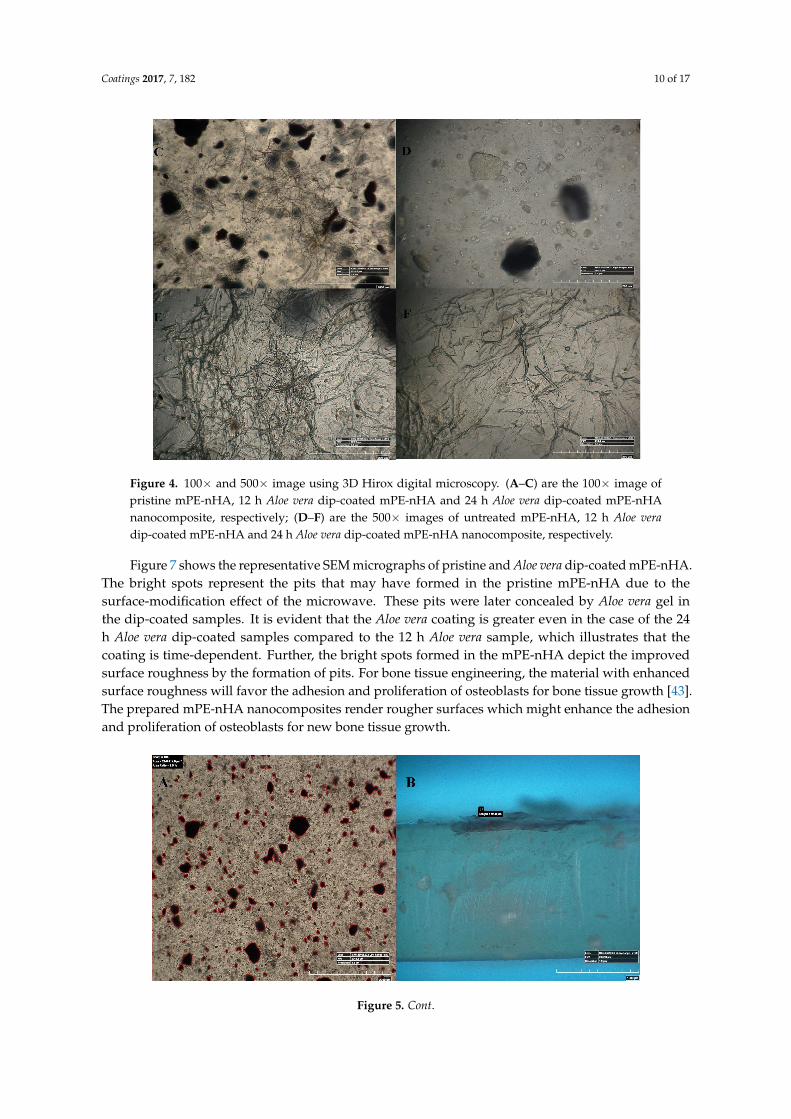
Figure 4 represents the 100× and 500× 3D Hirox digital microscopy images of mPE-nHA andAloe vera dip-coated mPE-nHA. A tangible coating of Aloe vera was observed after the 12 and 24 hexposure to Aloe vera extracts. Figure 5A shows the distribution of hydroxyapatite nanorods in themPE sample. It was found that the area ratio of nHA was 7.6% and the distribution of the nHAnanorod analysis in mPE-nHA depicts that there are 184 nHA nanorods present in a 2,144,112 µm2
area. The thickness of the Aloe vera coating measured was found to be 94.8 µm for the 24 h Aloe veradip-coated mPE-nHA, as represented in Figure 5B. Figure 5C–E further shows that the 1000× imagesof 3D hirox further dictate the presence of the fibrous Aloe vera structure, which is present in the 12 hand 24 h Aloe vera dip-coated mPE-nHA, and absent in the pristine mPE-nHA. Figure 6 ascertains theprofiling lines of the mPE-nHA surface at 1000×magnification, where the thickness of the Aloe veracoating was found to be the highest for the 24 h Aloe vera dip-coated mPE-nHA. Al-Hijazi et al. [42]studied the biological effect of Aloe vera in bone healing and also investigated the expression of bonemorphogenetic protein 7 (BMP7) in Aloe vera treated bone tissue. It was observed that the Aloe veratreated group exhibited increased proliferation of osteogenic cells and enhanced the expression ofBMP7 compared to the untreated group. Hence in our prepared composites containing significantamounts of Aloe vera content might favor the enhanced proliferation of osteogenic cells for new bonetissue growth.
Coatings 2017, 7, 182
Table 2. Peak shifts observed in Zone A of the recorded FTIR.
Pristine mPE‐nHA 12 h Aloe‐Vera‐Treated mPE‐nHA 24 h Aloe‐Vera‐Treated mPE‐nHA
406 416 424
428 431 436
444 451 464
457 466 470
471 480 474
476 486 480
486 497 486
503 515 509
528 535 532
Figure 4 represents the 100× and 500× 3D Hirox digital microscopy images of mPE‐nHA and
Aloe vera dip‐coated mPE‐nHA. A tangible coating of Aloe vera was observed after the 12 and 24 h
exposure to Aloe vera extracts. Figure 5A shows the distribution of hydroxyapatite nanorods in the
mPE sample. It was found that the area ratio of nHA was 7.6% and the distribution of the nHA
nanorod analysis in mPE‐nHA depicts that there are 184 nHA nanorods present in a 2,144,112 μm2
area. The thickness of the Aloe vera coating measured was found to be 94.8 μm for the 24 h Aloe vera
dip‐coated mPE‐nHA, as represented in Figure 5B. Figure 5C–E further shows that the 1000× images
of 3D hirox further dictate the presence of the fibrous Aloe vera structure, which is present in the 12 h
and 24 h Aloe vera dip‐coated mPE‐nHA, and absent in the pristine mPE‐nHA. Figure 6 ascertains the
profiling lines of the mPE‐nHA surface at 1000× magnification, where the thickness of the Aloe vera
coating was found to be the highest for the 24 h Aloe vera dip‐coated mPE‐nHA. Al‐Hijazi et al. [42]
studied the biological effect of Aloe vera in bone healing and also investigated the expression of bone
morphogenetic protein 7 (BMP7) in Aloe vera treated bone tissue. It was observed that the Aloe vera
treated group exhibited increased proliferation of osteogenic cells and enhanced the expression of
BMP7 compared to the untreated group. Hence in our prepared composites containing significant
amounts of Aloe vera content might favor the enhanced proliferation of osteogenic cells for new bone
tissue growth.
Figure 7 shows the representative SEM micrographs of pristine and Aloe vera dip‐coated
mPE‐nHA. The bright spots represent the pits that may have formed in the pristine mPE‐nHA due
to the surface‐modification effect of the microwave. These pits were later concealed by Aloe vera gel
in the dip‐coated samples. It is evident that the Aloe vera coating is greater even in the case of the 24 h
Aloe vera dip‐coated samples compared to the 12 h Aloe vera sample, which illustrates that the coating
is time‐dependent. Further, the bright spots formed in the mPE‐nHA depict the improved surface
roughness by the formation of pits. For bone tissue engineering, the material with enhanced surface
roughness will favor the adhesion and proliferation of osteoblasts for bone tissue growth [43]. The
prepared mPE‐nHA nanocomposites render rougher surfaces which might enhance the adhesion and
proliferation of osteoblasts for new bone tissue growth.
Figure 4. Cont. Figure 4. Cont.
Coatings 2017, 7, 182 10 of 17
Coatings 2017, 7, 182
Figure 4. 100× and 500× image using 3D Hirox digital microscopy. (A–C) are the 100× image of pristine
mPE‐nHA, 12 h Aloe vera dip‐coated mPE‐nHA and 24 h Aloe vera dip‐coated mPE‐nHA nanocomposite,
respectively; (D–F) are the 500× images of untreated mPE‐nHA, 12 h Aloe vera dip‐coated mPE‐nHA
and 24 h Aloe vera dip‐coated mPE‐nHA nanocomposite, respectively.
Figure 5. Cont.
Figure 4. 100× and 500× image using 3D Hirox digital microscopy. (A–C) are the 100× image ofpristine mPE-nHA, 12 h Aloe vera dip-coated mPE-nHA and 24 h Aloe vera dip-coated mPE-nHAnanocomposite, respectively; (D–F) are the 500× images of untreated mPE-nHA, 12 h Aloe veradip-coated mPE-nHA and 24 h Aloe vera dip-coated mPE-nHA nanocomposite, respectively.
Figure 7 shows the representative SEM micrographs of pristine and Aloe vera dip-coated mPE-nHA.The bright spots represent the pits that may have formed in the pristine mPE-nHA due to thesurface-modification effect of the microwave. These pits were later concealed by Aloe vera gel inthe dip-coated samples. It is evident that the Aloe vera coating is greater even in the case of the 24h Aloe vera dip-coated samples compared to the 12 h Aloe vera sample, which illustrates that thecoating is time-dependent. Further, the bright spots formed in the mPE-nHA depict the improvedsurface roughness by the formation of pits. For bone tissue engineering, the material with enhancedsurface roughness will favor the adhesion and proliferation of osteoblasts for bone tissue growth [43].The prepared mPE-nHA nanocomposites render rougher surfaces which might enhance the adhesionand proliferation of osteoblasts for new bone tissue growth.
Coatings 2017, 7, 182
Figure 4. 100× and 500× image using 3D Hirox digital microscopy. (A–C) are the 100× image of pristine
mPE‐nHA, 12 h Aloe vera dip‐coated mPE‐nHA and 24 h Aloe vera dip‐coated mPE‐nHA nanocomposite,
respectively; (D–F) are the 500× images of untreated mPE‐nHA, 12 h Aloe vera dip‐coated mPE‐nHA
and 24 h Aloe vera dip‐coated mPE‐nHA nanocomposite, respectively.
Figure 5. Cont. Figure 5. Cont.

Coatings 2017, 7, 182 11 of 17
Coatings 2017, 7, 182
Figure 5. 50×, 100× and 1000× image using 3D Hirox digital microscopy. (A) nHA nanorod distribution
in pristine mPE‐nHA at 50×; (B) Thickness of Aloe vera coating in 24 h Aloe vera dip‐coated mPE‐nHA
at 100×; (C–E) are 1000× images of untreated mPE‐nHA, and 12 and 24 h Aloe vera dip‐coated
mPE‐nHA, respectively.
Figure 6. Cont.
Figure 5. 50×, 100× and 1000× image using 3D Hirox digital microscopy. (A) nHA nanoroddistribution in pristine mPE-nHA at 50×; (B) Thickness of Aloe vera coating in 24 h Aloe vera dip-coatedmPE-nHA at 100×; (C–E) are 1000× images of untreated mPE-nHA, and 12 and 24 h Aloe veradip-coated mPE-nHA, respectively.
Coatings 2017, 7, 182
Figure 5. 50×, 100× and 1000× image using 3D Hirox digital microscopy. (A) nHA nanorod distribution
in pristine mPE‐nHA at 50×; (B) Thickness of Aloe vera coating in 24 h Aloe vera dip‐coated mPE‐nHA
at 100×; (C–E) are 1000× images of untreated mPE‐nHA, and 12 and 24 h Aloe vera dip‐coated
mPE‐nHA, respectively.
Figure 6. Cont. Figure 6. Cont.

Coatings 2017, 7, 182 12 of 17
Coatings 2017, 7, 182
Figure 6. Different three‐dimensional representations using 3D Hirox digital microscopy.
(A) Profiling of pristine mPE‐nHA at 1000×; (B) Profiling of 12 h Aloe vera dip‐coated mPE‐nHA at
1000×; (C) Profiling of 24 h Aloe vera dip‐coated mPE‐nHA at 1000×.
Figure 7. Representative SEM micrographs of pristine and Aloe vera dip‐coated mPE‐nHA. (A) Pristine
mPE‐nHA at 100×; (B) mPE‐nHA at 500×; (C) mPE‐nHA at 2500×; (D) 12 h Aloe vera dip‐coated
mPE‐nHA at 100×; (E) 12 h Aloe vera dip‐coated mPE‐nHA at 500×; (F) 12 h Aloe vera dip‐coated mPE‐
nHA at 1500×; (G) 24 h Aloe vera dip‐coated mPE‐nHA at 100×; (H) 24 h Aloe vera dip‐coated mPE‐
nHA at 500×; (I) 24 h Aloe vera dip‐coated mPE‐nHA at 1500×.
Figure 6. Different three-dimensional representations using 3D Hirox digital microscopy. (A) Profilingof pristine mPE-nHA at 1000×; (B) Profiling of 12 h Aloe vera dip-coated mPE-nHA at 1000×;(C) Profiling of 24 h Aloe vera dip-coated mPE-nHA at 1000×.
Coatings 2017, 7, 182
Figure 6. Different three‐dimensional representations using 3D Hirox digital microscopy.
(A) Profiling of pristine mPE‐nHA at 1000×; (B) Profiling of 12 h Aloe vera dip‐coated mPE‐nHA at
1000×; (C) Profiling of 24 h Aloe vera dip‐coated mPE‐nHA at 1000×.
Figure 7. Representative SEM micrographs of pristine and Aloe vera dip‐coated mPE‐nHA. (A) Pristine
mPE‐nHA at 100×; (B) mPE‐nHA at 500×; (C) mPE‐nHA at 2500×; (D) 12 h Aloe vera dip‐coated
mPE‐nHA at 100×; (E) 12 h Aloe vera dip‐coated mPE‐nHA at 500×; (F) 12 h Aloe vera dip‐coated mPE‐
nHA at 1500×; (G) 24 h Aloe vera dip‐coated mPE‐nHA at 100×; (H) 24 h Aloe vera dip‐coated mPE‐
nHA at 500×; (I) 24 h Aloe vera dip‐coated mPE‐nHA at 1500×.
Figure 7. Representative SEM micrographs of pristine and Aloe vera dip-coated mPE-nHA. (A) PristinemPE-nHA at 100×; (B) mPE-nHA at 500×; (C) mPE-nHA at 2500×; (D) 12 h Aloe vera dip-coatedmPE-nHA at 100×; (E) 12 h Aloe vera dip-coated mPE-nHA at 500×; (F) 12 h Aloe vera dip-coatedmPE-nHA at 1500×; (G) 24 h Aloe vera dip-coated mPE-nHA at 100×; (H) 24 h Aloe vera dip-coatedmPE-nHA at 500×; (I) 24 h Aloe vera dip-coated mPE-nHA at 1500×.
Coatings 2017, 7, 182 13 of 17
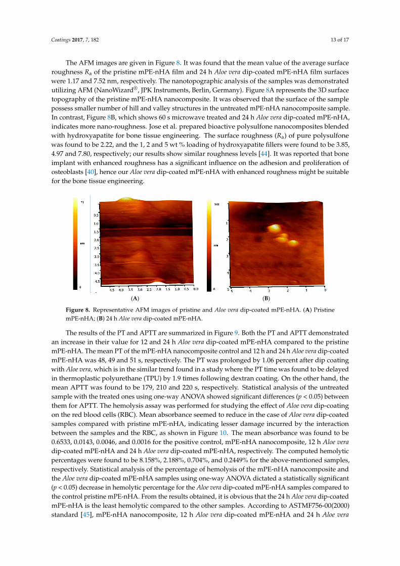
The AFM images are given in Figure 8. It was found that the mean value of the average surfaceroughness Ra of the pristine mPE-nHA film and 24 h Aloe vera dip-coated mPE-nHA film surfaceswere 1.17 and 7.52 nm, respectively. The nanotopographic analysis of the samples was demonstratedutilizing AFM (NanoWizard®, JPK Instruments, Berlin, Germany). Figure 8A represents the 3D surfacetopography of the pristine mPE-nHA nanocomposite. It was observed that the surface of the samplepossess smaller number of hill and valley structures in the untreated mPE-nHA nanocomposite sample.In contrast, Figure 8B, which shows 60 s microwave treated and 24 h Aloe vera dip-coated mPE-nHA,indicates more nano-roughness. Jose et al. prepared bioactive polysulfone nanocomposites blendedwith hydroxyapatite for bone tissue engineering. The surface roughness (Ra) of pure polysulfonewas found to be 2.22, and the 1, 2 and 5 wt % loading of hydroxyapatite fillers were found to be 3.85,4.97 and 7.80, respectively; our results show similar roughness levels [44]. It was reported that boneimplant with enhanced roughness has a significant influence on the adhesion and proliferation ofosteoblasts [40], hence our Aloe vera dip-coated mPE-nHA with enhanced roughness might be suitablefor the bone tissue engineering.
Coatings 2017, 7, 182
The AFM images are given in Figure 8. It was found that the mean value of the average surface
roughness Ra of the pristine mPE‐nHA film and 24 h Aloe vera dip‐coated mPE‐nHA film surfaces
were 1.17 and 7.52 nm, respectively. The nanotopographic analysis of the samples was demonstrated
utilizing AFM (NanoWizard®, JPK Instruments, Berlin, Germany). Figure 8A represents the 3D
surface topography of the pristine mPE‐nHA nanocomposite. It was observed that the surface of the
sample possess smaller number of hill and valley structures in the untreated mPE‐nHA
nanocomposite sample. In contrast, Figure 8B, which shows 60 s microwave treated and 24 h Aloe
vera dip‐coated mPE‐nHA, indicates more nano‐roughness. Jose et al. prepared bioactive polysulfone
nanocomposites blended with hydroxyapatite for bone tissue engineering. The surface roughness (Ra)
of pure polysulfone was found to be 2.22, and the 1, 2 and 5 wt % loading of hydroxyapatite fillers were
found to be 3.85, 4.97 and 7.80, respectively; our results show similar roughness levels [44]. It was
reported that bone implant with enhanced roughness has a significant influence on the adhesion and
proliferation of osteoblasts [40], hence our Aloe vera dip‐coated mPE‐nHA with enhanced roughness
might be suitable for the bone tissue engineering.
(A) (B)
Figure 8. Representative AFM images of pristine and Aloe vera dip‐coated mPE‐nHA. (A) Pristine
mPE‐nHA; (B) 24 h Aloe vera dip‐coated mPE‐nHA.
The results of the PT and APTT are summarized in Figure 9. Both the PT and APTT demonstrated
an increase in their value for 12 and 24 h Aloe vera dip‐coated mPE‐nHA compared to the pristine
mPE‐nHA. The mean PT of the mPE‐nHA nanocomposite control and 12 h and 24 h Aloe vera
dip‐coated mPE‐nHA was 48, 49 and 51 s, respectively. The PT was prolonged by 1.06 percent after
dip coating with Aloe vera, which is in the similar trend found in a study where the PT time was found
to be delayed in thermoplastic polyurethane (TPU) by 1.9 times following dextran coating. On the
other hand, the mean APTT was found to be 179, 210 and 220 s, respectively. Statistical analysis of
the untreated sample with the treated ones using one‐way ANOVA showed significant differences
(p < 0.05) between them for APTT. The hemolysis assay was performed for studying the effect of
Aloe vera dip‐coating on the red blood cells (RBC). Mean absorbance seemed to reduce in the case of
Aloe vera dip‐coated samples compared with pristine mPE‐nHA, indicating lesser damage incurred
by the interaction between the samples and the RBC, as shown in Figure 10. The mean absorbance
was found to be 0.6533, 0.0143, 0.0046, and 0.0016 for the positive control, mPE‐nHA nanocomposite,
12 h Aloe vera dip‐coated mPE‐nHA and 24 h Aloe vera dip‐coated mPE‐nHA, respectively. The
computed hemolytic percentages were found to be 8.158%, 2.188%, 0.704%, and 0.2449% for the
above‐mentioned samples, respectively. Statistical analysis of the percentage of hemolysis of the
mPE‐nHA nanocomposite and the Aloe vera dip‐coated mPE‐nHA samples using one‐way ANOVA
dictated a statistically significant (p < 0.05) decrease in hemolytic percentage for the Aloe vera
dip‐coated mPE‐nHA samples compared to the control pristine mPE‐nHA. From the results obtained,
it is obvious that the 24 h Aloe vera dip‐coated mPE‐nHA is the least hemolytic compared to the other
samples. According to ASTMF756‐00(2000) standard [45], mPE‐nHA nanocomposite, 12 h Aloe vera
dip‐coated mPE‐nHA and 24 h Aloe vera dip‐coated mPE‐nHA is inferred to be a non‐hemolytic
Figure 8. Representative AFM images of pristine and Aloe vera dip-coated mPE-nHA. (A) PristinemPE-nHA; (B) 24 h Aloe vera dip-coated mPE-nHA.
The results of the PT and APTT are summarized in Figure 9. Both the PT and APTT demonstratedan increase in their value for 12 and 24 h Aloe vera dip-coated mPE-nHA compared to the pristinemPE-nHA. The mean PT of the mPE-nHA nanocomposite control and 12 h and 24 h Aloe vera dip-coatedmPE-nHA was 48, 49 and 51 s, respectively. The PT was prolonged by 1.06 percent after dip coatingwith Aloe vera, which is in the similar trend found in a study where the PT time was found to be delayedin thermoplastic polyurethane (TPU) by 1.9 times following dextran coating. On the other hand, themean APTT was found to be 179, 210 and 220 s, respectively. Statistical analysis of the untreatedsample with the treated ones using one-way ANOVA showed significant differences (p < 0.05) betweenthem for APTT. The hemolysis assay was performed for studying the effect of Aloe vera dip-coatingon the red blood cells (RBC). Mean absorbance seemed to reduce in the case of Aloe vera dip-coatedsamples compared with pristine mPE-nHA, indicating lesser damage incurred by the interactionbetween the samples and the RBC, as shown in Figure 10. The mean absorbance was found to be0.6533, 0.0143, 0.0046, and 0.0016 for the positive control, mPE-nHA nanocomposite, 12 h Aloe veradip-coated mPE-nHA and 24 h Aloe vera dip-coated mPE-nHA, respectively. The computed hemolyticpercentages were found to be 8.158%, 2.188%, 0.704%, and 0.2449% for the above-mentioned samples,respectively. Statistical analysis of the percentage of hemolysis of the mPE-nHA nanocomposite andthe Aloe vera dip-coated mPE-nHA samples using one-way ANOVA dictated a statistically significant(p < 0.05) decrease in hemolytic percentage for the Aloe vera dip-coated mPE-nHA samples compared tothe control pristine mPE-nHA. From the results obtained, it is obvious that the 24 h Aloe vera dip-coatedmPE-nHA is the least hemolytic compared to the other samples. According to ASTMF756-00(2000)standard [45], mPE-nHA nanocomposite, 12 h Aloe vera dip-coated mPE-nHA and 24 h Aloe vera

Coatings 2017, 7, 182 14 of 17
dip-coated mPE-nHA is inferred to be a non-hemolytic material, since the percentage of damagefalls below two percent. Selvakumar et al. [27] prepared a nanocomposite based on polyurethaneincorporated with polypropylene glycol (PPG)-warped hydroxyapatite for bone tissue engineering.It was observed that the hydroxyapatite-loaded polyurethane nanocomposites exhibited enhancedblood compatibility and also observed enhanced osteoblast MG63 cell proliferation compared topristine polyurethane (PU). Hence, our developed membrane showing enhanced blood compatibilitymight favor osteoblast cell proliferation for new bone tissue growth.
Coatings 2017, 7, 182
material, since the percentage of damage falls below two percent. Selvakumar et al. [27] prepared a
nanocomposite based on polyurethane incorporated with polypropylene glycol (PPG)‐warped
hydroxyapatite for bone tissue engineering. It was observed that the hydroxyapatite‐loaded
polyurethane nanocomposites exhibited enhanced blood compatibility and also observed enhanced
osteoblast MG63 cell proliferation compared to pristine polyurethane (PU). Hence, our developed
membrane showing enhanced blood compatibility might favor osteoblast cell proliferation for new
bone tissue growth.
Figure 9. Comparison of prothrombin time (PT), activated partial thromboplastin time (APPT) and
absorbance of pristine mPE‐nHA, 12 h and 24 h Aloe vera dip‐coated mPE‐nHA (n = 3). Values shown
are mean ± SD and * indicates that differences in the mean are significant (p < 0.05) with respect to the
mPE control.
Figure 10. Comparison of the hemolysis percentage of the positive control, pristine mPE‐nHA, 12 h
and 24 h Aloe vera dip‐coated mPE‐nHA (n = 3). Values shown are mean ± SD and * indicates that
differences in the mean are significant (p < 0.05) with respect to the mPE control.
Figure 9. Comparison of prothrombin time (PT), activated partial thromboplastin time (APPT) andabsorbance of pristine mPE-nHA, 12 h and 24 h Aloe vera dip-coated mPE-nHA (n = 3). Values shownare mean ± SD and * indicates that differences in the mean are significant (p < 0.05) with respect to themPE control.
Coatings 2017, 7, 182
material, since the percentage of damage falls below two percent. Selvakumar et al. [27] prepared a
nanocomposite based on polyurethane incorporated with polypropylene glycol (PPG)‐warped
hydroxyapatite for bone tissue engineering. It was observed that the hydroxyapatite‐loaded
polyurethane nanocomposites exhibited enhanced blood compatibility and also observed enhanced
osteoblast MG63 cell proliferation compared to pristine polyurethane (PU). Hence, our developed
membrane showing enhanced blood compatibility might favor osteoblast cell proliferation for new
bone tissue growth.
Figure 9. Comparison of prothrombin time (PT), activated partial thromboplastin time (APPT) and
absorbance of pristine mPE‐nHA, 12 h and 24 h Aloe vera dip‐coated mPE‐nHA (n = 3). Values shown
are mean ± SD and * indicates that differences in the mean are significant (p < 0.05) with respect to the
mPE control.
Figure 10. Comparison of the hemolysis percentage of the positive control, pristine mPE‐nHA, 12 h
and 24 h Aloe vera dip‐coated mPE‐nHA (n = 3). Values shown are mean ± SD and * indicates that
differences in the mean are significant (p < 0.05) with respect to the mPE control.
Figure 10. Comparison of the hemolysis percentage of the positive control, pristine mPE-nHA, 12 hand 24 h Aloe vera dip-coated mPE-nHA (n = 3). Values shown are mean ± SD and * indicates thatdifferences in the mean are significant (p < 0.05) with respect to the mPE control.

Coatings 2017, 7, 182 15 of 17
4. Conclusions
In this study, the mPE was incorporated with nano-rods of hydroxyapaptite and then coated withAloe vera with the help of a microwave. The samples were subjected to physical characterization testssuch as contact angle, FTIR, SEM, AFM and 3D Hirox microscopy scanning. The results show that thehydrophilicity of the nano-hydroxyapaptite improves with dip coating with Aloe vera. The Aloe verajuice makes the surface topology of the mPE-nHA more hydrophilic, thereby improving its wettability.Surface roughness improvement was ascertained by the AFM result. The presence of Aloe vera dipcoating on mPE-nHA was confirmed by the SEM and 3D Hirox microscopy results. The PT wasdelayed by 1.06% for the 24 h Aloe vera dip-coated mPE-nHA compared to the pristine mPE-nHA.Likewise, the Aloe vera dip-coated mPE-nHA nanocomposite prolonged the APTT by 41 s against thepristine mPE-nHA. The hemolysis percentage was found to be the least for the 24 h Aloe vera dip-coatedmPE-nHA nanocomposite, which was only 0.2449%. Hence, the 24 h Aloe vera dip-coated mPE-nHAcan be plausibly exploited for bone tissue engineering applications.
Acknowledgments: This work was supported by the Ministry of Higher Education Malaysia with the Grantno.Q.J130000.2545.17H00 and FRGS Grant no.R.J130000.7809.4F444.
Author Contributions: Hairong Wang and Xueliang Zhang performed the experiments and analyzed the data.Mohan Prasath Mani assisted in doing the experiments, analyzed the data, wrote the paper, and prepared figuresand/or tables. Saravana Kumar Jaganathan conceived and designed the experiments, performed the experiments,analyzed the data, contributed reagents/materials/analysis tools, prepared figures and/or tables and revieweddrafts of the paper. Yi Huang and Chengzheng Wang contributed reagents/materials/analysis tools and revieweddrafts of the paper.
Conflicts of Interest: The authors declare no conflict of interest.
References
1. O’brien, F.J. Biomaterials & scaffolds for tissue engineering. Mater. Today 2011, 14, 88–95.2. Zohora, F.T.; Azim, A.Y.M.A. Biomaterials as a porous scaffold for tissue engineering applications: A review.
Eur. Sci. 2014, 10, 186–209.3. Kar, K.K.; Rana, S.; Pandey, J. Handbook of Polymer Nanocomposites Processing, Performance and Application;
Springer: Berlin/Heidelberg, Germany, 2015.4. De Santis, R.; Gloria, A.; Russo, T.; D’Amora, U.; Zeppetelli, S.; Dionigi, C.; Sytcheva, A.; Herrmannsdorfer, T.;
Dediu, V.; Ambrosio, L. A basic approach toward the development of nanocomposite magnetic scaffolds foradvanced bone tissue engineering. J. Appl. Polym. Sci. 2011, 122, 3599–3605. [CrossRef]
5. Gloria, A.; Russo, T.; D’Amora, U.; Zeppetelli, S.; D’Alessandro, T.; Sandri, M.; Bañobre-López, M.;Piñeiro-Redondo, Y.; Uhlarz, M.; Tampieri, A.; et al. Magnetic poly(epsilon-caprolactone)/iron-dopedhydroxyapatite nanocomposite substrates for advanced bone tissue engineering. J. R. Soc. Interface 2013, 10,20120833. [CrossRef] [PubMed]
6. Dorozhkin, S.V. Calcium Orthophosphates: Applications in Nature, Biology, and Medicine; CRC Press: Boca Raton,FL, USA, 2012; p. 854.
7. Eliaz, N.; Metoki, N. Calcium phosphate bioceramics: A review of their history, structure, properties, coatingtechnologies and biomedical applications. Materials 2017, 10, 334. [CrossRef] [PubMed]
8. Huber, F.X.; McArthur, N.; Hillmeier, J.; Kock, H.J.; Baier, M.; Diwo, M.; Berger, I.; Meeder, P.J. Voidfilling of tibia compression fracture zones using a novel resorbable nanocrystalline hydroxyapatite paste incombination with a hydroxyapatite ceramic core: First clinical results. Arch. Orthop. Trauma Surg. 2006, 126,533–540. [CrossRef] [PubMed]
9. Banobre-Lopez, M.; Pineiro-Redondo, Y.; De Santis, R.; Gloria, A.; Ambrosio, L.; Tampieri, A.; Dediu, V.;Rivas, J. Poly(caprolactone) based magnetic scaffolds for bone tissue engineering. J. Appl. Phys. 2011, 109,07B313. [CrossRef]
10. Fu, S.; Wang, X.; Guo, G.; Shi, S.; Liang, H.; Luo, F.; Wei, Y.; Qian, Z. Preparation and characterization ofnano-hydroxyapatite/poly(ε-caprolactone)-poly(ethylene glycol)-poly(ε-caprolactone) composite fibers fortissue engineering. J. Phys. Chem. C 2010, 114, 18372–18378. [CrossRef]

Coatings 2017, 7, 182 16 of 17
11. Laschke, M.W.; Strohe, A.; Menger, M.D.; Alini, M.; Eglin, D. In vitro and in vivo evaluation of a novelnanosize hydroxyapatite particles/poly(ester-urethane) composite scaffold for bone tissue engineering.Acta Biomater. 2010, 6, 2020–2027. [CrossRef] [PubMed]
12. Ma, R.; Weng, L.; Bao, X.; Ni, Z.; Song, S.; Cai, W. Characterization of in situ synthesizedhydroxyapatite/polyetheretherketone composite materials. Mater. Lett. 2012, 71, 117–119. [CrossRef]
13. Hamman, H.H. Composition and applications of Aloe vera leaf gel. Molecules 2008, 13, 1599–1616. [CrossRef][PubMed]
14. Surjushe, A.; Vasani, R.; Saple, D.G. Aloe vera: A short review. Ind. J. Dermatol. 2008, 53, 163–166. [CrossRef][PubMed]
15. Herzberg, F.; Gruenwald, J. Aloe Vera: An International Success Story; Nutraceutical World: Ramsey, NJ,USA, 2003.
16. Davis, R.H.; Donato, J.J.; Hartman, G.M.; Haas, R.C. Anti-inflammatory and wound healing activity of agrowth substance in Aloe vera. J. Am. Podiatr. Med. Assoc. 1994, 84, 77–81. [CrossRef] [PubMed]
17. Guruvenket, S.; Rao, G.M.; Komath, M.; Raichur, A.M. Plasma surface modification of polystyrene andpolyethylene. Appl. Surf. Sci. 2004, 236, 278–284. [CrossRef]
18. Hui, A.Y.N.; Wang, G.; Lin, B.; Chan, W.T. Microwave plasma treatment of polymer surface for irreversiblesealing of microfluidic devices. Lab Chip 2005, 5, 1173–1177. [CrossRef] [PubMed]
19. Ku, H.S.; Siores, E.; Ball, J.A.R. Review—Microwave processing of materials: Part I. HKIE Trans. 2001, 8,31–37.
20. Kavya, K.C.; Dixit, R.; Jayakumar, R.; Nair, S.V.; Chennazhi, K.P. Synthesis and characterizationof chitosan/chondroitin sulfate/nano-SiO2 composite scaffold for bone tissue engineering.J. Biomed. Nanotechnol. 2012, 8, 149–160. [CrossRef] [PubMed]
21. Zhou, S.; Zheng, X.; Yu, X.; Wang, J.; Weng, J.; Li, X.; Feng, B.; Yin, M. Hydrogen bonding interaction ofpoly(d,l-lactide)/hydroxyapatite nanocomposites. Chem. Mater. 2007, 19, 247–253. [CrossRef]
22. Gong, X.H.; Tang, C.Y.; Hu, H.C.; Zhou, X.P.; Xie, X.L. Improved mechanical properties ofHIPS/hydroxyapatite composites by surface modification of hydroxyapatite via in-situ polymerization ofstyrene. J. Mater. Sci. Mater. Med. 2004, 15, 1141–1146. [CrossRef] [PubMed]
23. Lee, H.J.; Choi, K.W.; Kim, K.J.; Lee, S.C. Modification of hydroxyapatite nanosurfaces for enhanced colloidalstability and improved interfacial adhesion in nanocomposites. Chem. Mater. 2006, 18, 5111–5118. [CrossRef]
24. Bose, S.; Saha, S.K. Synthesis and characterization of hydroxyapatite nanopowders by emulsion technique.Chem. Mater. 2003, 15, 4464–4469. [CrossRef]
25. Yuan, J.; Wu, Y.; Zheng, Q.X.; Xie, X.L. Synthesis and characterization of nano hydroxylapatite by reactionprecipitation in impinging streams. Adv. Mater. Res. 2011, 160, 1301–1308. [CrossRef]
26. Vidhya, G.; Kumar, G.S.; Kattimani, V.S.; Thamizhavel, A.; Girija, E.K. Microwave irradiation synthesis of 3Dflower-like hydroxyapatite. Int. J. Sci. Eng. Res. 2015, 6, 18–19.
27. Selvakumar, M.; Jaganathan, S.K.; Nando, G.B.; Chattopadhyay, S. Synthesis and characterization ofnovel polycarbonate based polyurethane/polymer wrapped hydroxyapatite nanocomposites: Mechanicalproperties, osteoconductivity and biocompatibility. J. Biomed. Nanotechnol. 2015, 11, 291–305. [CrossRef][PubMed]
28. Irzh, A.; Gedanken, A. A microwave-assisted process for coating polymer and glass surfaces withsemiconducting ZnO submicron particles. J. Appl. Polym. Sci. 2009, 113, 1773–1780. [CrossRef]
29. Irzh, A.; Perkas, N.; Gedanken, A. Microwave-assisted coating of PMMA beads by silver nanoparticles.Langmuir 2007, 23, 9891–9897. [CrossRef] [PubMed]
30. Su, H.C.; Chen, C.H.; Chen, Y.C.; Yao, D.J.; Chen, H.; Chang, Y.C.; Yew, T.R. Improving the adhesion ofcarbon nanotubes to a substrate using microwave treatment. Carbon 2010, 48, 805–812. [CrossRef]
31. Tuval, T.; Gedanken, A. A microwave-assisted polyol method for the deposition of silver nanoparticles onsilica spheres. Nanotechnology 2007, 18, 255601. [CrossRef]
32. Mohandas, H.; Sivakumar, G.; Kasi, P.; Jaganathan, S.K.; Supriyanto, E. Microwave-assisted surfacemodification of metallocene polyethylene for improving blood compatibility. BioMed Res. Int. 2013, 2013,253473. [CrossRef] [PubMed]
33. Xue, L.; Gao, X.; Zhao, K.; Liu, J.; Yu, X.; Han, Y. The formation of different structures ofpoly(3-hexylthiophene) film on a patterned substrate by dip coating from aged solution. Nanotechnology2010, 21, 145303. [CrossRef] [PubMed]
Coatings 2017, 7, 182 17 of 17
34. Pereira, C.; Busani, T.; Branco, L.C.; Joosten, I.; Sandu, I.C. Nondestructive characterization and enzymecleaning of painted surfaces: Assessment from the macro to nano level. Microsc. Microanal. 2013, 19,1632–1644. [CrossRef] [PubMed]
35. Pelagade, S.M.; Rane, R.S.; Mukherjee, S.; Deshpande, U.P.; Ganesan, V.; Shripathi, T. Investigation of surfacefree energy for PTFE polymer by bipolar argon plasma treatment. J. Surf. Eng. Mater. Adv. Technol. 2012, 2,132–136. [CrossRef]
36. Amarnath, L.P.; Srinivas, A.; Ramamurthi, A. In vitro hemocompatibility testing of UV-modified hyaluronanhydrogels. Biomaterials 2006, 27, 1416–1424. [CrossRef] [PubMed]
37. Jaganathan, S.K.; Mohandas, H.; Sivakumar, G.; Kasi, P.; Sudheer, T.; Avineri, V.S.; Murugesan, S.;Supriyanto, E. Enhanced blood compatibility of metallocene polyethylene subjected to hydrochloric acidtreatment for cardiovascular implants. BioMed Res. Int. 2014, 2014, 963149. [CrossRef] [PubMed]
38. Gentile, P.; Ghione, C.; Tonda-Turo, C.; Kalaskar, D.M. Peptide functionalisation of nanocomposite polymerfor bone tissue engineering using plasma surface polymerisation. RSC Adv. 2015, 5, 80039–80047. [CrossRef]
39. Agnes Mary, S.; Giri Dev, V.R. Electrospun herbal nanofibrous wound dressings for skin tissue engineering.J. Text. Inst. 2015, 106, 886–895. [CrossRef]
40. Suganya, S.; Venugopal, J.; Ramakrishna, S.; Lakshmi, B.S.; Dev, V.R. Naturally derived biofunctionalnanofibrous scaffold for skin tissue regeneration. Int. J. Biol. Macromol. 2014, 68, 135–143. [CrossRef][PubMed]
41. Müller, C.M.; Pejcic, B.; Esteban, L.; Piane, C.D.; Raven, M.; Mizaikoff, B. Infrared attenuated total reflectancespectroscopy: An innovative strategy for analyzing mineral components in energy relevant systems. Sci. Rep.2014, 4, 6764. [CrossRef] [PubMed]
42. Al-Hijazi, A.Y.; Al-Mahammadawy, A.K.; Issa, E. Expression of BMP7 in bone tissue treated with Aloe vera.Int. Res. J. Natl. Sci. 2015, 3, 39–48.
43. Huang, H.H.; Ho, C.T.; Lee, T.H.; Lee, T.L.; Liao, K.K.; Chen, F.L. Effect of surface roughness of groundtitanium on initial cell adhesion. Biomol. Eng. 2004, 21, 93–97. [CrossRef] [PubMed]
44. Jose, A.J.; Alagar, M. Development of bioactive polysulfone nanocomposites for bone tissue replacement.In Proceedings of the 18th International Conference on Composite Materials (ICCM-18), Jeju Island, Korea,21–26 August 2011.
45. ASTM F756-00 Standard Practice for Assessment of Hemolytic Properties of Materials; ASTM International:West Conshohocken, PA, USA, 2000.
© 2017 by the authors. Licensee MDPI, Basel, Switzerland. This article is an open accessarticle distributed under the terms and conditions of the Creative Commons Attribution(CC BY) license (http://creativecommons.org/licenses/by/4.0/).