microscopic studies of the link between salmonid proliferative kidney disease (pkd) & bryozoans
TRANSCRIPT
Fish Veterinary Journal • Number 8 • 2005 62
Microscopic studies of the link between salmonid proliferative kidney disease (PKD) & bryozoans
Charles McGurk, David J. Morris, Alexandra AdamsInstitute of Aquaculture, University of Stirling, Scotland. FK9 4LA
Abstract
Proliferative kidney disease is a significant financial burden to freshwater salmonid culture in the UK. This paper describes studies of the development of the causative agent, Tetracapsuloides bryosalmonae and another related malacosporean parasite within their invertebrate freshwater bryozoan hosts and discusses possible control methods.
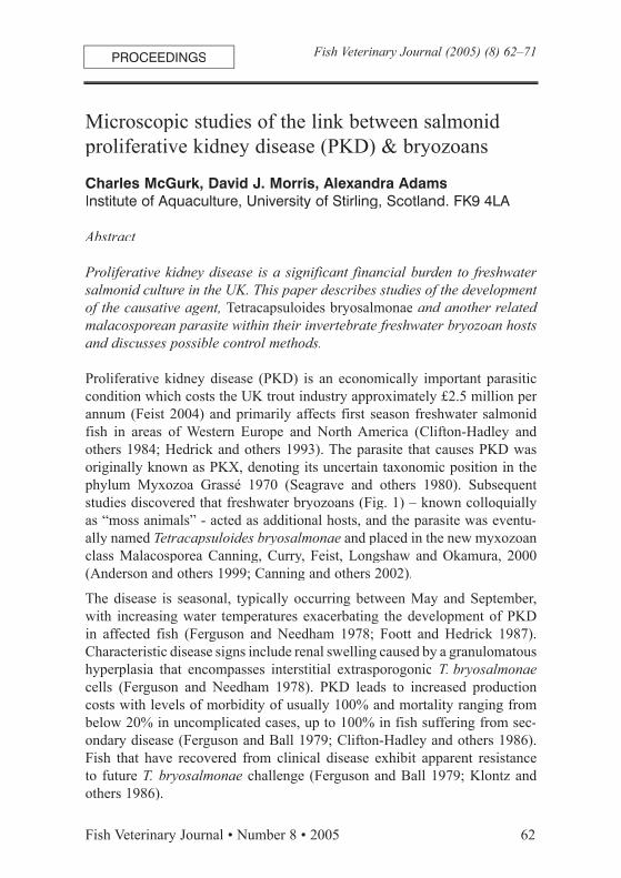
Proliferative kidney disease (PKD) is an economically important parasitic condition which costs the UK trout industry approximately £2.5 million per annum (Feist 2004) and primarily affects first season freshwater salmonid fish in areas of Western Europe and North America (Clifton-Hadley and others 1984; Hedrick and others 1993). The parasite that causes PKD was originally known as PKX, denoting its uncertain taxonomic position in the phylum Myxozoa Grassé 1970 (Seagrave and others 1980). Subsequent studies discovered that freshwater bryozoans (Fig. 1) – known colloquially as “moss animals” - acted as additional hosts, and the parasite was eventu-ally named Tetracapsuloides bryosalmonae and placed in the new myxozoan class Malacosporea Canning, Curry, Feist, Longshaw and Okamura, 2000 (Anderson and others 1999; Canning and others 2002).
The disease is seasonal, typically occurring between May and September, with increasing water temperatures exacerbating the development of PKD in affected fish (Ferguson and Needham 1978; Foott and Hedrick 1987). Characteristic disease signs include renal swelling caused by a granulomatous hyperplasia that encompasses interstitial extrasporogonic T. bryosalmonaecells (Ferguson and Needham 1978). PKD leads to increased production costs with levels of morbidity of usually 100% and mortality ranging from below 20% in uncomplicated cases, up to 100% in fish suffering from sec-ondary disease (Ferguson and Ball 1979; Clifton-Hadley and others 1986). Fish that have recovered from clinical disease exhibit apparent resistance to future T. bryosalmonae challenge (Ferguson and Ball 1979; Klontz and others 1986).
PROCEEDINGS Fish Veterinary Journal (2005) (8) 62–71
63 Fish Veterinary Journal • Number 8 • 2005
Various control methods have been developed for PKD, with varying levels of success. Husbandry measures - including lowering summer water temper-ature (using bore-hole water), delaying transfer of naïve stocks to endemic waters, eliminating secondary pathogens, reducing feeding rates and delay-ing grading - have been implemented as attempts to limit economic losses (Bucke and others 1981). Malachite green, the antibiotic fumagillin DCH and its synthetic analogue TNP-470 have been used therapeutically with some efficacy, but concerns over toxicity to fish, residue levels and envi-ronmental issues have prevented wide adoption of these treatments (Morris and others 2003). The perceived specific immunity that previously exposed fish demonstrate to T. bryosalmonae has led to interest in the possibility of has led to interest in the possibility of developing a vaccine to combat the condition, although no such product is currently available.
During this study, laboratory culture techniques were developed to allow continuous maintenance of the bryozoan hosts of T. bryosalmonae. These filter-feeding colonial invertebrates are normally sessile, being adhered to submerged substrates such as wood and plastic. Bryozoans were collected from a loch, germinated from their overwintering stages (statoblasts) and maintained in aquaria. A feeding trial was undertaken, which gauged the nutritional value of more than 50 species of commercially cultured protozoa and algae for the bryozoans. These monocultures were added individually to Petri dishes containing the bryozoans, which were then observed under a dissecting microscope for their ability to ingest the experimental diet. Subsequent examination of released bryozoan faecal pellets assessed the level of digestion of those species consumed. It was found that while certain species were readily ingested, they were not digested with organisms surviv-ing passage through the digestive tract, thus negating any nutritional benefit to the bryozoan. However, a selection of algae and protozoa were found to be equally readily ingested and digested by the bryozoans, presumably proving beneficial to their maintenance. No single species of algae or protozoa was identified which could maintain the bryozoan colonies alone, so mixtures of identified which could maintain the bryozoan colonies alone, so mixtures of digestible cultures were used for the long-term maintenance of bryozoans.
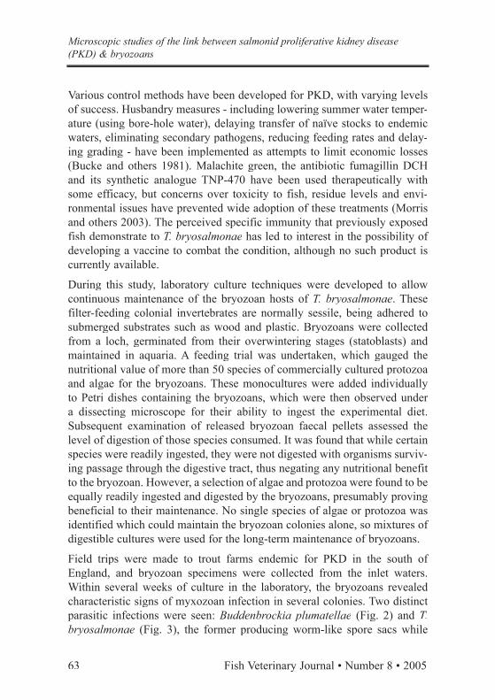
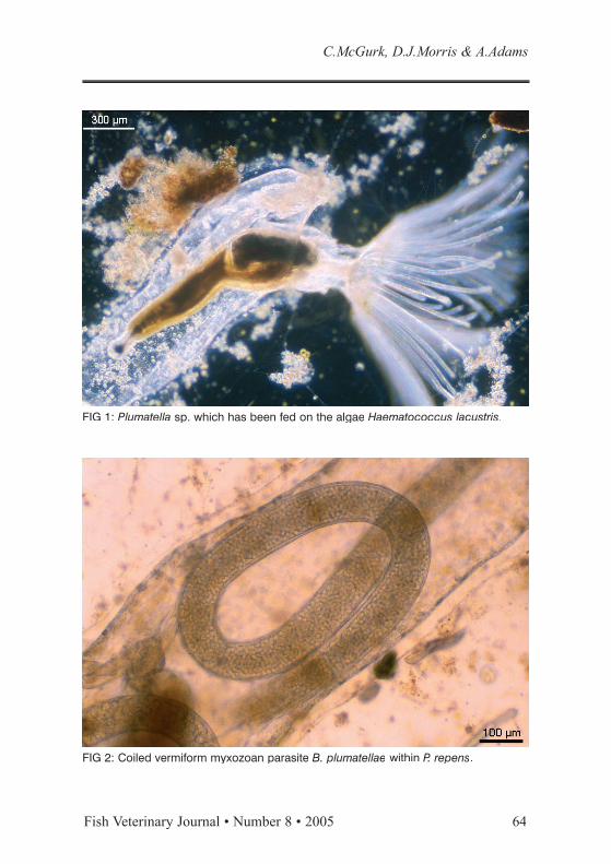
Field trips were made to trout farms endemic for PKD in the south of Field trips were made to trout farms endemic for PKD in the south of England, and bryozoan specimens were collected from the inlet waters. Within several weeks of culture in the laboratory, the bryozoans revealed characteristic signs of myxozoan infection in several colonies. Two distinct parasitic infections were seen: Buddenbrockia plumatellae (Fig. 2) and T. bryosalmonae (Fig. 3), the former producing worm-like spore sacs while
Microscopic studies of the link between salmonid proliferative kidney disease (PKD) & bryozoans
Fish Veterinary Journal • Number 8 • 2005 64
C.McGurk, D.J.Morris & A.Adams
FIG 2: Coiled vermiform myxozoan parasite B. plumatellae within P. repens.
FIG 1: Plumatella sp. which has been fed on the algae Haematococcus lacustris.
65 Fish Veterinary Journal • Number 8 • 2005
the latter developed spherical spore sacs. The ensuing development of these parasites was observed under an inverted microscope. Both still and video images were captured, demonstrating the movement of the parasitic develop-mental stages within the coelomic fluids of the bryozoan host.
With both myxozoan infections, the first observable sign of infection was the presence of numerous microscopic bodies swirling within the bryozoans. After one or two days, larger irregularly shaped bodies formed. In the case of B. plumatellae these adhered to the internal wall of the host, while with T. bryosalmonae they were free of attachment. Subsequently, the stages enlarged: B. plumatellae forming elongated oblong masses, while T. bry-osalmonae infection led to formation of spherical bodies. Over the period of about a week, mature spore sacs were seen to develop. The vermiform spore sacs of B. plumatellae seen within the bryozoan Plumatella repens were up to 2 mm in length, apparently writhing independently of their host. Mature spore sacs of T. bryosalmonae within Fredericella sultana were of were of approximate diameter 100 – 200 µm and were seen to rotate and flow in the currents within the coelomic cavity of the bryozoan host. Upon maturation
Microscopic studies of the link between salmonid proliferative kidney disease (PKD) & bryozoans
FIG 3: Spore sacs of T. bryosalmonae within F. sultana.
Fish Veterinary Journal • Number 8 • 2005 66
of the sacs, they burst releasing numerous spores into the bryozoan, which were subsequently ejected into the surrounding water. The spores of both parasites were approximately 20 µm in diameter, each possessing the four characteristic polar capsules of malacosporeans.
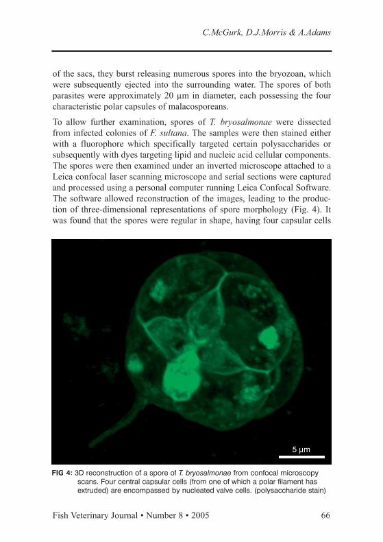
To allow further examination, spores of T. bryosalmonae were dissected from infected colonies of F. sultana. The samples were then stained either with a fluorophore which specifically targeted certain polysaccharides or subsequently with dyes targeting lipid and nucleic acid cellular components. The spores were then examined under an inverted microscope attached to a Leica confocal laser scanning microscope and serial sections were captured and processed using a personal computer running Leica Confocal Software. The software allowed reconstruction of the images, leading to the produc-tion of three-dimensional representations of spore morphology (Fig. 4). It was found that the spores were regular in shape, having four capsular cells
C.McGurk, D.J.Morris & A.Adams
FIG 4: 3D reconstruction of a spore of T. bryosalmonae from confocal microscopy scans. Four central capsular cells (from one of which a polar filament has extruded) are encompassed by nucleated valve cells. (polysaccharide stain)

67 Fish Veterinary Journal • Number 8 • 2005
Fish Veterinary Journal • Number 8 • 2005 68
which included spherical polar capsules, surrounded by structural valve cells encompassing two central germinative sporoplasms. In the examination of encompassing two central germinative sporoplasms. In the examination of samples following minimal processing, this work complemented previous ultrastructural studies of the morphology of T. bryosalmonae within bryo-zoan hosts (Canning and others 2000).
Infected bryozoan colonies were seen to thrive less well than unaffected regions. Waves of infection were seen within B. plumatellae-infected bryo-zoans, with a variety of developmental stages seen within individual colonies simultaneously. Fragmentation was a common feature in bryozoan colonies infected with malacosporeans, resulting in formation of numerous autono-mous units. Although statoblast formation was noted in infected colonies, it was at a lower level than in comparable uninfected colonies. As statoblasts represent the only overwintering stage of most freshwater Bryozoa - except those of the genus Fredericella which can overwinter as colonies - this development may present a means of maintenance of parasites from one season to the next.
Rainbow trout (Oncorhynchus mykiss) were experimentally affected by PKD following exposure to material released from bryozoans infected with T. bryosalmonae, but similar exposure to B. plumatellae material did not result in noticeable disease. Further experimental trials have allowed quantification of the lowest dose of T. bryosalmonae spores that elicited PKD in rainbow trout, demonstrating the highly infective nature of the parasite. No evidence of horizontal transmission of myxozoan infection between bryozoan colo-nies was obtained – despite prolonged cohabitation trials and injection of nies was obtained – despite prolonged cohabitation trials and injection of material between colonies using micromanipulation techniques - supporting the theory that the parasites rely on other hosts (such as teleosts) to complete their life cycles.
The successful laboratory culture of infected bryozoan colonies seems crucial in furthering our understanding of PKD. This goal would increase the potential of discovering the missing links in the life cycles of these significant malacosporean parasites. The translucent nature of bryozoan colonies cultured in the laboratory system presented an exciting oppor-tunity to observe myxozoan parasite development directly within a static host. Also, laboratory year-round maintenance of T. bryosalmonae could further allow controlled infection models to be developed without relying upon seasonally available bryozoan material. The observation that very high loads of T. bryosalmonae spores were released from bryozoans, in
C.McGurk, D.J.Morris & A.Adams
69 Fish Veterinary Journal • Number 8 • 2005
combination with the experimental findings of the infectivity of the spores implied that very limited volumes of infected bryozoans positioned in the inlets to farms could potentially infect large numbers of farmed salmonids. This would mean that any attempt at bryozoan control as a husbandry measure to curb PKD would have to effectively eradicate all bryozoans in a waterway: practically very complex to achieve. Therefore, future control methods may rely on limiting stress, exposure to spores and the develop-ment of vaccination strategies.
References
Anderson C.L., Canning E.U. and Okamura B. (1999) 18S rDNA Sequences indicate that PKX organism parasitizes Bryozoa. Bulletin of the European Association of Fish Pathologists 19, 94–97.
Bucke D., McGregor D., Hudson E.B. and Scott P. (1981) Control measures fail to stop the spread of PKD. Fish Farmer 4, 25.
Canning E.U., Curry A., Feist S.W., Longshaw M. and Okamura B. (2000) A new class and order of myxozoans to accommodate parasites of A new class and order of myxozoans to accommodate parasites of bryozoans with ultrastructural observations on Tetracapsula bryosal-monae (PKX organism). Journal of Eukaryotic Microbiology 47,456–468.
Canning E.U., Tops S., Curry A., Wood T.S. and Okamura B. (2002) Ecology, development and pathogenicity of Buddenbrockia plum-atellae Schröder, 1910 (Myxozoa, Malacosporea) (syn. Tetracapsula bryozoides) and establishment of Tetracapsuloides n. gen. for Tetracapsula bryosalmonae. Journal of Eukaryotic Microbiology 49,280-295.
Clifton-Hadley R.S., Bucke D. and Richards R.H. (1984) Proliferative kid-ney disease of salmonid fish: a review. Journal of Fish Diseases 7,363–377.
Clifton-Hadley R.S., Bucke D. and Richards R.H. (1986) Economic impor-tance of proliferative kidney disease in salmonid fish in England and Wales. Veterinary Record 119, 305–306.
Feist S.W. (2004) Progress on proliferative kidney disease (PKD) research. Trout News 38, 17–19.
Microscopic studies of the link between salmonid proliferative kidney disease (PKD) & bryozoans
Fish Veterinary Journal • Number 8 • 2005 70
Ferguson H.W. and Ball H.J. (1979) Epidemiological aspects of proliferative kidney disease amongst rainbow trout Salmo gairdneri Richardson in Northern Ireland. Journal of Fish Diseases 2, 219–225.
Ferguson H.W. and Needham E.A. (1978) Proliferative kidney disease in rainbow trout Salmo gairdneri Richardson. Journal of Fish Diseases1, 91–108.
Foott J.S. and Hedrick R.P. (1987) Seasonal occurrence of the infectious stage of proliferative kidney disease (PKD) and resistance of rainbow trout, Salmo gairdneri Richardson to reinfection. Journal of Fish Biology 30, 477–483.
Hedrick R.P., MacConnell E. and de Kinkelin P. (1993) Proliferative kid-ney disease of salmonid fish. Annual Review of Fish Diseases 3,277–290.
Klontz G.W., Rourke A.W. and Eckblad W. (1986) The immune response during proliferative kidney disease in rainbow trout: a case history. Veterinary Immunology and Immunopathology 12, 387–393.
Morris D.J., Adams A., Smith P. and Richards R.H. (2003) Effects of oral treatment with TNP-470 on rainbow trout (Oncorhynchus mykiss) infected with Tetracapsuloides bryosalmonae (Malacosporea), the causative agent of proliferative kidney disease. Aquaculture 221,51–64.
Seagrave C.P., Bucke D. and Alderman D.J. (1980) Ultrastructure of a Haplosporean-like organism: the possible causative agent of prolif-erative kidney disease in rainbow trout. Journal of Fish Biology 16,453–459.
C.McGurk, D.J.Morris & A.Adams
71 Fish Veterinary Journal • Number 8 • 2005
Following graduation from the Glasgow University Veterinary School in 1994, Charles McGurk worked for six years in mixed species general prac-tice. In 2001, he obtained an MSc (with distinction) in Aquatic Veterinary Studies from the Institute of Aquaculture, University of Stirling. He has recently completed a 3 year PhD project on salmonid proliferative kidney disease having also acted during this period as a part-time teaching assist-ant for the MSc courses taught at the institute. This paper represents part of ant for the MSc courses taught at the institute. This paper represents part of his PhD thesis.
Dr David Morris graduated with honours from the University of East Anglia in 1991. After working at the Institute of Food Research, Norwich he moved to Stirling to study for a PhD on proliferative kidney disease of salmonids. After completing his PhD in 1996 Dr Morris has continued research into proliferative kidney disease.proliferative kidney disease.
Professor Sandra Adams is head of the Aquatic Vaccine Unit at the Institute of Aquaculture. She has worked on the development of fish vaccines and rapid diagnostic tests since 1985, and has over 90 publications in this area, having graduated in Biochemistry at the University of Glasgow (BSc 1979) and University College London (PhD 1983). Professor Adams is also Chief and University College London (PhD 1983). Professor Adams is also Chief Executive Director of Aquatic Diagnostics Ltd (www.aquaticdiagnostics.com), a spin-out company from the University of Stirling, marketing mono-clonal antibodies for the identification of fish pathogens and detection of clonal antibodies for the identification of fish pathogens and detection of components of the fish immune system.
Microscopic studies of the link between salmonid proliferative kidney disease (PKD) & bryozoans
This paper is based on a presentation given at the spring scientific meeting of the Fish Veterinary Society in Glasgow on 19 May 2004. It was submit-ted for publication on 29 October 2004.