microneedles in drug delivery: progress and challenges - mdpi
TRANSCRIPT
micromachines
Review
Microneedles in Drug Delivery: Progress and Challenges
Muhammet Avcil and Ayhan Çelik *
�����������������
Citation: Avcil, M.; Çelik, A.
Microneedles in Drug Delivery:
Progress and Challenges.
Micromachines 2021, 12, 1321.
https://doi.org/10.3390/
mi12111321
Academic Editors: Jiangfan Yu
and Haojian Lu
Received: 27 September 2021
Accepted: 24 October 2021
Published: 28 October 2021
Publisher’s Note: MDPI stays neutral
with regard to jurisdictional claims in
published maps and institutional affil-
iations.
Copyright: © 2021 by the authors.
Licensee MDPI, Basel, Switzerland.
This article is an open access article
distributed under the terms and
conditions of the Creative Commons
Attribution (CC BY) license (https://
creativecommons.org/licenses/by/
4.0/).
Imperial Bioscience Ltd., Mocatta House, Trafalgar Place, Brighton, East Sussex BN1 4DU, UK;[email protected]* Correspondence: [email protected]
Abstract: In recent years, an innovative transdermal delivery technology has attracted great interestfor its ability to distribute therapeutics and cosmeceuticals for several applications, including vaccines,drugs, and biomolecules for skin-related problems. The advantages of microneedle patch technologyhave been extensively evaluated in the latest literature; hence, the academic publications in thisarea are rising exponentially. Like all new technologies, the microneedle patch application has greatpotential but is not without limitations. In this review, we will discuss the possible limitations byhighlighting the areas where a great deal of improvements are required. Emphasising these concernsearly on should help scientists and technologists to address the matters in a timely fashion and to usetheir resources wisely.
Keywords: microneedles; transdermal drug delivery; vaccine delivery; transdermal patch technology
1. Introduction
The skin is designed to perform an extensive range of jobs, and its barrier propertieskeep the underlying organs safeguarded from external difficulties, including physical,chemical, and microbial stresses. Using the skin as the drug administration site is anattractive option for distributing therapeutics such as vaccines, drugs, biomolecules, anddifficult-to-deliver small molecules. However, the hydrophobic and lipid-rich surface layerof the skin limits the bioavailability of therapeutics. Among the available transdermal drugdelivery (TDD) methods, the microneedle-mediated delivery system, which is defined asthe non-invasive delivery of medications through the skin surface, has attracted interestfrom many research institutes and companies. The defensive, inflammatory and immuno-logical properties of the skin make the microneedle (MN) delivery system an attractivealternative drug delivery system to address the limitations associated with conventionalmethods [1]. The MN delivery system, which consists of an array of submillimetre-sizedneedles (up to 1500 µm in length) attached to a base support, has been shown to be ableto penetrate into the viable epidermis of the skin, bypassing the stratum corneum (SC),the outermost layer of the skin. In this way, the delivery of pharmaceutical ingredientsbecomes possible in a pain-free manner, as the MN delivery system avoids interfering withthe dermal layer, which is where all nerve fibres and blood vessels are mainly located. Thesystem has been proven as a valuable technique in delivering drug molecules with highermasses (over 500 Da) and various polarities. The therapeutic ingredients include smallmolecules; biomacromolecules (proteins, hormones, peptides); vaccines for SARS, MERS,and COVID-19; and genes [2]. In fact, an example of an MN-based system has progressedinto phase III clinical trials (www.clinicaltrials.gov, accessed on 1 August 2021).
Although microneedle technology was originally conceptualised and patented in the1950s [3], it took some time for the benefits of microneedles to be widely recognised. It wasnot until 1998 that a report was released that looked at the potential use of microneedles forvaccines [4]. Since then, the number of investigational studies on MNs has grown considerably;over 4000 patents and research articles have been presented, with the number of these stillrising exponentially. In particular, there has been considerable progress in recent decades,
Micromachines 2021, 12, 1321. https://doi.org/10.3390/mi12111321 https://www.mdpi.com/journal/micromachines
Micromachines 2021, 12, 1321 2 of 15
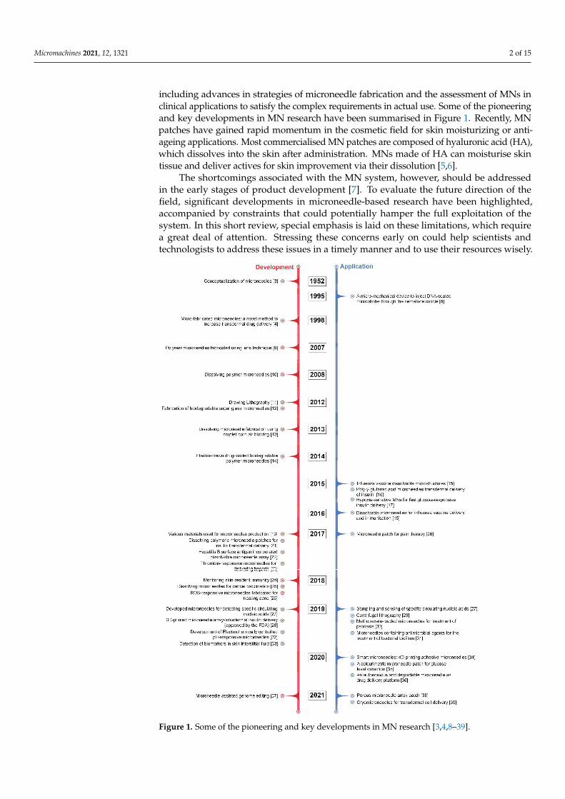
including advances in strategies of microneedle fabrication and the assessment of MNs inclinical applications to satisfy the complex requirements in actual use. Some of the pioneeringand key developments in MN research have been summarised in Figure 1. Recently, MNpatches have gained rapid momentum in the cosmetic field for skin moisturizing or anti-ageing applications. Most commercialised MN patches are composed of hyaluronic acid (HA),which dissolves into the skin after administration. MNs made of HA can moisturise skintissue and deliver actives for skin improvement via their dissolution [5,6].
The shortcomings associated with the MN system, however, should be addressedin the early stages of product development [7]. To evaluate the future direction of thefield, significant developments in microneedle-based research have been highlighted,accompanied by constraints that could potentially hamper the full exploitation of thesystem. In this short review, special emphasis is laid on these limitations, which requirea great deal of attention. Stressing these concerns early on could help scientists andtechnologists to address these issues in a timely manner and to use their resources wisely.
Micromachines 2021, 12, x FOR PEER REVIEW 3 of 17
Figure 1. Some of the pioneering and key developments in MN research [3,4,8–39].
Figure 1. Some of the pioneering and key developments in MN research [3,4,8–39].
Micromachines 2021, 12, 1321 3 of 15
2. Microneedle-Based Delivery Approaches
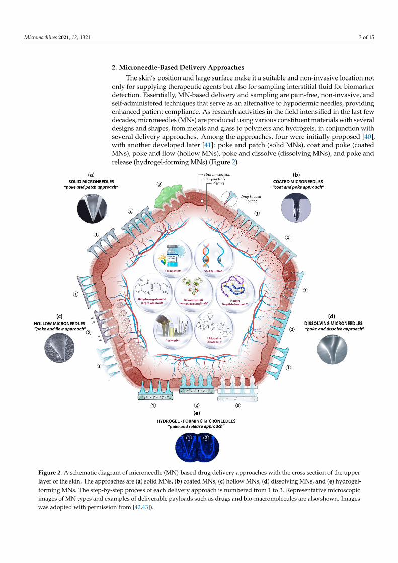
The skin’s position and large surface make it a suitable and non-invasive location notonly for supplying therapeutic agents but also for sampling interstitial fluid for biomarkerdetection. Essentially, MN-based delivery and sampling are pain-free, non-invasive, andself-administered techniques that serve as an alternative to hypodermic needles, providingenhanced patient compliance. As research activities in the field intensified in the last fewdecades, microneedles (MNs) are produced using various constituent materials with severaldesigns and shapes, from metals and glass to polymers and hydrogels, in conjunction withseveral delivery approaches. Among the approaches, four were initially proposed [40],with another developed later [41]: poke and patch (solid MNs), coat and poke (coatedMNs), poke and flow (hollow MNs), poke and dissolve (dissolving MNs), and poke andrelease (hydrogel-forming MNs) (Figure 2).
Micromachines 2021, 12, x FOR PEER REVIEW 4 of 17
2. Microneedle-Based Delivery Approaches The skin’s position and large surface make it a suitable and non-invasive location not
only for supplying therapeutic agents but also for sampling interstitial fluid for biomarker detection. Essentially, MN-based delivery and sampling are pain-free, non-invasive, and self-administered techniques that serve as an alternative to hypodermic needles, providing enhanced patient compliance. As research activities in the field intensified in the last few decades, microneedles (MNs) are produced using various constituent materials with several designs and shapes, from metals and glass to polymers and hydrogels, in conjunction with several delivery approaches. Among the approaches, four were initially proposed [40], with another developed later [41]: poke and patch (solid MNs), coat and poke (coated MNs), poke and flow (hollow MNs), poke and dissolve (dissolving MNs), and poke and release (hydrogel-forming MNs) (Figure 2).
Figure 2. A schematic diagram of microneedle (MN)-based drug delivery approaches with the cross section of the upper layer of the skin. The approaches are (a) solid MNs, (b) coated MNs, (c) hollow MNs, (d) dissolving MNs, and (e) hydrogel-forming MNs. The step-by-step process of each delivery approach is numbered from 1 to 3. Representative microscopic images of MN types and examples of deliverable payloads such as drugs and bio-macromolecules are also shown. Images was adopted with permission from [42,43]).
Figure 2. A schematic diagram of microneedle (MN)-based drug delivery approaches with the cross section of the upperlayer of the skin. The approaches are (a) solid MNs, (b) coated MNs, (c) hollow MNs, (d) dissolving MNs, and (e) hydrogel-forming MNs. The step-by-step process of each delivery approach is numbered from 1 to 3. Representative microscopicimages of MN types and examples of deliverable payloads such as drugs and bio-macromolecules are also shown. Imageswas adopted with permission from [42,43]).
Micromachines 2021, 12, 1321 4 of 15
The pore-performing pre-treatment of the “poke and patch” approach involves theapplication of a solid MN patch to create small holes in the skin, followed by a conventionaldrug application on the surface of the skin. The first reported fabrication of solid MNswas based on silicon to deliver calcein through excised human skin in vitro [4,8]. Cost,fragility, biocompatibility, and the complex manufacturing process have steered researchersto other materials, including metals, ceramics, and polymers, in order to achieve betteroutcomes. Although the production of solid MNs is technically simple—no loading orcoating is required—the two-step administration procedures and the no exact dosing withdrug reformulations requirement are the main limitations of solid MNs, along with safetymatters. Using solid MNs for the delivery of proteins, hormones, and vaccines have beenreviewed in detail elsewhere [1,44].
Coating therapeutic agents on the surface of microneedles (e.g., solid MNs—metallic,silica, or polymeric) is possible to create coated MNs. This “coat and poke” approachallows for effective drug delivery provided that the formulations are stable and uniformlylayered on the surface of the MNs. The drug formulation should also be water-soluble andallow layer-by-layer coating procedures. Choosing an appropriate coating technique is keyfor the successful generation of coated MNs. The delivery of vaccines [45], insulin [46],and hormones [47], along with other macromolecules, has been reported for the “coat andpoke” approach. A further extension in applications of coated MNs has been demonstratedrecently for the ultra-sensitive detection of protein biomarkers in an immunised mousemodel [48]. Polystyrene microneedles coated with a primary antibody were developedto capture inflammatory biomarkers in interstitial fluid with an improved limit of de-tection. The main distinguishing feature of coated microneedles is their ability to avoidthe degradation of bioactive molecules throughout the microneedle production process,thereby ensuring bioactivity. Furthermore, coating is one of the easiest and most controlledmethods of making microneedles functional. It enables sampling and isolation, especiallyfor microneedles with detecting capabilities. Common limitations, however, are that thesmall doses and loaded cargo may lessen the strength of the MNs, resulting in low strengthand penetration ability.
Relatively large quantities of therapeutic ingredients may be supplied into the skinwith the “poke and flow” approach, which, by using hollow MNs, could potentially over-come the dose limitation associated with solid MNs [49]. With hollow MNs, it is technicallypossible to control the flow and dosing by diffusion or pressure or electronically (e.g., usinga pump), and to integrate them into lab-on-chip devices. Similarly, bio-macromolecules,including proteins, vaccines, mRNA, and diagnostic agents, can be delivered via hollowMNs [50,51]. These MNs can also be used for the isolation and identification of biomarkersincluding glucose [52], and ECG measurements [53]. Nonetheless, the construction ofhollow MNs is relatively complicated and suffers from clogging, drug leakage, structuralfragility, and the requirement of a larger tip diameter, which leads to poor insertion.
The “poke and dissolve” approach, in which water-soluble therapeutic agents arecarried into the skin, uses mostly biocompatible/biodegradable and low-cost polymers.Hyaluronic acid, sucrose, polylactic/glycolic acid (PLA/PGA), and chitosan are amongthe polymers often used for the construction of dissolvable MNs (dMNs). Because of theirphysicochemical characteristics, which allow designing and engineering with tuneableproperties and functions, biomaterials like polysaccharides have been frequently utilisedto create dissolvable MNs. This has resulted in carbohydrate-based microarrays withtremendous potential for serving as an innovative step in medication administration,detection, and biological retorting [50]. A large number of articles on the production ofpolymeric MNs have been seen in a very short period. Unlike silicon or metal, this sort ofdelivery means is based on the breakdown of MNs upon exposure to the skin’s interstitialfluid. Conditional on the nature of the MN material, the dissolving process discharges thecargo from the matrix for local or systemic administration. To date, the majority of solubleMNs have been produced by utilising polymers and simple sugars and by employingcasting or micromoulding methods. The therapeutic loads are encapsulated, stored, and
Micromachines 2021, 12, 1321 5 of 15
protected in the scaffold and delivered into the targeted area after the skin insertion viaa polymer erosion mechanism, without leaving any biohazardous waste. A successfulapplication of dMNs in vaccine delivery, for instance, has been demonstrated [54]. Sucroseand fish gelatin-based MNs have been used for vaccine delivery. During the phase I trial,the influenza virus vaccines supplied by the microneedle patches (MNPs) were foundto be immunogenic and safe. The drawbacks associated with the sharp waste of solidMNs, the requirement of a pump, the high cost of hollow MNs, and the sophisticatedlayering procedure with coated MNs are eliminated here. However, dMNs have theirown drawbacks, including low mechanical strength, low doses, and doubtful penetrationabilities [55].
Soft materials such as swellable polymers, including poloxamer [56], PEG-crosslinkedpoly(methyl vinil ether-co-maleic acid), and silk fibroin with phenylboronic acid/acrylamide [57],have recently been used for hydrogel-forming MNs. The polymers absorb the intersti-tial fluid into their 3D matrix upon insertion into the skin, resulting in the delivery oftherapeutic agents through created micro-conduits. The response-related delivery of thera-peutic applications, such as glucose-responsive insulin delivery, is particularly noteworthy,as it eliminates the need for constant glucose monitoring and relies on the responses ofphysiological signals. Diagnostic applications of hydrogel-forming MNs have also beendescribed for the detection of glucose [58] and lithium monitoring [59]. Fine-tuning ofthe delivery time is possible by adjusting the polymer decomposition from minutes todays. Nonetheless, their low strength and limited drug doses are among the limitations ofhydrogel-forming MNs. This approach requires dramatic improvements to be used for anyfeasible commercial applications in the near future.
3. Challenges of the Microneedle Delivery System
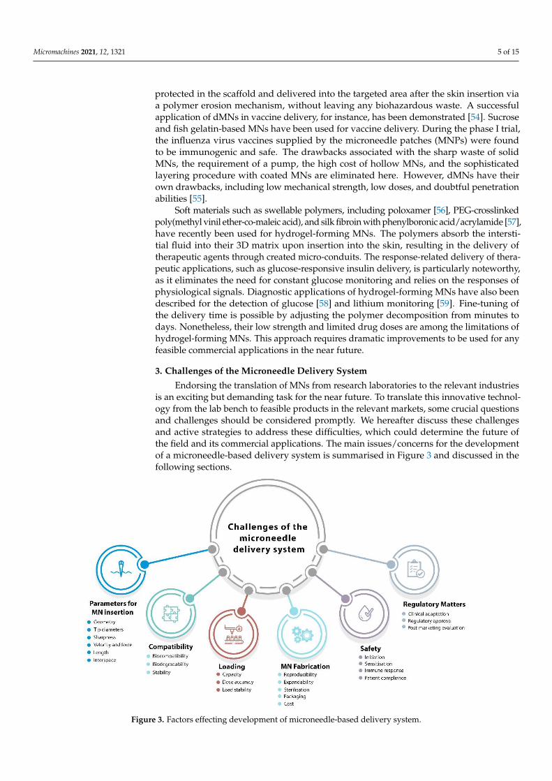
Endorsing the translation of MNs from research laboratories to the relevant industriesis an exciting but demanding task for the near future. To translate this innovative technol-ogy from the lab bench to feasible products in the relevant markets, some crucial questionsand challenges should be considered promptly. We hereafter discuss these challengesand active strategies to address these difficulties, which could determine the future ofthe field and its commercial applications. The main issues/concerns for the developmentof a microneedle-based delivery system is summarised in Figure 3 and discussed in thefollowing sections.
Micromachines 2021, 12, x FOR PEER REVIEW 7 of 17
Figure 3. Factors effecting development of microneedle-based delivery system.
3.1. Parameters Affecting MN Insertion The capability of MN patches to adequately puncture the skin is a vital requirement.
When addressing this matter, the skin’s characteristics, which might vary across the body and vary from person to person, should also be taken into account. The insertion and penetration behaviour of MNs to overcome the skin’s elasticity is strongly dependent on several parameters, such as geometry, base and tip diameters, length, and interspace (centre-to-centre spacing) [60,61]. An approach of “one-size-fits-all” cannot be envisaged in any design and development stages for any MN application. Infiltration and active delivery performance of MNs are strongly related to the geometry of individual MNs and the array, MN materials, the MN management method, and the characteristics of skin tissue [62]. Depending on the target medicines and applications, the microneedle mechanical strength, insertion depth, and drug release profile could be finely tuned by modifying the microneedle shape and composition.
The geometry: The geometry of MNs is a parameter that should be taken into consideration early on when developing MNs for clinical applications. A recent study indicated that the mechanical strength and penetration characteristics of MNs are affected by the geometric structure of microneedle arrays [63]. Simulations have shown a linear relationship between the mechanical strength and the number of vertices in the polygon base (e.g., triangular, square, and hexagonal microneedle bases), showing better insertion depths for the triangular and square-built microneedles. Superior capacity to insert into the skin was observed for the sharper edges of the triangular and square MNs compared to the hexagonal MNs. In a recent study, cone-shaped MNs were discovered to possess the ideal geometry for the delivery of ovalbumin and transcutaneous immunisation, with both greater needle insertion and a fast dismantling time for a more potent immune response obtained [64]. A further improvement has been proposed recently to reduce the
Figure 3. Factors effecting development of microneedle-based delivery system.
Micromachines 2021, 12, 1321 6 of 15
3.1. Parameters Affecting MN Insertion
The capability of MN patches to adequately puncture the skin is a vital requirement.When addressing this matter, the skin’s characteristics, which might vary across the bodyand vary from person to person, should also be taken into account. The insertion andpenetration behaviour of MNs to overcome the skin’s elasticity is strongly dependenton several parameters, such as geometry, base and tip diameters, length, and interspace(centre-to-centre spacing) [60,61]. An approach of “one-size-fits-all” cannot be envisagedin any design and development stages for any MN application. Infiltration and activedelivery performance of MNs are strongly related to the geometry of individual MNsand the array, MN materials, the MN management method, and the characteristics ofskin tissue [62]. Depending on the target medicines and applications, the microneedlemechanical strength, insertion depth, and drug release profile could be finely tuned bymodifying the microneedle shape and composition.
The geometry: The geometry of MNs is a parameter that should be taken into consider-ation early on when developing MNs for clinical applications. A recent study indicatedthat the mechanical strength and penetration characteristics of MNs are affected by thegeometric structure of microneedle arrays [63]. Simulations have shown a linear relation-ship between the mechanical strength and the number of vertices in the polygon base (e.g.,triangular, square, and hexagonal microneedle bases), showing better insertion depthsfor the triangular and square-built microneedles. Superior capacity to insert into the skinwas observed for the sharper edges of the triangular and square MNs compared to thehexagonal MNs. In a recent study, cone-shaped MNs were discovered to possess the idealgeometry for the delivery of ovalbumin and transcutaneous immunisation, with bothgreater needle insertion and a fast dismantling time for a more potent immune responseobtained [64]. A further improvement has been proposed recently to reduce the risk ofinsufficient drug delivery, wherein an array of hemispherical convexities was positioned inthe lower half of the cone-shaped dissolving MNs to increase drug flux [65].
Tip diameter and Sharpness: Tip diameter is another parameter for MN insertion. Rel-atively blunt MNs (tip diameters of 60–160 µm) require a relatively high insertion force(0.08–3.04 N) for controlled applications of MNs and are linearly reliant on the tip frontalarea [66]. To achieve a well-controlled manner to the desired depth, the fabrication ofMNs with sharp tips is essential. For the successful delivery of therapeutics, it has beenreported that MNs with smaller tip diameters (<15 µm) access the skin more smoothly thanMNs with a tip diameter of larger magnitude. This is particularly important in vaccinedelivery to achieve appropriate control over the penetration depth of MNs, not only fordelivering the antigens but also for specifically targeting Langerhans cells residing in theepidermal layer or dendritic cells dwelling in the dermal layer of the skin for a robustadaptive response [67]. The sharpness of the tips of microneedles can aid and controlthe puncture force. An increased tip sharpness, however, not only reduces the punctureforce but also reduces the structural strength of the microneedles, leading to a high riskof breakage.
Application velocity and force: In close relationship with the tip diameters, the appli-cation velocity and force are other parameters in the MN delivery system that should beconsidered in detail. Several studies have reported that the penetration depth of MN arraysvaries (from 10% up to 80%) and increases with the application velocity and force [68]. Avariety of patch configurations have been used, with similar outcomes of the penetrationforce per microneedle obtained. A 25-microneedle array with a tip radius of <100 nmrequires an insertion force of 10 mN per microneedle for effective penetration into theskin [69]. Two independent studies have also acknowledged these findings and reportedthat insertion forces of 15–20 mN [70] and 15–30 mN [71] per microneedle were requiredfor operational insertion. These forces represent arrays of 10–100 microneedles, which givea total applicator force of 0.1–3 N. Although these forces are low, the need for consistentapplication may necessitate a controlled application approach or a device.
Micromachines 2021, 12, 1321 7 of 15
Length: Because the thickness of the SC and other skin layers differs across individuals,the particle insertion depth may also vary. The transport capability of the skin, once a MNpatch has been applied, will depend on the perforation depth of the tissue. If a drug isrelatively small and has high diffusion capacity, creating surface pores by microneedleapplication should be sufficient for therapeutic function. However, if rapid delivery tothe bloodstream is the goal, it may be preferable to create pores that reach the dermis,where capillaries are located. This may be one reason for assorted microneedle lengths thathave been reported to date. In addition to the shorter microneedles, there have been manystudies that used long microneedles (up to 1000 µm long) to increase insulin permeabilityinto the skin [72].
Interspace (centre-to-centre spacing): The skin is a topographically diverse surface capa-ble of withstanding significant deformations prior to penetration. A significant number ofdistinct punctures must be generated when there is a high-density array of microneedles(e.g., more than 500/cm2). This takes a lot of energy. Naturally, as the density and numberof microneedles grow, so does the necessary force for skin puncture. This can result inincreased feeling for the patient and may require the use of a larger/stronger device forcertain applications. Needles with increasing width, length, and density can result inlarger, longer, and more crowded holes, through which a higher amount of medication maydiffuse. However, more tightly placed needles may cause the “bed-of-nails” effect too [68].
3.2. Biocompatibility, Biodegradability, and Stability
One of the safety aspects of MN systems in clinical use is biocompatibility. To ensurethat MN products are acceptable for human exposure, several tests are required to eval-uate their biocompatibility based on contact periods of less than 24 h, between 24 h and30 h, and more than 30 h. [73]. For the former two periods, the corresponding tests arecytotoxicity, sensitisation, irritation, and intracutaneous reactivity tests. Genotoxicity andsubacute/subchronic systematic toxicity tests are additionally recommended for the latterperiod of use. The use of biodegradable materials is desirable for microneedles becausethese materials can be degraded and removed from the body safely. Therefore, usingbiodegradable polymeric systems for MN fabrication has been pursued in recent years.The primary benefit of polymeric microneedle systems is their ability to load medicationinto the microneedle matrix for discharge in the skin via biodegradation or dissolution inthe body fluid of the skin.
The ability to manufacture microneedle structures from aqueous polymeric mixtures atroom temperature without the requirement of a heating step might be a significant benefitin retaining the stability of an integrated medication, particularly in the case of therapies inwhich proteins and peptides are involved. Nonetheless, the stability of MN cargo has to beevaluated to ensure that fragile and easily degradable therapeutics are protected duringstorage. This is usually done by storing MNs and their cargo at various temperatures,including −25 ◦C, 4 ◦C, 20 ◦C, 40 ◦C and 60 ◦C, followed by analytical assessments.Generally, the protein cargo of MNs has better storage stability and longer shelf-life dueto the rigid glassy microneedle matrices restraining the molecular mobility and limitingaccess to atmospheric oxygen [74]. This can be further extended by the incorporation ofstabilisers, including trehalose and sucrose. Attention to water is particularly critical whennon-vacuum storage conditions are present, as they can not only destroy the stability ofladen cargo but also the mechanical properties of the MNs themselves [75]. DissolvableMNs are very susceptible to the surrounding humidity; therefore, the storage environmentshould be dry and cool for prolonged stability and extended shelf-life.
3.3. Loading Capacity and Dosage Accuracy
Loading capacity: A coated microneedle device can only deliver a bolus dose of around1 mg of medicine. Although hollow microneedles allow for continuous infusion or “as-needed/on-demand” dosing, central exits may be obstructed by compressed skin tissueafter microneedle insertion. Even though MNs have the potential to overcome the skin’s
Micromachines 2021, 12, 1321 8 of 15
barrier properties, their success is very much dependent on passive diffusion of the biolog-ical formulation into the skin. This can make it difficult to administer large dosages, andmuch of the dose can be lost on the skin’s surface. As a result, the time of application andthe inability to monitor dose delivery have caused reluctance to use this technology forcertain clinical applications. One example is the distribution of vaccines for which dosageconstancy is critical. Recent work has shown that administering vaccines directly to theepidermis and dermis of the skin has the potential to induce immunological responseswith considerably less vaccine than standard intramuscular injection. These advantages,however, might be lost if just a tiny fraction of the administered dosage reaches the skin.While this is not an insurmountable obstacle to this technology, vaccines, in particular,require a threshold dosage to induce immunity, which might be more difficult to achievewhen depending on passive diffusion.
Dosage accuracy: The dosage accuracy of MN delivery systems in continuous drugdelivery is an issue that requires close attention. Several methods using separable micronee-dles have been proposed for minimising the patch-wearing time and quickly removingthe formulation from the MNs [76,77]. Storing and delivering protein drugs, includinginsulin, erythropoietin, glucagon, growth hormones, and parathyroid hormones, are chal-lenging tasks, as bio-macromolecules are prone to quick degradation and inactivation.These matters could be best handled by not only the incorporation of stabilisers but also byconsidering the whole process of MN manufacturing parameters, such as manufacturingand storage temperatures and drying conditions, polymer concentration, sterilisation,and packaging. As discussed earlier, MNs can be manufactured in various types andmaterials. The drug delivery efficiency when using solid MNs is rather difficult to controlaccurately. Coated MNs can efficiently deliver precise amounts of a drug but have limiteddrug loading capacity due to their small surface area for coating. Encapsulating drugsin the matrices of MNs is possible if dissolvable microneedles are fabricated primarilyfrom hydrophilic, biocompatible, and biodegradable materials, and if the cargo can bedischarged entirely within the skin’s interstitial fluid without leading to unwanted debris.Relatively large doses and the controlled release (slow or fast delivery) of various drugscan be transferred without issues of reservoir leakage. Dissolvable microneedles might bean efficient approach to preserve and stabilise nano-sized compositions while improvingnanoparticle penetration through the stratum corneum barrier. Various approaches havebeen thoroughly studied, and several analytical techniques for tracing and tracking thejourney of nanomaterials with their valuable payloads, both in vitro and in vivo, have beendeveloped [78,79].
3.4. Skin Irritation and Recovery
The immunogenic nature of the skin makes it a highly responsive organ towards theMN delivery of any therapeutic agent. Mild and temporary erythema may develop asa side effect depending on the size, substance, and type of the given medication. Skinirritation, sensitisation, and immune response must also be evaluated as part of the safetyassessments of MN products during clinical trials. This safety concern must be evaluatedusing animal testing before any human clinical trials. On the other hand, great immuneresponsiveness of the skin may present an opportunity for MN-based vaccine delivery ifother obstacles have been addressed properly, as discussed.
3.5. Cost of Microneedle Fabrication
Current microneedle manufacturing processes need to be improved to reach large-scale production in order to completely transfer microchip-based microneedles into ther-apeutic applications. Until now, extensive economic evaluations of the technology havenot yet been quantified thoroughly, but it is not difficult to predict that, as with every newtechnology, the clinical use of MNs can be comparatively expensive due to the complexfabrication and storage procedures and the slow and long approval process.
Micromachines 2021, 12, 1321 9 of 15
Even though MNs show promise in preclinical research, their economic and epidemio-logic implications have yet to be assessed thoroughly. However, to give an idea of the costsinvolved in MN application in vaccine delivery, one study projected the hypothetical costsof MN-based measles vaccine injection and compared them with those of traditional subcu-taneous (SC) injection [80]. For a population of 1 million children, the estimated prices of a2-dose vaccination program using the microneedle patch and SC injection were USD 0.95(range USD 0.71–USD 1.18) for the first dose and USD 1.65 (range USD 1.24–USD 2.06),respectively, assuming that the MN vaccine method is more heat-stable and requires cost-effective cool chains. The total costs of the vaccination program were estimated to beUSD 1.5 million for MN-based administration compared with USD 2.5 million for SCadministration. The authors commented that the cost-effectiveness of MN patches dependson numerous factors, including approval rates and the effectiveness of the MN patches inrelation to the traditional subcutaneous vaccine delivery method [80]. In another study, aneconomic model was applied to assess the value of MN patch technology for the seasonalinfluenza vaccine [81]. The model predicted that its introduction would be economical ordominant at a USD 9.50 price point in the majority of situations evaluated when healthcareworkers managed the MN-based system. If efficacy rose by ≥3%, MN vaccination wouldbe cost-effective or dominant for all price points ≤ USD 30 for all administration scenariosstudied. The growing economic pressure on the global healthcare system makes it crucialfor researchers to study the costs related to MN-based delivery systems. While the technol-ogy has great potential for transdermal drug delivery, its success is very much dependenton carrying out economic assessments while the technology is still under development, asshould be the case for the development of any new science-led, research-driven productdevelopments. Along with the other factors discussed earlier, the success of any MN appli-cation is also very much dependent on the fabrication technique and materials used. Scalingup to industrial manufacturing for mass production necessitates a focused strategic plan.Several factors should be considered carefully, including the accurate and reproducibleproduction of MNs (attributed to the maturation of MN fabrication technology), the abilityto expand for mass production, and the expandability of MN technology to a wide rangeof concerns or diseases, together with regulatory approval and clinical adaptation [82]. Thechoice of materials is also of utmost importance and should be compatible with the ladencargo for optimal insertion and delivery performance without any deleterious effects onthe bioactivity and stability/viability of the therapeutic agents. For optimisation, an idealproduction method should aid easy, rapid, and cost-effective modifications in the materialand geometry parameters.
3.6. Sterilisation of the Microneedle Patches
MN patch sterilisation is another challenge that should be taken into account early onwhen MN-based products are aimed for commercial application. If sterilisation is necessary,then the method of choice will be critical, because the most widely used methods, such asmoist heat, gamma or microwave radiation, and ethylene oxide may deleteriously affectany cargoes with sensitive ingredients, including biomolecules, vaccines, peptides, and/oreven the microneedles themselves [83]. Although the risk of introducing bioburden into thesterile area of the body (e.g., epidermis and dermis) by MNs is significantly smaller than asingle puncture by a hypodermic needle, complete sterilisation of MNs-based productsmay be obligatory by the regulatory bodies to safeguard the users. The material usedfor MN fabrication determines the method of choice for sterilisation. For solid MNs ofmetals, silicon, and glass, the sterilisation is straightforward; dry heat sterilisation, moistheat sterilisation, and gamma radiation are the most common methods employed [84].However, when delivering fragile biological active ingredients is in demand (e.g., usingcoated MNs), the method of choice should be carefully evaluated in terms of maintain-ing stability and activity of the coated ingredients. MNs constructed by carbohydratesand polymers (e.g., dissolving MNs) present the biggest challenge when choosing thesterilisation method, since the sterilisation not only affects the fragile loads but also the
Micromachines 2021, 12, 1321 10 of 15
morphological, physicochemical, and mechanical properties of MNs themselves. Theeffects of various sterilisation methods, such as moist and dry heat sterilisation and gammaradiation, on dissolving and hydrogel-forming MNs have been studied, with ibuprofenand ovalbumin as model drugs [85]. It was found that no measurable bioburden wasdetected, and levels of endotoxin were under the FDA limits if aseptic preparation wasfollowed. However, moist and dry heat sterilisation damaged all formulations, whereasthe gamma irradiation at a sterility assurance level (SAL) of 10−6 (according to the BritishPharmacopeia) can be used for sterilisation without causing structural damages or affect-ing delivery capabilities of hydrogel-forming MNs. The radiation, however, destroyedovalbumin and changed the appearance of ibuprofen. Alternative methods for delicateMNs have been proposed [86,87]. Ethylene oxide and electron beam sterilisation wereshown to be effective but less destructive methods for MN sterilisation. In another study,a self-sterilisation of MNs was proposed, in which silver nanoparticles were embeddedin CMC MNs. The authors implied that the pores produced by MNs were free frommicroorganisms until the skin is healed completely [88].
It is clear that the information available in the literature is rather limited, and there-fore, the sterilisation of MN-based products requires extensive research before going intocommercial production and approval; this presents one of the most important challenges inMN-based delivery systems. In particular, endpoint sterilization for MN products requiresa great deal of attention, as MN manufacturing under an aseptic condition could be bothcomplicated and costly.
3.7. Regulation of the Microneedle Patches
The quality of submissions received from combination products employing micronee-dles has been a source of concern for the US FDA, particularly in the areas of stability testing,content consistency, risk analysis, sterility validation, and manufacturing. As discussedearlier, MNs are a viable option for the delivery of therapeutic agents such as hormones,vaccines, enzymes, mRNA, and difficult-to-deliver small molecules via the skin. In view ofthe regulatory body, clinical application scenarios, as well as the repeatability and efficacyof microneedle devices, should be thoroughly shown using cell studies, animal testing, andclinical trials. Furthermore, a thorough understanding of human physiological settings,thorough examination of clinical demands, and the mobility and simplicity of microneedledevices can all help to promote such clinical translations. The number of MN-based medic-inal products for therapeutic applications is rising exponentially. However, the submissionprocess to the FDA for approval is not straightforward because submissions should be inthe form of combination products that use microneedles. Any submissions of this kindrequire satisfactory information about product analysis, testing and validations such asrisk analysis, content uniformity, stability testing (formulation/API migration/mechanicalcharacteristics), sterility validation, and manufacturing. The FDA has stated “Regulationof combination products must take into account the safety and effectiveness questionsassociated with each constituent and the product as a whole” [73]. The current strategyof product-specific approval (rather than specific MN-systems) for the licensing of mi-croneedle products adopted by the regulatory bodies causes great delays in approval,thereby restricting the commercialisation of MNs. To promote the commercialisation ofMN products, the cGMP and quality control should be merged, and licencing regulationsmust be defined clearly, covering the shape, formulation, sterilisation, and packaging. Onthe other hand, the clinical development of microneedle devices can advance separately todrug or vaccine formulation. This can greatly simplify any regulatory processes and mightallow for more rapid incorporation of the technology into the supply chains of particulardrugs. Although just small quantities of a molecule may reach their intended deliverysites, this approach’s simplicity may lessen the regulatory problems encountered by othercomplicated formulation techniques.
Due to the possibility that this MN design may be CE marked as a medical devicerather than a medicinal product, pharmaceutical firms may be prepared to invest in such a
Micromachines 2021, 12, 1321 11 of 15
device before investing in medication that incorporates MNs. In addition, robust guidanceis required to fully classify MN-based products; nevertheless, it has been proposed thatthis would most likely come within the medical device category for monitoring/diagnosticapplications, and as a “combination product” (drug and device) or “drug product” for thedelivery of drugs or vaccines [89,90]. Once this distinction is made, it may be possible toadapt existing quality control procedures for MNs. The current standard quality controlmethods may not be completely suitable to MN products due to the inherent differencesbetween transdermal patches and hypodermic needles. If all remaining concerns can besuitably addressed to meet the needs of both regulators and patients, the goal of bringingMN-based products to the transdermal market will soon become a reality. In 2020, the firstnew drug application for a pharmaceutical microneedle patch, Qtrypta, was submittedto the Food and Drug Administration (FDA) by Zosano Pharma. The patch is a titaniummicroneedle with a coated zolmitriptan for acute migraine treatment.
4. Conclusions and Perspectives
Development of marketable microneedle-based drug delivery products is highlylikely in near future. Extensive research in MNs is being conducted for the efficientdelivery of therapeutics, as innovative transdermal drug delivery methods are urgentlyrequired to expand the transdermal market for hydrophilic molecules, macromolecules,proteins, and conventional medicines for new therapeutic indications. The future of themicroneedle industry seems to be quite bright, with the rapid realisation of new informationfuelling industrial progress. The effectiveness of MNs has been demonstrated in severalclinical trials, but there have still been far more preclinical studies. Experts from academia,industry, and regulatory organisations are collaborating to help MNs to advance intosafe and effective clinical usage provided that the shortcomings associated with thesesystems are promptly and rationally addressed. It is believed that, in time, microneedle-based technology will lead to improved illness prevention, diagnosis, and control, as wellas an increase in the health-related quality of life of patients globally. Nonetheless, thecomplicated and expensive production of MNs, together with several application-relateddifficulties, could delay their clinical translation. This is evident from the clinical translationof microneedle applications in the pharmaceutical industry. For instance, the lack of clinicaldata on “www.clinicaltrials.gov” using “microneedle vaccine” indicates that the scale-upproduction of MNs is still a challenge. What is more, novel manufacturing methods,micromachining and 3D printing technologies in particular, are envisaged to lower thecosts and simplify fabrication procedures in the near future.
Author Contributions: M.A. and A.Ç. proposed the original idea and wrote the manuscript. Allauthors have read and agreed to the published version of the manuscript.
Funding: This research was funded by Imperial Bioscience ltd.
Acknowledgments: We would like to thank Nuriye I. Celik (iArch-London) for her help in thepreparation of figures.
Conflicts of Interest: The authors declare no conflict of interest.
References1. Ita, K. Transdermal Delivery of Drugs with Microneedles-Potential and Challenges. Pharmaceutics 2015, 7, 90–105. [CrossRef]
[PubMed]2. Alimardani, V.; Abolmaali, S.S.; Yousefi, G.; Rahiminezhad, Z.; Abedi, M.; Tamaddon, A.; Ahadian, S. Microneedle Arrays
Combined with Nanomedicine Approaches for Transdermal Delivery of Therapeutics. J. Clin. Med. 2021, 10, 181. [CrossRef]3. Reaume, S.E. The use of hydrofluoric acid in making glass microneedles. Science 1952, 116, 641. [CrossRef]4. Henry, S.; McAllister, D.V.; Allen, M.G.; Prausnitz, M.R. Microfabricated microneedles: A novel approach to transdermal drug
delivery. J. Pharm. Sci. 1998, 87, 922–925. [CrossRef]5. Avcil, M.; Akman, G.; Klokkers, J.; Jeong, D.; Çelik, A. Efficacy of bioactive peptides loaded on hyaluronic acid microneedle
patches: A monocentric clinical study. J. Cosmet. Dermatol. 2020, 19, 328–337. [CrossRef]
Micromachines 2021, 12, 1321 12 of 15
6. Avcil, M.; Akman, G.; Klokkers, J.; Jeong, D.; Çelik, A. Clinical efficacy of dissolvable microneedles armed with anti-melanogeniccompounds to counter hyperpigmentation. J. Cosmet. Dermatol. 2021, 20, 605–614. [CrossRef] [PubMed]
7. Chandran, R.; Tohit, E.R.M.; Stanslas, J.; Mahmood, T.M.T. Recent advances and challenges in microneedle-mediated trans-dermal protein and peptide drug delivery. In Biomaterials and Bionanotechnology; Elsevier: Amsterdam, The Netherlands, 2019;pp. 495–525.
8. Hashmi, S.; Ling, P.; Hashmi, G.; Reed, M.; Gaugler, R.; Trimmer, W. Genetic transformation of nematodes using arrays ofmicromechanical piercing structures. Biotechniques 1995, 19, 766–770.
9. Park, J.H.; Yoon, Y.K.; Choi, S.O.; Prausnitz, M.R.; Allen, M.G. Tapered conical polymer microneedles fabricated using anintegrated lens technique for transdermal drug delivery. IEEE Trans. Bio-Med. Eng. 2007, 54, 903–913. [CrossRef] [PubMed]
10. Sullivan, S.P.; Murthy, N.; Prausnitz, M.R. Minimally invasive protein delivery with rapidly dissolving polymer microneedles.Adv. Mater. 2008, 20, 933–938. [CrossRef]
11. Lee, K.; Jung, H. Drawing lithography for microneedles: A review of fundamentals and biomedical applications. Biomaterials2012, 33, 7309–7326. [CrossRef]
12. Martin, C.J.; Allender, C.J.; Brain, K.R.; Morrissey, A.; Birchall, J.C. Low temperature fabrication of biodegradable sugar glassmicroneedles for transdermal drug delivery applications. J. Control. Release Off. J. Control. Release Soc. 2012, 158, 93–101. [CrossRef][PubMed]
13. Kim, J.D.; Kim, M.; Yang, H.; Lee, K.; Jung, H. Droplet-born air blowing: Novel dissolving microneedle fabrication. J. Control.Release Off. J. Control. Release Soc. 2013, 170, 430–436. [CrossRef] [PubMed]
14. Vecchione, R.; Coppola, S.; Esposito, E.; Casale, C.; Vespini, V.; Grilli, S.; Ferraro, P.; Netti, P.A. Electro-drawn drug-loadedbiodegradable polymer microneedles as a viable route to hypodermic injection. Adv. Funct. Mater. 2014, 24, 3515–3523. [CrossRef]
15. Bonificio, A.; Ghartey-Tagoe, E.; Gallorini, S.; Baudner, B.; Chen, G.; Singh, P.; O’Hagan, D.T.; Kommareddy, S. Fabrication of cellculture-derived influenza vaccine dissolvable microstructures and evaluation of immunogenicity in guinea pigs. Vaccine 2015, 33,2930–2938. [CrossRef] [PubMed]
16. Chen, M.C.; Ling, M.H.; Kusuma, S.J. Poly-γ-glutamic acid microneedles with a supporting structure design as a potential toolfor transdermal delivery of insulin. Acta Biomater. 2015, 24, 106–116. [CrossRef]
17. Yu, J.; Zhang, Y.; Ye, Y.; DiSanto, R.; Sun, W.; Ranson, D.; Ligler, F.S.; Buse, J.B.; Gu, Z. Microneedle-array patches loaded withhypoxia-sensitive vesicles provide fast glucose-responsive insulin delivery. Proc. Natl. Acad. Sci. USA 2015, 112, 8260–8265.[CrossRef]
18. Vrdoljak, A.; Allen, E.A.; Ferrara, F.; Temperton, N.J.; Crean, A.M.; Moore, A.C. Induction of broad immunity by thermostabilisedvaccines incorporated in dissolvable microneedles using novel fabrication methods. J. Control. Release Off. J. Control. Release Soc.2016, 225, 192–204. [CrossRef]
19. Bhatnagar, S.; Dave, K.; Venuganti, V.V.K. Microneedles in the clinic. J. Control. Release Off. J. Control. Release Soc. 2017, 260,164–182. [CrossRef]
20. Xie, X.; Pascual, C.; Lieu, C.; Oh, S.; Wang, J.; Zou, B.; Xie, J.; Li, Z.; Xie, J.; Yeomans, D.C.; et al. Analgesic Microneedle Patch forNeuropathic Pain Therapy. ACS Nano 2017, 11, 395–406. [CrossRef] [PubMed]
21. Lee, I.C.; Lin, W.M.; Shu, J.C.; Tsai, S.W.; Chen, C.H.; Tsai, M.T. Formulation of two-layer dissolving polymeric microneedlepatches for insulin transdermal delivery in diabetic mice. J. Biomed. Mater. Res. Part. A 2017, 105, 84–93. [CrossRef]
22. Poirier, D.; Renaud, F.; Dewar, V.; Strodiot, L.; Wauters, F.; Janimak, J.; Shimada, T.; Nomura, T.; Kabata, K.; Kuruma, K.; et al.Hepatitis B surface antigen incorporated in dissolvable microneedle array patch is antigenic and thermostable. Biomaterials 2017,145, 256–265. [CrossRef] [PubMed]
23. Zhang, Y.; Yu, J.; Wang, J.; Hanne, N.J.; Cui, Z.; Qian, C.; Wang, C.; Xin, H.; Cole, J.H.; Gallippi, C.M.; et al. Thrombin-ResponsiveTranscutaneous Patch for Auto-Anticoagulant Regulation. Adv. Mater. 2017, 29, 1604043. [CrossRef] [PubMed]
24. Mandal, A.; Boopathy, A.V.; Lam, L.K.W.; Moynihan, K.D.; Welch, M.E.; Bennett, N.R.; Turvey, M.E.; Thai, N.; Van, J.H.;Love, J.C.; et al. Cell and fluid sampling microneedle patches for monitoring skin-resident immunity. Sci. Transl. Med. 2018,10, eaar2227. [CrossRef] [PubMed]
25. Kim, N.W.; Kim, S.Y.; Lee, J.E.; Yin, Y.; Lee, J.H.; Lim, S.Y.; Kim, E.S.; Duong, H.T.T.; Kim, H.K.; Kim, S.; et al. Enhanced CancerVaccination by In Situ Nanomicelle-Generating Dissolving Microneedles. ACS Nano 2018, 12, 9702–9713. [CrossRef] [PubMed]
26. Zhang, Y.; Feng, P.; Yu, J.; Yang, J.; Zhao, J.; Wang, J.; Shen, Q.; Gu, Z. Ros-responsive microneedle patch for acne vulgaristreatment. Adv. Ther. 2018, 1, 1800035. [CrossRef]
27. Al Sulaiman, D.; Chang, J.Y.H.; Bennett, N.R.; Topouzi, H.; Higgins, C.A.; Irvine, D.J.; Ladame, S. Hydrogel-Coated MicroneedleArrays for Minimally Invasive Sampling and Sensing of Specific Circulating Nucleic Acids from Skin Interstitial Fluid. ACS Nano2019, 13, 9620–9628. [CrossRef]
28. Economidou, S.N.; Pere, C.P.P.; Reid, A.; Uddin, M.J.; Windmill, J.F.C.; Lamprou, D.A.; Douroumis, D. 3D printed microneedlepatches using stereolithography (SLA) for intradermal insulin delivery. Mater. Sci. Eng. C Mater. Biol. Appl. 2019, 102, 743–755.[CrossRef]
29. Lee, S.; Fakhraei Lahiji, S.; Jang, J.; Jang, M.; Jung, H. Micro-Pillar Integrated Dissolving Microneedles for Enhanced TransdermalDrug Delivery. Pharmaceutics 2019, 11, 402. [CrossRef]
30. Du, H.; Liu, P.; Zhu, J.; Lan, J.; Li, Y.; Zhang, L.; Zhu, J.; Tao, J. Hyaluronic Acid-Based Dissolving Microneedle Patch Loaded withMethotrexate for Improved Treatment of Psoriasis. ACS Appl. Mater. Interfaces 2019, 11, 43588–43598. [CrossRef]
Micromachines 2021, 12, 1321 13 of 15
31. Xu, J.; Danehy, R.; Cai, H.; Ao, Z.; Pu, M.; Nusawardhana, A.; Rowe-Magnus, D.; Guo, F. Microneedle Patch-Mediated Treatmentof Bacterial Biofilms. ACS Appl. Mater. Interfaces 2019, 11, 14640–14646. [CrossRef]
32. Anderson, A.; Hegarty, C.; Casimero, C.; Davis, J. Electrochemically Controlled Dissolution of Nanocarbon-Cellulose AcetatePhthalate Microneedle Arrays. ACS Appl. Mater. Interfaces 2019, 11, 35540–35547. [CrossRef]
33. Zhang, X.; Chen, G.; Bian, F.; Cai, L.; Zhao, Y. Encoded Microneedle Arrays for Detection of Skin Interstitial Fluid Biomarkers.Adv. Mater. 2019, 31, e1902825. [CrossRef] [PubMed]
34. Han, D.; Morde, R.S.; Mariani, S.; La Mattina, A.A.; Vignali, E.; Yang, C.; Barillaro, G.; Lee, H. 4D printing of a bioinspiredmicroneedle array with backward-facing barbs for enhanced tissue adhesion. Adv. Funct. Mater. 2020, 30, 1909197. [CrossRef]
35. Wang, Z.; Li, H.; Wang, J.; Chen, Z.; Chen, G.; Wen, D.; Chan, A.; Gu, Z. Transdermal colorimetric patch for hyperglycemiasensing in diabetic mice. Biomaterials 2020, 237, 119782. [CrossRef]
36. Lopez-Ramirez, M.A.; Soto, F.; Wang, C.; Rueda, R.; Shukla, S.; Silva-Lopez, C.; Kupor, D.; McBride, D.A.; Pokorski, J.K.;Nourhani, A.; et al. Built-In Active Microneedle Patch with Enhanced Autonomous Drug Delivery. Adv. Mater. 2020, 32, e1905740.[CrossRef] [PubMed]
37. Wan, T.; Pan, Q.; Ping, Y. Microneedle-assisted genome editing: A transdermal strategy of targeting NLRP3 by CRISPR-Cas9 forsynergistic therapy of inflammatory skin disorders. Sci. Adv. 2021, 7, eabe2888. [CrossRef] [PubMed]
38. Kusama, S.; Sato, K.; Matsui, Y.; Kimura, N.; Abe, H.; Yoshida, S.; Nishizawa, M. Transdermal electroosmotic flow generated by aporous microneedle array patch. Nat. Commun. 2021, 12, 658. [CrossRef]
39. Chang, H.; Chew, S.W.T.; Zheng, M.; Lio, D.C.S.; Wiraja, C.; Mei, Y.; Ning, X.; Cui, M.; Than, A.; Shi, P.; et al. Cryomicroneedlesfor transdermal cell delivery. Nat. Biomed. Eng. 2021, 5, 1008–1018. [CrossRef]
40. Prausnitz, M.R. Microneedles for transdermal drug delivery. Adv. Drug Deliv. Rev. 2004, 56, 581–587. [CrossRef]41. Donnelly, R.F.; Singh, T.R.; Garland, M.J.; Migalska, K.; Majithiya, R.; McCrudden, C.M.; Kole, P.L.; Mahmood, T.M.; McCarthy,
H.O.; Woolfson, A.D. Hydrogel-Forming Microneedle Arrays for Enhanced Transdermal Drug Delivery. Adv. Funct. Mater. 2012,22, 4879–4890. [CrossRef]
42. Kim, Y.C.; Park, J.H.; Prausnitz, M.R. Microneedles for drug and vaccine delivery. Adv. Drug Deliv. Rev. 2012, 64, 1547–1568.[CrossRef] [PubMed]
43. Donnelly, R.F.; McCrudden, M.T.; Zaid Alkilani, A.; Larrañeta, E.; McAlister, E.; Courtenay, A.J.; Kearney, M.C.; Singh, T.R.;McCarthy, H.O.; Kett, V.L.; et al. Hydrogel-forming microneedles prepared from “super swelling” polymers combined withlyophilised wafers for transdermal drug delivery. PLoS ONE 2014, 9, e111547. [CrossRef]
44. Li, J.; Zeng, M.; Shan, H.; Tong, C. Microneedle Patches as Drug and Vaccine Delivery Platform. Curr. Med. Chem. 2017, 24,2413–2422. [CrossRef] [PubMed]
45. Meyer, B.K.; Kendall, M.A.F.; Williams, D.M.; Bett, A.J.; Dubey, S.; Gentzel, R.C.; Casimiro, D.; Forster, A.; Corbett, H.;Crichton, M.; et al. Immune response and reactogenicity of an unadjuvanted intradermally delivered human papillomavirusvaccine using a first generation Nanopatch™ in rhesus macaques: An exploratory, pre-clinical feasibility assessment. Vaccine: X2019, 2, 100030. [CrossRef]
46. Ross, S.; Scoutaris, N.; Lamprou, D.; Mallinson, D.; Douroumis, D. Inkjet printing of insulin microneedles for transdermal delivery.Drug Deliv. Transl. Res. 2015, 5, 451–461. [CrossRef]
47. Daddona, P.E.; Matriano, J.A.; Mandema, J.; Maa, Y.F. Parathyroid hormone (1-34)-coated microneedle patch system: Clinicalpharmacokinetics and pharmacodynamics for treatment of osteoporosis. Pharm. Res. 2011, 28, 159–165. [CrossRef] [PubMed]
48. Wang, Z.; Luan, J.; Seth, A.; Liu, L.; You, M.; Gupta, P.; Rathi, P.; Wang, Y.; Cao, S.; Jiang, Q.; et al. Microneedle patch for theultrasensitive quantification of protein biomarkers in interstitial fluid. Nat. Biomed. Eng. 2021, 5, 64–76. [CrossRef] [PubMed]
49. McAllister, D.V.; Wang, P.M.; Davis, S.P.; Park, J.H.; Canatella, P.J.; Allen, M.G.; Prausnitz, M.R. Microfabricated needles fortransdermal delivery of macromolecules and nanoparticles: Fabrication methods and transport studies. Proc. Natl. Acad. Sci.USA 2003, 100, 13755–13760. [CrossRef]
50. Lim, D.J.; Vines, J.B.; Park, H.; Lee, S.H. Microneedles: A versatile strategy for transdermal delivery of biological molecules. Int. J.Biol. Macromol. 2018, 110, 30–38. [CrossRef]
51. Xu, J.; Xu, D.; Xuan, X.; He, H. Advances of Microneedles in Biomedical Applications. Molecules 2021, 26, 5912. [CrossRef]52. Nicholas, D.; Logan, K.A.; Sheng, Y.; Goa, J.; Farrell, S.; Dixon, D.; Callan, B.; McHale, A.P.; Callan, J.F. Rapid paper based
colorimetric detection of glucose using a hollow microneedle device. Int. J. Pharm. 2008, 547, 244–249. [CrossRef]53. Yu, L.M.; Tay, F.E.H.; Guo, D.G.; Xu, L.; Yap, K.L. A microfabricated electrode with hollow microneedles for ECG measurement.
Sens. Actuators A: Phys. 2009, 151, 17–22. [CrossRef]54. Rouphael, N.G.; Lai, L.; Tandon, S.; McCullough, M.P.; Kong, Y.; Kabbani, S.; Natrajan, M.S.; Xu, Y.; Zhu, Y.; Wang, D.; et al.
Immunologic mechanisms of seasonal influenza vaccination administered by microneedle patch from a randomized phase I trial.NPJ Vaccines 2021, 6, 89. [CrossRef]
55. Ita, K. Dissolving microneedles for transdermal drug delivery: Advances and challenges. Biomed. Pharmacother. Biomed.Pharmacother. 2017, 93, 1116–1127. [CrossRef] [PubMed]
56. Sivaraman, A.; Banga, A.K. Novel in situ forming hydrogel microneedles for transdermal drug delivery. Drug Deliv. Transl. Res.2017, 7, 16–26. [CrossRef]
Micromachines 2021, 12, 1321 14 of 15
57. Chen, S.; Matsumoto, H.; Moro-Oka, Y.; Tanaka, M.; Miyahara, Y.; Suganami, T.; Matsumoto, A. Smart Microneedle Fabricatedwith Silk Fibroin Combined Semi-interpenetrating Network Hydrogel for Glucose-Responsive Insulin Delivery. ACS Biomater.Sci. Eng. 2019, 5, 5781–5789. [CrossRef] [PubMed]
58. Caffarel-Salvador, E.; Brady, A.J.; Eltayib, E.; Meng, T.; Alonso-Vicente, A.; Gonzalez-Vazquez, P.; Torrisi, B.M.; Vicente-Perez, E.M.;Mooney, K.; Jones, D.S.; et al. Hydrogel-Forming Microneedle Arrays Allow Detection of Drugs and Glucose In Vivo: Potentialfor Use in Diagnosis and Therapeutic Drug Monitoring. PLoS ONE 2015, 10, e0145644. [CrossRef]
59. Eltayib, E.; Brady, A.J.; Caffarel-Salvador, E.; Gonzalez-Vazquez, P.; Zaid Alkilani, A.; McCarthy, H.O.; McElnay, J.C.; Donnelly,R.F. Hydrogel-forming microneedle arrays: Potential for use in minimally-invasive lithium monitoring. Eur. J. Pharm. Biopharm.Off. J. Arb. Fur Pharm. Verfahr. E.V 2016, 102, 123–131. [CrossRef]
60. Sirbubalo, M.; Tucak, A.; Muhamedagic, K.; Hindija, L.; Rahic, O.; Hadžiabdic, J.; Cekic, A.; Begic-Hajdarevic, D.; Cohodar Husic,M.; Derviševic, A.; et al. 3D Printing-A “Touch-Button” Approach to Manufacture Microneedles for Transdermal Drug Delivery.Pharmaceutics 2021, 13, 924. [CrossRef]
61. Kjar, A.; Huang, Y. Application of Micro-Scale 3D Printing in Pharmaceutics. Pharmaceutics 2019, 11, 390. [CrossRef]62. Prausnitz, M.R. Engineering Microneedle Patches for Vaccination and Drug Delivery to Skin. Annu. Rev. Chem. Biomol. Eng. 2017,
8, 177–200. [CrossRef]63. Loizidou, E.Z.; Inoue, N.T.; Ashton-Barnett, J.; Barrow, D.A.; Allender, C.J. Evaluation of geometrical effects of microneedles on
skin penetration by CT scan and finite element analysis. Eur. J. Pharm. Biopharm. Off. J. Arb. Fur Pharm. Verfahr. E.V 2016, 107, 1–6.[CrossRef]
64. Li, Y.; Hu, X.; Dong, Z.; Chen, Y.; Zhao, W.; Wang, Y.; Zhang, L.; Chen, M.; Wu, C.; Wang, Q. Dissolving Microneedle Arrays withOptimized Needle Geometry for Transcutaneous Immunization. Eur. J. Pharm. Sci. Off. J. Eur. Fed. Pharm. Sci. 2020, 151, 105361.[CrossRef]
65. Zoudani, E.L.; Soltani, M. A new computational method of modeling and evaluation of dissolving microneedle for drug deliveryapplications: Extension to theoretical modeling of a novel design of microneedle (array in array) for efficient drug delivery. Eur. J.Pharm. Sci. Off. J. Eur. Fed. Pharm. Sci. 2020, 150, 105339. [CrossRef]
66. Römgens, A.M.; Bader, D.L.; Bouwstra, J.A.; Baaijens, F.P.T.; Oomens, C.W.J. Monitoring the penetration process of singlemicroneedles with varying tip diameters. J. Mech. Behav. Biomed. Mater. 2014, 40, 397–405. [CrossRef] [PubMed]
67. Romani, N.; Thurnher, M.; Idoyaga, J.; Steinman, R.M.; Flacher, V. Targeting of antigens to skin dendritic cells: Possibilities toenhance vaccine efficacy. Immunol. Cell Biol. 2010, 88, 424–430. [CrossRef] [PubMed]
68. Makvandi, P.; Kirkby, M.; Hutton, A.R.J.; Shabani, M.; Yiu, C.K.Y.; Baghbantaraghdari, Z.; Jamaledin, R.; Carlotti, M.; Mazzolai, B.;Mattoli, V.; et al. Engineering Microneedle Patches for Improved Penetration: Analysis, Skin Models and Factors AffectingNeedle Insertion. Nano-Micro Lett. 2021, 13, 93. [CrossRef] [PubMed]
69. Roxhed, N.; Gasser, T.C.; Griss, P.; Holzapfel, G.A.; Stemme, G. Penetration-enhanced ultrasharp microneedles and predictionon skin interaction for efficient transdermal drug delivery. J. Microelectromechanical Syst. A Jt. IEEE ASME Publ. Microstruct.Microactuators Microsens. Microsyst. 2007, 16, 1429–1440. [CrossRef]
70. O’Mahony, C. Structural characterization and in-vivo reliability evaluation of silicon microneedles. Biomed. Microdevices 2014, 16,333–343. [CrossRef]
71. Resnik, D.; Možek, M.; Pecar, B.; Janež, A.; Urbancic, V.; Iliescu, C.; Vrtacnik, D. In Vivo Experimental Study of NoninvasiveInsulin Microinjection through Hollow Si Microneedle Array. Micromachines 2018, 9, 40. [CrossRef]
72. Martanto, W.; Moore, J.S.; Couse, T.; Prausnitz, M.R. Mechanism of fluid infusion during microneedle insertion and retraction. J.Control. Release Off. J. Control. Release Soc. 2006, 112, 357–361. [CrossRef]
73. FDA. FDA Guidance. Use of International Standards. ISO 10993-1. Biological Evaluation of Medical Devices—Part 1:Evaluation andTesting within a Risk Management Process; FDA: Silver Spring, MD, USA, 2016.
74. Zhu, D.D.; Zhang, X.P.; Zhang, B.L.; Hao, Y.Y.; Guo, X.D. Safety Assessment of Microneedle Technology for Transdermal DrugDelivery: A Review. Adv. Ther. 2020, 3, 2000033. [CrossRef]
75. Chu, L.Y.; Ye, L.; Dong, K.; Compans, R.W.; Yang, C.; Prausnitz, M.R. Enhanced Stability of Inactivated Influenza VaccineEncapsulated in Dissolving Microneedle Patches. Pharm. Res. 2016, 33, 868–878. [CrossRef]
76. Choi, I.J.; Kang, A.; Ahn, M.H.; Jun, H.; Baek, S.K.; Park, J.H.; Na, W.; Choi, S.O. Insertion-responsive microneedles for rapidintradermal delivery of canine influenza vaccine. J. Control. Release Off. J. Control. Release Soc. 2018, 286, 460–466. [CrossRef]
77. Li, W.; Terry, R.N.; Tang, J.; Feng, M.R.; Schwendeman, S.P.; Prausnitz, M.R. Rapidly separable microneedle patch for the sustainedrelease of a contraceptive. Nat. Biomed. Eng. 2019, 3, 220–229. [CrossRef] [PubMed]
78. Permana, A.D.; Nainu, F.; Moffatt, K.; Larrañeta, E.; Donnelly, R.F. Recent advances in combination of microneedles andnanomedicines for lymphatic targeted drug delivery. Wiley Interdiscip. Rev. Nanomed. Nanobiotechnology 2021, 13, e1690.[CrossRef]
79. Sully, R.E.; Moore, C.J.; Garelick, H.; Loizidou, E.; Podoleanu, A.G.; Gubala, V. Nanomedicines and microneedles: A guide totheir analysis and application. Anal. Methods Adv. Methods Appl. 2021, 13, 3326–3347. [CrossRef]
80. Adhikari, B.B.; Goodson, J.L.; Chu, S.Y.; Rota, P.A.; Meltzer, M.I. Assessing the Potential Cost-Effectiveness of Microneedle Patchesin Childhood Measles Vaccination Programs: The Case for Further Research and Development. Drugs RD 2016, 16, 327–338.[CrossRef]
Micromachines 2021, 12, 1321 15 of 15
81. Lee, B.Y.; Bartsch, S.M.; Mvundura, M.; Jarrahian, C.; Zapf, K.M.; Marinan, K.; Wateska, A.R.; Snyder, B.; Swaminathan, S.;Jacoby, E.; et al. An economic model assessing the value of microneedle patch delivery of the seasonal influenza vaccine. Vaccine2015, 33, 4727–4736. [CrossRef]
82. Korkmaz, E.; Ozdoganlar, O.B. Dissolvable and Coated Microneedle Arrays: Design, Fabrication, Materials and AdministrationMethods. In Microneedling in Clinical Practice; CRC Press: Boca Raton, FL, USA, 2020; pp. 19–34.
83. Donnelly, R.F.; Singh, T.R.R.; Morrow, D.I.; Woolfson, A.D. Microneedle-Mediated Transdermal and Intradermal Drug Delivery; Wiley:Hoboken, NJ, USA, 2012.
84. Bhatnagar, S.; Gadeela, P.R.; Thathireddy, P.; Venuganti, V.V.K. Microneedle-based drug delivery: Materials of construction. J.Chem. Sci. 2019, 131, 1–28. [CrossRef]
85. McCrudden, M.T.; Alkilani, A.Z.; Courtenay, A.J.; McCrudden, C.M.; McCloskey, B.; Walker, C.; Alshraiedeh, N.; Lutton, R.E.;Gilmore, B.F.; Woolfson, A.D.; et al. Considerations in the sterile manufacture of polymeric microneedle arrays. Drug Deliv. Transl.Res. 2015, 5, 3–14. [CrossRef] [PubMed]
86. Lee, H.S.; Ryu, H.R.; Roh, J.Y.; Park, J.H. Bleomycin-Coated Microneedles for Treatment of Warts. Pharm. Res. 2017, 34, 101–112.[CrossRef]
87. Kim, S.; Lee, J.; Shayan, F.L.; Kim, S.; Huh, I.; Ma, Y.; Yang, H.; Kang, G.; Jung, H. Physicochemical study of ascorbic acid2-glucoside loaded hyaluronic acid dissolving microneedles irradiated by electron beam and gamma ray. Carbohydr. Polym. 2018,180, 297–303. [CrossRef] [PubMed]
88. González García, L.E.; MacGregor, M.N.; Visalakshan, R.M.; Ninan, N.; Cavallaro, A.A.; Trinidad, A.D.; Zhao, Y.; Hayball, A.J.D.;Vasilev, K. Self-sterilizing antibacterial silver-loaded microneedles. Chem. Commun. 2018, 55, 171–174. [CrossRef] [PubMed]
89. Donnelly, R.F.; Mooney, K.; Caffarel-Salvador, E.; Torrisi, B.M.; Eltayib, E.; McElnay, J.C. Microneedle-mediated minimallyinvasive patient monitoring. Ther. Drug Monit. 2014, 36, 10–17. [CrossRef]
90. Lutton, R.E.; Moore, J.; Larrañeta, E.; Ligett, S.; Woolfson, A.D.; Donnelly, R.F. Microneedle characterisation: The need foruniversal acceptance criteria and GMP specifications when moving towards commercialisation. Drug Deliv. Transl. Res. 2015, 5,313–331. [CrossRef]