membrane fission: model for intermediate structures
TRANSCRIPT
Biophysical Journal Volume 85 July 2003 85–96 85
Membrane Fission: Model for Intermediate Structures
Yonathan Kozlovsky and Michael M. KozlovDepartment of Physiology and Pharmacology, Sackler Faculty of Medicine, Tel Aviv University, Tel Aviv, Israel
ABSTRACT Membrane budding-fission is a fundamental process generating intracellular carriers of proteins. Earlier workswere focused only on formation of coated buds connected to the initial membrane by narrow membrane necks. We present thetheoretical analysis of the whole pathway of budding-fission, including the crucial stage where the membrane neck undergoesfission and the carrier separates from the donor membrane. We consider two successive intermediates of the reaction: 1),a constricted membrane neck coming out of aperture of the assembling protein coat, and 2), hemifission intermediate resultingfrom self-fusion of the inner monolayer of the neck, while its outer monolayer remains continuous. Transformation of theconstricted neck into the hemifission intermediate is driven by the membrane stress produced in the neck by the protein coat.Although apparently similar to hemifusion, the fission is predicted to have an opposite dependence on the monolayerspontaneous curvature. Analysis of the further stages of the process demonstrates that in all practically important cases thehemifission intermediate decays spontaneously into two separate membranes, thereby completing the fission process. Weformulate the ‘‘job description’’ for fission proteins by calculating the energy they have to deliver and the radii of the protein coataperture which have to be reached to drive the fission process.
INTRODUCTION
Membrane fission—division of one membrane into two—is
a crucial stage in formation of all kinds of small intracellular
carriers from vesicles to tubular objects. Fission of plasma
membrane generates endocytic vesicles, which transport pro-
teins from the outside medium to the cytoplasm (Schmid,
1997). Fission of membranes of the endoplasmic reticulum
and the Golgi complex produces carriers trafficking between
different compartments of these organelles and mediating
secretion (Griffiths, 2000; Lippincott-Schwartz, 2001; Mir-
onov et al., 1997). While being of major biological im-
portance, the molecular mechanism of membrane fission
remains poorly understood. In particular, the actual stage of
lipid bilayer division and the role of the fission proteins
known to be involved in this reaction remain unknown. The
present study is the first theoretical analysis of membrane
fission based on the elastic model of lipid bilayers that
provides a job description for the fission proteins.
A lipid bilayer is a stable continuous structure character-
ized by a certain shape of the two membrane monolayers.
The bilayer integrity is guaranteed by the powerful hy-
drophobic effect (Tanford, 1973) while the monolayer shapes
are maintained by the membrane resistance to deforma-
tions. Analogous to membrane fusion (Chernomordik and
Kozlov, 2003), evolution of the initial intact bilayer into two
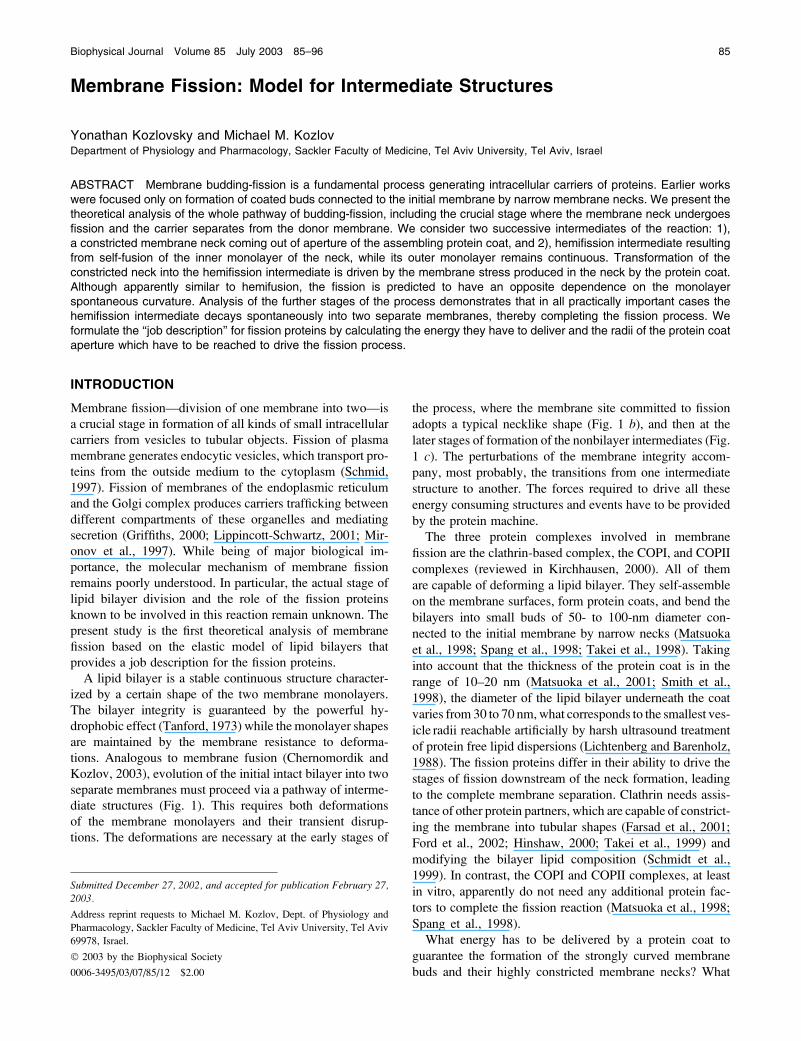
separate membranes must proceed via a pathway of interme-
diate structures (Fig. 1). This requires both deformations
of the membrane monolayers and their transient disrup-
tions. The deformations are necessary at the early stages of
the process, where the membrane site committed to fission
adopts a typical necklike shape (Fig. 1 b), and then at the
later stages of formation of the nonbilayer intermediates (Fig.
1 c). The perturbations of the membrane integrity accom-
pany, most probably, the transitions from one intermediate
structure to another. The forces required to drive all these
energy consuming structures and events have to be provided
by the protein machine.
The three protein complexes involved in membrane
fission are the clathrin-based complex, the COPI, and COPII
complexes (reviewed in Kirchhausen, 2000). All of them
are capable of deforming a lipid bilayer. They self-assemble
on the membrane surfaces, form protein coats, and bend the
bilayers into small buds of 50- to 100-nm diameter con-
nected to the initial membrane by narrow necks (Matsuoka
et al., 1998; Spang et al., 1998; Takei et al., 1998). Taking
into account that the thickness of the protein coat is in the
range of 10–20 nm (Matsuoka et al., 2001; Smith et al.,
1998), the diameter of the lipid bilayer underneath the coat
varies from 30 to 70 nm,what corresponds to the smallest ves-
icle radii reachable artificially by harsh ultrasound treatment
of protein free lipid dispersions (Lichtenberg and Barenholz,
1988). The fission proteins differ in their ability to drive the
stages of fission downstream of the neck formation, leading
to the complete membrane separation. Clathrin needs assis-
tance of other protein partners, which are capable of constrict-
ing the membrane into tubular shapes (Farsad et al., 2001;
Ford et al., 2002; Hinshaw, 2000; Takei et al., 1999) and
modifying the bilayer lipid composition (Schmidt et al.,
1999). In contrast, the COPI and COPII complexes, at least
in vitro, apparently do not need any additional protein fac-
tors to complete the fission reaction (Matsuoka et al., 1998;
Spang et al., 1998).
What energy has to be delivered by a protein coat to
guarantee the formation of the strongly curved membrane
buds and their highly constricted membrane necks? What
Submitted December 27, 2002, and accepted for publication February 27,2003.
Address reprint requests to Michael M. Kozlov, Dept. of Physiology and
Pharmacology, Sackler Faculty of Medicine, Tel Aviv University, Tel Aviv
69978, Israel.
� 2003 by the Biophysical Society
0006-3495/03/07/85/12 $2.00
features of a protein coat determine its ability to finalize the
bilayer fission and, hence, may be different for the clathrin
and the COP coats? These fundamental questions require
analysis of micromechanics of the fission intermediates.
Most previous models proposed for membrane fission
address the best-characterized reaction mediated by clathrin
partners, dynamin, and endophilin (Kozlov, 1999, 2001;
Sever et al., 2000b). While different views exist on the mode
of action of these proteins (Marks et al., 2001; Sever et al.,
2000a), all these models relate to the earlier stage of the
fission reaction, namely, formation of a constricted mem-
brane neck. None of them analyzes the membrane division
per se, i.e., scission of the neck.
In the present study, we suggest a model for the whole
pathway of membrane fission beginning from the constricted
membrane neck and proceeding via the hemifission inter-
mediate to the complete membrane separation. We show that
hemifission is driven by the elastic stress accumulated in the
constricted neck provided that the constriction is sufficiently
strong, and estimate the required energy, which has to be
delivered by the fission proteins. We demonstrate that, once
hemifission occurred, the membrane undergoes further tran-
sition to the complete fission spontaneously, because the
energy of the hemifission intermediate is larger than that of
the separated membranes in all practically important cases.
We analyze the effects of the membrane lipid composition
on fission for the case of symmetric bilayers. Our prediction
is that the lipid molecules having the effective shapes of
inverted cones, such as lysolipids, support division of the
membrane necks. This is opposite to the well-investigated
hemifusion, which is promoted by the conelike molecules
(Chernomordik et al., 1995).
MODEL FOR FISSION PATHWAY
Our goal is to study the most basic features of the fission
reaction. Therefore, we consider the simplest system con-
sisting of an initially flat membrane and fission proteins,
which self-assemble on the surface of the lipid bilayer into
a spherical coat and bend the membrane underneath (Fig. 1).
Since we are interested in the behavior of the lipid bilayer we
do not specify the mechanism by which the protein coat
deforms the membrane.
The bilayer portion covered by the coat and referred to
below as the coated bud has a shape of a spherical segment
with radius R (Fig. 1 a). Progressing self-assembly of the
coat is accompanied by a decrease in the coat aperture. This
results in deformation of the surrounding coat-free bilayer,
which adopts a necklike shape relaxing back to the flat sur-
face at increasing distances from the bud (Fig. 1 b). The closerthe shape of the bud is to that of the complete sphere, the
narrower is the membrane neck and, accordingly, the stronger
the deformation of the neck membrane and the related bi-
layer stress.
At a certain stage of budding, the elastic stress results in
decay of the membrane neck. Two scenarios of this events
are possible, a priori. First, the neck may simply rupture
and then reseal into a new configuration of two separate
membranes. In this case, fission would be expected to be
leaky. This prediction has been tested for membrane fission
mediated by COPII proteins, which did not exhibit any
significant transmembrane exchange of water-soluble dye
(Matsuoka et al., 1998). Hence, formation of large and long
living pores necessary to disrupt the neck has not been ob-
served, meaning that, analogously to membrane fusion, the
rupture-resealing mode of membrane fission is unlikely.
We focus on the second scenario where the membrane
remains impermeable through the whole process of fission.
We assume that the neck undergoes hemifission: its internal
monolayer self-fuses, whereas the outer monolayer main-
tains its integrity. The resulting structure referred to as the
hemifission intermediate is illustrated in (Fig. 1 c). This stageis followed by self-fusion of the outer monolayer that com-
pletes the fission reaction and results in separation of the
coated vesicle from the donor membrane (Fig. 1 d).To substantiate this model we have to calculate and
compare the energies of the constricted neck, the hemifission
intermediate and the separated membranes, and find the
conditions where the hierarchy of these energies validates the
suggested fission pathway.
PHYSICAL MODEL
The constricted neck and the hemifission intermediate are
characterized by deformations of their monolayers. There-
fore, the theoretical tool convenient for analyzing the
energies of these structures is the elastic model of lipid
membranes.
Elastic energy
The membrane of the neck is bent as compared to its initial
flat shape (Fig. 2 a). We account for the neck energy using
FIGURE 1 The intermediates of membrane
fission. (a) The coated bud at an initial stage of
the coat protein self-assembly. (b) Constrictedneck. (c) Hemifission intermediate. (d) Sepa-
rated coated vesicle.
86 Kozlovsky and Kozlov
Biophysical Journal 85(1) 85–96
the Helfrich bending model (Helfrich, 1973) reviewed in
Helfrich (1990).
We consider, separately, each monolayer of the lipid
bilayer. The monolayer shape is described by the shape of its
neutral surface lying at the interface between the polar heads
and the hydrocarbon chains (Leikin et al., 1996) at a distance
d � 1.2 nm (Rand and Parsegian, 1989) from the bilayer
midsurface. Although d corresponds to the thickness of the
hydrocarbon chains layer and does not include the polar
heads, we will use a loose terminology and refer to d as the
monolayer thickness.
Monolayer bending is quantified by the total curvature of
its surface, J. The structure of the monolayer is characterized
by its spontaneous curvature, Js, while the resistance of the
monolayer to deformation is accounted by the monolayer
bending modulus, k � 4 � 10�20 J (Niggemann et al., 1995).
The bending energy per unit area of the membrane mo-
nolayer related to the energy of the flat shape is given by
(Helfrich, 1973):
f ¼ 1
2� k � ðJ � JsÞ2 �
1
2� k � J2
s : (1)
Deformation of the monolayers of the hemifission in-
termediate is more complex (Fig. 2 b). Analogously to the
hemifusion structure considered in detail in Kozlovsky and
Kozlov (2002), the monolayers undergo, in addition to
bending, tilt, t, of the hydrocarbon chains with respect to the
membrane surface (Hamm and Kozlov, 2000). The tilt de-
formation is generated by packing the hydrocarbon chains in
the nonbilayer structural defect (Kozlovsky and Kozlov,
2002), which unavoidably emerges in the middle of the
hemifission intermediate and is referred to as the hydropho-
bic interstice (Siegel, 1993). Change of the tilt along the
monolayer surface generates an effective additional contri-
bution to the bending deformation, J, so that the latter is
considered in more general terms of splay of the hydrocarbon
chains, J (Hamm and Kozlov, 2000). The energy of the re-
sulting combined deformation per monolayer unit area re-
lated to the initial state of vanishing tilt and splay is given by
f ¼ 1
2� k � ðJ � JsÞ2 1
1
2� kt � t
2 � 1
2� k � J
2
s ; (2)
where kt � 40 mN/m is the tilt modulus (Hamm and Kozlov,
1998). Equations 1 and 2 are valid for small deformations,
satisfying
jJ � dj\1; jJ � dj\1; jtj\1: (3)
The total energy is obtained by integration of Eq. 1 for the
constricted neck, or Eq. 2 for the hemifission intermediate,
over the areas of the two monolayers,
F ¼ð
fin dAin 1
ðfout dAout; (4)
where the subscripts in and out refer to the inner and outer
monolayers, respectively.
Strategy of calculations
The degree of constriction of the membrane neck is deter-
mined by the radius of the aperture in the coat, rap (Fig. 2 a),and the radius R of the bud membrane (Fig. 1 b). We com-
pute the shape of the neck, which minimizes the elastic
energy (Eq. 4) and is connected smoothly to the lipid bilayer
of the bud. We do not assume any constrictions on the
membrane shape far from the bud. The energy of the re-
sulting membrane shape as a function of the bud and the
bilayer parameters, Fneck (rap, R, Js), represents the cons-
tricted neck.
A similar although more complicated procedure using
Eqs. 2 and 4 is performed for the hemifission intermediate.
The configurations of the membrane monolayers including
their shapes and distributions of tilt are optimized to mini-
mize the elastic energy. The constraints on the membrane tilt
and splay come in this case from the requirement of smooth
monolayer connections to the bud and from the conditions of
packing the hydrocarbon chains in the region of the
monolayer junction in the middle of the structure. The latter
constraints have been considered in the context of the
hemifusion intermediate in Kozlovsky and Kozlov (2002).
The resulting energy of the hemifission intermediate is also
computed as a function of the spontaneous curvature and the
bud parameters, Fhf (rap, R, Js).Comparison among Fneck, Fhf, and the energy of two
separated membranes provides us with the criteria of
transitions between different stages of the fission process.
The computations are performed numerically by the method
of Finite Elements, which is equivalent to solution of the
Euler-Lagrange equations.
FIGURE 2 Enlarged representation of (a) constricted neck and (b)
hemifission intermediate.
Theory of Membrane Fission 87
Biophysical Journal 85(1) 85–96
RESULTS
To emphasize the major features of configurations and
energies of the constricted neck and the hemifission inter-
mediate, we first present the results for the case of vanishing
spontaneous curvature of the membrane monolayers, Jouts ¼Jins ¼ 0, and then in a separate section we investigate the
effects of nonzero spontaneous curvature.
Constricted neck
The shape
We describe the configuration of the constricted neck by the
shape of its midsurface. Since we consider only the axi-
symmetric shapes, they will be described by the angle utangential to the surface profile as a function of the radial
distance, r (Fig. 2 a). The neck membrane is connected
smoothly to the membrane of the bud, meaning that sin u ¼rap/R at the boundary between the bud and the neck.
The major characteristic of the neck shape is the radius of
its narrowest cross-section, rneck (Fig. 2 a), measured at the
midsurface and referred to below as the neck radius.The character of the neck shape is determined by the
relationship between the neck radius rneck and the monolayer
thickness d. The neck is wide, meaning that rneck � d, if the
parameters of the coated bud satisfy r2ap=R � d. In this case,
the neck adopts a shape of a catenoid—the axisymmetric
surface of vanishing total curvature, J ¼ 0 (see, for example,
Nitsche, 1989). This result is expected, inasmuch as, in such
conditions, the absolute values of the curvatures of the two
monolayers are practically equal to that of the midsurface
(Appendix A) jJoutj � jJinj � jJj, so that the catenoidal shapecorresponds to the vanishing curvatures, Jout � Jin � 0, and,
hence, the vanishing bending energy (Eq. 1), fin� fout� 0, of
each membrane monolayer. The catenoidal shape is de-
termined by (Nitsche 1989),
sinu ¼ r�
r; (5)
where r* is a parameter determining the minimal radius of
the catenoid cross-section, and, hence, is equal to the neck
radius, r* ¼ rneck. Satisfying the constraint of smooth con-
nection of the neck to the bud, we obtain r� ¼ rneck ¼ r2ap=R.If the constriction of the neck is strong enough so that the
neck radius, rneck, becomes of the same order of magnitude
as the monolayer thickness, d, the difference between Jin andJout is considerable and the catenoidal surface does not
minimize the energy. In this case, the shape of the neck
resulting from the energy minimization is illustrated in Fig. 2
a for the coat parameters R ¼ 15 nm, rap ¼ 5 nm. The neck
radius in this case is rneck � 2.7 nm, and the shape deviates
considerably from the catenoid of the same rneck. The
detailed distribution of curvature over the monolayer sur-
faces is presented in the Appendix A. Note that, although the
neck is very narrow, rneck � 2.2 d, the maximal value of
the monolayer curvature reached in the inner monolayer of
the neck, constitutes jJmax � dj � 0.6 due to partial mutual
compensation of the two principal curvatures of opposite
signs. This means that the relationships in Eq. 3 are satisfied,
and that the elastic model (Eq. 1) we are using is applicable
on a semiquantitative level.
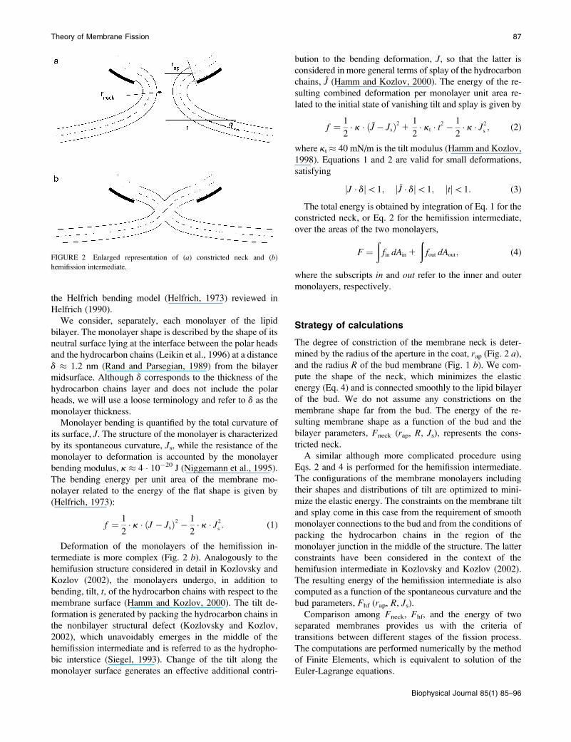
The energy
The energy of the neck, Fneck, for Jouts ¼ Jins ¼ 0 is presented
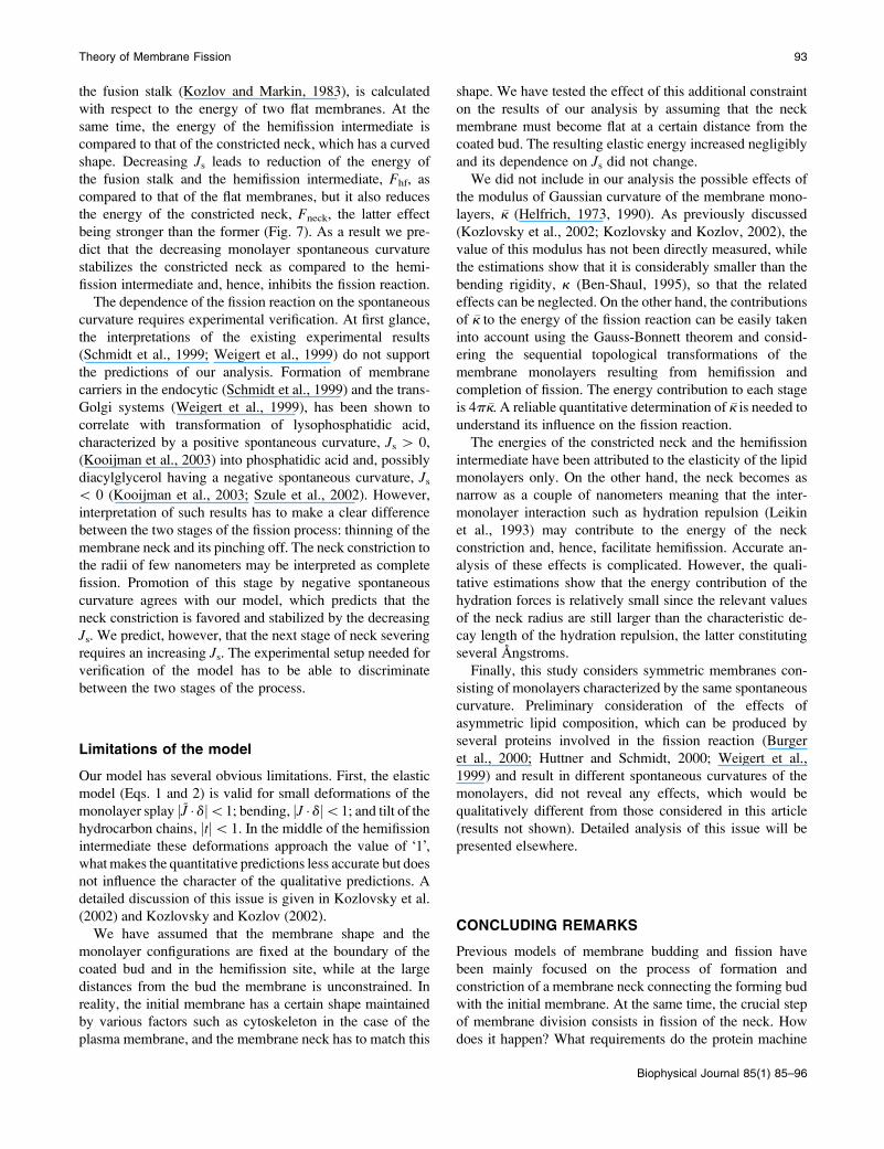
in Fig. 3 a as a function of the coat aperture radius, rap, fordifferent values of the bud radius, R. As expected, the energygrows with increasing constriction resulting from decreasing
FIGURE 3 The neck energy, Fneck, as a function of (a) the coat aperture radius, rap, and (b) the neck radius rneck. The bud radius, R, is (1) 12 nm; (2) 15 nm;
(3) 18 nm; (4) 25 nm; (5) 37 nm; and (6) 50 nm.
88 Kozlovsky and Kozlov
Biophysical Journal 85(1) 85–96
rap. It is instructive to re-plot the energy Fneck as a function of
the neck radius rneck, as presented in Fig. 3 b. The curves
obtained for the different bud radii R practically converged,
especially in the region of the small neck radii. This result
implies that the major energy is contributed by a relatively
limited region of the membrane around the narrowest neck
cross-section, and that the shape of this region depending
mostly on rneck is rather insensitive to the parameters of the
coated bud.
Hemifission intermediate
Membrane configuration
It is convenient to consider separately the lower part of the
hemifission structure, which begins at the monolayer junc-
tion site and extends downwards, and the upper part con-
necting the junction site with the coated bud (Fig. 2 b).The lower part is analogous to a half of the fusion stalk
considered in detail in Kozlovsky et al. (2002). The tan-
gential angle of the membrane profile, u, and the tilt angle,
ft, equal p/4 at the junction point and vanish at large
distances from the hemifission site. At a distance of ;6 nm
measured from the junction point along the contour of the
bilayer midsurface, the hydrocarbon chain tilt decays to zero
and the membrane shape relaxes to that of a catenoid with r*� 1.2 nm (the latter playing in this case a role of a parameter
determining the catenoidal surface (Eq. 5) rather than having
a meaning of a real neck radius).
The configuration of the upper part is constrained at one
boundary by the junction point where u¼ ft¼�p/4, and at
the other by the condition of smooth connection to the
bilayer of the bud, sin u ¼ rap/R. The tilt relaxes along an
;6-nm distance form the junction point, similar to the tilt
behavior in the lower part. The shape of the membrane pro-
file minimizing the energy of the upper part cannot be
described analytically. The numerical result for the mem-
brane shape of the hemifission site is illustrated in Fig. 2 b for
the coat parameters rap ¼ 5 nm and R ¼ 15 nm.
The membrane region expanding to the �6-nm distance
upwards and downwards from the junction point will be
referred to as the core of the hemifission intermediate, where
the major membrane deformations are concentrated. Accord-
ing to the results of our calculations, the structure of the core
region is practically not influenced by the parameters of the
coated bud.
The energy
The energy of the lower part of the hemifission intermediate
is independent of the parameters of the coat and for Js ¼ 0,
constitutes Flowhf � 39 kBT (Kozlovsky and Kozlov, 2002),
where kBT � 4.1 3 10�14 erg is the product of the
Boltzmann constant and the absolute temperature, T ¼ 300
K. Almost the whole energy is contributed by the core
region, while the energy of the catenoidal part of the
membrane vanishes.
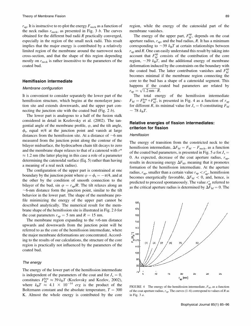
The energy of the upper part, Fuphf , depends on the coat
aperture radius, rap, and the bud radius, R. It has a minimum
corresponding to ;39 kBT at certain relationships between
rap and R. One can easily understand this result by taking intoaccount that Fup
hf consists of the contribution of the core
region, ;39 kBT, and the additional energy of membrane
deformation induced by the constraints on the boundary with
the coated bud. The latter contribution vanishes and Fuphf
becomes minimal if the membrane region connecting the
core to the bud has a shape of a catenoidal segment. This
happens if the coated bud parameters are related by
rap ¼ffiffiffiffiffiffiffiffiffiffiffiffiffiffiffiffiffiffiffiffi1:2 nm � R
p.
The total energy of the hemifission intermediate
Fhf ¼ Flowhf 1Fup
hf , is presented in Fig. 4 as a function of rapfor different R, its minimal value for Js ¼ 0 constituting Fhf
; 78 kBT.
Relative energies of fission intermediates:criterion for fission
Hemifission
The energy of transition from the constricted neck to the
hemifission intermediate, DFhf ¼ Fhf � Fneck, as a function
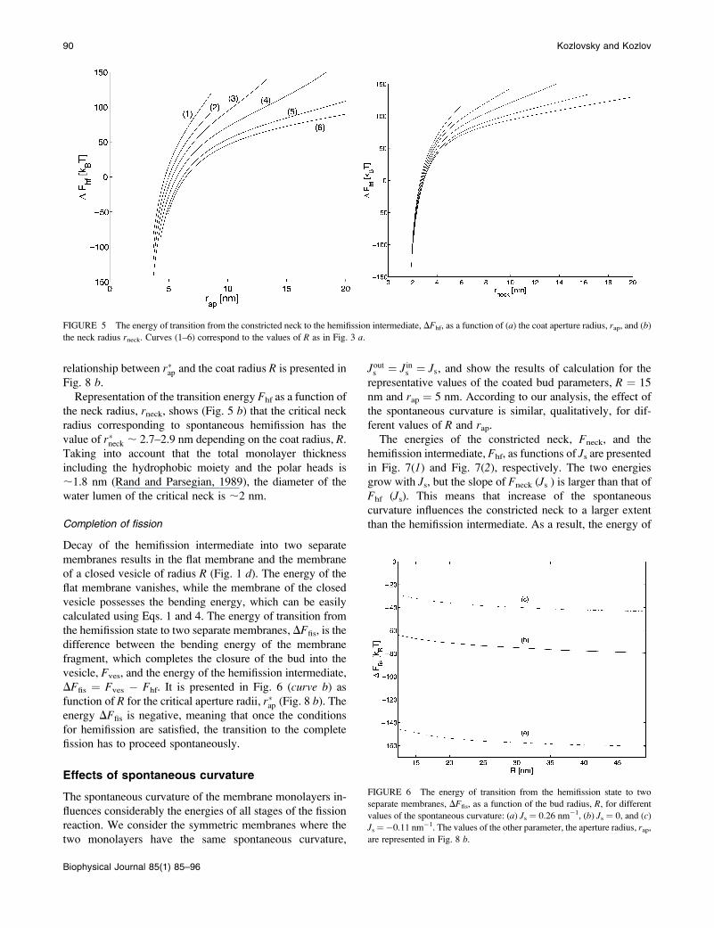
of the coated bud parameters, is presented in Fig. 5 a for Js ¼0. As expected, decrease of the coat aperture radius, rap,results in decreasing energy DFhf, meaning that it promotes
formation of the hemifission intermediate. At the aperture
radius, rap, smaller than a certain value rap\r�ap, hemifission
becomes energetically favorable, DFhf \ 0, and, hence, is
predicted to proceed spontaneously. The value r�ap referred toas the critical aperture radius is determined by DFhf ¼ 0. The
FIGURE 4 The energy of the hemifission intermediate, Fhf, as a function
of the coat aperture radius, rap. The curves (1–6) correspond to values of R as
in Fig. 3 a.
Theory of Membrane Fission 89
Biophysical Journal 85(1) 85–96
relationship between r�ap and the coat radius R is presented in
Fig. 8 b.Representation of the transition energy Fhf as a function of
the neck radius, rneck, shows (Fig. 5 b) that the critical neckradius corresponding to spontaneous hemifission has the
value of r�neck ; 2.7–2.9 nm depending on the coat radius, R.Taking into account that the total monolayer thickness
including the hydrophobic moiety and the polar heads is
;1.8 nm (Rand and Parsegian, 1989), the diameter of the
water lumen of the critical neck is ;2 nm.
Completion of fission
Decay of the hemifission intermediate into two separate
membranes results in the flat membrane and the membrane
of a closed vesicle of radius R (Fig. 1 d). The energy of the
flat membrane vanishes, while the membrane of the closed
vesicle possesses the bending energy, which can be easily
calculated using Eqs. 1 and 4. The energy of transition from
the hemifission state to two separate membranes, DFfis, is the
difference between the bending energy of the membrane
fragment, which completes the closure of the bud into the
vesicle, Fves, and the energy of the hemifission intermediate,
DFfis ¼ Fves � Fhf. It is presented in Fig. 6 (curve b) asfunction of R for the critical aperture radii, r�ap (Fig. 8 b). Theenergy DFfis is negative, meaning that once the conditions
for hemifission are satisfied, the transition to the complete
fission has to proceed spontaneously.
Effects of spontaneous curvature
The spontaneous curvature of the membrane monolayers in-
fluences considerably the energies of all stages of the fission
reaction. We consider the symmetric membranes where the
two monolayers have the same spontaneous curvature,
Jouts ¼ Jins ¼ Js, and show the results of calculation for the
representative values of the coated bud parameters, R ¼ 15
nm and rap ¼ 5 nm. According to our analysis, the effect of
the spontaneous curvature is similar, qualitatively, for dif-
ferent values of R and rap.The energies of the constricted neck, Fneck, and the
hemifission intermediate, Fhf, as functions of Js are presentedin Fig. 7(1) and Fig. 7(2), respectively. The two energies
grow with Js, but the slope of Fneck (Js ) is larger than that of
Fhf (Js). This means that increase of the spontaneous
curvature influences the constricted neck to a larger extent
than the hemifission intermediate. As a result, the energy of
FIGURE 5 The energy of transition from the constricted neck to the hemifission intermediate, DFhf, as a function of (a) the coat aperture radius, rap, and (b)
the neck radius rneck. Curves (1–6) correspond to the values of R as in Fig. 3 a.
FIGURE 6 The energy of transition from the hemifission state to two
separate membranes, DFfis, as a function of the bud radius, R, for different
values of the spontaneous curvature: (a) Js ¼ 0.26 nm�1, (b) Js ¼ 0, and (c)Js¼�0.11 nm�1. The values of the other parameter, the aperture radius, rap,
are represented in Fig. 8 b.
90 Kozlovsky and Kozlov
Biophysical Journal 85(1) 85–96
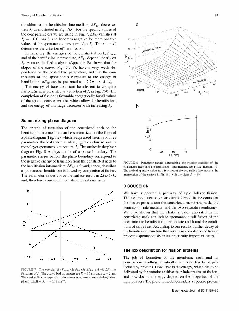
transition to the hemifission intermediate, DFhf, decreases
with Js as illustrated in Fig. 7(3). For the specific values of
the coat parameters we are using in Fig. 7, DFhf vanishes at
J�s ¼ �0:01 nm�1, and becomes negative for more positive
values of the spontaneous curvature, Js[J�s . The value J�sdetermines the criterion of hemifission.
Remarkably, the energies of the constricted neck, Fneck,
and of the hemifission intermediate, DFhf, depend linearly on
Js. A more detailed analysis (Appendix B) shows that the
slopes of the curves Fig. 7(1–3), have a very weak de-
pendence on the coated bud parameters, and that the con-
tribution of the spontaneous curvature to the energy of
hemifission, DFhf, can be presented as �7.7p � k � d � Js.The energy of transition from hemifission to complete
fission, DFfis, is presented as a function of Js in Fig. 7(4). Thecompletion of fission is favorable energetically for all values
of the spontaneous curvature, which allow for hemifission,
and the energy of this stage decreases with increasing Js.
Summarizing phase diagram
The criteria of transition of the constricted neck to the
hemifission intermediate can be summarized in the form of
a phase diagram (Fig. 8 a), which is expressed in terms of three
parameters: the coat aperture radius, rap, bud radius,R, and themonolayer spontaneous curvature, Js. The surface in the phasediagram Fig. 8 a plays a role of a phase boundary. The
parameter ranges bellow the phase boundary correspond to
the negative energy of transition from the constricted neck to
the hemifission intermediate, DFhf\0, and, hence, describes
a spontaneous hemifission followed by completion of fission.
The parameter values above the surface result in DFhf[ 0,
and, therefore, correspond to a stable membrane neck.
DISCUSSION
We have suggested a pathway of lipid bilayer fission.
The assumed successive structures formed in the course of
the fission process are: the constricted membrane neck, the
hemifission intermediate, and the two separate membranes.
We have shown that the elastic stresses generated in the
constricted neck can induce spontaneous self-fusion of the
neck into the hemifission intermediate and found the condi-
tions of this event. According to our results, further decay of
the hemifission structure that results in completion of fission
proceeds spontaneously in all practically important cases.
The job description for fission proteins
The job of formation of the membrane neck and its
constriction resulting, eventually, in fission has to be per-
formed by proteins. How large is the energy, which has to be
delivered by the proteins to drive the whole process of fission,
and how does this energy depend on the properties of the
lipid bilayer? The present model considers a specific protein
FIGURE 8 Parameter ranges determining the relative stability of the
constricted neck and the hemifission intermediate. (a) Phase diagram. (b)
The critical aperture radius as a function of the bud radius (the curve is the
intersection of the surface in Fig. 8 a with the plane Js ¼ 0).
FIGURE 7 The energies (1) Fneck, (2) Fhf, (3) DFhf, and (4) DFfis, as
functions of Js. The coated bud parameters are R ¼ 15 nm and rap ¼ 5 nm.
The vertical line corresponds to the spontaneous curvature of dioleoylphos-
phatidylcholine, Js ¼ �0.11 nm�1.
Theory of Membrane Fission 91
Biophysical Journal 85(1) 85–96
machine, namely, a protein coat, which self-assembles on
the membrane surface and bends the latter into a shape of
spherical segment with radius R. This can be directly related
to vesicle formation by the clathrin, COPI, and COPII coats
in the in vitro experiments (Matsuoka et al., 1998; Spang
et al., 1998; Takei et al., 1998) and, hopefully, accounts for
the major features of the in vivo reactions despite various
unclear issues (Nossal and Zimmerberg, 2002). In addition,
the presented estimations should give a correct order of
magnitude of the energy required to drive fission in other
membrane configurations such as, for example, in the ER-
Golgi system (Griffiths, 2000; Lippincott-Schwartz, 2001;
Mironov et al., 1997) generating tubular carriers.
The protein coat-induced bending of the membrane into
the spherical bud forms the membrane neck connecting the
bud with the donor membrane. Hence, the proteins have to
deliver the energy, Ftot, required for both the bud and the
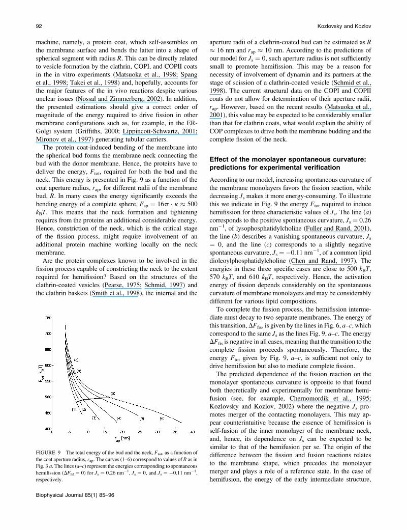
neck. This energy is presented in Fig. 9 as a function of the
coat aperture radius, rap, for different radii of the membrane
bud, R. In many cases the energy significantly exceeds the
bending energy of a complete sphere, Fsp ¼ 16p � k � 500
kBT. This means that the neck formation and tightening
requires from the proteins an additional considerable energy.
Hence, constriction of the neck, which is the critical stage
of the fission process, might require involvement of an
additional protein machine working locally on the neck
membrane.
Are the protein complexes known to be involved in the
fission process capable of constricting the neck to the extent
required for hemifission? Based on the structures of the
clathrin-coated vesicles (Pearse, 1975; Schmid, 1997) and
the clathrin baskets (Smith et al., 1998), the internal and the
aperture radii of a clathrin-coated bud can be estimated as R� 16 nm and rap � 10 nm. According to the predictions of
our model for Js ¼ 0, such aperture radius is not sufficiently
small to promote hemifission. This may be a reason for
necessity of involvement of dynamin and its partners at the
stage of scission of a clathrin-coated vesicle (Schmid et al.,
1998). The current structural data on the COPI and COPII
coats do not allow for determination of their aperture radii,
rap. However, based on the recent results (Matsuoka et al.,
2001), this value may be expected to be considerably smaller
than that for clathrin coats, what would explain the ability of
COP complexes to drive both the membrane budding and the
complete fission of the neck.
Effect of the monolayer spontaneous curvature:predictions for experimental verification
According to our model, increasing spontaneous curvature of
the membrane monolayers favors the fission reaction, while
decreasing Js makes it more energy-consuming. To illustrate
this we indicate in Fig. 9 the energy Ftot required to induce
hemifission for three characteristic values of Js. The line (a)corresponds to the positive spontaneous curvature, Js ¼ 0.26
nm�1, of lysophosphatidylcholine (Fuller and Rand, 2001),
the line (b) describes a vanishing spontaneous curvature, Js¼ 0, and the line (c) corresponds to a slightly negative
spontaneous curvature, Js ¼�0.11 nm�1, of a common lipid
dioleoylphosphatidylcholine (Chen and Rand, 1997). The
energies in these three specific cases are close to 500 kBT,570 kBT, and 610 kBT, respectively. Hence, the activation
energy of fission depends considerably on the spontaneous
curvature of membrane monolayers and may be considerably
different for various lipid compositions.
To complete the fission process, the hemifission interme-
diate must decay to two separate membranes. The energy of
this transition,DFfis, is given by the lines in Fig. 6, a–c, whichcorrespond to the same Js as the lines Fig. 9, a–c. The energyDFfis is negative in all cases, meaning that the transition to the
complete fission proceeds spontaneously. Therefore, the
energy Ftot given by Fig. 9, a–c, is sufficient not only to
drive hemifission but also to mediate complete fission.
The predicted dependence of the fission reaction on the
monolayer spontaneous curvature is opposite to that found
both theoretically and experimentally for membrane hemi-
fusion (see, for example, Chernomordik et al., 1995;
Kozlovsky and Kozlov, 2002) where the negative Js pro-motes merger of the contacting monolayers. This may ap-
pear counterintuitive because the essence of hemifission is
self-fusion of the inner monolayer of the membrane neck,
and, hence, its dependence on Js can be expected to be
similar to that of the hemifusion per se. The origin of the
difference between the fission and fusion reactions relates
to the membrane shape, which precedes the monolayer
merger and plays a role of a reference state. In the case of
hemifusion, the energy of the early intermediate structure,
FIGURE 9 The total energy of the bud and the neck, Ftot, as a function of
the coat aperture radius, rap. The curves (1–6) correspond to values of R as in
Fig. 3 a. The lines (a–c) represent the energies corresponding to spontaneous
hemifission (DFhf ¼ 0) for Js ¼ 0.26 nm�1, Js ¼ 0, and Js ¼ �0.11 nm�1,
respectively.
92 Kozlovsky and Kozlov
Biophysical Journal 85(1) 85–96
the fusion stalk (Kozlov and Markin, 1983), is calculated
with respect to the energy of two flat membranes. At the
same time, the energy of the hemifission intermediate is
compared to that of the constricted neck, which has a curved
shape. Decreasing Js leads to reduction of the energy of
the fusion stalk and the hemifission intermediate, Fhf, as
compared to that of the flat membranes, but it also reduces
the energy of the constricted neck, Fneck, the latter effect
being stronger than the former (Fig. 7). As a result we pre-
dict that the decreasing monolayer spontaneous curvature
stabilizes the constricted neck as compared to the hemi-
fission intermediate and, hence, inhibits the fission reaction.
The dependence of the fission reaction on the spontaneous
curvature requires experimental verification. At first glance,
the interpretations of the existing experimental results
(Schmidt et al., 1999; Weigert et al., 1999) do not support
the predictions of our analysis. Formation of membrane
carriers in the endocytic (Schmidt et al., 1999) and the trans-
Golgi systems (Weigert et al., 1999), has been shown to
correlate with transformation of lysophosphatidic acid,
characterized by a positive spontaneous curvature, Js [ 0,
(Kooijman et al., 2003) into phosphatidic acid and, possibly
diacylglycerol having a negative spontaneous curvature, Js\ 0 (Kooijman et al., 2003; Szule et al., 2002). However,
interpretation of such results has to make a clear difference
between the two stages of the fission process: thinning of the
membrane neck and its pinching off. The neck constriction to
the radii of few nanometers may be interpreted as complete
fission. Promotion of this stage by negative spontaneous
curvature agrees with our model, which predicts that the
neck constriction is favored and stabilized by the decreasing
Js. We predict, however, that the next stage of neck severing
requires an increasing Js. The experimental setup needed for
verification of the model has to be able to discriminate
between the two stages of the process.
Limitations of the model
Our model has several obvious limitations. First, the elastic
model (Eqs. 1 and 2) is valid for small deformations of the
monolayer splay jJ � dj\1; bending, jJ � dj\1; and tilt of the
hydrocarbon chains, jtj\1. In the middle of the hemifission
intermediate these deformations approach the value of �1�,what makes the quantitative predictions less accurate but does
not influence the character of the qualitative predictions. A
detailed discussion of this issue is given in Kozlovsky et al.
(2002) and Kozlovsky and Kozlov (2002).
We have assumed that the membrane shape and the
monolayer configurations are fixed at the boundary of the
coated bud and in the hemifission site, while at the large
distances from the bud the membrane is unconstrained. In
reality, the initial membrane has a certain shape maintained
by various factors such as cytoskeleton in the case of the
plasma membrane, and the membrane neck has to match this
shape. We have tested the effect of this additional constraint
on the results of our analysis by assuming that the neck
membrane must become flat at a certain distance from the
coated bud. The resulting elastic energy increased negligibly
and its dependence on Js did not change.
We did not include in our analysis the possible effects of
the modulus of Gaussian curvature of the membrane mono-
layers, k� (Helfrich, 1973, 1990). As previously discussed
(Kozlovsky et al., 2002; Kozlovsky and Kozlov, 2002), the
value of this modulus has not been directly measured, while
the estimations show that it is considerably smaller than the
bending rigidity, k (Ben-Shaul, 1995), so that the related
effects can be neglected. On the other hand, the contributions
of k� to the energy of the fission reaction can be easily taken
into account using the Gauss-Bonnett theorem and consid-
ering the sequential topological transformations of the
membrane monolayers resulting from hemifission and
completion of fission. The energy contribution to each stage
is 4pk�. A reliable quantitative determination of k�is needed tounderstand its influence on the fission reaction.
The energies of the constricted neck and the hemifission
intermediate have been attributed to the elasticity of the lipid
monolayers only. On the other hand, the neck becomes as
narrow as a couple of nanometers meaning that the inter-
monolayer interaction such as hydration repulsion (Leikin
et al., 1993) may contribute to the energy of the neck
constriction and, hence, facilitate hemifission. Accurate an-
alysis of these effects is complicated. However, the quali-
tative estimations show that the energy contribution of the
hydration forces is relatively small since the relevant values
of the neck radius are still larger than the characteristic de-
cay length of the hydration repulsion, the latter constituting
several Angstroms.
Finally, this study considers symmetric membranes con-
sisting of monolayers characterized by the same spontaneous
curvature. Preliminary consideration of the effects of
asymmetric lipid composition, which can be produced by
several proteins involved in the fission reaction (Burger
et al., 2000; Huttner and Schmidt, 2000; Weigert et al.,
1999) and result in different spontaneous curvatures of the
monolayers, did not reveal any effects, which would be
qualitatively different from those considered in this article
(results not shown). Detailed analysis of this issue will be
presented elsewhere.
CONCLUDING REMARKS
Previous models of membrane budding and fission have
been mainly focused on the process of formation and
constriction of a membrane neck connecting the forming bud
with the initial membrane. At the same time, the crucial step
of membrane division consists in fission of the neck. How
does it happen? What requirements do the protein machine
Theory of Membrane Fission 93
Biophysical Journal 85(1) 85–96
and the lipid composition of the membrane have to fulfill to
be able to drive and facilitate the fission reaction? The pre-
sent work suggests that a specific lipid structure, the hemi-
fission intermediate, is formed at the stage of transition from
the constricted neck to two separate membranes. Based on
this assumption, we formulate the job description for fission
proteins in terms of the stresses, which have to be induced in
the neck to trigger its fission, and predict that, in contrast to
membrane hemifusion, the fission reaction has to be pro-
moted by lipids with positive spontaneous curvature, such as
lysolipids. This model remains hypothetical and requires
experimental verification. Moreover, the model does not
consider the energy barriers related to probable transient
disruption of the membrane monolayer at the stages of
transition from the constricted neck to the hemifission inter-
mediate and further to two separate membranes. Analysis of
these barriers and their influence on the rate of the fission
reaction is a matter for future work.
APPENDIX A
Structure of the constricted neck
Relationships between the curvatures
We describe the membrane by three parallel surfaces—the bilayer midsur-
face and the neutral surfaces of the two monolayers. A monolayer neutral
surface lies close to the interface between the polar heads and the
hydrocarbon tails at a distance d from the midplane.
The total curvatures of each neutral surface of the outer, Jout, and inner,
Jin, monolayers are related to the total curvature, J, and the Gaussian
curvature, K, of the midsurface (see, e.g., Safran, 1994) by:
Jout ¼J 1 2K � d
11 J � d1K � d2 ; Jin ¼�J 1 2K � d
1� J � d1K � d2 : (A1)
In the case of small curvatures, jJ � dj � 1 and jK � d2j � 1, and absolute
values of the monolayer total curvatures approach that of the midsurface Jout� �Jin � J.
Catenoid
An axisymmetric surface is commonly described by the tangential angle of
the surface profile, u, and the distance from the axis of symmetry, r (see
Helfrich, 1990) The principal curvatures of an axisymmetric surface are the
parallel curvature, cp, and the meridian curvature, cm, expressed by
cp ¼sinu
r; cm ¼ d
drðsinuÞ: (A2)
A catenoid is described by
sinu ¼ r�=r; (A3)
where the parameter r* is the minimal radius of the surface cross-section.
The corresponding principal curvatures are cp ¼ r*/r2 and cm ¼ �r*/r2,while the total and the Gaussian curvatures are given by
J ¼ cp 1 cm ¼ 0; K ¼ cp � cm ¼ �ðr�=r2Þ2; (A4)
The catenoid shape minimizes the elastic energy (Eqs. 1 and 4 of the main
part) of an axisymmetric surface if its minimal radius, r*, is much larger than
the monolayer thickness, r* � d. This is shown by the following
calculation. The Gaussian curvature (Eq. A4) satisfies jKj # 1/(r*)2 since r
$ r*. Then the total curvatures (Eq. A1) satisfy Jout ¼ Jin � 2K � d � 1/d.
The elastic energy of the catenoid, F, can be estimated as the energy density
at the neck, f � k � J2in1k � J2out � k � ðK � dÞ2 � k � ðd=r�2Þ2 multiplied by
the characteristic area of the narrowest region of the catenoid ;r*2. Thisresults in vanishing energy, F/k � (d/r*)2 � 1.
Total curvatures of the strongly constricted neck
The shape of a strongly constricted neck characterized by a small neck radius
is found by numeric calculations. The monolayer total curvatures are shown
in Fig. 10 for the coated bud parameters R ¼ 15 nm and rap ¼ 5 nm. The
curvatures are presented as a function of the contour length measured from
the aperture of the coated bud. Fig. 10(4) gives the radial distance of the
midsurface as a function of the contour length. The minimal membrane
radius is rneck ¼ 2.7 nm and it occurs at contour length of ;4 nm.
We divide the constricted neck to three regions: 1), the region near the
coat aperture, which corresponds to small contour lengths; 2), the neck
region surrounding the cross-section of the minimal radius; and 3), the
region corresponding to large contour length. The total curvature of the
midsurface, J (Fig. 10(1)), is negative near the coat aperture. The maximum
occurs at the neck region and is positive. The total curvature of the neutral
surface of the outer monolayer, Jout (Fig. 10(2)), is negative at the coat
aperture, whereas that of the inner monolayer, Jin (Fig. 10(3)), is positive in
this region. At the neck region both Jin and Jout are negative. The minimum
of Jin occurs at the minimal radius of the constricted neck, rneck. Away from
the neck the total curvatures approach zero and the membrane tends to form
the shape of a catenoid. Note that it is impossible to discern the sign of the
total curvature of the neck region just by looking at the surface shape (Fig. 2
a of the main part). The total curvature of a saddlelike surface results from
a delicate balance between the negative meridian curvature and the positive
parallel curvature.
APPENDIX B
Energy dependence on the spontaneouscurvature in symmetric bilayers
We analyze the energy contribution to the fission intermediates related to the
spontaneous curvature by considering symmetric bilayers, which are
composed of monolayers with the same spontaneous curvature. We first
FIGURE 10 The total curvatures of the monolayers of a constricted neck
as functions of the contour length along the midsurface: (1) J, (2) Jout, and
(3) Jin. ( 4) Corresponding radial distance, r, of the midsurface. The coated
bud parameters are R ¼ 15 nm and rap ¼ 5 nm.
94 Kozlovsky and Kozlov
Biophysical Journal 85(1) 85–96
discuss the constricted neck where tilt is not involved. The part of the
bending energy (Eqs. 1 and 4) depending on the spontaneous curvature, FJ,
is:
FJ ¼ �kJs
ðJin dAin 1
ðJout dAout
� �: (B1)
By using the relations between the geometrical characteristics of parallel
surfaces (see, for example, Safran, 1994), it can be shown that the sum of
two integrals depends only on the Gaussian curvature, K, of the midsurface:
FJ ¼ �4kJsd
ðK dA: (B2)
The integral of the Gaussian curvature depends only on the boundary
conditions and the topology of the surface as stated by the Gauss-Bonnet
theorem. Therefore, for symmetric bilayers the monolayer spontaneous
curvature does not affect the membrane shape unless the membrane
topology changes.
The integral of the Gaussian curvature of an axial symmetric shape (Eq.
A2) is expressed by the tangential angle at the two boundaries, uR and u2:ðK dA ¼ �2pðcosuR � cosu2Þ: (B3)
The angle at the coated bud boundary is fixed by the bud,uR¼ arcsin(rap/R),
while the angle at the free boundary is u2 ¼ p. The resulting dependence of
the energy on the spontaneous curvature is linear:
Fneck
J ¼ 8pð11 cosuRÞkJsd: (B4)
This result determines the linear character and the slope of the graph; see Fig.
7(1). For coated buds with small uR, we obtain cos uR � 1, and the slope of
the curve does not practically depend on the coated bud parameters R and
rap.
The hemifission intermediate includes tilt deformation, so its elastic
energy is given by Eqs. 2 and 4. The energy dependence on the spontaneous
curvature is:
Fhf
J ¼ �kJs
ðJin dAin 1
ðJout dAout
� �: (B5)
We do not have analytical expressions for these integrals and we calculate
them numerically. We calculated FhfJ for different coated bud parameters and
obtained that it is given by
Fhf
J ¼ 8:3pkJsd1 8pðcosuR � 1ÞkJsd: (B6)
This linear relation determines the slope of the graph Fig. 7(2). The second
term of Eq. B6 arises from the connection of the core region of the
hemifission intermediate to the coated bud. Because uR is small, it is much
smaller than the first term. The first term arises from the splay at the
hemifission core. Remarkably, it is linear in Js and insensitive to the values
of the coated bud parameters, R and rap, provided that the latter are in an
experimentally relevant range. The energy of the core region consists of
approximately equal contributions of the splay of the inner and the outer
monolayers.
The contribution from the spontaneous curvature to the energy
difference, DFhfJ , between the hemifission intermediate (Eq. B6) and
constricted neck (Eq. B4) is:
DFhf
J ¼ Fhf
J � Fneck
J ¼ �7:7pkJsd: (B7)
The difference DFhfJ does not depend on the coated bud parameters. This
relation determines the linear character and the slope of the line; see Fig. 7(3).The energy of the transition from the hemifission state to two separate
membranes, DFfisJ , illustrated by Fig. 7(4) depends on the spontaneous
curvature according to DFfisJ ¼ �8:3pkJsd. It is also independent of the coat
parameters. In this case, the energy of the membrane fragment, which
completes the closure of the bud into the vesicle, exactly compensates the
second term of Eq. B6.
We are grateful to Leonid Chernomordik and Koert Burger for discussions
and critical reading of the manuscript.
This work was supported by the Human Frontier Science Program
Organization.
REFERENCES
Ben-Shaul, A. 1995. Molecular theory of chain packing, elasticity and lipid-protein interaction in lipid bilayers. In Structure and Dynamics ofMembranes. R. Lipowsky, and E. Sackmann, editors. Elsevier,Amsterdam, the Netherlands. pp.359–401.
Burger, K. N., R. A. Demel, S. L. Schmid, and B. de Kruijff. 2000.Dynamin is membrane-active: lipid insertion is induced by phosphoino-sitides and phosphatidic acid. Biochemistry. 39:12485–12493.
Chen, Z., and R. P. Rand. 1997. The influence of cholesterol onphospholipid membrane curvature and bending elasticity. Biophys. J. 73:267–276.
Chernomordik, L., M. Kozlov, and J. Zimmerberg. 1995. Lipids in bio-logical membrane fusion. J. Membr. Biol. 146:1–14.
Chernomordik, L. V., and M. M. Kozlov. 2003. Protein-lipid interplay infusion and fission of biological membranes. Annu. Rev. Biochem.72:175–207.
Farsad, K., N. Ringstad, K. Takei, S. R. Floyd, K. Rose, and P. De Camilli.2001. Generation of high curvature membranes mediated by directendophilin bilayer interactions. J. Cell Biol. 155:193–200.
Ford, M. G., I. G. Mills, B. J. Peter, Y. Vallis, G. J. Praefcke, P. R. Evans,and H. T. McMahon. 2002. Curvature of clathrin-coated pits driven byepsin. Nature. 419:361–366.
Fuller, N., and R. P. Rand. 2001. The influence of lysolipids on thespontaneous curvature and bending elasticity of phospholipid mem-branes. Biophys. J. 81:243–254.
Griffiths, G. 2000. Gut thoughts on the Golgi complex. Traffic. 1:738–745.
Hamm, M., and M. Kozlov. 1998. Tilt model of inverted amphiphilicmesophases. Euro. Phys. J. B. 6:519–528.
Hamm, M., and M. Kozlov. 2000. Elastic energy of tilt and bending of fluidmembranes. Eur. Phys. J. E. 3:323–335.
Helfrich, W. 1973. Elastic properties of lipid bilayers: theory and possibleexperiments. Z. Naturforsch. 28c:693–703.
Helfrich, W. 1990. Elasticity and thermal undulations of fluid films ofamphiphiles. In Liquids and Interfaces. J. Charvolin, J.-F. Joanny, andJ. Zinn-Justin, editors. Elsevier Science Publishers, Les Houches.
Hinshaw, J. E. 2000. Dynamin and its role in membrane fission. Annu. Rev.Cell Dev. Biol. 16:483–519.
Huttner, W. B., and A. Schmidt. 2000. Lipids, lipid modification and lipid-protein interaction in membrane budding and fission-insights from theroles of endophilin A1 and synaptophysin in synaptic vesicle endocy-tosis. Curr. Opin. Neurobiol. 10:543–551.
Kirchhausen, T. 2000. Three ways to make a vesicle. Nat. Rev. Mol. CellBiol. 1:187–198.
Kooijman, E. E., V. Chupin, B. de Kruijff, and K. N. J. Burger. 2003.Modulation of membrane curvature by phosphatidic acid and lysophos-phatidic acid. Traffic. 4:162–174.
Kozlov, M. M. 1999. Dynamin: possible mechanism of ‘‘Pinchase’’ action.Biophys. J. 77:604–616.
Kozlov, M. M. 2001. Fission of biological membranes: interplay betweendynamin and lipids. Traffic. 2:51–65.
Kozlov, M. M., and V. S. Markin. 1983. Possible mechanism of membranefusion. Biofizika. 28:255–261.
Theory of Membrane Fission 95
Biophysical Journal 85(1) 85–96
Kozlovsky, Y., L. V. Chernomordik, and M. M. Kozlov. 2002. Lipidintermediates in membrane fusion: formation, structure, and decay ofhemifusion diaphragm. Biophys. J. 83:2634–2651.
Kozlovsky, Y., and M. Kozlov. 2002. Stalk model of membrane fusion:solution of energy crisis. Biophys. J. 88:882–895.
Leikin, S., M. M. Kozlov, N. L. Fuller, and R. P. Rand. 1996. Measuredeffects of diacylglycerol on structural and elastic properties of phos-pholipid membranes. Biophys. J. 71:2623–2632.
Leikin, S., V. A. Parsegian, D. C. Rau, and R. P. Rand. 1993. Hydrationforces. Annu. Rev. Phys. Chem. 44:369–395.
Lichtenberg, D., and Y. Barenholz. 1988. Liposomes: preparation,characterization, and preservation. Methods Biochem. Anal. 33:337–462.
Lippincott-Schwartz, J. 2001. The secretory membrane system studied inreal-time. Robert Feulgen Prize Lecture for 2001. Histochem. Cell Biol.116:97–107.
Marks, B., M. H. Stowell, Y. Vallis, I. G. Mills, A. Gibson, C. R. Hopkins,and H. T. McMahon. 2001. GTPase activity of dynamin and resultingconformation change are essential for endocytosis. Nature. 410:231–235.
Matsuoka, K., L. Orci, M. Amherdt, S. Y. Bednarek, S. Hamamoto, R.Schekman, and T. Yeung. 1998. COPII-coated vesicle formation recon-stituted with purified coat proteins and chemically defined liposomes.Cell. 93:263–275.
Matsuoka, K., R. Schekman, L. Orci, and J. E. Heuser. 2001. Surfacestructure of the COPII-coated vesicle. Proc. Natl. Acad. Sci. USA. 98:13705–13709.
Mironov, A. A., P. Weidman, and A. Luini. 1997. Variations on theintracellular transport theme: maturing cisternae and trafficking tubules.J. Cell Biol. 138:481–484.
Niggemann, G., M. Kummrow, and W. Helfrich. 1995. The bendingrigidity of phosphatidylcholine bilayers. Dependence on experimentalmethods, sample cell sealing and temperature. J. Phys. II. 5:413–425.
Nitsche, J. C. C. 1989. Lectures on Minimal Surfaces. Cambridge Univer-sity Press, Cambridge, UK.
Nossal, R., and J. Zimmerberg. 2002. Endocytosis: curvature to the nth
degree. Curr. Biol. 12:R770–R772.
Pearse, B. M. F. 1975. Coated vesicles from pig brain: purification andbiochemical characterization. J. Mol. Biol. 97:93–98.
Rand, R. P., and V. A. Parsegian. 1989. Hydration forces betweenphospholipid bilayers. Biochim. Biophys. Acta. 988:351–376.
Safran, S. A. 1994. Statistical Thermodynamics of Surfaces, Interfaces, andMembranes. D. Pines, editor. Addison-Wesley, Reading, NY.
Schmid, S. L. 1997. Clathrin-coated vesicle formation and protein sorting:an integrated process. Annu. Rev. Biochem. 66:511–548.
Schmid, S. L., M. A. McNiven, and P. De Camilli. 1998. Dynamin and itspartners: a progress report. Curr. Opin. Cell Biol. 10:504–512.
Schmidt, A., M. Wolde, C. Thiele, W. Fest, H. Kratzin, A. Podtelejnikov,W. Witke, W. Huttner, and H. Soling. 1999. Endophilin I mediatessynaptic vesicle formation by transfer of arachidonate to lysophospha-tidic acid. Nature. 401:133–141.
Sever, S., H. Damke, and S. L. Schmid. 2000a. Dynamin:GTP controls theformation of constricted coated pits, the rate limiting step in clathrin-mediated endocytosis. J. Cell Biol. 150:1137–1148.
Sever, S., H. Damke, and S. L. Schmid. 2000b. Garrotes, springs, ratchets,and whips: putting dynamin models to the test. Traffic. 1:385–392.
Siegel, D. P. 1993. Energetics of intermediates in membrane fusion:comparison of stalk and inverted micellar intermediate mechanisms.Biophys. J. 65:2124–2140.
Smith, C. J., N. Grigorieff, and B. M. Pearse. 1998. Clathrin coats at 21 Aresolution: a cellular assembly designed to recycle multiple membranereceptors. EMBO J. 17:4943–4953.
Spang, A., K. Matsuoka, S. Hamamoto, R. Schekman, and L. Orci. 1998.Coatomer, Arf1p, and nucleotide are required to bud coat protein com-plex I-coated vesicles from large synthetic liposomes. Proc. Natl. Acad.Sci. USA. 95:11199–11204.
Szule, J. A., N. L. Fuller, and R. P. Rand. 2002. The effects of acyl chainlength and saturation of diacylglycerols and phosphatidylcholines onmembrane monolayer curvature. Biophys. J. 83:977–984.
Takei, K., V. Haucke, V. Slepnev, K. Farsad, M. Salazar, H. Chen, andP. De Camilli. 1998. Generation of coated intermediates of clathrin-mediated endocytosis on protein-free liposomes. Cell. 94:131–141.
Takei, K., V. I. Slepnev, V. Haucke, and P. De Camilli. 1999. Functionalpartnership between amphiphysin and dynamin in clathrin-mediatedendocytosis. Nat. Cell Biol. 1:33–39.
Tanford, C. 1973. The Hydrophobic Effect: Formation of Micelles andBiological Membranes. Wiley and Sons, New York.
Weigert, R., M. G. Silletta, S. Spano, G. Turacchio, C. Cericola, A. Colanzi,S. Senatore, R. Mancini, E. V. Polishchuk, M. Salmona, F. Facchiano,K. N. Burger, A. Mironov, A. Luini, and D. Corda. 1999. CtBP/BARSinduces fission of Golgi membranes by acylating lysophosphatidic acid.Nature. 402:429–433.
96 Kozlovsky and Kozlov
Biophysical Journal 85(1) 85–96