memantine leads to behavioral improvement and amyloid reduction in alzheimer's-disease-model...
TRANSCRIPT
Memantine Leads to Behavioral Improvement and AmyloidReduction in Alzheimer’s-Disease-Model Transgenic Mice Shownas by Micromagnetic Resonance Imaging
Henrieta Scholtzova1, Youssef Z. Wadghiri2, Moustafa Douadi2, Einar M. Sigurdsson3,4,Yong-Sheng Li1, David Quartermain1, Pradeep Banerjee5, and Thomas Wisniewski1,3,4,*1Department of Neurology, New York University School of Medicine, New York, New York2Department of Radiology, New York University School of Medicine, New York, New York3Department of Psychiatry, New York University School of Medicine, New York, New York4Department of Pathology, New York University School of Medicine, New York, New York5Forest Research Institute, Jersey City, New Jersey
AbstractMemantine, an N-methyl-D-aspartate (NMDA) receptor antagonist, has been shown to improvelearning and memory in several preclinical models of Alzheimer’s disease (AD). Memantine hasalso been shown to reduce the levels of amyloid β (Aβ) peptides in human neuroblastoma cells aswell as to inhibit Aβ oligomer-induced synaptic loss. In this study, we assessed whether NMDAreceptor inhibition by memantine in transgenic mice expressing human amyloid-beta precursorprotein (APP) and presenilin 1 (PS1) is associated with cognitive benefit and amyloid burdenreduction by using object recognition, micromagnetic resonance imaging (µMRI), and histology.APP/PS1 Tg mice were treated either with memantine or with vehicle for a period of 4 months startingat 3 months of age. After treatment, the mice were subjected to an object recognition test and analyzedby ex vivo µMRI, and histological examination of amyloid burden. µMRI was performed followinginjection with gadolinium-DTPA-Aβ1–40. We found that memantine-treated Tg mice performed thesame as wild-type control mice, whereas the performance of vehicle-treated Tg mice wassignificantly impaired (P = 0.0081, one-way ANOVA). Compared with vehicle-treated animals,memantine-treated Tg mice had a reduced plaque burden, as determined both histologically and byµMRI. This reduction in amyloid burden correlates with an improvement in cognitive performance.Thus, our findings provide further evidence of the potential role of NMDA receptor antagonists inameliorating AD-related pathology. In addition, our study shows, for the first time, the utility ofµMRI in conjunction with gadolinium-labeled Aβ labeling agents to monitor the therapeutic responseto amyloid-reducing agents.
Keywordsamyloid; Alzheimer’s disease; NMDA antagonist; micromagnetic resonance imaging
Alzheimer’s disease (AD), a chronic neurodegenerative disorder characterized clinically byprogressive loss of cognitive and behavioral function, is the most common form of dementia
© 2008 Wiley-Liss, Inc.*Correspondence to: Thomas Wisniewski, MD, New York University School of Medicine, Millhauser Laboratory, Room HN419, 550First Avenue, New York, NY 10016. E-mail: [email protected] .
NIH Public AccessAuthor ManuscriptJ Neurosci Res. Author manuscript; available in PMC 2009 August 10.
Published in final edited form as:J Neurosci Res. 2008 September ; 86(12): 2784–2791. doi:10.1002/jnr.21713.
NIH
-PA Author Manuscript
NIH
-PA Author Manuscript
NIH
-PA Author Manuscript

in the world (Sadowski and Wisniewski, 2007). The underlying pathogenesis of AD is causedby neuronal loss related to the abnormal extracellular accumulation of amyloid-beta (Aβ)peptide in oligomeric form and in neuritic plaques, as well as the intraneuronal aggregation ofhyperphosphorylated tau in the form of neurofibrillary tangles (Blennow et al., 2006). Thereare several other factors that contribute to neuronal degeneration, including inflammation,oxidative stress, and glutamatergic dysfunction.
The pathogenesis of AD is multifactorial. It has been proposed that inappropriate activation ofglutamate N-methyl-D-aspartate (NMDA) receptors is responsible for part of the neuronaltoxicity and memory and learning impairment observed in AD (Hynd et al., 2004; Chohan andIqbal, 2006). Abnormalities in glutamatergic signaling associated with AD have been linkedto excitotoxicity caused by the excessive influx of Ca through the NMDA receptor calciumchannel during sustained low-level stimulation of glutamatergic neurons (Parsons et al.,1999). Cumulative evidence indicates that glutamate-related alteration in AD can be correctedto some extent by NMDA receptor antagonists such as memantine (Banerjee et al., 2005;Robinson and Keating, 2006).
Memantine efficacy is believed to be related to its low to moderate level of affinity for theNMDA receptor calcium channel, strong voltage dependence, and rapid blocking/unblockingkinetics (Parsons et al., 1999; Robinson and Keating, 2006). Because of these pharmacologicalcharacteristics, memantine can preferentially inhibit pathological stimulation of the receptorwithout disrupting physiological NMDA receptor functioning, which is critical for learningand memory (Parsons et al., 1999). Hence, memantine does not disrupt normal glutaminergictransmission or affect glutamate levels at therapeutically relevant doses (Lipton et al., 2007).
The present study was undertaken to assess whether memantine’s role as an NMDA antagonistis associated with therapeutic effects on behavior and amyloid plaque burden in APP/PS1transgenic mice, by using object recognition and stereological assessments of plaque burden.In addition, we sought to determine whether our previously reported µMRI method fordetecting amyloid plaques (Wadghiri et al., 2003; Sigurdsson et al., 2008) could be used todocument a therapeutic response of amyloid burden reduction.
MATERIALS AND METHODSAnimals and Treatment
The studies were performed in a transgenic mouse model (Tg; APP/PS1–21) withoverexpression of mutated amyloid precursor protein (APP; KN670/671NL) and presenilin 1(PS-1; L166P) under the control of a postnatal, neuron-specific Thy-1 promoter (Radde et al.,2006). The background strain of these mice is C57BL/6J. Progressive cerebral amyloidosisand associated pathology in these animals begin at approximately 6–8 weeks of age. Theanimals used in this study were maintained on a 12-hr light-dark cycle. All mouse care andexperimental procedures were approved by the Institutional Animal Care and Use Committeeat the New York University School of Medicine. Two groups (7 mice/group) of 3-month-oldAPP/PS1 mice were treated either with memantine (10 mg/kg/day; i.p.) or with vehicle (water)for a period of 4 months. At the end of this period, mice were subjected to object recognitiontesting.
Behavioral Testing: Novel-Object Recognition TestThe object recognition test is based on the natural tendency of rodents to investigate a novelobject instead of a familiar one (Frick and Gresack, 2003). The choice to explore the novelobject reflects the use of learning and recognition memory processes. This test does not requirefood or water deprivation. The object recognition test was conducted in a square open-field
Scholtzova et al. Page 2
J Neurosci Res. Author manuscript; available in PMC 2009 August 10.
NIH
-PA Author Manuscript
NIH
-PA Author Manuscript
NIH
-PA Author Manuscript
box (48 cm square, with 18 cm high walls constructed from black Plexiglas). The light intensitywas set to 30lx. The test consists of a familiarization session (day 1, 15 min) in which miceexplored the open-field arena containing two identical, symmetrically placed objects (objectA). Mice were trained (day 2, 15 min) with two novel, identically placed objects (object B).Novel-object recognition was tested 3 hr after the training session, when mice were exposedto object B and a novel object C for 6 min. The animals were monitored using an automatictracking system (San Diego Instruments, San Diego, CA), which records time (in seconds)spent in a zone containing object B or C. The data are presented as percentage differences,calculated by subtracting the percentage time spent by mice exploring the novel object fromthe percentage time spent exploring the familiar object. The behavioral study was performedin seven memantine-treated Tg animals. Seven age-matched vehicle-treated Tg mice and sevennon-Tg age-matched littermates were used as controls.
Magnetic Resonance ImagingContrast agents and injection—Magnetically labeled peptides were produced bychelating gadolinium (Gd) to synthetic Aβ1–40 containing a chelating arm (DTPA) attached tothe amino terminus, as previously described (Wadghiri et al., 2003; Sigurdsson et al., 2008).Gd-DTPA-Aβ1–40 peptide was HPLC purified, lyophilized, and dissolved in water (4 mg/µl).Immediately before administration of the peptide, the solution was mixed with 15% mannitol(w/v in PBS, total volume 600 µl) to open the blood–brain barrier (BBB) transiently (Wadghiriet al., 2003; Danysz and Parsons, 2003). The Gd-DTPA-Aβ1–40 peptide was injected underanesthesia into the right common carotid artery (CCA), using a PHD2000 syringe pump(Harvard Apparatus, Hollison, MA), at a rate of 0.25 ml/kg/sec. These studies were performedin both the memantine-treated and the nonmemantine-treated mice at the age of 7 months, whenTg APP/PS1–21 mice have a substantial Aβ plaque burden (Radde et al., 2006). An additionalcontrol group included age-matched wild-type (WT) mice, which also received the Gd-DTPA-Aβ1–40 peptide in mannitol injection.
Ex vivo µMRI—Six hours after intracarotid injection, the mice were anesthetized with sodiumpentobarbital (150 mg/kg, i.p.) and perfused transaortically with 0.1 M PBS, pH 7.4, followedby 4% paraformaldehyde in PBS. All experiments were assessed with a 7T SMIS/Magnexsystem (gradient 250 mT/m, 200 µsec rise time). The system was enabled to scan up to fourbrains at a time by using a litz coil (Doty Scientific; ID = 25 mm, length = 22 mm) and a 30cc syringe (OD = 24 mm, ID = 20.5 mm); one brain was glued into place in each quadrant ofthe syringe plunger and immersed in Fomblin (Solvay Solexis Inc., Thorofare, NJ). Fomblinprovides a completely dark background on an MRI image, because it does not contain hydrogenprotons; hence it was used to provide good, dark contrast around the brains being imaged(Magnitsky et al., 2005). A 3D gradient echo (GE) sequence sensitized to the presence of eitheriron or Gd-Aβ1–40 peptide labeling plaques was used to provide 3D T2★-weighted data setsfor plaque visualization [50 µm isotropic spatial resolution TR = 50 msec, TE = 5 msec, flipangle (FA) = 18°, matrix = 5123, imaging time = 14 hr 35 min]. The brain data sets wereanalyzed with the highest detail through virtual resectioning using Analyze software (Lexena,KS). To assess an absolute quantification of the effect of the plaques on the MRI signal, a 2Dmultigradient echo sequence was acquired as well [four echoes, TR = 1.5 sec, TE = 7.24 msec,echo spacing (ES) = 7.5 msec, FA = 55°, 100 µm × 100 µm × 250 µm, 1 hr). The correspondingapparent transverse relaxation time T2★ was derived from several brain regions defined byregion of interest.
Histological StudiesAfter µMRI, the brains were placed in 2% DMSO/20% glycerol in PBS overnight or untilsectioning. Serial coronal brain sections (40 µm) were cut and five series of sections at 0.2-mm intervals saved for histological analysis using 1) 6E10/ 4G8-, 2) thioflavin-S-, 3) GFAP-,
Scholtzova et al. Page 3
J Neurosci Res. Author manuscript; available in PMC 2009 August 10.
NIH
-PA Author Manuscript
NIH
-PA Author Manuscript
NIH
-PA Author Manuscript

and 4) CD45-stained sections, as we have previously described (Sadowski et al., 2006). Aβdeposits were stained with a mixture of monoclonal antibodies 6E10/4G8 or thioflavin-S forfibrillar amyloid. GFAP is a component of the glial intermediate filaments; it forms part of thecytoskeleton and is found predominantly in astrocytes. CD45, a protein-thyrosine phosphatase,is normally moderately expressed on microglia and is a commonly used marker for microglialactivation (Sadowski et al., 2006). Both astrocytes and microglia are associated with amyloiddeposits. The series were placed in cryoprotectant (30% sucrose/30% ethylene glycol in 0.1mol/liter phosphate buffer) and stored at −20°C until used. Immunostaining of 6E10/4G8,GFAP, and CD45 was performed as previously described (Sadowski et al., 2006). Briefly, free-floating sections were incubated in 6E10/4G8, both monoclonal anti-Aβ antibodies (Signet,Dedham, MA), at a 1:1,000 dilution for 3 hr. A mouse-on-mouse immunodetection kit (Vector,Bulingame, CA) was used with the biotinylated anti-mouse IgG secondary antibody reactedfor 1 hr at a 1:1,000 dilution. Antibody staining was revealed with 3,3′-diaminobenzidine(DAB; Sigma-Aldrich, St. Louis, MO) with nickel ammonium sulfate intensification. GFAP(polyclonal; 1:500; 3 hr; Dako, Glostrup, Denmark) was performed with the primary antibodydiluent composed of 2% Triton X-100, 0.1% sodium azide, 0.01% bacitracin, 2% bovine serumalbumin, and 10% normal goat serum in PBS, and the secondary biotinylated goat anti-rabbitantibody (Vector) was reacted for 1 hr at 1:1,000 dilution. CD45 (rat antimouse; 1:1,000; 3 hr;Serotec, Bicester, United Kingdom) staining was performed similarly to GFAP staining exceptthat the secondary antibody was goat anti-rat (Vector) diluted 1:1,000. Thioflavin-S stainingwas performed on mounted sections, as published previously (Sadowski et al., 2006).
Image AnalysisImmunostained tissue sections were quantified with a Bioquant stereology image analysissystem (R&M Biometrics Inc., Nashville, TN) using random unbiased sampling, as publishedpreviously (Sadowski et al., 2006). All procedures were performed by an individual blindedto the experimental condition of the study. Aβ burden (defined as the percentage of test areaoccupied by Aβ) was quantified in the neocortex and in the hippocampus on coronal planesections stained with the mAb 6E10/4G8. Intensification with nickel ammonium sulfateresulted in black Aβ with minimal background staining that facilitated threshold detection. Thecortical area was dorsomedial from the cingulate cortex and extended ventrolaterally to therhinal fissure within the left hemisphere. Test areas (640 µm × 480 µm) were randomly selectedby applying a grid (800 µm × 800 µm) over the traced contour. Hippocampal measurements(600 µm × 600 µm) were performed in a manner similar to the cortical analysis (Sadowski etal., 2006). Quantitative analysis of CD45 microglia and GFAP astrocytes was performed asdescribed above.
Statistical AnalysisData from the object recognition test was analyzed by one-way ANOVA (GraphPad Prism4.0). Differences between groups in Aβ burden, CD45-activated microglia and GFAPastrogliosis within the brain were analyzed by using unpaired two-tailed t-tests, whereas theT2★ assessment was analyzed by using a one-tailed t-test.
RESULTSBehavioral Studies
After the treatment, mice were subjected to behavioral testing using a novel-object recognitiontest, which assesses the short-term memory deficits based on an animal’s exploratory behavior.Rodents generally exhibit a preference for the novel (or displaced) object, whereas animalswith cognitive deficits will not exhibit any exploratory preference. The performance of APP/PS1–21 mice was compared with that of age-matched WT littermates. The treatment withmemantine prevented memory deficits in APP/PS1–2 mice. We found that memantine-treated
Scholtzova et al. Page 4
J Neurosci Res. Author manuscript; available in PMC 2009 August 10.
NIH
-PA Author Manuscript
NIH
-PA Author Manuscript
NIH
-PA Author Manuscript

Tg mice performed similarly to wildtype control mice, whereas the performance of vehicle-treated Tg mice was significantly impaired (P = 0.0081, one-way ANOVA; Fig. 1).
MRI StudiesTo assess further whether memantine’s memory-enhancing effect correlated with its effect onamyloid plaques, the mice were subjected to µMRI image analysis. Our group has previouslysuccessfully used gadolinium-diethylenetriaminepentaacetic acid (Gd-DTPA) to label Aβpeptides to target amyloid plaques in transgenic AD-model mice (Wadghiri et al., 2003,2005; Sigurdsson et al., 2008). Intracarotid injection of Gd-DTPA-Aβ1–40, with mannitol toopen the BBB transiently, allowed Aβ plaque detection in the APP/PS1–21 Tg animals. µMRIscans demonstrated a lower amyloid burden in our memantine-treated Tg mice compared withvehicle-treated Tg mice and WT controls (Fig. 2).
Subsequently, T2★ absolute value quantification was performed. The absolute T2★ values weredetermined in the cortex and hippocampus. A higher T2★ value reflects on average a brighterregion with fewer dark spots, which correlates with a lower number of amyloid plaques(Sigurdsson et al., 2008). The cortical T2★ measurements in treated vs. nontreated Tg micewas significantly higher, correlating with the reduced amyloid burden determinedhistologically (P = 0.04, t-test; Fig. 3).
Amyloid Burden and Associated HistopathologyThe mice were sacrificed at 7 months of age after 4 months of treatment, and their brains wereprocessed for histology following the µMRI studies, as described elsewhere (Sadowski et al.,2006; Asuni et al., 2006). Histological observation in APP/PS1–21 Tg mice indicated thatmemantine-treated mice appeared to have fewer plaques compared with vehicle-Tg mice asvisualized by thioflavin-S staining and immunostaining (6E10/ 4G8; Fig. 4).
Quantitative analysis of the amyloid burden, defined as the percentage of area in themeasurement field occupied by reaction product, was determined on the immunostainedsections by stereological techniques. After 4 months of memantine administration, the Aβ loadin the neocortex and in the hippocampus of treated animals was 25% (P = 0.047) and 28%(P = 0.021) lower compared with age-matched control Tg animals, which received vehicle(Fig. 5).
In addition to the analysis of Aβ burden in the parenchyma, we evaluated the treatment effectof memantine on inflammatory responses. Subsequent staining for CD45 microglia and forGFAP astrocytes was performed. There was a trend for a reduction of CD45-immuoreactivemicroglia in memantine-treated Tg animals, but the difference lacked statistical significance(Fig. 6). Quantitative analysis of astroglial staining with GFAP also indicated a trend forreduced astroglial activation in the memantine-treated mice compared with their Tg controls,but the differences were not statistically significant (Fig. 7).
DISCUSSIONOne of the major targets of current treatment strategies under development is to lower levelsof toxic Aβ species (Wisniewski and Sigurdsson, 2007; Sadowski and Wisniewski, 2007). Itis postulated that NMDA receptors are also involved in Aβ-induced neurotoxicity (Hynd et al.,2004; Banerjee et al., 2005; Chohan and Iqbal, 2006; De Felice at al., 2007). Memantine, amoderate-affinity noncompetitive NMDA receptor antagonist has been shown to improvelearning and memory in several preclinical models of AD and is widely used clinically to treatAD (Parsons et al., 1999; Danysz and Parsons, 2003; Robinson and Keating, 2006). Severalstudies have suggested a relationship between anomalous glutamatergic activity and the
Scholtzova et al. Page 5
J Neurosci Res. Author manuscript; available in PMC 2009 August 10.
NIH
-PA Author Manuscript
NIH
-PA Author Manuscript
NIH
-PA Author Manuscript
amyloidogenic pathway. For example, it has been observed that chronic NMDAR activationpromotes neuronal Aβ production (Lesne et al., 2005). Neurotoxicity produced by Aβoligomers has been shown to be associated with glutamate excitotoxicity (Lacor et al., 2007;Shankar et al., 2007; De Felice et al., 2007). Earlier reports point to a direct role of memantinein diminishing the production and toxicity of Aβ peptide. For instance, memantine has beenshown to reduce the levels of APP, Aβ1–40, and Aβ1–42 peptides in human neuroblastoma cellsand in rat primary cortical neurons and to provide neuroprotection against Aβ neurotoxicity(Miguel-Hidalgo et al., 2002; Lahiri et al., 2003a,b). Memantine treatment for as little as 10days has been show to reduce the cortical levels of APP in AD Tg mice by 45–55% (Unger etal., 2006). More recent studies using cultured neuronal cells revealed a correlation between theamyloid-reducing effects of memantine and changes in the activity of secretases. In addition,memantine has been found to reduce β-secretase activity (Lahiri et al., 2006). The behavioraltreatment effect of memantine seemed to translate into improvements in cognition in variousmodels of impaired memory and learning (Barnes et al., 1996; Parsons et al., 1999; Danyszand Parsons, 2003). Memantine had the ability to ameliorate learning in transgenic AD micewith high brain levels of Aβ1–40 and Aβ1–42 (Minkeviciene et al., 2004) and delayed the declineof cognitive functions in moderate to severe AD patients (Reisberg et al., 2003; Robinson andKeating, 2006). In addition, in AD patients, memantine has been recently shown to reducecerebrospinal fluid (CSF) levels of phosphorylated tau protein, a relatively specific marker ofAD pathology, which has been linked to amyloid deposition-induced neurotoxicity (Degermanet al., 2007).
In this study, we assessed whether memantine’s memory-enhancing effects were correlatedwith its effects on amyloid plaque levels in APP/PS1 transgenic mice using objectdiscrimination and MRI imaging. Transgenic mice expressing human APP and PS1 developamyloid deposits from the age of about 2 months (Radde et al., 2006). Fourteen APP/PS1 Tgmice (7/ group) were treated either with memantine (10 mg/kg; i.p.) or with vehicle for a periodof 4 months starting at 3 months of age. After treatment, the mice were subjected to an objectdiscrimination test and analyzed by ex vivo µMRI and histological examination of amyloidburden. µMRI was performed following intracarotid injection with gadolinium-DTPA-Aβ1–40 (Wadghiri et al., 2003). Coronal brain sections were then stained with thioflavin-S orprocessed for Aβ immunostaining. We found that memantine-treated Tg mice performed thesame as wild-type control mice, whereas the performance of vehicle-treated Tg mice wassignificantly impaired (P = 0.0081, one-way ANOVA). Compared with vehicle-treatedanimals, memantine-treated Tg mice had fewer Aβ plaque lesions, reduced plaque burden, andreduced Aβ immunostaining in the hippocampus and cortex. Memantine treatment in this ADmodel reduces amyloid burden as assessed by both histological and µMRI studies. Theunbiased µMRI studies were able to detect the difference in amyloid burden, even though thedifference between the control and the treated groups is relatively modest. This represents thefirst demonstration that µMRI can be used to follow a therapeutic response in targeting amyloiddeposition and opens the possibility of using this method for longitudinal studies. This willallow the evaluation of different types of interventions in a single model animal and permit themore optimal use of nonhuman primates for AD therapeutic drug studies, where histologicalevaluation may not be practical. We also showed that the reduction in amyloid burden concurswith an improvement in cognitive performance. Our studies provide further data supportingthe hypothesis that NMDA antagonists such as memantine can affect the pathogenesis of ADand potentially provide more than symptomatic relief. Our study also demonstrates, for thefirst time, the potential of using µMRI in conjunction with gadolinium-labeled ligands to followtherapeutic amyloid-reducing interventions in model animals.
ACKNOWLEDGMENTSThis work was supported by NIH grants AG15408 and AG20245, as well as by a grant from Forest Research Institute.
Scholtzova et al. Page 6
J Neurosci Res. Author manuscript; available in PMC 2009 August 10.
NIH
-PA Author Manuscript
NIH
-PA Author Manuscript
NIH
-PA Author Manuscript

REFERENCESAsuni A, Boutajangout A, Scholtzova H, Knudsen E, Li Y, Quartermain D, Frangione B, Wisniewski T,
Sigurdsson EM. Aβ derivative vaccination in alum adjuvant prevents amyloid deposition and does notcause brain microhemorrhages in Alzheimer’s model mice. Eur J Neurosci 2006;24:2530–2542.[PubMed: 17100841]
Banerjee PK, Lahiri DK, Tanila H, Miguel-Hidalgo JJ, Iqbal K. Preclinical basis for the efficacy ofmemantine in Alzheimer’s disease. Biol Psychiatry 2005;57:173S–174S.
Barnes CA, Danysz W, Parsons CG. Effects of the uncompetitive NMDA receptor antagonist memantineon hippocampal long-term potentiation, short-term exploratory modulation and spatial memory inawake, freely moving rats. Eur J Neurosci 1996;8:565–571. [PubMed: 8963448]
Blennow K, De Leon MJ, Zetterberg H. Alzheimer’s disease. Lancet 2006;368:387–403. [PubMed:16876668]
Chohan MO, Iqbal K. From tau to toxicity: emerging roles of NMDA receptor in Alzheimer’s disease. JAlzheimers Dis 2006;10:81–87. [PubMed: 16988485]
Danysz W, Parsons CG. The NMDA receptor antagonist memantine as a symptomatological andneuroprotective treatment for Alzheimer’s disease: preclinical evidence. Int J Geriatr Psychiatry2003;18:S23–S32. [PubMed: 12973747]
De Felice FG, Velasco PT, Lambert MP, Viola K, Fernandez SJ, Ferreira ST, Klein WL. Abeta oligomersinduce neuronal oxidative stress through an N-methyl-D-aspartate receptor-dependent mechanism thatis blocked by the Alzheimer drug memantine. J Biol Chem 2007;282:11590–11601. [PubMed:17308309]
Degerman GM, Kilander L, Basun H, Lannfelt L. Reduction of phosphorylated tau during memantinetreatment of Alzheimer’s disease. Dement Geriatr Cogn Disord 2007;24:247–252. [PubMed:17700020]
Frick KM, Gresack JE. Sex differences in the behavioral response to spatial and object novelty in adultC57BL/6 mice. Behav Neurosci 2003;117:1283–1291. [PubMed: 14674847]
Hynd MR, Scott HL, Dodd PR. Glutamate-mediated excitotoxicity and neurodegeneration in Alzheimer’sdisease. Neurochem Int 2004;45:583–595. [PubMed: 15234100]
Lacor PN, Buniel MC, Furlow PW, Clemente AS, Velasco PT, Wood M, Viola KL, Klein WL. Abetaoligomer-induced aberrations in synapse composition, shape, and density provide a molecular basisfor loss of connectivity in Alzheimer’s disease. J Neurosci 2007;27:796–807. [PubMed: 17251419]
Lahiri DK, Alley GM, Chen DM, Ge YW, Farlow MR, Banerjee PK. Effects of memantine on the beta-amyloid precursor protein. Biol Psychiatry 2003a;53:112S.
Lahiri DK, Alley GM, Morgan C, Banerjee PK, Farlow MR. Effect of memantine on levels of the amyloidbeta peptide in cell cultures. J Neurochem 2003b;85:42.
Lahiri DK, Chen D, Alley GM, Banerjee PK. Effects of memantine on the activity of secretase enzymesin human neuroblastoma cells. Eur Neuropsychopharmacol 2006;16:S483–S484.
Lesne S, Ali C, Gabriel C, Croci N, MacKenzie ET, Glabe CG, Plotkine M, Marchand-Verrecchia C,Vivien D, Buisson A. NMDA receptor activation inhibits alpha-secretase and promotes neuronalamyloid-beta production. J Neurosci 2005;25:9367–9377. [PubMed: 16221845]
Lipton SA. Pathologically-activated therapeutics for neuroprotection: mechanism of NMDA receptorblock by memantine and S-nitrosylation. Curr Drug Targets 2007;8:621–632. [PubMed: 17504105]
Lipton SA, Gu Z, Nakamura T. Inflammatory mediators leading to protein misfolding and uncompetitive/fast off-rate drug therapy for neurodegenerative disorders. Int Rev Neurobiol 2007;82:1–27.[PubMed: 17678953]
Magnitsky S, Watson DJ, Walton RM, Pickup S, Bulte JW, Wolfe JH, Poptani H. In vivo and ex vivoMRI detection of localized and disseminated neural stem cell grafts in the mouse brain. Neuroimage2005;26:744–754. [PubMed: 15955483]
Miguel-Hidalgo JJ, Alvarez XA, Cacabelos R, Quack G. Neuroprotection by memantine againstneurodegeneration induced by beta-amyloid(1–40). Brain Res 2002;958:210–221. [PubMed:12468047]
Minkeviciene R, Banerjee P, Tanila H. Memantine improves spatial learning in a transgenic mouse modelof Alzheimer’s disease. J Pharmacol Exp Ther 2004;311:677–682. [PubMed: 15192085]
Scholtzova et al. Page 7
J Neurosci Res. Author manuscript; available in PMC 2009 August 10.
NIH
-PA Author Manuscript
NIH
-PA Author Manuscript
NIH
-PA Author Manuscript

Parsons CG, Danysz W, Quack G. Memantine is a clinically well tolerated N-methyl-D-aspartate(NMDA) receptor antagonist—a review of preclinical data. Neuropharmacology 1999;38:735–767.[PubMed: 10465680]
Radde R, Bolmont T, Kaeser S, Coomaraswamy J, Lindau D, Stoltze L, Calhoun ME, Jaggi F, WolburgH, Gengler S, Haas C, Ghetti B, Czech C, Holscher C, Mathews PM, Jucker M. Aβ42-driven cerebralamyloidosis in transgenic mice reveals early and robust pathology. EMBO Rep 2006;7:940–946.[PubMed: 16906128]
Reisberg B, Doody R, Stoffler A, Schmitt F, Ferris S, Mobius HJ. Memantine in moderate-to-severeAlzheimer’s disease. N Engl J Med 2003;348:1333–1341. [PubMed: 12672860]
Robinson DM, Keating GM. Memantine: a review of its use in Alzheimer’s disease. Drugs 2006;66:1515–1534. [PubMed: 16906789]
Sadowski M, Wisniewski T. Disease modifying approaches for Alzheimer’s pathology. CurrPharmaceutic Design 2007;13:1943–1954.
Sadowski M, Pankiewicz J, Scholtzova H, Mehta P, Prelli F, Quartermain D, Wisniewski T. Blockingthe apolipoproteinE/amyloid β interaction reduces the parenchymal and vascular amyloid-βdeposition and prevents memory deficit in AD transgenic mice. Proc Natl Acad Sci U S A2006;103:18787–18792. [PubMed: 17116874]
Shankar GM, Bloodgood BL, Townsend M, Walsh DM, Selkoe DJ, Sabatini BL. Natural oligomers ofthe Alzheimer amyloid-beta protein induce reversible synapse loss by modulating an NMDA-typeglutamate receptor-dependent signaling pathway. J Neurosci 2007;27:2866–2875. [PubMed:17360908]
Sigurdsson E, Wadghiri YZ, Mosconi L, Blind JA, Knudsen E, Asuni A, Tsui WH, Sadowski M, TurnbullD, de Leon M, Wisniewski T. A non-toxic ligand for voxel-based MRI analysis of plaques in ADtransgenic mice. Neurobiol Aging 2008;29:836–847. [PubMed: 17291630]
Unger C, Svedberg MM, Yu WF, Hedberg MM, Nordberg A. Effect of subchronic treatment ofmemantine, galantamine, and nicotine in the brain of Tg2576 (APPswe) transgenic mice. J PharmacolExp Ther 2006;317:30–36. [PubMed: 16354790]
Wadghiri YZ, Sigurdsson EM, Sadowski M, Elliot JI, Li Q, Scholtzova H, Tang CY, Aguinaldo JG,Pappolla M, Duff K, Turnbull D, Wisniewski T. Detection of Alzheimer’s amyloid in transgenicmice using magnetic resonance microimaging. Magn Reson Med 2003;50:293–302. [PubMed:12876705]
Wadghiri, YZ.; Sigurdsson, EM.; Wisniewski, T.; Turnbull, D. MR imaging of amyloid plaques intransgenic mice. In: Sigurdsson, EM., editor. Amyloid proteins: methods and protocols. Totowa, NJ:Humana Press Inc.; 2005. p. 365-379.
Wisniewski T, Sigurdsson EM. Therapeutic approaches for prion and Alzheimer’s diseases. FEBS J2007;274:3784–3798. [PubMed: 17617224]
Scholtzova et al. Page 8
J Neurosci Res. Author manuscript; available in PMC 2009 August 10.
NIH
-PA Author Manuscript
NIH
-PA Author Manuscript
NIH
-PA Author Manuscript
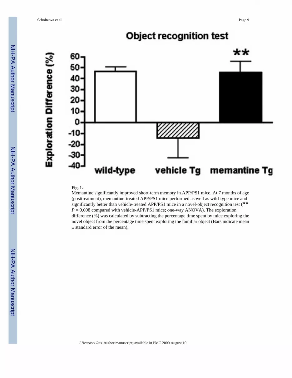
Fig. 1.Memantine significantly improved short-term memory in APP/PS1 mice. At 7 months of age(posttreatment), memantine-treated APP/PS1 mice performed as well as wild-type mice andsignificantly better than vehicle-treated APP/PS1 mice in a novel-object recognition test (★★P = 0.008 compared with vehicle-APP/PS1 mice; one-way ANOVA). The explorationdifference (%) was calculated by subtracting the percentage time spent by mice exploring thenovel object from the percentage time spent exploring the familiar object (Bars indicate mean± standard error of the mean).
Scholtzova et al. Page 9
J Neurosci Res. Author manuscript; available in PMC 2009 August 10.
NIH
-PA Author Manuscript
NIH
-PA Author Manuscript
NIH
-PA Author Manuscript
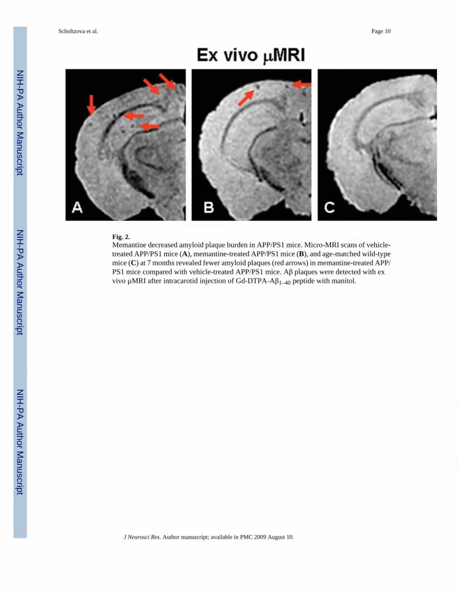
Fig. 2.Memantine decreased amyloid plaque burden in APP/PS1 mice. Micro-MRI scans of vehicle-treated APP/PS1 mice (A), memantine-treated APP/PS1 mice (B), and age-matched wild-typemice (C) at 7 months revealed fewer amyloid plaques (red arrows) in memantine-treated APP/PS1 mice compared with vehicle-treated APP/PS1 mice. Aβ plaques were detected with exvivo µMRI after intracarotid injection of Gd-DTPA-Aβ1–40 peptide with manitol.
Scholtzova et al. Page 10
J Neurosci Res. Author manuscript; available in PMC 2009 August 10.
NIH
-PA Author Manuscript
NIH
-PA Author Manuscript
NIH
-PA Author Manuscript

Fig. 3.Average cortical T2★ values in treated vs. control APP/PS1 Tg mice. Quantification of corticalabsolute T2★ values also demonstrated a memantine amyloid burden-lowering effect (★ P =0.04; t-test) (Bars indicate mean ± standard error of the mean).
Scholtzova et al. Page 11
J Neurosci Res. Author manuscript; available in PMC 2009 August 10.
NIH
-PA Author Manuscript
NIH
-PA Author Manuscript
NIH
-PA Author Manuscript

Fig. 4.Memantine decreased hippocampal and cortical plaque burden in APP/PS1 mice. Histologicalanalysis of APP/PS1–21 Tg mice showed the difference in Aβ burden. Thioflavin-S stainingrevealed more amyloid plaques in hippocampal sections of vehicle-treated APP/PS1 mice(A) compared with memantine-treated APP/PS1 mice (B). Similarly, Aβ immunostainingshowed greater Aβ accumulation in hippocampal sections of vehicle-treated APP/PS1 mice(C) compared with sections from memantine-treated APP/PS1 mice (D). Cortical Aβimmunoreactivity also revealed differences between vehicle-treated (E) and memantine-treated (F) APP/PS1 mice. Scale bars = 200 µm.
Scholtzova et al. Page 12
J Neurosci Res. Author manuscript; available in PMC 2009 August 10.
NIH
-PA Author Manuscript
NIH
-PA Author Manuscript
NIH
-PA Author Manuscript
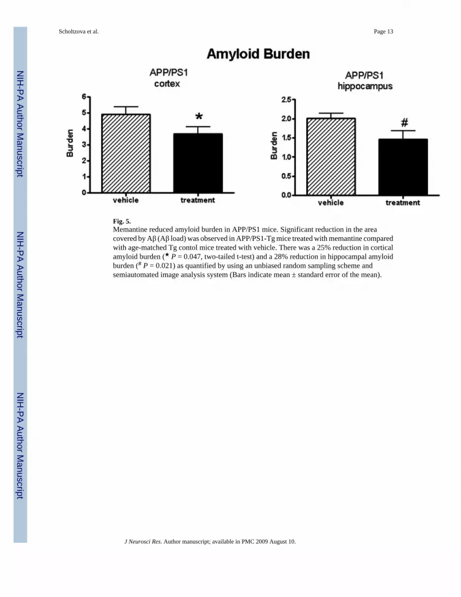
Fig. 5.Memantine reduced amyloid burden in APP/PS1 mice. Significant reduction in the areacovered by Aβ (Aβ load) was observed in APP/PS1-Tg mice treated with memantine comparedwith age-matched Tg contol mice treated with vehicle. There was a 25% reduction in corticalamyloid burden (★ P = 0.047, two-tailed t-test) and a 28% reduction in hippocampal amyloidburden (# P = 0.021) as quantified by using an unbiased random sampling scheme andsemiautomated image analysis system (Bars indicate mean ± standard error of the mean).
Scholtzova et al. Page 13
J Neurosci Res. Author manuscript; available in PMC 2009 August 10.
NIH
-PA Author Manuscript
NIH
-PA Author Manuscript
NIH
-PA Author Manuscript
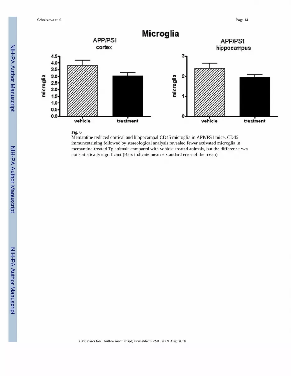
Fig. 6.Memantine reduced cortical and hippocampal CD45 microglia in APP/PS1 mice. CD45immunostaining followed by stereological analysis revealed fewer activated microglia inmemantine-treated Tg animals compared with vehicle-treated animals, but the difference wasnot statistically significant (Bars indicate mean ± standard error of the mean).
Scholtzova et al. Page 14
J Neurosci Res. Author manuscript; available in PMC 2009 August 10.
NIH
-PA Author Manuscript
NIH
-PA Author Manuscript
NIH
-PA Author Manuscript

Fig. 7.Memantine reduced cortical and hippocampal GFAP in APP/PS1 mice. GFAP immunostainingfollowed by stereological analysis revealed fewer activated astrocytes in memantine-treatedTg animals compared with vehicle-treated animals, but the difference was not statisticallysignificant (Bars indicate mean ± standard error of the mean).
Scholtzova et al. Page 15
J Neurosci Res. Author manuscript; available in PMC 2009 August 10.
NIH
-PA Author Manuscript
NIH
-PA Author Manuscript
NIH
-PA Author Manuscript